RU2704103C1 - Method for spinal injury modeling - Google Patents
Method for spinal injury modeling Download PDFInfo
- Publication number
- RU2704103C1 RU2704103C1 RU2018133926A RU2018133926A RU2704103C1 RU 2704103 C1 RU2704103 C1 RU 2704103C1 RU 2018133926 A RU2018133926 A RU 2018133926A RU 2018133926 A RU2018133926 A RU 2018133926A RU 2704103 C1 RU2704103 C1 RU 2704103C1
- Authority
- RU
- Russia
- Prior art keywords
- spinal cord
- injury
- spinal
- level
- vertebrae
- Prior art date
Links
- 238000000034 method Methods 0.000 title claims abstract description 31
- 208000020339 Spinal injury Diseases 0.000 title abstract description 10
- 210000000278 spinal cord Anatomy 0.000 claims abstract description 23
- 230000006378 damage Effects 0.000 claims abstract description 21
- 208000014674 injury Diseases 0.000 claims abstract description 12
- 208000027418 Wounds and injury Diseases 0.000 claims abstract description 11
- 208000020431 spinal cord injury Diseases 0.000 claims abstract description 10
- 206010002091 Anaesthesia Diseases 0.000 claims abstract description 7
- 241000283973 Oryctolagus cuniculus Species 0.000 claims abstract description 7
- 230000037005 anaesthesia Effects 0.000 claims abstract description 7
- 210000001951 dura mater Anatomy 0.000 claims abstract description 6
- 210000000988 bone and bone Anatomy 0.000 claims abstract description 5
- 210000003195 fascia Anatomy 0.000 claims abstract description 5
- 239000012634 fragment Substances 0.000 claims abstract description 5
- 239000002184 metal Substances 0.000 claims abstract description 5
- 238000007920 subcutaneous administration Methods 0.000 claims abstract description 5
- 241001465754 Metazoa Species 0.000 abstract description 6
- 230000002085 persistent effect Effects 0.000 abstract description 4
- 208000007542 Paresis Diseases 0.000 abstract description 3
- 239000003814 drug Substances 0.000 abstract description 3
- 230000004776 neurological deficiency Effects 0.000 abstract description 3
- 230000000694 effects Effects 0.000 abstract 1
- 208000012318 pareses Diseases 0.000 abstract 1
- 239000000126 substance Substances 0.000 abstract 1
- 230000007971 neurological deficit Effects 0.000 description 7
- 238000010171 animal model Methods 0.000 description 6
- 230000006870 function Effects 0.000 description 3
- 230000000472 traumatic effect Effects 0.000 description 3
- LFQSCWFLJHTTHZ-UHFFFAOYSA-N Ethanol Chemical compound CCO LFQSCWFLJHTTHZ-UHFFFAOYSA-N 0.000 description 2
- 229960004134 propofol Drugs 0.000 description 2
- OLBCVFGFOZPWHH-UHFFFAOYSA-N propofol Chemical compound CC(C)C1=CC=CC(C(C)C)=C1O OLBCVFGFOZPWHH-UHFFFAOYSA-N 0.000 description 2
- 210000000115 thoracic cavity Anatomy 0.000 description 2
- 230000008733 trauma Effects 0.000 description 2
- 238000005303 weighing Methods 0.000 description 2
- UVITTYOJFDLOGI-UHFFFAOYSA-N (1,2,5-trimethyl-4-phenylpiperidin-4-yl) propanoate Chemical compound C=1C=CC=CC=1C1(OC(=O)CC)CC(C)N(C)CC1C UVITTYOJFDLOGI-UHFFFAOYSA-N 0.000 description 1
- YQEZLKZALYSWHR-UHFFFAOYSA-N Ketamine Chemical compound C=1C=CC=C(Cl)C=1C1(NC)CCCCC1=O YQEZLKZALYSWHR-UHFFFAOYSA-N 0.000 description 1
- 206010033892 Paraplegia Diseases 0.000 description 1
- 230000001154 acute effect Effects 0.000 description 1
- 210000000709 aorta Anatomy 0.000 description 1
- 210000004556 brain Anatomy 0.000 description 1
- 230000006835 compression Effects 0.000 description 1
- 238000007906 compression Methods 0.000 description 1
- 235000019441 ethanol Nutrition 0.000 description 1
- 230000001771 impaired effect Effects 0.000 description 1
- 238000007912 intraperitoneal administration Methods 0.000 description 1
- 208000028867 ischemia Diseases 0.000 description 1
- 229960003299 ketamine Drugs 0.000 description 1
- 230000003902 lesion Effects 0.000 description 1
- 210000004705 lumbosacral region Anatomy 0.000 description 1
- 210000000056 organ Anatomy 0.000 description 1
- 230000008506 pathogenesis Effects 0.000 description 1
- 230000010410 reperfusion Effects 0.000 description 1
- 238000012552 review Methods 0.000 description 1
- 230000035807 sensation Effects 0.000 description 1
- 230000035945 sensitivity Effects 0.000 description 1
- 230000035939 shock Effects 0.000 description 1
- 210000004872 soft tissue Anatomy 0.000 description 1
- 230000004083 survival effect Effects 0.000 description 1
- 238000002560 therapeutic procedure Methods 0.000 description 1
- 210000001519 tissue Anatomy 0.000 description 1
Images
Classifications
-
- G—PHYSICS
- G09—EDUCATION; CRYPTOGRAPHY; DISPLAY; ADVERTISING; SEALS
- G09B—EDUCATIONAL OR DEMONSTRATION APPLIANCES; APPLIANCES FOR TEACHING, OR COMMUNICATING WITH, THE BLIND, DEAF OR MUTE; MODELS; PLANETARIA; GLOBES; MAPS; DIAGRAMS
- G09B23/00—Models for scientific, medical, or mathematical purposes, e.g. full-sized devices for demonstration purposes
- G09B23/28—Models for scientific, medical, or mathematical purposes, e.g. full-sized devices for demonstration purposes for medicine
Landscapes
- Engineering & Computer Science (AREA)
- Physics & Mathematics (AREA)
- General Physics & Mathematics (AREA)
- Computational Mathematics (AREA)
- Mathematical Analysis (AREA)
- Medicinal Chemistry (AREA)
- General Health & Medical Sciences (AREA)
- Algebra (AREA)
- Health & Medical Sciences (AREA)
- Chemical & Material Sciences (AREA)
- Medical Informatics (AREA)
- Mathematical Optimization (AREA)
- Mathematical Physics (AREA)
- Pure & Applied Mathematics (AREA)
- Business, Economics & Management (AREA)
- Educational Administration (AREA)
- Educational Technology (AREA)
- Theoretical Computer Science (AREA)
- Surgical Instruments (AREA)
Abstract
Description
Изобретение относится к экспериментальной медицине, а именно к нейрохирургии, и может быть использовано для моделирования на кроликах тяжелой спинномозговой травмы с грубым стойким неврологическим дефицитом для отработки методов восстановления функций спинного мозга у человека.The invention relates to experimental medicine, namely to neurosurgery, and can be used to simulate severe spinal cord injury with severe persistent neurological deficiency in rabbits to develop methods for restoring spinal cord functions in humans.
В настоящее время существует ряд экспериментальных моделей травмы спинного мозга (далее СМ), которые можно объединить в следующие группы:Currently, there are a number of experimental models of spinal cord injury (hereinafter referred to as SM), which can be combined into the following groups:
1) модели с использованием ишемии - реперфузии (путем пережатия или окклюзии аорты);1) models using ischemia - reperfusion (by clamping or occlusion of the aorta);
2) модели повреждения путем нанесения травмирующего воздействия: травмирование падающим предметом определенной массы; улучшенные модели, со стандартизацией наносимого травмирующего воздействия; модели с использованием пневматических, электромагнитных устройств; тракционные модели; другие варианты механического воздействия на СМ (пальцем, ручкой скальпеля, зажимом Кохера, хирургическим пинцетом, надувным баллоном, помещенным в эпидуральное пространство);2) damage models by applying a traumatic effect: trauma to a falling object of a certain mass; improved models, with standardization of the applied traumatic effect; models using pneumatic, electromagnetic devices; traction models; other options for mechanical impact on the SM (with a finger, a scalpel handle, Kocher clamp, surgical tweezers, an inflatable balloon placed in the epidural space);
3) модели с транссекцией СМ;3) models with CM transection;
4) фотохимически индуцированное поражение СМ.4) photochemically induced CM lesion.
В обзорах указывается на наличие у каждой модели положительных и отрицательных особенностей и отмечается необходимость адекватной, воспроизводимой и технически соответствующей требованиям модели.The reviews indicate the presence of positive and negative features in each model and note the need for an adequate, reproducible and technically relevant model.
Наиболее близким к предлагаемому является способ моделирования позвоночно-спинномозговой травмы шейного отдела путем травмирования спинного мозга по патенту РФ на изобретение №2154862 (МПК G09B 23/28, опубл. 20.08.2000). В данном способе проводят анестезию внутрибрюшинным введением кетамина в дозе 50 мг/кг. После наступления наркоза по анатомическим ориентирам идентифицируют 6-7-е шейные позвонки и вводят стерильную иглу диаметром, соответствующим диаметру спинного мозга, перпендикулярно оси позвоночного канала в спинной мозг до ощущения "провала", через 2 мин иглу извлекают.Closest to the proposed is a method of modeling a spinal cord injury of the cervical spine by injuring the spinal cord according to the patent of the Russian Federation for invention No. 2154862 (IPC G09B 23/28, publ. 08.20.2000). In this method, anesthesia is administered by intraperitoneal administration of ketamine at a dose of 50 mg / kg. After the onset of anesthesia, the 6-7th cervical vertebrae are identified by anatomical landmarks and a sterile needle is inserted with a diameter corresponding to the diameter of the spinal cord, perpendicular to the axis of the spinal canal into the spinal cord until there is a "failure", after 2 minutes the needle is removed.
К недостаткам способа по патенту РФ №2154862 следует отнести невозможность получения стандартизированного неврологического дефицита, поскольку нанесение травмы с помощью иглы до ощущения «провала» достаточно субъективно и не может гарантировать идентичность повреждений спинного мозга у всех экспериментальных животных.The disadvantages of the method according to the patent of the Russian Federation No. 2154862 include the impossibility of obtaining a standardized neurological deficit, since injuring with a needle until the sensation of "failure" is quite subjective and cannot guarantee the identity of spinal cord injuries in all experimental animals.
Задача (технический результат) предлагаемого изобретения - разработать способ моделирования спинномозговой травмы, обеспечивающий стандартизированный стойкий неврологический дефицит в виде грубых парезов конечностей без гибели лабораторного животного.The objective (technical result) of the present invention is to develop a method for simulating a spinal injury that provides a standardized persistent neurological deficit in the form of gross paresis of limbs without the death of a laboratory animal.
Поставленная задача решается тем, что способ моделирования спинномозговой травмы включает проведение анестезии и травмирование спинного мозга. Согласно предлагаемому изобретению травмирование спинного мозга осуществляют путем выполнения доступа к задней поверхности дурального мешка с подлежащим спинным мозгом. Для этого рассекают кожу продольно над позвоночником на 2-3 позвонка выше и ниже планируемого уровня повреждения над соответствующими остистыми отростками, подкожную фасцию рассекают по ходу кожного разреза, с двух сторон острым путем скелетируют остистые отростки и дужки соответствующих позвонков. Затем удаляют костный фрагмент остистого отростка позвонка предполагаемого уровня травмы и открывают доступ к задней поверхности дурального мешка с подлежащим спинным мозгом на заданном уровне. После выполнения доступа к задней поверхности дурального мешка с подлежащим спинным мозгом через интактную твердую мозговую оболочку в области удаленного остистого отростка однократно наносят повреждение металлическим ударником с насадкой, диаметр которой соответствует размеру спинного мозга, с энергией удара 0,07-0,08 Дж.The problem is solved in that the method of modeling a spinal injury involves anesthesia and trauma to the spinal cord. According to the invention, spinal cord injury is carried out by accessing the posterior surface of the dural sac with the underlying spinal cord. To do this, cut the skin longitudinally above the spine 2-3 vertebrae above and below the planned level of damage above the corresponding spinous processes, the subcutaneous fascia is cut along the skin incision, from the two sides, the spinous processes and arches of the corresponding vertebrae are skeletonized. Then remove the bone fragment of the spinous process of the vertebra of the alleged level of injury and open access to the posterior surface of the dural sac with the underlying spinal cord at a given level. After accessing the posterior surface of the dural sac with the underlying spinal cord through the intact dura mater in the region of the distant spinous process, damage is made once with a metal hammer with a nozzle, the diameter of which corresponds to the size of the spinal cord, with an impact energy of 0.07-0.08 J.
Доступ к задней поверхности дурального мешка с подлежащим спинным мозгом в совокупности с однократным нанесением повреждения с одинаковой силой обеспечивает идентичность травмы и неврологического дефицита у всех экспериментальных животных, что дает возможность проследить воздействие и оценить эффективность терапии, направленной на восстановление функций спинного мозга после травмы.Access to the back surface of the dural sac with the underlying spinal cord, combined with a single application of damage with the same strength, ensures the identity of the injury and neurological deficit in all experimental animals, which makes it possible to trace the impact and evaluate the effectiveness of therapy aimed at restoring the functions of the spinal cord after the injury.
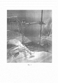
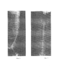
Предлагаемое изобретение поясняется представленной на фиг. 1 фотографией операции и представленными на фиг. 2 и 3 рентгеновскими снимками позвоночника экспериментального животного после нанесения травмы соответственно вид сбоку и вид сверху.The invention is illustrated in FIG. 1 with a photograph of the operation and shown in FIG. 2 and 3 x-rays of the spine of the experimental animal after injury, respectively, side view and top view.
Способ осуществляют следующим образом.The method is as follows.
Выполняют анестезию. После наступления анестезии рассекают кожу продольно над позвоночником на 2-3 позвонка выше и ниже планируемого уровня повреждения над соответствующими остистыми отростками, подкожную фасцию рассекают по ходу кожного разреза. С двух сторон острым путем скелетируют остистые отростки и дужки соответствующих позвонков. Удаляют костный фрагмент остистого отростка позвонка предполагаемого уровня травмы и открывают доступа к задней поверхности дурального мешка с подлежащим спинным мозгом на заданном уровне. Затем по интактной твердой мозговой оболочке в области удаленного остистого отростка однократно наносят удар металлическим ударником с насадкой, диаметр которой соответствует поперечному размеру спинного мозга. Экспериментальных исследования на кроликах показали, что ударное воздействие с энергией более 0,09 Дж приводит к гибели животного либо при нанесении травмы, либо в течение 7-8 часов после нее. При энергии ударного воздействия менее 0,06 Дж наблюдался только частичный двигательный неврологический дефицит, не дающий возможности изучать течение тяжелой спинномозговой травмы. Поэтому удар наносят с энергией удара 0,07-0,08 Дж. Мягкие ткани ушивают послойно.Perform anesthesia. After the onset of anesthesia, the skin is cut longitudinally above the spine 2-3 vertebrae above and below the planned level of damage over the corresponding spinous processes, the subcutaneous fascia is cut along the skin incision. On both sides, the spinous processes and arches of the corresponding vertebrae skeletonize sharply. The bone fragment of the spinous process of the vertebra of the expected level of injury is removed and access to the posterior surface of the dural sac with the underlying spinal cord at a predetermined level is opened. Then, an intact dura mater in the region of the distant spinous process is hit once with a metal hammer with a nozzle whose diameter corresponds to the transverse size of the spinal cord. Experimental studies on rabbits showed that a shock with an energy of more than 0.09 J leads to the death of the animal either when the injury was caused, or within 7-8 hours after it. When the impact energy was less than 0.06 J, only a partial motor neurological deficit was observed, which did not make it possible to study the course of severe spinal injury. Therefore, the blow is applied with impact energy of 0.07-0.08 J. Soft tissue is sutured in layers.
У всех экспериментальных животных сразу после нанесения травмы возникал стандартизированный неврологический дефицит в виде нижнего грубого пареза до параплегии, нарушения чувствительности, функции тазовых органов по типу задержки.All experimental animals immediately after the injury caused a standardized neurological deficit in the form of a lower gross paresis to paraplegia, impaired sensitivity, and function of the pelvic organs according to the type of delay.
Пример 1.Example 1
Лабораторное животное кролик с массой тела 3200 г. Laboratory animal rabbit weighing 3200 g.
Внутривенное вводили пропофол через дозатор шприцевого введения со скоростью 22 мг/кг/ч, промедол в/м 7 мг/кг.Propofol was injected intravenously through a syringe dispenser at a rate of 22 mg / kg / h, promedol i / m 7 mg / kg.
Операционное поле животного выбривали от лопаток до середины поясничного отдела - по длине и на уровне середины ребер справа и слева - по ширине. Животное фиксировали на операционном столике. Зону операционного поля обрабатывали 70% этиловым спиртом двукратно и изолировали с помощью стерильных салфеток, прикрепляемых к коже цапками. Кожу рассекали продольно над позвоночником от 10 грудного позвонка до 3-го поясничного над остистыми отростками. Подкожную фасцию рассекали ножницами по ходу кожного разреза. С двух сторон острым путем скелетировали остистые отростки и дужки 11, 12 грудных, 1, 2 поясничных позвонков. Удаляли костный фрагмент остистого отростка позвонка Th 12 и открывали доступ к задней поверхности дурального мешка с подлежащим спинным мозгом. Твердая мозговая оболочка не вскрывалась. Повреждение наносили однократно металлическим ударником с насадкой диаметром 0,5 см и толщиной 0,3 см через интактную твердую мозговую оболочку в области трепанационного окна с энергией удара 0,07-0,08 Дж. Удар наносился при помощи механического пружинного ударника устройства для моделирования повреждений головного и спинного мозга (патент РФ №2414005). Повреждения наносились с энергией ударного воздействия в диапазоне 0,07-0,08 Дж. Необходимые усилия сжатия установленной пружины задавались при помощи внешнего динамометра. После нанесения удара рана ушивалась послойно, прекращалось введение пропофола.The surgical field of the animal was shaved from the shoulder blades to the middle of the lumbar region - along the length and at the level of the middle of the ribs on the right and left - in width. The animal was fixed on the operating table. The area of the surgical field was treated with 70% ethyl alcohol twice and isolated using sterile wipes attached to the skin with slippers. The skin was dissected longitudinally above the spine from the 10th thoracic vertebra to the 3rd lumbar above the spinous processes. The subcutaneous fascia was dissected with scissors along the skin incision. On both sides, the spinous processes and arches of 11, 12 thoracic, 1, 2 lumbar vertebrae were skeletonized sharply. The bone fragment of the spinous process of the Th 12 vertebra was removed and access to the posterior surface of the dural sac with the underlying spinal cord was opened. The dura mater was not opened. Damage was applied once by a metal drummer with a nozzle with a diameter of 0.5 cm and a thickness of 0.3 cm through an intact dura mater in the area of a trepanation window with an impact energy of 0.07-0.08 J. The impact was applied using a mechanical spring hammer of a damage modeling device brain and spinal cord (RF patent No. 2414005). Damage was inflicted with impact energy in the range of 0.07-0.08 J. The necessary compression forces of the installed spring were set using an external dynamometer. After striking, the wound was sutured in layers, the introduction of propofol stopped.
Пример 2.Example 2
Спинномозговую травму моделировали на группе из 60 кроликов (самцы и самки в равном соотношении) массой 2500-3400 г. Все кролики остались живы, при этом во всех случаях отмечена стойкая клиника спинномозговой травмы и неврологического дефицита, удовлетворяющая требованиям к экспериментальным моделям.Cerebrospinal injury was modeled on a group of 60 rabbits (males and females in equal proportions) weighing 2500-3400 g. All rabbits remained alive, and in all cases there was a persistent clinic of spinal injury and neurological deficit that met the requirements for experimental models.
На основе полученной модели спинномозговой травмы проведена оценка динамики неврологического дефицита при лечении острой спинальной травмы в эксперименте.Based on the obtained model of spinal injury, the dynamics of neurological deficit in the treatment of acute spinal injury in an experiment is evaluated.
Таким образом, предложенный способ моделирования спинномозговой травмы:Thus, the proposed method for modeling spinal injury:
- отражает основные клинико-физиологические изменения при травме спинного мозга;- reflects the main clinical and physiological changes in spinal cord injury;
- является легко воспроизводимым и дозированным;- is easily reproducible and metered;
- имеет минимальные травмирующие воздействия на прилежащие к спинному мозгу структуры и ткани;- has minimal traumatic effects on structures and tissues adjacent to the spinal cord;
- позволяет изучить патогенез неврологического дефицита при травме спинного мозга;- allows you to study the pathogenesis of neurological deficiency in spinal cord injury;
- позволяет снизить количество требуемых для эксперимента лабораторных животных за счет увеличения процента их выживаемости.- allows to reduce the number of laboratory animals required for the experiment by increasing the percentage of their survival.
Claims (1)
Priority Applications (1)
| Application Number | Priority Date | Filing Date | Title |
|---|---|---|---|
| RU2018133926A RU2704103C1 (en) | 2018-09-25 | 2018-09-25 | Method for spinal injury modeling |
Applications Claiming Priority (1)
| Application Number | Priority Date | Filing Date | Title |
|---|---|---|---|
| RU2018133926A RU2704103C1 (en) | 2018-09-25 | 2018-09-25 | Method for spinal injury modeling |
Publications (1)
| Publication Number | Publication Date |
|---|---|
| RU2704103C1 true RU2704103C1 (en) | 2019-10-23 |
Family
ID=68318590
Family Applications (1)
| Application Number | Title | Priority Date | Filing Date |
|---|---|---|---|
| RU2018133926A RU2704103C1 (en) | 2018-09-25 | 2018-09-25 | Method for spinal injury modeling |
Country Status (1)
| Country | Link |
|---|---|
| RU (1) | RU2704103C1 (en) |
Cited By (1)
| Publication number | Priority date | Publication date | Assignee | Title |
|---|---|---|---|---|
| RU2768486C1 (en) * | 2021-08-16 | 2022-03-24 | федеральное государственное бюджетное учреждение "Национальный медицинский исследовательский центр детской травматологии и ортопедии имени Г.И. Турнера" Министерства здравоохранения Российской Федерации | Method for modeling traumatic injury of spinal cord from vental access in lumbar spine |
Citations (5)
| Publication number | Priority date | Publication date | Assignee | Title |
|---|---|---|---|---|
| SU625233A1 (en) * | 1977-04-29 | 1978-09-25 | Курганский Научно-Исследовательский Институт Экспериментальной И Клинической Ортопедии И Травматологии | Method of obtaining dystrophic scoliosis simulator |
| RU2013810C1 (en) * | 1991-02-20 | 1994-05-30 | Филиал МГУ им.М.В.Ломоносова в г.Ульяновске | Method for vertebral basilar insufficiency simulating |
| WO2000045773A2 (en) * | 1999-02-08 | 2000-08-10 | Uab Research Foundation | Apparatus for simulating traumatic brain injury and method for inducing spinal cord injury |
| RU2154862C2 (en) * | 1997-06-11 | 2000-08-20 | Новосибирский научно-исследовательский институт травматологии и ортопедии | Method for modeling vertebrocerebrospinal trauma of cervical segment |
| RU2348985C1 (en) * | 2007-06-29 | 2009-03-10 | Вячеслав Александрович Липатов | Method of peripheral nerve and muscle injury simulation |
-
2018
- 2018-09-25 RU RU2018133926A patent/RU2704103C1/en active
Patent Citations (5)
| Publication number | Priority date | Publication date | Assignee | Title |
|---|---|---|---|---|
| SU625233A1 (en) * | 1977-04-29 | 1978-09-25 | Курганский Научно-Исследовательский Институт Экспериментальной И Клинической Ортопедии И Травматологии | Method of obtaining dystrophic scoliosis simulator |
| RU2013810C1 (en) * | 1991-02-20 | 1994-05-30 | Филиал МГУ им.М.В.Ломоносова в г.Ульяновске | Method for vertebral basilar insufficiency simulating |
| RU2154862C2 (en) * | 1997-06-11 | 2000-08-20 | Новосибирский научно-исследовательский институт травматологии и ортопедии | Method for modeling vertebrocerebrospinal trauma of cervical segment |
| WO2000045773A2 (en) * | 1999-02-08 | 2000-08-10 | Uab Research Foundation | Apparatus for simulating traumatic brain injury and method for inducing spinal cord injury |
| RU2348985C1 (en) * | 2007-06-29 | 2009-03-10 | Вячеслав Александрович Липатов | Method of peripheral nerve and muscle injury simulation |
Non-Patent Citations (1)
| Title |
|---|
| DUCKER T.B. et al. Experimental Spinal Cord Trauma: Correlation of Blood Flow, Tissue oxygen and Neurologis Status in the Dog/J.Surg. neurol. 1978, v. 10, p. 60 - 70. * |
Cited By (1)
| Publication number | Priority date | Publication date | Assignee | Title |
|---|---|---|---|---|
| RU2768486C1 (en) * | 2021-08-16 | 2022-03-24 | федеральное государственное бюджетное учреждение "Национальный медицинский исследовательский центр детской травматологии и ортопедии имени Г.И. Турнера" Министерства здравоохранения Российской Федерации | Method for modeling traumatic injury of spinal cord from vental access in lumbar spine |
Similar Documents
| Publication | Publication Date | Title |
|---|---|---|
| Missios | Hippocrates, Galen, and the uses of trepanation in the ancient classical world | |
| Weber et al. | Neurosurgical aspects of trepanations from Neolithic times | |
| Gentile et al. | Management of midface maxillofacial trauma | |
| Setty et al. | Endoscopic vascular decompression for the treatment of trigeminal neuralgia: clinical outcomes and technical note | |
| RU2704103C1 (en) | Method for spinal injury modeling | |
| Shereen et al. | Treatment of arrow wounds: a review | |
| Pavlíková et al. | Piezosurgery prevents brain tissue damage: an experimental study on a new rat model | |
| RU2641569C1 (en) | Method for severe craniocerebral injury simulation | |
| Tripuraneni et al. | A surgical procedure for resecting the mouse rib: a model for large-scale long bone repair | |
| Beuriat et al. | Coronal and lambdoid suture evolution following total vault remodeling for scaphocephaly | |
| SU972560A1 (en) | Spinal cord compression and decompression simulation method | |
| RU2768486C1 (en) | Method for modeling traumatic injury of spinal cord from vental access in lumbar spine | |
| CN109248001A (en) | A kind of construction method of the bone defect model of the bilateral fibula osteotomy production of machin | |
| RU2766800C1 (en) | Method for movement of peroneal nerve in upper one-third of shin | |
| RU2523622C1 (en) | Method for simulation of false joint in shin fractures and device for implementing it | |
| Harrington et al. | Realignment of a seventh lumbar vertebral fracture/luxation using a Senn retractor in two puppies | |
| RU2763666C1 (en) | Method for modeling a spinal cord injury with persistent neurological deficit | |
| RU2803270C1 (en) | Method of restoring eye cornea trophism and skin sensitivity in the zone of innervation of the first branch of the trigeminal nerve in neurotrophic keratopathy | |
| RU2816791C1 (en) | Method of surgical approach to genital nerves for their decompression | |
| RU2784999C1 (en) | Method for correcting age-related changes in the gravitational nature of the soft tissues of the periorbital region | |
| Nayoan et al. | Endoscopy-assisted extraction of penetrating a wayer arrow in the ethmoid sinus | |
| Dev et al. | Surgical procedures in Sushruta Samhita and its relevance in modern surgery | |
| Lahey | The removal of broken spinal anesthesia needles | |
| RU2726011C1 (en) | Method for operative access to posterior support spine complex | |
| Carmel et al. | 100 years ago in neurosurgery: Description of the intracranial extradural approach to the Gasserian ganglion–the Hartley-Krause operation |