RU2698436C1 - Method of bone plasty - Google Patents
Method of bone plasty Download PDFInfo
- Publication number
- RU2698436C1 RU2698436C1 RU2018144106A RU2018144106A RU2698436C1 RU 2698436 C1 RU2698436 C1 RU 2698436C1 RU 2018144106 A RU2018144106 A RU 2018144106A RU 2018144106 A RU2018144106 A RU 2018144106A RU 2698436 C1 RU2698436 C1 RU 2698436C1
- Authority
- RU
- Russia
- Prior art keywords
- bone
- area
- bone block
- alveolar
- jaw
- Prior art date
Links
Images
Classifications
-
- A—HUMAN NECESSITIES
- A61—MEDICAL OR VETERINARY SCIENCE; HYGIENE
- A61C—DENTISTRY; APPARATUS OR METHODS FOR ORAL OR DENTAL HYGIENE
- A61C7/00—Orthodontics, i.e. obtaining or maintaining the desired position of teeth, e.g. by straightening, evening, regulating, separating, or by correcting malocclusions
- A61C7/006—Orthodontics, i.e. obtaining or maintaining the desired position of teeth, e.g. by straightening, evening, regulating, separating, or by correcting malocclusions using magnetic force
Abstract
Description
Изобретение относится к медицине, а именно к челюстно-лицевой хирургии, и может быть использовано для реконструкции атрофированной альвеолярной части нижней челюсти или альвеолярного отростка верхней челюсти с последующей дентальной имплантацией.The invention relates to medicine, namely to maxillofacial surgery, and can be used to reconstruct the atrophied alveolar part of the lower jaw or the alveolar process of the upper jaw, followed by dental implantation.
Концепция остеоинтеграции и ее клиническое применение в стоматологии кардинально изменили взгляд на решение проблемы восстановления зубного ряда и привели к созданию наиболее прогрессивной системы протезирования. В настоящее время синонимом остеоинтеграции стали дентальные имплантаты, которые широко используются в качестве опор для ортопедических конструкций при проведении лечения как в рутинных, так и в сложных клинических ситуациях частичной и полной адентии. Современные технологии имплантации обеспечивают эффективность применения данного метода на 95% и выше, открывая новые возможности в совершенствовании лечебных мероприятий, направленных на реабилитацию стоматологических больных (Жусев А.И., 2004; Иванов С.Ю. с соавт., 2004; Кулаков А.А., Лосев Ф.Ф., Гветадзе Р.Ш., 2006; Олесова В.Н. с соавт., 2008; Параскевич В.Л., 2009).The concept of osseointegration and its clinical application in dentistry have fundamentally changed our view of solving the problem of restoration of the dentition and led to the creation of the most advanced prosthetics system. Currently, dental implants have become synonymous with osseointegration, which are widely used as supports for orthopedic structures during treatment in both routine and complex clinical situations of partial and complete adentia. Modern implantation technologies ensure the effectiveness of this method by 95% and higher, opening up new possibilities in improving therapeutic measures aimed at the rehabilitation of dental patients (Zhusev A.I., 2004; Ivanov S.Yu. et al., 2004; Kulakov A. A., Losev F.F., Gvetadze R.Sh., 2006; Olesova V.N. et al., 2008; Paraskevich V.L., 2009).
Однако, по данным научной литературы, более чем в 30% случаев стандартные методики имплантации не могут применяться из-за атрофии костной ткани альвеолярных отростков по высоте и (или) толщине, низкого расположения дна верхнечелюстных пазух и грушевидного отверстия, неудовлетворительного качества кости и мягких тканей в области предполагаемой имплантации (Дробышев А.Ю., 2006, Иванов С.Ю., 2008, Кулаков А.А. 2011,).However, according to the scientific literature, in more than 30% of cases, standard implantation methods cannot be applied due to atrophy of the bone tissue of the alveolar processes in height and (or) thickness, low location of the bottom of the maxillary sinuses and pear-shaped opening, poor quality of bone and soft tissues in the field of implantation (Drobyshev A.Yu., 2006, Ivanov S.Yu., 2008, Kulakov A.A. 2011,).
В то же время ведется активный научный поиск и в направлении расширения показаний к проведению операции дентальной имплантации. Разрабатываются различные методики коррекции выраженной атрофии альвеолярной кости, которая резко ограничивает возможность дентальной имплантации (A.M. Панин 2003, Иванов С.Ю. 2010, Жусев А.И., 2012).At the same time, an active scientific search is underway in the direction of expanding the indications for the operation of dental implantation. Various methods are being developed for correcting severe atrophy of the alveolar bone, which sharply limits the possibility of dental implantation (A.M. Panin 2003, Ivanov S.Yu. 2010, Zhusev A.I., 2012).
Известен способ реконструкции атрофированной альвеолярной части нижней челюсти и альвеолярного отростка верхней челюсти (Khoury F. Happe A. Zur Diagnostik und Methodik von intra-oralen Knochenentnahmen. Zzahnarztl Implantol 1999:15(3):167-176), заключающийся в использовании костного блока - монокортикального аутотрансплантата. Способ предусматривает фиксацию костного блока с помощью внутрикостных шурупов к краю кости атрофированной альвеолярной части челюстей. Данный способ принят авторами за прототип.A known method of reconstructing the atrophied alveolar part of the lower jaw and the alveolar process of the upper jaw (Khoury F. Happe A. Zur Diagnostik und Methodik von intra-oralen Knochenentnahmen. Zzahnarztl Implantol 1999: 15 (3): 167-176), which consists in using a bone block - monocortical autograft. The method involves fixing the bone block using intraosseous screws to the edge of the bone of the atrophied alveolar part of the jaw. This method is adopted by the authors for the prototype.
Прототип имеет следующие недостатки:The prototype has the following disadvantages:
1. Конструкция прототипа не способна обеспечить стабильность объема костного блока на время формирования регенерата.1. The design of the prototype is not able to ensure the stability of the volume of the bone block during the formation of the regenerate.
2. Не обеспечиваются условия для создания максимальной площади соприкосновения костного блока с костью реципиентного ложа края атрофированной кости.2. Conditions are not provided for creating the maximum area of contact between the bone block and the bone of the recipient bed of the edge of the atrophied bone.
3. Выполнение костной пластики проводится только с использованием костного блока, что ограничивает объем восстановления костной ткани анатомическими зонами заборами забора костного трансплантата.3. Bone grafting is performed only with the use of a bone block, which limits the volume of bone tissue restoration by the anatomical zones by the bone graft fence.
Технический результат, на достижение которого направлено создание данного изобретения, заключается в создании идеального прилегания костного блока к реципиентной области, обеспечении надежной фиксации костного блока и стабильности объема костного блока на время формирования регенерата, создании максимальной площади соприкосновения костного блока с площадью реципиентного ложа и создания благоприятных условий для миграции остеогенных клеток.The technical result, the creation of this invention is aimed at, is to create an ideal fit of the bone block to the recipient area, to ensure reliable fixation of the bone block and stability of the volume of the bone block during the formation of the regenerate, to create a maximum area of contact between the bone block and the area of the recipient bed and create favorable conditions for the migration of osteogenic cells.
Указанный технический результат достигается тем, что костный блок укладывается на костнопластический материал, что создает конгруэнтную поверхность реципиентной зоны исключая зоны поднутрения, фиксируют костный блок мини-винтами.The specified technical result is achieved by the fact that the bone block is laid on osteoplastic material, which creates a congruent surface of the recipient zone excluding the undercut zone, fix the bone block with mini-screws.
По окончании операции укладывают слизисто-надкостничный лоскут, который фиксируют узловыми швами.At the end of the operation, a mucoperiosteal flap is placed, which is fixed with interrupted sutures.
Заполнение пространства между костным блоком и реципиентной зоной костнопластическим материалом создает благоприятные условия адаптации костного блока и способствует остеорегенерации.Filling the space between the bone block and the recipient area with bone-plastic material creates favorable conditions for the adaptation of the bone block and promotes osteoregeneration.
Способ осуществляют следующим образом.The method is as follows.
Проводится проводниковая и инфильтрационная анестезия на оперируемой стороне, внутримышечная или внутривенная седация. Проводится линейный разрез по вершине альвеолярного гребня. В случае наличия включенного дефекта зубного ряда с апроксимальных сторон в области соседних зубов проводятся два угловых разреза, тем самым формируется трапециевидный лоскут. В случае наличия концевого дефекта нижней челюсти у медиального края проводится угловой разрез, а у дистального - разрез, направленный латерально и кверху, в сторону венечного гребня. Таким образом, в случае наличия концевого дефекта также формируется трапециевидный лоскут, но со значительно большим основанием по сравнению с вершиной. При наличии концевого дефекта на нижней челюсти, как правило, формируется один доступ как для забора трансплантата из области наружной косой линии нижней челюсти, так и к зоне основного хирургического вмешательства. При проведении операции в дистальном отделе верхней челюсти оптимально создание Г-образного лоскута путем проведения линейного разреза по центру гребня с затрагиванием слизистой в области бугра верхней челюсти и добавлением вертикального разреза по медиальному краю зоны вмешательства. Откидывается слизисто-надкостничный лоскут. Скелетирование поверхности костной ткани путем отслаивания слизисто-надкостничного лоскута проводиться с особой осторожностью и тщательностью, так как даже минимальные разрывы слизистой оболочки в области разреза над зоной непосредственной аугментации могут привести к более тяжелому периоду послеоперационной реабилитации или к экспозиции зоны аугментации, что значительно снизит эффективность проведенного вмешательства. Определяют площадь реципиентного ложа края кости атрофированной альвеолярной части нижней челюсти или альвеолярного отростка верхней челюсти. Выполняют забор по меньшей мере одного костного блока в области наружной косой линии нижней челюсти с помощью пьезоскальпеля «Piezosurgery» в режиме «bone special» и костного долота. На по меньшей мере одном костном блоке формируют зигзагообразные насечки на поверхности, которая будет внешней после его установки в рану, также на по меньшей мере одном костном блоке формируют канавку по его периметру, на протяжении всей его боковой поверхности, Для активации реципиентного ложа проводят декортикацию вестибулярной поверхности челюсти с помощью фрезы и выполняют остеоперфорации в области фиксации костного блока. Для плотной адаптация костного блока к подготовленному реципиентному ложу пространство между костным блоком и воспринимающим участком заполняют костнопластическим материлом и сверху укладывают костный блок, достигают жесткой фиксации с помощью по меньшей мере 2-х микровинтов диаметром не менее 1,2 мм. Зона реконструкции закрывается коллагеновой мембраной. Мобилизация слизисто-надкостничного лоскута. Для этого выполняют аккуратные рассечения надкостницы в области наибольшей толщины лоскута и закрытие послеоперационной раны, тщательно сопоставив края раны, с небольшим выворотом комбинацией узловых и матрасных швов.Conducting and infiltration anesthesia is performed on the operated side, intramuscular or intravenous sedation. A linear incision is made along the top of the alveolar ridge. In the case of the presence of an included dentition defect from the proximal sides, two angular incisions are made in the area of adjacent teeth, thereby forming a trapezoidal flap. In the case of an end defect of the lower jaw, an angular incision is made at the medial edge, and an incision directed laterally and upward towards the coronal crest is performed at the distal edge. Thus, in the case of an end defect, a trapezoidal flap is also formed, but with a significantly larger base compared to the apex. In the presence of an end defect in the lower jaw, as a rule, one access is formed both for taking the graft from the area of the external oblique line of the lower jaw, and to the zone of the main surgical intervention. When performing an operation in the distal upper jaw, it is optimal to create a L-shaped flap by conducting a linear incision in the center of the ridge with touching the mucous membrane in the region of the upper jaw tuber and adding a vertical incision along the medial edge of the intervention zone. The mucoperiosteal flap leans back. Skeletalization of the surface of bone tissue by exfoliating the mucosal-periosteal flap is carried out with extreme caution and thoroughness, since even minimal ruptures of the mucous membrane in the incision region above the immediate augmentation zone can lead to a more difficult period of postoperative rehabilitation or to exposure of the augmentation zone, which will significantly reduce the effectiveness of the augmentation interventions. The area of the recipient bed of the edge of the bone of the atrophied alveolar part of the lower jaw or alveolar process of the upper jaw is determined. At least one bone block is taken in the region of the external oblique line of the lower jaw using the Piezosurgery piezoscalpel in the bone special mode and a bone bit. Zigzag notches are formed on at least one bone block on a surface that will be external after it is inserted into the wound, and a groove is formed on at least one bone block along its perimeter, along its entire lateral surface. To activate the recipient bed, vestibular decortication is performed the surface of the jaw with a cutter and perform osteoperforation in the area of fixation of the bone block. For tight adaptation of the bone block to the prepared recipient bed, the space between the bone block and the receiving area is filled with osteoplastic material and the bone block is placed on top, and hard fixation is achieved using at least 2 microscrews with a diameter of at least 1.2 mm. The reconstruction area is closed by a collagen membrane. Mobilization of the mucoperiosteal flap. To do this, perform accurate dissection of the periosteum in the region of the greatest thickness of the flap and close the postoperative wound, carefully comparing the edges of the wound with a slight eversion of a combination of nodal and mattress sutures.
Способ иллюстрируется клиническими примерами.The method is illustrated by clinical examples.
Пример 1. Пациент Л. 38 лет, обратился с жалобами по поводу эстетического дефекта при отсутствии зубов 2.1, 2.2, 2.3.Example 1. Patient L., 38 years old, complained of an aesthetic defect in the absence of teeth 2.1, 2.2, 2.3.
При осмотре полости рта выявлено: дефект зубного ряда обусловлен отсутствием 2.1, 2.2, 2.2 зубов.When examining the oral cavity revealed: a defect in the dentition due to the absence of 2.1, 2.2, 2.2 teeth.
По словам пациента, утрата зубов произошла 3 года назад в результате хронического периодонтита. В области отсутствующего 2.1, 2.2, 2.3, определяется дефект костной ткани с вестибулярной поверхности. Слизистая оболочка бледно-розового цвета, умеренно увлажнена. В области отсутствующих 2.1, 2.2, 2.3 зубов наблюдаются рубцовые изменения слизистой.According to the patient, tooth loss occurred 3 years ago as a result of chronic periodontitis. In the area of the absent 2.1, 2.2, 2.3, a defect in bone tissue from the vestibular surface is determined. The mucous membrane is pale pink, moderately moist. In the area of missing 2.1, 2.2, 2.3 teeth, cicatricial changes in the mucosa are observed.
Диагноз - Частичная вторичная адентия верхней челюсти - атрофия костной ткани в области отсутствующих 2.1, 2.2, 2.3Diagnosis - Partial secondary adentia of the upper jaw - atrophy of bone tissue in the missing 2.1, 2.2, 2.3
До момента обращения в клинику пациент пользовался съемным протезом. Проведено диагностическое рентгенологическое обследование. По данным КЛКТ исходная ширина альвеолярной части верхней челюсти составила 3,3 мм, высота - 12 мм.Prior to going to the clinic, the patient used a removable denture. Diagnostic x-ray examination. According to CBCT, the initial width of the alveolar part of the upper jaw was 3.3 mm, height - 12 mm.
Пациенту предложено восстановление зубного ряда на верхней челюсти с помощью дентальных имплантатов после предварительной реконструкции по увеличению объема альвеолярной костной ткани в области отсутствующих зубов 2.1, 2.2 2.3 с использованием аутокостного костного блока и костнопластического материала. После клинико-лабораторного обследования, согласования плана лечения и санации полости рта проведено хирургическое лечение - операция аутокостной трансплантации костного блока, включающая следующие этапы:The patient was offered restoration of the dentition on the upper jaw using dental implants after preliminary reconstruction to increase the volume of alveolar bone tissue in the area of missing teeth 2.1, 2.2 2.3 using autologous bone block and osteoplastic material. After a clinical and laboratory examination, approval of a treatment plan and sanitation of the oral cavity, surgical treatment was performed - an operation of autologous bone block transplantation, which includes the following steps:
Под инфильтрационной анестезией проведен трапециевидный разрез слизистой в области зубов 2.1, 2.2, 2.3, Отслоен слизисто-надкостничный лоскут.Under the infiltration anesthesia, a trapezoidal incision of the mucosa in the area of the teeth was performed 2.1, 2.2, 2.3. The mucoperiosteal flap was peeled off.
Под инфильтрационной и проводниковой анестезией проведен разрез слизистой в области наружной косой линии нижней челюсти, отслоен слизисто-надкостничный лоскут. Выполнена оценка зоны реконструкции. Произведен забор костного блока в области наружной косой линии нижней челюсти с помощью пьезоскальпеля «Piezosurgery» в режиме «bone special» и костного долота. Для активации реципиентного ложа выполнена декортикацию вестибулярной поверхности челюсти с помощью фрезы и выполняют остеоперфорации в области фиксации костного блока. Для плотной адаптация костного блока к подготовленному реципиентному ложу пространство между костным блоком и реципиентным участком заполнено костной стружкой и сверху зоны реконструкции установлен костный блок, который жестко фиксирован минимально 3-мя микровинтами диаметром 1,2 мм. Зона реконструкции закрывается коллагеновой мембраной. Выполнена мобилизация слизисто-надкостничного лоскута с помощью аккуратного рассечения надкостницы в области наибольшей толщины лоскута и ушивание послеоперационной раны, тщательно сопоставив края раны, с небольшим выворотом комбинацией узловых и матрасных швов.Under the infiltration and conduction anesthesia, a mucosal incision was made in the region of the external oblique line of the lower jaw, a mucoperiosteal flap was exfoliated. The reconstruction zone has been evaluated. A bone block was taken in the area of the external oblique line of the lower jaw using the Piezosurgery piezoscalpel in the bone special mode and a bone bit. To activate the recipient bed, the vestibular surface of the jaw was decorticated using a milling cutter and osteoperforations are performed in the area of fixation of the bone block. For tight adaptation of the bone block to the prepared recipient bed, the space between the bone block and the recipient area is filled with bone chips and a bone block is installed on top of the reconstruction zone, which is rigidly fixed with at least 3 microscrews with a diameter of 1.2 mm. The reconstruction area is closed by a collagen membrane. The mucosal-periosteal flap was mobilized by carefully dissecting the periosteum in the region of the greatest thickness of the flap and suturing the postoperative wound, carefully matching the wound edges with a slight eversion of a combination of nodal and mattress sutures.
Пример 2. Пациентка Т. 48 лет, обратилась с жалобами по поводу эстетического дефекта при отсутствии зубов 1.1, 1.2, 1.3, 2.1, 2.2, 2.3.Example 2. Patient T., 48 years old, complained of an aesthetic defect in the absence of teeth 1.1, 1.2, 1.3, 2.1, 2.2, 2.3.
При осмотре полости рта выявлено: дефект зубного ряда обусловлен отсутствием 1.1, 1.2, 1.3, 2.1, 2.2, 2.3 зубов.When examining the oral cavity revealed: a defect in the dentition due to the lack of 1.1, 1.2, 1.3, 2.1, 2.2, 2.3 teeth.
По словам пациентки утрата зубов произошла более 2-х лет назад в результате хронического периодонтита. В области отсутствующих 1.1, 1.2, 1.3, 2.1, 2.2, 2.3 определяется дефект костной ткани с вестибулярной поверхности. Слизистая оболочка бледно-розового цвета, умеренно увлажнена. В области отсутствующих 1.1, 1.2, 1.3, 2.1, 2.2, 2.3 зубов наблюдаются рубцовые изменения слизистой.According to the patient, tooth loss occurred more than 2 years ago as a result of chronic periodontitis. In the area of missing 1.1, 1.2, 1.3, 2.1, 2.2, 2.3, a defect in bone tissue from the vestibular surface is determined. The mucous membrane is pale pink, moderately moist. In the area of missing 1.1, 1.2, 1.3, 2.1, 2.2, 2.3 teeth, cicatricial changes in the mucosa are observed.
Диагноз - Частичная вторичная адентия верхней челюсти - атрофия костной ткани в области отсутствующих 1.1, 1.2, 1.3, 2.1, 2.2, 2.3Diagnosis - Partial secondary adentia of the upper jaw - atrophy of bone tissue in the area of absent 1.1, 1.2, 1.3, 2.1, 2.2, 2.3
До момента обращения в клинику пациентка пользовалась съемным протезом. Проведено диагностическое рентгенологическое обследование. По данным КЛКТ исходная ширина альвеолярной части верхней челюсти составила 3,5 мм, высота - 13 мм.Prior to going to the clinic, the patient used a removable denture. Diagnostic x-ray examination. According to CBCT, the initial width of the alveolar part of the upper jaw was 3.5 mm, and the height was 13 mm.
Пациентке предложено восстановление зубного ряда на верхней челюсти с помощью дентальных имплантатов после предварительной реконструкции по увеличению объема альвеолярной костной ткани в области отсутствующих зубов 1.1, 1.2 1.3, 2.1, 2.2, 2.3 с использованием аутокостных костных блоков и костнопластического материала. После клинико-лабораторного обследования, согласования плана лечения и санации полости рта проведено хирургическое лечение - операция аутокостной трансплантации костного блока, включающая следующие этапы:The patient was offered restoration of the dentition on the upper jaw using dental implants after preliminary reconstruction to increase the volume of alveolar bone tissue in the area of missing teeth 1.1, 1.2 1.3, 2.1, 2.2, 2.3 using autologous bone blocks and bone-plastic material. After a clinical and laboratory examination, approval of a treatment plan and sanitation of the oral cavity, surgical treatment was performed - an operation of autologous bone block transplantation, which includes the following steps:
Под инфильтрационной анестезией проведен трапециевидный разрез слизистой в области зубов 1.1, 1.2, 1.3, 2.1, 2.2, 2.3 Отслоен слизисто-надкостничный лоскут.Under the infiltration anesthesia, a trapezoidal mucosal incision was made in the area of the teeth 1.1, 1.2, 1.3, 2.1, 2.2, 2.3. The mucoperiosteal flap was exfoliated.
Под инфильтрационной и проводниковой анестезией проведен разрез слизистой в области наружной косой линии нижней челюсти, отслоен слизисто-надкостничный лоскут. Выполнена оценка зоны реконструкции. Произведен забор 2-х костных блоков в области наружной косой линии нижней челюсти слева и справа с помощью пьезоскальпеля «Piezosurgery» в режиме «bone special» и костного долота. Для активации реципиентного ложа выполнена декортикация вестибулярной поверхности челюсти с помощью фрезы и выполннены остеоперфорации в области фиксации костных блоков. Для плотной адаптация костных блоков к подготовленному реципиентному ложу пространство между костных блоков и реципиентным участком заполнено костной крошкой смешанной с плазмой крови, которую получают в медицинской центрифуге. Сверху зоны реконструкции установлены костные блоки, которые жестко фиксирован минимально 4-мя микровинтами диаметром 1,3 мм. Зона реконструкции закрыта коллагеновой мембраной. Выполнена мобилизация слизисто-надкостничного лоскута с помощью аккуратного рассечения надкостницы в области наибольшей толщины лоскута и ушивание послеоперационной раны, тщательно сопоставив края раны, с небольшим выворотом комбинацией узловых и матрасных швов.Under the infiltration and conduction anesthesia, a mucosal incision was made in the region of the external oblique line of the lower jaw, a mucoperiosteal flap was exfoliated. The reconstruction zone has been evaluated. 2 bone blocks were taken in the area of the external oblique line of the lower jaw on the left and right using the piezoscalpel “Piezosurgery” in the “bone special” mode and a bone bit. To activate the recipient bed, the vestibular surface of the jaw was decorticated using a milling cutter and osteoperforations were performed in the area of fixation of bone blocks. For tight adaptation of the bone blocks to the prepared recipient bed, the space between the bone blocks and the recipient site is filled with bone crumbs mixed with blood plasma, which is obtained in a medical centrifuge. Bone blocks are installed on top of the reconstruction zone, which are rigidly fixed with at least 4 microscrews with a diameter of 1.3 mm. The reconstruction area is closed by a collagen membrane. The mucosal-periosteal flap was mobilized by carefully dissecting the periosteum in the region of the greatest thickness of the flap and suturing the postoperative wound, carefully matching the wound edges with a slight eversion of a combination of nodal and mattress sutures.
Пример 3. Пациентка К. 48 лет, обратилась с жалобами по поводу эстетического дефекта при отсутствии зубов 1.1, 1.2, 1.3, 2.1, 2.2, 2.3, 2.4, 2.5, 2.6.Example 3. Patient K., 48 years old, complained of an aesthetic defect in the absence of teeth 1.1, 1.2, 1.3, 2.1, 2.2, 2.3, 2.4, 2.5, 2.6.
При осмотре полости рта выявлено: дефект зубного ряда обусловлен отсутствием 1.1, 1.2, 1.3, 2.1, 2.2, 2.3, 2.4, 2.5, 2.6 зубов.When examining the oral cavity revealed: a defect in the dentition due to the lack of 1.1, 1.2, 1.3, 2.1, 2.2, 2.3, 2.4, 2.5, 2.6 teeth.
По словам пациентки, утрата зубов произошла 3 года назад в результате хронического периодонтита. В области отсутствующего 1.1, 1.2, 1.3, 2.1, 2.2, 2.3, 2.4, 2.5, 2.6. определяется дефект костной ткани с вестибулярной поверхности. Слизистая оболочка бледно-розового цвета, умеренно увлажнена. В области отсутствующих 1.1, 1.2, 1.3, 2.1, 2.2, 2.3, 2.4, 2.5, 2.6 зубов наблюдаются рубцовые изменения слизистой.According to the patient, tooth loss occurred 3 years ago as a result of chronic periodontitis. In the area of the missing 1.1, 1.2, 1.3, 2.1, 2.2, 2.3, 2.4, 2.5, 2.6. a defect in bone tissue from the vestibular surface is determined. The mucous membrane is pale pink, moderately moist. In the area of missing 1.1, 1.2, 1.3, 2.1, 2.2, 2.3, 2.4, 2.5, 2.6 teeth, cicatricial changes in the mucosa are observed.
Диагноз - Частичная вторичная адентия верхней челюсти - атрофия костной ткани в области отсутствующих 1.1, 1.2, 1.3, 2.1, 2.2, 2.3, 2.4, 2.5, 2.6.Diagnosis - Partial secondary adentia of the upper jaw - atrophy of bone tissue in the area of absent 1.1, 1.2, 1.3, 2.1, 2.2, 2.3, 2.4, 2.5, 2.6.
До момента обращения в клинику пациентка пользовалась съемным протезом. Проведено диагностическое рентгенологическое обследование. По данным КЛКТ исходная ширина альвеолярной части верхней челюсти составила 3,2 мм, высота - 12 мм.Prior to going to the clinic, the patient used a removable denture. Diagnostic x-ray examination. According to CBCT, the initial width of the alveolar part of the upper jaw was 3.2 mm, height - 12 mm.
Пациентке предложено восстановление зубного ряда на верхней челюсти с помощью дентальных имплантатов после предварительной реконструкции по увеличению объема альвеолярной костной ткани в области отсутствующих зубов 1.1, 1.2 1.3, 2.1, 2.2, 2.3, 2.4, 2.5, 2.6 с использованием аутокостного костного блока и костнопластического материала. После клинико-лабораторного обследования, согласования плана лечения и санации полости рта проведено хирургическое лечение - операция аутокостной трансплантации костного блока, включающая следующие этапы:The patient was offered restoration of the dentition on the upper jaw using dental implants after preliminary reconstruction to increase the volume of alveolar bone tissue in the area of missing teeth 1.1, 1.2 1.3, 2.1, 2.2, 2.3, 2.4, 2.5, 2.6 using autologous bone block and bone-plastic material. After a clinical and laboratory examination, approval of a treatment plan and sanitation of the oral cavity, surgical treatment was performed - an operation of autologous bone block transplantation, which includes the following steps:
Под инфильтрационной анестезией проведен трапециевидный разрез слизистой в области зубов 1.1, 1.2, 1.3, 2.1, 2.2, 2.3, 2.4, 2.5, 2.6. Отслоен слизисто-надкостничный лоскут.Under the infiltration anesthesia, a trapezoidal mucosal incision was made in the area of the teeth 1.1, 1.2, 1.3, 2.1, 2.2, 2.3, 2.4, 2.5, 2.6. The mucoperiosteal flap is exfoliated.
Под инфильтрационной и проводниковой анестезией проведен разрез слизистой в области наружной косой линии нижней челюсти, отслоен слизисто-надкостничный лоскут. Выполнена оценка зоны реконструкции. Произведен забор 3-х костных блоков в области наружной косой линии нижней челюсти слева и справа с помощью пьезоскальпеля «Piezosurgery» в режиме «bone special» и костного долота. Для активации реципиентного ложа выполнена декортикацию вестибулярной поверхности челюсти с помощью фрезы и выполннены остеоперфорации в области фиксации костных блоков. Для плотной адаптация костных блоков к подготовленному реципиентному ложу пространство между костных блоков и реципиентным участком заполнено костнопластическим материалом - Коллапаном. Сверху зоны реконструкции установлены 3 костных блока, которые жестко фиксированы 6-ть микровинтами диаметром 1,5 мм. Зона реконструкции закрыта коллагеновой мембраной. Выполнена мобилизация слизисто-надкостничного лоскута с помощью аккуратного рассечения надкостницы в области наибольшей толщины лоскута и ушивание послеоперационной раны, тщательно сопоставив края раны, с небольшим выворотом комбинацией узловых и матрасных швов.Under the infiltration and conduction anesthesia, a mucosal incision was made in the region of the external oblique line of the lower jaw, a mucoperiosteal flap was exfoliated. The reconstruction zone has been evaluated. 3 bone blocks were taken in the area of the external oblique line of the lower jaw on the left and right using the piezoscalpel “Piezosurgery” in the “bone special” mode and a bone bit. To activate the recipient bed, the vestibular surface of the jaw was decorticated using a cutter and osteoperforations were performed in the area of bone block fixation. For tight adaptation of the bone blocks to the prepared recipient bed, the space between the bone blocks and the recipient site is filled with osteoplastic material - Collapan. At the top of the reconstruction zone there are 3 bone blocks that are rigidly fixed with 6 microscrews with a diameter of 1.5 mm. The reconstruction area is closed by a collagen membrane. The mucosal-periosteal flap was mobilized by carefully dissecting the periosteum in the region of the greatest thickness of the flap and suturing the postoperative wound, carefully matching the wound edges with a slight eversion of a combination of nodal and mattress sutures.
Реконструкция атрофированной альвеолярной части нижней челюсти и альвеолярного отростка верхней челюсти предлагаемым способом была произведена у 30 пациентов. Образование регенерата для установки дентальных имплантатов наступало через 4-6 месяцев после реконструкции.Reconstruction of the atrophied alveolar part of the lower jaw and the alveolar process of the upper jaw by the proposed method was performed in 30 patients. The formation of regenerate for the installation of dental implants occurred 4-6 months after reconstruction.
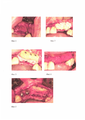
На Фиг. 1-5 представлены этапы выполнения операции.In FIG. 1-5 presents the stages of the operation.
Claims (1)
Priority Applications (1)
| Application Number | Priority Date | Filing Date | Title |
|---|---|---|---|
| RU2018144106A RU2698436C1 (en) | 2018-12-13 | 2018-12-13 | Method of bone plasty |
Applications Claiming Priority (1)
| Application Number | Priority Date | Filing Date | Title |
|---|---|---|---|
| RU2018144106A RU2698436C1 (en) | 2018-12-13 | 2018-12-13 | Method of bone plasty |
Publications (1)
| Publication Number | Publication Date |
|---|---|
| RU2698436C1 true RU2698436C1 (en) | 2019-08-26 |
Family
ID=67733644
Family Applications (1)
| Application Number | Title | Priority Date | Filing Date |
|---|---|---|---|
| RU2018144106A RU2698436C1 (en) | 2018-12-13 | 2018-12-13 | Method of bone plasty |
Country Status (1)
| Country | Link |
|---|---|
| RU (1) | RU2698436C1 (en) |
Cited By (1)
| Publication number | Priority date | Publication date | Assignee | Title |
|---|---|---|---|---|
| RU2741363C1 (en) * | 2020-03-23 | 2021-01-25 | Федеральное государственное бюджетное учреждение Национальный медицинский исследовательский центр "Центральный научно-исследовательский институт стоматологии и челюстно-лицевой хирургии" Министерства здравоохранения Российской Федерации | Method of bone autoplasty in atrophy and limited defects of maxillary alveolar process |
Citations (5)
| Publication number | Priority date | Publication date | Assignee | Title |
|---|---|---|---|---|
| US5397235A (en) * | 1993-07-02 | 1995-03-14 | Dental Marketing Specialists, Inc. | Method for installation of dental implant |
| RU93267U1 (en) * | 2010-02-08 | 2010-04-27 | Андрей Николаевич Жидков | BONE TISSUE RESTORE (OPTIONS) |
| RU2402289C1 (en) * | 2009-06-11 | 2010-10-27 | Государственное образовательное учреждение высшего профессионального образования "Санкт-Петербургский государственный медицинский университет имени академика И.П. Павлова Федерального агентства по здравоохранению и социальному развитию" | Repair technique for atrophied alveolar part of mandible |
| RU2592375C1 (en) * | 2015-07-14 | 2016-07-20 | Владимир Викторович Рыбалко | Method for osteoplasty with alloplant bone unit in jaw augmentation |
| RU2616337C1 (en) * | 2015-12-16 | 2017-04-14 | федеральное государственное бюджетное образовательное учреждение высшего образования "Самарский государственный медицинский университет" Министерства здравоохранения Российской Федерации (ФГБОУ ВО СамГМУ Минздрава России) | Method for plastic surgery on jaw alveolar process |
-
2018
- 2018-12-13 RU RU2018144106A patent/RU2698436C1/en active
Patent Citations (5)
| Publication number | Priority date | Publication date | Assignee | Title |
|---|---|---|---|---|
| US5397235A (en) * | 1993-07-02 | 1995-03-14 | Dental Marketing Specialists, Inc. | Method for installation of dental implant |
| RU2402289C1 (en) * | 2009-06-11 | 2010-10-27 | Государственное образовательное учреждение высшего профессионального образования "Санкт-Петербургский государственный медицинский университет имени академика И.П. Павлова Федерального агентства по здравоохранению и социальному развитию" | Repair technique for atrophied alveolar part of mandible |
| RU93267U1 (en) * | 2010-02-08 | 2010-04-27 | Андрей Николаевич Жидков | BONE TISSUE RESTORE (OPTIONS) |
| RU2592375C1 (en) * | 2015-07-14 | 2016-07-20 | Владимир Викторович Рыбалко | Method for osteoplasty with alloplant bone unit in jaw augmentation |
| RU2616337C1 (en) * | 2015-12-16 | 2017-04-14 | федеральное государственное бюджетное образовательное учреждение высшего образования "Самарский государственный медицинский университет" Министерства здравоохранения Российской Федерации (ФГБОУ ВО СамГМУ Минздрава России) | Method for plastic surgery on jaw alveolar process |
Cited By (1)
| Publication number | Priority date | Publication date | Assignee | Title |
|---|---|---|---|---|
| RU2741363C1 (en) * | 2020-03-23 | 2021-01-25 | Федеральное государственное бюджетное учреждение Национальный медицинский исследовательский центр "Центральный научно-исследовательский институт стоматологии и челюстно-лицевой хирургии" Министерства здравоохранения Российской Федерации | Method of bone autoplasty in atrophy and limited defects of maxillary alveolar process |
Similar Documents
| Publication | Publication Date | Title |
|---|---|---|
| Cordaro et al. | Ridge Augmentation Procedures in Implant Patients: A Staged Approach | |
| RU2611757C1 (en) | Method for maxillary sinus reconstruction in case of edentulism and maxillitis | |
| Kahnberg et al. | Sinus Lift Procedure Using a 2-Stage Surgical Technique: I. Clinical and Radiographic Report up to 5 Years. | |
| Sivolella et al. | Rehabilitation with implants after bone lid surgery in the posterior mandible | |
| RU2700543C1 (en) | Method for reconstructing an alveolar crest in the distal upper jaws for installing dental implants | |
| RU2364356C1 (en) | Mandibular alveoloplasty technique | |
| Scarano et al. | Expansion of the alveolar bone crest with ultrasonic surgery device: Clinical study in mandible | |
| RU2608702C1 (en) | Method of lifting maxillary sinus mucosa | |
| RU2698436C1 (en) | Method of bone plasty | |
| RU2535913C1 (en) | Method for osteoplasty accompanying atrophied alveolar bone tissue of jaws | |
| RU2416376C2 (en) | Method of dental implantation | |
| RU2398522C1 (en) | Method of surgical treatment of patients with chronic odontogenic maxillary sinusitis with oroantral fistula | |
| RU2375005C1 (en) | Method of plasty of perforation of maxillary sinus mucous tunic in case of sinus-lifting and implantation | |
| RU2648861C1 (en) | Method of directional jaw bone regeneration at atrophy of alveolary process | |
| RU2727863C1 (en) | Method of dental implantation in lateral sections of upper jaw | |
| RU2414181C2 (en) | Methods of plasty of alveolar process of lower jaw in case of its atrophy | |
| RU2806519C1 (en) | Method of increasing volume of jaw bone tissue | |
| Mitrea et al. | The Sinus Lift Procedure Applied in Cases Where the Thickness of the Alveolar Bone Is Insufficient Using Double Prf as Well as in the Case of an Intrasinus Mucocele | |
| Salame et al. | Evaluation of the Split-crest Technique with SimultaneousImplant Placement in Atrophic Edentulous Maxillary and Mandibular Bone: A 5-Year Follow-up Study | |
| RU2558998C1 (en) | Method for repairing atrophied alveolar part of lower jaw | |
| RU2467709C1 (en) | Method for surgical management of patients with oroantral fistula | |
| Kim et al. | Narrow Alveolar Ridge Management with Modified Ridge Splitting Technique: A Report of 3 Cases | |
| RU2326619C1 (en) | Method of surgical approach to mandibular canal | |
| RU2808511C1 (en) | Sinus lift method in presence of foreign body in area of bottom of maxillary sinus | |
| RU2613673C1 (en) | Method of alveolar bone plastics for children with congenital cleft lip and palate |