RU2648162C2 - Method for producing growth additive based on thrombocyte lysate from platelet of donors to medium for build-up of cellular weight of stem, progenitor, differentiated and tumor cells - Google Patents
Method for producing growth additive based on thrombocyte lysate from platelet of donors to medium for build-up of cellular weight of stem, progenitor, differentiated and tumor cells Download PDFInfo
- Publication number
- RU2648162C2 RU2648162C2 RU2016115622A RU2016115622A RU2648162C2 RU 2648162 C2 RU2648162 C2 RU 2648162C2 RU 2016115622 A RU2016115622 A RU 2016115622A RU 2016115622 A RU2016115622 A RU 2016115622A RU 2648162 C2 RU2648162 C2 RU 2648162C2
- Authority
- RU
- Russia
- Prior art keywords
- platelet
- samples
- donors
- sample
- growth
- Prior art date
Links
Images

Classifications
-
- C—CHEMISTRY; METALLURGY
- C12—BIOCHEMISTRY; BEER; SPIRITS; WINE; VINEGAR; MICROBIOLOGY; ENZYMOLOGY; MUTATION OR GENETIC ENGINEERING
- C12N—MICROORGANISMS OR ENZYMES; COMPOSITIONS THEREOF; PROPAGATING, PRESERVING, OR MAINTAINING MICROORGANISMS; MUTATION OR GENETIC ENGINEERING; CULTURE MEDIA
- C12N5/00—Undifferentiated human, animal or plant cells, e.g. cell lines; Tissues; Cultivation or maintenance thereof; Culture media therefor
- C12N5/06—Animal cells or tissues; Human cells or tissues
Abstract
Description
Изобретение относится к области биомедицины и направлено на создание «ростовой добавки», пригодной для культивирования клеток, в том числе на этапах получения клеточных продуктов и тканеинженерных конструкций, предназначенных для введения в организм человека.The invention relates to the field of biomedicine and is aimed at creating a “growth supplement” suitable for culturing cells, including at the stages of obtaining cellular products and tissue-engineering structures intended for introduction into the human body.
Культивирование и наращивание клеток человека in vitro является начальным, промежуточным этапом или самостоятельной процедурой во многих сферах, и в частности:The cultivation and growth of human cells in vitro is an initial, intermediate stage or an independent procedure in many areas, and in particular:
1. культивирование и наращивание разных типов клеток для экспериментальных разработок;1. cultivation and growth of different types of cells for experimental development;
2. культивирование и наращивание клеток гибридом с целью получения моноклональных антител;2. cultivation and growth of hybridoma cells in order to obtain monoclonal antibodies;
3. культивирование и наращивание клеток разных типов (дифференцированных, прогениторных, стволовых) для целей клеточной терапии;3. cultivation and growth of cells of different types (differentiated, progenitor, stem) for the purpose of cell therapy;
4. культивирование и наращивание клеток - как этап создания тканеинженерных конструкций для регенеративной медицины;4. cultivation and growth of cells - as a stage in the creation of tissue-engineering structures for regenerative medicine;
5. культивирование и наращивание клеток для биотехнологических целей (включая генетическую инженерию).5. cultivation and growth of cells for biotechnological purposes (including genetic engineering).
Культивирование клеток человека in vitro осуществляют в коммерческих питательных средах разных типов с добавками (supplements). Добавки делятся на 2 основных типа:Cultivation of human cells in vitro is carried out in commercial nutrient media of various types with additives (supplements). Additives are divided into 2 main types:
1) традиционно используемые как источники факторов роста, гормонов, витаминов -эмбриональная телячья сыворотка (далее - ЭТС);1) traditionally used as sources of growth factors, hormones, vitamins - fetal calf serum (hereinafter - ETS);
2) специальные добавки для решения конкретных задач.2) special additives for solving specific problems.
Одной из актуальных проблем регенеративной медицины является безопасность биомедицинских клеточных продуктов и тканеинженерных конструкций, использование которых способствовало бы восстановлению структуры и функции утраченных органов и тканей у пациентов без угрозы возникновения у них различных осложнений, в том числе иммунологических.One of the urgent problems of regenerative medicine is the safety of biomedical cell products and tissue-engineering structures, the use of which would help restore the structure and function of lost organs and tissues in patients without the threat of various complications, including immunological ones.
Для стандартизации и валидации процесса получения безопасных биомедицинских клеточных продуктов требуется разработка регламента их доклинических испытаний. К этому разделу относится и разработка технологий безопасного культивирования клеток, в частности, с использованием адекватных культуральных сред и добавок.To standardize and validate the process of obtaining safe biomedical cell products, the development of a procedure for their preclinical trials is required. This section also includes the development of technologies for safe cell culture, in particular, using adequate culture media and additives.
Традиционно в качестве ростовой добавки («supplement») для культивирования клеток используют ЭТС, которая считается комплексной смесью высоко- и низкомолекулярных биомолекул с физиологическим балансом стимулирующих и блокирующих факторов роста (ФР), с низким содержанием γ-глобулинов - ингибиторов роста и пролиферации клеток [Bieback K., 2013; Mojica-Henshaw М.P. et al., 2013]. Однако использование ЭТС как источника ксеногенных протеинов на этапах приготовления клеточных препаратов и тканеинженерных конструкций, предназначенных для введения человеку, в настоящее время не рекомендуется вследствие возможности аллергических реакций, а также переноса в организм человека вирусов, прионов и зоонозов [Flemming A. et al., 2011; Azouna N. et al., 2012; Vasanthan P. et al., 2014; Naaijkens B.A. et al., 2012; Chen B. et al., 2012; Griffiths S. et al., 2013; Warnke P.H. et al, 2013]. Так, в частности, показано, что при введении человеку ММСК, культивированных с ЭТС, вырабатываются антитела, опосредующие развитие реакции Артюса (Selvaggi et al, 1997). Возможная экспрессия главного комплекса гистосовместимости II типа (МНС II) в мультипотентных мезенхимальных стромальных клетках (ММСК) в присутствии ЭТС, по мнению ряда авторов, также ограничивает использование ЭТС в технологиях регенеративной медицины [Bieback K., 2013; Griffiths S. et al., 2013]. He исключено, что генетическая нестабильность ММСК ряда доноров in vitro тоже может быть индуцирована компонентами ЭТС [Crespo-Diaz R. et al., 2013].Traditionally, ETS, which is considered a complex mixture of high and low molecular weight biomolecules with a physiological balance of stimulating and blocking growth factors (RF), with a low content of γ-globulins, inhibitors of cell growth and proliferation, is used as a growth additive (“supplement”) for cell cultivation [ Bieback K., 2013; Mojica-Henshaw M.P. et al., 2013]. However, the use of ETS as a source of xenogenic proteins at the stages of preparing cellular preparations and tissue-engineering structures intended for administration to humans is currently not recommended due to the possibility of allergic reactions, as well as the transfer of viruses, prions and zoonoses into the human body [Flemming A. et al., 2011; Azouna N. et al., 2012; Vasanthan P. et al., 2014; Naaijkens B.A. et al., 2012; Chen B. et al., 2012; Griffiths S. et al., 2013; Warnke P.H. et al, 2013]. So, in particular, it was shown that when a MMSC cultured with ETS is administered to humans, antibodies are produced that mediate the development of the Arthus reaction (Selvaggi et al, 1997). The possible expression of the main histocompatibility complex of type II (MHC II) in multipotent mesenchymal stromal cells (MMSC) in the presence of ETS, according to some authors, also limits the use of ETS in regenerative medicine technologies [Bieback K., 2013; Griffiths S. et al., 2013]. It is possible that the genetic instability of MMSCs of a number of donors in vitro can also be induced by the components of ETS [Crespo-Diaz R. et al., 2013].
Кроме того, известно, что состав ЭТС значительно варьирует по содержанию активных компонентов от лота к лоту, что не дает возможности стандартизовать ее по наиболее значимым компонентам (Flemming A. et al., 2011; Vasanthan P. et al., 2014; Naaijkens B.A. et al., 2012). Это часто создает трудности при подборе ЭТС для определенных клеточных линий, а в ряде случаев - при сопоставлении результатов, полученных при использовании разных лотов ЭТС.In addition, it is known that the composition of ETS varies significantly in the content of active components from lot to lot, which makes it impossible to standardize it for the most significant components (Flemming A. et al., 2011; Vasanthan P. et al., 2014; Naaijkens BA et al., 2012). This often creates difficulties in selecting ETS for certain cell lines, and in some cases when comparing the results obtained using different lots of ETS.
На этапах разработки гуманизированных ростовых добавок к культуральным средам были апробированы экстракты различных тканей, биологические жидкости и бессывороточные среды с рекомбинантными ФР, однако удовлетворительные результаты получены не были [Seo J. et al., 2013; Vasanthan P. et al., 2014]. Аллогенные сыворотки в качестве замены ЭТС вызывали арест пролиферации и гибель ММСК (Shahdadfar et al., 2005). В аутологичных сыворотках ММСК пролиферировали адекватно, но делать ее дорого и непрактично (Kobayashi et al., 2005).At the stages of development of humanized growth additives for culture media, various tissue extracts, biological fluids and serum-free media with recombinant RF were tested, but satisfactory results were not obtained [Seo J. et al., 2013; Vasanthan P. et al., 2014]. Allogeneic sera as a substitute for ETS caused arrest of proliferation and death of MMSCs (Shahdadfar et al., 2005). In autologous sera, MMSCs proliferated adequately, but it is expensive and impractical to do it (Kobayashi et al., 2005).
В последние годы внимание исследователей обращено к тромбоцитам - естественным источникам многих ФР (PDGF, изоформы АА, АВ, ВВ; TGFα1, -β; HGF; IGF-1; VEGF; EGF; FGFb), в частности, к лизату тромбоцитов (ЛТ) человека для замены ЭТС [Шанский Я.Д. и соавт., 2013; Naaijkens В. et al., 2012; Flemming A. et al., 2011; Azouna N. et al., 2012; Chen B. et al., 2012; Vasanthan P. et al., 2014; Ruggiu A. et al., 2013; Griffiths S. et al., 2013; Warnke P.H. et al., 2013].In recent years, researchers have turned their attention to platelets, the natural sources of many RFs (PDGF, isoforms AA, AB, BB; TGFα1, -β; HGF; IGF-1; VEGF; EGF; FGFb), in particular, platelet lysate (LT) human to replace ETS [Shansky Y.D. et al., 2013; Naaijkens B. et al., 2012; Flemming A. et al., 2011; Azouna N. et al., 2012; Chen B. et al., 2012; Vasanthan P. et al., 2014; Ruggiu A. et al., 2013; Griffiths S. et al., 2013; Warnke P.H. et al., 2013].
Основными недостатками ЭТС, как ростовой добавки для культивирования клеток, предназначенных для введения человеку, являются:The main disadvantages of ETS, as a growth supplement for the cultivation of cells intended for administration to humans, are:
- ЭТС - ксеногенный (в отношении человека) материал,- ETS - xenogenic (in relation to humans) material,
- возможность переноса с ЭТС инфекционных агентов, включая зоонозы, прионы и вирусы (в т.ч. нераспознанные),- the ability to transfer infectious agents from ETS, including zoonoses, prions and viruses (including unrecognized ones),
- возможность переноса с ЭТС свободной ксеногенной ДНК,- the possibility of transferring free xenogenous DNA with ETS,
- возможность переноса с ЭТС нежелательных метаболитов,- the possibility of transferring unwanted metabolites from ETS
- возможность иммунологического ответа на ЭТС, в т.ч. аллергических реакций на ксеногенные компоненты,- the possibility of an immunological response to ECF, including allergic reactions to xenogenic components,
- изменение экспрессии ряда маркеров в клетках человека, культивируемых в присутствии ЭТС,- a change in the expression of a number of markers in human cells cultured in the presence of ETS,
- нестандартизированность ЭТС по функциональной активности,- non-standardization of ETS in functional activity,
- во многих странах, включая Россию, запрещено введение в организм (минуя желудочно- кишечный тракт) человека биологических клеточных продуктов, которые в процессе приготовления имели контакт с ксеногенными биологическими материалами.- in many countries, including Russia, it is forbidden to introduce biological cellular products into the body (bypassing the gastrointestinal tract) of a person, which during the preparation had contact with xenogenic biological materials.
Известен способ получения ЛТ из богатой тромбоцитами плазмы крови взрослых животных (WO 2006/137778 А1), в котором лизис тромбоцитов осуществляют добавлением к тромбоцитам в плазме растворимых солей кальция, либо деионизированной воды. Далее смесь центрифугируют и фильтруют.A known method of producing LT from platelet-rich blood plasma of adult animals (WO 2006/137778 A1), in which platelet lysis is carried out by adding to the platelet in the plasma of soluble calcium salts, or deionized water. The mixture is then centrifuged and filtered.
Однако данный способ получения ЛТ, обогащенный солями кальция в использованных концентрациях, может быть токсичным для ряда клеточных культур. Кальций в среде культивирования может изменять функционирование клеток, как один из ключевых ионов-регуляторов жизнедеятельности.However, this method of producing RT enriched with calcium salts at the concentrations used can be toxic to a number of cell cultures. Calcium in the cultivation medium can change the functioning of cells, as one of the key ion-regulators of life.
Отличительные признаки (от предложенного нами способа) являются:Distinctive features (from our proposed method) are:
- используют периферическую кровь животных;- use the peripheral blood of animals;
- способы дегрануляции тромбоцитов (для выхода в плазму ФР) - соли кальция или деионизированная вода.- methods of platelet degranulation (for release into the plasma of FR) - calcium salts or deionized water.
Известен способ приготовления ЛТ и культуральной среды с ЛТ (ЕР 0413727 А1) из цельной крови животных для выращивания гибридом с целью получения антител. Отличия от нашего изобретения:A known method of preparing RT and culture medium with RT (EP 0413727 A1) from whole blood of animals for growing hybridomas in order to obtain antibodies. Differences from our invention:
- используют периферическую кровь животных с бойни;- use the peripheral blood of animals from the slaughter;
- получаемый ЛТ - ксеногенный, исключена лишь внутрисердечная пункция эмбриона коровы;- the resulting RT is xenogenic; only intracardiac puncture of the cow embryo is excluded;
- способ разрушения и дегрануляции тромбоцитов - добавление Са+2.- a method of destruction and degranulation of platelets - the addition of Ca +2 .
Как и в предыдущем известном способе, полученный ЛТ, обогащенный солями кальция в использованных концентрациях, может быть токсичным для ряда клеточных культур. Кальций в среде культивирования может изменять функционирование клеток, как один из ключевых ионов-регуляторов жизнедеятельности.As in the previous known method, the obtained RT enriched with calcium salts in the used concentrations can be toxic to a number of cell cultures. Calcium in the cultivation medium can change the functioning of cells, as one of the key ion-regulators of life.
Известен способ получения ростовой добавки из обогащенной тромбоцитами плазмы крови животных (US 5198357 А) для культивирования гибридомных клеток. ЛТ получают из цельной крови животных с раствором цитрата натрия для предотвращения свертывания. Способ лизиса - добавление кальция, что одновременно приводит к коагуляции фибриногена. Сепарацию производят путем центрифугирования, последующую стерилизацию - путем фильтрования. Используют как более дешевую альтернативу ЭТС.A known method of obtaining growth supplements from platelet-rich blood plasma of animals (US 5198357 A) for the cultivation of hybridoma cells. RT is obtained from whole blood of animals with sodium citrate solution to prevent clotting. The method of lysis is the addition of calcium, which simultaneously leads to the coagulation of fibrinogen. Separation is carried out by centrifugation, subsequent sterilization by filtration. Use as a cheaper alternative to ETS.
Известен способ выделения ЛТ из ТМ, полученный путем афереза у доноров (CN 104673747 А). Общие с заявляемым способом черты получения ЛТ:A known method for the isolation of LT from TM obtained by apheresis in donors (CN 104673747 A). General features of the claimed method of obtaining LT:
- для разрушения тромбоцитов используют несколько циклов замораживания (-80°C) - оттаивания (+37°C) ТМ;- for the destruction of platelets using several cycles of freezing (-80 ° C) - thawing (+ 37 ° C) TM;
- авторы доказывают, что несколько циклов замораживания/оттаивания дают лучший выход ФР;- the authors prove that several cycles of freezing / thawing give the best yield of FR;
- используют центрифугирование для освобождения от фрагментов тромбоцитов;- use centrifugation to release platelet fragments;
- индивидуальные ЛТ, полученные описанным способом, поддерживают рост клеток в сходной с ЭТС концентрацией (10%).- individual RT obtained by the described method, support cell growth in a concentration similar to ETS (10%).
Известен способ получения ростовой добавки из обогащенной тромбоцитами плазмы крови (US 2012276632 Al), включающий температурный лизис тромбоцитов (замораживание/оттаивание).A known method of obtaining growth supplements from platelet-rich blood plasma (US 2012276632 Al), including temperature lysis of platelets (freezing / thawing).
В отличие от предлагаемого нами метода, авторы перед лизисом тромбоцитов в ТМ освобождаются от плазмы крови центрифугированием, разводя тромбоциты в среде, содержащей альбумин человеческой сыворотки, декстран и гидроксиэтилкрахмал, для восстановления осмолярности.In contrast to our method, the authors before platelet lysis in TM are released from blood plasma by centrifugation, diluting platelets in a medium containing human serum albumin, dextran and hydroxyethyl starch to restore osmolarity.
Известен способ изготовления и применения ЛТ (CN10467 3747-А-2015-06-03) из тромбомассы, полученной путем афереза. Лизис осуществляют путем однократного замораживания (-80°С) - оттаивания (+37°С) тромбомассы с последующей обработкой ультразвуком (100-400 вт, 3-6 секунд), повторяя последнюю процедуру от 5 до 15 раз. Далее ЛТ центрифугируют (для осаждения дебриса), освобождают от фибрина рекальцинацией (глюконатом кальция или хлористым кальцием) и используют ЛТ как ростовую добавку для культивирования ММСК. В полученном ЛТ определяют содержание VEGF, PDGF, заявляя их как наиболее значимые характеристики ЛТ для его функциональной активности.A known method of manufacture and use of RT (CN10467 3747-A-2015-06-03) from thrombomass obtained by apheresis. Lysis is carried out by a single freezing (-80 ° C) - thawing (+ 37 ° C) of thrombomass followed by ultrasound treatment (100-400 W, 3-6 seconds), repeating the last procedure from 5 to 15 times. Then, RTs are centrifuged (to precipitate debris), freed from fibrin by recalcification (calcium gluconate or calcium chloride), and RTs are used as a growth supplement for the cultivation of MMSCs. In the obtained RT, the content of VEGF, PDGF is determined, declaring them as the most significant characteristics of RT for its functional activity.
Самым близким к заявляемому нами способу является способ приготовления композиции, содержащий лизат тромбоцитов (WO 2010/033605 А2 ((Compositions, containing platelet contents»). Приготовление ЛТ включает в себя не менее 2-х циклов замораживания (-80°С) - оттаивания (+37°С), последующее центрифугирование (2000-4000g, оптимально - 3000g) в течение 30 минут. Для стерилизации ЛТ осуществляют ультрафильтрацию (размер пор - 0,45 мкм и 0,2 мкм). Полученный ЛТ в концентрации 5-10% от объема культуральной среды используют в качестве ростовой добавки к средам для культивирования ММСК из жировой ткани (ЖТ), или фибробластов человека (ФЧ), или культур опухолевых клеток (глиомы), что обеспечивает длительную (более 30 дней) пролиферацию этих культур in vitro с динамикой нарастания клеточной популяции, сходной с таковой для 5-10% ЭТС.The closest to our claimed method is a method of preparing a composition containing platelet lysate (WO 2010/033605 A2 ((Compositions, containing platelet contents "). Preparation of RT includes at least 2 cycles of freezing (-80 ° C) - thawing (+ 37 ° C), followed by centrifugation (2000-4000g, optimally 3000g) for 30 minutes. Ultrafiltration is carried out for sterilization of RT (pore size 0.45 μm and 0.2 μm). The obtained LT at a concentration of 5-10 % of the volume of the culture medium is used as a growth additive to the media for the cultivation of MMSC from fat Ani (VT), or human fibroblast (FCH), or cultures of tumor cells (glioma), which provides a long (more than 30 days), the proliferation of these cultures in vitro with the dynamics of growth of the cell population, similar to that for 5-10% FBS.
Однако в предложенном способе оценку качества приготовленного ЛТ проводят по содержанию фактора роста сосудов VEGF - не менее 200 пг/мл. В предлагаемом нами способе стандартизацию проводят по функциональным свойствам.However, in the proposed method, the quality of the prepared RT is assessed by the content of vascular growth factor VEGF - not less than 200 pg / ml. In our proposed method, standardization is carried out by functional properties.
Техническим результатом заявляемого изобретения является получение стандартизованных образцов нексеногенной ростовой добавки для культивирования клеток человека разных типов с функциональной активностью образцов, определяемой по скорости прироста тест-культур, различающейся не более чем на 10,0%.The technical result of the claimed invention is to obtain standardized samples of non-xenogenic growth supplements for the cultivation of human cells of different types with functional activity of the samples, determined by the growth rate of test cultures, differing by no more than 10.0%.
Задачей изобретения является получение добавки к ростовой среде лизата тромбоцитов, полученного из тромбомассы для наращивания клеточной массы.The objective of the invention is to obtain an additive to the growth medium of a platelet lysate obtained from thrombomass to increase cell mass.
Указанный технический результат при осуществлении изобретения достигается за счет того, что так же, как в известном способе проводят нормирование образца ТМ до заданной концентрации тромбоцитов (1,75×10⁹ тромбоцитов/мл) путем центрифугирования ТМ при 3130g в течение 40 минут при 20°С и ресуспендировании осадка в супернатанте заданного объема до концентрации тромбоцитов 1,75×10⁹ тромбоцитов/мл, 3-кратный температурный лизис тромбоцитов путем замораживанияThe specified technical result in the implementation of the invention is achieved due to the fact that, as in the known method, the TM sample is normalized to a given platelet concentration (1.75 × 10 9 platelets / ml) by centrifugation of the TM at 3130g for 40 minutes at 20 ° C and resuspension of the pellet in the supernatant of a given volume to a platelet concentration of 1.75 × 10 9 platelets / ml, 3-fold temperature platelet lysis by freezing
на сутки до -80°С и размораживания при +37°С, осаждение клеточного дебриса путем центрифугирования при 3130 g, 40 минут, 20°С, сбор супернатанта, микроскопический контроль при увеличении ×200 на наличие в поле зрения не более 3-х фрагментов тромбоцитов.per day to -80 ° C and thawing at + 37 ° C, sedimentation of cell debris by centrifugation at 3130 g, 40 minutes, 20 ° C, collection of supernatant, microscopic control at a magnification of × 200 for the presence in the field of view of no more than 3 platelet fragments.
Особенность заявляемого способа заключается в том, что стандартизацию лизата тромбоцитов проводят путем пулирования, объединяя равные по объему образцы, полученные из тромбоцитарной массы не менее 12 доноров обеих тендерных групп, пулированный ЛТ центрифугируют при 3130g, 40 минут при 20°С, далее осуществляют ультрафильтацию пулированного образца на фильтрах 0,45 μкм или 0,22 μкм, после чего разливают по аликвотам требуемого объема и замораживают до -80°С до использования.A feature of the proposed method is that the standardization of platelet lysate is carried out by pooling, combining equal in volume samples obtained from a platelet mass of at least 12 donors of both tender groups, the pulled RT is centrifuged at 3130g, 40 minutes at 20 ° C, then the ultrafiltration of the pulled the sample on the filters is 0.45 μkm or 0.22 μkm, after which it is poured into aliquots of the required volume and frozen to -80 ° C until use.
Изобретение поясняется подробным описанием, таблицами и иллюстрациями, на которых изображено:The invention is illustrated by a detailed description, tables and illustrations, which depict:
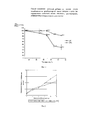
Фиг. 1 - Относительное изменение оптической плотности (OD) раствора формазана культуры фибробластов человека (5-е сутки культивирования) при использовании образцов ЛТ и ЭТС, инактивированных в течение 1 часа при различных температурах (в %, относительно 37°С).FIG. 1 - Relative change in optical density (OD) of a solution of formazan culture of human fibroblasts (5th day of cultivation) when using RT and ETS samples inactivated for 1 hour at various temperatures (in%, relative to 37 ° C).
Фиг. 2 - Изменение оптической плотности раствора формазана (МТТ-тест) при культивировании ФЧ в ПРС, содержащей 10% пулированного образца ЛТ или 10% ЭТС.FIG. 2 - Change in the optical density of a solution of formazan (MTT test) during the cultivation of PS in ORS containing 10% of a pulled RT sample or 10% ETS.
Фиг. 3 - Изменение оптической плотности раствора формазана (МТТ-тест) при культивировании ММСК в ПРС, содержащей 10% пулированного образца ЛТ или 10% ЭТС.FIG. 3 - Change in the optical density of a solution of formazan (MTT test) during the cultivation of MMSCs in ORS containing 10% of a pulled RT sample or 10% ETS.
Способ осуществляют следующим образом.The method is as follows.
Технологическая схема получения ЛТTechnological scheme for obtaining LT
1. Материал для получения ЛТ - образцы ТМ доноров, полученный путем аппаратного афереза.1. Material for RT preparation - samples of TM donors obtained by hardware apheresis.
Процедура заготовки ТМ валидирована и предполагает контаминацию лейкоцитами в количестве не более 1×10⁶ на дозу (~2×10¹¹ тромбоцитов), что контролируется в соответствии с техническим регламентом «О требованиях безопасности крови, ее продуктов, кровезамещающих растворов и технических средств, используемых в трансфузионно-инфузионной терапии, а также с применением стандарта «Донорская кровь и ее компоненты: характеристики и контроль качества. IX. Тромбоциты, полученные методом афереза» (Общероссийская общественная организация «Российская ассоциация трансфузиологов»)».The TM procurement procedure is validated and involves leukocyte contamination in an amount of not more than 1 × 10 6 per dose (~ 2 × 10 11 platelets), which is controlled in accordance with the technical regulation “On the safety requirements of blood, its products, blood substitute solutions and technical means used in transfusion and infusion therapy, as well as using the standard “Donor blood and its components: characteristics and quality control. IX. Platelets obtained by apheresis method "(All-Russian public organization" Russian Association of Transfusiologists ")."
2. Полученный образец ТМ доноров в стерильных условиях переносят в пробирку, тщательно перемешивают и отбирают аликвоту объемом 0,1 мл для определения концентрации тромбоцитов. Перед исследованием аликвоту разводят в 10 раз цитратным буфером ACD-A. Концентрацию тромбоцитов определяют с помощью автоматического гематологического анализатора (например, Sysmex КХ20, Япония).2. The obtained sample of TM donors under sterile conditions is transferred into a test tube, thoroughly mixed and an aliquot of 0.1 ml is taken to determine the platelet concentration. Prior to testing, an aliquot was diluted 10 times with ACD-A citrate buffer. Platelet concentration is determined using an automatic hematological analyzer (for example, Sysmex KX20, Japan).
3. Производят нормирование образца ТМ до заданной концентрации тромбоцитов (1,75×10⁹ тромбоцитов/мл). Для этого ТМ центрифугируют в пробирке объемом 15 мл (например, Corning, США) при 3000 g в течение 40 мин при 20°C (например, центрифуга Eppendorf 5810R, Германия). Осадок тромбоцитов ресуспендируют в супернатанте рассчитанного объема, обеспечивающего конечную концентрацию тромбоцитов (1,75×10⁹ тромбоцитов/мл).3. Normalize the TM sample to a given platelet concentration (1.75 × 10 9 platelets / ml). For this, TMs are centrifuged in a 15 ml tube (e.g., Corning, USA) at 3000 g for 40 min at 20 ° C (e.g., Eppendorf 5810R centrifuge, Germany). Platelet sediment is resuspended in the supernatant of the calculated volume, providing a final platelet concentration (1.75 × 10 9 platelets / ml).
4. Температурный лизис образца ТМ.4. Temperature lysis of the TM sample.
Для получения ЛТ - пробирку с образцом ТМ, нормированным по концентрации тромбоцитов, подвергают процедуре температурного лизиса: быстро замораживают при -80°C (низкотемпературный холодильник, например, Sanyo MDF-U500VX) и через 24 ч размораживают при +37°C (20-40 мин; термостат, например ТС-80М-2) до достижения указанной температуры. Процедуру повторяют трехкратно.To obtain an LT, a test tube with a TM sample normalized by platelet concentration is subjected to a temperature lysis procedure: it is quickly frozen at -80 ° C (a low-temperature refrigerator, for example, Sanyo MDF-U500VX) and after 24 hours it is thawed at + 37 ° C (20- 40 min; thermostat, for example ТС-80М-2) until the indicated temperature is reached. The procedure is repeated three times.
5. Центрифугирование образца ЛТ.5. Centrifugation of the LT sample.
Для осаждения фрагментов тромбоцитов (клеточного дебриса) пробирку с образцом ЛТ центрифугируют при 3000g в течение 40 мин при 20°C с минимальной скоростью торможения (например, центрифуга Eppendorf 5810R, Германия). После завершения этапа центрифугирования собирают супернатант, представляющий собой ЛТ.To precipitate platelet fragments (cell debris), the tube with an RT sample is centrifuged at 3000 g for 40 min at 20 ° C with a minimum inhibition rate (for example, an Eppendorf 5810R centrifuge, Germany). After completion of the centrifugation step, the supernatant representing LT is collected.
Представляем обоснование использования 3-х циклов замораживания и оттаивания и указанного режима центрифугирования.We present the rationale for using 3 cycles of freezing and thawing and the specified centrifugation mode.
Количество циклов замораживания/оттаивания в литературе варьирует, но очевидно, что оно должно быть минимизировано для сохранения структуры значимых компонентов. Поэтому с помощью световой микроскопии оценивали отсутствие неразрушенных тромбоцитов в образцах лизированной ТМ после 1, 2 и 3 циклов размораживания/оттаивания. Для отделения раствора, обогащенного ФР, от фрагментов тромбоцитов, были апробированы 6 режимов центрифугирования. В этих режимах варьировали такие показатели, как время центрифугирования (20, 30, 40 мин), температура (10, 20°C) и кратность центрифугирования (×1, ×2 и ×3) (табл. 1).The number of freeze / thaw cycles in the literature varies, but it is obvious that it should be minimized to preserve the structure of significant components. Therefore, using light microscopy, the absence of undamaged platelets in lysed TM samples after 1, 2, and 3 thawing / thawing cycles was evaluated. To separate the solution enriched with RF from platelet fragments, 6 centrifugation modes were tested. In these modes, indicators such as centrifugation time (20, 30, 40 min), temperature (10, 20 ° C), and centrifugation ratio (× 1, × 2, and × 3) varied (Table 1).

Выбор количества циклов замораживания/оттаивания для получения образцов ЛТ осуществляли в соответствии с визуальным и микроскопическим контролем супернатанта после окончания процедуры (прозрачность, наличие неразрушенных тромбоцитов и/или их фрагментов). Результаты наблюдений показали, что после 3 циклов замораживания/оттаивания наблюдается наименьшее количество неразрушенных тромбоцитов (табл. 2).The choice of the number of freezing / thawing cycles for obtaining RT samples was carried out in accordance with the visual and microscopic control of the supernatant after the end of the procedure (transparency, the presence of intact platelets and / or fragments thereof). The results of observations showed that after 3 cycles of freezing / thawing, the smallest number of undamaged platelets is observed (Table 2).

По результатам содержания тромбоцитов в супернатанте для оценки содержания ФР в ЛТ были отобраны следующие режимы центрифугирования:Based on the results of platelet count in the supernatant, the following centrifugation modes were selected to assess the RF content in RT:
- Режим №1: 3000 об/мин (1400g), 30 мин, 20°C, 3-кратно;- Mode No. 1: 3000 rpm (1400g), 30 min, 20 ° C, 3 times;
- Режим №2: 3000 об/мин (1600g), 20 мин, 20°C, 3-кратно;- Mode No. 2: 3000 rpm (1600g), 20 min, 20 ° C, 3 times;
- Режим №3: 4000 об/мин (3130g), 40 мин, 20°C, однократно.- Mode No. 3: 4000 rpm (3130g), 40 min, 20 ° C, once.
Результаты определения концентрации ФР в образцах ЛТ, полученных при данных режимах, приведены в Таблице 3.The results of determining the concentration of RF in RT samples obtained under these conditions are shown in Table 3.

Из приведенных в табл. 3 данных можно видеть, что при использовании всех трех режимов центрифугирования в образцах ЛТ определяются все исследованные нами ФР. Однако при использовании режимов центрифугирования №2 и №3 наблюдается статистически достоверно более высокий выход факторов PDGF АВ, TGF-β1, VEGF, IGF-1, чем при использовании режима №1. Также значимые отличия наблюдаются для режимов №1 vs №3 в отношении содержания PDGF АА в супернатанте. Выход фактора PDGF АА не различался значимо при режимах №1 vs №2, №2 vs №3 (р<0,05). Содержание фактора PDGF ВВ в супернатанте не различалось значимо для всех трех режимов центрифугирования. Выход PDGF АВ, TGF-β, VEGF и IGF-1 не различался значимо при режимах центрифугирования №2 и №3 и был достоверно выше, чем при использовании режима №1.From the above table. From the data in Fig. 3, it can be seen that when using all three centrifugation modes in RT samples, all the RFs studied by us are determined. However, when using centrifugation modes No. 2 and No. 3, a statistically significantly higher yield of factors PDGF AB, TGF-β1, VEGF, IGF-1 is observed than when using mode No. 1. Significant differences are also observed for regimes No. 1 vs No. 3 in relation to the content of PDGF AA in the supernatant. The output of the PDGF AA factor did not differ significantly under modes No. 1 vs No. 2, No. 2 vs No. 3 (p <0.05). The content of the PDGF BB factor in the supernatant did not differ significantly for all three centrifugation modes. The output of PDGF AB, TGF-β, VEGF and IGF-1 did not differ significantly under centrifugation modes No. 2 and No. 3 and was significantly higher than when using mode No. 1.
Таким образом, режимы центрифугирования №2 и №3 обеспечивают наиболее высокое и сходное содержание исследованных нами ФР в супернатанте после лизиса тромбоцитов и осаждения их фрагментов. В то же время, режим №3 требует меньших затрат времени на проведение операции центрифугирования (1 цикл центрифугирования в течение 40 мин vs 3 циклов центрифугирования в течение 20 мин).Thus, centrifugation modes No. 2 and No. 3 provide the highest and most similar content of the studied RF in the supernatant after platelet lysis and sedimentation of their fragments. At the same time, mode No. 3 requires less time for a centrifugation operation (1 centrifugation cycle for 40 minutes vs 3 centrifugation cycles for 20 minutes).
6. Микроскопический контроль образца ЛТ6. Microscopic control of an LT sample
Пробу образца ЛТ оценивают под световым микроскопом при увеличении ×200 на наличие/отсутствие видимых нелизированных тромбоцитов и/или их фрагментов. Очистку считают адекватной при наличии в поле зрения не более 3 фрагментов тромбоцитов.A sample of the LT sample is evaluated under a light microscope at a magnification of × 200 for the presence / absence of visible non-lysed platelets and / or fragments thereof. Cleaning is considered adequate if there are no more than 3 platelet fragments in the field of view.
7. Пулирование образцов ЛТ7. Pooling of LT samples
Обоснование принципаJustification of the principle
Установлено, что образцы ЛТ и ЭТС существенно различаются по содержанию ряда биохимических и гормональных показателей и, главное, по содержанию основных ФР, которые считались ранее ответственными за активирующую способность ЭТС как ростовой добавки. Так, содержание ФР (TGF-β, VEGF, PDGF АА, PDGF АВ, IGF-1, FGFb) в образцах ЛТ было в десятки, а для отдельных ФР в сотни раз выше, чем в образцах ЭТС (табл. 3). Пулы образцов ЛТ от мужчин и женщин обладали сходной способностью активировать пролиферацию клеток, несмотря на существенные различия в содержании женских и мужских половых гормонов. Кроме того, нами установлены различия в термочувствительности образцов ЭТС и ЛТ (Фиг. 1). Это позволило сделать заключение, что за функциональные свойства ЛТ и ЭТС как ростовых добавок ответственны различные коктейли факторов.It was found that LT and ETS samples differ significantly in the content of a number of biochemical and hormonal indicators and, most importantly, in the content of the main RFs, which were previously considered responsible for the activating ability of ETS as a growth supplement. Thus, the content of FR (TGF-β, VEGF, PDGF AA, PDGF AB, IGF-1, FGFb) in the RT samples was tens, and for individual RFs, hundreds of times higher than in the ETS samples (Table 3). Pools of RT samples from men and women had a similar ability to activate cell proliferation, despite significant differences in the content of female and male sex hormones. In addition, we established differences in the thermal sensitivity of the ETS and LT samples (Fig. 1). This allowed us to conclude that various cocktails of factors are responsible for the functional properties of LT and ETS as growth additives.
Поэтому в основу принципа пулирования мы положили функциональные свойства ЛТ как ростовой добавки - его способность обеспечивать пролиферацию иммортализованных фибробластов кожи человека (ФЧ, штамм 1608h TERT, ФГБУН «Институт молекулярной биологии В.А. Энгельгардта» РАН), задав условие, что по этому признаку пулы ЛТ не должны отличаться более чем нам 10%.Therefore, we based the principle of pooling on the functional properties of RT as a growth supplement — its ability to proliferate immortalized fibroblasts of human skin (PS, strain 1608h TERT, Institute of Molecular Biology V.A. Engelhardt RAS), setting the condition that this attribute LT pools should not differ by more than 10%.
Выбранный нами принцип пулирования образцов ЛТ был основан, таким образом, на оценке пролиферативной активности модельной культуры клеток (ФЧ) в присутствии ЛТ (как ростовой добавки) от индивидуальных доноров. Оценочной величиной служила скорость роста культуры клеток ФЧ in vitro в экспоненциальной фазе роста, наблюдавшейся на начальном этапе культивирования.Our principle of pooling RT samples was thus based on an assessment of the proliferative activity of a model cell culture (PS) in the presence of RT (as a growth supplement) from individual donors. The estimated value was the in vitro growth rate of the PS cell culture in the exponential growth phase observed at the initial stage of cultivation.
В исследовании влияния ЛТ (как ростовой добавки) на пролиферацию ФЧ in vitro средой культивирования служила DMEM с добавлением глютамина 0,58 мг/мл, гентамицина 1,25 мкг/мл, гепарина 2 МЕ/мл. Как ростовую добавку использовали образцы лизатов тромбоцитов от индивидуальных доноров (15 мужчин и 13 женщин) в концентрации 5%, а также образец 5% пулированного ЛТ (от указанных 28 доноров). Культивирование было проведено в 96-луночных планшетах Costar (США) в течение 1-7 суток при плотности посева клеток 1,5×10⁴ клеток/см2. Жизнеспособность клеток оценивали с помощью МТТ-теста (количество повторов каждой пробы - 5).In a study of the effect of RT (as a growth supplement) on in vitro proliferation of FCs, DMEM with the addition of glutamine 0.58 mg / ml, gentamicin 1.25 μg / ml,
По результатам измерения оптической плотности (OD) раствора формазана в МТТ-тесте вычислены скорости роста популяции ФЧ в экспоненциальной фазе в присутствии индивидуальных образцов лизатов по формулеAccording to the results of measuring the optical density (OD) of the formazan solution in the MTT test, the growth rates of the population of the PS in the exponential phase in the presence of individual lysate samples were calculated by the formula

где А - скорость роста популяции клеток (сут-1), Δt - интервал времени культивирования в экспоненциальной фазе роста (4 суток), OD - оптическая плотность раствора формазана на 3-й и 7-е сутки культивирования.where A is the cell population growth rate (day -1 ), Δt is the cultivation time interval in the exponential growth phase (4 days), OD is the optical density of the formazan solution on the 3rd and 7th day of cultivation.
В таблице 4 приведены рассчитанные по получаемым OD значения «А» для индивидуальных доноров, средние значения генеральной совокупности (Aср. ген.) и генеральные стандартные отклонения (σген).Table 4 shows the “A” values calculated for the ODs obtained for individual donors, average values of the general population (A cf. gen. ), And general standard deviations (σ gene ).
Данные таблицы 4 свидетельствуют о том, что скорость прироста клеток в присутствии индивидуальных образцов ЛТ в отдельных случаях отличалась примерно в 2 раза: минимальное соотношение OD на 7-е сутки к таковой на 3-й составило 2,77, а максимальное - 4,91. Соответствующие индивидуальные скорости находились в диапазоне 0,25-0,4.The data in table 4 indicate that the cell growth rate in the presence of individual RT samples in some cases differed by about 2 times: the minimum ratio of OD on
Средний прирост культуры ФЧ за 4 суток в присутствии пулированного 5% ЛТ (от 28 доноров) практически совпал с рассчитанными средними для индивидуальных образцов ЛТ мужчин и женщин - 3,90±0,04 vs 4,00±0,58 vs 3,92±0,60. Очевидно, что пулирование с целью стандартизации можно проводить, используя меньшее количество индивидуальных образцов, что математически обосновано ниже.The average increase in the culture of PS for 4 days in the presence of a pulled 5% RT (from 28 donors) almost coincided with the calculated average for individual RT samples of men and women - 3.90 ± 0.04 vs 4.00 ± 0.58 vs 3.92 ± 0.60. Obviously, pooling for the purpose of standardization can be carried out using a smaller number of individual samples, which is mathematically justified below.

Далее рассчитывали количество образцов ЛТ для пулирования, исходя из следующих положений.Next, the number of RT samples for pooling was calculated based on the following provisions.
Если имеются 2 пула, состоящие из n образцов каждый и выбранные из одной нормально распределенной генеральной совокупности, то стандартное отклонение σразн разностиIf there are two pools consisting of each n samples and the selected one of the normal distribution of the total population, the standard deviation σ of the difference diff

может быть вычислено по формулеcan be calculated by the formula

Для того чтобы эта разность не превосходила по абсолютной величине с вероятностью 0,95 значение, равное 10% от Аср.ген, требуется выполнение условияIn order for this difference not to exceed in absolute value with a probability of 0.95 a value equal to 10% of A cf.gen , the condition
1,96×σразн≤0,1×Aср,ген 1.96 × σ diff ≤0.1 × A cf, gene
илиor
![]()
![]()
Отсюда количество образцов в пуле, соответствующее выполнению условия, вычисляется по формулеHence, the number of samples in the pool corresponding to the fulfillment of the condition is calculated by the formula

В нашем случае:In our case:
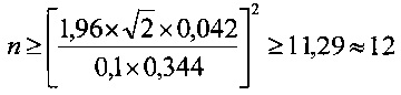
Таким образом, для того чтобы скорости прироста популяции ФЧ in vitro в присутствии образца пулированного ЛТ отличались от средней (по группе индивидуальных образцов ЛТ) скорости прироста не более чем на 10,0%, требуется объединять не менее 12 индивидуальных образцов ЛТ. Так как скорости прироста ФЧ в присутствии образцов ЛТ мужчин значимо не отличаются от таковых в присутствии образцов ЛТ женщин, для пулирования можно брать образцы обеих тендерных групп.Thus, in order for the growth rates of the in vitro PS population in the presence of a sample of pulmonary RT to differ from the average (in the group of individual RT samples) growth rate by no more than 10.0%, it is necessary to combine at least 12 individual RT samples. Since the growth rate of PS in the presence of male RT samples does not significantly differ from those in the presence of female RT samples, samples of both tender groups can be taken for pooling.
Учитывая вышесказанное, при пулировании ЛТ объединяют равные (по объему) образцы, полученные из ТМ 12 доноров. Повторяют центрифугирование по n5.Considering the above, when pooling RTs, equal (in volume) samples obtained from TM of 12 donors are combined. Repeat centrifugation at n5.
7. Микрофильтрация и аликвотирование образцов ЛТ7. Microfiltration and aliquotation of RT samples
ЛТ подвергают ультрафильтрации (фильтры 0,45 мкм и 0,22 мкм) и разливают в стерильные пластиковые криопробирки (например, Corning, США) по аликвотам от 100 до 500 мкл, маркируют с указанием номера образца, даты изготовления и объема образца ЛТ и замораживают до использования (-80°C).RTs are ultrafiltered (0.45 µm and 0.22 µm filters) and poured into sterile plastic cryovials (for example, Corning, USA) in aliquots from 100 to 500 µl, labeled with specimen number, date of manufacture and sample volume of RT and frozen before use (-80 ° C).
Пример конкретного исполнения №1An example of a specific performance No. 1
1. Заготовка образцов тромбоцитарной массы доноров1. Preparation of platelet donor mass samples
В стерильных условиях методом аппаратного тромбоцитафереза получено 12 индивидуальных образцов тромбоцитарной массы (ТМ, 6 мужчин, 6 женщин). Перед процедурой заготовки ТМ доноры проходили обследование на наличие ВИЧ I и II типа, HBs-антигена, вируса HCV, возбудителя сифилиса. Также был осуществлен клинический анализ крови.Under sterile conditions, 12 individual platelet mass samples (TM, 6 men, 6 women) were obtained by hardware-based thrombocytapheresis. Before the TM harvesting process, donors were screened for the presence of HIV type I and II, HBs antigen, HCV virus, and the causative agent of syphilis. A clinical blood test was also performed.
Через 24 часа после процедуры тромбоцитафереза образцы ТМ доноров в специальных пластиковых мешках помещали в транспортировочный контейнер и переносили в лабораторию с прилагаемой сопроводительной информацией (ФИО донора, пол, возраст, дата заготовки ТМ, результаты лабораторных исследований), где в асептических условиях (ламинарный бокс 2-класса защиты) тщательно перемешивали и переносили в пластиковые пробирки (например, Corning, США), примерно по 13,5 мл. Объем полученной ТМ доноров варьировал от 228 до 330 мл (табл. 5).24 hours after the thrombocytapheresis procedure, samples of TM donors in special plastic bags were placed in a shipping container and transferred to a laboratory with the accompanying information (donor's name, gender, age, date of harvesting of TM, laboratory results), where under aseptic conditions (laminar box 2 -class protection) were thoroughly mixed and transferred into plastic tubes (e.g., Corning, USA), approximately 13.5 ml each. The volume of TM donors obtained varied from 228 to 330 ml (Table 5).
2. Определение концентрации тромбоцитов в образцах ТМ2. Determination of platelet concentration in samples of TM
Производили определение концентрации тромбоцитов в образце ТМ каждого донора. Для этого тромбоциты ресуспендировали в указанных пробирках путем тщательного перемешивания с использованием механической пипетки на 5,0 мл (например, Eppendorf, Biohit). Далее отбирали пробу ТМ каждого донора объемом 50,0 мкл для определения в ней концентрации тромбоцитов и переносили в пластиковую пробирку (например, Eppendorf) объемом 0,5 или 1,0 мл. В пробирку с пробой добавляли 450 мкл цитратного буфера ACD-A для получения рабочего разведения пробы 1:10. Разведенную пробу вносили в лабораторный анализатор (например, Sysmex КХ21) и измеряли концентрацию тромбоцитов (табл. 5).Platelet concentration in the TM sample of each donor was determined. For this, platelets were resuspended in these tubes by thorough mixing using a 5.0 ml mechanical pipette (e.g. Eppendorf, Biohit). Next, a TM sample of each donor with a volume of 50.0 μl was taken to determine the platelet concentration in it and transferred to a plastic tube (for example, Eppendorf) with a volume of 0.5 or 1.0 ml. 450 μl of ACD-A citrate buffer was added to the sample tube to obtain a working dilution of the sample 1:10. The diluted sample was added to a laboratory analyzer (for example, Sysmex KX21) and the platelet concentration was measured (Table 5).

3. Нормирование образцов ТМ по количеству тромбоцитов3. Rationing of TM samples by platelet count
Образцы ТМ в указанных закрытых пробирках помещали в лабораторную центрифугу (например, Eppendorf 5810R, Швеция) и центрифугировали при 3130g в течение 40 мин при 20°C для осаждения тромбоцитов.TM samples in these closed tubes were placed in a laboratory centrifuge (for example, Eppendorf 5810R, Sweden) and centrifuged at 3130g for 40 min at 20 ° C to precipitate platelets.
Нормирование тромбоцитов производили в расчете на стандартную концентрацию: рассчитывали объем супернатанта для разведения в нем осадка тромбоцитов, необходимый для достижения концентрации 1,75×10⁹ тромбоцитов/мл. При концентрации тромбоцитов меньше заданной (образцы ТМ от доноров - мужчин №№2, 6 и 2, 3 и 5 доноров - женщин), удаляли лишний объем супернатанта и ресуспендировали осадок тромбоцитов в нужном для достижения указанной концентрации объеме. При концентрации тромбоцитов больше заданной (образцы №1, 3, 4, 5 от доноров - мужчин и 1, 4, 6 от доноров - женщин) ТМ разводили плазмой, аналогичной по составу бесклеточной фракции ТМ.Platelets were normalized based on the standard concentration: the volume of the supernatant was calculated to dilute the platelet sediment in it, necessary to achieve a concentration of 1.75 × 10 9 platelets / ml. When the platelet concentration is less than the specified one (TM samples from donors - men No. 2, 6 and 2, 3 and 5 donors - women), the excess volume of the supernatant was removed and the platelet sediment was resuspended in the volume necessary to achieve the indicated concentration. When the platelet concentration is higher than the set (samples No. 1, 3, 4, 5 from donors - men and 1, 4, 6 from donors - women), TM was diluted with plasma, similar in composition to the cell-free fraction of TM.
4. Температурный лизис образцов ТМ4. Temperature lysis of TM samples
Образцы ТМ доноров, нормированные по концентрации тромбоцитов, в указанных закрытых пробирках переносили в низкотемпературный холодильник (например, Sanyo) и выдерживали при температуре -80°C в течение 1 суток. По истечении указанного времени пробирки переносили в термостат (например, ТС-80М-2) и размораживали содержимое пробирок при +37°С в течение 20-30 мин. Циклы замораживания/оттаивания повторяли 3 раза.Samples of TM donors normalized by platelet concentration in these closed tubes were transferred to a low-temperature refrigerator (for example, Sanyo) and kept at -80 ° C for 1 day. After the specified time, the tubes were transferred to a thermostat (for example, TC-80M-2) and the contents of the tubes were thawed at + 37 ° C for 20-30 minutes. Freeze / thaw cycles were repeated 3 times.
5. Центрифугирование образцов ЛТ5. Centrifugation of RT samples
Для освобождения образцов лизированной тромбомассы от фрагментов тромбоцитов производили их центрифугирование. Пробирки с образцами ЛТ переносили в лабораторную центрифугу (например, Eppendorf 5810R, Швеция) и центрифугировали при 3130g в течение 40 мин для осаждения фрагментов тромбоцитов. Собирали супернатант из каждой пробирки.To release samples of lysed thrombomass from platelet fragments, they were centrifuged. Tubes with RT samples were transferred to a laboratory centrifuge (for example, Eppendorf 5810R, Sweden) and centrifuged at 3130g for 40 min to precipitate platelet fragments. Supernatant was collected from each tube.
6. Микроскопический контроль образцов ЛТ доноров осуществляли под световым микроскопом при увеличении ×200, оценивая на наличие/отсутствие видимых нелизированных тромбоцитов и/или их фрагментов. Содержание частиц клеточного дебриса в пробах ЛТ от 12 доноров не превосходило 2.6. Microscopic control of LT donor samples was carried out under a light microscope at a magnification of × 200, assessing for the presence / absence of visible non-lysed platelets and / or their fragments. The content of cell debris particles in RT samples from 12 donors did not exceed 2.
7. Пулирование, микрофильтрация и аликвотирование образцов ЛТ доноров7. Pooling, microfiltration and aliquoting of LT donor samples
На заключительном этапе производили пулирование образцов ЛТ доноров, пользуясь математической выкладкой о количестве образцов ЛТ в пуле.At the final stage, the donor RT samples were pooled using the mathematical calculation of the number of RT samples in the pool.
Для этого равные объемы 12 образцов ЛТ в стерильных условиях объединяли путем переноса с помощью механической лабораторной пипетки указанного типа в пластиковые пробирки объемом 15 мл (например, Corning, США).For this, equal volumes of 12 RT samples under sterile conditions were combined by transferring the indicated type using a mechanical laboratory pipette into 15 ml plastic tubes (for example, Corning, USA).
Образец пулированного ЛТ в пробирке помещали в лабораторную центрифугу (например, Eppendorf 5810R, Швеция) и центрифугировали при 3130g в течение 40 мин для дополнительного осаждения остаточного дебриса. Пробирку извлекали из центрифуги и в стерильных условиях осуществляли ультрафильтрацию супернатанта, последовательно через фильтры 0,45 мкм и 0,2 мкм (например, Corning, США) в пробирку аналогичного типа. Далее образец пулированного ЛТ разливали в стерильные пробирки по аликвотам (от 100 до 500 мкл), маркировали и замораживали при -80°С.A sample of a pooled RT in a tube was placed in a laboratory centrifuge (e.g., Eppendorf 5810R, Sweden) and centrifuged at 3130g for 40 min to further precipitate residual debris. The tube was removed from the centrifuge and, under sterile conditions, ultrafiltration of the supernatant was carried out sequentially through 0.45 μm and 0.2 μm filters (e.g., Corning, USA) into a similar type of tube. Next, the sample of the pulled RT was poured into sterile tubes in aliquots (from 100 to 500 μl), labeled and frozen at -80 ° C.
7. Проверка функциональной активности образца пулированного ЛТ на модели культивирования перевивной клеточной линии остеосаркомы человека (MG-63) и ФЧ7. Verification of the functional activity of the sample of the pulled RT on a model of cultivation of a transplantable cell line of human osteosarcoma (MG-63) and PS
Эксперименты in vitro по оценке динамики роста культур MG-63 с использованием пулированного образца ЛТ доноров (n=12) и образца ЭТС (РАА, Австрия, лот № А10106-0671) в качестве ростовых добавок к среде культивирования состава: среда DMEM (например, ПанЭко, Россия), глутамин (0,65 мг/мл, например, ПанЭко, Россия), буферный раствор HEPES (20 мМ, например, ПанЭко, Россия), гепарин (2 ЕД/мл), осуществляли в 96-луночных планшетах (например, Corning, США) с плотностью посева 5×10³ кл./лунка и со сменой среды дважды в неделю. На сроках культивирования 1, 3, 6 суток определяли In vitro experiments to assess the growth dynamics of MG-63 cultures using a pooled LT donor sample (n = 12) and an ETS sample (RAA, Austria, lot No. A10106-0671) as growth additives to the culture medium of the composition: DMEM medium (for example, PanEco, Russia), glutamine (0.65 mg / ml, e.g., PanEco, Russia), HEPES buffer solution (20 mM, e.g., PanEco, Russia), heparin (2 U / ml), was carried out in 96-well plates ( for example, Corning, USA) with a culture density of 5 × 10 3 cells / well and with a change of medium twice a week. On the terms of cultivation of 1, 3, 6 days was determined
жизнеспособность клеток с помощью МТТ-теста (МТТ, Sigma, США). Для проведения МТТ-теста по окончании срока культивирования из каждой лунки отбирали по 100 мкл среды и вносили по 25 мкл раствора МТТ в концентрации 5 мг/мл. Через 3 ч инкубации (5% СО2, 37°C) из каждой лунки полностью удаляли среду и производили растворение образовавшегося формазана с помощью изопропилового спирта (200 мкл/лунку). От осадка, образующегося в результате преципитации белков в изопропиловом спирте, освобождались центрифугированием планшет в течение 10 мин при 3000 об/мин (например, Jouan, Франция). Далее из каждой лунки переносили по 100 мкл супернатанта в 96-луночный планшет для спектрофотометрии (например, Costar, США) и оценивали оптическую плотность раствора формазана на спектрофотометре FC (например, Thermoscientific, США) при длине волны 540 нм. В качестве контроля использовали ПРС без клеток.cell viability using the MTT test (MTT, Sigma, USA). To conduct the MTT test, at the end of the cultivation period, 100 μl of medium were taken from each well and 25 μl of MTT solution was added at a concentration of 5 mg / ml. After 3 hours of incubation (5% CO 2 , 37 ° C), the medium was completely removed from each well and the resulting formazan was dissolved with isopropyl alcohol (200 μl / well). The precipitate resulting from the precipitation of proteins in isopropyl alcohol was freed by centrifuging the plate for 10 min at 3000 rpm (for example, Jouan, France). Next, 100 μl of the supernatant was transferred from each well to a 96-well spectrophotometric plate (e.g., Costar, USA) and the optical density of the formazan solution was evaluated on an FC spectrophotometer (e.g., Thermoscientific, USA) at a wavelength of 540 nm. ORS without cells was used as a control.
На основании результатов МТТ-теста рассчитывали величину прироста популяции клеток А (в процентах) в конкретный срок наблюдения по формуле:Based on the results of the MTT test, the growth rate of the A cell population (in percent) at a specific observation period was calculated by the formula:
![]()
![]()
где ОDнастыд - оптическая плотность раствора формазана в условных единицах в конкретный срок эксперимента, ODпредыд- оптическая плотность раствора формазана в предыдущий срок эксперимента. Результаты оценки функциональной активности клеток при культивировании приведены в табл. 6.where OD pread is the optical density of the solution of formazan in arbitrary units at a specific period of the experiment, OD previous is the optical density of the solution of formazan in the previous term of the experiment. The results of evaluating the functional activity of cells during cultivation are given in table. 6.
Показано, что при данном способе пулирования образцов ЛТ популяция ФЧ при культивировании в ПРС, обогащенной 10% ЛТ, эффективно нарастает и, начиная с 3-х суток наблюдения статистически достоверно превышает популяцию фибробластов в ПРС с 10% ЭТС. Так, показатель OD раствора формазана на 6-е сутки культивирования ФЧ в ПРС с 10% ЛТ (пул от 12 доноров) составил 1,090 усл. ед., величина прироста популяции ФЧ - 429% vs 0.564 усл. ед. и 248% при культивировании клеток в ПРС с 10% ЭТС соответственно (Табл. 6).It was shown that with this method of pooling RT samples, the population of PS during cultivation in ORS enriched with 10% RT is effectively increasing and, starting from the 3rd day of observation, statistically significantly exceeds the population of fibroblasts in ORS with 10% EF. So, the OD value of the solution of formazan on the 6th day of cultivation of PS in ORS with 10% RT (pool from 12 donors) amounted to 1,090 conv. units, the population growth rate of the PS is 429% vs 0.564 conv. units and 248% when culturing cells in ORS with 10% ETS, respectively (Table 6).
Скорость пролиферации клеток линии остеосаркомы человека MG-63 при условии ее культивирования в ПРС с 10% пулированного ЛТ доноров или в ПРС с 10% ЭТС при плотности посева 5 тыс. кл. на лунку практически не отличалась: величина OD раствора формазана составляла на 5 сутки 0,974 усл. ед., величина прироста 313% vs 1,109 усл. ед. и 386%, для ПРС с 10% пула ЛТ и ПРС с 10% ЭТС соответственно (Табл. 6).The proliferation rate of human osteosarcoma cell line MG-63 under the condition of its cultivation in ORS with 10% pululated RT donors or in ORS with 10% ETS with a culture density of 5 thousand cells. per well practically did not differ: the OD value of the solution of formazan was on

Пример конкретного исполнения 2
Для культивирования ФЧ и ММСК, выделенных их жировой ткани (ЖТ) человека (пассаж 4), был приготовлен образец пулированного ЛТ из ТМ других 12 доноров.To cultivate PS and MMSCs isolated from their adipose tissue (VT) of a person (passage 4), a sample of the pulled RT from TM of 12 other donors was prepared.
1. Заготовка образцов тромбоцитарной массы доноров1. Preparation of platelet donor mass samples
В стерильных условиях методом аппаратного тромбоцитафереза было получено 12 индивидуальных образцов ТМ (6 мужчин, 6 женщин). Перед процедурой заготовки ТМ доноры проходили обследование на наличие ВИЧ I и II типа, HBs-антигена, вируса HCV, возбудителя сифилиса. Также был осуществлен клинический анализ крови.Under sterile conditions, 12 individual TM specimens (6 men, 6 women) were obtained by hardware-based thrombocytapheresis. Before the TM harvesting process, donors were screened for the presence of HIV type I and II, HBs antigen, HCV virus, and the causative agent of syphilis. A clinical blood test was also performed.
Через 24 часа после процедуры тромбоцитафереза образцы ТМ доноров в специальных пластиковых мешках помещали в транспортировочный контейнер и переносили в лабораторию с прилагаемой сопроводительной информацией (ФИО донора, пол, возраст, дата заготовки ТМ, результаты лабораторных исследований), где в асептических условиях (ламинарный бокс 2-класса защиты) тщательно перемешивали и переносили в пластиковые пробирки (например, Corning, США) приметно по 13,5 мл. Объем полученной ТМ данных доноров варьировал от 249 до 330 мл (табл. 7).24 hours after the thrombocytapheresis procedure, samples of TM donors in special plastic bags were placed in a shipping container and transferred to a laboratory with the accompanying information (donor's name, gender, age, date of harvesting of TM, laboratory results), where under aseptic conditions (laminar box 2 -class of protection) was thoroughly mixed and transferred into plastic tubes (e.g., Corning, USA) of approximately 13.5 ml each. The volume of TM data from donors varied from 249 to 330 ml (Table 7).
2. Определение концентрации тромбоцитов в образцах ТМ2. Determination of platelet concentration in samples of TM
Производили определение концентрации тромбоцитов в образце ТМ каждого донора. Для этого тромбоциты ресуспендировали в указанных пробирках путем тщательного перемешивания с использованием механической пипетки на 5,0 мл (например, Eppendorf, Biohit). Далее отбирали пробу ТМ каждого донора объемом 50,0 мкл для определения в ней концентрации тромбоцитов и переносили в пластиковую пробирку (например, Eppendorf) объемом 0,5 мл или 1,0 мл. В пробирку с пробой добавляли 450 мкл цитратного буфера ACD-A для получения рабочего разведения пробы 1:10. Разведенную пробу вносили в лабораторный анализатор (например, Sysmex КХ21) и измеряли концентрацию тромбоцитов (табл. 7).Platelet concentration in the TM sample of each donor was determined. For this, platelets were resuspended in these tubes by thorough mixing using a 5.0 ml mechanical pipette (e.g. Eppendorf, Biohit). Next, a TM sample of each donor with a volume of 50.0 μl was taken to determine the platelet concentration in it and transferred to a 0.5 ml or 1.0 ml plastic tube (for example, Eppendorf). 450 μl of ACD-A citrate buffer was added to the sample tube to obtain a working dilution of the sample 1:10. The diluted sample was added to a laboratory analyzer (for example, Sysmex KX21) and the platelet concentration was measured (Table 7).

3. Нормирование ТМ по количеству тромбоцитов3. Normalization of TM according to the number of platelets
ТМ в указанных закрытых пробирках помещали в лабораторную центрифугу (например, Eppendorf 5810R, Швеция) и центрифугировали при 3130g в течение 40 мин при 20°C для осаждения тромбоцитов.TM in the indicated closed tubes was placed in a laboratory centrifuge (for example, Eppendorf 5810R, Sweden) and centrifuged at 3130g for 40 min at 20 ° C to precipitate platelets.
Нормирование тромбоцитов производили в расчете на стандартную концентрацию: рассчитывали объем супернатанта для разведения в нем осадка тромбоцитов, необходимый для достижения концентрации 1,75×10⁹ тромбоцитов/мл. При концентрации тромбоцитов меньше заданной (образцы ТМ от доноров-мужчин №№11 и 12 и №№10 и 12 от доноров-женщин) удаляли лишний объем супернатанта и ресуспендировали осадок тромбоцитов в нужном для достижения указанной концентрации объеме. При концентрации тромбоцитов больше заданной (образцы №№7, 8, 9, 10 от доноров - мужчин и №№7, 8, 9, 11 от доноров - женщин) ТМ разводили плазмой, аналогичной по составу бесклеточной фракции ТМ.Platelets were normalized based on the standard concentration: the volume of the supernatant was calculated to dilute the platelet sediment in it, necessary to achieve a concentration of 1.75 × 10 9 platelets / ml. When the platelet concentration is less than the set (TM samples from male donors No. 11 and 12 and No. 10 and 12 from female donors), the excess volume of the supernatant was removed and the platelet sediment was resuspended in the volume necessary to achieve the indicated concentration. When the platelet concentration is higher than the set one (samples No. 7, 8, 9, 10 from male donors and No. 7, 8, 9, 11 from female donors), TM was diluted with plasma, similar in composition to the cell-free fraction of TM.
4. Температурный лизис образцов ТМ4. Temperature lysis of TM samples
Образцы ТМ доноров, нормированные по концентрации тромбоцитов, в указанных закрытых пробирках переносили в низкотемпературный холодильник (например, Sanyo) и выдерживали при температуре -80°C в течение 1 суток. По истечении указанного срока пробирки переносили в термостат (например, ТС-80М-2) и размораживали содержимое пробирок при +37°C в течение 20-30 мин. Циклы замораживания/оттаивания повторяли 3 раза.Samples of TM donors normalized by platelet concentration in these closed tubes were transferred to a low-temperature refrigerator (for example, Sanyo) and kept at -80 ° C for 1 day. After this period, the tubes were transferred to a thermostat (for example, TC-80M-2) and the contents of the tubes were thawed at + 37 ° C for 20-30 minutes. Freeze / thaw cycles were repeated 3 times.
5. Центрифугирование ЛТ5. Centrifugation RT
Для освобождения образцов лизированной ТМ от фрагментов тромбоцитов производили их центрифугирование.To release samples of lysed TM from platelet fragments, they were centrifuged.
Пробирки с образцами ЛТ переносили в лабораторную центрифугу (например, Eppendorf 5810R, Швеция) и центрифугировали при 3130g в течение 40 мин для осаждения фрагментов тромбоцитов. Собирали супернатант из каждой пробирки.Tubes with RT samples were transferred to a laboratory centrifuge (for example, Eppendorf 5810R, Sweden) and centrifuged at 3130g for 40 min to precipitate platelet fragments. Supernatant was collected from each tube.
6. Микроскопический контроль образцов ЛТ доноров осуществляли под световым микроскопом при увеличении ×200, оценивая на наличие/отсутствие видимых нелизированных тромбоцитов и/или их фрагментов. Содержание частиц клеточного дебриса в пробах ЛТ от 12 доноров не превосходило 2.6. Microscopic control of LT donor samples was carried out under a light microscope at a magnification of × 200, assessing for the presence / absence of visible non-lysed platelets and / or their fragments. The content of cell debris particles in RT samples from 12 donors did not exceed 2.
7. Пулирование, микрофильтрация и аликвотирование образцов ЛТ доноров.7. Pooling, microfiltration and aliquotation of LT donor samples.
На заключительном этапе производили пулирование образцов ЛТ доноров, пользуясь математической выкладкой о количестве образцов ЛТ в пуле.At the final stage, the donor RT samples were pooled using the mathematical calculation of the number of RT samples in the pool.
Для этого равные объемы 12 образцов ЛТ в стерильных условиях объединяли путем переноса с помощью механической лабораторной пипетки указанного типа в пластиковые пробирки объемом 15 мл (например, Corning, США).For this, equal volumes of 12 RT samples under sterile conditions were combined by transferring the indicated type using a mechanical laboratory pipette into 15 ml plastic tubes (for example, Corning, USA).
Образец пулированного ЛТ в пробирке помещали в лабораторную центрифугу (например, Eppendorf 5810R, Швеция) и центрифугировали при 3130g в течение 40 мин для дополнительного осаждения остаточного дебриса. Пробирку извлекали из центрифуги и в стерильных условиях фильтровали супернатант, представляющий собой очищенный ЛТ, последовательно через фильтры 0,45 мкм и 0,2 мкм (Corning, США) в пробирку аналогичного типа. Далее образец пулированного ЛТ разливали в стерильные пробирки по аликвотам (от 100 до 500 мкл), маркировали и замораживали при -80°С.A sample of a pooled RT in a tube was placed in a laboratory centrifuge (e.g., Eppendorf 5810R, Sweden) and centrifuged at 3130g for 40 min to further precipitate residual debris. The tube was removed from the centrifuge and, under sterile conditions, the supernatant, which was purified RT, was filtered, sequentially through 0.45 μm and 0.2 μm filters (Corning, USA) into a tube of a similar type. Next, the sample of the pulled RT was poured into sterile tubes in aliquots (from 100 to 500 μl), labeled and frozen at -80 ° C.
8. Проверка функциональной активности образца пулированного ЛТ на модели ФЧ и ММСК из ЖТ человека.8. Verification of the functional activity of the sample of the pulled RT in the model of the human body and MMSC from human VT.
Эксперименты in vitro по оценке динамики роста культур ФЧ и ММСК ЖТ человека проводили с использованием 10% пулированного образца ЛТ доноров (n=12) и 10% образца ЭТС (РАА, Австрия, лот № А10106-0671) в качестве ростовых добавок к среде культивирования ФЧ состава: среда DMEM (например, ПанЭко, Россия), глутамин (0,65 мг/мл, например, ПанЭко, Россия), буферный раствор HEPES (20 мМ, например, ПанЭко, Россия), гепарин (2 ЕД/мл); к среде культивирования ММСК: среда DMEM/F12 (например, ПанЭко, Россия), глутамин (0,65 мг/мл, например, ПанЭко, Россия), буферный раствор HEPES (20 мМ, например, ПанЭко, Россия), гепарин (2 ЕД/мл). Культивирование клеток осуществляли в 96-луночных планшетах (например, Corning, США) с плотностью посева 2,5×10³ кл./лунка (ФЧ) и 5×10³ кл./лунка (ММСК) со сменой среды дважды в неделю. На сроках культивирования 1, 3, 5, 7, 10 и 14 суток определяли жизнеспособность клеток с помощью МТТ-теста. Для проведения МТТ-теста по окончании срока культивирования из каждой лунки отбирали по 100 мкл среды и вносили по 25 мкл раствора МТТ в концентрации 5 мг/мл. Через 3 ч инкубации (5% СО2, 37°C) из каждой лунки полностью удаляли среду и производили растворение образовавшегося формазана с помощью изопропилового спирта (200 мкл/лунку). От осадка, образующегося в результате преципитации белков в изопропиловом спирте, освобождались центрифугированием планшет в течение 10 мин. при 3000 об/мин (например, Jouan, Франция). Далее из каждой лунки переносили по 100 мкл супернатанта в 96-луночный планшет для спектрофотометрии (например, Costar, США) и оценивали оптическую плотность раствора формазана на спектрофотометре FC (например, Thermoscientific, США) при длине волны 540 нм. В качестве контроля использовали ПРС без клеток.In vitro experiments to assess the growth dynamics of cultures of human PS and MMSC VT of a human were performed using 10% of a sampled sample of donor RT (n = 12) and 10% of an ETS sample (RAA, Austria, lot No. A10106-0671) as growth additives to the culture medium PS of the composition: DMEM medium (e.g., PanEco, Russia), glutamine (0.65 mg / ml, e.g., PanEco, Russia), HEPES buffer solution (20 mM, e.g., PanEco, Russia), heparin (2 U / ml) ; MMSC cultivation medium: DMEM / F12 medium (e.g., PanEco, Russia), glutamine (0.65 mg / ml, e.g., PanEco, Russia), HEPES buffer solution (20 mM, e.g., PanEco, Russia), heparin (2 U / ml). Cell cultivation was carried out in 96-well plates (e.g., Corning, USA) with a culture density of 2.5 × 10 3 cells / well (PS) and 5 × 10 3 cells / well (MMSC) with medium change twice a week. At the cultivation periods of 1, 3, 5, 7, 10, and 14 days, cell viability was determined using the MTT test. To conduct the MTT test, at the end of the cultivation period, 100 μl of medium were taken from each well and 25 μl of MTT solution was added at a concentration of 5 mg / ml. After 3 hours of incubation (5% CO 2 , 37 ° C), the medium was completely removed from each well and the resulting formazan was dissolved with isopropyl alcohol (200 μl / well). From the precipitate resulting from the precipitation of proteins in isopropyl alcohol, the tablet was released by centrifugation for 10 minutes. at 3000 rpm (e.g. Jouan, France). Next, 100 μl of the supernatant was transferred from each well to a 96-well spectrophotometric plate (e.g., Costar, USA) and the optical density of the formazan solution was evaluated on an FC spectrophotometer (e.g., Thermoscientific, USA) at a wavelength of 540 nm. ORS without cells was used as a control.
На основании результатов МТТ-теста рассчитывали величину прироста популяции клеток А (в процентах) в конкретный срок наблюдения по формуле:Based on the results of the MTT test, the growth rate of the A cell population (in percent) at a specific observation period was calculated by the formula:
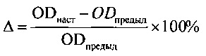
где ODнаст - оптическая плотность раствора формазана в условных единицах в конкретный срок эксперимента, ODпредыд - оптическая плотность раствора формазана в предьщущий срок эксперимента. Результаты оценки функциональной активности клеток при культивировании приведены в таблице 8 и на фиг. 2, 3.where OD nast is the optical density of the solution of formazan in arbitrary units at a specific period of the experiment, OD previous is the optical density of the solution of formazan in the previous term of the experiment. The results of evaluating the functional activity of cells during cultivation are shown in Table 8 and in FIG. 2, 3.
Выявлена высокая скорость клеточной экспансии как ФЧ, так и ММСК человека при их культивировании на протяжении 14 суток в ПРС, обогащенной 10% пулированного образца ЛТ: величина OD раствора формазана для ФЧ на 14 сутки эксперимента составила 1,296 усл. ед., показатель прироста клеточной популяции за весь период наблюдения 1100% vs 0,920 усл. ед. и 636% при использовании ПРС с 10% ЭТС, соответственно (статистически достоверная разница, р<0,05). Для ММСК эти показатели составили 0,895 усл. ед. и 509% vs 0,895 усл. ед. и 375% (ПРС с 10% ЭТС), соответственно (Табл. 8, Фиг. 2, 3).A high rate of cell expansion of both PS and human MMSC was revealed during their cultivation for 14 days in ORS enriched with 10% of a pulled RT sample: the OD value of the formazan solution for PS on the 14th day of the experiment was 1.296 conv. units, the growth rate of the cell population for the entire observation period 1100% vs 0.920 srvc. units and 636% when using ORS with 10% ETS, respectively (statistically significant difference, p <0.05). For MMSK, these indicators amounted to 0.895 conv. units and 509% vs 0.895 conv. units and 375% (ORS with 10% ETS), respectively (Table 8, Fig. 2, 3).

Заявленный способ позволил разработать технологию получения и пулирования ЛТ из тромбоцитарной массы (ТМ) доноров как стандартизированную по функциональной активности нексеногенную ростовую добавку для культивирования клеток человека разных типов (стволовых, прогениторных, дифференцированных, опухолевых).The claimed method made it possible to develop a technology for the production and pooling of LT from platelet mass (TM) of donors as a non-xenogenic growth supplement standardized by functional activity for culturing human cells of different types (stem, progenitor, differentiated, tumor).
Использование способа в биологических исследованиях и медицинской практике позволит:Using the method in biological research and medical practice will allow:
- избежать контакта культивируемых in vitro биомедицинских клеточных продуктов, предназначенных для введения человеку с диагностическими, профилактическими или лечебными целями, с ксеногенными компонентами, и исключить инфицирования агентами разных типов, аллергических и других нежелательных реакций;- to avoid contact of in vitro cultured biomedical cell products intended for administration to humans with diagnostic, prophylactic or therapeutic purposes, with xenogenic components, and to exclude infection by agents of various types, allergic and other undesirable reactions;
- культивировать клетки человека (стволовые, прогениторные, дифференцированные опухолевые, гибридомы, ооциты, в том числе в рамках ЭКО, и эндотелиальные прогениторы) для диагностических или лечебных или исследовательских целей в физиологическом микроокружении, что обеспечит сохранность их иммунофенотипа и функциональных характеристик;- cultivate human cells (stem, progenitor, differentiated tumor, hybridomas, oocytes, including in the framework of IVF, and endothelial progenitors) for diagnostic or therapeutic or research purposes in the physiological microenvironment, which will ensure the safety of their immunophenotype and functional characteristics;
- обеспечить повторяемость и возможность сопоставления результатов, полученных в разных сериях экспериментов при культивировании клеток in vitro, что было невозможно при использовании в качестве ростовых добавок разных лотов ЭТС и разных образцов ЛТ.- to ensure repeatability and the possibility of comparing the results obtained in different series of experiments when culturing cells in vitro, which was impossible when using different lots of ETS and different samples of RT as growth additives.
Claims (1)
Priority Applications (1)
| Application Number | Priority Date | Filing Date | Title |
|---|---|---|---|
| RU2016115622A RU2648162C2 (en) | 2016-04-21 | 2016-04-21 | Method for producing growth additive based on thrombocyte lysate from platelet of donors to medium for build-up of cellular weight of stem, progenitor, differentiated and tumor cells |
Applications Claiming Priority (1)
| Application Number | Priority Date | Filing Date | Title |
|---|---|---|---|
| RU2016115622A RU2648162C2 (en) | 2016-04-21 | 2016-04-21 | Method for producing growth additive based on thrombocyte lysate from platelet of donors to medium for build-up of cellular weight of stem, progenitor, differentiated and tumor cells |
Publications (2)
| Publication Number | Publication Date |
|---|---|
| RU2016115622A RU2016115622A (en) | 2016-09-10 |
| RU2648162C2 true RU2648162C2 (en) | 2018-03-22 |
Family
ID=56889360
Family Applications (1)
| Application Number | Title | Priority Date | Filing Date |
|---|---|---|---|
| RU2016115622A RU2648162C2 (en) | 2016-04-21 | 2016-04-21 | Method for producing growth additive based on thrombocyte lysate from platelet of donors to medium for build-up of cellular weight of stem, progenitor, differentiated and tumor cells |
Country Status (1)
| Country | Link |
|---|---|
| RU (1) | RU2648162C2 (en) |
Cited By (4)
| Publication number | Priority date | Publication date | Assignee | Title |
|---|---|---|---|---|
| RU2664478C2 (en) * | 2017-08-18 | 2018-08-17 | Федеральное государственное бюджетное учреждение "Национальный медицинский исследовательский радиологический центр" Министерства здравоохранения Российской Федерации (ФГБУ "НМИРЦ" Минздрава России) | Method for producing a culture growth supplement based on human platelet lysate |
| RU2732150C1 (en) * | 2020-03-04 | 2020-09-11 | Общество с ограниченной ответственностью "Центр клеточных технологий" | Method for producing autologous thrombocyte lysate as a base for cellular biomedical preparations of veterinary medicine |
| RU2739515C1 (en) * | 2020-04-29 | 2020-12-25 | Государственное бюджетное учреждение здравоохранения города Москвы "Научно-исследовательский институт скорой помощи им. Н.В. Склифосовского Департамента здравоохранения города Москвы" (ГБУЗ "НИИ СП ИМ. Н.В. СКЛИФОСОВСКОГО ДЗМ") | Method for preparing thrombocyte lysate with high content of growth factors |
| RU2790701C1 (en) * | 2022-06-03 | 2023-02-28 | Общество с ограниченной ответственностью "МеЛСиТек" | Method for obtaining a solution of growth factors from platelets |
Citations (1)
| Publication number | Priority date | Publication date | Assignee | Title |
|---|---|---|---|---|
| WO2010033605A2 (en) * | 2008-09-16 | 2010-03-25 | Mayo Foundation For Medical Education And Research | Compositions containing platelet contents |
-
2016
- 2016-04-21 RU RU2016115622A patent/RU2648162C2/en active
Patent Citations (1)
| Publication number | Priority date | Publication date | Assignee | Title |
|---|---|---|---|---|
| WO2010033605A2 (en) * | 2008-09-16 | 2010-03-25 | Mayo Foundation For Medical Education And Research | Compositions containing platelet contents |
Non-Patent Citations (3)
| Title |
|---|
| ЖУРЛОВ О.С. Фракционирование антимикробных пептидов тромбоцитарного лизата (hPL). МЕЖДУНАРОДНЫЙ ЖУРНАЛ ПРИКЛАДНЫХ И ФУНДАМЕНТАЛЬНЫХ ИССЛЕДОВАНИЙ, 22.03.2016, 3 (ч. 2), с. 247-250, материалы и методы. * |
| Н.С. СЕРГЕЕВА и др. Биологические эффекты тромбоцитарного лизата при добавлении в среду культивирования клеток человека. Гены энд Клетки, 2014, том IX, 31, с. 77-85, материалы и методы. * |
| Н.С. СЕРГЕЕВА и др. Биологические эффекты тромбоцитарного лизата при добавлении в среду культивирования клеток человека. Гены энд Клетки, 2014, том IX, 31, с. 77-85, материалы и методы. ЖУРЛОВ О.С. Фракционирование антимикробных пептидов тромбоцитарного лизата (hPL). МЕЖДУНАРОДНЫЙ ЖУРНАЛ ПРИКЛАДНЫХ И ФУНДАМЕНТАЛЬНЫХ ИССЛЕДОВАНИЙ, 22.03.2016, 3 (ч. 2), с. 247-250, материалы и методы. * |
Cited By (4)
| Publication number | Priority date | Publication date | Assignee | Title |
|---|---|---|---|---|
| RU2664478C2 (en) * | 2017-08-18 | 2018-08-17 | Федеральное государственное бюджетное учреждение "Национальный медицинский исследовательский радиологический центр" Министерства здравоохранения Российской Федерации (ФГБУ "НМИРЦ" Минздрава России) | Method for producing a culture growth supplement based on human platelet lysate |
| RU2732150C1 (en) * | 2020-03-04 | 2020-09-11 | Общество с ограниченной ответственностью "Центр клеточных технологий" | Method for producing autologous thrombocyte lysate as a base for cellular biomedical preparations of veterinary medicine |
| RU2739515C1 (en) * | 2020-04-29 | 2020-12-25 | Государственное бюджетное учреждение здравоохранения города Москвы "Научно-исследовательский институт скорой помощи им. Н.В. Склифосовского Департамента здравоохранения города Москвы" (ГБУЗ "НИИ СП ИМ. Н.В. СКЛИФОСОВСКОГО ДЗМ") | Method for preparing thrombocyte lysate with high content of growth factors |
| RU2790701C1 (en) * | 2022-06-03 | 2023-02-28 | Общество с ограниченной ответственностью "МеЛСиТек" | Method for obtaining a solution of growth factors from platelets |
Also Published As
| Publication number | Publication date |
|---|---|
| RU2016115622A (en) | 2016-09-10 |
Similar Documents
| Publication | Publication Date | Title |
|---|---|---|
| US10925901B2 (en) | Compositions containing platelet contents | |
| LU500561B1 (en) | In vitro construction method and use of liver organoids | |
| Davies et al. | Isolation and culture of human macrophages | |
| US20120276632A1 (en) | Plasma-free platelet lysate for use as a supplement in cell cultures and for the preparation of cell therapeutics | |
| KR20050109941A (en) | Methods of using adipose tissue-derived cells in the treatment of cardiovascular conditions | |
| WO2021223274A1 (en) | In-vitro culture, induction, activation and cryopreservation method and cell bank establishment for immune cells | |
| US20150299651A1 (en) | Cell culture supplements | |
| RU2648162C2 (en) | Method for producing growth additive based on thrombocyte lysate from platelet of donors to medium for build-up of cellular weight of stem, progenitor, differentiated and tumor cells | |
| Tancharoen et al. | Human platelet lysate as an alternative to fetal bovine serum for culture and endothelial differentiation of human amniotic fluid mesenchymal stem cells | |
| JP7144872B2 (en) | Serum-free medium for human lymphocyte cell culture | |
| Wong et al. | Development of a clinical-scale method for generation of dendritic cells from PBMC for use in cancer immunotherapy | |
| RUBINSTEIN | Placental blood-derived hematopoietic stem cells for unrelated bone marrow reconstitution | |
| US20170224736A1 (en) | Method and apparatus for recovery of umbilical cord tissue derived regenerative cells and uses thereof | |
| CN110090227A (en) | Purposes of the human amnion membrane in treatment graft versus host disease(GVH disease) | |
| CN112342191A (en) | hMSC production kit development and quality management standard optimization and system establishment | |
| Dhot et al. | Cord blood stem cell banking and transplantation | |
| Hashemi et al. | Comparison between human cord blood serum and platelet-rich plasma supplementation for human wharton's jelly stem cells and dermal fibroblasts culture | |
| Yu et al. | Clinical immunology Impact on red blood cell immunity patterns in postoperative phase following total hip arthroplasty | |
| WO2014193895A1 (en) | Ex vivo perfusion of donor organs prior to transplantation using mesenchymal stem cells | |
| Christy et al. | Refrigerated human mesenchymal stromal cells as an alternative to cryostorage for use in clinical investigation | |
| Fujiwara et al. | Effect of platelet lysate on Schwann-like cell differentiation of equine bone marrow-derived mesenchymal stem cells | |
| WO2012034352A1 (en) | Method for reverse differentiation of human somatic cells to produce autologous stem cells, kit and use thereof | |
| RU2664478C2 (en) | Method for producing a culture growth supplement based on human platelet lysate | |
| CN112156111B (en) | Application of cord blood platelet mitochondria in preparation of medicine for treating autoimmune disease | |
| CN113512529B (en) | Preparation method of specific antiviral adoptive immune cell AB |
Legal Events
| Date | Code | Title | Description |
|---|---|---|---|
| HE9A | Changing address for correspondence with an applicant |