RU2614101C1 - Method of surgical management of congenital hip dislocated in adolescence - Google Patents
Method of surgical management of congenital hip dislocated in adolescence Download PDFInfo
- Publication number
- RU2614101C1 RU2614101C1 RU2015145880A RU2015145880A RU2614101C1 RU 2614101 C1 RU2614101 C1 RU 2614101C1 RU 2015145880 A RU2015145880 A RU 2015145880A RU 2015145880 A RU2015145880 A RU 2015145880A RU 2614101 C1 RU2614101 C1 RU 2614101C1
- Authority
- RU
- Russia
- Prior art keywords
- thigh
- hip
- femur
- proximal end
- osteotomy
- Prior art date
Links
- 238000000034 method Methods 0.000 title claims abstract description 17
- 206010010356 Congenital anomaly Diseases 0.000 title claims abstract description 5
- 210000000689 upper leg Anatomy 0.000 claims abstract description 48
- 239000012634 fragment Substances 0.000 claims abstract description 23
- 210000003414 extremity Anatomy 0.000 claims abstract description 9
- 210000000528 lesser trochanter Anatomy 0.000 claims abstract description 8
- 230000001054 cortical effect Effects 0.000 claims abstract description 4
- 238000001356 surgical procedure Methods 0.000 claims description 6
- 210000000588 acetabulum Anatomy 0.000 claims description 4
- 210000001624 hip Anatomy 0.000 claims description 4
- 210000000988 bone and bone Anatomy 0.000 abstract description 9
- 239000003814 drug Substances 0.000 abstract description 2
- 230000000694 effects Effects 0.000 abstract 1
- 239000000126 substance Substances 0.000 abstract 1
- 210000004394 hip joint Anatomy 0.000 description 4
- 208000015181 infectious disease Diseases 0.000 description 3
- 230000002458 infectious effect Effects 0.000 description 3
- 210000002436 femur neck Anatomy 0.000 description 2
- 210000003692 ilium Anatomy 0.000 description 2
- 238000009940 knitting Methods 0.000 description 2
- 230000007774 longterm Effects 0.000 description 2
- 230000000399 orthopedic effect Effects 0.000 description 2
- 210000004197 pelvis Anatomy 0.000 description 2
- 230000000472 traumatic effect Effects 0.000 description 2
- 208000007366 Genu Valgum Diseases 0.000 description 1
- 206010062061 Knee deformity Diseases 0.000 description 1
- 210000000577 adipose tissue Anatomy 0.000 description 1
- 238000013459 approach Methods 0.000 description 1
- 230000015572 biosynthetic process Effects 0.000 description 1
- 238000003776 cleavage reaction Methods 0.000 description 1
- 239000004020 conductor Substances 0.000 description 1
- 238000007596 consolidation process Methods 0.000 description 1
- 238000006073 displacement reaction Methods 0.000 description 1
- 210000000527 greater trochanter Anatomy 0.000 description 1
- 210000002239 ischium bone Anatomy 0.000 description 1
- 238000002955 isolation Methods 0.000 description 1
- 210000003141 lower extremity Anatomy 0.000 description 1
- 230000001404 mediated effect Effects 0.000 description 1
- 230000008092 positive effect Effects 0.000 description 1
- 230000002980 postoperative effect Effects 0.000 description 1
- 230000002265 prevention Effects 0.000 description 1
- 230000007017 scission Effects 0.000 description 1
- 238000007920 subcutaneous administration Methods 0.000 description 1
Images
Classifications
-
- A—HUMAN NECESSITIES
- A61—MEDICAL OR VETERINARY SCIENCE; HYGIENE
- A61B—DIAGNOSIS; SURGERY; IDENTIFICATION
- A61B17/00—Surgical instruments, devices or methods
- A61B17/56—Surgical instruments or methods for treatment of bones or joints; Devices specially adapted therefor
Landscapes
- Surgical Instruments (AREA)
Abstract
Description
Изобретение относится к медицине, а именно к травматологии и ортопедии.The invention relates to medicine, namely to traumatology and orthopedics.
АналогиAnalogs
Известен способ восстановления опороспособности при врожденном вывихе бедра с одновременным его удлинением у подростков и взрослых. Он заключается в поднадкостничном выделении наружной поверхности в верхней трети бедренной кости, отсечении от внутреннего отдела подвертельной области кортикальной пластинки и дозированном ее перемещении с формированием костного регенерата до упора в седалищную кость стержнем, укрепленном в предварительно наложенном аппарате Илизарова и перемещаемом в просверленном в бедренной кости тоннеле. Удлинение бедра осуществляется после поперечной остеотомии его в нижней трети (Абакаров А.А., Богосьян А.Б. Аппараты и методы внешней фиксации в травматологии и ортопедии - Рига. 1985. - с. 115-120).A known method of restoring support when congenital dislocation of the thigh with its simultaneous lengthening in adolescents and adults. It consists in the subperiosteal isolation of the outer surface in the upper third of the femur, cutting off the cortical plate from the inner part of the undervert region and dosing it with the formation of bone regenerate until it stops in the ischium with a rod fixed in a preliminary superimposed Ilizarov apparatus and transported in the tunnel drilled in the femur . Hip lengthening is carried out after transverse osteotomy in the lower third (Abakarov A.A., Bogosyan A.B. Apparatus and methods of external fixation in traumatology and orthopedics - Riga. 1985. - pp. 115-120).
Так же известен способ оперативного лечения неопорного тазобедренного сустава, заключающийся в наложении аппарата Илизарова на бедро и крыло подвздошной кости, дозированном низведении бедра аппаратом с последующей остеотомией малого вертела или отщепом фрагмента бедренной кости с внутренней поверхности бедра, выполненного из отдельного доступа, введении в отщепленный участок кости через предварительно рассверленное отверстие спицы с упорной площадкой, соединенной с аппаратом Илизарова, и последующем дозированном перемещении остеотомированного фрагмента в вертлужную впадину с целью формирования дистракционного регенерата, выполняющего функцию шейки бедренной кости (Девятов И.А., Руденко Д.А., Ткачев В.А. Ортопед. травматолог. - 1991. - №1. – с. 54-55).A method for surgical treatment of an unsupported hip joint is also known, which consists in applying the Ilizarov apparatus to the thigh and wing of the ilium, dosed lowering of the thigh with an apparatus followed by osteotomy of the lesser trochanter or cleavage of a fragment of the femur from the inner surface of the thigh, made from a separate access, introducing into the cleaved area bones through a pre-drilled hole of the knitting needle with a thrust pad connected to the Ilizarov apparatus, and subsequent metered movement of the oste of the tomized fragment into the acetabulum in order to form a distraction regenerate that functions as the neck of the femur (Devyatov I.A., Rudenko D.A., Tkachev V.A. Orthopedist. Traumatologist. - 1991. - No. 1. - p. 54- 55).
Критика аналоговCriticism of analogues
Однако известные методы лечения с использованием АВФ по Илизарову требуют пребывания больного в условиях стационара более 100-120 койко-дней. Вокруг спиц и стержней создаются все условия для развития инфекционных осложнений. Проведение 2-уровневой остеотомии является достаточно травматичной операцией, а средний фрагмент бедра становится «плавающим» и трудно управляемым. Главным же недостатком операции является нивелирование созданного угла между фрагментами бедра в отдаленном периоде наблюдения за больными.However, well-known treatment methods using AVF according to Ilizarov require the patient to stay in a hospital for more than 100-120 days. Around the spokes and rods, all conditions are created for the development of infectious complications. Carrying out a 2-level osteotomy is a rather traumatic operation, and the middle fragment of the thigh becomes “floating” and difficult to manage. The main drawback of the operation is the leveling of the created angle between the fragments of the thigh in the long-term follow-up of patients.
ПрототипPrototype
В качестве прототипа взят способ оперативного лечения неопорного тазобедренного сустава, описанный в работе Девятова И.А., Руденко Д.А., Ткачев В.А. (Ортопед. травматолог. - 1991. - №1. – с. 54-55), который осуществляется путем наложения аппарата Илизарова на бедро и крыло подвздошной кости, дозированного низведения бедра аппаратом с последующей остеотомией малого вертела или отщепом фрагмента бедренной кости с внутренней поверхности бедра, выполненного из отдельного доступа. В отщепленный участок кости через предварительно рассверленное отверстие вводятся спицы с упорной площадкой, соединенной с аппаратом Илизарова, и последующим дозированным перемещением остеотомированного фрагмента в вертлужную впадину с целью формирования дистракционного регенерата, выполняющего функцию шейки бедренной кости.As a prototype, the method of surgical treatment of the unsupported hip joint, described in the work of Devyatov I.A., Rudenko D.A., Tkachev V.A. (Orthopedist. Traumatologist. - 1991. - No. 1. - pp. 54-55), which is carried out by applying the Ilizarov apparatus to the thigh and wing of the ilium, dosed lowering of the thigh by the apparatus with subsequent osteotomy of the little trochanter or a fragment of the femur from the inner surface hips made from separate access. Spokes with a thrust pad connected to the Ilizarov apparatus and subsequent dosed movement of the osteotomized fragment into the acetabulum with the aim of forming a distraction regenerate that performs the function of the femoral neck are introduced into the cleaved portion of the bone through a pre-drilled hole.
Критика прототипаPrototype criticism
Способ-прототип имеет следующие недостатки:The prototype method has the following disadvantages:
- требуется пребывание больного в условиях стационара более 100-120 койко-дней,- requires the patient to stay in a hospital for more than 100-120 days,
- вокруг спиц и стержней создаются все условия для развития инфекционных осложнений,- around the knitting needles and rods, all conditions are created for the development of infectious complications,
- проведение 2-уровневой остеотомии является достаточно травматичной операцией, средний фрагмент бедра становится и трудноуправляемым в аппарате Илизарова. Недостатком способа-прототипа является нивелирование созданного угла между фрагментами бедра в отдаленном периоде наблюдения за больными.- 2-level osteotomy is a rather traumatic operation, the middle fragment of the thigh becomes difficult to control in the Ilizarov apparatus. The disadvantage of the prototype method is the leveling of the created angle between the fragments of the thigh in the long-term follow-up of patients.
Цель изобретенияThe purpose of the invention
Целью изобретения является улучшение стабильности тазобедренного сустава, одномоментное удлинение бедра у подростков с врожденным вывихом бедра, восстановление функции и опороспособности нижней конечности при значительном сокращении сроков пребывания больного в условиях стационара и сохранении биомеханической оси конечности. Последнее обстоятельство имеет существенное значение в наше время, когда широко внедряются методики эндопротезирования тазобедренных суставов у взрослых.The aim of the invention is to improve the stability of the hip joint, simultaneous lengthening of the thigh in adolescents with congenital dislocation of the thigh, restoration of function and supportability of the lower limb with a significant reduction in the length of stay of the patient in a hospital and maintaining the biomechanical axis of the limb. The latter circumstance is essential in our time, when the methods of hip joint replacement in adults are widely introduced.
Способ осуществляется следующим образом.The method is as follows.
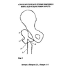
Предлагаемый способ иллюстрирован на фиг. 1, где поз. 1 - проксимальный конец бедра, поз. 2 - малый вертел, поз. 3 - угол между фрагментами бедра, поз. 4 - дистальный фрагмент бедра, проксимальный фрагмент бедра разворачивается на 20° - поз. 5.The proposed method is illustrated in FIG. 1, where pos. 1 - proximal end of the thigh, pos. 2 - small skewer, pos. 3 - the angle between the fragments of the thigh, pos. 4 - distal femur, proximal femur unfolds at 20 ° - pos. 5.
Технический результат задачи достигается тем, что производится косая чрезвертельная остеотомия бедра (поз. 1) сверху вниз снаружи кнутри и позади малого вертела (поз. 2). Между костными фрагментами создается угол (поз. 3), открытый кнаружи и кзади, а проксимальный фрагмент бедра разворачивается на 20° (поз. 5) в сагиттальной плоскости с центрацией малого вертела в сторону вертлужной впадины. Заданное положение бедра фиксируется на углообразной накостной пластине. Предлагаемый способ осуществляется следующим образом.The technical result of the task is achieved by the fact that oblique transverse pelvic osteotomy of the thigh (pos. 1) is performed from top to bottom from the outside, inside and behind the small trochanter (pos. 2). An angle (pos. 3) is created between the bone fragments, open outward and posteriorly, and the proximal femur fragment rotates 20 ° (pos. 5) in the sagittal plane with the center of the small trochanter in the direction of the acetabulum. The specified position of the thigh is fixed on the angular bone plate. The proposed method is as follows.
Доступом Лангенбека от верхушки большого вертела по ходу оси бедра рассекаются кожа, подкожная жировая клетчатка, поднадкостнично обнажается подвертельная область. Специальным проводником проводится пила Джигли вокруг бедренной кости на три сантиметра ниже намеченной точки остеотомии. Производится пересечение бедра на половину его диаметра. Затем вибропилой производится остеотомия наружной кортикальной части бедра косо сверху вниз и снаружи кнутри до встречи с предыдущей линией кортикотомии. Получаются два фрагмента бедра с косой линией излома в межвертельной и подвертельной областях. За малым вертелом желобоватым долотом производится выемка, куда с помощью ложки-направителя внедряется проксимальный конец дистального отломка. Однозубым крючком смещается проксимальный конец бедра максимально вниз и вперед под углом 20° (фиг. 1, поз. 5). Путем приведения дистальной части бедра восстанавливается биомеханическая ось конечности. Заданное положение конечности фиксируется на пластине. Рана дренируется и зашивается наглухо. Таким образом, формируется угол 140° к продольной оси бедра, открытый кнаружи и кзади, а проксимальный конец максимально приближается к тазу и разворачивается по отношению к сагиттальной плоскости на 20°. Абсолютная длина бедра увеличивается на 3,0 см.By access of Langenbek from the top of the greater trochanter along the axis of the thigh, the skin, subcutaneous fatty tissue are dissected, the subroperiostal region is exposed. A special conductor conducts a Jigley saw around the femur three centimeters below the intended point of the osteotomy. The hip is crossed at half its diameter. Then the osteotomy of the outer cortical part of the thigh is performed obliquely from top to bottom and from the inside of the vibropil to meet the previous corticotomy line. The result is two fragments of the thigh with an oblique line of fracture in the intertrochanteric and subtrochanteric regions. A recess is made behind a small trochanter with a grooved chisel, where the proximal end of the distal fragment is inserted using a guiding spoon. With a single-tooth hook, the proximal end of the thigh shifts as far down and forward as possible at an angle of 20 ° (Fig. 1, item 5). By bringing the distal part of the thigh, the biomechanical axis of the limb is restored. The predetermined position of the limb is fixed on the plate. The wound is drained and sutured tightly. Thus, an angle of 140 ° is formed to the longitudinal axis of the thigh, open outward and posteriorly, and the proximal end approaches the pelvis as much as possible and rotates 20 ° relative to the sagittal plane. The absolute length of the thigh is increased by 3.0 cm.
Признаки изобретения, отличительные от прототипа.Signs of the invention, distinctive from the prototype.
1. Одномоментное создание точки опоры под таз и удлинение бедра до 3,5 см.1. Simultaneous creation of a fulcrum under the pelvis and lengthening of the thigh to 3.5 cm.
2. Профилактика genu valgum.2. Prevention of genu valgum.
3. Производится косая чрезвертельная остеотомия бедра сверху вниз снаружи кнутри и позади малого вертела.3. Oblique oblique transverse osteotomy of the thigh is performed from top to bottom from the outside inwards and behind the small trochanter.
4. Между костными фрагментами создается угол, открытый кнаружи и кзади, фрагмент бедра.4. Between the bone fragments an angle is created that is open outwards and posteriorly, a fragment of the thigh.
5. Проксимальный фрагмент бедра разворачивается на 20° в сагиттальной плоскости.5. The proximal femur fragment unfolds at 20 ° in the sagittal plane.
6. Заданное положение бедра фиксируется на углообразной накостной пластине.6. The target position of the thigh is fixed on an angular osseous plate.
Положительный эффект от применения предлагаемого способаThe positive effect of the application of the proposed method
Осложнений в послеоперационном периоде не отмечено. Нагрузку на конечность разрешали через 4-4,5 месяца после операции. Больной выписывался из стационара на 10-12 день после хирургического лечения. Восстановительное лечение начиналось через месяц после выписки больного из стационара, обычно в амбулаторных условиях.There were no complications in the postoperative period. The load on the limb was allowed 4-4.5 months after surgery. The patient was discharged from the hospital 10-12 days after surgical treatment. Rehabilitation treatment began a month after the patient’s discharge from the hospital, usually on an outpatient basis.
Предложенный способ оперативного лечения ВВБ у подростков полностью исключал возможность смещения костных фрагментов бедра, улучшал условия консолидации, позволял восстановить биомеханическую ось конечности и сократить сроки стационарного пребывания больного. Положительным моментом удлиняющей подвертельной остеотомии бедра и отличием от известных паллиативных вмешательств является то, что медиализируется и смещается в сагиттальной плоскости только проксимальный отломок бедра, а дистальный фрагмент бедра устанавливается позади малого вертела с центрацией на головку бедра.The proposed method for surgical treatment of VVB in adolescents completely excluded the possibility of displacement of bone fragments of the femur, improved the conditions of consolidation, allowed to restore the biomechanical axis of the limb and reduce the length of hospital stay of the patient. The positive aspect of lengthening underwent osteotomy of the femur and the difference from known palliative interventions is that only the proximal femur fragment is mediated and displaced in the sagittal plane, and the distal femur fragment is placed behind the small trochanter centered on the femoral head.
Простота технического исполнения операции; сокращение сроков пребывания больного в стационаре в 6 раз; отсутствие необходимости постоянного наблюдение больного медицинским персоналом; отсутствие опасности инфекционных поздних осложнений в ране; экономичность способа как в государственном масштабе, так для семьи за счет ранней реабилитации больного.Ease of technical execution of the operation; reduction of the patient’s hospital stay by 6 times; lack of need for constant observation of the patient by medical personnel; lack of danger of infectious late complications in the wound; the cost-effectiveness of the method both on a state scale and for the family due to early rehabilitation of the patient.
Источники информацииInformation sources
Девятов И.А., Руденко Д.А., Ткачев В.А. Ортопед. травматолог. - 1991. - №1. – с. 54-55. – Прототип.Devyatov I.A., Rudenko D.A., Tkachev V.A. Orthopedist. traumatologist. - 1991. - No. 1. - from. 54-55. - The prototype.
Claims (1)
Priority Applications (1)
| Application Number | Priority Date | Filing Date | Title |
|---|---|---|---|
| RU2015145880A RU2614101C1 (en) | 2015-10-27 | 2015-10-27 | Method of surgical management of congenital hip dislocated in adolescence |
Applications Claiming Priority (1)
| Application Number | Priority Date | Filing Date | Title |
|---|---|---|---|
| RU2015145880A RU2614101C1 (en) | 2015-10-27 | 2015-10-27 | Method of surgical management of congenital hip dislocated in adolescence |
Publications (1)
| Publication Number | Publication Date |
|---|---|
| RU2614101C1 true RU2614101C1 (en) | 2017-03-22 |
Family
ID=58453003
Family Applications (1)
| Application Number | Title | Priority Date | Filing Date |
|---|---|---|---|
| RU2015145880A RU2614101C1 (en) | 2015-10-27 | 2015-10-27 | Method of surgical management of congenital hip dislocated in adolescence |
Country Status (1)
| Country | Link |
|---|---|
| RU (1) | RU2614101C1 (en) |
Citations (2)
| Publication number | Priority date | Publication date | Assignee | Title |
|---|---|---|---|---|
| RU2061426C1 (en) * | 1992-12-18 | 1996-06-10 | Московский медицинский стоматологический институт им.Н.А.Семашко | Method for treating congenital femoral dislocation in adolescents and adults |
| US6427698B1 (en) * | 2001-01-17 | 2002-08-06 | Taek-Rim Yoon | Innominate osteotomy |
-
2015
- 2015-10-27 RU RU2015145880A patent/RU2614101C1/en not_active IP Right Cessation
Patent Citations (2)
| Publication number | Priority date | Publication date | Assignee | Title |
|---|---|---|---|---|
| RU2061426C1 (en) * | 1992-12-18 | 1996-06-10 | Московский медицинский стоматологический институт им.Н.А.Семашко | Method for treating congenital femoral dislocation in adolescents and adults |
| US6427698B1 (en) * | 2001-01-17 | 2002-08-06 | Taek-Rim Yoon | Innominate osteotomy |
Non-Patent Citations (3)
| Title |
|---|
| YAN F. et al. A reduction technique of arthroplasty without subtrochanteric femoral shortening osteotomyfor the treatment of developmental high dislocation of hip: a case series of 28 hips. J Arthroplasty. 2014 Dec;29(12):2289-93 (Abstract) PMID:24412147 [PubMed - indexed for MEDLINE]. * |
| БАСКОВ В.Е. Ортопедо-хирургическое лечение детей с диспластическим маргинальным вывихом бедра. Авто дисс. канд. мед. наук. СПб, 2009, с.15. * |
| БАСКОВ В.Е. Ортопедо-хирургическое лечение детей с диспластическим маргинальным вывихом бедра. Автореферат дисс. канд. мед. наук. СПб, 2009, с.15. YAN F. et al. A reduction technique of arthroplasty without subtrochanteric femoral shortening osteotomyfor the treatment of developmental high dislocation of hip: a case series of 28 hips. J Arthroplasty. 2014 Dec;29(12):2289-93 (Abstract) PMID:24412147 [PubMed - indexed for MEDLINE]. * |
Similar Documents
| Publication | Publication Date | Title |
|---|---|---|
| Adam | Treatment of recent trochanteric fracture in adults | |
| EP1952776A1 (en) | Implant for fixing two fragments of bone together in osteotomy procedures | |
| Kempf et al. | Gamma nail in the treatment of closed trochanteric fractures. Results and indications of 121 cases | |
| Vicenti et al. | Micromotion in the fracture healing of closed distal metaphyseal tibial fractures: a multicentre prospective study | |
| Mueller | The intertrochanteric osteotomy and pseudarthrosis of the femoral neck | |
| RU2578839C1 (en) | Method for elimination of metacarpal bone defect with loss of distal portion and replacement of fetlock joint | |
| Drampalos et al. | Intramedullary and intra-osseous arthrodesis of the hallux metatarsophalangeal joint | |
| Pakuła et al. | Biomechanics of distal femoral fracture fixed with an angular stable LISS plate | |
| RU2614101C1 (en) | Method of surgical management of congenital hip dislocated in adolescence | |
| RU116340U1 (en) | LOCK FOR OSTEOSYNTHESIS OF Fractures OF THE PROXIMAL PART OF THE TIBERA | |
| RU2547780C1 (en) | Method for thigh elongation with installed endoprosthesis | |
| RU2334480C2 (en) | Method of ankle and subtalar joint arthrodesis | |
| RU2216291C1 (en) | Method for treating old lesions of distal interfibular syndesmosis at foot subluxation | |
| RU2342912C1 (en) | Method of hip replacement with angular deformation of diaphysis of femur | |
| RU2411013C1 (en) | Method longitudinal osteotomy of proximal femur in inserting femoral component of hip joint | |
| RU2212861C2 (en) | Method for repairing disconnected head when making femur cervix osteolysis | |
| Wolf et al. | Impacted steel sleeves for a minimally invasive approach in intramedullary nailing | |
| RU2585140C1 (en) | Method of treating refracture of thighbone, complicating periprosthetic fracture with osteoporosis | |
| RU2803382C1 (en) | Method of hip arthroplasty with simultaneous elimination of deformity of the proximal femur | |
| RU2749106C1 (en) | Method for osteosynthesis of femoral neck | |
| RU2394517C1 (en) | Bone grafting technique in correction of tibia recurvation deformation combined with lengthening of short lower extremity in children | |
| WO2021010913A1 (en) | Fully anatomical poly-axial locking distal radius plate designed for quadrupeds | |
| RU2549296C1 (en) | Method of arthrodesis of back part of foot joints | |
| RU156762U1 (en) | FASTENER FOR OSTEOSYNTHESIS OF FEMORAL FEMAL FRACTURES | |
| RU2779373C1 (en) | Method for correction of posttraumatic deformation of the external ankle under external fixation |
Legal Events
| Date | Code | Title | Description |
|---|---|---|---|
| MM4A | The patent is invalid due to non-payment of fees |
Effective date: 20181028 |