RU2441918C2 - Vitro diagnostic kit for identification of human papillomavirus in clinical samples - Google Patents
Vitro diagnostic kit for identification of human papillomavirus in clinical samples Download PDFInfo
- Publication number
- RU2441918C2 RU2441918C2 RU2008108517/10A RU2008108517A RU2441918C2 RU 2441918 C2 RU2441918 C2 RU 2441918C2 RU 2008108517/10 A RU2008108517/10 A RU 2008108517/10A RU 2008108517 A RU2008108517 A RU 2008108517A RU 2441918 C2 RU2441918 C2 RU 2441918C2
- Authority
- RU
- Russia
- Prior art keywords
- hpv
- probes
- amplification
- specific
- seq
- Prior art date
Links
Images


Classifications
-
- C—CHEMISTRY; METALLURGY
- C12—BIOCHEMISTRY; BEER; SPIRITS; WINE; VINEGAR; MICROBIOLOGY; ENZYMOLOGY; MUTATION OR GENETIC ENGINEERING
- C12Q—MEASURING OR TESTING PROCESSES INVOLVING ENZYMES, NUCLEIC ACIDS OR MICROORGANISMS; COMPOSITIONS OR TEST PAPERS THEREFOR; PROCESSES OF PREPARING SUCH COMPOSITIONS; CONDITION-RESPONSIVE CONTROL IN MICROBIOLOGICAL OR ENZYMOLOGICAL PROCESSES
- C12Q1/00—Measuring or testing processes involving enzymes, nucleic acids or microorganisms; Compositions therefor; Processes of preparing such compositions
- C12Q1/70—Measuring or testing processes involving enzymes, nucleic acids or microorganisms; Compositions therefor; Processes of preparing such compositions involving virus or bacteriophage
- C12Q1/701—Specific hybridization probes
- C12Q1/708—Specific hybridization probes for papilloma
-
- C—CHEMISTRY; METALLURGY
- C12—BIOCHEMISTRY; BEER; SPIRITS; WINE; VINEGAR; MICROBIOLOGY; ENZYMOLOGY; MUTATION OR GENETIC ENGINEERING
- C12Q—MEASURING OR TESTING PROCESSES INVOLVING ENZYMES, NUCLEIC ACIDS OR MICROORGANISMS; COMPOSITIONS OR TEST PAPERS THEREFOR; PROCESSES OF PREPARING SUCH COMPOSITIONS; CONDITION-RESPONSIVE CONTROL IN MICROBIOLOGICAL OR ENZYMOLOGICAL PROCESSES
- C12Q1/00—Measuring or testing processes involving enzymes, nucleic acids or microorganisms; Compositions therefor; Processes of preparing such compositions
- C12Q1/68—Measuring or testing processes involving enzymes, nucleic acids or microorganisms; Compositions therefor; Processes of preparing such compositions involving nucleic acids
- C12Q1/6813—Hybridisation assays
- C12Q1/6834—Enzymatic or biochemical coupling of nucleic acids to a solid phase
- C12Q1/6837—Enzymatic or biochemical coupling of nucleic acids to a solid phase using probe arrays or probe chips
-
- C—CHEMISTRY; METALLURGY
- C12—BIOCHEMISTRY; BEER; SPIRITS; WINE; VINEGAR; MICROBIOLOGY; ENZYMOLOGY; MUTATION OR GENETIC ENGINEERING
- C12Q—MEASURING OR TESTING PROCESSES INVOLVING ENZYMES, NUCLEIC ACIDS OR MICROORGANISMS; COMPOSITIONS OR TEST PAPERS THEREFOR; PROCESSES OF PREPARING SUCH COMPOSITIONS; CONDITION-RESPONSIVE CONTROL IN MICROBIOLOGICAL OR ENZYMOLOGICAL PROCESSES
- C12Q1/00—Measuring or testing processes involving enzymes, nucleic acids or microorganisms; Compositions therefor; Processes of preparing such compositions
- C12Q1/68—Measuring or testing processes involving enzymes, nucleic acids or microorganisms; Compositions therefor; Processes of preparing such compositions involving nucleic acids
- C12Q1/6876—Nucleic acid products used in the analysis of nucleic acids, e.g. primers or probes
- C12Q1/6888—Nucleic acid products used in the analysis of nucleic acids, e.g. primers or probes for detection or identification of organisms
Abstract
SUBSTANCE: invention relates to the area of biotechnology, namely to the method for detection and typing of human papillomavirus (HPV) in a sample, as is a reaction vessel. The invention also relates to apparatus for use in the kit and the HPV detection and typing method. The assay comprises: performing a nucleic acid amplification reaction on a sample, the amplification reaction being intended to amplify an HPV target sequence in a non-type specific manner; obtaining single stranded oligonucleotides from any amplification products; allowing single stranded oligonucleotides to hybridise where possible with the HPV type-specific probes chosen from the group containing SEQ ID NO:1 - SEQ ID NO:133 and chosen according to their type, as given in table 1, provided on a solid support, the support being located within a reaction vessel suitable for containing the sample; and detecting hybridised oligonucleotides if HPV is present in the sample.
Description
ОБЛАСТЬ ТЕХНИКИ, К КОТОРОЙ ОТНОСИТСЯ ИЗОБРЕТЕНИЕFIELD OF THE INVENTION
Настоящее изобретение относится к in vitro диагностическому набору и к способу для идентификации папилломавируса человека (HPV) в клинических образцах. Настоящее изобретение также относится к устройству для применения этого набора и способа.The present invention relates to an in vitro diagnostic kit and to a method for identifying human papillomavirus (HPV) in clinical samples. The present invention also relates to a device for applying this kit and method.
Более конкретно, в предпочтительных вариантах осуществления настоящее изобретение относится к in vitro диагностическому набору для специфического детектирования генотипов папилломавируса человека в клинических образцах с использованием зондов для генотипирования HPV, к платформе, на которой объединены матрицы нуклеиновых кислот, включая зонды, и стандартный лабораторный реакционный сосуд, к устройству для автоматической обработки результатов и к способу диагностики инфекции HPV с использованием этого in vitro диагностического набора.More specifically, in preferred embodiments, the present invention relates to an in vitro diagnostic kit for specifically detecting human papillomavirus genotypes in clinical samples using HPV genotyping probes, a platform on which nucleic acid matrices, including probes, and a standard laboratory reaction vessel are combined, a device for automatically processing results and a method for diagnosing HPV infection using this in vitro diagnostic kit.
УРОВЕНЬ ТЕХНИКИBACKGROUND
До настоящего времени было описано приблизительно 100 типов папилломавируса человека (HPV). Тип HPV считается новым типом, когда по меньшей мере 10% последовательностей генов в областях Е6, Е7 и L1 HPV отличаются от ранее известного типа. Подтипы, или варианты, отличаются от первичного типа менее чем на 2-5%. Эти вирусы имеют тропизм к эпителию человека и связаны с серьезными заболеваниями человека, в частности с карциномой слизистой оболочки половых органов и полости рта.To date, approximately 100 types of human papillomavirus (HPV) have been described. The HPV type is considered a new type when at least 10% of the gene sequences in the HPV regions E6, E7 and L1 are different from the previously known type. Subtypes, or variants, differ from the primary type by less than 2-5%. These viruses have a tropism for human epithelium and are associated with serious human diseases, in particular with carcinoma of the mucous membrane of the genital organs and oral cavity.
Приблизительно 50 типов HPV были выделены из аногенитальной слизистой оболочки. Они были подразделены на типы низкого риска (например, тип HPV 6, 11, 42, 43 и 44) и типы высокого риска (например, тип 16, 18, 31, 33 и 45) в зависимости от их связи с раком шейки матки. Детектирование и идентификация типов HPV является крайне важной, так как персистирующая инфекция типов HPV высокого риска является главным этиологическим фактором рака шейки матки.Approximately 50 types of HPV were isolated from the anogenital mucosa. They were divided into low-risk types (e.g.,
Детектирование и идентификацию генотипов HPV проводят тестированием ДНК HPV. Эти способы могут выполняться прямым детектированием ДНК HPV или детектированием амплифицированной ДНК HPV. Способы прямого детектирования ДНК HPV включают способ захвата гибридов (НС) от Digene Corp., Gaithersburg, Md., USA и способы гибридизации in situ. Способ НС одобрен FDA и основан на способе усиления сигнала гибридизации. Использующиеся гибридизационные зонды представляют собой HPV-специфические РНК-последовательности. После инкубирования этих зондов с денатурированной ДНК HPV из клинического образца образуются гибриды РНК/ДНК, которые могут быть детектированы с помощью специфического антитела. Способ НС позволяет провести различия типов HPV высокого и низкого риска, но не может идентифицировать тип HPV. Еще одним недостатком этого тест-способа является то, что применение коктейля (смеси) зондов часто приводит к перекрестным реакциям между типами HPV этих двух классов.Detection and identification of HPV genotypes is performed by testing HPV DNA. These methods can be performed by direct detection of HPV DNA or by detection of amplified HPV DNA. Methods for direct detection of HPV DNA include a method for capturing hybrids (NS) from Digene Corp., Gaithersburg, Md., USA and in situ hybridization methods. The NA method is approved by the FDA and is based on a hybridization signal amplification method. The hybridization probes used are HPV-specific RNA sequences. After incubation of these probes with denatured HPV DNA, RNA / DNA hybrids are formed from the clinical sample, which can be detected using a specific antibody. The HC method allows to distinguish between high and low risk types of HPV, but cannot identify the type of HPV. Another disadvantage of this test method is that the use of a cocktail (mixture) of probes often leads to cross-reactions between the HPV types of these two classes.
Способы идентификации типа HPV посредством амплификации вирусного генома проводили в основном с помощью полимеразной цепной реакции (ПЦР). Генотипирование HPV может быть выполнено типоспецифической ПЦР с использованием праймеров, которые распознают только один конкретный тип. Альтернативным подходом является применение ПЦР с универсальными праймерами для амплификации всех типов HPV. Папилломавирусы типируют последующим анализом последовательности амплифицированного фрагмента гена. Анализ этой последовательности может проводиться различными способами, такими как секвенирование ДНК, полиморфизм длин рестрикционных фрагментов (RFLP) или гибридизация нуклеиновых кислот. Способы гибридизации, такие как обращенная блот-гибридизация, считаются наиболее подходящими для диагностических целей (Kleter et al., J Clin Microbiol. 1999, 37: 2508-21517; Van den Brule et al., J Clin Micrоbiol. 2002, 40: 779-787).Methods for identifying the type of HPV by amplification of the viral genome were carried out mainly using polymerase chain reaction (PCR). Genotyping of HPV can be performed by type-specific PCR using primers that recognize only one specific type. An alternative approach is to use PCR with universal primers to amplify all types of HPV. Papillomaviruses are typed by subsequent sequence analysis of the amplified gene fragment. The analysis of this sequence can be carried out in various ways, such as DNA sequencing, restriction fragment length polymorphism (RFLP), or nucleic acid hybridization. Hybridization methods, such as reverse blot hybridization, are considered most suitable for diagnostic purposes (Kleter et al., J Clin Microbiol. 1999, 37: 2508-21517; Van den Brule et al., J Clin Microbiol. 2002, 40: 779 -787).
Недавно была разработана технология анализа микроматриц (см., например, патент США No. 5445934). Термин микроматриц обозначает анализ многих малых пятен для облегчения крупномасштабного анализа нуклеиновых кислот, позволяющий проводить одновременно анализ тысяч последовательностей ДНК. Как известно в данной области, обращенный блоттинг обычно проводят на мембранах, тогда как анализ микроматрицы обычно проводят на твердом носителе, и его, кроме того, можно проводить в небольшом масштабе. Технология микроматриц успешно применялась в области диагностики HPV (см. патентные публикации WO0168915 и № СА2484681).Recently, microarray analysis technology has been developed (see, for example, US Pat. No. 5,445,934). The term microarray refers to the analysis of many small spots to facilitate large-scale analysis of nucleic acids, allowing simultaneous analysis of thousands of DNA sequences. As is known in the art, reverse blotting is usually carried out on membranes, while microarray analysis is usually carried out on a solid support, and it can also be carried out on a small scale. Microarray technology has been successfully applied in the field of HPV diagnostics (see patent publications WO0168915 and No. CA2484681).
Однако существует отрицательная сторона в применении технологии микроматриц, заключающаяся в том, что требуется дорогостоящее оборудование и трудоемкое манипулирование. Это неудобство было устранено благодаря Заявке на патент № US2005064469, в которой предложена «пробирка-микроматрица». Термин «пробирка-микроматрица» относится к реакционному сосуду, который имеет форму и размер, обычные для реакционного сосуда (например, пробирки Эппендоф по 1,5 мл), с микроматрицей, размещенным на его основании, в которой могут проводиться тесты, основанные на микроматрицах.However, there is a negative side to the application of microarray technology, namely that expensive equipment and laborious manipulation are required. This inconvenience was eliminated by Patent Application No. US2005064469, which proposes a “microarray test tube”. The term “microarray test tube” refers to a reaction vessel that has the shape and size common to a reaction vessel (eg, 1.5 ml Eppendof tubes) with a microarray placed on its base, in which microarray-based tests can be performed .
ЦЕЛИ ИЗОБРЕТЕНИЯOBJECTS OF THE INVENTION
В соответствии с описанным выше, целью настоящего изобретения является создание надежного способа для специфической идентификации типов HPV, возможно, присутствующих в клиническом образце.As described above, an object of the present invention is to provide a reliable method for the specific identification of types of HPV, possibly present in a clinical specimen.
Более конкретно, целью настоящего изобретения является создание способа специфической идентификации типов HPV с использованием платформы «пробирки-микроматрицы».More specifically, it is an object of the present invention to provide a method for specifically identifying HPV types using a microarray tube platform.
Целью настоящего изобретения также является создание зондов для специфического детектирования и/или идентификации различных типов HPV.An object of the present invention is also to provide probes for the specific detection and / or identification of various types of HPV.
Кроме того, целью настоящего изобретения является создание набора для детектирования и/или идентификации типов HPV, содержащего реагенты, протоколы и HPV-специфические зонды, размещенные на «пробирке-микроматрице», позволяющие надежное специфическое детектирование и/или идентификацию типов HPV, возможно, присутствующих в клиническом образце.In addition, it is an object of the present invention to provide a kit for detecting and / or identifying types of HPV containing reagents, protocols and HPV-specific probes placed on a “microarray tube” allowing reliable specific detection and / or identification of HPV types possibly present in a clinical sample.
СУЩНОСТЬ ИЗОБРЕТЕНИЯSUMMARY OF THE INVENTION
Согласно первому аспекту изобретение относится к анализу детектирования и типирования папилломавируса человека (HPV) в образце, причем этот анализ включает: проведение реакции амплификации нуклеиновых кислот в образце, причем эта реакция амплификации предназначена для амплификации мишеневой последовательности HPV неспецифическим для типа образом; получение одноцепочечных олигонуклеотидов из любых продуктов амплификации; гибридизацию одноцепочечного олигонуклеотида, если возможно, со множеством HPV-типоспецифических зондов, обеспеченных на твердом носителе, причем этот носитель расположен в реакционном сосуде, подходящем для содержания этого образца; и детектирование гибридизованных олигонуклеотидов.According to a first aspect, the invention relates to an analysis for detecting and typing human papillomavirus (HPV) in a sample, the analysis comprising: conducting an amplification reaction of nucleic acids in a sample, wherein the amplification reaction is intended to amplify a target HPV sequence in a non-type-specific manner; obtaining single-stranded oligonucleotides from any amplification products; hybridization of a single chain oligonucleotide, if possible, with a plurality of HPV type-specific probes provided on a solid support, the support being located in a reaction vessel suitable for containing this sample; and detecting hybridized oligonucleotides.
К аспектам изобретения также относятся анализ детектирования и типирования папилломавируса человека (HPV) в образце, включающий проведение реакции амплификации нуклеиновых кислот в образце, причем этот образец находится в контакте с твердым носителем, с иммобилизированном на нем множеством HPV-типоспецифических зондов, причем реакция амплификации предназначена для амплификации мишеневой последовательности HPV неспецифическим для типа образом; получение одноцепочечных олигонуклеотидов из любых продуктов амплификации; гибридизацию одноцепочечных олигонуклеотидов, если возможно, со множеством типоспецифических зондов HPV и детектирование гибридизованных олигонуклеотидов.Aspects of the invention also include analysis of the detection and typing of human papillomavirus (HPV) in a sample, comprising carrying out a nucleic acid amplification reaction in a sample, the sample being in contact with a solid carrier immobilized by a plurality of HPV type-specific probes, the amplification reaction being intended for amplification of the target HPV sequence in a non-type-specific manner; obtaining single-stranded oligonucleotides from any amplification products; hybridization of single-stranded oligonucleotides, if possible, with a variety of type-specific HPV probes; and detection of hybridized oligonucleotides.
Реакцией амплификации является предпочтительно ПЦР. Одноцепочечные олигонуклеотиды могут быть получены денатурацией любых присутствующих двухцепочечных олигонуклеотидов, например, путем нагревания. Гибридизацию одноцепочечных олигонуклеотидов, предпочтительно, проводят при строгих условиях; такие условия будут известны специалистам в данной области, и, предпочтительно, включают в себя инкубирование денатурированных олигонуклеотидов с мишенью при 55°С в буфере, содержащем 1×SSC.The amplification reaction is preferably PCR. Single-stranded oligonucleotides can be obtained by denaturation of any double-stranded oligonucleotides present, for example, by heating. Hybridization of single-stranded oligonucleotides is preferably carried out under stringent conditions; such conditions will be known to those skilled in the art, and preferably include incubating the denatured oligonucleotides with the target at 55 ° C. in a buffer containing 1 × SSC.
В предпочтительных вариантах осуществления, образец и твердый носитель находятся внутри реакционного сосуда; например, сосуда, описанного в US 2005064469.In preferred embodiments, the sample and the solid support are inside the reaction vessel; for example, a vessel described in US2005064469.
Предпочтительно, используют зонды, специфические по меньшей мере для типов HPV 5, 10, 15, 20, 25, 30, 35, 40 или 42, которые, предпочтительно, выбраны из типов HPV 6, 11, 16, 18, 26, 30, 31, 32, 33, 34/64, 35, 39, 40, 42, 43, 44, 45, 51, 52, 53, 54, 56, 57, 58, 59, 61, 62, 66, 67, 68, 69, 70, 71, 72, 73, 74, 81, 82, 83, 84, 85 и 89. Эти зонды предпочтительно имеют длину 20-40 нт, предпочтительно 25-35 нт, более предпочтительно 28-32 нт и наиболее предпочтительно приблизительно 30 нт. Все зонды не обязательно должны быть одной и той же длины. Эти зонды предпочтительно являются специфическими в отношении L1-области HPV. Каждый типоспецифический зонд может отличаться от зондов, специфических в отношении другого типа HPV по меньшей мере 1, 2, 3 или предпочтительно более чем 3 нуклеотидами. Предпочтительные зонды выбраны из группы, содержащей SEQ ID NO:1 - SEQ ID NO:133; несколько из этих зондов детектируют один и тот же тип HPV, как описано ниже. Предпочтительно, множество зондов являются специфическими для одного и того же типа HPV и, более предпочтительно, используют по меньшей мере два зонда, специфических для каждого типа HPV. Смеси этих зондов могут быть иммобилизованы в одном и том же местоположении на твердом носителе или каждый типоспецифический зонд может быть иммобилизован в отличающемся положении. Каждый зонд, специфический в отношении одного и того же типа HPV, детектирует отличающуюся часть мишеневой последовательности этого HPV.Preferably, probes specific for at least
Зонды могут находиться на двух повторностях твердого носителя для обеспечения множественных местоположений детектирования на избыточность.The probes may be in two replicates of a solid support to provide multiple redundancy detection locations.
Могут также детектироваться одна или несколько контрольных последовательностей; например, зонд, иммобилизованный на твердом носителе, который не гибридизуется ни с какой мишеневой последовательностью из типов HPV. Этим зондом может быть геномная мишеневая последовательность человека; кроме того, этот анализ может включать амплификацию мишеневой последовательности человека из этого образца и детектирование, произошла ли гибридизация. Дополнительный контроль может быть введен с использованием неспецифически меченных последовательностей, иммобилизованных на этом твердом носителе; детектирование этой метки может гарантировать, что метка работает правильным образом. Еще один контроль может быть обеспечен включением контрольной последовательности амплификации, которая может быть амплифицирована с теми же самыми праймерами, что и мишень человека, но которая будет детектироваться отличающимся олигонуклеотидом на этом твердом носителе. Этот контроль гарантирует, что амплификация работает корректно.One or more control sequences may also be detected; for example, a probe immobilized on a solid support that does not hybridize to any target HPV type sequence. This probe may be a human genomic target sequence; in addition, this analysis may include amplification of the target human sequence from this sample and detection of whether hybridization has occurred. Additional control may be introduced using non-specifically labeled sequences immobilized on this solid support; detecting this tag can ensure that the tag works correctly. Another control may be provided by including an amplification control sequence that can be amplified with the same primers as the human target, but which will be detected by a different oligonucleotide on this solid support. This control ensures that amplification works correctly.
Настоящее изобретение также относится к реакционному сосуду, включающему твердый носитель, имеющий множество HPV-типоспецифических зондов, иммобилизованных на нем. Также изобретение относится к набору для детектирования и типирования HPV, содержащему такой реакционный сосуд вместе со смесью для амплификации нуклеиновых кислот. Эта смесь может содержать HPV-консенсусные праймеры, такие как MY09 и MY11; и, необязательно, НМВ01; праймеры для амплификации мишеневой последовательности человека и контрольную мишеневую последовательность амплификации, включающую последовательности, соответствующие фланкирующим частям последовательности-мишени человека, так что амплификация обеих мишеневых последовательностей будет происходить с использованием одних и тех же праймеров. Этот набор может также включать в себя инструкции для его применения.The present invention also relates to a reaction vessel comprising a solid support having a plurality of HPV type-specific probes immobilized thereon. The invention also relates to a kit for detecting and typing HPV containing such a reaction vessel together with a mixture for amplification of nucleic acids. This mixture may contain HPV consensus primers such as MY09 and MY11; and optionally HMB01; primers for amplifying a human target sequence and a control amplification target sequence comprising sequences corresponding to the flanking portions of the human target sequence, so that amplification of both target sequences will occur using the same primers. This kit may also include instructions for its use.
КРАТКОЕ ОПИСАНИЕ ЧЕРТЕЖЕЙBRIEF DESCRIPTION OF THE DRAWINGS
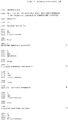
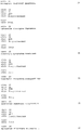
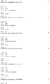
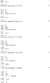
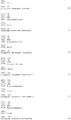
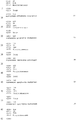
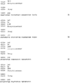
На фиг.1 показано расположение зондов на поверхности микроматрицы с 12×11=132 положениями. Номера соответствуют номеру SEQ ID NO из списка последовательностей. Отдельные зонды фиксировали в двух разных положениях для детектирования 21 различного типа HPV, контроль качества образца ДНК и контроль амплификации. LR = зонды для стандарта местоположения (SEQ ID NO:140 + SEQ ID NO:141).Figure 1 shows the location of the probes on the surface of the microarray with 12 × 11 = 132 positions. The numbers correspond to the SEQ ID NO from the sequence list. Separate probes were fixed in two different positions to detect 21 different types of HPV, DNA sample quality control and amplification control. LR = probes for a location standard (SEQ ID NO: 140 + SEQ ID NO: 141).
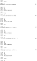
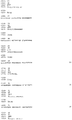
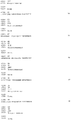
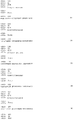
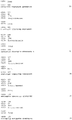
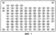
На фиг.2 показано расположение зондов на поверхности микроматрицы с 12×11=132 положениями. Номера соответствуют номеру SEQ ID NO из списка последовательностей. Отдельные зонды фиксировали в двух разных местоположениях для детектирования 23 различных типов HPV, контроль качества образца ДНК и контроль амплификации. LR = зонды для стандарта местоположения (SEQ ID NO:140 + SEQ ID NO:141).Figure 2 shows the location of the probes on the surface of the microarray with 12 × 11 = 132 positions. The numbers correspond to the SEQ ID NO from the sequence list. Separate probes were fixed at two different locations to detect 23 different types of HPV, DNA sample quality control and amplification control. LR = probes for a location standard (SEQ ID NO: 140 + SEQ ID NO: 141).
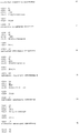
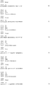
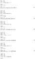
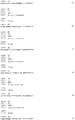
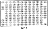
На Фиг.3 показано расположение зондов на поверхности микроматрицы с 12×11=132 местоположениями. Номера соответствуют номеру SEQ ID NO: из списка последовательностей. Смеси зондов фиксировали в двух разных местоположениях для детектирования 42 различных типов HPV и контроль качества образца ДНК. LR = зонды для стандарта местоположения (SEQ ID NO:140 + SEQ ID NO:141); М1 = SEQ ID NO:76 + SEQ ID NO:77 + SEQ ID NO:78; М2 = SEQ ID NO:122 + SEQ ID NO:123 + SEQ ID NO:124; М3 = SEQ ID NO:116 + SEQ ID NO:117 + SEQ ID NO:118 + SEQ ID NO:119.Figure 3 shows the location of the probes on the surface of the microarray with 12 × 11 = 132 locations. The numbers correspond to the number SEQ ID NO: from the list of sequences. Mixtures of probes were fixed at two different locations to detect 42 different types of HPV and quality control of the DNA sample. LR = probes for a location standard (SEQ ID NO: 140 + SEQ ID NO: 141); M1 = SEQ ID NO: 76 + SEQ ID NO: 77 + SEQ ID NO: 78; M2 = SEQ ID NO: 122 + SEQ ID NO: 123 + SEQ ID NO: 124; M3 = SEQ ID NO: 116 + SEQ ID NO: 117 + SEQ ID NO: 118 + SEQ ID NO: 119.
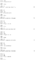
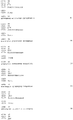
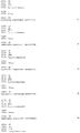
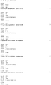
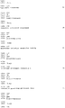
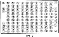
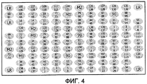
На Фиг. 4 показано расположение зондов на поверхности микроматрицы с 12×10=120 положениями. Номера соответствуют номеру SEQ ID NO из списка последовательностей. Отдельные зонды или смеси зондов фиксировали в трех разных местоположениях для детектирования 35 различных типов HPV, контроль качества образца ДНК и контроль амплификации. LR = зонды для стандарта местоположения (SEQ ID NO:140 + SEQ ID NO:141); М1 = SEQ ID NO:76 + SEQ ID NO:77 + SEQ ID NO:78; М2 = SEQ ID NO:122 + SEQ ID NO:123 + SEQ ID NO:124.In FIG. 4 shows the location of the probes on the surface of the microarray with 12 × 10 = 120 positions. The numbers correspond to the SEQ ID NO from the sequence list. Individual probes or probe mixtures were fixed at three different locations to detect 35 different types of HPV, DNA sample quality control and amplification control. LR = probes for a location standard (SEQ ID NO: 140 + SEQ ID NO: 141); M1 = SEQ ID NO: 76 + SEQ ID NO: 77 + SEQ ID NO: 78; M2 = SEQ ID NO: 122 + SEQ ID NO: 123 + SEQ ID NO: 124.
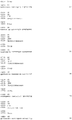
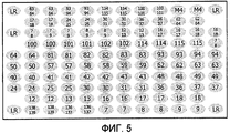
На фиг. 5 показано расположение зондов на поверхности микроматрицы с 12×10=120 положениями. Номера соответствуют номеру SEQ ID NO из списка последовательностей. Отдельные зонды фиксировали в двух разных положениях для детектирования 14 различных типов HPV, контроль качества образца ДНК и контроль амплификации. LR = зонды для стандарта местоположения (SEQ ID NO:140 + SEQ ID NO:141); М4 = SEQ ID NO:100 + SEQ ID NO:101 + SEQ ID NO:102.In FIG. 5 shows the location of the probes on the surface of the microarray with 12 × 10 = 120 positions. The numbers correspond to the SEQ ID NO from the sequence list. Separate probes were fixed in two different positions to detect 14 different types of HPV, DNA sample quality control and amplification control. LR = probes for a location standard (SEQ ID NO: 140 + SEQ ID NO: 141); M4 = SEQ ID NO: 100 + SEQ ID NO: 101 + SEQ ID NO: 102.
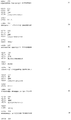
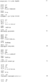
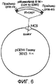
На фиг. 6 схематически показана рекомбинантная плазмида pPG44, используемая в реакции ПЦР в качестве положительного контроля амплификации.In FIG. Figure 6 schematically shows the recombinant plasmid pPG44 used in the PCR reaction as a positive control for amplification.
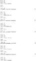
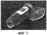
На фиг. 7 показана фотография «пробирки-матрицы», используемой в настоящем изобретении.In FIG. 7 is a photograph of a “matrix tube” used in the present invention.
ПОДРОБНОЕ ОПИСАНИЕ ИЗОБРЕТЕНИЯDETAILED DESCRIPTION OF THE INVENTION
Способ специфического детектирования и/или идентификации типов HPV предусматривет стадии:A method for specifically detecting and / or identifying types of HPV comprises the steps of:
(i) Амплификацию ДНК-образца: ДНК, полученную из клинических образцов, амплифицируют, предпочтительно при помощи ПЦР, с использованием универсальных праймеров для всех известных типов HPV, которые фланкируют геномный район, достаточно разнообразных для возможности дальнейшего генотипирования. Хотя ПЦР является предпочтительным способом амплификации, амплификация мишеневых последовательностей может быть выполнена любым другим способом, известным в данной области (лигазной цепной реакцией, системой амплификации на основе транскрипции, амплификацией замещения цепи и т.д.). В одном из вариантов осуществления настоящего изобретения, использовали праймеры MY11 и MY09 (Manos et al., Molecular Diagnostics of Human Cancer; Furth M, Greaves MF, eds.; Cold Spring Harbor Press, 1989, vol. 7: 209-214), которые амплифицируют вариабельный район L1.(i) Amplification of a DNA sample: DNA obtained from clinical samples is amplified, preferably by PCR, using universal primers for all known types of HPV that flank the genomic region, diverse enough to allow further genotyping. Although PCR is the preferred amplification method, amplification of target sequences can be performed by any other method known in the art (ligase chain reaction, transcription-based amplification system, chain substitution amplification, etc.). In one embodiment, primers MY11 and MY09 were used (Manos et al., Molecular Diagnostics of Human Cancer; Furth M, Greaves MF, eds .; Cold Spring Harbor Press, 1989, vol. 7: 209-214), which amplify the variable region L1.
Метку вводят в амплифицированную ДНК во время ее амплификации для создания возможности дальнейшего детектирования, предпочтительно метку, которая обеспечивает сигнал, который может быть детектирован колориметрическими способами. В предпочтительном варианте осуществления, по меньшей мере один из используемых праймеров метят на его 5'-конце биотином. Однако может быть использован любой другой тип метки, известный в данной области (например, дигоксигенин). Кроме того, мечение амплифицированной ДНК может альтернативно достигаться добавлением модифицированных нуклеотидов, несущих метку (например, биотинилированных или содержащих дигоксигенин производных dUTP) в ПЦР-смеси. Могут быть использованы радиоактивные метки или флуорофоры в некоторых вариантах осуществления.The label is introduced into the amplified DNA during its amplification to enable further detection, preferably a label that provides a signal that can be detected by colorimetric methods. In a preferred embodiment, at least one of the primers used is labeled at its 5 ′ end with biotin. However, any other type of label known in the art can be used (e.g., digoxygenin). In addition, labeling of amplified DNA can alternatively be achieved by adding modified label-carrying nucleotides (e.g., biotinylated or digoxigenin-derived dUTP derivatives) in a PCR mixture. Radioactive labels or fluorophores may be used in some embodiments.
(ii) Гибридизацию: амплифицированную ДНК из стадии (I) денатурируют (например, нагреванием) и наносят в «пробирку-микроматрицу» с одним или несколькими зондами из зондов, показанных в таблице 1 (SEQ ID NO:1-133). Могут быть также использованы другие способы получения одноцепочечной ДНК после амплификации. Каждый зонд, показанный в таблице 1 (SEQ ID NO:1-133), способен к специфической гибридизации с амплифицированным L1-районом из стадии (I) только одного типа HPV и, следовательно, позволяет специфически идентифицировать этот тип HPV, когда этот тип присутствует в биологическом образце. Другие типы HPV в образце могут быть идентифицированы гибридизацией амплифицированной ДНК из указанных типов HPV по меньшей мере с одним, но предпочтительно с несколькими зондами.(ii) Hybridization: the amplified DNA from step (I) is denatured (for example, by heating) and applied to a “microarray” with one or more probes from the probes shown in Table 1 (SEQ ID NO: 1-133). Other methods for preparing single stranded DNA after amplification may also be used. Each probe shown in Table 1 (SEQ ID NO: 1-133) is capable of specific hybridization with the amplified L1 region from step (I) of only one type of HPV and, therefore, allows specific identification of this type of HPV when this type is present in a biological sample. Other types of HPV in the sample can be identified by hybridization of the amplified DNA from these types of HPV with at least one, but preferably several probes.
(iii) Детектирование: ДНК-гибриды могут быть детектированы узнаванием этой метки по специфическому связыванию с лигандом или посредством иммунодетектирования. В предпочтительном варианте осуществления, метку биотин детектируют по специфическому связыванию со стрептавидином, конъюгированным с пероксидазой хрена (HRP), и последующему превращению тетраметилбензидина (ТМВ) в синий пигмент, который осаждается в конкретном местоположении, где связан соответствующий специфический зонд. Конъюгаты другого типа, хорошо известные в данной области, могут быть также пригодными для целей настоящего изобретения (например, конъюгат стрептавидин-Au). Вместо этого могут использоваться флуоресцентно меченные системы детектирования, меченные опосредованно или непосредственно. Альтернативно, могут быть использованы другие системы на основе ферментов.(iii) Detection: DNA hybrids can be detected by recognition of this label by specific binding to the ligand or by immunodetection. In a preferred embodiment, the biotin label is detected by specific binding to streptavidin conjugated to horseradish peroxidase (HRP) and the subsequent conversion of tetramethylbenzidine (TMB) to a blue pigment that precipitates at a specific location where the corresponding specific probe is bound. Other types of conjugates well known in the art may also be suitable for the purposes of the present invention (for example, streptavidin-Au conjugate). Instead, fluorescently labeled detection systems labeled indirectly or directly can be used. Alternatively, other enzyme-based systems may be used.
(iv) Анализ и обработка результатов: «пробирки-микроматрицы», обработанные таким образом, могут считываться с использованием простых оптических устройств, таких как оптический микроскоп или ATR01- и ATS-ридеры, изготовляемые CLONDIAC chip technologies GmbH (Jena, Germany).(iv) Analysis and processing of the results: “microarray tubes” thus treated can be read using simple optical devices such as an optical microscope or ATR01 and ATS readers manufactured by CLONDIAC chip technologies GmbH (Jena, Germany).
В альтернативном варианте осуществления стадии амплификации и гибридизации могут выполняться в одной и той же пробирке-микроматрице; то есть, образец добавляют в пробирку-микроматрицу и затем этот образец амплифицируют и гибридизуют с зондами внутри этой пробирки.In an alternative embodiment, the amplification and hybridization steps may be performed in the same microarray tube; that is, the sample is added to the microarray tube and then this sample is amplified and hybridized with probes inside the tube.
Один из способов получения «пробирки-микроматрицы» описан в заявке на патент US2005064469. В предпочтительном варианте осуществления настоящего изобретения, 5'-амино-связанные олигонуклеотидные зонды связывают с поверхностью твердого носителя в известных раздельных местоположениях. Указанные зонды могут быть иммобилизованы по отдельности или в виде смесей с очерченными местоположениями на этом твердом носителе. В предпочтительном варианте осуществления, используют два типоспецифических зонда для каждого типа HPV, что обеспечивает дополнительную гарантию того, что все HPV будут типированы правильно, в том числе варианты, в которых произошли замены нуклеотидов в районе одного типоспецифического зонда. Предпочтительно, используют два типоспецифических зонда, которые способны гибридизоваться в отдельных районах амплифицированного продукта.One way to obtain a "microarray test tube" is described in patent application US2005064469. In a preferred embodiment of the present invention, 5'-amino-linked oligonucleotide probes bind to the surface of the solid support at known separate locations. These probes can be immobilized individually or in the form of mixtures with outlined locations on this solid support. In a preferred embodiment, two type-specific probes for each type of HPV are used, which provides an additional guarantee that all HPVs will be typed correctly, including variants in which nucleotides are replaced in the region of one type-specific probe. Preferably, two type-specific probes are used that are capable of hybridizing in separate regions of the amplified product.
Указанные зонды или смеси зондов могут быть иммобилизованы в единственном местоположении твердого носителя, предпочтительно в двух отдельных местоположениях твердого носителя. Фиг. 1-5 в качестве примеров показывают схематические представления различных расположений зондов на поверхности этой микроматрицы.Said probes or probe mixtures can be immobilized at a single location of the solid support, preferably at two separate locations of the solid support. FIG. 1-5 show, by way of example, schematic representations of various arrangements of probes on the surface of this microarray.
«Пробирка-микроматрица», используемая в настоящем изобретении, может содержать один или несколько зондов HPV, выбранных из нуклеотидных последовательностей из списка последовательностей (SEQ ID NO:1-133). Кроме того, она может содержать один или несколько зондов для специфического детектирования контролей, таких как контроль реакции ПЦР или адекватности ДНК из этого образца контроля. Кроме того, он может также содержать один или несколько меченых олигонуклеотидов (например, модифицированных биотином олигонуклеотидов) для положительного контроля реакции детектирования и для позиционирования стандарта, так что могут быть определены местоположения всех остальных зондов.The "microarray test tube" used in the present invention may contain one or more HPV probes selected from nucleotide sequences from the list of sequences (SEQ ID NO: 1-133). In addition, it may contain one or more probes for the specific detection of controls, such as controlling the PCR reaction or the adequacy of DNA from this control sample. In addition, it may also contain one or more labeled oligonucleotides (for example, biotin-modified oligonucleotides) to positively control the detection reaction and to position the standard so that the locations of all other probes can be determined.
Специфические зонды для идентификации типов HPV были сконструированы следующим образом. Последовательности для всех ссылочных HPV (стандартов), депонированных в GenBank, в том числе известных вариантов, для амплифицированного L1-района сопоставляли с использованием общепринятой программы сопоставления нуклеиновых кислот, такой как BloEdit (4.8.6 version; Hall. Nucl. Acids Symp Ser. 1999, 41: 95-98), и определили местоположение районов наиболее вариабельных последовательностей среди различных типов HPV. Для использования в качестве специфических зондов из этих вариабельных последовательностей отбирали потенциальные последовательности олигонуклеотидов, имеющие следующие признаки: длина 20-40 оснований, предпочтительно приблизительная длина 30 оснований; предпочтительно без вторичных структур или нитей последовательного одного и того же нуклеотида, более длинных чем 4 нуклеотида; предпочтительно с отношением G+C 50% и Tm, как можно более сходной среди всех отобранных зондов; и предпочтительно с ошибочно спаренными нуклеотидами среди последовательностей разных типов HPV, находящимися как можно более в центре последовательности олигонуклеотида.Specific probes for identifying HPV types were constructed as follows. Sequences for all reference HPVs (standards) deposited in GenBank, including known variants, for the amplified L1 region were compared using a conventional nucleic acid matching program such as BloEdit (4.8.6 version; Hall. Nucl. Acids Symp Ser. 1999, 41: 95-98), and determined the locations of the regions of the most variable sequences among the various types of HPV. For use as specific probes, potential oligonucleotide sequences having the following characteristics were selected from these variable sequences: 20-40 bases long, preferably approximately 30 bases long; preferably without secondary structures or strands of the same nucleotide sequentially longer than 4 nucleotides; preferably with a G + C ratio of 50% and T m , as similar as possible among all selected probes; and preferably with erroneously paired nucleotides among sequences of different types of HPV, located as much as possible in the center of the sequence of the oligonucleotide.
Каждую потенциальную последовательность зонда, отобранную, как описано выше, сравнивали со всеми известными последовательностями HPV в амплифицированном L1-районе с использованием программы BLAST из NCBI webpage (Altschul et al., Nucleic Acid Res. 1997, 25: 3389-3402). Наконец, зонды, имеющие по меньшей мере три ошибочно спаренных нуклеотида при сравнении со всеми известными типами HPV (за исключением случая, когда их сравнивали с типом HPV, в отношении которого этот олигонуклеотидный зонд был специфическим), отбирали с предпочтением в отношении зондов, имеющих более трех ошибочных спариваний.Each potential probe sequence selected as described above was compared with all known HPV sequences in the amplified L1 region using the BLAST program from the NCBI webpage (Altschul et al., Nucleic Acid Res. 1997, 25: 3389-3402). Finally, probes having at least three erroneously paired nucleotides when compared with all known types of HPV (except when compared to the type of HPV for which this oligonucleotide probe was specific) were selected with preference for probes having more three mismatches.
Настоящее изобретение относится к зондам для специфического детектирования 42 наиболее клинически важных типов HPV: 6, 11, 16, 18, 26, 30, 31, 32, 33, 34/64, 35, 39, 40, 42, 43, 44, 45, 51, 52. 53, 54, 56, 57, 58, 59, 61, 62, 66, 67, 68, 69, 70, 71, 72, 73, 74, 81, 82, 83, 84, 85 и 89 (таблица 1; SEQ ID NO:1-133). Последовательности зондов представлены в виде одноцепочечных ДНК-олигонуклеотидов от 5'-конца к 3'-концу. В предпочтительном варианте осуществления этого изобретения, последовательности зондов соответствуют антисмысловой цепи, но любому специалисту с квалификацией в данной области очевидно, что любой из этих зондов может быть использован как таковой или в его комплементарной форме, или в форме его РНК (где Т заменен на U). Зонды по изобретению также могут быть получены путем добавления или изменения одного или нескольких нуклеотидов их последовательностей без значительного влияния на функциональность.The present invention relates to probes for the specific detection of 42 of the most clinically important types of HPV: 6, 11, 16, 18, 26, 30, 31, 32, 33, 34/64, 35, 39, 40, 42, 43, 44, 45 , 51, 52. 53, 54, 56, 57, 58, 59, 61, 62, 66, 67, 68, 69, 70, 71, 72, 73, 74, 81, 82, 83, 84, 85, and 89 (table 1; SEQ ID NO: 1-133). The probe sequences are presented as single-stranded DNA oligonucleotides from the 5'-end to the 3'-end. In a preferred embodiment of this invention, the sequences of the probes correspond to the antisense strand, but it is obvious to any person skilled in the art that any of these probes can be used as such either in its complementary form or in the form of its RNA (where T is replaced by U ) Probes according to the invention can also be obtained by adding or changing one or more nucleotides of their sequences without significant impact on functionality.


Нуклеотиды этих последовательностей имеют следующие обозначения: G=гуанин, А=аденин, Т=тимин, С=цитозин, R=G или A, Y=Е или С, М=А или С, К=G или Т, S=G или C, W=А или Т, Н=А или С или Т, В=G или Т или С, V=G или С или А, D=G или Т и, наконец, N=G или А или Т или С. Нуклеотиды, используемые в изобретении, могут быть рибонуклеотидами, дезоксирибонуклеотидами и модифицированными нуклеотидами, такими как инозин, или нуклеотидами, содержащими модифицированные группы, которые по существу не изменяют их гибридизационных свойств.The nucleotides of these sequences have the following notation: G = guanine, A = adenine, T = thymine, C = cytosine, R = G or A, Y = E or C, M = A or C, K = G or T, S = G or C, W = A or T, H = A or C or T, B = G or T or C, V = G or C or A, D = G or T, and finally, N = G or A or T or C. The nucleotides used in the invention may be ribonucleotides, deoxyribonucleotides and modified nucleotides, such as inosine, or nucleotides containing modified groups that do not substantially alter their hybridization properties.
Зонды по настоящему изобретению могут быть получены различными способами, такими как химический синтез (например, общепринятым фосфотриэфирным способом), или способами генетической инженерии, например, молекулярным клонированием рекомбинантных плазмид, в которых были встроены соответствующие нуклеотидные последовательности, и последние могут быть получены расщеплением нуклеазами.The probes of the present invention can be obtained by various methods, such as chemical synthesis (e.g., by the conventional phosphotether method), or by genetic engineering methods, e.g., by molecular cloning of recombinant plasmids in which the corresponding nucleotide sequences have been inserted, and the latter can be obtained by nuclease cleavage.
Для некоторых типов HPV зонды конструировали из района последовательности, который содержал отличающиеся нуклеотиды в конкретном положении для различных вариантов указанного типа HPV. В этих случаях, использовали вырожденные зонды, то есть смесь олигонуклеотидов, каждый из которых содержал альтернативные нуклеотиды в указанном положении. Это имеет место для зондов 39C3d [SEQ ID NO:41], 39C4d [SEQ ID NO:43], 45C1d [SEQ ID NO:57], 45C3d [SEQ ID NO:58] 57A1d [SEQ ID NO:74] 59C3_3d [SEQ ID NO:81], 66B1 [SEQ ID NO:91], 66C3d [SEQ ID NO:93] и 83B1d [SEQ ID NO:126]. Альтернативно, эквимолекулярные смеси двух олигонуклеотидов, содержащие точно один и тот же район последовательности, но отличающиеся в нуклеотидном составе для определенных положений, использовали в виде единого зонда (смесь олигонуклеотида 58В1а [SEQ ID NO:77] и 58B1b [SEQ ID NO:78]; 68C4b [SEQ ID NO:100] и 68С4 с [SEQ ID NO:101]; 74A1a [SEQ ID NO:116] и 74A1b [SEQ ID NO:117]; 74B1a [SEQ ID NO:118] и 74B1b [SEQ ID NO:119] и смесь олигонуклеотида 82A2a-AS [SEQ ID NO:122] и 82A2b-AS [SEQ ID NO:123]).For some types of HPV, probes were constructed from a region of the sequence that contained different nucleotides at a particular position for different variants of the indicated type of HPV. In these cases, degenerate probes were used, that is, a mixture of oligonucleotides, each of which contained alternative nucleotides at the indicated position. This is the case for probes 39C3d [SEQ ID NO: 41], 39C4d [SEQ ID NO: 43], 45C1d [SEQ ID NO: 57], 45C3d [SEQ ID NO: 58] 57A1d [SEQ ID NO: 74] 59C3_3d [ SEQ ID NO: 81], 66B1 [SEQ ID NO: 91], 66C3d [SEQ ID NO: 93] and 83B1d [SEQ ID NO: 126]. Alternatively, equimolecular mixtures of two oligonucleotides containing exactly the same region of the sequence but differing in nucleotide composition for specific positions were used as a single probe (mixture of oligonucleotide 58B1a [SEQ ID NO: 77] and 58B1b [SEQ ID NO: 78] ; 68C4b [SEQ ID NO: 100] and 68C4 with [SEQ ID NO: 101]; 74A1a [SEQ ID NO: 116] and 74A1b [SEQ ID NO: 117]; 74B1a [SEQ ID NO: 118] and 74B1b [SEQ ID NO: 119] and a mixture of oligonucleotide 82A2a-AS [SEQ ID NO: 122] and 82A2b-AS [SEQ ID NO: 123]).
Все зонды, описанные в изобретении, испытывали на специфическую гибридизацию с их мишеневыми последовательностями при тех же самых условиях гибридизации в платформе (позиции) «пробирки-микроматрицы». Этот факт делает возможным применение этих зондов для одновременной идентификации 42 различных типов с использованием этой платформы (позиции) микроматрицы. Высокое число типов HPV, идентифицированных с использованием «пробирки-микроматрицы», развитой в данном изобретении, позволяет рассматривать эту методологию также как прямой способ детектирования, так как остальные типы HPV являются клинически нерелевантными.All probes described in the invention were tested for specific hybridization with their target sequences under the same hybridization conditions in the "microarray test tube" platform (position). This fact makes it possible to use these probes for the simultaneous identification of 42 different types using this microarray platform (position). The high number of types of HPV identified using the "microarray tube" developed in this invention allows us to consider this methodology as a direct method of detection, since the remaining types of HPV are clinically irrelevant.
Одним из слабых моментов диагностических способов являются ложноотрицательные результаты. В случае данного способа ложные отрицательные результаты могут быть обусловлены образцами ДНК плохого качества или присутствием ингибиторов ДНК-полимеразы в анализируемых образцах. Настоящее изобретение иллюстрирует способ устранения этих ложных отрицательных результатов посредством использования двух типов контролей.One of the weak points of the diagnostic methods are false negative results. In the case of this method, false negative results may be due to poor quality DNA samples or the presence of DNA polymerase inhibitors in the analyzed samples. The present invention illustrates a method for eliminating these false negative results by using two types of controls.
Один контроль, состоящий из амплификации собственной ДНК пациента, используют предпочтительно для гарантии хорошего качества образца ДНК. Любой фрагмент последовательности из ДНК человека может быть использован в качестве мишени для этой цели. Фрагмент из однокопийного гена, такого как ген CFTR, считают особенно подходящей мишенью для положительного контроля качества ДНК в данном изобретении. Были сконструированы праймеры CFTR-F4 (SEQ ID NO:134) и CFTR-R5 (SEQ ID NO:135) для амплификации фрагмента 892 п.н. из гена CFTR. Использование однокопийной против многокопийной мишени и большего размера амплифицированного продукта контроля качества ДНК в сравнении с амплифицированным продуктом HPV, который имеет величину 892 п.н. против 450 п.н., позволило включать праймеры для амплификации CFTR в ту же реакционную смесь, которую использовали для амплификации L1-района генома HPV, с минимальными эффектами конкуренции. Таким образом, контроль качества ДНК может быть выполнен одновременно в той же самой реакционной пробирке, в которой анализируют образец, без влияния на чувствительность детектирования HPV.One control, consisting of amplification of the patient’s own DNA, is preferably used to guarantee a good quality DNA sample. Any fragment of a sequence from human DNA can be used as a target for this purpose. A fragment of a single copy gene, such as the CFTR gene, is considered a particularly suitable target for positive DNA quality control in this invention. Primers CFTR-F4 (SEQ ID NO: 134) and CFTR-R5 (SEQ ID NO: 135) were designed to amplify a 892 bp fragment. from the CFTR gene. The use of a single-copy versus multi-copy target and a larger amplified DNA quality control product compared to the amplified HPV product, which has a value of 892 bp versus 450 bp, it was possible to include primers for CFTR amplification in the same reaction mixture that was used to amplify the L1 region of the HPV genome, with minimal competition effects. Thus, DNA quality control can be performed simultaneously in the same reaction tube in which the sample is analyzed, without affecting the sensitivity of HPV detection.
Второй контроль может быть использован в качестве положительного контроля амплификации, который детектирует неуспешность ПЦР-реакции, обусловленную, например, присутствием ингибиторов ДНК-полимеразы. В предпочтительном варианте осуществления, положительный контроль амплификации состоит из рекомбинантной плазмиды, которая может быть амплифицирована с использованием тех же праймеров и тех же условий ПЦР, которые используют для амплификации фрагмента гена CFTR. Как размер, так и внутренняя последовательность относительно этих праймеров являются различными между ПЦР-продуктами, происходящими из амплификации гена CFTR и из амплификации рекомбинантной плазмиды. Таким образом, оба типа продуктов амплификации могут быть легко различены при помощи гель-электрофореза или гибридизации со специфическими зондами. Фиг. 6 показывает схематическое представление рекомбинантной плазмиды pPG44, имеющей эти характеристики.The second control can be used as a positive control of amplification, which detects the failure of the PCR reaction, due, for example, to the presence of DNA polymerase inhibitors. In a preferred embodiment, the positive amplification control consists of a recombinant plasmid that can be amplified using the same primers and the same PCR conditions that are used to amplify the CFTR gene fragment. Both the size and the internal sequence relative to these primers are different between PCR products derived from amplification of the CFTR gene and from amplification of a recombinant plasmid. Thus, both types of amplification products can be easily distinguished by gel electrophoresis or hybridization with specific probes. FIG. 6 shows a schematic representation of a recombinant plasmid pPG44 having these characteristics.
Плазмиду pPG44 конструировали способами молекулярного клонирования. Коротко, ДНК-инсерт, состоящий из фрагмента 1162 п.н. от положения 124 до положения 1285 вектора pBlueScript® II SK + (Stratagene, La Jolla, CA, USA), фланкированный праймерами CFTR, CFTR-F4 и CFTR-R5, клонировали в pGEM®-T Easy Vector с использованием коммерчески доступного набора из Promega Corporation, Madison, WI, USA. Очищенный препарат полученной рекомбинантной плазмиды pPG44 дополнительно характеризовали с использованием рестрикционных ферментов и анализа последовательности. Плазмиду pPG44 использовали в качестве положительного контроля процесса амплификации в линеаризованной форме.Plasmid pPG44 was constructed by molecular cloning methods. Briefly, DNA insert consisting of a 1162 bp fragment from
Присутствие положительного контроля в виде указанной рекомбинантной плазмиды в той же смеси для ПЦР-амплификации, в которой анализируют образец, предотвращает появление ложных отрицательных результатов, т.е. это предотвращает появление отрицательного результата даже в присутствии генома HPV-мишени в этом образце, так как в случае отсутствия генерирования продуктов амплификации следует предположить, что ПЦР-амплификация не работала правильно, и невозможно сделать вывод о присутствии или отсутствии генома HPV в данном образце.The presence of a positive control in the form of the indicated recombinant plasmid in the same PCR amplification mixture in which the sample is analyzed prevents false negative results, i.e. this prevents a negative result even in the presence of the HPV target genome in this sample, since in the absence of generation of amplification products, it should be assumed that PCR amplification did not work correctly, and it is impossible to conclude that the HPV genome is present or absent in this sample.
Зонды для специфического детектирования описанных двух типов положительных контролей, то есть контроля качества ДНК и контроля реакции амплификации, обеспечены в таблице 2 (SEQ ID NO:136-139 и SEQ ID NO:145-147). Олигонуклеотидные последовательности без значимой гомологии с любым из амплифицированных продуктов настоящего изобретения также обеспечены в этой таблице 2 (SEQ ID NO:140-141). При иммобилизации на поверхности этой микроматрицы модифицированные биотином олигонуклеотиды SEQ ID NO:140 и SEQ ID NO:141 служат в качестве положительного контроля реакции детектирования ПЦР-продуктов и в качестве стандарта позиционирования, так что может быть определено местоположение всех остальных зондов.Probes for the specific detection of the described two types of positive controls, i.e. DNA quality control and amplification reaction control, are provided in Table 2 (SEQ ID NO: 136-139 and SEQ ID NO: 145-147). Oligonucleotide sequences without significant homology with any of the amplified products of the present invention are also provided in this table 2 (SEQ ID NO: 140-141). When immobilized on the surface of this microarray, biotin-modified oligonucleotides SEQ ID NO: 140 and SEQ ID NO: 141 serve as a positive control of the detection reaction of PCR products and as a positioning standard, so that the location of all other probes can be determined.
![]()
![]()
![]()
![]()
![]()
![]()
![]()
![]()
![]()
![]()
![]()
![]()
![]()
![]()
![]()
![]()
![]()
![]()
Данное изобретение относится также к in vitro диагностическому набору для специфического детектирования типов HPV в клинических образцах. Предпочтительно, указанный набор может включать в себя любой из следующих компонентов или все следующие компоненты: смесь для амплификации, в том числе буфер для амплификации, dNTP, праймеры и контрольную плазмиду; промывочный буфер; реагенты детектирования; пробирку-микроматрицу, включающую в себя твердый носитель, включающий в себя HPV-типоспецифические зонды; реагенты для получения и приготовления образца. Конкретные компоненты будут зависеть от точных условий, при которых должен быть использован этот набор, хотя квалифицированный в данной области специалист сможет определить подходящие компоненты набора и буферные композиции.The invention also relates to an in vitro diagnostic kit for the specific detection of HPV types in clinical samples. Preferably, said kit may include any of the following components or all of the following components: an amplification mixture, including amplification buffer, dNTP, primers and a control plasmid; wash buffer; detection reagents; microarray tube including a solid carrier including HPV type-specific probes; reagents for the preparation and preparation of the sample. The specific components will depend on the exact conditions under which this kit should be used, although a person skilled in the art will be able to determine the appropriate kit components and buffer compositions.
ПРИМЕРЫEXAMPLES
Примеры, представленные ниже, даны только для иллюстрации изобретения и никаким образом не ограничивают объем сопроводительной формулы изобретения.The examples below are given only to illustrate the invention and in no way limit the scope of the accompanying claims.
ПРИМЕР 1: получение «пробирок-микроматриц»EXAMPLE 1: obtaining a "microarray tubes"
«Пробирки-микроматрицы» по изобретению были получены в CLONDIAG chip Technologies GmbH (Jena, Germany) следующим образом. Стандартную реакционную тест-пробирку Eppendorf из полипропилена и имеющую номинальный вмещаемый объем 1,5 мл, модифицировали переплавкой, создавая открытое углубление для носителя микроматрицы с адгезивным краем в пробирке.The "microarray tubes" of the invention were obtained from CLONDIAG chip Technologies GmbH (Jena, Germany) as follows. The standard Eppendorf polypropylene reaction test tube and a nominal holding volume of 1.5 ml was modified by remelting, creating an open recess for the microarray carrier with an adhesive edge in the tube.
Микроматрицы, которые вставляли в эти пробирки, получали с использованием MicroGrid II Arrayer (BioRobotics, Cambridge, Great Britain). Зонды, состоящие из модифицированных на 5'-амино-конце олигонуклеотидов, имеющих последовательность из списка последовательностей, помещали в определенных участках на поверхности эпоксидированного предметного стекла (размер предметного стекла: 75 мм × 25 мм) и ковалентно иммобилизовали. Единая микроматрица включала в себя 12×10=120 или 12×11=132 конкретных положений, в которые могли быть помещены олигонуклеотиды. Эти положения имеют промежутки 0,2 мм, так что ДНК-библиотека, включенная в каждую микроматрицу, покрывала площадь 2,4 мм × 2,4 мм, и, в целом, более 100 идентичных ДНК-библиотек могли бы быть получены таким образом на одном предметном стекле. В зависимости от типа эксперимента, либо единственный зонд, либо смесь зондов могли быть помещены в каждое из этих положений. Обычно, единственные зонды помещали в каждое местоположение при проведении экспериментов по специфичности и чувствительности для отбора зондов. После валидизации этих зондов смеси зондов, способных гибридизоваться в отдельных районах амплифицированного продукта конкретного типа HPV, могли быть помещены в то же самое местоположение при выполнении анализов идентификации генотипов HPV. На фиг. 1-5 показано различное положение зондов в микроматрицах, используемое в настоящем изобретении. В каждую микроматрицу включали два или три повтора для каждого зонда или каждой смеси зондов.Microarrays that were inserted into these tubes were obtained using MicroGrid II Arrayer (BioRobotics, Cambridge, Great Britain). Probes consisting of oligonucleotides modified at the 5'-amino end having a sequence from the list of sequences were placed in certain areas on the surface of an epoxidized slide (slide size: 75 mm × 25 mm) and covalently immobilized. The single microarray included 12 × 10 = 120 or 12 × 11 = 132 specific positions in which oligonucleotides could be placed. These positions have gaps of 0.2 mm, so that the DNA library included in each microarray covers an area of 2.4 mm × 2.4 mm, and, in general, more than 100 identical DNA libraries could be obtained in this way on one slide. Depending on the type of experiment, either a single probe or a mixture of probes could be placed in each of these positions. Typically, single probes were placed at each location for specificity and sensitivity experiments to select probes. After validation of these probes, probe mixtures capable of hybridizing in separate regions of the amplified product of a particular HPV type could be placed at the same location when performing HPV genotype identification analyzes. In FIG. 1-5 show the different positions of the probes in microarrays used in the present invention. Two or three repeats for each probe or each probe mixture were included in each microarray.
Кроме специфических зондов для генотипирования HPV и для контроля детектирования амплификации и контроля адекватности ДНК, микроматрицы включали в себя ссылочные маркеры в нескольких местоположениях, состоящие из модифицированных на 5'-конце биотином олигонуклеотидов (Маркер-1 [SEQ ID NO:140] и Маркер-2 [SEQ ID NO:141]) без значимой гомологии с любой из амплифицированных последовательностей из этого изобретения. Эти ссылочные маркеры служили как для подтверждения правильного осуществления реакции детектирования, так и для оптической ориентации изображения ридером, так чтобы местоположение всех остальных зондов могли быть определены и данные могли быть анализированы.In addition to specific probes for HPV genotyping and for monitoring the detection of amplification and monitoring the adequacy of DNA, microarrays included reference markers at several locations, consisting of oligonucleotides modified at the 5'-end of biotin (Marker-1 [SEQ ID NO: 140] and Marker- 2 [SEQ ID NO: 141]) without significant homology with any of the amplified sequences from this invention. These reference markers served both to confirm the correct implementation of the detection reaction, and for the optical orientation of the image by the reader, so that the location of all other probes could be determined and the data could be analyzed.
Все олигонуклеотиды были помещены на предметное стекло от 1× QMT Spotting Solution I (Quantifoil Micro Tools GmbH, Jena, Germany). Общая концентрация олигонуклеотидов в каждом растворе для нанесения пятен находится в диапазоне от 2,5 мкМ для ссылочных маркеров до 20 мкМ для специфических зондов. Затем олигонуклеотиды ковалентно связывали с эпоксигруппами на поверхности стекла термообработкой при 60°С в течение 30 минут с последующим многостадийным процессом промывки. Высушенные предметные стекла нарезают на кусочки стекла 3,15 мм × 3,15 мм, которые, строго говоря, и являются тем, что авторы называют микроматрицами. Затем, в конечной стадии изготовления «пробирок-микроматриц» эти микроматрицы вставляют в вышеуказанные модифицированные пробирки Эппендорфа и приклеивают к адгезивному краю. Фиг.7 показывает фотографию «пробирки-матрицы», изготовленной, как описано в данном примере.All oligonucleotides were placed on a slide from 1 × QMT Spotting Solution I (Quantifoil Micro Tools GmbH, Jena, Germany). The total concentration of oligonucleotides in each stain solution ranges from 2.5 μM for reference markers to 20 μM for specific probes. Then, the oligonucleotides were covalently linked to epoxy groups on the glass surface by heat treatment at 60 ° C for 30 minutes, followed by a multi-stage washing process. Dried glass slides are cut into 3.15 mm × 3.15 mm glass slices, which, strictly speaking, are what the authors call microarrays. Then, at the final stage of the manufacture of “microarray tubes”, these microarrays are inserted into the above modified Eppendorf tubes and glued to the adhesive edge. FIG. 7 shows a photograph of a “matrix tube” made as described in this example.
ПРИМЕР 2: получение образцов ДНКEXAMPLE 2: obtaining DNA samples
2.1. ДНК-стандарты HPV2.1. HPV DNA Standards
ДНК HPV, используемыми для оценки специфичности и чувствительности типоспецифических зондов, были либо рекомбинантные плазмиды, содержащие амплифицированный L1-район (типы HPV 6, 11, 13, 16, 18, 26, 31, 33, 35, 39, 40, 42, 44, 45, 51, 52, 53, 54, 56, 58, 61, 62, 66, 68, 70, 71, 72, 73, 81, 82, 83, 84, 85 и 89), либо ДНК, экстрагированные из клинических образцов, которые амплифицировали L1-район и были дополнительно охарактеризованы секвенированием ДНК. Рекомбинантные плазмиды конструировали способами молекулярного клонирования. Коротко, амплифицированный L1-район из каждого типа HPV клонировали в pGEM®-T Easy Vector с использованием коммерчески доступного набора из Promega Corporation, Madison, WI, USA. Очищенный препарат, полученный из каждой рекомбинантной плазмиды, дополнительно характеризовали анализом последовательности. 1-10 пг плазмидной ДНК использовали в оценке экспериментов по специфичности.The HPV DNA used to assess the specificity and sensitivity of type-specific probes was either recombinant plasmids containing the amplified L1 region (
ДНК клеточной линии К562 (Cataloge No. DD2011, Promega Corporation, Madison, WI, USA) использовали для оценки специфичности и чувствительности CFTR-специфических зондов.K562 cell line DNA (Catalog No. DD2011, Promega Corporation, Madison, WI, USA) was used to evaluate the specificity and sensitivity of CFTR-specific probes.
2.2. Клинические образцы2.2. Clinical Samples
Для детектирования HPV необходимо прежде всего отделить ДНК от остального биологического материала. Процедуры получения ДНК изменяются в зависимости от источника образцы. Конкретные примеры представлены для получения ДНК из образцов из различных источников:To detect HPV, it is first necessary to separate the DNA from the rest of the biological material. DNA production procedures vary depending on the source of the samples. Specific examples are presented for obtaining DNA from samples from various sources:
А. Мазки: образцы брали чистым, сухим хлопчатобумажным тампоном. Клетки из клинических мазков извлекали добавлением 1,5 мл солевого раствора непосредственно в контейнер с образцом и интенсивным встряхиванием на вортексе. Материал образца переносили в пробирку Эппендорф на 1,5 мл и осаждали центрифугированием. Супернатант отбрасывали и осажденные клетки суспендировали в 100 мкл лизисного буфера, содержащего 10 мМ Трис-HCl (рН 9,0 при 25°С) 50 мМ KCl, 0,15 мМ MgCl2, 0,1% Тритон® Х-100, 0,5% Твин 20 и 0,25 мг/мл протеиназы К. Смесь инкубировали при 56оС в течение приблизительно 2 часов и протеиназу К инактивировали нагреванием инкубированием этой смеси при 100°С в течение 10 минут. Детрит осаждали центрифугированием и супернатант переносили в чистую и стерильную пробирку. В ПЦР-реакции использовали аликвоту 5 мкл.A. Smears: Samples were taken with a clean, dry cotton swab. Cells from clinical smears were recovered by adding 1.5 ml of saline directly to the sample container and vortexing vigorously. The sample material was transferred into an 1.5 ml Eppendorf tube and pelleted by centrifugation. The supernatant was discarded and the pelleted cells were suspended in 100 μl of lysis buffer containing 10 mM Tris-HCl (pH 9.0 at 25 ° C) 50 mM KCl, 0.15 mM MgCl 2 , 0.1% Triton® X-100, 0 , 5
В. Суспензии клеток: этот тип образца относится к образцам, используемым в цитологических тестах на основе шеечно-влагалищной жидкости. Шеечные пробы брали кистью или шпателем и ресуспендировали в растворе PreservCyt (Cytus Corp., Marlborough, MA, USA). Аликвоту 1 мл центрифугировали и осадок ресуспендировали в 100 мкл лизисного буфера, такого же, какой использовали с образцами мазков в параграфе А, и протокол продолжали таким же образом, как описано в указанном разделе.B. Cell Suspensions: This type of sample refers to samples used in cervical vaginal fluid cytological tests. Cervical samples were taken with a brush or spatula and resuspended in PreservCyt solution (Cytus Corp., Marlborough, MA, USA). An aliquot of 1 ml was centrifuged and the pellet was resuspended in 100 μl of lysis buffer, the same as used with the smear samples in paragraph A, and the protocol was continued in the same manner as described in this section.
С. Фиксированные формалином и залитые в парафин биопсии: несколько срезов тканей шириной 5 мкм использовали в данном способе, обычно 2-5 срезов, в зависимости от площади поверхности из этой биопсии. Срезы помещали в стерильную пробирку на 1,5 мл и добавляли 100 мкл лизисного буфера, такого же, какой использовали с образцами мазков в параграфе А. Протокол продолжали таким же образом, как описано в указанном разделе, за исключением того, что обработку протеиназой К проводили в течение 3 часов.C. Formalin-fixed and paraffin embedded biopsies: several sections of
Альтернативно, коммерческий набор (NucleoSpin® Tissue kit Catalogue No. 635966 из BD Biosciebces Clontech, Palo Alto, CS, USA), предназначенный для выделения ДНК из образцов из различных источников, использовали для обработки мазков, суспензий клеток или фиксированных формалином или залитых в парафин образцов биопсий. В этом случае, начало протокола выделения ДНК было таким же, какое описано в разделах А, В и С. Вместо 100 мкл лизисного буфера к образцу добавляли 180 мкл Буфера Т1. Протокол продолжали в соответствии с указаниями изготовителя для выделения геномной ДНК из клеток и ткани.Alternatively, a commercial kit (NucleoSpin® Tissue kit Catalog No. 635966 from BD Biosciebces Clontech, Palo Alto, CS, USA), used to isolate DNA from samples from various sources, was used to treat smears, cell suspensions or formalin-fixed or paraffin embedded biopsy samples. In this case, the start of the DNA isolation protocol was the same as described in sections A, B and C. Instead of 100 μl of lysis buffer, 180 μl of T1 buffer was added to the sample. The protocol was continued in accordance with the manufacturer's instructions for isolation of genomic DNA from cells and tissue.
Независимо от того, использовали ли этот тип клинического образца или этот способ получения ДНК, отрицательные контроли обрабатывали параллельно с каждой серией образцов. Эти отрицательные контроли, состоящие из 1 мл солевого раствора, обрабатывали таким же образом, какой описан в разделе А.Regardless of whether this type of clinical sample was used or this method of obtaining DNA, negative controls were processed in parallel with each series of samples. These negative controls, consisting of 1 ml of saline, were treated in the same manner as described in section A.
ПРИМЕР 3: ПЦР-амплификацияEXAMPLE 3: PCR amplification
ПЦР-амплификацию выполняли с использованием консенсусных праймеров MY11 и MY09 (Manos et al., Molecular Diagnostics of Human Cancer; Furth M, Greaves MF, eds.; Cold Spring Harbor Press, 1989, vol. 7: 209-214). Третий праймер, НМВ01, который часто используют в комбинации с MY09 и MY11 для амплификации типа 51 HPV, который не амплифицируется эффективно только с MY09 и MY11 (Hildesheim et al., J Infect Dis. 1994, 169: 235-240), также включали в ПЦР-реакцию. Коротко, ПЦР-реакцию проводили в конечном объеме реакции 50 мкл, содержащем 10 мМ Трис-HCl рН 8,3, 50 мМ KCl, 1 мМ MgCl2, 0,3 мкМ каждый праймер MY09 и MY11 (SEQ ID NO:142 и 143), 0,03 мкМ праймер НМВ01 (SEQ ID NO:144) 200 мкМ каждый dNTP, 4 единицы AmpliTaq Gold ДНК-полимеразы (Applied Biosystems, Foster City, CA, USA) и 5 мкл каждого стандарта ДНК HPV из примера 2.1 или ДНК клинического образца из примера 2.2. Для тестирования пригодности ДНК образца 0,08 мкМ каждого праймера CFTR-F4 и CGTR-R5 (SEQ ID NO:134 и 135) также добавляли к реакционной смеси. Дополнительно, для проверки процесса амплификации и элиминации ложных отрицательных результатов вследствие неуспешности реакции 20 фг внутреннего контроля pPG44 включали в ту же самую реакционную пробирку, в которой анализировали эти образцы. Все прямые праймеры, использованные в ПЦР-реакции (MY11 [SEQ ID NO:143] и CFTR-F4 [SEQ ID NO:134]), были модифицированы биотином на 5'-конце таким образом, что любая амплифицированная ДНК могла быть затем детектирована.PCR amplification was performed using consensus primers MY11 and MY09 (Manos et al., Molecular Diagnostics of Human Cancer; Furth M, Greaves MF, eds .; Cold Spring Harbor Press, 1989, vol. 7: 209-214). A third primer, HMB01, which is often used in combination with MY09 and MY11 to amplify
Отрицательные контроли, состоящие из 5 мкл слепых образцов из примера 2.2, или 5 мкл деионизованной воды, обрабатывали параллельно с ДНК образцов. Применение этих типов отрицательных контролей служит для подтверждения, что загрязнения нет ни в одной точке манипулирования с образцом или в установке ПЦР-реакции и что все положительные результаты представляют истинное присутствие ДНК в конкретном образце.Negative controls, consisting of 5 μl of the blind samples from Example 2.2, or 5 μl of deionized water, were treated in parallel with the DNA of the samples. The use of these types of negative controls serves to confirm that there is no contamination at any point in the manipulation of the sample or in the PCR reaction unit, and that all positive results represent the true presence of DNA in a particular sample.
ПЦР-реакции проводили в термоциклере Mastercycler (Eppendorf, Hamburg, Germany), программируемом со следующим профилем циклов: один начальный цикл денатурации при 95°С в течение 9 минут, 45 циклов 30 секунд при 94°С, 60 секунд при 55°С и 90 секунд при 72°С, и один конечный цикл удлинения при 72°С в течение 8 минут. После амплификации 5 мкл каждой реакционной смеси использовали для последующего детектирования со специфическими зондами.PCR reactions were carried out in a Mastercycler thermal cycler (Eppendorf, Hamburg, Germany), programmed with the following cycle profile: one initial denaturation cycle at 95 ° C for 9 minutes, 45
ПРИМЕР 4: одновременная идентификация генотипов HPV с использованием «пробирок-микроматриц»EXAMPLE 4: simultaneous identification of HPV genotypes using microarray tubes
«Пробирки-микроматрицы» предварительно промывали непосредственно перед использованием добавлением 300 мкл 0,5х ЗФР-Твин 20-буфера в каждую пробирку и перевертыванием пробирок несколько раз. Всю жидкость изнутри каждой пробирки удаляли с использованием пастеровской пипетки с вакуумной системой.Microarray tubes were pre-washed immediately before use by adding 300 μl of 0.5x PBS-Tween 20-buffer to each tube and flipping the tubes several times. All liquid from the inside of each tube was removed using a Pasteur pipette with a vacuum system.
Реакции амплификации из примера 3 денатурировали нагреванием их до 95°С в течение 10 минут и, сразу же после этого, охлаждением их в течение 5 минут на льду. Пять микролитров денатурированной реакции амплификации вносили в «пробирку-микроматрицу», приготовленную в примере 1, вместе с 100 мкл раствора для гибридизации (200 мМ натрий-фосфатаный буфер, pH 7,2; 1× SSC; 0,2% Тритон® Х-100; 1 мМ ЭДТА, pH 8,0). Реакцию гибридизации проводили в Thermomixer comfort (Eppendorf, Hamburg, Germany) инкубированием «пробирок-микроматриц» при 55°С в течение одного часа при встряхивании при 550 об/мин. После периода инкубирования реакцию гибридизации удаляли Пастеровской пипеткой, присоединенной к вакуумной системе, и проводили стадию промывания 300 мкл 0,5× ЗФР-Твин 20-буфера.The amplification reactions of Example 3 were denatured by heating them to 95 ° C for 10 minutes and, immediately afterwards, by cooling them for 5 minutes on ice. Five microliters of the denatured amplification reaction was added to a “microarray test tube” prepared in Example 1, together with 100 μl of a hybridization solution (200 mM sodium phosphate buffer, pH 7.2; 1 × SSC; 0.2% Triton® X- 100; 1 mM EDTA, pH 8.0). The hybridization reaction was carried out in Thermomixer comfort (Eppendorf, Hamburg, Germany) by incubating microarray tubes at 55 ° C for one hour with shaking at 550 rpm. After an incubation period, the hybridization reaction was removed with a Pasteur pipette attached to a vacuum system, and a washing step of 300 μl of 0.5 × PBS-
Гибридизованную ДНК детектировали инкубированием в 100 мкл раствора 0,075 мкг/мл Поли-НRP-стрептавидина (Pierce Biotechnology Inc., Rockford, IL, USA) при 30°С в течение 15 минут со встряхиванием при 550 об/мин. Затем всю жидкость из «пробирки-микроматрицы» быстро удаляли и проводили две стадии промывки, как указано выше. Реакцию развития окраски выполняли в 100 мкл субстрата пероксидазы True Blue™ (KPL, Gaithersburg, MD, USA), который состоит из забуференного раствора, содержащего 3,3',5,5'-тетраметилбензидина (ТМВ) и Н2О2, инкубированием при 25°С в течение 10 минут. Окрашенные осадки, полученные таким образом, вызывают изменения в прозрачности для света в конкретных местоположениях микроматрицы, которые могут быть считаны с использованием ATR01- или ATS-ридера, изготавливаемого CLONDIAG chip technologies GmbH (Jena, Germany). Необязательно, ATS-ридер может иметь специальную программу, установленную для автоматической обработки результата анализа образцов, полученного с использованием «пробирки-микроматрицы», разработанной в данном изобретении.Hybridized DNA was detected by incubation in 100 μl of a solution of 0.075 μg / ml Poly-HRP-streptavidin (Pierce Biotechnology Inc., Rockford, IL, USA) at 30 ° C for 15 minutes with shaking at 550 rpm. Then all the liquid from the "microarray test tube" was quickly removed and two washing steps were performed as described above. The color development reaction was performed in 100 μl of True Blue ™ peroxidase substrate (KPL, Gaithersburg, MD, USA), which consists of a buffered solution containing 3.3 ', 5.5'-tetramethylbenzidine (TMB) and H 2 O 2 , incubation at 25 ° C for 10 minutes. The colored precipitates thus obtained cause changes in the transparency for light at specific locations of the microarray, which can be read using an ATR01 or ATS reader manufactured by CLONDIAG chip technologies GmbH (Jena, Germany). Optionally, the ATS reader may have a special program installed to automatically process the result of the analysis of samples obtained using the "microarray test tube" developed in this invention.
Claims (63)
осуществление реакции амплификации нуклеиновых кислот в образце, причем реакция амплификации предназначена для амплификации мишеневой последовательности HPV неспецифическим для типа образом;
получение одноцепочечных олигонуклеотидов из любых продуктов амплификации;
осуществление гибридизации одноцепочечных олигонуклеотидов с множеством HPV-типоспецифических зондов, выбранных из группы, содержащей SEQ ID NO:1 - SEQ ID NO:133, и отобранных по типу, как указано в таблице 1, находящихся на твердом носителе, причем носитель расположен в реакционном сосуде, подходящем для этого образца; и
детектирование гибридизованных олигонуклеотидов, если HPV присутствует в образце.1. A method for detecting and typing human papillomavirus (HPV) in a sample, comprising:
the implementation of the amplification reaction of nucleic acids in the sample, and the amplification reaction is designed to amplify the target HPV sequence in a non-type-specific manner;
obtaining single-stranded oligonucleotides from any amplification products;
hybridization of single-stranded oligonucleotides with a variety of HPV type-specific probes selected from the group consisting of SEQ ID NO: 1 to SEQ ID NO: 133 and selected by type, as indicated in Table 1, on a solid support, the carrier being located in the reaction vessel suitable for this sample; and
detection of hybridized oligonucleotides if HPV is present in the sample.
i) реагенты для экстракции и/или очистки ДНК;
ii) смесь для амплификации нуклеиновых кислот;
iii) реагенты для визуализации гибридизации нуклеиновых кислот с зондами реакционного сосуда.49. A kit for detecting and typing HPV, containing the reaction vessel according to item 27, together with one or more of the following components:
i) reagents for extraction and / or purification of DNA;
ii) a mixture for amplification of nucleic acids;
iii) reagents for visualizing the hybridization of nucleic acids with probes of the reaction vessel.
Applications Claiming Priority (2)
| Application Number | Priority Date | Filing Date | Title |
|---|---|---|---|
| GB0516145.0 | 2005-08-05 | ||
| GBGB0516145.0A GB0516145D0 (en) | 2005-08-05 | 2005-08-05 | In vitro diagnostic kit for identification of human papillomavirus in clinical samples |
Publications (2)
| Publication Number | Publication Date |
|---|---|
| RU2008108517A RU2008108517A (en) | 2009-09-10 |
| RU2441918C2 true RU2441918C2 (en) | 2012-02-10 |
Family
ID=34984158
Family Applications (1)
| Application Number | Title | Priority Date | Filing Date |
|---|---|---|---|
| RU2008108517/10A RU2441918C2 (en) | 2005-08-05 | 2006-08-04 | Vitro diagnostic kit for identification of human papillomavirus in clinical samples |
Country Status (12)
| Country | Link |
|---|---|
| US (1) | US20110070576A1 (en) |
| EP (1) | EP1910576A2 (en) |
| JP (1) | JP2009502190A (en) |
| KR (1) | KR20080045167A (en) |
| CN (1) | CN101379196B (en) |
| AU (1) | AU2006277711A1 (en) |
| BR (1) | BRPI0614388A2 (en) |
| CA (1) | CA2617978A1 (en) |
| GB (1) | GB0516145D0 (en) |
| IL (1) | IL189281A (en) |
| RU (1) | RU2441918C2 (en) |
| WO (1) | WO2007017699A2 (en) |
Families Citing this family (12)
| Publication number | Priority date | Publication date | Assignee | Title |
|---|---|---|---|---|
| US20110111389A1 (en) * | 2001-11-07 | 2011-05-12 | Diagcor Bioscience Incorporation Limited | Rapid genotyping analysis for human papillomavirus and the device thereof |
| CN101487042B (en) * | 2008-01-18 | 2012-05-30 | 中山大学达安基因股份有限公司 | HPV high risk and low risk subtype typing DNA micro-array chip |
| WO2010043418A2 (en) * | 2008-10-17 | 2010-04-22 | Febit Holding Gmbh | Integrated amplification, processing and analysis of biomolecules in a microfluidic reaction medium |
| CN101864493B (en) * | 2009-04-17 | 2012-11-28 | 上海生物信息技术研究中心 | Assay kit for detecting human papillomavirus and preparation and use thereof |
| US20130078614A1 (en) * | 2010-02-12 | 2013-03-28 | Yonsei University Wonju Industry-Academic Cooperation Foundation | Probe for hpv genotype diagnosis and analysis method thereof |
| ES2372840B1 (en) * | 2010-03-24 | 2012-11-22 | Genómica S.A.U. | KIT FOR THE DETECTION OF THE HUMAN PAPILOMA VIRUS. |
| CN103409553B (en) * | 2013-02-01 | 2016-04-20 | 港龙生物技术(深圳)有限公司 | A kind of gene chip for high-pass typing detection human papillomavirus, reagent and test kit thereof |
| GB2525024A (en) * | 2014-04-10 | 2015-10-14 | Vela Operations Pte Ltd | Universal controls for sequencing assays |
| CN104818342B (en) * | 2015-03-23 | 2017-09-22 | 厦门艾德生物医药科技有限公司 | Detection kit, detection architecture and method for 19 kinds of high-risk human mammilla papillomavirus (HPV) |
| GB201511470D0 (en) * | 2015-06-30 | 2015-08-12 | Cellcall Kft | HPV detection method |
| KR101886278B1 (en) * | 2016-11-04 | 2018-08-08 | 주식회사 퀀타매트릭스 | Composition for determinating genomic types of human papillomavirus |
| EP3321376A1 (en) | 2016-11-11 | 2018-05-16 | Genomica S.A.U. | Electrochemical dna detection |
Family Cites Families (6)
| Publication number | Priority date | Publication date | Assignee | Title |
|---|---|---|---|---|
| DE69527776T2 (en) * | 1994-02-21 | 2003-04-30 | Stichting Res Fonds Pathologie | DETECTING HUMAN PAPILLOMVIRUS WITH NUCLEIC ACID AMPLIFICATION USED BY GENERAL PRIMER |
| DE69806024T2 (en) * | 1997-09-16 | 2003-02-13 | Innogenetics Nv | DETECTION AND IDENTIFICATION OF HUMAN PAPILLOMAVIRUS BY PCR AND TYPE-SPECIFIC REVERSE HYBRIDIZATION |
| DE10201463B4 (en) * | 2002-01-16 | 2005-07-21 | Clondiag Chip Technologies Gmbh | Reaction vessel for performing array method |
| KR100517275B1 (en) * | 2003-03-04 | 2005-09-27 | 주식회사 바이오메드랩 | Genotyping probe for diagnosis of human papilloma virus infection and biochip comprising the same |
| US7741042B2 (en) * | 2004-04-19 | 2010-06-22 | Genomictree, Inc. | Method for preparing a DNA chip and use thereof |
| KR100663992B1 (en) * | 2004-07-05 | 2007-01-02 | (주)바이오메드랩 | The method selecting highly specific probes for HPV genotype analysis and the probes thereof |
-
2005
- 2005-08-05 GB GBGB0516145.0A patent/GB0516145D0/en not_active Ceased
-
2006
- 2006-08-04 JP JP2008524595A patent/JP2009502190A/en active Pending
- 2006-08-04 BR BRPI0614388-1A patent/BRPI0614388A2/en not_active Application Discontinuation
- 2006-08-04 EP EP06765379A patent/EP1910576A2/en not_active Withdrawn
- 2006-08-04 RU RU2008108517/10A patent/RU2441918C2/en active
- 2006-08-04 US US11/997,994 patent/US20110070576A1/en not_active Abandoned
- 2006-08-04 KR KR1020087005439A patent/KR20080045167A/en not_active Application Discontinuation
- 2006-08-04 AU AU2006277711A patent/AU2006277711A1/en not_active Abandoned
- 2006-08-04 CA CA002617978A patent/CA2617978A1/en not_active Abandoned
- 2006-08-04 WO PCT/GB2006/050231 patent/WO2007017699A2/en active Application Filing
- 2006-08-04 CN CN2006800371448A patent/CN101379196B/en active Active
-
2008
- 2008-02-05 IL IL189281A patent/IL189281A/en active IP Right Grant
Non-Patent Citations (1)
| Title |
|---|
| AMBRETTI ET AL., High-throughput polymerase chain reaction chemiluminescent enzyme immunoassay for typing and quantifying human papillomavirus DNAs, Anal Biochem., 2004, v.332, n.2, pp.349-357. * |
Also Published As
| Publication number | Publication date |
|---|---|
| KR20080045167A (en) | 2008-05-22 |
| AU2006277711A1 (en) | 2007-02-15 |
| GB0516145D0 (en) | 2005-09-14 |
| WO2007017699A3 (en) | 2007-08-09 |
| WO2007017699A2 (en) | 2007-02-15 |
| CN101379196B (en) | 2012-10-24 |
| RU2008108517A (en) | 2009-09-10 |
| BRPI0614388A2 (en) | 2011-03-22 |
| JP2009502190A (en) | 2009-01-29 |
| EP1910576A2 (en) | 2008-04-16 |
| CA2617978A1 (en) | 2007-02-15 |
| CN101379196A (en) | 2009-03-04 |
| IL189281A (en) | 2012-09-24 |
| US20110070576A1 (en) | 2011-03-24 |
| IL189281A0 (en) | 2008-06-05 |
Similar Documents
| Publication | Publication Date | Title |
|---|---|---|
| RU2441918C2 (en) | Vitro diagnostic kit for identification of human papillomavirus in clinical samples | |
| EP0433396B1 (en) | Detection of human papillomavirus by the polymerase chain reaction | |
| JP3744548B2 (en) | Nucleic acid primers and probes for detecting HIV-1 and HIV-2 | |
| AU741141B2 (en) | Specific and sensitive method for detecting nucleic acids | |
| EP1294939B1 (en) | Detection and typing of human papillomavirus using pna probes | |
| EP1082466B1 (en) | Method, reagent and kit for genotyping of human papillomavirus | |
| US20070281295A1 (en) | Detection of human papillomavirus E6 mRNA | |
| CN110885906A (en) | Compositions and methods for detecting human papillomavirus nucleic acids | |
| EA008388B1 (en) | Amplification-hybridisation method for detecting and typing human papillomavirus | |
| JP4628369B2 (en) | High-sensitivity nucleic acid multiplex analysis method | |
| WO2009126517A2 (en) | Optimized probes and primers and methods of using same for the detection, quantification and grouping of hiv-1 | |
| ES2253279T3 (en) | METHODS AND COMPOSITIONS FOR THE DETECTION OF SPECIES OF THE MYCOBACTERIUM AVIUM COMPLEX. | |
| EP3885455A1 (en) | Method and kit for the detection of sars-cov-2 virus in a sample based on reverse transcription loop-mediated isothermal amplification (rt-lamp) | |
| EP2336369A1 (en) | Probes for genotyping low-risk-HPV | |
| US10190176B2 (en) | Primers, probes, and methods for mycobacterium tuberculosis specific diagnosis | |
| Käller et al. | Tag-array based HPV genotyping by competitive hybridization and extension | |
| MX2008001751A (en) | In vitro diagnostic kit for identification of human papillomavirus in clinical samples | |
| US20110039715A1 (en) | Influenza b virus detection method and kit therefor | |
| WO2013102061A1 (en) | Actb primers and probes | |
| JPH0819400A (en) | Detection of virus involved in hemorrhagic fever with renal syndrome |