KR20130038407A - Therapy of platinum-resistant cancer - Google Patents
Therapy of platinum-resistant cancer Download PDFInfo
- Publication number
- KR20130038407A KR20130038407A KR1020137005305A KR20137005305A KR20130038407A KR 20130038407 A KR20130038407 A KR 20130038407A KR 1020137005305 A KR1020137005305 A KR 1020137005305A KR 20137005305 A KR20137005305 A KR 20137005305A KR 20130038407 A KR20130038407 A KR 20130038407A
- Authority
- KR
- South Korea
- Prior art keywords
- antibody
- her2
- ser
- antibodies
- cells
- Prior art date
Links
Images


Classifications
-
- A—HUMAN NECESSITIES
- A61—MEDICAL OR VETERINARY SCIENCE; HYGIENE
- A61K—PREPARATIONS FOR MEDICAL, DENTAL OR TOILETRY PURPOSES
- A61K39/00—Medicinal preparations containing antigens or antibodies
- A61K39/395—Antibodies; Immunoglobulins; Immune serum, e.g. antilymphocytic serum
- A61K39/39533—Antibodies; Immunoglobulins; Immune serum, e.g. antilymphocytic serum against materials from animals
- A61K39/39558—Antibodies; Immunoglobulins; Immune serum, e.g. antilymphocytic serum against materials from animals against tumor tissues, cells, antigens
-
- A—HUMAN NECESSITIES
- A61—MEDICAL OR VETERINARY SCIENCE; HYGIENE
- A61K—PREPARATIONS FOR MEDICAL, DENTAL OR TOILETRY PURPOSES
- A61K31/00—Medicinal preparations containing organic active ingredients
- A61K31/33—Heterocyclic compounds
- A61K31/395—Heterocyclic compounds having nitrogen as a ring hetero atom, e.g. guanethidine or rifamycins
- A61K31/435—Heterocyclic compounds having nitrogen as a ring hetero atom, e.g. guanethidine or rifamycins having six-membered rings with one nitrogen as the only ring hetero atom
- A61K31/47—Quinolines; Isoquinolines
- A61K31/4738—Quinolines; Isoquinolines ortho- or peri-condensed with heterocyclic ring systems
- A61K31/4745—Quinolines; Isoquinolines ortho- or peri-condensed with heterocyclic ring systems condensed with ring systems having nitrogen as a ring hetero atom, e.g. phenantrolines
-
- A—HUMAN NECESSITIES
- A61—MEDICAL OR VETERINARY SCIENCE; HYGIENE
- A61K—PREPARATIONS FOR MEDICAL, DENTAL OR TOILETRY PURPOSES
- A61K31/00—Medicinal preparations containing organic active ingredients
- A61K31/70—Carbohydrates; Sugars; Derivatives thereof
- A61K31/7028—Compounds having saccharide radicals attached to non-saccharide compounds by glycosidic linkages
- A61K31/7034—Compounds having saccharide radicals attached to non-saccharide compounds by glycosidic linkages attached to a carbocyclic compound, e.g. phloridzin
- A61K31/704—Compounds having saccharide radicals attached to non-saccharide compounds by glycosidic linkages attached to a carbocyclic compound, e.g. phloridzin attached to a condensed carbocyclic ring system, e.g. sennosides, thiocolchicosides, escin, daunorubicin
-
- A—HUMAN NECESSITIES
- A61—MEDICAL OR VETERINARY SCIENCE; HYGIENE
- A61K—PREPARATIONS FOR MEDICAL, DENTAL OR TOILETRY PURPOSES
- A61K31/00—Medicinal preparations containing organic active ingredients
- A61K31/70—Carbohydrates; Sugars; Derivatives thereof
- A61K31/7042—Compounds having saccharide radicals and heterocyclic rings
- A61K31/7052—Compounds having saccharide radicals and heterocyclic rings having nitrogen as a ring hetero atom, e.g. nucleosides, nucleotides
- A61K31/706—Compounds having saccharide radicals and heterocyclic rings having nitrogen as a ring hetero atom, e.g. nucleosides, nucleotides containing six-membered rings with nitrogen as a ring hetero atom
- A61K31/7064—Compounds having saccharide radicals and heterocyclic rings having nitrogen as a ring hetero atom, e.g. nucleosides, nucleotides containing six-membered rings with nitrogen as a ring hetero atom containing condensed or non-condensed pyrimidines
- A61K31/7068—Compounds having saccharide radicals and heterocyclic rings having nitrogen as a ring hetero atom, e.g. nucleosides, nucleotides containing six-membered rings with nitrogen as a ring hetero atom containing condensed or non-condensed pyrimidines having oxo groups directly attached to the pyrimidine ring, e.g. cytidine, cytidylic acid
- A61K31/7072—Compounds having saccharide radicals and heterocyclic rings having nitrogen as a ring hetero atom, e.g. nucleosides, nucleotides containing six-membered rings with nitrogen as a ring hetero atom containing condensed or non-condensed pyrimidines having oxo groups directly attached to the pyrimidine ring, e.g. cytidine, cytidylic acid having two oxo groups directly attached to the pyrimidine ring, e.g. uridine, uridylic acid, thymidine, zidovudine
-
- A—HUMAN NECESSITIES
- A61—MEDICAL OR VETERINARY SCIENCE; HYGIENE
- A61P—SPECIFIC THERAPEUTIC ACTIVITY OF CHEMICAL COMPOUNDS OR MEDICINAL PREPARATIONS
- A61P15/00—Drugs for genital or sexual disorders; Contraceptives
-
- A—HUMAN NECESSITIES
- A61—MEDICAL OR VETERINARY SCIENCE; HYGIENE
- A61P—SPECIFIC THERAPEUTIC ACTIVITY OF CHEMICAL COMPOUNDS OR MEDICINAL PREPARATIONS
- A61P35/00—Antineoplastic agents
-
- A—HUMAN NECESSITIES
- A61—MEDICAL OR VETERINARY SCIENCE; HYGIENE
- A61P—SPECIFIC THERAPEUTIC ACTIVITY OF CHEMICAL COMPOUNDS OR MEDICINAL PREPARATIONS
- A61P35/00—Antineoplastic agents
- A61P35/04—Antineoplastic agents specific for metastasis
-
- A—HUMAN NECESSITIES
- A61—MEDICAL OR VETERINARY SCIENCE; HYGIENE
- A61P—SPECIFIC THERAPEUTIC ACTIVITY OF CHEMICAL COMPOUNDS OR MEDICINAL PREPARATIONS
- A61P43/00—Drugs for specific purposes, not provided for in groups A61P1/00-A61P41/00
Abstract
본 발명은 HER 이량체화를 효과적으로 억제하는 HER2 항체 및 젬시타빈의 조합물을 사용하는 백금 내성, 난소암, 원발성 복막암종 또는 난관암종의 치료 방법에 관한 것이다.The present invention relates to a method of treating platinum resistance, ovarian cancer, primary peritoneal carcinoma or fallopian carcinoma using a combination of HER2 antibody and gemcitabine that effectively inhibits HER dimerization.
Description
관련 출원Related application
본원은 그 내용이 본원에 참고로 포함된, 2004년 6월 16일 출원된 미국 가출원 60/580,333의 미국 특허법 35 USC §119(e) 하의 이익을 주장하면서 37 CFR 1.53(b) 하에 출원된 정규 출원이다.This application is a regular application filed under 37 CFR 1.53(b), claiming benefit under 35 USC §119(e) of U.S.
관련 출원Related application
본원은 그 내용이 본원에 참고로 포함된, 2004년 6월 16일 출원된 미국 가출원 60/580,333의 미국 특허법 35 USC §119(e) 하의 이익을 주장하면서 37 CFR 1.53(b) 하에 출원된 정규 출원이다.This application is a regular application filed under 37 CFR 1.53(b), claiming benefit under 35 USC §119(e) of U.S.
HER 항체HER antibody
수용체 티로신 키나제의 HER 패밀리는 세포 성장, 분화 및 생존의 중요한 매개제이다. 수용체 패밀리는 표피 성장인자 수용체 (EGFR, ErbB1 또는 HER1), HER2 (ErbB2 또는 p185neu), HER3 (ErbB3) 및 HER4 (ErbB4 또는 tyro2)를 포함하는 4개의 특유한 멤버를 포함한다.The HER family of receptor tyrosine kinases are important mediators of cell growth, differentiation and survival. The receptor family contains four distinct members including epidermal growth factor receptor (EGFR, ErbB1 or HER1), HER2 (ErbB2 or p185 neu ), HER3 (ErbB3) and HER4 (ErbB4 or tyro2).
erbB1 유전자에 의해 코딩되는 EGFR은 인간 악성종양의 원인으로서 관련되었다. 특히, EGFR의 발현 증가는 유방, 방광, 폐, 두부, 목 및 위암 및 아교모세포종에서 관찰되었다. 증가된 EGFR 수용체 발현은 동일한 종양 세포에 의한 EGFR 리간드인 전환 성장인자 알파 (TGF-α)의 생성 증가와 종종 관련되어 자가분비 자극 경로에 의한 수용체 활성화를 야기한다 (Baselga and Mendelsohn Pharmac. Ther. 64:127-154 (1994)). EGFR 또는 그의 리간드인 TGF-α 및 EGF에 대해 작용하는 모노클로날 항체가 상기 악성종양의 치료시의 치료제로서 평가되었다 (예를 들어 Baselga and Mendelsohn., 상기 문헌; Masui et al. Cancer Research 44:1002-1007 (1984); 및 Wu et al. J. Clin. Invest. 95:1897-1905 (1995) 참조).EGFR, encoded by the erbB1 gene, has been implicated as a cause of human malignancies. In particular, increased expression of EGFR was observed in breast, bladder, lung, head, neck and stomach cancers and glioblastoma. Increased EGFR receptor expression is often associated with increased production of the EGFR ligand, transforming growth factor alpha (TGF-α) by the same tumor cells, resulting in receptor activation by autocrine stimulation pathways (Baselga and Mendelsohn Pharmac. Ther. 64 :127-154 (1994)). Monoclonal antibodies acting against EGFR or its ligands, TGF-α and EGF, have been evaluated as therapeutic agents in the treatment of these malignancies (e.g. Baselga and Mendelsohn., supra; Masui et al. Cancer Research 44: 1002-1007 (1984); and Wu et al. J. Clin. Invest. 95:1897-1905 (1995)).
HER 패밀리의 제2 멤버인 p185neu는 화학적으로 처리된 래트의 신경모세포종으로부터의 형질전환 유전자의 생성물로서 처음 확인되었다. neu 원발암유전자의 활성화 형태는 코딩되는 단백질의 막(transmembrane) 영역에서의 점 돌연변이 (발린에서 글루탐산으로)에 기인한다. neu의 인간 상동체의 증폭이 유방 및 난소암에서 관찰되고, 이것은 빈약한 예후과 상호관련된다 (Slamon et al., Science, 235:177-182 (1987); Slamon et al., Science, 244:707-712 (1989); 및 미국 특허 4,968,603). 현재까지, neu 원발암유전자와 유사한 점 돌연변이는 인간 종양에서 보고되지 않았다. HER2의 과다발현 (빈번하지만 유전자 증폭에 의해 일정하지 않음)도 위, 자궁내막, 타액선, 폐, 신장, 결장, 갑상선, 췌장 및 방광의 암종을 포함하여 다른 암종에서 관찰되었다 (King et al., Science, 229:974 (1985); Yokota et al., Lancet 1:765-767 (1986); Fukushige et al., Mol Cell Biol, 6:955-958 (1986); Guerin et al., Oncogene Res., 3:21-31 (1988); Cohen et al., Oncogene, 4:81-88 (1989); Yonemura et al., Cancer Res., 51:1034 (1991); Borst et al., Gynecol Oncol, 38:364(1990); Weiner et al., Cancer Res., 50:421-425 (1990); Kern et al., CancerRes., 50:5184 (1990); Park et al., Cancer Res., 49:6605 (1989); Zhau et al., Mol. Carcinog., 3:254-257 (1990); Aasland et al. Br. J. Cancer 57 :358-363 (1988); Williams et al. Pathobiology 59:46-52 (1991); 및 McCann et al., Cancer, 65:88-92 (1990) 참조). HER2는 전립선암에서 과다발현될 수 있다 (Gu et al. Cancer Lett. 99:185-9 (1996); Ross et al. Hum. Pathol. 28:827-33 (1997); Ross et al. Cancer 79:2162-70 (1997); 및 Sadasivan et al. J. Urol. 150:126-31 (1993)).The second member of the HER family, p185 neu, was first identified as the product of a transgene from a chemically treated rat neuroblastoma. The activated form of the neu primary oncogene is due to a point mutation (valine to glutamic acid) in the transmembrane region of the encoded protein. Amplification of the human homologue of neu has been observed in breast and ovarian cancer, which correlates with poor prognosis (Slamon et al., Science, 235:177-182 (1987); Slamon et al., Science, 244:707). -712 (1989); and U.S. Patent 4,968,603). To date, no point mutations similar to the neu primary oncogene have been reported in human tumors. Overexpression of HER2 (frequent but not constant by gene amplification) has also been observed in other carcinomas, including carcinomas of the stomach, endometrium, salivary glands, lung, kidney, colon, thyroid, pancreas and bladder (King et al., Science, 229:974 (1985); Yokota et al., Lancet 1:765-767 (1986); Fukushige et al., Mol Cell Biol, 6:955-958 (1986); Guerin et al., Oncogene Res. , 3:21-31 (1988); Cohen et al., Oncogene, 4:81-88 (1989); Yonemura et al., Cancer Res., 51:1034 (1991); Borst et al., Gynecol Oncol, 38:364 (1990); Weiner et al., Cancer Res., 50:421-425 (1990); Kern et al., Cancer Res., 50:5184 (1990); Park et al., Cancer Res., 49 :6605 (1989); Zhau et al., Mol. Carcinog., 3:254-257 (1990); Aasland et al. Br. J. Cancer 57:358-363 (1988); Williams et al. Pathobiology 59: 46-52 (1991); and McCann et al., Cancer, 65:88-92 (1990)). HER2 can be overexpressed in prostate cancer (Gu et al. Cancer Lett. 99:185-9 (1996); Ross et al. Hum. Pathol. 28:827-33 (1997); Ross et al. Cancer 79) :2162-70 (1997); and Sadasivan et al. J. Urol. 150:126-31 (1993)).
래트 p185neu 및 인간 HER2 단백질 생성물에 대해 작용하는 항체가 문헌에 기재되었다. 드레빈 (Drebin)과 그의 동료는 래트 neu 유전자 생성물인 p185neu에 대해 항체를 생성시켰다 (예를 들어 Drebin et al., Cell 41:695-706 (1985); Myers et al., Meth. Enzym. 198:277-290 (1991); 및 WO94/22478 참조). 문헌 [Drebin et al. Oncogene 2:273-277 (1988)]에는 p185neu의 2개의 특유한 영역과 반응하는 항체 혼합물이 누드 마우스 내로 이식된 neu-형질전환된 NIH-3T3 세포에 대한 항-종양 시너지 효과를 야기한다고 보고되었다 (1998년 10월 20일 등록된 미국 특허 5,824,311 참조).Antibodies that act against rat p185 neu and human HER2 protein products have been described in the literature. Drebin and his colleagues generated antibodies against p185 neu , a rat neu gene product (e.g. Drebin et al., Cell 41:695-706 (1985); Myers et al., Meth.Enzym. 198:277-290 (1991); and WO94/22478). Drebin et al. Oncogene 2:273-277 (1988)] reported that an antibody mixture that reacts with two distinct regions of p185 neu causes anti-tumor synergistic effects on neu-transformed NIH-3T3 cells transplanted into nude mice. (See U.S. Patent 5,824,311, filed October 20, 1998).
문헌 [Hudziak et al., Mol. Cell. Biol. 9(3): 1165-1172 (1989)]은 인간 유방 종양 세포주 SK-BR-3을 사용하여 특성화된 HER2 항체의 패널의 생성을 기재하고 있다. 항체에 노출된 후에 SK-BR-3 세포의 상대적인 세포 증식은 72시간 후에 단일층의 크리스탈 바이올렛 염색에 의해 결정하였다. 상기 분석을 사용하여, 최대 억제는 세포 증식을 56% 억제하는 4D5로 불리는 항체를 사용하여 얻었다. 패널 내의 다른 항체는 상기 분석에서 보다 낮은 수준으로 세포 증식을 감소시켰다. 항체 4D5는 HER2 과다발현 유방 종양 세포주를 TNF-α의 세포독성 효과에 감수성으로 만드는 것으로 추가로 밝혀졌다 (1997년 10월 14일 등록된 미국 특허 5,677,171 참조). Hudziak 등에 의해 개시된 HER2 항체는 문헌 [Fendly et al. Cancer Research 50:1550-1558 (1990); Kotts et al. In Vitro 26(3):59A (1990); Sarup et al. Growth Regulation 1:72-82 (1991); Shepard et al. J. Clin. Immunol 11(3):117-127 (1991); Kumar et al. Mol. Cell. Biol 11(2):979-986 (1991); Lewis et al. Cancer Immunol. Immunother. 37:255-263 (1993); Pietras et al. Oncogene 9:1829-1838 (1994); Vitetta et al. Cancer Research 54:5301-5309 (1994); Sliwkowski et al. J. Biol. Chem. 269(20): 14661-14665 (1994); Scott et al. J. Biol. Chem. 266:14300-5 (1991); D'souza et al. Proc. Natl. Acad. Sci. 91:7202-7206 (1994); Lewis et al. Cancer Research 56:1457-1465 (1996); 및 Schaefer et al. Oncogene 15:1385-1394 (1997)]에서 추가로 특성이 결정되었다.See Hudziak et al., Mol. Cell. Biol. 9(3): 1165-1172 (1989) describes the generation of a panel of HER2 antibodies characterized using the human breast tumor cell line SK-BR-3. Relative cell proliferation of SK-BR-3 cells after exposure to the antibody was determined by monolayer crystal violet staining after 72 hours. Using this assay, maximal inhibition was obtained using an antibody called 4D5 that inhibits cell proliferation by 56%. Other antibodies in the panel reduced cell proliferation to a lower level in this assay. Antibody 4D5 was further found to make HER2 overexpressing breast tumor cell lines susceptible to the cytotoxic effect of TNF-α (see US Patent 5,677,171, issued on October 14, 1997). The HER2 antibody disclosed by Hudziak et al. is described in Fendly et al. Cancer Research 50:1550-1558 (1990); Kotts et al. In Vitro 26(3):59A (1990); Sarup et al. Growth Regulation 1:72-82 (1991); Shepard et al. J. Clin. Immunol 11(3):117-127 (1991); Kumar et al. Mol. Cell. Biol 11(2):979-986 (1991); Lewis et al. Cancer Immunol. Immunother. 37:255-263 (1993); Pietras et al. Oncogene 9:1829-1838 (1994); Vitetta et al. Cancer Research 54:5301-5309 (1994); Sliwkowski et al. J. Biol. Chem. 269(20): 14661-14665 (1994); Scott et al. J. Biol. Chem. 266:14300-5 (1991); D'souza et al. Proc. Natl. Acad. Sci. 91:7202-7206 (1994); Lewis et al. Cancer Research 56: 1457-1465 (1996); And Schaefer et al. Oncogene 15:1385-1394 (1997)] further characterization.
쥐 HER2 항체 4D5 (huMAb4D5-8, rhuMAb HER2, 트라스투주맙 또는 헤르셉틴(등록상표); 미국 특허 5,821,337)의 재조합 인간화 형태는 항암 요법 전에 심도있게 치료된 HER2 과다발현 전이 유방암 환자에서 임상적으로 활성을 보였다 (Baselga et al., J. Clin. Oncol. 14:737-744 (1996)). 트라스투주맙은 그의 종양이 HER2 단백질을 과다발현하는 환자의 치료를 위해 1998년 9월 25일에 FDA에 의해 시판이 승인되었다.The recombinant humanized form of murine HER2 antibody 4D5 (huMAb4D5-8, rhuMAb HER2, trastuzumab or Herceptin®; U.S. Patent 5,821,337) is clinically active in metastatic breast cancer patients with HER2 overexpression treated in depth prior to anticancer therapy. (Baselga et al., J. Clin. Oncol. 14:737-744 (1996)). Trastuzumab was marketed by the FDA on September 25, 1998 for the treatment of patients whose tumors overexpress the HER2 protein.
상이한 특성을 갖는 다른 HER2 항체가 문헌 [Tagliabue et al. Int. J. Cancer 47:933-937 (1991); McKenzie et al. Oncogene 4:543-548 (1989); Maier et al. Cancer Res. 51:5361-5369 (1991); Bacus et al. Molecular Carcinogenesis 3:350-362 (1990); Stancovski et al. PNAS (USA) 88:8691-8695 (1991); Bacus et al. Cancer Research 52:2580-2589 (1992); Xu et al. Int. J. Cancer 53:401-408 (1993); WO94/00136; Kasprzyk et al. Cancer Research 52:2771-2776 (1992); Hancock et al. Cancer Res. 51:4575-4580 (1991); Shawver et al. Cancer Res. 54:1367-1373 (1994); Arteaga et al. Cancer Res. 54:3758-3765 (1994); Harwerth et al. J. Biol. Chem. 267:15160-15167 (1992); 미국 특허 5,783,186; 및 Klapper et al. Oncogene 14:2099-2109 (1997)]에 기재되었다.Other HER2 antibodies with different properties are described in Tagliaube et al. Int. J. Cancer 47:933-937 (1991); McKenzie et al. Oncogene 4:543-548 (1989); Maier et al. Cancer Res. 51:5361-5369 (1991); Bacus et al. Molecular Carcinogenesis 3:350-362 (1990); Stancovski et al. PNAS (USA) 88:8691-8695 (1991); Bacus et al. Cancer Research 52:2580-2589 (1992); Xu et al. Int. J. Cancer 53:401-408 (1993); WO94/00136; Kasprzyk et al. Cancer Research 52:2771-2776 (1992); Hancock et al. Cancer Res. 51:4575-4580 (1991); Shawver et al. Cancer Res. 54:1367-1373 (1994); Arteaga et al. Cancer Res. 54:3758-3765 (1994); Harwerth et al. J. Biol. Chem. 267:15160-15167 (1992); US Patent 5,783,186; And Klapper et al. Oncogene 14:2099-2109 (1997).
상동성 스크리닝을 통해 2개의 다른 HER 수용체 패밀리 멤버인 HER3 (미국 특허 5,183,884 및 5,480,968 및 Kraus et al. PNAS (USA) 86:9193-9197 (1989)) 및 HER4 (유럽 특허 출원 599,274; Plowman et al., Proc. Natl. Acad. Sci. USA, 90:1746-1750 (1993); 및 Plowman et al., Nature, 366:473-475 (1993))를 확인하였다. 상기 두 수용체는 모두 적어도 몇몇 유방암 세포주 상에서 증가된 발현을 보인다. Through homology screening, two other HER receptor family members, HER3 (U.S. Patents 5,183,884 and 5,480,968 and Kraus et al. PNAS (USA) 86:9193-9197 (1989)) and HER4 (European Patent Application 599,274; Plowman et al. , Proc. Natl. Acad. Sci. USA, 90:1746-1750 (1993); and Plowman et al., Nature, 366:473-475 (1993)). Both receptors show increased expression on at least some breast cancer cell lines.
HER 수용체는 일반적으로 세포에서 상이한 조합으로 발견되고, 이종이량체화가 다양한 HER 리간드에 대한 세포 반응의 다양성을 증가시키는 것으로 생각된다 (Earp et al. Breast Cancer Research and Treatment 35: 115-132 (1995)). EGFR은 6개의 상이한 리간드인 표피 성장인자 (EGF), 전환 성장인자 알파 (TGF-α), 암피레굴린, 헤파린 결합 표피 성장인자 (HB-EGF), 베타셀룰린 및 에피레굴린에 의해 결합된다 (Groenen et al. Growth Factors 11:235-257 (1994)). 단일 유전자의 교대 스플라이싱에 의해 생성된 헤레굴린 단백질의 패밀리는 HER3 및 HER4에 대한 리간드이다. 헤레굴린 패밀리는 알파, 베타 및 감마 헤레굴린 (Holmes et al., Science, 256:1205-1210 (1992); 미국 특허 5,641,869; 및 Schaefer et al. Oncogene 15:1385-1394 (1997)); neu 분화 인자 (NDFs), 아교세포 성장인자 (GGF); 아세틸콜린 수용체 유도 활성 (ARIA); 및 감각 및 운동 뉴런 유도 인자 (SMDF)를 포함한다. 검토를 위해 다음 문헌을 참고한다 [Groenen et al. Growth Factors 11:235-257 (1994); Lemke, G. Molec. & Cell. Neurosci. 7:247-262 (1996) 및 Lee et al. Pharm. Rev. 47:51-85 (1995)]. 최근에 3개의 추가의 HER 리간드, 즉 HER3 또는 HER4에 결합하는 것으로 보고된 뉴레굴린-2 (NRG-2) (Chang et al. Nature 387 509-512 (1997) 및 Carraway et al Nature 387:512-516 (1997)); HER4에 결합하는 뉴레굴린-3 (Zhang et al. PNAS (USA) 94(18):9562-7 (1997)); 및 HER4 (Harari et al. Oncogene 18:2681-89 (1999))에 결합하는 뉴레굴린-4가 확인되었다. HB-EGF, 베타셀룰린 및 에피레굴린도 HER4에 결합한다.HER receptors are generally found in different combinations in cells, and heterodimerization is thought to increase the diversity of cellular responses to various HER ligands (Earp et al. Breast Cancer Research and Treatment 35: 115-132 (1995). ). EGFR is bound by six different ligands: epidermal growth factor (EGF), transforming growth factor alpha (TGF-α), amphiregulin, heparin binding epidermal growth factor (HB-EGF), betacellulin and epiregulin. (Groenen et al. Growth Factors 11:235-257 (1994)). The family of Heregulin proteins produced by alternating splicing of a single gene are ligands for HER3 and HER4. The heregulin family includes alpha, beta and gamma heregulins (Holmes et al., Science, 256:1205-1210 (1992); U.S. Patent 5,641,869; and Schaefer et al. Oncogene 15:1385-1394 (1997)); neu differentiation factor (NDFs), glial growth factor (GGF); Acetylcholine receptor inducing activity (ARIA); And sensory and motor neuron inducing factor (SMDF). For review, refer to the following literature [Groenen et al. Growth Factors 11:235-257 (1994); Lemke, G. Molec. & Cell. Neurosci. 7:247-262 (1996) and Lee et al. Pharm. Rev. 47:51-85 (1995)]. Neuroregulin-2 (NRG-2), recently reported to bind to three additional HER ligands, HER3 or HER4 (Chang et al. Nature 387 509-512 (1997) and Carraway et al Nature 387:512- 516 (1997)); Neuregulin-3 that binds to HER4 (Zhang et al. PNAS (USA) 94(18):9562-7 (1997)); And neuregulin-4 binding to HER4 (Harari et al. Oncogene 18:2681-89 (1999)) was identified. HB-EGF, betacellulin and epiregulin also bind to HER4.
EGF 및 TGF-α는 HER2에 결합하지 않지만, EGF는 EGFR 및 HER2를 자극하여 이종이량체를 형성하고, 이것은 EGFR을 활성화시켜 이종이량체에서 HER2의 인산전달을 야기한다. 이량체화 및(또는) 인산전달은 HER2 티로신 키나제를 활성화시키는 것으로 보인다 (Earp et al., 상기 문헌 참조). 유사하게, HER3이 HER2와 동시발현될 때, 활성 신호전달 복합체가 형성되고, HER2에 대해 작용하는 항체가 상기 복합체를 붕괴시킬 수 있다 (Sliwkowski et al., J. Biol. Chem., 269(20):14661-14665 (1994)). 추가로, HER3의 헤레굴린 (HRG)에 대한 친화도는 HER2와 동시발현될 때 보다 높은 친화도 상태로 증가한다. HER2-HER3 단백질 복합체에 대해서는 문헌 [Levi et al., Journal of Neuroscience 15: 1329-1340 (1995); Morrissey et al., Proc. Natl. Acad. Sci. USA 92: 1431-1435 (1995); 및 Lewis et al., Cancer Res., 56:1457-1465 (1996)]을 참조한다. HER3과 유사하게, HER4는 HER2와 활성 신호전달 복합체를 형성한다 (Carraway and Cantley, Cell 78:5-8 (1994)). EGF and TGF-α do not bind to HER2, but EGF stimulates EGFR and HER2 to form a heterodimer, which activates EGFR, causing phosphate transduction of HER2 in the heterodimer. Dimerization and/or phosphoric acid transfer appear to activate HER2 tyrosine kinase (Earp et al., see supra). Similarly, when HER3 is co-expressed with HER2, an active signaling complex is formed, and antibodies acting against HER2 can disrupt the complex (Sliwkowski et al., J. Biol. Chem., 269 (20). ):14661-14665 (1994)). Additionally, the affinity of HER3 for Heregulin (HRG) increases to a higher affinity state when co-expressed with HER2. For the HER2-HER3 protein complex, see Levi et al., Journal of Neuroscience 15: 1329-1340 (1995); Morrissey et al., Proc. Natl. Acad. Sci. USA 92: 1431-1435 (1995); And Lewis et al., Cancer Res., 56:1457-1465 (1996). Similar to HER3, HER4 forms an active signaling complex with HER2 (Carraway and Cantley, Cell 78:5-8 (1994)).
난소암Ovarian cancer
난소암은 여성 생식관의 악성 종양으로 인한 가장 일반적인 사인이다. 미국에서 매년 24,000명이 새로 진단받는 것으로 추정되고, 약 13,000명이 이 질병으로 사망한다. 진행 난소암 환자는 종종 탁산과 조합하여 백금계 화학요법제로 자주 치료된다. 상기 약제가 실패한 후에, 치료 방법이 거의 존재하지 않았다. 백금 감수성 질병의 환자는 종종 백금으로 재치료되지만, 상당히 많은 수의 환자가 재치료 후에 짧은 반응 지속 시간을 보인다. 백금 내성 질병 환자에 대해, 결과는 그리 좋지 않았다. 초기 또는 후속 화학요법이 실패한 환자에 대해 토포테칸이 FDA에 의해 승인되었고, 리포좀 독소루비신은 백금계 및 파클리탁셀계 화학요법 모두에 무반응성인 난소암 환자에 대해서만 승인되었다. 토포테칸 및 리포좀 독소루비신은 백금 내성 질병 환자에서 각각 6% 및 12%의 부분적인 반응율 및 14-18주의 평균 무진행 (progression free) 생존 기간을 보였다. 보다 최근에, 16%의 부분 반응율로서 젬시타빈을 사용한 유망한 결과가 백금 내성 난소암에서 보고되었고, 이에 의해 상기 물질의 2차 치료제로서의 사용이 증가하였다. 그러나, 기존 치료법이 실패한 진행 난소암 환자에 대한 새롭고 개선된 치료 방법에 대한 필요성이 분명히 존재한다.Ovarian cancer is the most common cause of malignant tumors in the female reproductive tract. An estimated 24,000 new diagnoses each year in the United States, and about 13,000 deaths from the disease. Patients with advanced ovarian cancer are often treated with platinum-based chemotherapy agents, often in combination with taxane. After the medicament failed, few treatment options existed. Patients with platinum-sensitive disease are often retreated with platinum, but a significant number of patients have a short duration of response after retreatment. For patients with platinum-resistant disease, the results were not very good. Topotecan has been approved by the FDA for patients who have failed initial or subsequent chemotherapy, and liposomal doxorubicin has only been approved for ovarian cancer patients who are non-responsive to both platinum-based and paclitaxel-based chemotherapy. Topotecan and liposomal doxorubicin showed partial response rates of 6% and 12%, respectively, and a mean progression free survival of 14-18 weeks in patients with platinum-resistant disease. More recently, promising results using gemcitabine with a partial response rate of 16% have been reported in platinum-resistant ovarian cancer, thereby increasing the use of the substance as a second-line therapy. However, there is clearly a need for new and improved treatment methods for patients with advanced ovarian cancer for which existing treatments have failed.
수용체 티로신 키나제의 ErbB 또는 인간 표피 성장인자 수용체 (BER) 패밀리는 난소암의 발병기전에 관련된다. HER 신호전달 경로를 표적화하기 위해, 퍼투주맙 (Pertuzumab; rhuMAb 2C4)가 HER2와 다른 HER 수용체의 이량체화를 억제하여 리간드-유도 인산화 및 활성화, 및 RAS 및 AKT 경로의 하류 활성화를 억제하는 인간화 항체로서 개발되었다.The ErbB or human epidermal growth factor receptor (BER) family of receptor tyrosine kinases are involved in the pathogenesis of ovarian cancer. To target the HER signaling pathway, Pertuzumab (rhuMAb 2C4) is a humanized antibody that inhibits the dimerization of HER2 and other HER receptors, thereby inhibiting ligand-induced phosphorylation and activation, and downstream activation of the RAS and AKT pathways. Was developed.
젬시타빈은 다양한 종양에서 사용되어 왔고, 췌장 및 폐암에 사용하도록 지시되었다. 단제 젬시타빈의 사용에 따른 가장 흔한 독성은 빈혈 및 호중성 백혈구 감소증의 발생율이 각각 68% 및 63%인 혈구감소증을 포함한다. 다른 흔한 독성은 구역질 및 구토로서, 이들의 조합 발생율은 69%이고, 등급 III은 13%, 등급 IV는 1%이다. 설사는 19%로 덜 빈번하게 발생한다. 발진은 30%로 보다 흔하게 발생하고, 등급 III의 발생율은 단지 1%이다. 젬시타빈은 임의의 유의한 증가 또는 예기치 않은 독성 없이 많은 다른 화학요법제, 예를 들어 탁산, 안트라사이클린 및 백금과 조합되었다.Gemcitabine has been used in a variety of tumors and has been indicated for use in pancreatic and lung cancer. The most common toxicity associated with the use of the single drug gemcitabine includes hemocytopenia, where the incidence of anemia and neutropenia are 68% and 63%, respectively. Other common toxicities are nausea and vomiting, their combined incidence is 69%, grade III is 13%, grade IV is 1%. Diarrhea occurs less frequently in 19%. Rash is more common at 30%, and the incidence rate of grade III is only 1%. Gemcitabine has been combined with many other chemotherapeutic agents such as taxanes, anthracyclines and platinum without any significant increase or unexpected toxicity.
트라스투주맙은 II상 시험에서 상이한 화학요법과 복수로 조합되어 젬시타빈과 조합되었고, 심장 또는 예기치 않은 독성이 관찰되지 않으면서 잘 허용되었다 [Safran et al. Proc Am. Soc. Clin. Oncol. 20:130a (2001), Miller et al. Oncology 15(2): 38-40 (2001)]. 트라스투주맙 및 젬시타빈의 조합에 대해서는 문헌 [Zinner et al. Proc. Am. Soc. Clin. Oncol. 20:328a (2001), Nagourney et al. Breast Cancer Res. Treat. 57:116, Abstract 475 (1999), Bun et al. Proc. Am. Assoc. Cane. Res. 41:719, Abstract #4571 (2000), Konecny et al. Breast Cancer Res Treat 57: 114, Abstract 467 (1999), O'Shaugnessy et al. Sem. Oncol 2(suppl3):22-26 (2004), Sledge et al. Sem. Oncol. 2(suppl3): 19-21 (2003), Zinner et al. Lung Cancer 44(1):99-110 (2004), Gatzemeier et al. Ann of Oncol. 15:19-27 (2004)]을 참조한다.Trastuzumab was combined with gemcitabine in combination with different chemotherapy and ascites in a phase II trial and was well tolerated with no cardiac or unexpected toxicity observed [Safran et al. Proc Am. Soc. Clin. Oncol. 20:130a (2001), Miller et al. Oncology 15(2): 38-40 (2001)]. For the combination of trastuzumab and gemcitabine, see Zinner et al. Proc. Am. Soc. Clin. Oncol. 20:328a (2001), Nagourney et al. Breast Cancer Res. Treat. 57:116, Abstract 475 (1999), Bun et al. Proc. Am. Assoc. Cane. Res. 41:719, Abstract #4571 (2000), Konecny et al. Breast Cancer Res Treat 57: 114, Abstract 467 (1999), O'Shaugnessy et al. Sem. Oncol 2(suppl3):22-26 (2004), Sledge et al. Sem. Oncol. 2 (suppl3): 19-21 (2003), Zinner et al. Lung Cancer 44(1):99-110 (2004), Gatzemeier et al. Ann of Oncol. 15:19-27 (2004).
고형 종양을 위한 단제로서 옴니타르그의 I상 시험에서, 3명의 진행 난소암 환자를 퍼투주맙(Pertuzumab)으로 치료하였다. 한 명은 지속적인 부분 반응을 보였고, 다른 한명은 15주 동안 안정한 질병을 보였다 [Agus et al. Proc Am Soc Clin Oncol 22: 192, Abstract 771 (2003)].In a phase I trial of Omnitarg as a single agent for solid tumors, three patients with advanced ovarian cancer were treated with Pertuzumab. One patient had a persistent partial response and the other showed stable disease for 15 weeks [Agus et al. Proc Am Soc Clin Oncol 22:192, Abstract 771 (2003)].
<발명의 개요><Overview of the invention>
본 발명은 제1 측면에서 트라스투주맙보다 더 효과적으로 HER 이량체화를 억제하는 HER2 항체 및 항대사물질 화학요법제를 각각 암 치료에 효과적인 양으로 환자에게 투여하는 것을 포함하는, 난소암, 원발성 복막암종 및 난관암종으로 이루어진 군 중에서 선택된 백금 내성 암의 치료 방법에 관한 것이다. In the first aspect, the present invention comprises administering to a patient a HER2 antibody that inhibits HER dimerization more effectively than trastuzumab and an anti-metabolite chemotherapeutic agent in an amount effective for treating cancer, respectively, ovarian cancer, primary peritoneal carcinoma And it relates to a method of treating platinum-resistant cancer selected from the group consisting of fallopian tube carcinoma.
다른 측면에서, 본 발명은 HER2 상의 이종이량체 결합 부위에 결합하는 HER2 항체 및 젬시타빈을 각각 암 치료에 효과적인 양으로 환자에게 투여하는 것을 포함하는, 난소암, 원발성 복막암종 및 난관암종으로 이루어진 군 중에서 선택된 백금 내성 암의 치료 방법에 관한 것이다. In another aspect, the present invention is a group consisting of ovarian cancer, primary peritoneal carcinoma, and tubal carcinoma, comprising administering to a patient a HER2 antibody and gemcitabine that bind to a heterodimer binding site on HER2 in an amount effective for treating cancer, respectively. It relates to a method of treating platinum-resistant cancer selected from among.
또다른 측면에서, 본 발명은 HER2의 도메인 II에 결합하는 HER2 항체 및 젬시타빈을 각각 암 치료에 효과적인 양으로 환자에게 투여하는 것을 포함하는, 난소암, 원발성 복막암종 및 난관암종으로 이루어진 군 중에서 선택된 백금 내성 암의 치료 방법에 관한 것이다.In another aspect, the present invention is selected from the group consisting of ovarian cancer, primary peritoneal carcinoma, and tubal carcinoma, comprising administering to a patient a HER2 antibody that binds to domain II of HER2 and gemcitabine in an amount effective for treating cancer, respectively. It relates to a method of treating platinum-resistant cancer.
도 1A 및 1B는 말단 절단 돌연변이체 분석 및 부위 지정 돌연변이에 의해 결정된, HER2 (도 1A에 도시된 시그날 서열을 포함하는 아미노산 서열; 서열 13)의 세포외 도메인 (ECD) 내의 잔기 22-645의 에피토프 맵핑을 보여준다 (Nakamura et al. J. of Virology 67(1O):6179-6191 (1993); 및 Renz et al. J. Cell Biol. 125(6):1395-1406 (1994)). 상이한 HER2-ECD 말단 절단 또는 점 돌연변이는 중합효소 연쇄반응 기술을 사용하여 cDNA로부터 제조하였다. HER2 돌연변이체는 포유동물 발현 플라스미드에서 gD 융합 단백질로서 발현되었다. 상기 발현 플라스미드는 SV40 종결부 및 폴리아데닐화 시그날이 삽입된 cDNA 하류에 위치하는 사이토메갈로바이러스 프로모터/인핸서를 이용한다. 플라스미드 DNA는 293 세포 내로 형질감염되었다. 형질감염 1일 후에, 1% 투석된 소 태아 혈청 및 각각 25 μCi의 35S 메티오닌 및 35S 시스테인을 함유하는, 메티오닌 및 시스테인 부재 저농도 글루코스 DMEM 중에서 세포를 밤새 대사적으로 표지하였다. 상등액을 수거하고, HER2 모노클로날 항체 또는 대조 항체를 상등액에 첨가하여 2-4시간 동안 4℃에서 인큐베이션하였다. 복합체를 침전시키고, 10-20% 트리신 SDS 구배 겔에 가하여 100 V에서 전기영동시켰다. 겔을 멤브레인 상에 전기블로팅시키고, 자가방사선술로 분석하였다. 도 1B에 도시된 바와 같이, HER2 항체 7C2, 7F3, 2C4, 7D3, 3E8, 4D5, 2H11 및 3H4는 상이한 HER2 ECD 에피토프에 결합한다.
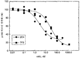
도 2A 및 2B는 MCF7 세포의 rHRGβ1 활성화에 대한 HER2 모노클로날 항체 2C4 및 7F3의 효과를 보여준다. 도 2A는 티로신 인산화의 HRG 자극의 2C4 또는 7F3 억제에 대한 투여량-반응 곡선을 보여준다. 도 2B는 2C4 또는 7F3에 의한 MCF7 세포에 대한 125I-표지된 rHRGβ1177-244 결합의 억제에 대한 투여량-반응 곡선을 보여준다.
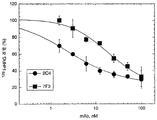
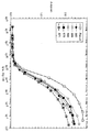
도 3은 HER2 모노클로날 항체 2C4 또는 7F3에 의한 인간 종양 세포주의 패널에 대한 특이적 125I-표지된 rHRGβ1177-244 결합의 억제를 보여준다. 모노클로날 항체-대조군은 rHRG 결합을 차단하지 않는 이소형-매치된 쥐 모노클로날 항체이다. 비특이적 125I-표지된 rHRGβ1177-244 결합은 100 nM rHRGβ1의 존재 하에 수행된 동시 (parallel) 인큐베이션으로부터 결정하였다. 비특이적 125I-표지된 rHRGβ1177-244 결합에 대한 값은 시험된 모든 세포주에 대해 총값의 1% 미만이었다.
도 4A 및 4B는 MDA-MB-175 (도 4A) 및 SK-BR-3 (도 4B) 세포의 증식에 대한 모노클로날 항체 2C4 및 4D5의 효과를 보여준다. MDA-MB-175 및 SK-BR-3 세포를 96 웰 플레이트에 씨딩하고, 2시간 동안 부착시켰다. 실험은 1% 혈청을 포함하는 배지에서 수행하였다. HER2 항체 또는 배지 단독을 첨가하고 세포를 2시간 동안 37℃에서 인큐베이션하였다. 이어서, rHRGβ1 (1 nM) 또는 배지 단독을 첨가하고, 세포를 4일 동안 인큐베이션하였다. 단일층을 세척하고, 0.5% 크리스탈 바이올렛으로 염색/고정시켰다. 세포 증식을 결정하기 위해서, 흡광도를 540 nm에서 측정하였다.
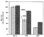
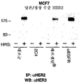
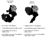
도 5A 및 5B는 낮은/정상 수준의 HER2를 발현하는 MCF7 세포 (도 5A) 및 높은 수준의 HER2를 발현하는 SK-BR-3 세포 (도 5B)에서 HER2와 HER3의 헤레굴린 (HRG) 의존성 회합에 대한 모노클로날 항체 2C4, 트라스투주맙 항체 또는 및 항-EGFR 항체의 효과를 보여준다 (하기 실시예 2 참조).
도 6A 및 6B는 무손상 (intact) 쥐 모노클로날 항체 2C4 (mu 2C4) 및 키메릭 2C4 Fab 단편의 활성을 비교한 것이다. 도 6A는 키메릭 2C4 Fab 또는 무손상 쥐 모노클로날 항체 2C4에 의한 MCF7 세포에 대한 125I-HRG 결합의 억제를 보여준다. MCF7 세포를 24웰 플레이트 (1 x 10⁵ 세포/웰)에 씨딩하고, 2일 동안 약 85% 융합도로 성장시켰다. 결합 실험은 문헌 [Lewis et al. Cancer Research 56:1457-1465 (1996)]에 기재된 바와 같이 수행하였다. 도 6B는 문헌 [Lewis et al. Cancer Research 56:1457-1465 (1996)]에 기재된 바와 같이 수행된 MCF7 세포에서 p180 티로신 인산화의 rHRGβ1 활성화 억제를 보여준다.
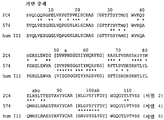
도 7A 및 7B는 쥐 모노클로날 항체 2C4의 가변 경쇄 (VL) (도 7A) 및 가변 중쇄 (VH) (도 7B) 도메인 (각각 서열 1 및 2); 인간화 2C4 버전 574의 VL 및 VH 도메인 (각각 서열 3 및 4), 및 인간 VL 및 VH 컨센서스 프레임워크 (hum κ1, 경쇄 카파 하위군 I; humIII, 중쇄 하위군 III) (각각 서열 5 및 6)의 아미노산 서열의 정렬을 보여준다. 별표는 인간화 2C4 버전 574와 쥐 모노클로날 항체 2C4 사이 또는 인간화 2C4 버전 574와 인간 프레임워크 사이의 차이를 확인한다. 상보성 결정 영역 (CDR)은 괄호로 나타낸다.
도 8A 내지 C는 실시예 3에서 ELISA에 의해 결정된, 키메릭 Fab 2C4 (Fab.v1) 및 복수의 인간화 2C4 변이체의 HER2 세포외 도메인 (ECD)에 대한 결합을 보여준다.
도 9는 백색 CDR 백본이 표지된, 모노클로날 항체 2C4의 VL 및 VH 도메인 (L1, L2, L3, H1, H2, H3)의 리본형 도식이다. 인간화 동안 돌연변이 발생에 의해 평가된 VH 측쇄 (실시예 3, 표 2 참조)도 도시된다.
도 10은 미토겐-활성화된 단백질 키나제 (MAPK)의 EGF, TGF-α, 또는 HRG-매개 활성화에 대한 모노클로날 항체 2C4 또는 트라스투주맙의 효과를 보여준다.
도 11A 및 11B는 각각 트라스투주맙 경쇄 (서열 14) 및 트라스투주맙 중쇄 (서열 15)의 아미노산 서열을 보여준다.
도 12A 및 12B는 각각 퍼투주맙 경쇄 (서열 16) 및 퍼투주맙 중쇄 (서열 17)의 아미노산 서열을 보여준다.
도 13은 활성화된 EGFR 또는 HER3과의 이종이량체화를 억제하는, HER2의 이종이량체 결합 부위에서의 2C4의 결합을 보여주는 모식도이다.
도 14는 HER2/HER3의 MAPK 및 Akt 경로에 대한 커플링을 보여준다.
도 15는 트라스투주맙 및 퍼투주맙의 활성을 비교한 것이다.
도 16은 HER2의 상이한 도메인을 보여주는 모식도이다.Figures 1A and 1B are epitopes of residues 22-645 in the extracellular domain (ECD) of HER2 (amino acid sequence comprising the signal sequence shown in Figure 1A; SEQ ID NO: 13), as determined by truncation mutant analysis and site directed mutation. Mapping is shown (Nakamura et al. J. of Virology 67(10):6179-6191 (1993); and Renz et al. J. Cell Biol. 125(6):1395-1406 (1994)). Different HER2-ECD truncation or point mutations were prepared from cDNA using the polymerase chain reaction technique. The HER2 mutant was expressed as a gD fusion protein in a mammalian expression plasmid. The expression plasmid uses a cytomegalovirus promoter/enhancer located downstream of the cDNA into which the SV40 terminator and the polyadenylation signal are inserted. Plasmid DNA was transfected into 293 cells. One day after transfection, cells were metabolically labeled overnight in low-concentration glucose DMEM without methionine and cysteine, containing 1% dialysis fetal bovine serum and 25 μCi of 35 S methionine and 35 S cysteine, respectively. The supernatant was collected, HER2 monoclonal antibody or control antibody was added to the supernatant and incubated at 4° C. for 2-4 hours. The complex was precipitated, added to a 10-20% tricine SDS gradient gel and subjected to electrophoresis at 100 V. The gel was electroblotting onto the membrane and analyzed by autoradiography. 1B, HER2 antibodies 7C2, 7F3, 2C4, 7D3, 3E8, 4D5, 2H11 and 3H4 bind different HER2 ECD epitopes.
2A and 2B show the effect of HER2 monoclonal antibodies 2C4 and 7F3 on rHRGβ1 activation in MCF7 cells. 2A shows dose-response curves for 2C4 or 7F3 inhibition of HRG stimulation of tyrosine phosphorylation. 2B shows a dose-response curve for inhibition of 125 I-labeled rHRGβ1 177-244 binding to MCF7 cells by 2C4 or 7F3.
3 shows inhibition of specific 125 I-labeled rHRGβ1 177-244 binding to a panel of human tumor cell lines by HER2 monoclonal antibodies 2C4 or 7F3. The monoclonal antibody-control is an isotype-matched murine monoclonal antibody that does not block rHRG binding. Nonspecific 125 I-labeled rHRGβ1 177-244 binding was determined from parallel incubations performed in the presence of 100 nM rHRGβ1. Values for nonspecific 125 I-labeled rHRGβ1 177-244 binding were less than 1% of the total value for all cell lines tested.
Figures 4A and 4B show the effect of monoclonal antibodies 2C4 and 4D5 on the proliferation of MDA-MB-175 (Figure 4A) and SK-BR-3 (Figure 4B) cells. MDA-MB-175 and SK-BR-3 cells were seeded into 96 well plates and allowed to adhere for 2 hours. The experiment was carried out in a medium containing 1% serum. HER2 antibody or medium alone was added and the cells were incubated at 37° C. for 2 hours. Then rHRGβ1 (1 nM) or medium alone was added and the cells were incubated for 4 days. The monolayer was washed and stained/fixed with 0.5% crystal violet. To determine cell proliferation, absorbance was measured at 540 nm.
Figures 5A and 5B show heregulin (HRG)-dependent association of HER2 and HER3 in MCF7 cells expressing low/normal levels of HER2 (Fig. 5A) and SK-BR-3 cells expressing high levels of HER2 (Fig. 5B). The effect of monoclonal antibody 2C4, trastuzumab antibody or and anti-EGFR antibody against against (see Example 2 below).
6A and 6B compare the activity of intact murine monoclonal antibody 2C4 (mu 2C4) and chimeric 2C4 Fab fragment. 6A shows inhibition of 125 I-HRG binding to MCF7 cells by chimeric 2C4 Fab or intact murine monoclonal antibody 2C4. MCF7 cells were seeded in 24 well plates (1 x 10 5 cells/well) and grown for 2 days to about 85% confluence. The binding experiment is described in Lewis et al. Cancer Research 56:1457-1465 (1996). 6B is described in Lewis et al. Cancer Research 56:1457-1465 (1996)] shows the inhibition of rHRGβ1 activation of p180 tyrosine phosphorylation in MCF7 cells.
Figures 7A and 7B show the variable light (V L ) (Figure 7A) and variable heavy (V H ) (Figure 7B) domains of murine monoclonal antibody 2C4 (SEQ ID NO: 1 and 2, respectively); V L and V H domains of humanized 2C4 version 574 (SEQ ID NO: 3 and 4, respectively), and human V L and V H consensus framework (hum κ1, light chain kappa subgroup I; humIII, heavy chain subgroup III) (SEQ ID NO: 5, respectively. And 6) shows the alignment of the amino acid sequence. The asterisk identifies the difference between the
Figures 8A-C show the binding of chimeric Fab 2C4 (Fab.v1) and multiple humanized 2C4 variants to the HER2 extracellular domain (ECD), as determined by ELISA in Example 3.
9 is a ribbon-shaped schematic of the V L and V H domains (L1, L2, L3, H1, H2, H3) of monoclonal antibody 2C4, labeled with a white CDR backbone. V H side chains evaluated by mutagenesis during humanization (see Example 3, Table 2) are also shown.
10 shows the effect of monoclonal antibody 2C4 or trastuzumab on EGF, TGF-α, or HRG-mediated activation of mitogen-activated protein kinase (MAPK).
11A and 11B show the amino acid sequences of trastuzumab light chain (SEQ ID NO: 14) and trastuzumab heavy chain (SEQ ID NO: 15), respectively.
12A and 12B show the amino acid sequences of Pertuzumab light chain (SEQ ID NO: 16) and Pertuzumab heavy chain (SEQ ID NO: 17), respectively.
13 is a schematic diagram showing the binding of 2C4 at the heterodimer binding site of HER2, which inhibits heterodimerization with activated EGFR or HER3.
14 shows the coupling of HER2/HER3 to the MAPK and Akt pathways.
15 is a comparison of the activities of trastuzumab and pertuzumab.
16 is a schematic diagram showing different domains of HER2.
I. 정의I. Definition
"HER 수용체"는 HER 수용체 패밀리에 속하는 수용체 단백질 티로신 키나제이고, EGFR, HER2, HER3 및 HER4 수용체 및 향후 확인되는 상기 패밀리의 다른 멤버를 포함한다. HER 수용체는 일반적으로 HER 리간드에 결합할 수 있는 세포외 도메인; 친지질 막 도메인(transmembrane domain); 보존된 세포내 티로신 키나제 도메인; 및 인산화될 수 있는 복수의 티로신 잔기를 갖는 카르복실 말단 신호전달 도메인을 포함할 것이다. HER 수용체는 "천연 서열" HER 수용체 또는 그의 "아미노산 서열 변이체"일 수 있다. 바람직하게는, HER 수용체는 천연 서열 인간 HER 수용체이다.“HER receptor” is a receptor protein tyrosine kinase belonging to the HER receptor family and includes EGFR, HER2, HER3 and HER4 receptors and other members of this family to be identified in the future. HER receptors generally have extracellular domains capable of binding to HER ligands; Lipophilic membrane domain (transmembrane domain); Conserved intracellular tyrosine kinase domain; And a carboxyl terminal signaling domain having a plurality of tyrosine residues capable of being phosphorylated. The HER receptor may be a "natural sequence" HER receptor or a "amino acid sequence variant" thereof. Preferably, the HER receptor is a native sequence human HER receptor.
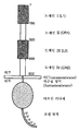
HER2의 세포외 도메인은 4개의 도메인, 즉 도메인 I (약 1-195의 아미노산 잔기), 도메인 II (약 196-320의 아미노산 잔기), 도메인 III (약 321-488의 아미노산 잔기) 및 도메인 IV (약 489-632의 아미노산 잔기) (잔기 넘버링은 시그날 펩티드 제외함)를 포함한다 (Garrett et al. Mol. Cell. 11: 495-505 (2003), Cho et al. Nature 421: 756-760 (2003), Franklin et al. Cancer cell 5:317-328 (2004), 또는 Plowman et al. Proc. Natl. Acad. Sci. 90:1746-1750 (1993), 및 본원의 도 16 참조).The extracellular domain of HER2 has four domains: domain I (amino acid residues of about 1-195), domain II (amino acid residues of about 196-320), domain III (amino acid residues of about 321-488) and domain IV ( Amino acid residues of about 489-632) (residue numbering excludes signal peptides) (Garrett et al. Mol. Cell. 11: 495-505 (2003), Cho et al. Nature 421: 756-760 (2003) ), Franklin et al. Cancer cell 5:317-328 (2004), or Plowman et al. Proc. Natl. Acad. Sci. 90:1746-1750 (1993), and see FIG. 16 herein).
용어 "ErbB1", "HER1", "표피 성장인자 수용체" 및 "EGFR"은 본원에서 상호 교환가능하게 사용되고, 그의 자연 발생 돌연변이체 형태 (예를 들어 Humphrey et al. PNAS (USA) 87:4207-4211 (1990)에 제시된 결실 돌연변이체 EGFR)를 포함하여 예를 들어 문헌 [Carpenter et al. Ann. Rev. Biochem. 56:881-914 (1987)]에 개시된 EGFR을 의미한다. erbB1은 EGFR 단백질 생성물을 코딩하는 유전자를 의미한다.The terms “ErbB1”, “HER1”, “epidermal growth factor receptor” and “EGFR” are used interchangeably herein, and their naturally occurring mutant forms (eg Humphrey et al. PNAS (USA) 87:4207-) 4211 (1990), the deletion mutant EGFR), including, for example, Carpenter et al. Ann. Rev. Biochem. 56:881-914 (1987)]. erbB1 refers to the gene encoding the EGFR protein product.
표현 "ErbB2" 및 "HER2"은 본원에서 상호 교환가능하게 사용되고, 예를 들어 문헌 [Semba et al., PNAS (USA) 82:6497-6501 (1985) 및 Yamamoto et al. Nature 319:230-234 (1986) (Genebank 기탁 번호 X03363)에 기재된 인간 HER2 단백질을 의미한다. 용어 "erbB2"는 인간 ErbB2를 코딩하는 유전자를 의미하고, "neu"는 래트 p185neu를 코딩하는 유전자를 의미한다. 바람직한 HER2는 천연 서열 인간 HER2이다.The expressions “ErbB2” and “HER2” are used interchangeably herein and are described, for example, in Semba et al., PNAS (USA) 82:6497-6501 (1985) and Yamamoto et al. Nature 319:230-234 (1986) (Genebank Accession No. X03363) refers to the human HER2 protein described. The term "erbB2" refers to a gene encoding human ErbB2, and "neu" refers to a gene encoding rat p185 neu. A preferred HER2 is a native sequence human HER2.
"ErbB3" 및 "HER3"은 예를 들어 미국 특허 5,183,884 및 5,480,968 및 문헌 [Kraus et al. PNAS (USA) 86:9193-9197 (1989)]에 기재된 수용체 폴리펩티드를 의미한다.“ErbB3” and “HER3” are described, for example, in US Patents 5,183,884 and 5,480,968 and in Kraus et al. PNAS (USA) 86:9193-9197 (1989)].
용어 "ErbB4" 및 "HER4"는 본원에서 예를 들어 1999년 4월 22일 공개된 WO99/19488에 개시된 그의 이소형을 포함하여, 예를 들어 유럽 특허 출원 599,274; 문헌 [Plowman et al., Proc. Natl. Acad. Sci. USA, 90:1746-1750 (1993); Plowman et al., Nature, 366:473-475 (1993)]에 개시된 수용체 폴리펩티드를 의미한다.The terms “ErbB4” and “HER4” refer to, for example, European patent application 599,274; See Plowman et al., Proc. Natl. Acad. Sci. USA, 90:1746-1750 (1993); Plowman et al., Nature, 366:473-475 (1993).
"HER 리간드"는 HER 수용체에 대해 결합하고(하거나) 활성화시키는 폴리펩티드를 의미한다. 본원에서 특히 흥미로운 HER 리간드는 천연 서열 인간 HER 리간드, 예를 들어 표피 성장인자 (EGF) (Savage et al., J. Biol. Chem. 247:7612-7621 (1972)); 전환 성장인자 알파 (TGF-α) (Marquardt et al., Science 223:1079-1082 (1984)); 신경초종 또는 케라티노사이트 자가분비 성장인자로도 알려진 암피레굴린 (Shoyab et al. Science 243: 1074-1076 (1989); Kimura et al. Nature 348:257-260 (1990); 및 Cook et al. Mol Cell Biol 11:2547-2557 (1991)); 베타셀룰린 (Shing et al., Science 259:1604-1607 (1993); 및 Sasada et al. Biochem. Biophys. Res. Commun. 190:1173 (1993)); 헤파린 결합 표피 성장인자 (HB-EGF) (Higashiyama et al., Science 251:936-939 (1991)); 에피레굴린 (Toyoda et al., J. Biol. Chem. 270:7495-7500 (1995); 및 Komurasaki et al. Oncogene 15:2841-2848 (1997)); 헤레굴린 (하기 참조); 뉴레굴린-2 (NRG-2) (Carraway et al., Nature 387:512-516 (1997)); 뉴레굴린-3 (NRG-3) (Zhang et al., Proc. Natl. Acad. Sci. 94:9562-9567 (1997)); 뉴레굴린-4 (NRG-4) (Harari et al. Oncogene 18:2681-89 (1999)) 또는 크립토 (CR-1) (Kannan et al. J. Biol Chem. 272(6):3330-3335 (1997))이다. EGFR에 결합하는 HER 리간드는 EGF, TGF-α, 암피레굴린, 베타셀룰린, HB-EGF 및 에피레굴린을 포함한다. HER3에 결합하는 HER 리간드는 헤레굴린을 포함한다. HER4에 결합할 수 있는 HER 리간드는 베타셀룰린, 에피레굴린, HB-EGF, NRG-2, NRG-3, NRG-4 및 헤레굴린을 포함한다.“HER ligand” refers to a polypeptide that binds and/or activates a HER receptor. HER ligands of particular interest herein include native sequence human HER ligands, such as epidermal growth factor (EGF) (Savage et al., J. Biol. Chem. 247:7612-7621 (1972)); Transforming growth factor alpha (TGF-α) (Marquardt et al., Science 223:1079-1082 (1984)); Ampiregulin, also known as schwannoma or keratinocyte autocrine growth factor (Shoyab et al. Science 243: 1074-1076 (1989); Kimura et al. Nature 348:257-260 (1990); and Cook et al. Mol. Cell Biol 11:2547-2557 (1991)); Betacellulose (Shing et al., Science 259:1604-1607 (1993); and Sasada et al. Biochem. Biophys. Res. Commun. 190:1173 (1993)); Heparin binding epidermal growth factor (HB-EGF) (Higashiyama et al., Science 251:936-939 (1991)); Epiregulin (Toyoda et al., J. Biol. Chem. 270:7495-7500 (1995); and Komurasaki et al. Oncogene 15:2841-2848 (1997)); Heregulin (see below); Neuregulin-2 (NRG-2) (Carraway et al., Nature 387:512-516 (1997)); Neuregulin-3 (NRG-3) (Zhang et al., Proc. Natl. Acad. Sci. 94:9562-9567 (1997)); Neuroregulin-4 (NRG-4) (Harari et al. Oncogene 18:2681-89 (1999)) or Crypto (CR-1) (Kannan et al. J. Biol Chem. 272(6):3330-3335 ( 1997)). HER ligands that bind EGFR include EGF, TGF-α, ampiregulin, betacellulin, HB-EGF and epiregulin. HER ligands that bind HER3 include heregulin. HER ligands capable of binding to HER4 include betacellulin, epiregulin, HB-EGF, NRG-2, NRG-3, NRG-4 and heregulin.
본원에서 사용되는 "헤레굴린" (HRG)은 미국 특허 5,641,869 또는 문헌 [Marchionni et al., Nature et al., 362:312-318 (1993)]에 개시된 바와 같은 헤레굴린 유전자 생성물에 의해 코딩되는 폴리펩티드를 의미한다. 헤레굴린의 예는 헤레굴린-α, 헤레굴린-β1, 헤레굴린-β2 및 헤레굴린-β3 (Holmes et al., Science, 256:1205-1210 (1992); 및 미국 특허 5,641,869); neu 분화 인자 (NDF) (Peles et al. Cell 69: 205-216 (1992)); 아세틸콜린 수용체-유도 활성 (ARIA) (Falls et al. Cell 72:801-815 (1993)); 아교세포 성장인자 (GGF) (Marchionni et al., Nature, 362:312-318 (1993)); 감각 및 운동 뉴런 유도 인자 (SMDF) (Ho et al. J. Biol Chem. 270:14523-14532 (1995)); γ-헤레굴린 (Schaefer et al. Oncogene 15:1385-1394 (1997))를 포함한다. 상기 용어는 천연 서열 HRG 폴리펩티드의 생물학적 활성 단편 및(또는) 아미노산 서열 변이체, 예를 들어 그의 EGF 유사 도메인 단편 (예를 들어 rHRGβ1177-244)를 포함한다.As used herein, “heregulin” (HRG) is a polypeptide encoded by a heregulin gene product as disclosed in US Pat. No. 5,641,869 or Marchionni et al., Nature et al., 362:312-318 (1993). Means. Examples of heregulins include heregulin-α, heregulin-β1, heregulin-β2 and heregulin-β3 (Holmes et al., Science, 256:1205-1210 (1992); and U.S. Patent 5,641,869); neu differentiation factor (NDF) (Peles et al. Cell 69: 205-216 (1992)); Acetylcholine receptor-induced activity (ARIA) (Falls et al. Cell 72:801-815 (1993)); Glial growth factor (GGF) (Marchionni et al., Nature, 362:312-318 (1993)); Sensory and motor neuron inducing factor (SMDF) (Ho et al. J. Biol Chem. 270:14523-14532 (1995)); γ-heregulin (Schaefer et al. Oncogene 15:1385-1394 (1997)). The term includes biologically active fragments and/or amino acid sequence variants of native sequence HRG polypeptides, such as EGF-like domain fragments thereof (eg rHRGβ1 177-244 ).
본원에서 "HER 이량체"는 적어도 2개의 상이한 HER 수용체를 포함하는 비공유 회합 이량체이다. 상기 복합체는 2 이상의 HER 수용체를 발현하는 세포가 HER 리간드에 노출될 때 형성될 수 있고, 면역침전에 의해 분리되어, 예를 들어 문헌 [Sliwkowski et al., J. Biol. Chem., 269(20): 14661-14665 (1994)]에 기재된 바와 같이 SDS-PAGE에 의해 분석될 수 있다. 상기 HER 이량체의 예는 EGFR-HER2, HER2-HER3 및 HER3-HER4 이종이량체를 포함한다. 또한, HER 이량체는 상이한 HER 수용체, 예를 들어 HER3, HER4 또는 EGFR과 조합된 2 이상의 HER2 수용체를 포함할 수 있다. 다른 단백질, 예를 들어 시토킨 수용체 서브유닛 (예를 들어 gp130)은 이량체와 회합될 수 있다.A “HER dimer” as used herein is a non-covalently associated dimer comprising at least two different HER receptors. Such complexes can be formed when cells expressing two or more HER receptors are exposed to HER ligands and are separated by immunoprecipitation, eg, Sliwkowski et al., J. Biol. Chem., 269(20): 14661-14665 (1994)]. Examples of such HER dimers include EGFR-HER2, HER2-HER3 and HER3-HER4 heterodimers. In addition, HER dimers may comprise two or more HER2 receptors in combination with different HER receptors, for example HER3, HER4 or EGFR. Other proteins, such as cytokine receptor subunits (eg gp130), can be associated with dimers.
HER2 상의 "이종이량체 결합 부위"는 그와 이량체 형성시에 EGFR, HER3 또는 HER4의 세포외 도메인 내의 영역과 접촉하거나 이 영역과 연결되는 HER2의 세포외 도메인 내의 영역을 의미한다. 상기 영역은 HER2의 도메인 II에서 발견된다 [Franklin et al. Cancer Cell 5:317-328 (2004)]."Heterodimer binding site" on HER2 refers to a region within the extracellular domain of HER2 that contacts or connects to a region within the extracellular domain of EGFR, HER3 or HER4 upon dimer formation therewith. This region is found in domain II of HER2 [Franklin et al. Cancer Cell 5:317-328 (2004)].
"HER 활성화" 또는 "HER2 활성화"는 임의의 하나 이상의 HER 수용체, 또는 HER2 수용체의 활성화 또는 인산화를 의미한다. 일반적으로, HER 활성화는 신호 전달 (예를 들어 HER 수용체 또는 기질 폴리펩티드의 티로신 잔기를 인산화시키는 HER 수용체의 세포내 키나제 도메인에 의해 유도되는)을 야기한다. HER 활성화는 목적하는 HER 수용체를 포함하는 HER 이량체에 대한 HER 리간드 결합에 의해 매개될 수 있다. HER 이량체에 대한 HER 리간드 결합은 이량체 내의 하나 이상의 HER 수용체의 키나제 도메인을 활성화시킬 수 있고, 이에 의해 하나 이상의 HER 수용체 내의 티로신 잔기의 인산화 및(또는) 추가의 기질 폴리펩티드(들), 예를 들어 Akt 또는 MAPK 세포내 키나제 내의 티로신 잔기의 인산화를 야기한다. “HER activation” or “HER2 activation” means the activation or phosphorylation of any one or more HER receptors, or HER2 receptors. In general, HER activation results in signal transduction (e.g. induced by the HER receptor or the intracellular kinase domain of the HER receptor that phosphorylates the tyrosine residue of the substrate polypeptide). HER activation can be mediated by binding of a HER ligand to a HER dimer comprising the desired HER receptor. HER ligand binding to a HER dimer can activate the kinase domain of one or more HER receptors in the dimer, thereby phosphorylating tyrosine residues in one or more HER receptors and/or additional substrate polypeptide(s), e.g. For example, it causes phosphorylation of tyrosine residues in Akt or MAPK intracellular kinases.
"천연 서열 (native sequence)" 폴리펩티드는 자연에서 유도되는 폴리펩티드 (예를 들어, HER 수용체 또는 HER 리간드)와 동일한 아미노산 서열을 갖는 것이다. 상기 천연 서열 폴리펩티드는 자연으로부터 단리될 수 있거나, 재조합 또는 합성 수단에 의해 제조될 수 있다. 따라서, 천연 서열 폴리펩티드는 자연 발생 인간 폴리펩티드, 쥐 폴리펩티드, 또는 임의의 다른 포유동물종으로부터의 폴리펩티드의 아미노산 서열을 가질 수 있다. A “native sequence” polypeptide is one that has the same amino acid sequence as a naturally derived polypeptide (eg, HER receptor or HER ligand). The native sequence polypeptide can be isolated from nature or can be produced by recombinant or synthetic means. Thus, the native sequence polypeptide can have the amino acid sequence of a naturally occurring human polypeptide, a murine polypeptide, or a polypeptide from any other mammalian species.
용어 "아미노산 서열 변이체"는 천연 서열 폴리펩티드와 일정 정도 상이한 아미노산 서열을 갖는 폴리펩티드이다. 통상적으로, 아미노산 서열 변이체는 천연 HER 리간드의 적어도 하나의 수용체 결합 도메인과 또는 천연 HER 수용체의 적어도 하나의 리간드 결합 도메인과 적어도 약 70%의 상동성을 가질 것이고, 바람직하게는 상기 수용체 또는 리간드 결합 도메인과 적어도 약 80%, 보다 바람직하게는 적어도 약 90% 상동성일 것이다. 아미노산 서열 변이체는 천연 아미노산 서열의 아미노산 서열 내의 특정 위치에서 치환, 결실 및(또는) 삽입을 갖는다.The term “amino acid sequence variant” is a polypeptide having an amino acid sequence that is somewhat different from the native sequence polypeptide. Typically, the amino acid sequence variant will have at least about 70% homology with at least one receptor binding domain of the native HER ligand or with at least one ligand binding domain of the native HER receptor, preferably the receptor or ligand binding domain. At least about 80%, more preferably at least about 90% homology with. Amino acid sequence variants have substitutions, deletions and/or insertions at specific positions within the amino acid sequence of the native amino acid sequence.
"상동성"은 서열을 정렬시키고 필요한 경우 최대 서열 상동성 비율을 달성하도록 갭을 도입시킨 후에 동일한 아미노산 서열 변이체 내의 잔기의 백분율로서 정의된다. 정렬을 위한 방법 및 컴퓨터 프로그램은 당업계에 잘 공지되어 있다. 하나의 상기 컴퓨터 프로그램은 1991년 12월 10일에 미국 저작국 (United States Copyright Office, 미국 워싱턴 디. 씨. 20559)에 사용자 제출서류로 출원된, 제넨테크 (Genentech) 소유의 "Align-2"이다. “Homology” is defined as the percentage of residues within the same amino acid sequence variant after aligning the sequences and introducing gaps to achieve the maximum ratio of sequence homology if necessary. Methods and computer programs for alignment are well known in the art. One of the above computer programs is "Align-2" owned by Genentech, filed as a user submission by the United States Copyright Office, Washington D. C. 20559, on December 10, 1991. to be.
본원에서 용어 "항체"는 가장 넓은 의미로 사용되고, 구체적으로 무손상 (intact) 모노클로날 항체, 폴리클로날 항체, 적어도 2개의 무손상 항체로 형성된 다중특이적 항체 (예를 들어 이중특이적 항체), 및 목적하는 생물학적 활성을 나타내는 한 항체 단편을 포함한다. The term "antibody" is used in the broadest sense herein, specifically, an intact monoclonal antibody, a polyclonal antibody, a multispecific antibody formed of at least two intact antibodies (eg, a bispecific antibody ), and one antibody fragment exhibiting a desired biological activity.
본원에서 사용되는 용어 "모노클로날 항체"는 실질적으로 균질한 항체 집단으로부터 수득된 항체를 말하는데, 즉 이러한 집단을 구성하는 개개의 항체는 소량으로 존재할 수도 있는, 모노클로날 항체 생성 동안 발생할 수 있는 가능한 변이체를 제외하고는 동일하고(하거나) 동일한 에피토프에 결합한다. 상이한 결정자 (에피토프)에 대해 작용하는 상이한 항체를 일반적으로 포함하는 폴리클로날 항체 제제에 비해, 각각의 모노클로날 항체는 항원 상의 단일 결정자에 대해 작용한다. 그의 특이성에 추가하여, 모노클로날 항체는 다른 면역글로불린에 의해 오염되지 않는다는 점에서 유리하다. 변경 표현 "모노클로날"은 항체의 실질적으로 균질한 집단으로부터 얻은 항체의 특성을 나타내고, 임의의 특정 방법에 의한 항체 제조를 필요로 하는 것으로서 생각하지 않아야 한다. 예를 들어, 본 발명에 따라 사용되는 모노클로날 항체는 문헌 [Kohler et al., Nature 256:495 (1975)]에 처음 기재된 하이브리도마 방법에 의해 제조할 수 있거나, 또는 재조합 DNA 방법 (예를 들어 미국 특허 4,816,567 참조)에 의해 제조할 수 있다. "모노클로날 항체"는 또한 예를 들어 문헌 [Clackson et al., Nature 352:624-628 (1991) 및 Marks et al., J. Mol. Biol. 222:581-597 (1991)]에 기재된 기술을 사용하여 파지 항체 라이브러리로부터 단리할 수 있다.As used herein, the term “monoclonal antibody” refers to an antibody obtained from a population of substantially homogeneous antibodies, ie, the individual antibodies constituting this population may be present in small amounts, which may occur during monoclonal antibody production. Except for possible variants, they bind to the same and/or the same epitope. Compared to polyclonal antibody preparations that generally contain different antibodies that act against different determinants (epitopes), each monoclonal antibody acts against a single determinant on the antigen. In addition to its specificity, monoclonal antibodies are advantageous in that they are not contaminated by other immunoglobulins. The altered expression “monoclonal” refers to the properties of an antibody obtained from a substantially homogeneous population of antibodies and should not be considered as requiring antibody preparation by any particular method. For example, the monoclonal antibody used according to the present invention can be prepared by the hybridoma method first described in Kohler et al., Nature 256:495 (1975), or a recombinant DNA method (e.g. For example, see U.S. Patent 4,816,567). “Monoclonal antibodies” are also described, for example, in Clackson et al., Nature 352:624-628 (1991) and Marks et al., J. Mol. Biol. 222:581-597 (1991)] can be used to isolate from phage antibody libraries.
본원에서 모노클로날 항체는 구체적으로 요구되는 생물학적 활성을 보이는 한, 중쇄 및(또는) 경쇄의 일부가 특정 종에서 유래하거나 특정 항체 클래스 또는 서브클래스에 속하는 항체의 대응하는 서열과 동일하거나 상동성이고 사슬(들)의 나머지는 다른 종에서 유래하거나 다른 항체 종류 또는 서브클래스에 속하는 항체의 대응하는 서열과 동일하거나 상동성인 "키메릭" 항체 및 상기 항체의 단편을 포함한다 (미국 특허 4,816,567; 및 Morrison et al. Proc. Natl. Acad. Sci. USA 81:6851-6855 (1984)). 본원에서 목적하는 키메릭 항체는 비-인간 영장류 (예, 구세계 원숭이, 유인원)에서 유래한 가변 도메인 항원 결합 서열 및 인간 불변 영역 서열을 포함하는 "프리머타이즈드 (primatized)" 항체를 포함한다. Monoclonal antibodies herein are part of the heavy and/or light chains that are identical or homologous to the corresponding sequences of antibodies derived from a particular species or belonging to a particular antibody class or subclass, as long as they exhibit the specifically required biological activity. The remainder of the chain(s) includes "chimeric" antibodies and fragments of such antibodies that are identical or homologous to the corresponding sequences of antibodies derived from different species or belonging to different antibody classes or subclasses (US Pat. No. 4,816,567; And Morrison et al. Proc. Natl. Acad. Sci. USA 81:6851-6855 (1984)). Chimeric antibodies of interest herein include “primatized” antibodies comprising variable domain antigen binding sequences and human constant region sequences derived from non-human primates (eg, Old World monkeys, apes).
"항체 단편"은 바람직하게는 그의 항원 결합 또는 가변 영역을 포함하는 무손상 항체의 일부를 포함한다. 항체 단편의 예는 Fab, Fab', F(ab')2 및 Fv 단편; 디아바디 (diabody); 선형 항체; 단쇄 항체 분자; 및 항체 단편(들)으로부터 형성된 다중특이적 항체를 포함한다.An “antibody fragment” preferably includes a portion of an intact antibody comprising an antigen binding or variable region thereof. Examples of antibody fragments include Fab, Fab', F(ab') 2 and Fv fragments; Diabody; Linear antibodies; Single chain antibody molecules; And multispecific antibodies formed from antibody fragment(s).
"무손상 항체 (intact antibody)"는 항원 결합 가변 영역 및 경쇄 불변 도메인 (C1) 및 중쇄 불변 도메인, CH1, CH2 및 CH3을 포함하는 항체이다. 불변 도메인은 천연 서열 불변 도메인 (예를 들어 인간 천연 서열 불변 도메인) 또는 그의 아미노산 서열 변이체일 수 있다. 바람직하게는, 무손상 항체는 하나 이상의 이펙터 기능을 갖는다.An “intact antibody” is an antibody comprising an antigen binding variable region and a light chain constant domain (C1) and a heavy chain constant domain,
항체 "이펙터 기능 (effector function)"은 항체의 Fc 영역 (천연 서열 Fc 영역 또는 아미노산 서열 변이체 Fc 영역)에 기인하는 생물학적 활성을 의미한다. 항체 이펙터 기능의 예는 C1q 결합; 보체 의존성 세포독성; Fc 수용체 결합; 항체 의존성 세포 매개 세포독성 (ADCC); 포식작용; 세포 표면 수용체 (예를 들어 B 세포 수용체; BCR)의 하향조절 등을 포함한다.An antibody “effector function” refers to a biological activity attributable to the Fc region (natural sequence Fc region or amino acid sequence variant Fc region) of an antibody. Examples of antibody effector functions include C1q binding; Complement dependent cytotoxicity; Fc receptor binding; Antibody dependent cell mediated cytotoxicity (ADCC); Phagocytosis; Downregulation of cell surface receptors (eg B cell receptor; BCR), and the like.
그들의 중쇄의 불변 도메인의 아미노산 서열에 따라, 무손상 항체는 상이한 "클래스"로 지정될 수 있다. 면역글로불린의 5가지 주요 클래스 IgA, IgD, IgE, IgG 및 IgM이 존재하고, 이들 중 몇몇은 서브클래스 (이소형), 예를 들어, IgG1, IgG2, IgG3, IgG4, IgA 및 IgA2로 추가로 나누어질 수 있다. 상이한 클래스의 항체에 대응하는 중쇄 불변 도메인은 각각 α, δ, ε, γ, 및 μ로 불린다. 상이한 클래스의 면역글로불린의 서브유닛 구조 및 3차원 형상은 잘 알려져 있다.Depending on the amino acid sequence of the constant domains of their heavy chains, intact antibodies can be assigned to different “classes”. There are five major classes of immunoglobulins IgA, IgD, IgE, IgG and IgM, some of which are further divided into subclasses (isotypes), e.g. IgG1, IgG2, IgG3, IgG4, IgA and IgA2. I can lose. The heavy chain constant domains corresponding to different classes of antibodies are referred to as α, δ, ε, γ, and μ, respectively. The subunit structures and three-dimensional shapes of different classes of immunoglobulins are well known.
"항체 의존성 세포 매개 세포독성" 및 "ADCC"는 Fc 수용체 (FcR)를 발현하는 비특이적 세포독성 세포 (예를 들어 천연 킬러 (NK) 세포, 호중구 및 마크로파지)가 표적 세포 상의 결합된 항체를 인식한 후, 표적 세포의 용해를 야기하는 세포 매개 반응이다. ADCC를 매개하는 1차 세포인 NK 세포는 FcγRIII만을 발현하지만, 단핵구는 FcγRI, FcγRII 및 FcγRIII을 발현한다. 조혈 세포 상의 FcR 발현은 문헌 [Ravetch and Kinet, Anne. Rev. Immunol 9: 457-92 (1991)]의 464 페이지 표 3에 요약되어 있다. 목적하는 분자의 ADCC 활성을 평가하기 위해서, 예를 들어 미국 특허 5,500,362 또는 5,821,337에 기재된 시험관내 ADCC 분석을 수행할 수 있다. 상기 분석에 유용한 이펙터 세포는 말초혈액 단핵세포 (PBMC) 및 천연 킬러 (NK) 세포를 포함한다. 별법으로, 또는 부가적으로, 목적하는 분자의 ADCC 활성은 예를 들어 문헌 [Clynes et al. PNAS (USA) 95: 652-656 (1998)]에 기재된 동물 모델에서 생체 내에서 평가할 수 있다. "Antibody dependent cell mediated cytotoxicity" and "ADCC" are non-specific cytotoxic cells expressing an Fc receptor (FcR) (eg natural killer (NK) cells, neutrophils and macrophages) that recognize bound antibodies on target cells. Later, it is a cell-mediated reaction that causes lysis of the target cell. The primary cells for mediating ADCC, NK cells, express only FcγRIII, whereas monocytes express FcγRI, FcγRII and FcγRIII. FcR expression on hematopoietic cells is described in Ravetch and Kinet, Anne. Rev. Immunol 9: 457-92 (1991)], page 464 is summarized in Table 3. In order to evaluate the ADCC activity of the molecule of interest, the in vitro ADCC assay described in, for example, US Pat. Nos. 5,500,362 or 5,821,337 can be performed. Effector cells useful for this assay include peripheral blood mononuclear cells (PBMC) and natural killer (NK) cells. Alternatively, or in addition, ADCC activity of the molecule of interest can be determined by, for example, Clynes et al. PNAS (USA) 95: 652-656 (1998)].
"인간 이펙터 세포"는 하나 이상의 FcR을 발현하고 이펙터 기능을 수행하는 백혈구이다. 바람직하게는, 상기 세포는 적어도 FcγRIII를 발현하고, ADCC 이펙터 기능을 수행한다. ADCC를 매개하는 인간 백혈구의 예는 말초혈액 단핵세포 (PBMC), 천연 킬러 (NK) 세포, 단핵구, 세포독성 T 세포 및 호중구를 포함하고, PBMC 및 NK 세포가 바람직하다. 이펙터 세포는 본원에서 설명되는 바와 같이 천연 공급원, 예를 들어 혈액 또는 PBMC로부터 단리할 수 있다.“Human effector cells” are leukocytes that express one or more FcRs and perform effector functions. Preferably, the cells express at least FcγRIII and perform ADCC effector functions. Examples of human leukocytes that mediate ADCC include peripheral blood mononuclear cells (PBMC), natural killer (NK) cells, monocytes, cytotoxic T cells and neutrophils, with PBMC and NK cells being preferred. Effector cells can be isolated from natural sources such as blood or PBMCs as described herein.
용어 "Fc 수용체" 또는 "FcR"은 항체의 Fc 영역에 결합하는 수용체를 설명하기 위해 사용된다. 바람직한 FcR은 천연 서열 인간 FcR이다. 더욱이, 바람직한 FcR은 IgG 항체 (감마 수용체)에 결합하고 상기 수용체의 대립 유전자 변이체 및 교대 스플라이싱된 형태를 포함하여 FcγRI, FcγRII 및 FcγRIII 서브클래스의 수용체를 포함하는 것이다. FcγRII 수용체는 FcγRIIA ("활성화 수용체") 및 FcγRIIB ("억제 수용체")를 포함하고, 이들은 그의 세포질 도메인에서 주로 상이한 유사한 아미노산 서열을 갖는다. 활성화 수용체 FcγRIIA는 그의 세포질 도메인에 면역수용체 티로신계 활성화 모티프 (ITAM)를 포함한다. 억제 수용체 FcγRIIB는 그의 세포질 도메인에 면역수용체 티로신계 억제 모티프 (ITIM)를 포함한다 (Daeron, Annu. Rev. Immunol. 15: 203-234 (1997) 참고). FcR은 문헌 [Ravetch and Kinet, Annu. Rev. Immunol 9: 457-92 (1991); Capel et al., Immunomethods 4: 25-34 (1994); 및 de Haas et al., J. Lab. Clin. Med. 126: 330-41 (1995)]에 개시되어 있다. 미래에 확인되는 것을 포함하여 다른 FcR가 본원의 용어 "FcR"에 포함된다. 또한, 이 용어는 모체 IgG의 태아로의 전달에 작용하는 신생아의 수용체 FcRn를 포함한다 (Guyer et al., J. Immunol. 117: 587 (1976) 및 Kim et al., J. Immunol. 24: 249 (1994)).The terms “Fc receptor” or “FcR” are used to describe a receptor that binds to the Fc region of an antibody. A preferred FcR is a native sequence human FcR. Moreover, preferred FcRs are those that bind to IgG antibodies (gamma receptors) and comprise receptors of the FcγRI, FcγRII and FcγRIII subclasses, including allelic variants and alternating spliced forms of the receptor. FcγRII receptors include FcγRIIA (“activating receptor”) and FcγRIIB (“inhibiting receptor”), which have similar amino acid sequences that differ primarily in their cytoplasmic domain. The activating receptor FcγRIIA contains an immunoreceptor tyrosine-based activation motif (ITAM) in its cytoplasmic domain. Inhibitory receptor FcγRIIB contains an immunoreceptor tyrosine-based inhibitory motif (ITIM) in its cytoplasmic domain (see Daeron, Annu. Rev. Immunol. 15: 203-234 (1997)). FcR is described in Ravetch and Kinet, Annu. Rev. Immunol 9: 457-92 (1991); Capel et al., Immunomethods 4: 25-34 (1994); And de Haas et al., J. Lab. Clin. Med. 126: 330-41 (1995). Other FcRs, including those identified in the future, are included in the term "FcR" herein. In addition, the term includes the neonatal receptor FcRn, which acts on the delivery of maternal IgG to the fetus (Guyer et al., J. Immunol. 117: 587 (1976) and Kim et al., J. Immunol. 24: 249 (1994)).
"보체 의존성 세포독성" 또는 "CDC"는 보체의 존재 하에 표적을 용해시키는 분자의 능력을 의미한다. 보체 활성화 경로는 보체 시스템의 제1 성분 (C1q)이 동족 항원과 복합체화된 분자 (예를 들어 항체)에 결합함으로써 개시된다. 보체 활성화를 평가하기 위해서, 예를 들어 문헌 [Gazzano-Santoro et al., J. Immunol. Methods 202: 163 (1996)]에 기재된 CDC 분석을 수행할 수 있다.“Complement dependent cytotoxicity” or “CDC” refers to the ability of a molecule to lyse a target in the presence of complement. The complement activation pathway is initiated by the binding of the first component (C1q) of the complement system to a molecule (eg antibody) complexed with a cognate antigen. To assess complement activation, see, for example, Gazzano-Santoro et al., J. Immunol. Methods 202: 163 (1996)].
"천연 항체 (native antibody)"는 대체로 2개의 동일한 경쇄 (L) 및 2개의 동일한 중쇄 (H)로 이루어진 약 150,000 달톤의 이종사량체 당단백질이다. 각각의 경쇄는 하나의 디술피드 공유결합에 의해 중쇄에 연결되고, 디술피드 연결의 수는 상이한 면역글로불린 이소형의 중쇄에서 상이하다. 또한, 각각의 중쇄 및 경쇄는 일정하게 이격된 사슬내 디술피드 다리를 갖는다. 각각의 중쇄는 한 말단에 가변 도메인 (VH)을 갖고, 이어서 많은 불변 도메인이 존재한다. 각각의 경쇄는 한 말단에 가변 도메인 (VL)을 갖고, 그의 다른 말단에 불변 도메인을 갖는다. 경쇄의 불변 도메인은 중쇄의 제1 불변 도메인에 정렬되고, 경쇄 가변 도메인은 중쇄의 가변 도메인에 정렬된다. 특정 아미노산 잔기는 경쇄 및 중쇄 가변 도메인 사이의 계면을 형성하는 것으로 생각된다.A “native antibody” is a heterotetrameric glycoprotein of about 150,000 Daltons, usually consisting of two identical light chains (L) and two identical heavy chains (H). Each light chain is linked to the heavy chain by one covalent disulfide bond, and the number of disulfide linkages is different in the heavy chains of different immunoglobulin isotypes. In addition, each of the heavy and light chains has uniformly spaced intra-chain disulfide bridges. Each heavy chain has a variable domain (V H ) at one end, followed by many constant domains. Each light chain has a variable domain (V L ) at one end and a constant domain at its other end. The constant domain of the light chain is aligned with the first constant domain of the heavy chain, and the light chain variable domain is aligned with the variable domain of the heavy chain. Certain amino acid residues are thought to form the interface between the light and heavy chain variable domains.
용어 "가변"은 가변 도메인의 특정 부분이 항체 내의 서열이 크게 상이하고 그의 특정 항원에 대한 각각의 특정 항체의 결합 및 특이성에 사용됨을 의미한다. 그러나, 가변성은 항체의 가변 도메인 전체에 걸쳐 균일하게 분포되지 않는다. 이것은 경쇄 및 중쇄 가변 도메인 모두에 초가변 영역으로 불리는 3개의 세그먼트에 집중된다. 가변 도메인의 보다 보존도가 큰 부분은 프레임워크 영역 (FR)으로 언급된다. 천연 중쇄 및 경쇄의 가변 도메인은 루프 연결부를 형성하고 일부 경우에 β-시트 구조의 일부를 형성하는 3개의 초가변 영역에 의해 연결되는, 주로 β-시트 형태를 취하는 4개의 FR을 각각 포함한다. 각 사슬 내의 초가변 영역은 FR에 의해 매우 근접하게 함께 유지되고, 다른 사슬의 초가변 영역과 함께 항체의 항원 결합 부위 형성에 기여한다 (Kabat et al., Sequences of Proteins of Immunological Interest, 5th Ed. Public Health Service, National Institutes of Health, Bethesda, MD. (1991)] 참조). 불변 도메인은 항체의 항원 결합에 직접 관여하지 않지만, 상이한 이펙터 기능, 예를 들어 항체 의존성 세포독성 (ADCC)에서 항체의 참여를 보인다. The term “variable” means that certain portions of the variable domains differ greatly in sequence within the antibody and are used for the binding and specificity of each specific antibody for its specific antigen. However, the variability is not uniformly distributed throughout the variable domain of the antibody. It is concentrated in three segments called hypervariable regions in both the light and heavy chain variable domains. The more conserved portion of the variable domain is referred to as the framework region (FR). The variable domains of the natural heavy and light chains each comprise four FRs that take the form of a predominantly β-sheet, joined by three hypervariable regions that form loop linkages and in some cases form part of the β-sheet structure. The hypervariable regions within each chain are held together in close proximity by the FR, and together with the hypervariable regions of the other chains contribute to the formation of the antigen binding site of the antibody (Kabat et al., Sequences of Proteins of Immunological Interest, 5th Ed. Public Health Service, National Institutes of Health, Bethesda, MD. (1991)). The constant domains are not directly involved in the antigen binding of the antibody, but show participation of the antibody in different effector functions, such as antibody dependent cytotoxicity (ADCC).
본원에서 사용되는 용어 "초가변 영역"은 항원 결합에 필요한 항체의 아미노산 잔기를 의미한다. 초가변 영역은 "상보성 결정 영역" 또는 "CDR"로부터의 아미노산 잔기 (예를 들어, 경쇄 가변 도메인의 잔기 24-34 (L1), 50-56 (L2) 및 89-97 (L3) 및 중쇄 가변 도메인의 31-35 (H1), 50-65 (H2) 및 95-102 (H3); Kabat et al., Sequences of Proteins of Immunological Interest, 5th Ed. Public Health Service, National Institutes of Health, Bethesda, MD. (1991)) 및(또는) "초가변 루프"로부터의 잔기 (즉, 경쇄 가변 도메인의 잔기 26-32 (L1), 50-52 (L2) 및 91-96 (L3) 및 중쇄 가변 도메인의 26-32 (H1), 53-55 (H2) 및 96-101 (H3); Chothia and Lesk J. Mol. Biol. 196: 901-917 (1987))를 포함한다. "프레임워크 영역" 또는 "FR" 잔기는 본원에서 규정되는 바와 같은 초가변 영역 잔기 이외의 다른 가변 도메인 잔기이다.The term "hypervariable region" as used herein refers to the amino acid residues of an antibody required for antigen binding. The hypervariable regions are amino acid residues from the "complementarity determining region" or "CDR" (eg, residues 24-34 (L1), 50-56 (L2) and 89-97 (L3) of the light chain variable domain and heavy chain variable Domains 31-35 (H1), 50-65 (H2) and 95-102 (H3); Kabat et al., Sequences of Proteins of Immunological Interest, 5th Ed.Public Health Service, National Institutes of Health, Bethesda, MD (1991)) and/or residues from the “hypervariable loop” (ie residues 26-32 (L1), 50-52 (L2) and 91-96 (L3) of the light chain variable domain and 26-32 (H1), 53-55 (H2) and 96-101 (H3); Chothia and Lesk J. Mol. Biol. 196: 901-917 (1987)). “Framework region” or “FR” residues are variable domain residues other than hypervariable region residues as defined herein.
항체를 파파인으로 분해하면, 각각 단일 항원 결합 부위를 갖는, "Fab" 단편으로 언급되는 2개의 동일한 항원 결합 단편 및 잔여 "Fc" 단편이 생성되고, 이 명칭은 그의 쉽게 결정화되는 능력을 반영한다. 펩신 처리는 2개의 항원 결합 부위를 갖고 여전히 항원을 가교결합시킬 수 있는 F(ab')2 단편을 생성시킨다.Digestion of an antibody with papain results in two identical antigen binding fragments referred to as "Fab" fragments and a residual "Fc" fragment, each having a single antigen binding site, and this designation reflects its ability to readily crystallize. Pepsin treatment produces an F(ab') 2 fragment that has two antigen binding sites and is still capable of crosslinking antigens.
"Fv"는 완전한 항원 인식 및 결합 부위를 포함하는 최소 항체 단편이다. 이 영역은 긴밀하게 비공유 회합된 하나의 중쇄 및 하나의 경쇄 가변 도메인의 이량체로 구성된다. 상기 형태에서, 각 가변 도메인의 3개의 초가변 영역은 상호작용하여 VH-VL 이량체의 표면 상의 항원 결합 부위를 규정한다. 집합적으로, 6개의 초가변 영역은 항체에 항원 결합 특이성을 부여한다. 그러나, 단일 가변 도메인 (또는 항원에 특이적인 3개의 초가변 영역만을 포함하는 Fv의 절반)도 전체 결합 부위보다 더 낮은 친화도이지만 항원을 인식하여 결합할 능력을 갖는다. “Fv” is the smallest antibody fragment that contains a complete antigen recognition and binding site. This region consists of a dimer of one heavy and one light chain variable domain in tight, non-covalent association. In this form, the three hypervariable regions of each variable domain interact to define the antigen binding site on the surface of the V H -V L dimer. Collectively, the six hypervariable regions confer antigen binding specificity to the antibody. However, even a single variable domain (or half of the Fv containing only three hypervariable regions specific to an antigen) has a lower affinity than the entire binding site, but has the ability to recognize and bind the antigen.
Fab 단편도 경쇄의 불변 도메인 및 중쇄의 제1 불변 도메인 (CH1)을 포함한다. Fab' 단편은 항체 힌지 영역으로부터 하나 이상의 시스테인을 포함하는 중쇄 CH1 도메인의 카르복시 말단에 몇개의 잔기의 부가에 의해 Fab 단편과 상이하다. Fab'-SH는 본원에서 불변 도메인의 시스테인 잔기(들)이 적어도 하나의 유리 티올기를 포함하는 Fab'의 명칭이다. F(ab')2 항체 단편은 본래 그들 사이의 힌지 시스테인을 갖는 한쌍의 Fab' 단편으로서 생성되었다. 항체 단편의 다른 화학적 커플링도 당업계에 공지되어 있다.The Fab fragment also contains the constant domain of the light chain and the first constant domain (CH1) of the heavy chain. Fab' fragments differ from Fab fragments by the addition of several residues from the antibody hinge region to the carboxy terminus of the heavy chain CH1 domain containing at least one cysteine. Fab'-SH is the designation herein of Fab' in which the cysteine residue(s) of the constant domain contains at least one free thiol group. F(ab') 2 antibody fragments were originally generated as a pair of Fab' fragments with hinge cysteines between them. Other chemical couplings of antibody fragments are also known in the art.
임의의 척추동물종의 항체의 "경쇄"는 그들의 불변 도메인의 아미노산 서열을 기초로 하여 카파 (κ) 및 람다 (λ)로 불리는 2개의 분명하게 상이한 종류의 하나로 분류될 수 있다. The "light chain" of antibodies of any vertebrate species can be classified into one of two distinctly different classes called kappa (κ) and lambda (λ) based on the amino acid sequence of their constant domains.
"단쇄 Fv" 또는 "scFv" 항체 단편은 항체의 VH 및 VL 도메인을 포함하고, 여기서 이들 도메인은 단일 폴리펩티드 사슬 내에 존재한다. 바람직하게는, Fv 폴리펩티드는 scFv가 항원 결합에 요구되는 구조를 형성하게 하는 VH 도메인과 VL 도메인 사이에 폴리펩티드 링커를 추가로 포함한다. scFv에 대해서는, 문헌 [Pluckthun in The Pharmacology of Monoclonal Antibodies, Vol 113, Rosenburg and Moore eds. Springer-Verlag, New York, pp. 269-315 (1994)]을 참조한다. HER2 항체 scFv 단편은 WO93/16185; 미국 특허 5,571,894; 및 미국 특허 5,587,458에 기재되어 있다. “Single chain Fv” or “scFv” antibody fragments comprise the V H and V L domains of an antibody, wherein these domains reside within a single polypeptide chain. Preferably, the Fv polypeptide further comprises a polypeptide linker between the V H domain and the V L domain allowing the scFv to form the structure required for antigen binding. For scFv, see Pluckthun in The Pharmacology of Monoclonal Antibodies, Vol 113, Rosenburg and Moore eds. Springer-Verlag, New York, pp. 269-315 (1994). HER2 antibody scFv fragments are described in WO93/16185; US Patent 5,571,894; And U.S. Patent 5,587,458.
용어 "디아바디 (diabody)"는 2개의 항원 결합 부위를 갖는 작은 항체 단편을 의미하고, 이 단편은 동일한 폴리펩티드 사슬 (VH-VL) 내의 경쇄 가변 도메인 (VL)에 연결된 중쇄 가변 도메인 (VH)을 포함한다. 동일한 사슬 상의 2개의 도메인 사이의 페어링을 허용하기에는 너무 짧은 링커를 사용함으로써, 도메인은 다른 사슬의 상보성 도메인과 페어링하여 2개의 항원 결합 부위를 생성시키게 된다. 디아바디는 예를 들어 EP 404,097; WO 93/11161; 및 문헌 [Hollinger et al., Proc. Natl. Acad. Sci. USA 90: 6444-6448 (1993)]에 상세하게 기재되어 있다.The term “diabody” refers to a small antibody fragment having two antigen binding sites, which fragment is a heavy chain variable domain ( V H -V L ) linked to a light chain variable domain (V L) within the same polypeptide chain (V H -V L) V H ). By using a linker that is too short to allow pairing between the two domains on the same chain, the domains pair with the complementary domains of the other chain, resulting in two antigen binding sites. Diabodies are, for example, EP 404,097; WO 93/11161; And Hollinger et al., Proc. Natl. Acad. Sci. USA 90: 6444-6448 (1993).
비인간 (예, 설치류) 항체의 "인간화" 형태는 비인간 면역글로불린에서 유래한 최소 서열을 포함하는 키메릭 항체이다. 대부분의 경우, 인간화 항체는 수여자의 초가변 영역의 잔기가 요구되는 특이성, 친화도 및 능력을 갖는 비인간종 (공여 항체), 예를 들어 마우스, 래트, 토끼 또는 비인간 영장류의 초가변 영역의 잔기로 치환된 인간 면역글로불린 (수여 항체)이다. 일부 경우에, 인간 면역글로불린의 프레임워크 영역 (FR) 잔기는 대응하는 비인간 잔기로 치환된다. 또한, 인간화 항체는 수여 항체 또는 공여 항체에서 발견되지 않는 잔기를 포함할 수 있다. 이러한 변경은 항체 성능을 보다 개선하기 위한 것이다. 일반적으로, 인간화 항체는 실질적으로 적어도 하나, 일반적으로 2개의 가변 도메인을 모두 포함할 것이고, 여기서 모든 또는 실질적으로 모든 초가변 루프는 비인간 면역글로불린의 초가변 루프에 대응하고, 모든 또는 실질적으로 모든 FR은 인간 면역글로불린 서열의 FR에 대응한다. 인간화 항체는 또한 임의로 적어도 일부의 면역글로불린 불변 영역 (Fc), 일반적으로 일부의 인간 면역글로불린을 포함할 것이다. 보다 상세한 내용은 문헌 [Jones et al, Nature 321: 522-525 (1986); Riechmann et al, Nature 332: 323-329 (1988); 및 Presta, Curr. Op. Struct. Biol. 2: 593-596 (1992)] 참조.The "humanized" form of a non-human (eg, rodent) antibody is a chimeric antibody that contains a minimal sequence derived from a non-human immunoglobulin. In most cases, a humanized antibody is a non-human species (donor antibody) that has the specificity, affinity and ability of the recipient's hypervariable region residues, e.g., the residues of the hypervariable region of a mouse, rat, rabbit or non-human primate. Is a human immunoglobulin (grant antibody) substituted with. In some cases, framework region (FR) residues of the human immunoglobulin are replaced with corresponding non-human residues. In addition, humanized antibodies may contain residues not found in the donor antibody or in the donor antibody. These changes are intended to further improve antibody performance. In general, a humanized antibody will comprise substantially at least one, generally both variable domains, wherein all or substantially all hypervariable loops correspond to hypervariable loops of a non-human immunoglobulin, and all or substantially all FRs Corresponds to the FR of the human immunoglobulin sequence. The humanized antibody will also optionally comprise at least a portion of an immunoglobulin constant region (Fc), generally a portion of a human immunoglobulin. For more details see Jones et al, Nature 321: 522-525 (1986); Riechmann et al, Nature 332: 323-329 (1988); And Presta, Curr. Op. Struct. Biol. 2: 593-596 (1992).
인간화 HER2 항체는 본원에 참고로 명백하게 포함된 미국 특허 5,821,337의 표 3에 설명된 바와 같은 huMAb4D5-1, huMAb4D5-2, huMAb4D5-3, huMAb4D5-4, huMAb4D5-5, huMAb4D5-6, huMAb4D5-7 및 huMAb4D5-8 또는 트라스투주맙 (헤르셉틴(등록상표)); 인간화 520C9 (WO93/21319) 및 본원에 설명된 바와 같은 인간화 2C4 항체를 포함한다.Humanized HER2 antibodies are huMAb4D5-1, huMAb4D5-2, huMAb4D5-3, huMAb4D5-4, huMAb4D5-5, huMAb4D5-6, huMAb4D5-7 and as described in Table 3 of U.S. Pat. huMAb4D5-8 or Trastuzumab (Herceptin®); Humanized 520C9 (WO93/21319) and humanized 2C4 antibodies as described herein.
본원의 목적을 위해, "트라스투주맙", "헤르셉틴 (등록상표)" 및 "huMAb4D5-8"은 각각 서열 14 및 15의 경쇄 및 중쇄 아미노산 서열을 포함하는 항체를 의미한다.For the purposes of this application, “trastuzumab”, “Herceptin (registered trademark)” and “huMAb4D5-8” refer to an antibody comprising the light and heavy chain amino acid sequences of SEQ ID NOs: 14 and 15, respectively.
본원에서, "퍼투주맙" 및 "옴니타르그™"은 각각 서열 16 및 17의 경쇄 및 중쇄 아미노산 서열을 포함하는 항체를 의미한다.As used herein, “pertuzumab” and “omnitarg™” refer to an antibody comprising the light and heavy chain amino acid sequences of SEQ ID NOs: 16 and 17, respectively.
"네이키드 (naked) 항체"는 이종 분자, 예를 들어 세포독성 잔기 또는 방사성 표지에 컨쥬게이팅되지 않은 항체 (본원에서 정의된)이다.A “naked antibody” is an antibody (as defined herein) that is not conjugated to a heterologous molecule, eg a cytotoxic moiety or a radiolabel.
"단리된" 항체는 그의 자연 환경의 성분으로부터 확인 및 분리 및(또는) 회수된 것이다. 그의 자연 환경의 오염 성분은 항체의 진단 또는 치료 용도를 방해하는 물질이고, 효소, 호르몬, 및 다른 단백질 또는 비단백질 용질을 포함할 수 있다. 바람직한 실시태양에서, 항체는 (1) 로우리 (Lowry) 방법에 의해 측정시에 항체의 95 중량% 초과, 가장 바람직하게는 99 중량% 초과 수준까지, (2) 스피닝 컵 (spinning cup) 서열분석기를 사용하여 N-말단 또는 내부 아미노산 서열의 적어도 15개의 잔기를 얻기에 충분한 수준까지 또는 (3) 쿠마시 블루 또는 바람직하게는 은 염색을 사용하여 환원 또는 비환원 조건 하에 SDS-PAGE에 의해 균질할 때까지 정제될 것이다. 단리된 항체는 항체의 자연 환경의 적어도 한 성분이 존재하지 않을 것이기 때문에 재조합 세포 내의 계내 (in situ) 항체를 포함한다. 그러나, 통상적으로 단리된 항체는 하나 이상의 정제 단계에 의해 제조될 것이다.An “isolated” antibody is one that has been identified and separated and/or recovered from a component of its natural environment. Contaminant components of its natural environment are substances that interfere with diagnostic or therapeutic uses of the antibody, and may include enzymes, hormones, and other protein or non-protein solutes. In a preferred embodiment, the antibody comprises (1) up to a level of greater than 95% by weight, most preferably greater than 99% by weight of the antibody as measured by the Lowry method, (2) using a spinning cup sequencer. To a level sufficient to obtain at least 15 residues of the N-terminal or internal amino acid sequence using or (3) homogeneous by SDS-PAGE under reducing or non-reducing conditions using Coomassie blue or preferably silver staining. Will be refined until. Isolated antibodies include antibodies in situ within recombinant cells since at least one component of the antibody's natural environment will not be present. However, typically isolated antibodies will be prepared by one or more purification steps.
"트라스투주맙보다 HER 이량체화를 더 효과적으로 억제하는" HER2 항체는 트라스투주맙보다 HER 이량체를 더 효과적으로 (예를 들어 적어도 약 2배 더 효과적으로) 감소시키거나 제거하는 것이다. 바람직하게는, 상기 항체는 적어도 쥐 모노클로날 항체 2C4, 쥐 모노클로날 항체 2C4의 Fab 단편, 퍼투주맙, 및 퍼투주맙의 Fab 단편으로 이루어지는 군 중에서 선택되는 항체만큼 효과적으로 HER2 이량체화ㄹ를 억제한다. HER 이량체화 억제는 HER 이량체의 직접 연구, 또는 HER 이량체화에 기인한 HER 활성화, 또는 하류 신호전달의 평가 및(또는) 항체-HER2 결합 부위의 평가 등에 의해 평가할 수 있다. 트라스투주맙보다 HER 이량체화를 더 효과적으로 억제하는 능력을 갖는 항체의 스크리닝을 위한 분석은 문헌 [Agus et al. Cancer Cell 2: 127-137 (2002)] 및 본원의 실시예 1-2 및 4에 기재되어 있다. 단지 예로서, 예를 들어 HER 이량체 형성의 억제 (예를 들어, 문헌 [Agus et al. Cancer Cell 2: 127-137 (2002)]의 도 1A-B; 및 본원의 실시예 2 참조); HER 이량체를 발현하는 세포의 HER 리간드 활성화의 감소 (예를 들어, 본원의 실시예 1 및 문헌 [Agus et al. Cancer Cell 2: 127-137 (2002)]의 도 2A-B); HER 이량체를 발현하는 세포에 대한 HER 리간드 결합의 차단 (예를 들어, 본원의 실시예 1, 및 문헌 [Agus et al. Cancer Cell 2: 127-137 (2002)]의 도 2E); HER 리간드의 존재 (또는 부재) 하에 HER 이량체를 발현하는 암세포의 세포 성장 억제 (예를 들어 MCF7, MDA-MD-134, ZR-75-1, MD-MB-175, T-47D 세포) (예를 들어, 본원의 실시예 1 및 문헌 [Agus et al. Cancer Cell 2: 127-137 (2002)]의 도 3A-D); 하류 신호전달의 억제 (예를 들어, HRG-의존성 AKT 인산화의 억제 또는 HRG- 또는 TGF-α-의존성 MAPK 인산화의 억제) (본원의 실시예 4, 및 문헌 [Agus et al. Cancer Cell 2: 127-137 (2002)]의 도 2C-D 참조)를 평가하여 HER 이량체화의 억제를 분석할 수 있다. 또한, 항체-HER2 결합 부위의 연구, 예를 들어 HER2에 결합된 항체의 구조 또는 모델, 예를 들어 결정 구조의 평가에 의해 항체가 HER 이량체화를 억제하는지를 평가할 수 있다 (예를 들어, Franklin et al. Cancer Cell 5:317-328 (2004) 참조).A HER2 antibody that “more effectively inhibits HER dimerization than trastuzumab” is one that reduces or eliminates HER dimers more effectively (eg, at least about 2 times more effectively) than trastuzumab. Preferably, the antibody inhibits HER2 dimerization at least as effectively as an antibody selected from the group consisting of murine monoclonal antibody 2C4, a Fab fragment of murine monoclonal antibody 2C4, pertuzumab, and a Fab fragment of pertuzumab. . Inhibition of HER dimerization can be evaluated by direct study of HER dimers, or by HER activation due to HER dimerization, or by evaluation of downstream signaling and/or evaluation of antibody-HER2 binding sites. Assays for screening of antibodies with the ability to inhibit HER dimerization more effectively than trastuzumab are described in Agus et al. Cancer Cell 2: 127-137 (2002)] and Examples 1-2 and 4 herein. By way of example only, inhibition of HER dimer formation (see, eg, FIGS. 1A-B of Agus et al. Cancer Cell 2: 127-137 (2002); and Example 2 herein); Reduction in HER ligand activation in cells expressing HER dimers (eg, Figures 2A-B of Example 1 herein and Agus et al. Cancer Cell 2: 127-137 (2002)); Blocking of HER ligand binding to cells expressing a HER dimer (eg, Example 1 herein, and FIG. 2E of Agus et al. Cancer Cell 2: 127-137 (2002)); Inhibition of cell growth of cancer cells expressing HER dimers in the presence (or absence) of a HER ligand (e.g. MCF7, MDA-MD-134, ZR-75-1, MD-MB-175, T-47D cells) ( For example, Figures 3A-D of Example 1 herein and Agus et al. Cancer Cell 2: 127-137 (2002)); Inhibition of downstream signaling (eg, inhibition of HRG-dependent AKT phosphorylation or inhibition of HRG- or TGF-α-dependent MAPK phosphorylation) (Example 4 herein, and Agus et al. Cancer Cell 2: 127 -137 (2002)] can be analyzed for inhibition of HER dimerization. In addition, studies of the antibody-HER2 binding site, e.g., the structure or model of the antibody bound to HER2, e.g., the evaluation of the crystal structure, can evaluate whether the antibody inhibits HER dimerization (e.g., Franklin et al. al. Cancer Cell 5:317-328 (2004)).
HER2 항체는 트라스투주맙보다 더 효과적으로 (예를 들어 적어도 2배 더 효과적으로) "HRG-의존성 AKT 인산화" 및(또는) "HRG- 또는 TGF-α-의존성 MAPK 인산화"를 억제할 수 있다 (예로서 문헌 [Agus et al. Cancer Cell 2: 127-137 (2002)] 및 본원의 실시예 4 참조). HER2 antibodies can inhibit "HRG-dependent AKT phosphorylation" and/or "HRG- or TGF-α-dependent MAPK phosphorylation" more effectively (eg, at least 2 times more effectively) than trastuzumab (eg See Agus et al. Cancer Cell 2: 127-137 (2002) and Example 4 herein).
HER2 항체는 "HER2 외측 도메인 (ectodomain) 절단을 억제하지 않는" 것일 수 있다 (Molina et al. Cancer Res. 61:4744-4749 (2001)).The HER2 antibody may be one that “does not inhibit HER2 ectodomain cleavage” (Molina et al. Cancer Res. 61:4744-4749 (2001)).
HER2의 "이종이량체 결합 부위에 결합하는" HER2 항체는 도메인 II의 잔기에 결합하고 (임의로 또한 HER2 세포외 도메인의 다른 도메인, 예를 들어 도메인 I 및 III의 잔기에도 결합하고), HER2-EGFR, HER2-HER3 또는 HER2-HER4 이종이량체의 형성을 적어도 일정 정도로 입체적으로 방해할 수 있다. 문헌 [Franklin et al. Cancer Cell 5:317-328 (2004)]은 RCSB 단백질 데이타 뱅크 (ID 코드 IS78)에 기탁된 HER2-퍼투주맙 결정 구조를 특성화하고, HER2의 이종이량체 결합 부위에 결합하는 예시적인 항체를 제시하고 있다.A HER2 antibody that "binds to the heterodimer binding site" of HER2 binds to a residue of domain II (optionally also binds to other domains of the HER2 extracellular domain, such as residues of domains I and III), and HER2-EGFR , HER2-HER3 or HER2-HER4 heterodimer can be steric hindrance to at least to some extent. Franklin et al. Cancer Cell 5:317-328 (2004)] characterizes the HER2-pertuzumab crystal structure deposited in the RCSB protein data bank (ID code IS78) and presents exemplary antibodies that bind to the heterodimer binding site of HER2 and have.
HER2의 "도메인 II에 결합하는" 항체는 도메인 II의 잔기 및 임의로 HER2의 다른 도메인(들), 예를 들어 도메인 I 및 III의 잔기에 결합한다. 바람직하게는, 도메인 II에 결합하는 항체는 HER2의 도메인 I, II 및 III 사이의 연결부에 결합한다.Antibodies that “bind to domain II” of HER2 bind to residues of domain II and optionally other domain(s) of HER2, eg, residues of domains I and III. Preferably, the antibody that binds to domain II binds to the linkage between domains I, II and III of HER2.
"난소"는 여성 자궁의 양 측면에 위치하는 2개의 작은 아몬드형 장기의 하나이다.The "ovary" is one of two small almond-shaped organs located on either side of the female uterus.
"난관" 또는 "수란관"은 포유동물 암컷의 난소로부터 자궁으로 이어지는 2개의 미세관 중 하나이다. The “fallopian tube” or “oviduct” is one of two microtubules that run from the ovary of a mammalian female to the uterus.
"복막"은 체강, 예를 들어 복부의 상피 내막이다. The “peritoneum” is the body cavity, eg, the epithelial lining of the abdomen.
"난소암"은 하나 또는 2개의 모든 난소에서 발생하는 잠재적으로 생명을 위협하는 악성 종양이다. 난소암의 증상이 나타날 때까지, 난소 종양은 암세포를 복부 전체에 이르게 하기에 충분히 크게 성장할 수 있다. 난소 외부로 퍼진 난소암 세포는 전이 난소암으로 언급된다. 난소 종양은 횡경막, 창자 및(또는) 그물막 (복부 내의 장기를 덮고 지지하는 지방층)으로 퍼지는 경향이 있다. 암세포는 또한 림프관 및 혈류를 통해 다른 장기로 퍼질 수 있다. 본원에서 치료되는 난소암은 3가지 주요 클래스의 악성 난소 종양, 즉, 상피 종양, 생식 세포 종양, 및 기질 종양을 포함한다."Ovarian cancer" is a potentially life-threatening malignant tumor that occurs in one or both ovaries. Until symptoms of ovarian cancer develop, the ovarian tumor can grow large enough to bring the cancerous cells all over the abdomen. Ovarian cancer cells that have spread outside the ovary are referred to as metastatic ovarian cancer. Ovarian tumors tend to spread to the diaphragm, intestines, and/or reticulum (a layer of fat that covers and supports the organs in the abdomen). Cancer cells can also spread to other organs through the lymphatic vessels and bloodstream. Ovarian cancers treated herein include three main classes of malignant ovarian tumors: epithelial tumors, germ cell tumors, and stromal tumors.
"원발성 복막암종"은 복막 내에서 발생하는 암을 의미한다. 원발성 복막암종은 현미경적 외관, 증상, 확산 패턴 및 예후 면에서 상피 난소암과 매우 유사할 수 있다. 난소를 제거한 여성도 여전히 원발성 복막암종에 걸릴 수 있다.“Primary peritoneal carcinoma” refers to cancer that develops within the peritoneum. Primary peritoneal carcinoma can be very similar to epithelial ovarian cancer in terms of microscopic appearance, symptoms, diffusion pattern, and prognosis. Women who have their ovaries removed can still develop primary peritoneal carcinoma.
"난관암종"은 난관 및(또는) 광인대의 암을 의미한다."Falveal carcinoma" means cancer of the fallopian tube and/or the optical ligament.
"상피 종양"은 난소의 외부를 둘러싸는 생식 상피로서 알려진 큐브형 세포층에서 발생한다. 상피 종양은 모든 난소암의 90%까지의 원인이 된다.“Epithelial tumors” develop in a cubic layer of cells known as the reproductive epithelium that surrounds the outside of the ovary. Epithelial tumors are responsible for up to 90% of all ovarian cancers.
"생식 세포 종양"은 난소의 난자 성숙 세포(들)에서 발견된다. 모든 난소암의 약 3%의 원인이 되는 생식 세포 종양은 십대 및 젊은 여성에서 가장 종종 발생한다.“Germ cell tumors” are found in the ovarian mature cell(s) of the ovary. Germ cell tumors, which cause about 3% of all ovarian cancers, most often occur in teenage and young women.
"기질 종양"은 난소를 함께 유지시키고 여성 호르몬, 에스트로겐 및 프로게스테론을 생산하는 결합 조직 세포에서 발생한다. 기질 종양은 모든 난소암의 6%의 원인이 된다.“Substrate tumors” arise in connective tissue cells that hold the ovaries together and produce female hormones, estrogens, and progesterone. Stromal tumors are responsible for 6% of all ovarian cancers.
본원에서 "종양 샘플"은 환자의 종양으로부터 유래되거나 환자의 종양으로부터의 종양 세포를 포함하는 샘플이다. 본원에서 종양 샘플의 예는 종양으로부터 유래되거나 종양 유사 특성을 나타내는 종양 생검, 순환 종양 세포, 순환 혈장 단백질, 복수액, 일차 세포 배양액 또는 세포주, 및 보존된 종양 샘플, 예를 들어 포르말린-고정된, 파라핀-매립 종양 샘플을 포함하고 이로 제한되지 않는다.A “tumor sample” as used herein is a sample comprising tumor cells derived from or from a patient's tumor. Examples of tumor samples herein include tumor biopsies derived from or exhibiting tumor-like properties, circulating tumor cells, circulating plasma proteins, ascites fluid, primary cell culture fluid or cell lines, and preserved tumor samples such as formalin-immobilized, Including, but not limited to, paraffin-embedded tumor samples.
본원에서 사용되는 "성장억제제"는 시험관 내에서 또는 생체 내에서 세포, 특히 HER 발현 암 세포의 성장을 억제하는 화합물 또는 조성물을 의미한다. 따라서, 성장억제제는 S기에서 HER 발현 세포의 비율을 유의하게 감소시키는 것일 수 있다. 성장억제제의 예는 세포 주기 진행을 (S기 이외의 다른 시기에) 차단하는 물질, 예를 들어 G1 정지 및 M기 정지를 유도하는 물질을 포함한다. 전통적인 M기 차단제는 빈카 (빈크리스틴 및 빈블라스틴), 탁산, 및 토포 II 억제제, 예를 들어 독소루비신, 에피루비신, 다우노루비신, 에토포시드 및 블레오마이신을 포함한다. G1을 정지시키는 억제제, 예를 들어, DNA 알킬화제, 예를 들어 타목시펜, 프레드니손, 다카르바진, 메클로레타민, 시스플라틴, 메토트렉세이트, 5-플루오로우라실 및 아라-C는 S기 정지로까지 이어질 수 있다. 추가의 정보는 문헌 [The Molecular Basis of Cancer, Mendelsohn and Israel, eds., Chapter 1, entitled "Cell cycle regulation, oncogenes, and antineoplastic drugs" by Murakami et al. (WB Saunders: Philadelphia, 1995), 특히 p.13]에서 볼 수 있다. As used herein, “growth inhibitory agent” refers to a compound or composition that inhibits the growth of cells, particularly HER-expressing cancer cells, in vitro or in vivo. Therefore, the growth inhibitory agent may be one that significantly reduces the proportion of HER-expressing cells in the S phase. Examples of growth inhibitory agents include substances that block cell cycle progression (at a time other than phase S), for example substances that induce G1 arrest and M phase arrest. Traditional M-phase blockers include vinca (vincristine and vinblastine), taxane, and topo II inhibitors such as doxorubicin, epirubicin, daunorubicin, etoposide and bleomycin. Inhibitors that arrest G1, e.g., DNA alkylating agents, such as tamoxifen, prednisone, dacarbazine, mechloretamine, cisplatin, methotrexate, 5-fluorouracil and ara-C can lead to S phase arrest. For further information, see The Molecular Basis of Cancer, Mendelsohn and Israel, eds.,
"성장 억제" 항체의 예는 HER2에 결합하고 HER2를 과다발현하는 암세포의 성장을 억제하는 것이다. 바람직한 성장 억제 HER2 항체는 세포 배양액에서 SK-BR-3 유방 종양 세포의 성장을 약 0.5 내지 30 μg/ml의 항체 농도에서 20% 초과, 바람직하게는 50% 초과 (예를 들어 약 50% 내지 약 100%)로 억제하고, 여기서 성장 억제는 SK-BR-3 세포의 항체에 대한 노출 6일 후에 결정한다 (미국 특허 5,677,171 (1997년 10월 14일자 허여됨) 참조). SK-BR-3 세포 성장 억제 분석은 상기 특허 및 추후 본원에서 보다 상세하게 설명된다. 바람직한 성장 억제 항체는 쥐 모노클로날 항체 4D5, 예를 들어 트라스투주맙의 인간화 변이체이다.An example of a “growth inhibiting” antibody is one that binds to HER2 and inhibits the growth of cancer cells that overexpress HER2. Preferred growth inhibitory HER2 antibodies increase the growth of SK-BR-3 breast tumor cells in cell culture by more than 20%, preferably more than 50% (e.g., from about 50% to about) at an antibody concentration of about 0.5 to 30 μg/ml. 100%), wherein growth inhibition is determined 6 days after exposure of SK-BR-3 cells to the antibody (see US Pat. No. 5,677,171 (issued Oct. 14, 1997)). The SK-BR-3 cell growth inhibition assay is described in more detail in the above patent and later herein. A preferred growth inhibitory antibody is a murine monoclonal antibody 4D5, for example a humanized variant of trastuzumab.
"아폽토시스 (apoptosis)"을 유도하는 항체는 아넥신 V의 결합, DNA의 단편화, 세포 수축, 소포체의 팽창, 세포 단편화 및(또는) 막 베지클 (membrane vesicle; 아폽토시스체로 칭함)의 형성에 의해 결정되는, 프로그래밍된 세포 사멸을 유도하는 것이다. 세포는 대체로 HER2 수용체를 과다발현하는 것이다. 바람직하게는, 세포는 종양 세포, 예를 들어 유방, 난소, 위, 자궁내막, 타액선, 폐, 신장, 결장, 갑상선, 췌장 또는 방광 세포이다. 시험관 내에서 세포는 SK-BR-3, BT474, Calu 3 세포, MDA-MB-453, MDA-MB-361 또는 SKO V3 세포일 수 있다. 아폽토시스와 관련된 세포 사건을 평가하기 위한 다양한 방법이 이용가능하다. 예를 들어, 포스파티딜 세린 (PS) 전위 (translocation)는 아넥신 결합에 의해 측정할 수 있고; DNA 단편화는 DNA 래더링 (laddering)을 통해 평가할 수 있고; DNA 단편화와 함께 핵/염색질의 응축은 저이배체 세포의 임의의 증가에 의해 평가할 수 있다. 바람직하게는, 아폽토시스를 유도하는 항체는 BT474 세포를 사용한 아넥신 결합 분석 (하기 참조)에서 비처리 세포에 비해 아넥신 결합을 약 2 내지 50배, 바람직하게는 약 5 내지 50배, 가장 바람직하게는 약 10 내지 50배 야기하는 것이다. 아폽토시스를 유도하는 HER2 항체의 예는 7C2 및 7F3이다.Antibodies that induce "apoptosis" are determined by the binding of annexin V, fragmentation of DNA, cell contraction, expansion of endoplasmic reticulum, cell fragmentation and/or formation of membrane vesicles (referred to as apoptosis bodies). To induce programmed cell death. Cells usually overexpress the HER2 receptor. Preferably, the cells are tumor cells, such as breast, ovarian, stomach, endometrial, salivary, lung, kidney, colon, thyroid, pancreatic or bladder cells. In vitro cells can be SK-BR-3, BT474,
"에피토프 2C4"는 항체 2C4가 결합하는 HER2의 세포외 도메인 내의 영역이다. 2C4 에피토프에 결합하는 항체를 스크리닝하기 위해, 문헌 [Antibodies, A Laboratory Manual, Cold Spring Harbor Laboratory, Ed Harlow and David Lane (1988)]에 기재된 것과 같은 통상적인 교차 차단 분석을 수행할 수 있다. 별법으로, 에피토프 맵핑은 항체가 HER2의 2C4 에피토프 (예를 들어 HER2의 대략 잔기 22 내지 대략 잔기 584의 영역 내의 임의의 하나 이상의 잔기, 포괄적임; 도 1A-B 참조)에 결합하는지를 평가하기 위해 수행할 수 있다. 에피토프 2C4는 HER2의 세포외 도메인 내의 도메인 II로부터의 잔기를 포함한다. 2C4 및 퍼투주맙은 도메인 I, II 및 III의 연결부에서 HER2의 세포외 도메인에 결합한다 [Franklin et al. Cancer Cell 5:317-328 (2004)]."Epitope 2C4" is the region within the extracellular domain of HER2 to which antibody 2C4 binds. To screen for antibodies that bind to the 2C4 epitope, conventional cross-blocking assays such as those described in Antibodies, A Laboratory Manual, Cold Spring Harbor Laboratory, Ed Harlow and David Lane (1988) can be performed. Alternatively, epitope mapping is performed to assess whether the antibody binds to the 2C4 epitope of HER2 (e.g., any one or more residues in the region of approximately
"에피토프 4D5"는 항체 4D5 (ATCC CRL 10463) 및 트라스투주맙이 결합하는 HER2의 세포외 도메인 내의 영역이다. 상기 에피토프는 HER2의 막 도메인에 근접하고, HER2의 도메인 IV 내에 존재한다. 4D5 에피토프에 결합하는 항체를 스크리닝하기 위해, 문헌 [Antibodies, A Laboratory Manual, Cold Spring Harbor Laboratory, Ed Harlow and David Lane (1988)]에 기재된 것과 같은 통상적인 교차 차단 분석을 수행할 수 있다. 별법으로, 에피토프 맵핑은 항체가 HER2의 4D5 에피토프 (예를 들어 대략 잔기 529 내지 대략 잔기 625의 영역 내의 임의의 하나 이상의 잔기, 포괄적임; 도 1A-B 참조)에 결합하는지를 평가하기 위해 수행할 수 있다.“Epitope 4D5” is the region in the extracellular domain of HER2 to which antibody 4D5 (ATCC CRL 10463) and trastuzumab bind. This epitope is close to the membrane domain of HER2 and is within domain IV of HER2. To screen for antibodies that bind to the 4D5 epitope, conventional cross-blocking assays such as those described in Antibodies, A Laboratory Manual, Cold Spring Harbor Laboratory, Ed Harlow and David Lane (1988) can be performed. Alternatively, epitope mapping can be performed to assess whether the antibody binds to the 4D5 epitope of HER2 (e.g., any one or more residues in the region of approximately residues 529 to approximately residues 625, inclusive; see Figures 1A-B). have.
"에피토프 7C2/7F3"은 7C2 및(또는) 7F3 항체 (각각 ATCC에 기탁됨, 하기 참조)가 결합하는 HER2의 세포외 도메인 내의 N 말단의 영역이다. 7C2/7F3 에피토프에 결합하는 항체를 스크리닝하기 위해, 문헌 [Antibodies, A Laboratory Manual, Cold Spring Harbor Laboratory, Ed Harlow and David Lane (1988)]에 기재된 것과 같은 통상적인 교차 차단 분석을 수행할 수 있다. 별법으로, 에피토프 맵핑은 항체가 HER2 상의 7C2/7F3 에피토프 (예를 들어 대략 잔기 22 내지 대략 잔기 53의 영역 내의 임의의 하나 이상의 잔기; 도 1A-B 참조)에 결합하는지를 확립하기 위해 수행할 수 있다."Epitope 7C2/7F3" is the region of the N-terminus in the extracellular domain of HER2 to which the 7C2 and/or 7F3 antibodies (respectively deposited with ATCC, see below) bind. To screen for antibodies that bind to the 7C2/7F3 epitope, conventional cross-blocking assays such as those described in Antibodies, A Laboratory Manual, Cold Spring Harbor Laboratory, Ed Harlow and David Lane (1988) can be performed. Alternatively, epitope mapping can be performed to establish whether the antibody binds to the 7C2/7F3 epitope on HER2 (e.g., any one or more residues within the region of approximately
"치료"는 치료적 처리 및 예방적 조치를 모두 의미한다. 치료를 필요로 하는 대상은 암이 이미 발생한 대상 및 암 예방이 필요한 대상을 포함한다. 따라서, 환자는 암을 갖는 것으로서 진단되었거나 암에 걸리기 쉽거나 취약할 수 있다."Treatment" means both therapeutic treatment and prophylactic measures. Subjects in need of treatment include subjects in which cancer has already occurred and subjects in need of cancer prevention. Thus, the patient may have been diagnosed as having cancer or may be prone or susceptible to cancer.
표현 "유효량"은 환자에서 암 치료에 효과적인 약물의 양을 의미한다. 약물의 유효량은 암세포의 수를 감소시키고; 종양 크기를 감소시키고; 주변 장기로 암세포 침윤을 억제하고 (즉, 일정 정도로 저하시키고, 바람직하게는 정지시키고); 종양 전이를 억제하고 (즉, 일정 정도로 저하시키고, 바람직하게는 정지시키고); 종양 성장을 일정 정도로 억제하고; 및(또는) 암과 관련된 하나 이상의 증상을 일정 정도로 감소시킬 수 있다. 약물이 존재하는 암세포의 성장을 억제하고(하거나) 치사시킬 수 있는 정도까지, 약물은 세포활동 억제성 및(또는) 세포독성일 수 있다. 유효량은 무진행 생존 (예를 들어 고형 종양에 대한 반응 평가 기준인 RECIST 또는 CA-125 변화로 평가시에)을 연장시키고, 객관적인 반응 (objective response; 부분 반응 PR 또는 완전 반응 CR 포함)을 유도하고, 전체 생존 시간을 증가시키고, 및(또는) 하나 이상의 암 증상 (예를 들어 FOSI에 의해 평가시에)을 개선시킬 수 있다.The expression “effective amount” means an amount of a drug that is effective in treating cancer in a patient. The effective amount of the drug decreases the number of cancer cells; Reduce tumor size; Inhibiting (ie, lowering to some extent, preferably stopping) cancer cell invasion into the surrounding organs; Inhibit (ie, lower to some extent, preferably arrest) tumor metastasis; Inhibit tumor growth to some extent; And/or one or more symptoms associated with cancer may be reduced to some extent. To the extent that the drug is capable of inhibiting and/or killing the growth of present cancer cells, the drug may be cytotoxic and/or cytotoxic. An effective amount prolongs progression-free survival (e.g., when assessed by RECIST or CA-125 change, which is a response evaluation criterion for solid tumors), induces an objective response (including partial response PR or complete response CR), and , Increase overall survival time, and/or improve one or more cancer symptoms (eg, as assessed by FOSI).
"전체 생존"은 예를 들어 진단 또는 치료시로부터 소정의 기간, 예를 들어 1년, 5년 등 동안 환자가 생존해 있음을 의미한다.“Overall survival” means that the patient has survived, eg, for a predetermined period of time, eg, 1 year, 5 years, etc. from the time of diagnosis or treatment.
"무진행 생존 (progression free survival)"은 암이 악화되지 않으면서 환자가 생존해 있음을 의미한다. "Progression free survival" means that the patient is alive without the cancer getting worse.
"객관적인 반응 (objective response)"은 완전 반응 (CR) 또는 부분 반응 (PR)을 포함하여 측정가능한 반응을 의미한다. “Objective response” means a measurable response, including complete response (CR) or partial response (PR).
"완전 반응" 또는 "완전 관해 (remission)"는 치료에 반응하여 암의 모든 증상이 사라짐을 의도한다. 이것은 항상 암이 치료되었음을 의미하지는 않는다.“Complete response” or “remission” is intended to eliminate all symptoms of cancer in response to treatment. This does not always mean that the cancer has been cured.
"부분 반응"은 치료에 반응하여 하나 이상의 종양 또는 병변, 또는 신체 내의 암의 정도의 감소를 의미한다.“Partial response” means a reduction in the degree of one or more tumors or lesions, or cancer in the body, in response to treatment.
"HER-발현 암"은 세포 표면에 HER 단백질이 존재하는 세포를 포함하는 것이다."HER-expressing cancer" includes cells with HER protein present on the cell surface.
"HER2-발현 암"은 HER2 항체가 그에 대해 결합하여 암에 대한 치료 효과를 가질 수 있도록 그의 세포 표면에 HER2의 충분한 수준을 생성시키는 것이다."HER2-expressing cancer" is to produce a sufficient level of HER2 on its cell surface so that the HER2 antibody can bind to it and have a therapeutic effect on the cancer.
HER 수용체를 "과다발현"하는 암은 동일한 조직형의 비암성 세포에 비해 유의하게 더 높은 수준의 HER 수용체, 예를 들어 HER2를 갖는 것이다. 상기 과다발현은 유전자 증폭 또는 증가된 전사 또는 번역에 의해 야기될 수 있다. HER 수용체 과다발현은 세포 표면에 존재하는 HER 단백질의 증가된 수준을 평가함으로써 (예를 들어 면역조직화학 분석; IHC을 통해) 진단 또는 예후 분석에서 결정될 수 있다. 별법으로 또는 부가적으로, 예를 들어 형광 계내 혼성화 (FISH; 1998년 10월 공개된 WO98/45479 참조), 서던 블로팅, 또는 중합효소 연쇄반응 (PCR) 기술, 예를 들어 실시간 정량적 PCR (RT-PCR)을 통해 세포에서 HER을 코딩하는 핵산의 수준을 측정할 수 있다. 또한, 생물학적 유체, 예를 들어 혈청에서 쉐드 (shed) 항원 (예를 들어 HER 세포외 도메인)을 측정함으로써 HER 수용체 과다발현을 연구할 수 있다 (예를 들어 미국 특허 4,933,294 (1990년 6월 12일자 허여됨); WO91/05264 (1991년 4월 18일자 공개됨); 미국 특허 5,401,638 (1995년 3월 28일자 허여됨); 및 Sias et al. J. Immunol. Methods 132: 73-80 (1990)). 상기 분석 이외에, 다양한 생체내 분석이 당업계의 숙련인에게 이용가능하다. 예를 들어, 검출가능한 표지, 예를 들어 방사성 동위원소로 임의로 표지된 항체에 환자 신체 내의 세포를 노출시키고, 예를 들어 방사성의 외부 스캐닝에 의해 또는 이전에 항체에 노출된 환자로부터 취한 생검을 분석함으로써 환자의 세포에 대한 항체의 결합을 평가할 수 있다.Cancers that “overexpress” the HER receptor are those with significantly higher levels of the HER receptor, eg HER2, compared to noncancerous cells of the same tissue type. The overexpression can be caused by gene amplification or increased transcription or translation. HER receptor overexpression can be determined in diagnostic or prognostic analysis (eg via immunohistochemical analysis; IHC) by assessing the increased levels of HER protein present on the cell surface. Alternatively or additionally, for example fluorescence in situ hybridization (FISH; see WO98/45479 published October 1998), Southern blotting, or polymerase chain reaction (PCR) techniques, such as real-time quantitative PCR (RT -PCR) can be used to measure the level of nucleic acid encoding HER in cells. In addition, HER receptor overexpression can be studied by measuring shed antigens (e.g. HER extracellular domains) in biological fluids such as serum (e.g. U.S. Patent 4,933,294 (June 12, 1990) Issued); WO91/05264 (published April 18, 1991); U.S. Patent 5,401,638 (issued March 28, 1995); And Sias et al. J. Immunol.Methods 132: 73-80 (1990)) . In addition to the above assays, various in vivo assays are available to those skilled in the art. For example, by exposing cells in the patient's body to an antibody optionally labeled with a detectable label, e.g., a radioactive isotope, and analyzing a biopsy taken from a patient previously exposed to the antibody, e. By doing so, the binding of the antibody to the patient's cells can be evaluated.
반대로, "HER2 수용체를 과다발현하지 않는" 암은 동일한 조직형의 비암성 세포에 비해 HER2 수용체를 정상 수준보다 높은 수준으로 발현하지 않는 것이다.Conversely, a cancer that "does not overexpress the HER2 receptor" is one that does not express the HER2 receptor at a higher than normal level compared to non-cancerous cells of the same tissue type.
HER 리간드를 "과다발현"하는 암은 동일한 조직형의 비암성 세포에 비해 유의하게 더 높은 수준의 리간드를 생산하는 것이다. 상기 과다발현은 유전자 증폭 또는 증가된 전사 또는 번역에 의해 야기될 수 있다. HER 리간드의 과다발현은 환자, 예를 들어 종양 생검에서 리간드 (또는 그를 코딩하는 핵산)의 수준 평가에 의해 또는 다양한 진단 분석, 예를 들어 IHC, FISH, 서던 블로팅, PCR 또는 상기한 바와 같은 생체내 분석에 의해 진단 방식으로 결정될 수 있다.Cancers that “overexpress” the HER ligand are those that produce significantly higher levels of the ligand compared to noncancerous cells of the same tissue type. The overexpression can be caused by gene amplification or increased transcription or translation. Overexpression of a HER ligand can be achieved by assessing the level of the ligand (or nucleic acid encoding it) in a patient, e.g. a tumor biopsy, or by various diagnostic assays, e.g. IHC, FISH, Southern blotting, PCR or in vivo as described above. It can be determined in a diagnostic manner by my analysis.
본원에서 사용되는 용어 "세포독성제"는 세포의 기능의 억제 또는 제한 및(또는) 세포의 파괴를 야기하는 물질을 의미한다. 이 용어는 방사성 동위원소 (예를 들어 At21l, Il31, Il25, Y90, Re186, Re188, Sm153, Bi2l2, P32 및 Lu의 방사성 동위원소), 화학요법제, 및 톡신, 예를 들어 세균, 진균, 식물 또는 동물 기원의 소분자 톡신 또는 효소학적 활성 톡신, 또는 그의 단편 및(또는) 변이체를 포함하고자 한 것이다. As used herein, the term "cytotoxic agent" refers to a substance that causes inhibition or limitation of the function of a cell and/or destruction of a cell. The term radioactive isotopes (e.g. At 21l, I l31, I l25, Y 90, Re 186, Re 188, Sm 153, Bi 2l2, P radioisotope of 32, and Lu), chemotherapeutic agents, and toxins , For example, small molecule toxins or enzymatically active toxins of bacterial, fungal, plant or animal origin, or fragments and/or variants thereof.
"화학요법제"는 암 치료에 유용한 화학적 화합물이다. 화학요법제의 예는 알킬화제, 예를 들어 티오테파 및 시클로스포스파미드 (CYTOXAN(등록상표)); 알킬 술포네이트, 예를 들어 부술판, 임프로술판 및 피포술판; 아지리딘, 예를 들어 벤조도파, 카르보쿠온, 메트우레도파 및 우레도파; 에틸렌이민 및 메틸아멜라민, 예를 들어 알트레타민, 트리에틸렌멜라민, 트리에틸렌포스포라미드, 트리에틸렌티오포스포라미드 및 트리메틸올로멜라민; 아세토게닌 (특히 불라타신 및 불라타시논); 델타-9-테트라히드로칸나비놀 (드로나비놀, MARINOL(등록상표)); 베타-라파콘; 라파콜; 콜히친; 베툴린산; 캄프토테신 (합성 유사체 토포테칸 (HYCAMTIN(등록상표) 포함), CPT-11 (이리노테칸, CAMPTOSAR(등록상표)), 아세틸캄프토테신, 스코폴렉틴 및 9-아미노캄프토테신); 브리오스타틴; 칼리스타틴; CC-1065 (그의 아도젤레신, 카르젤레신 및 비젤레신 합성 유사체 포함); 포도필로톡신; 포도필린산; 테니포시드; 크립토피신 (특히 크립토피신 1 및 크립토피신 8); 돌라스타틴; 듀오카르마이신; (합성 유사체 KW-2189 및 CB1-TM1 포함); 엘류테로빈; 판크라티스타틴; 사르코딕티인; 스폰지스타틴; 질소 머스타드, 예를 들어 클로람부실, 클로르나파진, 클로로포스파미드, 에스트라무스틴, 이포스파미드, 메클로레타민, 메클로레타민 옥시드 염산염, 멜팔란, 노벰비친, 페네스테린, 프레드니무스틴, 트로포스파미드, 우라실 머스타드; 니트로스우레아, 예를 들어 카르무스틴, 클로로조토신, 포테무스틴, 로무스틴, 니무스틴 및 라니무스틴; 항생제, 예를 들어 에네디인 항생제 (예를 들어 칼리케아미신, 특히 칼리케아미신 감마1I 및 칼리케아미신 오메가 I1 (예를 들어 Agnew, Chem Intl. Ed. Engl., 33: 183-186 (1994) 참조); 디네미신, 예를 들어 디네미신 A; 비스포스포네이트, 예를 들어 클로드로네이트; 에스페라미신; 및 네오카르지노스타틴 발색단 및 관련 색단백질 에네디인 항생 발색단), 아클라시노마이신, 악티노마이신, 아우트라마이신, 아자세린, 블레오마이신, 칵티노마이신, 칼리케아미신, 카라비신, 카르미노마이신, 카르지노필린, 크로모마이신, 닥티노마이신, 다우노루비신, 데토루비신, 6-디아조-5-옥소-L-노르류신, ADRIAMYCIN(등록상표) 독소루비신 (모르폴리노-독소루비신, 시아노모르폴리노-독소루비신, 2-피롤리노-독소루비신 및 데옥시독소루비신 포함), 에피루비신, 에소루비신, 이다루비신, 마르셀로마이신, 미토마이신, 예를 들어 미토마이신 C, 미코페놀산, 노갈라마이신, 올리보마이신, 페플로마이신, 포트피로마이신, 푸로마이신, 쿠엘라마이신, 로도루비신, 스트렙토니그린, 스트렙토조신, 튜베르시딘, 우베니멕스, 지노스타틴, 조루비신; 항-대사체, 예를 들어 메토트렉세이트 및 5-플루오로우라실 (5-FU); 엽산 유사체, 예를 들어 데노프테린, 메토트렉세이트, 프테로프테린, 트리메트렉세이트; 푸린 유사체, 예를 들어 플루다라빈, 6-메르캅토푸린, 티아미프린, 티오구아닌; 피리미딘 유사체, 예를 들어 안시타빈, 아자시티딘, 6-아자우리딘, 카르모푸르, 시타라빈, 디데옥시우리딘, 독시플루리딘, 에노시타빈, 플록스우리딘; 안드로겐, 예를 들어 칼루스테론, 드로모스타놀론 프로피오네이트, 에피티오스타놀, 메피티오스탄, 테스토락톤; 항-아드레날, 예를 들어 아미노글루테티미드, 미토탄, 트리로스탄; 엽산 보충제, 예를 들어 프롤린산; 아세글라톤; 알도포스파미드 글리코사이드; 아미노레불린산; 에닐우라실; 암사크린; 베스트라부실; 비산트렌; 에다트락세이트; 데포파민; 데메콜신; 디아지쿠온; 엘포르니틴; 엘립티늄 아세테이트; 에포틸론; 에토글루시드; 질산갈륨; 히드록시우레아; 렌티난; 로니다민; 마이탄시노이드, 예를 들어 마이탄신 및 안사미토신; 미토구아존; 미토잔트론; 모피다몰; 니트라크린; 펜토스타틴; 페나메트; 피라루비신; 로소잔트론; 2-에틸히드라지드; 프로카르바진; PSK(등록상표) 다당류 복합체 (JHS Natural Products, 미국 오레곤주 유진 소재); 라족산; 리족신; 시조피란; 스피로게르마늄; 테누아존산; 트리아지쿠온; 2,2',2"-트리클로로트리에틸아민; 트리콜테센 (특히 T-2 톡신, 베라쿠린 A, 로리딘 A 및 안구이딘); 우레탄; 빈데신 (ELDISINE(등록상표), FELDESIN(등록상표)); 다카르바진; 만노무스틴; 미토브로니톨; 미토락톨; 피포브로만; 가사이토신; 아라비노사이드 ("Ara-C"); 시클로포스파미드; 티오테파; 탁산, 예를 들어 TAXOL(등록상표) 파클리탁셀 (Bristol-Myers Squibb Oncology, 미국 뉴저지주 프린스톤 소재), ABRAXANE™ (파클리탁셀의 크레모포르-부재, 알부민-처리된 나노입자 제제, American Pharmaceutical Partners, 미국 일리노이주 샤움버그 소재), 및 TAXOTERE(등록상표) 독세탁셀 (Rhone-Poulenc Rorer, 프랑스 안토니 소재); 클로란부실; 젬시타빈 (GEMZAR(등록상표)); 6-티오구아닌; 머캅토푸린; 메토트렉세이트; 백금 유사체, 예를 들어 시스플라틴 및 카르보플라틴; 빈블라스틴 (VELBAN(등록상표)); 백금; 에토포사이드 (VP-16); 이포스파미드; 미토잔트론; 빈크리스틴 (ONCOVIN(등록상표)); 옥살리플라틴; 류코보빈; 비노렐빈 (NAVELBINE(등록상표)); 노반트론; 에다트렉세이트; 다우노마이신; 아미노프테린; 젤로다; 이반드로네이트; 토포아이소머라제 억제제 RFS 2000; 디플루오로메틸오르니틴 (DMFO); 레티노이드, 예를 들어 레티노산; 카페시타빈; 상기 임의의 물질의 제약상 허용되는 염, 산 또는 유도체; 및 상기 2 이상의 조합물, 예를 들어 CHOP (시클로포스파미드, 독소루비신, 빈크리스틴 및 프레드니솔론의 조합 요법제의 약어) 및 FOLFOX (5-FU 및 류코보빈과 조합된 옥살리플라틴 (ELOXATIN™)을 사용한 치료법에 대한 약어)를 포함한다. "Chemotherapeutic agents" are chemical compounds useful in the treatment of cancer. Examples of chemotherapeutic agents include alkylating agents such as thiotepa and cyclosphosphamide (CYTOXAN®); Alkyl sulfonates such as busulfan, improsulfan and piposulfan; Aziridines such as benzodopa, carboquone, meturedopa and uredopa; Ethyleneimine and methylamelamine, such as altretamine, triethylenemelamine, triethylenephosphoramid, triethylenethiophosphoramid and trimethylolomelamine; Acetogenin (especially bullatacin and bullatacinone); Delta-9-tetrahydrocannabinol (dronabinol, MARINOL®); Beta-rapacon; Rapacol; Colchicine; Betulinic acid; Camptothecin (synthetic analogs Topotecan (including HYCAMTIN®), CPT-11 (irinotecan, CAMPTOSAR®), acetylcamptothecin, scopolectin and 9-aminocamptothecin); Bryostatin; Calistatin; CC-1065 (including its adozelesin, carzelesin and bizelesin synthetic analogues); Grapephyllotoxin; Podophyllic acid; Teniposide; Cryptophycin (particularly cryptophycin 1 and cryptophycin 8); Dolastatin; Duocarmycin; (Including synthetic analogues KW-2189 and CB1-TM1); Eluterobin; Pancratistatin; Sarcodictine; Spongestatin; Nitrogen mustards such as chlorambucil, chlornapazine, chlorophosphamide, estramustine, ifosfamide, mechloretamine, mechloretamine oxide hydrochloride, melphalan, nobembicine, penesterine, Prednimustine, trophosphamide, uracil mustard; Nitrosureas such as carmustine, chlorozotosine, potemustine, lomustine, nimustine and ranimustine; Antibiotics, for example enediin antibiotics (for example calicheamicin, especially calicheamicin gamma1I and calicheamicin omega I1 (eg Agnew, Chem Intl. Ed. Engl., 33: 183-186 (1994)) )); Dinemycin, e.g. Dinemycin A; Bisphosphonates, e.g. Clodronate; Esperamicin; And Neocarzinostatin chromophore and related color protein enedyin antibiotic chromophore), Aclacinomycin, Ax. Tinomycin, Autramycin, Azaserine, Bleomycin, Cactinomycin, Calicheamicin, Carabicin, Carminomycin, Carginophylline, Chromomycin, Dactinomycin, Daunorubicin, Detorubicin, 6- Diazo-5-oxo-L-norleucine, ADRIAMYCIN (registered trademark) doxorubicin (including morpholino-doxorubicin, cyanomorpholino-doxorubicin, 2-pyrrolino-doxorubicin and deoxydoxorubicin), epirubicin , Esorubicin, darubicin, marcelomycin, mitomycin, for example mitomycin C, mycophenolic acid, nogalamycin, olibomycin, peplomycin, potpyromycin, puromycin, quelamycin, rhodorubi Sin, streptonigrin, streptozosin, tubercidin, ubenimex, zinostatin, zorubicin; Anti-metabolites such as methotrexate and 5-fluorouracil (5-FU); Folic acid analogs such as denopterin, methotrexate, pteropterin, trimetrexate; Purine analogs such as fludarabine, 6-mercaptopurine, thiamiprine, thioguanine; Pyrimidine analogs such as ancitabine, azacitidine, 6-azauridine, carmofur, cytarabine, dideoxyuridine, doxyfluridine, enocitabine, phloxuridine; Androgens such as calosterone, dromostanolone propionate, epithiostanol, mepithiostan, testolactone; Anti-adrenergic such as aminoglutetimide, mitotan, trirostan; Folic acid supplements such as prolinic acid; Aceglatone; Aldophosphamide glycoside; Aminolevulinic acid; Enyluracil; Amsaclean; Best Labusil; Bisantrene; Edatroxate; Depopamine; Demecolsin; Diazicuon; Elfornitine; Elliptinium acetate; Epothilone; Etogluside; Gallium nitrate; Hydroxyurea; Lentinan; Ronidamine; Mytansinoids such as mytansine and ansamitosine; Mitoguazone; Mitoxantrone; Fur damole; Nitraclean; Pentostatin; Penamet; Pyrarubicin; Rossoxantrone; 2-ethylhydrazide; Procarbazine; PSK® polysaccharide complex (JHS Natural Products, Eugene, Oregon, USA); Lajok acid; Lizoxine; Sizopiran; Spirogermanium; Tenuazonic acid; Triazicion; 2,2',2"-trichlorotriethylamine; tricholtecene (especially T-2 toxin, verraculin A, loridine A and anguidine); urethane; vindesine (ELDISINE®, FELDESIN®) )); Dacarbazine; Mannomustine; Mitobronitol; Mitoractol; Pipobroman; Gasaitosine; Arabinoside ("Ara-C"); Cyclophosphamide; Thiotepa; Taxane, for example TAXOL (Registered trademark) Paclitaxel (Bristol-Myers Squibb Oncology, Princeton, NJ), ABRAXANE™ (cremophor-free of paclitaxel, albumin-treated nanoparticle formulation, American Pharmaceutical Partners, Schaumburg, Illinois), And TAXOTERE (registered trademark) docetaxel (Rhone-Poulenc Rorer, Antony, France); Chloranbucil; Gemcitabine (GEMZAR (registered trademark)); 6-thioguanine; mercaptopurine; methotrexate; platinum analogues, for example For example cisplatin and carboplatin; vinblastine (VELBAN®); platinum; etoposide (VP-16); ifosfamide; mitoxantrone; vincristine (ONCOVIN®); oxaliplatin; leucovovin ; Vinorelbine (NAVELBINE®); Novantrone; Edatrexate; Daunomycin; Aminopterin; Zeloda; Ibandronate; Topoisomerase inhibitor RFS 2000; Difluoromethylornithine (DMFO) ; Retinoids such as retinoic acid; capecitabine; pharmaceutically acceptable salts, acids or derivatives of any of the above; and combinations of two or more of the above, for example CHOP (cyclophosphamide, doxorubicin, vincristine and Abbreviation for combination therapy of prednisolone) and FOLFOX (abbreviation for therapy with oxaliplatin (ELOXATIN™) in combination with 5-FU and leucovobin).
또한, 상기 정의에는 종양에 대한 호르몬 작용을 조절 또는 억제하는 작용을 하는 항-호르몬제, 예를 들어 항-에스트로겐 및 선택적 에스트로겐 수용체 조절제 (SERM), 예를 들어 타목시펜 (NOLVADEX(등록상표) 타목시펜 포함), 랄록시펜, 드롤록시펜, 4-히드록시타목시펜, 트리옥시펜, 케옥시펜, LY1 17018, 오나프리스톤 및 FARESTON(등록상표) 토레미펜; 부신에서 에스트로겐 생산을 조절하는, 효소 아로마타제를 억제하는 아로마타제 억제제, 예를 들어 4(5)-이미다졸, 아미노글루테티미드, MEGASE(등록상표) 메게스트롤 아세테이트, AROMASIN(등록상표) 엑세메스탄, 포르메스타니, 파드로졸, RIVISOR(등록상표) 보로졸, FEMARA(등록상표) 레트로졸 및 ARIMIDEX(등록상표) 아나스트로졸; 및 항-안드로겐, 예를 들어 플루타미드, 닐루타미드, 비칼루타미드, 류프롤리드 및 고세렐린; 및 트록사시타빈 (1,3-디옥솔란 뉴클레오시드 시토신 유사체); 안티센스 올리고뉴클레오티드, 특히 이상 세포 증식에 관련된 신호전달 경로에서 유전자의 발현을 억제하는 것, 예를 들어 PKC-알파, Raf, H-Ras 및 표피 성장인자 수용체 (EGF-R); 백신, 예를 들어 유전자 치료 백신, 예를 들어 ALLOVECTIN(등록상표) 백신, LEUVECTIN(등록상표) 백신, 및 VAXID(등록상표) 백신; PROLEUKIN(등록상표) rIL-2; LURTOTECAN(등록상표) 토포이소머라제 1 억제제; ABARELIX(등록상표) rmRH; 및 상기 임의의 물질의 제약상 허용되는 염, 산 또는 유도체가 포함된다.In addition, the definition includes anti-hormonal agents that act to modulate or inhibit hormonal action on tumors, such as anti-estrogens and selective estrogen receptor modulators (SERM), such as tamoxifen (NOLVADEX® tamoxifen). ), raloxifene, droloxifen, 4-hydroxytamoxifen, trioxyfen, keoxyfen, LY1 17018, onapristone and FARESTON (registered trademark) toremifene; Aromatase inhibitors that inhibit the enzyme aromatase, which regulate estrogen production in the adrenal gland, for example 4(5)-imidazole, aminoglutetimide, MEGASE (registered trademark) megestrol acetate, AROMASIN (registered trademark) ex Semestane, formestani, padrozole, RIVISOR (registered trademark) borosol, FEMARA (registered trademark) letrozole and ARIMIDEX (registered trademark) anastrozole; And anti-androgens such as flutamide, nilutamide, bicalutamide, leuprolide and goserelin; And troxacitabine (a 1,3-dioxolane nucleoside cytosine analog); Antisense oligonucleotides, particularly those that inhibit the expression of genes in signaling pathways involved in abnormal cell proliferation, for example PKC-alpha, Raf, H-Ras and epidermal growth factor receptor (EGF-R); Vaccines, eg gene therapy vaccines, eg ALLOVECTIN® vaccine, LEUVECTIN® vaccine, and VAXID® vaccine; PROLEUKIN (registered trademark) rIL-2; LURTOTECAN® topoisomerase 1 inhibitor; ABARELIX® rmRH; And pharmaceutically acceptable salts, acids or derivatives of any of the above substances.
"항대사물질 화학요법제"는 대사물질에 구조적으로 유사하지만 생산적인 방식으로 신체에 의해 이용될 수 없는 물질이다. 많은 항대사물질 화학요법제는 핵산, RNA 및 DNA의 생산을 방해한다. 항대사물질 화학요법제의 예는 젬시타빈 (GEMZAR(등록상표)), 5-플루오로우라실 (5-FU), 카페시타빈 (XELODA™), 6-머캅토푸린, 메토트렉세이트, 6-티오구아닌, 페메트렉세드, 랄티트렉세드, 아라비노실시토신 아라-C 시타라빈 (CYTOSAR-U(등록상표)), 다카르바진 (DTIC-DOME(등록상표)), 아조시토신, 데옥시시토신, 피리드미덴, 플루다라빈 (FLUDARA(등록상표)), 클라드라빈, 2-데옥시-D-글루코스 등을 포함한다. 바람직한 항대사물질 화학요법제는 젬시타빈이다. “Antimetabolite chemotherapeutic agents” are substances that are structurally similar to metabolites but cannot be used by the body in a productive manner. Many antimetabolite chemotherapeutic agents interfere with the production of nucleic acids, RNA and DNA. Examples of antimetabolite chemotherapeutic agents include gemcitabine (GEMZAR®), 5-fluorouracil (5-FU), capecitabine (XELODA™), 6-mercaptopurine, methotrexate, 6-thioguanine. , Pemetrexed, raltitrexed, arabinocytosine ara-C cytarabine (CYTOSAR-U (registered trademark)), dacarbazine (DTIC-DOME (registered trademark)), azocytosine, deoxycytosine, pyridmiden, Fludarabine (FLUDARA®), cladravine, 2-deoxy-D-glucose, and the like. A preferred antimetabolite chemotherapeutic agent is gemcitabine.
"젬시타빈" 또는 "2'-데옥시-2',2'-디플루오로시티딘 모노히드로클로라이드 (b-이성질체)"는 항종양 활성을 보이는 뉴클레오시드 유사체이다. 젬시타빈 HCl에 대한 실험식은 C9H11F2N3O4·HCl이다. 젬시타빈 HCl은 일라이 릴리 (Eli Lilly)에서 상표명 GEMZAR(등록상표)로 시판되고 있다."Gemcitabine" or "2'-deoxy-2',2'-difluorocytidine monohydrochloride (b-isomer)" is a nucleoside analog that exhibits antitumor activity. The empirical formula for gemcitabine HCl is C 9 H 11 F 2 N 3 O 4 ·HCl. Gemcitabine HCl is commercially available from Eli Lilly under the trade name GEMZAR (registered trademark).
"백금계 화학요법제"는 분자의 필수 부분으로서 백금을 포함하는 유기 분자를 포함한다. 백금계 화학요법제의 예는 카르보플라틴, 시스플라틴 및 옥살리플래티넘을 포함한다."Platinum-based chemotherapeutic agents" include organic molecules comprising platinum as an integral part of the molecule. Examples of platinum-based chemotherapeutic agents include carboplatin, cisplatin and oxaliplatinum.
"백금계 화학요법"은 임의로 하나 이상의 다른 화학요법제와 조합하여 하나 이상의 백금계 화학요법제를 사용하여 치료법을 의미한다."Platinum-based chemotherapy" means therapy with one or more platinum-based chemotherapeutic agents, optionally in combination with one or more other chemotherapeutic agents.
"백금 내성" 암은 암환자가 백금계 화학요법을 받는 동안 암이 진행되거나 (즉, 환자는 "백금 무반응"임), 또는 환자가 백금계 화학요법 완료 후 12개월 내에 (예를 들어 6개월 내에) 암이 진행됨을 의미한다."Platinum tolerant" cancer is when the cancer progresses while the cancer patient is receiving platinum-based chemotherapy (ie, the patient is "platinum no response"), or within 12 months after the patient has completed platinum-based chemotherapy (eg, 6 months). Within) means that the cancer is progressing.
본원에서 사용되는 용어 "EGFR-표적화 약물"은 EGFR에 결합하고, 임의로 EGFR 활성화를 억제하는 치료제를 의미한다. 상기 치료제의 예는 EGFR에 결합하는 항체 및 작은 분자를 포함한다. EGFR에 결합하는 항체의 예는 MAb 579 (ATCC CRL HB 8506), MAb 455 (ATCC CRL HB8507), MAb 225 (ATCC CRL 8508), MAb 528 (ATCC CRL 8509) (미국 특허 4,943,533, Mendelsohn et al. 참조) 및 그의 변이체, 예를 들어 키메릭 225 (C225 또는 세툭시맙; ERBUTIX(등록상표)) 및 재형성 (reshaped) 인간 225 (H225) (WO 96/40210, 임클론 시스템즈 인크. (Imclone Systems Inc.) 참조); 타입 II 돌연변이체 EGFR에 결합하는 항체 (미국 특허 5,212,290); 미국 특허 5,891,996에 설명된 바와 같이 EGFR에 결합하는 인간화 및 키메릭 항체; 및 EGFR에 결합하는 인간 항체, 예를 들어 ABX-EGF (WO98/50433, Abgenix)를 포함한다. 항-EGFR 항체는 세포독성제에 컨쥬게이팅되어 면역컨쥬게이트를 생성시킬 수 있다 (예를 들어, EP659,439A2, Merck Patent GmbH). EGFR에 결합할 수 있는 작은 분자의 예는 ZD1839 또는 게피티닙 (IRESSA™; 아스트라제네카 (AstraZeneca)), CP-358774 또는 에르로티닙 HCL (TARCEVA™; 제넨테크/OSI) 및 AG1478, AG1571 (SU 5271; 수겐 (Sugen))을 포함한다.The term “EGFR-targeting drug” as used herein refers to a therapeutic agent that binds to EGFR and optionally inhibits EGFR activation. Examples of such therapeutic agents include antibodies and small molecules that bind to EGFR. For examples of antibodies that bind to EGFR, see MAb 579 (ATCC CRL HB 8506), MAb 455 (ATCC CRL HB8507), MAb 225 (ATCC CRL 8508), MAb 528 (ATCC CRL 8509) (US Pat. No. 4,943,533, Mendelsohn et al. ) And variants thereof, for example chimeric 225 (C225 or cetuximab; ERBUTIX®) and reshaped human 225 (H225) (WO 96/40210, Imclone Systems Inc. ) Reference); Antibodies that bind to type II mutant EGFR (US Pat. No. 5,212,290); Humanized and chimeric antibodies that bind EGFR as described in U.S. Patent 5,891,996; And human antibodies that bind EGFR, such as ABX-EGF (WO98/50433, Abgenix). Anti-EGFR antibodies can be conjugated to cytotoxic agents to generate immunoconjugates (eg EP659,439A2, Merck Patent GmbH). Examples of small molecules capable of binding EGFR are ZD1839 or gefitinib (IRESSA™; AstraZeneca), CP-358774 or erlotinib HCL (TARCEVA™; Genentech/OSI) and AG1478, AG1571 (SU 5271; Sugen).
"티로신 키나제 억제제"는 티로신 키나제, 예를 들어 HER 수용체의 티로신 키나제 활성을 일정 정도로 억제하는 분자이다. 상기 억제제의 예는 위 문단에서 설명한 EGFR-표적화 약물 및 작은 분자 HER2 티로신 키나제 억제제, 예를 들어 TAK165 (Takeda로부터 입수가능), EGFR에 우선적으로 결합하지만 HER2 & EGFR-과다발현 세포를 모두 억제하는 이중-HER 억제제, 예를 들어 EKB-569 (와이어쓰 (Wyeth)로부터 입수가능), GW572016 (글락소 (Glaxo)로부터 입수가능함) 경구 HER2 및 EGFR 티로신 키나제 억제제, 및 PKI-166 (노바티스 (Novartis)로부터 입수가능함); 범용-HER 억제제, 예를 들어 카네르티닙 (CI-1033; 파마시아 (Pharmacia)); Raf-1 신호전달을 억제하는 Raf-1 억제제, 예를 들어 안티센스제 ISIS-5132 (ISIS 파마슈티칼스 (Pharmaceuticals)로부터 입수가능); 비-HER 표적화 TK 억제제, 예를 들어 글락소로부터 입수가능한 이마티닙 메실레이트 (Gleevac™); MAPK 세포외 조절된 키나제 I 억제제 CI-1040 (파마시아); 퀴나졸린, 예를 들어 PD 153035,4-(3-클로로아닐리노) 퀴나졸린; 피리도피리미딘; 피리미도피리미딘; 피롤로피리미딘, 예를 들어 CGP 59326, CGP 60261 및 CGP 62706; 피라졸로피리미딘, 4-(페닐아미노)-7H-피롤로[2,3-d]피리미딘; 쿠르쿠민 (디페룰로일 메탄, 4,5-비스 (4-플루오로아닐리노)프탈이미드); 니트로티오펜 잔기를 포함하는 티르포스틴; PD-0183805 (워너-램버트 (Warner Lambert)); 안티센스 분자 (예를 들어 HER 코딩 핵산에 결합하는 것); 퀴녹살린 (미국 특허 5,804,396); 트리포스틴 (미국 특허 5,804,396); ZD6474 (아스트라 제네카); PTK-787 (노바티스/쉐링 (Schering AG)); 범용-HER 억제제, 예를 들어 CI-1033 (화이저 (Pfizer)); 아피니탁 (ISIS 3521; Isis/릴리); 이마티닙 메실레이트 (Gleevac; 노바티스); PKI 166 (노바티스); GW2016 (글락소 스미스클라인 (Glaxo SmithKline)); CI-1033 (화이저); EKB-569 (와이어쓰); 세막시닙 (수겐); ZD6474 (아스트라제네카); PTK-787 (노바티스/쉐링); INC-IC11 (임클론); 또는 미국 특허 5,804,396; WO99/09016 (American Cyanimid); WO98/43960 (American Cyanamid); WO97/38983 (워너-램버트); WO99/06378 (워너-램버트); WO99/06396 (워너-램버트); WO96/30347 (화이저); WO96/33978 (제네카); WO96/3397 (제네카); 및 WO96/33980 (제네카)에 설명된 것을 포함한다. A “tyrosine kinase inhibitor” is a molecule that inhibits tyrosine kinase, eg, the tyrosine kinase activity of the HER receptor to some extent. Examples of such inhibitors include EGFR-targeting drugs and small molecule HER2 tyrosine kinase inhibitors described in the paragraph above, such as TAK165 (available from Takeda), a dual that binds preferentially to EGFR but inhibits both HER2 & EGFR-overexpressing cells. -HER inhibitors such as EKB-569 (available from Wyeth), GW572016 (available from Glaxo) oral HER2 and EGFR tyrosine kinase inhibitors, and PKI-166 (available from Novartis) Possible); Universal-HER inhibitors such as canertinib (CI-1033; Pharmacia); Raf-1 inhibitors that inhibit Raf-1 signaling, for example the antisense agent ISIS-5132 (available from ISIS Pharmaceuticals); Non-HER targeting TK inhibitors such as imatinib mesylate available from Glaxo (Gleevac™); MAPK extracellular regulated kinase I inhibitor CI-1040 (Pharmacia); Quinazolines, for example PD 153035,4-(3-chloroanilino) quinazoline; Pyridopyrimidine; Pyrimidopyrimidine; Pyrrolopyrimidines such as CGP 59326, CGP 60261 and CGP 62706; Pyrazolopyrimidine, 4-(phenylamino)-7H-pyrrolo[2,3-d]pyrimidine; Curcumin (diferuloyl methane, 4,5-bis (4-fluoroanilino)phthalimide); Tyrphostin containing nitrothiophene residues; PD-0183805 (Warner Lambert); Antisense molecules (eg, those that bind to HER-encoding nucleic acids); Quinoxaline (US Pat. No. 5,804,396); Tripostin (US Pat. No. 5,804,396); ZD6474 (Astra Zeneca); PTK-787 (Novatis/Schering AG); Universal-HER inhibitors such as CI-1033 (Pfizer); Afinitac (ISIS 3521; Isis/Lily); Imatinib mesylate (Gleevac; Novartis); PKI 166 (Novatis); GW2016 (Glaxo SmithKline); CI-1033 (Pfizer); EKB-569 (Wireth); Semaxinib (Sugen); ZD6474 (AstraZeneca); PTK-787 (Novatis/Schering); INC-IC11 (Imclones); Or US Patent 5,804,396; WO99/09016 (American Cyanimid); WO98/43960 (American Cyanamid); WO97/38983 (Warner-Lambert); WO99/06378 (Warner-Lambert); WO99/06396 (Warner-Lambert); WO96/30347 (Pizer); WO96/33978 (Zeneca); WO96/3397 (Zeneca); And those described in WO96/33980 (Zeneca).
"항혈관신생제"는 혈관 발생을 일정 정도로 차단 또는 방해하는 화합물을 의미한다. 항-혈관신생 인자는 예를 들어 혈관신생 촉진에 관여하는 성장인자 또는 성장인자 수용체에 결합하는 작은 분자 또는 항체일 수 있다. 본원에서 바람직한 항-혈관신생 인자는 혈관내피 성장인자 (VEGF)에 결합하는 항체, 예를 들어 베바시주맙 (AVASTIN(등록상표))이다."Antiangiogenic agent" means a compound that blocks or interferes with the development of blood vessels to a certain degree. Anti-angiogenic factors may be, for example, small molecules or antibodies that bind to growth factors or growth factor receptors involved in promoting angiogenesis. A preferred anti-angiogenic factor herein is an antibody that binds to vascular endothelial growth factor (VEGF), for example bevacizumab (AVASTIN®).
용어 "시토킨"은 세포간 매개제로서 다른 세포에 대해 작용하는 한 세포 집단에 의해 방출되는 단백질에 대한 관용어이다. 상기 시토킨의 예는 림포킨, 모노킨 및 전통적인 폴리펩티드 호르몬이다. 시토킨에는 성장 호르몬, 예를 들어 인간 성장 호르몬, N-메티오닐 인간 성장 호르몬 및 소 성장 호르몬; 부갑상선 호르몬; 티록신; 인슐린; 전구인슐린; 릴랙신; 프로릴랙신; 당단백질 호르몬, 예를 들어 난포 자극 호르몬 (FSH), 갑상선 자극 호르몬 (TSH) 및 황체 형성 호르몬 (LH); 간 성장인자; 섬유모세포 성장인자; 프로락틴; 태반 락토겐; 종양 괴사 인자-α 및 -β; 뮬러관 억제 물질; 마우스 고나도트로핀-회합 펩티드; 인히빈; 악티빈; 혈관내피 성장인자; 인테그린; 트롬보포이에틴 (TPO); 신경 성장인자, 예를 들어 NGF-β; 혈소판-성장인자; 전환 성장인자 (TGF), 예를 들어 TGF-α 및 TGF-β; 인슐린 유사 성장인자-I 및 -II; 에리트로포이에틴 (EPO); 골유도 인자; 인터페론, 예를 들어 인터페론-α, -β 및 -γ; 콜로니 자극 인자 (CSF), 예를 들어 마크로파지-CSF (M-CSF); 과립구-마크로파지-CSF (GM-CSF); 및 과립구-CSF (G-CSF); 인터루킨 (IL), 예를 들어 IL-1, IL-1α, IL-2, IL-3, IL-4, IL-5, IL-6, IL-7, IL-8, IL-9, IL-1O, IL-11, IL-12; 종양 괴사 인자, 예를 들어 TNF-α 또는 TNF-β; 및 LIF 및 kit 리간드 (KL)를 포함하는 다른 폴리펩티드 인자가 포함된다. 본원에서 사용되는 용어 시토킨은 천연 공급원 또는 재조합 세포 배양물로부터의 단백질 및 천연 서열 시토킨의 생물학적 활성 균등물을 포함한다. The term “cytokine” is an idiom for a protein released by a population of cells that acts on another cell as an intercellular mediator. Examples of such cytokines are lymphokines, monokines and traditional polypeptide hormones. Cytokines include growth hormones such as human growth hormone, N-methionyl human growth hormone and bovine growth hormone; Parathyroid hormone; Thyroxine; insulin; Precursor insulin; Relaxin; Prorelaxin; Glycoprotein hormones such as follicle stimulating hormone (FSH), thyroid stimulating hormone (TSH) and luteinizing hormone (LH); Liver growth factor; Fibroblast growth factor; Prolactin; Placental lactogen; Tumor necrosis factor-α and -β; Mueller's tract inhibitors; Mouse gonadotropin-associated peptide; Inhibin; Actibin; Vascular endothelial growth factor; Integrin; Thrombopoietin (TPO); Nerve growth factors such as NGF-β; Platelet-growth factor; Transforming growth factors (TGF), such as TGF-α and TGF-β; Insulin-like growth factors-I and -II; Erythropoietin (EPO); Bone inducing factor; Interferons such as interferon-α, -β and -γ; Colony stimulating factor (CSF), such as macrophage-CSF (M-CSF); Granulocyte-macrophage-CSF (GM-CSF); And granulocyte-CSF (G-CSF); Interleukin (IL), for example IL-1, IL-1α, IL-2, IL-3, IL-4, IL-5, IL-6, IL-7, IL-8, IL-9, IL- 10, IL-11, IL-12; Tumor necrosis factors such as TNF-α or TNF-β; And other polypeptide factors including LIF and kit ligand (KL). As used herein, the term cytokine includes proteins from natural sources or from recombinant cell culture and biologically active equivalents of native sequence cytokines.
II. HER2 항체의 생산II. Production of HER2 antibody
본 발명에 따라 사용되는 항체의 생산을 위한 예시적인 기술에 대해서 아래에 설명한다. 항체 생산을 위해 사용되는 HER2 항원은 예를 들어 목적하는 에피토프를 포함하는 HER2의 세포외 도메인의 가용성 형태 또는 그의 일부일 수 있다. 별법으로, 그의 세포 표면에서 HER2를 발현하는 세포 (예를 들어 HER2를 과다발현하도록 형질전환된 NIH-3T3 세포; 또는 암종 세포주, 예를 들어 SK-BR-3 세포, Stancovski et al. PNAS (USA) 88:8691-8695 (1991) 참조)를 사용하여 항체를 생성시킬 수 있다. 항체 생성에 유용한 다른 형태의 HER2를 당업계의 숙련인은 분명하게 알 것이다.Exemplary techniques for the production of antibodies used in accordance with the present invention are described below. The HER2 antigen used for antibody production may be, for example, a soluble form of the extracellular domain of HER2 comprising the desired epitope or a portion thereof. Alternatively, cells expressing HER2 on their cell surface (e.g. NIH-3T3 cells transformed to overexpress HER2; or a carcinoma cell line, e.g. SK-BR-3 cells, Stancovski et al. PNAS (USA) ) 88:8691-8695 (1991)) can be used to generate antibodies. Other forms of HER2 useful for antibody production will be apparent to those of skill in the art.
(i) 폴리클로날 항체(i) polyclonal antibody
폴리클로날 항체는 바람직하게는 관련 항원 및 어쥬번트의 피하 (sc) 또는 복강내 (ip) 다중 주사에 의해 동물에서 생성된다. 이관능성 또는 유도체화제, 예를 들어 말레이미도벤조일 술포숙신이미드 에스테르 (시스테인 잔기를 통한 컨쥬게이션), N-히드록시숙신이미드 (리신 잔기를 통한), 글루타르알데히드, 숙신산 무수물, SOC12 또는 R1N=C=NR (여기서, R 및 Rl은 상이한 알킬기임)를 사용하여 면역처리되는 종에서 면역원성인 단백질, 예를 들어 키홀 림펫 헤모시아닌, 혈청 알부민, 소 티로글로불린 또는 대두 트립신 억제제에 관련 항원을 컨쥬게이팅시키는 것이 유용할 수 있다.Polyclonal antibodies are preferably produced in animals by subcutaneous (sc) or intraperitoneal (ip) multiple injections of the relevant antigen and adjuvant. Bifunctional or derivatizing agents, for example maleimidobenzoyl sulfosuccinimide ester (conjugation via cysteine residue), N-hydroxysuccinimide (via lysine residue), glutaraldehyde, succinic anhydride, SOC1 2 or Proteins immunogenic in species immunized using R 1 N=C=NR (where R and R 1 are different alkyl groups), e.g. keyhole limpet hemocyanin, serum albumin, bovine thyroglobulin or soybean trypsin inhibitor It may be useful to conjugate antigens related to
동물은 예를 들어 100 ㎍ 또는 5 ㎍의 단백질 또는 컨쥬게이트 (각각 토끼 또는 마우스에 대해)를 3 부피의 프로이트 (Freund) 완전 어쥬번트와 합한 후 용액을 다중 부위에 피내 주사함으로써 항원, 면역원성 컨쥬게이트, 또는 유도체에 대해 면역처리된다. 1개월 후에, 동물은 프로이트 완전 어쥬번트 중의 펩티드 또는 컨쥬게이트를 처음 양의 1/5 내지 1/10로 다중 부위에 피하 주사하여 부스터된다. 7일 내지 14일 후에, 동물에서 채혈하여 혈청의 항체 역가를 분석한다. 동물은 역가 평탄역까지 부스터한다. 바람직하게는, 동물은 상이한 단백질에 및(또는) 상이한 가교결합제를 통해 컨쥬게이팅된 동일한 항원의 컨쥬게이트로 부스터된다. 컨쥬게이트는 또한 단백질 융합체로서 재조합 세포 배양으로 제조될 수도 있다. 또한, 면역 반응을 증강시키기 위해 응집제, 예를 들어 명반을 적절하게 사용한다.Animals can be administered antigen, immunogenic conjugates, for example by combining 100 μg or 5 μg of protein or conjugate (for rabbit or mouse, respectively) with 3 volumes of Freund's complete adjuvant and then intradermally injecting the solution into multiple sites. Immunized against the gate, or derivative. After 1 month, animals are boosted by subcutaneous injection at multiple sites with 1/5 to 1/10 of the initial amount of the peptide or conjugate in Freud's complete adjuvant. After 7-14 days, blood is collected from animals and analyzed for antibody titer in serum. The animal boosts the titer to the Pyeongtan area. Preferably, animals are boosted with conjugates of the same antigen conjugated to different proteins and/or via different crosslinking agents. Conjugates can also be prepared in recombinant cell culture as protein fusions. In addition, a coagulant, for example alum, is appropriately used to enhance the immune response.
(ii) 모노클로날 항체 (ii) monoclonal antibody
모노클로날 항체는 실질적으로 균질한 항체 집단으로부터 수득된 항체를 말하는데, 즉 이러한 집단을 구성하는 개개의 항체는 소량으로 존재할 수도 있는 가능한 자연발생 돌연변이를 제외하고는 동일하다. 따라서, 변경 표현 "모노클로날"은 별개의 항체의 혼합물이 아닌 것으로서 항체의 특성을 나타낸다.Monoclonal antibodies refer to antibodies obtained from a population of substantially homogeneous antibodies, i.e., the individual antibodies that make up this population are identical except for possible naturally occurring mutations that may be present in small amounts. Thus, the altered expression “monoclonal” characterizes an antibody as not being a mixture of separate antibodies.
예를 들어, 모노클로날 항체는 문헌 [Kohler et al., Nature 256: 495 (1975)]에 기재된 하이브리도마 방법에 의해 제조할 수 있거나, 또는 재조합 DNA 방법 (예를 들어 미국 특허 4,816,567 참조)에 의해 제조할 수 있다.For example, monoclonal antibodies can be prepared by the hybridoma method described in Kohler et al., Nature 256: 495 (1975), or the recombinant DNA method (see for example US Pat. No. 4,816,567). It can be produced by.
하이브리도마 방법에서, 마우스 또는 다른 적절한 숙주 동물, 예를 들어 햄스터는 면역처리를 위해 사용된 단백질에 특이적으로 결합하는 항체를 생성하거나 생성할 수 있는 림프구를 유도하기 위해 상기 설명한 바와 같이 면역처리된다. 별법으로, 림프구는 시험관 내에서 면역처리될 수 있다. 이어서, 림프구는 하이브리도마 세포를 형성하기 위해 적합한 융합제, 예를 들어 폴리에틸렌 글리콜을 사용하여 골수종 세포와 융합된다 (Goding, Monoclonal antibody: Principles and Practice, pp. 59-103 (Academic Press, 1986)). In the hybridoma method, a mouse or other suitable host animal, e.g., a hamster, is immunized as described above to produce or induce lymphocytes capable of producing antibodies that specifically bind to the protein used for immunization. do. Alternatively, lymphocytes can be immunized in vitro. The lymphocytes are then fused with myeloma cells using a suitable fusing agent, for example polyethylene glycol, to form hybridoma cells (Goding, Monoclonal antibody: Principles and Practice, pp. 59-103 (Academic Press, 1986). ).
이와 같이 제조한 하이브리도마 세포는 바람직하게는 융합되지 않은 모골수종 세포의 성장 또는 생존을 억제하는 하나 이상의 물질을 포함하는 적합한 배지에 접종하여 성장시킨다. 예를 들어, 모골수종 세포에 효소 히포크산틴 구아닌 포스포리보실 트랜스퍼라제 (HGPRT 또는 HPRT)가 결핍된 경우, 하이브리도마의 배지는 일반적으로 히포크산틴, 아미노프테린 및 티미딘 (HAT 배지)를 포함할 것이고, 이들 물질은 HGPRT-결핍 세포의 성장을 억제한다.The hybridoma cells thus prepared are preferably grown by inoculating in a suitable medium containing one or more substances that inhibit the growth or survival of unfused blastomyeloma cells. For example, if the morphogenetic myeloma cells are deficient in the enzyme hypoxanthine guanine phosphoribosyl transferase (HGPRT or HPRT), the hybridoma's medium is usually hypoxanthine, aminopterin and thymidine (HAT medium). And these substances inhibit the growth of HGPRT-deficient cells.
바람직한 골수종 세포는 효율적으로 융합하고, 선택된 항체 생성 세포에 의한 항체의 안정적인 고수준 생성을 지지하고, 배지, 예를 들어 HAT 배지에 민감한 세포이다. 이 중에서, 바람직한 골수종 세포주는 쥐 골수종 라인, 예를 들어 MOPC-21 및 MPC-11 마우스 종양 (미국 캘리포니아주 샌디에고 소재의 솔크 인스티튜트 셀 디스트리뷰션 센터 (Salk Institute Cell Distribution Center)) 및 SP-2 또는 X63-Ag8-653 세포 (미국 메릴랜드주 록빌 소재의 아메리칸 타입 컬쳐 컬렉션 (American Type Culture Collection))에서 유도된 것이다. 인간 골수종 및 마우스-인간 이종골수종 세포주도 인간 모노클로날 항체 생성을 위해 설명된 바 있다 (Kozbor, J. Immunol., 133: 3001 (1984); Brodeur et al., Monoclonal Antibody Production Techniques and Applications, pp. 51-63 (Marcel Dekker,Inc., New York, 1987)). Preferred myeloma cells are cells that fuse efficiently, support stable high-level production of antibodies by selected antibody producing cells, and are sensitive to media, such as HAT media. Among them, preferred myeloma cell lines are murine myeloma lines, such as MOPC-21 and MPC-11 mouse tumors (Salk Institute Cell Distribution Center, San Diego, CA) and SP-2 or X63- It is derived from Ag8-653 cells (American Type Culture Collection, Rockville, MD). Human myeloma and mouse-human xenomyeloma cell lines have also been described for the production of human monoclonal antibodies (Kozbor, J. Immunol., 133: 3001 (1984); Brodeur et al., Monoclonal Antibody Production Techniques and Applications, pp. 51-63 (Marcel Dekker, Inc., New York, 1987)).
하이브리도마 세포가 성장하는 배지는 항원에 대해 작용하는 모노클로날 항체의 생성에 대해 분석한다. 바람직하게는, 하이브리도마 세포에 의해 생성되는 모노클로날 항체의 결합 특이성은 면역침전 또는 시험관내 결합 분석, 예를 들어 방사성 면역 분석 (RIA) 또는 효소-결합 면역흡착 분석 (ELISA)에 의해 결정한다.The medium in which hybridoma cells are grown is assayed for the production of monoclonal antibodies that act on the antigen. Preferably, the binding specificity of monoclonal antibodies produced by hybridoma cells is determined by immunoprecipitation or in vitro binding assays, for example radioimmunoassay (RIA) or enzyme-linked immunosorbent assay (ELISA). do.
모노클로날 항체의 결합 친화도는 예를 들어 문헌 [Munson et al., Anal. Biochem., 107: 220 (1980)]의 스캐챠드 (Scatchard) 분석에 의해 결정할 수 있다.The binding affinity of monoclonal antibodies is described, for example, in Munson et al., Anal. Biochem., 107: 220 (1980)].
목적하는 특이성, 친화도 및(또는) 활성의 항체를 생성하는 하이브리도마 세포가 확인된 후에, 클론은 제한 희석 과정에 의해 서브클로닝하고 표준 방법(Goding, Monoclonal Antibody: Principles and Practice 9 pp. 59-103 (Academic Press, 1986))으로 성장시킬 수 있다. 상기 목적에 적합한 배지는 예를 들어 D-MEM 또는 RPMI-1640 배지를 포함한다. 또한, 하이브리도마 세포는 동물에서 복수 종양으로서 생체 내에서 성장시킬 수 있다.After hybridoma cells producing antibodies of the desired specificity, affinity and/or activity have been identified, the clones are subcloned by a limiting dilution procedure and standard methods (Goding, Monoclonal Antibody: Principles and Practice 9 pp. 59). -103 (Academic Press, 1986)). A medium suitable for this purpose includes, for example, D-MEM or RPMI-1640 medium. In addition, hybridoma cells can be grown in vivo as multiple tumors in animals.
서브클론에서 분비되는 모노클로날 항체는 통상적인 면역글로불린 정제 과정, 예를 들어 단백질 A-세파로즈, 히드록실아파타이트 크로마토그래피, 겔 전기영동, 투석 또는 친화도 크로마토그래피에 의해 배지, 복수액 또는 혈청으로부터 적합하게 분리된다.Monoclonal antibodies secreted from subclones are prepared in medium, ascites or serum by conventional immunoglobulin purification procedures, such as protein A-sepharose, hydroxylapatite chromatography, gel electrophoresis, dialysis or affinity chromatography. Is suitably separated from
모노클로날 항체를 코딩하는 DNA는 쉽게 단리되고, 통상적인 방법을 사용하여 (예를 들어 쥐 항체의 중쇄 및 경쇄를 코딩하는 유전자에 특이적으로 결합할 수 있는 올리고뉴클레오티드 프로브를 사용하여) 서열을 결정한다. 하이브리도마 세포는 상기 DNA의 바람직한 공급원으로 작용한다. 일단 단리된 DNA는 발현 벡터에 삽입되고, 이 벡터는 재조합 숙주 세포에서 모노클로날 항체를 합성하기 위해 본래 면역글로불린 단백질을 생성하지 않는 숙주 세포, 예를 들어 이. 콜라이 (E. coli) 세포, 원숭이 COS 세포, 차이니즈 햄스터 난소 (CHO) 세포 또는 골수종 세포를 형질감염시킨다. 항체를 코딩하는 DNA의 세균에서의 재조합 발현은 문헌 [Skerra et al., Curr. Opinion in Immunol., 5: 256-262 (1993) 및 Pluckthun, Immunol. Revs., 130: 151-188 (1992)]을 참조한다. DNA encoding monoclonal antibodies is easily isolated and sequenced using conventional methods (e.g., using oligonucleotide probes capable of specifically binding to genes encoding heavy and light chains of murine antibodies). Decide. Hybridoma cells serve as a preferred source of the DNA. Once isolated, the DNA is inserted into an expression vector, which is a host cell that does not natively produce an immunoglobulin protein to synthesize a monoclonal antibody in the recombinant host cell, e.g., E. E. coli cells, monkey COS cells, Chinese hamster ovary (CHO) cells or myeloma cells are transfected. Recombinant expression of the DNA encoding the antibody in bacteria is described in Skerra et al., Curr. Opinion in Immunol., 5: 256-262 (1993) and Pluckthun, Immunol. Revs., 130: 151-188 (1992).
추가의 실시태양에서, 항체 또는 항체 단편은 문헌 [McCafferty et al., Nature, 348: 552-554 (1990)]에 기재된 기술을 이용하여 항체 파지 라이브러리로부터 단리될 수 있다. 문헌 [Clackson et al., Nature, 352: 624-628 (1991) 및 Marks et al., J. Mol. Biol., 222: 581-597 (1991)]에서는 파지 라이브러리를 사용하여 각각 쥐 및 인간 항체의 단리를 설명하고 있다. 후속 문헌은 사슬 셔플링에 의한 고친화도 (nM 범위) 인간 항체의 생성 (Marks et al., Bio/Technology, 10: 779-783 (1992)), 및 매우 큰 파지 라이브러리 제조를 위한 전략으로서 조합 감염 및 생체내 재조합 (Waterhouse et al., Nuc. Acids.Res., 21: 2265-2266 (1993))을 설명하고 있다. 따라서, 이들 기술은 모노클로날 항체 단리를 위한 전통적인 모노클로날 항체 하이브리도마 기술의 실행가능한 대안이다.In a further embodiment, antibodies or antibody fragments can be isolated from antibody phage libraries using techniques described in McCafferty et al., Nature, 348: 552-554 (1990). Clackson et al., Nature, 352: 624-628 (1991) and Marks et al., J. Mol. Biol., 222: 581-597 (1991), describes the isolation of murine and human antibodies, respectively, using a phage library. Subsequent literature is the generation of high affinity (nM range) human antibodies by chain shuffling (Marks et al., Bio/Technology, 10: 779-783 (1992)), and combinatorial infection as a strategy for the production of very large phage libraries. And in vivo recombination (Waterhouse et al., Nuc. Acids. Res., 21: 2265-2266 (1993)). Thus, these techniques are viable alternatives to traditional monoclonal antibody hybridoma techniques for monoclonal antibody isolation.
또한, DNA는 예를 들어 상동성 쥐 서열 대신에 인간 중쇄 및 경쇄 불변 도메인 코딩 서열을 치환시키거나 (미국 특허 4,816,567; Morrison, et al., Proc. Natl. Acad. Sci. USA, 81: 6851 (1984)), 비-면역글로불린 폴리펩티드 코팅 서열의 전부 또는 일부를 면역글로불린 코딩 서열에 공유결합시킴으로써 변형시킬 수 있다.In addition, DNA may substitute for, for example, a human heavy and light chain constant domain coding sequence instead of a homologous murine sequence (US Pat. No. 4,816,567; Morrison, et al., Proc. Natl. Acad. Sci. USA, 81: 6851 ( 1984)), can be modified by covalently linking all or part of the non-immunoglobulin polypeptide coating sequence to the immunoglobulin coding sequence.
대체로, 상기 비-면역글로불린 폴리펩티드는 항체의 불변 도메인을 대체하거나, 항체의 한 항원 결합 부위의 가변 도메인을 대체하여 항원에 대한 특이성을 갖는 하나의 항원 결합 부위 및 상이한 항원에 대한 특이성을 갖는 다른 항원 결합 부위를 포함하는 키메릭 2가 항체를 생성시킨다.In general, the non-immunoglobulin polypeptide replaces the constant domain of the antibody or the variable domain of one antigen-binding site of the antibody, so that one antigen-binding site having specificity for an antigen and another antigen having specificity for a different antigen. A chimeric bivalent antibody comprising a binding site is generated.
(iii) 인간화 항체 (iii) humanized antibody
비-인간 항체를 인간화하기 위한 방법은 당업계에 기술되어 있다. 바람직하게는, 인간화 항체는 비-인간 공급원으로부터 그에 도입된 하나 이상의 아미노산 잔기를 갖는다. 이들 비-인간 아미노산 잔기는 일반적으로 "도입 (import)" 가변 도메인으로부터 취한 "도입" 잔기로서 종종 언급된다. 인간화는 본질적으로 인간 항체의 대응하는 서열을 초가변 영역 서열로 대체함으로써 윈터 (Winter) 등의 방법 (Jones et al., Nature, 321: 522-525 (1986); Riechmann et al., Nature, 332: 323-327 (1988); Verhoeyen et al., Science, 239: 1534-1536 (1988))을 따라 수행할 수 있다. 따라서, 상기 "인간화" 항체는 무손상 인간 가변 도메인보다 실질적으로 더 작은 도메인이 비-인간종의 대응하는 서열로 대체된 키메릭 항체 (미국 특허 4,816,567)이다. 실제로, 인간화 항체는 일반적으로 일부 초가변 영역 잔기 및 가능하게는 일부 FR 잔기가 설치류 항체의 유사 부위로부터의 잔기로 대체된 인간 항체이다. Methods for humanizing non-human antibodies are described in the art. Preferably, the humanized antibody has one or more amino acid residues introduced into it from a non-human source. These non-human amino acid residues are often referred to as "import" residues, generally taken from an "import" variable domain. Humanization is essentially a method of Winter et al. (Jones et al., Nature, 321: 522-525 (1986); Riechmann et al., Nature, 332) by replacing the corresponding sequence of a human antibody with a hypervariable region sequence. : 323-327 (1988); Verhoeyen et al., Science, 239: 1534-1536 (1988)). Thus, such “humanized” antibodies are chimeric antibodies (US Pat. No. 4,816,567) in which substantially smaller domains than intact human variable domains have been replaced with corresponding sequences of non-human species. Indeed, humanized antibodies are generally human antibodies in which some hypervariable region residues and possibly some FR residues have been replaced with residues from similar sites of rodent antibodies.
인간화 항체 제조에 사용되는 경쇄 및 중쇄 모두의 인간 가변 도메인의 선택은 항원성 감소에 매우 중요하다. 소위 "최적 맞춤 (best-fit)" 방법에 따르면, 설치류 항체의 가변 도메인 서열은 공지의 인간 가변-도메인 서열의 전체 라이브러리에 대해 스크리닝된다. 설치류와 가장 근접한 인간 서열은 이어서 인간화 항체의 인간 프레임워크 영역 (FR)으로서 허용된다 (Sims et al., J. Immunol., 151: 2296 (1993); Chothia et al., J. Mol. Biol, 196: 901 (1987)). 다른 방법은 경쇄 또는 중쇄의 특정 하위그룹의 모든 인간 항체의 컨센서스 서열로부터 유래한 특정 프레임워크 영역을 이용한다. 동일한 프레임워크가 복수개의 상이한 인간화 항체에 대해 사용될 수 있다 (Carter et al., Proc. Natl. Acad. Sci. USA, 89: 4285 (1992); Presta et al., J. Immunol., 151: 2623 (1993)). The selection of the human variable domains of both light and heavy chains to be used in the production of humanized antibodies is very important for reducing antigenicity. According to the so-called “best-fit” method, the variable domain sequences of rodent antibodies are screened against the entire library of known human variable-domain sequences. The human sequence closest to the rodent is then accepted as the human framework region (FR) of the humanized antibody (Sims et al., J. Immunol., 151: 2296 (1993); Chothia et al., J. Mol. Biol, 196: 901 (1987)). Another method uses specific framework regions derived from the consensus sequence of all human antibodies of a specific subgroup of light or heavy chains. The same framework can be used for multiple different humanized antibodies (Carter et al., Proc. Natl. Acad. Sci. USA, 89: 4285 (1992); Presta et al., J. Immunol., 151: 2623 (1993)).
항체가 항원에 대한 높은 친화도 및 다른 바람직한 생물학적 특성을 보유하도록 인간화되는 것이 더욱 중요하다. 이를 달성하기 위해서, 바람직한 방법에 따르면, 인간화 항체는 모서열 및 인간화 서열의 3차원 모델을 이용하여 모서열 및 상이한 개념적 인간화 생성물의 분석 과정에 의해 제조된다. 3차원 면역글로불린 모델은 통상 입수가능하고, 당업계의 숙련인에게 잘 알려져 있다. 선택된 후보 면역글로불린 서열의 가능한 3차원 형태 구조를 그려 디스플레이하는 컴퓨터 프로그램이 이용가능하다. 상기 디스플레이의 조사를 통해 후보 면역글로불린 서열의 기능에서 잔기의 가능한 역할의 분석, 즉 후보 면역글로불린이 그의 항원에 결합하는 능력에 영향을 끼치는 잔기의 분석이 가능하다. 이러한 방식으로, FR 잔기는 수여자 서열 및 도입 서열로부터 선택 및 조합되어 목적하는 항체 특성, 예를 들어 표적 항원(들)에 대한 친화도 증가를 달성할 수 있다. 일반적으로, 초가변 영역 잔기는 항원 결합에 대한 영향에 직접적으로 가장 실질적으로 관련된다.It is more important that the antibody is humanized to retain high affinity for the antigen and other desirable biological properties. To achieve this, according to a preferred method, humanized antibodies are prepared by analysis of the parental sequence and different conceptual humanized products using a three-dimensional model of the parental sequence and the humanized sequence. Three-dimensional immunoglobulin models are commonly available and are well known to those skilled in the art. Computer programs are available that draw and display possible three-dimensional conformational structures of selected candidate immunoglobulin sequences. Through the examination of the display, it is possible to analyze the possible role of the residues in the function of the candidate immunoglobulin sequence, that is, the analysis of the residues that affect the ability of the candidate immunoglobulin to bind to its antigen. In this way, FR residues can be selected and combined from the recipient sequence and the import sequence to achieve the desired antibody property, such as increased affinity for the target antigen(s). In general, hypervariable region residues are most substantially directly related to their effect on antigen binding.
하기 실시예 3은 HER2에 결합하여 HER 수용체의 리간드 활성화를 차단하는 예시적인 인간화 HER2 항체의 생산을 설명한다. 본원에서 목적하는 인간화 항체는 본질적으로 쥐 모노클로날 항체 2C4 (또는 그의 Fab 단편)만큼 효과적으로 MAPK의 EGF, TGF-α 및(또는) HRG 매개 활성화를 차단 및(또는) 본질적으로 쥐 모노클로날 항체 2C4 (또는 그의 Fab 단편)만큼 효과적으로 HER2에 결합한다. 본원에서 인간화 항체는 예를 들어 인간 가변 중쇄 도메인에 도입된 비인간 초가변영역 잔기를 포함할 수 있고, 문헌 [Kabat et al., Sequences of Proteins of Immunological Interest, 5th Ed. Public Health Service, National Institutes of Health, Bethesda, MD (1991)]에 제시된 가변 도메인 넘버링 시스템을 사용하여 69H, 71H 및 73H로 이루어지는 군 중에서 선택된 위치에서의 프레임워크 영역 (FR) 치환을 추가로 포함할 수 있다. 한 실시태양에서, 인간화 항체는 위치 69H, 71H 및 73H의 둘 모든 위치에서의 FR 치환을 포함한다.Example 3 below describes the production of an exemplary humanized HER2 antibody that binds to HER2 and blocks ligand activation of the HER receptor. Humanized antibodies of interest herein block EGF, TGF-α and/or HRG mediated activation of MAPK essentially as effectively as murine monoclonal antibody 2C4 (or its Fab fragment) and/or essentially murine monoclonal antibody It binds to HER2 as effectively as 2C4 (or its Fab fragment). The humanized antibody herein may comprise, for example, a non-human hypervariable region residue introduced into a human variable heavy domain, as described in Kabat et al., Sequences of Proteins of Immunological Interest, 5th Ed. Public Health Service, National Institutes of Health, Bethesda, MD (1991)] using the variable domain numbering system to further include a framework region (FR) substitution at a position selected from the group consisting of 69H, 71H and 73H. I can. In one embodiment, the humanized antibody comprises a FR substitution at both positions 69H, 71H and 73H.
본원에서 목적하는 예시적인 인간화 항체는 예를 들어 변형이 본질적으로 항체의 친화도를 유지 또는 개선시키는 CDR 잔기의 아미노산 변형을 임의로 포함하는, 가변 중쇄 도메인 상보성 결정 잔기 GFTFTD YTMX (여기서 X는 바람직하게는 D 또는 S임) (서열 7); DVNPNSGGSIYNQRFKG (서열 8); 및(또는) NLGPSFYFDY (서열 9)를 포함한다. 예를 들어, 목적하는 항체 변이체는 상기 가변 중쇄 CDR 서열에 약 1 내지 약 7개 또는 약 5개의 아미노산 치환을 가질 수 있다. 상기 항체 변이체는 예를 들어 하기 설명되는 바와 같이 친화도 돌연변이에 의해 제조할 수 있다. 가장 바람직한 인간화 항체는 서열 4에서 가변 중쇄 도메인 아미노산 서열을 포함한다.Exemplary humanized antibodies of interest herein are variable heavy chain domain complementarity determining residues GFTFTD YTMX (where X is preferably D or S) (SEQ ID NO: 7); DVNPNSGGSIYNQRFKG (SEQ ID NO: 8); And/or NLGPSFYFDY (SEQ ID NO:9). For example, the antibody variant of interest may have about 1 to about 7 or about 5 amino acid substitutions in the variable heavy chain CDR sequence. Such antibody variants can be prepared, for example, by affinity mutation as described below. The most preferred humanized antibody comprises the variable heavy domain amino acid sequence in SEQ ID NO: 4.
인간화 항체는 예를 들어 상기 문단의 가변 중쇄 도메인 CDR 잔기 이외에 가변 경쇄 도메인 상보성 결정 잔기 KASQDVSIGVA (서열 10); SASYX1X2X3 (여기서 X1은 바람직하게는 R 또는 L이고, X2는 바람직하게는 Y 또는 E이고, X3은 바람직하게는 T 또는 S임) (서열 11); 및(또는) QQYYIYPYT (서열 12)를 포함할 수 있다. 상기 인간화 항체는 변형이 본질적으로 항체의 친화도를 유지 또는 개선시키는 상기 CDR 잔기의 아미노산 변형을 임의로 포함한다. 예를 들어, 목적하는 항체 변이체는 상기 가변 경쇄 CDR 서열에 약 1 내지 약 7개 또는 약 5개의 아미노산 치환을 가질 수 있다. 상기 항체 변이체는 예를 들어 하기 설명되는 바와 같이 친화도 돌연변이에 의해 제조할 수 있다. 가장 바람직한 인간화 항체는 서열 3에서 가변 경쇄 도메인 아미노산 서열을 포함한다.Humanized antibodies include, for example, variable light domain complementarity determining residues KASQDVSIGVA (SEQ ID NO: 10) in addition to the variable heavy domain CDR residues in the paragraph above; SASYX 1 X 2 X 3 (where X 1 is preferably R or L, X 2 is preferably Y or E, and X 3 is preferably T or S) (SEQ ID NO: 11); And/or QQYYIYPYT (SEQ ID NO: 12). The humanized antibody optionally comprises amino acid modifications of the CDR residues in which the modification essentially maintains or improves the affinity of the antibody. For example, the antibody variant of interest may have about 1 to about 7 or about 5 amino acid substitutions in the variable light chain CDR sequence. Such antibody variants can be prepared, for example, by affinity mutation as described below. The most preferred humanized antibody comprises the variable light domain amino acid sequence in SEQ ID NO: 3.
또한, 본원은 HER2에 결합하고 HER 수용체의 리간드 활성화를 차단하는 친화도 성숙 항체를 포함한다. 모 항체는 인간 항체 또는 인간화 항체, 예를 들어 각각 서열 3 및 4의 가변 경쇄 및(또는) 중쇄 서열을 포함하는 항체 (즉 변이체 574)일 수 있다. 친화도 성숙 항체는 바람직하게는 쥐 2C4 또는 변이체 574보다 훨씬 우수한 친화도 (예를 들어 HER2-세포외 도메인 (ECD) ELISA로 평가시에, 예를 들어 약 2배 또는 약 4배, 약 100배 또는 약 1000배 개선된 친화도)를 갖는 HER2 수용체에 결합한다. 치환을 위한 예시적인 가변 중쇄 CDR 잔기는 H28, H30, H34, H35, H64, H96, H99, 또는 2 이상의 잔기 (예를 들어 2, 3, 4, 5, 6 또는 7개의 상기 잔기)의 조합을 포함한다. 변형을 위한 가변 경쇄 CDR 잔기의 예는 L28, L50, L53, L56, L91, L92, L93, L94, L96, L97 또는 2 이상의 잔기 (예를 들어 2 내지 3, 4, 5 또는 약 10개 이하의 상기 잔기)의 조합을 포함한다.In addition, the present application includes affinity matured antibodies that bind to HER2 and block ligand activation of the HER receptor. The parent antibody may be a human antibody or a humanized antibody, eg, an antibody comprising the variable light and/or heavy chain sequences of SEQ ID NOs: 3 and 4, respectively (ie variant 574). The affinity matured antibody preferably has a much better affinity than murine 2C4 or variant 574 (e.g., as assessed by HER2-extracellular domain (ECD) ELISA, e.g. about 2 times or about 4 times, about 100 times. Or about 1000-fold improved affinity). Exemplary variable heavy chain CDR residues for substitution are H28, H30, H34, H35, H64, H96, H99, or a combination of two or more residues (e.g. 2, 3, 4, 5, 6 or 7 of the above residues). Includes. Examples of variable light chain CDR residues for modification include L28, L50, L53, L56, L91, L92, L93, L94, L96, L97 or 2 or more residues (e.g., 2 to 3, 4, 5 or up to about 10 And a combination of the above residues).
인간화 항체 또는 친화도 성숙 항체의 다양한 형태도 포함된다. 예를 들어, 인간화 항체 또는 친화도 성숙 항체는 면역컨쥬게이트를 생성시키기 위해서 하나 이상의 세포독성제(들)과 임의로 컨쥬게이팅된 항체 단편, 예를 들어 Fab일 수 있다. 별법으로, 인간화 항체 또는 친화도 성숙 항체는 무손상 항체, 예를 들어 무손상 IgG1 항체일 수 있다.Various forms of humanized or affinity matured antibodies are also included. For example, a humanized antibody or affinity matured antibody can be an antibody fragment, such as a Fab, optionally conjugated with one or more cytotoxic agent(s) to generate an immunoconjugate. Alternatively, the humanized antibody or affinity matured antibody can be an intact antibody, such as an intact IgG1 antibody.
(iv) 인간 항체 (iv) human antibody
인간화에 대한 대안으로서, 인간 항체가 생성될 수 있다. 예를 들어, 면역처리시에 내생 면역글로불린 생성의 부재시에 인간 항체의 전체 레퍼토리를 생성시킬 수 있는 트랜스제닉 (transgenic) 동물 (예, 마우스)을 현재 생성시킬 수 있다. 예를 들어, 키메릭 및 생식세포 돌연변이체 마우스의 항체 중쇄 연결 영역 (JH) 유전자의 동종 접합성 결실이 내생 항체 생성을 완전히 억제한다고 설명되었다. 상기 생식세포 돌연변이체 마우스에서 인간 생식세포 면역글로불린 유전자 어레이의 전이는 항원 시험시에 인간 항체의 생성을 야기할 것이다. 예를 들어, 문헌 [Jakobovits et al., Proc. Natl. Acad. Sci. USA, 90:2551 (1993); Jakobovits et al., Nature, 362:255-258 (1993); Bruggermann et al., Year in Immuno., 7:33 (1993)] 및 미국 특허 5,591,669, 5,589,369 및 5,545,807을 참조한다.As an alternative to humanization, human antibodies can be generated. For example, transgenic animals (eg mice) can now be generated that are capable of producing a full repertoire of human antibodies in the absence of endogenous immunoglobulin production upon immunization. For example, it has been described that homozygous deletion of the antibody heavy chain linking region (J H) gene in chimeric and germ cell mutant mice completely inhibits endogenous antibody production. Transfer of the human germ cell immunoglobulin gene array in the germ cell mutant mice will result in the production of human antibodies upon antigen testing. See, eg, Jakobovits et al., Proc. Natl. Acad. Sci. USA, 90:2551 (1993); Jakobovits et al., Nature, 362:255-258 (1993); Bruggermann et al., Year in Immuno., 7:33 (1993)] and US Patents 5,591,669, 5,589,369 and 5,545,807.
별법으로, 파지 디스플레이 기술 (McCafferty et al., Nature 348: 552-553 (1990))을 사용하여 비면역처리된 공여자로부터 면역글로불린 가변 (V) 도메인 유전자 레퍼토리로부터 인간 항체 및 항체 단편을 시험관 내에서 생성시킬 수 있다. 이 기술에 따라, 항체 V 도메인 유전자는 필라멘트성 박테리오파지, 예를 들어 M13 또는 fd의 주요 또는 마이너 코트 단백질 유전자 내로 인 프레임으로 클로닝되어 파지 입자의 표면 상에 기능적 항체 단편으로 디스플레이된다. 필라멘트성 입자는 파지 게놈의 단일가닥 DNA 카피를 포함하기 때문에, 항체의 기능성 특성에 기초한 선별을 통해 또한 상기 특성을 보이는 항체를 코딩하는 유전자의 선택이 가능하다. 따라서, 파지는 B 세포의 특성 일부를 모방한다. 파지 디스플레이는 다양한 포맷으로 수행할 수 있다. 이에 대해서는, 예를 들어 문헌 [Johnson, Kevin S. and Chiswell, David J., Current Opinion in Structural Biology 3:564-571 (1993)]을 참조한다. V-유전자 세그먼트의 복수의 공급원이 파지 디스플레이에 사용될 수 있다. 문헌 [Clackson et al., Nature, 352:624-628 (1991)]에서는 면역처리된 마우스의 췌장으로부터 유래된 V 유전자의 작은 랜덤 조합 라이브러리로부터 항-옥사졸론 항체의 다양한 어레이를 단리하였다. 비면역처리된 인간 공여자로부터의 V 유전자 레퍼토리를 제조할 수 있고, 다양한 항원 어레이 (자가항원 포함)에 대한 항체는 본질적으로 문헌 [Marks et al., J. Mol. Biol. 222:581-597 (1991), 또는 Griffith et al., EMBO J. 12:725-734 (1993)]에 기재된 기술에 따라 단리될 수 있다. 또한 미국 특허 5,565,332 및 5,573,905를 참조한다.Alternatively, in vitro human antibodies and antibody fragments from immunoglobulin variable (V) domain gene repertoires from non-immunized donors using phage display technology (McCafferty et al., Nature 348: 552-553 (1990)). Can be created. According to this technique, the antibody V domain gene is cloned in frame into a major or minor coat protein gene of a filamentous bacteriophage, such as M13 or fd, and displayed as a functional antibody fragment on the surface of the phage particle. Since the filamentous particles contain a single-stranded DNA copy of the phage genome, selection based on the functional properties of the antibody also allows selection of the gene encoding the antibody exhibiting these properties. Thus, phage mimic some of the properties of B cells. Phage display can be performed in a variety of formats. For this, see, for example, Johnson, Kevin S. and Chiswell, David J., Current Opinion in Structural Biology 3:564-571 (1993). Multiple sources of V-gene segments can be used for phage display. In Clackson et al., Nature, 352:624-628 (1991), a diverse array of anti-oxazolone antibodies was isolated from a small random combinatorial library of V genes derived from the pancreas of immunized mice. V gene repertoires from non-immunized human donors can be prepared, and antibodies against a variety of antigen arrays (including autoantigens) are essentially described in Marks et al., J. Mol. Biol. 222:581-597 (1991), or Griffith et al., EMBO J. 12:725-734 (1993). See also U.S. Patents 5,565,332 and 5,573,905.
상기 논의한 바와 같이, 인간 항체는 또한 시험관 내에서 활성화된 B 세포에 의해 생성될 수 있다 (미국 특허 5,567,610 및 5,229,275 참조).As discussed above, human antibodies can also be produced by activated B cells in vitro (see US Pat. Nos. 5,567,610 and 5,229,275).
인간 HER2 항체는 1998년 6월 3일 등록된 미국 특허 5,772,997 및 1997년 1월 3일 공개된 WO 97/00271에 기재되어 있다.Human HER2 antibodies are described in U.S. Patent 5,772,997, issued on June 3, 1998, and in WO 97/00271, published on January 3, 1997.
(v) 항체 단편 (v) antibody fragment
항체 단편의 생산을 위해 다양한 기술이 개발되었다. 전통적으로, 이들 단편은 무손상 항체의 단백분해성 소화를 통해 유도되었다 (예를 들어, 문헌 [Morimoto et al., Journal of Biochemical and Biophysical Methods 24:107-117 (1992) 및 Brennan et al., Science, 229:81 (1985)] 참조). 그러나, 이들 단편은 현재 재조합 숙주 세포에 의해 직접 생산될 수 있다. 예를 들어, 항체 단편은 상기 논의된 항체 파지 라이브러리로부터 단리될 수 있다. 별법으로, Fab'-SH 단편은 이. 콜라이 (E. coli)로부터 직접 회수되고 화학적으로 커플링되어 F(ab')2 단편을 형성할 수 있다 (Carter et al., Bio/Technology 10:163-167 (1992)). 다른 방법에 따라, F(ab')2 단편은 재조합 숙주 세포 배양물로부터 직접 단리될 수 있다. 항체 단편의 생산을 위한 다른 기술은 숙련된 실무자에게 명백할 것이다. 다른 실시태양에서, 선택되는 항체는 단쇄 Fv 단편 (scFv)이다. WO93/16185; 미국 특허 5,571,894; 및 미국 특허 5,587,458을 참조한다. 항체 단편은 또한 예를 들어 미국 특허 5,641,870에 기재된 바와 같이 "선형 항체"일 수 있다. 상기 선형 항체 단편은 일특이적 또는 이중특이적일 수 있다.Various techniques have been developed for the production of antibody fragments. Traditionally, these fragments have been derived through proteolytic digestion of intact antibodies (see, for example, Morimoto et al., Journal of Biochemical and Biophysical Methods 24:107-117 (1992) and Brennan et al., Science. , 229:81 (1985)). However, these fragments can now be produced directly by recombinant host cells. For example, antibody fragments can be isolated from the antibody phage libraries discussed above. Alternatively, the Fab'-SH fragment is E. It can be recovered directly from E. coli and chemically coupled to form F(ab') 2 fragments (Carter et al., Bio/Technology 10:163-167 (1992)). According to another method, F(ab') 2 fragments can be isolated directly from recombinant host cell culture. Other techniques for the production of antibody fragments will be apparent to the skilled practitioner. In another embodiment, the antibody of choice is a single chain Fv fragment (scFv). WO93/16185; US Patent 5,571,894; And US Patent 5,587,458. Antibody fragments can also be “linear antibodies” as described in US Pat. No. 5,641,870, for example. The linear antibody fragment may be monospecific or bispecific.
(vi) 이중특이적 항체 (vi) bispecific antibodies
이중특이적 항체는 적어도 2개의 상이한 에피토프에 결합 특이성을 갖는 항체이다. 예시적인 이중특이적 항체는 HER2 단백질의 2개의 상이한 에피토프에 결합할 수 있다. 다른 상기 항체는 HER2 결합 부위를 EGFR, HER3 및(또는) HER4에 대한 결합 부위(들)와 결합할 수 있다. 별법으로, HER2 결합 아암 (arm)은 세포 방어 메카니즘을 HER2 발현 세포에 집중시키도록 백혈구 상의 촉발 (triggering) 분자, 예를 들어 T-세포 수용체 분자 (예, CD2 또는 CD3), 또는 IgG에 대한 Fc 수용체 (FcγR), 예를 들어 FcγRI (CD64), FcγRII (CD32) 및 FcγRIII (CD16)에 결합하는 아암과 결합될 수 있다. 이중특이적 항체는 또한 세포독성제를 HER2 발현 세포에 편재하도록 사용될 수 있다. 이들 항체는 HER2 결합 아암 및 세포독성제 (예, 사포린, 항-인터페론-α, 빈카 알칼로이드, 리신 (ricin) A 사슬, 메토트렉세이트 또는 방사성 동위원소 합텐)에 결합하는 아암을 갖는다. 이중특이적 항체는 전장 항체 또는 항체 단편 (예, F(ab')2 이중특이적 항체)로서 제조될 수 있다. Bispecific antibodies are antibodies that have binding specificities for at least two different epitopes. Exemplary bispecific antibodies can bind to two different epitopes of the HER2 protein. Other such antibodies may bind the HER2 binding site with the binding site(s) for EGFR, HER3 and/or HER4. Alternatively, the HER2 binding arm is a triggering molecule on leukocytes, e.g., a T-cell receptor molecule (e.g., CD2 or CD3), or an Fc against IgG to focus the cellular defense mechanisms on the HER2-expressing cells. Receptors (FcγR), for example FcγRI (CD64), FcγRII (CD32) and an arm that binds FcγRIII (CD16). Bispecific antibodies can also be used to localize cytotoxic agents to HER2 expressing cells. These antibodies have a HER2 binding arm and an arm that binds to a cytotoxic agent (eg saporin, anti-interferon-α, vinca alkaloid, ricin A chain, methotrexate or radioactive isotope hapten). Bispecific antibodies can be prepared as full length antibodies or antibody fragments (eg, F(ab') 2 bispecific antibodies).
WO 96/16673은 이중특이적 HER2/FcγRIII 항체를 기재하고 있고, 미국 특허 5,837,234는 이중특이적 HER2/FcγRI 항체 IDM1 (Osidem)를 개시하고 있다. 이중특이적 HER2/Fcα 항체는 WO98/02463에 제시되어 있다. 미국 특허 5,821,337에는 이중특이적 HER2/CD3 항체가 교시되어 있다. MDX-210은 이중특이적 HER2-FcγRIII Ab이다.WO 96/16673 describes a bispecific HER2/FcγRIII antibody, and U.S. Patent 5,837,234 discloses a bispecific HER2/FcγRI antibody IDM1 (Osidem). Bispecific HER2/Fcα antibodies are presented in WO98/02463. U.S. Patent 5,821,337 teaches bispecific HER2/CD3 antibodies. MDX-210 is a bispecific HER2-FcγRIII Ab.
이중특이적 항체를 제조하기 위한 방법은 당업계에 공지되어 있다. 전장 이중특이적 항체의 전통적인 제조는 2개의 면역글로불린 중쇄-경쇄 쌍의 동시발현에 기초하고, 여기서 2개의 사슬은 상이한 특이성을 갖는다 (Millstein et al., Nature, 305: 537-539 (1983)). 면역글로불린 중쇄 및 경쇄의 랜덤 분류 때문에, 이들 하이브리도마 (콰드로마 (quadroma))는 하나만 정확한 이중특이적 구조를 갖는 10개의 상이한 항체 분자의 잠재적인 혼합물을 생산한다. 보통 친화도 크로마토그래피 단계에 의해 수행되는 정확한 분자의 정제는 다소 성가시고, 생성물 수율은 낮다. 유사한 절차가 WO93/08829 및 문헌 [Traunecker et al., EMBO J., 10:3655-3659 (1991)]에 개시되어 있다. Methods for making bispecific antibodies are known in the art. Traditional production of full-length bispecific antibodies is based on the co-expression of two immunoglobulin heavy-light chain pairs, where the two chains have different specificities (Millstein et al., Nature, 305: 537-539 (1983)). . Because of the random classification of immunoglobulin heavy and light chains, these hybridomas (quadromas) produce a potential mixture of 10 different antibody molecules with only one correct bispecific structure. Purification of the exact molecule, usually carried out by an affinity chromatography step, is somewhat cumbersome and the product yield is low. Similar procedures are disclosed in WO93/08829 and in Traunecker et al., EMBO J., 10:3655-3659 (1991).
상이한 방법에 따라, 목적하는 결합 특이성 (항체-항원 결합 부위)를 갖는 항체 가변 도메인이 면역글로불린 불변 도메인 서열에 융합된다. 융합은 바람직하게는 힌지의 적어도 일부, CH2 및 CH3 영역을 포함하는 면역글로불린 중쇄 불변 도메인을 사용한다. 융합체의 적어도 하나에 존재하는, 경쇄 결합에 필요한 부위를 함유하는 제1 중쇄 불변 영역 (CH1)을 갖는 것이 바람직하다. 면역글로불린 중쇄 융합체 및, 필요한 경우 면역글로불린 경쇄를 코딩하는 DNA를 별개의 발현 벡터 내에 삽입하고, 적합한 숙주 유기체 내로 동시형질감염시킨다. 이는 제작에 사용된 3개의 폴리펩티드 사슬의 불균등한 비율이 최적 수율을 제공하는 실시태양에서 3개의 폴리펩티드 단편의 상호 비율을 조정하는데 큰 탄력성을 제공한다. 그러나, 균등한 비율의 적어도 2개의 폴리펩티드 사슬의 발현이 고수율을 나타낼 때 또는 상기 비가 특히 중요하지 않을 때 하나의 발현 벡터 내에 2개 또는 3개 모두의 폴리펩티드 사슬의 코딩 서열을 삽입하는 것이 가능하다. According to different methods, antibody variable domains with the desired binding specificity (antibody-antigen binding site) are fused to the immunoglobulin constant domain sequence. The fusion preferably uses an immunoglobulin heavy chain constant domain comprising at least a portion of the hinge, CH2 and CH3 regions. It is preferred to have a first heavy chain constant region (CH1) containing a site necessary for light chain binding, present in at least one of the fusions. The DNA encoding the immunoglobulin heavy chain fusion and, if necessary, the immunoglobulin light chain, is inserted into separate expression vectors and cotransfected into a suitable host organism. This provides great flexibility in adjusting the reciprocal ratio of the three polypeptide fragments in embodiments where the unequal proportions of the three polypeptide chains used in the construction provide optimal yields. However, it is possible to insert the coding sequences of two or all three polypeptide chains in one expression vector when expression of at least two polypeptide chains in equal proportions shows a high yield or when the ratio is not particularly important. .
상기 방법의 바람직한 실시태양에서, 이중특이적 항체는 한쪽 아암에 제1 결합 특이성을 갖는 하이브리드 면역글로불린 중쇄, 및 다른 쪽 아암에 하이브리드 면역글로불린 중쇄-경쇄쌍 (제2 결합 특이성을 제공하는)으로 이루어진다. 이중특이적 분자의 반쪽에만 면역글로불린 경쇄가 존재하는 것이 손쉬운 분리 방법을 제공하므로, 상기 비대칭 구조는 원치 않는 면역글로불린 사슬 조합물로부터 목적하는 이중특이적 화합물의 분리를 용이하게 하는 것으로 밝혀졌다. 이 방법은 WO 94/04690에 개시되어 있다. 이중특이적 항체를 생성하는 추가의 상세한 내용에 대해서는 예를 들어 문헌 [Suresh et al., Methods in Enzymology, 121:210 (1986)]을 참조한다. In a preferred embodiment of the method, the bispecific antibody consists of a hybrid immunoglobulin heavy chain having a first binding specificity on one arm, and a hybrid immunoglobulin heavy chain-light chain pair (providing a second binding specificity) on the other arm. . Since the presence of the immunoglobulin light chain only on one half of the bispecific molecule provides an easy method of separation, it has been found that the asymmetric structure facilitates the separation of the desired bispecific compound from unwanted immunoglobulin chain combinations. This method is disclosed in WO 94/04690. For further details of generating bispecific antibodies, see, for example, Suresh et al., Methods in Enzymology, 121:210 (1986).
미국 특허 5,731,168에 기재된 다른 방법에 따라, 한쌍의 항체 분자들 사이의 계면은 재조합 세포 배양물로부터 회수되는 이종이량체의 비율을 최대화하도록 처리될 수 있다. 바람직한 계면은 항체 불변 도메인의 CH3 도메인의 적어도 일부를 포함한다. 상기 방법에서, 제1 항체 분자의 계면으로부터 하나 이상의 작은 아미노산 측쇄가 보다 큰 측쇄 (예, 티로신 또는 트립토판)으로 교체된다. 큰 아미노산 측쇄를 보다 작은 것 (예, 알라닌 또는 트레오닌)으로 교체함으로써 큰 측쇄(들)에 동일하거나 유사한 크기의 보상적 "캐비티 (cavity)"가 제2 항체 분자의 계면 상에 생성된다. 이는 다른 원치 않는 최종 생성물, 예를 들어 동종이량체에 비해 이종이량체의 수율을 증가시키기 위한 메카니즘을 제공한다. According to another method described in US Pat. No. 5,731,168, the interface between a pair of antibody molecules can be treated to maximize the proportion of heterodimers recovered from the recombinant cell culture. A preferred interface comprises at least a portion of the C H 3 domain of an antibody constant domain. In this method, one or more small amino acid side chains from the interface of the first antibody molecule are replaced with larger side chains (eg, tyrosine or tryptophan). By replacing the large amino acid side chain with a smaller one (eg, alanine or threonine), a compensatory “cavity” of the same or similar size in the large side chain(s) is created on the interface of the second antibody molecule. This provides a mechanism for increasing the yield of heterodimers compared to other undesired end products, such as homodimers.
이중특이적 항체는 가교결합된 또는 "헤테로컨쥬게이트" 항체를 포함한다. 예를 들어, 헤테로컨쥬게이트 내의 항체 중 하나는 아비딘에 커플링되고, 다른 하나는 비오틴에 커플링될 수 있다. 그러한 항체는 예를 들어, 면역계 세포를 원치 않는 세포에 표적화시키기 위해 (미국 특허 4,676,980), 및 HIV 감염의 치료를 위해 (WO 91/00360, WO 92/200373 및 EP 03089) 제안되었다. 헤테로컨쥬게이트 항체는 임의의 편리한 가교결합 방법을 이용하여 제조할 수 있다. 적합한 가교결합제는 당업계에 잘 알려져 있고, 많은 가교결합 기술과 함께 미국 특허 4,676,980에 개시되어 있다. Bispecific antibodies include crosslinked or “heteroconjugate” antibodies. For example, one of the antibodies in the heteroconjugate can be coupled to avidin and the other to biotin. Such antibodies have been proposed, for example, for targeting cells of the immune system to unwanted cells (US Pat. No. 4,676,980), and for the treatment of HIV infection (WO 91/00360, WO 92/200373 and EP 03089). Heteroconjugate antibodies can be prepared using any convenient crosslinking method. Suitable crosslinking agents are well known in the art and are disclosed in US Pat. No. 4,676,980 along with many crosslinking techniques.
항체 단편으로부터 이중특이적 항체를 생성하기 위한 기술은 또한 문헌에서 설명되었다. 예를 들어, 이중특이적 항체는 화학적 결합을 이용하여 제조될 수 있다. 문헌 [Brennan et al., Science, 229: 81 (1985)]에서는 무손상 항체가 단백분해적으로 절단되어 F(ab')2 단편을 생성하는 절차를 기술하고 있다. 이들 단편은 인접 디티올을 안정화시키고 분자간 디술피드 형성을 방지하기 위해 디티올 착화제인 아비소산나트륨의 존재 하에 환원된다. 이어서 생성된 Fab' 단편은 티오니트로벤조에이트 (TNB) 유도체로 전환된다. 이어서 Fab'-TNB 유도체 중 하나는 머캅토에틸아민으로 환원시켜 Fab'-티올로 재전환되고, 등몰량의 다른 Fab'-TNB 유도체와 혼합되어 이중특이적 항체를 형성한다. 생산된 이중특이적 항체는 효소의 선택적 고정화를 위한 약제로서 사용될 수 있다. Techniques for generating bispecific antibodies from antibody fragments have also been described in the literature. For example, bispecific antibodies can be prepared using chemical bonding. Brennan et al., Science, 229: 81 (1985) describes a procedure for proteolytic cleavage of intact antibodies to produce F(ab') 2 fragments. These fragments are reduced in the presence of sodium arsenite, a dithiol complexing agent, to stabilize adjacent dithiols and prevent intermolecular disulfide formation. The resulting Fab' fragment is then converted to a thionitrobenzoate (TNB) derivative. One of the Fab'-TNB derivatives is then reconverted to Fab'-thiol by reduction with mercaptoethylamine, and mixed with an equimolar amount of another Fab'-TNB derivative to form a bispecific antibody. The produced bispecific antibody can be used as a drug for selective immobilization of enzymes.
최근의 진보는 이중특이적 항체를 형성하도록 화학적으로 커플링될 수 있는 이. 콜라이 (E. coli)로부터 Fab'-SH 단편의 직접 회수를 용이하게 하였다. 문헌 [Shalaby et al., J. Exp. Med., 175:217-225 (1992)]에서는 완전 인간화 이중특이적 항체 F(ab')2 분자의 생산을 기술하고 있다. 각각의 Fab' 단편은 이. 콜라이 (E. coli)로부터 개별적으로 분비되었고, 이중특이적 항체를 형성하도록 시험관 내에서 화학 커플링되었다. 이렇게 형성된 이중특이적 항체는 ErbB2 수용체를 과다발현하는 세포 및 정상 인간 T 세포에 결합할 뿐만 아니라 인간 유방 종양 표적에 대한 인간 세포독성 림프구의 용해 활성을 촉발시킬 수 있었다. Recent advances have been made in E. Direct recovery of Fab'-SH fragments from E. coli was facilitated. Shalaby et al., J. Exp. Med., 175:217-225 (1992) describes the production of fully humanized bispecific antibody F(ab') 2 molecules. Each Fab' fragment is E. It was secreted individually from E. coli and chemically coupled in vitro to form bispecific antibodies. The bispecific antibody thus formed could not only bind to cells overexpressing the ErbB2 receptor and normal human T cells, but also trigger the lytic activity of human cytotoxic lymphocytes against human breast tumor targets.
이중특이적 항체 단편을 재조합 세포 배양액으로부터 직접 제조하고 단리시키기 위한 다양한 기술이 또한 설명되었다. 예를 들어, 이중특이적 항체는 류신 지퍼 (zipper)를 사용하여 생산되었다 (Kostelny et al., J. Immunol., 148(5):1547-1553 (1992)). Fos 및 Jun 단백질로부터의 류신 지퍼 펩티드가 유전자 융합에 의해 2개의 상이한 항체의 Fab' 부분에 연결되었다. 항체 동종이량체는 힌지 영역에서 환원되어 단량체를 형성한 다음, 재산화되어 항체 이종이량체를 형성하였다. 본 방법은 또한 항체 동종이량체의 생산에 이용될 수 있다. 문헌 [Hollinger et al., Proc. Natl. Acad. Sci. USA, 90:6444-6448 (1993)]에 기재된 "디아바디" 기술은 이중특이적 항체 단편 제조를 위한 다른 메카니즘을 제공하였다. 이 단편은 동일한 사슬 상의 두개의 도메인 사이에 페어링을 허용하기에는 너무 짧은 링커에 의해 경쇄 가변 도메인 (VL)에 연결된 중쇄 가변 도메인 (VH)을 포함한다. 따라서, 한 단편의 VH 및 VL 도메인은 다른 단편의 상보성 VL 및 VH 도메인과 페어링하고, 이에 의해 2개의 항원 결합 부위를 형성한다. 단쇄 Fv (sFv) 이량체를 사용한 이중특이적 항체 단편의 다른 제조 전략도 보고된 바 있다 (Gruber et al., J. Immunol., 152:5368 (1994) 참조). Various techniques for preparing and isolating bispecific antibody fragments directly from recombinant cell culture have also been described. For example, bispecific antibodies were produced using leucine zippers (Kostelny et al., J. Immunol., 148(5):1547-1553 (1992)). Leucine zipper peptides from Fos and Jun proteins were linked by gene fusion to the Fab' portions of two different antibodies. The antibody homodimer was reduced in the hinge region to form a monomer and then re-oxidized to form the antibody heterodimer. The method can also be used for the production of antibody homodimers. See Hollinger et al., Proc. Natl. Acad. Sci. USA, 90:6444-6448 (1993), the "diabody" technology provided another mechanism for making bispecific antibody fragments. This fragment contains a heavy chain variable domain (V H ) linked to a light chain variable domain (V L ) by a linker that is too short to allow pairing between two domains on the same chain. Thus, the V H and V L domains of one fragment pair with the complementary V L and V H domains of the other fragment, thereby forming two antigen binding sites. Other production strategies for bispecific antibody fragments using single chain Fv (sFv) dimers have also been reported (see Gruber et al., J. Immunol., 152:5368 (1994)).
3가 이상의 항체가 고려된다. 예를 들어, 삼중특이적 항체를 제조할 수 있다 (Tutt et al. J. Immunol. 147:60 (1991)). Trivalent or higher antibodies are contemplated. For example, trispecific antibodies can be prepared (Tutt et al. J. Immunol. 147:60 (1991)).
(vii) 다른 아미노산 서열 변형(vii) other amino acid sequence modifications
본원에서 설명되는 HER2 항체의 아미노산 서열 변형(들)이 고려된다. 예를 들어, 항체의 결합 친화도 및(또는) 다른 생물학적 특성을 개선시키는 것이 바람직할 수 있다. HER2 항체의 아미노산 서열 변이체는 적절한 뉴클레오티드 변경을 HER2 항체 핵산 내로 도입하거나, 펩티드 합성에 의해 제조된다. 상기 변형은 예를 들어 HER2 항체의 아미노산 서열로부터의 결실 및(또는) 서열 내로의 삽입 및(또는) 내부 잔기의 치환을 포함한다. 최종 구성체가 목적하는 특성을 갖는다면, 최종 구성체에 도달하도록 결실, 삽입 및 치환의 임의의 조합을 수행한다. 아미노산 변경은 또한 HER2 항체의 번역후 프로세싱, 예를 들어 글리코실화 부위의 수 또는 위치를 변경시킬 수 있다.The amino acid sequence modification(s) of the HER2 antibody described herein are contemplated. For example, it may be desirable to improve the binding affinity and/or other biological properties of the antibody. Amino acid sequence variants of the HER2 antibody are prepared by introducing appropriate nucleotide alterations into the HER2 antibody nucleic acid or by peptide synthesis. Such modifications include, for example, deletions from the amino acid sequence of the HER2 antibody and/or insertions into the sequence and/or substitutions of internal residues. If the final construct has the desired properties, any combination of deletions, insertions and substitutions is performed to arrive at the final construct. Amino acid alterations can also alter the post-translational processing of the HER2 antibody, such as the number or position of glycosylation sites.
돌연변이 발생에 대해 바람직한 위치인 HER2 항체의 특정 잔기 또는 영역의 확인에 유용한 방법은 문헌 [Cunningham and Wells Science, 244:1081-1085 (1989)]에 기재된 바와 같이 "알라닌 스캐닝 돌연변이 발생"으로 불린다. 여기서, 표적 잔기들의 잔기 또는 기가 확인되고 (예, arg, asp, his, lys 및 glu과 같은 대전된 잔기), 항원과의 아미노산의 상호작용에 영향을 주도록 중성 또는 음으로 대전된 아미노산 (가장 바람직하게는 알라닌 또는 폴리알라닌)으로 교체된다. 이어서, 치환에 대한 기능적 감수성을 나타내는 이들 아미노산 위치는 치환 부위에서 또는 치환 부위에 대해 추가의 또는 다른 변이체를 도입함으로써 개량된다. 따라서, 아미노산 서열 변이를 도입하기 위한 부위는 미리 결정되지만, 돌연변이 자체의 성질은 미리 결정할 필요가 없다. 예를 들어, 주어진 부위에서 돌연변이의 성능을 분석하기 위해 ala 스캐닝 또는 랜덤 돌연변이 발생이 표적 코돈 또는 영역에서 수행되고, 발현된 HER2 항체 변이체는 목적하는 활성에 대해 스크리닝된다. A useful method for the identification of specific residues or regions of HER2 antibodies that are preferred positions for mutagenesis is called “alanine scanning mutagenesis” as described in Cunningham and Wells Science, 244:1081-1085 (1989). Here, the residues or groups of the target residues are identified (e.g., charged residues such as arg, asp, his, lys and glu), and neutral or negatively charged amino acids (most preferred It is replaced with alanine or polyalanine). Subsequently, these amino acid positions that exhibit functional sensitivity to substitution are improved by introducing additional or other variants at or to the substitution site. Thus, the site for introducing the amino acid sequence mutation is determined in advance, but the nature of the mutation itself does not need to be determined in advance. For example, ala scanning or random mutagenesis is performed in a target codon or region to analyze the performance of a mutation at a given site, and the expressed HER2 antibody variants are screened for the desired activity.
아미노산 서열 삽입은 길이가 하나의 잔기 내지 100개 이상의 잔기를 함유하는 폴리펩티드까지인 아미노- 및(또는) 카르복실-말단 융합체 및 단일 또는 다수 아미노산 잔기의 서열 내 삽입을 포함한다. 말단 삽입의 예는 N-말단 메티오닐 잔기를 갖는 HER2 항체 또는 세포독성 폴리펩티드에 융합된 항체를 포함한다. HER2 항체 분자의 다른 삽입 변이체는 효소의 HER2 항체의 N- 또는 C-말단, 또는 항체의 혈청 반감기를 증가시키는 폴리펩티드에 대한 융합을 포함한다. Amino acid sequence insertions include amino- and/or carboxyl-terminal fusions ranging in length from one residue to a polypeptide containing at least 100 residues and insertions into the sequence of single or multiple amino acid residues. Examples of terminal insertions include HER2 antibodies with N-terminal methionyl residues or antibodies fused to cytotoxic polypeptides. Other insertional variants of the HER2 antibody molecule include the fusion of the enzyme to the N- or C-terminus of the HER2 antibody, or to a polypeptide that increases the serum half-life of the antibody.
다른 종류의 변이체는 아미노산 치환 변이체이다. 이들 변이체는 HER2 항체 분자의 적어도 하나의 아미노산 잔기가 상이한 잔기로 치환된다. 치환 돌연변이를 위한 가장 흥미로운 부위는 초가변 영역을 포함하지만, FR 변이도 포함된다. 보존적 치환은 "바람직한 치환"의 표제 하에 표 1에 나타낸다. 상기 치환이 생물학적 활성을 변경시키면, 하기 표 1에 "예시적인 치환"으로 명명된 또는 아미노산 종류에 대해 아래에서 상세히 설명하는 보다 큰 변화가 도입되고, 생성물을 스크리닝할 수 있다.Another type of variant is an amino acid substitution variant. In these variants, at least one amino acid residue of the HER2 antibody molecule is replaced with a different residue. The most interesting sites for substitutional mutations include hypervariable regions, but also FR mutations. Conservative substitutions are shown in Table 1 under the heading of "preferred substitutions". If such a substitution alters the biological activity, a larger change is introduced, designated as "exemplary substitution" in Table 1 below, or detailed below for the amino acid type, and the product can be screened.
[표 1][Table 1]

항체의 생물학적 특성의 실질적인 변형은 (a) 예를 들어, 시트 또는 나선 형태로서의 치환 영역 내의 폴리펩티드 백본의 구조, (b) 표적 부위의 분자의 전하 또는 소수성, 또는 (c) 대부분의 측쇄를 유지하는 것에 대한 그의 효과가 크게 상이한 치환을 선택함으로써 달성한다. 아미노산은 그의 측쇄의 특성의 유사성에 따라 다음 군으로 분류할 수 있다 (A. L. Lehninger, in Biochemistry, second ed., pp. 73-75, Worth Publishers, New York (1975)).Substantial modifications of the biological properties of the antibody include (a) the structure of the polypeptide backbone in the substitution region, e.g., in the form of a sheet or helix, (b) the charge or hydrophobicity of the molecule of the target site, or (c) retaining most of the side chains. Its effect on what is achieved by choosing substitutions that differ greatly. Amino acids can be classified into the following groups according to the similarity of the characteristics of their side chains (A. L. Lehninger, in Biochemistry, second ed., pp. 73-75, Worth Publishers, New York (1975)).
(1) 비극성: Ala (A), Val (V), Leu (L), Ile (I), Pro (P), Phe (F), Trp (W), Met (M)(1) Non-polar: Ala (A), Val (V), Leu (L), Ile (I), Pro (P), Phe (F), Trp (W), Met (M)
(2) 비하전 극성: Gly (G), Ser (S), Thr (T), Cys (C), Tyr (Y), Asn (N), Gln (Q)(2) Uncharged polarity: Gly (G), Ser (S), Thr (T), Cys (C), Tyr (Y), Asn (N), Gln (Q)
(3) 산성: Asp (D), Glu (E)(3) Acidic: Asp (D), Glu (E)
(4) 염기성: Lys (K), Arg (R), His (H)(4) Basic: Lys (K), Arg (R), His (H)
별법으로, 자연 발생 잔기는 공통 측쇄 특성을 기초로 다음 집단으로 나누어질 수 있다:Alternatively, naturally occurring residues can be divided into the following populations based on common side chain properties:
(1) 소수성: 노르류신, Met, Ala, Val, Leu, Ile;(1) hydrophobicity: norleucine, Met, Ala, Val, Leu, Ile;
(2) 중성 친수성: Cys, Ser, Thr, Asn, Gln;(2) neutral hydrophilicity: Cys, Ser, Thr, Asn, Gin;
(3) 산성: Asp, Glu; (3) acidic: Asp, Glu;
(4) 염기성: His, Lys, Arg;(4) basic: His, Lys, Arg;
(5) 사슬 배향에 영향을 주는 잔기: Gly, Pro;(5) residues that influence chain orientation: Gly, Pro;
(6) 방향족: Trp, Tyr, Phe.(6) Aromatic: Trp, Tyr, Phe.
비보존성 치환은 상기 종류의 하나의 멤버를 다른 종류와 교환하는 것을 수반한다.Non-conservative substitutions entail exchanging one member of the class for another class.
HER2 항체의 적절한 형태를 유지하는데 관여하지 않는 임의의 시스테인 잔기가 또한 분자의 산화적 안정성을 개선시키고 비정상적 가교결합을 방지하기 위해 일반적으로 세린으로 치환될 수 있다. 반대로, 그의 안정성을 개선시키기 위해 시스테인 결합(들)이 항체에 첨가될 수 있다 (특히 항체가 항체 단편, 예를 들어 Fv 단편인 경우). Any cysteine residues that are not involved in maintaining the proper form of the HER2 antibody can also be substituted with serine in general to improve the oxidative stability of the molecule and prevent aberrant crosslinking. Conversely, cysteine bond(s) may be added to the antibody to improve its stability (especially when the antibody is an antibody fragment, eg Fv fragment).
특히 바람직한 종류의 치환 변이체는 모 항체 (예를 들어 인간화 또는 인간 항체)의 하나 이상의 초가변 영역 잔기를 치환하는 것을 포함한다. 일반적으로, 추가 개발을 위해 선택된 생성되는 변이체(들)은 이들이 생성되는 모 항체에 비해 개선된 생물학적 특성을 가질 것이다. 그러한 치환 변이체를 생성하기 위한 간편한 방법은 파지 디스플레이를 사용하는 친화도 성숙이다. 간단히 설명하면, 몇몇 초가변 영역 부위 (예를 들어 6-7개의 부위)가 각 부위에서 모든 가능한 아미노 치환을 생성하도록 돌연변이된다. 이렇게 생성된 항체 변이체는 각 입자 내에 패키지된 M13의 유전자 III 산물에 대한 융합체로서 필라멘트상 파지 입자로부터 1가 양식으로 디스플레이된다. 이어서 파지-디스플레이된 변이체를 본원에 개시된 바와 같이 그들의 생물학적 활성 (예를 들어 결합 친화도)에 대해 스크리닝한다. 변형을 위한 후보 초가변 영역 부위를 확인하기 위해, 항원 결합에 유의하게 기여하는 초가변 영역 잔기를 확인하도록 알라닌 스캐닝 돌연변이를 수행할 수 있다. 별법으로 또는 부가적으로, 인간 HER2와 항원 사이의 접촉점을 확인하기 위해 항원-항체 복합체의 결정 구조를 분석하는 것이 유익할 수 있다. 그러한 접촉 잔기 및 이웃 잔기가 본원에서 설명된 기술에 따른 치환을 위한 후보이다. 일단 그러한 변이체가 생성되면, 변이체의 패널을 본원에 기재된 바와 같이 스크리닝시키고, 하나 이상의 관련 분석에서 우수한 특성을 갖는 항체를 추가 개발을 위해 선택할 수 있다. A particularly preferred class of substitutional variants involves substituting one or more hypervariable region residues of a parent antibody (eg humanized or human antibody). In general, the resulting variant(s) selected for further development will have improved biological properties compared to the parent antibody from which they are produced. A convenient way to generate such substitutional variants is affinity maturation using phage display. Briefly, several hypervariable region sites (eg 6-7 sites) are mutated to produce all possible amino substitutions at each site. The antibody variants thus generated are displayed in a monovalent form from filamentous phage particles as a fusion to the gene III product of M13 packaged in each particle. Phage-displayed variants are then screened for their biological activity (eg binding affinity) as disclosed herein. To identify candidate hypervariable region sites for modification, alanine scanning mutations can be performed to identify hypervariable region residues that significantly contribute to antigen binding. Alternatively or additionally, it may be beneficial to analyze the crystal structure of the antigen-antibody complex to identify the point of contact between human HER2 and the antigen. Such contacting moieties and neighboring moieties are candidates for substitution according to the techniques described herein. Once such variants are generated, a panel of variants is screened as described herein, and antibodies with superior properties in one or more relevant assays can be selected for further development.
항체의 다른 종류의 아미노산 변이체는 항체의 원래 글리코실화 패턴을 변경시킨다. 이러한 변경은 항체에서 발견되는 하나 이상의 탄수화물 부분의 결실및(또는) 항체 내에 존재하지 않는 하나 이상의 글리코실화 부위의 첨가를 포함한다.Different kinds of amino acid variants of the antibody alter the original glycosylation pattern of the antibody. Such alterations include deletion of one or more carbohydrate moieties found in the antibody and/or addition of one or more glycosylation sites that are not present in the antibody.
항체의 글리코실화는 대개 N-연결되거나 또는 0-연결된다. N-연결된은 아스파라긴 잔기의 측쇄에 탄수화물 부분의 부착을 나타낸다. 트리펩티드 서열, 즉 아스파라긴-X-세린 및 아스파라긴-X-트레오닌 (여기서, X는 프롤린을 제외한 임의의 아미노산임)이 아스파라긴 측쇄에 탄수화물 부분의 효소적 부착을 위한 인지 서열이다. 따라서, 폴리펩티드 내의 이들 트리펩티드 서열 중 하나의 존재는 잠재적인 글리코실화 부위를 생성한다. 0-연결된 글리코실화는 히드록시아미노산, 가장 일반적으로 세린 또는 트레오닌에 당, 즉 N-아세일갈락토사민, 갈락토즈 또는 크실로즈 중의 하나의 부착을 나타내지만, 5-히드록시프롤린 또는 5-히드록시리신이 또한 사용될 수 있다.Glycosylation of antibodies is usually N-linked or zero-linked. N-linked denotes the attachment of a carbohydrate moiety to the side chain of an asparagine residue. Tripeptide sequences, i.e. asparagine-X-serine and asparagine-X-threonine, where X is any amino acid except proline, are recognition sequences for enzymatic attachment of carbohydrate moieties to the asparagine side chain. Thus, the presence of one of these tripeptide sequences in the polypeptide creates a potential glycosylation site. 0-linked glycosylation refers to the attachment of one of the sugars, namely N-aceylgalactosamine, galactose or xylose, to a hydroxyamino acid, most commonly serine or threonine, but 5-hydroxyproline or 5-hydroxy Lysine can also be used.
항체에 대한 글리코실화 부위의 부가는 하나 이상의 상기한 트리펩티드 서열을 갖도록 아미노산 서열을 변경시킴으로써 간편하게 달성된다 (N-연결된 글리코실화 부위의 경우). 변경은 또한 본래의 항체의 서열에 하나 이상의 세린 또는 트레오닌 잔기를 부가하거나 치환시켜 이루어질 수 있다 (0-연결된 글리코실화 부위의 경우).The addition of glycosylation sites to the antibody is conveniently accomplished by altering the amino acid sequence to have one or more of the above-described tripeptide sequences (for N-linked glycosylation sites). Alterations can also be made by adding or substituting one or more serine or threonine residues to the sequence of the original antibody (for 0-linked glycosylation sites).
HER2 항체의 아미노산 서열 변이체를 코딩하는 핵산 분자는 당업계에 공지된 다양한 방법에 의해 제조된다. 이들 방법은 천연 원료로부터의 단리 (천연 아미노산 서열 변이체의 경우) 또는 HER2 항체의 보다 빨리 제조된 변이체 또는 비-변이체 버전의 올리고뉴클레오티드-매개 (또는 부위 지정) 돌연변이, PCR 돌연변이 및 카세트 돌연변이에 의한 제조를 포함하지만 이에 제한되지는 않는다.Nucleic acid molecules encoding amino acid sequence variants of the HER2 antibody are prepared by various methods known in the art. These methods are either isolated from natural sources (for natural amino acid sequence variants) or prepared by faster prepared variant or non-variant versions of oligonucleotide-mediated (or site-directed) mutations, PCR mutations and cassette mutations of the HER2 antibody. Including, but not limited to.
예를 들어, 항체의 항원-의존성 세포-매개 세포독성 (ADCC) 및(또는) 보체 의존성 세포독성 (CDC)을 향상시키도록 이펙터 기능에 관하여 본 발명의 항체를 변형시키는 것이 바람직할 수 있다. 이는 항체의 Fc 영역에 하나 이상의 아미노산 치환을 도입함으로써 달성할 수 있다. 별법으로 또는 부가적으로, 시스테인 잔기(들)이 Fc 영역에 도입되어, 이 영역에서 사슬간 디술피드 결합이 형성될 수 있다. 이렇게 생성된 동종이량체성 항체는 개선된 내재화 능력 및(또는) 증가된 보체 매개 세포 치사 및 항체-의존성 세포독성 (ADCC)을 가질 수 있다. 문헌 [Caron et al., J. Exp Med. 176:1191-1195 (1992) 및 Shopes, B. J. Immunol. 148:2918-2922 (1992)]을 참조한다. 향상된 항종양 활성을 갖는 동종이량체성 항체는 또한 문헌 [Wolff et al. Cancer Research 53:2560-2565 (1993)]에 기재된 바와 같이 헤테로 이관능성 가교결합제를 사용하여 제조될 수 있다. 별법으로, 이중 Fc 영역을 가져서 향상된 보체 용해 및 ADCC 능력을 가질 수 있는 항체가 공학적으로 처리될 수 있다. 문헌 [Stevenson et al. Anti-Cancer Drug Design 3: 219-230 (1989)]을 참조한다. For example, it may be desirable to modify the antibodies of the invention with respect to effector function to enhance antigen-dependent cell-mediated cytotoxicity (ADCC) and/or complement dependent cytotoxicity (CDC) of the antibody. This can be achieved by introducing one or more amino acid substitutions in the Fc region of the antibody. Alternatively or additionally, cysteine residue(s) can be introduced into the Fc region, whereby interchain disulfide bonds can be formed. Homodimeric antibodies so produced may have improved internalization capacity and/or increased complement mediated cell death and antibody-dependent cytotoxicity (ADCC). Caron et al., J. Exp Med. 176:1191-1195 (1992) and Shopes, B. J. Immunol. 148:2918-2922 (1992). Homodimeric antibodies with enhanced anti-tumor activity are also described in Wolff et al. Cancer Research 53:2560-2565 (1993)]. Alternatively, antibodies can be engineered to have dual Fc regions and thus have improved complement lysis and ADCC capabilities. See Stevenson et al. Anti-Cancer Drug Design 3: 219-230 (1989).
항체의 혈청 반감기를 증가시키기 위해, 예를 들어 미국 특허 5,739,277에 기재된 바와 같이 샐비지 (salvage) 수용체 결합 에피토프를 항체 (특히, 항체 단편) 내로 포함시킬 수 있다. 본원에서 사용되는 용어 "샐비지 수용체 결합 에피토프"는 IgG 분자의 생체내 혈청 반감기를 증가시키는 IgG 분자 (예, IgG1, IgG2, IgG3 또는 IgG4)의 Fc 영역의 에피토프를 나타낸다. In order to increase the serum half-life of the antibody, a salvage receptor binding epitope can be incorporated into the antibody (especially antibody fragments) as described, for example, in US Pat. No. 5,739,277. The term “salvage receptor binding epitope” as used herein refers to an epitope of the Fc region of an IgG molecule (eg, IgG 1 , IgG 2 , IgG 3 or IgG 4) that increases the serum half-life of the IgG molecule in vivo.
(viii) 목적하는 특성을 갖는 항체에 대한 스크리닝(viii) screening for antibodies with desired properties
항체를 생산하는 기술을 상기 설명하였다. 요구되는 특정 생물학적 특성을 갖는 항체를 추가로 선택할 수 있다.Techniques for producing antibodies have been described above. Antibodies with the specific biological properties required can be further selected.
HER 수용체의 리간드 활성화를 차단하는 항체를 확인하기 위해서, (예를 들어 목적하는 HER 수용체가 그와 함께 HER 이종올리고머를 형성하는 다른 HER 수용체와 함께) HER 수용체를 발현하는 세포에 대한 HER 리간드 결합을 차단하는 항체의 능력을 결정할 수 있다. 예를 들어, HER 이종올리고머의 HER 수용체를 천연적으로 발현하거나 발현하도록 형질감염된 세포를 항체와 함께 인큐베이션한 후, 표지된 HER 리간드에 노출시킬 수 있다. 이어서, HER 이종올리고머의 HER 수용체에 대한 리간드 결합을 차단하는 HER2 항체의 능력을 평가할 수 있다. To identify antibodies that block ligand activation of the HER receptor (e.g., the HER receptor of interest together with other HER receptors forming a HER heterooligomer), binding of the HER ligand to cells expressing the HER receptor is performed. The ability of the antibody to block can be determined. For example, cells transfected to naturally express or express the HER receptor of a HER heterooligomer can be incubated with an antibody and then exposed to a labeled HER ligand. The ability of the HER2 antibody to block ligand binding of the HER heterooligomer to the HER receptor can then be evaluated.
예를 들어, HER2 항체에 의한 MCF7 유방 종양 세포주에 대한 HRG 결합의 억제는 본질적으로 하기 실시예 1에 설명된 바와 같이 24웰-플레이트 포맷으로 빙상에서 단일층 MCF7 배양액을 사용하여 수행할 수 있다. HER2 모노클로날 항체는 각각의 웰에 첨가하여 30분 동안 인큐베이션할 수 있다. 125I-표지된 rHRGβ1177-224 (25 pm)를 이어서 첨가하고, 인큐베이션을 4 내지 16시간 동안 계속할 수 있다. 투여량 반응 곡선은 작성하여 IC50 값을 목적하는 항체에 대해 계산할 수 있다. 한 실시태양에서, HER 수용체의 리간드 활성화를 차단하는 항체는 상기 분석에서 MCF7 세포에 대한 HRG 결합을 억제하기 위한 IC50이 약 5O nM 이하, 보다 바람직하게는 1O nM 이하일 것이다. 항체가 항체 단편, 예를 들어 Fab 단편일 경우, 상기 분석에서 MCF7 세포에 대한 HRG 결합을 억제하기 위한 IC50은 예를 들어 약 10O nM 이하, 보다 바람직하게는 5O nM 이하일 것이다.For example, inhibition of HRG binding to MCF7 breast tumor cell lines by HER2 antibodies can be performed using monolayer MCF7 cultures on ice in a 24-well-plate format essentially as described in Example 1 below. The HER2 monoclonal antibody can be added to each well and incubated for 30 minutes. 125 I-labeled rHRGβ1 177-224 (25 pm) is then added and the incubation can continue for 4-16 hours. Dose response curves can be created and IC 50 values can be calculated for the desired antibody. In one embodiment, the antibody that blocks ligand activation of the HER receptor will have an IC 50 for inhibiting HRG binding to MCF7 cells in the assay of about 50 nM or less, more preferably 10 nM or less. If the antibody is an antibody fragment, for example a Fab fragment, the IC 50 for inhibiting HRG binding to MCF7 cells in the assay will be, for example, about 10 nM or less, more preferably 50 nM or less.
별법으로 또는 부가적으로, HER 이종올리고머에 존재하는 HER 수용체의 HER 리간드-자극된 티로신 인산화를 자극하는 HER2 항체의 능력을 평가할 수 있다. 예를 들어, HER 수용체를 내인성으로 발현하거나 발현하도록 형질감염된 세포를 항체와 함께 인큐베이션한 후, 항-포스포티로신 모노클로날 (검출가능한 표지로 임의로 컨쥬게이팅된)을 사용하여 HER 리간드-의존성 티로신 인산화 활성에 대해 분석할 수 있다. 또한, 미국 특허 5,766,863에 기재된 키나제 수용체 활성화 분석을 HER 수용체 활성화 및 항체에 의한 상기 활성의 차단을 결정하기 위해 이용할 수 있다.Alternatively or additionally, the ability of the HER2 antibody to stimulate HER ligand-stimulated tyrosine phosphorylation of the HER receptor present in the HER heterooligomer can be evaluated. For example, HER ligand-dependent tyrosine using an anti-phosphotyrosine monoclonal (optionally conjugated with a detectable label) after incubating with an antibody the cells transfected to endogenously express or express the HER receptor. It can be analyzed for phosphorylation activity. In addition, the kinase receptor activation assay described in US Pat. No. 5,766,863 can be used to determine HER receptor activation and blocking of this activity by antibodies.
한 실시태양에서, 본질적으로 하기 실시예 1에서 설명되는 바와 같이 MCF7 세포에서 p180 티로신 인산화의 HRG 자극을 억제하는 항체를 스크리닝할 수 있다. 예를 들어, MCF7 세포는 24웰 플레이트에 플레이팅할 수 있고, HER2에 대한 모노클로날 항체를 각각의 웰에 첨가하여 실온에서 30분 동안 인큐베이션한 후, rHRGβ1177-244를 0.2 nM의 최종 농도로 각각의 웰에 첨가할 수 있고, 인큐베이션을 8분 동안 계속할 수 있다. 배지를 각각의 웰로부터 흡인하고, 100 μl의 SDS 샘플 버퍼 (5% SDS, 25 mM DTT, 및 25 mM Tris-HCl, pH 6.8)을 첨가하여 반응을 정지시킬 수 있다. 각각의 샘플 (25 μl)을 4-12% 구배 겔 (Novex) 상에서 전기영동시킨 후, 폴리비닐리덴 디플루오라이드 막에 전기이동에 의해 이송할 수 있다. 항포스포티로신 (1 μg/ml) 면역블로트를 현상할 수 있고, Mr ~180,000에서 우세한 반응성 밴드의 강도를 반사 밀도계에 의해 정량할 수 있다. 선택된 항체는 바람직하게는 상기 분석에서 p180 티로신 인산화의 HRG 자극을 대조군의 약 0-35%까지 유의하게 억제할 것이다. 반사 밀도계에 의해 측정된 p180 티로신 인산화의 HRG 자극의 억제에 대한 투여량-반응 곡선은 작성할 수 있고, 목적하는 항체에 대한 IC50을 계산할 수 있다. 한 실시태양에서, HER 수용체의 리간드 활성화를 차단하는 항체는 상기 분석에서 p180 티로신 인산화의 HRG 자극을 억제하기 위한 IC50이 약 5O nM 이하, 보다 바람직하게는 1O nM 이하일 것이다. 항체가 항체 단편, 예를 들어 Fab 단편일 경우, 상기 분석에서 p180 티로신 인산화의 HRG 자극을 억제하기 위한 IC50은 예를 들어 약 10O nM 이하, 보다 바람직하게는 5O nM 이하일 수 있다.In one embodiment, antibodies that inhibit HRG stimulation of p180 tyrosine phosphorylation in MCF7 cells can be screened essentially as described in Example 1 below. For example, MCF7 cells can be plated in a 24-well plate, a monoclonal antibody against HER2 is added to each well and incubated for 30 minutes at room temperature, followed by rHRGβ1 177-244 at a final concentration of 0.2 nM. Can be added to each well and the incubation can continue for 8 minutes. The medium can be aspirated from each well and the reaction can be stopped by adding 100 μl of SDS sample buffer (5% SDS, 25 mM DTT, and 25 mM Tris-HCl, pH 6.8). Each sample (25 μl) can be electrophoresed on a 4-12% gradient gel (Novex) and then transferred to a polyvinylidene difluoride membrane by electrophoresis. Antiphosphotyrosine (1 μg/ml) immunoblots can be developed, and the intensity of the dominant reactive band at M r ~180,000 can be quantified by a reflection density meter. The selected antibody will preferably significantly inhibit HRG stimulation of p180 tyrosine phosphorylation in this assay by about 0-35% of the control. Dose-response curves for inhibition of HRG stimulation of p180 tyrosine phosphorylation as measured by reflectance densitometer can be created and IC 50 for the antibody of interest can be calculated. In one embodiment, the antibody that blocks ligand activation of the HER receptor will have an IC 50 for inhibiting HRG stimulation of p180 tyrosine phosphorylation in the assay, less than or equal to about 50 nM, more preferably less than or equal to 10 nM. When the antibody is an antibody fragment, for example a Fab fragment, the IC 50 for inhibiting HRG stimulation of p180 tyrosine phosphorylation in the assay may be, for example, about 10 nM or less, more preferably 50 nM or less.
또한, 예를 들어 본질적으로 문헌 [Schaefer et al. Oncogene 15:1385-1394 (1997)]에 기재된 바와 같이 MDA-MB-175 세포에 대한 항체의 성장 억제 효과를 평가할 수 있다. 상기 분석에 따르면, MDA-MB-175 세포는 HER2 모노클로날 항체 (lO μg/mL)로 4일 동안 처리하여 크리스탈 바이올렛으로 염색할 수 있다. HER2 항체와의 인큐베이션은 모노클로날 항체 2C4에 의해 제시된 것과 유사한, 상기 세포주에 대한 성장 억제 효과를 보일 수 있다. 추가의 실시태양에서, 외인성 HRG는 상기 억제를 유의하게 역전시키지 않을 것이다. 바람직하게는, 항체는 외인성 HRG의 존재 및 부재 하에 모노클로날 항체 4D5보다 더 큰 정도로 (및 임의로 모노클로날 항체 7F3보다 더 큰 정도로) MDA-MB-175 세포의 세포 증식을 억제할 수 있을 것이다.Also, for example, essentially see Schaefer et al. Oncogene 15:1385-1394 (1997)], the growth inhibitory effect of the antibody against MDA-MB-175 cells can be evaluated. According to the above analysis, MDA-MB-175 cells can be stained with crystal violet by treatment with HER2 monoclonal antibody (10 μg/mL) for 4 days. Incubation with HER2 antibody can exhibit a growth inhibitory effect on this cell line, similar to that presented by monoclonal antibody 2C4. In a further embodiment, exogenous HRG will not significantly reverse the inhibition. Preferably, the antibody will be able to inhibit the cell proliferation of MDA-MB-175 cells to a greater extent (and optionally to a greater extent than monoclonal antibody 7F3) than monoclonal antibody 4D5 in the presence and absence of exogenous HRG. .
한 실시태양에서, 목적하는 HER2 항체는 실시예 2에 기재된 바와 같은 동시면역침전 실험에서 측정시에 MCF7 및 SK-BR-3 세포 모두에서 모노클로날 항체 4D5보다 실질적으로 더 효과적으로, 바람직하게는 모노클로날 항체 7F3보다 실질적으로 더 효과적으로 HER2와 HER3의 헤레굴린 의존성 회합을 차단할 수 있다.In one embodiment, the HER2 antibody of interest is substantially more effective than monoclonal antibody 4D5, preferably monoclonal antibody 4D5 in both MCF7 and SK-BR-3 cells as measured in a co-immunoprecipitation experiment as described in Example 2. It is able to block the Heregulin-dependent association of HER2 and HER3 substantially more effectively than the ronal antibody 7F3.
성장 억제 HER2 항체를 확인하기 위해, HER2를 과다발현하는 암세포의 성장을 억제하는 항체를 스크리닝할 수 있다. 한 실시태양에서, 선택되는 성장 억제 항체는 약 0.5 내지 30 μg/ml의 항체 농도에서 세포 배양액에서 SK-BR-3 세포의 성장을 약 20-100%, 바람직하게는 약 50-100% 억제할 수 있다. 상기 항체를 확인하기 위해, 미국 특허 5,677,171에 기재된 SK-BR-3 분석을 수행할 수 있다. 상기 분석에 따르면, SK-BR-3 세포는 F12와 10% 소 태아 혈청, 글루타민 및 페니실린 스트렙토마이신으로 보충된 DMEM 배지의 1:1 혼합물 중에서 성장한다. SK-BR-3 세포는 35 mm 세포 배양 접시 (2 ml/35 mm 접시)에 20,000 세포로 플레이팅한다. 접시당 0.5 내지 30 μg/ml의 HER2 항체를 첨가한다. 6일 후에, 비처리 세포에 비교한 상기 세포의 수를 전자 COULTER™ 세포 계수기로 계수한다. SK-BR-3 세포의 성장을 약 20-100% 또는 약 50-100% 억제하는 상기 항체를 성장 억제 항체로 선택할 수 있다. 성장 억제 항체, 예를 들어 4D5 및 3E8의 스크리닝을 위한 분석에 대해서는 미국 특허 5,677,171을 참조한다.In order to identify the growth-inhibiting HER2 antibody, an antibody that inhibits the growth of cancer cells overexpressing HER2 can be screened. In one embodiment, the growth inhibitory antibody of choice will inhibit the growth of SK-BR-3 cells in cell culture by about 20-100%, preferably about 50-100%, at an antibody concentration of about 0.5-30 μg/ml. I can. In order to confirm the antibody, the SK-BR-3 analysis described in US Patent 5,677,171 can be performed. According to this assay, SK-BR-3 cells are grown in a 1:1 mixture of F12 and DMEM medium supplemented with 10% fetal bovine serum, glutamine and penicillin streptomycin. SK-BR-3 cells are plated at 20,000 cells in a 35 mm cell culture dish (2 ml/35 mm dish). Add 0.5-30 μg/ml of HER2 antibody per dish. After 6 days, the number of cells compared to untreated cells is counted with an electronic COULTER™ cell counter. Such an antibody that inhibits the growth of SK-BR-3 cells by about 20-100% or about 50-100% may be selected as the growth inhibitory antibody. See US Pat. No. 5,677,171 for assays for screening growth inhibitory antibodies such as 4D5 and 3E8.
아폽토시스를 유도하는 항체를 선택하기 위해서, BT474 세포를 사용한 아넥신 결합 분석을 이용할 수 있다. BT474 세포를 배양하여 상기 문단에서 논의한 접시에 씨딩한다. 이어서, 배지를 제거하고, 신선한 배지 단독으로 또는 1O μg/ml의 모노클로날 항체를 포함하는 배지로 교체한다. 3일 인큐베이션 기간 후, 단일층을 PBS로 세척하고 트립신 처리에 의해 분리시켰다. 이어서, 세포를 원심분리하고, Ca2+ 결합 버퍼에 재현탁시키고, 세포 사멸 분석에 대해 상기한 튜브 내로 분취한다. 이어서, 튜브에 표지된 아넥신 (예를 들어 아넥신 V-FTIC) (1 μg/ml)을 넣는다. 샘플은 FACSCAN™ 유동 세포계 및 FACSCONVERT™ CellQuest 소프트웨어 (벡톤 디킨슨 (Becton Dickinson))을 사용하여 분석할 수 있다. 대조군에 비해 통계적으로 유의한 수준의 아넥신 결합을 유도하는 상기 항체를 아폽토시스 유도 항체로 선택한다. 아넥신 결합 분석 이외에, BT474 세포를 사용한 DNA 염색 분석을 이용할 수 있다. 이 분석을 수행하기 위해서, 위의 두 문단에서 기재한 목적하는 항체로 처리된 BT474 세포를 9 μg/ml HOECHST 33342™과 2 hr 동안 37℃에서 인큐베이션한 후, MODFIT LT™ 소프트웨어 (베리티 소프트웨어 하우스 (Verity Software House))를 사용하여 EPICS ELITE™ 유동 세포계 (쿨터 코포레이션 (Coulter Corporation))로 분석한다. 비처리 세포 (100% 이하의 아폽토시스성 세포)보다 2배 이상 (바람직하게는 3배 이상)의 아폽토시스성 세포의 비율의 변화를 유도하는 항체를 상기 분석을 사용하여 아폽토시스 유도 (pro-apoptotic) 항체로 선택할 수 있다. 아폽토시스를 유도하는 항체, 예를 들어 7C2 및 7F3의 스크리닝을 위한 분석에 대해서는 WO98/17797을 참조한다.To select antibodies that induce apoptosis, an annexin binding assay using BT474 cells can be used. BT474 cells are cultured and seeded in the dishes discussed in the paragraph above. Subsequently, the medium is removed and replaced with fresh medium alone or with medium containing 10 μg/ml of monoclonal antibody. After a 3-day incubation period, the monolayer was washed with PBS and separated by trypsin treatment. The cells are then centrifuged, resuspended in Ca 2+ binding buffer and aliquoted into the tubes described above for cell death assay. Then, the labeled annexin (eg Annexin V-FTIC) (1 μg/ml) is added to the tube. Samples can be analyzed using the FACSCAN™ flow cytometer and FACSCONVERT™ CellQuest software (Becton Dickinson). The antibody that induces a statistically significant level of annexin binding compared to the control is selected as the apoptosis-inducing antibody. In addition to the annexin binding assay, a DNA staining assay using BT474 cells can be used. To perform this assay, BT474 cells treated with the antibody of interest described in the two paragraphs above were incubated with 9 μg/ml HOECHST 33342™ for 2 hr at 37° C., followed by MODFIT LT™ software (Verity Software House ( Verity Software House)) using an EPICS ELITE™ flow cytometer (Coulter Corporation). Antibodies that induce a change in the proportion of apoptotic cells at least 2 times (preferably at least 3 times) than untreated cells (100% or less apoptotic cells) were identified using the above assay to induce apoptosis (pro-apoptotic) antibodies. You can choose See WO98/17797 for assays for screening of antibodies that induce apoptosis, such as 7C2 and 7F3.
목적하는 항체에 의해 결합된 HER2 상의 에피토프에 결합하는 항체를 스크리닝하기 위해서, 예를 들어 문헌 [Antibodies, A Laboratory Manual, Cold Spring Harbor Laboratory, Ed Harlow and David Lane (1988)]에 기재된 통상적인 교차 차단 분석을 수행하여 항체가 항체, 예를 들어 2C4 또는 퍼투주맙의 HER2에 대한 결합을 교차 차단하는 지를 평가할 수 있다. 별법으로 또는 부가적으로, HER2의 어느 도메인(들)이 항체에 의해 결합되는 지를 조사하기 위해 에피토프 맵핑 (mapping)을 당업계에 공지된 방법 (예를 들어 본원의 도 1A 및 1B 참조)에 의해 수행할 수 있고(있거나) 항체-HER2 구조를 조사할 수 있다 (Franklin et al. Cancer Cell 5:317-328 (2004)). In order to screen for antibodies that bind to the epitope on HER2 bound by the antibody of interest, for example, conventional cross-blocking described in Antibodies, A Laboratory Manual, Cold Spring Harbor Laboratory, Ed Harlow and David Lane (1988). An assay can be performed to assess whether the antibody cross-blocks the binding of the antibody, such as 2C4 or Pertuzumab to HER2. Alternatively or additionally, epitope mapping is performed by methods known in the art (see, for example, FIGS. 1A and 1B herein) to investigate which domain(s) of HER2 are bound by the antibody. Can be performed and/or the antibody-HER2 structure can be investigated (Franklin et al. Cancer Cell 5:317-328 (2004)).
(ix) 면역컨쥬게이트(ix) Immune conjugate
본 발명은 또한 화학치료제, 톡신 (예를 들어 세균, 진균, 식물 또는 동물 기원의 효소 활성의 작은 분자 톡신 및(또는) 그의 단편 및 변이체 포함), 또는 방사성 동위원소 (즉, 방사성 컨쥬게이트)와 같은 세포독성제에 컨쥬게이팅된 항체를 포함하는 면역컨쥬게이트에 관한 것이다. The present invention also relates to chemotherapeutic agents, toxins (including small molecule toxins and/or fragments and variants thereof of enzymatic activity, for example of bacterial, fungal, plant or animal origin), or radioactive isotopes (i.e., radioactive conjugates). It relates to an immunoconjugate comprising an antibody conjugated to the same cytotoxic agent.
상기 면역컨쥬게이트 생성에 유용한 화학치료제는 상기 설명한 바 있다. 항체 및 하나 이상의 작은 분자 톡신, 예를 들어 칼리케아미신, 메이탄신 (미국 특허 5,208,020), 트리코텐 및 CC1065의 컨쥬게이트도 또한 본원에서 고려된다. Chemotherapeutic agents useful for the generation of the immunoconjugate have been described above. Conjugates of antibodies and one or more small molecule toxins such as calicheamicin, maytansine (US Pat. No. 5,208,020), tricotene and CC1065 are also contemplated herein.
본 발명의 한 바람직한 실시태양에서, 항체는 하나 이상의 메이탄신 분자에 컨쥬게이팅된다 (예, 항체 1분자 당 약 1 내지 약 10개의 메이탄신 분자). 메이탄신은 예를 들어 May-SS-Me로 전환될 수 있고, 이는 May-SH3으로 환원되고 변형된 항체와 반응하여 (Chari et al. Cancer Research 52:127-131 (1992)) 메이탄시노이드-항체 면역컨쥬게이트를 생성할 수 있다. In one preferred embodiment of the invention, the antibody is conjugated to one or more maytansine molecules (eg, about 1 to about 10 maytansine molecules per molecule of antibody). Maytansine can be converted, for example, to May-SS-Me, which is reduced to May-SH3 and reacts with a modified antibody (Chari et al. Cancer Research 52:127-131 (1992)) to a maytansinoid. -Can generate antibody immune conjugates.
목적하는 다른 면역컨쥬게이트는 하나 이상의 칼리케아미신 분자에 컨쥬게이팅된 HER2 항체를 포함한다. 칼리케아미신 패밀리의 항생제는 피코몰 미만 농도에서 이중가닥 DNA 브레이크 (break)를 생산할 수 있다. 사용될 수 있는 칼리케아미신의 구조 유사체는 γ1 I, α2 I, α3 I, N-아세틸-γ1 I, PSAG 및 θI 1을 포함하지만 이에 제한되지 않는다 (Hinman et al. Cancer Research 53:3336-3342 (1993) 및 Lode et al. Cancer Research 58:2925-2928 (1998)). 또한, 참고로 본원에 명백하게 포함된 미국 특허 5,714,586; 5,712,374; 5,264,586; 및 5,773,001을 참조한다.Other immunoconjugates of interest include HER2 antibodies conjugated to one or more calicheamicin molecules. The calicheamicin family of antibiotics can produce double-stranded DNA breaks at sub-picomolar concentrations. Structural analogs of calicheamicin that can be used include, but are not limited to, γ 1 I , α 2 I , α 3 I , N-acetyl-γ 1 I , PSAG and θ I 1 (Hinman et al. Cancer Research 53 :3336-3342 (1993) and Lode et al. Cancer Research 58:2925-2928 (1998)). See also US Pat. No. 5,714,586, expressly incorporated herein by reference; 5,712,374; 5,264,586; And 5,773,001.
사용될 수 있는 효소 활성 톡신 및 그의 단편은 디프테리아 A 사슬, 디프테리아 톡신의 비결합 활성 단편, 엑소톡신 A 사슬 (슈도모나스 애루기노사 (Pseudomonas aeruginosa)로부터), 리신 A 사슬, 아브린 A 사슬, 모데신 A 사슬, 알파-사르신, 알류리테스 포르디이 (Aleurites fordii) 단백질, 디안틴 단백질, 파이토라카 아메리카나 (Phytolaca americana) 단백질 (PAPI, PAPII 및 PAP-S), 모모르디카 카란티아 (momordica charantia) 억제제, 쿠르신, 크로틴, 사파오나리아 오피시날리스 (sapaonaria officinalis) 억제제, 겔로닌, 미토겔린, 레스트릭토신, 페노마이신, 에노마이신 및 트리코테센을 포함한다. 예를 들어, WO 93/21232 (1993년 10월 28일 공개)를 참조한다. Enzymatically active toxins and fragments thereof that can be used include diphtheria A chain, unbound active fragment of diphtheria toxin, exotoxin A chain (from Pseudomonas aeruginosa), lysine A chain, abrin A chain, modecin A Chain, alpha-sarcin, Aleurites fordii protein, diantin protein, Phytolaca americana protein (PAPI, PAPII and PAP-S), momordica charantia inhibitor , Cursin, crotin, sapaonaria officinalis inhibitors, gelonine, mitogelin, restrictocin, phenomycin, enomycin and tricotecene. See, for example, WO 93/21232 (published Oct. 28, 1993).
본 발명은 핵산 분해 활성을 갖는 화합물 (예, 리보뉴클레아제 또는 DNA 엔도뉴클레아제, 예를 들어 데옥시리보뉴클레아제; DNase)과 항체 사이에 형성된 면역컨쥬게이트를 추가로 고려한다. The present invention further contemplates an immunoconjugate formed between an antibody and a compound having a nucleolytic activity (eg, ribonuclease or DNA endonuclease, such as deoxyribonuclease; DNase).
다양한 방사성 동위원소가 방사성 컨쥬게이팅된 HER2 항체의 제조에 이용가능하다. 그 예는 At211, I131, I125, Y90, Re186, Re188, Sm153, Bi212, P32 및 Lu의 방사성 동위원소를 포함한다. A variety of radioactive isotopes are available for the preparation of radioconjugated HER2 antibodies. Examples include At 211 , I 131 , I 125 , Y 90 , Re 186 , Re 188 , Sm 153 , Bi 212 , P 32 and radioactive isotopes of Lu.
항체 및 세포독성제의 컨쥬게이트는 다양한 이관능성 단백질 커플링제, 예를 들어 N-숙신이미딜-3-(2-피리딜디티올)프로피오네이트 (SPDP), 숙신이미딜-4-(N-말레이미도메틸)시클로헥산-1-카르복실레이트, 이미노티올란 (IT), 이미도에스테르의 이관능성 유도체 (예를 들어 디메틸 아디피미데이트 HCL), 활성 에스테르 (예를 들어 디숙신이미딜 수베레이트), 알데히드 (예를 들어 글루타르알데히드), 비스-아지도 화합물 (예를 들어 비스(p-아지도벤조일)헥산디아민), 비스-디아조늄 유도체 (예를 들어 비스-(p-디아조늄벤조일)-에틸렌디아민), 디이소시아네이트 (예를 들어 톨리엔 2,6-디이소시아네이트) 및 비스-활성 불소 화합물 (예를 들어 1,5-디플루오로-2,4-디니트로벤젠)을 사용하여 제조할 수 있다. 예를 들어, 리신 면역톡신은 문헌 [Vitetta et al. Science 238: 1098 (1987)]에 기재된 바와 같이 제조할 수 있다. 탄소-14-표지된 1-이소티오시아나토벤질-3-메틸디에틸렌 트리아민펜타아세트산 (MX-DTPA)이 길항제에 방사성 뉴클레오티드의 컨쥬게이션을 위한 예시적인 킬레이팅제이다. WO94/11026을 참조한다. 링커는 세포에서 세포독성 약물의 방출을 촉진시키는 "절단가능한 링커"일 수 있다. 예를 들어, 산-불안정성 링커, 펩티다제-감수성 링커, 디메틸 링커 또는 디술피드 함유 링커 (Chari et al. Cancer Research 52: 127-131 (1992))를 사용할 수 있다.Conjugates of antibodies and cytotoxic agents include various bifunctional protein coupling agents, such as N-succinimidyl-3-(2-pyridyldithiol)propionate (SPDP), succinimidyl-4-(N -Maleimidomethyl)cyclohexane-1-carboxylate, iminothiolane (IT), difunctional derivatives of imidoesters (e.g. dimethyl adipimidate HCL), active esters (e.g. disuccinimidyl suberate) ), aldehydes (e.g. glutaraldehyde), bis-azido compounds (e.g. bis(p-azidobenzoyl)hexanediamine), bis-diazonium derivatives (e.g. bis-(p-diazoniumbenzoyl) )-Ethylenediamine), diisocyanate (
별법으로, HER2 항체 및 세포독성제를 포함하는 융합 단백질은 예를 들어 재조합 기술 또는 펩티드 합성에 의해 제조할 수 있다. Alternatively, fusion proteins comprising HER2 antibodies and cytotoxic agents can be prepared, for example by recombinant techniques or peptide synthesis.
또다른 실시태양에서, 항체는 종양 예비표적화에 사용하기 위한 "수용체" (예를 들어 스트렙타비딘)에 컨쥬게이팅될 수 있고, 여기서 항체-수용체 컨쥬게이트는 환자에게 투여된 후, 제거제 (clearing agent)를 사용하여 순환계로부터 비결합 컨쥬게이트를 제거하고 세포독성제 (예, 방사성 뉴클레오티드)에 컨쥬게이팅된 "리간드" (예, 아비딘)를 투여한다. In another embodiment, the antibody may be conjugated to a “receptor” (eg streptavidin) for use in tumor pretargeting, wherein the antibody-receptor conjugate is administered to a patient and then a clearing agent ) To remove the unbound conjugate from the circulation and administer a “ligand” (eg, avidin) conjugated to a cytotoxic agent (eg, radionucleotide).
(x) 항체 의존성 효소 매개 전구약물 요법 (ADEPT)(x) Antibody Dependent Enzyme Mediated Prodrug Therapy (ADEPT)
본 발명의 항체는 또한 전구약물 (예, 펩티딜 화학요법제, WO81/01145 참조)을 활성 항암 약물로 전환시키는 전구약물-활성화 효소에 항체를 컨쥬게이팅시킴으로써 ADEPT에 사용될 수 있다Antibodies of the invention can also be used in ADEPT by conjugating the antibody to a prodrug-activating enzyme that converts a prodrug (eg peptidyl chemotherapeutic agent, see WO81/01145) into an active anticancer drug.
ADEPT에 유용한 면역컨쥬게이트의 효소 성분은 전구약물을 그의 보다 활성인 세포독성 형태로 전환시키는 방식으로 전구약물에 작용할 수 있는 임의의 효소를 포함한다. Enzymatic components of immunoconjugates useful for ADEPT include any enzyme capable of acting on a prodrug in a manner that converts the prodrug to its more active cytotoxic form.
본 발명의 방법에 유용한 효소는 포스페이트 함유 전구약물을 유리 약물로 전환시키는데 유용한 알칼린 포스파타제; 술페이트 함유 전구약물을 유리 약물로 전환시키는데 유용한 아릴술파타제; 무독성 5-플루오로시토신을 항암 약물인 5-플루오로우라실로 전환시키는데 유용한 시토신 데아미나제; 펩티드 함유 전구약물을 유리 약물로 전환시키는데 유용한 프로테아제, 예를 들어 세라티아 (serratia) 프로테아제, 테르몰리신, 섭틸리신, 카르복시펩티다제 및 카텝신 (예를 들어, 카텝신 B 및 L); D-아미노산 치환체를 함유하는 전구약물을 전화시키는데 유용한 D-알라닐카르복시펩티다제; 글리코실화된 전구약물을 유리 약물로 전환시키는데 유용한 탄수화물-절단 효소, 예를 들어 β-갈락토시다제 및 뉴라미니다제; β-락탐으로 유도체화된 약물을 유리 약물로 전환시키는데 유용한 β-락타마제; 및 그들의 아민 질소에서 각각 페녹시아세틸기 또는 페닐아세틸기로 유도체화된 약물을 유리 약물로 전환시키는데 유용한 페니실린 아미다제, 예를 들어 페니실린 V 아미다제 또는 페니실린 G 아미다제를 포함하고, 이로 제한되지 않는다. 별법으로, 효소 활성을 갖는 항체 (당업계에서 "아브자임 (abzyme)"으로도 알려짐)가 본 발명의 전구약물을 유리 활성 약물로 전환시키기 위해 사용될 수 있다 (예를 들어 문헌 [Massey, Nature 328: 457-458 (1987)] 참조). 항체-아브자임 컨쥬게이트는 종양 세포 집단으로 아브자임의 전달을 위해 본원에 기재된 바와 같이 제조할 수 있다.Enzymes useful in the methods of the present invention include alkaline phosphatase useful for converting phosphate containing prodrugs into free drugs; Arylsulfatases useful for converting sulfate-containing prodrugs into free drugs; Cytosine deaminase useful for converting non-toxic 5-fluorocytosine to 5-fluorouracil, an anticancer drug; Proteases useful for converting peptide-containing prodrugs into free drugs, such as serratia protease, termolysin, subtilisin, carboxypeptidase and cathepsins (eg, cathepsins B and L); D-alanylcarboxypeptidases useful for inverting prodrugs containing D-amino acid substituents; Carbohydrate-cleaving enzymes useful for converting glycosylated prodrugs into free drugs, such as β-galactosidase and neuraminidase; β-lactamase useful for converting drugs derivatized with β-lactam into free drugs; And penicillin amidases, such as penicillin V amidase or penicillin G amidase, useful for converting drugs derivatized with phenoxyacetyl groups or phenylacetyl groups respectively in their amine nitrogen to free drugs. Alternatively, antibodies with enzymatic activity (also known in the art as "abzyme") can be used to convert the prodrugs of the present invention into free active drugs (see, for example, Massey, Nature 328 : 457-458 (1987)]. Antibody-abzyme conjugates can be prepared as described herein for delivery of abzyme to a population of tumor cells.
본 발명의 효소는 당업계에 잘 알려진 기술에 의해, 예를 들어 상기 논의된 헤테로 이관능성 가교결합제를 사용하여 HER2 항체에 공유 결합될 수 있다. 별법으로, 적어도 본 발명의 효소의 기능적 활성 부분에 연결된 본 발명의 항체의 항원 결합 영역을 적어도 포함하는 융합 단백질은 당업계에 잘 알려진 재조합 DNA 기술을 이용하여 제작할 수 있다 (예를 들어, 문헌 [Neuberger et al., Nature, 312: 604-608 (1984)]을 참조한다).Enzymes of the present invention can be covalently linked to HER2 antibodies by techniques well known in the art, for example using the heterobifunctional crosslinking agents discussed above. Alternatively, a fusion protein comprising at least an antigen-binding region of an antibody of the present invention linked to at least a functionally active portion of an enzyme of the present invention can be prepared using recombinant DNA techniques well known in the art (for example, literature [ Neuberger et al., Nature, 312: 604-608 (1984)).
(xi) 다른 항체 변형(xi) other antibody modifications
항체의 다른 변형이 본원에서 고려된다. 예를 들어, 항체는 다양한 비단백성 중합체, 예를 들어 폴리에틸렌 글리콜, 폴리프로필렌 글리콜, 폴리옥시알킬렌, 또는 폴리에틸렌 글리콜과 폴리프로필렌 글리콜의 공중합체 중 하나에 연결될 수 있다. 또한, 항체는 예를 들어 콜로이드성 약물 전달계 (예를 들어, 리포좀, 알부민 미세구, 마이크로에멀젼, 나노입자 및 나노캡슐)에서 또는 마크로에멀젼에서 액적 형성 기술 또는 계면 중합에 의해 제조되는 마이크로캡슐 (예를 들어 각각 히드록시메틸셀룰로즈 또는 젤라틴-마이크로캡슐 및 폴리(메틸메타크릴레이트) 마이크로캡슐) 내에 봉입될 수 있다. 이러한 기술은 문헌 [Remington's Pharmaceutical Sciences 16th edition, Osol, A. Ed. (1980)]에 개시되어 있다.Other modifications of the antibody are contemplated herein. For example, the antibody can be linked to one of a variety of non-proteinaceous polymers, such as polyethylene glycol, polypropylene glycol, polyoxyalkylene, or a copolymer of polyethylene glycol and polypropylene glycol. In addition, antibodies can be prepared by droplet formation techniques or interfacial polymerization in, for example, colloidal drug delivery systems (e.g., liposomes, albumin microspheres, microemulsions, nanoparticles and nanocapsules) or in macroemulsions (e.g. For example, it may be enclosed in hydroxymethylcellulose or gelatin-microcapsules and poly(methylmethacrylate) microcapsules, respectively. This technique is described in Remington's Pharmaceutical Sciences 16th edition, Osol, A. Ed. (1980)].
본원에 개시된 HER2 항체는 또한 면역리포좀으로서 제형화될 수 있다. 항체를 함유하는 리포좀은 문헌 [Epstein et al., Proc. Natl. Acad. Sci. USA, 82:3688 (1985); Hwang et al., Proc. Natl. Acad. Sci. USA, 77: 4030 (1980); 미국 특허 4,485,045 및 4,544,545; 및 WO97/38731 (1997년 10월 23일 공개)]에 기재된 것과 같은 당업계에 공지된 방법에 의해 제조된다. 향상된 순환 시간을 갖는 리포좀은 미국 특허 5,013,556에 개시되어 있다. The HER2 antibodies disclosed herein can also be formulated as immunoliposomes. Liposomes containing antibodies are described in Epstein et al., Proc. Natl. Acad. Sci. USA, 82:3688 (1985); Hwang et al., Proc. Natl. Acad. Sci. USA, 77: 4030 (1980); U.S. Patents 4,485,045 and 4,544,545; And WO97/38731 (published Oct. 23, 1997). Liposomes with improved circulation time are disclosed in US Pat. No. 5,013,556.
특히 유용한 리포좀은 포스파티딜콜린, 콜레스테롤 및 PEG-유도체화된 포스파티딜에탄올아민 (PEG-PE)을 포함하는 지질 조성물을 사용하는 역상 증발법에 의해 생성할 수 있다. 리포좀은 목적하는 직경을 갖는 리포좀을 수득하도록 규정된 공극 크기의 필터를 통해 압출된다. 본 발명의 항체의 Fab' 단편은 디술피드 상호교환 반응을 통해 문헌 [Martin et al. J. Biol. Chem. 257: 286-288 (1982)]에 기재된 바와 같이 리포좀에 컨쥬게이팅될 수 있다. 화학요법제는 임의로 리포좀 내에 함유된다. 문헌 [Gabizon et al. J. National Gancer Inst. 81(19)1484 (1989)]를 참조한다.Particularly useful liposomes can be produced by reverse phase evaporation using a lipid composition comprising phosphatidylcholine, cholesterol and PEG-derivated phosphatidylethanolamine (PEG-PE). Liposomes are extruded through filters of defined pore size to obtain liposomes with the desired diameter. Fab' fragments of the antibodies of the present invention are described in Martin et al. J. Biol. Chem. 257: 286-288 (1982). The chemotherapeutic agent is optionally contained within the liposome. Gabizon et al. J. National Gancer Inst. 81(19)1484 (1989).
III. 제약 제형III. Pharmaceutical formulation
본 발명에 따라 사용되는 항체의 치료 제형은 저장을 위해 목적하는 정도의 순도를 갖는 항체를 임의의 제약학상 허용되는 담체, 부형제 또는 안정화제 (Remington's Pharmaceutical Sciences 16th edition, Osol, A. Ed. (1980))와 혼합함으로써 동결건조 제형 또는 수용액의 형태로 제조된다. 허용되는 담체, 부형제 또는 안정화제는 사용된 투여량 및 농도에서 수여자에게 무독성이고, 버퍼, 예를 들어 포스페이트, 시트레이트 및 다른 유기산; 아스코르브산 및 메티오닌을 포함하는 항산화제; 보존제 (예를 들어, 옥타데실디메틸벤질 암모늄 클로라이드; 헥사메토늄 클로라이드; 벤즈알코늄 클로라이드, 벤즈에토늄 클로라이드; 페놀, 부틸 또는 벤질 알콜; 알킬 파라벤, 예를 들어 메틸 또는 프로필 파라벤; 카테콜; 레소르시놀; 시클로헥산올; 3-펜탄올; 및 m-크레졸); 저분자량 (약 10개 미만의 잔기) 폴리펩티드; 단백질, 예를 들어 혈청 알부민, 젤라틴 또는 면역글로불린; 친수성 중합체, 예를 들어 폴리비닐피롤리돈; 아미노산, 예를 들어 글리신, 글루타민, 아스파라긴, 히스티딘, 아르기닌 또는 리신; 단당류, 이당류 및 다른 탄수화물, 예를 들어 글루코즈, 만노즈 또는 덱스트린; 킬레이팅제, 예를 들어 EDTA; 당류, 예를 들어 수크로즈, 만니톨, 트레할로즈 또는 소르비톨; 염 형성 반대 이온, 예를 들어 나트륨; 금속 착물 (예, Zn-단백질 착물); 및(또는) 비이온성 계면활성제, 예를 들어 TWEEN™, PLURONICS™ 또는 폴리에틸렌 글리콜 (PEG)를 포함한다. 바람직한 동결건조된 HER2 항체 제형은 참고로 본원에 명백하게 포함된 WO 97/04801에 기재되어 있다.The therapeutic formulation of the antibody to be used according to the present invention contains any pharmaceutically acceptable carrier, excipient or stabilizer (Remington's Pharmaceutical Sciences 16th edition, Osol, A. Ed. (1980) of the antibody having the desired degree of purity for storage. )) and prepared in the form of a lyophilized formulation or an aqueous solution. Acceptable carriers, excipients or stabilizing agents are non-toxic to the recipient at the dosages and concentrations used, and buffers such as phosphates, citrates and other organic acids; Antioxidants including ascorbic acid and methionine; Preservatives (e.g., octadecyldimethylbenzyl ammonium chloride; hexamethonium chloride; benzalkonium chloride, benzethonium chloride; phenol, butyl or benzyl alcohol; alkyl parabens such as methyl or propyl parabens; catechol; lethonium chloride; Sorcinol; cyclohexanol; 3-pentanol; and m-cresol); Low molecular weight (less than about 10 residues) polypeptides; Proteins such as serum albumin, gelatin or immunoglobulins; Hydrophilic polymers such as polyvinylpyrrolidone; Amino acids such as glycine, glutamine, asparagine, histidine, arginine or lysine; Monosaccharides, disaccharides and other carbohydrates such as glucose, mannose or dextrin; Chelating agents such as EDTA; Sugars such as sucrose, mannitol, trehalose or sorbitol; Salt forming counter ions such as sodium; Metal complexes (eg, Zn-protein complexes); And/or nonionic surfactants such as TWEEN™, PLURONICS™ or polyethylene glycol (PEG). Preferred lyophilized HER2 antibody formulations are described in WO 97/04801, expressly incorporated herein by reference.
또한, 본원에서 제형은 치료되는 특정 증상에 필요하면 바람직하게는 서로 불리한 영향을 끼치지 않는 상보적인 활성을 갖는 하나 이상의 활성 화합물을 함유할 수 있다. 예를 들어, 한 제형 내에 EGFR, HER2 (예를 들어 HER2 상의 상이한 에피토프에 결합하는 항체), HER3, HER4, 또는 혈관 내피 성장인자 (VEGF)에 결합하는 항체를 추가로 제공하는 것이 바람직할 수 있다. 별법으로 또는 부가적으로, 조성물은 화학요법제, 세포독성제, 시토킨, 성장 억제제, 항호르몬제, EGFR 표적화 약물, 항혈관신생제 및(또는) 심보호제를 추가로 포함할 수 있다. 상기 분자는 의도하는 목적에 효과적인 양으로 조합되어 적합하게 존재한다.In addition, the formulations herein may contain one or more active compounds having complementary activities, preferably not adversely affecting each other, if necessary for the particular condition being treated. For example, it may be desirable to further provide antibodies that bind to EGFR, HER2 (e.g., antibodies that bind to different epitopes on HER2), HER3, HER4, or vascular endothelial growth factor (VEGF) in one formulation. . Alternatively or additionally, the composition may further comprise a chemotherapeutic agent, a cytotoxic agent, a cytokine, a growth inhibitory agent, an anti-hormonal agent, an EGFR targeting drug, an anti-angiogenic agent, and/or a cardioprotective agent. The molecules are suitably present in combination in an amount effective for the intended purpose.
활성 성분은 또한 예를 들어 콜로이드성 약물 전달계 (예를 들어, 리포좀, 알부민 미세구, 마이크로에멀젼, 나노입자 및 나노캡슐)에서 또는 마크로에멀젼에서 액적 형성 기술 또는 계면 중합에 의해 제조되는 마이크로캡슐, 예를 들어 각각 히드록시메틸셀룰로즈 또는 젤라틴-마이크로캡슐 및 폴리(메틸메타크릴레이트) 마이크로캡슐 내에 봉입될 수 있다. 이러한 기술은 문헌 [Remington's Pharmaceutical Sciences 16th edition, Osol, A. Ed. (1980)]에 개시되어 있다.The active ingredient is also, for example, in colloidal drug delivery systems (e.g. liposomes, albumin microspheres, microemulsions, nanoparticles and nanocapsules) or in macroemulsions by droplet formation techniques or microcapsules prepared by interfacial polymerization, e.g. For example, it may be enclosed in hydroxymethylcellulose or gelatin-microcapsules and poly(methylmethacrylate) microcapsules, respectively. This technique is described in Remington's Pharmaceutical Sciences 16th edition, Osol, A. Ed. (1980)].
지연 방출 (sustained release) 제제를 제조할 수 있다. 지연 방출 제제의 적합한 예는 항체를 함유하는 고체 소수성 중합체의 반투성 매트릭스를 포함하고, 상기 매트릭스는 성형된 용품의 형태, 예를 들어 필름 또는 마이크로캡슐이다. 지연 방출 매트릭스의 예는 폴리에스테르, 하이드로겔 (예를 들어, 폴리(2-히드록시에틸-메타크릴레이트) 또는 폴리(비닐알콜)), 폴리락타이드 (미국 특허 3,773,919), L-글루탐산과 γ 에틸-L-글루타메이트의 공중합체, 비분해성 에틸렌-비닐 아세테이트, 분해성 락트산-글리콜산 공중합체, 예를 들어 LUPRON DEPOT™ (락트산-글리콜산 공중합체 및 류프로라이드 아세테이트로 이루어진 주사가능 미세구) 및 폴리-D-(-)-3-히드록시부티르산을 포함한다. Sustained release formulations can be prepared. Suitable examples of delayed release formulations include semipermeable matrices of solid hydrophobic polymers containing the antibody, which matrices are in the form of molded articles, for example films or microcapsules. Examples of delayed release matrices are polyesters, hydrogels (e.g., poly(2-hydroxyethyl-methacrylate) or poly(vinyl alcohol)), polylactide (U.S. Patent 3,773,919), L-glutamic acid and γ Copolymers of ethyl-L-glutamate, non-degradable ethylene-vinyl acetate, degradable lactic acid-glycolic acid copolymers such as LUPRON DEPOT™ (injectable microspheres consisting of lactic acid-glycolic acid copolymer and leuprolide acetate) and Poly-D-(-)-3-hydroxybutyric acid.
생체내 투여에 사용되는 제형은 멸균되어야 한다. 이는 멸균 여과막을 통한 여과에 의해 쉽게 달성된다. Formulations used for in vivo administration must be sterile. This is easily achieved by filtration through a sterile filtration membrane.
IV. 요법을 위한 환자의 스크리닝IV. Screening of patients for therapy
본 발명의 바람직한 실시태양에서, 요법을 위해 선택되는 환자는 HER (및 바람직하게는 HER2) 활성화를 보이는 종양을 갖는다. 한 실시태양에서, 암세포에서 HER (또는 HER2) 활성화의 정도는 동일한 조직형의 비암성 세포에서 상기 수용체의 활성화 수준을 유의하게 초과한다. 상기 과도한 활성화는 HER 수용체의 과다발현 및(또는) 암세포에서 HER 수용체 활성화에 이용가능한 HER 리간드의 정상 수준보다 큰 수준에 의해 발생할 수 있다. 상기 과도한 활성화는 암세포의 악성 상태를 야기하고(하거나) 상기 악성 상태에 의해 야기될 수 있다. 몇몇 실시태양에서, 암은 HER 수용체의 상기 과도한 활성화를 야기하는 HER 수용체의 증폭 및(또는) 과다발현이 발생하는지를 결정하기 위해 진단 또는 예후 분석에 적용될 것이다. 별법으로 또는 부가적으로, 암은 상기 수용체의 과도한 활성화의 원인이 되는 HER 리간드의 증폭 및(또는) 과다발현이 암세포에서 발생하는지를 결정하기 위해 진단 또는 예후 분석에 적용될 것이다. 상기 암의 하위군에서, 수용체의 과도한 활성화는 자가분비 자극 경로에 의해 발생할 수 있다. HER 활성화를 결정하기 위한 다양한 분석을 아래에서 보다 상세하게 설명한다.In a preferred embodiment of the invention, the patient selected for therapy has a tumor that exhibits HER (and preferably HER2) activation. In one embodiment, the degree of activation of HER (or HER2) in cancer cells significantly exceeds the level of activation of the receptor in non-cancerous cells of the same tissue type. This excessive activation can be caused by overexpression of the HER receptor and/or by higher than normal levels of HER ligands available for HER receptor activation in cancer cells. The excessive activation causes a malignant state of cancer cells and/or may be caused by the malignant state. In some embodiments, the cancer will be subjected to a diagnostic or prognostic assay to determine if amplification and/or overexpression of the HER receptor causing the excessive activation of the HER receptor occurs. Alternatively or additionally, the cancer will be subjected to a diagnostic or prognostic analysis to determine if amplification and/or overexpression of the HER ligand responsible for excessive activation of the receptor occurs in cancer cells. In this subgroup of cancers, excessive activation of receptors can occur by autocrine stimulation pathways. Various assays to determine HER activation are described in more detail below.
(i) HER 이량체(i) HER dimer
종양 샘플은 HER 또는 HER2 활성화를 나타내는, HER 이량체의 존재에 대해 평가될 수 있다. 당업계에 공지된 임의의 방법을 사용하여 종양에서 HER2 이량체, 예를 들어 EGFR-HER2, HER2-HER3을 검출할 수 있다. 복수의 바람직한 방법을 아래에서 설명한다. 상기 방법은 비공유 단백질-단백질 상호작용을 검출하거나 또는 목적하는 단백질 사이의 근접성을 나타낸다.Tumor samples can be evaluated for the presence of HER dimers, indicating HER or HER2 activation. Any method known in the art can be used to detect HER2 dimers, such as EGFR-HER2, HER2-HER3, in tumors. Several preferred methods are described below. The method detects non-covalent protein-protein interactions or shows proximity between proteins of interest.
면역친화도 기반 방법, 예를 들어 면역침전 또는 ELISA를 사용하여 HER 이량체를 검출할 수 있다. 한 실시태양에서, HER2 항체는 종양 세포로부터의 HER2를 포함하는 복합체를 면역침전시키기 위해 사용되고, 생성되는 면역침전물은 면역블로팅에 의해 EGFR 또는 HER3의 존재에 대해 프로빙된다. 다른 실시태양에서, EGFR 또는 HER3 항체는 면역침전 단계를 위해 사용되고, 이어서 면역침전물은 HER2 항체로 프로빙될 수 있다. 추가의 실시태양에서, EGFR, HER3, EGFR/HER2 복합체 또는 HER2/HER3 복합체에 특이적인 HER 리간드를 사용하여 복합체를 침전시킬 수 있고, 이어서 복합체는 HER2의 존재에 대해 프로빙된다. 예를 들어, 리간드는 아비딘에 컨쥬게이팅되고 복합체를 비오틴 컬럼에서 정제할 수 있다.HER dimers can be detected using immunoaffinity based methods such as immunoprecipitation or ELISA. In one embodiment, a HER2 antibody is used to immunoprecipitate a complex comprising HER2 from a tumor cell, and the resulting immunoprecipitate is probed for the presence of EGFR or HER3 by immunoblotting. In another embodiment, the EGFR or HER3 antibody is used for the immunoprecipitation step, and the immunoprecipitate can then be probed with the HER2 antibody. In a further embodiment, the complex can be precipitated using a HER ligand specific for the EGFR, HER3, EGFR/HER2 complex or HER2/HER3 complex, and the complex is then probed for the presence of HER2. For example, the ligand can be conjugated to avidin and the complex can be purified on a biotin column.
다른 실시태양, 예를 들어 ELISA 또는 항체 "샌드위치"-타입 분석에서, HER2에 대한 항체는 고체 지지체 상에 고정되고, 종양 세포 또는 종양 세포 용해물과 접촉하고, 세척한 후, EGFR 또는 HER3에 대해 작용하는 항체에 노출된다. 직접 또는 검출가능한 표지에 컨쥬게이팅된 2차 항체에 의해 검출될 수 있는, EGFR 또는 HER3에 대해 작용하는 항체의 결합은 이종이량체의 존재를 나타낸다. 특정 실시태양에서, EGFR 또는 HER3 항체가 고정되고, HER2 항체가 검출 단계에 사용된다. 다른 실시태양에서, HER 리간드가 HER 항체 대신에 또는 이와 조합되어 사용될 수 있다.In another embodiment, for example ELISA or antibody “sandwich”-type assay, the antibody against HER2 is immobilized on a solid support, contacted with tumor cells or tumor cell lysates, washed, and then against EGFR or HER3. Exposed to an antibody that works. Binding of an antibody acting against EGFR or HER3, which can be detected either directly or by a secondary antibody conjugated to a detectable label, indicates the presence of a heterodimer. In certain embodiments, the EGFR or HER3 antibody is immobilized and the HER2 antibody is used in the detection step. In other embodiments, HER ligands can be used in place of or in combination with HER antibodies.
또한, 화학적 또는 UV 가교결합을 사용하여 살아있는 세포의 표면 상에서 이량체를 공유 연결시킬 수도 있다 (Hunter et al., Biochem. J., 320:847-53). 화학적 가교링커의 예는 디티오비스(숙신이미딜) 프로피오네이트 (DSP) 및 3,3'디티오비스(술포숙신이미딜) 프로피오네이트 (DTSSP)를 포함한다. 한 실시태양에서, 화학적으로 가교결합된 종양 세포로부터의 세포 추출물을 SDS-PAGE로 분석하고, EGFR 및(또는) HER3에 대한 항체로 면역블로팅시킨다. 적절한 분자량의 크게 이동한 밴드가 EGFR-HER2 또는 HER2-HER3 이량체를 나타낼 가능성이 가장 크고, 이것은 HER2가 EGFR 및 HER3에 대한 바람직한 이량체화 파트너이기 때문이다. 이 결과는 HER2 항체를 사용한 후속 면역블로팅에 의해 확인할 수 있다It is also possible to covalently link dimers on the surface of living cells using chemical or UV crosslinking (Hunter et al., Biochem. J., 320:847-53). Examples of chemical crosslinkers include dithiobis(succinimidyl) propionate (DSP) and 3,3' dithiobis(sulfosuccinimidyl) propionate (DTSSP). In one embodiment, cell extracts from chemically crosslinked tumor cells are analyzed by SDS-PAGE and immunoblotting with antibodies against EGFR and/or HER3. A large shifted band of appropriate molecular weight is most likely to represent the EGFR-HER2 or HER2-HER3 dimer, as HER2 is the preferred dimerization partner for EGFR and HER3. This result can be confirmed by subsequent immunoblotting with HER2 antibody.
형광 공명 에너지 전이 (FRET)를 사용하여 EGFR-HER2 또는 HER2-HER3 이량체를 검출할 수도 있다. FRET는 공여 형광단으로부터 수여 형광단으로의 에너지 전이를 기초로 하여 생체 내에서 및 시험관 내에서 단백질 배열 변화 및 단백질-단백질 상호작용을 검출한다 (Selvin, Nat. Struct. Biol, 7:730-34 (2000)). 에너지 전이는 공여 형광단이 수여 형광단에 충분히 근접한 경우에만 발생한다. 전형적인 FRET 실험에서, 두개의 단백질 또는 하나의 단백질 상의 두개의 부위가 상이한 형광 프로브로 표지된다. 프로브 중의 하나인 공여 프로브는 특정 파장의 입사광에 의해 보다 높은 에너지 상태로 여기된다. 공여 프로브는 이어서 제2 프로브인 수여 프로브에 그의 에너지를 전달하여 공여 프로브의 형광 강도를 감소시키고 수여 프로브의 형광 방출을 증가시킨다. 에너지 전이의 정도를 측정하기 위해서, 공여 및 수여 프로브로 표지된 샘플에서 공여 프로브의 강도를 공여 프로브로만 표지된 샘플에서 그의 강도와 비교한다. 임의로, 수여 프로브 강도는 공여/수여 및 수여 단독 샘플에서 비교된다. 적합한 프로브는 당업계에 공지되어 있고, 예를 들어 막 투과성 염료, 예를 들어 플루오레세인 및 로다민, 유기 염료, 예를 들어 시아닌 염료 및 란탄 원자를 포함한다 (Selvin, 상기 문헌). 에너지 전이를 검출 및 측정하기 위한 방법 및 도구도 당업계에 공지되어 있다 (Selvin, 상기 문헌).Fluorescence resonance energy transfer (FRET) can also be used to detect EGFR-HER2 or HER2-HER3 dimers. FRET detects protein sequence changes and protein-protein interactions in vivo and in vitro based on energy transfer from a donor fluorophore to a donor fluorophore (Selvin, Nat. Struct. Biol, 7:730-34 (2000)). Energy transfer occurs only when the donor fluorophore is sufficiently close to the donor fluorophore. In a typical FRET experiment, two proteins or two sites on one protein are labeled with different fluorescent probes. One of the probes, the donor probe, is excited with a higher energy state by incident light of a specific wavelength. The donor probe then transfers its energy to the second probe, the donor probe, to reduce the fluorescence intensity of the donor probe and increase the fluorescence emission of the donor probe. To determine the degree of energy transfer, the intensity of the donor probe in samples labeled with the donor and award probes is compared to its intensity in samples labeled with the donor probe only. Optionally, the awarding probe intensity is compared in the donor/grant and award alone samples. Suitable probes are known in the art and include, for example, membrane permeable dyes such as fluorescein and rhodamine, organic dyes such as cyanine dyes and lanthanum atoms (Selvin, supra). Methods and tools for detecting and measuring energy transfer are also known in the art (Selvin, supra).
개개의 세포에서 단백질-단백질 상호작용을 검출 및 측정하기 적합한 FRET 기반 기술도 당업계에 공지되어 있다. 예를 들어, 공여 광탈색 형광 공명 에너지 전이 (pbFRET) 현미경 및 형광 수명 이미징 현미경 (FLIM)을 사용하여 세포 표면 수용체의 이량체화를 검출할 수 있다 (Selvin, 상기 문헌; Gadella & Jovin, J. Cell Biol., 129:1543-58 (1995)). 한 실시태양에서, pbFRET는 문헌 [Nagy et al., Cytometry, 32:120-131 (1998)]에 기재된 바와 같이 EGFR-HER2 또는 HER2-HER3 이량체의 형성을 검출 및 측정하기 위해 "현탁액 내에서" 또는 "계 내에서"세포에 대해 사용된다. 상기 기술은 에너지 전이에 의한 공여 형광 수명의 감소를 측정한다. 특정 실시태양에서, 유동 세포 측정 Foerster-형 FRET 기술 (FCET)을 사용하여 문헌 [Nagy et al., 상기 문헌, 및 Brockhoff et al., Cytometry, 44:338-48 (2001)]에 기재된 바와 같이 EGFR-HER2 및 HER2-HER3 이량체화를 조사할 수 있다.FRET-based techniques suitable for detecting and measuring protein-protein interactions in individual cells are also known in the art. For example, donor photobleaching fluorescence resonance energy transfer (pbFRET) microscopy and fluorescence lifetime imaging microscopy (FLIM) can be used to detect dimerization of cell surface receptors (Selvin, supra; Gadella & Jovin, J. Cell. Biol., 129:1543-58 (1995)). In one embodiment, pbFRET is "in suspension" to detect and measure the formation of EGFR-HER2 or HER2-HER3 dimers as described in Nagy et al., Cytometry, 32:120-131 (1998). "Or "in-system" is used for cells. This technique measures the reduction in donor fluorescence lifetime due to energy transfer. In certain embodiments, flow cytometric Foerster-type FRET technology (FCET) is used as described in Nagy et al., supra, and Brockhoff et al., Cytometry, 44:338-48 (2001). EGFR-HER2 and HER2-HER3 dimerization can be investigated.
FRET는 바람직하게는 표준 면역조직화학 표지 기술과 함께 사용된다 (Kenworthy, Methods, 24:289-96 (2001)). 예를 들어, 적합한 형광 염료에 컨쥬게이팅된 항체를 2개의 상이한 단백질 표지를 위한 프로브로서 사용할 수 있다. 단백질이 서로 근접하여 존재할 경우, 형광 염료는 FRET를 위한 공여 및 수여자로서 기능한다. 에너지 전이는 표준 수단에 의해 검출된다. 에너지 전이는 유동 세포측정 수단에 의해 또는 전하 결합 소자 (CCD) 카메라에 연결된 디지탈 현미경 시스템, 예를 들어 공초점 현미경 또는 광시야 형광 현미경에 의해 검출할 수 있다.FRET is preferably used in conjunction with standard immunohistochemical labeling techniques (Kenworthy, Methods, 24:289-96 (2001)). For example, antibodies conjugated to suitable fluorescent dyes can be used as probes for two different protein labels. When proteins are present in close proximity to each other, fluorescent dyes function as donors and recipients for FRET. Energy transfer is detected by standard means. Energy transfer can be detected by flow cytometric means or by a digital microscope system connected to a charge coupled device (CCD) camera, for example a confocal microscope or a widefield fluorescence microscope.
본 발명의 한 실시태양에서, HER2 항체 및 EGFR 또는 HER3 항체는 예를 들어 문헌 [Nagy et al., 상기 문헌]에 기재된 바와 같이 2개의 상이한 형광단으로 직접 표지된다. 종양 세포 또는 종양 세포 용해물은 EGFR-HER2 또는 HER2-HER3 이량체의 존재 하에 FRET를 위한 공여자 및 수여자로서 기능하는 상이하게 표지된 항체와 접촉한다. 별법으로, HER2 및 EGFR 또는 HER3에 대해 작용하는 비표지된 항체는 공여자 및 수여자로서 기능하는 상이하게 표지된 2차 항체와 함께 사용된다 (예를 들어 Brockhoff et al., 상기 문헌 참조). 에너지 전이가 검출되고, 표지가 근접하여 발견될 경우 이량체의 존재가 결정된다.In one embodiment of the invention, the HER2 antibody and the EGFR or HER3 antibody are directly labeled with two different fluorophores as described, for example, in Nagy et al., supra. Tumor cells or tumor cell lysates are contacted with differently labeled antibodies that function as donors and recipients for FRET in the presence of EGFR-HER2 or HER2-HER3 dimers. Alternatively, unlabeled antibodies that act against HER2 and EGFR or HER3 are used with differently labeled secondary antibodies that function as donors and recipients (see e.g. Brockhoff et al., supra). Energy transfer is detected, and the presence of a dimer is determined when a label is found in close proximity.
다른 실시태양에서, HER2 및 HER1 또는 HER3에 특이적인 HER 수용체 리간드는 형광 표지되고, FRET 연구에 사용된다.In another embodiment, HER2 and HER receptor ligands specific for HER1 or HER3 are fluorescently labeled and used in FRET studies.
본 발명의 또다른 실시태양에서, 종양 세포의 표면 상의 이량체의 존재는 표준 직접 또는 간접 면역형광 기술 및 공초점 레이저 스캐닝 현미경을 사용하여 EGFR 또는 HER3과 HER2의 동시 편재화에 의해 입증된다. 별법으로, 레이저 스캐닝 이미징 (LSI)은 문헌 [Zuck et al., Proc. Natl. Acad. Sci. USA, 96:11122-27 (1999)]에 기재된 바와 같이 항체 결합 및 EGFR 또는 HER3과 HER2의 동시 편재화를 고출력 방식, 예를 들어 마이크로웰 플레이트에서 검출하기 위해 사용된다.In another embodiment of the invention, the presence of dimers on the surface of tumor cells is demonstrated by the simultaneous localization of EGFR or HER3 and HER2 using standard direct or indirect immunofluorescence techniques and confocal laser scanning microscopy. Alternatively, laser scanning imaging (LSI) is described in Zuck et al., Proc. Natl. Acad. Sci. USA, 96:11122-27 (1999)] for detection of antibody binding and simultaneous localization of EGFR or HER3 and HER2 in a high power mode, eg in microwell plates.
추가의 실시태양에서, EGFR-HER2 및(또는) HER2-HER3 이량체의 존재는 이량체 성분의 근접성에 다른 효소 활성을 확인함으로써 결정된다. HER2 항체는 하나의 효소와 컨쥬게이팅되고, EGFR 또는 HER3 항체는 제2 효소에 컨쥬게이팅된다. 제1 효소의 제1 기질이 첨가되고, 반응은 제2 효소의 제2 기질을 생성시킨다. 이에 의해 다른 분자와 반응이 개시되어 검출가능한 화합물, 예를 들어 염료를 생성시킨다. 다른 화학물질의 존재는 제2 기질을 붕괴시켜, 제1 및 제2 효소, 및 따라서 2개의 항체가 근접하여 존재하지 않는 한 제2 효소와의 반응이 억제된다. 특정 실시태양에서, 종양 세포 또는 세포 용해물은 글루코스 옥시다제로 컨쥬게이팅된 HER2 항체 및 양고추냉이 퍼옥시다제로 컨쥬게이팅된 HER3 또는 HER1 항체와 접촉한다. 글루코스를 염료 전구체, 예를 들어 DAB, 및 카탈라제와 함께 반응액에 첨가한다. 이량체의 존재는 DAB 염색시에 발색에 의해 결정된다.In a further embodiment, the presence of EGFR-HER2 and/or HER2-HER3 dimers is determined by identifying other enzymatic activities to the proximity of the dimer component. The HER2 antibody is conjugated to one enzyme and the EGFR or HER3 antibody is conjugated to a second enzyme. The first substrate of the first enzyme is added and the reaction produces a second substrate of the second enzyme. This initiates a reaction with another molecule to produce a detectable compound, such as a dye. The presence of other chemicals disrupts the second substrate so that the reaction with the first and second enzymes, and thus the second enzyme, is inhibited unless the two antibodies are present in close proximity. In certain embodiments, the tumor cells or cell lysates are contacted with a HER2 antibody conjugated with glucose oxidase and a HER3 or HER1 antibody conjugated with horseradish peroxidase. Glucose is added to the reaction liquid together with a dye precursor such as DAB, and catalase. The presence of the dimer is determined by color development upon DAB staining.
또한, 이량체는 예를 들어 그 전부가 본원에 명백하게 참고로 포함된 미국 특허 출원 공개 2001/0049105 (2001년 12월 6일 공개)에 기재된 바와 같이 eTag™ 분석 시스템 (아클라라 바이오사이언시스 (Aclara BioSciences), 미국 캘리포니아주 마운틴 뷰 소재)을 기초로 한 방법을 사용하여 검출할 수 있다. eTag™ 또는 "전기영동 태그"는 검출가능 리포터 잔기, 예를 들어 형광기를 포함한다. 이것은 또한 필수적으로 특유한 전기영동 이동도를 갖는 잔기로 이루어지는 "이동도 변형제"를 포함할 수 있다. 상기 잔기는 규정된 전기영동 조건, 예를 들어 모세관 전기영동 (CE) 하에서 복합체 혼합물로부터 eTag™의 분리 및 검출을 허용한다. 리포터 잔기 및 임의로 이동도 변형제를 포함하는 eTag™의 부분은 제1 결합 화합물을 생성시키기 위해서 절단가능 연결기에 의해 제1 표적 결합 잔기에 연결된다. 제1 표적 결합 잔기는 특정 제1 표적, 예를 들어 핵산 또는 단백질을 특이적으로 인식한다. 제1 표적 결합 잔기는 임의의 방식으로 제한되지 않고, 예를 들어 폴리뉴클레오티드 또는 폴리펩티드일 수 있다. 바람직하게는, 제1 표적 결합 잔기는 항체 또는 항체 단편이다. 별법으로, 제1 표적 결합 잔기는 HER 수용체 리간드 또는 그의 결합-가능 단편이다.In addition, dimers can be used in the eTag™ analysis system (Aclara Biosciences) as described in, for example, U.S. Patent Application Publication 2001/0049105 (published Dec. 6, 2001), the entirety of which is expressly incorporated herein by reference. BioSciences), Mountain View, CA, USA). The eTag™ or “electrophoretic tag” includes a detectable reporter moiety, such as a fluorescent group. It may also include "mobility modifiers" consisting essentially of moieties with a unique electrophoretic mobility. This moiety allows separation and detection of eTag™ from complex mixtures under defined electrophoretic conditions, such as capillary electrophoresis (CE). The portion of the eTag™ comprising the reporter moiety and optionally the mobility modifier is linked to the first target binding moiety by a cleavable linker to generate the first binding compound. The first target binding moiety specifically recognizes a specific first target, such as a nucleic acid or protein. The first target binding moiety is not limited in any way, and may be, for example, a polynucleotide or a polypeptide. Preferably, the first target binding moiety is an antibody or antibody fragment. Alternatively, the first target binding moiety is a HER receptor ligand or a binding-capable fragment thereof.
연결기는 바람직하게는 절단 가능 잔기, 예를 들어 효소 기질, 또는 규정된 조건 하에 절단될 수 있는 임의의 화학적 결합을 포함한다. 제1 표적 결합 잔기가 그의 표적에 결합할 때, 절단제가 도입 및(또는) 활성화되고, 연결기가 절단되어 리포터 잔기 및 이동도 변형제를 포함하는 eTag™의 부분을 방출한다. 따라서, "유리" eTag™의 존재는 표적 결합 잔기의 그의 표적에 대한 결합을 나타낸다.The linking group preferably comprises a cleavable moiety, such as an enzyme substrate, or any chemical bond capable of being cleaved under defined conditions. When the first target binding moiety binds its target, the cleavage agent is introduced and/or activated, and the linking group is cleaved to release a portion of the eTag™ comprising the reporter moiety and the mobility modifier. Thus, the presence of a “free” eTag™ indicates binding of a target binding moiety to its target.
바람직하게는, 제2 결합 화합물은 절단제 및 제2 표적을 특이적으로 인식하는 제2 표적 결합 잔기를 포함한다. 제2 표적 결합 잔기도 임의의 방식으로 제한되지 않고, 예를 들어 항체 또는 항체 단편 또는 HER 수용체 리간드 또는 결합 가능 리간드 단편일 수 있다. 절단제는 제1 결합 화합물 및 제2 결합 화합물이 근접하여 위치할 경우 제1 결합 화합물 내의 연결기만을 절단할 것이다.Preferably, the second binding compound comprises a cleavage agent and a second target binding moiety that specifically recognizes the second target. The second target binding moiety is also not limited in any way and may be, for example, an antibody or antibody fragment or a HER receptor ligand or a bindable ligand fragment. The cleaving agent will cleave only the linking groups in the first binding compound when the first binding compound and the second binding compound are placed in close proximity.
본 발명의 실시태양에서, 제1 결합 화합물은 HER2에 대한 항체가 제1 표적 결합 잔기로서 기능하는 eTag™을 포함한다. 제2 결합 화합물은 eTag™의 연결기를 절단할 수 있는 절단제에 연결된 EGFR 또는 HER3에 대한 항체를 포함한다. 바람직하게는, 절단제는 연결기를 절단할 수 있기 위해서 활성화되어야 한다. 종양 세포 또는 종양 세포 용해물은 HER2에 결합하는 eTag™ 및 세포 표면 상의 EGFR 또는 HER3에 결합하는 변형 EGFR 또는 HER3 항체와 접촉한다. 비결합된 결합 화합물은 바람직하게는 제거되고, 절단제는 필요한 경우 활성화된다. EGFR-HER2 또는 HER2-HER3 이량체가 존재할 경우, 연결기에 절단제가 근접하기 때문에 절단제는 연결기를 절단하고, eTag™을 방출시킬 것이다. 유리 eTag™은 이어서 당업계에 공지된 임의의 방법, 예를 들어 모세관 전기영동에 의해 검출할 수 있다.In an embodiment of the invention, the first binding compound comprises eTag™, in which the antibody against HER2 functions as the first target binding moiety. The second binding compound includes an antibody against EGFR or HER3 linked to a cleaving agent capable of cleaving the linking group of eTag™. Preferably, the cleaving agent must be activated in order to be able to cleave the linker. Tumor cells or tumor cell lysates are contacted with eTag™ that binds HER2 and a modified EGFR or HER3 antibody that binds EGFR or HER3 on the cell surface. The unbound binding compound is preferably removed and the cleaving agent is activated if necessary. When the EGFR-HER2 or HER2-HER3 dimer is present, the cleaving agent will cleave the linking group and release eTag™ because the cleaving agent is close to the linking group. The glass eTag™ can then be detected by any method known in the art, for example capillary electrophoresis.
한 실시태양에서, 절단제는 연결기에 대해 작용하는 활성화가능 화학물질종이다. 예를 들어, 절단제는 샘플을 광에 노출시켜 활성화될 수 있다.In one embodiment, the cleaving agent is a species of activatable chemical that acts on the linking group. For example, the cutting agent can be activated by exposing the sample to light.
다른 실시태양에서, eTag™은 제1 표적 결합 잔기로서 EGFR 또는 HER3에 대한 항체를 사용하여 제조하고, 제2 결합 화합물은 HER2에 대한 항체로부터 제조한다.In another embodiment, eTag™ is prepared using an antibody against EGFR or HER3 as the first target binding moiety, and the second binding compound is prepared from an antibody against HER2.
또다른 실시태양에서, HER 이량체는 이량체 내의 어느 한 HER 수용체에 대한 그의 결합에 비해 이량체에 특이적으로 또는 우선적으로 결합하는 항체 또는 다른 시약을 사용하여 검출된다.In another embodiment, the HER dimer is detected using an antibody or other reagent that specifically or preferentially binds the dimer relative to its binding to either HER receptor in the dimer.
(ii) HER2 인산화(ii) HER2 phosphorylation
선행 섹션에서 논의된 바와 같은 EGFR, HER2 또는 HER3 항체를 사용한 면역침전 후에, 면역블로팅에 대한 대안 또는 보충으로서 이량체에 대한 기능 분석을 임의로 수행할 수 있다. 한 실시태양에서, HER3 항체를 사용한 면역침전 후에, 면역침전물 내의 수용체 티로신 키나제 활성의 분석을 수행한다. HER3은 고유한 티로신 키나제 활성을 갖지 않기 때문에, 면역침전물 내의 티로신 키나제 활성은 HER3이 HER2와 회합될 가능성이 가장 큼을 나타낸다 [Graus-Porta et al., EMBO J., 16:1647-55 (1997); Klapper et al., Proc. Natl. Acad. Sci. USA, 96:4995-5000 (1999)]. 이 결과는 HER2 항체를 사용한 면역블로팅에 의해 확인할 수 있다. 다른 실시태양에서, HER2 항체를 사용한 면역침전 후에, EGFR 수용체 티로신 키나제 활성에 대한 분석을 수행한다. 상기 분석에서, 면역침전물은 방사성 ATP 및 EGFR에 의한 HER2의 인산전달의 생체내 부위를 모방하는 펩티드 기질과 접촉한다. 펩티드의 인산화는 EGFR와 HER2의 동시 면역침전 및 따라서 이량체화를 나타낸다. 수용체 티로신 키나제 활성 분석은 당업계에 공지되어 있고, 예를 들어 포스포티로신 항체에 의한 표적 기질의 인산화 및 동족 신호 전달 경로, 예를 들어 MAPK 경로의 활성화를 검출하는 분석을 포함한다.After immunoprecipitation with EGFR, HER2 or HER3 antibodies as discussed in the preceding section, functional assays on the dimers can optionally be performed as an alternative or supplement to immunoblotting. In one embodiment, after immunoprecipitation with a HER3 antibody, an assay of receptor tyrosine kinase activity in the immunoprecipitate is performed. Since HER3 does not have intrinsic tyrosine kinase activity, tyrosine kinase activity in immunoprecipitates indicates that HER3 is most likely to associate with HER2 [Graus-Porta et al., EMBO J., 16:1647-55 (1997) ; Klapper et al., Proc. Natl. Acad. Sci. USA, 96:4995-5000 (1999)]. This result can be confirmed by immunoblotting using HER2 antibody. In another embodiment, after immunoprecipitation with a HER2 antibody, an assay for EGFR receptor tyrosine kinase activity is performed. In this assay, the immunoprecipitates are contacted with a peptide substrate that mimics the in vivo site of phosphoric acid transfer of HER2 by radioactive ATP and EGFR. Phosphorylation of the peptide indicates simultaneous immunoprecipitation and thus dimerization of EGFR and HER2. Receptor tyrosine kinase activity assays are known in the art and include assays that detect phosphorylation of target substrates and activation of cognate signaling pathways, such as the MAPK pathway, for example by phosphotyrosine antibodies.
HER 수용체의 인산화는 하나 이상의 HER 수용체, 예를 들어 HER2 (HER2) 수용체의 면역침전 및 웨스턴 블롯 분석에 의해 평가할 수 있다. 예를 들어, 양성은 면역침전된 HER 수용체(들) 내의 인산화된 티로신 잔기(들)을 검출하기 위한 항-포스포티로신 항체를 사용하여 겔 상의 포스포-HER2 밴드의 존재에 의해 결정된다. 항-포스포티로신 항체는 인비트로겐 (Invitrogen)의 자회사인 판 베라 (Pan Vera, 미국 이스콘신주 매디슨), 케미콘 인터내셔날 인크. (Chemicon International Inc., 미국 캘리포니아주 테메쿨라), 또는 업스테이트 바이오테크놀로지 (Upstate Biotechnology, 미국 뉴욕주 레이크 플래시드)로부터 상업적으로 입수가능하다. 음성은 밴드의 부재에 의해 결정된다.Phosphorylation of HER receptors can be assessed by immunoprecipitation of one or more HER receptors, for example HER2 (HER2) receptors and Western blot analysis. For example, positivity is determined by the presence of a phospho-HER2 band on a gel using an anti-phosphotyrosine antibody to detect phosphorylated tyrosine residue(s) in immunoprecipitated HER receptor(s). Anti-phosphotyrosine antibodies are available from Pan Vera (Madison, Isconsin, USA), a subsidiary of Invitrogen, Chemicon International Inc. (Chemicon International Inc., Temecula, CA, USA), or from Upstate Biotechnology, Lake Placid, NY, USA. Voice is determined by the absence of the band.
다른 실시태양에서, HER2 (HER2) 수용체의 인산화는 포스포-특이적 HER2 항체를 사용한 면역조직화학에 의해 평가된다 (클론 PN2A; Thor et al., J. Clin. Oncol, 18(18):3230-3239 (2000)). In another embodiment, phosphorylation of the HER2 (HER2) receptor is assessed by immunohistochemistry using a phospho-specific HER2 antibody (clone PN2A; Thor et al., J. Clin. Oncol, 18(18):3230) -3239 (2000)).
HER 수용체(들)의 인산화를 검출하기 위한 다른 방법은 KIRA ELISA (미국 특허 5,766,863; 5,891,650; 5,914,237; 6,025,145; 및 6,287,784), 질량 분광법 (인산화된 및 비인산화된 HER2의 크기 비교), 및 HER (예를 들어 HER2) 항체 및 포스포-특이적 또는 포스포-티로신 특이적 항체 모두를 사용한 e-태그 근접성 분석 (예를 들어, 아클라라 바이오사이언시스로부터 입수가능한 eTag™ 분석 키트 사용)을 포함하고, 이로 제한되지 않는다. eTag 분석의 상세한 내용은 상기한 바와 같다.Other methods for detecting phosphorylation of HER receptor(s) include KIRA ELISA (U.S. Pat. E-tag proximity assays using both, for example, HER2) antibodies and phospho-specific or phospho-tyrosine specific antibodies (e.g., using the eTag™ assay kit available from Aclara Biosciences), It is not limited to this. Details of the eTag analysis are as described above.
또한, 신호 전달 단백질의 세포 샘플에서 인산화 상태를 검출하기 위한 세포 어레이에서 포스포-특이적 항체를 사용할 수 있다 (US 2003/0190689).In addition, phospho-specific antibodies can be used in cell arrays for detecting phosphorylation status in cell samples of signal transduction proteins (US 2003/0190689).
(iii) HER2 리간드(iii) HER2 ligand
종양 내의 또는 종양과 회합된 HER 리간드, 예를 들어 TGF-α의 수준은 공지의 과정에 따라 결정할 수 있다. 상기 분석은 시험되는 샘플에서 단백질 및(또는) 그를 코딩하는 핵산을 검출할 수 있다. 한 실시태양에서, 종양 내의 HER 리간드 수준은 면역조직화학 (IHC)를 사용하여 결정할 수 있다 (예를 들어 Scher et al. Clin. Cancer Research 1:545-550 (1995) 참조). 별법으로 또는 부가적으로, 예를 들어 FISH, 서던 블로팅 또는 PCR 기술을 통해 시험되는 샘플 내의 HER 리간드-코딩 핵산의 수준을 평가할 수 있다.The level of a HER ligand, for example TGF-α, in or associated with a tumor can be determined according to known procedures. The assay can detect the protein and/or the nucleic acid encoding it in the sample to be tested. In one embodiment, the level of HER ligand in a tumor can be determined using immunohistochemistry (IHC) (see, eg, Scher et al. Clin. Cancer Research 1:545-550 (1995)). Alternatively or additionally, the level of HER ligand-encoding nucleic acid in the sample being tested can be assessed, for example via FISH, Southern blotting or PCR techniques.
(iv) 비-HER2 과다발현 암(iv) non-HER2 overexpressing cancer
암을 HER2 수용체의 과다발현에 의해 특성화할 수 있지만, 본원은 HER2 과다발현하는 것으로 간주되지 않는 암을 치료하는 방법을 추가로 제공한다.While cancer can be characterized by overexpression of the HER2 receptor, the present application further provides methods of treating cancer that are not considered to be overexpressing HER2.
암에서 HER2 발현을 결정하기 위해서, 다양한 진단/예후 분석을 이용할 수 있다. 한 실시태양에서, HER2 과다발현은 예를 들어 HERCEPTEST(등록상표) (Dako)를 사용하여 IHC에 의해 분석할 수 있다. 종양 생검으로부터의 파라핀 매립 조직 절편을 IHC 분석에 적용하고 다음과 같은 HER2 단백질 염색 강도 기준을 부여할 수 있다.To determine HER2 expression in cancer, a variety of diagnostic/prognostic assays can be used. In one embodiment, HER2 overexpression can be analyzed by IHC, for example using HERCEPTEST® (Dako). Paraffin-embedded tissue sections from tumor biopsies can be subjected to IHC analysis and given the following HER2 protein staining intensity criteria.
스코어 0: 어떠한 염색도 관찰되지 않거나 막 염색이 10% 미만의 종양 세포에서 관찰됨.Score 0: No staining was observed or membrane staining was observed in less than 10% of tumor cells.
스코어 1+: 희미한/거의 감지할 수 없는 막 염색이 10% 초과의 종양 세포에서 검출됨. 세포는 그의 막의 일부에서만 염색됨.
스코어 2+: 약한 수준 내지 중등도의 완전한 막 염색이 10% 초과의 종양 세포에서 관찰됨.
스코어 3+: 중등도 내지 강한 완전한 막 염색이 10% 초과의 종양 세포에서 관찰됨.
HER2 과다발현 평가에 대해 0 또는 1+ 스코어를 갖는 종양은 HER2를 과다발현하지 않는 것으로서 특성화할 수 있는 반면, 2+ 또는 3+ 스코어를 갖는 종양은 HER2를 과다발현하는 것으로서 특성화할 수 있다.Tumors with 0 or 1+ scores for HER2 overexpression assessment can be characterized as not overexpressing HER2, whereas tumors with 2+ or 3+ scores can be characterized as overexpressing HER2.
HER2를 과다발현하는 종양은 세포당 발현된 HER2 분자의 카피수에 대응하는 면역조직화학적 스코어로 등급을 매길 수 있고, 생화학적으로 결정될 수 있다.Tumors overexpressing HER2 can be graded on an immunohistochemical score corresponding to the number of copies of HER2 molecule expressed per cell and can be determined biochemically.
0 = 0-10,000 카피/세포, 0 = 0-10,000 copies/cell,
1+ = 적어도 약 200,000 카피/세포, 1+ = at least about 200,000 copies/cell,
2+ = 적어도 약 500,000 카피/세포, 2+ = at least about 500,000 copies/cell,
3+ = 적어도 약 2,000,000 카피/세포.3+ = at least about 2,000,000 copies/cell.
티로신 키나제의 리간드-독립적 활성화를 야기하는 3+ 수준에서의 HER2의 과다발현 (Hudziak et al, Proc. Natl. Acad. Sci. USA, 84:7159-7163 (1987))은 약 30%의 유방암에서 발생하고, 이들 환자에서 비재발 생존 및 전체 생존은 감소한다 (Slamon et al., Science, 244:707-712 (1989); Slamon et al., Science, 235:177-182 (1987)). Overexpression of HER2 at the 3+ level resulting in ligand-independent activation of tyrosine kinase (Hudziak et al, Proc. Natl. Acad. Sci. USA, 84:7159-7163 (1987)) in about 30% of breast cancer. Occurs, and non-recurrent survival and overall survival decrease in these patients (Slamon et al., Science, 244:707-712 (1989); Slamon et al., Science, 235:177-182 (1987)).
별법으로 또는 부가적으로, 종양에서 HER2 과다발현의 정도 (존재할 경우)를 결정하기 위해서 FISH 분석, 예를 들어 INFORM™ (벤타나 (Ventana, 미국 애리조나주 소재)) 또는 PATHVISION™ (비시스 (Vysis, 미국 일리노이주 소재))을 포르말린-고정된, 파라핀-매립 종양 조직에 대해 수행할 수 있다.Alternatively or additionally, FISH analysis, for example INFORM™ (Ventana, Arizona, USA) or PATHVISION™ (Vysis , Illinois, USA)) can be performed on formalin-fixed, paraffin-embedded tumor tissue.
한 실시태양에서, 암은 EGFR을 발현하는 (과다발현할 수 있는) 것일 것이고, 상기 발현은 상기한 바와 같은 HER2 발현 평가 방법에서와 같이 평가할 수 있다.In one embodiment, the cancer will be one that expresses (can overexpress) EGFR, and the expression can be assessed as in the HER2 expression evaluation method as described above.
HER 수용체 또는 HER 리간드 과다발현 또는 증폭은 생체내 진단 분석을 사용하여, 예를 들어 검출되는 분자에 결합하는, 검출가능 표지 (예를 들어 방사성 동위원소)로 태그 형성된 분자 (예를 들어 항체)를 투여하고 표지의 위치에 대해 환자를 외부에서 스캐닝함으로써 평가할 수도 있다.HER receptor or HER ligand overexpression or amplification uses in vivo diagnostic assays, e.g., to target molecules (e.g., antibodies) tagged with a detectable label (e.g., radioactive isotopes), which bind to the molecule to be detected. It can also be administered and evaluated by externally scanning the patient for the location of the label.
V. HER2 항체를 사용한 치료V. Treatment with HER2 antibody
본 발명에 따르면, HER2 항체는 화학요법 내성 암 또는 백금 내성 암, 예를 들어 백금 내성 암 치료에 사용될 수 있음이 고려된다. 암은 일반적으로 본 발명의 HER2 항체가 암세포에 결합할 수 있도록 HER2 발현 세포를 포함한다.In accordance with the present invention, it is contemplated that HER2 antibodies can be used to treat chemotherapy resistant cancers or platinum resistant cancers, for example platinum resistant cancers. Cancer generally includes HER2 expressing cells so that the HER2 antibody of the present invention can bind to cancer cells.
환자는 HER2 항체, 바람직하게는 퍼투주맙, 및 항대사물질 화학요법제, 바람직하게는 젬시타빈의 조합물로 치료된다. 조합 투여는 별개의 제형 또는 단일 제약 제형을 사용한 병합 투여 또는 동시 투여 및 임의의 순서의 연속 투여를 포함하고, 여기서 바람직하게는 두개의 (또는 모든) 활성제가 그의 생물학적 활성을 동시에 발휘하는 시간 간격이 존재한다. 따라서, 항대사물질 화학요법제는 HER2 항체의 투여 전에 또는 후에 투여될 수 있다. 이 실시태양에서, 항대사물질 화학요법제의 적어도 하나의 투여와 HER2 항체의 적어도 하나의 투여 사이의 시간 간격은 바람직하게는 약 1 개월 이내, 가장 바람직하게는 약 2주 이내이다. 별법으로, 항대사물질 화학요법제 및 HER2 항체는 단일 제형 또는 개별 제형으로 환자에게 동시에 투여된다. Patients are treated with a combination of a HER2 antibody, preferably pertuzumab, and an antimetabolite chemotherapeutic agent, preferably gemcitabine. Combination administration includes combined administration or simultaneous administration using separate formulations or a single pharmaceutical formulation and sequential administration in any sequence, wherein preferably the time interval at which two (or all) active agents simultaneously exert their biological activity exist. Thus, antimetabolite chemotherapeutic agents can be administered before or after administration of the HER2 antibody. In this embodiment, the time interval between at least one administration of the antimetabolite chemotherapeutic agent and at least one administration of the HER2 antibody is preferably within about 1 month, and most preferably within about 2 weeks. Alternatively, the antimetabolite chemotherapeutic agent and the HER2 antibody are administered to the patient simultaneously in a single formulation or in separate formulations.
조합물을 사용한 치료는 암의 징후 또는 증상을 개선시킬 것이다. 예를 들어, 상기 요법은 항대사물질 화학요법제만으로 치료된 환자에 비해 생존 (전체 생존 및(또는) 무진행 생존)을 개선시킬 수 있고(있거나) 객관적인 임상 반응 (부분 또는 완전)을 야기할 수 있다. 또한, 항대사물질 화학요법제 (예를 들어 젬시타빈)과 HER2 항체 (예를 들어 퍼투주맙)의 조합물을 사용한 치료는 시너지, 또는 상가(additive)보다 더 큰 치료상의 이익을 환자에게 줄 수 있다.Treatment with the combination will improve the signs or symptoms of cancer. For example, the therapy may improve survival (overall survival and/or progression-free survival) compared to patients treated with antimetabolite chemotherapy alone and/or may cause an objective clinical response (partial or complete). I can. In addition, treatment with a combination of an antimetabolite chemotherapeutic agent (e.g. gemcitabine) and a HER2 antibody (e.g. pertuzumab) may provide a synergistic, or more additive, therapeutic benefit to the patient. have.
바람직하게는, 투여되는 HER2 항체는 네이키드 항체이다. 그러나, 투여되는 HER2 항체는 세포독성제에 컨쥬게이팅될 수 있다. 바람직하게는, 면역컨쥬게이트 및(또는) 세포독성제가 결합하는 HER2 단백질은 세포에 의해 내재화되어 결합하는 암 세포를 치사시키는 면역컨쥬게이트의 치료 효능을 증가시킨다. 바람직한 실시태양에서, 세포독성제는 암세포 내의 핵산을 표적으로 하거나 이를 방해한다. 상기 세포독성제의 예는 메이탄시노이드, 칼리케아미신, 리보뉴클레아제 및 DNA 엔도뉴클레아제를 포함한다.Preferably, the HER2 antibody administered is a naked antibody. However, the administered HER2 antibody can be conjugated to a cytotoxic agent. Preferably, the HER2 protein to which the immunoconjugate and/or cytotoxic agent binds is internalized by the cell to increase the therapeutic efficacy of the immunoconjugate to kill the bound cancer cell. In a preferred embodiment, the cytotoxic agent targets or interferes with nucleic acids in cancer cells. Examples of such cytotoxic agents include maytansinoid, calicheamicin, ribonuclease and DNA endonuclease.
HER2 항체 (또는 HER2 항체 면역컨쥬게이트) 및 항대사물질 화학요법제는 공지의 방법, 예를 들어 볼러스로서 또는 일정 시간에 걸친 연속 주입에 의한 정맥내 투여, 근내, 복강내, 뇌척수내, 피하, 관절내, 활액내, 경막내, 경구, 국소 또는 흡입 경로에 의한 투여에 의해 인간 환자에게 투여된다. HER2 항체 및 항대사물질 화학요법제는 동일한 투여 경로에 의해 투여될 수 있지만 반드시 그럴 필요는 없다. 항체 및 항대사물질 화학요법제의 정맥내 투여가 바람직하다.HER2 antibody (or HER2 antibody immunoconjugate) and antimetabolite chemotherapeutic agents are known methods, e.g., intravenous administration as a bolus or by continuous infusion over a period of time, intramuscularly, intraperitoneally, intracerebral spinal cord, subcutaneous , Intraarticular, synovial, intrathecal, oral, topical, or administered by inhalation route to human patients. The HER2 antibody and anti-metabolite chemotherapeutic agents can be administered by the same route of administration, but need not. Intravenous administration of antibodies and antimetabolite chemotherapeutic agents is preferred.
질병의 예방 또는 치료를 위해, HER2 항체의 적절한 투여량은 상기 정의된 바와 같은 치료되는 질병의 종류, 질병의 심도 및 경과, 항체가 예방 또는 치료 목적으로 투여되는 지의 여부, 이전에 시행된 요법, HER2 항체에 대한 환자의 임상력 및 반응, 의사의 판단에 따라 결정될 것이다. HER2 항체는 한번에 또는 일련의 치료에 걸쳐서 적절하게 투여된다. 질병의 종류 및 심도에 따라, 약 1 ㎍/kg 내지 15 mg/kg (예를 들어 0.1-20 mg/kg)의 HER2 항체가 예를 들어 하나 이상의 별개의 투여에 의해 또는 연속 주입에 의해서인지에 따라 환자에게 투여하기 위한 초기 후보 투여량이다. 한 실시태양에서, HER2 항체에 대한 초기 주입 시간은 후속 주입 시간보다 길 것이다. 예를 들어 초기 주입은 약 90분이고, 후속 주입은 약 30분일 것이다 (초기 주입이 잘 관용될 경우). HER2 항체의 바람직한 투여량은 약 0.05 mg/kg 내지 약 10 mg/kg일 것이다. 따라서, 약 0.5 mg/kg, 2.0 mg/kg, 4.0 mg/kg 또는 10 mg/kg (또는 이들의 임의의 조합)의 하나 이상의 투여량을 환자에게 투여할 수 있다. 상기 투여량은 간헐적으로, 예를 들어 매주 또는 3주마다 (예를 들어 환자가 약 2 내지 약 20, 예를 들어 약 6회 투여량의 HER2 항체를 투여받도록) 투여될 수 있다. 초기에는 보다 높은 로딩 투여량, 이어서 하나 이상의 보다 낮은 투여량을 투여할 수 있다. 한 실시태양에서, HER2 항체는 약 840 mg, 이어서 약 420 mg의 로딩 투여량으로 약 3주마다 투여된다. 다른 실시태양에서, HER2 항체는 약 1050 mg의 투여량으로 약 3주마다 투여된다. For the prophylaxis or treatment of a disease, the appropriate dosage of HER2 antibody is the type of disease being treated as defined above, the severity and course of the disease, whether the antibody is administered for prophylactic or therapeutic purposes, the therapy previously implemented, It will be determined based on the patient's clinical history and response to the HER2 antibody and the judgment of the physician. The HER2 antibody is appropriately administered at one time or over a series of treatments. Depending on the type and severity of the disease, whether about 1 μg/kg to 15 mg/kg (e.g. 0.1-20 mg/kg) of HER2 antibody is, for example, by one or more separate administrations or by continuous infusion. Therefore, it is an initial candidate dosage for administration to patients. In one embodiment, the initial injection time for the HER2 antibody will be longer than the subsequent injection time. For example, the initial infusion will be about 90 minutes and subsequent infusions will be about 30 minutes (if the initial infusion is well tolerated). A preferred dosage of HER2 antibody will be about 0.05 mg/kg to about 10 mg/kg. Thus, one or more dosages of about 0.5 mg/kg, 2.0 mg/kg, 4.0 mg/kg or 10 mg/kg (or any combination thereof) can be administered to the patient. The dosage may be administered intermittently, eg, weekly or every 3 weeks (eg, such that the patient receives about 2 to about 20, eg about 6 doses of the HER2 antibody). Initially a higher loading dose may be administered followed by one or more lower doses. In one embodiment, the HER2 antibody is administered about every 3 weeks at a loading dose of about 840 mg, followed by about 420 mg. In another embodiment, the HER2 antibody is administered about every 3 weeks at a dosage of about 1050 mg.
항대사물질 화학요법제는 대체로 그에 대해 공지된 투여량으로 투여되거나, 또는 약물의 조합 작용 또는 항대사물질 화학요법제의 투여에 따른 부작용 때문에 임의로 더 낮은 투여량으로 투여된다. 상기 화학요법제에 대한 제제 및 투여 계획은 제조자의 지시에 따라 사용하거나 또는 숙련의에 의해 경험적으로 결정될 수 있다. 상기 화학요법을 위한 제제 및 투여 계획은 또한 문헌 [Chemotherapy Service, Ed., M.C. Perry, Williams & Wilkins, Baltimore, MD, 1992)]에 기재되어 있다. 항대사물질 화학요법제가 젬시타빈인 경우, 이것은 바람직하게는 예를 들어 3주 사이클의 1일 및 8일에 약 600 mg/m2 내지 1250 mg/m2 (예를 들어 약 1000 mg/m2)의 투여량으로 투여된다.Antimetabolite chemotherapeutic agents are usually administered at known dosages therefor, or, optionally, lower dosages due to the combination action of drugs or side effects of administration of the antimetabolite chemotherapeutic agents. The formulation and administration schedule for the chemotherapeutic agent may be used according to the manufacturer's instructions or may be determined empirically by a skilled practitioner. Formulations and dosing regimens for such chemotherapy are also described in the Chemotherapy Service, Ed., MC Perry, Williams & Wilkins, Baltimore, MD, 1992. When the antimetabolite chemotherapeutic agent is gemcitabine, it is preferably about 600 mg/m 2 to 1250 mg/m 2 (eg about 1000 mg/m 2 on days 1 and 8 of a 3-week cycle) ).
HER2 항체 및 항대사물질 화학요법제 이외에, 다른 치료법도 조합될 수 있다. 예를 들어, 제2 (제3, 제4 등) 화학요법제(들)을 투여할 수 있고, 여기서 제2 화학요법제는 다른 상이한 항대사물질 화학요법제이거나, 또는 항대사물질이 아닌 화학요법제이다. 예를 들어, 제2 화학요법제는 탁산 (예를 들어 파클리탁셀 또는 도세탁셀), 카페시타빈, 또는 백금계 화학요법제 (예를 들어 카르보플라틴, 시스플라틴, 또는 옥살리플라틴), 안트라사이클린 (예를 들어 독소루비신, 리포좀 독소루비신 포함), 토포테칸, 페메트렉세드, 빈카 알칼로이드 (예를 들어 비노렐빈) 및 TLK 286일 수 있다. 상이한 화학요법제의 "칵테일"을 투여할 수 있다.In addition to the HER2 antibody and antimetabolite chemotherapeutic agents, other therapies can also be combined. For example, a second (third, fourth, etc.) chemotherapeutic agent(s) may be administered, wherein the second chemotherapeutic agent is another different antimetabolite chemotherapeutic agent, or It is a therapy. For example, the second chemotherapeutic agent is a taxane (e.g. paclitaxel or docetaxel), capecitabine, or a platinum-based chemotherapeutic agent (e.g. carboplatin, cisplatin, or oxaliplatin), anthracycline (e.g. Doxorubicin, including liposome doxorubicin), topotecan, pemetrexed, vinca alkaloids (eg vinorelbine) and TLK 286. “Cocktails” of different chemotherapeutic agents can be administered.
HER2 항체 및 항대사물질 화학요법제와 조합될 수 있는 다른 치료제는 제2의 상이한 HER2 항체 (예를 들어, 성장 억제 HER2 항체, 예를 들어 트라스투주맙, 또는 HER2 과다발현 세포의 아폽토시스를 유도하는 HER2 항체, 예를 들어 7C2, 7F3 또는 그의 인간화 변이체); 다른 종양 회합 항원, 예를 들어 EGFR, HER3, HER4에 대해 작용하는 제2 항체; 항-호르몬 화합물, 예를 들어 항-에스트로겐 화합물, 예를 들어 타목시펜, 또는 아로마타제 억제제; 심보호제 (요법과 관련된 임의의 심근 기능부전의 예방 또는 감소); 시토킨; EGFR-표적화 약물 (예를 들어 TARCEVA(등록상표), IRESSA(등록상표) 또는 세툭시맙); 항혈관신생제 (특히 제넨테크에서 상표명 AVASTIN™으로 시판하는 베바시주맙); 티로신 키나제 억제제; COX 억제제 (예를 들어 COX-1 또는 COX-2 억제제); 비스테로이드성 소염약, 셀레콕시브 (CELEBREX(등록상표)); 파르네실 트랜스퍼라제 억제제 (예를 들어 티피파르닙/ZARNESTRA(등록상표) R115777 (존슨 앤 존슨 (Johnson and Johnson)) 또는 로나파르닙 SCH66336 (쉐링-플로); 종양태아성 단백질 CA 125에 결합하는 항체, 예를 들어 오레고보맙 (MoAb B43.13); HER2 백신 (예를 들어 HER2 AutoVac 백신 (파르멕시아 (Pharmexia)), 또는 APC8024 단백질 백신 (덴드레온 (Dendreon)), 또는 HER2 펩티드 백신 (GSK/코릭사 (Corixa)); 다른 HER 표적화 요법제 (예를 들어 트라스투주맙, 세툭시맙, 게피티닙, 에르로티닙, CI1033, GW2016 등); Raf 및(또는) ras 억제제 (예를 들어 WO2003/86467); 독실; 토페테칸; 탁산; GW572016; TLK286; EMD-7200; 구역질 치료 의약, 예를 들어 세로토닌 길항제, 스테로이드, 또는 벤조디아제핀; 국소 또는 경구 항생제를 포함하여 피부 발진 예방 또는 치료용 의약 또는 표준 여드름 치료제; 체온 저하 의약, 예를 들어 아세트아미노펜, 디펜히드라민, 또는 메페리딘; 조혈 성장인자 등 중의 임의의 하나 이상을 포함한다.Other therapeutic agents that can be combined with a HER2 antibody and an antimetabolite chemotherapeutic agent are a second different HER2 antibody (e.g., a growth inhibitory HER2 antibody, e.g. trastuzumab, or a HER2 overexpressing cell that induces apoptosis. HER2 antibodies, eg 7C2, 7F3 or a humanized variant thereof); A second antibody that acts against other tumor-associated antigens such as EGFR, HER3, HER4; Anti-hormonal compounds, such as anti-estrogen compounds, such as tamoxifen, or aromatase inhibitors; Cardioprotective agents (prevent or reduce any myocardial dysfunction related to therapy); Cytokines; EGFR-targeting drugs (eg TARCEVA®, IRESSA® or cetuximab); Anti-angiogenic agents (especially bevacizumab marketed by Genentech under the trade name AVASTIN™); Tyrosine kinase inhibitors; COX inhibitors (eg COX-1 or COX-2 inhibitors); Nonsteroidal anti-inflammatory drug, celecoxib (CELEBREX (registered trademark)); Farnesyl transferase inhibitors (e.g. tipiparnib/ZARNESTRA® R115777 (Johnson and Johnson)) or lonafarnib SCH66336 (Schering-Flo); antibody that binds to
상기 임의의 병합 투여되는 약제의 적합한 투여량은 현재 사용되는 양이고, 약제와 HER2 항체의 조합 작용 (시너지)에 의해 감소될 수 있다.A suitable dosage of any of the above combined administration agents is the amount currently used and can be reduced by the combined action (synergy) of the agent and the HER2 antibody.
상기 치료법 이외에, 환자는 암세포의 수술 제거 및(또는) 방사선 요법에 적용될 수 있다.In addition to the above treatment, the patient may be subjected to surgical removal of cancer cells and/or radiation therapy.
항체 단백질의 환자에 대한 투여 이외에, 본원은 유전자 치료에 의한 항체 투여를 고려한다. 상기 항체 코딩 핵산의 투여는 표현 "항체 투여"에 포함된다. 세포내 항체를 생성시키기 위한 유전자 치료의 사용에 대해서는 예를 들어 1996년 3월 14일 공개된 WO96/07321을 참고한다.In addition to administration of the antibody protein to a patient, the present application contemplates administration of the antibody by gene therapy. Administration of the antibody-encoding nucleic acid is included in the expression “antibody administration”. For the use of gene therapy to generate intracellular antibodies see, for example, WO96/07321 published March 14, 1996.
핵산 (임의로 벡터에 포함됨)을 생체 내에서 및 생체 외에서 환자의 세포에 도입하는 2가지의 주요한 방법이 존재한다. 생체내 전달을 위해, 핵산은 대체로 항체가 필요한 부위에서 환자에게 직접 주사된다. 생체외 치료를 위해, 환자의 세포를 분리하고, 핵산을 단리된 세포 내로 도입하고, 변형된 세포를 환자에게 직접 투여하거나 또는 예를 들어 다공성 막 내에 봉입되어 환자에게 이식된다 (예를 들어 미국 특허 4,892,538 및 5,283,187 참조). 핵산을 생존가능 세포 내로 도입하기 위해 이용가능한 다양한 기술이 존재한다. 이 기술은 핵산이 시험관 내에서 배양된 세포 내로 전달되는지 또는 의도하는 숙주의 세포에 생체 내에서 전달되는지에 따라 상이하다. 핵산을 포유동물 세포 내로 시험관 내에서 전달하기에 적합한 기술은 리포좀, 전기천공, 미세주사, 세포 융합, DEAE-덱스트란, 인산칼슘 침전 방법 등의 사용을 포함한다. 유전자의 생체외 전달을 위해 통상 사용되는 벡터는 레트로바이러스이다.There are two main methods of introducing nucleic acids (optionally included in a vector) into a patient's cells in vivo and ex vivo. For in vivo delivery, nucleic acids are usually injected directly into the patient at the site where the antibody is needed. For ex vivo treatment, the patient's cells are isolated, the nucleic acid is introduced into the isolated cells, and the modified cells are administered directly to the patient or, for example, enclosed within a porous membrane and implanted into the patient (e.g., U.S. Patent 4,892,538 and 5,283,187). There are a variety of techniques available for introducing nucleic acids into viable cells. This technique differs depending on whether the nucleic acid is delivered into cells cultured in vitro or in vivo to the cells of the intended host. Suitable techniques for in vitro delivery of nucleic acids into mammalian cells include the use of liposomes, electroporation, microinjection, cell fusion, DEAE-dextran, calcium phosphate precipitation methods, and the like. Vectors commonly used for ex vivo delivery of genes are retroviruses.
현재 바람직한 생체내 핵산 전달 기술은 바이러스 벡터 (예를 들어 아데노바이러스, 제1형 단순 포진 바이러스 또는 아데노-관련 바이러스) 및 지질계 시스템 (유전자의 지질-매개 전달을 위한 유용한 지질은 예를 들어 DOTMA, DOPE 및 DC-Chol임)을 사용한 형질감염을 포함한다. 일부 상황에서, 핵산 공급원에 표적 세포를 표적화하는 물질, 예를 들어 세포 표면 막 단백질 또는 표적 세포에 특이적인 항체, 표적 세포 상의 수용체에 대한 리간드 등을 제공하는 것이 바람직하다. 리포좀이 사용될 경우, 표적화 및(또는) 흡수 촉진을 위해 세포내 이입 (endocytosis)을 사용하여 회합된 세포 표면 막 단백질에 결합하는 단백질, 예를 들어 특정 세포 종류에 대해 작용하는 캡시드 단백질 또는 그의 단편, 세포 주기에서 내재화를 경험하는 단백질에 대한 항체, 및 세포내 국재화를 표적화하고 세포내 반감기를 향상시키는 단백질을 이용할 수 있다. 수용체-매개된 세포내 이입의 기술은 예를 들어 문헌 [Wu et al., J. Biol. Chem. 262:4429-4432 (1987); 및 Wagner et al., Proc. Natl. Acad. Sci. USA 87:3410-3414 (1990)]에 설명되어 있다. 현재 공지된 유전자 표지 및 유전자 요법 프로토콜의 리뷰에 대해서는 문헌 [Anderson et al., Science 256:808-813 (1992)]을 참조한다. 또한, WO 93/25673 및 그에 인용된 참조문헌을 참조한다.Currently preferred in vivo nucleic acid delivery techniques are viral vectors (e.g. adenovirus,
VI. 물질의 기탁VI. Deposit of substances
다음 하이브리도마 세포주를 미국 20110-2209 버지니아주 매나사스 유니버시티 불러바드 10801 소재의 아메리칸 타입 컬쳐 컬렉션 (ATCC)에 기탁하였다:The following hybridoma cell lines were deposited with the American Type Culture Collection (ATCC), 10801, Manassas University Boulevard, Virginia, USA 20110-2209:

본 발명의 추가의 상세한 설명은 하기 비제한적인 실시예로 제시된다. 명세서에서 모든 인용문의 개시내용은 명백하게 본원에 참고로 포함된다.A further detailed description of the invention is presented in the following non-limiting examples. The disclosure of all citations in the specification is expressly incorporated herein by reference.
실시예 1Example 1
모노클로날 항체 2C4의 생산 및 특성화Production and characterization of monoclonal antibody 2C4
HER2의 세포외 도메인에 특이적으로 결합하는 쥐 모노클로날 항체 2C4, 7F3 및 4D5를 문헌 [Fendly et al., Cancer Research 50:1550-1558 (1990)]에 기재된 바와 같이 생산하였다. 간략히 설명하면, 문헌 [Hudziak et al. Proc. Natl. Acad. Sci. (USA) 84:7159-7163 (1987)에 기재된 바와 같이 생산된 NIH 3T3/HER2-3400 세포 (약 1 x 10⁵ HER2 분자/세포를 발현하는)를 25mM EDTA를 함유하는 인산염 완충 염수 (PBS)를 사용하여 수확하여 BALB/c 마우스를 면역화시키기 위해 사용하였다. 마우스에게 0, 2, 5 및 7주에 0.5ml PBS 중 107 세포를 i.p. 주사하였다. 면역침전된 32P-표지된 HER2 항혈청을 갖는 마우스에게 9 및 13주에 맥아 아글루티닌-세파로즈 (WGA) 정제된 HER2 막 추출물을 i.p. 주사하였다. 이어서 0.1 ml의 HER2 제제를 i.v. 주사하고, 비장세포를 마우스 골수종주 X63-Ag8.653과 융합시켰다.The murine monoclonal antibodies 2C4, 7F3 and 4D5 that specifically bind to the extracellular domain of HER2 were produced as described in Fendly et al., Cancer Research 50:1550-1558 (1990). Briefly, see Hudziak et al. Proc. Natl. Acad. Sci. (USA) NIH 3T3/HER2-3 400 cells (expressing about 1 x 10 5 HER2 molecule/cell) produced as described in 84:7159-7163 (1987) were treated with phosphate buffered saline (PBS) containing 25 mM EDTA. ) Were harvested and used to immunize BALB/c mice. Mice were injected ip with 10 7 cells in 0.5 ml PBS at 0, 2, 5 and 7 weeks. Mice with immunoprecipitated 32 P-labeled HER2 antisera were injected ip with malt aglutinin-Sepharose (WGA) purified HER2 membrane extract at 9 and 13 weeks. Subsequently, 0.1 ml of HER2 preparation was injected iv, and splenocytes were fused with mouse myeloma X63-Ag8.653.
하이브리도마 상등액을 ELISA 및 방사성면역침전에 의해 HER2-결합에 대해 스크리닝하였다.Hybridoma supernatant was screened for HER2-binding by ELISA and radioimmunoprecipitation.
모노클로날 항체 4D5, 7F3 및 2C4에 의해 결합된 HER2 에피토프를 경쟁 결합 분석에 의해 결정하였다 (Fendly et al. Cancer Research 50:1550-1558 (1990)). 형광을 정량하기 위해 PANDEX™ Screen Machine을 사용하는 무손상 세포에 대한 직접 형광에 의해 항체에 대해 교차 차단 연구를 수행하였다. 각각의 모노클로날 항체를 확립된 절차 (Wofsy et al. Selected Methods in Cellular Immunology, p.287, Mishel and Schiigi (eds.) San Francisco: W.J. Freeman Co. (1980))를 이용하여 플루오레세인 이소티오시아네이트 (FITC)와 컨쥬게이팅시켰다. NIH 3T3/HER2-3400 세포의 융합 단일층을 트립신 처리하고, 1회 세척하고, 0.5% 소 혈청 알부민 (BSA) 및 0.1% NaN3을 함유하는 냉 PBS 중에 1.75 x 10⁶ 세포/ml로 재현탁시켰다. PANDEX™ 플레이트 막의 엉김을 감소시키기 위해 최종 농도 1% 라텍스 입자 (아이디씨 (IDC, 미국 오레곤주 포틀랜드)를 첨가하였다. 현탁액 중 세포 20 μl, 및 20 μl의 정제된 모노클로날 항체 (100μg/ml 내지 0.1 μg/ml)를 PANDEX™ 플레이트 웰에 첨가하고 얼음 상에서 30분 동안 인큐베이션하였다. 20 μl 중 FITC-표지된 모노클로날 항체의 소정 희석액을 각각의 웰에 첨가하고, 30분 동안 인큐베이션하고, 세척하고, 형광을 PANDEX™에 의해 정량하였다. 모노클로날 항체는 무관한 모노클로날 항체 대조군에 비교하여 각각 다른 것의 결합을 50% 이상 차단하면 에피토프를 공유하는 것으로 간주하였다. 상기 실험에서, 모노클로날 항체 4D5, 7F3 및 2C4를 각각 에피토프 I, G/F 및 F로 명명하였다.HER2 epitopes bound by monoclonal antibodies 4D5, 7F3 and 2C4 were determined by competitive binding assay (Fendly et al. Cancer Research 50:1550-1558 (1990)). To quantify fluorescence, a cross-blocking study was performed on the antibody by direct fluorescence on intact cells using the PANDEX™ Screen Machine. Each monoclonal antibody was fluorescein isoform using established procedures (Wofsy et al. Selected Methods in Cellular Immunology, p.287, Mishel and Schiigi (eds.) San Francisco: WJ Freeman Co. (1980)). Conjugated with thiocyanate (FITC). The fusion monolayer of NIH 3T3/HER2-3 400 cells was trypsinized, washed once, and reproduced at 1.75 x 10 6 cells/ml in cold PBS containing 0.5% bovine serum albumin (BSA) and 0.1% NaN 3 It was cloudy. A final concentration of 1% latex particles (IDC, Portland, Oregon) was added to reduce coagulation of the PANDEX™ plate membrane. 20 μl of cells in suspension, and 20 μl of purified monoclonal antibody (100 μg/ml -0.1 μg/ml) was added to PANDEX™ plate wells and incubated for 30 minutes on ice. A predetermined dilution of FITC-labeled monoclonal antibody in 20 μl was added to each well and incubated for 30 minutes, Washing and fluorescence was quantified by PANDEX™ Monoclonal antibodies were considered to share an epitope if they blocked the binding of each other by 50% or more compared to an irrelevant monoclonal antibody control. Rhonal antibodies 4D5, 7F3 and 2C4 were designated epitopes I, G/F and F, respectively.
모노클로날 항체 2C4, 7F3 및 4D5의 성장 억제 특징은 유방 종양 세포주, SK-BR-3 (Rudziak et al. Molec. Cell. Biol. 9(3):1165-1172 (1989) 참조)을 사용하여 평가하였다. 간략히 설명하면, SK-BR-3 세포를 0.25% (vol/vol) 트립신을 사용하여 분리시키고 완전 배지 내에 4 x 10⁵ 세포/ml의 밀도로 현탁시켰다. 100 μl의 분취액 (4 x 10⁴ 세포)을 96-웰 마이크로희석 플레이트 내로 플레이팅하고, 세포를 부착하게 하고, 100 μl의 배지 단독 또는 모노클로날 항체 (최종 농도 5 μg/ml)를 함유하는 배지를 첨가하였다. 72시간 후, 플레이트를 PBS (pH 7.5)로 2회 세척하고, 크리스탈 바이올렛 (메탄올 중 0.5%)으로 염색하고, 문헌 [Sugarman et al. Science 230:943-945 (1985)]에 설명된 바와 같이 상대적인 세포 증식에 대해 분석하였다. 모노클로날 항체 2C4 및 7F3은 모노클로날 항체 4D5를 사용하여 달성되는 약 56% 억제에 비해 SK-BR-3 상대적인 세포 증식을 각각 약 20% 및 약 38% 억제하였다.The growth inhibition characteristics of the monoclonal antibodies 2C4, 7F3 and 4D5 were characterized using a breast tumor cell line, SK-BR-3 (see Rudziak et al. Molec. Cell. Biol. 9(3):1165-1172 (1989)). Evaluated. Briefly, SK-BR-3 cells were isolated using 0.25% (vol/vol) trypsin and suspended in complete medium at a density of 4 x 10 5 cells/ml. 100 μl aliquots (4 x 10 4 cells) were plated into 96-well microdilution plates, allowed to adhere to cells, and contained 100 μl of medium alone or monoclonal antibody (
모노클로날 항체 2C4, 4D5 및 7F3을 MCF7 세포의 전체-세포 용해물로부터 Mr 180,000 범위에서 단백질의 HRG-자극된 티로신 인산화를 억제하는 능력에 대해 평가하였다 (Lewis et al. Cancer Research 56:1457-1465 (1996)). MCF7 세포는 모든 공지의 HER 수용체를 상대적으로 낮은 수준으로 발현하는 것으로 보고되었다. HER2, HER3 및 HER4는 분자 크기가 거의 동일하므로, 전체-세포 용해물을 웨스턴 블롯 분석으로 평가할 때 어떤 단백질이 티로신 인산화되는지 구분하는 것은 불가능하다.Monoclonal antibodies 2C4, 4D5 and 7F3 were evaluated for their ability to inhibit HRG-stimulated tyrosine phosphorylation of proteins in the range of M r 180,000 from whole-cell lysates of MCF7 cells (Lewis et al. Cancer Research 56:1457). -1465 (1996)). MCF7 cells have been reported to express relatively low levels of all known HER receptors. Since HER2, HER3 and HER4 are nearly identical in molecular size, it is impossible to distinguish which proteins are tyrosine phosphorylated when whole-cell lysates are evaluated by Western blot analysis.
그러나, 이들 세포는 사용된 분석 조건 하에 외인성으로 첨가된 HRG의 부재 하에 Mr 180,000 범위에서 낮은 내지 검출불가능한 수준의 티로신 인산화 단백질을 나타내기 때문에 HRG 티로신 인산화 분석에 이상적이다. However, these cells are ideal for HRG tyrosine phosphorylation assays as they exhibit low to undetectable levels of tyrosine phosphorylation protein in the range of M r 180,000 in the absence of HRG exogenously added under the assay conditions used.
MCF7 세포는 24웰 플레이트에 플레이팅하고, HER2에 대한 모노클로날 항체를 각각의 웰에 첨가하여 실온에서 30분 동안 인큐베이션한 후, rHRGβ1177-244를 0.2 nM의 최종 농도로 각각의 웰에 첨가하고, 인큐베이션을 8분 동안 계속하였다. 배지를 각각의 웰로부터 조심스럽게 흡인하고, 100 μl의 SDS 샘플 버퍼 (5% SDS, 25 mM DTT, 및 25 mM Tris-HCl, pH 6.8)을 첨가하여 반응을 정지시켰다. 각각의 샘플 (25 μl)을 4-12% 구배 겔 (Novex) 상에서 전기영동시킨 후, 폴리비닐리덴 디플루오라이드 막에 전기이동에 의해 이송하였다. 항포스포티로신 (4G10, UBI 제품, 1 μg/ml로 사용) 면역블로트를 현상하고, Mr ~180,000에서 우세한 반응성 밴드의 강도를 문헌 (Holmes et al. Science 256: 1205-1210 (1992); Sliwkowski et al. J. Biol. Chem. 269:14661-14665 (1994))에 기재된 바와 같이 반사 밀도계에 의해 정량하였다. MCF7 cells were plated on a 24-well plate, monoclonal antibody against HER2 was added to each well and incubated for 30 minutes at room temperature, and rHRGβ1 177-244 was added to each well at a final concentration of 0.2 nM. And the incubation was continued for 8 minutes. The medium was carefully aspirated from each well and the reaction was stopped by adding 100 μl of SDS sample buffer (5% SDS, 25 mM DTT, and 25 mM Tris-HCl, pH 6.8). Each sample (25 μl) was electrophoresed on a 4-12% gradient gel (Novex) and then transferred to a polyvinylidene difluoride membrane by electrophoresis. Antiphosphotyrosine (4G10, UBI product, used at 1 μg/ml) immunoblots were developed, and the intensity of the dominant reactive band at M r ~180,000 was determined by Holmes et al. Science 256: 1205-1210 (1992). ; Sliwkowski et al. J. Biol. Chem. 269:14661-14665 (1994)).
모노클로날 항체 2C4, 7F3 및 4D5는 Mr 180,000에서 HRG-유도 티로신 인산화 시그날의 생성을 유의하게 억제하였다. HRG의 부재시에, 상기 항체는 Mr 180,000 범위에서 단백질의 티로신 인산화를 자극할 수 없었다. 또한, 이들 항체는 EGFR (Fendly et al. Cancer Research 50:1550-1558 (1990)), HER3 또는 HER4와 교차반응하지 않는다. 항체 2C4 및 7F3는 p180 티로신 인산화의 HRG 자극을 대조군의 25% 미만으로 유의하게 억제하였다. 모노클로날 항체 4D5는 티로신 인산화의 HRG 자극을 ~50% 차단할 수 있었다. 도 2A는 반사 밀도계에 의해 측정된, p180 티로신 인산화의 HRG 자극의 2C4 또는 7F3 억제에 대한 투여량-반응 곡선을 보여준다. 4-파라미터 최적 (parameter fit)을 사용한 상기 억제 곡선의 평가를 통해 2C4 및 7F3에 대해 각각 2.8±0.7 nM및 29.0±4.1 nM의 IC50을 얻었다.Monoclonal antibodies 2C4, 7F3 and 4D5 significantly inhibited the generation of HRG-induced tyrosine phosphorylation signals at M r 180,000. In the absence of HRG, the antibody was unable to stimulate tyrosine phosphorylation of proteins in the range of M r 180,000. In addition, these antibodies do not cross-react with EGFR (Fendly et al. Cancer Research 50:1550-1558 (1990)), HER3 or HER4. Antibodies 2C4 and 7F3 significantly inhibited HRG stimulation of p180 tyrosine phosphorylation to less than 25% of the control group. Monoclonal antibody 4D5 was able to block -50% of HRG stimulation of tyrosine phosphorylation. Figure 2A shows the dose-response curves for 2C4 or 7F3 inhibition of HRG stimulation of p180 tyrosine phosphorylation, as measured by reflex densitometer. Evaluation of the inhibition curve using a 4-parameter fit yielded IC 50s of 2.8±0.7 nM and 29.0±4.1 nM, respectively, for 2C4 and 7F3.
HER2 항체에 의한 MCF7 유방 종양 세포주에 대한 HRG 결합의 억제는 24웰-플레이트 포맷으로 빙상에서 단일층 배양액을 사용하여 수행하였다 (Lewis et al. Cancer Research 56:1457-1465 (1996)). HER2 모노클로날 항체를 각각의 웰에 첨가하고, 30분 동안 인큐베이션하였다. 125I-표지된 rHRGβ1177-224 (25 pm)를 첨가하고, 4 내지 16시간 동안 계속 인큐베이션하였다. 도 2B는 MCF7 세포에 대한 HRG 결합의 2C4 또는 7F3 억제에 대한 투여량-반응 곡선을 제공한다. 상이한 농도의 2C4 또는 7F3을 125I-표지된 rHRGβ1의 존재 하에 MCF7 세포와 함께 인큐베이션하였고, 억제 곡선을 도 2B에 도시한다. 이들 데이타의 분석을 통해 2C4 및 7F3에 대해 각각 2.4±0.3 nM및 19.0±7.3 nM의 IC50을 얻었다. 2C4 및 7F3에 대한 ~74%의 최대 억제는 티로신 인산화 데이타와 일치하였다.Inhibition of HRG binding to the MCF7 breast tumor cell line by HER2 antibody was performed using a monolayer culture medium on ice in a 24-well-plate format (Lewis et al. Cancer Research 56: 1457-1465 (1996)). HER2 monoclonal antibody was added to each well and incubated for 30 minutes. 125 I-labeled rHRGβ1 177-224 (25 pm) was added and incubation continued for 4-16 hours. 2B provides a dose-response curve for 2C4 or 7F3 inhibition of HRG binding to MCF7 cells. Different concentrations of 2C4 or 7F3 were incubated with MCF7 cells in the presence of 125 I-labeled rHRGβ1, and inhibition curves are shown in Figure 2B. Analysis of these data gave an IC 50 of 2.4±0.3 nM and 19.0±7.3 nM for 2C4 and 7F3, respectively. The maximal inhibition of -74% for 2C4 and 7F3 was consistent with the tyrosine phosphorylation data.
MCF7 세포에 대해 관찰된 HER2 항체의 효과가 일반적인 현상인지를 결정하기 위해서, 인간 종양 세포주를 2C4 또는 7F3과 함께 인큐베이션하고, 특이적 125I-표지된 rHRGβ1 결합의 정도를 결정하였다 (Lewis et al. Cancer Research 56:1457-1465 (1996)). 상기 분석 결과를 도 3에 도시한다. 121I-표지된 rHRGβ1의 결합은 HER2를 거의 또는 전혀 발현하지 않은 것으로 보고된 유방암 세포주 MDA-MB-468을 제외하는 모든 세포주에서 2C4 또는 7F3에 의해 유의하게 억제될 수 있었다. 나머지 세포주는 HER2를 발현하는 것으로 보고되고, HER2 발현 수준은 상기 세포주 사이에서 크게 상이하다. 실제로, 시험된 세포주에서 HER2 발현 범위는 2차수 초과의 규모로 상이하다. 예를 들어, BT-20, MCF7 및 Caov3은 ~104 HER2 수용체/세포를 발현하고, BT-474 및 SK-BR-3은 ~106 HER2 수용체/세포를 발현한다. 상기 세포에서 HER2 발현의 넓은 범위 및 상기 데이타를 통해, HER2와 HER3 또는 HER4 사이의 상호작용은 형질막의 표면에서 발생하는, 그 자체의 고친화도 상호작용이라고 결론을 내렸다.To determine whether the effect of the HER2 antibody observed on MCF7 cells is a general phenomenon, human tumor cell lines were incubated with 2C4 or 7F3 and the degree of specific 125 I-labeled rHRGβ1 binding was determined (Lewis et al. Cancer Research 56:1457-1465 (1996)). The analysis results are shown in FIG. 3. Binding of 121 I-labeled rHRGβ1 could be significantly inhibited by 2C4 or 7F3 in all cell lines except for the breast cancer cell line MDA-MB-468, which was reported to express little or no HER2. The remaining cell lines are reported to express HER2, and the level of HER2 expression differs greatly between these cell lines. Indeed, the range of HER2 expression in the cell lines tested differs on a scale of more than a second order. For example, BT-20, MCF7 and Caov3 express -10 4 HER2 receptor/cell, and BT-474 and SK-BR-3 express -10 6 HER2 receptor/cell. From the data and the wide range of HER2 expression in these cells, it was concluded that the interaction between HER2 and HER3 or HER4 is its own high affinity interaction occurring on the surface of the plasma membrane.
외인성 rHRGβ1의 존재 또는 부재 하에 MDA-MB-175 및 SK-BR-3 세포에 대한 모노클로날 항체 2C4 및 4D5의 성장 억제 효과를 평가하였다 (Schaefer et al. Oncogene 15:1385-1394 (1997)). MDA-MB-175 세포의 HER2 수준은 정상 유방 상피 세포에서 발견되는 수준보다 4-6배 더 높고, HER2-HER4 수용체는 MDA-MB-175 세포에서 구성적으로 티로신 인산화된다. MDA-MB-175 세포를 HER2 모노클로날 항체 2C4 및 4D5 (lO μg/mL)로 4일 동안 처리하였다. 크리스탈 바이올렛 염색 분석에서, 2C4와의 인큐베이션은 상기 세포주에 대한 강한 성장 억제 효과를 보였다 (도 4A). 외인성 HRG는 상기 억제를 유의하게 역전시키지 않았다. 다른 한편으로, 2C4는 HER2 과다발현 세포주 SK-BR-3에 대한 억제 효과를 보이지 않았다 (도 4B). 모노클로날 항체 2C4는 외인성 HRG의 존재 및 부재 하 모두에서 모노클로날 항체 4D5보다 더 큰 정도로 MDA-MB-175 세포의 세포 증식을 억제할 수 있었다. 4D5에 의한 세포 증식의 억제는 HER2 발현 수준에 의존한다 (Lewis et al. Cancer Immunol. Immunother. 37:255-263 (1993)). SK-BR-3 세포에서 66%의 최대 억제를 검출할 수 있었다 (도 4B). 그러나, 이러한 효과는 외인성 HRG에 의해 극복될 수 있었다.The growth inhibitory effect of monoclonal antibodies 2C4 and 4D5 on MDA-MB-175 and SK-BR-3 cells in the presence or absence of exogenous rHRGβ1 was evaluated (Schaefer et al. Oncogene 15:1385-1394 (1997)). . HER2 levels in MDA-MB-175 cells are 4-6 times higher than those found in normal mammary epithelial cells, and HER2-HER4 receptors are constitutively tyrosine phosphorylated in MDA-MB-175 cells. MDA-MB-175 cells were treated with HER2 monoclonal antibodies 2C4 and 4D5 (10 μg/mL) for 4 days. In the crystal violet staining analysis, incubation with 2C4 showed a strong growth inhibitory effect on the cell line (Fig. 4A). Exogenous HRG did not significantly reverse this inhibition. On the other hand, 2C4 did not show an inhibitory effect on the HER2 overexpressing cell line SK-BR-3 (FIG. 4B). Monoclonal antibody 2C4 was able to inhibit the cell proliferation of MDA-MB-175 cells to a greater extent than monoclonal antibody 4D5 in both the presence and absence of exogenous HRG. Inhibition of cell proliferation by 4D5 depends on the level of HER2 expression (Lewis et al. Cancer Immunol. Immunother. 37:255-263 (1993)). A maximum inhibition of 66% could be detected in SK-BR-3 cells (Fig. 4B). However, this effect could be overcome by exogenous HRG.
실시예 2Example 2
HER2의 HER3과의 HRG 의존성 회합은 모노클로날 항체 2C4에 의해 차단된다HRG dependent association of HER2 with HER3 is blocked by monoclonal antibody 2C4
HER2와 회합하는 HER3의 능력을 동시면역침전 실험으로 시험하였다. 1.0 x 10⁶ MCF7 또는 SK-BR-3 세포를 6웰 조직 배양 플레이트에서 50:50 DMEM/10% 소 태아 혈청 (FBS) 및 10 mM HEPES (pH 7.2)를 포함하는 Ham의 F12 배지 (성장 배지)에 씨딩하고, 철야 부착시켰다. 세포를 실험 개시 전에 혈청이 없는 성장 배지에서 2시간 동안 단식시켰다.The ability of HER3 to associate with HER2 was tested by co-immunoprecipitation experiments. 1.0 x 10 6 MCF7 or SK-BR-3 cells were added to Ham's F12 medium (growth medium) containing 50:50 DMEM/10% fetal bovine serum (FBS) and 10 mM HEPES (pH 7.2) in a 6-well tissue culture plate. ), and attached overnight. Cells were fasted for 2 hours in serum-free growth medium prior to initiation of the experiment.
세포를 인산염 완충 염수 (PBS)로 잠시 세척한 후, 10 mM HEPES, pH 7.2 (결합 버퍼)와 함께 0.2% w/v 소 혈청 알부민 (BSA), RPMI 배지로 희석된 100 nM의 표시된 항체와 함께 또는 결합 버퍼 단독 (대조군)과 함께 인큐베이션하였다. 1시간 후에 실온에서, HRG를 5 nM의 최종 농도로 웰의 1/2로 첨가하였다 (+). 유사한 부피의 결합 버퍼를 다른 웰에 첨가하였다 (-). 인큐베이션을 약 10분 동안 지속하였다.Cells were briefly washed with phosphate buffered saline (PBS), followed by 0.2% w/v bovine serum albumin (BSA) with 10 mM HEPES, pH 7.2 (binding buffer), with 100 nM of the indicated antibody diluted in RPMI medium. Or incubated with binding buffer alone (control). After 1 hour at room temperature, HRG was added in half of the wells to a final concentration of 5 nM (+). A similar volume of binding buffer was added to the other wells (-). The incubation was continued for about 10 minutes.
상등액을 흡인여과에 의해 제거하고, 세포를 0.2 mM PMSF, 10 μg/ml 류펩틴, 및 10 TU/ml 아프로티닌을 포함하는 RPMI, 10 mM HEPES, pH 7.2, 1.0% v/v TRITON X-100™, 1.0% w/v CHAPS (용해 버퍼)에 용해시켰다. 용해물을 원심분리시켜 불용성 물질을 제거하였다.The supernatant was removed by suction filtration, and the cells were removed from RPMI containing 0.2 mM PMSF, 10 μg/ml leupeptin, and 10 TU/ml aprotinin, 10 mM HEPES, pH 7.2, 1.0% v/v TRITON X-100. ™, 1.0% w/v dissolved in CHAPS (dissolution buffer). The lysate was centrifuged to remove insoluble matter.
친화도 겔에 공유 커플링된 모노클로날 항체를 사용하여 HER2를 면역침전시켰다 (Affi-Prep 10, 바이오-라드 (Bio-Rad)). 상기 항체 (Ab-3, 온코진 사이언시스 (Oncogene Sciences))는 세포질 도메인 에피토프를 인식한다. 면역침전은 약 8.5 μg의 고정된 항체를 포함하는 10 μl의 겔 슬러리를 각각의 용해물에 첨가하여 수행하고, 샘플을 실온에서 2시간 동안 혼합하였다. 이어서, 겔을 원심분리에 의해 수집하였다. 겔을 용해 버퍼로 3회 배치식으로 세척하여 비결합된 물질을 제거하였다. SDS 샘플 버퍼를 이어서 첨가하고, 샘플을 비등 수조에서 짧게 가열하였다.HER2 was immunoprecipitated using a monoclonal antibody covalently coupled to an affinity gel (Affi-
상등액을 4-12% 폴리아크릴아미드 겔 상에서 이동시키고, 니트로세룰로스막에 전기블로팅시켰다. HER3의 존재는 그의 세포질 도메인 에피토프에 대해 작용하는 폴리클로날 항체 (c-17, 산타 크루즈 바이오텍 (Santa Cruz Biotech))로 블로트를 프로빙하여 평가하였다. 블로트는 화학발광 기질 (ECL, 애머샴 (Amersham))을 사용하여 가시화시켰다.The supernatant was transferred on a 4-12% polyacrylamide gel and electroblotting onto a nitrocellulose membrane. The presence of HER3 was assessed by probing the blot with a polyclonal antibody (c-17, Santa Cruz Biotech) that acts against its cytoplasmic domain epitope. The blot was visualized using a chemiluminescent substrate (ECL, Amersham).
각각 MCF7 및 SK-BR-3 세포에 대해 도 5A 및 5B의 대조군 레인에 도시된 바와 같이, HER3은 세포를 HRG로 자극한 경우에만 HER2 면역침전물에 존재하였다. 세포를 먼저 모노클로날 항체 2C4와 인큐베이션하면, HER3 시그날은 MCF7 세포에서 사라지거나 (도 5A, 레인 2C4 +), SK-BR-3 세포에서 감소하였다 (도 5B, 레인 2C4+). 5A-B에 도시된 바와 같이, 모노클로날 항체 2C4는 MCF7과 SK-BR-3 세포 모두에서 HER3과 HER2의 헤레굴린 의존성 회합을 트라스투주맙보다 실질적으로 더 효과적으로 차단한다. 트라스투주맙과의 예비인큐베이션은 MCF7 용해물에서 HER3 시그날을 감소시켰지만, SK-BR-3 용해물로부터 동시침전시킨 HER3의 양에 대해서는 거의 또는 전혀 효과를 보이지 않았다. EGF 수용체에 대한 항체 (Ab-1, 온코진 사이언시스)와의 예비인큐베이션은 HER3이 세포주에서 HER2과 동시면역침전하는 능력에 대해 효과를 보이지 않았다.As shown in the control lanes of FIGS. 5A and 5B for MCF7 and SK-BR-3 cells, respectively, HER3 was present in HER2 immunoprecipitates only when cells were stimulated with HRG. When cells are first incubated with monoclonal antibody 2C4, the HER3 signal disappeared in MCF7 cells (Fig. 5A, lane 2C4+) or decreased in SK-BR-3 cells (Fig. 5B, lane 2C4+). As shown in 5A-B, monoclonal antibody 2C4 blocks the heregulin-dependent association of HER3 and HER2 in both MCF7 and SK-BR-3 cells substantially more effectively than trastuzumab. Pre-incubation with trastuzumab reduced the HER3 signal in the MCF7 lysate, but showed little or no effect on the amount of HER3 co-precipitated from the SK-BR-3 lysate. Pre-incubation with an antibody against the EGF receptor (Ab-1, Oncogene Sciences) showed no effect on the ability of HER3 to co-immunoprecipitate with HER2 in the cell line.
실시예 3Example 3
인간화 2C4 항체Humanized 2C4 antibody
쥐 모노클로날 항체 2C4의 가변 도메인을 마우스/인간 키메릭 Fab 단편의 생산을 허용하는 벡터에 먼저 클로닝하였다. 스트라타젠 (Stratagene) RNA 추출 키트를 제조자의 프로토콜에 따라 사용하여 하이브리도마 세포로부터 총 RNA를 단리하였다. 가변 도메인을 RT-PCR에 의해 증폭하고, 겔 정제하고, 문헌 (Carter et al. PNAS(USA) 89:4285 (1992); 및 미국 특허 5,821,337)에 기재된 바와 같은 인간 카파 불변 도메인 및 인간 CH1 도메인을 포함하는 pUC119-계 플라스미드 유도체에 삽입하였다. Fab 단편의 발현을 위해 생성되는 플라스미드를 이. 콜라이 (E. coli) 균주 16C9 내로 형질전환시켰다. 배양액 성장, 단백질 발현 유도 및 Fab 단편의 정제는 문헌 (Werther et al. J. Immunol. 157:4986-4995 (1996); Presta et al. Cancer Research 57: 4593-4599 (1997))에 기재된 바 있다.The variable domain of the murine monoclonal antibody 2C4 was first cloned into a vector allowing the production of mouse/human chimeric Fab fragments. Total RNA was isolated from hybridoma cells using a Stratagene RNA extraction kit according to the manufacturer's protocol. Variable domains were amplified by RT-PCR, gel purified, and human kappa constant domains and human C H 1 as described in Carter et al. PNAS (USA) 89:4285 (1992); and U.S. Patent 5,821,337. It was inserted into the pUC119-based plasmid derivative containing the domain. The plasmid generated for the expression of the Fab fragment was E. E. coli strain 16C9 was transformed. Culture medium growth, protein expression induction and purification of Fab fragments have been described in Werther et al. J. Immunol. 157:4986-4995 (1996); Presta et al. Cancer Research 57: 4593-4599 (1997). .
MCF7 세포에 대한 125I-HRG 결합을 억제하고 MCF7 세포에서 p180 티로신 인산화의 rHRG 활성화를 억제하는 그의 능력에 대해 정제된 키메릭 2C4 Fab 단편을 쥐 모 항체 2C4와 비교하였다. 도 6A에 도시된 바와 같이, 키메릭 2C4 Fab 단편은 인간 유방암 세포주 MCF7 상의 높은 친화도 HER2-HER3 결합 부위의 형성을 붕괴시키는데 매우 효과적이다. 무손상 쥐 2C4에 대해 계산된 상대적인 IC50 값은 4.0±0.4 nM이고, Fab 단편에 대한 값은 7.7±1.1 nM이다. 도 6B에 도시된 바와 같이, 1가 키메릭 2C4 Fab 단편은 HRG-의존성 HER2-HER3 활성화 붕괴에 매우 효과적이다. 무손상 쥐 모노클로날 항체 2C4에 대해 계산된 IC50 값은 6.0±2 nM이고, Fab 단편에 대한 값은 15.0±2 nM이다.The purified chimeric 2C4 Fab fragment was compared with the murine parental antibody 2C4 for its ability to inhibit 125 I-HRG binding to MCF7 cells and to inhibit rHRG activation of p180 tyrosine phosphorylation in MCF7 cells. As shown in Figure 6A, the chimeric 2C4 Fab fragment is very effective in disrupting the formation of the high affinity HER2-HER3 binding site on the human breast cancer cell line MCF7. The calculated relative IC 50 value for the intact rat 2C4 is 4.0±0.4 nM, and the value for the Fab fragment is 7.7±1.1 nM. As shown in Figure 6B, the monovalent chimeric 2C4 Fab fragment is very effective in disrupting HRG-dependent HER2-HER3 activation. The calculated IC 50 value for the intact murine monoclonal antibody 2C4 is 6.0±2 nM, and the value for the Fab fragment is 15.0±2 nM.
키메릭 클론의 DNA 서열결정을 통해 CDR 잔기를 확인하였다 (Kabat et al., Sequences of Proteins of Immunological Interest, 5th Ed. Public Health Service, National Institutes of Health, Bethesda, MD (1991)) (도 7A 및 B). 올리고뉴클레오티드 부위 지정 돌연변이를 사용하여, 6개 모두의 상기 CDR 영역을 문헌 (Presta et al., Cancer Research 57: 4593-4599 (1997))에 기재된 바와 같은 플라스미드 VX4에 포함된 완전 인간 프레임워크 (VL 카파 하위군 I 및 VH 하위군 III) 내에 도입하였다. 생성되는 "CDR-교환 (swap)"에 의한 단백질을 상기한 바와 같이 발현시키고 정제하였다. 2개의 버전을 비교하기 위해 결합 연구를 수행하였다. 간략히 설명하면, NUNC MAXISORP™ 플레이트를 50 mM 카르보네이트 버퍼, pH 9.6 중에서 HER2 세포외 도메인 (ECD; WO 90/14357에 기재된 바와 같이 생성됨) 1 ml당 1 마이크로그램으로 4℃에서 철야 코팅한 후, ELISA 희석액 (0.5% BSA, 0.05% 폴리소르베이트 20, PBS)으로 실온에서 1시간 동안 차단하였다. ELISA 희석액 중의 샘플의 연속 희석액을 플레이트에서 2시간 동안 인큐베이션하였다. 세척 후에, 결합된 Fab 단편을 기질로서 3,3',5,5'-테트라메틸 벤지딘 (키르케가드 앤 페리 래보래토리즈 (Kirkegaard & Perry Laboratories), 미국 매릴랜드주 게이터스버그)를 사용하여 비오티닐화 쥐 항-인간 카파 항체 (ICN 634771)로, 이어서 스트렙타비딘-컨쥬게이팅된 양고추냉이 퍼옥시다제 (시그마 (Sigma))로 검출하였다. 흡광도를 450 nm에서 판독하였다. 도 8A에 도시된 바와 같이, 모든 결합은 CDR-교환 인간 Fab 단편의 구성체에서 상실되었다.CDR residues were identified through DNA sequencing of chimeric clones (Kabat et al., Sequences of Proteins of Immunological Interest, 5th Ed.Public Health Service, National Institutes of Health, Bethesda, MD (1991)) (Fig.7A and B). Using oligonucleotide site-directed mutations, all six of these CDR regions were transformed into a fully human framework (V) contained in plasmid VX4 as described in Presta et al., Cancer Research 57: 4593-4599 (1997). L kappa subgroup I and V H subgroup III). The resulting "CDR-exchange (swap)" protein was expressed and purified as described above. A binding study was performed to compare the two versions. Briefly, NUNC MAXISORP™ plates were coated overnight at 4° C. at 1 microgram per ml of HER2 extracellular domain (ECD; produced as described in WO 90/14357) in 50 mM carbonate buffer, pH 9.6. , ELISA diluted solution (0.5% BSA, 0.05
인간화 Fab의 결합을 회복하기 위해서, 템플레이트로서 CDR-교환에 따른 DNA를 사용하여 돌연변이체를 제조하였다. 컴퓨터 생성 모델 (도 9)을 사용하여, 상기 돌연변이는 그 변화가 CDR 형상 또는 항체-항원 계면에 영향을 줄 수 있는 위치에서 인간 프레임워크 영역 잔기를 그들의 쥐 대응물로 변경시키도록 디자인되었다. 돌연변이체를 표 2에 나타낸다.In order to restore the binding of the humanized Fab, a mutant was prepared using DNA according to CDR-exchange as a template. Using a computer-generated model (Figure 9), the mutations were designed to alter human framework region residues into their murine counterparts at positions where the change could affect the CDR shape or antibody-antigen interface. The mutants are shown in Table 2.
[표 2][Table 2]

상이한 돌연변이체에 대한 결합 곡선을 도 8A-C에 도시한다. 변경 ArgH71Val, AspH73Arg 및 IleH69Leu을 갖는 인간화 Fab 버전 574는 본래의 키메릭 2C4 Fab 단편의 결합으로 회복된 결합을 갖는 것으로 보인다. 인간화 항체에 대한 결합을 보다 개선시키기 위해서, 추가의 FR 및(또는) CDR 잔기, 예를 들어 L2, L54, L55, L56, H35 및(또는) H48이 변형될 수 있다 (예를 들어 다음과 같이 치환될 수 있다 - IleL2Thr; ArgL54Leu; TyrL55Glu; ThrL56Ser; AspH35Ser; 및 ValH48Ile). 별법으로 또는 부가적으로, 인간화 항체는 그의 친화도 및(또는) 다른 생물학적 활성을 보다 개선시키기 위해서 친화도 성숙 처리될 수 있다 (상기 참조).Binding curves for different mutants are shown in Figures 8A-C. The humanized
인간화 2C4 버전 574는 파지 디스플레이 방법을 사용하여 친화도 성숙 처리될 수 있다. 간략히 설명하면, 인간화 2C4.574 Fab를 geneIII 융합체로서 파지 디스플레이 벡터 내에 클로닝하였다. 파지 입자가 M13KO7 헬퍼 파지 감염에 의해 유도될 때, 상기 융합에 의해 Fab가 파지 테일-섬유 단백질 geneIII의 N-말단 상에 디스플레이될 수 있다 (Baca et al. J Biol Chem. 272:10678 (1997)).
상기 확인된 6개 각각의 CDR에 대한 개별 라이브러리를 제조하였다. 이들 라이브러리에서, 컴퓨터 생성 모델 (도 9)을 사용하여 HER2에 대한 결합이 잠재적으로 유의한 것으로서 확인된 CDR의 아미노산을, 그 코돈으로서 "NNS"를 포함하는 올리고뉴클레오티드를 사용하여 랜덤화하였다. 이어서, 모든 차단 용액 대신에 사용된 0.2% TWEEN 20(등록상표) (MPBST)을 갖는 PBS 중의 3% 분유를 사용하여 NUNC MAXISORP™ 플레이트 상에 코팅된 HER2 ECD에 대해 라이브러리를 패닝 (panning)하였다. 패닝 3, 4 및 5 라운드에서 2C4.574보다 큰 친화도를 갖는 파지를 선택하기 위해서, 가용성 HER2 ECD 또는 가용성 Fab 2C4.574를 경쟁자로서 세척 단계 동안 첨가하였다. 세척 시간은 실온에서 1시간으로 연장하였다. Separate libraries were prepared for each of the six identified CDRs. In these libraries, amino acids of the CDRs identified as potentially significant in binding to HER2 using a computer-generated model (FIG. 9) were randomized using an oligonucleotide containing “NNS” as its codon. The library was then panned for HER2 ECD coated on NUNC MAXISORP™ plates using 3% milk powder in PBS with 0.2
5 라운드의 패닝 후에, 개별 클론을 다시 파지-ELISA로 분석하였다. 개별 클론을 Costar 96웰 U자형 바닥 조직 배양 플레이트에서 성장시키고, 헬퍼 파지를 첨가하여 파지를 유도하였다. 철야 성장시킨 후에, 이. 콜라이 (E. coli) 세포를 펠렛화시키고, 파지 함유 상등액을 96-웰 플레이트에 이송하고, 여기서 파지를 MPBST로 1시간 동안 실온에서 차단하였다. HER2 ECD로 코팅된 NUNC MAXISORP™ 플레이트를 다시 상기한 바와 같이 MPBST로 차단하였다. 차단된 파지를 플레이트에서 2시간 동안 인큐베이션하였다. 세척 후에, MPBST 중에 1:5000으로 희석된 양고추냉이-퍼옥시다제-컨쥬게이팅된 항-M13 모노클로날 항체 (아머샴 파마시아 바이오텍, 인크. 27-9421-01), 이어서 기질로서 3,3',5,5'-테트라메틸 벤지딘을 사용하여 결합된 파지를 검출하였다. 흡광도를 450 nm에서 판독하였다.After 5 rounds of panning, individual clones were again analyzed by phage-ELISA. Individual clones were grown in Costar 96 well U-bottom tissue culture plates, and helper phage was added to induce phage. After growing all night, this. E. coli cells were pelleted and the phage-containing supernatant was transferred to a 96-well plate, where the phage was blocked with MPBST for 1 hour at room temperature. NUNC MAXISORP™ plates coated with HER2 ECD were again blocked with MPBST as described above. Blocked phages were incubated on the plate for 2 hours. After washing, horseradish-peroxidase-conjugated anti-M13 monoclonal antibody (Amersham Pharmacia Biotech, Inc. 27-9421-01) diluted 1:5000 in MPBST, followed by 3,3 as substrate ',5,5'-tetramethyl benzidine was used to detect bound phage. The absorbance was read at 450 nm.
가장 높은 시그날을 갖는 각각의 라이브러리로부터의 48개 클론의 DNA 서열을 결정하였다. 서열이 가장 빈번하게 발생한 클론을, 가용성 Fab의 발현을 허용하는 상기한 벡터 내로 서브클로닝하였다. 상기 Fab를 유도하고, 단백질을 정제하고, 정제된 Fab를 상기한 바와 같이 ELISA에 의해 결합에 대해 분석하고, 결합을 출발 인간화 2C4.574 버전과 비교하였다.The DNA sequence of 48 clones from each library with the highest signal was determined. The clones in which the sequence occurred most frequently were subcloned into the vector described above allowing the expression of soluble Fab. The Fab was derived, the protein was purified, and the purified Fab was analyzed for binding by ELISA as described above, and binding was compared to the starting humanized 2C4.574 version.
개별 CDR에서의 흥미로운 돌연변이를 확인한 후, 상기 돌연변이의 다양한 조합인 추가의 돌연변이체를 제조하고, 상기와 같이 시험하였다. 574에 비해 개선된 결합을 보이는 돌연변이체를 표 3에 제시한다.After identifying interesting mutations in individual CDRs, additional mutants, various combinations of these mutations, were prepared and tested as above. Mutants showing improved binding compared to 574 are shown in Table 3.
[표 3][Table 3]

다음 돌연변이체를 또한 제작하였고, 현재 평가중이다:The following mutants were also constructed and are currently being evaluated:

상동성 스캔에 의해 제시된 다음 돌연변이체를 현재 제작중이다:The following mutants, presented by homology scan, are currently being constructed:

H34에 바람직한 아미노산은 메티오닌일 것이다. 상기 위치에서 산화가 발견되면, 류신으로 변경될 수 있다. The preferred amino acid for H34 would be methionine. If oxidation is found at this site, it can be converted to leucine.
asnH52 및 asnH53은 결합에 크게 바람직한 것으로 밝혀졌다. 상기 잔기를 알라닌 또는 아스파르트산으로 변경하면 결합이 크게 감소하였다. 인간 IgG1 중쇄 불변 영역과 함께 인간화 버전 574의 가변 경쇄 및 중쇄 도메인을 포함하는 무손상 항체를 제조하였다 (미국 특허 5,821,337 참조). 무손상 항체는 차이니즈 햄스터 난소 (CHO) 세포에 의해 제조된다. 상기 분자를 본원에서 퍼투주맙으로 명명한다.It has been found that asnH52 and asnH53 are highly desirable for binding. When the residue was changed to alanine or aspartic acid, the binding was greatly reduced. An intact antibody was prepared comprising the variable light and heavy domains of humanized
실시예 4Example 4
모노클로날 항체 2C4는 MAPK 및 Akt 키나제의 EGF, TGF-α 또는 HRG 매개 활성화를 차단한다Monoclonal antibody 2C4 blocks EGF, TGF-α or HRG mediated activation of MAPK and Akt kinases
많은 성장인자 수용체는 미토겐-활성화된 단백질 키나제 (MAPK) 경로를 통해 신호를 전달한다. 상기 이중 특이성 키나제는 최종적으로 암세포 분열을 촉진하는 신호 전달 경로의 중요한 종말점의 하나이다. 모노클로날 항체 2C4 또는 트라스투주맙이 MAPK의 EGF, TGF-α 또는 HRG 활성화를 억제하는 능력은 다음 방식으로 평가하였다. Many growth factor receptors signal through the mitogen-activated protein kinase (MAPK) pathway. The bispecific kinase is one of the important endpoints of the signaling pathway that ultimately promotes cancer cell division. The ability of the monoclonal antibody 2C4 or trastuzumab to inhibit the activation of EGF, TGF-α or HRG of MAPK was evaluated in the following manner.
MCF7 세포 (105 세포/웰)를 12-웰 세포 배양 플레이트 내의 혈청 함유 배지에 플레이팅하였다. 다음날, 세포 배지를 제거하고, 0.1% 혈청을 함유하는 신선한 배지를 각각의 웰에 첨가하였다. 이어서, 이 과정을 다음날 반복하고, 분석 전에 배지를 혈청 무첨가 결합 버퍼로 교체하였다 (Jones et al. J. Biol. Chem. 273:11667-74 (1998); 및 Schaefer et al. J. Biol. Chem. 274:859-66 (1999)). 세포를 실온으로 평형화시킨 후, 0.5 mL의 200 nM 트라스투주맙 또는 모노클로날 항체 2C4와 함께 30분 동안 인큐베이션하였다. 이어서, 세포를 1 nM EGF, 1 nM TGF-α 또는 0.2 nM HRG로 15분 동안 처리하였다. 세포 배지를 흡인한 후, 1% DTT를 함유하는 0.2 mL SDS-PAGE 샘플 버퍼를 첨가하여 반응을 정지시켰다. MAPK 활성화는 문헌 (Jones et al. J. Biol. Chem. 273:11667-74 (1998))에 기재된 바와 같이 항-활성 MAPK 항체 (프로메가 (Promega))를 사용하여 웨스턴 블롯팅으로 평가하였다.MCF7 cells (10 5 cells/well) were plated in serum containing medium in 12-well cell culture plates. The next day, the cell medium was removed, and fresh medium containing 0.1% serum was added to each well. Subsequently, this process was repeated the next day, and the medium was replaced with a serum-free binding buffer before analysis (Jones et al. J. Biol. Chem. 273:11667-74 (1998); And Schaefer et al. J. Biol. Chem. 274:859-66 (1999)). After equilibrating the cells to room temperature, they were incubated with 0.5 mL of 200 nM trastuzumab or monoclonal antibody 2C4 for 30 minutes. The cells were then treated with 1 nM EGF, 1 nM TGF-α or 0.2 nM HRG for 15 minutes. After aspirating the cell medium, the reaction was stopped by adding 0.2 mL SDS-PAGE sample buffer containing 1% DTT. MAPK activation was evaluated by Western blotting using an anti-active MAPK antibody (Promega) as described in Jones et al. J. Biol. Chem. 273:11667-74 (1998).
도 10에 도시된 바와 같이, 모노클로날 항체 2C4는 MAPK의 EGF, TGF-α 및 HRG 매개 활성화를 트라스투주맙보다 더 큰 정도로 유의하게 차단한다. 이들 데이타는 모노클로날 항체 2C4가 EGFR 또는 HER3과의 회합에 이용되는 HER2의 표면에 결합하여 신호전달 수용체 복합체의 형성을 방지함을 제시한다.As shown in Figure 10, monoclonal antibody 2C4 significantly blocks EGF, TGF-α and HRG mediated activation of MAPK to a greater extent than trastuzumab. These data suggest that the monoclonal antibody 2C4 binds to the surface of HER2 used for association with EGFR or HER3 and prevents the formation of a signaling receptor complex.
모노클로날 항체 2C4는 또한 헤레굴린 (HRG)-의존성 Akt 활성화를 억제하는 것으로 밝혀졌다. PI3 키나제 신호 전달 경로의 활성화는 세포 생존에 중요하다 (Carraway et al. J. Biol. Chem. 270:7111-6 (1995)). 종양 세포에서, PI3 키나제 활성화는 침습적 표현형에서 역할을 수행할 수 있다 (Tan et al. Cancer Research. 59:1620-1625 (1999)). 생존 경로는 일차적으로 세린/트레오닌 키나제 AKT에 의해 매개된다 (Bos et al. Trends Biochem Sci. 20:441-442 (1995)). HER2와 HER3 또는 EGFR 사이에 형성된 복합체는 각각 헤레굴린 또는 EGF에 반응하여 상기 경로를 개시시킬 수 있다 (Olayioye et al. Mol. & Cell. Biol. 18:5042-51 (1998); Karunagaran et al., EMBO Journal. 15:254-264 (1996); 및 Krymskaya et al. Am. J. Physiol. 276:L246-55 (1999)). MCF7 유방암 세포와 2C4의 인큐베이션은 헤레굴린-매개 Akt 활성화를 억제한다 [Agus et al. Cancer Cell 2:127-137 (2002)]. 또한, 헤레굴린 첨가가 없을 때 존재하는 Akt 활성화의 기저 수준은 2C4의 첨가에 의해 더욱 감소된다. The monoclonal antibody 2C4 has also been shown to inhibit Heregulin (HRG)-dependent Akt activation. Activation of the PI3 kinase signaling pathway is important for cell survival (Carraway et al. J. Biol. Chem. 270:7111-6 (1995)). In tumor cells, PI3 kinase activation can play a role in the invasive phenotype (Tan et al. Cancer Research. 59:1620-1625 (1999)). The survival pathway is primarily mediated by the serine/threonine kinase AKT (Bos et al. Trends Biochem Sci. 20:441-442 (1995)). The complex formed between HER2 and HER3 or EGFR can initiate this pathway in response to heregulin or EGF, respectively (Olayioye et al. Mol. & Cell. Biol. 18:5042-51 (1998); Karunagaran et al. , EMBO Journal.15:254-264 (1996); and Krymskaya et al. Am. J. Physiol. 276:L246-55 (1999)). Incubation of 2C4 with MCF7 breast cancer cells inhibits heregulin-mediated Akt activation [Agus et al. Cancer Cell 2:127-137 (2002)]. In addition, the basal level of Akt activation present in the absence of the addition of heregulin is further reduced by the addition of 2C4.
실시예 5Example 5
백금 내성 암의 치료Treatment of platinum-resistant cancer
본 실시예는 백금 내성 난소암, 원발성 복막암종 또는 난관암종 환자에서 젬시타빈과 조합된 퍼투주맙의 안전성, 허용성 및 효능을 입증한다. 무진행 생존에 대한 퍼투주맙 및 젬시타빈의 효과를 모든 환자, 및 종양이 HER2의 활성화를 나타내는 마커를 포함하는 환자의 하위군에서 평가한다.This Example demonstrates the safety, tolerability, and efficacy of pertuzumab in combination with gemcitabine in patients with platinum-resistant ovarian cancer, primary peritoneal carcinoma or fallopian tube carcinoma. The effect of pertuzumab and gemcitabine on progression-free survival is evaluated in all patients and in a subgroup of patients whose tumors contain markers indicative of activation of HER2.
백금계 화학요법 치료를 받는 동안 또는 치료를 받은 지 6개월 내에 진행된 환자가 본 연구에 적합할 것이다. 환자는 젬시타빈을 퍼투주맙과 조합하여, 또는 젬시타빈을 위약과 조합하여 투여되도록 무작위로 분류될 것이다. 본원에서 처리된 환자는 연구에 참여하기 전에 백금 내성 질병에 대한 선행 구제 요법 치료를 받지 않은 환자, 및 백금 내성 질병에 대한 사전 치료를 받은 환자를 포함한다.Patients who progressed during or within 6 months of receiving platinum-based chemotherapy will be eligible for this study. Patients will be randomized to be administered gemcitabine in combination with pertuzumab or gemcitabine in combination with placebo. Patients treated herein include patients who did not receive prior relief therapy treatment for platinum-resistant disease prior to participation in the study, and patients who received prior treatment for platinum-resistant disease.
젬시타빈은 각각 21일 사이클의 1일 및 8일에 1000 mg/m2로 투여될 것이다. 젬시타빈은 먼저 30분에 걸쳐 주입될 것이다. 독성 때문에 투여량 감소가 허용될 것이다. 위약 또는 퍼투주맙은 21일 사이클의 1일에 투여될 것이다. 퍼투주맙 투여를 위해 무작위 선발된 대상에게 840 mg (사이클 1)의 초기 로딩 투여량, 이어서 사이클 2와 이후 사이클에서 420 mg을 계속해서 투여할 것이다. 위약 투여를 위해 무작위 선발된 대상에게 사이클 1, 사이클 2, 이후 사이클에서 퍼투주맙과 동일한 부피의 위약을 투여할 것이다. 진행성 질병이 없는 대상에게 17 사이클 또는 1년 이하 동안 치료를 실시할 수 있다.Gemcitabine will be administered at 1000 mg/m 2 on
환자는 혈구감소증의 결과로서 표준 젬시타빈 투여량 감소 및 유지된 투여량을 가질 것이다. 퍼투주맙도 임의의 유지된 1일 젬시타빈 투여량에 대해 유지될 것이다. 후속 투여량은 감소된 투여량이고 증가되지 않을 것이다. 투여량 감소 또는 투여량 유지가 4회를 초과하여 필요하거나 투여량이 3주를 초과하여 유지되면, 젬시타빈을 중단할 것이고, 담당 의사의 승인 및 의료 모니터에 따라 질병 진행시까지 맹검 약물 (blinded drug)을 계속 투여할 수 있다. 8일 젬시타빈 투여량이 유지되면, 8일 투여량은 생략되고, 후속 처리는 다음 사이클 (이전 사이클의 22일)에서 개시될 것이다. Patients will have a standard gemcitabine dose reduced and maintained dose as a result of cytopenia. Pertuzumab will also be maintained for any maintained daily gemcitabine dose. Subsequent doses are reduced doses and will not be increased. If the dose reduction or maintenance of the dose is required more than 4 times, or the dose is maintained for more than 3 weeks, gemcitabine will be stopped, and according to the approval of the in charge of the doctor and medical monitor, a blinded drug until disease progression. ) Can continue to be administered. If the 8-day gemcitabine dosage is maintained, the 8-day dosage will be omitted and subsequent treatment will begin on the next cycle (
젬시타빈은 유지되고 투여량은 하기 표에 권고된 바와 같이 감소될 것이다.Gemcitabine will be maintained and the dosage will be reduced as recommended in the table below.

투여량 감소를 필요로 하는 임의의 환자에 대한 후속 투여량은 감소된 투여량일 것이다. 투여량이 혈구감소증의 결과로서 3주를 초과하여 유지될 경우, 환자는 허용되지 않는 독성을 갖는 것으로 가정하여 젬시타빈 투여를 중단할 것이다. 다른 추가 등급 III 또는 IV 독성이 존재하지 않으면, 맹검 약물의 투여 지속은 의사 및 의료 모니터링에 따라 결정될 것이다. 젬시타빈의 혈액학적 독성은 투여 속도에 관련된다. 젬시타빈은 총 투여량에 상관없이 30분에 걸쳐 투여될 것이다. NCI-CTC 등급 2 혈구감소증에 대한 콜로니-자극제는 담당 의사의 판단에 따라 사용될 수 있다.The subsequent dose for any patient in need of a dose reduction will be the reduced dose. If the dose is maintained for more than 3 weeks as a result of cytopenia, the patient will stop taking gemcitabine assuming unacceptable toxicity. If no other additional grade III or IV toxicity is present, the duration of administration of the blinded drug will be determined by the physician and medical monitoring. The hematologic toxicity of gemcitabine is related to the rate of administration. Gemcitabine will be administered over 30 minutes regardless of the total dose. Colony-stimulating agents for NCI-
단제 퍼투주맙으로의 전환에 대한 선택이 제안될 것이다. 840 mg의 로딩 투여량이 다음 사이클에 투여될 것이고, 21일마다 후속 사이클에서 420 mg이 계속 투여될 예정이다. 파라핀-매립 종양 시료 또는 암을 포함하는 대표적인 종양의 염색되지 않은 파라핀 슬라이드를 얻고 HER2 인산화 상태에 대해 평가할 것이다. 약 20-40%의 난소암 환자가 검출가능한 HER2 인산화를 가질 수 있다.The option to switch to the single drug pertuzumab will be suggested. A loading dose of 840 mg will be administered in the next cycle, and 420 mg will continue to be administered in subsequent cycles every 21 days. Unstained paraffin slides of paraffin-embedded tumor samples or representative tumors including cancer will be obtained and evaluated for HER2 phosphorylation status. About 20-40% of ovarian cancer patients can have detectable HER2 phosphorylation.
반응은 사이클 2, 4, 6, 8, 12 및 17의 종료시에 평가될 것이다. 측정가능한 질병은 임상 평가 및 CT 스캐닝 또는 동등한 평가에 의해 고형 종양에 대한 반응 평가 기준 (RECIST)을 이용하여 평가될 것이다. 평가가능한 질병을 갖는 대상에 대한 반응은 CA-125로의 변화 및 질병의 임상 및 방사선 증거에 따라 평가될 것이다. 반응은 반응의 초기 기록 후 4-8주에 확정될 것이다. 평가가능한 대상은 적어도 하나의 반응 평가를 받았고 그들의 종양 시료에서 HER2 인산화 상태를 결정한 대상으로 이루어질 것이다.The reaction will be evaluated at the end of
다음 결과 측정이 평가될 것이다. The next outcome measure will be evaluated.
일차 효능 종말점Primary efficacy endpoint
무진행 생존: 각각의 아암에서 모든 대상의 배정된 연구 처리의 개시 이후 RECIST 또는 CA-125 변화를 이용하여 연구자 평가에 의해 결정됨.Progression Free Survival: Determined by investigator evaluation using RECIST or CA-125 change after initiation of all subjects' assigned study treatment in each arm.
무진행 생존: 다음 하위군에서 각각의 아암에서 배정된 연구 처리의 개시 이후 RECIST 또는 CA-125 변화를 이용하여 연구자 평가에 의해 결정됨.Progression Free Survival: Determined by investigator evaluation using RECIST or CA-125 change after initiation of study treatment assigned to each arm in the following subgroups.
HER2 활성화의 검출가능 마커를 갖는 대상.Subjects with detectable markers of HER2 activation.
HER2 활성화의 검출가능 마커를 갖지 않는 대상.Subjects that do not have a detectable marker of HER2 activation.
이차 효능 종말점Secondary efficacy endpoint
객관적인 반응 (PR 또는 CR) Objective response (PR or CR)
반응 지속 기간Duration of reaction
생존 시간Survival time
4개월에서 진행의 부재Absence of progress at 4 months
상기 종말점은 다음 하위군에서 각각의 아암에서 모든 대상에서 평가될 것이다.The endpoint will be evaluated in all subjects in each arm in the next subgroup.
HER2 활성화의 검출가능 마커를 갖는 대상.Subjects with detectable markers of HER2 activation.
HER2 활성화의 검출가능 마커를 갖지 않는 대상.Subjects that do not have a detectable marker of HER2 activation.
가능한 구역질 및 구토를 예방 또는 치료하기 위해서, 환자에게 세로토닌 길항제, 스테로이드 및(또는) 벤조디아제핀을 예비투약할 수 있다. 가능한 발진을 예방 또는 치료하기 위해서, 국소 및(또는) 경구 항생제를 포함하는 표준 여드름 요법제를 사용할 수 있다. 다른 가능한 동시 투여 의약은 제1 일차의 7일 이전에 시작하여 다음 기간의 마지막 날까지 연속되는 간격에서 대상에 의해 사용되는 임의의 처방 의약 또는 OTC 제제이다. 38.5℃ 초과까지의 주입 관련 온도 상승 또는 다른 주입 관련 증상을 경험하는 대상은 증상에 따라 아세트아미노펜, 디펜히드라민, 또는 메페리딘으로 치료될 수 있다. 비실험적 조혈 성장인자는 NCI-CTC 등급 2 혈구감소증에 대해 투여될 수 있다.To prevent or treat possible nausea and vomiting, patients may be predosed with serotonin antagonists, steroids and/or benzodiazepines. To prevent or treat possible rashes, standard acne remedies including topical and/or oral antibiotics can be used. Another possible co-administration medicament is any prescription medicament or OTC formulation used by the subject at consecutive intervals beginning 7 days prior to the first day and until the end of the next period. Subjects experiencing an infusion-related temperature rise to greater than 38.5° C. or other infusion-related symptoms may be treated with acetaminophen, diphenhydramine, or meperidine, depending on the symptoms. Non-experimental hematopoietic growth factors can be administered for NCI-
상기한 바와 같이 치료된 환자는 임의의 하나 이상의 일차 또는 이차 효능 종말점에 의해 평가되는, 난소암, 원발성 복막암종 또는 난관암종의 징후 또는 증상 개선을 보일 것이다. 또한, 퍼투주맙 및 젬시타빈의 조합물을 사용하여 상기한 바와 같이 치료된 백금 내성 환자는 젬시타빈만으로 치료된 백금 내성 환자에 비해 임의의 암 징후 또는 증상의 보다 큰 개선을 보일 것이다. Patients treated as described above will exhibit an improvement in signs or symptoms of ovarian cancer, primary peritoneal carcinoma or fallopian tube carcinoma, as assessed by any one or more primary or secondary efficacy endpoints. In addition, platinum-resistant patients treated as described above with a combination of pertuzumab and gemcitabine will show a greater improvement in any cancer signs or symptoms compared to platinum-resistant patients treated with gemcitabine alone.
<110> KELSEY, STEPHEN M. <120> THERAPY OF PLATINUM-RESISTANT CANCER <130> P2146R1 <141> 2005-06-15 <150> US 60/580,333 <151> 2004-06-16 <160> 17 <210> 1 <211> 107 <212> PRT <213> Mus musculus <400> 1 Asp Thr Val Met Thr Gln Ser His Lys Ile Met Ser Thr Ser Val 1 5 10 15 Gly Asp Arg Val Ser Ile Thr Cys Lys Ala Ser Gln Asp Val Ser 20 25 30 Ile Gly Val Ala Trp Tyr Gln Gln Arg Pro Gly Gln Ser Pro Lys 35 40 45 Leu Leu Ile Tyr Ser Ala Ser Tyr Arg Tyr Thr Gly Val Pro Asp 50 55 60 Arg Phe Thr Gly Ser Gly Ser Gly Thr Asp Phe Thr Phe Thr Ile 65 70 75 Ser Ser Val Gln Ala Glu Asp Leu Ala Val Tyr Tyr Cys Gln Gln 80 85 90 Tyr Tyr Ile Tyr Pro Tyr Thr Phe Gly Gly Gly Thr Lys Leu Glu 95 100 105 Ile Lys <210> 2 <211> 119 <212> PRT <213> Mus musculus <400> 2 Glu Val Gln Leu Gln Gln Ser Gly Pro Glu Leu Val Lys Pro Gly 1 5 10 15 Thr Ser Val Lys Ile Ser Cys Lys Ala Ser Gly Phe Thr Phe Thr 20 25 30 Asp Tyr Thr Met Asp Trp Val Lys Gln Ser His Gly Lys Ser Leu 35 40 45 Glu Trp Ile Gly Asp Val Asn Pro Asn Ser Gly Gly Ser Ile Tyr 50 55 60 Asn Gln Arg Phe Lys Gly Lys Ala Ser Leu Thr Val Asp Arg Ser 65 70 75 Ser Arg Ile Val Tyr Met Glu Leu Arg Ser Leu Thr Phe Glu Asp 80 85 90 Thr Ala Val Tyr Tyr Cys Ala Arg Asn Leu Gly Pro Ser Phe Tyr 95 100 105 Phe Asp Tyr Trp Gly Gln Gly Thr Thr Leu Thr Val Ser Ser 110 115 <210> 3 <211> 107 <212> PRT <213> Artificial Sequence <220> <223> sequence is synthesized <400> 3 Asp Ile Gln Met Thr Gln Ser Pro Ser Ser Leu Ser Ala Ser Val 1 5 10 15 Gly Asp Arg Val Thr Ile Thr Cys Lys Ala Ser Gln Asp Val Ser 20 25 30 Ile Gly Val Ala Trp Tyr Gln Gln Lys Pro Gly Lys Ala Pro Lys 35 40 45 Leu Leu Ile Tyr Ser Ala Ser Tyr Arg Tyr Thr Gly Val Pro Ser 50 55 60 Arg Phe Ser Gly Ser Gly Ser Gly Thr Asp Phe Thr Leu Thr Ile 65 70 75 Ser Ser Leu Gln Pro Glu Asp Phe Ala Thr Tyr Tyr Cys Gln Gln 80 85 90 Tyr Tyr Ile Tyr Pro Tyr Thr Phe Gly Gln Gly Thr Lys Val Glu 95 100 105 Ile Lys <210> 4 <211> 119 <212> PRT <213> Artificial Sequence <220> <223> sequence is synthesized <400> 4 Glu Val Gln Leu Val Glu Ser Gly Gly Gly Leu Val Gln Pro Gly 1 5 10 15 Gly Ser Leu Arg Leu Ser Cys Ala Ala Ser Gly Phe Thr Phe Thr 20 25 30 Asp Tyr Thr Met Asp Trp Val Arg Gln Ala Pro Gly Lys Gly Leu 35 40 45 Glu Trp Val Ala Asp Val Asn Pro Asn Ser Gly Gly Ser Ile Tyr 50 55 60 Asn Gln Arg Phe Lys Gly Arg Phe Thr Leu Ser Val Asp Arg Ser 65 70 75 Lys Asn Thr Leu Tyr Leu Gln Met Asn Ser Leu Arg Ala Glu Asp 80 85 90 Thr Ala Val Tyr Tyr Cys Ala Arg Asn Leu Gly Pro Ser Phe Tyr 95 100 105 Phe Asp Tyr Trp Gly Gln Gly Thr Leu Val Thr Val Ser Ser 110 115 <210> 5 <211> 107 <212> PRT <213> Artificial Sequence <220> <223> sequence is synthesized <400> 5 Asp Ile Gln Met Thr Gln Ser Pro Ser Ser Leu Ser Ala Ser Val 1 5 10 15 Gly Asp Arg Val Thr Ile Thr Cys Arg Ala Ser Gln Ser Ile Ser 20 25 30 Asn Tyr Leu Ala Trp Tyr Gln Gln Lys Pro Gly Lys Ala Pro Lys 35 40 45 Leu Leu Ile Tyr Ala Ala Ser Ser Leu Glu Ser Gly Val Pro Ser 50 55 60 Arg Phe Ser Gly Ser Gly Ser Gly Thr Asp Phe Thr Leu Thr Ile 65 70 75 Ser Ser Leu Gln Pro Glu Asp Phe Ala Thr Tyr Tyr Cys Gln Gln 80 85 90 Tyr Asn Ser Leu Pro Trp Thr Phe Gly Gln Gly Thr Lys Val Glu 95 100 105 Ile Lys <210> 6 <211> 119 <212> PRT <213> Artificial Sequence <220> <223> sequence is synthesized <400> 6 Glu Val Gln Leu Val Glu Ser Gly Gly Gly Leu Val Gln Pro Gly 1 5 10 15 Gly Ser Leu Arg Leu Ser Cys Ala Ala Ser Gly Phe Thr Phe Ser 20 25 30 Ser Tyr Ala Met Ser Trp Val Arg Gln Ala Pro Gly Lys Gly Leu 35 40 45 Glu Trp Val Ala Val Ile Ser Gly Asp Gly Gly Ser Thr Tyr Tyr 50 55 60 Ala Asp Ser Val Lys Gly Arg Phe Thr Ile Ser Arg Asp Asn Ser 65 70 75 Lys Asn Thr Leu Tyr Leu Gln Met Asn Ser Leu Arg Ala Glu Asp 80 85 90 Thr Ala Val Tyr Tyr Cys Ala Arg Gly Arg Val Gly Tyr Ser Leu 95 100 105 Tyr Asp Tyr Trp Gly Gln Gly Thr Leu Val Thr Val Ser Ser 110 115 <210> 7 <211> 10 <212> PRT <213> Artificial sequence <220> <223> Sequence is synthesized. <220> <221> Xaa <222> 10 <223> Xaa is preferrably D or S <400> 7 Gly Phe Thr Phe Thr Asp Tyr Thr Met Xaa 5 10 <210> 8 <211> 17 <212> PRT <213> Artificial sequence <220> <223> sequence is synthesized <400> 8 Asp Val Asn Pro Asn Ser Gly Gly Ser Ile Tyr Asn Gln Arg Phe 1 5 10 15 Lys Gly <210> 9 <211> 10 <212> PRT <213> Artificial sequence <220> <223> sequence is synthesized <400> 9 Asn Leu Gly Pro Ser Phe Tyr Phe Asp Tyr 5 10 <210> 10 <211> 11 <212> PRT <213> Artificial sequence <220> <223> sequence is synthesized <400> 10 Lys Ala Ser Gln Asp Val Ser Ile Gly Val Ala 5 10 <210> 11 <211> 7 <212> PRT <213> Artificial sequence <220> <223> sequence is synthesized <220> <221> Xaa <222> 5 <223> Xaa is preferably R or L <220> <221> Xaa <222> 6 <223> Xaa is preferably Y or E <220> <221> Xaa <222> 7 <223> Xaa is preferably T or S <400> 11 Ser Ala Ser Tyr Xaa Xaa Xaa 5 <210> 12 <211> 9 <212> PRT <213> Artificial sequence <220> <223> sequence is synthesized <400> 12 Gln Gln Tyr Tyr Ile Tyr Pro Tyr Thr 5 <210> 13 <211> 645 <212> PRT <213> Homo sapiens <400> 13 Met Glu Leu Ala Ala Leu Cys Arg Trp Gly Leu Leu Leu Ala Leu 1 5 10 15 Leu Pro Pro Gly Ala Ala Ser Thr Gln Val Cys Thr Gly Thr Asp 20 25 30 Met Lys Leu Arg Leu Pro Ala Ser Pro Glu Thr His Leu Asp Met 35 40 45 Leu Arg His Leu Tyr Gln Gly Cys Gln Val Val Gln Gly Asn Leu 50 55 60 Glu Leu Thr Tyr Leu Pro Thr Asn Ala Ser Leu Ser Phe Leu Gln 65 70 75 Asp Ile Gln Glu Val Gln Gly Tyr Val Leu Ile Ala His Asn Gln 80 85 90 Val Arg Gln Val Pro Leu Gln Arg Leu Arg Ile Val Arg Gly Thr 95 100 105 Gln Leu Phe Glu Asp Asn Tyr Ala Leu Ala Val Leu Asp Asn Gly 110 115 120 Asp Pro Leu Asn Asn Thr Thr Pro Val Thr Gly Ala Ser Pro Gly 125 130 135 Gly Leu Arg Glu Leu Gln Leu Arg Ser Leu Thr Glu Ile Leu Lys 140 145 150 Gly Gly Val Leu Ile Gln Arg Asn Pro Gln Leu Cys Tyr Gln Asp 155 160 165 Thr Ile Leu Trp Lys Asp Ile Phe His Lys Asn Asn Gln Leu Ala 170 175 180 Leu Thr Leu Ile Asp Thr Asn Arg Ser Arg Ala Cys His Pro Cys 185 190 195 Ser Pro Met Cys Lys Gly Ser Arg Cys Trp Gly Glu Ser Ser Glu 200 205 210 Asp Cys Gln Ser Leu Thr Arg Thr Val Cys Ala Gly Gly Cys Ala 215 220 225 Arg Cys Lys Gly Pro Leu Pro Thr Asp Cys Cys His Glu Gln Cys 230 235 240 Ala Ala Gly Cys Thr Gly Pro Lys His Ser Asp Cys Leu Ala Cys 245 250 255 Leu His Phe Asn His Ser Gly Ile Cys Glu Leu His Cys Pro Ala 260 265 270 Leu Val Thr Tyr Asn Thr Asp Thr Phe Glu Ser Met Pro Asn Pro 275 280 285 Glu Gly Arg Tyr Thr Phe Gly Ala Ser Cys Val Thr Ala Cys Pro 290 295 300 Tyr Asn Tyr Leu Ser Thr Asp Val Gly Ser Cys Thr Leu Val Cys 305 310 315 Pro Leu His Asn Gln Glu Val Thr Ala Glu Asp Gly Thr Gln Arg 320 325 330 Cys Glu Lys Cys Ser Lys Pro Cys Ala Arg Val Cys Tyr Gly Leu 335 340 345 Gly Met Glu His Leu Arg Glu Val Arg Ala Val Thr Ser Ala Asn 350 355 360 Ile Gln Glu Phe Ala Gly Cys Lys Lys Ile Phe Gly Ser Leu Ala 365 370 375 Phe Leu Pro Glu Ser Phe Asp Gly Asp Pro Ala Ser Asn Thr Ala 380 385 390 Pro Leu Gln Pro Glu Gln Leu Gln Val Phe Glu Thr Leu Glu Glu 395 400 405 Ile Thr Gly Tyr Leu Tyr Ile Ser Ala Trp Pro Asp Ser Leu Pro 410 415 420 Asp Leu Ser Val Phe Gln Asn Leu Gln Val Ile Arg Gly Arg Ile 425 430 435 Leu His Asn Gly Ala Tyr Ser Leu Thr Leu Gln Gly Leu Gly Ile 440 445 450 Ser Trp Leu Gly Leu Arg Ser Leu Arg Glu Leu Gly Ser Gly Leu 455 460 465 Ala Leu Ile His His Asn Thr His Leu Cys Phe Val His Thr Val 470 475 480 Pro Trp Asp Gln Leu Phe Arg Asn Pro His Gln Ala Leu Leu His 485 490 495 Thr Ala Asn Arg Pro Glu Asp Glu Cys Val Gly Glu Gly Leu Ala 500 505 510 Cys His Gln Leu Cys Ala Arg Gly His Cys Trp Gly Pro Gly Pro 515 520 525 Thr Gln Cys Val Asn Cys Ser Gln Phe Leu Arg Gly Gln Glu Cys 530 535 540 Val Glu Glu Cys Arg Val Leu Gln Gly Leu Pro Arg Glu Tyr Val 545 550 555 Asn Ala Arg His Cys Leu Pro Cys His Pro Glu Cys Gln Pro Gln 560 565 570 Asn Gly Ser Val Thr Cys Phe Gly Pro Glu Ala Asp Gln Cys Val 575 580 585 Ala Cys Ala His Tyr Lys Asp Pro Pro Phe Cys Val Ala Arg Cys 590 595 600 Pro Ser Gly Val Lys Pro Asp Leu Ser Tyr Met Pro Ile Trp Lys 605 610 615 Phe Pro Asp Glu Glu Gly Ala Cys Gln Pro Cys Pro Ile Asn Cys 620 625 630 Thr His Ser Cys Val Asp Leu Asp Asp Lys Gly Cys Pro Ala Glu 635 640 645 <210> 14 <211> 214 <212> PRT <213> Artificial sequence <220> <223> sequence is synthesized <400> 14 Asp Ile Gln Met Thr Gln Ser Pro Ser Ser Leu Ser Ala Ser Val 1 5 10 15 Gly Asp Arg Val Thr Ile Thr Cys Arg Ala Ser Gln Asp Val Asn 20 25 30 Thr Ala Val Ala Trp Tyr Gln Gln Lys Pro Gly Lys Ala Pro Lys 35 40 45 Leu Leu Ile Tyr Ser Ala Ser Phe Leu Tyr Ser Gly Val Pro Ser 50 55 60 Arg Phe Ser Gly Ser Arg Ser Gly Thr Asp Phe Thr Leu Thr Ile 65 70 75 Ser Ser Leu Gln Pro Glu Asp Phe Ala Thr Tyr Tyr Cys Gln Gln 80 85 90 His Tyr Thr Thr Pro Pro Thr Phe Gly Gln Gly Thr Lys Val Glu 95 100 105 Ile Lys Arg Thr Val Ala Ala Pro Ser Val Phe Ile Phe Pro Pro 110 115 120 Ser Asp Glu Gln Leu Lys Ser Gly Thr Ala Ser Val Val Cys Leu 125 130 135 Leu Asn Asn Phe Tyr Pro Arg Glu Ala Lys Val Gln Trp Lys Val 140 145 150 Asp Asn Ala Leu Gln Ser Gly Asn Ser Gln Glu Ser Val Thr Glu 155 160 165 Gln Asp Ser Lys Asp Ser Thr Tyr Ser Leu Ser Ser Thr Leu Thr 170 175 180 Leu Ser Lys Ala Asp Tyr Glu Lys His Lys Val Tyr Ala Cys Glu 185 190 195 Val Thr His Gln Gly Leu Ser Ser Pro Val Thr Lys Ser Phe Asn 200 205 210 Arg Gly Glu Cys <210> 15 <211> 449 <212> PRT <213> Artificial sequence <220> <223> sequence is synthesized <400> 15 Glu Val Gln Leu Val Glu Ser Gly Gly Gly Leu Val Gln Pro Gly 1 5 10 15 Gly Ser Leu Arg Leu Ser Cys Ala Ala Ser Gly Phe Asn Ile Lys 20 25 30 Asp Thr Tyr Ile His Trp Val Arg Gln Ala Pro Gly Lys Gly Leu 35 40 45 Glu Trp Val Ala Arg Ile Tyr Pro Thr Asn Gly Tyr Thr Arg Tyr 50 55 60 Ala Asp Ser Val Lys Gly Arg Phe Thr Ile Ser Ala Asp Thr Ser 65 70 75 Lys Asn Thr Ala Tyr Leu Gln Met Asn Ser Leu Arg Ala Glu Asp 80 85 90 Thr Ala Val Tyr Tyr Cys Ser Arg Trp Gly Gly Asp Gly Phe Tyr 95 100 105 Ala Met Asp Tyr Trp Gly Gln Gly Thr Leu Val Thr Val Ser Ser 110 115 120 Ala Ser Thr Lys Gly Pro Ser Val Phe Pro Leu Ala Pro Ser Ser 125 130 135 Lys Ser Thr Ser Gly Gly Thr Ala Ala Leu Gly Cys Leu Val Lys 140 145 150 Asp Tyr Phe Pro Glu Pro Val Thr Val Ser Trp Asn Ser Gly Ala 155 160 165 Leu Thr Ser Gly Val His Thr Phe Pro Ala Val Leu Gln Ser Ser 170 175 180 Gly Leu Tyr Ser Leu Ser Ser Val Val Thr Val Pro Ser Ser Ser 185 190 195 Leu Gly Thr Gln Thr Tyr Ile Cys Asn Val Asn His Lys Pro Ser 200 205 210 Asn Thr Lys Val Asp Lys Lys Val Glu Pro Lys Ser Cys Asp Lys 215 220 225 Thr His Thr Cys Pro Pro Cys Pro Ala Pro Glu Leu Leu Gly Gly 230 235 240 Pro Ser Val Phe Leu Phe Pro Pro Lys Pro Lys Asp Thr Leu Met 245 250 255 Ile Ser Arg Thr Pro Glu Val Thr Cys Val Val Val Asp Val Ser 260 265 270 His Glu Asp Pro Glu Val Lys Phe Asn Trp Tyr Val Asp Gly Val 275 280 285 Glu Val His Asn Ala Lys Thr Lys Pro Arg Glu Glu Gln Tyr Asn 290 295 300 Ser Thr Tyr Arg Val Val Ser Val Leu Thr Val Leu His Gln Asp 305 310 315 Trp Leu Asn Gly Lys Glu Tyr Lys Cys Lys Val Ser Asn Lys Ala 320 325 330 Leu Pro Ala Pro Ile Glu Lys Thr Ile Ser Lys Ala Lys Gly Gln 335 340 345 Pro Arg Glu Pro Gln Val Tyr Thr Leu Pro Pro Ser Arg Glu Glu 350 355 360 Met Thr Lys Asn Gln Val Ser Leu Thr Cys Leu Val Lys Gly Phe 365 370 375 Tyr Pro Ser Asp Ile Ala Val Glu Trp Glu Ser Asn Gly Gln Pro 380 385 390 Glu Asn Asn Tyr Lys Thr Thr Pro Pro Val Leu Asp Ser Asp Gly 395 400 405 Ser Phe Phe Leu Tyr Ser Lys Leu Thr Val Asp Lys Ser Arg Trp 410 415 420 Gln Gln Gly Asn Val Phe Ser Cys Ser Val Met His Glu Ala Leu 425 430 435 His Asn His Tyr Thr Gln Lys Ser Leu Ser Leu Ser Pro Gly 440 445 <210> 16 <211> 214 <212> PRT <213> Artificial sequence <220> <223> Sequence is synthesized. <400> 16 Asp Ile Gln Met Thr Gln Ser Pro Ser Ser Leu Ser Ala Ser Val 1 5 10 15 Gly Asp Arg Val Thr Ile Thr Cys Lys Ala Ser Gln Asp Val Ser 20 25 30 Ile Gly Val Ala Trp Tyr Gln Gln Lys Pro Gly Lys Ala Pro Lys 35 40 45 Leu Leu Ile Tyr Ser Ala Ser Tyr Arg Tyr Thr Gly Val Pro Ser 50 55 60 Arg Phe Ser Gly Ser Gly Ser Gly Thr Asp Phe Thr Leu Thr Ile 65 70 75 Ser Ser Leu Gln Pro Glu Asp Phe Ala Thr Tyr Tyr Cys Gln Gln 80 85 90 Tyr Tyr Ile Tyr Pro Tyr Thr Phe Gly Gln Gly Thr Lys Val Glu 95 100 105 Ile Lys Arg Thr Val Ala Ala Pro Ser Val Phe Ile Phe Pro Pro 110 115 120 Ser Asp Glu Gln Leu Lys Ser Gly Thr Ala Ser Val Val Cys Leu 125 130 135 Leu Asn Asn Phe Tyr Pro Arg Glu Ala Lys Val Gln Trp Lys Val 140 145 150 Asp Asn Ala Leu Gln Ser Gly Asn Ser Gln Glu Ser Val Thr Glu 155 160 165 Gln Asp Ser Lys Asp Ser Thr Tyr Ser Leu Ser Ser Thr Leu Thr 170 175 180 Leu Ser Lys Ala Asp Tyr Glu Lys His Lys Val Tyr Ala Cys Glu 185 190 195 Val Thr His Gln Gly Leu Ser Ser Pro Val Thr Lys Ser Phe Asn 200 205 210 Arg Gly Glu Cys <210> 17 <211> 449 <212> PRT <213> Artificial sequence <220> <223> Sequence is synthesized. <400> 17 Glu Val Gln Leu Val Glu Ser Gly Gly Gly Leu Val Gln Pro Gly 1 5 10 15 Gly Ser Leu Arg Leu Ser Cys Ala Ala Ser Gly Phe Thr Phe Thr 20 25 30 Asp Tyr Thr Met Asp Trp Val Arg Gln Ala Pro Gly Lys Gly Leu 35 40 45 Glu Trp Val Ala Asp Val Asn Pro Asn Ser Gly Gly Ser Ile Tyr 50 55 60 Asn Gln Arg Phe Lys Gly Arg Phe Thr Leu Ser Val Asp Arg Ser 65 70 75 Lys Asn Thr Leu Tyr Leu Gln Met Asn Ser Leu Arg Ala Glu Asp 80 85 90 Thr Ala Val Tyr Tyr Cys Ala Arg Asn Leu Gly Pro Ser Phe Tyr 95 100 105 Phe Asp Tyr Trp Gly Gln Gly Thr Leu Val Thr Val Ser Ser Ala 110 115 120 Ser Thr Lys Gly Pro Ser Val Phe Pro Leu Ala Pro Ser Ser Lys 125 130 135 Ser Thr Ser Gly Gly Thr Ala Ala Leu Gly Cys Leu Val Lys Asp 140 145 150 Tyr Phe Pro Glu Pro Val Thr Val Ser Trp Asn Ser Gly Ala Leu 155 160 165 Thr Ser Gly Val His Thr Phe Pro Ala Val Leu Gln Ser Ser Gly 170 175 180 Leu Tyr Ser Leu Ser Ser Val Val Thr Val Pro Ser Ser Ser Leu 185 190 195 Gly Thr Gln Thr Tyr Ile Cys Asn Val Asn His Lys Pro Ser Asn 200 205 210 Thr Lys Val Asp Lys Lys Val Glu Pro Lys Ser Cys Asp Lys Thr 215 220 225 His Thr Cys Pro Pro Cys Pro Ala Pro Glu Leu Leu Gly Gly Pro 230 235 240 Ser Val Phe Leu Phe Pro Pro Lys Pro Lys Asp Thr Leu Met Ile 245 250 255 Ser Arg Thr Pro Glu Val Thr Cys Val Val Val Asp Val Ser His 260 265 270 Glu Asp Pro Glu Val Lys Phe Asn Trp Tyr Val Asp Gly Val Glu 275 280 285 Val His Asn Ala Lys Thr Lys Pro Arg Glu Glu Gln Tyr Asn Ser 290 295 300 Thr Tyr Arg Val Val Ser Val Leu Thr Val Leu His Gln Asp Trp 305 310 315 Leu Asn Gly Lys Glu Tyr Lys Cys Lys Val Ser Asn Lys Ala Leu 320 325 330 Pro Ala Pro Ile Glu Lys Thr Ile Ser Lys Ala Lys Gly Gln Pro 335 340 345 Arg Glu Pro Gln Val Tyr Thr Leu Pro Pro Ser Arg Glu Glu Met 350 355 360 Thr Lys Asn Gln Val Ser Leu Thr Cys Leu Val Lys Gly Phe Tyr 365 370 375 Pro Ser Asp Ile Ala Val Glu Trp Glu Ser Asn Gly Gln Pro Glu 380 385 390 Asn Asn Tyr Lys Thr Thr Pro Pro Val Leu Asp Ser Asp Gly Ser 395 400 405 Phe Phe Leu Tyr Ser Lys Leu Thr Val Asp Lys Ser Arg Trp Gln 410 415 420 Gln Gly Asn Val Phe Ser Cys Ser Val Met His Glu Ala Leu His 425 430 435 Asn His Tyr Thr Gln Lys Ser Leu Ser Leu Ser Pro Gly Lys 440 445 <110> KELSEY, STEPHEN M. <120> THERAPY OF PLATINUM-RESISTANT CANCER <130> P2146R1 <141> 2005-06-15 <150> US 60/580,333 <151> 2004-06-16 <160> 17 <210> 1 <211> 107 <212> PRT <213> Mus musculus <400> 1 Asp Thr Val Met Thr Gln Ser His Lys Ile Met Ser Thr Ser Val 1 5 10 15 Gly Asp Arg Val Ser Ile Thr Cys Lys Ala Ser Gln Asp Val Ser 20 25 30 Ile Gly Val Ala Trp Tyr Gln Gln Arg Pro Gly Gln Ser Pro Lys 35 40 45 Leu Leu Ile Tyr Ser Ala Ser Tyr Arg Tyr Thr Gly Val Pro Asp 50 55 60 Arg Phe Thr Gly Ser Gly Ser Gly Thr Asp Phe Thr Phe Thr Ile 65 70 75 Ser Ser Val Gln Ala Glu Asp Leu Ala Val Tyr Tyr Cys Gln Gln 80 85 90 Tyr Tyr Ile Tyr Pro Tyr Thr Phe Gly Gly Gly Thr Lys Leu Glu 95 100 105 Ile Lys <210> 2 <211> 119 <212> PRT <213> Mus musculus <400> 2 Glu Val Gln Leu Gln Gln Ser Gly Pro Glu Leu Val Lys Pro Gly 1 5 10 15 Thr Ser Val Lys Ile Ser Cys Lys Ala Ser Gly Phe Thr Phe Thr 20 25 30 Asp Tyr Thr Met Asp Trp Val Lys Gln Ser His Gly Lys Ser Leu 35 40 45 Glu Trp Ile Gly Asp Val Asn Pro Asn Ser Gly Gly Ser Ile Tyr 50 55 60 Asn Gln Arg Phe Lys Gly Lys Ala Ser Leu Thr Val Asp Arg Ser 65 70 75 Ser Arg Ile Val Tyr Met Glu Leu Arg Ser Leu Thr Phe Glu Asp 80 85 90 Thr Ala Val Tyr Tyr Cys Ala Arg Asn Leu Gly Pro Ser Phe Tyr 95 100 105 Phe Asp Tyr Trp Gly Gln Gly Thr Thr Leu Thr Val Ser Ser 110 115 <210> 3 <211> 107 <212> PRT <213> Artificial Sequence <220> <223> sequence is synthesized <400> 3 Asp Ile Gln Met Thr Gln Ser Pro Ser Ser Leu Ser Ala Ser Val 1 5 10 15 Gly Asp Arg Val Thr Ile Thr Cys Lys Ala Ser Gln Asp Val Ser 20 25 30 Ile Gly Val Ala Trp Tyr Gln Gln Lys Pro Gly Lys Ala Pro Lys 35 40 45 Leu Leu Ile Tyr Ser Ala Ser Tyr Arg Tyr Thr Gly Val Pro Ser 50 55 60 Arg Phe Ser Gly Ser Gly Ser Gly Thr Asp Phe Thr Leu Thr Ile 65 70 75 Ser Ser Leu Gln Pro Glu Asp Phe Ala Thr Tyr Tyr Cys Gln Gln 80 85 90 Tyr Tyr Ile Tyr Pro Tyr Thr Phe Gly Gln Gly Thr Lys Val Glu 95 100 105 Ile Lys <210> 4 <211> 119 <212> PRT <213> Artificial Sequence <220> <223> sequence is synthesized <400> 4 Glu Val Gln Leu Val Glu Ser Gly Gly Gly Leu Val Gln Pro Gly 1 5 10 15 Gly Ser Leu Arg Leu Ser Cys Ala Ala Ser Gly Phe Thr Phe Thr 20 25 30 Asp Tyr Thr Met Asp Trp Val Arg Gln Ala Pro Gly Lys Gly Leu 35 40 45 Glu Trp Val Ala Asp Val Asn Pro Asn Ser Gly Gly Ser Ile Tyr 50 55 60 Asn Gln Arg Phe Lys Gly Arg Phe Thr Leu Ser Val Asp Arg Ser 65 70 75 Lys Asn Thr Leu Tyr Leu Gln Met Asn Ser Leu Arg Ala Glu Asp 80 85 90 Thr Ala Val Tyr Tyr Cys Ala Arg Asn Leu Gly Pro Ser Phe Tyr 95 100 105 Phe Asp Tyr Trp Gly Gln Gly Thr Leu Val Thr Val Ser Ser 110 115 <210> 5 <211> 107 <212> PRT <213> Artificial Sequence <220> <223> sequence is synthesized <400> 5 Asp Ile Gln Met Thr Gln Ser Pro Ser Ser Leu Ser Ala Ser Val 1 5 10 15 Gly Asp Arg Val Thr Ile Thr Cys Arg Ala Ser Gln Ser Ile Ser 20 25 30 Asn Tyr Leu Ala Trp Tyr Gln Gln Lys Pro Gly Lys Ala Pro Lys 35 40 45 Leu Leu Ile Tyr Ala Ala Ser Ser Leu Glu Ser Gly Val Pro Ser 50 55 60 Arg Phe Ser Gly Ser Gly Ser Gly Thr Asp Phe Thr Leu Thr Ile 65 70 75 Ser Ser Leu Gln Pro Glu Asp Phe Ala Thr Tyr Tyr Cys Gln Gln 80 85 90 Tyr Asn Ser Leu Pro Trp Thr Phe Gly Gln Gly Thr Lys Val Glu 95 100 105 Ile Lys <210> 6 <211> 119 <212> PRT <213> Artificial Sequence <220> <223> sequence is synthesized <400> 6 Glu Val Gln Leu Val Glu Ser Gly Gly Gly Leu Val Gln Pro Gly 1 5 10 15 Gly Ser Leu Arg Leu Ser Cys Ala Ala Ser Gly Phe Thr Phe Ser 20 25 30 Ser Tyr Ala Met Ser Trp Val Arg Gln Ala Pro Gly Lys Gly Leu 35 40 45 Glu Trp Val Ala Val Ile Ser Gly Asp Gly Gly Ser Thr Tyr Tyr 50 55 60 Ala Asp Ser Val Lys Gly Arg Phe Thr Ile Ser Arg Asp Asn Ser 65 70 75 Lys Asn Thr Leu Tyr Leu Gln Met Asn Ser Leu Arg Ala Glu Asp 80 85 90 Thr Ala Val Tyr Tyr Cys Ala Arg Gly Arg Val Gly Tyr Ser Leu 95 100 105 Tyr Asp Tyr Trp Gly Gln Gly Thr Leu Val Thr Val Ser Ser 110 115 <210> 7 <211> 10 <212> PRT <213> Artificial sequence <220> <223> Sequence is synthesized. <220> <221> Xaa <222> 10 <223> Xaa is preferrably D or S <400> 7 Gly Phe Thr Phe Thr Asp Tyr Thr Met Xaa 5 10 <210> 8 <211> 17 <212> PRT <213> Artificial sequence <220> <223> sequence is synthesized <400> 8 Asp Val Asn Pro Asn Ser Gly Gly Ser Ile Tyr Asn Gln Arg Phe 1 5 10 15 Lys Gly <210> 9 <211> 10 <212> PRT <213> Artificial sequence <220> <223> sequence is synthesized <400> 9 Asn Leu Gly Pro Ser Phe Tyr Phe Asp Tyr 5 10 <210> 10 <211> 11 <212> PRT <213> Artificial sequence <220> <223> sequence is synthesized <400> 10 Lys Ala Ser Gln Asp Val Ser Ile Gly Val Ala 5 10 <210> 11 <211> 7 <212> PRT <213> Artificial sequence <220> <223> sequence is synthesized <220> <221> Xaa <222> 5 <223> Xaa is preferably R or L <220> <221> Xaa <222> 6 <223> Xaa is preferably Y or E <220> <221> Xaa <222> 7 <223> Xaa is preferably T or S <400> 11 Ser Ala Ser Tyr Xaa Xaa Xaa 5 <210> 12 <211> 9 <212> PRT <213> Artificial sequence <220> <223> sequence is synthesized <400> 12 Gln Gln Tyr Tyr Ile Tyr Pro Tyr Thr 5 <210> 13 <211> 645 <212> PRT <213> Homo sapiens <400> 13 Met Glu Leu Ala Ala Leu Cys Arg Trp Gly Leu Leu Leu Ala Leu 1 5 10 15 Leu Pro Pro Gly Ala Ala Ser Thr Gln Val Cys Thr Gly Thr Asp 20 25 30 Met Lys Leu Arg Leu Pro Ala Ser Pro Glu Thr His Leu Asp Met 35 40 45 Leu Arg His Leu Tyr Gln Gly Cys Gln Val Val Gln Gly Asn Leu 50 55 60 Glu Leu Thr Tyr Leu Pro Thr Asn Ala Ser Leu Ser Phe Leu Gln 65 70 75 Asp Ile Gln Glu Val Gln Gly Tyr Val Leu Ile Ala His Asn Gln 80 85 90 Val Arg Gln Val Pro Leu Gln Arg Leu Arg Ile Val Arg Gly Thr 95 100 105 Gln Leu Phe Glu Asp Asn Tyr Ala Leu Ala Val Leu Asp Asn Gly 110 115 120 Asp Pro Leu Asn Asn Thr Thr Pro Val Thr Gly Ala Ser Pro Gly 125 130 135 Gly Leu Arg Glu Leu Gln Leu Arg Ser Leu Thr Glu Ile Leu Lys 140 145 150 Gly Gly Val Leu Ile Gln Arg Asn Pro Gln Leu Cys Tyr Gln Asp 155 160 165 Thr Ile Leu Trp Lys Asp Ile Phe His Lys Asn Asn Gln Leu Ala 170 175 180 Leu Thr Leu Ile Asp Thr Asn Arg Ser Arg Ala Cys His Pro Cys 185 190 195 Ser Pro Met Cys Lys Gly Ser Arg Cys Trp Gly Glu Ser Ser Glu 200 205 210 Asp Cys Gln Ser Leu Thr Arg Thr Val Cys Ala Gly Gly Cys Ala 215 220 225 Arg Cys Lys Gly Pro Leu Pro Thr Asp Cys Cys His Glu Gln Cys 230 235 240 Ala Ala Gly Cys Thr Gly Pro Lys His Ser Asp Cys Leu Ala Cys 245 250 255 Leu His Phe Asn His Ser Gly Ile Cys Glu Leu His Cys Pro Ala 260 265 270 Leu Val Thr Tyr Asn Thr Asp Thr Phe Glu Ser Met Pro Asn Pro 275 280 285 Glu Gly Arg Tyr Thr Phe Gly Ala Ser Cys Val Thr Ala Cys Pro 290 295 300 Tyr Asn Tyr Leu Ser Thr Asp Val Gly Ser Cys Thr Leu Val Cys 305 310 315 Pro Leu His Asn Gln Glu Val Thr Ala Glu Asp Gly Thr Gln Arg 320 325 330 Cys Glu Lys Cys Ser Lys Pro Cys Ala Arg Val Cys Tyr Gly Leu 335 340 345 Gly Met Glu His Leu Arg Glu Val Arg Ala Val Thr Ser Ala Asn 350 355 360 Ile Gln Glu Phe Ala Gly Cys Lys Lys Ile Phe Gly Ser Leu Ala 365 370 375 Phe Leu Pro Glu Ser Phe Asp Gly Asp Pro Ala Ser Asn Thr Ala 380 385 390 Pro Leu Gln Pro Glu Gln Leu Gln Val Phe Glu Thr Leu Glu Glu 395 400 405 Ile Thr Gly Tyr Leu Tyr Ile Ser Ala Trp Pro Asp Ser Leu Pro 410 415 420 Asp Leu Ser Val Phe Gln Asn Leu Gln Val Ile Arg Gly Arg Ile 425 430 435 Leu His Asn Gly Ala Tyr Ser Leu Thr Leu Gln Gly Leu Gly Ile 440 445 450 Ser Trp Leu Gly Leu Arg Ser Leu Arg Glu Leu Gly Ser Gly Leu 455 460 465 Ala Leu Ile His His Asn Thr His Leu Cys Phe Val His Thr Val 470 475 480 Pro Trp Asp Gln Leu Phe Arg Asn Pro His Gln Ala Leu Leu His 485 490 495 Thr Ala Asn Arg Pro Glu Asp Glu Cys Val Gly Glu Gly Leu Ala 500 505 510 Cys His Gln Leu Cys Ala Arg Gly His Cys Trp Gly Pro Gly Pro 515 520 525 Thr Gln Cys Val Asn Cys Ser Gln Phe Leu Arg Gly Gln Glu Cys 530 535 540 Val Glu Glu Cys Arg Val Leu Gln Gly Leu Pro Arg Glu Tyr Val 545 550 555 Asn Ala Arg His Cys Leu Pro Cys His Pro Glu Cys Gln Pro Gln 560 565 570 Asn Gly Ser Val Thr Cys Phe Gly Pro Glu Ala Asp Gln Cys Val 575 580 585 Ala Cys Ala His Tyr Lys Asp Pro Pro Phe Cys Val Ala Arg Cys 590 595 600 Pro Ser Gly Val Lys Pro Asp Leu Ser Tyr Met Pro Ile Trp Lys 605 610 615 Phe Pro Asp Glu Glu Gly Ala Cys Gln Pro Cys Pro Ile Asn Cys 620 625 630 Thr His Ser Cys Val Asp Leu Asp Asp Lys Gly Cys Pro Ala Glu 635 640 645 <210> 14 <211> 214 <212> PRT <213> Artificial sequence <220> <223> sequence is synthesized <400> 14 Asp Ile Gln Met Thr Gln Ser Pro Ser Ser Leu Ser Ala Ser Val 1 5 10 15 Gly Asp Arg Val Thr Ile Thr Cys Arg Ala Ser Gln Asp Val Asn 20 25 30 Thr Ala Val Ala Trp Tyr Gln Gln Lys Pro Gly Lys Ala Pro Lys 35 40 45 Leu Leu Ile Tyr Ser Ala Ser Phe Leu Tyr Ser Gly Val Pro Ser 50 55 60 Arg Phe Ser Gly Ser Arg Ser Gly Thr Asp Phe Thr Leu Thr Ile 65 70 75 Ser Ser Leu Gln Pro Glu Asp Phe Ala Thr Tyr Tyr Cys Gln Gln 80 85 90 His Tyr Thr Thr Pro Pro Thr Phe Gly Gln Gly Thr Lys Val Glu 95 100 105 Ile Lys Arg Thr Val Ala Ala Pro Ser Val Phe Ile Phe Pro Pro 110 115 120 Ser Asp Glu Gln Leu Lys Ser Gly Thr Ala Ser Val Val Cys Leu 125 130 135 Leu Asn Asn Phe Tyr Pro Arg Glu Ala Lys Val Gln Trp Lys Val 140 145 150 Asp Asn Ala Leu Gln Ser Gly Asn Ser Gln Glu Ser Val Thr Glu 155 160 165 Gln Asp Ser Lys Asp Ser Thr Tyr Ser Leu Ser Ser Thr Leu Thr 170 175 180 Leu Ser Lys Ala Asp Tyr Glu Lys His Lys Val Tyr Ala Cys Glu 185 190 195 Val Thr His Gln Gly Leu Ser Ser Pro Val Thr Lys Ser Phe Asn 200 205 210 Arg Gly Glu Cys <210> 15 <211> 449 <212> PRT <213> Artificial sequence <220> <223> sequence is synthesized <400> 15 Glu Val Gln Leu Val Glu Ser Gly Gly Gly Leu Val Gln Pro Gly 1 5 10 15 Gly Ser Leu Arg Leu Ser Cys Ala Ala Ser Gly Phe Asn Ile Lys 20 25 30 Asp Thr Tyr Ile His Trp Val Arg Gln Ala Pro Gly Lys Gly Leu 35 40 45 Glu Trp Val Ala Arg Ile Tyr Pro Thr Asn Gly Tyr Thr Arg Tyr 50 55 60 Ala Asp Ser Val Lys Gly Arg Phe Thr Ile Ser Ala Asp Thr Ser 65 70 75 Lys Asn Thr Ala Tyr Leu Gln Met Asn Ser Leu Arg Ala Glu Asp 80 85 90 Thr Ala Val Tyr Tyr Cys Ser Arg Trp Gly Gly Asp Gly Phe Tyr 95 100 105 Ala Met Asp Tyr Trp Gly Gln Gly Thr Leu Val Thr Val Ser Ser 110 115 120 Ala Ser Thr Lys Gly Pro Ser Val Phe Pro Leu Ala Pro Ser Ser 125 130 135 Lys Ser Thr Ser Gly Gly Thr Ala Ala Leu Gly Cys Leu Val Lys 140 145 150 Asp Tyr Phe Pro Glu Pro Val Thr Val Ser Trp Asn Ser Gly Ala 155 160 165 Leu Thr Ser Gly Val His Thr Phe Pro Ala Val Leu Gln Ser Ser 170 175 180 Gly Leu Tyr Ser Leu Ser Ser Val Val Thr Val Pro Ser Ser Ser 185 190 195 Leu Gly Thr Gln Thr Tyr Ile Cys Asn Val Asn His Lys Pro Ser 200 205 210 Asn Thr Lys Val Asp Lys Lys Val Glu Pro Lys Ser Cys Asp Lys 215 220 225 Thr His Thr Cys Pro Pro Cys Pro Ala Pro Glu Leu Leu Gly Gly 230 235 240 Pro Ser Val Phe Leu Phe Pro Pro Lys Pro Lys Asp Thr Leu Met 245 250 255 Ile Ser Arg Thr Pro Glu Val Thr Cys Val Val Val Asp Val Ser 260 265 270 His Glu Asp Pro Glu Val Lys Phe Asn Trp Tyr Val Asp Gly Val 275 280 285 Glu Val His Asn Ala Lys Thr Lys Pro Arg Glu Glu Gln Tyr Asn 290 295 300 Ser Thr Tyr Arg Val Val Ser Val Leu Thr Val Leu His Gln Asp 305 310 315 Trp Leu Asn Gly Lys Glu Tyr Lys Cys Lys Val Ser Asn Lys Ala 320 325 330 Leu Pro Ala Pro Ile Glu Lys Thr Ile Ser Lys Ala Lys Gly Gln 335 340 345 Pro Arg Glu Pro Gln Val Tyr Thr Leu Pro Pro Ser Arg Glu Glu 350 355 360 Met Thr Lys Asn Gln Val Ser Leu Thr Cys Leu Val Lys Gly Phe 365 370 375 Tyr Pro Ser Asp Ile Ala Val Glu Trp Glu Ser Asn Gly Gln Pro 380 385 390 Glu Asn Asn Tyr Lys Thr Thr Pro Pro Val Leu Asp Ser Asp Gly 395 400 405 Ser Phe Phe Leu Tyr Ser Lys Leu Thr Val Asp Lys Ser Arg Trp 410 415 420 Gln Gln Gly Asn Val Phe Ser Cys Ser Val Met His Glu Ala Leu 425 430 435 His Asn His Tyr Thr Gln Lys Ser Leu Ser Leu Ser Pro Gly 440 445 <210> 16 <211> 214 <212> PRT <213> Artificial sequence <220> <223> Sequence is synthesized. <400> 16 Asp Ile Gln Met Thr Gln Ser Pro Ser Ser Leu Ser Ala Ser Val 1 5 10 15 Gly Asp Arg Val Thr Ile Thr Cys Lys Ala Ser Gln Asp Val Ser 20 25 30 Ile Gly Val Ala Trp Tyr Gln Gln Lys Pro Gly Lys Ala Pro Lys 35 40 45 Leu Leu Ile Tyr Ser Ala Ser Tyr Arg Tyr Thr Gly Val Pro Ser 50 55 60 Arg Phe Ser Gly Ser Gly Ser Gly Thr Asp Phe Thr Leu Thr Ile 65 70 75 Ser Ser Leu Gln Pro Glu Asp Phe Ala Thr Tyr Tyr Cys Gln Gln 80 85 90 Tyr Tyr Ile Tyr Pro Tyr Thr Phe Gly Gln Gly Thr Lys Val Glu 95 100 105 Ile Lys Arg Thr Val Ala Ala Pro Ser Val Phe Ile Phe Pro Pro 110 115 120 Ser Asp Glu Gln Leu Lys Ser Gly Thr Ala Ser Val Val Cys Leu 125 130 135 Leu Asn Asn Phe Tyr Pro Arg Glu Ala Lys Val Gln Trp Lys Val 140 145 150 Asp Asn Ala Leu Gln Ser Gly Asn Ser Gln Glu Ser Val Thr Glu 155 160 165 Gln Asp Ser Lys Asp Ser Thr Tyr Ser Leu Ser Ser Thr Leu Thr 170 175 180 Leu Ser Lys Ala Asp Tyr Glu Lys His Lys Val Tyr Ala Cys Glu 185 190 195 Val Thr His Gln Gly Leu Ser Ser Pro Val Thr Lys Ser Phe Asn 200 205 210 Arg Gly Glu Cys <210> 17 <211> 449 <212> PRT <213> Artificial sequence <220> <223> Sequence is synthesized. <400> 17 Glu Val Gln Leu Val Glu Ser Gly Gly Gly Leu Val Gln Pro Gly 1 5 10 15 Gly Ser Leu Arg Leu Ser Cys Ala Ala Ser Gly Phe Thr Phe Thr 20 25 30 Asp Tyr Thr Met Asp Trp Val Arg Gln Ala Pro Gly Lys Gly Leu 35 40 45 Glu Trp Val Ala Asp Val Asn Pro Asn Ser Gly Gly Ser Ile Tyr 50 55 60 Asn Gln Arg Phe Lys Gly Arg Phe Thr Leu Ser Val Asp Arg Ser 65 70 75 Lys Asn Thr Leu Tyr Leu Gln Met Asn Ser Leu Arg Ala Glu Asp 80 85 90 Thr Ala Val Tyr Tyr Cys Ala Arg Asn Leu Gly Pro Ser Phe Tyr 95 100 105 Phe Asp Tyr Trp Gly Gln Gly Thr Leu Val Thr Val Ser Ser Ala 110 115 120 Ser Thr Lys Gly Pro Ser Val Phe Pro Leu Ala Pro Ser Ser Lys 125 130 135 Ser Thr Ser Gly Gly Thr Ala Ala Leu Gly Cys Leu Val Lys Asp 140 145 150 Tyr Phe Pro Glu Pro Val Thr Val Ser Trp Asn Ser Gly Ala Leu 155 160 165 Thr Ser Gly Val His Thr Phe Pro Ala Val Leu Gln Ser Ser Gly 170 175 180 Leu Tyr Ser Leu Ser Ser Val Val Thr Val Pro Ser Ser Ser Leu 185 190 195 Gly Thr Gln Thr Tyr Ile Cys Asn Val Asn His Lys Pro Ser Asn 200 205 210 Thr Lys Val Asp Lys Lys Val Glu Pro Lys Ser Cys Asp Lys Thr 215 220 225 His Thr Cys Pro Pro Cys Pro Ala Pro Glu Leu Leu Gly Gly Pro 230 235 240 Ser Val Phe Leu Phe Pro Pro Lys Pro Lys Asp Thr Leu Met Ile 245 250 255 Ser Arg Thr Pro Glu Val Thr Cys Val Val Val Asp Val Ser His 260 265 270 Glu Asp Pro Glu Val Lys Phe Asn Trp Tyr Val Asp Gly Val Glu 275 280 285 Val His Asn Ala Lys Thr Lys Pro Arg Glu Glu Gln Tyr Asn Ser 290 295 300 Thr Tyr Arg Val Val Ser Val Leu Thr Val Leu His Gln Asp Trp 305 310 315 Leu Asn Gly Lys Glu Tyr Lys Cys Lys Val Ser Asn Lys Ala Leu 320 325 330 Pro Ala Pro Ile Glu Lys Thr Ile Ser Lys Ala Lys Gly Gln Pro 335 340 345 Arg Glu Pro Gln Val Tyr Thr Leu Pro Pro Ser Arg Glu Glu Met 350 355 360 Thr Lys Asn Gln Val Ser Leu Thr Cys Leu Val Lys Gly Phe Tyr 365 370 375 Pro Ser Asp Ile Ala Val Glu Trp Glu Ser Asn Gly Gln Pro Glu 380 385 390 Asn Asn Tyr Lys Thr Thr Pro Pro Val Leu Asp Ser Asp Gly Ser 395 400 405 Phe Phe Leu Tyr Ser Lys Leu Thr Val Asp Lys Ser Arg Trp Gln 410 415 420 Gln Gly Asn Val Phe Ser Cys Ser Val Met His Glu Ala Leu His 425 430 435 Asn His Tyr Thr Gln Lys Ser Leu Ser Leu Ser Pro Gly Lys 440 445
Claims (1)
Applications Claiming Priority (3)
| Application Number | Priority Date | Filing Date | Title |
|---|---|---|---|
| US58033304P | 2004-06-16 | 2004-06-16 | |
| US60/580,333 | 2004-06-16 | ||
| PCT/US2005/021286 WO2006007398A1 (en) | 2004-06-16 | 2005-06-15 | Therapy of platinum-resistant cancer |
Related Parent Applications (1)
| Application Number | Title | Priority Date | Filing Date |
|---|---|---|---|
| KR1020077000952A Division KR20070029804A (en) | 2004-06-16 | 2005-06-15 | Therapy of platinum-resistant cancer |
Related Child Applications (1)
| Application Number | Title | Priority Date | Filing Date |
|---|---|---|---|
| KR1020137034789A Division KR101517715B1 (en) | 2004-06-16 | 2005-06-15 | Therapy of platinum-resistant cancer |
Publications (1)
| Publication Number | Publication Date |
|---|---|
| KR20130038407A true KR20130038407A (en) | 2013-04-17 |
Family
ID=35432102
Family Applications (3)
| Application Number | Title | Priority Date | Filing Date |
|---|---|---|---|
| KR1020137034789A KR101517715B1 (en) | 2004-06-16 | 2005-06-15 | Therapy of platinum-resistant cancer |
| KR1020137005305A KR20130038407A (en) | 2004-06-16 | 2005-06-15 | Therapy of platinum-resistant cancer |
| KR1020077000952A KR20070029804A (en) | 2004-06-16 | 2005-06-15 | Therapy of platinum-resistant cancer |
Family Applications Before (1)
| Application Number | Title | Priority Date | Filing Date |
|---|---|---|---|
| KR1020137034789A KR101517715B1 (en) | 2004-06-16 | 2005-06-15 | Therapy of platinum-resistant cancer |
Family Applications After (1)
| Application Number | Title | Priority Date | Filing Date |
|---|---|---|---|
| KR1020077000952A KR20070029804A (en) | 2004-06-16 | 2005-06-15 | Therapy of platinum-resistant cancer |
Country Status (28)
| Country | Link |
|---|---|
| US (2) | US20060013819A1 (en) |
| EP (1) | EP1755671B1 (en) |
| JP (3) | JP2008503476A (en) |
| KR (3) | KR101517715B1 (en) |
| CN (2) | CN105194667A (en) |
| AR (1) | AR049305A1 (en) |
| AU (2) | AU2005262459B2 (en) |
| BR (1) | BRPI0511377A (en) |
| CA (1) | CA2567808C (en) |
| DK (1) | DK1755671T3 (en) |
| ES (1) | ES2398689T3 (en) |
| GT (1) | GT200500155A (en) |
| HK (1) | HK1102757A1 (en) |
| IL (1) | IL179401A (en) |
| JO (1) | JO2794B1 (en) |
| MX (1) | MXPA06014687A (en) |
| MY (1) | MY163024A (en) |
| NO (1) | NO20070243L (en) |
| NZ (1) | NZ551376A (en) |
| PA (1) | PA8637101A1 (en) |
| PE (1) | PE20060461A1 (en) |
| PL (1) | PL1755671T3 (en) |
| RU (1) | RU2403065C2 (en) |
| SI (1) | SI1755671T1 (en) |
| SV (1) | SV2006002143A (en) |
| TW (1) | TWI375564B (en) |
| WO (1) | WO2006007398A1 (en) |
| ZA (1) | ZA200610132B (en) |
Families Citing this family (57)
| Publication number | Priority date | Publication date | Assignee | Title |
|---|---|---|---|---|
| ZA9811162B (en) * | 1997-12-12 | 2000-06-07 | Genentech Inc | Treatment with anti-ERBB2 antibodies. |
| PL400669A1 (en) | 2000-05-19 | 2012-11-05 | Genentech, Inc. | Genetic probe for detecting a response to ErbB antagonist for improving the likelihood and efficiency of cancer treatment |
| JP2008507520A (en) * | 2004-07-22 | 2008-03-13 | ジェネンテック・インコーポレーテッド | HER2 antibody composition |
| JO3000B1 (en) | 2004-10-20 | 2016-09-05 | Genentech Inc | Antibody Formulations. |
| AU2005314127A1 (en) * | 2004-12-07 | 2006-06-15 | Genentech, Inc. | Selecting patients for therapy with a HER inhibitor |
| KR20190110637A (en) * | 2005-01-21 | 2019-09-30 | 제넨테크, 인크. | Fixed dosing of her antibodies |
| EP1853250B1 (en) * | 2005-02-18 | 2011-11-02 | Abraxis BioScience, LLC | Combinations and modes of administration of therapeutic agents and combination therapy |
| US20070166388A1 (en) | 2005-02-18 | 2007-07-19 | Desai Neil P | Combinations and modes of administration of therapeutic agents and combination therapy |
| US8735394B2 (en) * | 2005-02-18 | 2014-05-27 | Abraxis Bioscience, Llc | Combinations and modes of administration of therapeutic agents and combination therapy |
| RU2404806C2 (en) * | 2005-02-23 | 2010-11-27 | Дженентек, Инк. | Extension of time to progression of disease or lifetime of oncologic patients with application of her dimerisation inhibitors |
| PE20070207A1 (en) * | 2005-07-22 | 2007-03-09 | Genentech Inc | COMBINED TREATMENT OF TUMORS THAT EXPRESS HER |
| JP2009538317A (en) * | 2006-05-26 | 2009-11-05 | バイエル ヘルスケア リミティド ライアビリティ カンパニー | Drug combinations using substituted diarylureas for cancer treatment |
| KR20090019890A (en) * | 2006-06-05 | 2009-02-25 | 제넨테크, 인크. | Extending survival of cancer patients with elevated levels of egf or tgf-alpha |
| BRPI0717416A2 (en) | 2006-09-21 | 2013-11-12 | Prometheus Lab Inc | METHOD FOR PERFORMING A HIGH PRODUCTIVITY COMPLEX IMMUNOASON, AND |
| WO2011008990A1 (en) | 2009-07-15 | 2011-01-20 | Prometheus Laboratories Inc. | Drug selection for gastric cancer therapy using antibody-based arrays |
| ES2700074T3 (en) | 2006-12-14 | 2019-02-13 | Abraxis Bioscience Llc | Therapy for breast cancer based on the status of the hormone receptors with nanoparticles that comprise taxane |
| PE20090681A1 (en) * | 2007-03-02 | 2009-06-10 | Genentech Inc | PREDICTION OF RESPONSE TO A HER INHIBITOR |
| TWI472339B (en) | 2008-01-30 | 2015-02-11 | Genentech Inc | Composition comprising antibody that binds to domain ii of her2 and acidic variants thereof |
| DK2602623T3 (en) | 2008-02-25 | 2015-11-09 | Nestec Sa | METHOD OF DETECTING INTRACELLULAR TRUNCTED RECEPTORS |
| JP2010006705A (en) * | 2008-06-13 | 2010-01-14 | Atlas Antibodies Ab | Her2 subset |
| BRPI0812682A2 (en) | 2008-06-16 | 2010-06-22 | Genentech Inc | metastatic breast cancer treatment |
| WO2010051502A2 (en) * | 2008-10-31 | 2010-05-06 | Biogen Idec Ma Inc. | Light targeting molecules and uses thereof |
| KR20110124368A (en) | 2009-04-07 | 2011-11-16 | 로슈 글리카트 아게 | Bispecific anti-erbb-2/anti-c-met antibodies |
| CA2781529C (en) | 2009-09-23 | 2017-10-24 | Indu Javeri | Methods for the preparation of liposomes comprising docetaxel |
| US10143652B2 (en) * | 2009-09-23 | 2018-12-04 | Curirx Inc. | Methods for the preparation of liposomes |
| CA2781682A1 (en) | 2009-12-04 | 2011-06-09 | Genentech, Inc. | Multispecific antibodies, antibody analogs, compositions, and methods |
| US8937159B2 (en) * | 2009-12-16 | 2015-01-20 | Abbvie Biotherapeutics Inc. | Anti-HER2 antibodies and their uses |
| EP3696194A1 (en) * | 2010-02-23 | 2020-08-19 | F. Hoffmann-La Roche AG | Anti-angiogenesis therapy for the treatment of ovarian cancer |
| KR20130028728A (en) | 2010-03-29 | 2013-03-19 | 아브락시스 바이오사이언스, 엘엘씨 | Methods of treating cancer |
| JP6242213B2 (en) | 2010-03-29 | 2017-12-06 | アブラクシス バイオサイエンス, エルエルシー | Methods for improving the effectiveness of drug delivery and therapeutic agents |
| KR20180049180A (en) | 2010-06-04 | 2018-05-10 | 아브락시스 바이오사이언스, 엘엘씨 | Methods of treatment of pancreatic cancer |
| TW201302793A (en) | 2010-09-03 | 2013-01-16 | Glaxo Group Ltd | Novel antigen binding proteins |
| US9719995B2 (en) | 2011-02-03 | 2017-08-01 | Pierian Holdings, Inc. | Drug selection for colorectal cancer therapy using receptor tyrosine kinase profiling |
| MX2013013104A (en) * | 2011-05-09 | 2014-10-15 | Univ Virginia Patent Found | Compositions and methods for treating cancer. |
| EP2751562B1 (en) * | 2011-09-02 | 2015-09-16 | Nestec S.A. | Profiling of signal pathway proteins to determine therapeutic efficacy |
| CN116271013A (en) | 2011-10-14 | 2023-06-23 | 霍夫曼-拉罗奇有限公司 | Use of the HER2 dimerization inhibitor pertuzumab and articles of manufacture comprising the HER2 dimerization inhibitor pertuzumab |
| JP6187960B2 (en) * | 2012-01-20 | 2017-08-30 | 日本製粉株式会社 | Cancer treatment or prevention agent |
| SG11201404417PA (en) | 2012-03-13 | 2014-10-30 | Hoffmann La Roche | Combination therapy for the treatment of ovarian cancer |
| US9593314B2 (en) * | 2012-03-16 | 2017-03-14 | Covagen Ag | Binding molecules with antitumoral activity |
| EP2638916A1 (en) * | 2012-03-16 | 2013-09-18 | Covagen AG | Novel binding molecules with antitumoral activity |
| AR094403A1 (en) | 2013-01-11 | 2015-07-29 | Hoffmann La Roche | ANTI-HER3 ANTIBODY COMBINATION THERAPY |
| US20160067182A1 (en) * | 2013-04-09 | 2016-03-10 | Merrimack Pharmaceuticals, Inc. | Methods and compositions for improving outcomes of liposomal chemotherapy |
| EP3800204A1 (en) | 2013-04-16 | 2021-04-07 | F. Hoffmann-La Roche AG | Pertuzumab variants and evaluation thereof |
| WO2014208760A1 (en) * | 2013-06-27 | 2014-12-31 | 国立大学法人九州大学 | Anticancer monoclonal antibody and method for producing same |
| CN104726462A (en) * | 2013-12-20 | 2015-06-24 | 北京天广实生物技术股份有限公司 | Anti-HER2 humanized antibody MIL41, and preparation method and applications thereof |
| EP3581586A1 (en) | 2014-04-25 | 2019-12-18 | F. Hoffmann-La Roche AG | Methods of treating early breast cancer with trastuzumab-mcc-dm1 and pertuzumab |
| US10385380B2 (en) | 2014-10-02 | 2019-08-20 | The Regents Of The University Of California | Personalized protease assay to measure protease activity in neoplasms |
| CN107614015A (en) | 2015-05-30 | 2018-01-19 | 豪夫迈·罗氏有限公司 | Treat HER2 positives Locally Advanced or the previously method of untreated metastatic breast cancer |
| US9770462B2 (en) | 2015-10-05 | 2017-09-26 | National Cheng Kung University | Method of treating ovarian, tubal and peritoneal cancer |
| WO2017087280A1 (en) | 2015-11-16 | 2017-05-26 | Genentech, Inc. | Methods of treating her2-positive cancer |
| FI127460B (en) * | 2016-01-15 | 2018-06-29 | Targovax Oy | Combining adenovirus and chemotherapeutic agents for treating cancer |
| AU2017355432A1 (en) | 2016-11-04 | 2019-05-16 | F. Hoffmann-La Roche Ag | Treatment of HER2-positive breast cancer |
| TW201827077A (en) | 2016-12-28 | 2018-08-01 | 美商建南德克公司 | Treatment of advanced her2 expressing cancer |
| LT3570884T (en) | 2017-01-17 | 2020-12-10 | Genentech, Inc. | Subcutaneous her2 antibody formulations |
| AU2018227788B2 (en) | 2017-03-02 | 2021-04-22 | F. Hoffmann-La Roche Ag | Adjuvant treatment of HER2-positive breast cancer |
| WO2018200505A1 (en) | 2017-04-24 | 2018-11-01 | Genentech, Inc. | Erbb2/her2 mutations in the transmbrane or juxtamembrane domain |
| RU2738167C1 (en) * | 2020-06-08 | 2020-12-09 | федеральное государственное бюджетное учреждение "Национальный медицинский исследовательский центр онкологии" Министерства здравоохранения Российской Федерации | Method for determining the effectiveness of chemotherapy with platinum preparations in iii-iv stage ovarian cancer |
Family Cites Families (55)
| Publication number | Priority date | Publication date | Assignee | Title |
|---|---|---|---|---|
| US3773919A (en) | 1969-10-23 | 1973-11-20 | Du Pont | Polylactide-drug mixtures |
| US4968603A (en) * | 1986-12-31 | 1990-11-06 | The Regents Of The University Of California | Determination of status in neoplastic disease |
| US5824311A (en) * | 1987-11-30 | 1998-10-20 | Trustees Of The University Of Pennsylvania | Treatment of tumors with monoclonal antibodies against oncogene antigens |
| WO1989006692A1 (en) * | 1988-01-12 | 1989-07-27 | Genentech, Inc. | Method of treating tumor cells by inhibiting growth factor receptor function |
| US5720937A (en) * | 1988-01-12 | 1998-02-24 | Genentech, Inc. | In vivo tumor detection assay |
| DE3920358A1 (en) | 1989-06-22 | 1991-01-17 | Behringwerke Ag | BISPECIFIC AND OLIGO-SPECIFIC, MONO- AND OLIGOVALENT ANTI-BODY CONSTRUCTS, THEIR PRODUCTION AND USE |
| US6884418B1 (en) * | 1989-08-04 | 2005-04-26 | Berlex Laboratories, Inc. | Use of ligand-mimicking agents and anti-neoplastic drugs in cancer therapy |
| US5183884A (en) * | 1989-12-01 | 1993-02-02 | United States Of America | Dna segment encoding a gene for a receptor related to the epidermal growth factor receptor |
| US5571894A (en) | 1991-02-05 | 1996-11-05 | Ciba-Geigy Corporation | Recombinant antibodies specific for a growth factor receptor |
| IL101943A0 (en) * | 1991-05-24 | 1992-12-30 | Genentech Inc | Structure,production and use of heregulin |
| WO1994004679A1 (en) * | 1991-06-14 | 1994-03-03 | Genentech, Inc. | Method for making humanized antibodies |
| US6800738B1 (en) * | 1991-06-14 | 2004-10-05 | Genentech, Inc. | Method for making humanized antibodies |
| LU91067I2 (en) * | 1991-06-14 | 2004-04-02 | Genentech Inc | Trastuzumab and its variants and immunochemical derivatives including immotoxins |
| US5587458A (en) | 1991-10-07 | 1996-12-24 | Aronex Pharmaceuticals, Inc. | Anti-erbB-2 antibodies, combinations thereof, and therapeutic and diagnostic uses thereof |
| CA2122732C (en) | 1991-11-25 | 2008-04-08 | Marc D. Whitlow | Multivalent antigen-binding proteins |
| ATE503496T1 (en) | 1992-02-06 | 2011-04-15 | Novartis Vaccines & Diagnostic | BIOSYNTHETIC BINDING PROTEIN FOR TUMOR MARKERS |
| ZA932522B (en) | 1992-04-10 | 1993-12-20 | Res Dev Foundation | Immunotoxins directed against c-erbB-2(HER/neu) related surface antigens |
| WO1993024640A2 (en) | 1992-06-04 | 1993-12-09 | The Regents Of The University Of California | Methods and compositions for in vivo gene therapy |
| ES2091684T3 (en) | 1992-11-13 | 1996-11-01 | Idec Pharma Corp | THERAPEUTIC APPLICATION OF CHEMICAL AND RADIO-MARKED ANTIBODIES AGAINST THE RESTRICTED DIFFERENTIATION ANTIGEN OF HUMAN B-LYMPHOCYTES FOR THE TREATMENT OF B-CELL LYMPHOMA. |
| US6287784B1 (en) | 1993-11-23 | 2001-09-11 | Genentech, Inc. | Kinase receptor activation assay |
| EP0730646A1 (en) | 1993-11-23 | 1996-09-11 | Genentech, Inc. | PROTEIN TYROSINE KINASES NAMED Rse |
| DE69408541T2 (en) | 1993-11-23 | 1998-08-06 | Genentech Inc | KINAS RECEPTOR ACTIVATION TEST |
| US6685940B2 (en) * | 1995-07-27 | 2004-02-03 | Genentech, Inc. | Protein formulation |
| US6267958B1 (en) * | 1995-07-27 | 2001-07-31 | Genentech, Inc. | Protein formulation |
| US5783186A (en) * | 1995-12-05 | 1998-07-21 | Amgen Inc. | Antibody-induced apoptosis |
| ZA9811162B (en) * | 1997-12-12 | 2000-06-07 | Genentech Inc | Treatment with anti-ERBB2 antibodies. |
| CA2324494A1 (en) * | 1998-03-27 | 1999-09-30 | Genentech, Inc. | Apo-2 ligand-anti-her-2 antibody synergism |
| PT1308456E (en) * | 1998-05-06 | 2007-12-03 | Genentech Inc | Antibody purification by ion exchange chromatography |
| WO2001000238A1 (en) * | 1999-06-25 | 2001-01-04 | Genentech, Inc. | TREATING PROSTATE CANCER WITH ANTI-ErbB2 ANTIBODIES |
| US20040013667A1 (en) * | 1999-06-25 | 2004-01-22 | Genentech, Inc. | Treatment with anti-ErbB2 antibodies |
| US20030086924A1 (en) * | 1999-06-25 | 2003-05-08 | Genentech, Inc. | Treatment with anti-ErbB2 antibodies |
| HU226742B1 (en) * | 1999-06-25 | 2009-08-28 | Genentech Inc | Humanized anti-erbb2 antibodies and treatment with anti-erbb2 antibodies |
| US6949245B1 (en) * | 1999-06-25 | 2005-09-27 | Genentech, Inc. | Humanized anti-ErbB2 antibodies and treatment with anti-ErbB2 antibodies |
| US7041292B1 (en) * | 1999-06-25 | 2006-05-09 | Genentech, Inc. | Treating prostate cancer with anti-ErbB2 antibodies |
| NZ517150A (en) * | 1999-08-27 | 2005-01-28 | Genentech Inc | Dosages for treatment with anti-ErbB2 antibodies |
| US7097840B2 (en) * | 2000-03-16 | 2006-08-29 | Genentech, Inc. | Methods of treatment using anti-ErbB antibody-maytansinoid conjugates |
| PL400669A1 (en) * | 2000-05-19 | 2012-11-05 | Genentech, Inc. | Genetic probe for detecting a response to ErbB antagonist for improving the likelihood and efficiency of cancer treatment |
| JP2004002211A (en) * | 2001-04-27 | 2004-01-08 | Takeda Chem Ind Ltd | Method for prophylaxis and treatment of cancer |
| US20030190689A1 (en) | 2002-04-05 | 2003-10-09 | Cell Signaling Technology,Inc. | Molecular profiling of disease and therapeutic response using phospho-specific antibodies |
| GB0215593D0 (en) * | 2002-07-05 | 2002-08-14 | Univ Leeds | New anticancer drugs |
| JP5069843B2 (en) * | 2002-07-15 | 2012-11-07 | ジェネンテック, インコーポレイテッド | Method for identifying tumors responsive to treatment with anti-ErbB2 antibodies |
| JP2006516117A (en) * | 2002-11-21 | 2006-06-22 | ジェネンテック・インコーポレーテッド | Treatment of non-malignant diseases or disorders using anti-ErbB2 antibodies |
| CA2563341C (en) * | 2004-04-08 | 2014-10-21 | David B. Agus | Erbb antagonists for pain therapy |
| BRPI0510883B8 (en) * | 2004-06-01 | 2021-05-25 | Genentech Inc | drug-antibody conjugate compound, pharmaceutical composition, method of manufacturing a drug-antibody conjugate compound, and uses of a formulation, a drug-antibody conjugate and a chemotherapeutic agent, and a combination |
| NZ551406A (en) * | 2004-06-03 | 2010-03-26 | Hoffmann La Roche | Treatment of non small cell lung cancer with gemcitabine and erlotinib (an egfr kinase inhibitor) |
| JP2008507520A (en) * | 2004-07-22 | 2008-03-13 | ジェネンテック・インコーポレーテッド | HER2 antibody composition |
| JO3000B1 (en) * | 2004-10-20 | 2016-09-05 | Genentech Inc | Antibody Formulations. |
| AU2005314127A1 (en) * | 2004-12-07 | 2006-06-15 | Genentech, Inc. | Selecting patients for therapy with a HER inhibitor |
| KR20190110637A (en) * | 2005-01-21 | 2019-09-30 | 제넨테크, 인크. | Fixed dosing of her antibodies |
| RU2404806C2 (en) * | 2005-02-23 | 2010-11-27 | Дженентек, Инк. | Extension of time to progression of disease or lifetime of oncologic patients with application of her dimerisation inhibitors |
| US20060204505A1 (en) * | 2005-03-08 | 2006-09-14 | Sliwkowski Mark X | Methods for identifying tumors responsive to treatment with HER dimerization inhibitors (HDIs) |
| JP2006316040A (en) * | 2005-05-13 | 2006-11-24 | Genentech Inc | Herceptin(r) adjuvant treatment |
| PE20070207A1 (en) * | 2005-07-22 | 2007-03-09 | Genentech Inc | COMBINED TREATMENT OF TUMORS THAT EXPRESS HER |
| US7700299B2 (en) * | 2005-08-12 | 2010-04-20 | Hoffmann-La Roche Inc. | Method for predicting the response to a treatment |
| US9135948B2 (en) | 2009-07-03 | 2015-09-15 | Microsoft Technology Licensing, Llc | Optical medium with added descriptor to reduce counterfeiting |
-
2005
- 2005-06-14 GT GT200500155A patent/GT200500155A/en unknown
- 2005-06-14 SV SV2005002143A patent/SV2006002143A/en not_active Application Discontinuation
- 2005-06-15 AU AU2005262459A patent/AU2005262459B2/en not_active Ceased
- 2005-06-15 DK DK05761257.4T patent/DK1755671T3/en active
- 2005-06-15 PE PE2005000682A patent/PE20060461A1/en not_active Application Discontinuation
- 2005-06-15 MX MXPA06014687A patent/MXPA06014687A/en active IP Right Grant
- 2005-06-15 ZA ZA200610132A patent/ZA200610132B/en unknown
- 2005-06-15 RU RU2007101378/14A patent/RU2403065C2/en not_active IP Right Cessation
- 2005-06-15 MY MYPI20052703A patent/MY163024A/en unknown
- 2005-06-15 JO JO200588A patent/JO2794B1/en active
- 2005-06-15 AR ARP050102455A patent/AR049305A1/en unknown
- 2005-06-15 KR KR1020137034789A patent/KR101517715B1/en not_active IP Right Cessation
- 2005-06-15 ES ES05761257T patent/ES2398689T3/en active Active
- 2005-06-15 PL PL05761257T patent/PL1755671T3/en unknown
- 2005-06-15 PA PA20058637101A patent/PA8637101A1/en unknown
- 2005-06-15 CN CN201510684542.XA patent/CN105194667A/en active Pending
- 2005-06-15 BR BRPI0511377-6A patent/BRPI0511377A/en not_active IP Right Cessation
- 2005-06-15 CN CNA2005800279305A patent/CN101014366A/en active Pending
- 2005-06-15 US US11/154,337 patent/US20060013819A1/en not_active Abandoned
- 2005-06-15 JP JP2007516733A patent/JP2008503476A/en active Pending
- 2005-06-15 KR KR1020137005305A patent/KR20130038407A/en not_active Application Discontinuation
- 2005-06-15 NZ NZ551376A patent/NZ551376A/en not_active IP Right Cessation
- 2005-06-15 WO PCT/US2005/021286 patent/WO2006007398A1/en active Application Filing
- 2005-06-15 KR KR1020077000952A patent/KR20070029804A/en not_active Application Discontinuation
- 2005-06-15 EP EP05761257A patent/EP1755671B1/en active Active
- 2005-06-15 SI SI200531668T patent/SI1755671T1/en unknown
- 2005-06-15 CA CA2567808A patent/CA2567808C/en not_active Expired - Fee Related
- 2005-06-15 TW TW094119914A patent/TWI375564B/en not_active IP Right Cessation
-
2006
- 2006-11-20 IL IL179401A patent/IL179401A/en not_active IP Right Cessation
-
2007
- 2007-01-15 NO NO20070243A patent/NO20070243L/en not_active Application Discontinuation
- 2007-08-20 HK HK07109063.6A patent/HK1102757A1/en not_active IP Right Cessation
-
2011
- 2011-03-03 AU AU2011200947A patent/AU2011200947A1/en not_active Abandoned
- 2011-12-01 US US13/309,206 patent/US20120107391A1/en not_active Abandoned
-
2012
- 2012-09-21 JP JP2012208489A patent/JP2013014611A/en active Pending
-
2015
- 2015-04-09 JP JP2015079987A patent/JP2015157831A/en active Pending
Also Published As
Similar Documents
| Publication | Publication Date | Title |
|---|---|---|
| KR101517715B1 (en) | Therapy of platinum-resistant cancer | |
| US20180236072A1 (en) | Extending time to disease progression or survival in cancer patients | |
| JP4969440B2 (en) | ErbB antagonist for the treatment of pain | |
| KR20090019890A (en) | Extending survival of cancer patients with elevated levels of egf or tgf-alpha | |
| AU2010200692A1 (en) | Methods for identifying tumors that are responsive to treatment with anti-ErbB2 antibodies | |
| CA2540547A1 (en) | Herceptin adjuvant therapy | |
| JP2003503361A (en) | Treatment of prostate cancer with anti-ErbB2 antibodies | |
| AU2011202479A1 (en) | Extending time to disease progression or survival in cancer patients using a HER dimerization inhibitor |
Legal Events
| Date | Code | Title | Description |
|---|---|---|---|
| A107 | Divisional application of patent | ||
| A201 | Request for examination | ||
| E902 | Notification of reason for refusal | ||
| A107 | Divisional application of patent | ||
| WITB | Written withdrawal of application |