JP7517990B2 - System for performing nerve regeneration therapy - Google Patents
System for performing nerve regeneration therapy Download PDFInfo
- Publication number
- JP7517990B2 JP7517990B2 JP2020543241A JP2020543241A JP7517990B2 JP 7517990 B2 JP7517990 B2 JP 7517990B2 JP 2020543241 A JP2020543241 A JP 2020543241A JP 2020543241 A JP2020543241 A JP 2020543241A JP 7517990 B2 JP7517990 B2 JP 7517990B2
- Authority
- JP
- Japan
- Prior art keywords
- electrode
- nerve
- stimulation
- lead
- configurations
- Prior art date
- Legal status (The legal status is an assumption and is not a legal conclusion. Google has not performed a legal analysis and makes no representation as to the accuracy of the status listed.)
- Active
Links
- 210000005036 nerve Anatomy 0.000 title claims description 254
- 238000011069 regeneration method Methods 0.000 title description 49
- 230000008929 regeneration Effects 0.000 title description 48
- 238000002560 therapeutic procedure Methods 0.000 title description 36
- 230000000638 stimulation Effects 0.000 claims description 227
- 239000000853 adhesive Substances 0.000 claims description 48
- 230000001070 adhesive effect Effects 0.000 claims description 48
- 230000000007 visual effect Effects 0.000 claims description 42
- 238000011282 treatment Methods 0.000 claims description 38
- 238000012790 confirmation Methods 0.000 claims description 31
- 238000012795 verification Methods 0.000 claims description 26
- 230000004044 response Effects 0.000 claims description 24
- 210000003205 muscle Anatomy 0.000 claims description 18
- 230000033001 locomotion Effects 0.000 claims description 17
- 230000004936 stimulating effect Effects 0.000 claims description 13
- 238000012384 transportation and delivery Methods 0.000 claims description 11
- 230000001172 regenerating effect Effects 0.000 claims description 8
- 238000003780 insertion Methods 0.000 claims description 6
- 230000037431 insertion Effects 0.000 claims description 6
- 230000001755 vocal effect Effects 0.000 claims description 6
- 230000011514 reflex Effects 0.000 claims description 4
- 230000036982 action potential Effects 0.000 claims description 3
- 230000036461 convulsion Effects 0.000 claims description 3
- 206010028347 Muscle twitching Diseases 0.000 claims description 2
- 230000001537 neural effect Effects 0.000 description 68
- 238000000034 method Methods 0.000 description 63
- 210000001519 tissue Anatomy 0.000 description 54
- 239000000523 sample Substances 0.000 description 39
- 238000012360 testing method Methods 0.000 description 32
- 239000000463 material Substances 0.000 description 31
- 230000006870 function Effects 0.000 description 26
- 230000007246 mechanism Effects 0.000 description 25
- 230000008901 benefit Effects 0.000 description 21
- 238000001356 surgical procedure Methods 0.000 description 17
- 230000006378 damage Effects 0.000 description 12
- 238000010586 diagram Methods 0.000 description 12
- 208000027418 Wounds and injury Diseases 0.000 description 10
- 229910052751 metal Inorganic materials 0.000 description 10
- 239000002184 metal Substances 0.000 description 10
- 230000001225 therapeutic effect Effects 0.000 description 10
- 208000014674 injury Diseases 0.000 description 9
- 239000004020 conductor Substances 0.000 description 8
- 230000004807 localization Effects 0.000 description 8
- 238000007726 management method Methods 0.000 description 8
- 210000000578 peripheral nerve Anatomy 0.000 description 8
- 230000008439 repair process Effects 0.000 description 8
- 210000003050 axon Anatomy 0.000 description 7
- 239000011888 foil Substances 0.000 description 7
- 238000011084 recovery Methods 0.000 description 7
- 230000008602 contraction Effects 0.000 description 6
- 238000001514 detection method Methods 0.000 description 6
- 230000000694 effects Effects 0.000 description 6
- 230000000977 initiatory effect Effects 0.000 description 6
- 239000010410 layer Substances 0.000 description 6
- WABPQHHGFIMREM-UHFFFAOYSA-N lead(0) Chemical compound [Pb] WABPQHHGFIMREM-UHFFFAOYSA-N 0.000 description 6
- 210000000944 nerve tissue Anatomy 0.000 description 6
- 208000028389 Nerve injury Diseases 0.000 description 5
- 230000009471 action Effects 0.000 description 5
- 230000004397 blinking Effects 0.000 description 5
- 238000010168 coupling process Methods 0.000 description 5
- 238000005859 coupling reaction Methods 0.000 description 5
- 238000013461 design Methods 0.000 description 5
- 229920001971 elastomer Polymers 0.000 description 5
- 230000004048 modification Effects 0.000 description 5
- 238000012986 modification Methods 0.000 description 5
- 230000008764 nerve damage Effects 0.000 description 5
- 230000008569 process Effects 0.000 description 5
- 239000005060 rubber Substances 0.000 description 5
- 239000007787 solid Substances 0.000 description 5
- 238000010200 validation analysis Methods 0.000 description 5
- 208000010886 Peripheral nerve injury Diseases 0.000 description 4
- 229910045601 alloy Inorganic materials 0.000 description 4
- 239000000956 alloy Substances 0.000 description 4
- 230000008878 coupling Effects 0.000 description 4
- BASFCYQUMIYNBI-UHFFFAOYSA-N platinum Chemical compound [Pt] BASFCYQUMIYNBI-UHFFFAOYSA-N 0.000 description 4
- -1 polyethylene Polymers 0.000 description 4
- 238000012545 processing Methods 0.000 description 4
- 230000001953 sensory effect Effects 0.000 description 4
- 230000004913 activation Effects 0.000 description 3
- 210000000467 autonomic pathway Anatomy 0.000 description 3
- 230000002051 biphasic effect Effects 0.000 description 3
- 210000004027 cell Anatomy 0.000 description 3
- 230000008859 change Effects 0.000 description 3
- 230000001276 controlling effect Effects 0.000 description 3
- 230000006837 decompression Effects 0.000 description 3
- 239000013536 elastomeric material Substances 0.000 description 3
- PCHJSUWPFVWCPO-UHFFFAOYSA-N gold Chemical compound [Au] PCHJSUWPFVWCPO-UHFFFAOYSA-N 0.000 description 3
- 229910052737 gold Inorganic materials 0.000 description 3
- 239000010931 gold Substances 0.000 description 3
- 238000002847 impedance measurement Methods 0.000 description 3
- 230000001976 improved effect Effects 0.000 description 3
- 230000000670 limiting effect Effects 0.000 description 3
- 230000013011 mating Effects 0.000 description 3
- 210000001617 median nerve Anatomy 0.000 description 3
- 150000002739 metals Chemical class 0.000 description 3
- HWLDNSXPUQTBOD-UHFFFAOYSA-N platinum-iridium alloy Chemical compound [Ir].[Pt] HWLDNSXPUQTBOD-UHFFFAOYSA-N 0.000 description 3
- 229920002635 polyurethane Polymers 0.000 description 3
- 239000004814 polyurethane Substances 0.000 description 3
- 231100000241 scar Toxicity 0.000 description 3
- 229920002379 silicone rubber Polymers 0.000 description 3
- 239000004945 silicone rubber Substances 0.000 description 3
- 206010001497 Agitation Diseases 0.000 description 2
- MHAJPDPJQMAIIY-UHFFFAOYSA-N Hydrogen peroxide Chemical compound OO MHAJPDPJQMAIIY-UHFFFAOYSA-N 0.000 description 2
- 208000034693 Laceration Diseases 0.000 description 2
- 108010025020 Nerve Growth Factor Proteins 0.000 description 2
- 102000007072 Nerve Growth Factors Human genes 0.000 description 2
- 206010029174 Nerve compression Diseases 0.000 description 2
- 241000237503 Pectinidae Species 0.000 description 2
- 210000003484 anatomy Anatomy 0.000 description 2
- 238000013459 approach Methods 0.000 description 2
- 239000003990 capacitor Substances 0.000 description 2
- 210000005056 cell body Anatomy 0.000 description 2
- 210000003169 central nervous system Anatomy 0.000 description 2
- 230000009989 contractile response Effects 0.000 description 2
- 230000007423 decrease Effects 0.000 description 2
- 230000001419 dependent effect Effects 0.000 description 2
- OSVXSBDYLRYLIG-UHFFFAOYSA-N dioxidochlorine(.) Chemical compound O=Cl=O OSVXSBDYLRYLIG-UHFFFAOYSA-N 0.000 description 2
- 230000009977 dual effect Effects 0.000 description 2
- 230000005684 electric field Effects 0.000 description 2
- 230000000642 iatrogenic effect Effects 0.000 description 2
- 238000011065 in-situ storage Methods 0.000 description 2
- 238000007373 indentation Methods 0.000 description 2
- 239000004973 liquid crystal related substance Substances 0.000 description 2
- 238000002690 local anesthesia Methods 0.000 description 2
- 239000003550 marker Substances 0.000 description 2
- 238000012544 monitoring process Methods 0.000 description 2
- 230000004118 muscle contraction Effects 0.000 description 2
- 230000003387 muscular Effects 0.000 description 2
- 201000001119 neuropathy Diseases 0.000 description 2
- 230000007823 neuropathy Effects 0.000 description 2
- 230000003018 neuroregenerative effect Effects 0.000 description 2
- 239000003900 neurotrophic factor Substances 0.000 description 2
- 230000036961 partial effect Effects 0.000 description 2
- 208000033808 peripheral neuropathy Diseases 0.000 description 2
- 239000004033 plastic Substances 0.000 description 2
- 229920003023 plastic Polymers 0.000 description 2
- 229910052697 platinum Inorganic materials 0.000 description 2
- 229920000642 polymer Polymers 0.000 description 2
- 238000003825 pressing Methods 0.000 description 2
- 239000011241 protective layer Substances 0.000 description 2
- 108090000623 proteins and genes Proteins 0.000 description 2
- 230000002829 reductive effect Effects 0.000 description 2
- 230000002441 reversible effect Effects 0.000 description 2
- 238000005096 rolling process Methods 0.000 description 2
- 235000020637 scallop Nutrition 0.000 description 2
- 239000004065 semiconductor Substances 0.000 description 2
- 238000007493 shaping process Methods 0.000 description 2
- 239000010935 stainless steel Substances 0.000 description 2
- 229910001220 stainless steel Inorganic materials 0.000 description 2
- 229920002725 thermoplastic elastomer Polymers 0.000 description 2
- 230000008719 thickening Effects 0.000 description 2
- 239000010409 thin film Substances 0.000 description 2
- 238000012549 training Methods 0.000 description 2
- 238000012546 transfer Methods 0.000 description 2
- 206010002091 Anaesthesia Diseases 0.000 description 1
- 206010006074 Brachial plexus injury Diseases 0.000 description 1
- OKTJSMMVPCPJKN-UHFFFAOYSA-N Carbon Chemical compound [C] OKTJSMMVPCPJKN-UHFFFAOYSA-N 0.000 description 1
- 239000004155 Chlorine dioxide Substances 0.000 description 1
- IAYPIBMASNFSPL-UHFFFAOYSA-N Ethylene oxide Chemical compound C1CO1 IAYPIBMASNFSPL-UHFFFAOYSA-N 0.000 description 1
- RYECOJGRJDOGPP-UHFFFAOYSA-N Ethylurea Chemical compound CCNC(N)=O RYECOJGRJDOGPP-UHFFFAOYSA-N 0.000 description 1
- 208000004929 Facial Paralysis Diseases 0.000 description 1
- WHXSMMKQMYFTQS-UHFFFAOYSA-N Lithium Chemical compound [Li] WHXSMMKQMYFTQS-UHFFFAOYSA-N 0.000 description 1
- 208000009318 Neonatal Brachial Plexus Palsy Diseases 0.000 description 1
- 206010028980 Neoplasm Diseases 0.000 description 1
- 206010033799 Paralysis Diseases 0.000 description 1
- 239000004952 Polyamide Substances 0.000 description 1
- 229920002614 Polyether block amide Polymers 0.000 description 1
- 239000004698 Polyethylene Substances 0.000 description 1
- 239000004743 Polypropylene Substances 0.000 description 1
- 239000004793 Polystyrene Substances 0.000 description 1
- 206010037779 Radiculopathy Diseases 0.000 description 1
- 208000003217 Tetany Diseases 0.000 description 1
- 239000004433 Thermoplastic polyurethane Substances 0.000 description 1
- 208000036826 VIIth nerve paralysis Diseases 0.000 description 1
- 230000004308 accommodation Effects 0.000 description 1
- 229910052782 aluminium Inorganic materials 0.000 description 1
- XAGFODPZIPBFFR-UHFFFAOYSA-N aluminium Chemical compound [Al] XAGFODPZIPBFFR-UHFFFAOYSA-N 0.000 description 1
- 230000037005 anaesthesia Effects 0.000 description 1
- 230000000712 assembly Effects 0.000 description 1
- 238000000429 assembly Methods 0.000 description 1
- 210000003403 autonomic nervous system Anatomy 0.000 description 1
- 230000003376 axonal effect Effects 0.000 description 1
- 230000004888 barrier function Effects 0.000 description 1
- 238000005452 bending Methods 0.000 description 1
- 230000009286 beneficial effect Effects 0.000 description 1
- 210000003461 brachial plexus Anatomy 0.000 description 1
- 210000004556 brain Anatomy 0.000 description 1
- 201000011510 cancer Diseases 0.000 description 1
- 229910052799 carbon Inorganic materials 0.000 description 1
- 210000000038 chest Anatomy 0.000 description 1
- 235000019398 chlorine dioxide Nutrition 0.000 description 1
- 238000004891 communication Methods 0.000 description 1
- 239000002131 composite material Substances 0.000 description 1
- 210000002808 connective tissue Anatomy 0.000 description 1
- 239000000109 continuous material Substances 0.000 description 1
- 238000002316 cosmetic surgery Methods 0.000 description 1
- 238000005336 cracking Methods 0.000 description 1
- 238000002788 crimping Methods 0.000 description 1
- 238000005520 cutting process Methods 0.000 description 1
- 230000003247 decreasing effect Effects 0.000 description 1
- 230000002950 deficient Effects 0.000 description 1
- 230000032798 delamination Effects 0.000 description 1
- 230000000881 depressing effect Effects 0.000 description 1
- 230000000994 depressogenic effect Effects 0.000 description 1
- 238000002224 dissection Methods 0.000 description 1
- 239000003814 drug Substances 0.000 description 1
- 239000013013 elastic material Substances 0.000 description 1
- 238000010894 electron beam technology Methods 0.000 description 1
- 238000005516 engineering process Methods 0.000 description 1
- 238000002594 fluoroscopy Methods 0.000 description 1
- 210000000245 forearm Anatomy 0.000 description 1
- 238000003384 imaging method Methods 0.000 description 1
- 238000002513 implantation Methods 0.000 description 1
- 238000002347 injection Methods 0.000 description 1
- 239000007924 injection Substances 0.000 description 1
- 239000011810 insulating material Substances 0.000 description 1
- 238000009413 insulation Methods 0.000 description 1
- 230000003601 intercostal effect Effects 0.000 description 1
- 229910052741 iridium Inorganic materials 0.000 description 1
- GKOZUEZYRPOHIO-UHFFFAOYSA-N iridium atom Chemical compound [Ir] GKOZUEZYRPOHIO-UHFFFAOYSA-N 0.000 description 1
- 230000001788 irregular Effects 0.000 description 1
- 238000005304 joining Methods 0.000 description 1
- 229910052744 lithium Inorganic materials 0.000 description 1
- 230000007774 longterm Effects 0.000 description 1
- 210000003141 lower extremity Anatomy 0.000 description 1
- 230000028161 membrane depolarization Effects 0.000 description 1
- 239000002991 molded plastic Substances 0.000 description 1
- 238000000465 moulding Methods 0.000 description 1
- 230000008035 nerve activity Effects 0.000 description 1
- 210000001640 nerve ending Anatomy 0.000 description 1
- 210000004126 nerve fiber Anatomy 0.000 description 1
- 230000007383 nerve stimulation Effects 0.000 description 1
- 210000002569 neuron Anatomy 0.000 description 1
- 210000000056 organ Anatomy 0.000 description 1
- 230000000399 orthopedic effect Effects 0.000 description 1
- 239000005022 packaging material Substances 0.000 description 1
- 208000021090 palsy Diseases 0.000 description 1
- 230000002093 peripheral effect Effects 0.000 description 1
- 230000000704 physical effect Effects 0.000 description 1
- 229920002647 polyamide Polymers 0.000 description 1
- 229920000728 polyester Polymers 0.000 description 1
- 229920000573 polyethylene Polymers 0.000 description 1
- 229920001155 polypropylene Polymers 0.000 description 1
- 229920002223 polystyrene Polymers 0.000 description 1
- 239000004800 polyvinyl chloride Substances 0.000 description 1
- 229920000915 polyvinyl chloride Polymers 0.000 description 1
- 230000002265 prevention Effects 0.000 description 1
- 230000009290 primary effect Effects 0.000 description 1
- 230000001737 promoting effect Effects 0.000 description 1
- 230000005855 radiation Effects 0.000 description 1
- 230000001105 regulatory effect Effects 0.000 description 1
- 230000004043 responsiveness Effects 0.000 description 1
- 230000000630 rising effect Effects 0.000 description 1
- 210000003497 sciatic nerve Anatomy 0.000 description 1
- 238000000926 separation method Methods 0.000 description 1
- 238000001228 spectrum Methods 0.000 description 1
- 210000000278 spinal cord Anatomy 0.000 description 1
- 230000007480 spreading Effects 0.000 description 1
- 238000003892 spreading Methods 0.000 description 1
- 229920001169 thermoplastic Polymers 0.000 description 1
- 229920002803 thermoplastic polyurethane Polymers 0.000 description 1
- 239000004416 thermosoftening plastic Substances 0.000 description 1
- 210000000115 thoracic cavity Anatomy 0.000 description 1
- 230000008733 trauma Effects 0.000 description 1
- 238000002604 ultrasonography Methods 0.000 description 1
- 210000001364 upper extremity Anatomy 0.000 description 1
- 210000001186 vagus nerve Anatomy 0.000 description 1
- 210000005166 vasculature Anatomy 0.000 description 1
- 238000012800 visualization Methods 0.000 description 1
- 238000003466 welding Methods 0.000 description 1
- 210000000707 wrist Anatomy 0.000 description 1
Images
Classifications
-
- A—HUMAN NECESSITIES
- A61—MEDICAL OR VETERINARY SCIENCE; HYGIENE
- A61N—ELECTROTHERAPY; MAGNETOTHERAPY; RADIATION THERAPY; ULTRASOUND THERAPY
- A61N1/00—Electrotherapy; Circuits therefor
- A61N1/02—Details
- A61N1/04—Electrodes
- A61N1/05—Electrodes for implantation or insertion into the body, e.g. heart electrode
- A61N1/0551—Spinal or peripheral nerve electrodes
-
- A—HUMAN NECESSITIES
- A61—MEDICAL OR VETERINARY SCIENCE; HYGIENE
- A61N—ELECTROTHERAPY; MAGNETOTHERAPY; RADIATION THERAPY; ULTRASOUND THERAPY
- A61N1/00—Electrotherapy; Circuits therefor
- A61N1/18—Applying electric currents by contact electrodes
- A61N1/32—Applying electric currents by contact electrodes alternating or intermittent currents
- A61N1/36—Applying electric currents by contact electrodes alternating or intermittent currents for stimulation
- A61N1/3605—Implantable neurostimulators for stimulating central or peripheral nerve system
- A61N1/36125—Details of circuitry or electric components
-
- A—HUMAN NECESSITIES
- A61—MEDICAL OR VETERINARY SCIENCE; HYGIENE
- A61N—ELECTROTHERAPY; MAGNETOTHERAPY; RADIATION THERAPY; ULTRASOUND THERAPY
- A61N1/00—Electrotherapy; Circuits therefor
- A61N1/18—Applying electric currents by contact electrodes
- A61N1/32—Applying electric currents by contact electrodes alternating or intermittent currents
- A61N1/36—Applying electric currents by contact electrodes alternating or intermittent currents for stimulation
- A61N1/3605—Implantable neurostimulators for stimulating central or peripheral nerve system
- A61N1/36128—Control systems
- A61N1/36146—Control systems specified by the stimulation parameters
- A61N1/36167—Timing, e.g. stimulation onset
- A61N1/36171—Frequency
-
- A—HUMAN NECESSITIES
- A61—MEDICAL OR VETERINARY SCIENCE; HYGIENE
- A61B—DIAGNOSIS; SURGERY; IDENTIFICATION
- A61B5/00—Measuring for diagnostic purposes; Identification of persons
- A61B5/24—Detecting, measuring or recording bioelectric or biomagnetic signals of the body or parts thereof
- A61B5/316—Modalities, i.e. specific diagnostic methods
- A61B5/388—Nerve conduction study, e.g. detecting action potential of peripheral nerves
-
- A—HUMAN NECESSITIES
- A61—MEDICAL OR VETERINARY SCIENCE; HYGIENE
- A61N—ELECTROTHERAPY; MAGNETOTHERAPY; RADIATION THERAPY; ULTRASOUND THERAPY
- A61N1/00—Electrotherapy; Circuits therefor
- A61N1/02—Details
- A61N1/04—Electrodes
- A61N1/05—Electrodes for implantation or insertion into the body, e.g. heart electrode
- A61N1/0551—Spinal or peripheral nerve electrodes
- A61N1/0556—Cuff electrodes
Landscapes
- Health & Medical Sciences (AREA)
- Neurology (AREA)
- Neurosurgery (AREA)
- Engineering & Computer Science (AREA)
- Biomedical Technology (AREA)
- Nuclear Medicine, Radiotherapy & Molecular Imaging (AREA)
- Radiology & Medical Imaging (AREA)
- Life Sciences & Earth Sciences (AREA)
- Animal Behavior & Ethology (AREA)
- General Health & Medical Sciences (AREA)
- Public Health (AREA)
- Veterinary Medicine (AREA)
- Orthopedic Medicine & Surgery (AREA)
- Cardiology (AREA)
- Heart & Thoracic Surgery (AREA)
- Electrotherapy Devices (AREA)
Description
関連出願
本出願は、2017年10月25日に出願された米国仮特許出願第62/577,141号及び2018年6月11日に出願された米国仮特許出願第62/683,019号の優先権を主張し、それら両方の内容全体は、その全体が、参照により本明細書に組み込まれるものとする。
RELATED APPLICATIONS This application claims priority to U.S. Provisional Patent Application No. 62/577,141, filed October 25, 2017, and U.S. Provisional Patent Application No. 62/683,019, filed June 11, 2018, the entire contents of both of which are incorporated herein by reference in their entireties.
本出願は、広義には、損傷した組織の、位置を特定し、かつ/又は処置をする(例えば、再生をする、処置を容易にする、など)ためのデバイス、システム及び方法に関し、より具体的には、損傷した神経の再生(例えば、神経再生)を促進するデバイス、システム及び方法に関する。 This application relates broadly to devices, systems and methods for locating and/or treating (e.g., regenerating, facilitating treatment, etc.) damaged tissue, and more specifically to devices, systems and methods for promoting regeneration of damaged nerves (e.g., nerve regeneration).
末梢神経損傷(peripheral nerve injuries:PNI)は、ひどく衰弱させるものであり、他の点では健康な患者に、その患者の日常生活における活動を行う能力を制限することによって、影響を与えるものである。末梢神経損傷は、複雑な外傷から、医原性及び圧迫性の神経障害まで、様々な病因に起因し得る。しかし、様々な病因にもかかわらず、末梢神経のダメージを修復するための主力は、横切された神経末端の外科的修復、又は圧迫されている神経の外科的解放である。残念なことに、最高の外科的処置でさえ、通常、患者に著しい欠陥を残す。PNIに関連する身体障害を考えると、成果を改善する必要性が、明らかに存在する。 Peripheral nerve injuries (PNI) are severely debilitating and affect otherwise healthy patients by limiting their ability to perform activities of daily living. Peripheral nerve injuries can result from a variety of etiologies, ranging from complex trauma to iatrogenic and compressive neuropathies. However, despite the various etiologies, the mainstay for repairing peripheral nerve damage is surgical repair of the transected nerve endings or surgical release of compressed nerves. Unfortunately, even the best surgical procedures usually leave patients with significant disabilities. Given the disability associated with PNI, a clear need exists for improved outcomes.
現在、損傷した末梢神経の臨床処置は、主として外科的であり、神経圧迫源を解放するか、又は横切された神経を、直接に若しくは移植材料を用いて、再び取り付ける。手術は、神経の連続性を回復させることによって、神経が再生できるようにするが、機能の回復は、依然として不十分なままとなる。一般に、神経は、ゆっくりと再生し(最速で約1mm/日)、除神経状態となった標的の筋肉又は感覚終末器官と再接続するまでには、長い期間を必要とする。神経再生の機会の窓は、短期間であり、損傷したニューロンの再生能力、及び遠位神経断端の再生支持体は、時間及び距離とともに減少する。これらの因子は、神経の再生についての誤った指針とともに、しばしば起きる不十分な回復の原因となる。 Currently, clinical treatment of injured peripheral nerves is primarily surgical, either to release the nerve compression source or to reattach the transected nerve, either directly or with graft materials. Surgery allows the nerve to regenerate by restoring nerve continuity, but functional recovery remains inadequate. In general, nerves regenerate slowly (up to about 1 mm/day) and require long periods of time to reconnect with denervated target muscles or sensory end organs. The window of opportunity for nerve regeneration is short, and the regenerative capacity of the injured neurons and regenerative support of the distal nerve stump decreases with time and distance. These factors, together with misguided guidance on nerve regeneration, often contribute to inadequate recovery.
いくつかの実施形態によれば、標的を定めた電気刺激療法を損傷した神経に対して行うように構成されているシステム(及び対応する方法)は、それぞれ異なる解剖学的領域、損傷した神経、神経の直径、及び神経損傷のタイプを含む、それぞれ異なる損傷及び臨床ワークフローのニーズに適合しやすい。本システムは、有利なことに、神経インターフェースをシームレスに交換して、(例えば、神経再生のために)神経再生治療に接続して神経再生治療を行う能力を、ユーザに提供することができる。本明細書で開示されている実施形態は、所望により又は必要に応じて、手術前に、手術時に、手術後に、又はそれらの組み合わせで、神経再生治療を適用する柔軟性を、ユーザに提供する。 According to some embodiments, the system (and corresponding method) configured to deliver targeted electrical stimulation therapy to damaged nerves is amenable to adapting to different injuries and clinical workflow needs, including different anatomical regions, damaged nerves, nerve diameters, and types of nerve injury. The system can advantageously provide a user with the ability to seamlessly exchange neural interfaces to connect to and deliver neural regeneration therapy (e.g., for nerve regeneration). The embodiments disclosed herein provide a user with the flexibility to apply neural regeneration therapy pre-operatively, intra-operatively, post-operatively, or a combination thereof, as desired or required.
いくつかの実施形態によれば、さらに、本システム及び本方法は、物理的な自己確認又は自動的な確認ステップを通じて電極及び/又は本システムの完全性を確認する手段を提供することにより、刺激電極が正しく機能していることの確認を可能にする。このことは、運動神経が横切されて物理的反応が存在しない(例えば、筋肉の収縮が存在しない)状況、又は純粋な感覚神経が横切されてそもそも物理的反応がない状況で、有利になる。ま
さにこの確認方法によって、電極を通る電流の流れを監視することにより、神経再生治療を安全かつ継続的に行うことが可能になる。
According to some embodiments, the system and method further provide a means to verify the integrity of the electrodes and/or the system through a physical self-verification or automatic verification step, thereby allowing verification that the stimulation electrodes are functioning properly. This is advantageous in situations where a motor nerve is transected and there is no physical response (e.g., no muscle contraction) or where a purely sensory nerve is transected and there is no physical response at all. This very verification method allows for a safe and continuous nerve regeneration therapy by monitoring the current flow through the electrodes.
いくつかの実施形態によれば、本明細書で開示されているシステム及び方法により、ユーザは、神経再生治療を開始する前に、同一の又はそれぞれ異なる神経インターフェースを使用して、神経の位置特定というタスクを実行することがさらにできるようになる。本システムは、刺激パラメータ、システムモード、及び治療時間を制御する、単一のボタンを組み込むようにも構成されており、臨床医のための、トレーニング及び複雑さを最小化する使いやすいインターフェースを提供する。 According to some embodiments, the systems and methods disclosed herein further allow the user to perform the task of nerve localization using the same or a different neural interface prior to initiating nerve regeneration therapy. The system is also configured to incorporate a single button that controls stimulation parameters, system mode, and treatment time, providing an easy-to-use interface that minimizes training and complexity for the clinician.
いくつかの実施形態によれば、対象の標的神経を刺激する方法は、第1段階中に、少なくとも1つの電極アセンブリを介して、第1の周波数の刺激エネルギーを送達することと、第2段階中に、少なくとも1つの電極アセンブリを介して、所定の期間、第2の周波数の刺激エネルギーを対象に送達することと、を含み、第2段階中に対象に刺激エネルギーを送達することは、標的神経に再生効果(例えば、神経再生効果)を生じさせ、第1段階中に刺激エネルギーを送達することは、少なくとも1つの検証条件の確認となるように構成されている。いくつかの実施形態では、第2の周波数は、第1の周波数を超えるものである。 According to some embodiments, a method of stimulating a target nerve of a subject includes delivering stimulation energy of a first frequency via at least one electrode assembly during a first phase and delivering stimulation energy of a second frequency to the subject via at least one electrode assembly during a second phase for a predetermined period of time, wherein delivering the stimulation energy to the subject during the second phase produces a regenerative effect (e.g., a nerve regeneration effect) in the target nerve, and delivering the stimulation energy during the first phase is configured to confirm at least one verification condition. In some embodiments, the second frequency is greater than the first frequency.
いくつかの実施形態によれば、対象の標的神経を刺激する方法は、第1段階中に、少なくとも1つの電極アセンブリを介して、第1の周波数の刺激エネルギーを送達することと、第2段階中に、少なくとも1つの電極アセンブリを介して、所定の期間、第2の周波数の刺激エネルギーを対象に送達することと、を含み、第2段階中に対象に刺激エネルギーを送達することは、標的神経に神経再生効果を生じさせ、第2の周波数は、第1の周波数を超えるものである。 According to some embodiments, a method of stimulating a target nerve of a subject includes delivering stimulation energy of a first frequency via at least one electrode assembly during a first stage and delivering stimulation energy of a second frequency to the subject via at least one electrode assembly for a predetermined period of time during a second stage, where delivering stimulation energy to the subject during the second stage produces a neuroregenerative effect in the target nerve, the second frequency being greater than the first frequency.
いくつかの実施形態によれば、少なくとも1つの検証条件は、少なくとも1つの電極アセンブリが機能していることである。いくつかの実施形態では、第1段階中に刺激エネルギーを送達することは、少なくとも1つの電極アセンブリが機能していることを示すインジケータを、作動させるように構成されている。いくつかの実施形態では、インジケータは、視覚的インジケータ(例えば、LED又はその他の光源)を含む。他の構成では、インジケータは、非視覚的インジケータ(例えば、可聴的インジケータ、触覚的フィードバックインジケータなど)を含む。 According to some embodiments, at least one verification condition is that at least one electrode assembly is functional. In some embodiments, delivering stimulation energy during the first stage is configured to activate an indicator that indicates that at least one electrode assembly is functional. In some embodiments, the indicator includes a visual indicator (e.g., an LED or other light source). In other configurations, the indicator includes a non-visual indicator (e.g., an audible indicator, a tactile feedback indicator, etc.).
いくつかの実施形態によれば、少なくとも1つの検証条件は、少なくとも1つの電極が標的神経に接触していることである。いくつかの実施形態では、第1段階中に刺激エネルギーを送達することは、標的神経の位置を特定することを容易にするように構成されている。いくつかの実施形態では、第1段階中に第1の周波数で刺激エネルギーを送達することは、対象からの可視及び/又は言語的(例えば、口頭での)反応を生じさせる。いくつかの実施形態では、可視反応は、単収縮、反射、筋肉の反応又はその他の不随意の体の動きを含む。 According to some embodiments, at least one verification condition is that at least one electrode is in contact with a target nerve. In some embodiments, delivering stimulation energy during the first stage is configured to facilitate locating the target nerve. In some embodiments, delivering stimulation energy at a first frequency during the first stage produces a visible and/or verbal (e.g., verbal) response from the subject. In some embodiments, the visible response includes a twitch, a reflex, a muscular response, or other involuntary body movement.
いくつかの実施形態によれば、所定の期間は、少なくとも10分である。いくつかの実施形態によれば、所定の期間は、少なくとも30分である。いくつかの構成では、所定の期間は、30分~60分である。 According to some embodiments, the predetermined period of time is at least 10 minutes. According to some embodiments, the predetermined period of time is at least 30 minutes. In some configurations, the predetermined period of time is between 30 and 60 minutes.
いくつかの実施形態によれば、第1の周波数は、1Hz~40Hzである。いくつかの実施形態では、第1の周波数は、40Hzよりも低い。いくつかの実施形態では、第2の周波数は、1Hz~100Hzである。いくつかの実施形態では、第1の周波数は、1Hz~10Hzであり、かつ第2の周波数は、10Hz~100Hzである。 According to some embodiments, the first frequency is between 1 Hz and 40 Hz. In some embodiments, the first frequency is less than 40 Hz. In some embodiments, the second frequency is between 1 Hz and 100 Hz. In some embodiments, the first frequency is between 1 Hz and 10 Hz and the second frequency is between 10 Hz and 100 Hz.
いくつかの実施形態によれば、本方法は、少なくとも1つの電極アセンブリを、対象の標的神経に隣接して配置すること、をさらに含む。 According to some embodiments, the method further includes positioning at least one electrode assembly adjacent to a target nerve of the subject.
本方法は、少なくとも1つの電極アセンブリを、対象の標的神経に、少なくとも部分的に固定すること、をさらに含む。いくつかの実施形態では、少なくとも1つの電極アセンブリを、標的神経に、少なくとも部分的に固定することは、縫合糸、かかり、組織アンカー、フラップ、及び別のタイプの機械的コネクタのうちの少なくとも1つを使用することを含む。一実施形態では、少なくとも1つの電極アセンブリを、標的神経に、少なくとも部分的に固定することは、接着剤を使用することを含む。 The method further includes at least partially securing the at least one electrode assembly to the target nerve of the subject. In some embodiments, at least partially securing the at least one electrode assembly to the target nerve includes using at least one of a suture, a barb, a tissue anchor, a flap, and another type of mechanical connector. In one embodiment, at least partially securing the at least one electrode assembly to the target nerve includes using an adhesive.
いくつかの実施形態によれば、少なくとも1つの電極アセンブリを、標的神経に隣接して配置することは、少なくとも1つの電極アセンブリを、対象に固定しないことを含む。いくつかの実施形態では、少なくとも1つの電極アセンブリを、標的神経に隣接して配置することは、少なくとも1つの電極アセンブリを、標的神経に隣接して、又は標的神経の近くに、(例えば、挿入具の助けを借りて、又は挿入具の助けを借りずに)整列することを含む。 According to some embodiments, placing the at least one electrode assembly adjacent to the target nerve includes not fixing the at least one electrode assembly to the subject. In some embodiments, placing the at least one electrode assembly adjacent to the target nerve includes aligning the at least one electrode assembly adjacent to or near the target nerve (e.g., with or without the aid of an insertion tool).
いくつかの実施形態によれば、第1段階中に、刺激エネルギーを、対象に送達することは、刺激エネルギーを、反復バーストシーケンスで送達することを含む。いくつかの実施形態では、反復バーストシーケンスは、少なくとも2つのパルスを含む。いくつかの実施形態では、反復バーストシーケンスは、少なくとも3つのパルスを含む。 According to some embodiments, delivering stimulation energy to the subject during the first stage includes delivering the stimulation energy in a repeating burst sequence. In some embodiments, the repeating burst sequence includes at least two pulses. In some embodiments, the repeating burst sequence includes at least three pulses.
いくつかの実施形態によれば、少なくとも1つの電極は、双極電極アセンブリの一部として含まれる。いくつかの実施形態では、少なくとも1つの電極アセンブリを、標的神経に、又は標的神経に隣接して、配置することは、少なくとも1つの電極アセンブリ、及び少なくとも1つの電極に固定されたリード線を、カニューレ、シース、又はその他の内部開口部を備えたデバイスを通して、前進させることを含む。いくつかの実施形態では、手術中の環境において、少なくとも1つの電極アセンブリは、カフ電極を含む。 According to some embodiments, the at least one electrode is included as part of a bipolar electrode assembly. In some embodiments, positioning the at least one electrode assembly at or adjacent to the target nerve includes advancing the at least one electrode assembly and a lead secured to the at least one electrode through a cannula, sheath, or other device with an internal opening. In some embodiments, in an intraoperative environment, the at least one electrode assembly includes a cuff electrode.
いくつかの実施形態によれば、対象の標的神経を刺激するためのデバイスは、少なくとも1つの電極アセンブリと、少なくとも1つの電極アセンブリに物理的に結合された、リード線と、を含み、第1段階中に、少なくとも1つの電極は、少なくとも1つの電極アセンブリを介して、第1の周波数の刺激エネルギーを送達するように構成されており、第2段階中に、少なくとも1つの電極は、少なくとも1つの電極アセンブリを介して、所定の期間、第2の周波数の刺激エネルギーを対象に送達するように構成されており、第2段階中の、対象への刺激エネルギーの送達は、標的神経に神経再生効果を生じさせるものであり、第1段階中の、対象への刺激エネルギーの送達は、少なくとも1つの検証条件の確認となるように構成されている。 According to some embodiments, a device for stimulating a target nerve of a subject includes at least one electrode assembly and a lead wire physically coupled to the at least one electrode assembly, wherein during a first stage, the at least one electrode is configured to deliver a stimulation energy of a first frequency via the at least one electrode assembly, and during a second stage, the at least one electrode is configured to deliver a stimulation energy of a second frequency via the at least one electrode assembly to the subject for a predetermined period of time, wherein the delivery of the stimulation energy to the subject during the second stage produces a neuroregenerative effect in the target nerve, and wherein the delivery of the stimulation energy to the subject during the first stage is configured to confirm at least one validation condition.
いくつかの実施形態によれば、少なくとも1つの検証条件は、少なくとも1つの電極アセンブリが機能していることである。いくつかの実施形態では、デバイスは、インジケータをさらに含み、第1段階中に刺激エネルギーを送達することは、インジケータを作動させるように構成されており、インジケータは、少なくとも1つの電極アセンブリが機能していることの確認を提供するように構成されている。いくつかの実施形態では、インジケータは、視覚的インジケータ(例えば、LED又はその他の光源)を含む。いくつかの実施形態では、インジケータは、非視覚的インジケータ(例えば、可聴的インジケータ、触覚的フィードバックインジケータなど)を含む。 According to some embodiments, the at least one verification condition is that the at least one electrode assembly is functional. In some embodiments, the device further includes an indicator, and delivering stimulation energy during the first stage is configured to activate the indicator, the indicator configured to provide confirmation that the at least one electrode assembly is functional. In some embodiments, the indicator includes a visual indicator (e.g., an LED or other light source). In some embodiments, the indicator includes a non-visual indicator (e.g., an audible indicator, a tactile feedback indicator, etc.).
いくつかの実施形態によれば、少なくとも1つの検証条件は、少なくとも1つの電極が
標的神経に接触していることである。いくつかの実施形態では、第1段階中の刺激エネルギーの送達は、標的神経の位置を特定することを容易にするように構成されている。いくつかの実施形態では、第1段階中の第1の周波数での刺激エネルギーの送達は、対象からの可視及び/又は言語的(例えば、口頭での)反応を生じさせる。いくつかの実施形態では、可視反応は、単収縮、反射、筋肉の反応又はその他の不随意の体の動きを含む。
According to some embodiments, at least one verification condition is that at least one electrode is in contact with a target nerve. In some embodiments, the delivery of stimulation energy during the first stage is configured to facilitate locating the target nerve. In some embodiments, the delivery of stimulation energy at a first frequency during the first stage produces a visible and/or verbal (e.g., verbal) response from the subject. In some embodiments, the visible response includes a twitch, a reflex, a muscular response, or other involuntary body movement.
いくつかの実施形態によれば、第1の周波数は、1Hz~40Hzである。いくつかの実施形態では、第1の周波数は、40Hzよりも低い。いくつかの実施形態では、第2の周波数は、1Hz~100Hzである。いくつかの実施形態では、第1の周波数は、1Hz~10Hzであり、かつ第2の周波数は、10Hz~100Hzである。 According to some embodiments, the first frequency is between 1 Hz and 40 Hz. In some embodiments, the first frequency is less than 40 Hz. In some embodiments, the second frequency is between 1 Hz and 100 Hz. In some embodiments, the first frequency is between 1 Hz and 10 Hz and the second frequency is between 10 Hz and 100 Hz.
いくつかの実施形態によれば、第1段階中の、対象への刺激エネルギーの送達は、刺激エネルギーを、反復バーストシーケンスで送達することを含む。いくつかの実施形態では、反復バーストシーケンスは、少なくとも2つのパルスを含む。いくつかの実施形態では、反復バーストシーケンスは、少なくとも3つのパルスを含む。いくつかの実施形態では、最小の1つの電極アセンブリは、カフ電極を含む。 According to some embodiments, delivering stimulation energy to the subject during the first phase includes delivering the stimulation energy in a repeating burst sequence. In some embodiments, the repeating burst sequence includes at least two pulses. In some embodiments, the repeating burst sequence includes at least three pulses. In some embodiments, the minimum one electrode assembly includes a cuff electrode.
いくつかの実施形態によれば、対象の標的神経を刺激する方法は、標的神経を識別することと、標的神経を選択的に刺激するために、対象に対して相対的に、少なくとも1つの電極アセンブリを配置することと、標的神経に神経再生効果を生じさせるために、少なくとも1つの電極アセンブリを介して、所定の期間、対象に、治療刺激エネルギーを送達することと、を含み、所定の期間は、少なくとも30分であり、少なくとも1つの電極アセンブリは、標的神経にすぐ隣接して配置された第1の電極、及び第2の電極を含み、第2の電極は、第1の電極から物理的に離れて配置されている。 According to some embodiments, a method of stimulating a target nerve of a subject includes identifying the target nerve, positioning at least one electrode assembly relative to the subject to selectively stimulate the target nerve, and delivering therapeutic stimulation energy to the subject via the at least one electrode assembly for a predetermined period of time to produce a nerve regeneration effect in the target nerve, the predetermined period of time being at least 30 minutes, and the at least one electrode assembly including a first electrode positioned immediately adjacent to the target nerve and a second electrode, the second electrode being positioned physically distant from the first electrode.
いくつかの実施形態によれば、第2の電極は、対象の皮膚表面に配置されたパッチ電極を含む。一実施形態では、第1の電極及び第2の電極は、双極電極アセンブリの一部として含まれる。いくつかの実施形態では、標的神経を識別することは、対象への検証刺激の送達によって、対象からの反応を誘うことを含む。いくつかの実施形態では、検証刺激は、治療刺激エネルギーの周波数よりも低い周波数をなす。いくつかの実施形態では、検証刺激は、少なくとも2つのパルスを有する反復バーストシーケンスを含む。いくつかの実施形態では、反復バーストシーケンスは、少なくとも3つのパルスを含む。 According to some embodiments, the second electrode comprises a patch electrode disposed on the skin surface of the subject. In one embodiment, the first electrode and the second electrode are included as part of a bipolar electrode assembly. In some embodiments, identifying the target nerve comprises eliciting a response from the subject by delivery of a validation stimulus to the subject. In some embodiments, the validation stimulus is of a lower frequency than the frequency of the therapeutic stimulation energy. In some embodiments, the validation stimulus comprises a repeating burst sequence having at least two pulses. In some embodiments, the repeating burst sequence comprises at least three pulses.
いくつかの実施形態によれば、対象の標的神経を刺激する方法は、標的神経を識別することと、標的神経に隣接して少なくとも1つの電極アセンブリを配置することと、標的神経に隣接して少なくとも1つの電極アセンブリを配置することの前に、少なくとも1つの電極が、検証刺激源から生じる検証刺激を受けると電気的に作動させられることを検証することと、標的神経に神経再生効果を生じさせるために、少なくとも1つの電極アセンブリを介して、所定の期間、対象に、治療刺激源から生じる治療刺激を送達することと、を含み、所定の期間は、少なくとも30分である。 According to some embodiments, a method of stimulating a target nerve of a subject includes identifying the target nerve, positioning at least one electrode assembly adjacent to the target nerve, verifying that the at least one electrode is electrically activated upon receiving a verification stimulus resulting from a verification stimulus source prior to positioning the at least one electrode assembly adjacent to the target nerve, and delivering a therapeutic stimulus resulting from a therapeutic stimulus source via the at least one electrode assembly to the subject for a predetermined period of time to produce a nerve regeneration effect in the target nerve, the predetermined period of time being at least 30 minutes.
いくつかの実施形態によれば、検証刺激源は、検証刺激源及び治療刺激源が単一の刺激経路をなすように、治療刺激源と同一である。いくつかの実施形態では、単一の刺激源は、ハンドヘルドデバイスをなす。いくつかの実施形態では、検証刺激源は、治療刺激源と異なる。いくつかの実施形態では、検証刺激は、治療刺激よりも低い周波数をなす。いくつかの実施形態では、検証刺激は、少なくとも2つのパルスを有する反復バーストシーケンスを含む。いくつかの実施形態では、反復バーストシーケンスは、少なくとも3つのパルス(例えば、3つ、4つ、5つのパルス、5つを超えるパルスなど)を、含む。 According to some embodiments, the verification stimulus source is the same as the therapeutic stimulus source such that the verification stimulus source and the therapeutic stimulus source form a single stimulation path. In some embodiments, the single stimulus source is a handheld device. In some embodiments, the verification stimulus source is different from the therapeutic stimulus source. In some embodiments, the verification stimulus is a lower frequency than the therapeutic stimulus. In some embodiments, the verification stimulus includes a repeating burst sequence having at least two pulses. In some embodiments, the repeating burst sequence includes at least three pulses (e.g., three, four, five pulses, more than five pulses, etc.).
いくつかの実施形態によれば、本出願は、電気刺激システムを開示している。ここで、
この電気刺激システムは、手術中又は周術期の神経への刺激のために使用され得る、1又は複数の電極を含む。いくつかの実施形態では、本システムは、エンドユーザの手のひらに収まるように適した比較的小さなサイズに構成されている。いくつかの実施形態では、本明細書で開示されている構成のうち1又は複数は、本システムをそれぞれ異なる電極とインターフェースする能力を提供する。いくつかの実施形態では、本システムは、単回使用かつ使い捨てできるように設計されており、外科医又はその他の専門家などのエンドユーザに、(例えば、本システムが、滅菌され、かつ適切なパッケージング材料でパッケージ化されているという条件の下で)本システムを手術中に使用する能力を提供する。
According to some embodiments, the present application discloses an electrical stimulation system, comprising:
The electrical stimulation system includes one or more electrodes that may be used for intraoperative or perioperative nerve stimulation. In some embodiments, the system is configured in a relatively small size suitable to fit in the palm of an end user's hand. In some embodiments, one or more of the configurations disclosed herein provide the ability to interface the system with different electrodes. In some embodiments, the system is designed to be single-use and disposable, providing an end user, such as a surgeon or other professional, with the ability to use the system intraoperatively (e.g., provided that the system is sterilized and packaged in appropriate packaging material).
いくつかの実施形態では、本明細書で開示されている様々なシステム、デバイス、及び方法は、ダメージを受けた神経の位置を特定して、そのダメージを受けた神経を電気刺激で処置する方法を、専門家に提供する。これらの実施形態は、手術中に又は周術期に使用されることができる。 In some embodiments, the various systems, devices, and methods disclosed herein provide a practitioner with a way to locate damaged nerves and treat the damaged nerves with electrical stimulation. These embodiments can be used intraoperatively or perioperatively.
いくつかの実施形態では、本システムは、周術期の環境で使用されることができる。本システムのハウジングは、刺激振幅及び/又はその他の設定を変更するための、コントロール(例えば、コントロールの、1又は複数のセット)を、含むことができる。 In some embodiments, the system can be used in a perioperative environment. The housing of the system can include controls (e.g., one or more sets of controls) for changing stimulation amplitude and/or other settings.
いくつかの実施形態では、本システムは、損傷した組織を治癒するためにエネルギーの送達を開始、停止、一時停止、再開、及び/又はその他変更するための、1又は複数のコントロールを含む。いくつかの実施形態では、本システムは、本システムへの電力を有効にするための回路をさらに含み、そのようにして、適切なインターフェースが接続されている場合にのみ、刺激を提供する。視覚的インジケータが、上記ハウジング又は接続されているインターフェースに含まれていてもよい。これらのインジケータは、エンドユーザに信号を提供してもよい。ここで、この信号は、本システムのステータス、アクティブな使用インターフェース、動作しているモード、刺激設定、及び/又は処置が行われる残り時間に関する、情報を取り次ぐ。インジケータは、複数の発光ダイオード、グラフィカルなディスプレイ、又は類似する放射要素を含んでもよい。上記ハウジングは、本システムを外科用ドレープ又はその他の構造体に固定するために使用される、要素も含んでもよい。この要素は、接着剤、ストラップ、フック、又はクリップであってもよいが、これらに限定されない。 In some embodiments, the system includes one or more controls for starting, stopping, pausing, resuming, and/or otherwise modifying the delivery of energy to heal damaged tissue. In some embodiments, the system further includes circuitry for enabling power to the system, so that it provides stimulation only when the appropriate interface is connected. Visual indicators may be included on the housing or the connected interface. These indicators may provide a signal to an end user, where the signal relays information regarding the status of the system, the active interface in use, the mode in which it is operating, the stimulation settings, and/or the time remaining for treatment to occur. The indicators may include multiple light emitting diodes, a graphical display, or similar emissive elements. The housing may also include elements used to secure the system to a surgical drape or other structure. These elements may be, but are not limited to, adhesives, straps, hooks, or clips.
本システムのさらなる態様は、単極刺激又は双極刺激のいずれかを提供する能力を含む。手術中の使用の場合であって、損傷し、かつ露出した組織が、好ましくは神経であるときは、本システムは、双極又は単極電極装置を使用して配置されて、損傷した神経とインターフェースしてもよい。記載されている電極装置は、神経の周りに電極キャリア本体を巻き付け、タブを使用して巻き付けられた部分を定位置に固定することにより、ユーザが任意の直径の神経をインターフェースさせることを可能にしてもよい。タブの横方向の偏向により、この巻かれた神経が解放され、電極を容易に取り外すことができる。電極装置の1つの態様は、平らな又は開かれた構成であって、神経の周りに巻かれたときに電極がこの構成へとスプリングバックすることを可能にするものに、成形されていることである。電極上の成形されたタブにより、電極のヘッド部分がそのタブの下に固定されて、電極がその平らな構成へとスプリングバックすることが阻止され、かつインターフェースされた神経の周りへの巻き付けが維持されて、適切な刺激処置を行うことが可能になる。 Further aspects of the system include the ability to provide either monopolar or bipolar stimulation. For intraoperative use, when the damaged and exposed tissue is preferably a nerve, the system may be deployed using a bipolar or monopolar electrode device to interface with the damaged nerve. The described electrode device may allow the user to interface nerves of any diameter by wrapping the electrode carrier body around the nerve and using tabs to secure the wrapped portion in place. Lateral deflection of the tabs releases the wrapped nerve and allows the electrode to be easily removed. One aspect of the electrode device is that it is shaped into a flat or open configuration that allows the electrode to spring back into this configuration when wrapped around the nerve. A shaped tab on the electrode allows the head portion of the electrode to be secured under the tab, preventing the electrode from springing back into its flat configuration and maintaining the wrap around the interfaced nerve to allow for proper stimulation procedures.
いくつかの実施形態では、周術期の使用のために、電極は、外科的処置中に、又は経皮的方法を使用して周術期に、配置される。いくつかの実施形態では、電極インターフェースが、神経と直接接触しない可能性がある場合、単極電極が、使用されることができる。そのような構成では、本システムは、(例えば、皮膚に配置され、かつ本システムに直接接続された、パッチタイプ電極を含むことができる)戻り電極に、接続し、又はその他結
合することができる。
In some embodiments, for perioperative use, electrodes are placed during a surgical procedure or perioperatively using a percutaneous method. In some embodiments, where the electrode interface may not be in direct contact with the nerve, a monopolar electrode may be used. In such a configuration, the system may connect or otherwise couple to a return electrode (which may include, for example, a patch-type electrode placed on the skin and connected directly to the system).
いくつかの実施形態によれば、本システムのさらなる態様は、(例えば、定電圧又は定電流パルスを含み得る)刺激信号を、含む。いくつかの実施形態では、定電流パルスが、使用される。いくつかの実施形態では、定電流の刺激振幅は、0~20ミリアンペア(例えば、0~1、1~2、2~3、3~4、4~5、5~6、6~7、7~8、8~9、9~10、10~15、15~20ミリアンペア、前述の範囲内の値など)の範囲内である。 According to some embodiments, further aspects of the system include a stimulation signal (which may include, for example, a constant voltage or constant current pulse). In some embodiments, a constant current pulse is used. In some embodiments, the constant current stimulation amplitude is in the range of 0-20 milliamps (e.g., 0-1, 1-2, 2-3, 3-4, 4-5, 5-6, 6-7, 7-8, 8-9, 9-10, 10-15, 15-20 milliamps, values within the aforementioned ranges, etc.).
いくつかの実施形態によれば、期間時間中にわたる安全な刺激のために、二相性のパルス出力が、使用される。これは、電極インターフェースに正味の電荷が送達されていないことを確実にするために、役立つことができる。いくつかの実施形態では、電荷の平衡は、(例えば、刺激装置の出力に結合されたコンデンサなどの)受動素子を使用して、達成される。いくつかの実施形態では、能動的な方法は、フィードバックループ内で刺激装置の出力オフセットをサンプリングし、かつ/又はこれを、正しい極性の追加のパルスを生成し、若しくは逆オフセットを注入することによって、正味の電荷がゼロであることを確実にするように、修正する。本明細書で特に説明されていない他の方法も、採用されてもよい。 According to some embodiments, a biphasic pulse output is used for safe stimulation over a period of time. This can help to ensure that no net charge is delivered to the electrode interface. In some embodiments, charge balancing is achieved using a passive element (e.g., a capacitor coupled to the stimulator output). In some embodiments, an active method samples the stimulator output offset in a feedback loop and/or corrects this by generating additional pulses of the correct polarity or injecting a reverse offset to ensure that the net charge is zero. Other methods not specifically described herein may also be employed.
いくつかの実施形態によれば、本システムのさらなる態様は、(例えば、ユーザが、(例えば、0.1~10Hzの)低周波刺激を、最初に送達できるようにする)テストモードを含む。ここで、この低周波刺激は、損傷した組織が刺激に反応するかどうかをエンドユーザが視覚化することを、可能にする。いくつかの実施形態では、様々な構成によって、ユーザは、刺激出力を所望のレベルに調整し、電気刺激を治療的に行うことを開始することが、できるようになる。追加のパラメータが、調整されることができる。 According to some embodiments, further aspects of the system include a test mode (e.g., allowing the user to initially deliver low frequency stimulation (e.g., 0.1-10 Hz) that allows the end user to visualize whether the damaged tissue will respond to the stimulation). In some embodiments, various configurations allow the user to adjust the stimulation output to a desired level and begin delivering therapeutic electrical stimulation. Additional parameters can be adjusted.
いくつかの実施形態によれば、本システムのさらなる態様は、第1段階の刺激、及び双極プローブ電極を収容するためのハウジングの、神経ロケータとして機能させるための修正を含む。いくつかの実施形態では、テストモードは、末梢神経とのプローブ接続の後で、筋肉の強い収縮性反応を誘発するのに十分な電気刺激パルスを送達するように、変更されることができる。いくつかの構成では、テストモードは、キャッチのような筋肉の性質を利用するために使用される、刺激ダブレットパルス(例えば、ダブレット)を提供する。この場合、相次ぐ刺激が、融合して一体となった収縮につながる。ある一定の実施形態では、ダブレットは、筋肉のより大きな可動域を可能にし、神経の位置特定に役立つことができる。 According to some embodiments, further aspects of the system include modification of the first stage stimulation and the housing for receiving the bipolar probe electrodes to function as a nerve locator. In some embodiments, the test mode can be modified to deliver an electrical stimulation pulse sufficient to induce a strong contractile response in the muscle after probe connection with the peripheral nerve. In some configurations, the test mode provides a stimulation doublet pulse (e.g., a doublet) that is used to exploit the catch-like properties of the muscle, where successive stimuli fuse together to lead to a unified contraction. In certain embodiments, the doublet allows for a greater range of motion of the muscle and can aid in nerve localization.
いくつかの実施形態によれば、損傷した組織(例えば、神経)を処置する方法は、最初に、組織を、ユースケース(例えば、手術中又は周術期、他の処置など)に適した電極とインターフェースさせることを含む。本方法は、電極を本システムに固定又はその他結合することも含むことができる。いくつかの実施形態では、その後、本システムは、電気刺激に対する組織の反応性を確認するために、テスト刺激を提供することが可能になる。いくつかの実施形態では、本システムは、ユーザが、1又は複数の動作パラメータ(例えば、振幅)を変更することを許すように、構成されている。本方法は、ユーザが、損傷した組織(例えば、標的神経)を処置するために、神経再生治療を開始することを、さらに含む。 According to some embodiments, a method for treating damaged tissue (e.g., a nerve) includes first interfacing the tissue with an electrode appropriate for the use case (e.g., intraoperative or perioperative, other treatment, etc.). The method may also include securing or otherwise coupling the electrode to the system. In some embodiments, the system is then capable of providing a test stimulus to confirm responsiveness of the tissue to the electrical stimulus. In some embodiments, the system is configured to allow a user to modify one or more operating parameters (e.g., amplitude). The method further includes the user initiating a nerve regeneration therapy to treat the damaged tissue (e.g., a target nerve).
いくつかの実施形態によれば、本明細書で開示されている方法は、損傷した組織を電気刺激で処置することに標的を定めたアプローチを提供するように、構成されている。いくつかの実施形態では、他の刺激システムとは異なり、本明細書で開示されているシステム、デバイス、及び方法は、所定の適切な電極インターフェースを使用して手術中にシステ
ムを適用するか、適切な電極インターフェースを使用して周術期にシステムを適用するかを、ユーザが選択することを可能にするように、構成されることができる。いくつかの実施形態では、外科的処置の長さによって、デバイスがどのように適用されるかが決まるであろう。
According to some embodiments, the methods disclosed herein are configured to provide a targeted approach to treating damaged tissue with electrical stimulation. In some embodiments, unlike other stimulation systems, the systems, devices, and methods disclosed herein can be configured to allow a user to select whether to apply the system intraoperatively using a predefined suitable electrode interface, or perioperatively using a suitable electrode interface. In some embodiments, the length of the surgical procedure will determine how the device is applied.
本出願の、これら及びその他の特徴、態様、及び利点は、ある一定の実施形態の図面を参照して説明され、それらの実施形態は、本明細書で開示されている概念を、例示するが限定しないということが、意図されている。添付の図面は、本明細書で開示されている実施形態の少なくともいくつかの、概念を例示する目的で提供されており、縮尺通りではない場合がある。 These and other features, aspects, and advantages of the present application are described with reference to drawings of certain embodiments, which are intended to illustrate, but not limit, the concepts disclosed herein. The accompanying drawings are provided for the purpose of illustrating the concepts of at least some of the embodiments disclosed herein and may not be to scale.
本明細書で説明されているデバイス、システム、及び関連する方法は、外科的処置中に使用されて、神経組織の位置を特定し、神経組織の興奮性をテストし、かつ/又は神経再生治療(例えば、電気刺激)を提供して、標的神経組織(例えば、損傷した神経組織)を処置してもよい。本明細書で開示されている実施形態は、末梢神経に使用されることができる。しかし、例えば自律神経系の神経又は中枢神経系の神経などの、他のタイプの神経も、標的とされることができる。例えば、末梢神経は、上肢の正中神経、下肢の坐骨神経、より小さな神経(例えば、胸部における肋間の枝)などを含み得る。自律神経は、限定するものではないが、迷走神経を含み得る。中枢神経系の神経は、脊髄内又は脳内に存在し得る。 The devices, systems, and associated methods described herein may be used during surgical procedures to locate nerve tissue, test nerve tissue excitability, and/or provide nerve regeneration therapy (e.g., electrical stimulation) to treat target nerve tissue (e.g., damaged nerve tissue). The embodiments disclosed herein may be used on peripheral nerves. However, other types of nerves may also be targeted, such as nerves of the autonomic nervous system or nerves of the central nervous system. For example, peripheral nerves may include the median nerve of the upper limbs, the sciatic nerve of the lower limbs, smaller nerves (e.g., intercostal branches in the chest), and the like. Autonomic nerves may include, but are not limited to, the vagus nerve. Nerves of the central nervous system may be present in the spinal cord or in the brain.
いくつかの実施形態では、標的を定めた電気刺激療法を損傷した神経に対して行うように構成されているシステム(及び対応する方法)は、それぞれ異なる解剖学的領域、損傷した神経、神経の直径、及び神経損傷のタイプを含む、それぞれ異なる損傷及び臨床ワー
クフローのニーズに適合しやすい。本システムは、有利なことに、神経インターフェースをシームレスに交換して、(例えば、神経再生のために)神経再生治療に接続して神経再生治療を行う能力を、ユーザに提供することができる。本明細書で開示されている実施形態は、所望により又は必要に応じて、手術前に、手術時に、手術後に、又はそれらの組み合わせで、神経再生治療を適用する柔軟性を、ユーザに提供する。
In some embodiments, the system (and corresponding method) configured to deliver targeted electrical stimulation therapy to damaged nerves is amenable to adapting to different injuries and clinical workflow needs, including different anatomical regions, damaged nerves, nerve diameters, and types of nerve injury. The system can advantageously provide users with the ability to seamlessly exchange neural interfaces to connect to and deliver neural regeneration therapy (e.g., for nerve regeneration). The embodiments disclosed herein provide users with the flexibility to apply neural regeneration therapy pre-operatively, intra-operatively, post-operatively, or a combination thereof, as desired or required.
さらに、本システム及び本方法は、物理的な自己確認又は自動的な確認ステップを通じて電極及び/又は本システムの完全性を確認する手段を提供することにより、刺激電極が正しく機能していることの確認を可能にする。このことは、運動神経が横切されて物理的反応が存在しない(例えば、筋肉の収縮が存在しない)状況、又は純粋な感覚神経が横切されてそもそも物理的反応がない状況で、有利になる。まさにこの確認方法によって、電極を通る電流の流れを監視することにより、神経再生治療を安全かつ継続的に行うことが可能になる。 Furthermore, the system and method allow for confirmation that the stimulation electrodes are functioning properly by providing a means to confirm the integrity of the electrodes and/or the system through a physical self-check or automatic check step. This is advantageous in situations where a motor nerve has been transected and there is no physical response (e.g., no muscle contraction) or where a purely sensory nerve has been transected and there is no physical response at all. This very check allows for safe and continuous nerve regeneration therapy by monitoring the current flow through the electrodes.
いくつかの実施形態では、本明細書で開示されているシステム及び方法により、ユーザは、神経再生治療を開始する前に、同一の又はそれぞれ異なる神経インターフェースを使用して、神経の位置特定というタスクを実行することがさらにできるようになる。本システムは、刺激パラメータ、システムモード、及び治療時間を制御する、単一のボタンを組み込むようにも構成されており、臨床医のための、トレーニング及び複雑さを最小化する使いやすいインターフェースを提供する。 In some embodiments, the systems and methods disclosed herein further allow the user to perform the task of nerve localization using the same or a different neural interface prior to initiating nerve regeneration therapy. The system is also configured to incorporate a single button that controls stimulation parameters, system mode, and treatment time, providing an easy-to-use interface that minimizes training and complexity for the clinician.
長期の刺激のための適切な神経インターフェースに対応し、かつ神経再生を加速するために損傷した神経に電気刺激を送達することができるようにする目的で設計された、システムが必要とされている。医学の多くの分野が、開示されているシステム及びインターフェースを使用して神経再生を加速することによって、利益を得ることができる。これらの分野には、形成外科、整形外科、耳鼻咽喉科、口腔外科、及び神経外科が含まれるが、これらに限定されない。さらに、開示されているデバイスを使用することによって改善される臨床診断には、鋭い裂傷、神経の横切、神経の圧迫、圧迫性の神経障害、神経への癌損傷、末梢神経障害、医原性の神経損傷、産科腕神経叢麻痺、新生児腕神経叢麻痺、顔面麻痺、神経根障害が含まれるが、これらに限定されない。 There is a need for a system designed to accommodate an appropriate nerve interface for long-term stimulation and to be able to deliver electrical stimulation to damaged nerves to accelerate nerve regeneration. Many fields of medicine can benefit from accelerating nerve regeneration using the disclosed systems and interfaces. These fields include, but are not limited to, plastic surgery, orthopedics, otorhinolaryngology, oral surgery, and neurosurgery. Additionally, clinical diagnoses that are improved by using the disclosed devices include, but are not limited to, sharp lacerations, nerve transections, nerve compression, compressive neuropathy, cancer damage to nerves, peripheral neuropathy, iatrogenic nerve injury, obstetric brachial plexus palsy, neonatal brachial plexus palsy, facial palsy, and radiculopathy.
より具体的には、開示されているシステム及びインターフェースは、手術中の使用及び/又は周術期の使用のために設計されており、以下の状況において外科的結果を改善し得る。神経の横切、神経の減圧、神経移行、神経移植、神経剥離、神経同種移植、胸郭出口の減圧、手根管開放、肘部管開放、及び足根管開放。これらの列記された例は、開示されているデバイスから利益を得る場合があるが、その列記は、網羅的ではなく、単にどのような病状が処置され得るかという例を提供するものである。 More specifically, the disclosed systems and interfaces are designed for intraoperative and/or perioperative use and may improve surgical outcomes in the following situations: nerve transection, nerve decompression, nerve transfer, nerve graft, nerve avulsion, nerve allograft, thoracic outlet decompression, carpal tunnel release, cubital tunnel release, and tarsal tunnel release. While these listed examples may benefit from the disclosed devices, the list is not exhaustive and merely provides examples of what conditions may be treated.
いくつかの構成では、ある一定の外科的処置(例えば、複雑な、又は込み入った外科的処置)の過程で、神経は、可視でない場合があり、かつ/又は結合組織、瘢痕組織及び/若しくはその他のタイプの組織によって囲まれている場合がある。神経ロケータなどのデバイスは、電気刺激を使用して組織をプローブして、その組織が神経であるかどうかをテスト及び確認するために、使用されることができる。さらにまた、神経移行処置の前に神経束の運動成分をテストするために、神経ロケータが使用される場合がある。 In some configurations, during certain surgical procedures (e.g., complex or intricate surgical procedures), nerves may not be visible and/or may be surrounded by connective tissue, scar tissue, and/or other types of tissue. Devices such as nerve locators can be used to probe tissue using electrical stimulation to test and confirm whether the tissue is a nerve. Furthermore, nerve locators may be used to test the motor component of a nerve bundle prior to a nerve transfer procedure.
いくつかの実施形態では、神経は、横切若しくは切断され(例えば、部分的に横切され、大部分が横切され、完全に横切され、など)、押しつぶされ、かつ/又はその他、損傷し若しくはダメージを受けているものであり得る。そのような場合、損傷した神経は、刺激療法の適用によって利益を得る場合がある。例えば、いくつかの実施形態では、損傷した神経の近位部に加えられる、短いが継続的な電気刺激は、標的神経に治療及び/又はそ
の他の利益を提供することができる。いくつかの実施形態では、そのような処置は、損傷した神経の神経再生を加速することができる。この処置は、本明細書において、神経再生治療と呼ばれる。
In some embodiments, a nerve may be transected or severed (e.g., partially transected, mostly transected, completely transected, etc.), crushed, and/or otherwise injured or damaged. In such cases, the injured nerve may benefit from the application of a stimulation therapy. For example, in some embodiments, a brief but continuous electrical stimulation applied proximal to the injured nerve can provide therapeutic and/or other benefits to the target nerve. In some embodiments, such treatment can accelerate nerve regeneration of the injured nerve. This treatment is referred to herein as nerve regeneration therapy.
本明細書で開示されている様々な実施形態は、1又は複数の利点を提供する。例えば、本明細書で説明されているデバイス及びシステムは、損傷した神経の処置(例えば、神経再生)のための、神経の位置特定/テスト機能と神経再生治療(例えば、継続的な刺激、断続的な刺激など)の両方を提供するように設計され、及びその他構成される、ハンドヘルドの、二重の目的をもった技術として機能する能力を提供する。記載されている実施形態のさらなる利点は、双極刺激神経プローブと単極刺激神経プローブはもとより、本システムとインターフェースされることができる他のプローブ又は電極との間で、切り替える能力である。 Various embodiments disclosed herein provide one or more advantages. For example, the devices and systems described herein provide the ability to function as handheld, dual-purpose technology designed and otherwise configured to provide both nerve localization/testing functionality and nerve regeneration therapy (e.g., continuous stimulation, intermittent stimulation, etc.) for the treatment of damaged nerves (e.g., nerve regeneration). A further advantage of the described embodiments is the ability to switch between bipolar and monopolar stimulating nerve probes, as well as other probes or electrodes that can be interfaced with the system.
いくつかの実施形態では、外科医又はその他の専門家は、本明細書で開示されている様々なデバイス、システム、及び/又は方法を使用することによって、利益を得る。例えば、本明細書で開示されている様々な実施形態は、完全に統合されることができ、複数の(例えば、2つ若しくはそれを超える、別個の、など)デバイス及び/若しくはシステムを置き換えることができ、片手で制御されることができ、かつ/又は1若しくは複数の利益若しくは利点を提供することができる。 In some embodiments, a surgeon or other professional may benefit from using the various devices, systems, and/or methods disclosed herein. For example, the various embodiments disclosed herein may be fully integrated, may replace multiple (e.g., two or more, separate, etc.) devices and/or systems, may be controlled with one hand, and/or may provide one or more benefits or advantages.
本明細書で説明されている実施形態のうち1つ又は複数によって提供される別の利益は、開示されているデバイス/システムが、損傷した組織を処置するために使用される場合において、本システムをハンズフリーで(例えば、刺激エネルギーを送達するためのボタン又はその他のコントローラを、操作し又はその他使用する必要なく)使用できるようにしながら、予め定義された期間にわたって継続的な刺激を加え得ることである。 Another benefit provided by one or more of the embodiments described herein is that the disclosed devices/systems, when used to treat damaged tissue, can provide continuous stimulation for a predefined period of time while allowing the system to be used hands-free (e.g., without the need to manipulate or otherwise use a button or other controller to deliver stimulation energy).
全体的なシステムの概要
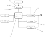
一実施形態では、本システムは、ハウジング、神経プローブ、追加の電極のためのポート、視覚的インジケータ、電源、刺激パルス発生器/コントローラ、中央処理装置、及びユーザコントロールからなる。図1の概略図を特に参照すると、システム100は、複数の構成で機能するように、構成されることができる。本開示の図1及び/又はその他の図には特に示されていないが、本システムの追加の構成も、存在し得る。本明細書で開示されている実施形態のいずれについても、デバイス又はシステムは、所望により又は必要に応じて、より少ないコンポーネント及び/又は特徴を含むことができる。例えば、いくつかの構成では、デバイス又はシステムは、視覚的インジケータ、電源及び/又はその他を含まない。
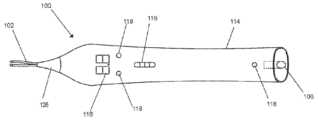
Overall System Overview In one embodiment, the system consists of a housing, a neural probe, a port for additional electrodes, a visual indicator, a power source, a stimulation pulse generator/controller, a central processing unit, and a user control. With particular reference to the schematic diagram of FIG. 1, the
一構成では、神経プローブ102は、双極式である、すなわち、刺激発生器に内部で接続されている2つの別個の電極導体を含む。別の構成では、図1にも示されているように、神経プローブは、双極式、すなわち、2つの別個の電極導体からなってもよい。しかし、図示された構成では、導体104を内部で互いに短絡させて、単一のプローブを本質的に作り出すことができる。この接続されたプローブは、いくつかの実施形態では、適切な戻り電極がシステムの電極ポート106に接続されている場合、単極プローブとして機能し得る。図1に概略的に示されているように、戻り電極108は、帰還路が存在する限り、針、表面パッド、及び/又は別の導電性材料を、含むことができる。そのような実施形態に関するさらなる詳細は、本明細書で提供される。
In one configuration, the
上記で説明した構成では、システム100は、神経プローブに、又は神経プローブの近くに送達されている刺激で、神経組織をプローブするために使用されることができる。したがって、システム100は、神経ロケータ又は評価器として使用されることができる。
プローブされている神経の位置、又は行われている外科的処置のタイプによっては、双極構成か又は単極構成が、外科医にとって有利であり得る。
In the configuration described above,
Depending on the location of the nerve being probed or the type of surgical procedure being performed, either a bipolar or monopolar configuration may be advantageous to the surgeon.
図1にも示されている実施形態におけるものなどの、さらに別の構成では、神経と(例えば、直接的に、間接的に、など)インターフェースするように構成されているカフタイプ電極110が、本システムの電極ポートに接続され得る。そのような実施形態は、損傷した神経に対して神経再生治療を行うために、有利な場合がある。そのような構成では、刺激出力は(例えば、もっぱら)、神経プローブではなく、プラグインされた電極に打ち込まれ得る。
In yet another configuration, such as in the embodiment also shown in FIG. 1, a cuff-
いくつかの実施形態では、ポートにプラグインされる電極は、1又は複数の電極接点からなり得て、電極ポートを通して刺激発生器に物理的に接続され得る。厳密にどのような構成が使用されるかとは関係なく、本出願のいくつかの実施形態では、本システムは、電極がポートにプラグインされているかどうか、及びどの電極がポートにプラグインされているかを検知して、適切な刺激が出力されて打ち込まれていることを確実にするように、構成されることができる。様々な、システムの実施形態、コンポーネント、部分、サブシステム及び/又はその他に関する追加の詳細が、以下で提供される。 In some embodiments, the electrodes plugged into the ports may consist of one or more electrode contacts and may be physically connected to the stimulation generator through the electrode port. Regardless of the exact configuration used, in some embodiments of the present application, the system may be configured to detect whether and which electrodes are plugged into the ports to ensure that the appropriate stimulation is output and delivered. Additional details regarding various system embodiments, components, parts, subsystems, and/or the like are provided below.
ハウジング
いくつかの実施形態では、図2Aに示されているように、本システムは、ハウジング114を含むことができる。ここで、ハウジング114は、所望により又は必要に応じて、ユーザコントロール116、視覚的インジケータ118、電源、刺激パルス発生器/コントローラ、中央処理装置、及び/又はその他の任意のコンポーネント若しくは部分を含み得る。示されているように、このハウジングは、所望により又は必要に応じて、例えば熱可塑性タイプの材料(例えば、ポリエチレン、ポリプロピレン、ポリ塩化ビニル、ポリスチレン、ポリアミド類、ポリエステル類、ポリウレタン類など)、(例えば、いくつかの実施形態では柔らかいグリップ可能な質感をもたらす)熱可塑性エラストマー、金属又は合金(例えば、ステンレス鋼、アルミニウム、その他のブラッシング若しくは研磨された金属若しくは合金など)、複合材料、及び/又はその他などの、1又は複数の材料を含むことができる。
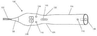
2A, the system can include a
いくつかの実施形態では、このハウジング及び付随する内部コンポーネントは、再利用されるように構成されている場合がある。したがって、そのようなコンポーネント又は部分は、滅菌され、及び/又はその他清潔にされるように、設計され、及びその他構成されている場合がある。例えば、本システムは、エチレンオキシド、二酸化塩素、気化した過酸化水素、ガンマ線、電子ビーム、及び/又はその他への曝露によって、滅菌される場合がある。 In some embodiments, the housing and associated internal components may be configured to be reused. Thus, such components or parts may be designed and otherwise configured to be sterilized and/or otherwise cleaned. For example, the system may be sterilized by exposure to ethylene oxide, chlorine dioxide, vaporized hydrogen peroxide, gamma radiation, electron beam, and/or the like.
いくつかの実施形態によれば、このハウジングは、図2Bに示されているように、人間工学的に外科医の手に収まるように設計されている。したがって、いくつかの構成では、このハウジングは、溝若しくはスカラップ120を有する形状とされ、かつ/又はこのハウジングは、ユーザの利き手(例えば、右利き若しくは左利き)に関係なく保持を容易にするための人間工学的形状を含む。他の実施形態では、このハウジングは、ユーザの単一の利き手(例えば、右利き又は左利き)のために特に設計されている。このような溝又はスカラップ120は、対称であり、非対称であり、整列され、オフセットされ、かつ/又はその他構成されることができる。示されているように、一実施形態では、溝120の最も深い部分は、このハウジングの最も幅の広い部分から、0.1~10mm(例えば、0.1~0.2、0.2~0.3、0.3~0.4、0.4~0.5、0.5~0.6、0.6~0.7、0.7~0.8、0.8~0.9、0.9~1、1~2、2~3、3~4、4~5、5~6、6~7、7~8、8~9、9~10mm、前述のものの間の長さなど
)の範囲内で、オフセットされていてもよい。
According to some embodiments, the housing is designed to ergonomically fit in a surgeon's hand, as shown in FIG. 2B. Thus, in some configurations, the housing is shaped with grooves or
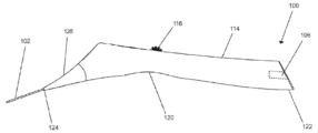
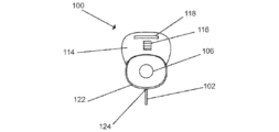
いくつかの実施形態では、溝120は、長手方向軸、水平軸、このハウジングの下側、又は上記の組み合わせに沿っていてもよい。例えば、図2Cを参照のこと。いくつかの実施形態では、このハウジングは、近位端部122及び遠位端部124を含む。遠位端部124は、視覚的インジケータ118及び神経プローブ102又はそれらの組み合わせを含むことができる。いくつかの実施形態では、この近位端部は、神経プローブ102を含み得る神経ポート106を含む。
In some embodiments, the
いくつかの実施形態では、遠位端部及び近位端部は、同一直線上にあるか又はオフセットされていてもよい。例えば、いくつかの構成では、遠位端部及び近位端部は、1°~30°(例えば、1~2、2~3、3~4、4~5、5~6、6~7、7~8、7~8、8~9、9~10、10~15、15~20、20~25、25~30、5~25、10~20°、前述の範囲の間の角度など)の範囲内の角度によってオフセットされており、(例えば、近位端部に対して)角度のついた遠位端部をもたらしている。いくつかの実施形態では、そのような角度のついた遠位端部の構成は、有利なことに、例えば図2Cに示されているように、上記デバイスの使用を容易にすることができる。さらに、この角度のついたオフセットは、外科医又はその他の専門家が他の手術タスクを実行するためにこのハウジングを脇に置くときなど、緩やかに傾斜しているかは不均一な面に置かれた場合に、ハウジングが、(例えば、テーブル、カート、その他の台などから離れて)転がることを防ぐのに役立つ場合がある。 In some embodiments, the distal and proximal ends may be collinear or offset. For example, in some configurations, the distal and proximal ends are offset by an angle in the range of 1° to 30° (e.g., 1-2, 2-3, 3-4, 4-5, 5-6, 6-7, 7-8, 7-8, 8-9, 9-10, 10-15, 15-20, 20-25, 25-30, 5-25, 10-20°, angles between the aforementioned ranges, etc.), resulting in an angled distal end (e.g., relative to the proximal end). In some embodiments, such an angled distal end configuration can advantageously facilitate use of the device, for example, as shown in FIG. 2C. Additionally, the angled offset may help prevent the housing from rolling (e.g., off a table, cart, other platform, etc.) when placed on a gently sloping or uneven surface, such as when a surgeon or other professional sets the housing aside to perform other surgical tasks.
いくつかの実施形態では、このハウジングの長さ、又は近位端部から遠位端部までの距離は、10~40cm(例えば、10~15、15~20、20~25、25~30、30~35、35~40、15~25、20~40cm、前述の範囲の間の長さなど)であることができる。他の実施形態では、このハウジングの長さは、特定の用途又は使用による所望により又は必要に応じて、10cm未満であるか又は40cmを超えることができる。さらに、ハウジングの幅(例えば、直径又は断面寸法)は、0.5~3cm(例えば、0.5~1、1~1.5、1.5~2、0.5~2、2~2.5、2.5~3cm前述の範囲の間の幅など)であることができる。 In some embodiments, the length of the housing, or the distance from the proximal end to the distal end, can be 10-40 cm (e.g., 10-15, 15-20, 20-25, 25-30, 30-35, 35-40, 15-25, 20-40 cm, lengths between the aforementioned ranges, etc.). In other embodiments, the length of the housing can be less than 10 cm or greater than 40 cm, as desired or required by a particular application or use. Additionally, the width (e.g., diameter or cross-sectional dimension) of the housing can be 0.5-3 cm (e.g., 0.5-1, 1-1.5, 1.5-2, 0.5-2, 2-2.5, 2.5-3 cm, widths between the aforementioned ranges, etc.).
いくつかの実施形態では、このハウジングの近位端部は、フック形状、若しくはその他の湾曲した若しくは角度のついた延長部、又はこのハウジングに物理的に接続された、密閉されたリングを含む。いくつかの構成では、この延長部は、IVポール、別のタイプのフック及び/又はその他に、このハウジングを吊るすために使用され得る。 In some embodiments, the proximal end of the housing includes a hook-shaped or other curved or angled extension or a sealed ring that is physically connected to the housing. In some configurations, the extension can be used to hang the housing from an IV pole, another type of hook, and/or the like.
いくつかの実施形態では、このハウジングは、バッテリとインターフェースするプルタブを可能にするための、スロット又は開口部を含み得る。このプルタブは、本システムへの電力供給を阻止する、バッテリ接点の分離を、可能にし得る。これは、そのことが、とりわけ、本システムの保管寿命を延ばすから、有利である。いくつかの実施形態では、このプルタブスロット又はプルタブ開口部は、このハウジングの近位端部に、このハウジングの近位端部の中に、又はこのハウジングの近位端部に沿って配置され、かつスロットの幅は、5mm~30mmの範囲内(又はこのハウジングの幅)であり得る。このスロットの高さは、0.1~2mm(例えば、0.1~0.2、0.2~0.3、0.3~0.4、0.4~0.5、0.5~0.6、0.6~0.7、0.7~0.8、0.8~0.9、0.9~1、1~1.5、1.5~2mm、前述の範囲の間の高さなど)の範囲内であることができる。 In some embodiments, the housing may include a slot or opening to allow a pull tab to interface with the battery. The pull tab may allow for separation of the battery contacts, preventing power to the system. This is advantageous because, among other things, it extends the shelf life of the system. In some embodiments, the pull tab slot or opening is located at, in, or along the proximal end of the housing, and the width of the slot may be within the range of 5 mm to 30 mm (or the width of the housing). The height of the slot may be within the range of 0.1 to 2 mm (e.g., 0.1 to 0.2, 0.2 to 0.3, 0.3 to 0.4, 0.4 to 0.5, 0.5 to 0.6, 0.6 to 0.7, 0.7 to 0.8, 0.8 to 0.9, 0.9 to 1, 1 to 1.5, 1.5 to 2 mm, heights between the aforementioned ranges, etc.).
図2A~図2Dに示されているように、システム100は、送達されている刺激のパラメータを調整するための、ユーザ操作可能なコントロール116の第1のセットを含むこ
とができる。いくつかの実施形態では、本システムは、ユーザが、刺激パラメータ、例えば刺激の振幅又はパルス幅を個別に制御して、神経活性化の閾値を決定すること(例えば、閾値のテスト)及び/又はその他を決定することを、許すように構成されている。これは、神経が、瘢痕組織及び/又はその他の組織に囲まれ(ており、例えば、脱分極するために比較的大きな刺激電流を必要とし)ている場合、特に関連性があり得る。瘢痕及び/又はその他の閉塞組織の切開は、結果として、必要な活性化電流を小さくする場合がある。
As shown in Figures 2A-2D, the
一実施形態では、コントロールは、2つのボタンを含み得る。別の構成では、コントロールの第1のセットは、スライダ又は類似する機能又はデバイスを含み得る。さらに別の構成では、コントロールの第1のセットは、ホイールコントロール(例えば、ローラー、ホイールなど)を含み得る。しかし、他の任意のタイプのコントロール(例えば、ボタン、ダイヤルなど)が、スライダ及び/若しくはホイールコントロールの代わりに、又はそれらに加えてのいずれかで、上記デバイス内に組み込まれることができる。いくつかの構成では、ホイールコントロールを使用する場合、離散的なステップのセットによって、刺激の振幅を調整することを可能にし得る。したがって、ホイール及び/又はその他の任意のコントロールが、離散的なステップ又は位置の間で、移動されるように構成されることができる。しかし、他の構成では、刺激の振幅は、非離散的なレベルの範囲に従って(例えば、振幅の連続スペクトルに従って)選択されることができる。いくつかの構成では、ホイールは、ホイールの動きを離散化するために、ロータリエンコーダに結合され得る。他の構成では、このロータリエンコーダは、コントロールに係合するときにユーザに触覚フィードバックを提供するための、戻り止めを含み得る。 In one embodiment, the controls may include two buttons. In another configuration, the first set of controls may include sliders or similar features or devices. In yet another configuration, the first set of controls may include a wheel control (e.g., roller, wheel, etc.). However, any other type of control (e.g., button, dial, etc.) may be incorporated into the device, either instead of or in addition to the slider and/or wheel control. In some configurations, the use of a wheel control may allow the amplitude of the stimulation to be adjusted by a set of discrete steps. Thus, the wheel and/or any other control may be configured to be moved between discrete steps or positions. However, in other configurations, the amplitude of the stimulation may be selected according to a range of non-discrete levels (e.g., according to a continuous spectrum of amplitudes). In some configurations, the wheel may be coupled to a rotary encoder to discretize the movement of the wheel. In other configurations, the rotary encoder may include detents to provide tactile feedback to the user when engaging the control.
いくつかの構成によれば、本システムは、ユーザ操作可能なコントロールの第2のセットも含み得る。そのような第2のコントロールは、処置の始動又は休止を、開始、停止、及び/又は一時停止するように構成されることができる。一実施形態では、第2のコントロールを使用して、本システムの電源をオン及びオフにし得る。いくつかの構成では、コントロールの第2のセットは、ユーザ操作可能なコントロールの第1のセットの近くに、配置され得る。一実施形態では、第2のコントロールは、第1のユーザコントロールの一部であり得る。例えば、スライダコントロール又はホイールコントロールは、そのスライダコントロール又はホイールコントロールを押すと、結果としてモーメンタリスイッチ又は類似するものが作動するように、スイッチに結合されていてもよい。他の任意のタイプのコントロール(例えば、ボタン、スイッチ、フットペダル、タッチスクリーンなど)が、使用されることができる。 According to some configurations, the system may also include a second set of user-operable controls. Such second controls may be configured to start, stop, and/or pause the initiation or pause of the treatment. In one embodiment, the second controls may be used to power the system on and off. In some configurations, the second set of controls may be located near the first set of user-operable controls. In one embodiment, the second controls may be part of the first user control. For example, a slider control or wheel control may be coupled to a switch such that pressing the slider control or wheel control results in actuation of a momentary switch or the like. Any other type of control (e.g., a button, switch, foot pedal, touch screen, etc.) may be used.
一実施形態では、本システムは、本システムへの電力を制御するための、(例えば、本明細書で説明されているような)プルタブコントロールを含む。いくつかの構成では、本システムは、本システムへの電力を制御するために使用される、スイッチ又はボタンを含む。 In one embodiment, the system includes a pull tab control (e.g., as described herein) for controlling power to the system. In some configurations, the system includes a switch or button that is used to control power to the system.
いくつかの実施形態によれば、本明細書でより詳細に説明されているように、本システムは、上記ハウジングに結合された(例えば、物理的に結合された、動作的に結合された、など)、神経ポート106も含み得る。これにより、ユーザは、別個の電極を、本システムに接続(例えば、物理的に又は動作的に結合)できる。制御として、別個の電極を本システムにプラグインし、又は物理的に接続する行為は、システム構成で既に説明したように、本システムの動作モードを変更し得る。
According to some embodiments, as described in more detail herein, the system may also include a
図2A~図2Dに示されているいくつかの実施形態では、この神経ポートは、上記ハウジングの近位側122に含み得る。いくつかの構成では、この神経ポートは、上記ハウジングの長手方向軸に平行な接続を可能にすることができる(例えば、図2Dを参照のこと
)。他の構成では、この神経ポートは、接続されたコンポーネントコネクタが、上記ハウジングの長手方向軸に対して垂直(例えば、厳密に垂直、又は略垂直若しくは実質的に垂直)であるように、含まれ得る。一実施形態では、本システムに別個の電極をプラグインし、又は本システムに別個の電極を物理的に接続する行為により、本システムの電源をオンにできるようになる。
In some embodiments shown in Figures 2A-2D, the neural port may be included on the
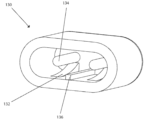
電極が存在して本システムに物理的に接続されているかどうかを検知するために、変更されたジャック130が、図3に示されているように、神経ポートとともに含まれていてもよい。例えば、いくつかの実施形態では、ジャック130は、(例えば、IEC 60601に従って設計された)タッチプルーフジャックを含むことができる。この変更されたジャックは、フレキシブル接点132を含んでもよい。ここで、フレキシブル接点132は、メインピン134と物理的に接触していてもよい。しかし、そのような実施形態では、電極リード線コネクタの導入時、これらのフレキシブル接点とメインピンとの間の物理的接続が切断されるように、構成されることができる。いくつかの構成では、このジャックは、1又は複数のフレキシブル接点を含むことができる。他の構成では、接続されるプラグの極性が正しいことを確実にするために、極性スタンドオフ136が、ジャック130とともに含まれ得る。いくつかの実施形態では、複数のピン/接点又は同等物を備えた、標準的なタッチプルーフジャック130が、使用される。他の実施形態では、ジャック又はカップリング130は、所望により又は必要に応じて、異なるように(例えば、特徴又はコンポーネントの別のセットによって)、構成され又は設計されることができる。
To detect whether an electrode is present and physically connected to the system, a modified
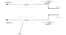
いくつかの実施形態では、このジャック上の接点は、図4に示されているものなどの、142で示されているジャック検知回路に配線され得る。回路142は、接続のステータスを検知するために使用され得るマイクロコントローラ及び受動部品を、含むことができる。
In some embodiments, the contacts on this jack can be wired to a jack detection circuit, shown at 142, such as that shown in FIG. 4. The
一実施形態では、回路142は、ヘッドセットを検知するために携帯電話産業で使用される、標準的なジャック検知チップ142を含む。これらのチップは、NXP SemiconductorsのNCX8193、又はMaxim SemiconductorのMAX13330を含み得るが、これらに限定されない。いくつかの構成では、これらのチップは、湿気検知の利点を含むように構成されることができる。このことによって、本システムは、ジャックハウジング内で湿気が検知された場合に、フル電力又はその他の設定を有効にすることを阻止できるようになる。このようなことは、例えば本システムが手術中の環境で使用される場合に、生じ得る。
In one embodiment, the
インジケータ
いくつかの実施形態では、本システムは、インジケータ118の少なくとも1つのセットを含む。一実施形態では、第1のインジケータは、少なくとも2つの視覚的放出デバイス(例えば、LED)を互いの近くに配置することによって作られた、棒グラフタイプのディスプレイを含み得る。いくつかの構成では、第1のインジケータは、図2Aに示されているように、マルチセグメント(例えば、7セグメント)ディスプレイを含み得る。マルチセグメント(例えば、7セグメント)のディスプレイは、所望により又は必要に応じて、1つを超える、数字及び小数の桁を含み得る。いくつかの実施形態では、第1のインジケータは、液晶ディスプレイ(liquid crystal display:LCD)、プラズマディスプレイ、ブラウン管ディスプレイ(cathode ray tube:CRT)、有機発光ダイオードディスプレイ(organic light-emitting diode display:OLED)、薄膜トランジスタディスプレイ(thin-film transistor:TFT)及び/又はその他の任意のタイプのディスプレイからなり得る。
Indicators In some embodiments, the system includes at least one set of
一実施形態では、そのようなディスプレイ又はインジケータの目的は、例えば、限定するものではないが、振幅、パルス幅、周波数、継続時間、その他の時間パラメータ、及び/又はその他の、刺激パラメータに関する情報を伝えることである。いくつかの構成では、このディスプレイは、例えばタイマー又はカウントダウンクロックなどの、時間に関連する情報を提供するように構成されている。そのような情報は、処置適用の残り時間又は経過時間を決定する上で有益かつ有利な場合があり、外科医又はその他の専門家が、損傷したか又はその他の標的神経組織の、神経再生治療(例えば、短い電気刺激、その他のタイプの電気刺激など)を実施しているのを誘導するために役立つ場合がある。いくつかの構成では、刺激パラメータ、時間関連パラメータ、及び/又はその他の任意のデータ若しくは情報の、組み合わせが、このインジケータに表示される。そのようなデータ及び/又は情報は、その厳密な性質に関係なく、ユーザがコントロールするスイッチ及び/又はその他によって、交互する態様で表示されることができる。いくつかの構成では、ユーザは、データ及び/又は情報がこのインジケータによって提供される際の、タイプ及び/又は態様(例えば、どのようなデータ/情報が提供されるか、それがどのようにユーザに提供されるかなど)を、カスタマイズできる。 In one embodiment, the purpose of such a display or indicator is to convey information regarding stimulation parameters, such as, for example, but not limited to, amplitude, pulse width, frequency, duration, other time parameters, and/or other. In some configurations, the display is configured to provide time-related information, such as, for example, a timer or countdown clock. Such information may be useful and advantageous in determining the remaining or elapsed time of treatment application and may help guide a surgeon or other professional administering neural regeneration therapy (e.g., brief electrical stimulation, other types of electrical stimulation, etc.) of damaged or other target neural tissue. In some configurations, a combination of stimulation parameters, time-related parameters, and/or any other data or information is displayed on the indicator. Regardless of the exact nature of such data and/or information, it may be displayed in an alternating manner, such as by user-controlled switches and/or otherwise. In some configurations, the user may customize the type and/or manner in which data and/or information is provided by the indicator (e.g., what data/information is provided, how it is provided to the user, etc.).
例として、いくつかの構成によれば、インジケータの第1のセットが、本システムに供給されている電力を示すために使用され得る。一例では、本システムへの電力供給を制御するために前述のプルタブ(又は類似する特徴若しくはコンポーネント)が使用されて、ユーザがそのタブを引くと、第1のインジケータが点灯して本システムに電力が供給されていることが示され得る。 By way of example, according to some configurations, a first set of indicators may be used to indicate power being provided to the system. In one example, the aforementioned pull tab (or similar feature or component) may be used to control the power supply to the system, such that when a user pulls the tab, a first indicator may be illuminated to indicate that power is being provided to the system.
いくつかの実施形態では、本システムは、所望により又は必要に応じて、追加のインジケータを含む。例えば、本システムは、第1のインジケータ(例えば、時間関連のデータ又は情報、刺激パラメータなど)によって提供される追加のデータ及び/又は情報を、有利なことに、専門家又はその他のユーザに提供できる、インジケータ118の第2の(又は追加の)セットを含むことができる。いくつかの実施形態では、本システムは、インジケータ118の第3のセットを含み得る。一実施形態では、インジケータ118の第3のセットは、上記ハウジングの遠位側、又は上記ハウジングの遠位側の近くに配置されている。しかし、本システムのインジケータは、いくつ含まれるか、どのようなデータ/情報をそれらのインジケータが提供するように構成されているかなどに関係なく、所望により又は必要に応じて、任意の場所に含まれる場合がある。例として、インジケータの第3のセットは、ライトパイプとして作用するキャップ126内に配置された、LEDを含むことができる。そのようなキャップは、物理的に(例えば、直接的又は間接的に)ハウジング114に結合されることができる。いくつかの構成において、このキャップ及びライトパイプは、例えば上記ハウジングを、(例えば、遠位側に角度がついているために)上、下、横、後ろ、及び/又はその他から見るときなどに、任意の方向から見えることを許すように設計及び/又はその他構成されることができる。
In some embodiments, the system includes additional indicators as desired or required. For example, the system may include a second (or additional) set of
いくつかの実施形態では、インジケータ(例えば、第1、第2、第3のインジケータなど)は、本システムのステータスを表示するように構成されることができる。例えば、第1のベタ色は、出力の状態(例えば、アクティブ又は非アクティブ)を示すことができる。いくつかの構成では、出力は、神経プローブに物理的に(例えば、直接的又は間接的に)接続又は結合され得る。したがって、例えば、いくつかの構成では、第1のベタ色は、神経プローブがアクティブであるかどうかを示し得る。具体的な一例では、前述のシステム構成を参照すると、第1のベタ色は、神経プローブが、双極構成でアクティブであること、又は単極構成でアクティブであることの、いずれかを示し得る。 In some embodiments, the indicators (e.g., the first, second, third indicators, etc.) can be configured to display a status of the system. For example, a first solid color can indicate a state of the output (e.g., active or inactive). In some configurations, the output can be physically connected or coupled (e.g., directly or indirectly) to the neural probe. Thus, for example, in some configurations, the first solid color can indicate whether the neural probe is active. In one specific example, with reference to the system configurations described above, the first solid color can indicate that the neural probe is either active in a bipolar configuration or active in a monopolar configuration.
いくつかの実施形態では、インジケータのセット(例えば、インジケータの第3のセット)は、第1の色を点滅(例えば、オン/オフ)させて、刺激の出力を示すように構成さ
れることができる。いくつかの構成では、点滅は、刺激パルスの出力と一致するようにタイミングを合わせられていてもよい。他の構成では、点滅は、刺激パルスの出力と非同期であり得る。所望により又は必要に応じて、他の任意のタイプの構成が、上記インジケータ(例えば、異なるテキスト表現及び/又はグラフィカルな表現、異なるアラート効果など)を介してユーザにデータ及び/又はその他の情報を提供するために、使用されることができる。
In some embodiments, the set of indicators (e.g., the third set of indicators) can be configured to flash (e.g., on/off) a first color to indicate output of the stimulation. In some configurations, the flashing can be timed to coincide with the output of the stimulation pulse. In other configurations, the flashing can be asynchronous with the output of the stimulation pulse. Any other type of configuration can be used to provide data and/or other information to the user via the indicators (e.g., different textual and/or graphical representations, different alert effects, etc.) as desired or required.
いくつかの実施形態では、点滅は、脈動出力で置き換えられている。いくつかの構成では、脈動出力は、光強度のゼロから所定の最大値への増加、及びその後のこの最大値からゼロへの減少を含み得る。いくつかの構成では、脈動出力は、ゼロでない強度値から、増加を開始し、この最大値からゼロでない強度値へ向かって、減少を終了する。 In some embodiments, the blinking is replaced with a pulsating output. In some configurations, the pulsating output may include an increase in light intensity from zero to a predefined maximum value, and then a decrease from this maximum value back to zero. In some configurations, the pulsating output starts increasing from a non-zero intensity value, and ends decreasing from this maximum value back to the non-zero intensity value.
いくつかの実施形態では、視覚的インジケータ(例えば、視覚的インジケータの第3のセット)は、刺激電極と戻り電極との間の開回路を示すために、点滅又は脈動するように構成されることができる。具体的な一例では、神経プローブの双極の先端の間で接触がない場合、開回路が示され、そのことによって、視覚的インジケータの点滅又は脈動が、引き起こされ得る。別の具体的な例では、本システムが、前述の単極構成で動作しているとき、刺激電極と戻り電極の間の電流の流れの欠如によって、視覚的インジケータの点滅又は脈動が、引き起こされ得る。いくつかの実施形態では、第1の色を有するインジケータ(例えば、第3のインジケータ)の点滅は、ゼロより大きい刺激設定で起こり得る。 In some embodiments, the visual indicators (e.g., the third set of visual indicators) can be configured to flash or pulse to indicate an open circuit between the stimulation electrode and the return electrode. In one specific example, an open circuit is indicated when there is no contact between the bipolar tips of the neural probe, which may cause the visual indicators to flash or pulse. In another specific example, when the system is operating in the monopolar configuration described above, a lack of current flow between the stimulation electrode and the return electrode may cause the visual indicators to flash or pulse. In some embodiments, flashing of the indicators (e.g., the third indicators) having the first color may occur at stimulation settings greater than zero.
いくつかの実施形態では、インジケータ(例えば、第3のインジケータ)の点滅又は脈動は、第2の色で起こり得る。いくつかの構成では、第2の色は、第1の色と異なり得る。例として、第2の色の点滅又は脈動は、閉回路を示し得る。一実施形態では、神経プローブの双極の先端の間に、電流が流れている接触がある場合、閉回路の表示が与えられ、そのことによって、視覚的インジケータの点滅又は脈動が、引き起こされる。別の具体的な例では、本システムが、前述の単極構成で動作しているときに、刺激電極と戻り電極の間に電流の流れが存在するときは、視覚的インジケータの点滅又は脈動が、作動させられ又は引き起こされ得る。 In some embodiments, the blinking or pulsating of the indicator (e.g., the third indicator) may occur in a second color. In some configurations, the second color may be different than the first color. By way of example, the blinking or pulsating of the second color may indicate a closed circuit. In one embodiment, when there is contact between the bipolar tips of the neural probe with current flowing, an indication of a closed circuit is given, which causes the blinking or pulsating of the visual indicator. In another specific example, when the system is operating in the monopolar configuration described above, the blinking or pulsating of the visual indicator may be activated or caused when there is current flow between the stimulating electrode and the return electrode.
いくつかの実施形態では、本システムは、特定の用途又は使用のために所望により又は必要に応じて、インジケータ118の第4の(又は追加の)セットを含み得る。いくつかの構成では、インジケータの第4のセットは、神経ポート106のすぐ隣又は近くに存在し得る。いくつかの実施形態では、インジケータの第4のセットは、神経ポート106のステータスを示し得る。いくつかの構成では、インジケータの第4のセットは、インジケータの第3のセット(及び/又はその他のセット)と同様に機能し得て、複数色のインジケータ及び/又は類似する出力からなることができる。
In some embodiments, the system may include a fourth (or additional) set of
上記のインジケータは、本明細書で説明されているデバイス又はシステムの実施形態のいずれにも組み込まれることができ、所望により又は必要に応じて、変更され得る。 The above indicators may be incorporated into any of the device or system embodiments described herein and may be modified as desired or required.
中央処理装置
いくつかの実施形態では、本明細書で説明されているシステム構成のいずれも、様々な電子機能、安全機構及び/又はその他を備えたスマートシステムの一部となることができる。そのような機能、機構、及び/又はその他の特性のいくつかについての詳細は、本明細書で提供される。
Central Processing Unit In some embodiments, any of the system configurations described herein can be part of a smart system with various electronic functions, safety features, and/or other characteristics. Details regarding some of such functions, features, and/or other characteristics are provided herein.
いくつかの実施形態では、本システムの制御サブシステム又は中央処理装置は、マイクロコントローラ150に関連して(例えば、マイクロコントローラ150の周りに)構築されることができる。いくつかの構成では、このマイクロコントローラは、コードを格納
及び実行し、かつ/又はその他、本システムをプロパティ(property)かつ効果的に動作させるためのある一定のタスクを実行するために、プログラムされているように構成されている。いくつかの実施形態では、このマイクロコントローラは、例えば時間を合わせた刺激出力を可能にする、タイミング機能を含む。他の実施形態では、このマイクロコントローラは、本明細書で説明されている少なくとも1又は複数のサブシステム152とインターフェースする(例えば、図5を参照のこと)。
In some embodiments, the control subsystem or central processing unit of the system can be built in association with (e.g., around) a
電源
いくつかの実施形態では、本明細書で開示されているシステムのいずれも、比較的小さく、使い捨てでき、ハンドヘルド操作を含み、かつ/又はその他の所望の又は必要な特徴を含むように、設計されることができる。他の構成では、本システムは、再利用可能であり得る。
Power Source In some embodiments, any of the systems disclosed herein can be designed to be relatively small, disposable, include handheld operation, and/or include other desired or required features. In other configurations, the systems may be reusable.
いくつかの実施形態では、本システムは、バッテリ、AC電源及び/又はその他などのエネルギー源を使用して、電力を供給され又は電気的にエネルギーを与えられる。いくつかの構成では、電源は、標準的なリチウムコインセルを備えたバッテリを含む。しかし、他の構成では、より大きな容量が必要な場合、Nタイプのバッテリ、又はその他の類似するアルカリバッテリが、代用され得る。いくつかの構成では、このバッテリは、充電可能であり得る。所望により又は必要に応じて、その他の任意のタイプのバッテリ又はその他のローカル電源が、使用されることができる。 In some embodiments, the system is powered or electrically energized using an energy source such as a battery, an AC power source, and/or others. In some configurations, the power source includes a battery with standard lithium coin cells. However, in other configurations, if greater capacity is required, an N-type battery, or other similar alkaline battery, may be substituted. In some configurations, the battery may be rechargeable. Any other type of battery or other local power source may be used as desired or required.
いくつかの実施形態では、本システムは、電力管理サブシステムを含み得る。これらの実施形態では、電力管理サブシステムは、3~5Vの範囲内の安定した動作電圧を維持するために、例えばスイッチングタイプの低ノイズ・低ドロップアウト・レギュレータなどの、1又は複数のサブコンポーネントを含み得る。例えば、いくつかの実施形態では、Texas Instruments LP5912が、使用されることができる。 In some embodiments, the system may include a power management subsystem. In these embodiments, the power management subsystem may include one or more subcomponents, such as a switching type low noise, low dropout regulator, to maintain a stable operating voltage in the range of 3-5V. For example, in some embodiments, a Texas Instruments LP5912 may be used.
いくつかの実施形態では、電力管理サブシステムは、組織を刺激するために必要な、より高い電圧を発生する手段を有する、第2のサブコンポーネントを含む。そのような実施形態では、電力管理サブシステムは、例として、限定するものではないが、Texas Instruments TPS61096Aなどの、低電力昇圧の昇圧コンバータを含むことができる。いくつかの実施形態では、このより高い電圧範囲は、10~50V(例えば、10~15~15~20、20~25、25~30、30~35、35~40、40~45、45~50、20~40、25~35V、前述のものの間の値など)であり得る。 In some embodiments, the power management subsystem includes a second subcomponent having a means for generating the higher voltage required to stimulate the tissue. In such embodiments, the power management subsystem may include a low power step-up boost converter, such as, by way of example and not limitation, the Texas Instruments TPS61096A. In some embodiments, this higher voltage range may be 10-50V (e.g., 10-15-15-20, 20-25, 25-30, 30-35, 35-40, 40-45, 45-50, 20-40, 25-35V, values in between the foregoing, etc.).
いくつかの実施形態では、電力管理サブシステムのサブコンポーネントは、マイクロコントローラ150を介して、制御される(例えば、有効及び無効にされる)。他の構成では、電極を神経ポート106に物理的に接続又はプラグインする行為によって、本明細書で説明されたジャック検知回路142を介して、電力管理サブシステムのサブコンポーネントが、有効又は無効にされ得る。
In some embodiments, the subcomponents of the power management subsystem are controlled (e.g., enabled and disabled) via the
いくつかの実施形態では、電力管理サブシステムは、傾斜センサを含み得る。そのような傾斜センサは、データ及び/又はその他の情報をユーザに提供するように、構成されることができる。例えば、データ又は情報は、本システムが、平らな面に置かれたか、又は水平でない姿勢で保持されているか、に関するものである場合がある。いくつかの構成では、この傾斜センサの出力は、電力管理サブシステムを通して、低電力モードに関与し得る。 In some embodiments, the power management subsystem may include a tilt sensor. Such a tilt sensor may be configured to provide data and/or other information to a user. For example, the data or information may be regarding whether the system is placed on a flat surface or is being held in a non-level position. In some configurations, the output of this tilt sensor may be involved in a low power mode through the power management subsystem.
出力段階
いくつかの実施形態では、本システムは、刺激出力段階サブシステムを含む。いくつかの構成では、そのようなサブシステムの電気出力は、組織を選択的に刺激するように構成されている。本システムのマイクロコントローラは、刺激出力段階サブシステムによって調整される、矩形刺激パルスを生成するように構成されることができる。刺激出力段階サブシステムは、刺激パルスを生成するように構成されることができる。いくつかの実施形態では、刺激パルスは、二相性の定電流又は定電圧パルスからなる。
Output Stage In some embodiments, the system includes a stimulation output stage subsystem. In some configurations, the electrical output of such a subsystem is configured to selectively stimulate tissue. The microcontroller of the system can be configured to generate rectangular stimulation pulses that are regulated by the stimulation output stage subsystem. The stimulation output stage subsystem can be configured to generate the stimulation pulses. In some embodiments, the stimulation pulses consist of biphasic constant current or constant voltage pulses.
いくつかの実施形態では、刺激出力段階サブシステムは、例えば正味ゼロの電荷が組織に送達されていることを確実にするために役立つように、容量結合された出力を含む。いくつかの実施形態では、刺激出力段階サブシステムは、電流の極性を切り替えるために使用される、Hブリッジを含む。このHブリッジは、所望により又は必要に応じて、電流源に結合されることができる。いくつかの実施形態では、この電流源は、ハウランド電流ポンプを含む。 In some embodiments, the stimulation output stage subsystem includes a capacitively coupled output, for example to help ensure that a net zero charge is delivered to the tissue. In some embodiments, the stimulation output stage subsystem includes an H-bridge that is used to switch the polarity of the current. The H-bridge can be coupled to a current source as desired or required. In some embodiments, the current source includes a Howland current pump.
いくつかの実施形態によれば、刺激出力段階サブシステムは、1~500マイクロ秒(例えば、1~5、5~10、10~20、20~30、30~40、40~50、50~60、60~70~70~80、80~90~100、100~120~120~140、140~160、160~180、180~200、200~250、250~300、300~350、350~400、400~450、450~500、0~20、1~30、0~40、0~50、0~100、50~150、100~200、100~300マイクロ秒、前述の範囲の間の継続時間など)の範囲内のパルス継続時間を有する、刺激パルスを生成するように構成されている。いくつかの構成では、このサブシステムは、0~20mA(例えば、0~1、1~2、2~3、3~4、4~5、5~6、6~7、7~8、8~9、9~10、10~15、15~20mA、前述のものの間の値など)の範囲内の刺激パルス振幅を生成するように構成されている。いくつかの構成では、このサブシステムは、0.1~100Hz(例えば、0.1~0.5、0.5~1、1~2、2~3、3~4、4~5、5~10、10~15、15~20、20~25、25~30、30~40、40~50、50~60、60~70、70~80、80~90、90~100、20~80~40~60、10~70Hz、前述のものの間の周波数など)の周波数範囲内のパルス列を生成するように構成されている。 According to some embodiments, the stimulation output stage subsystem is configured to generate stimulation pulses having pulse durations in the range of 1 to 500 microseconds (e.g., 1 to 5, 5 to 10, 10 to 20, 20 to 30, 30 to 40, 40 to 50, 50 to 60, 60 to 70 to 70 to 80, 80 to 90 to 100, 100 to 120 to 120 to 140, 140 to 160, 160 to 180, 180 to 200, 200 to 250, 250 to 300, 300 to 350, 350 to 400, 400 to 450, 450 to 500, 0 to 20, 1 to 30, 0 to 40, 0 to 50, 0 to 100, 50 to 150, 100 to 200, 100 to 300 microseconds, durations between the aforementioned ranges, etc.). In some configurations, the subsystem is configured to generate stimulation pulse amplitudes within a range of 0-20 mA (e.g., 0-1, 1-2, 2-3, 3-4, 4-5, 5-6, 6-7, 7-8, 8-9, 9-10, 10-15, 15-20 mA, values between the aforementioned, etc.). In some configurations, the subsystem is configured to generate pulse trains within a frequency range of 0.1-100 Hz (e.g., 0.1-0.5, 0.5-1, 1-2, 2-3, 3-4, 4-5, 5-10, 10-15, 15-20, 20-25, 25-30, 30-40, 40-50, 50-60, 60-70, 70-80, 80-90, 90-100, 20-80-40-60, 10-70 Hz, frequencies between the aforementioned, etc.).
いくつかの実施形態によれば、パルス列154は、ダブレットパルス156と呼ばれる、1~10ms(例えば、1~2、2~3、3~4、4~5、5~6、6~7、7~8、8~9、9~10ms、前述のものの間の間隔など)の範囲内の短いパルス間間隔によって分離された振幅Aの1又は複数のパルスを含む。そのような構成の一実施形態は、図6Aに概略的に示されている。いくつかの実施形態では、ダブレットパルスの使用によって、自然なキャッチのような筋肉の性質が利用できるようになり、筋肉のより大きな収縮又は可視反応がもたらされる。
According to some embodiments, the
いくつかの実施形態では、ダブレットパルス156は、個別に電荷の平衡がとれていてもよく、すなわち、各ダブレットパルスの後に、継続時間が等しいが逆極性の、電荷回収パルス158が続いてもよい。例えば、図6Bを参照のこと。他の構成では、このダブレットパルスは、電荷回収パルス158の継続時間が、個々のダブレットパルス156の継続時間の合計と等しくなるように、全体として電荷の平衡がとれていてもよい。例えば、図6Cを参照のこと。他の構成では、刺激装置の出力をAC結合することにより、電荷回収パルスが、受動的に生成されてもよい。そのような実施形態は、図6Dに示されているように、各出力パルスが後に続く、指数関数的に減衰する電荷回収パルス158を生み出す(例えば、各ダブレットパルスは、受動的に生成された電荷回復パルスを含み得る)。いくつかの実施形態では、このサブシステムは、神経線維を脱分極させ、活動電位を誘起するために十分な、刺激パルス、振幅、及び/又は列を生成する。
In some embodiments, the
安全機構
患者の安全を確実にするため、いくつかの実施形態では、電気エネルギーは、インピーダンスが10kOhm未満(例えば、10kOhm未満、9kOhm未満、8kOhm未満、7kOhm未満、6kOhm未満、5kOhm未満、4kOhm未満、3kOhm未満、2kOhm未満、1kOhm未満など)であるときにのみ、神経組織と接触している刺激電極又は神経プローブ106に送達される。いくつかの実施形態では、電気エネルギーは、インピーダンスが0~10kOhm(例えば、0~1、1~2、2~3、3~4、4~5、5~6、6~7、7~8、8~9、9~10KOhm、前述の範囲の間のインピーダンスなど)であるときにのみ、神経組織と接触している刺激電極又は神経プローブ106に送達される。
Safety Mechanisms To ensure patient safety, in some embodiments, electrical energy is delivered to a stimulating electrode or
いくつかの実施形態では、本明細書で開示されている構成のいずれによるシステムも、インピーダンス測定サブシステムを含む。このようなサブシステムは、正弦波を生成する回路を含むことができる。ここで、当該回路は、定電流源に結合されており、さらに電極のセットに結合されている。いくつかの実施形態では、この定電流源は、ハウランド電流ポンプからなり得る。 In some embodiments, a system according to any of the configurations disclosed herein includes an impedance measurement subsystem. Such a subsystem may include a circuit for generating a sine wave, where the circuit is coupled to a constant current source, which is further coupled to a set of electrodes. In some embodiments, the constant current source may comprise a Howland current pump.
いくつかの実施形態では、正弦波は、上記マイクロコントローラのデジタル/アナログ変換器を使用して生成される。他の構成では、上記マイクロコントローラのパルス幅変調出力が、使用され、ローパスフィルタに結合されている。いくつかの構成では、このローパスフィルタは、受動部品を含み、一方、他の構成では、このフィルタは、能動部品(例えば、アクティブフィルタ)を含む。 In some embodiments, a sine wave is generated using a digital-to-analog converter of the microcontroller. In other configurations, a pulse-width modulated output of the microcontroller is used and coupled to a low-pass filter. In some configurations, the low-pass filter includes passive components, while in other configurations, the filter includes active components (e.g., an active filter).
いくつかの実施形態では、インピーダンス測定サブシステムは、インピーダンスを測定するために使用される、電極経路に結合された計装用増幅器を含む。インピーダンス測定値は、このサブシステム内で計算されて上記マイクロコントローラに(例えば、デジタルで)送信されることができる。他の構成では、上記マイクロコントローラは、アナログインピーダンス値をサンプリングして、その値を内部でデジタル表現に変換する。 In some embodiments, the impedance measurement subsystem includes an instrumentation amplifier coupled to the electrode path that is used to measure impedance. Impedance measurements can be calculated within this subsystem and transmitted (e.g., digitally) to the microcontroller. In other configurations, the microcontroller samples analog impedance values and converts them internally to a digital representation.
いくつかの実施形態では、電極が神経組織と接触して適切に配置されているとき、電極-組織インピーダンスは、電極が組織と接触していないときのインピーダンスより小さくなるであろう。いくつかの構成では、そのような状況でのインピーダンスは、典型的には、10kOhm未満(例えば、0~1、1~2、2~3、3~4、4~5、5~6、6~7、7~8、8~9、9~10、3~8、1~10、4~8kOhm、前述の範囲の間のインピーダンスなど)である。そのような低い抵抗値は、人間の健全な内部組織が、電流が通り得る比較的低い抵抗値の電気経路を提供しているゆえに、存在する場合がある。いくつかの実施形態では、閾値(例えば、10kOhm)を超える抵抗値は、電極の不適切な配置を示している場合がある。いくつかの構成では、本システムは、そのような閾値を超える値を識別するように構成されている。 In some embodiments, when the electrodes are properly placed in contact with neural tissue, the electrode-tissue impedance will be less than the impedance when the electrodes are not in contact with tissue. In some configurations, the impedance in such situations is typically less than 10 kOhm (e.g., 0-1, 1-2, 2-3, 3-4, 4-5, 5-6, 6-7, 7-8, 8-9, 9-10, 3-8, 1-10, 4-8 kOhm, impedances between the aforementioned ranges, etc.). Such low resistance values may be present because healthy internal human tissue provides a relatively low resistance electrical path through which electrical current may pass. In some embodiments, resistance values above a threshold value (e.g., 10 kOhm) may indicate improper placement of the electrodes. In some configurations, the system is configured to identify values above such threshold values.
いくつかの実施形態では、本システムは、電気刺激を継続的に加えている間、インピーダンスを定期的に検知するように構成されている。いくつかの構成では、比較的高いインピーダンスが検知された(例えば、閾値レベル又は上限と比較して、高い)場合、本システムは、継続的な電気刺激を加えることを一時停止して、ハウジング114上のインジケータ118による操作者への警告を可能にするように、設計され、及びその他構成されることができる。他の構成では、本システムは、刺激出力を終了(例えば、自動的に停止)し、かつ/又はユーザにプロンプトするように構成されている。いくつかの実施形態では、上記インジケータは、視覚的インジケータを含む。しかし、他の実施形態では、上記インジケータは、視覚的表示に加えて、又はその代わりに、聴覚的インジケータ及び/又は
その他の任意のタイプのインジケータを含むことができる。
In some embodiments, the system is configured to periodically detect impedance while continuously applying electrical stimulation. In some configurations, if a relatively high impedance is detected (e.g., high compared to a threshold level or upper limit), the system can be designed and otherwise configured to pause the application of continuous electrical stimulation and enable an operator alert via
本明細書で述べられているように、患者の安全性をさらに高めるために、いくつかの実施形態では、本システムの出力は、容量結合されて、電極組織インターフェースへの正味DCの電荷の流入を阻止している。 As described herein, to further enhance patient safety, in some embodiments, the output of the system is capacitively coupled to prevent net DC charge from flowing into the electrode-tissue interface.
神経プローブ及び電極
神経プローブ102は、組織をプローブして興奮性をテストするために、使用されることができる。神経などの身体組織は、物理的手段、電気刺激及び/又はその他を使用してプローブされると、筋肉へ向けて活動電位を伝導する場合があり、筋肉の単収縮、反射、又は収縮がもたらされる。いくつかの実施形態によれば、本明細書で開示されているデバイス及びシステムは、上記ハウジングに物理的に結合され、かつ刺激出力段階サブシステムに電気的に結合されている、神経プローブを含む。
Neural Probes and Electrodes Neural probes 102 can be used to probe tissue to test excitability. When body tissue such as nerves are probed using physical means, electrical stimulation and/or otherwise, they may conduct action potentials towards muscles resulting in muscle twitches, reflexes or contractions. According to some embodiments, the devices and systems disclosed herein include a neural probe that is physically coupled to the housing and electrically coupled to the stimulation output stage subsystem.
いくつかの実施形態では、この神経プローブは、上記ハウジングの遠位端部から延びる円柱又はロッドの形状の、単一の導電性要素を含む。他の実施形態では、導電性要素の形状は、変化することができる。いくつかの構成では、この神経プローブは、この神経プローブの遠位側上の小さな脱絶縁されたコンポーネントを除いて、完全に(又はほとんど完全に)電気的に絶縁されている。例えば、この神経プローブの大部分は、電気的に絶縁されている(例えば、70%を越えて、70~100%、80~95%など)。脱絶縁されている面積は、0.1mm2~10mm2(例えば、0.1~0.5、0.5~1、1~2、2~3、3~4、4~5、5~6、6~7、7~8、8~9、9~10、2~8、1~9、4~8mm2、前述の範囲内の面積など)の範囲内であることができる。いくつかの実施形態では、この絶縁は、例えば1~30%(例えば、1~2、2~3、3~4、4~5、5~10、10~15、15~20、20~25、25~30%、前述の範囲内のパーセンテージなど)などの、変化するパーセンテージでこのプローブを脱絶縁するように手動で構成されてもよい。 In some embodiments, the neural probe includes a single conductive element in the shape of a cylinder or rod extending from the distal end of the housing. In other embodiments, the shape of the conductive element can vary. In some configurations, the neural probe is completely (or almost completely) electrically insulated except for a small de-insulated component on the distal side of the neural probe. For example, a majority of the neural probe is electrically insulated (e.g., greater than 70%, 70-100%, 80-95%, etc.). The area that is de-insulated can be in the range of 0.1 mm 2 to 10 mm 2 (e.g., 0.1-0.5, 0.5-1, 1-2, 2-3, 3-4, 4-5, 5-6, 6-7, 7-8, 8-9, 9-10, 2-8, 1-9, 4-8 mm 2 , areas within the aforementioned ranges, etc.). In some embodiments, the insulation may be manually configured to de-insulate the probe by varying percentages, such as, for example, 1-30% (e.g., 1-2, 2-3, 3-4, 4-5, 5-10, 10-15, 15-20, 20-25, 25-30%, percentages within the aforementioned ranges, etc.).
いくつかの構成では、単一の導電性要素は、カソード又は刺激電極として機能する。このような配置では、電流が流れるための適切な帰還路が、必要である。いくつかの実施形態では、帰還路は、戻り電極を神経ポート106に接続することによって提供されてもよい。戻り電極は、針、表面パッド、別の導電性要素及び/又はその他を含むことができる。
In some configurations, a single conductive element functions as the cathode or stimulation electrode. In such an arrangement, a suitable return path for the current to flow is required. In some embodiments, the return path may be provided by connecting a return electrode to the
いくつかの実施形態では、この神経プローブは、複数の導電性要素を含む。いくつかの構成では、導電性要素のうち1又は複数が、戻り電極又はアノードとして機能するように設計され、及びその他構成されることができ、一方、導電性要素のうち1又は複数が、カソード又は刺激電極として機能するように設計され、及びその他構成されることができる。 In some embodiments, the neural probe includes multiple conductive elements. In some configurations, one or more of the conductive elements can be designed and otherwise configured to function as a return electrode or anode, while one or more of the conductive elements can be designed and otherwise configured to function as a cathode or stimulating electrode.
いくつかの実施形態では、導電性要素は、1又は複数の金属又は合金(例えば、ステンレス鋼、白金、イリジウム、金など)を含む。一構成では、上記導電性要素は、白金又は90/10の白金イリジウム、金を含む。導電性要素は、所望により又は必要に応じて、他の任意の導電性の金属、合金及び/又はその他の材料を含むことができる。 In some embodiments, the conductive elements include one or more metals or alloys (e.g., stainless steel, platinum, iridium, gold, etc.). In one configuration, the conductive elements include platinum or 90/10 platinum iridium, gold. The conductive elements can include any other conductive metals, alloys, and/or other materials as desired or required.
いくつかの実施形態では、導電性要素の長さは、0.5~10cm(例えば、0.5~1、1~2、2~3、3~4、4~5、5~6、6~7、7~8、8~9、9~10、3~7、5~10、0~5cm、前述の範囲内の長さなど)である。しかし、特定の用途又は使用の要件を満たすために、導電性要素の長さは、10cmを超える場合がある。導電性要素の直径又はその他の断面寸法は、0.1~5mm(例えば、0.1~0.2、0.
2~0.3、0.3~0.4、0.4~0.5、0.5~1、1~2、2~3、3~4、4~5、0.1~2、1~2mm、前述の範囲内の値など)であることができる。しかし、特定の用途又は使用の要件を満たすために、導電性要素の直径(又はその他の断面寸法)は、5mm及び/又は(and/or)を超える場合がある。
In some embodiments, the length of the conductive elements is 0.5 to 10 cm (e.g., 0.5-1, 1-2, 2-3, 3-4, 4-5, 5-6, 6-7, 7-8, 8-9, 9-10, 3-7, 5-10, 0-5 cm, lengths within the aforementioned ranges, etc.). However, to meet the requirements of a particular application or use, the length of the conductive elements may be greater than 10 cm. The diameter or other cross-sectional dimension of the conductive elements is 0.1 to 5 mm (e.g., 0.1 to 0.2, 0.
2-0.3, 0.3-0.4, 0.4-0.5, 0.5-1, 1-2, 2-3, 3-4, 4-5, 0.1-2, 1-2 mm, values within the aforementioned ranges, etc. However, to meet the requirements of a particular application or use, the diameter (or other cross-sectional dimension) of the conductive element may be greater than 5 mm and/or.
いくつかの実施形態では、本システムは、神経プローブを含まない。代わりに、そのような構成については、本システムは、神経ポートに接続されるために、適切な電極装置に頼ることができる。いくつかの構成では、電極装置は、単極又は双極カテーテルタイプの装置を含む。そのような構成は、リード線が、手術中に配置され得て、かつその形状のために、閉鎖された切開部又は経皮的アクセスポイントから容易に取り外され得る、周術期シナリオにおいて特に有利な場合がある。 In some embodiments, the system does not include a neural probe. Instead, for such configurations, the system may rely on a suitable electrode device to be connected to the neural port. In some configurations, the electrode device includes a monopolar or bipolar catheter-type device. Such a configuration may be particularly advantageous in perioperative scenarios, where the lead can be placed during surgery and, due to its shape, can be easily removed from a closed incision or percutaneous access point.
いくつかの実施形態では、神経カフ電極装置が、神経ポートに接続される。カフ電極は、切開された神経が容易にアクセス可能な、手術中の環境において特に有利な場合がある。しかし、本明細書で説明されているように、本出願で開示されているいずれの実施形態についても、電極は、カフ電極以外の構成を含むことができる。 In some embodiments, a nerve cuff electrode device is connected to the nerve port. Cuff electrodes may be particularly advantageous in intraoperative settings where transected nerves are easily accessible. However, as described herein, for any of the embodiments disclosed in this application, the electrodes may include configurations other than cuff electrodes.
神経カフ電極装置10の、ある特定の実施形態が、図7~図11に示されている。示されているように、神経カフ電極装置10は、キャリア本体12を含むことができる。いくつかの実施形態では、キャリア本体12、及び/又は装置10のその他の部分は、例えばシリコーンゴム、その他のエラストマー材料及び/若しくはポリマー材料、並びに/又はその他の任意の材料などの、1又は複数の絶縁材料を含む。ゴムの代わりに、又はゴムに加えて、例えば、限定するものではないが、熱可塑性エラストマー、エラストマーポリウレタン及び/又はその他などの、1又は複数の材料が、使用されることができる。いくつかの実施形態では、この装置に含まれる材料は、使用中の動きによってもたらされる曲げを、壊れ、割れ、及び/又はその他のダメージを伴わずに許すように、可撓性である。
Certain embodiments of the nerve
いくつかの実施形態では、電極装置10は、長手方向に配置されており、長さが幅よりもかなり長い。例えば、いくつかの実施形態では、電極装置10の長さの、幅に対する比は、1:1~20:1(例えば、2:1、3:1、4:1、5:1、6:1、7:1、8:1、9:1、10:1、15:1、20:1、前述の範囲内の比など)であることができる。いくつかの実施形態では、電極装置10は、ヘッド部分16とテール部分26との、2つの端部を含む。
In some embodiments, the
図7Aに示されているように、装置10は、装置10に沿って長手方向又は長さ方向に延びる、長手方向軸11を含むことができる。上記デバイスの長さは、20~80mmであることができる。装置10の幅(例えば、長手方向軸11に垂直な寸法)は、5~30mmであることができる。したがって、いくつかの実施形態では、装置10の長さは、この装置の幅の2~5倍である。しかし、他の構成では、幅は、長さに等しいか長さより大きい場合がある。いくつかの実施形態では、ヘッド部分16の厚さは、1.5mmであることができる。いくつかの構成では、その厚さは、所望により又は必要に応じて、0.1mm~5mm(例えば、0.1~0.2、0.2~0.3、0.3~0.4、0.4~0.5、0.5~0.6、0.6~0.7、0.7~0.8、0.8~0.9、0.9~1、1~2、2~3、3~4、4~5mm、前述の範囲内の厚さなど)の範囲内であることができる。
As shown in FIG. 7A, the
ヘッド部分は、丸みのあるテーパ状端部16を含むことができ、かつ外科医による電極装置のハンドリングを助ける把持構造18を含むことができる。いくつかの実施形態では、把持構造18は、このキャリアのテーパ状端部のどちらの側にも存在する。しかし、他の実施形態では、把持構造18は、一方の側にのみ含まれる。いくつかの実施形態では、
把持構造18は、隆起、くぼみ、突起、及び/又はその他を含む。いくつかの実施形態では、そのような構造18は、この装置の表面又は本体の中に形成され、そのようにして、それら構造18が含まれる部分との一体構造を形成する。例えば、隆起又はその他の構造18は、この装置の主要部分と一緒に成形(例えば、射出成形)されることができる。しかし、他の構成では、把持構造18は、この装置の主要部分から分離されており、かつ/又はこの装置の主要部分が形成された後に(例えば、1又は複数の接続技術又は接続方法によって、材料の除去によって、などで)作られる。いくつかの実施形態では、この把持構造は、1又は複数の縫合糸が通されることができる、貫通穴又は複数の穴によって置き換えられる。
The head portion can include a rounded
The gripping
図7Aを引き続き参照すると、装置10のテール部分26は、非テーパ状であることができる。しかし、他の構成では、所望により又は必要に応じて、テール部分26の少なくとも一部は、テーパ状である。いくつかの実施形態では、テール部分26は、この装置のハンドリングを容易にするための、丸みのある又は湾曲した角を含む。いくつかの実施形態では、このテール部分は、電極リード線の配置を可能にする長手方向軸11上の貫通穴を囲むための、ヘッド部分よりも実質的に厚い、1又は複数の領域を含む。いくつかの実施形態では、このより厚い領域は、ヘッド部分よりも、1倍を超えて厚い(例えば、1~5、1~10、1~20倍厚いなど)ことができるが、溝付き本体24より厚くはない。いくつかの実施形態では、テール部分を厚くすることは、溝付き本体24を厚くすることと反対方向であり得て、かつ、電極リード線の収容を可能にするために、テール部分の底面から本体中央への対角線方向の、貫通穴を含むこともできる。
7A, the
図7Aに示されているように、このキャリアの中央部分は、2つの特徴、すなわち溝付き本体24及び翼付きロッキング機構20を、含むことができる。いくつかの実施形態では、溝付き本体24は、外科医が電極装置10に神経を配置すると、神経の長手方向軸が電極の長手方向軸11に対して垂直(例えば、実質的に垂直)になり、神経が自ら自然に溝付き本体のより薄い中央部分28に定着するように、構造化及び構成されることができる。
As shown in FIG. 7A, the central portion of the carrier can include two features: a
いくつかの実施形態では、この翼付きロッキング機構は、様々な位置に長手方向に配置され得て、各位置に、1つを超えるセット(例えば、2つの翼)を含み得る。いくつかの実施形態では、ロッキング翼は、千鳥配置で配置されている。 In some embodiments, the winged locking mechanism may be longitudinally positioned at various locations and may include more than one set (e.g., two wings) at each location. In some embodiments, the locking wings are arranged in a staggered configuration.
溝付き本体24のより薄い中央部分28は、神経の配置を容易にし、かつ神経の周りで(例えば、少なくとも部分的に神経の周りで)この装置のヘッド部分16を巻き付け又は曲げるように、形作られている場合がある。神経の配置の観点からすれば、いくつかの実施形態では、より薄い中央部分28は、半円の形状、又はその他の円形の若しくは湾曲した形状に従う。これは、神経組織が電極装置10から滑り落ちることを防止するために役立つ場合がある。より薄い中央部分28の曲率半径は、1~10mm(例えば、1~2、2~3、3~4、4~5、5~6、6~7、7~8、8~9、9~10、2~8、4~8、5~10mm、前述の範囲内の半径など)の範囲内であることができる。いくつかの構成では、この曲率半径は、所望により又は必要に応じて、10mmより大きい(例えば、10~15、15~20、10~20、10~50mm、前述のものの間の半径、50mmより大きい、100mmより大きいなど)。
The thinner
いくつかの実施形態では、溝付き本体24のより薄い中央部分28は、平らであり、電極装置10のヘッド部分16の接平面となっている。この中央部分の厚さは、0.1mm~5mmの範囲内であることができる。上記の望ましい湾曲した形状などのいくつかの構成では、最も薄い断面は、0.1mm(例えば、0.05mm~2mm)であることができる。いくつかの実施形態では、最も薄い断面は、電極装置10のヘッド部分16の最も
厚い断面の10%(例えば、5~25%)であることができる。中央部分28の厚さは、ヘッド部分16が神経の周りに巻き付けられるときにこの装置がどのくらい可撓性であるかを調整するように設計され、及びその他構成されることができる。
In some embodiments, the thinner
いくつかの実施形態では、中央部分28の溝付き本体24は、均一又は略均一な厚さでなる。すなわち、この中央部分は、接続されているヘッド部分16と同様の厚さであり、平らな面を提供する。いくつかの構成では、このキャリアは、キャリア12の下側に配置された電極を含み得る。いくつかの実施形態では、このロッキング機構は、キャリアが折り曲げられたときに、ヘッド部分16がテール部分の方向からロッキング機構に係合するように、逆の向きで係合されてもよい。
In some embodiments, the
いくつかの構成では、本明細書で説明されている望ましい湾曲した形状は、ヘッド部分16の厚さと均一な厚さの、半円の形状、又はその他の部分的に丸みのある若しくは湾曲した形状に従う。いくつかの構成では、半円形の溝の基部は、少なくとも部分的にヘッド部分16の面の下に突出して、神経の配置のためのより大きな周囲部を作り出すことができる。
In some configurations, the desired curved shape described herein follows a semicircular shape, or other partially rounded or curved shape, of uniform thickness with the thickness of the
いくつかの実施形態では、この溝は、以下でさらに概説されるように、様々な構成で存在し得る1又は複数の導電性電極30も含む。いくつかの構成では、この溝の目的は、神経とのインターフェースを、その神経の滑りを防止又は制限しながら、容易にすることである。そのような構成は、いくつかの実施形態では、その神経と導電性電極30との間の接触を容易にすることもできる。一旦、神経が定位置になると、外科医又はその他の専門家は、(例えば、鉗子、その者の指、その他のデバイス又は方法などのいずれかを使用して)キャリアのテーパ状ヘッド部分16を把持して、神経を(例えば、少なくとも部分的に)包むことができる。いくつかの実施形態では、カフを定位置でロックするために、テーパ状ヘッド部分16は、翼付きロッキング機構20の下に配置されている。電極装置は、有利には、それぞれ異なる直径の神経又は神経束を収容するように構成されることができるから、外科医又はその他の専門家は、神経を十分に覆うための大きな又は小さな巻き付け圧力を加えることができる。テーパ状ヘッド部分16を確実にロックするために、外科医は、翼付きロッキングタブ22を横方向に偏向させ、その下にテーパ状ヘッド部分16を配置し、それからタブを解放することができる。
In some embodiments, the groove also includes one or more
一実施形態では、このキャリアは、平らにされた姿勢又は開かれた姿勢で、成形される。例えば、このキャリアは、曲げられてロッキングタブ22の下に配置されることができる。いくつかの実施形態では、テーパ状ヘッド部分16をロッキングタブ22に、押し付け又はその他付勢する自然なバイアス力が、存在し、このテーパ状部分が、装置の長手方向軸11をさらに滑り落ちてインターフェースされた神経を潜在的に加圧することを防いでいる。
In one embodiment, the carrier is molded in a flattened or open position. For example, the carrier can be bent and placed under the
いくつかの構成では、外科医又はその他の専門家が、電極装置10の使用を終えると、彼又は彼女は、ロッキングタブ22を横方向に偏向させることができて、キャリアのテーパ状ヘッド部分16は、その元の平らな又は開かれた形態へと押し戻すバイアス力によって、スプリングバックするように構成されることができる。そのような特徴は、インターフェースされた神経の迅速な解放を可能にする場合がある。したがって、外科医又はその他の専門家は、それから、神経カフをテール端部26から引っ張り、インターフェースされた神経の下から、その神経を損傷することなく、その神経カフを滑らせることができる。
In some configurations, when the surgeon or other professional is finished using the
図7Aを引き続き参照すると、単一の導電性電極パッド30が、キャリア24の溝付き部分上に配置されている。一部の構成では、これは、このキャリアの最も厚い部分であり
、1又は複数の目的にかなうものである(例えば、絶縁性のバリアを提供して、電流の広がりを防ぐ、剛性を与えて、電極パッドが神経の周りで曲げられてそれから解放される間中の、電極パッドの剥離の可能性を低減するなど)。
7A, a single
いくつかの実施形態では、電極パッド30は、単極の単一接点の構成を含む。リード線32は、(例えば、レーザー溶接又は抵抗溶接、圧着、その他の技術又は手法を使用することなどによって)電極パッド30に結合されることができる。いくつかの実施形態では、物理的な導電接続は、電極パッドによってなされる。図7Bは、電極装置10の中心面を通る断面図を示す。示されているように、電極パッド30は、リード線32に結合されている。リード線36のテール端部は、電極装置から外部化されることができる。
In some embodiments, the
いくつかの実施形態によれば、図8Aに示されているように、リード線36のテール端部は、キャリア26のテール端部から、このキャリアの長手方向軸に平行又は略平行に、装置を出る。いくつかの構成では、神経14(図8Bを参照のこと)とインターフェースするとき、翼付きロッキング機構20を使用してヘッド部分16が固定されている装置10は、上記リード線が、この神経から都合よく離れて配置されている。このリード線が偶発的に引っ張られ又は動かされることによって、インターフェースされている神経14が、その長手方向軸に垂直に近い方向に動かされる場合がある。別の実施形態では、図8Cに示されているように、リード線36のテール端部は、キャリア12の長手方向軸11に対して垂直に出ることができる。
According to some embodiments, as shown in FIG. 8A, the tail end of the
いくつかの構成では、図9Aに示されているように、電極パッド30及びリード線32は、貫通穴40を介して上記キャリアに接続されている。いくつかの実施形態では、貫通穴は、キャリア26のテール端部の厚くされた部分42に掘り抜かれ、その他作成されている。いくつかの実施形態では、図9Bに示されているように、貫通穴は、キャリア26のテール端部からの直線形であることができ、溝付き本体24の穴又はその他の開口部46を通って出ることができる。
In some configurations, as shown in FIG. 9A, the
いくつかの実施形態では、リード線32が入っている貫通穴は、1又は複数の貫通穴であって、それらの長手方向軸の間の角度が変化するものを含む。そのような構成は、図7Bに示されているものと同様にリード線32が配置されることができる、チャンバをもたらすことができる。いくつかの構成では、それら貫通穴の長手方向軸は、互いに対して45度(例えば、30~60度)の角度がつけられている。一部の構成では、所望により又は必要に応じて、そのような角度は、0~90度(0~10~10~20、20~30、30~40、40~50、50~60、60~70、70~80、80~90、45~90、45~60、15~45度、前述の範囲内の角度など)である。
In some embodiments, the through-holes containing the
いくつかの実施形態では、電極パッド30は、貫通穴よりも実質的に大きい第2の拡大された電極領域34を介して、上記装置に固定されている。ここで、上記リード線は、この貫通穴の中に挿入されている。いくつかの実施形態では、電極パッド30は、上記装置の隣接する部分と比べて突出している。他の構成では、この電極パッドは、上記装置の隣接する部分と比べて同じ高さであるか又はくぼんでいるように、インサート成形されていてもよい。他の構成では、この電極パッドは、所望により又は必要に応じて、拡大された第2の電極領域34によって固定され、かつ/又は上記装置の隣接する部分と比べて同じ高さであるか若しくはくぼんでいる。
In some embodiments, the
いくつかの実施形態によれば、図10Aに示されているように、電極装置10’の電極接触パッドは、単一の導電性パッドに限定されない。代わりに、示されているように、この装置は、(例えば、単一の電極パッド30と類似する態様で)双極構成で配置された、2つの(又はそれを超える)パッドを含むことができる。双極パッド間の間隔は、5mm
であることができる。いくつかの構成では、この間隔は、0.1mm~5mm、又は0.1mmから電極装置の幅までの範囲内であることができる。
According to some embodiments, as shown in Figure 10A, the electrode contact pad of the electrode device 10' is not limited to a single conductive pad. Instead, as shown, the device can include two (or more) pads arranged in a bipolar configuration (e.g., in a manner similar to the single electrode pad 30). The spacing between the bipolar pads can be 5 mm or less.
In some configurations, the spacing can be in the range of 0.1 mm to 5 mm, or from 0.1 mm to the width of the electrode device.
神経内の軸索のより正確な又はより選択的な刺激のために、図10Bの電極装置10’’に示されているように、三極電極パッド構成が、使用されてもよい。隣接する電極パッド間の間隔は、0.1mm~2mmの範囲内であってもよい。いくつかの構成では、電極パッドがこの装置の幅に収まる限り、0.1mmからの範囲内の接触間隔で、3つを超える複数の電極パッドが、導入されることができる。 For more precise or more selective stimulation of axons within a nerve, a tripolar electrode pad configuration may be used, as shown in electrode device 10'' in FIG. 10B. The spacing between adjacent electrode pads may be in the range of 0.1 mm to 2 mm. In some configurations, multiple electrode pads greater than three can be implemented with contact spacing in the range of 0.1 mm, as long as the electrode pads fit within the width of the device.
さらに他の実施形態では、図11A及び図11Bに示されているように、電極パッドは、導電性電極ストリップ50の構成であって、かつ、キャリアに印刷され、接着剤を使用してキャリアに接着され、かつ/又はその他の何らかの方法若しくは技術を使用してキャリアに配置されていることができる。いくつかの実施形態では、電極パッドは、金属箔(例えば、白金イリジウム90/10)を含み、かつ双極構成で配置されている(図11A)。いくつかの実施形態では、この金属箔は、所望により又は必要に応じて、白金イリジウムに加えて、又は白金イリジウムの代わりに、金及び/又はその他の任意の導電性材料を含むことができる。いくつかの実施形態では、図11Bに示されているように、箔ストリップの三極構成が、使用されることができる。隣接するストリップ間の間隔は、上記の電極パッド間の間隔と同様であることができる。
In yet other embodiments, the electrode pads can be in the form of conductive electrode strips 50, printed on the carrier, attached to the carrier using an adhesive, and/or disposed on the carrier using some other method or technique, as shown in Figures 11A and 11B. In some embodiments, the electrode pads include a metal foil (e.g.,
導電性電極ストリップ50は、図11Cに示されているように、キャリア本体12内に埋め込まれることができる。いくつかの実施形態では、上記キャリアは、金属接点を少なくとも部分的に露出させて組織とのインターフェースを許すための、レーザー切断による窓又はその他の開口部54を含む。いくつかの構成によれば、上記箔の大部分は、溝付き本体のより薄い中央部分28、又はキャリア12の溝付き本体24の中に配置されている。これは、いくつかの実施形態では、ヘッド部分16が神経14の周りに巻き付けられて翼付きロッキング機構20とインターフェースされるときの、剥離の可能性を最小化又は低減するために、導電性ストリップの電極配置に関して特に有用な場合がある。導電性電極ストリップは、特定の用途又は使用のために所望により又は必要に応じて、(3つを超える、例えば4つ、5つ、6つなどの)複数の電極ストリップのアレイ配置を含む、様々な方法で構成されることができる。いくつかの実施形態では、本明細書でより詳細に説明されているように、電極装置10は、組織又は神経の活動を記録するとともに、電気刺激を送達するために使用されることができる。
11C. In some embodiments, the carrier includes a laser cut window or other opening 54 to at least partially expose the metal contacts to allow interfacing with tissue. According to some configurations, the majority of the foil is located in the thinner
図12は、電極装置のさらに別の実施形態を示す。示されているように、ロッキング装置は、外科医又はその他の専門家が、(例えば、鉗子又はその他の何らかの器具を使用して)ストラップ60の下でキャリア本体12のヘッド部分16を引っ張ることができるようにしている、ストラップ60を含むことができる。このような動きによって、外科医又はその他の専門家が、キャリア本体12を、神経に適切にフィットするサイズにすることを可能にすることができる。先の実施形態のように、平らな構成での電極装置10の成形は、(例えば、キャリアのヘッド部分16がストラップの下に通されるときに、ストラップを上向きに押す)付勢力を生み出すことができる。このような付勢力は、ヘッド部分16が望ましくない動きをする可能性を防止又は低減するために役立つ場合がある。さらに、エラストマー材料の摩擦は、キャリア本体12のヘッド部分16が、ストラップ60と係合している間に動く可能性をさらに防止又は低減することができる。いくつかの実施形態では、電極装置を神経から取り外すために、外科医又はその他の専門家は、ストラップ60を切断し、又はその他傷つけ得る。これによって、ヘッド部分16が解放されることができ、上記付勢力は、電極装置10が平ら又は略平らな形態に戻ることを可能にする場合がある。
12 illustrates yet another embodiment of the electrode device. As shown, the locking device can include a
いくつかの実施形態では、導電性電極ストリップ50は、本明細書で開示されている電極構成のいずれにも含まれることができる。このストリップは、ストラップ60の位置まで1つの向きで、及び上記装置のヘッド部分16のテーパ状部分まで、延びていることができる。導電性ストリップ50は、(例えば、前述のように)電極キャリア本体12に取り付けられ、又はその他結合されることができる。上記電極は、記載されているストリップに限定されてはならず、先に記載された、他の電極の実施形態も含んでもよい。
In some embodiments, a
いくつかの構成では、図13に示されているように、ロッキング装置は、1又は複数の対になっている開口部70、並びに対応する1対の、表面突起を有する、(例えば、その開口部よりも大きい直径を有する)突起72又はボタンを含む。開口部は、直径(又はその他の断面寸法)が1~10mm(例えば、1~2、2~3、3~4、4~5、5~6、6~7、7~8、8~9、9~10、2~8、4~8、1~5、5~10mm、前述の範囲内の寸法など)の範囲内であることができる。しかし、その直径又はその他の断面寸法は、特定の用途又は使用のために所望により又は必要に応じて、10mmより大きく、又は1mmより小さい場合がある。対応する対になっているボタン又は突起72は、対になっている開口部70に等しいか又はそれより大きいものであり得る直径又はその他の断面寸法を有する、キャップ74(例えば、半球形のキャップ)を含むことができる。いくつかの実施形態では、この突起は、キャリア本体12に取り付けられ、半球形キャップ74より小さいものであり得る、円柱形(又は略円柱形)の部分を含む。
In some configurations, as shown in FIG. 13, the locking device includes one or
いくつかの実施形態では、ヘッド端部とテール端部がより確実に固定されるように、1又は複数のそれぞれ異なるロッキング機構が、組み合わされることができる。いくつかの実施形態では、ヘッド部分16は、所定の長手方向の姿勢で翼付き機構で固定され、(例えば、同一の翼付き機構又はその他の機構の)第2の機構でさらに固定されて、ヘッド部分を確実に保持する際の追加のロッキング強度を提供する。外科医又はその他の専門家は、追加のロッキング機構を使用する必要があるかどうかを、使用時に判断できる。
In some embodiments, one or more different locking mechanisms can be combined to more securely secure the head and tail ends. In some embodiments, the
本明細書で開示されている実施形態のいずれにおいても、1又は複数の導電性電極ストリップ50が、含まれることができる。このストリップは、最後の対又はロッキング開口部70まで1つの向きで、及び対になっているボタン72まで他の向きで、延びていることができる。導電性ストリップ50は、前述のように電極キャリア本体12に取り付けられることができる。上記電極は、記載されているストリップに限定されてはならず、特定の用途又は使用のために所望により又は必要に応じて、本明細書で説明されている、他の電極の実施形態も含んでもよい。
In any of the embodiments disclosed herein, one or more conductive electrode strips 50 can be included. The strips can extend in one orientation to the final pair or locking
いくつかの実施形態では、キャリア本体12は、(例えば、キャリア本体内の対称的な溝の両側に配置された)翼付きロッキング機構の2つ又はそれを超えるセットを含む。いくつかの構成では、キャリア本体は、溝内に1又は複数の電極を含むことができる。キャリア本体及び付随するロッキング機構は、1又は複数の電極を含む第2のキャリア本体と係合するように、構成されることができる。いくつかの実施形態では、第2のキャリア本体は、第1のキャリア本体と、等しいか又はそれよりも長い。第2のキャリア本体は、第1のキャリア本体の上に配置されて、両方のキャリア本体の間に配置された神経を固定するために、ロッキング機構に係合することができる。
In some embodiments, the
ロッキング機構と溝との間の距離は、可変であることができ、又はその溝の中心線に関して対称であることができる。いくつかの構成では、ロッキング機構は、本明細書で説明されているような、ストラップ又はロッキング開口部を含む。 The distance between the locking mechanism and the groove can be variable or symmetrical about the centerline of the groove. In some configurations, the locking mechanism includes a strap or locking aperture, as described herein.
いくつかの実施形態によれば、図14に示されているように、接着パッチ80が、リード線32に追加される。これは、有利には、電極装置10を安定させ、かつあらゆる不注
意による動きを防止する(又は少なくとも、あらゆる不注意による動きの可能性を低減する)ことを助けることができる。例として、そのような動きは、ユーザが、外科的処置中に、偶発的にリード線32を引っ張るか、又はその線と相互作用した場合に、起きる場合がある。接着パッチ80は、処置されているか又は標的とされている切開部又はその他の領域に近い、解剖学的組織の一部に(例えば、直接的又は間接的に)貼り付けられることができる。
According to some embodiments, as shown in Fig. 14, an
一実施形態では、接着パッチ80は、炭素入りゴム又は類似のエラストマー材料を含む。パッチ80に使用されるエラストマー材料は、所望により又は必要に応じて、導電性要素が混ぜられていることができる。一実施形態では、接着パッチ80(例えば、パッチの、1つの側)は、導電性接着性ゲル(例えば、Parker Labs Tensive
Conductive Adhesive Gel)を含むことができる。
In one embodiment, the
Conductive Adhesive Gel).
接着パッチ80は、矩形形状をなしている場合がある。例えば、いくつかの実施形態では、このパッチは、矩形であり、長さ10mm~100mm、及び幅5mm~50mmを含む。したがって、いくつかの実施形態では、接着パッチ80の長さは、接着パッチ80の幅の1.5~5倍である。一実施形態では、接着パッチ80から電極装置10までの距離は、50mm~300mmの範囲内であり得る。このパッチのサイズ、向き、寸法、及び/又はその他の特性は、本明細書で開示されているものとは異なることができる。例えば、いくつかの構成では、このパッチの形状は、円形、楕円形、三角形、その他の多角形、不規則及び/又はその他である。
The
図14を引き続き参照すると、接着パッチ80は、ユーザへのステータス表示を提供するために、LEDなどの、1又は複数の視覚的インジケータ82を含むことができる。このインジケータは、電気刺激装置又は生体アンプなどの、接続されたデバイスによって電力を供給されることができる。
With continued reference to FIG. 14, the
いくつかの実施形態では、この表示は、刺激パルスの適切な送達又は電極の高インピーダンスを確認するように構成されている。いくつかの構成では、このインジケータは、神経再生治療の場合、タイマー又は刺激振幅レベルを表示するように構成されている。他の任意のデータ、情報、確認及び/又はその他が、所望により又は必要に応じて、ユーザに提供されるように構成されることができる。 In some embodiments, the indication is configured to confirm proper delivery of the stimulation pulse or high impedance of the electrodes. In some configurations, the indicator is configured to display a timer or stimulation amplitude level in the case of neural regeneration therapy. Any other data, information, confirmation and/or the like can be configured to be provided to the user as desired or required.
いくつかの実施形態では、1又は複数のインジケータ82(例えば、視覚的インジケータ)を含む接着パッチ80は、このインジケータを埋め込むための成形された部分84を含むことができる。いくつかの構成では、この成形された部分は、必要又は要求に応じて、(例えば、このインジケータに電力を供給する、このインジケータを制御する、他の非視覚的な合図をエンドユーザに提供するなどのための)追加の回路を含む。
In some embodiments, an
いくつかの実施形態では、接着パッチ80に接続されている電極装置10は、単極刺激場をもたらす単一の電極接点を含む。いくつかの構成では、接着パッチ80は、単極刺激場の戻り電極として機能することができる。
In some embodiments, the
他の構成では、図15Aに示されているように、接着パッチ80は、針又は針状装置を含む単極電極180に、接続し得る。接着パッチ80は、接続された電極に対して刺激パルスを形成して送達するための回路を含む(例えば、図15Bを参照のこと)。いくつかの構成では、このパッチ及びこの回路は、含まれているバッテリ電源182によって、電力を供給される。この回路は、マイクロコントローラ150などの、電気刺激システムの他の実施形態を参照して本明細書で説明されているものと類似する要素を、含むことができる。いくつかの実施形態では、この回路は、刺激パルスを生成するために使用される、
タイム(例えば、555タイマー)又は類似するデバイス、コンポーネント、若しくは特徴を含む。上記で説明した実施形態では、結合受動素子には、必要又は要求に応じて、抵抗器190及びコンデンサ192などの回路も含まれることができる。
In other configurations, as shown in FIG. 15A, an
The coupled passive elements may include a timer (e.g., a 555 timer) or a similar device, component, or feature. In the embodiments described above, the coupled passive elements may also include circuitry such as
いくつかの実施形態では、刺激パルスを形成して送達するために使用される回路を含む接着パッチ80は、本明細書でより詳細に説明されているように、1又は複数のインジケータ82(例えば、視覚的インジケータ)を含み得る。いくつかの実施形態では、接着パッチ80及び含まれる回路の機能は、単極電極の接続性及び配置をテストすることである。
In some embodiments, the
いくつかの実施形態では、図15Aに示されているように、接着パッチ80は、電極180に対して1又は複数の刺激パルスを開始又は送達するために使用される、押しボタン184を含む。いくつかの構成では、そのような押しボタン184は、特定の用途又は使用のために所望により又は必要に応じて、刺激振幅及び/又はその他の刺激パラメータ(例えば、周波数、パルス幅及び/又はその他)を変更するように構成されることもできる。
15A, the
いくつかの実施形態では、接着パッチ80は、マルチセグメントディスプレイ(例えば、7セグメントディスプレイ)を含み得る。7セグメント又はその他のマルチセグメントディスプレイは、所望により又は必要に応じて、1つを超える、数字及び小数の桁を含むことができる。このディスプレイは、特定の用途又は使用のために所望により又は必要に応じて、液晶ディスプレイ(LCD)、プラズマディスプレイ、有機発光ダイオードディスプレイ(OLED)、薄膜トランジスタディスプレイ(TFT)及び/又はその他の任意のタイプのディスプレイを含むことができる。このディスプレイは、刺激パラメータ、残りの治療時間、バッテリ電力レベル、動作モード、他のデバイスとの接続性などの、情報を提供し得る。
In some embodiments, the
いくつかの構成では、接着パッチ80は、第2の刺激システムとインターフェースするために使用されるコネクタ186を含む。そのようなコネクタは、前述の神経ポートの実施形態と同様に機能することができる。いくつかの構成では、コネクタ186は、接着パッチ80と(例えば、シームレスに又はほとんどシームレスに)インターフェースする、成形されたコンポーネント188を含む。いくつかの実施形態では、第2の刺激システムは、特定の用途又は使用のために所望により又は必要に応じて、より大きな容量のバッテリ、又はその他の任意のタイプのシステム、デバイス、コンポーネント及び/若しくはその他を含む。第2の刺激システムは、本明細書の他の実施形態を参照して説明される刺激システムと類似する要素を含むことができる。いくつかの実施形態では、第2の刺激システムは、上記接着パッチに組み込まれ得る。
In some configurations, the
いくつかの実施形態によれば、上記接着パッチ上に存在する回路は、第1の刺激システム又は第2の刺激システムからの刺激出力を導くための、スイッチ194又はその他のコンポーネント若しくは特徴などの、要素を含み得る。いくつかの実施形態では、上記接着パッチは、複数の層からなる(例えば、図15Cを参照のこと)。そのように確認される層のいくつかは、ゲル化した導電性ゴム層196、電気回路及びその他の電気要素を組み込んだ層198、エラストマー保護層200、並びに/又はその他の任意の層若しくはコンポーネントを含むことができる。
According to some embodiments, the circuitry present on the adhesive patch may include elements such as a
いくつかの実施形態では、図16Aに示されているように、電極キャリア本体12は、ポリマー又は顕微手術の背景材料92に、1又は複数の小さなタブ90を介して(例えば、直接的又は間接的に)結合されている。そのような材料92は、典型的には、神経又はその他の組織を、周囲の組織から分離又は隔離するために使用されることができ、外科医
又はその他の専門家が、その隔離された組織の修復又は切開に(例えば、もっぱら又はより排他的に)集中できるようにする。
16A, the
いくつかの実施形態によれば、図16Bに示されているように、キャリア12及び結合された背景材料は、損傷した神経94(例えば、横切された神経)を、その損傷した神経の近位端部を電極装置10上に配置して、隔離するために使用される。損傷した神経94の遠位端部は、この背景材料上に配置されることができる。横切された神経が、図16Bに示されている例として、近位端部と遠位端部との間の距離を橋渡しするための神経移植片を必要としない場合、両方の神経端部の接合は、背景材料の上で直接達成されることができる。横切された神経の近位端部と遠位端部との間の隔たりを橋渡しするために神経移植片が使用されることとなる場合、その神経移植片の挿入は、背景材料上で達成されることができる。
According to some embodiments, as shown in FIG. 16B, the
一実施形態では、損傷した神経94の修復の前(例えば、そのような修復の直前)に、電極装置10は、タブ90を切断する(又はその他傷つける)ことにより、背景材料から切断されることができる。いくつかの実施形態では、それから、切断されたキャリア本体12は、図16Cに示されているように、翼付きロッキング機構20に係合する前に近位神経断端を包んで、損傷した神経94を包み、又は囲むことができ、かつ、神経再生治療(例えば、比較的短い電気刺激)が、神経再生を加速及び促進するために行われ得る。この時間の間、外科医は、その神経の近位部分の遠位側、及び遠位の横切された神経94が、背景材料92上に配置されて、電極装置の遠位(例えば、すぐ遠位)で、神経修復又は移植片設置を行うことができる。
In one embodiment, prior to (e.g., immediately prior to) repair of the damaged
本明細書で開示されている実施形態のいずれについても、又はそれらと同等のものについて、導電性電極パッド30に接続されたリード線32は、刺激出力サブシステムに結合(例えば、接続)されることができる。これは、刺激パルスを与えて、軸索を脱分極させ又は組織を電気的に刺激するために、役立つ場合がある。他の実施形態において、このリード線は、特定の用途、表示又は使用のために所望により又は必要に応じて、神経又はその他の組織からの信号を記録するための生体アンプ、並びに/又はその他の任意のシステム、サブシステム、デバイス、及び/若しくはその他に接続されていてもよい。
For any of the embodiments disclosed herein, or equivalents thereof, the
いくつかの実施形態によれば、経皮電極リード線250は、接着パッチ80に結合されていてもよい。図17Aに示されているように、この電極リード線は、1又は複数の導電性要素252からなってもよい。この導電性要素は、図17Aの一例に示されているように、このリード線の先端の上又は近くに配置されることができる。他の構成では、導電性要素252は、所望により又は必要に応じて、このリード線の先端若しくはこのリード線の先端の近くにある代わりに、又はこのリード線の先端若しくはこのリード線の先端の近くにあることに加えて、このリード線の長さに沿って配置されることができる。
According to some embodiments, a
いくつかの実施形態では、この電極リード線は、円形の又は湾曲した形状(例えば、少なくとも部分的な、円形の又は湾曲した形状)をなしており、0.1~5mm(例えば、0.1~0.2、0.2~0.3、0.3~0.4、0.4~0.5、0.5~0.6、0.6~0.7、0.7~0.8、0.8~0.9、0.9~1、1~2、2~3、3~4、4~5、1~4、0.5~4、1~4mm、前述のものの間の範囲など)の外径(又はその他の断面寸法)をなしている。いくつかの構成では、この径又はその他の断面寸法は、リード線ハウジング254によって(例えば、このリード線ハウジングのサイズ、形状及び/又はその他の特性によって)、(少なくとも部分的に、例えば、大部分)決定される。他の実施形態では、電極は、針を含む。 In some embodiments, the electrode lead has a circular or curved shape (e.g., at least partially circular or curved shape) and an outer diameter (or other cross-sectional dimension) of 0.1-5 mm (e.g., 0.1-0.2, 0.2-0.3, 0.3-0.4, 0.4-0.5, 0.5-0.6, 0.6-0.7, 0.7-0.8, 0.8-0.9, 0.9-1, 1-2, 2-3, 3-4, 4-5, 1-4, 0.5-4, 1-4 mm, ranges between the foregoing, etc.). In some configurations, the diameter or other cross-sectional dimension is determined (at least in part, e.g., in large part) by the lead housing 254 (e.g., by the size, shape, and/or other characteristics of the lead housing). In other embodiments, the electrode comprises a needle.
リード線ハウジング254は、例えばシリコーンゴム(例えば、シリコーンゴムチュー
ブ)、ポリウレタン、その他のポリマー材料、その他のタイプのエラストマー材料又はゴム材料、その他の可撓性又は半可撓性材料などの、1又は複数の弾性又は半弾性材料を含むことができる。このリード線は、既存の切開部を介して、又は(例えば、その神経へのアクセスを提供するために、カニューレを備えた針又はその他の鋭利な物体が、利用される)経皮的アプローチを介して、神経の近くに配置されることができる。経皮電極リード線250の容易な除去を可能にするために、リード線ハウジング254の材料/構成の可撓性、及び/又は同様の物理的特性が、選択されることができる。いくつかの実施形態では、リード線ハウジング254は、例えば図17Bに示されているように、導電性要素252に物理的に接続されたリード線256を含む。
The
いくつかの実施形態では、このリード線ハウジングは、このリード線の長さ全体にわたって均一又は連続的な材料厚さからなることができる。他の実施形態では、このリード線ハウジングは、このリード線を成形するための追加の可撓性を提供することができる曲げ可能な接合部として機能する、異なる厚さの領域を含む。いくつかの構成では、所望の形状は、このリード線を再び成形する他の力が加えられるまで、例えば、手動操作又はこのリード線を除去する行為がなされるまで、保持される。他の実施形態では、これらの接合部分は、上記ハウジングの残部とは異なる材料からなり得る。いくつかの実施形態では、この接合部分は、このリード線ハウジングの残部よりも多少可撓性であり得る。 In some embodiments, the lead housing can be of uniform or continuous material thickness throughout the length of the lead. In other embodiments, the lead housing includes regions of different thickness that act as bendable joints that can provide additional flexibility for shaping the lead. In some configurations, the desired shape is maintained until another force is applied that reshapes the lead, such as manual manipulation or an act of removing the lead. In other embodiments, these joints can be of a different material than the remainder of the housing. In some embodiments, the joints can be more or less flexible than the remainder of the lead housing.
いくつかの実施形態では、このリード線ハウジングは、このリード線に形状記憶を与えるために使用される、コイル状ワイヤを含み得る。これは、このリード線が曲がってその形状を維持する必要がある領域で、特に有利な場合がある。いくつかの構成では、このコイル状ワイヤは、このリード線の長さにわたっている。他の構成では、このコイル状ワイヤは、このリード線の第1の長さ又は部分(例えば、例えば0~10、2~8、1~5、5~10cm、前述のものの間の長さの範囲などの、最初の10cm以下)の長さにのみわたっていてもよい。しかし、このコイル状ワイヤの範囲は、これらの距離に限定される必要はない(例えば、所望により又は必要に応じて、10cmより大きい場合がある)。いくつかの実施形態では、このコイル状ワイヤは、電極に(例えば、直接的又は間接的に)物理的に結合されている。他の実施形態では、このコイル状ワイヤは、いずれの刺激電極にも電気的に結合されていない。いくつかの実施形態では、このコイル状ワイヤは、このリード線ハウジングの遠位端部にか、このリード線ハウジングの遠位端部に沿ってか、又はこのリード線ハウジングの遠位端部の近くに配置された、他の回路への電気コネクタとして機能する。用途、必要な可撓性、及び記憶特性、並びに/又はその他の設計検討に応じて、隣接するコイル間の間隔は、ゼロ(例えば、それらコイルが、互いに接触している)、又は固定距離である。いくつかの実施形態では、このコイル状ワイヤは、絶縁されているか、又は絶縁されていない。いくつかの構成では、このコイル状ワイヤは、様々なデュロメータのPellethane(登録商標)若しくはPebax(登録商標)又は類似する熱可塑性ポリウレタン若しくはエラストマー材料などの、可撓性材料に入れられる。 In some embodiments, the lead housing may include a coiled wire that is used to provide shape memory to the lead. This may be particularly advantageous in areas where the lead needs to bend and maintain its shape. In some configurations, the coiled wire spans the length of the lead. In other configurations, the coiled wire may span only a first length or portion of the lead (e.g., the first 10 cm or less, such as 0-10, 2-8, 1-5, 5-10 cm, ranges of lengths in between the aforementioned, etc.). However, the extent of the coiled wire need not be limited to these distances (e.g., may be greater than 10 cm, as desired or required). In some embodiments, the coiled wire is physically coupled (e.g., directly or indirectly) to an electrode. In other embodiments, the coiled wire is not electrically coupled to any stimulating electrodes. In some embodiments, the coiled wire serves as an electrical connector to other circuitry located at, along, or near the distal end of the lead housing. Depending on the application, the required flexibility and memory characteristics, and/or other design considerations, the spacing between adjacent coils may be zero (e.g., the coils touch each other) or a fixed distance. In some embodiments, the coiled wire may be insulated or uninsulated. In some configurations, the coiled wire is encased in a flexible material, such as Pellethane® or Pebax® of various durometers or similar thermoplastic polyurethane or elastomeric materials.
いくつかの実施形態では、経皮電極リード線250は、延長ワイヤに接続されることができる。ここで、この延長ワイヤは、電極リード線250が配置される領域からより遠くに刺激ユニットが配置されている場合に、さらなる長さを提供するために、刺激ユニットにさらに接続されている。この延長ワイヤは、30~100cm(30~40、40~50、50~60、60~70、70~80、80~90、90~100、50~80cm、前述の範囲の間の長さなど)の追加の長さを含むことができる。この延長ワイヤは、電極リード線とインターフェースして電気的接触をなす少なくとも1つの端部と、刺激ユニットとインターフェースして電気的接触をなす少なくとも1つの端部とを備えた、類似又は異なる材料特性のものであってもよい。いくつかの実施形態では、刺激ユニット、延長ワイヤ、及び電極リード線が接続されると、それらは、統合ユニットとして機能し、説明
されているのと同様に動作する。
In some embodiments, the
いくつかの実施形態では、電極リード線ハウジング254は、リード線の先端からの距離を示すために役立つための、基準及び/又はその他のマーカを含む。他のマーカ(例えば、基準マーカ)は、リード線の画像誘導配置のための、放射線不透過性マーカ又は高エコー輝度マーカを含むことができる。電極リード線250の画像誘導配置は、損傷した神経及び/又は電極リード線の直接的な視覚化が可能でない状況において、有利であり得る。そのような状況は、例えば、患者が局所麻酔下で神経修復又は減圧処置を受けているときに発生し得る。軸索の脱分極は、麻酔されている領域内では、可能ではない。いくつかの実施形態では、損傷した神経に神経再生治療を提供するために、電極リード線250を麻酔の領域の近くに配置することが有利である。いくつかの実施形態では、この治療が効果的(又はより効果的)であるためには、このリード線によって作り出される電場が、損傷した神経の麻酔されていない近位枝を脱分極させなければならない。非限定的な一例では、患者は、手首を麻酔する処置である、局所麻酔下の手根管開放術を受け得る。いくつかの実施形態では、前腕内などの正中神経の近位枝は表在性ではなく、電極リード線250の画像誘導配置は、損傷した正中神経の近位コンポーネントを正確に標的とするために、外科医にとって有利なものとなるであろう。いくつかの構成では、画像誘導構成は、リード線の盲目的挿入によって周囲の血管構造へのダメージがもたらされないことも保証する。
In some embodiments, the
別の例では、画像誘導を使用した電極リード線の挿入は、神経修復又はその他の手術が以前に行われているかもしれないがしかしその患者が当初の修復処置のときに電気刺激療法を受けなかったという状況でも、有利であり得る。そのような状況では、画像誘導を使用した電極リード線の配置は、修復処置後の数時間、数日、又は数週間に、行われる場合がある。電極の配置はまた、神経修復処置の前に行われてもよい。そのような配置は、細胞体の電気刺激の調整効果を誘発し得る。画像誘導は、超音波、蛍光透視法、X線、又はその他のイメージングモダリティを使用して行われてもよい。 In another example, insertion of electrode leads using image guidance may also be advantageous in situations where nerve repair or other surgery may have been previously performed, but the patient did not receive electrical stimulation therapy at the time of the original repair procedure. In such situations, placement of electrode leads using image guidance may occur hours, days, or weeks after the repair procedure. Electrode placement may also occur prior to the nerve repair procedure. Such placement may induce the modulating effects of electrical stimulation of cell bodies. Image guidance may be performed using ultrasound, fluoroscopy, x-ray, or other imaging modalities.
いくつかの実施形態では、上記マーカは、1又は複数の突起及び/又はくぼみ(例えば、ディンプル又は逆ディンプル)を含む。そのような構成では、その突起、くぼみ、及び/又は類似の特徴は、二重の又は多面的な、目的又は機能を果たすことができる。例えば、そのような機能は、基準マーカとして機能することができるだけでなく、物体(例えば、カニューレ、シース、別の円柱形オブジェクト、1又は複数の開口部を有する、別の物体など)内部に配置されたときの、リード線の動きを制限(又は限定)するためにも役立つことができる。 In some embodiments, the marker includes one or more protrusions and/or indentations (e.g., dimples or inverted dimples). In such configurations, the protrusions, indentations, and/or similar features can serve dual or multiple purposes or functions. For example, such functions can not only function as a fiducial marker, but can also serve to limit (or restrict) movement of the lead when placed within an object (e.g., a cannula, a sheath, another cylindrical object, another object having one or more openings, etc.).
いくつかの実施形態では、例として、図17Cに示されているように、リード線ハウジング254は、テクスチャ加工され、リブ付き、かつ/又はその他の滑らかでない表面286を含む。そのような構成は、接触表面積を増加させ、リード線の、隣接組織とのその場(in situ)での局所化された固定を改善するために役立つ場合がある。例えば、(例えば翼付きのような態様で)この表面から突出する、円形又は直線形のサブ構造が、含まれることができる。さらに、又はそのような実施形態の代わりに、くぼんでいる特徴が、含まれ得る。例えば、そのようなくぼんだ特徴は、材料の表面内に押し下げられている場合がある。いくつかの実施形態では、サブ構造は、ある一定の方向への動きを制限し、又はある一定の方向への動きの阻止を強化するように、形作られ、又は異なるサイズにされていてもよく、一方、リード線の、その場での改善された一時的な固定を提供するように、他の動きを容易にする。さらに別の実施形態では、サブ構造は、単一又は複数のリング、フック形状の構造、及び/又は隣接組織に対して縫合又は類似するものによって電極リード線の固定を改善し又はその他強化するための、その他の任意の固定する特徴又は部材288を含み得る。そのような設計は、刺激リード線を標的の神経組織に極めて近
接して貼り、意図されていない動きが最小限であり又は低減されていることを確実にし、一旦刺激が完了すれば組織への障害を最小限にしてリード線を除去することを助け、かつ/又はその他の1又は複数の利点若しくは利益を提供するために、ユーザにとって有利な場合がある。例として、ユーザは、リード線を、標的神経構造に対して平行若しくは略平行であり又は接するように配置し、かつ所望により、標準的な医療用縫合糸及び/又はその他の固定技術を使用して可撓性構造をリード線に係合させて、組織に固定することができる。
In some embodiments, as shown, by way of example, in FIG. 17C, the
いくつかの実施形態では、図17Dに示されているように、経皮電極リード線250は、複数の導電性要素252、258を含む。導電性要素252、258は、所望により又は必要に応じて、それぞれ異なる形状、サイズ及び/又はその他の特性を含むことができる。例えば、図17Dに示されているように、電極リード線の先端252の導電性要素は、電極リード線ハウジング254をキャップする態様で形作られていて、末梢神経などの組織に刺激電流を提供するために使用され得る、より大きな表面積も提供する場合がある。図17Dを引き続き参照すると、第2の導電性要素258は、リングとして形作られていることができ、当該リングは、厚さが0.1~10mm(例えば、0.1~0.2、0.2~0.3、0.3~0.4、0.4~0.5、0.5~0.6、0.6~0.7、0.7~0.8、0.8~0.9、0.9~1、1~2、2~3、3~4、4~5、5~6、6~7、7~8、8~9、9~10、2~8、3~6、1~3、3~5、5~8mm、前述のものの間の範囲など)で変化するものであり、導電性絶縁ワイヤ260に物理的に結合されることができる。いくつかの構成では、第2の導電性要素258は、第1の導電性要素のための戻り電極として使用されることができて、本質的に双極刺激場を作り出す。他の構成では、第2の導電性要素は、他の回路のための信号経路として使用され、リード線上に配置された追加の導電性要素と組み合わされる。複数の導電性要素も、電気刺激アレイを形成して、電流場を形作り又はその他電流場を変化させ若しくは電流場に影響を与えることを、可能にすることができる。
In some embodiments, as shown in FIG. 17D, a
いくつかの実施形態では、図18Aに示されているように、経皮電極リード線250は、囲い262(例えば、成形された、プラスチック又はその他のエラストマー若しくは非導電性の囲い又は部材)及び導電性表面264を含む、キャップ要素に結合されている。いくつかの構成では、導電性表面264は、囲い262よりも大きい。プラスチックの囲い262は、所望により又は必要に応じて、電極リード線の直径の1倍より大きく20倍未満(例えば、1~5、2~4、5~10、10~20、5~15、前述のものの間の値などの1~20倍)であることができる。いくつかの構成では、(本明細書で前述したように)先端252に導電性要素を含む経皮電極リード線250は、このキャップ上に存在するより大きな導電性表面264と物理的に接触(例えば、少なくとも部分的に、物理的に接触)してより大きな刺激面を効果的に作り出し得る。例として、この電極リード線が、記載されているキャップで覆われたパルス発生器に結合されて、当該パルス発生器が長い継続時間のパルス(例えば、200μsより大きい波長を有するパルス)を出力する場合、このキャップのより大きな導電性表面は、筋肉組織を刺激するために使用され得る。刺激出力を確認するコンテキストでは、(例えば、電極リード線に含まれるような)より小さな導電性表面は、筋肉の視認できる収縮を誘発するために十分に大きな電場を作り出すことができない場合があるから、これは、有利であり得る。このキャップ上のより大きな導電性表面を使用して筋肉を刺激することは、例えば、完全な無傷の運動神経に刺激のためにアクセスすることが容易にできない状況で、生じる場合がある。キャップを使用して筋肉に視認できる収縮を生じさせることによって、刺激が電極に出力されていることの、ユーザへの確認の強化をもたらすことができる。
In some embodiments, as shown in FIG. 18A, the
いくつかの構成では、このキャップは、保持、把持、操作、使用及び/又はその他を容易にするために、ペンのような構造に類似して形作られていてもよい。そのような構成で
は、このキャップは、神経ロケータとして機能し得る。いくつかの実施形態では、このキャップは、単極プローブ又は双極プローブを含み得る。これらのプローブは、神経の解剖学的位置をマッピングするために、刺激場の精細な分解能のためのより小さな導電性表面を提供するように構成されることができる。これらの構成は、有利なことに、神経位置特定機能と神経再生機能の両方を提供する多機能システムを可能にすることができる。
In some configurations, the cap may be shaped similarly to a pen-like structure for ease of holding, grasping, manipulation, use, and/or the like. In such configurations, the cap may function as a neural locator. In some embodiments, the cap may include monopolar or bipolar probes. These probes may be configured to provide a smaller conductive surface for fine resolution of the stimulation field to map the anatomical location of the nerve. These configurations may advantageously enable a multi-function system providing both neural localization and neural regeneration functions.
いくつかの実施形態では、このキャップは、電極リード線の周りで互いにスナップ嵌めする、2つ又はそれを超える部品又は部分を含む。他の実施形態では、摩擦嵌め又は圧入嵌め、(例えば、標準的又は非標準的な)カップリング、機械的ファスナー又はその他の機械的接続などの、他の任意のタイプの接続又は取り付けの方法又は技術が、使用されることができる。 In some embodiments, the cap includes two or more pieces or portions that snap together around the electrode lead. In other embodiments, any other type of connection or attachment method or technique can be used, such as a friction or press fit, a coupling (e.g., standard or non-standard), a mechanical fastener, or other mechanical connection.
いくつかの実施形態では、囲い262(例えば、成形された囲い)は、埋め込み回路のためのスペースを含む。いくつかの構成では、この囲いは、成形プラスチックの囲いを含む。図18Bは、図18Aに描かれた平面265を参照して、このキャップの断面図を提供するものであり、回路266のための潜在的なスペースを含む。いくつかの実施形態では、そのような回路は、図18Cに示されているものなどの、1又は複数のアセンブリ270を含む。一例では、示されているように、アセンブリ270は、プリント回路基板274とインターフェースする2つの導電性コンポーネント、遠位コンポーネント272及び近位コンポーネント280を含む。このプリント回路基板は、受動部品及び/又は能動部品を含むことができる。いくつかの実施形態では、プリント回路基板274は、抵抗器276及びLED278を含む。
In some embodiments, the enclosure 262 (e.g., a molded enclosure) includes space for embedded circuitry. In some configurations, the enclosure includes a molded plastic enclosure. FIG. 18B provides a cross-sectional view of the cap, with reference to plane 265 depicted in FIG. 18A, including potential space for
さらに他の実施形態では、この回路は、例として1若しくは複数のLED(及び/若しくはその他のインジケータ)、並びに/又は1若しくは複数のボタン若しくはその他のコントロール若しくはコントローラを含む、インジケータとコントロールの組み合わせを含む。そのような設計は、パルス生成を作動させ、先端での機能的出力を(例えば、LEDを点灯させることによって)確認するために、及び/又は1若しくは複数のその他の態様において、ユーザにとって有利な場合がある。例として、ユーザは、電極リード線250をパルス発生器に接続して、電極キャップのボタンを使用してパルス発生を作動させて、電極キャップのLEDを観察することによって、機能していることを確認できる。そのようなボタン、コントロール又はその他のコントローラは、特定の用途又は使用のために所望により又は必要に応じて、刺激振幅及び/又は1若しくは複数のその他の刺激パラメータ(例えば、周波数、パルス幅及び/又はその他)を変更するように構成されることもできる。
In yet other embodiments, the circuitry includes a combination of indicators and controls, including, by way of example, one or more LEDs (and/or other indicators) and/or one or more buttons or other controls or controllers. Such a design may be advantageous for a user to activate pulse generation, confirm functional output at the tip (e.g., by lighting an LED), and/or in one or more other manners. By way of example, a user may connect the
図18Dは、埋め込まれている図18Cのアセンブリ270を備えた、電極キャップ261を示す。いくつかの実施形態では、遠位導電性コンポーネント272は、上記キャップの導電性表面264に結合されている(例えば、物理的に接続されている)。キャップが電極リード線250に対して完全に組み立てられると、例えば図18Eに示されているように、キャップの近位導電性コンポーネント280は、電極リード線上の導電性要素258と物理的に接触することができる。
Figure 18D shows an electrode cap 261 with the
いくつかの構成では、キャップが適切に形作られ及びその他構成されている場合、リード線の導電性先端252の、遠位導電性コンポーネント272への接続は、リード線258上の第2の導電性要素の、近位導電性コンポーネント280への接続とともに、電流が、リード線の先端から、プリンタ回路基板274の回路を通って出て、リード線上の第2の導電性要素258に流れることを、可能にすることができる。このような設計は、導電性先端が機能しているかどうかをユーザがテストできるようにするために有利な場合がある。
In some configurations, when the cap is properly shaped and otherwise configured, the connection of the
例として、ユーザは、電極リード線250をパルス発生器に接続することができる。導電性先端252がダメージを受けておらず、かつ、キャップが、導電性先端252とリード線258上の第2の導電性要素の両方と適切にインターフェースされているという条件の下で、発生器からパルスが誘起されるときに、電流が、回路基板を通って流れて、LED又はその他のインジケータを作動させてもよい。したがって、電流が導電性先端252を通って流れていることの視覚的な確認が、ユーザに提供されることができる。いくつかの実施形態では、導電性先端への電流の流れの確認は、限定するものではないが視覚的、聴覚的、触覚的、かつ/又は前述のものの組み合わせを含む他の任意の態様のものを含む、1又は複数の形態で提供され得る。そのような構成は、本明細書で開示されている実装又はその変形のいずれにも組み込まれることができる。
As an example, a user can connect the
いくつかの実施形態によれば、接着パッチ80は、図19に示されているように、(例えば、少なくとも部分的に)露出した、導電性接点282を含む。さらに、接着パッチ80は、1又は複数の抵抗性要素又はその他の要素を通して導電性接点に(例えば、直接又は間接的に)結合されることができる、1又は複数のLED(及び/又はその他の視覚的インジケータ)284を含むことができる。例として、図19に示されているように、接着パッチ80は、(例えば、本明細書で説明されているもの又はその同等物による)経皮電極リード線250を含むことができる。いくつかの構成では、リード線252の導電性先端が機能しているかどうかをユーザがテストするために、専門家又はその他のユーザは、先端252を、この接着パッチ上の露出した導電性接点282と物理的に接触(例えば、少なくとも部分的に物理的接触)させて配置することができる。いくつかの実施形態では、このパッチが刺激パルスを出力している、ユーザによる特定のアクション(例えば、スイッチ184を押し下げること)、という条件の下で、LED又はその他の視覚的若しくはその他のインジケータ284が作動(例えば、点灯)させられることができて、それにより、導電性先端が機能しており刺激電流を通すことができることの視覚的な確認が提供される。いくつかの実施形態では、導電性要素が機能していることの確認は、視覚的、可聴的若しくは触覚的な表示、又はそれらの組み合わせを含み得る。
According to some embodiments, the
いくつかの実施形態では、パッチ80は、マイクロコントローラ及び刺激発生器を、いくつかの実施形態では確認信号として使用され得る低振幅AC波形をこの刺激発生器が出力するように、含む。いくつかの実施形態では、例として、振幅は、0.1μA~10μA(例えば、0.1~0.2、0.2~0.3、0.4~0.5、0.5~0.6、0.6~0.7、0.7~0.8、0.8~0.9、0.9~1、1~2、2~3、3~4、4~5、5~6、6~7、7~8、8~9、9~10、0.1~1、0.5~2、1~5、5~10μA、前述のものの間の値など)である。他の実施形態では、振幅は、所望により又は必要に応じて、0.1μA未満(例えば、0.01~0.1、0.005~0.001μA、0.005μA未満など)であるか、又は10μAを超える(例えば、10~15、15~20μA、20μAを超えるなど)。AC波形には、1Hzを超える周波数(例えば、1~2、2~3、3~4、4~5、5~6、6~7、7~8、8~9、9~10Hz、前述の範囲の間の周波数、10Hzを超えるものなど)、又は1Hzより小さい周波数(0.01~0.1、0.1~0.2、0.2~0.3、0.3~0.4、0.4~0.5、0.5~1、0.3~0.7Hz、前述の範囲の間の周波数など)の矩形波、正弦波、又はその他の交流電流波形が含まれ得る。
In some embodiments, the
いくつかの実施形態では、刺激発生器は、電極リード線に、(例えば、物理的に(例えば、直接的又は間接的に)、動作可能になど)結合されている。いくつかの実施形態では、上記マイクロコントローラは、例えば図19に示されているように、露出した導電性接点282に確認信号が加えられるまで、(例えば、本明細書で説明されているパルスなどの)刺激パルスの出力を阻止するようにプログラムされている。このような「ロック解除
するための確認」機能は、電極リード線250及び導電性先端252の機能性及び完全性を(例えば、直接)確認するから、ユーザにとって有利な場合がある。
In some embodiments, the stimulation generator is (e.g., physically (e.g., directly or indirectly), operatively, etc.) coupled to the electrode lead. In some embodiments, the microcontroller is programmed to prevent output of a stimulation pulse (e.g., such as a pulse described herein) until a confirming signal is applied to exposed
いくつかの実施形態では、電極リード線は、(例えば、本明細書で説明されている装置、その変形、及び/又はその他の任意のタイプのカフ電極などの)カフ電極装置10に結合されている。カフ電極の出力を確認するため、図20Aに示されているものなどの確認バー290は、1又は複数の導電性要素292を含むことができ、巻かれた又は巻かれていないカフ電極内に配置されることができる。
In some embodiments, the electrode lead is coupled to a cuff electrode device 10 (e.g., a device described herein, variations thereof, and/or any other type of cuff electrode). To verify the output of the cuff electrode, a
いくつかの構成によれば、図20Bに示されているように、単極電極構成(例えば、本明細書で説明されているその実施形態のいずれかなど)を有する、カフ電極装置10は、確認バー290又は類似する特徴若しくは部分とインターフェースするように構成されている。いくつかの構成では、確認バー内の導電性要素は、カフ内の単極電極と物理的に接触(例えば、少なくとも部分的に物理的に接触)している。さらに、この導電性要素は、このバーの先端又はこのバーの先端の近くにあって電極によって巻かれていない領域内にある導電性要素に、結合されることもできる。いくつかの実施形態では、ユーザは、リード線テストに使用される、パッチ上の前述の露出した接点282などの、リード線テストに使用される露出した接触表面に、露出した導電性先端を配置することにより、カフ電極装置10の刺激出力を選択的に確認できる。この確認のプロセスは、導電性先端を備える経皮リード線を使用する場合には本明細書で説明されているものと類似しており、本明細書で開示されているいずれの実施形態にも適用されることができる。
According to some configurations, as shown in FIG. 20B, a
ある一定の実施形態では、確認バー290は、確認をテストするための1又は複数のLED及び/又はその他の視覚的インジケータ284、を含む。一例では、図20Cに示されているように、確認バー290は、(例えば、本明細書で前述したものなどの)電極装置10の電極と接触している導電性要素292、を含む。確認バー290の導電性要素292は、第2の導電性要素292に(例えば、動作可能に、電気的になど)結合されている、LED又はその他の視覚的インジケータに結合されている場合がある。ユーザは、リード線テストに使用される露出した接触表面(例えば、リード線テストに使用される、パッチ上の前述の露出した接点282)に、露出した導電性先端を配置することにより、カフ電極装置10の刺激出力を確認できる。いくつかの実施形態では、確認バー内のLED又はその他のインジケータ284の作動は、電極装置10からの適切な電流伝導を示す場合がある。
In certain embodiments, the
いくつかの実施形態では、確認バー290は、複数接点の電極装置とインターフェースするように設計され、及びその他構成されている。一例では、図20Dに示されているように、確認バー290は、確認バーの各導電性要素290がカフ電極装置10の別個の電極と物理的に接触(例えば、少なくとも部分的に物理的に接触)するように、2つの導電性要素292を含む。いくつかの実施形態では、複数の電極とインターフェースする確認バー290は、所望により又は必要に応じて、1又は複数のLED及び/又はその他の視覚的若しくはその他のインジケータを含むことができる。図20Dに示されている例を引き続き参照すると、刺激出力の確認は、有利なことに、リード線テストのための別個の露出した接触表面を使用することなく、電極装置のレベルで直接行われることができる。いくつかの実施形態では、確認バー290は、接着パッチ80に物理的に結合されていてもよい。
In some embodiments, the
いくつかの実施形態では、(例えば、本明細書で説明されているような)複数の導電性要素258を備える経皮電極リード線250は、電極リード線の接続性及び配置をテストするための回路を含むことができる刺激源に、(例えば、物理的に、電気的に、動作可能に、など)結合されている。いくつかの構成では、この刺激源は、(例えば、刺激エネル
ギーの送達を介して)神経再生治療を提供するように、構成されることもできる。機能化された電極と呼ぶことができるそのような一実施形態が、図21A及び図21Bに示されている。いくつかの実施形態では、そのような電極は、刺激源を含み得るハウジングに結合された、経皮リード線を含むことができる。
In some embodiments, a
いくつかの実施形態では、図21Cに示されているように、刺激出力の確認は、本明細書で説明されているように、確認テスト要素からなる電気刺激装置のハウジング114に、リード線の1又は複数の導電性要素を挿入することによって、行われることができる。いくつかの実施形態では、そのような刺激装置は、1又は複数の視覚的要素284を含むことができ、かつ/又は刺激出力が確認されたことをユーザに通知するためのユーザへの別のタイプの表示(例えば、可聴的表示、触覚的表示など)を含むことができる。
21C, confirmation of the stimulation output can be performed by inserting one or more conductive elements of the lead into the
いくつかの実施形態によれば、電気刺激装置のハウジングは、(例えば、他の実施形態を参照して本明細書で説明されているように)プルタブ187を含む。いくつかの構成では、刺激装置は、(例えば、本明細書でも説明されているように)本システムを「ロック解除」モードにするために役立つ、1又は複数の確認機構又は確認機能を含む。例として、複数の導電性要素を備える経皮電極リード線は、プルタブ又は類似する特徴を備える刺激装置のハウジングに挿入された、1又は複数の導電性要素とともにパッケージ化されることができる。ユーザがプルタブを取り外すと、刺激装置は、対応するデバイスに含まれているインジケータ(例えば、視覚的インジケータ、オーディオインジケータ、触覚的インジケータ、それらの組み合わせなど)を使用して、刺激出力のステータスをユーザに通知する(例えば、1秒未満の間などの即時に通知する)ことができる。他の実施形態では、ユーザへの通知は、所望により又は必要に応じて、他の形式をとれることができる(例えば、他のタイプの表示、他の時間枠内など)。このステータスは、刺激回路を「ロック解除」し、若しくはオンにし、又は回路に電力が供給されることを阻止するために、使用されてもよい。これは、パルス発生器の適切な機能性と電極リード線の完全性が、単一のステップで、興奮性組織の上又は近くにリード線を配置して刺激パルスを送達する必要なしに評価され得るという点において、ユーザにとって有利であり得る。 According to some embodiments, the housing of the electrical stimulator includes a pull tab 187 (e.g., as described herein with reference to other embodiments). In some configurations, the stimulator includes one or more confirmation mechanisms or features that serve to place the system in an "unlocked" mode (e.g., as also described herein). By way of example, a percutaneous electrode lead with multiple conductive elements can be packaged with one or more conductive elements inserted into a stimulator housing with a pull tab or similar feature. When the user removes the pull tab, the stimulator can notify the user of the status of the stimulation output (e.g., immediately, such as for less than a second) using an indicator (e.g., a visual indicator, an audio indicator, a tactile indicator, a combination thereof, etc.) included in the corresponding device. In other embodiments, the notification to the user can take other forms (e.g., other types of indications, within other time frames, etc.) as desired or required. This status may be used to "unlock" or turn on the stimulation circuitry or to prevent power from being provided to the circuitry. This can be advantageous to the user in that proper functionality of the pulse generator and the integrity of the electrode leads can be assessed in a single step, without the need to place leads on or near excitable tissue and deliver stimulation pulses.
いくつかの実施形態では、上記プルタブは、触覚スイッチ185又はその他のタイプのスイッチで置き換えられ得る。いくつかの構成では、刺激装置は、複数のインジケータ(例えば、視覚的インジケータ、可聴的インジケータ、触覚的インジケータ、その他のインジケータなど)を含む。ここで、この複数のインジケータは、時間、相対刺激振幅118及び/又はその他を限定することなく含む情報を提供(例えば、表示)するために使用され得る。
In some embodiments, the pull tab may be replaced with a
図22及び図23に示されているいくつかの構成によれば、上記デバイス又は本システムは、まず神経の位置を特定し(300、310)、損傷した神経が見つかったか又はその他検知された場合(302、312)、それに対して神経再生治療を提供する(304、314)ために、使用される。いくつかの実施形態では、上記デバイス又は本システムは、神経ロケータとしてのみ、又は再生治療システムとしてのみ、使用される。しかし、いくつかの構成では、上記デバイス又は本システムが、(例えば、神経再生治療のために)検知とその後のエネルギーの送達の両方を行うように、適合されることが有利である。そのような特徴は、本明細書で開示されているデバイス又はシステムの実施形態のいずれにも組み込まれることができる。 22 and 23, the device or system is used to first locate (300, 310) a nerve and then provide (304, 314) nerve regeneration therapy to a damaged nerve if one is found or otherwise detected (302, 312). In some embodiments, the device or system is used solely as a nerve locator or solely as a regenerative therapy system. However, in some configurations, it is advantageous for the device or system to be adapted to both detect and subsequently deliver energy (e.g., for nerve regeneration therapy). Such features can be incorporated into any of the device or system embodiments disclosed herein.
いくつかの実施形態では、上記デバイス又は本システムは、刺激の第1段階と刺激の第2段階との間で切り替えるために使用される、単一のコントロールボタン又はその他の(例えば、ボタン以外の何かの形態をとり得る)コントロール若しくはコントローラを含む。そのようなボタン又はその他のコントロール若しくはコントローラは、所望により又は
必要に応じて、刺激出力パラメータを調整し、視覚的インジケータを制御し、及び/又はその他の任意の機能を実行するためにも、使用されることができる。
In some embodiments, the device or system includes a single control button or other control or controller (e.g., which may take the form of something other than a button) that is used to switch between a first stage of stimulation and a second stage of stimulation. Such a button or other control or controller can also be used to adjust stimulation output parameters, control visual indicators, and/or perform any other function as desired or required.
手術中における神経の位置特定及び治療
いくつかの構成によれば、本システムの1つの意図されている使われ方は、手術室においてである。したがって、本システムは、そのような意図されている使われ方を念頭に置いて、設計され、カスタマイズされ、及びその他構成されることができる。本明細書で開示されている様々なシステムは、有利には、神経位置特定機能と神経(例えば、神経再生)治療の両方のニーズを満たす、二重の目的をもったデバイスとして機能し、動作することができる。
Intra-Surgery Nerve Localization and Treatment According to some configurations, one intended use of the present system is in the operating room. Accordingly, the present system can be designed, customized, and otherwise configured with such intended use in mind. The various systems disclosed herein can advantageously function and operate as dual purpose devices that meet the needs of both nerve localization functions and nerve (e.g., nerve regeneration) therapy.
いくつかの実施形態では、本システムのハウジングは、カフタイプ電極を接続するために使用されるポートを備えた、神経の位置特定の目的で使用される双極プローブタイプ電極を含む。ここで、このカフタイプ電極は、損傷した神経とインターフェースして、損傷した神経組織に対して神経再生治療を行うために、使用されることができる。双極電極装置は、本明細書でより詳細に説明されているもののいずれと類似してもよい。一実施形態では、この損傷した組織は、末梢神経である。しかし、他の構成では、この損傷した組織は、自律神経などの、他の任意のタイプの神経を含むことができる。他の実施形態では、双極電極が、当業者に一般的な様々なタイプの1つであり得る。そのような構成及び使われ方において、外科医又はその他の専門家は、リード線及びコネクタを備えた電極を、ハウジングユニット上に配置されたジャック又はその他の結合位置に、物理的に接続することができる。二重の目的をもったデバイスの使用の一実施形態のフロー図が、図24に概略的に示され、以下でより詳細に説明される。 In some embodiments, the housing of the system includes a bipolar probe-type electrode used for nerve localization purposes with a port used to connect a cuff-type electrode, where the cuff-type electrode can be used to interface with a damaged nerve and provide nerve regeneration therapy to the damaged nerve tissue. The bipolar electrode device can be similar to any of those described in more detail herein. In one embodiment, the damaged tissue is a peripheral nerve. However, in other configurations, the damaged tissue can include any other type of nerve, such as an autonomic nerve. In other embodiments, the bipolar electrode can be one of a variety of types common to those skilled in the art. In such a configuration and manner of use, a surgeon or other professional can physically connect an electrode with a lead wire and connector to a jack or other connection location located on the housing unit. A flow diagram of one embodiment of the use of the dual-purpose device is shown generally in FIG. 24 and described in more detail below.
一実施形態では、図24の例に示されているように、本システムは、最初に電力が供給されてオンにされると(354)、「テスト」モードに入るように構成されている。一例では、テストモードは、神経の位置を特定することを支援するように、適合されている(356)。テストモードは、パルス列を含むことができ、各刺激パルスは、(例えば、特定のパルス間間隔によって分離された)ダブレットパルスを含む。例えば、いくつかの実施形態では、パルス間間隔は、本明細書で説明され、例えば図6Aに示されているように、5msであることができる。しかし、他の構成では、パルス間間隔は、所望により又は必要に応じて、5ms未満か又は5msを超える(例えば、0~5ms、5~10ms、10msを超えるなど)ことができる。パルス列は、接続された筋肉の完全性と機能をテストするために標的の神経に加えられる(358)ことができる。ダブレットパルスは、トルクの時間間隔を、増加(例えば、最大化)し又はその他向上して、要求される刺激振幅を低減するように構成されることができる。 In one embodiment, as shown in the example of FIG. 24, the system is configured to enter a “test” mode when initially powered on (354). In one example, the test mode is adapted to assist in locating the nerve (356). The test mode can include a pulse train, with each stimulation pulse including a doublet pulse (e.g., separated by a particular interpulse interval). For example, in some embodiments, the interpulse interval can be 5 ms, as described herein and shown, for example, in FIG. 6A. However, in other configurations, the interpulse interval can be less than 5 ms or greater than 5 ms (e.g., 0-5 ms, 5-10 ms, greater than 10 ms, etc.), as desired or required. The pulse train can be applied to the target nerve (358) to test the integrity and function of the connected muscle. The doublet pulse can be configured to increase (e.g., maximize) or otherwise enhance the time interval of the torque to reduce the required stimulation amplitude.
いくつかの実施形態では、パルスは、10Hz以下の周波数で出力されて、テタニーのような収縮をもたらす。周波数範囲には、0.1~40Hz(例えば、0.1~0.2、0.2~0.3、0.3~0.4、0.4~0.5、0.5~1、1~2、2~3、3~4、4~5、5~10、10~15、15~20、20~30、30~40Hz、前述の範囲内の周波数など)が含まれ得る。 In some embodiments, pulses are delivered at a frequency of 10 Hz or less to produce tetany-like contractions. Frequency ranges can include 0.1-40 Hz (e.g., 0.1-0.2, 0.2-0.3, 0.3-0.4, 0.4-0.5, 0.5-1, 1-2, 2-3, 3-4, 4-5, 5-10, 10-15, 15-20, 20-30, 30-40 Hz, frequencies within the aforementioned ranges, etc.).
いくつかの実施形態では、所望の反応に達する(362)まで、刺激の振幅が調整される(360)ように、構成されることができる。ユーザが標的の神経をテストすることに満足した場合、そのユーザは、他の神経をテストする(364)ことを選択的に選ぶことができ、振幅を調整するプロセスが、繰り返されることができる。いくつかの実施形態では、一旦、テストされることとなっていた神経についての所望の反応に達すると、ユーザは、システムの使用を完了とする(366)ことができる。 In some embodiments, the system can be configured such that the amplitude of the stimulation is adjusted (360) until a desired response is reached (362). Once the user is satisfied with testing the target nerve, the user can selectively choose to test other nerves (364), and the process of adjusting the amplitude can be repeated. In some embodiments, once the desired response is reached for the nerve that was to be tested, the user can complete use of the system (366).
いくつかの実施形態では、図24を引き続き参照すると、ユーザが神経の位置を特定することに満足したとき、及び/又は神経が損傷している(368)(例えば、特定の閾値レベル又は反応から考えて、神経が損傷している)と判定されたとき、専門家は、電極(例えば、カフタイプ電極、他の任意の種類の電極など)を、ハウジング370上に配置された神経ポートに接続することができる。所望により又は必要に応じて、他の任意のタイプの電極が、使用されることができる。本システムは、電極を検知して(372)、刺激出力をプローブから電極に適切に導くように構成されることができる。このことが起きると、システムモードは、神経再生処置モードに切り替えられることができる(374)。所望の反応に達する(378)まで、刺激の振幅が変更されることができる(376)。それから、ユーザは、神経再生治療を使用して、損傷した神経の処置を開始する(380)ことができる。いくつかの実施形態では、本システムは、神経再生治療の継続時間を制限する(382)タイマーを含み、合計時間を超過しなかったことを確実にするためにチェックする(384)。そのような実施形態では、一旦、規定されているか又は必要とされている、必要な時間に達すれば、本システムは、(例えば、所定のプロトコル又はアルゴリズムに従って、自動的に)停止する(386)ように構成されることができる。さらに、本システムは、専門家に、適切な表示又は合図(例えば、視覚的な合図、オーディオによる合図及び/又はその他の任意の表示)を提供するように、構成されることができる。 In some embodiments, with continued reference to FIG. 24, when the user is satisfied with locating the nerve and/or when it is determined that the nerve is damaged (368) (e.g., given a certain threshold level or response), the professional can connect an electrode (e.g., a cuff-type electrode, any other type of electrode, etc.) to the nerve port located on the housing 370. Any other type of electrode can be used as desired or required. The system can be configured to detect the electrode (372) and appropriately direct the stimulation output from the probe to the electrode. When this occurs, the system mode can be switched to a nerve regeneration treatment mode (374). The amplitude of stimulation can be changed (376) until a desired response is reached (378). The user can then begin treating the damaged nerve using nerve regeneration therapy (380). In some embodiments, the system includes a timer that limits the duration of nerve regeneration therapy (382) and checks to ensure that the total time has not been exceeded (384). In such an embodiment, the system can be configured to stop (386) (e.g., automatically according to a predefined protocol or algorithm) once the required time, as defined or required, has been reached. Additionally, the system can be configured to provide the professional with an appropriate indication or cue (e.g., a visual cue, an audio cue, and/or any other indication).
いくつかの実施形態では、手術中における本システムの使用は、ハンズフリー使用(完全な又は部分的なハンズフリー使用)を含むことができる。例えば、本システムは、手術野内に配置され得て、刺激の期間中に必要とされる手術スタッフからの注意が最小限であるか又は少なくなるようなモードで、動作することができる。前述のように、本システムが、手術台又は患者の上に置かれた滅菌タオルから転がって離れることを防止するために、ハウジングの形状が、使用されることができる。さらに、本明細書でも説明されているように、特別な形状(例えば、フック形状)の延長要素又はその他の特徴を含めることにより、ユーザが、本システムを、処置されている対象の比較的近くにあるIVポール又はその他の構造物に、結合することを、助けることができる。有利なことに、これらの特徴は、手術スタッフからの介入を最小限にして、又は低減して、ハンズフリー使用を許し、かつ促進することができる。いくつかの構成では、本システムは、単回使用であり、したがって、一旦オンにされるか又はその他作動させられると、本システムは、もはやオフにされることができない。例えば、いくつかの構成では、上記デバイスは、プルタブが取り外されて電源に電力が供給されると、バッテリの寿命によって決まる有限の期間、機能する。いくつかの実施形態では、プルタブは、オン/オフスイッチ又は類似する特徴若しくはコンポーネントによって置き換えられ得、それによって、デバイスは、再利用可能となる。 In some embodiments, use of the system during surgery can include hands-free use (fully or partially hands-free use). For example, the system can be placed in the surgical field and operated in a mode that requires minimal or less attention from surgical staff during stimulation. As previously mentioned, the shape of the housing can be used to prevent the system from rolling off the surgical table or a sterile towel placed on the patient. Additionally, as also described herein, the inclusion of a specially shaped (e.g., hook-shaped) extension element or other features can help the user to couple the system to an IV pole or other structure relatively close to the subject being treated. Advantageously, these features can allow and encourage hands-free use with minimal or reduced intervention from surgical staff. In some configurations, the system is single-use, and thus once turned on or otherwise activated, the system can no longer be turned off. For example, in some configurations, the device functions for a finite period of time determined by the battery life once the pull tab is removed and power is applied to the power source. In some embodiments, the pull tab may be replaced by an on/off switch or similar feature or component, thereby making the device reusable.
周術期の使用
本明細書で述べられ、以下でより詳細に説明されるように、本明細書で開示されているいずれのシステム実施形態も、周術期の環境又は用途でも使用されることができる。例えば、いくつかの実施形態では、本明細書で説明されている様々な構成によるシステムは、(例えば、活性刺激電極又はカソードとして機能する)単極電極に接続するように構成されることができる。そのような単極電極は、限定するものではないが、針電極、カテーテル若しくは円柱タイプ電極及び/又はその他を含む様々な形態を含むことができ、損傷した神経の近くに経皮的に、又はその損傷した神経が露出しているときには神経の近くに、配置され得る。本明細書で開示されているように、他の任意のタイプの電極が、デバイス、若しくはシステムの設計、又は処置方法の実行に組み込まれることができる。
Peri-Operative Uses As mentioned herein and described in more detail below, any of the system embodiments disclosed herein can also be used in peri-operative settings or applications. For example, in some embodiments, systems according to various configurations described herein can be configured to connect to a monopolar electrode (e.g., functioning as an active stimulating electrode or cathode). Such monopolar electrodes can include various forms, including but not limited to needle electrodes, catheter or cylinder type electrodes and/or others, and can be placed percutaneously near the damaged nerve or near the nerve when the damaged nerve is exposed. As disclosed herein, any other type of electrode can be incorporated into the design of the device or system or the implementation of the treatment method.
いくつかの実施形態では、単極リード線は、神経プローブの代わりとして置き換えられ、戻り電極は、神経ポートに接続される。いくつかの実施形態では、多導体電極リード線
が、神経ポートに接続される。本システムの導体の1つは、患者の体内に配置された単極リード線に接続でき、一方、第2の導体は、表面パッド又は針などの、戻り電極に接続され得る。いくつかの実施形態では、例えば図21Aに示されているように、電気刺激パルスを出力するために神経ポートへの接続のみを必要としている神経プローブは、存在しない。本システムのいくつかの実施形態は、ある一定の徴候において、及び例えば手根管開放術などの、ある一定の用途のために、有利な場合がある。
In some embodiments, a unipolar lead is substituted for the neural probe and a return electrode is connected to the neural port. In some embodiments, a multi-conductor electrode lead is connected to the neural port. One of the conductors of the system can be connected to a unipolar lead placed inside the patient's body, while a second conductor can be connected to a return electrode, such as a surface pad or needle. In some embodiments, there is no neural probe, which only needs to be connected to the neural port to output electrical stimulation pulses, as shown in FIG. 21A for example. Some embodiments of the system may be advantageous in certain indications and for certain applications, such as carpal tunnel release.
上記で概説した両方のユースケース(例えば、手術中及び周術期のコンテキストの両方)において、本システムは、それらのユースケースのために説明した態様で使用されるように、必ずしも限定されない。例えば、エンドユーザが、単極電極リード線又は類似するリード線を使用して刺激することがより適切であると決定する場合、周術期のユースケースは、手術中にも適用されてもよい。 In both use cases outlined above (e.g., in both the intraoperative and perioperative contexts), the system is not necessarily limited to being used in the manner described for those use cases. For example, the perioperative use cases may also be applied intraoperatively if the end user determines that stimulation using a monopolar electrode lead or similar lead is more appropriate.
図25は、(例えば、本明細書で説明されているパッチなどの)周術期システムの使用の、一実施形態のフロー図を概説する。一実施形態では、ユーザは、接着パッチ80を患者の皮膚に配置する(402)。このパッチの位置は、特定のものではなく、いくつかの実施形態では、それが患者又は対象の皮膚と少なくとも部分的に物理的に接触していれば足りる。一実施形態では、パッチに内蔵された刺激発生器は、スイッチ又はその他のコントローラを押すことによって作動させられる(404)。このスイッチ又はその他のコントローラは、本明細書で説明されているものと同一又は類似であることができる。
25 outlines a flow diagram of one embodiment of the use of a perioperative system (e.g., a patch as described herein). In one embodiment, a user places an
いくつかの実施形態によれば、パルス発生器の出力は、単一のパルス、パルス列、ダブレットパルス列、又はその他の任意のタイプのパルスを含む。このパルスは、適切な電極接点及び/又はその他の動作パラメータが使用されているという条件の下で、神経又は筋肉を脱分極させるのに十分な振幅を有する定電圧パルス又は定電流パルスであることができる。一構成では、一旦刺激発生器が作動させられると、ユーザは、刺激出力406を確認したいと欲する場合がある。いくつかの実施形態では、そのような確認は、本明細書で説明されている構成のうちの1又は複数に従って、1又は複数のステップで達成される。
According to some embodiments, the output of the pulse generator includes a single pulse, a pulse train, a doublet pulse train, or any other type of pulse. The pulse can be a constant voltage or constant current pulse having sufficient amplitude to depolarize the nerve or muscle, provided that appropriate electrode contacts and/or other operating parameters are used. In one configuration, once the stimulation generator is activated, the user may wish to verify the
いくつかの構成では、ユーザが、パッチ自体上の刺激出力LED82を観察し(並びに/又は別のタイプの視覚的インジケータ、可聴的インジケータ、触覚的インジケータ、及び/若しくはその他のインジケータ若しくは出力を使用して刺激出力について注意喚起され)得る(408)。本明細書で説明されている実施形態と類似する電極キャップが、電極リード線とともに組み立てられる場合、ユーザは、刺激が出力されるとき、そのキャップ内のLED(又はその他のインジケータ)がオンになるのを観察することができる(410)。
In some configurations, the user may observe a
キャップが存在しないいくつかの構成によれば、ユーザは、電極リード線を、出力をテストするために使用されるパッチ自体上の導電性表面に、触れさせることができ(412)、接続されていると、パッチ上のLEDが、オンになる。キャップが存在せず、かつカフ電極装置10が使用されるいくつかの構成では、ユーザは、カフ(存在する場合)内の確認バー上の露出した接点を、出力をテストするために使用されるパッチ自体上の導電性表面に、触れさせることによって、刺激出力を確認し得る。いくつかの構成では、カフ装置10は、双極構成又は多極構成の、複数の電極を含み得る。この構成における刺激出力の確認は、確認バーの、LED又はその他のインジケータが、作動させられる(例えば、オンになる)かどうかを観察することを含むことができる。
In some configurations where no cap is present, the user can touch the electrode lead to a conductive surface on the patch itself that is used to test the output (412), and when connected, an LED on the patch will turn on. In some configurations where no cap is present and a
いくつかの実施形態では、上記の確認ステップのいずれか又はすべてが否定的である場合、刺激発生器又は電極リード線に欠陥があるかもしれないから、ユーザは、処置を停止して(414)、パッチを取り外すことができる。しかし、テストのうち1又は複数が肯
定的な場合、ユーザは、処置を続行できる。一実施形態では、例えば、切開部が開いている外科的処置中に、ユーザは、キャップのより大きい導電性表面を使用して(416)、又は導電性リード線の先端を使用して、完全な無傷の露出した神経又は近くの筋肉に触れることにより、出力をさらに確認することができる。
In some embodiments, if any or all of the above verification steps are negative, the user may stop the procedure (414) and remove the patch, as the stimulator generator or electrode lead may be defective. However, if one or more of the tests are positive, the user may continue with the procedure. In one embodiment, for example, during an open incision surgical procedure, the user may further verify the output by using the larger conductive surface of the cap (416) or the tip of a conductive lead to touch an intact, exposed nerve or nearby muscle.
いくつかの構成では、ユーザが出力に満足し、かつキャップが接続されている場合、ユーザは、キャップを取り外して(418)、神経再生治療で処置されることとなる損傷した神経に隣接して(例えば、すぐ隣に)リード線を配置できる(420)。いくつかの実施形態では、ユーザは、外科的処置を完了して(422)、露出した経皮リード線が適切に創傷から出ていることを維持しながら創傷を縫合して閉じる(424)ことができる。 In some configurations, once the user is satisfied with the output and the cap is connected, the user can remove the cap (418) and place the lead adjacent (e.g., immediately adjacent) to the damaged nerve that will be treated with the nerve regeneration therapy (420). In some embodiments, the user can complete the surgical procedure (422) and suture the wound closed (424) while maintaining the exposed percutaneous lead properly exiting the wound.
いくつかの構成によれば、それから、患者は、手術室から連れ去られ、看護師などのユーザは、第2の刺激ユニットをパッチコネクタに接続することができる(426)。第2の刺激ユニットは、オンにされることができて、損傷した神経の神経再生治療を開始する(428)。ユーザは、治療時間が経過する間を通して、刺激振幅を調整してもよい(430)。所望の反応が達成され(432)、かつそれが患者の感想、筋肉の収縮性反応又はその他の測定基準に基づく場合、ユーザは、いくつかの実施形態では、刺激ユニットから離れて治療を完了することができる。治療が完了したとき(434)、刺激ユニットはオフにされて(436)、電極リード線は身体から取り外されて(438)、処置は完了であることができる(440)。 According to some configurations, the patient is then removed from the operating room and a user, such as a nurse, can connect a second stimulation unit to the patch connector (426). The second stimulation unit can be turned on to begin nerve regeneration therapy of the damaged nerve (428). The user may adjust the stimulation amplitude (430) throughout the treatment time. When the desired response is achieved (432), and is based on patient feedback, muscle contractile response, or other metrics, the user can, in some embodiments, disconnect from the stimulation unit to complete the treatment. When the treatment is completed (434), the stimulation unit is turned off (436), the electrode leads are removed from the body (438), and the procedure can be complete (440).
いくつかの実施形態では、第1の刺激ユニットは、神経、筋肉をテストし、かつ/又は電極及び/若しくはシステムの機能性を確認する、両方のために使用される確認刺激パルスを提供でき、第2の刺激ユニットを必要とせずに神経再生治療を提供するために必要なハードウェアも含むことができる。 In some embodiments, the first stimulation unit can provide a confirmation stimulation pulse that is used to both test nerves, muscles, and/or confirm functionality of the electrodes and/or system, and can also include the hardware necessary to provide nerve regeneration therapy without the need for a second stimulation unit.
そのようなシステムの機能のフローチャートが、図26で与えられている。図26の例示的な実施形態に示されているように、例えばプルタブの除去によって(322)、本システムに電力が加えられると、本システムは、自己確認状態で動作するように構成されることができる(324)。本明細書で説明されているように、自己確認は、電極を本システムのハウジング上の露出した接点に触れさせること、又はリード線を本システムのハウジング内に配置することを含むことができる。いくつかの実施形態では、自己確認は、電極を、露出した又はくぼんだ接点上に配置し、刺激源が、本システムを「ロック解除」するために使用される固有の周波数パターンを与えることを含む。いくつかの構成では、本システムは、自己確認を完了すると(326)、神経の位置を特定する(例えば、「テスト」する)ために(330)、使用されることができる。いくつかの構成では、例えばキャップ又はハンドヘルドアタッチメントなどの、上記デバイス又は本システムの付属物又はコンポーネントは、電極リード線に、クリップで留められ又はその他(例えば、直接的又は間接的に)取り付けられて、ハンドヘルド神経ロケータとしての、把持及び使用を容易にし得る。 A flow chart of the functionality of such a system is provided in FIG. 26. As shown in the exemplary embodiment of FIG. 26, once power is applied to the system, e.g., by removal of a pull tab (322), the system can be configured to operate in a self-verifying state (324). As described herein, self-verifying can include touching an electrode to an exposed contact on the housing of the system or placing a lead within the housing of the system. In some embodiments, self-verifying can include placing an electrode on an exposed or recessed contact and a stimulation source providing a unique frequency pattern that is used to "unlock" the system. In some configurations, once the system has completed self-verifying (326), it can be used to locate (e.g., "test") (330) a nerve. In some configurations, an accessory or component of the device or system, such as a cap or handheld attachment, can be clipped or otherwise attached (e.g., directly or indirectly) to the electrode lead to facilitate grasping and use as a handheld nerve locator.
図26を引き続き参照すると、本システム自体が使用されるか、又はユーザ(例えば、専門家、医療専門家、その他の何らかの機構又はプロトコルを通して、など)によってアプリオリに決定されて、損傷した神経の位置が特定され(332)、電極リード線は、(例えば、本明細書で開示されている実施形態による)挿入具を使用して、一時的に埋め込まれ得る(334)。埋込みの方法は、リード線の固定を支持し、(例えば、リード線の長手方向軸に垂直な)横方向のリード線の動きを低減又は最小化するために、切開領域のそばの組織(例えば、好ましくは非切開組織又は非侵食組織)を使用することに、少なくとも部分的に依存する場合がある。一旦、埋込みがされると、送達される刺激振幅及び/又は刺激エネルギーは、調整されることができ、適切な電極配置を確実にするために第2
の確認が行われることができる(336)。この確認ステップは、所望により又は必要に応じて、アノード電極とカソード電極との間の電流の流れの測定、神経の活動電位の測定、運動反応若しくは感覚反応の測定、及び/又はその他の任意の確認ステップ若しくは確認方法からなり得る。この第2の確認ステップのために使用される刺激パラメータは、少なくとも2つのパルスでの反復バーストシーケンスを含み得る。いくつかの実施形態では、反復バーストシーケンスは、少なくとも3つ(例えば、3つ、4つ、5つ、5つを超える、など)の、パルスを含む。確認に対する所望の反応が起きたとき(340)、損傷した神経において神経再生治療が開始されることができる(342)。本明細書で開示されているように、本システムは、適切な時間の長さにわたって損傷した神経に神経再生処置を提供するように、構成されることができる。そのような時間が経過したとき(344)、電極は、身体から取り外されてもよく(346)、348に関して、電極と刺激源の両方が、処分されてもよい。
26 , the damaged nerve is located (332), either using the system itself or determined a priori by a user (e.g., by a specialist, medical professional, or through some other mechanism or protocol), and an electrode lead may be temporarily implanted (334) using an insertion tool (e.g., according to an embodiment disclosed herein). The method of implantation may rely, at least in part, on using tissue (e.g., preferably non-incised or non-eroded tissue) near the incision area to support lead fixation and reduce or minimize lateral lead movement (e.g., perpendicular to the longitudinal axis of the lead). Once implanted, the delivered stimulation amplitude and/or stimulation energy may be adjusted and/or a second stimulation may be performed to ensure proper electrode placement.
A confirmation can be performed (336). This confirmation step can consist of measuring current flow between the anodal and cathodal electrodes, measuring neural action potentials, measuring motor or sensory responses, and/or any other confirmation step or method, as desired or required. The stimulation parameters used for this second confirmation step can include a repeated burst sequence with at least two pulses. In some embodiments, the repeated burst sequence includes at least three (e.g., three, four, five, more than five, etc.) pulses. When a desired response to the confirmation occurs (340), nerve regeneration therapy can be initiated at the damaged nerve (342). As disclosed herein, the system can be configured to provide nerve regeneration treatment to the damaged nerve for an appropriate length of time. When such time has elapsed (344), the electrode can be removed from the body (346), and both the electrode and the stimulation source can be disposed of per 348.
複数の神経損傷
複数の神経が損傷している腕神経叢損傷などの場合、すべての損傷した神経に一度に神経再生治療を提供することが望ましい場合がある。そのような場合及び構成では、本システムは、それぞれ異なる電極構成に対して出力するように、設計及び構成されることができる。例えば、図27を参照のこと。
In cases where multiple nerves are injured, such as a brachial plexus injury, it may be desirable to provide nerve regeneration therapy to all injured nerves at once. In such cases and configurations, the system can be designed and configured to output to each different electrode configuration. See, for example, FIG. 27.
いくつかの実施形態では、図27の例に示されているように、電極装置コネクタ208は、神経ポートに接続されることができる。ここで、電極装置コネクタ208は、本システムによって制御されるアナログデマルチプレクサ210を備えている。いくつかの構成では、これにより、本システムは、チャネル212間で切り替えることにより一度に1つのチャネルに出力を提供できるようになる。
In some embodiments, as shown in the example of FIG. 27, the
いくつかの実施形態では、神経ポートは、電力及び制御情報をアナログデマルチプレクサに伝えるための、追加の導電性の信号経路又は信号線を含む。いくつかの実施形態では、上記コネクタは、本システムに接続された各電極のために1つの制御信号線が必要な、いずれかの並列構成(例えば、ON Semi MC14067B,Analog Devices ADG5412,Maxim Integrated MAX4623、又は同等のもの)を使用してアナログデマルチプレクサを制御するための接続を、特徴とし得る。他の構成では、上記コネクタは、シリアル周辺インターフェース(serial peripheral interface:SPI)(例えば、Analog Devices ADGS1412)を使用してアナログデマルチプレクサにインターフェースするための、3つの制御信号を含むことができる。 In some embodiments, the neural port includes additional conductive signal paths or lines for carrying power and control information to the analog demultiplexer. In some embodiments, the connector may feature a connection for controlling the analog demultiplexer using any parallel configuration (e.g., ON Semi MC14067B, Analog Devices ADG5412, Maxim Integrated MAX4623, or equivalent), requiring one control signal line for each electrode connected to the system. In other configurations, the connector may include three control signals for interfacing to the analog demultiplexer using a serial peripheral interface (SPI) (e.g., Analog Devices ADGS1412).
いくつかの実施形態によれば、複数の電極を含むケーブルは、コネクタ・ハウジング・ユニットを含むことができる。ここで、このコネクタ・ハウジング・ユニットは、デマルチプレクサ回路、及び例えばアクティブなチャネルを表示しているLEDなどのインジケータを含む。さらに他の構成では、コネクタ・ハウジング・ユニットは、メモリ、及び当該メモリに電力を供給するためのエネルギー源(例えば、例えばコインセルバッテリなどの、比較的小さなエネルギー源)を含むことができる。いくつかの構成では、メモリは、情報、例えば、刺激の設定、その他の動作パラメータ及び/又はその他を、格納するように構成されている。いくつかの構成では、コネクタ・ハウジング・ユニットは、所望により又は必要に応じて、メモリ、エネルギー源、デマルチプレクサ回路、及び/又はその他の任意の特徴若しくはコンポーネントを含むことができる。電極装置に接続される各リード線は、コネクタ・ハウジング・ユニットを含むことができる。ここで、このコネクタ・ハウジング・ユニットは、本明細書で開示されている実施形態のいずれかを参照してより詳細に説明されているコンポーネントのうち、1又は複数を含む。 According to some embodiments, the cable including the electrodes can include a connector housing unit, where the connector housing unit includes a demultiplexer circuit and an indicator, such as an LED indicating an active channel. In yet other configurations, the connector housing unit can include a memory and an energy source (e.g., a relatively small energy source, such as, for example, a coin cell battery) for powering the memory. In some configurations, the memory is configured to store information, such as stimulation settings, other operating parameters, and/or the like. In some configurations, the connector housing unit can include memory, an energy source, a demultiplexer circuit, and/or any other features or components as desired or required. Each lead connected to the electrode device can include a connector housing unit, where the connector housing unit includes one or more of the components described in more detail with reference to any of the embodiments disclosed herein.
処置方法の継続時間
治療継続時間に関しては、研究により、最適な継続時間はわずか30分であることが示されている。しかし、ほとんどの研究は、1時間又は1時間近くの処置継続時間を利用している。したがって、処置方法の継続時間は、所望により又は必要に応じて、10~90分(例えば、10~20、20~30、30~40、40~50、50~60、60~70、70~80、80~90、10~30、30~60、40~80分、前述の範囲内の時間など)であることができる。他の実施形態では、処置手順は、90分より長く、又は10分未満かかる場合がある。
Duration of Treatment Method With regard to treatment duration, studies have shown that the optimal duration is no more than 30 minutes. However, most studies have utilized treatment durations of one hour or close to one hour. Thus, the duration of the treatment method can be from 10 to 90 minutes (e.g., 10-20, 20-30, 30-40, 40-50, 50-60, 60-70, 70-80, 80-90, 10-30, 30-60, 40-80 minutes, times within the aforementioned ranges, etc.) as desired or required. In other embodiments, the treatment procedure may take longer than 90 minutes or less than 10 minutes.
いくつかの実施形態では、損傷した神経の処置は、適切な電極装置の準備、及び適切な刺激パラメータの設定、処置の開始、並びに軸索を脱分極させるのに十分な刺激振幅の維持を含む。さらにまた、処置の総時間が、最適な刺激継続時間と同等である限り、処置継続時間は、処置継続時間全体にわたって継続的に適用される必要はない。例えば、電極装置が、外科的処置中に動かされる必要がある場合、エンドユーザは、先に概説したように、ユーザ操作可能なコントロールを使用して、処置を一時停止することができる。本システムは、一時停止されると、電気刺激出力の送達を停止し、一時停止のコントロールが再度使用されたときにのみ、再開する。この特定の例では、処置が1時間適用されたらすぐに、前述のインジケータが、処置の完了をユーザに通知し得る。 In some embodiments, treatment of the damaged nerve includes preparing the appropriate electrode device and setting the appropriate stimulation parameters, initiating treatment, and maintaining a stimulation amplitude sufficient to depolarize the axon. Furthermore, the treatment duration does not need to be applied continuously throughout the treatment duration, as long as the total treatment time is equivalent to the optimal stimulation duration. For example, if the electrode device needs to be moved during the surgical procedure, the end user can pause the treatment using the user-operable controls as outlined above. When paused, the system stops delivering electrical stimulation output and only resumes when the pause control is used again. In this particular example, once the treatment has been applied for one hour, the aforementioned indicator may notify the user that the treatment is complete.
いくつかの構成では、複数回の短い電気刺激であって、各回が前述の継続時間(例えば、10~90分)及び刺激パラメータからなるものを、送達することが、有利であり得る。次回の短い電気刺激との間のタイミング、又は休止期間は、1日あたり複数回から、1日又は複数日を空けて、1日又は複数日にわたって送達される、単一回まで、様々であり得る。いくつかの実施形態では、損傷した軸索の単一回の短い電気刺激は、損傷した軸索の細胞体内の再生関連遺伝子及び神経栄養因子を、一時的に上方調節する。いくつかの実施形態によれば、複数回であることによって、これらの遺伝子及び神経栄養因子の上方調節された発現を、引き延ばし、一時的に増加し、又は維持し得ることが意図されている。 In some configurations, it may be advantageous to deliver multiple brief electrical stimuli, each of the aforementioned duration (e.g., 10-90 minutes) and stimulation parameters. The timing, or rest period, between successive brief electrical stimuli may vary from multiple times per day to a single delivered over one or more days, one or more days apart. In some embodiments, a single brief electrical stimulation of the injured axon transiently upregulates regeneration-related genes and neurotrophic factors within the cell body of the injured axon. It is contemplated that the multiple times may prolong, temporarily increase, or maintain the upregulated expression of these genes and neurotrophic factors, according to some embodiments.
送達される回数は、神経の損傷のタイプの範囲によって様々であり得る。例として、肩の近位の損傷は、損傷した軸索が、損傷部位から、手の遠位の筋肉へ、再生するために最低450日を必要とし得る。毎日の刺激が与えられるシナリオでは、複数回の刺激を送達する場合、このシナリオでは少なくとも450回が必要となる。より遠位の損傷、人間の指における指神経のそのような裂傷では、大幅により少ない回数、例えば、毎日の刺激の場合、30~60回が必要であり得る。送達される回数は、損傷依存であり、アプリオリに決定されることはできない。場合によっては、繰り返される回が有益でない場合があるため、数回だけ必要とされる。刺激の主な効果は、損傷部位にわたって神経の生長を増進することであり、そのようにして、一定の日数後には、損傷したすべての軸索が、損傷部位を横断しているし、さらなる神経再生処置から利益を得ない場合がある。 The number of times delivered may vary depending on the type of nerve injury. As an example, a proximal injury to the shoulder may require a minimum of 450 days for the damaged axons to regenerate from the injury site to the distal muscles of the hand. In a scenario where daily stimulation is given, at least 450 times would be required in this scenario if multiple stimulations were delivered. In more distal injuries, such as lacerations of the digital nerve in a human finger, significantly fewer times may be required, for example 30-60 times for daily stimulation. The number of times delivered is injury dependent and cannot be determined a priori. In some cases, only a few times are required, as repeated times may not be beneficial. The primary effect of stimulation is to promote nerve growth across the injury site, such that after a certain number of days, all damaged axons have traversed the injury site and may not benefit from further nerve regeneration procedures.
いくつかの実施形態では、複数回の短い電気刺激の実施には、前述のシステム及びデバイスの修正が必要になる場合がある。一例では、接着パッチ80上のコネクタ186とインターフェースする第2の刺激システムは、適切な処置継続時間及び処置回数が行われたかどうかを、監視、追跡、又は確認するようにプログラムされていてもよい。いくつかの実施形態では、第2の刺激システムは、処置に対する患者コンプライアンスを追跡するために、一意の識別子などの患者情報を保存することができる。
In some embodiments, the administration of multiple short electrical stimulations may require modification of the aforementioned systems and devices. In one example, the second stimulation system that interfaces with the
別の例では、接着パッチ80は、複数回の送達の継続時間にわたって持続するように、エネルギー源とともに構成されることができる。そのような実施形態では、接着パッチ80は、その接着パッチ80が検証のエネルギー源と刺激のエネルギー源の両方として機能することを可能にする要素を含み得る。接着パッチに含まれ得る追加の要素は、本明細書
で言及されるような刺激パラメータを調整するための、インジケータ又はユーザコントロールである。
In another example, the
いくつかの実施形態では、接着パッチは、外部デバイス若しくは埋め込まれたデバイス又はそれらの組み合わせとの無線通信に使用される、回路要素を含む。1つの例示的な構成では、そのようなデバイスは、スマートフォン又はその他のコンピューティングデバイス(例えば、タブレット)を含み得る。そのような用途において、当該スマートフォンは、刺激パラメータを変更し、処置継続時間及び用途が適切であることを確認するために使用される、ソフトウェアアプリケーションを含み得る。別の例では、そのようなデバイスは、埋込みの電極リード線を含み得る。そのような用途では、電極リード線は、パッチと通信し電気刺激などの所望の機能を実行するための、ハードウェア及び回路を含み得る。 In some embodiments, the adhesive patch includes circuitry used for wireless communication with external or implanted devices, or a combination thereof. In one exemplary configuration, such a device may include a smartphone or other computing device (e.g., a tablet). In such applications, the smartphone may include software applications used to modify stimulation parameters and verify that treatment duration and application are appropriate. In another example, such a device may include an implanted electrode lead. In such applications, the electrode lead may include hardware and circuitry to communicate with the patch and perform the desired functions, such as electrical stimulation.
本明細書ではいくつかの実施形態及び例が開示されているが、本出願は、特に開示されている実施形態を超えて、その他の代替実施形態、並びに/若しくは様々な発明及び修正の使用、並びに/又はそれらと同等なものに及ぶ。実施形態の特定の特徴及び態様の様々な組み合わせ又はサブコンビネーションが、作られてもよく、かつ依然として本発明の範囲内にあるということも、考慮される。したがって、開示されている発明の様々なモードをなすために、開示されている実施形態の様々な特徴及び態様が、互いに組み合わされ、又は互いに置き換えられることができる。それゆえ、本明細書で開示されている様々な発明の範囲は、上記のいずれの特定の実施形態によっても、限定されるべきではない。本明細書で開示されている実施形態は、様々な修正及び代替形態が可能であるが、その具体的な例は、図面に示されており、かつ本明細書で詳細に説明されている。しかし、本出願の発明は、開示されている特定の形態又は方法に限定されるのではなく、逆に、記載されている様々な実施形態及び添付の特許請求の範囲の、精神及び範囲内にあるすべての修正、同等なもの、及び代替に及ぶ。さらに、実装又は実施形態に関連する、あらゆる特定の特徴、態様、方法、特性、特徴、質、属性、要素及び/又はその他の、本明細書の開示は、本明細書に記載の他のすべての実装又は実施形態において、使用されることができる。 Although several embodiments and examples are disclosed herein, the application extends beyond the specifically disclosed embodiments to other alternative embodiments and/or the use of various inventions and modifications, and/or equivalents thereof. It is also contemplated that various combinations or subcombinations of the specific features and aspects of the embodiments may be made and still fall within the scope of the invention. Thus, various features and aspects of the disclosed embodiments can be combined with or substituted for one another to produce various modes of the disclosed invention. Therefore, the scope of the various inventions disclosed herein should not be limited by any of the specific embodiments described above. The embodiments disclosed herein are susceptible to various modifications and alternative forms, specific examples of which are shown in the drawings and described in detail herein. However, the invention of the present application is not limited to the specific forms or methods disclosed, but on the contrary extends to all modifications, equivalents, and alternatives within the spirit and scope of the various embodiments described and the appended claims. Furthermore, any particular feature, aspect, method, property, characteristic, quality, attribute, element, and/or other disclosure herein relating to an implementation or embodiment may be used in any other implementation or embodiment described herein.
本明細書で開示されているあらゆる方法において、行為又は操作は、任意の適切な順序で行われることができ、いずれの特定の開示されている順序にも必ずしも限定されず、説明されている順序で行われるように必ずしも限定されない。様々な操作は、ある一定の実施形態を理解するのに役立つことができる態様で、複数の別個の操作として順番に説明される場合がある。しかし、説明の順序は、これらの操作が順序に依存することを含意するものと解釈されてはならない。さらに、本明細書で説明されている任意の構造は、統合されたコンポーネントとして、又は別個のコンポーネントとして、具現化されることができる。様々な実施形態を比較する目的で、これらの実施形態のある一定の態様及び利点が、説明されている。必ずしもすべてのそのような態様又は利点が、いずれかの特定の実施形態によって達成されるわけではない。したがって、例えば、実施形態は、必ずしも他の利点又は利点の群を達成することなく、1つの利点、又は利点の1つの群を達成又は最適化する態様で、実施されることができる。 In any method disclosed herein, acts or operations may be performed in any suitable order and are not necessarily limited to any particular disclosed order, nor to the order described. Various operations may be described in sequence as multiple separate operations, in a manner that may be helpful in understanding certain embodiments. However, the order of description should not be construed to imply that these operations are order dependent. Furthermore, any structures described herein may be embodied as integrated components or as separate components. For purposes of comparing various embodiments, certain aspects and advantages of the embodiments are described. Not necessarily all such aspects or advantages are achieved by any particular embodiment. Thus, for example, an embodiment may be implemented in a manner that achieves or optimizes one advantage or group of advantages without necessarily achieving other advantages or group of advantages.
本明細書で開示されている方法は、専門家によって行われるある一定のアクションを含む。しかし、それらのアクションは、明示的に、又は黙示により、第三者によるそれらのアクションのあらゆる指示も含むことができる。例えば、神経の「位置を特定すること」、電極を神経に「結合すること」、刺激処置を「開始すること」などのアクションは、それぞれ「位置を特定することの指示」、「結合することの指示」、及び「開始することの指示」などを含む。本明細書で開示されている範囲は、あらゆるすべての重複、部分範囲、及びそれらの組み合わせも包含する。「まで」、「少なくとも」、「を超える」、「未満」、「の間(~)」などの言葉は、記載されている数字を含む。「約」又は「略」などの用語が前に付く数字は、記載されている数字を含み、かつ、状況に基づいて(例えば、
状況の下で合理的に可能な限り正確に、例えば±5%、±10%、±15%などで)、解釈されるべきである。例えば、「約1mm」は、「1mm」を含む。「実質的に」などの用語が前に付く句は、記載されている句を含み、かつ、(例えば、状況の下で合理的に可能な限り)状況に基づいて、解釈されるべきである。例えば、「実質的に鋼体の」は、「鋼体の」を含み、「実質的に平行な」は、「平行な」を含む。
The methods disclosed herein include certain actions performed by a professional. However, those actions may also include any instructions of those actions by a third party, either explicitly or implicitly. For example, actions such as "locating" a nerve, "coupling" an electrode to a nerve, and "initiating" a stimulation treatment include "instructions to locate,""instructions to couple," and "instructions to initiate," respectively. Ranges disclosed herein also encompass any and all overlaps, subranges, and combinations thereof. Words such as "up to,""atleast,""greaterthan,""lessthan,""between," and the like, include the recited number. Numbers preceded by terms such as "about" or "approximately" are inclusive of the recited number and may be changed based on the context (e.g.,
and should be interpreted as precisely as reasonably possible under the circumstances (e.g., ±5%, ±10%, ±15%, etc.). For example, "about 1 mm" includes "1 mm". Phrases preceded by terms such as "substantially" include the phrase being described and should be interpreted in accordance with the context (e.g., as precisely as reasonably possible under the circumstances). For example, "substantially solid" includes "solid" and "substantially parallel" includes "parallel".
Claims (24)
皮膚表面又は前記対象の他の外部領域に固定された刺激発生器であって、前記刺激発生器は、ハウジング、少なくとも1つの出力、及び少なくとも1つのユーザコントロールを備え、前記少なくとも1つのユーザコントロールは、前記刺激発生器の少なくとも1つの動作パラメータを調整するように構成されている、刺激発生器と、
少なくとも1つの電極アセンブリと、
前記少なくとも1つの電極アセンブリに物理的に結合され、前記刺激発生器と別個のものであり、且つ前記刺激発生器のハウジングへの前記リード線の挿入を介して前記刺激発生器に結合するように構成された、リード線と、
を含み、
第1段階中に、前記少なくとも1つの電極は、少なくとも1つの電極アセンブリを介して、前記刺激発生器から第1の周波数の刺激エネルギーを送達するように構成されており、
第2段階中に、前記少なくとも1つの電極は、前記少なくとも1つの電極アセンブリを介して、所定の期間、前記刺激発生器から第2の周波数の刺激エネルギーを前記標的神経に送達するように構成されており、前記所定の期間は、10分~90分であり、
前記第2段階中の、前記対象への刺激エネルギーの送達は、前記標的神経に再生効果を生じさせるものであり、
前記第1段階中の、前記対象への刺激エネルギーの送達は、少なくとも1つの検証条件の確認となるように構成され、
前記リード線及び前記少なくとも1つの電極アセンブリは、少なくとも1つの検証条件を確認することと、前記標的神経に再生効果を生じさせることの両方のために前記標的神経に刺激エネルギーを送達するために前記標的神経の近くに配置されるように構成され、前記リード線及び前記少なくとも1つの電極アセンブリは、処置の後に前記対象から取り外されるように構成されている、
デバイス。 1. A non-implantable device for stimulating and regenerating target nerves in the vicinity of a subject, comprising:
a stimulus generator secured to a skin surface or other external area of the subject, the stimulus generator comprising a housing, at least one output, and at least one user control, the at least one user control configured to adjust at least one operating parameter of the stimulus generator;
At least one electrode assembly;
a lead physically coupled to the at least one electrode assembly, separate from the stimulus generator, and configured to couple to the stimulus generator via insertion of the lead into a housing of the stimulus generator;
Including,
during a first stage, the at least one electrode is configured to deliver stimulation energy at a first frequency from the stimulation generator via the at least one electrode assembly;
during a second stage, the at least one electrode is configured to deliver stimulation energy of a second frequency from the stimulation generator to the target nerve via the at least one electrode assembly for a predetermined period of time, the predetermined period of time being between 10 minutes and 90 minutes;
delivery of stimulation energy to the subject during the second stage produces a regenerative effect in the target nerve;
delivery of stimulation energy to the subject during the first stage is configured to result in confirmation of at least one verification condition;
The lead and the at least one electrode assembly are configured to be positioned near the target nerve to deliver stimulation energy to the target nerve to both verify at least one verification condition and to produce a regenerative effect in the target nerve , and the lead and the at least one electrode assembly are configured to be removed from the subject following treatment.
device.
請求項1に記載のデバイス。 the at least one verification condition being that the at least one electrode assembly is functional;
The device of claim 1 .
請求項1又は2に記載のデバイス。 Delivering stimulation energy during the first stage is configured to activate the at least one output , the at least one output being configured to provide confirmation that the at least one verification condition has been confirmed.
3. A device according to claim 1 or 2.
請求項1に記載のデバイス。 the at least one output includes a visual indicator.
The device of claim 1 .
請求項4に記載のデバイス。 The visual indicator includes an LED or other light source.
The device of claim 4.
請求項1に記載のデバイス。 the at least one output includes a non-visual indicator.
The device of claim 1 .
請求項6に記載のデバイス。 the non-visual indicator comprises at least one of an audible indicator and a tactile feedback indicator;
The device of claim 6.
請求項1から7のいずれか一項に記載のデバイス。 the at least one verification condition being that the at least one electrode is in contact with the target nerve;
A device according to any one of claims 1 to 7.
請求項1から8のいずれか一項に記載のデバイス。 Delivering stimulation energy during the first stage is configured to facilitate locating the target nerve.
A device according to any one of claims 1 to 8.
請求項1から9のいずれか一項に記載のデバイス。 delivering stimulation energy at the first frequency during the first stage produces a response from the subject.
A device according to any one of claims 1 to 9.
請求項10に記載のデバイス。 The reaction includes a visible reaction.
The device of claim 10.
請求項11に記載のデバイス。 The visible response may include twitches, reflexes, muscle responses, or other involuntary body movements.
The device of claim 11.
請求項10に記載のデバイス。 The response includes a verbal or verbal response by the subject.
The device of claim 10.
請求項1から13のいずれか一項に記載のデバイス。 The first frequency is between 1 Hz and 40 Hz.
A device according to any one of claims 1 to 13.
請求項1から14のいずれか一項に記載のデバイス。 The first frequency is less than 40 Hz.
A device according to any one of claims 1 to 14.
請求項1から15のいずれか一項に記載のデバイス。 The second frequency is between 1 Hz and 100 Hz.
A device according to any one of claims 1 to 15.
前記第2の周波数は、10Hz~100Hzである、
請求項1から16のいずれか一項に記載のデバイス。 the first frequency is between 1 Hz and 10 Hz;
The second frequency is between 10 Hz and 100 Hz.
A device according to any one of claims 1 to 16.
請求項1から17のいずれか一項に記載のデバイス。 A minimum of one electrode assembly includes a cuff electrode,
A device according to any one of claims 1 to 17.
請求項1から18のいずれか一項に記載のデバイス。 The device is configured for one-handed operation or use.
19. A device according to any one of claims 1 to 18.
請求項1から19のいずれか一項に記載のデバイス。 the at least one verification condition includes measuring at least one electrical parameter associated with the target nerve.
20. A device according to any one of claims 1 to 19.
請求項20に記載のデバイス。 the at least one electrical parameter comprises an action potential of the target nerve;
21. The device of claim 20.
請求項1から21のいずれか一項に記載のデバイス。 the lead is configured to be advanced through a skin surface via a percutaneous access point;
22. A device according to any one of claims 1 to 21.
請求項1に記載のデバイス。 The stimulus generator is configured to be secured to the subject using an adhesive patch.
The device of claim 1 .
前記第2の周波数は、1Hz~100Hzであり、
前記リード線は、経皮的アクセスポイントを介して皮膚表面を前進するように構成されている、
請求項1に記載のデバイス。 the stimulus generator is configured to be secured to the subject using an adhesive patch;
the second frequency is between 1 Hz and 100 Hz;
the lead is configured to be advanced through a skin surface via a percutaneous access point ;
The device of claim 1 .
Priority Applications (1)
| Application Number | Priority Date | Filing Date | Title |
|---|---|---|---|
| JP2024108078A JP2024133608A (en) | 2017-10-25 | 2024-07-04 | System for performing nerve regeneration therapy |
Applications Claiming Priority (5)
| Application Number | Priority Date | Filing Date | Title |
|---|---|---|---|
| US201762577141P | 2017-10-25 | 2017-10-25 | |
| US62/577,141 | 2017-10-25 | ||
| US201862683019P | 2018-06-11 | 2018-06-11 | |
| US62/683,019 | 2018-06-11 | ||
| PCT/US2018/057375 WO2019084182A1 (en) | 2017-10-25 | 2018-10-24 | Systems and methods for delivering neuroregenerative therapy |
Related Child Applications (1)
| Application Number | Title | Priority Date | Filing Date |
|---|---|---|---|
| JP2024108078A Division JP2024133608A (en) | 2017-10-25 | 2024-07-04 | System for performing nerve regeneration therapy |
Publications (4)
| Publication Number | Publication Date |
|---|---|
| JP2021501027A JP2021501027A (en) | 2021-01-14 |
| JP2021501027A5 JP2021501027A5 (en) | 2021-12-02 |
| JPWO2019084182A5 JPWO2019084182A5 (en) | 2022-06-14 |
| JP7517990B2 true JP7517990B2 (en) | 2024-07-17 |
Family
ID=66246729
Family Applications (2)
| Application Number | Title | Priority Date | Filing Date |
|---|---|---|---|
| JP2020543241A Active JP7517990B2 (en) | 2017-10-25 | 2018-10-24 | System for performing nerve regeneration therapy |
| JP2024108078A Pending JP2024133608A (en) | 2017-10-25 | 2024-07-04 | System for performing nerve regeneration therapy |
Family Applications After (1)
| Application Number | Title | Priority Date | Filing Date |
|---|---|---|---|
| JP2024108078A Pending JP2024133608A (en) | 2017-10-25 | 2024-07-04 | System for performing nerve regeneration therapy |
Country Status (6)
| Country | Link |
|---|---|
| US (2) | US11247045B2 (en) |
| EP (1) | EP3700617A4 (en) |
| JP (2) | JP7517990B2 (en) |
| CN (1) | CN111511435A (en) |
| CA (1) | CA3079849A1 (en) |
| WO (1) | WO2019084182A1 (en) |
Families Citing this family (5)
| Publication number | Priority date | Publication date | Assignee | Title |
|---|---|---|---|---|
| US10589089B2 (en) | 2017-10-25 | 2020-03-17 | Epineuron Technologies Inc. | Systems and methods for delivering neuroregenerative therapy |
| JP7517990B2 (en) | 2017-10-25 | 2024-07-17 | エピニューロン テクノロジーズ インコーポレイテッド | System for performing nerve regeneration therapy |
| CA3152491A1 (en) | 2019-10-01 | 2021-04-08 | Epineuron Technologies Inc. | Systems and methods for delivering neuroregenerative therapy and reducing post-operative and chronic pain |
| US11247043B2 (en) * | 2019-10-01 | 2022-02-15 | Epineuron Technologies Inc. | Electrode interface devices for delivery of neuroregenerative therapy |
| US11806532B2 (en) | 2019-11-26 | 2023-11-07 | Heraeus Medical Components Llc | Implantable lead with asymmetric fiducial marker |
Citations (7)
| Publication number | Priority date | Publication date | Assignee | Title |
|---|---|---|---|---|
| JP2002113115A (en) | 2000-05-18 | 2002-04-16 | Seung Kee Mo | Electrotherapeutical device utilizing variant system |
| US20080300654A1 (en) | 2007-05-31 | 2008-12-04 | Scott Anthony Lambert | Implantable therapy system |
| JP2010514477A (en) | 2006-12-22 | 2010-05-06 | メド−エル エレクトロメディジニシェ ゲラテ ゲーエムベーハー | Equine airway disorders |
| US20100152811A1 (en) | 2006-06-30 | 2010-06-17 | Flaherty Christopher J | Nerve regeneration system and lead devices associated therewith |
| JP2010515487A5 (en) | 2008-01-08 | 2011-02-24 | ||
| JP2013534175A (en) | 2010-08-18 | 2013-09-02 | ボストン サイエンティフィック ニューロモデュレイション コーポレイション | User interface for segmented neurostimulation leads |
| US20170281945A1 (en) | 2016-03-31 | 2017-10-05 | The Cleveland Clinic Foundation | Nerve stimulation to promote neuroregeneration |
Family Cites Families (196)
| Publication number | Priority date | Publication date | Assignee | Title |
|---|---|---|---|---|
| US3664329A (en) | 1970-03-09 | 1972-05-23 | Concept | Nerve locator/stimulator |
| US3830226A (en) | 1973-06-15 | 1974-08-20 | Concept | Variable output nerve locator |
| WO1982001134A1 (en) | 1980-09-26 | 1982-04-15 | Corp Cordis | Sterile connector system for packaged pacer |
| US4662884A (en) | 1984-04-25 | 1987-05-05 | University Of Utah Research Foundation | Prostheses and methods for promoting nerve regeneration |
| DE3535202A1 (en) | 1985-10-02 | 1987-04-02 | Siemens Ag | CIRCUIT ARRANGEMENT FOR DETERMINING THE CHARGE STATE OF THE BATTERY OF A HEART PACEMAKER |
| US4774967A (en) | 1986-09-09 | 1988-10-04 | American Biointerface Corporation | Method and apparatus for mammalian nerve regeneration |
| US4919140A (en) | 1988-10-14 | 1990-04-24 | Purdue Research Foundation | Method and apparatus for regenerating nerves |
| US4940065A (en) | 1989-01-23 | 1990-07-10 | Regents Of The University Of California | Surgically implantable peripheral nerve electrode |
| US4979511A (en) | 1989-11-03 | 1990-12-25 | Cyberonics, Inc. | Strain relief tether for implantable electrode |
| US5314457A (en) | 1993-04-08 | 1994-05-24 | Jeutter Dean C | Regenerative electrical |
| US5344438A (en) | 1993-04-16 | 1994-09-06 | Medtronic, Inc. | Cuff electrode |
| US5433735A (en) | 1993-09-27 | 1995-07-18 | Zanakis; Michael F. | Electrical stimulation technique for tissue regeneration |
| US5505201A (en) | 1994-04-20 | 1996-04-09 | Case Western Reserve University | Implantable helical spiral cuff electrode |
| US5487756A (en) | 1994-12-23 | 1996-01-30 | Simon Fraser University | Implantable cuff having improved closure |
| US5702429A (en) | 1996-04-04 | 1997-12-30 | Medtronic, Inc. | Neural stimulation techniques with feedback |
| SE9604477D0 (en) | 1996-12-04 | 1996-12-04 | Pacesetter Ab | Electrode lead |
| US5951539A (en) | 1997-06-10 | 1999-09-14 | Target Therpeutics, Inc. | Optimized high performance multiple coil spiral-wound vascular catheter |
| US8626302B2 (en) | 1998-06-03 | 2014-01-07 | Spr Therapeutics, Llc | Systems and methods to place one or more leads in muscle for providing electrical stimulation to treat pain |
| US6941171B2 (en) | 1998-07-06 | 2005-09-06 | Advanced Bionics Corporation | Implantable stimulator methods for treatment of incontinence and pain |
| US6292701B1 (en) | 1998-08-12 | 2001-09-18 | Medtronic Xomed, Inc. | Bipolar electrical stimulus probe with planar electrodes |
| US6436129B1 (en) | 1999-01-20 | 2002-08-20 | Oratec Interventions, Inc. | Method and apparatus for stimulating nerve regeneration |
| US6270460B1 (en) | 1999-06-24 | 2001-08-07 | Acuson Corporation | Apparatus and method to limit the life span of a diagnostic medical ultrasound probe |
| US6556873B1 (en) | 1999-11-29 | 2003-04-29 | Medtronic, Inc. | Medical electrical lead having variable bending stiffness |
| US6292697B1 (en) | 2000-02-15 | 2001-09-18 | Medtronic, Inc. | Testing sterile packaged components of an implantable medical device prior to chronic implantation |
| US6312392B1 (en) | 2000-04-06 | 2001-11-06 | Garrett D. Herzon | Bipolar handheld nerve locator and evaluator |
| US20020055688A1 (en) | 2000-05-18 | 2002-05-09 | Jefferson Jacob Katims | Nervous tissue stimulation device and method |
| US7160241B1 (en) | 2000-10-06 | 2007-01-09 | Ewa Herbst | Treatment of living tissues using electromagnetic fields |
| US6937904B2 (en) | 2000-12-13 | 2005-08-30 | Alfred E. Mann Institute For Biomedical Engineering At The University Of Southern California | System and method for providing recovery from muscle denervation |
| US8849403B2 (en) | 2001-04-13 | 2014-09-30 | Greatbatch Ltd. | Active implantable medical system having EMI shielded lead |
| US8065014B2 (en) | 2001-06-21 | 2011-11-22 | Vanderbilt University | Method for promoting selective reinnervation of denervated tissue |
| US6600956B2 (en) | 2001-08-21 | 2003-07-29 | Cyberonics, Inc. | Circumneural electrode assembly |
| WO2003026738A1 (en) | 2001-09-28 | 2003-04-03 | Northstar Neuroscience, Inc. | Methods and apparatus for electrically stimulating cells implanted in the nervous system |
| EP1503685B1 (en) | 2002-01-23 | 2012-10-31 | The Regents of The University of California | Implantable thermal treatment apparatus |
| US8494625B2 (en) | 2002-02-04 | 2013-07-23 | Cerephex Corporation | Methods and apparatus for electrical stimulation of tissues using signals that minimize the effects of tissue impedance |
| US20140214135A1 (en) | 2002-05-23 | 2014-07-31 | Bio Control Medical (B.C.M.) Ltd. | Dissolvable electrode device |
| AU2003247512A1 (en) | 2002-06-13 | 2003-12-31 | Edward L. Paul Jr. | Transcutaneous electrical nerve stimulation device and method using microcurrent |
| WO2004062470A2 (en) | 2003-01-03 | 2004-07-29 | Advanced Neuromodulation Systems, Inc. | System and method for stimulation of a person’s brain stem |
| US7277759B2 (en) | 2003-02-04 | 2007-10-02 | Advanced Bionics Corporation | Method of rapid neural response measurement without amplitude attenuation |
| US7797058B2 (en) | 2004-08-04 | 2010-09-14 | Ndi Medical, Llc | Devices, systems, and methods employing a molded nerve cuff electrode |
| US7343202B2 (en) | 2004-02-12 | 2008-03-11 | Ndi Medical, Llc. | Method for affecting urinary function with electrode implantation in adipose tissue |
| US7392093B2 (en) | 2003-09-26 | 2008-06-24 | Advanced Neuromodulation Systems, Inc. | Electrical stimulation system including a device for partially shielding electrical energy emitted from one or more electrical stimulation leads implanted in a human's body |
| US7120499B2 (en) | 2004-02-12 | 2006-10-10 | Ndi Medical, Llc | Portable percutaneous assemblies, systems and methods for providing highly selective functional or therapeutic neuromuscular stimulation |
| US20080161874A1 (en) | 2004-02-12 | 2008-07-03 | Ndi Medical, Inc. | Systems and methods for a trial stage and/or long-term treatment of disorders of the body using neurostimulation |
| US20080132969A1 (en) | 2004-02-12 | 2008-06-05 | Ndi Medical, Inc. | Systems and methods for bilateral stimulation of left and right branches of the dorsal genital nerves to treat urologic dysfunctions |
| US8086318B2 (en) | 2004-02-12 | 2011-12-27 | Ndi Medical, Llc | Portable assemblies, systems, and methods for providing functional or therapeutic neurostimulation |
| WO2005079295A2 (en) | 2004-02-12 | 2005-09-01 | Ndi Medical, Llc | Portable assemblies, systems and methods for providing functional or therapeutic neuromuscular stimulation |
| US8467875B2 (en) | 2004-02-12 | 2013-06-18 | Medtronic, Inc. | Stimulation of dorsal genital nerves to treat urologic dysfunctions |
| GB2414407B (en) | 2004-05-28 | 2009-04-15 | Eumedic Ltd | Treatment apparatus for applying electrical impulses to the body of a patient |
| US20070066995A1 (en) | 2004-06-10 | 2007-03-22 | Ndi Medical, Llc | Implantable pulse generator systems and methods for providing functional and/or therapeutic stimulation of muscles and/or nerves and/or central nervous system tissue |
| US7283867B2 (en) | 2004-06-10 | 2007-10-16 | Ndi Medical, Llc | Implantable system and methods for acquisition and processing of electrical signals from muscles and/or nerves and/or central nervous system tissue |
| US9308382B2 (en) | 2004-06-10 | 2016-04-12 | Medtronic Urinary Solutions, Inc. | Implantable pulse generator systems and methods for providing functional and/or therapeutic stimulation of muscles and/or nerves and/or central nervous system tissue |
| US8165692B2 (en) | 2004-06-10 | 2012-04-24 | Medtronic Urinary Solutions, Inc. | Implantable pulse generator power management |
| US20070060955A1 (en) | 2004-06-10 | 2007-03-15 | Ndi Medical, Llc | Implantable pulse generator systems and methods for providing functional and/or therapeutic stimulation of muscles and/or nerves and/or central nervous system tissue |
| US7894913B2 (en) | 2004-06-10 | 2011-02-22 | Medtronic Urinary Solutions, Inc. | Systems and methods of neuromodulation stimulation for the restoration of sexual function |
| US20080071321A1 (en) | 2004-06-10 | 2008-03-20 | Ndi Medical, Inc. | Systems and methods of neuromodulation stimulation for the restoration of sexual function |
| US20070060979A1 (en) | 2004-06-10 | 2007-03-15 | Ndi Medical, Llc | Implantable pulse generator systems and methods for providing functional and / or therapeutic stimulation of muscles and / or nerves and / or central nervous system tissue |
| US8195304B2 (en) | 2004-06-10 | 2012-06-05 | Medtronic Urinary Solutions, Inc. | Implantable systems and methods for acquisition and processing of electrical signals |
| EP1761303B1 (en) | 2004-06-10 | 2011-09-07 | Medtronic Urinary Solutions, Inc. | Systems and methods for bilateral stimulation of left and right branches of the dorsal genital nerves to treat dysfunctions, such as urinary incontinence |
| US20070067000A1 (en) | 2004-06-10 | 2007-03-22 | Ndi Medical, Llc | Implantable pulse generator systems and methods for providing functional and/or therapeutic stimulation of muscles and/or nerves and/or central nervous system tissue |
| US7761167B2 (en) | 2004-06-10 | 2010-07-20 | Medtronic Urinary Solutions, Inc. | Systems and methods for clinician control of stimulation systems |
| US9205255B2 (en) | 2004-06-10 | 2015-12-08 | Medtronic Urinary Solutions, Inc. | Implantable pulse generator systems and methods for providing functional and/or therapeutic stimulation of muscles and/or nerves and/or central nervous system tissue |
| US20050283148A1 (en) | 2004-06-17 | 2005-12-22 | Janssen William M | Ablation apparatus and system to limit nerve conduction |
| US10342452B2 (en) | 2004-07-29 | 2019-07-09 | Medtronic Xomed, Inc. | Stimulator handpiece for an evoked potential monitoring system |
| US7544171B2 (en) | 2004-10-22 | 2009-06-09 | General Patent Llc | Methods for promoting nerve regeneration and neuronal growth and elongation |
| GB2423020A (en) | 2005-02-14 | 2006-08-16 | Algotec Ltd | Percutaneous electrical stimulation probe for pain relief |
| US20060194724A1 (en) | 2005-02-25 | 2006-08-31 | Whitehurst Todd K | Methods and systems for nerve regeneration |
| US7896815B2 (en) | 2005-03-01 | 2011-03-01 | Checkpoint Surgical, Llc | Systems and methods for intra-operative stimulation |
| US20110060243A1 (en) | 2005-03-01 | 2011-03-10 | Checkpoint Surgical, Llc | Systems and methods for intra-operative regional neural stimulation |
| US20110060242A1 (en) | 2005-03-01 | 2011-03-10 | Checkpoint Surgical, Llc | Systems and methods for intra-operative stimulation within a surgical field |
| US20120296442A1 (en) | 2005-03-01 | 2012-11-22 | Checkpoint Surgical, Llc | Systems and methods for intra-operative physiological functional stimulation |
| US20060200219A1 (en) | 2005-03-01 | 2006-09-07 | Ndi Medical, Llc | Systems and methods for differentiating and/or identifying tissue regions innervated by targeted nerves for diagnostic and/or therapeutic purposes |
| US20110060238A1 (en) | 2005-03-01 | 2011-03-10 | Checkpoint Surgical, Llc | Systems and methods for intra-operative physiological functional stimulation |
| US10154792B2 (en) | 2005-03-01 | 2018-12-18 | Checkpoint Surgical, Inc. | Stimulation device adapter |
| US20110054346A1 (en) | 2005-03-01 | 2011-03-03 | Checkpoint Surgical, Llc | Systems and methods for Intra-operative semi-quantitative threshold neural response testing related applications |
| US7878981B2 (en) | 2005-03-01 | 2011-02-01 | Checkpoint Surgical, Llc | Systems and methods for intra-operative stimulation |
| US7231256B2 (en) | 2005-03-11 | 2007-06-12 | Medtronic, Inc. | Neurostimulation site screening |
| AU2005331913A1 (en) | 2005-05-13 | 2006-11-23 | Ndi Medical, Llc | Systems for electrical stimulation of nerves in adipose tissue regions |
| USD559987S1 (en) | 2005-06-06 | 2008-01-15 | Ndi Medical, Llc | Implantable pulse generator |
| US20070299483A1 (en) | 2005-06-10 | 2007-12-27 | Ndi Medical, Inc. | Implantable pulse generator for providing functional and/or therapeutic stimulation of muscles and/or nerves and/or central nervous system tissue |
| US20070021803A1 (en) | 2005-07-22 | 2007-01-25 | The Foundry Inc. | Systems and methods for neuromodulation for treatment of pain and other disorders associated with nerve conduction |
| US7822486B2 (en) | 2005-08-17 | 2010-10-26 | Enteromedics Inc. | Custom sized neural electrodes |
| US7450992B1 (en) | 2005-08-18 | 2008-11-11 | Advanced Neuromodulation Systems, Inc. | Method for controlling or regulating therapeutic nerve stimulation using electrical feedback |
| EP1945299A4 (en) | 2005-10-21 | 2009-04-15 | Purdue Research Foundation | Wireless electrical stimulation of neural injury |
| US7957796B2 (en) | 2005-10-28 | 2011-06-07 | Cyberonics, Inc. | Using physiological sensor data with an implantable medical device |
| US20080051647A1 (en) | 2006-05-11 | 2008-02-28 | Changwang Wu | Non-invasive acquisition of large nerve action potentials (NAPs) with closely spaced surface electrodes and reduced stimulus artifacts |
| US9480846B2 (en) | 2006-05-17 | 2016-11-01 | Medtronic Urinary Solutions, Inc. | Systems and methods for patient control of stimulation systems |
| US8463393B2 (en) | 2006-06-22 | 2013-06-11 | Medtronic, Inc. | Implantable medical devices having a liquid crystal polymer housing |
| WO2008002917A2 (en) | 2006-06-27 | 2008-01-03 | Cyberkinetics Neurotechnology Systems, Inc. | Systems and methods for promoting nerve regeneration |
| US10786669B2 (en) * | 2006-10-02 | 2020-09-29 | Emkinetics, Inc. | Method and apparatus for transdermal stimulation over the palmar and plantar surfaces |
| USD581530S1 (en) | 2007-01-09 | 2008-11-25 | Ndi Medical, Llc. | Surgical tissue stimulator/locator |
| US7996092B2 (en) | 2007-01-16 | 2011-08-09 | Ndi Medical, Inc. | Devices, systems, and methods employing a molded nerve cuff electrode |
| US7917230B2 (en) | 2007-01-30 | 2011-03-29 | Cardiac Pacemakers, Inc. | Neurostimulating lead having a stent-like anchor |
| US9339643B1 (en) | 2007-03-30 | 2016-05-17 | Boston Scientific Neuromodulation Corporation | Acutely stiff implantable electrodes |
| US9089339B2 (en) | 2007-04-04 | 2015-07-28 | Biosense Webster, Inc. | Electrophysiology catheter with improved tip electrode |
| US7848812B2 (en) | 2007-07-20 | 2010-12-07 | Cvrx, Inc. | Elective service indicator based on pulse count for implantable device |
| US8155757B1 (en) | 2007-07-26 | 2012-04-10 | Advanced Neuromodulation Systems, Inc. | Cuff electrode having tubular body with controlled closing force |
| DE102007036862B4 (en) | 2007-08-06 | 2011-04-07 | Dr. Langer Medical Gmbh | Electrode for intraoperative nerve stimulation |
| WO2009029866A2 (en) | 2007-08-29 | 2009-03-05 | Advanced Bionics, Llc | Minimizing trauma during and after insertion of a cochlear lead |
| US7957817B1 (en) | 2007-09-04 | 2011-06-07 | Pacesetter, Inc. | Medical electrode and tool for delivering the electrode |
| EP2211980B1 (en) | 2007-10-10 | 2015-01-07 | Sorin CRM SAS | Neurostimulator and method for regulating the same |
| CA2715543C (en) | 2008-02-15 | 2017-02-14 | Angeltear Solutions Inc. | Adjustable tissue or nerve cuff and method of use |
| GR1006568B (en) | 2008-04-22 | 2009-10-13 | Αλεξανδρος Μπερης | Method and system for recording of, and aiding in, the regeneration of a peripheral nerve. |
| JP5575113B2 (en) | 2008-05-02 | 2014-08-20 | メドトロニック,インコーポレイテッド | Self-expanding electrode cuff |
| AU2009277036B2 (en) | 2008-08-01 | 2016-03-03 | Spr Therapeutics, Inc. | Portable assemblies, systems, and methods for providing functional or therapeutic neurostimulation |
| US8700177B2 (en) | 2008-08-01 | 2014-04-15 | Ndi Medical, Llc | Systems and methods for providing percutaneous electrical stimulation |
| US8923981B2 (en) | 2008-10-03 | 2014-12-30 | Duke University | Non-regular electrical stimulation patterns designed with a cost function for treating neurological disorders |
| CA3153124C (en) | 2008-12-05 | 2024-05-14 | Joseph W. Ii Boggs | Systems and methods to place one or more leads in tissue to electrically stimulate nerves of passage to treat pain |
| US9084551B2 (en) | 2008-12-08 | 2015-07-21 | Medtronic Xomed, Inc. | Method and system for monitoring a nerve |
| US8515520B2 (en) | 2008-12-08 | 2013-08-20 | Medtronic Xomed, Inc. | Nerve electrode |
| US8989855B2 (en) | 2009-01-30 | 2015-03-24 | Medtronic Xomed, Inc. | Nerve monitoring during electrosurgery |
| US9403001B2 (en) | 2009-03-20 | 2016-08-02 | ElectroCore, LLC | Non-invasive magnetic or electrical nerve stimulation to treat gastroparesis, functional dyspepsia, and other functional gastrointestinal disorders |
| WO2011016864A1 (en) | 2009-08-05 | 2011-02-10 | Ndi Medical, Llc | Systems and methods for maintaining airway patency |
| US8929998B2 (en) | 2009-09-30 | 2015-01-06 | Mayo Foundation For Medical Education And Research | Percutaneous placement of electrodes |
| AU2010313487A1 (en) * | 2009-10-26 | 2012-05-24 | Emkinetics, Inc. | Method and apparatus for electromagnetic stimulation of nerve, muscle, and body tissues |
| US8606368B2 (en) | 2010-01-26 | 2013-12-10 | Olympus Corporation | Electrode unit, electrode system, electrode implanting apparatus, and electrode implanting system |
| US8478428B2 (en) | 2010-04-23 | 2013-07-02 | Cyberonics, Inc. | Helical electrode for nerve stimulation |
| JP2013525027A (en) | 2010-04-27 | 2013-06-20 | エヌディーアイ メディカル, エルエルシー | System and method for transcutaneous electrical stimulation |
| USD674105S1 (en) | 2010-04-27 | 2013-01-08 | Ndi Medical, Llc | Electrical stimulator |
| AU2011245359B2 (en) | 2010-04-27 | 2015-07-09 | Rhode Island Hospital | Pain management |
| WO2012075497A1 (en) | 2010-12-03 | 2012-06-07 | Spr Therapeutics, Llc | Systems and methods for treating shoulder pain related to subacromial impingement syndrome |
| AU2011258026A1 (en) | 2010-05-27 | 2012-12-20 | Ndi Medical, Llc | Waveform shapes for treating neurological disorders optimized for energy efficiency |
| WO2012075192A2 (en) | 2010-11-30 | 2012-06-07 | The Regents Of The University Of California | Pulse generator for cranial nerve stimulation |
| US8079865B1 (en) | 2010-12-01 | 2011-12-20 | Ndi Medical, Llc | Systems and methods of coupling electrical conductors |
| US20130018445A1 (en) | 2011-01-14 | 2013-01-17 | Ndi Medical, Llc | Neurostimulation lead |
| WO2012097297A2 (en) | 2011-01-14 | 2012-07-19 | The Regents Of The University Of Michigan | Regenerative peripheral nerve interface |
| US9789313B2 (en) | 2011-02-23 | 2017-10-17 | John D. LIPANI | System and methods for diagnosis and treatment of discogenic lower back pain |
| US8880189B2 (en) | 2011-02-23 | 2014-11-04 | John D. LIPANI | System and method for electrical stimulation of the lumbar vertebral column |
| US9399134B2 (en) | 2011-03-10 | 2016-07-26 | ElectroCore, LLC | Non-invasive vagal nerve stimulation to treat disorders |
| USD665085S1 (en) | 2011-04-05 | 2012-08-07 | Ndi Medical, Llc | Electrical stimulator and mounting patch assembly |
| USD683320S1 (en) | 2011-04-05 | 2013-05-28 | Ndi Medical Llc | Electrical controller |
| US8965499B2 (en) | 2011-04-29 | 2015-02-24 | Cyberonics, Inc. | Overwrap for nerve stimulation system |
| US9974455B2 (en) | 2011-05-13 | 2018-05-22 | Saluda Medical Pty Ltd. | Method and apparatus for estimating neural recruitment |
| WO2012155187A1 (en) | 2011-05-13 | 2012-11-22 | National Ict Australia Ltd | Method and apparatus for application of a neural stimulus - i |
| CA2835448C (en) | 2011-05-13 | 2020-08-18 | Saluda Medical Pty Limited | Method and apparatus for controlling a neural stimulus - e |
| WO2012155190A1 (en) | 2011-05-13 | 2012-11-22 | National Ict Australia Ltd | Method and apparatus for measurement of neural response |
| US9076187B1 (en) | 2011-06-16 | 2015-07-07 | Advanced Uro-Solutions, Llc | Medical device payment system |
| AU2012271480B2 (en) | 2011-06-16 | 2016-07-14 | Advanced Uro-Solutions, Llc | Percutaneous tibial nerve stimulator |
| US20130204315A1 (en) | 2011-08-05 | 2013-08-08 | Ndi Medical, Llc | Systems for and methods of transcranial direct current electrical stimulation |
| CA2848054C (en) | 2011-09-06 | 2022-11-08 | Spr Therapeutics, Llc | Systems and methods to place one or more leads in tissue to electrically stimulate nerves of passage to treat pain |
| US20130245490A1 (en) | 2011-09-08 | 2013-09-19 | Checkpoint Surgical, Llc | System for providing targeted electrical stimulation to tissue |
| WO2013067018A2 (en) | 2011-11-01 | 2013-05-10 | Synthes Usa, Llc | Intraoperative neurophysiological monitoring system |
| WO2013106884A1 (en) | 2012-01-20 | 2013-07-25 | University Of Western Sydney | An apparatus and method for facilitating treatment of tissue |
| EP2822641B1 (en) | 2012-03-08 | 2019-06-19 | SPR Therapeutics, Inc. | System for treatment of pain related to limb joint replacement surgery |
| AU2013231842A1 (en) | 2012-03-15 | 2014-10-02 | SPR Therapeutics, LLC. | Systems and methods related to the treatment of back pain |
| KR101371359B1 (en) | 2012-03-20 | 2014-03-19 | 한국과학기술연구원 | Peripheral Nerve Interface System and Method for Prosthetic Hand Control |
| EP2827946B1 (en) | 2012-03-22 | 2021-06-23 | Advanced Bionics AG | Systems for fitting an electro-acoustic stimulation system to a patient |
| WO2013149034A2 (en) * | 2012-03-28 | 2013-10-03 | Cibiem, Inc. | Carotid body modulation planning and assessment |
| US20130304176A1 (en) | 2012-05-04 | 2013-11-14 | Kent Riddle | Integrated bandage and electrical stimulation transcutaneous electrical neuron-stimulation (tens) device |
| US8862236B2 (en) | 2012-08-02 | 2014-10-14 | Health Research, Inc. | Method and device to restore and/or improve nervous system functions by modifying specific nervous system pathways |
| WO2014043704A1 (en) | 2012-09-17 | 2014-03-20 | Boston Scientific Scimed, Inc. | Pressure sensing guidewire |
| JP6096300B2 (en) | 2012-10-02 | 2017-03-15 | カーディアック ペースメイカーズ, インコーポレイテッド | Cuff electrode assembly and lead assembly |
| JP6162245B2 (en) | 2012-10-02 | 2017-07-12 | カーディアック ペースメイカーズ, インコーポレイテッド | Lead assembly with cuff electrode assembly |
| US10029101B2 (en) | 2013-01-09 | 2018-07-24 | Mayo Foundation For Medical Education And Research | Systems for the detection and delivery of neurochemical and electrical signals for functional restoration |
| CN105142714B (en) | 2013-01-21 | 2018-02-16 | 卡拉健康公司 | For controlling the apparatus and method trembled |
| EP2948216A1 (en) | 2013-01-28 | 2015-12-02 | Enteromedics Inc. | Energy efficient neuromodulation |
| US9149627B2 (en) | 2013-03-14 | 2015-10-06 | Medtronic, Inc. | Kits and methods for implanting an implantable lead extension |
| US9861810B2 (en) | 2013-06-17 | 2018-01-09 | Massachusetts Institute Of Technology | Methods and apparatus for stimulating and recording neural activity |
| US10433785B2 (en) | 2014-05-05 | 2019-10-08 | Checkpoint Surgical, Inc. | Percutaneous stimulation device and method for detecting compartment syndrome |
| US10398369B2 (en) | 2014-08-08 | 2019-09-03 | Medtronic Xomed, Inc. | Wireless stimulation probe device for wireless nerve integrity monitoring systems |
| CA2958199C (en) | 2014-08-15 | 2023-03-07 | Axonics Modulation Technologies, Inc. | Electromyographic lead positioning and stimulation titration in a nerve stimulation system for treatment of overactive bladder |
| JP6779860B2 (en) | 2014-08-15 | 2020-11-04 | アクソニクス モジュレーション テクノロジーズ インコーポレイテッド | Integrated EMG clinician programming device for use with implantable neurostimulators |
| CA2982572C (en) | 2014-08-15 | 2022-10-11 | Axonics Modulation Technologies, Inc. | Implantable lead affixation structure for nerve stimulation to alleviate bladder dysfunction and other indications |
| EP3180073B1 (en) | 2014-08-15 | 2020-03-11 | Axonics Modulation Technologies, Inc. | System for neurostimulation electrode configurations based on neural localization |
| AU2015301489B2 (en) | 2014-08-15 | 2020-01-23 | Axonics Modulation Technologies, Inc. | External pulse generator device and associated methods for trial nerve stimulation |
| EP3928829B1 (en) | 2014-10-13 | 2022-05-11 | Ecole Polytechnique Fédérale de Lausanne (EPFL) | Systems for treating sexual disorders using electro-stimulation |
| CN114209979A (en) * | 2014-11-26 | 2022-03-22 | Spr治疗股份有限公司 | Electrical stimulator for peripheral stimulation |
| EP3242712B1 (en) | 2015-01-09 | 2019-04-10 | Axonics Modulation Technologies, Inc. | Patient remote and associated methods of use with a nerve stimulation system |
| CN107427685B (en) | 2015-01-09 | 2021-09-21 | 艾克索尼克斯股份有限公司 | Attachment devices for use with neurostimulation charging devices and associated methods |
| CN107427683B (en) | 2015-01-09 | 2019-06-21 | 艾克索尼克斯调制技术股份有限公司 | For can plant the improvement antenna and application method of nerve stimulator |
| US9113912B1 (en) | 2015-01-21 | 2015-08-25 | Serene Medical, Inc. | Systems and devices to identify and limit nerve conduction |
| WO2016125250A1 (en) | 2015-02-03 | 2016-08-11 | オリンパス株式会社 | Medical electrostimulation electrode |
| US10376145B2 (en) | 2015-02-24 | 2019-08-13 | Elira, Inc. | Systems and methods for enabling a patient to achieve a weight loss objective using an electrical dermal patch |
| EP3261712B1 (en) | 2015-02-24 | 2024-04-03 | Elira, Inc. | System for enabling appetite modulation and/or improving dietary compliance using an electro-dermal patch |
| US9956393B2 (en) | 2015-02-24 | 2018-05-01 | Elira, Inc. | Systems for increasing a delay in the gastric emptying time for a patient using a transcutaneous electro-dermal patch |
| US10864367B2 (en) | 2015-02-24 | 2020-12-15 | Elira, Inc. | Methods for using an electrical dermal patch in a manner that reduces adverse patient reactions |
| US10765863B2 (en) | 2015-02-24 | 2020-09-08 | Elira, Inc. | Systems and methods for using a transcutaneous electrical stimulation device to deliver titrated therapy |
| US10335302B2 (en) | 2015-02-24 | 2019-07-02 | Elira, Inc. | Systems and methods for using transcutaneous electrical stimulation to enable dietary interventions |
| CN107949421B (en) | 2015-05-21 | 2021-05-04 | Ebt医药有限责任公司 | System and method for treating urinary dysfunction |
| AU2016291554B2 (en) | 2015-07-10 | 2021-01-07 | Axonics Modulation Technologies, Inc. | Implantable nerve stimulator having internal electronics without ASIC and methods of use |
| GB201518263D0 (en) | 2015-10-15 | 2015-12-02 | Oxford Bioelectronics Ltd | Method |
| US20170312499A1 (en) | 2015-11-16 | 2017-11-02 | St. Jude Medical Luxembourg Holdings Smi S.A.R.L. ("Sjm Lux Smi") | Stimulation leads, delivery systems and methods of use |
| CA3014195A1 (en) | 2016-02-12 | 2017-08-17 | Axonics Modulation Technologies, Inc. | External pulse generator device and associated methods for trial nerve stimulation |
| CN106039600B (en) * | 2016-05-17 | 2018-10-30 | 西安交通大学 | A kind of wearable ultrasonic nerve stimulation device of wireless control |
| WO2018048954A1 (en) | 2016-09-06 | 2018-03-15 | Axion Biosystems, Inc. | Devices and methods for repairing damage to a nerve |
| US10994138B2 (en) | 2016-11-08 | 2021-05-04 | Advanced Bionics Ag | Electrode arrays and cochlear implants including the same |
| US11376005B2 (en) | 2017-05-24 | 2022-07-05 | University Of Florida Research Foundation, Incorporated | Tissue-engineered electronic peripheral nerve interface |
| CN111032146A (en) * | 2017-06-22 | 2020-04-17 | 赤克邦外科有限公司 | Nerve regeneration system and method |
| JP7517990B2 (en) | 2017-10-25 | 2024-07-17 | エピニューロン テクノロジーズ インコーポレイテッド | System for performing nerve regeneration therapy |
| US10589089B2 (en) | 2017-10-25 | 2020-03-17 | Epineuron Technologies Inc. | Systems and methods for delivering neuroregenerative therapy |
| US20190151660A1 (en) | 2017-11-21 | 2019-05-23 | Spr Therapeutics, Inc. | Systems and methods for treatment of pain following amputation and surgical tissue removal |
| EP3717057B1 (en) | 2017-11-30 | 2024-07-24 | Advanced Bionics AG | Slotted stiffening member for facilitating an insertion of an electrode lead into a cochlea of a patient |
| CA3090810A1 (en) | 2018-02-22 | 2019-08-29 | Axonics Modulation Technologies, Inc. | Neurostimulation leads for trial nerve stimulation and methods of use |
| CN113348013A (en) | 2018-11-08 | 2021-09-03 | 赤克邦外科股份有限公司 | System and method for neuroregenerative therapy |
| US11583658B2 (en) | 2018-11-16 | 2023-02-21 | Medtronic, Inc. | Out of plane deflectable catheters |
| CA3152491A1 (en) | 2019-10-01 | 2021-04-08 | Epineuron Technologies Inc. | Systems and methods for delivering neuroregenerative therapy and reducing post-operative and chronic pain |
| US11247043B2 (en) | 2019-10-01 | 2022-02-15 | Epineuron Technologies Inc. | Electrode interface devices for delivery of neuroregenerative therapy |
-
2018
- 2018-10-24 JP JP2020543241A patent/JP7517990B2/en active Active
- 2018-10-24 US US16/759,257 patent/US11247045B2/en active Active
- 2018-10-24 CN CN201880082905.4A patent/CN111511435A/en active Pending
- 2018-10-24 CA CA3079849A patent/CA3079849A1/en active Pending
- 2018-10-24 EP EP18870935.6A patent/EP3700617A4/en active Pending
- 2018-10-24 WO PCT/US2018/057375 patent/WO2019084182A1/en unknown
-
2022
- 2022-02-11 US US17/670,351 patent/US20220233849A1/en active Pending
-
2024
- 2024-07-04 JP JP2024108078A patent/JP2024133608A/en active Pending
Patent Citations (7)
| Publication number | Priority date | Publication date | Assignee | Title |
|---|---|---|---|---|
| JP2002113115A (en) | 2000-05-18 | 2002-04-16 | Seung Kee Mo | Electrotherapeutical device utilizing variant system |
| US20100152811A1 (en) | 2006-06-30 | 2010-06-17 | Flaherty Christopher J | Nerve regeneration system and lead devices associated therewith |
| JP2010514477A (en) | 2006-12-22 | 2010-05-06 | メド−エル エレクトロメディジニシェ ゲラテ ゲーエムベーハー | Equine airway disorders |
| US20080300654A1 (en) | 2007-05-31 | 2008-12-04 | Scott Anthony Lambert | Implantable therapy system |
| JP2010515487A5 (en) | 2008-01-08 | 2011-02-24 | ||
| JP2013534175A (en) | 2010-08-18 | 2013-09-02 | ボストン サイエンティフィック ニューロモデュレイション コーポレイション | User interface for segmented neurostimulation leads |
| US20170281945A1 (en) | 2016-03-31 | 2017-10-05 | The Cleveland Clinic Foundation | Nerve stimulation to promote neuroregeneration |
Also Published As
| Publication number | Publication date |
|---|---|
| AU2018354250A1 (en) | 2020-06-11 |
| US20220233849A1 (en) | 2022-07-28 |
| JP2021501027A (en) | 2021-01-14 |
| US11247045B2 (en) | 2022-02-15 |
| WO2019084182A1 (en) | 2019-05-02 |
| CN111511435A (en) | 2020-08-07 |
| CA3079849A1 (en) | 2019-05-02 |
| JP2024133608A (en) | 2024-10-02 |
| US20200338338A1 (en) | 2020-10-29 |
| EP3700617A1 (en) | 2020-09-02 |
| EP3700617A4 (en) | 2021-08-04 |
Similar Documents
| Publication | Publication Date | Title |
|---|---|---|
| JP7517990B2 (en) | System for performing nerve regeneration therapy | |
| US11247044B2 (en) | Devices for delivering neuroregenerative therapy | |
| US9265941B2 (en) | Lead set for nerve stimulator and method of operation thereof | |
| US7668598B2 (en) | Method and apparatus for stimulating a nerve of a patient | |
| US11364381B2 (en) | Methods for delivering neuroregenerative therapy and reducing post-operative and chronic pain | |
| EP4037541A1 (en) | Systems and methods for delivering neuroregenerative therapy and reducing post-operative and chronic pain | |
| CN111032146A (en) | Nerve regeneration system and method | |
| CN105682593B (en) | Stimulation device adapter | |
| AU2018354250B2 (en) | Systems and methods for delivering neuroregenerative therapy | |
| WO2007136713A2 (en) | Lead set for nerve stimulator and method of operation thereof | |
| US20240099632A1 (en) | Nerve stimulation patterns and device for detectability of nerve function and health |
Legal Events
| Date | Code | Title | Description |
|---|---|---|---|
| A521 | Request for written amendment filed |
Free format text: JAPANESE INTERMEDIATE CODE: A523 Effective date: 20211025 |
|
| A621 | Written request for application examination |
Free format text: JAPANESE INTERMEDIATE CODE: A621 Effective date: 20211025 |
|
| A521 | Request for written amendment filed |
Free format text: JAPANESE INTERMEDIATE CODE: A523 Effective date: 20220530 |
|
| A521 | Request for written amendment filed |
Free format text: JAPANESE INTERMEDIATE CODE: A523 Effective date: 20220606 |
|
| A977 | Report on retrieval |
Free format text: JAPANESE INTERMEDIATE CODE: A971007 Effective date: 20221021 |
|
| A131 | Notification of reasons for refusal |
Free format text: JAPANESE INTERMEDIATE CODE: A131 Effective date: 20221101 |
|
| A601 | Written request for extension of time |
Free format text: JAPANESE INTERMEDIATE CODE: A601 Effective date: 20230131 |
|
| A601 | Written request for extension of time |
Free format text: JAPANESE INTERMEDIATE CODE: A601 Effective date: 20230331 |
|
| A521 | Request for written amendment filed |
Free format text: JAPANESE INTERMEDIATE CODE: A523 Effective date: 20230501 |
|
| A131 | Notification of reasons for refusal |
Free format text: JAPANESE INTERMEDIATE CODE: A131 Effective date: 20230801 |
|
| A601 | Written request for extension of time |
Free format text: JAPANESE INTERMEDIATE CODE: A601 Effective date: 20231101 |
|
| A601 | Written request for extension of time |
Free format text: JAPANESE INTERMEDIATE CODE: A601 Effective date: 20231227 |
|
| A521 | Request for written amendment filed |
Free format text: JAPANESE INTERMEDIATE CODE: A523 Effective date: 20240201 |
|
| TRDD | Decision of grant or rejection written | ||
| A01 | Written decision to grant a patent or to grant a registration (utility model) |
Free format text: JAPANESE INTERMEDIATE CODE: A01 Effective date: 20240604 |
|
| A61 | First payment of annual fees (during grant procedure) |
Free format text: JAPANESE INTERMEDIATE CODE: A61 Effective date: 20240704 |
|
| R150 | Certificate of patent or registration of utility model |
Ref document number: 7517990 Country of ref document: JP Free format text: JAPANESE INTERMEDIATE CODE: R150 |

