JP7190034B2 - 高スループット光トモグラフィ三次元イメージングシステム - Google Patents
高スループット光トモグラフィ三次元イメージングシステム Download PDFInfo
- Publication number
- JP7190034B2 JP7190034B2 JP2021523973A JP2021523973A JP7190034B2 JP 7190034 B2 JP7190034 B2 JP 7190034B2 JP 2021523973 A JP2021523973 A JP 2021523973A JP 2021523973 A JP2021523973 A JP 2021523973A JP 7190034 B2 JP7190034 B2 JP 7190034B2
- Authority
- JP
- Japan
- Prior art keywords
- sample
- image
- pixels
- imaging
- module
- Prior art date
- Legal status (The legal status is an assumption and is not a legal conclusion. Google has not performed a legal analysis and makes no representation as to the accuracy of the status listed.)
- Active
Links
Images


Classifications
-
- G—PHYSICS
- G01—MEASURING; TESTING
- G01N—INVESTIGATING OR ANALYSING MATERIALS BY DETERMINING THEIR CHEMICAL OR PHYSICAL PROPERTIES
- G01N21/00—Investigating or analysing materials by the use of optical means, i.e. using sub-millimetre waves, infrared, visible or ultraviolet light
- G01N21/62—Systems in which the material investigated is excited whereby it emits light or causes a change in wavelength of the incident light
- G01N21/63—Systems in which the material investigated is excited whereby it emits light or causes a change in wavelength of the incident light optically excited
- G01N21/64—Fluorescence; Phosphorescence
- G01N21/645—Specially adapted constructive features of fluorimeters
- G01N21/6456—Spatial resolved fluorescence measurements; Imaging
- G01N21/6458—Fluorescence microscopy
-
- G—PHYSICS
- G02—OPTICS
- G02B—OPTICAL ELEMENTS, SYSTEMS OR APPARATUS
- G02B21/00—Microscopes
- G02B21/36—Microscopes arranged for photographic purposes or projection purposes or digital imaging or video purposes including associated control and data processing arrangements
- G02B21/365—Control or image processing arrangements for digital or video microscopes
- G02B21/367—Control or image processing arrangements for digital or video microscopes providing an output produced by processing a plurality of individual source images, e.g. image tiling, montage, composite images, depth sectioning, image comparison
-
- G—PHYSICS
- G01—MEASURING; TESTING
- G01N—INVESTIGATING OR ANALYSING MATERIALS BY DETERMINING THEIR CHEMICAL OR PHYSICAL PROPERTIES
- G01N21/00—Investigating or analysing materials by the use of optical means, i.e. using sub-millimetre waves, infrared, visible or ultraviolet light
- G01N21/84—Systems specially adapted for particular applications
-
- G—PHYSICS
- G01—MEASURING; TESTING
- G01N—INVESTIGATING OR ANALYSING MATERIALS BY DETERMINING THEIR CHEMICAL OR PHYSICAL PROPERTIES
- G01N1/00—Sampling; Preparing specimens for investigation
- G01N1/28—Preparing specimens for investigation including physical details of (bio-)chemical methods covered elsewhere, e.g. G01N33/50, C12Q
- G01N1/286—Preparing specimens for investigation including physical details of (bio-)chemical methods covered elsewhere, e.g. G01N33/50, C12Q involving mechanical work, e.g. chopping, disintegrating, compacting, homogenising
-
- G—PHYSICS
- G01—MEASURING; TESTING
- G01N—INVESTIGATING OR ANALYSING MATERIALS BY DETERMINING THEIR CHEMICAL OR PHYSICAL PROPERTIES
- G01N21/00—Investigating or analysing materials by the use of optical means, i.e. using sub-millimetre waves, infrared, visible or ultraviolet light
- G01N21/01—Arrangements or apparatus for facilitating the optical investigation
-
- G—PHYSICS
- G01—MEASURING; TESTING
- G01N—INVESTIGATING OR ANALYSING MATERIALS BY DETERMINING THEIR CHEMICAL OR PHYSICAL PROPERTIES
- G01N21/00—Investigating or analysing materials by the use of optical means, i.e. using sub-millimetre waves, infrared, visible or ultraviolet light
- G01N21/62—Systems in which the material investigated is excited whereby it emits light or causes a change in wavelength of the incident light
- G01N21/63—Systems in which the material investigated is excited whereby it emits light or causes a change in wavelength of the incident light optically excited
- G01N21/64—Fluorescence; Phosphorescence
- G01N21/6428—Measuring fluorescence of fluorescent products of reactions or of fluorochrome labelled reactive substances, e.g. measuring quenching effects, using measuring "optrodes"
-
- G—PHYSICS
- G02—OPTICS
- G02B—OPTICAL ELEMENTS, SYSTEMS OR APPARATUS
- G02B21/00—Microscopes
- G02B21/0004—Microscopes specially adapted for specific applications
- G02B21/002—Scanning microscopes
- G02B21/0024—Confocal scanning microscopes (CSOMs) or confocal "macroscopes"; Accessories which are not restricted to use with CSOMs, e.g. sample holders
- G02B21/0032—Optical details of illumination, e.g. light-sources, pinholes, beam splitters, slits, fibers
-
- G—PHYSICS
- G02—OPTICS
- G02B—OPTICAL ELEMENTS, SYSTEMS OR APPARATUS
- G02B21/00—Microscopes
- G02B21/0004—Microscopes specially adapted for specific applications
- G02B21/002—Scanning microscopes
- G02B21/0024—Confocal scanning microscopes (CSOMs) or confocal "macroscopes"; Accessories which are not restricted to use with CSOMs, e.g. sample holders
- G02B21/0036—Scanning details, e.g. scanning stages
-
- G—PHYSICS
- G02—OPTICS
- G02B—OPTICAL ELEMENTS, SYSTEMS OR APPARATUS
- G02B21/00—Microscopes
- G02B21/0004—Microscopes specially adapted for specific applications
- G02B21/002—Scanning microscopes
- G02B21/0024—Confocal scanning microscopes (CSOMs) or confocal "macroscopes"; Accessories which are not restricted to use with CSOMs, e.g. sample holders
- G02B21/0052—Optical details of the image generation
- G02B21/0076—Optical details of the image generation arrangements using fluorescence or luminescence
-
- G—PHYSICS
- G02—OPTICS
- G02B—OPTICAL ELEMENTS, SYSTEMS OR APPARATUS
- G02B21/00—Microscopes
- G02B21/0004—Microscopes specially adapted for specific applications
- G02B21/002—Scanning microscopes
- G02B21/0024—Confocal scanning microscopes (CSOMs) or confocal "macroscopes"; Accessories which are not restricted to use with CSOMs, e.g. sample holders
- G02B21/008—Details of detection or image processing, including general computer control
-
- G—PHYSICS
- G02—OPTICS
- G02B—OPTICAL ELEMENTS, SYSTEMS OR APPARATUS
- G02B21/00—Microscopes
- G02B21/06—Means for illuminating specimens
-
- G—PHYSICS
- G02—OPTICS
- G02B—OPTICAL ELEMENTS, SYSTEMS OR APPARATUS
- G02B21/00—Microscopes
- G02B21/24—Base structure
- G02B21/26—Stages; Adjusting means therefor
-
- G—PHYSICS
- G02—OPTICS
- G02B—OPTICAL ELEMENTS, SYSTEMS OR APPARATUS
- G02B21/00—Microscopes
- G02B21/36—Microscopes arranged for photographic purposes or projection purposes or digital imaging or video purposes including associated control and data processing arrangements
- G02B21/361—Optical details, e.g. image relay to the camera or image sensor
-
- G—PHYSICS
- G02—OPTICS
- G02B—OPTICAL ELEMENTS, SYSTEMS OR APPARATUS
- G02B21/00—Microscopes
- G02B21/36—Microscopes arranged for photographic purposes or projection purposes or digital imaging or video purposes including associated control and data processing arrangements
- G02B21/365—Control or image processing arrangements for digital or video microscopes
-
- G—PHYSICS
- G06—COMPUTING; CALCULATING OR COUNTING
- G06T—IMAGE DATA PROCESSING OR GENERATION, IN GENERAL
- G06T11/00—2D [Two Dimensional] image generation
- G06T11/003—Reconstruction from projections, e.g. tomography
-
- G—PHYSICS
- G06—COMPUTING; CALCULATING OR COUNTING
- G06T—IMAGE DATA PROCESSING OR GENERATION, IN GENERAL
- G06T11/00—2D [Two Dimensional] image generation
- G06T11/003—Reconstruction from projections, e.g. tomography
- G06T11/006—Inverse problem, transformation from projection-space into object-space, e.g. transform methods, back-projection, algebraic methods
-
- G—PHYSICS
- G06—COMPUTING; CALCULATING OR COUNTING
- G06T—IMAGE DATA PROCESSING OR GENERATION, IN GENERAL
- G06T17/00—Three dimensional [3D] modelling, e.g. data description of 3D objects
-
- G—PHYSICS
- G06—COMPUTING; CALCULATING OR COUNTING
- G06T—IMAGE DATA PROCESSING OR GENERATION, IN GENERAL
- G06T7/00—Image analysis
- G06T7/0002—Inspection of images, e.g. flaw detection
-
- H—ELECTRICITY
- H04—ELECTRIC COMMUNICATION TECHNIQUE
- H04N—PICTORIAL COMMUNICATION, e.g. TELEVISION
- H04N13/00—Stereoscopic video systems; Multi-view video systems; Details thereof
- H04N13/10—Processing, recording or transmission of stereoscopic or multi-view image signals
- H04N13/106—Processing image signals
- H04N13/156—Mixing image signals
-
- H—ELECTRICITY
- H04—ELECTRIC COMMUNICATION TECHNIQUE
- H04N—PICTORIAL COMMUNICATION, e.g. TELEVISION
- H04N13/00—Stereoscopic video systems; Multi-view video systems; Details thereof
- H04N13/20—Image signal generators
- H04N13/204—Image signal generators using stereoscopic image cameras
- H04N13/207—Image signal generators using stereoscopic image cameras using a single 2D image sensor
- H04N13/221—Image signal generators using stereoscopic image cameras using a single 2D image sensor using the relative movement between cameras and objects
-
- G—PHYSICS
- G01—MEASURING; TESTING
- G01N—INVESTIGATING OR ANALYSING MATERIALS BY DETERMINING THEIR CHEMICAL OR PHYSICAL PROPERTIES
- G01N1/00—Sampling; Preparing specimens for investigation
- G01N1/28—Preparing specimens for investigation including physical details of (bio-)chemical methods covered elsewhere, e.g. G01N33/50, C12Q
- G01N1/286—Preparing specimens for investigation including physical details of (bio-)chemical methods covered elsewhere, e.g. G01N33/50, C12Q involving mechanical work, e.g. chopping, disintegrating, compacting, homogenising
- G01N2001/2873—Cutting or cleaving
-
- G—PHYSICS
- G01—MEASURING; TESTING
- G01N—INVESTIGATING OR ANALYSING MATERIALS BY DETERMINING THEIR CHEMICAL OR PHYSICAL PROPERTIES
- G01N21/00—Investigating or analysing materials by the use of optical means, i.e. using sub-millimetre waves, infrared, visible or ultraviolet light
- G01N21/01—Arrangements or apparatus for facilitating the optical investigation
- G01N2021/0106—General arrangement of respective parts
- G01N2021/0112—Apparatus in one mechanical, optical or electronic block
-
- G—PHYSICS
- G01—MEASURING; TESTING
- G01N—INVESTIGATING OR ANALYSING MATERIALS BY DETERMINING THEIR CHEMICAL OR PHYSICAL PROPERTIES
- G01N21/00—Investigating or analysing materials by the use of optical means, i.e. using sub-millimetre waves, infrared, visible or ultraviolet light
- G01N21/17—Systems in which incident light is modified in accordance with the properties of the material investigated
- G01N2021/1765—Method using an image detector and processing of image signal
-
- G—PHYSICS
- G01—MEASURING; TESTING
- G01N—INVESTIGATING OR ANALYSING MATERIALS BY DETERMINING THEIR CHEMICAL OR PHYSICAL PROPERTIES
- G01N21/00—Investigating or analysing materials by the use of optical means, i.e. using sub-millimetre waves, infrared, visible or ultraviolet light
- G01N21/17—Systems in which incident light is modified in accordance with the properties of the material investigated
- G01N2021/178—Methods for obtaining spatial resolution of the property being measured
- G01N2021/1785—Three dimensional
- G01N2021/1787—Tomographic, i.e. computerised reconstruction from projective measurements
-
- G—PHYSICS
- G02—OPTICS
- G02B—OPTICAL ELEMENTS, SYSTEMS OR APPARATUS
- G02B21/00—Microscopes
- G02B21/06—Means for illuminating specimens
- G02B21/08—Condensers
- G02B21/082—Condensers for incident illumination only
-
- G—PHYSICS
- G02—OPTICS
- G02B—OPTICAL ELEMENTS, SYSTEMS OR APPARATUS
- G02B27/00—Optical systems or apparatus not provided for by any of the groups G02B1/00 - G02B26/00, G02B30/00
- G02B27/09—Beam shaping, e.g. changing the cross-sectional area, not otherwise provided for
- G02B27/0938—Using specific optical elements
- G02B27/095—Refractive optical elements
- G02B27/0955—Lenses
- G02B27/0966—Cylindrical lenses
Landscapes
- Physics & Mathematics (AREA)
- General Physics & Mathematics (AREA)
- Chemical & Material Sciences (AREA)
- Analytical Chemistry (AREA)
- Engineering & Computer Science (AREA)
- Optics & Photonics (AREA)
- Health & Medical Sciences (AREA)
- Immunology (AREA)
- Multimedia (AREA)
- Life Sciences & Earth Sciences (AREA)
- Pathology (AREA)
- Biochemistry (AREA)
- General Health & Medical Sciences (AREA)
- Computer Vision & Pattern Recognition (AREA)
- Theoretical Computer Science (AREA)
- Nuclear Medicine, Radiotherapy & Molecular Imaging (AREA)
- Signal Processing (AREA)
- Chemical Kinetics & Catalysis (AREA)
- General Engineering & Computer Science (AREA)
- Quality & Reliability (AREA)
- Computer Graphics (AREA)
- Geometry (AREA)
- Software Systems (AREA)
- Pure & Applied Mathematics (AREA)
- Mathematical Physics (AREA)
- Mathematical Optimization (AREA)
- Mathematical Analysis (AREA)
- Algebra (AREA)
- Microscoopes, Condenser (AREA)
- Investigating Or Analysing Materials By Optical Means (AREA)
- Investigating, Analyzing Materials By Fluorescence Or Luminescence (AREA)
- Studio Devices (AREA)
Description
前記ビーム変調モジュールは、ビームを、対物レンズの焦点面に焦点を合わせ、対物レンズの焦点ぼけ面で発散することができる変調ビームに変調するために使用され、前記変調ビームは、対物レンズの焦点面で完全に同じでない変調強度を有する。
前記イメージングモジュールは、変調ビームの照明下でのサンプルの少なくとも1つの表層の少なくとも1つのサンプルストリップを異なる画素でイメージングするために使用される。
前記切除モジュールは、サンプルのイメージングされた表層を切除するために使用される。
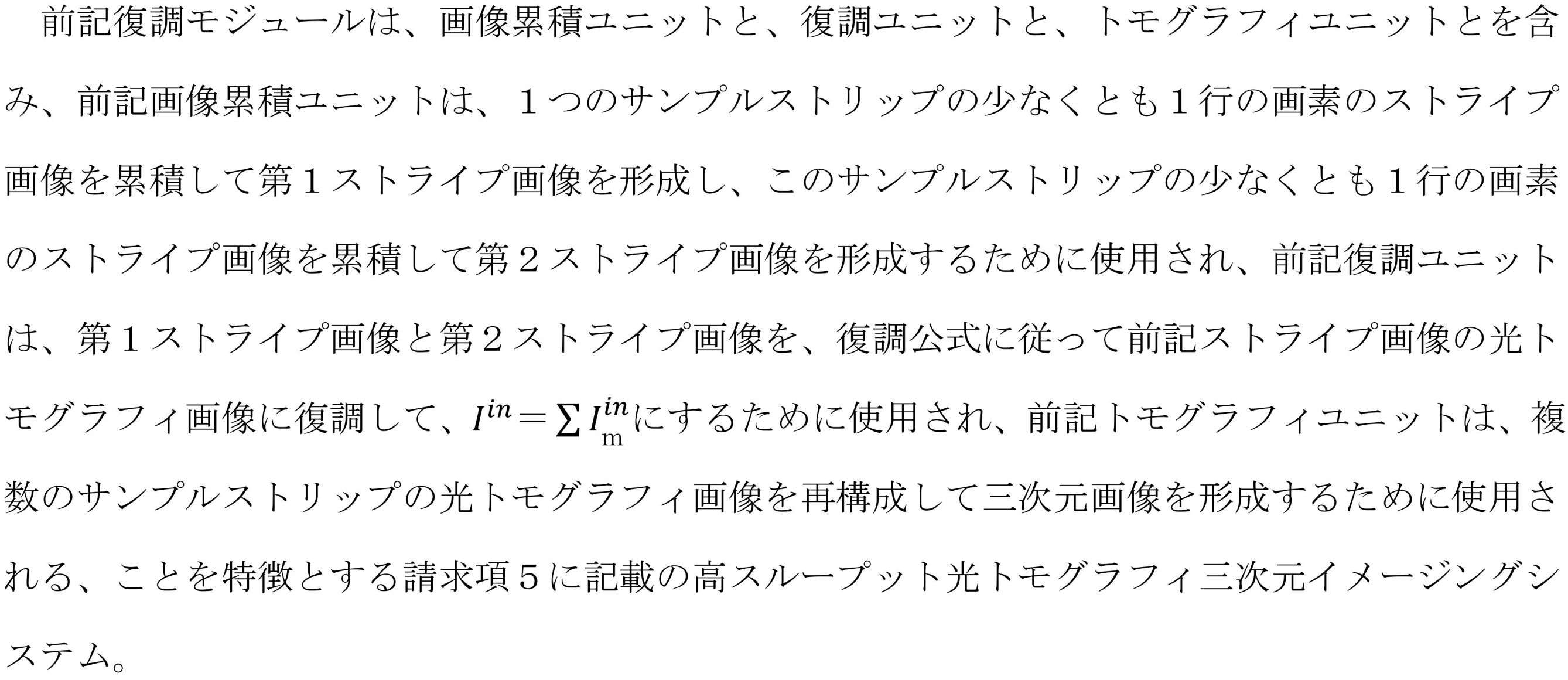
前記復調モジュールは、一つの表層の一つのサンプルストリップのサンプル画像を復調して光トモグラフィ画像を形成し、各表層の各サンプルストリップの光トモグラフィ画像を再構成して三次元画像を形成するために使用される。


Iin=c×|βI1-αI2|
である。
α、βは正の整数であり、cは0より大きい定数であり、I1はα個の画素で取得されたストライプ画像の累積合計であり、I2はβ個の画素で取得されたサンプル画像の累積合計である。α個の画素でのサンプル画像に対応する変調強度の累積値が、β個の画素でのサンプル画像に対応する変調強度の累積値とは異なる。

図4(a)に示すように、サンプルがN行の画素の配置方向に沿って移動すると、時間t1~時間tN+M-1の間にN+M-1フレームの画像(Mは、完全なストライプ画像に対応するストライプ画像ブロックの数であり、この実施例では、Nは8であり、Mは9である)を取得することができる。N+M-1フレームの画像における各行の画素は、1つのストライプ画像ブロックに対応する。例えば、第1フレームの画像の第1行の画素のストライプ画像ブロックI1(1)、第2フレームの画像の第1行の画素のストライプ画像ブロックI2(1)、第Nフレームの画像の第1行の画素のストライプ画像ブロックIN(1)、及び第N+M-1フレームの画像の第1行の画素のストライプ画像ブロックI(N+M-1)(1)を取得することができる。上記のストライプ画像ブロックI1(1)、ストライプ画像ブロックI2(1)からストライプ画像ブロックI(N+M-1)(1)を順次スプライスして、ストライプ画像を形成することができる。対応する第2行の画素~第N行の画素をスプライスして、対応するストライプ画像を形成することができる。


Claims (10)
- 高スループット光トモグラフィ三次元イメージングシステムであって、ビーム変調モジュールと、イメージングモジュールと、切除モジュールと、復調モジュールとを含み、
前記ビーム変調モジュールは、ビームを、対物レンズの焦点面に焦点を合わせ、対物レンズの焦点ぼけ面で発散することができる変調ビームに変調するために使用され、
前記イメージングモジュールは、カメラを使用して、変調ビームの照明下での同一のサンプルの少なくとも1つの表層の少なくとも1つのサンプルストリップを異なる画素でイメージングし、複数のサンプル画像を形成するために使用され、前記変調ビームは、異なる行画素の配置方向において異なる変調強度を有し、イメージング時に、前記異なる行画素の前記配置方向に沿ってサンプルストライプが移動し、これにより前記異なる行の各行の画素が、前記配置方向に沿って複数のストライプ画像ブロックを順次形成し、前記異なる行の各行の画素により形成された前記複数のストライプ画像ブロックは、同一のサンプルの前記複数のサンプル画像のうちの対応する一つのサンプル画像をスプライスするために使用され、
前記切除モジュールは、サンプルのイメージングされた表層を切除するために使用され、
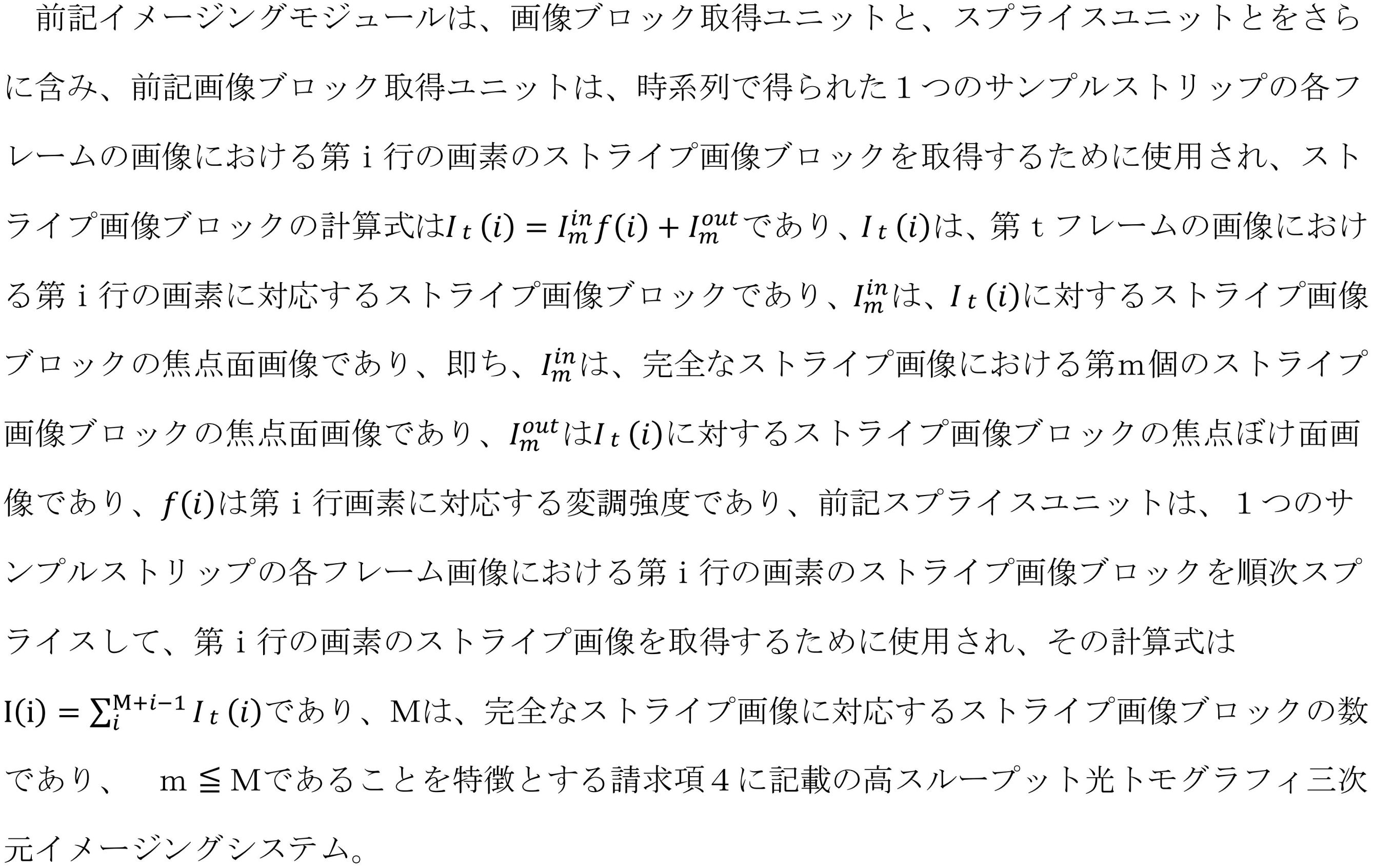
前記復調モジュールは、一つの表層の一つのサンプルストリップのサンプル画像を復調して光トモグラフィ画像を形成し、各表層の各サンプルストリップの光トモグラフィ画像を再構成して三次元画像を形成するために使用され、複数のサンプル画像を形成することを特徴とする高スループット光トモグラフィ三次元イメージングシステム。 - 前記イメージングモジュールでは、形成されたサンプルストリップのサンプル画像の計算式は次のとおりであり、
I(i)=Iinf(i)+Iout
I(i)は、第i画素で形成されたサンプル画像であり、f(i)は、サンプル画像I(i)に対応する変調強度であり、Iinは、サンプル画像の焦点面画像であり、Ioutはサンプル画像の焦点ぼけ面画像であり、
前記復調モジュールでは、復調式は、
Iin=c×|βI1-αI2|
であり、
α、βは正の整数であり、cは0より大きい定数であり、I1はα行の画素で取得されたサンプル画像の累積合計であり、I2はβ行の画素で取得されたサンプル画像の累積合計であり、β×(α行の画素でのサンプル画像に対応する変調強度の累積値)は、α×(β行の画素でのサンプル画像に対応する変調強度の累積値)とは異なる、ことを特徴とする請求項1に記載の高スループット光トモグラフィ三次元イメージングシステム。 - 前記イメージングモジュールは、ビーム変調モジュールとサンプルを駆動して、互いに垂直な3つの方向に相対的に移動させるための駆動ユニットと、サンプルストリップの長さ方向に沿って連続的なイメージングを行うイメージングユニットとを含み、前記サンプルストリップの長さ方向は、ビーム変調モジュールとサンプルのうちの1つの相対的な移動方向と同じである、ことを特徴とする請求項2に記載の高スループット光トモグラフィ三次元イメージングシステム。
- 前記イメージングモジュールにおけるカメラのイメージング領域はN行の画素であり、N≧2であり、サンプルのイメージング平面に平行な平面上に互いに垂直なX方向とY方向である2つの方向を形成し、前記変調ビームは、X方向とY方向にそれぞれ次の特性を持ち、前記変調ビームは、前記N行の画素でX方向に沿って異なる変調強度を有し、前記変調ビームは、前記N行の画素の各行の画素でY方向に沿って同じ変調強度を有し、前記画素は行画素であり、前記サンプル画像はストライプ画像である、ことを特徴とする請求項3に記載の高スループット光トモグラフィ三次元イメージングシステム。
-
-
- 前記イメージングモジュールによるイメージングの単一フレームの露光時間は、ビーム変調モジュールとサンプルがサンプルストリップの長さ方向に沿って1つの行の画素を相対的に移動する時間と同じであり、前記N行の画素の分布方向及び幅は、それぞれ、変調ビームの分布方向及び幅と同じであり、互いに物体画像共役関係にある、ことを特徴とする請求項6に記載の高スループット光トモグラフィ三次元イメージングシステム。
- 前記ビーム変調モジュールは、線状を呈する線状ビームに照明光線を整形するための整形光路と、線状ビームを線状照明変調ビームに変調するための変調光路とを含む、ことを特徴とする請求項7に記載の高スループット光トモグラフィ三次元イメージングシステム。
- 前記整形光路は、照明光線の伝送方向に沿って順次配置されたレーザ光源と、第1レンズと、第2レンズと、柱レンズとを含み、前記変調光路は、線状ビームの発散光線を平行光線に変調するための第3レンズと、線状ビームの入射方向を変調するダイクロイックミラーと、入射方向を変調した線状ビームと同軸に配置された対物レンズとを含む、ことを特徴とする請求項8に記載の高スループット光トモグラフィ三次元イメージングシステム。
- 前記駆動ユニットは、サンプルを駆動して、互いに垂直な3つの方向に移動させるための平行移動ステージであり、前記平行移動ステージは、前記対物レンズのダイクロイックミラーから離れた側に位置し、前記変調ビームの光軸に垂直であり、前記切除モジュールは、振動スライスカッタ、ダイヤモンドカッタ、及び硬質合金カッタのうちの1つ又は複数を含む、ことを特徴とする請求項9に記載の高スループット光トモグラフィ三次元イメージングシステム。
Applications Claiming Priority (3)
| Application Number | Priority Date | Filing Date | Title |
|---|---|---|---|
| CN201811296073.4A CN111122567B (zh) | 2018-11-01 | 2018-11-01 | 一种高通量光学层析三维成像系统 |
| CN201811296073.4 | 2018-11-01 | ||
| PCT/CN2019/098367 WO2020088014A1 (zh) | 2018-11-01 | 2019-07-30 | 一种高通量光学层析三维成像系统 |
Publications (2)
| Publication Number | Publication Date |
|---|---|
| JP2022511676A JP2022511676A (ja) | 2022-02-01 |
| JP7190034B2 true JP7190034B2 (ja) | 2022-12-14 |
Family
ID=70464702
Family Applications (1)
| Application Number | Title | Priority Date | Filing Date |
|---|---|---|---|
| JP2021523973A Active JP7190034B2 (ja) | 2018-11-01 | 2019-07-30 | 高スループット光トモグラフィ三次元イメージングシステム |
Country Status (8)
| Country | Link |
|---|---|
| US (1) | US11906723B2 (ja) |
| EP (1) | EP3876022A4 (ja) |
| JP (1) | JP7190034B2 (ja) |
| KR (1) | KR102593253B1 (ja) |
| CN (1) | CN111122567B (ja) |
| AU (1) | AU2019372392B2 (ja) |
| CA (1) | CA3118393C (ja) |
| WO (1) | WO2020088014A1 (ja) |
Families Citing this family (2)
| Publication number | Priority date | Publication date | Assignee | Title |
|---|---|---|---|---|
| US11521401B2 (en) * | 2020-07-31 | 2022-12-06 | Bridging Biosciences, LLC | Fertility window prediction using a convolutional neural network (CNN) and other learning methods |
| CN117705775B (zh) * | 2024-02-05 | 2024-04-26 | 中国科学院长春光学精密机械与物理研究所 | 多色荧光显微成像系统、成像方法、自动聚焦方法 |
Citations (6)
| Publication number | Priority date | Publication date | Assignee | Title |
|---|---|---|---|---|
| JP2000506634A (ja) | 1997-04-04 | 2000-05-30 | イシス イノヴェーション リミテッド | 顕微鏡撮像装置および方法 |
| JP2004109348A (ja) | 2002-09-17 | 2004-04-08 | Institute Of Physical & Chemical Research | 顕微鏡装置 |
| JP2004515780A (ja) | 2000-12-13 | 2004-05-27 | メディカル リサーチ カウンシル | 組織標本を撮像するための装置および方法 |
| JP2005525551A (ja) | 2002-05-14 | 2005-08-25 | アマシャム バイオサイエンス ユーケイ リミテッド | バイオフィルムを評価する方法 |
| CN101661159A (zh) | 2008-08-25 | 2010-03-03 | 麦克奥迪实业集团有限公司 | 一种基于二维调制技术的切层图像获取方法 |
| WO2018094290A1 (en) | 2016-11-18 | 2018-05-24 | Tissuevision, Inc. | Automated tissue section capture, indexing and storage system and methods |
Family Cites Families (18)
| Publication number | Priority date | Publication date | Assignee | Title |
|---|---|---|---|---|
| JP2773607B2 (ja) * | 1993-10-28 | 1998-07-09 | 日本電気株式会社 | 顕微鏡照明装置 |
| JP5307439B2 (ja) * | 2007-04-23 | 2013-10-02 | オリンパス株式会社 | レーザ顕微鏡 |
| US9285575B2 (en) * | 2009-01-26 | 2016-03-15 | President And Fellows Of Harvard College | Systems and methods for selective detection and imaging in coherent Raman microscopy by spectral excitation shaping |
| DE102010013830A1 (de) * | 2010-03-26 | 2011-09-29 | Carl Zeiss Microlmaging Gmbh | Mikroskop und Verfahren zur mikroskopischen Erfassung von Licht einer Probe |
| JP5454424B2 (ja) * | 2010-09-03 | 2014-03-26 | ソニー株式会社 | 撮像方法 |
| US20120127464A1 (en) * | 2010-11-22 | 2012-05-24 | Canon Kabushiki Kaisha | Light source apparatus |
| US8649024B2 (en) * | 2010-12-03 | 2014-02-11 | Zygo Corporation | Non-contact surface characterization using modulated illumination |
| CN102928970B (zh) * | 2012-10-19 | 2014-10-29 | 华中科技大学 | 一种大样本快速三维显微成像的方法和系统 |
| CN103207449B (zh) * | 2013-04-17 | 2015-04-29 | 华中科技大学 | 一种结构光快速扫描显微成像方法 |
| TWI486625B (zh) * | 2013-05-16 | 2015-06-01 | Univ Nat Central | 數位全像顯微鏡 |
| JP2015215509A (ja) * | 2014-05-12 | 2015-12-03 | パナソニックIpマネジメント株式会社 | 表示装置、表示方法およびプログラム |
| CN104061879B (zh) * | 2014-06-19 | 2017-11-24 | 四川大学 | 一种连续扫描的结构光三维面形垂直测量方法 |
| CN108020503B (zh) * | 2017-11-20 | 2020-09-08 | 苏州博芮恩光电科技有限公司 | 一种光片照明显微切片成像系统及成像结果处理方法 |
| CN108593605B (zh) * | 2018-04-23 | 2020-02-28 | 清华大学 | 三维高速宽视场层析成像方法及装置 |
| KR20200071563A (ko) * | 2018-12-11 | 2020-06-19 | 삼성전자주식회사 | Hsi 기반 검사 장치 |
| US10839542B2 (en) * | 2019-01-29 | 2020-11-17 | The Chinese University Of Hong Kong | Systems and methods for 3D laparoscopic surface reconstruction |
| DE102020213715A1 (de) * | 2020-11-01 | 2022-05-05 | Carl Zeiss Microscopy Gmbh | Vorrichtung und Verfahren zur schnellen dreidimensionalen Erfassung von Bilddaten |
| US20230045982A1 (en) * | 2021-08-11 | 2023-02-16 | Vergent Research Pty Ltd | Shuttered Light Field Display |
-
2018
- 2018-11-01 CN CN201811296073.4A patent/CN111122567B/zh active Active
-
2019
- 2019-07-30 KR KR1020217016637A patent/KR102593253B1/ko active IP Right Grant
- 2019-07-30 EP EP19878242.7A patent/EP3876022A4/en active Pending
- 2019-07-30 CA CA3118393A patent/CA3118393C/en active Active
- 2019-07-30 JP JP2021523973A patent/JP7190034B2/ja active Active
- 2019-07-30 AU AU2019372392A patent/AU2019372392B2/en active Active
- 2019-07-30 WO PCT/CN2019/098367 patent/WO2020088014A1/zh unknown
-
2021
- 2021-04-30 US US17/302,330 patent/US11906723B2/en active Active
Patent Citations (6)
| Publication number | Priority date | Publication date | Assignee | Title |
|---|---|---|---|---|
| JP2000506634A (ja) | 1997-04-04 | 2000-05-30 | イシス イノヴェーション リミテッド | 顕微鏡撮像装置および方法 |
| JP2004515780A (ja) | 2000-12-13 | 2004-05-27 | メディカル リサーチ カウンシル | 組織標本を撮像するための装置および方法 |
| JP2005525551A (ja) | 2002-05-14 | 2005-08-25 | アマシャム バイオサイエンス ユーケイ リミテッド | バイオフィルムを評価する方法 |
| JP2004109348A (ja) | 2002-09-17 | 2004-04-08 | Institute Of Physical & Chemical Research | 顕微鏡装置 |
| CN101661159A (zh) | 2008-08-25 | 2010-03-03 | 麦克奥迪实业集团有限公司 | 一种基于二维调制技术的切层图像获取方法 |
| WO2018094290A1 (en) | 2016-11-18 | 2018-05-24 | Tissuevision, Inc. | Automated tissue section capture, indexing and storage system and methods |
Non-Patent Citations (1)
| Title |
|---|
| Vincent Poher ET AL,"Improved sectioning in a slit scanning confocal microscope",OPTICS LETTERS,2008年08月15日,Vol. 33, No. 16,pages 1813-1815 |
Also Published As
| Publication number | Publication date |
|---|---|
| CN111122567A (zh) | 2020-05-08 |
| US20210333536A1 (en) | 2021-10-28 |
| KR102593253B1 (ko) | 2023-10-24 |
| AU2019372392B2 (en) | 2023-01-19 |
| KR20210086694A (ko) | 2021-07-08 |
| JP2022511676A (ja) | 2022-02-01 |
| WO2020088014A1 (zh) | 2020-05-07 |
| CA3118393A1 (en) | 2020-05-07 |
| CN111122567B (zh) | 2022-09-16 |
| EP3876022A1 (en) | 2021-09-08 |
| CA3118393C (en) | 2023-06-27 |
| AU2019372392A1 (en) | 2021-06-03 |
| US11906723B2 (en) | 2024-02-20 |
| EP3876022A4 (en) | 2021-12-29 |
Similar Documents
| Publication | Publication Date | Title |
|---|---|---|
| CN112930492B (zh) | 用于使用时间复用光片的快速体积荧光显微术的装置和方法 | |
| JP7190034B2 (ja) | 高スループット光トモグラフィ三次元イメージングシステム | |
| KR20200070313A (ko) | 이미지 재건 방법, 장치 및 현미 이미징 장치 | |
| CN110470667A (zh) | 基于压缩感知和随机多焦点扫描的三维体成像方法及装置 | |
| CN115248197A (zh) | 一种三维成像装置和成像方法 | |
| EP3520074B1 (en) | Method for the analysis of spatial and temporal information of samples by means of optical microscopy | |
| CN112654911A (zh) | 具有虚拟物镜的显微镜设备 | |
| CN110220875A (zh) | 一种基于荧光差分法的晶格光切片荧光显微成像装备及方法 | |
| CN108982455A (zh) | 一种多焦点光切片荧光显微成像方法和装置 | |
| JP7235861B2 (ja) | 高スループット光トモグラフィイメージング方法及びイメージングシステム | |
| CN212060720U (zh) | 一种基于折射窗扫描仪的显微成像装置 | |
| KR101391180B1 (ko) | 레이저 스캔 구조조명 이미징 방법 | |
| CN111260747B (zh) | 基于虚拟数字调制的高通量光学层析成像方法及系统 | |
| CN117631249A (zh) | 线扫共聚焦扫描光场显微成像装置及方法 | |
| CN115689959A (zh) | 一种正交线扫描成像处理方法及系统 |
Legal Events
| Date | Code | Title | Description |
|---|---|---|---|
| A621 | Written request for application examination |
Free format text: JAPANESE INTERMEDIATE CODE: A621 Effective date: 20210610 |
|
| A521 | Request for written amendment filed |
Free format text: JAPANESE INTERMEDIATE CODE: A523 Effective date: 20211227 |
|
| A977 | Report on retrieval |
Free format text: JAPANESE INTERMEDIATE CODE: A971007 Effective date: 20220530 |
|
| A131 | Notification of reasons for refusal |
Free format text: JAPANESE INTERMEDIATE CODE: A131 Effective date: 20220628 |
|
| A521 | Request for written amendment filed |
Free format text: JAPANESE INTERMEDIATE CODE: A523 Effective date: 20220927 |
|
| TRDD | Decision of grant or rejection written | ||
| A01 | Written decision to grant a patent or to grant a registration (utility model) |
Free format text: JAPANESE INTERMEDIATE CODE: A01 Effective date: 20221108 |
|
| A61 | First payment of annual fees (during grant procedure) |
Free format text: JAPANESE INTERMEDIATE CODE: A61 Effective date: 20221202 |
|
| R150 | Certificate of patent or registration of utility model |
Ref document number: 7190034 Country of ref document: JP Free format text: JAPANESE INTERMEDIATE CODE: R150 |