JP6914269B2 - Anti-cancer drug screening method that inhibits the binding between AIMP2-DX2 and HSP70 - Google Patents
Anti-cancer drug screening method that inhibits the binding between AIMP2-DX2 and HSP70 Download PDFInfo
- Publication number
- JP6914269B2 JP6914269B2 JP2018548001A JP2018548001A JP6914269B2 JP 6914269 B2 JP6914269 B2 JP 6914269B2 JP 2018548001 A JP2018548001 A JP 2018548001A JP 2018548001 A JP2018548001 A JP 2018548001A JP 6914269 B2 JP6914269 B2 JP 6914269B2
- Authority
- JP
- Japan
- Prior art keywords
- aimp2
- cancer
- hsp70
- binding
- protein
- Prior art date
- Legal status (The legal status is an assumption and is not a legal conclusion. Google has not performed a legal analysis and makes no representation as to the accuracy of the status listed.)
- Active
Links
Images


Classifications
-
- G—PHYSICS
- G01—MEASURING; TESTING
- G01N—INVESTIGATING OR ANALYSING MATERIALS BY DETERMINING THEIR CHEMICAL OR PHYSICAL PROPERTIES
- G01N33/00—Investigating or analysing materials by specific methods not covered by groups G01N1/00 - G01N31/00
- G01N33/48—Biological material, e.g. blood, urine; Haemocytometers
- G01N33/50—Chemical analysis of biological material, e.g. blood, urine; Testing involving biospecific ligand binding methods; Immunological testing
- G01N33/5005—Chemical analysis of biological material, e.g. blood, urine; Testing involving biospecific ligand binding methods; Immunological testing involving human or animal cells
- G01N33/5008—Chemical analysis of biological material, e.g. blood, urine; Testing involving biospecific ligand binding methods; Immunological testing involving human or animal cells for testing or evaluating the effect of chemical or biological compounds, e.g. drugs, cosmetics
- G01N33/5011—Chemical analysis of biological material, e.g. blood, urine; Testing involving biospecific ligand binding methods; Immunological testing involving human or animal cells for testing or evaluating the effect of chemical or biological compounds, e.g. drugs, cosmetics for testing antineoplastic activity
-
- A—HUMAN NECESSITIES
- A61—MEDICAL OR VETERINARY SCIENCE; HYGIENE
- A61K—PREPARATIONS FOR MEDICAL, DENTAL OR TOILETRY PURPOSES
- A61K31/00—Medicinal preparations containing organic active ingredients
- A61K31/33—Heterocyclic compounds
- A61K31/395—Heterocyclic compounds having nitrogen as a ring hetero atom, e.g. guanethidine or rifamycins
- A61K31/535—Heterocyclic compounds having nitrogen as a ring hetero atom, e.g. guanethidine or rifamycins having six-membered rings with at least one nitrogen and one oxygen as the ring hetero atoms, e.g. 1,2-oxazines
- A61K31/5375—1,4-Oxazines, e.g. morpholine
- A61K31/5377—1,4-Oxazines, e.g. morpholine not condensed and containing further heterocyclic rings, e.g. timolol
-
- A—HUMAN NECESSITIES
- A61—MEDICAL OR VETERINARY SCIENCE; HYGIENE
- A61K—PREPARATIONS FOR MEDICAL, DENTAL OR TOILETRY PURPOSES
- A61K31/00—Medicinal preparations containing organic active ingredients
- A61K31/70—Carbohydrates; Sugars; Derivatives thereof
- A61K31/7088—Compounds having three or more nucleosides or nucleotides
- A61K31/7105—Natural ribonucleic acids, i.e. containing only riboses attached to adenine, guanine, cytosine or uracil and having 3'-5' phosphodiester links
-
- G—PHYSICS
- G01—MEASURING; TESTING
- G01N—INVESTIGATING OR ANALYSING MATERIALS BY DETERMINING THEIR CHEMICAL OR PHYSICAL PROPERTIES
- G01N33/00—Investigating or analysing materials by specific methods not covered by groups G01N1/00 - G01N31/00
- G01N33/48—Biological material, e.g. blood, urine; Haemocytometers
- G01N33/50—Chemical analysis of biological material, e.g. blood, urine; Testing involving biospecific ligand binding methods; Immunological testing
- G01N33/53—Immunoassay; Biospecific binding assay; Materials therefor
- G01N33/574—Immunoassay; Biospecific binding assay; Materials therefor for cancer
-
- G—PHYSICS
- G01—MEASURING; TESTING
- G01N—INVESTIGATING OR ANALYSING MATERIALS BY DETERMINING THEIR CHEMICAL OR PHYSICAL PROPERTIES
- G01N2500/00—Screening for compounds of potential therapeutic value
- G01N2500/02—Screening involving studying the effect of compounds C on the interaction between interacting molecules A and B (e.g. A = enzyme and B = substrate for A, or A = receptor and B = ligand for the receptor)
Description
本出願は、2016年3月7日に出願された韓国特許出願第10−2016−0027077号の優先権を主張し、前記明細書全体は本出願の参考文献である This application claims the priority of Korean Patent Application No. 10-2016-0027077 filed on March 7, 2016, the entire specification of which is a reference of the present application.
本発明は、AIMP2−DX2とHSP70の結合を阻害する抗がん剤スクリーニング方法に関するもので、より詳細には(a)試験物質の存在下又は非存在下で、AIMP2−DX2又はその断片と、HSP70又はその断片とを接触させる段階;(b)試験物質の存在下又は非存在下でのAIMP2−DX2とHSP70との結合を測定する段階;(c)試験物質の存在下でのAIMP2−DX2とHSP70との結合と、試験物質非存在下でのAIMP2−DX2とHSP70との結合とを比較して、試験物質によるAIMP2−DX2とHSP70との結合水準の変化を判断する段階;(d)AIMP2−DX2とHSP70との結合水準を減少させる試験物質を選別する段階;及び(e)選別された試験物質の抗がん活性を、細胞又は動物で確認する段階を含む抗がん剤スクリーニング方法と、前記の方法で選別された抗がん剤を有効成分として含む抗がん用組成物に関するものである。 The present invention relates to a method for screening an anticancer drug that inhibits the binding between AIMP2-DX2 and HSP70, and more specifically, (a) in the presence or absence of a test substance, AIMP2-DX2 or a fragment thereof, and The step of contacting the HSP70 or a fragment thereof; (b) the step of measuring the binding of AIMP2-DX2 to the HSP70 in the presence or absence of the test substance; (c) the step of measuring the binding between the AIMP2-DX2 and the HSP70; The step of comparing the binding between HSP70 and AIMP2-DX2 and HSP70 in the absence of the test substance to determine the change in the binding level between AIMP2-DX2 and HSP70 due to the test substance; (d). A method for screening an anticancer drug, which comprises a step of selecting a test substance that reduces the binding level between AIMP2-DX2 and HSP70; and (e) a step of confirming the anticancer activity of the selected test substance in cells or animals. And the anti-cancer composition containing the anti-cancer agent selected by the above-mentioned method as an active ingredient.
がん特異的マーカーの開発は、がんの診断だけでなく、がん特異的な治療のためにも要求されている。サイトトキシン治療(cytotoxic therapies)は、最初の抗がん治療薬として使用されて以来、50余年間がんの治療に広く使われてきたが、がん細胞の他に分裂速度が比較的速い他の臓器の細胞に非特異的に作用して強い毒性を示し、深刻な副作用をもたらす問題がある。このような従来の抗がん剤の副作用及び耐性を克服するために、正常細胞のがん化過程で示されるがん特異的マーカー(cancer specific marker)を利用して、腫瘍細胞特異的に作用する治療剤開発の研究が進められた。抗がん剤による毒性を最小化するための方案として浮上する、がんターゲット治療(cancertargeted therapy)の核心は、がん細胞特異的な遺伝子を探し出すことである。 The development of cancer-specific markers is required not only for cancer diagnosis but also for cancer-specific treatment. Cytotoxic therapies have been widely used in the treatment of cancer for more than 50 years since they were first used as anticancer drugs, but in addition to cancer cells, they have a relatively high division rate. There is a problem that it acts non-specifically on the cells of the organs, shows strong toxicity, and causes serious side effects. In order to overcome the side effects and resistance of such conventional anticancer drugs, a cancer specific marker shown in the process of carcinogenesis of normal cells is used to act specifically on tumor cells. Research on the development of therapeutic agents has been advanced. At the heart of cancertargeted therapy, which has emerged as a way to minimize the toxicity of anticancer drugs, is to seek out genes that are specific to cancer cells.
一方、熱衝撃タンパク質(Heat Shock Protein、HSP)とは、タンパク質の恒常性維持に核心的な役割をする分子的シャペロンである。HSPは低酸素症のようなストレス状況で、細胞の生存にとって重要である。HSP、特にHSP90とHSP70は、広範囲の腫瘍[Morano KA、Annals of the New York Academy of Sciences、1113:1-14、2007; Calderwood SK et al、Trends in biochemical sciences、31:164-72、2006]で多く発現される。いくつかのHSPの発現は、いくつかのがんで腫瘍細胞の増殖、分化、アポトーシス(apoptosis)と相関関係があることが明らかにされ、これはHSPが、それ自身が有する細胞保護の役割のために、がん細胞の生存に重要な役割をすることを示している。HSP70の過剰発現は、マウス繊維肉腫細胞の腫瘍形成を誘発し、遺伝子導入されたマウスのT−細胞内HSP70が過剰発現されると、そのマウスのT細胞リンパ腫の増加を誘発すると報告されている[Jaattela M、International journal of cancer Journal international du cancer、60:689-93、1995; Seo JS et al、Biochemical and biophysical research communications、218:582-7、1996; Volloch VZ et al、Oncogene、18:3648-51、1999;Murphy ME、Carcinogenesis、34:1181-8、2013]。特に、HSP70は、アポトーシスから細胞を保護する重要な役割をすると知られている。それだけでなく、HSPの発現が増加することは、血管形成、侵襲、転移に関連していることが知られている[Calderwood SK et al、Trends in biochemical sciences、31:164-72、2006; Zhou J et al、The Journal of biological chemistry、279:13506-13、2004; Bruns AF et al、PloS one、7:e48539、2012; Sun J et al、Arteriosclerosis、thrombosis and vascular biology 24:2238-44、2004; Gong W、et al、Oncology reports、2013; Eustace BK et al、Cell cycle、3:1098-100、2004; Eustace BK et al、Nature cell biology、6:507-14、2004]。 On the other hand, a heat shock protein (HSP) is a molecular chaperone that plays a central role in maintaining protein homeostasis. HSP is important for cell survival in stress situations such as hypoxia. HSPs, especially HSP90 and HSP70, are a widespread tumor [Morano KA, Annals of the New York Academy of Sciences, 1113: 1-14, 2007; Calderwood SK et al, Trends in biochemical sciences, 31: 164-72, 2006] It is often expressed in. Expression of some HSPs has been shown to correlate with tumor cell proliferation, differentiation and apoptosis in some cancers, due to HSP's own cell-protecting role. It has been shown that it plays an important role in the survival of cancer cells. It has been reported that overexpression of HSP70 induces tumorigenesis in mouse fibrosarcoma cells, and overexpression of T-intracellular HSP70 in transgenic mice induces an increase in T-cell lymphoma in the mice. [Jaattela M, International journal of cancer Journal international du cancer, 60: 689-93, 1995; Seo JS et al, Biochemical and biophysical research communications, 218: 582-7, 1996; Volloch VZ et al, Oncogene, 18: 3648 -51, 1999; Murphy ME, Carcinogenesis, 34: 1118-8, 2013]. In particular, HSP70 is known to play an important role in protecting cells from apoptosis. Not only that, increased expression of HSP is known to be associated with angiogenesis, invasion, and metastasis [Calderwood SK et al, Trends in biochemical sciences, 31: 164-72, 2006; Zhou. J et al, The Journal of biological chemistry, 279: 13506-13, 2004; Bruns AF et al, PloS one, 7: e48539, 2012; Sun J et al, Arteriosclerosis, thrombosis and vascular biology 24: 2238-44, 2004 Gong W, et al, Oncology reports, 2013; Eustace BK et al, Cell cycle, 3: 1098-100, 2004; Eustace BK et al, Nature cell biology, 6: 507-14, 2004].
AIMP2(ARS-interacting multi-functional protein 2)は、アミノアシル−tRNA合成酵素複合体(Aminoacyl-tRNA synthetase:ARSs)の形成に関連したタンパク質の一つで、p38/JTV−1又はp38とも称される。AIMP2が、新規ながん抑制因子(tumor suppressor)として、Smad2/3との直接的相互作用を通じてTGF−βの信号伝達を強化する機能を有することが報告されており、がん細胞株及び組織で、AIMP2のエキソン2が欠損された形態の変異体であるAIMP2−DX2が特異的に発現されることが知られている(韓国登録特許第10−0762995号公報)。
AIMP2 (ARS-interacting multi-functional protein 2) is one of the proteins involved in the formation of aminoacyl-tRNA synthetase (ARSs), and is also called p38 / JTV-1 or p38. .. It has been reported that AIMP2, as a novel tumor suppressor, has a function of enhancing TGF-β signal transduction through direct interaction with Smad2 / 3, and cancer cell lines and tissues. It is known that AIMP2-DX2, which is a variant of AIMP2 in which
以上の通り、AIMP2−DX2及びHSP70それぞれが腫瘍細胞の分化又は生存と関連していることは知られているが、それらの相互関係ががんにどのような関連性があるかについては知られていない。 As described above, it is known that each of AIMP2-DX2 and HSP70 is associated with the differentiation or survival of tumor cells, but it is known how their interrelationship is related to cancer. Not.
[発明の詳細な説明]
[技術的課題]
そこで、本発明者らはAIMP2−DX2及びHSP70の相互作用を研究中、HSP70がAIMP2−DX2に直接結合してAIMP2−DX2タンパク質を安定化させ、結果的にがん細胞の生存と分化に決定的な役割をすることを確認した。従って、HSP70とAIMP2−DX2との結合を抑制すると、細胞の分裂と増殖を抑制できることに着目して本発明を完成した。
[Detailed description of the invention]
[Technical issues]
Therefore, while the present inventors are studying the interaction between AIMP2-DX2 and HSP70, HSP70 directly binds to AIMP2-DX2 to stabilize the AIMP2-DX2 protein, and as a result, it is determined to survive and differentiate cancer cells. It was confirmed that it would play a role. Therefore, the present invention was completed by focusing on the fact that suppressing the binding between HSP70 and AIMP2-DX2 can suppress cell division and proliferation.
従って、本発明の目的は、
(a)試験物質の存在下又は非存在下で、AIMP2−DX2又はその断片と、HSP70又はその断片とを接触させる段階;
(b)試験物質の存在下又は非存在下でのAIMP2−DX2とHSP70との結合を測定する段階;
(c)試験物質の存在下でのAIMP2−DX2とHSP70との結合と、試験物質非存在下でのAIMP2−DX2とHSP70との結合とを比較して、試験物質によるAIMP2−DX2とHSP70との結合水準の変化を判断する段階;
(d)AIMP2−DX2とHSP70との結合水準を減少させる試験物質を選別する段階;及び
(e)選別された試験物質の抗がん活性を、細胞又は動物で確認する段階を含む抗がん剤スクリーニング方法を提供することである。
Therefore, an object of the present invention is
(A) The step of contacting AIMP2-DX2 or a fragment thereof with HSP70 or a fragment thereof in the presence or absence of a test substance;
(B) The step of measuring the binding between AIMP2-DX2 and HSP70 in the presence or absence of the test substance;
(C) Comparing the binding of AIMP2-DX2 and HSP70 in the presence of the test substance with the binding of AIMP2-DX2 and HSP70 in the absence of the test substance, AIMP2-DX2 and HSP70 by the test substance Steps to determine changes in the binding level of
Anti-cancer including (d) a step of selecting a test substance that reduces the binding level between AIMP2-DX2 and HSP70; and (e) a step of confirming the anti-cancer activity of the selected test substance in cells or animals. To provide a drug screening method.
本発明の他の目的は、
前記抗がん剤スクリーニングの方法で選別された抗がん剤を有効成分として含むがんの予防又は治療用薬学的組成物を提供することである。
Another object of the present invention is
It is an object of the present invention to provide a pharmaceutical composition for preventing or treating cancer, which comprises an anticancer agent selected by the method for screening an anticancer agent as an active ingredient.
また、本発明の他の目的は、
前記抗がん剤スクリーニングの方法で選別された抗がん剤を有効成分として構成されるがんの予防又は治療用薬学的組成物を提供することである。
Another object of the present invention is
It is an object of the present invention to provide a pharmaceutical composition for preventing or treating cancer, which comprises an anticancer agent selected by the method for screening an anticancer agent as an active ingredient.
また、本発明の他の目的は、
前記抗がん剤スクリーニングの方法で選別された抗がん剤を有効成分として必須的に構成されるがんの予防又は治療用薬学的組成物を提供することである。
Another object of the present invention is
It is an object of the present invention to provide a pharmaceutical composition for preventing or treating cancer, which is essentially composed of an anticancer agent selected by the method for screening an anticancer agent as an active ingredient.
本発明の他の目的は、
がんの予防又は治療用製剤を製造するための、前記抗がん剤スクリーニング方法で選別された抗がん剤の使用を提供することである。
Another object of the present invention is
It is an object of the present invention to provide the use of an anticancer agent selected by the above-mentioned anticancer agent screening method for producing a pharmaceutical product for preventing or treating cancer.
本発明の他の目的は、
前記抗がん剤スクリーニング方法で選別された抗がん剤の有効量を、これを必要とする個体に投与することを特徴とするがんの予防又は治療方法を提供することである。
Another object of the present invention is
It is an object of the present invention to provide a method for preventing or treating cancer, which comprises administering an effective amount of an anticancer agent selected by the above-mentioned anticancer agent screening method to an individual in need thereof.
[技術的解決方法]
前記のような目的を達成するために、本発明は、
(a)試験物質の存在下又は非存在下で、AIMP2−DX2又はその断片と、HSP70又はその断片とを接触させる段階;
(b)試験物質の存在下又は非存在下でのAIMP2−DX2とHSP70との結合を測定する段階;
(c)試験物質の存在下でのAIMP2−DX2とHSP70との結合と、試験物質非存在下でのAIMP2−DX2とHSP70との結合とを比較して、試験物質によるAIMP2−DX2とHSP70との結合水準の変化を判断する段階;
(d)AIMP2−DX2とHSP70との結合水準を減少させる試験物質を選別する段階;及び
(e)選別された試験物質の抗がん活性を、細胞又は動物で確認する段階を含む抗がん剤スクリーニング方法を提供する。
[Technical solution]
In order to achieve the above object, the present invention
(A) The step of contacting AIMP2-DX2 or a fragment thereof with HSP70 or a fragment thereof in the presence or absence of a test substance;
(B) The step of measuring the binding between AIMP2-DX2 and HSP70 in the presence or absence of the test substance;
(C) Comparing the binding of AIMP2-DX2 and HSP70 in the presence of the test substance with the binding of AIMP2-DX2 and HSP70 in the absence of the test substance, AIMP2-DX2 and HSP70 by the test substance Steps to determine changes in the binding level of
Anti-cancer including (d) a step of selecting a test substance that reduces the binding level between AIMP2-DX2 and HSP70; and (e) a step of confirming the anti-cancer activity of the selected test substance in cells or animals. A drug screening method is provided.
本発明の他の目的を達成するために、本発明は、
前記抗がん剤スクリーニングの方法で選別された抗がん剤を有効成分として含む抗がん用組成物を提供する。
In order to achieve other objects of the present invention, the present invention
Provided is an anti-cancer composition containing an anti-cancer agent selected by the above-mentioned anti-cancer agent screening method as an active ingredient.
また、本発明の他の目的を達成するために本発明は、
有効成分として、前記抗がん剤スクリーニングの方法で選別された抗がん剤からなる抗がん用組成物を提供する。
In addition, in order to achieve other objects of the present invention, the present invention
As an active ingredient, an anti-cancer composition comprising an anti-cancer agent selected by the above-mentioned anti-cancer agent screening method is provided.
また、本発明の他の目的を達成するために本発明は、
有効成分として、本質的に、前記抗がん剤スクリーニングの方法で選別された抗がん剤からなる抗がん用組成物を提供する。
In addition, in order to achieve other objects of the present invention, the present invention
As an active ingredient, an anti-cancer composition comprising an anti-cancer agent selected by the above-mentioned anti-cancer agent screening method is provided.
本発明の他の目的を達成するために本発明は、
がんの予防又は治療用製剤を製造するための、前記抗がん剤スクリーニング方法で選別された抗がん剤の使用を提供する。
To achieve other objects of the present invention
Provided is the use of an anticancer agent selected by the above-mentioned anticancer agent screening method for producing a pharmaceutical product for preventing or treating cancer.
本発明の他の目的を達成するために本発明は、
前記抗がん剤スクリーニング方法で選別された抗がん剤の有効量を、これを必要とする個体に投与することを特徴とするがん治療方法を提供する。
To achieve other objects of the present invention
Provided is a cancer treatment method characterized by administering an effective amount of an anticancer agent selected by the anticancer agent screening method to an individual in need thereof.
以下、本発明を詳細に説明する。 Hereinafter, the present invention will be described in detail.
本発明は、
(a)試験物質の存在下又は非存在下で、AIMP2−DX2又はその断片と、HSP70又はその断片とを接触させる段階;
(b)試験物質の存在下又は非存在下でのAIMP2−DX2とHSP70との結合を測定する段階;
(c)試験物質の存在下でのAIMP2−DX2とHSP70との結合と、試験物質非存在下でのAIMP2−DX2とHSP70との結合とを比較して、試験物質によるAIMP2−DX2とHSP70との結合水準の変化を判断する段階;
(d)AIMP2−DX2とHSP70との結合水準を減少させる試験物質を選別する段階;及び
(e)選別された試験物質の抗がん活性を、細胞又は動物から確認する段階を含む抗がん剤スクリーニング方法を提供する。
The present invention
(A) The step of contacting AIMP2-DX2 or a fragment thereof with HSP70 or a fragment thereof in the presence or absence of a test substance;
(B) The step of measuring the binding between AIMP2-DX2 and HSP70 in the presence or absence of the test substance;
(C) Comparing the binding of AIMP2-DX2 and HSP70 in the presence of the test substance with the binding of AIMP2-DX2 and HSP70 in the absence of the test substance, AIMP2-DX2 and HSP70 by the test substance Steps to determine changes in the binding level of
Anti-cancer including (d) a step of selecting a test substance that reduces the binding level between AIMP2-DX2 and HSP70; and (e) a step of confirming the anti-cancer activity of the selected test substance from cells or animals. A drug screening method is provided.
本発明者らはHSP70が、元来の機能の他に、AIMP2−DX2に結合してAIMP2−DX2タンパク質を分解されないように安定化させ、AIMP2−DX2による腫瘍細胞の分化と成長を促進させる作用を示すことを発見した。HSP70の発現を抑制してAIMP2−DX2と反応することができるHSP70の水準を減少させるか、又はHSP70とAIMP2−DX2の反応を直接阻害することができる因子は、AIMP2−DX2タンパク質の水準を減少させ、結果的に腫瘍細胞の成長と分化が阻害されるようにする。本発明者らは、このようなHSP70とAIMP2−DX2の機能的な関係に対する発見に基づいて、HSP70とAIMP2−DX2との結合を阻害することができる製剤をスクリーニングして、抗がん剤として選別することができる抗がん剤スクリーニング方法を本明細書で初めて公開する。 In addition to its original function, the present inventors have the effect of binding to AIMP2-DX2 to stabilize the AIMP2-DX2 protein so that it is not degraded, and promoting the differentiation and growth of tumor cells by AIMP2-DX2. I found that it shows. Factors capable of suppressing HSP70 expression and reducing the level of HSP70 capable of reacting with AIMP2-DX2, or directly inhibiting the reaction of HSP70 with AIMP2-DX2, reduce the level of AIMP2-DX2 protein. As a result, the growth and differentiation of tumor cells are inhibited. Based on the discovery of such a functional relationship between HSP70 and AIMP2-DX2, the present inventors screened a preparation capable of inhibiting the binding between HSP70 and AIMP2-DX2, and used it as an anticancer agent. The anti-cancer drug screening method that can be selected is disclosed for the first time in this specification.
前記(a)試験物質の存在下又は非存在下で、AIMP2−DX2又はその断片と、HSP70又はその断片とを接触させる段階である。 (A) A step of contacting AIMP2-DX2 or a fragment thereof with HSP70 or a fragment thereof in the presence or absence of the test substance.
本発明で“タンパク質”は“ポリペプチド(polypeptide)”又は“ペプチド(peptide)”と互換性を有して使用され、例えば、自然状態のタンパク質から一般的に発見されるような、アミノ酸残基の重合体を意味する。前記“断片”は、タンパク質の一部分を意味する。また、本発明で“ポリヌクレオチド(polynucleotide)”又は“核酸”は、単一の又は二重鎖の形態をなすデオキシリボヌクレオチド(DNA)又はリボヌクレオチド(RNA)を意味する。他の制限がない限り、自然に生成されるヌクレオチドと類似した方法で核酸に混成化される自然のヌクレオチドの公知のアナログも含まれる。“mRNA”は、タンパク質合成の過程で、特定の遺伝子の塩基配列の遺伝情報を、ポリペプチドを形成するリボソームに伝達するRNAである。 In the present invention, "protein" is used interchangeably with "polypeptide" or "peptide", for example, amino acid residues commonly found in naturally occurring proteins. Means a polymer of. The "fragment" means a portion of a protein. Also, in the present invention, "polynucleotide" or "nucleic acid" means a deoxyribonucleotide (DNA) or ribonucleotide (RNA) in the form of a single or double strand. Unless otherwise limited, it also includes known analogs of natural nucleotides that are hybridized to nucleic acids in a manner similar to naturally produced nucleotides. "MRNA" is an RNA that transmits the genetic information of the base sequence of a specific gene to the ribosome that forms a polypeptide in the process of protein synthesis.
本発明で、前記“AIMP2−DX2”は、AIMP2タンパク質配列(312aa version:AAC50391.1またはGI:1215669; 320aa version:AAH13630.1,GI:15489023, BC013630.1)の中のエキソン2の領域が欠失された変異体として、AIMP2同等物(アミノ酸配列の置換、欠失、挿入、またはこれらの組み合わせによる変形体として、AIMP2と実質的に同等の活性を有する機能的同等物、または物理化学的性質を増加または減少させる変形を有し、AIMP2と実質的に同等の活性を有する機能的誘導体)からエキソン2の領域が欠失されたタンパク質を含む。本発明で、AIMP2タンパク質配列のうち“エキソン2の領域が欠失”したとは、AIMP2タンパク質からエキソン2領域のアミノ酸配列が部分的に、又は全体的に失われて生じた変異体が、AIMP2タンパク質とヘテロダイマーを形成して、AIMP2の正常的な機能を妨害することを意味する。従って、AIMP2−DX2タンパク質は、AIMP2タンパク質のエキソン2のアミノ酸配列がすべて欠失されたり、この領域のアミノ酸配列を含むエキソン1、エキソン3、エキソン4、又はこれらの領域の全てから、これらの領域の一部も欠失されたり、エキソン2のアミノ酸配列の一部のみが欠失されたタンパク質を含む。
In the present invention, the "AIMP2-DX2" has an
本発明の方法を実施するためのAIMP2−DX2タンパク質は、好ましくは、ヒトを含む哺乳類に由来するものでもあって、最も好ましくは、配列番号1で示されるヒトのAIMP2−DX2タンパク質のアミノ酸配列を含むものでもある。 The AIMP2-DX2 protein for carrying out the method of the present invention is preferably derived from mammals including humans, and most preferably the amino acid sequence of the human AIMP2-DX2 protein shown in SEQ ID NO: 1. It also includes.
また、前記AIMP2−DX2の断片は、AIMP2−DX2においてHSP70との結合に必要な部分を含む断片を意味する。本発明者らは、AIMP2−DX2のタンパク質から、AIMP2部分ではなく、DX2部分が、HSP70と結合することを確認した。従って、本発明を実施するためのAIMP2−DX2の断片は、配列番号1で表示されるヒトのAIMP2−DX2アミノ酸配列から1乃至87番目のアミノ酸を含んでいることが好ましく、最も好ましくは、配列番号2で表されるアミノ酸配列であることができるが、これに限定されるものではない。
Further, the fragment of AIMP2-DX2 means a fragment containing a portion necessary for binding to HSP70 in AIMP2-DX2. We have confirmed from the AIMP2-DX2 protein that the DX2 portion, not the AIMP2 portion, binds to HSP70. Therefore, the fragment of AIMP2-DX2 for carrying out the present invention preferably contains the
本発明で、“HSP70”は、熱衝撃タンパク質70(Heat Shock Protein,HSP)であって、タンパク質の恒常性の維持に核心的な役割をする分子的シャペロンである。HSP70は、低酸素症のようなストレス状況で、細胞の生存にとって重要である。一方、HSP70は、広範囲の腫瘍[Morano KA、Annals of the New York Academy of Sciences、1113:1-14、2007;Calderwood SK et al、Trends in biochemical sciences、31:164-72、2006]で多く発現される。HSP70の発現は、いくつかのがんで腫瘍細胞の増殖、分化、アポトーシス(apoptosis)との相関関係があることが明らかにされており、これはHSP70自体が有する細胞保護役割のために、がん細胞の生存に重要な役割を示している。HSP70の過剰発現は、マウス繊維肉腫細胞の腫瘍形成を誘発して、遺伝子導入されたマウスのT−細胞内HSP70が過剰発現されると、そのマウスのT細胞リンパ腫の増加を誘発すると報告されている[Jaattela M、International journal of cancer Journal international ducancer、60:689-93、1995;Seo JS et al、Biochemical and biophysical research communications、218:582-7、1996;Volloch VZ et al、Oncogene、18:3648-51、1999;Murphy ME、Carcinogenesis、34:1181-8、2013]。 In the present invention, "HSP70" is a heat shock protein (HSP), which is a molecular chaperone that plays a central role in maintaining protein homeostasis. HSP70 is important for cell survival in stress situations such as hypoxia. On the other hand, HSP70 is highly expressed in a wide range of tumors [Morano KA, Annals of the New York Academy of Sciences, 1113: 1-14, 2007; Calderwood SK et al, Trends in biochemical sciences, 31: 164-72, 2006]. Will be done. Expression of HSP70 has been shown to correlate with tumor cell proliferation, differentiation, and apoptosis in some cancers, due to the cytoprotective role of HSP70 itself. It shows an important role in cell survival. It has been reported that overexpression of HSP70 induces tumorigenesis in mouse fibrosarcoma cells, and overexpression of T-cell T-cell lymphoma in transgenic mice induces an increase in T-cell lymphoma in the mice. [Jaattela M, International journal of cancer Journal international ducancer, 60: 689-93, 1995; Seo JS et al, Biochemical and biophysical research communications, 218: 582-7, 1996; Volloch VZ et al, Oncogene, 18: 3648 -51, 1999; Murphy ME, Carcinogenesis, 34: 1118-8, 2013].
本発明の方法を実施するためのHSP70タンパク質は、好ましくは、ヒトを含む哺乳動物に由来するものでもあって、最も好ましくは、配列番号3で示されるアミノ酸配列を含むものでもある。 The HSP70 protein for carrying out the method of the present invention is preferably derived from a mammal including humans, and most preferably contains the amino acid sequence shown in SEQ ID NO: 3.
また、前記HSP70の断片は、AIMP2−DX2との結合に必要な部分を含んでいる断片を意味する。本発明者らは、AIMP2−DX2が、HSP70の基質結合ドメイン(Substrate binding domain)部位に結合することを確認した。具体的には、ヒトのHSP70のアミノ酸(配列番号3)配列における385乃至536番目のアミノ酸(配列番号4)を含む断片が、AIMP2−DX2と結合することを明らかにした。従って、本発明を実施するためのHSP70の断片は、配列番号3における385乃至536番目のアミノ酸(配列番号4)配列を含む断片でもある。 Further, the fragment of HSP70 means a fragment containing a portion necessary for binding to AIMP2-DX2. We have confirmed that AIMP2-DX2 binds to the Substrate binding domain site of HSP70. Specifically, it was revealed that the fragment containing the amino acid 385 to 536 (SEQ ID NO: 4) in the amino acid (SEQ ID NO: 3) sequence of human HSP70 binds to AIMP2-DX2. Therefore, the fragment of HSP70 for carrying out the present invention is also a fragment containing the amino acid (SEQ ID NO: 4) sequence of positions 385 to 536 in SEQ ID NO: 3.
また、本発明に係るAIMP2−DX2又はその断片、さらに、HSP70又はその断片は、これらの機能的同等物を含んでいる。前記“機能的同等物”とは、前記AIMP2−DX2とその断片、さらに、HSP70又はこれらの断片のアミノ酸配列と少なくとも70%以上、好ましくは80%以上、より好ましくは90%以上の配列相同性(つまり、同一性)を有するポリペプチドを意味するもので、配列番号1で示されるAIMP2−DX2と、配列番号3で示されるHSP70のポリペプチドと実質的に同質の生理活性を示すポリペプチドを意味する。ここで“実質的に同質の生理活性”とは、野生型AIMP2−DX2と野生型HSP70との間の直接的で特異的な結合を再現できることを意味する。つまり、AIMP2−DX2の断片の機能的同等物は、全体の長さのHSP70又はその断片に結合することができる活性を有するものであり、HSP70の断片の機能的同等物とは、全体の長さのAIMP2−DX2又はAIMP2−DX2におけるHSP70に結合する部位に結合できる活性を有するものを意味する。前記機能的同等物は、アミノ酸配列の一部が付加、置換又は欠失された結果、生成されたものでもある。前記でアミノ酸の置換は好ましくは保存的置換である。天然に存在するアミノ酸の保存的置換の例は、以下の通りである;脂肪族アミノ酸(Gly、Ala、Pro)、疎水性アミノ酸(Ile、Leu、Val)、芳香族アミノ酸(Phe、Tyr、Trp)、酸性アミノ酸(Asp、Glu)、塩基性アミノ酸(His、Lys、Arg、Gln、Asn)及び硫黄含有アミノ酸(Cys、Met)、又は前記機能的同等物には、アミノ酸配列上でアミノ酸の一部が欠失された変形体も含まれる。前記アミノ酸の欠失又は置換は、好ましくは、本発明のポリペプチドの生理活性に直接的に関連していない領域に位置している。また、アミノ酸の欠失は好ましくは、AIMP2−DX2又はHSP70ポリペプチドの生理活性に直接関与していない部分に位置する。従って、前記アミノ酸配列の両末端又は配列内に幾つかのアミノ酸が付加された変形体も含まれる。また、本発明の機能的同等物の範囲には、本発明によるポリペプチドの基本骨格及びその生理活性を維持しながら、ポリペプチドの一部の化学構造が変形されたポリペプチド誘導体も含まれる。例えば、本発明のポリペプチドの安定性、貯蔵性、揮発性又は溶解度等を変更させるための構造変更がこれに含まれる。 In addition, AIMP2-DX2 or a fragment thereof according to the present invention, and further, HSP70 or a fragment thereof contains functional equivalents thereof. The "functional equivalent" is at least 70% or more, preferably 80% or more, more preferably 90% or more sequence homology with the amino acid sequence of the AIMP2-DX2 and its fragments, and HSP70 or these fragments. It means a polypeptide having (that is, identity), and a polypeptide showing substantially the same physiological activity as the polypeptide of AIMP2-DX2 shown in SEQ ID NO: 1 and the polypeptide of HSP70 shown in SEQ ID NO: 3. means. Here, "substantially homogeneous bioactivity" means that the direct and specific binding between wild-type AIMP2-DX2 and wild-type HSP70 can be reproduced. That is, the functional equivalent of the AIMP2-DX2 fragment is one that has the activity of being able to bind to the HSP70 of the entire length or a fragment thereof, and the functional equivalent of the fragment of the HSP70 is the overall length. It means a substance having an activity capable of binding to a site that binds to HSP70 in AIMP2-DX2 or AIMP2-DX2. The functional equivalent is also produced as a result of addition, substitution or deletion of a part of the amino acid sequence. The amino acid substitutions described above are preferably conservative substitutions. Examples of conservative substitutions of naturally occurring amino acids are: aliphatic amino acids (Gly, Ala, Pro), hydrophobic amino acids (Ile, Leu, Val), aromatic amino acids (Phe, Tyr, Trp). ), Acidic amino acids (Asp, Glu), basic amino acids (His, Lys, Arg, Gln, Asn) and sulfur-containing amino acids (Cys, Met), or the functional equivalents are one of the amino acids on the amino acid sequence. Variants with deleted parts are also included. Deletions or substitutions of the amino acids are preferably located in regions that are not directly related to the bioactivity of the polypeptides of the invention. Also, the amino acid deletion is preferably located at a portion that is not directly involved in the bioactivity of the AIMP2-DX2 or HSP70 polypeptide. Therefore, variants in which some amino acids are added at both ends of the amino acid sequence or within the sequence are also included. The range of functional equivalents of the present invention also includes polypeptide derivatives in which a part of the chemical structure of the polypeptide is modified while maintaining the basic skeleton of the polypeptide according to the present invention and its physiological activity. For example, this includes structural changes for altering the stability, storability, volatility, solubility, etc. of the polypeptides of the invention.
本発明の方法で“接触(contacting)”とは、一般的な意味であり、2つ以上の製剤(例えば、2つのポリペプチド)を結合させたり、製剤と細胞(例えば、タンパク質と細胞)を結合させることを意味する。接触は試験管内(in vitro)で起こり得る。例えば、試験管(test tube)又は他のコンテナ(container)で2つ以上の製剤を結合させたり、試験製剤と細胞とを、又は細胞溶解物と試験製剤とをを結合させることである。また、接触は、細胞又はインシチュー(in situ)で起こることもある。例えば、2つのポリペプチドをコードする組換えポリヌクレオチドを細胞内で共同発現(coexpression)させることで、細胞又は細胞溶解物から2つのポリペプチドを接触させることである。また、テストしようとするタンパク質が固定相の表面に配列されたタンパク質チップ(protein chip)やタンパク質アレイ(protein array)を利用することもできる。 In the method of the present invention, "contacting" has a general meaning and is used to bind two or more pharmaceutical products (for example, two polypeptides) or to bind a pharmaceutical product and a cell (for example, a protein and a cell). It means to combine. Contact can occur in vitro. For example, combining two or more pharmaceutical products in a test tube or another container, or combining a test pharmaceutical product with cells, or a cell lysate with a test pharmaceutical product. Contact may also occur in cells or in situ. For example, the recombinant polynucleotide encoding the two polypeptides is co-expressed intracellularly to bring the two polypeptides into contact from the cell or cell lysate. It is also possible to utilize a protein chip or protein array in which the protein to be tested is arranged on the surface of the stationary phase.
本発明の方法を、細胞内で実施するときには、AIMP2−DX2とHSP70は、細胞内在的に発現されることを利用したり、AIMP2−DX2やその断片、HSP70又はその断片をコードする核酸を細胞内に導入して形質転換させることで過発現されるようにして、実施することができる。また、本発明の方法を試験管内又はタンパク質アレイのようなin situで実施するときには、AIMP2−DX2又はその断片、HSP70又はその断片は、天然から抽出したり、遺伝工学的方法により製作して準備することができる。例えば、通常の方法によって、前記ポリペプチド又はその機能的同等物をコードする核酸と組換えて発現ベクターを作製して、適切な宿主細胞において発現させて得ることができる。また、本発明の方法を実施するために必要な前記ポリペプチドは、当業界において公知である化学的合成方法でも製作することができる。 When the method of the present invention is carried out intracellularly, AIMP2-DX2 and HSP70 utilize the intracellular expression of AIMP2-DX2 and fragments thereof, and nucleic acids encoding HSP70 or fragments thereof are used in cells. It can be carried out by introducing it into the intracellular body and transforming it so that it is overexpressed. In addition, when the method of the present invention is carried out in situ or in situ such as a protein array, AIMP2-DX2 or a fragment thereof, HSP70 or a fragment thereof may be extracted from nature or prepared by a genetic engineering method. can do. For example, an expression vector can be prepared by recombination with a nucleic acid encoding the polypeptide or a functional equivalent thereof by a usual method and expressed in a suitable host cell. In addition, the polypeptide required for carrying out the method of the present invention can also be produced by a chemical synthesis method known in the art.
また、本発明の方法で“試験物質”とは、試験製剤(test agent)又は製剤(agent)と互換可能に使用することができるもので、任意の物質(substance)、分子(molecule)、元素(element)、化合物(compound)、実在物(entity)又はこれらの組み合わせを含む。例えばタンパク質、ポリペプチド、低分子有機化合物(small organic molecule)、多糖類(polysaccharide)、ポリヌクレオチドなどを含む。また、天然産物(natural product)、合成化合物又は化学化合物又は2つ以上の物質の組み合わせでもある。 Further, in the method of the present invention, the "test substance" is a substance that can be used interchangeably with a test agent or an agent, and is an arbitrary substance, molecule, or element. Includes element, compound, entity or a combination thereof. For example, it includes proteins, polypeptides, small organic molecules, polysaccharides, polynucleotides and the like. It is also a natural product, a synthetic or chemical compound, or a combination of two or more substances.
具体的には、本発明の方法を通じて選別される抗がん剤は、AIMP2−DX2とHSP70とのタンパク質結合を阻害したり、HSP70と結合できるAIMP2−DX2タンパク質の発現水準を下げることができるものであれば物質の特性に制限されず、例えば、siRNA、shRNA、miRNA、ribozyme、DNAzyme、PNA(peptide nucleic acid)、アンチセンスオリゴヌクレオチド、抗体、アプタマー、ペプチド、天然抽出物、及び化学物質などでもある。好ましくは、AIMP2−DX2の発現を下げることができるshRNA、siRNA又は化学物質でもあるがこれに限定されるものではない。 Specifically, the anticancer agent selected through the method of the present invention can inhibit the protein binding between AIMP2-DX2 and HSP70, or can lower the expression level of the AIMP2-DX2 protein capable of binding to HSP70. If so, it is not limited to the characteristics of the substance, and for example, siRNA, shRNA, miRNA, ribozyme, DNAzyme, PNA (peptide nucleic acid), antisense oligonucleotide, antibody, aptamer, peptide, natural extract, chemical substance, etc. be. Preferably, it is also, but is not limited to, a shRNA, siRNA or chemical that can reduce the expression of AIMP2-DX2.
前記(b)段階は、(a)段階で試験物質の存在下又は非存在下で接触させたAIMP2−DX2とHSP70との結合を測定する段階である。 The step (b) is a step of measuring the binding between AIMP2-DX2 and the HSP70 that have been contacted in the presence or absence of the test substance in the step (a).
前記AIMP2−DX2とHSP70との結合を測定する段階は、二つのタンパク質の結合程度を測定するために、当業界で通常的に使用されるものであれば制限なく選択して使用することができる。例えば、ツーハイブリッド(two hybrid)方法、共免疫沈降方法(co-immunoprecipitation assay、 co-IP)、組織免疫染色と共局所化分析(co-localization assay)、シンチレーション(閃光)近接測定法(scintillation proximity assay,SPA)、UV又は化学的架橋結合方法、二分子相互作用分析(bimolecular interactionan alysis、BIA)、質量分析法(mass spectrometry,MS)、NMR(nuclear magnetic resonance)、蛍光偏光分析法(fluorescence polarization assays,FPA)及び試験管内プル−ダウンアッセイ(in vitro pull-down assay)、ELISA(enzyme linked immunosorbent assay)、タンパク質チップ(protein chip)又はアレー(array)、Venus BiFC(bimolecular fluorescence complementation,BiFC)などから適宜選択して、AIMP2−DX2とHSP70との結合を測定することができる。 The step of measuring the binding between AIMP2-DX2 and HSP70 can be selected and used without limitation as long as it is commonly used in the art for measuring the degree of binding between the two proteins. .. For example, two hybrid methods, co-immunoprecipitation assay (co-IP), tissue immunostaining and co-localization assay, scintillation proximity. Assay, SPA), UV or chemical cross-linking method, bimolecular interactionan alysis (BIA), mass spectrometry (MS), NMR (nuclear magnetic resonance), fluorescence polarization. assays, FPA) and in vitro pull-down assay, ELISA (enzyme linked immunosorbent assay), protein chip or array, Venus BiFC (bimolecular fluorescence complementation, BiFC), etc. The binding between AIMP2-DX2 and HSP70 can be measured by appropriately selecting from.
本発明の具体的な実施例では、細胞内にHA、Strep、放射性同位元素等で適切に標識されたDX2とHSP70を過発現させ、co−IP実験を行ったり、GST pull−down実験を利用して、DX2に結合しているHSP70タンパク質の水準を測定したり、逆にHSP70に結合しているDX2のタンパク質の水準を測定した。 In a specific example of the present invention, DX2 and HSP70 appropriately labeled with HA, Protein, radioactive isotope, etc. are overexpressed in cells, and a co-IP experiment is performed or a GST pull-down experiment is used. Then, the level of the HSP70 protein bound to DX2 was measured, and conversely, the level of the DX2 protein bound to HSP70 was measured.
前記(c)段階は、(b)で測定した試験物質の存在下でのAIMP2−DX2とHSP70との結合と、試験物質非存在下でのAIMP2−DX2とHSP70との結合とを比較して、試験物質によるAIMP2−DX2とHSP70との結合水準の変化を判断する段階である。 In step (c), the binding of AIMP2-DX2 and HSP70 in the presence of the test substance measured in (b) is compared with the binding of AIMP2-DX2 and HSP70 in the absence of the test substance. This is the stage to determine the change in the binding level between AIMP2-DX2 and HSP70 depending on the test substance.
つまり、(b)段階で導出された試験物質の存在下又は非存在下で接触させたAIMP2−DX2とHSP70とのタンパク質の結合水準の差を把握して、試験物質がAIMP2−DX2とHSP70との結合に及ぼす影響を判断する段階である。 That is, by grasping the difference in the protein binding level between AIMP2-DX2 and HSP70 that were contacted in the presence or absence of the test substance derived in step (b), the test substances were AIMP2-DX2 and HSP70. This is the stage to determine the effect on the binding of.
前記(d)段階は、AIMP2−DX2とHSP70との結合水準を減少させる試験物質を選別する段階である。 The step (d) is a step of selecting a test substance that reduces the binding level of AIMP2-DX2 and HSP70.
本発明者らは、具体的な実施例で、AIMP2−DX2とHSP70とは細胞内で特異的に直接結合し、HSP70との結合を通じてAIMP2−DX2は分解されないように安定化され、結果的にAIMP2−DX2による細胞の増殖と分化が維持されることを確認した。AIMP2−DX2タンパク質の安定化は、HSP70タンパク質との直接的な結合を通じて行われるもので、AIMP2−DX2タンパク質がHSP70タンパク質から解離されると、AIMP2−DX2タンパク質が不安定になり、分解されてその水準が減少するようになり、結果的にAIMP2−DX2による細胞の増殖と分化が阻害される。このような効果は、HSP70の発現を抑制するsiRNA又はHSP70阻害剤を利用して確認することができた。AIMP2−DX2とHSP70との間の結合を抑制することにより、非正常的に細胞分裂するがん細胞の発生と増殖を抑制できることが分かった。 In a specific example, the present inventors specifically bind AIMP2-DX2 and HSP70 directly in the cell, and through the binding to HSP70, AIMP2-DX2 is stabilized so as not to be decomposed, and as a result. It was confirmed that the proliferation and differentiation of cells by AIMP2-DX2 were maintained. Stabilization of the AIMP2-DX2 protein is carried out through direct binding to the HSP70 protein, and when the AIMP2-DX2 protein is dissociated from the HSP70 protein, the AIMP2-DX2 protein becomes unstable and is degraded. Levels are reduced, resulting in inhibition of cell proliferation and differentiation by AIMP2-DX2. Such an effect could be confirmed by using siRNA or an HSP70 inhibitor that suppresses the expression of HSP70. It was found that by suppressing the binding between AIMP2-DX2 and HSP70, the development and proliferation of cancer cells that undergo abnormal cell division can be suppressed.
また、他の実施例では、腫瘍発生関連細胞実験と異種移植(xenograft)の動物実験を通じて、AIMP2−DX2とHSP70との間の結合を抑制すると、がん細胞の増殖、腫瘍の形成と成長が抑制されることを確認した。 In other examples, inhibition of binding between AIMP2-DX2 and HSP70 through tumorigenesis-related cell experiments and xenograft animal experiments resulted in cancer cell growth, tumor formation and growth. It was confirmed that it was suppressed.
以上の本発明者らの発見を通じて、当業者は、AIMP2−DX2とHSP70との間の結合を抑制又は結合水準を減少させる物質が、AIMP2−DX2を不安定にしてがん細胞の増殖と分化を抑制できることが分かる。前記AIMP2−DX2とHSP70との結合水準を減少させる物質は、代表的には、AIMP2−DX2とHSP70との結合自体を阻害する物質、又はAIMP2−DX2と結合可能なHSP70タンパク質の量を減少させる物質でもある。本発明の抗がん剤スクリーニング方法によって選別された抗がん剤に対する具体的な例は、本発明によるがんの予防又は治療用薬学的組成物に対する説明に記述されている。 Through the above discoveries by the present inventors, those skilled in the art will appreciate that a substance that suppresses the binding between AIMP2-DX2 and HSP70 or reduces the binding level destabilizes AIMP2-DX2 and proliferates and differentiates cancer cells. It can be seen that can be suppressed. The substance that reduces the binding level between AIMP2-DX2 and HSP70 is typically a substance that inhibits the binding itself between AIMP2-DX2 and HSP70, or reduces the amount of HSP70 protein that can bind to AIMP2-DX2. It is also a substance. Specific examples of the anti-cancer agents selected by the anti-cancer agent screening method of the present invention are described in the description of the pharmaceutical composition for preventing or treating cancer according to the present invention.
前記(e)段階は、(d)段階で選別された物質の抗がん活性を細胞又は動物で確認する段階である。 The step (e) is a step of confirming the anticancer activity of the substance selected in the step (d) in cells or animals.
(e)段階は、(d)段階でAIMP2−DX2とHSP70との結合水準を減少させるものに選別された試験物質が、具体的にがん又は腫瘍モデルの細胞又は動物からAIMP2−DX2タンパク質の不安定化と予測される抗がん活性を有するかを確認する段階である。前記抗がん活性は、非正常的な細胞分裂の増加、正常細胞のがん細胞への転換、がん細胞の細胞分裂と増殖、腫瘍の発生と成長などを抑制することを意味する。 In the step (e), the test substance selected to reduce the binding level of AIMP2-DX2 and HSP70 in the step (d) is specifically obtained from cells or animals of a cancer or tumor model of the AIMP2-DX2 protein. It is a stage to confirm whether it has anticancer activity predicted to be destabilizing. The anticancer activity means suppressing an increase in abnormal cell division, conversion of normal cells into cancer cells, cell division and proliferation of cancer cells, tumor development and growth, and the like.
前記がん又は腫瘍モデルの細胞又は動物は、当業界で通常的に使用されるものであれば適宜選択して、(d)段階で選別された物質の抗がん活性を確認することができる。本発明の具体的な実施例では、ヒトのがん細胞株をマウスに注入する異種移植を利用して、体内腫瘍の発生と形成過程を観察している。 The cells or animals of the cancer or tumor model can be appropriately selected as long as they are commonly used in the art, and the anticancer activity of the substance selected in step (d) can be confirmed. .. In a specific example of the present invention, the development and formation process of an in-vivo tumor is observed by utilizing xenotransplantation in which a human cancer cell line is injected into a mouse.
本発明に係る抗がん剤スクリーニング方法は、前記(d)と(e)段階の間に追加的に
(1)試験物質を、AIMP2−DX2を発現する細胞に接触させる段階;
(2)前記細胞と試験物質とを接触しない対照群細胞でAIMP2−DX2タンパク質の水準を測定する段階;及び
(3)対照群細胞と比較してAIMP2−DX2タンパク質の水準を減少させる試験物質を選別する段階を実施することができる。
The anti-cancer drug screening method according to the present invention additionally contacts (1) a test substance with cells expressing AIMP2-DX2 between the steps (d) and (e);
(2) A step of measuring the level of AIMP2-DX2 protein in a control group cell in which the cells are not in contact with the test substance; and (3) a test substance that reduces the level of AIMP2-DX2 protein as compared with the control group cell. A sorting step can be carried out.
本発明によれば、AIMP2−DX2が分解されないように安定化するにはHSP70との直接的な結合が必要なため、AIMP2−DX2とHSP70との結合水準が減少すると、AIMP2−DX2のタンパク質水準が減少することを理解できる。前記(1)乃至(3)の段階は、(d)段階でAIMP2−DX2とHSP70との結合水準を減少させるものに選別された試験物質が、AIMP2−DX2タンパク質の水準を減少させるか否かをさらに確認することである。前記の追加的な段階は、(d)段階でAIMP2−DX2とHSP70との結合水準を減少させる製剤として選別されたが、偽陰性(false-positive)である場合、又はAIMP2−DX2とHSP70との結合を阻害する水準がAIMP2−DX2の不安定化を引き起こすことに不足した場合を判断して除去するために、実施することができる。 According to the present invention, direct binding to HSP70 is required to stabilize AIMP2-DX2 so that it is not degraded. Therefore, when the binding level between AIMP2-DX2 and HSP70 decreases, the protein level of AIMP2-DX2 Can be understood to decrease. In the steps (1) to (3), whether or not the test substance selected to reduce the binding level of AIMP2-DX2 and HSP70 in the step (d) reduces the level of the AIMP2-DX2 protein. Is to confirm further. The additional step described above was selected as a formulation that reduces the binding level of AIMP2-DX2 and HSP70 in step (d), but if it is false-positive, or with AIMP2-DX2 and HSP70. It can be carried out to determine and eliminate cases where the level of inhibition of binding is insufficient to cause destabilization of AIMP2-DX2.
前記(1)段階は、(d)段階で選別された試験物質をAIMP2−DX2を発現する細胞に接触させる段階である。 The step (1) is a step of contacting the test substance selected in the step (d) with cells expressing AIMP2-DX2.
前記AIMP2−DX2を発現する細胞は、AIMP2−DX2を内在的に発現する細胞でもあって、AIMP2−DX2をコードするポリヌクレオチドを含む組換え発現ベクターに形質転換されてAIMP2−DX2を過剰発現する細胞でもある。例えば、AIMP2−DX2を発現する多様ながん細胞株の中から適宜選択して、試験物質のAIMP2−DX2不安定化効果を検証することができる。前記“接触”の意味は先に説明した通りである。 The cells expressing AIMP2-DX2 are also cells that endogenously express AIMP2-DX2, and are transformed into a recombinant expression vector containing a polynucleotide encoding AIMP2-DX2 to overexpress AIMP2-DX2. It is also a cell. For example, the AIMP2-DX2 destabilizing effect of the test substance can be verified by appropriately selecting from various cancer cell lines expressing AIMP2-DX2. The meaning of "contact" is as described above.
前記(2)段階は、(1)段階で試験物質と接触した細胞と試験物質を接触していない対照群細胞からAIMP2−DX2タンパク質水準を測定する段階である。 The step (2) is a step of measuring the AIMP2-DX2 protein level from the cells in contact with the test substance and the control group cells not in contact with the test substance in the step (1).
前記AIMP2−DX2タンパク質の水準を測定するために、当業界で通常に使用されるタンパク質検出方法を制限なく選択して使用することができ、例えば、ウェスタンウェスタンブロット(western blotting)、ドットブロッティング(dot blotting)、酵素結合免疫吸着分析法(enzyme linked immunosorbent assay,ELISA)、放射免疫分析法(RIA)、放射免疫拡散法、オウクテロニ免疫拡散法、ロケット免疫電気泳動、免疫組織化学染色、免疫沈降法(immunoprecipitation)、補体固定分析法、フローサイトメトリー(FACS)又はタンパク質チップ(chip)方法などを使用することができる。 In order to measure the level of the AIMP2-DX2 protein, a protein detection method commonly used in the art can be selected and used without limitation, for example, Western blotting, dot blotting (dot). Blotting), enzyme-linked immunosorbent assay (ELISA), radioimmunoassay (RIA), radioimmunoassay, octeloni immunodiffusion, rocket immunoelectrometry, immunohistochemical staining, immunoprecipitation ( Immunoprecipitation), complement fixation analysis, flow cytometry (FACS) or protein chip method can be used.
前記(c)段階は、対照群の細胞と比較してAIMP2−DX2タンパク質の水準を減少させる試験物質を選別する段階である。 The step (c) is a step of selecting a test substance that reduces the level of AIMP2-DX2 protein as compared with the cells of the control group.
前記(c)段階は、先に(b)のステップで測定した試験物質を接触した細胞と接触していない対照群細胞とで測定したAIMP2−DX2タンパク質の水準を比較し、AIMP2−DX2タンパク質水準が実際に減少したことが示された試験物質をさらに選別することになる。 In step (c), the level of AIMP2-DX2 protein measured in the control group cells in contact with the test substance previously measured in step (b) was compared, and the level of AIMP2-DX2 protein was compared. Will be further screened for test substances that have been shown to actually decrease.
本発明では、前記がんは乳がん、肺がん、結腸がん、肛門がん、星状細胞腫、白血病、リンパ腫、頭頸部がん、肝臓がん、睾丸がん、子宮頸部がん、肉腫、血管腫、食道がん、眼がん、喉頭がん、経口がん、中皮腫、骨髄腫、口腔がん、直腸がん、咽喉がん、膀胱がん、子宮がん、卵巣がん、前立腺がん、大腸がん、膵臓がん、腎臓がん、胃がん、皮膚がん、基底細胞がん、黒色腫、扁平上皮がん腫、口腔扁平上皮がん腫、大腸直腸がん、膠芽腫(膠細胞腫)、子宮内膜がん及び悪性脳膠腫からなる群から選ばれ得るがこれらに限定されるものではない。 In the present invention, the cancers are breast cancer, lung cancer, colon cancer, anal cancer, stellate cell tumor, leukemia, lymphoma, head and neck cancer, liver cancer, testicle cancer, cervical cancer, sarcoma, Hemangiomas, esophageal cancer, eye cancer, laryngeal cancer, oral cancer, mesenteric tumor, myeloma, oral cancer, rectal cancer, throat cancer, bladder cancer, uterine cancer, ovarian cancer, Prostate cancer, colon cancer, pancreatic cancer, kidney cancer, gastric cancer, skin cancer, basal cell cancer, melanoma, squamous cell carcinoma, oral squamous cell carcinoma, colonic rectal cancer, glue bud It can be selected from, but is not limited to, the group consisting of tumors (collagen tumors), endometrioid cancers and malignant encephalograms.
さらに、本発明は、本発明に係る抗がん剤スクリーニングの方法で選別された抗がん剤を有効成分として含むがんの予防又は治療用薬学的組成物を提供する。 Furthermore, the present invention provides a pharmaceutical composition for preventing or treating cancer, which comprises an anticancer agent selected by the method for screening an anticancer agent according to the present invention as an active ingredient.
また、本発明は、有効成分として、本発明に係る抗がん剤スクリーニングの方法で選別された抗がん剤からなるがんの予防又は治療用薬学的組成物を提供する。 The present invention also provides a pharmaceutical composition for preventing or treating cancer, which comprises an anticancer drug selected by the method for screening an anticancer drug according to the present invention as an active ingredient.
また、本発明は、有効成分として、本質的に、本発明に係る抗がん剤スクリーニングの方法で選別された抗がん剤からなるがんの予防又は治療用薬学的組成物を提供する。 The present invention also provides a pharmaceutical composition for preventing or treating cancer, which is essentially composed of an anticancer drug selected by the method for screening an anticancer drug according to the present invention as an active ingredient.
本発明の方法により選別された抗がん剤は、AIMP2−DX2とHSP70との結合水準を減少させ、AIMP2−DX2タンパク質を不安定化してがん細胞の増殖と分化を抑制できるものであれば物質の特性に特別な制限はないが、作用機作によって代表的にAIMP2−DX2とHSP70二つのタンパク質の結合自体を阻害する物質又はHSP70の発現を抑制してHSP70と結合可能なAIMP2−DX2タンパク質の水準をさげる物質を考えてみる。 If the anticancer agent selected by the method of the present invention can reduce the binding level between AIMP2-DX2 and HSP70, destabilize the AIMP2-DX2 protein, and suppress the growth and differentiation of cancer cells. Although there are no particular restrictions on the properties of the substance, a substance that inhibits the binding of the two proteins AIMP2-DX2 and HSP70 itself by the mechanism of action or an AIMP2-DX2 protein that can suppress the expression of HSP70 and bind to HSP70. Consider a substance that lowers the standard of.
本発明の方法で選別された抗がん剤は具体的にはsiRNA、shRNA、miRNA、リボザイム、DNAザイム、PNA(peptide nucleic acid)、アンチセンスオリゴヌクレオチド、抗体、アプタマー、ペプチド、天然抽出物及び化学物質からなる群から選ばれたものでもある。 The anticancer agents selected by the method of the present invention are specifically siRNA, shRNA, miRNA, ribozyme, DNAzyme, PNA (peptide nucleic acid), antisense oligonucleotide, antibody, aptamer, peptide, natural extract and It is also selected from a group of chemical substances.
本発明者らはHSP70に特異的なsiRNA(si−HSP70)、又はHSP70阻害剤を使用して、がん細胞から、HSP70の発現を抑制するとHSP70に結合したAIMP2−DX2の水準が減少してAIMP2−DX2タンパク質が分解して減少し、結果的にAIMP2−DX2によるがん細胞の増殖と分化が抑制されることを確認した。 The present inventors used an HSP70-specific siRNA (si-HSP70) or an HSP70 inhibitor to suppress the expression of HSP70 from cancer cells, which reduced the level of AIMP2-DX2 bound to HSP70. It was confirmed that the AIMP2-DX2 protein was degraded and decreased, and as a result, the growth and differentiation of cancer cells by AIMP2-DX2 were suppressed.
これは、siRNA又はHSP70阻害剤などHSP70の発現を抑制できる製剤は、がん疾患の予防又は治療に効果的に利用できることを示した。 This indicates that a preparation capable of suppressing the expression of HSP70, such as siRNA or an HSP70 inhibitor, can be effectively used for the prevention or treatment of cancer diseases.
従って、本発明に係る抗がん剤スクリーニングで選別された抗がん剤は、HSP70特異的なsiRNA、shRNA、又はHSP70阻害剤でもある。前記siRNA又はshRNAは、HSP70mRNAに特異的に結合してmRNAの分解を誘導することができる10乃至30個の塩基配列からなり、当業界において公知の方法により通常の技術者が容易に製作することができる。 Therefore, the anti-cancer agent selected by the anti-cancer agent screening according to the present invention is also an HSP70-specific siRNA, shRNA, or HSP70 inhibitor. The siRNA or shRNA is composed of 10 to 30 base sequences capable of specifically binding to HSP70 mRNA and inducing the degradation of mRNA, and can be easily produced by a conventional engineer by a method known in the art. Can be done.
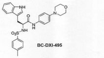
本発明の具体的な実施例では、下記化学式1の化合物がAIMP2−DX2とHSP70との結合を濃度依存的に阻害し、結果的にAIMP2−DX2が不安定化してその発現が阻害されることによりがん細胞の増殖と分化が抑制されることを確認した。
In a specific example of the present invention, the compound of the following

本発明に係るがんの予防又は治療用薬学的組成物は、経口的又は非経口的に投与することができる。非経口的投与方法は、これに限定はされないが、静脈内、筋肉内、動脈内、骨髄内、境膜内、心臓内、経皮、皮下、腹腔内、鼻腔内、腸管、局所、舌下又は直腸内投与でもあって、好ましくは血管内投与でもある。 The pharmaceutical composition for preventing or treating cancer according to the present invention can be administered orally or parenterally. Parenteral administration methods are, but are not limited to, intravenous, intramuscular, intraarterial, intramedullary, intraperitoneal, intracardiac, transdermal, subcutaneous, intraperitoneal, intranasal, intestinal, topical, sublingual. Alternatively, it may be administered rectally, preferably intravascularly.
また、本発明に係るがん疾患の予防又は治療用組成物は、薬学的に許容される担体と共に、当業界において公知の方法で、投与経路により多様に剤形化することができる。“薬学的に許容される”とは、生理学的に許容されてヒトに投与されるとき、活性成分の作用を阻害しない、通常的に胃腸障害、めまいのようなアレルギー反応又はこれと類似した反応を起こさない非毒性の組成物を意味する。前記担体には全ての種類の溶媒、分散媒質、水中油又は油中水エマルジョン、水性組成物、リポソーム、マイクロビーズ及びマイクロソームが含まれる。 In addition, the composition for preventing or treating a cancer disease according to the present invention can be variously formulated with a pharmaceutically acceptable carrier by a method known in the art according to the route of administration. "Pharmaceutically acceptable" means an allergic reaction, such as gastrointestinal disorders, dizziness, or a similar reaction that does not interfere with the action of the active ingredient when it is physiologically tolerated and administered to humans. Means a non-toxic composition that does not cause dizziness. The carriers include all types of solvents, dispersion media, oil-in-water or water-in-oil emulsions, aqueous compositions, liposomes, microbeads and microsomes.
本発明の薬学的組成物を非経口的に投与する場合、本発明の組成物は、適切な非経口用担体と共に、注射剤、経皮投与剤及び鼻腔吸込剤の形態で、当該技術分野で公知の方法により製剤化することができる。前記注射剤の場合には、必ず滅菌しなければならず、バクテリア、真菌のような微生物の汚染から保護されるべきである。注射剤の場合、適切な担体の例としては、これに限定されないが、水、エタノール、ポリオール(例えば、グリセロール、プロピレングリコール及び液体ポリエチレングリコール等)、これらの混合物及び/又は植物油を含む溶媒又は分散媒質でもある。より好ましくは、適切な担体としては、ハンクス溶液、リンゲル液、トリエタノールアミンが含有されたPBS(phosphate buffered saline;リン酸緩衝生理食塩水)又は注射用滅菌水、10%エタノール、40%プロピレングリコール及び5%デキストローズのような等張溶液などを使用することができる。前記注射剤を微生物汚染から保護するためには、パラベン、クロロブタノール、フェノール、ソルビン酸、ティーメロサルなどのような多様な抗菌剤及び抗真菌剤をさらに含むことができる。また、前記注射剤は、殆どの場合、糖又は塩化ナトリウムのような等張化剤をさらに含むことができる。 When the pharmaceutical composition of the present invention is administered parenterally, the composition of the present invention, together with a suitable parenteral carrier, is in the form of an injection, a transdermal agent and a nasal inhaler in the art. It can be formulated by a known method. In the case of the injection, it must be sterilized and protected from contamination by microorganisms such as bacteria and fungi. In the case of injectables, examples of suitable carriers include, but are not limited to, water, ethanol, polyols (eg, glycerol, propylene glycol and liquid polyethylene glycol, etc.), mixtures and / or dispersions thereof containing vegetable oils. It is also a medium. More preferably, suitable carriers include Hanks solution, Ringer's solution, PBS (phosphate buffered saline) containing triethanolamine or sterile water for injection, 10% ethanol, 40% propylene glycol and the like. An isotonic solution such as 5% dextrose can be used. In order to protect the injection from microbial contamination, various antibacterial and antifungal agents such as parabens, chlorobutanol, phenol, sorbic acid, teamerosal and the like can be further included. In addition, the injection may further contain an isotonic agent such as sugar or sodium chloride in most cases.
経皮投与剤の場合、軟膏剤、クリーム剤、ローション剤、ゲル剤、外用液剤、パスタ剤、リニメント剤、エアロゾル剤などの形態が含まれる。前記で“経皮投与”とは、本発明の組成物を局所的に皮膚に投与して組成物に含有された有効な量の活性成分が皮膚内に伝達されることを意味する。例えば、本発明の組成物を注射型剤形で製造してこれを30ゲージの細い注射針で皮膚を軽く淡刺(prick;プリック)又は皮膚に直接的に塗布する方法で投与することができる。これらの剤形は、製薬化学において一般的に公知である処方の文献(Remington’s Pharmaceutical Science、15th Edition、1975、Mack Publishing Company、Easton、Pennsylvania)に記述されている。 In the case of a transdermal preparation, the forms such as an ointment, a cream, a lotion, a gel, an external liquid, a pasta, a liniment, and an aerosol are included. As mentioned above, "transdermal administration" means that the composition of the present invention is locally administered to the skin and an effective amount of the active ingredient contained in the composition is transmitted into the skin. For example, the composition of the present invention can be produced in an injectable dosage form and administered by lightly pricking the skin with a fine 30-gauge injection needle or by applying it directly to the skin. .. These dosage forms are described in the prescribing literature commonly known in pharmaceutical chemistry (Remington's Pharmaceutical Science, 15th Edition, 1975, Mack Publishing Company, Easton, Pennsylvania).
吸込投与剤の場合、本発明に係る組成物は、適切な推進剤、例えば、ジクロロフルオロメタン、トリクロロフルオロメタン、ジクロロテトラフルオロエタン、二酸化炭素又は他の適切な基体を使用して、加圧パック又は煙霧器からエアロゾルスプレーの形態で伝達すことができる。加圧エアロゾルの場合、投薬単位は計量された量を伝達するバルブを提供して決定することができる。例えば、吸込器又は吹込器に使用されるゼラチンカプセル及びカートリッジは、化合物及びラクトース又は澱粉のような適切な粉末基剤の粉末混合物を含有するように製剤化することができる。 For inhalation doses, the compositions according to the invention are pressure packed using suitable propellants such as dichlorofluoromethane, trichlorofluoromethane, dichlorotetrafluoroethane, carbon dioxide or other suitable substrates. Alternatively, it can be transmitted from a smoker in the form of an aerosol spray. For pressurized aerosols, the dosage unit can be determined by providing a valve that conveys the measured amount. For example, gelatin capsules and cartridges used in inhalers or blowers can be formulated to contain a compound and a powder mixture of suitable powder bases such as lactose or starch.
その他の薬学的に許容される担体としては、次の文献に記載されていることを参考にすることができる(Remington’s Pharmaceutical Sciences、19thed.、Mack Publishing Company、Easton、PA、1995)。 Other pharmaceutically acceptable carriers can be referred to as described in the following literature (Remington's Pharmaceutical Sciences, 19thed., Mack Publishing Company, Easton, PA, 1995).
また、本発明に係る薬学的組成物は、一つ以上の緩衝剤(例えば、食塩水またはPBS)、カーボハイドレート(例えば、グルコース、マンノース、スクロースまたはデキストラン)、抗酸化剤、静菌剤、キレート化剤(例えば、EDTAまたはグルタチオン)、アジュバント(例えば、アルミニウムヒドロキシサイド)、懸濁剤、濃厚剤及び/又は保存剤をさらに含むことができる。 In addition, the pharmaceutical composition according to the present invention includes one or more buffers (eg, saline or PBS), carbohydrates (eg, glucose, mannose, sucrose or dextran), antioxidants, bacteriostatic agents, and the like. Further may include a chelating agent (eg, EDTA or glutathione), an adjuvant (eg, aluminum hydroxyside), a suspending agent, a thickener and / or a preservative.
また、本発明の薬学的組成物は、哺乳動物に投与された後、活性成分の迅速化、遅速化又は遅延化された放出を提供することができるように、当業界に公知の方法を使用して、多様に剤形化することができる。本発明の組成物は、また、がん疾患を予防又は治療する効果がある公知の化合物と併用して投与することができる。 Also, the pharmaceutical compositions of the present invention use methods known in the art such that they can provide accelerated, delayed or delayed release of the active ingredient after being administered to a mammal. Therefore, it can be formulated in various ways. The compositions of the present invention can also be administered in combination with known compounds that are effective in preventing or treating cancer diseases.
本発明は、がんの予防又は治療用製剤を製造するための、前記抗がん剤スクリーニング方法で選別された抗がん剤の使用を提供する。 The present invention provides the use of an anticancer agent selected by the anticancer agent screening method for producing a pharmaceutical product for preventing or treating cancer.
本発明は、前記抗がん剤スクリーニング方法で選別された抗がん剤の有効量を、これを必要とする個体に投与することを特徴とするがん治療方法を提供する。 The present invention provides a cancer treatment method, which comprises administering an effective amount of an anticancer agent selected by the anticancer agent screening method to an individual who needs it.
本発明の前記“有効量”とは、個体に投与したとき、がんの改善、治療、予防、検出、診断、又はがん転移抑制又は減少効果を示す量を意味し、前記“個体”とは、動物、好ましくは哺乳動物、特にヒトを含む動物でもあって、動物に由来する細胞、組織、器官等でもある。前記個体は、前記の効果が必要な患者(patient)でもある。 The "effective amount" of the present invention means an amount that, when administered to an individual, exhibits an effect of improving, treating, preventing, detecting, diagnosing, or suppressing or reducing cancer metastasis of cancer, and is referred to as the "individual". Is also an animal, preferably a mammal, particularly an animal including a human, and is also an animal-derived cell, tissue, organ, or the like. The individual is also a patient who needs the above effects.
本発明の前記“治療”とは、がん又はがん関連疾患の症状を改善させることを包括的に指して、これはこれらの疾患を治癒したり、実質的に予防するか、又は状態を改善させることを含むことができ、がん又はがん関連疾患から始まった一つの症状又は殆どの症状を緩和させたり、癒したり、予防することを含むが、これに制限されるものではない。 The "treatment" of the present invention comprehensively refers to ameliorating the symptoms of cancer or cancer-related diseases, which cure, substantially prevent, or condition these diseases. It can include ameliorating, but is not limited to, alleviating, healing, or preventing one or most symptoms starting from cancer or a cancer-related disease.
本発明の用語“〜を含む(comprising)”とは、“含有する”又は“特徴とする”と同じく使用され、組成物又は方法において、記載されていない追加的な成分、要素又は方法段階などを排除しない。用語“〜からなる(consisting of)”とは別に記載されていない追加的な要素、段階又は成分などを除外することを意味する。用語“本質的に〜からなる(essentially consisting of)”とは、組成物又は方法の範囲において、記載された成分の要素又は段階と共に、これの基本的な特性に実質的に影響を与えない成分要素又は段階などを含むことを意味する。 The term "comprising" in the present invention is used in the same manner as "contains" or "features" and is not described in the composition or method, such as additional ingredients, elements or method steps. Do not exclude. It means excluding additional elements, stages or components that are not described separately from the term "consisting of". The term "essentially consisting of" means, in the scope of a composition or method, an ingredient that, along with the elements or stages of the described ingredient, does not substantially affect its basic properties. It means to include elements or stages.
[有利な効果]
従って、本発明は、HSP70が、がん発生の主な原因タンパク質の一つであるAIMP2−DX2と直接結合して安定化させるという発見に基づいて、AIMP2−DX2又はその断片と、HSP70又はその断片とを利用して、AIMP2−DX2と、HSP70との結合水準を減少させる物質を抗がん剤として選別する抗がん剤スクリーニング方法と、前記方法で選別された抗がん剤を有効成分として含むがんの予防又は治療用薬学的組成物を提供する。本発明に係るHSP70の発現を抑制するsiRNA、shRNAなどの製剤、HSP70とAIMP2−DX2との結合を抑制する化合物などは、がんにおけるAIMP2−DX2タンパク質の水準を下げて、がんの発生と進行を抑制する効果が優れている。
[Advantageous effect]
Therefore, the present invention is based on the discovery that HSP70 directly binds to and stabilizes AIMP2-DX2, which is one of the main causative proteins of cancer development, and AIMP2-DX2 or a fragment thereof, and HSP70 or a fragment thereof. An anti-cancer drug screening method for selecting a substance that reduces the binding level between AIMP2-DX2 and HSP70 as an anti-cancer drug using a fragment, and an anti-cancer drug selected by the above method as an active ingredient. Provided are a pharmaceutical composition for preventing or treating cancer, which is contained as. The preparations such as siRNA and shRNA that suppress the expression of HSP70 and the compounds that suppress the binding between HSP70 and AIMP2-DX2 according to the present invention lower the level of AIMP2-DX2 protein in cancer and cause cancer development. It has an excellent effect of suppressing the progress.
各図面に記載されたDX2はAIMP2−DX2の略称である。 DX2 described in each drawing is an abbreviation for AIMP2-DX2.
以下、本発明を詳細に説明する。 Hereinafter, the present invention will be described in detail.
但し、下記の実施例は、本発明を例示するのみのものであって、本発明の内容が下記の実施例に限定されるものではない。 However, the following examples merely exemplify the present invention, and the content of the present invention is not limited to the following examples.
<実施例1>
AIMP2−DX2及びHSP70の結合関係分析
Strep−AIMP2−DX2 strep−AIMP2をそれぞれ発現させた293T細胞のlysateを、strep−tag column(GE Healthcare)を利用して免疫沈降(immunoprecipitation,IP)した後、カラムによって沈降されたタンパク質をSDS−PAGEで分離した。分離されたタンパク質は、trysin(Hyclone)を利用して、ゲル内消化(in-gel digestion)方法でペプチドの水準に分解し、このペプチドをLC−マススペクトロメトリー(Thermo)装備を通じて分析した(図1A)。
<Example 1>
Binding relationship analysis of AIMP2-DX2 and HSP70 A lysate of 293T cells expressing Strep-AIMP2-DX2 strip-AIMP2 was immunoprecipitated (IP) using a strip-tag colon (GE Healthcare), and then immunoprecipitation (IP). Proteins precipitated by the column were separated by SDS-PAGE. The isolated protein was degraded to peptide levels using an in-gel digestion method using trisin (Hyclone), and this peptide was analyzed through LC-mas spectrometry (Thermo) equipment (Fig.). 1A).
293T細胞は、ATCCから供給を受けて使用し、Strep−AIMP2−DX2 strep−AIMP2はDX2、AIMP2配列をそれぞれpEXPR−IBA5ベクターにクローニングして入れた後、293T細胞にTurboFect(Thermo)と称される遺伝子導入試薬(transfection reagent)を利用して過剰発現した。 293T cells are supplied and used from ATCC, and Strep-AIMP2-DX2 strip-AIMP2 is called TurboFect (Thermo) in 293T cells after cloning and inserting DX2 and AIMP2 sequences into the pEXPR-IBA5 vector, respectively. It was overexpressed using a transfection reagent.
細胞の溶解(lysis)は、適量の溶解バッファー(50mM Tis(PH7.4)、100mM NaCl、10% Glycerol、1mM EDTA、0.5%TritonX−100、PBS)を入れて30分間、4°Cでインキュベーションすることによって行い、溶解させた細胞可溶化物(cell lysate)を13200 rpmで15分間、遠心分離した後、細胞可溶化物から分離された上澄み液のみを採った後、上澄み液をstrep−tagカラム(thermo)に通過させた。上澄み液を通過させた後、提供される溶出バッファーを利用して、カラムに結合されているタンパク質を溶出して集めた後、集められたタンパク質をSDS−PAGEで分離した。 For cell lysis, add an appropriate amount of lysis buffer (50 mM Tis (PH 7.4), 100 mM NaCl, 10% Glycerol, 1 mM EDTA, 0.5% Triton X-100, PBS) for 30 minutes at 4 ° C. After centrifuging the lysed cell lysate at 13200 rpm for 15 minutes, only the supernatant separated from the cell lysate was taken, and then the supernatant was stripped. It was passed through a -tag column (thermo). After passing through the supernatant, the provided elution buffer was used to elute and collect the proteins bound to the column, and then the collected proteins were separated by SDS-PAGE.
図1Aに示した通り、AIMP2−DX2とAIMP2の相互作用をマススペクトロメトリー技法で分析した結果、AIMP2−DX2の結合タンパク質107個、AIMP2の結合タンパク質148個を確認できた。その中で45個の結合タンパク質が重なることを確認した。 As shown in FIG. 1A, as a result of analyzing the interaction between AIMP2-DX2 and AIMP2 by the mass spectrometry technique, 107 binding proteins of AIMP2-DX2 and 148 binding proteins of AIMP2 could be confirmed. It was confirmed that 45 binding proteins overlapped among them.
一方、図1Bに示した通り、AIMP2と比較してAIMP2−DX2にHSP70タンパク質が優勢で結合することが確認できた。 On the other hand, as shown in FIG. 1B, it was confirmed that the HSP70 protein predominantly binds to AIMP2-DX2 as compared with AIMP2.
より具体的には、図1Bの赤色のグラフは、AIMP2−DX2、青色のグラフは、AIMP2の結合タンパク質を示すグラフである。mass analysis(質量分析)で多く検出するほどグラフの横軸の左側に表示されるようなり、検出された頻度が縦軸に表示される。図1Bの赤色のグラフでAIMP2−DX2結合タンパク質で示されたHSPA8、HSPD1、HSPA5、HSPA9、HSPH1、HSPA2、HSPA1L、HSPA4、HSPA1Aは全てHSP70のアイソフォームである。一方、青色のグラフのAIMP2の場合には、HSP70のアイソフォーム(HSPA4)が一つだけ検出されることを確認した。のみならず、AIMP2の場合には、DX2では観察されなかったHSP90のアイソフォームであるHSP90B1が検出されることを確認した。以上の結果の通り、HSP70がAIMP2−DX2に優勢して結合する結論を得ることができた。 More specifically, the red graph in FIG. 1B is a graph showing AIMP2-DX2, and the blue graph is a graph showing the binding protein of AIMP2. The more mass spectrometry is detected, the more it is displayed on the left side of the horizontal axis of the graph, and the frequency of detection is displayed on the vertical axis. HSPA8, HSPD1, HSPA5, HSPA9, HSPH1, HSPA2, HSPA1L, HSPA4, and HSPA1A, which are shown as AIMP2-DX2 binding proteins in the red graph of FIG. 1B, are all isoforms of HSP70. On the other hand, in the case of AIMP2 in the blue graph, it was confirmed that only one isoform of HSP70 (HSPA4) was detected. Not only that, in the case of AIMP2, it was confirmed that HSP90B1, which is an isoform of HSP90 that was not observed in DX2, was detected. From the above results, it was possible to conclude that HSP70 binds predominantly to AIMP2-DX2.
<実施例2>
AIMP2−DX2とHSP70の結合関係の確認
H460細胞に内皮成長因子(endothelial growth factor,EGF(Peprotech))を30分間処理した後、IPバッファー(50mM Tris(pH7.4)、100mM NaCl、10% Glycerol、1mM EDTA、0.5%TritonX−100、PBS)で30分間、4℃で細胞を溶解させた。細胞の可溶化物をHSP70抗体(Abcam)を利用してIPした。IP後、SDS−PAGEとウェスタンブロットを通じて、DX2、AIMP2、HSP70タンパク質を確認した。Actin(Sigma)は、添加対照(loading control)を意味し、WCLは、全細胞可溶化物(whole cell lysate)を意味する。
<Example 2>
Confirmation of binding relationship between AIMP2-DX2 and HSP70 After treating H460 cells with endothelial growth factor (EGF (Peprotech)) for 30 minutes, IP buffer (50 mM Tris (pH 7.4), 100 mM NaCl, 10% Glycerol) Cells were lysed in 1 mM EDTA, 0.5% Triton X-100, PBS) for 30 minutes at 4 ° C. Cell solubilized products were IP-IPed using HSP70 antibody (Abcam). After IP, DX2, AIMP2 and HSP70 proteins were confirmed through SDS-PAGE and Western blot. Actin (Sigma) means loading control and WCL means whole cell lysate.
図2Aに示した通り、EGF処理により内在性(endogeneous)HSP70のAIMP2−DX2タンパク質との結合が増加することを確認し、この時、AIMP2はHSP70と結合していないことが分かった。 As shown in FIG. 2A, it was confirmed that EGF treatment increased the binding of endogenous HSP70 to the AIMP2-DX2 protein, and at this time, it was found that AIMP2 did not bind to HSP70.
Strep−AIMP2−DX2が過剰発現された293T細胞をEGFで30分間処理した。前記実験方法と同様に細胞を溶解させた後、Strep−tagカラム(GE Helathcare)を利用してIPを進行した。IP後、SDS−PAGE、ウェスタンブロットを行い、Strep−AIMP2−DX2、HSP70、HSP90の量を確認する。先のタンパク質は、StrepMAB−Classic−HRP(IBA)、HSP70特異的抗体(Santa Cruz)、HSP90特異的抗体(Santa Cruz)を利用して確認した。 293T cells overexpressing Strept-AIMP2-DX2 were treated with EGF for 30 minutes. After lysing the cells in the same manner as in the above experimental method, IP was advanced using a Strip-tag column (GE Healthcare). After IP, SDS-PAGE and Western blotting are performed to confirm the amounts of Strept-AIMP2-DX2, HSP70 and HSP90. The above protein was confirmed using StrepMAB-Classic-HRP (IBA), HSP70-specific antibody (Santa Cruz), and HSP90-specific antibody (Santa Cruz).
図2Bに示した通り、EGF処理により外来性(exogeneous)AIMP2−DX2のHSP70タンパク質との結合が増加することを確認し、この時、HSP90は、AIMP2−DX2との結合が極めて弱く、EGFにより結合が増加しないことが分かった。 As shown in FIG. 2B, it was confirmed that EGF treatment increased the binding of exogeneous AIMP2-DX2 to the HSP70 protein, at which time HSP90 had extremely weak binding to AIMP2-DX2 due to EGF. It was found that the binding did not increase.
RFP−AIMP2−DX2、GFP−HSP70が共に過剰発現された293T細胞にEGFを30分間処理した。EGF処理後、冷たいPBSを利用して3回洗浄した後、冷たいメタノールを使用して、細胞を10分間固定した。固定後、冷たいPBSで再び3回洗浄し、DAPI(Invitrogen)溶液を利用して核を染色した。染色が終わった後、共焦点顕微鏡を利用して各蛍光を観察した。 293T cells overexpressing both RFP-AIMP2-DX2 and GFP-HSP70 were treated with EGF for 30 minutes. After EGF treatment, the cells were washed 3 times with cold PBS and then fixed with cold methanol for 10 minutes. After fixation, the cells were washed again with cold PBS three times and the nuclei were stained with DAPI (Invitrogen) solution. After the staining was completed, each fluorescence was observed using a confocal microscope.
図2Cに示した通り、EGF処理によりRFP−AIMP2−DX2とGFP−HSP70の結合が増加することを共焦点顕微鏡で確認できた。 As shown in FIG. 2C, it was confirmed by a confocal microscope that the binding between RFP-AIMP2-DX2 and GFP-HSP70 was increased by the EGF treatment.
HSP70タンパク質とAIMP2−DX2タンパク質との間の結合をSPR装備(surface plasmon resonance(GE))を通じて分析した。HSP70タンパク質を固定させた後、AIMP2−DX2タンパク質を濃度別に流して各タンパク質の結合、分離値を得た後、KD値を計算した。 The binding between the HSP70 protein and the AIMP2-DX2 protein was analyzed through SPR equipment (surface plasmon resonance (GE)). After fixing the HSP70 protein, the AIMP2-DX2 protein was flowed according to the concentration to obtain the binding and separation values of each protein, and then the KD value was calculated.
図2Dに示した通り、精製されたAIMP2−DX2、HSP70タンパク質が、KD値4.79×10⁻¹⁰水準に直接結合することを、SPRアッセイを通じて観察した。 As shown in FIG. 2D, it was observed through the SPR assay that the purified AIMP2-DX2, HSP70 protein binds directly to the KD value of 4.79 × 10⁻¹⁰ levels.
<実施例3>
AIMP2−DX2とHSP70の結合構造分析
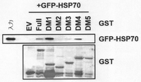
GST−tagが融合したAIMP2−DX2タンパク質のいくつかの断片(DM1乃至DM5)と、293T細胞可溶化物とを混合した。混合した後、glutathione−sepharose(GE Healthcare)を利用して、各AIMP2−DX2タンパク質をプルダウン(pull down)した。AIMP2−DX2タンパク質と一緒にプルダウンされたいくつかのHSP70タンパク質の量は、マススペクトロメトリー技法を利用して確認した。実験に使用したAIMP2−DX2タンパク質断片のアミノ酸位置を図3Aに示した。
<Example 3>
Binding Structure Analysis of AIMP2-DX2 and HSP70 Several fragments of the AIMP2-DX2 protein fused with GST-tag (DM1 to DM5) were mixed with 293T cell solubilized products. After mixing, each AIMP2-DX2 protein was pulled down using glutathione-sepharose (GE Healthcare). The amount of some HSP70 proteins pulled down along with the AIMP2-DX2 protein was confirmed using mass spectrometry techniques. The amino acid positions of the AIMP2-DX2 protein fragment used in the experiment are shown in FIG. 3A.
図3Bに示した通り、AIMP2−DX2タンパク質の断片のうち、配列番号2のアミノ酸配列を有するDM1(配列番号1のアミノ酸配列のうち1−87番目のアミノ酸に対応する断片)にいくつかのHSP70ファミリーが優勢して結合することをマススペクトロメトリー技法を通じて確認することができた。 As shown in FIG. 3B, among the fragments of the AIMP2-DX2 protein, DM1 having the amino acid sequence of SEQ ID NO: 2 (fragment corresponding to the 1-87th amino acid in the amino acid sequence of SEQ ID NO: 1) has several HSP70s. It was possible to confirm through mass spectrometry techniques that the families predominantly bind.
次に、AIMP2−DX2とHSP70との結合構造をより具体的に確認するための実験を行った。 Next, an experiment was conducted to more specifically confirm the binding structure between AIMP2-DX2 and HSP70.
GST−EV、GST−AIMP2−DX2 full、GST−AIMP2−DX2断片(DM1−DM5)をGFP−HSP70が過剰発現された293T細胞可溶化物と混合した。混合した後、GSTタンパク質をglutathione−sepharoseを利用してプルダウンし、プルダウンされたタンパク質を、SDS−PAGE、ウェスタンブロットを通じて分析した。GFPタンパク質は、GFP特異的抗体を利用して確認し、GSTタンパク質はクーマシー染色(coomassie staining)を通じて確認した。 GST-EV, GST-AIMP2-DX2 full, GST-AIMP2-DX2 fragments (DM1-DM5) were mixed with 293T cell solubilized products overexpressing GFP-HSP70. After mixing, the GST protein was pulled down using glutathione-sepharose, and the pulled down protein was analyzed through SDS-PAGE, Western blot. The GFP protein was confirmed using a GFP-specific antibody, and the GST protein was confirmed through coomassie staining.
図3C及び図3Dに示した通り、AIMP2−DX2タンパク質断片のうち、1−87アミノ酸を含む配列番号2のアミノ酸配列を有するDM1断片が、HSP70に結合することが分かった。 As shown in FIGS. 3C and 3D, among the AIMP2-DX2 protein fragments, the DM1 fragment having the amino acid sequence of SEQ ID NO: 2 containing 1-87 amino acids was found to bind to HSP70.
一方、HSP70タンパク質内でAIMP2−DX2に結合する部分を確認するための実験を行った。 On the other hand, an experiment was conducted to confirm the portion of the HSP70 protein that binds to AIMP2-DX2.
Strep−AIMP2−DX2と共に、GFP−HSP70 full、GFP−HSP70断片(AD、SB、Lid)を293T細胞に過剰発現させた後、293T細胞の可溶化物をstrep−tagカラムを利用してIPた。Strep−AIMP2−DX2とIPされたタンパク質は、SDS−PAGEを通じて分離され、各GFPタンパク質の結合は、GFP特異的抗体を利用してウェスタンブロットを通じて確認した。HSP70タンパク質内で実験に使用されたそれぞれの断片(AD、SB、Lid)のアミノ酸位置を図3Eに示した。 After overexpressing GFP-HSP70 full and GFP-HSP70 fragments (AD, SB, Lid) in 293T cells together with Strep-AIMP2-DX2, solubilized 293T cells were IP-generated using a strip-tag column. .. The protein IP with Strep-AIMP2-DX2 was separated through SDS-PAGE, and the binding of each GFP protein was confirmed by Western blotting using a GFP-specific antibody. The amino acid positions of each fragment (AD, SB, Lid) used in the experiment within the HSP70 protein are shown in FIG. 3E.
図3F及び図4に示した通り、HSP70タンパク質断片のうち、配列番号4のアミノ酸配列を有するSB(substrate binding domain)部分を含む断片だけがAIMP2−DX2と結合することが分かった。 As shown in FIGS. 3F and 4, it was found that among the HSP70 protein fragments, only the fragment containing the SB (substrate binding domain) portion having the amino acid sequence of SEQ ID NO: 4 binds to AIMP2-DX2.
<実施例4>
HSP70によるAIMP2−DX2の安定化増加
H460細胞にGFP−EV、GFP−HSP70を遺伝子導入(transfection)を通じて過剰発現させた。また、HSP70に特異的なsi−RNA(si−HSP70, Santa Cruz)を利用して、HSP70の発現を減少させ、この時、対照群としてsi−control(Invitrogen)を使用した。HSP70の発現を増加、減少させたH460細胞のタンパク質発現量は、ウェスタンブロット、mRNA発現量は、RT−PCRを用いて確認した。HSP70特異的プライマー配列には F:GCG TAA TAC GAC TCA CTA TAG GGA GAA TGC CCC CAG CTA CGT GGC CTT C、R:GCG TAA TAC GAC TCA CTA TAG GGA GAT AAA GCT TGG CGT CGC GCA GAG Cを利用した。
<Example 4>
Stabilization increase of AIMP2-DX2 by HSP70 GFP-EV and GFP-HSP70 were overexpressed in H460 cells through gene transfection. In addition, HSP70-specific si-RNA (si-HSP70, Santa Cruz) was used to reduce the expression of HSP70, and at this time, si-control (Invitrogen) was used as a control group. The protein expression level of H460 cells in which the expression of HSP70 was increased or decreased was confirmed by Western blotting, and the mRNA expression level was confirmed by using RT-PCR. The HSP70-specific primer sequence includes F: GCG TAA TAC GAC TCA CTA TAG GGA GAA TGC CCC CAG CTA CGT GGC CTT C, R: GCG TAA TAC GAC TCA CTA TAG GGA G.
図5Aに示した通り、HSP70過剰発現によってAIMP2−DX2タンパク質が増加し、si−RNA技法を通じたHSP70の減少によりAIMP2−DX2タンパク質が減少することを確認した。この時、AIMP2−DX2の転写には影響がないことにより、HSP70によるAIMP2−DX2の調節は、転写後に調節、つまりタンパク質水準での調節であることが分かった。 As shown in FIG. 5A, it was confirmed that the overexpression of HSP70 increased the AIMP2-DX2 protein, and the decrease of HSP70 through the si-RNA technique decreased the AIMP2-DX2 protein. At this time, since there was no effect on the transcription of AIMP2-DX2, it was found that the regulation of AIMP2-DX2 by HSP70 is post-transcriptional regulation, that is, regulation at the protein level.
一方、H460細胞にHSP70阻害剤であるピフィスリン−μ(Tocris)、VER155008(Sigma)とHSP90阻害剤であるゲルダナマイシン(Tocris)、PUH71(Tocris)を処理した後、ウェスタンブロット、RT−PCR技法を通じてタンパク質、mRNA発現量を確認した。AIMP2−DX2タンパク質の発現量は、AIMP2−DX2特異的抗体(Cell signaling)を利用して確認した。 On the other hand, H460 cells are treated with HSP70 inhibitors pifithrin-μ (Tocris), VER155008 (Sigma) and HSP90 inhibitors Gerdanamycin (Tocris), PUH71 (Tocris), followed by Western blotting and RT-PCR techniques. The expression levels of protein and mRNA were confirmed through. The expression level of the AIMP2-DX2 protein was confirmed using AIMP2-DX2-specific antibody (Cell signaling).
図5Bに示した通り、HSP70阻害剤であるPES(ピフィスリン−μ)、VER(VER155008)処理によってAIMP2−DX2タンパク質の発現が減少することが分かった。HSP90阻害剤Gel(Geldanamycin)、PU(PUH71)処理によっては、AIMP2−DX2タンパク質の発現に影響がないことを確認し、HSP70、HSP90阻害剤によってAIMP2の発現は影響がないことが分かった。のみならず、各抑制剤処理によりAIMP2−DX2の転写には影響がないことにより、HSP70抑制剤処理によるAIMP2−DX2の調節は、転写後の調節、つまりタンパク質水準での調節であることが分かった。 As shown in FIG. 5B, it was found that treatment with the HSP70 inhibitors PES (pifithrin-μ) and VER (VER155008) reduced the expression of the AIMP2-DX2 protein. It was confirmed that the treatment with the HSP90 inhibitors Gel (Geldanamycin) and PU (PUH71) did not affect the expression of the AIMP2-DX2 protein, and it was found that the expression of AIMP2 was not affected by the HSP70 and HSP90 inhibitors. Not only that, each inhibitor treatment did not affect the transcription of AIMP2-DX2, indicating that the regulation of AIMP2-DX2 by HSP70 inhibitor treatment is post-transcriptional regulation, that is, regulation at the protein level. rice field.
以上の結果を通じて、HSP70は、AIMP2−DX2タンパク質を特異的に安定化するものと判断された。 Based on the above results, it was determined that HSP70 specifically stabilizes the AIMP2-DX2 protein.
<実施例5>
DX2を経由したHSP70の細胞分裂調節
Strep−AIMP2−DX2を過剰発現した293T細胞と過剰発現していない293T細胞を、HSP70阻害剤であるピフィスリン−μ(PES)、VER155008(VER)及びHSP90阻害剤であるゲルダナマイシン(Gel)、PUH71(Pu)で12時間処理した後、MTT(Amresco)アッセイを行った。各実験は3回独立的に行い、HSP70に特異的なsi−RNA(si−HSP70)を利用してHSP70の発現を減少させた293T細胞にStrep−AIMP2−DX2を過剰発現した後、MTTアッセイを行った。各実験は3回独立的に行った。
<Example 5>
Cell division regulation of HSP70 via
これに対する結果を図6A及び6Bに示した。 The results for this are shown in FIGS. 6A and 6B.
図6Aに示した通り、HSP70抑制剤PES、VER、HSP90抑制剤Gel、Puを処理した後、AIMP2−DX2によって増加する細胞の成長を観察した結果、HSP70抑制剤を処理した場合、AIMP2−DX2による細胞成長の増加が観察されなかった。また、図6Bに示した通り、si−RNA技法を通じてHPS70の発現を減少させた結果、AIMP2−DX2による細胞成長が観察されなかった。これらの結果を通じてHSP70がDX2を通じて細胞の成長を調節していることが分かった。 As shown in FIG. 6A, after treating the HSP70 inhibitors PES, VER, and the HSP90 inhibitors Gel, Pu, as a result of observing the cell growth increased by AIMP2-DX2, when the HSP70 inhibitor was treated, AIMP2-DX2 No increase in cell growth was observed. In addition, as shown in FIG. 6B, as a result of reducing the expression of HPS70 through the si-RNA technique, cell growth by AIMP2-DX2 was not observed. From these results, it was found that HSP70 regulates cell growth through DX2.
一方、si−RNAの技法を通じてHSP70の発現を減少させた後、AIMP2−DX2による細胞成長の結果物のDNA合成をEdUアッセイを通じて観察した結果、HSP70の発現が減少したとき、AIMP2−DX2について、発現を通じて増加されるDNA合成が観察されないことが分かる(図6C)。 On the other hand, when the expression of HSP70 was reduced through the si-RNA technique and then the DNA synthesis of the result of cell growth by AIMP2-DX2 was observed through the EdU assay, when the expression of HSP70 was reduced, the expression of AIMP2-DX2 was reduced. It can be seen that no increased DNA synthesis is observed through expression (Fig. 6C).
<実施例6>
AIMP2−DX2とHSP70の結合阻害剤の探索
図7Aに示した通り、AIMP2−DX2モニタリングアッセイを作成した後、本発明に係る化合物ライブラリに存在するいくつかの化合物で処理して、AIMP2−DX2を減少させる化合物を一次に選別した。一次に選別された化合物の中で、AIMP2は減少させず、MTTアッセイを通じて細胞生存率を測定して正常細胞に影響を与えずにがん細胞にのみ影響を与える化合物を最終的に選別した。
<Example 6>
Search for Binding Inhibitors of AIMP2-DX2 and HSP70 As shown in FIG. 7A, the AIMP2-DX2 monitoring assay was created and then treated with some of the compounds present in the compound library of the present invention to give AIMP2-DX2. The compounds to be reduced were primarily selected. Among the compounds selected in the first order, the compounds that did not reduce AIMP2 and measured the cell viability through the MTT assay and affected only the cancer cells without affecting the normal cells were finally selected.
選別された化合物をBC−DXI−495と命名し、その構造を図7Bに示した。 The selected compound was named BC-DXI-495 and its structure is shown in FIG. 7B.
<実施例7>
BC−DXI−495のAIMP2−DX2とHSP70の結合阻害能及びDX2発現阻害能
前記実施例6を通じて選別されたBC−DXI−495がAIMP2−DX2とHSP70との結合を阻害できるか否かを評価した。Strep−DX2が過剰発現された293T細胞を、BC−DXI−495化合物で、濃度別(2.5、5、10μM)に12時間処理した後、細胞を1%triton X−100(BD Science)が含まれたPBSで溶解した。可溶化物を13200rpmの速度で遠心分離した後、分離された浮遊物と沈殿物からウェスタンブロットを通じてAIMP2−DX2、AIMP2のタンパク質発現量を確認した。AIMP2−DX2の発現は、StrepMAB−Classic−HRP(IBA)を利用して確認した。
<Example 7>
Evaluation of the ability of BC-DXI-495 to inhibit the binding of AIMP2-DX2 to HSP70 and the ability to inhibit DX2 expression Whether BC-DXI-495 selected through Example 6 can inhibit the binding of AIMP2-DX2 to HSP70. did. 293T cells overexpressing Strept-DX2 were treated with BC-DXI-495 compound for 12 hours by concentration (2.5, 5, 10 μM), and then the cells were treated with 1% triton X-100 (BD Science). Was dissolved in PBS containing. After centrifuging the solubilized product at a speed of 13200 rpm, the protein expression levels of AIMP2-DX2 and AIMP2 were confirmed from the separated suspended matter and precipitate through Western blotting. Expression of AIMP2-DX2 was confirmed using StrepMAB-Classic-HRP (IBA).
これに対する結果を図8Aに示した。 The results for this are shown in FIG. 8A.
図8Aに示した通り、BC−DXI−495化合物で濃度依存的に処理する場合、AIMP2−DX2とHSP70との結合が濃度依存的に減少することを確認した。 As shown in FIG. 8A, it was confirmed that the binding between AIMP2-DX2 and HSP70 decreased in a concentration-dependent manner when treated with the BC-DXI-495 compound in a concentration-dependent manner.
BC−DXI−495化合物によるAIMP2−DX2、AIMP2の減少を確認するために、A549細胞にnanoluciferase−AIMP2−DX2、nanoluciferase−AIMP2をそれぞれ過剰発現した後、BC−DXI−495化合物を濃度依存的(2.5、5、10、20、40μM)に4時間処理した。処理後、細胞内にあるAIMP2−DX2、AIMP2タンパク質の量をルシフェラーゼアッセイ(cat#)を通じて確認した。各濃度でのルシフェラーゼ阻害程度を測定して、AIMP2−DX2、AIMP2を50%減少させる化合物の濃度を計算し、これをIC50で示した。また、BC−DXI−495化合物でH460細胞を濃度別(2.5、5、10μM)に12時間処理した後、AIMP2−DX2、AIMP2、Actinタンパク質発現と、AIMP2−DX2、AIMP2、Actin mRNA発現をウェスタンブロットとRT−PCRで観察した。 In order to confirm the decrease of AIMP2-DX2 and AIMP2 by the BC-DXI-495 compound, the BC-DXI-495 compound was concentrated-dependent after overexpressing the nanoluciphase-AIMP2-DX2 and the nanoluxiphase-AIMP2 in A549 cells, respectively. 2.5, 5, 10, 20, 40 μM) for 4 hours. After the treatment, the amount of AIMP2-DX2 and AIMP2 proteins in the cells was confirmed through a luciferase assay (cat #). The degree of luciferase inhibition at each concentration was measured, and the concentration of the compound that reduces AIMP2-DX2 and AIMP2 by 50% was calculated and shown by IC50. In addition, after treating H460 cells with BC-DXI-495 compound by concentration (2.5, 5, 10 μM) for 12 hours, AIMP2-DX2, AIMP2, Actin protein expression and AIMP2-DX2, AIMP2, Actin mRNA expression. Was observed by Western blot and RT-PCR.
その結果、図8B及び8Cに示した通り、BC−DXI−495化合物はAIMP2発現に影響を与えず(IC50 > 100μM)、AIMP2−DX2発現のみを特異的に減少させることが確認され(IC50: 4.2μM)、BC−DXI−495化合物で濃度依存的に処理する場合、AIMP2に影響を与えずAIMP2−DX2タンパク質のみを特異的に減少させることが確認された(図8D)。また、BC−DXI−495化合物で濃度依存的に処理する場合、soluble AIMP2−DX2タンパク質(浮遊層分画)が減少して、insoluble AIMP2−DX2タンパク質(沈殿物分画)が増加することが分かった(図8E)。 As a result, as shown in FIGS. 8B and 8C, it was confirmed that the BC-DXI-495 compound did not affect AIMP2 expression (IC50> 100 μM) and specifically reduced only AIMP2-DX2 expression (IC50: 4.2 μM), it was confirmed that when treated with the BC-DXI-495 compound in a concentration-dependent manner, it did not affect AIMP2 and specifically reduced only the AIMP2-DX2 protein (Fig. 8D). It was also found that when treated with the BC-DXI-495 compound in a concentration-dependent manner, the soluble AIMP2-DX2 protein (suspended layer fraction) decreased and the soluble AIMP2-DX2 protein (precipitate fraction) increased. (Fig. 8E).
<実施例8>
BC−DXI−495の抗がん効能評価
<8−1>BC−DXI−495の細胞毒性
BC−DXI−495化合物により肺がん、正常肺細胞の細胞活性がどのように変化するかを確認するために、A549(肺がん細胞)、WI−26(正常肺細胞)を、BC−DXI−495化合物で濃度依存的(2.5、5、10、20、40μM)に処理した。処理後、MTTアッセイを行い、各細胞の活性を測定した。細胞の活性を50%減少させる化合物濃度を計算し、これをGI50で示した。
<Example 8>
Evaluation of anticancer efficacy of BC-DXI-495 <8-1> Cytotoxicity of BC-DXI-495 To confirm how the cell activity of lung cancer and normal lung cells is changed by the BC-DXI-495 compound. A549 (lung cancer cells) and WI-26 (normal lung cells) were treated with BC-DXI-495 compound in a concentration-dependent manner (2.5, 5, 10, 20, 40 μM). After the treatment, MTT assay was performed to measure the activity of each cell. Compound concentrations that reduced cell activity by 50% were calculated and indicated by GI50.
図9Aに示した通り、肺がん細胞であるA549と正常肺細胞であるWI−26を、BC−DXI−495化合物で濃度依存的に処理した結果、正常細胞には影響せず、肺がん細胞にのみ細胞死滅を誘導することが分かった。 As shown in FIG. 9A, as a result of treating A549, which is a lung cancer cell, and WI-26, which is a normal lung cell, with a BC-DXI-495 compound in a concentration-dependent manner, it does not affect normal cells and only affects lung cancer cells. It was found to induce cell death.
また、ドキシサイクリン(Sigma)によってAIMP2−DX2の発現を誘導することができる肺がん細胞のA549を、ドキシサイクリン(0.5mg/ml)で処理して、AIMP2−DX2による細胞の成長を誘導した。48時間毎にドキシサイクリンで処理し、7日目にBC−DXI−495(40μM)を処理して、96時間、細胞成長抑制及び死滅を確認した。 In addition, A549 of lung cancer cells whose expression of AIMP2-DX2 can be induced by doxycycline (Sigma) was treated with doxycycline (0.5 mg / ml) to induce cell growth by AIMP2-DX2. Treatment with doxycycline every 48 hours and BC-DXI-495 (40 μM) on day 7 confirmed cell growth inhibition and death for 96 hours.
図9Bに示した通り、AIMP2−DX2タンパク質の発現を誘導する場合(ピンクグラフ)、そうでない場合に比べて、BC−DXI−495化合物による細胞毒性がより大きく誘導されることが分かった。 As shown in FIG. 9B, it was found that when the expression of the AIMP2-DX2 protein was induced (pink graph), the cytotoxicity of the BC-DXI-495 compound was more induced than when it was not.
また、AIMP2−DX2発現量が多様な二つの肺正常細胞(WI−26、WI−38)と、7つの肺がん細胞(NCI−H2087、HCC−1359、HCC−95、HCC−366、HCC−1438、HCC−1588、NCI−H460)を、BC−DXI−495化合物で濃度別に処理した後、MTTアッセイを行い、GI50を計算した。各細胞のAIMP2−DX2発現量は、ウェスタンブロットを通じて確認した。 In addition, two normal lung cells (WI-26, WI-38) with various AIMP2-DX2 expression levels and seven lung cancer cells (NCI-H2087, HCC-1359, HCC-95, HCC-366, HCC-1438). , HCC-1588, NCI-H460) by concentration with BC-DXI-495 compound, then MTT assay was performed to calculate GI50. The expression level of AIMP2-DX2 in each cell was confirmed by Western blotting.
図9Cに示した通り、内在性AIMP2−DX2タンパク質発現程度に応じ肺がん細胞を3群(Low(低)、Median(中)、High(高))に分けた後、BC−DXI−495で処理して、細胞毒性程度(GI50)を測定した結果、AIMP2−DX2発現が高い細胞において化合物による細胞死滅効果が優れていることが分かった。 As shown in FIG. 9C, lung cancer cells were divided into 3 groups (Low (low), Median (medium), High (high)) according to the degree of endogenous AIMP2-DX2 protein expression, and then treated with BC-DXI-495. As a result of measuring the degree of cytotoxicity (GI50), it was found that the cell killing effect of the compound was excellent in cells having high AIMP2-DX2 expression.
<8−2> BC−DXI−495のin vivo抗がん効果の評価
H460細胞5×10⁶個を8週齢Balb/cヌードマウスなどの表皮に注射して腫瘍を形成させた後、BC−DXI−495化合物を50mg/kgの濃度で週5回注射して計10回注射し、陽性対照物質であるtaxolは15mg/kgの濃度で週2回合計5回注射した。実験終了後、腫瘍を分離して重量を測定した。実験動物の体重及び腫瘍の大きさは週2回測定した。
<8-2> Evaluation of in vivo anticancer effect of BC-DXI-495 After injecting 5 × 10 6 H460 cells into the epidermis of 8-week-old Balb / c nude mice to form a tumor, BC The -DXI-495 compound was injected 5 times a week at a concentration of 50 mg / kg for a total of 10 injections, and the positive control substance taxol was injected twice a week for a total of 5 times at a concentration of 15 mg / kg. After completion of the experiment, the tumor was separated and weighed. The body weight and tumor size of the experimental animals were measured twice a week.
AIMP2−DX2発現が高いH460細胞をマウスに移植(xenograft)した後、BC−DXI−495化合物を50mg/kgで投与した結果、対照群であるTaxol(15mpk)と類似する程度にがん細胞の成長を抑制することが分かった。一方、このような腫瘍抑制効果は、腫瘍の重量からも同じく確認された(図10A、10B及び10C)。実験の進行過程の間にマウスの体重には影響がなかった(図10D)。 After transplanting H460 cells with high AIMP2-DX2 expression into mice (xenograft), the BC-DXI-495 compound was administered at 50 mg / kg, and as a result, the cancer cells were similar to the control group Taxol (15 mpk). It was found to suppress growth. On the other hand, such a tumor-suppressing effect was also confirmed from the weight of the tumor (FIGS. 10A, 10B and 10C). There was no effect on mouse body weight during the course of the experiment (Fig. 10D).
本発明は、HSP70ががんの発生の主な原因タンパク質の一つであるAIMP2−DX2と直接結合して安定化させるとの発見に基づいて、AIMP2−DX2又はその断片と、HSP70又はその断片とを利用して、AIMP2−DX2とHSP70との結合水準を減少させる物質を抗がん剤として選別する抗がん剤スクリーニング方法と、前記の方法で選別された抗がん剤を有効成分として含むがんの予防又は治療用薬学的組成物を提供する。本発明に係るHSP70の発現を抑制するsiRNA、shRNAなどの製剤、さらに、HSP70とAIMP2−DX2との結合を抑制する化合物などは、がんにおいてAIMP2−DX2タンパク質の水準を下げて、がんの発生と進行を抑制する効果が優れて産業上の利用可能性が極めて優れている。 The present invention is based on the discovery that HSP70 directly binds to and stabilizes AIMP2-DX2, which is one of the major causative proteins of cancer development, and AIMP2-DX2 or a fragment thereof, and HSP70 or a fragment thereof. An anti-cancer drug screening method for selecting a substance that reduces the binding level between AIMP2-DX2 and HSP70 as an anti-cancer agent, and an anti-cancer drug selected by the above method as an active ingredient. Provided are a pharmaceutical composition for the prevention or treatment of cancer, which comprises. Preparations such as siRNA and shRNA that suppress the expression of HSP70 according to the present invention, and compounds that suppress the binding between HSP70 and AIMP2-DX2 lower the level of AIMP2-DX2 protein in cancer and cause cancer. It has an excellent effect of suppressing the occurrence and progression, and has extremely excellent industrial applicability.
Claims (9)
(b)試験物質の存在下又は非存在下でのAIMP2−DX2とHSP70との結合を測定する段階;
(c)試験物質の存在下でのAIMP2−DX2とHSP70との結合と、試験物質非存在下でのAIMP2−DX2とHSP70との結合とを比較して、試験物質によるAIMP2−DX2とHSP70との結合水準の変化を判断する段階;
(d)AIMP2−DX2とHSP70との結合水準を減少させる試験物質を選別する段階;及び
(e)選別された試験物質の抗がん活性を、細胞又は動物で確認する段階を含み、
前記AIMP2−DX2の断片は、前記AIMP2−DX2の1乃至87番目のアミノ酸残基を含み、
前記HSP70の断片は、前記HSP70の385乃至536番目のアミノ酸残基を含む、
抗がん剤スクリーニング方法。 (A) The step of contacting AIMP2-DX2 or a fragment thereof with HSP70 or a fragment thereof in the presence or absence of a test substance;
(B) The step of measuring the binding between AIMP2-DX2 and HSP70 in the presence or absence of the test substance;
(C) Comparing the binding of AIMP2-DX2 and HSP70 in the presence of the test substance with the binding of AIMP2-DX2 and HSP70 in the absence of the test substance, AIMP2-DX2 and HSP70 by the test substance Steps to determine changes in the binding level of
(D) AIMP2-DX2 and steps for selecting a test substance that reduces the binding level of HSP70; anticancer activity and (e) sorted test substance, viewed including the step of confirming a cell or an animal,
The fragment of AIMP2-DX2 contains amino acid residues 1 to 87 of AIMP2-DX2.
The fragment of HSP70 contains amino acid residues 385 to 536 of HSP70.
Anti-cancer drug screening method.
(1)前記試験物質を、AIMP2−DX2を発現する細胞に接触させる段階;
(2)前記細胞と前記試験物質とを接触させない対照群細胞から、AIMP2−DX2タンパク質の水準を測定する段階;及び
(3)前記対照群細胞と比較して、前記AIMP2−DX2タンパク質の水準を減少させる試験物質を選別する段階を追加的に含むことを特徴とする請求項1記載の方法。 The anticancer drug screening method is performed between the (d) and the (e).
(1) The step of contacting the test substance with cells expressing AIMP2-DX2;
(2) The step of measuring the level of AIMP2-DX2 protein from the control group cells in which the cells are not brought into contact with the test substance; and (3) the level of the AIMP2-DX2 protein as compared with the control group cells. The method according to claim 1, further comprising a step of selecting a test substance to be reduced.
The anticancer agent was selected from the group consisting of siRNA, shRNA, miRNA, ribozyme, DNAzyme, PNA (peptide nucleic acid), antisense oligonucleotide, antibody, aptamer, peptide, natural extract, and chemical substance. The method according to any one of claims 1 to 8, wherein the method is characterized by.
Applications Claiming Priority (3)
| Application Number | Priority Date | Filing Date | Title |
|---|---|---|---|
| KR1020160027077A KR102297505B1 (en) | 2016-03-07 | 2016-03-07 | Methods for screening anti-cancer drugs inhibiting interactions between AIMP2-DX2 and HSP70 |
| KR10-2016-0027077 | 2016-03-07 | ||
| PCT/KR2017/002442 WO2017155277A1 (en) | 2016-03-07 | 2017-03-07 | Method for screening anticancer agent inhibiting binding of aimp2-dx2 and hsp70 |
Publications (3)
| Publication Number | Publication Date |
|---|---|
| JP2019512693A JP2019512693A (en) | 2019-05-16 |
| JP2019512693A5 JP2019512693A5 (en) | 2020-06-18 |
| JP6914269B2 true JP6914269B2 (en) | 2021-08-04 |
Family
ID=59790548
Family Applications (1)
| Application Number | Title | Priority Date | Filing Date |
|---|---|---|---|
| JP2018548001A Active JP6914269B2 (en) | 2016-03-07 | 2017-03-07 | Anti-cancer drug screening method that inhibits the binding between AIMP2-DX2 and HSP70 |
Country Status (6)
| Country | Link |
|---|---|
| US (1) | US11442057B2 (en) |
| EP (1) | EP3418739B1 (en) |
| JP (1) | JP6914269B2 (en) |
| KR (1) | KR102297505B1 (en) |
| CN (1) | CN109073638B (en) |
| WO (1) | WO2017155277A1 (en) |
Families Citing this family (3)
| Publication number | Priority date | Publication date | Assignee | Title |
|---|---|---|---|---|
| US11758948B2 (en) | 2018-01-29 | 2023-09-19 | Altria Client Services Llc | Lighting unit for aerosol-generating systems |
| KR102308134B1 (en) | 2018-04-26 | 2021-10-01 | 재단법인 의약바이오컨버젼스연구단 | Novel compounds as mTOR inhibitor and uses thereof |
| CN115786504B (en) * | 2022-08-31 | 2023-09-19 | 中国人民解放军总医院京南医疗区 | Use of HSPA4 inhibitors for the treatment of melanoma |
Family Cites Families (21)
| Publication number | Priority date | Publication date | Assignee | Title |
|---|---|---|---|---|
| US8039218B2 (en) * | 2002-11-14 | 2011-10-18 | John Wayne Cancer Institute | Detection of cancer cells in body fluids |
| US7459529B2 (en) * | 2004-11-24 | 2008-12-02 | Seoul National University Industry Foundation | AIMP2-DX2 and its uses |
| US8003780B2 (en) * | 2004-11-24 | 2011-08-23 | Neomics Co., Ltd. | AIMP2-DX2 gene and SiRNA targeting AIMP2-DX2 |
| KR20060057992A (en) * | 2004-11-24 | 2006-05-29 | 재단법인서울대학교산학협력재단 | P38-dx2 and its use |
| US20060275844A1 (en) * | 2005-04-19 | 2006-12-07 | Linke Steven P | Diagnostic markers of breast cancer treatment and progression and methods of use thereof |
| US20080280297A1 (en) * | 2005-07-15 | 2008-11-13 | The Trustees Of Columbia University In The City Of | Compositions and Methods for Differential Diagnosis of Chronic Lymphocytic Leukemia |
| KR20070113926A (en) * | 2006-05-26 | 2007-11-29 | 재단법인서울대학교산학협력재단 | Use of aimp2dx2 for the diagnosis and treatment of cancer |
| US8754094B2 (en) * | 2007-08-15 | 2014-06-17 | The Research Foundation Of State University Of New York | Methods for heat shock protein dependent cancer treatment |
| KR101067816B1 (en) * | 2007-11-09 | 2011-09-27 | (주)네오믹스 | Composition for preventing and treating inflammatory diseases comprising inhibitor of AIMP2-DX2 as an active ingredient |
| WO2011035433A1 (en) * | 2009-09-23 | 2011-03-31 | University Health Network | Selected strains on serum-free growth media for proteomics analysis of lung cancer biomarkers |
| CA2798139C (en) * | 2010-05-04 | 2019-09-24 | Atyr Pharma, Inc. | Innovative discovery of therapeutic, diagnostic, and antibody compositions related to protein fragments of p38 multi-trna synthetase complex |
| WO2013006076A1 (en) * | 2011-07-04 | 2013-01-10 | New York University | The use of intranasally administered hsp70 protein to treat neurodegenerative diseases |
| KR20130016041A (en) * | 2011-08-04 | 2013-02-14 | 재단법인 의약바이오컨버젼스연구단 | Novel aniline derivatives and use thereof |
| KR20130118612A (en) * | 2012-04-20 | 2013-10-30 | (주)네오믹스 | Novel aminopyridine derivatives and use thereof |
| KR101500804B1 (en) * | 2012-04-20 | 2015-03-11 | 단국대학교 산학협력단 | Trans-splicing enzymatic nucleic acid molecule specific to splicing variant of AIMP2 gene, and pharmaceutical composition for treating lung cancer and composition for diagnosing lung cancer using the same |
| KR101464360B1 (en) * | 2012-11-09 | 2014-11-21 | 주식회사 대웅 | Adenovirus Containing Ribozyme and shRNA, and Therapeutic Composition Comprising Thereof |
| WO2014142619A1 (en) * | 2013-03-14 | 2014-09-18 | 부산대학교산학협력단 | Method for screening therapeutic agent for cancers by using aimp2-dx2 and pl4/arf interaction |
| KR101762433B1 (en) * | 2013-06-05 | 2017-07-28 | 재단법인 의약바이오컨버젼스연구단 | Novel Maleic acid derivatives, preparation method thereof, and anti-cancer compositions containing them |
| KR101514320B1 (en) * | 2013-06-14 | 2015-04-22 | 재단법인 의약바이오컨버젼스연구단 | Novel pharmaceutical composition for preventing or treating cancer |
| KR101621963B1 (en) * | 2014-03-06 | 2016-05-17 | 한국과학기술원 | Moelecular Beacon for Dectecting Mutant of exon2-deleted AIMP2 and Method for Detecting Mutant of exon2-deleted AIMP2 Using thereof |
| WO2016003158A2 (en) * | 2014-06-30 | 2016-01-07 | 부산대학교 산학협력단 | New compound for inhibiting binding between dx2 protein and p14/arf protein, and pharmaceutical composition for treating or preventing cancer disease containing same as effective ingredient |
-
2016
- 2016-03-07 KR KR1020160027077A patent/KR102297505B1/en active IP Right Grant
-
2017
- 2017-03-07 EP EP17763538.0A patent/EP3418739B1/en active Active
- 2017-03-07 JP JP2018548001A patent/JP6914269B2/en active Active
- 2017-03-07 WO PCT/KR2017/002442 patent/WO2017155277A1/en active Application Filing
- 2017-03-07 CN CN201780027761.8A patent/CN109073638B/en active Active
-
2018
- 2018-09-07 US US16/125,113 patent/US11442057B2/en active Active
Also Published As
| Publication number | Publication date |
|---|---|
| US11442057B2 (en) | 2022-09-13 |
| WO2017155277A1 (en) | 2017-09-14 |
| KR20170104263A (en) | 2017-09-15 |
| WO2017155277A8 (en) | 2018-11-08 |
| JP2019512693A (en) | 2019-05-16 |
| EP3418739A4 (en) | 2019-03-06 |
| EP3418739A1 (en) | 2018-12-26 |
| EP3418739B1 (en) | 2021-05-12 |
| US20190033295A1 (en) | 2019-01-31 |
| CN109073638B (en) | 2022-04-26 |
| KR102297505B1 (en) | 2021-09-01 |
| CN109073638A (en) | 2018-12-21 |
Similar Documents
| Publication | Publication Date | Title |
|---|---|---|
| Lagadec et al. | Radiation-induced Notch signaling in breast cancer stem cells | |
| Zhou et al. | Targeting EZH2 regulates tumor growth and apoptosis through modulating mitochondria dependent cell-death pathway in HNSCC | |
| Gong et al. | Kindlin-2 controls sensitivity of prostate cancer cells to cisplatin-induced cell death | |
| US9597369B2 (en) | Inhibitors of atypical protein kinase C and their use in treating hedgehog pathway-dependent cancers | |
| JP6914269B2 (en) | Anti-cancer drug screening method that inhibits the binding between AIMP2-DX2 and HSP70 | |
| Cui et al. | MEIS1 functions as a potential AR negative regulator | |
| Cai et al. | Overexpression of caveolin‐1 induces alteration of multidrug resistance in Hs578T breast adenocarcinoma cells | |
| Hua et al. | FRK suppresses the proliferation of human glioma cells by inhibiting cyclin D1 nuclear accumulation | |
| Ko et al. | Phosphorylation of the N‐terminal domain of p48 Ebp1 by CDK2 is required for tumorigenic function of p48 | |
| Ludwig et al. | The tumor suppressor, p190RhoGAP, differentially initiates apoptosis and confers docetaxel sensitivity to breast cancer cells | |
| Zhu et al. | Human inhibitor of growth 1 inhibits hepatoma cell growth and influences p53 stability in a variant‐dependent manner | |
| US9630953B2 (en) | Small compounds targeting TACC3 | |
| ES2902484T3 (en) | Compositions for the treatment and prevention of pancreatitis and kidney damage | |
| Zhao et al. | Noxa and Puma genes regulated by hTERT promoter can mitigate growth and induce apoptosis in hepatocellular carcinoma mouse model | |
| KR101367832B1 (en) | Use of Hades as a target for tumor suppressor | |
| Huang et al. | Phragmunis a suppresses glioblastoma through the regulation of MCL1-FBXW7 by blocking ELK1-SRF complex-dependent transcription | |
| US8304399B2 (en) | Hedgehog signaling pathway proteins and uses thereof | |
| She et al. | Discovery of novel organoarsenicals as robust thioredoxin reductase inhibitors for oxidative stress mediated cancer therapy | |
| KR100880850B1 (en) | A pharmaceutical composition for rendering resistance to cisplatin in cancer cells comprising a material inhibiting an expression of cyclophilin A and a method for rendering resistance to cisplatin in cancer cells by using them | |
| US20130345145A1 (en) | Atip3 and biologically active fragments thereof for use in the treatment of cancer | |
| KR20240000032A (en) | Composition for preventing, improving or treating breast cancer comprising LRRK2 protein | |
| AU2011260745B2 (en) | Cancer therapy method | |
| EP3746789A1 (en) | Screening method for the identification of cancer therapeutics | |
| Li et al. | New Peptide PBAG1 Induces Hypoxia, Impairs HR Repair and Sensitizes BRCA1/2-Proficient TNBC to PARP Inhibitors by Decreasing C1QBP and ZC3H18 |
Legal Events
| Date | Code | Title | Description |
|---|---|---|---|
| A524 | Written submission of copy of amendment under article 19 pct |
Free format text: JAPANESE INTERMEDIATE CODE: A524 Effective date: 20200305 |
|
| A621 | Written request for application examination |
Free format text: JAPANESE INTERMEDIATE CODE: A621 Effective date: 20200305 |
|
| A521 | Request for written amendment filed |
Free format text: JAPANESE INTERMEDIATE CODE: A523 Effective date: 20200511 |
|
| A977 | Report on retrieval |
Free format text: JAPANESE INTERMEDIATE CODE: A971007 Effective date: 20210226 |
|
| A131 | Notification of reasons for refusal |
Free format text: JAPANESE INTERMEDIATE CODE: A131 Effective date: 20210309 |
|
| A521 | Request for written amendment filed |
Free format text: JAPANESE INTERMEDIATE CODE: A523 Effective date: 20210531 |
|
| TRDD | Decision of grant or rejection written | ||
| A01 | Written decision to grant a patent or to grant a registration (utility model) |
Free format text: JAPANESE INTERMEDIATE CODE: A01 Effective date: 20210615 |
|
| A61 | First payment of annual fees (during grant procedure) |
Free format text: JAPANESE INTERMEDIATE CODE: A61 Effective date: 20210713 |
|
| R150 | Certificate of patent or registration of utility model |
Ref document number: 6914269 Country of ref document: JP Free format text: JAPANESE INTERMEDIATE CODE: R150 |