US7459529B2 - AIMP2-DX2 and its uses - Google Patents

AIMP2-DX2 and its uses Download PDF

Info

Publication number
US7459529B2
US7459529B2 US11/264,725 US26472505A US7459529B2 US 7459529 B2 US7459529 B2 US 7459529B2 US 26472505 A US26472505 A US 26472505A US 7459529 B2 US7459529 B2 US 7459529B2
Authority
US
United States
Prior art keywords
aimp2
cancer
protein
cells
tgf
Prior art date
Legal status (The legal status is an assumption and is not a legal conclusion. Google has not performed a legal analysis and makes no representation as to the accuracy of the status listed.)
Active
Application number
US11/264,725
Other versions
US20060110397A1 (en
Inventor
Sunghoon Kim
Current Assignee (The listed assignees may be inaccurate. Google has not performed a legal analysis and makes no representation or warranty as to the accuracy of the list.)
Medicinal Bioconvergence Research Center
Original Assignee
Seoul National University Industry Foundation
Priority date (The priority date is an assumption and is not a legal conclusion. Google has not performed a legal analysis and makes no representation as to the accuracy of the date listed.)
Filing date
Publication date
Priority claimed from KR1020050039073A external-priority patent/KR20060057992A/en
Application filed by Seoul National University Industry Foundation filed Critical Seoul National University Industry Foundation
Assigned to SEOUL NATIONAL UNIVERSITY INDUSTRY FOUNDATION reassignment SEOUL NATIONAL UNIVERSITY INDUSTRY FOUNDATION ASSIGNMENT OF ASSIGNORS INTEREST (SEE DOCUMENT FOR DETAILS). Assignors: KIM, SUNGHOON
Publication of US20060110397A1 publication Critical patent/US20060110397A1/en
Priority to US12/255,943 priority Critical patent/US8003780B2/en
Application granted granted Critical
Publication of US7459529B2 publication Critical patent/US7459529B2/en
Assigned to NEOMICS CO., LTD. reassignment NEOMICS CO., LTD. ASSIGNMENT OF ASSIGNORS INTEREST (SEE DOCUMENT FOR DETAILS). Assignors: SEOUL NATIONAL UNIVERSITY INDUSTRY FOUNDATION
Priority to US13/011,386 priority patent/US20110117572A1/en
Priority to US13/011,174 priority patent/US8431393B2/en
Assigned to MEDICINAL BIOCONVERGENCE RESEARCH CENTER reassignment MEDICINAL BIOCONVERGENCE RESEARCH CENTER ASSIGNMENT OF ASSIGNORS INTEREST (SEE DOCUMENT FOR DETAILS). Assignors: NEOMICS CO., LTD.
Priority to US15/074,498 priority patent/US9944713B2/en
Active legal-status Critical Current
Anticipated expiration legal-status Critical

Links

Images

Classifications

    • GPHYSICS
    • G01MEASURING; TESTING
    • G01NINVESTIGATING OR ANALYSING MATERIALS BY DETERMINING THEIR CHEMICAL OR PHYSICAL PROPERTIES
    • G01N33/00Investigating or analysing materials by specific methods not covered by groups G01N1/00 - G01N31/00
    • G01N33/48Biological material, e.g. blood, urine; Haemocytometers
    • G01N33/50Chemical analysis of biological material, e.g. blood, urine; Testing involving biospecific ligand binding methods; Immunological testing
    • G01N33/53Immunoassay; Biospecific binding assay; Materials therefor
    • G01N33/574Immunoassay; Biospecific binding assay; Materials therefor for cancer
    • G01N33/57407Specifically defined cancers
    • G01N33/57423Specifically defined cancers of lung
    • CCHEMISTRY; METALLURGY
    • C07ORGANIC CHEMISTRY
    • C07KPEPTIDES
    • C07K16/00Immunoglobulins [IGs], e.g. monoclonal or polyclonal antibodies
    • C07K16/18Immunoglobulins [IGs], e.g. monoclonal or polyclonal antibodies against material from animals or humans
    • C07K16/28Immunoglobulins [IGs], e.g. monoclonal or polyclonal antibodies against material from animals or humans against receptors, cell surface antigens or cell surface determinants
    • C07K16/30Immunoglobulins [IGs], e.g. monoclonal or polyclonal antibodies against material from animals or humans against receptors, cell surface antigens or cell surface determinants from tumour cells
    • GPHYSICS
    • G01MEASURING; TESTING
    • G01NINVESTIGATING OR ANALYSING MATERIALS BY DETERMINING THEIR CHEMICAL OR PHYSICAL PROPERTIES
    • G01N33/00Investigating or analysing materials by specific methods not covered by groups G01N1/00 - G01N31/00
    • G01N33/48Biological material, e.g. blood, urine; Haemocytometers
    • G01N33/50Chemical analysis of biological material, e.g. blood, urine; Testing involving biospecific ligand binding methods; Immunological testing
    • G01N33/53Immunoassay; Biospecific binding assay; Materials therefor
    • G01N33/574Immunoassay; Biospecific binding assay; Materials therefor for cancer
    • G01N33/57407Specifically defined cancers
    • G01N33/57438Specifically defined cancers of liver, pancreas or kidney
    • GPHYSICS
    • G01MEASURING; TESTING
    • G01NINVESTIGATING OR ANALYSING MATERIALS BY DETERMINING THEIR CHEMICAL OR PHYSICAL PROPERTIES
    • G01N2500/00Screening for compounds of potential therapeutic value

Definitions

  • the present invention relates to a variant of AIMP2 lacking exon 2, named as AIMP2-DX2, which is specifically expressed in cancer cells, and its uses in diagnosis and treatment of cancer.
  • Cancer is generally diagnosed by radiography examinations such as X-ray radiography, computed tomography and bronchography or bronchoscopy examination.
  • radiography examinations such as X-ray radiography, computed tomography and bronchography or bronchoscopy examination.
  • X-ray radiography computed tomography
  • bronchography or bronchoscopy examination.
  • bronchoscopy examination provide no diagnostic data in terms of cell physiology molecular genetics, while they allow to determine anatomical progress of cancer.
  • Lung and liver cancers are known to exhibit high incidence rate and mortality over the world.
  • Korean Pat. Appln. No. 10-1998-0038212 relating to the process for evaluating metastasis of lung cancer discloses that local metastatic lung cancer may be assessed by measuring the expression of mitogen activated protein kinase phosphatase-1 (MKP-1) in lung tissues.
  • MKP-1 mitogen activated protein kinase phosphatase-1
  • WO 2004/005891 suggests various disgnostic markers for lung cancer such as AOE372, ATP5D, B4GALT, Ppase, GRP58, GSTM4, P4HB, TPI and UCHL1.
  • Monoclonal antibodies to LCGA have been proposed to diagnose and treat non-small cell lung carcinoma and ovary cancer as described in U.S. Pat. No. 6,117,981.
  • EP 0804451 discloses a method for diagnosing and treating lung cancer by use of lung cancer-specific antigen HCAVIII.
  • U.S. Pat. Nos. 6,746,846 and 6,737,514 and EP 0621480 also discuss lung cancer markers.
  • Korean Pat. Appln. No. 10-2000-0040609 discloses the early detection method for liver diseases including liver cirrhosis and cancer by measuring the level of asialo-glycoproteins in accordance with sandwich assay in which lectins serve as a capture protein and/or probe protein.
  • Korean Pat. Appln. No. 10-2002-0035260 describes that compositions comprising long-chain fatty-acid-Coenzyme A ligase 4, farnesyl diphosphate synthase, syndecan 2, emopamil-binding protein, preferentially expressed antigen in melanoma and histidine ammonia-lyase are useful in diagnosing human liver cancer.
  • EP 0334962 suggests that the comparison of level of UDP-N-acetyglucosamine with that of glycoprotein N-acetylglucosamine transferase permits to detect liver cancer. Furthermore, EP 0339097 discloses diagnostic methods for liver cancer by measuring the level of inhibitors of collagenase in serum, plasma or synovia in a sandwich assay format.
  • markers for lung and liver cancers so far proposed permit restricted application in the senses that they are also detectable in normal cells. Therefore, assays or diagnostics using such markers are generally carried out by comparing their expression levels in normal and cancerous cells, resulting in unreliable and erroneous diagnosis.
  • the present inventors have made intensive research to develop a novel cancer-specific molecular species and a result, found that a variant of AIMP2 lacking exon 2, AIMP2-DX2 is specifically expressed in cancer cells not normal cells and permits to diagnose cancer occurrence in more reliable manner.
  • the present inventors have discovered that antibody, siRNA and antisense oligonucleotide specific to AIMP2-DX2 allow to effectively treat cancer.
  • FIGS. 1 a - 1 e represent the functional importance of AIMP2 in TGF- ⁇ signaling and its interaction with Smad2/3.
  • AIMP2 +/+ and AIMP2 ⁇ / ⁇ MEFs were compared in the effect of TGF- ⁇ on cell proliferation ( FIG. 1 a ), colony formation ( FIG. 1 b ), cell cycle progression ( FIG. 1 c ), and nuclear translocation of Smad2 and Smad3 ( FIG. 1 d ).
  • FIG. 1 a the cells were incubated with the indicated concentrations (0, 2 and 4 ng/ml) of TGF- ⁇ 1 for 6 hr. Thymidine incorporation in the untreated cells was taken as 1 and the values are the averages of four independent experiments.
  • FIG. 1 a the cells were incubated with the indicated concentrations (0, 2 and 4 ng/ml) of TGF- ⁇ 1 for 6 hr.
  • Thymidine incorporation in the untreated cells was taken as 1 and the values are the averages of four independent experiments
  • AIMP2 +/+ and AIMP2 ⁇ / ⁇ MEFs (14.5 day) were cultivated in the presence of TGF- ⁇ (2 ng/ml) for 4 day, fixed with paraformaldehyde, and the colonies were visualized by Giemsa staining.
  • FIG. 1 c MEFs were treated with TGF- ⁇ for 24 hr, and the portion of G0/G1 phase cells was determined by flow cytometry (FACS caliubur, Becton Dickinson, US).
  • FIG. 1 d MEFs were treated with TGF- ⁇ (2 ng/ml) for 1 hr.
  • Smad2 and Smad3 were reacted with their specific antibodies and visualized by FITC-conjugated antibody (green). Nuclei were stained with PI (red).
  • FIG. 1 e the interactions of AIMP2 with Smad2 and Smad3 were tested by co-immunoprecipitation with antibodies against Smad2 and Smad3.
  • FIGS. 2 a - 2 f represent the working mechanism of AIMP2 in TGF- ⁇ signaling.
  • A549 cells were harvested at the indicated times after treatment of TGF- ⁇ (2 ng/ml) and the extracted proteins were immunoprecipitated with anti-Smad2 or Smad3 antibody, and coprecipitation of AIMP2 was determined with anti-AIMP2 antibody.
  • WCL stands for the Western blots of the proteins of whole cell lysates.
  • FIG. 2 b the expression of the TGF- ⁇ target genes, p15, p21 and PAI-1 was determined by RT-PCR in the control and AIMP2-transfected DU145.
  • FIG. 1 the expression of the TGF- ⁇ target genes, p15, p21 and PAI-1 was determined by RT-PCR in the control and AIMP2-transfected DU145.
  • FIG. 2 c demonstrates the effect of AIMP2 on the phosphorylation of Smad2 in AIMP2 ⁇ / ⁇ MEFs.
  • FIG. 2 d the different domains of Smad2 (Mad-homology domain, MH1, MH2 and linker) were expressed as LexA fusion proteins and tested for the interaction with B42-fused AIMP2 by the blue colony formation on X-gal-containing yeast medium.
  • FIG. 2 e the TGF- ⁇ -dependent interaction of Smad2 with TGF- ⁇ receptor was determined by coimmunoprecipitation. MEFs were treated with TGF- ⁇ at 37° C. and incubated at 8° C. before immunoprecipitation.
  • FIG. 2 f shows the time course of the total Smad2, phosphorylated Smad2 (p-Smad2), and AIMP2 levels in AIMP2 +/+ and AIMP2 ⁇ / ⁇ MEFs after TGF- ⁇ treatment.
  • FIGS. 3 a - 3 f represents the differential expression of AIMP2 and generation of its exon 2-deletion form.
  • the AIMP2 levels were compared in various cancer cell lines by Western blot analysis ( FIG. 3 a ) and flow cytometry ( FIG. 3 b ).
  • A549, NCI-H460, -H322, and H290 are lung cancer cell lines while DU145 and HCT116 are prostate and colon cancer cell lines, respectively.
  • “negative” indicates DU145 treated only with secondary antibody. 2000 cells were analyzed for each cell line.
  • FIG. 3 c the AIMP2 transcript level was compared by RT-PCR with different primer pairs.
  • the transcripts spanning exons 3-4 and 1-3 were generated by the primer pairs of 6/7 and 1/5 (see FIG. 8 b ), respectively.
  • GAPDH is a loading control.
  • This smaller transcript is the alternative splicing form of AIMP2 lacking exon 2 (AIMP2-DX2).
  • This transcript was produced only from the low-AIMP2 cell lines by RT-PCR with the primer AIMP2-DX2-F and the other one specific to the junction sequence between exon 1 and 3 (AIMP2-DX2-B) (see FIG. 8 b ).
  • AIMP2-DX2-F the primer AIMP2-DX2-F and the other one specific to the junction sequence between exon 1 and 3
  • TGF- ⁇ -dependent induction and nuclear translocation of AIMP2 were examined by immunofluorescence staining after the incubation with TGF- ⁇ for 2 hr.
  • FIG. 3 f TGF- ⁇ -dependent induction of the target genes, p21 and PAI-1, was compared by RT-PCR after the incubation with TGF- ⁇ for 2 hr.
  • FIGS. 4 a - 4 h demonstrates the Effect of AIMP2-DX2 on the cellular stability of AIMP2.
  • AIMP2-DX2 was transfected into DU145 untreated or treated with TGF- ⁇ for 2 hr, and the AIMP2 level was determined by Western blotting. The expression of c-Myc, AIMP2-DX2 and GAPDH (control) was monitored by RT-PCR.
  • FIG. 4 c the interaction between AIMP2-F and AIMP2-DX2 was determined by yeast two hybrid assay as previously described (Rho, S. B. et al., PNAS. USA, 96:4488-93(1999)).
  • FIG. 4 d AIMP2-F and AIMP2-DX2 were synthesized by in vitro translation in the presence of [ 35 S] methionine, mixed with either GST-AIMP2-F or -CDK2 (control), and precipitated with glutathione-Sepharose. The precipitated proteins were separated by SDS-PAGE and detected by autoradiography.
  • FIG. 4 e the interaction of AIMP2-F or AIMP2-DX2 with FUSE-binding protein (FBP; Kim, M. J.
  • FIGS. 5 a - 5 e represent the disruptive effect of AIMP2-DX2 on cell growth control and TGF- ⁇ signaling.
  • AIMP2-DX2 or empty vector
  • FIG. 5 a siRNA targeting AIMP2-DX2 (si-DX2) was introduced into H322 and its suppressive effect on the AIMP2-DX2 transcript was determined by RT-PCR (top).
  • si-DX2 did not affect the full-length AIMP2 transcript as shown by RT-PCR of the transcript for exon 3-4.
  • the effect of si-DX2 on the phosphorylation of Smad2 and AIMP2 expression was also determined by Western blotting (bottom).
  • the effect of si-DX2 on the restoration of the TGF- ⁇ signaling was also determined by immunofluorescence staining of p-Smad2 ( FIG. 5 c ), TGF- ⁇ -dependent reporter assay under 3TP promoter ( FIG. 5 d ) and growth arrest ( FIG. 5 e ) using H322 cells.
  • p-Smad2 and nuclei were stained with FITC-conjugated secondary antibody (green) and PI (red), respectively, 30 min after TGF- ⁇ treatment. Notice that p-Smad2 was increased and nuclear located by the transfection of si-DX2.
  • FIGS. 6 a - 6 c represent the association of AIMP2 with lung cancer formation.
  • lung tumor formation was monitored at time interval after the intraperitoneal administration of benzo-( ⁇ )-pyrene into AIMP2 +/+ and AIMP2 +/ ⁇ mice. “N” stands for the number of the sacrificed mice.
  • FIG. 6 b total RNAs were isolated from the tissues, and subjected to RT-PCR with the AIMP2-DX2-specific primer. Normal and tumor tissues of the same patients (indicated by code number) were RT-PCR ( FIG. 6 c ).
  • the exon 4 region of AIMP2 and GAPDH were used as control.
  • FIG. 7 represents the determination of the Smad2 domain involved in the interaction with AIMP2.
  • the different deletion fragments of Smad2 were synthesized by in vitro translation in the presence of [ 35 S] methionine using the TNT coupled translation kit (Promega).
  • the GST fusion proteins bound to the glutathione Sepharose beads were incubated with the [ 35 S] methionine-labeled Smad2 fragments in the binding buffer of PBS buffer (pH 7.4) containing 0.5 mM EDTA, 0.5 mM phenylmethylsulfonylfluoride (PMSF), and 1% Trition X-100.
  • the binding mixture was incubated overnight 4° C. with rotation and washed four times with the binding buffer containing 0.5% Trition X-100. After addition of the SDS sample buffer, the bound proteins were eluted by boiling and separated by SDS gel electrophoresis. The presence of Smad2 fragments was determined by autoradiography.
  • FIGS. 8 a and 8 b show the exon arrangement of human AIMP2 gene and the primer locations in AIMP2 cDNA.
  • the AIMP2 gene is composed of four exons encoding the polypeptides of the indicated size.
  • FIG. 8 b is the schematic representation for the locations of the primers used to generate the cDNAs spanning different regions of AIMP2 and their sequences.
  • FIGS. 9 a - 9 d demonstrate reduced expression of AIMP2, and generation of AIMP2-DX2 in cancer cell lines.
  • FIG. 9 a to compare the AIMP2 levels between the AIMP2-DX2-positive and -negative cells, we cultured DU145 and H460 cells in one dish, and performed immunofluorescence staining with anti-AIMP2 antibody (green). The two cells lines were distinguished by immunofluorescence staining of p53 (red) since DU145 cells express p53 at high level due to its mutation, whereas H460 cells containing the wild type p53 maintain it at low level. The cells were treated with TGF- ⁇ for 2 hr, and fixed with methanol.
  • TGF- ⁇ for 2 hr
  • FIG. 9 b to address the effect of AIMP2-DX2 on expression of AIMP2, we monitored the AIMP2 level by flow cytometry. We transfected 2 ⁇ g/ml of empty vector or AIMP2-DX2 into DU145 cells, and incubated for 24 hr. The cells were then fixed with 70% ethanol and reacted with anti-AIMP2 antibody, and subsequently FITC-conjugated secondary antibody.
  • FIG. 9 c the effect of AIMP2-DX2 on the TGF- ⁇ -dependent cell cycle arrest was compared by flow cytometry. While the portion of the G0/G1 phase cells was increased in DU145 cells transfected with empty vector, but not in the AIMP2-DX2-transfected cells.
  • the black and blues lines indicated the cells untreated and treated with TGF- ⁇ , respectively.
  • FIG. 9 d we compared the AIMP2 levels in H460 cells that were untreated (control) or treated with 10 ⁇ M ALLN for 2 hr. “Negative” indicates the cells incubated only with FITC-conjugated secondary antibody.
  • FIG. 10 shows the generation of AIMP2-DX2 and suppression of AIMP2 in lung cancer tissues.
  • Lungs were isolated from the AIMP2 +/+ and AIMP2 +/ ⁇ mice injected with benzoypyrene, and RNAs were isolated from each lung for RT-PCR to determine the generation of AIMP2-DX2. All of the lungs generating AIMP2-DX2 showed tumor formation in lung (marked +).
  • FIG. 11 represents the expression of AIMP2-DX2 and suppression of AIMP2 in liver cancer tissues. RT-PCR was performed to determine the expression of AIMP2-DX2 in liver cancer tissues.
  • FIG. 12 shows the expression vector carrying the AIMP2-DX2 gene.
  • the abbreviations, pCMV, BGH pA, f1 ori, neomycin, ampicillin, SV40, SV40 pA, ColE1, T7 and Sp6 denote human cytomegalovirus immediate-early promoter, bovine growth hormone polyadenylation signal, f1 replication origin, neomycin resistance gene, ampicillin resistance gene, SV40 replication origin, SV40 polyadenylation signal, ColE1 replication origin, T7 viral promoter and Sp6 viral promoter.
  • AIMP2 genetic disruption of p38 (newly designated herein as “AIMP2”) to induce overexpression of c-myc causes neonatal lethality in mice through overproliferation of alveolar epithelial cells and transforming growth factor- ⁇ (TGF- ⁇ ) induces AIMP2 expression and promoted its translocation to nuclei for the downregulation of c-myc (M. J. Kim, et al., Nat. Genet. 34:330-336(2003)).
  • TGF- ⁇ transforming growth factor- ⁇
  • AIMP2 is a novel tumor suppressor, playing a unique role in TGF- ⁇ signaling via interaction with Smad2/3.
  • AIMP2-DX2 the aberrant variant of AIMP2 lacking exon II
  • AIMP2-DX2 was specifically expressed in cancer cell lines and tissues.
  • the existence of AIMP2 lacking exon II (AIMP2-DX2) was verified by RT-PCR using combinations of AIMP2-specific primers. When the primers were used to generate AIMP2 cDNA spanning exon 3 and 4, the decrease of AIMP2 transcript was not observed in the cells showing the reduced level of AIMP2 in Western blot analysis.
  • AIMP2 level was dramatically reduced regardless of TGF- ⁇ , demonstrating that the generation of AIMP2-DX2 leads to loss of AIMP2 activity.
  • the introduction of AIMP2-DX2 elevated the expression of c-myc and relived the growth arrest by TGF- ⁇ .
  • AIMP2-DX2 forms a heterodimer with AIMP2 that is ubiquitinated to be rapidly degraded by proteasome-dependent degradation process. Consequently, we are urged to reason that AIMP2-DX2 is closely associated with tumorigenesis by inducing the decrease of AIMP2 level.
  • In vivo study provides additional evidences to verify that AIMP2-DX2 is strongly related to lung and liver cancer formation as well.
  • a AIMP2-DX2 protein comprising a AIMP2 amino acid sequence in which the exon 2 region of the AIMP2 amino acid sequence is deleted, that is specifically expressed in cancer cells, in particular, lung and liver cancer cells.
  • the AIMP2-DX2 protein consists of the AIMP2 amino acid sequence in which the exon 2 region of the AIMP2 amino acid sequence is deleted.
  • the AIMP2-DX2 protein is a deletion variant of AIMP2 lacking exon 2.
  • the amino acid sequence of the AIMP2 protein is found in several databases (312aa version: accession Nos. AAC50391.1 and GI:1215669; 320aa version: accession Nos. AAH13630.1, GI:15489023 and BC013630.1 available from GenBank) and publications (312aa version: Nicolaides, N. C., Kinzler, K. W. and Vogelstein, B.
  • the amino acid sequence of the AIMP2-DX2 protein comprises, preferably, consists of that of AIMP2 lacking exon 2 region as afore-described known sequences.
  • the Korean Pat. Appln. No. 10-2003-0018424 discloses cancer therapy efficacy of the AIMP2 protein, teachings of which are incorporated herein by reference in its entity.
  • the AIMP2-DX2 protein includes exon 2-deleted variant of AIMP2 equivalents, for example, functional equivalents resulting from substitution, deletion, insertion or their combinations of AIMP2 that exhibit substantially identical activity to the wild type AIMP2, or functional derivatives with modifications to alter physical and/or biochemical properties of the wild type AIMP2 that exhibit substantially identical activity to the wild type AIMP2.
  • the deletion of exon 2 in AIMP2 as described herein means that the amino acid sequence spanning exon 2 region in AIMP2 (corresponding to amino acid 46-114) is partially or wholly deleted to generate a deletion variant of AIMP2 capable of forming a heterodimer with AIMP2 to inhibit normal function of AIMP2 and promote degradation of AIMP2.
  • the AIMP2-DX2 protein of this invention may include any variant of AIMP2 with whole or partial exon 2 deletion in which exon 1, 3 and/or 4 is natural or modified by amino acid substitution, deletion or insertion, so long as the variant is able to form a heterodimer with AIMP2 to inhibit normal function of AIMP2.
  • the AIMP2-DX2 protein comprises a whole exon 2 deletion and intact exon 1, 3 and 4. More preferably, the AIMP2-DX2 protein comprises, most preferably, consists of an amino acid sequence of SEQ ID NO:2.
  • the AIMP2-DX2 protein may comprise its natural-occurring amino acid sequences and variants having modified sequences as well, so long as the variants retain activity of the AIMP2-DX2 protein described above.
  • the variants of the AIMP2-DX2 protein refer to proteins having different sequences from its natural-occurring amino acid sequence prepared by deletion, insertion, non-conserved or conserved substitution or their combinations.
  • the silent alteration of amino acid residues not to substantially impair protein activity is well known to one skilled in the art (H. Neurath, R. L. Hill, The Proteins, Academic Press, New York, 1979).
  • Such amino acid alteration includes Ala/Ser, Val/Ile, Asp/Glu, Thr/Ser, Ala/Gly, Ala/Thr, Ser/Asn, Ala/Val, Ser/Gly, Thy/Phe, Ala/Pro, Lys/Arg, Asp/Asn, Leu/Ile, Leu/Val, Ala/Glu and Asp/Gly, but not limited to.
  • the AIMP2-DX2 protein may comprise post-translational modifications such as phosphorylation, sulfation, acrylation, glycosylation, methylation and farnesylation.
  • the AIMP2-DX2 protein and its variants may be obtained by the isolation from natural sources, synthesis (Merrifleld, J. Amer. Chem. Soc. 85:2149-2156(1963)) or recombinant DNA technology (Joseph Sambrook, et al., Molecular Cloning, A Laboratory Manual , Cold Spring Harbor Laboratory Press, Cold Spring Harbor, N.Y. (2001)). Where a recombinant DNA technology is applied, host cells are transformed with an expression vector carrying a nucleic acid molecule encoding AIMP2-DX2 and then cultured, followed by recovering the AIMP2-DX2 expressed.
  • the AIMP2-DX2 protein is specifically expressed in a variety of cancer cells including breast cancer, large intestinal cancer, lung cancer, small cell lung cancer, stomach cancer, liver cancer, blood cancer, bone cancer, pancreatic cancer, skin cancer, head or neck cancer, cutaneous or intraocular melanoma, uterine sarcoma, ovarian cancer, rectal cancer, anal cancer, colon cancer, fallopian tube carcinoma, endometrial carcinoma, cervical cancer, vulval cancer, vaginal carcinoma, Hodgkin's disease esophageal cancer, small intestine cancer, endocrine cancer, thyroid cancer, parathyroid cancer, adrenal cancer, soft tissue tumor, urethral cancer, penile cancer, prostate cancer, bronchogenic cancer, nasopharyngeal cancer, laryngeal cancer, chronic or acute leukemia, lymphocytic lymphoma, bladder cancer, kidney cancer, ureter cancer, renal cell carcinoma, renal pelvic carcinoma, CNS tumor, primary CNS lymphoma, bone m
  • nucleic acid molecule comprising a nucleotide sequence encoding the AIMP2-DX2 protein described above.
  • the nucleic acid molecule coding for the AIMP2-DX2 protein comprises a nucleotide sequence of SEQ ID NO:1. More preferably, the nucleic acid molecule of this invention consists of a nucleotide sequence of SEQ ID NO:1.
  • homologous sequences due to codon degeneracy may be encompassed within the nucleic acid molecule of the present invention, showing at least 60%, preferably 80%, most preferably 90-95% nucleotide similarity to that of SEQ ID NO:1, as measured using one of the sequence comparison algorithms.
  • Methods of alignment of sequences for comparison are well-known in the art. Various programs and alignment algorithms are described in: Smith and Waterman, Adv. Appl. Math. 2:482(1981); Needleman and Wunsch, J. Mol. Bio. 48:443(1970); Pearson and Lipman, Methods in Mol. Biol.
  • the nucleic acid molecule of this invention may be single- or double-chain DNA (cDNA and gDNA) or single-chain RNA (mRNA).
  • the nucleic acid molecule encoding the AIMP2-DX2 protein may be prepared by the isolation from natural sources, synthesis or recombinant DNA technology.
  • the AIMP2-DX2 nucleic acid molecule may be included in a suitable vector to provide the AIMP2-DX2 protein.
  • a recombinant vector which comprises a nucleic acid molecule comprising a nucleotide sequence encoding the AIMP2-DX2 protein.
  • recombinant vector refers to a genetic carrier to express a protein or RNA of interest in a suitable host cell, comprising a corresponding foreign sequence operably linked to a nucleic acid expression control sequence (such as a promoter, signal sequence and array of transcription factor binding sites).
  • a nucleic acid expression control sequence such as a promoter, signal sequence and array of transcription factor binding sites
  • operably linked refers to functional linkage between a nucleic acid expression control sequence and a second nucleic acid sequence of interest, wherein the expression control sequence affects transcription and/or translation of the nucleic acid corresponding to the second sequence.
  • the vector of this invention may be constructed according to conventional recombinant DNA technology in which site-specific DNA cleavage and ligation are performed using commercially available enzymes.
  • the vector of the present invention includes plasmid, cosmid, bacteriophage and viral vectors, but not limited to.
  • the vector may comprise expression control elements such as promoter, operator, start and stop codons, polyadenylation signal and enhancer as well as signal or leader sequences for membrane targeting or secretion.
  • the promoter used in vectors may be constitutive or inducible one.
  • the vector may carry a selection marker for selecting host cells harboring the vector and a replication origin.
  • the signal sequence in the vector includes, but not limited to, PhoA and OmpA signal sequences for Escherichia host cells, ⁇ -amylase and subtilicin signal sequences for Bacillus host cells, MF ⁇ and SUC2 signal sequences for yeast host cells, and insulin, ⁇ -interferon and antibody molecule signal sequences for animal host cells.
  • the transformation may be carried out according to any known approach for transforming nucleic acid molecules into organism, cell, tissue or organ, including electroporation, protoplasm fusion, CaPO 4 precipitation, CaCl 2 precipitation, agitation using silicon carbamide fiber, Agrobacterium -mediated transformation, PEG, dextran sulfate and lipofectamine, but not limited to.
  • a suitable transformation method may be selected based on the type of host cells.
  • Host cells include, but not limited to, prokaryotic cells such as Escherichia coli, Bacillus subtilis, Streptomyces, Pseudomonas, Proteus mirabilis and Staphylococcus , fungi (e.g., Aspergillus ), yeast (e.g., Pichia pastoris, Saccharomyces cerevisiae, Schizosaccharomyces and Neurospora crassa ), insect cells, plant cells and mammalian cells.
  • prokaryotic cells such as Escherichia coli, Bacillus subtilis, Streptomyces, Pseudomonas, Proteus mirabilis and Staphylococcus
  • fungi e.g., Aspergillus
  • yeast e.g., Pichia pastoris, Saccharomyces cerevisiae, Schizosaccharomyces and Neurospora crassa
  • insect cells plant cells and mamm
  • the culturing is carried out by conventional methods known to those skilled in the art under conditions suitable to express the AIMP2-DX2 protein of interest.
  • the AIMP2-DX2 protein expressed may be purified by conventional methods, for example, salting out (e.g., ammonium sulfate and sodium phosphate precipitation), solvent precipitation (e.g., protein fractionation precipitation using acetone or ethanol), dialysis, gel filtration, ion exchange, reverse-phase column chromatography, ultrafiltration or their combinations (Sambrook et al., Molecular Cloning : A Laboratory Manual, 3rd Ed., Cold Spring Harbor Laboratory Press (2001); and Deutscher, M., Guide to Protein Purification Methods, Enzymology, vol. 182. Academic Press. Inc., San Diego, Calif. (1990)).
  • salting out e.g., ammonium sulfate and sodium phosphate precipitation
  • solvent precipitation e.g., protein fractionation precipitation using acetone or ethanol
  • dialysis gel filtration, ion exchange, reverse-phase column chromatography, ultrafiltration or their combinations
  • cancer marker refers to a substance that provides information to evaluate cancer likelihood, occurrence or development by examining qualitatively or quantitatively its expression in tissues or cells.
  • the term means an organic biomolecule (e.g., protein, DNA and RNA) that is expressed in cancer cells in a different pattern from normal cells.
  • the AIMP2-DX2 gene or protein of this invention is specifically expressed in cancer cells but not in normal cells, permitting to accurately diagnose cancer (preferably, lung and liver cancer).
  • the cancer diagnosis using AIMP2-DX2 expression is performed in mRNA and/or protein level.
  • an antibody specific to the AIMP2-DX2 protein there is provided an antibody specific to the AIMP2-DX2 protein.
  • antibody used herein means a protein molecule specifically directed toward an antigenic site.
  • the antibody of this invention refers to antibodies to specifically recognize AIMP2-DX2 with discriminating AIMP2, including polyclonal and monoclonal antibodies.
  • Antibodies against the novel protein, AIMP2-DX2 may be prepared in accordance with conventional technologies known to one skilled in the art.
  • Polyclonal antibodies may be prepared according to known processes in which the AIMP2-DX2 protein as an immunogen is injected into animals and then antiserum is collected. Immunized animals include, but not limited to, goat, rabbit, sheep, monkey, horse, pig, cattle and dog.
  • Monoclonal antibodies may be prepared in accordance with a fusion method (Kohler and Milstein, European Journal of Immunology, 6:511-519(1976)), a recombinant DNA method (U.S. Pat. No. 4,816,56) or a phage antibody library (Clackson et al, Nature , 352:624-628(1991); and Marks et al, J. Mol. Biol., 222:58, 1-597(1991)).
  • Antibodies against the AIMP2-DX2 protein may be an intact immunoglobulin molecule or its fragments containing antigen-binding site such as F(v), Fab, Fab′ and F(ab′)2.
  • the antibodies of this invention specific to AIMP2-DX2 permit to diagnose cancer (preferably, lung and liver cancer) as well as to treat cancer (preferably, lung and liver cancer) by suppressing the activity of AIMP2-DX2.
  • the antibodies may be coupled to conventional therapeutic agents in direct or indirect (through a linker) manner.
  • the therapeutic agent coupled to the antibodies of this invention includes, but not limited to, radionuclide (e.g., 131I, 90Y, 105Rh, 47Sc, 67Cu, 212Bi, 211At, 67Ga, 125I, 186Re, 188Re, 177Lu, 153Sm, 123I and 111In), drug (e.g., methotrexate and adriamycin), lymphokine (interferon), toxin (ricin, abrin and diphtheria) and heterofunctional antibody that forms a complex with other antibody to posses a bi-functional binding capacity both to cancer cell and effector cell (e.g., T killer cell).
  • radionuclide e.g., 131I, 90Y, 105Rh, 47Sc, 67Cu, 212Bi, 211At, 67Ga, 125I, 186Re, 188Re, 177Lu, 153Sm, 123I and
  • the antibody of this invention may be administered per se or in the form of a pharmaceutical composition.
  • the pharmaceutical composition comprising antibody may be formulated with a pharmaceutically acceptable carrier.
  • the form of the pharmaceutical composition varies depending on the administration mode.
  • the composition comprises one of surfactants for facilitating transmembrane delivery.
  • surfactant includes steroid-derived compounds, cationic lipids such as N-[1-(2,3-dioleyloxy)propyl]-N,N,N-trimethylammonium chloride (DOTMA), cholesterol hemisuccinate and phosphatidyl glycerol.
  • DOTMA N-[1-(2,3-dioleyloxy)propyl]-N,N,N-trimethylammonium chloride
  • cholesterol hemisuccinate phosphatidyl glycerol.
  • the pharmaceutical composition comprising the antibody of this invention is administered in a pharmaceutically effective amount to treat cancer or prevent cancer metastasis.
  • the pharmaceutical composition may be administered in a single or multiple dosing regimen.
  • the administration mode of the pharmaceutical composition includes parenteral, subcutaneous, intraperitoneal, intrapulmonary, intranasal, and local administration.
  • Parenteral administration includes subcutaneous, intradermal, intramuscular, intravenous, intrabursa, intrasternal, intrathecal and intraperitoneal injection.
  • the pharmaceutical composition is generally formulated in a pH range of 4-8 for antibody stability (chemical and physical stability) and safety.
  • the pharmaceutical composition may be formulated in an oral dosing form. Typical dose is optimized using standard clinical techniques.
  • the antibody of this invention may be administered in a form of nucleic acid molecule to induce in vivo production of antibody (WO 96/07321).
  • a diagnostic kit for cancer which comprises an antibody specific to the AIMP2-DX2 protein.
  • the cancer diagnosis kit of this invention may comprise antibody specific to AIMP2-DX2 as well as general instruments and reagents for immunoassay including carrier, detectable signal-generating label, dissolving agent, washing agent, buffer and stabilizer. Where an enzyme is used as a label, its substrate and reaction quencher may be included.
  • carrier include soluble carriers, for example, physiologically acceptable buffer known in the art (e.g. PBS), insoluble carriers, for example, polystyrene, polyethylene, polypropylene, polyester, polyacrylonitrile, fluorine resin, cross-linked dextran, polysaccharides, polymers such as magnetic microparticles made of latex coated with a metal, paper, glass, metals, agarose and combinations thereof.
  • Non-limiting examples of the assay system useful in the cancer diagnosis kit of the present invention include ELISA plates, dip-stick devices, immunochromatography test strips and radial partition immunoassy devices and flow-through devices.
  • a method for diagnosing cancer which comprises the steps of: (a) providing a sample to be assayed; and (b) detecting in the sample an expression of a nucleotide sequence encoding the AIMP2-DX2 protein of claim 1 , wherein the detection of the expression of the nucleotide sequence encoding the AIMP2-DX2 protein is indicative of cancer.
  • the sample used in the present invention includes any biological sample such as tissue, cell, whole blood, serum, plasma, saliva, semen, urine, synovia and spinal fluid and may be pretreated for assay.
  • the present method may be carried out at protein or mRNA level. Where it is performed to detect the AIMP2-DX2 protein, antibodies to specifically recognize the AIMP2-DX2 protein are used and the detection is carried out by contacting the sample to the antibody specific to the AIMP2-DX2 protein and evaluating a formation of antigen-antibody complex.
  • the evaluation on antigen-antibody complex formation may be carried out using immunohistochemical staining, radioimmuno assay (RIA), enzyme-linked immunosorbent assay (ELISA), Western blotting, immunoprecipitation assay, immunodiffusion assay, complement fixation assay, FACS and protein chip assay.
  • the evaluation on antigen-antibody complex formation may be performed qualitatively or quantitatively, in particular, by measuring signal from detection label.
  • the label to generate measurable signal for antigen-antibody complex formation includes, but not limited to, enzyme, fluorophore, ligand, luminophore, microparticle, redox molecules and radioisotopes.
  • the enzymatic label includes, but not limited to, ⁇ -glucuronidase, ⁇ -D-glucosidase, ⁇ -D-galactosidase, urease, peroxidase, alkaline phosphatase, acetylcholinesterase, glucose oxidase, hexokinase, GDPase, RNase, luciferase, phosphofructokinase, phosphoenolpyruvate carboxylase, aspartate aminotransferase, phosphenolpyruvate decarboxylase, ⁇ -lactamase.
  • the fluorescent label includes, but not limited to, fluorescein, isothiocyanate, rhodamine, phycoerythrin, phycocyanin, allophysocyanin, o-phthalate and fluorescamine.
  • the ligand serving as a label includes, but not limited to, biotin derivatives.
  • Non-limiting examples of the luminescent label includes acridinium ester, luciferin and luciferase.
  • Microparticles as label include colloidal gold and colored latex, but not limited to.
  • Redox molecules for labeling include ferrocene, lutenium complex compound, viologen, quinone, Ti ion, Cs ion, diimide, 1,4-benzoquinone, hydroquinone, K 4 W(CN) 8 , [Os (bpy) 3 ] 2+ , [Ru(bpy) 3 ] 2+ and [Mo(CN) 8 ] 4 ⁇ , but not limited to.
  • the radioisotopes includes 3 H, 14 C, 32 P, 35 S, 36 Cl, 51 Cr, 57 Co, 58 Co, 59 Fe, 90 Y, 125 I, 131 I and 186 Re, but not limited to.
  • the detection step may be carried out by an amplification reaction or a hybridization reaction well-known in the art.
  • detection of the AIMP2-DX2 mRNA used herein is intended to refer to analyze the existence or amount of the AIMP2-DX2 mRNA as cancer diagnosis marker in cells by use of primer or probe specifically hybridized with the AIMP2-DX2 mRNA.
  • primer used herein means an oligonucleotide, whether occurring naturally as in a purified restriction digest or produced synthetically, which is capable of acting as a point of initiation of synthesis when placed under conditions in which synthesis of a primer extension product which is complementary to a nucleic acid strand is induced, i.e., in the presence of four different nucleoside triphosphates and a thermostable enzyme in an appropriate buffer and at a suitable temperature.
  • probe refers to a linear oligomer of natural or modified monomers or linkages, including deoxyribonucleotides, ribonucleotides and the like, which is capable of specifically hybridizing with a target nucleotide sequence, whether occurring naturally or produced synthetically.
  • the probe used in the present method may be prepared in the form of oligonucleotide probe, single-stranded DNA probe, double-stranded DNA probe and RNA probe. It may be labeled with biotin, FITC, rhodamine, DIG and radioisotopes.
  • the method to detect the AIMP2-DX2 mRNA using either primer or probe includes, but not limited to, DNA sequencing, RT-PCR (reverse transcription-polymerase chain reaction), primer extension method (Nikiforov, T. T. et al., Nucl Acids Res 22, 4167-4175(1994)), oligonucleotide ligation analysis (OLA) (Nickerson, D. A. et al., Pro Nat Acad Sci USA, 87, 8923-8927(1990)), allele-specific PCR (Rust, S. et al., Nucl Acids Res, 6, 3623-3629(1993)), RNase mismatch cleavage (Myers R. M.
  • the method by amplification reaction is carried out by RT-PCR using a primer capable of differentiating an mRNA of AIMP2-DX2 from an mRNA of AIMP2.
  • RT-PCR process suggested by P. Seeburg (1986) for RNA research involves PCR amplification of cDNA obtained from mRNA reverse transcription.
  • a primer pair specifically annealed to AIMP2-DX2 is used.
  • the primer is designed to generate two different sized bands in electrophoresis in which one is specific to the AIMP2 mRNA and the other to AIMP2-DX2 mRNA.
  • the primer is designed to generate only electrophoresis band specific to AIMP2-DX2 mRNA.
  • the primer pair to generate two different sized bands for the AIMP2 mRNA and AIMP2-DX2 mRNA is prepared to amplify a region corresponding to exon 2.
  • the nucleotide sequence of such primers is not limited; most preferably, a primer set consisting of SEQ ID NOs:5 and 6.
  • a primer set consisting of SEQ ID NOs:5 and 6.
  • one of primers is designed to comprise the junction sequence between C-terminal of exon 1 and N-terminal of exon 3.
  • the primer of SEQ ID NO:8 annealed to the junction sequence is used together with the primer of SEQ ID NO:7 for RT-PCR.
  • the RT-PCR analysis is convenient in the senses that cancer diagnosis is accomplished by observing the electrophoresis band pattern to evaluate expression of the AIMP2-DX2 mRNA.
  • the present method may be carried out in accordance with hybridization reaction using suitable probes.
  • stringent conditions of nucleic acid hybridization suitable for forming such double stranded structures are described by Joseph Sambrook, et al., Molecular Cloning, A Laboratory Manual , Cold Spring Harbor Laboratory Press, Cold Spring Harbor, N.Y. (2001) and Haymes, B. D., et al., Nucleic Acid Hybridization, A Practical Approach , IRL Press, Washington, D.C. (1985).
  • stringent condition refers to the conditions of temperature, ionic strength (buffer concentration), and the presence of other compounds such as organic solvents, under which hybridization or annealing is conducted.
  • the stringent conditions are sequence dependent and are different under different environmental parameters. Longer sequences hybridize or anneal specifically at higher temperatures.
  • the probes used in the hybridization reaction have a AIMP2-DX2 specific nucleotide sequence which is not found in AIMP2.
  • the probes are designed to comprise the junction sequence between exons 1 and 3, most preferably, having the nucleotide sequence of SEQ ID NO:8.
  • the present method is very useful in diagnosing a variety of cancer including breast cancer, large intestinal cancer, lung cancer, small cell lung cancer, stomach cancer, liver cancer, blood cancer, bone cancer, pancreatic cancer, skin cancer, head or neck cancer, cutaneous or intraocular melanoma, uterine sarcoma, ovarian cancer, rectal cancer, anal cancer, colon cancer, fallopian tube carcinoma, endometrial carcinoma, cervical cancer, vulval cancer, vaginal carcinoma, Hodgkin's disease esophageal cancer, small intestine cancer, endocrine cancer, thyroid cancer, parathyroid cancer, adrenal cancer, soft tissue tumor, urethral cancer, penile cancer, prostate cancer, bronchogenic cancer, nasopharyngeal cancer, laryngeal cancer, chronic or acute leukemia, lymphocytic lymphoma, bladder cancer, kidney cancer, ureter cancer, renal cell carcinoma, renal pelvic carcinoma, CNS tumor, primary CNS lymphoma, bone marrow tumor, brain stem nerve
  • siRNA small interfering RNA
  • siRNA refers to a short RNA duplex to induce RNAi (RNA interference) phenomenon through mRNA cleavage.
  • the siRNA consists of a sense RNA strand corresponding to target mRNA and an antisense RNA strand complementary to target mRNA.
  • siRNA to inhibit expression of a target gene provides effective gene knock-down method or gene therapy method.
  • the siRNA of this invention is not restricted to a RNA duplex of which two strands are completely paired and may comprise non-paired portion such as mismatched portion with non-complementary bases and bulge with no opposite bases.
  • the overall length of the siRNA is 10-100 nucleotides, preferably, 15-80 nucleotides, and more preferably, 20-70 nucleotides.
  • the siRNA may comprise either blunt or cohesive end so long as it enables to silent the AIMP2-DX2 expression due to RNAi effect.
  • the cohesive end may be prepared in 3′-end overhanging structure or 5′-end overhanging structure.
  • the overhanging bases are not limited in its length, for example, 1-8 nucleotides, preferably, 2-6 nucleotides.
  • the overall length as described herein is expressed as the total of length of central double-stranded portion and terminal single-stranded overhanging portion.
  • AIMP2-DX2 siRNA may contain a low molecular weight RNA (which may be a natural RNA molecule such as tRNA, rRNA or viral RNA, or an artificial RNA molecule), for example, in the overhanging portion at its either end. It is not necessary that both ends of AIMP2-DX2 siRNA have a cleavage structure.
  • the AIMP2-DX2 siRNA of this invention may comprise a stem-loop structure in which either end (head or tail) is connected via a linker. The length of the linker is not limited unless it impairs base pairing in stem structure.
  • siRNA of this invention is intended to express the inhibition of target gene expression with no influence on other genes.
  • the siRNA of this invention is specific to the AIMP2-DX gene.
  • the siRNA of this invention comprise a sense strand containing a corresponding sequence to a junction sequence between exons 1 and 3 and an antisense strand containing a complementary sequence.
  • inhibitor of gene expression means that the level of mRNA and/or protein generated from the target gene is quenched or reduced, which is induced by RNA interference via occurrence of mRNA cleavage.
  • the siRNA of this invention may be synthesized in vitro and then introduced into cells via transfection. In addition, it may be transfected into cells in the form of siRNA expression vector or PCR-derived siRNA expression cassette. Suitable preparation and transfection methods may be determined in considering the experiment aim and target gene function.
  • siRNA sequences and length of the siRNA are not limited as long as it enables to suppress the AIMP2-DX2 expression.
  • 3 illustrative siRNA expression vectors to silence AIMP2-DX2 are found in Examples described hereunder, suppressing cellular level of AIMP2-DX2 and restoring AIMP2 function and TGF- ⁇ signal transduction.
  • the preferable siRNA of this invention comprises a corresponding sequence to the junction sequence between exons 1 and 3 of the AIMP2-DX2 mRNA. More preferably, the siRNA of this invention is a RNA duplex described as (i) No.3 siRNA consisting of two RNA molecules expressed from nucleotide sequences of SEQ ID NOs:9 and 10, (ii) No.4 siRNA consisting of two RNA molecules expressed from nucleotide sequences of SEQ ID NOs:11 and 12, or (iii) No.5 siRNA consisting of two RNA molecules expressed from nucleotide sequences of SEQ ID NOs:13 and 14. The siRNA consisting of two RNA molecules expressed from nucleotide sequences of SEQ ID NOs:11 and 12 is most preferred. The single or mixed type of siRNA molecules may be used.
  • an antisense oligonucleotide which is complementary to a region of an mRNA of the AIMP2-DX2 protein.
  • antisense oligonucleotide used herein is intended to refer to nucleic acids, preferably, DNA, RNA or its derivatives, that are complementary to the base sequences of a target mRNA, characterized in that they binds to the target mRNA and interfere its translation to protein.
  • the antisense oligonucleotide of this invention means DNA or RNA sequences complementary and binding to AIMP2-DX2 mRNA, that are able to inhibit translation, translocation, maturation or other biological functions of AIMP2-DX2 mRNA.
  • the antisense nucleic acid is 6-100, preferably, 8-60, more preferably, 10-40 nucleotides in length.
  • the antisense oligonucleotide may at lease one modification in its base, sugar or backbone for its higher inhibition efficacy (De Mesmaeker et al., Curr Opin Struct Biol., 5(3):343-55(1995)).
  • the modified nucleic acid backbone comprises phosphorothioate, phosphotriester, methyl phosphonate, short chain alkyl or cycloalkyl intersugar linkages or short chain heteroatomic or heterocyclic intersugar linkages.
  • the antisense oligonucleotide may also contain one or more substituted sugar moieties.
  • the antisense nucleic acid may include one or more modified bases, for example, hypoxanthine, 6-methyladenine, 5-me pyrimidines (particularly, 5-methylcytosine), 5-hydroxymethylcytosine (HMC), glycosyl HMC and gentobiosyl HMC, as well as synthetic nucleobases, e.g., 2-aminoadenine, 2-(methylamino)adenine, 2-(imidazolylalkyl)adenine, 2-(aminoalklyamino)adenine or other heterosubstituted alkyladenines, 2-thiouracil, 2-thiothymine, 5-bromouracil, 5-hydroxymethyluracil, 8-azaguanine, 7-deazaguanine, N 6 (6-aminohexyl)adenine and 2,6-diaminopurine.
  • modified bases for example, hypoxanthine, 6-methyladenine, 5-me pyrimidines (particularly, 5-methylcytos
  • oligonucleotides of the invention involves chemically linking to the oligonucleotide one or more moieties or conjugates which enhance the activity or cellular uptake of the oligonucleotide.
  • moieties include but are not limited to lipid moieties such as a cholesterol moiety, a cholesteryl moiety (Letsinger et al., Proc. Natl. Acad. Sci. USA, 86:6553(1989)), cholic acid (Manoharan et al. Bioorg. Med. Chem.
  • a thioether e.g., hexyl-S-tritylthiol (Manoharan et al. Ann. N.Y. Acad. Sci., 660:306(1992); Manoharan et al. Bioorg. Med. Chem. Let., 3: 2765(1993)), a thiocholesterol (Oberhauser et al., Nucl. Acids Res., 20:533(1992)), an aliphatic chain, e.g., dodecandiol or undecyl residues (Saison-Behmoaras et al.
  • Oligonucleotides comprising lipophilic moieties, and methods for preparing such oligonucleotides are known in the art, for example, U.S. Pat. Nos. 5,138,045, 5,218,105 and 5,459,255. The modifications described above enhance stability against nuclease degradation and increase affinity of the antisense oligonucleotide toward its target mRNA.
  • the antisense oligonucleotide specific to AIMP2-DX2 comprises a complementary sequence to a junction region between exons 1 and 3 of AIMP2-DX2 mRNA.
  • the antisense RNA molecule is conventionally synthesized in vitro and then transmitted to cells. In addition, it is intracellularly produced by transcription from foreign sequence. In vitro synthesis involves RNA polymerase I. In vivo transcription for preparing antisense RNA uses vector having origin of recognition region (MCS) in opposite orientation.
  • the antisense RNA preferably comprises a translation stop codon for inhibiting translation to peptide.
  • a pharmaceutical composition for treating cancer which comprises (a) the antisense oligonucleotide or siRNA specific to AIMP2-DX2 mRNA as an active ingredient; and (b) a pharmaceutically acceptable carrier.
  • a method for treating cancer in a patient which comprises administrating into the patient a pharmaceutical composition (a) the antisense oligonucleotide or siRNA specific to AIMP2-DX2 mRNA as an active ingredient; and (b) a pharmaceutically acceptable carrier.
  • the pharmaceutical composition comprising at least one of AIMP2-DX2 siRNAs or antisense oligonucleotides may contain additional agent to suppress tumor cell proliferation and to facilitate the transduction of siRNA or antisense nucleic acid, for example, liposome (U.S. Pat. Nos. 4,897,355, 4,394,448, 4,235,871, 4,231,877, 4,224,179, 4,753,788, 4,673,567, 4,247,411 and 4,814,270), or lipophilic carrier including sterols such as cholesterol, cholate and deoxycholic acid.
  • liposome U.S. Pat. Nos. 4,897,355, 4,394,448, 4,235,871, 4,231,877, 4,224,179, 4,753,788, 4,673,567, 4,247,411 and 4,814,270
  • lipophilic carrier including sterols such as cholesterol, cholate and deoxycholic acid.
  • siRNA or antisense nucleic acid is conjugated to cell-adsorbing peptides such as peptide hormones, antigens and peptide toxins (Haralambid et al, WO 89/03849; Lebleu et al., EP 0263740).
  • the pharmaceutical composition may contain binder, lubricant, disintegrator, diluent, solubilizer, dispersing agent, stabilizer, suspending agent, pigment and sweetener.
  • the pharmaceutical composition may contain buffer, preservative, solubilizer, tonicity agent and stabilizer.
  • the pharmaceutical composition may contain substrate, diluent, lubricant and preservative.
  • the formulation of the pharmaceutical composition may be prepared by formulating with pharmaceutically acceptable carriers described above.
  • the pharmaceutical composition may be in the form of tablet, troche, capsule, elixir, suspension, syrup and wafer.
  • the injectable composition may be formulated in unit dosage ample or multi dosage form.
  • the correct dosage of the pharmaceutical compositions of this invention comprising AIMP2-DX2 siRNA or antisense oligonucleotide will be varied according to the particular formulation, the mode of application, age, body weight and sex of the patient, diet, time of administration, condition of the patient, drug combinations, reaction sensitivities and severity of the disease. It is understood that the ordinary skilled physician will readily be able to determine and prescribe a correct dosage of this pharmaceutical compositions.
  • the administration mode of the pharmaceutical composition includes oral and parenteral such as subcutaneous, intradermal, intramuscular, intravenous, intrabursa, intrasternal, intrathecal and intraperitoneal injections.
  • a method of screening for an agent which inhibits the formation of a heterodimer between the AIMP2-DX2 protein of claim 1 and the AIMP2 protein comprising the steps of: (a) contacting a test substance to a composition which comprises the AIMP2-DX2 protein and the AIMP2 protein; and (b) determining whether the test substance inhibits the heterodimer formation between the AIMP2-DX2 protein and the AIMP2 protein, wherein the test substance to inhibit the heterodimer formation between the AIMP2-DX2 protein and the AIMP2 protein is evaluated as an anticancer agent.
  • the formation of the heterodimer between the AIMP2-DX2 protein and AIMP2 protein is associated with cancer as demonstrated in Examples described hereunder. Therefore, a substance capable of inhibiting the heterodimer formation is evaluated as a candidate for anticancer agent.
  • the instant method is performed by a yeast-two-hybrid assay and in vitro pull-down assay.
  • the composition comprising the AIMP2-DX2 protein and the AIMP2 protein is a cell harboring the respective gene.
  • the AIMP2-DX2 protein and AIMP2 protein can be used as either “bait” or “prey” (see, e.g., U.S. Pat. No. 5,283,317; Zervos et al., Cell 72, 223-232(1993); Maduraetal., J. Biol. Chem. 268, 12046-12054(1993); Bartel et al., BioTechniques 14, 920-924(1993); Iwabuchi et al., Oncogene 8, 1693-1696(1993); and Brent WO 94/10300), to identify substances which inhibits the interaction of AIMP2-DX2 with AIMP2 to form a heterodimer.
  • the two-hybrid system is based on the modular nature of most transcription factors, which consist of separable DNA-binding and activation domains.
  • the assay utilizes two different DNA constructs.
  • polynucleotide encoding the AIMP2-DX2 protein can be fused to a polynucleotide encoding the DNA binding domain of a known transcription factor (e.g. Lex).
  • a DNA sequence that encodes the AIMP2 protein can be fused to a polynucleotide that codes for the activation domain of the known transcription factor (e.g. B42).
  • the DNA-binding and activation domains of the transcription factor are not brought into close proximity. This proximity allows transcription of a reporter gene (e.g. lac Z), which is operably linked to a transcriptional regulatory site responsive to the transcription factor. Expression of the reporter gene can be easily detected.
  • a reporter gene e.g. lac Z
  • the AIMP2-DX2 gene and AIMP2 gene can be used as either bait or prey.
  • the AIMP2 protein in one construct for bait, is fused to a protein that allows the AIMP2 protein to be bound to a solid support.
  • glutathione-S-transferase (GST) fusion proteins can be adsorbed onto glutathione Sepharose beads or glutathione derivatized microtiter plates.
  • the AIMP2-DX2 protein is synthesized by in vitro translation system (e.g., reticulocyte lysate) using 35 S.
  • the radioactive AIMP2-DX2 protein is added to the GST-AIMP2 protein bound to glutathione Sepharose beads together with the addition of the test substance.
  • the reaction mixture is washed and the proteins bound to the Sepharose beads are eluted, followed by electrophoresis.
  • test substance added to the reaction mixture is able to inhibit the interaction between the AIMP2-DX2 protein and AIMP2 protein, the electrophoresis result shows no band.
  • a method of screening for an agent which inhibits the expression of the AIMP2-DX2 gene comprising the steps of: (a) contacting a test substance to cells which express the AIMP2-DX2 gene; and (b) determining whether the test substance inhibits the expression of the AIMP2-DX2 gene, wherein the test substance to inhibit the expression of the AIMP2-DX2 gene is evaluated as an anticancer agent.
  • the expression of the AIMP2-DX2 gene is assessed at mRNA or protein level.
  • the present method is carried out to detect the AIMP2-DX2 mRNA expressed, it is preferably performed by RT-PCR using AIMP2-DX2 specific primers described hereinabove.
  • the present method is carried out to detect the AIMP2-DX2 protein expressed, it is preferably performed by a variety of immunoassay processes using AIMP2-DX2 specific antibodies as described above.
  • TGF- ⁇ was purchased from R&D system, and anti-Smad2 and anti-Smad4 antibodies from Santa Cruz.
  • RNAs were isolated following the protocol of the manufacturer (Qiagen). Briefly, the freshly prepared tissues (3 ⁇ 3 ⁇ 3 mm) were chopped into small pieces, mixed with 350 ⁇ l lysis buffer and homogenized using homogenizer or syringe. After adding 350 ⁇ l of 70% ethanol, the lysates were inverted several times, loaded onto column, centrifuged at 13,000 RPM for 15 sec. After washing the column with wash buffer twice, RNAs were eluted with 40 ⁇ l RNase-free DW. For reverse transcription, 1 ⁇ g of the isolated RNA was used as a template with the AIMP2-specific primer ( FIG. 8 b ).
  • the mixture was diluted 3 fold with DW and 1 ⁇ l of its aliquot was used for 30 ⁇ l PCR reaction containing 0.5 ⁇ l dNTP (2.5 mM each), 0.5 ⁇ l of the indicated primers (each 10 ⁇ M), 1.5 ⁇ l DMSO and 0.1 ⁇ l Taq polymerase (5 U/ ⁇ l).
  • siRNA against AIMP2-DX2 was designed with the sequence of TCGA GCTGGCCACGTGCAGGATTA CGAGTACTGG TAATCCTGCACGTGGCCAGC TTTT (underlined regions are matched to the AIMP2-DX2 sequence) and cloned it into 16 IMT-700 vector system (Imgenex) using SalI and XbaI, followed by DNA sequencing for confirming the cloned sequence.
  • H322 cells ATCC, human lung epithelial carcinoma
  • si-AIMP2-DX2 expression vector were transfected with 2 ⁇ g of si-AIMP2-DX2 expression vector.
  • H322 cells were treated with 1 ml RPMI-1640 containing 20% FBS. Then, cells were incubated in serum-free medium with or without TGF- ⁇ for 20 hr.
  • the cDNAs encoding human AIMP2 and Smad2 (and its deletion fragments) were obtained by PCR using specific primers.
  • the PCR products of Smad2 and AIMP2 were digested with EcoRI and XhoI, and ligated into the same sites of pEG202 (LexA) and pJG4-5 (B42), respectively (Gyuris, J et al., a human G1 and S phase protein phosphatase that associates with Cdk2 . Cell 75, 791-803(1993)).
  • the interactions between Lex-Smad2 fragments and B42-AIMP2 were analyzed for their ability to grow on yeast medium containing X-gal as previously described (Rho, S. B. et al., Proc Natl Acad Sci USA 96, 4488-93. (1999)).
  • AIMP2, AIMP2-DX2 and CDK2 were expressed as GST fusion proteins and immobilized to glutathione-Sepharose 4B (Pharmacia).
  • the cDNA fragments encoding the AIMP2 and AIMP2-DX2 were obtained by PCR using specific primers and cloned into pET-28a vector for in vitro transcription and translation (Promega).
  • the wild type MEFs were transfected with pcDNA-AIMP2-DX2 or pcDNA (Invitrogen) itself and the transfectants were selected in DMEM medium containing 400 ⁇ g/ml G418. After removing the untransfected cells, the transfectants were cultured in the normal medium without G418 for 3 days, fixed with 2% PFA and stained with Giemsa.
  • the frozen tissue slides were fixed with 2% paraformaldehyde and washed with ice cold PBS. After blocking and permeablization with PBS containing 0.2% Triton X-100 (PBST) and 1% BSA, the slides were incubated with anti-AIMP2 antibody for 2 hr. After washing with PBS, they were also incubated with anti-rabbit goat IgG-FITC (Pierce) and propidium iodide (50 ⁇ g/ml, Sigma) for 1 hr, washed with PBS, mounted, and observed under a confocal microscopy ( ⁇ Radiance, Bio-Rad).
  • PBST Triton X-100
  • BSA Triton X-100
  • AIMP2-DX2 expressing vector cDNA of AIMP2-DX2 was cloned into pcDNA3.1-myc.
  • AIMP2-DX2 from H322 cDNA was amplified using primers with linker for EcoRI and XhoI and cloned into pcDNA3.1-myc (Invitrogen) using EcoRI and XhoI.
  • the pcDNA3.1-myc/AIMP2-DX2 vector to express AIMP2DX ( FIG. 12 ) was introduced into E. coli DH5 ⁇ , which was deposited on Oct. 25, 2004 in the International Depository Authority, the Korean Collection for Type Cultures (KCTC) and was given accession number KCTC 10710BP.
  • AIMP2 +/ ⁇ mice (19 male and 13 female mice) and 25 AIMP2 +/+ mice (14 male and 11 female mice) were used for experiment.
  • BP benzo-( ⁇ )-pyrene
  • 3 AIMP2 +/+ (2 male and 1 female) and 5 AIMP2 +/ ⁇ mice (4 male and 1 female) were injected with bical solution (10% DMSO and 35% PEG 40 in saline).
  • mice treated were sacrificed at the indicated time and their lung tissues were fixed in formaldehyde and undergone H&E staining as described in Kim, M. J. et al., Nat. Genet. 34:330-336(2003), followed by the observation under a microscope.
  • AIMP2 showed the interaction with Smad2/3 that was enhanced by TGF- ⁇ ( FIG. 1 e ).
  • the direct interaction of AIMP2 with two R-Smads was also confirmed by yeast two hybrid and in vitro pull-down assays ( FIGS. 2 and 7 ).
  • the amount of AIMP2 bound to Smad2/3 was increased according to the induction of AIMP2 by TGF- ⁇ ( FIG. 2 a ).
  • the AIMP2 level was increased by transfection, the expression of the TGF- ⁇ target genes was enhanced, suggesting its stimulatory role in TGF- ⁇ signaling ( FIG. 2 b ).
  • TGF- ⁇ -induced association of Smad2 with TGF- ⁇ receptor in the normal and AIMP2-deficient cells.
  • the cells were treated with TGF- ⁇ and the association of Smad2 and the receptor was monitored by co-immunoprecipitation of type I receptor with Smad2 at time interval. While the TGF- ⁇ -induced Smad2 binding to TGF- ⁇ receptor was observed at early time point and decreased in the wild type cells, the receptor bound to Smad2 was accumulated in the late stage after TGF- ⁇ treatment in the AIMP2-deficient cells ( FIG. 2 e ).
  • AIMP2 AIMP2 level in different cancer cell lines [A549 (lung epithelial carcinoma), DU145 (prostate carcinoma), HCT116 (human colorectal carcinoma), H460 (large cell lung carcinoma), H322 (lung bronchioalveolar carcinoma) and H290 (mesothelioma cancer cell line) available from ATCC].
  • Three of the six tested cell lines showed lower AIMP2 level in Western blot ( FIG. 3 a ) and FACS analyses ( FIG. 3 b ).
  • AIMP2-DX2 was detected in SaOS2 (osteosarcoma) and MCF7 (breast adenocarcinoma cell).
  • AIMP2-DX2 was reduced in the AIMP2-DX2-transfected cells, as demonstrated by Western blot ( FIG. 4 a , first row) and FACS analyses ( FIG. 9 b ). Moreover, the expression of the AIMP2 target, c-myc, was elevated by the introduction of AIMP2-DX2 ( FIG. 4 a , third row). AIMP2-DX2 also relieved the growth arrest by TGF- ⁇ ( FIGS. 4 b and 9 c ). We then investigated how AIMP2-DX2 would affect the functional AIMP2 (AIMP2-F).
  • AIMP2 has a potential to form a homodimer (Quevillon, S. et al., J. Mol. Biol. 285, 183-195(1999); and Kim, J. Y. et al., Proc. Natl. Acad. Sci. USA 99, 7912-7916(2002)).
  • yeast two hybrid assay AIMP2-DX2 showed the interaction with AIMP2-F, but not with itself ( FIG. 4 c ).
  • both of radioactively synthesized AIMP2-F and AIMP2-DX2, but not AIMP2-DX2 were co-purified with GST-AIMP2-F ( FIG.
  • AIMP2 level is controlled by proteasome-dependent degradation process.
  • the AIMP2-DX2-producing H322 cells were treated with the proteasome inhibitor, ALLN (Zhou, M., et al., J. Biol. Chem. 271, 24769-24775(1996)) and tested whether the AIMP2 level is increased by the blockage of proteasome.
  • ALLN proteasome inhibitor
  • the AIMP2 level was significantly increased by the treatment of ALLN as shown by Western blotting ( FIG. 4 f ), immunofluorescence staining ( FIG. 4 g ) and flow cytometry ( FIG. 9 d ), suggesting that its cellular level would be controlled by proteasome-mediated degradation.
  • AIMP2-DX2 form was also detected by the inhibition of proteasome ( FIG. 4 f ), confirming the notion that AIMP2-DX2 would be unstable due to the rapid proteasome-dependent degradation.
  • the low intensity of AIMP2-DX2 appears to result from its lower transcription compared to the normal AIMP2, as demonstrated by RT-PCR analysis ( FIG. 3 c ) and/or less efficient recognition by anti-AIMP2 antibody. Since AIMP2 degradation is mediated by proteasome, we tested whether its ubiquitination is promoted by AIMP2-DX2.
  • AIMP2-DX2-transfected cells When AIMP2 was immunoprecipitated from the control and AIMP2-DX2-transfected cells that were treated with ALLN and blotted with anti-ubiquitin antibody, higher amount of the ubiquitinated AIMP2 was observed in the AIMP2-DX2-transfected cells compared to that in control cells ( FIG. 4 h ). Combined together, AIMP2-DX2 appears to work as a dominant negative mutant to form an inactive complex with AIMP2 that is rapidly driven to degradation process.
  • AIMP2-DX2 Inactivates TGF- ⁇ Signaling and Promotes Cell Growth
  • AIMP2-DX2 was transfected into MEFs and monitored its effect on cell growth by microscopic analysis and colony formation. The cell growth was significantly enhanced by the introduction of AIMP2-DX2 ( FIG. 5 a ).
  • siRNA si-AIMP2-DX2 that specifically suppresses the AIMP2-DX2 transcript and checked whether it can restore the normal level of AIMP2 and TGF- ⁇ signaling in H322 cells expressing AIMP2-DX2.
  • si-AIMP2-DX2 ablated the AIMP2-DX2 transcript ( FIG. 5 b top) and resumed the normal AIMP2 level and TGF- ⁇ -induced phosphorylation of Smad2 ( FIG. 5 b bottom), nuclear localization of p-Smad2 ( FIG. 5 c ), TGF- ⁇ -dependent reporter expression ( FIG. 5 d ) and growth arrest ( FIG. 5 e ). All of these results demonstrated the disruptive effect of AIMP2-DX2 on TGF- ⁇ signaling and cell growth control.
  • FIGS. 3 a , 3 b and 3 d Since the reduction of AIMP2 is frequently detected in different cancer cell lines ( FIGS. 3 a , 3 b and 3 d ), and loss of AIMP2 leads to hyper-proliferation of lung cells (Kim, M. J. et al., Nat. Genet. 34, 330-336(2003)), we examined the association of AIMP2 abnormality with lung cancer formation.
  • BP benzo-( ⁇ )-pyrene
  • FIG. 6 a From 6 weeks after the administration of BP, lung tumors were observed at 50-70% frequency in AIMP2 +/ ⁇ mice, and at about 30% in the wild type littermates ( FIG. 6 a ), implying that the heterozygous mice are more susceptible to BP-induced tumorigenesis.
  • AIMP2-DX2 was generated in the lungs of the BP-injected mice. Three out of four lungs isolated from AIMP2 +/ ⁇ mice showed tumors, while only one developed tumors among three AIMP2 +/+ mice and all of these tumors generated AIMP2-DX2 ( FIG. 10 ), further supporting the relevance of AIMP2-DX2 to tumor formation.
  • AIMP2-DX2 was generated only in tumor tissues ( FIG. 6 b ).
  • AIMP2 level and AIMP2-DX2 formation may vary depending on different individuals.
  • AIMP2-DX2 was not detected in one case showing the normal AIMP2 level in cancer region ( FIG. 6 c , patient 22872).
  • AIMP2-DX2 was evaluated in normal and cancer tissue (hepatocellular carcinoma) by RT-PCR and the level of AIMP2 by immunofluorescence analyses as described previously. As a result, AIMP2-DX2 was detected in live cancer tissues ( FIG. 11 ).

Abstract

The present invention relates to a variant of AIMP2 lacking exon 2, named as AIMP2-DX2, which is specifically expressed in cancer cells. The AIMP2-DX2 protein and gene can be successfully used in the development of diagnosis and treatment of cancer.

Description

CROSS-REFERENCE TO RELATED APPLICATIONS
This application claims priority from Korean Patent Application Nos. 10-2004-0097164 and 10-2005-0039073, filed on Nov. 24, 2004 and May 10, 2005, respectively in the Korean Intellectual Property Office, the disclosure of which is incorporated herein by reference.
BACKGROUND OF THE INVENTION
1. Field of the Invention
The present invention relates to a variant of AIMP2 lacking exon 2, named as AIMP2-DX2, which is specifically expressed in cancer cells, and its uses in diagnosis and treatment of cancer.
2. Description of the Related Art
Cancer is generally diagnosed by radiography examinations such as X-ray radiography, computed tomography and bronchography or bronchoscopy examination. However, such methods provide no diagnostic data in terms of cell physiology molecular genetics, while they allow to determine anatomical progress of cancer. Lung and liver cancers are known to exhibit high incidence rate and mortality over the world.
To overcome shortcomings of such conventional examination technologies, a number of markers have been suggested for diagnosing lung or liver cancer as described hereunder:
Korean Pat. Appln. No. 10-1998-0038212 relating to the process for evaluating metastasis of lung cancer discloses that local metastatic lung cancer may be assessed by measuring the expression of mitogen activated protein kinase phosphatase-1 (MKP-1) in lung tissues. WO 2004/005891 suggests various disgnostic markers for lung cancer such as AOE372, ATP5D, B4GALT, Ppase, GRP58, GSTM4, P4HB, TPI and UCHL1. Monoclonal antibodies to LCGA have been proposed to diagnose and treat non-small cell lung carcinoma and ovary cancer as described in U.S. Pat. No. 6,117,981. EP 0804451 discloses a method for diagnosing and treating lung cancer by use of lung cancer-specific antigen HCAVIII. In addition, U.S. Pat. Nos. 6,746,846 and 6,737,514 and EP 0621480 also discuss lung cancer markers.
Korean Pat. Appln. No. 10-2000-0040609 discloses the early detection method for liver diseases including liver cirrhosis and cancer by measuring the level of asialo-glycoproteins in accordance with sandwich assay in which lectins serve as a capture protein and/or probe protein. Korean Pat. Appln. No. 10-2002-0035260 describes that compositions comprising long-chain fatty-acid-Coenzyme A ligase 4, farnesyl diphosphate synthase, syndecan 2, emopamil-binding protein, preferentially expressed antigen in melanoma and histidine ammonia-lyase are useful in diagnosing human liver cancer. EP 0334962 suggests that the comparison of level of UDP-N-acetyglucosamine with that of glycoprotein N-acetylglucosamine transferase permits to detect liver cancer. Furthermore, EP 0339097 discloses diagnostic methods for liver cancer by measuring the level of inhibitors of collagenase in serum, plasma or synovia in a sandwich assay format.
However, markers for lung and liver cancers so far proposed permit restricted application in the senses that they are also detectable in normal cells. Therefore, assays or diagnostics using such markers are generally carried out by comparing their expression levels in normal and cancerous cells, resulting in unreliable and erroneous diagnosis.
Accordingly, there remains a need to propose novel diagnostic makers for lung or live cancer.
Throughout this application, several patents and publications are referenced and citations are provided in parentheses. The disclosure of these patents and publications is incorporated into this application in order to more fully describe this invention and the state of the art to which this invention pertains.
SUMMARY OF THE INVENTION
Under such circumstances, the present inventors have made intensive research to develop a novel cancer-specific molecular species and a result, found that a variant of AIMP2 lacking exon 2, AIMP2-DX2 is specifically expressed in cancer cells not normal cells and permits to diagnose cancer occurrence in more reliable manner. In addition, the present inventors have discovered that antibody, siRNA and antisense oligonucleotide specific to AIMP2-DX2 allow to effectively treat cancer.
Accordingly, it is an object of this invention to provide a AIMP2-DX2 protein with deleted exon 2 region of AIMP2.
It is another object of this invention to provide a nucleic acid molecule comprising a nucleotide sequence encoding the AIMP2-DX2 protein.
It is still another object of this invention to provide a recombinant vector carrying a nucleotide sequence encoding the AIMP2-DX2 protein.
It is further object of this invention to provide a transformant which is transformed with the recombinant vector carrying a nucleotide sequence encoding the AIMP2-DX2 protein.
It is still further object of this invention to provide an antibody against the AIMP2-DX2 protein.
It is another object of this invention to provide a diagnostic kit for cancer comprising an antibody specific to the AIMP2-DX2 protein.
It is still another object of this invention to provide a method for diagnosing cancer.
It is further object of this invention to provide an antisense oligonucleotide which is complementary to a region of an mRNA of the AIMP2-DX2 protein.
It is still further object of this invention to provide a pharmaceutical composition for treating cancer.
It is another object of this invention to provide a method of screening for an agent which inhibits the formation of a heterodimer between the AIMP2-DX2 protein and the AIMP2 protein.
It is still another object of this invention to a method of screening for an agent which inhibits the expression of the AIMP2-DX2 gene.
Other objects and advantages of the present invention will become apparent from the detailed description to follow taken in conjugation with the appended claims and drawings.
BRIEF DESCRIPTION OF THE DRAWINGS
FIGS. 1 a-1 e represent the functional importance of AIMP2 in TGF-β signaling and its interaction with Smad2/3. AIMP2+/+ and AIMP2−/− MEFs were compared in the effect of TGF-β on cell proliferation (FIG. 1 a), colony formation (FIG. 1 b), cell cycle progression (FIG. 1 c), and nuclear translocation of Smad2 and Smad3 (FIG. 1 d). In FIG. 1 a, the cells were incubated with the indicated concentrations (0, 2 and 4 ng/ml) of TGF-β1 for 6 hr. Thymidine incorporation in the untreated cells was taken as 1 and the values are the averages of four independent experiments. In FIG. 1 b, AIMP2+/+ and AIMP2−/− MEFs (14.5 day) were cultivated in the presence of TGF-β (2 ng/ml) for 4 day, fixed with paraformaldehyde, and the colonies were visualized by Giemsa staining. In FIG. 1 c, MEFs were treated with TGF-β for 24 hr, and the portion of G0/G1 phase cells was determined by flow cytometry (FACS caliubur, Becton Dickinson, US). In FIG. 1 d, MEFs were treated with TGF-β (2 ng/ml) for 1 hr. Smad2 and Smad3 were reacted with their specific antibodies and visualized by FITC-conjugated antibody (green). Nuclei were stained with PI (red). In FIG. 1 e, the interactions of AIMP2 with Smad2 and Smad3 were tested by co-immunoprecipitation with antibodies against Smad2 and Smad3.
FIGS. 2 a-2 f represent the working mechanism of AIMP2 in TGF-β signaling. In FIG. 2 a, A549 cells were harvested at the indicated times after treatment of TGF-β (2 ng/ml) and the extracted proteins were immunoprecipitated with anti-Smad2 or Smad3 antibody, and coprecipitation of AIMP2 was determined with anti-AIMP2 antibody. WCL stands for the Western blots of the proteins of whole cell lysates. In FIG. 2 b, the expression of the TGF-β target genes, p15, p21 and PAI-1 was determined by RT-PCR in the control and AIMP2-transfected DU145. FIG. 2 c demonstrates the effect of AIMP2 on the phosphorylation of Smad2 in AIMP2−/− MEFs. In FIG. 2 d, the different domains of Smad2 (Mad-homology domain, MH1, MH2 and linker) were expressed as LexA fusion proteins and tested for the interaction with B42-fused AIMP2 by the blue colony formation on X-gal-containing yeast medium. In FIG. 2 e, the TGF-β-dependent interaction of Smad2 with TGF-β receptor was determined by coimmunoprecipitation. MEFs were treated with TGF-β at 37° C. and incubated at 8° C. before immunoprecipitation. The association of TGF-β receptor with Smad2 was monitored by immunoblotting with anti-TβRI antibody (Santa Cruz biotech). FIG. 2 f shows the time course of the total Smad2, phosphorylated Smad2 (p-Smad2), and AIMP2 levels in AIMP2+/+ and AIMP2−/− MEFs after TGF-β treatment.
FIGS. 3 a-3 f represents the differential expression of AIMP2 and generation of its exon 2-deletion form. The AIMP2 levels were compared in various cancer cell lines by Western blot analysis (FIG. 3 a) and flow cytometry (FIG. 3 b). A549, NCI-H460, -H322, and H290 are lung cancer cell lines while DU145 and HCT116 are prostate and colon cancer cell lines, respectively. In FIG. 3 b, “negative” indicates DU145 treated only with secondary antibody. 2000 cells were analyzed for each cell line. In FIG. 3 c, the AIMP2 transcript level was compared by RT-PCR with different primer pairs. The transcripts spanning exons 3-4 and 1-3 were generated by the primer pairs of 6/7 and 1/5 (see FIG. 8 b), respectively. GAPDH is a loading control. Note the generation of a smaller AIMP2 transcript from the primer pair for the transcript of exon 1-3. This smaller transcript is the alternative splicing form of AIMP2 lacking exon 2 (AIMP2-DX2). This transcript was produced only from the low-AIMP2 cell lines by RT-PCR with the primer AIMP2-DX2-F and the other one specific to the junction sequence between exon 1 and 3 (AIMP2-DX2-B) (see FIG. 8 b). In FIG. 3 d, TGF-β-dependent induction and nuclear translocation of AIMP2 were examined by immunofluorescence staining after the incubation with TGF-β for 2 hr. In FIG. 3 e, the effect of TGF-β was compared on the proliferation of the indicated cells by [3H] thymidine incorporation (n=4). In FIG. 3 f, TGF-β-dependent induction of the target genes, p21 and PAI-1, was compared by RT-PCR after the incubation with TGF-β for 2 hr.
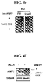
FIGS. 4 a-4 h demonstrates the Effect of AIMP2-DX2 on the cellular stability of AIMP2. In FIG. 4 a, AIMP2-DX2 was transfected into DU145 untreated or treated with TGF-β for 2 hr, and the AIMP2 level was determined by Western blotting. The expression of c-Myc, AIMP2-DX2 and GAPDH (control) was monitored by RT-PCR. In FIG. 4 b, AIMP2-DX2 or empty vector was transfected into DU145, and its effect on the TGF-β-dependent cell growth inhibition was monitored by thymidine incorporation (n=4). In FIG. 4 c, the interaction between AIMP2-F and AIMP2-DX2 was determined by yeast two hybrid assay as previously described (Rho, S. B. et al., PNAS. USA, 96:4488-93(1999)). In FIG. 4 d, AIMP2-F and AIMP2-DX2 were synthesized by in vitro translation in the presence of [35S] methionine, mixed with either GST-AIMP2-F or -CDK2 (control), and precipitated with glutathione-Sepharose. The precipitated proteins were separated by SDS-PAGE and detected by autoradiography. In FIG. 4 e, the interaction of AIMP2-F or AIMP2-DX2 with FUSE-binding protein (FBP; Kim, M. J. et al., Nat. Genet. 34:330-336:2003)) and Smad2 was determined by yeast two hybrid assay. In FIG. 4 f, the effect of the proteasome inhibitor, ALLN (50 μM for 4 hr) on the levels of the full-length (F) and AIMP2-DX2 of AIMP2 was monitored in the AIMP2-DX2-generating H322 cells by Western blotting with anti-AIMP2 antibody. The AIMP2-DX2 form was confirmed by its co-migration in gel with its in vitro synthesized counterpart. In FIG. 4 g, the increase of AIMP2 by the treatment of ALLN (20 μM for 2 hr) was also shown by immunofluorescence staining with anti-AIMP2 antibody in H322 cells. In FIG. 4 h, myc-tagged AIMP2-DX2 was transfected to DU145 that were treated with ALLN. Then, AIMP2 was immunoprecipitated with anti-AIMP2 antibody, and the ubiquitinated AIMP2 molecules were monitored by immunoblotting with anti-ubiquitin antibody (Ubi).
FIGS. 5 a-5 e represent the disruptive effect of AIMP2-DX2 on cell growth control and TGF-β signaling. In FIG. 5 a, AIMP2-DX2 (or empty vector) was transfected into MEFs and monitored its effect on cell growth. The cells and colonies were visualized by light microscopy (top) and Giemsa staining (bottom), respectively. In FIG. 5 b, siRNA targeting AIMP2-DX2 (si-DX2) was introduced into H322 and its suppressive effect on the AIMP2-DX2 transcript was determined by RT-PCR (top). si-DX2 did not affect the full-length AIMP2 transcript as shown by RT-PCR of the transcript for exon 3-4. The effect of si-DX2 on the phosphorylation of Smad2 and AIMP2 expression was also determined by Western blotting (bottom). The effect of si-DX2 on the restoration of the TGF-β signaling was also determined by immunofluorescence staining of p-Smad2 (FIG. 5 c), TGF-β-dependent reporter assay under 3TP promoter (FIG. 5 d) and growth arrest (FIG. 5 e) using H322 cells. In FIG. 5 c, p-Smad2 and nuclei were stained with FITC-conjugated secondary antibody (green) and PI (red), respectively, 30 min after TGF-β treatment. Notice that p-Smad2 was increased and nuclear located by the transfection of si-DX2.
FIGS. 6 a-6 c represent the association of AIMP2 with lung cancer formation. In FIG. 6 a, lung tumor formation was monitored at time interval after the intraperitoneal administration of benzo-(α)-pyrene into AIMP2+/+ and AIMP2+/− mice. “N” stands for the number of the sacrificed mice. In FIG. 6 b, total RNAs were isolated from the tissues, and subjected to RT-PCR with the AIMP2-DX2-specific primer. Normal and tumor tissues of the same patients (indicated by code number) were RT-PCR (FIG. 6 c). In FIG. 6 c, the exon 4 region of AIMP2 and GAPDH were used as control.
FIG. 7 represents the determination of the Smad2 domain involved in the interaction with AIMP2. We ligated the cDNAs encoding AIMP2 or CDK2 to the EcoRI and XhoI sites of pGEX4T-1 to express them in E. coli BL21 (DE3) as the GST-fusion proteins and purified them following the manufacturer's instruction. The different deletion fragments of Smad2 were synthesized by in vitro translation in the presence of [35S] methionine using the TNT coupled translation kit (Promega). The GST fusion proteins bound to the glutathione Sepharose beads were incubated with the [35S] methionine-labeled Smad2 fragments in the binding buffer of PBS buffer (pH 7.4) containing 0.5 mM EDTA, 0.5 mM phenylmethylsulfonylfluoride (PMSF), and 1% Trition X-100. The binding mixture was incubated overnight 4° C. with rotation and washed four times with the binding buffer containing 0.5% Trition X-100. After addition of the SDS sample buffer, the bound proteins were eluted by boiling and separated by SDS gel electrophoresis. The presence of Smad2 fragments was determined by autoradiography.
FIGS. 8 a and 8 b show the exon arrangement of human AIMP2 gene and the primer locations in AIMP2 cDNA. In FIG. 8 a, the AIMP2 gene is composed of four exons encoding the polypeptides of the indicated size. FIG. 8 b is the schematic representation for the locations of the primers used to generate the cDNAs spanning different regions of AIMP2 and their sequences.
FIGS. 9 a-9 d demonstrate reduced expression of AIMP2, and generation of AIMP2-DX2 in cancer cell lines. In FIG. 9 a, to compare the AIMP2 levels between the AIMP2-DX2-positive and -negative cells, we cultured DU145 and H460 cells in one dish, and performed immunofluorescence staining with anti-AIMP2 antibody (green). The two cells lines were distinguished by immunofluorescence staining of p53 (red) since DU145 cells express p53 at high level due to its mutation, whereas H460 cells containing the wild type p53 maintain it at low level. The cells were treated with TGF-β for 2 hr, and fixed with methanol. In FIG. 9 b, to address the effect of AIMP2-DX2 on expression of AIMP2, we monitored the AIMP2 level by flow cytometry. We transfected 2 μg/ml of empty vector or AIMP2-DX2 into DU145 cells, and incubated for 24 hr. The cells were then fixed with 70% ethanol and reacted with anti-AIMP2 antibody, and subsequently FITC-conjugated secondary antibody. In FIG. 9 c, the effect of AIMP2-DX2 on the TGF-β-dependent cell cycle arrest was compared by flow cytometry. While the portion of the G0/G1 phase cells was increased in DU145 cells transfected with empty vector, but not in the AIMP2-DX2-transfected cells. The black and blues lines indicated the cells untreated and treated with TGF-β, respectively. In FIG. 9 d, we compared the AIMP2 levels in H460 cells that were untreated (control) or treated with 10 μM ALLN for 2 hr. “Negative” indicates the cells incubated only with FITC-conjugated secondary antibody.
FIG. 10 shows the generation of AIMP2-DX2 and suppression of AIMP2 in lung cancer tissues. Lungs were isolated from the AIMP2+/+ and AIMP2+/− mice injected with benzoypyrene, and RNAs were isolated from each lung for RT-PCR to determine the generation of AIMP2-DX2. All of the lungs generating AIMP2-DX2 showed tumor formation in lung (marked +).
FIG. 11 represents the expression of AIMP2-DX2 and suppression of AIMP2 in liver cancer tissues. RT-PCR was performed to determine the expression of AIMP2-DX2 in liver cancer tissues.
FIG. 12 shows the expression vector carrying the AIMP2-DX2 gene. In FIG. 12, the abbreviations, pCMV, BGH pA, f1 ori, neomycin, ampicillin, SV40, SV40 pA, ColE1, T7 and Sp6 denote human cytomegalovirus immediate-early promoter, bovine growth hormone polyadenylation signal, f1 replication origin, neomycin resistance gene, ampicillin resistance gene, SV40 replication origin, SV40 polyadenylation signal, ColE1 replication origin, T7 viral promoter and Sp6 viral promoter.
DETAILED DESCRIPTION OF THIS INVETNION
The present inventors elucidated that the genetic disruption of p38 (newly designated herein as “AIMP2”) to induce overexpression of c-myc causes neonatal lethality in mice through overproliferation of alveolar epithelial cells and transforming growth factor-β (TGF-β) induces AIMP2 expression and promoted its translocation to nuclei for the downregulation of c-myc (M. J. Kim, et al., Nat. Genet. 34:330-336(2003)).
Following the previous research, the present inventors have revealed that AIMP2 is a novel tumor suppressor, playing a unique role in TGF-β signaling via interaction with Smad2/3. In addition, we have discovered that the aberrant variant of AIMP2 lacking exon II (AIMP2-DX2) is specifically expressed in cancer cell lines and tissues. The existence of AIMP2 lacking exon II (AIMP2-DX2) was verified by RT-PCR using combinations of AIMP2-specific primers. When the primers were used to generate AIMP2 cDNA spanning exon 3 and 4, the decrease of AIMP2 transcript was not observed in the cells showing the reduced level of AIMP2 in Western blot analysis. When we used the primers generating the transcript from exon 1 to 3, we obtained not only the transcript of the expected size, but also a smaller one. Sequencing analysis of this small transcript revealed that it lacks exon 2 encoding 69 amino acid residues of AIMP2. RT-PCR analysis using the primer targeting to the junction sequence of exon 1 and 3 showed that the cell lines expressing lower AIMP2 level generated the smaller transcript, confirming the generation of AIMP2-DX2.
Furthermore, the inventors observed that AIMP2 level was dramatically reduced regardless of TGF-β, demonstrating that the generation of AIMP2-DX2 leads to loss of AIMP2 activity. In addition to this, the introduction of AIMP2-DX2 elevated the expression of c-myc and relived the growth arrest by TGF-β. Surprisingly, we found that AIMP2-DX2 forms a heterodimer with AIMP2 that is ubiquitinated to be rapidly degraded by proteasome-dependent degradation process. Consequently, we are urged to reason that AIMP2-DX2 is closely associated with tumorigenesis by inducing the decrease of AIMP2 level. In vivo study provides additional evidences to verify that AIMP2-DX2 is strongly related to lung and liver cancer formation as well.
It should be noted that p38DX2 described in the priority documents of this application, i.e., the Korean Pat. Appln. Nos. 2004-0097164 and 2005-0039073 is newly named as AIMP2-DX2.
In one aspect of this invention, there is provided a AIMP2-DX2 protein comprising a AIMP2 amino acid sequence in which the exon 2 region of the AIMP2 amino acid sequence is deleted, that is specifically expressed in cancer cells, in particular, lung and liver cancer cells.
Preferably, the AIMP2-DX2 protein consists of the AIMP2 amino acid sequence in which the exon 2 region of the AIMP2 amino acid sequence is deleted.
The AIMP2-DX2 protein is a deletion variant of AIMP2 lacking exon 2. The amino acid sequence of the AIMP2 protein is found in several databases (312aa version: accession Nos. AAC50391.1 and GI:1215669; 320aa version: accession Nos. AAH13630.1, GI:15489023 and BC013630.1 available from GenBank) and publications (312aa version: Nicolaides, N. C., Kinzler, K. W. and Vogelstein, B. Analysis of the 5′ region of PMS2 reveals heterogeneous transcripts and a novel overlapping gene, Genomics 29 (2):329-334(1995); 320 aa version: Generation and initial analysis of more than 15,000 full-length human and mouse cDNA sequences, Proc. Natl. Acad. Sci. U.S.A. 99(26): 16899-16903(2002)). The amino acid sequence of the AIMP2-DX2 protein comprises, preferably, consists of that of AIMP2 lacking exon 2 region as afore-described known sequences. The Korean Pat. Appln. No. 10-2003-0018424 discloses cancer therapy efficacy of the AIMP2 protein, teachings of which are incorporated herein by reference in its entity.
In addition, the AIMP2-DX2 protein includes exon 2-deleted variant of AIMP2 equivalents, for example, functional equivalents resulting from substitution, deletion, insertion or their combinations of AIMP2 that exhibit substantially identical activity to the wild type AIMP2, or functional derivatives with modifications to alter physical and/or biochemical properties of the wild type AIMP2 that exhibit substantially identical activity to the wild type AIMP2.
The deletion of exon 2 in AIMP2 as described herein means that the amino acid sequence spanning exon 2 region in AIMP2 (corresponding to amino acid 46-114) is partially or wholly deleted to generate a deletion variant of AIMP2 capable of forming a heterodimer with AIMP2 to inhibit normal function of AIMP2 and promote degradation of AIMP2. Accordingly, the AIMP2-DX2 protein of this invention may include any variant of AIMP2 with whole or partial exon 2 deletion in which exon 1, 3 and/or 4 is natural or modified by amino acid substitution, deletion or insertion, so long as the variant is able to form a heterodimer with AIMP2 to inhibit normal function of AIMP2. Preferably, the AIMP2-DX2 protein comprises a whole exon 2 deletion and intact exon 1, 3 and 4. More preferably, the AIMP2-DX2 protein comprises, most preferably, consists of an amino acid sequence of SEQ ID NO:2.
The AIMP2-DX2 protein may comprise its natural-occurring amino acid sequences and variants having modified sequences as well, so long as the variants retain activity of the AIMP2-DX2 protein described above. The variants of the AIMP2-DX2 protein refer to proteins having different sequences from its natural-occurring amino acid sequence prepared by deletion, insertion, non-conserved or conserved substitution or their combinations. The silent alteration of amino acid residues not to substantially impair protein activity is well known to one skilled in the art (H. Neurath, R. L. Hill, The Proteins, Academic Press, New York, 1979). Such amino acid alteration includes Ala/Ser, Val/Ile, Asp/Glu, Thr/Ser, Ala/Gly, Ala/Thr, Ser/Asn, Ala/Val, Ser/Gly, Thy/Phe, Ala/Pro, Lys/Arg, Asp/Asn, Leu/Ile, Leu/Val, Ala/Glu and Asp/Gly, but not limited to.
In addition, the AIMP2-DX2 protein may comprise post-translational modifications such as phosphorylation, sulfation, acrylation, glycosylation, methylation and farnesylation.
The AIMP2-DX2 protein and its variants may be obtained by the isolation from natural sources, synthesis (Merrifleld, J. Amer. Chem. Soc. 85:2149-2156(1963)) or recombinant DNA technology (Joseph Sambrook, et al., Molecular Cloning, A Laboratory Manual, Cold Spring Harbor Laboratory Press, Cold Spring Harbor, N.Y. (2001)). Where a recombinant DNA technology is applied, host cells are transformed with an expression vector carrying a nucleic acid molecule encoding AIMP2-DX2 and then cultured, followed by recovering the AIMP2-DX2 expressed.
As described previously, the AIMP2-DX2 protein is specifically expressed in a variety of cancer cells including breast cancer, large intestinal cancer, lung cancer, small cell lung cancer, stomach cancer, liver cancer, blood cancer, bone cancer, pancreatic cancer, skin cancer, head or neck cancer, cutaneous or intraocular melanoma, uterine sarcoma, ovarian cancer, rectal cancer, anal cancer, colon cancer, fallopian tube carcinoma, endometrial carcinoma, cervical cancer, vulval cancer, vaginal carcinoma, Hodgkin's disease esophageal cancer, small intestine cancer, endocrine cancer, thyroid cancer, parathyroid cancer, adrenal cancer, soft tissue tumor, urethral cancer, penile cancer, prostate cancer, bronchogenic cancer, nasopharyngeal cancer, laryngeal cancer, chronic or acute leukemia, lymphocytic lymphoma, bladder cancer, kidney cancer, ureter cancer, renal cell carcinoma, renal pelvic carcinoma, CNS tumor, primary CNS lymphoma, bone marrow tumor, brain stem nerve gliomas and pituitary adenoma, in particular, lung and liver cancer tissues, demonstrating that the AIMP2-DX2 protein can serve as a cancer diagnostic marker.
In another aspect of this invention, there is provided a nucleic acid molecule comprising a nucleotide sequence encoding the AIMP2-DX2 protein described above.
Preferably, the nucleic acid molecule coding for the AIMP2-DX2 protein comprises a nucleotide sequence of SEQ ID NO:1. More preferably, the nucleic acid molecule of this invention consists of a nucleotide sequence of SEQ ID NO:1.
It is well understood by the skilled artisan that homologous sequences due to codon degeneracy may be encompassed within the nucleic acid molecule of the present invention, showing at least 60%, preferably 80%, most preferably 90-95% nucleotide similarity to that of SEQ ID NO:1, as measured using one of the sequence comparison algorithms. Methods of alignment of sequences for comparison are well-known in the art. Various programs and alignment algorithms are described in: Smith and Waterman, Adv. Appl. Math. 2:482(1981); Needleman and Wunsch, J. Mol. Bio. 48:443(1970); Pearson and Lipman, Methods in Mol. Biol. 24: 307-31(1988); Higgins and Sharp, Gene 73:237-44(1988); Higgins and Sharp, CABIOS 5:151-3(1989); Corpet et al., Nuc. Acids Res. 16:10881-90(1988); Huang et al., Comp. Appl. BioSci. 8:155-65(1992); and Pearson et al., Meth. Mol. Biol. 24:307-31(1994). The NCBI Basic Local Alignment Search Tool (BLAST) (Altschul et al., J. Mol. Biol. 215:403-10(1990)) is available from several sources, including the National Center for Biological Information (NBCl, Bethesda, Md.) and on the Internet, for use in connection with the sequence analysis programs blastp, blasm, blastx, tblastn and tblastx. It can be accessed at http://www.ncbi.nlm.nih.gov/BLAST/. A description of how to determine sequence identity using this program is available at http://www.ncbi.nlm.nih.gov/BLAST/blast_help.html.
The nucleic acid molecule of this invention may be single- or double-chain DNA (cDNA and gDNA) or single-chain RNA (mRNA).
The nucleic acid molecule encoding the AIMP2-DX2 protein may be prepared by the isolation from natural sources, synthesis or recombinant DNA technology. The AIMP2-DX2 nucleic acid molecule may be included in a suitable vector to provide the AIMP2-DX2 protein.
In still another aspect of this invention, there is provided a recombinant vector which comprises a nucleic acid molecule comprising a nucleotide sequence encoding the AIMP2-DX2 protein.
The term “recombinant vector” used herein refers to a genetic carrier to express a protein or RNA of interest in a suitable host cell, comprising a corresponding foreign sequence operably linked to a nucleic acid expression control sequence (such as a promoter, signal sequence and array of transcription factor binding sites).
The term “operably linked” used herein refers to functional linkage between a nucleic acid expression control sequence and a second nucleic acid sequence of interest, wherein the expression control sequence affects transcription and/or translation of the nucleic acid corresponding to the second sequence. The vector of this invention may be constructed according to conventional recombinant DNA technology in which site-specific DNA cleavage and ligation are performed using commercially available enzymes.
The vector of the present invention includes plasmid, cosmid, bacteriophage and viral vectors, but not limited to. The vector may comprise expression control elements such as promoter, operator, start and stop codons, polyadenylation signal and enhancer as well as signal or leader sequences for membrane targeting or secretion. The promoter used in vectors may be constitutive or inducible one. Furthermore, the vector may carry a selection marker for selecting host cells harboring the vector and a replication origin.
The signal sequence in the vector includes, but not limited to, PhoA and OmpA signal sequences for Escherichia host cells, α-amylase and subtilicin signal sequences for Bacillus host cells, MFα and SUC2 signal sequences for yeast host cells, and insulin, α-interferon and antibody molecule signal sequences for animal host cells.
In further aspect of this invention, there is provided a transformant which is transformed with the recombinant vector of this invention described above.
The transformation may be carried out according to any known approach for transforming nucleic acid molecules into organism, cell, tissue or organ, including electroporation, protoplasm fusion, CaPO4 precipitation, CaCl2 precipitation, agitation using silicon carbamide fiber, Agrobacterium-mediated transformation, PEG, dextran sulfate and lipofectamine, but not limited to. A suitable transformation method may be selected based on the type of host cells.
A suitable host cell is generally decided in considering the expression level and post-translation modification. Host cells include, but not limited to, prokaryotic cells such as Escherichia coli, Bacillus subtilis, Streptomyces, Pseudomonas, Proteus mirabilis and Staphylococcus, fungi (e.g., Aspergillus), yeast (e.g., Pichia pastoris, Saccharomyces cerevisiae, Schizosaccharomyces and Neurospora crassa), insect cells, plant cells and mammalian cells.
In still further aspect of this invention, there is provided a process for preparing the AIMP2-DX2 protein by culturing transformed cells described previously.
The culturing is carried out by conventional methods known to those skilled in the art under conditions suitable to express the AIMP2-DX2 protein of interest.
The AIMP2-DX2 protein expressed may be purified by conventional methods, for example, salting out (e.g., ammonium sulfate and sodium phosphate precipitation), solvent precipitation (e.g., protein fractionation precipitation using acetone or ethanol), dialysis, gel filtration, ion exchange, reverse-phase column chromatography, ultrafiltration or their combinations (Sambrook et al., Molecular Cloning: A Laboratory Manual, 3rd Ed., Cold Spring Harbor Laboratory Press (2001); and Deutscher, M., Guide to Protein Purification Methods, Enzymology, vol. 182. Academic Press. Inc., San Diego, Calif. (1990)).
The term “cancer marker” used herein refers to a substance that provides information to evaluate cancer likelihood, occurrence or development by examining qualitatively or quantitatively its expression in tissues or cells. Preferably, the term means an organic biomolecule (e.g., protein, DNA and RNA) that is expressed in cancer cells in a different pattern from normal cells. The AIMP2-DX2 gene or protein of this invention is specifically expressed in cancer cells but not in normal cells, permitting to accurately diagnose cancer (preferably, lung and liver cancer). Preferably, the cancer diagnosis using AIMP2-DX2 expression is performed in mRNA and/or protein level.
In another aspect of this invention, there is provided an antibody specific to the AIMP2-DX2 protein.
The term “antibody” used herein means a protein molecule specifically directed toward an antigenic site. The antibody of this invention refers to antibodies to specifically recognize AIMP2-DX2 with discriminating AIMP2, including polyclonal and monoclonal antibodies.
Antibodies against the novel protein, AIMP2-DX2, may be prepared in accordance with conventional technologies known to one skilled in the art.
Polyclonal antibodies may be prepared according to known processes in which the AIMP2-DX2 protein as an immunogen is injected into animals and then antiserum is collected. Immunized animals include, but not limited to, goat, rabbit, sheep, monkey, horse, pig, cattle and dog.
Monoclonal antibodies may be prepared in accordance with a fusion method (Kohler and Milstein, European Journal of Immunology, 6:511-519(1976)), a recombinant DNA method (U.S. Pat. No. 4,816,56) or a phage antibody library (Clackson et al, Nature, 352:624-628(1991); and Marks et al, J. Mol. Biol., 222:58, 1-597(1991)).
Antibodies against the AIMP2-DX2 protein may be an intact immunoglobulin molecule or its fragments containing antigen-binding site such as F(v), Fab, Fab′ and F(ab′)2.
The antibodies of this invention specific to AIMP2-DX2 permit to diagnose cancer (preferably, lung and liver cancer) as well as to treat cancer (preferably, lung and liver cancer) by suppressing the activity of AIMP2-DX2. Where the antibodies are used as a therapeutic agent, they may be coupled to conventional therapeutic agents in direct or indirect (through a linker) manner.
The therapeutic agent coupled to the antibodies of this invention includes, but not limited to, radionuclide (e.g., 131I, 90Y, 105Rh, 47Sc, 67Cu, 212Bi, 211At, 67Ga, 125I, 186Re, 188Re, 177Lu, 153Sm, 123I and 111In), drug (e.g., methotrexate and adriamycin), lymphokine (interferon), toxin (ricin, abrin and diphtheria) and heterofunctional antibody that forms a complex with other antibody to posses a bi-functional binding capacity both to cancer cell and effector cell (e.g., T killer cell).
The antibody of this invention may be administered per se or in the form of a pharmaceutical composition.
The pharmaceutical composition comprising antibody may be formulated with a pharmaceutically acceptable carrier. The form of the pharmaceutical composition varies depending on the administration mode. Typically, the composition comprises one of surfactants for facilitating transmembrane delivery. Such surfactant includes steroid-derived compounds, cationic lipids such as N-[1-(2,3-dioleyloxy)propyl]-N,N,N-trimethylammonium chloride (DOTMA), cholesterol hemisuccinate and phosphatidyl glycerol.
The pharmaceutical composition comprising the antibody of this invention is administered in a pharmaceutically effective amount to treat cancer or prevent cancer metastasis. The pharmaceutical composition may be administered in a single or multiple dosing regimen. The administration mode of the pharmaceutical composition includes parenteral, subcutaneous, intraperitoneal, intrapulmonary, intranasal, and local administration. Parenteral administration includes subcutaneous, intradermal, intramuscular, intravenous, intrabursa, intrasternal, intrathecal and intraperitoneal injection. The pharmaceutical composition is generally formulated in a pH range of 4-8 for antibody stability (chemical and physical stability) and safety. In addition, the pharmaceutical composition may be formulated in an oral dosing form. Typical dose is optimized using standard clinical techniques.
Furthermore, the antibody of this invention may be administered in a form of nucleic acid molecule to induce in vivo production of antibody (WO 96/07321).
In still another aspect of this invention, there is provided a diagnostic kit for cancer, which comprises an antibody specific to the AIMP2-DX2 protein.
The cancer diagnosis kit of this invention may comprise antibody specific to AIMP2-DX2 as well as general instruments and reagents for immunoassay including carrier, detectable signal-generating label, dissolving agent, washing agent, buffer and stabilizer. Where an enzyme is used as a label, its substrate and reaction quencher may be included. Non-limiting examples of carrier include soluble carriers, for example, physiologically acceptable buffer known in the art (e.g. PBS), insoluble carriers, for example, polystyrene, polyethylene, polypropylene, polyester, polyacrylonitrile, fluorine resin, cross-linked dextran, polysaccharides, polymers such as magnetic microparticles made of latex coated with a metal, paper, glass, metals, agarose and combinations thereof.
Non-limiting examples of the assay system useful in the cancer diagnosis kit of the present invention include ELISA plates, dip-stick devices, immunochromatography test strips and radial partition immunoassy devices and flow-through devices.
In further aspect of this invention, there is provided a method for diagnosing cancer, which comprises the steps of: (a) providing a sample to be assayed; and (b) detecting in the sample an expression of a nucleotide sequence encoding the AIMP2-DX2 protein of claim 1, wherein the detection of the expression of the nucleotide sequence encoding the AIMP2-DX2 protein is indicative of cancer.
The sample used in the present invention includes any biological sample such as tissue, cell, whole blood, serum, plasma, saliva, semen, urine, synovia and spinal fluid and may be pretreated for assay.
The present method may be carried out at protein or mRNA level. Where it is performed to detect the AIMP2-DX2 protein, antibodies to specifically recognize the AIMP2-DX2 protein are used and the detection is carried out by contacting the sample to the antibody specific to the AIMP2-DX2 protein and evaluating a formation of antigen-antibody complex. The evaluation on antigen-antibody complex formation may be carried out using immunohistochemical staining, radioimmuno assay (RIA), enzyme-linked immunosorbent assay (ELISA), Western blotting, immunoprecipitation assay, immunodiffusion assay, complement fixation assay, FACS and protein chip assay. The evaluation on antigen-antibody complex formation may be performed qualitatively or quantitatively, in particular, by measuring signal from detection label.
The label to generate measurable signal for antigen-antibody complex formation includes, but not limited to, enzyme, fluorophore, ligand, luminophore, microparticle, redox molecules and radioisotopes. The enzymatic label includes, but not limited to, β-glucuronidase, β-D-glucosidase, β-D-galactosidase, urease, peroxidase, alkaline phosphatase, acetylcholinesterase, glucose oxidase, hexokinase, GDPase, RNase, luciferase, phosphofructokinase, phosphoenolpyruvate carboxylase, aspartate aminotransferase, phosphenolpyruvate decarboxylase, β-lactamase. The fluorescent label includes, but not limited to, fluorescein, isothiocyanate, rhodamine, phycoerythrin, phycocyanin, allophysocyanin, o-phthalate and fluorescamine. The ligand serving as a label includes, but not limited to, biotin derivatives. Non-limiting examples of the luminescent label includes acridinium ester, luciferin and luciferase. Microparticles as label include colloidal gold and colored latex, but not limited to. Redox molecules for labeling include ferrocene, lutenium complex compound, viologen, quinone, Ti ion, Cs ion, diimide, 1,4-benzoquinone, hydroquinone, K4 W(CN)8, [Os (bpy)3]2+, [Ru(bpy)3]2+ and [Mo(CN)8]4−, but not limited to. The radioisotopes includes 3H, 14C, 32P, 35S, 36Cl, 51Cr, 57Co, 58Co, 59Fe, 90Y, 125I, 131I and 186Re, but not limited to.
Where the present method is performed to detect the AIMP2-DX2 mRNA, the detection step may be carried out by an amplification reaction or a hybridization reaction well-known in the art.
The phrase “detection of the AIMP2-DX2 mRNA” used herein is intended to refer to analyze the existence or amount of the AIMP2-DX2 mRNA as cancer diagnosis marker in cells by use of primer or probe specifically hybridized with the AIMP2-DX2 mRNA.
The term “primer” used herein means an oligonucleotide, whether occurring naturally as in a purified restriction digest or produced synthetically, which is capable of acting as a point of initiation of synthesis when placed under conditions in which synthesis of a primer extension product which is complementary to a nucleic acid strand is induced, i.e., in the presence of four different nucleoside triphosphates and a thermostable enzyme in an appropriate buffer and at a suitable temperature.
The term “probe” used herein refers to a linear oligomer of natural or modified monomers or linkages, including deoxyribonucleotides, ribonucleotides and the like, which is capable of specifically hybridizing with a target nucleotide sequence, whether occurring naturally or produced synthetically. The probe used in the present method may be prepared in the form of oligonucleotide probe, single-stranded DNA probe, double-stranded DNA probe and RNA probe. It may be labeled with biotin, FITC, rhodamine, DIG and radioisotopes.
The method to detect the AIMP2-DX2 mRNA using either primer or probe includes, but not limited to, DNA sequencing, RT-PCR (reverse transcription-polymerase chain reaction), primer extension method (Nikiforov, T. T. et al., Nucl Acids Res 22, 4167-4175(1994)), oligonucleotide ligation analysis (OLA) (Nickerson, D. A. et al., Pro Nat Acad Sci USA, 87, 8923-8927(1990)), allele-specific PCR (Rust, S. et al., Nucl Acids Res, 6, 3623-3629(1993)), RNase mismatch cleavage (Myers R. M. et al., Science, 230, 1242-1246(1985)), single strand conformation polymorphism (SSCP; Orita M. et al., Pro Nat Acad Sci USA, 86, 2766-2770(1989)), simultaneous analysis of SSCP and heteroduplex (Lee et al., Mol Cells, 5:668-672(1995)), denaturation gradient gel electrophoresis (DGGE; Cariello N F. et al., Am J Hum Genet, 42, 726-734(1988)) and denaturing high performance liquid chromatography (D-HPLC, Underhill Pa. et al., Genome Res, 7, 996-1005(1997)).
Preferably, the method by amplification reaction is carried out by RT-PCR using a primer capable of differentiating an mRNA of AIMP2-DX2 from an mRNA of AIMP2. RT-PCR process suggested by P. Seeburg (1986) for RNA research involves PCR amplification of cDNA obtained from mRNA reverse transcription. For amplification, a primer pair specifically annealed to AIMP2-DX2 is used. Preferably, the primer is designed to generate two different sized bands in electrophoresis in which one is specific to the AIMP2 mRNA and the other to AIMP2-DX2 mRNA. Alternatively, the primer is designed to generate only electrophoresis band specific to AIMP2-DX2 mRNA. The primer pair to generate two different sized bands for the AIMP2 mRNA and AIMP2-DX2 mRNA is prepared to amplify a region corresponding to exon 2. The nucleotide sequence of such primers is not limited; most preferably, a primer set consisting of SEQ ID NOs:5 and 6. To observe only one electrophoresis band specific to AIMP2-DX2 mRNA, one of primers is designed to comprise the junction sequence between C-terminal of exon 1 and N-terminal of exon 3. In Examples described below, the primer of SEQ ID NO:8 annealed to the junction sequence is used together with the primer of SEQ ID NO:7 for RT-PCR. The RT-PCR analysis is convenient in the senses that cancer diagnosis is accomplished by observing the electrophoresis band pattern to evaluate expression of the AIMP2-DX2 mRNA.
The present method may be carried out in accordance with hybridization reaction using suitable probes.
The stringent conditions of nucleic acid hybridization suitable for forming such double stranded structures are described by Joseph Sambrook, et al., Molecular Cloning, A Laboratory Manual, Cold Spring Harbor Laboratory Press, Cold Spring Harbor, N.Y. (2001) and Haymes, B. D., et al., Nucleic Acid Hybridization, A Practical Approach, IRL Press, Washington, D.C. (1985). As used herein the term “stringent condition” refers to the conditions of temperature, ionic strength (buffer concentration), and the presence of other compounds such as organic solvents, under which hybridization or annealing is conducted. As understood by those of skill in the art, the stringent conditions are sequence dependent and are different under different environmental parameters. Longer sequences hybridize or anneal specifically at higher temperatures.
The probes used in the hybridization reaction have a AIMP2-DX2 specific nucleotide sequence which is not found in AIMP2. Preferably, the probes are designed to comprise the junction sequence between exons 1 and 3, most preferably, having the nucleotide sequence of SEQ ID NO:8.
The present method is very useful in diagnosing a variety of cancer including breast cancer, large intestinal cancer, lung cancer, small cell lung cancer, stomach cancer, liver cancer, blood cancer, bone cancer, pancreatic cancer, skin cancer, head or neck cancer, cutaneous or intraocular melanoma, uterine sarcoma, ovarian cancer, rectal cancer, anal cancer, colon cancer, fallopian tube carcinoma, endometrial carcinoma, cervical cancer, vulval cancer, vaginal carcinoma, Hodgkin's disease esophageal cancer, small intestine cancer, endocrine cancer, thyroid cancer, parathyroid cancer, adrenal cancer, soft tissue tumor, urethral cancer, penile cancer, prostate cancer, bronchogenic cancer, nasopharyngeal cancer, laryngeal cancer, chronic or acute leukemia, lymphocytic lymphoma, bladder cancer, kidney cancer, ureter cancer, renal cell carcinoma, renal pelvic carcinoma, CNS tumor, primary CNS lymphoma, bone marrow tumor, brain stem nerve gliomas and pituitary adenoma. More preferably, the present method is used for diagnosing lung cancer and liver cancer, most preferably, lung cancer.
In still further aspect of this invention, there is provided a siRNA (small interfering RNA) molecule which comprise a nucleotide sequence complementary to a region of an mRNA of the AIMP2-DX2 protein.
The term “siRNA” used herein refers to a short RNA duplex to induce RNAi (RNA interference) phenomenon through mRNA cleavage. The siRNA consists of a sense RNA strand corresponding to target mRNA and an antisense RNA strand complementary to target mRNA. siRNA to inhibit expression of a target gene provides effective gene knock-down method or gene therapy method.
The siRNA of this invention is not restricted to a RNA duplex of which two strands are completely paired and may comprise non-paired portion such as mismatched portion with non-complementary bases and bulge with no opposite bases. The overall length of the siRNA is 10-100 nucleotides, preferably, 15-80 nucleotides, and more preferably, 20-70 nucleotides. The siRNA may comprise either blunt or cohesive end so long as it enables to silent the AIMP2-DX2 expression due to RNAi effect. The cohesive end may be prepared in 3′-end overhanging structure or 5′-end overhanging structure. The overhanging bases are not limited in its length, for example, 1-8 nucleotides, preferably, 2-6 nucleotides. The overall length as described herein is expressed as the total of length of central double-stranded portion and terminal single-stranded overhanging portion. Furthermore, as long as AIMP2-DX2 siRNA is able to maintain its gene silencing effect on the target gene, it may contain a low molecular weight RNA (which may be a natural RNA molecule such as tRNA, rRNA or viral RNA, or an artificial RNA molecule), for example, in the overhanging portion at its either end. It is not necessary that both ends of AIMP2-DX2 siRNA have a cleavage structure. The AIMP2-DX2 siRNA of this invention may comprise a stem-loop structure in which either end (head or tail) is connected via a linker. The length of the linker is not limited unless it impairs base pairing in stem structure.
The term “specific” used herein in conjunction with siRNA is intended to express the inhibition of target gene expression with no influence on other genes. The siRNA of this invention is specific to the AIMP2-DX gene.
It is preferred that the siRNA of this invention comprise a sense strand containing a corresponding sequence to a junction sequence between exons 1 and 3 and an antisense strand containing a complementary sequence.
The phrase “inhibition of gene expression” means that the level of mRNA and/or protein generated from the target gene is quenched or reduced, which is induced by RNA interference via occurrence of mRNA cleavage.
The siRNA of this invention may be synthesized in vitro and then introduced into cells via transfection. In addition, it may be transfected into cells in the form of siRNA expression vector or PCR-derived siRNA expression cassette. Suitable preparation and transfection methods may be determined in considering the experiment aim and target gene function.
The sequences and length of the siRNA are not limited as long as it enables to suppress the AIMP2-DX2 expression. 3 illustrative siRNA expression vectors to silence AIMP2-DX2 are found in Examples described hereunder, suppressing cellular level of AIMP2-DX2 and restoring AIMP2 function and TGF-β signal transduction.
The preferable siRNA of this invention comprises a corresponding sequence to the junction sequence between exons 1 and 3 of the AIMP2-DX2 mRNA. More preferably, the siRNA of this invention is a RNA duplex described as (i) No.3 siRNA consisting of two RNA molecules expressed from nucleotide sequences of SEQ ID NOs:9 and 10, (ii) No.4 siRNA consisting of two RNA molecules expressed from nucleotide sequences of SEQ ID NOs:11 and 12, or (iii) No.5 siRNA consisting of two RNA molecules expressed from nucleotide sequences of SEQ ID NOs:13 and 14. The siRNA consisting of two RNA molecules expressed from nucleotide sequences of SEQ ID NOs:11 and 12 is most preferred. The single or mixed type of siRNA molecules may be used.
In another aspect of this invention, there is provided an antisense oligonucleotide which is complementary to a region of an mRNA of the AIMP2-DX2 protein.
The term “antisense oligonucleotide” used herein is intended to refer to nucleic acids, preferably, DNA, RNA or its derivatives, that are complementary to the base sequences of a target mRNA, characterized in that they binds to the target mRNA and interfere its translation to protein. The antisense oligonucleotide of this invention means DNA or RNA sequences complementary and binding to AIMP2-DX2 mRNA, that are able to inhibit translation, translocation, maturation or other biological functions of AIMP2-DX2 mRNA. The antisense nucleic acid is 6-100, preferably, 8-60, more preferably, 10-40 nucleotides in length.
The antisense oligonucleotide may at lease one modification in its base, sugar or backbone for its higher inhibition efficacy (De Mesmaeker et al., Curr Opin Struct Biol., 5(3):343-55(1995)). The modified nucleic acid backbone comprises phosphorothioate, phosphotriester, methyl phosphonate, short chain alkyl or cycloalkyl intersugar linkages or short chain heteroatomic or heterocyclic intersugar linkages. The antisense oligonucleotide may also contain one or more substituted sugar moieties. The antisense nucleic acid may include one or more modified bases, for example, hypoxanthine, 6-methyladenine, 5-me pyrimidines (particularly, 5-methylcytosine), 5-hydroxymethylcytosine (HMC), glycosyl HMC and gentobiosyl HMC, as well as synthetic nucleobases, e.g., 2-aminoadenine, 2-(methylamino)adenine, 2-(imidazolylalkyl)adenine, 2-(aminoalklyamino)adenine or other heterosubstituted alkyladenines, 2-thiouracil, 2-thiothymine, 5-bromouracil, 5-hydroxymethyluracil, 8-azaguanine, 7-deazaguanine, N6(6-aminohexyl)adenine and 2,6-diaminopurine. Another modification of the oligonucleotides of the invention involves chemically linking to the oligonucleotide one or more moieties or conjugates which enhance the activity or cellular uptake of the oligonucleotide. Such moieties include but are not limited to lipid moieties such as a cholesterol moiety, a cholesteryl moiety (Letsinger et al., Proc. Natl. Acad. Sci. USA, 86:6553(1989)), cholic acid (Manoharan et al. Bioorg. Med. Chem. Let., 4:1053(1994)), a thioether, e.g., hexyl-S-tritylthiol (Manoharan et al. Ann. N.Y. Acad. Sci., 660:306(1992); Manoharan et al. Bioorg. Med. Chem. Let., 3: 2765(1993)), a thiocholesterol (Oberhauser et al., Nucl. Acids Res., 20:533(1992)), an aliphatic chain, e.g., dodecandiol or undecyl residues (Saison-Behmoaras et al. EMBO J., 10:111(1991); Kabanov et al. FEBS Lett., 259:327(1990); Svinarchuk et al. Biochimie, 75:49(1993), a phospholipid, e.g., di-hexadecyl-rac-glycerol or triethylammonium 1,2-di-O-hexadecyl-rac-glycero-3-H-phosphonate (Manoharan et al. Tetrahedron Lett., 36:3651(1995); Shea et al. Nucl. Acids Res., 18:3777(1990)), a polyamine or a polyethylene glycol chain (Manoharan et al. Nucleosides & Nucleotides, 14:969(1995)), or adamantane acetic acid (Manoharan et al. Tetrahedron Lett., 36: 3651(1995)). Oligonucleotides comprising lipophilic moieties, and methods for preparing such oligonucleotides are known in the art, for example, U.S. Pat. Nos. 5,138,045, 5,218,105 and 5,459,255. The modifications described above enhance stability against nuclease degradation and increase affinity of the antisense oligonucleotide toward its target mRNA.
It is preferred that the antisense oligonucleotide specific to AIMP2-DX2 comprises a complementary sequence to a junction region between exons 1 and 3 of AIMP2-DX2 mRNA.
The antisense RNA molecule is conventionally synthesized in vitro and then transmitted to cells. In addition, it is intracellularly produced by transcription from foreign sequence. In vitro synthesis involves RNA polymerase I. In vivo transcription for preparing antisense RNA uses vector having origin of recognition region (MCS) in opposite orientation. The antisense RNA preferably comprises a translation stop codon for inhibiting translation to peptide.
In another aspect of this invention, there is provided a pharmaceutical composition for treating cancer, which comprises (a) the antisense oligonucleotide or siRNA specific to AIMP2-DX2 mRNA as an active ingredient; and (b) a pharmaceutically acceptable carrier.
In still another aspect of this invention, there is provided a method for treating cancer in a patient, which comprises administrating into the patient a pharmaceutical composition (a) the antisense oligonucleotide or siRNA specific to AIMP2-DX2 mRNA as an active ingredient; and (b) a pharmaceutically acceptable carrier.
The pharmaceutical composition comprising at least one of AIMP2-DX2 siRNAs or antisense oligonucleotides may contain additional agent to suppress tumor cell proliferation and to facilitate the transduction of siRNA or antisense nucleic acid, for example, liposome (U.S. Pat. Nos. 4,897,355, 4,394,448, 4,235,871, 4,231,877, 4,224,179, 4,753,788, 4,673,567, 4,247,411 and 4,814,270), or lipophilic carrier including sterols such as cholesterol, cholate and deoxycholic acid. In addition, the siRNA or antisense nucleic acid is conjugated to cell-adsorbing peptides such as peptide hormones, antigens and peptide toxins (Haralambid et al, WO 89/03849; Lebleu et al., EP 0263740).
Where the pharmaceutical composition is formulated for oral administration, it may contain binder, lubricant, disintegrator, diluent, solubilizer, dispersing agent, stabilizer, suspending agent, pigment and sweetener. Where the pharmaceutical composition is formulated for injection, it may contain buffer, preservative, solubilizer, tonicity agent and stabilizer. For topical administration, the pharmaceutical composition may contain substrate, diluent, lubricant and preservative. The formulation of the pharmaceutical composition may be prepared by formulating with pharmaceutically acceptable carriers described above. For oral administration, the pharmaceutical composition may be in the form of tablet, troche, capsule, elixir, suspension, syrup and wafer. The injectable composition may be formulated in unit dosage ample or multi dosage form.
The correct dosage of the pharmaceutical compositions of this invention comprising AIMP2-DX2 siRNA or antisense oligonucleotide will be varied according to the particular formulation, the mode of application, age, body weight and sex of the patient, diet, time of administration, condition of the patient, drug combinations, reaction sensitivities and severity of the disease. It is understood that the ordinary skilled physician will readily be able to determine and prescribe a correct dosage of this pharmaceutical compositions.
The administration mode of the pharmaceutical composition includes oral and parenteral such as subcutaneous, intradermal, intramuscular, intravenous, intrabursa, intrasternal, intrathecal and intraperitoneal injections.
In further aspect of this invention, there is provided a method of screening for an agent which inhibits the formation of a heterodimer between the AIMP2-DX2 protein of claim 1 and the AIMP2 protein, comprising the steps of: (a) contacting a test substance to a composition which comprises the AIMP2-DX2 protein and the AIMP2 protein; and (b) determining whether the test substance inhibits the heterodimer formation between the AIMP2-DX2 protein and the AIMP2 protein, wherein the test substance to inhibit the heterodimer formation between the AIMP2-DX2 protein and the AIMP2 protein is evaluated as an anticancer agent.
The formation of the heterodimer between the AIMP2-DX2 protein and AIMP2 protein is associated with cancer as demonstrated in Examples described hereunder. Therefore, a substance capable of inhibiting the heterodimer formation is evaluated as a candidate for anticancer agent.
According to a preferred embodiment, the instant method is performed by a yeast-two-hybrid assay and in vitro pull-down assay.
Where the present method is carried out by yeast-two-hybrid assay format, the composition comprising the AIMP2-DX2 protein and the AIMP2 protein is a cell harboring the respective gene.
In a yeast two-hybrid assay, the AIMP2-DX2 protein and AIMP2 protein can be used as either “bait” or “prey” (see, e.g., U.S. Pat. No. 5,283,317; Zervos et al., Cell 72, 223-232(1993); Maduraetal., J. Biol. Chem. 268, 12046-12054(1993); Bartel et al., BioTechniques 14, 920-924(1993); Iwabuchi et al., Oncogene 8, 1693-1696(1993); and Brent WO 94/10300), to identify substances which inhibits the interaction of AIMP2-DX2 with AIMP2 to form a heterodimer. The two-hybrid system is based on the modular nature of most transcription factors, which consist of separable DNA-binding and activation domains. Briefly, the assay utilizes two different DNA constructs. For example, in one construct, polynucleotide encoding the AIMP2-DX2 protein can be fused to a polynucleotide encoding the DNA binding domain of a known transcription factor (e.g. Lex). In the other construct a DNA sequence that encodes the AIMP2 protein can be fused to a polynucleotide that codes for the activation domain of the known transcription factor (e.g. B42).
If the test substance treated to cells expressing the two-hybrid system is able to inhibit the interaction between the AIMP2-DX2 protein and AIMP2 protein, the DNA-binding and activation domains of the transcription factor are not brought into close proximity. This proximity allows transcription of a reporter gene (e.g. lac Z), which is operably linked to a transcriptional regulatory site responsive to the transcription factor. Expression of the reporter gene can be easily detected.
In an in vitro pull-down assay format, the AIMP2-DX2 gene and AIMP2 gene can be used as either bait or prey. For example, in one construct for bait, the AIMP2 protein is fused to a protein that allows the AIMP2 protein to be bound to a solid support. For example, glutathione-S-transferase (GST) fusion proteins can be adsorbed onto glutathione Sepharose beads or glutathione derivatized microtiter plates. In the other construct for prey, the AIMP2-DX2 protein is synthesized by in vitro translation system (e.g., reticulocyte lysate) using 35S. The radioactive AIMP2-DX2 protein is added to the GST-AIMP2 protein bound to glutathione Sepharose beads together with the addition of the test substance. The reaction mixture is washed and the proteins bound to the Sepharose beads are eluted, followed by electrophoresis.
If the test substance added to the reaction mixture is able to inhibit the interaction between the AIMP2-DX2 protein and AIMP2 protein, the electrophoresis result shows no band.
In still further aspect of this invention, there is provided a method of screening for an agent which inhibits the expression of the AIMP2-DX2 gene, comprising the steps of: (a) contacting a test substance to cells which express the AIMP2-DX2 gene; and (b) determining whether the test substance inhibits the expression of the AIMP2-DX2 gene, wherein the test substance to inhibit the expression of the AIMP2-DX2 gene is evaluated as an anticancer agent.
In the present method, the expression of the AIMP2-DX2 gene is assessed at mRNA or protein level. Where the present method is carried out to detect the AIMP2-DX2 mRNA expressed, it is preferably performed by RT-PCR using AIMP2-DX2 specific primers described hereinabove. Where the present method is carried out to detect the AIMP2-DX2 protein expressed, it is preferably performed by a variety of immunoassay processes using AIMP2-DX2 specific antibodies as described above.
The following specific examples are intended to be illustrative of the invention and should not be construed as limiting the scope of the invention as defined by appended claims.
EXAMPLES
Methods
Cell Culture, Chemicals and Cell Cycle Measurement
Cells were maintained in RPMI-1640 containing 10% FBS. Mouse embryonic fibroblasts (MEFs) were isolated from 12.5-14.5 day embryos and cultivated in DMEM (Dulbecco's Modified Eagle Medium) containing 20% FBS. To evaluate the effect of TGF-β on cell cycle, cells were incubated with 2 ng/ml TGF-β in serum-free or 1% FBS-containing medium for 24 hr and harvested for FACS analysis. Cell proliferation was also determined by [3H] thymidine incorporation. Cells were incubated in serum-free medium with or without TGF-β for 20 hr, and then in the presence of 1 μCi/ml of [3H] thymidine for 4 hr. The incorporated thymidine was quantified by liquid scintillation counting as previously described (Kim, M. J. et al., Nat. Genet. 34, 330-336(2003)). TGF-β was purchased from R&D system, and anti-Smad2 and anti-Smad4 antibodies from Santa Cruz.
Immunoblotting and Immunoprecipitation
Cells were treated with TGF-β for the indicated times and proteins were extracted with protease-containing RIPA buffer (1% Nonidet P-40, 0.5% sodium deoxycholate, 0.1% SDS), separated by 10-12% SDS-PAGE, and immunoblotted with the specific antibodies using ECL system (Santa cruz biotech). For immunoprecipitation, the cell lysates were cleared by pre-incubation with IgG (Pierce) and agarose-conjugated protein A (Invitrogen). After centrifugation, the supernatants were incubated with the specific antibody, and agarose-conjugated protein A for 2 hr. After washing with ice-cold PBS twice and RIPA once, the bound proteins were precipitated with the specific antibody, eluted and subjected to Western blot analysis.
RT-PCR
The total RNAs were isolated following the protocol of the manufacturer (Qiagen). Briefly, the freshly prepared tissues (3×3×3 mm) were chopped into small pieces, mixed with 350 μl lysis buffer and homogenized using homogenizer or syringe. After adding 350 μl of 70% ethanol, the lysates were inverted several times, loaded onto column, centrifuged at 13,000 RPM for 15 sec. After washing the column with wash buffer twice, RNAs were eluted with 40 μl RNase-free DW. For reverse transcription, 1 μg of the isolated RNA was used as a template with the AIMP2-specific primer (FIG. 8 b). After the reaction, the mixture was diluted 3 fold with DW and 1 μl of its aliquot was used for 30 μl PCR reaction containing 0.5 μl dNTP (2.5 mM each), 0.5 μl of the indicated primers (each 10 μM), 1.5 μl DMSO and 0.1 μl Taq polymerase (5 U/μl).
Suppression of AIMP2-DX2 with si-RNA
To suppress the expression of AIMP2-DX2, we designed the siRNA against AIMP2-DX2 with the sequence of TCGAGCTGGCCACGTGCAGGATTACGAGTACTGGTAATCCTGCACGTGGCCAGCTTTT (underlined regions are matched to the AIMP2-DX2 sequence) and cloned it into 16 IMT-700 vector system (Imgenex) using SalI and XbaI, followed by DNA sequencing for confirming the cloned sequence. H322 cells (ATCC, human lung epithelial carcinoma) were transfected with 2 μg of si-AIMP2-DX2 expression vector. Following 3-hr incubation with DNA-liposome complex in 1 ml serum-free media, H322 cells were treated with 1 ml RPMI-1640 containing 20% FBS. Then, cells were incubated in serum-free medium with or without TGF-β for 20 hr.
Yeast Two-Hybrid Analysis
The cDNAs encoding human AIMP2 and Smad2 (and its deletion fragments) were obtained by PCR using specific primers. The PCR products of Smad2 and AIMP2 were digested with EcoRI and XhoI, and ligated into the same sites of pEG202 (LexA) and pJG4-5 (B42), respectively (Gyuris, J et al., a human G1 and S phase protein phosphatase that associates with Cdk2. Cell 75, 791-803(1993)). The interactions between Lex-Smad2 fragments and B42-AIMP2 were analyzed for their ability to grow on yeast medium containing X-gal as previously described (Rho, S. B. et al., Proc Natl Acad Sci USA 96, 4488-93. (1999)).
In Vitro Pull-Down Assay
AIMP2, AIMP2-DX2 and CDK2 were expressed as GST fusion proteins and immobilized to glutathione-Sepharose 4B (Pharmacia). The cDNA fragments encoding the AIMP2 and AIMP2-DX2 were obtained by PCR using specific primers and cloned into pET-28a vector for in vitro transcription and translation (Promega). Aliquots (10 μl) of TNT products were incubated with 5 μg of GST-AIMP2, -AIMP2-DX2 and -CDK2 immobilized on the beads in 100 μl of PBS containing 0.5% Triton X-100, 0.5 mM EDTA, and 0.5 mM phenylmethylsulfonyl fluoride. The beads were vigorously washed with the binding buffer, and the bound proteins were eluted, resolved by SDS-PAGE, and determined by autoradiography.
Construction of Cells Stably Generating AIMP2-DX2
The wild type MEFs were transfected with pcDNA-AIMP2-DX2 or pcDNA (Invitrogen) itself and the transfectants were selected in DMEM medium containing 400 μg/ml G418. After removing the untransfected cells, the transfectants were cultured in the normal medium without G418 for 3 days, fixed with 2% PFA and stained with Giemsa.
Immunostaining and Histological Analysis
The frozen tissue slides were fixed with 2% paraformaldehyde and washed with ice cold PBS. After blocking and permeablization with PBS containing 0.2% Triton X-100 (PBST) and 1% BSA, the slides were incubated with anti-AIMP2 antibody for 2 hr. After washing with PBS, they were also incubated with anti-rabbit goat IgG-FITC (Pierce) and propidium iodide (50 μg/ml, Sigma) for 1 hr, washed with PBS, mounted, and observed under a confocal microscopy (μ Radiance, Bio-Rad).
Construction of AIMP2-DX2 Expression Vector
To construct the AIMP2-DX2 expressing vector, cDNA of AIMP2-DX2 was cloned into pcDNA3.1-myc. First, AIMP2-DX2 from H322 cDNA was amplified using primers with linker for EcoRI and XhoI and cloned into pcDNA3.1-myc (Invitrogen) using EcoRI and XhoI. The pcDNA3.1-myc/AIMP2-DX2 vector to express AIMP2DX (FIG. 12) was introduced into E. coli DH5α, which was deposited on Oct. 25, 2004 in the International Depository Authority, the Korean Collection for Type Cultures (KCTC) and was given accession number KCTC 10710BP. In the RECEIP IN THE CASE OF AN ORIGINAL DEPOSIT attached, while the identification reference read as Escherichia coli DH5@/p38DX2, it should be noted that p38DX2 is newly designated herein as AIMP2-DX2.
Lung Caner Formation
32 AIMP2+/− mice (19 male and 13 female mice) and 25 AIMP2+/+ mice (14 male and 11 female mice) were used for experiment. We induced lung tumor through the intraperitoneal injection of chemical carcinogen, benzo-(α)-pyrene (BP, 100 mg/kg, Sigma) into AIMP2+/+ and AIMP2+/− mice, and monitored the tumor formation in lung. As a control group, 3 AIMP2+/+ (2 male and 1 female) and 5 AIMP2+/− mice (4 male and 1 female) were injected with bical solution (10% DMSO and 35% PEG 40 in saline). The mice treated were sacrificed at the indicated time and their lung tissues were fixed in formaldehyde and undergone H&E staining as described in Kim, M. J. et al., Nat. Genet. 34:330-336(2003), followed by the observation under a microscope.
Results
Functional Importance and Working Mode of AIMP2 in TGF-β Signaling
To see the importance of AIMP2 in TGF-β signaling, we compared the responses of the AIMP2+/+ and AIMP2−/− MEFs to TGF-β-induced growth arrest, nuclear translocation of Smad2/3 and interaction of AIMP2 with Smad2/3. While the growth of the wild type cells was suppressed by TGF-β, the AIMP2-deficient cells did not respond to the signal as determined by thymidine incorporation, colony formation and flow cytometry (FIGS. 1 a, 1 b and 1 c, respectively). When MEFs were treated with TGF-β, Smad2 and Smad3 were translocated to nuclei in the normal cells, but not in the AIMP2−/− cells (FIG. 1 d). All of these results suggest the functional importance of AIMP2 in TGF-β signaling via Smad2 and Smad3.
We then checked the possible interaction of AIMP2 with Smad2 and 3 by coimmunoprecipitation. AIMP2 showed the interaction with Smad2/3 that was enhanced by TGF-β (FIG. 1 e). The direct interaction of AIMP2 with two R-Smads was also confirmed by yeast two hybrid and in vitro pull-down assays (FIGS. 2 and 7). The amount of AIMP2 bound to Smad2/3 was increased according to the induction of AIMP2 by TGF-β (FIG. 2 a). When the AIMP2 level was increased by transfection, the expression of the TGF-β target genes was enhanced, suggesting its stimulatory role in TGF-β signaling (FIG. 2 b). Since AIMP2 binds to both of Smad2 and 3, we expected that it would work to these two R-Smads in a similar way and thus focused on its relationship to Smad2 in more detail. The TGF-β-dependent phosphorylation of Smad2 was suppressed in the AIMP2-deficient MEFs, but restored when AIMP2 was introduced to AIMP2−/− cells (FIG. 2 c). We then determined the domain of Smad2 involved in the interaction with AIMP2 by yeast two hybrid (FIG. 2 d) and in vitro pull-down assay (FIG. 7). The two experiments revealed that the interaction involves the MH2 domain of Smad2.
To determine the working mode of AIMP2 in TGF-β signaling, we compared the TGF-β-induced association of Smad2 with TGF-β receptor in the normal and AIMP2-deficient cells. The cells were treated with TGF-β and the association of Smad2 and the receptor was monitored by co-immunoprecipitation of type I receptor with Smad2 at time interval. While the TGF-β-induced Smad2 binding to TGF-β receptor was observed at early time point and decreased in the wild type cells, the receptor bound to Smad2 was accumulated in the late stage after TGF-β treatment in the AIMP2-deficient cells (FIG. 2 e). In the TGF-β-induced phosphorylation of Smad2, the phosphorylated Smad2 was gradually increased in the wild type cells, but severely suppressed in the AIMP2−/− cells (FIG. 2 f). These results demonstrate that AIMP2 plays a critical role in the TGF-β induced phosphorylation of R-Smads via its direct interaction with R-Smad2.
Suppression of AIMP2 and Generation of its Deletion Variant in Cancer Cells
To explore the possible association of AIMP2 with cancer formation, we checked the variation of AIMP2 level in different cancer cell lines [A549 (lung epithelial carcinoma), DU145 (prostate carcinoma), HCT116 (human colorectal carcinoma), H460 (large cell lung carcinoma), H322 (lung bronchioalveolar carcinoma) and H290 (mesothelioma cancer cell line) available from ATCC]. Three of the six tested cell lines showed lower AIMP2 level in Western blot (FIG. 3 a) and FACS analyses (FIG. 3 b). All of the cell lines with low AIMP2 level expressed the normal level of the TGF-β type II receptor and retained its kinase activity, implying that the low AIMP2 level does not result from the mal-functionality of the receptor. To determine whether the variation of AIMP2 level resulted from the difference in transcription, we performed RT-PCR with different combinations of the AIMP2-specific primers. The AIMP2 gene consists of four exons (FIG. 8 a). When the primers were used to generate AIMP2 cDNA spanning exon 3 and 4, the decrease of AIMP2 transcript was not observed in the cells showing the reduced level of AIMP2 (FIG. 3 c, first row), suggesting that it does not result from lower transcription. When we used the primers generating the transcript from exon 1 to 3, we obtained not only the transcript of the expected size, but also a smaller one (FIG. 3 c, second row). Sequencing analysis of this small transcript revealed that it lacks exon 2 encoding 69 aa of AIMP2 (FIG. 8 b). To confirm the generation of this smaller transcript, we designed the primer (FIG. 8 b, primer DX-B) targeting to the junction sequence of exon 1 and 3 that is generated by the deletion of exon 2, and conducted RT-PCR with this primer. The cell lines expressing lower AIMP2 level generated the smaller transcript (designated AIMP2-DX2, FIG. 3 c, second and third rows).
Western blot analysis with anti-AIMP2 antibody detected only the full-length AIMP2, but not AIMP2-DX2 (FIG. 2 a), implying that AIMP2-DX2 may be very unstable at protein level. Immunofluorescence staining also demonstrated the lower AIMP2 level in H460, H322 and H290 (FIG. 3 d). To exclude the possibility of staining artifact, we co-cultivated H460 and DU145 cells in the same plate and stained AIMP2. Again, the staining intensity of AIMP2 in H460 was much weaker than that in DU145 (FIG. 9 a). In addition, the TGF-β-dependent nuclear localization of AIMP2 was not observed in the cells with low AIMP2 expression (FIG. 9 d, bottom row). To address the linkage between the AIMP2 level and functionality of TGF-β, we measured growth suppression by TGF-β in these cell lines. While the growth of A549 and DU145 cells was suppressed by TGF-β, the cells with low AIMP2 level did not respond to TGF-β (FIG. 9 e). This result is consistent with previous reports that A549 is sensitive whereas H460 is resistant to TGF-β signal (Osada, H. et al. Cancer Res. 61, 8331-8339(2001); Kim, T. K. et al., Lung Cancer 31, 181-191(2001)). In addition, the target genes were induced by TGF-β in A549 and DU145, but not in H322 and two other cell lines with low AIMP2 (FIG. 3 f).
Furthermore, we tested by RT-PCR whether other cancer cell lines generates AIMP2-DX2. As a result, it was revealed that AIMP2-DX2 was detected in SaOS2 (osteosarcoma) and MCF7 (breast adenocarcinoma cell).
The Inactive Deletion Variant Forms a Complex with Functional AIMP2
To comprehend the causal relationship of the AIMP2-DX2 generation and suppression of AIMP2, we checked the change of the AIMP2 level after transfection of AIMP2-DX2 into DU145. AIMP2 was reduced in the AIMP2-DX2-transfected cells, as demonstrated by Western blot (FIG. 4 a, first row) and FACS analyses (FIG. 9 b). Moreover, the expression of the AIMP2 target, c-myc, was elevated by the introduction of AIMP2-DX2 (FIG. 4 a, third row). AIMP2-DX2 also relieved the growth arrest by TGF-β (FIGS. 4 b and 9 c). We then investigated how AIMP2-DX2 would affect the functional AIMP2 (AIMP2-F). Since AIMP2 has a potential to form a homodimer (Quevillon, S. et al., J. Mol. Biol. 285, 183-195(1999); and Kim, J. Y. et al., Proc. Natl. Acad. Sci. USA 99, 7912-7916(2002)), we examined whether AIMP2-DX2 would interact with AIMP2-F by yeast two hybrid assay. AIMP2-DX2 showed the interaction with AIMP2-F, but not with itself (FIG. 4 c). In in vitro pull-down assay, both of radioactively synthesized AIMP2-F and AIMP2-DX2, but not AIMP2-DX2, were co-purified with GST-AIMP2-F (FIG. 4 d), proving the direct interaction between AIMP2-F and AIMP2-DX2. To see whether AIMP2-DX2 is active in TGF-β signaling, we tested its interaction with Smad2 by yeast two hybrid assay. While AIMP2 interacted with Smad2 as well as FBP that is the known target of AIMP2 (Kim, M. J. et al., Nat. Genet. 34, 330-336(2003)), AIMP2-DX2 did not bind to any of these proteins, suggesting that it would be functionally inactive (FIG. 4 e).
To understand how the heterodimer formation would suppress AIMP2, we checked whether the AIMP2 level is controlled by proteasome-dependent degradation process. The AIMP2-DX2-producing H322 cells were treated with the proteasome inhibitor, ALLN (Zhou, M., et al., J. Biol. Chem. 271, 24769-24775(1996)) and tested whether the AIMP2 level is increased by the blockage of proteasome. The AIMP2 level was significantly increased by the treatment of ALLN as shown by Western blotting (FIG. 4 f), immunofluorescence staining (FIG. 4 g) and flow cytometry (FIG. 9 d), suggesting that its cellular level would be controlled by proteasome-mediated degradation. In addition, AIMP2-DX2 form was also detected by the inhibition of proteasome (FIG. 4 f), confirming the notion that AIMP2-DX2 would be unstable due to the rapid proteasome-dependent degradation. The low intensity of AIMP2-DX2 appears to result from its lower transcription compared to the normal AIMP2, as demonstrated by RT-PCR analysis (FIG. 3 c) and/or less efficient recognition by anti-AIMP2 antibody. Since AIMP2 degradation is mediated by proteasome, we tested whether its ubiquitination is promoted by AIMP2-DX2. When AIMP2 was immunoprecipitated from the control and AIMP2-DX2-transfected cells that were treated with ALLN and blotted with anti-ubiquitin antibody, higher amount of the ubiquitinated AIMP2 was observed in the AIMP2-DX2-transfected cells compared to that in control cells (FIG. 4 h). Combined together, AIMP2-DX2 appears to work as a dominant negative mutant to form an inactive complex with AIMP2 that is rapidly driven to degradation process.
AIMP2-DX2 Inactivates TGF-α Signaling and Promotes Cell Growth
We transfected AIMP2-DX2 into MEFs and monitored its effect on cell growth by microscopic analysis and colony formation. The cell growth was significantly enhanced by the introduction of AIMP2-DX2 (FIG. 5 a). We then introduced siRNA (si-AIMP2-DX2) that specifically suppresses the AIMP2-DX2 transcript and checked whether it can restore the normal level of AIMP2 and TGF-β signaling in H322 cells expressing AIMP2-DX2. si-AIMP2-DX2 ablated the AIMP2-DX2 transcript (FIG. 5 b top) and resumed the normal AIMP2 level and TGF-β-induced phosphorylation of Smad2 (FIG. 5 b bottom), nuclear localization of p-Smad2 (FIG. 5 c), TGF-β-dependent reporter expression (FIG. 5 d) and growth arrest (FIG. 5 e). All of these results demonstrated the disruptive effect of AIMP2-DX2 on TGF-β signaling and cell growth control.
Association of AIMP2 Deletion Variant with Human Lung Cancer
Since the reduction of AIMP2 is frequently detected in different cancer cell lines (FIGS. 3 a, 3 b and 3 d), and loss of AIMP2 leads to hyper-proliferation of lung cells (Kim, M. J. et al., Nat. Genet. 34, 330-336(2003)), we examined the association of AIMP2 abnormality with lung cancer formation. We induced lung tumor through the intraperitoneal injection of chemical carcinogen, benzo-(α)-pyrene (BP, Wang, Y. et al., Cancer Res. 63, 4389-4395 (2003)) into AIMP2+/+ and AIMP2+/− mice, and monitored the tumor formation in lung. From 6 weeks after the administration of BP, lung tumors were observed at 50-70% frequency in AIMP2+/− mice, and at about 30% in the wild type littermates (FIG. 6 a), implying that the heterozygous mice are more susceptible to BP-induced tumorigenesis. We examined whether AIMP2-DX2 is generated in the lungs of the BP-injected mice. Three out of four lungs isolated from AIMP2+/− mice showed tumors, while only one developed tumors among three AIMP2+/+ mice and all of these tumors generated AIMP2-DX2 (FIG. 10), further supporting the relevance of AIMP2-DX2 to tumor formation. Moreover, AIMP2-DX2 was generated only in tumor tissues (FIG. 6 b). To exclude the possibility that the AIMP2 level and AIMP2-DX2 formation may vary depending on different individuals, we carried out RT-PCR analyses in the normal and tumor pairs isolated from the same patients. Again, the cancer-specific reduction of AIMP2 was coupled with the generation of AIMP2-DX2 (FIG. 6 c). AIMP2-DX2 was not detected in one case showing the normal AIMP2 level in cancer region (FIG. 6 c, patient 22872). We further examined 10 different pathologically-diagnosed lung adenocarcinoma, squamous cell carcinoma and large cell adenocarcinoma samples, and observed the cancer-specific reduction of AIMP2 and generation of AIMP2-DX2 in 8 cases (Table 1). Although AIMP2-DX2 was detected in the histologically normal regions in two cases, its occurrence was still coupled with the low level of AIMP2.
TABLE 1
The relationship between AIMP2 level and AIMP2-DX2
generation in different lung cancer patients
Code Cell AIMP2-F AIMP2-DX2
No (MLLG) type Opdate Recur Sex Age N T N T
1 H004 SQC Jan. 06, 1926 FALSE M 64 + +
2 H008 ADC Jan. 09, 1926 FALSE M 62 + +
3 H001 ADC Jan. 12, 1914 FALSE M 68 + +
4 H010 SQC Jan. 12, 1926 FALSE M 56 + +
5 H018 ADC Feb. 04, 1926 FALSE M 60 + +
6 H021 ADC Feb. 05, 1907 TRUE F 64 + +
7 H024 ADC Feb. 05, 1917 FALSE F 59 + +
8 H025 SQC Feb. 05, 1928 FALSE M 73 + +
9 H029 ADC Feb. 06, 1914 FALSE M 67 + +
10 H031 LAC Feb. 07, 1908 FALSE M 71 + +
Positive (+) and negative (−) in the AIMP2-F column denote the immunofluorescence staining of AIMP2 in normal (N) and tumor (T) tissues determined by histological analysis. Positive and negative in AIMP2-DX2 column indicate the generation of AIMP2-DX2 that was determined by RT-PCR.
Note for abbreviations: Recur (recurrence), SQC (squamous cell carcinoma), ADC (adenocarcinoma), LAC (large cell adenocarcinoma) and Opdate (operation date).

Association of AIMP2 and its Deletion Variant with Human Liver Cancer
We examined the relationship of AIMP2 or its deletion variant with human liver cancer using human tissues. The formation of AIMP2-DX2 was evaluated in normal and cancer tissue (hepatocellular carcinoma) by RT-PCR and the level of AIMP2 by immunofluorescence analyses as described previously. As a result, AIMP2-DX2 was detected in live cancer tissues (FIG. 11).
Having described a preferred embodiment of the present invention, it is to be understood that variants and modifications thereof falling within the spirit of the invention may become apparent to those skilled in this art, and the scope of this invention is to be determined by appended claims and their equivalents.

Claims (3)

1. An isolated AIMP2-DX2 protein comprising the amino acid sequence of SEQ ID NO:2.
2. A method of screening for an agent which inhibits the formation of a heterodimer between the AIMP2-DX2 protein and the AIMP2 protein, comprising the steps of:
(a) contacting a test substance to a composition which comprises the AIMP2-DX2 protein of SEQ ID NO:2 and the AIMP2 protein; and
(b) determining whether the test substance inhibits the heterodimer formation between the AIMP2-DX2 protein and the AIMP2 protein,
wherein the test substance to inhibit the heterodimer formation between the AIMP2-DX2 protein and the AIMP2 protein is evaluated as an anticancer agent.
3. The method according to claim 2, wherein the method is performed by a yeast-two-hybrid assay or in vitro pull-down assay.
US11/264,725 2004-11-24 2005-11-01 AIMP2-DX2 and its uses Active US7459529B2 (en)

Priority Applications (4)

Application Number Priority Date Filing Date Title
US12/255,943 US8003780B2 (en) 2004-11-24 2008-10-22 AIMP2-DX2 gene and SiRNA targeting AIMP2-DX2
US13/011,386 US20110117572A1 (en) 2004-11-24 2011-01-21 Antibody Specific to the AIMP2-DX2
US13/011,174 US8431393B2 (en) 2004-11-24 2011-01-21 AIMP2-DX2 gene and its uses
US15/074,498 US9944713B2 (en) 2004-11-24 2016-03-18 Antibody specific to the AIMP2-DX2

Applications Claiming Priority (4)

Application Number Priority Date Filing Date Title
KR2004-0097164 2004-11-24
KR20040097164 2004-11-24
KR2005-0039073 2005-05-10
KR1020050039073A KR20060057992A (en) 2004-11-24 2005-05-10 P38-dx2 and its use

Related Child Applications (1)

Application Number Title Priority Date Filing Date
US12/255,943 Continuation-In-Part US8003780B2 (en) 2004-11-24 2008-10-22 AIMP2-DX2 gene and SiRNA targeting AIMP2-DX2

Publications (2)

Publication Number Publication Date
US20060110397A1 US20060110397A1 (en) 2006-05-25
US7459529B2 true US7459529B2 (en) 2008-12-02

Family

ID=36461168

Family Applications (1)

Application Number Title Priority Date Filing Date
US11/264,725 Active US7459529B2 (en) 2004-11-24 2005-11-01 AIMP2-DX2 and its uses

Country Status (4)

Country Link
US (1) US7459529B2 (en)
EP (1) EP1814909B1 (en)
KR (1) KR100762995B1 (en)
WO (1) WO2006057500A1 (en)

Cited By (40)

* Cited by examiner, † Cited by third party
Publication number Priority date Publication date Assignee Title
US20090156536A1 (en) * 2004-11-24 2009-06-18 Sunghoon Kim AIMP2-DX2 Gene and SiRNA Targeting AIMP2-DX2
US20100028352A1 (en) * 2008-06-26 2010-02-04 Atyr Pharma, Inc. COMPOSITIONS AND METHODS COMPRISING GLYCYL-tRNA SYNTHETASES HAVING NON-CANONICAL BIOLOGICAL ACTIVITIES
US20100092434A1 (en) * 2008-06-11 2010-04-15 Atyr Pharma, Inc. Thrombopoietic activity of tyrosyl-trna synthetase polypeptides
US20100297149A1 (en) * 2009-03-16 2010-11-25 Atyr Pharma, Inc. Compositions and methods comprising histidyl-trna synthetase splice variants having non-canonical biological activities
US20100310576A1 (en) * 2009-03-31 2010-12-09 Adams Ryan A COMPOSITIONS AND METHODS COMPRISING ASPARTYL-tRNA SYNTHETASES HAVING NON-CANONICAL BIOLOGICAL ACTIVITIES
US20110150885A1 (en) * 2009-12-11 2011-06-23 Atyr Pharma, Inc. Aminoacyl trna synthetases for modulating hematopoiesis
US8828395B2 (en) 2009-12-11 2014-09-09 Atyr Pharma, Inc. Antibodies that bind tyrosyl-tRNA synthetases
US8835387B2 (en) 2012-02-16 2014-09-16 Atyr Pharma, Inc. Histidyl-tRNA synthetases for treating autoimmune and inflammatory diseases
US8945541B2 (en) 2010-05-14 2015-02-03 Atyr Pharma, Inc. Innovative discovery of therapeutic, diagnostic, and antibody compositions related to protein fragments of phenylalanyl-beta-tRNA synthetases
US8946157B2 (en) 2010-05-03 2015-02-03 Atyr Pharma, Inc. Innovative discovery of therapeutic, diagnostic, and antibody compositions related to protein fragments of seryl-tRNA synthetases
US8961961B2 (en) 2010-05-03 2015-02-24 a Tyr Pharma, Inc. Innovative discovery of therapeutic, diagnostic, and antibody compositions related protein fragments of arginyl-tRNA synthetases
US8961960B2 (en) 2010-04-27 2015-02-24 Atyr Pharma, Inc. Innovative discovery of therapeutic, diagnostic, and antibody compositions related to protein fragments of isoleucyl tRNA synthetases
US8962560B2 (en) 2010-06-01 2015-02-24 Atyr Pharma Inc. Innovative discovery of therapeutic, diagnostic, and antibody compositions related to protein fragments of Lysyl-tRNA synthetases
US8969301B2 (en) 2010-07-12 2015-03-03 Atyr Pharma Inc. Innovative discovery of therapeutic, diagnostic, and antibody compositions related to protein fragments of aspartyl-tRNA synthetases
US8980253B2 (en) 2010-04-26 2015-03-17 Atyr Pharma, Inc. Innovative discovery of therapeutic, diagnostic, and antibody compositions related to protein fragments of cysteinyl-tRNA synthetase
US8981045B2 (en) 2010-05-03 2015-03-17 Atyr Pharma, Inc. Innovative discovery of therapeutic, diagnostic, and antibody compositions related to protein fragments of methionyl-tRNA synthetases
US8986681B2 (en) 2010-04-27 2015-03-24 Atyr Pharma, Inc. Innovative discovery of therapeutic, diagnostic, and antibody compositions related to protein fragments of threonyl-tRNA synthetases
US8986680B2 (en) 2010-04-29 2015-03-24 Atyr Pharma, Inc. Innovative discovery of therapeutic, diagnostic, and antibody compositions related to protein fragments of Asparaginyl tRNA synthetases
US8993723B2 (en) 2010-04-28 2015-03-31 Atyr Pharma, Inc. Innovative discovery of therapeutic, diagnostic, and antibody compositions related to protein fragments of alanyl-tRNA synthetases
US8999321B2 (en) 2010-07-12 2015-04-07 Atyr Pharma, Inc. Innovative discovery of therapeutic, diagnostic, and antibody compositions related to protein fragments of glycyl-tRNA synthetases
US9029506B2 (en) 2010-08-25 2015-05-12 Atyr Pharma, Inc. Innovative discovery of therapeutic, diagnostic, and antibody compositions related to protein fragments of tyrosyl-tRNA synthetases
US9034598B2 (en) 2010-05-17 2015-05-19 Atyr Pharma, Inc. Innovative discovery of therapeutic, diagnostic, and antibody compositions related to protein fragments of leucyl-tRNA synthetases
US9034320B2 (en) 2010-04-29 2015-05-19 Atyr Pharma, Inc. Innovative discovery of therapeutic, diagnostic, and antibody compositions related to protein fragments of Valyl-tRNA synthetases
US9034321B2 (en) 2010-05-03 2015-05-19 Atyr Pharma, Inc. Innovative discovery of therapeutic, diagnostic, and antibody compositions related to protein fragments of phenylalanyl-alpha-tRNA synthetases
US9062301B2 (en) 2010-05-04 2015-06-23 Atyr Pharma, Inc. Innovative discovery of therapeutic, diagnostic, and antibody compositions related to protein fragments of glutamyl-prolyl-tRNA synthetases
US9062302B2 (en) 2010-05-04 2015-06-23 Atyr Pharma, Inc. Innovative discovery of therapeutic, diagnostic, and antibody compositions related to protein fragments of p38 multi-tRNA synthetase complex
US9068177B2 (en) 2010-04-29 2015-06-30 Atyr Pharma, Inc Innovative discovery of therapeutic, diagnostic, and antibody compositions related to protein fragments of glutaminyl-tRNA synthetases
US9127268B2 (en) 2009-12-11 2015-09-08 Atyr Pharma, Inc. Aminoacyl tRNA synthetases for modulating inflammation
US9399770B2 (en) 2010-10-06 2016-07-26 Atyr Pharma, Inc. Innovative discovery of therapeutic, diagnostic, and antibody compositions related to protein fragments of tryptophanyl-tRNA synthetases
US9422539B2 (en) 2010-07-12 2016-08-23 Atyr Pharma, Inc. Innovative discovery of therapeutic, diagnostic, and antibody compositions related to protein fragments of histidyl-tRNA synthetases
US9453214B2 (en) 2009-02-27 2016-09-27 Atyr Pharma, Inc. Polypeptide structural motifs associated with cell signaling activity
US9539236B2 (en) 2013-06-14 2017-01-10 Medicinal Bioconvergence Research Center Pharmaceutical composition for preventing or treating cancer
US9587235B2 (en) 2013-03-15 2017-03-07 Atyr Pharma, Inc. Histidyl-tRNA synthetase-Fc conjugates
US9688978B2 (en) 2011-12-29 2017-06-27 Atyr Pharma, Inc. Aspartyl-tRNA synthetase-Fc conjugates
US9714419B2 (en) 2011-08-09 2017-07-25 Atyr Pharma, Inc. PEGylated tyrosyl-tRNA synthetase polypeptides
US9796972B2 (en) 2010-07-12 2017-10-24 Atyr Pharma, Inc. Innovative discovery of therapeutic, diagnostic, and antibody compositions related to protein fragments of glycyl-tRNA synthetases
US9816084B2 (en) 2011-12-06 2017-11-14 Atyr Pharma, Inc. Aspartyl-tRNA synthetases
US9822353B2 (en) 2011-12-06 2017-11-21 Atyr Pharma, Inc. PEGylated aspartyl-tRNA synthetase polypeptides
US9944713B2 (en) 2004-11-24 2018-04-17 Medicinal Bioconvergence Research Center Antibody specific to the AIMP2-DX2
US11767520B2 (en) 2017-04-20 2023-09-26 Atyr Pharma, Inc. Compositions and methods for treating lung inflammation

Families Citing this family (22)

* Cited by examiner, † Cited by third party
Publication number Priority date Publication date Assignee Title
US20090074785A1 (en) * 2007-01-16 2009-03-19 Smith Jeffrey W Compositions and methods for treatment of colorectal cancer
KR101067816B1 (en) * 2007-11-09 2011-09-27 (주)네오믹스 Composition for preventing and treating inflammatory diseases comprising inhibitor of AIMP2-DX2 as an active ingredient
KR20130118612A (en) 2012-04-20 2013-10-30 (주)네오믹스 Novel aminopyridine derivatives and use thereof
WO2014142619A1 (en) * 2013-03-14 2014-09-18 부산대학교산학협력단 Method for screening therapeutic agent for cancers by using aimp2-dx2 and pl4/arf interaction
JP2016516804A (en) 2013-04-17 2016-06-09 ファイザー・インク N-piperidin-3-ylbenzamide derivatives for treating cardiovascular diseases
KR101762433B1 (en) 2013-06-05 2017-07-28 재단법인 의약바이오컨버젼스연구단 Novel Maleic acid derivatives, preparation method thereof, and anti-cancer compositions containing them
KR20150131655A (en) * 2014-05-15 2015-11-25 재단법인 의약바이오컨버젼스연구단 AIMP2-DX2-34S protein and method for manufacturing the same
US10072021B2 (en) * 2014-06-30 2018-09-11 Pusan National University Industry-University Cooperation Foundation Compound for inhibiting binding between DX2 protein and P14/ARF protein, and pharmaceutical composition for treating or preventing cancer disease containing same as effective ingredient
KR101636821B1 (en) * 2014-06-30 2016-07-07 부산대학교 산학협력단 Composition for diagnosing lung cancer comprising AIMP2-DX2 protein or fragment thereof
KR102297505B1 (en) * 2016-03-07 2021-09-01 재단법인 의약바이오컨버젼스연구단 Methods for screening anti-cancer drugs inhibiting interactions between AIMP2-DX2 and HSP70
KR101831435B1 (en) * 2016-03-10 2018-02-22 재단법인 의약바이오컨버젼스연구단 Antibodies specifically binding to AIMP2-DX2 protein
KR102290511B1 (en) * 2016-08-04 2021-08-17 주식회사 자이메디 Methods for screening anti-cancer drugs inhibiting interactions between AIMP2-DX2 and K-Ras
US10716866B2 (en) * 2018-03-29 2020-07-21 Generoath Co., Ltd Pharmaceutical composition comprising AIMP2-DX2 for preventing or treating neuronal diseases and use thereof
KR102248420B1 (en) * 2019-03-15 2021-05-06 주식회사 제너로스 Recombinant vector containing target sequence for miR-142-3p
CN113874361A (en) 2019-03-29 2021-12-31 医药生命融合研究团 Novel compound with anticancer activity and preparation method thereof
KR102252879B1 (en) 2019-11-15 2021-05-17 성균관대학교산학협력단 Method for diagnosing parkinson's disease using aimp2 in blood plasma, composition therefor and kit containing the same
CA3172932A1 (en) * 2020-03-23 2021-09-30 Curigin Co.,Ltd. Structure of oncolytic virus comprising bispecific nucleic acid molecule
KR102626543B1 (en) * 2020-09-14 2024-01-23 주식회사 제너로스 Recombinant vector containing AIMP2-DX2 and target sequence for miR-142-3p
EP4221759A1 (en) * 2020-09-30 2023-08-09 Generoath Co., Ltd. Methods of treating neuronal diseases using aimp2-dx2 and optionally a target sequence for mir-142 and compositions thereof
KR20230110503A (en) * 2020-09-30 2023-07-24 주식회사 제너로스 Method for treating age-related macular disease using target sequences for AIMP2-DX2 and optionally miR-142 and compositions thereof
KR20230114646A (en) 2022-01-25 2023-08-01 재단법인 한국파스퇴르연구소 Benzodioxane derivative compounds and their medical use
WO2023218430A1 (en) * 2022-05-13 2023-11-16 Generoath Co., Ltd. Methods of treating retinal degenerative diseases using aimp2-dx2 and optionally a target sequence for mir‑142 and compositions thereof

Citations (1)

* Cited by examiner, † Cited by third party
Publication number Priority date Publication date Assignee Title
US20040175375A1 (en) 2003-03-03 2004-09-09 Seoul National University Industry Foundation Method for treating cancer using p38/JTV-1 and method for screening pharmaceutical composition for treating cancer

Family Cites Families (13)

* Cited by examiner, † Cited by third party
Publication number Priority date Publication date Assignee Title
JPS5753564Y2 (en) 1977-06-01 1982-11-19
CH624011A5 (en) 1977-08-05 1981-07-15 Battelle Memorial Institute
FR2416008A1 (en) 1978-02-02 1979-08-31 Oreal LIPOSOME LYOPHILISATES
US4235871A (en) 1978-02-24 1980-11-25 Papahadjopoulos Demetrios P Method of encapsulating biologically active materials in lipid vesicles
US4816567A (en) 1983-04-08 1989-03-28 Genentech, Inc. Recombinant immunoglobin preparations
JPS6150912A (en) 1984-08-16 1986-03-13 Shionogi & Co Ltd Production of liposome preparation
US4814270A (en) 1984-09-13 1989-03-21 Becton Dickinson And Company Production of loaded vesicles
US4897355A (en) 1985-01-07 1990-01-30 Syntex (U.S.A.) Inc. N[ω,(ω-1)-dialkyloxy]- and N-[ω,(ω-1)-dialkenyloxy]-alk-1-yl-N,N,N-tetrasubstituted ammonium lipids and uses therefor
US4753788A (en) 1985-01-31 1988-06-28 Vestar Research Inc. Method for preparing small vesicles using microemulsification
FR2604438B1 (en) 1986-09-26 1988-12-23 Centre Nat Rech Scient NOVEL COUPLING CONJUGATES BETWEEN RNA OR DNA SEQUENCES AND A PROTEIN, THEIR PREPARATION PROCESS AND THEIR BIOLOGICAL APPLICATION
US5080891A (en) 1987-08-03 1992-01-14 Ddi Pharmaceuticals, Inc. Conjugates of superoxide dismutase coupled to high molecular weight polyalkylene glycols
ATE192465T1 (en) 1987-10-28 2000-05-15 Florey Howard Inst OLIGONUCLEOTIDE-POLYAMIDE CONJUGATES
ATE256738T1 (en) 1992-10-30 2004-01-15 Gen Hospital Corp A NEW CELL CYCLE CONTROL PROTEIN

Patent Citations (1)

* Cited by examiner, † Cited by third party
Publication number Priority date Publication date Assignee Title
US20040175375A1 (en) 2003-03-03 2004-09-09 Seoul National University Industry Foundation Method for treating cancer using p38/JTV-1 and method for screening pharmaceutical composition for treating cancer

Non-Patent Citations (1)

* Cited by examiner, † Cited by third party
Title
Kim et al., "Downregulation of FUSE-binding protein and c-myc by tRNA synthetase cofactor p38 is required for lung cell differentiation," Nat. Genet. 34:330-336, 2003.

Cited By (98)

* Cited by examiner, † Cited by third party
Publication number Priority date Publication date Assignee Title
US9944713B2 (en) 2004-11-24 2018-04-17 Medicinal Bioconvergence Research Center Antibody specific to the AIMP2-DX2
US8003780B2 (en) 2004-11-24 2011-08-23 Neomics Co., Ltd. AIMP2-DX2 gene and SiRNA targeting AIMP2-DX2
US20090156536A1 (en) * 2004-11-24 2009-06-18 Sunghoon Kim AIMP2-DX2 Gene and SiRNA Targeting AIMP2-DX2
US8431393B2 (en) 2004-11-24 2013-04-30 Neomics Co., Ltd. AIMP2-DX2 gene and its uses
US20110136119A1 (en) * 2004-11-24 2011-06-09 Neomics Co., Ltd. AIMP2-DX2 Gene and its Uses
US20110117572A1 (en) * 2004-11-24 2011-05-19 Neomics Co., Ltd. Antibody Specific to the AIMP2-DX2
US9499810B2 (en) 2008-06-11 2016-11-22 Atyr Pharma, Inc. Thrombopoietic activity of tyrosyl-tRNA synthetase polypeptides
US20100092434A1 (en) * 2008-06-11 2010-04-15 Atyr Pharma, Inc. Thrombopoietic activity of tyrosyl-trna synthetase polypeptides
US20100028352A1 (en) * 2008-06-26 2010-02-04 Atyr Pharma, Inc. COMPOSITIONS AND METHODS COMPRISING GLYCYL-tRNA SYNTHETASES HAVING NON-CANONICAL BIOLOGICAL ACTIVITIES
US9157076B2 (en) 2008-06-26 2015-10-13 Atyr Pharma, Inc. Compositions and methods comprising glycyl-tRNA synthetases having non-canonical biological activities
US8404471B2 (en) 2008-06-26 2013-03-26 Atyr Pharma, Inc. Compositions and methods comprising glycyl-tRNA synthetases having non-canonical biological activities
US9585946B2 (en) 2008-06-26 2017-03-07 Atyr Pharma, Inc. Compositions and methods comprising glycyl-tRNA synthetases having non-canonical biological activities
US8747840B2 (en) 2008-06-26 2014-06-10 Atyr Pharma, Inc. Compositions and methods comprising glycyl-tRNA synthetases having non-canonical biological activities
US9453214B2 (en) 2009-02-27 2016-09-27 Atyr Pharma, Inc. Polypeptide structural motifs associated with cell signaling activity
US10526419B2 (en) 2009-03-16 2020-01-07 Atyr Pharma, Inc. Compositions and methods comprising histidyl-tRNA synthetase splice variants having non-canonical biological activities
US8753638B2 (en) 2009-03-16 2014-06-17 Atyr Pharma, Inc. Compositions and methods comprising histidyl-TRNA synthetase splice variants having non-canonical biological activities
US10941214B2 (en) 2009-03-16 2021-03-09 Atyr Pharma, Inc. Compositions and methods comprising histidyl-tRNA synthetase splice variants having non-canonical biological activities
US20100297149A1 (en) * 2009-03-16 2010-11-25 Atyr Pharma, Inc. Compositions and methods comprising histidyl-trna synthetase splice variants having non-canonical biological activities
US11078299B2 (en) 2009-03-16 2021-08-03 Atyr Pharma, Inc. Compositions and methods comprising histidyl-tRNA synthetase splice variants having non-canonical biological activities
US9605265B2 (en) 2009-03-16 2017-03-28 Atyr Pharma, Inc. Compositions and methods comprising histidyl-tRNA synthetase splice variants having non-canonical biological activities
US8404242B2 (en) 2009-03-16 2013-03-26 Atyr Pharma, Inc. Compositions and methods comprising histidyl-tRNA synthetase splice variants having non-canonical biological activities
US10017582B2 (en) 2009-03-16 2018-07-10 Atyr Pharma, Inc. Compositions and methods comprising histidyl-trna synthetase splice variants having non-canonical biological activities
US20100310576A1 (en) * 2009-03-31 2010-12-09 Adams Ryan A COMPOSITIONS AND METHODS COMPRISING ASPARTYL-tRNA SYNTHETASES HAVING NON-CANONICAL BIOLOGICAL ACTIVITIES
US9896680B2 (en) 2009-03-31 2018-02-20 Atyr Pharma, Inc. Compositions and methods comprising aspartyl-tRNA synthetases having non-canonical biological activities
US20110150885A1 (en) * 2009-12-11 2011-06-23 Atyr Pharma, Inc. Aminoacyl trna synthetases for modulating hematopoiesis
US9127268B2 (en) 2009-12-11 2015-09-08 Atyr Pharma, Inc. Aminoacyl tRNA synthetases for modulating inflammation
US9328340B2 (en) 2009-12-11 2016-05-03 Atyr Pharma, Inc. Amino acyl tRNA synthetases for modulating inflammation
US9540628B2 (en) 2009-12-11 2017-01-10 Atyr Pharma, Inc. Aminoacyl tRNA synthetases for modulating inflammation
US9943577B2 (en) 2009-12-11 2018-04-17 Atyr Pharma, Inc. Aminoacyl tRNA synthetases for modulating inflammation
US8828395B2 (en) 2009-12-11 2014-09-09 Atyr Pharma, Inc. Antibodies that bind tyrosyl-tRNA synthetases
US10717786B2 (en) 2010-04-26 2020-07-21 aTye Pharma, Inc. Innovative discovery of therapeutic, diagnostic, and antibody compositions related to protein fragments of Cysteinyl-tRNA synthetase
US8980253B2 (en) 2010-04-26 2015-03-17 Atyr Pharma, Inc. Innovative discovery of therapeutic, diagnostic, and antibody compositions related to protein fragments of cysteinyl-tRNA synthetase
US9540629B2 (en) 2010-04-26 2017-01-10 Atyr Pharma, Inc. Innovative discovery of therapeutic, diagnostic, and antibody compositions related to protein fragments of Cysteinyl-tRNA synthetase
US10030077B2 (en) 2010-04-26 2018-07-24 Atyr Pharma, Inc. Innovative discovery of therapeutic, diagnostic, and antibody compositions related to protein fragments of cysteinyl-tRNA synthetase
US8986681B2 (en) 2010-04-27 2015-03-24 Atyr Pharma, Inc. Innovative discovery of therapeutic, diagnostic, and antibody compositions related to protein fragments of threonyl-tRNA synthetases
US9896515B2 (en) 2010-04-27 2018-02-20 Atyr Pharma, Inc. Innovative discovery of therapeutic, diagnostic, and antibody compositions related to protein fragments of isoleucyl tRNA synthetases
US9580706B2 (en) 2010-04-27 2017-02-28 Atyr Pharma, Inc. Innovative discovery of therapeutic, diagnostic, and antibody compositions related to protein fragments of threonyl-tRNA synthetases
US10563192B2 (en) 2010-04-27 2020-02-18 Atyr Pharma, Inc. Innovative discovery of therapeutic, diagnostic, and antibody compositions related to protein fragments of threonyl-tRNA synthetases
US10150958B2 (en) 2010-04-27 2018-12-11 Atyr Pharma, Inc. Innovative discovery of therapeutic, diagnostic, and antibody compositions related to protein fragments of threonyl-tRNA synthetases
US9528103B2 (en) 2010-04-27 2016-12-27 Atyr Pharma, Inc. Innovative discovery of therapeutic, diagnostic, and antibody compositions related to protein fragments of isoleucyl tRNA synthetases
US8961960B2 (en) 2010-04-27 2015-02-24 Atyr Pharma, Inc. Innovative discovery of therapeutic, diagnostic, and antibody compositions related to protein fragments of isoleucyl tRNA synthetases
US8993723B2 (en) 2010-04-28 2015-03-31 Atyr Pharma, Inc. Innovative discovery of therapeutic, diagnostic, and antibody compositions related to protein fragments of alanyl-tRNA synthetases
US9320782B2 (en) 2010-04-28 2016-04-26 Atyr Pharma, Inc. Innovative discovery of therapeutic, diagnostic, and antibody compositions related to protein fragments of alanyl tRNA synthetases
US8986680B2 (en) 2010-04-29 2015-03-24 Atyr Pharma, Inc. Innovative discovery of therapeutic, diagnostic, and antibody compositions related to protein fragments of Asparaginyl tRNA synthetases
US9034320B2 (en) 2010-04-29 2015-05-19 Atyr Pharma, Inc. Innovative discovery of therapeutic, diagnostic, and antibody compositions related to protein fragments of Valyl-tRNA synthetases
US9068177B2 (en) 2010-04-29 2015-06-30 Atyr Pharma, Inc Innovative discovery of therapeutic, diagnostic, and antibody compositions related to protein fragments of glutaminyl-tRNA synthetases
US9556425B2 (en) 2010-04-29 2017-01-31 Atyr Pharma, Inc. Innovative discovery of therapeutic, diagnostic, and antibody compositions related to protein fragments of Valyl-tRNA synthetases
US9623093B2 (en) 2010-04-29 2017-04-18 Atyr Pharma, Inc. Innovative discovery of therapeutic, diagnostic, and antibody compositions related to protein fragments of asparaginyl tRNA synthetases
US10189911B2 (en) 2010-04-29 2019-01-29 Atyr Pharma, Inc. Innovative discovery of therapeutic, diagnostic, and antibody compositions related to protein fragments of Valyl-tRNA synthetases
US9340780B2 (en) 2010-05-03 2016-05-17 Atyr Pharma, Inc. Innovative discovery of therapeutic, diagnostic, and antibody compositions related to protein fragments of seryl-tRNA synthetases
US9593323B2 (en) 2010-05-03 2017-03-14 Atyr Pharma, Inc. Innovative discovery of therapeutic, diagnostic, and antibody compositions related to protein fragments of phenylalanyl-alpha-tRNA synthetases
US8981045B2 (en) 2010-05-03 2015-03-17 Atyr Pharma, Inc. Innovative discovery of therapeutic, diagnostic, and antibody compositions related to protein fragments of methionyl-tRNA synthetases
US10179906B2 (en) 2010-05-03 2019-01-15 Atyr Pharma, Inc. Innovative discovery of therapeutic, diagnostic, and antibody compositions related to protein fragments of phenylalanyl-alpha-tRNA synthetases
US9034321B2 (en) 2010-05-03 2015-05-19 Atyr Pharma, Inc. Innovative discovery of therapeutic, diagnostic, and antibody compositions related to protein fragments of phenylalanyl-alpha-tRNA synthetases
US8961961B2 (en) 2010-05-03 2015-02-24 a Tyr Pharma, Inc. Innovative discovery of therapeutic, diagnostic, and antibody compositions related protein fragments of arginyl-tRNA synthetases
US9422538B2 (en) 2010-05-03 2016-08-23 Atyr Pharma, Inc. Innovative discovery of therapeutic, diagnostic, and antibody compositions related to protein fragments of methionyl-tRNA synthetasis
US9593322B2 (en) 2010-05-03 2017-03-14 Atyr Pharma, Inc. Innovative discovery of therapeutic, diagnostic, and antibody compositions related to protein fragments of arginyl-trna synthetases
US8946157B2 (en) 2010-05-03 2015-02-03 Atyr Pharma, Inc. Innovative discovery of therapeutic, diagnostic, and antibody compositions related to protein fragments of seryl-tRNA synthetases
US9574187B2 (en) 2010-05-04 2017-02-21 Atyr Pharma, Inc. Innovative discovery of therapeutic, diagnostic, and antibody compositions related to protein fragments of glutamyl-prolyl-tRNA synthetases
US9062302B2 (en) 2010-05-04 2015-06-23 Atyr Pharma, Inc. Innovative discovery of therapeutic, diagnostic, and antibody compositions related to protein fragments of p38 multi-tRNA synthetase complex
US9404104B2 (en) 2010-05-04 2016-08-02 Atyr Pharma, Inc. Innovative discovery of therapeutic, diagnostic, and antibody compositions related to protein fragments of P38 multi-tRNA synthetase complex
US9062301B2 (en) 2010-05-04 2015-06-23 Atyr Pharma, Inc. Innovative discovery of therapeutic, diagnostic, and antibody compositions related to protein fragments of glutamyl-prolyl-tRNA synthetases
US10160814B2 (en) 2010-05-04 2018-12-25 Atyr Pharma, Inc. Innovative discovery of therapeutic, diagnostic, and antibody compositions related to protein fragments of glutamyl-prolyl-tRNA synthetases
US8945541B2 (en) 2010-05-14 2015-02-03 Atyr Pharma, Inc. Innovative discovery of therapeutic, diagnostic, and antibody compositions related to protein fragments of phenylalanyl-beta-tRNA synthetases
US10220080B2 (en) 2010-05-14 2019-03-05 aTyr Pharam, Inc. Innovative discovery of therapeutic, diagnostic, and antibody compositions related to protein fragments of phenylalanyl-beta-tRNA synthetases
US9687533B2 (en) 2010-05-14 2017-06-27 Atyr Pharma, Inc. Innovative discovery of therapeutic, diagnostic, and antibody compositions related to protein fragments of phenylalanyl-beta-tRNA synthetases
US9790482B2 (en) 2010-05-17 2017-10-17 Atyr Pharma, Inc. Innovative discovery of therapeutic, diagnostic, and antibody compositions related to protein fragments of leucyl-tRNA synthetases
US10179908B2 (en) 2010-05-17 2019-01-15 Atyr Pharma, Inc. Innovative discovery of therapeutic, diagnostic, and antibody compositions related to protein fragments of leucyl-tRNA synthetases
US9034598B2 (en) 2010-05-17 2015-05-19 Atyr Pharma, Inc. Innovative discovery of therapeutic, diagnostic, and antibody compositions related to protein fragments of leucyl-tRNA synthetases
US9347053B2 (en) 2010-05-27 2016-05-24 Atyr Pharma, Inc. Innovative discovery of therapeutic, diagnostic, and antibody compositions related to protein fragments of glutaminyl-tRNA synthetases
US8962560B2 (en) 2010-06-01 2015-02-24 Atyr Pharma Inc. Innovative discovery of therapeutic, diagnostic, and antibody compositions related to protein fragments of Lysyl-tRNA synthetases
US9322009B2 (en) 2010-06-01 2016-04-26 Atyr Pharma, Inc. Innovative discovery of therapeutic, diagnostic, and antibody compositions related to protein fragments of Lysyl-tRNA synthetases
US10669533B2 (en) 2010-07-12 2020-06-02 Atyr Pharma, Inc. Innovative discovery of therapeutic, diagnostic, and antibody compositions related to protein fragments of Histidyl-tRNA synthetases
US9422539B2 (en) 2010-07-12 2016-08-23 Atyr Pharma, Inc. Innovative discovery of therapeutic, diagnostic, and antibody compositions related to protein fragments of histidyl-tRNA synthetases
US9315794B2 (en) 2010-07-12 2016-04-19 Atyr Pharma, Inc. Innovative discovery of therapeutic, diagnostic, and antibody compositions related to protein fragments of aspartyl-tRNA synthetases
US10196629B2 (en) 2010-07-12 2019-02-05 Atyr Pharma, Inc. Innovative discovery of therapeutic, diagnostic, and antibody compositions related to protein fragments of glycyl-tRNA synthetases
US10196628B2 (en) 2010-07-12 2019-02-05 Atyr Pharma, Inc. Innovative discovery of therapeutic, diagnostic, and antibody compositions related to protein fragments of histidyl-tRNA synthetases
US8999321B2 (en) 2010-07-12 2015-04-07 Atyr Pharma, Inc. Innovative discovery of therapeutic, diagnostic, and antibody compositions related to protein fragments of glycyl-tRNA synthetases
US9796972B2 (en) 2010-07-12 2017-10-24 Atyr Pharma, Inc. Innovative discovery of therapeutic, diagnostic, and antibody compositions related to protein fragments of glycyl-tRNA synthetases
US8969301B2 (en) 2010-07-12 2015-03-03 Atyr Pharma Inc. Innovative discovery of therapeutic, diagnostic, and antibody compositions related to protein fragments of aspartyl-tRNA synthetases
US9637730B2 (en) 2010-07-12 2017-05-02 Atyr Pharma, Inc. Innovative discovery of therapeutic, diagnostic, and antibody compositions related to protein fragments of histidyl-tRNA synthetases
US9428743B2 (en) 2010-08-25 2016-08-30 Atyr Pharma, Inc. Innovative discovery of therapeutic, diagnostic, and antibody compositions related to protein fragments of tyrosyl-trna synthetases
US9029506B2 (en) 2010-08-25 2015-05-12 Atyr Pharma, Inc. Innovative discovery of therapeutic, diagnostic, and antibody compositions related to protein fragments of tyrosyl-tRNA synthetases
US9399770B2 (en) 2010-10-06 2016-07-26 Atyr Pharma, Inc. Innovative discovery of therapeutic, diagnostic, and antibody compositions related to protein fragments of tryptophanyl-tRNA synthetases
US10563191B2 (en) 2010-10-06 2020-02-18 Atyr Pharma, Inc. Innovative discovery of therapeutic, diagnostic, and antibody compositions related protein fragments of tryptophanyl tRNA synthetases
US9714419B2 (en) 2011-08-09 2017-07-25 Atyr Pharma, Inc. PEGylated tyrosyl-tRNA synthetase polypeptides
US9822353B2 (en) 2011-12-06 2017-11-21 Atyr Pharma, Inc. PEGylated aspartyl-tRNA synthetase polypeptides
US9816084B2 (en) 2011-12-06 2017-11-14 Atyr Pharma, Inc. Aspartyl-tRNA synthetases
US9688978B2 (en) 2011-12-29 2017-06-27 Atyr Pharma, Inc. Aspartyl-tRNA synthetase-Fc conjugates
US9273302B2 (en) 2012-02-16 2016-03-01 Atyr Pharma, Inc. Histidyl-tRNA synthetases for treating autoimmune and inflammatory diseases
US8835387B2 (en) 2012-02-16 2014-09-16 Atyr Pharma, Inc. Histidyl-tRNA synthetases for treating autoimmune and inflammatory diseases
US10472618B2 (en) 2013-03-15 2019-11-12 Atyr Pharma, Inc. Histidyl-tRNA synthetase-Fc conjugates
US10711260B2 (en) 2013-03-15 2020-07-14 Atyr Pharma, Inc. Histidyl-tRNA synthetase-Fc conjugates
US10093915B2 (en) 2013-03-15 2018-10-09 Atyr Pharma Inc. Histidyl-tRNA synthetase-Fc conjugates
US9587235B2 (en) 2013-03-15 2017-03-07 Atyr Pharma, Inc. Histidyl-tRNA synthetase-Fc conjugates
US11072787B2 (en) 2013-03-15 2021-07-27 Atyr Pharma Inc. Histidyl-tRNA synthetase-Fc conjugates
US9539236B2 (en) 2013-06-14 2017-01-10 Medicinal Bioconvergence Research Center Pharmaceutical composition for preventing or treating cancer
US11767520B2 (en) 2017-04-20 2023-09-26 Atyr Pharma, Inc. Compositions and methods for treating lung inflammation

Also Published As

Publication number Publication date
KR100762995B1 (en) 2007-10-04
WO2006057500A1 (en) 2006-06-01
EP1814909A1 (en) 2007-08-08
KR20060058014A (en) 2006-05-29
US20060110397A1 (en) 2006-05-25
EP1814909B1 (en) 2011-08-24
EP1814909A4 (en) 2008-06-25

Similar Documents

Publication Publication Date Title
US7459529B2 (en) AIMP2-DX2 and its uses
US8431393B2 (en) AIMP2-DX2 gene and its uses
US20210017259A1 (en) Colon disease targets and uses thereof
US7998689B2 (en) Detection of CXADR protein for diagnosis of kidney cancer
US9090682B2 (en) Methods and compositions for treating and diagnosing diseases
US20200325189A1 (en) Colon disease targets and uses thereof
US7807392B1 (en) Lung disease targets and uses thereof
US20090196873A1 (en) Methods and compositions for treating diseases targeting CDCP1
JP4718556B2 (en) Use of AIMP2DX2 for cancer diagnosis and treatment
US9944713B2 (en) Antibody specific to the AIMP2-DX2
US9789159B2 (en) Diagnosis and treatment of prostate cancer
US8507213B1 (en) Methods and compositions for treating diseases targeting maba

Legal Events

Date Code Title Description
AS Assignment

Owner name: SEOUL NATIONAL UNIVERSITY INDUSTRY FOUNDATION, KOR

Free format text: ASSIGNMENT OF ASSIGNORS INTEREST;ASSIGNOR:KIM, SUNGHOON;REEL/FRAME:017188/0209

Effective date: 20050920

STCF Information on status: patent grant

Free format text: PATENTED CASE

AS Assignment

Owner name: NEOMICS CO., LTD., KOREA, REPUBLIC OF

Free format text: ASSIGNMENT OF ASSIGNORS INTEREST;ASSIGNOR:SEOUL NATIONAL UNIVERSITY INDUSTRY FOUNDATION;REEL/FRAME:022694/0378

Effective date: 20090513

FPAY Fee payment

Year of fee payment: 4

AS Assignment

Owner name: MEDICINAL BIOCONVERGENCE RESEARCH CENTER, KOREA, R

Free format text: ASSIGNMENT OF ASSIGNORS INTEREST;ASSIGNOR:NEOMICS CO., LTD.;REEL/FRAME:035651/0204

Effective date: 20150427

FPAY Fee payment

Year of fee payment: 8

MAFP Maintenance fee payment

Free format text: PAYMENT OF MAINTENANCE FEE, 12TH YR, SMALL ENTITY (ORIGINAL EVENT CODE: M2553); ENTITY STATUS OF PATENT OWNER: SMALL ENTITY

Year of fee payment: 12