JP6878679B2 - デジタルx線画像における低情報コンテンツを有する領域の検出 - Google Patents
デジタルx線画像における低情報コンテンツを有する領域の検出 Download PDFInfo
- Publication number
- JP6878679B2 JP6878679B2 JP2020505277A JP2020505277A JP6878679B2 JP 6878679 B2 JP6878679 B2 JP 6878679B2 JP 2020505277 A JP2020505277 A JP 2020505277A JP 2020505277 A JP2020505277 A JP 2020505277A JP 6878679 B2 JP6878679 B2 JP 6878679B2
- Authority
- JP
- Japan
- Prior art keywords
- image
- histogram
- interest
- structural
- region
- Prior art date
- Legal status (The legal status is an assumption and is not a legal conclusion. Google has not performed a legal analysis and makes no representation as to the accuracy of the status listed.)
- Active
Links
- 238000001514 detection method Methods 0.000 title description 7
- 238000012545 processing Methods 0.000 claims description 56
- 238000000926 separation method Methods 0.000 claims description 42
- 238000012800 visualization Methods 0.000 claims description 36
- 230000000295 complement effect Effects 0.000 claims description 22
- 238000004590 computer program Methods 0.000 claims description 22
- 230000005855 radiation Effects 0.000 claims description 22
- 210000003484 anatomy Anatomy 0.000 claims description 21
- 238000003672 processing method Methods 0.000 claims description 9
- 238000001914 filtration Methods 0.000 claims description 6
- 230000006978 adaptation Effects 0.000 claims description 5
- 238000011002 quantification Methods 0.000 claims 1
- 238000000034 method Methods 0.000 description 36
- 238000003384 imaging method Methods 0.000 description 29
- 238000009877 rendering Methods 0.000 description 26
- 230000006870 function Effects 0.000 description 21
- 238000009826 distribution Methods 0.000 description 16
- 238000004458 analytical method Methods 0.000 description 11
- 238000004422 calculation algorithm Methods 0.000 description 11
- 238000013507 mapping Methods 0.000 description 10
- 230000006399 behavior Effects 0.000 description 8
- 239000000203 mixture Substances 0.000 description 8
- 230000007717 exclusion Effects 0.000 description 7
- 238000013459 approach Methods 0.000 description 6
- 230000008859 change Effects 0.000 description 6
- 238000010586 diagram Methods 0.000 description 6
- 230000036961 partial effect Effects 0.000 description 6
- 230000008569 process Effects 0.000 description 6
- 230000000670 limiting effect Effects 0.000 description 4
- 230000015572 biosynthetic process Effects 0.000 description 3
- 238000004891 communication Methods 0.000 description 3
- 230000001419 dependent effect Effects 0.000 description 3
- 230000000694 effects Effects 0.000 description 3
- 238000002595 magnetic resonance imaging Methods 0.000 description 3
- 238000000513 principal component analysis Methods 0.000 description 3
- 230000001012 protector Effects 0.000 description 3
- 230000004044 response Effects 0.000 description 3
- 238000007619 statistical method Methods 0.000 description 3
- 230000001131 transforming effect Effects 0.000 description 3
- 230000009286 beneficial effect Effects 0.000 description 2
- 238000006243 chemical reaction Methods 0.000 description 2
- 230000002829 reductive effect Effects 0.000 description 2
- 230000002787 reinforcement Effects 0.000 description 2
- 229920006395 saturated elastomer Polymers 0.000 description 2
- 210000001519 tissue Anatomy 0.000 description 2
- 238000012546 transfer Methods 0.000 description 2
- 230000003936 working memory Effects 0.000 description 2
- 238000012935 Averaging Methods 0.000 description 1
- 238000007476 Maximum Likelihood Methods 0.000 description 1
- 238000012896 Statistical algorithm Methods 0.000 description 1
- 210000001015 abdomen Anatomy 0.000 description 1
- 230000008901 benefit Effects 0.000 description 1
- 210000000988 bone and bone Anatomy 0.000 description 1
- 238000004364 calculation method Methods 0.000 description 1
- 238000002591 computed tomography Methods 0.000 description 1
- 230000007423 decrease Effects 0.000 description 1
- 230000003247 decreasing effect Effects 0.000 description 1
- 238000003745 diagnosis Methods 0.000 description 1
- 238000002059 diagnostic imaging Methods 0.000 description 1
- 230000005670 electromagnetic radiation Effects 0.000 description 1
- 230000002349 favourable effect Effects 0.000 description 1
- 238000002594 fluoroscopy Methods 0.000 description 1
- 238000005286 illumination Methods 0.000 description 1
- 230000003993 interaction Effects 0.000 description 1
- 239000000463 material Substances 0.000 description 1
- 239000011159 matrix material Substances 0.000 description 1
- 238000012986 modification Methods 0.000 description 1
- 230000004048 modification Effects 0.000 description 1
- 238000010606 normalization Methods 0.000 description 1
- 230000003287 optical effect Effects 0.000 description 1
- 230000000704 physical effect Effects 0.000 description 1
- 230000003068 static effect Effects 0.000 description 1
- 238000000551 statistical hypothesis test Methods 0.000 description 1
- 238000013179 statistical model Methods 0.000 description 1
- 239000000126 substance Substances 0.000 description 1
- 230000002195 synergetic effect Effects 0.000 description 1
- 238000012360 testing method Methods 0.000 description 1
- 230000009466 transformation Effects 0.000 description 1
- 238000002604 ultrasonography Methods 0.000 description 1
- 230000000007 visual effect Effects 0.000 description 1
- 238000004846 x-ray emission Methods 0.000 description 1
Images
Classifications
-
- G—PHYSICS
- G06—COMPUTING; CALCULATING OR COUNTING
- G06T—IMAGE DATA PROCESSING OR GENERATION, IN GENERAL
- G06T7/00—Image analysis
- G06T7/10—Segmentation; Edge detection
- G06T7/11—Region-based segmentation
-
- G—PHYSICS
- G06—COMPUTING; CALCULATING OR COUNTING
- G06F—ELECTRIC DIGITAL DATA PROCESSING
- G06F18/00—Pattern recognition
- G06F18/20—Analysing
- G06F18/24—Classification techniques
- G06F18/243—Classification techniques relating to the number of classes
-
- G—PHYSICS
- G06—COMPUTING; CALCULATING OR COUNTING
- G06T—IMAGE DATA PROCESSING OR GENERATION, IN GENERAL
- G06T5/00—Image enhancement or restoration
- G06T5/20—Image enhancement or restoration using local operators
-
- G—PHYSICS
- G06—COMPUTING; CALCULATING OR COUNTING
- G06T—IMAGE DATA PROCESSING OR GENERATION, IN GENERAL
- G06T5/00—Image enhancement or restoration
- G06T5/40—Image enhancement or restoration using histogram techniques
-
- G—PHYSICS
- G06—COMPUTING; CALCULATING OR COUNTING
- G06T—IMAGE DATA PROCESSING OR GENERATION, IN GENERAL
- G06T7/00—Image analysis
- G06T7/0002—Inspection of images, e.g. flaw detection
- G06T7/0012—Biomedical image inspection
-
- G—PHYSICS
- G06—COMPUTING; CALCULATING OR COUNTING
- G06T—IMAGE DATA PROCESSING OR GENERATION, IN GENERAL
- G06T7/00—Image analysis
- G06T7/10—Segmentation; Edge detection
- G06T7/136—Segmentation; Edge detection involving thresholding
-
- G—PHYSICS
- G06—COMPUTING; CALCULATING OR COUNTING
- G06T—IMAGE DATA PROCESSING OR GENERATION, IN GENERAL
- G06T2207/00—Indexing scheme for image analysis or image enhancement
- G06T2207/10—Image acquisition modality
- G06T2207/10116—X-ray image
-
- G—PHYSICS
- G06—COMPUTING; CALCULATING OR COUNTING
- G06T—IMAGE DATA PROCESSING OR GENERATION, IN GENERAL
- G06T2207/00—Indexing scheme for image analysis or image enhancement
- G06T2207/30—Subject of image; Context of image processing
- G06T2207/30004—Biomedical image processing
Landscapes
- Engineering & Computer Science (AREA)
- Theoretical Computer Science (AREA)
- Physics & Mathematics (AREA)
- General Physics & Mathematics (AREA)
- Computer Vision & Pattern Recognition (AREA)
- Data Mining & Analysis (AREA)
- Quality & Reliability (AREA)
- Radiology & Medical Imaging (AREA)
- Nuclear Medicine, Radiotherapy & Molecular Imaging (AREA)
- Medical Informatics (AREA)
- General Health & Medical Sciences (AREA)
- Health & Medical Sciences (AREA)
- Bioinformatics & Computational Biology (AREA)
- General Engineering & Computer Science (AREA)
- Evolutionary Computation (AREA)
- Evolutionary Biology (AREA)
- Bioinformatics & Cheminformatics (AREA)
- Artificial Intelligence (AREA)
- Life Sciences & Earth Sciences (AREA)
- Apparatus For Radiation Diagnosis (AREA)
- Image Processing (AREA)
- Multimedia (AREA)
Description
入力画像を受け取るように構成された入力インターフェースと、
ある範囲の画像値を含む構造画像を入力画像から取得するために、入力画像をフィルタリングするように構成されたフィルタと、
構造画像のための画像ヒストグラムに基づいて、範囲内で、入力画像における関心領域(ROI)と関連付けられている画像値の部分範囲を識別するように構成された範囲識別器と、
i)画像値の部分範囲のための仕様、ii)部分範囲と関連付けられており、ROIを示すように構成されたマスク画像、及び/又はiii)部分範囲の補集合と関連付けられており、関心領域の補集合を示すように構成された相補的マスク画像を出力するための出力インターフェースと、
部分範囲の外部にある画像値のためのそれぞれの重みを計算するように構成された画像値範囲評価器であって、重みは、入力画像の視覚化におけるそれぞれの画像値の寄与を決定し、重みは、1つが関心領域に対応し、少なくとも他の1つが背景に又は少なくとも1つの放射不透過性の対象に対応する少なくとも2つのクラスの間の分離度を測定する、画像値範囲評価器と
を備える画像処理システムが提供される。
入力画像を受け取るステップと、
ある範囲の画像値を異なる画像位置において含む構造画像を入力画像から取得するため、入力画像をフィルタリングするステップと、
構造画像のための画像ヒストグラムに基づいて、範囲内で関心領域と関連付けられている画像値の部分範囲を識別するステップと、
i)画像値の部分範囲のための仕様、ii)部分範囲と関連付けられており、関心領域を示すように構成されたマスク画像、及び/又はiii)部分範囲の補集合と関連付けられており、関心領域の補集合を示すように構成された相補的なマスク画像を出力するステップと、
部分範囲の外部にある画像値のためのそれぞれの重みを計算するステップであって、重みは、入力画像の視覚化におけるそれぞれの画像値の寄与を決定し、重みは、1つが関心領域に対応し、少なくとも他の1つが背景に又は少なくとも1つの放射不透過性の対象に対応する少なくとも2つのクラスの間の分離度を測定する、ステップと
を有する画像処理方法が提供される。
撮像装置と、上述されたシステムと
を備える撮像機器が提供される。
S=FS[I]
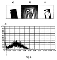
構造画像Sの内部では、低情報コンテンツを有する領域におけるピクセル値(構造振幅)は、解剖学的構造の領域におけるピクセル値と比較すると小さいのであり、その理由は、低情報コンテンツを有する領域は、非常に少数の小さなサイズの詳細を有するだけであるのが典型的であるからである。グローバルな勾配又は皮膚のラインの結果として生じる大きなサイズの変調は、ローカルな空間構造強化フィルタリングによって除去される。これが、図3において図解されていた問題を克服する。
FS=Bn*(MAXn−MINn)
ある例示的実施形態では、n=5が選択されたが、例えばn=3又はそれより大きな値など、nに対する他の値も考えられる。構造画像Sは、特定の範囲の画像値を有する。
1.小さな構造振幅(典型的には、0.005のオーダーより小さい)を有するピクセルを含む低情報コンテンツクラス(RLI)。
2.典型的には約0.02のオーダーの構造振幅を有する解剖学的構造のピクセルを含む解剖学的構造のクラス。
3.コリメーション境界又は皮膚のラインで生じる強いエッジのピクセルを含む(強い)エッジのクラス。典型的には、構造振幅ヒストグラムにおいてロングテール(図5を参照)が存在しており、0.05から0.1のオーダーの範囲を有する。
sr=f(s)
ここで、fは、単調減少する1階導関数を有する、単調増加関数である。いくつかの関数が、これらの要件を満たす。例としては、対数関数と、指数がゼロと1との間にあるベキ関数がある。ある例示的実施形態では、C>0及び0<β<1であるC及びβを用いて、次の数式によるベキ関数を用いる。
f(s)=Csβ
Pi(sr│i)=G(sr;mi,σi)
ただし、ここで、G(sr;mi,σi)は、次の数式で与えられる。
Pe’(Θr)=0
これが、最小の分離誤差のための、必要条件である。微積分の基本定理を用いると、これから、次の数式が得られる。
π1G(Θr;m1,σ1)=π2G(Θr;m2,σ2)
これは、分離誤差を最小化する閾値は2つのガウス分布の交点であることを意味している。上述の方程式からは、次に示される、再マッピングされた分離閾値のための2次方程式が得られる。
Θ=f−1(Θr)
WM=1−SM
ある範囲の画像値を含む構造画像を前記入力画像から取得するために、前記入力画像をフィルタリングするように構成されたフィルタ(FIL)と、
前記構造画像のための画像ヒストグラムに基づいて、前記範囲内で、関心領域と関連付けられている画像値の部分範囲を識別するように構成された範囲識別器(RID)と、
i)前記画像値の部分範囲のための仕様、ii)部分範囲と関連付けられており、関心領域を示すように構成されたマスク画像、及び/又はiii)前記部分範囲の補集合と関連付けられており、前記関心領域の補集合を示すように構成された相補的マスク画像を出力するための出力インターフェース(OUT)と
を備える画像処理システム(IPS)。
ある範囲の画像値を異なる画像位置において含む構造画像を前記入力画像から取得するため、前記入力画像をフィルタリングするステップ(S620)と、
前記構造画像のための画像ヒストグラムに基づいて、前記範囲内で関心領域と関連付けられている画像値の部分範囲を識別するステップ(S630)と、
i)前記画像値の部分範囲のための仕様、ii)部分範囲と関連付けられており、関心領域を示すように構成されたマスク画像、及び/又はiii)前記部分範囲の補集合と関連付けられており、前記関心領域の補集合を示すように構成された相補的なマスク画像を出力するステップ(S640)と
を有する、画像処理方法。
Claims (14)
- 入力画像を受け取る入力インターフェースと、
ある範囲の画像値を含む構造画像を前記入力画像から取得するために、前記入力画像をフィルタリングするフィルタと、
前記構造画像のための画像ヒストグラムに基づいて、前記範囲内で、関心領域と関連付けられている画像値の部分範囲を識別する範囲識別器と、
前記部分範囲と関連付けられ、前記関心領域を示すマスク画像、及び/又は前記部分範囲の補集合と関連付けられ、前記関心領域の補集合を示す相補的マスク画像を出力するための出力インターフェースと、
を備える、画像処理システムにおいて、
前記部分範囲の外部にある画像値のためのそれぞれの重みを計算する画像値範囲評価器であって、前記重みは、前記入力画像の視覚化におけるそれぞれの画像値の寄与を決定し、前記重みは、1つのクラスが前記関心領域に対応し、少なくとも他の1つのクラスが背景に又は少なくとも1つの放射不透過性の対象に対応する少なくとも2つのクラスの間の分離度を数値化したものである、画像値範囲評価器を備えることを特徴とする、画像処理システム。 - 前記画像処理システムは、前記構造画像における画像値から前記画像ヒストグラムを形成することによって、前記画像ヒストグラムを形成するヒストグラム形成器を備えるか、又は、ヒストグラム変換器を更に備え、前記ヒストグラム形成器が、i)前記構造画像における画像値のための中間画像ヒストグラムを形成し、前記ヒストグラム変換器が、前記中間画像ヒストグラムを前記画像ヒストグラムに変換するか、又は、ii)前記入力画像を中間画像に変換し、前記中間画像から前記画像ヒストグラムを形成する、請求項1に記載の画像処理システム。
- 前記ヒストグラム変換器は、前記中間画像ヒストグラムを変換するときに、面積を保存する補間を適用する、請求項2に記載の画像処理システム。
- 前記関心領域のためのマスクに基づいて、表示ユニット上に前記入力画像の可視化をレンダリングする画像レンダラを更に備える、請求項1から3のいずれか一項に記載の画像処理システム。
- コントラスト及び/又は輝度の適応のための前記関心領域の内部における前記画像値の寄与が前記重みに従いながら、画像レンダラが、表示ユニット上に前記入力画像の可視化をレンダリングする、請求項1に記載の画像処理システム。
- 画像レンダラが、前記画像値範囲評価器によって計算された前記重みを表す可視化方式を用いて、前記関心領域の補集合のためのマスクの可視化を表示ユニット上にレンダリングする、請求項1から5のいずれか一項に記載の画像処理システム。
- 前記範囲識別器は、統計的混合モデルを、前記画像ヒストグラムに又は変換された前記画像ヒストグラムに適合させることによって、前記部分範囲を識別する、請求項1から6のいずれか一項に記載の画像処理システム。
- 前記統計的混合モデルは、前記少なくとも2つのクラスに対応する少なくとも2つのコンポーネントを含む、請求項7に記載の画像処理システム。
- 前記コンポーネントのうちの1つが、前記背景又は放射線不透過性の対象に対応する一方、前記少なくとも1つの他のコンポーネントが、関心の1つ又は複数の解剖学的構造を含む前記関心領域に対応する、請求項8に記載の画像処理システム。
- 前記統計的混合モデルは、少なくとも3つのコンポーネントを含み、少なくとも1つの更なるコンポーネントは、エッジ構造に対応する、請求項9に記載の画像処理システム。
- 入力画像を受け取るステップと、
ある範囲の画像値を異なる画像位置において含む構造画像を前記入力画像から取得するため、前記入力画像をフィルタリングするステップと、
前記構造画像のための画像ヒストグラムに基づいて、前記範囲内で関心領域と関連付けられている画像値の部分範囲を識別するステップと、
前記部分範囲と関連付けられ、前記関心領域を示すマスク画像、及び/又は前記部分範囲の補集合と関連付けられ、前記関心領域の補集合を示す相補的なマスク画像を出力するステップと、
を有する、画像処理方法において、
前記部分範囲の外部にある画像値のためのそれぞれの重みを計算するステップであって、前記重みは、前記入力画像の視覚化におけるそれぞれの画像値の寄与を決定し、前記重みは、1つのクラスが前記関心領域に対応し、少なくとも他の1つのクラスが背景に又は少なくとも1つの放射不透過性の対象に対応する少なくとも2つのクラスの間の分離度を数値化したものである、前記重みを計算するステップを有することを特徴とする、画像処理方法。 - 撮像装置と、請求項1から10のいずれか一項に記載の画像処理システムとを備える、撮像機器。
- 少なくとも1つの処理ユニットによって実行されると、請求項11に記載の画像処理方法を前記処理ユニットに実行させる、コンピュータプログラム。
- 請求項13に記載のコンピュータプログラムを記憶した、コンピュータ可読媒体。
Applications Claiming Priority (3)
| Application Number | Priority Date | Filing Date | Title |
|---|---|---|---|
| EP17184418.6 | 2017-08-02 | ||
| EP17184418.6A EP3438928A1 (en) | 2017-08-02 | 2017-08-02 | Detection of regions with low information content in digital x-ray images |
| PCT/EP2018/069959 WO2019025225A1 (en) | 2017-08-02 | 2018-07-24 | DETECTION OF LOW-CONTENT INFORMATION REGIONS IN DIGITAL RADIOLOGICAL IMAGES |
Publications (3)
| Publication Number | Publication Date |
|---|---|
| JP2020529253A JP2020529253A (ja) | 2020-10-08 |
| JP2020529253A5 JP2020529253A5 (ja) | 2021-01-07 |
| JP6878679B2 true JP6878679B2 (ja) | 2021-06-02 |
Family
ID=59686734
Family Applications (1)
| Application Number | Title | Priority Date | Filing Date |
|---|---|---|---|
| JP2020505277A Active JP6878679B2 (ja) | 2017-08-02 | 2018-07-24 | デジタルx線画像における低情報コンテンツを有する領域の検出 |
Country Status (5)
| Country | Link |
|---|---|
| US (1) | US11354806B2 (ja) |
| EP (2) | EP3438928A1 (ja) |
| JP (1) | JP6878679B2 (ja) |
| CN (1) | CN110506294B (ja) |
| WO (1) | WO2019025225A1 (ja) |
Families Citing this family (2)
| Publication number | Priority date | Publication date | Assignee | Title |
|---|---|---|---|---|
| CN117495750A (zh) * | 2022-07-22 | 2024-02-02 | 戴尔产品有限公司 | 用于视频重构的方法、电子设备和计算机程序产品 |
| CN117689961B (zh) * | 2024-02-02 | 2024-05-07 | 深圳大学 | 视觉识别模型训练、视觉识别方法、系统、终端及介质 |
Family Cites Families (26)
| Publication number | Priority date | Publication date | Assignee | Title |
|---|---|---|---|---|
| JPH0642882B2 (ja) * | 1987-04-20 | 1994-06-08 | 富士写真フイルム株式会社 | 所望画像信号範囲決定方法 |
| US5262945A (en) * | 1991-08-09 | 1993-11-16 | The United States Of America As Represented By The Department Of Health And Human Services | Method for quantification of brain volume from magnetic resonance images |
| US5268967A (en) | 1992-06-29 | 1993-12-07 | Eastman Kodak Company | Method for automatic foreground and background detection in digital radiographic images |
| US5606587A (en) | 1996-03-21 | 1997-02-25 | Eastman Kodak Company | Determination of direct x-ray exposure regions in digital medical imaging |
| JP3096732B2 (ja) * | 1997-12-25 | 2000-10-10 | 工業技術院長 | 画像処理方法、画像処理プログラムを記録したコンピュータ読み取り可能な記録媒体及び画像処理装置 |
| US6112112A (en) | 1998-09-18 | 2000-08-29 | Arch Development Corporation | Method and system for the assessment of tumor extent in magnetic resonance images |
| JP2001160903A (ja) * | 1999-12-02 | 2001-06-12 | Nippon Telegr & Teleph Corp <Ntt> | 画像補正方法及び装置及びその方法を実行するプログラムを記録した記録媒体 |
| JP2001266142A (ja) | 2000-01-13 | 2001-09-28 | Nikon Corp | データ分類方法及びデータ分類装置、信号処理方法及び信号処理装置、位置検出方法及び位置検出装置、画像処理方法及び画像処理装置、露光方法及び露光装置、並びにデバイス製造方法 |
| US7218763B2 (en) * | 2003-02-27 | 2007-05-15 | Eastman Kodak Company | Method for automated window-level settings for magnetic resonance images |
| EP1624411A3 (en) | 2004-08-06 | 2006-08-09 | Gendex Corporation | Soft tissue filtering in medical images |
| US7840066B1 (en) | 2005-11-15 | 2010-11-23 | University Of Tennessee Research Foundation | Method of enhancing a digital image by gray-level grouping |
| SG139602A1 (en) * | 2006-08-08 | 2008-02-29 | St Microelectronics Asia | Automatic contrast enhancement |
| US8150110B2 (en) * | 2006-11-22 | 2012-04-03 | Carestream Health, Inc. | ROI-based rendering for diagnostic image consistency |
| WO2009110850A1 (en) * | 2008-03-03 | 2009-09-11 | Agency For Science, Technology And Research | A method and system of segmenting ct scan data |
| US20100185459A1 (en) * | 2009-01-16 | 2010-07-22 | German Guillermo Vera | Systems and methods for x-ray image identification |
| US8958620B2 (en) | 2010-03-08 | 2015-02-17 | Koninklijke Philips N.V. | Region of interest definition in cardiac imaging |
| JP5555672B2 (ja) * | 2011-07-14 | 2014-07-23 | 東芝テック株式会社 | 画像処理装置 |
| TW201331796A (zh) * | 2012-01-20 | 2013-08-01 | Univ Nat Taipei Technology | 能根據環境光線變化來最佳化觸控亮點之多點觸控系統及其方法 |
| US9311740B2 (en) * | 2012-03-27 | 2016-04-12 | Carestream Health, Inc. | Method for enhancing reconstructed 3-D tomosynthesis volume image |
| EP3053139B1 (en) * | 2013-10-01 | 2020-10-21 | Ventana Medical Systems, Inc. | Line-based image registration and cross-image annotation devices, systems and methods |
| US10376230B2 (en) * | 2013-11-19 | 2019-08-13 | Icad, Inc. | Obtaining breast density measurements and classifications |
| CA2948226C (en) * | 2014-06-30 | 2023-09-05 | Ventana Medical Systems, Inc. | Detecting edges of a nucleus using image analysis |
| WO2016083248A1 (en) | 2014-11-24 | 2016-06-02 | Koninklijke Philips N.V. | Simulating dose increase by noise model based multi scale noise reduction |
| EP3334347B1 (en) | 2015-09-01 | 2019-01-09 | Koninklijke Philips N.V. | Apparatus for displaying medical image data of a body part |
| CN108369735B (zh) * | 2015-12-10 | 2022-05-13 | 凯杰有限公司 | 用于确定数字图像中多个对象的位置的方法 |
| JP6880733B2 (ja) | 2016-12-28 | 2021-06-02 | 住友電気工業株式会社 | 光モジュール |
-
2017
- 2017-08-02 EP EP17184418.6A patent/EP3438928A1/en not_active Withdrawn
-
2018
- 2018-07-24 JP JP2020505277A patent/JP6878679B2/ja active Active
- 2018-07-24 US US16/500,851 patent/US11354806B2/en active Active
- 2018-07-24 CN CN201880024837.6A patent/CN110506294B/zh active Active
- 2018-07-24 WO PCT/EP2018/069959 patent/WO2019025225A1/en unknown
- 2018-07-24 EP EP18740864.6A patent/EP3577630B1/en active Active
Also Published As
| Publication number | Publication date |
|---|---|
| EP3577630A1 (en) | 2019-12-11 |
| EP3438928A1 (en) | 2019-02-06 |
| US20200380686A1 (en) | 2020-12-03 |
| EP3577630B1 (en) | 2021-02-17 |
| JP2020529253A (ja) | 2020-10-08 |
| US11354806B2 (en) | 2022-06-07 |
| CN110506294A (zh) | 2019-11-26 |
| CN110506294B (zh) | 2021-01-29 |
| WO2019025225A1 (en) | 2019-02-07 |
Similar Documents
| Publication | Publication Date | Title |
|---|---|---|
| JP7194143B2 (ja) | 肝臓腫瘍例のレビューを容易にするシステムおよび方法 | |
| US10489907B2 (en) | Artifact identification and/or correction for medical imaging | |
| US11494957B2 (en) | Automated correction of metal affected voxel representations of x-ray data using deep learning techniques | |
| IL271743B1 (en) | Classification and 3D modeling of dental structures - 3D maxilla using deep learning methods | |
| CN112651885A (zh) | 用于减少图像记录噪声的方法和装置 | |
| US20200242744A1 (en) | Forecasting Images for Image Processing | |
| CA2778599C (en) | Bone imagery segmentation method and apparatus | |
| JP6878679B2 (ja) | デジタルx線画像における低情報コンテンツを有する領域の検出 | |
| US10062167B2 (en) | Estimated local rigid regions from dense deformation in subtraction | |
| EP4014878A1 (en) | Multi-phase filter | |
| CN114387380A (zh) | 用于生成3d医学图像数据的基于计算机的可视化的方法 | |
| CN117437144A (zh) | 用于图像去噪的方法和系统 | |
| WO2022212953A1 (en) | Systems and methods for multi-kernel synthesis and kernel conversion in medical imaging | |
| EP4094223A1 (en) | Weakly supervised lesion segmentation | |
| EP4336452A1 (en) | Computer-implemented method for processing spectral computed tomography (spectral ct) data, computer program and spectral ct system | |
| US20220284556A1 (en) | Confidence map for radiographic image optimization | |
| Hoye | Truth-based Radiomics for Prediction of Lung Cancer Prognosis | |
| Robins | Hybrid Reference Datasets for Quantitative Computed Tomography Characterization and Conformance | |
| Passand | Quality assessment of clinical thorax CT images | |
| Dreyer et al. | Digital imaging fundamentals |
Legal Events
| Date | Code | Title | Description |
|---|---|---|---|
| A521 | Request for written amendment filed |
Free format text: JAPANESE INTERMEDIATE CODE: A523 Effective date: 20201118 |
|
| A621 | Written request for application examination |
Free format text: JAPANESE INTERMEDIATE CODE: A621 Effective date: 20201118 |
|
| A871 | Explanation of circumstances concerning accelerated examination |
Free format text: JAPANESE INTERMEDIATE CODE: A871 Effective date: 20201118 |
|
| A975 | Report on accelerated examination |
Free format text: JAPANESE INTERMEDIATE CODE: A971005 Effective date: 20201124 |
|
| A131 | Notification of reasons for refusal |
Free format text: JAPANESE INTERMEDIATE CODE: A131 Effective date: 20201208 |
|
| A521 | Request for written amendment filed |
Free format text: JAPANESE INTERMEDIATE CODE: A523 Effective date: 20210217 |
|
| TRDD | Decision of grant or rejection written | ||
| A01 | Written decision to grant a patent or to grant a registration (utility model) |
Free format text: JAPANESE INTERMEDIATE CODE: A01 Effective date: 20210401 |
|
| A61 | First payment of annual fees (during grant procedure) |
Free format text: JAPANESE INTERMEDIATE CODE: A61 Effective date: 20210428 |
|
| R150 | Certificate of patent or registration of utility model |
Ref document number: 6878679 Country of ref document: JP Free format text: JAPANESE INTERMEDIATE CODE: R150 |
|
| R250 | Receipt of annual fees |
Free format text: JAPANESE INTERMEDIATE CODE: R250 |


