JP6841753B2 - Methods and compositions for increasing somatic cell nuclear transfer (SCNT) efficiency by removing histone H3-lysine trimethylation - Google Patents
Methods and compositions for increasing somatic cell nuclear transfer (SCNT) efficiency by removing histone H3-lysine trimethylation Download PDFInfo
- Publication number
- JP6841753B2 JP6841753B2 JP2017514421A JP2017514421A JP6841753B2 JP 6841753 B2 JP6841753 B2 JP 6841753B2 JP 2017514421 A JP2017514421 A JP 2017514421A JP 2017514421 A JP2017514421 A JP 2017514421A JP 6841753 B2 JP6841753 B2 JP 6841753B2
- Authority
- JP
- Japan
- Prior art keywords
- scnt
- cell
- human
- seq
- cells
- Prior art date
- Legal status (The legal status is an assumption and is not a legal conclusion. Google has not performed a legal analysis and makes no representation as to the accuracy of the status listed.)
- Active
Links
- 238000010374 somatic cell nuclear transfer Methods 0.000 title claims description 323
- 238000000034 method Methods 0.000 title claims description 207
- 239000000203 mixture Substances 0.000 title claims description 71
- 230000001965 increasing effect Effects 0.000 title claims description 56
- 108010033040 Histones Proteins 0.000 title description 16
- 239000004472 Lysine Substances 0.000 title description 5
- 210000004027 cell Anatomy 0.000 claims description 326
- 150000007523 nucleic acids Chemical class 0.000 claims description 173
- 210000000287 oocyte Anatomy 0.000 claims description 161
- 210000002257 embryonic structure Anatomy 0.000 claims description 146
- 102000039446 nucleic acids Human genes 0.000 claims description 126
- 108020004707 nucleic acids Proteins 0.000 claims description 126
- 241000282414 Homo sapiens Species 0.000 claims description 123
- 108090000765 processed proteins & peptides Proteins 0.000 claims description 123
- 239000012634 fragment Substances 0.000 claims description 108
- 102000004196 processed proteins & peptides Human genes 0.000 claims description 106
- 210000001161 mammalian embryo Anatomy 0.000 claims description 95
- 229920001184 polypeptide Polymers 0.000 claims description 95
- 102000008157 Histone Demethylases Human genes 0.000 claims description 74
- 108010074870 Histone Demethylases Proteins 0.000 claims description 74
- 210000002459 blastocyst Anatomy 0.000 claims description 44
- 108091028043 Nucleic acid sequence Proteins 0.000 claims description 41
- 210000004962 mammalian cell Anatomy 0.000 claims description 40
- 238000004519 manufacturing process Methods 0.000 claims description 35
- 238000002347 injection Methods 0.000 claims description 26
- 239000007924 injection Substances 0.000 claims description 26
- 238000011161 development Methods 0.000 claims description 25
- 230000004913 activation Effects 0.000 claims description 21
- 238000000338 in vitro Methods 0.000 claims description 20
- 230000007159 enucleation Effects 0.000 claims description 9
- 238000012258 culturing Methods 0.000 claims description 6
- 210000000472 morula Anatomy 0.000 claims description 6
- 230000003213 activating effect Effects 0.000 claims 1
- 210000003101 oviduct Anatomy 0.000 claims 1
- 108090000623 proteins and genes Proteins 0.000 description 191
- 239000003795 chemical substances by application Substances 0.000 description 163
- 108020004459 Small interfering RNA Proteins 0.000 description 129
- 239000003112 inhibitor Substances 0.000 description 119
- 108091032973 (ribonucleotides)n+m Proteins 0.000 description 111
- 239000004055 small Interfering RNA Substances 0.000 description 111
- 230000014509 gene expression Effects 0.000 description 106
- 102000004169 proteins and genes Human genes 0.000 description 102
- 210000001082 somatic cell Anatomy 0.000 description 101
- 238000012228 RNA interference-mediated gene silencing Methods 0.000 description 97
- 230000009368 gene silencing by RNA Effects 0.000 description 97
- 125000003729 nucleotide group Chemical group 0.000 description 88
- 101150032905 Kdm4d gene Proteins 0.000 description 85
- 230000004048 modification Effects 0.000 description 85
- 238000012986 modification Methods 0.000 description 85
- 230000008672 reprogramming Effects 0.000 description 81
- 239000002773 nucleotide Substances 0.000 description 77
- 210000004940 nucleus Anatomy 0.000 description 76
- 235000018102 proteins Nutrition 0.000 description 71
- 108091034117 Oligonucleotide Proteins 0.000 description 68
- 150000001413 amino acids Chemical class 0.000 description 68
- 108060004795 Methyltransferase Proteins 0.000 description 62
- 102000016397 Methyltransferase Human genes 0.000 description 52
- -1 but not limited to Chemical class 0.000 description 49
- 108020004999 messenger RNA Proteins 0.000 description 49
- 239000012190 activator Substances 0.000 description 45
- 239000003697 methyltransferase inhibitor Substances 0.000 description 42
- 101000687346 Homo sapiens PR domain zinc finger protein 2 Proteins 0.000 description 36
- 238000012546 transfer Methods 0.000 description 36
- 230000000694 effects Effects 0.000 description 35
- JLCPHMBAVCMARE-UHFFFAOYSA-N [3-[[3-[[3-[[3-[[3-[[3-[[3-[[3-[[3-[[3-[[3-[[5-(2-amino-6-oxo-1H-purin-9-yl)-3-[[3-[[3-[[3-[[3-[[3-[[5-(2-amino-6-oxo-1H-purin-9-yl)-3-[[5-(2-amino-6-oxo-1H-purin-9-yl)-3-hydroxyoxolan-2-yl]methoxy-hydroxyphosphoryl]oxyoxolan-2-yl]methoxy-hydroxyphosphoryl]oxy-5-(5-methyl-2,4-dioxopyrimidin-1-yl)oxolan-2-yl]methoxy-hydroxyphosphoryl]oxy-5-(6-aminopurin-9-yl)oxolan-2-yl]methoxy-hydroxyphosphoryl]oxy-5-(6-aminopurin-9-yl)oxolan-2-yl]methoxy-hydroxyphosphoryl]oxy-5-(6-aminopurin-9-yl)oxolan-2-yl]methoxy-hydroxyphosphoryl]oxy-5-(6-aminopurin-9-yl)oxolan-2-yl]methoxy-hydroxyphosphoryl]oxyoxolan-2-yl]methoxy-hydroxyphosphoryl]oxy-5-(5-methyl-2,4-dioxopyrimidin-1-yl)oxolan-2-yl]methoxy-hydroxyphosphoryl]oxy-5-(4-amino-2-oxopyrimidin-1-yl)oxolan-2-yl]methoxy-hydroxyphosphoryl]oxy-5-(5-methyl-2,4-dioxopyrimidin-1-yl)oxolan-2-yl]methoxy-hydroxyphosphoryl]oxy-5-(5-methyl-2,4-dioxopyrimidin-1-yl)oxolan-2-yl]methoxy-hydroxyphosphoryl]oxy-5-(6-aminopurin-9-yl)oxolan-2-yl]methoxy-hydroxyphosphoryl]oxy-5-(6-aminopurin-9-yl)oxolan-2-yl]methoxy-hydroxyphosphoryl]oxy-5-(4-amino-2-oxopyrimidin-1-yl)oxolan-2-yl]methoxy-hydroxyphosphoryl]oxy-5-(4-amino-2-oxopyrimidin-1-yl)oxolan-2-yl]methoxy-hydroxyphosphoryl]oxy-5-(4-amino-2-oxopyrimidin-1-yl)oxolan-2-yl]methoxy-hydroxyphosphoryl]oxy-5-(6-aminopurin-9-yl)oxolan-2-yl]methoxy-hydroxyphosphoryl]oxy-5-(4-amino-2-oxopyrimidin-1-yl)oxolan-2-yl]methyl [5-(6-aminopurin-9-yl)-2-(hydroxymethyl)oxolan-3-yl] hydrogen phosphate Polymers Cc1cn(C2CC(OP(O)(=O)OCC3OC(CC3OP(O)(=O)OCC3OC(CC3O)n3cnc4c3nc(N)[nH]c4=O)n3cnc4c3nc(N)[nH]c4=O)C(COP(O)(=O)OC3CC(OC3COP(O)(=O)OC3CC(OC3COP(O)(=O)OC3CC(OC3COP(O)(=O)OC3CC(OC3COP(O)(=O)OC3CC(OC3COP(O)(=O)OC3CC(OC3COP(O)(=O)OC3CC(OC3COP(O)(=O)OC3CC(OC3COP(O)(=O)OC3CC(OC3COP(O)(=O)OC3CC(OC3COP(O)(=O)OC3CC(OC3COP(O)(=O)OC3CC(OC3COP(O)(=O)OC3CC(OC3COP(O)(=O)OC3CC(OC3COP(O)(=O)OC3CC(OC3COP(O)(=O)OC3CC(OC3COP(O)(=O)OC3CC(OC3CO)n3cnc4c(N)ncnc34)n3ccc(N)nc3=O)n3cnc4c(N)ncnc34)n3ccc(N)nc3=O)n3ccc(N)nc3=O)n3ccc(N)nc3=O)n3cnc4c(N)ncnc34)n3cnc4c(N)ncnc34)n3cc(C)c(=O)[nH]c3=O)n3cc(C)c(=O)[nH]c3=O)n3ccc(N)nc3=O)n3cc(C)c(=O)[nH]c3=O)n3cnc4c3nc(N)[nH]c4=O)n3cnc4c(N)ncnc34)n3cnc4c(N)ncnc34)n3cnc4c(N)ncnc34)n3cnc4c(N)ncnc34)O2)c(=O)[nH]c1=O JLCPHMBAVCMARE-UHFFFAOYSA-N 0.000 description 34
- UYTPUPDQBNUYGX-UHFFFAOYSA-N guanine Chemical compound O=C1NC(N)=NC2=C1N=CN2 UYTPUPDQBNUYGX-UHFFFAOYSA-N 0.000 description 34
- 238000006467 substitution reaction Methods 0.000 description 33
- 102000040650 (ribonucleotides)n+m Human genes 0.000 description 32
- 102100024885 PR domain zinc finger protein 2 Human genes 0.000 description 32
- 241000699666 Mus <mouse, genus> Species 0.000 description 31
- 108091027967 Small hairpin RNA Proteins 0.000 description 31
- 230000030279 gene silencing Effects 0.000 description 31
- 101001088895 Homo sapiens Lysine-specific demethylase 4D Proteins 0.000 description 30
- 102000011787 Histone Methyltransferases Human genes 0.000 description 29
- 108010036115 Histone Methyltransferases Proteins 0.000 description 29
- 229940123379 Methyltransferase inhibitor Drugs 0.000 description 29
- 239000000126 substance Substances 0.000 description 29
- 210000000130 stem cell Anatomy 0.000 description 27
- 101000613625 Homo sapiens Lysine-specific demethylase 4A Proteins 0.000 description 26
- 241001465754 Metazoa Species 0.000 description 26
- 230000027455 binding Effects 0.000 description 26
- OPTASPLRGRRNAP-UHFFFAOYSA-N cytosine Chemical compound NC=1C=CNC(=O)N=1 OPTASPLRGRRNAP-UHFFFAOYSA-N 0.000 description 26
- 235000000346 sugar Nutrition 0.000 description 26
- 235000013601 eggs Nutrition 0.000 description 25
- 238000012226 gene silencing method Methods 0.000 description 25
- 238000002513 implantation Methods 0.000 description 25
- 125000002496 methyl group Chemical group [H]C([H])([H])* 0.000 description 25
- 238000007069 methylation reaction Methods 0.000 description 25
- 230000018109 developmental process Effects 0.000 description 24
- 230000002401 inhibitory effect Effects 0.000 description 24
- 230000011987 methylation Effects 0.000 description 24
- 150000003384 small molecules Chemical class 0.000 description 24
- 101000613629 Homo sapiens Lysine-specific demethylase 4B Proteins 0.000 description 23
- 125000000217 alkyl group Chemical group 0.000 description 23
- 239000000427 antigen Substances 0.000 description 23
- 108091007433 antigens Proteins 0.000 description 23
- 102000036639 antigens Human genes 0.000 description 23
- 125000002467 phosphate group Chemical group [H]OP(=O)(O[H])O[*] 0.000 description 23
- 101001088893 Homo sapiens Lysine-specific demethylase 4C Proteins 0.000 description 22
- 102100033231 Lysine-specific demethylase 4D Human genes 0.000 description 22
- ISAKRJDGNUQOIC-UHFFFAOYSA-N Uracil Chemical compound O=C1C=CNC(=O)N1 ISAKRJDGNUQOIC-UHFFFAOYSA-N 0.000 description 22
- 230000004888 barrier function Effects 0.000 description 22
- 241000894007 species Species 0.000 description 22
- 241000283690 Bos taurus Species 0.000 description 21
- 210000000805 cytoplasm Anatomy 0.000 description 21
- 230000002452 interceptive effect Effects 0.000 description 21
- 125000000446 sulfanediyl group Chemical group *S* 0.000 description 19
- 102100040860 Lysine-specific demethylase 4B Human genes 0.000 description 18
- 102100033230 Lysine-specific demethylase 4C Human genes 0.000 description 18
- 239000000556 agonist Substances 0.000 description 18
- 210000001671 embryonic stem cell Anatomy 0.000 description 18
- 230000005764 inhibitory process Effects 0.000 description 18
- 102100040863 Lysine-specific demethylase 4A Human genes 0.000 description 17
- 108091093037 Peptide nucleic acid Proteins 0.000 description 17
- 102000055027 Protein Methyltransferases Human genes 0.000 description 17
- 108700040121 Protein Methyltransferases Proteins 0.000 description 17
- 210000001109 blastomere Anatomy 0.000 description 17
- 239000002777 nucleoside Chemical class 0.000 description 17
- 239000013598 vector Substances 0.000 description 17
- 101000696705 Homo sapiens Histone-lysine N-methyltransferase SUV39H1 Proteins 0.000 description 16
- 230000015572 biosynthetic process Effects 0.000 description 16
- 230000000295 complement effect Effects 0.000 description 16
- 238000003786 synthesis reaction Methods 0.000 description 16
- 230000004927 fusion Effects 0.000 description 15
- 229910052760 oxygen Inorganic materials 0.000 description 15
- 230000008569 process Effects 0.000 description 15
- 230000008685 targeting Effects 0.000 description 15
- 101000696699 Homo sapiens Histone-lysine N-methyltransferase SUV39H2 Proteins 0.000 description 14
- 241000700159 Rattus Species 0.000 description 14
- 238000010367 cloning Methods 0.000 description 14
- 239000001301 oxygen Substances 0.000 description 14
- 238000002360 preparation method Methods 0.000 description 14
- 101710163270 Nuclease Proteins 0.000 description 13
- 229910019142 PO4 Inorganic materials 0.000 description 13
- 229940104302 cytosine Drugs 0.000 description 13
- 230000001973 epigenetic effect Effects 0.000 description 13
- 125000005647 linker group Chemical group 0.000 description 13
- 239000010452 phosphate Substances 0.000 description 13
- 229930024421 Adenine Natural products 0.000 description 12
- GFFGJBXGBJISGV-UHFFFAOYSA-N Adenine Chemical compound NC1=NC=NC2=C1N=CN2 GFFGJBXGBJISGV-UHFFFAOYSA-N 0.000 description 12
- 108020004414 DNA Proteins 0.000 description 12
- 229960000643 adenine Drugs 0.000 description 12
- QVGXLLKOCUKJST-UHFFFAOYSA-N atomic oxygen Chemical compound [O] QVGXLLKOCUKJST-UHFFFAOYSA-N 0.000 description 12
- 230000015556 catabolic process Effects 0.000 description 12
- 150000001875 compounds Chemical class 0.000 description 12
- 238000006731 degradation reaction Methods 0.000 description 12
- 125000002887 hydroxy group Chemical group [H]O* 0.000 description 12
- 230000036961 partial effect Effects 0.000 description 12
- 229940035893 uracil Drugs 0.000 description 12
- 230000000692 anti-sense effect Effects 0.000 description 11
- 125000000623 heterocyclic group Chemical group 0.000 description 11
- 230000007246 mechanism Effects 0.000 description 11
- 150000003833 nucleoside derivatives Chemical class 0.000 description 11
- 230000009467 reduction Effects 0.000 description 11
- 108091023037 Aptamer Proteins 0.000 description 10
- 238000001353 Chip-sequencing Methods 0.000 description 10
- 125000003118 aryl group Chemical group 0.000 description 10
- 230000006870 function Effects 0.000 description 10
- 102000054786 human SUV39H1 Human genes 0.000 description 10
- 102000050832 human SUV39H2 Human genes 0.000 description 10
- 230000001404 mediated effect Effects 0.000 description 10
- 108010021625 Immunoglobulin Fragments Proteins 0.000 description 9
- 102000008394 Immunoglobulin Fragments Human genes 0.000 description 9
- 241000124008 Mammalia Species 0.000 description 9
- 101100422861 Mus musculus Suv39h1 gene Proteins 0.000 description 9
- 101100422868 Mus musculus Suv39h2 gene Proteins 0.000 description 9
- 241000283973 Oryctolagus cuniculus Species 0.000 description 9
- 108700019146 Transgenes Proteins 0.000 description 9
- 150000001412 amines Chemical class 0.000 description 9
- 125000002091 cationic group Chemical group 0.000 description 9
- 210000000349 chromosome Anatomy 0.000 description 9
- 102000045775 human KDM4A Human genes 0.000 description 9
- 102000049881 human KDM4D Human genes 0.000 description 9
- 150000003212 purines Chemical class 0.000 description 9
- 108091033409 CRISPR Proteins 0.000 description 8
- 108090000994 Catalytic RNA Proteins 0.000 description 8
- 102000053642 Catalytic RNA Human genes 0.000 description 8
- 102100028998 Histone-lysine N-methyltransferase SUV39H1 Human genes 0.000 description 8
- 108020005196 Mitochondrial DNA Proteins 0.000 description 8
- PYMYPHUHKUWMLA-LMVFSUKVSA-N Ribose Natural products OC[C@@H](O)[C@@H](O)[C@@H](O)C=O PYMYPHUHKUWMLA-LMVFSUKVSA-N 0.000 description 8
- 125000003282 alkyl amino group Chemical group 0.000 description 8
- HMFHBZSHGGEWLO-UHFFFAOYSA-N alpha-D-Furanose-Ribose Natural products OCC1OC(O)C(O)C1O HMFHBZSHGGEWLO-UHFFFAOYSA-N 0.000 description 8
- 125000001769 aryl amino group Chemical group 0.000 description 8
- 239000002299 complementary DNA Substances 0.000 description 8
- 125000004663 dialkyl amino group Chemical group 0.000 description 8
- 125000005240 diheteroarylamino group Chemical group 0.000 description 8
- 238000003197 gene knockdown Methods 0.000 description 8
- 230000002068 genetic effect Effects 0.000 description 8
- 210000004602 germ cell Anatomy 0.000 description 8
- 125000005241 heteroarylamino group Chemical group 0.000 description 8
- 239000002609 medium Substances 0.000 description 8
- 229910052698 phosphorus Inorganic materials 0.000 description 8
- 239000000047 product Substances 0.000 description 8
- 108091092562 ribozyme Proteins 0.000 description 8
- 229910052717 sulfur Inorganic materials 0.000 description 8
- HMFHBZSHGGEWLO-SOOFDHNKSA-N D-ribofuranose Chemical compound OC[C@H]1OC(O)[C@H](O)[C@@H]1O HMFHBZSHGGEWLO-SOOFDHNKSA-N 0.000 description 7
- 108010053770 Deoxyribonucleases Proteins 0.000 description 7
- 102000016911 Deoxyribonucleases Human genes 0.000 description 7
- 102000006947 Histones Human genes 0.000 description 7
- 241000699670 Mus sp. Species 0.000 description 7
- 238000004458 analytical method Methods 0.000 description 7
- 125000004429 atom Chemical group 0.000 description 7
- 230000008901 benefit Effects 0.000 description 7
- 229910052799 carbon Inorganic materials 0.000 description 7
- 230000017858 demethylation Effects 0.000 description 7
- 238000010520 demethylation reaction Methods 0.000 description 7
- 238000013461 design Methods 0.000 description 7
- 125000004986 diarylamino group Chemical group 0.000 description 7
- 201000010099 disease Diseases 0.000 description 7
- 208000037265 diseases, disorders, signs and symptoms Diseases 0.000 description 7
- 210000001654 germ layer Anatomy 0.000 description 7
- 239000003446 ligand Substances 0.000 description 7
- 239000003550 marker Substances 0.000 description 7
- 238000010373 organism cloning Methods 0.000 description 7
- 230000037361 pathway Effects 0.000 description 7
- 241000282412 Homo Species 0.000 description 6
- 239000002202 Polyethylene glycol Substances 0.000 description 6
- RWRDLPDLKQPQOW-UHFFFAOYSA-N Pyrrolidine Chemical compound C1CCNC1 RWRDLPDLKQPQOW-UHFFFAOYSA-N 0.000 description 6
- IQFYYKKMVGJFEH-XLPZGREQSA-N Thymidine Chemical compound O=C1NC(=O)C(C)=CN1[C@@H]1O[C@H](CO)[C@@H](O)C1 IQFYYKKMVGJFEH-XLPZGREQSA-N 0.000 description 6
- 239000000074 antisense oligonucleotide Substances 0.000 description 6
- 238000012230 antisense oligonucleotides Methods 0.000 description 6
- 210000003855 cell nucleus Anatomy 0.000 description 6
- 230000003247 decreasing effect Effects 0.000 description 6
- 230000013020 embryo development Effects 0.000 description 6
- 125000005843 halogen group Chemical group 0.000 description 6
- 229910052739 hydrogen Inorganic materials 0.000 description 6
- FDGQSTZJBFJUBT-UHFFFAOYSA-N hypoxanthine Chemical compound O=C1NC=NC2=C1NC=N2 FDGQSTZJBFJUBT-UHFFFAOYSA-N 0.000 description 6
- 238000001727 in vivo Methods 0.000 description 6
- 150000002632 lipids Chemical class 0.000 description 6
- 125000003835 nucleoside group Chemical class 0.000 description 6
- 125000004437 phosphorous atom Chemical group 0.000 description 6
- 229920001223 polyethylene glycol Polymers 0.000 description 6
- 229920000642 polymer Polymers 0.000 description 6
- 230000000392 somatic effect Effects 0.000 description 6
- 150000008163 sugars Chemical class 0.000 description 6
- 230000001225 therapeutic effect Effects 0.000 description 6
- 210000001519 tissue Anatomy 0.000 description 6
- 238000013518 transcription Methods 0.000 description 6
- 230000035897 transcription Effects 0.000 description 6
- 229940045145 uridine Drugs 0.000 description 6
- GVUOPSNMFBICMM-UHFFFAOYSA-N 5-bromo-6-morpholin-4-yl-1h-pyrimidine-2,4-dione Chemical compound OC1=NC(O)=C(Br)C(N2CCOCC2)=N1 GVUOPSNMFBICMM-UHFFFAOYSA-N 0.000 description 5
- FHYZUCKIFBIOMX-UHFFFAOYSA-N 5-bromo-6-piperidin-1-yl-1h-pyrimidine-2,4-dione Chemical compound OC1=NC(O)=C(Br)C(N2CCCCC2)=N1 FHYZUCKIFBIOMX-UHFFFAOYSA-N 0.000 description 5
- 101100422859 Bos taurus SUV39H1 gene Proteins 0.000 description 5
- 101100422864 Bos taurus SUV39H2 gene Proteins 0.000 description 5
- 108010077544 Chromatin Proteins 0.000 description 5
- 101000615488 Homo sapiens Methyl-CpG-binding domain protein 2 Proteins 0.000 description 5
- 102100021299 Methyl-CpG-binding domain protein 2 Human genes 0.000 description 5
- 108091093105 Nuclear DNA Proteins 0.000 description 5
- 102000000574 RNA-Induced Silencing Complex Human genes 0.000 description 5
- 108010016790 RNA-Induced Silencing Complex Proteins 0.000 description 5
- NINIDFKCEFEMDL-UHFFFAOYSA-N Sulfur Chemical compound [S] NINIDFKCEFEMDL-UHFFFAOYSA-N 0.000 description 5
- DRTQHJPVMGBUCF-XVFCMESISA-N Uridine Chemical compound O[C@@H]1[C@H](O)[C@@H](CO)O[C@H]1N1C(=O)NC(=O)C=C1 DRTQHJPVMGBUCF-XVFCMESISA-N 0.000 description 5
- 241000700605 Viruses Species 0.000 description 5
- DHKHKXVYLBGOIT-UHFFFAOYSA-N acetaldehyde Diethyl Acetal Natural products CCOC(C)OCC DHKHKXVYLBGOIT-UHFFFAOYSA-N 0.000 description 5
- 125000000304 alkynyl group Chemical group 0.000 description 5
- 238000013459 approach Methods 0.000 description 5
- 230000001413 cellular effect Effects 0.000 description 5
- 210000003483 chromatin Anatomy 0.000 description 5
- 238000003776 cleavage reaction Methods 0.000 description 5
- 230000001086 cytosolic effect Effects 0.000 description 5
- 238000010586 diagram Methods 0.000 description 5
- 230000004069 differentiation Effects 0.000 description 5
- 239000000539 dimer Substances 0.000 description 5
- 125000004119 disulfanediyl group Chemical group *SS* 0.000 description 5
- 230000001605 fetal effect Effects 0.000 description 5
- 108020001507 fusion proteins Proteins 0.000 description 5
- 102000037865 fusion proteins Human genes 0.000 description 5
- 102000052630 human KDM4B Human genes 0.000 description 5
- 102000046705 human KDM4C Human genes 0.000 description 5
- 239000001257 hydrogen Substances 0.000 description 5
- 230000006872 improvement Effects 0.000 description 5
- 239000002679 microRNA Substances 0.000 description 5
- 230000003472 neutralizing effect Effects 0.000 description 5
- 125000002924 primary amino group Chemical group [H]N([H])* 0.000 description 5
- 238000012545 processing Methods 0.000 description 5
- 239000002213 purine nucleotide Substances 0.000 description 5
- 150000003230 pyrimidines Chemical class 0.000 description 5
- 108700024797 rat Suv39h1 Proteins 0.000 description 5
- 230000007017 scission Effects 0.000 description 5
- 239000011593 sulfur Substances 0.000 description 5
- 238000011191 terminal modification Methods 0.000 description 5
- 230000014616 translation Effects 0.000 description 5
- 239000013603 viral vector Substances 0.000 description 5
- FWMNVWWHGCHHJJ-SKKKGAJSSA-N 4-amino-1-[(2r)-6-amino-2-[[(2r)-2-[[(2r)-2-[[(2r)-2-amino-3-phenylpropanoyl]amino]-3-phenylpropanoyl]amino]-4-methylpentanoyl]amino]hexanoyl]piperidine-4-carboxylic acid Chemical compound C([C@H](C(=O)N[C@H](CC(C)C)C(=O)N[C@H](CCCCN)C(=O)N1CCC(N)(CC1)C(O)=O)NC(=O)[C@H](N)CC=1C=CC=CC=1)C1=CC=CC=C1 FWMNVWWHGCHHJJ-SKKKGAJSSA-N 0.000 description 4
- LRFVTYWOQMYALW-UHFFFAOYSA-N 9H-xanthine Chemical compound O=C1NC(=O)NC2=C1NC=N2 LRFVTYWOQMYALW-UHFFFAOYSA-N 0.000 description 4
- 238000010354 CRISPR gene editing Methods 0.000 description 4
- 241000282472 Canis lupus familiaris Species 0.000 description 4
- OKTJSMMVPCPJKN-UHFFFAOYSA-N Carbon Chemical group [C] OKTJSMMVPCPJKN-UHFFFAOYSA-N 0.000 description 4
- 108010047041 Complementarity Determining Regions Proteins 0.000 description 4
- 241000283086 Equidae Species 0.000 description 4
- PIICEJLVQHRZGT-UHFFFAOYSA-N Ethylenediamine Chemical compound NCCN PIICEJLVQHRZGT-UHFFFAOYSA-N 0.000 description 4
- 241000282326 Felis catus Species 0.000 description 4
- 108010043121 Green Fluorescent Proteins Proteins 0.000 description 4
- 102000004144 Green Fluorescent Proteins Human genes 0.000 description 4
- 102100028988 Histone-lysine N-methyltransferase SUV39H2 Human genes 0.000 description 4
- 108010021466 Mutant Proteins Proteins 0.000 description 4
- 102000008300 Mutant Proteins Human genes 0.000 description 4
- 206010028980 Neoplasm Diseases 0.000 description 4
- 241000282577 Pan troglodytes Species 0.000 description 4
- 238000003559 RNA-seq method Methods 0.000 description 4
- 241000282887 Suidae Species 0.000 description 4
- 238000007792 addition Methods 0.000 description 4
- OIRDTQYFTABQOQ-KQYNXXCUSA-N adenosine Chemical compound C1=NC=2C(N)=NC=NC=2N1[C@@H]1O[C@H](CO)[C@@H](O)[C@H]1O OIRDTQYFTABQOQ-KQYNXXCUSA-N 0.000 description 4
- 210000004102 animal cell Anatomy 0.000 description 4
- 210000004952 blastocoel Anatomy 0.000 description 4
- 230000000903 blocking effect Effects 0.000 description 4
- 230000003197 catalytic effect Effects 0.000 description 4
- 210000000170 cell membrane Anatomy 0.000 description 4
- 239000003153 chemical reaction reagent Substances 0.000 description 4
- HVYWMOMLDIMFJA-DPAQBDIFSA-N cholesterol Chemical compound C1C=C2C[C@@H](O)CC[C@]2(C)[C@@H]2[C@@H]1[C@@H]1CC[C@H]([C@H](C)CCCC(C)C)[C@@]1(C)CC2 HVYWMOMLDIMFJA-DPAQBDIFSA-N 0.000 description 4
- 230000007423 decrease Effects 0.000 description 4
- 230000007812 deficiency Effects 0.000 description 4
- 239000003814 drug Substances 0.000 description 4
- 239000000975 dye Substances 0.000 description 4
- 210000001900 endoderm Anatomy 0.000 description 4
- 210000002304 esc Anatomy 0.000 description 4
- 230000035558 fertility Effects 0.000 description 4
- 230000004720 fertilization Effects 0.000 description 4
- 239000005090 green fluorescent protein Substances 0.000 description 4
- 239000001963 growth medium Substances 0.000 description 4
- 210000005260 human cell Anatomy 0.000 description 4
- 230000001939 inductive effect Effects 0.000 description 4
- 230000003993 interaction Effects 0.000 description 4
- 238000000520 microinjection Methods 0.000 description 4
- 102000035118 modified proteins Human genes 0.000 description 4
- 108091005573 modified proteins Proteins 0.000 description 4
- 239000000178 monomer Substances 0.000 description 4
- 230000000877 morphologic effect Effects 0.000 description 4
- 229910052757 nitrogen Inorganic materials 0.000 description 4
- 210000000056 organ Anatomy 0.000 description 4
- 230000002018 overexpression Effects 0.000 description 4
- 210000001778 pluripotent stem cell Anatomy 0.000 description 4
- 230000035755 proliferation Effects 0.000 description 4
- 229940121649 protein inhibitor Drugs 0.000 description 4
- 239000012268 protein inhibitor Substances 0.000 description 4
- 239000002719 pyrimidine nucleotide Substances 0.000 description 4
- 230000002829 reductive effect Effects 0.000 description 4
- 108091069025 single-strand RNA Proteins 0.000 description 4
- 239000000243 solution Substances 0.000 description 4
- 230000000638 stimulation Effects 0.000 description 4
- 230000002194 synthesizing effect Effects 0.000 description 4
- 238000002560 therapeutic procedure Methods 0.000 description 4
- RYYWUUFWQRZTIU-UHFFFAOYSA-K thiophosphate Chemical compound [O-]P([O-])([O-])=S RYYWUUFWQRZTIU-UHFFFAOYSA-K 0.000 description 4
- ZEMGGZBWXRYJHK-UHFFFAOYSA-N thiouracil Chemical compound O=C1C=CNC(=S)N1 ZEMGGZBWXRYJHK-UHFFFAOYSA-N 0.000 description 4
- 238000001890 transfection Methods 0.000 description 4
- 238000013519 translation Methods 0.000 description 4
- RTKIYFITIVXBLE-QEQCGCAPSA-N trichostatin A Chemical compound ONC(=O)/C=C/C(/C)=C/[C@@H](C)C(=O)C1=CC=C(N(C)C)C=C1 RTKIYFITIVXBLE-QEQCGCAPSA-N 0.000 description 4
- QOECJCJVIMVJGX-UHFFFAOYSA-N 2-cyclohexyl-6-methoxy-N-(1-propan-2-yl-4-piperidinyl)-7-[3-(1-pyrrolidinyl)propoxy]-4-quinazolinamine Chemical compound N1=C(C2CCCCC2)N=C2C=C(OCCCN3CCCC3)C(OC)=CC2=C1NC1CCN(C(C)C)CC1 QOECJCJVIMVJGX-UHFFFAOYSA-N 0.000 description 3
- BKCDJTRMYWSXMC-UHFFFAOYSA-N 5'-methoxy-6'-(3-pyrrolidin-1-ylpropoxy)spiro[cyclobutane-1,3'-indole]-2'-amine Chemical compound COC1=CC(C2(CCC2)C(N)=N2)=C2C=C1OCCCN1CCCC1 BKCDJTRMYWSXMC-UHFFFAOYSA-N 0.000 description 3
- MSSXOMSJDRHRMC-UHFFFAOYSA-N 9H-purine-2,6-diamine Chemical compound NC1=NC(N)=C2NC=NC2=N1 MSSXOMSJDRHRMC-UHFFFAOYSA-N 0.000 description 3
- 108020000948 Antisense Oligonucleotides Proteins 0.000 description 3
- IJGRMHOSHXDMSA-UHFFFAOYSA-N Atomic nitrogen Chemical compound N#N IJGRMHOSHXDMSA-UHFFFAOYSA-N 0.000 description 3
- 239000002126 C01EB10 - Adenosine Substances 0.000 description 3
- 101100287812 Caenorhabditis elegans jmjd-2 gene Proteins 0.000 description 3
- KXDHJXZQYSOELW-UHFFFAOYSA-M Carbamate Chemical compound NC([O-])=O KXDHJXZQYSOELW-UHFFFAOYSA-M 0.000 description 3
- 241000255581 Drosophila <fruit fly, genus> Species 0.000 description 3
- 241000196324 Embryophyta Species 0.000 description 3
- 102000004190 Enzymes Human genes 0.000 description 3
- 108090000790 Enzymes Proteins 0.000 description 3
- NYHBQMYGNKIUIF-UUOKFMHZSA-N Guanosine Chemical group C1=NC=2C(=O)NC(N)=NC=2N1[C@@H]1O[C@H](CO)[C@@H](O)[C@H]1O NYHBQMYGNKIUIF-UUOKFMHZSA-N 0.000 description 3
- 102100035043 Histone-lysine N-methyltransferase EHMT1 Human genes 0.000 description 3
- 101710119194 Histone-lysine N-methyltransferase SUV39H1 Proteins 0.000 description 3
- 101000907904 Homo sapiens Endoribonuclease Dicer Proteins 0.000 description 3
- 101001028703 Homo sapiens Probable JmjC domain-containing histone demethylation protein 2C Proteins 0.000 description 3
- 108010091358 Hypoxanthine Phosphoribosyltransferase Proteins 0.000 description 3
- UGQMRVRMYYASKQ-KQYNXXCUSA-N Inosine Chemical compound O[C@@H]1[C@H](O)[C@@H](CO)O[C@H]1N1C2=NC=NC(O)=C2N=C1 UGQMRVRMYYASKQ-KQYNXXCUSA-N 0.000 description 3
- 241000713666 Lentivirus Species 0.000 description 3
- 108060001084 Luciferase Proteins 0.000 description 3
- 241000282553 Macaca Species 0.000 description 3
- 229940122938 MicroRNA inhibitor Drugs 0.000 description 3
- 101100287809 Mus musculus Kdm4d gene Proteins 0.000 description 3
- WSDRAZIPGVLSNP-UHFFFAOYSA-N O.P(=O)(O)(O)O.O.O.P(=O)(O)(O)O Chemical compound O.P(=O)(O)(O)O.O.O.P(=O)(O)(O)O WSDRAZIPGVLSNP-UHFFFAOYSA-N 0.000 description 3
- 206010033266 Ovarian Hyperstimulation Syndrome Diseases 0.000 description 3
- 241000282320 Panthera leo Species 0.000 description 3
- 102100037169 Probable JmjC domain-containing histone demethylation protein 2C Human genes 0.000 description 3
- CZPWVGJYEJSRLH-UHFFFAOYSA-N Pyrimidine Chemical compound C1=CN=CN=C1 CZPWVGJYEJSRLH-UHFFFAOYSA-N 0.000 description 3
- 101100287810 Rattus norvegicus Kdm4d gene Proteins 0.000 description 3
- 102000007056 Recombinant Fusion Proteins Human genes 0.000 description 3
- 108010008281 Recombinant Fusion Proteins Proteins 0.000 description 3
- 108010041897 SU(VAR)3-9 Proteins 0.000 description 3
- 229940124639 Selective inhibitor Drugs 0.000 description 3
- 108091081021 Sense strand Proteins 0.000 description 3
- 102000040945 Transcription factor Human genes 0.000 description 3
- 108091023040 Transcription factor Proteins 0.000 description 3
- 150000001241 acetals Chemical class 0.000 description 3
- 239000002253 acid Substances 0.000 description 3
- 229960005305 adenosine Drugs 0.000 description 3
- 230000002411 adverse Effects 0.000 description 3
- 125000003342 alkenyl group Chemical group 0.000 description 3
- 230000004075 alteration Effects 0.000 description 3
- PYMYPHUHKUWMLA-UHFFFAOYSA-N arabinose Natural products OCC(O)C(O)C(O)C=O PYMYPHUHKUWMLA-UHFFFAOYSA-N 0.000 description 3
- 210000003719 b-lymphocyte Anatomy 0.000 description 3
- SRBFZHDQGSBBOR-UHFFFAOYSA-N beta-D-Pyranose-Lyxose Natural products OC1COC(O)C(O)C1O SRBFZHDQGSBBOR-UHFFFAOYSA-N 0.000 description 3
- 230000010261 cell growth Effects 0.000 description 3
- 238000004590 computer program Methods 0.000 description 3
- 230000021615 conjugation Effects 0.000 description 3
- 210000001771 cumulus cell Anatomy 0.000 description 3
- 125000004122 cyclic group Chemical group 0.000 description 3
- 125000000753 cycloalkyl group Chemical group 0.000 description 3
- 238000012217 deletion Methods 0.000 description 3
- 230000037430 deletion Effects 0.000 description 3
- 230000001419 dependent effect Effects 0.000 description 3
- 238000009795 derivation Methods 0.000 description 3
- NAGJZTKCGNOGPW-UHFFFAOYSA-K dioxido-sulfanylidene-sulfido-$l^{5}-phosphane Chemical compound [O-]P([O-])([S-])=S NAGJZTKCGNOGPW-UHFFFAOYSA-K 0.000 description 3
- 238000009826 distribution Methods 0.000 description 3
- 210000002308 embryonic cell Anatomy 0.000 description 3
- 229940088598 enzyme Drugs 0.000 description 3
- 238000011156 evaluation Methods 0.000 description 3
- 238000002474 experimental method Methods 0.000 description 3
- 210000002950 fibroblast Anatomy 0.000 description 3
- 108091006047 fluorescent proteins Proteins 0.000 description 3
- 102000034287 fluorescent proteins Human genes 0.000 description 3
- 125000000524 functional group Chemical group 0.000 description 3
- 125000003147 glycosyl group Chemical group 0.000 description 3
- 230000012010 growth Effects 0.000 description 3
- 125000004435 hydrogen atom Chemical group [H]* 0.000 description 3
- 230000001976 improved effect Effects 0.000 description 3
- 230000006698 induction Effects 0.000 description 3
- 210000000936 intestine Anatomy 0.000 description 3
- 230000000670 limiting effect Effects 0.000 description 3
- 210000004072 lung Anatomy 0.000 description 3
- 229920002521 macromolecule Polymers 0.000 description 3
- 230000008774 maternal effect Effects 0.000 description 3
- 125000000325 methylidene group Chemical group [H]C([H])=* 0.000 description 3
- 108091070501 miRNA Proteins 0.000 description 3
- 208000012268 mitochondrial disease Diseases 0.000 description 3
- 229940126619 mouse monoclonal antibody Drugs 0.000 description 3
- 229930014626 natural product Natural products 0.000 description 3
- 230000007935 neutral effect Effects 0.000 description 3
- 230000027758 ovulation cycle Effects 0.000 description 3
- 239000012071 phase Substances 0.000 description 3
- NBIIXXVUZAFLBC-UHFFFAOYSA-K phosphate Chemical compound [O-]P([O-])([O-])=O NBIIXXVUZAFLBC-UHFFFAOYSA-K 0.000 description 3
- 150000004713 phosphodiesters Chemical class 0.000 description 3
- 239000013600 plasmid vector Substances 0.000 description 3
- 230000032361 posttranscriptional gene silencing Effects 0.000 description 3
- 239000002243 precursor Substances 0.000 description 3
- 125000002568 propynyl group Chemical group [*]C#CC([H])([H])[H] 0.000 description 3
- 230000001172 regenerating effect Effects 0.000 description 3
- 230000001105 regulatory effect Effects 0.000 description 3
- 238000011160 research Methods 0.000 description 3
- 239000000523 sample Substances 0.000 description 3
- 239000007790 solid phase Substances 0.000 description 3
- 125000001424 substituent group Chemical group 0.000 description 3
- 235000021092 sugar substitutes Nutrition 0.000 description 3
- 239000003765 sweetening agent Substances 0.000 description 3
- 230000001360 synchronised effect Effects 0.000 description 3
- 125000004001 thioalkyl group Chemical group 0.000 description 3
- 230000001052 transient effect Effects 0.000 description 3
- 239000001226 triphosphate Substances 0.000 description 3
- 241001430294 unidentified retrovirus Species 0.000 description 3
- ZEQIWKHCJWRNTH-UHFFFAOYSA-N 1h-pyrimidine-2,4-dithione Chemical compound S=C1C=CNC(=S)N1 ZEQIWKHCJWRNTH-UHFFFAOYSA-N 0.000 description 2
- SXUXMRMBWZCMEN-UHFFFAOYSA-N 2'-O-methyl uridine Natural products COC1C(O)C(CO)OC1N1C(=O)NC(=O)C=C1 SXUXMRMBWZCMEN-UHFFFAOYSA-N 0.000 description 2
- SXUXMRMBWZCMEN-ZOQUXTDFSA-N 2'-O-methyluridine Chemical compound CO[C@@H]1[C@H](O)[C@@H](CO)O[C@H]1N1C(=O)NC(=O)C=C1 SXUXMRMBWZCMEN-ZOQUXTDFSA-N 0.000 description 2
- IQFYYKKMVGJFEH-BIIVOSGPSA-N 2'-deoxythymidine Natural products O=C1NC(=O)C(C)=CN1[C@@H]1O[C@@H](CO)[C@@H](O)C1 IQFYYKKMVGJFEH-BIIVOSGPSA-N 0.000 description 2
- 102100025425 2-oxoglutarate and iron-dependent oxygenase JMJD4 Human genes 0.000 description 2
- GJTBSTBJLVYKAU-XVFCMESISA-N 2-thiouridine Chemical compound O[C@@H]1[C@H](O)[C@@H](CO)O[C@H]1N1C(=S)NC(=O)C=C1 GJTBSTBJLVYKAU-XVFCMESISA-N 0.000 description 2
- OIVLITBTBDPEFK-UHFFFAOYSA-N 5,6-dihydrouracil Chemical compound O=C1CCNC(=O)N1 OIVLITBTBDPEFK-UHFFFAOYSA-N 0.000 description 2
- OSXFATOLZGZLSK-UHFFFAOYSA-N 6,7-dimethoxy-2-(4-methyl-1,4-diazepan-1-yl)-N-[1-(phenylmethyl)-4-piperidinyl]-4-quinazolinamine Chemical compound C=12C=C(OC)C(OC)=CC2=NC(N2CCN(C)CCC2)=NC=1NC(CC1)CCN1CC1=CC=CC=C1 OSXFATOLZGZLSK-UHFFFAOYSA-N 0.000 description 2
- OGHAROSJZRTIOK-KQYNXXCUSA-O 7-methylguanosine Chemical compound C1=2N=C(N)NC(=O)C=2[N+](C)=CN1[C@@H]1O[C@H](CO)[C@@H](O)[C@H]1O OGHAROSJZRTIOK-KQYNXXCUSA-O 0.000 description 2
- 108020005544 Antisense RNA Proteins 0.000 description 2
- DWRXFEITVBNRMK-UHFFFAOYSA-N Beta-D-1-Arabinofuranosylthymine Natural products O=C1NC(=O)C(C)=CN1C1C(O)C(O)C(CO)O1 DWRXFEITVBNRMK-UHFFFAOYSA-N 0.000 description 2
- 102100023962 Bifunctional arginine demethylase and lysyl-hydroxylase JMJD6 Human genes 0.000 description 2
- 102100023960 Bifunctional peptidase and (3S)-lysyl hydroxylase JMJD7 Human genes 0.000 description 2
- 102100037468 Bifunctional peptidase and arginyl-hydroxylase JMJD5 Human genes 0.000 description 2
- 108010008532 Deoxyribonuclease I Proteins 0.000 description 2
- 102000007260 Deoxyribonuclease I Human genes 0.000 description 2
- 102000004533 Endonucleases Human genes 0.000 description 2
- 108010042407 Endonucleases Proteins 0.000 description 2
- 102100023387 Endoribonuclease Dicer Human genes 0.000 description 2
- 241000283073 Equus caballus Species 0.000 description 2
- 108060002716 Exonuclease Proteins 0.000 description 2
- 102100027727 F-box/LRR-repeat protein 19 Human genes 0.000 description 2
- 241000287828 Gallus gallus Species 0.000 description 2
- 108010034791 Heterochromatin Proteins 0.000 description 2
- 102000003964 Histone deacetylase Human genes 0.000 description 2
- 108090000353 Histone deacetylase Proteins 0.000 description 2
- 102100035864 Histone lysine demethylase PHF8 Human genes 0.000 description 2
- 102100025210 Histone-arginine methyltransferase CARM1 Human genes 0.000 description 2
- 102100023696 Histone-lysine N-methyltransferase SETDB1 Human genes 0.000 description 2
- 101000934666 Homo sapiens 2-oxoglutarate and iron-dependent oxygenase JMJD4 Proteins 0.000 description 2
- 101000975541 Homo sapiens Bifunctional arginine demethylase and lysyl-hydroxylase JMJD6 Proteins 0.000 description 2
- 101000975540 Homo sapiens Bifunctional peptidase and (3S)-lysyl hydroxylase JMJD7 Proteins 0.000 description 2
- 101001025948 Homo sapiens Bifunctional peptidase and arginyl-hydroxylase JMJD5 Proteins 0.000 description 2
- 101000862205 Homo sapiens F-box/LRR-repeat protein 19 Proteins 0.000 description 2
- 101001000378 Homo sapiens Histone lysine demethylase PHF8 Proteins 0.000 description 2
- 101000877314 Homo sapiens Histone-lysine N-methyltransferase EHMT1 Proteins 0.000 description 2
- 101000975533 Homo sapiens JmjC domain-containing protein 8 Proteins 0.000 description 2
- 101000613958 Homo sapiens Lysine-specific demethylase 2A Proteins 0.000 description 2
- 101001025945 Homo sapiens Lysine-specific demethylase 7A Proteins 0.000 description 2
- 101000692954 Homo sapiens Lysine-specific demethylase PHF2 Proteins 0.000 description 2
- 101000613960 Homo sapiens Lysine-specific histone demethylase 1B Proteins 0.000 description 2
- 101000796144 Homo sapiens Protein arginine N-methyltransferase 9 Proteins 0.000 description 2
- UGQMRVRMYYASKQ-UHFFFAOYSA-N Hypoxanthine nucleoside Natural products OC1C(O)C(CO)OC1N1C(NC=NC2=O)=C2N=C1 UGQMRVRMYYASKQ-UHFFFAOYSA-N 0.000 description 2
- 102100029098 Hypoxanthine-guanine phosphoribosyltransferase Human genes 0.000 description 2
- 108010054477 Immunoglobulin Fab Fragments Proteins 0.000 description 2
- 102000001706 Immunoglobulin Fab Fragments Human genes 0.000 description 2
- 108010067060 Immunoglobulin Variable Region Proteins 0.000 description 2
- 102000017727 Immunoglobulin Variable Region Human genes 0.000 description 2
- 102100023958 JmjC domain-containing protein 8 Human genes 0.000 description 2
- 241000254158 Lampyridae Species 0.000 description 2
- 102000006830 Luminescent Proteins Human genes 0.000 description 2
- 108010047357 Luminescent Proteins Proteins 0.000 description 2
- KDXKERNSBIXSRK-UHFFFAOYSA-N Lysine Natural products NCCCCC(N)C(O)=O KDXKERNSBIXSRK-UHFFFAOYSA-N 0.000 description 2
- 102100040598 Lysine-specific demethylase 2A Human genes 0.000 description 2
- 102100040581 Lysine-specific demethylase 3A Human genes 0.000 description 2
- 102100033246 Lysine-specific demethylase 5A Human genes 0.000 description 2
- 102100033247 Lysine-specific demethylase 5B Human genes 0.000 description 2
- 102100037465 Lysine-specific demethylase 7A Human genes 0.000 description 2
- 102100026395 Lysine-specific demethylase PHF2 Human genes 0.000 description 2
- 102100040596 Lysine-specific histone demethylase 1B Human genes 0.000 description 2
- 108700011259 MicroRNAs Proteins 0.000 description 2
- YNAVUWVOSKDBBP-UHFFFAOYSA-N Morpholine Chemical compound C1COCCN1 YNAVUWVOSKDBBP-UHFFFAOYSA-N 0.000 description 2
- 240000000249 Morus alba Species 0.000 description 2
- 235000008708 Morus alba Nutrition 0.000 description 2
- 101100256746 Mus musculus Setdb1 gene Proteins 0.000 description 2
- 101100489547 Mus musculus Zscan4d gene Proteins 0.000 description 2
- NWIBSHFKIJFRCO-WUDYKRTCSA-N Mytomycin Chemical compound C1N2C(C(C(C)=C(N)C3=O)=O)=C3[C@@H](COC(N)=O)[C@@]2(OC)[C@@H]2[C@H]1N2 NWIBSHFKIJFRCO-WUDYKRTCSA-N 0.000 description 2
- 108700005081 Overlapping Genes Proteins 0.000 description 2
- 208000018737 Parkinson disease Diseases 0.000 description 2
- 241001494479 Pecora Species 0.000 description 2
- 241000283080 Proboscidea <mammal> Species 0.000 description 2
- RJKFOVLPORLFTN-LEKSSAKUSA-N Progesterone Chemical compound C1CC2=CC(=O)CC[C@]2(C)[C@@H]2[C@@H]1[C@@H]1CC[C@H](C(=O)C)[C@@]1(C)CC2 RJKFOVLPORLFTN-LEKSSAKUSA-N 0.000 description 2
- 102100031369 Protein arginine N-methyltransferase 9 Human genes 0.000 description 2
- 230000004570 RNA-binding Effects 0.000 description 2
- 238000011529 RT qPCR Methods 0.000 description 2
- 108020004511 Recombinant DNA Proteins 0.000 description 2
- 241000269821 Scombridae Species 0.000 description 2
- 102000039471 Small Nuclear RNA Human genes 0.000 description 2
- RYYWUUFWQRZTIU-UHFFFAOYSA-N Thiophosphoric acid Chemical class OP(O)(S)=O RYYWUUFWQRZTIU-UHFFFAOYSA-N 0.000 description 2
- 108010022394 Threonine synthase Proteins 0.000 description 2
- 102000006601 Thymidine Kinase Human genes 0.000 description 2
- 108020004440 Thymidine kinase Proteins 0.000 description 2
- 230000002159 abnormal effect Effects 0.000 description 2
- 230000005856 abnormality Effects 0.000 description 2
- DZBUGLKDJFMEHC-UHFFFAOYSA-N acridine Chemical compound C1=CC=CC2=CC3=CC=CC=C3N=C21 DZBUGLKDJFMEHC-UHFFFAOYSA-N 0.000 description 2
- 230000009471 action Effects 0.000 description 2
- 125000005600 alkyl phosphonate group Chemical group 0.000 description 2
- 125000004103 aminoalkyl group Chemical group 0.000 description 2
- 239000005557 antagonist Substances 0.000 description 2
- PYMYPHUHKUWMLA-WDCZJNDASA-N arabinose Chemical compound OC[C@@H](O)[C@@H](O)[C@H](O)C=O PYMYPHUHKUWMLA-WDCZJNDASA-N 0.000 description 2
- 125000003710 aryl alkyl group Chemical group 0.000 description 2
- IQFYYKKMVGJFEH-UHFFFAOYSA-N beta-L-thymidine Natural products O=C1NC(=O)C(C)=CN1C1OC(CO)C(O)C1 IQFYYKKMVGJFEH-UHFFFAOYSA-N 0.000 description 2
- DRTQHJPVMGBUCF-PSQAKQOGSA-N beta-L-uridine Natural products O[C@H]1[C@@H](O)[C@H](CO)O[C@@H]1N1C(=O)NC(=O)C=C1 DRTQHJPVMGBUCF-PSQAKQOGSA-N 0.000 description 2
- 230000004071 biological effect Effects 0.000 description 2
- 210000004556 brain Anatomy 0.000 description 2
- 201000011510 cancer Diseases 0.000 description 2
- 125000003178 carboxy group Chemical group [H]OC(*)=O 0.000 description 2
- 125000002057 carboxymethyl group Chemical group [H]OC(=O)C([H])([H])[*] 0.000 description 2
- 230000024245 cell differentiation Effects 0.000 description 2
- 230000032823 cell division Effects 0.000 description 2
- PZPPOCZWRGNKIR-PNVYSBBASA-N chaetocin Chemical compound N([C@@H]1N2C(=O)[C@]3(CO)SS[C@]2(C(N3C)=O)C2)C3=CC=CC=C3[C@]12[C@@]12C[C@]3(SS4)C(=O)N(C)[C@]4(CO)C(=O)N3[C@H]2NC2=CC=CC=C12 PZPPOCZWRGNKIR-PNVYSBBASA-N 0.000 description 2
- PZPPOCZWRGNKIR-UHFFFAOYSA-N chaetocin Natural products C1C2(C(N3C)=O)SSC3(CO)C(=O)N2C2NC3=CC=CC=C3C21C12CC3(SS4)C(=O)N(C)C4(CO)C(=O)N3C2NC2=CC=CC=C12 PZPPOCZWRGNKIR-UHFFFAOYSA-N 0.000 description 2
- 230000008859 change Effects 0.000 description 2
- 235000013330 chicken meat Nutrition 0.000 description 2
- 235000012000 cholesterol Nutrition 0.000 description 2
- 230000002759 chromosomal effect Effects 0.000 description 2
- 108010030886 coactivator-associated arginine methyltransferase 1 Proteins 0.000 description 2
- 239000003184 complementary RNA Substances 0.000 description 2
- 239000012141 concentrate Substances 0.000 description 2
- FPUGCISOLXNPPC-IOSLPCCCSA-N cordysinin B Chemical compound CO[C@@H]1[C@H](O)[C@@H](CO)O[C@H]1N1C2=NC=NC(N)=C2N=C1 FPUGCISOLXNPPC-IOSLPCCCSA-N 0.000 description 2
- 238000004132 cross linking Methods 0.000 description 2
- 230000002950 deficient Effects 0.000 description 2
- 230000001335 demethylating effect Effects 0.000 description 2
- 239000000412 dendrimer Substances 0.000 description 2
- 229920000736 dendritic polymer Polymers 0.000 description 2
- 239000005547 deoxyribonucleotide Substances 0.000 description 2
- 125000002637 deoxyribonucleotide group Chemical group 0.000 description 2
- 230000008021 deposition Effects 0.000 description 2
- 102000004419 dihydrofolate reductase Human genes 0.000 description 2
- 239000001177 diphosphate Substances 0.000 description 2
- NAGJZTKCGNOGPW-UHFFFAOYSA-N dithiophosphoric acid Chemical class OP(O)(S)=S NAGJZTKCGNOGPW-UHFFFAOYSA-N 0.000 description 2
- 210000004039 endoderm cell Anatomy 0.000 description 2
- 230000002255 enzymatic effect Effects 0.000 description 2
- 210000003527 eukaryotic cell Anatomy 0.000 description 2
- 102000013165 exonuclease Human genes 0.000 description 2
- 238000010195 expression analysis Methods 0.000 description 2
- 239000013604 expression vector Substances 0.000 description 2
- 239000012530 fluid Substances 0.000 description 2
- GNBHRKFJIUUOQI-UHFFFAOYSA-N fluorescein Chemical compound O1C(=O)C2=CC=CC=C2C21C1=CC=C(O)C=C1OC1=CC(O)=CC=C21 GNBHRKFJIUUOQI-UHFFFAOYSA-N 0.000 description 2
- 238000009472 formulation Methods 0.000 description 2
- 238000012239 gene modification Methods 0.000 description 2
- 238000010353 genetic engineering Methods 0.000 description 2
- 230000005017 genetic modification Effects 0.000 description 2
- 235000013617 genetically modified food Nutrition 0.000 description 2
- 125000001072 heteroaryl group Chemical group 0.000 description 2
- 210000004458 heterochromatin Anatomy 0.000 description 2
- 238000002744 homologous recombination Methods 0.000 description 2
- 230000006801 homologous recombination Effects 0.000 description 2
- 102000051823 human PRDM2 Human genes 0.000 description 2
- 210000004754 hybrid cell Anatomy 0.000 description 2
- 210000004408 hybridoma Anatomy 0.000 description 2
- 230000028993 immune response Effects 0.000 description 2
- 239000007943 implant Substances 0.000 description 2
- 230000003834 intracellular effect Effects 0.000 description 2
- 238000010253 intravenous injection Methods 0.000 description 2
- 239000010410 layer Substances 0.000 description 2
- 239000006166 lysate Substances 0.000 description 2
- 235000020640 mackerel Nutrition 0.000 description 2
- 238000007726 management method Methods 0.000 description 2
- 239000011159 matrix material Substances 0.000 description 2
- 210000002752 melanocyte Anatomy 0.000 description 2
- 210000004379 membrane Anatomy 0.000 description 2
- 239000012528 membrane Substances 0.000 description 2
- 125000006533 methyl amino methyl group Chemical group [H]N(C([H])([H])[H])C([H])([H])* 0.000 description 2
- 125000000250 methylamino group Chemical group [H]N(*)C([H])([H])[H] 0.000 description 2
- 239000000693 micelle Substances 0.000 description 2
- 230000003278 mimic effect Effects 0.000 description 2
- 230000000394 mitotic effect Effects 0.000 description 2
- 238000002156 mixing Methods 0.000 description 2
- 230000002107 myocardial effect Effects 0.000 description 2
- 210000000633 nuclear envelope Anatomy 0.000 description 2
- 239000011824 nuclear material Substances 0.000 description 2
- 230000009437 off-target effect Effects 0.000 description 2
- 210000000496 pancreas Anatomy 0.000 description 2
- 239000002245 particle Substances 0.000 description 2
- 239000013612 plasmid Substances 0.000 description 2
- 210000004508 polar body Anatomy 0.000 description 2
- 102000040430 polynucleotide Human genes 0.000 description 2
- 108091033319 polynucleotide Proteins 0.000 description 2
- 239000002157 polynucleotide Substances 0.000 description 2
- 230000001124 posttranscriptional effect Effects 0.000 description 2
- 230000003389 potentiating effect Effects 0.000 description 2
- 108091007428 primary miRNA Proteins 0.000 description 2
- 230000002062 proliferating effect Effects 0.000 description 2
- 230000001737 promoting effect Effects 0.000 description 2
- 125000001436 propyl group Chemical group [H]C([*])([H])C([H])([H])C([H])([H])[H] 0.000 description 2
- 229940048914 protamine Drugs 0.000 description 2
- 238000000746 purification Methods 0.000 description 2
- RXWNCPJZOCPEPQ-NVWDDTSBSA-N puromycin Chemical compound C1=CC(OC)=CC=C1C[C@H](N)C(=O)N[C@H]1[C@@H](O)[C@H](N2C3=NC=NC(=C3N=C2)N(C)C)O[C@@H]1CO RXWNCPJZOCPEPQ-NVWDDTSBSA-N 0.000 description 2
- 108010045647 puromycin N-acetyltransferase Proteins 0.000 description 2
- KIDHWZJUCRJVML-UHFFFAOYSA-N putrescine Chemical compound NCCCCN KIDHWZJUCRJVML-UHFFFAOYSA-N 0.000 description 2
- BBEAQIROQSPTKN-UHFFFAOYSA-N pyrene Chemical compound C1=CC=C2C=CC3=CC=CC4=CC=C1C2=C43 BBEAQIROQSPTKN-UHFFFAOYSA-N 0.000 description 2
- 238000003762 quantitative reverse transcription PCR Methods 0.000 description 2
- 230000006798 recombination Effects 0.000 description 2
- 238000005215 recombination Methods 0.000 description 2
- 230000008929 regeneration Effects 0.000 description 2
- 238000011069 regeneration method Methods 0.000 description 2
- 125000006853 reporter group Chemical group 0.000 description 2
- 239000011347 resin Substances 0.000 description 2
- 229920005989 resin Polymers 0.000 description 2
- 239000002342 ribonucleoside Substances 0.000 description 2
- DWRXFEITVBNRMK-JXOAFFINSA-N ribothymidine Chemical compound O=C1NC(=O)C(C)=CN1[C@H]1[C@H](O)[C@H](O)[C@@H](CO)O1 DWRXFEITVBNRMK-JXOAFFINSA-N 0.000 description 2
- 229920002477 rna polymer Polymers 0.000 description 2
- 238000012216 screening Methods 0.000 description 2
- 108091029842 small nuclear ribonucleic acid Proteins 0.000 description 2
- 239000007787 solid Substances 0.000 description 2
- PFNFFQXMRSDOHW-UHFFFAOYSA-N spermine Chemical compound NCCCNCCCCNCCCN PFNFFQXMRSDOHW-UHFFFAOYSA-N 0.000 description 2
- 238000011476 stem cell transplantation Methods 0.000 description 2
- 230000004936 stimulating effect Effects 0.000 description 2
- 238000003860 storage Methods 0.000 description 2
- 239000000758 substrate Substances 0.000 description 2
- 238000012360 testing method Methods 0.000 description 2
- 150000003573 thiols Chemical class 0.000 description 2
- RWQNBRDOKXIBIV-UHFFFAOYSA-N thymine Chemical compound CC1=CNC(=O)NC1=O RWQNBRDOKXIBIV-UHFFFAOYSA-N 0.000 description 2
- 239000003104 tissue culture media Substances 0.000 description 2
- 230000002103 transcriptional effect Effects 0.000 description 2
- 108091006107 transcriptional repressors Proteins 0.000 description 2
- 235000011178 triphosphate Nutrition 0.000 description 2
- UNXRWKVEANCORM-UHFFFAOYSA-N triphosphoric acid Chemical compound OP(O)(=O)OP(O)(=O)OP(O)(O)=O UNXRWKVEANCORM-UHFFFAOYSA-N 0.000 description 2
- 238000009281 ultraviolet germicidal irradiation Methods 0.000 description 2
- DRTQHJPVMGBUCF-UHFFFAOYSA-N uracil arabinoside Natural products OC1C(O)C(CO)OC1N1C(=O)NC(=O)C=C1 DRTQHJPVMGBUCF-UHFFFAOYSA-N 0.000 description 2
- 210000004291 uterus Anatomy 0.000 description 2
- GRYSXUXXBDSYRT-WOUKDFQISA-N (2r,3r,4r,5r)-2-(hydroxymethyl)-4-methoxy-5-[6-(methylamino)purin-9-yl]oxolan-3-ol Chemical compound C1=NC=2C(NC)=NC=NC=2N1[C@@H]1O[C@H](CO)[C@@H](O)[C@H]1OC GRYSXUXXBDSYRT-WOUKDFQISA-N 0.000 description 1
- YIMATHOGWXZHFX-WCTZXXKLSA-N (2r,3r,4r,5r)-5-(hydroxymethyl)-3-(2-methoxyethoxy)oxolane-2,4-diol Chemical compound COCCO[C@H]1[C@H](O)O[C@H](CO)[C@H]1O YIMATHOGWXZHFX-WCTZXXKLSA-N 0.000 description 1
- STGXGJRRAJKJRG-JDJSBBGDSA-N (3r,4r,5r)-5-(hydroxymethyl)-3-methoxyoxolane-2,4-diol Chemical compound CO[C@H]1C(O)O[C@H](CO)[C@H]1O STGXGJRRAJKJRG-JDJSBBGDSA-N 0.000 description 1
- 150000000178 1,2,4-triazoles Chemical class 0.000 description 1
- UIYWFOZZIZEEKJ-XVFCMESISA-N 1-[(2r,3r,4r,5r)-3-fluoro-4-hydroxy-5-(hydroxymethyl)oxolan-2-yl]pyrimidine-2,4-dione Chemical compound F[C@@H]1[C@H](O)[C@@H](CO)O[C@H]1N1C(=O)NC(=O)C=C1 UIYWFOZZIZEEKJ-XVFCMESISA-N 0.000 description 1
- QPHRQMAYYMYWFW-FJGDRVTGSA-N 1-[(2r,3s,4r,5r)-3-fluoro-3,4-dihydroxy-5-(hydroxymethyl)oxolan-2-yl]pyrimidine-2,4-dione Chemical compound O[C@]1(F)[C@H](O)[C@@H](CO)O[C@H]1N1C(=O)NC(=O)C=C1 QPHRQMAYYMYWFW-FJGDRVTGSA-N 0.000 description 1
- HBOMLICNUCNMMY-KJFJCRTCSA-N 1-[(4s,5s)-4-azido-5-(hydroxymethyl)oxolan-2-yl]-5-methylpyrimidine-2,4-dione Chemical compound O=C1NC(=O)C(C)=CN1C1O[C@H](CO)[C@@H](N=[N+]=[N-])C1 HBOMLICNUCNMMY-KJFJCRTCSA-N 0.000 description 1
- DURPTKYDGMDSBL-UHFFFAOYSA-N 1-butoxybutane Chemical compound CCCCOCCCC DURPTKYDGMDSBL-UHFFFAOYSA-N 0.000 description 1
- KAESVJOAVNADME-UHFFFAOYSA-N 1H-pyrrole Natural products C=1C=CNC=1 KAESVJOAVNADME-UHFFFAOYSA-N 0.000 description 1
- UNFCIEAAJFHALK-UHFFFAOYSA-N 1h-diazepine;quinazolin-2-amine Chemical class N1C=CC=CC=N1.C1=CC=CC2=NC(N)=NC=C21 UNFCIEAAJFHALK-UHFFFAOYSA-N 0.000 description 1
- FPUGCISOLXNPPC-UHFFFAOYSA-N 2'-O-Methyladenosine Natural products COC1C(O)C(CO)OC1N1C2=NC=NC(N)=C2N=C1 FPUGCISOLXNPPC-UHFFFAOYSA-N 0.000 description 1
- OVYNGSFVYRPRCG-UHFFFAOYSA-N 2'-O-Methylguanosine Natural products COC1C(O)C(CO)OC1N1C(NC(N)=NC2=O)=C2N=C1 OVYNGSFVYRPRCG-UHFFFAOYSA-N 0.000 description 1
- OVYNGSFVYRPRCG-KQYNXXCUSA-N 2'-O-methylguanosine Chemical compound CO[C@@H]1[C@H](O)[C@@H](CO)O[C@H]1N1C(N=C(N)NC2=O)=C2N=C1 OVYNGSFVYRPRCG-KQYNXXCUSA-N 0.000 description 1
- MXHRCPNRJAMMIM-SHYZEUOFSA-N 2'-deoxyuridine Chemical compound C1[C@H](O)[C@@H](CO)O[C@H]1N1C(=O)NC(=O)C=C1 MXHRCPNRJAMMIM-SHYZEUOFSA-N 0.000 description 1
- FDZGOVDEFRJXFT-UHFFFAOYSA-N 2-(3-aminopropyl)-7h-purin-6-amine Chemical compound NCCCC1=NC(N)=C2NC=NC2=N1 FDZGOVDEFRJXFT-UHFFFAOYSA-N 0.000 description 1
- WWJMLJDSGOGNFJ-UHFFFAOYSA-N 2-amino-4-(2,4-dioxo-1h-pyrimidin-5-yl)butanoic acid Chemical compound OC(=O)C(N)CCC1=CNC(=O)NC1=O WWJMLJDSGOGNFJ-UHFFFAOYSA-N 0.000 description 1
- SCVJRXQHFJXZFZ-KVQBGUIXSA-N 2-amino-9-[(2r,4s,5r)-4-hydroxy-5-(hydroxymethyl)oxolan-2-yl]-3h-purine-6-thione Chemical compound C1=2NC(N)=NC(=S)C=2N=CN1[C@H]1C[C@H](O)[C@@H](CO)O1 SCVJRXQHFJXZFZ-KVQBGUIXSA-N 0.000 description 1
- MWBWWFOAEOYUST-UHFFFAOYSA-N 2-aminopurine Chemical compound NC1=NC=C2N=CNC2=N1 MWBWWFOAEOYUST-UHFFFAOYSA-N 0.000 description 1
- SMADWRYCYBUIKH-UHFFFAOYSA-N 2-methyl-7h-purin-6-amine Chemical compound CC1=NC(N)=C2NC=NC2=N1 SMADWRYCYBUIKH-UHFFFAOYSA-N 0.000 description 1
- 125000006325 2-propenyl amino group Chemical group [H]C([H])=C([H])C([H])([H])N([H])* 0.000 description 1
- USCCECGPGBGFOM-UHFFFAOYSA-N 2-propyl-7h-purin-6-amine Chemical compound CCCC1=NC(N)=C2NC=NC2=N1 USCCECGPGBGFOM-UHFFFAOYSA-N 0.000 description 1
- VPLZGVOSFFCKFC-UHFFFAOYSA-N 3-methyluracil Chemical compound CN1C(=O)C=CNC1=O VPLZGVOSFFCKFC-UHFFFAOYSA-N 0.000 description 1
- ASFAFOSQXBRFMV-LJQANCHMSA-N 3-n-(2-benzyl-1,3-dihydroxypropan-2-yl)-1-n-[(1r)-1-(4-fluorophenyl)ethyl]-5-[methyl(methylsulfonyl)amino]benzene-1,3-dicarboxamide Chemical compound N([C@H](C)C=1C=CC(F)=CC=1)C(=O)C(C=1)=CC(N(C)S(C)(=O)=O)=CC=1C(=O)NC(CO)(CO)CC1=CC=CC=C1 ASFAFOSQXBRFMV-LJQANCHMSA-N 0.000 description 1
- LOJNBPNACKZWAI-UHFFFAOYSA-N 3-nitro-1h-pyrrole Chemical compound [O-][N+](=O)C=1C=CNC=1 LOJNBPNACKZWAI-UHFFFAOYSA-N 0.000 description 1
- ZLOIGESWDJYCTF-UHFFFAOYSA-N 4-Thiouridine Natural products OC1C(O)C(CO)OC1N1C(=O)NC(=S)C=C1 ZLOIGESWDJYCTF-UHFFFAOYSA-N 0.000 description 1
- 229960000549 4-dimethylaminophenol Drugs 0.000 description 1
- VHYFNPMBLIVWCW-UHFFFAOYSA-N 4-dimethylaminopyridine Substances CN(C)C1=CC=NC=C1 VHYFNPMBLIVWCW-UHFFFAOYSA-N 0.000 description 1
- PHAFOFIVSNSAPQ-UHFFFAOYSA-N 4-fluoro-6-methyl-1h-benzimidazole Chemical compound CC1=CC(F)=C2NC=NC2=C1 PHAFOFIVSNSAPQ-UHFFFAOYSA-N 0.000 description 1
- QCXGJTGMGJOYDP-UHFFFAOYSA-N 4-methyl-1h-benzimidazole Chemical compound CC1=CC=CC2=C1N=CN2 QCXGJTGMGJOYDP-UHFFFAOYSA-N 0.000 description 1
- ZLOIGESWDJYCTF-XVFCMESISA-N 4-thiouridine Chemical compound O[C@@H]1[C@H](O)[C@@H](CO)O[C@H]1N1C(=O)NC(=S)C=C1 ZLOIGESWDJYCTF-XVFCMESISA-N 0.000 description 1
- 101710169336 5'-deoxyadenosine deaminase Proteins 0.000 description 1
- 125000004032 5'-inosinyl group Chemical group 0.000 description 1
- WPQLFQWYPPALOX-UHFFFAOYSA-N 5-(2-aminopropyl)-1h-pyrimidine-2,4-dione Chemical compound CC(N)CC1=CNC(=O)NC1=O WPQLFQWYPPALOX-UHFFFAOYSA-N 0.000 description 1
- LMNPKIOZMGYQIU-UHFFFAOYSA-N 5-(trifluoromethyl)-1h-pyrimidine-2,4-dione Chemical compound FC(F)(F)C1=CNC(=O)NC1=O LMNPKIOZMGYQIU-UHFFFAOYSA-N 0.000 description 1
- DGCFDETWIXOSIF-UHFFFAOYSA-N 5-[(2,4-dioxo-1H-pyrimidin-5-yl)diazenyl]-1H-pyrimidine-2,4-dione Chemical compound N(=NC=1C(NC(NC=1)=O)=O)C=1C(NC(NC=1)=O)=O DGCFDETWIXOSIF-UHFFFAOYSA-N 0.000 description 1
- IPRQAJTUSRLECG-UHFFFAOYSA-N 5-[6-(dimethylamino)purin-9-yl]-2-(hydroxymethyl)-4-methoxyoxolan-3-ol Chemical compound COC1C(O)C(CO)OC1N1C2=NC=NC(N(C)C)=C2N=C1 IPRQAJTUSRLECG-UHFFFAOYSA-N 0.000 description 1
- LQLQRFGHAALLLE-UHFFFAOYSA-N 5-bromouracil Chemical compound BrC1=CNC(=O)NC1=O LQLQRFGHAALLLE-UHFFFAOYSA-N 0.000 description 1
- KSNXJLQDQOIRIP-UHFFFAOYSA-N 5-iodouracil Chemical compound IC1=CNC(=O)NC1=O KSNXJLQDQOIRIP-UHFFFAOYSA-N 0.000 description 1
- KELXHQACBIUYSE-UHFFFAOYSA-N 5-methoxy-1h-pyrimidine-2,4-dione Chemical compound COC1=CNC(=O)NC1=O KELXHQACBIUYSE-UHFFFAOYSA-N 0.000 description 1
- OZFPSOBLQZPIAV-UHFFFAOYSA-N 5-nitro-1h-indole Chemical compound [O-][N+](=O)C1=CC=C2NC=CC2=C1 OZFPSOBLQZPIAV-UHFFFAOYSA-N 0.000 description 1
- UJBCLAXPPIDQEE-UHFFFAOYSA-N 5-prop-1-ynyl-1h-pyrimidine-2,4-dione Chemical compound CC#CC1=CNC(=O)NC1=O UJBCLAXPPIDQEE-UHFFFAOYSA-N 0.000 description 1
- 108091027075 5S-rRNA precursor Proteins 0.000 description 1
- JTDYUFSDZATMKU-UHFFFAOYSA-N 6-(1,3-dioxo-2-benzo[de]isoquinolinyl)-N-hydroxyhexanamide Chemical compound C1=CC(C(N(CCCCCC(=O)NO)C2=O)=O)=C3C2=CC=CC3=C1 JTDYUFSDZATMKU-UHFFFAOYSA-N 0.000 description 1
- OHILKUISCGPRMQ-UHFFFAOYSA-N 6-amino-5-(trifluoromethyl)-1h-pyrimidin-2-one Chemical compound NC1=NC(=O)NC=C1C(F)(F)F OHILKUISCGPRMQ-UHFFFAOYSA-N 0.000 description 1
- CKOMXBHMKXXTNW-UHFFFAOYSA-N 6-methyladenine Chemical compound CNC1=NC=NC2=C1N=CN2 CKOMXBHMKXXTNW-UHFFFAOYSA-N 0.000 description 1
- JKUJKKGMOZDDJV-ZRNGKTOUSA-N 8-O-methylsterigmatocystin Chemical compound O([C@H]1OC=C[C@H]1C1=C2O3)C1=CC(OC)=C2C(=O)C1=C3C=CC=C1OC JKUJKKGMOZDDJV-ZRNGKTOUSA-N 0.000 description 1
- RGKBRPAAQSHTED-UHFFFAOYSA-N 8-oxoadenine Chemical compound NC1=NC=NC2=C1NC(=O)N2 RGKBRPAAQSHTED-UHFFFAOYSA-N 0.000 description 1
- DLFVBJFMPXGRIB-UHFFFAOYSA-N Acetamide Chemical compound CC(N)=O DLFVBJFMPXGRIB-UHFFFAOYSA-N 0.000 description 1
- 102220504802 Acetylcholine receptor subunit alpha_G4C_mutation Human genes 0.000 description 1
- 241000251468 Actinopterygii Species 0.000 description 1
- 102100036664 Adenosine deaminase Human genes 0.000 description 1
- 229920000936 Agarose Polymers 0.000 description 1
- 241000282452 Ailuropoda melanoleuca Species 0.000 description 1
- 102000002260 Alkaline Phosphatase Human genes 0.000 description 1
- 108020004774 Alkaline Phosphatase Proteins 0.000 description 1
- 108091029845 Aminoallyl nucleotide Proteins 0.000 description 1
- 235000002198 Annona diversifolia Nutrition 0.000 description 1
- 108020004491 Antisense DNA Proteins 0.000 description 1
- 101100108891 Arabidopsis thaliana PRMT11 gene Proteins 0.000 description 1
- 208000023275 Autoimmune disease Diseases 0.000 description 1
- 108700020463 BRCA1 Proteins 0.000 description 1
- 102000036365 BRCA1 Human genes 0.000 description 1
- 108091032955 Bacterial small RNA Proteins 0.000 description 1
- 241000283726 Bison Species 0.000 description 1
- ZOXJGFHDIHLPTG-UHFFFAOYSA-N Boron Chemical compound [B] ZOXJGFHDIHLPTG-UHFFFAOYSA-N 0.000 description 1
- 241000701822 Bovine papillomavirus Species 0.000 description 1
- BVKZGUZCCUSVTD-UHFFFAOYSA-L Carbonate Chemical compound [O-]C([O-])=O BVKZGUZCCUSVTD-UHFFFAOYSA-L 0.000 description 1
- 102000055007 Cartilage Oligomeric Matrix Human genes 0.000 description 1
- 101710176668 Cartilage oligomeric matrix protein Proteins 0.000 description 1
- 241000700198 Cavia Species 0.000 description 1
- 241000700199 Cavia porcellus Species 0.000 description 1
- 241000282994 Cervidae Species 0.000 description 1
- 241000221955 Chaetomium Species 0.000 description 1
- GXGJIOMUZAGVEH-UHFFFAOYSA-N Chamazulene Chemical group CCC1=CC=C(C)C2=CC=C(C)C2=C1 GXGJIOMUZAGVEH-UHFFFAOYSA-N 0.000 description 1
- 108091060290 Chromatid Proteins 0.000 description 1
- 108020004705 Codon Proteins 0.000 description 1
- 235000016795 Cola Nutrition 0.000 description 1
- 244000228088 Cola acuminata Species 0.000 description 1
- 235000011824 Cola pachycarpa Nutrition 0.000 description 1
- 241001550206 Colla Species 0.000 description 1
- 206010010099 Combined immunodeficiency Diseases 0.000 description 1
- 201000003883 Cystic fibrosis Diseases 0.000 description 1
- 125000003317 D-arabinofuranosyl group Chemical group [H]OC([H])([H])[C@@]1([H])OC([H])(*)[C@@]([H])(O[H])[C@]1([H])O[H] 0.000 description 1
- 102000053602 DNA Human genes 0.000 description 1
- 230000004543 DNA replication Effects 0.000 description 1
- 230000004568 DNA-binding Effects 0.000 description 1
- 108090000626 DNA-directed RNA polymerases Proteins 0.000 description 1
- 102000004163 DNA-directed RNA polymerases Human genes 0.000 description 1
- 101100447432 Danio rerio gapdh-2 gene Proteins 0.000 description 1
- 108091027757 Deoxyribozyme Proteins 0.000 description 1
- 241000702421 Dependoparvovirus Species 0.000 description 1
- 101000619676 Drosophila melanogaster Lipid storage droplets surface-binding protein 2 Proteins 0.000 description 1
- 101150067270 Ehmt1 gene Proteins 0.000 description 1
- 241001331845 Equus asinus x caballus Species 0.000 description 1
- 241000283070 Equus zebra Species 0.000 description 1
- 241000588724 Escherichia coli Species 0.000 description 1
- 108700039887 Essential Genes Proteins 0.000 description 1
- IAYPIBMASNFSPL-UHFFFAOYSA-N Ethylene oxide Chemical compound C1CO1 IAYPIBMASNFSPL-UHFFFAOYSA-N 0.000 description 1
- 108010022894 Euchromatin Proteins 0.000 description 1
- 241000289695 Eutheria Species 0.000 description 1
- 108091060211 Expressed sequence tag Proteins 0.000 description 1
- 108091092566 Extrachromosomal DNA Proteins 0.000 description 1
- 102100038581 F-box only protein 10 Human genes 0.000 description 1
- 108090000331 Firefly luciferases Proteins 0.000 description 1
- 241000233866 Fungi Species 0.000 description 1
- 101150112014 Gapdh gene Proteins 0.000 description 1
- 241000282819 Giraffa Species 0.000 description 1
- 241000282818 Giraffidae Species 0.000 description 1
- 102000003886 Glycoproteins Human genes 0.000 description 1
- 108090000288 Glycoproteins Proteins 0.000 description 1
- 241000650678 Gorilla gorilla diehli Species 0.000 description 1
- 108010016918 Histone-Lysine N-Methyltransferase Proteins 0.000 description 1
- 102000000581 Histone-lysine N-methyltransferase Human genes 0.000 description 1
- 102100022103 Histone-lysine N-methyltransferase 2A Human genes 0.000 description 1
- 102100022102 Histone-lysine N-methyltransferase 2B Human genes 0.000 description 1
- 102100027755 Histone-lysine N-methyltransferase 2C Human genes 0.000 description 1
- 108091016366 Histone-lysine N-methyltransferase EHMT1 Proteins 0.000 description 1
- 102100035042 Histone-lysine N-methyltransferase EHMT2 Human genes 0.000 description 1
- 102100038970 Histone-lysine N-methyltransferase EZH2 Human genes 0.000 description 1
- 102100027770 Histone-lysine N-methyltransferase KMT5B Human genes 0.000 description 1
- 102100027788 Histone-lysine N-methyltransferase KMT5C Human genes 0.000 description 1
- 102100032742 Histone-lysine N-methyltransferase SETD2 Human genes 0.000 description 1
- 102100027704 Histone-lysine N-methyltransferase SETD7 Human genes 0.000 description 1
- 101710168120 Histone-lysine N-methyltransferase SETDB1 Proteins 0.000 description 1
- 102100023676 Histone-lysine N-methyltransferase SETDB2 Human genes 0.000 description 1
- 102100029239 Histone-lysine N-methyltransferase, H3 lysine-36 specific Human genes 0.000 description 1
- 102100039489 Histone-lysine N-methyltransferase, H3 lysine-79 specific Human genes 0.000 description 1
- 101710095112 Histone-lysine N-methyltransferase, H3 lysine-9 specific SUVH1 Proteins 0.000 description 1
- 101100173424 Homo sapiens FBXO10 gene Proteins 0.000 description 1
- 101001045846 Homo sapiens Histone-lysine N-methyltransferase 2A Proteins 0.000 description 1
- 101001045848 Homo sapiens Histone-lysine N-methyltransferase 2B Proteins 0.000 description 1
- 101001008892 Homo sapiens Histone-lysine N-methyltransferase 2C Proteins 0.000 description 1
- 101001008894 Homo sapiens Histone-lysine N-methyltransferase 2D Proteins 0.000 description 1
- 101000877312 Homo sapiens Histone-lysine N-methyltransferase EHMT2 Proteins 0.000 description 1
- 101000882127 Homo sapiens Histone-lysine N-methyltransferase EZH2 Proteins 0.000 description 1
- 101001008821 Homo sapiens Histone-lysine N-methyltransferase KMT5B Proteins 0.000 description 1
- 101001008824 Homo sapiens Histone-lysine N-methyltransferase KMT5C Proteins 0.000 description 1
- 101000654725 Homo sapiens Histone-lysine N-methyltransferase SETD2 Proteins 0.000 description 1
- 101000650682 Homo sapiens Histone-lysine N-methyltransferase SETD7 Proteins 0.000 description 1
- 101000684609 Homo sapiens Histone-lysine N-methyltransferase SETDB1 Proteins 0.000 description 1
- 101000684615 Homo sapiens Histone-lysine N-methyltransferase SETDB2 Proteins 0.000 description 1
- 101000634050 Homo sapiens Histone-lysine N-methyltransferase, H3 lysine-36 specific Proteins 0.000 description 1
- 101000963360 Homo sapiens Histone-lysine N-methyltransferase, H3 lysine-79 specific Proteins 0.000 description 1
- 101000614013 Homo sapiens Lysine-specific demethylase 2B Proteins 0.000 description 1
- 101000614017 Homo sapiens Lysine-specific demethylase 3A Proteins 0.000 description 1
- 101000614020 Homo sapiens Lysine-specific demethylase 3B Proteins 0.000 description 1
- 101001088892 Homo sapiens Lysine-specific demethylase 5A Proteins 0.000 description 1
- 101001088883 Homo sapiens Lysine-specific demethylase 5B Proteins 0.000 description 1
- 101001088887 Homo sapiens Lysine-specific demethylase 5C Proteins 0.000 description 1
- 101001088879 Homo sapiens Lysine-specific demethylase 5D Proteins 0.000 description 1
- 101001025971 Homo sapiens Lysine-specific demethylase 6B Proteins 0.000 description 1
- 101001050886 Homo sapiens Lysine-specific histone demethylase 1A Proteins 0.000 description 1
- 101001008816 Homo sapiens N-lysine methyltransferase KMT5A Proteins 0.000 description 1
- 101000708645 Homo sapiens N-lysine methyltransferase SMYD2 Proteins 0.000 description 1
- 101100521074 Homo sapiens PRDM2 gene Proteins 0.000 description 1
- 101001056567 Homo sapiens Protein Jumonji Proteins 0.000 description 1
- 101000757216 Homo sapiens Protein arginine N-methyltransferase 1 Proteins 0.000 description 1
- 101000757232 Homo sapiens Protein arginine N-methyltransferase 2 Proteins 0.000 description 1
- 101000924541 Homo sapiens Protein arginine N-methyltransferase 3 Proteins 0.000 description 1
- 101000775582 Homo sapiens Protein arginine N-methyltransferase 6 Proteins 0.000 description 1
- 101000693024 Homo sapiens Protein arginine N-methyltransferase 7 Proteins 0.000 description 1
- 101000796142 Homo sapiens Protein arginine N-methyltransferase 8 Proteins 0.000 description 1
- 101000755643 Homo sapiens RIMS-binding protein 2 Proteins 0.000 description 1
- 101000756365 Homo sapiens Retinol-binding protein 2 Proteins 0.000 description 1
- 108091066896 Homo sapiens miR-331 stem-loop Proteins 0.000 description 1
- 108091061778 Homo sapiens miR-615 stem-loop Proteins 0.000 description 1
- 108091068856 Homo sapiens miR-98 stem-loop Proteins 0.000 description 1
- 102000018251 Hypoxanthine Phosphoribosyltransferase Human genes 0.000 description 1
- 108060003951 Immunoglobulin Proteins 0.000 description 1
- 108700005091 Immunoglobulin Genes Proteins 0.000 description 1
- 208000026350 Inborn Genetic disease Diseases 0.000 description 1
- 229930010555 Inosine Natural products 0.000 description 1
- 101150086476 KDM5B gene Proteins 0.000 description 1
- 101150018389 Kdm3a gene Proteins 0.000 description 1
- 101150013319 Kdm4c gene Proteins 0.000 description 1
- 150000008575 L-amino acids Chemical class 0.000 description 1
- SRBFZHDQGSBBOR-OWMBCFKOSA-N L-ribopyranose Chemical compound O[C@H]1COC(O)[C@@H](O)[C@H]1O SRBFZHDQGSBBOR-OWMBCFKOSA-N 0.000 description 1
- 241000282838 Lama Species 0.000 description 1
- 108091026898 Leader sequence (mRNA) Proteins 0.000 description 1
- 229930190887 Leptomycin Natural products 0.000 description 1
- 108090001030 Lipoproteins Proteins 0.000 description 1
- 102000004895 Lipoproteins Human genes 0.000 description 1
- 241000406668 Loxodonta cyclotis Species 0.000 description 1
- 239000005089 Luciferase Substances 0.000 description 1
- 102100040584 Lysine-specific demethylase 2B Human genes 0.000 description 1
- 102100040582 Lysine-specific demethylase 3B Human genes 0.000 description 1
- 102100033249 Lysine-specific demethylase 5C Human genes 0.000 description 1
- 102100033143 Lysine-specific demethylase 5D Human genes 0.000 description 1
- 102100037461 Lysine-specific demethylase 6B Human genes 0.000 description 1
- 102100024985 Lysine-specific histone demethylase 1A Human genes 0.000 description 1
- 108700041567 MDR Genes Proteins 0.000 description 1
- 102000029749 Microtubule Human genes 0.000 description 1
- 108091022875 Microtubule Proteins 0.000 description 1
- 241000218213 Morus <angiosperm> Species 0.000 description 1
- 241000711408 Murine respirovirus Species 0.000 description 1
- 101100225545 Mus musculus Ehmt1 gene Proteins 0.000 description 1
- 101100517376 Mus musculus Ntm gene Proteins 0.000 description 1
- IYYIBFCJILKPCO-WOUKDFQISA-O N(2),N(2),N(7)-trimethylguanosine Chemical compound C1=2NC(N(C)C)=NC(=O)C=2N(C)C=[N+]1[C@@H]1O[C@H](CO)[C@@H](O)[C@H]1O IYYIBFCJILKPCO-WOUKDFQISA-O 0.000 description 1
- ZBYRSRLCXTUFLJ-IOSLPCCCSA-O N(2),N(7)-dimethylguanosine Chemical compound CNC=1NC(C=2[N+](=CN([C@H]3[C@H](O)[C@H](O)[C@@H](CO)O3)C=2N=1)C)=O ZBYRSRLCXTUFLJ-IOSLPCCCSA-O 0.000 description 1
- BVIAOQMSVZHOJM-UHFFFAOYSA-N N(6),N(6)-dimethyladenine Chemical compound CN(C)C1=NC=NC2=C1N=CN2 BVIAOQMSVZHOJM-UHFFFAOYSA-N 0.000 description 1
- VQAYFKKCNSOZKM-IOSLPCCCSA-N N(6)-methyladenosine Chemical compound C1=NC=2C(NC)=NC=NC=2N1[C@@H]1O[C@H](CO)[C@@H](O)[C@H]1O VQAYFKKCNSOZKM-IOSLPCCCSA-N 0.000 description 1
- 102100027771 N-lysine methyltransferase KMT5A Human genes 0.000 description 1
- 102100032806 N-lysine methyltransferase SMYD2 Human genes 0.000 description 1
- 229930193140 Neomycin Natural products 0.000 description 1
- 108091007494 Nucleic acid- binding domains Proteins 0.000 description 1
- 108091005461 Nucleic proteins Proteins 0.000 description 1
- JKUJKKGMOZDDJV-UHFFFAOYSA-N O-Methyl-sterigmatocystin Natural products O1C2=C3C4C=COC4OC3=CC(OC)=C2C(=O)C2=C1C=CC=C2OC JKUJKKGMOZDDJV-UHFFFAOYSA-N 0.000 description 1
- TTZMPOZCBFTTPR-UHFFFAOYSA-N O=P1OCO1 Chemical compound O=P1OCO1 TTZMPOZCBFTTPR-UHFFFAOYSA-N 0.000 description 1
- 208000008589 Obesity Diseases 0.000 description 1
- 108010038807 Oligopeptides Proteins 0.000 description 1
- 102000015636 Oligopeptides Human genes 0.000 description 1
- XAFYCPTXURWXGH-UHFFFAOYSA-N P(O)(O)(O)=S.P(O)(O)=O Chemical compound P(O)(O)(O)=S.P(O)(O)=O XAFYCPTXURWXGH-UHFFFAOYSA-N 0.000 description 1
- 238000012408 PCR amplification Methods 0.000 description 1
- 241000282579 Pan Species 0.000 description 1
- 241000282377 Panthera tigris sumatrae Species 0.000 description 1
- 108010079855 Peptide Aptamers Proteins 0.000 description 1
- NBIIXXVUZAFLBC-UHFFFAOYSA-N Phosphoric acid Chemical group OP(O)(O)=O NBIIXXVUZAFLBC-UHFFFAOYSA-N 0.000 description 1
- ABLZXFCXXLZCGV-UHFFFAOYSA-N Phosphorous acid Chemical class OP(O)=O ABLZXFCXXLZCGV-UHFFFAOYSA-N 0.000 description 1
- 241000254064 Photinus pyralis Species 0.000 description 1
- 108091036407 Polyadenylation Proteins 0.000 description 1
- 241000288906 Primates Species 0.000 description 1
- 108010007568 Protamines Proteins 0.000 description 1
- 102000007327 Protamines Human genes 0.000 description 1
- 102100025733 Protein Jumonji Human genes 0.000 description 1
- 102100022985 Protein arginine N-methyltransferase 1 Human genes 0.000 description 1
- 102100022988 Protein arginine N-methyltransferase 2 Human genes 0.000 description 1
- 102100034603 Protein arginine N-methyltransferase 3 Human genes 0.000 description 1
- 102100034607 Protein arginine N-methyltransferase 5 Human genes 0.000 description 1
- 101710084427 Protein arginine N-methyltransferase 5 Proteins 0.000 description 1
- 102100032140 Protein arginine N-methyltransferase 6 Human genes 0.000 description 1
- 102100026297 Protein arginine N-methyltransferase 7 Human genes 0.000 description 1
- 102100031365 Protein arginine N-methyltransferase 8 Human genes 0.000 description 1
- 229930185560 Pseudouridine Natural products 0.000 description 1
- PTJWIQPHWPFNBW-UHFFFAOYSA-N Pseudouridine C Natural products OC1C(O)C(CO)OC1C1=CNC(=O)NC1=O PTJWIQPHWPFNBW-UHFFFAOYSA-N 0.000 description 1
- 239000005700 Putrescine Substances 0.000 description 1
- 230000006819 RNA synthesis Effects 0.000 description 1
- 241000283011 Rangifer Species 0.000 description 1
- 101100247004 Rattus norvegicus Qsox1 gene Proteins 0.000 description 1
- 108010052090 Renilla Luciferases Proteins 0.000 description 1
- 241000242743 Renilla reniformis Species 0.000 description 1
- 108700008625 Reporter Genes Proteins 0.000 description 1
- 241000282806 Rhinoceros Species 0.000 description 1
- 108010057163 Ribonuclease III Proteins 0.000 description 1
- 102000003661 Ribonuclease III Human genes 0.000 description 1
- 108091028664 Ribonucleotide Proteins 0.000 description 1
- 108020004487 Satellite DNA Proteins 0.000 description 1
- 241000555745 Sciuridae Species 0.000 description 1
- 229920002684 Sepharose Polymers 0.000 description 1
- XUIMIQQOPSSXEZ-UHFFFAOYSA-N Silicon Chemical compound [Si] XUIMIQQOPSSXEZ-UHFFFAOYSA-N 0.000 description 1
- 108091081024 Start codon Proteins 0.000 description 1
- KYGRCGGBECLWMH-UHFFFAOYSA-N Sterigmatocystin Natural products COc1cc2OC3C=COC3c2c4Oc5cccc(O)c5C(=O)c14 KYGRCGGBECLWMH-UHFFFAOYSA-N 0.000 description 1
- UTSVPXMQSFGQTM-UHFFFAOYSA-N Sterigmatrocystin Natural products O1C2=CC=CC(O)=C2C(=O)C2=C1C(C1C=COC1O1)=C1C=C2OC UTSVPXMQSFGQTM-UHFFFAOYSA-N 0.000 description 1
- 241000272534 Struthio camelus Species 0.000 description 1
- 238000000692 Student's t-test Methods 0.000 description 1
- 241000282898 Sus scrofa Species 0.000 description 1
- 210000001744 T-lymphocyte Anatomy 0.000 description 1
- 206010043276 Teratoma Diseases 0.000 description 1
- 239000004098 Tetracycline Substances 0.000 description 1
- 102000002933 Thioredoxin Human genes 0.000 description 1
- 108091036066 Three prime untranslated region Proteins 0.000 description 1
- 108020004566 Transfer RNA Proteins 0.000 description 1
- 101710188297 Trehalose synthase/amylase TreS Proteins 0.000 description 1
- GBOGMAARMMDZGR-UHFFFAOYSA-N UNPD149280 Natural products N1C(=O)C23OC(=O)C=CC(O)CCCC(C)CC=CC3C(O)C(=C)C(C)C2C1CC1=CC=CC=C1 GBOGMAARMMDZGR-UHFFFAOYSA-N 0.000 description 1
- 108091007416 X-inactive specific transcript Proteins 0.000 description 1
- 108091035715 XIST (gene) Proteins 0.000 description 1
- 230000001594 aberrant effect Effects 0.000 description 1
- 125000002777 acetyl group Chemical group [H]C([H])([H])C(*)=O 0.000 description 1
- 150000007513 acids Chemical class 0.000 description 1
- 208000037919 acquired disease Diseases 0.000 description 1
- 239000013543 active substance Substances 0.000 description 1
- 125000002015 acyclic group Chemical group 0.000 description 1
- 230000002730 additional effect Effects 0.000 description 1
- 238000004115 adherent culture Methods 0.000 description 1
- 230000001464 adherent effect Effects 0.000 description 1
- 210000004504 adult stem cell Anatomy 0.000 description 1
- 239000000443 aerosol Substances 0.000 description 1
- 238000001042 affinity chromatography Methods 0.000 description 1
- PPQRONHOSHZGFQ-LMVFSUKVSA-N aldehydo-D-ribose 5-phosphate Chemical group OP(=O)(O)OC[C@@H](O)[C@@H](O)[C@@H](O)C=O PPQRONHOSHZGFQ-LMVFSUKVSA-N 0.000 description 1
- 150000004703 alkoxides Chemical class 0.000 description 1
- 125000003545 alkoxy group Chemical group 0.000 description 1
- 150000005215 alkyl ethers Chemical group 0.000 description 1
- 125000006350 alkyl thio alkyl group Chemical group 0.000 description 1
- 125000004414 alkyl thio group Chemical group 0.000 description 1
- 150000001408 amides Chemical class 0.000 description 1
- 125000003277 amino group Chemical group 0.000 description 1
- 125000002431 aminoalkoxy group Chemical group 0.000 description 1
- HSTJJKALJLWLBR-UHFFFAOYSA-N aminophosphonic acid;2-methoxyethoxyphosphonamidic acid Chemical compound NP(O)(O)=O.COCCOP(N)(O)=O HSTJJKALJLWLBR-UHFFFAOYSA-N 0.000 description 1
- 230000002547 anomalous effect Effects 0.000 description 1
- 125000002178 anthracenyl group Chemical group C1(=CC=CC2=CC3=CC=CC=C3C=C12)* 0.000 description 1
- 239000003242 anti bacterial agent Substances 0.000 description 1
- 229940088710 antibiotic agent Drugs 0.000 description 1
- 239000003816 antisense DNA Substances 0.000 description 1
- 125000004104 aryloxy group Chemical group 0.000 description 1
- FIVPIPIDMRVLAY-UHFFFAOYSA-N aspergillin Natural products C1C2=CC=CC(O)C2N2C1(SS1)C(=O)N(C)C1(CO)C2=O FIVPIPIDMRVLAY-UHFFFAOYSA-N 0.000 description 1
- 238000003556 assay Methods 0.000 description 1
- 230000001580 bacterial effect Effects 0.000 description 1
- 210000000270 basal cell Anatomy 0.000 description 1
- FCPVYOBCFFNJFS-LQDWTQKMSA-M benzylpenicillin sodium Chemical compound [Na+].N([C@H]1[C@H]2SC([C@@H](N2C1=O)C([O-])=O)(C)C)C(=O)CC1=CC=CC=C1 FCPVYOBCFFNJFS-LQDWTQKMSA-M 0.000 description 1
- WGDUUQDYDIIBKT-UHFFFAOYSA-N beta-Pseudouridine Natural products OC1OC(CN2C=CC(=O)NC2=O)C(O)C1O WGDUUQDYDIIBKT-UHFFFAOYSA-N 0.000 description 1
- 239000011230 binding agent Substances 0.000 description 1
- 230000003115 biocidal effect Effects 0.000 description 1
- 230000033228 biological regulation Effects 0.000 description 1
- 238000001574 biopsy Methods 0.000 description 1
- 210000004703 blastocyst inner cell mass Anatomy 0.000 description 1
- 238000006664 bond formation reaction Methods 0.000 description 1
- 229910052796 boron Inorganic materials 0.000 description 1
- 210000004899 c-terminal region Anatomy 0.000 description 1
- 239000001506 calcium phosphate Substances 0.000 description 1
- 229910000389 calcium phosphate Inorganic materials 0.000 description 1
- 235000011010 calcium phosphates Nutrition 0.000 description 1
- 238000004422 calculation algorithm Methods 0.000 description 1
- 239000007894 caplet Substances 0.000 description 1
- 239000011203 carbon fibre reinforced carbon Substances 0.000 description 1
- 125000005587 carbonate group Chemical group 0.000 description 1
- 125000002915 carbonyl group Chemical group [*:2]C([*:1])=O 0.000 description 1
- 239000003054 catalyst Substances 0.000 description 1
- 230000022131 cell cycle Effects 0.000 description 1
- 230000011712 cell development Effects 0.000 description 1
- 230000007910 cell fusion Effects 0.000 description 1
- 239000013592 cell lysate Substances 0.000 description 1
- 238000002659 cell therapy Methods 0.000 description 1
- 230000033077 cellular process Effects 0.000 description 1
- 230000008668 cellular reprogramming Effects 0.000 description 1
- 210000002230 centromere Anatomy 0.000 description 1
- 229930191237 chaetoglobosin Natural products 0.000 description 1
- OUMWCYMRLMEZJH-VOXRAUTJSA-N chaetoglobosin A Chemical compound N([C@@H](CC=1C2=CC=CC=C2NC=1)[C@@H]1[C@@H]([C@]2(O[C@H]22)C)C)C(=O)[C@@]11[C@H]2\C=C\C[C@H](C)\C=C(C)\[C@@H](O)C(=O)\C=C\C1=O OUMWCYMRLMEZJH-VOXRAUTJSA-N 0.000 description 1
- 229930187278 chaetoglobosins Natural products 0.000 description 1
- 150000005829 chemical entities Chemical class 0.000 description 1
- 125000003636 chemical group Chemical group 0.000 description 1
- 238000007385 chemical modification Methods 0.000 description 1
- 238000006243 chemical reaction Methods 0.000 description 1
- 210000001612 chondrocyte Anatomy 0.000 description 1
- 210000004756 chromatid Anatomy 0.000 description 1
- 230000010428 chromatin condensation Effects 0.000 description 1
- 238000004587 chromatography analysis Methods 0.000 description 1
- 238000012411 cloning technique Methods 0.000 description 1
- 238000010835 comparative analysis Methods 0.000 description 1
- 230000000052 comparative effect Effects 0.000 description 1
- 230000001268 conjugating effect Effects 0.000 description 1
- 239000000470 constituent Substances 0.000 description 1
- 238000013270 controlled release Methods 0.000 description 1
- 238000007796 conventional method Methods 0.000 description 1
- 230000008878 coupling Effects 0.000 description 1
- 238000010168 coupling process Methods 0.000 description 1
- 238000005859 coupling reaction Methods 0.000 description 1
- 238000012136 culture method Methods 0.000 description 1
- 108010082025 cyan fluorescent protein Proteins 0.000 description 1
- 125000004093 cyano group Chemical group *C#N 0.000 description 1
- 125000004966 cyanoalkyl group Chemical group 0.000 description 1
- GBOGMAARMMDZGR-JREHFAHYSA-N cytochalasin B Natural products C[C@H]1CCC[C@@H](O)C=CC(=O)O[C@@]23[C@H](C=CC1)[C@H](O)C(=C)[C@@H](C)[C@@H]2[C@H](Cc4ccccc4)NC3=O GBOGMAARMMDZGR-JREHFAHYSA-N 0.000 description 1
- GBOGMAARMMDZGR-TYHYBEHESA-N cytochalasin B Chemical compound C([C@H]1[C@@H]2[C@@H](C([C@@H](O)[C@@H]3/C=C/C[C@H](C)CCC[C@@H](O)/C=C/C(=O)O[C@@]23C(=O)N1)=C)C)C1=CC=CC=C1 GBOGMAARMMDZGR-TYHYBEHESA-N 0.000 description 1
- 239000000824 cytostatic agent Substances 0.000 description 1
- 231100000433 cytotoxic Toxicity 0.000 description 1
- 239000002254 cytotoxic agent Substances 0.000 description 1
- 230000001472 cytotoxic effect Effects 0.000 description 1
- 235000013365 dairy product Nutrition 0.000 description 1
- 230000006378 damage Effects 0.000 description 1
- 230000007547 defect Effects 0.000 description 1
- 230000007850 degeneration Effects 0.000 description 1
- 238000002716 delivery method Methods 0.000 description 1
- MXHRCPNRJAMMIM-UHFFFAOYSA-N desoxyuridine Natural products C1C(O)C(CO)OC1N1C(=O)NC(=O)C=C1 MXHRCPNRJAMMIM-UHFFFAOYSA-N 0.000 description 1
- 230000001687 destabilization Effects 0.000 description 1
- 230000009547 development abnormality Effects 0.000 description 1
- 206010012601 diabetes mellitus Diseases 0.000 description 1
- 125000004985 dialkyl amino alkyl group Chemical group 0.000 description 1
- 238000004720 dielectrophoresis Methods 0.000 description 1
- 238000009792 diffusion process Methods 0.000 description 1
- RHGQIWVTIHZRLI-UHFFFAOYSA-N dihydrosterigmatocystin Natural products O1C2=CC=CC(O)=C2C(=O)C2=C1C(C1CCOC1O1)=C1C=C2OC RHGQIWVTIHZRLI-UHFFFAOYSA-N 0.000 description 1
- 125000000118 dimethyl group Chemical group [H]C([H])([H])* 0.000 description 1
- USIUVYZYUHIAEV-UHFFFAOYSA-N diphenyl ether Chemical compound C=1C=CC=CC=1OC1=CC=CC=C1 USIUVYZYUHIAEV-UHFFFAOYSA-N 0.000 description 1
- XPPKVPWEQAFLFU-UHFFFAOYSA-J diphosphate(4-) Chemical class [O-]P([O-])(=O)OP([O-])([O-])=O XPPKVPWEQAFLFU-UHFFFAOYSA-J 0.000 description 1
- 235000011180 diphosphates Nutrition 0.000 description 1
- POLCUAVZOMRGSN-UHFFFAOYSA-N dipropyl ether Chemical compound CCCOCCC POLCUAVZOMRGSN-UHFFFAOYSA-N 0.000 description 1
- KPUWHANPEXNPJT-UHFFFAOYSA-N disiloxane Chemical group [SiH3]O[SiH3] KPUWHANPEXNPJT-UHFFFAOYSA-N 0.000 description 1
- 239000003968 dna methyltransferase inhibitor Substances 0.000 description 1
- 229940079593 drug Drugs 0.000 description 1
- 239000003937 drug carrier Substances 0.000 description 1
- 238000012377 drug delivery Methods 0.000 description 1
- 238000009509 drug development Methods 0.000 description 1
- 230000004064 dysfunction Effects 0.000 description 1
- 210000004670 early embryonic cell Anatomy 0.000 description 1
- 230000005684 electric field Effects 0.000 description 1
- 238000004520 electroporation Methods 0.000 description 1
- 230000008030 elimination Effects 0.000 description 1
- 238000003379 elimination reaction Methods 0.000 description 1
- 238000005516 engineering process Methods 0.000 description 1
- 230000002708 enhancing effect Effects 0.000 description 1
- 238000010201 enrichment analysis Methods 0.000 description 1
- 210000001339 epidermal cell Anatomy 0.000 description 1
- 230000008995 epigenetic change Effects 0.000 description 1
- 210000002919 epithelial cell Anatomy 0.000 description 1
- 210000000981 epithelium Anatomy 0.000 description 1
- 210000003743 erythrocyte Anatomy 0.000 description 1
- 150000002148 esters Chemical class 0.000 description 1
- 229940011871 estrogen Drugs 0.000 description 1
- 239000000262 estrogen Substances 0.000 description 1
- 230000001158 estrous effect Effects 0.000 description 1
- 125000005745 ethoxymethyl group Chemical group [H]C([H])([H])C([H])([H])OC([H])([H])* 0.000 description 1
- 125000001495 ethyl group Chemical group [H]C([H])([H])C([H])([H])* 0.000 description 1
- 210000000632 euchromatin Anatomy 0.000 description 1
- 230000001747 exhibiting effect Effects 0.000 description 1
- 125000005313 fatty acid group Chemical group 0.000 description 1
- 230000002349 favourable effect Effects 0.000 description 1
- 239000000835 fiber Substances 0.000 description 1
- 235000019688 fish Nutrition 0.000 description 1
- 238000000684 flow cytometry Methods 0.000 description 1
- GVEPBJHOBDJJJI-UHFFFAOYSA-N fluoranthrene Natural products C1=CC(C2=CC=CC=C22)=C3C2=CC=CC3=C1 GVEPBJHOBDJJJI-UHFFFAOYSA-N 0.000 description 1
- 239000007850 fluorescent dye Substances 0.000 description 1
- 125000001153 fluoro group Chemical group F* 0.000 description 1
- 235000013305 food Nutrition 0.000 description 1
- 238000007306 functionalization reaction Methods 0.000 description 1
- 238000001641 gel filtration chromatography Methods 0.000 description 1
- 230000037440 gene silencing effect Effects 0.000 description 1
- 208000016361 genetic disease Diseases 0.000 description 1
- 230000007614 genetic variation Effects 0.000 description 1
- 210000004392 genitalia Anatomy 0.000 description 1
- 210000004907 gland Anatomy 0.000 description 1
- FIVPIPIDMRVLAY-RBJBARPLSA-N gliotoxin Chemical group C1C2=CC=C[C@H](O)[C@H]2N2[C@]1(SS1)C(=O)N(C)[C@@]1(CO)C2=O FIVPIPIDMRVLAY-RBJBARPLSA-N 0.000 description 1
- 229940103893 gliotoxin Drugs 0.000 description 1
- 229930190252 gliotoxin Natural products 0.000 description 1
- 150000004676 glycans Chemical class 0.000 description 1
- 150000002337 glycosamines Chemical class 0.000 description 1
- 230000013595 glycosylation Effects 0.000 description 1
- 238000006206 glycosylation reaction Methods 0.000 description 1
- 239000003102 growth factor Substances 0.000 description 1
- 230000036541 health Effects 0.000 description 1
- 210000002216 heart Anatomy 0.000 description 1
- 210000003958 hematopoietic stem cell Anatomy 0.000 description 1
- 210000003494 hepatocyte Anatomy 0.000 description 1
- 125000005842 heteroatom Chemical group 0.000 description 1
- 101150113423 hisD gene Proteins 0.000 description 1
- 229940088597 hormone Drugs 0.000 description 1
- 239000005556 hormone Substances 0.000 description 1
- 238000009396 hybridization Methods 0.000 description 1
- 238000004191 hydrophobic interaction chromatography Methods 0.000 description 1
- 125000004029 hydroxymethyl group Chemical group [H]OC([H])([H])* 0.000 description 1
- 210000002865 immune cell Anatomy 0.000 description 1
- 210000000987 immune system Anatomy 0.000 description 1
- 230000003053 immunization Effects 0.000 description 1
- 229940127121 immunoconjugate Drugs 0.000 description 1
- 102000018358 immunoglobulin Human genes 0.000 description 1
- 210000004263 induced pluripotent stem cell Anatomy 0.000 description 1
- 208000015181 infectious disease Diseases 0.000 description 1
- 238000001802 infusion Methods 0.000 description 1
- 229960003786 inosine Drugs 0.000 description 1
- 238000003780 insertion Methods 0.000 description 1
- 230000037431 insertion Effects 0.000 description 1
- 230000010354 integration Effects 0.000 description 1
- 238000009830 intercalation Methods 0.000 description 1
- 230000010468 interferon response Effects 0.000 description 1
- 239000000543 intermediate Substances 0.000 description 1
- 238000004255 ion exchange chromatography Methods 0.000 description 1
- 150000002500 ions Chemical class 0.000 description 1
- 238000002955 isolation Methods 0.000 description 1
- 125000001449 isopropyl group Chemical group [H]C([H])([H])C([H])(*)C([H])([H])[H] 0.000 description 1
- 210000002510 keratinocyte Anatomy 0.000 description 1
- 101150044508 key gene Proteins 0.000 description 1
- 210000003734 kidney Anatomy 0.000 description 1
- 238000002372 labelling Methods 0.000 description 1
- 150000002605 large molecules Chemical class 0.000 description 1
- YACHGFWEQXFSBS-RJXCBBHPSA-N leptomycin Chemical compound OC(=O)/C=C(C)/C[C@H](C)[C@@H](O)[C@H](C)C(=O)[C@H](C)/C=C(\C)/C=C/C[C@@H](C)\C=C(/CC)\C=C\[C@@H]1OC(=O)C=C[C@@H]1C YACHGFWEQXFSBS-RJXCBBHPSA-N 0.000 description 1
- 239000002502 liposome Substances 0.000 description 1
- 210000004185 liver Anatomy 0.000 description 1
- 244000144972 livestock Species 0.000 description 1
- 230000004807 localization Effects 0.000 description 1
- 230000007774 longterm Effects 0.000 description 1
- 230000000938 luteal effect Effects 0.000 description 1
- 210000004698 lymphocyte Anatomy 0.000 description 1
- 239000003120 macrolide antibiotic agent Substances 0.000 description 1
- 229940041033 macrolides Drugs 0.000 description 1
- 210000002540 macrophage Anatomy 0.000 description 1
- 238000013507 mapping Methods 0.000 description 1
- 239000000463 material Substances 0.000 description 1
- 230000013011 mating Effects 0.000 description 1
- 230000035800 maturation Effects 0.000 description 1
- 210000003593 megakaryocyte Anatomy 0.000 description 1
- 230000021121 meiosis Effects 0.000 description 1
- 210000003716 mesoderm Anatomy 0.000 description 1
- 230000002503 metabolic effect Effects 0.000 description 1
- 230000031864 metaphase Effects 0.000 description 1
- 125000004184 methoxymethyl group Chemical group [H]C([H])([H])OC([H])([H])* 0.000 description 1
- DJLUSNAYRNFVSM-UHFFFAOYSA-N methyl 2-(2,4-dioxo-1h-pyrimidin-5-yl)acetate Chemical compound COC(=O)CC1=CNC(=O)NC1=O DJLUSNAYRNFVSM-UHFFFAOYSA-N 0.000 description 1
- 108010066354 methylcobalamin-coenzyme M methyltransferase Proteins 0.000 description 1
- 125000006362 methylene amino carbonyl group Chemical group [H]N(C([*:2])=O)C([H])([H])[*:1] 0.000 description 1
- YACKEPLHDIMKIO-UHFFFAOYSA-N methylphosphonic acid Chemical compound CP(O)(O)=O YACKEPLHDIMKIO-UHFFFAOYSA-N 0.000 description 1
- 125000002816 methylsulfanyl group Chemical group [H]C([H])([H])S[*] 0.000 description 1
- 238000002406 microsurgery Methods 0.000 description 1
- 210000004688 microtubule Anatomy 0.000 description 1
- 230000002438 mitochondrial effect Effects 0.000 description 1
- 238000010852 mitochondrial transfer Methods 0.000 description 1
- 229960004857 mitomycin Drugs 0.000 description 1
- 239000003607 modifier Substances 0.000 description 1
- 238000012544 monitoring process Methods 0.000 description 1
- 210000001616 monocyte Anatomy 0.000 description 1
- 210000005087 mononuclear cell Anatomy 0.000 description 1
- 150000004712 monophosphates Chemical class 0.000 description 1
- 210000000663 muscle cell Anatomy 0.000 description 1
- 239000003471 mutagenic agent Substances 0.000 description 1
- 231100000707 mutagenic chemical Toxicity 0.000 description 1
- 230000003505 mutagenic effect Effects 0.000 description 1
- 230000035772 mutation Effects 0.000 description 1
- 239000002636 mycotoxin Substances 0.000 description 1
- BPRQFDNBWVMLPS-UHFFFAOYSA-N n-(3-methylbut-3-enyl)-7h-purin-6-amine Chemical compound CC(=C)CCNC1=NC=NC2=C1NC=N2 BPRQFDNBWVMLPS-UHFFFAOYSA-N 0.000 description 1
- FZQMZXGTZAPBAK-UHFFFAOYSA-N n-(3-methylbutyl)-7h-purin-6-amine Chemical compound CC(C)CCNC1=NC=NC2=C1NC=N2 FZQMZXGTZAPBAK-UHFFFAOYSA-N 0.000 description 1
- 239000002105 nanoparticle Substances 0.000 description 1
- 125000001624 naphthyl group Chemical group 0.000 description 1
- 239000006199 nebulizer Substances 0.000 description 1
- 229960004927 neomycin Drugs 0.000 description 1
- 230000004770 neurodegeneration Effects 0.000 description 1
- 208000015122 neurodegenerative disease Diseases 0.000 description 1
- 210000002569 neuron Anatomy 0.000 description 1
- YCWSUKQGVSGXJO-NTUHNPAUSA-N nifuroxazide Chemical group C1=CC(O)=CC=C1C(=O)N\N=C\C1=CC=C([N+]([O-])=O)O1 YCWSUKQGVSGXJO-NTUHNPAUSA-N 0.000 description 1
- QJGQUHMNIGDVPM-UHFFFAOYSA-N nitrogen group Chemical group [N] QJGQUHMNIGDVPM-UHFFFAOYSA-N 0.000 description 1
- 230000008689 nuclear function Effects 0.000 description 1
- 238000010449 nuclear transplantation Methods 0.000 description 1
- 238000001668 nucleic acid synthesis Methods 0.000 description 1
- 230000001293 nucleolytic effect Effects 0.000 description 1
- 230000000269 nucleophilic effect Effects 0.000 description 1
- 238000011580 nude mouse model Methods 0.000 description 1
- 235000015097 nutrients Nutrition 0.000 description 1
- 235000020824 obesity Nutrition 0.000 description 1
- 229940046166 oligodeoxynucleotide Drugs 0.000 description 1
- 238000002515 oligonucleotide synthesis Methods 0.000 description 1
- 230000005868 ontogenesis Effects 0.000 description 1
- 150000002894 organic compounds Chemical class 0.000 description 1
- 230000002611 ovarian Effects 0.000 description 1
- 150000002923 oximes Chemical class 0.000 description 1
- 125000004430 oxygen atom Chemical group O* 0.000 description 1
- 230000026792 palmitoylation Effects 0.000 description 1
- 230000008186 parthenogenesis Effects 0.000 description 1
- 238000010647 peptide synthesis reaction Methods 0.000 description 1
- 238000002823 phage display Methods 0.000 description 1
- 125000001997 phenyl group Chemical group [H]C1=C([H])C([H])=C(*)C([H])=C1[H] 0.000 description 1
- ACVYVLVWPXVTIT-UHFFFAOYSA-N phosphinic acid Chemical compound O[PH2]=O ACVYVLVWPXVTIT-UHFFFAOYSA-N 0.000 description 1
- PTMHPRAIXMAOOB-UHFFFAOYSA-L phosphoramidate Chemical compound NP([O-])([O-])=O PTMHPRAIXMAOOB-UHFFFAOYSA-L 0.000 description 1
- 150000008298 phosphoramidates Chemical class 0.000 description 1
- 150000008300 phosphoramidites Chemical class 0.000 description 1
- RHDXSLLGPJSSGW-VEGRVEBRSA-N phosphoric acid;(2r,3r,4r)-2,3,4,5-tetrahydroxypentanal Chemical group OP(O)(O)=O.OC[C@@H](O)[C@@H](O)[C@@H](O)C=O RHDXSLLGPJSSGW-VEGRVEBRSA-N 0.000 description 1
- 239000011574 phosphorus Substances 0.000 description 1
- LFGREXWGYUGZLY-UHFFFAOYSA-N phosphoryl Chemical class [P]=O LFGREXWGYUGZLY-UHFFFAOYSA-N 0.000 description 1
- 125000005642 phosphothioate group Chemical group 0.000 description 1
- 230000035790 physiological processes and functions Effects 0.000 description 1
- 230000006461 physiological response Effects 0.000 description 1
- 239000000049 pigment Substances 0.000 description 1
- 229920000768 polyamine Polymers 0.000 description 1
- 229920000447 polyanionic polymer Polymers 0.000 description 1
- 229920001282 polysaccharide Polymers 0.000 description 1
- 239000005017 polysaccharide Substances 0.000 description 1
- 230000020775 positive regulation of microtubule depolymerization Effects 0.000 description 1
- 230000003334 potential effect Effects 0.000 description 1
- 244000144977 poultry Species 0.000 description 1
- 235000013594 poultry meat Nutrition 0.000 description 1
- 230000035935 pregnancy Effects 0.000 description 1
- 229960003387 progesterone Drugs 0.000 description 1
- 239000000186 progesterone Substances 0.000 description 1
- 125000006239 protecting group Chemical group 0.000 description 1
- 230000004952 protein activity Effects 0.000 description 1
- 229940076376 protein agonist Drugs 0.000 description 1
- 108020001580 protein domains Proteins 0.000 description 1
- 230000004853 protein function Effects 0.000 description 1
- 238000001243 protein synthesis Methods 0.000 description 1
- PTJWIQPHWPFNBW-GBNDHIKLSA-N pseudouridine Chemical compound O[C@@H]1[C@H](O)[C@@H](CO)O[C@H]1C1=CNC(=O)NC1=O PTJWIQPHWPFNBW-GBNDHIKLSA-N 0.000 description 1
- 239000002212 purine nucleoside Substances 0.000 description 1
- 150000003834 purine nucleoside derivatives Chemical class 0.000 description 1
- 229950010131 puromycin Drugs 0.000 description 1
- 125000001725 pyrenyl group Chemical group 0.000 description 1
- UBQKCCHYAOITMY-UHFFFAOYSA-N pyridin-2-ol Chemical compound OC1=CC=CC=N1 UBQKCCHYAOITMY-UHFFFAOYSA-N 0.000 description 1
- UNZGDQIKANZIDF-UHFFFAOYSA-N pyrrolo[2,3-b]indole Chemical compound C1=CC=C2C3=CC=NC3=NC2=C1 UNZGDQIKANZIDF-UHFFFAOYSA-N 0.000 description 1
- JWVCLYRUEFBMGU-UHFFFAOYSA-N quinazoline Chemical compound N1=CN=CC2=CC=CC=C21 JWVCLYRUEFBMGU-UHFFFAOYSA-N 0.000 description 1
- 239000013074 reference sample Substances 0.000 description 1
- 210000003289 regulatory T cell Anatomy 0.000 description 1
- 230000003252 repetitive effect Effects 0.000 description 1
- 238000009256 replacement therapy Methods 0.000 description 1
- 230000001850 reproductive effect Effects 0.000 description 1
- 230000000241 respiratory effect Effects 0.000 description 1
- 230000004044 response Effects 0.000 description 1
- 230000004043 responsiveness Effects 0.000 description 1
- 230000002441 reversible effect Effects 0.000 description 1
- 238000012552 review Methods 0.000 description 1
- 239000002336 ribonucleotide Substances 0.000 description 1
- 125000002652 ribonucleotide group Chemical group 0.000 description 1
- 108020004418 ribosomal RNA Proteins 0.000 description 1
- 210000003705 ribosome Anatomy 0.000 description 1
- 229910052594 sapphire Inorganic materials 0.000 description 1
- 239000010980 sapphire Substances 0.000 description 1
- 230000003248 secreting effect Effects 0.000 description 1
- 230000008684 selective degradation Effects 0.000 description 1
- 229910052711 selenium Inorganic materials 0.000 description 1
- 238000000926 separation method Methods 0.000 description 1
- UQDJGEHQDNVPGU-UHFFFAOYSA-N serine phosphoethanolamine Chemical compound [NH3+]CCOP([O-])(=O)OCC([NH3+])C([O-])=O UQDJGEHQDNVPGU-UHFFFAOYSA-N 0.000 description 1
- 210000000717 sertoli cell Anatomy 0.000 description 1
- 210000002966 serum Anatomy 0.000 description 1
- 238000004904 shortening Methods 0.000 description 1
- 229910052710 silicon Inorganic materials 0.000 description 1
- 239000010703 silicon Substances 0.000 description 1
- 238000002741 site-directed mutagenesis Methods 0.000 description 1
- 210000003491 skin Anatomy 0.000 description 1
- 238000010532 solid phase synthesis reaction Methods 0.000 description 1
- 125000006850 spacer group Chemical group 0.000 description 1
- 229940063675 spermine Drugs 0.000 description 1
- 230000002269 spontaneous effect Effects 0.000 description 1
- 238000010186 staining Methods 0.000 description 1
- UTSVPXMQSFGQTM-DCXZOGHSSA-N sterigmatocystin Chemical compound O1C2=CC=CC(O)=C2C(=O)C2=C1C([C@@H]1C=CO[C@@H]1O1)=C1C=C2OC UTSVPXMQSFGQTM-DCXZOGHSSA-N 0.000 description 1
- 150000003431 steroids Chemical class 0.000 description 1
- 210000002784 stomach Anatomy 0.000 description 1
- 230000035882 stress Effects 0.000 description 1
- 125000000547 substituted alkyl group Chemical group 0.000 description 1
- 229940124530 sulfonamide Drugs 0.000 description 1
- 150000003456 sulfonamides Chemical class 0.000 description 1
- BDHFUVZGWQCTTF-UHFFFAOYSA-M sulfonate Chemical compound [O-]S(=O)=O BDHFUVZGWQCTTF-UHFFFAOYSA-M 0.000 description 1
- 230000001629 suppression Effects 0.000 description 1
- 238000004114 suspension culture Methods 0.000 description 1
- 238000010189 synthetic method Methods 0.000 description 1
- 230000002123 temporal effect Effects 0.000 description 1
- 229960002180 tetracycline Drugs 0.000 description 1
- 229930101283 tetracycline Natural products 0.000 description 1
- 235000019364 tetracycline Nutrition 0.000 description 1
- 150000003522 tetracyclines Chemical class 0.000 description 1
- ABZLKHKQJHEPAX-UHFFFAOYSA-N tetramethylrhodamine Chemical compound C=12C=CC(N(C)C)=CC2=[O+]C2=CC(N(C)C)=CC=C2C=1C1=CC=CC=C1C([O-])=O ABZLKHKQJHEPAX-UHFFFAOYSA-N 0.000 description 1
- 238000010257 thawing Methods 0.000 description 1
- VOVUARRWDCVURC-UHFFFAOYSA-N thiirane Chemical compound C1CS1 VOVUARRWDCVURC-UHFFFAOYSA-N 0.000 description 1
- OFBPGACXRPVDQW-UHFFFAOYSA-N thiirane 1,1-dioxide Chemical compound O=S1(=O)CC1 OFBPGACXRPVDQW-UHFFFAOYSA-N 0.000 description 1
- 125000005309 thioalkoxy group Chemical group 0.000 description 1
- 150000003568 thioethers Chemical class 0.000 description 1
- 125000003396 thiol group Chemical class [H]S* 0.000 description 1
- 108060008226 thioredoxin Proteins 0.000 description 1
- 229940094937 thioredoxin Drugs 0.000 description 1
- 210000000115 thoracic cavity Anatomy 0.000 description 1
- 229940113082 thymine Drugs 0.000 description 1
- 210000003014 totipotent stem cell Anatomy 0.000 description 1
- 238000011222 transcriptome analysis Methods 0.000 description 1
- 238000010361 transduction Methods 0.000 description 1
- 230000026683 transduction Effects 0.000 description 1
- 230000009261 transgenic effect Effects 0.000 description 1
- 238000002054 transplantation Methods 0.000 description 1
- QORWJWZARLRLPR-UHFFFAOYSA-H tricalcium bis(phosphate) Chemical compound [Ca+2].[Ca+2].[Ca+2].[O-]P([O-])([O-])=O.[O-]P([O-])([O-])=O QORWJWZARLRLPR-UHFFFAOYSA-H 0.000 description 1
- 210000002993 trophoblast Anatomy 0.000 description 1
- 238000010396 two-hybrid screening Methods 0.000 description 1
- 241000701161 unidentified adenovirus Species 0.000 description 1
- 238000011144 upstream manufacturing Methods 0.000 description 1
- 210000003708 urethra Anatomy 0.000 description 1
- 210000003932 urinary bladder Anatomy 0.000 description 1
- 230000002485 urinary effect Effects 0.000 description 1
- 239000003981 vehicle Substances 0.000 description 1
- 230000035899 viability Effects 0.000 description 1
- XLYOFNOQVPJJNP-UHFFFAOYSA-N water Substances O XLYOFNOQVPJJNP-UHFFFAOYSA-N 0.000 description 1
- 229940075420 xanthine Drugs 0.000 description 1
- 150000003952 β-lactams Chemical group 0.000 description 1
Images


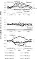


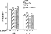


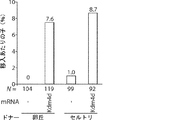

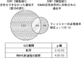


Classifications
-
- C—CHEMISTRY; METALLURGY
- C12—BIOCHEMISTRY; BEER; SPIRITS; WINE; VINEGAR; MICROBIOLOGY; ENZYMOLOGY; MUTATION OR GENETIC ENGINEERING
- C12N—MICROORGANISMS OR ENZYMES; COMPOSITIONS THEREOF; PROPAGATING, PRESERVING, OR MAINTAINING MICROORGANISMS; MUTATION OR GENETIC ENGINEERING; CULTURE MEDIA
- C12N15/00—Mutation or genetic engineering; DNA or RNA concerning genetic engineering, vectors, e.g. plasmids, or their isolation, preparation or purification; Use of hosts therefor
- C12N15/09—Recombinant DNA-technology
- C12N15/63—Introduction of foreign genetic material using vectors; Vectors; Use of hosts therefor; Regulation of expression
- C12N15/79—Vectors or expression systems specially adapted for eukaryotic hosts
- C12N15/85—Vectors or expression systems specially adapted for eukaryotic hosts for animal cells
- C12N15/8509—Vectors or expression systems specially adapted for eukaryotic hosts for animal cells for producing genetically modified animals, e.g. transgenic
-
- C—CHEMISTRY; METALLURGY
- C12—BIOCHEMISTRY; BEER; SPIRITS; WINE; VINEGAR; MICROBIOLOGY; ENZYMOLOGY; MUTATION OR GENETIC ENGINEERING
- C12N—MICROORGANISMS OR ENZYMES; COMPOSITIONS THEREOF; PROPAGATING, PRESERVING, OR MAINTAINING MICROORGANISMS; MUTATION OR GENETIC ENGINEERING; CULTURE MEDIA
- C12N15/00—Mutation or genetic engineering; DNA or RNA concerning genetic engineering, vectors, e.g. plasmids, or their isolation, preparation or purification; Use of hosts therefor
- C12N15/09—Recombinant DNA-technology
- C12N15/87—Introduction of foreign genetic material using processes not otherwise provided for, e.g. co-transformation
- C12N15/873—Techniques for producing new embryos, e.g. nuclear transfer, manipulation of totipotent cells or production of chimeric embryos
- C12N15/877—Techniques for producing new mammalian cloned embryos
-
- A—HUMAN NECESSITIES
- A01—AGRICULTURE; FORESTRY; ANIMAL HUSBANDRY; HUNTING; TRAPPING; FISHING
- A01K—ANIMAL HUSBANDRY; CARE OF BIRDS, FISHES, INSECTS; FISHING; REARING OR BREEDING ANIMALS, NOT OTHERWISE PROVIDED FOR; NEW BREEDS OF ANIMALS
- A01K67/00—Rearing or breeding animals, not otherwise provided for; New breeds of animals
- A01K67/027—New breeds of vertebrates
- A01K67/0273—Cloned animals
-
- C—CHEMISTRY; METALLURGY
- C12—BIOCHEMISTRY; BEER; SPIRITS; WINE; VINEGAR; MICROBIOLOGY; ENZYMOLOGY; MUTATION OR GENETIC ENGINEERING
- C12N—MICROORGANISMS OR ENZYMES; COMPOSITIONS THEREOF; PROPAGATING, PRESERVING, OR MAINTAINING MICROORGANISMS; MUTATION OR GENETIC ENGINEERING; CULTURE MEDIA
- C12N15/00—Mutation or genetic engineering; DNA or RNA concerning genetic engineering, vectors, e.g. plasmids, or their isolation, preparation or purification; Use of hosts therefor
- C12N15/09—Recombinant DNA-technology
- C12N15/11—DNA or RNA fragments; Modified forms thereof; Non-coding nucleic acids having a biological activity
- C12N15/113—Non-coding nucleic acids modulating the expression of genes, e.g. antisense oligonucleotides; Antisense DNA or RNA; Triplex- forming oligonucleotides; Catalytic nucleic acids, e.g. ribozymes; Nucleic acids used in co-suppression or gene silencing
- C12N15/1137—Non-coding nucleic acids modulating the expression of genes, e.g. antisense oligonucleotides; Antisense DNA or RNA; Triplex- forming oligonucleotides; Catalytic nucleic acids, e.g. ribozymes; Nucleic acids used in co-suppression or gene silencing against enzymes
-
- C—CHEMISTRY; METALLURGY
- C12—BIOCHEMISTRY; BEER; SPIRITS; WINE; VINEGAR; MICROBIOLOGY; ENZYMOLOGY; MUTATION OR GENETIC ENGINEERING
- C12N—MICROORGANISMS OR ENZYMES; COMPOSITIONS THEREOF; PROPAGATING, PRESERVING, OR MAINTAINING MICROORGANISMS; MUTATION OR GENETIC ENGINEERING; CULTURE MEDIA
- C12N15/00—Mutation or genetic engineering; DNA or RNA concerning genetic engineering, vectors, e.g. plasmids, or their isolation, preparation or purification; Use of hosts therefor
- C12N15/09—Recombinant DNA-technology
- C12N15/87—Introduction of foreign genetic material using processes not otherwise provided for, e.g. co-transformation
- C12N15/873—Techniques for producing new embryos, e.g. nuclear transfer, manipulation of totipotent cells or production of chimeric embryos
-
- C—CHEMISTRY; METALLURGY
- C12—BIOCHEMISTRY; BEER; SPIRITS; WINE; VINEGAR; MICROBIOLOGY; ENZYMOLOGY; MUTATION OR GENETIC ENGINEERING
- C12N—MICROORGANISMS OR ENZYMES; COMPOSITIONS THEREOF; PROPAGATING, PRESERVING, OR MAINTAINING MICROORGANISMS; MUTATION OR GENETIC ENGINEERING; CULTURE MEDIA
- C12N5/00—Undifferentiated human, animal or plant cells, e.g. cell lines; Tissues; Cultivation or maintenance thereof; Culture media therefor
- C12N5/06—Animal cells or tissues; Human cells or tissues
- C12N5/0602—Vertebrate cells
- C12N5/0603—Embryonic cells ; Embryoid bodies
- C12N5/0604—Whole embryos; Culture medium therefor
-
- C—CHEMISTRY; METALLURGY
- C12—BIOCHEMISTRY; BEER; SPIRITS; WINE; VINEGAR; MICROBIOLOGY; ENZYMOLOGY; MUTATION OR GENETIC ENGINEERING
- C12N—MICROORGANISMS OR ENZYMES; COMPOSITIONS THEREOF; PROPAGATING, PRESERVING, OR MAINTAINING MICROORGANISMS; MUTATION OR GENETIC ENGINEERING; CULTURE MEDIA
- C12N5/00—Undifferentiated human, animal or plant cells, e.g. cell lines; Tissues; Cultivation or maintenance thereof; Culture media therefor
- C12N5/06—Animal cells or tissues; Human cells or tissues
- C12N5/0602—Vertebrate cells
- C12N5/0603—Embryonic cells ; Embryoid bodies
- C12N5/0606—Pluripotent embryonic cells, e.g. embryonic stem cells [ES]
-
- A—HUMAN NECESSITIES
- A01—AGRICULTURE; FORESTRY; ANIMAL HUSBANDRY; HUNTING; TRAPPING; FISHING
- A01K—ANIMAL HUSBANDRY; CARE OF BIRDS, FISHES, INSECTS; FISHING; REARING OR BREEDING ANIMALS, NOT OTHERWISE PROVIDED FOR; NEW BREEDS OF ANIMALS
- A01K2227/00—Animals characterised by species
- A01K2227/10—Mammal
- A01K2227/105—Murine
-
- C—CHEMISTRY; METALLURGY
- C12—BIOCHEMISTRY; BEER; SPIRITS; WINE; VINEGAR; MICROBIOLOGY; ENZYMOLOGY; MUTATION OR GENETIC ENGINEERING
- C12N—MICROORGANISMS OR ENZYMES; COMPOSITIONS THEREOF; PROPAGATING, PRESERVING, OR MAINTAINING MICROORGANISMS; MUTATION OR GENETIC ENGINEERING; CULTURE MEDIA
- C12N2310/00—Structure or type of the nucleic acid
- C12N2310/10—Type of nucleic acid
- C12N2310/14—Type of nucleic acid interfering N.A.
-
- C—CHEMISTRY; METALLURGY
- C12—BIOCHEMISTRY; BEER; SPIRITS; WINE; VINEGAR; MICROBIOLOGY; ENZYMOLOGY; MUTATION OR GENETIC ENGINEERING
- C12N—MICROORGANISMS OR ENZYMES; COMPOSITIONS THEREOF; PROPAGATING, PRESERVING, OR MAINTAINING MICROORGANISMS; MUTATION OR GENETIC ENGINEERING; CULTURE MEDIA
- C12N2517/00—Cells related to new breeds of animals
- C12N2517/04—Cells produced using nuclear transfer
-
- C—CHEMISTRY; METALLURGY
- C12—BIOCHEMISTRY; BEER; SPIRITS; WINE; VINEGAR; MICROBIOLOGY; ENZYMOLOGY; MUTATION OR GENETIC ENGINEERING
- C12Y—ENZYMES
- C12Y201/00—Transferases transferring one-carbon groups (2.1)
- C12Y201/01—Methyltransferases (2.1.1)
- C12Y201/01043—Histone-lysine N-methyltransferase (2.1.1.43)
Description
関連出願の相互参照
本願は、35 U.S.C. 119(e)の下で、2014年9月15日に出願された米国仮特許出願第62/050,308号および2014年9月22日に出願された米国仮特許出願第62/053,514号の恩典を主張し、その各内容は全体として参照により本明細書に組み入れられる。
Cross-reference to related applications This application is under 35 USC 119 (e), 35 USC 119 (e), and US Provisional Patent Application No. 62 / 050,308 filed on September 15, 2014 and US Provisional on September 22, 2014. Claim the benefits of 35/053,514, the contents of which are incorporated herein by reference in their entirety.
政府による支援
本発明は、米国国立衛生研究所(National Institutes of Health/NIH)から授与されたNIH U01DK089565の下で政府の支援を受けてなされたものである。米国政府は、本発明に関して一定の権利を保有する。
Government Support The invention was made with government support under the NIH U01DK089565 awarded by the National Institutes of Health (NIH). The United States Government reserves certain rights with respect to the present invention.
発明の背景
最終分化した体細胞は、体細胞核移入(SCNT)によって除核卵母細胞内に移植されると全能状態にリプログラミングされることができる(Gurdon, 1962)。SCNTは、分化した体細胞の単一の核から動物全体の生成を可能にするので、農業、生物医学工業、および絶滅危惧種の保護に大きな潜在性を有する(Yang et al., 2007)。実際、ヒツジにおける哺乳動物クローニングが1997年に最初に成功して以来(Wilmut et al., 1997)、20種よりも多い哺乳動物種がSCNTを経てクローニングされている(Rodriguez-Osorio et al., 2012)。そのうえ、SCNTによって作製された胚盤胞から多能性胚性幹細胞を樹立することができるので(Wakayama et al., 2001)、SCNTは、ヒトの治療に大いに有望である(Hochedlinger and Jaenisch, 2003)。この有望性は、最初のヒト核移入胚性幹細胞(ntESC)の派生における最近の成功(Tachibana et al., 2013)、および高齢者またはヒト患者細胞からのヒトntESC作製(Chung et al., 2014; Yamada et al., 2014)の後に、より現実に近づいている。これらのntESCは、インビトロ疾患モデル化用の細胞の貴重な供給源として、ならびに再生療法および細胞/組織置換療法用の細胞の供給源として役立ち得る。
Background of the Invention Final differentiated somatic cells can be reprogrammed to pluripotency when transplanted into enucleated oocytes by somatic cell nuclear transfer (SCNT) (Gurdon, 1962). Since SCNTs allow the production of whole animals from a single nucleus of differentiated somatic cells, they have great potential for agriculture, biomedical industry, and protection of endangered species (Yang et al., 2007). In fact, since the first successful mammalian cloning in sheep in 1997 (Wilmut et al., 1997), more than 20 mammalian species have been cloned via SCNT (Rodriguez-Osorio et al.,). 2012). Moreover, since pluripotent embryonic stem cells can be established from blastocysts produced by SCNT (Wakayama et al., 2001), SCNT is highly promising for human treatment (Hochedlinger and Jaenisch, 2003). ). This promising is a recent success in the derivation of the first human nuclear-transferred embryonic stem cells (ntESCs) (Tachibana et al., 2013), and the production of human ntESCs from aged or human patient cells (Chung et al., 2014). After Yamada et al., 2014), it is closer to reality. These ntESCs can serve as a valuable source of cells for in vitro disease modeling and as a source of cells for regenerative therapy and cell / tissue replacement therapy.
その大きな潜在性にもかかわらず、いくつかの技術上の問題が、SCNTの実用的用途を阻止しており、特に、SCNTは、クローン動物の産生に極めて低い効率を有する。例えばマウスSCNT胚の約半数は、着床前に発生停止を示し、代理母に移入された胚のわずか1〜2%が満期まで発生する(Ogura et al., 2013)。高率の生殖クローニング効率(5〜20%)を有するウシ種を除き、全ての他の種における全体的な生殖クローニング効率は、非常に低い(1〜5%)(Rodriguez-Osorio et al., 2012)。さらに、ヒトntESC樹立の成功率も、その不十分な着床前発生のせいで低い(胚盤胞期までが10〜25%; Tachibana et al., 2013; Yamada et al., 2014)。 Despite its enormous potential, some technical problems have hampered the practical use of SCNTs, in particular SCNTs have extremely low efficiencies in the production of cloned animals. For example, about half of mouse SCNT embryos show developmental arrest prior to implantation, and only 1-2% of embryos transferred to surrogate mothers develop to maturity (Ogura et al., 2013). Overall reproductive cloning efficiency in all other species is very low (1-5%) (Rodriguez-Osorio et al.,, Except for bovine species with high rates of reproductive cloning efficiency (5-20%). 2012). In addition, the success rate of human ntESC establishment is also low due to its inadequate preimplantation development (10-25% up to blastocyst stage; Tachibana et al., 2013; Yamada et al., 2014).
SCNTの適用潜在性を実現するために、SCNTのクローニング効率を改善する努力がなされている。第一に、トリコスタチンA(TSA)またはスクリプタイドのようなヒストンデアセチラーゼ(HDAC)阻害剤を用いた1細胞期SCNT胚の一過性処置は、マウス(Kishigami et al., 2006; Van Thuan et al., 2009)、ブタ(Zhao et al., 2009)、ウシ(Akagi et al., 2011)およびヒト(Tachibana et al., 2013; Yamada et al., 2014)を含めた多様な哺乳動物種のリプログラミング効率を改善することが報告されている。第二に、Xistのノックアウトまたはノックダウンは、マウスSCNT胚の着床後発生を改善することが報告されている(Inoue et al., 2010; Matoba et al., 2011)。しかし、これらの方法のどれも、SCNTが、非ヒト哺乳動物の生殖クローニングに、または治療的クローニングもしくは再生療法用のヒト多能性幹細胞(例えばヒトntESC)の作製に有用であるのに十分なほど、SCNTのクローニング効率を改善しない。 Efforts have been made to improve the cloning efficiency of SCNT in order to realize the application potential of SCNT. First, transient treatment of 1-cell stage SCNT embryos with histone deacetylase (HDAC) inhibitors such as tricostatin A (TSA) or scriptide is a mouse (Kishigami et al., 2006; Van). Diverse mammals including Thuan et al., 2009), pigs (Zhao et al., 2009), cattle (Akagi et al., 2011) and humans (Tachibana et al., 2013; Yamada et al., 2014) It has been reported to improve the efficiency of reprogramming of animal species. Second, knockout or knockdown of Xist has been reported to improve post-implantation development of mouse SCNT embryos (Inoue et al., 2010; Matoba et al., 2011). However, any of these methods are sufficient for SCNT to be useful for reproductive cloning of non-human mammals or for the production of human pluripotent stem cells (eg, human ntESC) for therapeutic cloning or regenerative therapy. It does not improve the cloning efficiency of SCNT as much.
SCNT胚の発生異常は、マウスで2細胞期に、ブタ、ウシおよびヒトで4〜8細胞期に起こる接合体遺伝子活性化(ZGA)のときに出現し始める(Schultz, 2002)。SCNT胚は、ドナー細胞のゲノム中に先在する未確定のエピジェネティックなバリアのせいで、ZGAが困難である。マウス2細胞期SCNT胚(Inoue et al., 2006; Suzuki et al., 2006; Vassena et al., 2007)および卵割後期のヒトSCNT胚(Noggle et al., 2011)において、いくつかの調節不全遺伝子が同定されているものの、「先在するエピジェネティックなバリア」の性質およびそれらとSCNT胚におけるZGA障害との関係は、未知である。 Developmental abnormalities of SCNT embryos begin to appear during zygote gene activation (ZGA), which occurs at the 2-cell stage in mice and at the 4-8 cell stage in pigs, cattle and humans (Schultz, 2002). SCNT embryos are difficult to ZGA due to an undetermined epigenetic barrier pre-existing in the donor cell's genome. Some regulation in mouse 2-cell stage SCNT embryos (Inoue et al., 2006; Suzuki et al., 2006; Vassena et al., 2007) and late cleavage human SCNT embryos (Noggle et al., 2011). Although defective genes have been identified, the nature of "pre-existing epigenetic barriers" and their relationship to ZGA damage in SCNT embryos is unknown.
それゆえに、SCNT胚が接合体遺伝子活性化(ZGA)を効率的に進めることができ、かつSCNT胚の発生異常または生存能の喪失もなしに2、4および8細胞期を経て胚盤胞に進むことができるように、ドナー細胞核のゲノムからそのようなエピジェネティックなバリアを取り除くことによって哺乳動物のクローニング効率を改善する必要がある。 Therefore, SCNT embryos can efficiently promote conjugate gene activation (ZGA) and become blastocysts through 2, 4 and 8 cell stages without abnormal development or loss of viability of SCNT embryos. To be able to proceed, it is necessary to improve the cloning efficiency of mammals by removing such epigenetic barriers from the donor cell nucleus genome.
哺乳動物卵母細胞は、体細胞を全能状態にリプログラミングすることができ、そのことは、体細胞核移入(SCNT)を経た動物生殖クローニングまたはSCNT胚から発生する胚盤胞からのES細胞株(ntESC)の生成を可能にする。しかし、大部分のSCNT胚は、未確定のリプログラミング欠損のせいで胚盤胞または満期まで発生できない。哺乳動物SCNTの非効率は、再生医学の適用のために患者特異的hESC株を開発する重大な制約である。 Mammalian egg mother cells can reprogram somatic cells to the pluripotent state, which means animal reproductive cloning via somatic cell nuclear transfer (SCNT) or ES cell lines from blastocysts that develop from SCNT embryos (SCNT). Allows the generation of ntESC). However, most SCNT embryos cannot develop into blastocysts or maturity due to uncertain reprogramming deficiencies. The inefficiency of mammalian SCNT is a significant constraint on the development of patient-specific hESC strains for regenerative medicine applications.
ヒトドナー体細胞を使用するヒトSCNT由来ヒト胚盤胞の生成が報告されているが、胚盤胞の質および発生効率は、ヒト胚性幹細胞株(ヒトntESC)を生成可能にするには不十分であった(French AJ et al., Stem Cells 26, 485-493 (2008))。ヒト核移入胚性幹細胞(ntESC)(Tachibana et al., 2013)および高齢者成熟細胞またはヒト患者細胞からのヒトntESCの作製(Chung et al., 2014; Yamada et al., 2014)が報告されている。しかし、ヒトntESC樹立についての成功率は、不十分な着床前発生のせいで非常に低い(わずか10〜25%が胚盤胞期まで発生する; Tachibana et al., 2013; Yamada et al., 2014)。したがって、ヒトSCNT技法の改良は、ヒトSCNT胚の胚盤胞期までの発生を改善するため、SCNTに必要なドナー卵母細胞数を低減するため、ならびに研究および細胞に基づく治療用のヒトおよび患者特異的な同質遺伝子胚性幹細胞株を成功裏に生成するために重大な意味を持つ。 Generation of human SCNT-derived human blastocysts using human donor somatic cells has been reported, but the quality and developmental efficiency of blastocysts are insufficient to enable the production of human embryonic stem cell lines (human ntESCs). (French AJ et al., Stem Cells 26, 485-493 (2008)). Production of human ntESC from human nuclear-transferred embryonic stem cells (ntESC) (Tachibana et al., 2013) and aged mature cells or human patient cells (Chung et al., 2014; Yamada et al., 2014) has been reported. ing. However, the success rate for the establishment of human ntESC is very low due to inadequate preimplantation development (only 10-25% occur until the blastocyst stage; Tachibana et al., 2013; Yamada et al. , 2014). Therefore, improvements in human SCNT techniques are to improve the development of human SCNT embryos up to the blastocyst stage, to reduce the number of donor oocytes required for SCNT, and to study and cell-based therapeutic humans and It has important implications for the successful generation of patient-specific homogen embryonic stem cell lines.
本明細書において、本発明者らは、分化した体細胞のドナー核のゲノムにおけるヒストンH3リジン9のトリメチル化(H3K9me3)が、SCNTによる効率的なリプログラミングにとって主要な先在するエピジェネティックなバリアであることを発見し、H3K9me3メチル化を減少させることで、SCNTの効率を増加させることができ、特にSCNT胚から2細胞期、4細胞期、または胚盤胞期への着床前発生効率を増加させることができることを発見した。より具体的には、比較分析により、本発明者らは、SCNT胚における接合体遺伝子活性化(ZGA)に耐性のゲノムドメインを発見した。これらのリプログラミング耐性領域(RRR)は、体細胞に抑制的ヒストン修飾H3K9me3が濃縮されており、(i)卵母細胞における例えばKdm4dもしくはKdm4ファミリーのメンバーのようなH3K9me3特異的デメチラーゼの増加した発現、および/または(ii)体細胞ドナー核における例えばSuv39h1もしくはSuv39h2もしくはその両方(すなわちSuv39h1/2)のようなH3K9メチルトランスフェラーゼをノックダウンもしくは阻害することのいずれかによる、このエピジェネティックなマークの除去は、ZGA欠損を減弱しかつRRRを再活性化するだけでなく、SCNTの効率も大きく改善し、例えば、2細胞期、4細胞期または胚盤胞期に発生するSCNT胚の%を増加させる。したがって、本発明者らは、Suv39h1/2介在性H3K9me3がSCNTの「エピジェネティックなバリア」であることを発見し、ドナー体細胞、レシピエント卵母細胞またはSCNT胚のいずれかの核におけるH3K9me3のトリメチル化の阻害および/もしくは除去が、ヒトSCNTのクローニングおよび他の非ヒト哺乳動物を含めた哺乳動物細胞のSCNTクローニング効率を有意に改善できることを実証した。 As used herein, we present that trimethylation of histone H3 lysine 9 (H3K9me3) in the genome of a donor nucleus of differentiated somatic cells is a major pre-existing epigenetic barrier for efficient reprogramming by SCNT. By reducing H3K9me3 methylation, the efficiency of SCNT can be increased, especially the pre-implantation developmental efficiency from SCNT embryos to 2-cell, 4-cell, or blastocyst stages. Was found to be able to increase. More specifically, by comparative analysis, we have discovered a genomic domain resistant to zygote gene activation (ZGA) in SCNT embryos. These reprogramming resistant regions (RRRs) are enriched in somatic cells with suppressive histone-modified H3K9me3, and (i) increased expression of H3K9me3-specific demethylases in oocytes, such as members of the Kdm4d or Kdm4 family. And / or (ii) removal of this epigenetic mark by either knocking down or inhibiting H3K9 methyltransferases such as, for example, Suv39h1 and / or Suv39h2 (ie, Suv39h1 / 2) in somatic cell donor nuclei. Not only attenuates ZGA deficiency and reactivates RRR, but also greatly improves the efficiency of SCNT, for example, increasing the percentage of SCNT embryos that develop during the 2-cell, 4-cell or blastocyst stages. .. Therefore, we have discovered that Suv39h1 / 2 mediated H3K9me3 is an "epigenetic barrier" for SCNT, and that H3K9me3 in the nucleus of either donor somatic cells, recipient oocytes or SCNT embryos. We have demonstrated that inhibition and / or elimination of trimethylation can significantly improve the cloning efficiency of human SCNT and SCNT cloning efficiency in mammalian cells, including other non-human mammals.
それゆえに、本発明は、H3K9me3が、SCNT胚の産生に採用される体細胞中のRRRに濃縮されており、かつ体細胞におけるH3K9me3バリアがSuv39h1/2によって樹立されるという本発明者の発見に基づく。重要なことには、本発明者らは、レシピエント卵母細胞におけるKdm4dの過剰発現(例えば、外因性Kdm4d mRNAもしくはcDNAの導入による)によるH3K9me3の除去、および/またはドナー体細胞核におけるSuv39h1/2の阻害が、SCNTのクローニング効率に驚くほど顕著な増加を招くことを実証した。興味深いことに、本発明者らは驚くことに、H3K9me3の除去が、胚盤胞期に進んだSCNT胚が約80%超という予想外で有意なSCNT効率の増加を招いたのに対し、H3K9me3を除去しない正常SCNT胚の効率は約30%のままであり、胚盤胞期へのSCNTの効率において予想外の50%の増加を示したことを発見した。そのうえ、本発明者らは、H3K9me3の除去が、SCNT胚の着床後発生の予想外で有意な改善を招き、人工的H3K9me3除去の不在下と比較してマウス子産出を8倍よりも大きく増加させたことも実証した。 Therefore, the present invention concludes that H3K9me3 is enriched in RRR in somatic cells used for the production of SCNT embryos, and that the H3K9me3 barrier in somatic cells is established by Suv39h1 / 2. Based on. Importantly, we found that removal of H3K9me3 by overexpression of Kdm4d in recipient oocytes (eg, by introduction of exogenous Kdm4d mRNA or cDNA) and / or Suv39h1 / 2 in donor somatic cell nuclei. We have demonstrated that inhibition of SCNT results in a surprisingly significant increase in SCNT cloning efficiency. Interestingly, we surprisingly found that removal of H3K9me3 resulted in an unexpectedly significant increase in SCNT efficiency of more than about 80% in blastocyst-advanced SCNT embryos, whereas H3K9me3 We found that the efficiency of normal SCNT embryos without removal remained about 30%, showing an unexpected 50% increase in the efficiency of SCNT to the blastocyst stage. Moreover, we found that removal of H3K9me3 resulted in an unexpected and significant improvement in post-implantation development of SCNT embryos, resulting in more than eight-fold higher mouse offspring compared to the absence of artificial removal of H3K9me3. It also demonstrated that it was increased.
それゆえに、本発明の局面は、ヒストンH3-リジン9(H3K9me3)のトリメチル化が効率的な体細胞核リプログラミングまたはSCNTを阻止するという発見に基づく。本発明者らは、(i)デメチラーゼKdm4d(Jmjd2dまたはJhdm3dとしても公知)の過剰発現(すなわち外因性発現)を使用することによってH3K9me3の脱メチル化を促進すること、および(ii)ヒストンメチルトランスフェラーゼSuv39h1および/またはSuv39h2を阻害することによってH3K9me3のメチル化を阻害することによる、マウス胚のSCNT効率を改善するための2つの方法を評価した。このように、Kdm4d/Jmjd2d遺伝子の過剰発現、および/またはSuv39h1/2タンパク質もしくは遺伝子の阻害剤は、細胞リプログラミング、特にSCNTにおけるエピジェネティックなバリアを除去するために有用である。 Therefore, aspects of the invention are based on the finding that trimethylation of histone H3-lysine 9 (H3K9me3) prevents efficient somatic cell nuclear transfer or SCNT. We use (i) overexpression (ie, extrinsic expression) of the demethylase Kdm4d (also known as Jmjd2d or Jhdm3d) to promote demethylation of H3K9me3, and (ii) histone methyltransferase. Two methods were evaluated to improve SCNT efficiency in mouse embryos by inhibiting methylation of H3K9me3 by inhibiting Suv39h1 and / or Suv39h2. Thus, overexpression of the Kdm4d / Jmjd2d gene and / or inhibitors of the Suv39h1 / 2 protein or gene are useful for cell reprogramming, especially for removing epigenetic barriers in SCNT.
それゆえに、本発明の局面は、(i)H3K9me3を脱メチル化する能力のあるヒストンデメチラーゼ、例えば、非限定的にJmjd2d/Kdm4dもしくはJmjd2a/Kdm4a、もしくJmjd2b/Kdm4bもしくはJmjd2c/Kdm4cのような、例えばKdm4ファミリーのヒストンデメチラーゼのメンバーを発現させること、および/または(ii)H3K9me3のメチル化に関与するヒストンメチルトランスフェラーゼを阻害すること、例えばSuv39h1、Suv39h2またはSetdb1の任意の1つまたは組み合わせを阻害することのいずれかによって、H3K9me3のメチル化を低減することによりSCNTの効率を増加させることを目指す方法、組成物およびキットに関する。いくつかの態様では、Kdm4ファミリーのヒストンデメチラーゼKdm4d(Jmjd2d)、またはKdm4a(Jmjd2a)、またはJmjd2b/Kdm4bまたはJmjd2c/Kdm4cの発現または活性を増加させる作用物質。 Therefore, aspects of the invention are (i) such as histone demethylases capable of demethylating H3K9me3, such as, but not limited to, Jmjd2d / Kdm4d or Jmjd2a / Kdm4a, or Jmjd2b / Kdm4b or Jmjd2c / Kdm4c. For example, expressing a member of the Kdm4 family of histone demethylases and / or (ii) inhibiting histone methyltransferases involved in the methylation of H3K9me3, eg, any one or combination of Suv39h1, Suv39h2 or Setdb1. With respect to methods, compositions and kits aimed at increasing the efficiency of SCNTs by reducing the methylation of H3K9me3 by either inhibiting. In some embodiments, an agent that increases the expression or activity of the Kdm4 family of histone demethylases Kdm4d (Jmjd2d), or Kdm4a (Jmjd2a), or Jmjd2b / Kdm4b or Jmjd2c / Kdm4c.
H3K9me3の脱メチル化(Kdm4c/Jmjd2cによる)は、体細胞リプログラミング効率を増加させるために使用されることが報告されているものの(例えば、人工多能性幹(iPS)細胞の作製(Sridharan et al., 2013))、最終分化した体細胞からのSCNT効率を増加させるためのH3K9me3の脱メチル化は、まだ報告されていない。Antonyらは、「Transient JMJD2B-Mediated Reduction of H3K9me3 Levels Improve Reprogramming of Embryonic Stem Cells in Cloned Embryos.」 Mol. Cell Biol., 2013; 33(5);974において、多能性ES細胞由来のドナー核から得られるSCNTにKdm4b/Jmjd2bを使用すること報告している。多能性ES細胞は、最終分化した体細胞と同じではない、発生的に未熟な細胞である。重要なことには、分化した体細胞と比較して、胚性幹(ES)細胞または人工多能性幹(iPS)細胞の全般的エピジェネティック状態に有意な差がある。多能性ES細胞は、より少ないエピジェネティックなバリア(例えば、特にリプログラミング耐性領域(RRR)におけるより少ないメチル化)を有し、したがって、ES細胞核がドナー核として使用されるときに生成されるSCNT胚の効率は、最終分化した体細胞由来の核が使用されるときに生成されるSCNT胚の効率と非常に異なる(Rideout et al., 2000, Nature Genetics, 24(2), 109-10)。 Although demethylation of H3K9me3 (by Kdm4c / Jmjd2c) has been reported to be used to increase somatic cell reprogramming efficiency (eg, induced pluripotent stem (iPS) cell production (Sridharan et.) al., 2013)), demethylation of H3K9me3 to increase SCNT efficiency from final differentiated somatic cells has not yet been reported. Antony et al., In "Transient JMJD2B-Mediated Reduction of H3K9me3 Levels Improve Reprogramming of Embryonic Stem Cells in Cloned Embryos." Mol. Cell Biol., 2013; 33 (5); 974, from pluripotent ES cell-derived donor nuclei. It is reported that Kdm4b / Jmjd2b is used for the obtained SCNT. Pluripotent ES cells are developmentally immature cells that are not the same as end-differentiated somatic cells. Importantly, there is a significant difference in the general epigenetic status of embryonic stem (ES) cells or induced pluripotent stem (iPS) cells compared to differentiated somatic cells. Pluripotent ES cells have less epigenetic barriers (eg, less methylation, especially in the reprogramming resistant region (RRR)) and are therefore generated when ES cell nuclei are used as donor nuclei. The efficiency of SCNT embryos is very different from that of SCNT embryos produced when nuclei derived from final differentiated somatic cells are used (Rideout et al., 2000, Nature Genetics, 24 (2), 109-10). ).
Antonyらによる報告とは対照的に、本発明者らは、最終分化した体細胞核におけるH3K9me3レベルを減少させることが、着床前発生における驚くべき増加を招き、H3K9me3の脱メチル化が減少されるとき、SCNT胚の80%よりも多くが胚盤胞に成功裏に発生することを本明細書において実証している(例えば、卵丘細胞由来の核が使用されるとき26%から89%に:セルトリ細胞由来の核を使用されるとき26%から81%に、およびMEF細胞由来の核が使用されるとき7%から82%に増加)。Antonyらが、ES細胞を用いて得るドナー核が使用されても、着床前発生に小さな改善(約9%の改善)だけしか報告していないことを考えれば、この結果は、大いに予想外である。 In contrast to the report by Antony et al., We found that reducing H3K9me3 levels in terminally differentiated somatic cell nuclei resulted in a surprising increase in preimplantation development and reduced demethylation of H3K9me3. It is demonstrated herein that more than 80% of SCNT embryos successfully develop in blastocysts (eg, from 26% to 89% when implant cell-derived nuclei are used). : Increased from 26% to 81% when using Sertri cell-derived nuclei, and from 7% to 82% when using MEF cell-derived nuclei). This result is highly unexpected, considering that Antony et al. Reported only a small improvement (about 9% improvement) in pre-implantation development when donor nuclei obtained using ES cells were used. Is.
さらに、Antonyらが、Jmjd2b/Kdm4b誘導により着床後発生に何も改善を実証できていない一方で、本明細書において本発明者らは、SCNT胚からのマウス子作製に8倍よりも大きな増加を示している。SCNTの一目的が、非ヒト哺乳動物の生殖クローニングおよび着床後発生の完了を必要とする個別の動物の作製であることを考えれば、Antonyらを読んでいる当業者は、ドナー細胞核へのKdm4b注射がSCNT効率を改善できないと結論するであろう。さらに、Antonyらは、ES細胞におけるSuvh1/2を阻害しない。 Furthermore, while Antony et al. Have not demonstrated any improvement in post-implantation development by induction of Jmjd2b / Kdm4b, we present here more than eight-fold greater than eight-fold increase in mouse offspring production from SCNT embryos. It shows an increase. Given that one purpose of SCNT is the production of individual animals that require reproductive cloning of non-human mammals and completion of post-implantation development, those who read Antony et al. We would conclude that Kdm4b injection cannot improve SCNT efficiency. Furthermore, Antony et al. Do not inhibit Suvh1 / 2 in ES cells.
さらに、H3K9me3の脱メチル化がより早い発生期に体細胞をリプログラミングする効率を増加させるという数多くの報告がある(例えば、人工多能性幹(iPS)細胞の作製)(例えば、その全体として参照により本明細書に組み入れられる米国特許出願第2011/0136145号および同第2012/0034192号)一方で、iPS細胞の作製のための体細胞リプログラミングメカニズムは、SCNT胚の作製のための体細胞リプログラミングメカニズムと有意に異なる(Pasque et al., 2011, Mechanisms of nuclear reprogramming by eggs and oocytes: a deterministic process? Nat. Rev. Mol. Cell Biol. 12, 453-459;およびApostolou, E., and Hochedlinger, K., 2013; Chromatin dynamics during cellular reprogramming. Nature 502, 462-471に考察されているように)。したがって、iPS細胞の作製でのH3K9me3の脱メチル化から学ばれることは、関連性がなく、SCNT胚の成功裏な作製のための方法ならびにSCNT胚の着床前および着床後両方の効率を増加させる方法に移行することはできない。 In addition, there are numerous reports that demethylation of H3K9me3 increases the efficiency of reprogramming somatic cells during earlier development (eg, the production of induced pluripotent stem (iPS) cells) (eg, as a whole). US Patent Application Nos. 2011/0136145 and 2012/0034192) incorporated herein by reference, on the other hand, a somatic cell reprogramming mechanism for the production of iPS cells is the somatic cell for the production of SCNT embryos. Significantly different from reprogramming mechanisms (Pasque et al., 2011, Mechanisms of nuclear reprogramming by eggs and oocytes: a deterministic process? Nat. Rev. Mol. Cell Biol. 12, 453-459; and Apostolou, E., and Hochedlinger, K., 2013; Chromatin dynamics during cellular reprogramming. As discussed in Nature 502, 462-471). Therefore, what is learned from the demethylation of H3K9me3 in the production of iPS cells is irrelevant and the method for the successful production of SCNT embryos and the efficiency of both pre-implantation and post-implantation of SCNT embryos. It is not possible to move to a method of increasing.
特に、SCNTリプログラミングとiPSリプログラミングとの間でバリアに関して著しい差が存在する。第一に、マウスiPSCリプログラミングにおけるH3K9me3バリアは、主にSetdb1によって樹立される(Chen et al., 2013; Sridharan et al., 2013)。対照的に、SCNTリプログラミングについてH3K9me3バリアを樹立する重大な酵素として、本発明者らの結果は、Setdb1ではなくSuv39h1/2をはっきりと指し示している。第二に、成功裏なiPSCおよびSCNTリプログラミングに必要な下流の遺伝子ネットワークは、異なる。例えば、iPSCリプログラミングでは、H3K9me3バリアによって抑制されるNanogおよびSox2のような鍵となるコア多能性ネットワーク遺伝子が、リプログラミングの相対的後期に発現される(Chen et al., 2013; Sridharan et al., 2013)。対照的に、SCNTリプログラミングでは、H3K9me3によって抑制される鍵遺伝子が発現され、胚2細胞期で重大な機能を有する(本明細書下記)。この区別は、各状況で成功裏なリプログラミングに要する転写因子セットの差に由来する可能性が最も高い。実際、iPSCリプログラミングに要するコア転写因子Oct4/Pou5f1は、SCNTのリプログラミングに不必要であることが実証されている(Wu et al., 2013)。したがって、H3K9m3は、iPS細胞の作製および成功裏なSCNTの両方に共通のリプログラミングバリアであるように見えるものの、その沈着およびそれがどのようにリプログラミング過程に影響するかは、iPS細胞を作製するためのリプログラミング方法またはSCNT胚を作製するためのリプログラミング方法で非常に異なる。 In particular, there are significant differences in barriers between SCNT reprogramming and iPS reprogramming. First, the H3K9me3 barrier in mouse iPSC reprogramming is established primarily by Setdb1 (Chen et al., 2013; Sridharan et al., 2013). In contrast, our results clearly point to Suv39h1 / 2 rather than Setdb1 as a critical enzyme that establishes the H3K9me3 barrier for SCNT reprogramming. Second, the downstream genetic networks required for successful iPSC and SCNT reprogramming are different. For example, in iPSC reprogramming, key core pluripotent network genes such as Nanog and Sox2, which are suppressed by the H3K9me3 barrier, are expressed in the relative late stages of reprogramming (Chen et al., 2013; Sridharan et. al., 2013). In contrast, SCNT reprogramming expresses a key gene that is suppressed by H3K9me3 and has significant function during the embryonic 2-cell stage (see below). This distinction is most likely due to the difference in transcription factor sets required for successful reprogramming in each situation. In fact, the core transcription factors Oct4 / Pou5f1 required for iPSC reprogramming have been demonstrated to be unnecessary for SCNT reprogramming (Wu et al., 2013). Therefore, although H3K9m3 appears to be a common reprogramming barrier for both iPS cell production and successful SCNTs, its deposition and how it affects the reprogramming process creates iPS cells. The method of reprogramming to produce SCNT embryos is very different.
したがって、たとえiPS細胞にリプログラミングされた体細胞においてH3K9me3バリアの除去が実証されているにしても、異なるリプログラミング遺伝子およびリプログラミングメカニズムが使用されるので、そのような方法がSCNT胚の作製のために体細胞をリプログラミングするのに役立つかは、分からない。実際、米国特許出願第2011/0136145号および同第2012/0034192号の両方は、それらの方法が体細胞のiPSCへのリプログラミングにだけあてはまり、全能細胞の作製またはSCNT胚の生成に適さないことを具体的に述べている。したがって、米国特許出願第2011/0136145号および同第2012/0034192号の両方は、本発明から遠ざかることを教示している。 Therefore, even if removal of the H3K9me3 barrier has been demonstrated in somatic cells reprogrammed into iPS cells, different reprogramming genes and reprogramming mechanisms are used, and such methods are used to make SCNT embryos. I don't know if it helps to reprogram somatic cells for this. In fact, both U.S. Patent Application Nos. 2011/0136145 and 2012/0034192 apply only to the reprogramming of somatic cells to iPSC and are not suitable for pluripotent cell production or SCNT embryo production. Is specifically stated. Therefore, both U.S. Patent Application Nos. 2011/0136145 and 2012/0034192 teach away from the present invention.
さらに、下の下表1に要約されるSCNT胚の作製と比較して、iPSCの作製で体細胞リプログラミングのために使用される非常に異なるメカニズムと同様に、iPSCを生成するために体細胞をリプログラミングすることから生成される幹細胞は、SCNT胚から得られる幹細胞と顕著に異なる(Ma et al., 2014, Abnormalities in human pluripotent cells due to reprogramming mechanisms. Nature, 511(7508), 177-183)。 In addition, somatic cells to generate iPSCs, as well as the very different mechanisms used for somatic cell reprogramming in iPSC production, compared to the SCNT embryo production summarized in Table 1 below. The stem cells produced by reprogramming are significantly different from those obtained from SCNT embryos (Ma et al., 2014, Abnormalities in human pluripotent cells due to reprogramming mechanisms. Nature, 511 (7508), 177-183. ).
(表1)SCNT介在性リプログラミングとiPS介在性リプログラミングとの間の主要な差の要約

したがって、上述のように、体細胞をiPS細胞にリプログラミングするリプログラミング遺伝子およびメカニズムは、体細胞をSCNTにリプログラミングするリプログラミング遺伝子およびメカニズムと有意に異なり、かつ結果として生じる細胞が有意に異なるので、リプログラミングに作用してiPSCを生成する方法が、SCNTの作製のためのリプログラミングを作動させると考える示唆も理由もない。 Therefore, as mentioned above, the reprogramming genes and mechanisms that reprogram somatic cells into iPS cells are significantly different from the reprogramming genes and mechanisms that reprogram somatic cells into SCNTs, and the resulting cells are significantly different. So there is no suggestion or reason to think that the method of acting on reprogramming to generate iPSCs would trigger reprogramming for the fabrication of SCNTs.
したがって、本発明の一局面は、体細胞核移入(SCNT)の効率を増加させるための方法であって、ドナー哺乳動物体細胞、レシピエント哺乳動物卵母細胞または哺乳動物SCNT胚(すなわちドナー核と卵母細胞との融合後)のいずれか1つを、ドナー哺乳動物細胞、レシピエント哺乳動物卵母細胞または哺乳動物SCNT胚におけるH3K9me3メチル化を減少させる作用物質と接触させる段階を含む方法によって、SCNTの効率を増加させること、例えば胚盤胞へのSCNT胚の着床前発生を増加させること、および/または胚盤胞から生存可能な子孫へのSCNT胚の着床後発生を増加させることに関する。 Thus, one aspect of the invention is a method for increasing the efficiency of somatic cell nuclear transfer (SCNT), with donor mammalian body cells, recipient mammalian egg mother cells or mammalian SCNT embryos (ie, donor nuclei). By a method comprising contacting any one of the (after fusion with the egg mother cell) with an agent that reduces H3K9me3 methylation in the donor mammalian cell, recipient mammalian egg mother cell or mammalian SCNT embryo. Increasing the efficiency of SCNT, eg, increasing the pre-implantation development of SCNT embryos into blastocysts and / or increasing the post-implantation development of SCNT embryos from blastocysts into viable offspring. Regarding.
いくつかの態様では、本発明は、ドナー哺乳動物体細胞、レシピエント哺乳動物卵母細胞または哺乳動物SCNT胚を、(i)Kdm4ファミリーのヒストンデメチラーゼおよび/または(ii)H3K9メチルトランスフェラーゼ阻害剤の、少なくとも1つと接触させて、ドナー哺乳動物細胞、レシピエント哺乳動物卵母細胞または哺乳動物SCNT胚におけるH3K9me3メチル化を減少させることによって、RRRにおけるエピジェネティックなバリアを除去し、かつSCNTの効率を増加させる段階を含む、体細胞核移入(SCNT)の効率を増加させるための方法を提供する。 In some embodiments, the invention presents donor mammalian somatic cells, recipient mammalian oocytes or mammalian SCNT embryos, (i) Kdm4 family histone demethylases and / or (ii) H3K9 methyltransferase inhibitors. By contacting with at least one of the donor mammalian cells, recipient mammalian oocytes or mammalian SCNT embryos to reduce H3K9me3 methylation, the epigenetic barrier in RRR is removed and the efficiency of SCNTs Provides a method for increasing the efficiency of somatic cell nuclear transfer (SCNT), including the step of increasing.
いくつかの態様では、体細胞核移入(SCNT)の効率を増加させることは、ドナー哺乳動物体細胞を、(i)Kdm4ファミリーのヒストンデメチラーゼおよび/または(ii)H3K9メチルトランスフェラーゼ阻害剤の、少なくとも1つと接触させる段階を含む。いくつかの態様では、体細胞核移入(SCNT)の効率を増加させることは、レシピエント哺乳動物卵母細胞、例えば有核または除核のいずれかを、(i)Kdm4ファミリーのヒストンデメチラーゼおよび/または(ii)H3K9メチルトランスフェラーゼ阻害剤の、少なくとも1つと接触させる段階を含む。いくつかの態様では、体細胞核移入(SCNT)の効率を増加させることは、例えば少なくとも5hpa、または10〜12hpaの間(すなわち1細胞期)、または約20hpa(すなわち、2細胞初期)または20〜28hpaの間(すなわち2細胞期)のSCNT胚を、(i)Kdm4ファミリーのヒストンデメチラーゼおよび/または(ii)H3K9メチルトランスフェラーゼ阻害剤の、少なくとも1つと接触させる段階を含む。 In some embodiments, increasing the efficiency of somatic cell nuclear transfer (SCNT) causes donor mammalian somatic cells to at least (i) histone demethylases of the Kdm4 family and / or (ii) H3K9 methyltransferase inhibitors. Including the step of contacting one. In some embodiments, increasing the efficiency of somatic cell nuclear transfer (SCNT) causes recipient mammalian oocytes, such as either nucleated or enucleated, (i) the Kdm4 family of histone demethylases and /. Or (ii) include contacting with at least one of the H3K9 methyltransferase inhibitors. In some embodiments, increasing the efficiency of somatic cell nuclear transfer (SCNT) is, for example, at least between 5 hpa, or 10-12 hpa (ie, 1-cell stage), or about 20 hpa (ie, 2-cell early) or 20-. It comprises contacting an SCNT embryo between 28 hpa (ie, the two-cell stage) with at least one of (i) Kdm4 family histone demethylase and / or (ii) H3K9 methyltransferase inhibitor.
いくつかの態様では、H3K9me3メチル化を低減する段階は、哺乳動物ドナー卵母細胞(除核前もしくは除核後のいずれか)またはSCNT胚(例えば、活性化の少なくとも5時間後(5hpa)もしくは1細胞期、もしくは2細胞期)、またはドナー哺乳動物体細胞の任意の1つまたは組み合わせにおけるKdm4遺伝子、例えばKdm4dを過剰発現させることまたはその外因性発現によって起こる。いくつかの態様では、Kdm4遺伝子、例えば、Kdm4dの外因性発現は、哺乳動物ドナー卵母細胞において起こる。いくつかの態様では、Kdm4遺伝子、例えばKdm4dの外因性発現は、除核した哺乳動物ドナー卵母細胞で起こる。いくつかの態様では、Kdm4遺伝子、例えばKdm4dの外因性発現は、SCNT胚において5hpa、10〜12hpaの間(すなわち1細胞期)、約20hpa(すなわち、2細胞初期)または20〜28hpaの間(すなわち2細胞期)の任意の1つで起こる。SCNT胚が、例えば、Kdm4遺伝子、例えばKdm4dの外因性発現(例えば、Kdm4d mRNAまたはmod-RNA)を増加させる作用物質のような、H3K9me3を阻害する作用物質と接触されるいくつかの態様では、SCNT胚の各細胞(例えば、2細胞期胚の各細胞)にKdm4d活性化剤またはKdm4d過剰発現剤が注射される。
In some embodiments, the step of reducing H3K9me3 methylation is a mammalian donor oocyte (either before or after denuclearization) or an SCNT embryo (eg, at least 5 hours after activation (5 hpa) or). It occurs by overexpression of the Kdm4 gene, such as Kdm4d, or its extrinsic expression in any one or combination of donor mammalian somatic cells (1 cell stage, or 2 cell stage). In some embodiments, exogenous expression of the Kdm4 gene, eg, Kdm4d, occurs in a mammalian donor oocyte. In some embodiments, exogenous expression of the Kdm4 gene, eg Kdm4d, occurs in enucleated mammalian donor oocytes. In some embodiments, exogenous expression of the Kdm4 gene, eg Kdm4d, is between 5 hpa, 10-12 hpa (
他の態様では、H3K9me3メチル化を低減する段階は、哺乳動物ドナー卵母細胞(除核前もしくは除核後のいずれか)またはSCNT胚(例えば、活性化の少なくとも5時間後(5hpa)もしくは1細胞期、もしくは2細胞期)、またはドナー哺乳動物体細胞の任意の1つまたは組み合わせにおけるSuv39h1および/もしくはSuv39h2、またはその両方(Suv39h1/2)の発現を阻害することによって起こる。いくつかの態様では、Suv39h1および/もしくはSuv39h2、またはその両方(Suv39h1/2)の阻害は、ドナー体細胞において、例えば、除核した哺乳動物ドナー卵母細胞への移入のために核を取り出す少なくとも約24時間前、または少なくとも約48時間前、または少なくとも約3日前または少なくとも約4日前または4日よりも前に起こる。いくつかの態様では、Suv39h1および/もしくはSuv39h2、またはその両方(Suv39h1/2)の発現を阻害する段階は、siRNAによるものであり、核を取り出す前の期間において少なくとも12時間、または少なくとも24時間以上起こる。 In other embodiments, the step of reducing H3K9me3 methylation is a mammalian donor oocyte (either before or after denuclearization) or an SCNT embryo (eg, at least 5 hours after activation (5 hpa) or 1). It occurs by inhibiting the expression of Suv39h1 and / or Suv39h2, or both (Suv39h1 / 2) in any one or combination of cell stage, or two-cell stage), or donor mammalian somatic cells. In some embodiments, inhibition of Suv39h1 and / or Suv39h2, or both (Suv39h1 / 2), at least removes nuclei in donor somatic cells, eg, for transfer into enucleated mammalian donor oocytes. It occurs about 24 hours ago, or at least about 48 hours ago, or at least about 3 days ago, or at least about 4 days ago, or before 4 days. In some embodiments, the step of inhibiting expression of Suv39h1 and / or Suv39h2, or both (Suv39h1 / 2) is by siRNA and is at least 12 hours, or at least 24 hours or more, in the period prior to nuclear removal. Occur.
本発明の別の局面は、体細胞核移入(SCNT)の効率を増加させるための方法であって、哺乳動物SCNT胚を、レシピエント哺乳動物卵母細胞または哺乳動物SCNT胚におけるH3K9me3メチル化を減少させる作用物質と接触させる段階を含む方法によって、SCNTの効率を増加させることに関する。いくつかの態様では、レシピエント哺乳動物卵母細胞は、最終分化した体細胞から得るドナー核の注射前の哺乳動物卵母細胞である。いくつかの態様では、レシピエント哺乳動物卵母細胞は、除核した哺乳動物卵母細胞である。いくつかの態様では、SCNT胚は、1細胞期、または2細胞期SCNT胚である。いくつかの態様では、作用物質は、最終分化した体細胞由来の核を用いた核移入の前に、レシピエント哺乳動物卵母細胞または除核した哺乳動物卵母細胞と接触する。 Another aspect of the invention is a method for increasing the efficiency of somatic cell nuclear transfer (SCNT), reducing mammalian SCNT embryos and H3K9me3 methylation in recipient mammalian oocytes or mammalian SCNT embryos. It relates to increasing the efficiency of SCNT by a method involving contacting with an agent. In some embodiments, the recipient mammalian oocyte is a pre-injection mammalian oocyte of a donor nucleus obtained from a terminally differentiated somatic cell. In some embodiments, the recipient mammalian oocyte is an enucleated mammalian oocyte. In some embodiments, the SCNT embryo is a 1-cell or 2-cell stage SCNT embryo. In some embodiments, the agent contacts the recipient mammalian oocyte or enucleated mammalian oocyte prior to nuclear transfer using a nucleus derived from the final differentiated somatic cell.
いくつかの態様では、レシピエント哺乳動物卵母細胞または哺乳動物SCNT胚と接触する作用物質は、Kdm4ファミリーのヒストンデメチラーゼの少なくとも1つのメンバー、例えば:Kdm4a、Kdm4b、Kdm4cまたはKdm4dからなるKdm4(Jmjd2)ファミリーの少なくとも1つのメンバーの発現または活性を増加させる。いくつかの態様では、Kdm4ファミリーのヒストンデメチラーゼの発現または活性を増加させる作用物質は、Kdm4d(Jmjd2d)またはKdm4a(Jmjd2a)またはKdm4bまたはKdm4cの発現または活性を増加させる。いくつかの態様では、作用物質は、哺乳動物種由来のKdm4の核酸配列、例えば、ヒトKDM4d(SEQ ID NO:1)、マウスKdm4d(SEQ ID NO:2)、ラットKdm4d(SEQ ID NO:3)、ウサギKdm4d(SEQ ID NO:4)、ブタKdm4d(SEQ ID NO:5)、ウシKdm4d(SEQ ID NO:6)、マカクKdm4d(SEQ ID NO:7)、およびチンパンジーKdm4d(SEQ ID NO:8)、または少なくとも80%、もしくは少なくとも約85%、もしくは少なくとも約90%、もしくは少なくとも約95%、もしくは少なくとも約98%、もしくは少なくとも約99%の配列同一性のその生物学的に活性なフラグメントもしくはホモログであって、SEQ ID NO:1〜8の対応する配列と比較してSCNTの効率を類似同程度もしくはより大きな程度まで増加させる(例えば、少なくとも約110%、もしくは少なくとも約120%、もしくは少なくとも約130%、もしくは少なくとも約140%、もしくは少なくとも約150%、もしくは150%よりも大きく増加)、フラグメントもしくはホモログを含む。 In some embodiments, the agent that contacts the recipient mammalian oocyte or mammalian SCNT embryo is Kdm4 (consisting of at least one member of the Kdm4 family of histone demethylases, eg: Kdm4a, Kdm4b, Kdm4c or Kdm4d). Jmjd2) Increases the expression or activity of at least one member of the family. In some embodiments, agents that increase the expression or activity of the Kdm4 family of histone demethylases increase the expression or activity of Kdm4d (Jmjd2d) or Kdm4a (Jmjd2a) or Kdm4b or Kdm4c. In some embodiments, the agent is a nucleic acid sequence of Kdm4 from a mammalian species, such as human KDM4d (SEQ ID NO: 1), mouse Kdm4d (SEQ ID NO: 2), rat Kdm4d (SEQ ID NO: 3). ), Rabbit Kdm4d (SEQ ID NO: 4), Pig Kdm4d (SEQ ID NO: 5), Bovine Kdm4d (SEQ ID NO: 6), Macaque Kdm4d (SEQ ID NO: 7), and Chimpanzee Kdm4d (SEQ ID NO:: 8), or at least about 80%, or at least about 85%, or at least about 90%, or at least about 95%, or at least about 98%, or at least about 99% of the biologically active fragment of sequence identity. Alternatively, it is a homologue that increases the efficiency of SCNT to similar or greater efficiency compared to the corresponding sequence of SEQ ID NO: 1-8 (eg, at least about 110%, or at least about 120%, or Includes fragments or homologs (at least about 130%, or at least about 140%, or at least about 150%, or greater than 150%).
いくつかの態様では、レシピエント哺乳動物卵母細胞または哺乳動物SCNT胚と接触する作用物質は、SEQ ID NO:55のヒトKDM4dタンパク質の発現を増加させ、かつ/あるいは対応するSEQ ID NO:1のヒトKDM4d核酸配列、またはSEQ ID NO:1の核酸配列と比較してSCNTの効率を同程度もしくはより大きな程度まで増加させる(例えば、少なくとも約110%、もしくは少なくとも約120%、もしくは少なくとも約130%、もしくは少なくとも約140%、もしくは少なくとも約150%、もしくは150%よりも大きく増加した)その生物学的に活性なフラグメントを含む。いくつかの態様では、SEQ ID NO:55の生物学的に活性なフラグメントは、Antony et al., Nature, 2013に開示されるように、SEQ ID NO:55のアミノ酸1〜424番を含む。いくつかの態様では、SEQ ID NO:55の生物学的に活性なフラグメントは、また、SEQ ID NO:55のアミノ酸1〜424番のC末端もしくはN末端の少なくとも1個、もしくは少なくとも2個、もしくは少なくとも2〜10個の間、もしくは少なくとも10〜20個の間、もしくは少なくとも20〜50個の間、もしくは少なくとも50〜100個の間のアミノ酸も欠如する、またはSEQ ID NO:55のアミノ酸1〜424番のC末端およびN末端の少なくとも1個、もしくは少なくとも2個、もしくは少なくとも2〜10個の間、もしくは少なくとも10〜20個の間、もしくは少なくとも20〜50個の間、もしくは少なくとも50〜100個の間のアミノ酸を欠如する、SEQ ID NO:55のアミノ酸1〜424番を含む。
In some embodiments, the agent in contact with the recipient mammalian egg mother cell or mammalian SCNT embryo increases the expression of the human KDM4d protein of SEQ ID NO: 55 and / or the corresponding SEQ ID NO: 1. Increases the efficiency of SCNT to the same or greater extent compared to the human KDM4d nucleic acid sequence of, or the nucleic acid sequence of SEQ ID NO: 1 (eg, at least about 110%, or at least about 120%, or at least about 130). Includes its biologically active fragment (%, or at least about 140%, or at least about 150%, or greater than 150%). In some embodiments, the biologically active fragment of SEQ ID NO: 55 comprises amino acids 1-424 of SEQ ID NO: 55, as disclosed in Antony et al., Nature, 2013. In some embodiments, the biologically active fragment of SEQ ID NO: 55 is also at least one or at least two at the C- or N-terminus of amino acids 1-424 of SEQ ID NO: 55. Alternatively, amino acids between at least 2-10, or at least 10-20, or at least 20-50, or at least 50-100 are also absent, or
代替的な態様では、ドナー哺乳動物細胞、例えば、最終分化した細胞のドナー核と接触する作用物質は、Kdm4ファミリー、例えば非限定的に、上記のようなKdm4a、Kdm4b、Kdm4cまたはKdm4dからなるKdm4ファミリーのヒストンデメチラーゼの発現または活性を増加させる。 In an alternative embodiment, the agent that contacts the donor nucleus of the donor mammalian cell, eg, the terminally differentiated cell, is the Kdm4 family, eg, Kdm4 consisting of, but not limited to, Kdm4a, Kdm4b, Kdm4c or Kdm4d as described above. Increases the expression or activity of the family histone demethylases.
本発明の別の局面は、体細胞核移入(SCNT)の効率を増加させるための方法であって、ドナー哺乳動物細胞、例えば最終分化した体細胞の核を、ドナー哺乳動物体細胞の核におけるH3K9me3メチル化を減少させる作用物質と接触させる段階を含む方法によって、SCNTの効率を増加させることに関する。 Another aspect of the invention is a method for increasing the efficiency of somatic cell nuclear transfer (SCNT), in which a donor mammalian cell, eg, a nucleus of a terminally differentiated somatic cell, is transformed into a nucleus of a donor mammalian somatic cell H3K9me3. It relates to increasing the efficiency of SCNT by methods involving contact with agents that reduce methylation.
本発明の全ての局面のいくつかの態様では、ドナー哺乳動物体細胞と接触する作用物質は、H3K9メチルトランスフェラーゼ、例えば非限定的にSuv39h1、Suv39h2またはSetdb1の阻害剤である。いくつかの態様では、Suv39h1、Suv39h2またはSetdb1の阻害剤の少なくとも1つまたは任意の組み合わせを、SNCTの効率を増加させるための方法に使用することができる。いくつかの態様では、Suv39h1、Suv39h2またはSetdb1の阻害剤は、多様な哺乳動物種、例えば、ヒト、マウス、ラット、ウシ(例えば、SEQ ID NO:9〜14の)のSuv39h1、Suv39h2もしくはSetdb1核酸配列の発現、または多様な異なる哺乳動物、例えば、例えば、ヒト、マウス、ラット、ウシ由来のSuv39h1、Suv39h2もしくはSetdb1タンパク質の活性を阻害する。いくつかの態様では、H3K9メチルトランスフェラーゼの阻害剤は、Setdb1の阻害剤ではない。 In some aspects of all aspects of the invention, the agent in contact with the donor mammalian somatic cell is an inhibitor of H3K9 methyltransferase, such as, but not limited to, Suv39h1, Suv39h2 or Setdb1. In some embodiments, at least one or any combination of inhibitors of Suv39h1, Suv39h2 or Setdb1 can be used in methods to increase the efficiency of SNCT. In some embodiments, the inhibitor of Suv39h1, Suv39h2 or Setdb1 is a Suv39h1, Suv39h2 or Setdb1 nucleic acid from a variety of mammalian species, such as human, mouse, rat, bovine (eg, SEQ ID NO: 9-14). Sequence expression, or inhibition of the activity of a variety of different mammals, such as the Suv39h1, Suv39h2 or Setdb1 proteins from human, mouse, rat and bovine. In some embodiments, the inhibitor of H3K9 methyltransferase is not an inhibitor of Setdb1.
いくつかの態様では、H3K9メチルトランスフェラーゼの阻害剤は、RNAi剤、siRNA剤、shRNA、オリゴヌクレオチド、CRISPR/Cas9、中和抗体もしくは抗体フラグメント、アプタマー、小分子、タンパク質、ペプチド、小分子、アビジミル(avidimir)、およびその機能的フラグメントまたは誘導体などからなる群より選択される。いくつかの態様では、H3K9メチルトランスフェラーゼ阻害剤は、RNAi剤、例えば、siRNAまたはshRNA分子である。いくつかの態様では、作用物質は、ヒトSUV39H1(SEQ ID NO:9)、マウスSuv39h1(SEQ ID NO:11)、ラットSuv39h1(SEQ ID NO:13)またはウシSuv39h1(SEQ ID NO:15)の発現を阻害するために核酸阻害剤を含む。いくつかの態様では、作用物質は、ヒトSUV39H2(SEQ ID NO:10)、マウスSuv39h2(SEQ ID NO:12)、ラットSuv39h2(SEQ ID NO:14)またはウシSuv39h2(SEQ ID NO:16)の発現を阻害するために核酸阻害剤を含む。いくつかの態様では、マウスSuv39h1のsiRNA阻害剤は、SEQ ID NO:18またはその少なくとも10個の連続するヌクレオチドのフラグメント、またはSEQ ID NO:18と少なくとも80%の配列同一性(もしくは少なくとも約85%、もしくは少なくとも約90%、もしくは少なくとも約95%、もしくは少なくとも約98%、もしくは少なくとも約99%の配列同一性)を有する核酸配列である。いくつかの態様では、siRNAまたは他の核酸阻害剤は、Suv39h1のSEQ ID NO:17の標的配列と完全または部分的にハイブリダイズする。いくつかの態様では、マウスSuv39h2のsiRNA阻害剤は、SEQ ID NO:20またはその少なくとも10個の連続するヌクレオチドのフラグメント、またはSEQ ID NO:20と少なくとも80%(もしくは少なくとも約85%、もしくは少なくとも約90%、もしくは少なくとも約95%、もしくは少なくとも約98%、もしくは少なくとも約99%)の配列同一性を有する核酸配列である。いくつかの態様では、siRNAまたは他の核酸阻害剤は、全てまたは一部がSuv39h2のSEQ ID NO:19の標的配列にハイブリダイズする。 In some embodiments, the inhibitor of H3K9 methyltransferase is an RNAi agent, siRNA agent, shRNA, oligonucleotide, CRISPR / Cas9, neutralizing antibody or antibody fragment, aptamer, small molecule, protein, peptide, small molecule, avidimyl ( It is selected from the group consisting of avidimir) and its functional fragments or derivatives. In some embodiments, the H3K9 methyltransferase inhibitor is an RNAi agent, such as an siRNA or shRNA molecule. In some embodiments, the agent is of human SUV39H1 (SEQ ID NO: 9), mouse Suv39h1 (SEQ ID NO: 11), rat Suv39h1 (SEQ ID NO: 13) or bovine Suv39h1 (SEQ ID NO: 15). Contains nucleic acid inhibitors to inhibit expression. In some embodiments, the agent is of human SUV39H2 (SEQ ID NO: 10), mouse Suv39h2 (SEQ ID NO: 12), rat Suv39h2 (SEQ ID NO: 14) or bovine Suv39h2 (SEQ ID NO: 16). Contains nucleic acid inhibitors to inhibit expression. In some embodiments, the mouse Suv39h1 siRNA inhibitor is a fragment of SEQ ID NO: 18 or at least 10 contiguous nucleotides thereof, or at least 80% sequence identity (or at least about 85) with SEQ ID NO: 18. %, Or at least about 90%, or at least about 95%, or at least about 98%, or at least about 99% sequence identity). In some embodiments, the siRNA or other nucleic acid inhibitor hybridizes completely or partially to the target sequence of SEQ ID NO: 17 of Suv39h1. In some embodiments, the siRNA inhibitor of mouse Suv39h2 is a fragment of SEQ ID NO: 20 or at least 10 contiguous nucleotides thereof, or at least 80% (or at least about 85%, or at least) with SEQ ID NO: 20. A nucleic acid sequence having sequence identity of about 90%, or at least about 95%, or at least about 98%, or at least about 99%). In some embodiments, the siRNA or other nucleic acid inhibitor hybridizes in whole or in part to the target sequence of SEQ ID NO: 19 of Suv39h2.
いくつかの態様では、作用物質は、活性化前もしくは活性化の5時間後に、またはSCNT胚が1細胞期のときにSCNT胚と接触することができる。代替的な態様では、作用物質は、活性化の5時間後またはSCNT胚が2細胞期のときにSCNT胚と接触することができる。いくつかの態様では、作用物質が、レシピエント哺乳動物卵母細胞またはSCNT胚に、例えばレシピエント哺乳動物卵母細胞またはSCNT胚の核または細胞質への注射によって注射される。いくつかの態様では、作用物質は、Kdm4ファミリーのヒストンデメチラーゼの発現または活性を増加させる。
In some embodiments, the agent can contact the SCNT embryo before or 5 hours after activation, or when the SCNT embryo is in the 1-cell stage. In an alternative embodiment, the agent can contact the
いくつかの態様では、作用物質は、除核した哺乳動物卵母細胞へのドナー哺乳動物細胞の核の注射前に、ドナー哺乳動物細胞と、例えば、最終分化した体細胞の核または細胞質と接触する。いくつかの態様では、そのような作用物質は、ドナー哺乳動物体細胞と少なくとも1時間、または少なくとも2時間、またはそれよりも長い時間接触し、その際、接触は、ドナー哺乳動物体細胞から核を取り出して除核した哺乳動物卵母細胞に入れる前に、少なくとも1日(24時間)、または少なくとも2日、または少なくとも3日、または3日よりも長く起こる。 In some embodiments, the agent contacts the donor mammalian cell and, for example, the nucleus or cytoplasm of the final differentiated somatic cell prior to injection of the donor mammalian cell nucleus into the enucleated mammalian oocyte. To do. In some embodiments, such agent is in contact with the donor mammalian somatic cell for at least 1 hour, or at least 2 hours, or longer, in which case the contact is nucleated from the donor mammalian somatic cell. Occurs for at least 1 day (24 hours), or at least 2 days, or at least 3 days, or longer than 3 days before removal and placement in enucleated mammalian oocytes.
本発明の全ての局面では、SCNT胚は、分化した体細胞(多くの場合に最終分化した細胞であるが、ES細胞またはiPSCではない)由来のドナー体細胞核の除核卵母細胞への注射から生成され、その際、ドナー核は、胚性幹(ES)細胞または人工多能性幹(iPS)細胞または胎児細胞由来でない。本発明の全ての局面では、SCNT胚は、最終分化した体細胞由来のドナー核を除核した哺乳動物卵母細胞に注射することによって作製される。いくつかの態様では、ドナー哺乳動物体細胞は、レシピエント卵母細胞が由来する哺乳動物種と異なる哺乳動物種に由来する。 In all aspects of the invention, SCNT embryos are injections of donor somatic cell nuclei from differentiated somatic cells (often final differentiated cells, but not ES cells or iPSCs) into enucleated egg mother cells. Generated from, the donor nucleus is not derived from embryonic stem (ES) cells or induced pluripotent stem (iPS) cells or fetal cells. In all aspects of the invention, SCNT embryos are made by injecting donor nuclei from final differentiated somatic cells into enucleated mammalian oocytes. In some embodiments, the donor mammalian somatic cell is derived from a mammalian species different from the mammalian species from which the recipient oocyte is derived.
本発明の全ての局面では、ドナー哺乳動物細胞、レシピエント哺乳動物卵母細胞または哺乳動物SCNT胚は、ヒト細胞であり、例えば、ヒトドナー細胞、レシピエントヒト卵母細胞またはヒトSCNT胚である。本発明の局面では、ドナー哺乳動物細胞、レシピエント哺乳動物卵母細胞または哺乳動物SCNT胚は、非ヒト細胞、例えば、非ヒトドナー細胞、レシピエント非ヒト卵母細胞または非ヒトSCNT胚である。 In all aspects of the invention, the donor mammalian cell, recipient mammalian oocyte or mammalian SCNT embryo is a human cell, eg, a human donor cell, recipient human oocyte or human SCNT embryo. In aspects of the invention, the donor mammalian cell, recipient mammalian oocyte or mammalian SCNT embryo is a non-human cell, such as a non-human donor cell, recipient non-human oocyte or non-human SCNT embryo.
いくつかの態様では、ドナー哺乳動物細胞、レシピエント哺乳動物卵母細胞または哺乳動物SCNT胚は、例えば、マウス、ラット、ウサギ、雌ウシ、ウマ、ブタ、ニワトリ、イヌ、ネコ、マカク、チンパンジー、または雌ウシ、ブタ、ウマ、シカ、バイソン、ラマ、ラバ、ウサギ、トナカイ、ヒツジ、スイギュウ、ヤク、家禽、魚類のような飼育動物もしくは商業用動物ならびに食品、繊維および労働力のような商品のための農業環境で飼育される他の家畜からなる群より選択される、非ヒト哺乳動物である。いくつかの態様では、ドナー哺乳動物細胞、レシピエント哺乳動物卵母細胞または哺乳動物SCNT胚は、伴侶動物、例えば非限定的にイヌ、ネコ、ウサギ、モルモットまたはウマ由来である。いくつかの態様では、ドナー哺乳動物細胞、レシピエント哺乳動物卵母細胞または哺乳動物SCNT胚は、動物園の動物、例えばライオン、シマウマ、キリン、ゾウ、サイなどである。いくつかの態様では、方法は、絶滅に近い動物を生成するために使用することができ、いくつかの態様では、ドナー哺乳動物細胞、レシピエント哺乳動物卵母細胞または哺乳動物SCNT胚は、絶滅危惧種、例えば、絶滅に近い動物、例えばジャイアントパンダ、ホッキョクグマ、ライオン、ジャワサイ、クロスリバーゴリラ、スマトラトラ、ボルネオゾウなどである哺乳動物種由来である。 In some embodiments, the donor mammalian cell, recipient mammalian egg mother cell or mammalian SCNT embryo is, for example, a mouse, rat, rabbit, cow, horse, pig, chicken, dog, cat, mackerel, chimpanzee, Or domestic or commercial animals such as cows, pigs, horses, deer, bison, llamas, mules, rabbits, reindeer, sheep, squirrels, yaks, poultry, fish and commodities such as food, fiber and labor. A non-human mammal selected from the group consisting of other livestock raised in an agricultural environment for. In some embodiments, the donor mammalian cell, recipient mammalian oocyte or mammalian SCNT embryo is derived from a companion animal, such as, but not limited to, a dog, cat, rabbit, guinea pig or horse. In some embodiments, the donor mammalian cell, recipient mammalian oocyte or mammalian SCNT embryo is a zoo animal such as a lion, zebra, giraffe, elephant, or rhinoceros. In some embodiments, the method can be used to produce a near-endangered animal, and in some embodiments, the donor mammalian cell, recipient mammalian egg mother cell or mammalian SCNT embryo is extinct. It is derived from endangered species, such as mammals such as giant pandas, hokkyo bears, lions, Javanese, Cross River Gorilla, Sumatran tigers, and Borneo elephants.
それゆえに、本発明の全ての局面では、該方法は、H3K9me3メチル化を減少させる作用物質の不在下で行われるSCNTと比較して、SCNTの効率に少なくとも30%の増加、または少なくとも50%の増加、または50%〜80%の増加、または80%より大きい増加を招く。別の言い方をすれば、本明細書開示の方法は、SCNT胚の着床前発生の効率を増加させ、その際、少なくとも約50%、または少なくとも約70%、または少なくとも約80%、または80%より大きいSCNT胚が生存し、生存可能であり、胚盤胞まで発生する。別の態様では、方法は、SCNT胚の着床後発生の効率を、例えば、H3K9me3メチル化を減少させる作用物質の不在下と比較して、生きた動物を生成するSCNT胚の成功裏な着床後発生において少なくとも3倍、または少なくとも4倍、または少なくとも5倍、または少なくとも約6倍、または少なくとも約7倍、または少なくとも約8倍または8倍より多い増加に増加させる。いくつかの態様では、本明細書開示の方法および組成物によって提供される、SCNT効率における増加は、SCNT胚由来胚性幹細胞(ntESC)の作製における増加を表す。 Therefore, in all aspects of the invention, the method increases the efficiency of SCNT by at least 30%, or at least 50%, compared to SCNT performed in the absence of agents that reduce H3K9me3 methylation. It leads to an increase, or a 50% to 80% increase, or an increase greater than 80%. In other words, the methods disclosed herein increase the efficiency of pre-implantation development of SCNT embryos, at least about 50%, or at least about 70%, or at least about 80%, or 80. SCNT embryos larger than% are viable, viable, and even develop into blastocysts. In another aspect, the method compares the efficiency of post-implantation development of SCNT embryos, eg, in the absence of agents that reduce H3K9me3 methylation, to the successful implantation of SCNT embryos that produce live animals. Increase in post-implantation development by at least 3-fold, or at least 4-fold, or at least 5-fold, or at least about 6-fold, or at least about 7-fold, or at least about 8-fold or 8-fold. In some embodiments, the increase in SCNT efficiency provided by the methods and compositions disclosed herein represents an increase in the production of SCNT embryo-derived embryonic stem cells (ntESCs).
本発明の別の局面は、哺乳動物SCNT胚、レシピエント哺乳動物卵母細胞、または胚盤胞の少なくとも1つと、(i)Kdm4ファミリー(Jmjd2)のヒストンデメチラーゼの発現もしくは活性を増加させる作用物質、または(ii)H3K9メチルトランスフェラーゼを阻害する作用物質の少なくとも1つとを含む組成物に関する。 Another aspect of the invention is the action of increasing the expression or activity of (i) the Kdm4 family (Jmjd2) histone demethylase with at least one of a mammalian SCNT embryo, recipient mammalian oocyte, or blastocyst. Concerning a composition comprising a substance, or (ii) at least one of an agent that inhibits H3K9 methyltransferase.
いくつかの態様では、組成物は、最終分化した体細胞から得るドナー核の注射前の、除核した哺乳動物卵母細胞または哺乳動物卵母細胞である、レシピエント哺乳動物卵母細胞を含む。いくつかの態様では、SCNT胚は、1細胞期、または2細胞期SCNT胚である。いくつかの態様では、組成物は、Kdm4ファミリーのヒストンデメチラーゼのメンバーをコードする少なくとも1つの遺伝子の発現を増加させる、またはKdm4ファミリーのヒストンデメチラーゼ、例えばKdm4a、Kdm4b、Kdm4cもしくはKdm4dの少なくとも1つのメンバーの活性を増加させる、作用物質を含む。いくつかの態様では、作用物質は、Kdm4d(Jmjd2d)またはKdm4a(Jmjd2a)の発現または活性を増加させる。いくつかの態様では、作用物質は、多様な異なる哺乳動物種より選択されるKdm4d、例えば、ヒトKdm4d(SEQ ID NO:1)、マウスKdm4d(SEQ ID NO:2)、ラットKdm4d(SEQ ID NO:3)、ウサギKdm4d(SEQ ID NO:4)、ブタKdm4d(SEQ ID NO:5)、ウシKdm4d(SEQ ID NO:6)、マカクKdm4d(SEQ ID NO:7)、およびチンパンジーKdm4d(SEQ ID NO:8)の核酸配列を含む、またはSEQ ID NO:1〜8の対応する配列と比較してSCNTの効率を同程度またはより大きな程度まで増加させる、その生物学的に活性なフラグメントもしくはホモログである。いくつかの態様では、組成物は、SEQ ID NO:1に対応するヒトKDM4核酸配列、またはSEQ ID NO:1の核酸配列と比較してSCNTの効率を同程度もしくはより大きな程度まで増加させるその生物学的に活性なフラグメントを含む。 In some embodiments, the composition comprises recipient mammalian oocytes, which are enucleated mammalian oocytes or mammalian oocytes prior to injection of donor nuclei obtained from the final differentiated somatic cells. .. In some embodiments, the SCNT embryo is a 1-cell or 2-cell stage SCNT embryo. In some embodiments, the composition increases the expression of at least one gene encoding a member of the Kdm4 family of histone demethylases, or at least one of the Kdm4 family of histone demethylases such as Kdm4a, Kdm4b, Kdm4c or Kdm4d. Contains agents that increase the activity of one member. In some embodiments, the agent increases the expression or activity of Kdm4d (Jmjd2d) or Kdm4a (Jmjd2a). In some embodiments, the agent is selected from a wide variety of different mammalian species, such as human Kdm4d (SEQ ID NO: 1), mouse Kdm4d (SEQ ID NO: 2), rat Kdm4d (SEQ ID NO). : 3), Rabbit Kdm4d (SEQ ID NO: 4), Pig Kdm4d (SEQ ID NO: 5), Bovine Kdm4d (SEQ ID NO: 6), Macaque Kdm4d (SEQ ID NO: 7), and Chimpanzee Kdm4d (SEQ ID ID) A biologically active fragment or homolog that contains the nucleic acid sequence of NO: 8) or increases the efficiency of SCNT to the same or greater extent compared to the corresponding sequence of SEQ ID NO: 1-8. Is. In some embodiments, the composition increases the efficiency of SCNT to the same or greater extent as compared to the human KDM4 nucleic acid sequence corresponding to SEQ ID NO: 1, or the nucleic acid sequence of SEQ ID NO: 1. Contains biologically active fragments.
いくつかの態様では、組成物は、H3K9メチルトランスフェラーゼ、例えば非限定的に、Suv39h1、Suv39h2またはSetdb1の阻害剤である作用物質を含む。いくつかの態様では、Suv39h1、Suv39h2またはSetdb1の阻害剤の少なくとも1つまたは任意の組み合わせを、SNCTの効率を増加させる方法に使用することができる。いくつかの態様では、Suv39h1、Suv39h2またはSetdb1の阻害剤は、多様な哺乳動物種、例えば、ヒト、マウス、ラット、ウシのSuv39h1、Suv39h2もしくはSetdb1核酸配列(例えば、SEQ ID NO:9〜14)の発現、または多様な異なる哺乳動物、例えば、例えば、ヒト、マウス、ラット、ウシ由来のSuv39h1、Suv39h2もしくはSetdb1タンパク質の活性を阻害する。 In some embodiments, the composition comprises an agent that is an inhibitor of H3K9 methyltransferase, such as, but not limited to, Suv39h1, Suv39h2 or Setdb1. In some embodiments, at least one or any combination of inhibitors of Suv39h1, Suv39h2 or Setdb1 can be used in methods of increasing the efficiency of SNCT. In some embodiments, the inhibitor of Suv39h1, Suv39h2 or Setdb1 is a Suv39h1, Suv39h2 or Setdb1 nucleic acid sequence of a variety of mammalian species, such as human, mouse, rat, bovine (eg, SEQ ID NO: 9-14). Expression, or inhibition of the activity of a variety of different mammals, such as the Suv39h1, Suv39h2 or Setdb1 proteins from human, mouse, rat and bovine.
いくつかの態様では、組成物は、siRNA、shRNA、中和抗体または抗体フラグメント、アプタマー、小分子、タンパク質、ペプチド、小分子などからなる群より選択されるH3K9メチルトランスフェラーゼ阻害剤を含む。いくつかの態様では、H3K9メチルトランスフェラーゼ阻害剤は、Suv39h1またはSuv39h2またはSetdb1を阻害するsiRNAまたはshRNA分子である。いくつかの態様では、組成物は、ヒトSuv39h1(SEQ ID NO:9)、マウスSuv39h1(SEQ ID NO:11)、ラットSuv39h1(SEQ ID NO:13)またはウシSuv39h1(SEQ ID NO:15)の発現を阻害するための核酸阻害剤を含む。いくつかの態様では、組成物は、ヒトSuv39h2(SEQ ID NO:10)、マウスSuv39h2(SEQ ID NO:12)、ラットSuv39h2(SEQ ID NO:14)またはウシSuv39h2(SEQ ID NO:16)の発現を阻害するための核酸阻害剤を含む。いくつかの態様では、組成物は、SEQ ID NO:18に対応するマウスSuv39h1のsiRNA阻害剤またはその少なくとも10個の連続するヌクレオチドのフラグメント、またはSEQ ID NO:18と少なくとも80%の配列同一性(もしくは少なくとも約85%、もしくは少なくとも約90%、もしくは少なくとも約95%、もしくは少なくとも約98%、もしくは少なくとも約99%の配列同一性)を有する核酸配列を含む。いくつかの態様では、組成物は、全体または部分的に、Suv39h1のSEQ ID NO:17の標的配列にハイブリダイズするsiRNAまたは他の核酸阻害剤を含む。いくつかの態様では、組成物は、SEQ ID NO:20に対応するマウスSuv39h2のsiRNA阻害剤またはその少なくとも10個の連続するヌクレオチドのフラグメント、またはSEQ ID NO:20と少なくとも80%(もしくは少なくとも約85%、もしくは少なくとも約90%、もしくは少なくとも約95%、もしくは少なくとも約98%、もしくは少なくとも約99%)の配列同一性を有する核酸配列を含む。いくつかの態様では、組成物は、全体または部分的に、Suv39h2のSEQ ID NO:19の標的配列にハイブリダイズするsiRNAまたは他の核酸阻害剤を含む。 In some embodiments, the composition comprises an H3K9 methyltransferase inhibitor selected from the group consisting of siRNAs, shRNAs, neutralizing antibodies or antibody fragments, aptamers, small molecules, proteins, peptides, small molecules and the like. In some embodiments, the H3K9 methyltransferase inhibitor is a siRNA or shRNA molecule that inhibits Suv39h1 or Suv39h2 or Setdb1. In some embodiments, the composition is of human Suv39h1 (SEQ ID NO: 9), mouse Suv39h1 (SEQ ID NO: 11), rat Suv39h1 (SEQ ID NO: 13) or bovine Suv39h1 (SEQ ID NO: 15). Includes a nucleic acid inhibitor to inhibit expression. In some embodiments, the composition is of human Suv39h2 (SEQ ID NO: 10), mouse Suv39h2 (SEQ ID NO: 12), rat Suv39h2 (SEQ ID NO: 14) or bovine Suv39h2 (SEQ ID NO: 16). Includes a nucleic acid inhibitor to inhibit expression. In some embodiments, the composition is a siRNA inhibitor of mouse Suv39h1 corresponding to SEQ ID NO: 18 or a fragment of at least 10 contiguous nucleotides thereof, or at least 80% sequence identity with SEQ ID NO: 18. Includes nucleic acid sequences having (or at least about 85%, or at least about 90%, or at least about 95%, or at least about 98%, or at least about 99% sequence identity). In some embodiments, the composition comprises siRNA or other nucleic acid inhibitor that hybridizes, in whole or in part, to the target sequence of SEQ ID NO: 17 of Suv39h1. In some embodiments, the composition is a siRNA inhibitor of mouse Suv39h2 corresponding to SEQ ID NO: 20 or a fragment of at least 10 contiguous nucleotides thereof, or at least 80% (or at least about) with SEQ ID NO: 20. Contains nucleic acid sequences having 85%, or at least about 90%, or at least about 95%, or at least about 98%, or at least about 99%) sequence identity. In some embodiments, the composition comprises siRNA or other nucleic acid inhibitor that hybridizes, in whole or in part, to the target sequence of SEQ ID NO: 19 of Suv39h2.
いくつかの態様では、組成物は、1細胞または2細胞期であるSCNT胚を含む。いくつかの態様では、組成物は、除核した哺乳動物卵母細胞を含む。いくつかの態様では、組成物は、ヒトSCNT胚、レシピエントヒト卵母細胞もしくはヒト胚盤胞を含む、または代替的な態様では、組成物は、例えば非限定的にマウス、ラット、ウサギ、雌ウシ、ウマ、ブタ、ニワトリ、イヌ、ネコ、マカク、チンパンジー由来の、非ヒト哺乳動物SCNT胚、非ヒトレシピエント哺乳動物卵母細胞もしくは非ヒト胚盤胞を含む。いくつかの態様では、組成物は、伴侶動物、例えば非限定的に、イヌ、ネコ、ウサギ、モルモットもしくはウマ由来、または動物園の動物、例えばライオン、シマウマ、キリン、ゾウ、サイなど由来の、ドナー哺乳動物細胞、レシピエント哺乳動物卵母細胞または哺乳動物SCNT胚を含む。いくつかの態様では、絶滅に近い動物を生成するために、本明細書開示の方法に組成物を使用することができる。 In some embodiments, the composition comprises SCNT embryos that are in the 1-cell or 2-cell stage. In some embodiments, the composition comprises enucleated mammalian oocytes. In some embodiments, the composition comprises a human SCNT embryo, recipient human oocyte or human blastocyst, or in an alternative embodiment, the composition is, for example, non-limitingly mouse, rat, rabbit, etc. Includes non-human mammalian SCNT embryos, non-human recipient mammalian oocytes or non-human blastocysts from cows, horses, pigs, chickens, dogs, cats, mackerel, and chimpanzees. In some embodiments, the composition is a donor of companion animals, such as, but not limited to, dogs, cats, rabbits, guinea pigs or horses, or zoo animals such as lions, zebras, giraffes, elephants, sai. Includes mammalian cells, recipient mammalian egg mother cells or mammalian SCNT embryos. In some embodiments, the composition can be used in the methods disclosed herein to produce near-extinct animals.
別の態様は、(i)Kdm4ファミリーのヒストンデメチラーゼの発現もしくは活性を増加させる作用物質または(ii)H3K9メチルトランスフェラーゼを阻害する作用物質を含むキットに関した。 Another aspect relates to a kit comprising (i) an agent that increases the expression or activity of the Kdm4 family of histone demethylases or (ii) an agent that inhibits the H3K9 methyltransferase.
発明の詳細な説明
その基礎科学および治療用途の両方の莫大な潜在性にもかかわらず、体細胞核移入(SCNT)による哺乳動物クローニングの効率は、非常に低いままである。SCNT後の生きた幼体の出生率は、使用される種、ドナー細胞の種類、プロトコール、または技法に関わらず10%未満である。同様に、クローニングされた胚の発生率は、正常に受精した胚の発生率よりも低く、胚盤胞への発生不良および胚盤胞でのより少ない細胞数を招く。これらの欠損は、クローニングされたマウスSCNT胚からES細胞株の成功裏な樹立が滅多に起こらないことの原因にもなり、成功は、正常な胚が使用されるときの約30%の成功と比較して、マウス系統またはドナー細胞型にかかわらず約5%である。クローニングされた胚の機能不全は、主として、ドナー核における不完全な核プログラミングおよび/またはエピジェネティックなバリアが原因である。
Detailed Description of the Invention Despite the enormous potential of both its basic science and therapeutic applications, the efficiency of mammalian cloning by somatic cell nuclear transfer (SCNT) remains very low. The fertility rate of live larvae after SCNT is less than 10% regardless of the species used, donor cell type, protocol, or technique. Similarly, the incidence of cloned embryos is lower than that of normally fertilized embryos, leading to poor development in blastocysts and lower cell numbers in blastocysts. These deficiencies also contribute to the rare occurrence of successful establishment of ES cell lines from cloned mouse SCNT embryos, with success being approximately 30% success when normal embryos are used. By comparison, it is about 5% regardless of mouse lineage or donor cell type. The dysfunction of cloned embryos is primarily due to incomplete nuclear programming and / or epigenetic barriers in the donor nucleus.
本発明は、ドナー細胞核におけるリプログラミング耐性領域(RRR)でのヒストンH3-リジン9のトリメチル化(H3K9me3)が、SCNTによる効率的な体細胞核リプログラミングを阻止するエピジェネティックなバリアであるという発見に基づく。本明細書に開示されるように、本発明者らは、SCNTの有効性を改善する2つの方法を、第一にデメチラーゼKdm4d(Jmjd2dもしくはJhdm3dとしても公知)の外因性もしくは増加した発現を使用することによるH3K9me3の脱メチル化を促進することによって、および/またはヒストンメチルトランスフェラーゼSuv39h1もしくはSuv39h2を阻害することによるH3K9me3のメチル化を阻害することによって実証した。それゆえに、本発明は、エピジェネティックなバリアを除去し、SCNTの効率を増加させるための、H3K9me3ヒストンデメチラーゼ活性化剤、例えばKdm4/Jmjd2ファミリー活性化剤および/またはH3K9me3メチルトランスフェラーゼ阻害剤、例えばSuv39h1もしくはSuv39h2もしくはSetdb1の阻害剤を含む、方法、組成物およびキットに関する。これは、ドナー体細胞(例えば、細胞質もしくは核における)および/またはレシピエント卵母細胞(例えば、有核もしくは除核卵母細胞)および/またはSCNT胚(例えば、5hpa、もしくは10〜12hpa、もしくは20〜28hpa、1細胞期、2細胞期SCNT胚)の任意の1つにおいて行うことができる。 The present invention found that trimethylation of histone H3-lysine 9 (H3K9me3) in the reprogramming resistant region (RRR) in donor cell nuclei is an epigenetic barrier that prevents efficient somatic cell nuclear transfer by SCNT. Based on. As disclosed herein, we use two methods to improve the efficacy of SCNTs, primarily using exogenous or increased expression of the demethylase Kdm4d (also known as Jmjd2d or Jhdm3d). Demonstrated by promoting demethylation of H3K9me3 by stimulating and / or inhibiting methylation of H3K9me3 by inhibiting the histone methyltransferases Suv39h1 or Suv39h2. Therefore, the present invention presents H3K9me3 histone demethylase activators such as Kdm4 / Jmjd2 family activators and / or H3K9me3 methyltransferase inhibitors for removing epigenetic barriers and increasing the efficiency of SCNTs, eg. Concerning methods, compositions and kits comprising inhibitors of Suv39h1 or Suv39h2 or Setdb1. This includes donor somatic cells (eg, in the cytoplasm or nucleus) and / or recipient oocytes (eg, nucleated or denuclearized oocytes) and / or SCNT embryos (eg, 5 hpa, or 10-12 hpa, or It can be done in any one of 20-28 hpa, 1-cell stage, 2-cell stage SCNT embryos).
それゆえに、本発明の局面は、(i)H3K9me3を脱メチル化する能力のあるヒストンデメチラーゼ、例えば、例えばKdm4ファミリーのヒストンデメチラーゼのメンバー、例えば非限定的にJmjd2b/Kdm4bもしくはJmjd2a/Kdm4a、もしくはJmjd2c/Kdm4cもしくはJmjd2d/Kdm4dを発現させること、および/または(ii)H3K9me3のメチル化に関与するヒストンメチルトランスフェラーゼを阻害すること、例えばSuv39h1、Suv39h2もしくはSetdb1の任意の1つもしくは組み合わせの阻害のいずれかによって、H3K9me3メチル化を低減することにより、SCNT効率を増加させることを目指す方法、組成物およびキットに関する。いくつかの態様では、Kdm4ファミリーのヒストンデメチラーゼの発現または活性を増加させる作用物質は、Kdm4d(Jmjd2d)、Kdm4c(Jmjd2c)、Kdm4b(Jmjd2b)またはKdm4a(Jmjd2a)の発現または活性を増加させる。 Therefore, aspects of the invention are: (i) Histone demethylases capable of demethylating H3K9me3, such as members of the Kdm4 family of histone demethylases, such as, but not limited to, Jmjd2b / Kdm4b or Jmjd2a / Kdm4a, Alternatively, expressing Jmjd2c / Kdm4c or Jmjd2d / Kdm4d and / or (ii) inhibiting the histone methyltransferase involved in the methylation of H3K9me3, eg, inhibiting any one or combination of Suv39h1, Suv39h2 or Setdb1. With respect to methods, compositions and kits that aim to increase SCNT efficiency by reducing H3K9me3 methylation, either. In some embodiments, agents that increase the expression or activity of the Kdm4 family of histone demethylases increase the expression or activity of Kdm4d (Jmjd2d), Kdm4c (Jmjd2c), Kdm4b (Jmjd2b) or Kdm4a (Jmjd2a).
別の局面は、1つまたは複数の割球まで発生させるために、本明細書開示の方法および組成物を使用して生成されるSCNT胚の使用に関し、該SCNT胚は、取り出すもしくは生検することができ、かつ/またはES細胞(すなわちntESC)を作製するためもしくは非ヒト哺乳動物の生殖クローニング用に使用することができる。 Another aspect relates to the use of SCNT embryos produced using the methods and compositions disclosed herein to generate up to one or more blastomeres, which SCNT embryos are removed or biopsied. And / or can be used to generate ES cells (ie ntESC) or for reproductive cloning of non-human mammals.
定義
便宜上、本出願全体に採用される特定の用語(明細書、実施例、および添付の特許請求の範囲を含む)を、ここに集める。特に定義しない限り、本明細書に使用される全ての技術用語および科学用語は、本発明が属する技術分野の技術者によって通常理解されるのと同じ意味を有する。
Definitions For convenience, the specific terms used throughout this application, including the specification, examples, and the appended claims, are collected here. Unless otherwise defined, all technical and scientific terms used herein have the same meaning as commonly understood by engineers in the art to which the present invention belongs.
語句「体細胞核移入」または「SCNT」は、通例、治療用または生殖クローニングとも呼ばれ、体細胞が除核卵母細胞と融合される工程である。体細胞核は、遺伝情報を提供し、一方で卵母細胞は、胚発生に必要な栄養分および他のエネルギー産生物質を提供する。融合が起こった後、細胞は全能であり、最終的には胚盤胞まで発生し、その時点で内部細胞塊が単離される。 The phrase "somatic cell nuclear transfer" or "SCNT", also commonly referred to as therapeutic or reproductive cloning, is the process by which somatic cells are fused with an enucleated oocyte. The somatic cell nucleus provides the genetic information, while the oocyte provides the nutrients and other energy-producing substances needed for embryogenesis. After fusion occurs, the cells are omnipotent and eventually develop to the blastocyst, at which point the inner cell mass is isolated.
本明細書に使用される用語「核移入」は、除核卵母細胞を細胞核遺伝物質または体細胞核と人工的に組み合わせることによって獲得される、同一の特徴および品質を可能にする遺伝子操作技法を表す。いくつかの態様では、核移入手順は、ドナー体細胞由来の核または核遺伝物質が除核卵または除核卵母細胞(核/前核が除去された卵または卵母細胞)に移入される手順である。ドナー核は、体細胞由来であることができる。 As used herein, the term "nuclear transfer" refers to a genetic engineering technique that allows for the same characteristics and qualities obtained by artificially combining an enucleated oocyte with a cell nuclear genetic material or somatic cell nucleus. Represent. In some embodiments, the nuclear transfer procedure involves transferring a nucleus or nuclear genetic material from a donor somatic cell into a denuclearized egg or denuclearized oocyte (egg or oocyte from which the nucleus / pronucleus has been removed). The procedure. The donor nucleus can be derived from somatic cells.
用語「核遺伝物質」は、個体に関する情報をコードするポリヌクレオチド(例えばDNA)を含む核に見られる構造および/または分子を表す。核遺伝物質は、染色体およびクロマチンを含む。本用語は、親細胞から娘細胞への分裂のような細胞分裂によって産生される核遺伝物質(例えば染色体)も表す。核遺伝物質は、ミトコンドリアDNAを含まない。 The term "nuclear genetic material" refers to a structure and / or molecule found in the nucleus that contains a polynucleotide (eg, DNA) that encodes information about an individual. Nuclear genetic material includes chromosomes and chromatin. The term also refers to nuclear genetic material (eg, chromosomes) produced by cell division, such as division from a parent cell to a daughter cell. Nuclear genetic material does not contain mitochondrial DNA.
用語「SCNT胚」は、体細胞の核または核遺伝物質と融合された除核卵母細胞の細胞またはその全能性子孫を表す。SCNT胚は、胚盤胞まで発生することができ、着床後に生きた子孫まで発生することができる。SCNT胚は、1細胞期胚、2細胞期胚、4細胞期胚、または胚盤胞になる前の任意の期の胚であることができる。 The term "SCNT embryo" refers to a cell of an enucleated oocyte fused with a somatic nucleus or nuclear genetic material or its totipotent offspring. SCNT embryos can develop into blastocysts and even live offspring after implantation. SCNT embryos can be 1-cell stage embryos, 2-cell stage embryos, 4-cell stage embryos, or embryos of any stage before becoming blastocysts.
用語「親胚(parental embryo)」は、単一割球が取り出されるまたは生検されるSCNT胚を表すために使用される。生検後、残りの親胚(生検された割球を除いた親胚)を割球と共に培養して、割球の増殖促進を助けることができる。残りの、生存可能な親SCNT胚は、続いて、長期もしくは永久貯蔵のために、または将来使用するために凍結され得る。あるいは、生存可能な親胚を使用して妊娠が引き起こされ得る。 The term "parental embryo" is used to describe an SCNT embryo from which a single blastomere is removed or biopsied. After the biopsy, the remaining parent embryo (parent embryo excluding the biopsied blastomere) can be cultured together with the blastomere to help promote the growth of the blastomere. The remaining, viable parental SCNT embryos can subsequently be frozen for long-term or permanent storage, or for future use. Alternatively, a viable parental embryo can be used to trigger pregnancy.
用語「ドナー哺乳動物細胞」または「ドナー哺乳動物体細胞」は、核アクセプターまたはレシピエントとしてのレシピエント卵母細胞に移入される体細胞または細胞核を表す。 The term "donor mammalian cell" or "donor mammalian somatic cell" refers to a somatic cell or cell nucleus that is transferred into a recipient oocyte as a nuclear acceptor or recipient.
用語「体細胞」は、生殖細胞または生殖細胞前駆体ではない植物または動物細胞を表す。いくつかの態様では、分化した細胞は、生殖細胞ではない。体細胞は、多能性細胞または全能性細胞と関係しない。いくつかの態様では、体細胞は、胚に存在しないまたは胚から得られない体細胞であり、そのような細胞のインビトロ増殖に起因しない体細胞が意味される、「非胚性体細胞」である。いくつかの態様では、体細胞は、胚もしくは胎児以外の生物に存在するもしくは得られる、またはそのような細胞のインビトロ増殖に起因する細胞が意味される「成熟体細胞」である。 The term "somatic cell" refers to a plant or animal cell that is not a germ cell or germ cell precursor. In some embodiments, the differentiated cell is not a germ cell. Somatic cells are not associated with pluripotent cells or totipotent cells. In some embodiments, somatic cells are "non-embryonic somatic cells", which are somatic cells that are not present in or cannot be obtained from the embryo and are meant somatic cells that are not due to in vitro proliferation of such cells. is there. In some embodiments, a somatic cell is a "mature somatic cell" that is meant to be a cell that is present or obtained in an organism other than an embryo or foetation, or that results from the in vitro proliferation of such cells.
本明細書に使用される用語「分化した細胞」は、体細胞系列に分化する過程の、または最終分化した、任意の細胞を表す。例えば胚細胞は、腸を覆う上皮細胞に分化することができる。分化した細胞は、例えば胎児または生きて生まれた動物から単離することができる。 As used herein, the term "differentiated cell" refers to any cell that is in the process of differentiating into a somatic cell lineage or that has undergone final differentiation. For example, embryonic cells can differentiate into epithelial cells that line the intestine. Differentiated cells can be isolated, for example, from a foetation or a living animal.
細胞個体発生に関連して、形容詞「分化した(differentiated)」、または「分化している(differentiating)」は、比較されている細胞よりも、「分化した細胞」が発生経路をさらに下流に進行した細胞であることを意味する、相対的な用語である。したがって、幹細胞は、系列制限された前駆細胞(例えば中胚葉幹細胞)に分化することができ、それは、今度はその経路のさらに下流の他の前駆細胞型(例えば心筋細胞前駆細胞)に、それから特定の組織型に特徴的な役割を果たす最終期の分化した細胞に分化することができ、さらに増殖する能力を保持する場合、または保持しない場合がある。 In relation to cell ontogeny, the acronym "differentiated" or "differentiating" means that "differentiated cells" proceed further downstream in the developmental pathway than the cells being compared. It is a relative term that means that the cell is a differentiated cell. Thus, stem cells can differentiate into lineage-restricted progenitor cells (eg, mesoderm stem cells), which in turn identify to other progenitor cell types further downstream of the pathway (eg, myocardial progenitor cells). It can differentiate into terminally differentiated cells that play a characteristic role in histological type, and may or may not retain the ability to proliferate.
本明細書に使用される用語「卵母細胞」は、減数分裂の中期IIに到達した成熟卵母細胞を表す。卵母細胞は、また、生殖に関与する雌性配偶子または生殖細胞を説明するために使用され、通例、卵とも呼ばれる。成熟卵は、単一セットの母系染色体(ヒト霊長類では23X)を有し、中期IIで停止される。「雑種」卵母細胞は、第1の霊長類卵母細胞(「レシピエント」と呼ばれる)由来の細胞質を有するが、レシピエントの核遺伝物質を有さず;それは、「ドナー」と呼ばれる別の卵母細胞からの核遺伝物質を有する。 As used herein, the term "oocyte" refers to a mature oocyte that has reached metaphase II of meiosis. Oocytes are also used to describe female gametes or germ cells involved in reproduction and are also commonly referred to as eggs. Mature eggs have a single set of maternal chromosomes (23X in human primates) and are arrested in mid-stage II. A "hybrid" oocyte has a cytoplasm derived from a first primate oocyte (called a "recipient") but does not have a recipient's nuclear genetic material; it is another called a "donor". Has nuclear genetic material from oocytes.
本明細書に使用される用語「除核卵母細胞」は、その核が除去された卵母細胞を表す。 As used herein, the term "denuclearized oocyte" refers to an oocyte whose nuclei have been removed.
本明細書に使用される「レシピエント哺乳動物卵母細胞」は、その本来の核を除去後に哺乳動物核ドナー細胞由来の核を受け入れる哺乳動物卵母細胞を表す。 As used herein, "recipient mammalian oocyte" refers to a mammalian oocyte that accepts a nucleus from a mammalian nucleus donor cell after removing its original nucleus.
本明細書に使用される用語「融合」は、核ドナー細胞とレシピエント卵母細胞の脂質膜との組み合わせを表す。例えば、脂質膜は、細胞の形質膜または核膜であり得る。融合は、核ドナー細胞およびレシピエント卵母細胞が互いに隣り合って置かれるとき、または核ドナー細胞がレシピエント卵母細胞の囲卵腔内に置かれるときに、それらの間に電気刺激を適用すると起こり得る。 As used herein, the term "fusion" refers to the combination of a nuclear donor cell and a lipid membrane of a recipient oocyte. For example, the lipid membrane can be the plasma or nuclear membrane of a cell. Fusion applies electrical stimulation between nuclear donor cells and recipient oocytes when they are placed next to each other, or when nuclear donor cells are placed within the oocyte enclosure of the recipient oocyte. Then it can happen.
本明細書に使用される用語「活性化」は、核移入の前、途中または後に分割するような細胞刺激を表す。好ましくは、本発明において、核移入後に分割するような細胞刺激を意味する。 As used herein, the term "activation" refers to cell stimulation that divides before, during, or after nuclear transfer. Preferably, in the present invention, it means a cell stimulation that divides after nuclear transfer.
本明細書に使用される用語「生きた子孫」は、子宮外で生存できる動物を意味する。好ましくは、それは、1秒、1分、1日、1週間、1ヶ月、6ヶ月または1年よりも長く生存することができる動物である。動物は、生存のために子宮内環境を要さない場合がある。 As used herein, the term "living offspring" means an animal that can survive extrauterine. Preferably, it is an animal that can survive longer than 1 second, 1 minute, 1 day, 1 week, 1 month, 6 months or 1 year. Animals may not require an intrauterine environment to survive.
用語「出生前」は、誕生前に存在または起こることを表す。同様に、用語「出生後」は、誕生後に存在するまたは起こることである。 The term "prenatal" refers to being or occurring before birth. Similarly, the term "postnatal" is to exist or occur after birth.
本明細書に使用される用語「胚盤胞」は、胎盤哺乳動物における細胞約30〜150個の着床前胚(マウスの受精の約3日後、ヒトの受精の約5日後)を表す。胚盤胞期は桑実胚期に続き、その独特な形態によって識別することができる。胚盤胞は、細胞層(栄養外胚葉)、液体で満たされた腔(卵割腔または胚盤胞腔)、および内部の細胞クラスター(内部細胞塊またはICM)から構成される球体からなる。未分化細胞からなるICMは、胚盤胞が子宮に着床したならば胎児になるものを生み出す。これらの同じICM細胞は、培養状態で成長するならば、胚性幹細胞株を生じることができる。着床時に、マウス胚盤胞は、約70個の栄養膜細胞および30個のICM細胞から構成されている。 As used herein, the term "blastocyst" refers to about 30-150 pre-implantation embryos in placental mammals (about 3 days after fertilization in mice, about 5 days after fertilization in humans). The blastocyst stage follows the morula stage and can be identified by its unique morphology. Blastocysts consist of spheres composed of a cell layer (nutoderm), a fluid-filled cavity (blastocoel or blastocyst cavity), and an internal cell cluster (inner cell mass or ICM). ICM, which consists of undifferentiated cells, produces what becomes a foetation when a blastocyst implants in the uterus. These same ICM cells can give rise to embryonic stem cell lines if they grow in culture. Upon implantation, mouse blastocysts are composed of approximately 70 trophoblast cells and 30 ICM cells.
本明細書に使用される用語「胞胚」は、割腔と呼ばれる、液体で満たされた腔を囲む細胞の中空球体からなる、胚発生の初期を表す。胞胚という用語は、ときに胚盤胞と互換的に使用される。 As used herein, the term "blastocoel" refers to the early stages of embryonic development, consisting of hollow spheres of cells surrounding a fluid-filled cavity called the blastocoel. The term blastocyst is sometimes used interchangeably with blastocyst.
用語「割球」は、着床前胚から得られる少なくとも1つの割球(例えば1、2、3、4など)を表すために全体にわたり使用される。用語「2つ以上の割球のクラスター」は、「割球由来外植片(outgrowth)」と互換的に使用され、割球のインビトロ培養の間に作製される細胞を表す。例えば、割球がSCNT胚から得られて最初に培養された後、それは、一般的に少なくとも1回分裂して2つ以上の割球のクラスター(割球由来外植片としても公知)を産生する。このクラスターを胚細胞または胎児細胞と共にさらに培養することができる。最終的に、割球由来外植片は、分裂し続ける。培養方法の過程で、これらの構造からES細胞、全能性幹(TS)細胞、および部分的に分化した細胞型が発生する。 The term "blastomere" is used throughout to represent at least one blastomere (eg, 1, 2, 3, 4, etc.) obtained from a pre-implantation embryo. The term "cluster of two or more blastomeres" is used interchangeably with "outgrowth of blastomeres" to represent cells produced during in vitro culture of blastomeres. For example, after blastomeres are obtained from SCNT embryos and first cultured, they generally divide at least once to produce two or more blastomere clusters (also known as blastomere-derived explants). To do. This cluster can be further cultured with embryonic or fetal cells. Eventually, the blastomere-derived explants continue to divide. During the course of the culture method, these structures give rise to ES cells, totipotent stem (TS) cells, and partially differentiated cell types.
本明細書に使用される用語「カリオプラスト」は、細胞から除核によって得られる、細胞質および形質膜の狭い周縁部によって囲まれる細胞核を表す。 As used herein, the term "carioplast" refers to a cell nucleus that is obtained by enucleation from a cell and is surrounded by a narrow periphery of the cytoplasm and plasma membrane.
本明細書に使用される用語「細胞カプレット」は、融合および/または活性化前の除核卵母細胞および体細胞または胎児カリオプラストを表す。 As used herein, the term "cell caplet" refers to denuclearized oocytes and somatic or fetal carioplasts prior to fusion and / or activation.
本明細書に使用される用語「分割パターン」は、非常に初期の胚で細胞が分裂するパターンを表し、各生物種は、顕微鏡観察できる特徴的な分割パターンを示す。特徴的なパターンからの逸脱は、通常、胚が異常であることを示し、それで、胚の着床前スクリーニングのための基準として分割パターンが使用される。 As used herein, the term "division pattern" refers to a pattern in which cells divide in a very early embryo, with each species exhibiting a distinctive microscopic division pattern. Deviations from the characteristic pattern usually indicate that the embryo is abnormal, so the split pattern is used as a criterion for pre-implantation screening of embryos.
本明細書に使用される用語「クローン」は、DNA分子、細胞、組織、器官、植物もしくは動物全体、または別の生物と同じ核ゲノムを有する生物の正確な遺伝的複製を表す。 As used herein, the term "clone" refers to an exact genetic replica of a DNA molecule, cell, tissue, organ, plant or whole animal, or an organism that has the same nuclear genome as another organism.
本明細書に使用される用語「クローニングされた(またはクローニング)」は、別の個別のユニットと同一の遺伝子セットを有する新しい個別のユニットを調製するための遺伝子操作技法を表す。本発明において、本明細書に使用される用語「クローニングされた」は、別の細胞、胚細胞、胎児細胞、分化した細胞、および/または動物細胞の核DNA配列と実質的に類似または同一の核DNA配列を有する、細胞、胚細胞、胎児細胞、および/または動物細胞を表す。用語「実質的に類似」および「同一」は、本明細書に記載される。クローニングされたSCNT胚は、1つの核移入から生じることができ、または代替的に、クローニングされたSCNT胚は、少なくとも1つの再クローニング段階を含むクローニング工程から生じることができる。 As used herein, the term "cloned (or cloned)" refers to a genetic engineering technique for preparing a new individual unit having the same gene set as another individual unit. In the present invention, the term "cloned" as used herein is substantially similar or identical to the nuclear DNA sequence of another cell, embryonic cell, fetal cell, differentiated cell, and / or animal cell. Represents a cell, embryonic cell, fetal cell, and / or animal cell having a nuclear DNA sequence. The terms "substantially similar" and "identical" are described herein. The cloned SCNT embryo can result from one nuclear transfer, or alternatively, the cloned SCNT embryo can result from a cloning step involving at least one recloning step.
本明細書に使用される用語「トランスジェニック生物」は、ホストが、その染色体組成中に移入された遺伝子の遺伝形質を獲得するように、別の生物由来の遺伝物質が実験的に移入された生物を表す。 As used herein, the term "transgenic organism" has been experimentally introduced with a genetic material from another organism such that the host acquires the genetic trait of the gene transferred into its chromosomal composition. Represents a living thing.
本明細書に使用される用語「胚分割」は、初期胚を、同一の遺伝的構成を有する2つ以上の胚に分離して、本質的に、同一の双生児またはより高度な多生児(三つ子、四つ子など)を創出することを表す。 As used herein, the term "embryo division" separates an early embryo into two or more embryos with the same genetic makeup, essentially the same twin or more highly prolific infant ( It means to create triplets, quadruples, etc.).
本明細書に使用される用語「桑実胚」は、胚が細胞12〜32個から構成される固形の塊(割球)である、受精の3〜4日後の着床前胚を表す。8細胞期の後に、着床前胚の細胞は、相互によりしっかりと接着し始め、「緻密」になる。結果として生じた胚は、桑の実に似ており、桑実胚と呼ばれる(ラテン語:morus=桑の実)。 As used herein, the term "morula" refers to a pre-implantation embryo 3-4 days after fertilization, where the embryo is a solid mass (blastomere) composed of 12-32 cells. After the 8-cell stage, the cells of the pre-implantation embryo begin to adhere more tightly to each other and become "dense". The resulting embryo resembles a mulberry and is called a morula (Latin: morus = mulberry).
本明細書に使用される用語「除核」は、細胞の核物質が除去され、細胞質だけを残す工程を表す。卵に適用された場合、除核は、核膜に囲まれていない母系染色体の除去を表す。用語「除核卵母細胞」は、核物質または核が除去された卵母細胞を表す。 As used herein, the term "denuclearization" refers to the process by which the nuclear material of a cell is removed, leaving only the cytoplasm. When applied to the egg, enucleation represents the removal of maternal chromosomes that are not surrounded by the nuclear envelope. The term "denuclearized oocyte" refers to an oocyte from which nuclear material or nuclei have been removed.
用語「胚性幹細胞」(ES細胞)は、細胞株として連続継代されている胚盤胞または桑実胚の内部細胞塊から派生する多能性細胞を表す。ES細胞は、卵細胞と精子またはDNAとの受精、核移入、例えばSCNT、単為生殖などから派生し得る。用語「ヒト胚性幹細胞」(hES細胞)は、ヒトES細胞を表す。用語「ntESC」は、SCNTから産出された胚盤胞または桑実胚の内部細胞塊から得られる胚性幹細胞を表す。ESCの作製は、米国特許第5843780号および同第6200806号に開示されており、体細胞核移入から派生する胚盤胞の内部細胞塊から得られるESCは、米国特許第5945577号、同第5994619号、および同第6235970号に記載されており、それらは、その全体として参照により本明細書に組み入れられる。胚性幹細胞の顕著な特徴は、胚性幹細胞の表現型を定義する。それゆえに、胚性幹細胞が他の細胞と識別することができるように細胞が1つまたは複数の独特な特徴を有する場合、細胞は、胚性幹細胞の表現型を有する。例示的な顕著な胚性幹細胞の特徴には、非限定的に、遺伝子発現プロファイル、増殖能、分化能、核型、特定の培養条件への応答性などが含まれる。 The term "embryonic stem cell" (ES cell) refers to a pluripotent cell derived from the inner cell mass of a blastocyst or morula that is continuously passaged as a cell line. ES cells can be derived from fertilization of an egg cell with sperm or DNA, nuclear transfer, such as SCNT, parthenogenesis, etc. The term "human embryonic stem cell" (hES cell) represents a human ES cell. The term "ntESC" refers to embryonic stem cells obtained from the inner cell mass of a blastocyst or morula produced from SCNT. The production of ESCs is disclosed in US Pat. Nos. 5843780 and 6200806, and the ESCs obtained from the inner cell mass of blastocysts derived from somatic cell nuclear transfer are US Pat. Nos. 5945577 and 5994619. , And 6235970, which are incorporated herein by reference in their entirety. A prominent feature of embryonic stem cells defines the phenotype of embryonic stem cells. Therefore, a cell has the embryonic stem cell phenotype when the cell has one or more unique characteristics so that the embryonic stem cell can be distinguished from other cells. Exemplary prominent embryonic stem cell characteristics include, but are not limited to, gene expression profile, proliferative potential, differentiation potential, karyotype, responsiveness to specific culture conditions, and the like.
本明細書に使用される用語「多能性」は、異なる条件下で1つよりも多い分化した細胞型に分化する能力、好ましくは、3種の胚葉細胞(germ cell layer)全てに特徴的な細胞型に分化する能力を有する細胞を表す。多能性細胞は、主として、例えばヌードマウス奇形腫形成アッセイを使用して、それらが1つよりも多い細胞型、好ましくは3つの胚葉全てに分化できるそれらの能力によって特徴付けられる。そのような細胞には、hES細胞、ヒト胚由来細胞(hEDC)、ヒトSCNT胚由来幹細胞および成体由来幹細胞が含まれる。多能性幹細胞は、遺伝的に修飾されている、または遺伝子修飾されていない場合がある。遺伝的に修飾された細胞には、細胞の特定を促進するための蛍光タンパク質のようなマーカーを含み得る。多能性に関する好ましい検査は、3つの胚葉それぞれの細胞への分化能を実証することであるものの、多能性は、胚性幹(ES)細胞マーカーの発現によっても証明される。そのような細胞を単に培養することが、自然にそれらを多能性にするわけではないことに留意されたい。リプログラミングされた多能性細胞(例えばその用語が本明細書において定義されるところのiPS細胞)は、一般的に培養状態で限られた回数だけ分裂する能力を有する初代細胞親と比較して、成長能を喪失せずに長期間継代できるという特徴も有する。 As used herein, the term "pluripotency" is characteristic of the ability to differentiate into more than one differentiated cell type under different conditions, preferably all three germ cell layers. Represents a cell capable of differentiating into a different cell type. Pluripotent cells are primarily characterized by their ability to differentiate into more than one cell type, preferably all three germ layers, using, for example, a nude mouse teratoma formation assay. Such cells include hES cells, human embryo-derived cells (hEDC), human SCNT embryo-derived stem cells and adult-derived stem cells. Pluripotent stem cells may or may not be genetically modified. Genetically modified cells may contain markers such as fluorescent proteins to facilitate cell identification. Although a preferred test for pluripotency is to demonstrate the ability of each of the three germ layers to differentiate into cells, pluripotency is also demonstrated by the expression of embryonic stem (ES) cell markers. It should be noted that simply culturing such cells does not naturally make them pluripotent. Reprogrammed pluripotent cells (eg, iPS cells, as the term is defined herein) are generally compared to primary cell parents who have the ability to divide a limited number of times in culture. It also has the feature that it can be passed on for a long time without losing its growth ability.
SCNT胚に関連して本明細書に使用される用語「全能」は、生きて生まれた動物まで発生することができるSCNT胚を表す。 The term "omnipotent" as used herein in connection with an SCNT embryo refers to an SCNT embryo that can develop into a living animal.
本明細書に使用される用語「iPS細胞」および「人工多能性幹細胞」は、互換的に使用され、非多能性細胞、典型的には成熟体細胞から、例えば1種または複数種の遺伝子の強制発現を誘導することによって人工的に派生した(例えば、誘導された、または完全な後退による)多能性幹細胞を表す。 As used herein, the terms "iPS cells" and "induced pluripotent stem cells" are used interchangeably from non-pluripotent cells, typically mature cells, eg, one or more species. Represents pluripotent stem cells artificially derived (eg, by induced or complete regression) by inducing forced expression of a gene.
本明細書に使用される用語「リプログラミング」は、核の発生時計がリセットされるように体細胞の分化状態を変更または後退させる工程;例えば、初期胚細胞核の遺伝プログラムを遂行することができるように、成熟分化細胞核の発生状態をリセットして、胚発生に必要な全てのタンパク質を製造することを表す。いくつかの態様では、ドナー哺乳動物細胞は、SCNTによるリプログラミング前に最終分化する。本明細書開示のリプログラミングは、体細胞の分化状態の多能性または全能生細胞への完全後退を包含する。リプログラミングは、一般的に、接合体が成体まで発生する細胞分化の間に起こる、核酸修飾(例えばメチル化)、クロマチン凝縮、エピジェネティックな変化、ゲノム刷り込みなどの遺伝パターンの少なくとも一部の、変更、例えば後退を含む。体細胞核移入(SCNT)において、レシピエント卵母細胞細胞質の成分は、体細胞核のリプログラミングに重要な役割を演じて、胚性核の機能を遂行すると考えられる。 As used herein, the term "reprogramming" is the step of altering or retreating the state of somatic differentiation so that the developmental clock of the nucleus is reset; eg, the genetic program of the early embryonic cell nucleus can be performed. Thus, it represents resetting the developmental state of mature differentiated cell nuclei to produce all the proteins required for embryonic development. In some embodiments, donor mammalian cells undergo final differentiation prior to reprogramming with SCNT. The reprogramming disclosed herein includes the complete regression of somatic cell differentiation into pluripotent or omnipotent living cells. Reprogramming generally involves at least some of the genetic patterns such as nucleic acid modification (eg, methylation), chromatin condensation, epigenetic changes, and genomic imprinting that occur during cell differentiation during zygote development to adulthood. , Changes, such as retreat. In somatic cell nuclear transfer (SCNT), the components of the recipient oocyte cytoplasm are thought to play an important role in reprogramming the somatic cell nucleus and perform embryonic nucleus function.
SCNT胚に関連して本明細書に使用される用語「培養すること」は、胚を培地中に入れることを含む実験手順を表す。SCNT胚を適切な時間、培養培地中に入れて、培地中にSCNT胚を静止状態であるが機能的なままにさせる、またはSCNT胚を培地中で成長させることができる。胚を培養するために適した培地は、当業者に周知である。例えば、全ての図面、表および描画を含めてその全体として参照により本明細書に組み入れられる、1993年5月25日に発行された「In vitro Culture of Bovine Embryos」(Firstら)という名称の米国特許第5,213,979号、および1992年3月17日に発行された「Bovine Embryo Medium」(Rosenkrans, Jr.ら)という名称の米国特許第5,096,822号を参照されたい。 The term "culturing" as used herein in connection with SCNT embryos refers to an experimental procedure involving placing an embryo in a medium. The SCNT embryos can be placed in the culture medium for an appropriate period of time to leave the SCNT embryos quiescent but functional in the medium, or the SCNT embryos can be grown in the medium. Suitable media for culturing embryos are well known to those of skill in the art. For example, the United States named "In vitro Culture of Bovine Embryos" (First et al.) Published May 25, 1993, which is incorporated herein by reference in its entirety, including all drawings, tables and drawings. See US Pat. No. 5,213,979 and US Pat. No. 5,096,822, entitled "Bovine Embryo Medium" (Rosenkrans, Jr. et al.), Issued March 17, 1992.
用語「培養培地」は、「適切な培地」と互換的に使用され、細胞増殖を可能にする任意の培地を表す。適切な培地は、最大増殖を促進する必要はなく、測定可能な細胞増殖のみを促進する必要がある。いくつかの態様では、培養培地は、細胞を多能性または全能状態に維持する。 The term "culture medium" is used interchangeably with "suitable medium" to represent any medium that allows cell growth. Suitable media need not promote maximal growth, only measurable cell growth. In some embodiments, the culture medium keeps the cells pluripotent or omnipotent.
SCNT胚に関連して本明細書に使用される用語「着床させること」は、代理母雌性動物に本明細書記載のSCNT胚を受胎させることを表す。本技法は、当業者に周知である。例えば、Seidel and Elsden, 1997, Embryo Transfer in Dairy Cattle, W. D. Hoard & Sons, Co., Hoards Dairymanを参照されたい。胚は、子宮内で発生可能であり得、または代替的に、胎児は、分娩前に子宮環境から取り出され得る。 The term "implantation" as used herein in connection with an SCNT embryo refers to the conception of a surrogate maternal female animal with the SCNT embryo described herein. This technique is well known to those of skill in the art. See, for example, Seidel and Elsden, 1997, Embryo Transfer in Dairy Cattle, W.D. Hoard & Sons, Co., Hoards Dairyman. Embryos can develop in utero, or, alternative, the foetation can be removed from the uterine environment prior to labor.
本明細書に使用される用語「作用物質」は、非限定的に、小分子、核酸、ポリペプチド、ペプチド、薬物、イオンなどのような任意の化合物または物質を意味する。「作用物質」は、非限定的に、合成および天然のタンパク質性および非タンパク質性実体を含めた、任意の化学物質、実体または部分であることができる。いくつかの態様では、作用物質は、非限定的に、タンパク質、オリゴヌクレオチド、リボザイム、DNAザイム、糖タンパク質、siRNA、リポタンパク質、アプタマー、ならびにそれらの修飾物および組み合わせなどを含めた、核酸、核酸類似体、タンパク質、抗体、ペプチド、アプタマー、核酸のオリゴマー、アミノ酸、または糖質である。特定の態様では、作用物質は、化学部分を有する小分子である。例えば、化学部分には、マクロライド、レプトマイシンおよび関連天然産物またはその類似体を含む、非置換または置換されたアルキル、芳香族、またはヘテロシクリル部分が含まれた。化合物は、所望の活性および/もしくは性質を有することが公知であることができ、または多様な化合物のライブラリーより選択することができる。 As used herein, the term "acting agent" means any compound or substance, such as, but not limited to, small molecules, nucleic acids, polypeptides, peptides, drugs, ions, and the like. The "acting agent" can be any chemical, entity or moiety, including, but not limited to, synthetic and intrinsically disordered and non-proteinaceous entities. In some embodiments, the agent is a nucleic acid, nucleic acid, including, but not limited to, proteins, oligonucleotides, ribozymes, DNAzymes, glycoproteins, siRNAs, lipoproteins, aptamers, and modifications and combinations thereof. An analog, protein, antibody, peptide, aptamer, nucleic acid oligomer, amino acid, or sugar. In certain embodiments, the agent is a small molecule with a chemical moiety. For example, chemical moieties included unsubstituted or substituted alkyl, aromatic, or heterocyclyl moieties, including macrolides, leptomycin and related natural products or analogs thereof. Compounds can be known to have the desired activity and / or properties, or can be selected from a library of diverse compounds.
本明細書に使用される用語「接触させること」(すなわち、哺乳動物ドナー細胞、哺乳動物レシピエント卵母細胞またはSCNT胚を作用物質と接触させること)は、作用物質、および細胞、卵母細胞またはSCNT胚を一緒にインビトロでインキュベートすることを含むことが意図される(例えば、培養状態または容器中のドナー細胞、卵母細胞またはSCNT胚に作用物質を添加すること)。いくつかの態様では、用語「接触させること」は、対象において自然に起こり得る、本明細書開示の作用物質への細胞のインビボ曝露(すなわち、自然生理過程の結果として起こり得る曝露)を含むことが意図されない。哺乳動物ドナー細胞、哺乳動物レシピエント卵母細胞またはSCNT胚を、本明細書開示の作用物質と接触させることは、任意の適切な方法で行うことができる。例えば、哺乳動物ドナー細胞、哺乳動物レシピエント卵母細胞またはSCNT胚は、接着培養または懸濁培養として処置され得る。哺乳動物ドナー細胞、哺乳動物レシピエント卵母細胞またはSCNT胚は、本明細書開示の作用物質と接触されることができ、同時にまたは続いて、成長因子もしくは他の分化作用物質のような別の作用物質または環境と接触させて、細胞を安定化するまたは細胞をさらに分化させることもできることが理解されている。同様に、哺乳動物ドナー細胞、哺乳動物レシピエント卵母細胞またはSCNT胚は、本明細書開示の作用物質(例えば、Kdm4ヒストンデメチラーゼ活性化剤)と、および次に本明細書開示の第2の作用物質(例えばH3K9メチルトランスフェラーゼ阻害剤)と接触させることができ、逆もまた同様である。いくつかの態様では、哺乳動物ドナー細胞、哺乳動物レシピエント卵母細胞、SCNT胚は、本明細書開示の作用物質および本明細書開示の第2の作用物質と接触され、接触は、時間的に分離されている。いくつかの態様では、哺乳動物ドナー細胞、哺乳動物レシピエント卵母細胞、SCNT胚は、本明細書開示の1種または複数種の作用物質と実質的に同時に接触される(例えば、Kdm4ヒストンデメチラーゼ活性化剤およびH3K9メチルトランスフェラーゼ阻害剤と実質的に同時に接触される)。 As used herein, the term "contacting" (ie, contacting a mammalian donor cell, a mammalian recipient oocyte or SCNT embryo) with an agonist and cell, oocyte. Alternatively, it is intended to include incubating SCNT embryos together in vitro (eg, adding an agent to donor cells, oocytes or SCNT embryos in a cultured state or container). In some embodiments, the term "contacting" includes in vivo exposure of cells to the agents disclosed herein (ie, possible exposure as a result of natural physiological processes) that may occur naturally in the subject. Is not intended. Contacting mammalian donor cells, mammalian recipient oocytes or SCNT embryos with the agents disclosed herein can be done in any suitable manner. For example, mammalian donor cells, mammalian recipient oocytes or SCNT embryos can be treated as adherent or suspension cultures. Mammalian donor cells, mammalian recipient oocytes or SCNT embryos can be contacted with the agents disclosed herein, simultaneously or subsequently, with another such as growth factor or other differentiation agent. It is also understood that cells can be stabilized or further differentiated by contact with agents or the environment. Similarly, mammalian donor cells, mammalian recipient oocytes or SCNT embryos are the agents disclosed herein (eg, Kdm4 histone demethylase activator), and then the second of the disclosure herein. Can be contacted with agents (eg, H3K9 methyltransferase inhibitors) and vice versa. In some embodiments, the mammalian donor cell, mammalian recipient oocyte, SCNT embryo is contacted with an agent disclosed herein and a second agent disclosed herein, and the contact is temporal. It is separated into. In some embodiments, the mammalian donor cell, mammalian recipient oocyte, SCNT embryo is contacted substantially simultaneously with one or more agonists disclosed herein (eg, Kdm4 histone de). Contact with methylase activator and H3K9 methyltransferase inhibitor substantially simultaneously).
用語「外因性」は、そのネイティブな起源以外の細胞または生物中に存在する物質を表す。例えば、用語「外因性核酸」または「外因性タンパク質」は、ヒトの手を必要とする工程によって細胞または生物のような生体システム中に導入された核酸またはタンパク質を表す(該細胞または生物において、それは通常見出されない、またはより少量見出される)。ある物質が細胞に導入される、または該物質を継承する細胞の先祖に導入されるならば、外因性と見なされる。対照的に、用語「内因性」は、その時点で生体システムまたは細胞にネイティブな物質を表す。例えば、「外因性Kdm4d」は、その時点で細胞または生物に通常見出されないまたは発現されないKdm4dのmRNAまたはcDNAの導入を表す。 The term "exogenous" refers to a substance present in a cell or organism other than its native origin. For example, the term "exogenous nucleic acid" or "exogenous protein" refers to a nucleic acid or protein that has been introduced into a biological system such as a cell or organism by a process that requires human hands (in the cell or organism). It is usually not found, or found in smaller quantities). If a substance is introduced into a cell or into the ancestors of a cell that inherits the substance, it is considered exogenous. In contrast, the term "endogenous" refers to a substance that is then native to the biological system or cell. For example, "exogenous Kdm4d" refers to the introduction of Kdm4d mRNA or cDNA that is not normally found or expressed in cells or organisms at that time.
用語「発現」は、該当する場合RNAおよびタンパク質の産生に関与する細胞過程、例えば転写、翻訳、フォールディング、修飾およびプロセシングを表す。「発現産物」には、遺伝子から転写されるmRNAの翻訳によって得られる遺伝子およびポリペプチドから転写されるRNAが含まれる。 The term "expression" refers to cellular processes involved in the production of RNA and proteins, where applicable, such as transcription, translation, folding, modification and processing. "Expression products" include genes transcribed from genes and RNA transcribed from polypeptides, which are obtained by translating mRNA transcribed from the gene.
「遺伝的に修飾された」または「操作された」細胞は、外因性核酸がヒトの手を必要とする工程によって導入されている細胞(または核酸の少なくとも一部を受け継いだ、そのような細胞の子孫)を表す。核酸は、例えば、細胞に外因性である配列を含み得、核酸は、ネイティブな配列(すなわち、細胞中に自然に見出される配列)を自然に存在しない配置(例えば、異なる遺伝子由来のプロモーターと連結されるコード領域)で、またはネイティブな配列を変更されたバージョンなどで含み得る。核を細胞内に移入する工程は、任意の適切な技法によって達成することができる。適切な技法には、リン酸カルシウムまたは脂質介在性トランスフェクション、エレクトロポレーション、およびウイルスベクターを使用するトランスダクションまたは感染が含まれる。いくつかの態様では、ポリヌクレオチドまたはその部分は、細胞のゲノム内に組み入れられる。核酸はその後、ゲノムから除去または切り出されていてもよいが、ただしそのような除去または切り出しは、未修飾である以外は同等の細胞と比較して細胞における検出可能な変更をもたらす。 "Genetically modified" or "manipulated" cells are cells in which exogenous nucleic acids have been introduced by a process that requires human hands (or inherited at least a portion of the nucleic acids, such cells. Represents a descendant of). Nucleic acids can include, for example, sequences that are exogenous to the cell, and nucleic acids have sequences that do not naturally exist (ie, sequences that are naturally found in the cell) (eg, linkages with promoters from different genes). Can be included in the code area) or in a modified version of the native array. The step of transferring the nucleus into the cell can be accomplished by any suitable technique. Appropriate techniques include calcium phosphate or lipid-mediated transfection, electroporation, and transduction or infection using viral vectors. In some embodiments, the polynucleotide or portion thereof is incorporated into the genome of the cell. The nucleic acid may then be removed or excised from the genome, but such excision or excision results in detectable alterations in the cell as compared to comparable cells except that they are unmodified.
用語「同一性」は、2種以上の核酸またはポリペプチドの配列が同じである度合いを表す。評価ウインドウにわたる、例えば関心対象の配列長にわたる、関心対象の配列と第2の配列との間の同一性パーセントは、配列をアライメントすること、ギャップの導入を見込みながら同一性が最大になるように相手側の評価ウインドウ内で同一残基である残基(ヌクレオチドまたはアミノ酸)の数を判定すること、ウインドウ内に入る関心対象の配列または第2の配列(どちらでも大きい方)の総残基数で割ること、および100を掛けることによって計算され得る。特定の同一性パーセントを達成するために必要な同一残基数を計算するとき、分数を丸めて最も近い整数にするべきである。同一性パーセントは、当技術分野において公知の多様なコンピュータープログラムを使用して計算することができる。例えば、BLAST2、BLASTN、BLASTP、ギャップ付きBLASTなどのコンピュータープログラムは、アライメントを作製し、関心対象の配列間の同一性パーセントを提供する。Karlin and Altschul, Proc. Natl. Acad. Sci. USA 90:5873-5877, 1993のように改変された、KarlinおよびAltschul(Karlin and Altschul, Proc. Natl. Acad. Sci. USA 87:22264-2268, 1990)のアルゴリズムは、Altschulら(Altschul, et al., J. Mol. Biol. 215:403-410, 1990)のNBLASTおよびXBLASTプログラムに組み入れられている。比較目的でギャップ付きアライメントを得るために、ギャップ付きBLASTが、Altschulら(Altschul, et al. Nucleic Acids Res. 25: 3389-3402, 1997)に記載されているように利用される。BLASTおよびギャップ付きBLASTプログラムを利用するとき、それぞれのプログラムのデフォルトのパラメーターが使用され得る。PAM250またはBLOSUM62マトリックスが使用され得る。BLAST解析を行うためのソフトウェアは、米国国立バイオテクノロジー情報センター(National Center for Biotechnology Information:NCBI)を経由して公的に入手可能である。これらのプログラムについては、www.ncbi.nlm.nih.govのURLを有するウェブサイトを参照されたい。具体的な一態様では、同一性パーセントは、NCBIによって提供されるデフォルトのパラメーターを用いてBLAST2を使用して計算される。いくつかの態様では、核酸またはアミノ酸配列は、該核酸またはアミノ酸配列と少なくとも80%、または少なくとも約85%、または少なくとも約90%、または少なくとも約95%、または少なくとも約98%または少なくとも約99%の配列同一性を有する。 The term "identity" refers to the degree to which two or more nucleic acids or polypeptides have the same sequence. The percentage of identity between the sequence of interest and the second sequence across the evaluation window, eg, the sequence length of interest, is such that the identity is maximized with the expectation of aligning the sequences and introducing gaps. Determine the number of residues (nucleotides or amino acids) that are the same residue in the evaluation window of the other party, and the total number of residues of the sequence of interest or the second sequence (both larger) that enter the window. It can be calculated by dividing by and multiplying by 100. When calculating the number of identical residues required to achieve a particular percentage of identity, the fraction should be rounded to the closest integer. Percent identity can be calculated using a variety of computer programs known in the art. For example, computer programs such as BLAST2, BLASTN, BLASTP, and gapped BLAST create alignments and provide percent identity between sequences of interest. Karlin and Altschul, Proc. Natl. Acad. Sci. USA 90: 5873-5877, 1993, modified as Karlin and Altschul (Karlin and Altschul, Proc. Natl. Acad. Sci. USA 87: 22264-2268, The algorithm of 1990) has been incorporated into the NBLAST and XBLAST programs of Altschul et al. (Altschul, et al., J. Mol. Biol. 215: 403-410, 1990). Gaped BLAST is used for comparative purposes as described by Altschul et al. (Altschul, et al. Nucleic Acids Res. 25: 3389-3402, 1997). When utilizing BLAST and gapped BLAST programs, the default parameters of each program may be used. A PAM250 or BLOSUM62 matrix can be used. Software for performing BLAST analysis is publicly available via the National Center for Biotechnology Information (NCBI). For these programs, please refer to the website with the URL of www.ncbi.nlm.nih.gov. In one specific embodiment, the percent identity is calculated using BLAST2 with the default parameters provided by NCBI. In some embodiments, the nucleic acid or amino acid sequence is at least 80%, or at least about 85%, or at least about 90%, or at least about 95%, or at least about 98% or at least about 99% of the nucleic acid or amino acid sequence. Has sequence identity of.
本明細書に使用される用語「単離された」または「部分精製された」は、核酸またはポリペプチドの場合、その天然起源から見出される核酸もしくはポリペプチドと共に存在する少なくとも1種の他の成分(例えば、核酸またはポリペプチド)から、かつ/あるいは細胞によって発現される場合は核酸もしくはポリペプチドと共に存在する、または分泌型ポリペプチドの場合は分泌される成分から分離された、核酸またはポリペプチドを表す。化学合成された核酸もしくはポリペプチド、またはインビトロ転写/翻訳を使用して合成された核酸もしくはポリペプチドは、「単離された」と見なされる。「単離された細胞」は、本来それが見い出された生物から取り出された細胞、またはそのような細胞の子孫である。任意で、細胞は、例えば他の細胞の存在下で、インビトロ培養されたものである。任意で、細胞は、その後に第2の生物に導入される、またはそれが単離された生物(もしくはその細胞が由来する細胞)に再導入される。 As used herein, the term "isolated" or "partially purified", in the case of a nucleic acid or polypeptide, is at least one other component present with the nucleic acid or polypeptide found from its natural origin. A nucleic acid or polypeptide that is present from (eg, a nucleic acid or polypeptide) and / or with the nucleic acid or polypeptide if expressed by a cell, or separated from a secreted component in the case of a secretory polypeptide. Represent. Nucleic acids or polypeptides that have been chemically synthesized, or nucleic acids or polypeptides that have been synthesized using in vitro transcription / translation, are considered "isolated." An "isolated cell" is a cell originally extracted from the organism in which it was found, or a progeny of such a cell. Optionally, the cells are, for example, those cultured in vitro in the presence of other cells. Optionally, the cell is subsequently introduced into a second organism, or reintroduced into the organism from which it was isolated (or the cell from which the cell is derived).
本明細書に使用される単離された細胞集団に関連する用語「単離された集団」は、細胞の混合集団または不均一集団から取り出され、分離された細胞集団を表す。いくつかの態様では、単離された集団は、細胞が単離された、または濃縮された元の不均一集団と比較して、実質的に純粋な細胞集団である。 As used herein, the term "isolated population" associated with an isolated cell population refers to a cell population that has been removed from a mixed or heterogeneous population of cells and isolated. In some embodiments, the isolated population is a substantially pure cell population as compared to the original heterogeneous population in which the cells were isolated or concentrated.
特定の細胞集団に関連する用語「実質的に純粋な」は、総細胞集団を構成する細胞に関して、少なくとも約75%、好ましくは少なくとも約85%、より好ましくは少なくとも約90%、最も好ましくは少なくとも約95%純粋な細胞集団を表す。整理し直すと、胚体内胚葉細胞集団に関する用語「実質的に純粋な」または「本質的に精製された」は、本明細書における用語によって定義される胚体内胚葉細胞またはそれらの子孫ではない細胞を、約20%よりも少ない、より好ましくは約15%、10%、8%、7%よりも少ない、最も好ましくは約5%、4%、3%、2%、1%よりも少ない、または1%未満含有する細胞集団を表す。いくつかの態様では、本発明は、増大した胚体内胚葉細胞集団が胚体内胚葉細胞の実質的純粋な集団である、胚体内胚葉細胞集団を増大させる方法を包含する。同様に、SCNT由来幹細胞集団または多能性幹細胞集団に関して、「実質的に純粋な」または「本質的に精製された」集団は、本明細書における用語によって定義されるような幹細胞またはそれらの子孫ではない細胞を、約20%よりも少ない、より好ましくは約15%、10%、8%、7%よりも少ない、最も好ましくは約5%、4%、3%、2%、1%よりも少ない、または1%未満含有する細胞集団を表す。 The term "substantially pure" associated with a particular cell population is at least about 75%, preferably at least about 85%, more preferably at least about 90%, most preferably at least, with respect to the cells that make up the total cell population. Represents a population of approximately 95% pure cells. Reorganized, the term "substantially pure" or "essentially purified" with respect to the embryonic germ layer cell population is defined by the terms herein and is not an embryonic germ layer cell or a cell progeny thereof. Less than about 20%, more preferably less than about 15%, 10%, 8%, less than 7%, most preferably less than about 5%, 4%, 3%, 2%, less than 1%, Or represents a cell population containing less than 1%. In some embodiments, the invention includes a method of increasing an embryonic germ layer cell population, wherein the increased embryonic germ layer cell population is a substantially pure population of embryonic germ layer cells. Similarly, with respect to an SCNT-derived stem cell population or a pluripotent stem cell population, a "substantially pure" or "essentially purified" population is a stem cell or its progeny as defined by the terms herein. Less than about 20%, more preferably less than about 15%, 10%, 8%, less than 7%, most preferably less than about 5%, 4%, 3%, 2%, 1% Represents a cell population that also contains less than or less than 1%.
用語「濃縮すること」または「濃縮される」は、本明細書において互換的に使用され、1種の細胞の収率(割合)が、出発培養物または調製物中のその種の細胞の割合よりも少なくとも10%増加していることを意味する。 The terms "concentrate" or "concentrate" are used interchangeably herein and the yield (percentage) of a cell is the percentage of that cell in the starting culture or preparation. It means that it is increased by at least 10%.
用語「再生」または「自己再生」または「増殖」は、本明細書において互換的に使用され、幹細胞が長期間および/または何ヶ月〜何年にもわたり同じ特殊化していない細胞型に分裂することによって自己再生する能力を表すために使用される。場合によっては、増殖は、単一細胞から2つの同一の娘細胞への反復分裂による細胞の増大を表す。 The terms "regeneration" or "self-renewal" or "proliferation" are used interchangeably herein and stem cells divide into the same non-specialized cell type for extended periods of time and / or months to years. Used to represent the ability to self-regenerate by. In some cases, proliferation represents cell growth due to repeated division from a single cell to two identical daughter cells.
本明細書に使用される用語「系列」は、共通の祖先を有する細胞または共通の発生的運命を有する細胞を説明する。内胚葉起源または「内胚葉系列」である細胞に関連して、これは、細胞が内胚葉細胞から派生したが、胚体内胚葉細胞を生じる1種または複数種の発生系列経路のような内胚葉系列に限られた経路に沿って分化することができ、それが今度は肝細胞、胸腺、膵臓、肺および腸に分化することができることを意味する。 The term "series" as used herein describes cells with a common ancestor or cells with a common developmental fate. In connection with cells of endoderm origin or "endoderm lineage", this is an endoderm such as one or more developmental lineage pathways in which the cells are derived from endoderm cells but give rise to endoderm endoderm cells. It means that it can differentiate along a pathway confined to the lineage, which in turn can differentiate into hepatocytes, thoracic glands, pancreas, lungs and intestines.
本明細書に使用される用語「異種の」は、異なる種由来の細胞を表す。 As used herein, the term "heterologous" refers to cells from different species.
本明細書に使用される用語「マーカー」は、細胞の特徴および/または表現型を説明するために使用される。マーカーは、関心対象の特徴を含む細胞の選択のために使用することができる。マーカーは、固有の細胞により様々である。マーカーは、特定の細胞型の細胞、または該細胞型によって発現される分子の形態的、機能的または生化学的(酵素的)特徴のいずれかの特徴である。好ましくは、そのようなマーカーは、タンパク質であり、より好ましくは、当技術分野において利用可能な抗体または他の結合分子についてのエピトープを有する。しかし、マーカーは、非限定的にタンパク質(ペプチドおよびポリペプチド)、脂質、多糖、核酸およびステロイドを含めた細胞から見出される任意の分子からなり得る。形態学的特徴または形質の例には、非限定的に、形状、サイズ、および核細胞質比が含まれる。機能的特徴または形質の例には、非限定的に、特定の基材に接着する能力、特定の色素を取り込むまたは排除する能力、特定条件下で遊走する能力、および特定の系列に沿って分化する能力が含まれる。マーカーは、当業者に利用可能な任意の方法によって検出され得る。マーカーは、また、形態的特徴の非存在またはタンパク質、脂質などの非存在であることができる。マーカーは、ポリペプチドおよび他の形態的特徴の存在および非存在の独特な特徴のパネルの組み合わせであることができる。 The term "marker" as used herein is used to describe a cell's characteristics and / or phenotype. Markers can be used to select cells that contain the features of interest. Markers vary depending on the unique cell. A marker is a characteristic of either a cell of a particular cell type or a morphological, functional or biochemical (enzymatic) characteristic of a molecule expressed by that cell type. Preferably, such a marker is a protein, more preferably an epitope for an antibody or other binding molecule available in the art. However, markers can consist of any molecule found in cells, including but not limited to proteins (peptides and polypeptides), lipids, polysaccharides, nucleic acids and steroids. Examples of morphological features or traits include, but are not limited to, shape, size, and nuclear cytoplasmic ratio. Examples of functional features or traits include, but are not limited to, the ability to adhere to a particular substrate, to incorporate or eliminate a particular pigment, to migrate under certain conditions, and to differentiate along a particular lineage. Includes the ability to do. Markers can be detected by any method available to those of skill in the art. Markers can also be the absence of morphological features or the absence of proteins, lipids, etc. The marker can be a combination of panels of unique features with and without the presence and absence of polypeptides and other morphological features.
用語「モジュレートする」は、当技術分野におけるその使用と一致して使用され、すなわち、関心対象の工程、経路、または現象における質的または量的な変化、変更、または改変を引き起こすまたは助長することを意味する。非限定的に、そのような変化は、工程、経路、または現象の異なる成分または部門の相対強度または活性における増加、減少、または変化であり得る。「モジュレーター」は、関心対象の工程、経路、または現象における質的または量的な変化、変更、または改変を引き起こすまたは助長する作用物質である。 The term "modulate" is used in line with its use in the art, i.e., causing or facilitating qualitative or quantitative changes, changes, or alterations in the process, pathway, or phenomenon of interest. Means that. Not limited, such changes can be an increase, decrease, or change in the relative intensity or activity of different components or departments of the process, pathway, or phenomenon. A "modulator" is an agent that causes or promotes qualitative or quantitative changes, changes, or alterations in a process, pathway, or phenomenon of interest.
用語「RNA干渉」または「RNAi」は、当技術分野におけるその意味と一致して本明細書において使用されて、二本鎖RNA(dsRNA)が、dsRNAの1つの鎖と相補性を有する対応するmRNAの配列特異的分解または翻訳抑制をトリガーする現象を表す。dsRNAの該鎖とmRNAとの間の相補性は、100%である必要はなく、遺伝子発現の阻害(「サイレンシング」または「ノックダウン」とも呼ばれる)を媒介するために十分でさえあればよいことが理解されよう。例えば、相補性の度合いは、鎖が、(i)RNA誘導サイレンシング複合体(RISC)中のmRNAの切断をガイドすること;または(ii)mRNAの翻訳抑制を引き起こすことのいずれかを行うことができる程度である。特定の態様では、RNAの二本鎖部分は、約30ヌクレオチド長未満、例えば17から29ヌクレオチド長の間である。哺乳動物細胞において、RNAiは、細胞に適切な二本鎖核酸を導入すること、または細胞中で核酸を発現させ、次にその核酸を細胞内でプロセシングしてその中でdsRNAをもたらすことによって達成され得る。RNAiを媒介する能力のある核酸は、本明細書において「RNAi剤」と呼ばれる。RNAiを媒介する能力のある例示的な核酸は、低分子ヘアピン型RNA(shRNA)、低分子干渉RNA(siRNA)、およびマイクロRNA前駆体である。これらの用語は周知であり、当技術分野におけるそれらの意味と一致して本明細書に使用される。siRNAは、典型的には、相互にハイブリダイズされて二重鎖を形成する2つの別々の核酸鎖を含む。例えば、標準的な核酸合成技法を使用して、それらをインビトロ合成することができる。それらは、多種多様な修飾ヌクレオシド、ヌクレオシド類似体を含むことができ、化学的または生物学的に修飾された塩基、修飾された骨格などを含むことができる。当技術分野においてRNAiに有用であると認識されている任意の修飾を使用することができる。いくつかの修飾は、増加した安定性、細胞取り込み、効力などを招く。特定の態様では、siRNAは、約19ヌクレオチド長の二重鎖および1〜5ヌクレオチド長の1または2つの3'オーバーハングを含み、オーバーハングは、デオキシリボヌクレオチドから構成され得る。shRNAは、主に非自己相補性領域によって分離された2つの相補的部分を含む一本の核酸鎖を含む。相補性部分はハイブリダイズして二重鎖構造を形成し、非自己相補性領域は、二重鎖の片方の鎖の3'末端ともう一方の鎖の5'末端とを繋ぐループを形成する。shRNAは、細胞内プロセシングを受けてsiRNAを産出する。
The term "RNA interference" or "RNAi" is used herein in line with its meaning in the art, and the corresponding double-stranded RNA (dsRNA) is complementary to one strand of dsRNA. Represents a phenomenon that triggers sequence-specific degradation or translational repression of mRNA. The complementarity between the strand of dsRNA and the mRNA does not have to be 100%, as long as it is sufficient to mediate inhibition of gene expression (also called "silencing" or "knockdown"). Will be understood. For example, the degree of complementarity is that the strand either (i) guides cleavage of the mRNA in RNA-induced silencing complex (RISC); or (ii) causes translational repression of the mRNA. Can be done. In certain embodiments, the double-stranded portion of RNA is less than about 30 nucleotides in length, eg, between 17 and 29 nucleotides in length. In mammalian cells, RNAi is achieved by introducing the appropriate double-stranded nucleic acid into the cell or by expressing the nucleic acid intracellularly and then processing the nucleic acid intracellularly to yield dsRNA therein. Can be done. Nucleic acids capable of mediating RNAi are referred to herein as "RNAi agents". Exemplary nucleic acids capable of mediating RNAi are small hairpin RNAs (shRNA), small interfering RNAs (siRNAs), and microRNA precursors. These terms are well known and are used herein in line with their meaning in the art. siRNAs typically contain two separate nucleic acid strands that hybridize with each other to form double strands. For example, standard nucleic acid synthesis techniques can be used to synthesize them in vitro. They can include a wide variety of modified nucleosides, nucleoside analogs, and can include chemically or biologically modified bases, modified backbones, and the like. Any modification recognized as useful for RNAi in the art can be used. Some modifications result in increased stability, cell uptake, potency, etc. In certain embodiments, the siRNA comprises a duplex approximately 19 nucleotides in length and one or two
用語「選択マーカー」は、発現されたときに、細胞傷害剤もしくは細胞増殖抑制剤に対する耐性(例えば抗生物質耐性)、原栄養性、またはタンパク質を発現する細胞をタンパク質を発現しない細胞と識別するための基礎として使用することができる特定のタンパク質の発現のような選択可能な表現型を細胞に付与する、遺伝子、RNA、またはタンパク質を表す。蛍光もしくは発光タンパク質のような、発現を容易に検出できるタンパク質、または着色、蛍光、もしくは発光物質(「検出可能なマーカー」)を産生する基質に作用する酵素が、選択マーカーのサブセットを構成する。通常、多能性細胞に選択的または独占的に発現される遺伝子にネイティブな発現制御エレメントに連結した選択マーカーの存在は、多能性状態にリプログラミングされている体細胞を特定および選択することを可能にする。ネオマイシン耐性遺伝子(neo)、ピューロマイシン耐性遺伝子(puro)、グアニンホスホリボシルトランスフェラーゼ(gpt)、ジヒドロ葉酸レダクターゼ(DHFR)、アデノシンデアミナーゼ(ada)、ピューロマイシン-N-アセチルトランスフェラーゼ(PAC)、ヒグロマイシン耐性遺伝子(hyg)、多剤耐性遺伝子(mdr)、チミジンキナーゼ(TK)、ヒポキサンチン-グアニンホスホリボシルトランスフェラーゼ(HPRT)、およびhisD遺伝子のような多様な選択マーカー遺伝子を使用することができる。検出可能なマーカーには、緑色蛍光タンパク質(GFP)、青色、サファイア、黄色、赤色、橙色、およびシアン蛍光タンパク質ならびにこれらのいずれかのバリアントが含まれる。ルシフェラーゼ(例えば、ホタルまたはウミシイタケ(Renilla)ルシフェラーゼ)のような発光タンパク質も有用である。当業者に明白なように、本明細書に使用される用語「選択マーカー」は、遺伝子または遺伝子発現産物、例えばコードされるタンパク質を表すことができる。 The term "selection marker" is used to distinguish cells that, when expressed, resistance to cytotoxic or cytostatic agents (eg, antibiotic resistance), prototrophic, or protein-expressing cells from protein-non-expressing cells. Represents a gene, RNA, or protein that imparts a selectable phenotype to a cell, such as expression of a particular protein that can be used as the basis for. Enzymes that act on substrates that produce easily detectable proteins, such as fluorescent or luminescent proteins, or colored, fluorescent, or luminescent substances (“detectable markers”) constitute a subset of selectable markers. Normally, the presence of selectable markers linked to gene-native expression control elements that are selectively or exclusively expressed in pluripotent cells identifies and selects somatic cells that are reprogrammed into pluripotency states. To enable. Neomycin resistance gene (neo), puromycin resistance gene (puro), guanine phosphoribosyl transferase (gpt), dihydrofolate reductase (DHFR), adenosine deaminase (ada), puromycin-N-acetyltransferase (PAC), hypoxanthine resistance gene Diverse selection marker genes such as (hyg), multidrug resistance gene (mdr), thymidine kinase (TK), hypoxanthine-guanine phosphoribosyl transferase (HPRT), and hisD gene can be used. Detectable markers include green fluorescent protein (GFP), blue, sapphire, yellow, red, orange, and cyan fluorescent proteins and variants of any of these. Photoproteins such as luciferase (eg, firefly or Renilla luciferase) are also useful. As will be apparent to those of skill in the art, the term "selectable marker" as used herein can represent a gene or gene expression product, eg, a protein encoded by it.
用語「小分子」は、複数の炭素炭素結合および1500ダルトン未満の分子量を有する有機化合物を表す。典型的には、そのような化合物は、タンパク質との構造的相互作用、例えば水素結合形成を媒介する1つまたは複数の官能基を含み、典型的には、少なくとも1つのアミン、カルボニル、ヒドロキシルまたはカルボキシル基を含み、いくつかの態様では、少なくとも2つの官能化学基を含む。小分子作用物質は、1つまたは複数の化学官能基および/またはヘテロ原子で置換された、環状炭素もしくは複素環構造および/または芳香族もしくは多環芳香族構造を含み得る。 The term "small molecule" refers to an organic compound having multiple carbon-carbon bonds and a molecular weight of less than 1500 daltons. Typically, such compounds contain one or more functional groups that mediate structural interactions with proteins, such as hydrogen bond formation, typically at least one amine, carbonyl, hydroxyl or It contains a carboxyl group and, in some embodiments, contains at least two functional chemical groups. Small molecule agents may include cyclic carbon or heterocyclic structures and / or aromatic or polycyclic aromatic structures substituted with one or more chemical functional groups and / or heteroatoms.
本明細書に使用される用語「ポリペプチド」は、アミノ酸のポリマーを表す。用語「タンパク質」および「ポリペプチド」は、本明細書において互換的に使用される。ペプチドは、典型的には約2から60アミノ酸長の間の、相対的に短いポリペプチドである。本明細書に使用されるポリペプチドは、典型的には、タンパク質中から最も一般的に見出される20種のL-アミノ酸のようなアミノ酸を含む。しかし、当技術分野において公知の他のアミノ酸および/またはアミノ酸類似体を使用することができる。ポリペプチド中の1つまたは複数のアミノ酸は、例えば糖質基、リン酸基、脂肪酸基、コンジュゲーション、官能化などのためのリンカーのような化学実体の付加によって修飾され得る。共有結合または非共有結合した非ポリペプチド部分を有するポリペプチドは、依然、「ポリペプチド」と見なされる。例示的な修飾には、グリコシル化およびパルミトイル化が含まれる。ポリペプチドは、天然起源から精製され、組み換えDNA技法を使用して産生され、従来の固相ペプチド合成などのような化学的手段により合成され得る。本明細書に使用される用語「ポリペプチド配列」または「アミノ酸配列」は、ポリペプチドを生化学的に特徴付けるポリペプチド物質自体および/または配列情報(すなわち、アミノ酸名の略語として使用される文字または三文字コードの連続)を表すことができる。本明細書に示されるポリペプチド配列は、特に記載しない限り、N末端からC末端方向に表示される。 As used herein, the term "polypeptide" refers to a polymer of amino acids. The terms "protein" and "polypeptide" are used interchangeably herein. Peptides are relatively short polypeptides, typically between about 2 and 60 amino acids in length. Polypeptides used herein typically include amino acids such as the 20 most commonly found L-amino acids found in proteins. However, other amino acids and / or amino acid analogs known in the art can be used. One or more amino acids in a polypeptide can be modified by the addition of chemical entities such as linkers for, for example, sugar groups, phosphate groups, fatty acid groups, conjugations, functionalizations and the like. A polypeptide having a covalent or non-covalent non-polypeptide moiety is still considered a "polypeptide". Exemplary modifications include glycosylation and palmitoylation. Polypeptides are purified from natural origin, produced using recombinant DNA techniques, and can be synthesized by chemical means such as conventional solid phase peptide synthesis. As used herein, the term "polypeptide sequence" or "amino acid sequence" refers to the polypeptide substance itself and / or sequence information that biochemically characterizes the polypeptide (ie, the letters or letters used as abbreviations for amino acid names. It can represent a series of three-character codes). Unless otherwise specified, the polypeptide sequences shown herein are displayed from the N-terminus to the C-terminus.
ポリペプチドまたは核酸配列を指すときの用語「バリアント」は、例えば完全長ポリペプチドまたは核酸配列と少なくとも80%、85%、90%、95%、98%、または99%の配列同一性を有するポリペプチドまたは核酸配列であることもできる。いくつかの態様では、バリアントは、完全長ポリペプチドまたは核酸配列のフラグメントであることができる。いくつかの態様では、バリアントは、天然に存在するスプライスバリアントであることもできる。バリアントは、完全長ポリペプチドまたは完全長核酸配列の少なくとも50%の長さのフラグメントと、少なくとも80%、85%、90%、95%、98%、または99%の配列同一性を有するポリペプチドまたは核酸配列であることもでき、その際、フラグメントは、関心対象の活性を有する完全長野生型ポリペプチドまたは核酸配列の少なくとも50%、60%、70%、80%、85%、90%、95%、98%、または99%の長さである。例えば、SCNTの効率を増加させる能力を、Kdm4dポリペプチドまたはKdm4d核酸配列と比較して同じまたは同程度有するKdm4dのバリアントである。 The term "variant" when referring to a polypeptide or nucleic acid sequence is, for example, a poly having at least 80%, 85%, 90%, 95%, 98%, or 99% sequence identity with a full-length polypeptide or nucleic acid sequence. It can also be a peptide or nucleic acid sequence. In some embodiments, the variant can be a full-length polypeptide or fragment of a nucleic acid sequence. In some embodiments, the variant can also be a naturally occurring splicing variant. Variants are polypeptides that have at least 50% of the length of a full-length polypeptide or full-length nucleic acid sequence and at least 80%, 85%, 90%, 95%, 98%, or 99% sequence identity. Alternatively, it can be a nucleic acid sequence, in which the fragment is at least 50%, 60%, 70%, 80%, 85%, 90%, of a full-length wild-type polypeptide or nucleic acid sequence having the activity of interest. It is 95%, 98%, or 99% long. For example, a variant of Kdm4d that has the same or similar ability to increase the efficiency of SCNT compared to the Kdm4d polypeptide or Kdm4d nucleic acid sequence.
用語「機能的フラグメント」または「生物学的に活性なフラグメント」は、本明細書において互換的に使用され、フラグメントの元となるポリペプチドよりもサイズが小さなアミノ酸配列を有するポリペプチドを表し、その際、機能的フラグメントのポリペプチドは、フラグメントの元となるポリペプチドの少なくとも約50%、または60%または70%または80%または90%または100%の、または100%よりも大きい、例えば1.5倍、2倍、3倍、4倍の、または4倍よりも大きい、生物学的作用を有する。機能的フラグメント性ポリペプチドは、減少した抗原性、増加したDNA結合性(転写因子の場合のように)、または変更されたRNA結合性(RNAの安定性または分解を調節する場合のように)を含むことができる追加的な機能を有し得る。いくつかの態様では、生物学的に活性なフラグメントは、フラグメントの元となるポリペプチドと実質的に相同である。理論に限定されることなく、Kdm4ヒストンデメチラーゼ活性化剤Kdm4dの機能的フラグメントの例示的な例は、同じ方法を使用し同じ条件下で、SEQ ID NO:1のアミノ酸を含むKdm4dポリペプチドと比較して、少なくとも約50%、または60%または70%または80%または90%または100%の、または100%よりも大きい、例えば1.5倍、2倍、3倍、4倍の、または4倍よりも大きい、SCNTの効率を増加させる能力を有する、SEQ ID NO:55のフラグメントを含む(例えばその際、フラグメントは、SEQ ID NO:55の少なくとも50%、60%、70%、80%、85%、90%、95%、98%、または99%の長さである)。いくつかの態様では、SEQ ID NO:55の生物学的に活性なフラグメントは、Antony et al., Nature, 2013に開示されるような、SEQ ID NO:55のアミノ酸1〜424番を含む。いくつかの態様では、SEQ ID NO:55の生物学的に活性なフラグメントは、SEQ ID NO:55のアミノ酸1〜424番のC末端またはN末端に、少なくとも1つ、または少なくとも2つ、または少なくとも2〜10個の間、または少なくとも10〜20個の間、または少なくとも20〜50個の間、または少なくとも50〜100個の間のアミノ酸も欠如する、SEQ ID NO:55のアミノ酸1〜424番を含む。いくつかの態様では、SEQ ID NO:55の生物学的に活性なフラグメントは、SEQ ID NO:55のアミノ酸1〜424番のC末端およびN末端の両方に、少なくとも1つ、または少なくとも2つ、または少なくとも2〜10個の間、または少なくとも10〜20個の間、または少なくとも20〜50個の間、または少なくとも50〜100個の間のアミノ酸も欠如する、SEQ ID NO:55のアミノ酸1〜424番を含む。
The terms "functional fragment" or "biologically active fragment" are used interchangeably herein to represent a polypeptide having an amino acid sequence that is smaller in size than the polypeptide from which the fragment is derived. When the polypeptide of the functional fragment is at least about 50%, or 60% or 70% or 80% or 90% or 100%, or greater than 100% of the polypeptide from which the fragment is derived, eg 1.5 times. , 2x, 3x, 4x, or greater than 4x, have biological effects. Functional fragmented polypeptides have reduced antigenicity, increased DNA binding (as in the case of transcription factors), or altered RNA binding (as in the case of regulating RNA stability or degradation). May have additional functionality that can include. In some embodiments, the biologically active fragment is substantially homologous to the polypeptide from which the fragment is derived. An exemplary example of a functional fragment of the Kdm4 histone demethylase activator Kdm4d, without limitation to theory, is with a Kdm4d polypeptide containing the amino acid SEQ ID NO: 1 using the same method and under the same conditions. By comparison, at least about 50%, or 60% or 70% or 80% or 90% or 100%, or greater than 100%, eg 1.5x, 2x, 3x, 4x, or 4x Includes a fragment of SEQ ID NO: 55 that is greater than and has the ability to increase the efficiency of SCNT (eg, the fragment is at least 50%, 60%, 70%, 80% of SEQ ID NO: 55, 85%, 90%, 95%, 98%, or 99% long). In some embodiments, the biologically active fragment of SEQ ID NO: 55 comprises amino acids 1-424 of SEQ ID NO: 55, as disclosed in Antony et al., Nature, 2013. In some embodiments, the biologically active fragment of SEQ ID NO: 55 is at least one, or at least two, or at the C-terminus or N-terminus of amino acids 1-424 of SEQ ID NO: 55. Amino acids 1-424 of SEQ ID NO: 55, which also lack amino acids between at least 2-10, or at least 10-20, or at least 20-50, or at least 50-100. Including the number. In some embodiments, the biologically active fragment of SEQ ID NO: 55 is at least one or at least two at both the C- and N-terminus of amino acids 1-424 of SEQ ID NO: 55. , Or at least between 2 and 10, or at least 10 to 20, or at least 20 to 50, or at least 50 to 100, also lacking amino acids, SEQ ID NO: 55,
核酸配列に関して本明細書に使用される用語「機能的フラグメント」または「生物学的に活性なフラグメント」は、フラグメントの元となる核酸配列よりもサイズが小さな核酸配列を表し、その際、該核酸配列は、フラグメントの元となる生物学的に活性なフラグメントの少なくとも約50%、または60%または70%または80%または90%または100%の、または100%よりも大きい、例えば1.5倍、2倍、3倍、4倍の、または4倍よりも大きい、同じ生物学的作用を有する。理論に限定されることなく、Kdm4ヒストンデメチラーゼ活性化剤Kdm4dの核酸配列の機能的フラグメントの例示的な例は、同じ方法を使用し同じ条件下で、SEQ ID NO:1のKdm4d核酸配列と比較して、少なくとも約50%、または60%または70%または80%または90%または100%の、または100%よりも大きい、例えば1.5倍、2倍、3倍、4倍の、または4倍よりも大きい、SCNTの効率を増加させる能力を有する、SEQ ID NO:1のフラグメントを含む(例えばその際、フラグメントは、SEQ ID NO:1の少なくとも50%、60%、70%、80%、85%、90%、95%、98%、または99%の長さである)。 As used herein with respect to a nucleic acid sequence, the term "functional fragment" or "biologically active fragment" refers to a nucleic acid sequence that is smaller in size than the nucleic acid sequence from which the fragment is derived. The sequence is at least about 50%, or 60% or 70%, 80%, 90% or 100%, or greater than 100%, eg, 1.5-fold, 2 of the biologically active fragment from which the fragment is derived. Has the same biological effect, fold, triple, quadruple, or greater than quadruple. Without being limited to theory, an exemplary example of a functional fragment of the nucleic acid sequence of the Kdm4 histone demethylase activator Kdm4d is with the Kdm4d nucleic acid sequence of SEQ ID NO: 1 using the same method and under the same conditions. By comparison, at least about 50%, or 60% or 70% or 80% or 90% or 100%, or greater than 100%, eg 1.5x, 2x, 3x, 4x, or 4x Includes a fragment of SEQ ID NO: 1, which is greater than and has the ability to increase the efficiency of SCNT (eg, the fragment is at least 50%, 60%, 70%, 80% of SEQ ID NO: 1). 85%, 90%, 95%, 98%, or 99% length).
単離された細胞に適用されるような用語「処置する」、「処置すること」、「処置」などは、細胞を任意の種類の工程もしくは条件に供すること、または細胞に任意の種類の操作もしくは手順を行うことを含む。対象に適用されるような該用語は、個体に医学的または外科的な注意、ケア、または管理を提供することを表す。個体は、たいてい病気である(医学/外科的注意を必要とする疾患または他の状態を患う)もしくは負傷している、または集団の平均的メンバーと比較して病気になるリスクが増加しており、かつそのような注意、ケア、もしくは管理の必要がある。本明細書において「個体」は、「対象」と互換的に使用される。本発明の任意の態様では、「個体」は、ヒト、例えば、細胞療法が有用な(「指示される」)疾患を患う、またはそのリスクがあるヒトであり得る。 The terms "treating," "treating," "treatment," etc., as applied to isolated cells, refer to any type of process or condition for a cell, or any kind of manipulation of a cell. Or it involves performing a procedure. The term, as applied to a subject, refers to providing an individual with medical or surgical attention, care, or management. Individuals are usually ill (suffering from a disease or other condition that requires medical / surgical attention) or are injured, or have an increased risk of becoming ill compared to the average member of the population. And there is a need for such attention, care, or management. In the present specification, "individual" is used interchangeably with "object". In any aspect of the invention, the "individual" can be a human, eg, a human who suffers from or is at risk of a disease for which cell therapy is useful ("directed").
発情周期に関して本明細書に使用される用語「同期化された」または「同期的な」は、当業者に周知の生殖補助技術を表す。これらの技術は、以前の節に引用された参考文献に十分に説明されている。典型的には、エストロゲンおよびプロゲステロンホルモンは、雌性動物の発情周期を胚の発生周期と同期化するために利用される。本明細書に使用される用語「発生周期」は、本発明の胚、および胚内で各細胞分裂の間に存在する時間を表す。この時間は、胚にとって予測可能であり、レシピエント動物の発情周期と同期化することができる。 The terms "synchronized" or "synchronous" as used herein with respect to the estrous cycle refer to reproductive assistive techniques well known to those of skill in the art. These techniques are well described in the references cited in previous sections. Typically, estrogens and progesterone hormones are utilized to synchronize the estrous cycle of female animals with the embryonic development cycle. As used herein, the term "developmental cycle" refers to the embryo of the invention and the time that exists between each cell division within the embryo. This time is predictable for the embryo and can be synchronized with the estrous cycle of the recipient animal.
核DNA配列に関連して本明細書に使用される用語「実質的に類似する」は、ほぼ同一の2つの核DNA配列を表す。2つの配列は、核DNAの複製の間に通常起こるコピー誤差だけ異なり得る。実質的に類似のDNA配列は、好ましくは97%よりも大きく同一であり、より好ましくは98%よりも大きく同一であり、最も好ましくは99%よりも大きく同一である。同一性は、2つの配列の中で同一な残基数を残基の総数で割り、結果に100を掛けることによって測定される。したがって、正確に同じ配列の2つのコピーは、100%の同一性を有し、一方であまり高度に保存されていない欠失、付加、または置換を有する配列は、より低い程度の同一性を有する。当業者は、配列比較を行って、配列同一性を決定するためにいくつかのコンピュータープログラムが利用可能であることを認識している。 The term "substantially similar" as used herein in connection with a nuclear DNA sequence refers to two nearly identical nuclear DNA sequences. The two sequences can differ by the copy error that normally occurs during nuclear DNA replication. Substantially similar DNA sequences are preferably greater than 97% identical, more preferably greater than 98% identical, and most preferably greater than 99% identical. Identity is measured by dividing the number of identical residues in the two sequences by the total number of residues and multiplying the result by 100. Thus, two copies of the exact same sequence have 100% identity, while sequences with less highly conserved deletions, additions, or substitutions have a lower degree of identity. .. One of ordinary skill in the art recognizes that several computer programs are available to perform sequence comparisons and determine sequence identity.
用語「より低い」、「低下した」、「低下」または「減少する」または「阻害する」は、全て、本明細書において統計的に有意な量だけの減少を意味するために一般的に使用される。しかし、誤解を避けるために、「より低い」、「低下した」、「低下」または「減少する」または「阻害する」は、参照レベルと比較して少なくとも10%の減少、例えば少なくとも約20%、または少なくとも約30%、または少なくとも約40%、または少なくとも約50%、または少なくとも約60%、または少なくとも約70%、または少なくとも約80%、または少なくとも約90%の減少または最大100%以下の減少(すなわち参照試料と比較して非存在のレベル)、または参照レベルと比較して10〜100%の間の任意の減少を意味する。 The terms "lower", "decreased", "decreased" or "decreased" or "inhibited" are all commonly used herein to mean a statistically significant amount of reduction. Will be done. However, to avoid misunderstanding, "lower", "decreased", "decreased" or "decreased" or "inhibited" is a reduction of at least 10% compared to the reference level, eg at least about 20%. , Or at least about 30%, or at least about 40%, or at least about 50%, or at least about 60%, or at least about 70%, or at least about 80%, or at least about 90% reduction or up to 100% Means a reduction (ie, a level that is absent compared to the reference sample), or any reduction between 10 and 100% compared to the reference level.
用語「増加した」、「増加する」または「高まる」または「活性化する」は、全て、本明細書において統計的に有意な量だけの増加を一般的に意味するために使用され、任意の誤解を避けるために、用語「増加した」、「増加する」または「高まる」または「活性化する」は、参照レベルと比較して少なくとも10%の増加、例えば少なくとも約20%、または少なくとも約30%、または少なくとも約40%、または少なくとも約50%、または少なくとも約60%、または少なくとも約70%、または少なくとも約80%、または少なくとも約90%または最大で100%以下の増加または参照レベルと比較して10〜100%の間の任意の増加、または参照レベルと比較して少なくとも約2倍、または少なくとも約3倍、または少なくとも約4倍、または少なくとも約5倍または少なくとも約10倍の増加、または2倍〜10倍以上の間の任意の増加を意味する。 The terms "increased", "increased" or "increased" or "activated" are all used herein to generally mean a statistically significant amount of increase and are optional. To avoid misunderstanding, the terms "increased", "increased" or "increased" or "activated" increase by at least 10% compared to the reference level, eg at least about 20%, or at least about 30. %, Or at least about 40%, or at least about 50%, or at least about 60%, or at least about 70%, or at least about 80%, or at least about 90% or up to 100% or less increase or reference level Any increase between 10 and 100%, or at least about 2 times, or at least about 3 times, or at least about 4 times, or at least about 5 times or at least about 10 times the increase compared to the reference level. Or means any increase between 2x and 10x or more.
用語「統計的に有意な」または「有意に」は、統計的有意性を表し、マーカーの正常濃度から2標準偏差(2SD)よりも下を一般的に意味する。本用語は、差があるという統計的証拠を表す。それは、帰無仮説が実際に真である場合に帰無仮説を棄却する決定をなす確率として定義される。決定は、多くの場合にp値を使用してなされる。 The term "statistically significant" or "significantly" represents statistical significance and generally means less than 2 standard deviations (2SD) from the normal concentration of markers. The term represents statistical evidence that there is a difference. It is defined as the probability of making a decision to reject the null hypothesis if it is really true. Decisions are often made using p-values.
本明細書に使用される用語「含んでいる」または「含む」は、本発明に不可欠な組成物、方法、およびそれらの各々の成分に関して使用されるが、不可欠であろうとなかろうと、それでも不特定の要素を包含する。 The terms "contains" or "contains" as used herein are used with respect to the compositions, methods, and components thereof that are essential to the present invention, but are not essential or not. Includes specific elements.
本明細書に使用される用語「から本質的になる」は、所与の態様に必要とされる要素を表す。本用語は、本発明のその態様の基本的な特徴および新規または機能的な特徴に実質的に影響しない追加的な要素の存在を許す。 As used herein, the term "becomes essential" refers to the elements required in a given embodiment. The term allows for the presence of additional elements that do not substantially affect the basic features and novel or functional features of that aspect of the invention.
用語「からなる」は、態様のその記載に列挙されない任意の要素を排除する、本明細書記載の組成物、方法、およびそれらの各々の成分を表す。 The term "consisting of" refers to the compositions, methods, and components of each of those described herein, excluding any element not listed in the description of the embodiment.
本明細書および添付の特許請求の範囲に使用される、単数形「1つの(a)」、「1つの(an)」、および「該(the)」は、文脈がはっきりと他のことを示さない限り、複数の言及を含む。したがって、例えば「該方法」の言及は、本明細書記載の、および/または本開示などを読むと当業者に明らかになる種類の、1つまたは複数の方法、および/または段階を含む。 As used herein and in the appended claims, the singular forms "one (a)", "one (an)", and "the" have a clear context and other things. Includes multiple references unless indicated. Thus, for example, reference to "the method" includes one or more methods, and / or steps of the type described herein and / or will be apparent to those skilled in the art upon reading such disclosure.
前述の詳細な説明および以下の実施例は、単なる例証であり、本発明の範囲に対する限定としてとらえるべきではないことが理解されている。開示された態様に対する当業者に明らかな様々な変形および改変が、本発明の精神および範囲から逸脱せずに加えられ得る。さらに、特定される全ての特許、特許出願、および刊行物は、本発明に関連して使用され得る、例えばそのような刊行物の中で記載される方法論を説明および開示する目的で、参照により本明細書に明白に組み入れられる。これらの刊行物は、本出願の出願日前のそれらの開示内容のためだけに提供される。これに関して何も、本発明者らが、先行発明のために、または任意の他の理由でそのような開示に先行する権利がないと認めるものとして解釈されるべきではない。日付に関する全ての記述またはこれらの文書の内容についての表現は、本出願人が入手可能な情報に基づくものであり、これらの文書の日付または内容の正確性に関するいかなる容認も構成するものではない。 It is understood that the above detailed description and the following examples are merely exemplary and should not be taken as a limitation to the scope of the invention. Various modifications and modifications apparent to those skilled in the art to the disclosed aspects can be made without departing from the spirit and scope of the invention. In addition, all patents, patent applications, and publications identified may be used in connection with the present invention, eg, by reference, for the purpose of explaining and disclosing the methodologies described in such publications. Explicitly incorporated herein. These publications are provided solely for their disclosure prior to the filing date of this application. Nothing in this regard should be construed as recognizing that the inventors do not have the right to precede such disclosure for the sake of prior invention or for any other reason. All statements regarding dates or expressions regarding the content of these documents are based on the information available to Applicants and do not constitute any acceptance of the accuracy of the dates or contents of these documents.
Kdm4ヒストンデメチラーゼ活性化剤
一局面では、本発明は、SCNTの効率を増加させる方法であって、ドナー哺乳動物細胞、またはSCNT胚、または卵母細胞の核または細胞質を、ヒストンメチル化を阻害する、特にH3K9メチル化を阻害する、特にH3H9me3トリメチル化を阻害する作用物質と接触させる段階を含む方法を提供する。いくつかの態様では、作用物質は、Kdm4ヒストンデメチラーゼ活性化剤である。
Kdm4 histone demethylase activator In one aspect, the present invention is a method of increasing the efficiency of SCNTs, which inhibits histone methylation of donor mammalian cells, or SCNT embryos, or oocytes nuclei or cytoplasms. Provided are methods that include the step of contacting an agent that inhibits H3K9 methylation, in particular H3H9me3 trimethylation. In some embodiments, the agent is a Kdm4 histone demethylase activator.
いくつかの態様では、本明細書開示の方法、組成物およびキットに有用なKdm4ヒストンデメチラーゼ活性化剤は、Kdm4ファミリーのヒストンデメチラーゼをコードする遺伝子の発現を増加させる、またはKdm4ファミリーのヒストンデメチラーゼ、例えばKdm4a、Kdm4b、Kdm4cまたはKdm4dの活性を増加させる作用物質である。いくつかの態様では、作用物質は、Kdm4d(Jmjd2d)またはKdm4a(Jmjd2a)の発現または活性を増加させる。 In some embodiments, the Kdm4 histone demethylase activator useful in the methods, compositions and kits disclosed herein increases the expression of genes encoding the Kdm4 family of histone demethylases, or the Kdm4 family of histones. It is an agent that increases the activity of demethylases, such as Kdm4a, Kdm4b, Kdm4c or Kdm4d. In some embodiments, the agent increases the expression or activity of Kdm4d (Jmjd2d) or Kdm4a (Jmjd2a).
いくつかの態様では、作用物質は、多様な異なる哺乳動物種より選択されるKdm4d、例えばヒトKDM4D(SEQ ID NO:1)、マウスKdm4d(SEQ ID NO:2)、ラットKdm4d(SEQ ID NO:3)、ウサギKdm4d(SEQ ID NO:4)、ブタKdm4d(SEQ ID NO:5)、 ウシKdm4d(SEQ ID NO:6)、マカクKdm4d(SEQ ID NO:7)、およびチンパンジーKdm4d(SEQ ID NO:8)の核酸配列を含む、または作用物質は、SEQ ID NO:1〜8の対応する配列と比較してSCNTの効率を同程度またはより大きい程度まで増加させる、それと少なくとも80%の配列同一性(もしくは少なくとも約85%、もしくは少なくとも約90%、もしくは少なくとも約95%、もしくは少なくとも約98%、もしくは少なくとも約99%の配列同一性)の、その生物学的に活性なフラグメント もしくはホモログもしくはバリアントである。いくつかの態様では、組成物は、SEQ ID NO:1の対応するヒトKDM4D核酸配列を含む、またはSEQ ID NO:1の核酸配列と比較してSCNTの効率を同程度もしくはより大きな程度まで増加させる、その生物学的に活性なフラグメントを含む。 In some embodiments, the agent is Kdm4d selected from a wide variety of different mammalian species, such as human KDM4D (SEQ ID NO: 1), mouse Kdm4d (SEQ ID NO: 2), rat Kdm4d (SEQ ID NO:: 3), Rabbit Kdm4d (SEQ ID NO: 4), Pig Kdm4d (SEQ ID NO: 5), Bovine Kdm4d (SEQ ID NO: 6), Macaque Kdm4d (SEQ ID NO: 7), and Chimpanzee Kdm4d (SEQ ID NO: 7). : 8) Nucleic acid sequence or agent increases the efficiency of SCNT to the same or greater extent compared to the corresponding sequence of SEQ ID NO: 1-8, and at least 80% sequence identity. A biologically active fragment or homolog or variant of sex (or at least about 85%, or at least about 90%, or at least about 95%, or at least about 98%, or at least about 99% sequence identity). Is. In some embodiments, the composition comprises the corresponding human KDM4D nucleic acid sequence of SEQ ID NO: 1, or increases the efficiency of SCNT to the same or greater extent compared to the nucleic acid sequence of SEQ ID NO: 1. Contains its biologically active fragment.
いくつかの態様では、本明細書開示の方法、組成物およびキットに有用なKdm4ヒストンデメチラーゼ活性化剤は、KDM4Aポリペプチドをコードする核酸作用物質もしくはKDM4Aポリペプチド、またはそのバリアントもしくは生物学的に活性なフラグメントである。本明細書に使用されるヒトKdm4aヌクレオチド配列は、Genbank Accession No. NM_014663.2に対応し、SEQ ID NO:49を表す。KDM4Aは、リジン(K)特異的デメチラーゼ4A、JMJD2、JMJD2A、「十文字ドメイン含有2」、または「十文字ドメイン含有2A」としても公知である。ヒトKDM4Aタンパク質は、Genebank Accession no. NP_055478.2(SEQ ID NO:50)に対応する。それゆえに、KDM4Aのタンパク質配列は、以下の通りである:
いくつかの態様では、本明細書開示の方法、組成物およびキットに有用なKdm4ヒストンデメチラーゼ活性化剤は、KDM4Bポリペプチドをコードする核酸作用物質もしくはKDM4Bポリペプチド、またはそのバリアントもしくは生物学的に活性なフラグメントである。本明細書に使用されるヒトKDM4B核酸は、Genbank Accession No. NM_015015.2に対応し、本明細書開示のSEQ ID NO:51を表す。KDM4Bは、リジン(K)特異的デメチラーゼ4B、JMJD2Bもしくは「十文字ドメイン含有2B」、KIAA0876、TDRD14B、または「チューダードメイン含有14Bとしても公知である。ヒトKDM4Bタンパク質は、Genebank Accession no. NP_055830.1(SEQ ID NO:52)に対応する。それゆえに、KDM4Bのタンパク質配列は、以下の通りである:
いくつかの態様では、本明細書開示の方法、組成物およびキットに有用なKdm4ヒストンデメチラーゼ活性化剤は、KDM4Cポリペプチドをコードする核酸作用物質もしくはKDM4Cポリペプチド、またはそのバリアントもしくは生物学的に活性なフラグメントである。本明細書に使用されるヒトKDM4C核酸配列は、本明細書開示のGenbank Accession No. NM_015061.3(SEQ ID NO:53)に対応する。KDM4Cは、リジン(K)特異的デメチラーゼC、JMJD2Cもしくは「十文字ドメイン含有2C」GASC1、KIAA0780、TDRD14Cまたは「チューダードメイン含有14Cとしても公知である。ヒトKDM4Cタンパク質は、Genebank Accession no. NP_055876.2(SEQ ID NO:54)に対応する。それゆえに、KDM4Cのタンパク質配列は、以下の通りである:
いくつかの態様では、本明細書開示の方法、組成物およびキットに有用なKdm4ヒストンデメチラーゼ活性化剤は、KDM4Dポリペプチドをコードする核酸作用物質もしくはKDM4Dポリペプチド、またはそのバリアントもしくは生物学的に活性なフラグメントである。本明細書に使用されるヒトKDM4D核酸配列は、Genbank Accession No. NM_018039.2に対応し、本明細書開示のSEQ ID NO:1を表す。KDM4Dは、リジン(K)特異的デメチラーゼ4D、FLJ10251、JMJD2Dまたは「十文字ドメイン含有2D」としても公知である。ヒトKDM4Dタンパク質は、Genebank Accession no. NP_060509.2"(SEQ ID NO:55)に対応する。それゆえに、KDM4Dのタンパク質配列は、以下の通りである:
いくつかの態様では、レシピエント哺乳動物卵母細胞または哺乳動物SCNT胚と接触する作用物質は、SEQ ID NO:55のヒトKDM4dタンパク質の発現を増加させ、かつ/またはSEQ ID NO:1の対応するヒトKDM4d核酸配列、もしくはSEQ ID NO:1の核酸配列と比較して、SCNTの効率を同程度またはより大きな程度まで増加させる(例えば、少なくとも約110%、もしくは少なくとも約120%、もしくは少なくとも約130%、もしくは少なくとも約140%、もしくは少なくとも約150%、もしくは150%よりも大きく増加)、その生物学的に活性なフラグメントを含む。 In some embodiments, the agent in contact with the recipient mammalian egg mother cell or mammalian SCNT embryo increases the expression of the human KDM4d protein with SEQ ID NO: 55 and / or the correspondence of SEQ ID NO: 1. Increases the efficiency of SCNT to the same or greater extent (eg, at least about 110%, or at least about 120%, or at least about) as compared to the human KDM4d nucleic acid sequence or the nucleic acid sequence of SEQ ID NO: 1. Contains 130%, or at least about 140%, or at least about 150%, or greater than 150%) of its biologically active fragment.
いくつかの態様では、SEQ ID NO:55の生物学的に活性なフラグメントは、Antony et al., Nature, 2013に開示されるような、SEQ ID NO:55のアミノ酸1〜424番を含む。いくつかの態様では、SEQ ID NO:55の生物学的に活性なフラグメントは、また、SEQ ID NO:55のアミノ酸1〜424番のC末端またはN末端に少なくとも1つ、または少なくとも2つ、または少なくとも2〜10個の間、または少なくとも10〜20個の間、または少なくとも20〜50個の間、または少なくとも50〜100個の間のアミノ酸を欠如する、SEQ ID NO:55のアミノ酸1〜424番を含む。いくつかの態様では、SEQ ID NO:55の生物学的に活性なフラグメントは、また、SEQ ID NO:55のアミノ酸1〜424番のC末端およびN末端の両方に少なくとも1つ、または少なくとも2つ、または少なくとも2〜10個の間、または少なくとも10〜20個の間、または少なくとも20〜50個の間、または少なくとも50〜100個の間のアミノ酸を欠如する、SEQ ID NO:55のアミノ酸1〜424番を含む。いくつかの態様では、SEQ ID NO:55の生物学的に活性なフラグメントは、SEQ ID NO:64を含み、その際、SEQ ID NO:64のタンパク質配列は、以下を含む:
いくつかの態様では、SEQ ID NO:55の生物学的に活性なフラグメントは、また、SEQ ID NO:64のC末端に、少なくとも1つ、または少なくとも2つ、または少なくとも2〜10個の間、または少なくとも10〜20個の間、または少なくとも20〜50個の間のアミノ酸を欠如する、SEQ ID NO:64のアミノ酸を含む。いくつかの態様では、SEQ ID NO:55の生物学的に活性なフラグメントは、また、SEQ ID NO:64のN末端に、少なくとも1つ、または少なくとも2つ、または少なくとも2〜10個の間、または少なくとも10〜20個の間、または少なくとも20〜50個の間のアミノ酸を欠如する、SEQ ID NO:64のアミノ酸を含む。 In some embodiments, the biologically active fragment of SEQ ID NO: 55 is also at the C-terminus of SEQ ID NO: 64, between at least one, or at least two, or at least 2-10. , Or lacking at least 10-20 amino acids, or at least 20-50 amino acids, comprising the amino acid of SEQ ID NO: 64. In some embodiments, the biologically active fragment of SEQ ID NO: 55 is also at the N-terminus of SEQ ID NO: 64, between at least one, or at least two, or at least 2-10. , Or lacking at least 10-20 amino acids, or at least 20-50 amino acids, comprising the amino acid of SEQ ID NO: 64.
いくつかの態様では、本明細書開示の方法に使用するためのヒストンデメチラーゼ活性化剤は、任意の哺乳動物KDM4Aポリペプチドをコードする、または哺乳動物KDM4Aポリペプチドのバリアントもしくは生物学的に活性なフラグメントをコードする、核酸作用物質より選択される。いくつかの態様では、本明細書開示の方法に使用するためのヒストンデメチラーゼ活性化剤は、任意の哺乳動物KDM4Aポリペプチド、またはそのような哺乳動物KDM4Aポリペプチドのバリアントもしくは生物学的に活性なフラグメントより選択される。当業者が、本明細書開示の方法および組成物に使用するための、ヒトKDM4Aポリペプチドの適切な哺乳動物ホモログ、およびそのような哺乳動物ホモログをコードする核酸を特定できることが、本発明に包含されている。 In some embodiments, the histone demethylase activator for use in the methods disclosed herein encodes any mammalian KDM4A polypeptide, or is a variant or biologically active of a mammalian KDM4A polypeptide. Selected from nucleic acid agonists that encode specific fragments. In some embodiments, the histone demethylase activator for use in the methods disclosed herein is any mammalian KDM4A polypeptide, or variant or biologically active of such a mammalian KDM4A polypeptide. Selected from various fragments. Included in the present invention will be the ability of one of ordinary skill in the art to identify suitable mammalian homologues of human KDM4A polypeptides for use in the methods and compositions disclosed herein, and nucleic acids encoding such mammalian homologues. Has been done.
いくつかの態様では、本明細書開示の方法に使用するためのヒストンデメチラーゼ活性化剤は、任意の哺乳動物KDM4Bポリペプチドをコードする、または哺乳動物KDM4Bポリペプチドのバリアントもしくは生物学的に活性なフラグメントをコードする、核酸作用物質より選択される。いくつかの態様では、本明細書開示の方法に使用するためのヒストンデメチラーゼ活性化剤は、任意の哺乳動物KDM4Bポリペプチド、またはそのような哺乳動物KDM4Bポリペプチドのバリアントもしくは生物学的に活性なフラグメントより選択される。当業者が、本明細書開示の方法および組成物に使用するために、ヒトKDM4Bポリペプチドの適切な哺乳動物ホモログ、およびそのような哺乳動物ホモログをコードする核酸を特定できることが、本発明に包含されている。 In some embodiments, the histone demethylase activator for use in the methods disclosed herein encodes any mammalian KDM4B polypeptide, or is a variant or biologically active of a mammalian KDM4B polypeptide. Selected from nucleic acid agonists that encode specific fragments. In some embodiments, the histone demethylase activator for use in the methods disclosed herein is any mammalian KDM4B polypeptide, or variant or biologically active of such mammalian KDM4B polypeptide. Selected from various fragments. It is encompassed by the present invention that one of ordinary skill in the art can identify suitable mammalian homologs of human KDM4B polypeptides and nucleic acids encoding such mammalian homologs for use in the methods and compositions disclosed herein. Has been done.
いくつかの態様では、本明細書開示の方法に使用するためのヒストンデメチラーゼ活性化剤は、任意の哺乳動物KDM4Cポリペプチドをコードする、または哺乳動物KDM4Cポリペプチドのバリアントもしくは生物学的に活性なフラグメントをコードする、核酸作用物質より選択される。いくつかの態様では、本明細書開示の方法に使用するためのヒストンデメチラーゼ活性化剤は、任意の哺乳動物KDM4Cポリペプチド、またはそのような哺乳動物KDM4Cポリペプチドのバリアントもしくは生物学的に活性なフラグメントより選択される。当業者が、本明細書開示の方法および組成物に使用するために、ヒトKDM4Cポリペプチドの適切な哺乳動物ホモログ、およびそのような哺乳動物ホモログをコードする核酸を特定できることが、本発明に包含されている。 In some embodiments, the histone demethylase activator for use in the methods disclosed herein encodes any mammalian KDM4C polypeptide, or is a variant or biologically active of a mammalian KDM4C polypeptide. Selected from nucleic acid agonists that encode specific fragments. In some embodiments, the histone demethylase activator for use in the methods disclosed herein is any mammalian KDM4C polypeptide, or variant or biologically active of such mammalian KDM4C polypeptide. Selected from various fragments. It is encompassed by the present invention that one of ordinary skill in the art can identify suitable mammalian homologues of human KDM4C polypeptides and nucleic acids encoding such mammalian homologues for use in the methods and compositions disclosed herein. Has been done.
いくつかの態様では、本明細書開示の方法に使用するためのヒストンデメチラーゼ活性化剤は、任意の哺乳動物KDM4Dポリペプチドをコードする、または哺乳動物KDM4Dポリペプチドのバリアントもしくは生物学的に活性なフラグメントをコードする、核酸作用物質より選択される。いくつかの態様では、本明細書開示の方法に使用するためのヒストンデメチラーゼ活性化剤は、任意の哺乳動物KDM4Dポリペプチド、またはそのような哺乳動物KDM4Dポリペプチドのバリアントもしくは生物学的に活性なフラグメントより選択される。当業者が、本明細書開示の方法および組成物に使用するために、ヒトKDM4Dポリペプチドの適切な哺乳動物ホモログ、およびそのような哺乳動物ホモログをコードする核酸を特定できることが、本発明に包含されている。 In some embodiments, the histone demethylase activator for use in the methods disclosed herein encodes any mammalian KDM4D polypeptide, or is a variant or biologically active of a mammalian KDM4D polypeptide. Selected from nucleic acid agonists that encode specific fragments. In some embodiments, the histone demethylase activator for use in the methods disclosed herein is any mammalian KDM4D polypeptide, or variant or biologically active of such a mammalian KDM4D polypeptide. Selected from various fragments. It is encompassed by the present invention that one of ordinary skill in the art can identify suitable mammalian homologs of human KDM4D polypeptides and nucleic acids encoding such mammalian homologs for use in the methods and compositions disclosed herein. Has been done.
いくつかの態様に使用されるように、本明細書開示の方法に使用するためのヒストンデメチラーゼ活性化剤は、AOF(LSD1)、AOF1(LSD2)、FBXL11(JHDM1A)、Fbxl10(JHDM1B)、FBXL19(JHDM1C)、KIAA1718(JHDM1D)、PHF2(JHDM1E)、PHF8(JHDM1F)、JMJD1A(JHDM2A)、JMJD1B(JHDM2B)、JMJD1C(JHDM2C)、KDM4A(JMJD2A;JHDM3A)、KDM4B(JMJD2B;JHDM3B)、KDM4C(JMJD2C;JHDM3C)、KDM4D(JMJD2D;JHDM3D)、RBP2(JARID1A)、PLU1(JARID1B)、SMCX(JARID1C)、SMCY(JARID1D)、十文字(JARID2)、UTX(UTX)、UTY(UTY)、JMJD3(JMJD3)、JMJD4(JMJD4)、JMJD5(JMJD5)、JMJD6(JMJD6)、JMJD7(JMJD7)、JMJD8(JMJD8)からなる群のいずれかより選択される。そのようなヒストンデメチラーゼ活性化剤は、その全体として参照により本明細書に組み入れられる米国特許出願公開第2011/0139145号に開示されている。 As used in some embodiments, histone demethylase activators for use in the methods disclosed herein are AOF (LSD1), AOF1 (LSD2), FBXL11 (JHDM1A), Fbxl10 (JHDM1B), FBXL19 (JHDM1C), KIAA1718 (JHDM1D), PHF2 (JHDM1E), PHF8 (JHDM1F), JMJD1A (JHDM2A), JMJD1B (JHDM2B), JMJD1C (JHDM2B), JMJD1C (JHDM2C), JMJD1C (JHDM2C), KDM4A (JHDM2C), KDM4A (JHDM2C) (JMJD2C; JHDM3C), KDM4D (JMJD2D; JHDM3D), RBP2 (JARID1A), PLU1 (JARID1B), SMCX (JARID1C), SMCY (JARID1D), Jumonji (JARID2), UTX (UTX), UTY It is selected from one of the groups consisting of JMJD3), JMJD4 (JMJD4), JMJD5 (JMJD5), JMJD6 (JMJD6), JMJD7 (JMJD7), and JMJD8 (JMJD8). Such histone demethylase activators are disclosed in US Patent Application Publication No. 2011/0139145, which is incorporated herein by reference in its entirety.
いくつかの態様では、KDM4ヒストンデメチラーゼ活性化剤は、SEQ ID NO:50、52、54、55の、またはSEQ ID NO:1〜8に対応する任意の1つの核酸配列によってコードされる、任意のヒトKdm4ポリペプチド(ヒトKDM4A〜KDM4D)の完全長ポリペプチドまたはポリペプチドフラグメントと少なくとも80%、85%、90%、95%、98%、または99%同一な、ポリペプチドバリアントまたはポリペプチドバリアントをコードする核酸配列である。 In some embodiments, the KDM4 histone demethylase activator is encoded by any one nucleic acid sequence corresponding to SEQ ID NO: 50, 52, 54, 55, or SEQ ID NO: 1-8. A polypeptide variant or polypeptide that is at least 80%, 85%, 90%, 95%, 98%, or 99% identical to a full-length polypeptide or polypeptide fragment of any human Kdm4 polypeptide (human KDM4A to KDM4D). A nucleic acid sequence encoding a variant.
いくつかの態様では、KDM4ヒストンデメチラーゼ活性化剤は、SEQ ID NO:50、52、54、55(ヒトKDM4A〜KDM4D)のKdm4ポリペプチドの完全長ポリペプチドまたはポリペプチドのフラグメントと少なくとも80%、85%、90%、95%、98%、または99%同一な、ポリペプチドバリアントまたはポリペプチドバリアントをコードする核酸配列である。いくつかの態様では、Kdm4ヒストンデメチラーゼは、SEQ ID NO:50、52、54、55(ヒトKDM4A〜KDM4D)の少なくとも20個の連続するアミノ酸のフラグメント、またはそれぞれSEQ ID NO:50、52、54、55(ヒトKDM4A〜KDM4D)のタンパク質の効率と比較して、SCNTの効率を増加させる能力が少なくとも80%以上のような、関心対象の活性を有する、完全長野生型ポリペプチドもしくはそのドメインの少なくとも50%、60%、70%、80%、85%、90%、95%、98%、もしくは99%の長さのヒトKDM4A、KDM4B、KDM4CもしくはKDM4Dのフラグメントである。 In some embodiments, the KDM4 histone demethylase activator is at least 80% with a full-length polypeptide or fragment of the polypeptide of the Kdm4 polypeptide of SEQ ID NO: 50, 52, 54, 55 (human KDM4A to KDM4D). , 85%, 90%, 95%, 98%, or 99% identical, a polypeptide variant or nucleic acid sequence encoding a polypeptide variant. In some embodiments, the Kdm4 histone demethylase is a fragment of at least 20 contiguous amino acids of SEQ ID NO: 50, 52, 54, 55 (human KDM4A to KDM4D), or SEQ ID NO: 50, 52, respectively. A full-length wild-type polypeptide or domain thereof having activity of interest, such as at least 80% or more ability to increase the efficiency of SCNT compared to the efficiency of proteins 54, 55 (human KDM4A to KDM4D). It is a fragment of human KDM4A, KDM4B, KDM4C or KDM4D that is at least 50%, 60%, 70%, 80%, 85%, 90%, 95%, 98%, or 99% of the length of.
いくつかの態様では、ヒトKDM4Aの生物学的に活性なフラグメントは、同じ方法を使用して同じ条件下で、SEQ ID NO:50のアミノ酸を含むKDM4Aポリペプチドと比較して、少なくとも約50%、または60%または70%または80%または90%または100%または100%よりも大きな、例えば1.5倍、2倍、3倍、4倍または4倍よりも大きな、SCNTの効率を増加させる能力を有する、SEQ ID NO:50のフラグメント(例えば、その際、フラグメントは、SEQ ID NO:50の少なくとも50%、60%、70%、80%、85%、90%、95%、98%、または99%の長さである)を含む。 In some embodiments, the biologically active fragment of human KDM4A is at least about 50% compared to the KDM4A polypeptide containing the amino acid SEQ ID NO: 50 under the same conditions using the same method. , Or 60% or 70% or 80% or 90% or 100% or greater than 100%, eg 1.5x, 2x, 3x, 4x or greater than 4x, the ability to increase the efficiency of SCNT Have a fragment of SEQ ID NO: 50 (eg, at least 50%, 60%, 70%, 80%, 85%, 90%, 95%, 98%, or fragment of SEQ ID NO: 50. 99% length) is included.
いくつかの態様では、ヒトKDM4Bの生物学的に活性なフラグメントは、同じ方法を使用して同じ条件下で、SEQ ID NO:52のアミノ酸を含むKDM4Bポリペプチドと比較して、少なくとも約50%、または60%または70%または80%または90%または100%または100%よりも大きな、例えば1.5倍、2倍、3倍、4倍または4倍よりも大きな、SCNTの効率を増加させる能力を有する、SEQ ID NO:52のフラグメント(例えば、その際、フラグメントは、SEQ ID NO:52の少なくとも50%、60%、70%、80%、85%、90%、95%、98%、または99%の長さである)を含む。 In some embodiments, the biologically active fragment of human KDM4B is at least about 50% compared to the KDM4B polypeptide containing the amino acid SEQ ID NO: 52 under the same conditions using the same method. , Or 60% or 70% or 80% or 90% or 100% or greater than 100%, eg 1.5x, 2x, 3x, 4x or greater than 4x, the ability to increase the efficiency of SCNT Has a fragment of SEQ ID NO: 52 (eg, at least 50%, 60%, 70%, 80%, 85%, 90%, 95%, 98%, or fragment of SEQ ID NO: 52. 99% length) is included.
いくつかの態様では、ヒトKDM4Cの生物学的に活性なフラグメントは、同じ方法を使用して同じ条件下で、SEQ ID NO:54のアミノ酸を含むKDM4Cポリペプチドと比較して、少なくとも約50%、または60%または70%または80%または90%または100%または100%よりも大きな、例えば1.5倍、2倍、3倍、4倍または4倍よりも大きな、SCNTの効率を増加させる能力を有する、SEQ ID NO:54のフラグメント(例えば、その際、フラグメントは、SEQ ID NO:54の少なくとも50%、60%、70%、80%、85%、90%、95%、98%、または99%の長さである)を含む。 In some embodiments, the biologically active fragment of human KDM4C is at least about 50% compared to the KDM4C polypeptide containing the amino acid SEQ ID NO: 54 under the same conditions using the same method. , Or 60% or 70% or 80% or 90% or 100% or greater than 100%, eg 1.5x, 2x, 3x, 4x or greater than 4x, the ability to increase the efficiency of SCNT Have a fragment of SEQ ID NO: 54 (eg, at least 50%, 60%, 70%, 80%, 85%, 90%, 95%, 98%, or fragment of SEQ ID NO: 54. 99% length) is included.
いくつかの態様では、ヒトKDM4Dの生物学的に活性なフラグメントは、同じ方法を使用して同じ条件下で、SEQ ID NO:55のアミノ酸を含むKDM4Dポリペプチドと比較して、少なくとも約50%、または60%または70%または80%または90%または100%または100%よりも大きな、例えば1.5倍、2倍、3倍、4倍または4倍よりも大きなSCNTの効率を増加させる能力を有する、SEQ ID NO:55のフラグメント(例えば、その際、フラグメントは、SEQ ID NO:55の少なくとも50%、60%、70%、80%、85%、90%、95%、98%、または99%の長さである)を含む。いくつかの態様では、SEQ ID NO:55の生物学的に活性なフラグメントは、Antony et al., Nature, 2013に開示されるような、SEQ ID NO:55のアミノ酸1〜424番を含む。いくつかの態様では、SEQ ID NO:55の生物学的に活性なフラグメントは、また、SEQ ID NO:55のアミノ酸1〜424番のC末端またはN末端に少なくとも1つ、または少なくとも2つ、または少なくとも2〜10個の間、または少なくとも10〜20個の間、または少なくとも20〜50個の間、または少なくとも50〜100個の間のアミノ酸を欠如する、SEQ ID NO:55のアミノ酸1〜424番を含む。いくつかの態様では、SEQ ID NO:55の生物学的に活性なフラグメントは、また、SEQ ID NO:55のアミノ酸1〜424番のC末端およびN末端の両方に少なくとも1つ、または少なくとも2つ、または少なくとも2〜10個の間、または少なくとも10〜20個の間、または少なくとも20〜50個の間、または少なくとも50〜100個の間のアミノ酸を欠如する、SEQ ID NO:55のアミノ酸1〜424番を含む。いくつかの態様では、SEQ ID NO:55の生物学的に活性なフラグメントは、SEQ ID NO:64を含み、その際、SEQ ID NO:64のタンパク質配列は、以下を含む:
いくつかの態様では、SEQ ID NO:55の生物学的に活性なフラグメントは、また、SEQ ID NO:64のC末端に、少なくとも1つ、または少なくとも2つ、または少なくとも2〜10個の間、または少なくとも10〜20個の間、または少なくとも20〜50個の間のアミノ酸を欠如する、SEQ ID NO:64のアミノ酸を含む。いくつかの態様では、SEQ ID NO:55の生物学的に活性なフラグメントは、また、SEQ ID NO:64のN末端に、少なくとも1つ、または少なくとも2つ、または少なくとも2〜10個の間、または少なくとも10〜20個の間、または少なくとも20〜50個の間のアミノ酸を欠如する、SEQ ID NO:64のアミノ酸を含む。 In some embodiments, the biologically active fragment of SEQ ID NO: 55 is also at the C-terminus of SEQ ID NO: 64, between at least one, or at least two, or at least 2-10. , Or lacking at least 10-20 amino acids, or at least 20-50 amino acids, comprising the amino acid of SEQ ID NO: 64. In some embodiments, the biologically active fragment of SEQ ID NO: 55 is also at the N-terminus of SEQ ID NO: 64, between at least one, or at least two, or at least 2-10. , Or lacking at least 10-20 amino acids, or at least 20-50 amino acids, comprising the amino acid of SEQ ID NO: 64.
いくつかの態様では、ヒトKDM4Aの生物学的に活性なバリアントは、同じ方法を使用して同じ条件下で、SEQ ID NO:50のアミノ酸を含むKDM4Aポリペプチドと比較して、少なくとも約50%、または60%または70%または80%または90%または100%のまたは100%よりも大きな、例えば1.5倍、2倍、3倍、4倍のまたは4倍よりも大きな、SCNTの効率を増加させる能力を有する、SEQ ID NO:50と少なくとも80%の配列同一性(または少なくとも約85%、または少なくとも約90%、または少なくとも約95%、または少なくとも約98%、または少なくとも約99%の配列同一性)を有する、SEQ ID NO:50のバリアント(例えば、その際、バリアントは、SEQ ID NO:50の少なくとも85%、90%、95%、98%、または99%同一である)を含む。 In some embodiments, the biologically active variant of human KDM4A is at least about 50% compared to the KDM4A polypeptide containing the amino acid SEQ ID NO: 50 under the same conditions using the same method. , Or 60% or 70% or 80% or 90% or 100% or greater than 100%, eg 1.5x, 2x, 3x, 4x or greater than 4x, increasing the efficiency of SCNT Capable, SEQ ID NO: 50 and at least 80% sequence identity (or at least about 85%, or at least about 90%, or at least about 95%, or at least about 98%, or at least about 99% sequence identity Contains a variant of SEQ ID NO: 50 (eg, the variant is at least 85%, 90%, 95%, 98%, or 99% identical to SEQ ID NO: 50).
いくつかの態様では、ヒトKDM4Bの生物学的に活性なバリアントは、同じ方法を使用して同じ条件下で、SEQ ID NO:52のアミノ酸を含むKDM4Bポリペプチドと比較して、少なくとも約50%、または60%または70%または80%または90%または100%のまたは100%よりも大きな、例えば1.5倍、2倍、3倍、4倍のまたは4倍よりも大きな、SCNTの効率を増加させる能力を有する、SEQ ID NO:52と少なくとも80%の配列同一性(または少なくとも約85%、または少なくとも約90%、または少なくとも約95%、または少なくとも約98%、または少なくとも約99%の配列同一性)を有するSEQ ID NO:52のバリアント(例えば、その際、バリアントは、SEQ ID NO:52と少なくとも85%、90%、95%、98%、または99%同一である)を含む。 In some embodiments, the biologically active variant of human KDM4B is at least about 50% compared to the KDM4B polypeptide containing the amino acid SEQ ID NO: 52 under the same conditions using the same method. , Or 60% or 70% or 80% or 90% or 100% or greater than 100%, eg 1.5x, 2x, 3x, 4x or greater than 4x, increasing the efficiency of SCNT Capable, SEQ ID NO: 52 and at least 80% sequence identity (or at least about 85%, or at least about 90%, or at least about 95%, or at least about 98%, or at least about 99% sequence identity Includes a variant of SEQ ID NO: 52 having (eg, the variant is at least 85%, 90%, 95%, 98%, or 99% identical to SEQ ID NO: 52).
いくつかの態様では、ヒトKDM4Cの生物学的に活性なバリアントは、同じ方法を使用して同じ条件下で、SEQ ID NO:54のアミノ酸を含むKDM4Cポリペプチドと比較して、少なくとも約50%、または60%または70%または80%または90%または100%のまたは100%よりも大きな、例えば1.5倍、2倍、3倍、4倍のまたは4倍よりも大きな、SCNTの効率を増加させる能力を有する、SEQ ID NO:54と少なくとも80%の配列同一性(または少なくとも約85%、または少なくとも約90%、または少なくとも約95%、または少なくとも約98%、または少なくとも約99%の配列同一性)を有するSEQ ID NO:54のバリアント(例えば、その際、バリアントは、SEQ ID NO:54と少なくとも85%、90%、95%、98%、または99%同一である)を含む。 In some embodiments, the biologically active variant of human KDM4C is at least about 50% compared to the KDM4C polypeptide containing the amino acid SEQ ID NO: 54 under the same conditions using the same method. , Or 60% or 70% or 80% or 90% or 100% or greater than 100%, eg 1.5x, 2x, 3x, 4x or greater than 4x, increasing the efficiency of SCNT Capable, SEQ ID NO: 54 and at least 80% sequence identity (or at least about 85%, or at least about 90%, or at least about 95%, or at least about 98%, or at least about 99% sequence identity Includes a variant of SEQ ID NO: 54 having (eg, in which case the variant is at least 85%, 90%, 95%, 98%, or 99% identical to SEQ ID NO: 54).
いくつかの態様では、ヒトKDM4Dの生物学的に活性なバリアントは、同じ方法を使用して同じ条件下で、SEQ ID NO:55のアミノ酸を含むKDM4Dポリペプチドと比較して、少なくとも約50%、または60%または70%または80%または90%または100%のまたは100%よりも大きな、例えば1.5倍、2倍、3倍、4倍のまたは4倍よりも大きな、SCNTの効率を増加させる能力を有する、SEQ ID NO:55と少なくとも80%の配列同一性(または少なくとも約85%、または少なくとも約90%、または少なくとも約95%、または少なくとも約98%、または少なくとも約99%の配列同一性)を有するSEQ ID NO:55のバリアント(例えば、その際、バリアントは、SEQ ID NO:55と少なくとも85%、90%、95%、98%、または99%同一である)を含む。 In some embodiments, the biologically active variant of human KDM4D is at least about 50% compared to the KDM4D polypeptide containing the amino acid SEQ ID NO: 55 under the same conditions using the same method. , Or 60% or 70% or 80% or 90% or 100% or greater than 100%, eg 1.5x, 2x, 3x, 4x or greater than 4x, increasing the efficiency of SCNT Capable, SEQ ID NO: 55 and at least 80% sequence identity (or at least about 85%, or at least about 90%, or at least about 95%, or at least about 98%, or at least about 99% sequence identity Includes a variant of SEQ ID NO: 55 having (eg, the variant is at least 85%, 90%, 95%, 98%, or 99% identical to SEQ ID NO: 55).
いくつかの態様では、本明細書開示の方法および組成物およびキットに有用なKdm4ヒストンデメチラーゼ活性化剤は、SEQ ID NO:50、52、54もしくは55のポリペプチド、またはその生物学的に活性なバリアントもしくはフラグメントをコードする、RNAまたは米国特許出願公開第US2012/03228640号に開示されているような修飾RNA(modRNA)のような核酸作用物質である。いくつかの態様では、KDM4ヒストンデメチラーゼ活性化剤は、SEQ ID NO:1〜8、49、51もしくは53のいずれか、またはSEQ ID NO:1〜8、49、51もしくは53と少なくとも80%の配列同一性(もしくは少なくとも約85%、もしくは少なくとも約90%、もしくは少なくとも約95%、もしくは少なくとも約98%、もしくは少なくとも約99%の配列同一性)を有する核酸バリアントより選択される核酸作用物質を含む。いくつかの態様では、KDM4ヒストンデメチラーゼ活性化剤は、SEQ ID NO:1〜8、49、51または53の任意の1つの少なくとも20個の連続するアミノ酸のフラグメント、例えば、SEQ ID NO:1〜8、49、51または53の少なくとも20個、または少なくとも30個または少なくとも40個または少なくとも50個の核酸のフラグメントである核酸を含む。いくつかの態様では、該方法および組成物およびキットに有用な核酸作用物質であるKDM4ヒストンデメチラーゼ活性化剤は、ベクター、例えば、ウイルスベクターから発現される。 In some embodiments, the Kdm4 histone demethylase activator useful in the methods and compositions and kits disclosed herein is a polypeptide of SEQ ID NO: 50, 52, 54 or 55, or biologically thereof. An RNA or nucleic acid agonist such as a modified RNA (modRNA) as disclosed in US Patent Application Publication No. US2012 / 03228640, which encodes an active variant or fragment. In some embodiments, the KDM4 histone demethylase activator is either SEQ ID NO: 1-8, 49, 51 or 53, or SEQ ID NO: 1-8, 49, 51 or 53 and at least 80%. Nucleic acid agonist selected from nucleic acid variants having sequence identity (or at least about 85%, or at least about 90%, or at least about 95%, or at least about 98%, or at least about 99% sequence identity). including. In some embodiments, the KDM4 histone demethylase activator is a fragment of at least 20 contiguous amino acids of any one of SEQ ID NO: 1-8, 49, 51 or 53, eg, SEQ ID NO: 1. Includes nucleic acids that are fragments of at least 20, or at least 30, at least 40, or at least 50 nucleic acids of ~ 8, 49, 51, or 53. In some embodiments, the KDM4 histone demethylase activator, which is a nucleic acid agonist useful in the methods and compositions and kits, is expressed from a vector, eg, a viral vector.
代替的な態様では、本明細書に使用するために包含されるKDM4ヒストンデメチラーゼ活性化剤は、SEQ ID NO:1〜8、49、51または53をコードする合成修飾RNA(modRNA)である。合成修飾RNA(modRNA)は、米国特許出願公開第US2012/03228640号;第US2009/0286852号および第US2013/0111615号ならびに米国特許第8,278,036号;同第8,691,966号;同第8,748,089号;同第8,835,108号に記載されており、これらは、その全体として参照により本明細書に組み入れられる。いくつかの態様では、合成修飾RNA分子は、ベクターにおいて発現されず、合成修飾RNA分子は、裸の合成修飾RNA分子であることができる。いくつかの態様では、組成物は、脂質複合体中に存在する少なくとも1つの合成修飾RNA分子を含むことができる。 In an alternative embodiment, the KDM4 histone demethylase activator included herein is a synthetically modified RNA (modRNA) encoding SEQ ID NO: 1-8, 49, 51 or 53. .. Synthetic modified RNAs (modRNAs) are available in US Patent Application Publications US2012 / 03228640; US2009 / 0286852 and US2013 / 0111615 and US Pat. Nos. 8,278,036; 8,691,966; 8,748,089; 8,835,108. These are incorporated herein by reference in their entirety. In some embodiments, the synthetically modified RNA molecule is not expressed in the vector and the synthetically modified RNA molecule can be a bare synthetically modified RNA molecule. In some embodiments, the composition can comprise at least one synthetically modified RNA molecule present in the lipid complex.
いくつかの態様では、合成修飾RNA分子は、少なくとも2つの修飾ヌクレオシドを含み、例えば少なくとも2つの修飾ヌクレオシドは、5-メチルシチジン(5mC)、N6-メチルアデノシン(m6A)、3,2'-O-ジメチルウリジン(m4U)、2-チオウリジン(s2U)、2'フルオロウリジン、プソイドウリジン、2'-O-メチルウリジン(Um)、2'デオキシウリジン(2'dU)、4-チオウリジン(s4U)、5-メチルウリジン(m5U)、2'-O-メチルアデノシン(m6A)、N6,2'-O-ジメチルアデノシン(m6Am)、N6,N6,2'-O-トリメチルアデノシン(m62Am)、2'-O-メチルシチジン(Cm)、7-メチルグアノシン(m7G)、2'-O-メチルグアノシン(Gm)、N2,7-ジメチルグアノシン(m2,7G)、N2,N2,7-トリメチルグアノシン(m2,2,7G)、およびイノシン(I)からなる群より選択される。いくつかの態様では、合成修飾RNA分子は、さらに、5'キャップ類似体、例えば、5'ジグアノシンキャップのような5'キャップを含む。いくつかの態様では、本明細書開示の方法および組成物に使用するための合成修飾RNA分子は、5'三リン酸エステルを含まない。いくつかの態様では、本明細書開示の方法および組成物に使用するための合成修飾RNA分子は、さらに、ポリ(A)尾部、コザック配列、3'非翻訳領域、5'非翻訳領域、またはその任意の組み合わせを含み、いくつかの態様では、合成修飾RNA分子を、任意で、アルカリホスファターゼで処理することができる。 In some embodiments, the synthetically modified RNA molecule comprises at least two modified nucleosides, eg, at least two modified nucleosides are 5-methyluridine (5mC), N6-methyladenosine (m6A), 3,2'-O. -Dimethyluridine (m4U), 2-thiouridine (s2U), 2'fluorouridine, pseudouridine, 2'-O-methyluridine (Um), 2'deoxyuridine (2'dU), 4-thiouridine (s4U), 5 -Methyluridine (m5U), 2'-O-methyladenosine (m6A), N6,2'-O-dimethyladenosine (m6Am), N6, N6,2'-O-trimethyladenosine (m62Am), 2'-O -Methyluridine (Cm), 7-methylguanosine (m7G), 2'-O-methylguanosine (Gm), N2,7-dimethylguanosine (m2,7G), N2, N2,7-trimethylguanosine (m2,2) , 7G), and inosin (I). In some embodiments, the synthetically modified RNA molecule further comprises a 5'cap analog, eg, a 5'cap such as a 5'diguanosine cap. In some embodiments, the synthetically modified RNA molecule for use in the methods and compositions disclosed herein is free of 5'triphosphate. In some embodiments, the synthetically modified RNA molecule for use in the methods and compositions disclosed herein further comprises a poly (A) tail, Kozak sequence, 3'untranslated region, 5'untranslated region, or. In some embodiments, the synthetically modified RNA molecule can optionally be treated with alkaline phosphatase, including any combination thereof.
H3K9メチルトランスフェラーゼ阻害剤
一局面では、本発明は、SCNTの効率を増加させる方法であって:ドナー哺乳動物細胞またはSCNT胚または卵母細胞の核または細胞質を、ヒストンメチル化を阻害する、特にH3K9メチル化を阻害する、特にH3H9me3トリメチル化を阻害する作用物質と接触させる段階を含む、方法を提供する。本発明の特定の態様では、作用物質は、ヒストンメチルトランスフェラーゼ活性を阻害する。本発明の特定の態様では、作用物質は、ヒストンメチルトランスフェラーゼの発現を阻害する。本発明の特定の態様では、阻害剤は、H3K9メチルトランスフェラーゼの阻害剤である。本明細書に述べられるように、本発明者らは、H3K9メチルトランスフェラーゼタンパク質の阻害をSCNTの効率を増加させるために使用することができることを発見した。いくつかの態様では、H3K9メチルトランスフェラーゼ阻害剤は、タンパク質 阻害剤であり、いくつかの態様では、阻害剤は、H3K9メチルトランスフェラーゼタンパク質の機能またはその遺伝子からのH3K9メチルトランスフェラーゼの発現を阻害する任意の作用物質である。
H3K9 Methyltransferase Inhibitor In one aspect, the invention is a method of increasing the efficiency of SCNT: the nucleus or cytoplasm of a donor mammalian cell or SCNT embryo or oocyte, which inhibits histone methylation, in particular H3K9. Provided are methods comprising contacting with an agent that inhibits methylation, particularly H3H9me3 trimethylation. In certain aspects of the invention, the agent inhibits histone methyltransferase activity. In certain aspects of the invention, the agent inhibits the expression of histone methyltransferase. In a particular aspect of the invention, the inhibitor is an inhibitor of H3K9 methyltransferase. As described herein, we have discovered that inhibition of the H3K9 methyltransferase protein can be used to increase the efficiency of SCNT. In some embodiments, the H3K9 methyltransferase inhibitor is a protein inhibitor, and in some embodiments, the inhibitor is any that inhibits the function of the H3K9 methyltransferase protein or the expression of H3K9 methyltransferase from its gene. It is an agonist.
本発明の特定の態様では、作用物質は、ヒストンメチルトランスフェラーゼSuv39h1を阻害する。本発明の特定の態様では、作用物質は、ヒストンメチルトランスフェラーゼSuv39h2を阻害する。本発明の特定の態様では、作用物質は、ヒストンメチルトランスフェラーゼ Ehmt1の阻害剤である。本発明の特定の態様では、作用物質は、ヒストンメチルトランスフェラーゼSetdb1を阻害する。特定の態様では、少なくとも2つのH3K9メチルトランスフェラーゼ(例えば2、3、4つなど)が、阻害される。本発明の特定の態様では、Suv39h1およびSuv39h2の両方が、同じ作用物質(例えば、Suv39h1/2阻害剤)によって、または2つ以上の別々の作用物質によって、阻害される。本発明の特定の態様では、作用物質は、RNAi作用物質、例えばヒストンメチルトランスフェラーゼ、例えばH3K9メチルトランスフェラーゼ、例えばSuv39h1、Suv39h2、またはSetdb1の発現を阻害するsiRNAまたはshRNAである。 In certain aspects of the invention, the agent inhibits the histone methyltransferase Suv39h1. In certain aspects of the invention, the agent inhibits the histone methyltransferase Suv39h2. In a particular aspect of the invention, the agent is an inhibitor of the histone methyltransferase Ehmt1. In certain aspects of the invention, the agent inhibits the histone methyltransferase Setdb1. In certain embodiments, at least two H3K9 methyltransferases (eg, 2, 3, 4, etc.) are inhibited. In certain aspects of the invention, both Suv39h1 and Suv39h2 are inhibited by the same agent (eg, Suv39h1 / 2 inhibitor) or by two or more separate agents. In certain aspects of the invention, the agent is a siRNA or shRNA that inhibits the expression of RNAi agents such as histone methyltransferases such as H3K9 methyltransferases such as Suv39h1, Suv39h2 or Setdb1.
本明細書に使用される用語「Suv39h1」または「H3K9ヒストンメチルトランスフェラーゼSuv39h1」は、当技術分野におけるその一般的な意味を有し、ヒストンH3のLys-9をメチル化するヒストンメチルトランスフェラーゼ「斑入り(variegation)サプレッサー3-9ホモログ1(ショウジョウバエ(Drosophila))」を表す(Aagaard L, Laible G, Selenko P, Schmid M, Dorn R, Schotta G, Kuhfittig S, Wolf A, Lebersorger A, Singh P B, Reuter G, Jenuwein T (June 1999). "Functional mammalian homologues of the Drosophila PEV-modifier Su(var)3-9 encode centromere-associated proteins which complex with the heterochromatin component M31". EMBO J 18 (7): 1923-38.)。該ヒストンメチルトランスフェラーゼは、MG44、KMT1A、SUV39H、ヒストン-リジンN-メチルトランスフェラーゼSUV39H1、H3-K9-HMTase1、OTTHUMP00000024298、Su(var)3-9ホモログ1、リジンN-メチルトランスフェラーゼ1A、ヒストンH3-K9メチルトランスフェラーゼ1、斑入り位置効果3-9ホモログ、ヒストン-リジン N-メチルトランスフェラーゼ、またはH3リジン-9特異的1としても公知である。本用語は、SU(VAR)3-9のような全てのSuv39h1オーソログを包含する。
The terms "Suv39h1" or "H3K9 histone methyltransferase Suv39h1" used herein have their general meaning in the art and are histone methyltransferases "variegated (variegated) that methylate Lys-9 of histone H3. variegation) Suppressor 3-9 Homolog 1 (Drosophila) ”(Aagaard L, Laible G, Selenko P, Schmid M, Dorn R, Schotta G, Kuhfittig S, Wolf A, Lebersorger A, Singh PB, Reuter G , Jenuwein T (June 1999). "Functional mammalian homologues of the Drosophila PEV-modifier Su (var) 3-9 encode centromere-associated proteins which complex with the heterochromatin component M31". EMBO J 18 (7): 1923-38. ). The histone methyltransferases are MG44, KMT1A, SUV39H, histone-lysine N-methyltransferase SUV39H1, H3-K9-HMTase1, OTTHUMP00000024298, Su (var) 3-9
本発明によると、Suv39h1の阻害剤は、H3K9ヒストンメチルトランスフェラーゼSuv39h1の阻害剤;H3K9ヒストンメチルトランスフェラーゼSuv39h1の遺伝子発現阻害剤からなる群より選択される。 According to the present invention, the inhibitor of Suv39h1 is selected from the group consisting of an inhibitor of H3K9 histone methyltransferase Suv39h1; an inhibitor of gene expression of H3K9 histone methyltransferase Suv39h1.
用語「H3K9ヒストンメチルトランスフェラーゼSuv39h1の阻害剤」は、H3K9ヒストンメチルトランスフェラーゼSuv39h1によるヒストンH3のLys-9のメチル化を阻害する能力を有する、天然または非天然の任意の化合物を表す。用語「H3K9ヒストンメチルトランスフェラーゼSuv39h2の阻害剤」は、H3K9ヒストンメチルトランスフェラーゼSuv39h2によるヒストンH3のLys-9のtheメチル化を阻害する能力を有する、天然または非天然の任意の化合物を表す。 The term "inhibitor of H3K9 histone methyltransferase Suv39h1" refers to any natural or non-natural compound capable of inhibiting the methylation of Lys-9 of histone H3 by the H3K9 histone methyltransferase Suv39h1. The term "inhibitor of H3K9 histone methyltransferase Suv39h2" refers to any natural or non-natural compound capable of inhibiting the methylation of Lys-9 of histone H3 by the H3K9 histone methyltransferase Suv39h2.
化合物の阻害活性は、Greiner D. Et al. Nat Chem Biol. 2005 August; 1(3):143-5またはEskeland, R. et al. Biochemistry 43, 3740-3749 (2004)に記載されるように、様々な方法を使用して決定され得る。
The inhibitory activity of the compounds is described in Greiner D. Et al. Nat Chem Biol. 2005 August; 1 (3): 143-5 or Eskeland, R. et al.
いくつかの態様では、H3K9メチルトランスフェラーゼの阻害は、作用物質による。任意の作用物質、例えば非限定的に、核酸、核酸類似体、ペプチド、ファージ、ファージミド、ポリペプチド、ペプチド模倣体、リボソーム、アプタマー、抗体、小型もしくは大型の有機もしくは無機分子、またはその任意の組み合わせを使用することができる。 In some embodiments, inhibition of H3K9 methyltransferase depends on the agent. Any agent, such as, but not limited to, nucleic acids, nucleic acid analogs, peptides, phages, phagemids, polypeptides, peptide mimetics, ribosomes, aptamers, antibodies, small or large organic or inorganic molecules, or any combination thereof. Can be used.
いくつかの態様では、H3K9メチルトランスフェラーゼの阻害剤は、RNAi剤、siRNA剤、shRNA、オリゴヌクレオチド、CRISPR/Cas9、中和抗体または抗体フラグメント、アプタマー、小分子、タンパク質、ペプチド、小分子、アビジミル、アビミル(avimir)、およびその機能的フラグメントまたは誘導体などからなる群より選択される。CRISPR/Cas9システムを介してSuv39h1および/またはSuv39h2をノックアウトするために市販されている配列は、Origene(製品番号KN202428およびKN317005)ならびにSanta Cruz Biotechnology(製品番号:sc-401717)から入手可能であり、本明細書開示の方法および組成物での使用のために包含されている。 In some embodiments, the inhibitor of H3K9 methyltransferase is an RNAi agent, siRNA agent, shRNA, oligonucleotide, CRISPR / Cas9, neutralizing antibody or antibody fragment, aptamer, small molecule, protein, peptide, small molecule, avidimyl, It is selected from the group consisting of avimir and its functional fragments or derivatives. Sequences commercially available for knocking out Suv39h1 and / or Suv39h2 via the CRISPR / Cas9 system are available from Origene (product numbers KN202428 and KN317005) and Santa Cruz Biotechnology (product number: sc-401717). Included for use in the methods and compositions disclosed herein.
本明細書開示の方法に有用な作用物質は、遺伝子発現も阻害する(すなわち遺伝子の発現を抑えるおよび/または抑制する)ことができる。そのような作用物質は、当技術分野において「遺伝子サイレンサー」と呼ばれ、当業者に一般的に公知である。例には、非限定的に、RNA、DNAまたは核酸類似体のための核酸配列が含まれ、一本鎖または二本鎖であることができ、関心対象のタンパク質をコードする核酸、オリゴヌクレオチド、核酸、核酸類似体、例えば非限定的に、ペプチド核酸(PNA)、疑似相補性PNA(pc-PNA)、ロックド核酸(LNA)およびその誘導体などを含む群より選択することができる。核酸作用物質には、また、例えば非限定的に、転写リプレッサーとして作用するタンパク質をコードする核酸配列、アンチセンス分子、リボザイム、低分子阻害性核酸配列、例えば非限定的に、RNAi、shRNAi、siRNA、マイクロRNAi(miRNA)、アンチセンスオリゴヌクレオチドなどが含まれる。 Agents useful in the methods disclosed herein can also inhibit gene expression (ie, suppress and / or suppress gene expression). Such agents are referred to in the art as "gene silencers" and are generally known to those of skill in the art. Examples include, but are not limited to, nucleic acid sequences for RNA, DNA or nucleic acid analogs, which can be single-stranded or double-stranded, nucleic acids, oligonucleotides, encoding the protein of interest, It can be selected from the group containing nucleic acids, nucleic acid analogs, such as, but not limited to, peptide nucleic acids (PNAs), pseudo-complementary PNAs (pc-PNAs), locked nucleic acids (LNAs) and derivatives thereof. Nucleic acid agents also include, for example, but not limited to, nucleic acid sequences encoding proteins that act as transcriptional repressors, antisense molecules, ribozymes, small molecule inhibitory nucleic acid sequences such as, but not limited to, RNAi, shRNAi, Includes siRNA, microRNAi (miRNA), antisense oligonucleotides and the like.
本発明の全ての局面のいくつかの態様では、ドナー哺乳動物細胞と接触する作用物質は、H3K9メチルトランスフェラーゼ、例えば非限定的に、Suv39h1、Suv39h2またはSetdb1の阻害剤である。いくつかの態様では、Suv39h1、Suv39h2またはSetdb1の阻害剤の少なくとも1つまたは任意の組み合わせを、SNCTの効率を増加させるための方法に使用することができる。いくつかの態様では、Suv39h1、Suv39h2またはSetdb1の阻害剤は、多様な哺乳動物種、例えば、ヒト、マウス、ラット、ウシのSuv39h1、Suv39h2もしくはSetdb1核酸配列(例えば、SEQ ID NO:9〜14)の発現、または多様な異なる哺乳動物、例えば、例えば、ヒト、マウス、ラット、ウシ由来のSuv39h1、Suv39h2もしくはSetdb1タンパク質の活性を阻害する。 In some aspects of all aspects of the invention, the agent in contact with the donor mammalian cell is an inhibitor of H3K9 methyltransferase, such as, but not limited to, Suv39h1, Suv39h2 or Setdb1. In some embodiments, at least one or any combination of inhibitors of Suv39h1, Suv39h2 or Setdb1 can be used in methods to increase the efficiency of SNCT. In some embodiments, the inhibitor of Suv39h1, Suv39h2 or Setdb1 is a Suv39h1, Suv39h2 or Setdb1 nucleic acid sequence of a variety of mammalian species, such as human, mouse, rat, bovine (eg, SEQ ID NO: 9-14). Expression, or inhibition of the activity of a variety of different mammals, such as the Suv39h1, Suv39h2 or Setdb1 proteins from human, mouse, rat and bovine.
本発明に関連して、H3K9ヒストンメチルトランスフェラーゼSuv39h1/2の阻害剤は、好ましくは、他の分子と比較して、H3K9ヒストンメチルトランスフェラーゼSuv39h1/2に選択的である。「選択的」によって、阻害剤の親和性が、他のヒストンメチルトランスフェラーゼに対する親和性よりも、少なくとも10倍、好ましくは25倍、より好ましくは100倍、なお好ましくは500倍高いことが意味される。 In the context of the present invention, inhibitors of the H3K9 histone methyltransferase Suv39h1 / 2 are preferably selective for the H3K9 histone methyltransferase Suv39h1 / 2 as compared to other molecules. "Selective" means that the affinity of the inhibitor is at least 10-fold, preferably 25-fold, more preferably 100-fold, and even more preferably 500-fold higher than the affinity for other histone methyltransferases. ..
典型的には、H3K9ヒストンメチルトランスフェラーゼSuv39h1および/またはSuv39h2の阻害剤は、小型有機分子である。用語「小型有機分子」は、一般的に医薬品に使用される有機分子に匹敵するサイズの分子を表す。本用語は、生物学的高分子(例えばタンパク質、核酸など)を除外するものである。好ましい小型有機分子は、最大で約5000Da、より好ましくは最大で2000Da、最も好ましくは最大で約1000Daサイズの範囲である。 Typically, the inhibitors of the H3K9 histone methyltransferase Suv39h1 and / or Suv39h2 are small organic molecules. The term "small organic molecule" refers to a molecule of a size comparable to that of organic molecules commonly used in pharmaceuticals. The term excludes biological macromolecules (eg, proteins, nucleic acids, etc.). Preferred small organic molecules range in size up to about 5000 Da, more preferably up to 2000 Da, and most preferably up to about 1000 Da.
特定の一態様では、H3K9ヒストンメチルトランスフェラーゼSuv39h1の阻害剤は、Greiner D, Bonaldi T, Eskeland R、Roemer E, Imhof A. Identification of a specific inhibitor of the histone methyltransferase SU(VAR)3-9. Nat Chem Biol. 2005 August; 1(3):143-5. Epub 2005 Jul. 17.; Weber, H. P., et al., The molecular structure and absolute configuration of chaetocin. Acta Cryst., B28, 2945-2951 (1972); Udagawa, S., et al., The production of chaetoglobosins、sterigmatocystin, O-methylsterigmatocystin, and chaetocin by Chaetomium spp. and related fungi. Can. J. microbiol., 25, 170-177 (1979).; Gardiner, D. M., et al., The epipolythiodioxopiperazine (ETP) class of fungal toxins: distribution, mode of action, functions and biosynthesis. Microbiol., 151, 1021-1032 (2005)によって記載されるカエトシン(chaetocin)(CAS 28097-03-2)である。例えばカエトシンは、Sigma Aldrichから市販されている。 In one particular embodiment, the inhibitor of H3K9 histone methyltransferase Suv39h1 is Greiner D, Bonaldi T, Eskeland R, Roemer E, Imhof A. Identification of a specific inhibitor of the histone methyltransferase SU (VAR) 3-9. Nat Chem. Biol. 2005 August; 1 (3): 143-5. Epub 2005 Jul. 17.; Weber, HP, et al., The molecular structure and absolute configuration of chaetocin. Acta Cryst., B28, 2945-2951 (1972) Udagawa, S., et al., The production of chaetoglobosins, sterigmatocystin, O-methylsterigmatocystin, and chaetocin by Chaetomium spp. And related fungi. Can. J. microbiol., 25, 170-177 (1979) .; Gardiner, DM, et al., The epipolythiodioxopiperazine (ETP) class of fungal toxins: distribution, mode of action, functions and biosynthesis. Microbiol., 151, 1021-1032 (2005). -2). For example, caetosine is commercially available from Sigma Aldrich.
別の態様では、H3K9ヒストンメチルトランスフェラーゼSuv39h1の阻害剤は、アプタマーである。アプタマーは、分子認識に関して抗体の代替となる分子の一クラスである。アプタマーは、高い親和性および特異性を有する事実上任意のクラスの標的分子を認識する能力を有するオリゴヌクレオチド配列またはオリゴペプチド配列である。そのようなリガンドは、Tuerk C. and Gold L., 1990に記載されるように、ランダムな配列ライブラリーの試験管内進化法(SELEX)を経て単離され得る。ランダムな配列ライブラリーは、DNAのコンビナトリアル化学合成によって入手可能である。このライブラリーにおいて、各メンバーは、ユニーク配列の、最終的に化学修飾された直鎖オリゴマーである。このクラスの分子の可能な修飾、使用および利点は、Jayasena S. D., 1999に総説されている。ペプチドアプタマーは、ツーハイブリッド法によってコンビナトリアルライブラリーより選択される、大腸菌(E. coli)チオレドキシンAのようなプラットフォームタンパク質によってディスプレイされる立体配座的に束縛された抗体可変領域からなる(Colas et al., 1996)。 In another aspect, the inhibitor of H3K9 histone methyltransferase Suv39h1 is an aptamer. Aptamers are a class of molecules that are alternatives to antibodies for molecular recognition. Aptamers are oligonucleotide or oligopeptide sequences that have the ability to recognize virtually any class of target molecule with high affinity and specificity. Such ligands can be isolated via in vitro evolution (SELEX) of a random sequence library, as described in Tuerk C. and Gold L., 1990. Random sequence libraries are available by combinatorial chemical synthesis of DNA. In this library, each member is a uniquely sequenced, finally chemically modified linear oligomer. Possible modifications, uses and benefits of this class of molecules are reviewed in Jayasena S.D., 1999. Peptide aptamers consist of conformationally bound antibody variable regions displayed by platform proteins such as E. coli thioredoxin A selected from combinatorial libraries by the two-hybrid method (Colas et al). ., 1996).
本発明に使用するための発現阻害剤は、アンチセンスオリゴヌクレオチド構築物に基づき得る。アンチセンスRNA分子およびアンチセンスDNA分子を含めたアンチセンスオリゴヌクレオチドは、H3K9ヒストンメチルトランスフェラーゼSuv39h1またはHP1αのmRNAに結合することによってその翻訳を直接遮断することで、タンパク質の翻訳を阻止するように、またはmRNAの分解を増加させることによってH3K9ヒストンメチルトランスフェラーゼSuv39h1もしくはHP1αのレベルを減少させ、このようにして細胞内の活性に作用する。例えば、少なくとも約15塩基であり、H3K9ヒストンメチルトランスフェラーゼSuv39h1をコードするmRNA転写物配列のユニーク領域に相補的なアンチセンスオリゴヌクレオチドを、例えば、従来のホスホジエステル技法によって合成し、例えば静脈内注射または注入によって投与することができる。配列が公知な遺伝子の遺伝子発現を特異的に阻害するためにアンチセンス技法を使用するための方法は、当技術分野において周知である(例えば、米国特許第6,566,135号;同第6,566,131号;同第6,365,354号;同第6,410,323号;同第6,107,091号;同第6,046,321号;および同第5,981,732号を参照されたい)。Suv39h1の阻害剤は、その全体として参照により本明細書に組み入れられる米国特許出願公開第2015/0038496号に開示されている。小分子、ベチシリン(Veticillin)は、その全体として参照により本明細書に組み入れられる米国特許出願公開第2014/0161785号に開示されるような、Suv39h1およびSuv39h2の両方に対する選択的阻害剤(すなわち、Suv39h1/2を阻害する)として特定される。 Expression inhibitors for use in the present invention can be based on antisense oligonucleotide constructs. Antisense oligonucleotides, including antisense RNA and antisense DNA molecules, block the translation of proteins by directly blocking their translation by binding to the mRNA of the H3K9 histone methyltransferase Suv39h1 or HP1α. Alternatively, it reduces the level of H3K9 histone methyltransferase Suv39h1 or HP1α by increasing the degradation of mRNA, thus acting on intracellular activity. For example, an antisense oligonucleotide that is at least about 15 bases and is complementary to the unique region of the mRNA transcript sequence encoding the H3K9 histone methyltransferase Suv39h1 can be synthesized, for example, by conventional phosphodiester techniques and, for example, by intravenous injection or It can be administered by infusion. Methods for using antisense techniques to specifically inhibit gene expression in genes whose sequences are known are well known in the art (eg, US Pat. Nos. 6,566,135; 6,566,131; See Nos. 6,365,354; Nos. 6,410,323; Nos. 6,107,091; Nos. 6,046,321; and Nos. 5,981,732). Inhibitors of Suv39h1 are disclosed in US Patent Application Publication No. 2015/0038496, which is incorporated herein by reference in its entirety. The small molecule, Veticillin, is a selective inhibitor of both Suv39h1 and Suv39h2, as disclosed in US Patent Application Publication No. 2014/0161785, which is incorporated herein by reference in its entirety (ie, Suv39h1). (Inhibits / 2).
Suv39h2の阻害剤およびその特定方法は、その全体として参照により本明細書に組み入れられる米国特許出願公開第2014/0094387号に開示されている。 Inhibitors of Suv39h2 and methods of identifying them are disclosed in US Patent Application Publication No. 2014/0094387, which is incorporated herein by reference in its entirety.
H3K9メチルトランスフェラーゼのRNAi阻害剤。いくつかの態様では、H3K9メチルトランスフェラーゼ阻害剤は、RNAi剤、例えば、siRNAまたはshRNA分子である。Suv39h1、Suv39h2、Setdb1、Ehmt1、およびPRDM2のRNAi剤は、当技術分野において周知である。いくつかの態様では、H3K9メチルトランスフェラーゼの阻害剤は、RNAi剤である。いくつかの態様では、RNAi剤は、本明細書開示のSuv39h1、Suv39h2またはSetdb1のタンパク質の任意の1つの発現を阻害する。 RNAi inhibitor of H3K9 methyltransferase. In some embodiments, the H3K9 methyltransferase inhibitor is an RNAi agent, such as an siRNA or shRNA molecule. RNAi agents of Suv39h1, Suv39h2, Setdb1, Ehmt1 and PRDM2 are well known in the art. In some embodiments, the inhibitor of H3K9 methyltransferase is an RNAi agent. In some embodiments, the RNAi agent inhibits the expression of any one of the Suv39h1, Suv39h2 or Setdb1 proteins disclosed herein.
H3K9メチルトランスフェラーゼ遺伝子の阻害は、当業者に一般的に公知の方法による遺伝子サイレンシングRNAi分子によることができる。いくつかの態様では、H3K9メチルトランスフェラーゼ阻害剤は、表2より選択されるsiRNA剤の任意の1つまたは組み合わせであるRNAi剤である。 Inhibition of the H3K9 methyltransferase gene can be achieved by gene silencing RNAi molecules by methods generally known to those of skill in the art. In some embodiments, the H3K9 methyltransferase inhibitor is an RNAi agent that is any one or combination of siRNA agents selected from Table 2.
例えば、NM_003173.3(SEQ ID NO:43)に対応するヒトSuv39h1に特異的に標的化される遺伝子サイレンシングsiRNAオリゴヌクレオチド二重鎖は、Suv39h1の発現をノックダウンするために容易に使用することができる。Suv39h1 mRNAは、siRNAを使用して成功裏に標的化することができ;他のsiRNA分子は、標的mRNAの公知配列に基づき当業者によって容易に調製され得る。誤解を避けるために、ヒトSuv39h1の配列は、例えばGenBank Accession Nos. NM_003173.3(SEQ ID NO:43)に提供される。当業者は、タンパク質であるヒトSuv39h1(SEQ ID NO:9)、マウスSuv39h1(SEQ ID NO:11)、ラットSuv39h1(SEQ ID NO:13)もしくはウシSuv39h1(SEQ ID NO:15)をコードするmRNAの発現を阻害する、または任意の他の哺乳動物Suv39h1タンパク質の発現を阻害する、使用されるべきRNAi剤を選択することができる。 For example, a gene silencing siRNA oligonucleotide duplex specifically targeted for human Suv39h1 corresponding to NM_003173.3 (SEQ ID NO: 43) should be readily used to knock down expression of Suv39h1. Can be done. Suv39h1 mRNA can be successfully targeted using siRNA; other siRNA molecules can be readily prepared by one of ordinary skill in the art based on the known sequence of the target mRNA. For the avoidance of doubt, the sequence of human Suv39h1 is provided, for example, in GenBank Accession Nos. NM_003173.3 (SEQ ID NO: 43). Those skilled in the art are mRNAs encoding the proteins human Suv39h1 (SEQ ID NO: 9), mouse Suv39h1 (SEQ ID NO: 11), rat Suv39h1 (SEQ ID NO: 13) or bovine Suv39h1 (SEQ ID NO: 15). RNAi agents to be used that inhibit the expression of, or inhibit the expression of any other mammalian Suv39h1 protein, can be selected.
誤解を避けるために、ヒトSuv39h1 cDNAの配列は、例えばGenBank Accession No:NM_003173.3(SEQ ID NO:43)に提供され、本明細書開示の方法および組成物にH3K9メチル転移阻害剤として使用するための、ヒトSuv39h1 mRNAの発現を阻害する遺伝子サイレンシングRNAiモジュレーターを設計するために使用することができる。いくつかの態様では、Suv39h1の阻害剤は、siRNA剤、例えば非限定的に:
![]()
For the avoidance of doubt, the sequence of human Suv39h1 cDNA is provided, for example, in GenBank Accession No: NM_003173.3 (SEQ ID NO: 43) and is used as an H3K9 methyl transfer inhibitor in the methods and compositions disclosed herein. Can be used to design gene silencing RNAi modulators that inhibit the expression of human Suv39h1 mRNA. In some embodiments, the inhibitor of Suv39h1 is an siRNA agent, eg, but not limited:
![]()
本明細書に使用される用語「Suv39h1タンパク質」は、本明細書開示のSEQ ID NO:9の核酸、および機能の構造に不利に影響しない、その中に保存的置換、付加、欠失を含むそのホモログを表す。本明細書に使用されるSuv39h1タンパク質は、以下のようなヒトSuv39h1転写物の核酸配列(SEQ ID NO:43)によってコードされる:
いくつかの態様では、作用物質は、ヒトSuv39h1(SEQ ID NO:9)、マウスSuv39h1(SEQ ID NO:11)、ラットSuv39h1(SEQ ID NO:13)またはウシSuv39h1(SEQ ID NO:15)の発現を少なくとも50%(Suv39h1阻害剤の非存在下と比較して)阻害または低下させる核酸阻害剤を含む。いくつかの態様では、作用物質は、ヒトSuv39h2(SEQ ID NO:10)、マウスSuv39h2(SEQ ID NO:12)、ラットSuv39h2(SEQ ID NO:14)またはウシSuv39h2(SEQ ID NO:16)のタンパク質発現を阻害または減少させる核酸阻害剤を含む。いくつかの態様では、マウスSuv39h1のsiRNA阻害剤は、SEQ ID NO:18もしくはその少なくとも10個の連続するヌクレオチドのフラグメント、またはSEQ ID NO:18と少なくとも80%の配列同一性(もしくは少なくとも約85%、もしくは少なくとも約90%、もしくは少なくとも約95%、もしくは少なくとも約98%、もしくは少なくとも約99%の配列同一性)を有する核酸配列である。いくつかの態様では、siRNAまたは他の核酸阻害剤は、Suv39h1のSEQ ID NO:17の標的配列と全体的または部分的にハイブリダイズする。いくつかの態様では、マウスSuv39h2のsiRNA阻害剤は、SEQ ID NO:20もしくはその少なくとも10個の連続するヌクレオチドのフラグメント、またはSEQ ID NO:20と少なくとも80%(もしくは少なくとも約85%、もしくは少なくとも約90%、もしくは少なくとも約95%、もしくは少なくとも約98%、もしくは少なくとも約99%)の配列同一性を有する核酸配列である。いくつかの態様では、siRNAまたは他の核酸阻害剤は、全体または部分的に、Suv39h2のSEQ ID NO:19の標的配列とハイブリダイズする。 In some embodiments, the agent is of human Suv39h1 (SEQ ID NO: 9), mouse Suv39h1 (SEQ ID NO: 11), rat Suv39h1 (SEQ ID NO: 13) or bovine Suv39h1 (SEQ ID NO: 15). Includes a nucleic acid inhibitor that inhibits or reduces expression by at least 50% (compared to the absence of the Suv39h1 inhibitor). In some embodiments, the agent is of human Suv39h2 (SEQ ID NO: 10), mouse Suv39h2 (SEQ ID NO: 12), rat Suv39h2 (SEQ ID NO: 14) or bovine Suv39h2 (SEQ ID NO: 16). Includes nucleic acid inhibitors that inhibit or reduce protein expression. In some embodiments, the mouse Suv39h1 siRNA inhibitor is a fragment of SEQ ID NO: 18 or at least 10 contiguous nucleotides thereof, or at least 80% sequence identity (or at least about 85) with SEQ ID NO: 18. %, Or at least about 90%, or at least about 95%, or at least about 98%, or at least about 99% sequence identity). In some embodiments, the siRNA or other nucleic acid inhibitor hybridizes in whole or in part to the target sequence of SEQ ID NO: 17 of Suv39h1. In some embodiments, the siRNA inhibitor of mouse Suv39h2 is a fragment of SEQ ID NO: 20 or at least 10 contiguous nucleotides thereof, or at least 80% (or at least about 85%, or at least) with SEQ ID NO: 20. A nucleic acid sequence having sequence identity of about 90%, or at least about 95%, or at least about 98%, or at least about 99%). In some embodiments, the siRNA or other nucleic acid inhibitor hybridizes, in whole or in part, to the target sequence of SEQ ID NO: 19 of Suv39h2.
H3K9メチルトランスフェラーゼ遺伝子の阻害は、当業者によって一般的に公知の方法に従ってRNAi分子を遺伝子サイレンシングすることによることができる。Suv39h1、Suv39h2、Setdb1、Ehmt1、およびPRDM2の阻害は、当技術分野において周知である。いくつかの態様では、H3K9メチルトランスフェラーゼ阻害剤は、表2より選択されるsiRNA剤の任意の1つまたは組み合わせであるRNAi剤である。 Inhibition of the H3K9 methyltransferase gene can be achieved by gene silencing of the RNAi molecule according to methods generally known to those of skill in the art. Inhibition of Suv39h1, Suv39h2, Setdb1, Ehmt1, and PRDM2 is well known in the art. In some embodiments, the H3K9 methyltransferase inhibitor is an RNAi agent that is any one or combination of siRNA agents selected from Table 2.
いくつかの態様では、Suv39h1は、hsa-mir-98-5p(MIRT027407)、hsa-mir-615-3p(MIRT040438)、hsa-mir-331-3p(MIRT043442)またはそれと少なくとも85%の配列同一性のmiRバリアントによって標的化および阻害されることができる。ヒト、マウスおよびラットにおいてSuv39h1および/またはSuv39h2を阻害する、市販のsiRNA、RNAiおよびshRNA製品は、Origene、QiagenおよびSanta Cruz Biotechnilogyから入手可能であり、当業者によって使用されることができる。 In some embodiments, Suv39h1 has at least 85% sequence identity with hsa-mir-98-5p (MIRT027407), hsa-mir-615-3p (MIRT040438), hsa-mir-331-3p (MIRT043442). Can be targeted and inhibited by the miR variant of. Commercially available siRNA, RNAi and shRNA products that inhibit Suv39h1 and / or Suv39h2 in humans, mice and rats are available from Origene, Qiagen and Santa Cruz Biotechnilogy and can be used by those of skill in the art.
例えば、NM_024670.3(SEQ ID NO:43)に対応するヒトSuv39h2に特異的に標的化されるsiRNAオリゴヌクレオチド二重鎖を遺伝子サイレンシングすることは、Suv39h2の発現をノックダウンするために容易に使用することができる。Suv39h2 mRNAは、siRNAを使用して成功裏に標的化することができ;他のsiRNA分子は、標的mRNAの公知の配列に基づき当業者によって容易に調製され得る。誤解を避けるために、ヒトSuv39h2の配列は、例えばGenBank Accession No:NM_024670.3(SEQ ID NO:44)に提供される。誤解を避けるために、ヒトSuv39h2 cDNAの配列は、例えばGenBank Accession No:NM_024670.3 (SEQ ID NO:44)に提供され、本明細書開示の方法および組成物においてH3K9メチル転移阻害剤として使用するために、ヒトSuv39h2 mRNAの発現を阻害する遺伝子サイレンシングRNAiモジュレーターを設計するために使用することができる。いくつかの態様では、Suv39h2の阻害剤は、siRNA剤、例えば非限定的に:
![]()
![]()
本明細書に使用される、用語「Suv39h2タンパク質」は、本明細書開示のSEQ ID NO:10の核酸、および機能の構造に不利に影響しない、その中に保存的置換、付加、欠失を含むそのホモログを表す。本明細書に使用されるSuv39h2タンパク質は、以下のようなヒトSuv39h2転写物についての核酸配列(SEQ ID NO:44)によってコードされる:
いくつかの態様では、または、本明細書開示のSEQ ID NO:2のmRNA発現を阻害する。いくつかの態様では、当業者は、ヒトSuv39h2(SEQ ID NO:10)、マウスSuv39h2(SEQ ID NO:12)、ラットSuv39h2(SEQ ID NO:14)もしくはウシSuv39h2(SEQ ID NO:16)のタンパク質をコードするmRNAの発現を阻害する、または任意の他の哺乳動物Suv39h2タンパク質の発現を阻害する、使用されるべきRNAi剤を選択することができる。 In some embodiments, or inhibit mRNA expression of SEQ ID NO: 2 disclosed herein. In some embodiments, the person skilled in the art is of human Suv39h2 (SEQ ID NO: 10), mouse Suv39h2 (SEQ ID NO: 12), rat Suv39h2 (SEQ ID NO: 14) or bovine Suv39h2 (SEQ ID NO: 16). RNAi agents to be used that inhibit the expression of the protein-encoding mRNA or inhibit the expression of any other mammalian Suv39h2 protein can be selected.
Suv39h1およびSuv39h2についての他の例示的なsiRNA配列は、その全体として参照により本明細書に組み入れられる米国特許出願公開第2012/0034192号に開示されている。 Other exemplary siRNA sequences for Suv39h1 and Suv39h2 are disclosed in US Patent Application Publication No. 2012/0034192, which is incorporated herein by reference in its entirety.
(表2)H3K9メチルトランスフェラーゼを阻害するための例示的なsiRNA配列
誤解を避けるために、ヒトSetdb1 cDNAの配列は、例えばGenBank Accession No:NM_001145415.1(SEQ ID NO:45)に提供され、本明細書開示の方法および組成物においてH3K9メチル転移阻害剤として使用するための、ヒトSetdb1 mRNAの発現を阻害する遺伝子サイレンシングRNAiモジュレーターを設計するために当業者によって使用されることができる。 For the avoidance of doubt, the sequence of human Setdb1 cDNA is provided, for example, in GenBank Accession No: NM_001145415.1 (SEQ ID NO: 45) and is used as an H3K9 methyl transfer inhibitor in the methods and compositions disclosed herein. Can be used by those of skill in the art to design gene silencing RNAi modulators that inhibit the expression of human Setdb1 mRNA.
誤解を避けるために、ヒトEhmt1 cDNAの配列は、例えばGenBank Accession No:NM_024757.4(SEQ ID NO:46)に提供され、本明細書開示の方法および組成物においてH3K9メチル転移阻害剤として使用するための、ヒトEhmt1 mRNAの発現を阻害する遺伝子サイレンシングRNAiモジュレーターを設計するために当業者によって使用されることができる。 For the avoidance of doubt, the sequence of human Ehmt1 cDNA is provided, for example, in GenBank Accession No: NM_024757.4 (SEQ ID NO: 46) and used as an H3K9 methyl transfer inhibitor in the methods and compositions disclosed herein. Can be used by those of skill in the art to design gene silencing RNAi modulators that inhibit the expression of human Ehmt1 mRNA.
誤解を避けるために、ヒトPRDM2 cDNAの配列は、例えばGenBank Accession No:NM_012231.4(SEQ ID NO:47)に提供され、本明細書開示の方法および組成物においてH3K9メチル転移阻害剤として使用するための、ヒトPRDM2 mRNAの発現を阻害する遺伝子サイレンシングRNAiモジュレーターを設計するために当業者によって使用されることができる。 For the avoidance of doubt, the sequence of human PRDM2 cDNA is provided, for example, in GenBank Accession No: NM_012231.4 (SEQ ID NO: 47) and is used as an H3K9 methyl transfer inhibitor in the methods and compositions disclosed herein. Can be used by those of skill in the art to design gene silencing RNAi modulators that inhibit the expression of human PRDM2 mRNA.
いくつかの態様では、H3K9メチルトランスフェラーゼの阻害剤は、RNAi剤、siRNA剤、shRNA、オリゴヌクレオチド、CRISPR/Cas9、中和抗体または抗体フラグメント、アプタマー、小分子、タンパク質、ペプチド、小分子、アビジミル、およびその機能的フラグメントまたは誘導体などからなる群より選択される。いくつかの態様では、H3K9メチルトランスフェラーゼ阻害剤は、RNAi剤、例えばsiRNAまたはshRNA分子である。いくつかの態様では、作用物質は、ヒトSuv39h1(SEQ ID NO:9)、マウスSuv39h1(SEQ ID NO:11)、ラットSuv39h1(SEQ ID NO:13)またはウシSuv39h1(SEQ ID NO:15)のタンパク質発現を低下させる核酸阻害剤を含む。いくつかの態様では、作用物質は、ヒトSuv39h2(SEQ ID NO:10)、マウスSuv39h2(SEQ ID NO:12)、ラットSuv39h2(SEQ ID NO:14)またはウシSuv39h2(SEQ ID NO:16)の発現を阻害するための核酸阻害剤を含む。いくつかの態様では、マウスSuv39h1のsiRNA阻害剤は、SEQ ID NO:18もしくはその少なくとも10個の連続するヌクレオチドのフラグメント、またはSEQ ID NO:18と少なくとも80%の配列同一性(もしくは少なくとも約85%、もしくは少なくとも約90%、もしくは少なくとも約95%、もしくは少なくとも約98%、もしくは少なくとも約99%の配列同一性)を有する核酸配列である。いくつかの態様では、siRNAまたは他の核酸阻害剤は、Suv39h1のSEQ ID NO:17の標的配列と全体的または部分的にハイブリダイズする。いくつかの態様では、マウスSuv39h2のsiRNA阻害剤は、SEQ ID NO:20もしくはその少なくとも10個の連続するヌクレオチドのフラグメント、またはSEQ ID NO:20と少なくとも80%(もしくは少なくとも約85%、もしくは少なくとも約90%、もしくは少なくとも約95%、もしくは少なくとも約98%、もしくは少なくとも約99%)の配列同一性を有する核酸配列である。いくつかの態様では、siRNAまたは他の核酸阻害剤は、全体または部分的に、Suv39h2のSEQ ID NO:19またはSEQ ID NO:44の標的配列とハイブリダイズする。 In some embodiments, the inhibitor of H3K9 methyltransferase is an RNAi agent, siRNA agent, shRNA, oligonucleotide, CRISPR / Cas9, neutralizing antibody or antibody fragment, aptamer, small molecule, protein, peptide, small molecule, avidimyl, And its functional fragments or derivatives are selected from the group. In some embodiments, the H3K9 methyltransferase inhibitor is an RNAi agent, such as an siRNA or shRNA molecule. In some embodiments, the agent is of human Suv39h1 (SEQ ID NO: 9), mouse Suv39h1 (SEQ ID NO: 11), rat Suv39h1 (SEQ ID NO: 13) or bovine Suv39h1 (SEQ ID NO: 15). Contains nucleic acid inhibitors that reduce protein expression. In some embodiments, the agent is of human Suv39h2 (SEQ ID NO: 10), mouse Suv39h2 (SEQ ID NO: 12), rat Suv39h2 (SEQ ID NO: 14) or bovine Suv39h2 (SEQ ID NO: 16). Includes a nucleic acid inhibitor to inhibit expression. In some embodiments, the mouse Suv39h1 siRNA inhibitor is a fragment of SEQ ID NO: 18 or at least 10 contiguous nucleotides thereof, or at least 80% sequence identity (or at least about 85) with SEQ ID NO: 18. %, Or at least about 90%, or at least about 95%, or at least about 98%, or at least about 99% sequence identity). In some embodiments, the siRNA or other nucleic acid inhibitor hybridizes in whole or in part to the target sequence of SEQ ID NO: 17 of Suv39h1. In some embodiments, the siRNA inhibitor of mouse Suv39h2 is a fragment of SEQ ID NO: 20 or at least 10 contiguous nucleotides thereof, or at least 80% (or at least about 85%, or at least) with SEQ ID NO: 20. A nucleic acid sequence having sequence identity of about 90%, or at least about 95%, or at least about 98%, or at least about 99%). In some embodiments, the siRNA or other nucleic acid inhibitor hybridizes, in whole or in part, to the target sequence of SEQ ID NO: 19 or SEQ ID NO: 44 of Suv39h2.
上記局面の他の態様では、H3K9メチルトランスフェラーゼ阻害剤は、SUV39H1、SUV39H2、G9A(EHMT2)、EHMT1、ESET(SETDB1)、SETDB2、MLL、MLL2、MLL3、SETD2、NSD1、SMYD2、DOT1L、SETD8、SUV420H1、SUV420H2、EZH2、SETD7、PRDM2、PRMT1、PRMT2、PRMT3、PRMT4、PRMT5、PRMT6、PRMT7、PRMT8、PRMT9、PRMT10、PRMT11、CARM1からなる群より選択される前記のヒストンメチルトランスフェラーゼの任意の1つを阻害する。 In another aspect of the above aspects, the H3K9 methyltransferase inhibitor is SUV39H1, SUV39H2, G9A (EHMT2), EHMT1, ESET (SETDB1), SETDB2, MLL, MLL2, MLL3, SETD2, NSD1, SMYD2, DOT1L, SETD8, SUV420H1. , SUV420H2, EZH2, SETD7, PRDM2, PRMT1, PRMT2, PRMT3, PRMT4, PRMT5, PRMT6, PRMT7, PRMT8, PRMT9, PRMT10, PRMT11, any one of the above histone methyltransferases selected from the group consisting of CARM1. Inhibit.
いくつかの態様では、H3K9メチルトランスフェラーゼ、例えば、Suv39h1、Suv39h2またはSetdb1を阻害する作用物質は、核酸である。H3K9メチルトランスフェラーゼ、例えば、Suv39h1、Suv39h2またはSetdb1の核酸阻害剤には、例えば非限定的に、RNA干渉(RNAi)誘導分子、例えば非限定的に、siRNA、dsRNA、stRNA、shRNAおよびその修飾バージョンが含まれ、その際、RNA干渉(RNAi)分子は、Suv39h1、Suv39h2またはSetdb1遺伝子の任意の1つからの遺伝子発現をサイレンシングする。 In some embodiments, the agent that inhibits the H3K9 methyltransferase, eg, Suv39h1, Suv39h2 or Setdb1, is a nucleic acid. H3K9 methyltransferases, such as Suv39h1, Suv39h2 or Setdb1, nucleic acid inhibitors include, for example, non-limiting RNA interference (RNAi) -inducing molecules, eg non-limiting siRNA, dsRNA, stRNA, shRNA and modified versions thereof. Including, RNA interference (RNAi) molecules silence gene expression from any one of the Suv39h1, Suv39h2 or Setdb1 genes.
それゆえに、いくつかの態様では、H3K9メチルトランスフェラーゼ、例えば、Suv39h1、Suv39h2またはSetdb1の阻害剤は、当業者によって一般的に公知の任意の「遺伝子サイレンシング」方法によって阻害することができる。いくつかの態様では、H3K9メチルトランスフェラーゼ、例えばSuv39h1、Suv39h2またはSetdb1の核酸阻害剤は、アンチセンスオリゴ核酸、または核酸類似体、例えば非限定的に、DNA、RNA、ペプチド-核酸(PNA)、疑似相補性PNA(pc-PNA)、またはロックド核酸(LNA)などである。代替的な態様では、核酸は、DNAまたはRNA、および核酸類似体、例えばPNA、pcPNAおよびLNAである。核酸は、一本鎖または二本鎖であることができ、関心対象のタンパク質をコードする核酸、オリゴヌクレオチド、PNAなどを含む群より選択することができる。そのような核酸配列には、例えば非限定的に、転写リプレッサーとして作用するタンパク質をコードする核酸配列、アンチセンス分子、リボザイム、低分子阻害性核酸配列、例えば非限定的に、RNAi、shRNAi、siRNA、マイクロRNAi(mRNAi)、アンチセンスオリゴヌクレオチドなどが含まれる。 Therefore, in some embodiments, an inhibitor of H3K9 methyltransferase, such as Suv39h1, Suv39h2 or Setdb1, can be inhibited by any "gene silencing" method commonly known to those of skill in the art. In some embodiments, the nucleic acid inhibitor of H3K9 methyltransferase, such as Suv39h1, Suv39h2 or Setdb1, is an antisense oligonucleic acid, or nucleic acid analog, such as, but not limited to, DNA, RNA, peptide-nucleic acid (PNA), pseudo Complementary PNA (pc-PNA), or locked nucleic acid (LNA). In an alternative embodiment, the nucleic acids are DNA or RNA, and nucleic acid analogs such as PNA, pcPNA and LNA. The nucleic acid can be single-stranded or double-stranded and can be selected from the group containing nucleic acids encoding the protein of interest, oligonucleotides, PNAs and the like. Such nucleic acid sequences include, for example, but not limited to, nucleic acid sequences encoding proteins that act as transcriptional repressors, antisense molecules, ribozymes, small molecule inhibitory nucleic acid sequences, such as, but not limited to, RNAi, shRNAi,. Includes siRNA, microRNAi (mRNAi), antisense oligonucleotides and the like.
いくつかの態様では、真核細胞に内因的に見出されるRNAの一形態である一本鎖RNA(ssRNA)を使用して、RNAi分子を形成させることができる。細胞性ssRNA分子には、メッセンジャーRNA(および前駆体のプレ-メッセンジャーRNA)、核内低分子RNA、核小体低分子RNA、転移RNAおよびリボソームRNAが含まれる。二本鎖RNA(dsRNA)は、サイズ依存性免疫応答を誘導し、それにより30bpよりも大きなdsRNAがインターフェロン応答を活性化し、一方でより短いdsRNAがDicer酵素下流の細胞の内因性RNA干渉機構に供給される。 In some embodiments, single-strand RNA (ssRNA), a form of RNA found endogenously in eukaryotic cells, can be used to form RNAi molecules. Cellular ssRNA molecules include messenger RNA (and precursor pre-messenger RNA), small nuclear RNA, small nuclear RNA, transfer RNA and ribosomal RNA. Double-stranded RNA (dsRNA) induces a size-dependent immune response, which causes dsRNAs larger than 30 bp to activate the interferon response, while shorter dsRNAs contribute to the endogenous RNA interference mechanism of cells downstream of the Dicer enzyme. Be supplied.
RNA干渉(RNAi)は、選択された標的ポリペプチドの発現を阻害するための強力なアプローチを提供する。RNAiは、選択的分解のために、標的ポリペプチドをコードするメッセンジャーRNAを標的化する低分子干渉RNA(siRNA)二重鎖を使用する。遺伝子発現のsiRNA依存性転写後サイレンシングは、siRNAによってガイドされる部位で標的メッセンジャーRNA分子を切断することを伴う。 RNA interference (RNAi) provides a powerful approach for inhibiting the expression of selected target polypeptides. RNAi uses small interfering RNA (siRNA) duplexes that target the messenger RNA encoding the target polypeptide for selective degradation. SiRNA-dependent post-transcriptional silencing of gene expression involves cleaving the target messenger RNA molecule at a site guided by the siRNA.
RNA干渉(RNAi)は、標的遺伝子と同一または高度に類似の配列のRNAの発現または導入が、その標的化遺伝子から転写されるメッセンジャーRNA(mRNA)の配列特異的分解または特異的転写後遺伝子サイレンシング(PTGS)を招く(Coburn, G. and Cullen, B. (2002) J. of Virology 76(18):9225参照)ことによって標的遺伝子の発現を阻害する、進化的に保存された過程である。一態様では、RNAは、二本鎖RNA(dsRNA)である。この過程は、植物、無脊椎動物、および哺乳動物細胞において記載されている。自然界で、RNAiは、長鎖dsRNAからsiRNAと呼ばれる二本鎖フラグメントの連続的切断を促進するdsRNA特異的エンドヌクレアーゼDicerによって開始される。siRNAは、標的mRNAを認識および切断するタンパク質複合体(「RNA誘導サイレンシング複合体」または「RISC」と呼ばれる)に組み入れられる。また、核酸分子、例えば合成siRNAまたはRNA干渉剤を導入して、標的遺伝子の発現を阻害またはサイレンシングすることによって、RNAiを開始することができる。本明細書に使用される、「標的遺伝子発現の阻害」は、RNA干渉が誘導されていない状況と比較して、標的遺伝子または標的遺伝子によってコードされるタンパク質の発現またはタンパク質活性またはレベルにおける任意の減少を含む。減少は、標的遺伝子の発現またはRNA干渉剤によって標的化されていない標的遺伝子によってコードされるタンパク質の活性もしくはレベルと比較して、少なくとも30%、40%、50%、60%、70%、80%、90%、95%または99%またはそれよりも大きいことができる。 RNA interference (RNAi) is a sequence-specific degradation or specific post-transcriptional gene siren of messenger RNA (mRNA) that is transcribed from the target gene by expressing or introducing RNA of the same or highly similar sequence as the target gene. An evolutionarily conserved process that inhibits target gene expression by inducing PTGS (see Coburn, G. and Cullen, B. (2002) J. of Virology 76 (18): 9225). .. In one aspect, the RNA is double-strand RNA (dsRNA). This process has been described in plant, invertebrate, and mammalian cells. In nature, RNAi is initiated by the dsRNA-specific endonuclease Dicer, which promotes the continuous cleavage of double-stranded fragments called siRNAs from long-stranded dsRNAs. The siRNA is incorporated into a protein complex that recognizes and cleaves the target mRNA (called "RNA-induced silencing complex" or "RISC"). RNAi can also be initiated by introducing a nucleic acid molecule, such as a synthetic siRNA or RNA interfering agent, to inhibit or silencing the expression of the target gene. As used herein, "inhibition of target gene expression" refers to any target gene or any protein activity or level of protein encoded by the target gene as compared to situations in which RNA interference is not induced. Including reduction. The reduction is at least 30%, 40%, 50%, 60%, 70%, 80 compared to the expression of the target gene or the activity or level of the protein encoded by the target gene not targeted by the RNA interfering agent. It can be%, 90%, 95% or 99% or greater.
本明細書において「低分子干渉RNA」とも呼ばれる「短鎖干渉RNA」(siRNA)は、標的遺伝子、例えばRNAiによる、標的遺伝子の発現を阻害するように機能する作用物質として定義される。siRNAは、化学合成することができ、インビトロ転写によって産生することができ、またはホスト細胞内で産生することができる。一態様では、siRNAは、約15〜約40ヌクレオチド長、好ましくは約15〜約28ヌクレオチド、より好ましくは約19〜約25ヌクレオチド長、より好ましくは約19、20、21、22、または23ヌクレオチド長の二本鎖RNA(dsRNA)分子であり、各鎖上に約0、1、2、3、4、または5ヌクレオチド長を有する3'および/または5'オーバーハングを含むことができる。オーバーハングの長さは、2つの鎖の間で無関係であり、すなわち、一方の鎖上のオーバーハングの長さは、2番目の鎖上のオーバーハングの長さに依存しない。好ましくは、siRNAは、標的メッセンジャーRNA(mRNA)の分解または特異的転写後遺伝子サイレンシング(PTGS)によりRNA干渉を促進する能力がある。 A "short interfering RNA" (siRNA), also referred to herein as a "small interfering RNA," is defined as an agent that functions to inhibit the expression of a target gene by a target gene, such as RNAi. siRNAs can be chemically synthesized, produced by in vitro transcription, or produced within the host cell. In one aspect, the siRNA is about 15 to about 40 nucleotides in length, preferably about 15 to about 28 nucleotides in length, more preferably about 19 to about 25 nucleotides in length, more preferably about 19, 20, 21, 22, or 23 nucleotides in length. It is a long double-stranded RNA (dsRNA) molecule and can contain 3'and / or 5'overhangs having a length of about 0, 1, 2, 3, 4, or 5 nucleotides on each strand. The length of the overhang is irrelevant between the two strands, that is, the length of the overhang on one strand does not depend on the length of the overhang on the second strand. Preferably, siRNA is capable of promoting RNA interference by degradation of target messenger RNA (mRNA) or specific post-transcriptional gene silencing (PTGS).
siRNAには、また、低分子ヘアピン型(ステムループとも呼ばれる)RNA(shRNA)が含まれる。一態様では、これらのshRNAは、短鎖(例えば、ヌクレオチド約19〜約25個の)アンチセンス鎖に続くヌクレオチド約5〜約9個のヌクレオチドループ、および類似のセンス鎖から構成される。あるいは、センス鎖は、ヌクレオチドループ構造に先行することができ、アンチセンス鎖は、後続することができる。これらのshRNAは、プラスミド、レトロウイルス、およびレンチウイルス中に含有され、例えばpol III U6プロモーター、または別のプロモーターから発現されることができる(例えば、その全体として参照により本明細書に組み入れられる、Stewart, et al. (2003) RNA Apr;9(4):493-501を参照されたい)。 siRNAs also include small hairpin-type (also called stem-loop) RNAs (shRNA). In one aspect, these shRNAs consist of a short strand (eg, about 19 to about 25 nucleotides) antisense strand followed by about 5 to about 9 nucleotide loops, and a similar sense strand. Alternatively, the sense strand can precede the nucleotide loop structure and the antisense strand can follow. These shRNAs are contained in plasmids, retroviruses, and lentiviruses and can be expressed, for example, from the pol III U6 promoter, or another promoter (eg, incorporated herein by reference in their entirety). See Stewart, et al. (2003) RNA Apr; 9 (4): 493-501).
RNA干渉剤の標的遺伝子または配列は、細胞性遺伝子またはゲノム配列、例えばSuv39h1、Suv39h2またはSetdb1遺伝子配列のH3K9メチルトランスフェラーゼ遺伝子配列であることができる。siRNAは、標的遺伝子もしくはゲノム配列、またはそのフラグメントと実質的に相同であることができる。これに関連して使用されるように、用語「相同である」は、標的のRNA干渉を引き起こすために、標的mRNAまたはそのフラグメントと実質的に同一、十分に相補的、または類似であるとして定義される。ネイティブなRNA分子に加えて、標的配列の発現を阻害または干渉するために適切なRNAには、RNA誘導体および類似体が含まれる。好ましくは、siRNAは、その標的配列と同一である。 The target gene or sequence of the RNA interfering agent can be a cellular gene or genomic sequence, eg, the H3K9 methyltransferase gene sequence of the Suv39h1, Suv39h2 or Setdb1 gene sequences. The siRNA can be substantially homologous to the target gene or genomic sequence, or a fragment thereof. As used in this context, the term "homologous" is defined as being substantially identical, sufficiently complementary, or similar to the target mRNA or fragment thereof in order to cause RNA interference of the target. Will be done. In addition to the native RNA molecule, suitable RNA for inhibiting or interfering with the expression of the target sequence includes RNA derivatives and analogs. Preferably, the siRNA is identical to its target sequence.
siRNAは、好ましくは、1つの配列だけを標的化する。siRNAのような各RNA干渉剤は、例えば発現プロファイリングにより潜在的オフターゲット効果についてスクリーニングすることができる。そのような方法は、当業者に公知であり、例えば、Jackson et al, Nature Biotechnology 6:635-637, 2003に記載されている。発現プロファイリングに加えて、配列データベース中の類似配列について潜在的標的配列をスクリーニングして、オフターゲット効果を有する可能性がある潜在的配列を特定することができる。例えばJacksonら(同上)によると、配列が同一な15個、またはことによるとわずか11個の連続するヌクレオチドは、標的化されていない転写物のサイレンシングを指令するのに十分である。したがって、提案されたsiRNAを最初にスクリーニングして、BLASTのような任意の公知の配列比較方法による配列同一性分析を使用して潜在的なオフターゲットサイレンシングを回避することができる。 The siRNA preferably targets only one sequence. Each RNA interfering agent, such as siRNA, can be screened for potential off-target effects, for example by expression profiling. Such methods are known to those of skill in the art and are described, for example, in Jackson et al, Nature Biotechnology 6: 635-637, 2003. In addition to expression profiling, potential target sequences can be screened for similar sequences in a sequence database to identify potential sequences that may have off-target effects. For example, according to Jackson et al. (Ibid.), 15 contiguous nucleotides of the same sequence, or possibly only 11 contiguous nucleotides, are sufficient to direct silencing of untargeted transcripts. Therefore, the proposed siRNA can be screened first and sequence identity analysis by any known sequence comparison method such as BLAST can be used to avoid potential off-target silencing.
siRNA分子は、RNAだけを含む分子に限定される必要はなく、例えばさらに化学修飾ヌクレオチドおよび非ヌクレオチドを包含し、また、リボース糖分子が、別の糖分子または類似の機能を果たす分子の代わりに置換された分子を含む。そのうえ、ヌクレオチド残基間にホスホロチオエート結合のような非天然結合を使用することができる。例えば、RNA中に見出される天然D-リボヌクレオシドの代わりにD-アラビノフラノシル構造を含むsiRNAを、本発明によるRNAi分子に使用することができる(米国特許第5,177,196号)。他の例には、ヌクレオシドの糖と複素環塩基との間にヌクレアーゼ耐性を付与するo-結合を含むRNA分子および2'-O-メチルリボース、アラビノース、特にD-アラビノースを含むオリゴヌクレオチドに類似のオリゴヌクレオチド分子に結合する、緊密な相補鎖が含まれる(米国特許第5,177,196号)。 The siRNA molecule need not be limited to a molecule containing only RNA, for example further comprising chemically modified nucleotides and non-nucleotides, and the ribose sugar molecule replaces another sugar molecule or a molecule having a similar function. Contains substituted molecules. Moreover, unnatural bonds such as phosphorothioate bonds can be used between nucleotide residues. For example, siRNAs containing a D-arabinofuranosyl structure instead of the native D-ribonucleosides found in RNA can be used in RNAi molecules according to the invention (US Pat. No. 5,177,196). Other examples are similar to RNA molecules containing o-bonds that confer nuclease resistance between nucleoside sugars and heterocyclic bases and oligonucleotides containing 2'-O-methylribose, arabinose, especially D-arabinose. Contains a tight complementary strand that binds to the oligonucleotide molecule of (US Pat. No. 5,177,196).
RNA鎖は、フルオロフォアのようなレポーター基の反応性官能基で誘導体化することができる。特に有用な誘導体は、RNA鎖の1つまたは複数の末端で、典型的にはセンス鎖の3'末端で、修飾される。例えば、3'末端の2'-ヒドロキシルを、多様な基で容易におよび選択的に誘導体化することができる。 The RNA chain can be derivatized with a reactive functional group of a reporter group such as a fluorophore. Particularly useful derivatives are modified at one or more ends of the RNA strand, typically at the 3'end of the sense strand. For example, the 2'-hydroxyl at the 3'end can be easily and selectively derivatized with a variety of groups.
他の有用なRNA誘導体は、2'O-アルキル化残基または2'-O-メチルリボシル誘導体および2'-O-フルオロリボシル誘導体のような修飾糖質部分を有するヌクレオチドを組み入れている。RNA塩基も修飾することができる。標的配列の発現を阻害または干渉するために有用な任意の修飾塩基を、使用することができる。例えば、5-ブロモウラシルおよび5-ヨードウラシルのようなハロゲン化塩基を組み入れることができる。また塩基をアルキル化することができ、例えば、グアノシン残基の代わりに7-メチルグアノシンを組み入れることができる。成功裏な阻害をもたらす非天然塩基も、組み入れることができる。 Other useful RNA derivatives incorporate nucleotides with modified sugar moieties such as 2'O-alkylated residues or 2'-O-methylribosyl derivatives and 2'-O-fluororibosyl derivatives. RNA bases can also be modified. Any modified base that is useful for inhibiting or interfering with the expression of the target sequence can be used. For example, halogenated bases such as 5-bromouracil and 5-iodouracil can be incorporated. The base can also be alkylated, for example, 7-methylguanosine can be incorporated in place of the guanosine residue. Unnatural bases that result in successful inhibition can also be incorporated.
最も好ましいsiRNA修飾には、2'-デオキシ-2'-フルオロウリジンまたはロックド核酸(LNA)ヌクレオチドおよびホスホジエステルまたは様々な数のホスホロチオエート結合のいずれかを含むRNA二重鎖が含まれる。そのような修飾は、当業者に公知であり、例えば、Braasch et al., Biochemistry, 42: 7967-7975, 2003に記載されている。アンチセンスオリゴヌクレオチド技法のために樹立された化学反応を使用して、siRNA分子への有用な修飾の大部分を導入することができる。好ましくは、修飾は、最小限の2'-O-メチル修飾を伴い、好ましくはそのような修飾を排除する。修飾は、また、好ましくはsiRNAの遊離5'-ヒドロキシル基の修飾を排除する。 Most preferred siRNA modifications include RNA duplexes containing either 2'-deoxy-2'-fluorouridine or locked nucleic acid (LNA) nucleotides and phosphodiester bonds or various numbers of phosphorothioate bonds. Such modifications are known to those of skill in the art and are described, for example, in Braasch et al., Biochemistry, 42: 7967-7975, 2003. Most of the useful modifications to siRNA molecules can be introduced using the chemical reactions established for the antisense oligonucleotide technique. Preferably, the modification involves minimal 2'-O-methyl modification, preferably eliminating such modification. The modification also preferably eliminates the modification of the free 5'-hydroxyl group of the siRNA.
それらの3'-またはそれらの5'-末端のいずれかまたは両方に共有結合した様々な「尾部」を有するsiRNAおよびmiRNA分子も、当技術分野において公知であり、本発明の方法を使用して送達されるsiRNAおよびmiRNA分子を安定化するために使用することができる。一般的に述べると、挿入基(intercalating group)、RNA分子の3'または5'末端に結合される様々な種類のレポーター基および親油性基は、当業者に周知であり、本発明の方法により有用である。本発明により有用な修飾RNA分子の調製に適用可能な3'-コレステロールまたは3'-アクリジン修飾オリゴヌクレオチドの合成の説明は、例えば文献:Gamper, H. B., Reed, M. W., Cox, T., Virosco, J. S., Adams, A. D., Gall, A., Scholler, J. K. and Meyer, R. B. (1993) Facile Preparation and Exonuclease Stability of 3'-Modified Oligodeoxynucleotides. Nucleicc Acids Res. 21 145-150;およびReed, M. W., Adams, A. D., Nelson, J. S., and Meyer, R. B.,Jr. (1991) Acridine and Cholesterol-Derivatized Solid Supports for Improved Synthesis of 3'-Modified Oligonucleotides. Bioconjugate Chem. 2 217-225 (1993)から見出すことができる。 SiRNA and miRNA molecules with various "tails" covalently attached to either or both of their 3'- or their 5'-ends are also known in the art and using the methods of the invention. It can be used to stabilize the delivered siRNA and miRNA molecules. Generally speaking, intercalating groups, various types of reporter groups and lipophilic groups attached to the 3'or 5'ends of RNA molecules are well known to those of skill in the art and are according to the methods of the invention. It is useful. A description of the synthesis of 3'-cholesterol or 3'-acridine-modified oligonucleotides applicable to the preparation of modified RNA molecules useful by the present invention is described, for example, in the literature: Gamper, HB, Reed, MW, Cox, T., Virosco, JS, Adams, AD, Gall, A., Scholler, JK and Meyer, RB (1993) Facile Preparation and Exonuclease Stability of 3'-Modified Oligodeoxynucleotides. Nucleicc Acids Res. 21 145-150; and Reed, MW, Adams, AD , Nelson, JS, and Meyer, RB, Jr. (1991) Acridine and Cholesterol-Derivatized Solid Supports for Improved Synthesis of 3'-Modified Oligonucleotides. Bioconjugate Chem. 2 217-225 (1993).
H3K9メチルトランスフェラーゼ、例えばSuv39h1、Suv39h2またはSetdb1遺伝子の標的化に有用な他のsiRNAを、容易に設計および検査することができる。それゆえに、本明細書記載の方法に有用なsiRNAには、特異的H3K9メチルトランスフェラーゼ遺伝子、例えばSuv39h1、Suv39h2またはSetdb1遺伝子に相同な、約15〜約40または約15〜約28ヌクレオチド長のsiRNA分子が含まれる。いくつかの態様では、H3K9メチルトランスフェラーゼ標的化作用物質、例えばSuv39h1、Suv39h2またはSetdb1標的化siRNA分子は、約25〜約29ヌクレオチド長を有する。いくつかの態様では、H3K9メチルトランスフェラーゼ標的化siRNA、例えばSuv39h1、Suv39h2またはSetdb1標的化siRNA分子は、約27、28、29、または30ヌクレオチド長を有する。いくつかの態様では、H3K9メチルトランスフェラーゼ標的化RNAi、例えばSuv39h1、Suv39h2またはSetdb1標的化siRNA分子は、また、3'ヒドロキシル基を含むことができる。いくつかの態様では、H3K9メチルトランスフェラーゼ標的化siRNA、例えばSuv39h1、Suv39h2またはSetdb1標的化siRNA分子は、一本鎖または二本鎖であることができ、そのような分子は、平滑末端である、またはオーバーハング末端(例えば、5'、3')を含むことができる。特定の態様では、RNA分子は、二本鎖であり、かつ平滑末端であるか、またはオーバーハング末端を含むかのいずれかであることができる。 H3K9 methyltransferases, such as other siRNAs useful for targeting the Suv39h1, Suv39h2 or Setdb1 genes, can be easily designed and tested. Therefore, siRNAs useful for the methods described herein include siRNA molecules that are homologous to specific H3K9 methyltransferase genes, such as the Suv39h1, Suv39h2 or Setdb1 genes, and are about 15 to about 40 or about 15 to about 28 nucleotides in length. Is included. In some embodiments, the H3K9 methyltransferase targeting agent, such as a Suv39h1, Suv39h2 or Setdb1 targeting siRNA molecule, has a length of about 25 to about 29 nucleotides. In some embodiments, the H3K9 methyltransferase targeted siRNA, such as the Suv39h1, Suv39h2 or Setdb1 targeted siRNA molecules, has a length of approximately 27, 28, 29, or 30 nucleotides. In some embodiments, the H3K9 methyltransferase targeted RNAi, such as Suv39h1, Suv39h2 or Setdb1 targeted siRNA molecules, can also contain a 3'hydroxyl group. In some embodiments, the H3K9 methyltransferase targeted siRNA, eg, Suv39h1, Suv39h2 or Setdb1 targeted siRNA molecules, can be single-stranded or double-stranded, and such molecules are blunt-ended, or Overhang ends (eg, 5', 3') can be included. In certain embodiments, the RNA molecule can be double-stranded and either blunt-ended or include overhang ends.
一態様では、H3K9メチルトランスフェラーゼ、例えばSuv39h1、Suv39h2またはSetdb1の標的化RNA分子の少なくとも1つの鎖は、約0〜約6ヌクレオチド(例えばピリミジンヌクレオチド、プリンヌクレオチド)長の3'オーバーハングを有する。他の態様では、3'オーバーハングは、約1〜約5ヌクレオチド、約1〜約3ヌクレオチド、および約2〜約4ヌクレオチド長である。一態様では、Suv39h1/2、Setdb1、Ehmt1またはPRDM2標的化RNA分子は、二本鎖であり、一方の鎖が3'オーバーハングを有し、もう一方の鎖が平滑末端である、またはオーバーハングを有することができる。H3K9メチルトランスフェラーゼ、例えばSuv39h1、Suv39h2 Setdb1、Ehmt1またはPRDM2のRNAi剤が二本鎖であり、両方の鎖が、オーバーハングを含む態様では、オーバーハングの長さは、各鎖で同じまたは異なることができる。特定の一態様では、本発明のRNAは、約19、20、21、または22個のヌクレオチドを含み、それらは対形成しており、かつそれらはRNAの両方の3'末端に約1〜約3つ、特に約2つのヌクレオチドのオーバーハングを有する。一態様では、3'オーバーハングを、分解に対抗して安定化することができる。好ましい一態様では、RNAは、アデノシンまたはグアノシンヌクレオチドのようなプリンヌクレオチドを含めることによって安定化される。あるいは修飾類似体によるピリミジンヌクレオチドの置換、例えば、2'-デオキシチミジンによるウリジン2ヌクレオチド3'オーバーハングの置換は、耐容され、RNAiの効率に影響しない。2'ヒドロキシルの非存在は、組織培養培地中のオーバーハングのヌクレアーゼ耐性を顕著に高める。
In one aspect, at least one strand of the targeted RNA molecule of an H3K9 methyltransferase, such as Suv39h1, Suv39h2 or Setdb1, has a 3'overhang of about 0 to about 6 nucleotides (eg, pyrimidine nucleotides, purine nucleotides) in length. In other embodiments, the 3'overhang is about 1 to about 5 nucleotides, about 1 to about 3 nucleotides, and about 2 to about 4 nucleotides in length. In one aspect, the Suv39h1 / 2, Setdb1, Ehmt1 or PRDM2 targeting RNA molecule is double-stranded, with one strand having a 3'overhang and the other strand having a blunt end, or overhang. Can have. In embodiments where the H3K9 methyltransferase, eg, Suv39h1, Suv39h2 Setdb1, Ehmt1 or PRDM2 RNAi, is double-stranded and both strands contain an overhang, the length of the overhang may be the same or different for each strand. it can. In a particular aspect, the RNAs of the invention contain about 19, 20, 21, or 22 nucleotides, they are paired, and they are about 1 to about 1 to about 1 to about 1 to about 1 to about 3'ends of both RNAs. It has an overhang of three, especially about two nucleotides. In one aspect, the 3'overhang can be stabilized against degradation. In a preferred embodiment, RNA is stabilized by including purine nucleotides such as adenosine or guanosine nucleotides. Alternatively, substitution of pyrimidine nucleotides with a modified analog, eg, substitution of a
本明細書の実施例に開示するように、H3K9メチルトランスフェラーゼSuv39h1、Suv39h2およびSetdb1に対するsiRNAは、SCNTの効率を増加させるために成功裏に使用されている。H3K9メチルトランスフェラーゼ、例えばSuv39h1、Suv39h2 Setdb1、Ehmt1またはPRDM2の遺伝子サイレンシングRNAiが市販されていないいくつかの態様では、H3K9メチルトランスフェラーゼ、例えばSuv39h1、Suv39h2 Setdb1、Ehmt1またはPRDM2の阻害を標的化する遺伝子サイレンシングRNAi剤を、当業者が本明細書開示の方法により産生することができる。いくつかの態様では、H3K9メチルトランスフェラーゼ、例えばSuv39h1、Suv39h2 Setdb1、Ehmt1またはPRDM2のmRNAおよび/またはタンパク質の発現および/またはノックダウンの評価は、当業者に公知の市販のキットを使用して決定することができる。当業者は、標的mRNAの公知の配列に基づいて他を容易に調製することができる。 As disclosed in the examples herein, siRNAs against the H3K9 methyltransferases Suv39h1, Suv39h2 and Setdb1 have been successfully used to increase the efficiency of SCNTs. Gene silencing of H3K9 methyltransferases such as Suv39h1, Suv39h2 Setdb1, Ehmt1 or PRDM2 In some embodiments where RNAi is not commercially available, gene silencing targeting inhibition of H3K9 methyltransferases such as Suv39h1, Suv39h2 Setdb1, Ehmt1 or PRDM2. Thing RNAi agents can be produced by those skilled in the art by the methods disclosed herein. In some embodiments, evaluation of mRNA and / or protein expression and / or knockdown of H3K9 methyltransferases such as Suv39h1, Suv39h2 Setdb1, Ehmt1 or PRDM2 is determined using commercially available kits known to those of skill in the art. be able to. One of ordinary skill in the art can readily prepare others based on the known sequence of the target mRNA.
いくつかの態様では、H3K9メチルトランスフェラーゼの阻害剤は、Suv39h1、Suv39h2 Setdb1、Ehmt1またはPRDM2のmRNAレベルの任意の1種または複数種を下方調節または減少させる遺伝子サイレンシングRNAi剤であり、かつ25ntヘアピン型配列であることができる。いくつかの態様では、H3K9メチルトランスフェラーゼ阻害剤は、例えば、Suv39h1、Suv39h2 Setdb1、Ehmt1またはPRDM2の任意の1種または複数種のshRNA配列のような遺伝子サイレンシングRNAiである。 In some embodiments, the inhibitor of H3K9 methyltransferase is a gene silencing RNAi agent that down-regulates or reduces any one or more mRNA levels of Suv39h1, Suv39h2 Setdb1, Ehmt1 or PRDM2, and is a 25nt hairpin. It can be a type array. In some embodiments, the H3K9 methyltransferase inhibitor is a gene silencing RNAi such as, for example, any one or more shRNA sequences of Suv39h1, Suv39h2 Setdb1, Ehmt1 or PRDM2.
一態様では、本明細書記載の方法に使用されるRNA干渉剤は、RNA干渉剤、例えば本発明の方法に使用されるsiRNAの効率的なインビボ送達を例証しているベクターを使用せずに静脈内注射、例えばハイドロダイナミック注射の後に細胞によって能動的にインビボ取り込みされる。 In one aspect, the RNA interfering agent used in the methods described herein does not use an RNA interfering agent, eg, a vector exemplifying efficient in vivo delivery of siRNA used in the methods of the invention. It is actively taken up in vivo by cells after intravenous injection, eg, hydrodynamic injection.
例えばベクター、例えばプラスミドまたはウイルスベクター、例えばレンチウイルスベクターによる送達のような、RNA干渉剤、例えば本発明の方法に使用されるsiRNAまたはshRNAの送達のための他の戦略も用いることができる。そのようなベクターを、例えばXiao-Feng Qin et al. Proc. Natl. Acad. Sci. U.S.A., 100: 183-188に記載されているように使用することができる。他の送達方法には、RNA干渉剤を塩基性ペプチド、例えばTATペプチドのフラグメントとコンジュゲートもしくは混合すること、陽イオン性脂質と混合すること、または粒子に製剤化することにより塩基性ペプチドを用いる、RNA干渉剤、例えば本発明のsiRNAまたはshRNAの送達が含まれる。 Other strategies for the delivery of RNA interfering agents, such as the siRNA or shRNA used in the methods of the invention, can also be used, such as delivery by a vector, eg, a plasmid or viral vector, eg, a lentiviral vector. Such vectors can be used, for example, as described in Xiao-Feng Qin et al. Proc. Natl. Acad. Sci. U.S.A., 100: 183-188. Other delivery methods use basic peptides by conjugating or mixing RNA interfering agents with basic peptides, such as fragments of TAT peptides, mixing with cationic lipids, or formulating into particles. , RNA interfering agents, such as delivery of siRNAs or shRNAs of the invention.
述べたように、siRNAまたはshRNAのようなdsRNAを、テトラサイクリン誘導ベクターなどの誘導ベクターを使用して送達することができる。例えば、pTet-Onベクター(BD Biosciences Clontech, Palo Alto, CA)を用いる、Wang et al. Proc. Natl. Acad. Sci. 100: 5103-5106に記載の方法を使用することができる。いくつかの態様では、ベクターは、プラスミドベクター、ウイルスベクター、または挿入のために適合された任意の他の適切なビヒクルおよび真核細胞に導入するための外来配列であることができる。ベクターは、アゴニストまたはアンタゴニスト核酸分子のDNA配列のRNAへの転写を指令する能力のある発現ベクターであることができる。ウイルス発現ベクターは、例えば レトロウイルス、レンチウイルス、エプスタイン-バーウイルス、ウシパピローマウイルス、アデノウイルスおよびアデノ随伴ウイルスに基づくベクターまたは上記のいずれかの雑種ウイルスを含む群より選択することができる。一態様では、ベクターはエピソーム性である。適切なエピソームベクターの使用は、対象におけるアンタゴニスト核酸分子を高コピー数染色体外DNA中に維持することによって、染色体組み込みの潜在的効果を除去する手段を提供する。 As mentioned, dsRNAs such as siRNA or shRNA can be delivered using induction vectors such as the tetracycline induction vector. For example, the method described in Wang et al. Proc. Natl. Acad. Sci. 100: 5103-5106 using the pTet-On vector (BD Biosciences Clontech, Palo Alto, CA) can be used. In some embodiments, the vector can be a plasmid vector, a viral vector, or a foreign sequence for introduction into any other suitable vehicle and eukaryotic cell adapted for insertion. The vector can be an expression vector capable of directing the transcription of the DNA sequence of an agonist or antagonist nucleic acid molecule into RNA. The virus expression vector can be selected from the group comprising, for example, a vector based on a retrovirus, a lentivirus, an Epstein-bar virus, a bovine papillomavirus, an adenovirus and an adeno-associated virus, or a hybrid virus of any of the above. In one aspect, the vector is episomal. The use of appropriate episomal vectors provides a means of eliminating the potential effect of chromosomal integration by maintaining antagonist nucleic acid molecules in the subject in high copy number extrachromosomal DNA.
本明細書開示の方法に有用なRNA干渉分子および核酸阻害剤は、直接化学合成のような公知の技法を使用して、組み換えDicerタンパク質またはショウジョウバエ胚溶解物への曝露によるより長い二本鎖RNAのプロセシングを介して、S2細胞由来インビトロシステムを介して、ファージRNAポリメラーゼ、RNA依存性RNAポリメラーゼ、およびDNAベースのベクターを使用して、産生することができる。細胞溶解物の使用またはインビトロプロセシングはさらに、短鎖の、例えば約21〜23個のヌクレオチドのsiRNAを溶解物などからその後、単離することを伴うことができる。化学合成は、通常、2つの一本鎖RNAオリゴマーを製造することによって進み、続いて2つの一本鎖オリゴマーが二本鎖RNAにアニーリングされる。他の例には、siRNAの化学的および酵素的合成を教示する国際公開公報第99/32619号および同第01/68836号に開示される方法が含まれる。そのうえ、特定のsiRNAを設計および製造するために多数の商業サービスが利用可能である(例えば、QIAGEN Inc., Valencia, CAおよびAMBION Inc., Austin, TXを参照されたい)。 RNA interfering molecules and nucleic acid inhibitors useful in the methods disclosed herein are longer double-stranded RNA by exposure to recombinant Dicer protein or Drosophila embryo lysate using known techniques such as direct chemical synthesis. It can be produced using phage RNA polymerase, RNA-dependent RNA polymerase, and DNA-based vectors via the S2 cell-derived in vitro system through the processing of. The use of cell lysates or in vitro processing can further involve the subsequent isolation of short chain, eg, about 21-23 nucleotide siRNAs, from lysates and the like. Chemical synthesis usually proceeds by producing two single-stranded RNA oligomers, followed by the two single-stranded oligomers being annealed to the double-stranded RNA. Other examples include the methods disclosed in WO 99/32619 and 01/68836, which teach the chemical and enzymatic synthesis of siRNA. Moreover, numerous commercial services are available for designing and manufacturing specific siRNAs (see, eg, QIAGEN Inc., Valencia, CA and AMBION Inc., Austin, TX).
用語「アンチミル(antimir)」「マイクロRNA阻害剤」または「miR阻害剤」は、同義であり、特異的miRNAの活性を干渉するオリゴヌクレオチドを表す。阻害剤は、一本鎖、二本鎖(RNA/RNAまたはRNA/DNA二重鎖)、およびヘアピン設計を含めた多様な立体配置を採ることができ、一般に、マイクロRNA阻害剤は、標的化されるべきmiRNAの1つ(または複数の)成熟鎖と相補的または部分的に相補的な、1つまたは複数の配列または配列の部分を含み、加えて、miRNA阻害剤は、成熟miRNAの逆相補鎖である配列の5'および3'に位置する追加的な配列も含むことができる。追加的な配列は、成熟miRNAが得られる一次miRNA(pri-miRNA)中の成熟miRNAに隣接する配列の逆相補鎖であることができ、または付加的な配列は、任意の配列であることができる(A、G、C、U、またはdTの混合を有する)。いくつかの態様では、付加的な配列の一方または両方は、ヘアピンを形成する能力のある任意の配列である。したがって、いくつかの態様では、miRNAの逆相補鎖である配列は、ヘアピン構造により5'側および3'側に隣接する。マイクロRNA阻害剤は、二本鎖の場合、向かい合う鎖のヌクレオチド間にミスマッチを含むことができる。 The terms "antimir," "microRNA inhibitor," or "miR inhibitor" are synonymous and represent oligonucleotides that interfere with the activity of specific miRNAs. Inhibitors can take a variety of configurations, including single-stranded, double-stranded (RNA / RNA or RNA / DNA duplex), and hairpin designs, and in general, microRNA inhibitors are targeted. Containing one or more sequences or parts of a sequence that are complementary or partially complementary to one (or more) mature strands of the miRNA to be made, in addition, the miRNA inhibitor is the reverse of the mature miRNA. Additional sequences located at 5'and 3'of the complementary strand sequences can also be included. The additional sequence can be the inverse complement of a sequence flanking the mature miRNA in the primary miRNA (pri-miRNA) from which the mature miRNA is obtained, or the additional sequence can be any sequence. Can (have a mixture of A, G, C, U, or dT). In some embodiments, one or both of the additional sequences is any sequence capable of forming hairpins. Thus, in some embodiments, the sequences that are the inverse complementary strands of the miRNA are flanked by the hairpin structure on the 5'and 3'sides. In the case of double strands, microRNA inhibitors can contain mismatches between nucleotides in opposite strands.
いくつかの態様では、作用物質は、Suv39h1、Suv39h2 Setdb1、Ehmt1および/またはPRDM2のようなH3K9メチルトランスフェラーゼの任意の1つまたは組み合わせの発現を阻害する、タンパク質またはポリペプチドまたはRNAi剤である。そのような態様では、細胞は、改変されて(例えば相同組み換えにより)、そのような作用物質の増加した発現を提供することができ、改変は、例えば天然プロモーターを全体でまたは部分的に異種プロモーターの全てまたは部分と置換することにより、その結果、細胞は、Suv39h1、Suv39h2 Setdb1、Ehmt1および/またはPRDM2のようなH3K9メチルトランスフェラーゼの阻害剤、例えばタンパク質またはRNAi剤(例えば遺伝子サイレンシングRNAi剤)を発現する。典型的には、異種プロモーターは、作用物質をコードする所望の核酸に機能的に連結されるように挿入される。例えば、Transkaryotic Therapies, Inc.によるPCT国際公開公報WO94/12650、Cell Genesys, Inc.によるPCT国際公開公報WO92/20808、およびApplied Research SystemsによるPCT国際公開公報WO91/09955を参照されたい。阻害性作用物質を含む内因性遺伝子を誘導調節エレメントの制御下で発現するように、細胞を操作することもでき、その場合、内因性遺伝子の調節配列を、相同組み換えによって置換することができる。遺伝子活性化技法は、Chappelへの米国特許第5,272,071号;Sherwinらへの米国特許第5,578,461号;SeldenらによるPCT/US92/09627(国際公開公報第93/09222号);およびSkoultchiらによるPCT/US90/06436(国際公開公報第91/06667号)に記載されている。作用物質は、miRNAを発現するために適した培養条件下で形質転換宿主細胞を培養することによって調製することができる。次に、結果として生じた発現済み作用物質を、そのような培養物から(すなわち培地または細胞抽出物から)、ゲル濾過およびイオン交換クロマトグラフィーのような公知の精製工程を使用して精製することができる。Suv39h1、Suv39h2 Setdb1、Ehmt1および/またはPRDM2のようなH3K9メチルトランスフェラーゼのペプチドまたは核酸作用物質阻害剤の精製は、また、タンパク質と結合する作用物質を含有するアフィニティーカラム;コンカナバリンA-アガロース、HEPARIN-TOYOPEARL(商標)またはシバクロンブルー(Cibacrom blue)3GAセファロースのようなアフィニティー樹脂による1つまたは複数のカラム段階;フェニルエーテル、ブチルエーテル、もしくはプロピルエーテルのような樹脂を使用する疎水性相互作用クロマトグラフィーを伴う1つまたは複数の段階;イムノアフィニティークロマトグラフィー、または相補的cDNAアフィニティークロマトグラフィーを含むことができる。 In some embodiments, the agent is a protein or polypeptide or RNAi agent that inhibits the expression of any one or combination of H3K9 methyltransferases such as Suv39h1, Suv39h2 Setdb1, Ehmt1 and / or PRDM2. In such an embodiment, the cells can be modified (eg, by homologous recombination) to provide increased expression of such agents, the modification being, for example, a native promoter in whole or in part as a heterologous promoter. By substituting all or part of, as a result, the cell receives an inhibitor of H3K9 methyltransferases such as Suv39h1, Suv39h2 Setdb1, Ehmt1 and / or PRDM2, such as a protein or RNAi agent (eg, a gene silencing RNAi agent). Express. Typically, the heterologous promoter is inserted so as to be functionally linked to the desired nucleic acid encoding the agent. See, for example, PCT International Publication WO94 / 12650 by Transkaryotic Therapies, Inc., PCT International Publication WO92 / 20808 by Cell Genesys, Inc., and PCT International Publication WO91 / 09955 by Applied Research Systems. Cells can also be engineered to express endogenous genes, including inhibitory agents, under the control of inducible regulatory elements, in which case the regulatory sequences of the endogenous genes can be replaced by homologous recombination. Gene activation techniques include US Pat. No. 5,272,071 to Chappel; US Pat. No. 5,578,461 to Sherwin et al.; PCT / US92 / 09627 by Selden et al. (International Publication No. 93/09222); and PCT / by Skoultchi et al. It is described in US90 / 06436 (International Publication No. 91/06667). The agent can be prepared by culturing the transformed host cell under suitable culture conditions for expressing miRNA. The resulting expressed agent is then purified from such cultures (ie from medium or cell extract) using known purification steps such as gel filtration and ion exchange chromatography. Can be done. Purification of H3K9 methyltransferase peptide or nucleic acid agonist inhibitors such as Suv39h1, Suv39h2 Setdb1, Ehmt1 and / or PRDM2 is also an affinity column containing a protein-binding agonist; concanavalin A-agarose, HEPARIN-TOYOPEARL. One or more column steps with an affinity resin such as Cibacrom blue 3GA Sepharose; with hydrophobic interaction chromatography using a resin such as phenyl ether, butyl ether, or propyl ether. One or more steps; immunoaffinity chromatography, or complementary cDNA affinity chromatography can be included.
一態様では、Suv39h1、Suv39h2 Setdb1、Ehmt1および/またはPRDM2のようなH3K9メチルトランスフェラーゼの核酸阻害剤、例えば(遺伝子サイレンシングRNAi剤)は、合成的に、例えば、当業者に公知の任意の合成方法により核酸を化学合成することによって得ることができる。次に、Suv39h1、Suv39h2 Setdb1、Ehmt1および/またはPRDM2のようなH3K9メチルトランスフェラーゼの合成核酸阻害剤を、当技術分野において公知の任意の方法によって精製することができる。核酸の化学合成のための方法には、非限定的に、ホスホトリエステル、リン酸エステルもしくはホスホロアミダイト化学反応および固相技法を使用する、またはデオキシヌクレオシドH-ホスホン酸中間体を介する、インビトロ化学合成(Bhongleへの米国特許第5,705,629号参照)が含まれる。 In one aspect, nucleic acid inhibitors of H3K9 methyltransferases such as Suv39h1, Suv39h2 Setdb1, Ehmt1 and / or PRDM2, eg (gene silencing RNAi agents), can be synthesized synthetically, eg, any synthetic method known to those of skill in the art. It can be obtained by chemically synthesizing nucleic acid. Synthetic nucleic acid inhibitors of H3K9 methyltransferases such as Suv39h1, Suv39h2 Setdb1, Ehmt1 and / or PRDM2 can then be purified by any method known in the art. Methods for chemical synthesis of nucleic acids include, but are not limited to, using phosphotriesters, phosphate or phosphoroamideite chemistries and solid phase techniques, or via deoxynucleoside H-phosphonic acid intermediates, in vitro. Includes chemical synthesis (see US Pat. No. 5,705,629 to Bhongle).
一部の状況では、例えば、核酸阻害剤の増加したヌクレアーゼ安定性が望ましい場合、核酸類似体および/または修飾ヌクレオシド間結合を有する核酸を使用することができる。修飾ヌクレオシド間結合を含む核酸を、また、当技術分野において周知の試薬および方法を使用して合成することができる。例えば、ホスホネート ホスホロチオエート、ホスホロジチオエート、ホスホルアミデート メトキシエチルホスホルアミデート、ホルムアセタール、チオホルムアセタール、ジイソプロピルシリル、アセトアミデート、カルバメート、ジメチレン-スルフィド(-CH2-S-CH2)、ジメチレン(diinethylene)-スルホキシド(-CH2-SO-CH2)、ジメチレン-スルホン(-CH2-SO2-CH2)、2'-O-アルキル、および2'-デオキシ-2'-フルオロ'ホスホロチオエートヌクレオシド間結合を含有する核酸を合成する方法は、当技術分野において周知である(Uhlmann et al., 1990, Chem. Rev. 90:543-584; Schneider et al., 1990, Tetrahedron Lett. 31:335およびその中に引用される参考文献を参照されたい)。Cookらへの米国特許第5,614,617号および同第5,223,618号、Acevedoらへの同第5,714,606号、Cookらへの同第5,378,825号、Buhrらへの同第5,672,697号および同第5,466,786号、Cookらへの同第5,777,092号、De Mesmackerらへの同第5,602,240号、Cookらへの同第5,610,289号、およびWangへの同第5,858,988号も、増強されたヌクレアーゼ安定性および細胞取り込みのための核酸類似体を記載している。 In some situations, for example, nucleic acids with nucleic acid analogs and / or modified nucleoside linkages can be used if increased nuclease stability of the nucleic acid inhibitor is desired. Nucleic acids containing modified nucleoside linkages can also be synthesized using reagents and methods well known in the art. For example, phosphonate phosphorothioate, phosphorodithioate, phosphoramidate methoxyethyl phosphoramidate, form acetal, thioform acetal, diisopropylsilyl, acetamidate, carbamate, dimethylene-sulfide (-CH2-S-CH2), dimethylene Contains bonds between (diinethylene) -sulfoxide (-CH2-SO-CH2), dimethylene-sulfone (-CH2-SO2-CH2), 2'-O-alkyl, and 2'-deoxy-2'-fluoro'phospholothioate nucleosides. Methods for synthesizing nucleic acids are well known in the art (Uhlmann et al., 1990, Chem. Rev. 90: 543-584; Schneider et al., 1990, Tetrahedron Lett. 31: 335 and in them. See references cited). U.S. Pat. Nos. 5,614,617 and 5,223,618 to Cook et al., 5,714,606 to Acevedo et al., 5,378,825 to Cook et al., 5,672,697 and 5,466,786 to Buhr et al., Cook et al. No. 5,777,092, No. 5,602,240 to De Mesmacker et al., No. 5,610,289 to Cook et al., And No. 5,858,988 to Wang are also nucleic acid analogs for enhanced nuclease stability and cell uptake. Is described.
shRNA分子を含む合成siRNA分子も、当業者に公知のいくつかの技法を使用して容易に得ることができる。例えば、当技術分野において公知の方法を使用して、例えば適切に保護されたリボヌクレオシドホスホロアミダイトおよび従来のDNA/RNA合成装置を使用して、siRNA分子を化学合成または組み換え産生することができる(例えば、Elbashir, S.M. et al. (2001) Nature 411:494-498; Elbashir, S.M., W. Lendeckel and T. Tuschl (2001) Genes & Development 15:188-200; Harborth, J. et al. (2001) J. Cell Science 114:4557-4565; Masters, J.R. et al. (2001) Proc. Natl. Acad. Sci., USA 98:8012-8017;およびTuschl, T. et al. (1999) Genes & Development 13:3191-3197を参照されたい)。あるいは、非限定的に、Proligo(Hamburg, Germany)、Dharmacon Research(Lafayette, CO, USA)、Pierce Chemical(Perbio Scienceの一部門、Rockford, IL , USA)、Glen Research(Sterling, VA, USA)、ChemGenes(Ashland, MA, USA)、およびCruachem (Glasgow, UK)を含めた、いくつかの商業的RNA合成供給業者が、利用可能である。このように、siRNA分子は、合成が過度に困難なわけではなく、RNAiに適切な品質で容易に供給される。加えて、プラスミドベクター、レトロウイルスおよびレンチウイルスによってコードされるステムループ構造としてdsRNAを発現させることができる(Paddison, P.J. et al. (2002) Genes Dev. 16:948-958; McManus, M.T. et al. (2002) RNA 8:842-850; Paul, C.P. et al. (2002) Nat. Biotechnol. 20:505-508; Miyagishi, M. et al. (2002) Nat. Biotechnol. 20:497-500; Sui, G. et al. (2002) Proc. Natl. Acad. Sci., USA 99:5515-5520; Brummelkamp, T. et al. (2002) Cancer Cell 2:243; Lee, N.S., et al. (2002) Nat. Biotechnol. 20:500-505; Yu, J.Y., et al. (2002) Proc. Natl. Acad. Sci., USA 99:6047-6052; Zeng, Y., et al. (2002) Mol. Cell 9:1327-1333; Rubinson, D.A., et al. (2003) Nat. Genet. 33:401-406; Stewart, S.A., et al. (2003) RNA 9:493-501)。これらのベクターは、一般的に、dsRNAの上流にpolIIIプロモーターを有し、センスおよびアンチセンスRNA鎖を別々に、および/またはヘアピン構造として発現することができる。細胞内で、Dicerは、低分子ヘアピン型RNA(shRNA)を有効なsiRNAにプロセシングする。 Synthetic siRNA molecules, including shRNA molecules, can also be readily obtained using several techniques known to those of skill in the art. For example, siRNA molecules can be chemically synthesized or recombinantly produced using methods known in the art, eg, using appropriately protected ribonucleoside phosphoramidite and conventional DNA / RNA synthesizers. (For example, Elbashir, SM et al. (2001) Nature 411: 494-498; Elbashir, SM, W. Lendeckel and T. Tuschl (2001) Genes & Development 15: 188-200; Harborth, J. et al. ( 2001) J. Cell Science 114: 4557-4565; Masters, JR et al. (2001) Proc. Natl. Acad. Sci., USA 98: 8012-8017; and Tuschl, T. et al. (1999) Genes & See Development 13: 3191-3197). Alternatively, but not limited to, Proligo (Hamburg, Germany), Dharmacon Research (Lafayette, CO, USA), Pierce Chemical (a division of Perbio Science, Rockford, IL, USA), Glen Research (Sterling, VA, USA), Several commercial RNA synthesis suppliers are available, including ChemGenes (Ashland, MA, USA), and Cruachem (Glasgow, UK). As such, siRNA molecules are not overly difficult to synthesize and are easily supplied to RNAi in appropriate quality. In addition, dsRNA can be expressed as a stem-loop structure encoded by plasmid vectors, retroviruses and lentiviruses (Paddison, PJ et al. (2002) Genes Dev. 16: 948-958; McManus, MT et al. (2002) RNA 8: 842-850; Paul, CP et al. (2002) Nat. Biotechnol. 20: 505-508; Miyagishi, M. et al. (2002) Nat. Biotechnol. 20: 497-500; Sui, G. et al. (2002) Proc. Natl. Acad. Sci., USA 99: 5515-5520; Brummelkamp, T. et al. (2002) Cancer Cell 2:243; Lee, NS, et al. ( 2002) Nat. Biotechnol. 20: 500-505; Yu, JY, et al. (2002) Proc. Natl. Acad. Sci., USA 99: 6047-6052; Zeng, Y., et al. (2002) Mol . Cell 9: 1327-1333; Rubinson, DA, et al. (2003) Nat. Genet. 33: 401-406; Stewart, SA, et al. (2003) RNA 9: 493-501). These vectors generally have a polIII promoter upstream of dsRNA and can express sense and antisense RNA strands separately and / or as hairpin structures. In the cell, Dicer processes small hairpin RNA (shRNA) into effective siRNA.
いくつかの態様では、H3K9メチルトランスフェラーゼの阻害剤は、Suv39h1、Suv39h2 Setdb1、Ehmt1および/またはPRDM2遺伝子の任意の1つを標的化する遺伝子サイレンシングsiRNA分子であり、特定の態様では、開始コドンの約25〜50ヌクレオチド、約50〜75ヌクレオチド、または約75〜100ヌクレオチド下流から開始する、Suv39h1、Suv39h2 Setdb1、Ehmt1および/またはPRDM2のようなH3K9メチルトランスフェラーゼのコードmRNA配列を標的化する。本発明のsiRNA分子を設計する一方法は、ヌクレオチド29個の配列モチーフAA(N29)TT(式中、Nは、任意のヌクレオチドであることができる)(SEQ ID NO:48)を特定する段階、および少なくとも25%、30%、35%、40%、45%、50%、55%、60%、65%、70%または75%のG/C含量を有するヒットを選択する段階を伴う。配列の「TT」部分は、任意である。あるいは、そのような配列が見つからないならば、モチーフNA(N21)(式中、Nは、任意のヌクレオチドであることができる)を使用して検索を拡張することができる。この状況下で、センスsiRNAの3'末端をTTに変換して、センスおよびアンチセンスの3'オーバーハングの配列組成に関して対称的な二重鎖の作製を可能にすることができる。次に、アンチセンスsiRNA分子を、ヌクレオチド23個の配列モチーフのヌクレオチド位置1〜21番の相補体として合成することができる。対称的3'TTオーバーハングの使用は、低分子干渉リボ核タンパク質粒子(siRNP)がおよそ等しい比のセンス標的RNA切断siRNPおよびアンチセンス標的RNA切断siRNPで形成されることを確実にするために有利であることができる(Elbashir et al. (2001)前記およびElbashir et al. 2001前記)。非限定的に、NCBI、BLAST、DerwentおよびGenSeqを含めた配列データベースの解析ならびにOLIGOENGINE(登録商標)のような市販のオリゴ合成ソフトウェアも使用して、ESTライブラリーに対してsiRNA配列を選択して、唯一の遺伝子が標的化されることを確実にすることができる。 In some embodiments, the inhibitor of the H3K9 methyltransferase is a gene silencing siRNA molecule that targets any one of the Suv39h1, Suv39h2 Setdb1, Ehmt1 and / or PRDM2 genes, and in certain embodiments the start codon. Target the coding mRNA sequences of H3K9 methyltransferases such as Suv39h1, Suv39h2 Setdb1, Ehmt1 and / or PRDM2, starting from about 25-50 nucleotides, about 50-75 nucleotides, or about 75-100 nucleotides downstream. One method of designing the siRNA molecule of the present invention is to identify the sequence motif AA (N29) TT of 29 nucleotides (in the formula, N can be any nucleotide) (SEQ ID NO: 48). , And involves selecting hits with a G / C content of at least 25%, 30%, 35%, 40%, 45%, 50%, 55%, 60%, 65%, 70% or 75%. The "TT" part of the array is optional. Alternatively, if no such sequence is found, the search can be extended using the motif NA (N21), where N can be any nucleotide. Under this circumstance, the 3'end of the sense siRNA can be converted to TT to allow the production of double strands that are symmetric with respect to the sequence composition of the sense and antisense 3'overhangs. The antisense siRNA molecule can then be synthesized as a complement to nucleotide positions 1-21 of the 23 nucleotide sequence motif. The use of symmetric 3'TT overhangs is advantageous to ensure that small interfering ribonuclear protein particles (siRNPs) are formed with approximately equal proportions of sense-targeted RNA-cleaving siRNP and antisense-targeted RNA-cleaving siRNP. Can be (Elbashir et al. (2001) supra and Elbashir et al. 2001 supra). Non-limitingly, analysis of sequence databases including NCBI, BLAST, Derwent and GenSeq and commercial oligosynthesis software such as OLIGOENGINE® were also used to select siRNA sequences for the EST library. , Only one gene can be ensured to be targeted.
本明細書記載の方法に有用なsiRNAには、Suv39h1、Suv39h2 Setdb1、Ehmt1および/またはPRDM2のようなH3K9メチルトランスフェラーゼの任意の1つに相同な、約15〜約40または約15〜約28ヌクレオチド長のsiRNA分子が含まれる。好ましくは、Suv39h1、Suv39h2 Setdb1、Ehmt1および/またはPRDM2のようなH3K9メチルトランスフェラーゼ遺伝子に対する標的化siRNA分子は、約19〜約25ヌクレオチド長を有する。より好ましくは、標的化siRNA分子は、約19、20、21、または22ヌクレオチド長を有する。標的化siRNA分子は、また、3'ヒドロキシル基を含むことができる。標的化siRNA分子は、一本鎖または二本鎖であることができ、そのような分子は平滑末端である、またはオーバーハング末端(例えば、5'、3')を含むことができる。特定の態様では、RNA分子は、二本鎖であり、かつ平滑末端であるか、またはオーバーハング末端を含むかのいずれかである。 SiRNAs useful for the methods described herein include about 15 to about 40 or about 15 to about 28 nucleotides, homologous to any one of the H3K9 methyltransferases such as Suv39h1, Suv39h2 Setdb1, Ehmt1 and / or PRDM2. Contains long siRNA molecules. Preferably, the targeted siRNA molecule for the H3K9 methyltransferase gene, such as Suv39h1, Suv39h2 Setdb1, Ehmt1 and / or PRDM2, has a length of about 19 to about 25 nucleotides. More preferably, the targeted siRNA molecule has a length of about 19, 20, 21, or 22 nucleotides. Targeted siRNA molecules can also contain 3'hydroxyl groups. Targeted siRNA molecules can be single-stranded or double-stranded, and such molecules can have blunt ends or include overhang ends (eg, 5', 3'). In certain embodiments, the RNA molecule is either double-stranded and has a blunt end or contains an overhang end.
一態様では、RNA分子を標的化するH3K9メチルトランスフェラーゼRNAiの少なくとも1つの鎖は、約0〜約6ヌクレオチド(例えば、ピリミジンヌクレオチド、プリンヌクレオチド)長の3'オーバーハングを有する。他の態様では、3'オーバーハングは、約1〜約5ヌクレオチド、約1〜約3ヌクレオチドおよび約2〜約4ヌクレオチド長である。一態様では、標的化RNA分子は、二本鎖であり、一方の鎖が3'オーバーハングを有し、もう一方の鎖が平滑末端である、またはオーバーハングを有することができる。標的化RNA分子が二本鎖である態様において、両方の鎖は、オーバーハングを含み、オーバーハングの長さは、各鎖で同じまたは異なることができる。特定の一態様では、本発明のRNAは、約19、20、21、または22個のヌクレオチドを含み、それらは対形成しており、かつそれらはRNAの両方の3'末端に約1〜約3つ、特に約2つのヌクレオチドのオーバーハングを有する。一態様では、3'オーバーハングは、分解に対して安定化することができる。好ましい一態様では、RNAは、アデノシンまたはグアノシンヌクレオチドのようなプリンヌクレオチドを含めることによって安定化される。あるいは、修飾類似体によるピリミジンヌクレオチドの置換、例えば、2'-デオキシチミジンによるウリジン2つのヌクレオチドの3'オーバーハングの置換は耐容性であり、RNAiの効率に影響しない。2'ヒドロキシルの非存在は、組織培養培地中でのオーバーハングのヌクレアーゼ耐性を顕著に高める。 In one aspect, at least one strand of the H3K9 methyltransferase RNAi that targets the RNA molecule has a 3'overhang of about 0 to about 6 nucleotides (eg, pyrimidine nucleotides, purine nucleotides) in length. In other embodiments, the 3'overhang is about 1 to about 5 nucleotides, about 1 to about 3 nucleotides, and about 2 to about 4 nucleotides in length. In one aspect, the targeting RNA molecule can be double-stranded, one strand having a 3'overhang and the other strand having a blunt end or an overhang. In an embodiment in which the targeting RNA molecule is double-stranded, both strands contain an overhang and the length of the overhang can be the same or different for each strand. In a particular aspect, the RNAs of the invention contain about 19, 20, 21, or 22 nucleotides, they are paired, and they are about 1 to about 1 to about 1 to about 1 to about 1 to about 3'ends of both RNAs. It has an overhang of three, especially about two nucleotides. In one aspect, the 3'overhang can be stabilized against degradation. In a preferred embodiment, RNA is stabilized by including purine nucleotides such as adenosine or guanosine nucleotides. Alternatively, substitution of pyrimidine nucleotides with a modified analog, eg, substitution of a 3'overhang of two uridine nucleotides with 2'-deoxythymidine, is tolerable and does not affect the efficiency of RNAi. The absence of 2'hydroxyl significantly enhances nuclease resistance of overhangs in tissue culture medium.
オリゴヌクレオチドの修飾
未修飾オリゴヌクレオチドは、一部の適用において最適未満である可能性があり、例えば未修飾オリゴヌクレオチドは、例えば細胞ヌクレアーゼによる分解を受けやすい可能性がある。ヌクレアーゼは、核酸ホスホジエステル結合を加水分解することができる。しかし、オリゴヌクレオチドの1つまたは複数のサブユニットに対する化学修飾は、改善された性質を付与することができ、例えば、オリゴヌクレオチドをヌクレアーゼに対してより安定にすることができる。
Oligonucleotide Modified Unmodified oligonucleotides may be suboptimal for some applications, for example unmodified oligonucleotides may be susceptible to degradation, for example by cellular nucleases. The nuclease can hydrolyze nucleic acid phosphodiester bonds. However, chemical modification of one or more subunits of an oligonucleotide can confer improved properties, for example, making the oligonucleotide more stable to the nuclease.
修飾核酸およびヌクレオチド代用物は、(i)変更、例えば、ホスホジエステル骨格結合における一方もしくは両方の非結合型リン酸エステル性酸素および/または1つもしくは複数の結合型リン酸エステル性酸素の置換、(ii)変更、例えば、リボース糖の構成要素の、例えばリボース糖の2'ヒドロキシルの置換;(iii)「デホスホ」リンカーによるリン酸エステル部分の大規模置換;(iv)非天然塩基による天然塩基の修飾または置換;(v)リボースリン酸エステル骨格の置換または修飾;(vi)オリゴヌクレオチドの3'末端または5'末端の修飾、例えば、末端リン酸基の除去、修飾もしくは置換、またはオリゴヌクレオチドの3'もしくは5'末端のいずれかへの部分、例えば蛍光標識部分のコンジュゲーション;および(vii)糖(例えば六員環)の修飾の、1つまたは複数を含むことができる。 Modified nucleic acid and nucleotide substitutes are (i) modified, eg, substitution of one or both unbound phosphate oxygen and / or one or more bound phosphate oxygen in the phosphodiester skeletal bond, (Ii) Modifications, eg, substitution of 2'hydroxyl of ribose sugar components, eg ribose sugar; (iii) large-scale substitution of the phosphate ester moiety by a "dephospho" linker; (iv) natural nucleotides by unnatural nucleotides. Modification or substitution of; (v) Substitution or modification of the ribose phosphate skeleton; (vi) Modification of the 3'end or 5'end of the oligonucleotide, for example, removal, modification or substitution of the terminal phosphate group, or of the oligonucleotide. Conjugation of a moiety to either the 3'or 5'end, eg, a fluorescently labeled moiety; and modification of a (vii) sugar (eg, a 6-membered ring) can include one or more.
この文脈で使用される置換、修飾、変更のような用語は、任意の工程の限定を意味せず、例えば修飾は、参照または天然のリボ核酸から出発してそれを修飾して修飾リボ核酸を産生しなければならないことを意味するわけでなく、むしろ修飾は、単に天然分子と異なることを示す。 Terms such as substitution, modification, modification as used in this context do not imply any process limitation, eg modification starts with a reference or native ribonucleic acid and modifies it to give the modified ribonucleic acid It does not mean that it must be produced, but rather that the modification simply indicates that it differs from the natural molecule.
オリゴヌクレオチドは、サブユニットまたはモノマーのポリマーであるので、本明細書記載の修飾、例えば核酸塩基、糖、リン酸エステル部分、またはリン酸エステル部分の非架橋型酸素の修飾の多くは、オリゴヌクレオチド内で繰り返される位置に生じることができる。所与のオリゴヌクレオチドにおける全ての位置が一様に修飾されている必要はなく、実際、上述の修飾の1つよりも多くを、単一のオリゴヌクレオチドに、またはオリゴヌクレオチド内の単一のヌクレオシドにさえ、組み入れることができる。 Since oligonucleotides are polymers of subunits or monomers, many of the modifications described herein, such as non-bridging oxygen modifications of nucleobases, sugars, phosphate moieties, or phosphate moieties, are oligonucleotides. It can occur in repeated positions within. Not all positions in a given oligonucleotide need to be uniformly modified, and in fact more than one of the modifications described above can be made into a single oligonucleotide, or a single nucleoside within an oligonucleotide. Can even be incorporated into.
一部の例では、修飾は、オリゴヌクレオチドにおける対象位置の全てに生じるが、多くの、実際に大部分の場合で、そうならない。例として、修飾は、3'または5'末端位にだけ生じることができ、内部領域にだけ生じることができ、末端領域に、例えばオリゴヌクレオチドの末端ヌクレオチドの位置に、または最後の2、3、4、5、もしくは10個のヌクレオチドにだけ生じることができる。修飾は、二本鎖領域、一本鎖領域、またはその両方に生じることができる。修飾は、オリゴヌクレオチドの二本鎖領域にだけ生じることができ、またはオリゴヌクレオチドの一本鎖領域にだけ生じることができる。例えば、非架橋型酸素位置でのホスホロチオエート修飾は、一方または両方の末端にだけ生じることができ、末端領域、例えば末端ヌクレオチドの位置に、または鎖の最後の2、3、4、5、もしくは10個のヌクレオチドにだけ生じることができ、または二本鎖および一本鎖領域の特に末端に生じることができる。1つまたは複数の5'末端をリン酸化することができる。 In some examples, modifications occur at all positions of interest in the oligonucleotide, but in many, in fact, most cases they do not. As an example, modifications can occur only at the 3'or 5'terminal position, only at the internal region, at the terminal region, eg at the position of the terminal nucleotide of the oligonucleotide, or at the last few, 3, It can occur only on 4, 5, or 10 nucleotides. Modifications can occur in the double-stranded region, the single-stranded region, or both. Modifications can occur only in the double-stranded region of the oligonucleotide, or only in the single-stranded region of the oligonucleotide. For example, phosphorothioate modification at the non-crosslinked oxygen position can occur at only one or both ends, at the end region, eg at the position of the terminal nucleotide, or at the last 2, 3, 4, 5, or 10 of the chain. It can occur only on a single nucleotide, or can occur specifically at the ends of double- and single-stranded regions. One or more 5'ends can be phosphorylated.
本明細書記載の修飾は、唯一の修飾、もしくは複数のヌクレオチド上に含まれる唯一の種類の修飾であることができ、または修飾は、本明細書記載の1つまたは複数の他の修飾と組み合わせることができる。本明細書記載の修飾は、また、オリゴヌクレオチド上に組み合わせることができ、例えばオリゴヌクレオチドの異なるヌクレオチドは、本明細書記載の異なる修飾を有する。 The modifications described herein can be the only modification, or the only type of modification contained on multiple nucleotides, or the modification can be combined with one or more of the other modifications described herein. be able to. The modifications described herein can also be combined on oligonucleotides, eg, different nucleotides of an oligonucleotide have different modifications as described herein.
いくつかの態様では、例えば安定性を高めるために、オーバーハング中に特定の核酸塩基を含むこと、または一本鎖オーバーハング中に、例えば5'もしくは3'オーバーハング、もしくはその両方に修飾ヌクレオチドもしくはヌクレオチド代用物を含むことが特に好ましい。例えば、オーバーハング中にプリンヌクレオチドを含めることが理想的であることができる。いくつかの態様では、3'または5'オーバーハング中の塩基の全てまたは一部は、例えば本明細書記載の修飾で修飾される。修飾は、例えばリボース糖の2'OH基に修飾を使用すること、例えばリボヌクレオチドの代わりにデオキシリボヌクレオチド、例えばデオキシチミジンを使用すること、およびリン酸基を修飾すること、例えばホスホチオエート(phosphothioate)修飾をすることを含むことができる。オーバーハングは、標的配列と相同である必要はない。 In some embodiments, nucleotides that include a particular nucleobase in the overhang, eg, during a single-strand overhang, or, for example, a 5'or 3'overhang, or both, for example to increase stability. Alternatively, it is particularly preferable to include a nucleotide substitute. For example, it can be ideal to include purine nucleotides in the overhang. In some embodiments, all or part of the base in the 3'or 5'overhang is modified, for example, with the modifications described herein. Modifications include, for example, the use of modifications to the 2'OH group of ribose sugars, eg, the use of deoxyribonucleotides, such as deoxythymidine, instead of ribonucleotides, and the modification of phosphate groups, such as phosphothioate modifications. Can include doing. The overhang does not have to be homologous to the target sequence.
オリゴヌクレオチドへの特異的修飾
リン酸基
リン酸基は、負荷電化学種である。電荷は、2つの非架橋型酸素原子に等しく分布される。しかし、リン酸基を、酸素の一方を異なる置換基により置換することによって修飾することができる。RNAリン酸エステル骨格に対するこの修飾の1つの結果は、核酸分解性破壊に対するオリゴリボヌクレオチドの増加した耐性であることができる。したがって、理論に縛られることを望むわけではないが、非荷電リンカーまたは非対称な電荷分布を有する荷電リンカーのいずれかを生じる変更を導入することが、いくつかの態様で理想的であることができる。
Specific Modifications to Oligonucleotide Phosphate Groups Phosphate groups are stress electrochemical species. The charge is equally distributed over the two non-crosslinked oxygen atoms. However, the phosphate group can be modified by substituting one of the oxygens with a different substituent. One result of this modification to the RNA phosphate skeleton can be the increased resistance of oligoribonucleotides to nucleolytic disruption. Therefore, although we do not want to be bound by theory, it can be ideal in some embodiments to introduce modifications that result in either an uncharged linker or a charged linker with an asymmetric charge distribution. ..
修飾リン酸基の例には、ホスホロチオエート、ホスホロセレネート、ボラノリン酸、ボラノリン酸エステル、ホスホン酸水素、ホスホロアミデート、アルキルホスホネートまたはアリールホスホネートおよびホスホトリエステルが含まれる。特定の態様では、リン酸エステル骨格部分における非架橋型リン酸エステル性酸素原子の1つを、以下:S、Se、BR3(Rは、水素、アルキル、アリールである)、C(すなわち、アルキル基、アリール基など…)、H、NR2(Rは、水素、アルキル、アリールである)、またはOR(Rは、アルキルまたはアリールである)のいずれかによって置換することができる。未修飾リン酸基中のリン原子は、アキラルである。しかし、上記原子または原子群の1つによる非架橋型酸素の1つの置換は、リン原子をキラルにし;言い換えると、この方法で修飾されたリン酸基におけるリン原子は不斉中心である。不斉リン原子は、「R」立体配置(本明細書においてRp)または「S」立体配置(本明細書においてSp)のいずれかを有することができる。 Examples of modified phosphate groups include phosphorothioates, phosphorocelenates, boranophosphates, boranophosphates, hydrogen phosphonates, phosphoramidates, alkylphosphonates or arylphosphonates and phosphotriesters. In certain embodiments, one of the non-bridging phosphate oxygen atoms in the phosphate ester skeleton is the following: S, Se, BR3 (R is hydrogen, alkyl, aryl), C (ie, alkyl). It can be substituted with either a group, an aryl group, etc ....), H, NR2 (R is hydrogen, alkyl, aryl), or OR (R is alkyl or aryl). The phosphorus atom in the unmodified phosphate group is achiral. However, one substitution of non-bridging oxygen by one of the above atoms or groups of atoms makes the phosphorus atom chiral; in other words, the phosphorus atom in the phosphoric acid group modified in this way is an asymmetric center. The asymmetric phosphorus atom can have either an "R" configuration (Rp herein) or an "S" configuration (Sp herein).
ホスホロジチオエートは、両方の非架橋型酸素を硫黄により置換されている。ホスホロジチオエートにおけるリン中心は、アキラルであり、このことが、オリゴリボヌクレオチドジアステレオマーの形成を妨げている。したがって、理論に縛られることを望むわけではないが、キラル中心を除去する両方の非架橋型酸素への修飾、例えばホスホロジチオエート形成は、それらがジアステレオマー混合物を産生できない点で理想的であることができる。したがって、非架橋型酸素は、独立して、S、Se、B、C、H、N、またはOR(Rは、アルキルまたはアリールである)の任意の1つであることができる。 Phosphorodithioates have both non-crosslinked oxygens replaced by sulfur. The phosphorus center in phosphorodithioate is achiral, which prevents the formation of oligoribonucleotide diastereomers. Therefore, although not bound by theory, modifications to both non-crosslinked oxygens that remove the chiral center, such as phosphorodithioate formation, are ideal in that they cannot produce diastereomeric mixtures. Can be. Thus, the non-crosslinked oxygen can be independently any one of S, Se, B, C, H, N, or OR (R is alkyl or aryl).
リン酸エステルリンカーは、また、架橋型酸素(すなわち、リン酸エステルをヌクレオシドに結合する酸素)の、窒素(架橋型ホスホロアミデート)、硫黄(架橋型ホスホロチオエート)および炭素(架橋型メチレンホスホネート)による置換によって修飾することができる。置換は、一方の結合型酸素または両方の結合型酸素で生じることができる。架橋型酸素がヌクレオシドの3'-酸素である場合、炭素による置換が好ましい。架橋型酸素がヌクレオシドの5'-酸素である場合、窒素による置換が好ましい。 The phosphate ester linker also contains cross-linked oxygen (ie, the oxygen that binds the phosphate ester to the nucleoside), nitrogen (cross-linked phosphoramidate), sulfur (cross-linked phosphorothioate) and carbon (cross-linked methylene phosphonate). It can be modified by substitution with. Substitution can occur with one bound oxygen or both bound oxygen. If the crosslinked oxygen is the 3'-oxygen of the nucleoside, carbon substitution is preferred. When the crosslinked oxygen is the 5'-oxygen of the nucleoside, substitution with nitrogen is preferred.
リン酸基の置換
リン酸基を、リン不含コネクターによって置換することができる。理論に縛られることを望むわけではないが、荷電ホスホジエステル基は、核酸分解における反応中心であるので、中性構造模倣物によるその置換は、増強したヌクレアーゼ安定性を付与するはずであると考えられる。さらに、理論に縛られることを望むわけではないが、いくつかの態様において、荷電リン酸基が中性部分によって置換される変更を導入することが理想的であることができる。
Phosphate Group Substitution Phosphate groups can be substituted with phosphorus-free connectors. We do not want to be bound by theory, but since the charged phosphodiester group is the reaction center in nucleic acid degradation, we believe that its substitution by a neutral structure mimic should confer enhanced nuclease stability. Be done. Moreover, although not bound by theory, in some embodiments it may be ideal to introduce a modification in which the charged phosphate group is replaced by a neutral moiety.
リン酸基を置換することができる部分の例には、メチルホスホネート、ヒドロキシルアミノ、シロキサン、カルボネート、カルボキシメチル、カルバメート、アミド、チオエーテル、エチレンオキシドリンカー、スルホネート、スルホンアミド、チオホルムアセタール、ホルムアセタール、オキシム、メチレンイミノ、メチレンメチルイミノ、メチレンヒドラゾ、メチレンジメチルヒドラゾ、およびメチレンオキシメチルイミノが含まれる。好ましい置換には、メチレンカルボニルアミノおよびメチレンメチルイミノ基が含まれる。 Examples of moieties where the phosphate group can be substituted include methylphosphonate, hydroxylamino, siloxane, carbonate, carboxymethyl, carbamate, amide, thioether, ethylene oxide linker, sulfonate, sulfonamide, thioform acetal, form acetal, oxime. , Methyleneimino, Methylenemethylimino, Methylenehydrazo, Methylenedimethylhydrazo, and Methyleneoxymethylimino. Preferred substitutions include methylenecarbonylamino and methylenemethylimino groups.
リン酸エステルに結合している酸素の少なくとも1つが置換されている、またはリン酸基が非リン基によって置換されている修飾リン酸エステル結合も、「非ホスホジエステル骨格結合」と呼ばれる。 A modified phosphate bond in which at least one of the oxygens attached to the phosphate is substituted or the phosphate group is substituted by a non-phosphodiester bond is also referred to as a "non-phosphodiester skeleton bond".
リボリン酸エステル骨格の置換
リン酸リンカーおよびリボース糖が、ヌクレアーゼ耐性のヌクレオシド代用物またはヌクレオチド代用物によって置換されている、オリゴヌクレオチド模倣足場も構築することができる。理論に縛られることを望むわけではないが、反復荷電した骨格の非存在は、ポリアニオンを認識するタンパク質(例えばヌクレアーゼ)への結合を減少させると考えられる。さらに、理論に縛られることを望むわけではないが、ある態様において、塩基が中性代用物骨格によって繋がれている変更を導入することが理想的であることができる。例には、モルホリノ、シクロブチル、ピロリジンおよびペプチド核酸(PNA)ヌクレオシド代用物が含まれる。好ましい代用物は、PNA代用物である。
Substitution of Ribophosphate Skeletons Oligonucleotide mimic scaffolds can also be constructed in which the phosphate linker and ribose sugar are replaced by a nuclease-resistant nucleoside or nucleotide substitute. Without wishing to be bound by theory, the absence of a repetitively charged scaffold is thought to reduce binding to proteins that recognize polyanions (eg, nucleases). Moreover, although not bound by theory, in some embodiments it can be ideal to introduce a modification in which the bases are linked by a neutral substitute backbone. Examples include morpholino, cyclobutyl, pyrrolidine and peptide nucleic acid (PNA) nucleoside substitutes. A preferred substitute is a PNA substitute.
糖修飾
オリゴヌクレオチドは、核酸の糖基の全てまたは一部の修飾を含むことができる。例えば、2'ヒドロキシル基(OH)を、いくつかの異なる「オキシ」または「デオキシ」置換基により修飾または置換することができる。理論に縛られるわけではなく、ヒドロキシルが脱プロトン化されてもはや2'-アルコキシドイオンを形成することができないので、増強した安定性が予期される。2'-アルコキシドは、リンカーのリン原子への分子内求核攻撃による分解を触媒することができる。さらに、理論に縛られることを望むわけではないが、2'位でのアルコキシド形成が不可能である変更を導入することが、いくつかの態様に理想的であることができる。
Glycosylated oligonucleotides can include modifications of all or part of the glycosyl groups of nucleic acids. For example, the 2'hydroxyl group (OH) can be modified or substituted with several different "oxy" or "deoxy" substituents. Not bound by theory, enhanced stability is expected as the hydroxyl is deprotonated and can no longer form 2'-alkoxide ions. The 2'-alkoxide can catalyze the degradation of the linker by intramolecular nucleophilic attack on the phosphorus atom. Moreover, although not bound by theory, it can be ideal for some embodiments to introduce a modification that makes alkoxide formation impossible at the 2'position.
「オキシ」-2'ヒドロキシル基の修飾の例には、アルコキシまたはアリールオキシ(OR、例えば、R=H、アルキル、シクロアルキル、アリール、アラルキル、ヘテロアリールまたは糖);ポリエチレングリコール(PEG)、O(CH2CH2O)nCH2CH2OR;2'ヒドロキシルが、例えばメチレン架橋によって同じリボース糖の4'炭素に結合される「ロックド」核酸(LNA);O-アミン(アミン=NH2;アルキルアミノ、ジアルキルアミノ、ヘテロシクリル、アリールアミノ、ジアリールアミノ、ヘテロアリールアミノ、またはジヘテロアリールアミノ、エチレンジアミン、ポリアミノ)およびアミノアコキシ、O(CH2)nアミン、(例えば、アミン=NH2;アルキルアミノ、ジアルキルアミノ、ヘテロシクリル、アリールアミノ、ジアリールアミノ、ヘテロアリールアミノ、またはジヘテロアリールアミノ、エチレンジアミン、ポリアミノ)が含まれる。メトキシエチル基(MOE)(OCH2CH2OCH3、PEG誘導体)だけを含むオリゴヌクレオチドが、頑健なホスホロチオエート修飾を有する修飾に匹敵するヌクレアーゼ安定性を示すことは、注目すべきである。 Examples of modifications of the "oxy" -2'hydroxyl group are alkoxy or aryloxy (OR, eg, R = H, alkyl, cycloalkyl, aryl, aralkyl, heteroaryl or sugar); polyethylene glycol (PEG), O. (CH2CH2O) nCH2CH2OR; "locked" nucleic acid (LNA) in which the 2'hydroxyl is attached to the 4'carbon of the same ribose sugar, for example by methylene cross-linking; O-amine (amine = NH2; alkylamino, dialkylamino, heterocyclyl, aryl Amino, diarylamino, heteroarylamino, or diheteroarylamino, ethylenediamine, polyamino) and aminoacoxy, O (CH2) n-amine, (eg amine = NH2; alkylamino, dialkylamino, heterocyclyl, arylamino, diarylamino, Heteroarylamino, or diheteroarylamino, ethylenediamine, polyamino) is included. It is noteworthy that oligonucleotides containing only the methoxyethyl group (MOE) (OCH2CH2OCH3, PEG derivative) exhibit nuclease stability comparable to modifications with robust phosphorothioate modifications.
「デオキシ」修飾には、水素(すなわち、部分的dsRNAのオーバーハング部分に特に関連するデオキシリボース糖);ハロ(例えばフルオロ);アミノ(例えばNH2;アルキルアミノ、ジアルキルアミノ、ヘテロシクリル、アリールアミノ、ジアリールアミノ、ヘテロアリールアミノ、ジヘテロアリールアミノ、またはアミノ酸);NH(CH2CH2NH)nCH2CH2-アミン(アミン=NH2;アルキルアミノ、ジアルキルアミノ、ヘテロシクリル、アリールアミノ、ジアリールアミノ、ヘテロアリールアミノ、またはジヘテロアリールアミノ)、-NHC(O)R(R=アルキル、シクロアルキル、アリール、アラルキル、ヘテロアリールまたは糖)、シアノ;メルカプト;アルキル-チオ-アルキル;チオアルコキシ;チオアルキル;ならびに例えばアミノ官能基により置換されてもよいことができるアルキル、シクロアルキル、アリール、アルケニルおよびアルキニルが含まれる。 "Deoxy" modifications include hydrogen (ie, deoxyribose sugars specifically associated with the overhang portion of partial dsRNA); halo (eg fluoro); amino (eg NH2; alkylamino, dialkylamino, heterocyclyl, arylamino, diaryl). Amino, heteroarylamino, diheteroarylamino, or amino acid); NH (CH2CH2NH) nCH2CH2-amine (amine = NH2; alkylamino, dialkylamino, heterocyclyl, arylamino, diarylamino, heteroarylamino, or diheteroarylamino ), -NHC (O) R (R = alkyl, cycloalkyl, aryl, aralkyl, heteroaryl or sugar), cyano; mercapto; alkyl-thio-alkyl; thioalkoxy; thioalkyl; Also may include alkyl, cycloalkyl, aryl, alkenyl and alkynyl.
糖基は、また、リボース中の対応する炭素と比較して逆の立体化学的立体配置を有する、1つまたは複数の炭素を含むことができる。したがって、オリゴヌクレオチドは、例えば、糖としてアラビノースを含むヌクレオチドを含むことができる。モノマーは、糖の1'位にα結合例えば、α-ヌクレオシドを有することができる。オリゴヌクレオチドは、また、C-1'に核酸塩基を欠如する「脱塩基」糖を含むことができる。これらの脱塩基糖は、また、構成糖原子の1つまたは複数にさらに修飾を含むことができる。オリゴヌクレオチドは、また、L体である1つまたは複数の糖、例えばL-ヌクレオシドを含むことができる。 Glycosyls can also contain one or more carbons that have the opposite stereochemical configuration as compared to the corresponding carbons in ribose. Thus, oligonucleotides can include, for example, nucleotides containing arabinose as the sugar. The monomer can have an α bond, eg, an α-nucleoside, at the 1'position of the sugar. Oligonucleotides can also contain "debase" sugars that lack a nucleobase in C-1'. These debased sugars can also contain further modifications to one or more of the constituent sugar atoms. Oligonucleotides can also include one or more sugars that are L-form, such as L-nucleosides.
好ましい置換基は、2'-O-Me(2'-O-メチル)、2'-O-MOE(2'-O-メトキシエチル)、2'-F、2'-O-[2-(メチルアミノ)-2-オキソエチル](2'-O-NMA)、2'-S-メチル、2'-O-CH2-(4'-C)(LNA)、2'-O-CH2CH2-(4'-C)(ENA)、2'-O-アミノプロピル(2'-O-AP)、2'-O-ジメチルアミノエチル(2'-O-DMAOE)、2'-O-ジメチルアミノプロピル(2'-O-DMAP)および2'-O-ジメチルアミノエチルオキシエチル(2'-O-DMAEOE)である。 Preferred substituents are 2'-O-Me (2'-O-methyl), 2'-O-MOE (2'-O-methoxyethyl), 2'-F, 2'-O- [2-( Methylamino) -2-oxoethyl] (2'-O-NMA), 2'-S-methyl, 2'-O-CH2- (4'-C) (LNA), 2'-O-CH2CH2- (4) '-C) (ENA), 2'-O-aminopropyl (2'-O-AP), 2'-O-dimethylaminoethyl (2'-O-DMAOE), 2'-O-dimethylaminopropyl (2'-O-dimethylaminopropyl) 2'-O-DMAP) and 2'-O-dimethylaminoethyloxyethyl (2'-O-DMAEOE).
末端修飾
オリゴヌクレオチドの3ダッシュ(3')および5ダッシュ(5')末端を修飾することができる。そのような修飾は、分子の3'末端、5'末端または両末端にあることができる。それらは、末端リン酸全体またはリン酸基の原子の1つまたは複数の修飾または置換を含むことができる。例えば、オリゴヌクレオチドの3'および5'末端を、標識部分、例えばフルオロフォア(例えば、ピレン、TAMRA、フルオレセイン、Cy3もしくはCy5色素)または保護基(例えば、硫黄、ケイ素、ホウ素またはエステルに基づく)のような他の機能的分子実体にコンジュゲートすることができる。機能的分子実体を、リン酸基および/またはリンカーを経由して糖に結合させることができる。リンカーの末端原子は、リン酸基の結合原子または糖のC-3'もしくはC-5'のO、N、SもしくはC基と結合またはそれを置換することができる。あるいは、リンカーは、ヌクレオチド代用物(例えばPNA)の末端原子と結合またはそれを置換することができる。
Terminal Modifications The 3 dash (3') and 5 dash (5') ends of oligonucleotides can be modified. Such modifications can be at the 3'end, 5'end or both ends of the molecule. They can contain modifications or substitutions of the entire terminal phosphate or one or more atoms of the phosphate group. For example, the 3'and 5'ends of an oligonucleotide can be labeled with a labeled moiety, such as a fluorophore (eg, pyrene, TAMRA, fluorescein, Cy3 or Cy5 dye) or protecting group (eg, based on sulfur, silicon, boron or ester). It can be conjugated to other functional molecular entities such as. Functional molecular entities can be attached to sugars via phosphate groups and / or linkers. The terminal atom of the linker can be bonded to or substituted with the O, N, S or C group of the C-3'or C-5' of the phosphate group or the C-3'or sugar. Alternatively, the linker can bind to or replace the terminal atom of a nucleotide substitute (eg, PNA).
リンカー/リン酸-機能的分子実体-リンカー/リン酸アレイがdsRNAの2つの鎖の間に挿置された場合、このアレイは、ヘアピン型RNA作用物質においてヘアピン型RNAループの代わりとなることができる。 If a linker / phosphate-functional molecular entity-linker / phosphate array is inserted between two strands of dsRNA, this array can replace the hairpin RNA loop in the hairpin RNA agonist. it can.
活性をモジュレートするために有用な末端修飾には、リン酸エステルまたはリン酸エステル類似体による5'末端の修飾が含まれる。例えば、好ましい態様では、dsRNAのアンチセンス鎖は、5'リン酸化されている、または5'ダッシュ末端にホスホリル類似体を含む。5'-リン酸修飾には、RISC介在性遺伝子サイレンシングに適合するものが含まれる。5'末端での修飾は、また、対象の免疫系を刺激または阻害することに有用であることができる。適切な修飾には:5'-一リン酸((HO)2(O)P-O-5');5'-二リン酸((HO)2(O)P-O-P(HO)(O)-O-5');5'-三リン酸((HO)2(O)P-O-(HO)(O)P-O-P(HO)(O)-O-5');5'-グアノシンキャップ(7-メチル化または非メチル化)(7m-G-O-5'-(HO)(O)P-O-(HO)(O)P-O-P(HO)(O)-O-5');5'-アデノシンキャップ(Appp)、および任意の修飾または未修飾ヌクレオチドキャップ構造(N-O-5'-(HO)(O)P-O-(HO)(O)P-O-P(HO)(O)-O-5');5'-モノチオホスフェート(ホスホロチオエート;(HO)2(S)P-O-5');5'-モノジチオホスフェート(ホスホロジチオエート;(HO)(HS)(S)P-O-5')、5'-ホスホロチオレート(phosphorothiolate)((HO)2(O)P-S-5');酸素/硫黄置換された一リン酸エステル、二リン酸エステルおよび三リン酸エステルの任意の追加的な組み合わせ(例えば5'-α-チオ三リン酸、5'-β-チオ三リン酸、5'-γ-チオ三リン酸など)、5'-ホスホルアミデート((HO)2(O)P-NH-5'、(HO)(NH2)(O)P-O-5')、5'-アルキルホスホネート(R=アルキル=メチル、エチル、イソプロピル、プロピルなど、例えばRP(OH)(O)-O-5'-、(OH)2(O)P-5'-CH2-)、5'-アルキルエーテルホスホネート(R=アルキルエーテル=メトキシメチル(MeOCH2-)、エトキシメチルなど、例えばRP(OH)(O)-O-5'-)が含まれる。他の態様には、BH3、BH3-および/またはSeによる酸素/硫黄の置換が含まれる。 Terminal modifications useful for modulating activity include modification of the 5'end with a phosphate ester or phosphate ester analog. For example, in a preferred embodiment, the antisense strand of dsRNA is 5'phosphorylated or comprises a phosphoryl analog at the 5'dash end. 5'-phosphate modifications include those that are compatible with RISC-mediated gene silencing. Modifications at the 5'end can also be useful in stimulating or inhibiting the subject's immune system. For proper modification: 5'-monophosphate ((HO) 2 (O) PO-5'); 5'-diphosphate ((HO) 2 (O) POP (HO) (O) -O- 5'); 5'-Triphosphate ((HO) 2 (O) PO- (HO) (O) POP (HO) (O) -O-5'); 5'-guanosin cap (7-methylation) Or unmethylated) (7m-GO-5'-(HO) (O) PO- (HO) (O) POP (HO) (O) -O-5'); 5'-adenosine cap (Appp), And any modified or unmodified nucleotide cap structure (NO-5'-(HO) (O) PO- (HO) (O) POP (HO) (O) -O-5'); 5'-monothiophosphate (Phosphrothioate; (HO) 2 (S) PO-5'); 5'-monodithiophosphate (phosphologithioate; (HO) (HS) (S) PO-5'), 5'-phospholothiolate (Phosphorothiolate) ((HO) 2 (O) PS-5'); any additional combination of oxygen / sulfur substituted monophosphate, diphosphate and triphosphate (eg 5'-α) -Thiotriphosphate, 5'-β-thiotriphosphate, 5'-γ-thiotriphosphate, etc.), 5'-phosphorumidate ((HO) 2 (O) P-NH-5', (HO) (NH2) (O) PO-5'), 5'-alkylphosphonate (R = alkyl = methyl, ethyl, isopropyl, propyl, etc., for example RP (OH) (O) -O-5'-, ( OH) 2 (O) P-5'-CH2-), 5'-alkyl ether phosphorate (R = alkyl ether = methoxymethyl (MeOCH2-), ethoxymethyl, etc., for example RP (OH) (O) -O-5 '-) Is included. Other embodiments include oxygen / sulfur substitution with BH3, BH3- and / or Se.
末端修飾は、また、分布をモニタリングするために有用であることができ、そのような場合、付加されるべき好ましい基には、フルオロフォア、例えばフルオレセインまたはALEXA(登録商標)色素、例えばALEXA(登録商標)488が含まれる。末端修飾は、また、取り込みを高めるために有用であることができ、このために有用な修飾には、コレステロールが含まれる。末端修飾は、また、RNA作用物質を別の部分に架橋結合させるために有用であることができ、このために有用な修飾には、マイトマイシンCが含まれる。 Terminal modifications can also be useful for monitoring the distribution, in which case the preferred groups to be added include fluorophores such as fluorescein or ALEXA® dyes such as ALEXA®. Trademark) 488 is included. Terminal modifications can also be useful for enhancing uptake, and modifications useful for this include cholesterol. Terminal modifications can also be useful for cross-linking the RNA agonist to another moiety, and modifications useful for this include mitomycin C.
核酸塩基
アデニン、グアニン、シトシンおよびウラシルは、RNAに見出される最も一般的な塩基である。これらの塩基を、修飾または置換して、改善された性質を有するRNAを提供することができる。例えばヌクレアーゼ耐性オリゴリボヌクレオチドを、これらの塩基を用いて、または合成および天然の核酸塩基(例えば、イノシン、チミン、キサンチン、ヒポキサンチン、ヌブラリン(nubularine)、イソグアニシン(isoguanisine)、もしくはツベルシジン)および上記修飾の任意の1つを用いて調製することができる。あるいは、上記塩基および「ユニバーサル塩基」のいずれかの置換または修飾類似体を用いることができる。例には、2-(ハロ)アデニン、2-(アルキル)アデニン、2-(プロピル)アデニン、2(アミノ)アデニン、2-(アミノアルキル)アデニン、2(アミノプロピル)アデニン、2(メチルチオ)N6(イソペンテニル)アデニン、6(アルキル)アデニン、6(メチル)アデニン、7(デアザ)アデニン、8(アルケニル)アデニン、8-(アルキル)アデニン、8(アルキニル)アデニン、8(アミノ)アデニン、8-(ハロ)アデニン、8-(ヒドロキシル)アデニン、8(チオアルキル)アデニン、8-(チオール)アデニン、N6-(イソペンチル)アデニン、N6(メチル)アデニン、N6,N6(ジメチル)アデニン、2-(アルキル)グアニン、2(プロピル)グアニン、6-(アルキル)グアニン、6(メチル)グアニン、7(アルキル)グアニン、7(メチル)グアニン、7(デアザ)グアニン、8(アルキル)グアニン、8-(アルケニル)グアニン、8(アルキニル)グアニン、8-(アミノ)グアニン、8(ハロ)グアニン、8-(ヒドロキシル)グアニン、8(チオアルキル)グアニン、8-(チオール)グアニン、N(メチル)グアニン、2-(チオ)シトシン、3(デアザ)5(アザ)シトシン、3-(アルキル)シトシン、3(メチル)シトシン、5-(アルキル)シトシン、5-(アルキニル)シトシン、5(ハロ)シトシン、5(メチル)シトシン、5(プロピニル)シトシン、5(プロピニル)シトシン、5(トリフルオロメチル)シトシン、6-(アゾ)シトシン、N4(アセチル)シトシン、3(3アミノ-3カルボキシプロピル)ウラシル、2-(チオ)ウラシル、5(メチル)2(チオ)ウラシル、5(メチルアミノメチル)-2(チオ)ウラシル、4-(チオ)ウラシル、5(メチル)4(チオ)ウラシル、5(メチルアミノメチル)-4(チオ)ウラシル、5(メチル)2,4(ジチオ)ウラシル、5(メチルアミノメチル)-2,4(ジチオ)ウラシル、5(2-アミノプロピル)ウラシル、5-(アルキル)ウラシル、5-(アルキニル)ウラシル、5-(アリルアミノ)ウラシル、5(アミノアリル)ウラシル、5(アミノアルキル)ウラシル、5(グアニジニウムアルキル)ウラシル、5(1,3-ジアゾール-1-アルキル)ウラシル、5-(シアノアルキル)ウラシル、5-(ジアルキルアミノアルキル)ウラシル、5(ジメチルアミノアルキル)ウラシル、5-(ハロ)ウラシル、5-(メトキシ)ウラシル、ウラシル-5オキシ酢酸、5(メトキシカルボニルメチル)-2-(チオ)ウラシル、5(メトキシカルボニル-メチル)ウラシル、5(プロピニル)ウラシル、5(プロピニル)ウラシル、5(トリフルオロメチル)ウラシル、6(アゾ)ウラシル、ジヒドロウラシル、N3(メチル)ウラシル、5-ウラシル(すなわち、プソイドウラシル)、2(チオ)プソイドウラシル、4(チオ)プソイドウラシル、2,4-(ジチオ)プソイドウラシル、5-(アルキル)プソイドウラシル、5-(メチル)プソイドウラシル、5-(アルキル)-2-(チオ)プソイドウラシル、5-(メチル)-2-(チオ)プソイドウラシル、5-(アルキル)-4(チオ)プソイドウラシル、5-(メチル)-4(チオ)プソイドウラシル、5-(アルキル)-2,4(ジチオ)プソイドウラシル、5-(メチル)-2,4(ジチオ)プソイドウラシル、1置換プソイドウラシル、1置換2(チオ)-プソイドウラシル、1置換4(チオ)プソイドウラシル、1置換2,4-(ジチオ)プソイドウラシル、1(アミノカルボニルエチレニル)-プソイドウラシル、1(アミノカルボニルエチレニル)-2(チオ)-プソイドウラシル、1(アミノカルボニルエチレニル)-4(チオ)プソイドウラシル、1(アミノカルボニルエチレニル)-2,4-(ジチオ)プソイドウラシル、1(アミノアルキルアミノカルボニルエチレニル)-プソイドウラシル、1(アミノアルキルアミノ-カルボニルエチレニル)-2(チオ)-プソイドウラシル、1(アミノアルキルアミノカルボニルエチレニル)-4(チオ)プソイドウラシル、1(アミノアルキルアミノカルボニルエチレニル)-2,4-(ジチオ)プソイドウラシル、1,3-(ジアザ)-2-(オキソ)-フェノキサジン-1-イル、1-(アザ)-2-(チオ)-3-(アザ)-フェノキサジン-1-イル、1,3-(ジアザ)-2-(オキソ)-フェンチアジン(phenthiazin)-1-イル、1-(アザ)-2-(チオ)-3-(アザ)-フェンチアジン-1-イル、7-置換1,3-(ジアザ)-2-(オキソ)-フェノキサジン-1-イル、7-置換1-(アザ)-2-(チオ)-3-(アザ)-フェノキサジン-1-イル、7-置換1,3-(ジアザ)-2-(オキソ)-フェンチアジン-1-イル、7-置換1-(アザ)-2-(チオ)-3-(アザ)-フェンチアジン-1-イル、7-(アミノアルキルヒドロキシ)-1,3-(ジアザ)-2-(オキソ)-フェノキサジン-1-イル、7-(アミノアルキルヒドロキシ)-1-(アザ)-2-(チオ)-3-(アザ)-フェノキサジン-1-イル、7-(アミノアルキルヒドロキシ)-1,3-(ジアザ)-2-(オキソ)-フェンチアジン-1-イル、7-(アミノアルキルヒドロキシ)-1-(アザ)-2-(チオ)-3-(アザ)-フェンチアジン-1-イル、7-(グアニジニウムアルキルヒドロキシ)-1,3-(ジアザ)-2-(オキソ)-フェノキサジン-1-イル、7-(グアニジニウムアルキルヒドロキシ)-1-(アザ)-2-(チオ)-3-(アザ)-フェノキサジン-1-イル、7-(グアニジニウムアルキル-ヒドロキシ)-1,3-(ジアザ)-2-(オキソ)-フェンチアジン-1-イル、7-(グアニジニウムアルキルヒドロキシ)-1-(アザ)-2-(チオ)-3-(アザ)-フェンチアジン-1-イル、1,3,5-(トリアザ)-2,6-(ジオキサ)-ナフタレン、イノシン、キサンチン、ヒポキサンチン、ヌブラリン、ツベルシジン、イソグアニシン(isoguanisine)、イノシニル、2-アザ-イノシニル、7-デアザ-イノシニル、ニトロイミダゾリル、ニトロピラゾリル、ニトロベンゾイミダゾリル、ニトロインダゾリル、アミノインドリル、ピロールピリミジニル、3-(メチル)イソカルボスチリリル(isocarbostyrilyl)、5-(メチル)イソカルボスチリリル、3-(メチル)-7-(プロピニル)イソカルボスチリリル、7-(アザ)インドリル、6-(メチル)-7-(アザ)インドリル、イミジゾピリジニル(imidizopyridinyl)、9-(メチル)-イミジゾピリジニル、ピロールピリジニル、イソカルボスチリリル、7-(プロピニル)イソカルボスチリリル、プロピニル-7-(アザ)インドリル、2,4,5-(トリメチル)フェニル、4-(メチル)インドリル、4,6-(ジメチル)インドリル、フェニル、ナフタレニル、アントラセニル、フェナントラセニル、ピレニル、スチルベニル、テトラセニル、ペンタセニル、ジフルオロトリル、4-(フルオロ)-6-(メチル)ベンゾイミダゾール、4-(メチル)ベンゾイミダゾール、6-(アゾ)チミン、2-ピリジノン、5ニトロインドール、3ニトロピロール、6-(アザ)ピリミジン、2(アミノ)プリン、2,6-(ジアミノ)プリン、5置換ピリミジン、N2-置換プリン、N6-置換プリン、O6-置換プリン、置換1,2,4-トリアゾール、またはその任意のO-アルキル化もしくはN-アルキル化誘導体が含まれる。
Nucleic Acid Bases Adenine, guanine, cytosine and uracil are the most common bases found in RNA. These bases can be modified or substituted to provide RNA with improved properties. For example, nuclease-resistant oligoribonucleotides using these bases, or synthetic and naturally occurring nucleobases (eg, inosine, thymine, xanthine, hypoxanthine, nubularine, isoguanisine, or tubersidine) and the modifications described above. Can be prepared using any one of. Alternatively, a substituted or modified analog of any of the above bases and "universal bases" can be used. Examples include 2- (halo) adenine, 2- (alkyl) adenine, 2- (propyl) adenine, 2 (amino) adenine, 2- (aminoalkyl) adenine, 2 (aminopropyl) adenine, 2 (methylthio). N6 (isopentenyl) adenine, 6 (alkyl) adenine, 6 (methyl) adenine, 7 (deaza) adenine, 8- (alkenyl) adenine, 8- (alkyl) adenine, 8- (alkynyl) adenine, 8 (amino) adenine, 8- (halo) adenine, 8- (hydroxyl) adenine, 8- (thioalkyl) adenine, 8- (thiol) adenine, N6- (isopentyl) adenine, N6 (methyl) adenine, N6, N6 (dimethyl) adenine, 2- (Alkyl) guanine, 2 (propyl) guanine, 6- (alkyl) guanine, 6 (methyl) guanine, 7 (alkyl) guanine, 7 (methyl) guanine, 7 (deaza) guanine, 8 (alkyl) guanine, 8- (Alkenyl) guanine, 8- (alkynyl) guanine, 8- (amino) guanine, 8- (halo) guanine, 8- (hydroxyl) guanine, 8- (thioalkyl) guanine, 8- (thiol) guanine, N (methyl) guanine, 2- (thio) cytosine, 3 (deaza) 5 (aza) cytosine, 3- (alkyl) cytosine, 3 (methyl) cytosine, 5- (alkyl) cytosine, 5- (alkynyl) cytosine, 5 (halo) cytosine, 5 (methyl) cytosine, 5 (propynyl) cytosine, 5 (propynyl) cytosine, 5 (trifluoromethyl) cytosine, 6- (azo) cytosine, N4 (acetyl) cytosine, 3 (3amino-3carboxypropyl) uracil, 2- (thio) uracil, 5 (methyl) 2 (thio) uracil, 5 (methylaminomethyl) -2 (thio) uracil, 4- (thio) uracil, 5 (methyl) 4 (thio) uracil, 5 (methyl) Aminomethyl) -4 (thio) uracil, 5 (methyl) 2,4 (dithio) uracil, 5 (methylaminomethyl) -2,4 (dithio) uracil, 5 (2-aminopropyl) uracil, 5- (alkyl ) Uracyl, 5- (alkynyl) uracil, 5- (allylamino) uracil, 5 (aminoallyl) uracil, 5 (aminoalkyl) uracil, 5 (guanidiniumalkyl) uracil, 5 (1,3-diazole-1-alkyl) ) Uracyl, 5- (cyanoalkyl) uracil, 5- (dialkylaminoalkyl) uracil, 5 (dimethylaminoalkyl) ) Ulacyl, 5- (halo) uracil, 5- (methoxy) uracil, uracil-5 oxyacetic acid, 5 (methoxycarbonylmethyl) -2- (thio) uracil, 5 (methoxycarbonyl-methyl) uracil, 5 (propynyl) Carbonyl, 5 (propynyl) uracil, 5 (trifluoromethyl) uracil, 6 (azo) uracil, dihydrouracil, N3 (methyl) uracil, 5-uracil (ie, pseudouracil), 2 (thio) pseudouracil, 4 (thio) Psoid urasyl, 2,4- (dithio) psoid urasyl, 5- (alkyl) psoid urasyl, 5- (methyl) psoid urasyl, 5- (alkyl) -2- (thio) psoid urasyl, 5- (methyl) -2- (thio) psoid urasyl , 5- (alkyl) -4 (thio) psoid uracil, 5- (methyl) -4 (thio) psoid uracil, 5- (alkyl) -2,4 (dithio) psoid uracil, 5- (methyl) -2,4 (dithio) ) Psoid uracil, 1-substituted psoid uracil, 1-substituted 2- (thio) -psoid uracil, 1-substituted 4 (thio) pseudo-uracil, 1-substituted 2,4- (dithio) psoid uracil, 1 (aminocarbonylethylenel)-psoid uracil, 1 (aminocarbonylethire) Nyl) -2 (thio) -psoid uracil, 1 (aminocarbonylethanetylenyl) -4 (thio) pseudouracil, 1 (aminocarbonylethyrenyl) -2,4- (dithio) pseudouracil, 1 (aminoalkylaminocarbonylethylenel) -Psoid uracil, 1 (aminoalkylamino-carbonylethylenel) -2 (thio) -psoid uracil, 1 (aminoalkylaminocarbonylethylenel) -4 (thio) pseudouracil, 1 (aminoalkylaminocarbonylethylenel) -2,4 -(Dithio) Psoiduracil, 1,3- (Diaza) -2- (Oxo) -Phenoxazine-1-yl, 1- (Aza) -2- (Thio) -3- (Aza) -Phenoxazine-1- Il, 1,3- (diaza) -2- (oxo) -phenthiazin-1-yl, 1- (aza) -2- (thio) -3- (aza) -fenthiazin-1-yl, 7 -Substitution 1,3- (diaza) -2- (oxo) -phenoxazine-1-yl, 7-substituted 1- (aza) -2- (thio) -3- (aza) -phenoxazine-1-yl , 7-Substitution 1,3- (diaza) -2- (oxo) -fenthiazine-1 -Il, 7-substituted 1- (aza) -2- (thio) -3- (aza) -phenthiazine-1-yl, 7- (aminoalkylhydroxy) -1,3- (diaza) -2- (oxo )-Phenyloxadin-1-yl, 7- (aminoalkylhydroxy) -1- (aza) -2- (thio) -3- (aza) -phenoxadin-1-yl, 7- (aminoalkylhydroxy)- 1,3- (diaza) -2- (oxo) -phenthiazine-1-yl, 7- (aminoalkylhydroxy) -1- (aza) -2- (thio) -3- (aza) -phenthiazine-1- Il, 7- (guanidinium alkylhydroxy) -1,3- (diaza) -2- (oxo) -phenoxazine-1-yl, 7- (guanidinium alkylhydroxy) -1- (aza) -2 -(Thio) -3- (aza) -phenoxazine-1-yl, 7- (guanidinium alkyl-hydroxy) -1,3- (diaza) -2- (oxo) -phenthiazine-1-yl, 7 -(Guanidinium alkylhydroxy) -1- (aza) -2- (thio) -3- (aza) -phenthiazine-1-yl, 1,3,5- (triaza) -2,6- (dioxa) -Naphthalene, inosin, xanthin, hypoxanthin, nubraline, tubersidine, isoguanisine, inosinyl, 2-aza-inosinyl, 7-deaza-inosinyl, nitroimidazolyl, nitropyrazolyl, nitrobenzoimidazolyl, nitroindazolyl, aminoindrill , Pyrrol pyrimidinyl, 3- (methyl) isocarbostyrilyl, 5- (methyl) isocarbostylyl, 3- (methyl) -7- (propynyl) isocarbostyryl, 7- (aza) indrill, 6- (Methyl) -7- (Aza) Indrill, imidizopyridinyl, 9- (Methyl) -Imidisopyridinyl, Pyrolpyridinyl, Isocarbostyryl, 7- (Propinyl) Iso Carbostilyl, propynyl-7- (aza) indrill, 2,4,5- (trimethyl) phenyl, 4- (methyl) indrill, 4,6- (dimethyl) indrill, phenyl, naphthalenyl, anthracenyl, phenanthrasenyl , Pyrenyl, stillbenyl, tetrasenyl, pentasenyl, difluorotril, 4- (fluoro) -6- (methyl) benzoimidazole, 4- (methyl) benzoimidazole Zol, 6- (azo) thymine, 2-pyridinone, 5 nitroindole, 3 nitropyrrole, 6- (aza) pyrimidine, 2 (amino) purine, 2,6- (diamino) purine, 5-substituted pyrimidine, N2-substituted Includes purines, N6-substituted purines, O6-substituted purines, substituted 1,2,4-triazoles, or any O-alkylated or N-alkylated derivative thereof.
さらなるプリンおよびピリミジンには、参照により本明細書に組み入れられる米国特許第3,687,808号に開示されるもの、Concise Encyclopedia Of Polymer Science And Engineering, pages 858-859, Kroschwitz, J. I., ed. John Wiley & Sons, 1990に開示されるもの、およびEnglisch et al., Angewandte Chemie, International Edition, 1991, 30, 613に開示されるものが含まれる。 Additional purines and pyrimidines include those disclosed in US Pat. No. 3,687,808, which is incorporated herein by reference, Concise Encyclopedia Of Polymer Science And Engineering, pages 858-859, Kroschwitz, JI, ed. John Wiley & Sons, Includes those disclosed in 1990 and those disclosed in Englisch et al., Angewandte Chemie, International Edition, 1991, 30, 613.
陽イオン性基
オリゴヌクレオチドへの修飾は、また、リン酸エステルまたは修飾リン酸エステル骨格部分の糖、塩基、および/またはリン原子に対する1つまたは複数の陽イオン性基の結合を含むことができる。陽イオン性基は、天然、異常またはユニバーサル塩基上での置換が可能な任意の原子に結合することができる。好ましい位置は、ハイブリダイゼーションを妨害しない、すなわち、塩基対形成のために必要な水素結合形成相互作用を妨害しない位置である。陽イオン性基は、例えば糖のC2'位または環式もしくは非環式糖代用物における類似の位置を経由して結合することができる。例えば、陽イオン性基は、例えば、O-アミン(アミン=NH2;アルキルアミノ、ジアルキルアミノ、ヘテロシクリル、アリールアミノ、ジアリールアミノ、ヘテロアリールアミノ、もしくはジヘテロアリールアミノ、エチレンジアミン、ポリアミノ);アミノアルコキシ、例えば、O(CH2)nアミン、(例えば、アミン=NH2;アルキルアミノ、ジアルキルアミノ、ヘテロシクリル、アリールアミノ、ジアリールアミノ、ヘテロアリールアミノ、もしくはジヘテロアリールアミノ、エチレンジアミン、ポリアミノ);アミノ(例えばNH2;アルキルアミノ、ジアルキルアミノ、ヘテロシクリル、アリールアミノ、ジアリールアミノ、ヘテロアリールアミノ、ジヘテロアリールアミノ、もしくはアミノ酸);またはNH(CH2CH2NH)nCH2CH2-アミン(アミン=NH2;アルキルアミノ、ジアルキルアミノ、ヘテロシクリル、アリールアミノ、ジアリールアミノ、ヘテロアリールアミノ、もしくはジヘテロアリールアミノ)から得られるプロトン化アミノ基を含むことができる。
Modifications to cationic group oligonucleotides can also include the binding of one or more cationic groups to the sugar, base, and / or phosphorus atom of the phosphate or modified phosphate skeleton. .. Cationic groups can be attached to any atom that can be substituted on a natural, anomalous or universal base. Preferred positions are those that do not interfere with hybridization, i.e., the hydrogen bond-forming interactions required for base pairing. Cationic groups can be attached via, for example, the C2'position of sugars or similar positions in cyclic or acyclic sugar substitutes. For example, the cationic group is, for example, an O-amine (amine = NH2; alkylamino, dialkylamino, heterocyclyl, arylamino, diarylamino, heteroarylamino, or diheteroarylamino, ethylenediamine, polyamino); aminoalkoxy, For example, O (CH2) n amines (eg amine = NH2; alkylamino, dialkylamino, heterocyclyl, arylamino, diarylamino, heteroarylamino, or diheteroarylamino, ethylenediamine, polyamino); amino (eg NH2; Alkylamino, dialkylamino, heterocyclyl, arylamino, diarylamino, heteroarylamino, diheteroarylamino, or amino acid); or NH (CH2CH2NH) nCH2CH2-amine (amine = NH2; alkylamino, dialkylamino, heterocyclyl, arylamino) , Diarylamino, heteroarylamino, or diheteroarylamino) can include protonated amino groups.
オリゴヌクレオチド内の配置
いくつかの修飾は、好ましくは、オリゴヌクレオチド上の特定の部位で、例えば鎖の内部位置で、またはオリゴヌクレオチドの5'もしくは3'末端上に含まれることができる。オリゴヌクレオチド上の修飾の好ましい部位は、作用物質に好ましい性質を付与することができる。例えば、特定の修飾の好ましい部位は、最適な遺伝子サイレンシング性質、またはエンドヌクレアーゼもしくはエキソヌクレアーゼ活性に対する増加した耐性を付与することができる。
Arrangements within the oligonucleotide Some modifications can preferably be included at specific sites on the oligonucleotide, eg, at the internal position of the strand, or on the 5'or 3'end of the oligonucleotide. Preferred sites for modification on the oligonucleotide can impart favorable properties to the agent. For example, the preferred site of a particular modification can confer optimal gene silencing properties or increased resistance to endonuclease or exonuclease activity.
オリゴヌクレオチドの1つまたは複数のヌクレオチドは、2'-5'結合を有することができる。オリゴヌクレオチドの1つまたは複数のヌクレオチドは、逆結合(inverted linkage)、例えば3'-3'、5'-5'、2'-2'または2'-3'結合を有することができる。 One or more nucleotides of an oligonucleotide can have a 2'-5'bond. One or more nucleotides of an oligonucleotide can have an inverted linkage, such as a 3'-3', 5'-5', 2'-2' or 2'-3' bond.
オリゴヌクレオチドは、少なくとも1つの5'-ピリミジン-プリン-3'(5'-PyPu-3')ジヌクレオチドを含むことができ、その際、ピリミジンは、2'-O-Me(2'-O-メチル)、2'-O-MOE(2'-O-メトキシエチル)、2'-F、2'-O-[2-(メチルアミノ)-2-オキソエチル](2'-O-NMA)、2'-S-メチル、2'-O-CH2-(4'-C)(LNA)および2'-O-CH2CH2-(4'-C)(ENA)からなる群より独立して選択される修飾で修飾されている。 The oligonucleotide can contain at least one 5'-pyrimidine-purine-3'(5'-PyPu-3') dinucleotide, wherein the pyrimidine is 2'-O-Me (2'-O). -Methyl), 2'-O-MOE (2'-O-methoxyethyl), 2'-F, 2'-O- [2- (methylamino) -2-oxoethyl] (2'-O-NMA) , 2'-S-methyl, 2'-O-CH2- (4'-C) (LNA) and 2'-O-CH2CH2- (4'-C) (ENA) independently selected It is modified with the modification.
一態様では、オリゴヌクレオチド中の配列モチーフ5'-ピリミジン-プリン-3'(5'-PyPu-3')ジヌクレオチドの全ての出現における、最も5'側のピリミジンは、2"-O-Me(2'-O-メチル)、2'-O-MOE(2'-O-メトキシエチル)、2'-F、2'-O-[2-(メチルアミノ)-2-オキソエチル](2'-O-NMA)、2'-S-メチル、2'-O-CH2-(4'-C)(LNA)および2'-O-CH2CH2-(4'-C)(ENA)からなる群より選択される修飾により修飾されている。 In one aspect, in all appearances of the sequence motif 5'-pyrimidine-purine-3'(5'-PyPu-3') dinucleotide in the oligonucleotide, the most 5'side pyrimidine is 2 "-O-Me. (2'-O-methyl), 2'-O-MOE (2'-O-methoxyethyl), 2'-F, 2'-O- [2- (methylamino) -2-oxoethyl] (2' -O-NMA), 2'-S-methyl, 2'-O-CH2- (4'-C) (LNA) and 2'-O-CH2CH2- (4'-C) (ENA) It is modified by the modification selected.
二本鎖オリゴヌクレオチドは、少なくとも1つの5'-ウリジン-アデニン-3'(5'-UA-3')ジヌクレオチド(ここで、該ウリジンは、2'-修飾ヌクレオチドである)、または5'-ウリジン-グアニン-3'(5'-UG-3')ジヌクレオチド(ここで、該5'-ウリジンは、2'-修飾ヌクレオチドである)、または末端5'-シチジン-アデニン-3'(5'-CA-3')ジヌクレオチド(ここで、該5'-シチジンは2'-修飾ヌクレオチドである)、または末端5'-ウリジン-ウリジン-3'(5'-UU-3')ジヌクレオチド(ここで、該5'-ウリジンは、2'-修飾ヌクレオチドである)、または末端5'-シチジン-シチジン-3'(5'-CC-3')ジヌクレオチド(ここで、該5'-シチジンは、2'-修飾ヌクレオチドである)、または末端5'-シチジン-ウリジン-3'(5'-CU-3')ジヌクレオチド(ここで、該5'-シチジンは、2'-修飾ヌクレオチドである)、または末端5'-ウリジン-シチジン-3'(5'-UC-3')ジヌクレオチド(ここで、該5'-ウリジンは、2'-修飾ヌクレオチドである)を含むことができる。これらの修飾を含めた二本鎖オリゴヌクレオチドは、エンドヌクレアーゼ活性に対して特に安定化されている。 The double-stranded oligonucleotide is at least one 5'-uridine-adenin-3'(5'-UA-3') dinucleotide (where the uridine is a 2'-modified nucleotide), or 5'. -Ulysine-guanine-3'(5'-UG-3') dinucleotide (where the 5'-uridine is a 2'-modified nucleotide), or terminal 5'-citidine-adenin-3'(where the 5'-uridine is a 2'-modified nucleotide) 5'-CA-3') dinucleotide (where the 5'-citidine is a 2'-modified nucleotide), or terminal 5'-uridine-uridine-3'(5'-UU-3') dinucleotide Nucleotides (where the 5'-uridine is a 2'-modified nucleotide) or terminal 5'-citidine-citidine-3'(5'-CC-3') dinucleotide (where the 5' -Citidin is a 2'-modified nucleotide), or a terminal 5'-citidine-uridine-3'(5'-CU-3') dinucleotide (where the 5'-citidine is 2'-modified. Can contain nucleotides) or terminal 5'-uridine-citidine-3'(5'-UC-3') dinucleotides, where the 5'-uridine is a 2'-modified nucleotide. it can. Double-stranded oligonucleotides, including these modifications, are particularly stabilized for endonuclease activity.
一般的な参考文献
本発明により使用されるオリゴリボヌクレオチドおよびオリゴリボヌクレオシドは、固相合成で合成することができる。例えば、"Oligonucleotide synthesis, a practical approach", Ed. M. J. Gait, IRL Press, 1984; "Oligonucleotides and Analogues, A Practical Approach", Ed. F. Eckstein, IRL Press, 1991(特にChapter 1, Modern machine-aided methods of oligodeoxyribonucleotide synthesis、Chapter 2, Oligoribonucleotide synthesis、Chapter 3, 2'-O--Methyloligoribonucleotide- s: synthesis and applications、Chapter 4, Phosphorothioate oligonucleotides、Chapter 5, Synthesis of oligonucleotide phosphorodithioates、Chapter 6, Synthesis of oligo-2'-deoxyribonucleoside methylphosphonates、およびChapter 7, Oligodeoxynucleotides containing modified basesを参照されたい。他の特に有用な合成手順、試薬、ブロッキング基および反応条件は、Martin, P., Helv. Chim. Acta, 1995, 78, 486-504; Beaucage, S. L. and Iyer, R. P., Tetrahedron, 1992, 48, 2223-2311およびBeaucage, S. L. and Iyer, R. P., Tetrahedron, 1993, 49, 6123-6194、またはその中で参照される参考文献に記載されている。国際公開公報第00/44895号、同第01/75164号、または同第02/44321号に記載されている修飾を、本明細書において使用することができる。本明細書に挙げられる全ての刊行物、特許、および公開された特許出願の開示は、参照により本明細書に組み入れられる。
General References The oligoribonucleotides and oligoribonucleosides used in the present invention can be synthesized by solid phase synthesis. For example, "Oligonucleotide synthesis, a practical approach", Ed. MJ Gait, IRL Press, 1984; "Oligonucleotides and Analogues, A Practical Approach", Ed. F. Eckstein, IRL Press, 1991 (especially
リン酸基の参考文献
ホスフィン酸オリゴリボヌクレオチドの調製は、米国特許第5,508,270号に記載されている。アルキルホスホネートオリゴリボヌクレオチドの調製は、米国特許第4,469,863号に記載されている。ホスホロアミダイトオリゴリボヌクレオチドの調製は、米国特許第5,256,775号または同第5,366,878号に記載されている。ホスホトリエステルオリゴリボヌクレオチドの調製は、米国特許第5,023,243号に記載されている。ボラノリン酸オリゴリボヌクレオチドの調製は、米国特許第5,130,302号および第5,177,198号に記載されている。3'-デオキシ-3'-アミノホスホルアミデートオリゴリボヌクレオチドの調製は、米国特許第5,476,925号に記載されている。3'-デオキシ-3'-メチレンホスホネートオリゴリボヌクレオチドは、An, H, et al. J. Org. Chem. 2001, 66, 2789-2801に記載されている。硫黄架橋ヌクレオチドの調製は、Sproat et al. Nucleosides Nucleotides 1988, 7,651およびCrosstick et al. Tetrahedron Lett. 1989, 30, 4693に記載されている。
References for Phosphate Groups The preparation of phosphinic acid oligoribonucleotides is described in US Pat. No. 5,508,270. The preparation of alkylphosphonate oligoribonucleotides is described in US Pat. No. 4,469,863. The preparation of phosphoramidite oligoribonucleotides is described in US Pat. No. 5,256,775 or 5,366,878. The preparation of phosphotriester oligoribonucleotides is described in US Pat. No. 5,023,243. The preparation of boranophosphate oligoribonucleotides is described in US Pat. Nos. 5,130,302 and 5,177,198. The preparation of 3'-deoxy-3'-aminophosphoramidate oligoribonucleotides is described in US Pat. No. 5,476,925. 3'-deoxy-3'-methylenephosphonate oligoribonucleotides are described in An, H, et al. J. Org. Chem. 2001, 66, 2789-2801. Preparations of sulfur-crosslinked nucleotides are described in Sproat et al. Nucleosides Nucleotides 1988, 7, 651 and Crossstick et al. Tetrahedron Lett. 1989, 30, 46 93.
糖基の参考文献
2'修飾への修飾は、Verma, S. et al. Annu. Rev. Biochem. 1998, 67, 99-134およびその中の全ての参考文献に見出すことができる。リボースへの特異的修飾は、以下の参考文献中に見出すことができる:2'-フルオロ(Kawasaki et. al., J. Med. Chem., 1993, 36, 831-841)、2'-MOE(Martin, P. Helv. Chim. Acta 1996, 79, 1930-1938)、「LNA」(Wengel, J. Acc. Chem. Res. 1999, 32, 301-310)。
Glycosyl references
Modifications to the 2'modification can be found in Verma, S. et al. Annu. Rev. Biochem. 1998, 67, 99-134 and all references therein. Specific modifications to ribose can be found in the following references: 2'-fluoro (Kawasaki et. Al., J. Med. Chem., 1993, 36, 831-841), 2'-MOE (Martin, P. Helv. Chim. Acta 1996, 79, 1930-1938), "LNA" (Wengel, J. Acc. Chem. Res. 1999, 32, 301-310).
リン酸基の置換の参考文献
本明細書においてMMI結合型オリゴリボヌクレオシドとしても特定されるメチレンメチルイミノ結合型オリゴリボヌクレオシド、本明細書においてMDH結合型オリゴリボヌクレオシドとしても特定されるメチレンジメチルヒドラゾ結合型オリゴリボヌクレオシド、および本明細書においてアミド-3結合型オリゴリボヌクレオシドとしても特定されるメチレンカルボニルアミノ結合型オリゴヌクレオシド、および本明細書においてアミド-4結合型オリゴリボヌクレオシドとしても特定されるメチレンアミノカルボニル結合型オリゴヌクレオシド、ならびに例えば交互のMMI結合およびPOまたはPS結合として有する混合骨格化合物は、米国特許第5,378,825号、同第5,386,023号、同第5,489,677号および公開されたPCT出願PCT/US92/04294およびPCT/US92/04305(それぞれ国際公開公報第92/20822号および同第92/20823号として公開されている)に記載されているように調製することができる。ホルムアセタール結合型オリゴリボヌクレオシドおよびチオホルムアセタール結合型オリゴリボヌクレオシドは、米国特許第5,264,562号および同第5,264,564号に記載されているように調製することができる。エチレンオキシド結合型オリゴリボヌクレオシドは、米国特許第5,223,618号に記載されているように調製することができる。シロキサン置換は、Cormier,J.F. et al. Nucleic Acids Res. 1988, 16, 4583に記載されている。カルボネート置換は、Tittensor, J.R. J. Chem. Soc. C 1971, 1933に記載されている。カルボキシメチル置換は、Edge, M.D. et al. J. Chem. Soc. Perkin Trans. 1 1972, 1991に記載されている。カルバメート置換は、Stirchak, E.P. Nucleic Acids Res. 1989, 17, 6129に記載されている。
References for Substitution of Phosphate Group Methylenemethylimino-conjugated oligoribonucleoside, which is also specified herein as MMI-linked oligoribonucleoside, Methylenedimethylhydra, which is also identified herein as MDH-linked oligoribonucleoside. Zo-linked oligoribonucleosides, and methylenecarbonylamino-linked oligonucleosides, which are also specified herein as amide-3-linked oligoribonucleosides, and amide-4-linked oligoribonucleosides, which are also specified herein. Methyleneaminocarbonyl-linked oligonucleosides, and mixed skeleton compounds having, for example, alternating MMI and PO or PS bonds, are described in US Pat. Nos. 5,378,825, 5,386,023, 5,489,677 and published PCT applications PCT / It can be prepared as described in US92 / 04294 and PCT / US92 / 04305 (published as International Publication Nos. 92/20822 and 92/20823, respectively). Form acetal-linked oligoribonucleosides and thioform-acetal-linked oligoribonucleosides can be prepared as described in US Pat. Nos. 5,264,562 and 5,264,564. Ethylene oxide-bound oligoribonucleosides can be prepared as described in US Pat. No. 5,223,618. Siloxane substitutions are described in Cormier, JF et al. Nucleic Acids Res. 1988, 16, 4583. Carbonate substitutions are described in Tittensor, JRJ Chem. Soc. C 1971, 1933. Carboxymethyl substitutions are described in Edge, MD et al. J. Chem. Soc. Perkin Trans. 1 1972, 1991. Carbamate substitutions are described in Stirchak, EP Nucleic Acids Res. 1989, 17, 6129.
リン酸-リボース骨格の置換の参考文献
シクロブチル糖代用化合物は、米国特許第5,359,044号に記載されているように調製することができる。ピロリジン糖代用物は、米国特許第5,519,134号に記載されているように調製することができる。モルホリノ糖代用物は、米国特許第5,142,047号および同第5,235,033号および他の関連特許開示に記載されているように調製することができる。ペプチド核酸(PNA)は、それ自体が公知であり、Peptide Nucleic Acids (PNA): Synthesis, Properties and Potential Applications, Bioorganic & Medicinal Chemistry, 1996, 4, 5-23の中で参照される様々な手順のいずれかに従って調製することができる。それらは、その全体として参照により本明細書に組み入れられる米国特許第5,539,083号に従って調製することもできる。
References for Phosphate-Ribose Skeleton Substitution Compounds can be prepared as described in US Pat. No. 5,359,044. Pyrrolidine sugar substitutes can be prepared as described in US Pat. No. 5,519,134. Morpholine sugar substitutes can be prepared as described in US Pat. Nos. 5,142,047 and 5,235,033 and other related patent disclosures. Peptide Nucleic Acids (PNAs) are known in their own right and of the various procedures referred to in Peptide Nucleic Acids (PNA): Synthesis, Properties and Potential Applications, Bioorganic & Medicinal Chemistry, 1996, 4, 5-23. It can be prepared according to either. They can also be prepared in accordance with US Pat. No. 5,539,083, which is incorporated herein by reference in its entirety.
末端修飾の参考文献
末端修飾は、Manoharan, M. et al. Antisense and Nucleic Acid Drug Development 12, 103-128 (2002)およびその中の参考文献に記載されている。
References for end modifications End modifications are described in Manoharan, M. et al. Antisense and Nucleic
核酸塩基の参考文献
N-2置換プリンヌクレオシドアミダイトは、米国特許第5,459,255号に記載されているように調製することができる。3-デアザプリンヌクレオシドアミダイトは、米国特許第5,457,191号に記載されているように調製することができる。5,6-置換ピリミジンヌクレオシドアミダイトは、米国特許第5,614,617号に記載されているように調製することができる。5-プロピニルピリミジンヌクレオシドアミダイトは、米国特許第5,484,908号に記載されているように調製することができる。追加的な参考文献は、塩基修飾に関する上記の節に開示されている。
Nucleobase references
N-2 substituted purine nucleoside amidite can be prepared as described in US Pat. No. 5,459,255. 3-Darezapurine nucleoside amidite can be prepared as described in US Pat. No. 5,457,191. The 5,6-substituted pyrimidin nucleoside amidite can be prepared as described in US Pat. No. 5,614,617. 5-Propinylpyrimidine nucleoside amidite can be prepared as described in US Pat. No. 5,484,908. Additional references are disclosed in the above section on base modification.
オリゴヌクレオチドの産生
本発明のオリゴヌクレオチド化合物は、溶液相または固相有機合成を使用して調製することができる。有機合成は、非天然ヌクレオチドまたは修飾ヌクレオチドを含むオリゴヌクレオチド鎖を容易に調製できるという利点を提供する。当技術分野において公知のそのような合成のための任意の他の手段を、追加的または代替的に用いることができる。ホスホロチオエート、ホスホロジチオエートおよびアルキル化誘導体のような他のオリゴヌクレオチドを調製するために類似の技法を使用することも公知である。本発明の二本鎖オリゴヌクレオチド化合物は、二段階手順を使用して調製することができる。最初に、二本鎖分子の個別の鎖は、別々に調製される。次に、構成要素の鎖がアニーリングされる。
Production of Oligonucleotides The oligonucleotide compounds of the present invention can be prepared using solution phase or solid phase organic synthesis. Organic synthesis offers the advantage that oligonucleotide chains containing unnatural or modified nucleotides can be readily prepared. Any other means for such synthesis known in the art can be used additionally or as an alternative. It is also known to use similar techniques to prepare other oligonucleotides such as phosphorothioates, phosphorodithioates and alkylated derivatives. The double-stranded oligonucleotide compounds of the present invention can be prepared using a two-step procedure. First, the individual strands of the double-stranded molecule are prepared separately. Next, the component chains are annealed.
合成方法にかかわらず、オリゴヌクレオチドは、製剤化に適切な溶液(例えば、水溶液および/または有機溶液)として調製することができる。例えば、オリゴヌクレオチド調製物を沈殿させ、純粋な再蒸留水中に再溶解させ、凍結乾燥することができる。次に、乾燥したオリゴヌクレオチドを、意図される製剤化工程に適した溶液中に再懸濁することができる。 Regardless of the method of synthesis, oligonucleotides can be prepared as solutions suitable for formulation (eg, aqueous and / or organic solutions). For example, the oligonucleotide preparation can be precipitated, redissolved in pure redistilled water and lyophilized. The dried oligonucleotide can then be resuspended in a solution suitable for the intended formulation step.
特定の修飾オリゴヌクレオチドの合成に関する教示は、以下の米国特許または係属中の特許出願:ポリアミンコンジュゲート型オリゴヌクレオチドに関する米国特許第5,138,045号および同第5,218,105号;キラルなリン結合を有するオリゴヌクレオチドの調製のためのモノマーに関する米国特許第5,212,295号;修飾された骨格を有するオリゴヌクレオチドに関する米国特許第5,378,825号および第5,541,307号;骨格修飾型オリゴヌクレオチドおよび還元カップリングによるその調製に関する米国特許第5,386,023号;3-デアザプリン環系に基づく修飾核酸塩基およびその合成方法に関する米国特許第5,457,191号;N-2置換プリンに基づく修飾核酸塩基に関する米国特許第5,459,255号;キラルなリン結合を有するオリゴヌクレオチドを調製するための方法に関する米国特許第5,521,302号;ペプチド核酸に関する米国特許第5,539,082号;β-ラクタム骨格を有するオリゴヌクレオチドに関する米国特許第5,554,746号;オリゴヌクレオチドの合成のための方法および材料に関する米国特許第5,571,902号;ヌクレオシドの多様な位置のいずれかに結合される他の部分へのリンカーとして使用できるアルキルチオ基を有するヌクレオシドに関する米国特許第5,578,718号;高いキラル純度のホスホロチオエート結合を有するオリゴヌクレオチドに関する米国特許第5,587,361号および同第5,599,797号;2,6-ジアミノプリン化合物を含めた2'-O-アルキルグアノシンおよび関連化合物の調製のための方法に関する米国特許第5,506,351号;N-2置換プリンを有するオリゴヌクレオチドに関する米国特許第5,587,469号;3-デアザプリンを有するオリゴヌクレオチドに関する米国特許第5,587,470号;共にコンジュゲート型4'-デスメチルヌクレオシド類似体に関する米国特許第5,223,168号および米国特許第5,608,046号;骨格修飾オリゴヌクレオチド類似体に関する米国特許第5,602,240号および同第5,610,289号;ならびにとりわけ2'-フルオロ-オリゴヌクレオチドを合成する方法に関する米国特許第6,262,241号および同第5,459,255号から見い出すことができる。 Teachings on the synthesis of specific modified oligonucleotides include the following US patents or pending patent applications: US Pat. Nos. 5,138,045 and 5,218,105 for polyamine-conjugated oligonucleotides; preparation of oligonucleotides with chiral phosphorus bonds. US Pat. No. 5,212,295 for monomers for, US Pat. Nos. 5,378,825 and 5,541,307 for oligonucleotides with modified skeletons; US Pat. No. 5,386,023 for their preparation by skeletal-modified oligonucleotides and reduction couplings; 3 -US Pat. No. 5,457,191 on modified nucleic acid bases based on the deazapurine ring system and methods for synthesizing them; US Pat. No. 5,459,255 on modified nucleic acid bases based on N-2 substituted purines; US Pat. No. 5,521,302 on methods; US Pat. No. 5,539,082 on peptide nucleic acids; US Pat. No. 5,554,746 on oligonucleotides with β-lactam skeletons; US Pat. No. 5,571,902 on methods and materials for the synthesis of oligonucleotides; US Pat. No. 5,578,718 for nucleosides with alkylthio groups that can be used as linkers to other moieties attached to any of the various positions of; US Pat. No. 5,587,361 for oligonucleotides with high chiral purity phosphorothioate linkages and the same. No. 5,599,797; US Pat. No. 5,506,351 for methods for the preparation of 2'-O-alkylguanosine and related compounds, including 2,6-diaminopurine compounds; US Pat. No. 5,506,351 for oligonucleotides with N-2 substituted purines. 5,587,469; US Pat. No. 5,587,470 for oligonucleotides with 3-deazapurine; US Pat. No. 5,223,168 and US Pat. No. 5,608,046 for conjugated 4'-desmethylnucleoside analogs; US patent for skeletal-modified oligonucleotide analogs. It can be found in Patents 5,602,240 and 5,610,289; and in particular US Patents 6,262,241 and 5,459,255 on methods of synthesizing 2'-fluoro-oligonucleotides.
RNA干渉剤の送達:
標的細胞(例えば、基底細胞または肺および/もしくは呼吸器系の細胞または他の所望の標的細胞)にRNAi剤、例えばsiRNA、またはRNAi剤を含有するベクターを送達する方法は、当業者に周知である。いくつかの態様では、Suv39h1、Suv39h2 Setdb1、Ehmt1および/またはPRDM2の任意の1つを阻害する、RNAi剤のようなH3K9メチルトランスフェラーゼの阻害剤である、RNAi剤(例えば遺伝子サイレンシングRNAi剤)を、エアロゾル手段を介して、例えばネブライザーなどを使用して、対象に投与することができる。代替的な態様では、H3K9メチルトランスフェラーゼ阻害剤、例えば、Suv39h1、Suv39h2 Setdb1、Ehmt1および/またはPRDM2の任意の1つの阻害剤である、RNAi剤(例えば遺伝子サイレンシングRNAi剤)の投与は、例えば(i)RNA干渉剤、例えばsiRNAを含有する組成物の注射、または(ii)細胞(例えば、ドナー哺乳動物細胞、レシピエント卵母細胞、またはSCNT胚)を、RNAi剤、例えばsiRNAを含む組成物と直接接触させることを含むことができる。
RNA Interfering Agent Delivery:
Methods of delivering RNAi agents, such as siRNA, or vectors containing RNAi agents to target cells (eg, basal cells or lung and / or respiratory cells or other desired target cells) are well known to those of skill in the art. is there. In some embodiments, RNAi agents (eg, gene silencing RNAi agents) that are inhibitors of H3K9 methyltransferases, such as RNAi agents, that inhibit any one of Suv39h1, Suv39h2 Setdb1, Ehmt1 and / or PRDM2. , Can be administered to the subject via aerosol means, for example using a nebulizer or the like. In an alternative embodiment, administration of an RNAi agent (eg, a gene silencing RNAi agent), which is any one inhibitor of an H3K9 methyltransferase inhibitor, eg, Suv39h1, Suv39h2 Setdb1, Ehmt1 and / or PRDM2, is eg ( i) Injection of a composition containing an RNA interfering agent, such as siRNA, or (ii) a composition containing an RNAi agent, such as siRNA, into a cell (eg, donor mammalian cell, recipient egg mother cell, or SCNT embryo). Can include direct contact with.
いくつかの態様では、細胞、卵母細胞または胚の投与は、単回注射または2回以上の注射によることができる。いくつかの態様では、RNAi剤は、薬学的に許容される担体中に入れて送達される。1種または複数種のRNAi剤を同時に使用することができ、例えば、Suv39h1、Suv39h2 Setdb1、Ehmt1および/またはPRDM2のようなH3K9メチルトランスフェラーゼの1種または複数種の遺伝子サイレンシングRNAi剤阻害剤を一緒に投与することができる。RNA干渉剤、例えば、Suv39h1、Suv39h2 Setdb1、Ehmt1および/またはPRDM21の阻害剤などのsiRNA性H3K9メチルトランスフェラーゼ阻害剤を、単回で、または他のRNA干渉剤、例えばsiRNA、例えば他の細胞性遺伝子に対するsiRNAと組み合わせて送達することができる。 In some embodiments, administration of cells, oocytes or embryos can be by a single injection or two or more injections. In some embodiments, the RNAi agent is delivered in a pharmaceutically acceptable carrier. One or more RNAi agents can be used simultaneously, eg, one or more gene silencing RNAi agent inhibitors of H3K9 methyltransferases such as Suv39h1, Suv39h2 Setdb1, Ehmt1 and / or PRDM2 together. Can be administered to. SiRNA-induced H3K9 methyltransferase inhibitors such as inhibitors of RNA interfering agents such as Suv39h1, Suv39h2 Setdb1, Ehmt1 and / or PRDM21, in a single dose or with other RNA interfering agents such as siRNA, such as other cellular genes. Can be delivered in combination with siRNA against.
いくつかの態様では、特定の細胞は、RNA干渉の非特異的標的化によって起こるRNA干渉の潜在的副作用を限定しながら、RNA干渉を用いて標的化される。該方法は、例えば細胞標的化部分と、RNAiを細胞内に効果的に送達するために使用されるRNA干渉結合性部分とを含む複合体または融合分子を使用することができる。例えば抗体-プロタミン融合タンパク質は、siRNAと混合した場合、siRNAと結合し、かつ抗体によって認識される抗原を発現している細胞内にsiRNAを選択的に送達し、結果として抗体によって特定される抗原を発現する細胞だけに遺伝子発現のサイレンシングを招く。 In some embodiments, specific cells are targeted using RNA interference, limiting the potential side effects of RNA interference caused by non-specific targeting of RNA interference. The method can use, for example, a complex or fusion molecule containing a cell targeting moiety and an RNA interference binding moiety used to effectively deliver RNAi intracellularly. For example, an antibody-protamine fusion protein, when mixed with an siRNA, selectively delivers the siRNA into a cell that binds to the siRNA and expresses the antigen recognized by the antibody, resulting in the antigen identified by the antibody. Silence of gene expression is induced only in cells expressing the above.
いくつかの態様では、siRNAまたはRNAi結合性部分は、タンパク質またはタンパク質の核酸結合性ドメインもしくはフラグメントであり、結合性部分は、標的化部分の一部に融合される。標的化部分の部位は、構築物のカルボキシル末端もしくはアミノ末端または融合タンパク質の中央のいずれかであることができる。 In some embodiments, the siRNA or RNAi binding moiety is the protein or nucleic acid binding domain or fragment of the protein, and the binding moiety is fused to a portion of the targeting moiety. The site of the targeting moiety can be either the carboxyl terminus or the amino terminus of the construct or the center of the fusion protein.
いくつかの態様では、ウイルス介在性送達メカニズムは、また、Xia, H. et al. (2002) Nat Biotechnol 20(10):1006)に記載されているように、siRNAを、例えばSuv39h1、Suv39h2 Setdb1、Ehmt1および/またはPRDM2のようなH3K9メチルトランスフェラーゼのsiRNA(例えば遺伝子サイレンシングRNAi剤)阻害剤を細胞にインビトロ送達するために用いることができる。shRNAのプラスミドまたはウイルス介在性送達メカニズムを、また、Rubinson, D.A.ら((2003) Nat. Genet. 33:401-406)およびStewart, S.A.ら((2003) RNA 9:493-501)に記載されるように、shRNAを細胞にインビトロおよびインビボ送達するために用いることができる。あるいは、他の態様では、RNAi剤、例えば、Suv39h1、Suv39h2 Setdb1、Ehmt1および/またはPRDM2のようなH3K9メチルトランスフェラーゼの遺伝子サイレンシングRNAi剤阻害剤は、また、細胞、卵母細胞またはSCNT胚をRNAi剤阻害剤単独またはRNAi剤を発現しているウイルスベクターと共に培養することにより、細胞内に導入することができる。 In some embodiments, the virus-mediated delivery mechanism is also described in Xia, H. et al. (2002) Nat Biotechnol 20 (10): 10.06) with siRNAs such as Suv39h1, Suv39h2 Setdb1. , Ehmt1 and / or PRDM2 and other H3K9 methyltransferase siRNA (eg, gene silencing RNAi agents) inhibitors can be used to deliver to cells in vitro. The plasmid or virus-mediated delivery mechanism of shRNA is also described in Rubinson, DA et al. ((2003) Nat. Genet. 33: 401-406) and Stewart, SA et al. ((2003) RNA 9: 493-501). As such, shRNA can be used for in vitro and in vivo delivery to cells. Alternatively, in other embodiments, gene silencing RNAi agent inhibitors of H3K9 methyltransferases such as RNAi agents, such as Suv39h1, Suv39h2 Setdb1, Ehmt1 and / or PRDM2, also RNAi cells, oocytes or SCNT embryos. It can be introduced into cells by culturing the agent inhibitor alone or with a viral vector expressing an RNAi agent.
一般に、核酸分子を送達する任意の方法を、RNAi干渉分子を用いた使用のために適応させることができる(例えば、その全体として参照により本明細書に組み入れられるAkhtar S. and Julian RL. (1992) Trends Cell. Biol. 2(5):139-144;国際公開公報第94/02595号を参照されたい)。 In general, any method of delivering a nucleic acid molecule can be adapted for use with RNAi interfering molecules (eg, Akhtar S. and Julian RL. (1992), which is incorporated herein by reference in its entirety. ) Trends Cell. Biol. 2 (5): 139-144; see International Publication No. 94/02595).
細胞取り込みを高め、かつ分解を防止するために、RNA干渉分子を、化学コンジュゲーションによってコレステロールのような親油性基に修飾することができる。代替的な一態様では、例えば、ナノ粒子、デンドリマー、ポリマー、リポソーム、または陽イオン性送達システムのような薬物送達システムを使用して、RNAi分子を送達することができる。陽性荷電した陽イオン性送達システムは、RNA干渉分子(陰性荷電)の結合を容易にし、また陰性荷電した細胞膜での相互作用を高めて、細胞によるsiRNAの効率的な取り込みを可能にする。陽イオン性脂質、デンドリマー、またはポリマーは、RNA干渉分子に結合しているか、またはRNAi分子を収容する小胞もしくはミセルを形成するように誘導されるかのいずれかであることができる(例えば、Kim SH., et al (2008) Journal of Controlled Release 129(2):107-116を参照されたい)。小胞またはミセルの形成は、さらに、全身投与したときのRNAi分子の分解を防止する。陽イオン-RNAi複合体を製造および投与するための方法は、十分に当業者の能力の範囲内である(例えば、その全体として参照により本明細書に組み入れられるSorensen, DR., et al (2003) J. Mol. Biol 327:761-766; Verma, UN., et al (2003) Clin. Cancer Res. 9:1291-1300; Arnold, AS et al (2007) J. Hypertens. 25:197-205を参照されたい)。 RNA interfering molecules can be modified to lipophilic groups such as cholesterol by chemical conjugation to enhance cell uptake and prevent degradation. In an alternative aspect, RNAi molecules can be delivered using, for example, nanoparticles, dendrimers, polymers, liposomes, or drug delivery systems such as cationic delivery systems. The positively charged cationic delivery system facilitates the binding of RNA interfering molecules (negatively charged) and enhances interaction with negatively charged cell membranes, allowing cells to efficiently uptake siRNA. Cationic lipids, dendrimers, or polymers can either bind to RNA interfering molecules or be induced to form vesicles or micelles containing RNAi molecules (eg,). See Kim SH., Et al (2008) Journal of Controlled Release 129 (2): 107-116). The formation of vesicles or micelles also prevents the degradation of RNAi molecules when administered systemically. Methods for producing and administering cation-RNAi complexes are well within the capacity of one of ordinary skill in the art (eg, Sorensen, DR., Et al (2003), which is incorporated herein by reference in its entirety. ) J. Mol. Biol 327: 761-766; Verma, UN., Et al (2003) Clin. Cancer Res. 9: 1291-1300; Arnold, AS et al (2007) J. Hypertens. 25: 197-205 Please refer to).
特定のRNAi剤の用量は、RNA干渉、例えば、H3K9メチルトランスフェラーゼ遺伝子の遺伝子サイレンシングを引き起こすことによって、Suv39h1、Suv39h2 Setdb1、Ehmt1および/またはPRDM2の遺伝子発現レベルのようなH3K9メチルトランスフェラーゼの遺伝子発現レベルの現象ならびにその後のそれぞれのタンパク質発現レベルにおける減少を導くために必要な量である。 Specific RNAi agent doses cause RNA interference, eg, gene silencing of the H3K9 methyltransferase gene, resulting in H3K9 methyltransferase gene expression levels such as Suv39h1, Suv39h2 Setdb1, Ehmt1 and / or PRDM2 gene expression levels. This is the amount required to guide the phenomenon and subsequent reduction in each protein expression level.
RNAi分子が、それらの標的配列と完璧に一致する必要がないことも公知である。しかし、好ましくは、siRNAのアンチセンス(ガイド)鎖の5'および中央部分は、Suv39h1、Suv39h2 Setdb1、Ehmt1および/またはPRDM2のようなH3K9メチルトランスフェラーゼ遺伝子の標的核酸配列と完全に相補的である。 It is also known that RNAi molecules do not have to match their target sequences perfectly. However, preferably, the 5'and central portion of the antisense (guide) strand of the siRNA is perfectly complementary to the target nucleic acid sequence of the H3K9 methyltransferase gene, such as Suv39h1, Suv39h2 Setdb1, Ehmt1 and / or PRDM2.
それゆえに、本明細書開示のSuv39h1、Suv39h2 Setdb1、Ehmt1および/またはPRDM2のようなH3K9メチルトランスフェラーゼの遺伝子サイレンシングRNAi剤阻害剤として機能するRNAi分子は、例えば非限定的に、短鎖一時的(short-temporal)RNA(stRNA)、低分子干渉RNA(siRNA)、低分子ヘアピン型RNA(shRNA)、マイクロRNA(miRNA)、二本鎖RNA(dsRNA)(例えばBaulcombe, Science 297:2002-2003, 2002を参照されたい)を含めた、未修飾および修飾二本鎖(ds)RNA分子である。dsRNA分子、例えばsiRNAは、また、3'オーバーハング、好ましくは3'UUまたは3'TTオーバーハングを含むことができる。一態様では、本発明のsiRNA分子には、約30〜40塩基、約40〜50塩基、約50塩基以上よりも大きなssRNAを含むRNA分子が含まれない。一態様では、本発明のsiRNA分子は、それらの長さの約25%超、約50%超、約60%超、約70%超、約80%超、約90%超について二本鎖である。 Therefore, RNAi molecules that function as gene-silensing RNAi agent inhibitors of H3K9 methyltransferases, such as Suv39h1, Suv39h2 Setdb1, Ehmt1 and / or PRDM2 disclosed herein, are, for example, non-limitingly short-chain transient ( Short-temporal RNA (stRNA), small interfering RNA (siRNA), small hairpin RNA (shRNA), microRNA (miRNA), double-stranded RNA (dsRNA) (eg Baulcombe, Science 297: 2002-2003, Unmodified and modified double-stranded (ds) RNA molecules, including (see 2002). The dsRNA molecule, eg siRNA, can also include a 3'overhang, preferably a 3'UU or 3'TT overhang. In one aspect, the siRNA molecules of the invention do not include RNA molecules containing ssRNA greater than about 30-40 bases, about 40-50 bases, about 50 bases or more. In one aspect, the siRNA molecules of the invention are double-stranded for more than about 25%, more than 50%, more than 60%, more than 70%, more than 80%, more than 90% of their length. is there.
いくつかの態様では、Suv39h1、Suv39h2 Setdb1、Ehmt1および/またはPRDM2のようなH3K9メチルトランスフェラーゼの遺伝子サイレンシングRNAi核酸阻害剤は、H3K9メチルトランスフェラーゼ遺伝子、例えば、Suv39h1、Suv39h2 Setdb1、Ehmt1および/またはPRDM2遺伝子と結合し、かつその発現を阻害する任意の作用物質であり、その際、それぞれのメチルトランスフェラーゼ遺伝子の発現が阻害される。 In some embodiments, gene silencing RNAi nucleic acid inhibitors of H3K9 methyltransferases such as Suv39h1, Suv39h2 Setdb1, Ehmt1 and / or PRDM2 are H3K9 methyltransferase genes such as Suv39h1, Suv39h2 Setdb1, Ehmt1 and / or PRDM2 genes. It is an agent that binds to and inhibits its expression, and at that time, the expression of each methyltransferase gene is inhibited.
本発明の別の態様では、Suv39h1、Suv39h2 Setdb1、Ehmt1および/またはPRDM2のようなH3K9メチルトランスフェラーゼの阻害剤は、RNA転写物を切断することによって野生型タンパク質の産生を阻止する能力のある、例えばリボザイムのような触媒性核酸構築物であることができる。リボザイムは、リボザイム触媒部位に隣接する標的と相補性の2つの配列領域のおかげで特定の配列に標的化され、その配列とアニーリングする。結合した後、リボザイムは、標的を部位特異的に切断する。例えば当業者に周知の技法(例えばLleber and Strauss, (1995) Mol Cell Biol 15:540.551、その開示は、参照により本明細書に組み入れられる)によってSuv39h1、Suv39h2 Setdb1、Ehmt1および/またはPRDM2のようなH3K9メチルトランスフェラーゼの切断のための、本明細書記載の遺伝子産物の配列を特異的に認識して切断するリボザイムの設計および検査。 In another aspect of the invention, inhibitors of H3K9 methyltransferases such as Suv39h1, Suv39h2 Setdb1, Ehmt1 and / or PRDM2 are capable of blocking the production of wild-type proteins by cleaving RNA transcripts, eg. It can be a catalytic nucleic acid construct such as a ribozyme. Ribozymes are targeted to a particular sequence and annealed to that sequence thanks to two sequence regions that are complementary to the target adjacent to the ribozyme catalytic site. After binding, the ribozyme site-specifically cleaves the target. For example, by techniques well known to those of skill in the art (eg Lleber and Strauss, (1995) Mol Cell Biol 15: 540.551, the disclosure of which is incorporated herein by reference) such as Suv39h1, Suv39h2 Setdb1, Ehmt1 and / or PRDM2. Design and testing of ribozymes for the cleavage of H3K9 methyltransferases that specifically recognize and cleave the sequences of the gene products described herein.
H3K9メチルトランスフェラーゼのタンパク阻害剤およびペプチド阻害剤
いくつかの態様では、H3K9メチルトランスフェラーゼ阻害剤は、Suv39h1、Suv39h2、Setdb1、Ehmt1またはPRDM2のようなH3K9メチルトランスフェラーゼの任意の1つのタンパク質阻害剤および/またはペプチド阻害剤、例えば非限定的に、Suv39h1、Suv39h2、Setdb1、Ehmt1またはPRDM2のようなH3K9メチルトランスフェラーゼの変異型タンパク質;治療用タンパク質および組み換えタンパク質ならびにドミナントネガティブ阻害剤(例えば、H3K9メチルトランスフェラーゼの非機能的タンパク質、またはH3K9メチルトランスフェラーゼに競合的に結合するH3K9メチルトランスフェラーゼの非機能的リガンド)である。タンパク質およびペプチド阻害剤は、また、例えば変異型タンパク質、遺伝的に修飾されたタンパク質、ペプチド、合成ペプチド、組み換えタンパク質、キメラタンパク質、抗体、ヒト化タンパク質、ヒト化抗体、キメラ抗体、修飾タンパク質およびそのフラグメントを含むことができる。
Protein Inhibitors and Peptide Inhibitors of H3K9 Methyltransferase In some embodiments, the H3K9 methyltransferase inhibitor is any one protein inhibitor of H3K9 methyltransferase such as Suv39h1, Suv39h2, Setdb1, Ehmt1 or PRDM2 and / or Mutant proteins of H3K9 methyltransferases such as peptide inhibitors, such as, but not limited to, Suv39h1, Suv39h2, Setdb1, Ehmt1 or PRDM2; therapeutic and recombinant proteins as well as dominant negative inhibitors (eg, non-function of H3K9 methyltransferase). A protein, or a non-functional ligand for H3K9 methyltransferase that competitively binds to H3K9 methyltransferase). Proteins and peptide inhibitors also include, for example, mutant proteins, genetically modified proteins, peptides, synthetic peptides, recombinant proteins, chimeric proteins, antibodies, humanized proteins, humanized antibodies, chimeric antibodies, modified proteins and the like. Can contain fragments.
本明細書に使用される、H3K9メチルトランスフェラーゼ、例えば、Suv39h1、Suv39h2もしくはSetdb1の遺伝子発現の阻害剤および/またはSuv39h1、Suv39h2もしくはSetdb1タンパク質機能の阻害としての方法に有用な作用物質は、任意の種類の実体、例えば非限定的に化学物質、核酸配列、核酸類似体、タンパク質、ペプチドまたはそのフラグメントであることができる。いくつかの態様では、作用物質は、非限定的に、合成および天然の非タンパク質性実体を含めた、任意の化学物質、実体または部分である。特定の態様では、作用物質は、化学部分を有する小分子である。 Any type of H3K9 methyltransferase used herein, eg, an inhibitor of gene expression of Suv39h1, Suv39h2 or Setdb1, and / or an agent useful in the method as an inhibitor of Suv39h1, Suv39h2 or Setdb1 protein function. It can be an entity of, eg, but not limited to, a chemical substance, a nucleic acid sequence, a nucleic acid analog, a protein, a peptide or a fragment thereof. In some embodiments, the agent is any chemical, entity or moiety, including, but not limited to, synthetic and natural non-proteinaceous entities. In certain embodiments, the agent is a small molecule with a chemical moiety.
代替的な態様では、本明細書開示の方法に有用な作用物質は、H3K9メチルトランスフェラーゼ、例えばSuv39h1、Suv39h2またはSetdb1の遺伝子発現または機能を阻害するタンパク質および/もしくはペプチドまたはそのフラグメントである。そのような作用物質には、例えば非限定的に、タンパク質バリアント、変異型タンパク質、治療用タンパク質、切断型タンパク質およびタンパク質フラグメントが含まれる。変異型タンパク質、遺伝的に修飾されたタンパク質、ペプチド、合成ペプチド、組み換えタンパク質、キメラタンパク質、抗体、ミディボディー(midibody)、ミニボディー(minibody)、トリアボディー(triabody)、ヒト化タンパク質、ヒト化抗体、キメラ抗体、修飾タンパク質およびそのフラグメントを含む群よりタンパク質作用物質を選択することもできる。 In an alternative embodiment, useful agents in the methods disclosed herein are H3K9 methyltransferases, such as proteins and / or peptides or fragments thereof that inhibit gene expression or function of Suv39h1, Suv39h2 or Setdb1. Such agents include, for example, but not limited to, protein variants, mutant proteins, therapeutic proteins, cleavage proteins and protein fragments. Mutant protein, genetically modified protein, peptide, synthetic peptide, recombinant protein, chimeric protein, antibody, midibody, minibody, triabody, humanized protein, humanized antibody , A protein agonist can also be selected from the group comprising chimeric antibodies, modified proteins and fragments thereof.
あるいは、H3K9メチルトランスフェラーゼ、例えば、Suv39h1、Suv39h2またはSetdb1の阻害剤として本明細書開示の方法に有用な作用物質は、非限定的に、合成および天然の非タンパク質性実体を含めた化学物質、小分子、大分子または実体または部分であることができる。特定の態様では、作用物質は、本明細書開示の化学部分を有する小分子である。 Alternatively, agents useful in the methods disclosed herein as inhibitors of H3K9 methyltransferases, such as Suv39h1, Suv39h2 or Setdb1, are, but are not limited to, chemicals, including synthetic and natural non-proteinaceous entities, small molecules. It can be a molecule, a large molecule or an entity or part. In certain embodiments, the agent is a small molecule having the chemical moieties disclosed herein.
いくつかの態様では、本明細書開示の方法および組成物に使用するためのH3K9メチルトランスフェラーゼ阻害剤は、H3K9メチルトランスフェラーゼのドミナントネガティブバリアント、例えば、Suv39h1、Suv39h2 Setdb1、Ehmt1および/またはPRDM2のようなH3K9メチルトランスフェラーゼの非機能的バリアント、例えば、SEQ ID NO:9もしくは10のようなタンパク質またはSEQ ID NO:9もしくは10の少なくとも約50個、もしくは少なくとも約60個、もしくは少なくとも約70個、もしくは少なくとも約80個もしくは少なくとも約90個もしくは90個よりも多いアミノ酸のフラグメントである。いくつかの態様では、Suv39h1、Suv39h2 Setdb1、Ehmt1および/またはPRDM2タンパク質のようなH3K9メチルトランスフェラーゼタンパク質のドミナントネガティブ阻害剤は、H3K9メチルトランスフェラーゼタンパク質の可溶性細胞外ドメインである。DBC1(Deleted Breast Cancer 1)遺伝子の遺伝子産物またはタンパク質のようなタンパク質阻害剤は、SUV39H1触媒ドメインに結合し、それがヒストンH3をインビトロおよびインビボでメチル化する能力を阻害し(Lu et al., Inhibition of SUV39H1 Methyltransferase Activity by DBC1, JBC, 2009, 284; 10361-10366)、本明細書開示の方法および組成物における使用のために包含される。 In some embodiments, the H3K9 methyltransferase inhibitor for use in the methods and compositions disclosed herein is a dominant negative variant of H3K9 methyltransferase, such as Suv39h1, Suv39h2 Setdb1, Ehmt1 and / or PRDM2. Non-functional variants of H3K9 methyltransferase, such as proteins such as SEQ ID NO: 9 or 10, or at least about 50, or at least about 60, or at least about 70, or at least of SEQ ID NO: 9 or 10. Fragments of about 80 or at least about 90 or more amino acids. In some embodiments, the dominant negative inhibitor of the H3K9 methyltransferase protein, such as Suv39h1, Suv39h2 Setdb1, Ehmt1 and / or PRDM2 protein, is the soluble extracellular domain of the H3K9 methyltransferase protein. Protein inhibitors such as the gene product or protein of the DBC1 (Deleted Breast Cancer 1) gene bind to the SUV39H1 catalytic domain, which inhibits the ability to methylate histone H3 in vitro and in vivo (Lu et al.,, Inhibition of SUV39H1 Methyltransferase Activity by DBC1, JBC, 2009, 284; 10361-10366), incorporated for use in the methods and compositions disclosed herein.
抗体
いくつかの態様では、本発明の方法に有用なH3K9メチルトランスフェラーゼ阻害剤には、例えば、モノクローナル、キメラヒト化、および組み換え抗体ならびにその抗原結合性フラグメントを含めた抗体が含まれる。いくつかの態様では、中和抗体は、H3K9メチルトランスフェラーゼ阻害剤として使用することができる。抗体は、ウサギまたはマウスのような動物において、抗原を免疫処置することによって容易に産生される。免疫処置されたマウスは、ハイブリドーマを製造するためのB細胞源を提供するために特に有用であり、今度はハイブリドーマが培養されて、大量のモノクローナル抗体を産生する。Suv39h1および/またはSuv39h2の市販の抗体阻害剤が、本発明における使用のために包含され、例えばSanta Cruz biotechnologyなどから入手可能である。
Antibodies In some embodiments, H3K9 methyltransferase inhibitors useful in the methods of the invention include antibodies, including, for example, monoclonal, chimeric humanized, and recombinant antibodies and antigen-binding fragments thereof. In some embodiments, the neutralizing antibody can be used as an H3K9 methyltransferase inhibitor. Antibodies are readily produced in animals such as rabbits or mice by immunizing the antigen. Immunotreated mice are particularly useful for providing a B cell source for producing hybridomas, which in turn are cultured to produce large amounts of monoclonal antibodies. Commercially available antibody inhibitors of Suv39h1 and / or Suv39h2 are included for use in the present invention and are available, for example, from Santa Cruz biotechnology.
本発明の一態様では、本明細書において特定される遺伝子産物に対する阻害剤は、抗体分子または抗体分子のエピトープ結合性部分などであることができる。抗体は、多種多様な標的抗原およびハプテンに対して高い結合アビディティーおよび独特な特異性を提供する。本発明の実施に有用なモノクローナル抗体は、抗体全体およびそのフラグメントを含み、ハイブリドーマ合成、組み換えDNA技法およびタンパク質合成のような従来技法により作製される。 In one aspect of the invention, the inhibitor for the gene product identified herein can be an antibody molecule or an epitope-binding portion of an antibody molecule. Antibodies provide high binding avidity and unique specificity for a wide variety of target antigens and haptens. Monoclonal antibodies useful in the practice of the present invention include whole antibodies and fragments thereof and are made by conventional techniques such as hybridoma synthesis, recombinant DNA techniques and protein synthesis.
有用なモノクローナル抗体およびフラグメントは、任意の種(ヒトを含む)由来であることができ、または1種よりも多い配列を用いるキメラタンパク質として形成させることができる。ヒトモノクローナル抗体または「ヒト化」マウス抗体は、また、本発明により使用される。例えばマウスモノクローナル抗体は、マウスFv領域(すなわち抗原結合部位を含む)またはその相補性決定領域をコードするヌクレオチド配列を、ヒト定常ドメイン領域およびFc領域をコードするヌクレオチド配列と遺伝的に組み換えすることによって「ヒト化」することができる。ヒト化標的化部分は、ホストレシピエントにおける抗体またはポリペプチドの免疫反応性を減少させ、欧州特許出願第0,411,893A2号に開示されるものに類似して半減期の増加および有害免疫反応の可能性の減少を可能にすることが認識されている。マウスモノクローナル抗体は、好ましくは、ヒト化形態として用いるべきである。抗原結合活性は、抗体の可変部分(Fv)の軽鎖および重鎖に位置する6つの相補性決定領域(CDR)(各3つ)のアミノ酸の配列およびコンフォメーションによって決定される。軽鎖可変領域(VL)および重鎖可変領域(VH)が短鎖ペプチドスペーサー配列を介して結合したものから構成される25kDaの一本鎖Fv(scFv)分子は、これまでのところ開発された最も小さな抗体フラグメントである。scFvに対する遺伝子を含む線状ファージの表面にscFv分子をディスプレイするための技法が開発されている。広い範囲の抗原特異性を有するscFv分子は、scFvファージライブラリーの単一の大型プール中に存在することができる。本発明の方法に有用な高親和性モノクローナル抗体およびそのキメラ誘導体のいくつかの例は、欧州特許出願EP186,833;PCT特許出願である国際公開公報第92/16553号;および米国特許第6,090,923号に記載されている。 Useful monoclonal antibodies and fragments can be derived from any species (including humans) or can be formed as chimeric proteins with more than one sequence. Human monoclonal antibodies or "humanized" mouse antibodies are also used according to the present invention. For example, mouse monoclonal antibodies are produced by genetically recombining a nucleotide sequence encoding a mouse Fv region (ie, including an antigen binding site) or its complementarity determining regions with a nucleotide sequence encoding a human constant domain region and an Fc region. Can be "humanized". The humanized targeting moiety reduces the immunoreactivity of the antibody or polypeptide in the host recipient, increasing half-life and potential adverse immune responses similar to those disclosed in European Patent Application No. 0,411,893A2. It is recognized that it allows for a reduction in. Mouse monoclonal antibodies should preferably be used as a humanized form. Antigen binding activity is determined by the amino acid sequences and conformations of the six complementarity determining regions (CDRs) (3 each) located in the light and heavy chains of the variable portion (Fv) of the antibody. A 25 kDa single chain Fv (scFv) molecule consisting of a light chain variable region (VL) and a heavy chain variable region (VH) linked via a short peptide spacer sequence has been developed so far. It is the smallest antibody fragment. Techniques for displaying scFv molecules on the surface of linear phage containing genes for scFv have been developed. ScFv molecules with a wide range of antigen specificity can be present in a single large pool of scFv phage libraries. Some examples of high affinity monoclonal antibodies and chimeric derivatives thereof useful in the methods of the invention are provided in European Patent Application EP186,833; PCT Patent Application International Publication No. 92/16553; and US Pat. No. 6,090,923. It is described in.
キメラ抗体は、様々な動物種由来の2つ以上のセグメントまたは部分によって特徴付けられる免疫グロビン(immunoglobin)分子である。一般的に、キメラ抗体の可変領域は、マウスモノクローナル抗体のような非ヒト哺乳動物抗体由来であり、免疫グロビン定常領域は、ヒト免疫グロビン分子由来である。好ましくは、両方の領域および組み合わせは、日常的に決定されるように低免疫原性を有する。 Chimeric antibodies are immunoglobin molecules characterized by two or more segments or moieties from various animal species. In general, the variable region of a chimeric antibody is derived from a non-human mammalian antibody such as a mouse monoclonal antibody, and the immunoglobin constant region is derived from a human immunoglobin molecule. Preferably, both regions and combinations have hypoimmunogenicity as routinely determined.
scFv分子の1つの限界は、それらが標的抗原と一価的に相互作用することである。scFvからその標的抗原への結合を改善する最も容易な方法の1つは、マルチマーの創出によりその機能的親和性を増加させることである。ダイアボディー、トリアボディーおよびテトラボディーを形成するための同一のscFv分子の結合は、いくつかの同一のFvモジュールを含むことができる。したがって、これらの試薬は、多価であるが一特異性である。異なる親Ig由来のVHおよびVLドメインをそれぞれ含む、2つの異なるscFv分子の結合は、完全に機能的な二重特異性ダイアボディーを形成する。二重特異性scFvの独特な適用は、2つの部位を2つの(隣接する)表面エピトープを介して同じ標的分子上に同時に結合させることである。これらの試薬は、単一のscFvまたはFabフラグメントよりも顕著なアビディティーという利点を獲得している。例えばミニ抗体、ダイマー性ミニ抗体、ミニボディー、(scFv)2、ダイアボディーおよびトリアボディーを含めた、scFvに基づくいくつかの多価構造が操作されている。これらの分子は、一連の価数(2〜4つの結合部位)、サイズ(50〜120kDa)、柔軟性および産生の容易さに及ぶ。一本鎖Fv抗体フラグメント(scFv)は、主にモノマー性であり、その際、VHおよびVLドメインは、少なくとも12残基のポリペプチドリンカーによって繋がれている。モノマー性scFvは、全ての条件下で12および25アミノ酸長のリンカーを有して熱力学的に安定である。非共有ダイアボディーおよびトリアボディー分子は、操作しやすく、単一のscFv分子の可変重鎖および可変軽鎖を連結するペプチドリンカーを短縮することによって産生される。scFvダイマーは、高度の柔軟性を提供する両親媒性ヘリックスによって繋がれ、ミニ抗体構造を修飾して、二重ヘリックスを介して結合される2つのミニ抗体(4つのscFv分子)を含有するダイマー性二重特異性(DiBi)ミニ抗体を創出することができる。遺伝子融合またはジスルフィド結合scFv二量体は、中等度の柔軟性を提供し、C末端Gly4Cys(SEQ ID NO:65)配列を付加する直接的クローニング技法によって産生される。scFv-CH3ミニボディーは、直接(LDミニボディ)または極めて柔軟なヒンジ領域(Flexミニボディ)を介してのいずれかでIgG CH3ドメインに繋がれた2つのscFv分子から構成される。約80kDaの分子量により、これらの二価構築物は、顕著に抗原に結合する能力がある。Flexミニボディーは、マウスにおいてすばらしい腫瘍局在化を示す。二重特異性および三重特異性マルチマーを、異なるscFv分子の結合によって形成させることができる。Fabまたは単鎖Fv抗体フラグメント(scFv)フラグメントが、ダイマー、トリマー、またはより大きな凝集物に複合体化されると、機能的親和性の増加を達成することができる。一価scFvおよびFabフラグメントを上回る多価scFvの最も重要な利点は、標的抗原に対する機能的結合親和性(アビディティー)の獲得である。高アビディティーは、scFvマルチマーが別々の標的抗原に同時に結合する能力があることを必要とする。scFvモノマーと比較してscFvダイアボディーについての機能的親和性の獲得は有意であり、主として、2つ以上の標的抗原との多重結合、および1つのFvが解離した場合の再結合に起因する解離速度定数(off-rate)の低下に見られる。そのようなscFv分子が結合してマルチマーになる場合、それらを、単一標的抗原に対する高アビディティー、または異なる標的抗原に対する多重特異性のいずれかを有するように設計することができる。抗原との多重結合は、Fvモジュールにおける正確なアライメントおよび配向に依存する。多価scFv標的における最大限のアビディティーのために、抗原結合部位は同じ方向を向いていなければならない。多重結合が立体的に不可能ならば、機能的親和性の明らかな獲得は、拡散速度および抗原濃度に依存する再結合の増加の効果に起因し得る。これらの性質を改善する部分とコンジュゲートした抗体も本発明のために考えられている。例えば、抗体のインビボ半減期を増加させるPEGとの抗体コンジュゲートを本発明のために使用することができる。免疫ライブラリーは、ナイーブなまたは免疫処置された動物または患者のBリンパ球由来の可変抗体フラグメントをコードする遺伝子をPCR増幅に供することによって調製される。免疫グロブリン遺伝子または免疫グロブリン遺伝子ファミリーに特異的なオリゴヌクレオチドの組み合わせが使用される。免疫グロブリン生殖系列遺伝子を使用して、半合成抗体レパートリーを調製することができ、可変フラグメントの相補性決定領域が、縮重プライマーを使用してPCRによって増幅される。これらの単一ポットライブラリーは、多数の抗原に対する抗体フラグメントを、単一のライブラリーから単離できるという利点を有する。ファージディスプレイ技法を使用して、抗体フラグメントの親和性を増加させることができ、新たなライブラリーが、ランダムな、コドンに基づく、または部位特異的な変異誘発によって、個別のドメインの鎖をナイーブなレパートリー由来のフラグメントの鎖とシャッフルすることによって、または細菌ミューテーター株の使用によって、既存の抗体フラグメントから調製される。
One limitation of scFv molecules is that they interact monovalently with the target antigen. One of the easiest ways to improve the binding of scFv to its target antigen is to increase its functional affinity by creating a multimer. The binding of the same scFv molecule to form the diabody, triabody and tetrabody can include several identical Fv modules. Therefore, these reagents are multivalent but monospecific. The binding of two different scFv molecules, each containing VH and VL domains from different parent Igs, forms a fully functional bispecific diabody. A unique application of bispecific scFv is the simultaneous binding of two sites onto the same target molecule via two (adjacent) surface epitopes. These reagents have gained the advantage of significant avidity over a single scFv or Fab fragment. Several scFv-based multivalued structures have been engineered, including, for example, mini-antibodies, dimer mini-antibodies, minibodies, (scFv) 2, diabodies and triabodies. These molecules range in series of valences (2-4 binding sites), size (50-120 kDa), flexibility and ease of production. The single chain Fv antibody fragment (scFv) is predominantly monomeric, in which the VH and VL domains are linked by a polypeptide linker of at least 12 residues. Monomeric scFv is thermodynamically stable with 12 and 25 amino acid long linkers under all conditions. Unshared diabodies and triabodies are easy to manipulate and are produced by shortening the peptide linkers that link the variable and variable light chains of a single scFv molecule. The scFv dimer is a dimer containing two mini-antibodies (4 scFv molecules) that are linked by amphipathic helices that provide a high degree of flexibility, modify the mini-antibody structure, and are bound via the double helix. Sex bispecific (DiBi) mini-antibodies can be created. Gene fusion or disulfide bond scFv dimers provide moderate flexibility and are produced by direct cloning techniques that add the C-terminal Gly4Cys (SEQ ID NO: 65) sequence. The scFv-CH3 minibody is composed of two scFv molecules linked to the IgG CH3 domain either directly (LD minibody) or through a highly flexible hinge region (Flex minibody). With a molecular weight of about 80 kDa, these divalent constructs are significantly capable of binding antigens. The Flex minibody shows excellent tumor localization in mice. Bispecific and trispecific multimers can be formed by binding different scFv molecules. Increased functional affinity can be achieved when a Fab or single chain Fv antibody fragment (scFv) fragment is complexed into a dimer, trimmer, or larger aggregate. The most important advantage of multivalent scFv over monovalent scFv and Fab fragments is the acquisition of functional binding affinity (avidity) for the target antigen. High avidity requires the scFv multimer to be capable of simultaneously binding to different target antigens. The acquisition of functional affinity for scFv diabodies is significant compared to scFv monomers, primarily due to multiple bonds with two or more target antigens and recombination when one Fv dissociates. It is seen in the decrease of the off-rate. When such scFv molecules bind to multimers, they can be designed to have either high avidity for a single target antigen or multiple specificity for different target antigens. Multiple bonds with the antigen depend on the exact alignment and orientation in the Fv module. For maximum avidity in the multivalent scFv target, the antigen binding sites must point in the same direction. If multiple bonds are sterically impossible, the apparent acquisition of functional affinity may be due to the effect of increased recombination depending on diffusion rate and antigen concentration. Antibodies conjugated with moieties that improve these properties are also considered for the present invention. For example, antibody conjugates with PEG that increase the in vivo half-life of the antibody can be used for the present invention. The immune library is prepared by subjecting a gene encoding a variable antibody fragment from B lymphocytes of a naive or immunotreated animal or patient to PCR amplification. A combination of immunoglobulin genes or oligonucleotide gene families specific is used. Immunoglobulin germline genes can be used to prepare a semi-synthetic antibody repertoire, and the complementarity determining regions of variable fragments are amplified by PCR using degenerate primers. These single pot libraries have the advantage that antibody fragments against multiple antigens can be isolated from a single library. Phage display techniques can be used to increase the affinity of antibody fragments, and new libraries naive individual domain strands by random, codon-based, or site-specific mutagenesis. Prepared from existing antibody fragments by shuffling with chains of fragments from the repertoire or by using bacterial mutagen strains.
あるいは、SCID-huマウス、例えば、Genpharmによって開発されたモデルを使用して、抗体またはそのフラグメントを産生することができる。一態様では、多価相互作用の効果を利用することによって創出された、ペプタボディーと呼ばれる新しい種類の高アビディティー結合分子が考えられている。短いペプチドリガンドが半剛性ヒンジ領域を介して軟骨オリゴマー性マトリクスタンパク質のコイルドコイル集合ドメインと融合されて、五量体多価結合分子が生じた。本発明の好ましい態様では、リガンドおよび/またはキメラ阻害剤を、例えば、抗リガンド抗体(Ab)および特異的標的物に対するAbの化学的連結により産生される二重特異性抗体を使用することによって、組織特異的標的または腫瘍特異的標的に標的化することが可能である。化学コンジュゲートの限界を回避するために、抗体の分子コンジュゲートを、リガンドおよび/またはキメラ阻害剤を細胞表面分子に向ける組み換え二重特異性単鎖Abの産生のために使用することができる。あるいは、標的化部分に結合した2種以上の活性作用物質および/または阻害剤を投与することができ、その際、各コンジュゲートは、標的化部分、例えば異なる抗体を含む。各抗体は、異なる標的部位エピトープと反応する(同じまたは異なる標的部位抗原と結合)。作用物質が結合した異なる抗体は、所望の標的部位で相加的に蓄積する。抗体に基づくまたは抗体に基づかない標的化部分を用いて、標的部位にリガンドまたは阻害剤を送達することができる。好ましくは、この目的のために、調節されていない抗原または疾患関連抗原に対する天然結合作用物質が使用される。 Alternatively, SCID-hu mice, such as the model developed by Genpharm, can be used to produce antibodies or fragments thereof. In one aspect, a new class of high avidity binding molecules called peptide bodies is considered, created by taking advantage of the effects of multivalued interactions. A short peptide ligand was fused with the coiled-coil assembly domain of the cartilage oligomeric matrix protein via the semi-rigid hinge region to give a pentameric polyvalent binding molecule. In a preferred embodiment of the invention, a ligand and / or chimeric inhibitor is used, for example, by using an anti-ligand antibody (Ab) and a bispecific antibody produced by chemical ligation of Ab to a specific target. It is possible to target tissue-specific or tumor-specific targets. To avoid the limitations of chemical conjugates, molecular conjugates of antibodies can be used for the production of recombinant bispecific single chain Abs that direct ligands and / or chimeric inhibitors to cell surface molecules. Alternatively, two or more active agents and / or inhibitors bound to the targeting moiety can be administered, where each conjugate comprises a targeting moiety, eg, a different antibody. Each antibody reacts with a different target site epitope (binds to the same or different target site antigen). Different antibodies to which the agent is bound accumulate additively at the desired target site. The antibody-based or non-antibody-based targeting moiety can be used to deliver the ligand or inhibitor to the target site. Preferably, a naturally binding agent for an unregulated or disease-related antigen is used for this purpose.
小分子
上の節に述べられる適用の全ては、参照により本明細書に組み入れられる。いくつかの態様では、当業者は、H3K9メチルトランスフェラーゼ阻害剤として他の作用物質を使用することができ、例えば、抗体、デコイ抗体、またはRNAiは、本明細書開示のSCNTの効率を増加させるための方法、化合物およびキットに有効である。
Small Molecules All of the applications mentioned in the above section are incorporated herein by reference. In some embodiments, one of ordinary skill in the art can use other agents as H3K9 methyltransferase inhibitors, eg, antibodies, decoy antibodies, or RNAi to increase the efficiency of SCNTs disclosed herein. Effective for methods, compounds and kits.
いくつかの態様では、H3K9メチルトランスフェラーゼ阻害剤は、グリオトキシンまたは関連エピポリチオジオキソピペラジン、またはBIX-01294(その全体として参照により本明細書に組み入れられるTakahashi et al., 2012, J. Antibiotics 65, 263-265またはShaabam et al., Chemistry & Biology, Volume 14, Issue 3, March 2007, Pages 242-244に開示されるようなジアゼピン-キナゾリン-アミン誘導体)である。BIX-01294は、以下の化学構造を有する。
UNC0638としても公知のキナゾリンも、G9aを阻害し、本明細書開示の方法および組成物に使用するために包含される。UNC0638は、以下の構造を有する。
Suv39h1の小分子阻害剤は、その全体として参照により本明細書に組み入れられる米国特許出願公開第2015/0038496号に開示されている。その全体として参照により本明細書に組み入れられる米国特許出願公開第2014/0161785号に開示されるように、小分子ベルチシリンAは、Suv39h1およびSuv39h2の両方についての選択的阻害剤(すなわち、Suv39h1/2を阻害する)として特定され、かつ本明細書開示の方法および組成物に使用するために包含される。 The small molecule inhibitor of Suv39h1 is disclosed in US Patent Application Publication No. 2015/0038496, which is incorporated herein by reference in its entirety. As disclosed in US Patent Application Publication No. 2014/0161785, which is incorporated herein by reference in its entirety, the small molecule Berticillin A is a selective inhibitor for both Suv39h1 and Suv39h2 (ie, Suv39h1 / 2). Inhibits) and is included for use in the methods and compositions disclosed herein.
Suv39h1の他の小分子阻害剤には、カエトシン(化学名:(3S,3'S,5aR,5aR,10bR,10'bR,11aS,11'aS)-2,2',3,3',5a,5'a,6,6'-オクタヒドロ-3,3'-ビス(ヒドロキシメチル)-2,2'-ジメチル-[10b,10'b(11H,11'H)-ビ3,11a-エピジチオ-11aH-ピラジノ[1',2':1,5]ピロロ[2,3-b]インドール]-1,1',4,4'-テトロン)(Bernhard et al., FEBS Letts, 2011, 585 (22); 3549-3554を参照されたい)が含まれ、カエトシンは以下の化学構造を有し、かつ本明細書開示の方法および組成物に使用するために包含される。
化合物A-366(CHEMBL3109630とも呼ばれる)(PubChem CID: 76285486)も、3.3nMのIC50を有し、21種の他のメチルトランスフェラーゼと比較して1000倍よりも大きな選択性を有する、G9aとしても公知のEHMT2(ユークロマチン性ヒストンメチルトランスフェラーゼ2)の強力な阻害剤であることが見出されており(Sweis et al.,. Discovery and development of potent and selective inhibitors of histone methyltransferase G9a. ACS medical Chem Letts, 2014; 5(2); 205-209を参照されたい)、本明細書開示の方法および組成物に使用するために包含される。小分子A-366は、以下の構造を有する。
3-デアザネプラノシンA(DZNep)(CAS No: 102052-95-9)は、SetDB1 H3K9me3 HMTaseの減少を招き、H3K27me3およびH3K9me3の両方の低下したレベルにおける減少を招き(Lee et al., Biochem Biophys Res Comm, 2013, 438(4); 647-652)、本明細書開示の方法および組成物に使用するために包含される。DZNpは、次式を有する。
HMTase阻害剤IVであるUNC0638(Calbiochemから入手可能)は、Suv39h2を最低限に阻害し(IC50>10μM)(Vedadi, M., et al. 2011. Nat. Chem. Biol. 7, 566; およびLiu, F., et al. 2011. J. Med. Chem. 54, 6139を参照されたい)、本明細書開示の方法および組成物に使用するために包含される。HMTase阻害剤IVは、また、同義語:2-シクロヘキシル-N-(1-イソプロピルピペリジン-4-イル)-6-メトキシ-7-(3-(ピロリジン-1-イル)プロポキシ)キナゾリン-4-アミン、DNAメチルトランスフェラーゼ阻害剤III、DNA MTase阻害剤III、EHMT1/GLP阻害剤II、EHMT2/G9a阻害剤IVによって公知であり、以下の化学式を有する。
SCNT
本発明の目的は、SCNTの効率を増加させ、かつSCNTを使用して体細胞をより効率的にクローニングする手段を提供することである。本開示の方法は、哺乳動物をクローニングするため、全能細胞もしくは多能性細胞を得るため、または哺乳動物細胞をリプログラミングするために使用され得る。
SCNT
An object of the present invention is to increase the efficiency of SCNT and to provide a means for cloning somatic cells more efficiently using SCNT. The methods of the present disclosure can be used to clone mammals, to obtain pluripotent or pluripotent cells, or to reprogram mammalian cells.
レシピエント哺乳動物卵母細胞
特定の態様では、本発明の方法、キットおよび組成物に使用するためのレシピエント卵母細胞は、任意の哺乳動物種由来であり得る。特定の態様では、凍結保存した卵母細胞が、レシピエント卵母細胞として使用される。特定の態様では、レシピエント卵母細胞はヒト性である。卵母細胞の低温貯蔵および解凍は、当業者に公知である(Tucker et al., Curr Opin Obstet Gynecol. 1995 June; 7(3):188-92を参照されたい)。いくつかの態様では、レシピエント卵母細胞は、自発的な雌性ドナー、例えば卵ドナーから得られる。いくつかの態様では、卵母細胞は、卵巣刺激または卵巣の過剰刺激(すなわち排卵誘発または制御卵巣過剰刺激)を受けた雌性哺乳動物対象から得られる。制御卵巣過剰刺激の方法は、例えばその全体として参照により本明細書に組み入れられる米国特許第8,173,592号および国際特許出願である国際公開公報第2000/059542号に開示されるように、当技術分野において周知である。
Recipient Mammalian Oocytes In certain embodiments, the recipient oocytes for use in the methods, kits and compositions of the present invention can be of any mammalian species. In certain embodiments, cryopreserved oocytes are used as recipient oocytes. In certain embodiments, the recipient oocyte is human. Cold storage and thawing of oocytes are known to those of skill in the art (see Tucker et al., Curr Opin Obstet Gynecol. 1995 June; 7 (3): 188-92). In some embodiments, the recipient oocyte is obtained from a spontaneous female donor, such as an egg donor. In some embodiments, oocytes are obtained from a female mammalian subject that has undergone ovarian stimulation or ovarian hyperstimulation (ie, fertility or controlled ovarian hyperstimulation). Methods of controlled ovarian hyperstimulation are described in the art as disclosed, for example, in US Pat. No. 8,173,592 and international patent application, WO 2000/059542, which are incorporated herein by reference in their entirety. It is well known.
いくつかの態様では、レシピエント卵母細胞は除核卵母細胞である。ドナー卵母細胞の除核は、参照により本明細書に組み入れられる米国特許第4,994,384号に記載されるような公知の方法によって達成され得る。例えば、中期II(MII)卵母細胞は、即時除核について、任意で7.5マイクログラム/ミリリットルのサイトカラシンBを含有するHECM中に入れられるか、または適切な培地、例えば10%発情期雌ウシ血清を加えたCR1aaに入れられ得るかのいずれかであり、次いでその後、除核される。除核は、また、マイクロ手術によって、極体および隣接細胞質を取り出すためにマイクロピペットを使用して達成することができる。次に、細胞がスクリーニングされて、成功裏に除核された細胞が特定され得る。このスクリーニングは、HECM 1mL中1マイクログラムの33342 Hoechst色素で細胞を染色し、次に紫外線照射下で細胞を10秒未満観察することによって達成され得る。次に、成功裏に除核された細胞を適切な培養培地中に入れることができる。 In some embodiments, the recipient oocyte is an enucleated oocyte. Enucleation of donor oocytes can be achieved by known methods as described in US Pat. No. 4,994,384, which is incorporated herein by reference. For example, mid-term II (MII) oocytes are placed in HECM containing optionally 7.5 micrograms / milliliter of cytochalasin B for immediate enucleation, or in a suitable medium, eg, 10% estrous cows. Either can be placed in CR1aa with serum and then enucleated. Enucleation can also be achieved by microsurgery using a micropipette to remove the polar body and adjacent cytoplasm. The cells can then be screened to identify successfully enucleated cells. This screening can be accomplished by staining cells with 1 microgram of 33342 Hoechst dye in 1 mL of HECM and then observing the cells under UV irradiation for less than 10 seconds. The successfully enucleated cells can then be placed in a suitable culture medium.
いくつかの態様では、紫外線照射が日常的な手順として使用される、例えば両生類由来卵母細胞の除核のための手順に類似して、卵母細胞の除核のための非侵襲アプローチを使用することができる(Gurdon Q. J. Microsc. Soc. 101 299-311 (1960))。いくつかの態様では、哺乳動物卵母細胞の卵母細胞除核は、紫外線にマウス卵母細胞を30秒よりも大きく曝露することが細胞の発生能を低下させる、DNA特異的蛍光色素を使用して行うことができる(Tsunoda et al., J. Reprod. Fertil. 82 173 (1988))。 In some embodiments, UV irradiation is used as a routine procedure, for example using a non-invasive approach for oocyte enucleation, similar to the procedure for amphibian-derived oocyte enucleation. Can be (Gurdon QJ Microsc. Soc. 101 299-311 (1960)). In some embodiments, oocyte enucleation of mammalian oocytes uses a DNA-specific fluorescent dye that exposes the mouse oocytes to UV light for more than 30 seconds reduces cell development. (Tsunoda et al., J. Reprod. Fertil. 82 173 (1988)).
いくつかの態様では、除核した哺乳動物卵母細胞は、減数分裂紡錘体の微小管の不安定化(例えば脱重合)を介して減数分裂紡錘体を破壊することによる卵母細胞の除核を表す「誘発除核」を受けている(その全体として参照により本明細書に組み入れられる米国特許出願公開第2006/0015950号を参照されたい)。微小管の不安定化は、減数分裂成熟の間に染色分体が分離するのを阻止し(例えば、成功する核分裂を阻止し)、卵母細胞ゲノム(例えば核クロマチン)が不等分離する(例えば歪む)ように誘導し、その際、卵母細胞の本質的に全ての内因性クロマチンが第二極体中に集まる。 In some embodiments, the enucleated mammalian oocyte enucleates the oocyte by disrupting the meiotic mitotic spindle through destabilization of the meiotic mitotic spindle microtubules (eg, depolymerization). (See US Patent Application Publication No. 2006/0015950, which is incorporated herein by reference in its entirety). Microtubule destabilization prevents chromatids from separating during meiotic maturation (eg, blocking successful nuclear division) and unequal separation of the oocyte genome (eg, nuclear chromatin) (eg, nuclear chromatin). Induces (eg, distorts), in which essentially all endogenous chromatin of the oocyte gathers in the second polar body.
いくつかの態様では、卵母細胞の供与は、健康な女性、例えば、健康なヒト雌性卵母細胞ドナーからのものである。いくつかの態様では、ヒト卵母細胞ドナーの必要性を回避するために、異種間SCNTが探究されており、その際、非ヒト卵母細胞がヒトドナー体細胞の核リプログラミングのために報告されている(Chung et al., Cloning and Stem Cells 11, 1-11 (2009))。それゆえに、いくつかの態様では、ドナー卵母細胞はウシまたは任意の他の非ヒト哺乳動物種であり、それは、ヒトドナー体細胞から得られた核または核遺伝物質のためのレシピエント卵母細胞であることができる。 In some embodiments, the oocyte donation is from a healthy female, eg, a healthy human female oocyte donor. In some embodiments, interspecific SCNTs have been explored to avoid the need for human oocyte donors, in which non-human oocytes have been reported for nuclear reprogramming of human donor somatic cells. (Chung et al., Cloning and Stem Cells 11, 1-11 (2009)). Therefore, in some embodiments, the donor oocyte is a bovine or any other non-human mammalian species, which is a recipient oocyte for a nucleus or nuclear genetic material obtained from a human donor somatic cell. Can be.
いくつかの態様では、卵母細胞は、ミトコンドリア病を有さない雌性対象から得られる。いくつかの態様では、卵母細胞は、ミトコンドリア病を有する雌性対象から得られる。典型的にはミトコンドリアDNA(mtDNA)中に保因されるミトコンドリア病は、当業者によって周知である。 In some embodiments, the oocyte is obtained from a female subject who does not have mitochondrial disease. In some embodiments, the oocyte is obtained from a female subject with mitochondrial disease. Mitochondrial diseases typically carried in mitochondrial DNA (mtDNA) are well known to those of skill in the art.
それゆえに、いくつかの態様では、レシピエント卵母細胞は、ドナー体細胞が得られる対象/個体と同じ哺乳動物種の異なる対象または個体から得られる。いくつかの態様では、レシピエント卵母細胞は、ドナー体細胞が得られる対象/個体と異なる哺乳動物種の異なる対象または個体から得られる。いくつかの態様では、レシピエント卵母細胞は、SCNT胚が着床後発生のために体内移植されるのと同じ対象から得られる。 Therefore, in some embodiments, the recipient oocyte is obtained from a different subject or individual of the same mammalian species as the subject / individual from which the donor somatic cell is obtained. In some embodiments, the recipient oocyte is obtained from a different subject or individual of a different mammalian species than the subject / individual from which the donor somatic cell is obtained. In some embodiments, recipient oocytes are obtained from the same subject in which the SCNT embryo is transplanted into the body for post-implantation development.
いくつかの態様では、雌性対象がミトコンドリアDNA欠損またはmtDNAにおける変異を有するならば、健康なミトコンドリアおよび野生型mtDNAを有する卵細胞質を卵細胞質移入とも呼ばれる細胞質移入を介してレシピエント卵母細胞に導入して、ヘテロプラスミー卵母細胞を生じるように、ミトコンドリア移入が起こることができる(Sterneckert et al., Nat Reviews Genetics, Genetics 15, 625-639 (2014)およびMa et al., 2015; Metabolic rescue in pluripotent cells from patients with mtDNA disease, Nature 524, 234-238を参照されたい)。細胞質移入のための方法は、周知であり、例えば、その全体として参照により本明細書に組み入れられる米国特許出願公開第2004/0268422号に記載されている。次に、そのようなヘテロプラスミー卵母細胞を除核し、ドナー体細胞からの核遺伝物質の注射のためのレシピエント卵母細胞として使用することができる。それゆえに、いくつかの態様では、結果として生じたSCNT胚は、3つの別々の個体由来であることができ;すなわち、ドナー体細胞由来の核遺伝物質、レシピエント卵母細胞由来の細胞質、および第3の個体またはドナー対象由来の野生型または変異型mtDNAを含有することができる。
In some embodiments, if the female subject has mitochondrial DNA deficiency or mutations in mtDNA, the egg cytoplasm with healthy mitochondrial and wild-type mtDNA is introduced into the recipient cytoplasm via cytoplasmic transfer, also called egg cytoplasmic transfer. Mitochondrial transfer can then occur to give rise to heteroplasmy egg matrix cells (Sterneckert et al., Nat Reviews Genetics,
ドナー哺乳動物細胞
本明細書開示の方法、キットおよび組成物は、ドナー哺乳動物細胞を含み、その細胞から核が除核卵母細胞内に注射されてSCNT胚が作製される。いくつかの態様では、ドナー哺乳動物細胞は、最終分化した体細胞である。いくつかの態様では、ドナー哺乳動物細胞は、胚性幹細胞または成熟幹細胞またはiPS細胞ではない。いくつかの態様では、ドナー哺乳動物細胞は、ドナー哺乳動物細胞として本明細書開示の方法に使用するためのヒトまたは動物細胞であり、その際、ドナー細胞由来の核は、除核卵母細胞内に移入される。いくつかの態様では、ドナー体細胞は、雄性哺乳動物対象、例えばXY対象から得られる。代替的な態様では、体細胞のドナーは、雌性対象、例えばXX対象から得られる。いくつかの態様では、体細胞のドナーは、XXY対象から得られる。
Donor Mammalian Cells The methods, kits and compositions disclosed herein include donor mammalian cells, from which nuclei are injected into enucleated oocytes to produce SCNT embryos. In some embodiments, the donor mammalian cell is a terminally differentiated somatic cell. In some embodiments, the donor mammalian cell is not an embryonic stem cell or an adult stem cell or an iPS cell. In some embodiments, the donor mammalian cell is a human or animal cell for use in the methods disclosed herein as a donor mammalian cell, wherein the donor cell-derived nucleus is an enucleated egg mother cell. Will be imported into. In some embodiments, donor somatic cells are obtained from a male mammalian subject, such as an XY subject. In an alternative embodiment, the somatic donor is obtained from a female subject, such as an XX subject. In some embodiments, somatic donors are obtained from XXY subjects.
本発明に有用なヒトおよび動物/哺乳動物ドナー体細胞には、例として、上皮、神経細胞、表皮細胞、角化細胞、造血細胞、メラニン細胞、軟骨細胞、リンパ球(BおよびTリンパ球)、他の免疫細胞、赤血球、マクロファージ、メラニン細胞、単球、単核球、線維芽細胞、心筋細胞、卵丘細胞および他の筋細胞などが含まれる。そのうえ、核移入のために使用されるヒト細胞は、異なる器官、例えば、皮膚、肺、膵臓、肝臓、胃、腸、心臓、生殖器、膀胱、腎臓、尿道および他の泌尿器などから得られ得る。これらは、適切な哺乳動物ドナー細胞のいくつかの例に過ぎない。適切なドナー細胞、すなわち、対象発明に有用な細胞は、身体の任意の細胞または器官から得られ得る。これには、全ての体細胞およびいくつかの態様では生殖細胞、例えば、始原生殖細胞、精子細胞が含まれる。いくつかの態様では、ドナー細胞またはドナー細胞由来の核(すなわち核遺伝物質)は、これがクローニング有効性を高めると報告されているので、活発に分裂している、すなわち非静止状態の細胞である。そのようなドナー体細胞には、G1、G2 SまたはM細胞期の細胞が含まれる。あるいは、静止状態の細胞が使用され得る。いくつかの態様では、そのようなドナー細胞は、G1細胞周期である。特定の態様では、本出願のドナーおよび/またはレシピエント細胞は、2細胞ブロックを受けない。 Human and animal / mammalian donor somatic cells useful in the present invention include, for example, epithelium, nerve cells, epidermal cells, keratinocytes, hematopoietic cells, melanocytes, chondrocytes, lymphocytes (B and T lymphocytes). , Other immune cells, erythrocytes, macrophages, melanocytes, monocytes, mononuclear cells, fibroblasts, myocardial cells, ocher cells and other muscle cells. Moreover, the human cells used for nuclear transfer can be obtained from different organs such as skin, lungs, pancreas, liver, stomach, intestines, heart, genitals, bladder, kidneys, urethra and other urinary organs. These are just a few examples of suitable mammalian donor cells. Suitable donor cells, ie cells useful for the subject invention, can be obtained from any cell or organ in the body. This includes all somatic cells and, in some embodiments, germ cells, such as primordial germ cells, sperm cells. In some embodiments, the donor cell or the nucleus derived from the donor cell (ie, the nuclear genetic material) is an actively dividing, i.e. non-quiescent cell, as it has been reported to enhance cloning efficacy. .. Such donor somatic cells include cells in the G1, G2 S or M cell stage. Alternatively, quiescent cells can be used. In some embodiments, such donor cells are in the G1 cell cycle. In certain embodiments, the donor and / or recipient cells of the application do not undergo a two-cell block.
いくつかの態様では、哺乳動物ドナー体細胞の核遺伝物質(すなわち核)は、卵丘細胞、セルトリ細胞または胚性線維芽細胞または成熟線維芽細胞から得られる。 In some embodiments, the nuclear genetic material (ie, nucleus) of a mammalian donor somatic cell is obtained from an oval cell, a Sertoli cell or an embryonic fibroblast or a mature fibroblast.
いくつかの態様では、核遺伝物質は、一般的に、例えば遺伝的変異もしくは異常を修正するように、または遺伝的修飾を導入するように、例えば疾患モデルにおける、例えばSCNT胚から得られるntESCにおける遺伝的修飾の効果を検討するように、遺伝的に修飾される。いくつかの態様では、核遺伝物質は、例えばドナー体細胞に所望の特徴を導入するように、遺伝的に修飾される。体細胞を遺伝的に修飾するための方法は、当業者によって周知であり、本明細書開示の方法および組成物に使用するために包含される。 In some embodiments, the nuclear genetic material is generally used, for example, to correct a genetic variation or abnormality, or to introduce a genetic modification, eg, in a disease model, eg, in an nt ESC obtained from an SCNT embryo. It is genetically modified to examine the effect of the genetic modification. In some embodiments, the nuclear genetic material is genetically modified, eg, to introduce the desired feature into the donor somatic cell. Methods for genetically modifying somatic cells are well known to those of skill in the art and are included for use in the methods and compositions disclosed herein.
いくつかの態様では、ドナー体細胞は、その全体として参照により本明細書に組み入れられる米国特許出願公開第2004/0025193号に開示されるような方法に従って選択され、該特許出願は、ドナー体細胞に所望の導入遺伝子を導入することおよび導入遺伝子を有する体細胞を選択すること、その後、レシピエント卵母細胞内に注射するために核を得ることを開示している。 In some embodiments, donor somatic cells are selected according to methods as disclosed in US Patent Application Publication No. 2004/0025193, which is incorporated herein by reference in its entirety, and the patent application is a donor somatic cell. Discloses the introduction of the desired transgene into a somatic cell carrying the transgene and then obtaining a nucleus for injection into the recipient egg mother cell.
特定の態様では、ドナー核(例えば、ドナー体細胞からの核遺伝物質)は、標識され得る。細胞は、緑色蛍光タンパク質のような容易に視覚化されるタンパク質(Yang, M., et al., 2000, Proc. Natl. Acad. Sci. USA, 97:1206-1211)、もしくはその誘導体の1つをコードする導入遺伝子を用いて遺伝的に修飾され得る、またはホタル(Photinus pyralis)ルシフェラーゼ遺伝子(Fluc)から構築された導入遺伝子を用いて(Sweeney, T. J., et al. 1999, Proc. Natl. Acad. Sci. USA, 96: 12044-12049)もしくはウミシイタケ(Renilla reniformis)ルシフェラーゼ遺伝子(Rluc)から構築された導入遺伝子を用いて(Bhaumik, S., and Ghambhir, S. S., 2002, Proc. Natl. Acad. Sci. USA, 99:377-382)修飾され得る。 In certain embodiments, donor nuclei (eg, nuclear genetic material from donor somatic cells) can be labeled. Cells are easily visualized proteins such as green fluorescent protein (Yang, M., et al., 2000, Proc. Natl. Acad. Sci. USA, 97: 1206-1211), or one of its derivatives. Can be genetically modified with a transgene encoding one, or with a transgene constructed from a firefly (Photinus pyralis) luciferase gene (Fluc) (Sweeney, TJ, et al. 1999, Proc. Natl. Acad. Sci. USA, 96: 12044-12049) or using a transgene constructed from the Renilla reniformis luciferase gene (Rluc) (Bhaumik, S., and Ghambhir, SS, 2002, Proc. Natl. Acad. . Sci. USA, 99: 377-382) Can be modified.
ドナー体細胞の核遺伝物質中に導入される1つもしくは複数の導入遺伝子が「ハウスキーピング遺伝子」プロモーターを使用して構成的発現され得ることにより、導入遺伝子は、多数もしくは全ての細胞に高レベルで発現され、または導入遺伝子が組織特異的および/もしくは特定の発生期特異的遺伝子プロモーターを使用して発現され得ることにより、特別なニッチに局在し、特定の組織もしくは細胞型に発生した特定の細胞系列もしくは細胞だけが、導入遺伝子を発現し、視覚化される(導入遺伝子がレポーター遺伝子の場合)。追加的なレポーター導入遺伝子または標識化試薬には、非限定的に、蛍光タンパク質類似体およびバイオセンサーを含めた発光標識された高分子、緑色蛍光タンパク質およびその変異体と一緒に形成されるものを含めた発光高分子キメラ、生理学的応答に関与する細胞性抗原と反応する発光標識された一次または二次抗体、発光株、色素、および他の小分子が含まれる。モザイク胚盤胞由来の標識細胞を、例えばクローニングされた集団を単離するためのフローサイトメトリーによって選別することができる。 High levels of transgenes are present in many or all cells by allowing one or more transgenes to be introduced into the nuclear genetic material of donor somatic cells to be constitutively expressed using the "housekeeping gene" promoter. By being expressed in or by allowing the introduced gene to be expressed using a tissue-specific and / or specific developmental stage-specific gene promoter, it is localized in a particular niche and identified as occurring in a particular tissue or cell type. Only the cell line or cells of the gene express and visualize the transgene (if the transgene is a reporter gene). Additional reporter-introducing genes or labeling reagents include, but are not limited to, those formed with luminescent labeled macromolecules, including fluorescent protein analogs and biosensors, green fluorescent protein and variants thereof. Included are luminescent polymer chimeras, luminescent labeled primary or secondary antibodies that react with cellular antigens involved in physiological responses, luminescent strains, dyes, and other small molecules. Labeled cells from mosaic blastocysts can be sorted, for example, by flow cytometry to isolate the cloned population.
いくつかの態様では、哺乳動物ドナー体細胞は、健康なドナー、例えば健康なヒト、または先在する医学的状態を有するドナー(例えば、パーキンソン病(PD)および加齢性黄斑変性(AMD)、糖尿病、肥満症、嚢胞性線維症、自己免疫疾患、神経変性疾患、遺伝的もしくは後天性疾患を有する任意の対象)または既存のもしくは先在するもしくは発生中の状態もしくは疾患を処置するために再生療法もしくは幹細胞移植を必要とする任意の対象由来であることができる。例えば、いくつかの態様では、ドナー哺乳動物体細胞は、SCNT由来ヒトES細胞(ntESC)の幹細胞移植のレシピエントであるべき対象から得られ、それによって患者特異的hES細胞の自家移植が可能になる。それゆえに、いくつかの態様では、該方法および組成物は、患者特異的同質遺伝子胚性幹細胞株(すなわち同質遺伝子ntESC株)の産生を可能にする。 In some embodiments, the mammalian donor somatic cells are a healthy donor, such as a healthy human, or a donor with a pre-existing medical condition (eg, Parkinson's disease (PD) and age-related luteal degeneration (AMD), Regeneration to treat diabetes, obesity, cystic fibrosis, autoimmune disease, neurodegenerative disease, any subject with genetic or acquired disease) or existing, pre-existing or developing condition or disease It can be from any subject requiring therapy or stem cell transplantation. For example, in some embodiments, donor mammalian somatic cells are obtained from a subject that should be the recipient of SCNT-derived human ES cells (ntESC) stem cell transplantation, thereby allowing autologous transplantation of patient-specific hES cells. Become. Therefore, in some embodiments, the method and composition allow the production of a patient-specific homogeneous gene embryonic stem cell line (ie, a homogeneous gene ntESC strain).
いくつかの態様では、ドナー体細胞または核(すなわち核遺伝物質)は、本明細書開示のH3K9メチルトランスフェラーゼ阻害剤、例えば本明細書開示の方法によるSuv39h1、Suv39h2もしくはSetdb1の阻害剤の任意の1つで処置される。特定の態様では、ドナー哺乳動物細胞または核は、核移入の前に予備処置されず、SCNT胚は本明細書開示の方法によるH3K9メチルトランスフェラーゼ阻害剤および/またはKdm4ヒストンデメチラーゼ活性化剤で処置される。特定の態様では、ドナー細胞または核は、核移入または除核レシピエント卵母細胞に注射するための遺伝物質(もしくは核)の収集前に、スペルミン、プロタミン、またはプトレッシンで前処置されない。 In some embodiments, the donor somatic cell or nucleus (ie, nuclear genetic material) is any one of the H3K9 methyltransferase inhibitors disclosed herein, eg, an inhibitor of Suv39h1, Suv39h2 or Setdb1 according to the methods disclosed herein. It will be treated with one. In certain embodiments, donor mammalian cells or nuclei are not pretreated prior to nuclear transfer and SCNT embryos are treated with H3K9 methyltransferase inhibitors and / or Kdm4 histone demethylase activators according to the methods disclosed herein. Will be done. In certain embodiments, donor cells or nuclei are not pretreated with spermine, protamine, or putrescine prior to collection of genetic material (or nuclei) for injection into nuclear transfer or denuclearization recipient oocytes.
ドナー哺乳動物細胞、レシピエント哺乳動物卵母細胞またはSCNTを、H3K9me3メチル化を減少させる作用物質と接触させる段階
いくつかの態様では、ドナー哺乳動物体細胞は、H3K9メチルトランスフェラーゼ阻害剤および/またはKdm4ヒストンデメチラーゼ活性化剤で処置される、またはそれと接触される。いくつかの態様では、ドナー哺乳動物細胞の核(または核遺伝物質)は、H3K9メチルトランスフェラーゼ阻害剤および/またはKdm4ヒストンデメチラーゼ活性化剤で処置される、またはそれと接触される。いくつかの態様では、ドナー哺乳動物細胞の細胞質および/または核は、本明細書開示のH3K9メチルトランスフェラーゼ阻害剤、例えばSuv39h1、Suv39h2および/またはSetdb1の任意の1つまたは組み合わせの阻害剤で処置される、またはそれと接触される。いくつかの態様では、接触は、ドナー哺乳動物体細胞の細胞質および/または核へのH3K9メチルトランスフェラーゼ阻害剤および/またはKdm4ヒストンデメチラーゼ活性化剤のマイクロインジェクションである。
Steps of contacting a donor mammalian cell, recipient mammalian oocyte or SCNT with an agent that reduces H3K9me3 methylation In some embodiments, the donor mammalian somatic cell is an H3K9 methyltransferase inhibitor and / or Kdm4. Treated with or contacted with a histone demethylase activator. In some embodiments, the nucleus (or nuclear genetic material) of the donor mammalian cell is treated with or contacted with an H3K9 methyltransferase inhibitor and / or a Kdm4 histone demethylase activator. In some embodiments, the cytoplasm and / or nucleus of the donor mammalian cell is treated with any one or combination of H3K9 methyltransferase inhibitors disclosed herein, such as Suv39h1, Suv39h2 and / or Setdb1. Or be in contact with it. In some embodiments, contact is microinjection of an H3K9 methyltransferase inhibitor and / or Kdm4 histone demethylase activator into the cytoplasm and / or nucleus of donor mammalian somatic cells.
いくつかの態様では、ドナー体細胞は、除核哺乳動物ドナー卵母細胞への移入のために核を取り出す少なくとも約24時間、または少なくとも約48時間、または少なくとも約3日または少なくとも約4日または4日よりも長い前に、Suv39h1および/もしくはSuv39h2、またはその両方(Suv39h1/2)の阻害剤と接触される。いくつかの態様では、Suv39h1および/もしくはSuv39h2、またはその両方(Suv39h1/2)の阻害剤は、siRNAにより、Suv39h1および/もしくはSuv39h2、またはその両方(Suv39h1/2)の発現阻害は、レシピエント卵母細胞に注射するために核を取り出す前に、少なくとも12時間、または少なくとも24時間またはそれよりも長い期間起こる。いくつかの態様では、Suv39h1および/もしくはSuv39h2、または両方(Suv39h1/2)の阻害は、除核哺乳動物ドナー卵母細胞への移入のために核を取り出す、例えば、少なくとも約24時間、または少なくとも約48時間、または少なくとも約3日または少なくとも約4日または4日よりも長い前に、ドナー体細胞において起こる。いくつかの態様では、Suv39h1および/もしくはSuv39h2、またはその両方(Suv39h1/2)の発現を阻害することは、siRNAにより、核を除去する前に、少なくとも12時間、または少なくとも24時間またはそれより長い期間起こる。 In some embodiments, the donor somatic cells take out nuclei for transfer into an enucleated mammalian donor oocyte for at least about 24 hours, or at least about 48 hours, or at least about 3 days or at least about 4 days, or Prior to more than 4 days, contact with inhibitors of Suv39h1 and / or Suv39h2, or both (Suv39h1 / 2). In some embodiments, inhibitors of Suv39h1 and / or Suv39h2, or both (Suv39h1 / 2), inhibit expression of Suv39h1 and / or Suv39h2, or both (Suv39h1 / 2) by siRNA, recipient eggs. It occurs for at least 12 hours, or at least 24 hours or longer, before removing the nuclei for injection into the mother cells. In some embodiments, inhibition of Suv39h1 and / or Suv39h2, or both (Suv39h1 / 2) removes nuclei for transfer into enucleated mammalian donor oocytes, eg, at least about 24 hours, or at least. It occurs in donor somatic cells about 48 hours, or at least about 3 days or at least about 4 days or longer than about 4 days. In some embodiments, inhibiting the expression of Suv39h1 and / or Suv39h2, or both (Suv39h1 / 2) is at least 12 hours, or at least 24 hours, or longer before removal of the nucleus by siRNA. Occurs for a period of time.
いくつかの態様では、いくつかの態様では、哺乳動物卵母細胞は、H3K9メチルトランスフェラーゼ阻害剤および/またはKdm4ヒストンデメチラーゼ活性化剤で処置される、またはそれと接触される。いくつかの態様では、哺乳動物卵母細胞は、例えば除核卵母細胞の細胞質への直接注射によって、H3K9メチルトランスフェラーゼ阻害剤および/またはKdm4ヒストンデメチラーゼ活性化剤で処置される、またはそれと接触される除核卵母細胞である。いくつかの態様では、哺乳動物卵母細胞または除核した哺乳動物卵母細胞は、Kdm4ヒストンデメチラーゼ活性化剤、例えば非限定的に、Kdm4ファミリーのヒストンデメチラーゼのメンバー、例えばKdm4A、Kdm4B、Kdm4CまたはKdm4Dの任意の1つまたは組み合わせを活性化する作用物質で処置される、またはそれと接触される。いくつかの態様では、除核卵母細胞は、ドナー核の遺伝物質を注射されたこと、または受けたことがない。 In some embodiments, mammalian oocytes are treated with or contacted with an H3K9 methyltransferase inhibitor and / or a Kdm4 histone demethylase activator. In some embodiments, the mammalian oocyte is treated with or contacted with an H3K9 methyltransferase inhibitor and / or a Kdm4 histone demethylase activator, for example by direct injection into the cytoplasm of the enucleated oocyte. Enucleated oocytes. In some embodiments, the mammalian oocyte or enucleated mammalian oocyte is a Kdm4 histone demethylase activator, such as, but not limited to, a member of the Kdm4 family of histone demethylases, such as Kdm4A, Kdm4B, Treated with or contacted with an agent that activates any one or combination of Kdm4C or Kdm4D. In some embodiments, the enucleated oocyte has been or has never been injected with or received the genetic material of the donor nucleus.
代替的な態様では、レシピエント卵母細胞は、核移入前(すなわち、ドナー核の遺伝物質を注射される前)の約40時間の時間枠内でH3K9メチルトランスフェラーゼ阻害剤および/またはKdm4ヒストンデメチラーゼ活性化剤で処置される。そのような接触は、核移入の約40時間前に、またはより好ましくは核移入の約12もしくは24時間前の時間枠内で、最も好ましくは核移入の約4〜9時間前の時間枠内で起きることができる。いくつかの態様では、レシピエント卵母細胞が雑種卵母細胞である(すなわち、ドナー体細胞由来の核遺伝物質を含むが、まだ活性化されていない)とき、レシピエント卵母細胞は、H3K9メチルトランスフェラーゼ阻害剤および/またはKdm4ヒストンデメチラーゼ活性化剤と接触される。そのような接触は、核移入の約40時間後、またはより好ましくは核移入の約1〜4もしくは4〜12時間の時間枠内で、または24時間後以内の任意の時間に、最も好ましくは、核移入の約1〜4または4〜9時間後の時間枠内であるが、融合または活性化の前に起きることができる。 In an alternative embodiment, the recipient oocyte is subjected to an H3K9 methyltransferase inhibitor and / or Kdm4 histone within a time frame of approximately 40 hours prior to nuclear transfer (ie, prior to injection of the donor nuclear genetic material). Treated with a methylase activator. Such contact occurs about 40 hours before nuclear transfer, or more preferably within a time frame of about 12 or 24 hours before nuclear transfer, and most preferably within a time frame of about 4-9 hours before nuclear transfer. Can get up at. In some embodiments, when the recipient oocyte is a hybrid oocyte (ie, contains a nuclear genetic material derived from the donor somatic cell, but has not yet been activated), the recipient oocyte is H3K9. Contact with methyltransferase inhibitor and / or Kdm4 histone demethylase activator. Such contact is most preferably after about 40 hours of nuclear transfer, or more preferably within a time frame of about 1 to 4 or 4 to 12 hours of nuclear transfer, or at any time within 24 hours. Within a time frame, approximately 1-4 or 4-9 hours after nuclear transfer, but can occur prior to fusion or activation.
レシピエント卵母細胞を、核移入の前、同時、または後のいずれかにH3K9メチルトランスフェラーゼ阻害剤および/またはKdm4ヒストンデメチラーゼ活性化剤で処置することができる。一般に、レシピエント卵母細胞は、核移入の5時間以内または活性化もしくは融合の5時間以内(例えば、5hpa;活性化の5時間後)に処置される。いくつかの態様では、活性化(または融合)は、遺伝物質をドナー体細胞から除核卵母細胞に注射した1〜2または2〜4時間後以内に起き、その場合、SCNT胚は、H3K9メチルトランスフェラーゼ阻害剤および/またはKdm4ヒストンデメチラーゼ活性化剤と接触される。 Recipient oocytes can be treated with H3K9 methyltransferase inhibitors and / or Kdm4 histone demethylase activators either before, at the same time, or after nuclear transfer. Generally, recipient oocytes are treated within 5 hours of nuclear transfer or within 5 hours of activation or fusion (eg, 5 hpa; 5 hours after activation). In some embodiments, activation (or fusion) occurs within 1-2 or 2-4 hours after injection of the genetic material from the donor somatic cells into the enucleated oocytes, in which case the SCNT embryo is H3K9. Contact with methyltransferase inhibitor and / or Kdm4 histone demethylase activator.
いくつかの態様では、SCNT胚は、H3K9メチルトランスフェラーゼ阻害剤および/またはKdm4ヒストンデメチラーゼ活性化剤で処置される。SCNT胚は、核(例えば核遺伝物質)をドナー体細胞から除核レシピエント卵母細胞に注射して、「雑種卵母細胞」を形成させることから作製され、雑種卵母細胞は活性化(または融合)されてSCNT胚が作製される。SCNT胚は、レシピエント卵母細胞の細胞質を用いたドナー核の活性化(融合としても公知)後に作製される。いくつかの態様では、ドナー細胞および/または除核卵母細胞由来の細胞質または核のいずれかまたは両方は、本明細書開示のH3K9メチルトランスフェラーゼ阻害剤および/またはKdm4ヒストンデメチラーゼ活性化剤で処置されている、またはそれと接触されている。いくつかの態様では、ドナー細胞および/または除核卵母細胞のどれも、H3K9メチルトランスフェラーゼ阻害剤および/またはKdm4ヒストンデメチラーゼ活性化剤で処置されていない。 In some embodiments, SCNT embryos are treated with H3K9 methyltransferase inhibitors and / or Kdm4 histone demethylase activators. SCNT embryos are made by injecting nuclei (eg, nuclear genetic material) from donor somatic cells into enucleated recipient oocytes to form "hybrid oocytes", which activate (hybrid oocytes). Or fused) to produce SCNT embryos. SCNT embryos are produced after activation of donor nuclei (also known as fusion) using the cytoplasm of recipient oocytes. In some embodiments, the donor cell and / or the cytoplasm or nucleus from the enucleated oocyte is treated with the H3K9 methyltransferase inhibitor and / or the Kdm4 histone demethylase activator disclosed herein. Has been or is in contact with it. In some embodiments, none of the donor cells and / or denuclearized oocytes have been treated with an H3K9 methyltransferase inhibitor and / or a Kdm4 histone demethylase activator.
いくつかの態様では、体細胞核移入(SCNT)の効率を増加させる段階は、例えば少なくとも5hpa、または10〜12hpaの間(すなわち1細胞期)、または約20hpa(すなわち2細胞初期)または20〜28hpaの間(すなわち2細胞期)のSCNT胚を、(i)Kdm4ファミリーのヒストンデメチラーゼおよび/または(ii)H3K9メチルトランスフェラーゼ阻害剤の少なくとも1つと接触させる段階を含む。いくつかの態様では、Kdm4遺伝子、例えばKdm4dの外因性発現は、5hpa、10〜12hpaの間(すなわち1細胞期)、または約20hpa(すなわち2細胞初期)または20〜28hpaの間(すなわち2細胞期)の任意の1つにSCNT胚において起こる。SCNT胚が、H3K9me3を阻害する作用物質、例えば、Kdm4遺伝子、例えばKdm4dの外因性発現(例えばKdm4d mRNAまたはmod-RNA)を増加させる作用物質のような作用物質と接触されるいくつかの態様では、SCNT胚の各細胞(例えば、2細胞期胚の各細胞)に、Kdm4dを活性化または過剰発現させる作用物質が注射される。いくつかの態様では、Kdm4遺伝子、例えばKdm4dの外因性発現は、5hpa、10〜12hpaの間(すなわち1細胞期)、または約20hpa(すなわち2細胞初期)または20〜28hpaの間(すなわち2細胞期)の任意の1つにSCNT胚において起こる。SCNT胚が、H3K9me3を阻害する作用物質、例えば、Kdm4遺伝子、例えばKdm4dの外因性発現(例えばKdm4d mRNAまたはmod-RNA)を増加させる作用物質のような作用物質と接触されるいくつかの態様では、SCNT胚の各細胞(例えば、2細胞期胚の各細胞)に、Kdm4dを活性化または過剰発現させる作用物質が注射される。 In some embodiments, the steps that increase the efficiency of somatic cell nuclear transfer (SCNT) are, for example, at least between 5 hpa, or 10-12 hpa (ie, 1-cell stage), or about 20 hpa (ie, 2-cell early) or 20-28 hpa. The intervening (ie, two-cell stage) SCNT embryo is contacted with at least one of (i) Kdm4 family histone demethylase and / or (ii) H3K9 methyltransferase inhibitor. In some embodiments, exogenous expression of the Kdm4 gene, eg Kdm4d, is between 5 hpa, 10-12 hpa (ie, 1-cell stage), or about 20 hpa (ie, 2-cell early) or between 20-28 hpa (ie, 2 cells). Phase) occurs in any one of the SCNT embryos. In some embodiments, the SCNT embryo is contacted with an agent that inhibits H3K9me3, such as an agent that increases the exogenous expression of the Kdm4 gene, such as Kdm4d (eg, Kdm4d mRNA or mod-RNA). , Each cell of the SCNT embryo (eg, each cell of the 2-cell stage embryo) is injected with an agent that activates or overexpresses Kdm4d. In some embodiments, exogenous expression of the Kdm4 gene, eg Kdm4d, is between 5 hpa, 10-12 hpa (ie, 1-cell stage), or about 20 hpa (ie, 2-cell early) or between 20-28 hpa (ie, 2 cells). Phase) occurs in any one of the SCNT embryos. In some embodiments, the SCNT embryo is contacted with an agent that inhibits H3K9me3, such as an agent that increases the exogenous expression of the Kdm4 gene, such as Kdm4d (eg, Kdm4d mRNA or mod-RNA). , Each cell of the SCNT embryo (eg, each cell of the 2-cell stage embryo) is injected with an agent that activates or overexpresses Kdm4d.
核移入の方法
本発明の一目的は、体細胞をより効率的にクローニングする手段を提供することである。本開示の方法および組成物は、哺乳動物、例えば非ヒト哺乳動物をクローニングするために、哺乳動物(例えば、ヒトおよび非ヒト哺乳動物)多能性細胞および全能細胞を得るために、および哺乳動物細胞をリプログラミングするために、使用され得る。
Method of Nuclear Transfer One object of the present invention is to provide a means for cloning somatic cells more efficiently. The methods and compositions of the present disclosure are used to clone mammals, such as non-human mammals, to obtain mammalian (eg, human and non-human mammal) pluripotent and pluripotent cells, and to mammals. Can be used to reprogram cells.
核移入技法または核移植技法は、文献から公知である。特に、その全体として参照により本明細書に組み入れられるCampbell et al, Theriogenology, 43:181 (1995); Collas et al, Mol. Report Dev., 38:264-267 (1994); Keefer et al, Biol. Reprod., 50:935-939 (1994); Sims et al, Proc. Natl. Acad. Sci., USA, 90:6143-6147 (1993);国際公開公報第94/26884号;同第94/24274号、および同第90/03432号を参照されたい。また、米国特許第4,944,384号および同第5,057,420号は、ウシ核移植の手順を記載している。Cibelli et al, Science, Vol. 280:1256-1258 (1998)も参照されたい。 Nuclear transfer techniques or nuclear transplant techniques are known from the literature. In particular, Campbell et al, Theriogenology, 43: 181 (1995); Collas et al, Mol. Report Dev., 38: 264-267 (1994); Keefer et al, Biol, which is incorporated herein by reference in its entirety. Reprod., 50: 935-939 (1994); Sims et al, Proc. Natl. Acad. Sci., USA, 90: 6143-6147 (1993); International Publication No. 94/26884; See 24274 and 90/03432. In addition, US Pat. Nos. 4,944,384 and 5,057,420 describe the procedure for bovine nuclear transplantation. See also Cibelli et al, Science, Vol. 280: 1256-1258 (1998).
ドナー核をレシピエントの受精胚に移入する段階は、マイクロインジェクション装置を用いて行われ得る。特定の態様では、最小限の細胞質が核と共に移入される。最小限の細胞質の移入は、細胞融合アプローチによる移入とは対照的に、核がマイクロインジェクションを使用して移入される場合に達成可能である。一態様では、マイクロインジェクション装置は、圧電ユニットを含む。典型的には、針に振動を付与するように、圧電ユニットが針に操作可能的に取り付けられる。しかし、針に振動を付与することができる任意の構成の圧電ユニットが、本発明の範囲内に含まれる。特定の場合に、圧電ユニットは、針が物体内を通過するのを助けることができる。特定の態様では、圧電ユニットは、核と共に最小限の細胞質を移入するために使用され得る。その目的に適した任意の圧電ユニットが使用され得る。特定の態様では、圧電ユニットは、圧電マイクロマニピュレーター制御装置PMM150(PrimeTech, Japan)である。 The step of transferring the donor nucleus into the fertilized embryo of the recipient can be performed using a microinjection device. In certain embodiments, minimal cytoplasm is transferred with the nucleus. Minimal cytoplasmic transfer is achievable when the nucleus is transferred using microinjection, as opposed to transfer by the cell fusion approach. In one aspect, the microinjection device comprises a piezoelectric unit. Typically, the piezoelectric unit is operably attached to the needle so as to vibrate the needle. However, a piezoelectric unit having an arbitrary configuration capable of applying vibration to the needle is included within the scope of the present invention. In certain cases, the piezoelectric unit can help the needle pass through the object. In certain embodiments, the piezoelectric unit can be used to transfer minimal cytoplasm with the nucleus. Any piezoelectric unit suitable for that purpose can be used. In a particular aspect, the piezoelectric unit is a piezoelectric micromanipulator controller PMM150 (PrimeTech, Japan).
いくつかの態様では、該方法は、ドナー核を除核卵母細胞と融合させる段階を含む。細胞質体と核との融合は、ポリエチレングリコール(Pontecorvo "Polyethylene Glycol (PEG) in the Production of Mammalian Somatic Cell Hybrids" Cytogenet Cell Genet. 16(1-5):399-400 (1976)参照、核の直接注射、センダイウイルス介在性融合(米国特許第4,664,097号およびGraham Wistar Inst. Symp. Monogr. 9 19 (1969)参照)、または電気融合のような当技術分野において公知の他の技法を含む、当技術分野において公知のいくつかの技法を使用して行われる。細胞の電気融合は、細胞を一緒に近接させて、それらを交流電場に曝露することを伴う。適切な条件下で、細胞は一緒に押し付けられ、そこで細胞膜の融合、次に融合(fusate)細胞または雑種細胞の形成がある。細胞の電気融合およびそれを行うための装置は、例えば米国特許第4,441,972号、同第4,578,168号および同第5,283,194号、国際特許出願PCT/AU92/00473[国際公開公報第1993/05166号として公開]、Pohl, "Dielectrophoresis", Cambridge University Press, 1978およびZimmerman et al., Biochimica et Bioplzysica Acta 641: 160-165, 1981に記載されている。 In some embodiments, the method comprises fusing the donor nucleus with an enucleated oocyte. For fusion of cytoplasm and nucleus, see Polyethylene Glycol (PEG) in the Production of Mammalian Somatic Cell Hybrids "Cytogenet Cell Genet. 16 (1-5): 399-400 (1976), Direct Nuclear The technology, including injection, Sendai virus-mediated fusion (see US Pat. No. 4,664,097 and Graham Wistar Inst. Symp. Monogr. 9 19 (1969)), or other techniques known in the art such as electrofusion. It is performed using several techniques known in the art. Electrofusion of cells involves bringing the cells together and exposing them to an AC electric field. Under appropriate conditions, the cells together. It is pressed, where there is fusion of the cell membranes, followed by the formation of fusate cells or hybrid cells. The electrofusion of cells and the devices for doing so are, for example, US Pat. Nos. 4,441,972, 4,578,168 and the same. 5,283,194, International Patent Application PCT / AU92 / 00473 [Published as International Publication No. 1993/05166], Pohl, "Dielectrophoresis", Cambridge University Press, 1978 and Zimmerman et al., Biochimica et Bioplzysica Acta 641: 160-165 , 1981.
SCNTの方法、およびレシピエント卵母細胞の細胞質を用いたドナー核遺伝物質の活性化(すなわち融合)は、その全体として参照により本明細書に組み入れられる米国特許出願公開第2004/0148648号に開示されている。 The method of SCNT and activation (ie fusion) of donor nuclear genetic material using the cytoplasm of recipient oocytes is disclosed in US Patent Application Publication No. 2004/0148648, which is incorporated herein by reference in its entirety. Has been done.
卵母細胞の収集
卵母細胞ドナーは、以前に記載されたように同期化および過排卵させることができ(Gavin W.G., 1996)、精管切除された雄と48時間にわたり交配された。収集後、2mM L-グルタミンおよび1%ペニシリン/ストレプトマイシン(各10,000I.U./ml)を補充した10%FBSを有する平衡化M199中で卵母細胞を培養した。核移入は、インビボまたはインビトロで成熟できた卵母細胞を利用することもできる。インビボ成熟した卵母細胞を、上に説明したように派生させ、インビトロ成熟した卵母細胞を特定の細胞期までインビトロで発生させ、その後、核移入に使用するためにそれらが回収される。
Oocyte collection Oocyte donors were able to synchronize and overovulate as previously described (Gavin WG, 1996) and were mated with vasectomized males for 48 hours. After collection, oocytes were cultured in equilibrated M199 with 10% FBS supplemented with 2 mM L-glutamine and 1% penicillin / streptomycin (10,000 IU / ml each). Nuclear transfer can also utilize oocytes that have matured in vivo or in vitro. In vivo mature oocytes are derived as described above, in vitro mature oocytes are generated in vitro until a specific cell stage, and then they are recovered for use in nuclear transfer.
細胞質体の調製および除核
卵丘細胞が付着した卵母細胞は、典型的には廃棄される。卵丘細胞のない卵母細胞を2群に分類した:停止した分裂中期II(1つの極体)および分裂終期IIのプロトコール(はっきりと視認可能な極体または部分的に突出している第二極体の存在がない)。停止した分裂中期IIのプロトコールでは卵母細胞は最初に除核される。活性化分裂終期IIのプロトコールに割り当てられた卵母細胞は、M199/10%FBS中で2〜4時間培養することによって調製された。この期間の後に、全ての活性化卵母細胞(部分的に突出した第二極体が存在)を、培養誘導カルシウム活性化分裂終期II卵母細胞(分裂終期II-Ca)として群分けし、除核した。続いて、培養期間中に活性化されなかった卵母細胞を、7%エタノール含有M199、10%FBS中で5分間インキュベートして活性化を誘導し、次に10%FBSを有するM199中で追加的に3時間培養して、分裂終期IIに到達させた(分裂終期II-EtOHプロトコール)。除核前に、全ての卵母細胞をサイトカラシン-Bで15〜30分間処置する。ガラスピペットで第一極体および極体周囲の隣接細胞質(細胞質の約30%)吸引して赤道板を除去することによって、分裂中期IIの卵母細胞を除核した。第一極体および部分的に突出している第二極体を含む周囲の細胞質(細胞質の10〜30%)を除去することによって、分裂終期II-Caおよび分裂終期II-EtOH卵母細胞を除核した。除核後、全ての卵母細胞を直ちに再構築した。
Cytoplasmic preparation and denuclearization Oocytes to which cumulus cells are attached are typically discarded. Oocytes without cumulus cells were divided into two groups: metaphase II (one polar body) stopped and telophase II protocol (clearly visible polar body or partially protruding second polar body). There is no body). In the stopped metaphase II protocol, oocytes are first enucleated. Oocytes assigned to the Telophase II protocol were prepared by culturing in M199 / 10% FBS for 2-4 hours. After this period, all activated oocytes (with partially protruding secondary polar bodies) were grouped as culture-induced calcium-activated telophase II oocytes (telophase II-Ca). The nucleus was denuclearized. Subsequently, oocytes that were not activated during the culture period were incubated in 7% ethanol-containing M199, 10% FBS for 5 minutes to induce activation, and then added in M199 with 10% FBS. The cells were cultured for 3 hours to reach telophase II (end-mitotic II-EtOH protocol). Prior to enucleation, all oocytes are treated with cytochalasin-B for 15-30 minutes. Metaphase II oocytes were enucleated by removing the equatorial plate by aspirating the first polar body and adjacent cytoplasm (about 30% of the cytoplasm) around the polar body with a glass pipette. Telophase II-Ca and telophase II-EtOH oocytes are eliminated by removing the surrounding cytoplasm (10-30% of the cytoplasm), including the first polar body and the partially protruding second polar body. It was nuclear. Immediately after enucleation, all oocytes were reconstructed.
核移入および再構築
卵母細胞の除核のために使用された同じ培地中でドナー細胞の注射を行った。ガラスピペットを使用して、透明帯と卵細胞質膜との間にドナー細胞を1つ置いた。電気融合および活性化手順の前に、細胞-卵母細胞のカプレット(couplet)をM199中で30〜60分間インキュベートした。再構築された卵母細胞を、融合緩衝液(300mM マンニトール、0.05mM CaCl2、0.1mM MgSO4、1mM K2HPO4、0.1mMグルタチオン、0.1mg/ml BSA)中で2分間平衡化した。融合培地で満たされた「融合スライド」になるよう形成された2つのステンレス鋼電極(間隙500μm; BTX-Genetronics, San Diego, Calif.)を有する融合チャンバー中において、電気融合および活性化を室温で行った。
Nuclear transfer and reconstruction Donor cells were injected in the same medium used for oocyte enucleation. A glass pipette was used to place one donor cell between the zona pellucida and the egg cytoplasmic membrane. Prior to the electrofusion and activation procedure, cell-oocyte couplets were incubated in M199 for 30-60 minutes. The reconstituted oocytes were equilibrated in fusion buffer (300 mM mannitol, 0.05 mM CaCl2, 0.1 mM DDL4, 1 mM K2HPO4, 0.1 mM glutathione, 0.1 mg / ml BSA) for 2 minutes. Electrofusion and activation at room temperature in a fusion chamber with two stainless steel electrodes (gap 500 μm; BTX-Genetronics, San Diego, Calif.) Formed to form a “fusion slide” filled with fusion medium. went.
融合スライドを使用して融合(例えば活性化)を行う。融合ディッシュ内に融合スライドを置き、十分量の融合緩衝液にこのディッシュを浸して、融合スライドの電極を覆った。カプレットを培養インキュベーターから取り出し、融合緩衝液に通して洗浄した。実体顕微鏡を使用して、カリオプラスト/細胞質体接合部が電極と平行となるよう、カプレットを両方の電極から等距離に置いた。活性化および融合を促進するためにカプレットに印加される電圧範囲は、1.0kV/cm〜10.0kV/cmであることができることに留意すべきである。しかし、最初の単回同時融合および活性化電気パルスは、好ましくは、電圧範囲2.0〜3.0kV/cm、最も好ましくは2.5kV/cmを有し、好ましくは持続時間少なくとも20μ秒である。これは、BTX ECM 2001 Electrocell Manipulatorを使用して細胞カプレットに印加される。マイクロパルスの持続時間は、10〜80μsecまで変動することができる。この工程の後、処置されたカプレットは、典型的には1滴の新鮮な融合緩衝液に移動される。融合処置されたカプレットを平衡化SOF/FBSに通して洗浄し、次にサイトカラシンBを有するまたは有さない平衡化SOF/FBSに移動させた。サイトカラシンBが使用される場合、その濃度は、1〜15μg/mlまで変動することができ、最も好ましくは5μg/mlである。空気中に約5%のCO2が入った加湿ガスチャンバー中でカプレットを37〜39℃でインキュベートした。本開示に提供される任意のプロトコールの全体にわたり、サイトカラシンBの代わりにマンニトールが使用され得る(Ca+2およびBSAを有するHEPES緩衝マンニトール(0.3mm)に基づく培地)ことに留意すべきである。融合後10〜90分の間に、最も好ましくは融合の30分後に開始して、その後の体内移植または核移入の追加的なラウンドに使用するためのトランスジェニック胚の発生のために、実際のカリオプラスト/細胞質体融合体の存在が判定される。 Fusion (eg, activation) is performed using fusion slides. The fusion slide was placed in the fusion dish and the dish was dipped in a sufficient amount of fusion buffer to cover the electrodes of the fusion slide. The caplet was removed from the culture incubator and washed through fusion buffer. Using a stereomicroscope, the caplet was equidistant from both electrodes so that the carioplast / cytoplasmic junction was parallel to the electrodes. It should be noted that the voltage range applied to the caplet to facilitate activation and fusion can be 1.0 kV / cm to 10.0 kV / cm. However, the first single simultaneous fusion and activation electrical pulse preferably has a voltage range of 2.0-3.0 kV / cm, most preferably 2.5 kV / cm, and preferably has a duration of at least 20 μsec. This is applied to the cell caplet using the BTX ECM 2001 Electrocell Manipulator. The duration of the micropulse can vary from 10 to 80 μsec. After this step, the treated caplet is typically transferred to a drop of fresh fusion buffer. The fused-treated caplets were washed through an equilibrated SOF / FBS and then transferred to an equilibrated SOF / FBS with or without cytochalasin B. When cytochalasin B is used, its concentration can vary from 1 to 15 μg / ml, most preferably 5 μg / ml. Caplets were incubated at 37-39 ° C. in a humidified gas chamber containing approximately 5% CO2 in the air. It should be noted that mannitol can be used in place of cytochalasin B throughout any of the protocols provided in this disclosure (medium based on HEPES buffered mannitol (0.3 mm) with Ca + 2 and BSA). .. For the development of transgenic embryos for use between 10-90 minutes after fusion, most preferably 30 minutes after fusion, and for subsequent rounds of intracorporeal transplantation or nuclear transfer. The presence of carioplast / cytoplasmic fusion is determined.
シクロヘキシミド処置後に、少なくとも0.1%、好ましくは少なくとも0.7%、好ましくは0.8%のウシ血清アルブミンに加えて100U/mlペニシリンおよび100μg/mlストレプトマイシン(SOF/BSA)を補充した平衡化SOF培地でカプレットを徹底的に洗浄する。カプレットを平衡化SOF/BSAに移し、約6%O2、5%CO2、残りは窒素が入った加湿モジュールインキュベーションチャンバー中に入れ、37〜39℃で24〜48時間静置培養した。齢に応じた発生を示す核移入胚(24〜48時間で1細胞〜最大8細胞)を代理母である同期化されたレシピエントに移入した。 Thorough caplet after cycloheximide treatment with equilibrated SOF medium supplemented with at least 0.1%, preferably at least 0.7%, preferably 0.8% bovine serum albumin plus 100 U / ml penicillin and 100 μg / ml streptomycin (SOF / BSA). Clean. The caplets were transferred to an equilibrated SOF / BSA and placed in a humidifying module incubation chamber containing approximately 6% O2, 5% CO2 and the rest nitrogen and allowed to stand at 37-39 ° C for 24-48 hours. Nucleated embryos (1 to up to 8 cells in 24-48 hours) showing age-related development were transferred to the surrogate mother, a synchronized recipient.
核移入胚培養およびレシピエントへの移入
SCNT胚の培養
SCNTによって得られた胚は、胚が通常培養される条件(少なくともインビボ)以外のインビボ培養条件から利益を得る、またはそれを必要にさえし得ると示唆されている。ウシ胚の日常的な増倍において、再構築された胚(その多くが一度に)は、ヒツジ輸卵管内で5〜6日間培養されたものである(Willadsen, In Mammalian Egg Transfer (Adams, E. E., ed.) 185 CRC Press, Boca Raton, Fla. (1982)によって記載)。特定の態様では、SCNT胚は、移入前に寒天のような保護培地中に包埋され、次に一時的レシピエントから回収後に寒天から切り出され得る。保護寒天または他の培地の機能は、2つある:第一に、それは、透明帯を一緒に保持することによってSCNT胚用の構造補助物として作用し、第二に、それは、レシピエント動物の免疫系細胞に対するバリアとして作用する。このアプローチは、胚盤胞を形成する胚の比率を増加させるものの、いくつかの胚が失われ得るという欠点がある。いくつかの態様では、SCNT胚を、50μlの液滴中でフィーダー細胞、例えば初代ヤギ輸卵管上皮細胞の単層上で共培養することができる。胚培養物を、5%CO2を有する加湿39℃インキュベーター中で48時間維持し、その後、胚をレシピエント代理母に移入することができる。
Nuclear transfer Embryo culture and transfer to recipients
Culture of SCNT embryos
It has been suggested that embryos obtained by SCNT may benefit from, or even need to, in vivo culture conditions other than those in which the embryos are normally cultured (at least in vivo). In routine multiplication of bovine embryos, the reconstructed embryos (many of which at once) are cultured in ovis oviducts for 5-6 days (Willadsen, In Mammalian Egg Transfer (Adams, EE,). ed.) 185 Described by CRC Press, Boca Raton, Fla. (1982)). In certain embodiments, SCNT embryos can be embedded in a protective medium such as agar prior to transfer and then excised from the agar after recovery from a temporary recipient. There are two functions of protective agar or other medium: first, it acts as a structural aid for SCNT embryos by holding the zona pellucida together, and second, it is of the recipient animal. It acts as a barrier to immune system cells. Although this approach increases the proportion of embryos that form blastocysts, it has the disadvantage that some embryos can be lost. In some embodiments, SCNT embryos can be co-cultured in a 50 μl droplet on a monolayer of feeder cells, such as primary goat oviductal epithelial cells. Embryo cultures can be maintained in a humidified 39 ° C. incubator with 5% CO 2 for 48 hours, after which the embryos can be transferred to the recipient surrogate mother.
SCNT実験から、成熟分化体細胞由来の核を全能状態にリプログラミングできることが示された。それゆえに、本明細書開示の方法を使用して作製されるSCNT胚を、全能または胚性の幹細胞または幹様細胞および細胞コロニーの作製のために適したインビトロ培養培地中で培養することができる。胚の培養および成熟に適した培養培地は、当技術分野において周知である。ウシ胚の培養および維持のために使用され得る公知の培地の例には、HamのF-10+10%ウシ胎児血清(FCS)、組織培養培地-199(TCM-199)+10%ウシ胎児血清、タイロード-アルブミン-ラクテート-ピルベート(TALP)、Dulbeccoリン酸緩衝食塩水(PBS)、EagleおよびWhitten培地が含まれる。卵母細胞の収集および成熟のために使用される最も一般的な培地の1つは、TCM-199、およびウシ胎児血清、新生児血清、発情期雌ウシ血清、子ヒツジ血清または去勢ウシ血清を含む、1〜20%血清補充物である。好ましい維持培地には、アール塩、10%ウシ胎児血清、0.2Maピルビン酸塩および50ug/ml硫酸ゲンタマイシンを有するTCM-199が含まれる。上記のいずれかは、顆粒膜細胞、輸卵管細胞、BRL細胞および子宮細胞およびSTO細胞のような多様な細胞型と一緒の共培養も伴い得る。 SCNT experiments have shown that nuclei derived from mature differentiated cells can be reprogrammed to the omnipotent state. Therefore, SCNT embryos produced using the methods disclosed herein can be cultured in in vitro culture media suitable for the production of pluripotent or embryonic stem cells or stem-like cells and cell colonies. .. Culture media suitable for embryo culture and maturation are well known in the art. Examples of known media that can be used for culturing and maintaining bovine embryos include Ham's F-10 + 10% fetal bovine serum (FCS), tissue culture medium-199 (TCM-199) + 10% fetal bovine serum, Thailand. Includes Lord-Albumin-Lactate-Pilbate (TALP), Dulbecco Phosphate Buffered Saline (PBS), Eagle and Whitten Medium. One of the most common media used for egg mother cell collection and maturation contains TCM-199 and bovine fetal serum, neonatal serum, estrous cow serum, lamb serum or castored bovine serum. , 1-20% serum supplement. Preferred maintenance media include TCM-199 with Earl salt, 10% fetal bovine serum, 0.2 Ma pyruvate and 50 ug / ml gentamicin sulfate. Any of the above may also involve co-culture with a variety of cell types such as granulosa cells, fallopian tube cells, BRL cells and uterine cells and STO cells.
特に、子宮内膜のヒト上皮細胞は、着床前および着床期の間に白血病抑制因子(LIF)を分泌する。したがって、いくつかの態様では、SCNT由来胚のインビトロ発生を高めるために、培養培地へのLIFの添加が包含される。胚または幹様細胞培養物のためのLIFの使用は、参照により本明細書に組み入れられる米国特許第5,712,156号に記載されている。 In particular, human epithelial cells of the endometrium secrete leukemia inhibitory factor (LIF) before and during the implantation period. Thus, in some embodiments, the addition of LIF to the culture medium is included in order to enhance in vitro development of SCNT-derived embryos. The use of LIF for embryo or stem-like cell cultures is described in US Pat. No. 5,712,156, which is incorporated herein by reference.
別の維持培地が、参照により本明細書に組み入れられるRosenkrans, Jr.らへの米国特許第5,096,822号に記載されている。CR1と名付けられたこの胚培地は、胚を支援するために必要な栄養物を含有する。CR1は、ヘミカルシウムL-ラクテートを1.0mM〜10mM、好ましくは1.0mM〜5.0mMの範囲の量で含有する。ヘミカルシウムL-ラクテートは、ヘミカルシウム塩が組み入れられたL-ラクテートである。また、Thomson et al., Science, 282:1145-1147 (1998)およびProc. Natl. Acad. Sci., USA, 92:7844-7848 (1995)に述べられたような、ヒト胚性幹細胞を培養状態に維持するために適した培養培地である。 Another maintenance medium is described in US Pat. No. 5,096,822 to Rosenkrans, Jr. et al., Which is incorporated herein by reference. This embryo medium, named CR1, contains the nutrients needed to support the embryo. CR1 contains hemicalcium L-lactate in an amount in the range of 1.0 mM to 10 mM, preferably 1.0 mM to 5.0 mM. Hemicalcium L-Lactate is an L-Lactate that incorporates a hemicalcium salt. Also, human embryonic stem cells are cultured as described in Thomson et al., Science, 282: 1145-1147 (1998) and Proc. Natl. Acad. Sci., USA, 92: 7844-7848 (1995). A culture medium suitable for maintaining the condition.
いくつかの態様では、フィーダー細胞は、マウス胚性線維芽細胞を含む。適切な線維芽細胞フィーダー層の調製のための手段は、以下の実施例に記載されており、十分に当業者の技能の範囲内である。 In some embodiments, the feeder cells include mouse embryonic fibroblasts. Means for the preparation of a suitable fibroblast feeder layer are described in the examples below and are well within the skill of one of ordinary skill in the art.
胚盤胞期SCNT胚(またはその等価物)からES細胞(例えばntESC)を派生させる方法は、当技術分野において周知である。そのような技法は、SCNT胚からES細胞を派生させるために使用することができる。追加的または代替的に、ES細胞を、発生のより早い時期に、クローニングされたSCNT胚から派生させることができる。 Methods for deriving ES cells (eg, ntESC) from blastocyst stage SCNT embryos (or their equivalents) are well known in the art. Such techniques can be used to derive ES cells from SCNT embryos. Additional or alternative, ES cells can be derived from cloned SCNT embryos earlier in development.
適用
全能細胞を得る段階
特定の態様では、全能細胞を得るために、SCNT胚から作製される割球は、ガラスピペットを使用して解離され得る。いくつかの態様では、解離は、0.25%トリプシンの存在下で起こり得る(Collas and Robl, 43 BIOL. REPROD. 877-84, 1992; Stice and Robl, 39 BIOL. REPROD. 657-664, 1988; Kanka et al., 43 MOL. REPROD. DEV. 135-44, 1996)。
Steps to Obtain Applicable Pluripotent Cells In certain embodiments, blastomeres made from SCNT embryos can be dissociated using a glass pipette to obtain pluripotent cells. In some embodiments, dissociation can occur in the presence of 0.25% trypsin (Collas and Robl, 43 BIOL. REPROD. 877-84, 1992; Stice and Robl, 39 BIOL. REPROD. 657-664, 1988; Kanka. et al., 43 MOL. REPROD. DEV. 135-44, 1996).
特定の態様では、結果として生じたSCNT胚由来の胚盤胞または胚盤胞様クラスターを使用して、胚性幹細胞株、例えば核移入ESC(ntESC)細胞株を得ることができる。そのような株は、例えば、その全体として参照により本明細書に組み入れられるThomson et al., Science, 282:1145-1147 (1998)およびThomson et al., Proc. Natl. Acad. Sci., USA, 92:7544-7848 (1995)によって報告される培養方法に従って得ることができる。 In certain embodiments, the resulting SCNT embryo-derived blastocysts or blastocyst-like clusters can be used to obtain embryonic stem cell lines, such as nuclear transfer ESC (ntESC) cell lines. Such strains are, for example, Thomson et al., Science, 282: 1145-1147 (1998) and Thomson et al., Proc. Natl. Acad. Sci., USA, which are incorporated herein by reference in their entirety. , 92: 7544-7848 (1995).
多能性胚性幹細胞を、また、出生までの胚の正常な発生を妨害せずに、SCNT胚から取り出された単一割球から作製することができる。その開示が、その全体として参照により組み入れられる、2004年11月4日に出願された米国特許出願第60/624,827号;2005年3月14日に出願された同第60/662,489号;2005年6月3日に出願された同第60/687,158号;2005年10月3日に出願された同第60/723,066号;2005年10月14日に出願された同第60/726,775号;2005年11月4日に出願された同第11/267,555号;2005年11月4日に出願されたPCT出願PCT/US05/39776を参照されたく、それぞれの全開示が、その全体として参照により組み入れられる;Chung et al., Nature, Oct. 16, 2005 (印刷に先立って電子刊行された)およびChung et al., Nature V. 439, pp. 216-219 (2006)も参照されたい)。 Pluripotent embryonic stem cells can also be generated from single blastomeres removed from SCNT embryos without interfering with the normal development of embryos to birth. US Patent Application No. 60 / 624,827 filed November 4, 2004; No. 60 / 662,489 filed March 14, 2005; 2005, the disclosure of which is incorporated by reference in its entirety. No. 60 / 687,158 filed on June 3, 2005; No. 60 / 723,066 filed on October 3, 2005; No. 60 / 726,775 filed on October 14, 2005; 2005 See PCT application PCT / US05 / 39776, filed November 4, 2005; PCT application PCT / US05 / 39776, filed November 4, 2005, each of which is incorporated by reference in its entirety. See also Chung et al., Nature, Oct. 16, 2005 (published electronically prior to printing) and Chung et al., Nature V. 439, pp. 216-219 (2006)).
本発明の一局面では、該方法は、SCNT胚から派生した細胞の研究および治療法における利用を含む。そのような多能性細胞または全能細胞は、非限定的に、皮膚細胞、軟骨細胞、骨細胞、骨格筋細胞、心筋細胞、腎細胞、肝細胞、血液および血液形成細胞、血管前駆および血管内皮細胞、膵β細胞、神経細胞、グリア細胞、網膜細胞、内耳包細胞、腸細胞、肺細胞を含めた、体内の任意の細胞に分化し得る。 In one aspect of the invention, the method involves the study and therapeutic use of cells derived from SCNT embryos. Such pluripotent or pluripotent cells include, but are not limited to, skin cells, cartilage cells, bone cells, skeletal muscle cells, myocardial cells, renal cells, hepatocytes, blood and hematopoietic cells, vascular progenitors and vascular endothelial It can differentiate into any cell in the body, including cells, pancreatic β cells, nerve cells, glial cells, retinal cells, inner ear capsule cells, intestinal cells, and lung cells.
本発明の別の態様では、SCNT胚、または胚盤胞、またはSCNT胚(例えばntESC)から得られる多能性もしくは全能細胞を、網膜色素上皮、造血前駆細胞および血管芽前駆細胞ならびに外胚葉、中胚葉、および内胚葉の多数の他の有用な細胞型のような、他の治療的に有用な細胞をもたらすために、1種または複数種の分化誘導因子に曝露することができる。そのような誘導因子には、非限定的に、インターロイキン-αA、インターフェロン-αA/D、インターフェロン-β、インターフェロン-γ、インターフェロン-γ誘導タンパク質-10、インターロイキン-1-17、角化細胞成長因子、レプチン、白血病抑制因子、マクロファージコロニー刺激因子、およびマクロファージ炎症性タンパク質-1α、1-β、2、3α、3β、および単球走化性タンパク質1〜3、6カインなどのサイトカイン、アクチビンA、アンフィレギュリン、アンジオゲニン、B-内皮細胞成長因子、βセルリン(cellulin)、脳由来の神経栄養因子、C10、カルジオトロフィン-1、絨毛神経栄養因子、サイトカイン誘導好中球走化性因子-1,エオタキシン、上皮成長因子、上皮好中球活性化ペプチド-78、エリトロポエチン、エストロゲン受容体-α、エストロゲン受容体-β、線維芽細胞成長因子(酸性および塩基性)、ヘパリン、FLT-3/FLK-2リガンド、神経膠細胞株由来の神経栄養因子、Gly-His-Lys、顆粒球コロニー刺激因子、顆粒球マクロファージコロニー刺激因子、GRO-α/MGSA、GRO-β、GRO-γ、HCC-1、ヘパリン結合上皮成長因子、肝細胞成長因子、ヘレグリン-α、インスリン、インスリン成長因子結合タンパク質-1、インスリン様成長因子結合タンパク質-1、インスリン様成長因子、インスリン様成長因子II、神経発育因子、ニューロトロフィン-3,4、オンコスタチンM、胎盤成長因子、プレイオトロフィン、RANTES、幹細胞因子、ストロマ細胞由来因子1B、スロモポエチン(thromopoietin)、形質転換成長因子--(α、β1、2、3、4、5)、腫瘍壊死因子(αおよびβ)、血管内皮成長因子、および骨形成タンパク質、17B−エストラジオール、副腎皮質刺激ホルモン、アドレノメデュリン、α−メラニン細胞刺激ホルモン、絨毛性ゴナドトロピン、コルチコステロイド結合グロブリン、コルチコステロン、デキサメタゾン、エストリオール、卵胞刺激ホルモン、ガストリン1、グルカゴン、ゴナドトロピン、L-3,3',5'-トリヨードチロニン、黄体ホルモン、L-チロキシン、メラトニン、MZ-4、オキシトシン、副甲状腺ホルモン、PEC-60、下垂体成長ホルモン、プロゲステロン、プロラクチン、セクレチン、性ホルモン結合グロブリン、甲状腺刺激ホルモン、サイトロピン放出因子、チロキシン結合グロブリン、およびバソプレシンのようなホルモンおよびホルモンアンタゴニストの発現を変更する酵素、フィブロネクチン、フィブロネクチンのタンパク質分解フラグメント、ラミニン、テネイシン、トロンボスポンジンのような細胞外マトリックス成分、ならびにアグリカン、ヘパラン硫酸プロテオグリカン、コントロイチン硫酸プロテオグリカン、およびシンデカンのようなプロテオグリカンが含まれる。他の誘導因子には、本発明のリプログラミングされた細胞から派生する分化途中の細胞に誘導信号を提供するために使用される所定の組織由来の細胞に由来する細胞または成分が含まれる。そのようなインデューサー細胞(inducer cell)は、ヒト、非ヒト哺乳動物、または鳥類の、例えば特定病原体未感染(SPF)胚または成熟細胞から派生し得る。 In another aspect of the invention, pluripotent or pluripotent cells obtained from SCNT embryos, or blastocysts, or SCNT embryos (eg, ntESC), retinal pigment epithelium, hematopoietic progenitor cells and hemangioblast progenitor cells and ectoderm. One or more differentiation-inducing factors can be exposed to result in other therapeutically useful cells, such as the mesoderm, and numerous other useful cell types of endoderm. Such inducing factors include, but are not limited to, interleukin-αA, interferon-αA / D, interferon-β, interferon-γ, interferon-γ-inducing protein-10, interleukin-1-17, keratinized cells. Growth factors, leptins, leukemia suppressors, macrophages colony stimulators, and cytokines such as macrophages inflammatory proteins-1α, 1-β, 2, 3α, 3β, and monocytic chemotactic proteins 1-3, 6 cains, actibine A, amphiregulin, angiogenin, B-endothelial cell growth factor, β-cellulin, brain-derived neurotrophic factor, C10, cardiotrophin-1, chorionic villus neurotrophic factor, cytokine-induced neutrophil motility Factor-1, eotaxin, epithelial growth factor, epithelial neutrophil activating peptide-78, erythropoetin, estrogen receptor-α, estrogen receptor-β, fibroblast growth factor (acidic and basic), heparin, FLT- 3 / FLK-2 ligand, glial cell line-derived neurotrophic factor, Gly-His-Lys, granulocyte colony stimulator, granulocyte macrophage colony stimulator, GRO-α / MGSA, GRO-β, GRO-γ, HCC-1, heparin-binding epithelial growth factor, hepatocellular growth factor, heregrine-α, insulin, insulin growth factor-binding protein-1, insulin-like growth factor-binding protein-1, insulin-like growth factor, insulin-like growth factor II, nerve Growth Factor, Neurotrophin-3,4, Oncostatin M, Placement Growth Factor, Pleiotrophin, RANTES, Stem Cell Factor, Stroma Cell Derived Factor 1B, Thromopoietin, Transformed Growth Factor-(α, β1, 2, 3, 4, 5), tumor necrosis factors (α and β), vascular endothelial growth factors, and bone-forming proteins, 17B-estradiol, corticostimulatory hormone, adrenomedurin, α-melanin cell stimulating hormone, chorionic gonadotropin, Corticosteroid-binding globulin, corticosterone, dexamethasone, estriol, follicular stimulating hormone, gastrin 1, glucagon, gonadotropin, L-3,3', 5'-triiodotyronin, luteinizing hormone, L-tyroxin, melatonin, MZ-4, oxytocin, parathyroid hormone, PEC-60, pituitary growth hormone, progesterone, prolactin, secretin, sex hormone-binding globulin , Thyroid stimulating hormones, cytolopine-releasing factors, tyrosin-binding globulin, and enzymes that alter the expression of hormones and hormone antagonists such as vasopressin, fibronectin, proteoglycan fragments of fibronectin, cells such as laminin, teneicin, thrombospondin Includes extracellular matrix components, as well as proteoglycans such as aggrecan, heparan sulfate proteoglycan, controlotin sulfate proteoglycan, and syndecan. Other inducing factors include cells or components derived from cells derived from a given tissue used to provide an inducing signal to developing cells derived from the reprogrammed cells of the invention. Such inducer cells can be derived from human, non-human mammal, or avian, eg, uninfected (SPF) embryos or mature cells of a particular pathogen.
本発明の特定の態様では、SCNT胚(例えばntESC)から得られる多能性細胞または全能細胞を任意で分化させ、治療的有用性を示すために、これらの細胞が普通は属する組織内に導入することができる。例えば、SCNT胚から得られた多能性細胞または全能細胞を組織内に導入することができる。特定の他の態様では、SCNT胚から得られる多能性細胞または全能細胞を、全身的に、または治療的有用性が望まれる部位から距離をおいて導入することができる。そのような態様では、SCNT胚から得られる多能性細胞または全能細胞は、距離をおいて作用することができ、または所望の部位にホーミングし得る。 In certain aspects of the invention, pluripotent or pluripotent cells obtained from SCNT embryos (eg, ntESC) are optionally differentiated and introduced into the tissue to which they normally belong to demonstrate therapeutic utility. can do. For example, pluripotent cells or pluripotent cells obtained from SCNT embryos can be introduced into tissues. In certain other embodiments, pluripotent or pluripotent cells obtained from SCNT embryos can be introduced systemically or at a distance from sites where therapeutic usefulness is desired. In such an embodiment, pluripotent or pluripotent cells obtained from SCNT embryos can act at a distance or can be homing to the desired site.
本発明の特定の態様では、クローニングされた細胞、SCNT胚から得られる多能性細胞または全能細胞を、他の多能性幹細胞の分化を誘導することに利用することができる。遺伝子発現の胚性パターンを維持しながら、インビトロで増殖する能力のある細胞の単一細胞由来集団を作製することは、他の多能性幹細胞の分化を誘導する上で有用である。細胞-細胞誘導は、初期胚における分化を方向付ける、一般的な手段である。脊髄神経細胞、心臓細胞、膵β細胞、および二次造血細胞を含めた、多くの潜在的に医学に有用な細胞型は、正常な胚発生の間に誘導シグナルによって影響される。遺伝子発現の胚性パターンを維持しながら、インビトロで増殖する能力のある細胞の単一細胞由来集団を、多様なインビトロ、インオボ(in ovo)、またはインビボ培養条件で培養して、所望の細胞または組織型になるための他の多能性幹細胞の分化を誘導することができる。 In certain aspects of the invention, cloned cells, pluripotent cells or pluripotent cells obtained from SCNT embryos can be utilized to induce the differentiation of other pluripotent stem cells. Creating a single cell-derived population of cells capable of proliferating in vitro while maintaining the embryonic pattern of gene expression is useful in inducing the differentiation of other pluripotent stem cells. Cell-cell induction is a common means of directing differentiation in early embryos. Many potentially medically useful cell types, including spinal nerve cells, heart cells, pancreatic β-cells, and secondary hematopoietic cells, are affected by inducible signals during normal embryonic development. A single cell-derived population of cells capable of growing in vitro, while maintaining the embryonic pattern of gene expression, is cultured in a variety of in vitro, in ovo, or in vivo culture conditions to produce the desired cells or It can induce the differentiation of other pluripotent stem cells to become histological.
SCNT胚(例えばntESC)から得られる多能性細胞または全能細胞を使用して、任意の所望の分化した細胞型を得ることができる。そのような分化したヒト細胞の治療的使用は、並ぶものがない。例えば、ヒト造血幹細胞は、骨髄移植を必要とする医学処置に使用され得る。そのような手順は、多数の疾患、例えば、卵巣癌および白血病のような末期癌、ならびにAIDSのような免疫系を損なう疾患を処置するために使用される。造血幹細胞は、癌またはAIDS患者のドナー成熟最終分化体細胞、例えば上皮細胞またはリンパ球を、レシピエント除核卵母細胞、例えば非限定的に、ウシ卵母細胞と融合させること、本明細書開示の方法に従ってSCNT胚を得ること(SCNT胚は、次に、上記の多能性もしくは全脳細胞または幹様細胞を得るために使用することができる)、および造血幹細胞が得られるまでそのような細胞を分化に好都合な条件で培養することによって、得ることができる。そのような造血細胞は、癌およびAIDSを含めた疾患の処置に使用され得る。本明細書に述べられるように、成熟ドナー細胞、またはレシピエント卵母細胞またはSCNT胚を、本明細書開示の方法に従って、Kdm4ヒストンジメチラーゼ活性化剤および/またはH3K9メチルトランスフェラーゼ阻害剤で処置することができる。 Pluripotent or pluripotent cells from SCNT embryos (eg, ntESC) can be used to obtain any desired differentiated cell type. The therapeutic use of such differentiated human cells is unparalleled. For example, human hematopoietic stem cells can be used in medical procedures that require bone marrow transplantation. Such procedures are used to treat a number of diseases, such as terminal cancers such as ovarian cancer and leukemia, as well as diseases that impair the immune system, such as AIDS. Hematopoietic stem cells fuse donor mature end-differentiated somatic cells of cancer or AIDS patients, such as epithelial cells or lymphocytes, with recipient enucleated egg mother cells, such as, but not limited to, bovine egg mother cells. Obtaining SCNT embryos according to the methods disclosed (SCNT embryos can then be used to obtain the pluripotent or whole brain cells or stem-like cells described above), and so until hematopoietic stem cells are obtained. It can be obtained by culturing various cells under favorable conditions for differentiation. Such hematopoietic cells can be used in the treatment of diseases including cancer and AIDS. As described herein, mature donor cells, or recipient oocytes or SCNT embryos, are treated with Kdm4 histone dimethylase activators and / or H3K9 methyltransferase inhibitors according to the methods disclosed herein. be able to.
あるいは、ドナー哺乳動物細胞は、神経学的障害を有する患者由来の成熟体細胞であることができ、産生されたSCNT胚を使用して、多能性細胞または全能細胞を産生することができ、それらの細胞を分化条件で培養して、神経細胞株を産生することができる。そのようなヒト神経細胞の移植によって処置可能な特定の疾患には、例として、とりわけパーキンソン病、アルツハイマー病、ALSおよび脳性麻痺が含まれる。特定例のパーキンソン病では、移植された胎児脳神経細胞が周囲の細胞と適正に結合し、ドーパミンを産生することが実証されている。これは、パーキンソン病の症状の長期後退を招くことができる。 Alternatively, the donor mammalian cell can be a mature cell from a patient with a neurological disorder and the produced SCNT embryo can be used to produce pluripotent or pluripotent cells. These cells can be cultured under differentiating conditions to produce a neuronal cell line. Specific diseases that can be treated by such human nerve cell transplantation include, among others, Parkinson's disease, Alzheimer's disease, ALS and cerebral palsy. In certain cases of Parkinson's disease, it has been demonstrated that transplanted fetal brain nerve cells properly bind to surrounding cells to produce dopamine. This can lead to a long-term relapse of Parkinson's disease symptoms.
いくつかの態様では、SCNT胚(例えばntESC)から得られる多能性細胞または全能細胞は、遺伝子発現の皮膚科的出生前パターンを有する細胞に分化することができ、その細胞は、高度に弾性線維形成性である、または瘢痕形成を引き起こさずに再生する能力がある。特に、関節周囲の領域内のような、外皮が高レベルの弾性から利益を得る場所に対応する、哺乳動物胎児皮膚の皮膚線維芽細胞は、ターンオーバーなしに長年機能する弾性細線維の入り組んだ構造をデノボ合成することを担う。加えて、初期胚皮膚は、瘢痕形成なしに再生する能力がある。SCNT胚から得られる多能性細胞または全能細胞からの胚発生におけるこの時点からの細胞は、正常なエラスチン構造を形成することを含めた、瘢痕なしの皮膚再生を促進する上で有用である。これは、正常なヒト加齢の過程の症状を処置する上で、または皮膚の弛みおよびシワを含めた加齢性外観を招く皮膚の著明な弾性線維症がある可能性がある光線皮膚損傷に、特に有用である。 In some embodiments, pluripotent or pluripotent cells obtained from SCNT embryos (eg, ntESC) can differentiate into cells with a dermatological prenatal pattern of gene expression, which cells are highly elastic. It is fibrogenic or has the ability to regenerate without causing scar formation. In particular, the cutaneous fibroblasts of mammalian fetal skin, which correspond to places where the integument benefits from high levels of elasticity, such as within the periarticular area, are intricate with elastic fibrils that function for many years without turnover. Responsible for de novo synthesis of the structure. In addition, early embryonic skin is capable of regenerating without scar formation. Cells from this point in time in embryonic development from pluripotent cells or pluripotent cells obtained from SCNT embryos are useful in promoting scar-free skin regeneration, including forming normal elastin structures. It is a photoskin injury that may be in treating the symptoms of the normal human aging process or may have marked elastic fibrosis of the skin leading to an age-related appearance including sagging and wrinkles of the skin. Especially useful.
分化した細胞の特定の選択を可能にするために、いくつかの態様では、誘導プロモーターを介して発現される選択マーカーを、ドナー哺乳動物細胞にトランスフェクトすることによって、分化が誘導されたときに特別な細胞系列の選択または富化が可能になり得る。例えば、造血細胞の選択のためにCD34-neoが、筋細胞のためにPw1-neoが、交感神経細胞のためにMash-1-neoが、大脳皮質の灰白質のヒトCNS神経細胞のためにMal-neo神経細胞などが、使用され得る。 To allow specific selection of differentiated cells, in some embodiments, when differentiation is induced by transfecting a donor mammalian cell with a selectable marker expressed via an inducible promoter. Special cell lineage selection or enrichment may be possible. For example, CD34-neo for hematopoietic cell selection, Pw1-neo for muscle cells, Mash-1-neo for sympathetic neurons, and gray matter human CNS neurons in the cerebral cortex. Mal-neo neurons and the like can be used.
本発明の大きな利点は、SCNTの効率を増加させることによって、それが、移植に適する同質遺伝子ヒト細胞またはシンジェニックなヒト細胞、特に人工多能性幹細胞ではない多能性細胞の本質的に無限の供給を提供することである。いくつかの態様では、これらは、SCNT胚から得られる患者特異的多能性であり、その際、ドナー哺乳動物細胞は、多能性幹細胞またはその分化した子孫で処置されるべき対象から得られたものである。したがって、それは、現在の移植方法に関連する重大な問題、すなわち、宿主対移植片または移植片対宿主拒絶反応のせいで起こり得る、移植された組織の拒絶反応を回避する。慣例的に、拒絶反応は、サイクロスポリンのような抗拒絶反応薬の投与によって阻止または軽減される。しかし、そのような薬物は、重大な有害副作用、例えば免疫抑制、発癌性を有し、加えて非常に高価である。本発明は、サイクロスポリン、イムラン、FK-506、グルココルチコイド、およびラパマイシン、ならびにその誘導体のような抗拒絶反応薬の必要性を排除する、または少なくとも大きく低減するはずである。 A major advantage of the present invention is that by increasing the efficiency of SCNTs, it is essentially endless of homogeneous gene human cells or syngenic human cells suitable for transplantation, especially pluripotent cells that are not induced pluripotent stem cells. Is to provide a supply of. In some embodiments, these are patient-specific pluripotents obtained from SCNT embryos, in which donor mammalian cells are obtained from pluripotent stem cells or subjects to be treated with their differentiated progeny. It is a thing. Therefore, it avoids serious problems associated with current transplantation methods: host-versus-graft or graft-versus-host rejection, which can result in transplanted tissue rejection. By convention, rejection is blocked or mitigated by administration of anti-rejection agents such as cyclosporine. However, such drugs have serious adverse side effects such as immunosuppression, carcinogenicity, and are also very expensive. The present invention should eliminate, or at least significantly reduce, the need for anti-rejection agents such as cyclosporin, imran, FK-506, glucocorticoids, and rapamycin, and derivatives thereof.
同質遺伝子細胞療法によって処置可能な、他の疾患および状態には、例として、脊髄損傷、多発性硬化症、筋ジストロフィー、糖尿病、肝疾患、すなわち高コレステロール血症、心疾患、軟骨置換、熱傷、足潰瘍、消化管疾患、血管病、腎臓病、尿路疾患、ならびに加齢関連疾患および状態が含まれる。 Other diseases and conditions that can be treated with isogenic cell therapy include, for example, spinal cord injury, multiple sclerosis, muscular dystrophy, diabetes, liver disease, ie hypercholesterolemia, heart disease, cartilage replacement, burns, feet. Includes ulcers, gastrointestinal disorders, vascular disease, kidney disease, urinary tract disorders, and age-related disorders and conditions.
非ヒト動物の生殖クローニング
いくつかの態様では、非ヒト哺乳動物をクローニングするためのSCNT胚の産生効率を増加させるために、該方法および組成物を使用することができる。本明細書開示の方法および組成物から得られるSCNT胚から非ヒト哺乳動物をクローニングための方法は、当技術分野において周知である。哺乳動物をクローニングするために使用される2つの主な手順は、Roslin法およびHonolulu法である。これらの手順は、1996年にスコットランドのRoslin InstituteでヒツジのDolly(Campbell, K. H. et al. (1996) Nature 380:64-66)が、および1998年にホノルルのUniversity of HawaiiでマウスのCumulina(Wakayama, T. et al. (1998) Nature 394:369-374)が作製されたことにちなんで名付けられた。
Reproductive Cloning of Non-Human Animals In some embodiments, the methods and compositions can be used to increase the production efficiency of SCNT embryos for cloning non-human mammals. Methods for cloning non-human mammals from SCNT embryos obtained from the methods and compositions disclosed herein are well known in the art. The two main procedures used to clone mammals are the Roslin method and the Honolulu method. These procedures were performed by Ovis doll Dolly (Campbell, KH et al. (1996) Nature 380: 64-66) at the Roslin Institute in Scotland in 1996, and Cumulina (Wakayama) in mice at the University of Hawaii in Honolulu in 1998. , T. et al. (1998) Nature 394: 369-374), named after the creation.
他の態様では、本発明の方法は、親胚として使用できる、クローニングされた卵割期胚または桑実胚期胚を産生するために使用することができる。そのような親胚を、ES細胞を作製するために使用することができる。例えば、そのような親胚から割球(1、2、3、4割球)を取り出すまたは生検することができ、そのような割球を使用して、ES細胞を派生させることができる。 In another aspect, the methods of the invention can be used to produce cloned cleavage or morula embryos that can be used as parent embryos. Such parental embryos can be used to generate ES cells. For example, blastomeres (1, 2, 3, 40 blastomeres) can be removed or biopsied from such parent embryos, and such blastomeres can be used to derive ES cells.
特に本発明は、長い間望まれている、増加した重量、乳汁内容物、乳汁生産体積、乳汁分泌時間の長さおよび耐病性のような、特定の所望の形質または特徴を有する非ヒト哺乳動物を作製するためにSCNTを使用することに適用可能である。従来の繁殖工程は、いくつかの特異的に望ましい形質を有する動物を生成することができるが、しかし多くの場合、これらの形質は、望まれていないいくつかの特徴と関連し、時間および費用がかかり、信頼できない。そのうえ、これらの工程は、その他の場合には問題となる種の遺伝的相補物に全く非存在の所望のタンパク質治療薬(すなわち、ウシ乳汁中のクモの糸タンパク質)のような遺伝子産物を特定の動物系統に産生する能力が全くない。 In particular, the present invention is a non-human mammal having certain desired traits or characteristics, such as increased weight, milk content, milk production volume, length of lactation time and disease resistance, which have long been desired. Is applicable to the use of SCNTs to make. Traditional breeding steps can produce animals with some specifically desired traits, but often these traits are associated with some undesired traits, time and cost. It takes and is unreliable. Moreover, these steps identify gene products such as desired protein therapeutics (ie, spider silk proteins in bovine milk) that are completely absent from the genetic complement of the species in question in other cases. Has no ability to produce in animal strains.
いくつかの態様では、本明細書開示の方法および組成物は、例えば導入された所望の特徴を有する、または特別な望まれない特徴が非存在もしくは欠如する(例えば遺伝子ノックアウトにより)、トランスジェニック非ヒト哺乳動物を作製するために使用することができる。トランスジェニック動物を作製することができる技法の開発は、特定の形質を有するように操作された、または特定のタンパク質もしくは他の分子化合物を発現するように設計された、動物の生成における例外的な精度ための手段を提供する。すなわち、トランスジェニック動物は、発生初期に体細胞および/または生殖系列細胞に意図的に導入されている遺伝子を有する動物である。動物が発生および成長するにつれ、動物に操作されたタンパク質産物または特定の発生変化が明白になる。 In some embodiments, the methods and compositions disclosed herein have, for example, introduced desired features, or lack or lack special unwanted features (eg, by gene knockout), non-transgenic. It can be used to make human mammals. The development of techniques capable of producing transgenic animals is exceptional in the production of animals that have been engineered to have specific traits or designed to express specific proteins or other molecular compounds. Provide a means for accuracy. That is, a transgenic animal is an animal that has a gene that is intentionally introduced into somatic cells and / or germline cells in the early stages of development. As the animal develops and grows, animal-manipulated protein products or specific developmental changes become apparent.
あるいは、該方法および組成物を、非ヒト哺乳動物をクローニングするため、例えば特別な非ヒト哺乳動物の遺伝的に同一の子孫を生成するために使用することができる。そのような方法は、例えば所望の特徴を有する産業または商業用動物(例えば、高品質乳汁生産および/もしくは食肉生産用の筋肉を有する雌ウシ/ウシ)のクローニングに、または遺伝的に同一の伴侶動物、例えばペットまたは絶滅に近い動物をクローニングもしくは生成するのに有用である。 Alternatively, the methods and compositions can be used to clone non-human mammals, eg, to produce genetically identical offspring of special non-human mammals. Such methods are used, for example, for cloning industrial or commercial animals with the desired characteristics (eg, cows / cows with muscle for high quality milk production and / or meat production), or genetically identical companions. It is useful for cloning or producing animals such as pets or near-extinct animals.
簡潔に述べると、本発明の1つの利点は、選択された形質についてホモ接合性のトランスジェニック非ヒト哺乳動物の増加した生成効率を可能にする。非ヒト哺乳動物細胞株に、所望の遺伝子をコードする少なくとも1つのDNAを含む所与の導入遺伝子構築物をトランスフェクトすることによって、非ヒトドナー体細胞が遺伝的に修飾されているいくつかの態様では、所望の遺伝子が細胞または細胞株のゲノムに挿入されている細胞株を選択すること;所望の遺伝子についてヘテロ接合性のトランスジェニック動物を生成するために核移入手順を行うこと;ヘテロ接合性トランスジェニック動物の遺伝的組成を特徴付けること;選択剤の使用により所望の導入遺伝子についてホモ接合性の細胞を選択すること;公知の分子生物学的方法を使用して生存細胞を特徴付けること;第二ラウンドの核移入または胚移入に使用するために生存細胞または細胞コロニーの細胞を選び取ること;および所望の導入遺伝子についてホモ接合性の動物を生成すること。 Briefly, one advantage of the present invention allows for increased production efficiency of homozygous transgenic non-human mammals for selected traits. In some embodiments, non-human donor somatic cells are genetically modified by transfecting a non-human mammalian cell line with a given transgene construct containing at least one DNA encoding the desired gene. , Select a cell line in which the desired gene is inserted into the genome of the cell or cell line; perform a nuclear transfer procedure to generate a heterozygous transgenic animal for the desired gene; heterozygous trans Characterizing the genetic composition of a genetic animal; selecting homozygous cells for the desired transgene by using a selection agent; characterizing surviving cells using known molecular biological methods; second round Selecting live cells or cells in a cell colony for use in nuclear or embryonic transfer of the gene; and producing homozygous animals for the desired transgene.
本発明により行われ得る追加的な段階は、培養細胞および/または細胞株においてヘテロ接合性動物から得られる細胞株を増大させることである。本発明により行われ得る追加的な段階は、ヘテロ接合性トランスジェニック動物に生検を実施することである。 An additional step that can be performed by the present invention is to increase the cell line obtained from heterozygous animals in cultured cells and / or cell lines. An additional step that can be performed by the present invention is to perform a biopsy on a heterozygous transgenic animal.
あるいは、核移入手順は、研究、連続クローニング、またはインビトロ使用に有用な大量のトランスジェニック細胞を産生するために行うことができる。本発明のいくつかの態様では、生きたSCNT胚は、非限定的に、FISH、サザンブロット、PCRを含めたいくつかの公知の分子生物学的方法の1つによって特徴付けられる。先に提供される方法は、所望の導入遺伝子についてホモ接合性の群れの生成を加速することによって、所望の生物学的医薬品のより効率的な生産を可能にする。 Alternatively, the nuclear transfer procedure can be performed to produce large numbers of transgenic cells useful for research, serial cloning, or in vitro use. In some aspects of the invention, live SCNT embryos are characterized by one of several known molecular biological methods, including, but not limited to, FISH, Southern blot, PCR. The methods previously provided allow for more efficient production of the desired biopharmaceutical by accelerating the formation of homozygous swarms for the desired transgene.
いくつかの態様では、本発明は、遺伝的に望ましい家畜または非ヒト哺乳動物の生成を可能にする。例えば、いくつかの態様では、1種または複数種のタンパク質を、SCNT工程に使用されるドナー体細胞のゲノム中に組み入れて、トランスジェニック細胞株を産生することができる。関心対象の追加的な遺伝子/分子のための追加的なDNA導入遺伝子による逐次ラウンドのトランスフェクション(例えば、そのように産生することもできる分子には、非限定的に、抗体、生物学的医薬品が含まれる)。いくつかの態様では、これらの分子は、異なる生理学的条件下で発動される異なるプロモーターを利用することもでき、または異なる細胞型での産生に導く。βカゼインプロモーターは、哺乳動物上皮細胞での泌乳の間に作動されるようなプロモーターの1つであり、一方で他のプロモーターは、他の細胞組織において異なる条件下で作動されることもできる。 In some aspects, the invention allows the production of genetically desirable livestock or non-human mammals. For example, in some embodiments, one or more proteins can be incorporated into the genome of donor somatic cells used in the SCNT process to produce transgenic cell lines. Sequential round transfection with additional DNA transgenes for additional genes / molecules of interest (eg, antibodies, biopharmaceuticals, but not limited to molecules that can also be produced as such). Includes). In some embodiments, these molecules can also utilize different promoters that are activated under different physiological conditions, or lead to production in different cell types. The β-casein promoter is one of the promoters that is activated during lactation in mammalian epithelial cells, while other promoters can also be activated in other cell tissues under different conditions.
加えて、本発明の方法は、群れのスケールアップを可能にし、かつ以前の方法よりもずっと素早く所望のタンパク質の群れ収量を潜在的に増加させる、特に有益または貴重な遺伝子を有する1つまたは複数のホモ接合性動物の加速した発生を可能にする。同様に、本発明の方法は、また、疾患またはそれ自体の死亡により失われた特定のトランスジェニック動物の交替要員を提供する。それは、また、所望の生物学的医薬品の生産を最適化し、コストを下げるように、多様なDNA構築物を用いて構築されたトランスジェニック動物の生成を促進および加速する。別の態様では、ホモ接合性トランスジェニック動物が、異種移植目的でより素早く開発される、またはヒト化Ig遺伝子座を有するように開発される。 In addition, the methods of the invention allow one or more of particularly beneficial or valuable genes to allow herd scale-up and potentially increase herd yield of the desired protein much faster than previous methods. Allows accelerated development of homozygous animals. Similarly, the methods of the invention also provide replacement personnel for certain transgenic animals lost due to disease or death of their own. It also promotes and accelerates the production of transgenic animals constructed with a variety of DNA constructs to optimize the production of the desired biopharmaceutical and reduce costs. In another aspect, homozygous transgenic animals are developed more rapidly for xenograft purposes or to have a humanized Ig locus.
それゆえに、本発明の一局面は、SCNT胚を生成するための方法であって:ドナー哺乳動物細胞、レシピエント哺乳動物卵母細胞または哺乳動物体細胞核移入(SCNT)胚の少なくとも1つを、ドナー哺乳動物細胞、レシピエント哺乳動物卵母細胞または哺乳動物SCNT胚におけるH3K9me3メチル化を減少させる少なくとも1つの作用物質と接触させる段階であって、レシピエント哺乳動物卵母細胞が、有核または除核卵母細胞である段階;レシピエント哺乳動物卵母細胞が有核ならば、該哺乳動物卵母細胞を除核する段階;ドナー哺乳動物細胞由来の核を除核卵母細胞に移入する段階;および哺乳動物SCNT胚を形成させるために十分な時間、レシピエント卵母細胞をインキュベートする段階を含む、方法に関する。 Therefore, one aspect of the invention is a method for producing SCNT embryos: at least one of donor mammalian cells, recipient mammalian oocytes or mammalian somatic cell nuclear transfer (SCNT) embryos. At the stage of contact with at least one agent that reduces H3K9me3 methylation in donor mammalian cells, recipient mammalian oocytes or mammalian SCNT embryos, the recipient mammalian oocytes are nucleated or de-nucleated. The stage of being a nuclear oocyte; the stage of enucleating the mammalian oocyte if the recipient oocyte is nucleated; the stage of transferring the nucleus derived from the donor oocyte into the enucleated oocyte. And related to methods, including the step of incubating recipient oocytes for a sufficient amount of time to form a mammalian SCNT embryo.
割球の培養。一態様では、SCNT胚を使用して割球を作製し、着床前遺伝子診断(PGD)に現在使用される技法に関係するインビトロ技法を利用して、本明細書開示の方法によって作製されるSCNT胚から、SCNT胚を破壊せずに、またはその他の点でその生存率を顕著に変更することなく、単一割球を単離することができる。本明細書に示されるように、多能性ヒト胚性幹(hES)細胞および細胞株を、出生までの胚の正常な発生を干渉せずに本明細書開示のSCNT胚から取り出される単一割球から作製することができる。 Culture of blastomeres. In one aspect, SCNT embryos are used to make blastomeres and are made by the methods disclosed herein, utilizing in vitro techniques related to techniques currently used for preimplantation genetic diagnosis (PGD). Single blastomeres can be isolated from SCNT embryos without destroying the SCNT embryo or otherwise significantly altering its viability. As shown herein, pluripotent human embryonic stem (hES) cells and cell lines are removed from SCNT embryos disclosed herein without interfering with the normal development of embryos to birth. It can be made from blastomeres.
治療的クローニング
ヒツジ「Dolly」のクローニングにおけるWilmutらの発見(Wilmut, et al, Nature 385, 810 (1997)は、hESCを派生させることにおけるThomsonらの発見(Thomson et al., Science 282, 1145 (1998))と一緒になって、患者自身の核から作製されるSCNT胚またはSCNT操作された細胞塊から派生する患者特異的hESCの樹立に基づく再生細胞移植にかなりの熱狂を引き起こした。自家移植により免疫拒絶反応を避けることを目的とするこの戦略は、おそらく、SCNTについての最も強い臨床的な理論的根拠である。その証拠に、複合疾患特異的SCNT-hESCの派生は、疾患メカニズムの発見を加速し得る。細胞移植について、個別のマウス自身のSCNT由来mESCを用いたマウスSCIDおよびPDモデルの革新的な処置が有望である(Rideout et al, Cell 109, 17 (2002); Barberi, Nat. Biotechnol. 21, 1200 (2003))。最終的に、広い組織適合性を有するSCNT由来幹細胞のバンクを創出できることが、新しい卵母細胞を継続して供給する必要性を下げるであろう。
The discovery of Wilmut et al. In cloning the sheep "Dolly" (Wilmut, et al, Nature 385, 810 (1997)) is the discovery of Thomson et al. In deriving hESC (Thomson et al., Science 282, 1145 (Thomson et al., Science 282, 1145). Together with 1998)), it caused considerable enthusiasm for regenerative cell transfer based on the establishment of SCNT embryos or SCNT-manipulated cell clusters derived from the patient's own nucleus. This strategy, which aims to avoid immune rejection by means of, is probably the strongest clinical rationale for SCNT. In evidence, the derivation of complex disease-specific SCNT-hESC is the discovery of disease mechanisms. For cell transplantation, innovative treatment of mouse SCID and PD models using individual mouse own SCNT-derived embryos is promising (Rideout et al, Cell 109, 17 (2002); Barberi, Nat. Biotechnol. 21, 1200 (2003)). Ultimately, the ability to create banks of SCNT-derived stem cells with broad tissue compatibility will reduce the need for a continuous supply of new embryonic cells.
SCNTの効率を増加させるための本明細書記載の方法および組成物は、幹細胞研究および発生生物学の分野を進歩させる数多くの重要な用途を有する。例えば、SCNT胚は、ES細胞、ES細胞株、全能性幹(TS)細胞および細胞株を作製するために使用することができ、それから分化した細胞を、基礎発生生物学を研究するために使用することができ、数多くの疾患および状態の処置に治療的に使用することができる。追加的に、これらの細胞の成長、分化、生存、または移動をモジュレートするために使用することができる要因および条件を特定するために、これらの細胞をスクリーニングアッセイに使用することができる。特定された作用物質は、インビトロおよびインビボ細胞挙動を調節するために使用することができ、細胞または無細胞療法の基礎を形成し得る。 The methods and compositions described herein for increasing the efficiency of SCNTs have a number of important uses that advance the field of stem cell research and developmental biology. For example, SCNT embryos can be used to generate ES cells, ES cell lines, totipotent stem (TS) cells and cell lines, from which differentiated cells are used to study basal developmental biology. It can be used therapeutically in the treatment of numerous diseases and conditions. In addition, these cells can be used in screening assays to identify factors and conditions that can be used to modulate the growth, differentiation, survival, or migration of these cells. The identified agents can be used to regulate in vitro and in vivo cell behavior and can form the basis of cell or cell-free therapy.
多能性ヒト胚性幹細胞の単離および哺乳動物におけるSCNTでのブレークスルーは、組織修復および移植医療に潜在的適用を有して、研究に使用するための未分化細胞の潜在的に無限の供給源を作製するために、ヒトSCNTを行う可能性をもたらしている。 Isolation of pluripotent human embryonic stem cells and breakthroughs in SCNT in mammals have potential applications in tissue repair and transplantation medicine, and potentially endless undifferentiated cells for use in research. It offers the possibility of making human SCNTs to create a source.
「治療的クローニング」と呼ばれるときもあるこの概念は、除核ドナー卵母細胞への体細胞核の移入を表す(Lanza, et al., Nature Med. 5,975 (1999))。理論上、卵母細胞の細胞質は、体細胞遺伝子の全てをサイレンシングし、胚細胞遺伝子を活性化することによって、移入された核をリプログラミングする。ES細胞(すなわちntESC)は、クローニングされた着床前期胚の内部細胞塊(ICM)から単離される。治療環境において適用されると、これらの細胞は患者の核ゲノムを有し、したがって、定方向細胞分化の後に、免疫拒絶反応なしに細胞を移植して、糖尿病、変形性関節症、およびパーキンソン病(とりわけ)のような変性障害を処置できることが提唱されている。以前の報告は、クローニングされた胚盤胞のICMからのウシES様細胞(Cibelli et al., Nature Biotechnol. 16, 642 (1998))、およびマウスES細胞の作製(Munsie et al., Curro Bio! 10, 989 (2000); Kawase, et al., Genesis 28, 156 (2000); Wakayama et al., Science 292, 740 (2001))、ならびにクローニングされたヒト胚から8〜10細胞期および胚盤胞の発生(Cibelli et al., Regen. Med. 26, 25 (2001); Shu, et al., Fertil. Steril. 78, S286 (2002))を記載している。ここで、本発明を使用して、本明細書開示の方法によって作製されたSCNT操作細胞塊からヒト患者特異的ES細胞を作製させることができる。SCNTから作製されるそのようなES細胞は、本明細書において「ntESC」と呼ばれ、患者特異的同質遺伝子胚性幹細胞株を含むことができる。
This concept, sometimes referred to as "therapeutic cloning," represents the transfer of somatic cell nuclei into an enucleated donor oocyte (Lanza, et al., Nature Med. 5,975 (1999)). In theory, the oocyte cytoplasm reprograms the transferred nuclei by silencing all somatic genes and activating embryonic genes. ES cells (ie, ntESCs) are isolated from the inner cell mass (ICM) of the cloned early implantation embryo. When applied in a therapeutic environment, these cells have the patient's nuclear genome and therefore, after directional cell differentiation, transplant the cells without immune rejection, resulting in diabetes, osteoarthritis, and Parkinson's disease. It has been proposed that degenerative disorders such as (especially) can be treated. Previous reports have shown bovine ES-like cells from cloned blastocyst ICMs (Cibelli et al., Nature Biotechnol. 16, 642 (1998)), and the production of mouse ES cells (Munsie et al., Curro Bio). ! 10, 989 (2000); Kawase, et al.,
ヒトhESC株を産生するための本技法は、過剰なIVFクリニックの胚を利用し、患者特異的ES細胞を生じない。患者特異的な、免疫がマッチしたhESCは、疾患および発生の研究のために、および治療用幹細胞移植方法を進歩させるために、生物医学的に非常に重要であると予期されている。それゆえに、本発明は、提供された除核卵母細胞中にその核が挿入されている、インフォームド・ドナー由来のヒトドナー皮膚細胞、ヒトドナー卵丘細胞、または他のヒトドナー体細胞から作製されるSCNTからhESC株を樹立するために使用することができる。SCNT由来hESCのこれらの株は、動物タンパク質不含培養培地上で成長される。 This technique for producing human hESC strains utilizes excess IVF clinic embryos and does not give rise to patient-specific ES cells. Patient-specific, immune-matched hESCs are expected to be of great biomedical importance for disease and developmental studies and for advancing therapeutic stem cell transplantation methods. Therefore, the present invention is made from informed donor-derived human donor skin cells, human donor cumulus cells, or other human donor somatic cells whose nuclei are inserted into the donated denuclearized oocytes. It can be used to establish hESC strains from SCNT. These strains of SCNT-derived hESC are grown on animal protein-free culture medium.
免疫学的適合性を示すために、各SCNT由来hESCの主要組織適合遺伝子複合体の同一性を患者自身の同一性と比較することができるが、これは、来たるべき移植にとって重要である。これらのSCNT由来hESCの作製を用いて、遺伝的およびエピジェネティックな安定性の評価がなされる。 To demonstrate immunological compatibility, the identity of the major histocompatibility complex of each SCNT-derived hESC can be compared to the patient's own identity, which is important for upcoming transplantation. The production of these SCNT-derived hESCs is used to assess genetic and epigenetic stability.
多数のヒト傷害および疾患は、単一細胞型での欠損に起因する。欠損細胞を適切な幹細胞、前駆細胞、またはインビトロ分化した細胞により置換できるならば、および移植された細胞の免疫拒絶反応を回避できるならば、クリニックで疾患および傷害を細胞レベルで処置することが可能であり得る(Thomson et al., Science 282, 1145 (1998))。体細胞核が個別の患者からもたらされたヒトSCNT胚またはSCNT操作細胞塊からhESCを作製するという、核ゲノムがドナーのゲノムと同一(ミトコンドリアDNA(mtDNA)は同一でないにせよ)という状況によって、もしもこれらの細胞がヒトの処置に使用されるならば、免疫拒絶反応の可能性は排除され得るであろう(Jaenisch, N. Engl. Med. 351, 2787 (2004); Drukker, Benvenisty, Trends Biotechnol. 22, 136 (2004))。近年、重症複合免疫不全症(SCID)およびパーキンソン病(PD)のマウスモデル(Barberi et al., Nat. Biotechnol. 21, 1200 (2003)は、NT胚盤胞から派生した、分化したマウス自己胚性幹細胞(mESC)の移植という治療的クローニングとも呼ばれる工程によって成功裏に処置されている。 Many human injuries and diseases result from defects in a single cell type. Diseases and injuries can be treated at the cellular level in the clinic if the deficient cells can be replaced with appropriate stem cells, progenitor cells, or in vitro differentiated cells, and if the immune rejection of the transplanted cells can be avoided. (Thomson et al., Science 282, 1145 (1998)). Depending on the situation in which the somatic cell nuclear transfer produces hESC from human SCNT embryos or SCNT-manipulated cell clusters from individual patients, the nuclear genome is identical to the donor's genome (even if mitochondrial DNA (mtDNA) is not identical). If these cells were used in human treatment, the possibility of immune rejection could be ruled out (Jaenisch, N. Engl. Med. 351, 2787 (2004); Drukker, Benvenisty, Trends Biotechnol. . 22, 136 (2004)). In recent years, mouse models of severe combined immunodeficiency disease (SCID) and Parkinson's disease (PD) (Barberi et al., Nat. Biotechnol. 21, 1200 (2003) have been derived from NT blastocysts and differentiated mouse autologous embryos. It has been successfully treated by a process called therapeutic cloning, which is the transplantation of sex stem cells (mESCs).
本明細書開示の方法を使用して作製されたヒトSCNT胚またはSCNT操作細胞塊からhESCを作製することを、アルカリホスファターゼ(AP)、ステージ特異的胚抗原4(SSEA-4)、SSEA-3、腫瘍拒絶抗原1-81(Tra-I-81)、Tra-I-60、およびオクタマー-4(Oct-4)を含めたhESC多能性マーカーの発現について評価することができる。ヒト短反復配列プローブを用いたDNAフィンガープリンティングを使用して、各NT-hESC株が体細胞性哺乳動物細胞のそれぞれのドナーに端を発して派生したこと、ならびにこれらの株が、除核の失敗およびその後の単為生殖活性化の結果ではなかったことを高い確実性で示すこともできる。幹細胞は、自己再生できること、ならびに全部で3つの胚葉、すなわち外胚葉、中胚葉、および内胚葉から体細胞に分化できることによって定義される。分化は、適切な動物モデルへのIM注射によって実証されるように、奇形腫形成および胚様体(EB)形成に関して分析される。 Alkaline phosphatase (AP), stage-specific embryo antigen 4 (SSEA-4), SSEA-3 can be used to generate hESC from human SCNT embryos or SCNT-manipulated cell clusters prepared using the methods disclosed herein. , Tumor rejection antigens 1-81 (Tra-I-81), Tra-I-60, and octamer-4 (Oct-4) can be evaluated for expression of hESC pluripotency markers. Using DNA fingerprinting with human short repeat probes, each NT-hESC strain was derived from its own donor of somatic mammalian cells, and these strains were enucleated. It can also be shown with high certainty that it was not the result of failure and subsequent parthenogenetic activation. Stem cells are defined by their ability to self-renew and differentiate into somatic cells from a total of three germ layers: ectoderm, mesoderm, and endoderm. Differentiation is analyzed for teratoma formation and embryoid body (EB) formation, as demonstrated by IM injection into a suitable animal model.
要約すれば、SCNTの効率を増加させるための本方法は、ES細胞を派生させるための現行方法の代替を提供する。しかし、現行のアプローチとは異なり、ドナー組織と組織適合性のES細胞株を作製するためにSCNTを使用することができる。このように、本明細書開示の方法によって生成されるSCNT胚は、将来的に、処置を必要とする特別な患者と組織適合性の細胞療法を開発する機会を提供し得る。 In summary, this method for increasing the efficiency of SCNTs provides an alternative to current methods for deriving ES cells. However, unlike current approaches, SCNTs can be used to generate histocompatible ES cell lines with donor tissues. Thus, the SCNT embryos produced by the methods disclosed herein may provide an opportunity in the future to develop histocompatibility cell therapies with special patients in need of treatment.
いくつかの態様では、サービス提供会社が本明細書開示の方法、システム、キットおよび装置を実行することができ、例えばその際、研究者は、SCNT胚、またはSCNT胚から派生した多能性幹細胞もしくは全能幹細胞を提供するようサービス提供会社に要請することができ、SCNT胚は、サービス提供会社によって運営される研究室内で本明細書開示の方法を使用して作製されたものである。そのような態様では、ドナー哺乳動物細胞を得た後、サービス提供会社は、本明細書開示の方法を行って、SCNT胚、またはそのようなSCNT胚から派生する胚盤胞を生成し、研究者にSCNT胚、またはそのようなSCNT胚から派生する胚盤胞を提供することができる。いくつかの態様では、研究者は、ドナー哺乳動物細胞試料を任意の手段、例えば郵便、速達便などによりサービス提供会社に送ることができ、またはその代わりに、サービス提供会社は、ドナー哺乳動物細胞試料を研究者から収集し、それらをサービス提供会社の診断研究室に輸送するサービスを提供することができる。いくつかの態様では、研究者は、SCN方法において使用されるべきドナー哺乳動物細胞試料をサービス提供会社の研究室の活動場所に寄託することができる。代替的な態様では、サービス提供会社は、ストップバイ(stop-by)サービスを提供し、そのサービスでは、サービス提供会社が研究者の研究室に人員を派遣し、研究者の研究室において研究者の所望のドナー哺乳動物細胞の、本明細書に開示されるような本発明のSCNT方法の方法およびシステムを行うためのキット、装置、および試薬も提供する。そのようなサービスは、非ヒト哺乳動物の生殖クローニングに、例えば、本明細書開示の伴侶ペットおよび動物に、または治療的クローニングに、例えば、SCNT胚由来の胚盤胞から多能性幹細胞を得るために、例えば、再生細胞または組織療法の必要のある対象への移植のための患者特異的多能性幹細胞にとって、有用である。 In some embodiments, the service provider can implement the methods, systems, kits and devices disclosed herein, eg, in which the investigator may perform SCNT embryos, or pluripotent stem cells derived from SCNT embryos. Alternatively, the service provider may be requested to provide pluripotent stem cells, and the SCNT embryos were produced in a laboratory operated by the service provider using the methods disclosed herein. In such an embodiment, after obtaining donor mammalian cells, the service provider performs the methods disclosed herein to generate SCNT embryos, or blastocysts derived from such SCNT embryos, for study. Can be provided with SCNT embryos, or blastocysts derived from such SCNT embryos. In some embodiments, the researcher may send the donor mammalian cell sample to the service provider by any means, such as by mail, express delivery, etc., or instead, the service provider may send the donor mammalian cell. It is possible to provide a service that collects samples from researchers and transports them to the diagnostic laboratory of the service provider. In some embodiments, the investigator may deposit a donor mammalian cell sample to be used in the SCN method at the laboratory site of the service provider. In an alternative aspect, the service provider provides a stop-by service, in which the service provider dispatches personnel to the researcher's laboratory and the researcher in the researcher's laboratory. Also provided are kits, devices, and reagents for performing the methods and systems of the SCNT method of the invention as disclosed herein for the desired donor mammalian cells. Such services obtain pluripotent stem cells for reproductive cloning of non-human mammals, eg, for companion pets and animals disclosed herein, or for therapeutic cloning, eg, from blastocysts derived from SCNT embryos. Therefore, it is useful, for example, for patient-specific pluripotent stem cells for transplantation into regenerated cells or subjects in need of tissue therapy.
組成物およびキット
本発明の別の局面は、本明細書開示の方法によって生成されるSCNTから得られるntESC集団に関する。いくつかの態様では、ntESCは、ヒトntESC、例えば患者特異的ntESC、および/または患者特異的同質遺伝子ntESCである。いくつかの態様では、ntESCは、ntESCを全能または多能性状態に維持する培養培地のような培地中に存在する。いくつかの態様では、培養培地は、凍結保存に適した培地である。いくつかの態様では、ntESC集団は凍結保存される。低温保存は、例えば将来使用するために、例えば、治療使用のために、または他の使用、例えば研究使用のために、ntESCを貯蔵するために有用である。ntESCは増幅され得、増幅されたntESCの一部分が使用され得、別の部分が低温保存され得る。ntESCを増幅および保存できることは、かなりの柔軟性、例えば複数の患者特異的ヒトntESCの生成およびSCNT手順に使用するためのドナー体細胞の選択を可能にする。例えば、組織適合性ドナー由来の細胞が、増幅され、1つよりも多いレシピエントに使用され得る。組織バンクがntESCの低温保存を提供することができる。ntESCは、組織適合性データと共に凍結保存され得る。本明細書開示の方法を使用して生成されるntESCを、日常的な手順により凍結保存することができる。例えば、凍結保存を、適切な増殖培地、10%BSAおよび7.5%ジメチルスルホキシドを含むことができる「凍結」培地中の約100万〜1000万個の細胞に関して実施することができる。ntESCは遠心分離される。成長培地が吸引され、凍結培養培地により置換される。ntESCが球体として再懸濁される。細胞は、例えば-80℃の容器中に入れることによってゆっくりと凍結される。凍結したntESCは、37℃の浴中でかきまぜることによって解凍され、新鮮幹細胞培地中に再懸濁され、上記のように成長される。
Compositions and Kits Another aspect of the invention relates to the ntESC population obtained from SCNTs produced by the methods disclosed herein. In some embodiments, the ntESC is a human ntESC, such as a patient-specific ntESC, and / or a patient-specific homogeneous gene ntESC. In some embodiments, the ntESC is present in a medium such as a culture medium that maintains the ntESC in an omnipotent or pluripotent state. In some embodiments, the culture medium is a medium suitable for cryopreservation. In some embodiments, the ntESC population is cryopreserved. Cold storage is useful for storing ntESCs, for example for future use, for example for therapeutic use, or for other uses, such as research use. The ntESC can be amplified, one portion of the amplified ntESC can be used, and another portion can be stored cold. The ability to amplify and store ntESC allows considerable flexibility, eg, the generation of multiple patient-specific human ntESCs and the selection of donor somatic cells for use in the SCNT procedure. For example, cells from a histocompatibility donor can be amplified and used by more than one recipient. Tissue banks can provide cryopreservation of ntESC. The ntESC can be cryopreserved with histocompatibility data. The ntESC produced using the methods disclosed herein can be cryopreserved by routine procedures. For example, cryopreservation can be performed on approximately 1-10 million cells in "frozen" medium that can contain suitable growth medium, 10% BSA and 7.5% dimethyl sulfoxide. The nt ESC is centrifuged. Growth medium is aspirated and replaced with cryoculture medium. ntESC is resuspended as a sphere. The cells are slowly frozen, for example by placing them in a container at -80 ° C. The frozen ntESC is thawed by stirring in a bath at 37 ° C., resuspended in fresh stem cell medium and grown as described above.
いくつかの態様では、ntESCは、レシピエント卵母細胞が第三のドナー対象由来のmtDNAを含む、レシピエント卵母細胞の細胞質中にドナー体細胞由来の核遺伝物質を注射することから作製されたSCNT胚から作製される。 In some embodiments, the ntESC is made by injecting a donor somatic cell-derived nuclear genetic material into the cytoplasm of the recipient oocyte, wherein the recipient oocyte contains mtDNA from a third donor subject. It is made from SCNT embryos.
本発明は、また、本明細書開示の方法によって産生されるSCNT胚に関する。いくつかの態様では、SCNT胚はヒト胚であり、いくつかの態様では、SCNT胚は非ヒト哺乳動物胚である。いくつかの態様では、非ヒト哺乳動物SCNT胚は、遺伝的に修飾されており、例えば、ドナー核の遺伝物質中の少なくとも1つの導入遺伝子がSCNT手順の前に(すなわちドナー核を収集し、レシピエント卵母細胞の細胞質と融合させる前に)修飾された(例えば、導入または欠失または変化された)ものである。いくつかの態様では、SCNT胚は、ドナー体細胞由来の核DNA、レシピエント卵母細胞由来の細胞質、および第三のドナー対象由来のmtDNAを含む。 The present invention also relates to SCNT embryos produced by the methods disclosed herein. In some embodiments, the SCNT embryo is a human embryo, and in some embodiments, the SCNT embryo is a non-human mammalian embryo. In some embodiments, the non-human mammalian SCNT embryo is genetically modified, eg, at least one transgene in the donor nuclear cytoplasm is collected prior to the SCNT procedure (ie, the donor nucleus is collected, It is modified (eg, introduced or deleted or altered) prior to fusion with the cytoplasm of the recipient oocyte. In some embodiments, the SCNT embryo comprises nuclear DNA from a donor somatic cell, cytoplasm from a recipient oocyte, and mtDNA from a third donor subject.
本発明は、また、哺乳動物、例えば非ヒト哺乳動物の生存可能なまたは生きた子孫に関し、その際、生きた子孫は、本明細書開示の方法によって生成されるSCNT胚から発生する。 The present invention also relates to viable or live offspring of mammals, such as non-human mammals, where the live offspring develop from SCNT embryos produced by the methods disclosed herein.
本発明の別の局面は、哺乳動物SCNT胚もしくはその胚盤胞、またはレシピエント哺乳動物卵母細胞(有核もしくは除核)の少なくとも1つ、および(i)Kdm4ファミリーのヒストンデメチラーゼの発現もしくは活性を増加させる作用物質;または(ii)H3K9メチルトランスフェラーゼを阻害する作用物質の、少なくとも1つを含む組成物に関する。 Another aspect of the invention is the expression of at least one mammalian SCNT embryo or blastocyst thereof, or recipient mammalian oocyte (nucleated or enucleated), and (i) the Kdm4 family of histone demethylases. Alternatively, it relates to a composition comprising at least one of an agent that increases activity; or (ii) an agent that inhibits H3K9 methyltransferase.
別の態様では、本発明は、本発明の方法を実施するためのキットを提供する。本発明の別の局面は、(i)Kdm4ファミリーのヒストンデメチラーゼの発現または活性を増加させる作用物質および/またはH3K9メチルトランスフェラーゼを阻害する作用物質、ならびに(ii)哺乳動物卵母細胞を含む、1つまたは複数の容器を含むキットに関する。キットは、任意で、レシピエント卵母細胞および/またはSCNT胚のための培養培地、ならびにレシピエント卵母細胞の細胞質を用いたドナー核遺伝物質の活性化(例えば融合)のための1種または複数種試薬を含み得る。いくつかの態様では、哺乳動物卵母細胞は除核卵母細胞である。いくつかの態様では、哺乳動物卵母細胞は、非ヒト卵母細胞またはヒト卵母細胞である。いくつかの態様では、卵母細胞は、凍結保存用凍結培地中で凍結されている、かつ/または存在する。いくつかの態様では、卵母細胞は、ミトコンドリア病を有する、またはmtDNAに変異もしくは異常を有するドナー雌性対象から得られる。いくつかの態様では、卵母細胞は、ミトコンドリア病を有さない、またはmtDNAに変異を有さないドナー雌性対象から得られる。いくつかの態様では、卵母細胞は、第三の対象由来のmtDNAを含む。 In another aspect, the invention provides a kit for carrying out the methods of the invention. Another aspect of the invention comprises (i) an agent that increases the expression or activity of the Kdm4 family of histone demethylases and / or an agent that inhibits H3K9 methyltransferase, and (ii) mammalian oocytes. For kits containing one or more containers. The kit is optionally a culture medium for recipient oocytes and / or SCNT embryos, and one or more for activation (eg fusion) of donor nuclear genetic material using the cytoplasm of the recipient oocytes. May include multiple types of reagents. In some embodiments, the mammalian oocyte is an enucleated oocyte. In some embodiments, the mammalian oocyte is a non-human oocyte or a human oocyte. In some embodiments, the oocytes are frozen and / or present in a cryopreservation cryomedium. In some embodiments, the oocyte is obtained from a donor female subject who has mitochondrial disease or has a mutation or abnormality in mtDNA. In some embodiments, the oocyte is obtained from a donor female subject who does not have mitochondrial disease or has no mutations in mtDNA. In some embodiments, the oocyte comprises mtDNA from a third subject.
キットは、任意で、Kdm4ファミリーのヒストンデメチラーゼの発現もしくは活性を増加させる作用物質および/またはH3K9メチルトランスフェラーゼを阻害する作用物質の光もしくは他の有害条件による分解を阻止するために適したシステム(例えば不透明な容器)または安定化剤(例えば抗酸化剤)も含み得る。 The kit is optionally a system suitable for blocking the degradation of agents that increase the expression or activity of the Kdm4 family of histone demethylases and / or agents that inhibit H3K9 methyltransferase due to light or other adverse conditions. It may also contain, for example, an opaque container) or a stabilizer (eg, an antioxidant).
キットは、任意で、SCNT手順を行うため(例えば、卵母細胞を除核するため、および/またはレシピエント卵母細胞にドナー体細胞の核遺伝物質を注射するため、および/または融合/活性化、および/またはSCNT胚を培養するため)の指示書(すなわちプロトコール)を含む教材、ならびに少なくとも1つのドナー体細胞および/またはレシピエント卵母細胞および/またはSCNT胚を、Kdm4ファミリーのヒストンデメチラーゼの発現もしくは活性を増加させる作用物質および/またはH3K9メチルトランスフェラーゼを阻害する作用物質の少なくとも1つと接触させる使用説明書を含み得る。 The kit is optional for performing SCNT procedures (eg, for enucleating oocytes and / or for injecting donor somatic cell nuclear transfer material into recipient oocytes, and / or for fusion / activity. Teaching materials (ie, protocols) containing instructions (ie, protocols) for conversion and / or culturing SCNT embryos, as well as at least one donor somatic cell and / or recipient oocyte and / or SCNT embryo, in the Kdm4 family of histonde. It may include instructions for contact with at least one of an agent that increases the expression or activity of methylase and / or an agent that inhibits H3K9 methyl transferase.
本明細書記載の発明が完全に理解され得るように、以下の詳細な説明を述べる。 The following detailed description will be given so that the invention described herein can be fully understood.
本発明のいくつかの態様は、以下の番号付きパラグラフのいずれかに定義され得る。
1.
体細胞核移入(SCNT)の効率を増加させるための方法であって、
ドナー哺乳動物細胞、レシピエント哺乳動物卵母細胞、または哺乳動物SCNT胚を、ドナー哺乳動物細胞、レシピエント哺乳動物卵母細胞、または哺乳動物SCNT胚におけるH3K9me3メチル化を減少させる作用物質と接触させる段階であって、それによってSCNTの効率を増加させる、段階
を含む、SCNTの効率を増加させる方法。
2.
哺乳動物体細胞核移入(SCNT)胚を生成するための方法であって、
(a)ドナー哺乳動物細胞、レシピエント哺乳動物卵母細胞、または哺乳動物体細胞核移入(SCNT)胚の少なくとも1つを、ドナー哺乳動物細胞、レシピエント哺乳動物卵母細胞、または哺乳動物SCNT胚におけるH3K9me3メチル化を減少させる少なくとも1つの作用物質と接触させる段階であって、レシピエント哺乳動物卵母細胞が、有核または除核卵母細胞である、段階;
(b)レシピエント哺乳動物卵母細胞が有核である場合、レシピエント哺乳動物卵母細胞を除核する段階;
(c)ドナー哺乳動物細胞由来の核を除核卵母細胞に移入する段階;および
(d)哺乳動物SCNT胚を形成させるために十分な時間、レシピエント卵母細胞をインキュベートする段階
を含む、方法。
3.
作用物質が、Kdm4(Jmjd2)ファミリーのヒストンデメチラーゼの発現または活性を増加させる、パラグラフ1または2の方法。
4.
作用物質が、Kdm4a(Jmjd2a)、Kdm4b(Jmjd2b)、Kdm4c(Jmjd2c)、またはKdm4d(Jmjd2d)のうち少なくとも1つの発現または活性を増加させる、パラグラフ1〜3のいずれか一つの方法。
5.
作用物質が、Kdm4d(Jmjd2D)またはKdm4A(Jmjd2A)の発現または活性を増加させる、パラグラフ1〜4のいずれか一つの方法。
6.
作用物質が、SEQ ID NO:1〜8に対応する核酸配列、またはSEQ ID NO:1〜8の対応する配列と比較してSCNTの効率を同程度もしくはより大きな程度まで増加させるその生物学的に活性なフラグメントを含む、パラグラフ1〜5のいずれか一つの方法。
7.
作用物質が、SEQ ID NO:1に対応する核酸配列、またはSEQ ID NO:1の核酸配列と比較してSCNTの効率を同程度もしくはより大きな程度まで増加させるその生物学的に活性なフラグメントを含む、パラグラフ6の方法。
8.
作用物質が、H3K9メチルトランスフェラーゼの阻害剤である、パラグラフ1〜7のいずれか一つの方法。
9.
H3K9メチルトランスフェラーゼが、Suv39h1またはSuv39h2である、パラグラフ8の方法。
10.
H3K9メチルトランスフェラーゼが、Setdb1である、パラグラフ8の方法。
11.
Suv39h1、Suv39h2、およびSetdb1のうち2つ以上が阻害される、パラグラフ8の方法。
12.
H3K9メチルトランスフェラーゼを阻害する作用物質が、RNAi剤、CRISPR/Cas9、オリゴヌクレオチド、中和抗体または抗体フラグメント、アプタマー、小分子、ペプチド阻害剤、タンパク質阻害剤、アビジミル(avidimir)、およびそれらの機能的フラグメントまたは誘導体からなる群より選択される、パラグラフ8の方法。
13.
RNAi剤が、siRNAまたはshRNA分子である、パラグラフ12の方法。
14.
作用物質が、SEQ ID NO:9、11、13または15のいずれかの発現を阻害するための核酸阻害剤を含む、パラグラフ1〜13のいずれか一つの方法。
15.
作用物質が、SEQ ID NO:10、12、14または16のいずれかの発現を阻害する核酸阻害剤を含む、パラグラフ1〜13のいずれか一つの方法。
16.
RNAi剤が、SEQ ID NO:17またはSEQ ID NO:19の少なくとも一部とハイブリダイズする、パラグラフ15の方法。
17.
RNAi剤が、SEQ ID NO:18もしくはSEQ ID NO:20、またはその少なくとも10個の連続する核酸のフラグメント、またはSEQ ID NO:18もしくはSEQ ID NO:20と少なくとも80%同一の配列を有するホモログを含む、パラグラフ16の方法。
18.
レシピエント哺乳動物卵母細胞が、除核した哺乳動物卵母細胞である、パラグラフ1〜17のいずれか一つの方法。
19.
SCNT胚が、1細胞期SCNT胚、活性化の5時間後(5hpa)のSCNT胚、活性化の10〜12時間後(10〜12hpa)のSCNT胚、活性化の20〜28時間後(20〜28hpa)のSCNT胚、2細胞期SCNT胚のいずれかより選択される、パラグラフ1〜18のいずれか一つの方法。
20.
核移入前に、作用物質が、レシピエント哺乳動物卵母細胞または除核した哺乳動物卵母細胞と接触する、パラグラフ1〜19のいずれか一つの方法。
21.
活性化の前もしくは5時間後に、またはSCNT胚が1細胞期のときに、作用物質が、SCNT胚と接触する、パラグラフ1〜19のいずれか一つの方法。
22.
活性化の5時間後(5hpa)、もしくは活性化の12時間後(hpa)、もしくは活性化の20時間後(20hpa)に、またはSCNT胚が2細胞期のときに、または5hpaから28hpaの間の任意の時間に、作用物質が、SCNT胚と接触する、パラグラフ1〜19のいずれか一つの方法。
23.
レシピエント哺乳動物卵母細胞またはSCNT胚を作用物質と接触させる段階が、レシピエント哺乳動物卵母細胞またはSCNT胚の核または細胞質に作用物質を注射することを含む、パラグラフ1〜22のいずれか一つの方法。
24.
作用物質が、Kdm4ファミリーのヒストンデメチラーゼの発現または活性を増加させる、パラグラフ1〜23のいずれか一つの方法。
25.
除核した哺乳動物卵母細胞にドナー哺乳動物細胞の核を注射する前に、作用物質が、ドナー哺乳動物細胞の細胞質またはドナー哺乳動物細胞の核と接触する、パラグラフ1〜24のいずれか一つの方法。
26.
除核した哺乳動物卵母細胞にドナー哺乳動物細胞の核を注射する少なくとも24時間前に、または注射する前の少なくとも1日間、ドナー哺乳動物細胞が接触される、パラグラフ25の方法。
27.
除核した哺乳動物卵母細胞にドナー哺乳動物細胞の核を注射する前に、作用物質が、ドナー哺乳動物細胞と少なくとも24時間、または少なくとも48時間、または少なくとも3日間接触する、パラグラフ25の方法。
28.
作用物質が、H3K9メチルトランスフェラーゼを阻害する、パラグラフ25〜27のいずれか一つの方法。
29.
H3K9メチルトランスフェラーゼが、Suv39h1もしくはSuv39h2、またはSuv39h1およびSuv39h2(Suv39h1/2)である、パラグラフ25〜28のいずれか一つの方法。
30.
ドナー哺乳動物細胞が、最終分化した体細胞である、パラグラフ1〜29のいずれか一つの方法。
31.
ドナー哺乳動物細胞が、胚性幹細胞でも、人工多能性幹(iPS)細胞でも、胎児細胞でも、胚細胞でもない、パラグラフ1〜30のいずれか一つの方法。
32.
ドナー哺乳動物細胞が、卵丘細胞、上皮細胞、線維芽細胞、神経細胞、角化細胞、造血細胞、メラニン細胞、軟骨細胞、赤血球、マクロファージ、単球、筋細胞、Bリンパ球、Tリンパ球、胚性幹細胞、胚性生殖細胞、胎児細胞、胎盤細胞、および成熟細胞からなる群より選択される、パラグラフ1〜32のいずれか一つの方法。
33.
ドナー哺乳動物細胞が、線維芽細胞または卵丘細胞である、パラグラフ1〜32のいずれか一つの方法。
34.
除核したレシピエント哺乳動物卵母細胞に注射するためにドナー哺乳動物細胞から核を取り出す前に、作用物質が、ドナー哺乳動物細胞の核と接触する、パラグラフ1〜33のいずれか一つの方法。
35.
ドナー哺乳動物細胞、レシピエント哺乳動物卵母細胞、または哺乳動物SCNT胚が、ヒトドナー細胞、レシピエントヒト卵母細胞、またはヒトSCNT胚である、パラグラフ1〜34のいずれか一つの方法。
36.
ドナー哺乳動物細胞、レシピエント哺乳動物卵母細胞、または哺乳動物SCNT胚が、非ヒトドナー細胞、レシピエント非ヒト卵母細胞、または非ヒトSCNT胚である、パラグラフ1〜34のいずれか一つの方法。
37.
ドナー非ヒト哺乳動物細胞、レシピエント非ヒト哺乳動物卵母細胞、または非ヒト哺乳動物SCNT胚が、マウス、ラット、ウサギ、雌ウシ、ウマ、ブタ、ニワトリ、イヌ、ネコ、雌ウシ、マカク、チンパンジーからなる群より選択される、パラグラフ36の方法。
38.
ドナー哺乳動物細胞、レシピエント哺乳動物卵母細胞、または哺乳動物SCNT胚が、飼育動物または商業用動物由来である、パラグラフ36の方法。
39.
飼育動物が、アルパカ、バイソン、ラクダ、ネコ、ウシ、シカ、ゾウ、げっ歯動物、イヌ、ロバ、ガヤル、ヤギ、モルモット、ラマ、ウマ、サル、ラバ、雄ウシ、ブタ、ハト、非ヒト霊長類、ウサギ、トナカイ、ヒツジ、スイギュウ、またはヤクからなる群より選択される労役動物または競技動物または家畜動物または実験動物である、パラグラフ38の方法。
40.
ドナー哺乳動物細胞、レシピエント哺乳動物卵母細胞、または哺乳動物SCNT胚が、伴侶動物またはペット由来である、パラグラフ36の方法。
41.
伴侶動物が、イヌ、ネコ、雌ウシ、ハムスター、爬虫類、ウサギ、げっ歯動物、フェレット、チンチラ、鳥類ペット、モルモット、水生ペット、またはウマからなる群より選択される、パラグラフ41の方法。
42.
ドナー哺乳動物細胞、レシピエント哺乳動物卵母細胞、または哺乳動物SCNT胚が、絶滅に近い哺乳動物種由来である、パラグラフ36の方法。
43.
H3K9me3メチル化を減少させる作用物質の不在下で行われるSCNTと比較して、胚盤胞期へのSCNTの効率における少なくとも50%の増加を招く、パラグラフ1〜42のいずれか一つの方法。
44.
H3K9me3メチル化を減少させる作用物質の不在下で行われるSCNTと比較して、SCNTの効率における50%〜80%の増加を招く、パラグラフ1〜43のいずれか一つの方法。
45.
H3K9me3メチル化を減少させる作用物質の不在下で行われるSCNTと比較して、SCNTの効率における80%よりも大きな増加を招く、パラグラフ1〜44のいずれか一つの方法。
46.
SCNT効率における増加が、胚盤胞期へのSCNT胚の発生における増加である、パラグラフ44〜45のいずれか一つの方法。
47.
SCNT効率における増加が、SCNT胚の着床後発生における増加である、パラグラフ44〜46のいずれか一つの方法。
48.
SCNT効率における増加が、SCNT胚由来胚性幹細胞(ntESC)の派生における増加である、パラグラフ44〜47のいずれか一つの方法。
49.
ドナー哺乳動物細胞が、遺伝的に修飾されたドナー哺乳動物細胞である、パラグラフ1〜48のいずれか一つの方法。
50.
SCNT胚をインビトロ培養して胚盤胞を形成させる段階をさらに含む、パラグラフ2の方法。
51.
SCNT胚が、少なくとも1細胞期SCNT胚である、パラグラフ50の方法。
52.
SCNT胚が、少なくとも2細胞期SCNT胚である、パラグラフ50の方法。
53.
胚盤胞由来の内細胞塊から細胞を単離する段階、および
未分化状態の内細胞塊からの細胞を培養して哺乳動物胚性幹(ES)細胞を形成させる段階
をさらに含む、パラグラフ50の方法。
54.
ドナー哺乳動物細胞、レシピエント哺乳動物卵母細胞、または哺乳動物SCNT胚のうち任意の1つまたは複数が、凍結および解凍されたものである、パラグラフ1または2の方法。
55.
パラグラフ1〜54のいずれか一つの方法から生成された、哺乳動物SCNT胚由来胚性幹細胞(ntESC)集団。
56.
ntESCが、ヒトntESCである、パラグラフ55の哺乳動物ntESC集団。
57.
ntESCが、遺伝的に修飾されたntESCである、パラグラフ55の哺乳動物ntESC集団。
58.
ntESCが、多能性幹細胞である、パラグラフ55の哺乳動物ntESC集団。
59.
ntESCが、培養培地中に存在する、パラグラフ55の哺乳動物ntESC集団。
60.
培養培地が、ntESCを多能性または全能状態に維持する、パラグラフ59の哺乳動物ntESC集団。
61.
培養培地が、ntESCの凍結または凍結保存に適切な培地である、パラグラフ59の哺乳動物ntESC集団。
62.
凍結または凍結保存される、パラグラフ61の哺乳動物ntESC集団。
63.
パラグラフ1〜54の方法によって生成される、哺乳動物SCNT胚。
64.
遺伝的に修飾されている、パラグラフ63の哺乳動物SCNT胚。
65.
非ヒト哺乳動物SCNT胚である、パラグラフ63の哺乳動物SCNT胚。
66.
レシピエント哺乳動物卵母細胞由来ではないミトコンドリアDNAを含む、パラグラフ65の哺乳動物SCNT胚。
67.
培養培地中に存在する、パラグラフ63の哺乳動物SCNT胚。
68.
培養培地が、哺乳動物SCNTの凍結または凍結保存に適切な培地である、パラグラフ67の哺乳動物SCNT胚。
69.
凍結または凍結保存される、パラグラフ68の哺乳動物SCNT胚。
70.
体細胞核移入(SCNT)胚から非ヒト哺乳動物子孫を生成するための方法であって、
(a)ドナー非ヒト哺乳動物細胞、レシピエント非ヒト哺乳動物卵母細胞、または非ヒト哺乳動物体細胞核移入(SCNT)胚のうち少なくとも1つを、ドナー非ヒト哺乳動物細胞、レシピエント非ヒト哺乳動物卵母細胞、または非ヒト哺乳動物SCNT胚におけるH3K9me3メチル化を減少させる少なくとも1つの作用物質と接触させる段階であって、レシピエント非ヒト哺乳動物卵母細胞が、有核または除核卵母細胞である、段階;
(b)レシピエント非ヒト哺乳動物卵母細胞が有核である場合に、除核する段階;
(c)ドナー非ヒト哺乳動物細胞由来の核を非ヒト哺乳動物除核卵母細胞に移入し、核を除核卵母細胞と融合させ、融合した卵母細胞を活性化させる段階;
(d)非ヒト哺乳動物SCNT胚を形成させるために十分な時間、レシピエント卵母細胞をインキュベートする段階;
(e)非ヒト哺乳動物SCNT胚を非ヒト代理母の輸卵管に体内移植し、非ヒト哺乳動物SCNTの非ヒト哺乳動物子孫への発生を可能にする段階
を含む、方法。
71.
非ヒト哺乳動物SCNT胚が、胚1細胞期、胚2細胞期、胚4細胞期、桑実胚、または胚盤胞胚期に非ヒト代理母に体内移植される、パラグラフ70の方法。
72.
非ヒト代理母が、ドナー細胞またはレシピエント卵母細胞の起源ではない、パラグラフ71の方法。
73.
非ヒト代理母にSCNT胚を満期まで妊娠させる段階をさらに含む、パラグラフ70の方法。
74.
パラグラフ1〜47またはパラグラフ70〜73のいずれか一つの方法によって生成されるSCNT胚から生成される、非ヒト哺乳動物子孫。
75.
哺乳動物SCNT胚、レシピエント哺乳動物卵母細胞、または胚盤胞のうち少なくとも1つと、
(a)Kdm4ファミリーのヒストンデメチラーゼの発現もしくは活性を増加させる作用物質;または
(b)H3K9メチルトランスフェラーゼを阻害する作用物質
のうち少なくとも1つと
を含む、組成物。
76.
Kdm4(Jmjd2)ファミリーのヒストンデメチラーゼの発現または活性を増加させる作用物質が、 Kdm4a(Jmjd2a)、Kdm4b(Jmjd2b)、Kdm4c(Jmjd2c)、またはKdm4d(Jmjd2d)のうち少なくとも1つの発現または活性を増加させる、パラグラフ75の組成物。
77.
前記作用物質が、Kdm4d(Jmjd2d)またはKdm4a(Jmjd2a)の発現または活性を増加させる、パラグラフ76の組成物。
78.
前記作用物質が、SEQ ID NO:1〜8に対応する核酸、またはSEQ ID NO:1〜8の対応する配列と比較してSCNTの効率を同程度もしくはより大きな程度まで増加させるその生物学的に活性なフラグメントを含む、パラグラフ77の組成物。
79.
前記作用物質が、SEQ ID NO:1に対応する核酸、またはSEQ ID NO:1の核酸配列と比較してSCNTの効率を同程度もしくはより大きな程度まで増加させるその生物学的に活性なフラグメントを含む、パラグラフ75の組成物。
80.
H3K9メチルトランスフェラーゼの阻害剤が、Suv39h1、Suv39h2、またはSetdb1の少なくとも1つまたは任意の組み合わせを阻害する、パラグラフ75の組成物。
81.
哺乳動物SCNT胚が、1細胞期または2細胞期である、パラグラフ75の組成物。
82.
レシピエント哺乳動物卵母細胞が、除核したレシピエント哺乳動物卵母細胞である、パラグラフ75の組成物。
83.
哺乳動物SCNT胚が、最終分化した体細胞核の注射から生成され、または胚盤胞が、除核した哺乳動物卵母細胞に、最終分化した体細胞核を注射することから生成される哺乳動物SCNT胚から発生する、パラグラフ75の組成物。
84.
哺乳動物SCNT胚、レシピエント哺乳動物卵母細胞、または胚盤胞が、ヒトSCNT胚、レシピエントヒト卵母細胞、またはヒト胚盤胞である、パラグラフ75〜83のいずれか一つの組成物。
85.
哺乳動物SCNT胚、レシピエント哺乳動物卵母細胞、または胚盤胞が、非ヒト哺乳動物由来である、パラグラフ75〜84のいずれか一つの組成物。
86.
非ヒト哺乳動物が、マウス、ラット、ウサギ、雌ウシ、ウマ、ブタ、ニワトリ、イヌ、ネコ、マカク、チンパンジーからなる群より選択される、パラグラフ85の組成物。
87.
非ヒト哺乳動物が、飼育動物または商業用動物である、パラグラフ85の組成物。
88.
飼育動物または商業用動物が、アルパカ、バイソン、ラクダ、ネコ、ウシ、シカ、ゾウ、げっ歯動物、イヌ、ロバ、ガヤル、ヤギ、モルモット、ラマ、ウマ、サル、ラバ、雄ウシ、ブタ、ハト、非ヒト霊長類、ウサギ、トナカイ、ヒツジ、スイギュウ、またはヤクからなる群より選択される労役動物または競技動物または家畜動物または実験動物である、パラグラフ87の組成物。
89.
非ヒト哺乳動物が、伴侶動物またはペットである、パラグラフ85の組成物。
90.
伴侶動物が、イヌ、ネコ、雌ウシ、ハムスター、爬虫類、ウサギ、げっ歯動物、フェレット、チンチラ、鳥類ペット、モルモット、水生ペット、またはウマからなる群より選択される、パラグラフ89の組成物。
91.
非ヒト哺乳動物が、絶滅に近い哺乳動物種である、パラグラフ85の組成物。
92.
(i)Kdm4ファミリーのヒストンデメチラーゼの発現または活性を増加させる作用物質および/またはH3K9メチルトランスフェラーゼを阻害する作用物質、ならびに
(ii)哺乳動物卵母細胞
を含む、キット。
93.
哺乳動物卵母細胞が、除核した卵母細胞である、パラグラフ92のキット。
94.
哺乳動物卵母細胞が、非ヒト卵母細胞である、パラグラフ92のキット。
Some aspects of the invention can be defined in any of the following numbered paragraphs:
1. 1.
A method for increasing the efficiency of somatic cell nuclear transfer (SCNT)
Contact donor mammalian cells, recipient mammalian oocytes, or mammalian SCNT embryos with agents that reduce H3K9me3 methylation in donor mammalian cells, recipient mammalian oocytes, or mammalian SCNT embryos. A method of increasing the efficiency of an SCNT, including a step, thereby increasing the efficiency of the SCNT.
2. 2.
A method for producing mammalian somatic cell nuclear transfer (SCNT) embryos.
(A) At least one donor mammalian cell, recipient mammalian oocyte, or mammalian somatic cell nuclear transfer (SCNT) embryo, donor mammalian cell, recipient mammalian oocyte, or mammalian SCNT embryo. In the step of contacting with at least one agonist that reduces H3K9me3 methylation in, the recipient mammalian oocyte is a nucleated or denuclearized oocyte, step;
(B) If the recipient mammalian oocyte is nucleated, the stage of enucleating the recipient mammalian oocyte;
(C) Transferring nuclei from donor mammalian cells into enucleated oocytes; and (d) Incubating recipient oocytes for a sufficient amount of time to form mammalian SCNT embryos. Method.
3. 3.
The method of
4.
The method of any one of paragraphs 1-3, wherein the agent increases the expression or activity of at least one of Kdm4a (Jmjd2a), Kdm4b (Jmjd2b), Kdm4c (Jmjd2c), or Kdm4d (Jmjd2d).
5.
The method of any one of paragraphs 1-4, wherein the agent increases the expression or activity of Kdm4d (Jmjd2D) or Kdm4A (Jmjd2A).
6.
The biological agent in which the agent increases the efficiency of SCNT to the same or greater extent as compared to the nucleic acid sequence corresponding to SEQ ID NO: 1-8, or the corresponding sequence of SEQ ID NO: 1-8. The method of any one of paragraphs 1-5, comprising an active fragment in.
7.
The agent is a nucleic acid sequence corresponding to SEQ ID NO: 1, or a biologically active fragment thereof that increases the efficiency of SCNT to the same or greater extent compared to the nucleic acid sequence of SEQ ID NO: 1. Including, paragraph 6 method.
8.
The method of any one of paragraphs 1-7, wherein the agent is an inhibitor of H3K9 methyltransferase.
9.
The method of
10.
The method of
11.
12.
Agents that inhibit H3K9 methyltransferase are RNAi agents, CRISPR / Cas9, oligonucleotides, neutralizing antibodies or antibody fragments, aptamers, small molecules, peptide inhibitors, protein inhibitors, avidimir, and their functions. The method of
13.
The method of
14.
The method of any one of paragraphs 1-13, wherein the agent comprises a nucleic acid inhibitor for inhibiting the expression of any of SEQ ID NO: 9, 11, 13 or 15.
15.
The method of any one of paragraphs 1-13, wherein the agent comprises a nucleic acid inhibitor that inhibits the expression of any of SEQ ID NO: 10, 12, 14 or 16.
16.
The method of
17.
Homolog in which the RNAi agent has a sequence that is at least 80% identical to SEQ ID NO: 18 or SEQ ID NO: 20, or at least 10 contiguous nucleic acid fragments thereof, or SEQ ID NO: 18 or SEQ ID NO: 20.
18.
The method of any one of paragraphs 1-17, wherein the recipient mammalian oocyte is an enucleated mammalian oocyte.
19.
SCNT embryos are 1-cell stage SCNT embryos,
20.
The method of any one of paragraphs 1-19, wherein the agent is in contact with the recipient mammalian oocyte or the enucleated mammalian oocyte prior to nuclear transfer.
21.
The method of any one of paragraphs 1-19, wherein the agent contacts the SCNT embryo before or 5 hours after activation, or when the SCNT embryo is in the 1-cell stage.
22.
5 hours after activation (5 hpa), 12 hours after activation (hpa), or 20 hours after activation (20 hpa), or when the SCNT embryo is in the 2-cell stage, or between 5 hpa and 28 hpa At any time, the agent is in contact with the SCNT embryo, any one of paragraphs 1-19.
23.
Any of paragraphs 1-22, wherein the step of contacting the recipient mammalian oocyte or SCNT embryo with the agent comprises injecting the agent into the nucleus or cytoplasm of the recipient mammalian oocyte or SCNT embryo. One way.
24.
The method of any one of paragraphs 1-23, wherein the agent increases the expression or activity of the Kdm4 family of histone demethylases.
25.
Prior to injecting the nucleus of the donor mammalian cell into the enucleated mammalian oocyte, the agent contacts the cytoplasm of the donor mammalian cell or the nucleus of the donor mammalian cell, any one of paragraphs 1-24. Two ways.
26.
The method of paragraph 25, wherein the donor mammalian cells are contacted at least 24 hours before injection of the donor mammalian cell nuclei into the enucleated mammalian oocytes, or at least 1 day before injection.
27.
The method of paragraph 25, wherein the agent is in contact with the donor mammalian cell for at least 24 hours, or at least 48 hours, or at least 3 days before injecting the nucleus of the donor mammalian cell into the enucleated mammalian oocyte. ..
28.
The method of any one of paragraphs 25-27, wherein the agent inhibits H3K9 methyltransferase.
29.
The method of any one of paragraphs 25-28, wherein the H3K9 methyltransferase is Suv39h1 or Suv39h2, or Suv39h1 and Suv39h2 (Suv39h1 / 2).
30.
The method of any one of paragraphs 1-29, wherein the donor mammalian cell is the final differentiated somatic cell.
31.
The method of any one of paragraphs 1-30, wherein the donor mammalian cell is neither an embryonic stem cell, an induced pluripotent stem (iPS) cell, a fetal cell, or an embryonic cell.
32.
Donor mammalian cells include ocher cells, epithelial cells, fibroblasts, nerve cells, keratinized cells, hematopoietic cells, melanin cells, cartilage cells, erythrocytes, macrophages, monospheres, muscle cells, B lymphocytes, T lymphocytes. The method of any one of paragraphs 1-32, selected from the group consisting of embryonic stem cells, embryonic germ cells, fetal cells, placenta cells, and mature cells.
33.
The method of any one of paragraphs 1-32, wherein the donor mammalian cell is a fibroblast or cumulus cell.
34.
One method of paragraphs 1-33, wherein the agent contacts the nucleus of the donor mammalian cell before removing the nucleus from the donor mammalian cell for injection into the enucleated recipient mammalian oocyte. ..
35.
The method of any one of paragraphs 1-34, wherein the donor mammalian cell, recipient mammalian oocyte, or mammalian SCNT embryo is a human donor cell, recipient human oocyte, or human SCNT embryo.
36.
The method of any one of paragraphs 1-34, wherein the donor mammalian cell, recipient mammalian oocyte, or mammalian SCNT embryo is a non-human donor cell, recipient non-human oocyte, or non-human SCNT embryo. ..
37.
Donor non-human mammalian cells, recipient non-human mammal oocytes, or non-human mammalian SCNT embryos can be mice, rats, rabbits, cows, horses, pigs, chickens, dogs, cats, cows, macaques, The method of paragraph 36, selected from the group consisting of chimpanzees.
38.
The method of paragraph 36, wherein the donor mammalian cell, recipient mammalian oocyte, or mammalian SCNT embryo is from a domestic or commercial animal.
39.
Domestic animals are alpaca, bison, camel, cat, cow, deer, elephant, rodent, dog, donkey, gayal, goat, guinea pig, llama, horse, monkey, mule, bull, pig, pigeon, non-human primate. The method of paragraph 38, which is a labor animal or competition animal or livestock animal or laboratory animal selected from the group consisting of species, rabbits, reindeer, sheep, suikyu, or yak.
40.
The method of paragraph 36, wherein the donor mammalian cell, recipient mammalian oocyte, or mammalian SCNT embryo is from a companion or pet.
41.
The method of paragraph 41, wherein the companion animal is selected from the group consisting of dogs, cats, cows, hamsters, reptiles, rabbits, rodents, ferrets, chinchillas, bird pets, guinea pigs, aquatic pets, or horses.
42.
The method of paragraph 36, wherein the donor mammalian cell, recipient mammalian oocyte, or mammalian SCNT embryo is from a near-extinct mammalian species.
43.
The method of any one of paragraphs 1-42, which results in at least a 50% increase in the efficiency of SCNT to the blastocyst stage compared to SCNT performed in the absence of an agent that reduces H3K9me3 methylation.
44.
The method of any one of paragraphs 1-43, which results in a 50% -80% increase in the efficiency of SCNT compared to SCNT performed in the absence of an agent that reduces H3K9me3 methylation.
45.
The method of any one of paragraphs 1-44, which results in a greater than 80% increase in the efficiency of SCNT compared to SCNT performed in the absence of an agent that reduces H3K9me3 methylation.
46.
The method of any one of paragraphs 44-45, wherein the increase in SCNT efficiency is the increase in the development of SCNT embryos into the blastocyst stage.
47.
The method of any one of paragraphs 44-46, wherein the increase in SCNT efficiency is the increase in post-implantation development of SCNT embryos.
48.
The method of any one of paragraphs 44-47, wherein the increase in SCNT efficiency is the increase in the derivation of SCNT embryo-derived embryonic stem cells (ntESCs).
49.
The method of any one of paragraphs 1-48, wherein the donor mammalian cell is a genetically modified donor mammalian cell.
50.
The method of
51.
The method of
52.
The method of
53.
54.
The method of
55.
A mammalian SCNT embryo-derived embryonic stem cell (ntESC) population generated from any one of paragraphs 1-54.
56.
The mammalian ntESC population of
57.
The mammalian ntESC population of
58.
The mammalian ntESC population of
59.
The mammalian ntESC population of
60.
The mammalian ntESC population of paragraph 59, wherein the culture medium maintains the ntESC in a pluripotent or omnipotent state.
61.
The mammalian ntESC population of paragraph 59, wherein the culture medium is a suitable medium for freezing or cryopreservation of ntESC.
62.
Frozen or cryopreserved, mammalian ntESC population of paragraph 61.
63.
Mammalian SCNT embryos produced by the methods in paragraphs 1-54.
64.
Genetically modified mammalian SCNT embryo in
65.
A mammalian SCNT embryo in
66.
Recipient Mammalian SCNT embryo in paragraph 65, containing mitochondrial DNA not derived from oocytes.
67.
Mammalian SCNT embryos of
68.
Mammalian SCNT embryos in paragraph 67, wherein the culture medium is a suitable medium for freezing or cryopreserving mammalian SCNTs.
69.
Mammalian SCNT embryos in paragraph 68, frozen or cryopreserved.
70.
A method for producing non-human mammalian offspring from somatic cell nuclear transfer (SCNT) embryos.
(A) Donor non-human mammalian cells, recipient non-human mammalian oocytes, or non-human mammalian somatic cell nuclear transfer (SCNT) embryos at least one of the donor non-human mammalian cells, recipient non-humans. Recipient non-human oocytes are nucleated or denuclearized at the stage of contact with mammalian oocytes, or at least one agonist that reduces H3K9me3 methylation in non-human mammalian SCNT embryos. Mammal, stage;
(B) The stage of enucleation when recipient non-human mammalian oocytes are nucleated;
(C) The stage of transferring nuclei derived from donor non-human mammalian cells into non-human mammalian denuclearized oocytes, fusing the nuclei with denuclearized oocytes, and activating the fused oocytes;
(D) Incubating recipient oocytes for a sufficient amount of time to form non-human mammalian SCNT embryos;
(E) A method comprising the step of somatically transplanting a non-human mammalian SCNT embryo into the oviduct of a non-human surrogate mother to allow development of the non-human mammalian SCNT into non-human mammalian offspring.
71.
The method of
72.
The method of
73.
The method of
74.
Non-human mammalian offspring produced from SCNT embryos produced by any one of paragraphs 1-47 or 70-73.
75.
With at least one of a mammalian SCNT embryo, a recipient mammalian oocyte, or a blastocyst,
A composition comprising (a) an agent that increases the expression or activity of the Kdm4 family of histone demethylases; or (b) an agent that inhibits H3K9 methyltransferase.
76.
An agent that increases the expression or activity of the Kdm4 (Jmjd2) family of histone demethylases increases the expression or activity of at least one of Kdm4a (Jmjd2a), Kdm4b (Jmjd2b), Kdm4c (Jmjd2c), or Kdm4d (Jmjd2d). Let, the composition of paragraph 75.
77.
The composition of
78.
The biological agent in which the agent increases the efficiency of SCNT to the same or greater extent as compared to the nucleic acid corresponding to SEQ ID NO: 1-8, or the corresponding sequence of SEQ ID NO: 1-8. The composition of paragraph 77, comprising an active fragment in.
79.
The agent comprises a nucleic acid corresponding to SEQ ID NO: 1, or a biologically active fragment thereof that increases the efficiency of SCNT to the same or greater extent as compared to the nucleic acid sequence of SEQ ID NO: 1. The composition of paragraph 75, including.
80.
The composition of paragraph 75, wherein the inhibitor of H3K9 methyltransferase inhibits at least one or any combination of Suv39h1, Suv39h2, or Setdb1.
81.
The composition of paragraph 75, wherein the mammalian SCNT embryo is in the 1-cell or 2-cell stage.
82.
The composition of paragraph 75, wherein the recipient mammalian oocyte is an enucleated recipient mammalian oocyte.
83.
A mammalian SCNT embryo produced by injecting a final differentiated somatic cell nuclear transfer or a blastocyst by injecting a final differentiated somatic cell nuclear transfer into an enucleated mammalian egg matrix cell. The composition of paragraph 75, which arises from.
84.
The composition of any one of paragraphs 75-83, wherein the mammalian SCNT embryo, recipient mammalian oocyte, or blastocyst is a human SCNT embryo, recipient human oocyte, or human blastocyst.
85.
The composition of any one of paragraphs 75-84, wherein the mammalian SCNT embryo, recipient mammalian oocyte, or blastocyst is of non-human mammalian origin.
86.
The composition of paragraph 85, wherein the non-human mammal is selected from the group consisting of mice, rats, rabbits, cows, horses, pigs, chickens, dogs, cats, macaques, chimpanzees.
87.
The composition of paragraph 85, wherein the non-human mammal is a domestic or commercial animal.
88.
Domestic or commercial animals include alpaca, bison, camel, cat, cow, deer, elephant, rodent, dog, donkey, gayal, goat, guinea pig, llama, horse, monkey, mule, bull, pig, pigeon. The composition of paragraph 87, which is a labor or competitive animal or domestic animal or laboratory animal selected from the group consisting of non-human primates, rabbits, reindeer, sheep, squirrels, or yaks.
89.
The composition of paragraph 85, wherein the non-human mammal is a companion animal or pet.
90.
The composition of paragraph 89, wherein the companion animal is selected from the group consisting of dogs, cats, cows, hamsters, reptiles, rabbits, rodents, ferrets, chinchillas, bird pets, guinea pigs, aquatic pets, or horses.
91.
The composition of paragraph 85, wherein the non-human mammal is a near-extinct mammal species.
92.
A kit comprising (i) an agent that increases the expression or activity of the Kdm4 family of histone demethylases and / or an agent that inhibits H3K9 methyltransferase, and (ii) mammalian oocytes.
93.
The kit of
94.
The kit of
特に定義しない限り、本明細書に使用される全ての技術用語および科学用語は、本明細書が属する技術分野の技術者によって通常理解されるものと同じ意味を有する。本明細書記載の方法および材料と類似または等価の方法および材料を本発明または本発明の試験に使用することができるものの、適切な方法および材料が下に記載されている。これらの材料、方法および実施例は、例証に過ぎず、限定することを意図しない。 Unless otherwise defined, all technical and scientific terms used herein have the same meaning as commonly understood by engineers in the technical field to which this specification belongs. Although methods and materials similar or equivalent to the methods and materials described herein can be used in the present invention or in the tests of the present invention, suitable methods and materials are described below. These materials, methods and examples are merely exemplary and are not intended to be limiting.
本明細書において言及される全ての刊行物、特許、特許公報、特許出願および他の文書は、その全体として参照により組み入れられる。 All publications, patents, patent gazettes, patent applications and other documents referred to herein are incorporated by reference in their entirety.
上に要約するように、本発明は、初期胚の単一割球から、胚を必ずしも破壊せずに、ES細胞、ES細胞株、および分化した細胞型を派生させるための方法を提供する。該方法の様々な特徴が、下に詳細に記載されている。上および下に詳述される本発明の様々な局面および態様の組み合わせの全てが、考察されている。 As summarized above, the present invention provides a method for deriving ES cells, ES cell lines, and differentiated cell types from a single blastomere of an early embryo without necessarily destroying the embryo. The various features of the method are described in detail below. All combinations of various aspects and aspects of the invention detailed above and below are considered.
本明細書に紹介される実施例は、SCNT胚および/またはドナー核における、(i)Kdm4ファミリーのヒストンデメチラーゼの発現もしくは活性を増加させること、および/または(ii)メチルトランスフェラーゼSuv39h1もしくはSuv39h2もしくはSetdb1の任意の1つを阻害することのいずれかによって、H3K9me3を減少または低下させることによって、SCNTの効率を増加させるための方法および組成物に関する。本出願にわたり、様々な刊行物が参照される。刊行物および該刊行物の中でその全体で引用される参考文献の全ての開示は、本発明が属する現況技術をより十分に説明するために、本明細書によって参照により本出願に組み入れられる。以下の実施例は、本発明への特許請求の範囲を限定することを意図するのでなく、むしろ、特定の態様の例示を意図する。当業者に思い浮かぶ、例示された方法における任意の変形は、本発明の範囲内に入ることが意図される。 The examples presented herein are (i) to increase the expression or activity of the Kdm4 family of histone demethylases in SCNT embryos and / or donor nuclei, and / or (ii) the methyltransferases Suv39h1 or Suv39h2 or With respect to methods and compositions for increasing the efficiency of SCNTs by reducing or lowering H3K9me3, either by inhibiting any one of Setdb1. Various publications are referenced throughout this application. All disclosures of the publication and the references cited in its entirety within the publication are incorporated herein by reference in order to better illustrate the current technology to which the invention belongs. The following examples are not intended to limit the scope of the claims to the present invention, but rather to illustrate certain embodiments. Any modification in the illustrated method that comes to mind for those skilled in the art is intended to fall within the scope of the present invention.
実験手順
動物
C57BL/6J雌性マウスをDBA/2J雄と交配して、B6D2F1/J(BDF1)マウスを生成した。BDF1およびCD-1(ICR)成体雌を、それぞれレシピエント卵母細胞の収集および胚移入レシピエントのために使用した。BDF1マウスを、発生分析用のドナー体細胞の収集のために使用した。(C57BL/6×CAST/EiJ)F1マウスを、RNA-seq用のドナー細胞の収集のために使用した。GOF18デルタ-PE(Jackson Laboratory, 004654:Tg(Pou5f1-EGFP)2Mnn、C57BL/6Jバックグラウンド)を有するE13.5胚をマウス胚性線維芽細胞(MEF)の単離のために使用した。全ての動物実験は、Harvard Medical Schoolの所内動物実験委員会(Institutional Animal Care and Use Committee)によって承認された。
Experimental procedure Animal
C57BL / 6J female mice were mated with DBA / 2J males to generate B6D2F1 / J (BDF1) mice. Adult BDF1 and CD-1 (ICR) females were used for recipient oocyte collection and embryo transfer recipients, respectively. BDF1 mice were used for collection of donor somatic cells for developmental analysis. (C57BL / 6 × CAST / EiJ) F1 mice were used for collection of donor cells for RNA-seq. E13.5 embryos with GOF18 delta-PE (Jackson Laboratory, 004654: Tg (Pou5f1-EGFP) 2Mnn, C57BL / 6J background) were used for isolation of mouse embryonic fibroblasts (MEFs). All animal studies were approved by the Harvard Medical School Institutional Animal Care and Use Committee.
ドナー細胞の調製
妊馬血清性ゴナドトロピン(PMSG; Harbor)7.5IUおよびヒト絨毛性ゴナドトロピン(hCG; Millipore)7.5IUを注射することによる過排卵を経て成体BDF1雌から卵丘細胞を収集した。hCG注射の15時間後に、卵丘-卵母細胞複合体(COC)を輸卵管から収集し、300U/mlウシ精巣ヒアルロニダーゼ(Calbiochem #385931)を含有するHepes緩衝カリウムシンプレックス最適化培地(KSOM)で短時間処置して、解離した卵丘細胞を得た。記載されたように(Matoba et al., 2011)、セルトリ細胞を3〜5日齢BDF1雄性マウスの精巣から収集した。精巣の塊を、0.1mg/mlコラゲナーゼ(Life Technologies #17104-019)を含有するPBS中に入れ、37℃で30分間インキュベートし、続いて、1mM EDTAを有する0.25%トリプシン(Life Technologies #25200-056)を用いて室温で5分処置した。3mg/mlウシ血清アルブミンを含有するPBSで4回洗浄後、解離した細胞をHepes-KSOM培地中で懸濁した。
Preparation of Donor Cells Cumulus cells were collected from adult BDF1 females via hyperovulation by injecting equine chorionic gonadotropin (PMSG; Harbor) 7.5 IU and human chorionic gonadotropin (hCG; Millipore) 7.5 IU. Fifteen hours after hCG injection, cumulus oophorus-oocyte complex (COC) was collected from the oviduct and short in Hepes buffered potassium simplex optimized medium (KSOM) containing 300 U / ml bovine testis hyaluronidase (Calbiochem # 385931). Time treatment resulted in dissociated cumulus cells. As described (Matoba et al., 2011), Sertoli cells were collected from the testis of 3-5 day old BDF1 male mice. Testicular masses were placed in PBS containing 0.1 mg / ml collagenase (Life Technologies # 17104-019) and incubated at 37 ° C. for 30 minutes, followed by 0.25% trypsin with 1 mM EDTA (Life Technologies # 25200-). 056) was used for treatment at room temperature for 5 minutes. After washing 4 times with PBS containing 3 mg / ml bovine serum albumin, dissociated cells were suspended in Hepes-KSOM medium.
初代マウス胚性線維芽(MEF)細胞を13.5dpcのGOF18デルタ-PEマウス胚から樹立した。頭部および全ての器官の除去後、残りの体から細かく切り刻んだ組織を、1mM EDTAを有する0.25%トリプシン 500μl中に入れ、37℃で10分間解離させた。10%FBSおよびペニシリン/ストレプトマイシン(Life Technologies #15140-022)を含有する等量のDMEM(Life Technologies # 11995-073)で細胞懸濁物を希釈し、ピペットで20回吸引および吐出した。細胞懸濁物を新鮮培地で希釈し、100mmディッシュ上に蒔き、37℃で培養した。2日後に、MEF細胞を収集し、凍結した。MEF細胞の凍結貯蔵物を解凍し、1回継代後に実験のために使用した。 Primary mouse embryonic fibroblasts (MEF) cells were established from 13.5 dpc GOF18 delta-PE mouse embryos. After removal of the head and all organs, finely chopped tissue from the rest of the body was placed in 500 μl of 0.25% trypsin with 1 mM EDTA and dissociated at 37 ° C. for 10 minutes. Cell suspensions were diluted with equal amounts of DMEM (Life Technologies # 11995-073) containing 10% FBS and penicillin / streptomycin (Life Technologies # 15140-022) and aspirated and exhaled 20 times with a pipette. The cell suspension was diluted with fresh medium, sown on a 100 mm dish and cultured at 37 ° C. Two days later, MEF cells were collected and frozen. The cryopreservation of MEF cells was thawed and used for experiments after one passage.
siRNAのトランスフェクションによるMEF細胞のヒストンメチルトランスフェラーゼのノックダウン
マウスSuv39h1(Life Technologies #s74607)、Suv39h2(Life Technologies #s82300)およびSetdb1(Life Technologies #s96549)に対するsiRNAを、50μM 貯蔵液となるようにヌクレアーゼ不含水で希釈した。Lipofectamine RNAi Max(Life technologies #35050-061)を用いて、製造業者のプロトコールに従い、siRNAをMEFに導入した。簡潔には、MEF細胞1×10⁵個を24ウェルプレート上に蒔いた(0日目;図5A参照)。24時間後に、Lipofectamine RNAi Maxを使用して5pM siRNAをMEF細胞にトランスフェクトした(1日目)。最初のトランスフェクションの24時間後に、培地を新鮮M293T培地[10%FBS、0.1mM可欠アミノ酸(Life technologies #11140-050)、2mM GlutaMAX(Life technologies #35050-079)、50U/ml ペニシリン-ストレプトマイシンおよび0.1mM 2-メルカプトエタノール(Life technologies #21985-023)を補充したDMEM]に交換した(2日目)。3日目に、MEF細胞を細胞1×10⁵個の密度で24ウェルプレート上に再播種した。次に、トランスフェクションを上記のように1回繰り返した(4日目)。2回目のトランスフェクションの48時間後に(6日目)、MEF細胞を免疫染色、RT-qPCRまたはSCNTのために使用した。
Knockdown of histone methyltransferase in MEF cells by siRNA transfection SiRNA against mouse Suv39h1 (Life Technologies # s74607), Suv39h2 (Life Technologies # s82300) and Setdb1 (Life Technologies # s96549) was nucleaseed into a 50 μM reservoir. Diluted with water-free. Using Lipofectamine RNAi Max (Life technologies # 35050-061), siRNA was introduced into the MEF according to the manufacturer's protocol. Briefly, 1 x 10 5 MEF cells were sown on a 24-well plate (
SCNTおよびmRNA注射
以前に記載されたように、体細胞核移入を実施した(Matoba et al., 2011)。簡潔には、300U/ml ウシ精巣ヒアルロニダーゼ(Calbiochem)を用いた短時間処理により、過排卵成BDF1雌からレシピエントMII卵母細胞を収集した。単離されたMII卵母細胞を、7.5μg/ml サイトカラシンB(Calbiochem #250233)を含有するHepes緩衝KSOM培地中で除核した。圧電駆動マイクロマニピュレーター(Primetech #PMM-150FU)を使用して、ドナー卵丘細胞またはセルトリ細胞の核を除核卵母細胞中に注射した。不活性化センダイウイルスエンベロープ(HVJ-E; Ishihara Sangyo, Japan)によってMEF細胞を除核卵母細胞と融合した。KSOM中で1時間インキュベーション後に、再構築したSCNT卵母細胞を、5μg/ml サイトカラシンBを含有するCa不含KSOM中で1時間インキュベートすることによって活性化し、サイトカラシンBを有するKSOM中でさらに4時間培養した。活性化SCNT胚を、SrCl2処理の開始から5時間後(活性化後時間、hpa)に洗浄し、KSOM中で、5%CO2の加湿雰囲気中にて37.8℃で培養した。いくつかの実験では、圧電駆動マイクロマニピュレーター(Primetech)を使用することによって、5〜6hpaにSCNT胚に水(対照)、1800ng/μl 野生型または変異型(H189A)Kdm4d mRNA約10plを注射した。一部の実験では、培養培地にトリコスタチンA(TSA)を15nMになるよう活性化の開始から合計8時間添加した。着床前発生率は、スチューデントのT検定によって統計解析した。ドナー細胞の調製、胚移入、mRNA調製および他の手順に関するさらなる詳細は、増補の実験手順に含まれる。
Somatic cell nuclear transfer was performed as previously described for SCNT and mRNA injections (Matoba et al., 2011). Briefly, recipient MII oocytes were collected from hyperovulatory adult BDF1 females by short-term treatment with 300 U / ml bovine testis hyaluronidase (Calbiochem). Isolated MII oocytes were enucleated in Hepes-buffered KSOM medium containing 7.5 μg / ml cytochalasin B (Calbiochem # 250233). A piezoelectrically driven micromanipulator (Primetech # PMM-150FU) was used to inject the nuclei of donor cumulus cells or Sertoli cells into enucleated oocytes. MEF cells were fused with enucleated oocytes by the inactivated Sendai virus envelope (HVJ-E; Ishihara Sangyo, Japan). After 1 hour incubation in KSOM, reconstituted SCNT oocytes were activated by 1 hour incubation in Ca-free KSOM containing 5 μg / ml cytochalasin B and further in KSOM with cytochalasin B. It was cultured for 4 hours. Activated SCNT embryos were washed 5 hours after the start of SrCl 2 treatment (time after activation, hpa) and cultured in KSOM in a humidified atmosphere of 5% CO 2 at 37.8 ° C. In some experiments, SCNT embryos were injected with water (control), 1800 ng / μl wild-type or mutant (H189A) Kdm4d mRNA about 10 pl at 5-6 hpa by using a piezoelectric-driven micromanipulator (Primetech). In some experiments, tricostatin A (TSA) was added to the culture medium for a total of 8 hours from the start of activation to 15 nM. The pre-implantation incidence was statistically analyzed by Student's T-test. Further details regarding donor cell preparation, embryo transfer, mRNA preparation and other procedures are included in the augmentation experimental procedure.
Kdm4d mRNAのインビトロ転写
完全長Kdm4d mRNAのインビトロ転写用のテンプレートプラスミドを製造するために、マウスKdm4dのオープンリーディングフレームを、ES細胞から得られたcDNAからPCRによって増幅し、In-Fusionキット(Clonetech #638909)を使用することによってpcDNA3.1-ポリ(A)83プラスミド(Inoue & Zhang, 2014)中にクローニングした。PrimeSTAR変異誘発基本キット(TAKARA # R045A)を使用して触媒欠損変異体Kdm4d(H188A)を作製した。直線化されたテンプレートプラスミドから、mMESSAGE mMACHINE T7 Ultra Kit(Life technologies # AM1345)を使用するインビトロ転写によって、製造業者の説明書に従ってmRNAを合成した。合成したmRNAを塩化リチウムで沈殿させ、ヌクレアーゼ不含水に溶解させた。NanoDrop ND-1000分光光度計(NanoDrop Technologies)によって濃度を測定した後、使用まで一定分量を-80℃で保存した。
In vitro Transcription of Kdm4d mRNA To generate a template plasmid for in vitro transcription of full-length Kdm4d mRNA, an open reading frame of mouse Kdm4d was amplified by PCR from cDNA obtained from ES cells and in-Fusion kit (Clonetech # It was cloned into the pcDNA3.1-poly (A) 83 plasmid (Inoue & Zhang, 2014) by using 638909). A catalyst-deficient mutant Kdm4d (H188A) was prepared using the PrimeSTAR mutagenesis basic kit (TAKARA # R045A). MRNA was synthesized from the linearized template plasmid by in vitro transcription using the mMESSAGE mMACHINE T7 Ultra Kit (Life technologies # AM1345) according to the manufacturer's instructions. The synthesized mRNA was precipitated with lithium chloride and dissolved in nuclease-free water. After measuring the concentration with the NanoDrop ND-1000 spectrophotometer (NanoDrop Technologies), a fixed amount was stored at -80 ° C until use.
siRNAのトランスフェクションによるMEF細胞中のヒストンメチルトランスフェラーゼのノックダウン
マウスSuv39h1(Life Technologies #s74607)、Suv39h2(Life Technologies #s82300)およびSetdb1(Life Technologies #s96549)に対するsiRNAを、50μM貯蔵液となるようにヌクレアーゼ不含水で希釈した。Lipofectamine RNAi Max(Life technologies #35050-061)を用いて、製造業者のプロトコールに従ってsiRNAをMEFに導入した。簡潔には、MEF細胞1×10⁵個を24ウェルプレート上に蒔いた(0日目;図5A参照)。24時間後に、Lipofectamine RNAi Maxを使用して5pM siRNAをMEF細胞中にトランスフェクトした(1日目)。最初のトランスフェクションの24時間後に、培地を新鮮M293T培地[10%FBS、0.1mM 可欠アミノ酸(Life technologies #11140-050)、2mM GlutaMAX(Life technologies #35050-079)、50U/ml ペニシリン-ストレプトマイシンおよび0.1mM 2-メルカプトエタノール(Life technologies #21985-023)を補充したDMEM]に交換した(2日目)。3日目に、MEF細胞を1×10⁵個の密度で24ウェルプレート上に再播種した。次に、トランスフェクションを上記のように1回繰り返した(4日目)。2回目のトランスフェクションの48時間後に(6日目)、免疫染色、RT-qPCRまたはSCNTのためにMEF細胞を使用した。
Knockdown of histone methyltransferases in MEF cells by siRNA transfection SiRNA against mice Suv39h1 (Life Technologies # s74607), Suv39h2 (Life Technologies # s82300) and Setdb1 (Life Technologies # s96549) to be a 50 μM reservoir Diluted with nuclease-free water. Using Lipofectamine RNAi Max (Life technologies # 35050-061), siRNA was introduced into the MEF according to the manufacturer's protocol. Briefly, 1 x 10 5 MEF cells were sown on a 24-well plate (
逆転写およびリアルタイムPCR
RNeasyミニキット(Qiagen)を製造業者の説明書に従って使用して、MEF細胞から総RNAを精製した。オリゴ-dTプライマーおよびImProm-II Reverse Transcription Syetem(Promega)を用いてcDNAを合成した。Ssofast Evagreen Supermix(Bio-Rad)を使用するCFX384 Real-Time PCR検出システム(Bio-Rad)でリアルタイムPCRを行った。比較Ct法を使用して相対遺伝子発現レベルを分析し、CFX Managerソフトウェア(Bio-Rad)を使用してGapdhに対して標準化した。結果をスチューデントのT検定によって統計解析した。以下のプライマーを使用した:
Total RNA was purified from MEF cells using the RNeasy mini kit (Qiagen) according to the manufacturer's instructions. CDNA was synthesized using oligo-dT primers and ImProm-II Reverse Transcription Syetem (Promega). Real-time PCR was performed with a CFX384 Real-Time PCR detection system (Bio-Rad) using Ssofast Evagreen Supermix (Bio-Rad). Relative gene expression levels were analyzed using the comparative Ct method and standardized for Gapdh using CFX Manager software (Bio-Rad). The results were statistically analyzed by Student's T-test. The following primers were used:
胚移入
SCNTまたはインビトロ受精によって作製した2細胞期胚を偽妊娠(E0.5)ICR雌の輸卵管に移入した。分娩日(E19.5)に、帝王切開によって子を回収し、泌乳ICR雌により授乳させた。
Embryo transfer
Two-cell stage embryos prepared by SCNT or in vitro fertilization were transferred into the oviducts of pseudopregnant (E0.5) ICR females. On the day of delivery (E19.5), the offspring were collected by caesarean section and fed by lactating ICR females.
ntESCの樹立
酸性タイロード処理により胚盤胞を裸化し、マイトマイシン処理MEFフィーダー細胞上で、5%FBS、10%KnockOut血清代替物(Life Technologies #10828-028)、0.1mM 可欠アミノ酸、2mM GlutaMAX、50U/ml ペニシリン-ストレプトマイシン、0.1mM 2-メルカプトエタノールおよび2000U 白血病抑制因子(LIF, Millipore #ESG1107)を補充したDMEM中に入れて、37℃および5%CO2で培養した。4〜5日後に、付着した胚からの外植片を0.25%トリプシンで解離させ、全てを新しいフィーダー細胞上に継代した。翌日、培地を、N2B27-LIF培地[0.1mM 可欠アミノ酸、2mM GlutaMAX、50U/ml ペニシリン-ストレプトマイシン、0.1mM 2-メルカプトエタノール、0.5×N2サプリメント(Life technologies #17502-048)、0.5×B27サプリメント(Life technologies #17504-044)、3μM CHIR99021(STEMGENT #04-0004)、0.5μM PD0325901(STEMGENT #04-0006)および1000U LIFを補充したDMEM/F12(Life Technologies #10565-042)]により置換した。5日後に、増大した細胞を樹立ntESCとして再播種した。
Establishment of ntESC Blastocysts are naked by acid tie load treatment, and on mitomycin-treated MEF feeder cells, 5% FBS, 10% KnockOut serum substitute (Life Technologies # 10828-028), 0.1 mM essential amino acids, 2 mM GlutaMAX. , 50 U / ml penicillin-streptomycin, 0.1 mM 2-mercaptoethanol and 2000 U leukemia inhibitory factor (LIF, Millipore # ESG1107) were placed in DMEM supplemented and cultured at 37 ° C. and 5% CO2. After 4-5 days, explants from attached embryos were dissociated with 0.25% trypsin and all were passaged onto new feeder cells. The next day, the medium was N2B27-LIF medium [0.1 mM dispensable amino acids, 2 mM GlutaMAX, 50 U / ml penicillin-streptomycin, 0.1 mM 2-mercaptoethanol, 0.5 x N2 supplement (Life technologies # 17502-048), 0.5 x B27 supplement. (Life technologies # 17504-044), 3 μM CHIR99021 (STEMGENT # 04-0004), 0.5 μM PD0325901 (STEMGENT # 04-0006) and DMEM / F12 supplemented with 1000U LIF (Life Technologies # 10565-042)] .. After 5 days, the grown cells were reseeded as established nt ESCs.
免疫染色
胚またはMEF細胞を、3.7%パラホルムアルデヒド(PFA)を用いて室温で20分間固定した。10mg/ml BSAを含有するPBS(PBS/BSA)で洗浄後、固定された胚または細胞を0.5%トリトン-X100と共に15分間インキュベートすることによって透過処理した。PBS/BSA中に入れて室温で1時間ブロッキング後、それらを最初の抗体の混合物中に入れ、4℃で一晩インキュベートした。抗体には、マウス抗H3K9me3(1/500: Abcam #ab71604)、ウサギ抗H3K9me3(1/500: Millipore #07-442)、ウサギ抗H3K27me3(1/500: Millipore #07-449)、ヤギ抗Oct4(1/500: Santa Cruz #SC8628)、およびマウス抗Cdx2(1/100: BioGenex #AM392-5M)が含まれる。PBS/BSAで3回洗浄後、胚または細胞を2次抗体と共に室温で1時間インキュベートした。2次抗体には、フルオレセインイソチオシアネート-コンジュゲーション型ロバ抗マウスIgG(1/400, Jackson Immuno-Research)、Alexa Flour 568ロバ抗ウサギIgG(1/400, Life technologies)、および/またはAlexa Flour 647ロバ抗ヤギIgG(1/400, Life technologies)が含まれる。最終的に、4',6-ジアミジノ-2-フェニルインドール(DAPI)を有するVectashield(Vector Laboratories #H-1200)を用いてそれらを封入した。レーザー走査共焦点顕微鏡(Zeiss LSM510)およびEM-CCDカメラ(Hamamatsu ImagEM)を使用して蛍光シグナルを観察した。
Immunostaining Embryos or MEF cells were fixed with 3.7% paraformaldehyde (PFA) at room temperature for 20 minutes. After washing with PBS containing 10 mg / ml BSA (PBS / BSA), fixed embryos or cells were permeabilized by incubating with 0.5% Triton-X100 for 15 minutes. After blocking in PBS / BSA for 1 hour at room temperature, they were placed in the first antibody mixture and incubated overnight at 4 ° C. Antibodies include mouse anti-H3K9me3 (1/500: Abcam # ab71604), rabbit anti-H3K9me3 (1/500: Millipore # 07-442), rabbit anti-H3K27me3 (1/500: Millipore # 07-449), goat anti-Oct4. (1/500: Santa Cruz # SC8628), and mouse anti-Cdx2 (1/100: BioGenex # AM392-5M) are included. After washing 3 times with PBS / BSA, embryos or cells were incubated with secondary antibody for 1 hour at room temperature. Secondary antibodies include fluorescein isothiocyanate-conjugated donkey anti-mouse IgG (1/400, Jackson Immuno-Research), Alexa Flour 568 donkey anti-rabbit IgG (1/400, Life technologies), and / or Alexa Flour 647. Includes donkey anti-goat IgG (1/400, Life technologies). Finally, they were encapsulated with Vectashield (Vector Laboratories # H-1200) with 4', 6-diamidino-2-phenylindole (DAPI). Fluorescence signals were observed using a laser scanning confocal microscope (Zeiss LSM510) and an EM-CCD camera (Hamamatsu ImagEM).
RNA配列分析
胚を直接溶解し、SMARTer Ultra Low Input RNA cDNA調製キット(Clontech)を使用するcDNA合成のために使用した。増幅後、Covaris超音波処理器(Covaris)を使用してcDNA試料をフラグメント化した。Illumina用のNEBNext Ultra DNA Library Prep Kitを使用して、製造業者(New England Biolabs)の説明書に従って、フラグメント化されたDNAを用いて配列ライブラリーを製造した。単一端50bp配列決定を、HiSeq 2500シーケンサー(Illumina)で行った。NovoalignV3.02.00を用いてシーケンシングのリードをマウスゲノム(mm9)に対してマッピングした。全てのプログラムをデフォルト設定で行った(特に規定しない限り)。続いて、一意的にマッピングされるリード(総リードの約70%)をCufflinks v2.0.2を用いて集合させ、参照アノテーション(UCSC遺伝子モデル)によってガイドされる転写物を構築した。各遺伝子の発現レベルを、標準化FPKM(エキソン1キロ塩基あたり、マッピングされるフラグメント100万個あたりのフラグメント)を用いて定量した。有意に異なる転写物の機能的アノテーションおよびエンリッチメント解析をDAVIDで行った。統計解析をRで実行した(ワールドワイドウェブwww.r-project.org/)。独立した2群Wilcoxon順位和検定を使用して、Rでのwilcox.test関数を使用して分布を比較した。デフォルトのパラメーターを用いたcor関数を使用して、Pearsonのr係数を計算した。Rでのhearmap.2関数(gplotsパッケージ)を使用して、異なる試料での全般的遺伝子発現パターンの階層的クラスタリング分析を実施した。
RNA Sequence Analysis Embryos were lysed directly and used for cDNA synthesis using the SMARTer Ultra Low Input RNA cDNA Preparation Kit (Clontech). After amplification, the cDNA sample was fragmented using a Covaris sonicator (Covaris). The NEB Next Ultra DNA Library Prep Kit for Illumina was used to produce a sequence library using fragmented DNA according to the manufacturer's instructions (New England Biolabs). Single-ended 50 bp sequencing was performed on a HiSeq 2500 sequencer (Illumina). Sequencing reads were mapped to the mouse genome (mm9) using Novoalign V3.02.00. All programs were run with default settings (unless otherwise specified). Subsequently, uniquely mapped reads (approximately 70% of total reads) were assembled using Cufflinks v2.0.2 to construct transcripts guided by reference annotation (UCSC gene model). The expression level of each gene was quantified using standardized FPKM (fragments per kilobase of exon and per million mapped fragments). Functional annotation and enrichment analysis of significantly different transcripts was performed with DAVID. Statistical analysis was performed in R (World Wide Web www.r-project.org/). The distributions were compared using the Wilcox.test function in R using an independent 2-group Wilcoxon rank sum test. Pearson's r-coefficient was calculated using the cor function with the default parameters. A hierarchical clustering analysis of general gene expression patterns in different samples was performed using the hearmap.2 function in R (gplots package).
リプログラミング耐性領域の特定
スライドウインドウ(サイズ100kb、ステップサイズ20kb)を使用して、1細胞期胚および2細胞期胚のゲノムワイドな発現レベルを評価した。各ウインドウについて、標準化RPM(一意的にマッピングされるリード100万個あたりのリード)を用いて発現レベルを定量した。ストリンジェントな基準(FC>5、Fisherの正確検定p値<0.01、2細胞期IVF胚においてRPM>10)で、1細胞期IVF胚と比較して2細胞期IVF胚で有意に活性化された領域を特定し、オーバーラップする領域をマージした。これらの活性化領域を、SCNTおよびIVF 2細胞期胚におけるそれらの発現差に基づき3群に分類した。
Identifying Reprogramming Resistant Regions A slide window (size 100 kb,
公表されたDNAiおよびChIP-Seqデータセットの解析
図2およびS2においてヒストン修飾およびDNaseI過感受性エンリッチメント解析を行うために、本発明者らは、以下の公表されたChIP-seqおよびDNaseI-seqデータセットを使用した:MEF細胞でのH3K9me3およびH3K96me3(Pedersen et al., 2014);MEF細胞でのH3K4me2、H3K27me3(Chang et al., 2014);MEF細胞でのH3K4me1およびH3K27ac(ENCODE/LICRプロジェクト);CH12、赤芽球、巨核(ENCODE/PSUプロジェクト)および全脳(ENCODE/LICRプロジェクト)でのH3K9me3;NIH3T3、CH12、MEL、Treg、416Bおよび全脳(ENCODE/UWプロジェクト)でのDNaseI-seq。標準化FPKMを用いてChIP-seq強度を定量した。シーケンシングリードによりゲノム位置に関するカバー率を決定し、UCSCゲノムブラウザーでのカスタムトラックとして視覚化した。
Analysis of published DNAi and ChIP-Seq data sets To perform histone modification and DNase I hypersensitivity enrichment analysis in Figures 2 and S2, we present the following published ChIP-seq and DNase I-seq data. The set was used: H3K9me3 and H3K96me3 in MEF cells (Pedersen et al., 2014); H3K4me2, H3K27me3 in MEF cells (Chang et al., 2014); H3K4me1 and H3K27ac in MEF cells (ENCODE / LICR project) H3K9me3 in CH12, erythroblast, giant nucleus (ENCODE / PSU project) and whole brain (ENCODE / LICH project); DNaseI-seq in NIH3T3, CH12, MEL, Treg, 416B and whole brain (ENCODE / UW project) .. ChIP-seq intensity was quantified using standardized FPKM. Sequencing reads were used to determine genomic position coverage and visualized as a custom track in the UCSC Genome Browser.
実施例1
2細胞期SCNT胚における異常なZGA
インビトロ受精(IVF)およびSCNTを経て派生するマウス胚の間の最も初期の転写差を特定するために、本発明者らは、1細胞期(活性化の12時間後:hpa)および2細胞後期(28hpa)のプール胚(25〜40個/試料)を使用してRNA-seq実験を行った(図1A)。各試料について3000万個よりも多い一意的にマッピングされるリードを得、各試料の2つの生物学的反復は、高度に再現可能であった(図8Aおよび8B)。1細胞期トランスクリプトームの分析は、SCNT胚およびIVF胚がほぼ同一のトランスクリプトームを特徴とすることを明らかにした(R=0.99;図1B)。具体的には、検出された5517種の遺伝子(少なくとも1つの試料においてFPKM>5)の中で、106種の遺伝子だけがSCNT胚とIVF胚との間で3倍よりも大きな差を示した(図1B)。これは、ZGAが主としてマウス胚での最初の分割後に開始し(Schultz, 2002)、IVFまたはSCNTにかかわらず、1細胞期胚に存在する大部分の転写物が母方に保存される転写物であるという事実と一致する。したがって、本発明者らは、主要なZGAがマウス胚において明らかになる2細胞後期に本発明者らの分析の焦点を合わせた。
Example 1
Abnormal ZGA in 2-cell stage SCNT embryos
To identify the earliest transcriptional differences between mouse embryos derived via in vitro fertilization (IVF) and SCNT, we present one-cell stage (12 hours after activation: hpa) and two-cell stage. RNA-seq experiments were performed using (28 hpa) pooled embryos (25-40 / sample) (Fig. 1A). More than 30 million uniquely mapped leads were obtained for each sample, and the two biological iterations for each sample were highly reproducible (Figures 8A and 8B). Analysis of the one-cell stage transcriptome revealed that SCNT and IVF embryos feature nearly identical transcriptomes (R = 0.99; Figure 1B). Specifically, of the 5517 genes detected (FPKM> 5 in at least one sample), only 106 genes showed a greater than 3-fold difference between SCNT and IVF embryos. (Fig. 1B). This is a transcript in which ZGA begins primarily after the first division in mouse embryos (Schultz, 2002) and most transcripts present in 1-cell stage embryos, regardless of IVF or SCNT, are conserved maternally. Consistent with the fact that there is. Therefore, we focused our analysis on the late two-cell period when the major ZGA was revealed in mouse embryos.
2細胞期でのIVF胚とSCNT胚との間のトランスクリプトーム比較は、3倍よりも大きな発現差を示した1212種の遺伝子を特定した(図1C、少なくとも1つの試料においてFPKM>5)。ドナー卵丘細胞、2細胞期IVF胚および2細胞期SCNT胚のトランスクリプトームの対比較は、3775種の差次的に発現される遺伝子を特定し[変化倍数(FC)>5、FPKM>5]、それらの遺伝子は、教師なし階層的クラスター分析により5群に分類することができる(図1D)。これらの3775種の差次的に発現される遺伝子のうち、1549種は、SCNT胚およびIVF胚の両方で活性化された(群1および2)。遺伝子オントロジー(GO)分析は、細胞周期に関係する生物学的過程でこれらの遺伝子が有意に濃縮されていたことを明らかにしたが(図1E)、これは、SCNT胚が適正な細胞周期進行のためにIVF胚と同じように転写的に準備されることを実証している。ドナー卵丘細胞に高発現される遺伝子の一部が2細胞期SCNT胚で依然として発現されているにもかかわらず(372種の遺伝子;群5)、IVF胚での遺伝子に類似して、これらの遺伝子の大多数は、SCNT後にサイレンシングされた(1553種の遺伝子;群4)。群4の遺伝子は、酸化/還元および電子伝達鎖などの細胞代謝に関連する生物学的過程で有意に濃縮されていたが、これは、卵丘細胞特異的代謝過程がSCNT後に迅速に終止することを実証している。興味深いことに、301種の遺伝子の群は、IVF胚と比較してSCNT胚では適正に活性化されなかった(群3)。GO分析は、これらの遺伝子が転写またはmRNAプロセシングにおいて濃縮されたことを明らかにしたが、これは、SCNT胚における発生的に重要な調節因子の活性化が欠損している可能性を示唆している(図1E)。2細胞期胚での接合体遺伝子の適正な活性化が胚発生に重要と考えられることからして、本発明者らは、この群の遺伝子および関連ゲノム遺伝子座に本発明者らの分析の焦点を合わせた。
Transcriptome comparisons between IVF and SCNT embryos at the 2-cell stage identified 1212 genes with expression differences greater than 3-fold (Figure 1C, FPKM> 5 in at least one sample). .. Transcriptome comparisons of donor ovary cells, 2-cell stage IVF embryos and 2-cell stage SCNT embryos identified 3775 differentially expressed genes [multiplication of change (FC)> 5, FPKM> 5], those genes can be classified into 5 groups by unsupervised hierarchical cluster analysis (Fig. 1D). Of these 3775 differentially expressed genes, 1549 were activated in both SCNT and IVF embryos (
実施例2
2細胞期SCNT胚におけるリプログラミング耐性領域(RRR)の特定
タンパク質コード遺伝子に加えて、以前の研究は、LTRクラスIIIレトロトランスポゾンおよび主要サテライト反復などの非遺伝子反復エレメントが、特に2細胞期のマウス着床前胚に高発現されることを明らかにした(Evsikov et al., 2004; Peaston et al., 2004; Probst et al., 2010)。IVF 2細胞期胚とSCNT 2細胞期胚との間のトランスクリプトームの差を包括的に特徴付けるために、本発明者らは、検出可能な転写物に関連する全てのゲノム領域を特定するためにスライドウインドウ戦略を適用した。最初に、本発明者らは、1細胞期IVF胚と比較して2細胞期IVF胚において有意に活性化された(Fisherの正確検定p値<0.01)、100〜800kbの範囲の811種のゲノム領域を特定した[図2A、FC>5、IVF 2細胞期胚におけるRPM(一意的にマッピングされるリード100万個あたりのリード)>10]。811種のゲノム領域のうち、342種の領域が、IVF胚に類似したレベルでSCNT胚において活性化されたが(IVF 2細胞期胚をSCNT 2細胞期胚と比較してFC≦2)、これらの領域を、完全リプログラミング領域(FRR)と名付けた。本発明者らは、また、IVF胚と比較してSCNT胚において部分的に活性化された(FC>2およびFC≦5)「部分的リプログラミング領域」(PRR)と名付けられた247種の領域を特定した(図2A)。興味深いことに、本明細書において「リプログラミング耐性領域」(RRR)と呼ばれる残りの222種の領域は、SCNT胚において活性化できなかった(FC>5、図2A)。特に、RRR内で産出される転写物は、13番染色体上の代表的な領域に例示されるように、主として注釈を付けられない(図9A)。実際にFRRおよびPRRを比較して、RRRは、相対的に遺伝子に乏しい領域である(図9B)。しかし、RRRは、LINEおよびLTRなどの特異的リピート配列が濃縮されているが、SINEを枯渇している(図9C)。このように、比較トランスクリプトーム解析は、本発明者らに、SCNTによって作製される2細胞期胚での転写活性化に不応性の222種のRRRを特定させた。
Example 2
Identification of Reprogramming Resistant Regions (RRRs) in Two-Cell Stage SCNT Embryos In addition to protein-encoding genes, previous studies have shown that non-gene repeat elements such as LTR class III retrotransposon and major satellite repeats, especially two-cell stage mice. It was found to be highly expressed in preimplantation embryos (Evsikov et al., 2004; Peaston et al., 2004; Probst et al., 2010). To comprehensively characterize the transcriptome differences between IVF 2-cell stage embryos and SCNT 2-cell stage embryos, we identify all genomic regions associated with detectable transcripts. The slide window strategy was applied to. First, we found that 811 species in the range of 100-800 kb were significantly activated in 2-cell stage IVF embryos compared to 1-cell stage IVF embryos (Fisher's exact test p-value <0.01). Genomic regions were identified [Fig. 2A, FC> 5, RPM in IVF 2-cell stage embryos (uniquely mapped reads per million reads)> 10]. Of the 811 genomic regions, 342 regions were activated in SCNT embryos at levels similar to IVF embryos (FC ≤ 2 comparing IVF 2-cell stage embryos to SCNT 2-cell stage embryos). These areas were named Full Reprogramming Areas (FRR). We also found 247 species named "partially reprogramming regions" (PRRs) that were partially activated (FC> 2 and FC ≤ 5) in SCNT embryos compared to IVF embryos. The region was identified (Fig. 2A). Interestingly, the remaining 222 regions, referred to herein as "reprogramming resistant regions" (RRRs), could not be activated in SCNT embryos (FC> 5, Figure 2A). In particular, transcripts produced within RRR are largely unannotated, as illustrated in the representative region on chromosome 13 (Fig. 9A). In fact, comparing FRR and PRR, RRR is a region that is relatively gene-poor (Fig. 9B). However, RRR is enriched with specific repeat sequences such as LINE and LTR, but is depleted of SINE (Fig. 9C). Thus, comparative transcriptome analysis allowed us to identify 222 RRRs that are refractory to transcriptional activation in 2-cell stage embryos produced by SCNT.
体細胞においてRRRはH3K9me3が濃縮されている
RRRが2細胞期SCNT胚における転写活性化に不応性であるという事実は、RRRが、SCNT介在性リプログラミングについてのバリアとして役立つ特定のエピジェネティック修飾を有し得ることを示している。SCNT胚の発育不全が、マウス胚性線維芽(MEF)細胞を含めた異なるドナー体細胞型で観察されていることを考え(Ono et al., 2001)、本発明者らは、SCNT介在性リプログラミングについてのそのようなエピジェネティックなバリアが、異なる体細胞型に共通であるかどうかを評価した。MEF細胞は、包括的ヒストン修飾データベースのデータセットを有する数少ない体細胞型の1つであるので(Bernstein et al., 2012; Chang et al., 2014; Pedersen et al., 2014)、本発明者らは、6つの主要なヒストン修飾のいずれかがRRRで特異的に濃縮されているかどうかを評価した。本発明者らは、分析される任意の他の修飾でなくH3K9me3が、RRRで特異的に濃縮されたが、一方で、FRRまたはPRRにおいていかなるヒストン修飾の明白な濃縮も観察されなかったことを発見した(図2B)。実際、7番染色体上の代表的な領域の慎重な調査から、2細胞期SCNT胚で活性化できなかったRRRは、明らかにH3K9me3マークが濃縮されており、H3K9me3濃縮領域外側の領域は、2細胞期SCNT胚で適正に活性化されたことが示された(図2C)。RRRでのH3K9me3の類似の濃縮が、ENCODEプロジェクトからのH3K9me3 ChIP-seqデータセットの分析後に(図2Dおよび9D)、4つの他の体細胞型または組織型(CH12、赤芽球、巨核球および全脳)においても観察されたので(Bernstein et al., 2012)、この観察は、MEF細胞に独特ではない。したがって、本発明者らは、体細胞においてRRRにH3K9me3が濃縮されていると結論する。
RRR is enriched with H3K9me3 in somatic cells
The fact that RRR is refractory to transcriptional activation in 2-cell stage SCNT embryos indicates that RRR may have specific epigenetic modifications that serve as a barrier for SCNT-mediated reprogramming. Given that dysgenesis of SCNT embryos has been observed in different donor somatic cell types, including mouse embryonic fibroblasts (MEF) cells (Ono et al., 2001), we consider SCNT-mediated. We evaluated whether such epigenetic barriers to reprogramming were common to different somatic cell types. MEF cells are one of the few somatic cell types with a comprehensive histone modification database dataset (Bernstein et al., 2012; Chang et al., 2014; Pedersen et al., 2014). We evaluated whether any of the six major histone modifications were specifically enriched in RRR. We found that H3K9me3, but not any other modification analyzed, was specifically enriched with RRR, while no apparent enrichment of any histone modification was observed with FRR or PRR. Found (Fig. 2B). In fact, careful examination of representative regions on chromosome 7 revealed that RRRs that could not be activated in 2-cell stage SCNT embryos were clearly enriched with the H3K9me3 mark, and the region outside the H3K9me3 enriched region was 2. It was shown to be properly activated in cell-stage SCNT embryos (Fig. 2C). Similar enrichment of H3K9me3 in RRR, after analysis of the H3K9me3 ChIP-seq dataset from the ENCODE project (Figures 2D and 9D), four other somatic or histological types (CH12, erythroblasts, megakaryocytes and) This observation is not unique to MEF cells, as it was also observed in the whole brain (Bernstein et al., 2012). Therefore, we conclude that RRR is enriched with H3K9me3 in somatic cells.
以前の研究は、また、H3K9me3がいわゆるヘテロクロマチン領域の緊密にパッケージされた大型ドメインで一般的に濃縮されていることを示した(Lachner et al., 2001)。2細胞期SCNT胚においてRRRが活性化されないことに対する1つの可能性のある説明は、そのクロマチン不可触性である。この可能性を検定するために、本発明者らは、ENCODEプロジェクトから得られたデータを使用して6つの異なる体細胞型のDNaseI過感受性を分析した。注目すべきことには、本発明者らは、分析した全ての体細胞型または組織型でのFRRおよびPRRと比較して、RRRがDNaseIに対して有意に低感受性であったことを見出した。(図2Eおよび9E)。まとめると、これらの結果は、RRRが一般的に体細胞型に存在するヘテロクロマチンの特徴を有することを示している。 Previous studies have also shown that H3K9me3 is generally enriched in the tightly packaged large domains of the so-called heterochromatin region (Lachner et al., 2001). One possible explanation for the inactivation of RRR in 2-cell stage SCNT embryos is its chromatin untouchability. To test this possibility, we used data from the ENCODE project to analyze DNase I hypersensitivity in six different somatic cell types. Notably, we found that RRR was significantly less sensitive to DNase I compared to FRR and PRR in all somatic or histological types analyzed. .. (Figures 2E and 9E). Taken together, these results indicate that RRR has the characteristics of heterochromatin, which is commonly present in somatic cell types.
実施例3
Kdm4dによるH3K9me3の除去はSCNT胚における転写リプログラミングを回復する
RRRとH3K9me3濃縮との間の相関関係を立証したので、本発明者らは、次に、H3K9me3の除去がSCNT胚におけるRRRの転写リプログラミングを促進できるかどうかに取り組もうとした。この目的を達成するために、本発明者らは、H3K9me3特異的ヒストンデメチラーゼKdm4dをコードするmRNAを合成し(Krishnan and Trievel, 2013)、5hpaに該mRNAをSCNT胚に注射した(図3A)。免疫染色は、触媒欠損変異型ではなく、野生型Kdm4d mRNAの注射が、SCNT胚におけるH3K9me3レベルを大きく低下させたことを明らかにした(図3B)。
Example 3
Removal of H3K9me3 by Kdm4d restores transcriptional reprogramming in SCNT embryos
Having established a correlation between RRR and H3K9me3 enrichment, we then sought to determine whether removal of H3K9me3 could facilitate transcriptional reprogramming of RRR in SCNT embryos. To this end, we synthesized an mRNA encoding the H3K9me3-specific histone demethylase Kdm4d (Krishnan and Trievel, 2013) and injected the mRNA into SCNT embryos at 5 hpa (Fig. 3A). .. Immunostaining revealed that injection of wild-type Kdm4d mRNA, rather than the catalyst-deficient mutant, significantly reduced H3K9me3 levels in SCNT embryos (Fig. 3B).
Kdm4d介在性H3K9me3除去が2細胞SCNT胚の転写アウトカムに及ぼす効果を調査するために、本発明者らは、SCNTにおいて活性化できなかった222種のRRRに焦点を合わすRNA-seq分析を行った。対照SCNT胚と比較して、83%(184/222)のRRRが、触媒欠損変異型ではなく、野生型Kdm4dの注射によって活性化された(図3C、FC>2)。この結果は、H3K9me3の消去がRRR内の転写活性化を促進することを示す。図3Dに例示されるように、Zscan4遺伝子クラスターを含有する7番染色体上のRRRは、触媒欠損変異体ではなく、野生型Kdm4dの注射によって顕著に活性化された。特に、タンパク質コード遺伝子だけでなく、RRRからの注釈づけされていない転写物の大部分も、Kdm4d mRNAの注射を受けて活性化された(図10Aおよび10B)。興味深いことに、階層的クラスタリングトランスクリプトーム解析は、野生型Kdm4dを注射されたSCNT胚のトランスクリプトームが、対照SCNT胚または変異型Kdm4dを注射された胚よりもIVF胚のトランスクリプトームに類似したことを明らかにした(図3E)。実際に、SCNT 2細胞期胚とIVF 2細胞期胚との間で差次的に発現される遺伝子(FC>3)の総数は、Kdm4d注射によって1212種から475種に減少し(図3Fおよび10C)、これは、移入された体細胞核からのH3K9me3除去が、RRRの転写活性化を回復するだけでなく、SCNT胚の全般的トランスクリプトームも回復することを示唆している。 To investigate the effect of Kdm4d-mediated H3K9me3 removal on transcriptional outcomes of 2-cell SCNT embryos, we performed RNA-seq analysis focusing on 222 RRRs that could not be activated in SCNT. .. Compared to control SCNT embryos, 83% (184/222) of RRR was activated by injection of wild-type Kdm4d rather than the catalyst-deficient mutant (Fig. 3C, FC> 2). This result indicates that erasure of H3K9me3 promotes transcriptional activation within RRR. As illustrated in Figure 3D, RRR on chromosome 7 containing the Zscan4 gene cluster was significantly activated by injection of wild-type Kdm4d rather than a catalytically deficient mutant. In particular, most of the unannotated transcripts from RRR, as well as the protein-encoding genes, were activated upon injection of Kdm4d mRNA (FIGS. 10A and 10B). Interestingly, hierarchical clustering transcriptome analysis shows that the transcriptome of wild-type Kdm4d-injected SCNT embryos is more similar to the transcriptome of IVF embryos than control SCNT embryos or mutant Kdm4d-injected embryos. It was clarified that it was done (Fig. 3E). In fact, the total number of genes (FC> 3) differentially expressed between SCNT 2-cell and IVF 2-cell embryos was reduced from 1212 to 475 by Kdm4d injection (Fig. 3F and). 10C), which suggests that removal of H3K9me3 from the transferred somatic cell nuclei not only restores transcriptional activation of RRR, but also restores the general transcriptome of SCNT embryos.
Kdm4d mRNAの注射はSCNT胚の発生を大きく改善する
Kdm4d注射後のSCNT胚の転写回復の生物学的結果を調査するために、本発明者らは、最初に、卵丘細胞をドナー細胞として使用し、Kdm4d mRNAを注射されたSCNT胚の発生能を分析した。対照SCNT胚において、第一分割後に発生率は減少し始め、分割した胚の26.0%だけが、96時間培養後に胚盤胞期まで成功裏に発生したが(図4Aおよび4B、ならびに表S1)、これは、以前の研究に一致する知見である(Kishigami et al., 2006)。著しくは、野生型Kdm4d mRNAを注射されたSCNT胚は、2から4細胞期および4細胞期から桑実胚期の移行の間にめったに停止せず、胚盤胞期まで高い効率で発生する(88.6%;図4Aおよび4B、ならびに表3)。対照的に、触媒欠損変異型Kdm4d mRNAの注射は、SCNT胚の発生率に有意な影響を有さず、Kdm4d注射がSCNT胚発生に及ぼす改善がその酵素活性に依存することを示している。RRRにおけるH3K9me3濃縮が、異なる体細胞型における一般的な現象に見えることを考えれば、本発明者らは、SCNT胚発生に及ぼすKdm4dのプラス効果を他のドナー体細胞型に拡張できるはずだと予測した。実際、Kdm4d mRNAの注射は、セルトリ細胞またはC57BL/6バックグラウンドのMEF細胞がドナー細胞として使用されたときに、SCNT胚の発生効率も有意に改善した(図4Aおよび4B、表3)。まとめると、これらの結果は、Kdm4d mRNAの注射によるH3K9me3の除去が、ドナー体細胞型にかかわらずSCNT胚の着床前発生を有意に改善できることを実証している。
Injection of Kdm4d mRNA greatly improves SCNT embryogenesis
To investigate the biological consequences of transcriptional recovery of SCNT embryos after Kdm4d injection, we first used ocher cells as donor cells and developed the developmental potential of SCNT embryos injected with Kdm4d mRNA. Was analyzed. In control SCNT embryos, the incidence began to decrease after the first division, with only 26.0% of the divided embryos successfully developing to the blastocyst stage after 96 hours of culture (FIGS. 4A and 4B, and Table S1). , This is a finding consistent with previous studies (Kishigami et al., 2006). Remarkably, SCNT embryos injected with wild-type Kdm4d mRNA rarely stop during the transition from the 2 to 4 cell stage and the 4 cell stage to the morula stage, and develop with high efficiency up to the blastocyst stage (remarkably). 88.6%; Figures 4A and 4B, and Table 3). In contrast, injection of catalytically deficient mutant Kdm4d mRNA has no significant effect on SCNT embryogenesis, indicating that the improvement that Kdm4d injection has on SCNT embryogenesis depends on its enzymatic activity. Given that H3K9me3 enrichment in RRR appears to be a common phenomenon in different somatic cell types, we should be able to extend the positive effect of Kdm4d on SCNT embryogenesis to other donor somatic cell types. I predicted. In fact, injection of Kdm4d mRNA also significantly improved the developmental efficiency of SCNT embryos when Sertoli cells or C57BL / 6 background MEF cells were used as donor cells (FIGS. 4A and 4B, Table 3). Taken together, these results demonstrate that removal of H3K9me3 by injection of Kdm4d mRNA can significantly improve pre-implantation development of SCNT embryos regardless of donor somatic cell type.
(表3)Kdm4d mRNAを注射されたSCNT胚の着床前発生(図4および6に関連)。
以前の研究は、ヒストンデアセチラーゼ(HDAC)阻害剤を用いた一時的処置もSCNT胚の発生効率を有意に改善できることを実証している(Kishigami et al., 2006; Van Thuan et al., 2009)。HDAC阻害剤とKdm4dとの間の可能な関係を探究するために、本発明者らは、HDAC阻害剤、TSA、およびKdm4dを用いたSCNT胚のコンビナトリアル処置を行った。TSA単独の処置は、以前の報告と同様に(Kishigami et al., 2006)、胚盤胞率を26.0%から53.8%に改善した(図4Cおよび表3)。TSAと組み合わせたKdm4d mRNAの注射は、胚盤胞率を87.5%にさらに増加させたが、これは、Kdm4d注射単独と統計的に類似している(88.6%;図4Cおよび表3)。この結果は、TSA処置およびKdm4d mRNAの注射が、少なくとも着床前期にSCNTのリプログラミングに相乗効果を有さないこと、およびTSA処置がKdm4dと類似の経路によりその効果を発揮し得ることを実証している。 Previous studies have demonstrated that transient treatment with histone deacetylase (HDAC) inhibitors can also significantly improve the developmental efficiency of SCNT embryos (Kishigami et al., 2006; Van Thuan et al., 2009). To explore possible relationships between HDAC inhibitors and Kdm4d, we performed combinatorial treatment of SCNT embryos with HDAC inhibitors, TSA, and Kdm4d. Treatment with TSA alone improved the blastocyst rate from 26.0% to 53.8%, as in previous reports (Kishigami et al., 2006) (Fig. 4C and Table 3). Injection of Kdm4d mRNA in combination with TSA further increased the blastocyst rate to 87.5%, which is statistically similar to Kdm4d injection alone (88.6%; Figure 4C and Table 3). This result demonstrates that TSA treatment and injection of Kdm4d mRNA have no synergistic effect on SCNT reprogramming, at least pre-implantation, and that TSA treatment can exert its effect by a pathway similar to Kdm4d. doing.
本発明者らは、また、胚盤胞からのntESCの派生効率を調査した。ES細胞派生培地を用いて対照SCNT胚盤胞をフィーダーMEF細胞上で培養したとき、胚盤胞の71%がフィーダー細胞に付着し、胚盤胞の50%が最終的には樹立したntESC株を生じた(図4Dおよび表4)。Kdm4d mRNAの注射、TSA処置、またはKdm4d/TSAの組み合わせによって産出した胚盤胞は、対照と比較して、付着効率またはntESCの派生にいかなる有意差も示さなかった(図4Dおよび表4)。重要なことには、SCNTのために使用されたMII卵母細胞の総数に基づき計算したとき、Kdm4d注射によって効率が大きく改善した(図4Eおよび表4)。 We also investigated the efficiency of ntESC derivation from blastocysts. When control SCNT blastocysts were cultured on feeder MEF cells using ES cell-derived medium, 71% of the blastocysts attached to the feeder cells and 50% of the blastocysts were finally established ntESC strains. (Fig. 4D and Table 4). Blastocysts produced by injection of Kdm4d mRNA, TSA treatment, or a combination of Kdm4d / TSA showed no significant difference in attachment efficiency or ntESC derivation compared to controls (Fig. 4D and Table 4). Importantly, Kdm4d injection significantly improved efficiency when calculated based on the total number of MII oocytes used for SCNT (Figure 4E and Table 4).
(表4)Kdm4dを注射されたSCNT胚のインビボ発生(図4に関連)
Kdm4dが着床前発生に及ぼす正の効果が着床後発生を通じて維持できるかどうかを調査するために、本発明者らは、卵丘細胞から作製した2細胞期SCNT胚を偽妊娠雌マウスの輸卵管に移入した。E19.5(満期日)での帝王切開は、着床部位によって証明される着床率が、対照SCNT胚(21.2%、図4F)よりもKdm4d注射SCNT胚の方が3倍高かった(63.0%)ことを明らかにした。重要なことには、移入後のKdm4d注射2細胞期SCNT胚の7.6%(9/119)が満期まで発生したが、一方、同じ条件下で移入後の対照胚104個のどれも満期まで発生しなかった(図4Gおよび表5)。セルトリ細胞由来SCNT胚を使用する類似の実験も、Kdm4dが着床率(21%対64%)および満期までの発生率(1%対8.7%)に及ぼすプラス効果を実証した(図4F、Gおよび表S3)。さらに、Kdm4d注射により産出されたSCNT子は、成体まで正常に成長し、自然交配により子孫を産出した(図4H)。これらの結果は、体細胞におけるH3K9me3が、卵母細胞介在性ゲノムリプログラミングについてのバリアであり、SCNT胚発生のごく初期でのKdm4d注射によるH3K9me3除去が、マウス生殖クローニングの全体効率を有意に改善できることを実証している。 To investigate whether the positive effect of Kdm4d on pre-implantation development can be maintained throughout post-implantation development, we presented two-cell stage SCNT embryos made from cumulus cells in pseudopregnant female mice. Transferred to the oviduct. Caesarean section at E19.5 (maturity date) showed that the implantation rate demonstrated by the implantation site was 3 times higher in Kdm4d-injected SCNT embryos than in control SCNT embryos (21.2%, Figure 4F) (63.0). %)It revealed that. Importantly, 7.6% (9/119) of post-transplant Kdm4d-injected 2-cell stage SCNT embryos developed to maturity, while all 104 post-transfer control embryos developed to maturity under the same conditions. Did not (Figure 4G and Table 5). Similar experiments using Sertoli cell-derived SCNT embryos also demonstrated a positive effect of Kdm4d on implantation rate (21% vs. 64%) and incidence to maturity (1% vs. 8.7%) (Fig. 4F, G). And Table S3). In addition, SCNT offspring produced by Kdm4d injection grew normally to adulthood and produced offspring by spontaneous mating (Fig. 4H). These results show that H3K9me3 in somatic cells is a barrier to oocyte-mediated genomic reprogramming, and removal of H3K9me3 by Kdm4d injection very early in SCNT embryogenesis significantly improves overall efficiency of mouse reproductive cloning. Demonstrate what you can do.
(表5)Kdm4dを注射されたSCNT胚のインビボ発生(図4に関連)
実施例4
SCNT胚の発生表現型不良の原因となる遺伝子の候補
本発明者らは、次に、H3K9me3によって抑制される遺伝子のどれがSCNT胚の発生表現型不良の原因となるかを評価した。Kdm4dの過剰発現が胚盤胞期に達しつつあるSCNT胚の率を大きく増加させることを考えれば、原因遺伝子は、野生型Kdm4d注射SCNT胚において抑制解除されていなければならない。2細胞期SCNT胚において活性化できなかった遺伝子(図1Dの群3の遺伝子)、および変異型Kdm4d注射ではなく、野生型Kdm4d注射2細胞期SCNT胚において抑制解除される遺伝子の分析により、本発明者らは49種の共通遺伝子を特定した(図5Aおよび5B、FC>5)。GO分析は、この群の遺伝子に転写およびRNA代謝過程に関与する遺伝子が濃縮されていることを示した(図5A)。着床前発生における49種の遺伝子の大部分の機能は未知であるものの、2細胞期特異的Zscan4ファミリーのメンバーであるZscan4dは、着床前発生に重要であることが示されている(Falco et al., 2007)。したがって、本発明者らは、外因性Zscan4d mRNAの補充が、SCNT胚の発生効率を高めることができるかどうかを調査した。
Example 4
Candidates for genes responsible for poor developmental phenotype in SCNT embryos We then evaluated which of the genes suppressed by H3K9me3 causes poor developmental phenotype in SCNT embryos. Given that overexpression of Kdm4d significantly increases the rate of SCNT embryos reaching the blastocyst stage, the causative gene must be desuppressed in wild-type Kdm4d-injected SCNT embryos. By analysis of genes that could not be activated in 2-cell stage SCNT embryos (genes in group 3 in Figure 1D) and genes that were desuppressed in wild-type Kdm4d-injected 2-cell stage SCNT embryos rather than mutant Kdm4d injections, The inventors identified 49 common genes (Figs. 5A and 5B, FC> 5). GO analysis showed that this group of genes was enriched with genes involved in transcription and RNA metabolism (Fig. 5A). Although the function of most of the 49 genes in pre-implantation development is unknown, Zscan4d, a member of the 2-cell stage-specific Zscan4 family, has been shown to be important for pre-implantation development (Falco). et al., 2007). Therefore, we investigated whether supplementation with exogenous Zscan4d mRNA could increase the developmental efficiency of SCNT embryos.
本発明者らは、内因性Zscan4dの発現パターンを追跡しながら、完全長Zscan4dをコードするmRNAを2細胞初期(20hpa:図5C)のSCNT胚にマイクロインジェクトした。しかし、Zscan4d mRNAの注射は、濃度にかかわらずSCNT胚の発生表現型不良を回復できなかった(図5Dおよび表6)。したがって、2細胞期SCNT胚におけるZscan4dの活性化欠損は、単独でSCNT胚の着床前発生不良の原因である可能性は低い。むしろ、非遺伝子反復エレメントから得られた転写物を含む複雑な遺伝子ネットワーク(図11)が、SCNT胚の発生欠損の基礎をなす可能性がある。 While tracking the expression pattern of endogenous Zscan4d, we microinjected mRNA encoding full-length Zscan4d into SCNT embryos in early 2-cell (20 hpa: FIG. 5C). However, injection of Zscan4d mRNA failed to recover from developmental phenotypic defects in SCNT embryos regardless of concentration (Fig. 5D and Table 6). Therefore, deficiency of Zscan4d activation in 2-cell stage SCNT embryos alone is unlikely to be the cause of pre-implantation developmental defects in SCNT embryos. Rather, a complex genetic network containing transcripts from non-gene repeat elements (Figure 11) may underlie developmental defects in SCNT embryos.
(表6)Zscan4d mRNAを注射されたSCNT胚の着床前発生(図5に関連)。卵丘細胞型、BDF1バックグラウンド、雌細胞生ドナー細胞であったドナー細胞に対して全ての実験を行った。
実施例5
Suv39h1/2は体細胞におけるH3K9me3バリアの樹立の原因である
H3K9me3がSCNT介在性リプログラミングのエピジェネティックなバリアであると実証したので、本発明者らは、次に、体細胞ゲノムにおけるRRR内へのH3K9me3沈着の原因となるヒストンメチルトランスフェラーゼを特定しようとした。以前の研究は、少なくとも3つのヒストンリジンメチルトランスフェラーゼ(KMT)であるSuv39h1、Suv39h2およびSetdb1が、哺乳動物細胞におけるH3K9me3産出を触媒することができると報告している(Matsui et al., 2010; Peters et al., 2001)。最初に、本発明者らは、Suv39h1、Suv39h2およびSetdb1を標的化する低分子干渉RNA(siRNA)の混合物をトランスフェクトすることによって、MEF細胞から3つのH3K9me3メチルトランスフェラーゼを枯渇させた(図6A)。RT-qPCR分析は、トランスフェクションの48時間後に80〜60%のノックダウン効率が達成されたことを確認した(図12A〜12B)。免疫染色は、これらのsiRNAのトランスフェクション(6日の細胞培養の間に2回)がMEF細胞におけるH3K9me3レベルを大きく低下させることができたことを示した(図6Aおよび6B)が、これは、他の未特定の酵素でなく、3つのH3K9me3メチルトランスフェラーゼを体細胞におけるH3K9me3沈着の責任因子として実証するものである。ドナーとしてトリプルKD MEF細胞を使用して、本発明者らは、SCNT胚を作製し、それらの着床前発生を調査した。本発明者らは、対照SCNT胚のわずか6.7%が96時間培養後に胚盤胞期まで発生した(図6C、6D、および表3)が、一方でトリプルノックダウンMEF由来胚の65.6%が胚盤胞期まで発生したことを発見した(図6C、6Dおよび表3)。この結果は、体細胞H3K9me3がSCNT介在性リプログラミングのエピジェネティックなバリアであることを確認するばかりでなく、これらの3つの酵素がこのエピジェネティックなバリアを産出する原因となることも実証している。
Example 5
Suv39h1 / 2 is responsible for the establishment of the H3K9me3 barrier in somatic cells
Having demonstrated that H3K9me3 is an epigenetic barrier to SCNT-mediated reprogramming, we next attempted to identify the histone methyltransferase responsible for H3K9me3 deposition within the RRR in the somatic genome. .. Previous studies have reported that at least three histone lysine methyltransferases (KMTs), Suv39h1, Suv39h2 and Setdb1, can catalyze H3K9me3 production in mammalian cells (Matsui et al., 2010; Peters). et al., 2001). First, we depleted three H3K9me3 methyltransferases from MEF cells by transfecting a mixture of small interfering RNAs (siRNAs) that target Suv39h1, Suv39h2 and Setdb1 (Fig. 6A). .. RT-qPCR analysis confirmed that a knockdown efficiency of 80-60% was achieved 48 hours after transfection (FIGS. 12A-12B). Immunostaining showed that transfection of these siRNAs (twice during 6-day cell culture) was able to significantly reduce H3K9me3 levels in MEF cells (FIGS. 6A and 6B). It demonstrates three H3K9me3 methyltransferases, rather than other unspecified enzymes, as responsible factors for H3K9me3 deposition in somatic cells. Using triple KD MEF cells as donors, we generated SCNT embryos and investigated their pre-implantation development. We found that only 6.7% of control SCNT embryos developed to the blastocyst stage after 96 hours of culture (Fig. 6C, 6D, and Table 3), while 65.6% of triple knockdown MEF-derived embryos were embryos. It was found that it occurred until the blastocyst stage (Fig. 6C, 6D and Table 3). This result not only confirms that somatic cell H3K9me3 is an epigenetic barrier for SCNT-mediated reprogramming, but also demonstrates that these three enzymes are responsible for producing this epigenetic barrier. There is.
本発明者らは、次に、3つのヒストンメチルトランスフェラーゼのどれが、H3K9me3リプログラミングバリアの原因となるかを、MEF細胞からそれらを個別に枯渇させることによって調査した。Suv39h1およびSuv39h2は、H3K9トリメチル化に冗長な機能を有するので(Peters et al., 2001)、本発明者らは、両方の遺伝子を同時にノックダウンした。ノックダウンの6日目の免疫染色は、Suv39のKDが、特に動原体周囲領域での全般的H3K9me3レベルを低下させ、一方でSetdb1のノックダウンがH3K9me3レベルに全般的な変化を引き起こさなかった(図6B)ことを実証したが、これは、以前の報告に一致する知見である(Matsui et al., 2010)。これらのノックダウンMEF細胞がSCNT分析についてのドナーとして使用されたとき、胚盤胞期までの発生率は、対照における6.7%から、トリプルノックダウンMEF群の発生率に非常に近いSuv39h KD MEF群における49.9%に大きく改善した(図6C、6D、および表3)。対照的に、Setdb1のノックダウンは、有意に発生率を変化させなかった(図6C、6D、および表3)。まとめると、これらの結果は、SCNTの効率を増加させるためにSetdb1を使用することができるものの、Suv39h1/2は、主としてSCNT胚のゲノムリプログラミングにおけるバリアとして機能するH3K9me3の体細胞における樹立を担うことを実証している。 We then investigated which of the three histone methyltransferases contributed to the H3K9me3 reprogramming barrier by individually depleting MEF cells. Since Suv39h1 and Suv39h2 have redundant functions for H3K9 trimethylation (Peters et al., 2001), we knocked down both genes at the same time. Immunostaining on day 6 of knockdown showed that KD in Suv39 reduced general H3K9me3 levels, especially in the perikinetochore region, while knockdown in Setdb1 did not cause general changes in H3K9me3 levels. (Fig. 6B) demonstrated that this is a finding consistent with previous reports (Matsui et al., 2010). When these knockdown MEF cells were used as donors for SCNT analysis, the incidence up to the blastocyst stage was from 6.7% in the control to the Suv39h KD MEF group, which is very close to the incidence in the triple knockdown MEF group. It improved significantly to 49.9% in (Fig. 6C, 6D, and Table 3). In contrast, knockdown of Setdb1 did not significantly change the incidence (Figures 6C, 6D, and Table 3). Taken together, these results indicate that although Setdb1 can be used to increase the efficiency of SCNT, Suv39h1 / 2 is primarily responsible for the establishment of H3K9me3 in somatic cells, which acts as a barrier in genomic reprogramming of SCNT embryos. It is demonstrating that.
実施例6
ツメガエル(Xenopus)卵における体細胞核移入による動物クローニングの最初の実証以来50年超が経過した(Gurdon, 1962)。莫大な尽力にかかわらず、大部分の種でクローニング効率は比較的低いままであり、SCNT後のエピジェネティックリプログラミングの基礎をなすメカニズムは、あまり理解されていないままである。本研究において、比較トランスクリプトームおよび統合エピゲノム分析により、本発明者らは、ドナー体細胞においてSuv39h1/2によって沈着するH3K9me3が、ヒトおよびマウス卵母細胞における体細胞核リプログラミングのエピジェネティックなバリアとして機能することを発見した。IVF胚のトランスクリプトームと比較することによって、本発明者らは、RRR(リプログラミング耐性領域)と呼ばれる、SCNT胚における転写リプログラミングに耐性な222種のゲノム領域を特定した。RRRは、分析されたいくつかの体細胞型において、Suv39h1/2によって沈着されるH3K9me3の顕著な濃縮および低いDNase I到達性(どちらもヘテロクロマチンの一般的特徴である)によって特徴付けられる。Suv39h1/2のノックダウンまたは外因性Kdm4dの発現のいずれかによるH3K9me3の除去が、RRR活性化およびSCNT胚発生の有意な改善を招くので、RRR内の転写物の効率的な活性化は、SCNT胚の発生に重要に見える。したがって、本発明者らは、本明細書において、体細胞でSuv39h1/2によって沈着されるH3K9me3が、卵母細胞において発生的に重要な遺伝子の活性化についてのバリアとしての役目をし、SCNT胚の発生停止を導くモデルを実証している(図7)。SCNT後の外因性Kdm4dまたはドナー細胞におけるSuv39h1/2枯渇のいずれかによるこのエピジェネティックなバリアの除去は、発生遺伝子の発現およびSCNT胚発生の改善を可能にする(図7)。
Example 6
More than 50 years have passed since the first demonstration of animal cloning by somatic cell nuclear transfer in Xenopus eggs (Gurdon, 1962). Despite enormous efforts, cloning efficiency remains relatively low for most species, and the mechanisms underlying epigenetic reprogramming after SCNT remain poorly understood. In this study, by comparative transcriptome and integrated epigenome analysis, we found that H3K9me3 deposited by Suv39h1 / 2 in donor somatic cells as an epigenetic barrier to somatic cell nuclear reprogramming in human and mouse oocytes. I found it to work. By comparing with the transcriptome of IVF embryos, we identified 222 genomic regions resistant to transcriptional reprogramming in SCNT embryos called RRRs (Reprogramming Resistant Regions). RRR is characterized by a marked enrichment of H3K9me3 deposited by Suv39h1 / 2 and low DNase I reachability, both of which are common features of heterochromatin, in some somatic cell types analyzed. Efficient activation of transcripts within RRR results in significant improvement in RRR activation and SCNT embryogenesis, as removal of H3K9me3 by either knockdown of Suv39h1 / 2 or expression of exogenous Kdm4d leads to SCNT. Looks important for embryonic development. Therefore, we present here that H3K9me3, which is deposited by Suv39h1 / 2 in somatic cells, acts as a barrier to the activation of genes that are developmentally important in oocytes, and SCNT embryos. We are demonstrating a model that leads to the stoppage of the occurrence of. Removal of this epigenetic barrier by either exogenous Kdm4d after SCNT or Suv39h1 / 2 depletion in donor cells allows for improved developmental gene expression and SCNT embryogenesis (Figure 7).
H3K9me3はどのようにリプログラミンを妨害するのか?以前の研究は、H3K9me3がヘテロクロマチンタンパク質HP1によって認識および結合されることができると報告しており(Bannister et al., 2001; Lachner et al., 2001)、それは、ヘテロクロマチン形成の核となることができる(Canzio et al., 2013)。ヘテロクロマチンと核ラミナとの間の相互作用は、ヘテロクロマチンを核周縁部につなぎ止めて、エピジェネティックなサイレンシングを導くことができる(Poleshko and Katz, 2014)。したがって、H3K9me3によって開始されるヘテロクロマチン集合は、リプログラミングおよび転写因子への接近を阻止することによって、RRRにおける発生的に重要な遺伝子の活性化を阻止することができる。リプログラミングおよび転写因子の接近を阻止することに加えて、本発明者らが以前に実証したように(Wang et al., 2001)、H3K9me3は、H3K9のアセチル化およびH3K4のメチル化などの活性化マークのその後の沈着を阻害することが報告されている。 How does H3K9me3 interfere with reprogramming? Previous studies have reported that H3K9me3 can be recognized and bound by the heterochromatin protein HP1 (Bannister et al., 2001; Lachner et al., 2001), which is the core of heterochromatin formation. Can be done (Canzio et al., 2013). Interactions between heterochromatin and nuclear lamina can anchor heterochromatin to the nuclear periphery, leading to epigenetic silencing (Poleshko and Katz, 2014). Thus, heterochromatin assembly initiated by H3K9me3 can block the activation of developmentally important genes in RRR by blocking reprogramming and access to transcription factors. In addition to blocking reprogramming and transcription factor access, as we have previously demonstrated (Wang et al., 2001), H3K9me3 has activities such as H3K9 acetylation and H3K4 methylation. It has been reported to inhibit the subsequent deposition of the chemomark.
本発明者らは、本明細書において、H3K9me3介在性ヘテロクロマチンの形成が一般的なリプログラミングバリアとして機能することを実証し、これは、MEF細胞においてH3K9me3(Chen et al., 2013; Soufi et al., 2012)およびHP1(Sridharan et al., 2013)の両方がiPS細胞の作製を阻害するという報告に一致する。それにもかかわらず、バリアに関してSCNTとiPSのリプログラミングとの間でいくつかの重要な差が存在する。最初に、マウスのiPSリプログラミングにおけるH3K9me3バリアは、主としてSetdb1によって樹立される(Chen et al., 2013; Sridharan et al., 2013)。対照的に、本発明者らは、本明細書において、SCNTリプログラミングのH3K9me3バリアを樹立する重大な酵素としてSetdb1ではなくSuv39h1/2を明白に実証している。第二に、H3K9me3バリアによって抑制されるiPSリプログラミングおよびSCNTリプログラミングにおける成功裏なリプログラミングに必要な下流の遺伝子ネットワークは、非常に異なる。特に、iPSリプログラミングでは、H3K9me3バリア内の鍵となる下流の因子は、リプログラミングの比較的後期に作動するNanogおよびSox2のような核心的な多能性ネットワーク遺伝子である(Chen et al., 2013; Sridharan et al., 2013)。対照的に、SCNTリプログラミングでは、2細胞期に重大な機能を果たす転写物は、H3K9me3によって抑制される鍵となる因子である(本明細書記載)。この鍵となる差異は、それぞれiPSおよびSCNTにおける、成功裏なリプログラミングのために必要な転写因子セットの差のせいである。実際に、iPSリプログラミングに必要な核心的な転写因子であるOct4/Pou5f1は、SCNTリプログラミングに不要であることが見出されている(Wu et al., 2013)。したがって、H3K9m3は、iPSでのリプログラミングに重要に見えるものの、iPSリプログラミングおよびSCNTリプログラミングにおける鍵となるメカニズムの差のせいで、iPSリプログラミングのために役立つことをSCNTリプログラミングに適用することはできない。 We have demonstrated here that the formation of H3K9me3-mediated heterochromatin functions as a general reprogramming barrier, which in MEF cells is H3K9me3 (Chen et al., 2013; Soufi et. Both al., 2012) and HP1 (Sridharan et al., 2013) are consistent with reports that inhibit iPS cell production. Nevertheless, there are some important differences between SCNT and iPS reprogramming with respect to barriers. First, the H3K9me3 barrier in mouse iPS reprogramming is established primarily by Setdb1 (Chen et al., 2013; Sridharan et al., 2013). In contrast, we explicitly demonstrate here Suv39h1 / 2 rather than Setdb1 as the key enzyme that establishes the H3K9me3 barrier for SCNT reprogramming. Second, the downstream genetic networks required for successful reprogramming in iPS and SCNT reprogramming suppressed by the H3K9me3 barrier are very different. In particular, in iPS reprogramming, key downstream factors within the H3K9me3 barrier are core pluripotent network genes such as Nanog and Sox2 that operate relatively late in reprogramming (Chen et al., 2013; Sridharan et al., 2013). In contrast, in SCNT reprogramming, transcripts that perform critical functions during the two-cell stage are key factors suppressed by H3K9me3 (described herein). This key difference is due to the difference in the set of transcription factors required for successful reprogramming in iPS and SCNT, respectively. In fact, Oct4 / Pou5f1, the core transcription factor required for iPS reprogramming, has been found to be unnecessary for SCNT reprogramming (Wu et al., 2013). Therefore, although H3K9m3 appears to be important for reprogramming in iPS, it applies to SCNT reprogramming that it is useful for iPS reprogramming due to the differences in key mechanisms in iPS reprogramming and SCNT reprogramming. Can't.
それゆえに、本明細書に論じるように、H3K9me3の脱メチル化(Kdm4d/JmjdJmjd2Dによる)が、体細胞リプログラミングからのiPS細胞の作製効率を増加させるために使用することが報告されているものの、ES細胞と比較してiPSC細胞の作製および全般的エピジェネティック状態に多数の鍵となる有意な差があり(本明細書の表1参照)、かつSCNT胚の作製における細胞リプログラミングメカニズムと比較して、iPS細胞の作製では有意に異なる細胞リプログラミングメカニズム(表1)があるので、当業者は、iPS細胞の作製におけるH3K9me3の脱メチル化をSCNT胚の成功裏な作製ならびに着床前および着床後効率のための方法に移し替えできるとは分からないであろう。 Therefore, as discussed herein, although demethylation of H3K9me3 (according to Kdm4d / JmjdJmjd2D) has been reported to be used to increase the efficiency of iPS cell production from somatic cell reprogramming, There are a number of key significant differences in iPSC cell production and general epigenetic status compared to ES cells (see Table 1 herein) and compared to cell reprogramming mechanisms in SCNT embryo production. Because there are significantly different cell reprogramming mechanisms in the production of iPS cells (Table 1), those skilled in the art have successfully demethylated H3K9me3 in the production of iPS cells as well as pre-implantation and landing of SCNT embryos. You will not know that you can transfer to a method for post-floor efficiency.
本発明者らは、潜在的にSCNT胚の発生表現型不良の原因となる49種の候補遺伝子を列挙した。予想外に、候補遺伝子の1つであるZscan4dの過剰発現は、胚盤胞の割合を改善しなかった。Suv39h1およびSuv39h2がSCNTの効率を増加させたが、その他のヒストンリジンメチルトランスフェラーゼであるSetdb1を増加させなかったことも驚くべきことであった。したがって、SCNT胚が胚盤胞期までうまく発生するために、2細胞期IVF胚で活性化される他の因子が必要であると想像することができる。本明細書開示のタンパク質コード遺伝子に加えて、調節解除されたRRRは、また、2細胞期SCNT胚における抑制がH3K9me3依存性を示す、多数のアノテーションされていない転写物および反復配列を有する。例えば、主なサテライト反復の発現は、2細胞期SCNT胚において大きく抑えられ、この抑制は、触媒性変異型でなく、野生型Kdm4dの注射によって完全に回復した(図11)。同様に、Kdm4dの注射は、また、2細胞期SCNT胚におけるMERVLのようなクラスIIIレトロトランスポゾンエレメントのSCNT誘導抑制を部分的に軽減した(図11)。両方の反復配列の活性化が着床前発生に重要であると考えると(Kigami, 2003; Probst et al., 2010)、2細胞期SCNT胚におけるこれらの反復配列の転写調節解除は、また、これらの胚において観察される発生異常の原因になり得る。したがって、体細胞から遺伝したH3K9me3マークを有する、タンパク質コード遺伝子および反復配列の活性化欠損は、ひとまとめにしてSCNT胚における発生不全の原因となる可能性がある。 We have listed 49 candidate genes that could potentially cause developmental phenotypic defects in SCNT embryos. Unexpectedly, overexpression of one of the candidate genes, Zscan4d, did not improve the proportion of blastocysts. It was also surprising that Suv39h1 and Suv39h2 increased the efficiency of SCNT but not the other histone lysine methyltransferase, Setdb1. Therefore, it can be imagined that other factors activated in 2-cell stage IVF embryos are needed for SCNT embryos to develop successfully into the blastocyst stage. In addition to the protein-encoding genes disclosed herein, deregulated RRR also has a large number of unannotated transcripts and repetitive sequences in which inhibition in 2-cell stage SCNT embryos is H3K9me3-dependent. For example, expression of major satellite repeats was significantly suppressed in 2-cell stage SCNT embryos, which was completely restored by injection of wild-type Kdm4d rather than the catalytic variant (Figure 11). Similarly, injection of Kdm4d also partially reduced SCNT-induced inhibition of class III retrotransposon elements such as MERVL in 2-cell stage SCNT embryos (Fig. 11). Given that activation of both repetitive sequences is important for preimplantation development (Kigami, 2003; Probst et al., 2010), transcriptional deregulation of these repetitive sequences in 2-cell stage SCNT embryos also It can be responsible for the developmental abnormalities observed in these embryos. Therefore, activation deficiencies in protein-encoding genes and repetitive sequences with the H3K9me3 mark inherited from somatic cells can collectively contribute to developmental dysfunction in SCNT embryos.
本発明者らは、SCNT効率を増加させるための、本明細書開示の方法および組成物を、一般的に、他の動物および哺乳動物種に適用できることを示している。マウスに類似して、SCNT胚の発生異常は、ウサギ(Li et al., 2006)、ブタ(Zhao et al., 2009)、ウシ(Akagi et al., 2011)およびヒト(Noggle et al., 2011)のような他の哺乳動物種における、これらの種のSCNT胚において観察される異常なヘテロクロマチンまたはH3K9me3状態を有するZGAと同時に出現する(Pichugin et al., 2010; Santos et al., 2003; Yang et al., 2009)。したがって、H3K9me3のリプログラミングバリアは、異なる種の間で保存されている可能性がある。それゆえに、Kdm4d mRNAの注射は、ヒトおよび飼育動物を含めた広範囲の哺乳動物においてクローニング効率を強化および改善するために使用することができる。重要なことには、本発明者らは、本明細書において、Kdm4d mRNAの注射がマウスにおけるntESC派生効率を大きく高めたことを実証している。それゆえに、Kdm4d mRNAは、ヒト細胞からのntESC派生効率を高めて、ヒト治療的クローニング用のヒト細胞の重大な供給源を提供するために使用することができる(Hochedlinger and Jaenisch, 2003; Yang et al., 2007)。当業者は、SCNTの間にヒト卵母細胞にKdm4d mRNAを注射するという簡単なことを行うことができる。さらに当業者は、その代わりに、ドナー細胞と一緒の単純なインキュベーションにSuv39h特異的阻害剤を使用した後、核を除核卵母細胞に移入または注射して、ヒトntESC派生効率を高め、ヒト治療的クローニングのためのそれらの使用を高めることができる。 We have shown that the methods and compositions disclosed herein for increasing SCNT efficiency can generally be applied to other animal and mammalian species. Similar to mice, SCNT embryonic developmental abnormalities include rabbits (Li et al., 2006), pigs (Zhao et al., 2009), cattle (Akagi et al., 2011) and humans (Noggle et al.,). In other mammalian species such as 2011), it co-exists with ZGA with the abnormal heterochromatin or H3K9me3 status observed in SCNT embryos of these species (Pichugin et al., 2010; Santos et al., 2003). Yang et al., 2009). Therefore, the H3K9me3 reprogramming barrier may be conserved among different species. Therefore, injection of Kdm4d mRNA can be used to enhance and improve cloning efficiency in a wide range of mammals, including humans and domestic animals. Importantly, we demonstrate here that injection of Kdm4d mRNA significantly increased the efficiency of ntESC derivation in mice. Therefore, Kdm4d mRNA can be used to increase the efficiency of ntESC derivation from human cells and provide a significant source of human cells for human therapeutic cloning (Hochedlinger and Jaenisch, 2003; Yang et). al., 2007). One of ordinary skill in the art can do the simple task of injecting Kdm4d mRNA into human oocytes during SCNT. In addition, those skilled in the art will instead use Suv39h-specific inhibitors for simple incubation with donor cells and then transfer or inject nuclei into enucleated oocytes to increase human ntESC derivation efficiency and humans. Their use for therapeutic cloning can be enhanced.
参考文献
本明細書に開示する参考文献は、その全体が参照として組み入れられる。
配列表
Claims (15)
ドナー非ヒト哺乳動物細胞、非ヒト除核哺乳動物卵母細胞、または非ヒト哺乳動物SCNT胚に、Kdm4ヒストンデメチラーゼまたはKdm4ヒストンデメチラーゼをコードする核酸分子を注射する段階であって、それによってSCNTの効率を増加させる、段階
を含む、SCNTの効率を増加させる方法。 A method for increasing the efficiency of somatic cell nuclear transfer (SCNT)
The step of injecting a nucleic acid molecule encoding Kdm4 histone demethylase or Kdm4 histone demethylase into a donor non-human mammalian cell, non-human enucleated mammalian oocyte, or non-human mammalian SCNT embryo, thereby A method of increasing the efficiency of an SCNT, including steps, to increase the efficiency of the SCNT.
ドナー非ヒト哺乳動物細胞、レシピエント非ヒト哺乳動物卵母細胞、または非ヒト哺乳動物体細胞核移入(SCNT)胚の少なくとも1つに、Kdm4ヒストンデメチラーゼまたはKdm4ヒストンデメチラーゼをコードする核酸分子を注射する段階であって、レシピエント非ヒト哺乳動物卵母細胞が、有核または除核卵母細胞である、段階;
レシピエント非ヒト哺乳動物卵母細胞が有核である場合、レシピエント非ヒト哺乳動物卵母細胞を除核する段階;
ドナー非ヒト哺乳動物細胞由来の核を非ヒト除核卵母細胞に移入する段階;および
非ヒト哺乳動物SCNT胚を形成させるために十分な時間、レシピエント非ヒト卵母細胞をインキュベートする段階
を含む、方法。 A method for producing non-human mammalian somatic cell nuclear transfer (SCNT) embryos.
Nucleic acid molecules encoding Kdm4 histone demethylase or Kdm4 histone demethylase in at least one of the donor non-human mammalian cells, recipient non-human mammal oocytes, or non-human mammalian somatic cell nuclear transfer (SCNT) embryos. The stage of injection, where the recipient non-human mammalian oocyte is a nucleated or denuclearized oocyte;
If a recipient non-human mammalian oocytes are nucleated, steps to enucleated recipient non-human mammalian oocyte;
The stage of transferring nuclei from donor non-human mammalian cells into non-human enucleated oocytes; and
A method comprising incubating recipient non-human oocytes for a time sufficient to form a non-human mammalian SCNT embryo.
未分化状態の内細胞塊からの細胞を培養して非ヒト哺乳動物胚性幹(ES)細胞を形成させる段階
をさらに含む、請求項6記載の方法。 Further steps to isolate cells from the inner cell mass derived from non-human blastocysts and to culture cells from the undifferentiated inner cell mass to form non-human mammalian embryonic stem (ES) cells. The method of claim 6, including.
ドナー非ヒト哺乳動物細胞、レシピエント非ヒト哺乳動物卵母細胞、または非ヒト哺乳動物体細胞核移入(SCNT)胚のうち少なくとも1つに、Kdm4ヒストンデメチラーゼまたはKdm4ヒストンデメチラーゼをコードする核酸分子を注射する段階であって、レシピエント非ヒト哺乳動物卵母細胞が、有核または除核卵母細胞である、段階;
レシピエント非ヒト哺乳動物卵母細胞が有核である場合に、除核する段階;
ドナー非ヒト哺乳動物細胞由来の核を非ヒト哺乳動物除核卵母細胞に移入し、核を除核卵母細胞と融合させ、融合した卵母細胞を活性化させる段階;
非ヒト哺乳動物SCNT胚を形成させるために十分な時間、レシピエント卵母細胞をインキュベートする段階;
非ヒト哺乳動物SCNT胚を非ヒト代理母の輸卵管に体内移植し、非ヒト哺乳動物SCNTの非ヒト哺乳動物子孫への発生を可能にする段階
を含む、方法。 A method for producing non-human mammalian offspring from somatic cell nuclear transfer (SCNT) embryos.
A nucleic acid molecule encoding Kdm4 histone demethylase or Kdm4 histone demethylase in at least one of a donor non-human mammalian cell, a recipient non-human mammalian oocyte, or a non-human mammalian somatic cell nuclear transfer (SCNT) embryo. The stage in which the recipient non-human mammalian oocyte is a nucleated or denuclearized oocyte;
Recipient Non-human mammalian oocyte enucleation stage when nucleated;
The stage of transferring nuclei derived from donor non-human mammalian cells into non-human mammalian denuclearized oocytes, fusing the nuclei with denuclearized oocytes, and activating the fused oocytes;
Incubating recipient oocytes for a sufficient amount of time to form non-human mammalian SCNT embryos;
A method comprising the step of somatically transplanting a non-human mammalian SCNT embryo into the oviduct of a non-human surrogate mother to allow development of the non-human mammalian SCNT into non-human mammalian offspring.
(ii)非ヒト哺乳動物卵母細胞
を含む、キット。 A kit comprising (i) a nucleic acid molecule or Kdm4 polypeptide encoding Kdm4 histone demethylase, and (ii) non-human mammalian oocytes.
Applications Claiming Priority (5)
| Application Number | Priority Date | Filing Date | Title |
|---|---|---|---|
| US201462050308P | 2014-09-15 | 2014-09-15 | |
| US62/050,308 | 2014-09-15 | ||
| US201462053514P | 2014-09-22 | 2014-09-22 | |
| US62/053,514 | 2014-09-22 | ||
| PCT/US2015/050178 WO2016044271A2 (en) | 2014-09-15 | 2015-09-15 | Methods and compositions to increase somatic cell nuclear transfer (scnt) efficiency by removing histone h3-lysine trimethylation |
Related Child Applications (1)
| Application Number | Title | Priority Date | Filing Date |
|---|---|---|---|
| JP2021023834A Division JP7277494B2 (en) | 2014-09-15 | 2021-02-18 | Methods and compositions for increasing somatic cell nuclear transfer (SCNT) efficiency by removing histone H3-lysine trimethylation |
Publications (3)
| Publication Number | Publication Date |
|---|---|
| JP2017528142A JP2017528142A (en) | 2017-09-28 |
| JP2017528142A5 JP2017528142A5 (en) | 2018-11-01 |
| JP6841753B2 true JP6841753B2 (en) | 2021-03-10 |
Family
ID=55534004
Family Applications (3)
| Application Number | Title | Priority Date | Filing Date |
|---|---|---|---|
| JP2017514421A Active JP6841753B2 (en) | 2014-09-15 | 2015-09-15 | Methods and compositions for increasing somatic cell nuclear transfer (SCNT) efficiency by removing histone H3-lysine trimethylation |
| JP2021023834A Active JP7277494B2 (en) | 2014-09-15 | 2021-02-18 | Methods and compositions for increasing somatic cell nuclear transfer (SCNT) efficiency by removing histone H3-lysine trimethylation |
| JP2023001284A Pending JP2023026679A (en) | 2014-09-15 | 2023-01-06 | Methods and compositions for increasing somatic cell nuclear transfer (SCNT) efficiency by removing histone H3-lysine trimethylation |
Family Applications After (2)
| Application Number | Title | Priority Date | Filing Date |
|---|---|---|---|
| JP2021023834A Active JP7277494B2 (en) | 2014-09-15 | 2021-02-18 | Methods and compositions for increasing somatic cell nuclear transfer (SCNT) efficiency by removing histone H3-lysine trimethylation |
| JP2023001284A Pending JP2023026679A (en) | 2014-09-15 | 2023-01-06 | Methods and compositions for increasing somatic cell nuclear transfer (SCNT) efficiency by removing histone H3-lysine trimethylation |
Country Status (5)
| Country | Link |
|---|---|
| US (3) | US11390885B2 (en) |
| EP (1) | EP3194581A4 (en) |
| JP (3) | JP6841753B2 (en) |
| KR (2) | KR102473092B1 (en) |
| WO (1) | WO2016044271A2 (en) |
Families Citing this family (13)
| Publication number | Priority date | Publication date | Assignee | Title |
|---|---|---|---|---|
| KR102473092B1 (en) | 2014-09-15 | 2022-12-01 | 칠드런'즈 메디컬 센터 코포레이션 | Methods and compositions to increase somatic cell nuclear transfer (scnt) efficiency by removing histone h3-lysine trimethylation |
| CN106591374B (en) * | 2016-12-30 | 2019-08-09 | 中国农业科学院北京畜牧兽医研究所 | A method of improving pig nucleus transplantation embryonic development efficiency |
| CN106676136B (en) * | 2016-12-30 | 2019-11-29 | 中国农业科学院北京畜牧兽医研究所 | A method of improving pig nucleus transplantation efficiency |
| CN107299113A (en) * | 2017-06-12 | 2017-10-27 | 内蒙古大学 | Application processes of the H3K27me3 and its demethylase KDM6A/B in mouse nuclear transfer reconstructed embryo |
| CN108285906B (en) * | 2017-12-29 | 2021-07-09 | 温氏食品集团股份有限公司 | Construction method of site-specific integration exogenous DNA transgenic pig |
| CN108624621B (en) * | 2018-01-17 | 2019-04-12 | 中国科学院上海生命科学研究院 | The preparation method of the somatic cell clone animal of non-human primates |
| KR102204413B1 (en) * | 2018-01-23 | 2021-01-18 | 차의과학대학교 산학협력단 | Composition and Method for enhancing developmental rate of embryos using Rad51 activator |
| WO2019147026A1 (en) * | 2018-01-23 | 2019-08-01 | 차의과학대학교 산학협력단 | Composition for embryonic development, comprising melatonin, and method for improving efficiency of embryonic development using same |
| EP3772928A4 (en) * | 2018-04-06 | 2021-12-29 | Children's Medical Center Corporation | Compositions and methods for somatic cell reprogramming and modulating imprinting |
| CN110547291A (en) * | 2019-09-27 | 2019-12-10 | 安徽医科大学 | efficient antioxidant human oocyte cryoprotectant |
| CN111466367B (en) * | 2020-02-17 | 2022-03-01 | 天津大学 | Composition for preserving cells |
| KR102520268B1 (en) * | 2020-12-04 | 2023-04-12 | 대한민국 | Method for enhancing developmental rate of porcine somatic cell nuclear transfer embryos using microRNA-148a direct injection |
| CN113186152B (en) * | 2021-05-19 | 2023-05-26 | 南方医科大学 | Application of DNA methyltransferase inhibitor in improving animal round sperm injection embryo development efficiency |
Family Cites Families (121)
| Publication number | Priority date | Publication date | Assignee | Title |
|---|---|---|---|---|
| US3687808A (en) | 1969-08-14 | 1972-08-29 | Univ Leland Stanford Junior | Synthetic polynucleotides |
| US4469863A (en) | 1980-11-12 | 1984-09-04 | Ts O Paul O P | Nonionic nucleic acid alkyl and aryl phosphonates and processes for manufacture and use thereof |
| US5023243A (en) | 1981-10-23 | 1991-06-11 | Molecular Biosystems, Inc. | Oligonucleotide therapeutic agent and method of making same |
| US4441972A (en) | 1983-04-08 | 1984-04-10 | D.E.P. Systems, Inc. | Apparatus for electrofusion of biological particles |
| US4664097A (en) | 1984-05-09 | 1987-05-12 | The Wistar Institute Of Anatomy & Biology | Nuclear transplantation in the mammalian embryo by microsurgery and cell fusion |
| US4578168A (en) | 1984-07-27 | 1986-03-25 | Biotronics | Apparatus for fusing live cells with electric fields |
| IL73883A (en) | 1984-12-20 | 1990-12-23 | Yeda Res & Dev | Monoclonal antibodies against tnf-alpha,hybridomas producing them and method for the purification of tnf-alpha |
| US5235033A (en) | 1985-03-15 | 1993-08-10 | Anti-Gene Development Group | Alpha-morpholino ribonucleoside derivatives and polymers thereof |
| DE3650699T2 (en) | 1985-03-15 | 1999-04-15 | Antivirals Inc | Immunoassay for polynucleotide and methods |
| US4994384A (en) | 1986-12-31 | 1991-02-19 | W. R. Grace & Co.-Conn. | Multiplying bovine embryos |
| US5057420A (en) | 1987-06-05 | 1991-10-15 | Granada Biosciences, Inc. | Bovine nuclear transplantation |
| US5213979A (en) | 1987-12-30 | 1993-05-25 | W. R. Grace & Co.-Conn. | In vitro culture of bovine embryos |
| US6436701B1 (en) | 1988-09-21 | 2002-08-20 | Babraham Institute | Derivation of pluripotential embryonic cell lines from ungulate species |
| US4944384A (en) | 1989-01-30 | 1990-07-31 | Hay & Forage Industries | Trash discharge apparatus for crop transferring conveyor mechanism |
| US5256775A (en) | 1989-06-05 | 1993-10-26 | Gilead Sciences, Inc. | Exonuclease-resistant oligonucleotides |
| US5580774A (en) | 1989-07-31 | 1996-12-03 | Eli Lilly And Company | Chimeric antibodies directed against a human glycoprotein antigen |
| US5264562A (en) | 1989-10-24 | 1993-11-23 | Gilead Sciences, Inc. | Oligonucleotide analogs with novel linkages |
| ATE190981T1 (en) | 1989-10-24 | 2000-04-15 | Isis Pharmaceuticals Inc | 2'-MODIFIED NUCLEOTIDES |
| US5264564A (en) | 1989-10-24 | 1993-11-23 | Gilead Sciences | Oligonucleotide analogs with novel linkages |
| ATE174058T1 (en) | 1989-11-06 | 1998-12-15 | Cell Genesys Inc | PRODUCTION OF PROTEINS USING HOMOLOGOUS RECOMBINATION |
| US5177198A (en) | 1989-11-30 | 1993-01-05 | University Of N.C. At Chapel Hill | Process for preparing oligoribonucleoside and oligodeoxyribonucleoside boranophosphates |
| US5223168A (en) | 1989-12-12 | 1993-06-29 | Gary Holt | Surface cleaner and treatment |
| US5130302A (en) | 1989-12-20 | 1992-07-14 | Boron Bilogicals, Inc. | Boronated nucleoside, nucleotide and oligonucleotide compounds, compositions and methods for using same |
| JP3501286B2 (en) | 1989-12-22 | 2004-03-02 | アプライド リサーチ システムズ,エーアールエス ホールディング ナームロゼ ベノートスハップ | Methods for altering the expression characteristics of endogenous genes in certain cell lines or microorganisms |
| US5272071A (en) | 1989-12-22 | 1993-12-21 | Applied Research Systems Ars Holding N.V. | Method for the modification of the expression characteristics of an endogenous gene of a given cell line |
| US5578718A (en) | 1990-01-11 | 1996-11-26 | Isis Pharmaceuticals, Inc. | Thiol-derivatized nucleosides |
| US5459255A (en) | 1990-01-11 | 1995-10-17 | Isis Pharmaceuticals, Inc. | N-2 substituted purines |
| US5212295A (en) | 1990-01-11 | 1993-05-18 | Isis Pharmaceuticals | Monomers for preparation of oligonucleotides having chiral phosphorus linkages |
| US5587361A (en) | 1991-10-15 | 1996-12-24 | Isis Pharmaceuticals, Inc. | Oligonucleotides having phosphorothioate linkages of high chiral purity |
| US5506351A (en) | 1992-07-23 | 1996-04-09 | Isis Pharmaceuticals | Process for the preparation of 2'-O-alkyl guanosine and related compounds |
| US5587470A (en) | 1990-01-11 | 1996-12-24 | Isis Pharmaceuticals, Inc. | 3-deazapurines |
| US5457191A (en) | 1990-01-11 | 1995-10-10 | Isis Pharmaceuticals, Inc. | 3-deazapurines |
| US5149797A (en) | 1990-02-15 | 1992-09-22 | The Worcester Foundation For Experimental Biology | Method of site-specific alteration of rna and production of encoded polypeptides |
| CA2086330A1 (en) | 1990-07-09 | 1992-01-10 | Richard C. Fry | Enhanced implantation, development and maintenance of embryos using leukaemia inhibitory factor |
| US5096822A (en) | 1990-07-26 | 1992-03-17 | W. R. Grace & Co.- Conn. | Bovine embryo medium |
| US5608046A (en) | 1990-07-27 | 1997-03-04 | Isis Pharmaceuticals, Inc. | Conjugated 4'-desmethyl nucleoside analog compounds |
| US5541307A (en) | 1990-07-27 | 1996-07-30 | Isis Pharmaceuticals, Inc. | Backbone modified oligonucleotide analogs and solid phase synthesis thereof |
| US5623070A (en) | 1990-07-27 | 1997-04-22 | Isis Pharmaceuticals, Inc. | Heteroatomic oligonucleoside linkages |
| US5489677A (en) | 1990-07-27 | 1996-02-06 | Isis Pharmaceuticals, Inc. | Oligonucleoside linkages containing adjacent oxygen and nitrogen atoms |
| US5378825A (en) | 1990-07-27 | 1995-01-03 | Isis Pharmaceuticals, Inc. | Backbone modified oligonucleotide analogs |
| US5386023A (en) | 1990-07-27 | 1995-01-31 | Isis Pharmaceuticals | Backbone modified oligonucleotide analogs and preparation thereof through reductive coupling |
| EP0544824B1 (en) | 1990-07-27 | 1997-06-11 | Isis Pharmaceuticals, Inc. | Nuclease resistant, pyrimidine modified oligonucleotides that detect and modulate gene expression |
| US5138045A (en) | 1990-07-27 | 1992-08-11 | Isis Pharmaceuticals | Polyamine conjugated oligonucleotides |
| US5610289A (en) | 1990-07-27 | 1997-03-11 | Isis Pharmaceuticals, Inc. | Backbone modified oligonucleotide analogues |
| US5218105A (en) | 1990-07-27 | 1993-06-08 | Isis Pharmaceuticals | Polyamine conjugated oligonucleotides |
| US5223618A (en) | 1990-08-13 | 1993-06-29 | Isis Pharmaceuticals, Inc. | 4'-desmethyl nucleoside analog compounds |
| US5602240A (en) | 1990-07-27 | 1997-02-11 | Ciba Geigy Ag. | Backbone modified oligonucleotide analogs |
| US6262241B1 (en) | 1990-08-13 | 2001-07-17 | Isis Pharmaceuticals, Inc. | Compound for detecting and modulating RNA activity and gene expression |
| US5177196A (en) | 1990-08-16 | 1993-01-05 | Microprobe Corporation | Oligo (α-arabinofuranosyl nucleotides) and α-arabinofuranosyl precursors thereof |
| JP3104995B2 (en) | 1991-01-31 | 2000-10-30 | ワーナー−ランバート・コンパニー | Substituted heteroaryl analogs of 4,6-di-tert-butyl-5-hydroxy-1,3-pyrimidine useful as anti-inflammatory agents |
| US5672697A (en) | 1991-02-08 | 1997-09-30 | Gilead Sciences, Inc. | Nucleoside 5'-methylene phosphonates |
| AU668864B2 (en) | 1991-03-18 | 1996-05-23 | Centocor Inc. | Chimeric antibodies specific for human tumor necrosis factor |
| EP0539573A4 (en) | 1991-05-15 | 1993-12-29 | Cell Genesys, Inc. | Genomic modifications with homologous dna targeting |
| US5539082A (en) | 1993-04-26 | 1996-07-23 | Nielsen; Peter E. | Peptide nucleic acids |
| US5173158A (en) | 1991-07-22 | 1992-12-22 | Schmukler Robert E | Apparatus and methods for electroporation and electrofusion |
| US5599797A (en) | 1991-10-15 | 1997-02-04 | Isis Pharmaceuticals, Inc. | Oligonucleotides having phosphorothioate linkages of high chiral purity |
| PT101031B (en) | 1991-11-05 | 2002-07-31 | Transkaryotic Therapies Inc | PROCESS FOR THE SUPPLY OF PROTEINS BY GENETIC THERAPY |
| US5484908A (en) | 1991-11-26 | 1996-01-16 | Gilead Sciences, Inc. | Oligonucleotides containing 5-propynyl pyrimidines |
| US5359044A (en) | 1991-12-13 | 1994-10-25 | Isis Pharmaceuticals | Cyclobutyl oligonucleotide surrogates |
| JPH07509133A (en) | 1992-07-17 | 1995-10-12 | リボザイム・ファーマシューティカルズ・インコーポレイテッド | Methods and agents for the treatment of animal diseases |
| US6107543A (en) | 1992-08-20 | 2000-08-22 | Infigen, Inc. | Culture of totipotent embryonic inner cells mass cells and production of bovine animals |
| TW402639B (en) | 1992-12-03 | 2000-08-21 | Transkaryotic Therapies Inc | Protein production and protein delivery |
| US5476925A (en) | 1993-02-01 | 1995-12-19 | Northwestern University | Oligodeoxyribonucleotides including 3'-aminonucleoside-phosphoramidate linkages and terminal 3'-amino groups |
| US5858988A (en) | 1993-02-24 | 1999-01-12 | Wang; Jui H. | Poly-substituted-phenyl-oligoribo nucleotides having enhanced stability and membrane permeability and methods of use |
| GB9304620D0 (en) | 1993-03-06 | 1993-04-21 | Ciba Geigy Ag | Compounds |
| GB9308271D0 (en) | 1993-04-21 | 1993-06-02 | Univ Edinburgh | Method of isolating and/or enriching and/or selectively propagating pluripotential animal cells and animals for use in said method |
| US5523226A (en) | 1993-05-14 | 1996-06-04 | Biotechnology Research And Development Corp. | Transgenic swine compositions and methods |
| US5571902A (en) | 1993-07-29 | 1996-11-05 | Isis Pharmaceuticals, Inc. | Synthesis of oligonucleotides |
| US5519134A (en) | 1994-01-11 | 1996-05-21 | Isis Pharmaceuticals, Inc. | Pyrrolidine-containing monomers and oligomers |
| US5539083A (en) | 1994-02-23 | 1996-07-23 | Isis Pharmaceuticals, Inc. | Peptide nucleic acid combinatorial libraries and improved methods of synthesis |
| US5554746A (en) | 1994-05-16 | 1996-09-10 | Isis Pharmaceuticals, Inc. | Lactam nucleic acids |
| US5843780A (en) | 1995-01-20 | 1998-12-01 | Wisconsin Alumni Research Foundation | Primate embryonic stem cells |
| US5705629A (en) | 1995-10-20 | 1998-01-06 | Hybridon, Inc. | Methods for H-phosphonate synthesis of mono- and oligonucleotides |
| US5994619A (en) | 1996-04-01 | 1999-11-30 | University Of Massachusetts, A Public Institution Of Higher Education Of The Commonwealth Of Massachusetts, As Represented By Its Amherst Campus | Production of chimeric bovine or porcine animals using cultured inner cell mass cells |
| US5945577A (en) | 1997-01-10 | 1999-08-31 | University Of Massachusetts As Represented By Its Amherst Campus | Cloning using donor nuclei from proliferating somatic cells |
| US6011197A (en) | 1997-03-06 | 2000-01-04 | Infigen, Inc. | Method of cloning bovines using reprogrammed non-embryonic bovine cells |
| US6506559B1 (en) | 1997-12-23 | 2003-01-14 | Carnegie Institute Of Washington | Genetic inhibition by double-stranded RNA |
| US6566131B1 (en) | 2000-10-04 | 2003-05-20 | Isis Pharmaceuticals, Inc. | Antisense modulation of Smad6 expression |
| US6410323B1 (en) | 1999-08-31 | 2002-06-25 | Isis Pharmaceuticals, Inc. | Antisense modulation of human Rho family gene expression |
| US6107091A (en) | 1998-12-03 | 2000-08-22 | Isis Pharmaceuticals Inc. | Antisense inhibition of G-alpha-16 expression |
| US5981732A (en) | 1998-12-04 | 1999-11-09 | Isis Pharmaceuticals Inc. | Antisense modulation of G-alpha-13 expression |
| DE19956568A1 (en) | 1999-01-30 | 2000-08-17 | Roland Kreutzer | Method and medicament for inhibiting the expression of a given gene |
| US8173592B1 (en) | 1999-03-31 | 2012-05-08 | Zentaris Ivf Gmbh | Method for a programmed controlled ovarian stimulation protocol |
| US6046321A (en) | 1999-04-09 | 2000-04-04 | Isis Pharmaceuticals Inc. | Antisense modulation of G-alpha-i1 expression |
| AU2001245793A1 (en) | 2000-03-16 | 2001-09-24 | Cold Spring Harbor Laboratory | Methods and compositions for rna interference |
| EP2028278B1 (en) | 2000-03-30 | 2014-03-19 | Whitehead Institute For Biomedical Research | RNA sequence-specific mediators of RNA interference |
| US6365354B1 (en) | 2000-07-31 | 2002-04-02 | Isis Pharmaceuticals, Inc. | Antisense modulation of lysophospholipase I expression |
| US6566135B1 (en) | 2000-10-04 | 2003-05-20 | Isis Pharmaceuticals, Inc. | Antisense modulation of caspase 6 expression |
| DK2813582T3 (en) | 2000-12-01 | 2017-07-31 | Max-Planck-Gesellschaft Zur Förderung Der Wss E V | Small RNA molecules that mediate RNA interference |
| US7612250B2 (en) | 2002-07-29 | 2009-11-03 | Trustees Of Tufts College | Nuclear transfer embryo formation method |
| WO2004012499A2 (en) | 2002-08-01 | 2004-02-12 | Gtc Biotherapeutics, Inc. | Method of selecting cells for somatic cell nuclear transfer |
| WO2004053090A2 (en) | 2002-12-10 | 2004-06-24 | Gtc Biotherapeutics, Inc. | Method and system for utilizing somatic cell nuclear transfer embryos as cell donors for additional nuclear transfer |
| WO2004091288A2 (en) | 2003-04-09 | 2004-10-28 | Magee-Womens Health Corporation | Correcting mitotic spindle defects in somatic cell nuclear transfer |
| US8807129B2 (en) | 2004-08-10 | 2014-08-19 | Kevin Keith Mackamul | Tracker drive system and solar energy collection system |
| EP2960328A1 (en) | 2004-11-04 | 2015-12-30 | Ocata Therapeutics, Inc. | Derivation of embryonic stem cells |
| AU2006243810B2 (en) * | 2005-04-29 | 2012-06-07 | Monash University | A method for producing stem cells or stem cell-like cells from mammalian embryos |
| ES2937245T3 (en) | 2005-08-23 | 2023-03-27 | Univ Pennsylvania | RNA containing modified nucleosides and methods of using the same |
| JP2007117081A (en) * | 2005-09-30 | 2007-05-17 | Institute Of Physical & Chemical Research | Method for producing nuclear-transplanted egg |
| CA2597840A1 (en) | 2006-09-01 | 2008-03-01 | Government Of The United States Of America, As Represented By The Secret Ary, Department Of Health And Human Services | Methods and compositions for the treatment and prevention of cancer |
| US20110172107A1 (en) | 2008-04-30 | 2011-07-14 | Fox Chase Cancer Center | Assay for identifying agents that modulate epigenetic silencing, and agents identified thereby |
| US20110136145A1 (en) | 2008-05-22 | 2011-06-09 | The Johns Hopkins University | Methods for promoting fusion and reprogramming of somatic cells |
| US20120034192A1 (en) | 2008-09-19 | 2012-02-09 | Young Richard A | Compositions and methods for enhancing cell reprogramming |
| DE102009039097B3 (en) | 2009-08-27 | 2010-11-25 | Siemens Aktiengesellschaft | Method for transmitting data in a sensor network, sensor nodes and central computer |
| WO2011082038A2 (en) * | 2009-12-31 | 2011-07-07 | Fate Therapeutics, Inc. | Improved reprogramming compositions |
| WO2011130624A2 (en) | 2010-04-16 | 2011-10-20 | Immune Disease Institute, Inc. | Sustained polypeptide expression from synthetic, modified rnas and uses thereof |
| US8315599B2 (en) | 2010-07-09 | 2012-11-20 | Telecommunication Systems, Inc. | Location privacy selector |
| US9209627B2 (en) | 2010-08-25 | 2015-12-08 | Access Business Group International Llc | Wireless power supply system and multi-layer shim assembly |
| WO2012029957A1 (en) | 2010-09-03 | 2012-03-08 | 独立行政法人理化学研究所 | Method for creating cloned animal |
| KR101290029B1 (en) | 2011-01-20 | 2013-07-30 | 에스티팜 주식회사 | Preparation method of intermediate of sitagliptin |
| JP6064231B2 (en) | 2011-06-03 | 2017-01-25 | オンコセラピー・サイエンス株式会社 | SUV39H2 as a target gene for cancer treatment and diagnosis |
| JP2014528959A (en) | 2011-10-03 | 2014-10-30 | アンスティチュ ナショナル ドゥ ラ サンテ エ ドゥ ラ ルシェルシュ メディカル | Methods and pharmaceutical compositions for the treatment of Th2-mediated diseases |
| US20140161785A1 (en) | 2012-12-10 | 2014-06-12 | Feiyan Liu | Verticillin A Inhibition of Histone Methyltransferases |
| KR20150122688A (en) | 2013-02-15 | 2015-11-02 | 의료법인 성광의료재단 | Production of parthenogenetic stem cells and patient-specific human embryonic stem cells using somatic cell nuclear transfer |
| WO2014197835A2 (en) | 2013-06-06 | 2014-12-11 | The General Hospital Corporation | Methods and compositions for the treatment of cancer |
| KR102473092B1 (en) | 2014-09-15 | 2022-12-01 | 칠드런'즈 메디컬 센터 코포레이션 | Methods and compositions to increase somatic cell nuclear transfer (scnt) efficiency by removing histone h3-lysine trimethylation |
| CN109641015A (en) | 2015-10-09 | 2019-04-16 | 儿童医疗中心有限公司 | By removing tri-methylated increase human somatic cell nuclear transfer (SCNT) efficiency of histone H 3-lysine, and the method and composition of increase mankind NT-ESC derivative |
| WO2018073787A2 (en) | 2016-10-19 | 2018-04-26 | Fred Hutchinson Cancer Research Center | Compositions and methods for reprogramming cells and for somatic cell nuclear transfer using duxc expression |
| CN107299113A (en) | 2017-06-12 | 2017-10-27 | 内蒙古大学 | Application processes of the H3K27me3 and its demethylase KDM6A/B in mouse nuclear transfer reconstructed embryo |
| MA49651A (en) | 2017-07-19 | 2021-04-28 | Childrens Medical Center | COMPOSITIONS AND METHODS FOR TREATING DISEASES ASSOCIATED WITH IMPRESSION DEFECTS |
| EP3772928A4 (en) | 2018-04-06 | 2021-12-29 | Children's Medical Center Corporation | Compositions and methods for somatic cell reprogramming and modulating imprinting |
| WO2020018106A1 (en) | 2018-07-19 | 2020-01-23 | Children's Medical Center Corporation | Compositions and methods for generating physiological x chromosome inactivation |
-
2015
- 2015-09-15 KR KR1020177010127A patent/KR102473092B1/en active IP Right Grant
- 2015-09-15 US US15/511,348 patent/US11390885B2/en active Active
- 2015-09-15 KR KR1020227041766A patent/KR102617137B1/en active IP Right Grant
- 2015-09-15 JP JP2017514421A patent/JP6841753B2/en active Active
- 2015-09-15 EP EP15841324.5A patent/EP3194581A4/en active Pending
- 2015-09-15 WO PCT/US2015/050178 patent/WO2016044271A2/en active Application Filing
-
2018
- 2018-06-25 US US16/017,157 patent/US10266848B2/en active Active
-
2021
- 2021-02-18 JP JP2021023834A patent/JP7277494B2/en active Active
-
2022
- 2022-06-14 US US17/840,348 patent/US20230015276A1/en active Pending
-
2023
- 2023-01-06 JP JP2023001284A patent/JP2023026679A/en active Pending
Also Published As
| Publication number | Publication date |
|---|---|
| KR20220165807A (en) | 2022-12-15 |
| JP2023026679A (en) | 2023-02-24 |
| US20170327846A1 (en) | 2017-11-16 |
| JP7277494B2 (en) | 2023-05-19 |
| EP3194581A4 (en) | 2018-04-25 |
| US11390885B2 (en) | 2022-07-19 |
| US20180363008A1 (en) | 2018-12-20 |
| JP2021074023A (en) | 2021-05-20 |
| US10266848B2 (en) | 2019-04-23 |
| US20230015276A1 (en) | 2023-01-19 |
| EP3194581A2 (en) | 2017-07-26 |
| KR102617137B1 (en) | 2023-12-27 |
| JP2017528142A (en) | 2017-09-28 |
| WO2016044271A2 (en) | 2016-03-24 |
| KR102473092B1 (en) | 2022-12-01 |
| WO2016044271A3 (en) | 2016-05-19 |
| KR20170120089A (en) | 2017-10-30 |
Similar Documents
| Publication | Publication Date | Title |
|---|---|---|
| JP7277494B2 (en) | Methods and compositions for increasing somatic cell nuclear transfer (SCNT) efficiency by removing histone H3-lysine trimethylation | |
| US20200181648A1 (en) | Methods and compositions to increase human somatic cell nuclear transfer (scnt) efficiency by removing histone h3-lysine trimethylation, and derivation of human nt-esc | |
| ES2831424T3 (en) | Isolated naive pluripotent stem cells and methods to generate them | |
| Wu et al. | Establishment of bovine embryonic stem cells after knockdown of CDX2 | |
| KR20140101390A (en) | Haploid cells | |
| Zhang et al. | Rif1 and Hmgn3 regulate the conversion of murine trophoblast stem cells | |
| CN112272516B (en) | Compositions and methods for somatic reprogramming and imprinting | |
| JP2022534395A (en) | Compositions and methods for cell reprogramming | |
| Oh | Reprogramming Pluripotent Stem Cell Towards Totipotency | |
| Adams | The knockdown of Dnmt1 using small inhibitory RNA: a method to assist in the reprogramming of a donor genome during nuclear transfer | |
| Wilson | MicroRNA-mediated silencing of bovine NANOG and MBD3 |
Legal Events
| Date | Code | Title | Description |
|---|---|---|---|
| A521 | Request for written amendment filed |
Free format text: JAPANESE INTERMEDIATE CODE: A523 Effective date: 20170726 |
|
| A521 | Request for written amendment filed |
Free format text: JAPANESE INTERMEDIATE CODE: A523 Effective date: 20170922 |
|
| A521 | Request for written amendment filed |
Free format text: JAPANESE INTERMEDIATE CODE: A523 Effective date: 20180831 |
|
| A521 | Request for written amendment filed |
Free format text: JAPANESE INTERMEDIATE CODE: A523 Effective date: 20180918 |
|
| A621 | Written request for application examination |
Free format text: JAPANESE INTERMEDIATE CODE: A621 Effective date: 20180918 |
|
| A131 | Notification of reasons for refusal |
Free format text: JAPANESE INTERMEDIATE CODE: A131 Effective date: 20190718 |
|
| A977 | Report on retrieval |
Free format text: JAPANESE INTERMEDIATE CODE: A971007 Effective date: 20190719 |
|
| A601 | Written request for extension of time |
Free format text: JAPANESE INTERMEDIATE CODE: A601 Effective date: 20191017 |
|
| A601 | Written request for extension of time |
Free format text: JAPANESE INTERMEDIATE CODE: A601 Effective date: 20191216 |
|
| A521 | Request for written amendment filed |
Free format text: JAPANESE INTERMEDIATE CODE: A523 Effective date: 20200116 |
|
| A131 | Notification of reasons for refusal |
Free format text: JAPANESE INTERMEDIATE CODE: A131 Effective date: 20200528 |
|
| A601 | Written request for extension of time |
Free format text: JAPANESE INTERMEDIATE CODE: A601 Effective date: 20200828 |
|
| A521 | Request for written amendment filed |
Free format text: JAPANESE INTERMEDIATE CODE: A523 Effective date: 20201022 |
|
| TRDD | Decision of grant or rejection written | ||
| A01 | Written decision to grant a patent or to grant a registration (utility model) |
Free format text: JAPANESE INTERMEDIATE CODE: A01 Effective date: 20210120 |
|
| A61 | First payment of annual fees (during grant procedure) |
Free format text: JAPANESE INTERMEDIATE CODE: A61 Effective date: 20210218 |
|
| R150 | Certificate of patent or registration of utility model |
Ref document number: 6841753 Country of ref document: JP Free format text: JAPANESE INTERMEDIATE CODE: R150 |
|
| R250 | Receipt of annual fees |
Free format text: JAPANESE INTERMEDIATE CODE: R250 |