JP6378778B2 - 組織種類分離の助けを借りて磁気共鳴画像に基づき1つ以上のコンピュータ断層撮影画像を生成する方法及び装置 - Google Patents
組織種類分離の助けを借りて磁気共鳴画像に基づき1つ以上のコンピュータ断層撮影画像を生成する方法及び装置 Download PDFInfo
- Publication number
- JP6378778B2 JP6378778B2 JP2016559279A JP2016559279A JP6378778B2 JP 6378778 B2 JP6378778 B2 JP 6378778B2 JP 2016559279 A JP2016559279 A JP 2016559279A JP 2016559279 A JP2016559279 A JP 2016559279A JP 6378778 B2 JP6378778 B2 JP 6378778B2
- Authority
- JP
- Japan
- Prior art keywords
- tissue
- images
- image data
- substance
- image
- Prior art date
- Legal status (The legal status is an assumption and is not a legal conclusion. Google has not performed a legal analysis and makes no representation as to the accuracy of the status listed.)
- Active
Links
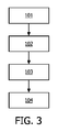
- 238000000034 method Methods 0.000 title claims description 54
- 238000002591 computed tomography Methods 0.000 title description 105
- 238000000926 separation method Methods 0.000 title description 6
- 210000001519 tissue Anatomy 0.000 claims description 120
- 239000000126 substance Substances 0.000 claims description 57
- 210000000988 bone and bone Anatomy 0.000 claims description 36
- 238000001959 radiotherapy Methods 0.000 claims description 28
- 210000003484 anatomy Anatomy 0.000 claims description 23
- 238000002595 magnetic resonance imaging Methods 0.000 claims description 16
- 238000004458 analytical method Methods 0.000 claims description 15
- 238000011282 treatment Methods 0.000 claims description 15
- 230000005855 radiation Effects 0.000 claims description 12
- XLYOFNOQVPJJNP-UHFFFAOYSA-N water Substances O XLYOFNOQVPJJNP-UHFFFAOYSA-N 0.000 claims description 11
- 238000004422 calculation algorithm Methods 0.000 claims description 8
- 230000001054 cortical effect Effects 0.000 claims description 8
- RGCLLPNLLBQHPF-HJWRWDBZSA-N phosphamidon Chemical compound CCN(CC)C(=O)C(\Cl)=C(/C)OP(=O)(OC)OC RGCLLPNLLBQHPF-HJWRWDBZSA-N 0.000 claims description 8
- 238000012545 processing Methods 0.000 claims description 8
- 210000001185 bone marrow Anatomy 0.000 claims description 7
- 238000004590 computer program Methods 0.000 claims description 6
- 238000000605 extraction Methods 0.000 claims description 4
- 239000000463 material Substances 0.000 description 25
- 238000003384 imaging method Methods 0.000 description 9
- 230000008901 benefit Effects 0.000 description 7
- 238000004891 communication Methods 0.000 description 7
- 238000004364 calculation method Methods 0.000 description 6
- 238000013170 computed tomography imaging Methods 0.000 description 6
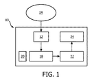
- 238000010586 diagram Methods 0.000 description 6
- 210000004197 pelvis Anatomy 0.000 description 6
- 230000011218 segmentation Effects 0.000 description 6
- 238000002059 diagnostic imaging Methods 0.000 description 4
- 238000002600 positron emission tomography Methods 0.000 description 4
- 210000000577 adipose tissue Anatomy 0.000 description 3
- 230000001419 dependent effect Effects 0.000 description 3
- 210000002307 prostate Anatomy 0.000 description 3
- 101100509468 Arabidopsis thaliana JASON gene Proteins 0.000 description 2
- 206010060862 Prostate cancer Diseases 0.000 description 2
- 208000000236 Prostatic Neoplasms Diseases 0.000 description 2
- 210000004556 brain Anatomy 0.000 description 2
- 238000005094 computer simulation Methods 0.000 description 2
- 238000004980 dosimetry Methods 0.000 description 2
- 230000000968 intestinal effect Effects 0.000 description 2
- 210000003734 kidney Anatomy 0.000 description 2
- 210000004185 liver Anatomy 0.000 description 2
- 238000013507 mapping Methods 0.000 description 2
- 238000009206 nuclear medicine Methods 0.000 description 2
- 210000000056 organ Anatomy 0.000 description 2
- 230000008569 process Effects 0.000 description 2
- PXFBZOLANLWPMH-UHFFFAOYSA-N 16-Epiaffinine Natural products C1C(C2=CC=CC=C2N2)=C2C(=O)CC2C(=CC)CN(C)C1C2CO PXFBZOLANLWPMH-UHFFFAOYSA-N 0.000 description 1
- 238000012935 Averaging Methods 0.000 description 1
- 241000271566 Aves Species 0.000 description 1
- 241000282412 Homo Species 0.000 description 1
- 241000124008 Mammalia Species 0.000 description 1
- 241001465754 Metazoa Species 0.000 description 1
- 230000003044 adaptive effect Effects 0.000 description 1
- 238000013459 approach Methods 0.000 description 1
- 230000009286 beneficial effect Effects 0.000 description 1
- 210000000476 body water Anatomy 0.000 description 1
- 230000037118 bone strength Effects 0.000 description 1
- 239000000969 carrier Substances 0.000 description 1
- 230000008859 change Effects 0.000 description 1
- 238000000205 computational method Methods 0.000 description 1
- 238000012937 correction Methods 0.000 description 1
- 238000013016 damping Methods 0.000 description 1
- 230000004069 differentiation Effects 0.000 description 1
- 201000010099 disease Diseases 0.000 description 1
- 208000037265 diseases, disorders, signs and symptoms Diseases 0.000 description 1
- 239000012153 distilled water Substances 0.000 description 1
- 238000005516 engineering process Methods 0.000 description 1
- 210000002468 fat body Anatomy 0.000 description 1
- 239000000835 fiber Substances 0.000 description 1
- 230000006870 function Effects 0.000 description 1
- 230000007614 genetic variation Effects 0.000 description 1
- 210000003128 head Anatomy 0.000 description 1
- 238000003709 image segmentation Methods 0.000 description 1
- 210000000936 intestine Anatomy 0.000 description 1
- 230000003902 lesion Effects 0.000 description 1
- 230000005415 magnetization Effects 0.000 description 1
- 230000000873 masking effect Effects 0.000 description 1
- 238000005259 measurement Methods 0.000 description 1
- 238000010295 mobile communication Methods 0.000 description 1
- 230000003287 optical effect Effects 0.000 description 1
- 238000005457 optimization Methods 0.000 description 1
- 238000003909 pattern recognition Methods 0.000 description 1
- 238000011158 quantitative evaluation Methods 0.000 description 1
- 210000000664 rectum Anatomy 0.000 description 1
- 238000002603 single-photon emission computed tomography Methods 0.000 description 1
- 239000007787 solid Substances 0.000 description 1
- 238000012549 training Methods 0.000 description 1
- 238000012546 transfer Methods 0.000 description 1
- 230000009466 transformation Effects 0.000 description 1
- 210000003932 urinary bladder Anatomy 0.000 description 1
Images


Classifications
-
- G—PHYSICS
- G06—COMPUTING; CALCULATING OR COUNTING
- G06T—IMAGE DATA PROCESSING OR GENERATION, IN GENERAL
- G06T7/00—Image analysis
- G06T7/10—Segmentation; Edge detection
- G06T7/11—Region-based segmentation
-
- A—HUMAN NECESSITIES
- A61—MEDICAL OR VETERINARY SCIENCE; HYGIENE
- A61B—DIAGNOSIS; SURGERY; IDENTIFICATION
- A61B5/00—Measuring for diagnostic purposes; Identification of persons
- A61B5/05—Detecting, measuring or recording for diagnosis by means of electric currents or magnetic fields; Measuring using microwaves or radio waves
- A61B5/055—Detecting, measuring or recording for diagnosis by means of electric currents or magnetic fields; Measuring using microwaves or radio waves involving electronic [EMR] or nuclear [NMR] magnetic resonance, e.g. magnetic resonance imaging
-
- A—HUMAN NECESSITIES
- A61—MEDICAL OR VETERINARY SCIENCE; HYGIENE
- A61B—DIAGNOSIS; SURGERY; IDENTIFICATION
- A61B5/00—Measuring for diagnostic purposes; Identification of persons
- A61B5/72—Signal processing specially adapted for physiological signals or for diagnostic purposes
- A61B5/7271—Specific aspects of physiological measurement analysis
- A61B5/7278—Artificial waveform generation or derivation, e.g. synthesising signals from measured signals
-
- A—HUMAN NECESSITIES
- A61—MEDICAL OR VETERINARY SCIENCE; HYGIENE
- A61B—DIAGNOSIS; SURGERY; IDENTIFICATION
- A61B6/00—Apparatus for radiation diagnosis, e.g. combined with radiation therapy equipment
- A61B6/02—Devices for diagnosis sequentially in different planes; Stereoscopic radiation diagnosis
- A61B6/03—Computerised tomographs
- A61B6/032—Transmission computed tomography [CT]
-
- A—HUMAN NECESSITIES
- A61—MEDICAL OR VETERINARY SCIENCE; HYGIENE
- A61B—DIAGNOSIS; SURGERY; IDENTIFICATION
- A61B6/00—Apparatus for radiation diagnosis, e.g. combined with radiation therapy equipment
- A61B6/50—Clinical applications
- A61B6/505—Clinical applications involving diagnosis of bone
-
- A—HUMAN NECESSITIES
- A61—MEDICAL OR VETERINARY SCIENCE; HYGIENE
- A61B—DIAGNOSIS; SURGERY; IDENTIFICATION
- A61B6/00—Apparatus for radiation diagnosis, e.g. combined with radiation therapy equipment
- A61B6/52—Devices using data or image processing specially adapted for radiation diagnosis
- A61B6/5211—Devices using data or image processing specially adapted for radiation diagnosis involving processing of medical diagnostic data
- A61B6/5217—Devices using data or image processing specially adapted for radiation diagnosis involving processing of medical diagnostic data extracting a diagnostic or physiological parameter from medical diagnostic data
-
- A—HUMAN NECESSITIES
- A61—MEDICAL OR VETERINARY SCIENCE; HYGIENE
- A61N—ELECTROTHERAPY; MAGNETOTHERAPY; RADIATION THERAPY; ULTRASOUND THERAPY
- A61N5/00—Radiation therapy
- A61N5/10—X-ray therapy; Gamma-ray therapy; Particle-irradiation therapy
- A61N5/103—Treatment planning systems
-
- G—PHYSICS
- G01—MEASURING; TESTING
- G01R—MEASURING ELECTRIC VARIABLES; MEASURING MAGNETIC VARIABLES
- G01R33/00—Arrangements or instruments for measuring magnetic variables
- G01R33/20—Arrangements or instruments for measuring magnetic variables involving magnetic resonance
- G01R33/44—Arrangements or instruments for measuring magnetic variables involving magnetic resonance using nuclear magnetic resonance [NMR]
- G01R33/48—NMR imaging systems
- G01R33/4808—Multimodal MR, e.g. MR combined with positron emission tomography [PET], MR combined with ultrasound or MR combined with computed tomography [CT]
- G01R33/481—MR combined with positron emission tomography [PET] or single photon emission computed tomography [SPECT]
-
- G—PHYSICS
- G01—MEASURING; TESTING
- G01R—MEASURING ELECTRIC VARIABLES; MEASURING MAGNETIC VARIABLES
- G01R33/00—Arrangements or instruments for measuring magnetic variables
- G01R33/20—Arrangements or instruments for measuring magnetic variables involving magnetic resonance
- G01R33/44—Arrangements or instruments for measuring magnetic variables involving magnetic resonance using nuclear magnetic resonance [NMR]
- G01R33/48—NMR imaging systems
- G01R33/4808—Multimodal MR, e.g. MR combined with positron emission tomography [PET], MR combined with ultrasound or MR combined with computed tomography [CT]
- G01R33/4812—MR combined with X-ray or computed tomography [CT]
-
- G—PHYSICS
- G01—MEASURING; TESTING
- G01R—MEASURING ELECTRIC VARIABLES; MEASURING MAGNETIC VARIABLES
- G01R33/00—Arrangements or instruments for measuring magnetic variables
- G01R33/20—Arrangements or instruments for measuring magnetic variables involving magnetic resonance
- G01R33/44—Arrangements or instruments for measuring magnetic variables involving magnetic resonance using nuclear magnetic resonance [NMR]
- G01R33/48—NMR imaging systems
- G01R33/4828—Resolving the MR signals of different chemical species, e.g. water-fat imaging
-
- G—PHYSICS
- G01—MEASURING; TESTING
- G01R—MEASURING ELECTRIC VARIABLES; MEASURING MAGNETIC VARIABLES
- G01R33/00—Arrangements or instruments for measuring magnetic variables
- G01R33/20—Arrangements or instruments for measuring magnetic variables involving magnetic resonance
- G01R33/44—Arrangements or instruments for measuring magnetic variables involving magnetic resonance using nuclear magnetic resonance [NMR]
- G01R33/48—NMR imaging systems
- G01R33/54—Signal processing systems, e.g. using pulse sequences ; Generation or control of pulse sequences; Operator console
- G01R33/56—Image enhancement or correction, e.g. subtraction or averaging techniques, e.g. improvement of signal-to-noise ratio and resolution
- G01R33/5608—Data processing and visualization specially adapted for MR, e.g. for feature analysis and pattern recognition on the basis of measured MR data, segmentation of measured MR data, edge contour detection on the basis of measured MR data, for enhancing measured MR data in terms of signal-to-noise ratio by means of noise filtering or apodization, for enhancing measured MR data in terms of resolution by means for deblurring, windowing, zero filling, or generation of gray-scaled images, colour-coded images or images displaying vectors instead of pixels
-
- G—PHYSICS
- G06—COMPUTING; CALCULATING OR COUNTING
- G06T—IMAGE DATA PROCESSING OR GENERATION, IN GENERAL
- G06T11/00—2D [Two Dimensional] image generation
- G06T11/003—Reconstruction from projections, e.g. tomography
- G06T11/008—Specific post-processing after tomographic reconstruction, e.g. voxelisation, metal artifact correction
-
- G06T5/90—
-
- G—PHYSICS
- G06—COMPUTING; CALCULATING OR COUNTING
- G06T—IMAGE DATA PROCESSING OR GENERATION, IN GENERAL
- G06T7/00—Image analysis
- G06T7/30—Determination of transform parameters for the alignment of images, i.e. image registration
- G06T7/33—Determination of transform parameters for the alignment of images, i.e. image registration using feature-based methods
-
- G—PHYSICS
- G16—INFORMATION AND COMMUNICATION TECHNOLOGY [ICT] SPECIALLY ADAPTED FOR SPECIFIC APPLICATION FIELDS
- G16H—HEALTHCARE INFORMATICS, i.e. INFORMATION AND COMMUNICATION TECHNOLOGY [ICT] SPECIALLY ADAPTED FOR THE HANDLING OR PROCESSING OF MEDICAL OR HEALTHCARE DATA
- G16H50/00—ICT specially adapted for medical diagnosis, medical simulation or medical data mining; ICT specially adapted for detecting, monitoring or modelling epidemics or pandemics
- G16H50/50—ICT specially adapted for medical diagnosis, medical simulation or medical data mining; ICT specially adapted for detecting, monitoring or modelling epidemics or pandemics for simulation or modelling of medical disorders
-
- A—HUMAN NECESSITIES
- A61—MEDICAL OR VETERINARY SCIENCE; HYGIENE
- A61N—ELECTROTHERAPY; MAGNETOTHERAPY; RADIATION THERAPY; ULTRASOUND THERAPY
- A61N5/00—Radiation therapy
- A61N5/10—X-ray therapy; Gamma-ray therapy; Particle-irradiation therapy
- A61N5/1048—Monitoring, verifying, controlling systems and methods
- A61N5/1049—Monitoring, verifying, controlling systems and methods for verifying the position of the patient with respect to the radiation beam
- A61N2005/1055—Monitoring, verifying, controlling systems and methods for verifying the position of the patient with respect to the radiation beam using magnetic resonance imaging [MRI]
-
- G—PHYSICS
- G06—COMPUTING; CALCULATING OR COUNTING
- G06T—IMAGE DATA PROCESSING OR GENERATION, IN GENERAL
- G06T2207/00—Indexing scheme for image analysis or image enhancement
- G06T2207/10—Image acquisition modality
- G06T2207/10072—Tomographic images
- G06T2207/10081—Computed x-ray tomography [CT]
-
- G—PHYSICS
- G06—COMPUTING; CALCULATING OR COUNTING
- G06T—IMAGE DATA PROCESSING OR GENERATION, IN GENERAL
- G06T2207/00—Indexing scheme for image analysis or image enhancement
- G06T2207/10—Image acquisition modality
- G06T2207/10072—Tomographic images
- G06T2207/10088—Magnetic resonance imaging [MRI]
Applications Claiming Priority (3)
| Application Number | Priority Date | Filing Date | Title |
|---|---|---|---|
| EP14162319.9 | 2014-03-28 | ||
| EP14162319 | 2014-03-28 | ||
| PCT/EP2015/055738 WO2015144540A1 (fr) | 2014-03-28 | 2015-03-19 | Procédé et dispositif permettant de générer une ou plusieurs image(s) de tomographie informatique sur la base d'images de résonance magnétique à l'aide de la séparation de classe de tissus |
Publications (3)
| Publication Number | Publication Date |
|---|---|
| JP2017508561A JP2017508561A (ja) | 2017-03-30 |
| JP2017508561A5 JP2017508561A5 (fr) | 2018-03-01 |
| JP6378778B2 true JP6378778B2 (ja) | 2018-08-22 |
Family
ID=50478701
Family Applications (1)
| Application Number | Title | Priority Date | Filing Date |
|---|---|---|---|
| JP2016559279A Active JP6378778B2 (ja) | 2014-03-28 | 2015-03-19 | 組織種類分離の助けを借りて磁気共鳴画像に基づき1つ以上のコンピュータ断層撮影画像を生成する方法及び装置 |
Country Status (5)
| Country | Link |
|---|---|
| US (1) | US10223794B2 (fr) |
| EP (1) | EP3123443B1 (fr) |
| JP (1) | JP6378778B2 (fr) |
| CN (1) | CN106133790B (fr) |
| WO (1) | WO2015144540A1 (fr) |
Families Citing this family (25)
| Publication number | Priority date | Publication date | Assignee | Title |
|---|---|---|---|---|
| US10870017B2 (en) | 2015-03-20 | 2020-12-22 | Koninklijke Philips N.V. | Fall-back solution for uncertain regions in MRCAT images |
| US10102451B2 (en) | 2015-10-13 | 2018-10-16 | Elekta, Inc. | Pseudo-CT generation from MR data using tissue parameter estimation |
| CN108310683A (zh) * | 2015-11-17 | 2018-07-24 | 南京中硼联康医疗科技有限公司 | 基于医学影像数据的几何模型建立方法 |
| WO2017103237A1 (fr) * | 2015-12-18 | 2017-06-22 | Koninklijke Philips N.V. | Procédé de détermination d'une marge localement variable spécifique à un patient |
| JP6727319B2 (ja) * | 2016-03-10 | 2020-07-22 | コーニンクレッカ フィリップス エヌ ヴェKoninklijke Philips N.V. | Synthetic CTイメージング |
| CN110234400B (zh) | 2016-09-06 | 2021-09-07 | 医科达有限公司 | 用于生成合成医学图像的神经网络 |
| CN108310677B (zh) * | 2017-01-11 | 2020-02-28 | 南京中硼联康医疗科技有限公司 | 基于医学影像数据的平滑几何模型建立方法 |
| KR102536168B1 (ko) * | 2017-02-24 | 2023-05-25 | 바이엘 헬쓰케어 엘엘씨 | 시뮬레이션된 컴퓨터 단층 촬영(ct) 이미지들을 생성하기 위한 시스템들 및 방법들 |
| EP3379281A1 (fr) * | 2017-03-20 | 2018-09-26 | Koninklijke Philips N.V. | Segmentation d'image à l'aide de valeurs d'échelle de gris de référence |
| CN107133996B (zh) * | 2017-03-21 | 2020-08-04 | 上海联影医疗科技有限公司 | 产生用于pet数据重建的衰减图的方法及pet/ct系统 |
| EP3431007B1 (fr) * | 2017-07-21 | 2020-06-17 | Koninklijke Philips N.V. | Création d'ensembles de données de densité d'électrons à partir d'ensembles de données de tomodensitométrie spectrale |
| US11883685B2 (en) | 2017-08-24 | 2024-01-30 | Shanghai United Imaging Healthcare Co., Ltd. | Therapeutic system and method |
| CN109420259A (zh) | 2017-08-24 | 2019-03-05 | 上海联影医疗科技有限公司 | 治疗系统和使用治疗系统的方法 |
| WO2019111327A1 (fr) * | 2017-12-05 | 2019-06-13 | 株式会社DSi | Système de calcul automatique de frais techniques, procédé de calcul automatique de frais techniques et programme |
| EP3521849A1 (fr) * | 2018-02-02 | 2019-08-07 | Koninklijke Philips N.V. | Irm avec séparation graisse/eau |
| CN109215014B (zh) * | 2018-07-02 | 2022-03-04 | 中国科学院深圳先进技术研究院 | Ct图像预测模型的训练方法、装置、设备及存储介质 |
| CN109389603B (zh) * | 2018-09-10 | 2021-09-24 | 北京大学 | 一种基于预加重策略的全自动腰椎图像分割方法 |
| JP7129869B2 (ja) * | 2018-10-01 | 2022-09-02 | 富士フイルム株式会社 | 疾患領域抽出装置、方法及びプログラム |
| US11497566B2 (en) * | 2018-10-26 | 2022-11-15 | Biosense Webster (Israel) Ltd. | Loose mode for robot |
| EP3654278A1 (fr) * | 2018-11-19 | 2020-05-20 | Koninklijke Philips N.V. | Caractérisation des lésions dans des images radiologiques |
| CN112535488A (zh) * | 2019-09-23 | 2021-03-23 | 佳能医疗系统株式会社 | 解析装置 |
| JP6738003B1 (ja) * | 2019-10-30 | 2020-08-12 | 日本テクトシステムズ株式会社 | Mri画像に基づく解剖学的部位の抽出装置,方法,プログラム |
| WO2021206157A1 (fr) * | 2020-04-08 | 2021-10-14 | キヤノンメディカルシステムズ株式会社 | Dispositif de traitement d'informations médicales et procédé de traitement d'informations médicales |
| CN113706541B (zh) * | 2020-05-20 | 2024-04-19 | 青岛海信医疗设备股份有限公司 | 一种图像处理方法及装置 |
| US20220044399A1 (en) * | 2020-08-07 | 2022-02-10 | Washington University | Deep-Learning-based T1-Enhanced Selection of Linear Coefficients (DL-TESLA) for PET/MR Attenuation Correction |
Family Cites Families (5)
| Publication number | Priority date | Publication date | Assignee | Title |
|---|---|---|---|---|
| EP2229981A1 (fr) | 2009-03-17 | 2010-09-22 | Paul Scherrer Institut | Procédé d'évaluation de données de modèle de rayonnement dans des applications de rayonnement à faisceaux de particules |
| JP5364009B2 (ja) * | 2010-02-12 | 2013-12-11 | 富士フイルム株式会社 | 画像生成装置、画像生成方法、及びそのプログラム |
| US8588498B2 (en) | 2011-09-30 | 2013-11-19 | General Electric Company | System and method for segmenting bones on MR images |
| JP5706389B2 (ja) | 2011-12-20 | 2015-04-22 | 富士フイルム株式会社 | 画像処理装置および画像処理方法、並びに、画像処理プログラム |
| JP5741980B2 (ja) * | 2012-03-28 | 2015-07-01 | 国立研究開発法人放射線医学総合研究所 | Mr画像からのpet吸収補正画像生成方法及びコンピュータプログラム |
-
2015
- 2015-03-19 CN CN201580016590.XA patent/CN106133790B/zh active Active
- 2015-03-19 US US15/127,426 patent/US10223794B2/en active Active
- 2015-03-19 JP JP2016559279A patent/JP6378778B2/ja active Active
- 2015-03-19 EP EP15710518.0A patent/EP3123443B1/fr active Active
- 2015-03-19 WO PCT/EP2015/055738 patent/WO2015144540A1/fr active Application Filing
Also Published As
| Publication number | Publication date |
|---|---|
| CN106133790A (zh) | 2016-11-16 |
| CN106133790B (zh) | 2020-09-29 |
| EP3123443A1 (fr) | 2017-02-01 |
| US20180174298A1 (en) | 2018-06-21 |
| US10223794B2 (en) | 2019-03-05 |
| JP2017508561A (ja) | 2017-03-30 |
| EP3123443B1 (fr) | 2018-06-13 |
| WO2015144540A1 (fr) | 2015-10-01 |
Similar Documents
| Publication | Publication Date | Title |
|---|---|---|
| JP6378778B2 (ja) | 組織種類分離の助けを借りて磁気共鳴画像に基づき1つ以上のコンピュータ断層撮影画像を生成する方法及び装置 | |
| JP7405818B2 (ja) | 変形可能テンプレートを使用して最適化された電極位置を有するttフィールドを用いて患者を治療する | |
| JP6567179B2 (ja) | 特徴回帰モデルを用いたmrデータからの疑似ct生成 | |
| US8787648B2 (en) | CT surrogate by auto-segmentation of magnetic resonance images | |
| JP7030050B2 (ja) | 組織パラメータ推定を用いたmrデータからの疑似ct生成 | |
| US10149987B2 (en) | Method and system for generating synthetic electron density information for dose calculations based on MRI | |
| US8953856B2 (en) | Method and system for registering a medical image | |
| JP2017508561A5 (fr) | ||
| US11443441B2 (en) | Deep inspiration breath-hold setup using x-ray imaging | |
| US10803354B2 (en) | Cross-modality image synthesis | |
| JP6387108B2 (ja) | 擬似ctハウスフィールドユニット値を推定する方法、磁気共鳴システム及びコンピュータプログラム | |
| Eiben et al. | Symmetric biomechanically guided prone-to-supine breast image registration | |
| EP3468668B1 (fr) | Suivi de tissu mou à l'aide d'un rendu de volume physiologique | |
| Mi et al. | Joint tumor growth prediction and tumor segmentation on therapeutic follow-up PET images | |
| Lappas et al. | Inter-observer variability of organ contouring for preclinical studies with cone beam Computed Tomography imaging | |
| US8265361B2 (en) | Automatic transfer of outlined objects from one data set into another data set | |
| WO2023110116A1 (fr) | Contour d'image respiratoire sans ct pour la planification d'une radiothérapie | |
| Dowling et al. | Image synthesis for MRI-only radiotherapy treatment planning | |
| Balasubramanian et al. | Registration of PET and MR images of human brain using normalized cross correlation algorithm and spatial transformation techniques |
Legal Events
| Date | Code | Title | Description |
|---|---|---|---|
| A521 | Request for written amendment filed |
Free format text: JAPANESE INTERMEDIATE CODE: A523 Effective date: 20180117 |
|
| A621 | Written request for application examination |
Free format text: JAPANESE INTERMEDIATE CODE: A621 Effective date: 20180117 |
|
| A871 | Explanation of circumstances concerning accelerated examination |
Free format text: JAPANESE INTERMEDIATE CODE: A871 Effective date: 20180117 |
|
| A975 | Report on accelerated examination |
Free format text: JAPANESE INTERMEDIATE CODE: A971005 Effective date: 20180320 |
|
| A131 | Notification of reasons for refusal |
Free format text: JAPANESE INTERMEDIATE CODE: A131 Effective date: 20180327 |
|
| A521 | Request for written amendment filed |
Free format text: JAPANESE INTERMEDIATE CODE: A523 Effective date: 20180619 |
|
| TRDD | Decision of grant or rejection written | ||
| A01 | Written decision to grant a patent or to grant a registration (utility model) |
Free format text: JAPANESE INTERMEDIATE CODE: A01 Effective date: 20180703 |
|
| A61 | First payment of annual fees (during grant procedure) |
Free format text: JAPANESE INTERMEDIATE CODE: A61 Effective date: 20180727 |
|
| R150 | Certificate of patent or registration of utility model |
Ref document number: 6378778 Country of ref document: JP Free format text: JAPANESE INTERMEDIATE CODE: R150 |
|
| R250 | Receipt of annual fees |
Free format text: JAPANESE INTERMEDIATE CODE: R250 |
|
| R250 | Receipt of annual fees |
Free format text: JAPANESE INTERMEDIATE CODE: R250 |
|
| R250 | Receipt of annual fees |
Free format text: JAPANESE INTERMEDIATE CODE: R250 |