JP6060525B2 - 眼底検査装置 - Google Patents
眼底検査装置 Download PDFInfo
- Publication number
- JP6060525B2 JP6060525B2 JP2012123939A JP2012123939A JP6060525B2 JP 6060525 B2 JP6060525 B2 JP 6060525B2 JP 2012123939 A JP2012123939 A JP 2012123939A JP 2012123939 A JP2012123939 A JP 2012123939A JP 6060525 B2 JP6060525 B2 JP 6060525B2
- Authority
- JP
- Japan
- Prior art keywords
- fundus
- target
- inspection
- inspection target
- eye
- Prior art date
- Legal status (The legal status is an assumption and is not a legal conclusion. Google has not performed a legal analysis and makes no representation as to the accuracy of the status listed.)
- Expired - Fee Related
Links
- 238000007689 inspection Methods 0.000 claims description 100
- 238000012360 testing method Methods 0.000 claims description 24
- 230000004382 visual function Effects 0.000 claims description 16
- 230000003287 optical effect Effects 0.000 description 62
- 210000000608 photoreceptor cell Anatomy 0.000 description 36
- 230000000007 visual effect Effects 0.000 description 36
- 230000008859 change Effects 0.000 description 29
- 238000003384 imaging method Methods 0.000 description 25
- 239000004973 liquid crystal related substance Substances 0.000 description 20
- 230000035945 sensitivity Effects 0.000 description 19
- 210000004027 cell Anatomy 0.000 description 15
- 238000005286 illumination Methods 0.000 description 11
- 230000007423 decrease Effects 0.000 description 8
- 230000002093 peripheral effect Effects 0.000 description 7
- 230000004044 response Effects 0.000 description 7
- 238000010586 diagram Methods 0.000 description 6
- 238000005259 measurement Methods 0.000 description 6
- 230000004907 flux Effects 0.000 description 5
- 108091008695 photoreceptors Proteins 0.000 description 5
- 206010025421 Macule Diseases 0.000 description 4
- 238000001514 detection method Methods 0.000 description 4
- 230000007246 mechanism Effects 0.000 description 4
- 230000002207 retinal effect Effects 0.000 description 4
- 208000003098 Ganglion Cysts Diseases 0.000 description 3
- 208000005400 Synovial Cyst Diseases 0.000 description 3
- 210000001525 retina Anatomy 0.000 description 3
- 238000011179 visual inspection Methods 0.000 description 3
- 206010027146 Melanoderma Diseases 0.000 description 2
- 210000004087 cornea Anatomy 0.000 description 2
- 230000006866 deterioration Effects 0.000 description 2
- 210000004220 fundus oculi Anatomy 0.000 description 2
- 210000001747 pupil Anatomy 0.000 description 2
- 238000000926 separation method Methods 0.000 description 2
- 208000010412 Glaucoma Diseases 0.000 description 1
- 206010034972 Photosensitivity reaction Diseases 0.000 description 1
- 230000004075 alteration Effects 0.000 description 1
- 238000013459 approach Methods 0.000 description 1
- 230000005540 biological transmission Effects 0.000 description 1
- 230000015572 biosynthetic process Effects 0.000 description 1
- 230000004300 dark adaptation Effects 0.000 description 1
- 230000007547 defect Effects 0.000 description 1
- 238000003745 diagnosis Methods 0.000 description 1
- 239000000835 fiber Substances 0.000 description 1
- 229910052736 halogen Inorganic materials 0.000 description 1
- 150000002367 halogens Chemical class 0.000 description 1
- 230000006872 improvement Effects 0.000 description 1
- 238000003780 insertion Methods 0.000 description 1
- 230000037431 insertion Effects 0.000 description 1
- 238000000034 method Methods 0.000 description 1
- 238000010606 normalization Methods 0.000 description 1
- 210000003733 optic disk Anatomy 0.000 description 1
- 210000001328 optic nerve Anatomy 0.000 description 1
- 230000036211 photosensitivity Effects 0.000 description 1
- 210000002763 pyramidal cell Anatomy 0.000 description 1
- 230000005855 radiation Effects 0.000 description 1
- 230000000638 stimulation Effects 0.000 description 1
- 238000002834 transmittance Methods 0.000 description 1
Images


Landscapes
- Eye Examination Apparatus (AREA)
Description
このため、従来技術のように眼底中心部と周辺部に投影される検査視標のサイズが同じであると、刺激視標に対して含まれる視細胞の数が変わることになり、特に眼底中心部において視機能感度の低下を検出しづらくなる可能性がある。
撮影部3の患者側には、患者に装置内部を覗き込ませる撮影窓9、眼(網膜)の視機能検査時などに患者が応答信号を入力するための応答ボタン7bがある。
観察照明光学系は、近赤外光の光束を照射する照明光源11、近赤外光を透過する赤外フィルター12、コンデンサレンズ13、コンデンサレンズ13とリングスリット17との間に配置されたダイクロイックミラー16、リングスリット17から孔あきミラー22までの光学系と、対物レンズ25を有する。
眼底撮影光学系は、対物レンズ25,撮影絞り31から結像レンズ33までの光学系を眼底観察光学系と共用する。また眼底撮影光学系は、可視域に感度を有する撮影用の二次元撮像素子35を備え、可視光源14で照明された眼底像が撮像素子35で撮影される。なお、撮影絞り31は眼Eの瞳孔と略共役な位置に置かれる。フォーカシングレンズ32はモータを備える移動機構49で光軸に沿って移動される。
また、本実施形態では穴あきミラーの開口付近(眼底の略共役位置)に点光源27が設けられており、眼底観察時に点光源27が点灯されることで、眼底に形成されたワーキングドットWによる作動距離方向のアライメントが行われるようになっている。
レバー45は光軸上に置かれ、スポットミラー44は光軸上を避けた位置に置かれるようにレバー45の先端に取り付けられる。これにより眼底の観察時に、スポットミラー44からの反射光が眼底上の光軸L1上を避けた位置に投影される。
図3に視標呈示部100の例を示す。視標呈示部100は、光源101、分光手段としての波長選択ミラー102,103、反射ミラー104〜106、液晶パネル110〜112、クロスダイクロプリズム120、光路分岐用のビームスプリッター130を持つ。光源101には白色のLED光源が使用される。これ以外にも光源101には、ハロゲンランプ等の周知の熱輻射光源や、レーザ光源などを使用できる。
クロスダイクロプリズム120は、各液晶パネル110〜112を透過した光束を同軸にする。これにより各液晶パネル110〜112に形成されたRGB毎の画像が合成されて一つの画像が形成される。
なお視機能に関する細胞には神経節細胞、視細胞等がある。以下では視細胞数(密度)に基づく視機能検査を行う例を説明する。

装置を起動させて患者の顔を装置1に近づけ、撮影眼(患者眼E)を撮影窓9に合わせて装置内部を覗き込ませてから、前眼部像を用いた位置合わせ(アライメント)を開始する。制御部80によって駆動手段26aの駆動で光軸L1に前眼部観察補助レンズ26が置かれ、光源35a、35bが点灯されると患者眼Eの前眼部が照明され、角膜上にアライメント視標が投影される。また制御部80は、視標呈示部100で患者眼Eに固視標を呈示させる。具体的には制御部80は光源101を点灯させた状態で、各液晶パネル110〜112の画素を駆動させ、各液晶パネル110〜112を透過した光束によって、光軸L2に対応する位置に固視標を形成する。
図6はモニタ8に表示される眼底像の例であり、図6(a)に眼底のフォーカスが合っていない状態、図6(b)に眼底のフォーカスが合っている状態が示されている。制御部80は、撮像素子38の撮像範囲の輝度分布に基づき、フォーカス指標S1,S2の位置を特定し、検出されたフォーカス指標S1,S2間の距離(分離状態)を求め、検出結果に基づくフォーカス合わせを行う。
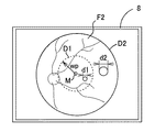
モニタ8に眼底像F2が鮮明に映る状態となると、制御部80によって視野計測中に生じる眼Eの移動及び回旋により生じる眼Eと撮影部3(光軸L1)の位置ずれを補正するオートアライメント、呈示視標の位置ずれを補正するトラッキングが行われる。なおオートアライメント及びトラッキングについての詳細な説明は特開2011‐255045号公報を参照されたい。
例えば検査視標の呈示位置が手動で設定される場合、制御部80はコントロール部7aからの入力信号に基づき、選択された検査視標の呈示位置(座標)を求める。そしてメモリ83に記憶されている視細胞密度の変化情報又は所定の演算を行うことで、選択された呈示位置(座標)に対する所定の投影径(サイズ)の検査視標を形成するよう視標呈示部100を駆動制御する。
なお検者に検査視標の大きさを分かり易く示すために、モニタ8に検査視標のグラフィックを表示しても良い。
8 モニタ
10 照明光学系
30 観察・撮影光学系
40 フォーカス指標投影光学系
70 視標呈示光学系
80 制御部
83 メモリ
100 視標呈示部
110、111、112 液晶パネル
Claims (2)
- 被検眼眼底の検査位置に検査視標を投影して眼底の検査を行う眼底検査装置であって、
被検眼に呈示して誘導するための固視標を形成する固視標形成手段と、
被検眼眼底に対する検査視標による検査位置を設定する検査視標設定手段と、
該検査視標設定手段にて設定された前記検査位置に対応する被検眼眼底の検査位置に前記検査視標を投影可能な視標投影手段と、
前記固視標形成手段によって形成される前記固視標の座標と前記検査視標設定手段により設定される前記検査視標の座標に基づいた前記固視標に対する前記検査視標のラジアル方向の距離に応じ、該ラジアル方向の距離が相対的に近い前記検査視標の投影径に対して相対的に遠い前記検査視標の投影径が大きくなるように前記視標投影手段を制御する制御手段と、
を備えることを特徴とする眼底検査装置。 - 請求項1の眼底検査装置であって、
前記検査視標設定手段は更に、眼底上での検査位置及び視機能に関する細胞の種類に応じて前記検査視標の投影条件を設定する眼底検査装置。
Priority Applications (1)
| Application Number | Priority Date | Filing Date | Title |
|---|---|---|---|
| JP2012123939A JP6060525B2 (ja) | 2012-05-31 | 2012-05-31 | 眼底検査装置 |
Applications Claiming Priority (1)
| Application Number | Priority Date | Filing Date | Title |
|---|---|---|---|
| JP2012123939A JP6060525B2 (ja) | 2012-05-31 | 2012-05-31 | 眼底検査装置 |
Publications (3)
| Publication Number | Publication Date |
|---|---|
| JP2013248075A JP2013248075A (ja) | 2013-12-12 |
| JP2013248075A5 JP2013248075A5 (ja) | 2015-07-16 |
| JP6060525B2 true JP6060525B2 (ja) | 2017-01-18 |
Family
ID=49847410
Family Applications (1)
| Application Number | Title | Priority Date | Filing Date |
|---|---|---|---|
| JP2012123939A Expired - Fee Related JP6060525B2 (ja) | 2012-05-31 | 2012-05-31 | 眼底検査装置 |
Country Status (1)
| Country | Link |
|---|---|
| JP (1) | JP6060525B2 (ja) |
Cited By (1)
| Publication number | Priority date | Publication date | Assignee | Title |
|---|---|---|---|---|
| US11120577B2 (en) | 2017-02-09 | 2021-09-14 | Komatsu Ltd. | Position measurement system, work machine, and position measurement method |
Families Citing this family (3)
| Publication number | Priority date | Publication date | Assignee | Title |
|---|---|---|---|---|
| JP6898969B2 (ja) * | 2015-03-30 | 2021-07-07 | キヤノン株式会社 | 眼科情報処理システムおよび眼科情報処理方法 |
| JP6750068B2 (ja) * | 2019-05-30 | 2020-09-02 | 株式会社トプコン | 眼底解析装置 |
| JP2022025239A (ja) * | 2020-07-29 | 2022-02-10 | 株式会社クリュートメディカルシステムズ | 視覚検査装置、視覚検査システム及び視覚検査プログラム |
Family Cites Families (1)
| Publication number | Priority date | Publication date | Assignee | Title |
|---|---|---|---|---|
| JP2009530062A (ja) * | 2006-03-21 | 2009-08-27 | ノバビジョン, インコーポレイテッド | 治療的な視覚刺激を配分するための過程および装置 |
-
2012
- 2012-05-31 JP JP2012123939A patent/JP6060525B2/ja not_active Expired - Fee Related
Cited By (1)
| Publication number | Priority date | Publication date | Assignee | Title |
|---|---|---|---|---|
| US11120577B2 (en) | 2017-02-09 | 2021-09-14 | Komatsu Ltd. | Position measurement system, work machine, and position measurement method |
Also Published As
| Publication number | Publication date |
|---|---|
| JP2013248075A (ja) | 2013-12-12 |
Similar Documents
| Publication | Publication Date | Title |
|---|---|---|
| JP5606813B2 (ja) | 眼科装置 | |
| JP6354979B2 (ja) | 眼底撮影装置 | |
| JP6899632B2 (ja) | 眼科撮影装置 | |
| JP2016041221A (ja) | 眼科撮影装置およびその制御方法 | |
| JP2009291409A (ja) | 眼屈折力測定装置 | |
| JP5101370B2 (ja) | 眼底撮影装置 | |
| JP2018047049A (ja) | 自覚式検眼装置、及び自覚式検眼プログラム | |
| JP2018047050A (ja) | 検眼装置及び検眼プログラム | |
| JP2017080136A (ja) | 眼科装置 | |
| JP6604020B2 (ja) | 眼底撮像装置及び眼底撮像プログラム | |
| JP5953740B2 (ja) | 眼底検査装置 | |
| JP6060525B2 (ja) | 眼底検査装置 | |
| JP2016158721A (ja) | 眼科装置 | |
| JP6736356B2 (ja) | 眼科装置 | |
| JP6452977B2 (ja) | 眼科撮影装置及びその制御方法 | |
| JP6003234B2 (ja) | 眼底撮影装置 | |
| JP2016049243A (ja) | 眼科装置 | |
| JP2013244363A (ja) | 眼底撮影装置 | |
| JP7607099B2 (ja) | 眼科装置 | |
| JP2007275160A (ja) | 眼科装置 | |
| JP4851176B2 (ja) | 視標提示光学装置 | |
| JP2013027672A (ja) | 眼底撮影装置 | |
| JP6557388B2 (ja) | 眼科撮影装置 | |
| JP2017143919A (ja) | 眼科装置 | |
| JP2017143918A (ja) | 眼科装置 |
Legal Events
| Date | Code | Title | Description |
|---|---|---|---|
| A521 | Request for written amendment filed |
Free format text: JAPANESE INTERMEDIATE CODE: A523 Effective date: 20150527 |
|
| A621 | Written request for application examination |
Free format text: JAPANESE INTERMEDIATE CODE: A621 Effective date: 20150527 |
|
| A977 | Report on retrieval |
Free format text: JAPANESE INTERMEDIATE CODE: A971007 Effective date: 20160329 |
|
| A131 | Notification of reasons for refusal |
Free format text: JAPANESE INTERMEDIATE CODE: A131 Effective date: 20160419 |
|
| A521 | Request for written amendment filed |
Free format text: JAPANESE INTERMEDIATE CODE: A523 Effective date: 20160620 |
|
| TRDD | Decision of grant or rejection written | ||
| A01 | Written decision to grant a patent or to grant a registration (utility model) |
Free format text: JAPANESE INTERMEDIATE CODE: A01 Effective date: 20161115 |
|
| A61 | First payment of annual fees (during grant procedure) |
Free format text: JAPANESE INTERMEDIATE CODE: A61 Effective date: 20161128 |
|
| R150 | Certificate of patent or registration of utility model |
Ref document number: 6060525 Country of ref document: JP Free format text: JAPANESE INTERMEDIATE CODE: R150 |
|
| R250 | Receipt of annual fees |
Free format text: JAPANESE INTERMEDIATE CODE: R250 |
|
| R250 | Receipt of annual fees |
Free format text: JAPANESE INTERMEDIATE CODE: R250 |
|
| R250 | Receipt of annual fees |
Free format text: JAPANESE INTERMEDIATE CODE: R250 |
|
| LAPS | Cancellation because of no payment of annual fees |