JP5944402B2 - PET calibration for variable simultaneous windows - Google Patents
PET calibration for variable simultaneous windows Download PDFInfo
- Publication number
- JP5944402B2 JP5944402B2 JP2013539383A JP2013539383A JP5944402B2 JP 5944402 B2 JP5944402 B2 JP 5944402B2 JP 2013539383 A JP2013539383 A JP 2013539383A JP 2013539383 A JP2013539383 A JP 2013539383A JP 5944402 B2 JP5944402 B2 JP 5944402B2
- Authority
- JP
- Japan
- Prior art keywords
- simultaneous
- window
- timing
- data
- windows
- Prior art date
- Legal status (The legal status is an assumption and is not a legal conclusion. Google has not performed a legal analysis and makes no representation as to the accuracy of the status listed.)
- Expired - Fee Related
Links
- 238000000034 method Methods 0.000 claims description 37
- 238000010606 normalization Methods 0.000 claims description 23
- 238000012937 correction Methods 0.000 claims description 21
- 230000005855 radiation Effects 0.000 claims description 14
- 230000002285 radioactive effect Effects 0.000 claims description 13
- 238000012545 processing Methods 0.000 claims description 4
- 239000012857 radioactive material Substances 0.000 claims description 4
- 230000035945 sensitivity Effects 0.000 claims description 3
- 238000004590 computer program Methods 0.000 claims 1
- 238000002600 positron emission tomography Methods 0.000 description 40
- 238000001514 detection method Methods 0.000 description 7
- 238000013459 approach Methods 0.000 description 5
- 230000008901 benefit Effects 0.000 description 4
- 230000006870 function Effects 0.000 description 4
- 230000004044 response Effects 0.000 description 4
- 238000012986 modification Methods 0.000 description 3
- 230000004048 modification Effects 0.000 description 3
- 238000012805 post-processing Methods 0.000 description 3
- 238000013480 data collection Methods 0.000 description 2
- CYTYCFOTNPOANT-UHFFFAOYSA-N Perchloroethylene Chemical compound ClC(Cl)=C(Cl)Cl CYTYCFOTNPOANT-UHFFFAOYSA-N 0.000 description 1
- 230000009286 beneficial effect Effects 0.000 description 1
- 238000011088 calibration curve Methods 0.000 description 1
- 238000006243 chemical reaction Methods 0.000 description 1
- 238000002059 diagnostic imaging Methods 0.000 description 1
- 230000005251 gamma ray Effects 0.000 description 1
- 239000000463 material Substances 0.000 description 1
- 238000005259 measurement Methods 0.000 description 1
- 238000002610 neuroimaging Methods 0.000 description 1
- 230000003287 optical effect Effects 0.000 description 1
- 230000000737 periodic effect Effects 0.000 description 1
- 230000002093 peripheral effect Effects 0.000 description 1
- 230000005258 radioactive decay Effects 0.000 description 1
- 230000009467 reduction Effects 0.000 description 1
Images


Classifications
-
- G—PHYSICS
- G01—MEASURING; TESTING
- G01T—MEASUREMENT OF NUCLEAR OR X-RADIATION
- G01T1/00—Measuring X-radiation, gamma radiation, corpuscular radiation, or cosmic radiation
- G01T1/16—Measuring radiation intensity
- G01T1/161—Applications in the field of nuclear medicine, e.g. in vivo counting
- G01T1/164—Scintigraphy
- G01T1/1641—Static instruments for imaging the distribution of radioactivity in one or two dimensions using one or several scintillating elements; Radio-isotope cameras
- G01T1/1648—Ancillary equipment for scintillation cameras, e.g. reference markers, devices for removing motion artifacts, calibration devices
-
- A—HUMAN NECESSITIES
- A61—MEDICAL OR VETERINARY SCIENCE; HYGIENE
- A61B—DIAGNOSIS; SURGERY; IDENTIFICATION
- A61B6/00—Apparatus for radiation diagnosis, e.g. combined with radiation therapy equipment
- A61B6/02—Devices for diagnosis sequentially in different planes; Stereoscopic radiation diagnosis
- A61B6/03—Computerised tomographs
- A61B6/037—Emission tomography
-
- A—HUMAN NECESSITIES
- A61—MEDICAL OR VETERINARY SCIENCE; HYGIENE
- A61B—DIAGNOSIS; SURGERY; IDENTIFICATION
- A61B6/00—Apparatus for radiation diagnosis, e.g. combined with radiation therapy equipment
- A61B6/58—Testing, adjusting or calibrating apparatus or devices for radiation diagnosis
- A61B6/582—Calibration
- A61B6/583—Calibration using calibration phantoms
-
- G—PHYSICS
- G01—MEASURING; TESTING
- G01T—MEASUREMENT OF NUCLEAR OR X-RADIATION
- G01T1/00—Measuring X-radiation, gamma radiation, corpuscular radiation, or cosmic radiation
- G01T1/29—Measurement performed on radiation beams, e.g. position or section of the beam; Measurement of spatial distribution of radiation
- G01T1/2914—Measurement of spatial distribution of radiation
- G01T1/2964—Scanners
-
- G—PHYSICS
- G01—MEASURING; TESTING
- G01T—MEASUREMENT OF NUCLEAR OR X-RADIATION
- G01T1/00—Measuring X-radiation, gamma radiation, corpuscular radiation, or cosmic radiation
- G01T1/29—Measurement performed on radiation beams, e.g. position or section of the beam; Measurement of spatial distribution of radiation
- G01T1/2914—Measurement of spatial distribution of radiation
- G01T1/2985—In depth localisation, e.g. using positron emitters; Tomographic imaging (longitudinal and transverse section imaging; apparatus for radiation diagnosis sequentially in different planes, steroscopic radiation diagnosis)
-
- G—PHYSICS
- G01—MEASURING; TESTING
- G01T—MEASUREMENT OF NUCLEAR OR X-RADIATION
- G01T7/00—Details of radiation-measuring instruments
Description
本願は、特にPETスキャナ較正を含むポジトロンエミッショントモグラフィ(PET)システムに特定の用途を見出す。しかしながら、記述される技法は、他の医用イメージング装置較正システム、他の較正シナリオ又は他のスキャナ較正技法にも用途を見出すことができることが分かるであろう。 The present application finds particular application in positron emission tomography (PET) systems, particularly including PET scanner calibration. However, it will be appreciated that the described techniques may find use in other medical imaging device calibration systems, other calibration scenarios, or other scanner calibration techniques.
一般的なPETスキャナは、米国特許第7,718,954号明細書に記載されているように、不均一な3D検出器応答を補正するために正規化較正を実施し、これは一般に約6時間かかる。更に、このようなスキャナは、画像内のカウントの放射能濃度への変換を提供する標準摂取率(SUV)較正を用いる。この較正は、崩壊するF−18線源を使用し、完了するのに11時間又はそれ以上かかりうる。 A typical PET scanner performs normalization calibration to correct for non-uniform 3D detector response, as described in US Pat. No. 7,718,954, which generally takes about 6 hours. In addition, such scanners use a standard uptake rate (SUV) calibration that provides conversion of counts in the image to radioactivity concentrations. This calibration uses a collapsing F-18 source and can take 11 hours or more to complete.
PETスキャナによって測定され、同時タイミングウィンドウ(例えば6ナノ秒)内に生じるいかなるイベントも、同時イベントとして、すなわち有効なラインオブレスポンス(LOR)を規定するものとして扱われ、例えば2つのランダムな単一イベントが同時イベントとして扱われる。ランダムイベントが有効であるとされる頻度を低減することが、いくつかの理由により有益である。例えば、ランダムイベントの低減は、システムの最大NECR(NEMA NU−2標準、Noise Equivalent Count Rate)性能を高め、再構成処理のためのデータ量を低減する(リストモード再構成のスピードを速める)。更に、ランダムイベントを低減することは、再構成の間に行われる必要がある補正の大きさを低減し、有効な同時イベントを取得するために、PETスキャナに関するより多くの帯域幅を提供する。 Any event measured by a PET scanner and occurring within a simultaneous timing window (eg 6 nanoseconds) is treated as a simultaneous event, ie defining an effective line of response (LOR), for example two random single Events are treated as simultaneous events. Reducing the frequency at which random events are considered useful is beneficial for several reasons. For example, the reduction of random events increases the maximum NECR (NEMA NU-2 standard, Noise Equivalent Count Rate) performance of the system and reduces the amount of data for reconfiguration processing (increases the speed of list mode reconfiguration). Furthermore, reducing random events reduces the amount of correction that needs to be done during reconstruction and provides more bandwidth for the PET scanner to obtain valid simultaneous events.
イメージングされる対象をカバーするのに必要とされる最小値まで同時タイミングウィンドウを低減することによって、ランダムイベントを低減する試みがなされている(米国特許第7,626,171号参照)。しかしながら、このようなアプローチにおいて、低減される同時ウィンドウは、システムの計数特性を変化させ、従って、使用される同時ウィンドウ設定ごとに、別個のSUV較正が必要とされる。 Attempts have been made to reduce random events by reducing the simultaneous timing window to the minimum required to cover the object being imaged (see US Pat. No. 7,626,171). However, in such an approach, the reduced simultaneous window changes the counting characteristics of the system and therefore a separate SUV calibration is required for each simultaneous window setting used.
今日、市販のPETスキャナは、例えば約6ナノ秒という固定の同時ウィンドウを有する。しかしながら、同時ウィンドウは、ある患者、特に比較的小さい患者の場合、及び/又は脳イメージング等を実施する際、短縮されることができ、真のイベントとノイズ(ランダムイベント)との間のより良好な区別を与える。しかしながら、同時ウィンドウの持続時間は、正規化及びSUVを含むさまざまな較正を与える。各々の較正ルーチンは、非常に長い時間がかかり、一般に、各同時ウィンドウごとに11−14時間又はそれ以上かかる。 Today, commercially available PET scanners have a fixed simultaneous window, for example about 6 nanoseconds. However, the simultaneous window can be shortened for certain patients, especially for relatively small patients, and / or when performing brain imaging, etc., and better between true events and noise (random events) Give a distinction. However, the duration of the simultaneous window gives various calibrations including normalization and SUV. Each calibration routine takes a very long time, typically 11-14 hours or more for each simultaneous window.
本出願は、放射性較正ファントムに関するSUVの取得中、インタレースされた同時タイミングウィンドウ設定を用いる新しい改善されたPETスキャナ較正システム及び方法を提供し、このシステム及び方法は、上述した問題その他を克服する。 The present application provides a new and improved PET scanner calibration system and method that uses interlaced simultaneous timing window settings during SUV acquisition for a radioactive calibration phantom, which system and method overcomes the above mentioned problems and others. .
1つの見地によれば、ポジトロンエミッショントモグラフィ(PET)スキャナの較正を容易にするシステムは、放射性較正ファントムが配置され、予め決められた期間中スキャンされるPETスキャナと、メモリに記憶されたコンピュータ実行可能命令を実行するプロセッサであって、前記命令が、複数の選択された同時タイミング及び/又はエネルギーウィンドウの設定を受け取ることを含む、プロセッサと、を有する。命令は更に、放射性較正ファントムをスキャンし、予め決められた期間の複数フレームの各々の最中、同時タイミング及び/又はエネルギーウィンドウ設定によって規定される複数の同時タイミング及び/又はエネルギーウィンドウの各々について、同時データを取得することを含む。更に、命令は、選択された同時タイミング及び/又はエネルギーウィンドウごとに、予め決められた期間にわたって各フレームにおいて検出される光子カウントの数から、標準摂取率を計算することを含む。 According to one aspect, a system that facilitates calibration of a positron emission tomography (PET) scanner includes a PET scanner in which a radioactive calibration phantom is placed and scanned for a predetermined period of time, and a computer stored in memory. A processor for executing executable instructions, the instructions including receiving a plurality of selected simultaneous timing and / or energy window settings. The instructions further scan the radioactive calibration phantom for each of a plurality of simultaneous timings and / or energy windows defined by the simultaneous timing and / or energy window settings during each of a plurality of frames for a predetermined period of time. Including obtaining simultaneous data. Further, the instructions include calculating a standard uptake rate from the number of photon counts detected in each frame over a predetermined period for each selected simultaneous timing and / or energy window.
別の見地によれば、ポジトロンエミッショントモグラフィ(PET)スキャナを較正する方法は、複数の選択された同時タイミング及び/又はエネルギーウィンドウ設定を受け取るステップと、放射性較正ファントムをスキャンし、予め決められた期間の複数フレームの各々の最中、同時タイミング及び/又はエネルギーウィンドウ設定によって規定される複数の同時タイミング及び/又はエネルギーウィンドウの各々について、同時データを取得するステップと、を含む。方法は更に、選択された同時タイミング及び/又はエネルギーウィンドウごとに、予め決められた期間にわたって各フレームにおいて検出される光子カウントの数から標準摂取率(SUV)を計算するステップを含む。 According to another aspect, a method for calibrating a positron emission tomography (PET) scanner receives a plurality of selected simultaneous timing and / or energy window settings, scans a radioactive calibration phantom, and is predetermined. Obtaining simultaneous data for each of a plurality of simultaneous timings and / or energy windows defined by the simultaneous timing and / or energy window settings during each of the plurality of frames of the period. The method further includes calculating a standard uptake rate (SUV) from the number of photon counts detected in each frame over a predetermined period for each selected simultaneous timing and / or energy window.
別の見地によれば、PETスキャナは、シンチレーションイベントを検出する複数の放射線検出器をもつガントリと、異なる長さの複数の同時ウィンドウ内で検出されたイベント対を識別する同時ウィンドウ処理回路と、ユーザがユーザ入力装置によって同時ウィンドウの少なくとも1つを選択する該ユーザ入力装置と、を有する。PETスキャナは更に、記憶された正規化補正値を、選択された同時ウィンドウ内で取得されたスキャンデータに適用する正規化補正モジュールと、記憶されたSUV補正値を、選択された同時ウィンドウ内で取得されたスキャンデータに適用するSUV補正モジュールと、ディスプレイに表示するために、補正されたスキャンデータを画像に再構成する再構成プロセッサと、を有する。 According to another aspect, a PET scanner includes a gantry having a plurality of radiation detectors that detect scintillation events, and a simultaneous window processing circuit that identifies event pairs detected within a plurality of simultaneous windows of different lengths; A user input device for selecting at least one of the simultaneous windows by a user input device. The PET scanner further includes a normalization correction module that applies the stored normalization correction value to the scan data acquired within the selected simultaneous window, and the stored SUV correction value within the selected simultaneous window. An SUV correction module that applies to the acquired scan data, and a reconstruction processor that reconstructs the corrected scan data into an image for display on a display.
1つの利点は、スキャナ較正時間が低減されることである。 One advantage is that scanner calibration time is reduced.
別の利点は、複数のタイミングウィンドウについてスキャナを較正することにある。 Another advantage resides in calibrating the scanner for multiple timing windows.
本発明の別の利点は、以下の詳細な説明を読み理解することによって当業者に理解されるであろう。 Other advantages of the present invention will be appreciated to those of ordinary skill in the art upon reading and understand the following detailed description.
本発明は、さまざまなコンポーネント及びコンポーネントの取り合わせ並びにさまざまなステップ及びステップの取り合わせの形をとりうる。図面は、さまざまな見地を示すためのものにすぎず、本発明を制限するものとして解釈されるべきではない。 The present invention may take the form of various components and combinations of components, and various steps and combinations of steps. The drawings are only for purposes of illustrating various aspects and are not to be construed as limiting the invention.
本発明は、SUV較正中に同時ウィンドウ設定をインタレースして較正持続時間を低減することによって、時間のかかる労働集約的なスキャナ較正の問題を克服する。別の実施形態において、較正持続時間は、最大の同時ウィンドウでSUV及び正規化較正取得を実施し、所望の同時ウィンドウに関するデータを後処理することによって低減される。 The present invention overcomes the time consuming labor intensive scanner calibration problem by interlacing simultaneous window settings during SUV calibration to reduce calibration duration. In another embodiment, the calibration duration is reduced by performing an SUV and normalized calibration acquisition with a maximum simultaneous window and post-processing data for the desired simultaneous window.
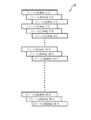
図1は、インタレースされた同時ウィンドウ設定を使用することにより複数の同時ウィンドウについて単一のSUV較正及び正規化を実施し、それにより、各々の同時ウィンドウ設定ごとに個々のSUV及び正規化較正を実施する必要をなくした、標準摂取率(SUV)取得シーケンス10を示す。図1には、3つのインタレースされたタイミングウィンドウ(例えば、2ナノ秒、4ナノ秒及び6ナノ秒)が、20フレーム(0−19と示す)に関して示されている。各フレームは、3つの部分に分割され、各部分は、タイミングウィンドウの1つに対応する。このようにして、ランダムイベントが、PETスキャナにおいて低減され、PETスキャナの最大雑音等価計数率(NECR)が、イメージングされる対象の直径の関数として最大にされる。SUVは、さまざまな時点の動的に連続する各画像又はフレームについてピクセルごとに又は関心領域(ROI)にわたって組織放射能濃度の比率として計算される。ここで使用されるとき、「ランダムイベント」又は「ランダム」は、2つの単一イベントがPETスキャナによって同時タイミングウィンドウ(例えば、6ナノ秒又は他の予め規定されるタイミングウィンドウ)内で測定され又は検出され、同時イベント(すなわち、単一の共通消滅イベントの結果として生じたもの)として誤って扱われ又は処理される事象を示す。 FIG. 1 performs a single SUV calibration and normalization for multiple simultaneous windows by using interlaced simultaneous window settings, whereby individual SUVs and normalized calibrations for each simultaneous window setting. A standard uptake rate (SUV) acquisition sequence 10 that eliminates the need to perform is shown. In FIG. 1, three interlaced timing windows (eg, 2 ns, 4 ns, and 6 ns) are shown for 20 frames (denoted 0-19). Each frame is divided into three parts, each part corresponding to one of the timing windows. In this way, random events are reduced in the PET scanner and the maximum noise equivalent count rate (NECR) of the PET scanner is maximized as a function of the diameter of the object being imaged. The SUV is calculated for each dynamically successive image or frame at various times, as a ratio of tissue radioactivity concentration per pixel or over the region of interest (ROI). As used herein, “random event” or “random” means that two single events are measured by a PET scanner within a simultaneous timing window (eg, 6 nanoseconds or other predefined timing window) or Indicates an event that is detected and mishandled or processed as a concurrent event (ie, as a result of a single common annihilation event).
当分野において知られているように、電子及び陽電子が遭遇すると、それらは、運動量保存の法則の原理により反対方向を向く2つの511keVガンマ線を放出して、消滅する。PETデータ取得において、2つの実質的に同時発生の又は同時の511keVガンマ線検出イベントは、同じ陽電子−電子消滅イベントから生じたものと推定される。従って、消滅イベントは、2つの実質的に同時の511keVガンマ線検出イベントを接続する「ラインオブレスポンス」(LOR)に沿って位置する。このラインオブレスポンスは投影とも呼ばれ、収集されたPETデータは、投影データと呼ばれる。 As is known in the art, when electrons and positrons are encountered, they annihilate, emitting two 511 keV gamma rays that point in opposite directions according to the principle of conservation of momentum. In PET data acquisition, two substantially simultaneous or simultaneous 511 keV gamma detection events are presumed to have resulted from the same positron-electron annihilation event. Thus, the annihilation event is located along a “line of response” (LOR) that connects two substantially simultaneous 511 keV gamma detection events. This line of response is also called projection, and the collected PET data is called projection data.
従来のPETにおいて、例えば互いが6ナノ秒以内であるというように、選択された短時間又は同時ウィンドウ内で生じる2つの511keVガンマ線検出イベントは、有効なLORを規定するものとされる。検出器素子に対する可変の消滅位置のため、同時のガンマ光子検出イベントの間に小さい時間差(例えば、サブナノ秒)が生じる。タイムオブフライトPET又はTOF−PETと呼ばれる関連する技法が、LORに沿って陽電子−電子消滅イベントの位置を更に突き止めるために、この小さい時間差を利用する。概して、消滅イベントは、LORに沿って、最初に生じたガンマ光線検出イベントに近いほうのポイントに生じる。2つのガンマ光線検出イベントが、検出器の時間分解能の範囲内で同時に発生する場合、消滅イベントは、LORの中間点で生じたことになる。 In conventional PET, two 511 keV gamma detection events that occur within a selected short time or simultaneous window, eg, within 6 nanoseconds of each other, shall define an effective LOR. Due to the variable annihilation position relative to the detector element, a small time difference (eg, sub-nanosecond) occurs between simultaneous gamma photon detection events. A related technique called time-of-flight PET or TOF-PET takes advantage of this small time difference to further locate the positron-electron annihilation event along the LOR. In general, annihilation events occur along the LOR at points closer to the first gamma ray detection event that occurred. If two gamma detection events occur simultaneously within the time resolution of the detector, the annihilation event has occurred at the midpoint of the LOR.
F18ファントムからの較正データのインタレースされた収集を使用しない従来のアプローチの下、20フレーム(例えば、フレーム0−19)のSUV取得シーケンスは、同時ウィンドウ設定ごとに11時間を要する。例えば、フレーム0−7のSUVデータは、1フレームあたり15分かかり、合計2時間を要する。フレーム8−13のSUVデータ取得は、フレームごとに30分かかり、合計3時間を要する。フレーム14−19のSUVデータ取得は、1フレームあたり1時間かかり、合計6時間を要する。較正スキャンに非常に長い時間がかかる1つの理由は、F18放射性物質の半減期が約110分であり、従って、11時間のスキャンは、材料の6回の半減期にわたってデータを提供する(すなわち、11時間後に、F18放射性物質の98.5%が崩壊する)。従って、放射能レベルのレンジにスキャナの光電子増倍管を較正するために、11時間の期間にわたるデータが必要である。すなわち、最初の110分の間に取得されるデータのみを使用したスキャナの較正は、例えば、較正スキャンの10又は11時間目の間に取得される較正データと等価でありうる低レベルの放射能の検出のためには十分な正確さを提供しない。そのうえ、ユーザが、3つの同時ウィンドウ設定(例えば2ナノ秒、4ナノ秒及び6ナノ秒)についてSUV取得シーケンスを実行する場合、従来のアプローチは、33時間に、同時ウィンドウごとに新しいF18較正ファントムを準備する時間を加えた時間がかかる(上述のF18の例を使用する場合)。 Under a conventional approach that does not use interlaced collection of calibration data from an F18 phantom, a 20-frame (eg, frames 0-19) SUV acquisition sequence takes 11 hours for each simultaneous window setting. For example, the SUV data of frames 0-7 takes 15 minutes per frame, which requires a total of 2 hours. Acquisition of SUV data for frame 8-13 takes 30 minutes per frame, which takes a total of 3 hours. Acquisition of SUV data for frames 14-19 takes 1 hour per frame, which takes a total of 6 hours. One reason that the calibration scan takes so long is that the F18 radioactive material has a half-life of about 110 minutes, so an 11-hour scan provides data over the six half-lives of the material (ie, After 11 hours, 98.5% of the F18 radioactive material decays). Therefore, data over an 11 hour period is required to calibrate the scanner photomultiplier tube to a range of radioactivity levels. That is, scanner calibration using only data acquired during the first 110 minutes may be equivalent to, for example, calibration data acquired during the 10th or 11th hour of the calibration scan. Does not provide sufficient accuracy for detection. Moreover, if the user performs an SUV acquisition sequence for three simultaneous window settings (eg, 2 nanoseconds, 4 nanoseconds and 6 nanoseconds), the conventional approach is a new F18 calibration phantom for each simultaneous window at 33 hours. It takes time to add time for preparing (when using the example of F18 described above).
本願は、各フレーム全体にわたって連続的にサンプリングされるのではなく、較正が各フレーム内で適切にサンプリングされることのみを必要とすることを認識する。図1に示すように複数の同時ウィンドウに関して較正データ収集をインタレースすることによって、上述の例示のSUV較正データ取得シーケンスは、F18ファントムによる単一の11時間スキャニング期間の中で、すべての3つの(又はそれより多くの)同時ウィンドウ設定に関して実施されることが可能である。図1は、各同時ウィンドウごとの較正データ取得が各フレーム内でインタレースされるように、一般的なSUV較正取得シーケンスを変更するためのアプローチの一例を示す。2ナノ秒、4ナノ秒及び6ナノ秒の同時ウィンドウ設定が、図1の例で与えられているが、当業者により理解されるように、他のウィンドウ設定が用いられることができる。更に、当業者により理解されるように、より多い又はより少ないインタレースされたウィンドウ設定(例えば2、3、4、5、6等)が用いられることもできる。 The present application recognizes that rather than being sampled continuously throughout each frame, the calibration need only be sampled properly within each frame. By interlacing the calibration data collection over multiple simultaneous windows as shown in FIG. 1, the above exemplary SUV calibration data acquisition sequence can be performed in a single 11-hour scanning period with an F18 phantom in all three It can be implemented for (or more) simultaneous window settings. FIG. 1 shows an example of an approach for modifying the general SUV calibration acquisition sequence so that the calibration data acquisition for each simultaneous window is interlaced within each frame. Although simultaneous window settings of 2 nanoseconds, 4 nanoseconds and 6 nanoseconds are given in the example of FIG. 1, other window settings can be used as will be appreciated by those skilled in the art. Further, as will be appreciated by those skilled in the art, more or fewer interlaced window settings (eg 2, 3, 4, 5, 6, etc.) may be used.
各フレームごとに、すべての3つの同時ウィンドウに関するデータが取得される。例えば、フレーム0の間、単一タイミングウィンドウに関するデータを取得するのに15分を費やすのではなく、フレーム0の間、3つの同時ウィンドウの各々に関するデータを取得するのに5分が費やされる。従来のプロトコルと比較して、これは、すべてのフレームにわたって所与の同時ウィンドウに関するデータのわずか1/3の取得をもたらすが、各々の同時ウィンドウに関して取得されるデータは、崩壊期間全体(例えば、11−14時間)にわたってなお分散されており、これは、PETスキャナのPMTのSUVの特徴づけ及び較正ために十分なデータを供給する。このようにして、較正データ(SUV)は、合計11時間にわたって、すべての3つの(又は他の数)同時ウィンドウに関して取得される。これは、各同時ウィンドウごとのスキャナの較正が、全放射性崩壊期間(例えば6−7回の半減期)にわたって収集されるデータを使用することによって改善され、較正が、各同時ウィンドウごとに完全な11時間相当のデータを必要としないので、有利である。そうではなく、11時間にわたって各フレームが取得される間、各同時ウィンドウについて周期的なサンプルが取得されることができる。 For each frame, data for all three simultaneous windows is acquired. For example, instead of spending 15 minutes to acquire data for a single timing window during frame 0, 5 minutes are spent acquiring data for each of three simultaneous windows during frame 0. Compared to conventional protocols, this results in acquisition of only 1/3 of the data for a given simultaneous window across all frames, but the data acquired for each simultaneous window is the entire collapse period (e.g., 11-14 hours), which provides enough data to characterize and calibrate the PMT SUV of the PET scanner. In this way, calibration data (SUV) is acquired for all three (or other numbers) simultaneous windows over a total of 11 hours. This is improved by using data where the calibration of the scanner for each simultaneous window is collected over the entire radioactive decay period (eg 6-7 half-lives), and the calibration is complete for each simultaneous window. This is advantageous because it does not require 11 hours worth of data. Rather, periodic samples can be acquired for each simultaneous window while each frame is acquired over 11 hours.
一実施形態において、取得ハードウェアが、各々の放射線イベントにタイムスタンプを付す。較正されるべき最大同時ウィンドウの外側のイベントが、捨てられることができるように、放射線イベントは、最大の同時ウィンドウに従う。ソフトウェアは、タイムスタンプを見て、同時時間によって同時対をソートし、例えば、2ナノ秒の範囲内、4ナノ秒の範囲内及び6ナノ秒の範囲内で同時であるイベントの間でソートを行う。このアプローチは、正規化及びSUV取得が、最も広い同時ウィンドウ(例えば6ナノ秒)によって一度実行され、後処理によって他の同時ウィンドウに関するソリューションを与えることを可能にする。 In one embodiment, the acquisition hardware timestamps each radiation event. Radiation events follow the maximum simultaneous window so that events outside the maximum simultaneous window to be calibrated can be discarded. The software looks at timestamps and sorts simultaneous pairs by simultaneous time, for example, sorting between events that are simultaneous within 2 nanoseconds, within 4 nanoseconds, and within 6 nanoseconds. Do. This approach allows normalization and SUV acquisition to be performed once with the widest simultaneous window (eg 6 nanoseconds) and post-processing to provide a solution for other simultaneous windows.
1つの技法において、それぞれ異なる同時ウィンドウの逐次の取得が使用される。一実施形態において、較正フレームは、複数の同時ウィンドウ設定を同時に適用するために、(例えば、ハードウェア、ソフトウェア又はその組み合わせを使用して)リアルタイムに処理される。別の実施形態において、較正は、エネルギーウィンドウ又は横方向視野のような付加の取得パラメータの変化を含むようにインタレースされる。 In one technique, sequential acquisition of different simultaneous windows is used. In one embodiment, the calibration frame is processed in real time (eg, using hardware, software or a combination thereof) to apply multiple simultaneous window settings simultaneously. In another embodiment, the calibration is interlaced to include changes in additional acquisition parameters such as energy window or lateral field of view.
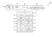
図2は、F18ファントムのようなファントム103を使用して、PETスキャナ102の較正を容易にするシステム100を示す。システム100は、PETスキャナ及びメモリ106に結合される、プロセッサ104を有する。メモリは、本明細書に記述されるさまざまな機能、方法、技法、プロシージャ等を実施するためのコンピュータ実行可能命令を記憶し、プロセッサが、それらの命令を実行する。システムは更に、1又は複数の入力装置110(例えば、キーボード、マウス、スタイラス、タッチスクリーン、マイクロホン等)を含むユーザインタフェース108と、情報がユーザに提示されるディスプレイ112と、を有する。
FIG. 2 illustrates a system 100 that facilitates calibration of a
メモリは、フルデオキシグルコース−18(F18)較正ファントムのスキャン中、F18がPETスキャナの検査領域において(例えば長時間の崩壊期間にわたって)放射能崩壊するとき、SUV較正データ115を決定するためのプロセッサ104によって実行されるSUV較正データ取得シーケンスソフトウェアモジュール114を記憶する。SUVデータは、取得スキャン中、同時ウィンドウの関数として標準摂取率を計算するために使用される。一実施形態において、ファントムは、20cm×30cmの円筒状ファントムである。別の実施形態において、ファントムは球状である。
The memory is a processor for determining
同時ウィンドウ設定情報116は、ユーザによってユーザインタフェース108に入力され、メモリ106に記憶される。更に、メモリは、PETスキャナ102の較正後、較正ファントム及び/又は他の対象の画像を再構成するために再構成プロセッサ120によって実行される1又は複数の再構成アルゴリズム118を記憶する。SUV較正データ115が取得されると、プロセッサは、PETスキャナ102の標準摂取率を較正する較正モジュール122を実行する。更に、プロセッサは、メモリに記憶された正規化モジュール124を実行して、PETスキャナの光検出器又は光電増倍管の各々について正規化関係を計算し、それにより、すべての放射線センサが入射放射線に対し共通の感度を有するようにする。正規化較正は更に、各々の同時ウィンドウについて実施される。SUV較正について上述したのと同じ技法が、同時ウィンドウの各々に関する正規化を較正するために使用されることができる。
The simultaneous
各々の同時ウィンドウに関するインタレースされた較正データ収集は、例えば2、4又は6ナノ秒の調整可能な及び/又は選択可能な同時ウィンドウを与えることを容易にする。SUV及び正規化値は、較正モジュール122及び正規化モジュール124を通じて、単一の較正プロシージャにおいて較正される。F−18は、幾時間か(例えば14時間)にわたって崩壊するので、SUV較正を実施するために、データが一般にF−18ファントムに関して取得される。一実施形態において、データが、図1の取得シーケンスに示すように、14時間にわたって同時ウィンドウ時間の各々について周期的に収集され、それにより一連のポイントを生成し、3つの摂取曲線を規定する。
Interlaced calibration data collection for each simultaneous window facilitates providing an adjustable and / or selectable simultaneous window of, for example, 2, 4 or 6 nanoseconds. SUVs and normalized values are calibrated in a single calibration procedure through
別の実施形態において、データのすべてに、十分な正確さを伴うタイムスタンプが付され、タイムスタンプを付されたデータ126は、プロセッサ104によって実行されるソートモジュール127によって、2ナノ秒の範囲内、4ナノ秒の範囲内又は6ナノ秒の範囲内で同時であるイベントにソートされる。これらの測定値及びそれらの収集時間は、各同時ウィンドウごとにSUV較正曲線128を生成するために、使用されることができる。
In another embodiment, all of the data is time stamped with sufficient accuracy, and the time stamped
別の実施形態において、エネルギーウィンドウ調整モジュール130が、調整可能なエネルギーウィンドウを提供する。すなわち、取得されたF−18スキャンデータについて、有効であると考えられる511keV付近のエネルギーピーク又はエネルギーのレンジの幅が、調整可能である。しかしながら、エネルギーウィンドウを変えることは、SUV及び正規化較正をも変更する。この場合、本明細書に記述される技法が、インタリーブ技法を使用して、又は各イベントのエネルギーを記録しエネルギーウィンドウでソートすることにより、単一の較正プロシージャの中で、複数のエネルギーウィンドウの各々に関するSUV及び正規化を較正するために使用される。
In another embodiment, the energy
別の実施形態によれば、核スキャナ102は、タイムオブフライト(TOF)PETスキャナであり、TOFデータ132が、PET画像の再構成の正確さを改善するために、メモリ106に記憶される。
According to another embodiment, the
上述のように、システム100は、本明細書に記述されるさまざまな機能、方法、プロシージャ等を実施するためにコンピュータ実行可能命令(例えば、ルーチン、プログラム、アルゴリズム、ソフトウェアコード等)を実行するプロセッサ104と、それらの命令を記憶するメモリ106と、を有する。更に、「モジュール」という語は、本明細書で用いられる場合、当業者によって理解されるように、コンピュータ実行可能命令、ソフトウェアコード、プログラム、ルーチン等を示す。
As described above, the system 100 is a processor that executes computer-executable instructions (eg, routines, programs, algorithms, software code, etc.) to perform various functions, methods, procedures, etc. described herein. 104 and a
メモリは、ディスク、ハードディスク等の制御プログラムが記憶されるコンピュータ可読媒体でありうる。コンピュータ可読媒体の一般的な形態は、例えば、フロッピーディスク、フレキシブルディスク、ハードディスク、磁気テープ又は任意の他の磁気記憶媒体、CD−ROM、DVD又は任意の他の光学媒体、RAM、ROM、PROM、EPROM、フラッシュEPROM、それらの異形、他のメモリチップ又はカートリッジ、又はプロセッサが読み実行することができる任意の他の有形媒体、を含む。この文脈において、ここに記述されるシステムは、1又は複数の汎用コンピュータ、(複数の)特定用途コンピュータ、プログラムされたマイクロプロセッサ又はマイクロコントローラ及び周辺集積回路素子、ASIC又は他の集積回路、デジタル信号プロセッサ、ディスクリート素子回路のような結線接続の電子又は論理回路、PLD、PLA、FPGA、グラフィックカードCPU(GPU)又はPAL等のプログラマブル論理装置において又はそのものとして実現されることができる。 The memory may be a computer-readable medium that stores a control program such as a disk or a hard disk. Common forms of computer readable media are, for example, floppy disks, flexible disks, hard disks, magnetic tapes or any other magnetic storage medium, CD-ROM, DVD or any other optical medium, RAM, ROM, PROM, EPROM, flash EPROM, variants thereof, other memory chips or cartridges, or any other tangible medium that can be read and executed by a processor. In this context, the systems described herein are one or more general purpose computers, (multiple) special purpose computers, programmed microprocessors or microcontrollers and peripheral integrated circuit elements, ASICs or other integrated circuits, digital signals It can be realized in or as a programmable logic device such as a processor, a connection-connected electronic or logic circuit such as a discrete element circuit, PLD, PLA, FPGA, graphic card CPU (GPU) or PAL.
図3及び図4は、さまざまなフィーチャによる、インタレースされた同時ウィンドウ設定を使用してPETスキャナを較正することに関連する方法を示す。方法は、一連の工程(アクト)として記述されているが、すべての工程が、記述された目標及び/又は結果を達成するために必要とされるわけではなく、ある工程は、特定の見地により、記述された特定の順序と異なる順序で実施されることができることが理解されるであろう。 3 and 4 illustrate methods associated with calibrating a PET scanner using interlaced simultaneous window settings according to various features. Although the method is described as a series of steps (acts), not all steps are required to achieve the stated goals and / or results, and certain steps may depend on specific aspects. It will be understood that it can be implemented in a different order than the particular order described.
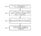
図3は、本明細書に記述されるさまざまな見地による、インタレースされた同時ウィンドウを使用してPETスキャナを較正する方法を示す。ステップ160において、インタレースされた同時ウィンドウが、選択され又は設定される。一例において、2ナノ秒、4ナノ秒及び6ナノ秒の3つのタイミングウィンドウが設定される。しかしながら、他のタイミングウィンドウ設定(例えば、1ナノ秒、1.5ナノ秒、2.7ナノ秒、3ナノ秒、5ナノ秒等)が、他の数のタイミングウィンドウ(例えば、2、4、5等)と共に設定されることもできる。ステップ162において、放射性ファントムがスキャンされ、同時データが、同時ウィンドウ設定及びエネルギーウィンドウの各々について取得される。ステップ164において、同時データが、各フレーム及び各タイミングウィンドウについてファントムの標準摂取率(SUV)を決定するために解析される。SUVは、さまざまな時点における動的に連続する各画像又は各フレームについて、ピクセルごとに又は関心領域(ROI)にわたって、組織放射能濃度の比率として計算される。ステップ166において、PETスキャナの光検出器が、取得された同時データを使用して、各々の同時ウィンドウ設定及び各々のエネルギーウィンドウ設定について、正規化される。光検出器の較正は、一般に、空間及びエネルギーの観点で同時性及び正確さを確実にするように、光検出器アレイの空間ゲイン及びオフセットを調整することを含む。
FIG. 3 illustrates a method for calibrating a PET scanner using interlaced simultaneous windows according to various aspects described herein. In
図4は、リストモードで放射性ファントムから取得された同時データを後処理することによって、PETスキャナを較正する方法を示す。ステップ178において、エネルギーウィンドウ設定が選択される。再構成のために取得スキャンデータを補正する場合、選択されたエネルギーウィンドウの外側にある検出放射線イベントは捨てられる。ステップ180において、同時タイミングウィンドウ設定が選択される。選択された同時ウィンドウ内に入る検出された放射線イベント対は、スキャナ較正及び正規化のためのSUVを決定するために使用される。ステップ182において、放射性ファントムは、放射性物質が崩壊するとき(例えば14時間等にわたって)データを取得するためにスキャンされる。データは、リストモードにおいて収集され、各イベントが、タイムスタンプを付され、そのエネルギーが記録される。ステップ184において、リストモードデータは、複数の選択された同時ウィンドウのどれに特定のデータが入るか、及び/又は複数の選択されたエネルギーウィンドウのどれに特定のデータが入るかに従って、データをビニングし又はソートするように後処理される。ステップ186において、各ビンの同時データが、各フレーム及び各タイミング及び/又はエネルギーウィンドウごとにファントムに関する標準摂取率(SUV)を計算するために解析される。SUVは、さまざまな時点の動的に連続する各画像又は各フレームについて、ピクセルごとに又は関心領域(ROI)にわたって、組織放射能濃度の比率として計算される。ステップ188において、SUV及び正規化の関係値が記憶される。患者がスキャンされるとき、ステップ190において、同時ウィンドウ及び/又はエネルギーウィンドウのうちの1つが選択される。ステップ192において、対応するSUV及び正規化の補正値が取り出される。ステップ194において、PETデータが取得され(例えば患者がスキャンされ)、取り出されたSUV及び正規化値に従って補正される。補正されたデータは、表示及び/又は記憶のために、画像に再構成される。
FIG. 4 illustrates a method for calibrating a PET scanner by post-processing the simultaneous data acquired from the radioactive phantom in list mode. In step 178, an energy window setting is selected. When correcting acquired scan data for reconstruction, detected radiation events outside the selected energy window are discarded. In
図5は、上述した較正方法の1つに従って較正されるPETスキャナ210を示す。PETスキャナは、放射線イベント(例えば、ガンマ線等)を検出する複数の放射線検出器をもつガントリを有する。ユーザ入力装置212が提供され、ユーザ入力装置212は同時ウィンドウ選択器214を有する。ユーザは、同時ウィンドウ選択器214を通じて、1又は複数の同時ウィンドウを表す同時ウィンドウ設定を入力し又は選択する。放射線イベントは、同時ウィンドウ処理回路216によって、入力された同時ウィンドウにビニングされ又は分類される。入力装置は更に、エネルギーウィンドウ選択器218を有し、ユーザは、エネルギーウィンドウ選択器218を通じて、1又は複数のエネルギーウィンドウを規定するエネルギーウィンドウ設定を入力し又は選択する。エネルギーウィンドウ回路220は、選択されたエネルギーウィンドウ内に入らない検出された放射線イベントを除外し又は捨てる。同時及び/又はエネルギーウィンドウが、ここに記述されるPETスキャナを較正するために使用される。
FIG. 5 shows a PET scanner 210 that is calibrated according to one of the calibration methods described above. A PET scanner has a gantry with multiple radiation detectors that detect radiation events (eg, gamma rays, etc.). A
システムが更に、複数の同時タイミングウィンドウ及び/又はエネルギーウィンドウに関する較正の間に導き出される正規化補正値224を記憶する正規化メモリ222と、複数の同時タイミングウィンドウ及び/又はエネルギーウィンドウに関する較正の間に導き出されるSUV補正値228を記憶するSUVメモリ226と、を有する。正規化補正モジュール230(例えばプロセッサ)は、被検体をスキャンする際に使用される所与の同時又はエネルギーウィンドウに関する正規化補正値224を取り出し、取得されたスキャンデータを正規化する。SUV補正モジュール232(例えばプロセッサ)は、記憶されたSUV補正値228を取り出し、取得されたスキャンデータに対しSUV補正を実施する。再構成プロセッサ234が、被検体の画像を再構成し、画像は、ディスプレイ236上でユーザに提示され、及び/又は後の取り出し及び表示のために、患者データベース238に記憶される。
The system further includes a
本発明は、幾つかの実施形態に関して記述された。変形例及び変更例が、先行する詳細な説明を読み理解することにより、当業者に思い付くであろう。すべてのこのような変形例及び変更例が添付の請求項又はその等価なものの範囲内にある限り、本発明は、それらの変形例及び変更例を含むものとして解釈されることが意図される。 The invention has been described with reference to several embodiments. Variations and modifications will occur to those skilled in the art upon reading and understanding the preceding detailed description. To the extent that all such variations and modifications are within the scope of the appended claims or their equivalents, the present invention is intended to be construed as including those variations and modifications.
Claims (15)
放射性較正ファントムがPETスキャナに配され、予め決められた期間中スキャンされる、該PETスキャナと、
メモリに記憶されたコンピュータ実行可能命令を実行するプロセッサと、
を有し、前記命令が、
複数の選択された同時タイミング及び/又はエネルギーウィンドウの設定を受け取り、
前記PETスキャナにより前記予め決められた期間中放射性較正ファントムをスキャンし、前記予め決められた期間の複数フレームの各々の間、前記同時タイミング及び/又はエネルギーウィンドウの設定によって規定される複数の同時タイミング及び/又はエネルギーウィンドウの各々について同時データを取得し、
前記選択された同時タイミング及び/又はエネルギーウィンドウの各々について、前記予め決められた期間にわたって各フレームにおいて検出された光子カウントの数から、標準摂取率を計算する、
ことを含むシステム。 A PET scanner calibration system comprising:
A PET scanner, wherein a radioactive calibration phantom is placed on the PET scanner and scanned for a predetermined period of time;
A processor for executing computer-executable instructions stored in memory;
And the instruction is
Receive a plurality of selected simultaneous timing and / or energy window settings;
A plurality of simultaneous timings defined by the setting of the simultaneous timing and / or energy window during each of a plurality of frames of the predetermined period, wherein the PET scanner scans the radioactive calibration phantom during the predetermined period. And / or obtain simultaneous data for each of the energy windows,
Calculating a standard uptake from the number of photon counts detected in each frame over the predetermined time period for each of the selected simultaneous timing and / or energy window;
Including the system.
前記命令が更に、インタレースされた同時タイミング及び/又はエネルギーウィンドウ設定によって規定される複数のインタレースされた同時タイミングウィンドウに関して同時データを取得することを含む、請求項1乃至4のいずれか1項に記載のシステム。 The plurality of simultaneous timing and / or energy window settings are interlaced;
5. The any one of claims 1-4, wherein the instructions further comprise obtaining simultaneous data for a plurality of interlaced simultaneous timing windows defined by interlaced simultaneous timing and / or energy window settings. The system described in.
前記複数の同時タイミングウィンドウ及び/又はエネルギーウィンドウについて、正規化補正値及びSUV補正値を記憶し、
被検体のPETスキャンを実施し、
前記正規化補正値及び前記SUV補正値を使用して、被検体の取得されたスキャンデータを補正し、
前記補正されたスキャンデータを使用して、被検体の画像を再構成し、
前記再構成された画像を患者データベースに記憶する、
ことを含む、請求項1乃至5のいずれか1項に記載のシステム。 The instruction further comprises:
Storing a normalization correction value and an SUV correction value for the plurality of simultaneous timing windows and / or energy windows;
Perform a PET scan of the subject,
Using the normalized correction value and the SUV correction value, the acquired scan data of the subject is corrected,
Reconstructing an image of the subject using the corrected scan data;
Storing the reconstructed image in a patient database;
The system according to claim 1, comprising:
選択された複数の同時タイミング及び/又はエネルギーウィンドウの設定を受け取るステップと、
放射性較正ファントムをスキャンし、予め決められた期間の複数フレームの各々の間、前記同時タイミング及び/又はエネルギーウィンドウ設定によって規定される複数の同時タイミング及び/又はエネルギーウィンドウの各々について同時データを取得するステップと、
前記選択された同時タイミング及び/又はエネルギーウィンドウの各々について、前記予め決められた期間にわたって各フレームにおいて検出された光子カウントの数から、標準摂取率を計算するステップと、
を含む方法。 A method for calibrating a PET scanner comprising:
Receiving a plurality of selected simultaneous timing and / or energy window settings;
Scan the radioactive calibration phantom and acquire simultaneous data for each of a plurality of simultaneous timings and / or energy windows defined by the simultaneous timing and / or energy window settings during each of a plurality of frames for a predetermined period of time. Steps,
Calculating a standard uptake from the number of photon counts detected in each frame over the predetermined time period for each of the selected simultaneous timing and / or energy window;
Including methods.
受け取られた放射線イベントの各々にタイムスタンプを付すステップと、
各々の同時イベント対が前記複数の同時タイミング及び/又はエネルギーウィンドウのどれに対応するかに従って、前記放射線イベントをソートするステップと、
を含む、請求項9乃至11のいずれか1項に記載の方法。 The simultaneous data is collected in list mode, the method comprising:
Time stamping each received radiation event;
Sorting the radiation events according to which of the plurality of simultaneous timings and / or energy windows each simultaneous event pair corresponds to;
The method according to claim 9, comprising:
異なる長さの複数の同時ウィンドウによって、検出されたイベント対を識別する同時ウィンドウ処理回路と、
ユーザがユーザ入力装置によって前記同時ウィンドウの少なくとも1つを選択する、該ユーザ入力装置と、
記憶された正規化補正値を前記選択された同時ウィンドウの取得されたスキャンデータに適用する正規化補正モジュールと、
記憶されたSUV補正値を前記選択された同時ウィンドウの取得されたスキャンデータに適用するSUV補正モジュールと、
ディスプレイに提示するために、補正されたスキャンデータを画像に再構成する再構成プロセッサと、
を有するPETスキャナ。 A gantry having a plurality of radiation detectors for detecting scintillation events;
A simultaneous window processing circuit that identifies detected event pairs by means of multiple simultaneous windows of different lengths;
The user input device wherein a user selects at least one of the simultaneous windows by means of a user input device;
Applying a stored normalization correction value to the acquired scan data of the selected simultaneous window;
Applying a stored SUV correction value to the acquired scan data of the selected simultaneous window; and
A reconstruction processor for reconstructing the corrected scan data into an image for presentation on a display;
A PET scanner.
Applications Claiming Priority (3)
| Application Number | Priority Date | Filing Date | Title |
|---|---|---|---|
| US41632310P | 2010-11-23 | 2010-11-23 | |
| US61/416,323 | 2010-11-23 | ||
| PCT/IB2011/055097 WO2012069960A2 (en) | 2010-11-23 | 2011-11-15 | Pet calibrations with varying coincidence windows |
Publications (3)
| Publication Number | Publication Date |
|---|---|
| JP2014500491A JP2014500491A (en) | 2014-01-09 |
| JP2014500491A5 JP2014500491A5 (en) | 2014-12-18 |
| JP5944402B2 true JP5944402B2 (en) | 2016-07-05 |
Family
ID=45218774
Family Applications (1)
| Application Number | Title | Priority Date | Filing Date |
|---|---|---|---|
| JP2013539383A Expired - Fee Related JP5944402B2 (en) | 2010-11-23 | 2011-11-15 | PET calibration for variable simultaneous windows |
Country Status (6)
| Country | Link |
|---|---|
| US (1) | US8987659B2 (en) |
| EP (1) | EP2643710B1 (en) |
| JP (1) | JP5944402B2 (en) |
| CN (1) | CN103221841B (en) |
| RU (1) | RU2582887C2 (en) |
| WO (1) | WO2012069960A2 (en) |
Families Citing this family (19)
| Publication number | Priority date | Publication date | Assignee | Title |
|---|---|---|---|---|
| JP5603647B2 (en) * | 2009-05-13 | 2014-10-08 | キヤノン株式会社 | Power feeding device, power feeding device control method, and power feeding communication system |
| CN105814454B (en) * | 2013-12-04 | 2019-11-05 | 皇家飞利浦有限公司 | For rebuilding the reconstructing device of PET image |
| US9297909B2 (en) * | 2014-04-18 | 2016-03-29 | Perkinelmer Health Sciences, Inc. | Guard efficiency compensation system and method |
| US9910162B2 (en) * | 2014-06-12 | 2018-03-06 | Siemens Medical Solutions Usa, Inc. | Calibrating in single photon emission computed tomography with multi-emission energies |
| WO2016046703A2 (en) * | 2014-09-23 | 2016-03-31 | Koninklijke Philips N.V. | Time of flight calibration in digital positron emission tomography |
| US9606245B1 (en) | 2015-03-24 | 2017-03-28 | The Research Foundation For The State University Of New York | Autonomous gamma, X-ray, and particle detector |
| CN105030263B (en) * | 2015-07-22 | 2018-06-12 | 湖北锐世数字医学影像科技有限公司 | The energy back bearing calibration of number PET a kind of and system |
| US10281596B2 (en) * | 2015-09-18 | 2019-05-07 | Koninklijke Philips N.V. | Correcting photon counts in a photon counting X-ray radiation detection system |
| JP6941098B2 (en) * | 2015-10-28 | 2021-09-29 | コーニンクレッカ フィリップス エヌ ヴェKoninklijke Philips N.V. | Equipment and methods for determining SUVs in radiation tomography |
| CN105395209B (en) | 2015-12-01 | 2018-10-02 | 沈阳东软医疗系统有限公司 | A kind of positron emission computerized tomography imaging system and method |
| CN106108935B (en) * | 2016-08-31 | 2019-06-28 | 北京康科达科技有限公司 | A kind of SPECT detection device |
| CN108109182B (en) * | 2016-11-24 | 2021-08-24 | 上海东软医疗科技有限公司 | PET image reconstruction method and device |
| JP7008728B2 (en) * | 2017-05-04 | 2022-01-25 | コーニンクレッカ フィリップス エヌ ヴェ | Prompt Gamma Positron Emission Tomography (PET) Timing Calibration Using Simultaneous Generation with High Energy Cascade Gamma from Positron Emission Nuclides |
| US10410383B2 (en) | 2017-08-26 | 2019-09-10 | Uih America, Inc. | System and method for image data processing in positron emission tomography |
| CN107595315A (en) * | 2017-09-30 | 2018-01-19 | 华中科技大学 | The acquisition methods of photoresponse line in a kind of transmitting imaging device |
| CN108294774A (en) * | 2018-01-04 | 2018-07-20 | 沈阳东软医疗系统有限公司 | A kind of PET correct scans method, apparatus and equipment |
| CN110215227B (en) * | 2019-06-05 | 2022-10-14 | 上海联影医疗科技股份有限公司 | Time window setting method and device, computer equipment and storage medium |
| US11249206B2 (en) * | 2020-01-06 | 2022-02-15 | Canon Medical Systems Corporation | Method and system for PET detector efficiency normalization |
| CN113852440B (en) * | 2021-09-29 | 2023-12-29 | 应急管理部国家自然灾害防治研究院 | Method and system for precise time calibration of electromagnetic field observation data of Zhangheng first satellite |
Family Cites Families (20)
| Publication number | Priority date | Publication date | Assignee | Title |
|---|---|---|---|---|
| JP3924879B2 (en) * | 1997-11-21 | 2007-06-06 | 株式会社島津製作所 | Nuclear medicine diagnostic equipment |
| US20050123183A1 (en) * | 2003-09-02 | 2005-06-09 | Paul Schleyer | Data driven motion correction for nuclear imaging |
| JP3717122B2 (en) * | 2003-09-29 | 2005-11-16 | 株式会社日立製作所 | γ-ray detection time determination method, γ-ray coincidence counting method, and nuclear medicine diagnostic apparatus |
| PT103200B (en) * | 2004-09-30 | 2006-08-24 | Taguspark-Soc. Prom.Desenv.Parq.Ci.Tec.Area Lisboa | POSITRON EMISSION TOMOGRAPHY SYSTEM (PET) |
| US7129495B2 (en) | 2004-11-15 | 2006-10-31 | General Electric Company | Method and apparatus for timing calibration in a PET scanner |
| US7057178B1 (en) | 2004-11-15 | 2006-06-06 | General Electric Company | Method and system for imaging using a filter for Time-of-Flight PET |
| US7301144B2 (en) * | 2004-12-29 | 2007-11-27 | General Electric Company | Method and system for calibrating a positron emission tomography system |
| US7227149B2 (en) | 2004-12-30 | 2007-06-05 | General Electric Company | Method and system for positron emission tomography image reconstruction |
| EP1844352B1 (en) * | 2005-01-28 | 2010-03-24 | Koninklijke Philips Electronics N.V. | Timing calibration using radioactive sources |
| US7381959B2 (en) | 2005-08-17 | 2008-06-03 | General Electric Company | Technique for reconstructing PET scan images |
| US7414246B2 (en) * | 2006-01-03 | 2008-08-19 | Koninklijke Philips Electronics N.V. | Achieving accurate time-of-flight calibrations with a stationary coincidence point source |
| CN101365963B (en) | 2006-01-09 | 2012-02-01 | 皇家飞利浦电子股份有限公司 | Method of constructing time-in-flight pet images |
| US7718954B2 (en) | 2006-01-31 | 2010-05-18 | Koninklijke Philips Electronics N.V. | Component method and system for PET detector efficiency normalization |
| US8965071B2 (en) * | 2008-05-30 | 2015-02-24 | Emory University | Assessing tumor response to therapy |
| WO2011061644A1 (en) * | 2009-11-18 | 2011-05-26 | Koninklijke Philips Electronics N.V. | Motion correction in radiation therapy |
| US9968309B2 (en) * | 2009-12-08 | 2018-05-15 | Koninklijke Philips N.V. | Method and a correction system for correcting tracer-uptake measurements |
| CN103260521B (en) * | 2010-12-14 | 2015-11-25 | 皇家飞利浦电子股份有限公司 | For the workflow of the integration of input function estimation accurately |
| GB201109344D0 (en) * | 2011-06-03 | 2011-07-20 | Siemens Medical Solutions | BIF from WB dynamic |
| US20130136328A1 (en) * | 2011-11-30 | 2013-05-30 | General Electric Company | Methods and systems for enhanced tomographic imaging |
| GB201219403D0 (en) * | 2012-10-29 | 2012-12-12 | Uni I Olso | Method for improved estimation of tracer uptake in physiological image volumes |
-
2011
- 2011-11-15 JP JP2013539383A patent/JP5944402B2/en not_active Expired - Fee Related
- 2011-11-15 WO PCT/IB2011/055097 patent/WO2012069960A2/en active Application Filing
- 2011-11-15 US US13/885,282 patent/US8987659B2/en active Active
- 2011-11-15 RU RU2013128552/28A patent/RU2582887C2/en not_active IP Right Cessation
- 2011-11-15 CN CN201180056345.3A patent/CN103221841B/en active Active
- 2011-11-15 EP EP11793870.4A patent/EP2643710B1/en active Active
Also Published As
| Publication number | Publication date |
|---|---|
| EP2643710B1 (en) | 2017-01-11 |
| CN103221841B (en) | 2015-11-25 |
| CN103221841A (en) | 2013-07-24 |
| US8987659B2 (en) | 2015-03-24 |
| RU2013128552A (en) | 2014-12-27 |
| WO2012069960A2 (en) | 2012-05-31 |
| RU2582887C2 (en) | 2016-04-27 |
| WO2012069960A3 (en) | 2012-11-01 |
| EP2643710A2 (en) | 2013-10-02 |
| JP2014500491A (en) | 2014-01-09 |
| US20130240721A1 (en) | 2013-09-19 |
Similar Documents
| Publication | Publication Date | Title |
|---|---|---|
| JP5944402B2 (en) | PET calibration for variable simultaneous windows | |
| JP5220617B2 (en) | Random reduction via TOFFOV | |
| US9867580B2 (en) | X-ray imaging based on image data from a photon-counting multi-bin X-ray detector | |
| US9747701B2 (en) | Systems and methods for emission tomography quantitation | |
| RU2517586C2 (en) | Reverse reconstruction of data for optimal time generation of count pulses in radiological physiological visualisation in list mode | |
| US9057788B2 (en) | Photon counting-based virtual detector | |
| US7227149B2 (en) | Method and system for positron emission tomography image reconstruction | |
| US9091771B2 (en) | System and method for improving detection of gamma interactions in a positron emission tomography system | |
| US9645260B2 (en) | Photon counting system and method | |
| US8440976B2 (en) | Method for optimizing step size in a multi-step whole-body PET imaging | |
| US8563935B2 (en) | Nuclear medicine imaging apparatus and control method | |
| US11096634B2 (en) | Scatter correction based on energy response | |
| US11231508B2 (en) | Gamma camera dead time determination in real time using long lived radioisotopes | |
| EP3819675B1 (en) | Imaging of photon-counting ct system | |
| US11163073B2 (en) | Charger integration-based virtual CT detector | |
| US11002867B1 (en) | Determination of crystal singles rates to estimate mean random coincidence rate | |
| CN112587159A (en) | Method and system for a PET detector | |
| US20200090378A1 (en) | Systems and methods for improved image reconstruction | |
| CN116068607A (en) | Method, system, device and medium for correcting timing walk based on energy information |
Legal Events
| Date | Code | Title | Description |
|---|---|---|---|
| A521 | Request for written amendment filed |
Free format text: JAPANESE INTERMEDIATE CODE: A523 Effective date: 20141031 |
|
| A621 | Written request for application examination |
Free format text: JAPANESE INTERMEDIATE CODE: A621 Effective date: 20141031 |
|
| A977 | Report on retrieval |
Free format text: JAPANESE INTERMEDIATE CODE: A971007 Effective date: 20150729 |
|
| A131 | Notification of reasons for refusal |
Free format text: JAPANESE INTERMEDIATE CODE: A131 Effective date: 20150825 |
|
| A521 | Request for written amendment filed |
Free format text: JAPANESE INTERMEDIATE CODE: A523 Effective date: 20151117 |
|
| TRDD | Decision of grant or rejection written | ||
| A01 | Written decision to grant a patent or to grant a registration (utility model) |
Free format text: JAPANESE INTERMEDIATE CODE: A01 Effective date: 20160506 |
|
| A61 | First payment of annual fees (during grant procedure) |
Free format text: JAPANESE INTERMEDIATE CODE: A61 Effective date: 20160525 |
|
| R150 | Certificate of patent or registration of utility model |
Ref document number: 5944402 Country of ref document: JP Free format text: JAPANESE INTERMEDIATE CODE: R150 |
|
| R250 | Receipt of annual fees |
Free format text: JAPANESE INTERMEDIATE CODE: R250 |
|
| R250 | Receipt of annual fees |
Free format text: JAPANESE INTERMEDIATE CODE: R250 |
|
| R250 | Receipt of annual fees |
Free format text: JAPANESE INTERMEDIATE CODE: R250 |
|
| R250 | Receipt of annual fees |
Free format text: JAPANESE INTERMEDIATE CODE: R250 |
|
| LAPS | Cancellation because of no payment of annual fees |