EP2643710B1 - Pet calibrations with varying coincidence windows - Google Patents
Pet calibrations with varying coincidence windows Download PDFInfo
- Publication number
- EP2643710B1 EP2643710B1 EP11793870.4A EP11793870A EP2643710B1 EP 2643710 B1 EP2643710 B1 EP 2643710B1 EP 11793870 A EP11793870 A EP 11793870A EP 2643710 B1 EP2643710 B1 EP 2643710B1
- Authority
- EP
- European Patent Office
- Prior art keywords
- coincidence
- timing
- windows
- energy
- data
- Prior art date
- Legal status (The legal status is an assumption and is not a legal conclusion. Google has not performed a legal analysis and makes no representation as to the accuracy of the status listed.)
- Active
Links
Images

Classifications
-
- G—PHYSICS
- G01—MEASURING; TESTING
- G01T—MEASUREMENT OF NUCLEAR OR X-RADIATION
- G01T1/00—Measuring X-radiation, gamma radiation, corpuscular radiation, or cosmic radiation
- G01T1/16—Measuring radiation intensity
- G01T1/161—Applications in the field of nuclear medicine, e.g. in vivo counting
- G01T1/164—Scintigraphy
- G01T1/1641—Static instruments for imaging the distribution of radioactivity in one or two dimensions using one or several scintillating elements; Radio-isotope cameras
- G01T1/1648—Ancillary equipment for scintillation cameras, e.g. reference markers, devices for removing motion artifacts, calibration devices
-
- A—HUMAN NECESSITIES
- A61—MEDICAL OR VETERINARY SCIENCE; HYGIENE
- A61B—DIAGNOSIS; SURGERY; IDENTIFICATION
- A61B6/00—Apparatus for radiation diagnosis, e.g. combined with radiation therapy equipment
- A61B6/02—Devices for diagnosis sequentially in different planes; Stereoscopic radiation diagnosis
- A61B6/03—Computerised tomographs
- A61B6/037—Emission tomography
-
- A—HUMAN NECESSITIES
- A61—MEDICAL OR VETERINARY SCIENCE; HYGIENE
- A61B—DIAGNOSIS; SURGERY; IDENTIFICATION
- A61B6/00—Apparatus for radiation diagnosis, e.g. combined with radiation therapy equipment
- A61B6/58—Testing, adjusting or calibrating apparatus or devices for radiation diagnosis
- A61B6/582—Calibration
- A61B6/583—Calibration using calibration phantoms
-
- G—PHYSICS
- G01—MEASURING; TESTING
- G01T—MEASUREMENT OF NUCLEAR OR X-RADIATION
- G01T1/00—Measuring X-radiation, gamma radiation, corpuscular radiation, or cosmic radiation
- G01T1/29—Measurement performed on radiation beams, e.g. position or section of the beam; Measurement of spatial distribution of radiation
- G01T1/2914—Measurement of spatial distribution of radiation
- G01T1/2964—Scanners
-
- G—PHYSICS
- G01—MEASURING; TESTING
- G01T—MEASUREMENT OF NUCLEAR OR X-RADIATION
- G01T1/00—Measuring X-radiation, gamma radiation, corpuscular radiation, or cosmic radiation
- G01T1/29—Measurement performed on radiation beams, e.g. position or section of the beam; Measurement of spatial distribution of radiation
- G01T1/2914—Measurement of spatial distribution of radiation
- G01T1/2985—In depth localisation, e.g. using positron emitters; Tomographic imaging (longitudinal and transverse section imaging; apparatus for radiation diagnosis sequentially in different planes, steroscopic radiation diagnosis)
-
- G—PHYSICS
- G01—MEASURING; TESTING
- G01T—MEASUREMENT OF NUCLEAR OR X-RADIATION
- G01T7/00—Details of radiation-measuring instruments
Definitions
- PET positron emission tomography
- Typical PET scanners perform a normalization calibration to correct nonuniform 3D detector response, as is described in U.S. Patent No. 7718954 , which typically requires about 6 hours. Furthermore, such scanners employ a Standard Uptake Value (SUV) calibration that provides the conversion of the counts in an image to an activity concentration. This calibration uses a decaying F-18 source and can take 11-hours or more to complete.
- SUV Standard Uptake Value
- NEMA NU-2 standard, Noise Equivalent Count Rate Noise Equivalent Count Rate
- coincidence window Today, commercial PET scanners have a fixed coincidence window, e.g., about 6 nanoseconds. However, the coincidence window can be shortened with some patients, particularly smaller patients, and/or when performing brain imaging, and the like, to give better discrimination between true events and noise (random events). However, the duration of the coincidence window effects various calibrations including normalization and SUV. Each calibration routine is very time-consumptive, typically taking 11-14 hours or more for each coincidence window.
- JP 11 153 669 A discloses a positron emission tomography (PET) scanner and an image processor connected thereto.
- Image data obtained with the PET scanner is sent to the image processor, wherein the radioactivity of a sample of radio active compound medicated to a patient is measured with a radioactivity meter.
- US 7,626,171 B2 discloses a method of reconstructing time-of-flight (TOF) images including obtaining a profile of a subject to be imaged in an examination region of an imaging system. Events associated with radiation emitted from the subject are detected and converted to electronic data. Electronic data attributable to radiation events located outside the profile are removed and images are reconstructed from the remaining electronic data.
- TOF time-of-flight
- the present application provides new and improved PET scanner calibration systems and methods that employ interlaced coincidence timing window settings during SUV acquisition on a radioactive calibration phantom, which overcome the above-referenced problems and others.
- positron emission tomography (PET) scanner calibration system includes a PET scanner in which a radioactive calibration phantom is placed and scanned for a predetermined time period, and a processor that executes computer-executable instructions stored in a memory, the instructions including receiving settings for a plurality of selected coincidence timing and/or energy windows.
- the instructions further include scanning the radioactive calibration phantom and acquiring coincidence data for each of the plurality of coincidence timing and/or energy windows defined by the timing and/or energy window settings during each of a plurality of frames of the predetermined time period. Additionally, the instructions include calculating standard uptake values from a number of photon counts detected in each frame over the predetermined time period for each selected coincidence timing and/or energy window.
- a method of calibrating a positron emission tomography (PET) scanner includes receiving settings for a plurality of selected coincidence timing and/or energy windows, and scanning the radioactive calibration phantom and acquiring coincidence data for each of the plurality of coincidence timing and/or energy windows defined by the timing and/or energy window settings during each of a plurality of frames of the predetermined time period.
- the method further includes calculating standard uptake values (SUVs) from a number of photon counts detected in each frame over the predetermined time period for each selected coincidence timing and/or energy window.
- SUVs standard uptake values
- a PET scanner includes a gantry with a plurality of radiation detectors that detect scintillation events, a coincidence windowing circuit that identifies pairs of detected events within a plurality of coincidence windows of different lengths, and a user input device by which a user selects at least one of the coincidence windows.
- the PET scanner additionally includes a normalization correction module that applies stored normalization correction values to acquired scan data in the selected coincidence window, an SUV correction module that applies stored SUV correction values to the acquired scan data in the selected coincidence window, and a reconstruction processor that reconstructs the corrected scan data into an image for presentation on a display.
- One advantage is that scanner calibration time is reduced.
- Another advantage resides in calibrating the scanner for multiple timing windows.
- the subject innovation overcomes the problem of time-consuming and labor-intensive scanner calibrations by reducing the calibration duration by interlacing the coincidence window settings within the SUV calibration.
- calibration duration is reduced by performing the SUV and normalization calibration acquisitions at the largest coincidence window and post-processing the data for the desired coincidence windows.
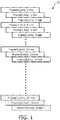
- FIGURE 1 illustrates a standard uptake value (SUV) acquisition sequence 10 that uses interlaced coincidence window settings to perform a single SUV calibration and normalization for multiple coincidence windows, which eliminates the need for performing separate SUV and normalization calibrations for each coincidence window setting.
- SAV standard uptake value
- FIG. 1 three interlaced timing windows (e.g. 2ns, 4ns, and 6ns) are shown for 20 frames (labeled 0-19). Each frame is divided into three portions, each of which corresponds to one of the timing windows. In this manner random events are reduced in a PET scanner the PET scanner's maximum noise equivalent count rate (NECR) is maximized as a function of the diameter of an object being imaged.
- NECR maximum noise equivalent count rate
- SUVs are calculated either pixel-wise or over a region of interest (ROI) for each image or frame of a dynamic series at various time points as a ratio of tissue radioactivity concentration.
- ROI region of interest
- a "random event” or “random” denotes an occurrence in which two single events are measured or detected by a PET scanner within a coincidence timing window (e.g., 6 ns or some other predefined timing window), and are mistakenly treated or processed as a coincident event (i.e., as occurring as a result of a single common annihilation event).
- two 511 keV gamma ray detection events occurring within a selected short time or coincidence window, such as within 6 nanoseconds of each other, are taken as defining a valid LOR. Due to the variable annihilation position with respect to the detector elements a small (e.g., sub-nanosecond) time difference between the coincident gamma photon detection events occurs.
- a related technique, called time-of-flight PET or TOF-PET takes advantage of this small time difference to further localize the positron-electron annihilation event along the LOR. In general, the annihilation event occurred along the LOR at a point closer to the gamma ray detection event that occurred first. If the two gamma ray detection events occur simultaneously within the time resolution of the detectors, then the annihilation event occurred at the midpoint of the LOR.
- an SUV acquisition sequence for 20 frames takes 11 hours per coincidence window setting.
- SUV data for frames 0-7 takes 15 minutes per frame, for a total of two hours; SUV data acquisition for frames 8-13 takes 30 minutes per frame, for a total of three hours, and SUV data acquisition for frames 14-19 takes one hour per frame, for a total of six hours.
- One reason that the calibration scan takes so long is that the half life of F18 radioactive material is approximately 110 minutes, so an 11 hour scan provides data over 6 half lives of the material (i.e., after 11 hours, 98.5% of the F18 radioactive material has decayed).
- the present application has recognized that the calibration only needs to be adequately sampled in each frame, rather than continuously sampled over each entire frame.
- the foregoing example SUV calibration data acquisition sequence can be performed for all three (or more) coincidence window settings in the single 11-hour scanning period with an F18 phantom.
- Figure 1 thus provides an example of an approach to modifying the typical SUV calibration acquisition sequence such that the calibration data acquisition for each of the coincidence windows is interlaced within each frame.
- 2ns, 4ns, and 6ns coincidence window settings are provided in the example of Figure 1 , although other window settings may be employed, as will be appreciated by those of skill in the art. Additionally, more or fewer interlaced window settings (e.g., 2, 3, 4, 5, 6, etc.) may be employed, as will be appreciated by those of skill in the art.
- calibration data is acquired for all three (or other number) coincidence windows over the total 11 hour period, which is advantageous because calibration of the scanner for each coincidence window is improved by using data collected over the full radioactive decay period (e.g., 6-7 half lives), but calibration does not require a full 11 hours worth of data for each coincidence window. Rather, periodic samples may be taken for each coincidence window, during each frame taken over the 11 hour period.
- acquisition hardware time stamps each radiation event.
- the radiation events are subject to the largest coincidence window such that events outside the largest coincidence window to be calibrated can be discarded.
- Software looks at the time stamps and sorts the coincident pairs by coincidence time, e.g. among events that are coincident within 2ns, with 4rs, and within 6ns. This approach allows the normalization and SUV acquisitions to be run once with the widest coincidence window (e.g., 6ns) and to generate solutions for other coincidence windows by post-processing.
- sequential acquisitions at different coincidence windows are used.
- calibrations are interlaced to include variation in additional acquisition parameters, e.g. energy window or transverse fields-of-view.
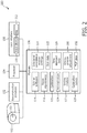
- FIGURE 2 illustrates a PET scanner calibration system 100 102 using a phantom 103, e.g. an F18 phantom.
- the system 100 includes a processor 104 that is coupled to the PET scanner and to a memory 106.
- the memory stores, and the processor executes, computer-executable instructions for performing the various functions, methods, techniques, procedures, etc., described herein.
- the system further includes a user interface 108 comprising one or more input devices 110 (e.g., a keyboard, mouse, stylus, touch screen, microphone, etc.), and a display 112 on which information is presented to a user.
- input devices 110 e.g., a keyboard, mouse, stylus, touch screen, microphone, etc.
- the memory stores an SUV calibration data acquisition sequence software module 114 that is executed by the processor 104 to determine SUV calibration data 115 during a scan of a fludeoxyglucose-18 (F-18) calibration phantom as it radioactively decays in the examination region of the PET scanner (e.g., over an hours-long decay period).
- the SUV data is used to calculate standard uptake values as a function of coincidence window during the acquisition scan.
- the phantom is a 20cm by 30cm cylindrical phantom.
- the phantom is spherical.
- Coincidence window setting information 116 is entered by a user into the user interface 108 and stored in the memory 106. Additionally, the memory stores one or more reconstruction algorithms 118 that are executed by a reconstruction processor 120 in order to reconstruct an image of the calibration phantom and/or other objects after calibration of the PET scanner 102.
- the processor executes a calibration module 122 that calibrates standard uptake values for the PET scanner 102.
- the processor executes a normalization module 124 that is stored in the memory to calculate normalize connections for each of the photodetectors or photomultiplier tubes in the PET scanner such that all radiation sensors have a common sensitivity to incident radiation.
- the normalization calibration is also performed for each coincidence window. The same techniques described above for the SUV calibration can be used to calibrate the normalization for each of the coincidence windows.
- the interlaced calibration data collection for each coincidence window facilitates providing an adjustable and/or selectable coincidence window, e.g., of 2, 4, or 6 nanoseconds.
- SUV and normalization values are calibrated in a single calibration procedure, via the calibration module 122 and the normalization module 124.
- data is typically taken with an F-18 phantom as the F-18 decays over a number of hours, e.g., 14 hours.
- data is cyclically collected for each of the coincidence window times to generate a series of points over the 14 hours to define three uptake curves, as shown in the acquisition sequence of Figure 1 .
- all of the data is time-stamped with sufficient accuracy, and the time stamped data 126 is sorted by a sorting module 127 executed by the processor 104 into events which are coincident within 2 nanoseconds, within 4 nanoseconds, or within 6 nanoseconds. These readings and their collection times can be used to generate the SUV calibration curves 128 for each coincidence window.
- an energy window adjustment module 130 provides an adjustable energy window. That is, for acquired F-18 scan data, a width of an energy peak or range of energy around 511 keV that is considered valid is adjustable. However, changing the energy window also changes the SUV and normalization calibrations. In this case, the herein-described techniques are used to calibrate the SUV and the normalization for each of a plurality of energy windows in a single calibration procedure using either the interleaving technique or by recording the energy of each event and sorting by energy window.
- the nuclear scanner 102 is a time-of-flight (TOF) PET scanner, and TOF data 132 is stored in the memory 106 for use in improving accuracy in the reconstruction of PET images.
- TOF time-of-flight
- the system 100 includes the processor 104 that executes, and the memory 106, which stores, computer-executable instructions (e.g., routines, programs, algorithms, software code, etc.) for performing the various functions, methods, procedures, etc., described herein.
- computer-executable instructions e.g., routines, programs, algorithms, software code, etc.
- module denotes a set of computer-executable instructions, software code, program, routine, or the like, as will be understood by those of skill in the art.
- the memory may be a computer-readable medium on which a control program is stored, such as a disk, hard drive, or the like.
- a control program stored in any computer-readable medium
- Common forms of computer-readable media include, for example, floppy disks, flexible disks, hard disks, magnetic tape, or any other magnetic storage medium, CD-ROM, DVD, or any other optical medium, RAM, ROM, PROM, EPROM, FLASH-EPROM, variants thereof, other memory chip or cartridge, or any other tangible medium from which the processor can read and execute.
- the systems described herein may be implemented on or as one or more general purpose computers, special purpose computer(s), a programmed microprocessor or microcontroller and peripheral integrated circuit elements, an ASIC or other integrated circuit, a digital signal processor, a hardwired electronic or logic circuit such as a discrete element circuit, a programmable logic device such as a PLD, PLA, FPGA, Graphical card CPU (GPU), or PAL, or the like.
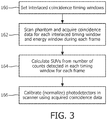
- FIGURE 3 illustrates a method for calibrating a PET scanner using interlaced coincidence windows, in accordance with an embodiment described herein.
- interlaced coincidence windows are selected or set.
- three timing windows are set, at 2ns, 4ns, and 6ns, respectively. It will be appreciated however, that other timing window settings (e.g., 1ns, 1.5ns, 2.7ns, 3ns, 5ns, etc.) may be set, as well as other numbers of timing windows (e.g., 2, 4, 5, etc.).
- a radioactive phantom is scanned and coincidence data is acquired for each of the coincident window settings and each of the energy windows.
- coincidence data is analyzed to determine standardized uptake values (SUV) for the phantom in each frame, and for each timing window.
- the SUV is calculated either pixel-wise or over a region of interest (ROI) for each image or frame of a dynamic series at various time points as a ratio of tissue radioactivity concentration.
- ROI region of interest
- photodetectors in the PET scanner are normalized using the acquired coincidence data for each coincidence window setting and each energy window setting. Photodetector calibration typically includes adjusting the spatial gain and offset of an array of photodetectors to ensure spatial and energy consistency and accuracy.
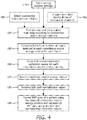
- FIGURE 4 illustrates a method for calibrating a PET scanner by post-processing acquired coincidence data from a radioactive phantom collected in a list mode.
- energy window settings are selected. Detected radiation events that fall outside of the selected energy window(s) will be discarded when correcting acquired scan data for reconstruction.
- coincidence timing window settings are selected. Pairs of detected radiation events that fall within the selected coincidence window(s) are used to determine SUVs for scanner calibration and normalization.
- a radioactive phantom is scanned to acquire data as the radioactive material decays (e.g., over a 14-hour period or the like). The data is collected in a list mode in which each event is time stamped and its energy recorded.
- the list mode data is post-processed to bin or sort the data according to which of a plurality of selected coincidence windows into which particular data falls and/or into which of a plurality of selected energy windows the particle data falls.
- coincidence data in each bin is analyzed to calculate standardized uptake values (SUVs) for the phantom in each frame, and for each timing and/or energy window.
- the SUV is calculated either pixel-wise or over a region of interest (ROI) for each image or frame of a dynamic series at various time points as a ratio of tissue radioactivity concentration.
- ROI region of interest
- the SUV and normalization connection values are stored. When a patient is to be scanned, one of the coincidence windows and/or energy windows is selected, at 190.
- the corresponding SUV and normalization correction values are retrieved, at 192.
- PET data is acquired (e.g., the patient is scanned) and corrected in accordance with the retrieved SUV and normalization values.
- the corrected data is reconstructed into an image for display and/or storage.
- FIGURE 5 illustrates a PET scanner 210 calibrated in accordance with one of the above described calibration methods.
- the PET scanner includes a gantry with a plurality of radiation detectors that detect radiation events (e.g., gamma rays, etc.).
- a user input device 212 is provided, and includes a coincidence window selector 214 via which a user inputs or selects coincidence window settings that delineate one or more coincidence windows into which radiation events are binned or categorized by a coincidence windowing circuit 216.
- the input device also includes an energy window selector 218 via which a user inputs or selects energy window settings that define one or more energy windows.
- An energy window circuit 220 excludes or discards detected radiation events that are not within the selected energy window. The coincidence and/or energy windows are used to calibrate the PET scanner as described herein.
- the system further includes a normalization memory 222, which stores normalization correction values 224 derived during calibration for a plurality of coincidence timing windows and/or energy windows, and an SUV memory 226 that stores SUV correction values 228 derived during calibration for the plurality of coincidence timing windows and/or energy windows.
- a normalization correction module 230 e.g., a processor retrieves normalization correction values 224 for a given coincidence or energy window used when scanning a subject, and normalizes acquired scan data.
- An SUV correction module 232 retrieves stored SUV correction values 228 and performs SUV correction on the acquired scan data.
- a reconstruction processor 234 then reconstructs an image of the subject, which is presented to a user on a display 236, and/or stored in a patient database 238 for later retrieval and display.
Description
- The present application finds particular application in positron emission tomography (PET) systems, particularly involving PET scanner calibration. However, it will be appreciated that the described technique may also find application in other medical imaging device calibration systems, other calibration scenarios, or other scanner calibration techniques.
- Typical PET scanners perform a normalization calibration to correct nonuniform 3D detector response, as is described in
U.S. Patent No. 7718954 , which typically requires about 6 hours. Furthermore, such scanners employ a Standard Uptake Value (SUV) calibration that provides the conversion of the counts in an image to an activity concentration. This calibration uses a decaying F-18 source and can take 11-hours or more to complete. - Any event, even two random single events, which are measured by a PET scanner and occur within the coincidence timing window (e.g., 6 ns) and are treated as a coincident event, i.e. as defining a valid line of response (LOR). Reducing the frequency with which random events are taken as valid is beneficial for several reasons. For instance, random event reduction increases the system's maximum NECR (NEMA NU-2 standard, Noise Equivalent Count Rate) performance, and reduces an amount of data for reconstruction processing (speeds up list-mode reconstructions). Additionally, reducing random events reduces the magnitude of the corrections that need to be made during reconstruction, and provides more bandwidth for a PET scanner to acquire valid coincidence events.
- Attempts have been made to reduce random events by reducing the coincidence timing window to the minimum required to cover the object being imaged (see, e.g.,
U.S. Patent No. 7,626,171 ). However, in such approaches, a reduced coincidence window changes the counting characteristics of the system, and therefore a separate SUV calibration is required for each coincidence window setting used. - Today, commercial PET scanners have a fixed coincidence window, e.g., about 6 nanoseconds. However, the coincidence window can be shortened with some patients, particularly smaller patients, and/or when performing brain imaging, and the like, to give better discrimination between true events and noise (random events). However, the duration of the coincidence window effects various calibrations including normalization and SUV. Each calibration routine is very time-consumptive, typically taking 11-14 hours or more for each coincidence window.
-
JP 11 153 669 A -
US 7,626,171 B2 discloses a method of reconstructing time-of-flight (TOF) images including obtaining a profile of a subject to be imaged in an examination region of an imaging system. Events associated with radiation emitted from the subject are detected and converted to electronic data. Electronic data attributable to radiation events located outside the profile are removed and images are reconstructed from the remaining electronic data. - The present application provides new and improved PET scanner calibration systems and methods that employ interlaced coincidence timing window settings during SUV acquisition on a radioactive calibration phantom, which overcome the above-referenced problems and others.
- In accordance with one aspect, positron emission tomography (PET) scanner calibration system includes a PET scanner in which a radioactive calibration phantom is placed and scanned for a predetermined time period, and a processor that executes computer-executable instructions stored in a memory, the instructions including receiving settings for a plurality of selected coincidence timing and/or energy windows. The instructions further include scanning the radioactive calibration phantom and acquiring coincidence data for each of the plurality of coincidence timing and/or energy windows defined by the timing and/or energy window settings during each of a plurality of frames of the predetermined time period. Additionally, the instructions include calculating standard uptake values from a number of photon counts detected in each frame over the predetermined time period for each selected coincidence timing and/or energy window.
- In accordance with another aspect, a method of calibrating a positron emission tomography (PET) scanner includes receiving settings for a plurality of selected coincidence timing and/or energy windows, and scanning the radioactive calibration phantom and acquiring coincidence data for each of the plurality of coincidence timing and/or energy windows defined by the timing and/or energy window settings during each of a plurality of frames of the predetermined time period. The method further includes calculating standard uptake values (SUVs) from a number of photon counts detected in each frame over the predetermined time period for each selected coincidence timing and/or energy window.
- In accordance with an embodiment a PET scanner includes a gantry with a plurality of radiation detectors that detect scintillation events, a coincidence windowing circuit that identifies pairs of detected events within a plurality of coincidence windows of different lengths, and a user input device by which a user selects at least one of the coincidence windows. The PET scanner additionally includes a normalization correction module that applies stored normalization correction values to acquired scan data in the selected coincidence window, an SUV correction module that applies stored SUV correction values to the acquired scan data in the selected coincidence window, and a reconstruction processor that reconstructs the corrected scan data into an image for presentation on a display.
- One advantage is that scanner calibration time is reduced.
- Another advantage resides in calibrating the scanner for multiple timing windows.
- Still further advantages of the subject innovation will be appreciated by those of ordinary skill in the art upon reading and understand the following detailed description.
- The innovation may take form in various components and arrangements of components, and in various steps and arrangements of steps. The drawings are only for purposes of illustrating various aspects and are not to be construed as limiting the invention.
-
FIGURE 1 illustrates a standard uptake value (SUV) acquisition sequence that uses interlaced coincidence window settings to perform a single SUV calibration and normalization for multiple coincidence windows, which eliminates the need for performing separate SUV and normalization calibrations for each coincidence window setting. -
FIGURE 2 illustrates a PET scanner calibration system using interlaced coincidence timing window settings during an SUV acquisition sequence, in accordance with various aspects described herein. -
FIGURE 3 illustrates a method for calibrating a PET scanner using interlaced coincidence timing windows, in accordance with various aspects described herein. -
FIGURE 4 illustrates a method for calibrating a PET scanner by post-processing acquired coincidence data from a radioactive phantom using interlaced coincidence timing windows to bin the acquired data, in accordance with various aspects described herein. -
FIGURE 5 illustrates a PET scanner calibrated in accordance with one of the above described calibration methods. - The subject innovation overcomes the problem of time-consuming and labor-intensive scanner calibrations by reducing the calibration duration by interlacing the coincidence window settings within the SUV calibration. In another embodiment, calibration duration is reduced by performing the SUV and normalization calibration acquisitions at the largest coincidence window and post-processing the data for the desired coincidence windows.
-
FIGURE 1 illustrates a standard uptake value (SUV)acquisition sequence 10 that uses interlaced coincidence window settings to perform a single SUV calibration and normalization for multiple coincidence windows, which eliminates the need for performing separate SUV and normalization calibrations for each coincidence window setting. InFigure 1 , three interlaced timing windows (e.g. 2ns, 4ns, and 6ns) are shown for 20 frames (labeled 0-19). Each frame is divided into three portions, each of which corresponds to one of the timing windows. In this manner random events are reduced in a PET scanner the PET scanner's maximum noise equivalent count rate (NECR) is maximized as a function of the diameter of an object being imaged. SUVs are calculated either pixel-wise or over a region of interest (ROI) for each image or frame of a dynamic series at various time points as a ratio of tissue radioactivity concentration. As used herein, a "random event" or "random" denotes an occurrence in which two single events are measured or detected by a PET scanner within a coincidence timing window (e.g., 6 ns or some other predefined timing window), and are mistakenly treated or processed as a coincident event (i.e., as occurring as a result of a single common annihilation event). - As is known in the art, when an electron and positron meet, they annihilate, emitting two 511 keV gamma rays that are oppositely directed in accordance with the principle of conservation of momentum. In PET data acquisition, two substantially simultaneous or coincident 511 keV gamma ray detection events are presumed to have originated from the same positron-electron annihilation event, which is therefore located somewhere along the "line of response" (LOR) connecting the two substantially simultaneous 511 keV gamma ray detection events. This line of response is also sometimes called a projection, and the collected PET data is referred to as projection data.
- In conventional PET, two 511 keV gamma ray detection events occurring within a selected short time or coincidence window, such as within 6 nanoseconds of each other, are taken as defining a valid LOR. Due to the variable annihilation position with respect to the detector elements a small (e.g., sub-nanosecond) time difference between the coincident gamma photon detection events occurs. A related technique, called time-of-flight PET or TOF-PET, takes advantage of this small time difference to further localize the positron-electron annihilation event along the LOR. In general, the annihilation event occurred along the LOR at a point closer to the gamma ray detection event that occurred first. If the two gamma ray detection events occur simultaneously within the time resolution of the detectors, then the annihilation event occurred at the midpoint of the LOR.
- Under a conventional approach that does not use interlaced collection of calibration data from an F18 phantom, an SUV acquisition sequence for 20 frames (e.g., frames 0-19) takes 11 hours per coincidence window setting. According to an example, SUV data for frames 0-7 takes 15 minutes per frame, for a total of two hours; SUV data acquisition for frames 8-13 takes 30 minutes per frame, for a total of three hours, and SUV data acquisition for frames 14-19 takes one hour per frame, for a total of six hours. One reason that the calibration scan takes so long is that the half life of F18 radioactive material is approximately 110 minutes, so an 11 hour scan provides data over 6 half lives of the material (i.e., after 11 hours, 98.5% of the F18 radioactive material has decayed). Thus, data is needed over an 11 hour period to calibrate photomultipliers in the scanner to a range of radioactivity levels. That is, calibrating the scanner using only data acquired during the first 110 minutes typically does not provide sufficient accuracy for detection of low-level radio activity, such as may be equivalent to calibration data acquired during the 10th or 11th hour of the calibration scan. Moreover, if a user runs an SUV acquisition sequence for three coincidence window settings (e.g., 2ns, 4ns, and 6ns), then the conventional approach will take 33 hours (using the above F18 example), plus the time to prepare a new F18 calibration phantom for each coincidence window.
- The present application has recognized that the calibration only needs to be adequately sampled in each frame, rather than continuously sampled over each entire frame. By interlacing the calibration data collection for a plurality of coincidence windows as shown in
Figure 1 , the foregoing example SUV calibration data acquisition sequence can be performed for all three (or more) coincidence window settings in the single 11-hour scanning period with an F18 phantom.Figure 1 thus provides an example of an approach to modifying the typical SUV calibration acquisition sequence such that the calibration data acquisition for each of the coincidence windows is interlaced within each frame. 2ns, 4ns, and 6ns coincidence window settings are provided in the example ofFigure 1 , although other window settings may be employed, as will be appreciated by those of skill in the art. Additionally, more or fewer interlaced window settings (e.g., 2, 3, 4, 5, 6, etc.) may be employed, as will be appreciated by those of skill in the art. - For each frame, data is acquired for all three coincidence windows. For example, rather than spending 15 minutes acquiring data for a single timing window during frame 0, 5 minutes are spent acquiring data for each of three coincidence windows during frame 0. This results in acquisition of only 1/3 of the data for a given coincidence window over all frames when compared to conventional protocols, but the acquired data for each coincidence window is still spread over the entire decay period (e.g., 11-14 hours), which provides more than ample data for SUV characterization and calibration of PMTs in a PET scanner. In this manner, calibration data (SUVs) is acquired for all three (or other number) coincidence windows over the total 11 hour period, which is advantageous because calibration of the scanner for each coincidence window is improved by using data collected over the full radioactive decay period (e.g., 6-7 half lives), but calibration does not require a full 11 hours worth of data for each coincidence window. Rather, periodic samples may be taken for each coincidence window, during each frame taken over the 11 hour period.
- In one embodiment, acquisition hardware time stamps each radiation event. The radiation events are subject to the largest coincidence window such that events outside the largest coincidence window to be calibrated can be discarded. Software looks at the time stamps and sorts the coincident pairs by coincidence time, e.g. among events that are coincident within 2ns, with 4rs, and within 6ns. This approach allows the normalization and SUV acquisitions to be run once with the widest coincidence window (e.g., 6ns) and to generate solutions for other coincidence windows by post-processing.
- In one technique, sequential acquisitions at different coincidence windows are used. In another embodiment, calibrations are interlaced to include variation in additional acquisition parameters, e.g. energy window or transverse fields-of-view.
-
FIGURE 2 illustrates a PETscanner calibration system 100 102 using aphantom 103, e.g. an F18 phantom. Thesystem 100 includes aprocessor 104 that is coupled to the PET scanner and to amemory 106. The memory stores, and the processor executes, computer-executable instructions for performing the various functions, methods, techniques, procedures, etc., described herein. The system further includes auser interface 108 comprising one or more input devices 110 (e.g., a keyboard, mouse, stylus, touch screen, microphone, etc.), and adisplay 112 on which information is presented to a user. - The memory stores an SUV calibration data acquisition
sequence software module 114 that is executed by theprocessor 104 to determineSUV calibration data 115 during a scan of a fludeoxyglucose-18 (F-18) calibration phantom as it radioactively decays in the examination region of the PET scanner (e.g., over an hours-long decay period). The SUV data is used to calculate standard uptake values as a function of coincidence window during the acquisition scan. In one example the phantom is a 20cm by 30cm cylindrical phantom. In another example the phantom is spherical. - Coincidence
window setting information 116 is entered by a user into theuser interface 108 and stored in thememory 106. Additionally, the memory stores one ormore reconstruction algorithms 118 that are executed by a reconstruction processor 120 in order to reconstruct an image of the calibration phantom and/or other objects after calibration of thePET scanner 102. Once theSUV calibration data 115 has been acquired, the processor executes acalibration module 122 that calibrates standard uptake values for thePET scanner 102. Additionally, the processor executes anormalization module 124 that is stored in the memory to calculate normalize connections for each of the photodetectors or photomultiplier tubes in the PET scanner such that all radiation sensors have a common sensitivity to incident radiation. The normalization calibration is also performed for each coincidence window. The same techniques described above for the SUV calibration can be used to calibrate the normalization for each of the coincidence windows. - The interlaced calibration data collection for each coincidence window facilitates providing an adjustable and/or selectable coincidence window, e.g., of 2, 4, or 6 nanoseconds. SUV and normalization values are calibrated in a single calibration procedure, via the
calibration module 122 and thenormalization module 124. To perform the SUV calibration, data is typically taken with an F-18 phantom as the F-18 decays over a number of hours, e.g., 14 hours. In one embodiment, data is cyclically collected for each of the coincidence window times to generate a series of points over the 14 hours to define three uptake curves, as shown in the acquisition sequence ofFigure 1 . - In another embodiment, all of the data is time-stamped with sufficient accuracy, and the time stamped
data 126 is sorted by asorting module 127 executed by theprocessor 104 into events which are coincident within 2 nanoseconds, within 4 nanoseconds, or within 6 nanoseconds. These readings and their collection times can be used to generate the SUV calibration curves 128 for each coincidence window. - In another embodiment, an energy
window adjustment module 130 provides an adjustable energy window. That is, for acquired F-18 scan data, a width of an energy peak or range of energy around 511 keV that is considered valid is adjustable. However, changing the energy window also changes the SUV and normalization calibrations. In this case, the herein-described techniques are used to calibrate the SUV and the normalization for each of a plurality of energy windows in a single calibration procedure using either the interleaving technique or by recording the energy of each event and sorting by energy window. - According to another embodiment, the
nuclear scanner 102 is a time-of-flight (TOF) PET scanner, andTOF data 132 is stored in thememory 106 for use in improving accuracy in the reconstruction of PET images. - As stated above, the
system 100 includes theprocessor 104 that executes, and thememory 106, which stores, computer-executable instructions (e.g., routines, programs, algorithms, software code, etc.) for performing the various functions, methods, procedures, etc., described herein. Additionally, "module," as used herein, denotes a set of computer-executable instructions, software code, program, routine, or the like, as will be understood by those of skill in the art. - The memory may be a computer-readable medium on which a control program is stored, such as a disk, hard drive, or the like. Common forms of computer-readable media include, for example, floppy disks, flexible disks, hard disks, magnetic tape, or any other magnetic storage medium, CD-ROM, DVD, or any other optical medium, RAM, ROM, PROM, EPROM, FLASH-EPROM, variants thereof, other memory chip or cartridge, or any other tangible medium from which the processor can read and execute. In this context, the systems described herein may be implemented on or as one or more general purpose computers, special purpose computer(s), a programmed microprocessor or microcontroller and peripheral integrated circuit elements, an ASIC or other integrated circuit, a digital signal processor, a hardwired electronic or logic circuit such as a discrete element circuit, a programmable logic device such as a PLD, PLA, FPGA, Graphical card CPU (GPU), or PAL, or the like.
-
FIGURE 3 illustrates a method for calibrating a PET scanner using interlaced coincidence windows, in accordance with an embodiment described herein. At 160, interlaced coincidence windows are selected or set. In one example, three timing windows are set, at 2ns, 4ns, and 6ns, respectively. It will be appreciated however, that other timing window settings (e.g., 1ns, 1.5ns, 2.7ns, 3ns, 5ns, etc.) may be set, as well as other numbers of timing windows (e.g., 2, 4, 5, etc.). At 162, a radioactive phantom is scanned and coincidence data is acquired for each of the coincident window settings and each of the energy windows. At 164, coincidence data is analyzed to determine standardized uptake values (SUV) for the phantom in each frame, and for each timing window. The SUV is calculated either pixel-wise or over a region of interest (ROI) for each image or frame of a dynamic series at various time points as a ratio of tissue radioactivity concentration. At 166, photodetectors in the PET scanner are normalized using the acquired coincidence data for each coincidence window setting and each energy window setting. Photodetector calibration typically includes adjusting the spatial gain and offset of an array of photodetectors to ensure spatial and energy consistency and accuracy. -
FIGURE 4 illustrates a method for calibrating a PET scanner by post-processing acquired coincidence data from a radioactive phantom collected in a list mode. At 178, energy window settings are selected. Detected radiation events that fall outside of the selected energy window(s) will be discarded when correcting acquired scan data for reconstruction. At 180, coincidence timing window settings are selected. Pairs of detected radiation events that fall within the selected coincidence window(s) are used to determine SUVs for scanner calibration and normalization. At 182, a radioactive phantom is scanned to acquire data as the radioactive material decays (e.g., over a 14-hour period or the like). The data is collected in a list mode in which each event is time stamped and its energy recorded. At 184, the list mode data is post-processed to bin or sort the data according to which of a plurality of selected coincidence windows into which particular data falls and/or into which of a plurality of selected energy windows the particle data falls. At 186, coincidence data in each bin is analyzed to calculate standardized uptake values (SUVs) for the phantom in each frame, and for each timing and/or energy window. The SUV is calculated either pixel-wise or over a region of interest (ROI) for each image or frame of a dynamic series at various time points as a ratio of tissue radioactivity concentration. At 188, the SUV and normalization connection values are stored. When a patient is to be scanned, one of the coincidence windows and/or energy windows is selected, at 190. The corresponding SUV and normalization correction values are retrieved, at 192. At 194, PET data is acquired (e.g., the patient is scanned) and corrected in accordance with the retrieved SUV and normalization values. The corrected data is reconstructed into an image for display and/or storage. -
FIGURE 5 illustrates aPET scanner 210 calibrated in accordance with one of the above described calibration methods. The PET scanner includes a gantry with a plurality of radiation detectors that detect radiation events (e.g., gamma rays, etc.). Auser input device 212 is provided, and includes acoincidence window selector 214 via which a user inputs or selects coincidence window settings that delineate one or more coincidence windows into which radiation events are binned or categorized by acoincidence windowing circuit 216. The input device also includes anenergy window selector 218 via which a user inputs or selects energy window settings that define one or more energy windows. Anenergy window circuit 220 excludes or discards detected radiation events that are not within the selected energy window. The coincidence and/or energy windows are used to calibrate the PET scanner as described herein. - The system further includes a
normalization memory 222, which stores normalization correction values 224 derived during calibration for a plurality of coincidence timing windows and/or energy windows, and anSUV memory 226 that stores SUV correction values 228 derived during calibration for the plurality of coincidence timing windows and/or energy windows. A normalization correction module 230 (e.g., a processor) retrieves normalization correction values 224 for a given coincidence or energy window used when scanning a subject, and normalizes acquired scan data. An SUV correction module 232 (e.g., a processor) retrieves stored SUV correction values 228 and performs SUV correction on the acquired scan data. Areconstruction processor 234 then reconstructs an image of the subject, which is presented to a user on adisplay 236, and/or stored in apatient database 238 for later retrieval and display.
Claims (15)
- A positron emission tomography (PET) scanner calibration system (100), including:a PET scanner (102, 210) which is configured for a radioactive calibration phantom to be placed and scanned for a predetermined time period;a processor (104) that is configured to execute computer-executable instructions stored in a memory (106), the instructions including:receiving settings for a plurality of selected coincidence timing and/or energy windows;scanning the radioactive calibration phantom with the PET scanner (102, 210) for the predetermined time period and acquiring coincidence data for each of the plurality of coincidence timing and/or energy windows defined by the timing and/or energy window settings during each of a plurality of frames of the predetermined time period; and calculating standard uptake values from a number of photon counts detected in each frame over the predetermined time period for each selected coincidence timing and/or energy window.
- The system according to claim 1, the instructions further including generating a calibration curve for each of the timing windows from the standard uptake values, and using the calibration curves to calibrate photodetectors.
- The system according to either of claims 1 or 2, the instructions further including:calculating normalization correction values that correct for differences in sensitivity of the photodetectors in the PET scanner.
- The system according to any one of the preceding claims, wherein the radioactive calibration phantom is a fludeoxyglucose-18 (F-18) calibration phantom.
- The system according to any one of the preceding claims, wherein the plurality of coincidence timing and/or energy window settings are interlaced and wherein the instructions further include:acquiring coincidence data for a plurality of interlaced coincidence timing windows defined by the interlaced coincidence timing and/or energy window settings.
- The system according to any one of the preceding claims, the instructions further including:storing normalization correction values (224) and SUV correction values (228) for the plurality of coincidence timing windows and/or energy windows;performing a PET scan of a subject;correcting acquired scan data of the subject using the normalization correction values and the SUV correction values;reconstructing an image of the subject using the corrected scan data; andstoring the reconstructed image to a patient database (238).
- The system according to any one of the preceding claims, wherein the plurality of frames includes approximately 20 frames, acquired over a period of time comprising approximately 6-8 half lives of radioactive material comprised by the radioactive calibration phantom.
- The system according to any one of the preceding claims, wherein the PET scanner is a time-of-flight PET scanner.
- The system according to claim 1, wherein the PET scanner (102, 210) includes:a gantry with a plurality of radiation detectors that detect scintillation events;a coincidence windowing circuit (216) that identifies pairs of detected events within the plurality of selected coincidence timing and/or energy windows of different lengths;a user input device by which a user selects at least one of the coincidence windows;a normalization correction module (230) that applies stored normalization correction values to acquired scan data in the selected timing and/or energy coincidence window;an SUV correction module (232) that applies the calculated standard uptake values to the acquired scan data in the selected timing and/or energy coincidence window; anda reconstruction processor (234) that reconstructs the corrected scan data into an image for presentation on a display.
- A method of calibrating a positron emission tomography (PET) scanner, including:receiving settings for a plurality of selected coincidence timing and/or energy windows;scanning the radioactive calibration phantom for a predetermined time period and acquiring coincidence data for each of the plurality of coincidence timing and/or energy windows defined by the timing and/or energy window settings during each of a plurality of frames of the predetermined time period; andcalculating standard uptake values (SUVs) from a number of photon counts detected in each frame over the predetermined time period for each selected coincidence timing and/or energy window.
- The method according to claim 10, further including:generating a calibration curve for each of the timing windows from the standard uptake values, and using the calibration curves to calibrate photodetectors.
- The method according to either of claims 10 or 11, further including:calculating normalization correction values that correct for differences in sensitivity of the photodetectors in the PET scanner.
- The method according to any one of claims 10-12 , wherein the coincidence data is collected in a list mode, and further including:timestamping each received radiation event;sorting the radiation events according to which of the plurality of coincidence timing and/or energy windows each pair of coincident events corresponds.
- The method according to any one of claims 10-12. wherein the plurality of coincidence timing and/or energy window settings are interlaced and wherein the instructions further include:acquiring coincidence data for a plurality of interlaced coincidence timing windows defined by the interlaced coincidence timing and/or energy window settings.
- A processor (104) or computer-readable medium (106) carrying a computer program that controls one or more processors to perform the method of any one of claims 10-14.
Applications Claiming Priority (2)
| Application Number | Priority Date | Filing Date | Title |
|---|---|---|---|
| US41632310P | 2010-11-23 | 2010-11-23 | |
| PCT/IB2011/055097 WO2012069960A2 (en) | 2010-11-23 | 2011-11-15 | Pet calibrations with varying coincidence windows |
Publications (2)
| Publication Number | Publication Date |
|---|---|
| EP2643710A2 EP2643710A2 (en) | 2013-10-02 |
| EP2643710B1 true EP2643710B1 (en) | 2017-01-11 |
Family
ID=45218774
Family Applications (1)
| Application Number | Title | Priority Date | Filing Date |
|---|---|---|---|
| EP11793870.4A Active EP2643710B1 (en) | 2010-11-23 | 2011-11-15 | Pet calibrations with varying coincidence windows |
Country Status (6)
| Country | Link |
|---|---|
| US (1) | US8987659B2 (en) |
| EP (1) | EP2643710B1 (en) |
| JP (1) | JP5944402B2 (en) |
| CN (1) | CN103221841B (en) |
| RU (1) | RU2582887C2 (en) |
| WO (1) | WO2012069960A2 (en) |
Families Citing this family (19)
| Publication number | Priority date | Publication date | Assignee | Title |
|---|---|---|---|---|
| JP5603647B2 (en) * | 2009-05-13 | 2014-10-08 | キヤノン株式会社 | Power feeding device, power feeding device control method, and power feeding communication system |
| CN105814454B (en) * | 2013-12-04 | 2019-11-05 | 皇家飞利浦有限公司 | For rebuilding the reconstructing device of PET image |
| US9297909B2 (en) * | 2014-04-18 | 2016-03-29 | Perkinelmer Health Sciences, Inc. | Guard efficiency compensation system and method |
| US9910162B2 (en) * | 2014-06-12 | 2018-03-06 | Siemens Medical Solutions Usa, Inc. | Calibrating in single photon emission computed tomography with multi-emission energies |
| WO2016046703A2 (en) * | 2014-09-23 | 2016-03-31 | Koninklijke Philips N.V. | Time of flight calibration in digital positron emission tomography |
| US9606245B1 (en) | 2015-03-24 | 2017-03-28 | The Research Foundation For The State University Of New York | Autonomous gamma, X-ray, and particle detector |
| CN105030263B (en) * | 2015-07-22 | 2018-06-12 | 湖北锐世数字医学影像科技有限公司 | The energy back bearing calibration of number PET a kind of and system |
| US10281596B2 (en) * | 2015-09-18 | 2019-05-07 | Koninklijke Philips N.V. | Correcting photon counts in a photon counting X-ray radiation detection system |
| JP6941098B2 (en) * | 2015-10-28 | 2021-09-29 | コーニンクレッカ フィリップス エヌ ヴェKoninklijke Philips N.V. | Equipment and methods for determining SUVs in radiation tomography |
| CN105395209B (en) | 2015-12-01 | 2018-10-02 | 沈阳东软医疗系统有限公司 | A kind of positron emission computerized tomography imaging system and method |
| CN106108935B (en) * | 2016-08-31 | 2019-06-28 | 北京康科达科技有限公司 | A kind of SPECT detection device |
| CN108109182B (en) * | 2016-11-24 | 2021-08-24 | 上海东软医疗科技有限公司 | PET image reconstruction method and device |
| JP7008728B2 (en) * | 2017-05-04 | 2022-01-25 | コーニンクレッカ フィリップス エヌ ヴェ | Prompt Gamma Positron Emission Tomography (PET) Timing Calibration Using Simultaneous Generation with High Energy Cascade Gamma from Positron Emission Nuclides |
| US10410383B2 (en) | 2017-08-26 | 2019-09-10 | Uih America, Inc. | System and method for image data processing in positron emission tomography |
| CN107595315A (en) * | 2017-09-30 | 2018-01-19 | 华中科技大学 | The acquisition methods of photoresponse line in a kind of transmitting imaging device |
| CN108294774A (en) * | 2018-01-04 | 2018-07-20 | 沈阳东软医疗系统有限公司 | A kind of PET correct scans method, apparatus and equipment |
| CN110215227B (en) * | 2019-06-05 | 2022-10-14 | 上海联影医疗科技股份有限公司 | Time window setting method and device, computer equipment and storage medium |
| US11249206B2 (en) * | 2020-01-06 | 2022-02-15 | Canon Medical Systems Corporation | Method and system for PET detector efficiency normalization |
| CN113852440B (en) * | 2021-09-29 | 2023-12-29 | 应急管理部国家自然灾害防治研究院 | Method and system for precise time calibration of electromagnetic field observation data of Zhangheng first satellite |
Family Cites Families (20)
| Publication number | Priority date | Publication date | Assignee | Title |
|---|---|---|---|---|
| JP3924879B2 (en) * | 1997-11-21 | 2007-06-06 | 株式会社島津製作所 | Nuclear medicine diagnostic equipment |
| US20050123183A1 (en) * | 2003-09-02 | 2005-06-09 | Paul Schleyer | Data driven motion correction for nuclear imaging |
| JP3717122B2 (en) * | 2003-09-29 | 2005-11-16 | 株式会社日立製作所 | γ-ray detection time determination method, γ-ray coincidence counting method, and nuclear medicine diagnostic apparatus |
| PT103200B (en) * | 2004-09-30 | 2006-08-24 | Taguspark-Soc. Prom.Desenv.Parq.Ci.Tec.Area Lisboa | POSITRON EMISSION TOMOGRAPHY SYSTEM (PET) |
| US7129495B2 (en) | 2004-11-15 | 2006-10-31 | General Electric Company | Method and apparatus for timing calibration in a PET scanner |
| US7057178B1 (en) | 2004-11-15 | 2006-06-06 | General Electric Company | Method and system for imaging using a filter for Time-of-Flight PET |
| US7301144B2 (en) * | 2004-12-29 | 2007-11-27 | General Electric Company | Method and system for calibrating a positron emission tomography system |
| US7227149B2 (en) | 2004-12-30 | 2007-06-05 | General Electric Company | Method and system for positron emission tomography image reconstruction |
| EP1844352B1 (en) * | 2005-01-28 | 2010-03-24 | Koninklijke Philips Electronics N.V. | Timing calibration using radioactive sources |
| US7381959B2 (en) | 2005-08-17 | 2008-06-03 | General Electric Company | Technique for reconstructing PET scan images |
| US7414246B2 (en) * | 2006-01-03 | 2008-08-19 | Koninklijke Philips Electronics N.V. | Achieving accurate time-of-flight calibrations with a stationary coincidence point source |
| CN101365963B (en) | 2006-01-09 | 2012-02-01 | 皇家飞利浦电子股份有限公司 | Method of constructing time-in-flight pet images |
| US7718954B2 (en) | 2006-01-31 | 2010-05-18 | Koninklijke Philips Electronics N.V. | Component method and system for PET detector efficiency normalization |
| US8965071B2 (en) * | 2008-05-30 | 2015-02-24 | Emory University | Assessing tumor response to therapy |
| WO2011061644A1 (en) * | 2009-11-18 | 2011-05-26 | Koninklijke Philips Electronics N.V. | Motion correction in radiation therapy |
| US9968309B2 (en) * | 2009-12-08 | 2018-05-15 | Koninklijke Philips N.V. | Method and a correction system for correcting tracer-uptake measurements |
| CN103260521B (en) * | 2010-12-14 | 2015-11-25 | 皇家飞利浦电子股份有限公司 | For the workflow of the integration of input function estimation accurately |
| GB201109344D0 (en) * | 2011-06-03 | 2011-07-20 | Siemens Medical Solutions | BIF from WB dynamic |
| US20130136328A1 (en) * | 2011-11-30 | 2013-05-30 | General Electric Company | Methods and systems for enhanced tomographic imaging |
| GB201219403D0 (en) * | 2012-10-29 | 2012-12-12 | Uni I Olso | Method for improved estimation of tracer uptake in physiological image volumes |
-
2011
- 2011-11-15 JP JP2013539383A patent/JP5944402B2/en not_active Expired - Fee Related
- 2011-11-15 WO PCT/IB2011/055097 patent/WO2012069960A2/en active Application Filing
- 2011-11-15 US US13/885,282 patent/US8987659B2/en active Active
- 2011-11-15 RU RU2013128552/28A patent/RU2582887C2/en not_active IP Right Cessation
- 2011-11-15 CN CN201180056345.3A patent/CN103221841B/en active Active
- 2011-11-15 EP EP11793870.4A patent/EP2643710B1/en active Active
Non-Patent Citations (1)
| Title |
|---|
| None * |
Also Published As
| Publication number | Publication date |
|---|---|
| CN103221841B (en) | 2015-11-25 |
| CN103221841A (en) | 2013-07-24 |
| US8987659B2 (en) | 2015-03-24 |
| RU2013128552A (en) | 2014-12-27 |
| JP5944402B2 (en) | 2016-07-05 |
| WO2012069960A2 (en) | 2012-05-31 |
| RU2582887C2 (en) | 2016-04-27 |
| WO2012069960A3 (en) | 2012-11-01 |
| EP2643710A2 (en) | 2013-10-02 |
| JP2014500491A (en) | 2014-01-09 |
| US20130240721A1 (en) | 2013-09-19 |
Similar Documents
| Publication | Publication Date | Title |
|---|---|---|
| EP2643710B1 (en) | Pet calibrations with varying coincidence windows | |
| US7626171B2 (en) | Method of constructing time-in-flight pet images | |
| EP3234647B1 (en) | Pixel based dead time correction | |
| US7129495B2 (en) | Method and apparatus for timing calibration in a PET scanner | |
| US9332952B2 (en) | Data-driven optimization of event acceptance/rejection logic | |
| EP2867701B1 (en) | Digital positron emission tomography (dpet) energy calibration method | |
| US9747701B2 (en) | Systems and methods for emission tomography quantitation | |
| US8530846B2 (en) | Apparatus and methods for detector scatter recovery for nuclear medicine imaging systems | |
| US20080260094A1 (en) | Method and Apparatus for Spectral Computed Tomography | |
| US9226716B2 (en) | Nuclear medicine imaging apparatus and radiation therapy apparatus | |
| RU2517586C2 (en) | Reverse reconstruction of data for optimal time generation of count pulses in radiological physiological visualisation in list mode | |
| CN108474862B (en) | Energy calibration with LU spectral subtraction | |
| JP2020507753A (en) | Photon counting detectors that enable matching | |
| JP2005326406A (en) | Method and system for normalizing positron emission tomography system | |
| US9645260B2 (en) | Photon counting system and method | |
| US20110142367A1 (en) | Methods and systems for correcting image scatter | |
| US7329873B2 (en) | Coincidence counting method of γ ray and nuclear medicine diagnostic apparatus | |
| US8563935B2 (en) | Nuclear medicine imaging apparatus and control method | |
| EP3819675A1 (en) | Imaging of photon-counting ct system | |
| CN112587159A (en) | Method and system for a PET detector |
Legal Events
| Date | Code | Title | Description |
|---|---|---|---|
| PUAI | Public reference made under article 153(3) epc to a published international application that has entered the european phase |
Free format text: ORIGINAL CODE: 0009012 |
|
| 17P | Request for examination filed |
Effective date: 20130624 |
|
| AK | Designated contracting states |
Kind code of ref document: A2 Designated state(s): AL AT BE BG CH CY CZ DE DK EE ES FI FR GB GR HR HU IE IS IT LI LT LU LV MC MK MT NL NO PL PT RO RS SE SI SK SM TR |
|
| DAX | Request for extension of the european patent (deleted) | ||
| 17Q | First examination report despatched |
Effective date: 20140819 |
|
| GRAP | Despatch of communication of intention to grant a patent |
Free format text: ORIGINAL CODE: EPIDOSNIGR1 |
|
| INTG | Intention to grant announced |
Effective date: 20160713 |
|
| GRAS | Grant fee paid |
Free format text: ORIGINAL CODE: EPIDOSNIGR3 |
|
| GRAA | (expected) grant |
Free format text: ORIGINAL CODE: 0009210 |
|
| AK | Designated contracting states |
Kind code of ref document: B1 Designated state(s): AL AT BE BG CH CY CZ DE DK EE ES FI FR GB GR HR HU IE IS IT LI LT LU LV MC MK MT NL NO PL PT RO RS SE SI SK SM TR |
|
| REG | Reference to a national code |
Ref country code: GB Ref legal event code: FG4D |
|
| REG | Reference to a national code |
Ref country code: CH Ref legal event code: EP |
|
| REG | Reference to a national code |
Ref country code: AT Ref legal event code: REF Ref document number: 861790 Country of ref document: AT Kind code of ref document: T Effective date: 20170115 |
|
| REG | Reference to a national code |
Ref country code: IE Ref legal event code: FG4D |
|
| REG | Reference to a national code |
Ref country code: DE Ref legal event code: R096 Ref document number: 602011034324 Country of ref document: DE |
|
| REG | Reference to a national code |
Ref country code: LT Ref legal event code: MG4D |
|
| REG | Reference to a national code |
Ref country code: NL Ref legal event code: MP Effective date: 20170111 |
|
| REG | Reference to a national code |
Ref country code: AT Ref legal event code: MK05 Ref document number: 861790 Country of ref document: AT Kind code of ref document: T Effective date: 20170111 |
|
| PG25 | Lapsed in a contracting state [announced via postgrant information from national office to epo] |
Ref country code: NL Free format text: LAPSE BECAUSE OF FAILURE TO SUBMIT A TRANSLATION OF THE DESCRIPTION OR TO PAY THE FEE WITHIN THE PRESCRIBED TIME-LIMIT Effective date: 20170111 |
|
| PG25 | Lapsed in a contracting state [announced via postgrant information from national office to epo] |
Ref country code: NO Free format text: LAPSE BECAUSE OF FAILURE TO SUBMIT A TRANSLATION OF THE DESCRIPTION OR TO PAY THE FEE WITHIN THE PRESCRIBED TIME-LIMIT Effective date: 20170411 Ref country code: LT Free format text: LAPSE BECAUSE OF FAILURE TO SUBMIT A TRANSLATION OF THE DESCRIPTION OR TO PAY THE FEE WITHIN THE PRESCRIBED TIME-LIMIT Effective date: 20170111 Ref country code: IS Free format text: LAPSE BECAUSE OF FAILURE TO SUBMIT A TRANSLATION OF THE DESCRIPTION OR TO PAY THE FEE WITHIN THE PRESCRIBED TIME-LIMIT Effective date: 20170511 Ref country code: FI Free format text: LAPSE BECAUSE OF FAILURE TO SUBMIT A TRANSLATION OF THE DESCRIPTION OR TO PAY THE FEE WITHIN THE PRESCRIBED TIME-LIMIT Effective date: 20170111 Ref country code: HR Free format text: LAPSE BECAUSE OF FAILURE TO SUBMIT A TRANSLATION OF THE DESCRIPTION OR TO PAY THE FEE WITHIN THE PRESCRIBED TIME-LIMIT Effective date: 20170111 Ref country code: GR Free format text: LAPSE BECAUSE OF FAILURE TO SUBMIT A TRANSLATION OF THE DESCRIPTION OR TO PAY THE FEE WITHIN THE PRESCRIBED TIME-LIMIT Effective date: 20170412 |
|
| PG25 | Lapsed in a contracting state [announced via postgrant information from national office to epo] |
Ref country code: BG Free format text: LAPSE BECAUSE OF FAILURE TO SUBMIT A TRANSLATION OF THE DESCRIPTION OR TO PAY THE FEE WITHIN THE PRESCRIBED TIME-LIMIT Effective date: 20170411 Ref country code: SE Free format text: LAPSE BECAUSE OF FAILURE TO SUBMIT A TRANSLATION OF THE DESCRIPTION OR TO PAY THE FEE WITHIN THE PRESCRIBED TIME-LIMIT Effective date: 20170111 Ref country code: PT Free format text: LAPSE BECAUSE OF FAILURE TO SUBMIT A TRANSLATION OF THE DESCRIPTION OR TO PAY THE FEE WITHIN THE PRESCRIBED TIME-LIMIT Effective date: 20170511 Ref country code: AT Free format text: LAPSE BECAUSE OF FAILURE TO SUBMIT A TRANSLATION OF THE DESCRIPTION OR TO PAY THE FEE WITHIN THE PRESCRIBED TIME-LIMIT Effective date: 20170111 Ref country code: PL Free format text: LAPSE BECAUSE OF FAILURE TO SUBMIT A TRANSLATION OF THE DESCRIPTION OR TO PAY THE FEE WITHIN THE PRESCRIBED TIME-LIMIT Effective date: 20170111 Ref country code: LV Free format text: LAPSE BECAUSE OF FAILURE TO SUBMIT A TRANSLATION OF THE DESCRIPTION OR TO PAY THE FEE WITHIN THE PRESCRIBED TIME-LIMIT Effective date: 20170111 Ref country code: ES Free format text: LAPSE BECAUSE OF FAILURE TO SUBMIT A TRANSLATION OF THE DESCRIPTION OR TO PAY THE FEE WITHIN THE PRESCRIBED TIME-LIMIT Effective date: 20170111 Ref country code: RS Free format text: LAPSE BECAUSE OF FAILURE TO SUBMIT A TRANSLATION OF THE DESCRIPTION OR TO PAY THE FEE WITHIN THE PRESCRIBED TIME-LIMIT Effective date: 20170111 |
|
| REG | Reference to a national code |
Ref country code: DE Ref legal event code: R097 Ref document number: 602011034324 Country of ref document: DE |
|
| PG25 | Lapsed in a contracting state [announced via postgrant information from national office to epo] |
Ref country code: RO Free format text: LAPSE BECAUSE OF FAILURE TO SUBMIT A TRANSLATION OF THE DESCRIPTION OR TO PAY THE FEE WITHIN THE PRESCRIBED TIME-LIMIT Effective date: 20170111 Ref country code: EE Free format text: LAPSE BECAUSE OF FAILURE TO SUBMIT A TRANSLATION OF THE DESCRIPTION OR TO PAY THE FEE WITHIN THE PRESCRIBED TIME-LIMIT Effective date: 20170111 Ref country code: SK Free format text: LAPSE BECAUSE OF FAILURE TO SUBMIT A TRANSLATION OF THE DESCRIPTION OR TO PAY THE FEE WITHIN THE PRESCRIBED TIME-LIMIT Effective date: 20170111 Ref country code: IT Free format text: LAPSE BECAUSE OF FAILURE TO SUBMIT A TRANSLATION OF THE DESCRIPTION OR TO PAY THE FEE WITHIN THE PRESCRIBED TIME-LIMIT Effective date: 20170111 Ref country code: CZ Free format text: LAPSE BECAUSE OF FAILURE TO SUBMIT A TRANSLATION OF THE DESCRIPTION OR TO PAY THE FEE WITHIN THE PRESCRIBED TIME-LIMIT Effective date: 20170111 |
|
| PLBE | No opposition filed within time limit |
Free format text: ORIGINAL CODE: 0009261 |
|
| STAA | Information on the status of an ep patent application or granted ep patent |
Free format text: STATUS: NO OPPOSITION FILED WITHIN TIME LIMIT |
|
| REG | Reference to a national code |
Ref country code: FR Ref legal event code: PLFP Year of fee payment: 7 |
|
| PG25 | Lapsed in a contracting state [announced via postgrant information from national office to epo] |
Ref country code: SM Free format text: LAPSE BECAUSE OF FAILURE TO SUBMIT A TRANSLATION OF THE DESCRIPTION OR TO PAY THE FEE WITHIN THE PRESCRIBED TIME-LIMIT Effective date: 20170111 Ref country code: DK Free format text: LAPSE BECAUSE OF FAILURE TO SUBMIT A TRANSLATION OF THE DESCRIPTION OR TO PAY THE FEE WITHIN THE PRESCRIBED TIME-LIMIT Effective date: 20170111 |
|
| 26N | No opposition filed |
Effective date: 20171012 |
|
| PG25 | Lapsed in a contracting state [announced via postgrant information from national office to epo] |
Ref country code: SI Free format text: LAPSE BECAUSE OF FAILURE TO SUBMIT A TRANSLATION OF THE DESCRIPTION OR TO PAY THE FEE WITHIN THE PRESCRIBED TIME-LIMIT Effective date: 20170111 |
|
| PG25 | Lapsed in a contracting state [announced via postgrant information from national office to epo] |
Ref country code: MC Free format text: LAPSE BECAUSE OF FAILURE TO SUBMIT A TRANSLATION OF THE DESCRIPTION OR TO PAY THE FEE WITHIN THE PRESCRIBED TIME-LIMIT Effective date: 20170111 |
|
| GBPC | Gb: european patent ceased through non-payment of renewal fee |
Effective date: 20171115 |
|
| PG25 | Lapsed in a contracting state [announced via postgrant information from national office to epo] |
Ref country code: LI Free format text: LAPSE BECAUSE OF NON-PAYMENT OF DUE FEES Effective date: 20171130 Ref country code: CH Free format text: LAPSE BECAUSE OF NON-PAYMENT OF DUE FEES Effective date: 20171130 |
|
| PG25 | Lapsed in a contracting state [announced via postgrant information from national office to epo] |
Ref country code: LU Free format text: LAPSE BECAUSE OF NON-PAYMENT OF DUE FEES Effective date: 20171115 |
|
| REG | Reference to a national code |
Ref country code: BE Ref legal event code: MM Effective date: 20171130 |
|
| REG | Reference to a national code |
Ref country code: IE Ref legal event code: MM4A |
|
| PG25 | Lapsed in a contracting state [announced via postgrant information from national office to epo] |
Ref country code: MT Free format text: LAPSE BECAUSE OF NON-PAYMENT OF DUE FEES Effective date: 20171115 |
|
| PG25 | Lapsed in a contracting state [announced via postgrant information from national office to epo] |
Ref country code: IE Free format text: LAPSE BECAUSE OF NON-PAYMENT OF DUE FEES Effective date: 20171115 |
|
| PG25 | Lapsed in a contracting state [announced via postgrant information from national office to epo] |
Ref country code: GB Free format text: LAPSE BECAUSE OF NON-PAYMENT OF DUE FEES Effective date: 20171115 Ref country code: BE Free format text: LAPSE BECAUSE OF NON-PAYMENT OF DUE FEES Effective date: 20171130 |
|
| PG25 | Lapsed in a contracting state [announced via postgrant information from national office to epo] |
Ref country code: HU Free format text: LAPSE BECAUSE OF FAILURE TO SUBMIT A TRANSLATION OF THE DESCRIPTION OR TO PAY THE FEE WITHIN THE PRESCRIBED TIME-LIMIT; INVALID AB INITIO Effective date: 20111115 |
|
| PG25 | Lapsed in a contracting state [announced via postgrant information from national office to epo] |
Ref country code: CY Free format text: LAPSE BECAUSE OF NON-PAYMENT OF DUE FEES Effective date: 20170111 |
|
| PG25 | Lapsed in a contracting state [announced via postgrant information from national office to epo] |
Ref country code: MK Free format text: LAPSE BECAUSE OF FAILURE TO SUBMIT A TRANSLATION OF THE DESCRIPTION OR TO PAY THE FEE WITHIN THE PRESCRIBED TIME-LIMIT Effective date: 20170111 |
|
| PG25 | Lapsed in a contracting state [announced via postgrant information from national office to epo] |
Ref country code: TR Free format text: LAPSE BECAUSE OF FAILURE TO SUBMIT A TRANSLATION OF THE DESCRIPTION OR TO PAY THE FEE WITHIN THE PRESCRIBED TIME-LIMIT Effective date: 20170111 |
|
| PG25 | Lapsed in a contracting state [announced via postgrant information from national office to epo] |
Ref country code: AL Free format text: LAPSE BECAUSE OF FAILURE TO SUBMIT A TRANSLATION OF THE DESCRIPTION OR TO PAY THE FEE WITHIN THE PRESCRIBED TIME-LIMIT Effective date: 20170111 |
|
| PGFP | Annual fee paid to national office [announced via postgrant information from national office to epo] |
Ref country code: FR Payment date: 20221122 Year of fee payment: 12 Ref country code: DE Payment date: 20220628 Year of fee payment: 12 |