JP5572040B2 - Radiography equipment - Google Patents
Radiography equipment Download PDFInfo
- Publication number
- JP5572040B2 JP5572040B2 JP2010193198A JP2010193198A JP5572040B2 JP 5572040 B2 JP5572040 B2 JP 5572040B2 JP 2010193198 A JP2010193198 A JP 2010193198A JP 2010193198 A JP2010193198 A JP 2010193198A JP 5572040 B2 JP5572040 B2 JP 5572040B2
- Authority
- JP
- Japan
- Prior art keywords
- imaging
- subject
- tomographic image
- radiation
- body thickness
- Prior art date
- Legal status (The legal status is an assumption and is not a legal conclusion. Google has not performed a legal analysis and makes no representation as to the accuracy of the status listed.)
- Expired - Fee Related
Links
- 238000002601 radiography Methods 0.000 title 1
- 238000003384 imaging method Methods 0.000 claims description 288
- 230000005855 radiation Effects 0.000 claims description 79
- 238000001514 detection method Methods 0.000 claims description 20
- 230000001678 irradiating effect Effects 0.000 claims description 10
- 238000003745 diagnosis Methods 0.000 description 23
- 238000000034 method Methods 0.000 description 22
- 238000012545 processing Methods 0.000 description 7
- 238000003325 tomography Methods 0.000 description 6
- 238000010586 diagram Methods 0.000 description 5
- 230000005484 gravity Effects 0.000 description 5
- 230000005540 biological transmission Effects 0.000 description 3
- 238000012790 confirmation Methods 0.000 description 3
- 238000012937 correction Methods 0.000 description 3
- 210000000988 bone and bone Anatomy 0.000 description 2
- 238000004891 communication Methods 0.000 description 2
- 238000002834 transmittance Methods 0.000 description 2
- 206010056342 Pulmonary mass Diseases 0.000 description 1
- 230000001276 controlling effect Effects 0.000 description 1
- 230000002542 deteriorative effect Effects 0.000 description 1
- 238000000605 extraction Methods 0.000 description 1
- 239000004973 liquid crystal related substance Substances 0.000 description 1
- 230000001105 regulatory effect Effects 0.000 description 1
- 238000013519 translation Methods 0.000 description 1
Images


Classifications
-
- A—HUMAN NECESSITIES
- A61—MEDICAL OR VETERINARY SCIENCE; HYGIENE
- A61B—DIAGNOSIS; SURGERY; IDENTIFICATION
- A61B6/00—Apparatus for radiation diagnosis, e.g. combined with radiation therapy equipment
- A61B6/06—Diaphragms
-
- A—HUMAN NECESSITIES
- A61—MEDICAL OR VETERINARY SCIENCE; HYGIENE
- A61B—DIAGNOSIS; SURGERY; IDENTIFICATION
- A61B6/00—Apparatus for radiation diagnosis, e.g. combined with radiation therapy equipment
- A61B6/02—Devices for diagnosis sequentially in different planes; Stereoscopic radiation diagnosis
- A61B6/03—Computerised tomographs
- A61B6/032—Transmission computed tomography [CT]
-
- A—HUMAN NECESSITIES
- A61—MEDICAL OR VETERINARY SCIENCE; HYGIENE
- A61B—DIAGNOSIS; SURGERY; IDENTIFICATION
- A61B6/00—Apparatus for radiation diagnosis, e.g. combined with radiation therapy equipment
- A61B6/46—Apparatus for radiation diagnosis, e.g. combined with radiation therapy equipment with special arrangements for interfacing with the operator or the patient
- A61B6/467—Apparatus for radiation diagnosis, e.g. combined with radiation therapy equipment with special arrangements for interfacing with the operator or the patient characterised by special input means
- A61B6/469—Apparatus for radiation diagnosis, e.g. combined with radiation therapy equipment with special arrangements for interfacing with the operator or the patient characterised by special input means for selecting a region of interest [ROI]
-
- A—HUMAN NECESSITIES
- A61—MEDICAL OR VETERINARY SCIENCE; HYGIENE
- A61B—DIAGNOSIS; SURGERY; IDENTIFICATION
- A61B6/00—Apparatus for radiation diagnosis, e.g. combined with radiation therapy equipment
- A61B6/54—Control of apparatus or devices for radiation diagnosis
- A61B6/542—Control of apparatus or devices for radiation diagnosis involving control of exposure
- A61B6/544—Control of apparatus or devices for radiation diagnosis involving control of exposure dependent on patient size
Description
本発明は、断層画像を生成する撮影を行うための放射線撮影装置に関するものである。 The present invention relates to a radiation imaging apparatus for performing imaging for generating a tomographic image.
近年、X線撮影装置において、患部をより詳しく観察するために、X線管を移動させて異なる角度から被写体にX線を照射して撮影を行い、得た画像を加算して所望の断層面を強調した画像を得ることができるトモシンセシス撮影が提案されている。トモシンセシス撮影では、撮影装置の特性や必要な断層画像に応じて、X線管をX線検出器と平行に移動させたり、円や楕円の弧を描くように移動させて、異なる照射角で被写体を撮影した複数の撮影画像を取得して、これらの撮影画像を再構成して断層画像を生成する。このようなトモシンセシス撮影を用いた断層画像の生成においては、所望とする断層面以外の構造物をぼかして抑制することにより、所望とする断層面内の構造物を強調した断層画像を得ることができるため、構造物が重なって見にくい肺結節、および微細骨折等の画像の視認性を向上させることができる。 In recent years, in an X-ray imaging apparatus, in order to observe the affected area in more detail, the X-ray tube is moved, the subject is irradiated with X-rays from different angles, and the obtained image is added to obtain a desired tomographic plane. Tomosynthesis photography that can obtain an image with emphasis on has been proposed. In tomosynthesis imaging, depending on the characteristics of the imaging device and the required tomographic image, the X-ray tube is moved in parallel with the X-ray detector, or moved in a circle or ellipse arc, and the subject is exposed at different irradiation angles. A plurality of photographed images obtained by photographing are acquired, and these photographed images are reconstructed to generate a tomographic image. In generating a tomographic image using such tomosynthesis imaging, it is possible to obtain a tomographic image in which a structure within a desired tomographic plane is emphasized by blurring and suppressing structures other than the desired tomographic plane. Therefore, it is possible to improve the visibility of images such as lung nodules and fine fractures that are difficult to see due to overlapping structures.
また、このようなトモシンセシス撮影を行う場合において、撮影前に低線量のX線を被写体に照射するプレショット撮影を行い、プレショット画像およびプレショット撮影時の撮影条件を用いて、トモシンセシス撮影を行う際のX線量を設定する手法が提案されている(特許文献1参照)。また、トモシンセシス撮影前に被写体の透過データを取得し、これに基づいて被写体の透過率を測定し、撮影時のX線管の管電圧を設定する手法が提案されている(特許文献2参照)。さらに、被検体の過去に撮影された撮影画像に基づいて、関心領域を設定する領域を決定し、これを実際に撮影された断層面を決定するためのスキャノグラムと対応させ、実際の撮影範囲である関心領域を決定し、撮影時に実際の関心領域の外部を通過する放射線を制限する手法も提案されている(特許文献3参照)。 In addition, when performing such tomosynthesis imaging, pre-shot imaging is performed by irradiating a subject with a low-dose X-ray before imaging, and tomosynthesis imaging is performed using the pre-shot image and imaging conditions at the time of pre-shot imaging. A method for setting the X-ray dose is proposed (see Patent Document 1). Further, a method has been proposed in which transmission data of a subject is acquired before tomosynthesis imaging, the transmittance of the subject is measured based on the acquired transmission data, and the tube voltage of the X-ray tube at the time of imaging is set (see Patent Document 2). . Further, based on the captured images taken in the past of the subject, the region for setting the region of interest is determined, and this is associated with the scanogram for determining the actually captured tomographic plane, and in the actual imaging range. There has also been proposed a method of determining a certain region of interest and limiting radiation that passes outside the actual region of interest at the time of imaging (see Patent Document 3).
しかしながら、特許文献1に記載された手法はプレショット画像を用いてトモシンセシス撮影時のX線量を、特許文献2に記載された手法はX線管の管電圧をそれぞれ設定しているに過ぎない。また、特許文献3に記載された手法は、関心領域を設定するために被写体の過去に撮影された撮影画像を参照する必要がある。
However, the method described in
また、特許文献2に記載された手法は、トモシンセシス撮影の前に被写体の透過率を測定するために透過データを取得する必要があるため、被写体への余計な放射線の照射が必要となる。また、特許文献3に記載された手法も、関心領域を設定するためにスキャノグラムを取得する必要があるため、被写体への余計な放射線の照射が必要となる。
Moreover, since the technique described in
トモシンセシス装置において断層画像を生成する際には、X線を複数回被写体に照射する必要があるため、1回のX線の照射によりX線画像を取得する単純撮影と比較して、被写体の被曝量が大きくなる。このため、まず単純撮影を行うことにより取得した単純撮影画像を用いて診断を行い、例えば骨折が疑われるにも拘わらず、単純撮影画像では骨折が確認できない場合等、必要な場合にのみ断層画像を生成するという利用形態が考えられる。 When generating a tomographic image in the tomosynthesis apparatus, it is necessary to irradiate the subject a plurality of times with X-rays. Therefore, compared with simple imaging in which an X-ray image is acquired by one X-ray irradiation, The amount increases. For this reason, diagnosis is first performed using a simple captured image obtained by performing simple imaging, and for example, a tomographic image is used only when necessary, such as when a fracture is suspected but a fracture cannot be confirmed with a simple captured image. It is possible to consider the usage form of generating
本発明は上記事情に鑑みなされたものであり、断層画像取得時における断層画像の取得範囲を効率よく設定できるようにすることを目的とする。 The present invention has been made in view of the above circumstances, and an object thereof is to enable an efficient setting of a tomographic image acquisition range when acquiring a tomographic image.
また本発明は、断層画像取得時における断層画像の取得範囲を設定する際に、被写体への被曝量を増加させないようにすることを他の目的とする。 Another object of the present invention is to prevent the exposure dose to the subject from being increased when setting the tomographic image acquisition range at the time of tomographic image acquisition.
本発明による放射線撮影装置は、被写体に放射線を照射する放射線源と、
前記被写体を透過した放射線を検出する検出手段と、
前記被写体の体厚情報を取得する体厚情報取得手段と、
前記体厚情報に基づいて、前記被写体における断層画像を取得する範囲を表す断層画像取得条件を設定する条件設定手段と、
前記断層画像取得条件に基づいて前記断層画像を取得する断層画像取得手段とを備えたことを特徴とするものである。
A radiation imaging apparatus according to the present invention includes a radiation source for irradiating a subject with radiation,
Detecting means for detecting radiation transmitted through the subject;
Body thickness information acquisition means for acquiring body thickness information of the subject;
Condition setting means for setting a tomographic image acquisition condition representing a range for acquiring a tomographic image of the subject based on the body thickness information;
And a tomographic image acquisition means for acquiring the tomographic image based on the tomographic image acquisition conditions.
「断層画像を取得する範囲」としては、具体的には、被写体の深さ方向(放射線が検出器に垂直に入射するように進む方向)における断層画像の再構成範囲、再構成範囲における断層面の中心面の位置、および深さ方向に直交する面内での放射線の照射範囲等の少なくとも1つを用いることができる。なお、再構成範囲および放射線の照射範囲により規定される3次元の領域が関心領域となる。「関心領域」とは、画像を用いた診断に際して、とくに関心度の高い領域であり、被写体内における断層画像を取得する対象となる領域である。 Specifically, the “range for acquiring a tomographic image” includes a tomographic image reconstruction range in the depth direction of the subject (a direction in which radiation proceeds so as to enter the detector perpendicularly), and a tomographic plane in the reconstruction range. At least one of the position of the central plane and the irradiation range of radiation in a plane orthogonal to the depth direction can be used. Note that a region of interest is a three-dimensional region defined by the reconstruction range and the radiation irradiation range. The “region of interest” is a region having a particularly high degree of interest in diagnosis using an image, and is a region that is a target for acquiring a tomographic image in a subject.
「再構成範囲」は、断層面の中心面の位置、断層面の最上面の位置、および断層面の最下面の位置の少なくとも1つを基準として設定することが可能である。 The “reconstruction range” can be set based on at least one of the position of the center plane of the tomographic plane, the position of the uppermost plane of the tomographic plane, and the position of the lowermost plane of the tomographic plane.
なお、本発明による放射線撮影装置においては、前記放射線源から前記被写体に前記放射線を照射するプレショット撮影を行って、プレショット画像を取得する画像取得手段をさらに備えるものとし、
前記体厚情報取得手段を、前記プレショット撮影時の撮影条件に基づいて前記被写体の体厚情報を取得する手段としてもよい。
The radiographic apparatus according to the present invention further includes image acquisition means for acquiring a pre-shot image by performing pre-shot imaging for irradiating the subject with the radiation from the radiation source,
The body thickness information acquisition unit may be a unit that acquires the body thickness information of the subject based on imaging conditions at the time of the pre-shot imaging.
「プレショット撮影」とは、断層画像を取得するための撮影の前に行うことが必要な撮影を意味し、具体的には、断層画像を取得するための撮影の撮影条件を決定するために必要な撮影の他、1回の撮影により取得した画像をのみを用いて診断を行うことが可能な、いわゆる単純撮影もプレショット撮影に含まれる。 “Pre-shot imaging” means imaging that needs to be performed before imaging to acquire a tomographic image, specifically, to determine imaging conditions for imaging to acquire a tomographic image. In addition to necessary imaging, so-called simple imaging, in which diagnosis can be performed using only images acquired by one imaging, is also included in the pre-shot imaging.
「撮影条件」とは、撮影対象を設定するために使用する各種条件を意味し、プレショット撮影を行う前に設定する条件のみならず、実際の撮影により得られる条件を含む。撮影を行う前に設定する条件としては、放射線の管電圧および照射線量等が挙げられる。これらは、断層画像を取得するための撮影時における管電圧および照射線量等の撮影条件を設定するために用いられる。実際の撮影により得られる条件としては、プレショット画像における画素値の分布範囲、被写体を透過することなく検出手段に検出されることにより得られる、プレショット画像上の直接線部の露出量、自動露出制御を行った場合の露出時間等が挙げられる。 “Shooting conditions” means various conditions used for setting a shooting target, and includes not only conditions set before pre-shot shooting but also conditions obtained by actual shooting. The conditions set before photographing include the tube voltage of radiation, the irradiation dose, and the like. These are used to set imaging conditions such as tube voltage and irradiation dose during imaging for acquiring a tomographic image. The conditions obtained by actual shooting include the distribution range of pixel values in the pre-shot image, the exposure amount of the direct line part on the pre-shot image obtained by the detection means without passing through the subject, automatic Examples include exposure time when exposure control is performed.
また、本発明による放射線撮影装置においては、前記プレショット撮影時の撮影条件を、該プレショット撮影時における撮影線量または該プレショット撮影時における前記放射線源の管電圧としてもよい。 In the radiation imaging apparatus according to the present invention, the imaging condition during the pre-shot imaging may be an imaging dose during the pre-shot imaging or a tube voltage of the radiation source during the pre-shot imaging.
また、本発明による放射線撮影装置においては、前記プレショット撮影時の撮影条件を、前記プレショット画像における画素値の分布範囲としてもよい。 In the radiation imaging apparatus according to the present invention, the imaging condition during the pre-shot imaging may be a pixel value distribution range in the pre-shot image.
また、本発明による放射線撮影装置においては、前記プレショット撮影後、前記断層画像の取得を選択的に行わせる操作手段をさらに備えるものとしてもよい。 The radiographic apparatus according to the present invention may further include operation means for selectively acquiring the tomographic image after the pre-shot imaging.
また、本発明による放射線撮影装置においては、前記断層画像取得手段を、前記断層画像取得時における前記放射線源の前記プレショット撮影を行った位置での撮影をスキップし、前記プレショット画像を前記断層画像の取得に使用する手段としてもよい。 Further, in the radiographic apparatus according to the present invention, the tomographic image acquisition unit skips imaging at the position where the pre-shot imaging of the radiation source at the time of acquiring the tomographic image is performed, and It may be a means used to acquire an image.
この場合、前記被写体の移動を検出する検出手段をさらに備えるものとし、
前記断層画像取得手段を、前記プレショット撮影時と前記断層画像の取得時とにおいて、前記被写体が所定値を超えて移動した場合には、前記断層画像の取得時に前記放射線源の前記プレショット撮影を行った位置での撮影を実行する手段としてもよい。
In this case, it is further provided with detection means for detecting the movement of the subject.
When the tomographic image acquisition means moves the pre-shot image and the tomographic image when the subject moves beyond a predetermined value, the pre-shot image of the radiation source is acquired when the tomographic image is acquired. It may be a means for performing photographing at the position where the image is performed.
また、本発明による放射線撮影装置においては、前記断層画像取得手段を、前記放射線源および前記検出手段を同期移動させつつ、前記放射線を前記被写体に多重照射することにより前記断層画像を取得する手段としてもよい。 Further, in the radiographic apparatus according to the present invention, the tomographic image acquisition means is a means for acquiring the tomographic image by irradiating the subject with multiple irradiations while synchronously moving the radiation source and the detection means. Also good.
また、本発明による放射線撮影装置においては、前記断層画像取得手段を、前記放射線源を前記検出手段に対して相対的に移動させ、前記放射線源の移動による複数の線源位置において前記被写体に前記放射線を照射することにより、前記複数の線源位置にそれぞれ対応する複数の撮影画像を取得し、該複数の撮影画像を再構成することにより前記被写体の断層画像を取得する手段としてもよい。 Further, in the radiographic apparatus according to the present invention, the tomographic image acquisition unit moves the radiation source relative to the detection unit, and moves the radiation source to the subject at a plurality of radiation source positions by the movement of the radiation source. A plurality of captured images respectively corresponding to the plurality of radiation source positions may be acquired by irradiating radiation, and a tomographic image of the subject may be acquired by reconstructing the plurality of captured images.
この場合、前記体厚情報取得手段を、前記複数の撮影画像を解析することにより、前記被写体の体厚情報を取得する手段としてもよい。 In this case, the body thickness information acquisition unit may be a unit that acquires the body thickness information of the subject by analyzing the plurality of captured images.
また、この場合、前記体厚情報取得手段を、前記複数の断層画像を再構成することにより、前記検出手段に直交する断層面における直交断層画像を再構成し、該直交断層画像における前記被写体の存在領域に基づいて、前記被写体の体厚情報を取得する手段としてもよい。 In this case, the body thickness information acquisition unit reconstructs the plurality of tomographic images, thereby reconstructing an orthogonal tomographic image on a tomographic plane orthogonal to the detecting unit, and the subject in the orthogonal tomographic image is reconstructed. The body thickness information of the subject may be acquired based on the existing area.
また、本発明による放射線画像処理装置においては、前記体厚情報取得手段を、前記被写体の体厚を検出することにより、前記体厚情報を取得する手段としてもよい。 In the radiographic image processing apparatus according to the present invention, the body thickness information acquisition unit may be a unit that acquires the body thickness information by detecting the body thickness of the subject.
また、本発明による放射線撮影装置においては、複数の被写体についての過去の撮影条件を記憶する記憶手段をさらに備えるものとし、
前記条件設定手段を、前記記憶手段に記憶された撮影条件を参照し、撮影対象の被写体について過去の撮影条件が記憶されている場合には、前記過去の撮影条件に基づいて前記断層画像取得条件を設定する手段としてもよい。
The radiographic apparatus according to the present invention further includes storage means for storing past imaging conditions for a plurality of subjects,
The condition setting means refers to the imaging conditions stored in the storage means, and if the past imaging conditions are stored for the subject to be imaged, the tomographic image acquisition conditions based on the past imaging conditions It is good also as a means to set.
本発明によれば、断層画像を取得するに際し、被写体の体厚情報を取得し、体厚情報に基づいて断層画像を取得する範囲を表す断層画像取得条件を設定し、設定した断層画像取得条件に基づいて断層画像を取得するようにしたものである。このため、被写体の体厚に応じた断層画像取得条件を効率よく設定でき、その結果、断層画像を効率よく取得することができる。 According to the present invention, when acquiring a tomographic image, body thickness information of the subject is acquired, a tomographic image acquisition condition indicating a range in which the tomographic image is acquired based on the body thickness information is set, and the set tomographic image acquisition condition is set. A tomographic image is acquired based on the above. For this reason, the tomographic image acquisition condition according to the body thickness of the subject can be set efficiently, and as a result, the tomographic image can be acquired efficiently.
ここで、プレショット撮影は、断層画像を取得する際の撮影条件を設定するため、あるいはプレショット撮影が単純撮影の場合には、被写体の診断のために必要なものであることから、断層画像を取得する前に必ず行われるものである。このため、プレショット撮影後に断層画像を取得するに際し、プレショット撮影時の撮影条件に基づいて被写体の体厚情報を取得することにより、被写体へ余計な放射線を照射することなく、断層画像取得条件を設定することができる。 Here, since pre-shot imaging is necessary for setting the imaging conditions for acquiring tomographic images, or when pre-shot imaging is simple imaging, it is necessary for diagnosis of the subject. It is always done before getting. Therefore, when acquiring a tomographic image after pre-shot imaging, the tomographic image acquisition condition can be obtained without irradiating the subject with extra radiation by acquiring body thickness information of the subject based on the imaging conditions at the time of pre-shot imaging. Can be set.
また、プレショット撮影後、断層画像の取得を選択的に行わせるようにすることにより、とくにプレショット撮影が単純撮影である場合において、プレショット画像のみを用いて診断を行うことが可能である場合に、断層画像の取得を行わないようにすることができ、これにより、被写体の被曝量を低減させることができる。 In addition, by making the tomographic image acquisition selectively after pre-shot imaging, it is possible to make a diagnosis using only the pre-shot image, particularly when the pre-shot imaging is simple imaging. In some cases, it is possible not to acquire a tomographic image, thereby reducing the exposure amount of the subject.
また、断層画像取得時における放射線源のプレショット撮影を行った位置での撮影をスキップし、プレショット画像をプレショット撮影を行った位置での撮影画像として使用することにより、断層画像を取得する場合の放射線源のプレショット撮影を行った位置における撮影を省略することができる。したがって、断層画像取得時における被写体の被曝量を低減することができる。 Also, the tomographic image is acquired by skipping the imaging at the position where the pre-shot imaging of the radiation source was performed at the time of tomographic image acquisition and using the pre-shot image as the imaging image at the position where the pre-shot imaging was performed. In this case, the imaging at the position where the pre-shot imaging of the radiation source is performed can be omitted. Therefore, it is possible to reduce the exposure amount of the subject at the time of tomographic image acquisition.
この場合、被写体の移動を検出し、プレショット撮影時と断層画像取得時とにおいて、被写体が所定値を超えて移動した場合には、断層画像取得時に、放射線源のプレショット撮影を行った位置での撮影を実行するようにすることにより、プレショット撮影を行った位置での撮影画像に含まれる被写体と、他の線源位置の撮影画像に含まれる被写体との位置ずれによって、断層画像の画質が低下してしまうことを防止できる。 In this case, when the movement of the subject is detected and the subject moves beyond a predetermined value during pre-shot imaging and tomographic image acquisition, the position where the pre-shot imaging of the radiation source was performed during tomographic image acquisition By performing the shooting at the position of the tomographic image by the positional deviation between the subject included in the captured image at the position where the pre-shot shooting was performed and the subject included in the captured image at the other radiation source position. It is possible to prevent the image quality from deteriorating.
また、複数の被写体についての過去の撮影条件を記憶する記憶手段に記憶された撮影条件を参照し、撮影対象の被写体について過去の撮影条件が記憶されている場合には、過去の撮影条件に基づいて断層画像取得条件を設定することにより、過去の撮影時と同一の撮影対象を設定して断層画像を取得することができるため、断層画像を用いた経過観察を精度よく行うことができる。 In addition, referring to the shooting conditions stored in the storage unit that stores the past shooting conditions for a plurality of subjects, and when the past shooting conditions are stored for the subject to be shot, it is based on the past shooting conditions. By setting the tomographic image acquisition condition, it is possible to acquire the tomographic image by setting the same imaging target as in the past imaging, so that the follow-up observation using the tomographic image can be performed with high accuracy.
以下、図面を参照して本発明の実施形態について説明する。図1は本発明の第1の実施形態による放射線撮影装置を適用したトモシンセシス撮影を行うためのX線撮影装置の概略図である。図1に示すように、第1の実施形態によるX線撮影装置10は、X線管12およびフラットパネルX線検出器(以下、単に検出器とする)14を備える。X線管12は移動機構16により直線または円弧に沿って移動し、移動経路上の複数の位置において、撮影台天板4上の被写体2にX線を照射する。本実施形態においては直線に沿って矢印A方向にX線管12を移動させるものとする。なお、被写体2へのX線照射量は後述する条件設定部28により、所定量となるように制御される。また、X線管12にはコリメータ(照射野絞り)6が接続されており、被写体2に照射されるX線の範囲を操作者が設定できるようになっている。
Hereinafter, embodiments of the present invention will be described with reference to the drawings. FIG. 1 is a schematic view of an X-ray imaging apparatus for performing tomosynthesis imaging to which the radiation imaging apparatus according to the first embodiment of the present invention is applied. As shown in FIG. 1, the
検出器14は、被写体2を透過したX線を検出するために、被写体2を載置する撮影台天板4を間に挟んでX線管12と対向するように配置されている。検出器14は、移動機構18により必要に応じて直線または円弧に沿って移動し、移動経路上の複数の位置において被写体2を透過したX線を検出する。本実施形態においては直線に沿って矢印B方向に検出器14を移動させるものとする。
The
また、X線撮影装置10は第1の画像取得部20を備える。第1の画像取得部20は、X線管12をその移動経路上における検出器14の重心を通る垂線と直交する所定線源位置に固定し、所定線源位置から被写体2にX線を照射し、被写体2を透過したX線を検出器14により検出し、X線管12の所定線源位置における単純撮影画像を取得する単純撮影を行う。
Further, the
また、X線撮影装置10は、第2の画像取得部21および再構成部22を備える。第2の画像取得部21は、X線管12をその移動経路上を移動させて異なる角度から被写体2にX線を照射し、被写体2を透過したX線を検出器14により検出して、移動中の複数の線源位置における複数の撮影画像を取得するトモシンセシス撮影を行う。再構成部22は、第2の画像取得部21が取得した複数のトモシンセシス用の撮影画像(以下、単に撮影画像とする)を再構成することにより、被写体2の所望の断面を示す断層画像を生成する。以下に、断層画像を再構成する方法を説明する。
The
図2に示すように、X線管12をS1、S2、・・・、Snの各位置から異なる照射角で被写体2を撮影すると、それぞれ撮影画像G1、G2、・・・、Gnが得られるものとする。そこで、例えば、線源の位置S1から、異なる深さに存在する対象物(O1、O2)を投影すると、撮影画像G1上にはP11、P12の位置に投影され、線源の位置S2から、対象物(O1、O2)を投影すると、撮影画像G2上にはP21、P22の位置に投影される。このように、繰り返し異なる線源位置S1、S2、・・・、Snから投影を行うと、各線源位置に対応して対象物O1は、P11、P21、・・・、Pn1の位置に投影され、対象物O2は、P12、P22、・・・、Pn2の位置に投影される。
As shown in FIG. 2, when the
対象物O1の存在する断面を強調したい場合には、撮影画像G2を(P21−P11)分移動させ、撮影画像G3を(P31−P11)分移動させ、・・・、撮影画像Gnを(Pn1−P11)分移動させた画像を加算することにより、対象物O1の深さにある断面上の構造物を強調した断層画像が作成される。また、対象物O2の存在する断面を強調したい場合には、撮影画像G2は(P22−P12)分移動させ、撮影画像G3を(P32−P12)分移動させ、・・・、撮影画像Gnを(Pn2−P12)分移動させて加算する。このようにして、必要とする断層の位置に応じて各撮影画像G1、G2、・・・、Gnを位置合わせして加算することにより、所望の位置における断層画像を強調した画像を取得することができる。 When it is desired to emphasize the cross section in which the object O1 exists, the captured image G2 is moved by (P21-P11), the captured image G3 is moved by (P31-P11), and the captured image Gn is (Pn1). -P11) By adding the images moved by the amount, a tomographic image in which the structure on the cross section at the depth of the object O1 is emphasized is created. When it is desired to emphasize the cross section where the object O2 exists, the captured image G2 is moved by (P22-P12), the captured image G3 is moved by (P32-P12), and the captured image Gn is changed. Move by (Pn2-P12) and add. In this way, an image in which a tomographic image at a desired position is emphasized is acquired by aligning and adding the captured images G1, G2,. Can do.
なお、本実施形態においては、図3に示すように、検出器14の重心を通る垂線とX線管12の移動経路との交点を、第1の画像取得部20が単純撮影を行う際にX線管12を固定する所定線源位置Scとして用いる。ここで、第2の画像取得部21がトモシンセシス撮影を行う場合には、複数の線源位置S1〜Snにおいて撮影を行うが、所定線源位置Scにおいては単純撮影画像が取得されているため、これを用いて断層画像を再構成することができる。したがって、本実施形態においては、トモシンセシス撮影を行う場合には、所定線源位置Scでの撮影をスキップし、所定線源位置Scの撮影画像として単純撮影画像を用いて断層画像を再構成するものとする。
In the present embodiment, as shown in FIG. 3, when the first
しかしながら、単純撮影時とトモシンセシス撮影時とで被写体2が移動してしまうと、所定線源位置Scでの単純撮影画像を断層画像の再構成に使用した場合、単純撮影画像に含まれる被写体の位置と、複数の撮影画像に含まれる被写体の位置とがずれるため、断層画像を正確に再構成することができなくなってしまう。このため、本実施形態によるX線撮影装置10は、被写体2の移動を検出する検出部8を備える。なお、検出部8は、超音波センサ、赤外線センサ等のセンサからなる。検出部8は、単純撮影時とトモシンセシス撮影時とにおける被写体2の移動を検出し、検出結果を第2の画像取得部21に出力する。第2の画像取得部21は、検出部8による検出結果に基づいて、所定線源位置Scにおいて取得した単純撮影画像を断層画像の再構成に使用するか否かを決定する。
However, if the subject 2 moves between the simple photographing and the tomosynthesis photographing, the position of the subject included in the simple photographed image is used when the simple photographed image at the predetermined source position Sc is used for reconstruction of the tomographic image. Since the positions of the subjects included in the plurality of captured images are deviated from each other, the tomographic image cannot be accurately reconstructed. Therefore, the
具体的には、第2の画像取得部21は、単純撮影時とトモシンセシス撮影時とにおいて被写体2が所定のしきい値以上移動したか否かを判定し、この判定が否定された場合には、所定線源位置Scにおいて取得した単純撮影画像を断層画像の再構成に使用するものとして、所定線源位置Scでの撮影をスキップする。逆にこの判定が肯定された場合には、被写体2が移動したものとして、所定線源位置Scにおいても撮影を行って撮影画像を取得する。
Specifically, the second
また、X線撮影装置10は、操作部24、表示部25および記憶部26を備える。操作部24はキーボード、マウスあるいはタッチパネル方式の入力装置からなり、操作者によるX線撮影装置10の操作を受け付ける。本実施形態においては、操作者が操作部24から入力した情報に従って、X線撮影装置10の各部が動作する。表示部25は液晶モニタ等の表示装置であり、第1の画像取得部20が取得した単純撮影画像および再構成部22が再構成した断層画像の他、操作に必要なメッセージ等を表示する。記憶部26は、X線撮影装置10を動作させるために必要な各種パラメータおよびテーブル等を記憶している。
The
また、X線撮影装置10は、体厚情報取得部27および条件設定部28を備える。体厚情報取得部27は、被写体2の体厚を表す体厚情報を取得する。条件設定部28は、単純撮影時およびトモシンセシス撮影時の撮影条件を設定するとともに、体厚情報取得部27が取得した体厚情報に基づいて、被写体2において断層画像を生成する対象となる範囲を表す断層画像取得条件を設定する。まず、条件設定部28は、単純撮影を行う際の、X線管12の管電圧および被写体への照射線量を単純撮影の撮影条件として設定する。この設定は、操作者が入力した撮影部位に応じた管電圧および照射線量を撮影条件として設定するものであってもよく、操作者が直接入力した管電圧および照射線量を撮影条件として設定するものであってもよい。条件設定部28は、設定した撮影条件を記憶部26に記憶する。また、条件設定部28は、単純撮影により取得した単純撮影画像の画素値の分布範囲、単純撮影画像における直接放射線が照射された部分の露出量(画素値から求められる)、および単純撮影時に自動露出制御を行った場合の露出時間等の少なくとも1つの情報を単純撮影の撮影条件として記憶部26に記憶する。
The
ここで、本実施形態においては、まず単純撮影を行うことにより取得した単純画像を表示部25に表示して診断を行い、例えば骨折が疑われるにも拘わらず、単純撮影画像では骨折が確認できない場合等、さらなる診断が必要な場合にのみトモシンセシス撮影を行う。このため、単純撮影後、操作者による指示が行われた場合に、体厚情報取得部27は、記憶部26に記憶された撮影条件に基づいて、被写体2の体厚情報を取得する。具体的には、単純撮影の照射線量、管電圧、および単純撮影画像の画素値の分布範囲等の少なくとも1つの情報から、被写体2の体厚情報を取得する。ここで、照射線量が大きい、管電圧が高い、あるいは画素値の分布範囲が大きい場合は体厚は大きく、照射線量が小さい、管電圧が低い、あるいは画素値の分布範囲が小さい場合は体厚は小さい。このため、条件設定部28は、照射線量、管電圧あるいは画素値の分布範囲を所定のしきい値と比較し、被写体2の体厚が厚いか、標準か、薄いかを、照射線量、管電圧あるいは画素値の分布範囲が所定のしきい値よりも大きいか否かにより判定する。
Here, in the present embodiment, a simple image acquired by performing simple imaging is first displayed on the
そして、条件設定部28は、体厚情報に基づいて、被写体2において断層画像を生成する対象となる範囲を表す断層画像取得条件を設定する。本実施形態においては、被写体2の深さ方向における断層面の中心面の位置および中心面の位置を基準とした断層画像の再構成範囲を断層画像取得条件として設定する。
Then, the
このため、第1の実施形態においては、体厚と断層画像を再構成する断層面の中心面の位置および再構成範囲との関係を規定したテーブルが記憶部26に記憶されている。図4は体厚と断層画像を再構成する断層面の中心面の位置および再構成範囲との関係を規定したテーブルを示す図である。図4に示すようにテーブルT1には、厚い体厚には、中心面の位置として15cm、再構成範囲として20cmが、標準の体厚には、中心面の位置として10cm、再構成範囲として10cmが、薄い体厚には、中心面の位置として5cm、再構成範囲の位置として5cmがそれぞれ対応づけられている。なお、中心面の位置は撮影台天板4からの距離を示す。このため、図5に示すように体厚の厚い被写体2A、標準の被写体2Bおよび体厚の薄い被写体2Cについて、それぞれ体厚に応じた中心面X0の位置および再構成範囲R0が設定される。
For this reason, in the first embodiment, a table defining the relationship between the body thickness, the position of the center plane of the tomographic plane that reconstructs the tomographic image, and the reconstruction range is stored in the
なお、テーブルとしては、図4に示すように中心面の位置および再構成範囲を規定するもののみならず、図6に示すテーブルT2のように、中心面の位置と体厚に応じた再構成範囲の補正量とを規定するものであってもよい。なお、再構成範囲の補正量は標準の体厚についてあらかじめ設定された再構成範囲の厚さを補正するものである。 As shown in FIG. 4, the table defines not only the position of the center plane and the reconstruction range, but also the table T2 shown in FIG. A range correction amount may be defined. The correction amount of the reconstruction range is to correct the thickness of the reconstruction range set in advance for the standard body thickness.
また、条件設定部28は、トモシンセシス撮影を行う際に被写体2に照射されるX線量が、単純撮影時の照射線量のN倍(線源位置数=N)となるように、トモシンセシス撮影時の各線源位置における放射線量をトモシンセシス撮影の撮影条件として設定する。なお、トモシンセシス撮影の撮影条件については、操作者が操作部24を用いて設定できるようにしてもよい。
In addition, the
また、条件設定部28は、トモシンセシス撮影を行う際の断層角度をトモシンセシス撮影の撮影条件として設定する。断層角度とは、検出器14の検出面、撮影台天板4の天板面、再構成範囲における検出器14の検出面に最も近い面等の、断層画像を取得する範囲を定める基準面上の基準点から、X線管12の移動範囲を定める2つの端部を臨む角度である。ここで、図3においては、撮影台天板4の天板面を基準面とし、天板面と検出器14の重心を通る垂線との交点を基準点とした場合における断層角度をθにて示している。
In addition, the
また、X線撮影装置10は関心領域設定部30を備える。ここで、通常のトモシンセシス撮影を行う場合、操作者は、操作部24を用いて、被写体2の深さ方向の範囲(例えば撮影台天板からの高さの範囲)を再構成範囲として設定し、深さ方向に直交する面内の範囲をX線照射範囲としてコリメータ6を用いて設定する。本実施形態においては、トモシンセシス撮影を行う場合の深さ方向の範囲、すなわち再構成範囲は、条件設定部28が設定するため、本実施形態においては、操作者は深さ方向に直交する面内の範囲、すなわちX線照射範囲のみを設定するものとする。なお、コリメータ6を用いてX線照射範囲を設定する際には、X線に代えて可視光がコリメータ6を介して被写体2に照射される。これにより、操作者は被写体2に照射された可視光の範囲をコリメータ6を用いて調整すれば、X線の照射範囲を設定することができる。関心領域設定部30は、条件設定部28が設定した被写体2の再構成範囲および操作者がコリメータ6を用いて設定したX線照射範囲に基づいて、3次元状の関心領域を設定する。
In addition, the
また、X線撮影装置10は、X線撮影装置10をネットワーク経由で外部のサーバ40と接続するための通信インターフェース(I/F)32を備える。ここで、外部のサーバ40には、複数の被写体について過去に撮影を行うことにより取得した、単純撮影画像および断層画像が送信されて保存される。また、トモシンセシス撮影を行った際の中心面の位置、再構成範囲、X線の照射範囲および断層角度の情報が参照情報としてサーバ40に送信される。サーバ40は、単純画像および断層画像、参照情報、並びに被写体を特定する被写体IDを対応づけて保存する。
The
さらに、X線撮影装置10は、X線撮影装置10の各部を制御するための制御部34を備える。制御部34は、操作部24からの指示に応じてX線撮影装置10の各部を制御する。
Further, the
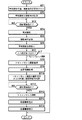
次いで第1の実施形態において行われる処理について説明する。図7は第1の実施形態において行われる処理を示すフローチャートである。なお、第1の実施形態においては、単純撮影の撮影条件を操作者が入力するものとして説明するが、これに限定されるものではなく、上述したように操作者が入力した撮影部位等の情報に基づいて設定してもよい。また、検出器14を移動せず、X線管12のみを移動してトモシンセシス撮影を行うものとする。まず、制御部34は操作者による操作部24からの、実施する診断として単純撮影の登録および単純撮影の撮影条件の設定を受け付ける(ステップST1)、これにより制御部34が、実施する診断として単純撮影を装置10に登録するとともに、条件設定部28が単純撮影の撮影条件を設定する(ステップST2)。そして、操作者による撮影開始指示がなされると(ステップST3肯定)、第1の画像取得部20が単純撮影を行って、単純撮影画像を取得する(ステップST4)。なお、撮影後、条件設定部28は単純撮影の撮影条件を記憶部26に記憶する(ステップST5)。
Next, processing performed in the first embodiment will be described. FIG. 7 is a flowchart showing processing performed in the first embodiment. In the first embodiment, it is assumed that the imaging conditions for simple imaging are input by the operator. However, the present invention is not limited to this, and information such as the imaging region input by the operator as described above. You may set based on. In addition, it is assumed that tomosynthesis imaging is performed by moving only the
次いで、制御部34は、単純撮影画像を表示部25に表示する(ステップST6)。図8は単純撮影画像の表示態様の例を示す図である。図8に示すように、制御部34は、単純撮影画像G0およびトモシンセシス撮影を行うか否かを問い合わせるメッセージM0を含む確認画面50を表示部25に表示する。操作者は表示された単純撮影画像を観察して診断を行い、トモシンセシス撮影が必要か否かを判断し、トモシンセシス撮影を行うか否かを操作部24を用いて、YESまたはNOにより指示する。このため、制御部34は、操作者による入力の指示を受け付け(ステップST7)、入力が診断の終了である場合、すなわち入力がNOである場合には処理を終了する。なお、確認画面50に代えて、トモシンセシス撮影を実行させるための実行ボタン52を含む確認画面51を表示部25に表示するようにしてもよい。この場合、操作者は、トモシンセシス撮影が必要な場合にのみ、実行ボタン52を操作すればよい。
Next, the
一方、入力がトモシンセシス撮影の実施である場合、すなわち入力がYESである場合、制御部34は実施する診断としてトモシンセシス撮影の登録を行い(ステップST8)、体厚情報取得部27が記憶部26に記憶された単純撮影の撮影条件に基づいて、被写体2の体厚情報を取得する(ステップST9)。さらに条件設定部28が、トモシンセシス撮影の撮影条件を設定するとともに、体厚情報に基づいて被写体2において断層画像を生成する対象となる範囲を表す断層画像取得条件を設定する(ステップST10)。そして、操作者による撮影開始指示がなされると(ステップST11肯定)、第2の画像取得部21がトモシンセシス撮影を行って複数の撮影画像を取得する(ステップST12)。この際、単純撮影時とトモシンセシス撮影時とにおいて、被写体が所定のしきい値を超えて移動したか否かが判定され、移動していない場合には、上述した所定線源位置Scにおける被写体2の撮影は行われないこととなる。そして、再構成部22が断層画像取得条件により表される範囲の断層画像が得られるように複数の撮影画像を再構成して断層画像を生成し(ステップST13)、制御部34が断層画像を表示部25に表示し(ステップST14)、処理を終了する。なお、生成された断層画像は、不図示のHDD等の記憶装置に記憶されるか、またはネットワークを介してサーバ40に送信される。
On the other hand, if the input is tomosynthesis imaging, that is, if the input is YES, the
このように、第1の実施形態においては、被写体2の体厚情報を取得し、体厚情報に基づいて、被写体2において断層画像を生成する対象となる範囲、すなわち断層画像を再構成する再構成範囲および再構成範囲の中心面の位置を表す断層画像取得条件を設定するようにしたものである。このため、被写体2の体厚に応じて断層画像取得条件を効率よく設定でき、その結果、断層画像を効率よく取得することができる。 As described above, in the first embodiment, the body thickness information of the subject 2 is acquired, and the range in which the tomographic image is generated in the subject 2 based on the body thickness information, that is, the reconstruction for reconstructing the tomographic image. A tomographic image acquisition condition indicating the position of the center plane of the configuration range and the reconstruction range is set. For this reason, the tomographic image acquisition conditions can be efficiently set according to the body thickness of the subject 2, and as a result, the tomographic image can be acquired efficiently.
また、第1の実施形態においては、単純撮影後にトモシンセシス撮影を行うに際し、被写体2の体厚を単純撮影時の撮影条件に基づいて設定するようにもしたものである。ここで、単純撮影は、被写体の診断に必要なものであるため、トモシンセシス撮影の前に必ず行われるものである。したがって、第1の実施形態によれば、被写体へ余計な放射線を照射することなく、断層画像取得条件を設定することができる。 In the first embodiment, when tomosynthesis imaging is performed after simple imaging, the body thickness of the subject 2 is set based on imaging conditions during simple imaging. Here, since simple imaging is necessary for diagnosis of a subject, it is always performed before tomosynthesis imaging. Therefore, according to the first embodiment, the tomographic image acquisition condition can be set without irradiating the subject with extra radiation.
また、単純撮影後にトモシンセシス撮影を選択的に行わせるようにすることにより、単純撮影画像のみを用いて診断を行うことが可能である場合に、トモシンセシス撮影を行わないようにすることができ、これにより、被写体の被曝量を低減させることができる。 In addition, by selectively performing tomosynthesis imaging after simple imaging, it is possible to prevent tomosynthesis imaging from being performed when diagnosis can be performed using only simple imaging images. Thus, the exposure amount of the subject can be reduced.
また、トモシンセシス撮影時における所定線源位置Scでの撮影をスキップし、単純撮影画像を所定線源位置Scでの撮影画像として使用することにより、トモシンセシス撮影を行う場合の所定線源位置Scにおける撮影を省略することができる。したがって、トモシンセシス撮影時における被写体の被曝量を低減することができる。 In addition, imaging at the predetermined source position Sc when tomosynthesis imaging is performed by skipping imaging at the predetermined source position Sc at the time of tomosynthesis imaging and using a simple captured image as an imaging image at the predetermined source position Sc. Can be omitted. Therefore, it is possible to reduce the exposure amount of the subject at the time of tomosynthesis imaging.
この場合、被写体2の移動を検出部8により検出し、単純撮影時とトモシンセシス撮影時において、被写体2が所定のしきい値を超えて移動した場合には、トモシンセシス撮影時において、X線管12の所定線源位置Scでの撮影を実行するようにすることにより、所定線源位置Scでの撮影画像に含まれる被写体2と、他の線源位置の撮影画像に含まれる被写体2との位置ずれによって、断層画像の画質が低下してしまうことを防止できる。
In this case, when the movement of the subject 2 is detected by the
なお、上記第1の実施形態においては、単純撮影画像の表示後、操作者の指示によりトモシンセシス撮影を行っているが、単純撮影画像の表示後、関心領域を設定した後にトモシンセシス撮影の指示を行うようにしてもよい。また、単純撮影画像の表示後、表示された単純撮影画像上に操作者が操作部24を用いてトモシンセシス撮影を所望する領域を関心領域として設定したことを検出して、第2の画像取得部21がトモシンセシス撮影を行うようにしてもよい。この場合、条件設定部28は、操作者が単純撮影画像上に設定した関心領域に基づいてX線照射範囲を設定し、これに基づいて第2の画像取得部21がトモシンセシス撮影を行う。
In the first embodiment, tomosynthesis imaging is performed according to an operator's instruction after displaying a simple captured image. However, after displaying the simple captured image, an instruction for tomosynthesis imaging is performed after setting a region of interest. You may do it. In addition, after the simple captured image is displayed, the second image acquisition unit detects that the operator has set a region for which tomosynthesis imaging is desired as the region of interest using the
また、上記第1の実施形態においては、単純撮影を行う際に、条件設定部28が、撮影対象の被写体2の被写体IDについて、過去の診断で取得した単純撮影画像および断層画像がサーバ40に保存されているか否かを判定し、この判定が肯定された場合に、被写体IDと対応づけられた参照情報をサーバ40から取得し、参照情報を用いて断層画像取得条件を設定するようにしてもよい。この場合、条件設定部28は、現在の診断で取得した単純撮影を行った後、単純撮影画像および参照情報を用いて、過去の診断で取得した断層画像と同一範囲の断層画像が取得されるように、X線の照射範囲を設定する。具体的には、過去の診断で取得した単純画像および参照情報を用いて、過去の診断で取得した単純撮影画像におけるX線の照射範囲を特定し、その照射範囲と現在の診断で取得した単純撮影画像との相関を求めることにより、照射範囲に対応する領域を現在の診断で取得した単純撮影画像に設定する。そして、現在の診断で取得した単純撮影画像に設定された領域に対応する領域をX線の照射領域として設定する。なお、断層面の中心面の位置および再構成範囲については参照情報と同一となるように設定してもよく、現在の診断で取得した単純撮影画像を用いて上記第1の実施形態と同様に設定してもよい。
Further, in the first embodiment, when performing simple imaging, the
また、上記第1の実施形態においては、トモシンセシス撮影に先立って単純撮影を行い、単純撮影時の撮影条件を用いて体厚情報を取得して、断層画像取得条件を設定しているが、単純撮影を行うことなく、トモシンセシス撮影の撮影条件を決定するためのプレショット撮影を行い、プレショット撮影時の撮影条件を用いて被写体2の体厚情報を取得して、断層画像取得条件を設定するようにしてもよい。この場合、プレショット画像を断層画像の再構成に用いることにより、トモシンセシス撮影時において、プレショット撮影を行ったX線管12の所定線源位置Scでの撮影を行う必要がなくなるため、トモシンセシス撮影時における被写体の被曝量を低減することができる。
In the first embodiment, simple imaging is performed prior to tomosynthesis imaging, body thickness information is acquired using imaging conditions during simple imaging, and tomographic image acquisition conditions are set. Perform pre-shot imaging to determine imaging conditions for tomosynthesis imaging without imaging, acquire body thickness information of
次いで、本発明の第2の実施形態について説明する。図9は本発明の第2の実施形態による放射線撮影装置を適用したトモシンセシス撮影を行うためのX線撮影装置の概略図である。なお、第2の実施形態において第1の実施形態と同一の構成については同一の参照番号を付与し、ここでは詳細な説明は省略する。第2の実施形態によるX線撮影装置10Aは、体厚情報取得部27Aにおいて、トモシンセシス撮影により取得した複数の撮影画像を解析することにより被写体2の体厚情報を取得するようにした点が第1の実施形態と異なる。以下、第2の実施形態における体厚情報取得部27Aが行う処理について説明する。
Next, a second embodiment of the present invention will be described. FIG. 9 is a schematic view of an X-ray imaging apparatus for performing tomosynthesis imaging to which the radiation imaging apparatus according to the second embodiment of the present invention is applied. In the second embodiment, the same components as those in the first embodiment are denoted by the same reference numerals, and detailed description thereof is omitted here. The
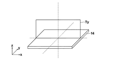
まず、体厚情報取得部27Aは、トモシンセシス撮影により取得した複数の撮影画像を再構成して、検出器14の検出面に直交する断層面の断層画像(以下直交断層画像とする)を生成する。例えば、図9に示すX線管12の移動方向をx方向、図9の紙面に垂直な方向をy方向、図9の紙面における上下方向をz方向とした場合、図10に示すように、検出器14の検出面の重心上におけるyz平面上の断層面の断層画像Tx、または図11に示すように、検出器14の検出面の重心上におけるxz平面上の断層面の断層画像Tyを直交断層画像として再構成する。なお、断層画像Txの各画素の画素値Tx(y,z)または断層画像Tyの各画素の画素値Ty(x,z)は、各断層面上の点(x,y,z)のi番目の撮影画像(i=1〜N)上への投影位置(ti,si)の画素値P(ti,si)を加算することにより取得することができる。具体的には、断層画像Tx上の画素値Tx(y,z)は下記の式(1)により算出できる。なお、直交断層画像の再構成は、再構成部22が行うようにしてもよい。

次いで、体厚情報取得部27Aは、直交断層画像に含まれる被写体2の領域を特定する。例えば、Sobelフィルタ等のエッジ抽出フィルタを用いて直交断層画像に含まれるエッジを抽出し、エッジにより特定される構造物が存在する領域を被写体2の領域として特定する。なお、フィルタの出力値が大きい、骨部等の構造物の強いエッジのみを抽出し、強いエッジにより特定される被写体2内の骨部等の構造物が存在する領域を被写体2の領域として特定するようにしてもよい。そして体厚情報取得部27Aは、特定した領域におけるz方向の距離を被写体2の体厚情報として取得する。なお、第2の実施形態においては、条件設定部28は、第1の実施形態と同様に、体厚情報に基づいて断層画像取得条件を設定する。
Next, the body thickness
次いで、第2の実施形態において行われる処理について説明する。図12は第2の実施形態において行われる処理を示すフローチャートである。なお、第2の実施形態においては、図7に示す第1の実施形態のフローチャートにおけるステップST8以降の処理のみが第1の実施形態と異なるため、ここでは、ステップST8以降の処理についてのみ説明する。 Next, processing performed in the second embodiment will be described. FIG. 12 is a flowchart showing processing performed in the second embodiment. In the second embodiment, only the processes after step ST8 in the flowchart of the first embodiment shown in FIG. 7 are different from those in the first embodiment, so only the processes after step ST8 will be described here. .
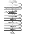
図7に示すフローチャートにおけるステップST7において入力がYESである場合、制御部34は実施する診断としてトモシンセシス撮影の登録を行い(ステップST21)、条件設定部28が、トモシンセシス撮影の撮影条件を設定する(ステップST22)。そして、操作者による撮影開始指示がなされると(ステップST23肯定)、第2の画像取得部21がトモシンセシス撮影を行って複数の撮影画像を取得する(ステップST24)。この際、単純撮影時とトモシンセシス撮影時とにおいて、被写体が所定のしきい値を超えて移動したか否かが判定され、移動していない場合には、上述した所定線源位置Scにおける被写体2の撮影は行われないこととなる。
If the input is YES in step ST7 in the flowchart shown in FIG. 7, the
次いで、体厚情報取得部27Aが複数の撮影画像を解析することにより、被写体2の体厚情報を取得する(ステップST25)。さらに条件設定部28が、体厚情報に基づいて被写体2において断層画像を生成する対象となる範囲を表す断層画像取得条件を設定する(ステップST26)。
Next, the body thickness
そして、再構成部22が断層画像取得条件により表される範囲の断層画像が得られるように複数の撮影画像を再構成して断層画像を生成し(ステップST27)、制御部34が断層画像を表示部25に表示し(ステップST28)、処理を終了する。なお、生成された断層画像は、不図示のHDD等の記憶装置に記憶されるか、またはネットワークを介してサーバ40に送信される。
Then, the
このように、第2の実施形態においても、体厚情報に基づいて断層画像取得条件を設定するようにしたため、被写体2の体厚に応じて断層画像取得条件を効率よく設定でき、その結果、断層画像を効率よく取得することができる。 As described above, also in the second embodiment, since the tomographic image acquisition condition is set based on the body thickness information, the tomographic image acquisition condition can be efficiently set according to the body thickness of the subject 2, and as a result, A tomographic image can be acquired efficiently.
次いで、本発明の第3の実施形態について説明する。図13は本発明の第3の実施形態による放射線撮影装置を適用したトモシンセシス撮影を行うためのX線撮影装置の概略図である。なお、第3の実施形態において第1の実施形態と同一の構成については同一の参照番号を付与し、ここでは詳細な説明は省略する。第3の実施形態によるX線撮影装置10Bは、体厚情報取得部27Bにおいて、撮影台天板4付近に設置された赤外線センサまたは超音波センサ等のセンサ42により、被写体2の体厚情報を取得するようにした点が第1の実施形態と異なる。なお、第3の実施形態においては、条件設定部28は、第1の実施形態と同様に、体厚情報に基づいて断層画像取得条件を設定する。
Next, a third embodiment of the present invention will be described. FIG. 13 is a schematic view of an X-ray imaging apparatus for performing tomosynthesis imaging to which the radiation imaging apparatus according to the third embodiment of the present invention is applied. Note that in the third embodiment, the same reference numerals are assigned to the same configurations as those in the first embodiment, and detailed description thereof is omitted here. In the
次いで、第3の実施形態において行われる処理について説明する。図14は第3の実施形態において行われる処理を示すフローチャートである。なお、第3の実施形態においては、図7に示す第1の実施形態のフローチャートにおけるステップST8以降の処理のみが第1の実施形態と異なるため、ここでは、ステップST8以降の処理についてのみ説明する。 Next, processing performed in the third embodiment will be described. FIG. 14 is a flowchart showing processing performed in the third embodiment. In the third embodiment, only the processes after step ST8 in the flowchart of the first embodiment shown in FIG. 7 are different from those in the first embodiment. Therefore, only the processes after step ST8 will be described here. .
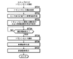
図7に示すフローチャートにおけるステップST7において入力がYESである場合、制御部34は実施する診断としてトモシンセシス撮影の登録を行い(ステップST31)、体厚情報取得部27Bがセンサ42の出力に基づいて被写体2の体厚情報を取得する(ステップST32)。さらに条件設定部28が、トモシンセシス撮影の撮影条件を設定するとともに、体厚情報に基づいて被写体2において断層画像を生成する対象となる範囲を表す断層画像取得条件を設定する(ステップST33)。そして、操作者による撮影開始指示がなされると(ステップST34肯定)、第2の画像取得部21がトモシンセシス撮影を行って複数の撮影画像を取得する(ステップST35)。この際、単純撮影時とトモシンセシス撮影時とにおいて、被写体が所定のしきい値を超えて移動したか否かが判定され、移動していない場合には、上述した所定線源位置Scにおける被写体2の撮影は行われないこととなる。そして、再構成部22が断層画像取得条件により表される範囲の断層画像が得られるように複数の撮影画像を再構成して断層画像を生成し(ステップST36)、制御部34が断層画像を表示部25に表示し(ステップST37)、処理を終了する。なお、生成された断層画像は、不図示のHDD等の記憶装置に記憶されるか、またはネットワークを介してサーバ40に送信される。
If the input is YES in step ST7 in the flowchart shown in FIG. 7, the
このように、第3の実施形態においても、体厚情報に基づいて断層画像取得条件を設定するようにしたため、被写体2の体厚に応じて断層画像取得条件を効率よく設定でき、その結果、断層画像を効率よく取得することができる。 Thus, also in the third embodiment, since the tomographic image acquisition condition is set based on the body thickness information, the tomographic image acquisition condition can be efficiently set according to the body thickness of the subject 2, and as a result, A tomographic image can be acquired efficiently.
なお、上記第3の実施形態においては、超音波センサ等のセンサを用いるようにしたが、体厚を計測できるものであればこれに限らず如何なるものを用いてもよい。例えば、被写体2をカメラ等で撮影した画像に基づいて、被写体2の体厚情報を取得するようにしてもよい。 In the third embodiment, a sensor such as an ultrasonic sensor is used. However, any sensor may be used as long as the body thickness can be measured. For example, the body thickness information of the subject 2 may be acquired based on an image obtained by photographing the subject 2 with a camera or the like.
また、上記第1から第3の実施形態においては、X線管12のみを移動させているが、X線管12と検出器14とを同期させて移動させる場合においても、本発明を適用できることはもちろんである。
In the first to third embodiments, only the
また、上記第1から第3の実施形態においては、被写体2を臥位にて撮影台に載置してトモシンセシス撮影を行っているが、立位の撮影台を用いてトモシンセシス撮影を行う場合にも本願発明を適用できることはもちろんである。
In the first to third embodiments, the
また、上記第1から第3の実施形態においては、本発明による放射線撮影装置をトモシンセシス撮影を行うX線撮影装置に適用しているが、トモグラフィ撮影を行う撮影装置に適用してもよい。以下、これを第4の実施形態として説明する。 In the first to third embodiments, the radiation imaging apparatus according to the present invention is applied to an X-ray imaging apparatus that performs tomosynthesis imaging, but may be applied to an imaging apparatus that performs tomography imaging. Hereinafter, this will be described as a fourth embodiment.
図15は本発明の第4の実施形態による放射線撮影装置を適用したトモグラフィ撮影を行うためのX線撮影装置の概略図である。なお、第4の実施形態において第1の実施形態と同一の構成については同一の参照番号を付与し、ここでは詳細な説明は省略する。 FIG. 15 is a schematic diagram of an X-ray imaging apparatus for performing tomography imaging to which a radiation imaging apparatus according to the fourth embodiment of the present invention is applied. Note that in the fourth embodiment, the same reference numerals are assigned to the same configurations as those in the first embodiment, and detailed description thereof is omitted here.
ここで、トモシンセシス撮影は、X線管12を移動させながら、複数の線源位置においてX線を被写体2に離散的に照射して複数の撮影画像を取得し、これら複数の撮影画像を再構成することにより断層画像を生成する。一方、第4の実施形態におけるX線撮影装置10Cが行うトモグラフィ撮影は、X線管12および検出器14を、連動させて相対的に反対方向に移動させつつ、X線を被写体2に連続的に多重照射することにより断層画像生成部48において断層画像を生成するものである。
Here, in tomosynthesis imaging, while moving the
なお、トモグラフィ撮影においては、1回の撮影により1つの断層画像が生成されるため、所望とする断層面の範囲において複数の断層面の断層画像を取得するには、撮影時のX線管12および検出器14の移動のさせ方を変更して複数回の撮影を行うこととなる。このため、第4の実施形態においては、体厚情報取得部27Cにおいて、被写体2の体厚情報を取得し、条件設定部28において、体厚情報に基づいて、所望とする断層面の範囲を断層画像取得条件として設定し、設定された断層画像取得条件に基づく断層面の範囲において複数の断層画像が取得されるようにトモグラフィ撮影を行うようにしたものである。
In tomography imaging, one tomographic image is generated by one imaging. Therefore, in order to obtain tomographic images of a plurality of tomographic planes in a desired tomographic range, an X-ray tube at the time of imaging is used. 12 and the method of moving the
なお、第4の実施形態においては、第1の実施形態と同様にプレショット画像を用いて被写体2の体厚情報を取得すればよい。また、第3の実施形態と同様に、センサを用いて被写体2の体厚情報を取得してもよい。 In the fourth embodiment, body thickness information of the subject 2 may be acquired using a pre-shot image, as in the first embodiment. Similarly to the third embodiment, the body thickness information of the subject 2 may be acquired using a sensor.
このように、第4の実施形態においても、体厚情報に基づいて断層画像取得条件を設定することにより、被写体2の体厚に応じて断層画像取得条件を効率よく設定でき、その結果、トモグラフィ撮影を行う場合においても断層画像を効率よく取得することができる。 As described above, also in the fourth embodiment, by setting the tomographic image acquisition condition based on the body thickness information, the tomographic image acquisition condition can be efficiently set according to the body thickness of the subject 2, and as a result, A tomographic image can be efficiently acquired even when performing photographic imaging.
2 被写体
4 撮影台天板
6 コリメータ
10,10A,10B,10C X線撮影装置
12 X線管
14 検出器
16,18 移動機構
20 第1の画像取得部
21 第2の画像取得部
22 再構成部
24 操作部
25 表示部
26 記憶部
37 体厚情報取得部
28 条件設定部
30 関心領域設定部
32 通信インターフェース
34 制御部
40 サーバ
2 subject 4 imaging table
Claims (13)
前記被写体を透過した放射線を検出する検出手段と、
前記被写体の体厚情報を取得する体厚情報取得手段と、
前記体厚情報に基づいて、前記被写体における断層画像を取得する範囲を表す断層画像取得条件を設定する条件設定手段と、
前記断層画像取得条件に基づいて前記断層画像を取得する断層画像取得手段とを備えたことを特徴とする放射線撮影装置。 A radiation source for irradiating the subject with radiation;
Detecting means for detecting radiation transmitted through the subject;
Body thickness information acquisition means for acquiring body thickness information of the subject;
Condition setting means for setting a tomographic image acquisition condition representing a range for acquiring a tomographic image of the subject based on the body thickness information;
A radiation imaging apparatus comprising: a tomographic image acquisition unit configured to acquire the tomographic image based on the tomographic image acquisition condition.
前記体厚情報取得手段は、前記プレショット撮影時の撮影条件に基づいて前記被写体の体厚情報を取得する手段であることを特徴とする請求項1記載の放射線撮影装置。 It further comprises image acquisition means for acquiring a pre-shot image by performing pre-shot imaging for irradiating the subject with the radiation from the radiation source,
The radiation imaging apparatus according to claim 1, wherein the body thickness information acquisition unit is a unit that acquires body thickness information of the subject based on imaging conditions during the pre-shot imaging.
前記断層画像取得手段は、前記プレショット撮影時と前記断層画像の取得時とにおいて、前記被写体が所定値を超えて移動した場合には、前記断層画像の取得時に前記放射線源の前記プレショット撮影を行った位置での撮影を実行する手段であることを特徴とする請求項6記載の放射線撮影装置。 Further comprising detection means for detecting movement of the subject,
The tomographic image acquisition unit is configured to acquire the pre-shot imaging of the radiation source at the time of acquiring the tomographic image when the subject moves beyond a predetermined value during the pre-shot imaging and the acquisition of the tomographic image. The radiation imaging apparatus according to claim 6, wherein the radiation imaging apparatus is a unit that performs imaging at a position where the operation is performed.
前記条件設定手段は、前記記憶手段に記憶された撮影条件を参照し、撮影対象の被写体について過去の撮影条件が記憶されている場合には、前記過去の撮影条件に基づいて前記断層画像取得条件を設定する手段であることを特徴とする請求項1から12のいずれか1項記載の放射線撮影装置。 A storage means for storing past shooting conditions for a plurality of subjects;
The condition setting means refers to the imaging conditions stored in the storage means, and if the past imaging conditions are stored for the subject to be imaged, the tomographic image acquisition conditions based on the past imaging conditions The radiation imaging apparatus according to claim 1, wherein the radiation imaging apparatus is a means for setting the value.
Priority Applications (2)
| Application Number | Priority Date | Filing Date | Title |
|---|---|---|---|
| JP2010193198A JP5572040B2 (en) | 2009-09-28 | 2010-08-31 | Radiography equipment |
| US12/923,557 US8675814B2 (en) | 2009-09-28 | 2010-09-28 | Radiography apparatus including a body thickness information obtainment unit |
Applications Claiming Priority (3)
| Application Number | Priority Date | Filing Date | Title |
|---|---|---|---|
| JP2009222252 | 2009-09-28 | ||
| JP2009222252 | 2009-09-28 | ||
| JP2010193198A JP5572040B2 (en) | 2009-09-28 | 2010-08-31 | Radiography equipment |
Publications (3)
| Publication Number | Publication Date |
|---|---|
| JP2011087917A JP2011087917A (en) | 2011-05-06 |
| JP2011087917A5 JP2011087917A5 (en) | 2013-03-07 |
| JP5572040B2 true JP5572040B2 (en) | 2014-08-13 |
Family
ID=43780397
Family Applications (1)
| Application Number | Title | Priority Date | Filing Date |
|---|---|---|---|
| JP2010193198A Expired - Fee Related JP5572040B2 (en) | 2009-09-28 | 2010-08-31 | Radiography equipment |
Country Status (2)
| Country | Link |
|---|---|
| US (1) | US8675814B2 (en) |
| JP (1) | JP5572040B2 (en) |
Families Citing this family (23)
| Publication number | Priority date | Publication date | Assignee | Title |
|---|---|---|---|---|
| JP5923889B2 (en) * | 2011-07-29 | 2016-05-25 | 株式会社島津製作所 | Trabecular bone analyzer |
| US20140369459A1 (en) | 2012-02-22 | 2014-12-18 | Carestream Health, Inc. | Mobile radiographic apparatus/methods with tomosynthesis capability |
| WO2013159792A1 (en) * | 2012-04-26 | 2013-10-31 | Universitätsklinikum Erlangen | Method and device for computed tomography angiography |
| JP5968153B2 (en) * | 2012-08-06 | 2016-08-10 | キヤノン株式会社 | Control device, control method, and program |
| JP6141995B2 (en) | 2013-09-27 | 2017-06-07 | 富士フイルム株式会社 | Mammography apparatus, radiographic imaging method and program |
| US9642581B2 (en) * | 2013-11-12 | 2017-05-09 | KUB Technologies, Inc. | Specimen radiography with tomosynthesis in a cabinet |
| JP6127936B2 (en) * | 2013-11-27 | 2017-05-17 | 株式会社島津製作所 | X-ray equipment |
| US9418415B2 (en) | 2014-02-05 | 2016-08-16 | Shimadzu Corporation | Trabecular bone analyzer |
| JP6162324B2 (en) * | 2014-03-28 | 2017-07-12 | 富士フイルム株式会社 | Radiographic imaging system, radiographic imaging method, and radiographic imaging program |
| WO2015147009A1 (en) * | 2014-03-28 | 2015-10-01 | 富士フイルム株式会社 | Radiological image photography system, radiological image photography method, and radiological image photography program |
| JP6379785B2 (en) | 2014-07-18 | 2018-08-29 | コニカミノルタ株式会社 | Tomographic image generation system |
| US9649084B2 (en) * | 2014-07-21 | 2017-05-16 | Samsung Electronics Co., Ltd. | X-ray imaging apparatus and method for creating X-ray image |
| JP6165695B2 (en) * | 2014-09-24 | 2017-07-19 | 富士フイルム株式会社 | Radiation image analysis apparatus and method, and program |
| JP6556005B2 (en) * | 2015-09-29 | 2019-08-07 | 富士フイルム株式会社 | Tomographic image generating apparatus, method and program |
| JP6512144B2 (en) * | 2016-03-18 | 2019-05-15 | 株式会社島津製作所 | Radiography device |
| US10488351B2 (en) | 2016-09-07 | 2019-11-26 | KUB Technologies, Inc. | Specimen radiography with tomosynthesis in a cabinet with geometric magnification |
| US10830712B2 (en) * | 2017-03-27 | 2020-11-10 | KUB Technologies, Inc. | System and method for cabinet x-ray systems with camera |
| US10779791B2 (en) | 2018-03-16 | 2020-09-22 | General Electric Company | System and method for mobile X-ray imaging |
| JP7122886B2 (en) | 2018-06-25 | 2022-08-22 | 富士フイルム株式会社 | Imaging control device, method and program |
| JP2020020730A (en) * | 2018-08-03 | 2020-02-06 | 株式会社日立ハイテクサイエンス | X-ray transmission inspection device and method for inspecting x-ray transmission |
| JP7086805B2 (en) * | 2018-09-27 | 2022-06-20 | 富士フイルム株式会社 | Tomosynthesis imaging device and how to operate it |
| US11052265B2 (en) * | 2019-02-11 | 2021-07-06 | Troy Long | Fluence map optimization for field-in-field radiation therapy |
| US11357466B2 (en) | 2019-10-02 | 2022-06-14 | Canon Medical Systems Corporation | X-ray diagnosis apparatus |
Family Cites Families (39)
| Publication number | Priority date | Publication date | Assignee | Title |
|---|---|---|---|---|
| JPH03224545A (en) * | 1990-01-31 | 1991-10-03 | Shimadzu Corp | X-ray tomographic device |
| US5526394A (en) * | 1993-11-26 | 1996-06-11 | Fischer Imaging Corporation | Digital scan mammography apparatus |
| US5949811A (en) * | 1996-10-08 | 1999-09-07 | Hitachi Medical Corporation | X-ray apparatus |
| JP3670439B2 (en) * | 1997-05-09 | 2005-07-13 | 株式会社日立メディコ | X-ray equipment |
| JP2001137223A (en) * | 1999-11-08 | 2001-05-22 | Ge Medical Systems Global Technology Co Llc | Fluoroscopic apparatus, x-ray imaging method, and method for fluoroscopy |
| FR2803069B1 (en) * | 1999-12-28 | 2002-12-13 | Ge Medical Syst Sa | METHOD AND SYSTEM FOR COMPENSATING THE THICKNESS OF AN ORGAN |
| US6914958B2 (en) * | 2001-07-06 | 2005-07-05 | Ge Medical Systems Global Technology Company, Llc | Multi-plane acquisition in digital x-ray radiography |
| US6827489B2 (en) * | 2001-11-01 | 2004-12-07 | Ge Medical Systems Global Technology Company, Llc | Low-dose exposure aided positioning (LEAP) for digital radiography |
| US6751285B2 (en) * | 2001-11-21 | 2004-06-15 | General Electric Company | Dose management system for mammographic tomosynthesis |
| JP4322459B2 (en) * | 2002-02-12 | 2009-09-02 | 東芝医用システムエンジニアリング株式会社 | X-ray diagnostic equipment |
| US6795526B2 (en) * | 2002-03-04 | 2004-09-21 | Ge Medical Systems Global Technology Co., Llc | Automatic exposure control for a digital image acquisition system |
| JP2003299643A (en) * | 2002-04-11 | 2003-10-21 | Hitachi Medical Corp | Tomographic equipment |
| US7254623B1 (en) * | 2002-04-16 | 2007-08-07 | General Electric Company | Method and apparatus for reducing x-ray dosage in CT imaging prescription |
| JP3673791B2 (en) * | 2002-05-22 | 2005-07-20 | キヤノン株式会社 | Radiographic apparatus and radiographic method |
| DE10234465A1 (en) * | 2002-07-29 | 2004-02-12 | Siemens Ag | X-ray sectional-imaging method in which the height of a sectional image is set using a camera arrangement for imaging the patient from the side so that a reference marking can be made on a reference image |
| US6970531B2 (en) * | 2002-10-07 | 2005-11-29 | General Electric Company | Continuous scan RAD tomosynthesis system and method |
| JP3667317B2 (en) * | 2002-11-26 | 2005-07-06 | キヤノン株式会社 | Radiation tomography equipment |
| US7123684B2 (en) * | 2002-11-27 | 2006-10-17 | Hologic, Inc. | Full field mammography with tissue exposure control, tomosynthesis, and dynamic field of view processing |
| DE10322143B4 (en) * | 2003-05-16 | 2016-09-22 | Siemens Healthcare Gmbh | Screening system and method for determining the effective skin input dose in fluoroscopic examinations |
| JP2005013346A (en) * | 2003-06-24 | 2005-01-20 | Canon Inc | Radiography apparatus |
| JP4528781B2 (en) * | 2003-10-29 | 2010-08-18 | コーニンクレッカ フィリップス エレクトロニクス エヌ ヴィ | Apparatus and method for adjusting imaging parameters of X-ray apparatus |
| JP4537037B2 (en) | 2003-11-11 | 2010-09-01 | 東芝Itコントロールシステム株式会社 | X-ray inspection apparatus and tube voltage / tube current adjustment method thereof |
| US6980624B2 (en) * | 2003-11-26 | 2005-12-27 | Ge Medical Systems Global Technology Company, Llc | Non-uniform view weighting tomosynthesis method and apparatus |
| EP3106094B1 (en) * | 2004-11-26 | 2021-09-08 | Hologic, Inc. | Integrated multi-mode mammography/tomosynthesis x-ray system |
| DE102005022544A1 (en) * | 2005-05-17 | 2006-11-23 | Siemens Ag | Method and device for recording a digital X-ray image |
| US7245694B2 (en) * | 2005-08-15 | 2007-07-17 | Hologic, Inc. | X-ray mammography/tomosynthesis of patient's breast |
| US7302031B2 (en) * | 2005-10-27 | 2007-11-27 | Sectra Mamea Ab | Method and arrangement relating to X-ray imaging |
| US7545907B2 (en) * | 2005-11-09 | 2009-06-09 | Dexela Limited | Methods and apparatus for obtaining low-dose imaging |
| JP5034221B2 (en) * | 2005-11-15 | 2012-09-26 | 株式会社島津製作所 | Radiography equipment |
| DE102005061357A1 (en) * | 2005-12-21 | 2007-07-05 | Siemens Ag | Patient`s projection images recording method for e.g. computer-tomography device, involves determining optical size of patient in low energy range and adjusting recording parameters for recording images in low energy range based on size |
| US7515682B2 (en) * | 2006-02-02 | 2009-04-07 | General Electric Company | Method and system to generate object image slices |
| JP2007236446A (en) * | 2006-03-06 | 2007-09-20 | Shimadzu Corp | Tomographic apparatus |
| JP4891662B2 (en) * | 2006-06-08 | 2012-03-07 | 株式会社東芝 | Mammography equipment |
| US7292675B1 (en) * | 2006-07-19 | 2007-11-06 | General Electric Company | Automatic protocol assistance methods and apparatus |
| FR2905256B1 (en) * | 2006-09-05 | 2008-11-21 | Gen Electric | METHOD FOR OBTAINING A TOMOSYNTHESIS IMAGE |
| JP4851296B2 (en) * | 2006-10-26 | 2012-01-11 | 富士フイルム株式会社 | Radiation tomographic image acquisition apparatus and radiation tomographic image acquisition method |
| JP4851298B2 (en) * | 2006-10-31 | 2012-01-11 | 富士フイルム株式会社 | Radiation tomographic image generator |
| JP5019930B2 (en) * | 2007-04-05 | 2012-09-05 | 富士フイルム株式会社 | Radiation tomographic image acquisition device |
| JP5208542B2 (en) * | 2008-02-29 | 2013-06-12 | 富士フイルム株式会社 | Radiation imaging system |
-
2010
- 2010-08-31 JP JP2010193198A patent/JP5572040B2/en not_active Expired - Fee Related
- 2010-09-28 US US12/923,557 patent/US8675814B2/en not_active Expired - Fee Related
Also Published As
| Publication number | Publication date |
|---|---|
| US20110075793A1 (en) | 2011-03-31 |
| US8675814B2 (en) | 2014-03-18 |
| JP2011087917A (en) | 2011-05-06 |
Similar Documents
| Publication | Publication Date | Title |
|---|---|---|
| JP5572040B2 (en) | Radiography equipment | |
| JP5600272B2 (en) | Radiation imaging apparatus and method, and program | |
| US8705695B2 (en) | Region of interest determination for X-ray imaging | |
| JP5301403B2 (en) | Radiography equipment | |
| JP5437001B2 (en) | Radiography equipment | |
| JP6658578B2 (en) | X-ray equipment | |
| JP2012016394A (en) | Radiation tomographic apparatus | |
| JP2016022095A (en) | Tomographic image generation system | |
| JPWO2010050032A1 (en) | Radiography equipment | |
| JP7342990B2 (en) | X-ray imaging device | |
| US20180199906A1 (en) | Control device, radiation imaging apparatus, radiation imaging method, and radiation imaging program | |
| JP2006218142A (en) | X-ray photographic apparatus | |
| JP2012075862A (en) | Body motion detector, method, and program | |
| JP2020156620A (en) | X-ray imaging apparatus | |
| KR101768520B1 (en) | A method of integrated operation of chest X-ray digital radiography and chest digital tomosynthesis | |
| JP2017169715A (en) | Image processing device, radiographic image capturing system, image processing method, and image processing program | |
| US11324461B2 (en) | X-ray imaging apparatus | |
| JP5559648B2 (en) | Radiation imaging apparatus, method and program | |
| CN114222530A (en) | Medical imaging system and medical imaging processing apparatus | |
| KR102388282B1 (en) | Image processing apparatus for c-arm | |
| JP2012055475A (en) | Radiographic image capturing apparatus, method and program | |
| JP5547565B2 (en) | Radiographic apparatus and method | |
| JP5616993B2 (en) | X-ray irradiation control unit of X-ray imaging apparatus, X-ray control apparatus, control method of X-ray imaging apparatus, program, and storage medium | |
| JP2012055474A (en) | Body motion detection device, method and program | |
| JP5498016B2 (en) | X-ray diagnostic apparatus and image processing apparatus |
Legal Events
| Date | Code | Title | Description |
|---|---|---|---|
| RD15 | Notification of revocation of power of sub attorney |
Free format text: JAPANESE INTERMEDIATE CODE: A7435 Effective date: 20110520 |
|
| A521 | Request for written amendment filed |
Free format text: JAPANESE INTERMEDIATE CODE: A523 Effective date: 20130123 |
|
| A621 | Written request for application examination |
Free format text: JAPANESE INTERMEDIATE CODE: A621 Effective date: 20130123 |
|
| A977 | Report on retrieval |
Free format text: JAPANESE INTERMEDIATE CODE: A971007 Effective date: 20131225 |
|
| A131 | Notification of reasons for refusal |
Free format text: JAPANESE INTERMEDIATE CODE: A131 Effective date: 20140114 |
|
| A521 | Request for written amendment filed |
Free format text: JAPANESE INTERMEDIATE CODE: A523 Effective date: 20140210 |
|
| TRDD | Decision of grant or rejection written | ||
| A01 | Written decision to grant a patent or to grant a registration (utility model) |
Free format text: JAPANESE INTERMEDIATE CODE: A01 Effective date: 20140624 |
|
| A61 | First payment of annual fees (during grant procedure) |
Free format text: JAPANESE INTERMEDIATE CODE: A61 Effective date: 20140627 |
|
| R150 | Certificate of patent or registration of utility model |
Ref document number: 5572040 Country of ref document: JP Free format text: JAPANESE INTERMEDIATE CODE: R150 |
|
| R250 | Receipt of annual fees |
Free format text: JAPANESE INTERMEDIATE CODE: R250 |
|
| R250 | Receipt of annual fees |
Free format text: JAPANESE INTERMEDIATE CODE: R250 |
|
| R250 | Receipt of annual fees |
Free format text: JAPANESE INTERMEDIATE CODE: R250 |
|
| R250 | Receipt of annual fees |
Free format text: JAPANESE INTERMEDIATE CODE: R250 |
|
| LAPS | Cancellation because of no payment of annual fees |