JP7122886B2 - Imaging control device, method and program - Google Patents
Imaging control device, method and program Download PDFInfo
- Publication number
- JP7122886B2 JP7122886B2 JP2018120295A JP2018120295A JP7122886B2 JP 7122886 B2 JP7122886 B2 JP 7122886B2 JP 2018120295 A JP2018120295 A JP 2018120295A JP 2018120295 A JP2018120295 A JP 2018120295A JP 7122886 B2 JP7122886 B2 JP 7122886B2
- Authority
- JP
- Japan
- Prior art keywords
- imaging
- body movement
- subject
- radiation source
- photographing
- Prior art date
- Legal status (The legal status is an assumption and is not a legal conclusion. Google has not performed a legal analysis and makes no representation as to the accuracy of the status listed.)
- Active
Links
- 238000003384 imaging method Methods 0.000 title claims description 246
- 238000000034 method Methods 0.000 title claims description 46
- 230000033001 locomotion Effects 0.000 claims description 215
- 230000005855 radiation Effects 0.000 claims description 123
- 210000000481 breast Anatomy 0.000 claims description 48
- 238000001514 detection method Methods 0.000 claims description 22
- 230000001678 irradiating effect Effects 0.000 claims description 5
- 230000000875 corresponding effect Effects 0.000 description 55
- 238000010586 diagram Methods 0.000 description 24
- 239000013598 vector Substances 0.000 description 24
- 238000003860 storage Methods 0.000 description 17
- 230000006835 compression Effects 0.000 description 10
- 238000007906 compression Methods 0.000 description 10
- 238000004364 calculation method Methods 0.000 description 9
- 239000010948 rhodium Substances 0.000 description 6
- 230000006870 function Effects 0.000 description 4
- 230000001788 irregular Effects 0.000 description 3
- 238000009607 mammography Methods 0.000 description 3
- 238000002601 radiography Methods 0.000 description 3
- 101100021996 Arabidopsis thaliana CYP97C1 gene Proteins 0.000 description 2
- ZOKXTWBITQBERF-UHFFFAOYSA-N Molybdenum Chemical compound [Mo] ZOKXTWBITQBERF-UHFFFAOYSA-N 0.000 description 2
- 238000013473 artificial intelligence Methods 0.000 description 2
- 238000013528 artificial neural network Methods 0.000 description 2
- 238000010894 electron beam technology Methods 0.000 description 2
- 230000003902 lesion Effects 0.000 description 2
- 229910052750 molybdenum Inorganic materials 0.000 description 2
- 239000011733 molybdenum Substances 0.000 description 2
- 230000003287 optical effect Effects 0.000 description 2
- 229910052703 rhodium Inorganic materials 0.000 description 2
- MHOVAHRLVXNVSD-UHFFFAOYSA-N rhodium atom Chemical compound [Rh] MHOVAHRLVXNVSD-UHFFFAOYSA-N 0.000 description 2
- 238000005070 sampling Methods 0.000 description 2
- 238000001228 spectrum Methods 0.000 description 2
- WFKWXMTUELFFGS-UHFFFAOYSA-N tungsten Chemical compound [W] WFKWXMTUELFFGS-UHFFFAOYSA-N 0.000 description 2
- 229910052721 tungsten Inorganic materials 0.000 description 2
- 239000010937 tungsten Substances 0.000 description 2
- 206010006187 Breast cancer Diseases 0.000 description 1
- 208000026310 Breast neoplasm Diseases 0.000 description 1
- 210000001015 abdomen Anatomy 0.000 description 1
- XAGFODPZIPBFFR-UHFFFAOYSA-N aluminium Chemical compound [Al] XAGFODPZIPBFFR-UHFFFAOYSA-N 0.000 description 1
- 229910052782 aluminium Inorganic materials 0.000 description 1
- 239000010405 anode material Substances 0.000 description 1
- 230000005540 biological transmission Effects 0.000 description 1
- 230000002308 calcification Effects 0.000 description 1
- 238000006243 chemical reaction Methods 0.000 description 1
- 210000000038 chest Anatomy 0.000 description 1
- 230000002596 correlated effect Effects 0.000 description 1
- 230000000694 effects Effects 0.000 description 1
- 238000005516 engineering process Methods 0.000 description 1
- 230000005484 gravity Effects 0.000 description 1
- 239000004973 liquid crystal related substance Substances 0.000 description 1
- 238000004519 manufacturing process Methods 0.000 description 1
- 239000000463 material Substances 0.000 description 1
- 238000012544 monitoring process Methods 0.000 description 1
- 238000003825 pressing Methods 0.000 description 1
- 239000004065 semiconductor Substances 0.000 description 1
- 239000007787 solid Substances 0.000 description 1
- 239000000126 substance Substances 0.000 description 1
- 239000010409 thin film Substances 0.000 description 1
Images


Classifications
-
- A—HUMAN NECESSITIES
- A61—MEDICAL OR VETERINARY SCIENCE; HYGIENE
- A61B—DIAGNOSIS; SURGERY; IDENTIFICATION
- A61B6/00—Apparatus or devices for radiation diagnosis; Apparatus or devices for radiation diagnosis combined with radiation therapy equipment
- A61B6/02—Arrangements for diagnosis sequentially in different planes; Stereoscopic radiation diagnosis
- A61B6/025—Tomosynthesis
-
- A—HUMAN NECESSITIES
- A61—MEDICAL OR VETERINARY SCIENCE; HYGIENE
- A61B—DIAGNOSIS; SURGERY; IDENTIFICATION
- A61B6/00—Apparatus or devices for radiation diagnosis; Apparatus or devices for radiation diagnosis combined with radiation therapy equipment
- A61B6/04—Positioning of patients; Tiltable beds or the like
- A61B6/0407—Supports, e.g. tables or beds, for the body or parts of the body
- A61B6/0414—Supports, e.g. tables or beds, for the body or parts of the body with compression means
-
- A—HUMAN NECESSITIES
- A61—MEDICAL OR VETERINARY SCIENCE; HYGIENE
- A61B—DIAGNOSIS; SURGERY; IDENTIFICATION
- A61B6/00—Apparatus or devices for radiation diagnosis; Apparatus or devices for radiation diagnosis combined with radiation therapy equipment
- A61B6/44—Constructional features of apparatus for radiation diagnosis
- A61B6/4429—Constructional features of apparatus for radiation diagnosis related to the mounting of source units and detector units
- A61B6/4452—Constructional features of apparatus for radiation diagnosis related to the mounting of source units and detector units the source unit and the detector unit being able to move relative to each other
-
- A—HUMAN NECESSITIES
- A61—MEDICAL OR VETERINARY SCIENCE; HYGIENE
- A61B—DIAGNOSIS; SURGERY; IDENTIFICATION
- A61B6/00—Apparatus or devices for radiation diagnosis; Apparatus or devices for radiation diagnosis combined with radiation therapy equipment
- A61B6/50—Apparatus or devices for radiation diagnosis; Apparatus or devices for radiation diagnosis combined with radiation therapy equipment specially adapted for specific body parts; specially adapted for specific clinical applications
- A61B6/502—Apparatus or devices for radiation diagnosis; Apparatus or devices for radiation diagnosis combined with radiation therapy equipment specially adapted for specific body parts; specially adapted for specific clinical applications for diagnosis of breast, i.e. mammography
-
- A—HUMAN NECESSITIES
- A61—MEDICAL OR VETERINARY SCIENCE; HYGIENE
- A61B—DIAGNOSIS; SURGERY; IDENTIFICATION
- A61B6/00—Apparatus or devices for radiation diagnosis; Apparatus or devices for radiation diagnosis combined with radiation therapy equipment
- A61B6/52—Devices using data or image processing specially adapted for radiation diagnosis
- A61B6/5205—Devices using data or image processing specially adapted for radiation diagnosis involving processing of raw data to produce diagnostic data
-
- A—HUMAN NECESSITIES
- A61—MEDICAL OR VETERINARY SCIENCE; HYGIENE
- A61B—DIAGNOSIS; SURGERY; IDENTIFICATION
- A61B6/00—Apparatus or devices for radiation diagnosis; Apparatus or devices for radiation diagnosis combined with radiation therapy equipment
- A61B6/52—Devices using data or image processing specially adapted for radiation diagnosis
- A61B6/5258—Devices using data or image processing specially adapted for radiation diagnosis involving detection or reduction of artifacts or noise
- A61B6/5264—Devices using data or image processing specially adapted for radiation diagnosis involving detection or reduction of artifacts or noise due to motion
-
- A—HUMAN NECESSITIES
- A61—MEDICAL OR VETERINARY SCIENCE; HYGIENE
- A61B—DIAGNOSIS; SURGERY; IDENTIFICATION
- A61B6/00—Apparatus or devices for radiation diagnosis; Apparatus or devices for radiation diagnosis combined with radiation therapy equipment
- A61B6/54—Control of apparatus or devices for radiation diagnosis
- A61B6/545—Control of apparatus or devices for radiation diagnosis involving automatic set-up of acquisition parameters
-
- A—HUMAN NECESSITIES
- A61—MEDICAL OR VETERINARY SCIENCE; HYGIENE
- A61B—DIAGNOSIS; SURGERY; IDENTIFICATION
- A61B8/00—Diagnosis using ultrasonic, sonic or infrasonic waves
- A61B8/52—Devices using data or image processing specially adapted for diagnosis using ultrasonic, sonic or infrasonic waves
- A61B8/5207—Devices using data or image processing specially adapted for diagnosis using ultrasonic, sonic or infrasonic waves involving processing of raw data to produce diagnostic data, e.g. for generating an image
-
- G—PHYSICS
- G06—COMPUTING; CALCULATING OR COUNTING
- G06T—IMAGE DATA PROCESSING OR GENERATION, IN GENERAL
- G06T11/00—2D [Two Dimensional] image generation
- G06T11/003—Reconstruction from projections, e.g. tomography
- G06T11/005—Specific pre-processing for tomographic reconstruction, e.g. calibration, source positioning, rebinning, scatter correction, retrospective gating
-
- G—PHYSICS
- G06—COMPUTING; CALCULATING OR COUNTING
- G06T—IMAGE DATA PROCESSING OR GENERATION, IN GENERAL
- G06T7/00—Image analysis
- G06T7/0002—Inspection of images, e.g. flaw detection
- G06T7/0012—Biomedical image inspection
-
- G—PHYSICS
- G06—COMPUTING; CALCULATING OR COUNTING
- G06T—IMAGE DATA PROCESSING OR GENERATION, IN GENERAL
- G06T7/00—Image analysis
- G06T7/20—Analysis of motion
- G06T7/254—Analysis of motion involving subtraction of images
-
- A—HUMAN NECESSITIES
- A61—MEDICAL OR VETERINARY SCIENCE; HYGIENE
- A61B—DIAGNOSIS; SURGERY; IDENTIFICATION
- A61B6/00—Apparatus or devices for radiation diagnosis; Apparatus or devices for radiation diagnosis combined with radiation therapy equipment
- A61B6/40—Arrangements for generating radiation specially adapted for radiation diagnosis
- A61B6/4035—Arrangements for generating radiation specially adapted for radiation diagnosis the source being combined with a filter or grating
-
- A—HUMAN NECESSITIES
- A61—MEDICAL OR VETERINARY SCIENCE; HYGIENE
- A61B—DIAGNOSIS; SURGERY; IDENTIFICATION
- A61B6/00—Apparatus or devices for radiation diagnosis; Apparatus or devices for radiation diagnosis combined with radiation therapy equipment
- A61B6/42—Arrangements for detecting radiation specially adapted for radiation diagnosis
- A61B6/4291—Arrangements for detecting radiation specially adapted for radiation diagnosis the detector being combined with a grid or grating
-
- A—HUMAN NECESSITIES
- A61—MEDICAL OR VETERINARY SCIENCE; HYGIENE
- A61B—DIAGNOSIS; SURGERY; IDENTIFICATION
- A61B6/00—Apparatus or devices for radiation diagnosis; Apparatus or devices for radiation diagnosis combined with radiation therapy equipment
- A61B6/48—Diagnostic techniques
- A61B6/482—Diagnostic techniques involving multiple energy imaging
-
- A—HUMAN NECESSITIES
- A61—MEDICAL OR VETERINARY SCIENCE; HYGIENE
- A61B—DIAGNOSIS; SURGERY; IDENTIFICATION
- A61B6/00—Apparatus or devices for radiation diagnosis; Apparatus or devices for radiation diagnosis combined with radiation therapy equipment
- A61B6/54—Control of apparatus or devices for radiation diagnosis
- A61B6/541—Control of apparatus or devices for radiation diagnosis involving acquisition triggered by a physiological signal
-
- A—HUMAN NECESSITIES
- A61—MEDICAL OR VETERINARY SCIENCE; HYGIENE
- A61B—DIAGNOSIS; SURGERY; IDENTIFICATION
- A61B6/00—Apparatus or devices for radiation diagnosis; Apparatus or devices for radiation diagnosis combined with radiation therapy equipment
- A61B6/54—Control of apparatus or devices for radiation diagnosis
- A61B6/542—Control of apparatus or devices for radiation diagnosis involving control of exposure
-
- G—PHYSICS
- G06—COMPUTING; CALCULATING OR COUNTING
- G06T—IMAGE DATA PROCESSING OR GENERATION, IN GENERAL
- G06T2207/00—Indexing scheme for image analysis or image enhancement
- G06T2207/10—Image acquisition modality
- G06T2207/10072—Tomographic images
- G06T2207/10112—Digital tomosynthesis [DTS]
Landscapes
- Health & Medical Sciences (AREA)
- Life Sciences & Earth Sciences (AREA)
- Engineering & Computer Science (AREA)
- Medical Informatics (AREA)
- Physics & Mathematics (AREA)
- Radiology & Medical Imaging (AREA)
- General Health & Medical Sciences (AREA)
- Nuclear Medicine, Radiotherapy & Molecular Imaging (AREA)
- Surgery (AREA)
- Public Health (AREA)
- Veterinary Medicine (AREA)
- Biomedical Technology (AREA)
- Heart & Thoracic Surgery (AREA)
- Molecular Biology (AREA)
- Pathology (AREA)
- Animal Behavior & Ethology (AREA)
- Biophysics (AREA)
- High Energy & Nuclear Physics (AREA)
- Optics & Photonics (AREA)
- Computer Vision & Pattern Recognition (AREA)
- General Physics & Mathematics (AREA)
- Theoretical Computer Science (AREA)
- Dentistry (AREA)
- Oral & Maxillofacial Surgery (AREA)
- Quality & Reliability (AREA)
- Multimedia (AREA)
- Apparatus For Radiation Diagnosis (AREA)
- Physiology (AREA)
Description
本開示は、複数の線源位置のそれぞれにおいて被写体を撮影して複数の投影画像を取得し、複数の投影画像から断層画像を生成するための撮影制御装置、方法およびプログラムに関するものである。 The present disclosure relates to an imaging control device, method, and program for imaging a subject at each of a plurality of radiation source positions, acquiring a plurality of projection images, and generating a tomographic image from the plurality of projection images.
近年、X線、ガンマ線等の放射線を用いた放射線画像撮影装置において、患部をより詳しく観察するために、放射線源を移動させて複数の線源位置から被写体に放射線を照射して撮影を行い、これにより取得した複数の投影画像から所望とする断層面を強調した断層画像を生成するトモシンセシス撮影が提案されている。トモシンセシス撮影では、撮影装置の特性や必要な断層画像に応じて、放射線源を放射線検出器と平行に移動させたり、円または楕円の弧を描くように移動させたりして、複数の線源位置において被写体を撮影することにより複数の投影画像を取得し、単純逆投影法またはフィルタ逆投影法等の逆投影法等を用いてこれらの投影画像を再構成して断層画像を生成する。このような断層画像を被写体における複数の断層面において生成することにより、断層面が並ぶ深さ方向に重なり合った構造を分離することができる。このため、従来の単純撮影により取得される2次元画像においては検出が困難であった病変を発見することが可能となる。なお、単純撮影とは、被写体に1回放射線を照射して、被写体の透過像である1枚の2次元画像を取得する撮影方法である。 In recent years, in radiographic imaging apparatuses using radiation such as X-rays and gamma rays, in order to observe the affected area in more detail, the radiation source is moved to irradiate the subject with radiation from multiple radiation source positions. Tomosynthesis imaging has been proposed in which a tomographic image in which a desired tomographic plane is emphasized is generated from a plurality of acquired projection images. In tomosynthesis imaging, the radiation source can be moved in parallel with the radiation detector, or in a circular or elliptical arc, depending on the characteristics of the imaging device and the required tomographic images. A plurality of projection images are acquired by photographing the object in , and these projection images are reconstructed using a back projection method such as a simple back projection method or a filtered back projection method to generate a tomographic image. By generating such tomographic images on a plurality of tomographic planes of the subject, it is possible to separate the overlapping structure in the depth direction where the tomographic planes are arranged. Therefore, it is possible to find a lesion that is difficult to detect in a two-dimensional image obtained by conventional simple imaging. Note that simple imaging is an imaging method in which a subject is irradiated with radiation once and a single two-dimensional image, which is a transmission image of the subject, is acquired.
一方、トモシンセシス撮影は、撮影装置の機械的な誤差、および複数の線源位置のそれぞれにおける撮影の時間差に起因する被写体の体動等の影響により、再構成された断層画像がぼけてしまうという問題もある。このように断層画像がぼけてしまうと、とくに乳房が被写体である場合において、乳癌の早期発見に有用な、微小な石灰化等の病変を発見することが困難となる。このため、トモシンセシス撮影を行う場合には、単純撮影も併せて行って、断層画像および2次元画像の双方を取得することが一般的に行われている。 On the other hand, tomosynthesis imaging has the problem that reconstructed tomographic images are blurred due to mechanical errors in the imaging device and body movements of the subject caused by the time difference between imaging at each of the multiple radiation source positions. There is also If the tomographic image is blurred in this way, it becomes difficult to detect a lesion such as minute calcification, which is useful for early detection of breast cancer, especially when the subject is a breast. Therefore, when tomosynthesis imaging is performed, simple imaging is also generally performed to obtain both a tomographic image and a two-dimensional image.
また、トモシンセシス撮影のように、被写体を複数回撮影する撮影方法において、体動の大きさに応じて再撮影の要否を判断する手法が提案されている(特許文献1参照)。 Also, in a method of photographing a subject a plurality of times, such as tomosynthesis photography, a method of determining whether re-imaging is necessary according to the magnitude of body movement has been proposed (see Patent Document 1).
一方、体動が発生するような撮影の状況においては、再撮影を行っても再度体動が発生する可能性が高い。例えば、被写体を水平方向に対して傾斜させたポジショニングで撮影を行う場合、重力が作用する方向に体動が発生しやすく、そのようなポジショニングでは再撮影を行っても、再度体動が発生する可能性が高い。その一方で、単純撮影は、被写体に1回放射線を照射するのみであるため、体動の影響は小さい。このため、再撮影時には単純撮影を行うことが考えられる。 On the other hand, in an imaging situation in which body movement occurs, there is a high possibility that body movement will occur again even if re-imaging is performed. For example, when shooting with the subject tilted with respect to the horizontal direction, body movement is likely to occur in the direction in which gravity acts. Probability is high. On the other hand, simple radiography irradiates the subject with radiation only once, and is less affected by body movement. For this reason, it is conceivable to perform simple photographing at the time of re-photographing.
しかしながら、再撮影を行う際には、被写体のポジショニングを再度行う必要があるため、撮影を終了するまでの時間が長くなる。とくに、乳房を撮影するための放射線画像撮影装置(マンモグラフィと呼ばれる)においては、乳房を圧迫した状態で撮影を行う必要があるため、被写体である患者の痛みを伴う。このようなマンモグラフィにおいて再撮影を行うと、乳房を再度圧迫する必要があるため、患者の負担が大きいものとなる。 However, when re-photographing, it is necessary to re-position the subject, so it takes a long time to complete the photographing. In particular, in a radiographic imaging apparatus (called mammography) for imaging the breast, it is necessary to perform imaging while the breast is compressed, which causes pain to the patient. If re-imaging is performed in such mammography, it is necessary to compress the breast again, which imposes a heavy burden on the patient.
本開示は上記事情に鑑みなされたものであり、体動が発生した場合に直ちに再撮影を行うことができるようにすることを目的とする。 The present disclosure has been made in view of the circumstances described above, and an object of the present disclosure is to enable immediate re-imaging when body movement occurs.
本開示による撮影制御装置は、放射線源を検出部に対して相対的に移動させ、放射線源の移動による複数の線源位置において、トモシンセシス撮影用の第1の撮影条件により被写体に放射線を照射するトモシンセシス撮影を撮影装置に行わせることにより生成された、複数の線源位置のそれぞれに対応する複数の投影画像を取得する画像取得部と、
複数の投影画像に基づいて、トモシンセシス撮影中に被写体の体動が発生しているか否かを判別する体動判別部と、
体動が発生している場合、単純撮影用の第2の撮影条件を撮影装置に設定する条件設定部とを備える。
An imaging control apparatus according to the present disclosure moves a radiation source relative to a detection unit, and irradiates a subject with radiation under a first imaging condition for tomosynthesis imaging at a plurality of radiation source positions resulting from the movement of the radiation source. an image acquisition unit that acquires a plurality of projection images corresponding to each of a plurality of radiation source positions generated by causing an imaging device to perform tomosynthesis imaging;
a body movement determination unit that determines whether body movement of the subject occurs during tomosynthesis imaging based on a plurality of projection images;
a condition setting unit that sets a second imaging condition for simple imaging to the imaging device when body movement occurs.
「放射線源を検出部に対して相対的に移動させる」とは、放射線源のみを移動する場合、検出部のみを移動する場合、および放射線源と検出部との双方を移動する場合のいずれをも含む。 "Moving the radiation source relative to the detection unit" means moving only the radiation source, moving only the detection unit, or moving both the radiation source and the detection unit. Also includes
「トモシンセシス撮影」とは、放射線源を検出部に対して相対的に移動させ、放射線源の移動による複数の線源位置において、被写体に放射線を照射して、複数の線源位置のそれぞれに対応する複数の画像を取得する撮影方法である。 "Tomosynthesis imaging" involves moving the radiation source relative to the detection unit, irradiating the subject with radiation at multiple radiation source positions resulting from the movement of the radiation source, and corresponding to each of the multiple radiation source positions. This is a photographing method for acquiring a plurality of images that
「単純撮影」とは、被写体に1回放射線を照射して、1枚の画像を取得する撮影方法である。 “Simple imaging” is an imaging method in which a subject is irradiated with radiation once and one image is obtained.
なお、本開示による撮影制御装置においては、複数の投影画像を再構成することにより、被写体の1つの断層面における断層画像を生成する再構成部をさらに備え、
体動判別部は、断層画像から少なくとも1つの特徴点を検出し、少なくとも1つの特徴点の複数の投影画像における投影位置を算出し、複数の投影画像間における投影位置の移動方向および移動量の少なくとも一方に基づいて、体動が発生しているか否かを判別するものであってもよい。
Note that the imaging control apparatus according to the present disclosure further includes a reconstruction unit that generates a tomographic image in one tomographic plane of the subject by reconstructing a plurality of projection images,
The body movement determination unit detects at least one feature point from the tomographic image, calculates the projection position of the at least one feature point in the plurality of projection images, and determines the direction and amount of movement of the projection position between the plurality of projection images. Based on at least one of them, it may be determined whether body movement is occurring.
また、本開示による撮影制御装置においては、体動判別部は、各種投影位置の移動方向および移動量の少なくとも一方を用いて体動が発生しているか否かを判定する学習がなされた判別器を有し、判別器に投影位置の移動方向および移動量の少なくとも一方を入力し、判別器の出力により、体動が発生しているか否かを判別するものであってもよい。 Further, in the imaging control device according to the present disclosure, the body motion determination unit is a discriminator that has learned to determine whether or not body motion occurs using at least one of the movement direction and the movement amount of various projection positions. , at least one of the moving direction and moving amount of the projection position is input to the discriminator, and whether or not the body motion is occurring may be discriminated based on the output of the discriminator.
また、本開示による撮影制御装置においては、体動判別部は、断層画像から複数の特徴点を検出し、複数の特徴点のそれぞれについて、複数の投影画像における投影位置を算出し、複数の特徴点のそれぞれについて、複数の投影画像間における投影位置の移動方向および移動量の少なくとも一方に基づいて、体動が発生しているか否かを判別し、複数の特徴点のそれぞれについての判別結果に基づいて、体動が発生しているか否かを判別するものであってもよい。 Further, in the imaging control apparatus according to the present disclosure, the body movement determination unit detects a plurality of feature points from the tomographic image, calculates the projection positions in the plurality of projection images for each of the plurality of feature points, and calculates the plurality of feature points. Based on at least one of the movement direction and movement amount of the projection position between the plurality of projection images, it is determined whether or not body movement occurs for each of the points, and the determination result for each of the plurality of feature points is determined. Based on this, it may be determined whether or not body movement occurs.
また、本開示による撮影制御装置においては、体動判別部は、複数の特徴点の数に対する、体動が発生していると判別された特徴点の数の割合が、予め定められたしきい値以上である場合に、体動が発生していると判別するものであってもよい。 In addition, in the imaging control device according to the present disclosure, the body movement determination unit determines that the ratio of the number of feature points determined to have body motion to the number of the plurality of feature points is set to a predetermined threshold value. If the value is equal to or greater than the value, it may be determined that body movement is occurring.
また、本開示による撮影制御装置においては、体動判別部は、複数の特徴点のそれぞれにおける判別器の出力値の統計値を算出し、統計値が予め定められたしきい値以上である場合に、体動が発生していると判別するものであってもよい。 Further, in the imaging control device according to the present disclosure, the body motion determination unit calculates the statistical value of the output value of the discriminator at each of the plurality of feature points, and if the statistical value is equal to or greater than a predetermined threshold value Alternatively, it may be determined that body motion is occurring.
また、本開示による撮影制御装置においては、体動判別部は、複数の投影画像間の少なくとも1つの対応点を検出し、複数の投影画像間の対応点を再構成して、被写体における対応点を表す少なくとも1つの構造の位置を特定し、複数の投影画像における構造の位置の投影位置を算出し、複数の投影画像のそれぞれにおける投影位置と対応点との距離を算出し、複数の投影位置についての距離の統計値が予め定められたしきい値以上であるか否かに応じて、体動が発生しているか否かを判別するものであってもよい。 Further, in the imaging control device according to the present disclosure, the body movement determination unit detects at least one corresponding point between the plurality of projection images, reconstructs the corresponding points between the plurality of projection images, Identify the position of at least one structure representing the position of the structure, calculate the projected position of the position of the structure in the plurality of projection images, calculate the distance between the projection position and the corresponding point in each of the plurality of projection images, and calculate the plurality of projection positions It may be determined whether or not body movement is occurring depending on whether or not the statistical value of the distance for is equal to or greater than a predetermined threshold value.
また、本開示による撮影制御装置においては、体動判別部は、複数の投影画像間の複数の対応点を検出し、複数の対応点のそれぞれについて、被写体における複数の対応点を表す複数の構造の位置を特定し、複数の投影画像における複数の構造の位置の投影位置を算出し、複数の対応点のそれぞれについて、複数の投影画像における対応する投影位置と対応点との距離を算出し、複数の投影画像のそれぞれについて、複数の対応点に関する距離の統計値が予め定められたしきい値以上であるか否かを判定し、複数の対応点のそれぞれについての判別結果に基づいて、体動が発生しているか否かを判別するものであってもよい。 Further, in the imaging control device according to the present disclosure, the body motion determination unit detects a plurality of corresponding points between the plurality of projection images, and for each of the plurality of corresponding points, a plurality of structures representing the plurality of corresponding points on the subject. , calculating the projected positions of the positions of the plurality of structures in the plurality of projection images, calculating the distance between the corresponding projection positions and the corresponding points in the plurality of projection images for each of the plurality of corresponding points, For each of the plurality of projection images, it is determined whether or not the statistical value of the distances for the plurality of corresponding points is equal to or greater than a predetermined threshold value, and based on the determination result for each of the plurality of corresponding points, the body is determined. It may be determined whether motion is occurring or not.
また、本開示による撮影制御装置においては、体動判別部は、複数の対応点の数に対する、体動が発生していると判別された対応点の数の割合が、予め定められたしきい値以上である場合に、体動が発生していると判別するものであってもよい。 In addition, in the imaging control device according to the present disclosure, the body motion determination unit sets the ratio of the number of corresponding points determined to have body motion to the number of the plurality of corresponding points by a predetermined threshold value. If the value is equal to or greater than the value, it may be determined that body movement is occurring.
また、本開示による撮影制御装置においては、体動判別部は、複数の対応点のそれぞれにおける距離の統計値の統計値を他の統計値としてさらに算出し、他の統計値が予め定められたしきい値以上である場合に、体動が発生していると判別するものであってもよい。 Further, in the imaging control device according to the present disclosure, the body movement determination unit further calculates statistical values of the statistical values of the distances at each of the plurality of corresponding points as other statistical values, and the other statistical values are determined in advance. It may be determined that the body motion is occurring when it is equal to or greater than the threshold value.
また、本開示による撮影制御装置においては、体動が発生している場合に、単純撮影を行う旨の通知を行う通知部をさらに備えるものであってもよい。 Further, the imaging control device according to the present disclosure may further include a notification unit that notifies that simple imaging is to be performed when body movement occurs.
また、本開示による撮影制御装置においては、画像取得部は、撮影装置に単純撮影を行わせることにより生成された、被写体の2次元画像を取得するものであってもよい。 Further, in the imaging control device according to the present disclosure, the image acquisition unit may acquire a two-dimensional image of the subject generated by causing the imaging device to perform simple imaging.
また、本開示による撮影制御装置においては、第1の撮影条件と第2の撮影条件とは、放射線源におけるターゲット/フィルタの種類、放射線源の管電圧、放射線源からの放射線量、および被写体を透過した放射線に含まれる散乱線成分を除去する散乱線除去グリッドの有無の少なくとも1つが異なるものであってもよい。 Further, in the imaging control apparatus according to the present disclosure, the first imaging condition and the second imaging condition are the type of target/filter in the radiation source, the tube voltage of the radiation source, the radiation dose from the radiation source, and the subject. At least one of the presence or absence of the scattered radiation removal grid for removing the scattered radiation component contained in the transmitted radiation may be different.
また、本開示による撮影制御装置においては、被写体が乳房であってもよい。 Further, in the imaging control device according to the present disclosure, the subject may be a breast.
本開示による撮影制御方法は、放射線源を検出部に対して相対的に移動させ、放射線源の移動による複数の線源位置において、トモシンセシス撮影用の第1の撮影条件により被写体に放射線を照射するトモシンセシス撮影を撮影装置に行わせることにより生成された、複数の線源位置のそれぞれに対応する複数の投影画像を取得し、
複数の投影画像に基づいて、トモシンセシス撮影中に被写体の体動が発生しているか否かを判別し、
体動が発生している場合、単純撮影用の第2の撮影条件を撮影装置に設定する。
An imaging control method according to the present disclosure moves a radiation source relative to a detection unit, and irradiates a subject with radiation under a first imaging condition for tomosynthesis imaging at a plurality of radiation source positions resulting from the movement of the radiation source. Acquiring a plurality of projection images corresponding to each of a plurality of radiation source positions generated by causing the imaging device to perform tomosynthesis imaging,
determining whether body movement of the subject occurs during tomosynthesis imaging based on a plurality of projected images;
When body movement occurs, the second imaging condition for simple imaging is set in the imaging device.
なお、本開示による撮影制御方法をコンピュータに実行させるためのプログラムとして提供してもよい。 In addition, you may provide as a program for making a computer perform the imaging|photography control method by this indication.
本開示による他の撮影制御装置は、コンピュータに実行させるための命令を記憶するメモリと、
記憶された命令を実行するよう構成されたプロセッサとを備え、プロセッサは、
放射線源を検出部に対して相対的に移動させ、放射線源の移動による複数の線源位置において、トモシンセシス撮影用の第1の撮影条件により被写体に放射線を照射するトモシンセシス撮影を撮影装置に行わせることにより生成された、複数の線源位置のそれぞれに対応する複数の投影画像を取得し、
複数の投影画像に基づいて、トモシンセシス撮影中に被写体の体動が発生しているか否かを判別し、
体動が発生している場合、単純撮影用の第2の撮影条件を撮影装置に設定する処理を実行する。
Another imaging control device according to the present disclosure includes a memory that stores instructions for execution by a computer;
a processor configured to execute stored instructions, the processor comprising:
The radiation source is moved relative to the detection unit, and the imaging apparatus is caused to perform tomosynthesis imaging in which the subject is irradiated with radiation under a first imaging condition for tomosynthesis imaging at a plurality of radiation source positions resulting from the movement of the radiation source. Obtaining a plurality of projection images corresponding to each of a plurality of radiation source positions generated by
determining whether body movement of the subject occurs during tomosynthesis imaging based on a plurality of projected images;
When body movement occurs, a process of setting second imaging conditions for simple imaging to the imaging device is executed.
本開示によれば、トモシンセシス撮影用の第1の撮影条件により、トモシンセシス撮影を撮影装置に行わせることにより生成された、複数の線源位置のそれぞれに対応する複数の投影画像が取得され、複数の投影画像に基づいて、トモシンセシス撮影中に被写体の体動が発生しているか否かが判別される。そして、体動が発生している場合、単純撮影用の第2の撮影条件が撮影装置に設定される。このため、トモシンセシス撮影時に体動が発生していた場合、被写体のポジショニングを再度行うことなく、直ちに単純撮影を行うことができる。したがって、体動が発生した場合における被写体である患者の負担を軽減することができる。 According to the present disclosure, a plurality of projection images corresponding to each of a plurality of radiation source positions generated by causing an imaging apparatus to perform tomosynthesis imaging under a first imaging condition for tomosynthesis imaging are acquired, and a plurality of Based on the projection image of , it is determined whether or not the body movement of the subject has occurred during tomosynthesis imaging. Then, when body movement occurs, the second imaging condition for simple imaging is set in the imaging device. Therefore, when body movement occurs during tomosynthesis imaging, simple imaging can be immediately performed without positioning the subject again. Therefore, it is possible to reduce the burden on the patient, who is the subject, when body movement occurs.
以下、図面を参照して本開示の実施形態について説明する。図1は本開示の第1の実施形態による撮影制御装置を適用した放射線画像撮影装置の概略構成図、図2は放射線画像撮影装置を図1の矢印A方向から見た図である。放射線画像撮影装置1は、乳房のトモシンセシス撮影を行って断層画像を生成するために、複数の線源位置から被写体である乳房Mを撮影して、複数の放射線画像、すなわち複数の投影画像を取得するマンモグラフィ撮影装置である。図1に示すように放射線画像撮影装置1は、撮影部10、撮影部10に接続されたコンピュータ2、並びにコンピュータ2に接続された表示部3および入力部4を備えている。なお、本実施形態による放射線画像撮影装置1は、トモシンセシス撮影と併せて、後述するように単純撮影を行って、乳房Mの2次元画像も取得する。
Embodiments of the present disclosure will be described below with reference to the drawings. FIG. 1 is a schematic configuration diagram of a radiographic imaging apparatus to which an imaging control apparatus according to the first embodiment of the present disclosure is applied, and FIG. 2 is a view of the radiographic imaging apparatus as seen from the direction of arrow A in FIG. In order to perform tomosynthesis imaging of the breast and generate a tomographic image, the
撮影部10は、不図示の基台に対して回転軸11により連結されたアーム部12を備えている。アーム部12の一方の端部には撮影台13が、その他方の端部には撮影台13と対向するように放射線照射部14が取り付けられている。アーム部12は、放射線照射部14が取り付けられた端部のみを回転することが可能に構成されており、これにより、撮影台13を固定して放射線照射部14のみを回転することが可能となっている。なお、アーム部12の回転は、コンピュータ2により制御される。
The
撮影台13の内部には、フラットパネルディテクタ等の放射線検出器15が備えられている。また、撮影台13の内部には、放射線検出器15から読み出された電荷信号を電圧信号に変換するチャージアンプ、チャージアンプから出力された電圧信号をサンプリングする相関2重サンプリング回路、および電圧信号をデジタル信号に変換するAD変換部等が設けられた回路基板等も設置されている。なお、放射線検出器15が検出部に対応する。
A
放射線検出器15は、放射線画像の記録および読み出しを繰り返して行うことができるものであり、放射線の照射を直接受けて電荷を発生する、いわゆる直接型の放射線検出器を用いてもよいし、放射線を一旦可視光に変換し、その可視光を電荷信号に変換する、いわゆる間接型の放射線検出器を用いるようにしてもよい。また、放射線画像信号の読出方式としては、TFT(thin film transistor)スイッチをオンおよびオフすることによって放射線画像信号が読み出される、いわゆるTFT読出方式のもの、または読取光を照射することによって放射線画像信号が読み出される、いわゆる光読出方式のものを用いることが望ましいが、これに限らずその他のものを用いるようにしてもよい。
The
放射線照射部14の内部には、放射線源であるX線源16が収納されている。X線源16から放射線であるX線を照射するタイミングおよびX線源16におけるX線発生条件、すなわちターゲットおよびフィルタの材質の選択、管電圧並びに照射時間等は、コンピュータ2により制御される。
An
また、アーム部12には、撮影台13の上方に配置されて乳房Mを押さえつけて圧迫する圧迫板17、圧迫板17を支持する支持部18、および支持部18を図1および図2の上下方向に移動させる移動機構19が設けられている。なお、圧迫板17と撮影台13との間隔、すなわち圧迫厚はコンピュータ2に入力される。
1 and 2, a
表示部3は、CRT(Cathode Ray Tube)または液晶モニタ等の表示装置であり、後述するように取得された投影画像および2次元画像、並びに生成された断層画像の他、操作に必要なメッセージ等を表示する。なお、表示部3は音声を出力するスピーカを内蔵するものであってもよい。
The
入力部4はキーボード、マウスあるいはタッチパネル方式の入力装置からなり、操作者による放射線画像撮影装置1の操作を受け付ける。また、トモシンセシス撮影を行うために必要な、撮影条件等の各種情報の入力および情報の修正の指示も受け付ける。本実施形態においては、操作者が入力部4から入力した情報に従って、放射線画像撮影装置1の各部が動作する。
The input unit 4 is composed of a keyboard, a mouse, or a touch panel type input device, and receives operations of the
コンピュータ2には、本実施形態による撮影制御プログラムがインストールされている。本実施形態においては、コンピュータは、操作者が直接操作するワークステーションあるいはパソコンでもよいし、それらとネットワークを介して接続されたサーバコンピュータでもよい。撮影制御プログラムは、DVD(Digital Versatile Disc)、CD-ROM(Compact Disc Read Only Memory)等の記録媒体に記録されて配布され、その記録媒体からコンピュータにインストールされる。もしくは、ネットワークに接続されたサーバコンピュータの記憶装置、あるいはネットワークストレージに、外部からアクセス可能な状態で記憶され、要求に応じてコンピュータにダウンロードされ、インストールされる。
A photographing control program according to the present embodiment is installed in the
図3はコンピュータ2に撮影制御プログラムをインストールすることにより実現された撮影制御装置の概略構成を示す図である。図3に示すように、撮影制御装置は、標準的なコンピュータの構成として、CPU(Central Processing Unit)21、メモリ22およびストレージ23を備えている。
FIG. 3 is a diagram showing a schematic configuration of a photographing control device realized by installing a photographing control program in the
ストレージ23は、ハードディスクまたはSSD(Solid State Drive)等のストレージデバイスからなり、放射線画像撮影装置1の各部を駆動するためのプログラムおよび撮影制御プログラムを含む各種情報が記憶されている。また、トモシンセシス撮影により取得された投影画像、単純撮影により取得された2次元画像、および後述するように生成された断層画像も記憶される。
The
メモリ22には、各種処理をCPU21に実行させるために、ストレージ23に記憶されたプログラム等が一時的に記憶される。撮影制御プログラムは、CPU21に実行させる処理として、放射線画像撮影装置1に、トモシンセシス撮影用の第1の撮影条件によりトモシンセシス撮影を行わせて、複数の線源位置のそれぞれに対応する乳房Mの複数の投影画像を取得する第1の画像取得処理、複数の投影画像に基づいて、トモシンセシス撮影中に被写体である乳房Mの体動が発生しているか否かを判別する体動判別処理、体動が発生している場合、トモシンセシス撮影用の第1の撮影条件および単純撮影用の第2の撮影条件を放射線画像撮影装置1に設定する条件設定処理、体動が発生している場合、単純撮影を行う旨の通知を行う通知処理、放射線画像撮影装置1に単純撮影を行わせて、乳房Mの2次元画像を取得する第2の画像取得処理、並びに複数の投影画像を再構成することにより、乳房Mの断層面における断層画像を生成する再構成処理を規定している。
The
そして、CPU21が撮影制御プログラムに従いこれらの処理を実行することで、コンピュータ2は、画像取得部31、体動判別部32、条件設定部33、通知部34および再構成部35として機能する。なお、画像取得部31は、上述した第1の画像取得処理および第2の画像取得処理の双方を行う。
The
なお、本実施形態においては、CPU21が撮影制御プログラムによって、各部の機能を実行するようにしたが、ソフトウェアを実行して各種の処理部として機能する汎用的なプロセッサとしては、CPU21の他、FPGA (Field Programmable Gate Array)等の製造後に回路構成を変更可能なプロセッサであるプログラマブルロジックデバイス(Programmable Logic Device:PLD)を用いることができる。また、ASIC(Application Specific Integrated Circuit)等の特定の処理を実行させるために専用に設計された回路構成を有するプロセッサである専用電気回路等により、各部の処理を実行するようにしてもよい。
In the present embodiment, the
1つの処理部は、これら各種のプロセッサのうちの1つで構成されてもよいし、同種または異種の2つ以上のプロセッサの組み合わせ(例えば、複数のFPGA、またはCPUとFPGAとの組み合わせ等)で構成されてもよい。また、複数の処理部を1つのプロセッサで構成してもよい。複数の処理部を1つのプロセッサで構成する例としては、第1に、クライアントおよびサーバ等のコンピュータに代表されるように、1つ以上のCPUとソフトウェアとの組み合わせで1つのプロセッサを構成し、このプロセッサが複数の処理部として機能する形態がある。第2に、システムオンチップ(System On Chip:SoC)等に代表されるように、複数の処理部を含むシステム全体の機能を1つのIC(Integrated Circuit)チップで実現するプロセッサを使用する形態がある。このように、各種の処理部は、ハードウェア的な構造として、上記各種のプロセッサを1つ以上用いて構成される。 One processing unit may be configured with one of these various processors, or a combination of two or more processors of the same or different type (for example, multiple FPGAs, or a combination of a CPU and an FPGA, etc.). may consist of Also, a plurality of processing units may be configured by one processor. As an example of configuring a plurality of processing units in one processor, first, as represented by computers such as clients and servers, one processor is configured by combining one or more CPUs and software, There is a form in which this processor functions as a plurality of processing units. Secondly, as typified by System On Chip (SoC), etc., there is a form of using a processor that realizes the functions of the entire system including a plurality of processing units with a single IC (Integrated Circuit) chip. be. In this way, the various processing units are configured using one or more of the above various processors as a hardware structure.
さらに、これらの各種のプロセッサのハードウェア的な構造は、より具体的には、半導体素子等の回路素子を組み合わせた電気回路(circuitry)である。 Further, the hardware structure of these various processors is, more specifically, an electric circuit combining circuit elements such as semiconductor elements.
画像取得部31は、第1の画像取得処理を行うに際し、アーム部12を回転軸11の周りに回転させることによりX線源16を移動させ、X線源16の移動による複数の線源位置において、トモシンセシス撮影用の第1の撮影条件により被写体である乳房MにX線を照射し、乳房Mを透過したX線を放射線検出器15により検出して、複数の線源位置における複数の投影画像Gi(i=1~n、nは線源位置の数であり、例えばn=15)を取得する。図4は投影画像Giの取得を説明するための図である。図4に示すように、X線源16をS1、S2、・・・、Snの各線源位置に移動し、各線源位置においてX線源16を駆動して乳房MにX線を照射し、乳房Mを透過したX線を放射線検出器15により検出することにより、各線源位置S1~Snに対応して、投影画像G1、G2、・・・、Gnが取得される。なお、各線源位置S1~Snにおいては、同一の線量のX線が乳房Mに照射される。取得された複数の投影画像Giはストレージ23に保存される。また、撮影制御プログラムとは別個のプログラムにより複数の投影画像Giを取得してストレージ23に保存するようにしてもよい。この場合、画像取得部31は、ストレージ23に保存された複数の投影画像Giを、再構成処理等のためにストレージ23から読み出すものとなる。
When performing the first image acquisition process, the
なお、図4において、線源位置Scは、X線源16からのX線の光軸が放射線検出器15と直交する線源位置である。
4, the radiation source position Sc is the radiation source position where the optical axis of the X-rays from the
画像取得部31は、第2の画像取得処理を放射線画像撮影装置1に行わせて、2次元画像Hcを取得する。図5は2次元画像Hcの取得を説明するための図である。図5に示すように、第2の画像取得部31は、アーム部12を回転軸11の周りに回転させることにより、X線源16を線源位置Scに移動させ、線源位置Scにおいて単純撮影用の第2の撮影条件により被写体である乳房MにX線を照射し、乳房Mを透過したX線を放射線検出器15により検出して、2次元の放射線画像である2次元画像Hcを取得する。取得された2次元画像Hcはストレージ23に保存される。なお、単純撮影を行う場合、後述する第2の撮影条件に従って、図6に示すように、乳房Mと放射線検出器15との間には、乳房Mを透過した散乱線を除去するための散乱線除去グリッド20(以下単にグリッドとする)が配置される。トモシンセシス撮影を行う場合は、後述する第1の撮影条件に従って、図2に示すようにグリッド20は配置されない。なお、グリッド20を配置した場合、圧迫厚はグリッド20の上面と圧迫板17との間隔となる。また、撮影制御プログラムとは別個のプログラムにより2次元画像Hcを取得してストレージ23に保存するようにしてもよい。
The
体動判別部32は、トモシンセシス撮影中に被写体である乳房Mの体動が発生しているか否かを判別する。体動判別部32が行う体動判別処理については後述する。
The body
条件設定部33は、放射線画像撮影装置1に対してトモシンセシス撮影用の第1の撮影条件、および単純撮影用の第2の撮影条件を設定する。具体的には、条件設定部33は、トモシンセシス撮影時には放射線画像撮影装置1に対して第1の撮影条件を設定する。また、体動判別部32が、トモシンセシス撮影中に被写体である乳房Mの体動が発生していると判別した場合、条件設定部33は、放射線画像撮影装置1に対して第2の撮影条件を設定する。
The
以下、第1および第2の撮影条件について説明する。X線源16は、電子線を出力するフィラメント、電子線が衝突することでX線を発生させるターゲット、およびX線のエネルギスペクトルを調整するフィルタを備える。ターゲットは、複数の異なる陽極物質、例えば、Mo(モリブデン)、Rh(ロジウム)およびW(タングステン)を有し、これらが選択可能に配置されている。フィルタは、複数の異なる物質、例えば、Mo(モリブデン)、Rh(ロジウム)、W(タングステン)およびAl(アルミニウム)を有し、これらが選択可能に配置されている。
The first and second imaging conditions will be described below. The
撮影条件は、乳房Mに照射するX線のエネルギスペクトル(線質)を調整して適切な放射線画像を得るための条件であり、例えば、X線源16を構成するターゲットの種類、フィルタの種類、およびフィラメントとターゲットとの間に印加される管電圧からなるX線発生条件、並びにグリッド20の有無を表すグリッド条件を含む。なお、撮影条件として、mAs値(管電流×放射線照射時間)を含んでいてもよい。
The imaging conditions are conditions for adjusting the energy spectrum (ray quality) of the X-rays irradiated to the breast M to obtain an appropriate radiation image. , X-ray generation conditions consisting of the tube voltage applied between the filament and the target, and grid conditions representing the presence or absence of the
本実施形態においては、トモシンセシス撮影および単純撮影のそれぞれについての撮影条件のテーブルがストレージ23に記憶されている。図7は撮影条件のテーブルを示す図である。図7に示すように撮影条件のテーブルLUT1は、複数の乳房厚さに対応する撮影条件を規定したものである。具体的には、ターゲットおよびフィルタの種類を示すT/F、管電圧およびグリッドの有無が設定されている。なお、INがグリッド有り、OUTがグリッド無しを表す。テーブルLUT1を参照することにより、例えば、乳房の厚さが45mmの場合、トモシンセシス撮影時には、T/FはW/Al(ターゲットがW、フィルタがAl)、管電圧は32kV、およびグリッド無しが第1の撮影条件として設定される。また、単純撮影時には、T/FはW/Rh(ターゲットがW、フィルタがRh)、管電圧は29kV、およびグリッド有りが第2の撮影条件として設定される。また、X線源16を線源位置Scに移動させることも第2の撮影条件として設定される。
In this embodiment, the
通知部34は、体動判別部32が、トモシンセシス撮影時に体動が発生していると判別した場合、単純撮影を行う旨の通知を行う。具体的には、表示部3に、例えば「単純撮影を行ってください」のテキストを表示することにより、単純撮影を行う旨の通知を行う。なお、単純撮影を行う旨のアイコン等を表示部3に表示することにより、通知を行ってもよい。また、通知部34は、単純撮影を行う旨の音声を出力することにより通知を行ってもよい。この際、条件設定部33により、放射線画像撮影装置1には単純撮影用の第2の撮影条件が設定されている。このため、操作者は、入力部4から撮影の指示を行うのみで、直ちに乳房Mの単純撮影を行い、2次元画像Hcを取得することができる。
When the body
再構成部35は、投影画像Giを再構成することにより、乳房Mの所望とする断層面を強調した断層画像を生成する。具体的には、再構成部35は、単純逆投影法あるいはフィルタ逆投影法等の周知の逆投影法等を用いて投影画像Giを再構成して、乳房Mの複数の断層面のそれぞれにおける断層画像を生成する。
The
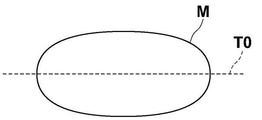
以下、体動判別部32が行う体動判別処理について説明する。第1の実施形態においては、体動判別部32は、再構成部35が生成した断層画像を用いて体動判別処理を行う。このために、第1の実施形態においては、体動判別部32は、再構成部35に対して、投影画像Giを再構成して断層画像を生成する指示を行う。なお、体動を判別するためには、複数の断層面における断層画像は必要ではなく、1つの断層面における1つの断層画像のみを生成すればよい。第1の実施形態においては、図8に示すように、圧迫厚の1/2となる断層面T0における断層画像D0を生成するものとする。
The body motion determination processing performed by the body
次いで、体動判別部32は、断層画像D0から特徴点を検出する。具体的には、Harrisのコーナー検出法、SIFT(Scale-Invariant Feature Transform)、FAST(Features from Accelerated Segment Test)あるいはSURF(Speeded Up Robust Features)等のアルゴリズムを用いて、断層画像D0に含まれる、エッジ、エッジの交点およびエッジの角部等の特徴点を少なくとも1つ検出する。なお、ここでは1つの特徴点のみを検出するものとする。そして体動判別部32は、投影画像Giにおいて特徴点の投影位置を算出する。なお、特徴点とは断層画像D0における1つの画素のみであってもよく、特徴となる構造の位置を表す複数の画素からなるものであってもよい。
Next, the body
図9は投影画像からの特徴点の投影位置の算出を説明するための図である。なお、図9においては説明を簡単なものとするために、3つの線源位置S1~S3に対応する3つの投影画像G1~G3からの投影位置の算出について説明する。また、図9においては、説明のため投影画像G1~G3が異なる平面に存在するように示しているが、実際には同一平面に存在する。また、図9においては、断層画像D0において特徴点F0が検出されているものとする。したがって、断層面T0には特徴点F0が含まれることとなる。図9に示すように、撮影時においては、乳房Mの断層面T0に含まれる特徴点F0は、投影画像G1~G3のそれぞれにおける位置P1~P3に投影される。線源位置S1~S3および乳房M内の特徴点F0の3次元空間における位置は既知である。また、投影画像G1~G3が生成される放射線検出器15の検出面の位置も既知である。このため、体動判別部32は、線源位置S1~S3および乳房M内の特徴点F0の3次元空間における位置、並びに投影画像G1~G3が生成される放射線検出器15の検出面の位置に基づいて、投影画像G1~G3における特徴点F0の投影位置P1~P3を算出する。
FIG. 9 is a diagram for explaining calculation of the projection position of the feature point from the projection image. In addition, in FIG. 9, in order to simplify the explanation, the calculation of the projection positions from the three projection images G1 to G3 corresponding to the three radiation source positions S1 to S3 will be explained. Also, in FIG. 9, the projected images G1 to G3 are shown to exist on different planes for the sake of explanation, but they actually exist on the same plane. Also, in FIG. 9, it is assumed that the feature point F0 is detected in the tomographic image D0. Therefore, the feature point F0 is included in the tomographic plane T0. As shown in FIG. 9, during imaging, a feature point F0 included in the tomographic plane T0 of the breast M is projected at positions P1 to P3 in projection images G1 to G3, respectively. The positions in the three-dimensional space of the radiation source positions S1 to S3 and the feature point F0 within the breast M are known. Also, the positions of the detection planes of the
続いて、体動判別部32は、図10に示すように、投影画像G1~G3において、投影位置P1~P3を中心とする予め定められた大きさの関心領域R1~R3を設定する。さらに、体動判別部32は、隣接する投影画像における関心領域R1~R3同士の位置合わせを行い、隣接する投影画像における関心領域間の移動方向および移動量を表すシフトベクトルを算出する。なお、シフトベクトルは投影画像の数よりも1つ少ないものとなる。例えば投影画像の数が15であれば、シフトベクトルの数は14となり、投影画像の数が3であれば、シフトベクトルの数は2となる。
Subsequently, as shown in FIG. 10, the body
図11は体動が発生していない場合のシフトベクトルを示す図、図12は体動が発生している場合のシフトベクトルを示す図である。なお、図11および図12においては、説明のために5つの投影画像G1~G5において算出された4つのシフトベクトルV12,V23,V34,V45を示す。また、図11および図12においては図の左右方向をX線源16の移動方向とする。体動が発生していない場合、図11に示すように、シフトベクトルV12,V23,V34,V45は、大きさがほぼ一定であり、かつX線源16の移動方向を向くものとなる。あるいは、シフトベクトルV12,V23,V34,V45の大きさはほぼ0となる。これに対して、体動が発生している場合、図12に示すように、シフトベクトルV12,V23,V34,V45は、不規則な方向を向き、かつ大きさも不規則となる。
FIG. 11 is a diagram showing shift vectors when body movement does not occur, and FIG. 12 is a diagram showing shift vectors when body movement occurs. 11 and 12 show four shift vectors V12, V23, V34 and V45 calculated in five projection images G1 to G5 for explanation. Also, in FIGS. 11 and 12, the horizontal direction of the drawing is the moving direction of the
第1の実施形態において、体動が発生しているか否かの判別の処理には、例えば、AI(Artificial Intelligence)技術を適用することができる。具体的には、シフトベクトルを入力とし、体動発生の有無を出力としたディープニューラルネットワークを作成する。次に、シフトベクトルおよびそのシフトベクトルによる体動発生の有無を含む教師データを用いて、作成したディープニューラルネットワークを学習させることによって得られた学習済みモデルからなる判別器をストレージ23に予め記憶しておく。体動判別部32は、算出したシフトベクトルを判別器に入力し、判別器から出力された体動発生の有無を表す情報を取得することによって、体動が発生しているか否かを判別する。例えば、判別器として-1~+1の間の出力値を出力するように学習がなされた学習済みモデルを使用し、判別器の出力が0以上となった場合に、体動が発生していると判別する。なお、判別器は体動判別部32に記憶されるものであってもよい。
In the first embodiment, AI (Artificial Intelligence) technology, for example, can be applied to the process of determining whether or not body movement occurs. Specifically, a deep neural network is created that takes the shift vector as an input and the presence or absence of body motion as an output. Next, a discriminator consisting of a trained model obtained by learning the created deep neural network using shift vectors and teacher data including presence/absence of body motion due to the shift vectors is stored in the
次いで、第1の実施形態において行われる処理について説明する。図13は第1の実施形態において行われる処理を示すフローチャートである。なお、本実施形態においては、まずトモシンセシス撮影が行われるため、条件設定部33により放射線画像撮影装置1に対して第1の撮影条件が設定されているものとする。操作者による処理開始の指示を入力部4が受け付けると、画像取得部31は放射線画像撮影装置1に第1の撮影条件によりトモシンセシス撮影を行わせて複数の投影画像Giを取得する第1の画像取得処理を行う(ステップST1)。そして、再構成部35が複数の投影画像Giを再構成して、乳房Mの複数の断層面における断層画像を生成する(ステップST2)。複数の投影画像Giおよび生成された断層画像は、ストレージ23に保存される。次いで、体動判別部32が、トモシンセシス撮影中に乳房Mの体動が発生しているか否かを判別する(ステップST3)。体動が発生している場合(ステップST3;YES)、条件設定部33が、放射線画像撮影装置1に対して単純撮影用の第2の撮影条件を設定し(ステップST4)、通知部34が、単純撮影を行う旨の通知を行う(ステップST5)。なお、ステップST5の処理をステップST4よりも先に行ってもよく、ステップST4およびステップST5の処理を同時に行ってもよい。
Next, processing performed in the first embodiment will be described. FIG. 13 is a flowchart showing processing performed in the first embodiment. In the present embodiment, since tomosynthesis imaging is performed first, it is assumed that the
画像取得部31は,入力部4から単純撮影の指示が行われたか否かの監視を開始し(ステップST6)、ステップST6が肯定されると、画像取得部31は、放射線画像撮影装置1に対して単純撮影を行わせて2次元画像Hcを取得する第2の画像取得処理を行い(ステップST7)、処理を終了する。なお、取得された2次元画像Hcは、断層画像と関連付けられてストレージ23に保存される。
The
一方、体動が発生していない場合(ステップST3;NO)、処理を終了する。処理が終了すると、圧迫板17による乳房Mの圧迫が解除される。
On the other hand, if body movement has not occurred (step ST3; NO), the process ends. When the processing ends, the compression of the breast M by the
このように、第1の実施形態においては、トモシンセシス撮影用の第1の撮影条件により、複数の投影画像Giを取得し、複数の投影画像Giに基づいて、トモシンセシス撮影中に被写体である乳房Mの体動が発生しているか否かを判別する。そして、体動が発生している場合、単純撮影用の第2の撮影条件を放射線画像撮影装置1に設定するようにした。このため、トモシンセシス撮影時に体動が発生していた場合、被写体である乳房Mのポジショニングを再度行うことなく、直ちに単純撮影を行うことができる。また、通知部34により単純撮影を行う旨の通知を行うようにしたため、操作者は通知に基づいて、直ちに単純撮影を行うことができる。したがって、本実施形態によれば、体動が発生した場合における被写体である患者の負担を軽減することができる。
As described above, in the first embodiment, a plurality of projection images Gi are acquired under the first imaging condition for tomosynthesis imaging, and the breast M, which is the subject, is acquired during tomosynthesis imaging based on the plurality of projection images Gi. body movement is occurring. Then, when body movement occurs, the
なお、上記第1の実施形態においては、体動判別部32において、断層画像から1つの特徴点を検出して体動が発生しているか否かを判別しているが、これに限定されるものではない。体動判別部32は、断層画像から複数の特徴点を検出し、複数の特徴点のそれぞれについて、投影画像Giの投影位置を検出して投影位置のシフトベクトルを算出し、複数の特徴点についてのシフトベクトルに基づいて、体動が発生しているか否かを判別するようにしてもよい。
In the first embodiment, the body
この場合、体動判別部32は、複数の特徴点のそれぞれにおける判別器の出力値に基づいて、複数の特徴点のそれぞれにおいて体動が発生しているか否かを判別し、複数の特徴点の数に対する体動が発生していると判別された特徴点の数の割合を算出する。そして、体動判別部32は、算出した割合が予め定められたしきい値Th1以上である場合に、体動が発生していると判別するようにすればよい。なお、しきい値Th1は例えば0.5とすればよいが、これに限定されるものではない。
In this case, the body
また、複数の特徴点についてのシフトベクトルに基づいて体動が発生しているか否かを判別する別の手法として、体動判別部32は、複数の特徴点のそれぞれにおける判別器の出力値の統計値を算出する。統計値としては、判別器の出力の平均値、中央値、最大値または最小値等を用いることができる。そして、体動判別部32は、算出した統計値が予め定められたしきい値Th2以上である場合に、体動が発生していると判別するようにしてもよい。なお、しきい値Th2としては、判別器の出力が-1以上1以下の間の値をとる場合、例えば0とすればよいが、これに限定されるものではない。
As another method for determining whether or not body motion occurs based on shift vectors for a plurality of feature points, the body
また、上記第1の実施形態においては、特徴点の投影位置に関心領域を設定し、関心領域の移動方向および移動量をシフトベクトルとして算出しているが、これに限定されるものではない。関心領域を設定することなく、特徴点の投影位置の投影画像間における移動方向および移動量をシフトベクトルとして算出してもよい。 In addition, in the above-described first embodiment, the region of interest is set at the projection position of the feature point, and the moving direction and amount of movement of the region of interest are calculated as the shift vector, but the present invention is not limited to this. The direction and amount of movement of the projection position of the feature point between projection images may be calculated as a shift vector without setting the region of interest.
また、上記第1の実施形態においては、関心領域(または特徴点の投影位置)の移動方向および移動量を表すシフトベクトルを用いて体動が発生しているか否かを判別しているが、これに限定されるものではない。体動判別部32は、移動方向のみを用いて体動が否かを判別してもよい。この場合、投影画像間における特徴点の投影位置の移動方向が不規則である場合に、体動が発生していると判別すればよい。また、移動量のみを用いて体動が発生しているか否かを判別してもよい。この場合、移動量が予め定められたしきい値を超える場合に、体動が発生していると判別すればよい。
In addition, in the above-described first embodiment, it is determined whether or not body movement occurs using a shift vector representing the movement direction and movement amount of the region of interest (or the projection position of the feature point). It is not limited to this. The body
次いで、本開示の第2の実施形態について説明する。なお、第2の実施形態による撮影制御装置の構成は、第1の実施形態による撮影制御装置の構成と同一であり、体動判別部32が行う処理のみが異なるため、ここでは構成についての詳細な説明は省略する。以下、第2の実施形態の撮影制御装置における、体動判別処理について説明する。
Next, a second embodiment of the present disclosure will be described. Note that the configuration of the imaging control device according to the second embodiment is the same as that of the imaging control device according to the first embodiment, and only the processing performed by the body
第2の実施形態においては、体動判別部32は、まず、複数の投影画像Giに含まれる共通の構造である対応点を検出する。具体的には、上記第1の実施形態における断層画像D0からの特徴点F0の検出と同様に、Harrisのコーナー検出法、SIFT、FASTあるいはSURF等のアルゴリズムを用いて、投影画像Giに含まれる、エッジ、エッジの交点およびエッジの角部等の、投影画像Gi間の構造である対応点を少なくとも1つ検出する。なお、ここでは1つの対応点を検出するものとして説明する。
In the second embodiment, the body
次いで、体動判別部32は、投影画像Giにおいて検出された対応点を逆投影して、乳房M内における対応点により表される構造の3次元空間における位置を算出する。図14は乳房M内における対応点により表される構造の3次元空間における位置の算出を説明するための図である。なお、図14においては説明を簡単なものとするために、3つの線源位置S1~S3に対応する3つの投影画像G1~G3を用いての、構造の3次元空間における位置の算出について説明する。また、図14においては、説明のため投影画像G1~G3が異なる平面に存在するように示しているが、実際には同一平面に存在することとなる。
Next, the body
図14に示すように、投影画像G1における対応点P11、投影画像G2における対応点P12および投影画像G3における対応点P13を逆投影することにより、乳房M内における対応点P11,P12,P13により表される構造の位置F10を特定する。逆投影により、構造の位置F10が存在する断層面およびその断層面における2次元状の位置を算出することができ、その結果、構造の位置F10の3次元空間における座標位置を算出することができる。 As shown in FIG. 14, the corresponding points P11, P12, and P13 in the breast M are represented by back-projecting the corresponding point P11 in the projection image G1, the corresponding point P12 in the projection image G2, and the corresponding point P13 in the projection image G3. Identify the location F10 of the structure to be processed. By back projection, it is possible to calculate the tomographic plane where the position F10 of the structure exists and the two-dimensional position on the tomographic plane, and as a result, it is possible to calculate the coordinate position of the position F10 of the structure in the three-dimensional space. .
第2の実施形態において、体動判別部32は、さらに構造の位置F10の投影画像G1~G3上における投影位置P21,P22,P23を算出する。図15は構造の位置F10の投影位置の算出を説明するための図である。なお、図15においては説明を簡単なものとするために、3つの線源位置S1~S3に対応する3つの投影画像G1~G3からの、構造の位置F10の投影位置の算出について説明する。また、図15においては、説明のため投影画像G1~G3が異なる平面に存在するように示しているが、実際には同一平面に存在することとなる。図15に示すように、構造の位置F10は、投影画像G1~G3のそれぞれにおける位置P21~P23に投影される。線源位置S1~S3および構造の位置F10の3次元空間における位置は既知である。また、投影画像G1~G3が生成される放射線検出器15の検出面の位置も既知である。このため、体動判別部32は、線源位置S1~S3および構造の位置F10の3次元空間における位置、並びに投影画像G1~G3が生成される放射線検出器15の検出面の位置に基づいて、投影画像G1~G3における構造の位置F10の投影位置P21~P23を算出する。
In the second embodiment, the body
なお、投影画像G1における構造の位置F10の投影位置P21と対応点P11とは、一致する場合もあるがずれる場合もある。また、投影画像G2における構造の位置F10の投影位置P22と対応点P12、および投影画像G3における構造の位置F10の投影位置P23と対応点P13も、一致する場合もあるがずれる場合もある。図15においては、投影位置P21と対応点P11、投影位置P22と対応点P12、および投影位置P23と対応点P13がずれている状態を示している。図16は、投影位置P21と対応点P11とが、投影位置P22と対応点P12とが、および投影位置P23と対応点P13とがずれている状態を示す図である。 Note that the projection position P21 of the position F10 of the structure in the projection image G1 and the corresponding point P11 may coincide with each other, but may also deviate from each other. Further, the projection position P22 of the structure position F10 in the projection image G2 and the corresponding point P12, and the projection position P23 of the structure position F10 and the corresponding point P13 in the projection image G3 may match or deviate. FIG. 15 shows a state in which the projection position P21 and the corresponding point P11, the projection position P22 and the corresponding point P12, and the projection position P23 and the corresponding point P13 are out of alignment. FIG. 16 is a diagram showing a state in which the projection position P21 and the corresponding point P11, the projection position P22 and the corresponding point P12, and the projection position P23 and the corresponding point P13 are out of alignment.
体動判別部32は、投影画像Giのそれぞれにおいて、対応点と投影位置とのずれの大きさを算出する。そして、すべての投影画像Giについてのずれの大きさの統計値を算出する。統計値としては、平均値、中央値、最大値または最小値等を用いることができる。そして、体動判別部32は、統計値を予め定められたしきい値Th3と比較し、統計値がしきい値Th3を超えた場合に体動が発生していると判別する。なお、しきい値Th3としては、その値を超えた場合に体動が発生していると見なせる、実験的に算出した値を用いることができる。
The body
第2の実施形態のように体動判別部32において体動が発生しているか否かを判別することによっても、トモシンセシス撮影時に体動が発生していた場合、第2の撮影条件を設定するようにしたため、被写体である乳房Mのポジショニングを再度行うことなく、直ちに単純撮影を行うことができる。したがって、第2の実施形態によっても、体動が発生した場合における被写体である患者の負担を軽減することができる。
By determining whether or not body movement occurs in the body
なお、上記第2の実施形態においては、体動判別部32において、投影画像Giから1つの対応点を検出して体動が発生しているか否かを判別しているが、これに限定されるものではない。体動判別部32は、投影画像Giのそれぞれから複数の対応点を検出し、複数の対応点のそれぞれについて、対応点と投影位置とのずれの大きさを算出し、複数の対応点についてのずれの大きさの統計値に基づいて、体動が発生しているか否かを判別するようにしてもよい。
In the second embodiment, the body
この場合、体動判別部32は、複数の対応点のそれぞれにおけるずれの大きさに基づいて、複数の対応点のそれぞれにおいて体動が発生しているか否かを判別し、複数の対応点の数に対する体動が発生していると判別された対応点の数の割合を算出する。そして、体動判別部32は、算出した割合が予め定められたしきい値Th4以上である場合に、体動が発生していると判別するようにすればよい。なお、しきい値Th4は例えば0.5とすればよいが、これに限定されるものではない。
In this case, the body
また、複数の対応点についてのずれの大きさに基づいて体動が発生しているか否かを判別する別の手法として、体動判別部32は、複数の対応点のそれぞれにおいて算出したずれの統計値についての統計値を他の統計値として算出する。そして、体動判別部32は、算出した他の統計値が予め定められたしきい値Th5以上である場合に、体動が発生していると判別するようにしてもよい。なお、しきい値Th5としては、その値を超えた場合に体動が発生していると見なせる、実験的に算出した値を用いることができる。
As another method for determining whether or not a body motion has occurred based on the magnitude of deviation for a plurality of corresponding points, the body
なお、上記各実施形態においては、通知部34が単純撮影を行う旨の通知を行うことにより、操作者が単純撮影を行っているが、これに限定されるものではない。体動判別部32が、トモシンセシス撮影時に体動が発生していると判別した場合、画像取得部31がX線源16を線源位置Scに移動して、単純撮影を行うようにしてもよい。
In each of the above-described embodiments, the
また、上記各実施形態においては、被写体を乳房Mとしているが、これに限定されるものではなく、人体の胸部、または腹部等、任意の部位を被写体としてもよいことはもちろんである。 In each of the above-described embodiments, the subject is the breast M, but the subject is not limited to this, and of course any part of the human body such as the chest or abdomen may be the subject.
また、上記各実施形態とは異なる手法を用いて体動を判別してもよい。例えば、米国特許第9498180号明細書等に記載された、公知の手法を用いて体動を判別してもよい。なお、米国特許第9498180号明細書に記載された手法は、投影画像において検出された実際の参照点の位置と、複数の投影画像から予測される参照点の予測位置との差に基づいて、体動を検出する手法である。 Also, a body motion may be determined using a method different from that in each of the embodiments described above. For example, body motion may be determined using a known method such as that described in US Pat. No. 9,498,180. Note that the technique described in US Pat. No. 9,498,180 is based on the difference between the actual position of the reference point detected in the projection image and the predicted position of the reference point predicted from a plurality of projection images . , is a technique for detecting body motion.
1 放射線画像撮影装置
2 コンピュータ
3 表示部
4 入力部
10 撮影部
15 放射線検出器
16 X線源
17 圧迫板
20 散乱線除去グリッド
21 CPU
22 メモリ
23 ストレージ
31 画像取得部
32 体動判別部
33 条件設定部
34 通知部
35 再構成部
D0 断層画像
F0 特徴点
F10 構造の位置
Gi(i=1~n) 投影画像
Hc 2次元画像
M 乳房
P1~P3 特徴点の投影位置
P11~P13 対応点
P21~P23 構造の位置の投影位置
R1~R3 関心領域
Si(i=1~n)、Sc 線源位置
T0 断層面
V12,V23,V34,V45 シフトベクトル
1
22
Claims (21)
前記複数の投影画像を再構成することにより、前記被写体の1つの断層面における断層画像を生成する再構成部と、
前記断層画像から少なくとも1つの特徴点を検出し、前記少なくとも1つの特徴点の前記複数の投影画像における投影位置を算出し、前記複数の投影画像間における前記投影位置の移動方向および移動量の少なくとも一方に基づいて、前記トモシンセシス撮影中に前記被写体の体動が発生しているか否かを判別する体動判別部と、
前記体動が発生していると判別された場合、単純撮影用の第2の撮影条件を前記撮影装置に設定する条件設定部とを備えた撮影制御装置。 Tomosynthesis imaging is performed on an imaging apparatus by moving a radiation source relative to a detection unit and irradiating a subject with radiation according to a first imaging condition for tomosynthesis imaging at a plurality of radiation source positions resulting from the movement of the radiation source. an image acquisition unit that acquires a plurality of projection images corresponding to each of the plurality of radiation source positions generated by
a reconstruction unit that generates a tomographic image in one tomographic plane of the subject by reconstructing the plurality of projection images;
detecting at least one feature point from the tomographic image, calculating a projection position of the at least one feature point in the plurality of projection images, and determining at least a movement direction and a movement amount of the projection position between the plurality of projection images; a body movement determination unit that determines whether or not body movement of the subject occurs during the tomosynthesis imaging based on one ;
and a condition setting unit for setting a second imaging condition for simple imaging to the imaging device when it is determined that the body movement occurs.
前記複数の投影画像に基づいて、前記トモシンセシス撮影中に前記被写体の体動が発生しているか否かを判別する体動判別部と、 a body movement determination unit that determines whether body movement of the subject occurs during the tomosynthesis imaging based on the plurality of projected images;
前記体動が発生していると判別された場合、前記被写体のポジショニングを維持したまま単純撮影用の第2の撮影条件を前記撮影装置に設定する条件設定部とを備えた撮影制御装置。 and a condition setting unit for setting a second photographing condition for simple photographing to the photographing device while maintaining the positioning of the subject when it is determined that the body movement occurs.
前記複数の投影画像を再構成することにより、前記被写体の1つの断層面における断層画像を生成し、
前記断層画像から少なくとも1つの特徴点を検出し、前記少なくとも1つの特徴点の前記複数の投影画像における投影位置を算出し、前記複数の投影画像間における前記投影位置の移動方向および移動量の少なくとも一方に基づいて、前記トモシンセシス撮影中に前記被写体の体動が発生しているか否かを判別し、
前記体動が発生していると判別された場合、単純撮影用の第2の撮影条件を前記撮影装置に設定する撮影制御方法。 Tomosynthesis imaging is performed on an imaging apparatus by moving a radiation source relative to a detection unit and irradiating a subject with radiation according to a first imaging condition for tomosynthesis imaging at a plurality of radiation source positions resulting from the movement of the radiation source. Acquiring a plurality of projection images corresponding to each of the plurality of source positions generated by
generating a tomographic image in one tomographic plane of the subject by reconstructing the plurality of projection images;
detecting at least one feature point from the tomographic image, calculating a projection position of the at least one feature point in the plurality of projection images, and determining at least a movement direction and a movement amount of the projection position between the plurality of projection images; determining whether or not body movement of the subject occurs during the tomosynthesis imaging based on one ;
A photographing control method for setting a second photographing condition for simple photographing in the photographing device when it is determined that the body movement is occurring.
前記複数の投影画像を再構成することにより、前記被写体の1つの断層面における断層画像を生成する手順と、
前記断層画像から少なくとも1つの特徴点を検出し、前記少なくとも1つの特徴点の前記複数の投影画像における投影位置を算出し、前記複数の投影画像間における前記投影位置の移動方向および移動量の少なくとも一方に基づいて、前記トモシンセシス撮影中に前記被写体の体動が発生しているか否かを判別する手順と、
前記体動が発生していると判別された場合、単純撮影用の第2の撮影条件を前記撮影装置に設定する手順とをコンピュータに実行させる撮影制御プログラム。 Tomosynthesis imaging is performed on an imaging apparatus by moving a radiation source relative to a detection unit and irradiating a subject with radiation according to a first imaging condition for tomosynthesis imaging at a plurality of radiation source positions resulting from the movement of the radiation source. acquiring a plurality of projection images corresponding to each of the plurality of source positions generated by applying the
a step of generating a tomographic image in one tomographic plane of the subject by reconstructing the plurality of projection images;
detecting at least one feature point from the tomographic image, calculating a projection position of the at least one feature point in the plurality of projection images, and determining at least a movement direction and a movement amount of the projection position between the plurality of projection images; a procedure for determining whether or not body movement of the subject occurs during the tomosynthesis imaging based on one ;
A photographing control program for causing a computer to execute a procedure for setting a second photographing condition for simple photographing to the photographing apparatus when it is determined that the body movement has occurred.
前記複数の投影画像に基づいて、前記トモシンセシス撮影中に前記被写体の体動が発生しているか否かを判別し、 determining whether or not body movement of the subject occurs during the tomosynthesis imaging based on the plurality of projected images;
前記体動が発生していると判別された場合、前記被写体のポジショニングを維持したまま単純撮影用の第2の撮影条件を前記撮影装置に設定する撮影制御方法。 A photographing control method for setting a second photographing condition for simple photographing to the photographing device while maintaining the positioning of the subject when it is determined that the body movement has occurred.
前記複数の投影画像に基づいて、前記トモシンセシス撮影中に前記被写体の体動が発生しているか否かを判別する手順と、 a step of determining whether or not body movement of the subject occurs during the tomosynthesis imaging based on the plurality of projected images;
前記体動が発生していると判別された場合、前記被写体のポジショニングを維持したまま単純撮影用の第2の撮影条件を前記撮影装置に設定する手順とをコンピュータに実行させる撮影制御プログラム。 a photographing control program for causing a computer to execute a procedure for setting a second photographing condition for simple photographing to the photographing apparatus while maintaining the positioning of the subject when it is determined that the body movement has occurred.
Priority Applications (3)
| Application Number | Priority Date | Filing Date | Title |
|---|---|---|---|
| JP2018120295A JP7122886B2 (en) | 2018-06-25 | 2018-06-25 | Imaging control device, method and program |
| US16/423,169 US11154257B2 (en) | 2018-06-25 | 2019-05-28 | Imaging control device, imaging control method, and imaging control program |
| EP19177105.4A EP3586750B1 (en) | 2018-06-25 | 2019-05-28 | Imaging control device, imaging control method, and imaging control program |
Applications Claiming Priority (1)
| Application Number | Priority Date | Filing Date | Title |
|---|---|---|---|
| JP2018120295A JP7122886B2 (en) | 2018-06-25 | 2018-06-25 | Imaging control device, method and program |
Publications (3)
| Publication Number | Publication Date |
|---|---|
| JP2020000313A JP2020000313A (en) | 2020-01-09 |
| JP2020000313A5 JP2020000313A5 (en) | 2020-09-24 |
| JP7122886B2 true JP7122886B2 (en) | 2022-08-22 |
Family
ID=66676274
Family Applications (1)
| Application Number | Title | Priority Date | Filing Date |
|---|---|---|---|
| JP2018120295A Active JP7122886B2 (en) | 2018-06-25 | 2018-06-25 | Imaging control device, method and program |
Country Status (3)
| Country | Link |
|---|---|
| US (1) | US11154257B2 (en) |
| EP (1) | EP3586750B1 (en) |
| JP (1) | JP7122886B2 (en) |
Families Citing this family (12)
| Publication number | Priority date | Publication date | Assignee | Title |
|---|---|---|---|---|
| US7616801B2 (en) | 2002-11-27 | 2009-11-10 | Hologic, Inc. | Image handling and display in x-ray mammography and tomosynthesis |
| US10638994B2 (en) | 2002-11-27 | 2020-05-05 | Hologic, Inc. | X-ray mammography with tomosynthesis |
| EP1816965B1 (en) | 2004-11-26 | 2016-06-29 | Hologic, Inc. | Integrated multi-mode mammography/tomosynthesis x-ray system |
| EP3445247B1 (en) | 2016-04-22 | 2021-03-10 | Hologic, Inc. | Tomosynthesis with shifting focal spot x-ray system using an addressable array |
| US11707244B2 (en) | 2017-08-16 | 2023-07-25 | Hologic, Inc. | Techniques for breast imaging patient motion artifact compensation |
| JP7167564B2 (en) * | 2018-09-05 | 2022-11-09 | 株式会社島津製作所 | Radiographic device and method of operating the radiographic device |
| US11090017B2 (en) * | 2018-09-13 | 2021-08-17 | Hologic, Inc. | Generating synthesized projection images for 3D breast tomosynthesis or multi-mode x-ray breast imaging |
| WO2020066109A1 (en) | 2018-09-27 | 2020-04-02 | 富士フイルム株式会社 | Tomographic image generation device, method, and program |
| JP7023254B2 (en) * | 2019-03-27 | 2022-02-21 | 富士フイルム株式会社 | Shooting support equipment, methods and programs |
| US20210361250A1 (en) * | 2020-05-19 | 2021-11-25 | Konica Minolta, Inc. | Dynamic analysis system, correction apparatus, storage medium, and dynamic imaging apparatus |
| WO2022064911A1 (en) * | 2020-09-28 | 2022-03-31 | 富士フイルム株式会社 | Control device, control method, and control program |
| US11786191B2 (en) | 2021-05-17 | 2023-10-17 | Hologic, Inc. | Contrast-enhanced tomosynthesis with a copper filter |
Citations (5)
| Publication number | Priority date | Publication date | Assignee | Title |
|---|---|---|---|---|
| JP2010158299A (en) | 2009-01-06 | 2010-07-22 | Fujifilm Corp | Tomographic image capturing apparatus and tomographic image capturing method |
| JP2010179094A (en) | 2009-01-08 | 2010-08-19 | Fujifilm Corp | Radiation tomographic image generator |
| JP2012075862A (en) | 2010-09-08 | 2012-04-19 | Fujifilm Corp | Body motion detector, method, and program |
| JP2015188604A (en) | 2014-03-28 | 2015-11-02 | 富士フイルム株式会社 | Radiography apparatus, method, and program |
| JP2017143943A (en) | 2016-02-16 | 2017-08-24 | 富士フイルム株式会社 | Radiation image processing device, method, and program |
Family Cites Families (6)
| Publication number | Priority date | Publication date | Assignee | Title |
|---|---|---|---|---|
| JPH02302248A (en) * | 1989-05-16 | 1990-12-14 | Toshiba Corp | Image re-pickup device for ct device |
| JP4110074B2 (en) | 2003-11-05 | 2008-07-02 | キヤノン株式会社 | Radiation image processing apparatus, radiation image processing method, program, and computer-readable medium |
| JP5400358B2 (en) * | 2008-11-13 | 2014-01-29 | 富士フイルム株式会社 | Radiation tomography equipment |
| JP5572040B2 (en) * | 2009-09-28 | 2014-08-13 | 富士フイルム株式会社 | Radiography equipment |
| US9498180B2 (en) * | 2010-08-05 | 2016-11-22 | Hologic, Inc. | Detecting and quantifying patient motion during tomosynthesis scans |
| JP6556005B2 (en) * | 2015-09-29 | 2019-08-07 | 富士フイルム株式会社 | Tomographic image generating apparatus, method and program |
-
2018
- 2018-06-25 JP JP2018120295A patent/JP7122886B2/en active Active
-
2019
- 2019-05-28 US US16/423,169 patent/US11154257B2/en active Active
- 2019-05-28 EP EP19177105.4A patent/EP3586750B1/en active Active
Patent Citations (5)
| Publication number | Priority date | Publication date | Assignee | Title |
|---|---|---|---|---|
| JP2010158299A (en) | 2009-01-06 | 2010-07-22 | Fujifilm Corp | Tomographic image capturing apparatus and tomographic image capturing method |
| JP2010179094A (en) | 2009-01-08 | 2010-08-19 | Fujifilm Corp | Radiation tomographic image generator |
| JP2012075862A (en) | 2010-09-08 | 2012-04-19 | Fujifilm Corp | Body motion detector, method, and program |
| JP2015188604A (en) | 2014-03-28 | 2015-11-02 | 富士フイルム株式会社 | Radiography apparatus, method, and program |
| JP2017143943A (en) | 2016-02-16 | 2017-08-24 | 富士フイルム株式会社 | Radiation image processing device, method, and program |
Also Published As
| Publication number | Publication date |
|---|---|
| EP3586750A1 (en) | 2020-01-01 |
| EP3586750B1 (en) | 2022-10-05 |
| US20190388051A1 (en) | 2019-12-26 |
| US11154257B2 (en) | 2021-10-26 |
| JP2020000313A (en) | 2020-01-09 |
Similar Documents
| Publication | Publication Date | Title |
|---|---|---|
| JP7122886B2 (en) | Imaging control device, method and program | |
| US10219769B2 (en) | Radiation image processing apparatus, radiation image processing method, and recording medium having radiation image processing program stored therein | |
| JP6556005B2 (en) | Tomographic image generating apparatus, method and program | |
| JP2008515522A5 (en) | ||
| EP3590431B1 (en) | Image display device, image display method, and image display program | |
| US12036051B2 (en) | Tomosynthesis imaging support apparatus, method, and program | |
| JP6682150B2 (en) | Breast volume acquisition device, method and program | |
| US11540797B2 (en) | Tomographic image generation apparatus, method, and program | |
| US10102624B2 (en) | Radiation image processing apparatus, radiation image processing method, and recording medium having radiation image processing program stored therein | |
| JP7275363B2 (en) | Positional deviation amount derivation device, method and program | |
| EP4119055A1 (en) | Image generation device and program, learning device and program, and image processing device and program | |
| JP7113790B2 (en) | Image processing device, method and program | |
| WO2020202612A1 (en) | Image processing device, method, and program | |
| JP7169430B2 (en) | Imaging control device, method and program | |
| JP7208874B2 (en) | Imaging control device, method and program | |
| JP2015177817A (en) | X-ray ct apparatus and image processor |
Legal Events
| Date | Code | Title | Description |
|---|---|---|---|
| A521 | Request for written amendment filed |
Free format text: JAPANESE INTERMEDIATE CODE: A523 Effective date: 20200812 |
|
| A621 | Written request for application examination |
Free format text: JAPANESE INTERMEDIATE CODE: A621 Effective date: 20200812 |
|
| A977 | Report on retrieval |
Free format text: JAPANESE INTERMEDIATE CODE: A971007 Effective date: 20210728 |
|
| A131 | Notification of reasons for refusal |
Free format text: JAPANESE INTERMEDIATE CODE: A131 Effective date: 20210810 |
|
| A521 | Request for written amendment filed |
Free format text: JAPANESE INTERMEDIATE CODE: A523 Effective date: 20210929 |
|
| A131 | Notification of reasons for refusal |
Free format text: JAPANESE INTERMEDIATE CODE: A131 Effective date: 20220208 |
|
| A521 | Request for written amendment filed |
Free format text: JAPANESE INTERMEDIATE CODE: A523 Effective date: 20220329 |
|
| TRDD | Decision of grant or rejection written | ||
| A01 | Written decision to grant a patent or to grant a registration (utility model) |
Free format text: JAPANESE INTERMEDIATE CODE: A01 Effective date: 20220719 |
|
| A61 | First payment of annual fees (during grant procedure) |
Free format text: JAPANESE INTERMEDIATE CODE: A61 Effective date: 20220809 |
|
| R150 | Certificate of patent or registration of utility model |
Ref document number: 7122886 Country of ref document: JP Free format text: JAPANESE INTERMEDIATE CODE: R150 |