JP5055284B2 - Biological markers for predicting anti-cancer responses to insulin-like growth factor-1 receptor kinase inhibitors - Google Patents
Biological markers for predicting anti-cancer responses to insulin-like growth factor-1 receptor kinase inhibitors Download PDFInfo
- Publication number
- JP5055284B2 JP5055284B2 JP2008532319A JP2008532319A JP5055284B2 JP 5055284 B2 JP5055284 B2 JP 5055284B2 JP 2008532319 A JP2008532319 A JP 2008532319A JP 2008532319 A JP2008532319 A JP 2008532319A JP 5055284 B2 JP5055284 B2 JP 5055284B2
- Authority
- JP
- Japan
- Prior art keywords
- igf
- tumor
- kinase inhibitor
- tumor cell
- inhibition
- Prior art date
- Legal status (The legal status is an assumption and is not a legal conclusion. Google has not performed a legal analysis and makes no representation as to the accuracy of the status listed.)
- Expired - Fee Related
Links
Images


Classifications
-
- G—PHYSICS
- G01—MEASURING; TESTING
- G01N—INVESTIGATING OR ANALYSING MATERIALS BY DETERMINING THEIR CHEMICAL OR PHYSICAL PROPERTIES
- G01N33/00—Investigating or analysing materials by specific methods not covered by groups G01N1/00 - G01N31/00
- G01N33/48—Biological material, e.g. blood, urine; Haemocytometers
- G01N33/50—Chemical analysis of biological material, e.g. blood, urine; Testing involving biospecific ligand binding methods; Immunological testing
- G01N33/53—Immunoassay; Biospecific binding assay; Materials therefor
- G01N33/574—Immunoassay; Biospecific binding assay; Materials therefor for cancer
- G01N33/57407—Specifically defined cancers
- G01N33/57423—Specifically defined cancers of lung
-
- A—HUMAN NECESSITIES
- A61—MEDICAL OR VETERINARY SCIENCE; HYGIENE
- A61P—SPECIFIC THERAPEUTIC ACTIVITY OF CHEMICAL COMPOUNDS OR MEDICINAL PREPARATIONS
- A61P35/00—Antineoplastic agents
-
- A—HUMAN NECESSITIES
- A61—MEDICAL OR VETERINARY SCIENCE; HYGIENE
- A61P—SPECIFIC THERAPEUTIC ACTIVITY OF CHEMICAL COMPOUNDS OR MEDICINAL PREPARATIONS
- A61P35/00—Antineoplastic agents
- A61P35/04—Antineoplastic agents specific for metastasis
-
- A—HUMAN NECESSITIES
- A61—MEDICAL OR VETERINARY SCIENCE; HYGIENE
- A61P—SPECIFIC THERAPEUTIC ACTIVITY OF CHEMICAL COMPOUNDS OR MEDICINAL PREPARATIONS
- A61P43/00—Drugs for specific purposes, not provided for in groups A61P1/00-A61P41/00
-
- C—CHEMISTRY; METALLURGY
- C12—BIOCHEMISTRY; BEER; SPIRITS; WINE; VINEGAR; MICROBIOLOGY; ENZYMOLOGY; MUTATION OR GENETIC ENGINEERING
- C12Q—MEASURING OR TESTING PROCESSES INVOLVING ENZYMES, NUCLEIC ACIDS OR MICROORGANISMS; COMPOSITIONS OR TEST PAPERS THEREFOR; PROCESSES OF PREPARING SUCH COMPOSITIONS; CONDITION-RESPONSIVE CONTROL IN MICROBIOLOGICAL OR ENZYMOLOGICAL PROCESSES
- C12Q1/00—Measuring or testing processes involving enzymes, nucleic acids or microorganisms; Compositions therefor; Processes of preparing such compositions
- C12Q1/68—Measuring or testing processes involving enzymes, nucleic acids or microorganisms; Compositions therefor; Processes of preparing such compositions involving nucleic acids
- C12Q1/6876—Nucleic acid products used in the analysis of nucleic acids, e.g. primers or probes
- C12Q1/6883—Nucleic acid products used in the analysis of nucleic acids, e.g. primers or probes for diseases caused by alterations of genetic material
- C12Q1/6886—Nucleic acid products used in the analysis of nucleic acids, e.g. primers or probes for diseases caused by alterations of genetic material for cancer
-
- G—PHYSICS
- G01—MEASURING; TESTING
- G01N—INVESTIGATING OR ANALYSING MATERIALS BY DETERMINING THEIR CHEMICAL OR PHYSICAL PROPERTIES
- G01N33/00—Investigating or analysing materials by specific methods not covered by groups G01N1/00 - G01N31/00
- G01N33/48—Biological material, e.g. blood, urine; Haemocytometers
- G01N33/50—Chemical analysis of biological material, e.g. blood, urine; Testing involving biospecific ligand binding methods; Immunological testing
- G01N33/53—Immunoassay; Biospecific binding assay; Materials therefor
- G01N33/574—Immunoassay; Biospecific binding assay; Materials therefor for cancer
-
- C—CHEMISTRY; METALLURGY
- C12—BIOCHEMISTRY; BEER; SPIRITS; WINE; VINEGAR; MICROBIOLOGY; ENZYMOLOGY; MUTATION OR GENETIC ENGINEERING
- C12Q—MEASURING OR TESTING PROCESSES INVOLVING ENZYMES, NUCLEIC ACIDS OR MICROORGANISMS; COMPOSITIONS OR TEST PAPERS THEREFOR; PROCESSES OF PREPARING SUCH COMPOSITIONS; CONDITION-RESPONSIVE CONTROL IN MICROBIOLOGICAL OR ENZYMOLOGICAL PROCESSES
- C12Q2600/00—Oligonucleotides characterized by their use
- C12Q2600/106—Pharmacogenomics, i.e. genetic variability in individual responses to drugs and drug metabolism
-
- C—CHEMISTRY; METALLURGY
- C12—BIOCHEMISTRY; BEER; SPIRITS; WINE; VINEGAR; MICROBIOLOGY; ENZYMOLOGY; MUTATION OR GENETIC ENGINEERING
- C12Q—MEASURING OR TESTING PROCESSES INVOLVING ENZYMES, NUCLEIC ACIDS OR MICROORGANISMS; COMPOSITIONS OR TEST PAPERS THEREFOR; PROCESSES OF PREPARING SUCH COMPOSITIONS; CONDITION-RESPONSIVE CONTROL IN MICROBIOLOGICAL OR ENZYMOLOGICAL PROCESSES
- C12Q2600/00—Oligonucleotides characterized by their use
- C12Q2600/136—Screening for pharmacological compounds
-
- C—CHEMISTRY; METALLURGY
- C12—BIOCHEMISTRY; BEER; SPIRITS; WINE; VINEGAR; MICROBIOLOGY; ENZYMOLOGY; MUTATION OR GENETIC ENGINEERING
- C12Q—MEASURING OR TESTING PROCESSES INVOLVING ENZYMES, NUCLEIC ACIDS OR MICROORGANISMS; COMPOSITIONS OR TEST PAPERS THEREFOR; PROCESSES OF PREPARING SUCH COMPOSITIONS; CONDITION-RESPONSIVE CONTROL IN MICROBIOLOGICAL OR ENZYMOLOGICAL PROCESSES
- C12Q2600/00—Oligonucleotides characterized by their use
- C12Q2600/158—Expression markers
-
- G—PHYSICS
- G01—MEASURING; TESTING
- G01N—INVESTIGATING OR ANALYSING MATERIALS BY DETERMINING THEIR CHEMICAL OR PHYSICAL PROPERTIES
- G01N2333/00—Assays involving biological materials from specific organisms or of a specific nature
- G01N2333/435—Assays involving biological materials from specific organisms or of a specific nature from animals; from humans
- G01N2333/705—Assays involving receptors, cell surface antigens or cell surface determinants
- G01N2333/71—Assays involving receptors, cell surface antigens or cell surface determinants for growth factors; for growth regulators
-
- G—PHYSICS
- G01—MEASURING; TESTING
- G01N—INVESTIGATING OR ANALYSING MATERIALS BY DETERMINING THEIR CHEMICAL OR PHYSICAL PROPERTIES
- G01N2333/00—Assays involving biological materials from specific organisms or of a specific nature
- G01N2333/90—Enzymes; Proenzymes
- G01N2333/91—Transferases (2.)
- G01N2333/912—Transferases (2.) transferring phosphorus containing groups, e.g. kinases (2.7)
- G01N2333/91205—Phosphotransferases in general
- G01N2333/9121—Phosphotransferases in general with an alcohol group as acceptor (2.7.1), e.g. general tyrosine, serine or threonine kinases
- G01N2333/91215—Phosphotransferases in general with an alcohol group as acceptor (2.7.1), e.g. general tyrosine, serine or threonine kinases with a definite EC number (2.7.1.-)
-
- G—PHYSICS
- G01—MEASURING; TESTING
- G01N—INVESTIGATING OR ANALYSING MATERIALS BY DETERMINING THEIR CHEMICAL OR PHYSICAL PROPERTIES
- G01N2500/00—Screening for compounds of potential therapeutic value
- G01N2500/04—Screening involving studying the effect of compounds C directly on molecule A (e.g. C are potential ligands for a receptor A, or potential substrates for an enzyme A)
-
- G—PHYSICS
- G01—MEASURING; TESTING
- G01N—INVESTIGATING OR ANALYSING MATERIALS BY DETERMINING THEIR CHEMICAL OR PHYSICAL PROPERTIES
- G01N2800/00—Detection or diagnosis of diseases
- G01N2800/52—Predicting or monitoring the response to treatment, e.g. for selection of therapy based on assay results in personalised medicine; Prognosis
Abstract
Description
本発明は、癌患者を診断及び治療する方法を対象とする。特に、本発明は、インシュリン様成長因子−1受容体(IGF−1R)キナーゼ阻害剤を用いた治療から最も利点を得る患者を判定する方法を対象とする。 The present invention is directed to methods for diagnosing and treating cancer patients. In particular, the present invention is directed to a method of determining patients that would benefit most from treatment with an insulin-like growth factor-1 receptor (IGF-1R) kinase inhibitor.
癌は、制御されない増殖、分化の欠如、及び局所組織に浸潤し、転移する能力を特徴とする、細胞の広範な悪性腫瘍の一般名である。これらの新生物悪性腫瘍は、体内のあらゆる組織及び器官において多様な罹患率で発症する。 Cancer is a generic name for a wide range of malignant tumors of cells characterized by uncontrolled growth, lack of differentiation, and the ability to invade local tissues and metastasize. These neoplastic malignancies develop with varying morbidity in every tissue and organ in the body.
種々の癌を治療するために、複数の治療薬が過去数十年にわたって開発されてきた。最も一般に使用される抗癌剤タイプとしては、DNAアルキル化剤(例えば、シクロホスファミド、イホスファミド)、代謝拮抗剤(例えば、メトトレキセート、葉酸塩拮抗物質及び5−フルオロウラシル、ピリミジン拮抗物質)、微小管分裂剤(例えば、ビンクリスチン、ビンブラスチン、パクリタキセル)、DNAインターカレーター(例えば、ドキソルビシン、ダウノマイシン、シスプラチン)及びホルモン療法(例えば、タモキシフェン、フルタミド)などが挙げられる。 Multiple therapeutic agents have been developed over the past decades to treat various cancers. The most commonly used anticancer agent types include DNA alkylating agents (eg, cyclophosphamide, ifosfamide), antimetabolites (eg, methotrexate, folate antagonists and 5-fluorouracil, pyrimidine antagonists), microtubule division Agents (eg, vincristine, vinblastine, paclitaxel), DNA intercalators (eg, doxorubicin, daunomycin, cisplatin) and hormone therapy (eg, tamoxifen, flutamide).
IGF−1Rは、主としてIGF−1に結合するが、IGF−II及びインシュリンにも、より低い親和性で結合する膜貫通RTKである。IGF−1受容体へのIGF−1の結合は、受容体のオリゴマー化、チロシンキナーゼの活性化、分子間受容体自己リン酸化及び細胞基質のリン酸化をもたらす(主な基質は、IRS1及びShcである。)。リガンドによって活性化されたIGF−1Rは、正常な細胞中で有糸分裂促進活性を誘導し、異常な増殖において重要な役割を果たす。IGF−1系の主要な生理的役割は、正常な増殖と再生を促進することである。過剰発現されたIGF−1R(1型インシュリン様成長因子受容体)は、有糸分裂誘発を開始させ、リガンド依存性新生物形質転換を促進することが可能である。さらに、IGF−1Rは、悪性表現型の確立及び維持において重要な役割を果たしている。上皮成長因子(EGF)受容体とは異なり、IGF−1Rの変異発癌形態は同定されていない。しかしながら、幾つかの発癌遺伝子は、IGF−1及びIGF−1R発現に影響を与えることが示されている。IGF−1R発現の減少と形質転換に対する耐性との間の相関が観察されてきた。IGF−1RRNAに対してアンチセンスであるmRNAに細胞を曝露することによって、幾つかのヒト腫瘍細胞株の軟寒天増殖が抑制される。IGF−1Rは、インビボ及びインビトロの両者で、アポトーシスへの進行を停止させる。野生型レベルを下回るIGF−1Rの減少が、インビボで、腫瘍細胞のアポトーシスを引き起こすことも示されている。IGF−1R破壊がアポトーシスを引き起こす能力は、正常な非腫瘍原性細胞中では減弱しているようである。 IGF-1R is a transmembrane RTK that binds primarily to IGF-1 but also binds to IGF-II and insulin with lower affinity. Binding of IGF-1 to the IGF-1 receptor results in receptor oligomerization, tyrosine kinase activation, intermolecular receptor autophosphorylation and cellular substrate phosphorylation (the main substrates are IRS1 and Shc .) IGF-1R activated by a ligand induces mitogenic activity in normal cells and plays an important role in abnormal growth. The primary physiological role of the IGF-1 system is to promote normal growth and regeneration. Overexpressed IGF-1R (type 1 insulin-like growth factor receptor) can initiate mitogenesis and promote ligand-dependent neoplastic transformation. Furthermore, IGF-1R plays an important role in establishing and maintaining a malignant phenotype. Unlike the epidermal growth factor (EGF) receptor, no mutated oncogenic form of IGF-1R has been identified. However, several oncogenes have been shown to affect IGF-1 and IGF-1R expression. A correlation has been observed between reduced IGF-1R expression and resistance to transformation. By exposing cells to mRNA that is antisense to IGF-1RRNA, soft agar growth of several human tumor cell lines is suppressed. IGF-1R stops progression to apoptosis both in vivo and in vitro. It has also been shown that reduction of IGF-1R below wild-type levels causes tumor cell apoptosis in vivo. The ability of IGF-1R disruption to cause apoptosis appears to be attenuated in normal non-tumorigenic cells.
ヒト腫瘍の発育におけるIGF−1経路は、重要な役割を有する。IGF−1R過剰発現は、しばしば、様々な腫瘍(乳癌、結腸癌、肺癌、肉腫)中に見出され、多くの場合、侵襲性の表現型を伴う。高いIGF1循環濃度は、前立腺癌、肺癌及び乳癌のリスクと強く相関している。さらに、形質転換された表現型の確立及び維持のために、インビトロ及びインビボにおいて、IGF−1Rが必要とされる(Baserga R.Exp.Cell.Res.,1999,253,1−6)。IGF−1Rのキナーゼ活性は、幾つかの発癌遺伝子:EGFR、PDGFR、SV40T抗原、活性化されたRas、Raf及びv−Srcの形質転換活性にとって不可欠である。正常な繊維芽細胞中でのIGF−1Rの発現は、新生物表現型を誘導し、次いで、インビボにおいて、新生物表現型は腫瘍を形成することができる。IGF−1R発現は、足場非依存性増殖において重要な役割を果たしている。IGF−1Rは、化学療法、放射線照射及びサイトカインによって誘導されるアポトーシスから細胞を保護することも示されている。逆に、ドミナントネガティブIGF−1R、三重螺旋形成又はアンチセンス発現ベクターによる内在性IGF−1Rの阻害は、インビトロにおいて形質転換活性を抑制し、動物モデルにおいて腫瘍増殖を抑制することが示されている。 The IGF-1 pathway in human tumor development has an important role. IGF-1R overexpression is often found in various tumors (breast cancer, colon cancer, lung cancer, sarcoma) and is often accompanied by an invasive phenotype. High circulating levels of IGF1 are strongly correlated with prostate cancer, lung cancer and breast cancer risk. In addition, IGF-1R is required in vitro and in vivo for the establishment and maintenance of transformed phenotypes (Baserga R. Exp. Cell. Res., 1999, 253, 1-6). IGF-1R kinase activity is essential for the transformation activity of several oncogenes: EGFR, PDGFR, SV40T antigen, activated Ras, Raf and v-Src. Expression of IGF-1R in normal fibroblasts induces a neoplastic phenotype, which can then form a tumor in vivo. IGF-1R expression plays an important role in anchorage-independent growth. IGF-1R has also been shown to protect cells from apoptosis induced by chemotherapy, radiation and cytokines. Conversely, inhibition of endogenous IGF-1R by dominant negative IGF-1R, triple helix formation or antisense expression vectors has been shown to suppress transformation activity in vitro and tumor growth in animal models. .
タンパク質チロシンキナーゼの阻害剤は、哺乳動物の癌細胞の増殖の選択的阻害剤として有用であることが認められる。例えば、BCR−ABL融合遺伝子産物のキナーゼ活性を阻害する2−フェニルピリミジンチロシンキナーゼ阻害剤であるGllevecTM(イマチニブメシラートとしても知られている。)が、CMLの治療に対して、アメリカ食品医薬品局によって承認された。4−アニリノキナゾリン化合物であるTarcevaTM(エルロチニブHCl)も、最近、FDAによって承認され、高い効力を有するEGF受容体キナーゼを選択的に阻害する。IGF−1Rのキナーゼ活性を直接的に阻害する化合物及びIGF−1Rの活性化を遮断することによって、IGF−1Rキナーゼ活性を低下させる抗体又はIGF−1R発現を遮断するアンチセンスオリゴヌクレオチドを抗癌剤として使用するための開発は、多大な研究努力が向けられている分野である(例えば、Larsson, O. et al(2005)Brit. J. Cancer 92:2097−2101;Ibrahim, Y.H. and Yee, D.(2005)Clin. Cancer Res.11:944s−950s;Mitsiades,C.S. et al.(2004)Cancer Cell 5:221−230;Camirand, A. et al.(2005)Breast Cancer Research 7:R570−R579(DOI10.1186/bcrlO28);Camirand, A. and Pollak, M.(2004)Brit.J.Cancer 90:1825−1829;Garcia−Echeverria, C. et al.(2004)Cancer Cell 5:231−239参照)。 It will be appreciated that inhibitors of protein tyrosine kinases are useful as selective inhibitors of the growth of mammalian cancer cells. For example, Glevec ™ (also known as imatinib mesylate), a 2-phenylpyrimidine tyrosine kinase inhibitor that inhibits the kinase activity of the BCR-ABL fusion gene product, is an American food and pharmaceutical for the treatment of CML. Approved by the bureau. Tarceva ™ (erlotinib HCl), a 4-anilinoquinazoline compound, has also recently been approved by the FDA and selectively inhibits the highly potent EGF receptor kinase. Compounds that directly inhibit IGF-1R kinase activity and antibodies that reduce IGF-1R kinase activity by blocking IGF-1R activation or antisense oligonucleotides that block IGF-1R expression as anticancer agents Development for use is an area where significant research efforts are directed (eg, Larsson, O. et al (2005) Brit. J. Cancer 92: 2097-2101; Ibrahim, YH and Yee. , D. (2005) Clin. Cancer Res. 11: 944s-950s; Mitsiades, CS et al. (2004) Cancer Cell 5: 221-230; Camiland, A. et al. (2005) Breast Cancer Res. rch 7: R570-R579 (DOI10.1186 / bcrO28); Camirand, A. and Pollak, M. (2004) Brit. J. Cancer 90: 1825-1829; Garcia-Echeverria, C. et al. (2004) Cell 5: 231-239).
抗新生物薬は、非悪性細胞に対するその毒性に比較して治療指数が広く、癌細胞を選択的に死滅させる理想的薬剤であろう。また、抗新生物薬は、該薬物に長時間曝露した後でも、悪性細胞に対してその効力を保持する。残念ながら、現行の化学療法のいずれも、かかる理想的な特性を持たない。むしろ、大部分は、極めて狭い治療指数を有する。さらに、致死濃度をわずかに下回る化学療法薬に曝露された癌細胞は、かかる薬剤に対して耐性を獲得することが極めて多く、幾つかの他の抗腫瘍薬に対しても交差耐性を獲得することが頻繁にある。さらに、任意の所与の癌タイプに対して、特定の治療に応答する可能性がある患者を予測することは、タンパク質チロシンキナーゼ阻害剤などのより新しい遺伝子標的療法でも不可能であることが多く、したがって、最も有効な療法を見出すためにかなりの試行錯誤を必要とし、患者に対してかなりのリスクと不快感を与えることが多い。 Anti-neoplastic agents have a wide therapeutic index compared to their toxicity to non-malignant cells and may be ideal drugs that selectively kill cancer cells. Antineoplastic drugs also retain their efficacy against malignant cells even after prolonged exposure to the drug. Unfortunately, none of the current chemotherapy has such ideal properties. Rather, most have a very narrow therapeutic index. In addition, cancer cells exposed to chemotherapeutic drugs that are slightly below the lethal concentration are very likely to acquire resistance to such drugs and also to cross resistance to several other anti-tumor drugs. There are often. In addition, predicting patients who are likely to respond to a particular treatment for any given cancer type is often not possible with newer gene-targeted therapies such as protein tyrosine kinase inhibitors Therefore, considerable trial and error is required to find the most effective therapy and often presents considerable risk and discomfort to the patient.
したがって、新形成及び他の増殖性障害に対するより効果的な治療、及びどの腫瘍がどの治療に応答するかを決定するより有効な手段が求められている。既存薬物の治療効力を高める戦略は、その投与スケジュールの変更が必要であり、他の抗癌剤又は生化学的調節剤との併用も必要である。併用療法は、各薬剤単体の治療上妥当な用量を使用するよりも、効力が大きく、副作用が減少し得る方法としてよく知られている。多剤併用の効力が相加的である(併用の効力が各薬物単体の効果の合計にほぼ等しい。)場合もあるが、効果が相乗的である(併用の効力が、単体投与された各薬物の効果の合計よりも大きい。)場合もある。標的特異的な治療アプローチは、一般に、慣用の細胞毒製剤と比べて、減少した毒性を伴い、従って、併用治療計画において、かかるアプローチが使用される。 Accordingly, there is a need for more effective treatments for neoplasia and other proliferative disorders and more effective means of determining which tumors respond to which treatment. Strategies for increasing the therapeutic efficacy of existing drugs require changes in their administration schedule, and in combination with other anticancer agents or biochemical modulators. Combination therapy is well known as a method that has greater efficacy and fewer side effects than using therapeutically relevant doses of each drug alone. In some cases, the efficacy of a multidrug combination is additive (the efficacy of the combination is approximately equal to the sum of the effects of each drug alone), but the effect is synergistic (the efficacy of the combination is Greater than the sum of the effects of the drug.) Target-specific therapeutic approaches are generally associated with reduced toxicity compared to conventional cytotoxic preparations, and thus such approaches are used in combination treatment regimens.
幾つかのグループが、タンパク質チロシンキナーゼ阻害剤、例えば、EGFR阻害剤に対する患者の応答を予測するバイオマーカー候補を検討した(例えば、PCT国際公開第2004/063709号、同2005/017493号、同2004/111273号及び同2004/071572号並びに米国特許出願公開第2005/0019785号及び同2004/0132097号参照)。しかし、このような阻害剤による患者の治療において開業医を手引することができる診断試験又は予後の試験はまだ出現していない。 Several groups have examined biomarker candidates that predict patient response to protein tyrosine kinase inhibitors, such as EGFR inhibitors (eg, PCT International Publication Nos. WO 2004/063709, 2005/017433, 2004). No. 111111273 and 2004/071572 and US Patent Application Publication Nos. 2005/0019785 and 2004/0132097). However, no diagnostic or prognostic test has yet emerged that can guide practitioners in treating patients with such inhibitors.
したがって、任意の特定の癌患者に対する最適な治療様式を決定するための改善された方法に対して、重大な要望が依然として存在する。本発明は、腫瘍細胞が上皮から間葉への遷移を経たかどうかに基づいて、何れの腫瘍がIGF−1Rキナーゼ阻害剤での治療に対して最も効果的に応答するかを決定する方法(“EMT”; Thiery, J.P. (2002) Nat. Rev. Cancer 2:442−454; Savagner, P. (2001) Bioessays 23:912−923; Kang Y. and Massague, J. (2004) Cell 118:277−279; Julien−Grille, S., et al. Cancer Research 63:2172−2178; Bates, R.C. et al. (2003) Current Biology 13:1721−1727; Lu Z., et al. (2003) Cancer Cell. 4(6):499−515)、及びこのような阻害剤が、単一の薬剤として使用されるかどうか、又は他の抗癌剤と組み合わされて地要されるかどうか、癌患者のためのさらに有効な治療計画にこのような決定を取り込む方法を提供する。 Accordingly, there remains a significant need for improved methods for determining the optimal treatment modality for any particular cancer patient. The present invention provides a method for determining which tumor responds most effectively to treatment with an IGF-1R kinase inhibitor based on whether the tumor cells have undergone a transition from epithelium to mesenchyme ( Thierry, J.P. (2002) Nat.Rev.Cancer 2: 442-454; 118: 277-279; Julien-Grill, S., et al. Cancer Research 63: 2172-2178; Bates, RC et al. (2003) Current Biology 13: 1721-1727; (2003) Cancer Cell. 4 (6): 499-515), and whether such inhibitors are used as a single agent or in combination with other anticancer agents. Whether or not, it provides a way to incorporate such a decision into a more effective treatment plan for cancer patients.
本発明は、IGF−1Rキナーゼ阻害剤による癌患者の治療の有効性を予測する診断方法及び予後の方法を提供する。IGF−1Rキナーゼ阻害剤による阻害に対する腫瘍細胞増殖の感受性が、かかる腫瘍細胞がEMTを起こすかどうかに依存するという驚くべき発見に基づいて、IGF−1Rキナーゼ阻害剤に対する腫瘍細胞の感受性を予測する上皮及び/又は間葉バイオマーカーを測定する方法を考案した。 The present invention provides diagnostic methods and prognostic methods for predicting the effectiveness of treatment of cancer patients with IGF-1R kinase inhibitors. Based on the surprising discovery that the sensitivity of tumor cell proliferation to inhibition by IGF-1R kinase inhibitors depends on whether such tumor cells undergo EMT, predicts the sensitivity of tumor cells to IGF-1R kinase inhibitors A method for measuring epithelial and / or mesenchymal biomarkers was devised.
したがって、本発明は、腫瘍細胞によって発現される上皮バイオマーカーのレベルを評価すること、及びIGF−1Rキナーゼ阻害剤による阻害に対する腫瘍細胞増殖の感受性を予測することを含み、腫瘍細胞上皮バイオマーカーの高い発現レベルがIGF−1Rキナーゼ阻害剤による阻害に対する高い感受性と相関する、IGF−1Rキナーゼ阻害剤による阻害に対する腫瘍細胞増殖の感受性を予測する方法を提供する。 Thus, the present invention includes assessing the level of epithelial biomarkers expressed by tumor cells and predicting the sensitivity of tumor cell proliferation to inhibition by IGF-1R kinase inhibitors, comprising the tumor cell epithelial biomarkers Provided is a method for predicting the sensitivity of tumor cell growth to inhibition by an IGF-1R kinase inhibitor, wherein a high expression level correlates with a high sensitivity to inhibition by an IGF-1R kinase inhibitor.
本発明は、腫瘍細胞によって発現された間葉バイオマーカーのレベルを評価すること;及びIGF−1Rキナーゼ阻害剤による阻害に対する腫瘍細胞増殖の感受性を予測することを含み、腫瘍細胞による間葉バイオマーカー発現の高いレベルは、IGF−1Rキナーゼ阻害剤による阻害に対する低い感受性と相関する、IGF−1Rキナーゼ阻害剤による阻害に対する腫瘍細胞増殖の感受性を予測する方法も提供する。 The present invention includes assessing the level of mesenchymal biomarkers expressed by tumor cells; and predicting the sensitivity of tumor cell growth to inhibition by IGF-1R kinase inhibitors, comprising mesenchymal biomarkers by tumor cells A high level of expression also provides a method for predicting the sensitivity of tumor cell growth to inhibition by an IGF-1R kinase inhibitor, which correlates with low sensitivity to inhibition by an IGF-1R kinase inhibitor.
上記方法論を取り込んだ、IGF−1Rキナーゼ阻害剤で癌患者を治療するための改善された方法も提供される。従って、さらに、本発明は、腫瘍細胞が上皮−間葉遷移を経たかどうかを評価することによって、IGF−1Rキナーゼ阻害剤に対して患者が応答し得る可能性を診断する段階、及びIGF−1Rキナーゼ阻害剤の治療有効量を前記患者に投与する段階を含む、患者中の腫瘍又は腫瘍転移を治療する方法を提供する。 Also provided is an improved method for treating cancer patients with an IGF-1R kinase inhibitor that incorporates the above methodology. Thus, the present invention further comprises diagnosing the possibility that a patient can respond to an IGF-1R kinase inhibitor by assessing whether the tumor cells have undergone an epithelial-mesenchymal transition, and IGF- A method of treating a tumor or tumor metastasis in a patient is provided, comprising administering to the patient a therapeutically effective amount of a 1R kinase inhibitor.
さらに、IGF−IRキナーゼ阻害剤に対する腫瘍の応答性を予測する新規上皮又は間葉バイオマーカーを同定するための方法が提供される。 Further provided are methods for identifying novel epithelial or mesenchymal biomarkers that predict tumor responsiveness to IGF-IR kinase inhibitors.
従って、例えば、本発明は、新生物症状を有する患者から得た新生物細胞含有試料中の候補上皮バイオマーカーのレベルを測定すること、及び前記患者から得た前記試料中の前記候補上皮バイオマーカーのレベルと、IGF−1Rキナーゼ阻害剤による、前記新生物症状の治療の有効性との間の相関を明らかにすることを含み、上皮バイオマーカーの高いレベルとIGF−1Rキナーゼ阻害剤による新生物症状のさらに有効な治療との相関は、前記上皮バイオマーカーが、IGF−1Rキナーゼ阻害剤による新生物症状のさらに有効な治療に対する診断指標である、IGF−1Rキナーゼ阻害剤での新生物症状のさらに有効な治療に対する診断指標である上皮バイオマーカーを明らかにする方法を提供する。 Thus, for example, the present invention measures the level of a candidate epithelial biomarker in a neoplastic cell-containing sample obtained from a patient having a neoplastic condition, and the candidate epithelial biomarker in the sample obtained from the patient And elucidating the correlation between the level of IGF-1R kinase inhibitor and the efficacy of treatment of said neoplastic condition, comprising high levels of epithelial biomarkers and neoplasms with IGF-1R kinase inhibitor Correlation of symptoms with a more effective treatment indicates that the epithelial biomarker is a diagnostic indicator for a more effective treatment of neoplastic symptoms with an IGF-1R kinase inhibitor of neoplastic symptoms with an IGF-1R kinase inhibitor Furthermore, the present invention provides a method for clarifying epithelial biomarkers that are diagnostic indicators for effective treatment.
さらに、本発明は、(a)新生物症状を有する患者から得た新生物細胞含有試料中の候補間葉バイオマーカーのレベルを測定すること、及び(b)前記患者から得た前記試料中の前記候補間葉バイオマーカーのレベルと、IGF−1Rキナーゼ阻害剤による、前記新生物症状の治療の有効性との間の相関を明らかにすることを含み、間葉バイオマーカーの高いレベルとIGF−1Rキナーゼ阻害剤による新生物症状のより低い有効性の治療との相関は、前記間葉バイオマーカーが、IGF−1Rキナーゼ阻害剤による新生物症状のより低い有効性の治療に対する診断指標である、IGF−1Rキナーゼ阻害剤での新生物症状のより低い有効性の治療に対する診断指標である間葉バイオマーカーを明らかにする方法を提供する。 Furthermore, the present invention includes (a) measuring the level of candidate mesenchymal biomarkers in a neoplastic cell-containing sample obtained from a patient having neoplastic symptoms; and (b) in the sample obtained from the patient. Elucidating the correlation between the level of the candidate mesenchymal biomarker and the efficacy of treatment of the neoplastic condition with an IGF-1R kinase inhibitor, the high level of mesenchymal biomarker and IGF- Correlation with lower efficacy treatment of neoplastic symptoms with 1R kinase inhibitor is that the mesenchymal biomarker is a diagnostic indicator for lower efficacy treatment of neoplastic symptoms with IGF-1R kinase inhibitor, Methods are provided for revealing mesenchymal biomarkers that are diagnostic indicators for the less effective treatment of neoplastic symptoms with IGF-1R kinase inhibitors.
さらに、IGF−IRキナーゼ阻害剤による阻害に対する、EMTを経た腫瘍細胞の感受性を回復させる因子を同定するための方法も提供される。従って、例えば、本発明は、腫瘍細胞が、以前に上皮間葉遷移を経た腫瘍細胞として特徴づけられ、前記腫瘍細胞の試料をIGF−1Rキナーゼ阻害剤と接触させること、前記腫瘍細胞の同一試料を試験薬剤の存在下でIGF−1Rキナーゼ阻害剤と接触させること、IGF−1Rキナーゼ阻害剤によって媒介される増殖阻害を試験薬剤の存在下と非存在下で比較すること、並びに試験薬剤がIGF−1Rキナーゼ阻害剤に対する腫瘍細胞増殖の感受性を高める薬剤であるかどうかを判定することを含む、IGF−1Rキナーゼ阻害剤に対する腫瘍細胞増殖の感受性を高める薬剤を特定する方法を提供する。 Further provided are methods for identifying factors that restore the sensitivity of tumor cells via EMT to inhibition by IGF-IR kinase inhibitors. Thus, for example, the present invention provides that the tumor cell is characterized as a tumor cell that has previously undergone epithelial-mesenchymal transition, contacting the sample of the tumor cell with an IGF-1R kinase inhibitor, the same sample of the tumor cell In contact with an IGF-1R kinase inhibitor in the presence of a test agent, comparing growth inhibition mediated by an IGF-1R kinase inhibitor in the presence and absence of the test agent, and A method of identifying an agent that increases the sensitivity of tumor cell growth to an IGF-1R kinase inhibitor, comprising determining whether the agent increases the sensitivity of tumor cell growth to an -1R kinase inhibitor.
図面の簡単な説明
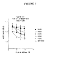
図1:IGF−1Rキナーゼ阻害剤である化合物66に対するNSCLC細胞株の感受性。
図2:IGF−1受容体阻害に対して感受性のNSCLC株は、E−カドヘリンの上昇したレベルを発現し、γ−カテニン及びα−カテニンに対して観察された傾向を有する。2つの異なる抗体を用いて、E−カドヘリンイムノブロットを行い、同様の結果を得た(データは示さず。)。化合物66による増殖阻害に対して比較的非感受性のNSCLC株は、間葉タンパク質であるビメンチン及び/又はフィブロネクチンを発現した。
図3:NSCLC株は、約500mm3の体積になるまで、SCIDマウス中での皮下異種移植片として増殖され、切り出され、液体窒素中で瞬間凍結された(4匹の動物/細胞株)。凍結された状態で、腫瘍組織を粉末化し、記載されているように、界面活性剤による溶解及びSDS−PAGEに供し、E−カドヘリン、γ−カテニン、Brk、フィブロネクチン、ビメンチン及びGAPDHに対する抗体で探索するイムノブロットに供した。インビトロでの結果と合致し、E−カドヘリンの発現は、化合物66感受性株に限定され、フィブロネクチンは相対的に非感受性株に限定されていた。
図4:IGF−1Rキナーゼ阻害に対して最も感受性であるNSCLC細胞株中でより高いBrk発現レベルを示すイムノブロット。
BRIEF DESCRIPTION OF THE FIGURES FIG. 1: Sensitivity of NSCLC cell line to compound 66, an IGF-1R kinase inhibitor.
FIG. 2: NSCLC strains that are sensitive to IGF-1 receptor inhibition express elevated levels of E-cadherin and have a tendency observed for γ-catenin and α-catenin. E-cadherin immunoblot was performed using two different antibodies and similar results were obtained (data not shown). NSCLC strains that are relatively insensitive to growth inhibition by
FIG. 3: NSCLC strains were grown as subcutaneous xenografts in SCID mice to a volume of approximately 500 mm 3 , excised and snap frozen in liquid nitrogen (4 animals / cell lines). In the frozen state, the tumor tissue is powdered and subjected to detergent lysis and SDS-PAGE as described and probed with antibodies to E-cadherin, γ-catenin, Brk, fibronectin, vimentin and GAPDH The sample was subjected to immunoblotting. Consistent with in vitro results, E-cadherin expression was restricted to the
FIG. 4: Immunoblot showing higher Brk expression levels in NSCLC cell lines that are most sensitive to IGF-1R kinase inhibition.
動物における「癌」という用語は、抑制されない増殖、不死、転移能、急速な成長及び増殖速度、ある種の特徴的な形態など、発癌細胞の典型的な特性を有する細胞の存在を指す。癌細胞は、腫瘍の形態であることが多いが、かかる細胞は、動物内に単独で存在し得、又は白血病細胞などの独立細胞として血流中を循環し得る。 The term “cancer” in an animal refers to the presence of cells having the typical characteristics of carcinogenic cells, such as uncontrolled proliferation, immortality, metastatic potential, rapid growth and proliferation rate, certain characteristic morphology. Cancer cells are often in the form of tumors, but such cells can exist alone in an animal or can circulate in the bloodstream as independent cells such as leukemia cells.
本明細書では「異常な細胞増殖」とは、別段の記載がないかぎり、正常な調節機構とは無関係である細胞増殖(例えば、接触阻止の喪失)を指す。「異常な細胞増殖」としては、(1)変異チロシンキナーゼの発現又は受容体チロシンキナーゼの過剰発現によって増殖する腫瘍細胞(腫瘍)、(2)異常なチロシンキナーゼ活性化が起こる他の増殖性疾患の良性及び悪性細胞、(4)受容体チロシンキナーゼによって増殖するあらゆる腫瘍、(5)異常なセリン/トレオニンキナーゼ活性化によって増殖するあらゆる腫瘍並びに(6)異常なセリン/トレオニンキナーゼ活性化が起こる他の増殖性疾患の良性及び悪性細胞、の異常な増殖などが挙げられる。 As used herein, “abnormal cell growth” refers to cell growth (eg, loss of contact inhibition) that is independent of normal regulatory mechanisms, unless otherwise noted. “Abnormal cell growth” includes (1) tumor cells (tumors) that proliferate due to expression of mutant tyrosine kinases or overexpression of receptor tyrosine kinases, and (2) other proliferative diseases in which abnormal tyrosine kinase activation occurs. Benign and malignant cells, (4) any tumor that grows by receptor tyrosine kinase, (5) any tumor that grows by abnormal serine / threonine kinase activation, and (6) others that cause abnormal serine / threonine kinase activation Benign and malignant cells of proliferative diseases, and the like.
本明細書では「治療する」という用語は、別段の記載がないかぎり、患者における腫瘍、腫瘍転移又は他の発癌細胞若しくは新生細胞の進行の逆転、緩和、阻害、或いは腫瘍、腫瘍転移又は他の発癌細胞若しくは新生細胞の増殖の部分的又は完全な抑制を意味する。本明細書では「治療」という用語は、別段の記載がないかぎり、治療行為を指す。 As used herein, the term “treating” refers to reversal, mitigation, inhibition of tumor, tumor metastasis or other carcinogenic or neoplastic cell progression in a patient, or tumor, tumor metastasis or other, unless otherwise stated. By partial or complete inhibition of the growth of carcinogenic or neoplastic cells. As used herein, the term “treatment” refers to a therapeutic act, unless stated otherwise.
「治療方法」という句又はその等価な句は、例えば、癌に適用するときには、動物における癌細胞数を減少させる、若しくはゼロにするように設計された、又は癌の症候を軽減するように設計された手順若しくは方針を指す。癌又は別の増殖性障害の「治療方法」は、癌細胞若しくは他の障害が実際に除去されること、細胞数若しくは障害が実際に抑制されること、又は癌若しくは他の障害の症候が実際に軽減することを必ずしも意味しない。癌を治療する方法は、成功の可能性が低い場合でも実施され、動物の病歴及び推定余命を考慮して、それでも全体として有益な方針と考えられるものであることが多い。 The phrase “therapeutic method” or its equivalent phrase is designed to reduce or eliminate the number of cancer cells in an animal or to reduce the symptoms of cancer, for example when applied to cancer. Refers to an established procedure or policy. A “treatment method” for cancer or another proliferative disorder is that the cancer cell or other disorder is actually removed, the cell number or disorder is actually suppressed, or the symptoms of the cancer or other disorder are actually It does not necessarily mean to reduce it. Methods of treating cancer are often performed even when the likelihood of success is low, and are often considered overall beneficial policies, considering the animal's medical history and life expectancy.
「治療有効薬剤」という用語は、研究者、獣医師、医師又は他の臨床家によって求められる、組織、系、動物又はヒトの生物学的又は医学的応答を誘発する組成物を意味する。 The term “therapeutically effective agent” refers to a composition that elicits a biological, medical response of a tissue, system, animal or human that is sought by a researcher, veterinarian, physician or other clinician.
「治療有効量」又は「有効量」という用語は、研究者、獣医師、医師又は他の臨床家によって求められる、組織、系、動物又はヒトの生物学的又は医学的応答を誘発する対象化合物又は組合せの量を意味する。 The term “therapeutically effective amount” or “effective amount” refers to a compound of interest that elicits a biological, medical response of a tissue, system, animal or human that is sought by a researcher, veterinarian, physician or other clinician. Or the quantity of a combination is meant.
以下の本明細書の実施例に示すデータによれば、細胞培養又はインビボで増殖されたNSCLC細胞などの腫瘍細胞は、上皮間葉遷移(EMT)を起こしたかどうかによって、IGF−1Rキナーゼ阻害剤による阻害に対してある範囲の感受性を示す。EMT前には、腫瘍細胞は、化合物66(強力な(約50nMのIC50)且つIGF−1R選択的な低分子量IGF−1Rキナーゼ阻害剤)などのIGF−1Rキナーゼ阻害剤による阻害に対して感受性が極めて高いが、EMTを起こした腫瘍細胞は、かかる化合物による阻害に対する感受性がかなり低い。このデータは、EMTが、IGF−1Rキナーゼ阻害剤に対する腫瘍の感受性レベルを決定する「全般的な生物学的スイッチ」であり得ることを示している。EMT現象の前に、又はそれに続いて、腫瘍細胞によって発現される、細胞に特徴的であるバイオマーカーのレベルを測定することによって、IGF−1Rキナーゼ阻害剤に対する腫瘍の感受性レベルを評価できることが実証された。例えば、E−カドヘリンなどの上皮バイオマーカーの腫瘍細胞発現レベルが高いことは、EMTをまだ起こしていない細胞を示し、IGF−1Rキナーゼ阻害剤に対する高い感受性と相関する。逆に、ビメンチン又はフィブロネクチンなどの間葉バイオマーカーの腫瘍細胞発現レベルが高いことは、EMTを起こした細胞を示し、IGF−1Rキナーゼ阻害剤に対する低い感受性と相関する。さらに、腫瘍細胞の上皮又は間葉状態及びそれに続いて起こる、IGF−1Rキナーゼ阻害剤による阻害に対する感受性に関する情報を提供するために、形態計測細胞分析を使用することが可能である。したがって、これらの知見は、腫瘍増殖に対するIGF−1Rキナーゼ阻害剤の効果を予測する貴重な新しい診断法の基礎を形成し得るものであり、患者に対して最も適切な治療を選択するのに役立つ追加のツールを腫瘍専門医に提供することができる。 According to the data presented in the examples herein below, tumor cells such as NSCLC cells grown in cell culture or in vivo, depending on whether they have undergone epithelial-mesenchymal transition (EMT), IGF-1R kinase inhibitors A range of sensitivity to inhibition by. Prior to EMT, the tumor cells are against inhibition by IGF-1R kinase inhibitors such as Compound 66 (a potent (about 50 nM IC 50 ) and IGF-1R selective low molecular weight IGF-1R kinase inhibitor). Although highly sensitive, tumor cells that have undergone EMT are much less sensitive to inhibition by such compounds. This data indicates that EMT may be a “general biological switch” that determines tumor sensitivity levels to IGF-1R kinase inhibitors. Demonstration that the level of tumor sensitivity to an IGF-1R kinase inhibitor can be assessed by measuring the level of cell-specific biomarkers expressed by tumor cells prior to or following the EMT event It was done. For example, high tumor cell expression levels of epithelial biomarkers such as E-cadherin indicate cells that have not yet undergone EMT and correlate with high sensitivity to IGF-1R kinase inhibitors. Conversely, high tumor cell expression levels of mesenchymal biomarkers such as vimentin or fibronectin indicate cells that have undergone EMT and correlate with low sensitivity to IGF-1R kinase inhibitors. In addition, morphometric cell analysis can be used to provide information about the epithelial or mesenchymal state of tumor cells and the subsequent sensitivity to inhibition by IGF-1R kinase inhibitors. Therefore, these findings can form the basis of a valuable new diagnostic method that predicts the effects of IGF-1R kinase inhibitors on tumor growth and help to select the most appropriate treatment for the patient Additional tools can be provided to oncologists.
したがって、本発明は、腫瘍細胞によって発現される上皮バイオマーカーのレベルを評価すること、及びIGF−1Rキナーゼ阻害剤による阻害に対する腫瘍細胞増殖の感受性を予測することを含み、腫瘍細胞上皮バイオマーカーの高い発現レベルはIGF−1Rキナーゼ阻害剤による阻害に対する高い感受性と相関する、IGF−1Rキナーゼ阻害剤による阻害に対する腫瘍細胞増殖の感受性を予測する方法を提供する。上皮バイオマーカーの好ましい例としてはE−カドヘリン及びBrk(すなわちPTK−6)が挙げられる(表1参照)。本発明による方法に利用することができる上皮バイオマーカーの追加の例としては、γ−カテニン(すなわち、ジャンクションプラコグロビン)、α−カテニン(すなわちα1、α2又はα3カテニン)、ケラチン8及びケラチン18(表1参照)が挙げられる。 Thus, the present invention includes assessing the level of epithelial biomarkers expressed by tumor cells and predicting the sensitivity of tumor cell proliferation to inhibition by IGF-1R kinase inhibitors, A high expression level provides a way to predict the sensitivity of tumor cell growth to inhibition by an IGF-1R kinase inhibitor, which correlates with high sensitivity to inhibition by an IGF-1R kinase inhibitor. Preferred examples of epithelial biomarkers include E-cadherin and Brk (ie PTK-6) (see Table 1). Additional examples of epithelial biomarkers that can be utilized in the method according to the invention include γ-catenin (ie, junction placoglobin), α-catenin (ie, α1, α2, or α3 catenin), keratin 8 and keratin 18 ( Table 1).
本発明は、腫瘍細胞によって発現される間葉バイオマーカーのレベルを評価すること、及びIGF−1Rキナーゼ阻害剤による阻害に対する腫瘍細胞増殖の感受性を予測することを含み、腫瘍細胞間葉バイオマーカーの高い発現レベルはIGF−1Rキナーゼ阻害剤による阻害に対する低い感受性と相関する、IGF−1Rキナーゼ阻害剤による阻害に対する腫瘍細胞増殖の感受性を予測する方法も提供する。間葉バイオマーカーの好ましい例としてはビメンチン及びフィブロネクチンが挙げられる(表1参照)。本発明による方法に利用することができる間葉バイオマーカーの追加の例としては、フィブリリン−1、フィブリリン−2、コラーゲンアルファ−2(IV)、コラーゲンアルファ−2(V)、LOXL1、ニドジェン、C11orf9、テネイシン、N−カドヘリン及び胚性EDB+フィブロネクチンが挙げられる(表1参照)。 The present invention includes assessing the level of mesenchymal biomarkers expressed by tumor cells and predicting the sensitivity of tumor cell proliferation to inhibition by IGF-1R kinase inhibitors, comprising the tumor cell mesenchymal biomarkers High expression levels also correlate with low sensitivity to inhibition by IGF-1R kinase inhibitors, also providing a method for predicting tumor cell growth sensitivity to inhibition by IGF-1R kinase inhibitors. Preferred examples of mesenchymal biomarkers include vimentin and fibronectin (see Table 1). Additional examples of mesenchymal biomarkers that can be utilized in the method according to the invention include fibrillin-1, fibrillin-2, collagen alpha-2 (IV), collagen alpha-2 (V), LOXL1, nidogen, C11orf9 , Tenascin, N-cadherin and embryonic EDB + fibronectin (see Table 1).
本発明の実施においては、好ましい上皮バイオマーカーを用いると、IGF−1Rキナーゼ阻害剤に対して感受性が高い腫瘍細胞における発現レベルは、一般に、バイオマーカーを、例えば特異的抗バイオマーカー抗体を検出に用いて、極めて容易に検出することができるような高いレベルである。好ましい上皮バイオマーカーを用いると、IGF−1Rキナーゼ阻害剤に対して感受性の比較的低い腫瘍細胞における発現レベルは、一般に、バイオマーカーが、類似の手順を用いて、検出されたとしてもわずかであるような低いレベルである(例えば、以下の本明細書の実施例に記載のデータにおいて、図2及び3中の感受性の高い腫瘍細胞と感受性の比較的低い腫瘍細胞のE−カドヘリンレベルを比較されたい。)。 In the practice of the present invention, using preferred epithelial biomarkers, the level of expression in tumor cells that are highly sensitive to IGF-1R kinase inhibitors is generally detectable in biomarkers, eg, specific anti-biomarker antibodies. It is at such a high level that it can be detected very easily. With preferred epithelial biomarkers, expression levels in tumor cells that are relatively insensitive to IGF-1R kinase inhibitors are generally low even if the biomarkers are detected using similar procedures (Eg, in the data described in the Examples herein below, the E-cadherin levels of the sensitive and relatively less sensitive tumor cells in FIGS. 2 and 3 were compared). I want.)
しかし、他のさほど好ましくない上皮バイオマーカーの場合には、IGF−1Rキナーゼ阻害剤に対して感受性の比較的低い腫瘍細胞におけるバイオマーカー発現レベルは、容易に検出可能であり得るが、それでも、IGF−1Rキナーゼ阻害剤に対して感受性が高い腫瘍細胞よりもかなり低い発現レベルである(例えば、以下の本明細書の実施例に記載のデータにおいて、図2中の感受性の比較的低い腫瘍細胞SW1573と感受性の高い腫瘍細胞H441、H358及びH292のα−カテニンレベルを比較されたい。)。 However, in the case of other less preferred epithelial biomarkers, biomarker expression levels in tumor cells that are relatively less sensitive to IGF-1R kinase inhibitors may be readily detectable, yet IGF Expression levels that are significantly lower than tumor cells that are more sensitive to -1R kinase inhibitors (eg, in the data described in the Examples herein below, the less sensitive tumor cells SW1573 in FIG. 2). Compare the α-catenin levels of the sensitive tumor cells H441, H358 and H292.)
同様に、本発明の実施においては、好ましい間葉バイオマーカーを用いると、IGF−1Rキナーゼ阻害剤に対して感受性の比較的低い腫瘍細胞における発現レベルは、一般に、バイオマーカーを、例えば特異的抗バイオマーカー抗体を検出に用いて、極めて容易に検出することができるような高いレベルである。好ましい間葉バイオマーカーを用いると、IGF−1Rキナーゼ阻害剤に対して感受性の比較的高い腫瘍細胞における発現レベルは、一般に、バイオマーカーが、類似の手順を用いて、検出されたとしてもわずかであるような低いレベルである(例えば、以下の本明細書の実施例に記載のデータにおいて、図2及び3中の感受性の高い腫瘍細胞と感受性の比較的低い腫瘍細胞のフィブロネクチン又はビメンチンレベルを比較されたい。)。 Similarly, in the practice of the present invention, using preferred mesenchymal biomarkers, the level of expression in tumor cells that are relatively insensitive to IGF-1R kinase inhibitors generally results in biomarkers, eg, specific anti-cancer agents. The biomarker antibody is used for detection at such a high level that it can be detected very easily. With the preferred mesenchymal biomarker, the expression level in tumor cells that are relatively sensitive to IGF-1R kinase inhibitors is generally low even if the biomarker is detected using a similar procedure. Comparing fibronectin or vimentin levels of sensitive and relatively less sensitive tumor cells in FIGS. 2 and 3 in the data described in the Examples herein below, such as some low levels I want to be.)
また、他のさほど好ましくない間葉バイオマーカーの場合には、IGF−1Rキナーゼ阻害剤に対して感受性の比較的高い腫瘍細胞におけるバイオマーカー発現レベルは、容易に検出可能であり得るが、それでも、IGF−1Rキナーゼ阻害剤に対して感受性の比較的低い腫瘍細胞よりもかなり低い発現レベルである。 Also, in the case of other less preferred mesenchymal biomarkers, biomarker expression levels in tumor cells that are relatively sensitive to IGF-1R kinase inhibitors may be readily detectable, yet The expression level is much lower than tumor cells that are less sensitive to IGF-1R kinase inhibitors.
任意の所与の上皮又は間葉バイオマーカーの場合、IGF−1Rキナーゼ阻害剤に対して感受性の比較的低い腫瘍細胞と感受性の高い腫瘍細胞の発現レベルの範囲は、例えば、本明細書に記載されている腫瘍細胞パネル上で試験することによって(例えば、図2)、又は腫瘍がIGF−1Rキナーゼ阻害剤(例えば、化合物66)に対してある範囲の感受性を示す患者から得られた腫瘍生検の試験によって、当業者によって容易に評価することができる。 For any given epithelial or mesenchymal biomarker, the range of expression levels of relatively insensitive and sensitive tumor cells to an IGF-1R kinase inhibitor is described, for example, herein. Tumor life obtained by testing on a panel of tumor cells being treated (eg, FIG. 2) or from a patient whose tumor exhibits a range of sensitivity to an IGF-1R kinase inhibitor (eg, compound 66) Tests can be easily evaluated by those skilled in the art.
本発明に関連して、IGF−1Rキナーゼ阻害剤に対して感受性の比較的低い腫瘍細胞の割合が比較的小さい場合には、単一のバイオマーカーレベルのみを評価する状況において、腫瘍細胞によって発現される上皮又は間葉バイオマーカーのレベルを評価することを含む、IGF−1Rキナーゼ阻害剤による阻害に対する腫瘍細胞増殖の感受性を予測する上記方法は、腫瘍細胞増殖が、IGF−1Rキナーゼ阻害剤による阻害に対して感受性が高いことを誤って予測する恐れがある。例えば、以下の本明細書の実施例に記載されているデータにおいて、H460腫瘍細胞中の上皮バイオマーカーγ−カテニン及びα−カテニンのレベル、又はH460細胞中の間葉バイオマーカービメンチンのレベルは、IGF−1Rキナーゼ阻害剤に対する高い感受性を誤って予測する(図2参照)。したがって、かかる誤った予測に基づいて、医師は、IGF−1Rキナーゼ阻害剤を用いて少数の患者を治療する恐れがあり、腫瘍は、阻害剤に対して感受性を示さない恐れがある。しかし、腫瘍細胞の大多数(例えば、以下の本明細書の実施例に記載のデータから、少なくとも90%)では、単一のバイオマーカー発現レベルの評価によって、IGF−1Rキナーゼ阻害剤に対する感受性レベルが正確に予測されると期待される。 In the context of the present invention, expressed by tumor cells in the context of assessing only a single biomarker level if the proportion of tumor cells that are relatively insensitive to IGF-1R kinase inhibitors is relatively small Assessing the sensitivity of tumor cell growth to inhibition by an IGF-1R kinase inhibitor, comprising assessing the level of epithelial or mesenchymal biomarkers, wherein the tumor cell growth is caused by an IGF-1R kinase inhibitor There is a risk of mispredicting the high sensitivity to inhibition. For example, in the data described in the Examples herein below, the level of epithelial biomarkers γ-catenin and α-catenin in H460 tumor cells, or the level of mesenchymal biomarker vimentin in H460 cells is High sensitivity to 1R kinase inhibitor is predicted incorrectly (see FIG. 2). Thus, based on such false predictions, physicians may treat a small number of patients with an IGF-1R kinase inhibitor and the tumor may not be sensitive to the inhibitor. However, in the majority of tumor cells (eg, at least 90% from the data set forth in the Examples herein below), the level of sensitivity to an IGF-1R kinase inhibitor is assessed by assessment of a single biomarker expression level. Is expected to be accurately predicted.
また、本発明に関連して最も重要なことには、上記方法によって試験したとき(単一のバイオマーカーレベルのみを評価する場合)、腫瘍細胞増殖がIGF−1Rキナーゼ阻害剤による阻害に対して低感受性であるという誤った予測を与える、IGF−1Rキナーゼ阻害剤に対して感受性が高い腫瘍細胞は見出されなかった。したがって、本明細書に記載の試験方法を利用することによって、患者がIGF−1Rキナーゼ阻害剤による治療から利点を得ることができる場合に、医師がかかる治療を差し控えることはないはずである。 Also, most importantly in connection with the present invention, when tested by the above method (when assessing only a single biomarker level), tumor cell proliferation is against inhibition by an IGF-1R kinase inhibitor. No tumor cells were found that were highly sensitive to IGF-1R kinase inhibitors that gave the false prediction of being insensitive. Thus, if the patient can benefit from treatment with an IGF-1R kinase inhibitor by utilizing the test methods described herein, the physician should not withhold such treatment.
また、医薬分野の当業者は、特に診断テスト及び治療用物質による治療の適用に関して、生体系は幾らか可変であり、完全に予測可能では必ずしもなく、したがって多数の良好な診断テスト又は治療用物質が無効である場合もあることを理解している。したがって、試験結果、患者の症状及び病歴並びに主治医の経験に基づいて、個々の患者に最適な治療コースを決定するのは、最終的には主治医の判断による。例えば、診断テスト又は他の判定基準から得られるデータに基づいて、腫瘍がIGF−1Rキナーゼ阻害剤に対して特に感受性であると予測されないときでも、特に、他の明白な治療選択肢の全部若しくは大部分が失敗した場合、又は別の治療と併用したときにある相乗作用が予測される場合には、医師がIGF−1Rキナーゼ阻害剤を用いて患者を治療することを選択する場合さえある。IGF−1Rキナーゼ阻害剤は、化合物のクラスとして、癌治療に使用されるより伝統的な化学療法又は細胞毒性薬などの多数の他の抗癌薬よりも比較的耐容性が良好であることから、IGF−1Rキナーゼ阻害剤はより実行可能な選択肢になっている。 Also, those skilled in the pharmaceutical arts will recognize that biological systems are somewhat variable and not entirely predictable, particularly with respect to diagnostic tests and therapeutic applications with therapeutic substances, and thus many good diagnostic tests or therapeutic substances. I understand that can be invalid. Therefore, it is ultimately at the discretion of the attending physician to determine the optimal course of treatment for an individual patient based on the test results, the patient's symptoms and medical history, and the attending physician's experience. For example, even if the tumor is not predicted to be particularly sensitive to an IGF-1R kinase inhibitor based on data obtained from diagnostic tests or other criteria, all or large of other obvious treatment options, A physician may even choose to treat a patient with an IGF-1R kinase inhibitor if a portion fails or if some synergy is expected when combined with another treatment. Because IGF-1R kinase inhibitors are relatively well tolerated as a class of compounds compared to many other anticancer drugs such as more traditional chemotherapy or cytotoxic drugs used in cancer treatment IGF-1R kinase inhibitors have become a more viable option.
E−カドヘリンなどの本発明における使用に適切な上皮バイオマーカーの好ましい例は、上記方法に使用したときに(単一のバイオマーカーレベルのみを評価する場合)、誤った予測をもたらさない。 Preferred examples of epithelial biomarkers suitable for use in the present invention, such as E-cadherin, do not result in false predictions when used in the above methods (when evaluating only a single biomarker level).
また、本発明は、腫瘍細胞における1種類を超えるバイオマーカーレベルの発現レベルの同時評価を利用する追加の方法も提供する。(以下に記述する)これらの方法の好ましい実施形態において、単一のバイオマーカー発現レベルを評価する上記方法の幾つかの場合と同様に、誤った予測のレベルが存在しない。 The present invention also provides additional methods that utilize the simultaneous assessment of expression levels of more than one biomarker level in tumor cells. In preferred embodiments of these methods (described below), there are no false prediction levels, as in some of the above methods for assessing a single biomarker expression level.
したがって、本発明は、腫瘍細胞によって発現される1つ又はそれ以上の種(又は1パネルの)上皮バイオマーカーのレベルを評価すること、及びIGF−1Rキナーゼ阻害剤による阻害に対する腫瘍細胞増殖の感受性を予測することを含み、評価された腫瘍細胞上皮バイオマーカー全部の同時の高い発現レベルはIGF−1Rキナーゼ阻害剤による阻害に対する高い感受性と相関する、IGF−1Rキナーゼ阻害剤による阻害に対する腫瘍細胞増殖の感受性を予測する方法を提供する。この方法の好ましい一実施形態において、上皮バイオマーカーはE−カドヘリン及びBrkを含み、2種類の腫瘍細胞上皮バイオマーカーの同時の高い発現レベルは、IGF−1Rキナーゼ阻害剤による阻害に対する高い感受性と相関する。この方法の別の好ましい実施形態において、上皮バイオマーカーはE−カドヘリン及びγ−カテニンを含み、2種類の腫瘍細胞上皮バイオマーカーの同時の高い発現レベルは、IGF−1Rキナーゼ阻害剤による阻害に対する高い感受性と相関する。後者2つの好ましい実施形態において、両方のバイオマーカーの高い発現レベルが、高い感受性を示すのに必要であることに注意されたい。 Thus, the present invention evaluates the level of one or more species (or a panel) of epithelial biomarkers expressed by tumor cells, and the sensitivity of tumor cell growth to inhibition by IGF-1R kinase inhibitors High expression levels of all of the evaluated tumor cell epithelial biomarkers correlate with high sensitivity to inhibition by IGF-1R kinase inhibitors, tumor cell proliferation against inhibition by IGF-1R kinase inhibitors Provides a method for predicting the sensitivity of In a preferred embodiment of this method, the epithelial biomarker comprises E-cadherin and Brk, and the simultaneous high expression level of the two tumor cell epithelial biomarkers correlates with a high sensitivity to inhibition by an IGF-1R kinase inhibitor. To do. In another preferred embodiment of this method, the epithelial biomarker comprises E-cadherin and γ-catenin, the simultaneous high expression level of the two tumor cell epithelial biomarkers is high for inhibition by an IGF-1R kinase inhibitor Correlates with sensitivity. Note that in the latter two preferred embodiments, high expression levels of both biomarkers are necessary to indicate high sensitivity.
本発明は、腫瘍細胞によって発現される1つ又はそれ以上の(又は1パネルの)間葉バイオマーカーのレベルを評価すること、及びIGF−1Rキナーゼ阻害剤による阻害に対する腫瘍細胞増殖の感受性を予測することを含み、評価された腫瘍細胞間葉バイオマーカー全部の同時の低い又は検出不可能な発現レベルはIGF−1Rキナーゼ阻害剤による阻害に対する高い感受性と相関する、IGF−1Rキナーゼ阻害剤による阻害に対する腫瘍細胞増殖の感受性を予測する方法も提供する。この方法の好ましい一実施形態において、間葉バイオマーカーはビメンチン及びフィブロネクチンを含み、2種類の腫瘍細胞間葉バイオマーカーの同時の低い又は検出不可能な発現レベルは、IGF−1Rキナーゼ阻害剤による阻害に対する高い感受性と相関する。後半の好ましい実施形態において、両方のバイオマーカーの低い又は検出不可能な発現が、高い感受性を示すのに必要であることに注意されたい。 The present invention evaluates the level of one or more (or a panel) of mesenchymal biomarkers expressed by tumor cells and predicts the sensitivity of tumor cell growth to inhibition by IGF-1R kinase inhibitors Inhibition by an IGF-1R kinase inhibitor, wherein a simultaneous low or undetectable expression level of all of the evaluated tumor cell mesenchymal biomarkers correlates with a high sensitivity to inhibition by the IGF-1R kinase inhibitor Also provided are methods for predicting the sensitivity of tumor cell growth to. In a preferred embodiment of this method, the mesenchymal biomarker comprises vimentin and fibronectin and the simultaneous low or undetectable expression level of the two tumor cell mesenchymal biomarkers is inhibited by an IGF-1R kinase inhibitor Correlates with high susceptibility to Note that in the latter preferred embodiment, low or undetectable expression of both biomarkers is necessary to indicate high sensitivity.
本発明は、腫瘍細胞によって発現される上皮バイオマーカーのレベルを評価すること、腫瘍細胞によって発現される間葉バイオマーカーのレベルを評価すること、及びIGF−1Rキナーゼ阻害剤による阻害に対する腫瘍細胞増殖の感受性を予測することを含み、上皮バイオマーカー発現レベルと間葉バイオマーカー発現レベルとの高い比はIGF−1Rキナーゼ阻害剤による阻害に対する高い感受性と相関する、IGF−1Rキナーゼ阻害剤による阻害に対する腫瘍細胞増殖の感受性を予測する方法も提供する。この方法の好ましい一実施形態において、上皮バイオマーカーはE−カドヘリンを含み、間葉バイオマーカーはフィブロネクチンを含む。この方法の別の好ましい実施形態において、上皮バイオマーカーはBrkを含み、間葉バイオマーカーはフィブロネクチンを含む。この方法の別の好ましい実施形態において、上皮バイオマーカーはE−カドヘリンを含み、間葉バイオマーカーはビメンチンを含む。この方法の別の好ましい実施形態において、上皮バイオマーカーはγ−カテニンを含み、間葉バイオマーカーはフィブロネクチンを含む。 The present invention assesses the level of epithelial biomarkers expressed by tumor cells, assesses the level of mesenchymal biomarkers expressed by tumor cells, and tumor cell proliferation against inhibition by IGF-1R kinase inhibitors High ratio of epithelial biomarker expression level to mesenchymal biomarker expression level correlates with high sensitivity to inhibition by IGF-1R kinase inhibitor, against inhibition by IGF-1R kinase inhibitor Also provided are methods for predicting the sensitivity of tumor cell growth. In a preferred embodiment of this method, the epithelial biomarker comprises E-cadherin and the mesenchymal biomarker comprises fibronectin. In another preferred embodiment of this method, the epithelial biomarker comprises Brk and the mesenchymal biomarker comprises fibronectin. In another preferred embodiment of this method, the epithelial biomarker comprises E-cadherin and the mesenchymal biomarker comprises vimentin. In another preferred embodiment of this method, the epithelial biomarker comprises γ-catenin and the mesenchymal biomarker comprises fibronectin.
本発明は、腫瘍細胞によって発現される1種類又はそれ以上の(又はパネルの)上皮バイオマーカーのレベルを評価すること、及びIGF−1Rキナーゼ阻害剤による阻害に対する腫瘍増殖の感受性を予測することを含み、評価された腫瘍細胞上皮バイオマーカー全部の同時の高い発現レベルはIGF−1Rキナーゼ阻害剤による阻害に対する高い感受性と相関する、IGF−1Rキナーゼ阻害剤による阻害に対する腫瘍増殖の感受性を予測する方法も提供する。この方法の好ましい一実施形態において、上皮バイオマーカーはE−カドヘリン及びBrkを含み、2種類の腫瘍細胞上皮バイオマーカーの同時の高い発現レベルは、IGF−1Rキナーゼ阻害剤による阻害に対する高い感受性と相関する。この方法の別の好ましい実施形態において、上皮バイオマーカーはE−カドヘリン及びγ−カテニンを含み、2種類の腫瘍細胞上皮バイオマーカーの同時の高い発現レベルは、IGF−1Rキナーゼ阻害剤による阻害に対する高い感受性と相関する。後半の2つの好ましい実施形態において、両方のバイオマーカーの高い発現レベルが、高い感受性を示すのに必要であることに注意されたい。 The present invention assesses the level of one or more (or panel) epithelial biomarkers expressed by tumor cells and predicts the sensitivity of tumor growth to inhibition by IGF-1R kinase inhibitors. A method of predicting tumor growth susceptibility to inhibition by an IGF-1R kinase inhibitor, wherein simultaneous high expression levels of all tumor cell epithelial biomarkers, including and correlated, correlate with high sensitivity to inhibition by an IGF-1R kinase inhibitor Also provide. In a preferred embodiment of this method, the epithelial biomarker comprises E-cadherin and Brk, and the simultaneous high expression level of the two tumor cell epithelial biomarkers correlates with a high sensitivity to inhibition by an IGF-1R kinase inhibitor. To do. In another preferred embodiment of this method, the epithelial biomarker comprises E-cadherin and γ-catenin, the simultaneous high expression level of the two tumor cell epithelial biomarkers is high for inhibition by an IGF-1R kinase inhibitor Correlates with sensitivity. Note that in the latter two preferred embodiments, high expression levels of both biomarkers are necessary to indicate high sensitivity.
本発明は、腫瘍細胞によって発現される1種類又はそれ以上の(又はパネルの)間葉バイオマーカーのレベルを評価すること、及びIGF−1Rキナーゼ阻害剤による阻害に対する腫瘍増殖の感受性を予測することを含み、評価された腫瘍細胞間葉バイオマーカー全部の同時の低い又は検出不可能な発現レベルはIGF−1Rキナーゼ阻害剤による阻害に対する高い感受性と相関する、IGF−1Rキナーゼ阻害剤による阻害に対する腫瘍増殖の感受性を予測する方法も提供する。この方法の好ましい一実施形態において、間葉バイオマーカーはビメンチン及びフィブロネクチンを含み、2種類の腫瘍細胞間葉バイオマーカーの同時の低い又は検出不可能な発現レベルは、IGF−1Rキナーゼ阻害剤による阻害に対する高い感受性と相関する。後半の好ましい実施形態において、両方のバイオマーカーの低い又は検出不可能な発現レベルが、高い感受性を示すのに必要であることに注意されたい。 The present invention evaluates the level of one or more (or panel of) mesenchymal biomarkers expressed by tumor cells and predicts the sensitivity of tumor growth to inhibition by IGF-1R kinase inhibitors A tumor against inhibition by an IGF-1R kinase inhibitor, wherein a simultaneous low or undetectable expression level of all of the evaluated tumor cell mesenchymal biomarkers correlates with a high sensitivity to inhibition by the IGF-1R kinase inhibitor Also provided is a method of predicting growth susceptibility. In a preferred embodiment of this method, the mesenchymal biomarker comprises vimentin and fibronectin and the simultaneous low or undetectable expression level of the two tumor cell mesenchymal biomarkers is inhibited by an IGF-1R kinase inhibitor Correlates with high susceptibility to Note that in the latter preferred embodiment, low or undetectable expression levels of both biomarkers are necessary to indicate high sensitivity.
本発明は、腫瘍細胞によって発現される上皮バイオマーカーのレベルを評価すること、腫瘍細胞によって発現される間葉バイオマーカーのレベルを評価すること、及びIGF−1Rキナーゼ阻害剤による阻害に対する腫瘍増殖の感受性を予測することを含み、上皮バイオマーカー発現レベルと間葉バイオマーカー発現レベルとの高い比はIGF−1Rキナーゼ阻害剤による阻害に対する高い感受性と相関する、IGF−1Rキナーゼ阻害剤による阻害に対する腫瘍増殖の感受性を予測する方法も提供する。この方法の好ましい一実施形態において、上皮バイオマーカーはE−カドヘリンを含み、間葉バイオマーカーはフィブロネクチンを含む。この方法の別の好ましい実施形態においては、上皮バイオマーカーはBrkを含み、間葉バイオマーカーはフィブロネクチンを含む。この方法の別の好ましい実施形態において、上皮バイオマーカーはE−カドヘリンを含み、間葉バイオマーカーはビメンチンを含む。この方法の別の好ましい実施形態において、上皮バイオマーカーはγ−カテニンを含み、間葉バイオマーカーはフィブロネクチンを含む。 The present invention assesses the level of epithelial biomarkers expressed by tumor cells, evaluates the level of mesenchymal biomarkers expressed by tumor cells, and tumor growth against inhibition by IGF-1R kinase inhibitors Tumors for inhibition by IGF-1R kinase inhibitors, including predicting sensitivity, wherein a high ratio of epithelial biomarker expression level to mesenchymal biomarker expression level correlates with high sensitivity to inhibition by IGF-1R kinase inhibitor Also provided is a method of predicting growth susceptibility. In a preferred embodiment of this method, the epithelial biomarker comprises E-cadherin and the mesenchymal biomarker comprises fibronectin. In another preferred embodiment of this method, the epithelial biomarker comprises Brk and the mesenchymal biomarker comprises fibronectin. In another preferred embodiment of this method, the epithelial biomarker comprises E-cadherin and the mesenchymal biomarker comprises vimentin. In another preferred embodiment of this method, the epithelial biomarker comprises γ-catenin and the mesenchymal biomarker comprises fibronectin.
本発明は、腫瘍細胞によって発現される1種類又はそれ以上の(又はパネルの)上皮バイオマーカーのレベルを評価すること、及び腫瘍がIGF−1Rキナーゼ阻害剤による治療に有効に応答するかどうかを予測することを含み、腫瘍細胞上皮バイオマーカー全部の同時の高い発現レベルはIGF−1Rキナーゼ阻害剤による治療に有効に応答する腫瘍と相関する、癌患者がIGF−1Rキナーゼ阻害剤による治療に有効に応答する腫瘍に罹患しているかどうかを予測する方法も提供する。この方法の好ましい一実施形態において、上皮バイオマーカーはE−カドヘリン及びBrkを含み、2種類の腫瘍細胞上皮バイオマーカーの同時の高い発現レベルは、IGF−1Rキナーゼ阻害剤による治療に有効に応答する腫瘍と相関する。この方法の別の好ましい実施形態において、上皮バイオマーカーはE−カドヘリン及びγ−カテニンを含み、2種類の腫瘍細胞上皮バイオマーカーの同時の高い発現レベルは、IGF−1Rキナーゼ阻害剤による治療に有効に応答する腫瘍と相関する。後半の2つの好ましい実施形態において、両方のバイオマーカーの高い発現レベルが、IGF−1Rキナーゼ阻害剤による治療に有効に応答する腫瘍を示すのに必要であることに注意されたい。 The present invention evaluates the level of one or more (or panel) epithelial biomarkers expressed by tumor cells and whether the tumor responds effectively to treatment with an IGF-1R kinase inhibitor. Predicting that simultaneous high expression levels of all tumor cell epithelial biomarkers correlate with tumors that respond effectively to treatment with IGF-1R kinase inhibitors, cancer patients are effective for treatment with IGF-1R kinase inhibitors Also provided is a method for predicting whether or not a tumor responds to. In a preferred embodiment of this method, the epithelial biomarker comprises E-cadherin and Brk, and the simultaneous high expression level of the two tumor cell epithelial biomarkers is effectively responsive to treatment with an IGF-1R kinase inhibitor Correlate with tumor. In another preferred embodiment of this method, the epithelial biomarker comprises E-cadherin and γ-catenin and the simultaneous high expression level of the two tumor cell epithelial biomarkers is effective for treatment with an IGF-1R kinase inhibitor Correlates with tumors that respond to Note that in the latter two preferred embodiments, high expression levels of both biomarkers are necessary to indicate tumors that respond effectively to treatment with an IGF-1R kinase inhibitor.
本発明は、腫瘍細胞によって発現される1種類又はそれ以上の(又はパネルの)間葉バイオマーカーのレベルを評価すること、及び腫瘍がIGF−1Rキナーゼ阻害剤による治療に有効に応答するかどうかを予測することを含み、腫瘍細胞間葉バイオマーカー全部の同時の低い又は検出不可能な発現レベルはIGF−1Rキナーゼ阻害剤による治療に有効に応答する腫瘍と相関する、癌患者がIGF−1Rキナーゼ阻害剤による治療に有効に応答する腫瘍に罹患しているかどうかを予測する方法も提供する。この方法の好ましい一実施形態において、間葉バイオマーカーはビメンチン及びフィブロネクチンを含み、2種類の腫瘍細胞間葉バイオマーカーの同時の低い又は検出不可能な発現レベルは、IGF−1Rキナーゼ阻害剤による治療に有効に応答する腫瘍と相関する。後半の好ましい実施形態において、両方のバイオマーカーの低い又は検出不可能な発現が、IGF−1Rキナーゼ阻害剤による治療に有効に応答する腫瘍を示すのに必要であることに注意されたい。 The present invention evaluates the level of one or more (or panel of) mesenchymal biomarkers expressed by tumor cells and whether the tumor responds effectively to treatment with an IGF-1R kinase inhibitor Cancer patients correlate with IGF-1R, wherein simultaneous low or undetectable expression levels of all tumor cell mesenchymal biomarkers correlate with tumors that respond effectively to treatment with an IGF-1R kinase inhibitor Also provided are methods for predicting whether a tumor is responsive to treatment with a kinase inhibitor. In one preferred embodiment of this method, the mesenchymal biomarker comprises vimentin and fibronectin and the simultaneous low or undetectable expression level of the two tumor cell mesenchymal biomarkers is treated with an IGF-1R kinase inhibitor Correlate with tumors that respond effectively to Note that in the latter preferred embodiment, low or undetectable expression of both biomarkers is necessary to indicate tumors that respond effectively to treatment with an IGF-1R kinase inhibitor.
本発明は、腫瘍細胞によって発現される上皮バイオマーカーのレベルを評価すること、腫瘍細胞によって発現される間葉バイオマーカーのレベルを評価すること、及び腫瘍がIGF−1Rキナーゼ阻害剤による治療に有効に応答するかどうかを予測することを含み、上皮バイオマーカー発現レベルと間葉バイオマーカー発現レベルとの高い比はIGF−1Rキナーゼ阻害剤による治療に有効に応答する腫瘍と相関する、癌患者がIGF−1Rキナーゼ阻害剤による治療に有効に応答する腫瘍に罹患しているかどうかを予測する方法も提供する。この方法の好ましい一実施形態において、上皮バイオマーカーはE−カドヘリンを含み、間葉バイオマーカーはフィブロネクチンを含む。この方法の別の好ましい実施形態において、上皮バイオマーカーはBrkを含み、間葉バイオマーカーはフィブロネクチンを含む。この方法の別の好ましい実施形態において、上皮バイオマーカーはE−カドヘリンを含み、間葉バイオマーカーはビメンチンを含む。この方法の別の好ましい実施形態において、上皮バイオマーカーはγ−カテニンを含み、間葉バイオマーカーはフィブロネクチンを含む。 The present invention evaluates the level of epithelial biomarkers expressed by tumor cells, evaluates the levels of mesenchymal biomarkers expressed by tumor cells, and treats tumors with IGF-1R kinase inhibitors A high ratio of epithelial biomarker expression level to mesenchymal biomarker expression level correlates with a tumor that responds effectively to treatment with an IGF-1R kinase inhibitor. Also provided are methods for predicting whether a tumor is effectively responsive to treatment with an IGF-1R kinase inhibitor. In a preferred embodiment of this method, the epithelial biomarker comprises E-cadherin and the mesenchymal biomarker comprises fibronectin. In another preferred embodiment of this method, the epithelial biomarker comprises Brk and the mesenchymal biomarker comprises fibronectin. In another preferred embodiment of this method, the epithelial biomarker comprises E-cadherin and the mesenchymal biomarker comprises vimentin. In another preferred embodiment of this method, the epithelial biomarker comprises γ-catenin and the mesenchymal biomarker comprises fibronectin.
本発明の方法に関連して、腫瘍細胞によって発現されるバイオマーカーは、腫瘍細胞の遷移状態を示す分子マーカー及び細胞マーカーを含むことができる。好ましい実施形態において、バイオマーカーは、腫瘍の特定の遷移状態、すなわち上皮若しくは間葉特性を示す腫瘍に特徴的な個々のマーカータンパク質、又はそれをコードするmRNAである。別の実施形態において、ある種の状況において、バイオマーカーは、上皮状態又は間葉状態に特徴的である細胞の巨大分子によって、腫瘍細胞中に形成される特徴的な形態学的パターンであり得る。従って、腫瘍細胞の上皮又は間葉状態及びそれに続いて起こる、IGF−1Rキナーゼ阻害剤による阻害に対する感受性に関する情報を提供するために、形態計測的細胞分析を使用することが可能である。 In connection with the methods of the present invention, biomarkers expressed by tumor cells can include molecular markers and cell markers that indicate the transition state of the tumor cells. In a preferred embodiment, the biomarker is an individual marker protein characteristic of a tumor exhibiting a particular transition state of the tumor, i.e. epithelial or mesenchymal properties, or mRNA encoding it. In another embodiment, in certain circumstances, a biomarker can be a characteristic morphological pattern formed in a tumor cell by a macromolecule of cells that is characteristic of an epithelial or mesenchymal state . Thus, morphometric cell analysis can be used to provide information regarding the epithelial or mesenchymal status of tumor cells and the subsequent sensitivity to inhibition by IGF-1R kinase inhibitors.

表1に、本明細書に記載の本発明の方法の実施に使用することができる分子バイオマーカーの例をコードする遺伝子を列挙する。分子バイオマーカーは、これらの遺伝子の変異体、例えば、発現mRNA又はタンパク質、スプライスバリアント、同時翻訳及び翻訳後修飾タンパク質、多形バリアントなどを含む、これらの遺伝子によって発現される任意の生成物を含むことができる。一実施形態において、バイオマーカーは、フィブロネクチン1遺伝子によって発現されるスプライスバリアントである胚性EDB+フィブロネクチンである(Kilian, O. et al. (2004) Bone 35(6):1334−1345)。フィブロネクチンのこの胎性形態(fetal form)を決定する考えられる利点は、間葉様腫瘍を周囲の間質組織から容易に区別することができることである。追加の実施形態において、バイオマーカーは、ヒト遺伝子産物の動物相同体であり得る(例えば、イヌ、マウス、ラット、ウサギ、ネコ、サル、類人猿由来など)。 Table 1 lists genes that encode examples of molecular biomarkers that can be used to practice the methods of the invention described herein. Molecular biomarkers include any product expressed by these genes, including variants of these genes, including expressed mRNA or protein, splice variants, cotranslational and post-translationally modified proteins, polymorphic variants, etc. be able to. In one embodiment, the biomarker is embryonic EDB + fibronectin, a splice variant expressed by the fibronectin 1 gene (Kilian, O. et al. (2004) Bone 35 (6): 1334-1345). A possible advantage of determining this fetal form of fibronectin is that mesenchymal tumors can be easily distinguished from the surrounding stromal tissue. In additional embodiments, the biomarker can be an animal homologue of a human gene product (eg, from a dog, mouse, rat, rabbit, cat, monkey, ape, etc.).
本明細書に記載の方法において、腫瘍細胞は、典型的には、癌、前癌状態又は異常細胞増殖の別の形態と診断された患者、及び治療を要する患者から得られる。癌は、肺癌(例えば、非小細胞肺癌(NSCLC))、すい癌、頭頚部癌、胃癌、乳癌、結腸癌、卵巣癌、又は本明細書に記載の以下の種々の他の癌のいずれかであり得る。癌は、好ましくは、IGF−1Rキナーゼ阻害剤を用いて治療可能であることが知られている癌である。 In the methods described herein, tumor cells are typically obtained from patients diagnosed with cancer, a precancerous condition or another form of abnormal cell growth, and patients in need of treatment. The cancer can be lung cancer (eg, non-small cell lung cancer (NSCLC)), pancreatic cancer, head and neck cancer, gastric cancer, breast cancer, colon cancer, ovarian cancer, or any of the various other cancers described herein below. It can be. The cancer is preferably a cancer known to be treatable with an IGF-1R kinase inhibitor.
本発明の方法において、バイオマーカー発現レベルは、発現レベルがEMTを通して一定である対照分子と比較して評価することができ、又は分子バイオマーカー(例えば、GAPDH、β−アクチン、チューブリンなどの「ハウスキーピング」遺伝子)によって示される上皮若しくは間葉遷移状態を発現する腫瘍細胞を比較したときに評価することができる。バイオマーカー発現レベルは、腫瘍細胞バイオマーカーのもう一方のタイプと比較して(すなわち、間葉と比較された上皮)、又は同じ組織の非腫瘍細胞中のバイオマーカーレベルと比較して、又はアッセイ基準として使用される別の細胞若しくは組織源と比較して評価することもできる。 In the methods of the present invention, the biomarker expression level can be assessed relative to a control molecule where the expression level is constant throughout the EMT, or a molecular biomarker (eg, “such as GAPDH, β-actin, tubulin, etc.” It can be assessed when comparing tumor cells expressing epithelial or mesenchymal transition states indicated by "housekeeping" genes). Biomarker expression level is compared to another type of tumor cell biomarker (ie, epithelium compared to mesenchyme) or compared to biomarker levels in non-tumor cells of the same tissue or assay It can also be evaluated relative to another cell or tissue source used as a reference.
本発明の方法において、腫瘍細胞によって発現される上皮又は間葉バイオマーカーのレベルは、以下により詳細に記述するように、例えば、ELISA、RIA、免疫沈降、免疫ブロット、免疫蛍光顕微鏡検査、RT−PCR、in situハイブリド形成、cDNAマイクロアレイなどなど、遺伝子の発現レベルを測定するための、当分野では公知の標準的バイオアッセイ手順のいずれかを用いることによって評価することができる。 In the methods of the invention, the level of epithelial or mesenchymal biomarkers expressed by tumor cells can be measured, for example, by ELISA, RIA, immunoprecipitation, immunoblotting, immunofluorescence microscopy, RT-, as described in more detail below. It can be assessed by using any of the standard bioassay procedures known in the art for measuring gene expression levels, such as PCR, in situ hybridization, cDNA microarrays, and the like.
本発明の方法において、腫瘍細胞上皮又は間葉バイオマーカーの発現レベルは、好ましくは腫瘍生検をアッセイすることによって評価される。しかし、別の実施形態において、腫瘍細胞バイオマーカーの発現レベルは、腫瘍又は腫瘍細胞から生じるバイオマーカーの検出可能なレベルを含有する体液又は排せつ物において評価することができる。本発明において有用である体液又は排せつ物としては、血液、尿、唾液、便、胸膜液、リンパ液、痰、腹水、前立腺液、脳脊髄液(CSF)又は任意の他の体分泌物若しくはその誘導体などが挙げられる。血液とは、全血、血しょう、血清又は血液の任意の誘導体を含むものとする。かかる体液又は排せつ物中の腫瘍上皮又は間葉バイオマーカーの評価は、観血的な試料採取方法が不適当又は不都合である状況において好ましいことがある。 In the methods of the invention, the expression level of tumor cell epithelium or mesenchymal biomarker is preferably assessed by assaying a tumor biopsy. However, in another embodiment, the expression level of a tumor cell biomarker can be assessed in a body fluid or excrement containing a detectable level of the biomarker arising from the tumor or tumor cell. Body fluids or excrements useful in the present invention include blood, urine, saliva, stool, pleural fluid, lymph fluid, sputum, ascites, prostate fluid, cerebrospinal fluid (CSF) or any other body secretion or derivative thereof, etc. Is mentioned. Blood shall include whole blood, plasma, serum or any derivative of blood. Evaluation of tumor epithelium or mesenchymal biomarkers in such body fluids or excreta may be preferred in situations where open sampling methods are inappropriate or inconvenient.
本発明の方法において、腫瘍細胞は、肺癌腫瘍細胞(例えば、非小細胞肺癌(NSCLC))、すい癌腫瘍細胞、乳癌腫瘍細胞、頭頚部癌腫瘍細胞、胃癌腫瘍細胞、結腸癌腫瘍細胞、卵巣癌腫瘍細胞、又は本明細書に記載の以下の種々の他の癌のいずれかに由来する腫瘍細胞であり得る。腫瘍細胞は、好ましくは、固形腫瘍由来の全腫瘍細胞がそうであるように、IGF−1Rキナーゼを発現することが知られているタイプ又は予想されるタイプである。IGF−1Rキナーゼは、野生型でも変異体でもよい。 In the method of the present invention, the tumor cells are lung cancer tumor cells (eg, non-small cell lung cancer (NSCLC)), pancreatic cancer tumor cells, breast cancer tumor cells, head and neck cancer tumor cells, stomach cancer tumor cells, colon cancer tumor cells, ovary It can be a cancerous tumor cell or a tumor cell derived from any of the various other cancers described herein below. The tumor cells are preferably of a type known or expected to express IGF-1R kinase, as do all tumor cells from solid tumors. The IGF-1R kinase may be wild type or mutant.
本発明の方法において、IGF−1Rキナーゼ阻害剤は、本明細書に記載の以下の任意のIGF−1Rキナーゼ阻害剤(医薬として許容されるその塩又は多形体を含む。)であり得る。 In the methods of the present invention, the IGF-1R kinase inhibitor can be any of the following IGF-1R kinase inhibitors described herein, including pharmaceutically acceptable salts or polymorphs thereof.
以下の方法は、本発明の方法の追加の具体的実施形態である。 The following method is an additional specific embodiment of the method of the present invention.
本発明は、腫瘍細胞によって発現される上皮バイオマーカーのレベルを評価すること、及びIGF−1Rキナーゼ阻害剤による阻害に対する腫瘍増殖の感受性を予測することを含み、腫瘍細胞上皮バイオマーカーの高い発現レベルはIGF−1Rキナーゼ阻害剤による阻害に対する腫瘍増殖の高い感受性と相関する、IGF−1Rキナーゼ阻害剤による阻害に対する腫瘍増殖の感受性を予測する方法を提供する。 The present invention includes assessing the level of epithelial biomarkers expressed by tumor cells and predicting the sensitivity of tumor growth to inhibition by IGF-1R kinase inhibitors, comprising high expression levels of tumor cell epithelial biomarkers Provides a method for predicting the sensitivity of tumor growth to inhibition by an IGF-1R kinase inhibitor that correlates with the high sensitivity of tumor growth to inhibition by an IGF-1R kinase inhibitor.
本発明は、腫瘍細胞によって発現される間葉バイオマーカーのレベルを評価すること、及びIGF−1Rキナーゼ阻害剤による阻害に対する腫瘍増殖の感受性を予測することを含み、腫瘍細胞間葉バイオマーカーの高い発現レベルはIGF−1Rキナーゼ阻害剤による阻害に対する腫瘍増殖の低い感受性と相関する、IGF−1Rキナーゼ阻害剤による阻害に対する腫瘍増殖の感受性を予測する方法を提供する。 The present invention includes assessing the level of mesenchymal biomarkers expressed by tumor cells and predicting tumor growth susceptibility to inhibition by IGF-1R kinase inhibitors, comprising high tumor cell mesenchymal biomarkers Expression levels provide a method for predicting tumor growth susceptibility to inhibition by IGF-1R kinase inhibitors, wherein the expression level correlates with low susceptibility of tumor growth to inhibition by IGF-1R kinase inhibitors.
本発明は、腫瘍細胞によって発現される上皮バイオマーカーのレベルを評価すること、及び腫瘍がIGF−1Rキナーゼ阻害剤による治療に有効に応答するかどうかを予測することを含み、腫瘍細胞上皮バイオマーカーの高い発現レベルはIGF−1Rキナーゼ阻害剤による治療に有効に応答する腫瘍と相関する、癌患者がIGF−1Rキナーゼ阻害剤による治療に有効に応答する腫瘍に罹患しているかどうかを予測する方法を提供する。 The present invention includes assessing the level of epithelial biomarkers expressed by tumor cells and predicting whether a tumor will effectively respond to treatment with an IGF-1R kinase inhibitor, comprising tumor cell epithelial biomarkers A method for predicting whether a cancer patient suffers from a tumor that responds effectively to treatment with an IGF-1R kinase inhibitor is correlated with a tumor that responds effectively to treatment with an IGF-1R kinase inhibitor I will provide a.
本発明の方法においては、腫瘍は、肺癌腫瘍(例えば、非小細胞肺癌(NSCLC))、すい癌腫瘍、乳癌腫瘍、頭頚部癌腫瘍、胃癌腫瘍、結腸癌腫瘍、卵巣癌腫瘍、又は本明細書に記載の以下の種々の他の癌のいずれかに由来する腫瘍であり得る。腫瘍は、好ましくは、全固形腫瘍がそうであるように、細胞がIGF−1Rキナーゼを発現することが知られているタイプ又は予想されるタイプである。IGF−1Rキナーゼは、野生型又は変異体であり得る。 In the method of the present invention, the tumor is a lung cancer tumor (eg, non-small cell lung cancer (NSCLC)), pancreatic cancer tumor, breast cancer tumor, head and neck cancer tumor, stomach cancer tumor, colon cancer tumor, ovarian cancer tumor, or the present specification. Tumors derived from any of the following various other cancers described in the literature. The tumor is preferably of a type that is known or expected that the cell expresses IGF-1R kinase, as does a whole solid tumor. The IGF-1R kinase can be wild type or mutant.
本発明は、腫瘍細胞によって発現される間葉バイオマーカーのレベルを評価すること、及び腫瘍がIGF−1Rキナーゼ阻害剤による治療に有効に応答するかどうかを予測することを含み、腫瘍細胞間葉バイオマーカーの高い発現レベルはIGF−1Rキナーゼ阻害剤による治療に対してより有効に応答しない腫瘍と相関する、癌患者がIGF−1Rキナーゼ阻害剤による治療に対して有効に応答する腫瘍に罹患しているかどうかを予測する方法を提供する。 The present invention includes assessing the level of mesenchymal biomarkers expressed by tumor cells and predicting whether a tumor will respond effectively to treatment with an IGF-1R kinase inhibitor, comprising tumor cell mesenchyme High expression levels of biomarkers correlate with tumors that do not respond more effectively to treatment with IGF-1R kinase inhibitors, cancer patients suffer from tumors that respond effectively to treatment with IGF-1R kinase inhibitors Provides a way to predict whether or not
本発明は、少なくとも1種類の上皮バイオマーカーポリペプチドの腫瘍細胞レベルを測定すること、少なくとも1種類の対照ポリペプチドの腫瘍細胞レベルを測定すること、少なくとも1種類の上皮バイオマーカーポリペプチドの腫瘍細胞レベルを少なくとも1種類の対照ポリペプチドの腫瘍細胞レベルと比較することを含み、腫瘍細胞バイオマーカーポリペプチドと腫瘍細胞対照ポリペプチドの高い比はIGF−1Rキナーゼ阻害剤による阻害に対する腫瘍細胞増殖の予測される高い感受性を示す、IGF−1Rキナーゼ阻害剤による阻害に対する腫瘍細胞増殖の感受性を予測する方法を提供する。この方法に関し、有用である上皮バイオマーカーポリペプチドの例として、E−カドヘリン、γ−カテニン、ケラチン8、ケラチン18及びBrkが挙げられる。 The invention relates to measuring tumor cell levels of at least one epithelial biomarker polypeptide, measuring tumor cell levels of at least one control polypeptide, tumor cells of at least one epithelial biomarker polypeptide Comparing the level to the tumor cell level of at least one control polypeptide, wherein a high ratio of tumor cell biomarker polypeptide to tumor cell control polypeptide predicts tumor cell proliferation against inhibition by an IGF-1R kinase inhibitor Provides a method for predicting the sensitivity of tumor cell growth to inhibition by an IGF-1R kinase inhibitor. Examples of useful epithelial biomarker polypeptides for this method include E-cadherin, γ-catenin, keratin 8, keratin 18, and Brk.
本発明は、ポリペプチドをコードする少なくとも1種類の上皮バイオマーカーポリヌクレオチドの腫瘍細胞レベルを測定すること、少なくとも1種類の対照ポリヌクレオチドの腫瘍細胞レベルを測定すること、ポリペプチドをコードする少なくとも1種類の上皮バイオマーカーポリヌクレオチドの腫瘍細胞レベルを少なくとも1種類の対照ポリヌクレオチドの腫瘍細胞レベルと比較することを含み、腫瘍細胞バイオマーカーポリヌクレオチドと腫瘍細胞対照ポリヌクレオチドの高い比はIGF−1Rキナーゼ阻害剤による阻害に対する腫瘍細胞増殖の予測される高い感受性を示す、IGF−1Rキナーゼ阻害剤による阻害に対する腫瘍細胞増殖の感受性を予測する方法を提供する。この方法に関し、上皮バイオマーカーポリヌクレオチドによってコードされるポリペプチドの例として、E−カドヘリン、γ−カテニン、ケラチン8、ケラチン18及びBrkが挙げられる。 The present invention relates to measuring tumor cell levels of at least one epithelial biomarker polynucleotide encoding a polypeptide, measuring tumor cell levels of at least one control polynucleotide, and at least one encoding a polypeptide. Comparing the tumor cell level of one type of epithelial biomarker polynucleotide to the tumor cell level of at least one control polynucleotide, wherein the high ratio of tumor cell biomarker polynucleotide to tumor cell control polynucleotide is IGF-1R kinase Provided are methods for predicting the sensitivity of tumor cell growth to inhibition by an IGF-1R kinase inhibitor that exhibits the expected high sensitivity of tumor cell growth to inhibition by an inhibitor. With respect to this method, examples of polypeptides encoded by epithelial biomarker polynucleotides include E-cadherin, γ-catenin, keratin 8, keratin 18, and Brk.
本発明は、少なくとも1種類の間葉バイオマーカーポリペプチドの腫瘍細胞レベルを測定すること、少なくとも1種類の対照ポリペプチドの腫瘍細胞レベルを測定すること、少なくとも1種類の間葉バイオマーカーポリペプチドの腫瘍細胞レベルと少なくとも1種類の対照ポリペプチドの腫瘍細胞レベルを比較することを含み、腫瘍細胞バイオマーカーポリペプチドと腫瘍細胞対照ポリペプチドの低い比はIGF−1Rキナーゼ阻害剤による阻害に対する腫瘍細胞増殖の予測された高い感受性を示す、IGF−1Rキナーゼ阻害剤による阻害に対する腫瘍細胞増殖の感受性を予測する方法を提供する。この方法に関し、有用である間葉バイオマーカーポリペプチドの例として、ビメンチン及びフィブロネクチンが挙げられる。 The invention relates to measuring tumor cell levels of at least one mesenchymal biomarker polypeptide, measuring tumor cell levels of at least one control polypeptide, Comparing the tumor cell level to the tumor cell level of at least one control polypeptide, wherein the low ratio of tumor cell biomarker polypeptide to tumor cell control polypeptide is tumor cell proliferation relative to inhibition by an IGF-1R kinase inhibitor A method for predicting the sensitivity of tumor cell growth to inhibition by an IGF-1R kinase inhibitor that exhibits the expected high sensitivity of is provided. Examples of useful mesenchymal biomarker polypeptides for this method include vimentin and fibronectin.
本発明は、ポリペプチドをコードする少なくとも1種類の間葉バイオマーカーポリヌクレオチドの腫瘍細胞レベルを測定すること、少なくとも1種類の対照ポリヌクレオチドの腫瘍細胞レベルを測定すること、ポリペプチドをコードする少なくとも1種類の間葉バイオマーカーポリヌクレオチドの腫瘍細胞レベルを少なくとも1種類の対照ポリヌクレオチドの腫瘍細胞レベルと比較することを含み、腫瘍細胞バイオマーカーポリヌクレオチドと腫瘍細胞対照ポリヌクレオチドの低い比は、IGF−1Rキナーゼ阻害剤による阻害に対する腫瘍細胞増殖の予測される高い感受性を示す、IGF−1Rキナーゼ阻害剤による阻害に対する腫瘍細胞増殖の感受性を予測する方法を提供する。この方法に関し、バイオマーカーポリヌクレオチドによってコードされる有用なポリペプチドの例として、ビメンチン及びフィブロネクチンが挙げられる。 The invention includes measuring tumor cell levels of at least one mesenchymal biomarker polynucleotide encoding a polypeptide, measuring tumor cell levels of at least one control polynucleotide, and at least encoding a polypeptide. Comparing the tumor cell level of one mesenchymal biomarker polynucleotide to the tumor cell level of at least one control polynucleotide, wherein the low ratio of tumor cell biomarker polynucleotide to tumor cell control polynucleotide is IGF Provided is a method for predicting the sensitivity of tumor cell growth to inhibition by an IGF-1R kinase inhibitor that exhibits the expected high sensitivity of tumor cell growth to inhibition by an -1R kinase inhibitor. For this method, examples of useful polypeptides encoded by the biomarker polynucleotide include vimentin and fibronectin.
本発明は、少なくとも1種類の上皮バイオマーカーポリペプチドの腫瘍細胞レベルを測定すること、少なくとも1種類の上皮バイオマーカーポリペプチドの非腫瘍細胞レベルを測定すること、少なくとも1種類の上皮バイオマーカーポリペプチドの腫瘍細胞レベルを少なくとも1種類の上皮バイオマーカーポリペプチドの非腫瘍細胞レベルと比較することを含み、腫瘍細胞バイオマーカーポリペプチドと非腫瘍細胞バイオマーカーポリペプチドの高い比は、IGF−1Rキナーゼ阻害剤による阻害に対する腫瘍細胞増殖の予測される高い感受性を示す、IGF−1Rキナーゼ阻害剤による阻害に対する腫瘍細胞増殖の感受性を予測する方法を提供する。この方法に関し、有用である上皮バイオマーカーポリペプチドの例として、E−カドヘリン、γ−カテニン、ケラチン8、ケラチン18及びBrkが挙げられる。 The invention relates to measuring tumor cell levels of at least one epithelial biomarker polypeptide, measuring non-tumor cell levels of at least one epithelial biomarker polypeptide, at least one epithelial biomarker polypeptide Comparing the tumor cell level of the tumor cell biomarker polypeptide to a non-tumor cell level of at least one epithelial biomarker polypeptide, wherein a high ratio of tumor cell biomarker polypeptide to non-tumor cell biomarker polypeptide is Provided is a method for predicting the sensitivity of tumor cell growth to inhibition by an IGF-1R kinase inhibitor that exhibits the expected high sensitivity of tumor cell growth to inhibition by the agent. Examples of useful epithelial biomarker polypeptides for this method include E-cadherin, γ-catenin, keratin 8, keratin 18, and Brk.
本発明は、ポリペプチドをコードする少なくとも1種類の上皮バイオマーカーポリヌクレオチドの腫瘍細胞レベルを測定すること、ポリペプチドをコードする少なくとも1種類の上皮バイオマーカーポリヌクレオチドの非腫瘍細胞レベルを測定すること、ポリペプチドをコードする少なくとも1種類の上皮バイオマーカーポリヌクレオチドの腫瘍細胞レベルとポリペプチドをコードする少なくとも1種類の上皮バイオマーカーポリヌクレオチドの非腫瘍細胞レベルを比較することを含み、腫瘍細胞バイオマーカーポリヌクレオチドと非腫瘍細胞バイオマーカーポリヌクレオチドの高い比は、IGF−1Rキナーゼ阻害剤による阻害に対する腫瘍細胞増殖の予測される高い感受性を示す、IGF−1Rキナーゼ阻害剤による阻害に対する腫瘍細胞増殖の感受性を予測する方法を提供する。この方法に関し、上皮バイオマーカーポリヌクレオチドによってコードされる有用なポリペプチドの例として、E−カドヘリン、γ−カテニン、ケラチン8、ケラチン18及びBrkが挙げられる。 The present invention measures tumor cell levels of at least one epithelial biomarker polynucleotide encoding a polypeptide, and measures non-tumor cell levels of at least one epithelial biomarker polynucleotide encoding a polypeptide. Comparing a tumor cell level of at least one epithelial biomarker polynucleotide encoding the polypeptide with a non-tumor cell level of at least one epithelial biomarker polynucleotide encoding the polypeptide, comprising: A high ratio of polynucleotide to non-tumor cell biomarker polynucleotide indicates a high sensitivity of tumor cells to inhibition by an IGF-1R kinase inhibitor, indicating a predicted high sensitivity of tumor cell proliferation to inhibition by an IGF-1R kinase inhibitor. It provides a method of predicting the growth of sensitive. For this method, examples of useful polypeptides encoded by epithelial biomarker polynucleotides include E-cadherin, γ-catenin, keratin 8, keratin 18, and Brk.
本発明は、少なくとも1種類の間葉バイオマーカーポリペプチドの腫瘍細胞レベルを測定すること、少なくとも1種類の間葉バイオマーカーポリペプチドの非腫瘍細胞レベルを測定すること、少なくとも1種類の間葉バイオマーカーポリペプチドの腫瘍細胞レベルと少なくとも1種類の間葉バイオマーカーポリペプチドの非腫瘍細胞レベルを比較することを含み、腫瘍細胞バイオマーカーポリペプチドと非腫瘍細胞バイオマーカーポリペプチドの低い比は、IGF−1Rキナーゼ阻害剤による阻害に対する腫瘍細胞増殖の予測される高い感受性を示す、IGF−1Rキナーゼ阻害剤による阻害に対する腫瘍細胞増殖の感受性を予測する方法を提供する。この方法に関し、有用である間葉バイオマーカーポリペプチドの例として、ビメンチン及びフィブロネクチンが挙げられる。 The present invention relates to measuring tumor cell levels of at least one mesenchymal biomarker polypeptide, measuring non-tumor cell levels of at least one mesenchymal biomarker polypeptide, at least one mesenchymal bio Comparing the tumor cell level of the marker polypeptide with the non-tumor cell level of the at least one mesenchymal biomarker polypeptide, wherein the low ratio of tumor cell biomarker polypeptide to non-tumor cell biomarker polypeptide is IGF Provided is a method for predicting the sensitivity of tumor cell growth to inhibition by an IGF-1R kinase inhibitor that exhibits the expected high sensitivity of tumor cell growth to inhibition by an -1R kinase inhibitor. Examples of useful mesenchymal biomarker polypeptides for this method include vimentin and fibronectin.
本発明は、ポリペプチドをコードする少なくとも1種類の間葉バイオマーカーポリヌクレオチドの腫瘍細胞レベルを測定すること、ポリペプチドをコードする少なくとも1種類の間葉バイオマーカーポリヌクレオチドの非腫瘍細胞レベルを測定すること、ポリペプチドをコードする少なくとも1種類の間葉バイオマーカーポリヌクレオチドの腫瘍細胞レベルを、ポリペプチドをコードする少なくとも1種類の間葉バイオマーカーポリヌクレオチドの非腫瘍細胞レベルと比較することを含み、腫瘍細胞バイオマーカーポリヌクレオチドと非腫瘍細胞バイオマーカーポリヌクレオチドの低い比は、IGF−1Rキナーゼ阻害剤による阻害に対する腫瘍細胞増殖の予測される高い感受性を示す、IGF−1Rキナーゼ阻害剤による阻害に対する腫瘍細胞増殖の感受性を予測する方法を提供する。この方法に関し、バイオマーカーポリヌクレオチドによってコードされる有用なポリペプチドの例として、ビメンチン及びフィブロネクチンが挙げられる。 The present invention measures tumor cell levels of at least one mesenchymal biomarker polynucleotide encoding a polypeptide, and measures non-tumor cell levels of at least one mesenchymal biomarker polynucleotide encoding a polypeptide. Comparing the tumor cell level of at least one mesenchymal biomarker polynucleotide encoding a polypeptide to the non-tumor cell level of at least one mesenchymal biomarker polynucleotide encoding a polypeptide. A low ratio of tumor cell biomarker polynucleotide to non-tumor cell biomarker polynucleotide indicates an expected high sensitivity of tumor cell proliferation to inhibition by an IGF-1R kinase inhibitor, relative to inhibition by an IGF-1R kinase inhibitor tumor It provides a method for predicting the cells proliferation of sensitive. For this method, examples of useful polypeptides encoded by the biomarker polynucleotide include vimentin and fibronectin.
本発明は、少なくとも1種類の上皮バイオマーカーポリペプチドの腫瘍細胞レベルを測定すること、少なくとも1種類の間葉バイオマーカーポリペプチドの腫瘍細胞レベルを測定すること、少なくとも1種類の上皮バイオマーカーポリペプチドのレベルを少なくとも1種類の間葉バイオマーカーポリペプチドのレベルと比較することを含み、上皮バイオマーカーポリペプチドと間葉バイオマーカーポリペプチドの高い比は、IGF−1Rキナーゼ阻害剤による阻害に対する腫瘍細胞増殖の予測される高い感受性を示す、IGF−1Rキナーゼ阻害剤による阻害に対する腫瘍細胞増殖の感受性を予測する方法を提供する。この方法に関し、有用である上皮バイオマーカーポリペプチドの例として、E−カドヘリン、γ−カテニン、ケラチン8、ケラチン18及びBrkが挙げられる。この方法に関し、有用である間葉バイオマーカーポリペプチドの例として、ビメンチン及びフィブロネクチンが挙げられる。 The invention relates to measuring tumor cell levels of at least one epithelial biomarker polypeptide, measuring tumor cell levels of at least one mesenchymal biomarker polypeptide, at least one epithelial biomarker polypeptide Comparing the level of at least one mesenchymal biomarker polypeptide with a high ratio of epithelial biomarker polypeptide to mesenchymal biomarker polypeptide in tumor cells against inhibition by an IGF-1R kinase inhibitor Provided is a method for predicting the sensitivity of tumor cell growth to inhibition by an IGF-1R kinase inhibitor that exhibits the expected high sensitivity of growth. Examples of useful epithelial biomarker polypeptides for this method include E-cadherin, γ-catenin, keratin 8, keratin 18, and Brk. Examples of useful mesenchymal biomarker polypeptides for this method include vimentin and fibronectin.
本発明は、ポリペプチドをコードする少なくとも1種類の上皮バイオマーカーポリヌクレオチドの腫瘍細胞レベルを測定すること、ポリペプチドをコードする少なくとも1種類の間葉バイオマーカーポリヌクレオチドの腫瘍細胞レベルを測定すること、(c)少なくとも1種類の上皮バイオマーカーポリヌクレオチドのレベルを少なくとも1種類の間葉バイオマーカーポリヌクレオチドのレベルと比較することを含み、上皮バイオマーカーポリヌクレオチドと間葉バイオマーカーポリヌクレオチドの高い比は、IGF−1Rキナーゼ阻害剤による阻害に対する腫瘍細胞増殖の予測される高い感受性を示す、IGF−1Rキナーゼ阻害剤による阻害に対する腫瘍細胞増殖の感受性を予測する方法を提供する。この方法に関し、上皮バイオマーカーポリヌクレオチドによってコードされる有用なポリペプチドの例として、E−カドヘリン、γ−カテニン、ケラチン8、ケラチン18及びBrkが挙げられる。この方法に関し、間葉バイオマーカーポリヌクレオチドによってコードされる有用なポリペプチドの例として、ビメンチン及びフィブロネクチンが挙げられる。 The present invention measures tumor cell levels of at least one epithelial biomarker polynucleotide encoding a polypeptide, and measures tumor cell levels of at least one mesenchymal biomarker polynucleotide encoding a polypeptide. (C) comparing the level of at least one epithelial biomarker polynucleotide to the level of at least one mesenchymal biomarker polynucleotide, wherein the ratio of epithelial biomarker polynucleotide to mesenchymal biomarker polynucleotide is high Provides a method for predicting the sensitivity of tumor cell growth to inhibition by an IGF-1R kinase inhibitor that exhibits the expected high sensitivity of tumor cell growth to inhibition by an IGF-1R kinase inhibitor. For this method, examples of useful polypeptides encoded by epithelial biomarker polynucleotides include E-cadherin, γ-catenin, keratin 8, keratin 18, and Brk. For this method, examples of useful polypeptides encoded by the mesenchymal biomarker polynucleotide include vimentin and fibronectin.
本発明は、患者試料中の間葉バイオマーカーの発現レベルを対照非癌試料中のバイオマーカーの正常発現レベルと比較することを含み、正常レベルに対する患者試料中の間葉バイオマーカーの発現レベルの有意な増加によって、IGF−1Rキナーゼ阻害剤による治療に有効に応答する可能性が低い癌に患者が罹患していることが示される、IGF−1Rキナーゼ阻害剤による治療に有効に応答する癌に癌患者が罹患しているかどうかを評価する方法を提供する。この方法に関し、有用である間葉バイオマーカーの例として、ビメンチン及びフィブロネクチン並びにこれらのタンパク質をコードする核酸が挙げられる。 The present invention comprises comparing the expression level of a mesenchymal biomarker in a patient sample with the normal expression level of the biomarker in a control non-cancer sample, by significantly increasing the expression level of the mesenchymal biomarker in the patient sample relative to the normal level. A cancer patient suffers from a cancer that responds effectively to treatment with an IGF-1R kinase inhibitor, wherein the patient is shown to suffer from a cancer that is unlikely to respond effectively to treatment with an IGF-1R kinase inhibitor Provide a way to assess whether you are doing. Examples of useful mesenchymal biomarkers for this method include vimentin and fibronectin and nucleic acids encoding these proteins.
本発明は、患者試料中の上皮バイオマーカーの発現レベルを対照非癌試料中のバイオマーカーの正常発現レベルと比較することを含み、正常レベルに対する患者試料中の上皮バイオマーカーの発現レベルの有意な減少によって、IGF−1Rキナーゼ阻害剤による治療に有効に応答する可能性が低い癌に患者が罹患していることが示される、IGF−1Rキナーゼ阻害剤による治療に有効に応答する癌に癌患者が罹患しているかどうかを評価する方法を提供する。この方法に関し、有用である上皮バイオマーカーの例として、E−カドヘリン、γ−カテニン、ケラチン8、ケラチン18及びBrk並びにこれらのタンパク質をコードする核酸が挙げられる。 The present invention comprises comparing the expression level of an epithelial biomarker in a patient sample with the normal expression level of the biomarker in a control non-cancer sample, wherein the expression level of the epithelial biomarker in the patient sample relative to the normal level is significant. A patient with cancer to a cancer that responds effectively to treatment with an IGF-1R kinase inhibitor, wherein the reduction indicates that the patient is suffering from a cancer that is unlikely to respond effectively to treatment with an IGF-1R kinase inhibitor Provides a method for assessing whether a person is affected. Examples of useful epithelial biomarkers for this method include E-cadherin, γ-catenin, keratin 8, keratin 18 and Brk, and nucleic acids encoding these proteins.
本発明は、患者試料中の上皮バイオマーカーの発現レベルを患者試料中の間葉バイオマーカーの発現レベルと比較することを含み、上皮バイオマーカーの発現レベルと間葉バイオマーカーの発現レベルの高い比によって、IGF−1Rキナーゼ阻害剤による治療に有効に応答する可能性が高い癌に患者が罹患していることが示される、IGF−1Rキナーゼ阻害剤による治療に有効に応答する癌に癌患者が罹患しているかどうかを評価する方法を提供する。この方法に関し、有用である上皮バイオマーカーの例として、E−カドヘリン、γ−カテニン、ケラチン8、ケラチン18及びBrk並びにこれらのタンパク質をコードする核酸が挙げられる。この方法に関し、有用である間葉バイオマーカーの例としては、ビメンチン及びフィブロネクチン並びにこれらのタンパク質をコードする核酸が挙げられる。 The present invention includes comparing the expression level of an epithelial biomarker in a patient sample with the expression level of a mesenchymal biomarker in a patient sample, by a high ratio of the expression level of the epithelial biomarker and the expression level of the mesenchymal biomarker, A cancer patient suffers from a cancer that responds effectively to treatment with an IGF-1R kinase inhibitor, wherein the patient is shown to be affected by a cancer that is likely to respond effectively to treatment with an IGF-1R kinase inhibitor Provide a way to evaluate whether or not. Examples of useful epithelial biomarkers for this method include E-cadherin, γ-catenin, keratin 8, keratin 18 and Brk, and nucleic acids encoding these proteins. Examples of mesenchymal biomarkers that are useful for this method include vimentin and fibronectin and nucleic acids encoding these proteins.
患者試料に関する上記方法のいずれかにおいて、かかる試料の例は腫瘍生検であり得る。 In any of the above methods for patient samples, an example of such a sample may be a tumor biopsy.
本発明は、(a)ヒト対象の細胞から生じるヒト核酸又はタンパク質を含有する生体物質の試料を採取すること、(b)試料中の1種類若しくはそれ以上の上皮細胞バイオマーカータンパク質又は1種類若しくはそれ以上の上皮細胞バイオマーカータンパク質特異的mRNAの発現レベルを定量的又は半定量的に測定すること、及び(c)(b)における発現レベルを正常対照におけるバイオマーカー発現レベルと、又は試料中の対照ポリペプチド若しくは核酸のレベルと比較することを含み、対照レベルに対する、1種類若しくはそれ以上の上皮細胞バイオマーカータンパク質又は1種類若しくはそれ以上の上皮細胞バイオマーカータンパク質特異的mRNAの発現の低下によって、ヒト対象においてIGF−1Rキナーゼ阻害剤による治療に有効に応答する可能性が低い腫瘍の存在が示される、ヒト対象において腫瘍がIGF−1Rキナーゼ阻害剤による治療に対して応答性であるかどうかを判定する方法を提供する。 The present invention includes (a) collecting a sample of biological material containing human nucleic acid or protein originating from cells of a human subject, (b) one or more epithelial cell biomarker proteins in the sample or one or Quantitatively or semi-quantitatively measuring the expression level of further epithelial cell biomarker protein-specific mRNA, and (c) the expression level in (b) is compared with the biomarker expression level in the normal control, or in the sample By reducing the expression of one or more epithelial cell biomarker proteins or one or more epithelial cell biomarker protein specific mRNAs relative to the control level, comprising comparing to the level of a control polypeptide or nucleic acid, Treatment with IGF-1R kinase inhibitors in human subjects Effective presence of low tumor likely to respond are illustrated in the tumor in a human subject provides a method for determining whether a response to treatment with IGF-1R kinase inhibitor.
本発明は、(a)ヒト対象の細胞から生じるヒト核酸又はタンパク質を含有する生体物質の試料を採取すること、(b)試料中の1種類若しくはそれ以上の間葉細胞バイオマーカータンパク質又は1種類若しくはそれ以上の間葉細胞バイオマーカータンパク質特異的mRNAの発現レベルを定量的又は半定量的に測定すること、及び(c)(b)における発現レベルを正常対照におけるバイオマーカー発現レベルと、又は試料中の対照ポリペプチド若しくは核酸のレベルと比較することを含み、対照レベルに対する、1種類若しくはそれ以上の間葉細胞バイオマーカータンパク質又は1種類若しくはそれ以上の間葉細胞バイオマーカータンパク質特異的mRNAの発現の増加によって、ヒト対象においてIGF−1Rキナーゼ阻害剤による治療に有効に応答する可能性が低い腫瘍の存在が示される、ヒト対象において腫瘍がIGF−1Rキナーゼ阻害剤による治療に対して応答性であるかどうかを判定する方法を提供する。 The present invention includes (a) collecting a sample of biological material containing human nucleic acid or protein originating from a human subject cell, (b) one or more mesenchymal cell biomarker proteins or one type in the sample Or quantitatively or semi-quantitatively measuring the expression level of mesenchymal cell biomarker protein-specific mRNA, and (c) the expression level in (b) is the biomarker expression level in a normal control, or sample Expression of one or more mesenchymal cell biomarker protein or one or more mesenchymal cell biomarker protein-specific mRNA relative to the control level, comprising comparing to the level of a control polypeptide or nucleic acid in Increase in IGF-1R kinase inhibitor treatment in human subjects Effective presence of low tumor likely to respond are illustrated in the tumor in a human subject provides a method for determining whether a response to treatment with IGF-1R kinase inhibitor.
本発明は、腫瘍細胞中の1種類又はそれ以上の上皮バイオマーカーのレベルを測定すること、前記レベルを非腫瘍対照における上皮バイオマーカー発現レベルと、又は腫瘍試料中の対照ポリペプチド若しくは核酸のレベルと比較すること、及び腫瘍細胞が1種類又はそれ以上の上皮バイオマーカーの比較的高いレベルを含有するかどうかを判定することを含み、高いレベルによって、腫瘍患者がIGF−1Rキナーゼ阻害剤を用いた療法によって比較的長い延命効果を示すことが示される、腫瘍患者がIGF−1Rキナーゼ阻害剤を用いた療法によって比較的長い延命効果を示す可能性を判定する方法を提供する。 The present invention measures the level of one or more epithelial biomarkers in tumor cells, said level is the level of epithelial biomarker expression in a non-tumor control, or the level of a control polypeptide or nucleic acid in a tumor sample And determining whether the tumor cells contain a relatively high level of one or more epithelial biomarkers, the high level allows the tumor patient to use an IGF-1R kinase inhibitor The present invention provides a method for determining the likelihood that a tumor patient will exhibit a relatively long life-span effect by therapy with an IGF-1R kinase inhibitor, which is shown to have a relatively long life-span effect by the therapy which has been performed.
本発明は、腫瘍細胞中の1種類又はそれ以上の間葉バイオマーカーのレベルを測定すること、前記レベルを非腫瘍対照における間葉バイオマーカー発現レベルと、又は腫瘍試料中の対照ポリペプチド若しくは核酸のレベルと比較すること、及び腫瘍細胞が1種類又はそれ以上の間葉バイオマーカーの比較的低いレベルを含有するかどうかを判定することを含み、低いレベルによって、腫瘍患者がIGF−1Rキナーゼ阻害剤を用いた療法によって比較的長い延命効果を示すことが示される、腫瘍患者がIGF−1Rキナーゼ阻害剤を用いた療法によって比較的長い延命効果を示す可能性を判定する方法を提供する。 The invention relates to measuring the level of one or more mesenchymal biomarkers in a tumor cell, said level being a mesenchymal biomarker expression level in a non-tumor control, or a control polypeptide or nucleic acid in a tumor sample And determining whether the tumor cells contain a relatively low level of one or more mesenchymal biomarkers, by which the tumor patient can inhibit IGF-1R kinase inhibition Provided is a method for determining the likelihood that a tumor patient will exhibit a relatively long life-span effect by therapy with an IGF-1R kinase inhibitor, wherein the therapy with the agent is shown to have a relatively long life-span effect.
本発明は、腫瘍細胞中の1種類又はそれ以上の上皮バイオマーカーのレベルを測定すること、前記レベルを非腫瘍対照における上皮バイオマーカー発現レベルと、又は腫瘍試料中の対照ポリペプチド若しくは核酸のレベルと比較すること、及び腫瘍細胞が1種類又はそれ以上の上皮バイオマーカーの比較的高いレベルを含有するかどうかを判定すること、腫瘍細胞中の1種類又はそれ以上の間葉バイオマーカーのレベルを測定すること、前記レベルを非腫瘍対照における間葉バイオマーカー発現レベルと、又は腫瘍試料中の対照ポリペプチド若しくは核酸のレベルと比較すること、及び腫瘍細胞が1種類又はそれ以上の間葉バイオマーカーの比較的低レベルを含有するかどうかを判定することを含み、1種類又はそれ以上の上皮バイオマーカーの高いレベル及び1種類又はそれ以上の間葉バイオマーカーの低いレベルによって、腫瘍患者がIGF−1Rキナーゼ阻害剤を用いた療法によって比較的長い延命効果を示すことが示される、腫瘍患者がIGF−1Rキナーゼ阻害剤を用いた療法によって比較的長い延命効果を示す可能性を判定する方法を提供する。 The present invention measures the level of one or more epithelial biomarkers in tumor cells, said level is the level of epithelial biomarker expression in a non-tumor control, or the level of a control polypeptide or nucleic acid in a tumor sample And determining whether the tumor cell contains a relatively high level of one or more epithelial biomarkers, determining the level of one or more mesenchymal biomarkers in the tumor cell Measuring, comparing said level to a mesenchymal biomarker expression level in a non-tumor control, or to a level of a control polypeptide or nucleic acid in a tumor sample, and one or more mesenchymal biomarkers of tumor cells Determining whether it contains a relatively low level of one or more epithelial biomarkers High levels of and low levels of one or more mesenchymal biomarkers indicate that tumor patients have a relatively long life-prolonging effect with therapy with an IGF-1R kinase inhibitor. Provided is a method for determining the possibility of a relatively long life-prolonging effect by therapy with a 1R kinase inhibitor.
本発明は、新生細胞に関連する上皮バイオマーカーのレベルを測定すること、及び前記上皮バイオマーカーのレベルを非腫瘍性上皮バイオマーカー基準レベルと、又は新生細胞に関連する対照ポリペプチド若しくは核酸のレベルと比較することを含み、新生細胞に関連する上皮バイオマーカーの減少したレベルが前記患者の減少した生存と相関する、IGF−1Rキナーゼ阻害剤を用いた療法に対して腫瘍性症状の患者の生存についての予後を判定する方法を提供する。 The present invention measures the level of epithelial biomarkers associated with neoplastic cells, and the level of said epithelial biomarkers with a non-neoplastic epithelial biomarker reference level, or the level of a control polypeptide or nucleic acid associated with neoplastic cells Survival of patients with neoplastic symptoms relative to therapy with an IGF-1R kinase inhibitor, wherein reduced levels of epithelial biomarkers associated with neoplastic cells correlate with reduced survival of said patients A method of determining a prognosis for is provided.
本発明は、新生細胞に関連する間葉バイオマーカーレベルを測定すること、及び前記間葉バイオマーカーレベルを非腫瘍性間葉バイオマーカー基準レベルと、又は新生細胞に関連する対照ポリペプチド若しくは核酸のレベルと比較することを含み、新生細胞に関連する間葉バイオマーカーレベルの増加が前記患者の生存の低下と相関する、IGF−1Rキナーゼ阻害剤療法に対して腫瘍性症状の患者の生存についての予後を判定する方法を提供する。 The invention relates to measuring mesenchymal biomarker levels associated with neoplastic cells, and said mesenchymal biomarker levels to a non-neoplastic mesenchymal biomarker reference level, or to a control polypeptide or nucleic acid associated with neoplastic cells. The increase in mesenchymal biomarker levels associated with neoplastic cells correlates with a decrease in survival of said patient, with respect to survival of patients with neoplastic symptoms relative to IGF-1R kinase inhibitor therapy A method for determining prognosis is provided.
腫瘍細胞上皮又は間葉バイオマーカー発現の評価では、腫瘍細胞又はこれらの腫瘍細胞によって産生されるタンパク質若しくは核酸を含有する患者試料を本発明の方法に使用することができる。これらの実施形態においては、腫瘍細胞試料、例えば、患者から得られた腫瘍生検、又は腫瘍由来の材料を含有する他の患者試料(例えば、血液、血清、尿又は本明細書の上記他の体液若しくは排せつ物)中のマーカーの量(例えば、絶対量又は濃度)を評価することによって、バイオマーカーの発現レベルを評価することができる。細胞試料は、試料中のマーカー量を評価する前に、採取後の種々の周知の調製及び保存技術(例えば、核酸及び/又はタンパク質抽出、固定、保存、凍結、限外ろ過、濃縮、蒸発、遠心分離など)に供することができることは言うまでもない。同様に、腫瘍生検も、採取後の調製及び保存技術、例えば、固定に供することができる。 For assessment of tumor cell epithelial or mesenchymal biomarker expression, patient samples containing tumor cells or proteins or nucleic acids produced by these tumor cells can be used in the methods of the invention. In these embodiments, a tumor cell sample, eg, a tumor biopsy obtained from a patient, or other patient sample containing tumor-derived material (eg, blood, serum, urine, or other as described herein above) By assessing the amount (eg, absolute amount or concentration) of the marker in the body fluid or excrement), the expression level of the biomarker can be assessed. A cell sample may be prepared using a variety of well-known preparation and storage techniques after collection (eg, nucleic acid and / or protein extraction, fixation, storage, freezing, ultrafiltration, concentration, evaporation, Needless to say, it can be used for centrifugation. Similarly, a tumor biopsy can be subjected to post-harvest preparation and storage techniques, such as fixation.
本発明の方法においては、バイオマーカータンパク質を発現する腫瘍細胞の表面に提示される少なくとも1個の部分を有するバイオマーカータンパク質の発現を検出することができる。マーカータンパク質又はその一部が細胞表面に露出しているかどうかを判定することは当業者には簡単なことである。例えば、免疫学的方法を使用して、ホールセル上のかかるタンパク質を検出することができ、又は周知のコンピュータ配列解析法を使用して、少なくとも1個の細胞外ドメイン(すなわち、分泌タンパク質及び少なくとも1個の細胞表面ドメインを有するタンパク質を含む。)の存在を予測することができる。マーカータンパク質を発現する細胞の表面に提示される少なくとも1個の部分を有するマーカータンパク質の発現は、腫瘍細胞を必ずしも溶解せずに(例えば、タンパク質の細胞表面ドメインに特異的に結合する標識抗体を用いて)検出することができる。 In the method of the present invention, the expression of a biomarker protein having at least one portion presented on the surface of a tumor cell that expresses the biomarker protein can be detected. It is easy for a person skilled in the art to determine whether a marker protein or part thereof is exposed on the cell surface. For example, immunological methods can be used to detect such proteins on whole cells, or using well-known computer sequence analysis methods, at least one extracellular domain (ie, secreted protein and at least The presence of a protein having a single cell surface domain). Expression of a marker protein having at least one portion presented on the surface of a cell that expresses the marker protein does not necessarily lyse tumor cells (eg, a labeled antibody that specifically binds to the cell surface domain of the protein). Can be detected).
本発明に記載のバイオマーカーの発現は、転写された核酸又はタンパク質の発現を検出する多種多様な周知の方法のいずれかによって評価することができる。かかる方法の非限定的例としては、分泌された、細胞表面タンパク質、細胞質タンパク質又は核タンパク質を検出する免疫学的方法、タンパク質精製方法、タンパク質の機能又は活性アッセイ、核酸ハイブリド形成法、核酸逆転写法及び核酸増幅方法が挙げられる。 The expression of the biomarkers described in the present invention can be assessed by any of a wide variety of well-known methods for detecting the expression of transcribed nucleic acids or proteins. Non-limiting examples of such methods include immunological methods for detecting secreted cell surface proteins, cytoplasmic proteins or nucleoproteins, protein purification methods, protein function or activity assays, nucleic acid hybridization methods, nucleic acid reverse transcription methods And nucleic acid amplification methods.
一実施形態において、バイオマーカーの発現は、腫瘍細胞において通常受ける翻訳後修飾(例えばグリコシル化、リン酸化、メチル化など)の全部又は一部を起こすバイオマーカータンパク質など、バイオマーカータンパク質又はその断片と特異的に結合する、抗体(例えば、放射性標識、発色団標識、蛍光団標識又は酵素標識抗体)、抗体誘導体(例えば、基質若しくはタンパク質との複合抗体又はタンパク質−リガンド対{例えばビオチン−ストレプトアビジン}のリガンド)又は抗体断片(例えば、単鎖抗体、単離抗体超可変ドメインなど)を用いて評価される。 In one embodiment, the expression of the biomarker is a biomarker protein or fragment thereof, such as a biomarker protein that causes all or part of the post-translational modifications that normally occur in tumor cells (eg, glycosylation, phosphorylation, methylation, etc.). An antibody (eg, radioactive label, chromophore label, fluorophore label or enzyme-labeled antibody), antibody derivative (eg, complex antibody or protein-ligand pair {eg, biotin-streptavidin} with a substrate or protein) that specifically binds Ligand) or antibody fragments (eg, single chain antibodies, isolated antibody hypervariable domains, etc.).
別の実施形態において、バイオマーカーの発現は、患者試料中の細胞からmRNA/cDNA(すなわち、転写ポリヌクレオチド)を調製することによって、及び、mRNA/cDNAをバイオマーカー核酸又はその断片の補体である基準ポリヌクレオチドとハイブリド形成することによって、評価される。cDNAは、基準ポリヌクレオチドとのハイブリド形成前に、種々のポリメラーゼ連鎖反応法のいずれかを用いて、場合によって増幅することが可能である。1種類又はそれ以上のバイオマーカーの発現は、同様に、バイオマーカーの発現レベルを評価する定量PCRを用いて検出することができる。或いは、本発明のバイオマーカーの変異体又は変種(例えば、一塩基多形体、欠失体など)を検出する多数の公知の方法のいずれかを使用して、患者におけるバイオマーカーの存在を検出することができる。 In another embodiment, biomarker expression is accomplished by preparing mRNA / cDNA (ie, a transcribed polynucleotide) from cells in a patient sample, and mRNA / cDNA with the complement of the biomarker nucleic acid or fragment thereof. Evaluated by hybridization to a reference polynucleotide. The cDNA can optionally be amplified using any of a variety of polymerase chain reaction methods prior to hybridization with a reference polynucleotide. The expression of one or more biomarkers can also be detected using quantitative PCR that assesses the level of biomarker expression. Alternatively, any of a number of known methods for detecting variants or variants (eg, single nucleotide polymorphs, deletions, etc.) of the biomarkers of the invention are used to detect the presence of a biomarker in a patient. be able to.
関係する実施形態においては、試料から得られた転写ポリヌクレオチドの混合物を、バイオマーカー核酸の少なくとも一部(例えば、少なくとも7、10、15、20、25、30、40、50、100、500以上のヌクレオチド残基)と相補的又は相同であるポリヌクレオチドが固定された基質と接触させる。相補的又は相同のポリヌクレオチドが、基質上で異なって検出可能である(例えば、異なる発色団若しくは蛍光団を用いて検出可能である、又は異なる選択位置に固定されている)場合には、複数のバイオマーカーの発現レベルを、単一の基質(例えば、選択位置に固定されたポリヌクレオチドの「遺伝子チップ」マイクロアレイ)を用いて同時に評価することができる。1種類の核酸と別の核酸のハイブリド形成を含むバイオマーカー発現評価方法を使用するときには、厳密なハイブリド形成条件下でハイブリド形成を実施することが好ましい。 In related embodiments, the mixture of transcriptional polynucleotides obtained from the sample is at least a portion of the biomarker nucleic acid (eg, at least 7, 10, 15, 20, 25, 30, 40, 50, 100, 500 or more. A polynucleotide that is complementary or homologous to the immobilized substrate is contacted with an immobilized substrate. If complementary or homologous polynucleotides can be detected differently on a substrate (eg, can be detected using different chromophores or fluorophores, or fixed at different selected positions), multiples The expression level of the biomarkers can be assessed simultaneously using a single substrate (eg, a “gene chip” microarray of polynucleotides immobilized at selected positions). When using a biomarker expression evaluation method including hybridization of one kind of nucleic acid and another nucleic acid, it is preferable to carry out hybridization under strict hybridization conditions.
本発明の方法において本発明の複数のバイオマーカーを使用するときは、患者試料中の各バイオマーカーの発現レベルを、単一反応混合物中(すなわち、各バイオマーカーに対する異なる蛍光プローブなどの試薬を用いて)又はバイオマーカーの1種類又はそれ以上に対応する個々の反応混合物中の、同じタイプの非癌試料中の複数のバイオマーカーの各々の正常発現レベルと比較することができる。 When using multiple biomarkers of the invention in the methods of the invention, the expression level of each biomarker in a patient sample is determined in a single reaction mixture (ie, using different reagents such as fluorescent probes for each biomarker). Or a normal expression level of each of a plurality of biomarkers in a non-cancer sample of the same type in an individual reaction mixture corresponding to one or more of the biomarkers.
正常(すなわち非癌)ヒト組織におけるバイオマーカー発現レベルは、種々の方法で評価することができる。一実施形態において、この正常発現レベルは、非癌性であると考えられる細胞部分におけるバイオマーカーの発現レベルを評価し、次いでこの正常発現レベルを腫瘍細胞部分における発現レベルと比較することによって評価される。交互に、特に、本明細書に記載の方法を定常的に実施した結果としてさらなる情報が利用可能になるので、本発明のバイオマーカーの正常発現の集団平均値を使用することができる。他の実施形態においては、「正常」バイオマーカー発現レベルは、非癌患者から得られた患者試料中の、患者における癌の発症が疑われる前に患者から得られた患者試料からの、保存された患者試料などからの、バイオマーカーの発現を評価することによって求めることができる。 Biomarker expression levels in normal (ie non-cancerous) human tissues can be assessed in various ways. In one embodiment, the normal expression level is assessed by assessing the expression level of the biomarker in a cell portion considered to be non-cancerous and then comparing the normal expression level to the expression level in the tumor cell portion. The Alternately, the population mean value of the normal expression of the biomarkers of the invention can be used, especially as more information becomes available as a result of routine implementation of the methods described herein. In other embodiments, the “normal” biomarker expression level is stored in a patient sample obtained from a non-cancer patient, from a patient sample obtained from the patient before the onset of cancer in the patient is suspected. It can be determined by evaluating the expression of the biomarker from a patient sample.
生体試料中のバイオマーカータンパク質又は核酸の有無を検出する例示的な方法は、生体試料(例えば、腫瘍に関連する体液)を被検者から採取すること、及び生体試料をポリペプチド又は核酸(例えば、mRNA、ゲノムDNA又はcDNA)を検出することができる化合物又は検出剤と接触させることを含む。したがって、本発明の検出方法を使用して、例えば生体試料中の、mRNA、タンパク質、cDNA又はゲノムDNAをインビトロ及びインビボで検出することができる。例えば、mRNAを検出するインビトロ技術としては、ノーザンハイブリド形成及びin situハイブリド形成が挙げられる。バイオマーカータンパク質を検出するインビトロ技術としては、酵素結合免疫吸着検定法(ELISA)、ウエスタンブロット法、免疫沈降、免疫蛍光などが挙げられる。ゲノムDNAを検出するためのインビトロ技術としては、サザンハイブリド形成が挙げられる。mRNAを検出するためのインビボ技術としては、ポリメラーゼ連鎖反応法(PCR)、ノーザンブロットハイブリド形成及びin situハイブリド形成が挙げられる。さらに、バイオマーカータンパク質を検出するためのインビボ技術としては、タンパク質又はその断片に対する標識抗体を対象に導入することが挙げられる。例えば、抗体は、対象における存在及び場所を標準画像化技術によって検出することができる放射性マーカーで標識することができる。 An exemplary method for detecting the presence or absence of a biomarker protein or nucleic acid in a biological sample is to collect a biological sample (eg, a bodily fluid associated with a tumor) from a subject and to remove the biological sample from a polypeptide or nucleic acid (eg, , MRNA, genomic DNA or cDNA) with a compound or detection agent capable of detecting. Therefore, using the detection method of the present invention, for example, mRNA, protein, cDNA or genomic DNA in a biological sample can be detected in vitro and in vivo. For example, in vitro techniques for detecting mRNA include Northern hybridization and in situ hybridization. In vitro techniques for detecting biomarker proteins include enzyme-linked immunosorbent assay (ELISA), Western blotting, immunoprecipitation, immunofluorescence, and the like. In vitro techniques for detecting genomic DNA include Southern hybrid formation. In vivo techniques for detecting mRNA include polymerase chain reaction (PCR), Northern blot hybridization and in situ hybridization. Furthermore, in vivo techniques for detecting biomarker proteins include introducing a labeled antibody against the protein or fragment thereof into the subject. For example, the antibody can be labeled with a radioactive marker whose presence and location in a subject can be detected by standard imaging techniques.
かかる診断及び予後のアッセイの一般的原理は、バイオマーカーとプローブを含有し得る試料又は反応混合物を適切な条件下で、バイオマーカーとプローブが相互作用し、結合して、反応混合物中で除去及び/又は検出することができる複合体を形成するのに十分な時間調製することを含む。これらのアッセイは、種々の方法で実施することができる。 The general principle of such diagnostic and prognostic assays is that a sample or reaction mixture that may contain a biomarker and a probe, under appropriate conditions, interacts and binds the biomarker and probe to be removed and removed in the reaction mixture. And / or preparing for a time sufficient to form a complex that can be detected. These assays can be performed in a variety of ways.
例えば、かかるアッセイを実施する一方法は、バイオマーカー又はプローブを、基質とも称される固相支持体上に固定すること、及び固相に固定された標的バイオマーカー/プローブ複合体を反応の最後に検出することを含む。かかる方法の一実施形態においては、バイオマーカーの存在及び/又は濃度をアッセイする、対象から得られた試料を担体又は固相支持体上に固定することができる。別の実施形態においては、逆の状況が考えられ、プローブを固相に固定し、対象から得られた試料をアッセイの非固定成分として反応させることができる。 For example, one way to perform such an assay is to immobilize a biomarker or probe on a solid support, also referred to as a substrate, and attach the target biomarker / probe complex immobilized on the solid phase to the end of the reaction. Including detecting. In one embodiment of such a method, a sample obtained from a subject that is assayed for the presence and / or concentration of a biomarker can be immobilized on a carrier or solid support. In another embodiment, the reverse situation is possible, where the probe can be immobilized on a solid phase and the sample obtained from the subject can be reacted as a non-immobilized component of the assay.
アッセイ成分を固相に固定する、確立された多数の方法が存在する。これらの方法としては、ビオチンとストレプトアビジンの連結によって固定されるバイオマーカー又はプローブ分子などが挙げられるが、これだけに限定されない。かかるビオチン化アッセイ成分は、当分野で公知の技術(例えば、ビオチン化キット、Pierce Chemicals、Rockford、Ill.)を用いて、ビオチン−NHS(N−ヒドロキシ−スクシンイミド)から調製することができ、ストレプトアビジン被覆96ウェルプレート(Pierce Chemical)のウェル中に固定することができる。ある実施形態においては、固定アッセイ成分を含む表面を前もって調製し、保存することができる。 There are a number of established methods for immobilizing assay components to a solid phase. These methods include, but are not limited to, biomarkers or probe molecules that are immobilized by linking biotin and streptavidin. Such biotinylation assay components can be prepared from biotin-NHS (N-hydroxy-succinimide) using techniques known in the art (eg, biotinylation kits, Pierce Chemicals, Rockford, Ill.) Can be fixed in wells of avidin-coated 96-well plates (Pierce Chemical). In certain embodiments, a surface containing immobilized assay components can be prepared and stored in advance.
かかるアッセイに適切な他の担体又は固相支持体として、バイオマーカー又はプローブが属する分子クラスに結合することができる任意の材料などが挙げられる。周知の支持体又は担体として、ガラス、ポリスチレン、ナイロン、ポリプロピレン、ナイロン、ポリエチレン、デキストラン、アミラーゼ、天然セルロース及び修飾されたセルロース、ポリアクリルアミド、斑れい岩及び磁鉄鉱が挙げられるが、これらだけに限定されない。 Other carriers or solid phase supports suitable for such assays include any material that can bind to the molecular class to which the biomarker or probe belongs. Well known supports or carriers include, but are not limited to, glass, polystyrene, nylon, polypropylene, nylon, polyethylene, dextran, amylase, natural and modified cellulose, polyacrylamide, gabbro and magnetite. .
上記アプローチによってアッセイを実施するために、非固定成分を固相に添加し、その後、第2の成分を固定する。反応が完結した後に、形成された任意の複合体が固相上に固定されて残るような条件下で非複合成分を(例えば、洗浄によって)除去することができる。固相に固定されたバイオマーカー/プローブ複合体は、本明細書に概説する幾つかの方法で検出することができる。 To perform the assay by the above approach, the non-immobilized component is added to the solid phase and then the second component is immobilized. After the reaction is complete, non-complex components can be removed (eg, by washing) under conditions such that any complexes formed will remain immobilized on the solid phase. Biomarker / probe complexes immobilized on a solid phase can be detected by several methods outlined herein.
一実施形態において、プローブが非固定アッセイ成分であるときには、プローブをアッセイの検出及び読み取り目的で、本明細書で考察する、当業者に周知の検出可能な標識で直接又は間接的に標識することができる。 In one embodiment, when the probe is a non-immobilized assay component, the probe is directly or indirectly labeled with a detectable label well known to those skilled in the art, discussed herein, for assay detection and reading purposes. Can do.
バイオマーカー/プローブ複合体の形成は、例えば蛍光エネルギー移動技術(すなわちFET、例えば、Lakowicz他、米国特許第5,631,169号、Stavrianopoulos他、米国特許第4,868,103号参照)を利用することによって、何れかの成分(バイオマーカー又はプローブ)をさらに操作又は標識せずに、直接検出することもできる。第1の「供与体」分子上の蛍光団標識は、適切な波長の入射光によって励起されると、その放射蛍光エネルギーが第2の「受容体」分子上の蛍光標識によって吸収され、蛍光標識が、吸収されたエネルギーによって蛍光を発することができるように選択される。交互に、「供与体」タンパク質分子は、トリプトファン残基の天然蛍光エネルギーを単に利用することもできる。光の異なる波長を発する標識が選択され、その結果、「受容体」分子標識を「供与体」の標識から区別することができる。標識間のエネルギー移動効率は分子間の距離に関係するので、分子間の空間的関係を評価することができる。結合が分子間で起こる状況においては、アッセイにおける「受容体」分子標識の蛍光発光は最大になるはずである。FET結合現象は、当分野で周知の標準的な蛍光定量検出手段(例えば、蛍光光度計を用いて)都合よく測定することができる。 Formation of the biomarker / probe complex utilizes, for example, fluorescence energy transfer technology (ie FET, see Lakowicz et al., US Pat. No. 5,631,169, Stavrianopoulos et al., US Pat. No. 4,868,103). By doing so, any component (biomarker or probe) can also be detected directly without further manipulation or labeling. When the fluorophore label on the first “donor” molecule is excited by incident light of the appropriate wavelength, its radiant fluorescence energy is absorbed by the fluorescent label on the second “acceptor” molecule, and the fluorescent label Are selected such that they can fluoresce with the absorbed energy. Alternately, the “donor” protein molecule can simply utilize the natural fluorescent energy of tryptophan residues. Labels that emit different wavelengths of light are selected so that “acceptor” molecular labels can be distinguished from “donor” labels. Since the energy transfer efficiency between the labels is related to the distance between the molecules, the spatial relationship between the molecules can be evaluated. In situations where binding occurs between molecules, the fluorescence emission of the “receptor” molecular label in the assay should be maximized. The FET binding phenomenon can be conveniently measured by standard fluorometric detection means well known in the art (eg, using a fluorimeter).
別の実施形態においては、プローブのバイオマーカー認識能力は、一方のアッセイ成分(プローブ又はバイオマーカー)を標識せずに、リアルタイム生体分子間相互作用解析(BIA)などの技術を利用することによって求めることができる(例えば、Sjolander, S. and Urbaniczky, C., 1991, Anal. Chem. 63:2338−2345及びSzabo et al., 1995, Curr. Opin. Struct. Biol. 5:699−705参照)。本明細書では「BIA」又は「表面プラズモン共鳴」は、相互作用物のいずれも標識せずに、生体分子特異的相互作用をリアルタイムに研究するための技術である(例えば、BIAcore)。(結合現象を示している)結合表面における質量変化は、表面近傍の光の屈折率を変化させ(表面プラズモン共鳴(SPR)の光学現象)、生体分子間のリアルタイム反応の指標として使用することができる検出可能なシグナルを生ずる。 In another embodiment, the biomarker recognition ability of a probe is determined by utilizing techniques such as real-time biomolecular interaction analysis (BIA) without labeling one assay component (probe or biomarker). (See, eg, Sjorander, S. and Urbanicsky, C., 1991, Anal. Chem. 63: 2338-2345 and Szabo et al., 1995, Curr. Opin. Struct. Biol. 5: 699-705). . As used herein, “BIA” or “surface plasmon resonance” is a technique for studying biomolecule-specific interactions in real time without labeling any of the interactants (eg, BIAcore). The mass change at the binding surface (indicating the binding phenomenon) changes the refractive index of light near the surface (surface plasmon resonance (SPR) optical phenomenon) and can be used as an indicator of real-time reactions between biomolecules. Produces a detectable signal.
或いは、別の実施形態において、バイオマーカー及びプローブを液相中の溶質として用いて、類似の診断及び予後のアッセイを実施することができる。かかるアッセイにおいて、複合化されたバイオマーカーとプローブは、分画遠心分離、クロマトグラフィー、電気泳動及び免疫沈降を含む(ただしこれらだけに限定されない。)、幾つかの標準技術のいずれかによって、非複合成分から分離される。分画遠心分離において、バイオマーカー/プローブ複合体は、異なるサイズ及び密度に基づく複合体の異なる沈降平衡のために、一連の遠心段階によって非複合アッセイ成分から分離することができる(例えば、Rivas, G., and Minton, A.P., 1993, Trends Biochem Sci. 18(8):284−7参照)。標準クロマトグラフィー技術を利用して、複合分子を非複合分子から分離することもできる。例えば、ゲルろ過クロマトグラフィーは、サイズに基づいて分子を分離し、例えば、カラム形式の適切なゲルろ過樹脂を利用することによって、比較的大きい複合体を比較的小さい非複合成分から分離することができる。同様に、非複合成分と比べて、バイオマーカー/プローブ複合体の比較的異なる電荷特性を利用して、例えばイオン交換クロマトグラフィー樹脂を利用して、複合体を非複合成分から区別することもできる。かかる樹脂及びクロマトグラフィー技術は当業者に周知である(例えば、Heegaard, N.H., 1998, J. Mol. Recognit. Winter 11(1−6):141−8;Hage, D.S., and Tweed, S.A. J. Chromatogr B Biomed Sci Appl 1997 Oct 10;699(1−2):499−525参照)。ゲル電気泳動を使用して、複合アッセイ成分を非結合成分から分離することもできる(例えば、Ausubel et al., ed., Current Protocols in Molecular Biology, John Wiley & Sons, New York, 1987−1999参照)。この技術において、タンパク質又は核酸複合体は、例えばサイズ又は電荷に基づいて分離される。電気泳動プロセス中の結合相互作用を維持するために、非変性ゲルマトリックス材料及び還元剤の非存在下の条件が典型的には好ましい。特定のアッセイ及びその成分に適切な条件は、当業者に周知である。 Alternatively, in another embodiment, similar diagnostic and prognostic assays can be performed using biomarkers and probes as solutes in the liquid phase. In such assays, conjugated biomarkers and probes are non-reactive by any of several standard techniques, including but not limited to differential centrifugation, chromatography, electrophoresis and immunoprecipitation. Separated from complex components. In differential centrifugation, the biomarker / probe complex can be separated from non-complexed assay components by a series of centrifugation steps due to different sedimentation equilibria of the complex based on different sizes and densities (eg, Rivas, G., and Minton, AP, 1993, Trends Biochem Sci. 18 (8): 284-7). Standard chromatographic techniques can also be used to separate complex molecules from non-complex molecules. For example, gel filtration chromatography separates molecules based on size, for example, by separating a relatively large complex from a relatively small non-complex component by utilizing an appropriate gel filtration resin in a column format. it can. Similarly, the relatively different charge characteristics of the biomarker / probe complex can be used to distinguish the complex from the non-composite component, eg, using an ion exchange chromatography resin, as compared to the non-composite component. . Such resins and chromatographic techniques are well known to those skilled in the art (eg, Heegaard, NH, 1998, J. Mol. Recognit. Winter 11 (1-6): 141-8; Hage, DS, and Tweed, S. A. J. Chromatogr B Biomed Sci Appl 1997 Oct 10; 699 (1-2): 499-525). Gel electrophoresis can also be used to separate complex assay components from unbound components (see, eg, Ausubel et al., Ed., Current Protocols in Molecular Biology, John Wiley & Sons, New York, 1987-1999). ). In this technique, protein or nucleic acid complexes are separated based on size or charge, for example. In order to maintain binding interactions during the electrophoresis process, conditions in the absence of non-denaturing gel matrix material and reducing agent are typically preferred. Appropriate conditions for a particular assay and its components are well known to those of skill in the art.
特定の実施形態において、バイオマーカーmRNAのレベルは、生体試料中で、当分野で公知の方法を用いて、in situ及びインビトロ形式で測定することができる。「生体試料」という用語は、対象から単離された組織、細胞、体液及びその単離物、並びに対象中に存在する組織、細胞及び体液を含むものとする。多数の発現検出方法が単離されたRNAを使用する。インビトロ方法のために、mRNAの単離に対して選択しない任意のRNA単離技術が、腫瘍細胞からRNAを精製するために利用され得る(例えば、Ausubel et al., ed., Current Protocols in Molecular Biology, John Wiley & Sons, New York 1987−1999参照)。さらに、多数の組織試料を、例えば、Chomczynskiの1段階RNA単離法(1989、米国特許第4,843,155号)などの当業者に周知の技術を用いて容易に加工することができる。 In certain embodiments, the level of biomarker mRNA can be measured in a biological sample in situ and in vitro format using methods known in the art. The term “biological sample” is intended to include tissues, cells, body fluids and isolates thereof isolated from a subject, as well as tissues, cells and body fluids present in a subject. A number of expression detection methods use isolated RNA. For in vitro methods, any RNA isolation technique that is not selected for mRNA isolation can be utilized to purify RNA from tumor cells (eg, Ausubel et al., Ed., Current Protocols in Molecular Biology, John Wiley & Sons, New York 1987-1999). In addition, a large number of tissue samples can be readily processed using techniques well known to those skilled in the art, such as, for example, Chomczynski's one-step RNA isolation method (1989, US Pat. No. 4,843,155).
単離されたmRNAは、サザン又はノーザンブロット分析、ポリメラーゼ連鎖反応解析、プローブアレイなど(ただし、これらだけに限定されない。)、ハイブリド形成又は増幅アッセイにおいて使用することができる。mRNAレベルを検出する好ましい一診断法は、単離されたmRNAを、検出されている遺伝子によってコードされるmRNAとハイブリッド形成することができる核酸分子(プローブ)と接触させることを含む。核酸プローブは、例えば、完全長cDNAとすることができ、又は少なくとも7、15、30、50、100、250若しくは500ヌクレオチド長の、また、本発明のバイオマーカーをコードするmRNA若しくはゲノムDNAと厳密な条件下で特異的にハイブリッド形成するのに十分な、オリゴヌクレオチドなどの完全長cDNAの一部とすることができる。本発明の診断アッセイに使用するのに適切な他のプローブを本明細書に記載する。mRNAとプローブのハイブリド形成は、問題のバイオマーカーが発現されていることを示している。 Isolated mRNA can be used in hybridization or amplification assays such as, but not limited to, Southern or Northern blot analysis, polymerase chain reaction analysis, probe arrays, and the like. One preferred diagnostic method for detecting mRNA levels involves contacting the isolated mRNA with a nucleic acid molecule (probe) that can hybridize to the mRNA encoded by the gene being detected. The nucleic acid probe can be, for example, a full-length cDNA, or exactly at least 7, 15, 30, 50, 100, 250 or 500 nucleotides in length and with mRNA or genomic DNA encoding a biomarker of the invention. Can be part of a full-length cDNA, such as an oligonucleotide, sufficient to specifically hybridize under mild conditions. Other probes suitable for use in the diagnostic assays of the invention are described herein. Hybridization of the mRNA and probe indicates that the biomarker in question is expressed.
一形式において、例えば単離されたmRNAをアガロースゲル上で泳動させ、mRNAをゲルからニトロセルロースなどの膜に転写することによって、mRNAを固体表面に固定し、プローブと接触させる。別の形式において、プローブは固体表面に固定され、mRNAを、例えばAffymetrix遺伝子チップアレイ中で、プローブと接触させる。当業者は、公知のmRNA検出方法を、本発明のバイオマーカーによってコードされるmRNAのレベルの検出に使用するために容易に改作することができる。 In one format, the mRNA is immobilized on a solid surface and contacted with a probe, for example by running the isolated mRNA on an agarose gel and transferring the mRNA from the gel to a membrane such as nitrocellulose. In another format, the probe is immobilized on a solid surface and the mRNA is contacted with the probe, for example in an Affymetrix gene chip array. One skilled in the art can readily adapt known mRNA detection methods for use in detecting the level of mRNA encoded by the biomarkers of the present invention.
試料中のmRNAバイオマーカーレベルを測定する代替法は、核酸増幅プロセス、例えば、RT−PCR(Mullis、1987、米国特許第4,683,202号に記載の実験実施形態)、リガーゼ連鎖反応(Barany, 1991, Proc. Natl. Acad. Sci. USA, 88:189−193)、自立配列複製(Guatelli et al., 1990, Proc. Natl. Acad. Sci. USA 87:1874−1878)、転写増幅システム(Kwoh et al., 1989, Proc. Natl. Acad. Sci. USA 86:1173−1177)、Q−Betaレプリカーゼ(Lizardi et al., 1988, Bio/Technology 6:1197)、ローリングサークル複製(Lizardi他、米国特許第5,854,033号)又は任意の他の核酸増幅方法と、それに続く当業者に周知の技術を用いた増幅分子の検出を含む。これらの検出スキームは、核酸分子が極めて少数しか存在しない場合に、かかる分子の検出に特に有用である。本明細書では、増幅プライマーは、遺伝子の5’又は3’領域にアニールすることができる核酸分子の1対(それぞれプラス及びマイナス鎖又はその逆)として定義され、間に短い領域を含有する。一般に、増幅プライマーは、約10から30ヌクレオチド長であり、約50から200ヌクレオチド長の領域に隣接する。適切な条件下で、適切な試薬を用いて、かかるプライマーによって、プライマーが隣接するヌクレオチド配列を含む核酸分子を増幅することができる。 Alternative methods for measuring mRNA biomarker levels in a sample include nucleic acid amplification processes such as RT-PCR (experimental embodiment described in Mullis, 1987, US Pat. No. 4,683,202), ligase chain reaction (Barany). USA, 1991, Proc. Natl. Acad. Sci. (Kwoh et al., 1989, Proc. Natl. Acad. Sci. USA 86: 1173-1177), Q-Beta replicase (Lizardi et al., 1988, Bio / Technology 6: 1. 97), including rolling circle replication (Lizardi et al., U.S. and Japanese Patent No. 5,854,033) or any other nucleic acid amplification method, the detection of the amplified molecules using techniques well known to subsequent to those skilled in the art it. These detection schemes are particularly useful for the detection of such molecules when there are very few nucleic acid molecules. As used herein, an amplification primer is defined as a pair of nucleic acid molecules (plus and minus strands or vice versa, respectively) that can anneal to the 5 'or 3' region of a gene and contain a short region in between. In general, amplification primers are about 10 to 30 nucleotides long and flank a region about 50 to 200 nucleotides long. Under appropriate conditions, with appropriate reagents, such primers can amplify nucleic acid molecules comprising nucleotide sequences flanked by primers.
in situ方法のために、mRNAを腫瘍細胞から検出前に単離する必要はない。かかる方法においては、細胞又は組織試料を公知の組織学的方法を用いて調製/加工する。次いで、試料を支持体、典型的にはスライドガラス上に固定し、次いでバイオマーカーをコードするmRNAとハイブリッド形成することができるプローブと接触させる。 For in situ methods, it is not necessary to isolate mRNA from tumor cells prior to detection. In such methods, cells or tissue samples are prepared / processed using known histological methods. The sample is then immobilized on a support, typically a glass slide, and then contacted with a probe that can hybridize with the mRNA encoding the biomarker.
バイオマーカーの絶対発現レベルに基づく測定の代替として、測定は、バイオマーカーの標準化された発現レベルに基づくことができる。バイオマーカーの絶対発現レベルをバイオマーカーでない遺伝子、例えば、恒常的に発現されるハウスキーピング遺伝子の発現と比較して、バイオマーカーの絶対発現レベルを補正することによって、発現レベルを標準化する。標準化に適切な遺伝子として、アクチン遺伝子などのハウスキーピング遺伝子、又は上皮細胞特異的遺伝子などが挙げられる。この標準化によって、一試料、例えば患者試料を別の試料、例えば非腫瘍試料と、又は異なる出所からの試料間で、発現レベルを比較することができる。 As an alternative to measurements based on the absolute expression level of the biomarker, the measurement can be based on the normalized expression level of the biomarker. The expression level is normalized by comparing the absolute expression level of the biomarker by comparing the absolute expression level of the biomarker with the expression of a gene that is not a biomarker, eg, a housekeeping gene that is constitutively expressed. Examples of genes suitable for standardization include housekeeping genes such as actin genes or epithelial cell-specific genes. This normalization allows expression levels to be compared between one sample, eg, a patient sample, with another sample, eg, a non-tumor sample, or between samples from different sources.
或いは、発現レベルを相対発現レベルとすることができる。バイオマーカーの相対発現レベル(例えば、間葉バイオマーカー)を求めるために、問題の試料の発現レベルを測定する前に、バイオマーカー発現レベルを正常対癌細胞単離体の10個又はそれ以上の試料、好ましくは50個又はそれ以上の試料について測定する。より多数の試料においてアッセイされた遺伝子の各々の平均発現レベルを求め、これをバイオマーカーのベースライン発現レベルとして使用する。次いで、試験試料について測定したバイオマーカー発現レベル(絶対発現レベル)をそのバイオマーカーについて得られた平均発現値で除算する。これによって相対発現レベルが得られる。 Alternatively, the expression level can be a relative expression level. To determine the relative expression level of a biomarker (eg, a mesenchymal biomarker), prior to measuring the expression level of the sample in question, the biomarker expression level is 10 or more of normal versus cancer cell isolates. Measure on a sample, preferably 50 or more samples. The average expression level of each of the genes assayed in a larger number of samples is determined and used as the baseline expression level for the biomarker. The biomarker expression level (absolute expression level) measured for the test sample is then divided by the average expression value obtained for that biomarker. This gives a relative expression level.
本発明の別の実施形態において、バイオマーカータンパク質を検出する。本発明のバイオマーカータンパク質を検出する好ましい薬剤は、かかるタンパク質又はその断片と結合することができる抗体、好ましくは検出可能な標識を有する抗体である。抗体はポリクローナル、又はより好ましくはモノクローナルであり得る。完全な抗体又はその断片若しくは誘導体(例えば、Fab又はF(ab’)2)を使用することができる。プローブ又は抗体に関する「標識された」という用語は、検出可能な物質をプローブ又は抗体と連結(すなわち、物理的に連結)することによるプローブ又は抗体の直接標識、及び直接標識された別の試薬との反応(reactivity)によるプローブ又は抗体の間接標識を包含するものとする。間接標識の例としては、蛍光標識二次抗体を用いた一次抗体の検出、及び蛍光標識ストレプトアビジンを用いて検出することができるようなビオチンによるDNAプローブの末端標識が挙げられる。 In another embodiment of the invention, a biomarker protein is detected. A preferred agent for detecting the biomarker protein of the present invention is an antibody capable of binding to such protein or fragment thereof, preferably an antibody having a detectable label. The antibody may be polyclonal, or more preferably monoclonal. An intact antibody, or a fragment or derivative thereof (eg, Fab or F (ab ′) 2 ) can be used. The term “labeled” with respect to a probe or antibody refers to direct labeling of the probe or antibody by linking (ie, physically linking) a detectable substance to the probe or antibody, and another reagent that is directly labeled. Indirect labeling of a probe or antibody by the reactivity of Examples of indirect labeling include detection of primary antibodies using fluorescently labeled secondary antibodies and end labeling of DNA probes with biotin that can be detected using fluorescently labeled streptavidin.
腫瘍細胞由来のタンパク質は、当業者に周知の技術を用いて単離することができる。用いられるタンパク質単離方法は、例えば、Harlow and Lane(Harlow and Lane, 1988, Antibodies: A Laboratory Manual, Cold Spring Harbor Laboratory Press, Cold Spring Harbor, N.Y.)に記載されている方法などである。 Tumor cell-derived proteins can be isolated using techniques well known to those skilled in the art. The protein isolation method used is described, for example, in Harlow and Lane (Harlow and Lane, 1988, Antibodies: A Laboratory Manual, Cold Spring Harbor Press, Cold Spring, N., etc.). .
多様な形式によって、試料が、所与の抗体に結合するタンパク質を含有するかどうかを判定することができる。かかる形式の例としては、酵素免疫測定法(EIA)、放射性免疫測定法(RIA)、ウエスタンブロット分析及び酵素結合免疫吸着(immunoabsorbant)測定法(ELISA)が挙げられるが、これらだけに限定されない。当業者は、公知のタンパク質/抗体検出方法を、腫瘍細胞が本発明のバイオマーカーを発現するかどうかの判定に使用するために容易に改作することができる。 A variety of formats can determine whether a sample contains a protein that binds to a given antibody. Examples of such formats include, but are not limited to, enzyme immunoassay (EIA), radioimmunoassay (RIA), Western blot analysis and enzyme-linked immunosorbent assay (ELISA). One skilled in the art can readily adapt known protein / antibody detection methods for use in determining whether tumor cells express a biomarker of the invention.
一形式において、抗体又は抗体断片若しくは誘導体をウエスタンブロット又は免疫蛍光技術などの方法に使用して、発現されたタンパク質を検出することができる。かかる使用においては、抗体又はタンパク質を固体支持体に固定することが一般に好ましい。適切な固相支持体又は担体として、抗原又は抗体と結合することができる任意の支持体などが挙げられる。周知の支持体又は担体として、ガラス、ポリスチレン、ポリプロピレン、ポリエチレン、デキストラン、ナイロン、アミラーゼ、天然セルロース及び修飾されたセルロース、ポリアクリルアミド、斑れい岩及び磁鉄鉱が挙げられる。 In one format, the antibody or antibody fragment or derivative can be used in methods such as Western blot or immunofluorescence techniques to detect the expressed protein. In such use, it is generally preferred to immobilize the antibody or protein on a solid support. Suitable solid phase supports or carriers include any support that can bind to an antigen or antibody. Well known supports or carriers include glass, polystyrene, polypropylene, polyethylene, dextran, nylon, amylase, natural cellulose and modified cellulose, polyacrylamide, gabbro and magnetite.
当業者は、抗体又は抗原と結合する多数の他の適切な担体を承知しており、かかる支持体を本発明での使用に改作することができる。例えば、腫瘍細胞から単離されたタンパク質を、ポリアクリルアミドゲル電気泳動によって泳動させ、ニトロセルロースなどの固相支持体上に固定することができる。次いで、支持体を適切な緩衝剤で洗浄し、続いて検出可能な標識抗体を用いて処理することができる。次いで、固相支持体を再度緩衝剤で洗浄して非結合抗体を除去することができる。次いで、固体支持体上の結合標識の量を従来手段によって検出することができる。 Those skilled in the art are aware of many other suitable carriers that bind antibodies or antigens, and such supports can be adapted for use in the present invention. For example, proteins isolated from tumor cells can be run by polyacrylamide gel electrophoresis and immobilized on a solid support such as nitrocellulose. The support can then be washed with a suitable buffer and subsequently treated with a detectable labeled antibody. The solid support can then be washed again with buffer to remove unbound antibody. The amount of bound label on the solid support can then be detected by conventional means.
ELISAアッセイの場合、特異的結合対は、免疫タイプでもよく、又は非免疫タイプでもよい。免疫特異的結合対は、抗原−抗体系又はハプテン/抗ハプテン系によって例示される。フルオレセイン/抗フルオレセイン、ジニトロフェニル/抗ジニトロフェニル、ビオチン/抗ビオチン、ペプチド/抗ペプチドなども挙げることができる。特異的結合対の抗体メンバーは、当業者によく知られた通例の方法によって生成することができる。かかる方法は、特異的結合対の抗原メンバーを有する動物を用いた免疫化を含む。特異的結合対の抗原メンバーが免疫原性でない場合、例えば、ハプテンである場合、担体タンパク質と共有結合させて免疫原性にすることができる。非免疫結合対は、2種類の成分が互いに天然の親和性を共有するが、抗体ではない系を含む。例示的な非免疫対は、ビオチン−ストレプトアビジン、内因子−ビタミンB12、葉酸−葉酸塩結合タンパク質などである。 In the case of an ELISA assay, the specific binding pair may be immune type or non-immune type. An immunospecific binding pair is exemplified by an antigen-antibody system or a hapten / anti-hapten system. Mention may also be made of fluorescein / anti-fluorescein, dinitrophenyl / anti-dinitrophenyl, biotin / anti-biotin, peptide / anti-peptide and the like. Antibody members of specific binding pairs can be generated by routine methods well known to those skilled in the art. Such methods include immunization with animals having antigen members of specific binding pairs. If the antigen member of a specific binding pair is not immunogenic, for example if it is a hapten, it can be covalently linked to a carrier protein to make it immunogenic. Non-immune binding pairs include systems where the two components share their natural affinity for each other but are not antibodies. Exemplary non-immune pairs are biotin-streptavidin, intrinsic factor-vitamin B 12 , folate-folate binding protein, and the like.
種々の方法を利用して、抗体を特異的結合対のメンバーで共有結合標識することができる。この方法は、特異的結合対メンバーの性質、所望の連結のタイプ、及び種々の複合化化学反応に対する抗体の寛容性に基づいて選択される。ビオチンは、市販の活性誘導体を利用して抗体へ共有結合的に連結させることができる。これらの一部は、タンパク質上のアミン基に結合するビオチン−N−ヒドロキシ−スクシンイミド;炭水化物部分、アルデヒド及びカルボキシル基とカルボジイミド連結によって結合するビオチンヒドラジド、並びにスルフヒドリル基と結合するビオチンマレイミド及びヨードアセチルビオチンである。フルオレセインは、フルオレセインイソチオシアナートを用いてタンパク質アミン基と連結させることができる。ジニトロフェニル基は、2,4−ジニトロベンゼンスルファート又は2,4−ジニトロフルオロベンゼンを用いてタンパク質アミン基と連結させることができる。ジアルデヒド、カルボジイミド連結、ホモ官能性架橋及びヘテロ二官能性架橋など、複合化の他の標準方法を使用して、モノクローナル抗体を特異的結合対メンバーと連結させることもできる。カルボジイミド連結は、一物質上のカルボキシル基を別の物質上のアミン基と連結させる有効な方法である。カルボジイミド連結は、市販試薬1−エチル−3−(ジメチル−アミノプロピル)−カルボジイミド(EDAC)を用いることによって促進される。 A variety of methods are available to covalently label antibodies with members of specific binding pairs. This method is selected based on the nature of the specific binding pair member, the type of linkage desired, and the tolerance of the antibody to various complexing chemical reactions. Biotin can be covalently linked to the antibody using commercially available active derivatives. Some of these include biotin-N-hydroxy-succinimide that binds to amine groups on proteins; biotin hydrazide that binds through carbohydrate moieties, aldehyde and carboxyl groups and carbodiimide linkages, and biotinmaleimide and iodoacetylbiotin that bind to sulfhydryl groups It is. Fluorescein can be linked to protein amine groups using fluorescein isothiocyanate. The dinitrophenyl group can be linked to the protein amine group using 2,4-dinitrobenzene sulfate or 2,4-dinitrofluorobenzene. Other standard methods of conjugation, such as dialdehydes, carbodiimide linkages, homofunctional crosslinks and heterobifunctional crosslinks can also be used to link monoclonal antibodies to specific binding pair members. Carbodiimide linkage is an effective way to link a carboxyl group on one substance with an amine group on another substance. Carbodiimide linkage is facilitated by using the commercially available reagent 1-ethyl-3- (dimethyl-aminopropyl) -carbodiimide (EDAC).
二官能性イミドエステル及び二官能性N−ヒドロキシスクシンイミドエステルなど、ホモ二官能性架橋剤は、市販されており、一物質上のアミン基と別の物質上のアミン基の連結に使用される。ヘテロ二官能性架橋剤は、異なる官能基を有する試薬である。最も一般的な市販ヘテロ二官能性架橋剤は、アミン反応性N−ヒドロキシスクシンイミドエステルを第1の官能基として有し、スルフヒドリル反応基を第2の官能基として有する。最も一般的なスルフヒドリル反応基は、マレイミド、ピリジルジスルフィド及び活性ハロゲンである。官能基の1つは、照射によって種々の基と反応する光活性アリールニトレンであり得る。 Homobifunctional crosslinkers, such as bifunctional imide esters and difunctional N-hydroxysuccinimide esters, are commercially available and are used to link amine groups on one material to amine groups on another material. Heterobifunctional crosslinkers are reagents having different functional groups. The most common commercial heterobifunctional crosslinkers have an amine-reactive N-hydroxysuccinimide ester as the first functional group and a sulfhydryl reactive group as the second functional group. The most common sulfhydryl reactive groups are maleimide, pyridyl disulfide and active halogen. One of the functional groups can be a photoactive aryl nitrene that reacts with various groups upon irradiation.
検出可能に標識された抗体、又は検出可能に標識された特異的結合対メンバーは、放射性同位体、酵素、蛍光発生材料、化学発光材料又は電気化学材料であり得るレポーターと連結させることによって調製される。一般に使用される2種類の放射性同位体は125I及び3Hである。標準放射性同位体標識手順としては、クロラミンT、ラクトペルオキシダーゼ、125IのBolton−Hunter方法及び3Hの還元的メチル化などが挙げられる。「検出可能に標識された」という用語は、標識の内因性酵素活性によって、又はそれ自体容易に検出することができる別の成分の標識との結合によって、容易に検出することができるように標識された分子を指す。 A detectably labeled antibody, or detectably labeled specific binding pair member, is prepared by conjugation with a reporter, which can be a radioisotope, enzyme, fluorogenic material, chemiluminescent material, or electrochemical material. The Two commonly used radioisotopes are 125 I and 3 H. Standard radioisotope labeling procedures include chloramine T, lactoperoxidase, 125 I Bolton-Hunter method and 3 H reductive methylation. The term “detectably labeled” refers to a label that can be easily detected by the endogenous enzyme activity of the label or by conjugation with a label of another component that can itself be easily detected. Refers to the molecule formed.
本発明に使用するのに適切な酵素として、西洋ワサビペルオキシダーゼ、アルカリホスファターゼ、β−ガラクトシダーゼ、グルコースオキシダーゼ、ホタル及びウミシイタケを含めたルシフェラーゼ、β−ラクタマーゼ、ウレアーゼ、緑色蛍光タンパク質(GFP)及びリゾチームが挙げられるが、これらだけに限定されない。酵素標識は、抗体と特異的結合対メンバーの連結の場合と同様に、上述のようにジアルデヒド、カルボジイミド連結、ホモ二官能性架橋剤及びヘテロ二官能性架橋剤を使用することによって促進される。 Suitable enzymes for use in the present invention include horseradish peroxidase, alkaline phosphatase, β-galactosidase, glucose oxidase, luciferase including firefly and Renilla, β-lactamase, urease, green fluorescent protein (GFP) and lysozyme. However, it is not limited to these. Enzymatic labeling is facilitated by using dialdehydes, carbodiimide linkages, homobifunctional and heterobifunctional crosslinkers as described above, as in the case of antibody and specific binding pair member linkages. .
選択される標識方法は、酵素及び標識される材料に対して利用可能な官能基、並びに連結条件に対する酵素及び標識される材料の耐性によって決まる。本発明に使用される標識方法は、これらだけに限定されないが、Engvall and Pearlmann, Immunochemistry 8, 871(1971), Avrameas and Ternynck, Immunochemistry 8, 1175(1975), Ishikawa et al., J. Immunoassay 4(3):209−327(1983)及びJablonski, Anal. Biochem. 148:199(1985)に記載の方法など、現在使用されるあらゆる従来法の1つとすることができる。 The labeling method selected will depend on the functional groups available for the enzyme and the material to be labeled and the resistance of the enzyme and the material to be labeled to the ligation conditions. The labeling methods used in the present invention are not limited to these, but include: Engvall and Pearlmann, Immunochemistry 8, 871 (1971), Avrameas and Turnynck, Immunochemistry 8, 1175 (1975), ah. , J. et al. Immunoassay 4 (3): 209-327 (1983) and Jablonski, Anal. Biochem. 148: 199 (1985) and can be one of all conventional methods currently used.
標識は、スペーサー又は特異的結合対の他のメンバーの使用などの間接的方法によることができる。間接的方法の例は、逐次又は同時に添加される非標識ストレプトアビジンとビオチン化酵素を用いたビオチン化抗体の検出である。したがって、本発明によれば、検出に使用される抗体は、レポーターによって直接的に、又は第1の特異的結合対メンバーによって間接的に、検出可能に標識することができる。抗体と第1の特異的結合対メンバーとの連結は、検出は、抗体−第1の特異的結合メンバー複合体を第2の標識又は非標識結合対メンバーと上述したように反応させることによって行われる。 Labeling can be by indirect methods such as the use of spacers or other members of specific binding pairs. An example of an indirect method is the detection of biotinylated antibodies using unlabeled streptavidin and biotinylated enzyme added sequentially or simultaneously. Thus, according to the present invention, the antibody used for detection can be detectably labeled either directly by the reporter or indirectly by the first specific binding pair member. Ligation of the antibody to the first specific binding pair member is performed by reacting the antibody-first specific binding member complex with a second labeled or unlabeled binding pair member as described above. Is called.
さらに、非標識検出抗体は、非標識抗体を非標識抗体に特異的な標識抗体と反応させることによって検出することができる。この場合、上で使用する「検出可能に標識された」は、非標識抗体に特異的な抗体が結合することができるエピトープを含有することを意味する。かかる抗抗体は、上で考察した手法のいずれかを用いて直接的又は間接的に標識することができる。例えば、抗抗体は、上で考察したストレプトアビジン−西洋ワサビペルオキシダーゼ系と反応させることによって検出されるビオチンと連結させることができる。 Furthermore, the unlabeled detection antibody can be detected by reacting the unlabeled antibody with a labeled antibody specific for the unlabeled antibody. In this case, “detectably labeled” as used above, means containing an epitope to which an antibody specific for an unlabeled antibody can bind. Such anti-antibodies can be labeled directly or indirectly using any of the techniques discussed above. For example, the anti-antibody can be linked to biotin which is detected by reacting with the streptavidin-horseradish peroxidase system discussed above.
本発明の一実施形態において、ビオチンが利用される。ビオチン化抗体は、次いで、ストレプトアビジン−西洋ワサビペルオキシダーゼ複合体と反応する。オルトフェニレンジアミン、4−クロロ−ナフトール、テトラメチルベンジジン(TMB)、ABTS、BTS又はASAを色素検出に使用することができる。 In one embodiment of the present invention, biotin is utilized. The biotinylated antibody then reacts with the streptavidin-horseradish peroxidase complex. Orthophenylenediamine, 4-chloro-naphthol, tetramethylbenzidine (TMB), ABTS, BTS or ASA can be used for dye detection.
本発明を実施するための一免疫測定法形式において、捕捉試薬が従来技術を用いて支持体表面に固定されたフォワード(forward)サンドイッチ測定法が使用される。アッセイに使用される適切な支持体として、ポリプロピレン、ポリスチレン、置換ポリスチレン、例えばアミノ化又はカルボキシル化ポリスチレン、ポリアクリルアミド、ポリアミド、ポリ塩化ビニル、ガラスビーズ、アガロース又はニトロセルロースなどの合成高分子支持体が挙げられる。 In one immunoassay format for practicing the present invention, a forward sandwich assay is used in which a capture reagent is immobilized on a support surface using conventional techniques. Suitable supports for use in the assay include synthetic polymeric supports such as polypropylene, polystyrene, substituted polystyrene, such as aminated or carboxylated polystyrene, polyacrylamide, polyamide, polyvinyl chloride, glass beads, agarose or nitrocellulose. Can be mentioned.
本発明は、生体試料中のバイオマーカータンパク質又は核酸の存在を検出するキットも包含する。かかるキットを使用して、被検者が、IGF−1Rキナーゼ阻害剤による阻害に対して影響されにくい腫瘍に罹患しているかどうか、又はその発症リスクが高いかどうかを判定することができる。例えば、キットは、生体試料中のバイオマーカータンパク質又は核酸を検出することができる標識化合物又は標識剤、及び試料中のタンパク質又はmRNAの量を測定する手段(例えば、タンパク質若しくはその断片に結合する抗体、又はタンパク質をコードするDNA若しくはmRNAに結合するオリゴヌクレオチドプローブ)を含むことができる。キットは、キットを用いて得られた結果を解釈する説明書を含むこともできる。 The present invention also includes a kit for detecting the presence of a biomarker protein or nucleic acid in a biological sample. Such kits can be used to determine whether a subject has a tumor that is less susceptible to inhibition by an IGF-1R kinase inhibitor or is at increased risk of developing it. For example, the kit comprises a labeled compound or labeling agent that can detect a biomarker protein or nucleic acid in a biological sample, and a means for measuring the amount of protein or mRNA in the sample (eg, an antibody that binds to the protein or a fragment thereof) Or oligonucleotide probes that bind to DNA or mRNA encoding the protein). The kit can also include instructions for interpreting the results obtained using the kit.
抗体を用いたキットの場合、キットは、例えば、(1)バイオマーカータンパク質と結合する(例えば、固体支持体に付着した)第1の抗体を含むことができ、(2)タンパク質又は第1の抗体と結合し、検出可能な標識に連結された第2の異なる抗体を場合によっては含んでいてもよい。 In the case of a kit using an antibody, the kit can include, for example, (1) a first antibody that binds to a biomarker protein (eg, attached to a solid support), and (2) a protein or first A second, different antibody that binds to the antibody and is linked to a detectable label may optionally be included.
オリゴヌクレオチドを用いたキットの場合、キットは、例えば、(1)バイオマーカータンパク質をコードする核酸配列とハイブリッド形成するオリゴヌクレオチド、例えば、検出可能に標識されたオリゴヌクレオチド、又は(2)バイオマーカー核酸分子の増幅に有用である1対のプライマーを含むことができる。キットは、例えば、緩衝剤、防腐剤又はタンパク質安定剤を含むこともできる。キットは、検出可能な標識の検出に必要な構成要素(例えば、酵素又は基質)をさらに含むこともできる。キットは、アッセイし、試験試料と比較することができる対照試料又は一連の対照試料を含有することもできる。キットの各構成要素は、個々の容器に封入することができ、種々の容器のすべてを、キットを用いて実施したアッセイの結果を解釈する説明書と一緒に、単一パッケージに入れることができる。 In the case of a kit using oligonucleotides, the kit is, for example, (1) an oligonucleotide that hybridizes with a nucleic acid sequence encoding a biomarker protein, such as a detectably labeled oligonucleotide, or (2) a biomarker nucleic acid. A pair of primers that are useful for amplification of the molecule can be included. The kit can also include, for example, a buffer, preservative, or protein stabilizer. The kit can further include components necessary for detection of the detectable label (eg, an enzyme or a substrate). The kit can also contain a control sample or a series of control samples that can be assayed and compared to the test sample. Each component of the kit can be enclosed in an individual container, and all of the various containers can be placed in a single package, along with instructions for interpreting the results of the assay performed using the kit. .
本発明は、さらに、腫瘍細胞が上皮間葉遷移を起こしたかどうかを評価することによって、例えば、腫瘍細胞上皮及び/又は間葉バイオマーカーの発現レベルを測定する本明細書に記載の方法のいずれかによって、IGF−1Rキナーゼ阻害剤に対する患者の想定される応答性を診断する段階と、IGF−1Rキナーゼ阻害剤の治療有効量を前記患者に投与する段階とを含む、患者における腫瘍又は腫瘍転移を治療する方法も提供する。この方法に関し、好ましいIGF−1Rキナーゼ阻害剤の例は、薬理学的に許容されるその塩又は多形体を含む、化合物66と類似の特性(例えば、選択性、効力)を有するものである。この方法において、個々の患者に関する任意の追加の状況と組み合わせて、IGF−1Rキナーゼ阻害剤に対する患者の考えられる応答性の予測を考慮して、投与医師によって適切であると判断されたときに、1種類又はそれ以上の追加の抗癌剤又は治療をIGF−1Rキナーゼ阻害剤と同時又は逐次投与することができる。
The present invention further provides any of the methods described herein for measuring, for example, the expression level of a tumor cell epithelium and / or mesenchymal biomarker by assessing whether the tumor cell has undergone epithelial-mesenchymal transition. Optionally diagnosing a patient's expected responsiveness to an IGF-1R kinase inhibitor and administering to said patient a therapeutically effective amount of an IGF-1R kinase inhibitor, a tumor or tumor metastasis in the patient A method of treating is also provided. For this method, examples of preferred IGF-1R kinase inhibitors are those having properties (eg, selectivity, potency) similar to
IGF−1Rキナーゼ阻害剤に対する患者の想定される応答性の診断後に、IGF−1Rキナーゼ阻害剤の治療有効量を前記患者に投与する具体的な方法は、主治医の判断によることを医薬分野の当業者は理解する。投与量、他の抗癌剤との組合せ、投与のタイミング及び頻度などを含めた投与の様式は、IGF−1Rキナーゼ阻害剤に対する患者の想定される応答性の診断、並びに患者の症状及び病歴によって左右され得る。したがって、腫瘍がIGF−1Rキナーゼ阻害剤に対して感受性が比較的低いと予測されると診断された患者でも、かかる阻害剤、特に他の抗癌剤と組み合わせた阻害剤による、又はIGF−1Rキナーゼ阻害剤に対する腫瘍の感受性を変化させ得る薬剤による治療の利点が依然として得られ得る。 The specific method of administering a therapeutically effective amount of an IGF-1R kinase inhibitor to the patient after diagnosing the assumed responsiveness of the patient to the IGF-1R kinase inhibitor depends on the judgment of the attending physician. The merchant understands. The mode of administration, including dosage, combination with other anti-cancer agents, timing and frequency of administration, etc. will depend on the diagnosis of the patient's expected responsiveness to the IGF-1R kinase inhibitor and the patient's symptoms and medical history. obtain. Thus, even patients whose tumors are diagnosed as being predicted to be relatively insensitive to IGF-1R kinase inhibitors can be inhibited by such inhibitors, particularly in combination with other anticancer agents, or by IGF-1R kinase inhibition The benefits of treatment with agents that can change the sensitivity of the tumor to the agent may still be obtained.
本発明は、さらに、腫瘍細胞が上皮間葉遷移を起こしたかどうかを評価することによって、例えば、腫瘍細胞上皮及び/又は間葉バイオマーカーの発現レベルを測定する本明細書に記載の方法のいずれかによって、IGF−1Rキナーゼ阻害剤に対する患者の想定される応答性を診断する段階と、患者を、IGF−1Rキナーゼ阻害剤による治療に対する有効な応答を示す可能性が高い患者として確認する段階と、IGF−1Rキナーゼ阻害剤の治療有効量を前記患者に投与する段階とを含む、患者における腫瘍又は腫瘍転移を治療する方法も提供する。 The present invention further provides any of the methods described herein for measuring, for example, the expression level of a tumor cell epithelium and / or mesenchymal biomarker by assessing whether the tumor cell has undergone epithelial-mesenchymal transition. Diagnosing a patient's supposed responsiveness to an IGF-1R kinase inhibitor and identifying the patient as a patient likely to exhibit an effective response to treatment with an IGF-1R kinase inhibitor And a method of treating a tumor or tumor metastasis in a patient, comprising administering to the patient a therapeutically effective amount of an IGF-1R kinase inhibitor.
本発明は、さらに、腫瘍細胞が上皮間葉遷移を起こしたかどうかを評価することによって、例えば、腫瘍細胞上皮及び/又は間葉バイオマーカーの発現レベルを測定する本明細書に記載の方法のいずれかによって、IGF−1Rキナーゼ阻害剤に対する患者の想定される応答性を診断する段階と、患者を、IGF−1Rキナーゼ阻害剤による治療に対する有効な応答を示す可能性が低い、又は可能性がない患者として確認する段階と、前記患者をIGF−1Rキナーゼ阻害剤以外の抗癌療法によって治療する段階とを含む、患者における腫瘍又は腫瘍転移を治療する方法も提供する。 The present invention further provides any of the methods described herein for measuring, for example, the expression level of a tumor cell epithelium and / or mesenchymal biomarker by assessing whether the tumor cell has undergone epithelial-mesenchymal transition. Depending on whether the patient's expected responsiveness to an IGF-1R kinase inhibitor is diagnosed and the patient is unlikely or unlikely to show an effective response to treatment with an IGF-1R kinase inhibitor Also provided is a method of treating a tumor or tumor metastasis in a patient, comprising identifying as a patient and treating the patient with an anti-cancer therapy other than an IGF-1R kinase inhibitor.
本発明は、さらに、(a)IGF−1Rキナーゼ阻害剤に対してある範囲の感受性を示す腫瘍細胞パネル中の上皮バイオマーカー候補の発現レベルを測定すること、及び(b)腫瘍細胞中の前記上皮バイオマーカー候補の発現レベルとIGF−1Rキナーゼ阻害剤による阻害に対する腫瘍細胞増殖の感受性との相関を明らかにすることを含み、上皮バイオマーカーの高いレベルとIGF−1Rキナーゼ阻害剤による阻害に対する腫瘍細胞増殖の高い感受性との相関によって、前記上皮バイオマーカーの発現レベルが、IGF−1Rキナーゼ阻害剤による阻害に対する腫瘍細胞増殖の感受性を予示することが示される、IGF−1Rキナーゼ阻害剤による阻害に対する腫瘍細胞増殖の感受性がその発現レベルによって予示される上皮バイオマーカーを特定する方法も提供する。この方法の一実施形態において、腫瘍細胞パネルは腫瘍細胞系パネルである。別の実施形態において、腫瘍細胞パネルは、患者又は実験動物モデルに由来する腫瘍試料から調製される原発腫瘍細胞パネルである。さらに別の実施形態において、腫瘍細胞パネルは、マウス異種移植片中の腫瘍細胞系パネルであり、腫瘍細胞増殖は、例えば、増殖の分子マーカー又は腫瘍増殖全体の測定、例えば、腫瘍寸法若しくは重量をモニタリングすることによって測定することができる。 The invention further comprises (a) measuring the expression level of a candidate epithelial biomarker in a panel of tumor cells exhibiting a range of sensitivity to an IGF-1R kinase inhibitor, and (b) said in a tumor cell Elucidating the correlation between expression levels of epithelial biomarker candidates and susceptibility of tumor cell proliferation to inhibition by IGF-1R kinase inhibitors, including tumors against high levels of epithelial biomarkers and inhibition by IGF-1R kinase inhibitors Inhibition by an IGF-1R kinase inhibitor, the correlation with a high sensitivity of cell proliferation indicates that the expression level of the epithelial biomarker is predictive of tumor cell proliferation sensitivity to inhibition by an IGF-1R kinase inhibitor Epithelial biomer whose sensitivity to tumor cell proliferation is predicted by its expression level Also provides a method of identifying over. In one embodiment of this method, the tumor cell panel is a tumor cell line panel. In another embodiment, the tumor cell panel is a primary tumor cell panel prepared from a tumor sample derived from a patient or experimental animal model. In yet another embodiment, the tumor cell panel is a tumor cell line panel in a mouse xenograft and tumor cell growth is measured, for example, by molecular markers of growth or measurement of overall tumor growth, eg, tumor size or weight. It can be measured by monitoring.
本発明は、さらに、(a)IGF−1Rキナーゼ阻害剤に対してある範囲の感受性を示す腫瘍細胞パネル中の間葉バイオマーカー候補の発現レベルを測定すること、及び(b)腫瘍細胞中の前記間葉バイオマーカー候補の発現レベルとIGF−1Rキナーゼ阻害剤による阻害に対する腫瘍細胞増殖の感受性との相関を明らかにすることを含み、間葉バイオマーカーの高いレベルとIGF−1Rキナーゼ阻害剤による阻害に対する腫瘍細胞増殖の低い感受性との相関によって、前記間葉バイオマーカーの発現レベルが、IGF−1Rキナーゼ阻害剤による阻害に対する腫瘍細胞増殖の感受性の欠如を予示することが示される、IGF−1Rキナーゼ阻害剤による阻害に対する腫瘍細胞増殖の感受性がその発現レベルによって予示される間葉バイオマーカーを特定する方法も提供する。この方法の一実施形態において、腫瘍細胞パネルは腫瘍細胞系パネルである。別の実施形態において、腫瘍細胞パネルは、患者モデルに由来する腫瘍試料から調製される原発腫瘍細胞パネルである。さらに別の実施形態において、腫瘍細胞パネルは、マウス異種移植片中の腫瘍細胞系パネルであり、腫瘍細胞増殖は、例えば、増殖の分子マーカー又は腫瘍増殖全体の測定、例えば、腫瘍寸法若しくは重量をモニタリングすることによって測定することができる。 The invention further comprises (a) measuring the expression level of a mesenchymal biomarker candidate in a panel of tumor cells exhibiting a range of sensitivity to an IGF-1R kinase inhibitor, and (b) said between in a tumor cell Elucidating the correlation between the expression level of leaf biomarker candidates and the sensitivity of tumor cell growth to inhibition by IGF-1R kinase inhibitors, including high levels of mesenchymal biomarkers and inhibition by IGF-1R kinase inhibitors Correlation with low sensitivity of tumor cell growth indicates that the expression level of the mesenchymal biomarker is predictive of lack of sensitivity of tumor cell growth to inhibition by IGF-1R kinase inhibitors The sensitivity of tumor cell growth to inhibition by inhibitors is predicted by its expression level. Also provides a method of identifying the marker. In one embodiment of this method, the tumor cell panel is a tumor cell line panel. In another embodiment, the tumor cell panel is a primary tumor cell panel prepared from a tumor sample derived from a patient model. In yet another embodiment, the tumor cell panel is a tumor cell line panel in a mouse xenograft and tumor cell growth is measured, for example, by molecular markers of growth or measurement of overall tumor growth, eg, tumor size or weight. It can be measured by monitoring.
本発明は、さらに、(a)腫瘍性症状の患者から得られた新生細胞含有試料中の上皮バイオマーカー候補のレベルを測定すること、及び(b)患者から得られた試料中の前記上皮バイオマーカー候補のレベルとIGF−1Rキナーゼ阻害剤による腫瘍性症状の治療の有効性との相関を求めることを含み、上皮バイオマーカーの高いレベルとIGF−1Rキナーゼ阻害剤による腫瘍性症状のより有効な治療との相関によって、前記上皮バイオマーカーが、IGF−1Rキナーゼ阻害剤による腫瘍性症状のより有効な治療の診断指標になることが示される、IGF−1Rキナーゼ阻害剤を用いた腫瘍性症状のより有効な治療の診断指標になる上皮バイオマーカーを特定する方法も提供する。 The invention further includes (a) measuring the level of candidate epithelial biomarkers in a neoplastic cell-containing sample obtained from a patient with neoplastic symptoms, and (b) said epithelial bio in a sample obtained from the patient. Determining the correlation between the level of a marker candidate and the efficacy of treatment of neoplastic symptoms with an IGF-1R kinase inhibitor, including higher levels of epithelial biomarkers and more effective of neoplastic symptoms with an IGF-1R kinase inhibitor Correlation with treatment indicates that the epithelial biomarker is a diagnostic indicator for more effective treatment of neoplastic symptoms with IGF-1R kinase inhibitors of neoplastic symptoms with IGF-1R kinase inhibitors Also provided are methods for identifying epithelial biomarkers that are diagnostic indicators for more effective treatments.
本発明は、さらに、(a)腫瘍性症状の患者から得られた新生細胞含有試料中の間葉バイオマーカー候補のレベルを測定すること、及び(b)患者から得られた試料中の前記間葉バイオマーカー候補のレベルとIGF−1Rキナーゼ阻害剤による腫瘍性症状の治療の有効性との相関を求めることを含み、間葉バイオマーカーの高いレベルとIGF−1Rキナーゼ阻害剤による腫瘍性症状のさほど有効でない治療との相関によって、前記間葉バイオマーカーが、IGF−1Rキナーゼ阻害剤による腫瘍性症状のさほど有効でない治療の診断指標になることが示される、IGF−1Rキナーゼ阻害剤を用いた腫瘍性症状のさほど有効でない治療の診断指標になる間葉バイオマーカーを特定する方法も提供する。 The invention further comprises (a) measuring the level of a mesenchymal biomarker candidate in a neoplastic cell-containing sample obtained from a patient with neoplastic symptoms, and (b) said mesenchymal bio in a sample obtained from the patient. Including determining the correlation between the level of a marker candidate and the effectiveness of treatment of neoplastic symptoms with an IGF-1R kinase inhibitor, including a high level of mesenchymal biomarkers and the effectiveness of neoplastic symptoms with an IGF-1R kinase inhibitor The mesenchymal biomarker is shown to be a diagnostic indicator of a less effective treatment of neoplastic symptoms with an IGF-1R kinase inhibitor, the correlation with non-treatment is not neoplastic with an IGF-1R kinase inhibitor Also provided are methods for identifying mesenchymal biomarkers that are diagnostic indicators for treatments that are less effective in symptoms.
上記方法における治療の有効性は、例えば、腫瘍性症状を有する患者における腫瘍サイズの減少を測定することによって、又は腫瘍細胞の増殖度の分子決定要因をアッセイすることによって、求めることができる。 The effectiveness of treatment in the above methods can be determined, for example, by measuring the decrease in tumor size in patients with neoplastic symptoms or by assaying molecular determinants of tumor cell proliferation.
本発明は、(a)腫瘍性症状を有する患者から得られた新生細胞含有試料中の上皮バイオマーカー候補のレベルを測定すること、及び(b)患者から得られた試料中の前記上皮バイオマーカー候補のレベルとIGF−1Rキナーゼ阻害剤を用いて治療したときの患者の生存との相関を求めることを含み、上皮バイオマーカーと前記患者の生存との相関によって、前記上皮バイオマーカーが、IGF−1Rキナーゼ阻害剤を用いて治療したときの、前記腫瘍性症状を有する患者の延命の診断指標になる、IGF−1Rキナーゼ阻害剤を用いて治療したときに、腫瘍性症状を有する患者の延命の診断指標になる上皮バイオマーカーを特定する方法を提供する。 The invention comprises (a) measuring the level of a candidate epithelial biomarker in a sample containing neoplastic cells obtained from a patient having neoplastic symptoms, and (b) said epithelial biomarker in a sample obtained from a patient Determining the correlation between the level of the candidate and the survival of the patient when treated with an IGF-1R kinase inhibitor, the correlation between the epithelial biomarker and the survival of the patient, wherein the epithelial biomarker When used with an IGF-1R kinase inhibitor, it is a diagnostic index for prolonging the life of patients with neoplastic symptoms when treated with an 1R kinase inhibitor. A method for identifying an epithelial biomarker that serves as a diagnostic index is provided.
本発明は、(a)腫瘍性症状を有する患者から得られた新生細胞含有試料中の間葉バイオマーカー候補のレベルを測定すること、及び(b)患者から得られた試料中の前記間葉バイオマーカー候補のレベルとIGF−1Rキナーゼ阻害剤を用いて治療したときの患者の生存との逆相関を求めることを含み、間葉バイオマーカーと前記患者の生存との逆相関によって、前記間葉バイオマーカーが、IGF−1Rキナーゼ阻害剤を用いて治療したときの、前記腫瘍性症状を有する患者の生存短縮の診断指標になる、IGF−1Rキナーゼ阻害剤を用いて治療したときに、腫瘍性症状を有する患者の生存短縮の診断指標になる間葉バイオマーカーを特定する方法を提供する。 The invention comprises (a) measuring the level of a mesenchymal biomarker candidate in a neoplastic cell-containing sample obtained from a patient having neoplastic symptoms, and (b) the mesenchymal biomarker in a sample obtained from a patient Determining an inverse correlation between the level of the candidate and the survival of the patient when treated with an IGF-1R kinase inhibitor, the inverse correlation between the mesenchymal biomarker and the survival of the patient, the mesenchymal biomarker Becomes a diagnostic index for shortening the survival of patients with neoplastic symptoms when treated with an IGF-1R kinase inhibitor. When treated with an IGF-1R kinase inhibitor, A method for identifying a mesenchymal biomarker that is a diagnostic index for shortening survival of a patient having the disease is provided.
本発明は、腫瘍細胞が、以前に上皮間葉遷移した腫瘍細胞として特徴づけられ、前記腫瘍細胞の試料をIGF−1Rキナーゼ阻害剤と接触させること、前記腫瘍細胞の同一試料を試験薬剤の存在下でIGF−1Rキナーゼ阻害剤と接触させること、IGF−1Rキナーゼ阻害剤によって媒介される増殖阻害を試験薬剤の存在下と非存在下で比較すること、並びに試験薬剤がIGF−1Rキナーゼ阻害剤に対する腫瘍細胞増殖の感受性を高める薬剤であるかどうかを判定することを含む、IGF−1Rキナーゼ阻害剤に対する腫瘍細胞増殖の感受性を高める薬剤を特定する方法を提供する。この方法に関し、好ましいIGF−1Rキナーゼ阻害剤の例は、薬理学的に許容されるその塩又は多形体を含む化合物66と類似の特性を有する化合物である。この方法の一実施形態において、腫瘍細胞試料は、腫瘍細胞系、原発腫瘍細胞培養物などのインビトロでの細胞であり得る。別の実施形態において、腫瘍細胞試料は、マウス異種移植片中の腫瘍細胞などのインビボでの細胞であり得る。後者の実施形態において、腫瘍細胞増殖は、例えば、増殖の分子マーカー又は腫瘍増殖全体の測定、例えば、腫瘍寸法若しくは重量をモニタリングすることによって測定することができる。
The present invention is characterized in that a tumor cell is characterized as a tumor cell that has previously undergone epithelial-mesenchymal transition, contacting said sample of tumor cells with an IGF-1R kinase inhibitor, said same sample of tumor cells being present in the presence of a test agent Contact with an IGF-1R kinase inhibitor, comparing growth inhibition mediated by an IGF-1R kinase inhibitor in the presence and absence of the test agent, and the test agent being an IGF-1R kinase inhibitor A method of identifying an agent that increases the sensitivity of tumor cell proliferation to an IGF-1R kinase inhibitor, comprising determining whether the agent increases the sensitivity of tumor cell proliferation to. For this method, examples of preferred IGF-1R kinase inhibitors are compounds that have similar properties as
上記方法において試験することができる適切な試験薬剤として、コンビナトリアルライブラリー、特定の化学物質、ペプチド及びペプチド模倣物、オリゴヌクレオチド、及びディスプレイ(例えばファージディスプレイライブラリー)などの天然物ライブラリー及び抗体産物などが挙げられる。試験薬剤は、例えば1つの反応当たり10種類の物質の初期スクリーニングに使用することができ、阻害又は活性化を示すこれらのバッチの物質を個々に試験することができる。試験薬剤は、1nMから1000μM、好ましくは1μMから100μM、より好ましくは1μMから10μMの濃度で使用することができる。 Suitable test agents that can be tested in the above methods include combinatorial libraries, specific chemicals, peptides and peptidomimetics, oligonucleotides, and natural product libraries and antibody products such as displays (eg, phage display libraries). Etc. Test agents can be used, for example, for initial screening of 10 substances per reaction, and these batches of substances showing inhibition or activation can be individually tested. The test agent can be used at a concentration of 1 nM to 1000 μM, preferably 1 μM to 100 μM, more preferably 1 μM to 10 μM.
上記方法によって特定されたIGF−1Rキナーゼ阻害剤に対する腫瘍細胞増殖の感受性を高める薬剤は、(肺癌、又は本明細書に記載の他の癌タイプのいずれかなど)IGF−1Rキナーゼ阻害剤による阻害に対する応答性が低いことが予測される癌の患者の治療に使用することができ、本発明の追加の実施形態である。したがって、本発明は、さらに、IGF−1Rキナーゼ阻害剤に関連して本明細書に記載する方法など、当分野で公知の方法のいずれかによって処方及び投与することができるかかる薬剤を含む組成物も提供する。IGF−1Rキナーゼ阻害剤に対する腫瘍細胞増殖の感受性を高めるかかる薬剤は、例えば、間葉上皮遷移(MET)を誘発する薬剤、又はIGF−1Rキナーゼ阻害剤に対する低感受性の原因である特定の細胞活性を阻害する薬剤、又はIGF−1Rキナーゼ阻害剤に対する感受性を高める特定の細胞活性を誘導する薬剤であり得る。適切な薬剤の例として、EMT誘発剤の拮抗物質が挙げられる。 Agents that increase the sensitivity of tumor cell growth to an IGF-1R kinase inhibitor identified by the above method are inhibitors by an IGF-1R kinase inhibitor (such as lung cancer or any of the other cancer types described herein) It is an additional embodiment of the present invention that can be used to treat patients with cancer that are predicted to be less responsive to. Accordingly, the present invention further includes compositions comprising such agents that can be formulated and administered by any of the methods known in the art, such as the methods described herein in connection with IGF-1R kinase inhibitors. Also provide. Such agents that increase the sensitivity of tumor cell growth to an IGF-1R kinase inhibitor are, for example, agents that induce mesenchymal epithelial transition (MET), or specific cellular activity that is responsible for the low sensitivity to an IGF-1R kinase inhibitor Or an agent that induces specific cellular activity that increases sensitivity to an IGF-1R kinase inhibitor. Examples of suitable agents include EMT inducer antagonists.
本発明は、さらに、IGF−1Rキナーゼ阻害剤の治療有効量と、さらに、1つ又はそれ以上の他の細胞傷害剤、化学療法剤若しくは抗癌剤又はこのような薬剤の効果を高める化合物とを同時又は逐次的に患者に投与することを含む、患者における腫瘍又は腫瘍転移を治療する前記方法も提供する。 The present invention further provides a therapeutically effective amount of an IGF-1R kinase inhibitor, and further one or more other cytotoxic agents, chemotherapeutic agents or anticancer agents or compounds that enhance the efficacy of such agents. Also provided is the method of treating a tumor or tumor metastasis in a patient, comprising administering to the patient sequentially.
本発明に関連して、追加の他の細胞傷害剤、化学療法剤若しくは抗癌剤、又はかかる薬剤の効果を高める化合物として、例えば、シクロホスファミド(CTX;例えば、CYTOXAN(登録商標))、クロランブシル(CHL;例えば、LEUKERAN(登録商標))、シスプラチン(CisP;例えば、PLATINOL(登録商標))ブスルファン(例えば、MYLERAN(登録商標))、メルファラン、カルムスチン(BCNU)、ストレプトゾトシン、トリエチレンメラミン(TEM)、マイトマイシンCなどのアルキル化剤又はアルキル化作用を有する薬剤;メトトレキセート(MTX)、エトポシド(VP16;例えば、VEPESID(登録商標))、6−メルカプトプリン(6MP)、6−チオcグアニン(6TG)、シタラビン(Ara−C)、5−フルオロウラシル(5−FU)、カペシタビン(例えば、XELODA(登録商標))、ダカルバジン(DTIC)などの抗代謝産物;アクチノマイシンD、ドキソルビシン(DXR;例えば、ADRIAMYCIN(登録商標))、ダウノルビシン(ダウノマイシン)、ブレオマイシン、ミトラマイシンなどの抗生物質;ビンクリスチン(VCR)、ビンブラスチンなどのビンカアルカロイドなどのアルカロイド、並びにパクリタキセル(例えば、TAXOL(登録商標))及びパクリタキセル(pactitaxel)誘導体、細胞分裂阻害剤、デキサメタゾン(DEX;例えば、DECADRON(登録商標))などのグルココルチコイド及びプレドニゾンなどのコルチコステロイド、ヒドロキシ尿素などのヌクレオシド酵素阻害剤、アスパラギナーゼなどのアミノ酸枯渇酵素、ロイコボリン及び他の葉酸誘導体、類似の多様な制癌剤などの他の制癌剤が挙げられる。以下の薬剤も追加の薬剤として使用することができる:アミフォスチン(arnifostine)(例えば、ETHYOL(登録商標))、ダクチノマイシン、メクロレタミン(ナイトロジェンマスタード)、ストレプトゾシン、シクロホスファミド、ロムスチン(CCNU)、ドキソルビシンリポ(lipo)(例えば、DOXIL(登録商標))、ゲムシタビン(例えば、GEMZAR(登録商標))、ダウノルビシンリポ(lipo)(例えば、DAUNOXOME(登録商標))、プロカルバジン、マイトマイシン、ドセタキセル(例えば、TAXOTERE(登録商標))、アルデスロイキン、カルボプラチン、オキサリプラチン、クラドリビン、カンプトセシン、CPT11(イリノテカン)、10−ヒドロキシ7−エチル−カンプトセシン(SN38)、フロクスウリジン、フルダラビン、イホスファミド、イダルビシン、メスナ、インターフェロンベータ、インターフェロンアルファ、ミトキサントロン、トポテカン、ロイプロリド、メゲストロール、メルファラン、メルカプトプリン、プリカマイシン、ミトタン、ペガスパルガーゼ、ペントスタチン、ピポブロマン、プリカマイシン、タモキシフェン、テニポシド、テストラクトン、チオグアニン、チオテパ、ウラシルマスタード、ビノレルビン、クロランブシル。 In the context of the present invention, additional other cytotoxic agents, chemotherapeutic or anticancer agents, or compounds that enhance the efficacy of such agents include, for example, cyclophosphamide (CTX; eg CYTOXAN®), chlorambucil (CHL; for example LEUKERAN®), cisplatin (CisP; for example PLATINOL®) busulfan (for example MYLERAN®), melphalan, carmustine (BCNU), streptozotocin, triethylenemelamine (TEM) ), Alkylating agents such as mitomycin C or drugs having an alkylating action; methotrexate (MTX), etoposide (VP16; for example, VEPESID®), 6-mercaptopurine (6MP), 6-thioc guanine (6TG) ) Antimetabolites such as cytarabine (Ara-C), 5-fluorouracil (5-FU), capecitabine (eg XELODA®), dacarbazine (DTIC); actinomycin D, doxorubicin (DXR; eg ADRIAMYCIN (registered) (Trademark)), antibiotics such as daunorubicin (daunomycin), bleomycin, mitramycin; alkaloids such as vincristine (VCR), vinca alkaloids such as vinblastine, and paclitaxel (eg, TAXOL®) and paclitaxel derivatives, Cell division inhibitors, glucocorticoids such as dexamethasone (DEX; eg DECADRON®) and corticosteroids such as prednisone, hydroxyurine Nucleoside enzyme inhibitors such as, amino acid depleting enzymes such as asparaginase, leucovorin and other folic acid derivatives include other anticancer drugs, such as similar diverse anticancer drugs. The following agents can also be used as additional agents: amifostine (eg, ETHYOL®), dactinomycin, mechlorethamine (nitrogen mustard), streptozocin, cyclophosphamide, lomustine (CCNU ), Doxorubicin lipo (eg, DOXIL®), gemcitabine (eg, GEMZAR®), daunorubicin lipo (eg, DAUNOXOME®), procarbazine, mitomycin, docetaxel (eg, , TAXOTERE (registered trademark)), aldesleukin, carboplatin, oxaliplatin, cladribine, camptothecin, CPT11 (irinotecan), 10-hydroxy 7-ethyl- Namptothecin (SN38), floxuridine, fludarabine, ifosfamide, idarubicin, mesna, interferon beta, interferon alpha, mitoxantrone, topotecan, leuprolide, megestrol, melphalan, mercaptopurine, pricamycin, mitotane, pegaspargase, Pentostatin, pipobroman, pricamycin, tamoxifen, teniposide, test lactone, thioguanine, thiotepa, uracil mustard, vinorelbine, chlorambucil.
本発明は、さらに、IGF−1Rキナーゼ阻害剤の治療有効量と、1種類又はそれ以上の抗ホルモン剤とを同時又は逐次的に患者に投与することを含む、患者における腫瘍又は腫瘍転移を治療する上記方法も提供する。ここで用いられているように、「抗ホルモン剤」という用語は、腫瘍に対するホルモン作用を調節又は阻害するように作用する天然又は合成の有機又はペプチド化合物を含む。 The present invention further treats a tumor or tumor metastasis in a patient, comprising administering to the patient a therapeutically effective amount of an IGF-1R kinase inhibitor and one or more anti-hormonal agents simultaneously or sequentially. The above method is also provided. As used herein, the term “anti-hormonal agent” includes natural or synthetic organic or peptide compounds that act to modulate or inhibit hormonal effects on tumors.
抗ホルモン剤として、例えば、ステロイド受容体拮抗物質、タモキシフェン、ラロキシフェン、アロマターゼ阻害4(5)−イミダゾール、他のアロマターゼ阻害剤、42−ヒドロキシタモキシフェン、トリオキシフェン、ケオキシフェン(keoxifene)、LY 117018、オナプリストン及びトレミフェン(例えば、FARESTON(登録商標))などの抗エストロゲン;フルタミド、ニルタミド、ビカルタミド、ロイプロリド及びゴセレリンなどの抗アンドロゲン;並びに薬剤として許容される、上記いずれかの塩、酸又は誘導体;卵胞刺激ホルモン(FSH)、甲状腺刺激ホルモン(TSH)並びに黄体形成ホルモン(LH)及びLHRH(黄体形成(leuteinizing)ホルモン放出ホルモン)などの糖タンパク質ホルモンの作動物質及び/又は拮抗物質;ZOLADEX(登録商標)(AstraZeneca)として市販されているLHRH作動物質である酢酸ゴセレリン;LHRH拮抗物質D−アラニンアミドN−アセチル−3−(2−ナフタレニル)−D−アラニル−4−クロロ−D−フェニルアラニル−3−(3−ピリジニル)−D−アラニル−L−セリル−N6−(3−ピリジニルカルボニル)−L−リシル−N6−(3−ピリジニルカルボニル)−D−リシル−L−ロイシル−N6−(1−メチルエチル)−L−リシル−L−プロリン(例えば、ANTIDE(登録商標)、Ares−Serono);LHRH拮抗物質 ガニレリクス酢酸塩;ステロイド性抗アンドロゲン 酢酸シプロテロン(CPA)及びMEGACE(登録商標)(Bristol−Myers Oncology)として市販されている酢酸メゲストロール;EULEXIN(登録商標)(Schering Corp.)として市販されている非ステロイド性抗アンドロゲンであるフルタミド(2−メチル−N−[4,20−ニトロ−3−(トリフルオロメチル)フェニルプロパンアミド);非ステロイド性抗アンドロゲンであるニルタミド、(5,5−ジメチル−3−[4−ニトロ−3−(トリフルオロメチル−4’−ニトロフェニル)−4,4−ジメチル−イミダゾリジン−ジオン);並びにRAR、RXR、TR、VDRなどの拮抗物質などの他の非許容受容体拮抗物質が挙げられる。 Examples of antihormonal agents include steroid receptor antagonists, tamoxifen, raloxifene, aromatase inhibition 4 (5) -imidazole, other aromatase inhibitors, 42-hydroxy tamoxifen, trioxyphene, keoxifen, LY 11018, ona Antiestrogens such as Pristone and Toremifene (eg, FARESTON®); Antiandrogens such as flutamide, nilutamide, bicalutamide, leuprolide and goserelin; and any of the above pharmaceutically acceptable salts, acids or derivatives; Glucose tongues such as hormones (FSH), thyroid stimulating hormone (TSH) and luteinizing hormone (LH) and LHRH (leutenizing hormone releasing hormone) Agonists and / or antagonists of cutaneous hormones; goserelin acetate, an LHRH agonist commercially available as ZOLADEX® (AstraZeneca); LHRH antagonist D-alaninamide N-acetyl-3- (2-naphthalenyl) ) -D-alanyl-4-chloro-D-phenylalanyl-3- (3-pyridinyl) -D-alanyl-L-seryl-N6- (3-pyridinylcarbonyl) -L-lysyl-N6- ( 3-pyridinylcarbonyl) -D-lysyl-L-leucyl-N6- (1-methylethyl) -L-lysyl-L-proline (eg, ANTIDE®, Ares-Serono); LHRH antagonist Ganyrex Acetates; steroidal antiandrogens cyproterone acetate (CPA) and MEGACE® Festamide (2-methyl-N- [4,20), a non-steroidal antiandrogen marketed as EUPLEXIN® (Schering Corp.); commercially available as (Bristol-Myers Oncology); -Nitro-3- (trifluoromethyl) phenylpropanamide); nilutamide, a non-steroidal antiandrogen, (5,5-dimethyl-3- [4-nitro-3- (trifluoromethyl-4'-nitrophenyl) ) -4,4-dimethyl-imidazolidine-dione); and other non-permissive receptor antagonists such as antagonists such as RAR, RXR, TR, VDR.
化学療法計画における上記細胞傷害剤及び他の抗癌剤の使用は、癌治療分野において一般特徴が明らかにされており、ここにおけるその使用も、耐性及び有効性のモニタリング並びに投与経路及び投与量の制御について、幾らかの調節の上、同様に考慮される。例えば、細胞毒性薬の実際の投与量は、組織培養法を使用することによって求められる、患者の培養細胞応答に応じて変化し得る。一般に、投与量は、追加の他の薬剤の非存在下で使用される量よりも減少する。 The use of the above cytotoxic agents and other anti-cancer agents in chemotherapy regimes has been characterized in general in the field of cancer treatment, where their use is also for monitoring resistance and efficacy and for controlling administration routes and doses. Considered some adjustments as well. For example, the actual dosage of a cytotoxic agent can vary depending on the patient's cultured cell response, as sought by using tissue culture methods. In general, the dosage will be less than the amount used in the absence of additional other drugs.
有効な細胞毒性薬の典型的な投与量は、製造者によって推奨される範囲内であり得、インビトロでの応答又は動物モデルにおける応答によって示される場合に、最高で約1桁の濃度又は量減少させ得る。したがって、実際の投与量は、医師の判断、患者の状態、及び悪性細胞の初代培養若しくは組織試料の組織培養のインビトロでの応答性に基づく、又は適切な動物モデルにおいて観察される応答に基づく治療方法の有効性によって決まる。 Typical dosages of effective cytotoxic drugs can be within the range recommended by the manufacturer, with a concentration or quantity reduction of up to about an order of magnitude when indicated by in vitro responses or responses in animal models. Can be. Thus, the actual dosage will be based on the judgment of the physician, the condition of the patient, and the in vitro responsiveness of the primary culture of malignant cells or tissue culture of the tissue sample, or a response based on the response observed in an appropriate animal model. It depends on the effectiveness of the method.
本発明は、さらに、IGF−1Rキナーゼ阻害剤の治療有効量と、さらに、1種類又はそれ以上の血管新生阻害剤とを同時又は逐次的に患者に投与することを含む、患者における腫瘍又は腫瘍転移を治療する上記方法も提供する。 The present invention further relates to a tumor or tumor in a patient further comprising administering to the patient simultaneously or sequentially a therapeutically effective amount of an IGF-1R kinase inhibitor and one or more angiogenesis inhibitors. Also provided is the above method of treating metastasis.
抗血管新生剤として、例えば、SU−5416及びSU−6668(Sugen Inc. of South San Francisco、Calif.、USA)又は例えば、国際公開第99/24440号、同99/62890号、同95/21613号、同99/61422号、同98/50356号、同99/10349号、同97/32856号、同97/22596号、同98/54093号、同98/02438号、同99/16755号及び同98/02437号並びに米国特許第5,883,113号、同5,886,020号、同5,792,783号、同5,834,504号及び同6,235,764号に記載のものなどのVEGFR阻害剤;IM862(Cytran Inc. of Kirkland、Wash.、USA)などのVEGF阻害剤;angiozyme、Ribozyme社(Boulder、Colo.)とChiron社(Emeryville、Calif.)製の合成リボザイム;及びベバシズマブ(例えば、AVASTIN(商標)、Genentech、South San Francisco、CA)、VEGFに対する組み換えヒト化抗体などのVEGFに対する抗体;αvβ3、αvβ5及びαvβ6インテグリン並びにそのサブタイプ、例えば、シレンギチド(cilengitide)(EMD 121974)などのインテグリン受容体拮抗物質及びインテグリン拮抗物質又は例えば、αvβ3特異的ヒト化抗体(例えば、VITAXIN(登録商標))などの抗インテグリン抗体;IFN−アルファ(米国特許第41530,901号、同4,503,035号及び同5,231,176号)などの因子;アンギオスタチン及びプラスミノゲン断片(例えば、クリングル1−4、クリングル5、クリングル1−3(O’Reilly, M.S. et al. (1994) Cell 79:315−328;Cao et al. (1996) J. Biol. Chem. 271:29461−29467;Cao et al. (1997) J. Biol. Chem. 272:22924−22928);エンドスタチン(O’Reilly, M.S. et al. (1997) Cell 88:277及び国際公開第97/15666号);トロンボスポンジン(TSP−1;Frazier, (1991) Curr. Opin. Cell Biol. 3:792);血小板因子4(PF4);プラスミノゲンアクチベーター/ウロキナーゼ阻害剤;ウロキナーゼ受容体拮抗物質;ヘパリナーゼ;TNP−4701などのフマギリン類似体;スラミン及びスラミン類似体;血管新生抑制(angiostatic)ステロイド;bFGF拮抗物質;flk−1及びflt−1拮抗物質;MMP−2(マトリックス−メタロプロテイナーゼ2)阻害剤及びMMP−9(マトリックス−メタロプロテイナーゼ9)阻害剤などの抗血管新生剤が挙げられる。有用なマトリックスメタロプロテイナーゼ阻害剤の例は、国際公開第96/33172号、同96/27583号、同98/07697号、同98/03516号、同98/34918号、同98/34915号、同98/33768号、同98/30566号、同90/05719号、同99/52910号、同99/52889号、同99/29667号及び同99/07675号、欧州特許公報第818,442号、同780,386号、同1,004,578号、同606,046号及び同931,788号;英国特許公報第9912961号及び米国特許第5,863,949号及び同5,861,510号である。好ましいMMP−2及びMMP−9阻害剤は、MMP−1を阻害する活性をほとんど又は全く持たない阻害剤である。他のマトリックス−メタロプロテイナーゼ(すなわちMMP−1、MMP−3、MMP−4、MMP−5、MMP−6、MMP−7、MMP−8、MMP−10、MMP−11、MMP−12及びMMP−13)に対して、MMP−2及び/又はMMP−9を選択的に阻害する阻害剤がより好ましい。
Anti-angiogenic agents include, for example, SU-5416 and SU-6668 (Sugen Inc. of South San Francisco, Calif., USA) or, for example, International Publication Nos. 99/24440, 99/62890, 95/21613. No. 99/61422, No. 98/50356, No. 99/10349, No. 97/32856, No. 97/22596, No. 98/54093, No. 98/02438, No. 99/16755 and 98/02437 and U.S. Pat. Nos. 5,883,113, 5,886,020, 5,792,783, 5,834,504 and 6,235,764 VEGFR inhibitors such as those; IM862 (Cytran Inc. of Kirkland, Wash. USA) VEGF inhibitors; synthetic ribozymes from angiozyme, Ribozyme (Boulder, Colo.) And Chiron (Emeryville, Calif.); And bevacizumab (eg, AVASTIN ™, Genentech, South San) Antibodies against VEGF, such as recombinant humanized antibodies against VEGF; integrin receptor antagonists such as α v β 3 , α v β 5 and α v β 6 integrins and subtypes thereof, for example, cilengitide (EMD 121974) and integrin antagonists or example, α v β 3 specific humanized antibodies (e.g., VITAXIN (R)) anti-integrin antibodies, such as; IFN-alpha (U.S. Patent Factors such as 41530,901, 4,503,035 and 5,231,176); angiostatin and plasminogen fragments (eg, Kringle 1-4,
本発明は、さらに、IGF−1Rキナーゼ阻害剤の治療有効量と、及び、さらに、1種類又はそれ以上の腫瘍細胞アポトーシス促進剤又はアポトーシス刺激剤とを同時又は逐次的に患者に投与することを含む、患者における腫瘍又は腫瘍転移を治療する上記方法も提供する。 The present invention further comprises administering to a patient a therapeutically effective amount of an IGF-1R kinase inhibitor and, further, one or more tumor cell apoptosis promoters or apoptosis stimulators simultaneously or sequentially. Also provided is the above method of treating a tumor or tumor metastasis in a patient.
本発明は、さらに、IGF−1Rキナーゼ阻害剤の治療有効量と、及び、さらに、1種類又はそれ以上のシグナル伝達阻害剤とを同時又は逐次的に患者に投与することを含む、患者における腫瘍又は腫瘍転移を治療する上記方法も提供する。 The present invention further relates to a tumor in a patient comprising administering to the patient simultaneously or sequentially a therapeutically effective amount of an IGF-1R kinase inhibitor and, further, one or more signaling inhibitors. Also provided is the above method of treating tumor metastasis.
シグナル伝達阻害剤として、有機分子、例えば、エルロチニブHCL(TARCEVA(登録商標))又はEGF受容体に結合する抗体などのEGFRキナーゼ阻害剤、例えば、有機分子などのerbB2受容体阻害剤又はerbB2受容体に結合する抗体、例えば、トラスツヅマブ(例えば、HERCEPTIN(登録商標));他のタンパク質チロシン−キナーゼ阻害剤、例えば、イマチニブ(imitinib)(例えば、GLEEVEC(登録商標));ras阻害剤;raf阻害剤(例えば、BAY 43−9006、Onyx Pharmaceuticals/Bayer Pharmaceuticals);MEK阻害剤;mTOR阻害剤;サイクリン依存性キナーゼ阻害剤;プロテインキナーゼC阻害剤;及びPDK−1阻害剤(かかる阻害剤の幾つかの例の記述、及び癌治療の臨床試験におけるその使用については、Dancey, J. and Sausville, E.A. (2003) Nature Rev. Drug Discovery 2:92−313を参照されたい。)が挙げられる。 Signal transduction inhibitors include EGFR kinase inhibitors such as antibodies that bind to organic molecules such as erlotinib HCL (TARCEVA®) or EGF receptor, eg erbB2 receptor inhibitors or erbB2 receptors such as organic molecules Antibodies, such as trastuzumab (eg, HERCEPTIN®); other protein tyrosine-kinase inhibitors, eg, imatinib (eg, GLEEVEC®); ras inhibitors; raf inhibitors (Eg, BAY 43-9006, Onyx Pharmaceuticals / Bayer Pharmaceuticals); MEK inhibitors; mTOR inhibitors; cyclin-dependent kinase inhibitors; protein kinase C inhibitors; and PDK-1 Pesticides (for a description of some examples of such inhibitors and their use in clinical trials for cancer treatment, see Dancey, J. and Sausville, EA (2003) Nature Rev. Drug Discovery 2: 92-313. For example).
EGFR阻害剤には、例えば、[6,7−ビス(2−メトキシエトキシ)−4−キナゾリン−4−イル]−(3−エチニルフェニル)アミン(OSI−774、エルロチニブ又はTARCEVATM(エルロチニブHCl)としても知られる;OSIPharmaceuticals/Genentech/Roche)(米国特許第5,747,498号;国際特許公報WO01/34574及びMoyer,J.D.et al.(1997)Cancer Res.57:4838−4848);CI−1033(従来、PD183805として知られていた。;Pfizer)(Sherwood et al.,1999,Proc.Am.Assoc.Cancer Res.40:723);PD−158780(Pfizer);AG−1478(University of California);CGP−59326(Novartis);PKI−166(Novartis);EKB−569(Wyeth);GW−2016(GW−572016又は二トシル酸ラパチニブとしても知られる;GSK);ゲフィチニブ(ZD1839又はIRESSATMとしても知られる;Astrazeneca)(Woodburn et al.,1997,Proc.Am.Assoc.Cancer Res.38:633);及び抗体をベースとしたEGFRキナーゼ阻害剤が含まれる。本発明において使用することができる特に好ましい小分子量EGFRキナーゼ阻害剤は、[6,7−ビス(2−メトキシエトキシ)−4−キナゾリン−4−イル]−(3−エチニルフェニル)アミン(すなわち、エルロチニブ)、その塩酸塩(すなわち、エルロチニブHCl、TARCEVATM)又はその他の塩形態(例えば、メシル酸エルロチニブ)である。抗体をベースとしたEGFRキナーゼ阻害剤には、その天然リガンドによって、EGFRの活性化を部分的又は完全に遮断することができる全ての抗EGFR抗体又は抗体断片が含まれる。抗体をベースとするEGFRキナーゼ阻害剤の非限定的な例には、「Modjtahedi,H.,et al.,1993,Br.J.Cancer67:247−253;Teramoto,T.,et al.,1996,Cancer77:639−645;Goldstein et al.,1995,Clin.CancerRes.1:1311−1318;Huang,S.M.,et al.,1999,Cancer Res.15:59(8):1935−40;and Yang,X.,et al.,1999,Cancer Res.59:1236−1243」に記載されているものが含まれる。従って、EGFRキナーゼ阻害剤は、モノクローナル抗体MabE7.6.3(Yang, X.D. et al.(1999)Cancer Res. 59:1236−43)又はMabC225(ATCC受託番号HB−8508)又は抗体若しくは結合特異性を有するその抗体断片であり得る。適切なモノクローナル抗体EGFRキナーゼ阻害剤には、IMC−C225(セツキシマブ又はERBITUXTMとしても知られる;Imclone Systems)、ABX−EGF(Abgenix)、EMD72000(Merck KgaA、Darmstadt)、RH3(York Medical Bioscience Inc.)及びMDX−447(Medarex/ Merck KgaA)が含まれるが、これらに限定されない。 EGFR inhibitors include, for example, [6,7-bis (2-methoxyethoxy) -4-quinazolin-4-yl]-(3-ethynylphenyl) amine (OSI-774, erlotinib or TARCEVA ™ (erlotinib HCl) (Also known as OSIP Pharmaceuticals / Genentech / Roche) (US Pat. No. 5,747,498; International Patent Publication WO 01/34574 and Moyer, JD et al. (1997) Cancer Res. 57: 4838-4848). CI-1033 (formerly known as PD183805; Pfizer) (Sherwood et al., 1999, Proc. Am. Assoc. Cancer Res. 40: 723); PD-158780 (Pfizer); AG-1478 (University of California); CGP-59326 (Novartis); PKI-166 (Novartis); EKB-569 (Wyeth); GW-2016 (GW-572016 or lapatinib ditosylate); GSK; Gefitinib (also known as ZD1839 or IRESSA ™ ; Astrazenca) (Woodburn et al., 1997, Proc. Am. Assoc. Cancer Res. 38: 633); and antibody-based EGFR kinase inhibitors. A particularly preferred small molecular weight EGFR kinase inhibitor that can be used in the present invention is [6,7-bis (2-methoxyethoxy) -4-quinazolin-4-yl]-(3-ethynylphenyl) amine (ie, Erlotinib), its hydrochloride (ie, erlotinib HCl, TARCEVA ™ ) or other salt forms (eg, erlotinib mesylate). Antibody-based EGFR kinase inhibitors include any anti-EGFR antibody or antibody fragment that can partially or completely block EGFR activation by its natural ligand. Non-limiting examples of antibody-based EGFR kinase inhibitors include "Modjtahedi, H., et al., 1993, Br. J. Cancer 67: 247-253; Teramoto, T., et al., 1996. , Cancer 77: 639-645; Goldstein et al., 1995, Clin. Cancer Res. 1: 1311-1318; Huang, SM, et al., 1999, Cancer Res. 15:59 (8): 1935-40. And Yang, X., et al., 1999, Cancer Res. 59: 1236-1243 ”. Therefore, EGFR kinase inhibitors are monoclonal antibodies MabE 7.6.3 (Yang, XD et al. (1999) Cancer Res. 59: 1236-43) or MabC225 (ATCC accession number HB-8508) or antibodies or It may be an antibody fragment thereof that has binding specificity. Suitable monoclonal antibody EGFR kinase inhibitors include IMC-C225 (also known as cetuximab or ERBITUX ™ ; Imclone Systems), ABX-EGF (Abgenix), EMD72000 (Merck KgaA, Darmstadt), RH3 (YorcMc. ) And MDX-447 (Medarex / Merck KgaA).
ErbB2受容体阻害剤として、例えば、GW−282974(Glaxo Wellcome plc)などのErbB2受容体阻害剤、AR−209(Aronex Pharmaceuticals Inc. of Woodlands、Tex.、USA)及び2B−1(Chiron)などのモノクローナル抗体、並びに国際公開第98/02434号、同99/35146号、同99/35132号、同98/02437号、同97/13760号及び同95/19970号並びに米国特許第5,587,458号、同5,877,305号、同6,465,449号及び同6,541,481号に記載のものなどのerbB2阻害剤が挙げられる。 Examples of ErbB2 receptor inhibitors include ErbB2 receptor inhibitors such as GW-282974 (Glaxo Wellcome plc), AR-209 (Aronex Pharmaceuticals Inc. of Woodlands, Tex., USA), and 2B-1 (Chiro). Monoclonal antibodies, and WO 98/02434, 99/35146, 99/35132, 98/02437, 97/13760 and 95/19970, and US Pat. No. 5,587,458. ErbB2 inhibitors such as those described in US Pat. Nos. 5,877,305, 6,465,449, and 6,541,481.
本発明は、さらに、IGF−1Rキナーゼ阻害剤の治療有効量と、さらに、抗HER2抗体又は免疫療法的に活性なその断片とを同時又は逐次的に患者に投与することを含む、患者における腫瘍又は腫瘍転移を治療する上記方法も提供する。 The invention further comprises a tumor in a patient comprising administering to the patient a therapeutically effective amount of an IGF-1R kinase inhibitor and further an anti-HER2 antibody or an immunotherapeutically active fragment thereof simultaneously or sequentially. Also provided is the above method of treating tumor metastasis.
本発明は、さらに、IGF−1Rキナーゼ阻害剤の治療有効量と、さらに、1種類又はそれ以上の追加の抗増殖剤とを同時又は逐次的に患者に投与することを含む、患者における腫瘍又は腫瘍転移を治療する上記方法も提供する。 The present invention further comprises a tumor or a patient in a patient comprising administering to the patient simultaneously or sequentially a therapeutically effective amount of an IGF-1R kinase inhibitor and one or more additional antiproliferative agents. Also provided is the above method of treating tumor metastasis.
追加の抗増殖剤として、例えば、米国特許第6,080,769号、同6,194,438号、同6,258,824号、同6,586,447号、同6,071,935号、同6,495,564号、同6,150,377号、同6,596,735号及び同6,479,513号並びに国際公開第01/40217号に開示及び特許請求されている化合物など、酵素ファルネシルタンパク質トランスフェラーゼの阻害剤及び受容体チロシンキナーゼPDGFRの阻害剤が挙げられる。 Additional antiproliferative agents include, for example, U.S. Pat. Nos. 6,080,769, 6,194,438, 6,258,824, 6,586,447, 6,071,935. 6,495,564, 6,150,377, 6,596,735 and 6,479,513, and compounds disclosed and claimed in WO 01/40217, etc. And inhibitors of the enzyme farnesyl protein transferase and inhibitors of the receptor tyrosine kinase PDGFR.
本発明は、さらに、IGF−1Rキナーゼ阻害剤の治療有効量と、さらに、COXII(シクロオキシゲナーゼII)阻害剤とを同時又は逐次的に患者に投与することを含む、患者における腫瘍又は腫瘍転移を治療する上記方法も提供する。有用なCOX−II阻害剤の例として、アレコキシブ(alecoxib)(例えば、CELEBREX(商標))、バルデコキシブ及びロフェコキシブが挙げられる。 The present invention further treats a tumor or tumor metastasis in a patient, comprising administering to the patient a therapeutically effective amount of an IGF-1R kinase inhibitor and a COXII (cyclooxygenase II) inhibitor simultaneously or sequentially. The above method is also provided. Examples of useful COX-II inhibitors include allecoxib (eg, CELEBREX ™), valdecoxib and rofecoxib.
本発明は、さらに、IGF−1Rキナーゼ阻害剤の治療有効量と、さらに、放射線治療又は放射性医薬品とを同時又は逐次的に患者に投与することを含む、患者における腫瘍又は腫瘍転移を治療する上記方法も提供する。 The present invention further relates to the treatment of a tumor or tumor metastasis in a patient, comprising administering to the patient simultaneously or sequentially a therapeutically effective amount of an IGF-1R kinase inhibitor and further radiotherapy or a radiopharmaceutical. A method is also provided.
放射線源は、治療する患者の外部でも内部でもよい。放射線源が患者の外部にあるときは、療法は外照射療法(EBRT)として知られている。放射線源が患者の内部にあるときは、治療は近接照射療法(BT)と称される。本発明に関連して使用される放射性原子は、ラジウム、セシウム−137、イリジウム−192、アメリシウム−241、金−198、コバルト−57、銅−67、テクネチウム−99、ヨウ素−123、ヨウ素−131及びインジウム−111など(ただし、これらだけに限定されない。)の群から選択することができる。本発明によるIGF−1Rキナーゼ阻害剤が抗体である場合、抗体をかかる放射性同位体で標識することもできる。 The radiation source may be external or internal to the patient being treated. When the radiation source is external to the patient, the therapy is known as external beam radiation therapy (EBRT). When the radiation source is inside the patient, the treatment is referred to as brachytherapy (BT). The radioactive atoms used in connection with the present invention are radium, cesium-137, iridium-192, americium-241, gold-198, cobalt-57, copper-67, technetium-99, iodine-123, iodine-131. And indium-111 and the like (but not limited to). When the IGF-1R kinase inhibitor according to the present invention is an antibody, the antibody can also be labeled with such a radioisotope.
放射線療法は、切除不能又は手術不能な腫瘍及び/又は腫瘍転移を制御する標準的な治療である。放射線療法を化学療法と組み合わせたときに、結果が改善される。放射線療法は、標的領域に対して与えられた高線量照射によって、腫瘍組織と正常組織の両方における生殖細胞が死滅するという原理に基づく。照射投与計画は、一般に、放射線吸収線量(Gy)、時間及び分割法の点から規定され、腫瘍専門医によって慎重に規定されなければならない。患者が受ける照射量は、種々考慮して決まるが、2つの最も重要なことは、体の他の重要な構造又は器官に対する腫瘍の場所と腫瘍の拡散度である。放射線療法を受けている患者の典型的な治療コースは、1から6週間の治療スケジュールであり、総線量10から80Gyを患者に約1.8から2.0Gyで1日1回、週5日分割投与する。本発明の好ましい実施形態において、ヒト患者における腫瘍を本発明と放射線の併用治療によって治療したときに相乗作用がある。換言すれば、本発明の組合せを構成する薬剤による腫瘍増殖阻害は、放射線と併用したときに、場合によってはさらに化学療法剤又は抗癌剤と併用したときに増強される。補助放射線療法のパラメータは、例えば、国際公開第99/60023号に記載されている。 Radiation therapy is a standard treatment that controls unresectable or inoperable tumors and / or tumor metastases. Results improve when radiation therapy is combined with chemotherapy. Radiation therapy is based on the principle that germ cells in both tumor and normal tissues are killed by high dose irradiation given to the target area. Irradiation regimens are generally defined in terms of absorbed dose (Gy), time, and resolution, and must be carefully defined by an oncologist. The amount of radiation a patient receives depends on various considerations, but the two most important are the location of the tumor relative to other important structures or organs in the body and the degree of tumor spread. A typical course of treatment for patients undergoing radiation therapy is a 1 to 6 week treatment schedule, with a total dose of 10 to 80 Gy given to the patient approximately 1.8 to 2.0 Gy once a day, 5 days a week Dosage in divided doses. In a preferred embodiment of the invention, there is synergy when tumors in human patients are treated with the combination therapy of the invention and radiation. In other words, tumor growth inhibition by the agents making up the combination of the present invention is enhanced when used in combination with radiation, and optionally further in combination with chemotherapeutic agents or anticancer agents. Auxiliary radiation therapy parameters are described, for example, in WO 99/60023.
本発明は、さらに、IGF−1Rキナーゼ阻害剤の治療有効量を患者に投与すること、及び、さらに、抗腫瘍免疫応答を増強することができる1種類又はそれ以上の薬剤による同時又は逐次的治療を含む、患者における腫瘍又は腫瘍転移を治療する方法も提供する。 The present invention further provides for administering a therapeutically effective amount of an IGF-1R kinase inhibitor to a patient, and further simultaneous or sequential treatment with one or more agents capable of further enhancing an anti-tumor immune response. A method of treating a tumor or tumor metastasis in a patient is also provided.
抗腫瘍免疫応答を増強することができる薬剤として、例えば、CTLA4(細胞傷害性リンパ球抗原4)抗体(例えば、MDX−CTLA4)、及びCTLA4を遮断することができる他の薬剤が挙げられる。本発明に使用することができる特異的CTLA4抗体として、米国特許第6,682,736号に記載の抗体などが挙げられる。 Agents that can enhance the anti-tumor immune response include, for example, CTLA4 (cytotoxic lymphocyte antigen 4) antibodies (eg, MDX-CTLA4), and other agents that can block CTLA4. Specific CTLA4 antibodies that can be used in the present invention include those described in US Pat. No. 6,682,736.
本発明に関連して、薬剤又は療法の「有効量」は上で定義した通りである。薬剤又は療法の「治療量未満」は、薬剤又は療法の有効量未満の量であるが、別の薬剤又は療法の有効量又は治療量未満と組み合わせたときに、例えば、得られる有効な効果における相乗作用のために、又は副作用が減少するために、医師が望む結果をもたらすことができる。 In the context of the present invention, an “effective amount” of a drug or therapy is as defined above. A “sub-therapeutic amount” of a drug or therapy is an amount that is less than the effective amount of the drug or therapy, but when combined with an effective or sub-therapeutic amount of another drug or therapy, for example, in the effective effect obtained. Because of the synergistic effect or to reduce side effects, it can produce the results the physician desires.
本明細書では「患者」という用語は、好ましくは、あらゆる目的でIGF−1Rキナーゼ阻害剤による治療を要するヒトを指し、より好ましくは、癌又は前癌状態若しくは病変の治療を要するヒトを指す。しかし、「患者」という用語は、非ヒト動物、好ましくはイヌ、ネコ、ウマ、ウシ、ブタ、ヒツジ及び非ヒト霊長目などのほ乳動物、とりわけ、IGF−1Rキナーゼ阻害剤による治療を要するほ乳動物も表し得る。 As used herein, the term “patient” preferably refers to a human in need of treatment with an IGF-1R kinase inhibitor for any purpose, more preferably a human in need of treatment for cancer or a precancerous condition or lesion. However, the term “patient” refers to non-human animals, preferably mammals such as dogs, cats, horses, cows, pigs, sheep and non-human primates, especially those requiring treatment with an IGF-1R kinase inhibitor. Can also be represented.
好ましい実施形態において、患者は、癌、前癌状態若しくは病変又は他の異常細胞増殖形態の治療を要するヒトである。癌は、好ましくは、IGF−1Rキナーゼ阻害剤の投与によってある程度又は完全に治療可能である任意の癌である。癌は、例えば、下記癌のいずれかの難治性癌など、肺癌、非小細胞肺(NSCL)癌、細気管支肺胞(bronchioloalviolar)癌、骨癌、すい癌、皮膚癌、頭部又は頚部癌、皮膚黒色腫又は眼内黒色腫、子宮癌、卵巣癌、直腸癌、肛門領域の癌、胃癌(stomach cancer)、胃癌(gastric cancer)、結腸癌、乳癌、子宮癌、ファロピウス管の癌腫、子宮内膜の癌腫、頚部の癌腫、膣の癌腫、外陰部の癌腫、ホジキン病、食道癌、小腸癌、内分泌系の癌、甲状腺癌、副甲状腺癌、副腎癌、軟部組織の肉腫、尿道癌、陰茎癌、前立腺癌、ぼうこう癌、腎癌又は尿管癌、腎細胞癌、腎うの癌腫、中皮腫、肝細胞癌、胆道癌、慢性若しくは急性白血病、リンパ球性リンパ腫、中枢神経系(CNS)の新生物、脊髄軸腫瘍、脳幹神経こう腫、多形性こう芽腫、星状細胞腫、シュワン腫、上衣細胞腫、髄芽腫、髄膜腫、へん平上皮癌、下垂体腺腫、又は上記癌の1つ若しくはそれ以上の組合せであり得る。前癌状態又は病変として、例えば、口腔白板症、光線性角化症(日光性角化症)、結腸又は直腸の前癌ポリープ、胃上皮形成異常、腺腫性異形成、遺伝性非腺腫性大腸癌症候群(HNPCC)、バレット食道、ぼうこう異形成及び頚部前癌状態からなる群が挙げられる。 In a preferred embodiment, the patient is a human in need of treatment for cancer, a pre-cancerous condition or a lesion or other abnormal cell growth form. The cancer is preferably any cancer that can be treated to some extent or completely by administration of an IGF-1R kinase inhibitor. The cancer includes, for example, lung cancer, non-small cell lung (NSCL) cancer, bronchioalveolar cancer, bone cancer, pancreatic cancer, skin cancer, head or neck cancer, such as refractory cancer of any of the following cancers: , Cutaneous melanoma or intraocular melanoma, uterine cancer, ovarian cancer, rectal cancer, anal cancer, stomach cancer, gastric cancer, colon cancer, breast cancer, uterine cancer, fallopian tube carcinoma, uterus Intimal carcinoma, cervical carcinoma, vaginal carcinoma, vulvar carcinoma, Hodgkin's disease, esophageal cancer, small intestine cancer, endocrine cancer, thyroid cancer, parathyroid cancer, adrenal cancer, soft tissue sarcoma, urethral cancer, Penile cancer, prostate cancer, bladder cancer, renal cancer or ureteral cancer, renal cell carcinoma, carcinoma of the renal pelvis, mesothelioma, hepatocellular carcinoma, biliary tract cancer, chronic or acute leukemia, lymphocytic lymphoma, central nervous system ( CNS) Spinal cord tumor, brain stem glioma, pleomorphic glioblastoma, astrocytoma, Schwannoma, ependymoma, medulloblastoma, meningioma, squamous cell carcinoma, pituitary adenoma, or the above cancer One or more combinations of Precancerous conditions or lesions include, for example, oral leukoplakia, actinic keratosis (actinic keratosis), colon or rectal precancerous polyps, gastric epithelial malformation, adenomatous dysplasia, hereditary non-adenomatous colon A group consisting of cancer syndrome (HNPCC), Barrett's esophagus, bladder dysplasia and cervical precancerous condition.
本明細書において使用される「難治性」という用語は、治療(例えば、化学療法薬、生物剤及び/又は放射線療法)が無効であることが証明された癌を定義するために使用される。難治性の癌腫瘍は、縮小し得るが、治療が有効であると決定された点までは縮小しない。しかしながら、典型的には、この腫瘍は、治療前と同じサイズに留まり(安定な疾病)又は増大する(進行性の疾病)。 As used herein, the term “refractory” is used to define a cancer that has proven to be ineffective for treatment (eg, chemotherapeutic drugs, biological agents and / or radiation therapy). Refractory cancer tumors can shrink, but not to the point where the treatment is determined to be effective. Typically, however, the tumor remains the same size as before treatment (stable disease) or grows (progressive disease).
本発明ではIGF−1Rキナーゼ阻害剤と追加の抗癌剤(両方の成分を以下では「2種類の活性薬剤」と称する。)「の同時投与」及び「を同時投与する」とは、2種類の活性薬剤の別個又は一緒の任意の投与を指す。2種類の活性薬剤は、併用療法の利点が得られるように設計された適切な投薬計画の一部として投与される。したがって、2種類の活性薬剤は、同じ薬剤組成物の一部として、又は別個の薬剤組成物として、投与することができる。追加の薬剤は、IGF−1Rキナーゼ阻害剤の投与前に、投与と同時に、若しくは投与に続いて投与することができ、又はそれらの組合せとして投与することができる。IGF−1Rキナーゼ阻害剤を、例えば標準治療コース中に、患者に繰り返して投与する場合、追加の薬剤を、IGF−1Rキナーゼ阻害剤又はそれらの何らかの組合せの各投与前に、投与と同時に、若しくは投与に続いて、又はIGF−1Rキナーゼ阻害剤治療に対して異なる間隔で投与することができ、又はIGF−1Rキナーゼ阻害剤を用いた治療コースの前に、治療コース中の任意の時間に、若しくは治療コースに続いて単回投与することができる。 In the present invention, an IGF-1R kinase inhibitor and an additional anticancer agent (both components are hereinafter referred to as “two types of active agents”) “co-administration” and “co-administer” are two types of activity. Refers to any administration of drugs separately or together. The two active agents are administered as part of an appropriate dosing schedule designed to provide the benefits of combination therapy. Thus, the two active agents can be administered as part of the same pharmaceutical composition or as separate pharmaceutical compositions. The additional agent can be administered prior to, concurrently with, or subsequent to administration of the IGF-1R kinase inhibitor, or can be administered as a combination thereof. If the IGF-1R kinase inhibitor is administered repeatedly to the patient, eg, during a standard course of treatment, the additional agent may be administered prior to, simultaneously with, or prior to each administration of the IGF-1R kinase inhibitor or any combination thereof, or Can be administered following administration or at different intervals relative to IGF-1R kinase inhibitor treatment, or at any time during the course of treatment prior to the course of treatment with the IGF-1R kinase inhibitor. Alternatively, it can be administered once following the course of treatment.
IGF−1Rキナーゼ阻害剤は、典型的に、当分野で公知の、また、例えば国際公開第01/34574号に開示された、患者が治療を受ける癌の(効力と安全性の両方の観点から)最も有効な治療をもたらす投薬計画で患者に投与される。本発明による治療方法を実施する際、IGF−1Rキナーゼ阻害剤は、治療する癌のタイプ、使用するIGF−1Rキナーゼ阻害剤のタイプ(例えば、小分子、抗体、RNAi、リボザイム又はアンチセンス構築体)及び、例えば公開された臨床試験の結果に基づく、処方医師の医学的判断に応じて、経口、局所、静脈内、腹膜内、筋肉内、関節内、皮下、鼻腔内、眼球内、膣、直腸又は皮内経路などの、当分野で公知の任意の有効な様式で投与することができる。 IGF-1R kinase inhibitors are typically known in the art and also disclosed, for example, in WO 01/34574, for cancer patients undergoing treatment (from both efficacy and safety perspectives). ) Administered to patients on a regimen that provides the most effective treatment. In carrying out the method of treatment according to the present invention, the IGF-1R kinase inhibitor may be the type of cancer being treated, the type of IGF-1R kinase inhibitor used (eg, small molecule, antibody, RNAi, ribozyme or antisense construct). And, depending on the medical judgment of the prescribing physician, for example, based on the results of published clinical trials, oral, topical, intravenous, intraperitoneal, intramuscular, intraarticular, subcutaneous, intranasal, intraocular, vaginal, Administration can be in any effective manner known in the art, such as rectal or intradermal routes.
投与されるIGF−1Rキナーゼ阻害剤の量及びIGF−1Rキナーゼ阻害剤投与のタイミングは、治療されている患者のタイプ(人種、性別、年齢、重量など)及び状態、治療されている疾患又は症状の重症度、並びに投与経路に応じて決まる。例えば、小分子IGF−1Rキナーゼ阻害剤は、患者に0.001から100mg/kg体重/日若しくは週の用量で単回若しくは分割投与することができ、又は連続注入することができる(例えば、国際公開第01/34574号参照)。特に、化合物66などの化合物又は類似の化合物は、患者に5−200mg/日若しくは100−1600mg/週の範囲の用量で単回若しくは分割投与することができ、又は連続注入することができる。好ましい用量は150mg/日である。抗体を用いたIGF−1Rキナーゼ阻害剤又はアンチセンス、RNAi若しくはリボザイム構築体は、患者に0.1から100mg/kg体重/日若しくは週の範囲の用量で単回若しくは分割投与することができ、又は連続注入することができる。前記範囲の下限未満の投与量レベルが十二分である場合もあるが、さらに多い用量が、1日を通して幾つかの小投与用量にまず分割すれば、有害な副作用を何ら起こさずに使用できる場合もある。
The amount of IGF-1R kinase inhibitor administered and the timing of IGF-1R kinase inhibitor administration depends on the type of patient being treated (race, gender, age, weight, etc.) and condition, the disease being treated or It depends on the severity of the symptoms and the route of administration. For example, a small molecule IGF-1R kinase inhibitor can be administered to a patient in single or divided doses from 0.001 to 100 mg / kg body weight / day or week, or can be infused continuously (eg, international Publication 01/34574). In particular, compounds such as
IGF−1Rキナーゼ阻害剤と他の追加の薬剤は、同じ又は異なる経路によって、別個に又は一緒に、また、多種多様な剤形で、投与することができる。例えば、IGF−1Rキナーゼ阻害剤は、好ましくは、経口又は非経口投与される。IGF−1Rキナーゼ阻害剤が化合物66又は類似のこのような化合物である場合には、経口投与が好ましい。IGF−1Rキナーゼ阻害剤と他の追加の薬剤の両方を単回又は複数回投与することができる。
The IGF-1R kinase inhibitor and other additional agents can be administered by the same or different routes, separately or together, and in a wide variety of dosage forms. For example, the IGF-1R kinase inhibitor is preferably administered orally or parenterally. Oral administration is preferred when the IGF-1R kinase inhibitor is
IGF−1Rキナーゼ阻害剤は、薬剤として許容される種々の不活性担体と一緒に、錠剤、カプセル剤、舐剤、トローチ剤、ハードキャンデー剤、散剤、噴霧剤、クリーム剤、軟膏剤(salve)、坐剤、ゼリー剤、ゲル剤、ペースト剤、ローション剤、軟膏剤(ointment)、エリキシル剤、シロップ剤などの形で投与することができる。かかる剤形の投与は、単回又は複数回用量で実施することができる。担体として、固体希釈剤又は充填剤、無菌水性媒体及び種々の無毒有機溶媒などが挙げられる。経口薬剤組成物は、適切に甘くし、及び/又は香味づけすることができる。 IGF-1R kinase inhibitors are tablets, capsules, lozenges, lozenges, hard candy, powders, sprays, creams, ointments (salves) together with various pharmaceutically acceptable inert carriers. Suppository, jelly, gel, paste, lotion, ointment, elixir, syrup and the like. Administration of such dosage forms can be performed in single or multiple doses. Carriers include solid diluents or fillers, sterile aqueous media and various non-toxic organic solvents. Oral pharmaceutical compositions can be suitably sweetened and / or flavored.
IGF−1Rキナーゼ阻害剤は、噴霧剤、クリーム剤、軟膏剤(salve)、坐剤、ゼリー剤、ゲル剤、ペースト剤、ローション剤、軟膏剤(ointment)などの形で、薬剤として許容される種々の不活性担体と組み合わせることができる。かかる剤形の投与は、単回又は複数回用量で実施することができる。担体としては、固体希釈剤又は充填剤、無菌水性媒体及び種々の無毒有機溶媒などが挙げられる。 IGF-1R kinase inhibitors are pharmaceutically acceptable in the form of sprays, creams, salves, suppositories, jellies, gels, pastes, lotions, ointments, etc. It can be combined with various inert carriers. Administration of such dosage forms can be performed in single or multiple doses. Carriers include solid diluents or fillers, sterile aqueous media and various non-toxic organic solvents.
タンパク質性IGF−1Rキナーゼ阻害剤を含む全製剤は、阻害剤の変性及び/又は分解及び生物活性喪失を回避するように選択すべきである。 All formulations containing proteinaceous IGF-1R kinase inhibitors should be selected to avoid denaturation and / or degradation of the inhibitor and loss of biological activity.
IGF−1Rキナーゼ阻害剤を含む薬剤組成物の調製方法は当分野で公知であり、例えば国際公開第01/34574号に記載されている。本発明の教示を考慮して、IGF−1Rキナーゼ阻害剤を含む薬剤組成物の調製方法は、上記刊行物及びRemington’s Pharmaceutical Sciences, Mack Publishing Company, Easton, Pa., 18th edition(1990)などの他の公知参考文献から明らかである。 Methods for preparing pharmaceutical compositions comprising an IGF-1R kinase inhibitor are known in the art and are described, for example, in WO 01/34574. In view of the teachings of the present invention, a method for preparing a pharmaceutical composition comprising an IGF-1R kinase inhibitor is described in the above publications and Remington's Pharmaceutical Sciences, Mack Publishing Company, Easton, Pa. , 18 th edition (1990).
IGF−1Rキナーゼ阻害剤の経口投与のために、活性薬剤の一方又は両方を含有する錠剤を、例えば、微結晶セルロース、クエン酸ナトリウム、炭酸カルシウム、リン酸二カルシウム及びグリシンなどの種々の賦形剤のいずれか、デンプン(好ましくは、トウモロコシ、ジャガイモ又はタピオカデンプン)、アルギン酸及びある種の複合シリカートなどの種々の崩壊剤、ポリビニルピロリドン、スクロース、ゼラチン及びアラビアゴムのような造粒結合剤と組み合わせる。さらに、ステアリン酸マグネシウム、ラウリル硫酸ナトリウム及びタルクなどの潤滑剤は、錠剤化目的に極めて有用であることが多い。類似のタイプの固体組成物は、ゼラチンカプセル中の充填剤として使用することもできる。これに関連して好ましい材料として、ラクトース又は乳糖並びに高分子量ポリエチレングリコールなども挙げられる。水性懸濁液剤及び/又はエリキシル剤が経口投与に対して望ましいとき、IGF−1Rキナーゼ阻害剤を種々の甘味剤又は香味料、着色剤又は色素と組み合わせることができ、所望の場合には、同様に乳化剤及び/又は懸濁剤と、水、エタノール、プロピレングリコール、グリセリン及びこれらの種々の類似の組み合せなどの希釈剤と一緒に組み合わせることができる。 For oral administration of an IGF-1R kinase inhibitor, tablets containing one or both of the active agents can be prepared in various forms such as, for example, microcrystalline cellulose, sodium citrate, calcium carbonate, dicalcium phosphate and glycine. Any of the agents, combined with various disintegrants such as starch (preferably corn, potato or tapioca starch), alginic acid and certain complex silicates, granulating binders such as polyvinylpyrrolidone, sucrose, gelatin and gum arabic . In addition, lubricants such as magnesium stearate, sodium lauryl sulfate and talc are often very useful for tableting purposes. Similar types of solid compositions can also be used as fillers in gelatin capsules. Preferred materials in this context include lactose or milk sugar and high molecular weight polyethylene glycols. When aqueous suspensions and / or elixirs are desired for oral administration, IGF-1R kinase inhibitors can be combined with various sweetening or flavoring agents, coloring agents or pigments, if desired Can be combined with emulsifiers and / or suspending agents and diluents such as water, ethanol, propylene glycol, glycerin and various similar combinations thereof.
活性薬剤の一方又は両方の非経口投与の場合、ゴマ若しくは落花生油溶液又はプロピレングリコール水溶液、並びに活性薬剤又は対応するその水溶性塩を含む無菌水溶液を使用することができる。かかる無菌水溶液は、好ましくは適切に緩衝され、また、好ましくは例えば十分な食塩水又はグルコースによって等張性にされている。これらの特定の水溶液は、静脈内、筋肉内、皮下及び腹腔内注射用に特に適切である。油状溶液は、関節内、筋肉内及び皮下注射用に適切である。これらの全溶液の無菌条件下での調製は、当業者に周知の標準製薬技術によって容易に実施することができる。タンパク質IGF−1Rキナーゼ阻害剤の投与に選択される任意の非経口製剤は、阻害剤の変性及び生物活性の喪失を回避するように選択すべきである。 For parenteral administration of one or both of the active agents, sterile aqueous solutions containing sesame or peanut oil solution or aqueous propylene glycol solution and the active agent or corresponding water-soluble salt thereof can be used. Such sterile aqueous solutions are preferably suitably buffered and preferably made isotonic, for example with sufficient saline or glucose. These particular aqueous solutions are particularly suitable for intravenous, intramuscular, subcutaneous and intraperitoneal injection. Oily solutions are suitable for intra-articular, intramuscular and subcutaneous injection. The preparation of all these solutions under sterile conditions can be readily carried out by standard pharmaceutical techniques well known to those skilled in the art. Any parenteral formulation selected for administration of a protein IGF-1R kinase inhibitor should be selected to avoid denaturation of the inhibitor and loss of biological activity.
さらに、活性薬剤の一方又は両方を、標準製薬実務に従って、例えば、クリーム剤、ローション剤、ゼリー剤、ゲル剤、ペースト剤、軟膏剤(ointment)、軟膏剤(salve)などとして、局所投与することができる。例えば、濃度約0.1%(w/v)から約5%(w/v)のIGF−1Rキナーゼ阻害剤を含む局所製剤を調製することができる。 In addition, one or both of the active agents may be administered topically in accordance with standard pharmaceutical practice, for example, as a cream, lotion, jelly, gel, paste, ointment, salve, etc. Can do. For example, a topical formulation comprising an IGF-1R kinase inhibitor at a concentration of about 0.1% (w / v) to about 5% (w / v) can be prepared.
獣医学目的のために、活性薬剤は、上記剤形のいずれかを用いて、また、上記経路のいずれかによって、別個に又は一緒に動物に投与することができる。好ましい実施形態において、IGF−1Rキナーゼ阻害剤をカプセル剤、ボーラス、錠剤、水薬(liquid drench)の形で、注射によって、又は移植片として、投与する。別法として、IGF−1Rキナーゼ阻害剤を動物飼料と一緒に投与することができる。このために、濃厚飼料添加剤又は混合飼料を、正常な動物飼料用に調製することができる。かかる製剤は、標準獣医実務に従って従来の様式で調製される。 For veterinary purposes, the active agents can be administered to animals separately or together using any of the above dosage forms and by any of the above routes. In a preferred embodiment, the IGF-1R kinase inhibitor is administered in the form of capsules, boluses, tablets, liquid drench, by injection or as an implant. Alternatively, the IGF-1R kinase inhibitor can be administered with animal feed. For this purpose, concentrated feed additives or mixed feeds can be prepared for normal animal feeds. Such formulations are prepared in a conventional manner in accordance with standard veterinary practice.
本明細書では「IGF−1Rキナーゼ阻害剤」という用語は、当分野で現在公知である、又は将来確認される、任意のIGF−1Rキナーゼ阻害剤を指し、患者に投与すると、さもなければその天然リガンドがIGF−1Rと結合することによって生じる下流の生物学的効果のいずれかなど、患者におけるIGF−1受容体の活性化に関連した生物活性を、阻害する任意の化学物質を含む。かかるIGF−1Rキナーゼ阻害剤として、IGF−1Rの活性化を遮断することができる、又は患者における癌治療に関連する、IGF−1R活性化の下流の生物学的効果のいずれかを遮断することができる、任意の薬剤などが挙げられる。かかる阻害剤は、受容体の細胞内ドメインに直接結合し、そのキナーゼ活性を阻害することによって作用することができる。或いは、かかる阻害剤は、IGF−1受容体のリガンド結合部位又はその一部を占め、それによってその天然リガンドを受容体に近づきにくくして、その正常な生物活性を阻止又は抑制することによって作用することができる。或いは、かかる阻害剤は、IGF−1Rポリペプチドの2量体化、若しくはIGF−1Rポリペプチドと他のタンパク質の相互作用を調節することによって作用することができ、又はIGF−1Rのユビキチン化及びエンドサイトーシス分解を増強することができる。IGF−1Rキナーゼの阻害は、IGF−1Rを活性化するために利用可能なIGF−1の量を低下させることによって、例えば、IGF−1のIGF−1受容体への結合を拮抗することによって、IGF−1のレベルを低下させることによって、又はIGF結合タンパク質などのIGF−1R以外のタンパク質(例えば、IGFBP3)とのIGF−1の会合を促進することによって作用することも可能である。IGF−1Rキナーゼ阻害剤として、低分子量阻害剤、抗体又は抗体断片、アンチセンス構築体、短い抑制性RNA(すなわち、dsRNAによるRNA干渉;RNAi)及びリボザイムなどが挙げられるが、これらだけに限定されない。好ましい実施形態において、IGF−1Rキナーゼ阻害剤は、ヒトIGF−1Rに特異的に結合する小さい有機分子又は抗体である。 As used herein, the term “IGF-1R kinase inhibitor” refers to any IGF-1R kinase inhibitor now known in the art or identified in the future that, when administered to a patient, otherwise Any chemical that inhibits a biological activity associated with activation of the IGF-1 receptor in a patient, such as any of the downstream biological effects caused by binding of the natural ligand to IGF-1R. As such an IGF-1R kinase inhibitor, can block either IGF-1R activation or a biological effect downstream of IGF-1R activation associated with cancer therapy in a patient. Any drug that can be used. Such inhibitors can act by binding directly to the intracellular domain of the receptor and inhibiting its kinase activity. Alternatively, such inhibitors act by occupying the ligand binding site of the IGF-1 receptor or a portion thereof, thereby making the natural ligand less accessible to the receptor and blocking or suppressing its normal biological activity. can do. Alternatively, such an inhibitor can act by modulating the dimerization of the IGF-1R polypeptide, or the interaction of the IGF-1R polypeptide with other proteins, or ubiquitination of the IGF-1R and Endocytosis degradation can be enhanced. Inhibition of IGF-1R kinase can be achieved by reducing the amount of IGF-1 available to activate IGF-1R, eg, by antagonizing IGF-1 binding to the IGF-1 receptor. It can also act by reducing the level of IGF-1, or by promoting the association of IGF-1 with proteins other than IGF-1R, such as IGF binding proteins (eg, IGFBP3). IGF-1R kinase inhibitors include, but are not limited to, low molecular weight inhibitors, antibodies or antibody fragments, antisense constructs, short inhibitory RNAs (ie, RNA interference by dsRNA; RNAi) and ribozymes. . In a preferred embodiment, the IGF-1R kinase inhibitor is a small organic molecule or antibody that specifically binds to human IGF-1R.
IGF−1Rキナーゼ阻害剤には、例えば、イミダゾピラジンIGF−1Rキナーゼ阻害剤、キナゾリンIGF−1Rキナーゼ阻害剤、ピリド−ピリミジンIGF−1Rキナーゼ阻害剤、ピリミド−ピリミジンIGF−1Rキナーゼ阻害剤、ピロロ−ピリミジンIGF−1Rキナーゼ阻害剤、ピラゾロ−ピリミジンIGF−1Rキナーゼ阻害剤、フェニルアミノ−ピリミジンIGF−1Rキナーゼ阻害剤、オキシインドールIGF−1Rキナーゼ阻害剤、インドロカルバゾールIGF−1Rキナーゼ阻害剤、フタラジンIGF−1Rキナーゼ阻害剤、イソフラボンIGF−1Rキナーゼ阻害剤、キナロン(quinalone)IGF−1Rキナーゼ阻害剤及びチロホスチンIGF−1Rキナーゼ阻害剤並びにこのようなIGF−1Rキナーゼ阻害剤の医薬として許容される全ての塩及び溶媒和物が挙げられる。 Examples of the IGF-1R kinase inhibitor include imidazopyrazine IGF-1R kinase inhibitor, quinazoline IGF-1R kinase inhibitor, pyrido-pyrimidine IGF-1R kinase inhibitor, pyrimido-pyrimidine IGF-1R kinase inhibitor, pyrrolo- Pyrimidine IGF-1R kinase inhibitor, pyrazolo-pyrimidine IGF-1R kinase inhibitor, phenylamino-pyrimidine IGF-1R kinase inhibitor, oxindole IGF-1R kinase inhibitor, indolocarbazole IGF-1R kinase inhibitor, phthalazine IGF -1R kinase inhibitor, isoflavone IGF-1R kinase inhibitor, quinalone IGF-1R kinase inhibitor and tyrophostin IGF-1R kinase inhibitor and such IGF-1R kinase All salts and solvates which are acceptable as pharmaceuticals inhibitors.
IGF−1Rキナーゼ阻害剤のさらなる例としては、イミダゾピラジンIGF−1Rキナーゼ阻害剤を記載している国際特許公報WO05/037836、IGF−1R関連疾患を治療するためのピリミジンを記載している国際特許公報WO03/018021及びWO03/018022、シクロリグナン及びIGF−1R阻害剤としてのシクロリグナンを記載している国際特許公報WO02/102804及びWO02/102805、IGF−1Rチロシンキナーゼの阻害に対して応答する疾病を治療するためのピロロピリミジンを記載している国際特許公報WO02/092599、チロシンキナーゼ阻害剤としてピロロピリミジンを記載している国際特許公報WO01/72751、及びキナーゼのピロロトリアジン阻害剤を記載している国際特許公報WO00/71129並びにピロロ[2,3−d]ピリミジン及びチロシンキナーゼ阻害剤としてのそれらの使用を記載している国際特許公報WO97/28161、インビトロ及びインビボでIGF−1R阻害活性を有するチロホスチンを記載しているParrizasら(Endocrinology,138:1427−1433(1997))、IGF−1R阻害剤としてのヘテロアリール−アリール尿素を記載している国際特許公報WO00/35455、IGF−1Rの調節物質として、ピリミジン誘導体を記載している国際特許公報WO03/048133、キナーゼタンパク質に対する阻害的効果を有する化学的化合物を記載している国際特許公報WO03/024967、WO03/035614、WO03/035615、WO03/035616及びWO03/035619、過剰増殖症状を治療するための方法及び組成物を記載している国際特許公報WO03/068265、タンパク質キナーゼ阻害剤としてのピロロピリミジンを記載している国際特許公報WO00/17203、セフェム化合物、その産生及び抗微生物組成物を記載している日本国特許公開JP07/133280、プテリジン研究及び4位が置換されていないプテリジンを記載するAlbert, A. et al., Journal of the Chemical Society,11:1540−1547(1970)、並びに3,4−ジヒドロプテリジンを介して、ピラジンからプテリジン(4位が置換されていない)の合成を記載するA. Albert et al.,Chem. Biol. Pteridines Proc. Int. Symp.,4th,4:1−5(1969)中のものが含まれる。 Further examples of IGF-1R kinase inhibitors include International Patent Publication WO05 / 037836 describing imidazopyrazine IGF-1R kinase inhibitors, International Patents describing pyrimidines for treating IGF-1R related diseases Publications WO 03/018021 and WO 03/018022, International patent publications WO 02/102804 and WO 02/102805 describing cyclolignan as cyclolignan and IGF-1R inhibitors, diseases responsive to inhibition of IGF-1R tyrosine kinase International patent publication WO02 / 092599 describing pyrrolopyrimidines for treating cancer, International patent publication WO01 / 72751 describing pyrrolopyrimidines as tyrosine kinase inhibitors, and pyrrolotriazine inhibitors of kinases International Patent Publication WO 00/71129 and International Patent Publication WO 97/28161, which describes pyrrolo [2,3-d] pyrimidine and their use as tyrosine kinase inhibitors, have IGF-1R inhibitory activity in vitro and in vivo. Parrizas et al. (Endocrinology, 138: 1427-1433 (1997)), which describes tyrophostins having, International Patent Publications WO 00/35455, IGF-1R describing heteroaryl-aryl ureas as IGF-1R inhibitors International Patent Publications WO03 / 048133 describing pyrimidine derivatives as modulators, International Patent Publications WO03 / 024967, WO03 / 035614 describing chemical compounds having an inhibitory effect on kinase proteins, WO03 / 035615, WO03 / 035616 and WO03 / 035619, International Patent Publication WO03 / 068265 describing methods and compositions for treating hyperproliferative symptoms, International describing pyrrolopyrimidines as protein kinase inhibitors Patent publication WO 00/17203, Japanese patent publication JP 07/133280 describing cephem compounds, their production and antimicrobial compositions, pteridine studies and Albert, A. describing pteridines in which the 4-position is not substituted. et al. , Journal of the Chemical Society, 11: 1540-1547 (1970), and the synthesis of pteridines (which are not substituted at position 4) from pyrazine via 3,4-dihydropteridine. Albert et al. , Chem. Biol. Pteridines Proc. Int. Symp. , 4th, 4: 1-5 (1969).
本発明において使用することができるIGF−1Rキナーゼ阻害剤の具体例は、h7C10(Centre de Recherche Pierre Fabre)、IGF−1アンタゴニスト;EM−164(ImmunoGen Inc.)、IGF−IR調節物質;CP−751871(Pfizer Inc.)、IGF−1アンタゴニスト;ランレオチド(Ipsen)、IGF−Iアンタゴニスト;IGF−1Rオリゴヌクレオチド(Lynx Therapeutics Inc.);IGF−1オリゴヌクレオチド(National Cancer Institute);Novartisによって開発されているIGF−1Rタンパク質−チロシンキナーゼ阻害剤(例えば、NVP−AEW541,Garcia−Echeverria, C et al.(2004)Cancer Cell 5:231−239;又はNVP−ADW742、Mitsiades、CS.et al.(2004)Cancer Cell 5:221−230);IGF−1Rタンパク質−チロシンキナーゼ阻害剤(Ontogen Corp);AG−1024(Camirand, A. et al.(2005)Breast Cancer Research 7:R570−R579(DOI10.1186/bcrl028);Camirand, A. and Pollak, M.(2004)Brit.J.Cancer 90:1825−1829; Pfizer Inc.)及びIGF−1アンタゴニスト;チルフォスチンAG−538及びI−OMe−AG538;BMS−536924、IGF−1Rの小分子阻害剤;及びPNU−145156E(Pharmacia & Upjohn SpA)、IGF−1アンタゴニストが挙げられる。 Specific examples of IGF-1R kinase inhibitors that can be used in the present invention include h7C10 (Center de Recherche Pierre Fabre), IGF-1 antagonist; EM-164 (ImmunoGen Inc.), IGF-IR modulator; CP- 751871 (Pfizer Inc.), IGF-1 antagonist; lanreotide (Ipsen), IGF-I antagonist; IGF-1R oligonucleotide (Lynx Therapeutics Inc.); IGF-1 oligonucleotide (National Cancer Institute); developed by Novartis IGF-1R protein-tyrosine kinase inhibitors (eg, NVP-AEW541, Garcia-Ech verria, C et al. (2004) Cancer Cell 5: 231-239; or NVP-ADW742, Mitsides, CS. et al. (2004) Cancer Cell 5: 221-230); IGF-1R protein-tyrosine kinase inhibitor AG-1024 (Camirand, A. et al. (2005) Breast Cancer Research 7: R570-R579 (DOI10.1186 / bcrl028); Camirand, A. and Pollak, M. (2004) B. (Ontogen Corp); Cancer 90: 1825-1829; Pfizer Inc.) and IGF-1 antagonists; Tylphostin AG-538 and I-OMe- G538; BMS-536924, a small molecule inhibitors of IGF-1R; and PNU-145156E (Pharmacia & Upjohn SpA), include IGF-1 antagonist.
抗体を用いたIGF−1Rキナーゼ阻害剤として、その天然リガンドによるIGF−1R活性化をある程度又は完全に遮断することができるあらゆる抗IGF−1R抗体又は抗体断片などが挙げられる。抗体を用いたIGF−1Rキナーゼ阻害剤として、IGF−1Rの活性化をある程度又は完全に遮断することができる全ての抗IGF−1抗体又は抗体断片も挙げられる。抗体を用いたIGF−1Rキナーゼ阻害剤の非限定的例としては、Larsson, O. et al(2005) Brit. J. Cancer 92:2097−2101及びIbrahim, Y.H. and Yee, D.(2005) Clin. Cancer Res. 1 l:944s−950sに記載されているもの、又はImclone(例えば、A12)若しくはSchering−Plough Research Instituteによって開発されているもの(例えば、19D12;又は米国特許出願公開US2005/0136063A1及び同2004/0018191A1に記載されている)が挙げられる。IGF−1Rキナーゼ阻害剤は、モノクローナル抗体、又は抗体若しくは結合特異性を有する抗体断片であり得る。 Examples of the IGF-1R kinase inhibitor using an antibody include any anti-IGF-1R antibody or antibody fragment capable of blocking the IGF-1R activation by its natural ligand to some extent or completely. IGF-1R kinase inhibitors using antibodies also include all anti-IGF-1 antibodies or antibody fragments that are capable of blocking IGF-1R activation to some extent or completely. Non-limiting examples of IGF-1R kinase inhibitors using antibodies include Larsson, O., et al. et al (2005) Brit. J. et al. Cancer 92: 2097-2101 and Ibrahim, Y. et al. H. and Yee, D.A. (2005) Clin. Cancer Res. 1 l: 944s-950s, or developed by Imclone (eg, A12) or Schering-Plough Research Institute (eg, 19D12; or US Patent Publications US2005 / 0136063A1 and 2004 / 0018191A1) Described). The IGF-1R kinase inhibitor can be a monoclonal antibody, or an antibody or antibody fragment with binding specificity.
抗体を用いたさらに別のIGF−1Rキナーゼ阻害剤は、公知の方法に従って、例えば、とりわけ、ブタ、ウシ、ウマ、ウサギ、ヤギ、ヒツジ及びマウスから選択される宿主動物に、適切な抗原又はエピトープを投与することによって、生産することができる。当分野で公知の種々のアジュバントを使用して、抗体産生を増加させることができる。 Still other IGF-1R kinase inhibitors using antibodies are suitable antigens or epitopes according to known methods, e.g. suitable for host animals selected from, inter alia, pigs, cows, horses, rabbits, goats, sheep and mice. Can be produced. Various adjuvants known in the art can be used to increase antibody production.
本発明の実施に有用である抗体はポリクローナルとすることもできるが、モノクローナル抗体が好ましい。IGF−1Rに対するモノクローナル抗体は、培養中の連続細胞系によって抗体分子を産生する任意の技術を用いて、調製し、単離することができる。産生及び単離技術として、Kohler及びMilsteinによって最初に記述されたハイブリドーマ技術(Nature, 1975, 256:495−497);ヒトB細胞ハイブリドーマ技術(Kosbor et al., 1983, Immunology Today 4:72;Cote et al., 1983, Proc. Nati. Acad. Sci. USA 80:2026−2030)及びEBV−ハイブリドーマ技術(Cole et al, 1985, Monoclonal Antibodies and Cancer Therapy, Alan R. Liss, Inc., pp.77−96)などが挙げられるが、これらだけに限定されない。 Although antibodies useful in the practice of the invention can be polyclonal, monoclonal antibodies are preferred. Monoclonal antibodies against IGF-1R can be prepared and isolated using any technique that produces antibody molecules by continuous cell lines in culture. As a production and isolation technique, the hybridoma technique first described by Kohler and Milstein (Nature, 1975, 256: 495-497); the human B cell hybridoma technique (Kosbor et al., 1983, Immunology Today 4:72; Cote et al., 1983, Proc. Nati. Acad. Sci. USA 80: 2026-2030) and EBV-hybridoma technology (Cole et al, 1985, Monoclonal Antibodies and Cancer Therapies, Alan R. c. -96), but is not limited thereto.
或いは、単鎖抗体の産生について記述された技術(例えば、米国特許第4,946,778号参照)を改作して、抗IGF−1R単鎖抗体を生産することができる。本発明の実施に有用である、抗体を用いたIGF−1Rキナーゼ阻害剤として、完全な抗体分子のペプシン消化によって生成し得るF(ab’)2断片、及びF(ab’)2断片のジスルフィド架橋を還元することによって生成し得るFab断片など(ただし、これらだけに限定されない。)、抗IGF−1R抗体断片なども挙げられる。或いは、Fab及び/又はscFv発現ライブラリーを構築して(例えば、Huse et al., 1989, Science 246:1275−1281参照)、IGF−1Rに対して所望の特異性を有する断片を迅速に特定することができる。 Alternatively, techniques described for the production of single chain antibodies (see, eg, US Pat. No. 4,946,778) can be adapted to produce anti-IGF-1R single chain antibodies. F (ab ′) 2 fragments that can be generated by pepsin digestion of complete antibody molecules, and disulfides of F (ab ′) 2 fragments as IGF-1R kinase inhibitors using antibodies that are useful in the practice of the present invention Examples include (but are not limited to) Fab fragments that can be generated by reducing cross-linking, anti-IGF-1R antibody fragments, and the like. Alternatively, Fab and / or scFv expression libraries can be constructed (see, eg, Huse et al., 1989, Science 246: 1275-1281) to quickly identify fragments with the desired specificity for IGF-1R. can do.
モノクローナル抗体及び抗体断片の産生及び単離技術は当分野で周知であり、Harlow and Lane, 1988, Antibodies: A Laboratory Manual, Cold Spring Harbor Laboratory及びJ.W. Goding, 1986, Monoclonal Antibodies: Principles and Practice, Academic Press, Londonに記載されている。ヒト化抗IGF−1R抗体及び抗体断片も、Vaughn, T.J. et al., 1998, Nature Biotech. 16:535−539及びその中で引用されている参考文献に記載の技術などの公知技術によって調製することができ、かかる抗体又はその断片も本発明の実施に有用である。 Techniques for the production and isolation of monoclonal antibodies and antibody fragments are well known in the art and are described in Harlow and Lane, 1988, Antibodies: A Laboratory Manual, Cold Spring Harbor Laboratory and J. Org. W. Goding, 1986, Monoclonal Antibodies: Principles and Practice, Academic Press, London. Humanized anti-IGF-1R antibodies and antibody fragments are also described in Vaughn, T .; J. et al. et al. , 1998, Nature Biotech. 16: 535-539 and the techniques described in the references cited therein, and such antibodies or fragments thereof are also useful in the practice of the present invention.
或いは、本発明に使用するIGF−1Rキナーゼ阻害剤は、アンチセンスオリゴヌクレオチド構築体に基づくことができる。アンチセンスRNA分子及びアンチセンスDNA分子など、アンチセンスオリゴヌクレオチドは、細胞中でIGF−1R mRNAに結合して、タンパク質翻訳を阻止し、又はmRNA分解を増加させて、IGF−1Rキナーゼタンパク質のレベルを減少させ、従って活性を減少させることによって、IGF−1R mRNAの翻訳を直接遮断するように作用する。例えば、少なくとも約15塩基からなり、IGF−1RをコードするmRNA転写配列特有の領域に相補的であるアンチセンスオリゴヌクレオチドは、例えば従来のリン酸ジエステル技術によって合成することができ、例えば静脈内注射又は注入によって投与することができる。配列が既知である遺伝子の遺伝子発現を特異的に阻害するアンチセンス技術を使用する方法は当分野で周知である(例えば、米国特許第6,566,135号、同6,566,131号、同6,365,354号、同6,410,323号、同6,107,091号、同6,046,321号及び同5,981,732号参照)。 Alternatively, the IGF-1R kinase inhibitor used in the present invention can be based on an antisense oligonucleotide construct. Antisense oligonucleotides, such as antisense RNA molecules and antisense DNA molecules, bind to IGF-1R mRNA in cells and prevent protein translation or increase mRNA degradation, resulting in levels of IGF-1R kinase protein Acts to directly block the translation of IGF-1R mRNA by reducing the activity and thus reducing the activity. For example, antisense oligonucleotides consisting of at least about 15 bases and complementary to a unique region of an mRNA transcript encoding IGF-1R can be synthesized, for example, by conventional phosphodiester technology, for example, intravenous injection Or it can be administered by infusion. Methods using antisense technology that specifically inhibits gene expression of genes of known sequence are well known in the art (eg, US Pat. Nos. 6,566,135, 6,566,131, (See 6,365,354, 6,410,323, 6,107,091, 6,046,321 and 5,981,732).
短い抑制性RNA(siRNA)も、本発明に使用されるIGF−1Rキナーゼ阻害剤として機能し得る。腫瘍、対象又は細胞を短い二本鎖RNA(dsRNA)、又は短い二本鎖RNAを産生するベクター若しくは構築体と接触させることによってIGF−1R遺伝子発現を減少させることができ、それによってIGF−1Rの発現が特異的に阻害される(すなわち、RNA干渉又はRNAi)。適切なdsRNA又はdsRNAをコードするベクターを選択する方法は、配列が既知である遺伝子について当分野で周知である(例えば、Tuschi, T., et al.(1999) Genes Dev. 13(24):3191−3197;Elbashir, S.M. et al.(2001) Nature 411:494−498;Hannon, G.J.(2002) Nature 418:244−251;McManus, M.T. and Sharp, P.A.(2002) Nature Reviews Genetics 3:737−747;Bremmelkamp, T.R. et al.(2002) Science 296:550−553;米国特許第6,573,099号及び同6,506,559号並びに国際公開第01/36646号、同99/32619号及び同01/68836号参照)。 Short inhibitory RNA (siRNA) can also function as IGF-1R kinase inhibitors used in the present invention. IGF-1R gene expression can be reduced by contacting a tumor, subject or cell with a short double stranded RNA (dsRNA), or a vector or construct that produces short double stranded RNA, thereby producing IGF-1R. Is specifically inhibited (ie, RNA interference or RNAi). Methods for selecting appropriate dsRNAs or vectors encoding dsRNA are well known in the art for genes of known sequence (eg, Tuschi, T., et al. (1999) Genes Dev. 13 (24): 3191-3197; Elbashir, SM et al. (2001) Nature 411: 494-498; Hannon, GJ (2002) Nature 418: 244-251; McManus, MT and Sharp, P. et al. A. (2002) Nature Reviews Genetics 3: 737-747; Bremmelkamp, TR et al. (2002) Science 296: 550-553; Patent and International Publication No. WO 01/36646 06,559, reference Nos. 99/32619 and Nos. 01/68836).
リボザイムも、本発明に使用されるIGF−1Rキナーゼ阻害剤として機能し得る。リボザイムは、RNAの特異的切断を触媒することができる酵素RNA分子である。リボザイムの作用機序は、リボザイム分子と相補的標的RNAの配列特異的ハイブリド形成と、それに続くエンドヌクレオチド鎖切断を含む。IGF−1R mRNA配列のエンドヌクレオチド鎖切断を特異的かつ効率的に触媒する、操作されたヘアピン又はハンマーヘッドモチーフリボザイム分子は、それによって本発明の範囲内で有用である。任意のRNA標的候補内の特異的リボザイム切断部位は、リボザイム切断部位について標的分子を走査することによって最初に特定され、典型的には以下の配列、すなわち、GUA、GUU及びGUCを含む。特定された後、切断部位を含有する標的遺伝子領域に対応する約15から20リボヌクレオチドの短いRNA配列を、オリゴヌクレオチド配列を不適当なものにし得る、二次構造などの構造上の予測される特徴について評価することができる。候補標的の適合性は、相補的オリゴヌクレオチドとのハイブリド形成に対する到達性を、例えばリボヌクレアーゼ保護アッセイを用いて、試験することによって評価することもできる。 Ribozymes can also function as IGF-1R kinase inhibitors used in the present invention. Ribozymes are enzymatic RNA molecules that can catalyze the specific cleavage of RNA. The mechanism of ribozyme action involves sequence-specific hybridization between a ribozyme molecule and a complementary target RNA, followed by endonucleotide strand breaks. Engineered hairpin or hammerhead motif ribozyme molecules that specifically and efficiently catalyze endonucleotide strand breaks in IGF-1R mRNA sequences are thereby useful within the scope of the present invention. Specific ribozyme cleavage sites within any RNA target candidate are first identified by scanning the target molecule for ribozyme cleavage sites and typically include the following sequences: GUA, GUU and GUC. Once identified, a short RNA sequence of about 15 to 20 ribonucleotides corresponding to the target gene region containing the cleavage site can be predicted structurally, such as secondary structure, which can render the oligonucleotide sequence inappropriate. Features can be evaluated. The suitability of a candidate target can also be assessed by testing its accessibility to hybridization with a complementary oligonucleotide, for example using a ribonuclease protection assay.
IGF−1Rキナーゼ阻害剤として有用であるアンチセンスオリゴヌクレオチドとリボザイムの両方は、公知の方法によって調製することができる。その方法として、例えば固相ホスホルアミダイト(phosphoramadite)化学合成などによる化学合成技術などが挙げられる。或いは、アンチセンスRNA分子は、RNA分子をコードするDNA配列のインビトロ又はインビボでの転写によって、作製することができる。かかるDNA配列は、T7又はSP6ポリメラーゼプロモーターなどの適切なRNAポリメラーゼプロモーターを取り込む多種多様なベクター中に組み込むことができる。本発明のオリゴヌクレオチドに、細胞内の安定性を高め、半減期を延長する手段として、種々の改変を導入することができる。考えられる改変として、リボヌクレオチド又はデオキシリボヌクレオチドのフランキング配列を分子の5’及び/又は3’末端に付加すること、或いはオリゴヌクレオチド骨格内のホスホジエステラーゼ結合ではなく、ホスホロチオアート又は2’−O−メチル結合を使用することなどが挙げられるが、これらだけに限定されない。 Both antisense oligonucleotides and ribozymes that are useful as IGF-1R kinase inhibitors can be prepared by known methods. Examples of the method include chemical synthesis techniques such as solid phase phosphoramidite chemical synthesis. Alternatively, antisense RNA molecules can be made by in vitro or in vivo transcription of a DNA sequence encoding an RNA molecule. Such DNA sequences can be incorporated into a wide variety of vectors that incorporate a suitable RNA polymerase promoter, such as a T7 or SP6 polymerase promoter. Various modifications can be introduced into the oligonucleotide of the present invention as a means for enhancing intracellular stability and extending the half-life. Possible modifications include adding a ribonucleotide or deoxyribonucleotide flanking sequence to the 5 'and / or 3' end of the molecule, or phosphorothioate or 2'-O rather than phosphodiesterase linkages within the oligonucleotide backbone. -Use of a methyl bond is mentioned, but it is not limited to these.
本発明の治療方法に関連して、IGF−1Rキナーゼ阻害剤は、薬剤として許容される担体と、(薬剤として許容されるその塩を含めた)IGF−1Rキナーゼ阻害剤化合物の無毒の治療有効量とを含む組成物として使用される。 In connection with the therapeutic methods of the present invention, an IGF-1R kinase inhibitor is a non-toxic therapeutic effect of a pharmaceutically acceptable carrier and an IGF-1R kinase inhibitor compound (including pharmaceutically acceptable salts thereof). And used as a composition.
「薬剤として許容される塩」という用語は、薬剤として許容される無毒の塩基又は酸から調製される塩を指す。本発明の化合物が酸性であるとき、その対応する塩は、無機塩基及び有機塩基など、薬剤として許容される無毒の塩基から都合良く調製することができる。かかる無機塩基から誘導される塩として、アルミニウム、アンモニウム、カルシウム、銅(銅(II)及び銅(I))、鉄(III)、鉄(II)、リチウム、マグネシウム、マンガン(マンガン(III)及びマンガン(II))、カリウム、ナトリウム、亜鉛などの塩が挙げられる。特に好ましい塩は、アンモニウム、カルシウム、マグネシウム、カリウム及びナトリウム塩である。薬剤として許容される無毒の有機塩基から誘導される塩として、第一級アミン、第二級アミン及び第三級アミン並びに環式アミン及び天然置換アミン及び合成置換アミンなどの置換アミンの塩などが挙げられる。塩を形成することができる、他の薬剤として許容される無毒の有機塩基として、例えば、アルギニン、ベタイン、カフェイン、コリン、N’,N’−ジベンジルエチレンジアミン、ジエチルアミン、2−ジエチルアミノエタノール、2−ジメチルアミノエタノール、エタノールアミン、エチレンジアミン、N−エチルモルホリン、N−エチルピペリジン、グルカミン、グルコサミン、ヒスチジン、ヒドラバミン、イソプロピルアミン、リジン、メチルグルカミン、モルホリン、ピペラジン、ピペリジン、ポリアミン樹脂、プロカイン、プリン、テオブロミン、トリエチルアミン(triethylameine)、トリメチルアミン、トリプロピルアミン、トロメタミンなどのイオン交換樹脂が挙げられる。 The term “pharmaceutically acceptable salts” refers to salts prepared from pharmaceutically acceptable non-toxic bases or acids. When the compound of the present invention is acidic, its corresponding salt can be conveniently prepared from pharmaceutically acceptable non-toxic bases, such as inorganic bases and organic bases. Salts derived from such inorganic bases include aluminum, ammonium, calcium, copper (copper (II) and copper (I)), iron (III), iron (II), lithium, magnesium, manganese (manganese (III) and And salts of manganese (II)), potassium, sodium, zinc and the like. Particularly preferred salts are the ammonium, calcium, magnesium, potassium and sodium salts. Salts derived from pharmaceutically acceptable non-toxic organic bases include primary amines, secondary amines and tertiary amines as well as salts of substituted amines such as cyclic amines and naturally substituted and synthetic substituted amines. Can be mentioned. Other pharmaceutically acceptable non-toxic organic bases that can form salts include, for example, arginine, betaine, caffeine, choline, N ′, N′-dibenzylethylenediamine, diethylamine, 2-diethylaminoethanol, 2 -Dimethylaminoethanol, ethanolamine, ethylenediamine, N-ethylmorpholine, N-ethylpiperidine, glucamine, glucosamine, histidine, hydrabamine, isopropylamine, lysine, methylglucamine, morpholine, piperazine, piperidine, polyamine resin, procaine, purine, Examples thereof include ion exchange resins such as theobromine, triethylamine, trimethylamine, tripropylamine, and tromethamine.
本発明に使用する化合物が塩基であるとき、その対応する塩は、無機酸及び有機酸など、薬剤として許容される無毒の酸から都合良く調製することができる。かかる酸として、例えば、酢酸、ベンゼンスルホン酸、安息香酸、カンファースルホン酸、クエン酸、エタンスルホン酸、フマル酸、グルコン酸、グルタミン酸、臭化水素酸、塩化水素酸、イセチオン酸、乳酸、マレイン酸、リンゴ酸、マンデル酸、メタンスルホン酸、粘液酸、硝酸、パモン酸、パントテン酸、リン酸、コハク酸、硫酸、酒石酸、p−トルエンスルホン酸などが挙げられる。特に好ましい酸は、クエン酸、臭化水素酸、塩化水素酸、マレイン酸、リン酸、硫酸及び酒石酸である。 When the compound used in the present invention is a base, its corresponding salt can be conveniently prepared from pharmaceutically acceptable non-toxic acids, such as inorganic and organic acids. Examples of such acids include acetic acid, benzenesulfonic acid, benzoic acid, camphorsulfonic acid, citric acid, ethanesulfonic acid, fumaric acid, gluconic acid, glutamic acid, hydrobromic acid, hydrochloric acid, isethionic acid, lactic acid, maleic acid , Malic acid, mandelic acid, methanesulfonic acid, mucous acid, nitric acid, pamonic acid, pantothenic acid, phosphoric acid, succinic acid, sulfuric acid, tartaric acid, p-toluenesulfonic acid and the like. Particularly preferred acids are citric acid, hydrobromic acid, hydrochloric acid, maleic acid, phosphoric acid, sulfuric acid and tartaric acid.
(薬剤として許容されるその塩を含めた)IGF−1Rキナーゼ阻害剤化合物を活性成分として含む、本発明に使用される薬剤組成物は、薬剤として許容される担体を含むことができ、他の治療成分又はアジュバントを含んでいてもよい。他の治療薬として、上記の細胞傷害剤、化学療法剤若しくは抗癌剤又はかかる薬剤の効果を高める薬剤などが挙げられる。本組成物は、経口、直腸、局所及び(皮下、筋肉内及び静脈内を含めた)非経口投与に適切な組成物を含むが、任意の所与の症例において最も適切な経路は、個々の宿主並びに活性成分を投与する症状の性質及び重症度に応じて決まる。薬剤組成物は、好都合には単位剤形とすることができ、薬学分野で周知の方法のいずれかによって調製することができる。 A pharmaceutical composition used in the present invention comprising an IGF-1R kinase inhibitor compound (including pharmaceutically acceptable salts thereof) as an active ingredient can include a pharmaceutically acceptable carrier, A therapeutic ingredient or adjuvant may be included. Examples of other therapeutic agents include the above cytotoxic agents, chemotherapeutic agents or anticancer agents, or agents that enhance the effects of such agents. The compositions include compositions suitable for oral, rectal, topical and parenteral administration (including subcutaneous, intramuscular and intravenous), but the most suitable route in any given case is the individual route It depends on the host and the nature and severity of the condition to which the active ingredient is administered. The pharmaceutical composition can conveniently be in unit dosage form and can be prepared by any of the methods well known in the pharmaceutical art.
実際には、本発明の(薬剤として許容されるその塩を含めた)IGF−1Rキナーゼ阻害剤化合物は、均質混合物中の活性成分として従来の薬剤配合技術によって薬剤担体と混合することができる。担体は、投与、例えば、経口又は(静脈内を含めた)非経口投与に望ましい調製の形態に応じて多種多様な形をとり得る。したがって、本発明の薬剤組成物は、活性成分の所定量を各々が含有するカプセル剤、カシェ剤又は錠剤などの経口投与に適切な分離単位として提供することができる。また、本組成物は、散剤、顆粒剤、溶液剤、水系懸濁液剤、非水系液剤、水中油型乳剤又は油中水型液体乳剤として提供することができる。上記一般的剤形に加えて、(薬剤として許容される、その各成分の塩を含めた)IGF−1Rキナーゼ阻害剤化合物は、制御放出手段及び/又は送達装置によって投与することもできる。組合せ組成物は、調剤方法のいずれかによって調製することができる。一般に、かかる方法は、活性成分を1種類又はそれ以上の必要な成分を構成する担体と会合させる段階を含む。一般に、本組成物は、活性成分を液体担体、微粉固体担体又はその両方と均一に十分混合することによって調製される。次いで、生成物を所望の形(presentation)に都合良く成形することができる。 In practice, the IGF-1R kinase inhibitor compounds of the present invention (including pharmaceutically acceptable salts thereof) can be mixed with drug carriers by conventional drug compounding techniques as the active ingredient in a homogeneous mixture. The carrier may take a wide variety of forms depending on the form of preparation desired for administration, eg, oral or parenteral (including intravenous). Therefore, the pharmaceutical composition of the present invention can be provided as a separate unit suitable for oral administration such as capsules, cachets or tablets each containing a predetermined amount of an active ingredient. Further, the present composition can be provided as a powder, granule, solution, aqueous suspension, non-aqueous liquid, oil-in-water emulsion, or water-in-oil liquid emulsion. In addition to the general dosage forms described above, IGF-1R kinase inhibitor compounds (including pharmaceutically acceptable salts of each of its components) can also be administered by controlled release means and / or delivery devices. The combination composition can be prepared by any of the dispensing methods. In general, such methods include a step of bringing into association the active ingredient with the carrier that constitutes one or more necessary ingredients. In general, the compositions are prepared by uniformly mixing the active ingredient with a liquid carrier, a finely divided solid carrier or both. The product can then be conveniently shaped into the desired presentation.
本発明に使用する(薬剤として許容されるその塩を含めた)IGF−1Rキナーゼ阻害剤化合物は、1種類又はそれ以上の他の治療有効化合物と組み合わせた薬剤組成物に含めることもできる。他の治療有効化合物として、上記の細胞傷害剤、化学療法剤若しくは抗癌剤又はかかる薬剤の効果を高める薬剤などが挙げられる。 The IGF-1R kinase inhibitor compounds (including pharmaceutically acceptable salts thereof) used in the present invention can also be included in pharmaceutical compositions in combination with one or more other therapeutically active compounds. Other therapeutically effective compounds include the above-mentioned cytotoxic agents, chemotherapeutic agents or anticancer agents, or agents that enhance the effects of such agents.
したがって、本発明の一実施形態において、薬剤組成物は、抗癌剤と組み合わせたIGF−1Rキナーゼ阻害剤化合物を含むことができ、前記抗癌剤は、アルキル化薬、代謝拮抗剤、微小管阻害剤、ポドフィロトキシン、抗生物質、ニトロソ尿素、ホルモン療法、キナーゼ阻害剤、腫瘍細胞アポトーシスの活性化剤及び血管新生阻害剤からなる群から選択される剤である。 Thus, in one embodiment of the invention, the pharmaceutical composition can comprise an IGF-1R kinase inhibitor compound in combination with an anticancer agent, wherein the anticancer agent comprises an alkylating agent, an antimetabolite, a microtubule inhibitor, It is an agent selected from the group consisting of dofilotoxins, antibiotics, nitrosoureas, hormone therapy, kinase inhibitors, tumor cell apoptosis activators and angiogenesis inhibitors.
使用する薬剤担体は、例えば、固体、液体又は気体とすることができる。固体担体の例としては、ラクトース、白土、スクロース、タルク、ゼラチン、寒天、ペクチン、アラビアゴム、ステアリン酸マグネシウム及びステアリン酸が挙げられる。液体担体の例は、糖シロップ、落花生油、オリーブ油及び水である。気体担体の例としては二酸化炭素及び窒素が挙げられる。 The drug carrier used can be, for example, a solid, liquid or gas. Examples of solid carriers include lactose, clay, sucrose, talc, gelatin, agar, pectin, gum arabic, magnesium stearate and stearic acid. Examples of liquid carriers are sugar syrup, peanut oil, olive oil and water. Examples of gas carriers include carbon dioxide and nitrogen.
経口剤形用組成物を調製する際には、任意の好都合な薬剤媒体を使用することができる。例えば、水、グリコール、油、アルコール、香味剤、防腐剤、着色剤などを使用して懸濁液剤、エリキシル剤及び溶液剤などの経口液体製剤を形成することができる。また、デンプン、糖、微結晶セルロース、希釈剤、造粒剤、潤滑剤、結合剤、崩壊剤などの担体を使用して、散剤、カプセル剤及び錠剤などの経口固体製剤を形成することができる。錠剤及びカプセル剤は、投与が容易なので好ましい経口投与単位であり、それによって固体薬剤担体が使用される。場合によって、錠剤は、標準の水系又は非水系技術によって被覆してもよい。 Any convenient pharmaceutical medium may be used in preparing the oral dosage form composition. For example, oral liquid preparations such as suspensions, elixirs and solutions can be formed using water, glycols, oils, alcohols, flavoring agents, preservatives, coloring agents and the like. Oral solid preparations such as powders, capsules and tablets can be formed using carriers such as starch, sugar, microcrystalline cellulose, diluents, granulating agents, lubricants, binders, and disintegrants. . Tablets and capsules are preferred oral dosage units because of their ease of administration, whereby solid drug carriers are used. In some cases, tablets may be coated by standard aqueous or non-aqueous techniques.
本発明に使用される組成物を含有する錠剤は、1種類又はそれ以上の副成分又はアジュバントと場合によって一緒に、圧縮又はモールディングによって調製することができる。圧縮錠剤は、場合によって結合剤、潤滑剤、不活性希釈剤、界面活性剤又は分散剤と混合されていてもよい、散剤又は顆粒剤などの易流動性の活性成分を適切な機械で圧縮することによって調製することができる。モールディングされた錠剤は、不活性希釈液で湿らせた粉末化合物の混合物を適切な機械でモールディングすることによって製造することができる。各錠剤は活性成分約0.05mgから約5gを好ましくは含有し、各カシェ剤又はカプセル剤は活性成分約0.05mgから約5gを好ましくは含有する。 Tablets containing the compositions used in the present invention can be prepared by compression or molding, optionally together with one or more accessory ingredients or adjuvants. Compressed tablets are compressed with a suitable machine a free-flowing active ingredient such as a powder or granules, optionally mixed with a binder, lubricant, inert diluent, surfactant or dispersant. Can be prepared. Molded tablets can be made by molding in a suitable machine a mixture of the powdered compound moistened with an inert diluent. Each tablet preferably contains from about 0.05 mg to about 5 g of the active ingredient, and each cachet or capsule preferably contains from about 0.05 mg to about 5 g of the active ingredient.
例えば、ヒトへの経口投与が予定されている製剤は、全組成物の約5から約95パーセントで変動し得る担体材料の適切で好都合な量と配合された活性薬剤の約0.5mgから約5gを含有し得る。単位剤形は、活性成分を一般に約1mgから約2g、典型的には25mg、50mg、100mg、200mg、300mg、400mg、500mg、600mg、800mg又は1000mg含有する。 For example, formulations intended for oral administration to humans can contain from about 0.5 mg to about 0.5 mg of active agent formulated with a suitable and convenient amount of carrier material that can vary from about 5 to about 95 percent of the total composition. May contain 5 g. Dosage unit forms will generally contain from about 1 mg to about 2 g of the active ingredient, typically 25 mg, 50 mg, 100 mg, 200 mg, 300 mg, 400 mg, 500 mg, 600 mg, 800 mg or 1000 mg.
非経口投与に適切である、本発明に使用される薬剤組成物は、活性化合物の水溶液又は水懸濁液として調製することができる。例えばヒドロキシプロピルセルロースなどの適切な界面活性剤を含むことができる。分散剤は、グリセリン中で、液状ポリエチレングリコール中で、オイル中のそれらの混合物中で調製することもできる。また、防腐剤は、微生物の有害な増殖を防止するために含めることができる。 Pharmaceutical compositions used in the present invention that are suitable for parenteral administration can be prepared as aqueous solutions or suspensions of the active compounds. A suitable surfactant can be included such as, for example, hydroxypropylcellulose. Dispersants can also be prepared in glycerin, in liquid polyethylene glycols, and mixtures thereof in oil. Preservatives can also be included to prevent harmful growth of microorganisms.
注射用に適切である、本発明に使用される薬剤組成物として、無菌水溶液又は分散液が挙げられる。また、本組成物は、かかる無菌注射用溶液又は分散液を即座に調製するための無菌散剤の形とすることができる。すべての場合において、最終注射用剤形は無菌でなければならず、注射を容易にするために効果的に流動性でなければならない。本薬剤組成物は、製造及び貯蔵条件下で安定でなければならず、したがって、細菌及び真菌などの微生物の汚染作用に対して好ましくは保護されるべきである。担体は、例えば、水、エタノール、ポリオール(例えば、グリセリン、プロピレングリコール及び液状ポリエチレングリコール)、植物油及びこれらの適切な混合物を含有する溶媒又は分散媒とすることができる。 Pharmaceutical compositions used in the present invention that are suitable for injection include sterile aqueous solutions or dispersions. The composition can also be in the form of a sterile powder for the immediate preparation of such sterile injectable solutions or dispersions. In all cases, the final injectable dosage form must be sterile and must be effectively fluid to facilitate injection. The pharmaceutical composition must be stable under the conditions of manufacture and storage and should therefore preferably be protected against the contaminating action of microorganisms such as bacteria and fungi. The carrier can be a solvent or dispersion medium containing, for example, water, ethanol, polyol (eg, glycerol, propylene glycol and liquid polyethylene glycol), vegetable oils, and suitable mixtures thereof.
本発明の薬剤組成物は、例えば、エアゾール剤、クリーム剤、軟膏剤、ローション剤、散布粉剤など局所用に適切な剤形とすることができる。また、本組成物は、経皮装置に使用するのに適切な剤形とすることができる。これらの製剤は、(薬剤として許容されるその塩を含めた)IGF−1Rキナーゼ阻害剤化合物を利用して、従来の加工方法によって調製することができる。例として、クリーム剤又は軟膏剤は、親水性材料と水を本化合物の約5重量%から約10重量%と一緒に混合して、所望の粘ちゅう性を有するクリーム剤又は軟膏剤を製造することによって調製される。 The pharmaceutical composition of the present invention can be made into a dosage form suitable for topical use such as an aerosol, a cream, an ointment, a lotion, or a spray powder. The composition can also be in a dosage form suitable for use in a transdermal device. These formulations can be prepared by conventional processing methods utilizing IGF-1R kinase inhibitor compounds (including pharmaceutically acceptable salts thereof). By way of example, a cream or ointment is prepared by mixing a hydrophilic material and water with about 5% to about 10% by weight of the compound to produce a cream or ointment having the desired consistency. It is prepared by.
本発明の薬剤組成物は、担体が固体である直腸投与に適切な形とすることができる。混合物は単位用量坐剤を形成することが好ましい。適切な担体として、カカオ脂及び当分野で通常使用される他の材料などが挙げられる。坐剤は、本組成物を軟化又は溶融担体とまず混合し、続いて型の中で冷却及び成形することによって都合良く形成することができる。 The pharmaceutical composition of the invention can be in a form suitable for rectal administration wherein the carrier is a solid. The mixture preferably forms unit dose suppositories. Suitable carriers include cocoa butter and other materials commonly used in the art. Suppositories can be conveniently formed by first mixing the composition with a softened or molten carrier, followed by cooling and molding in a mold.
上記医薬製剤は、上述の担体成分に加えて、希釈剤、緩衝剤、香味剤、結合剤、界面活性剤、増粘剤、潤滑剤、(抗酸化剤を含めた)防腐剤などの1種類又はそれ以上の追加の担体成分を適宜含むことができる。また、製剤を対象レシピエントの血液と等張性にするために他のアジュバントを含むことができる。(薬剤として許容されるその塩を含めた)IGF−1Rキナーゼ阻害剤化合物を含有する組成物は、粉体又は濃縮液体の形で調製することもできる。 In addition to the carrier component described above, the pharmaceutical preparation is one type of diluent, buffer, flavoring agent, binder, surfactant, thickener, lubricant, preservative (including antioxidants), etc. Or more additional carrier components can be included as appropriate. Other adjuvants can also be included to make the formulation isotonic with the blood of the intended recipient. Compositions containing IGF-1R kinase inhibitor compounds (including pharmaceutically acceptable salts thereof) can also be prepared in powder or concentrated liquid form.
本発明を実施するために使用される化合物の投与量レベルは、およそ、本明細書に記載の通りであり、又はこれらの化合物について当分野で記載されている通りである。しかし、任意の特定の患者に対する具体的用量レベルは、年齢、体重、全般的健康状態、性別、食餌、投与時間、投与経路、排出速度、薬物組合せ及び治療を受ける特定の疾患の重篤度など種々の要因に応じて決まることを理解されたい。 The dosage levels of the compounds used to practice the invention are approximately as described herein, or as described in the art for these compounds. However, specific dose levels for any particular patient include age, weight, general health, sex, diet, time of administration, route of administration, elimination rate, drug combination and severity of the particular disease being treated, etc. It should be understood that it depends on various factors.
例えば、本発明に関連する技術領域において利用可能な優れた手引書及び教科書の多くに記載されている(例えば、Using Antibodies, A Laboratory Manual, edited by Harlow, E. and Lane, D., 1999, Cold Spring Harbor Laboratory Press,(例えば、ISBN 0−87969−544−7);Roe B.A. et. al. 1996, DNA Isolation and Sequencing(Essential Techniques Series), John Wiley & Sons.(例えば、ISBN 0−471−97324−0);Methods in Enzymology: Chimeric Genes and Proteins”, 2000, ed. J. Abelson, M. Simon, S. Emr, J. Thorner. Academic Press;Molecular Cloning: a Laboratory Manual, 2001, 3rd Edition, by Joseph Sambrook and Peter MacCallum,(以前のManiatis Cloning manual)(例えば、ISBN 0−87969−577−3);Current Protocols in Molecular Biology, Ed. Fred M. Ausubel, et. al. John Wiley & Sons(例えば、ISBN 0−471−50338−X);Current Protocols in Protein Science, Ed. John E. Coligan, John Wiley & Sons(例えば、ISBN 0−471−11184−8)及びMethods in Enzymology: Guide to protein Purification, 1990, Vol. 182, Ed. Deutscher, M.P., Acedemic Press, Inc.(例えば、ISBN 0−12−213585−7))、又は分子生物学における実験法を記載した多数の大学及び商用のウェブサイトに記載されている、当分野で公知の多数の代替実験法によって、本発明の実施において本明細書に具体的に記載されている実験法を首尾良く置換することができる。 For example, it is described in many of the excellent manuals and textbooks available in the technical field related to the present invention (for example, Using Antibodies, A Laboratory Manual, edited by Harlow, E. and Lane, D., 1999, Cold Spring Harbor Laboratory Press, (e.g., ISBN 0-87969-544-7); Roe BA et al. 1996, DNA Isolation and Sequencing (e.g., Essential Techniques, SJ). -471-97324-0); Methods in Enzymology: Chimeric Genes an . Proteins ", 2000, ed J. Abelson, M. Simon, S. Emr, J. Thorner Academic Press; Molecular Cloning:. A Laboratory Manual, 2001, 3 rd Edition, by Joseph Sambrook and Peter MacCallum, ( formerly of Maniatis Cloning manual) (e.g., ISBN 0-87969-577-3); Current Protocols in Molecular Biology, Ed. Fred M. Ausubel, et. Al. John Wiley & Sons (e.g., -ISB 0-47; Current Protocols in Pr oten Science, Ed. John E. Coligan, John Wiley & Sons (e.g., ISBN 0-471-111184-8) and Methods in Enzymology: Guide to protein. , Acemic Press, Inc. (e.g., ISBN 0-12-213585-7)), or many universities and commercial websites that describe experimental methods in molecular biology, many known in the art This alternative experimental method can successfully replace the experimental method specifically described herein in the practice of the present invention.
本発明は、以下の実験の詳細によってより良く理解される。しかし、当業者は、考察する特定の方法及び結果は、以下の特許請求の範囲に詳述されている本発明を単に例示するためのものにすぎず、これらの方法及び結果に限定されるものでは決してないことを容易に理解される。 The invention is better understood with the following experimental details. However, one of ordinary skill in the art appreciates that the specific methods and results discussed are merely illustrative of the invention, as detailed in the following claims, and are limited to these methods and results. It is easily understood that it is never.
実験の詳細:
序論
IGF−1受容体機能の阻害剤は臨床上有用であり、治療の利点を得る可能性が最も高い患者の部分集合を記述する重要なIGF−1受容体シグナル伝達経路の定義は、重要な検討領域になった。腫瘍細胞が、細胞外マトリックス又は細胞−細胞接触の不存在下で増殖及び生存シグナルを維持する能力は、細胞の遊走及び転移において重要であるだけでなく、細胞外マトリックスが再構築され、細胞接触阻害が減弱している創傷様腫瘍環境中で細胞増殖及び生存を維持する上でも重要である。ここで、本発明者らは、IGF−1受容体阻害に対するNSCLC細胞の感受性が、E−カドヘリン上皮細胞表現型によって付与されることを示す。逆に、IGF−1受容体阻害に対する非感受性は、ビメンチン及び/又はフィブロネクチンの発現に伴う上皮−間葉遷移(EMT)を通じて媒介された。
Experimental details:
Introduction Inhibitors of IGF-1 receptor function are clinically useful and the definition of key IGF-1 receptor signaling pathways describing the subset of patients most likely to benefit from treatment is important It became a study area. The ability of tumor cells to maintain growth and survival signals in the absence of extracellular matrix or cell-cell contact is not only important in cell migration and metastasis, but the extracellular matrix is reconstituted and cell contact It is also important in maintaining cell growth and survival in a wound-like tumor environment where attenuation is attenuated. Here we show that the sensitivity of NSCLC cells to IGF-1 receptor inhibition is conferred by the E-cadherin epithelial cell phenotype. Conversely, insensitivity to IGF-1 receptor inhibition was mediated through epithelial-mesenchymal transition (EMT) associated with vimentin and / or fibronectin expression.
材料及び方法 Materials and methods
細胞培養及び細胞抽出物の調製
IGF−1Rを有するNSCLC系、H292、H358、H322、H441、A549、Calu6、H460、H1703及びSW1573を適切なATCC推奨補足培地中で培養した。細胞抽出物を、プロテアーゼ及びホスファターゼ阻害剤を含有する洗浄剤溶解((50mMTris−HCl、pH8、150mMNaCl、1%NP−40、0.5%デオキシコール酸Na、0.1%SDS)によって調製した。可溶タンパク質濃度をmicro−BSAアッセイ(Pierce、Rockford IL)によって測定した。
Cell culture and cell extract preparation NSCLC systems with IGF-IR, H292, H358, H322, H441, A549, Calu6, H460, H1703 and SW1573 were cultured in appropriate ATCC recommended supplemental media. Cell extracts were prepared by detergent lysis ((50 mM Tris-HCl, pH 8, 150 mM NaCl, 1% NP-40, 0.5% deoxycholate Na, 0.1% SDS) containing protease and phosphatase inhibitors. Soluble protein concentration was measured by micro-BSA assay (Pierce, Rockford IL).
IGF−1R阻害剤化合物原溶液
IGF−1R阻害剤である化合物66の原濃度は、100%DMSO(ジメチルスルホキシド)中で10mMであった。IGF−1R阻害剤の50%阻害用量を確立するために、段階希釈(1:3又は1:4)を使用した。投薬の前に、阻害剤を100%DMSO中に希釈し、次いで、2つ組みにて、所望の最終濃度で細胞に添加した。最終DMSO濃度は、0.3から0.5%の間であった。
IGF-1R inhibitor compound stock solution The original concentration of
NSCLC細胞系抽出物の免疫ブロット分析
SDS−PAGEによって分離されたタンパク質からニトロセルロースへの電気泳動的転写、抗体との温置及び化学発光第2段階検出(chemiluminescent second step detection)(Pico West;Pierce、Rockford、IL)によってタンパク質免疫検出を実施した。抗体は、E−カドヘリン(Santa Cruz Biotechnology、Santa Cruz、CA;sc21791)、α−カテニン(sc9988)、β−カテニン(sc7963)、γ−カテニン(sc8415)及びBrk(sc1188);ビメンチン(BD Biosciences、San Jose、CA;BD550513)及びフィブロネクチン(BD610077);GAPDH(AbCam、Cambridge、UK);Phospho−Akt(Cell Signaling、Beverly、MA #9271)、Akt(CS、#9272)、Phospho−p44/42 MapキナーゼT202/Y204(Erk1/2;CS #9101)、Phospho−SrcファミリーY416(CS #2101)、Phospho−STAT3Y705(CS、#9131)及びPhospho−S6S235/236(CS、#2211);β−アクチン(Sigma、Saint Louis、MO #A5441)であった。抗体は、さらに、Phospho−Shc(Cell Signaling、#2434、Beverly、MA)、Phospho−パキシリン(Cell Signaling、#2541)、Phospho−Akt(Ser473及びThr308)(Cell Signaling、#9271及び9275)、Phospho−HER2/ErbB2(Cell Signaling、#2245)、Phospho−Her3(Tyr1289)(Cell Signaling #4791)、Phospho−p44/42 Mapキナーゼ(Cell Signaling、#9101)、Phospho−IGF−1R(Tyr845)(Cell Signaling、#2231)、Phospho−EGFR(Tyr992)(Cell Signaling、#2235)、Phospho−EGFR(Tyr1045)(Cell Signaling、#2237)、EGFR(Cell Signaling、#2232)、Phospho−p70 S6キナーゼ(Cell Signaling、#9205)、Phospho−GSK−3アルファ/ベータ(Cell Signaling、#9331)、Phospho−EGFR(Tyr1068)(Cell Signaling、#2236)、Phospho−Srcファミリー(Tyr416)(Cell Signaling #2101)、Phospho−SAPK/JNK(Tyr183/Tyr185)(Cell Signaling #9251)、Phospho−STAT3(Tyr705)(Cell Signaling #9131)、ErbB2(Cell Signaling #2242);ErbB4(Cell Signaling 4795)、PY20(Exalpha Biologicals Inc.)、Brk(Santa Cruz Biochemicals)であった。
Immunoblot analysis of NSCLC cell line extracts Electrophoretic transfer of proteins separated by SDS-PAGE to nitrocellulose, incubation with antibodies and chemiluminescent second step detection (Pico West; Pierce , Rockford, IL). Protein immunodetection was performed. Antibodies include E-cadherin (Santa Cruz Biotechnology, Santa Cruz, CA; sc21791), α-catenin (sc9988), β-catenin (sc7963), γ-catenin (sc8415) and Brk (sc1188); vimentin (BD Biosciences, BD Biosciences). San Jose, CA; BD550513) and fibronectin (BD6100077); GAPDH (AbCam, Cambridge, UK); Phospho-Akt (Cell Signaling, Beverly, MA # 9271), Akt (CS, # 9272), Ph44 / P44, Ph44 / M44 kinase T202 / Y204 (Erk1 / 2; CS # 9101), Phospho-Src family Y416 (CS # Was β- actin (Sigma, Saint Louis, MO # A5441); 101), Phospho-STAT3 Y705 (CS, # 9131) and Phospho-S6 S235 / 236 (CS , # 2211). The antibodies are further described in Phospho-Shc (Cell Signaling, # 2434, Beverly, MA), Phospho-Paxillin (Cell Signaling, # 2541), Phospho-Akt (Ser473 and Thr308) (Cell Signaling, # 9271 and 92927). -HER2 / ErbB2 (Cell Signaling, # 2245), Phospho-Her3 (Tyr1289) (Cell Signaling # 4791), Phospho-p44 / 42 Map Kinase (Cell Signaling, # 9101), Phospho-IGF-1R (45G) Signaling, # 2231), Phospho-EGFR (Tyr992). (Cell Signaling, # 2235), Phospho-EGFR (Tyr1045) (Cell Signaling, # 2237), EGFR (Cell Signaling, # 2232), Phospho-p70 S6 kinase (Cell Signaling, # 9205), Phospho-G alpha-G / Beta (Cell Signaling, # 9331), Phospho-EGFR (Tyr1068) (Cell Signaling, # 2236), Phospho-Src Family (Tyr416) (Cell Signaling # 2101), Phospho-SAPK / Jy3 (Ty18 / Ty18 / Ty18 / Ty18 / Ty18 / Ty18 / Ty18 Signaling # 9251), Phospho-STAT3 (Ty r705) (Cell Signaling # 9131), ErbB2 (Cell Signaling # 2242); ErbB4 (Cell Signaling 4795), PY20 (Exalpha Biologics Inc.), Brk (Santa Cruz Biochem.).
インビトロ薬理学
細胞の生存性を測定するために、Promegaからキットとして購入可能なCell−Titer Gloを使用した。アッセイの基礎は、細胞培養プレートウェル中に存在するATPの発光の定量である。要約すれば、ウェル中に存在する生きた細胞の数が多いほど、存在するATPのレベルが高くなる。本アッセイは、ATPを結合して発光シグナルを発する基質を使用し、発光シグナルは、ルミノメータ上で読み取ることができる。別段の記載が無ければ、製造業者の指示書に正確に従った。簡潔に述べれば、第一日目に、白いポリスチレン96ウェルアッセイプレート中に、4000細胞/ウェルの密度で10%血清含有増殖培地120μL中に細胞を播種した。第二日目に、150μLの最終ウェル容量になるように、IGF−1R阻害剤である化合物66の10倍濃度15μL又はDMSOのみで細胞を処理した。阻害剤とともに72時間温置した後、細胞のアッセイを行った。結果は、DMSO対照細胞の割合として計算した。
In vitro pharmacology Cell-Titer Glo, available as a kit from Promega, was used to measure cell viability. The basis of the assay is the quantification of the luminescence of ATP present in the cell culture plate wells. In summary, the more live cells present in a well, the higher the level of ATP present. This assay uses a substrate that binds ATP and emits a luminescent signal, which can be read on a luminometer. Unless otherwise noted, the manufacturer's instructions were followed exactly. Briefly, on the first day, cells were seeded in 120 μL of 10% serum-containing growth medium at a density of 4000 cells / well in white polystyrene 96-well assay plates. On the second day, cells were treated with 10 μl concentration of
インビボ薬理学
メスのCD−1 nu/nuマウス(Charles River Laboratories)の側腹部えきか領域の単一皮下部位に、収集したNSCLC腫瘍細胞を移植した。腫瘍を200±50mm3まで増殖させた。腫瘍体積及び体重を毎週2回測定した。ノギスを用いて2方向を測定し、腫瘍体積=(長さ×幅2)/2の式を用いて計算することによって、腫瘍体積を求めた。動物から腫瘍を採取し、液体窒素中に瞬間凍結した。
In vivo pharmacology Collected NSCLC tumor cells were transplanted into a single subcutaneous site in the flank area of female CD-1 nu / nu mice (Charles River Laboratories). Tumors were grown to 200 ± 50 mm 3 . Tumor volume and body weight were measured twice weekly. Tumor volume was determined by measuring in two directions using calipers and calculating using the formula: tumor volume = (length × width 2 ) / 2. Tumors were collected from animals and snap frozen in liquid nitrogen.
結果
wtIGF−1受容体を含有するNSCLC株は、化合物66に対して幅広い感受性を示す
幅広いヒトNSCLC細胞株での化合物66感受性の分析によって、広範な感受性が示された。従って、本発明者らは、これらの細胞株を、比較的非感受性である細胞株(SW1573及びH460)、中間の感受性を示す細胞株(A549)及び化合物66媒介性増殖阻害に対して感受性である細胞株(H441、H358及びH292に広く分類した。このように、最も感受性のもの(H358)から最も感受性が低いもの(SW1573)にわたって、化合物66に対する細胞の幅広い感受性が観察された。
Results NSCLC strains containing wtIGF-1 receptor show broad susceptibility to compound 66 Analysis of
上皮及び間葉細胞マーカーの変化は、化合物66に対するNSCLC細胞株の感受性と相関している
本発明者らは、異常発現ビメンチン及び/又はフィブロネクチンにおける、化合物66感受性NSCLC株と比較的非感受性のNSCLC株の間の顕著な差を観察した(図2)。典型的には、ビメンチン及びフィブロネクチン発現は、間葉細胞に特徴的であり、上皮細胞系統では弱く、又は発現されない。ビメンチン発現は、主に、SW1573、H1703及びCalu6中に見出されたが、フィブロネクチン発現はH460細胞中で観察された。SW1573細胞は、インビトロにおいて、化合物66による増殖阻害に対して比較的非感受性であり、10μM阻害剤では10%未満の阻害であった。化合物66感受性NSCLC株であるH292、H441及びH358又は中間感受性株A549では、ビメンチン又はフィブロネクチン発現は、殆ど又は全く見出されなかった。
Changes in epithelial and mesenchymal cell markers correlate with the susceptibility of NSCLC cell lines to compound 66. We have determined that aberrantly expressed vimentin and / or fibronectin are relatively insensitive to compound 66 sensitive NSCLC lines. A significant difference between strains was observed (FIG. 2). Typically, vimentin and fibronectin expression is characteristic of mesenchymal cells and is weak or not expressed in epithelial cell lines. Vimentin expression was found primarily in SW1573, H1703 and Calu6, while fibronectin expression was observed in H460 cells. SW1573 cells were relatively insensitive to growth inhibition by
相対的に化合物66に対して非感受性であるNSCLC株中での間葉タンパク質の発現に基づいて、本発明者らは、上皮又は間葉表現型の何れかに特徴的なマーカーの存在又は不存在に関して、相対的に非感受性及び感受性のNSCLC細胞株の同一パネルから得たタンパク質抽出物を分析した(図2)。注目すべきことに、感受性細胞株(H441、H358及びH292)中にはE−カドヘリンが検出されたが、相対的に非感受性の細胞株中には存在しなかった(SW1573及びH460)。中間感受性細胞株A549は、低いが、検出可能な発現を示した。H460を除き、化合物66に対して相対的に非感受性の細胞中で、γ−カテニンの類似の喪失が観察された。従って、相対的に非感受性の細胞株は、上皮細胞マーカータンパク質の発現を喪失したようである。次に、本発明者らは、これらの細胞株が、間葉マーカーフィブロネクチン及び/又はビメンチンを発現したかどうかについて疑問を抱いた。相対的に非感受性の細胞株は、フィブロネクチン及び/又はビメンチンの一方又は両方を明確に発現したのに対して(図2)、化合物66に対して感受性の細胞株中では、何れのタンパク質も検出されなかった。興味深いことに、中間感受性細胞株A549は、同じく、両タンパク質の低いが、検出可能なレベルを示した。しかしながら、細胞の二重染色は観察されなかったので、E−カドヘリン及びビメンチンに対して特異的な抗体による免疫染色を用いた共焦点顕微鏡実験(結果は示さず。)は、用いたA549細胞培養は、細胞の混合された集団であるように見受けられることが示された。これは、この細胞株を用いて得られた結果が若干変動することを説明し、化合物66に対する中間感受性と合致する。
Based on the expression of mesenchymal proteins in NSCLC strains that are relatively insensitive to compound 66, we have the presence or absence of markers characteristic of either epithelial or mesenchymal phenotype. Protein extracts obtained from the same panel of relatively insensitive and sensitive NSCLC cell lines for presence were analyzed (Figure 2). Of note, E-cadherin was detected in sensitive cell lines (H441, H358 and H292), but not in relatively insensitive cell lines (SW1573 and H460). Intermediate sensitive cell line A549 showed low but detectable expression. Similar loss of γ-catenin was observed in cells relatively insensitive to compound 66, except H460. Thus, the relatively insensitive cell line appears to have lost expression of the epithelial cell marker protein. The inventors then questioned whether these cell lines expressed the mesenchymal markers fibronectin and / or vimentin. The relatively insensitive cell line clearly expressed one or both of fibronectin and / or vimentin (FIG. 2), whereas any protein was detected in the cell line sensitive to
化合物66感受性は、インビボでの腫瘍増殖時の上皮マーカーの維持と相関する
本発明者らは、インビトロで同定された化合物66感受性を予測するタンパク質マーカーがインビボでも観察可能であるかどうかを調べようと考えた。H460、Calu6、A549、H441及びH292細胞から増殖された3つの独立した腫瘍異種移植片からタンパク質抽出物を調製した。抽出物のイムノブロッティングによって、E−カドヘリンは、化合物66に対して比較的非感受性であるH460細胞に由来した異種移植片中では検出可能に発現されず、中間感受性のA549細胞に由来する異種移植片中では低いレベルで発現され、化合物66に対して感受性であるH441及びH292細胞株中では高いレベル発現されることが示された(図3)。γ−カテニンレベルの分析に対して、同様の結果が観察された。これに対して、H460から得られた異種移植片試料はフィブロネクチンのみを発現し、インビトロ細胞培養から得られた結果と合致していた(図2)。H441及びH292から得られた異種移植片抽出物は、フィブロネクチン又はビメンチンをほとんど又は全く発現しなかった。これらのインビボでの結果は、インビトロデータをさらに裏づけ、これらのタンパク質マーカーの存在が、細胞培養の人為的影響によるものではないことを示す。さらに、これらの結果は、化合物66感受性が上皮表現型を有する細胞に限定され得ること、並びにEMTを経た細胞は、細胞増殖及び生存に対するIGF−1Rシグナル伝達に依存する程度がより低くなるという仮説を支持する。
IGF−1受容体阻害に対して比較的非感受性であり、又は感受性であるNSCLC細胞株中でのBrkの発現
興味深いことに、高いBrk発現が、より高い66感受性に相当し、Brkの不存在又はより低い発現が非感受性株を特徴付ける傾向がある限り、Brkレベルと化合物66感受性との間に極めて優れた相関が存在する。
Expression of Brk in NSCLC cell lines that are relatively insensitive or sensitive to IGF-1 receptor inhibition Interestingly, high Brk expression corresponds to higher 66 sensitivity and absence of Brk Or as long as lower expression tends to characterize insensitive strains, there is a very good correlation between Brk levels and
結論
E−カドヘリン発現の喪失、及びより間葉性の表現型の獲得は、複数の上皮由来の固形腫瘍における予後不良と相関することが示された。E−カドヘリンの喪失は、IGF−1受容体阻害に対する細胞の非感受性と相関し、γ−カテニン及びBrkの喪失も、E−カドヘリンよりも低度に相関した。逆に、細胞の間葉マーカー、ビメンチン、フィブロネクチン又はフィブリリンの獲得は、IGF−1受容体阻害剤に対する感受性の喪失と相関する。本発明者らは、部分的又は完全な上皮間葉遷移が、IGF−1受容体阻害剤に対する細胞応答にネガティブに影響し、抗IGF−1受容体キナーゼ阻害剤及び抗IGF−1受容体抗体療法の利点を得る可能性が最も高い患者に対する診断指標になることを明確に示した。
Conclusion Loss of E-cadherin expression and acquisition of a more mesenchymal phenotype has been shown to correlate with poor prognosis in solid tumors derived from multiple epithelia. Loss of E-cadherin correlated with cellular insensitivity to IGF-1 receptor inhibition, and loss of γ-catenin and Brk was also less correlated than E-cadherin. Conversely, acquisition of cellular mesenchymal markers, vimentin, fibronectin or fibrillin correlates with a loss of sensitivity to IGF-1 receptor inhibitors. The inventors have shown that partial or complete epithelial-mesenchymal transition negatively affects cellular responses to IGF-1 receptor inhibitors, anti-IGF-1 receptor kinase inhibitors and anti-IGF-1 receptor antibodies It was clearly shown to be a diagnostic indicator for patients most likely to benefit from therapy.
略語
EGF、上皮成長因子;EMT、上皮間葉遷移;NSCLC、非小細胞肺癌;HNSCC、頭頚部へん平上皮癌;CRC、結腸直腸癌;MBC、転移性乳癌;EGFR、上皮成長因子受容体;Brk、(タンパク質チロシンキナーゼ6(PTK6)としても知られる)乳房腫瘍キナーゼ;LC、液体クロマトグラフィー;MS、質量分析法;IGF−1、インスリン様成長因子−1;IGF−1R又はIGFR、インシュリン様成長因子−1受容体;TGFα、トランスフォーミング成長因子アルファ;HB−EGF、ヘパリン結合性上皮成長因子;LPA、リゾホスファチジン酸;TGFα、トランスフォーミング成長因子アルファ;IC50、最大半減の抑制濃度;pY、ホスホチロシン;wt、野生型;PI3K、ホスファチジルイノシトール−3キナーゼ;GAPDH、グリセルアルデヒド3−ホスファートデヒドロゲナーゼ。
Abbreviations EGF, epidermal growth factor; EMT, epithelial-mesenchymal transition; NSCLC, non-small cell lung cancer; HNSCC, head and neck squamous cell carcinoma; CRC, colorectal cancer; MBC, metastatic breast cancer; EGFR, epidermal growth factor receptor; Brk, breast tumor kinase (also known as protein tyrosine kinase 6 (PTK6)); LC, liquid chromatography; MS, mass spectrometry; IGF-1, insulin-like growth factor-1; IGF-1R or IGFR, insulin-like Growth factor-1 receptor; TGFα, transforming growth factor alpha; HB-EGF, heparin-binding epidermal growth factor; LPA, lysophosphatidic acid; TGFα, transforming growth factor alpha; IC 50 , half-maximal inhibitory concentration; pY , Phosphotyrosine; wt, wild type; PI3K, phosphatidyl Inositol-3 kinase; GAPDH, glyceraldehyde 3-phosphate dehydrogenase.
参照による組み込み
本明細書に開示するすべての特許、公開特許出願及び他の参考文献を参照により本明細書に組み込む。
INCORPORATION BY REFERENCE All patents, published patent applications and other references disclosed herein are hereby incorporated by reference.
均等物
当業者は、本明細書に具体的に記載された本発明の具体的実施形態の多数の均等物を、定型的な実験法に過ぎない実験法を用いて認識し、又は確認することができる。かかる均等物は以下の特許請求の範囲に包含されるものとする。
Equivalents Those skilled in the art will recognize or identify many equivalents of the specific embodiments of the invention specifically described herein using experimental methods that are only routine experimental methods. Can do. Such equivalents are intended to be encompassed by the following claims.
Claims (12)
IGF−1Rキナーゼ阻害剤による阻害に対する腫瘍細胞増殖の感受性を予測することを含み、
腫瘍細胞による前記上皮バイオマーカー発現の高いレベルは、IGF−1Rキナーゼ阻害剤による阻害に対する高い感受性と相関する、
IGF−1Rキナーゼ阻害剤による阻害に対する腫瘍細胞増殖の感受性を予測する方法。Assessing the level of E-cadherin or Brk, epithelial biomarkers expressed by tumor cells, and predicting the sensitivity of tumor cell proliferation to inhibition by IGF-1R kinase inhibitors,
A high level of said epithelial biomarker expression by tumor cells correlates with a high sensitivity to inhibition by an IGF-1R kinase inhibitor,
A method for predicting the sensitivity of tumor cell growth to inhibition by an IGF-1R kinase inhibitor.
IGF−1Rキナーゼ阻害剤による阻害に対する腫瘍細胞増殖の感受性を予測することを含み、
腫瘍細胞による前記間葉バイオマーカー発現の高いレベルは、IGF−1Rキナーゼ阻害剤による阻害に対する低い感受性と相関する、
IGF−1Rキナーゼ阻害剤による阻害に対する腫瘍細胞増殖の感受性を予測する方法。Assessing the level of vimentin or fibronectin, which are mesenchymal biomarkers expressed by tumor cells, and predicting the sensitivity of tumor cell proliferation to inhibition by IGF-1R kinase inhibitors,
A high level of said mesenchymal biomarker expression by tumor cells correlates with a low sensitivity to inhibition by an IGF-1R kinase inhibitor,
A method for predicting the sensitivity of tumor cell growth to inhibition by an IGF-1R kinase inhibitor.
IGF−1Rキナーゼ阻害剤による阻害に対する腫瘍細胞増殖の感受性を予測することを含み、
評価された前記腫瘍細胞上皮バイオマーカーの同時の高い発現レベルは、IGF−1Rキナーゼ阻害剤による阻害に対する高い感受性と相関する、
IGF−1Rキナーゼ阻害剤による阻害に対する腫瘍細胞増殖の感受性を予測する方法。Assessing the levels of epithelial biomarkers E-cadherin and Brk expressed by tumor cells, and predicting the sensitivity of tumor cell proliferation to inhibition by IGF-1R kinase inhibitors,
A simultaneous high expression level of the tumor cell epithelial biomarker evaluated correlates with a high sensitivity to inhibition by an IGF-1R kinase inhibitor,
A method for predicting the sensitivity of tumor cell growth to inhibition by an IGF-1R kinase inhibitor.
IGF−1Rキナーゼ阻害剤による阻害に対する腫瘍細胞増殖の感受性を予測することを含み、
評価された前記腫瘍細胞上皮バイオマーカーの同時の高い発現レベルは、IGF−1Rキナーゼ阻害剤による阻害に対する高い感受性と相関する、
IGF−1Rキナーゼ阻害剤による阻害に対する腫瘍細胞増殖の感受性を予測する方法。Assessing the levels of E-cadherin and γ-catenin, epithelial biomarkers expressed by tumor cells, and predicting the sensitivity of tumor cell proliferation to inhibition by IGF-1R kinase inhibitors,
A simultaneous high expression level of the tumor cell epithelial biomarker evaluated correlates with a high sensitivity to inhibition by an IGF-1R kinase inhibitor,
A method for predicting the sensitivity of tumor cell growth to inhibition by an IGF-1R kinase inhibitor.
IGF−1Rキナーゼ阻害剤による阻害に対する腫瘍細胞増殖の感受性を予測することを含み、
評価された前記腫瘍細胞間葉バイオマーカーの同時の低い発現レベル又は検出不能な発現レベルが、IGF−1Rキナーゼ阻害剤による阻害に対する高い感受性と相関する、
IGF−1Rキナーゼ阻害剤による阻害に対する腫瘍細胞増殖の感受性を予測する方法。Assessing the levels of vimentin and fibronectin, which are mesenchymal biomarkers expressed by tumor cells, and predicting the sensitivity of tumor cell proliferation to inhibition by IGF-1R kinase inhibitors,
A simultaneous low or undetectable level of expression of said tumor cell mesenchymal biomarker correlated with a high sensitivity to inhibition by an IGF-1R kinase inhibitor,
A method for predicting the sensitivity of tumor cell growth to inhibition by an IGF-1R kinase inhibitor.
腫瘍細胞によって発現される間葉バイオマーカーであるビメンチン又はフィブロネクチンのレベルを評価すること、および
IGF−1Rキナーゼ阻害剤による阻害に対する腫瘍細胞増殖の感受性を予測することを含み、
前記上皮バイオマーカー発現レベルと前記間葉バイオマーカー発現レベルとの高い比が、IGF−1Rキナーゼ阻害剤による阻害に対する高い感受性と相関する、
IGF−1Rキナーゼ阻害剤による阻害に対する腫瘍細胞増殖の感受性を予測する方法。Assessing the level of E-cadherin or Brk, which are epithelial biomarkers expressed by tumor cells,
Assessing the level of vimentin or fibronectin, which are mesenchymal biomarkers expressed by tumor cells, and predicting the sensitivity of tumor cell proliferation to inhibition by IGF-1R kinase inhibitors,
A high ratio of the epithelial biomarker expression level to the mesenchymal biomarker expression level correlates with a high sensitivity to inhibition by an IGF-1R kinase inhibitor;
A method for predicting the sensitivity of tumor cell growth to inhibition by an IGF-1R kinase inhibitor.
(b)前記患者から得た前記試料中の前記候補上皮バイオマーカーのレベルと、IGF−1Rキナーゼ阻害剤による前記新生物症状の治療の有効性との間の相関を特定することを含み、
上皮バイオマーカーの高いレベルとIGF−1Rキナーゼ阻害剤による新生物症状のさらに有効な治療との相関が、前記上皮バイオマーカーがIGF−1Rキナーゼ阻害剤による新生物症状のさらに有効な治療に対する診断指標であることを示す、IGF−1Rキナーゼ阻害剤での新生物症状のさらに有効な治療に対する診断指標である上皮バイオマーカーを同定する方法。(A) measuring the level of a candidate epithelial biomarker in a neoplastic cell-containing sample obtained from a patient having a neoplastic condition; and (b) the level of the candidate epithelial biomarker in the sample obtained from the patient. And identifying a correlation between the efficacy of treatment of said neoplastic condition with an IGF-1R kinase inhibitor,
Correlation between high level of epithelial biomarker and more effective treatment of neoplastic condition with IGF-1R kinase inhibitor is a diagnostic indicator for more effective treatment of neoplastic condition with said epithelial biomarker IGF-1R kinase inhibitor A method of identifying an epithelial biomarker that is a diagnostic indicator for a more effective treatment of neoplastic symptoms with an IGF-1R kinase inhibitor.
(b)前記患者から得た前記試料中の前記候補間葉バイオマーカーのレベルと、IGF−1Rキナーゼ阻害剤による前記新生物症状の治療の有効性との間の相関を特定することを含み、
間葉バイオマーカーの高いレベルとIGF−1Rキナーゼ阻害剤による新生物症状のより低い有効性の治療との相関が、前記間葉バイオマーカーがIGF−1Rキナーゼ阻害剤による新生物症状のより低い有効性の治療に対する診断指標である、IGF−1Rキナーゼ阻害剤での新生物症状のより低い有効性の治療に対する診断指標である間葉バイオマーカーを同定する方法。(A) measuring the level of a candidate mesenchymal biomarker in a neoplastic cell-containing sample obtained from a patient with neoplastic symptoms; and (b) the candidate mesenchymal biomarker in the sample obtained from the patient. Identifying a correlation between the level of and the efficacy of treatment of said neoplastic condition with an IGF-1R kinase inhibitor,
Correlation between high levels of mesenchymal biomarkers and less effective treatment of neoplastic symptoms with IGF-1R kinase inhibitors indicates that the mesenchymal biomarkers have lower efficacy of neoplastic symptoms with IGF-1R kinase inhibitors A method of identifying a mesenchymal biomarker that is a diagnostic indicator for a less effective treatment of neoplastic symptoms with an IGF-1R kinase inhibitor that is a diagnostic indicator for sex therapy.
前記腫瘍細胞がE−カドヘリンを発現していた場合には、IGF−1Rキナーゼ阻害剤による増殖阻害に対する感受性があるとの予測を決定し、該腫瘍細胞がE−カドヘリンを発現していない場合には、IGF−1Rキナーゼ阻害剤による増殖阻害に対する感受性がないとの予測を決定する、
IGF−1Rキナーゼ阻害剤による阻害に対する腫瘍細胞増殖の感受性を予測する方法。Assessing the level of E-cadherin expressed by tumor cells of unknown susceptibility to growth inhibition by an IGF-1R kinase inhibitor, and if the tumor cells expressed E-cadherin, IGF-1R If the tumor cell is not expressing E-cadherin, the prediction that it is sensitive to the growth inhibition by the kinase inhibitor is determined, and the prediction that the tumor cell is not sensitive to the growth inhibition by the IGF-1R kinase inhibitor. decide,
A method for predicting the sensitivity of tumor cell growth to inhibition by an IGF-1R kinase inhibitor.
Applications Claiming Priority (3)
| Application Number | Priority Date | Filing Date | Title |
|---|---|---|---|
| US71864305P | 2005-09-20 | 2005-09-20 | |
| US60/718,643 | 2005-09-20 | ||
| PCT/US2006/036502 WO2007035744A1 (en) | 2005-09-20 | 2006-09-19 | Biological markers predictive of anti-cancer response to insulin-like growth factor-1 receptor kinase inhibitors |
Publications (3)
| Publication Number | Publication Date |
|---|---|
| JP2009509168A JP2009509168A (en) | 2009-03-05 |
| JP2009509168A5 JP2009509168A5 (en) | 2009-11-05 |
| JP5055284B2 true JP5055284B2 (en) | 2012-10-24 |
Family
ID=37685099
Family Applications (1)
| Application Number | Title | Priority Date | Filing Date |
|---|---|---|---|
| JP2008532319A Expired - Fee Related JP5055284B2 (en) | 2005-09-20 | 2006-09-19 | Biological markers for predicting anti-cancer responses to insulin-like growth factor-1 receptor kinase inhibitors |
Country Status (9)
| Country | Link |
|---|---|
| US (2) | US8062838B2 (en) |
| EP (2) | EP2372363A1 (en) |
| JP (1) | JP5055284B2 (en) |
| AT (1) | ATE533057T1 (en) |
| AU (1) | AU2006292278B2 (en) |
| CA (1) | CA2623125A1 (en) |
| DK (1) | DK1926996T3 (en) |
| ES (1) | ES2374450T3 (en) |
| WO (1) | WO2007035744A1 (en) |
Families Citing this family (39)
| Publication number | Priority date | Publication date | Assignee | Title |
|---|---|---|---|---|
| US8383357B2 (en) | 2005-03-16 | 2013-02-26 | OSI Pharmaceuticals, LLC | Biological markers predictive of anti-cancer response to epidermal growth factor receptor kinase inhibitors |
| ATE477495T1 (en) * | 2005-03-16 | 2010-08-15 | Osi Pharm Inc | BIOLOGICAL MARKERS PREDICTIVE OF CANCER RESPONSIVENESS TO EPIDERMAL GROWTH FACTOR RECEPTOR KINASE INHIBITORS |
| US8921102B2 (en) | 2005-07-29 | 2014-12-30 | Gpb Scientific, Llc | Devices and methods for enrichment and alteration of circulating tumor cells and other particles |
| WO2007035744A1 (en) | 2005-09-20 | 2007-03-29 | Osi Pharmaceuticals, Inc. | Biological markers predictive of anti-cancer response to insulin-like growth factor-1 receptor kinase inhibitors |
| EP2589668A1 (en) | 2006-06-14 | 2013-05-08 | Verinata Health, Inc | Rare cell analysis using sample splitting and DNA tags |
| US20080050739A1 (en) | 2006-06-14 | 2008-02-28 | Roland Stoughton | Diagnosis of fetal abnormalities using polymorphisms including short tandem repeats |
| EP2029779A4 (en) | 2006-06-14 | 2010-01-20 | Living Microsystems Inc | Use of highly parallel snp genotyping for fetal diagnosis |
| US8137912B2 (en) | 2006-06-14 | 2012-03-20 | The General Hospital Corporation | Methods for the diagnosis of fetal abnormalities |
| WO2008109075A2 (en) * | 2007-03-05 | 2008-09-12 | Bristol-Myers Squibb Company | Biomarkers and methods for determining sensitivity to ctla-4 antagonists |
| JP5240739B2 (en) * | 2007-04-13 | 2013-07-17 | オーエスアイ・フアーマシユーテイカルズ・エル・エル・シー | Biological markers that predict anticancer responses to kinase inhibitors |
| US8679485B2 (en) | 2007-08-02 | 2014-03-25 | Gilead Biologics, Inc. | Methods and compositions for treatment and diagnosis of fibrosis, tumor invasion, angiogenesis, and metastasis |
| CA2694154A1 (en) * | 2007-10-03 | 2009-04-09 | Osi Pharmaceuticals, Inc. | Biological markers predictive of anti-cancer response to insulin-like growth factor-1 receptor kinase inhibitors |
| US8048621B2 (en) * | 2007-10-03 | 2011-11-01 | OSI Pharmaceuticals, LLC | Biological markers predictive of anti-cancer response to insulin-like growth factor-1 receptor kinase inhibitors |
| MX2010006854A (en) | 2007-12-18 | 2010-09-09 | Schering Corp | Biomarkers for sensitivity to anti-igf1r therapy. |
| WO2009111067A2 (en) * | 2008-03-07 | 2009-09-11 | Osi Pharmaceuticals, Inc. | Methods for the identification of agents that inhibit mesenchymal -like tumor cells or their formation |
| CN102144163A (en) * | 2008-04-10 | 2011-08-03 | 麻省理工学院 | Methods for identification and use of agents targeting cancer stem cells |
| JP2012500631A (en) * | 2008-08-20 | 2012-01-12 | ザ リージェンツ オブ ザ ユニバーシティー オブ コロラド, ア ボディー コーポレイト | Markers for assessing cancer susceptibility to IGF-1R treatment |
| HUE031849T2 (en) | 2008-09-20 | 2017-08-28 | Univ Leland Stanford Junior | Noninvasive diagnosis of fetal aneuploidy by sequencing |
| EP2350317A4 (en) * | 2008-10-20 | 2012-06-27 | Univ Colorado Regents | Biological markers predictive of anti-cancer response to insulin-like growth factor-1 receptor kinase inhibitors |
| EP2349235A1 (en) * | 2008-11-07 | 2011-08-03 | Triact Therapeutics, Inc. | Use of catecholic butane derivatives in cancer therapy |
| JP2012519170A (en) * | 2009-02-26 | 2012-08-23 | オーエスアイ・ファーマシューティカルズ,エルエルシー | INSITU method for monitoring EMT status of tumor cells in vivo |
| WO2010099364A2 (en) * | 2009-02-27 | 2010-09-02 | Osi Pharmaceuticals, Inc. | Methods for the identification of agents that inhibit mesenchymal-like tumor cells or their formation |
| EP2401614A1 (en) * | 2009-02-27 | 2012-01-04 | OSI Pharmaceuticals, LLC | Methods for the identification of agents that inhibit mesenchymal-like tumor cells or their formation |
| JP5719347B2 (en) * | 2009-04-16 | 2015-05-20 | メルク・シャープ・アンド・ドーム・コーポレーションMerck Sharp & Dohme Corp. | Compositions and methods for cancer treatment |
| EP2419177A1 (en) * | 2009-04-17 | 2012-02-22 | OSI Pharmaceuticals, LLC | Biological markers predictive of anti-cancer response to epidermal growth factor receptor kinase inhibitors |
| CA2783665A1 (en) * | 2010-03-03 | 2011-09-09 | OSI Pharmaceuticals, LLC | Biological markers predictive of anti-cancer response to insulin-like growth factor-1 receptor kinase inhibitors |
| US20110275644A1 (en) | 2010-03-03 | 2011-11-10 | Buck Elizabeth A | Biological markers predictive of anti-cancer response to insulin-like growth factor-1 receptor kinase inhibitors |
| US8621235B2 (en) | 2011-01-06 | 2013-12-31 | Verifone, Inc. | Secure pin entry device |
| AU2012212075A1 (en) | 2011-02-02 | 2013-07-18 | Amgen Inc. | Methods and compositons relating to inhibition of IGF-1R |
| US20120214830A1 (en) | 2011-02-22 | 2012-08-23 | OSI Pharmaceuticals, LLC | Biological markers predictive of anti-cancer response to insulin-like growth factor-1 receptor kinase inhibitors in hepatocellular carcinoma |
| EP2702173A1 (en) | 2011-04-25 | 2014-03-05 | OSI Pharmaceuticals, LLC | Use of emt gene signatures in cancer drug discovery, diagnostics, and treatment |
| US10260097B2 (en) * | 2011-06-02 | 2019-04-16 | Almac Diagnostics Limited | Method of using a gene expression profile to determine cancer responsiveness to an anti-angiogenic agent |
| WO2014074805A1 (en) | 2012-11-08 | 2014-05-15 | Whitehead Institute For Biomedical Research | Selective targeting of cancer stem cells |
| SG11201504023SA (en) | 2012-12-03 | 2015-06-29 | Almac Diagnostics Ltd | Molecular diagnostic test for cancer |
| AU2014223548A1 (en) | 2013-02-26 | 2015-10-15 | Triact Therapeutics, Inc. | Cancer therapy |
| EP3044593A4 (en) | 2013-09-09 | 2017-05-17 | Triact Therapeutics, Inc. | Cancer therapy |
| WO2015168255A1 (en) | 2014-04-29 | 2015-11-05 | Whitehead Institute For Biomedical Research | Methods and compositions for targeting cancer stem cells |
| CN108680745B (en) * | 2018-01-03 | 2021-06-15 | 湖州市中心医院 | Application method of serum lipid biomarker in early diagnosis of NSCLC |
| CN111638345A (en) * | 2020-07-01 | 2020-09-08 | 山东凯歌智能机器有限公司 | Kit and method for detecting E-Cadherin gene mutation of peripheral blood circulating tumor cells of patient with non-small cell lung cancer |
Family Cites Families (122)
| Publication number | Priority date | Publication date | Assignee | Title |
|---|---|---|---|---|
| US4503035B1 (en) | 1978-11-24 | 1996-03-19 | Hoffmann La Roche | Protein purification process and product |
| US4530901A (en) | 1980-01-08 | 1985-07-23 | Biogen N.V. | Recombinant DNA molecules and their use in producing human interferon-like polypeptides |
| US5231176A (en) | 1984-08-27 | 1993-07-27 | Genentech, Inc. | Distinct family DNA encoding of human leukocyte interferons |
| US4683202A (en) | 1985-03-28 | 1987-07-28 | Cetus Corporation | Process for amplifying nucleic acid sequences |
| US4868103A (en) | 1986-02-19 | 1989-09-19 | Enzo Biochem, Inc. | Analyte detection by means of energy transfer |
| US4946778A (en) | 1987-09-21 | 1990-08-07 | Genex Corporation | Single polypeptide chain binding molecules |
| US4843155A (en) | 1987-11-19 | 1989-06-27 | Piotr Chomczynski | Product and process for isolating RNA |
| GB8827305D0 (en) | 1988-11-23 | 1988-12-29 | British Bio Technology | Compounds |
| US5587458A (en) | 1991-10-07 | 1996-12-24 | Aronex Pharmaceuticals, Inc. | Anti-erbB-2 antibodies, combinations thereof, and therapeutic and diagnostic uses thereof |
| CA2087413A1 (en) | 1992-01-17 | 1993-07-18 | Joseph R. Lakowicz | Fluorescent energy transfer immunoassay |
| ATE295420T1 (en) | 1992-02-06 | 2005-05-15 | Chiron Corp | MARKER FOR CANCER AND BIOSYNTHETIC BINDING PROTEIN FOR IT |
| US6177401B1 (en) | 1992-11-13 | 2001-01-23 | Max-Planck-Gesellschaft Zur Forderung Der Wissenschaften | Use of organic compounds for the inhibition of Flk-1 mediated vasculogenesis and angiogenesis |
| US5455258A (en) | 1993-01-06 | 1995-10-03 | Ciba-Geigy Corporation | Arylsulfonamido-substituted hydroxamic acids |
| JPH07133280A (en) | 1993-11-09 | 1995-05-23 | Takeda Chem Ind Ltd | Cephem compound, its production and antimicrobial composition |
| IL112248A0 (en) | 1994-01-25 | 1995-03-30 | Warner Lambert Co | Tricyclic heteroaromatic compounds and pharmaceutical compositions containing them |
| US5863949A (en) | 1995-03-08 | 1999-01-26 | Pfizer Inc | Arylsulfonylamino hydroxamic acid derivatives |
| CA2218503C (en) | 1995-04-20 | 2001-07-24 | Pfizer Inc. | Arylsulfonyl hydroxamic acid derivatives |
| US5747498A (en) | 1996-05-28 | 1998-05-05 | Pfizer Inc. | Alkynyl and azido-substituted 4-anilinoquinazolines |
| US5880141A (en) | 1995-06-07 | 1999-03-09 | Sugen, Inc. | Benzylidene-Z-indoline compounds for the treatment of disease |
| GB9520822D0 (en) | 1995-10-11 | 1995-12-13 | Wellcome Found | Therapeutically active compounds |
| KR19990066981A (en) | 1995-10-23 | 1999-08-16 | 윌리엄 뉴우 | Therapeutic angiogenesis composition and method |
| US5854033A (en) | 1995-11-21 | 1998-12-29 | Yale University | Rolling circle replication reporter systems |
| GB9624482D0 (en) | 1995-12-18 | 1997-01-15 | Zeneca Phaema S A | Chemical compounds |
| ATE225343T1 (en) | 1995-12-20 | 2002-10-15 | Hoffmann La Roche | MATRIX METALLOPROTEASE INHIBITORS |
| CH690773A5 (en) | 1996-02-01 | 2001-01-15 | Novartis Ag | Pyrrolo (2,3-d) pyrimides and their use. |
| CA2244897C (en) | 1996-03-05 | 2006-04-11 | Zeneca Limited | 4-anilinoquinazoline derivatives |
| AU2710597A (en) | 1996-06-27 | 1998-01-14 | Pfizer Inc. | Derivatives of 2-(2-oxo-ethylidene)-imidazolidin-4-one and their use as farnesyl protein transferase inhibitors |
| EP0818442A3 (en) | 1996-07-12 | 1998-12-30 | Pfizer Inc. | Cyclic sulphone derivatives as inhibitors of metalloproteinases and of the production of tumour necrosis factor |
| ID19430A (en) | 1996-07-13 | 1998-07-09 | Glaxo Group Ltd | COMPOUND HETEROSICLIC COMPOUND |
| HRP970371A2 (en) | 1996-07-13 | 1998-08-31 | Kathryn Jane Smith | Heterocyclic compounds |
| DE69716916T2 (en) | 1996-07-13 | 2003-07-03 | Glaxo Group Ltd | CONDENSED HETEROCYCLIC COMPOUNDS AS PROTEIN KINASE INHIBITORS |
| JP3299975B2 (en) | 1996-07-18 | 2002-07-08 | ファイザー・インク | Phosphinate-based inhibitors of matrix metalloproteases |
| US6153609A (en) | 1996-08-23 | 2000-11-28 | Pfizer Inc | Arylsulfonylamino hydroxamic acid derivatives |
| ATE272640T1 (en) | 1997-01-06 | 2004-08-15 | Pfizer | CYCLIC SULFONE DERIVATIVES |
| IL131042A (en) | 1997-02-03 | 2004-07-25 | Pfizer Prod Inc | Arylsulfonylamino hydroxamic acid derivatives and pharmaceutical compositions comprising them |
| AU5493598A (en) | 1997-02-07 | 1998-08-26 | Pfizer Inc. | N-hydroxy-beta-sulfonyl-propionamide derivatives and their use as inhibitors of matrix metalloproteinases |
| TR199901926T2 (en) | 1997-02-11 | 1999-12-21 | Pfizer Inc. | Arylsulfonyl hydroxamic acid derivatives |
| EP0984930B1 (en) | 1997-05-07 | 2005-04-06 | Sugen, Inc. | 2-indolinone derivatives as modulators of protein kinase activity |
| AU734009B2 (en) | 1997-05-30 | 2001-05-31 | Merck & Co., Inc. | Novel angiogenesis inhibitors |
| NZ502309A (en) | 1997-08-08 | 2002-02-01 | Pfizer Prod Inc | Aryloxyarylsulfonylamino hydroxamic acid derivatives and pharmaceutical use |
| US6294532B1 (en) | 1997-08-22 | 2001-09-25 | Zeneca Limited | Oxindolylquinazoline derivatives as angiogenesis inhibitors |
| JP2001518470A (en) | 1997-09-26 | 2001-10-16 | メルク エンド カムパニー インコーポレーテッド | Novel angiogenesis inhibitors |
| CN1280580A (en) | 1997-11-11 | 2001-01-17 | 辉瑞产品公司 | Thienopyrimidine and thienopyridine derivatives useful as anti-cancer agents |
| GB9725782D0 (en) | 1997-12-05 | 1998-02-04 | Pfizer Ltd | Therapeutic agents |
| US6506559B1 (en) | 1997-12-23 | 2003-01-14 | Carnegie Institute Of Washington | Genetic inhibition by double-stranded RNA |
| JPH11236333A (en) | 1997-12-30 | 1999-08-31 | Pfizer Prod Inc | Imidazolin-4-one derivative as anticancer agent |
| RS49779B (en) | 1998-01-12 | 2008-06-05 | Glaxo Group Limited, | Byciclic heteroaromatic compounds as protein tyrosine kinase inhibitors |
| GB9800575D0 (en) | 1998-01-12 | 1998-03-11 | Glaxo Group Ltd | Heterocyclic compounds |
| GB9801690D0 (en) | 1998-01-27 | 1998-03-25 | Pfizer Ltd | Therapeutic agents |
| AUPP249298A0 (en) | 1998-03-20 | 1998-04-23 | Ag-Gene Australia Limited | Synthetic genes and genetic constructs comprising same I |
| PA8469501A1 (en) | 1998-04-10 | 2000-09-29 | Pfizer Prod Inc | HYDROXAMIDES OF THE ACID (4-ARILSULFONILAMINO) -TETRAHIDROPIRAN-4-CARBOXILICO |
| PA8469401A1 (en) | 1998-04-10 | 2000-05-24 | Pfizer Prod Inc | BICYCLE DERIVATIVES OF HYDROXAMIC ACID |
| WO1999060023A1 (en) | 1998-05-15 | 1999-11-25 | Imclone Systems Incorporated | Treatment of human tumors with radiation and inhibitors of growth factor receptor tyrosine kinases |
| EP1082305A4 (en) | 1998-05-29 | 2001-09-26 | Sugen Inc | Pyrrole substituted 2-indolinone protein kinase inhibitors |
| UA60365C2 (en) | 1998-06-04 | 2003-10-15 | Пфайзер Продактс Інк. | Isothiazole derivatives, a method for preparing thereof, a pharmaceutical composition and a method for treatment of hyperproliferative disease of mammal |
| US6566131B1 (en) | 2000-10-04 | 2003-05-20 | Isis Pharmaceuticals, Inc. | Antisense modulation of Smad6 expression |
| EE200100118A (en) | 1998-08-27 | 2002-06-17 | Pfizer Products Inc. | Alkynyl-substituted quinolin-2-one derivatives useful as anticancer agents |
| CA2341739C (en) | 1998-08-27 | 2005-07-12 | Pfizer Products Inc. | Quinolin-2-one derivatives useful as anticancer agents |
| ID29028A (en) | 1998-09-18 | 2001-07-26 | Basf Ag | PIROLOPIRIMIDINA AS A PROTEIN OF INHINING PROTEIN |
| US6410323B1 (en) | 1999-08-31 | 2002-06-25 | Isis Pharmaceuticals, Inc. | Antisense modulation of human Rho family gene expression |
| DK1004578T3 (en) | 1998-11-05 | 2004-06-28 | Pfizer Prod Inc | 5-oxo-pyrrolidine-2-carboxylic acid hydroxamide derivatives |
| EP1006113A1 (en) | 1998-12-02 | 2000-06-07 | Pfizer Products Inc. | Derivatives of 2-(2-oxo-ethylidene)-imidazolidin-4-one and their use to inhibit abnormal cell growth |
| EP1006184A1 (en) * | 1998-12-03 | 2000-06-07 | F. Hoffmann-La Roche Ag | IGF-1 receptor interacting proteins (IIPs) genes coding therefor and uses thereof |
| US6107091A (en) | 1998-12-03 | 2000-08-22 | Isis Pharmaceuticals Inc. | Antisense inhibition of G-alpha-16 expression |
| US5981732A (en) | 1998-12-04 | 1999-11-09 | Isis Pharmaceuticals Inc. | Antisense modulation of G-alpha-13 expression |
| US6337338B1 (en) | 1998-12-15 | 2002-01-08 | Telik, Inc. | Heteroaryl-aryl ureas as IGF-1 receptor antagonists |
| US6682736B1 (en) | 1998-12-23 | 2004-01-27 | Abgenix, Inc. | Human monoclonal antibodies to CTLA-4 |
| UA71945C2 (en) | 1999-01-27 | 2005-01-17 | Pfizer Prod Inc | Substituted bicyclic derivatives being used as anticancer agents |
| JP3270834B2 (en) | 1999-01-27 | 2002-04-02 | ファイザー・プロダクツ・インク | Heteroaromatic bicyclic derivatives useful as anticancer agents |
| EP1150973B1 (en) | 1999-02-11 | 2005-06-15 | Pfizer Products Inc. | Heteroaryl-substituted quinolin-2-one derivatives useful as anticancer agents |
| US6586447B1 (en) | 1999-04-01 | 2003-07-01 | Pfizer Inc | 3,3-disubstituted-oxindole derivatives useful as anticancer agents |
| US6046321A (en) | 1999-04-09 | 2000-04-04 | Isis Pharmaceuticals Inc. | Antisense modulation of G-alpha-i1 expression |
| DK1183033T3 (en) | 1999-05-21 | 2006-06-06 | Bristol Myers Squibb Co | Pyrrolotriazine Inhibitors of Kinases |
| UA74803C2 (en) | 1999-11-11 | 2006-02-15 | Осі Фармасьютікалз, Інк. | A stable polymorph of n-(3-ethynylphenyl)-6,7-bis(2-methoxyetoxy)-4-quinazolinamine hydrochloride, a method for producing thereof (variants) and pharmaceutical use |
| GB9927444D0 (en) | 1999-11-19 | 2000-01-19 | Cancer Res Campaign Tech | Inhibiting gene expression |
| UA75055C2 (en) | 1999-11-30 | 2006-03-15 | Пфайзер Продактс Інк. | Benzoimidazole derivatives being used as antiproliferative agent, pharmaceutical composition based thereon |
| ATE259365T1 (en) | 1999-11-30 | 2004-02-15 | Pfizer Prod Inc | QUINOLINE DERIVATIVES USABLE FOR INHIBITING FARNESYL-PROTEIN TRANSFERASE |
| US6448086B1 (en) * | 2000-01-18 | 2002-09-10 | Diagnostic Systems Laboratories, Inc. | Insulin-like growth factor system and cancer |
| HN2000000266A (en) | 2000-01-21 | 2001-05-21 | Pfizer Prod Inc | ANTI-TARGET COMPOUND AND METHOD OF SEPARATION OF ENANTIOMERS USEFUL TO SYNTHEIZE SUCH COMPOUND. |
| JP2003526367A (en) | 2000-03-16 | 2003-09-09 | ジェネティカ インコーポレイテッド | RNA interference method and RNA interference composition |
| WO2001072751A1 (en) | 2000-03-29 | 2001-10-04 | Knoll Gesellschaft Mit Beschraenkter Haftung | Pyrrolopyrimidines as tyrosine kinase inhibitors |
| US6365354B1 (en) | 2000-07-31 | 2002-04-02 | Isis Pharmaceuticals, Inc. | Antisense modulation of lysophospholipase I expression |
| US6566135B1 (en) | 2000-10-04 | 2003-05-20 | Isis Pharmaceuticals, Inc. | Antisense modulation of caspase 6 expression |
| AR035885A1 (en) | 2001-05-14 | 2004-07-21 | Novartis Ag | DERIVATIVES OF 4-AMINO-5-FENIL-7-CYCLLOBUTILPIRROLO (2,3-D) PYRIMIDINE, A PROCESS FOR ITS PREPARATION, A PHARMACEUTICAL COMPOSITION AND THE USE OF SUCH DERIVATIVES FOR THE PREPARATION OF A PHARMACEUTICAL COMPOSITION |
| SE0102168D0 (en) | 2001-06-19 | 2001-06-19 | Karolinska Innovations Ab | New use and new compounds |
| US6939874B2 (en) | 2001-08-22 | 2005-09-06 | Amgen Inc. | Substituted pyrimidinyl derivatives and methods of use |
| US7115617B2 (en) | 2001-08-22 | 2006-10-03 | Amgen Inc. | Amino-substituted pyrimidinyl derivatives and methods of use |
| IL160915A0 (en) | 2001-09-19 | 2004-08-31 | Aventis Pharma Sa | Indolizines inhibiting kinase proteins |
| AU2002348020A1 (en) | 2001-10-25 | 2003-05-06 | Merck And Co., Inc. | Tyrosine kinase inhibitors |
| WO2003035619A1 (en) | 2001-10-25 | 2003-05-01 | Merck & Co., Inc. | Tyrosine kinase inhibitors |
| WO2003035614A2 (en) | 2001-10-25 | 2003-05-01 | Merck & Co., Inc. | Tyrosine kinase inhibitors |
| AU2002348394A1 (en) | 2001-10-25 | 2003-05-06 | Merck And Co., Inc. | Tyrosine kinase inhibitors |
| SE0104140D0 (en) | 2001-12-07 | 2001-12-07 | Astrazeneca Ab | Novel Compounds |
| JP2005523688A (en) * | 2002-01-18 | 2005-08-11 | ブリストル−マイヤーズ スクイブ カンパニー | Identification of polynucleotides and polypeptides for predicting the activity of protein tyrosine kinases and / or compounds that interact with protein tyrosine kinase pathways |
| US7241444B2 (en) * | 2002-01-18 | 2007-07-10 | Pierre Fabre Medicament | Anti-IGF-IR antibodies and uses thereof |
| AU2003205768A1 (en) | 2002-02-14 | 2003-09-04 | Dana-Farber Cancer Institute Inc. | Methods and compositions for treating hyperproliferative conditions |
| CA2484000A1 (en) | 2002-05-24 | 2003-12-04 | Schering Corporation | Neutralizing human anti-igfr antibody |
| US20040132097A1 (en) | 2002-06-19 | 2004-07-08 | Bacus Sarah S. | Method for predicting response to epidermal growth factor receptor-directed therapy |
| US20040209930A1 (en) * | 2002-10-02 | 2004-10-21 | Carboni Joan M. | Synergistic methods and compositions for treating cancer |
| TW200501960A (en) * | 2002-10-02 | 2005-01-16 | Bristol Myers Squibb Co | Synergistic kits and compositions for treating cancer |
| WO2004046386A1 (en) * | 2002-11-15 | 2004-06-03 | Genomic Health, Inc. | Gene expression profiling of egfr positive cancer |
| EP1597558A2 (en) | 2003-01-08 | 2005-11-23 | Bristol-Myers Squibb Company | Biomarkers and methods for determining sensitivity to epidermal growth factor receptor modulators |
| AU2004211955B2 (en) * | 2003-02-06 | 2009-05-14 | Cedars-Sinai Medical Center | Gene expression markers for response to EGFR inhibitor drugs |
| WO2004086038A2 (en) | 2003-03-28 | 2004-10-07 | Institut Curie | A method for screening compounds having the capacity to control tumor cell invasion or metastasis using constitutively active akt |
| CA2527321A1 (en) | 2003-05-30 | 2004-12-23 | Genomic Health, Inc. | Gene expression markers for response to egfr inhibitor drugs |
| US20060019256A1 (en) * | 2003-06-09 | 2006-01-26 | The Regents Of The University Of Michigan | Compositions and methods for treating and diagnosing cancer |
| US7538231B2 (en) | 2003-07-24 | 2009-05-26 | Merck & Co, Inc. | Tyrosine kinase inhibitors |
| EP1664716A4 (en) | 2003-08-15 | 2008-08-13 | Smithkline Beecham Corp | Biomarkers in cancer |
| AP2006003620A0 (en) | 2003-10-15 | 2006-06-30 | Osi Pharm Inc | Imidazopyrazine tyroshine kinase inhibitors |
| AR046639A1 (en) * | 2003-11-21 | 2005-12-14 | Schering Corp | ANTI-IGFR1 ANTIBODY THERAPEUTIC COMBINATIONS |
| US8017321B2 (en) * | 2004-01-23 | 2011-09-13 | The Regents Of The University Of Colorado, A Body Corporate | Gefitinib sensitivity-related gene expression and products and methods related thereto |
| US20080113874A1 (en) * | 2004-01-23 | 2008-05-15 | The Regents Of The University Of Colorado | Gefitinib sensitivity-related gene expression and products and methods related thereto |
| WO2005121380A1 (en) * | 2004-06-04 | 2005-12-22 | Smithkline Beecham Corporation | Predictive biomarkers in cancer therapy |
| BRPI0513200A (en) * | 2004-07-16 | 2008-04-29 | Pfizer Prod Inc | use of an anti-igf-1r antibody in the preparation of a medicament for combined treatment for nonhematological malignancies |
| FR2875601B1 (en) * | 2004-09-17 | 2007-04-13 | Genome Express S A Sa | VIMENTIN PHOSPHORYLEE AS A MARKER OF AGGRESSIVITY AND / OR INVASIVENESS OF TUMORS |
| EP2283831A3 (en) * | 2004-12-03 | 2013-10-23 | Merck Sharp & Dohme Corp. | Biomakers for pre-selection of patients for anti-IGF1R therapy |
| US8383357B2 (en) * | 2005-03-16 | 2013-02-26 | OSI Pharmaceuticals, LLC | Biological markers predictive of anti-cancer response to epidermal growth factor receptor kinase inhibitors |
| ATE477495T1 (en) * | 2005-03-16 | 2010-08-15 | Osi Pharm Inc | BIOLOGICAL MARKERS PREDICTIVE OF CANCER RESPONSIVENESS TO EPIDERMAL GROWTH FACTOR RECEPTOR KINASE INHIBITORS |
| WO2007035744A1 (en) | 2005-09-20 | 2007-03-29 | Osi Pharmaceuticals, Inc. | Biological markers predictive of anti-cancer response to insulin-like growth factor-1 receptor kinase inhibitors |
| CA2653745C (en) | 2006-06-02 | 2013-11-19 | Pfizer Products Inc. | Circulating tumor cell assay |
| WO2008019375A2 (en) * | 2006-08-07 | 2008-02-14 | The Board Of Regents Of The University Of Texas System | Proteomic patterns of cancer prognostic and predictive signatures |
| WO2008070460A2 (en) | 2006-11-27 | 2008-06-12 | The Regents Of The University Of California | The use of soluble e-cadherin as a novel serum marker |
-
2006
- 2006-09-19 WO PCT/US2006/036502 patent/WO2007035744A1/en active Application Filing
- 2006-09-19 EP EP11166119A patent/EP2372363A1/en not_active Withdrawn
- 2006-09-19 US US11/523,259 patent/US8062838B2/en not_active Expired - Fee Related
- 2006-09-19 CA CA002623125A patent/CA2623125A1/en not_active Abandoned
- 2006-09-19 AU AU2006292278A patent/AU2006292278B2/en not_active Ceased
- 2006-09-19 ES ES06814954T patent/ES2374450T3/en active Active
- 2006-09-19 DK DK06814954.1T patent/DK1926996T3/en active
- 2006-09-19 JP JP2008532319A patent/JP5055284B2/en not_active Expired - Fee Related
- 2006-09-19 EP EP06814954A patent/EP1926996B1/en active Active
- 2006-09-19 AT AT06814954T patent/ATE533057T1/en active
-
2011
- 2011-11-11 US US13/294,213 patent/US8388957B2/en not_active Expired - Fee Related
Also Published As
| Publication number | Publication date |
|---|---|
| AU2006292278A1 (en) | 2007-03-29 |
| ES2374450T3 (en) | 2012-02-16 |
| DK1926996T3 (en) | 2012-01-23 |
| US20120122841A1 (en) | 2012-05-17 |
| JP2009509168A (en) | 2009-03-05 |
| US20070065858A1 (en) | 2007-03-22 |
| WO2007035744A1 (en) | 2007-03-29 |
| US8062838B2 (en) | 2011-11-22 |
| EP1926996B1 (en) | 2011-11-09 |
| EP1926996A1 (en) | 2008-06-04 |
| CA2623125A1 (en) | 2007-03-29 |
| US8388957B2 (en) | 2013-03-05 |
| AU2006292278B2 (en) | 2012-03-08 |
| ATE533057T1 (en) | 2011-11-15 |
| EP2372363A1 (en) | 2011-10-05 |
Similar Documents
| Publication | Publication Date | Title |
|---|---|---|
| JP5055284B2 (en) | Biological markers for predicting anti-cancer responses to insulin-like growth factor-1 receptor kinase inhibitors | |
| JP5085529B2 (en) | Biological markers predicting anticancer responses to epidermal growth factor receptor kinase inhibitors | |
| US8048621B2 (en) | Biological markers predictive of anti-cancer response to insulin-like growth factor-1 receptor kinase inhibitors | |
| US7939272B2 (en) | Biological markers predictive of anti-cancer response to insulin-like growth factor-1 receptor kinase inhibitors | |
| US8377636B2 (en) | Biological markers predictive of anti-cancer response to kinase inhibitors | |
| US20070212738A1 (en) | Biological markers predictive of anti-cancer response to epidermal growth factor receptor kinase inhibitors | |
| US7998688B2 (en) | Inhibition of EMT induction in tumor cells by anti-cancer agents | |
| US20120214830A1 (en) | Biological markers predictive of anti-cancer response to insulin-like growth factor-1 receptor kinase inhibitors in hepatocellular carcinoma | |
| US20110275644A1 (en) | Biological markers predictive of anti-cancer response to insulin-like growth factor-1 receptor kinase inhibitors |
Legal Events
| Date | Code | Title | Description |
|---|---|---|---|
| A521 | Request for written amendment filed |
Free format text: JAPANESE INTERMEDIATE CODE: A523 Effective date: 20090911 |
|
| A621 | Written request for application examination |
Free format text: JAPANESE INTERMEDIATE CODE: A621 Effective date: 20090911 |
|
| A977 | Report on retrieval |
Free format text: JAPANESE INTERMEDIATE CODE: A971007 Effective date: 20110414 |
|
| A131 | Notification of reasons for refusal |
Free format text: JAPANESE INTERMEDIATE CODE: A131 Effective date: 20110419 |
|
| A601 | Written request for extension of time |
Free format text: JAPANESE INTERMEDIATE CODE: A601 Effective date: 20110701 |
|
| A602 | Written permission of extension of time |
Free format text: JAPANESE INTERMEDIATE CODE: A602 Effective date: 20110708 |
|
| A521 | Request for written amendment filed |
Free format text: JAPANESE INTERMEDIATE CODE: A523 Effective date: 20111019 |
|
| A131 | Notification of reasons for refusal |
Free format text: JAPANESE INTERMEDIATE CODE: A131 Effective date: 20120214 |
|
| A521 | Request for written amendment filed |
Free format text: JAPANESE INTERMEDIATE CODE: A523 Effective date: 20120514 |
|
| TRDD | Decision of grant or rejection written | ||
| A01 | Written decision to grant a patent or to grant a registration (utility model) |
Free format text: JAPANESE INTERMEDIATE CODE: A01 Effective date: 20120724 |
|
| A01 | Written decision to grant a patent or to grant a registration (utility model) |
Free format text: JAPANESE INTERMEDIATE CODE: A01 |
|
| A61 | First payment of annual fees (during grant procedure) |
Free format text: JAPANESE INTERMEDIATE CODE: A61 Effective date: 20120730 |
|
| R150 | Certificate of patent or registration of utility model |
Free format text: JAPANESE INTERMEDIATE CODE: R150 |
|
| FPAY | Renewal fee payment (event date is renewal date of database) |
Free format text: PAYMENT UNTIL: 20150803 Year of fee payment: 3 |
|
| R250 | Receipt of annual fees |
Free format text: JAPANESE INTERMEDIATE CODE: R250 |
|
| R250 | Receipt of annual fees |
Free format text: JAPANESE INTERMEDIATE CODE: R250 |
|
| R250 | Receipt of annual fees |
Free format text: JAPANESE INTERMEDIATE CODE: R250 |
|
| LAPS | Cancellation because of no payment of annual fees |