JP4672866B2 - Non-endogenous constitutively activated human G protein coupled receptor - Google Patents
Non-endogenous constitutively activated human G protein coupled receptor Download PDFInfo
- Publication number
- JP4672866B2 JP4672866B2 JP2000576019A JP2000576019A JP4672866B2 JP 4672866 B2 JP4672866 B2 JP 4672866B2 JP 2000576019 A JP2000576019 A JP 2000576019A JP 2000576019 A JP2000576019 A JP 2000576019A JP 4672866 B2 JP4672866 B2 JP 4672866B2
- Authority
- JP
- Japan
- Prior art keywords
- endogenous
- amino acid
- human
- gpcr
- receptor
- Prior art date
- Legal status (The legal status is an assumption and is not a legal conclusion. Google has not performed a legal analysis and makes no representation as to the accuracy of the status listed.)
- Expired - Fee Related
Links
Images


Classifications
-
- C—CHEMISTRY; METALLURGY
- C07—ORGANIC CHEMISTRY
- C07K—PEPTIDES
- C07K14/00—Peptides having more than 20 amino acids; Gastrins; Somatostatins; Melanotropins; Derivatives thereof
- C07K14/435—Peptides having more than 20 amino acids; Gastrins; Somatostatins; Melanotropins; Derivatives thereof from animals; from humans
- C07K14/705—Receptors; Cell surface antigens; Cell surface determinants
- C07K14/72—Receptors; Cell surface antigens; Cell surface determinants for hormones
-
- C—CHEMISTRY; METALLURGY
- C07—ORGANIC CHEMISTRY
- C07D—HETEROCYCLIC COMPOUNDS
- C07D231/00—Heterocyclic compounds containing 1,2-diazole or hydrogenated 1,2-diazole rings
- C07D231/02—Heterocyclic compounds containing 1,2-diazole or hydrogenated 1,2-diazole rings not condensed with other rings
- C07D231/10—Heterocyclic compounds containing 1,2-diazole or hydrogenated 1,2-diazole rings not condensed with other rings having two or three double bonds between ring members or between ring members and non-ring members
- C07D231/12—Heterocyclic compounds containing 1,2-diazole or hydrogenated 1,2-diazole rings not condensed with other rings having two or three double bonds between ring members or between ring members and non-ring members with only hydrogen atoms, hydrocarbon or substituted hydrocarbon radicals, directly attached to ring carbon atoms
-
- A—HUMAN NECESSITIES
- A61—MEDICAL OR VETERINARY SCIENCE; HYGIENE
- A61P—SPECIFIC THERAPEUTIC ACTIVITY OF CHEMICAL COMPOUNDS OR MEDICINAL PREPARATIONS
- A61P43/00—Drugs for specific purposes, not provided for in groups A61P1/00-A61P41/00
-
- C—CHEMISTRY; METALLURGY
- C07—ORGANIC CHEMISTRY
- C07D—HETEROCYCLIC COMPOUNDS
- C07D231/00—Heterocyclic compounds containing 1,2-diazole or hydrogenated 1,2-diazole rings
- C07D231/02—Heterocyclic compounds containing 1,2-diazole or hydrogenated 1,2-diazole rings not condensed with other rings
- C07D231/10—Heterocyclic compounds containing 1,2-diazole or hydrogenated 1,2-diazole rings not condensed with other rings having two or three double bonds between ring members or between ring members and non-ring members
- C07D231/14—Heterocyclic compounds containing 1,2-diazole or hydrogenated 1,2-diazole rings not condensed with other rings having two or three double bonds between ring members or between ring members and non-ring members with hetero atoms or with carbon atoms having three bonds to hetero atoms with at the most one bond to halogen, e.g. ester or nitrile radicals, directly attached to ring carbon atoms
- C07D231/16—Halogen atoms or nitro radicals
-
- C—CHEMISTRY; METALLURGY
- C07—ORGANIC CHEMISTRY
- C07D—HETEROCYCLIC COMPOUNDS
- C07D409/00—Heterocyclic compounds containing two or more hetero rings, at least one ring having sulfur atoms as the only ring hetero atoms
- C07D409/02—Heterocyclic compounds containing two or more hetero rings, at least one ring having sulfur atoms as the only ring hetero atoms containing two hetero rings
- C07D409/12—Heterocyclic compounds containing two or more hetero rings, at least one ring having sulfur atoms as the only ring hetero atoms containing two hetero rings linked by a chain containing hetero atoms as chain links
-
- C—CHEMISTRY; METALLURGY
- C07—ORGANIC CHEMISTRY
- C07K—PEPTIDES
- C07K14/00—Peptides having more than 20 amino acids; Gastrins; Somatostatins; Melanotropins; Derivatives thereof
- C07K14/435—Peptides having more than 20 amino acids; Gastrins; Somatostatins; Melanotropins; Derivatives thereof from animals; from humans
- C07K14/705—Receptors; Cell surface antigens; Cell surface determinants
- C07K14/70571—Receptors; Cell surface antigens; Cell surface determinants for neuromediators, e.g. serotonin receptor, dopamine receptor
-
- C—CHEMISTRY; METALLURGY
- C07—ORGANIC CHEMISTRY
- C07K—PEPTIDES
- C07K14/00—Peptides having more than 20 amino acids; Gastrins; Somatostatins; Melanotropins; Derivatives thereof
- C07K14/435—Peptides having more than 20 amino acids; Gastrins; Somatostatins; Melanotropins; Derivatives thereof from animals; from humans
- C07K14/705—Receptors; Cell surface antigens; Cell surface determinants
- C07K14/72—Receptors; Cell surface antigens; Cell surface determinants for hormones
- C07K14/723—G protein coupled receptor, e.g. TSHR-thyrotropin-receptor, LH/hCG receptor, FSH receptor
-
- G—PHYSICS
- G01—MEASURING; TESTING
- G01N—INVESTIGATING OR ANALYSING MATERIALS BY DETERMINING THEIR CHEMICAL OR PHYSICAL PROPERTIES
- G01N33/00—Investigating or analysing materials by specific methods not covered by groups G01N1/00 - G01N31/00
- G01N33/48—Biological material, e.g. blood, urine; Haemocytometers
- G01N33/50—Chemical analysis of biological material, e.g. blood, urine; Testing involving biospecific ligand binding methods; Immunological testing
- G01N33/53—Immunoassay; Biospecific binding assay; Materials therefor
- G01N33/566—Immunoassay; Biospecific binding assay; Materials therefor using specific carrier or receptor proteins as ligand binding reagents where possible specific carrier or receptor proteins are classified with their target compounds
-
- A—HUMAN NECESSITIES
- A61—MEDICAL OR VETERINARY SCIENCE; HYGIENE
- A61K—PREPARATIONS FOR MEDICAL, DENTAL OR TOILETRY PURPOSES
- A61K38/00—Medicinal preparations containing peptides
-
- G—PHYSICS
- G01—MEASURING; TESTING
- G01N—INVESTIGATING OR ANALYSING MATERIALS BY DETERMINING THEIR CHEMICAL OR PHYSICAL PROPERTIES
- G01N2333/00—Assays involving biological materials from specific organisms or of a specific nature
- G01N2333/435—Assays involving biological materials from specific organisms or of a specific nature from animals; from humans
- G01N2333/705—Assays involving receptors, cell surface antigens or cell surface determinants
- G01N2333/72—Assays involving receptors, cell surface antigens or cell surface determinants for hormones
- G01N2333/726—G protein coupled receptor, e.g. TSHR-thyrotropin-receptor, LH/hCG receptor, FSH
-
- G—PHYSICS
- G01—MEASURING; TESTING
- G01N—INVESTIGATING OR ANALYSING MATERIALS BY DETERMINING THEIR CHEMICAL OR PHYSICAL PROPERTIES
- G01N2500/00—Screening for compounds of potential therapeutic value
Abstract
Description
【0001】
1998年10月13日に出願された共通して所有する米国第09/170,496号、1997年4月14日に出願された米国第08/839,449号(現在放棄された)、1998年4月14日に出願された米国第09/060,188号;1998年6月26日に出願された米国仮出願第60/090,783号;及び1998年8月7日に出願された米国仮出願第60/095,677号の利益は本明細書により請求される。前述の出願の各々は引用することによりそれらの全部が本明細書に組み込まれる。
【0002】
発明の分野
本特許書類において開示する本発明は膜貫通受容体、より詳細には、改変したヒトGタンパク質共役受容体(GPCR)が構成的に活性化されるように改変されているGPCRに関する。最も好ましくは、改変したヒトGPCRを治療化合物のスクリーニングに用いる。
【0003】
発明の背景
多数の受容体クラスがヒトにおいて存在するが、明らかに最も豊富で且つ治療的に関係のあるのはGタンパク質共役受容体(GPCRまたはGPCRs)クラスにより代表される。ヒトゲノム内にはおよそ100,000個の遺伝子があると概算され、これらのうち約2%、すなわち2,000個の遺伝子がGPCRをコードすると見積もられる。これらの中に、GPCRに結合する内因性リガンドが同定されている約100個のGPCRがある。内因性GPCR及びその内因性リガンドの発見の間に存在する著しい時間的ずれのために、残りの1,900個のGPCRは、これらの受容体の内因性リガンドが同定されるずっと前に同定され、特性化されると推定することができる。実際、ヒトゲノムプロジェクトが100,000個のヒト遺伝子をシークエンスしている迅速さは、残りのヒトGPCRが次の数年以内に完全にシークエンスされることを示す。それにもかかわらず、そしてヒトゲノムをシークエンスする努力にもかかわらず、ヒト症状を改善し、良くするために科学者等がどのようにこの情報を迅速に、効果的に且つ効率よく利用することができるかに関してまだ非常に不明瞭である。本発明はこの重要な目的に適合する。
【0004】
GPCRを包含する、内因性リガンドが同定されている受容体は「既知の」受容体と呼ばれ、一方、内因性リガンドが同定されていない受容体は「オーファン」受容体と呼ばれる。この区別は、特にGPCRの場合、単に語義ではない。GPCRは製薬学的製品の開発のための重要な分野であり:100個の既知のGPCRのうち約20個から、全ての処方製薬の60%が開発されている。従って、オーファンGPCRは製薬産業にとって、19世紀末期に金がカリフォルニアにとってそうであったこと-成長、拡張、拡大及び発展を導く機会である。しかしながら、新規な治療法の開発に関してオーファン受容体には重大な欠点が存在する。これは、製薬の発見及び開発への伝統的な方法が受容体及びその内因性リガンドの両方の入手を必要としているからである。従って、これまで、オーファンGPCRは製薬の発見のための興味をかき立てる未開発の供給源を当該技術分野に示している。
【0005】
可能性がある治療法の発見への伝統的な方法では、一般に受容体が最初に同定されると言える。薬剤発見努力を開始できる前に、典型的には、該受容体の内因性リガンドを同定し、単離し、作製するために複雑で、多くの時間を必要とし且つ費用のかかる手続きが行われ(put into place)、この手続きは受容体当たり約500万ドル(U.S.)の費用で受容体当たり3〜10年を必要とする可能性がある。これらの時間及び財源は、薬剤発見への伝統的方法が開始できる前に使い果たされるはずである。これは、伝統的な薬剤発見技術が、内在性リガンドが受容体に結合するのを妨げる化合物(「アンタゴニスト」)または受容体に結合するリガンドの作用を高めるかもしくは模倣する化合物(「アゴニスト」)のいずれかを発見しようと努力して推定の治療因子が受容体に対して「スクリーニングされる」、いわゆる「競合結合アッセイ」に依存するからである。全体的な目的は、リガンドが受容体に結合した場合に細胞活性化を妨げる化合物(アンタゴニスト)またはリガンドが受容体と正しく結合している場合にそうでなければ生じる細胞活性を高めるかもしくは上げる化合物(アゴニスト)を同定することである。オーファンGPCRの内因性リガンドは自明のこととして同定されていないので、伝統的な薬剤発見技術を用いてこれらの受容体に対する新規で且つ独特な治療法を発見できることはあり得ない。本発明は、以下にさらに詳細に記述するように、そのような伝統的な薬剤発見技術によりもたらされるこれら及び他の深刻な制約を克服する。
【0006】
GPCRは共通の構造モチーフを共有する。これらの受容体は全て、7個のαヘリックスを形成する22〜24個の間の疎水性アミノ酸の7配列を有し、それらの各々は膜にまたがる(各スパンは番号により同定される、すなわち、膜貫通-1(TM-1)、膜貫通-2(TM-2)等)。これらの膜貫通ヘリックスは、細胞膜の外側、すなわち「細胞外」側で膜貫通-2と膜貫通-3、膜貫通-4と膜貫通-5、そして膜貫通-6と膜貫通-7の間でアミノ酸の鎖により連結されている(これらはそれぞれ「細胞外」領域1、2及び3(EC-1、EC-2及びEC-3)と呼ばれる)。膜貫通ヘリックスはまた、細胞膜の内側、すなわち「細胞内」側で膜貫通-1と膜貫通-2、膜貫通-3と膜貫通-4、そして膜貫通-5と膜貫通-6の間でアミノ酸の鎖により連結されている(これらはそれぞれ「細胞内」領域1、2及び3(IC-1、IC-2及びIC-3)と呼ばれる)。受容体の「カルボキシ」(「C」)末端は細胞内の細胞内空間にあり、そして受容体の「アミノ」(「N」)末端は細胞の外側の細胞外空間にある。Gタンパク質共役受容体の一般構造を図1に示す。
【0007】
一般に、内因性リガンドが受容体と結合すると(多くの場合、受容体の「活性化」と呼ばれる)、細胞内領域の構造の変化があり、これは細胞内領域と細胞内「Gタンパク質」間の共役を与える。他のGタンパク質も存在するが、現在、Gq、Gs、Gi及びGoが同定されているGタンパク質である。内因性リガンドにより活性化されたGタンパク質と共役するGPCRは、シグナリングカスケードプロセスを開始する(「シグナル伝達」と呼ばれる)。通常の条件下で、シグナル伝達は最終的に細胞活性化または細胞阻害をもたらす。受容体のIC-3ループ並びにカルボキシ末端がGタンパク質と相互作用すると考えられる。本発明の主要な焦点は、GPCRの膜貫通-6(TM6)領域及び細胞内-3(IC3)領域に向けられる。
【0008】
生理的条件下で、GPCRは2つの異なる構造:「不活性」状態及び「活性」状態間で平衡を保って細胞膜中に存在する。図2に図式的に示すように、不活性状態の受容体は、細胞内シグナリング伝達経路に連結して生物学的応答を引き起こすことができない。受容体構造を活性状態に変えることにより(Gタンパク質を介して)伝達経路に連結され、生物学的応答を引き起こす。
【0009】
受容体は、内因性リガンドまたは薬剤のような化合物により活性状態に安定させることができる。受容体のアミノ酸配列に対する改変を包含するがこれだけに限定されるものではない最近の発見は、活性状態構造の受容体を増進し安定させるために内因性リガンドまたは薬剤以外の手段を提供する。これらの手段は、受容体に結合する内因性リガンドの作用をまねることにより活性状態の受容体を効果的に安定させる。そのようなリガンドに依存しない手段による安定化は、「構成的受容体活性化」と呼ばれる。
【0010】
上述のように、スクリーニング目的のためのオーファン受容体の使用はあり得なかった。これは、化合物のスクリーニングに関する伝統的な「原則」が、受容体のリガンドが既知であることを要求するからである。従って、自明のこととして、この方法はオーファン受容体に関して適用性がない。従って、治療法の発見へのこの独断的な方法に固執することにより、当該技術分野は、本質的に、受容体の内因性リガンドが発見されるまでオーファン受容体の使用をやめるように教示し且つ教示されてきた。概算して2,000個のGタンパク質共役受容体があり、これらの大多数がオーファン受容体であることを考えれば、そのような原則は、治療法の発見への独創的で独特な異なる方法を訂正する。
【0011】
様々なGPCRの核酸及び/またはアミノ酸配列に関する情報を以下の表Aに要約する。本明細書において開示する本発明の重要な焦点はオーファンGPCRに向けられるので、以下に記載する参考文献の多くはオーファンGPCRに関連する。しかしながら、この表は、本明細書において開示する本発明がオーファンGPCRまたは以下に記載する特定のGPCRにのみ適用できることを意味するものではなく、またこの表は、法的にせよそうでないにせよ、そのように解釈されるべきではない。さらに、単離されているある種の受容体は公開自体の主題ではなく;例えば、GPCRを載せている「ワールド-ワイドウエッブ(world-wide web)」上のGタンパク質共役受容体データベース(指定された発明者も譲受人もこのサイトといかなる関係もない)を参照する。他のGPCRは本譲受人により所有される特許出願の主題であり、これらは以下に記載されていない(GPR3、GPR6及びGPR12を包含する;米国仮第60/094879号を参照)。
【0012】
【表1】

以下にさらに詳細に記述し、開示するように、ヒトGPCRの内因性配列を改変するための突然変異カセットの利用により、ヒトGPCRの構成的に活性化されたバージョンがもたらされる。ヒトGPCRのこれらの非内因性の構成的に活性化されたバージョンは、とりわけ、例えば治療関連性の化合物を直接同定するための候補化合物のスクリーニングに利用することができる。
【0014】
発明の要約
本明細書において開示するのは、GPCRの膜貫通-6(TM6)及び細胞内ループ-3(IC3)領域を横切る(a)最も好ましいアミノ酸配列領域として(C末端〜N末端の向き)、そして/または(b)最も好ましい核酸配列領域として(3'〜5'の向き):
(a)P1AA15X
ここで:
(1)P1はGPCRのTM6領域内に位置するアミノ酸残基であり、ここで、P1は(i)内因性GPCRのプロリン残基及び(ii)プロリン以外の非内因性アミノ酸残基よりなる群から選択される;
(2)AA15は(a)内因性GPCRのアミノ酸、(b)非内因性アミノ酸残基並びに(c)内因性GPCRのアミノ酸及び非内因性アミノ酸の組み合わせよりなる群から選択される15個のアミノ酸であり、ただし、GPCRのTM6領域内に位置する15個の内因性アミノ酸残基のいずれもプロリンでない;そして
(3)Xは該GPCRのIC3領域内に位置する非内因性アミノ酸残基であり、好ましくはリシン、ヒスチジン及びアルギニンよりなる群から選択され、最も好ましくはリシンであり、ただし、位置Xの内因性アミノ酸がリシンである場合には、Xはリシン以外のアミノ酸、好ましくはアラニンである
及び/または
(b)Pcodon (AA-codon)15 Xcodon
ここで:
(1)PcodonはGPCRのTM6領域内の核酸配列であり、ここで、Pcodonは(i)内因性GPCRのプロリン残基及び(ii)プロリン以外の非内因性アミノ酸残基よりなる群から選択されるアミノ酸をコードする;
(2)(AA-codon)15は(a)内因性GPCRのアミノ酸、(b)非内因性アミノ酸残基並びに(c)内因性GPCRのアミノ酸及び非内因性アミノ酸の組み合わせよりなる群から選択される15個のアミノ酸をコードする15個のコドンであり、ただし、GPCRのTM6領域内の15個の内因性コドンのいずれもプロリンアミノ酸残基をコードしない;そして
(3)Xcodonは該GPCRのIC3領域内に位置する領域残基をコードする核酸であり、ここで、Xcodonは非内因性アミノ酸、好ましくは、リシン、ヒスチジン及びアルギニンよりなる群から選択される非内因性アミノ酸、最も好ましくはリシンでをコードし、ただし、位置Xcodonの内因性コード領域がアミノ酸リシンをコードする場合には、Xcodonはリシン以外のアミノ酸、好ましくはアラニンをコードする
を含んでなる非内因性のヒトGタンパク質共役受容体である。
これらの配列カセットに関連する内因性及び非内因性という用語は、内因性GPCRに関してである。例えば、いったん内因性プロリン残基が特定のGPCRのTM6領域内に位置決定され、それから16番目のアミノ酸が受容体を構成的に活性化する突然変異のために同定されると、プロリン残基が突然変異されないことが最も好ましいが、この内因性のプロリン残基を突然変異させることもまた可能である(すなわち、いったんマーカーが位置決定され、そして突然変異させる16番目のアミノ酸が同定されると、マーカー自体を突然変異させることができる)。同様に、AA15はそれらの内因性形態で保たれることが最も好ましいが、これらのアミノ酸もまた突然変異させることができる。ヒトGPCRの非内因性バージョンにおいて突然変異させなければならない唯一のアミノ酸はXであり、すなわち、本明細書においてさらに開示するように、P1から16残基である内因性アミノ酸はその内因性形態で保つことができず、突然変異させなければならない。再び言明すると、ヒトGPCRの非内因性バージョンにおいて、P1及びAA15はそれらの内因性形態のままである(すなわち、それらの野性型形態と同一である)ことが好ましいが、いったんXが同定され、突然変異されると、P1及びAA15のいずれか及び/または全てを突然変異させることができる。これは核酸配列にも同様に当てはまる。位置Xの内因性アミノ酸がリシンである場合、そのようなGPCRの非内因性バージョンにおいて、Xはリシン以外のアミノ酸、好ましくはアラニンである。
【0015】
従って、仮定の例として、内因性GPCRが上記の位置で以下の内因性アミノ酸配列:P-AACCTTGGRRRDDDE-Q を有する場合、以下の代表的な仮定のカセットはいずれも本開示の範囲内に入る(非内因性アミノ酸をボールド体で記述する):
P−AACCTTGGRRRDDDE−K
P−AACCTTHIGRRDDDE−K
P−ADEETTGGRRRDDDE−A
P−LLKFMSTWZLVAAPQ−K
A−LLKFMSTWZLVAAPQ−K
AA15内にアミノ酸残基を付加することもまた可能であるが、そのような方法は特に提示されない。実際、最も好ましい態様として、ヒトGPCRの内因性バージョンと比較した場合にそのGPCRの非内因性バージョンにおいて異なる唯一のアミノ酸は位置Xのアミノ酸であり;このアミノ酸自体の突然変異が受容体の構成的活性化を導く。
【0016】
従って、特に好ましい態様として、P1及びPcodonはそれぞれ内因性プロリン及びプロリンをコードする内因性核酸コード領域であり;そしてX及びXcodonはそれぞれ非内因性のリシンまたはアラニン及びリシンまたはアラニンをコードする非内因性核酸コード領域であり、リシンが最も好ましい。これらの突然変異を含むヒトGPCRの非内因性バージョンは哺乳類細胞中に取り込まれ、候補化合物のスクリーニングに利用されることが最も好ましいので、突然変異を含む非内因性ヒトGPCRはそれ自体精製され、単離される必要はない(すなわち、これらは哺乳類細胞の細胞膜内に含まれる)が、そのような精製され、単離された非内因性ヒトGPCRは本開示の十分に範囲内である。非内因性ヒトGPCRを含む遺伝子ターゲッティングした及びトランスジェニックの非ヒト哺乳類(好ましくはラット及びマウス)もまた本発明の範囲内であり;特に、遺伝子ターゲッティングした哺乳類は、これらの動物が非ヒト哺乳類の内因性GPCRをコードする領域の代わりにヒトGPCRの非内因性バージョンを含む点で最も好ましい(非ヒト哺乳類のタンパク質コード領域をヒトコード領域で置き換えるためにそのような非ヒト哺乳類を作製する技術は周知である;例えば米国特許第5,777,194号を参照)。
【0017】
内因性ヒトGPCRに対するこれらの変異はGPCRを構成的に活性にし、その結果、本明細書においてさらに開示するように、ヒトGPCRの非内因性の構成的に活性化されたバージョンを、とりわけ、内因性リガンドの必要なしに候補化合物の直接スクリーニングに利用できることが発見された。従って、これらの材料を用いる方法及びこれらの方法により同定される生成物もまた以下の開示の範囲内である。
【0018】
詳細な記述
受容体に関して出ている科学文献は、受容体に対して様々な作用を有するリガンドを表すために多数の用語を採用している。明瞭化及び一貫性のために、以下の定義を本特許書類の全体にわたって用いる。これらの定義がこれらの用語の他の定義と矛盾するのであれば、以下の定義が支配するものとする:
アゴニストは、受容体に結合すると細胞内応答を活性化するかまたは膜へのGTP結合を高める化合物を意味するものとする。
【0019】
本明細書において用いるアミノ酸省略形を以下に示す。
【0020】
【表2】
部分アゴニストは、受容体に結合すると細胞内応答をアゴニストより少ない度合い/程度に活性化するかまたは膜へのGTP結合をアゴニストより少ない度合い/程度に高める化合物を意味するものとする。
【0022】
アゴニストは、アゴニストと同じ部位で受容体に競合的に結合するが、受容体の活性形態により開始される細胞内応答を活性化せず、それによりアゴニストまたは部分アゴニストによる細胞内応答を阻害することができる化合物を意味するものとする。アンタゴニストは、アゴニストまたは部分アゴニストの非存在下での基準細胞内応答を減少させない。
【0023】
候補化合物は、スクリーニング技術が容易にできる分子を意味するものとする(例えば、限定ではなく、化学化合物)。好ましくは、「候補化合物」という語句には、間接的同定方法により以前に決定されたような、受容体に対する逆アゴニスト、アゴニストまたはアンタゴニストよりなる群から選択される化合物であることが公的に既知であった化合物(「間接的に同定された化合物」)は包含されず;より好ましくは、少なくとも1つの哺乳類において治療効能を有することが以前に決定されている間接的に同定された化合物は包含されず;そして最も好ましくは、ヒトにおいて治療有用性を有することが以前に決定されている間接的に同定された化合物は包含されない。
【0024】
コドンは、一般にリン酸基に結合したヌクレオシド(アデノシン(A)、グアノシン(G)、シチジン(C)、ウリジン(U)及びチミジン(T))を含んでなり、翻訳された場合にアミノ酸をコードする3個のヌクレオチド(またはヌクレオチドの同等物)のグループを意味するものとする。
【0025】
化合物効能は、受容体結合親和性とは対照的に、受容体機能性を阻害するかまたは刺激する化合物の能力の測定を意味するものとする。化合物効能を検出する好ましい手段は、本特許書類の実施例の節においてさらに開示するように、例えば[35S]GTPγS結合の測定による。
【0026】
構成的に活性化された受容体は、構成的受容体活性化を受ける受容体を意味するものとする。本明細書において開示する本発明では、非内因性の構成的に活性化されたヒトGタンパク質共役受容体は、以下にさらに詳細に記述するように、アミノ酸カセットP1AA15Xを含むように突然変異されているものである。
【0027】
構成的受容体活性化は、受容体の内因性リガンドまたはその化学的同等物と受容体との結合以外の手段による活性状態の受容体の安定化を意味するものとする。好ましくは、本明細書において開示する本発明の構成的受容体活性化を受けたGタンパク質共役受容体は、そのGPCRの内因性形態と比較した場合に構成的活性化に関して測定したシグナルに対する応答の少なくとも10%の差(場合によって、増加または減少)、より好ましくは、そのような比較応答の約25%の差、最も好ましくはそのような比較応答の約50%の差を示す。候補化合物を直接同定する目的のために用いる場合、選択した候補化合物間を区別するために十分な差が内因性シグナルと非内因性シグナル間であるようにシグナルの差が少なくとも約50%であることが最も好ましい。大部分の場合において、「差」はシグナルの増加であるが;しかしながら、Gs共役GPCRに関しては、以下にさらに詳細に記述するように、測定される「差」は好ましくは減少である。
【0028】
接触または接触するは、インビトロ系にせよインビボ系にせよ、少なくとも2つの成分を一緒にすることを意味するものとする。
【0029】
「候補化合物」という語句に関連して、直接同定するまたは直接同定されたは、構成的に活性化されたGタンパク質共役受容体に対する候補化合物のスクリーニング及びそのような化合物の化合物効能を評価することを意味するものとする。この語句は、いかなる状況下でも、「間接的に同定する」または「間接的に同定された」という語句により包含されるかまたはそれを包含すると解釈されるかまたは理解されるべきではない。
【0030】
内因性のは、種のゲノムにより生来生産される物質を意味するものとする。例えば、限定ではなく、GPCRに関して内因性のは、ヒト、昆虫、植物、細菌またはウイルスにより生来生産されるものを意味するものとする。それに反して、これに関連して非内因性のという用語は、種のゲノムにより生来生産されないものを意味するものとする。例えば、限定ではなく、最も好ましくは、内因性形態では構成的に活性でないが、本明細書において開示するカセットを用いることにより突然変異させると、その後、構成的に活性になる受容体が、本明細書において「非内因性の構成的に活性化された受容体」と呼ばれる。両方の用語を「インビボ」及び「インビトロ」系の両方を表すために利用することができる。例えば、限定ではなく、スクリーニング方法において、内因性または非内因性受容体は、受容体が哺乳類細胞の細胞表面上に発現されるインビトロスクリーニング系に関してであることができる。さらなる例として、限定ではなく、哺乳類のゲノムが非内因性の構成的に活性化された受容体を含むように操作されている場合、インビボ系の手段による候補化合物のスクリーニングが実行可能である。
【0031】
宿主細胞は、その中にプラスミド及び/またはベクターを含むことができる細胞を意味するものとする。原核生物宿主細胞の場合、プラスミドは典型的に宿主細胞が複製する場合に自律的分子として複製され(一般に、プラスミドは真核生物宿主細胞中への導入のためにその後単離される);真核生物宿主細胞の場合、プラスミドは、真核生物宿主細胞が複製する場合にプラスミドが複製するように宿主細胞の細胞DNA中に組込まれる。好ましくは、本明細書において開示する本発明の目的のために、宿主細胞は真核生物の、より好ましくは哺乳類のであり、最も好ましきは293、293T及びCOS-7細胞よりなる群から選択される。
【0032】
間接的に同定するまたは間接的に同定されたは、内因性受容体に特異的な内因性リガンドの同定、リガンド-受容体相互作用を妨げるもの及び/またはそれと競合するものを決定するための受容体に対する候補化合物のスクリーニング並びに活性化された受容体と関連する少なくとも1つの第二メッセンジャー経路に影響を及ぼすことに関して化合物の効能を評価することを含む薬剤発見プロセスへの伝統的な方法を意味する。
【0033】
「応答」という用語に関連して阻害するまたは阻害しているは、化合物の非存在下とは対照的に化合物の存在下で応答が減少されるかまたは妨げられることを意味するものとする。
【0034】
逆アゴニストは、受容体の内因性形態または受容体の構成的に活性化された形態のいずれかに結合し、受容体の活性形態により開始される基準細胞内応答をアゴニストもしくは部分アゴニストの非存在下で認められる活性の通常基礎レベル未満に阻害するかまたは膜へのGTP結合を減少させる化合物を意味するものとする。好ましくは、基準細胞内応答は、逆アゴニストの非存在下での基準応答と比較した場合に、逆アゴニストの存在下で少なくとも30%、より好ましくは少なくとも50%、最も好ましくは少なくとも75%阻害される。
【0035】
既知の受容体は、その受容体に特異的な内因性リガンドが同定されている内因性受容体を意味するものとする。
【0036】
リガンドは、内因性の天然に存在する受容体に特異的な内因性の天然に存在する分子を意味するものとする。
【0037】
内因性受容体の核酸及び/またはアミノ酸配列に関する突然変異体または突然変異は、内因性の構成的に活性化されていない受容体の突然変異形態が受容体の構成的活性化を示すような、そのような内因性配列に対する特定の1つの変異または複数の変異を意味するものとする。特定の配列の同等物に関して、(a)受容体の次に突然変異させた形態の構成的活性化のレベルが受容体の第一の突然変異により示されるものと実質的に同じである場合;そして(b)受容体の次に突然変異させた形態と受容体の第一の突然変異の間の配列(アミノ酸及び/または核酸)相同性%が少なくとも約80%、より好ましくは少なくとも約90%、最も好ましくは少なくとも95%である場合、ヒト受容体の次に突然変異させた形態はヒト受容体の第一の突然変異と同等であると考えられる。理想的には、そして構成的活性化を行うために本明細書において開示する最も好ましいカセットはGPCRの内因性及び非内因性形態間で単一のアミノ酸及び/またはコドン変異(すなわち、XまたはXcodon)を含むということのために、配列相同性%は少なくとも98%であるはずである。
【0038】
オーファン受容体は、その受容体に特異的な内因性リガンドが同定されていないかまたは既知でない内因性受容体を意味するものとする。
【0039】
製薬学的組成物は、少なくとも1つの有効成分を含んでなる組成物を意味するものとし、それにより組成物は哺乳類(例えば、限定ではなくヒト)における特定の有効な結果に関して容易に調べることができる。当業者は、当業者の必要性に基づいて有効成分が所望する有効な結果を有するかどうかを決定するために適当な技術を理解し、認識する。
【0040】
プラスミドは、ベクターとcDNAの組み合わせを意味するものとする。一般に、プラスミドは、複製及び/またはタンパク質としてのcDNAの発現の目的のために宿主細胞中に導入される。
【0041】
「応答」という用語に関連して、刺激するまたは刺激しているは、化合物の非存在下とは対照的に化合物の存在下で応答が増加されることを意味するものとする。
【0042】
特定の核酸配列または特定のアミノ酸配列のいずれかに関して、横切るまたは横切っているは、該配列が少なくとも2つの異なる特定の領域内に位置することを意味するものとする。例えば、10個の成分のうち3個がGPCRのTM6領域中であり、そして残りの7個の成分がGPCRのIC3領域中である、10アミノ酸成分の長さであるアミノ酸配列において、この10アミノ酸成分はGPCRのTM6及びIC3領域を横切ると記述することができる。
【0043】
cDNAに関連するベクターは、少なくとも1つのcDNAを含むことができ且つ宿主細胞中に取り込むことができる環状DNAを意味するものとする。
【0044】
以下の節の順序は表象的効率のために記述され、以下の開示または請求に対する限定として意図されず、また解釈されるべきではない。
A.概論
受容体の伝統的研究は、いつも、受容体に作用することができるアンタゴニスト及び他の分子を発見により次に見出すことができるより前にまず内因性リガンドが同定されなければならないという(歴史的に基づく)前提から生じている。アンタゴニストが最初に既知である可能性がある場合でさえ、探索はすぐに内因性リガンドを探すことに及ぶ。この考え方は、構成的に活性化された受容体の発見後でさえ受容体研究において存続している。これまで認識されていないことは、受容体のアゴニスト、部分アゴニスト及び逆アゴニストを見出すために最も有用であるのは受容体の活性状態であるということである。過度に活性である受容体または不十分に活性である受容体に起因する疾病に対して、治療薬において所望されるものは、それぞれ、受容体の活性状態を減らすかまたは受容体の活性を高めるように作用する化合物であり、必ずしも内因性リガンドに対するアンタゴニストである薬剤ではない。これは、活性のある受容体状態の活性を減らすまたは高める化合物は内因性リガンドと同じ部位で結合する必要がないからである。従って、本発明の方法により教示されるように、治療化合物のあらゆる探索は、リガンドに依存しない活性状態に対して化合物をスクリーニングすることにより出発するはずである。
【0045】
非内因性の構成的に活性化されたGPCRに対して候補化合物をスクリーニングすることにより、受容体の内因性リガンドのいかなる事前の知識または使用も必要とせずに、これらの細胞表面受容体で作用する候補化合物を直接同定することができる。そのようなGPCRの内因性バージョンが発現される及び/または過剰に発現される体内の領域を決定することにより、これらの受容体の発現及び/または過剰発現と関係する関連疾病/疾患状態を決定することが可能であり;そのような方法を本特許書類において開示する。
B.疾病/疾患の同定及び/または選択
最も好ましくは、本発明の材料を用いて非内因性の構成的に活性化されたGPCRに対する逆アゴニストを同定することができる。そのような逆アゴニストは、これらの受容体に関連する疾病を処置するための薬剤発見プログラムにおける先導化合物(lead compound)として理想的な候補である。これらの受容体に対する逆アゴニスト、部分アゴニストまたはアゴニストを直接同定することができ、それにより製薬学的組成物を開発することができるので、これらの受容体と関連する疾病及び疾患の検索が可能である。例えば、これらの受容体の存在に関して罹病及び正常組織サンプルの両方を詳しく調べることは、今や学究的試行またはオーファン受容体の場合には内因性リガンドを同定する道筋に沿って実施されるかもしれないもの以上になる。組織精査は、広範囲の健康な組織及び罹患組織にわたって実施することができる。そのような組織精査は、疾病及び/または疾患と特定の受容体とを結び付けることにおける好ましい第一段階を与える。
【0046】
好ましくは、組織サンプルにおけるGPCRの発現の放射性標識したcDNAまたはRT-PCR同定のいずれかのプローブを作製するために内因性GPCRのDNA配列を用いる。好ましくは、罹病組織における受容体の存在または正常組織と比較して罹病組織における増大したもしくは減少した濃度での受容体の存在を、その疾病との相関関係を同定するために利用することができる。この技術により受容体を器官の全域に同等に適切に位置決定することができる。受容体が位置決定される特定の組織の既知の機能に基づき、受容体の一般に考えられる機能的役割を推定することができる。
C.「ヒトGPCRプロリンマーカー」アルゴリズム及び非内因性の構成的に活性のあるヒトGPCRの作製
バイオテクノロジー技術に立ち向かう多くの難題の中に、ある種から遺伝情報を収集し、そしてその情報を別の種に相関させることに関して予測できないことがあり、この問題は、この技術のいかなるところにも核酸及びタンパク質をコードする遺伝子配列におけるほど厄介な悪化を示さない。従って、一貫性のため、そしてこの技術の非常に予測できない性質のために、以下の発明は、哺乳類に関してはヒトGPCRに限定され、他の哺乳類種への本発明の適用性は、潜在的可能性はあるが、単なる機械的適用を超えると考えられる。
【0047】
一般に、ある関連したタンパク質配列から別のものにまたはある種から別のものに共通の「規則」を適用しようとする場合、当該技術は典型的に配列整列に頼っており、すなわち、配列を線状にし、次に2つまたはそれ以上の配列間の共通の性質の領域を見い出そうと試みる。有用ではあるが、この方法はいつも意味のある情報をもたらすとは限らない。GPCRの場合、一般的な構造モチーフは全てのGPCRに対して同一であるが、TM、EC及びICの長さの変動は、あるGPCRから別のものにそのような整列方法をせいぜい困難にする。従って、例えば構成的活性化への一貫した方法をあるGPCRから別のものに適用することが望ましい可能性があるが、あるGPCRから次のものへの配列の長さ、正確さ等の大きな違いのために、一般的に適用でき容易にうまくいく突然変異整列方法は本質的にあり得ない。類似として、そのような方法は、縮尺または距離マーカーが地図のどこにもなく、旅行者に地点Bへの多数の異なる地図を与えることにより地点Aで旅行者に旅行を開始させ、次にそれらの地図を用いることによってのみ目的地Bへの最短で且つ最も効率のよい経路を見出すように旅行者に求めることと同じである。そのような状況において、この作業は、(a)各地図上の共通の「場所マーカー」及び(b)その場所マーカーから目的地Bへの距離を測定する能力を有することにより容易に簡単にすることができ、従って、これにより旅行者は出発地点Aから目的地Bへの最も効率のよいものを選択することができる。
【0048】
本質的に、本発明の特徴は、ヒトGPCRの構成的に活性のある形態を容易に作製することができるヒトGPCR内のそのような位置を提供することである。
【0049】
当業者が認識するように、細胞の膜貫通領域は非常に疎水性であり;従って、標準的な疎水性プロット技術を用いて、当業者はGPCRのTM領域、特にTM6を容易に決定することができる(この同じ方法は、GPCRのEC及びIC領域を決定することにも適用できる)。ヒトGPCRのTM6領域内で、(一般にTM6の真ん中近くの)共通のプロリン残基が構成的活性化「マーカー」の役割を果たすことが発見された。このプロリンマーカーから15アミノ酸を数えることにより、(IC3ループ中に位置する)16番目のアミノ酸が、その内因性形態から非内因性形態に突然変異させると、受容体の構成的活性化を導く。便宜上、これを「ヒトGPCRプロリンマーカー」アルゴリズムと称する。この位置の非内因性アミノ酸はアミノ酸のいずれであってもよいが、最も好ましくは、非内因性アミノ酸はリシンである。いかなる理論によっても拘束されることを望まないが、我々は、この位置自体が独特であり、そしてこの位置での突然変異は構成的活性化を与えるように受容体に影響を与えると考える。
【0050】
例えば、16番目の位置の内因性アミノ酸がすでにリシンである場合(GPR4及びGPR32がそうであるが)、Xが非内因性アミノ酸であるためには、それはリシン以外でなければならず;従って、内因性GPCRが16番目の位置で内因性のリシン残基を有する状況では、そのGPCRの非内因性バージョンは、好ましくは、この位置でリシン以外のアミノ酸、好ましくはアラニン、ヒスチジン及びアルギニンを含むことを特筆する。さらに重要なことに(of further note)、GPR4はGsに共役し、その内因性形態で活性であるようであると決定された(データは示さない)。
【0051】
(天然に存在しないアミノ酸の使用もまた実行可能であるが)20個の天然に存在するアミノ酸しかないので、この16番目の位置での置換のために特定の非内因性アミノ酸の選択は実行可能であり、研究者の要求に適合する非内因性アミノ酸を効率よく選択することができる。しかしながら、記載したように、16番目の位置のより好ましい非内因性アミノ酸はリシン、ヒスチジン、アルギニン及びアラニンであり、リシンが最も好ましい。当業者は、所望する突然変異を行うためにコドンの配列を変える熟練した方法を容易に決定することができると考えられる。
【0052】
いつもとは限らないが、時折、TM6中でプロリン残基マーカーの前にW2が位置すること(すなわち、W2P1AA15X)もまた見出され、ここで、Wはトリプトファンであり、2はあらゆるアミノ酸残基である。
【0053】
我々の発見は、とりわけ、当該技術分野により一般的に用いられる予測できず且つ面倒な配列整列法の必要性を否定する。実際、我々の発見の長所は、アルゴリズムが事実上あるが(while an algorithm in nature)、独特で且つ非常に有用な最終生成物、すなわち、ヒトGPCRの構成的に活性化されたバージョンを得るために、当業者が熟練した扱いやすさで、ヒトGPCRにそれを容易に適用できることである。ヒトゲノムプロジェクトが明らかにしているヒトGPCRの内因性リガンドを決定するためには多年及びかなりの額の金銭が必要とされるので、開示した本発明は、この配列情報を実践的に利用するために必要な時間を減らすだけでなく、著しく費用を節減する。この方法は、例えば疾病におけるGPCRの役割を理解するためだけではなく、ヒト症状を改善する機会を与えるためにも遺伝子情報の利用を可能にするので、ヒトゲノムプロジェクトの重要性を適切に実証する。
D.候補化合物のスクリーニング
1.一般的なGPCRスクリーニングアッセイ技術
Gタンパク質受容体が構成的に活性になると、それはGタンパク質(例えばGq、Gs、Gi、Go)に共役し、GDPの遊離及び続いて起こるGタンパク質へのGTPの結合を刺激する。Gタンパク質は次にGTPアーゼとして働き、GTPをGDPにゆっくり加水分解し、それにより受容体は通常の条件下で非活性化されるようになる。しかしながら、本発明の非内因性の構成的に活性のあるヒトGPCRを包含する構成的に活性化された受容体は、GDPをGTPに交換し続ける。GTPの加水分解できない類似体[35S]GTPγSは、構成的に活性化された受容体を発現する膜上に存在するGタンパク質への増大した結合をモニターするために用いることができる。[35S]GTPγSは、リガンドの非存在下及び存在下で膜へのGタンパク質結合をモニターするために使用できることが報告されている。このモニタリングの例は、当業者が周知で且つ利用できる他の例の中、1995年にTraynor及びNahorskiにより報告された。このアッセイ系は、受容体の細胞内ドメインと相互作用する特定のGタンパク質にかかわらず全てのGタンパク質共役受容体に一般的に適用できるので、この系の好ましい使用は候補化合物の初期スクリーニングに対してである。
【0054】
B 2. 特定のGPCRスクリーニングアッセイ技術
C 「一般的な」Gタンパク質共役受容体アッセイ(すなわち、アゴニスト、部分アゴニストまたは逆アゴニストである化合物を選択するためのアッセイ)を用いていったん候補化合物が同定されると、それらの化合物が受容体部位で相互作用していることを確かめるためのさらなるスクリーニングが好ましい。例えば、「一般的な」アッセイにより同定された化合物は受容体に結合することができないが、その代わりに細胞内ドメインからGタンパク質を単に「離す」ことができる。
【0055】
a.Gs及びGi.
Gsは酵素アデニリルシクラーゼを刺激する。一方、Gi(及びGo)はこの酵素を阻害する。アデニリルシクラーゼはcAMPへのATPの転化を触媒し;従って、Gsタンパク質と共役する構成的に活性化されたGPCRはcAMPの増加した細胞レベルと関係する。一方、Gi(またはGo)タンパク質と共役する構成的に活性化されたGPCRはcAMPの減少した細胞レベルと関係する。一般的に、「Indirect Mechanisms of Synaptic Transmission」, 8章, From Neuron To Brain(第3版)Nichols, J. G. et al編集, Sinauer Associates, Inc. (1992)を参照。従って、候補化合物が受容体に対する例えば逆アゴニストであるかどうかを決定するためにcAMPを検出するアッセイを利用することができる(すなわち、そのような化合物はcAMPのレベルを下げる)。cAMPを測定するために当該技術分野において既知の様々な方法を利用することができ;最も好ましい方法は、ELISAに基づく形態の抗-cAMP抗体の使用による。利用することができる別のタイプのアッセイは、全細胞第二メッセンジャーレポーター系アッセイである。遺伝子上のプロモーターは、特定の遺伝子がコードするタンパク質の発現を導く。サイクリックAMPは、cAMP応答DNA結合タンパク質または転写因子(CREB)の結合を促進することにより遺伝子発現を導き、それは次にcAMP応答配列と呼ばれる特定の位置でプロモーターに結合し、遺伝子の発現を導く。レポーター遺伝子、例えばβ-ガラクトシダーゼまたはルシフェラーゼの前に多数のcAMP応答配列を含有するプロモーターを有するレポーター系を構築することができる。従って、構成的に活性化されたGs共役受容体(Gs-linked receptor)はcAMPの蓄積をもたらし、これは次にレポータータンパク質の遺伝子及び発現を活性化する。次に、標準的な生化学アッセイを用いてβ-ガラクトシダーゼまたはルシフェラーゼのようなレポータータンパク質を検出することができる(Chen et al. 1995)。Gi(またはGo)に共役し、従ってcAMPのレベルを下げるGPCRに関して、Gsに共役する(従って、cAMPのレベルを上げる)受容体の利用に基づく例えば逆アゴニストのスクリーニングの方法をGPR17及びGPR30について実施例の節において開示する。
【0056】
b.Go及びGq
Go及びGqは酵素ホスホリパーゼCの活性化と関係し、これは次にリン脂質PIP2を加水分解し、2つの細胞内メッセンジャー:ジアシルグリセロール(DAG)及びイノシトール1,4,5-三リン酸(IP3)を遊離する。IP3の増加した蓄積は、Gq-及びGo-共役受容体(Go-associated receptors)の活性化と関係する。一般的に、「Indirect Mechanisms of Synaptic Transmission」, 8章, From Neuron To Brain(第3版)Nichols, J. G. et al編集, Sinauer Associates, Inc. (1992)を参照。候補化合物がGq-またはGo-共役受容体に対する例えば逆アゴニストであるかどうかを決定するためにIP3蓄積を検出するアッセイを利用することができる(すなわち、そのような化合物はIP3のレベルを下げる)。Gq依存性ホスホリパーゼCはAP1要素を含有する遺伝子の活性化をもたらし;従って、活性化されたGq-共役受容体はそのような遺伝子の発現の増加を示し、それにより、その逆アゴニストはそのような発現の減少を示し、そしてアゴニストはそのような発現の増加を示すので、AP1レポーターアッセイを用いてGq-共役受容体を調べることもできる。そのような検出のために市販されているアッセイが利用できる。
E.医薬品化学
必ずしもそうとは限らないが、一般に、候補化合物の直接的同定は、好ましくは、組み合わせ化学技術により作製された化合物と共に実施され、それにより何千もの化合物がそのような分析のために無作為に調製される。一般に、そのようなスクリーニングの結果は、独特なコア構造を有する化合物であり;その後、これらの化合物は、好ましくは、その医薬品特性をさらに高めるために好ましいコア構造(1つまたは複数)の辺りのさらなる化学的改変に供される。そのような技術は当業者に既知であり、本特許書類において詳細には検討されない。
F.製薬学的組成物
さらなる開発のために選択された候補化合物は、当業者に周知の技術を用いて製薬学的組成物中に調合することができる。当業者は適当な製薬学的に許容しうる担体を利用できる;例えば、Remington's Pharmaceutical Sciences, 第16版, 1980, Mack Publishing Co., (Oslo et al.編集)を参照。
G.他の有用性
開示したヒトGPCRの非内因性バージョンの好ましい用途は、逆アゴニスト、アゴニストまたは部分アゴニストとして候補化合物を直接同定するため(好ましくは、製薬としての使用のため)であるが、これらの受容体を研究環境において利用することもできる。例えば、これらの受容体を含むインビボ及びインビトロ系は、正常及び罹病の両方のヒト状態におけるこれらの受容体の役割をさらに明らかにし理解するため並びにそれがシグナリングカスケードを理解することに適用される場合に構成的活性化の役割を理解することに利用することができる。これらの非内因性受容体の価値は、それらの独特な特徴のために、開示した受容体をその内因性リガンドが同定される前に人体における特定の受容体の役割を理解するために用いることができるので研究手段としてのそれらの有用性が高められることである。開示した受容体の他の用途は、とりわけ本特許書類の説明に基づいて当業者に明らかになる。
【0057】
実施例
以下の実施例は、本発明を明らかにする目的のために示し、限定ではない。本明細書において特定の核酸及びアミノ酸配列が開示されるが、ヒトGPCRを構成的に活性化するためにTM6中のプロリン残基に対する位置に基づき受容体のIC3ループにおいて突然変異カセットを利用することができるという本特許書類の教示に従って、当業者は以下に報告するものと同じまたは実質的に同様な結果を得ながらこれらの配列に対してわずかな改変を行うことができると考えられる。配列突然変異の特定の方法は、当業者の特定の必要性に基づき当業者の認識範囲内である。
【0058】
実施例1
内因性ヒトGPCRの調製
様々なGPCRを以下の実施例において利用した。いくつかの内因性ヒトGPCRは(以下に謝辞を示すように)発現ベクター中で親切にも提供され、そして他の内因性ヒトGPCRは公的に利用できる配列情報を用いて新規に合成した。
【0059】
1.GPR1(ジーンバンク受託番号:U13666)
GPR1のヒトcDNA配列は、Brian O'Dowd(University of Toronto)によりpRcCMV中で提供された。GPR1 cDNA(1.4kBフラグメント)をpRcCMVベクターからNdeI-XbaIフラグメントとして切り出し、pCMVベクター(図3参照)のNdeI-XbaI部位中にサブクローン化した。その後、ヒトGPR1の核酸(配列番号:1)及びアミノ酸(配列番号:2)配列を決定し、確認した。
【0060】
2.GPR4(ジーンバンク受託番号:L36148、U35399、U21051)
GPR4のヒトcDNA配列は、Brian O'Dowd(University of Toronto)によりpRcCMV中で提供された。GPR1 cDNA(1.4kBフラグメント)をpRcCMVベクターからApaI(平滑にした)-XbaIフラグメントとして切り出し、pCMVベクターのHindIII(平滑にした)-XbaI部位中に(5'非翻訳領域の大部分を除いて)サブクローン化した。その後、ヒトGPR4の核酸(配列番号:3)及びアミノ酸(配列番号:4)配列を決定し、確認した。
【0061】
3.GPR5(ジーンバンク受託番号:L36149)
ヒトGPR5のcDNAを以下のように作製し、pCMV発現ベクター中にクローン化した:鋳型としてゲノムDNA及びrTthポリメラーゼ(Perkin Elmer)を製造業者により提供されるバッファー系、0.25μMの各プライマー及び0.2mMの各4ヌクレオチドと共に用いてPCRを実施した。サイクル条件は94℃で1分間;64℃で1分間;そして72℃で1.5分間を30サイクルであった。5' PCRプライマーは配列:
5'-TATGAATTCAGATGCTCTAAACGTCCCTGC-3'(配列番号:5)
でEcoRI部位を含有し、そして3'プライマーは配列:
5'-TCCGGATCCACCTGCACCTGCGCCTGCACC-3'(配列番号:6)
でBamHI部位を含有した。1.1kbのPCRフラグメントをEcoRI及びBamHIで消化し、pCMV発現ベクターのEcoRI-BamHI部位中にクローン化した。その後、ヒトGPR5の核酸(配列番号:7)及びアミノ酸(配列番号:8)配列を決定し、確認した。
【0062】
4.GPR7(ジーンバンク受託番号:U22491)
ヒトGPR7のcDNAを以下のように作製し、pCMV発現ベクター中にクローン化した:PCR条件-鋳型としてゲノムDNA及びrTthポリメラーゼ(Perkin Elmer)を製造業者により提供されるバッファー系、0.25μMの各プライマー及び0.2mMの各4ヌクレオチドと共に用いてPCRを実施した。サイクル条件は94℃で1分間;62℃で1分間;そして72℃で1分20秒間を30サイクルであった。5' PCRプライマーは配列:
5'-GCAAGCTTGGGGGACGCCAGGTCGCCGGCT-3'(配列番号:9)
でHindIII部位を含有し、そして3'プライマーは配列:
5'-GCGGATCCGGACGCTGGGGGAGTCAGGCTGC-3'(配列番号:10)
でBamHI部位を含有した。1.1kbのPCRフラグメントをHindIII及びBamHIで消化し、pCMV発現ベクターのHindIII-BamHI部位中にクローン化した。その後、ヒトGPR7の核酸(配列番号:11)及びアミノ酸(配列番号:12)配列を決定し、確認した。
【0063】
5.GPR8(ジーンバンク受託番号:U22492)
ヒトGPR8のcDNAを以下のように作製し、pCMV発現ベクター中にクローン化した:鋳型としてゲノムDNA及びrTthポリメラーゼ(Perkin Elmer)を製造業者により提供されるバッファー系、0.25μMの各プライマー及び0.2mMの各4ヌクレオチドと共に用いてPCRを実施した。サイクル条件は94℃で1分間;62℃で1分間;そして72℃で1分20秒間を30サイクルであった。5' PCRプライマーは配列:
5'-CGGAATTCGTCAACGGTCCCAGCTACAATG-3'(配列番号:13)
でEcoRI部位を含有し、そして3'プライマーは配列:
5'-ATGGATCCCAGGCCCTTCAGCACCGCAATAT-3'(配列番号:14)
でBamHI部位を含有した。1.1kbのPCRフラグメントをEcoRI及びBamHIで消化し、pCMV発現ベクターのEcoRI-BamHI部位中にクローン化した。シークエンスした4個全てのcDNAクローンは、ArgからGlnへのアミノ酸206の変異を伴う、可能性がある多型を含有した。この違いを除いて、その後、ヒトGPR8の核酸(配列番号:15)及びアミノ酸(配列番号:16)配列を決定し、確認した。
【0064】
6.GPR9(ジーンバンク受託番号:X95876)
ヒトGPR9のcDNAを以下のように作製し、pCMV発現ベクター中にクローン化した:鋳型として(Brian O'Dowdにより提供された)クローン及びpfuポリメラーゼ(Stratagene)を10% DMSOを補足した製造業者により提供されるバッファー系、0.25μMの各プライマー及び0.5mMの各4ヌクレオチドと共に用いてPCRを実施した。サイクル条件は94℃で1分間;56℃で1分間;そして72℃で2.5分間を25サイクルであった。5' PCRプライマーは配列:
5'-ACGAATTCAGCCATGGTCCTTGAGGTGAGTGACCACCAAGTGCTAAAT-3'(配列番号:17)でEcoRI部位を含有し、そして3'プライマーは配列:
5'-GAGGATCCTGGAATGCGGGGAAGTCAG-3'(配列番号:18)
でBamHI部位を含有した。1.2kbのPCRフラグメントをEcoRIで消化し、pCMV発現ベクターのEcoRI-SmaI部位中にクローン化した。その後、ヒトGPR9の核酸(配列番号:19)及びアミノ酸(配列番号:20)配列を決定し、確認した。
【0065】
7.GPR9-6(ジーンバンク受託番号:U45982)
ヒトGPR9-6のcDNAを以下のように作製し、pCMV発現ベクター中にクローン化した:鋳型としてゲノムDNA及びrTthポリメラーゼ(Perkin Elmer)を製造業者により提供されるバッファー系、0.25μMの各プライマー及び0.2mMの各4ヌクレオチドと共に用いてPCRを実施した。サイクル条件は94℃で1分間;62℃で1分間;そして72℃で1分20秒間を30サイクルであった。5' PCRプライマーは配列:
5'-TTAAGCTTGACCTAATGCCATCTTGTGTCC-3'(配列番号:21)
でキナーゼ処理し(kinased)、そして3'プライマーは配列:
5'-TTGGATCCAAAAGAACCATGCACCTCAGAG-3'(配列番号:22)
でBamHI部位を含有した。1.2kbのPCRフラグメントをBamHIで消化し、pCMV発現ベクターのEcoRV-BamHI部位中にクローン化した。その後、ヒトGPR9-6の核酸(配列番号:23)及びアミノ酸(配列番号:24)配列を決定し、確認した。
【0066】
8.GPR10(ジーンバンク受託番号:U32672)
GPR10のヒトcDNA配列は、Brian O'Dowd(University of Toronto)によりpRcCMV中で提供された。GPR10 cDNA(1.3kBフラグメント)をpRcCMVベクターからEcoRI-XbaIフラグメントとして切り出し、pCMVベクターのEcoRI-XbaI部位中にサブクローン化した。その後、ヒトGPR10の核酸(配列番号:25)及びアミノ酸(配列番号:26)配列を決定し、確認した。
【0067】
9.GPR15(ジーンバンク受託番号:U34806)
GPR15のヒトcDNA配列は、Brian O'Dowd(University of Toronto)によりpCDNA3中で提供された。GPR15 cDNA(1.5kBフラグメント)をpCDNA3ベクターからHindIII-Bamフラグメントとして切り出し、pCMVベクターのHindIII-Bam部位中にサブクローン化した。その後、ヒトGPR15の核酸(配列番号:27)及びアミノ酸(配列番号:28)配列を決定し、確認した。
【0068】
10.GPR17(ジーンバンク受託番号:Z94154)
ヒトGPR17のcDNAを以下のように作製し、pCMV発現ベクター中にクローン化した:鋳型としてゲノムDNA及びrTthポリメラーゼ(Perkin Elmer)を製造業者により提供されるバッファー系、0.25μMの各プライマー及び0.2mMの各4ヌクレオチドと共に用いてPCRを実施した。サイクル条件は94℃で1分間;56℃で1分間;そして72℃で1分20秒間を30サイクルであった。5' PCRプライマーは配列:
5'-CTAGAATTCTGACTCCAGCCAAAGCATGAAT-3'(配列番号:29)
でEcoRI部位を含有し、そして3'プライマーは配列:
5'-GCTGGATCCTAAACAGTCTGCGCTCGGCCT-3'(配列番号:30)
でBamHI部位を含有した。1.1kbのPCRフラグメントをEcoRI及びBamHIで消化し、pCMV発現ベクターのEcoRI-BamHI部位中にクローン化した。その後、ヒトGPR17の核酸(配列番号:31)及びアミノ酸(配列番号:32)配列を決定し、確認した。
【0069】
11.GPR18(ジーンバンク受託番号:L42324)
ヒトGPR18のcDNAを以下のように作製し、pCMV発現ベクター中にクローン化した:鋳型としてゲノムDNA及びrTthポリメラーゼ(Perkin Elmer)を製造業者により提供されるバッファー系、0.25μMの各プライマー及び0.2mMの各4ヌクレオチドと共に用いてPCRを実施した。サイクル条件は94℃で1分間;54℃で1分間;そして72℃で1分20秒間を30サイクルであった。5' PCRプライマーは配列:
5'-ATAAGATGATCACCCTGAACAATCAAGAT-3'(配列番号:33)
でキナーゼ処理し、そして3'プライマーは配列:
5'-TCCGAATTCATAACATTTCACTGTTTATATTGC-3'(配列番号:34)
でEcoRI部位を含有した。1.0kbのPCRフラグメントをEcoRIで消化し、pCMV発現ベクターの平滑-EcoRI部位中にクローン化した。シークエンスした8個全てのcDNAクローンは、ThrからProへのアミノ酸12、AlaからGluへのアミノ酸86、IleからLeuへのアミノ酸97及びLeuからMetへのアミノ酸310の変異を伴う、可能性がある4つの多型を含有した。これらの変異を除いて、その後、ヒトGPR18の核酸(配列番号:35)及びアミノ酸(配列番号:36)配列を決定し、確認した。
【0070】
12.GPR20(ジーンバンク受託番号:U66579)
ヒトGPR20のcDNAを以下のように作製し、pCMV発現ベクター中にクローン化した:鋳型としてゲノムDNA及びrTthポリメラーゼ(Perkin Elmer)を製造業者により提供されるバッファー系、0.25μMの各プライマー及び0.2mMの各4ヌクレオチドと共に用いてPCRを実施した。サイクル条件は94℃で1分間;62℃で1分間;そして72℃で1分20秒間を30サイクルであった。5' PCRプライマーは配列:
5'-CCAAGCTTCCAGGCCTGGGGTGTGCTGG-3'(配列番号:37)
でキナーゼ処理し、そして3'プライマーは配列:
5'-ATGGATCCTGACCTTCGGCCCCTGGCAGA-3'(配列番号:38)
でBamHI部位を含有した。1.2kbのPCRフラグメントをBamHIで消化し、pCMV発現ベクターのEcoRV-BamHI部位中にクローン化した。その後、ヒトGPR20の核酸(配列番号:39)及びアミノ酸(配列番号:40)配列を決定し、確認した。
【0071】
13.GPR21(ジーンバンク受託番号:U66580)
ヒトGPR21のcDNAを以下のように作製し、pCMV発現ベクター中にクローン化した:鋳型としてゲノムDNA及びrTthポリメラーゼ(Perkin Elmer)を製造業者により提供されるバッファー系、0.25μMの各プライマー及び0.2mMの各4ヌクレオチドと共に用いてPCRを実施した。サイクル条件は94℃で1分間;62℃で1分間;そして72℃で1分20秒間を30サイクルであった。5' PCRプライマーは配列:
5'-GAGAATTCACTCCTGAGCTCAAGATGAACT-3'(配列番号:41)
でキナーゼ処理し、そして3'プライマーは配列:
5'-CGGGATCCCCGTAACTGAGCCACTTCAGAT-3'(配列番号:42)
でBamHI部位を含有した。1.1kbのPCRフラグメントをBamHIで消化し、pCMV発現ベクターのEcoRV-BamHI部位中にクローン化した。その後、ヒトGPR21の核酸(配列番号:43)及びアミノ酸(配列番号:44)配列を決定し、確認した。
【0072】
14.GPR22(ジーンバンク受託番号:U66581)
ヒトGPR22のcDNAを以下のように作製し、pCMV発現ベクター中にクローン化した:鋳型としてゲノムDNA及びrTthポリメラーゼ(Perkin Elmer)を製造業者により提供されるバッファー系、0.25μMの各プライマー及び0.2mMの各4ヌクレオチドと共に用いてPCRを実施した。サイクル条件は94℃で1分間;50℃で1分間;そして72℃で1.5分間を30サイクルであった。5' PCRプライマーは配列:
5'-TCCCCCGGGAAAAAAACCAACTGCTCCAAA-3'(配列番号:45)
でキナーゼ処理し、そして3'プライマーは配列:
5'-TAGGATCCATTTGAATGTGGATTTGGTGAAA-3'(配列番号:46)
でBamHI部位を含有した。1.38kbのPCRフラグメントをBamHIで消化し、pCMV発現ベクターのEcoRV-BamHI部位中にクローン化した。その後、ヒトGPR22の核酸(配列番号:47)及びアミノ酸(配列番号:48)配列を決定し、確認した。
【0073】
15.GPR24(ジーンバンク受託番号:U71092)
ヒトGPR24のcDNAを以下のように作製し、pCMV発現ベクター中にクローン化した:鋳型としてゲノムDNA及びrTthポリメラーゼ(Perkin Elmer)を製造業者により提供されるバッファー系、0.25μMの各プライマー及び0.2mMの各4ヌクレオチドと共に用いてPCRを実施した。サイクル条件は94℃で1分間;56℃で1分間;そして72℃で1分20秒間を30サイクルであった。5' PCRプライマーは配列:
5'-GTGAAGCTTGCCTCTGGTGCCTGCAGGAGG-3'(配列番号:49)
でHindIII部位を含有し、そして3'プライマーは配列:
5'-GCAGAATTCCCGGTGGCGTGTTGTGGTGCCC-3'(配列番号:50)
でEcoRI部位を含有した。1.3kbのPCRフラグメントをHindIII及びEcoRIで消化し、pCMV発現ベクターのHindIII-EcoRI部位中にクローン化した。その後、ヒトGPR24の核酸(配列番号:51)及びアミノ酸(配列番号:52)配列を決定し、確認した。
【0074】
16.GPR30(ジーンバンク受託番号:U63917)
ヒトGPR30のcDNAを以下のように作製し、クローン化した:GPR30のコーディング配列(1128bpの長さ)をプライマー:
5'-GGCGGATCCATGGATGTGACTTCCCAA-3'(配列番号:53)及び
5'-GGCGGATCCCTACACGGCACTGCTGAA-3'(配列番号:54)
を用いてゲノムDNAから増幅した。次に、増幅産物を市販されているベクターpCR2.1(Invitrogen)中に製造業者の説明書に従って「TOPO-TAクローニングキット」(Invitrogen、#K4500-01)を用いてクローン化した。全長GPR30インサートをBamHIでの消化により遊離し、アガロースゲル電気泳動によりベクターから分離し、Sephaglas BandprepTMキット(Pharmacia、#27-9285-01)を製造業者の説明書に従って用いて精製した。その後、ヒトGPR30の核酸(配列番号:55)及びアミノ酸(配列番号:56)配列を決定し、確認した。
【0075】
17.GPR31(ジーンバンク受託番号:U65402)
ヒトGPR31のcDNAを以下のように作製し、pCMV発現ベクター中にクローン化した:鋳型としてゲノムDNA及びrTthポリメラーゼ(Perkin Elmer)を製造業者により提供されるバッファー系、0.25μMの各プライマー及び0.2mMの各4ヌクレオチドと共に用いてPCRを実施した。サイクル条件は94℃で1分間;58℃で1分間;そして72℃で2分間を30サイクルであった。5' PCRプライマーは配列:
5'-AAGGAATTCACGGCCGGGTGATGCCATTCCC-3'(配列番号:57)
でEcoRI部位を含有し、そして3'プライマーは配列:
5'-GGTGGATCCATAAACACGGGCGTTGAGGAC-3'(配列番号:58)
でBamHI部位を含有した。1.0kbのPCRフラグメントをEcoRI及びBamHIで消化し、pCMV発現ベクターのEcoRI-BamHI部位中にクローン化した。その後、ヒトGPR31の核酸(配列番号:59)及びアミノ酸(配列番号:60)配列を決定し、確認した。
【0076】
18.GPR32(ジーンバンク受託番号:AF045764)
ヒトGPR32のcDNAを以下のように作製し、pCMV発現ベクター中にクローン化した:鋳型としてゲノムDNA及びrTthポリメラーゼ(Perkin Elmer)を製造業者により提供されるバッファー系、0.25μMの各プライマー及び0.2mMの各4ヌクレオチドと共に用いてPCRを実施した。サイクル条件は94℃で1分間;56℃で1分間;そして72℃で1分20秒間を30サイクルであった。5' PCRプライマーは配列:
5'-TAAGAATTCCATAAAAATTATGGAATGG-3'(配列番号:243)
でEcoRI部位を含有し、そして3'プライマーは配列:
5'-CCAGGATCCAGCTGAAGTCTTCCATCATTC-3'(配列番号:244)
でBamHI部位を含有した。1.1kbのPCRフラグメントをEcoRI及びBamHIで消化し、pCMV発現ベクターのEcoRI-BamHI部位中にクローン化した。その後、ヒトGPR32の核酸(配列番号:245)及びアミノ酸(配列番号:246)配列を決定し、確認した。
【0077】
19.GPR40(ジーンバンク受託番号:AF024687)
ヒトGPR40のcDNAを以下のように作製し、pCMV発現ベクター中にクローン化した:鋳型としてゲノムDNA及びrTthポリメラーゼ(Perkin Elmer)を製造業者により提供されるバッファー系、0.25μMの各プライマー及び0.2mMの各4ヌクレオチドと共に用いてPCRを実施した。サイクル条件は94℃で1分間;65℃で1分間;そして72℃で1分10秒間を30サイクルであった。5' PCRプライマーは配列:
5'-GCAGAATTCGGCGGCCCCATGGACCTGCCCCC-3'(配列番号:247)
でEcoRI部位を含有し、そして3'プライマーは配列:
5'-GCTGGATCCCCCGAGCAGTGGCGTTACTTC-3'(配列番号:248)
でBamHI部位を含有した。1kbのPCRフラグメントをEcoRI及びBamHIで消化し、pCMV発現ベクターのEcoRI-BamHI部位中にクローン化した。その後、ヒトGPR40の核酸(配列番号:249)及びアミノ酸(配列番号:250)配列を決定し、確認した。
【0078】
20.GPR41(ジーンバンク受託番号:AF024688)
ヒトGPR41のcDNAを以下のように作製し、pCMV発現ベクター中にクローン化した:鋳型としてゲノムDNA及びrTthポリメラーゼ(Perkin Elmer)を製造業者により提供されるバッファー系、0.25μMの各プライマー及び0.2mMの各4ヌクレオチドと共に用いてPCRを実施した。サイクル条件は94℃で1分間;65℃で1分間;そして72℃で1分10秒間を30サイクルであった。5' PCRプライマーは配列:
5'-CTCAAGCTTACTCTCTCTCACCAGTGGCCAC-3'(配列番号:251)
でHindIII部位を含有し、そして3'プライマーは配列:
5'-CCCTCCTCCCCCGGAGGACCTAGC-3'(配列番号:252)
でキナーゼ処理した。1kbのPCRフラグメントをHindIIIで消化し、pCMV発現ベクターのHindIII-平滑部位中にクローン化した。その後、ヒトGPR41の核酸(配列番号:253)及びアミノ酸(配列番号:254)配列を決定し、確認した。
【0079】
21.GPR43(ジーンバンク受託番号:AF024690)
ヒトGPR43のcDNAを以下のように作製し、pCMV発現ベクター中にクローン化した:鋳型としてゲノムDNA及びrTthポリメラーゼ(Perkin Elmer)を製造業者により提供されるバッファー系、0.25μMの各プライマー及び0.2mMの各4ヌクレオチドと共に用いてPCRを実施した。サイクル条件は94℃で1分間;65℃で1分間;そして72℃で1分10秒間を30サイクルであった。5' PCRプライマーは配列:
5'-TTTAAGCTTCCCCTCCAGGATGCTGCCGGAC-3'(配列番号:255)
でHindIII部位を含有し、そして3'プライマーは配列:
5'-GGCGAATTCTGAAGGTCCAGGGAAACTGCTA-3'(配列番号:256)
でEcoRI部位を含有した。1kbのPCRフラグメントをHindIII及びEcoRIで消化し、pCMV発現ベクターのHindIII-EcoRI部位中にクローン化した。その後、ヒトGPR43の核酸(配列番号:257)及びアミノ酸(配列番号:258)配列を決定し、確認した。
【0080】
22.APJ(ジーンバンク受託番号:U03642)
ヒトAPJ cDNA(pRcCMVベクター中)は、Brian O'Dowd(University of Toronto)により提供された。ヒトAPJ cDNAをpRcCMVベクターからEcoRI-XbaI(平滑にした)フラグメントとして切り出し、pCMVベクターのEcoRI-SmaI部位中にサブクローン化した。その後、ヒトAPJの核酸(配列番号:61)及びアミノ酸(配列番号:62)配列を決定し、確認した。
【0081】
23.BLR1(ジーンバンク受託番号:X68149)
ヒトBLR1のcDNAを以下のように作製し、pCMV発現ベクター中にクローン化した:鋳型として胸腺cDNA及びrTthポリメラーゼ(Perkin Elmer)を製造業者により提供されるバッファー系、0.25μMの各プライマー及び0.2mMの各4ヌクレオチドと共に用いてPCRを実施した。サイクル条件は94℃で1分間;62℃で1分間;そして72℃で1分20秒間を30サイクルであった。5' PCRプライマーは配列:
5'-TGAGAATTCTGGTGACTCACAGCCGGCACAG-3'(配列番号:63)
でEcoRI部位を含有し、そして3'プライマーは配列:
5'-GCCGGATCCAAGGAAAAGCAGCAATAAAAGG-3'(配列番号:64)
でBamHI部位を含有した。1.2kbのPCRフラグメントをEcoRI及びBamHIで消化し、pCMV発現ベクターのEcoRI-BamHI部位中にクローン化した。その後、ヒトBLR1の核酸(配列番号:65)及びアミノ酸(配列番号:66)配列を決定し、確認した。
【0082】
24.CEPR(ジーンバンク受託番号:U77827)
ヒトCEPRのcDNAを以下のように作製し、pCMV発現ベクター中にクローン化した:鋳型としてゲノムDNA及びrTthポリメラーゼ(Perkin Elmer)を製造業者により提供されるバッファー系、0.25μMの各プライマー及び0.2mMの各4ヌクレオチドと共に用いてPCRを実施した。サイクル条件は94℃で1分間;65℃で1分間;そして72℃で1分20秒間を30サイクルであった。5' PCRプライマーは配列:
5'-CAAAGCTTGAAAGCTGCACGGTGCAGAGAC-3'(配列番号:67)
でキナーゼ処理し、そして3'プライマーは配列:
5'-GCGGATCCCGAGTCACACCCTGGCTGGGCC-3'(配列番号:68)
でBamHI部位を含有した。1.2kbのPCRフラグメントをBamHIで消化し、pCMV発現ベクターのEcoRV-BamHI部位中にクローン化した。その後、ヒトCEPRの核酸(配列番号:69)及びアミノ酸(配列番号:70)配列を決定し、確認した。
【0083】
25.EBI1(ジーンバンク受託番号:L31581)
ヒトEBI1のcDNAを以下のように作製し、pCMV発現ベクター中にクローン化した:鋳型として胸腺cDNA及びrTthポリメラーゼ(Perkin Elmer)を製造業者により提供されるバッファー系、0.25μMの各プライマー及び0.2mMの各4ヌクレオチドと共に用いてPCRを実施した。サイクル条件は94℃で1分間;62℃で1分間;そして72℃で1分20秒間を30サイクルであった。5' PCRプライマーは配列:
5'-ACAGAATTCCTGTGTGGTTTTACCGCCCAG-3'(配列番号:71)
でEcoRI部位を含有し、そして3'プライマーは配列:
5'-CTCGGATCCAGGCAGAAGAGTCGCCTATGG-3'(配列番号:72)
でBamHI部位を含有した。1.2kbのPCRフラグメントをEcoRI及びBamHIで消化し、pCMV発現ベクターのEcoRI-BamHI部位中にクローン化した。その後、ヒトEBI1の核酸(配列番号:73)及びアミノ酸(配列番号:74)配列を決定し、確認した。
【0084】
26.EBI2(ジーンバンク受託番号:L08177)
ヒトEBI2のcDNAを以下のように作製し、pCMV発現ベクター中にクローン化した:鋳型としてcDNAクローン(Kevin Lynch、University of Virginia Health Sciences Centerにより親切に提供された;利用したベクターは供給者により同定されなかった)及びpfuポリメラーゼ(Stratagene)を10% DMSOを補足した製造業者により提供されるバッファー系、0.25μMの各プライマー及び0.5mMの各4ヌクレオチドと共に用いてPCRを実施した。サイクル条件は94℃で1分間;60℃で1分間;そして72℃で1分20秒間を30サイクルであった。5' PCRプライマーは配列:
5'-CTGGAATTCACCTGGACCACCACCAATGGATA-3'(配列番号:75)
でEcoRI部位を含有し、そして3'プライマーは配列:
5'-CTCGGATCCTGCAAAGTTTGTCATACAGTT-3'(配列番号:76)
でBamHI部位を含有した。1.2kbのPCRフラグメントをEcoRI及びBamHIで消化し、pCMV発現ベクターのEcoRI-BamHI部位中にクローン化した。その後、ヒトEBI2の核酸(配列番号:77)及びアミノ酸(配列番号:78)配列を決定し、確認した。
【0085】
27.ETBR-LP2(ジーンバンク受託番号:D38449)
ヒトETBR-LP2のcDNAを以下のように作製し、pCMV発現ベクター中にクローン化した:鋳型として脳cDNA及びrTthポリメラーゼ(Perkin Elmer)を製造業者により提供されるバッファー系、0.25μMの各プライマー及び0.2mMの各4ヌクレオチドと共に用いてPCRを実施した。サイクル条件は94℃で1分間;65℃で1分間;そして72℃で1.5分間を30サイクルであった。5' PCRプライマーは配列:
5'-CTGGAATTCTCCTGCTCATCCAGCCATGCGG-3'(配列番号:79)
でEcoRI部位を含有し、そして3'プライマーは配列:
5'-CCTGGATCCCCACCCCTACTGGGGCCTCAG-3'(配列番号:80)
でBamHI部位を含有した。1.5kbのPCRフラグメントをEcoRI及びBamHIで消化し、pCMV発現ベクターのEcoRI-BamHI部位中にクローン化した。その後、ヒトETBR-LP2の核酸(配列番号:81)及びアミノ酸(配列番号:82)配列を決定し、確認した。
【0086】
28.GHSR(ジーンバンク受託番号:U60179)
ヒトGHSRのcDNAを以下のように作製し、pCMV発現ベクター中にクローン化した:鋳型として海馬cDNA及びTaqPlus Precisionポリメラーゼ(Stratagene)を製造業者により提供されるバッファー系、0.25μMの各プライマー及び0.2mMの各4ヌクレオチドと共に用いてPCRを実施した。サイクル条件は94℃で1分間;68 ℃で1分間;そして72℃で1分10秒間を30サイクルであった。1回目のPCRのために、5' PCRプライマー配列は:
5'-ATGTGGAACGCGACGCCCAGCG-3'(配列番号:83)
であり、そして3'プライマー配列は:
5'-TCATGTATTAATACTAGATTCT-3'(配列番号:84)
であった。1回目のPCR のうち2μlを2回目のPCRに鋳型として用い、その場合、5'プライマーは配列:
5'-TACCATGTGGAACGCGACGCCCAGCGAAGAGCCGGGGT-3'(配列番号:85)
でキナーゼ処理し、そして3'プライマーは配列:
5'-CGGAATTCATGTATTAATACTAGATTCTGTCCAGGCCCG-3'(配列番号:86)
でEcoRI部位を含有した。 1.1kbのPCRフラグメントをEcoRIで消化し、pCMV発現ベクターの平滑-EcoRI部位中にクローン化した。その後、ヒトGHSRの核酸(配列番号:87)及びアミノ酸(配列番号:88)配列を決定し、確認した。
【0087】
29.GPCR-CNS(ジーンバンク受託番号:AFO17262)
ヒトGPCR-CNSのcDNAを以下のように作製し、pCMV発現ベクター中にクローン化した:鋳型として脳cDNA及びrTthポリメラーゼ(Perkin Elmer)を製造業者により提供されるバッファー系、0.25μMの各プライマー及び0.2mMの各4ヌクレオチドと共に用いてPCRを実施した。サイクル条件は94℃で1分間;65℃で1分間;そして72℃で2分間を30サイクルであった。5' PCRプライマーは配列:
5'-GCAAGCTTGTGCCCTCACCAAGCCATGCGAGCC-3'(配列番号:89)
でHindIII部位を含有し、そして3'プライマーは配列:
5'-CGGAATTCAGCAATGAGTTCCGACAGAAGC-3'(配列番号:90)
でEcoRI部位を含有した。1.9kbのPCRフラグメントをHindIII及びEcoRIで消化し、pCMV発現ベクターのHindIII-EcoRI部位中にクローン化した。シークエンスした9個全てのクローンは、S284C変異を伴う、可能性がある多型を含有した。この違いを除いて、その後、ヒトGPCR-CNSの核酸(配列番号:91)及びアミノ酸(配列番号:92)配列を決定し、確認した。
【0088】
30.GPR-NGA(ジーンバンク受託番号:U55312)
ヒトGPR-NGAのcDNAを以下のように作製し、pCMV発現ベクター中にクローン化した:鋳型としてゲノムcDNA及びrTthポリメラーゼ(Perkin Elmer)を製造業者により提供されるバッファー系、0.25μMの各プライマー及び0.2mMの各4ヌクレオチドと共に用いてPCRを実施した。サイクル条件は94℃で1分間;56℃で1分間;そして72℃で1.5分間を30サイクルであった。5' PCRプライマーは配列:
5'-CAGAATTCAGAGAAAAAAAGTGAATATGGTTTTT-3'(配列番号:93)
でEcoRI部位を含有し、そして3'プライマーは配列:
5'-TTGGATCCCTGGTGCATAACAATTGAAAGAAT-3'(配列番号:94)
でBamHI部位を含有した。1.3kbのPCRフラグメントをEcoRI及びBamHIで消化し、pCMV発現ベクターのEcoRI-BamHI部位中にクローン化した。その後、ヒトGPR-NGAの核酸(配列番号:95)及びアミノ酸(配列番号:96)配列を決定し、確認した。
【0089】
31.H9(ジーンバンク受託番号:U52219)
ヒトHB954のcDNAを以下のように作製し、pCMV発現ベクター中にクローン化した:鋳型として下垂体cDNA及びrTthポリメラーゼ(Perkin Elmer)を製造業者により提供されるバッファー系、0.25μMの各プライマー及び0.2mMの各4ヌクレオチドと共に用いてPCRを実施した。サイクル条件は94℃で1分間;62 ℃で1分間;そして72℃で2分間を30サイクルであった。5'プライマーは配列:
5'-GGAAAGCTTAACGATCCCCAGGAGCAACAT-3'(配列番号:97)
でHindIII部位を含有し、そして3'プライマーは配列:
5'-CTGGGATCCTACGAGAGCATTTTTCACACAG-3'(配列番号:98)
でBamHI部位を含有した。 1.9kbのPCRフラグメントをHindIII及びBamHIで消化し、pCMV発現ベクターのHindIII-BamHI部位中にクローン化した。公開配列と比較した場合に、細胞質尾部中に12bpのインフレーム挿入を有する異なるアイソフォームも同定され、「H9b」と称した。両方のアイソフォームは、アミノ酸P320S及びアミノ酸G448Aの変異を伴う2つの可能がある多型を含有した。アイソフォームH9aはアミノ酸S493Nの別の可能性がある多型を含有し、一方、アイソフォームH9bはアミノ酸I502T及びアミノ酸A532T(アイソフォームH9aのアミノ酸528に対応する)の変異を伴う2つのさらなる可能性がある多型を含有した。その後、ヒトH9の核酸(配列番号:99)及びアミノ酸(配列番号:100)配列を決定し、確認した(以下の節において、両方のアイソフォームをヒトGPCRプロリンマーカーアルゴリズムに従って突然変異させた)。
【0090】
32.HB954(ジーンバンク受託番号:D38449)
ヒトHB954のcDNAを以下のように作製し、pCMV発現ベクター中にクローン化した:鋳型として脳cDNA及びrTthポリメラーゼ(Perkin Elmer)を製造業者により提供されるバッファー系、0.25μMの各プライマー及び0.2mMの各4ヌクレオチドと共に用いてPCRを実施した。サイクル条件は94℃で1分間;58℃で1分間;そして72℃で2分間を30サイクルであった。5' PCRプライマーは配列:
5'-TCCAAGCTTCGCCATGGGACATAACGGGAGCT-3'(配列番号:101)
でHindIII部位を含有し、そして3'プライマーは配列:
5'-CGTGAATTCCAAGAATTTACAATCCTTGCT-3'(配列番号:102)
でEcoRI部位を含有した。1.6kbのPCRフラグメントをHindIII及びEcoRIで消化し、pCMV発現ベクターのHindIII-EcoRI部位中にクローン化した。その後、ヒトHB954の核酸(配列番号:103)及びアミノ酸(配列番号:104)配列を決定し、確認した。
【0091】
33.HG38(ジーンバンク受託番号:AF062006)
ヒトHG38のcDNAを以下のように作製し、pCMV発現ベクター中にクローン化した:鋳型として脳cDNA及びrTthポリメラーゼ(Perkin Elmer)を製造業者により提供されるバッファー系、0.25μMの各プライマー及び0.2mMの各4ヌクレオチドと共に用いてPCRを実施した。サイクル条件は94℃で1分間;56℃で1分間;そして72℃で1分30秒間を30サイクルであった。5'及び3'フラグメントを別個に得るために2つのPCR反応を実施した。5'フラグメントのために、5' PCRプライマーは配列:
5'-CCCAAGCTTCGGGCACCATGGACACCTCCC-3'(配列番号:259)
でHindIII部位を含有し、そして3'プライマーは配列:
5'-ACAGGATCCAAATGCACAGCACTGGTAAGC-3'(配列番号:260)
でBamHI部位を含有した。この1.5kbの5' PCRフラグメントをHindIII及びBamHIで消化し、pCMVのHindIII-BamHI部位中にクローン化した。3'フラグメントのために、5' PCRプライマーは配列:
5'-CTATAACTGGGTTACATGGTTTAAC-3'(配列番号:261)
でキナーゼで処理し、そして3'プライマーは配列:
5'-TTTGAATTCACATATTAATTAGAGACATGG-3'(配列番号:262)
でEcoRI部位を含有した。1.4kbの3' PCRフラグメントをEcoRIで消化し、pCMVベクターの平滑-EcoRI部位中にサブクローン化した。次に5'及び3'フラグメントを共通のEcoRV部位を介して一緒に連結して全長cDNAクローンを作製した。その後、ヒトHG38の核酸(配列番号:263)及びアミノ酸(配列番号:264)配列を決定し、確認した。
【0092】
34.HM74(ジーンバンク受託番号:D10923)
ヒトHM74のcDNAを以下のように作製し、pCMV発現ベクター中にクローン化した:鋳型としてゲノムDNAまたは胸腺cDNA(プールした)のいずれか及びrTthポリメラーゼ(Perkin Elmer)を製造業者により提供されるバッファー系、0.25μMの各プライマー及び0.2mMの各4ヌクレオチドと共に用いてPCRを実施した。サイクル条件は94℃で1分間;65 ℃で1分間;そして72℃で1分20秒間を30サイクルであった。5' PCRプライマーは配列:
5'-GGAGAATTCACTAGGCGAGGCGCTCCATC-3'(配列番号:105)
でEcoRI部位を含有し、そして3'プライマーは配列:
5'-GGAGGATCCAGGAAACCTTAGGCCGAGTCC-3'(配列番号:106)
でキナーゼ処理した。 1.3kbのPCRフラグメントをEcoRIで消化し、pCMV発現ベクターのEcoRI-SmaI部位中にクローン化した。シークエンスしたクローンは、N94K変異を伴う、可能性がある多型を示した。この違いを除いて、その後、ヒトHM74の核酸(配列番号:107)及びアミノ酸(配列番号:108)配列を決定し、確認した。
【0093】
35.MIG(ジーンバンク受託番号:AFO44600及びAFO44601)
ヒトMIGのcDNAを以下のように作製し、pCMV発現ベクター中にクローン化した:鋳型としてゲノムcDNA及び1回目のPCRにはTaqPlus Precisionポリメラーゼ(Stratagene)または2回目のPCRにはpfuポリメラーゼ(Stratagene)を製造業者により提供されるバッファー系、0.25μMの各プライマー及び0.2mM(TaqPlus Precision)または0.5mM(pfu)の各4ヌクレオチドと共に用いてPCRを実施した。Pfuを用いる場合、10% DMSOをバッファー中に含んだ。サイクル条件は94℃で1分間;65℃で1分間;そして72℃で:(a)1回目のPCRでは1分間;そして(b)2回目のPCRでは2分間を30サイクルであった。コーディング領域中にイントロンがあるので、重なり合う5'及び3'フラグメントを作製するために2組のプライマーを別個に用いた。5' フラグメントPCRプライマーは:
5'-ACCATGGCTTGCAATGGCAGTGCGGCCAGGGGGCACT-3'(外側のセンス)(配列番号:109)
及び
5'-CGACCAGGACAAACAGCATCTTGGTCACTTGTCTCCGGC-3'(内側のアンチセンス)(配列番号:110)
であった。3'フラグメントPCRプライマーは:
5'-GACCAAGATGCTGTTTGTCCTGGTCGTGGTGTTTGGCAT-3'(内側のセンス)(配列番号:111)
及び
5'-CGGAATTCAGGATGGATCGGTCTCTTGCTGCGCCT-3'(EcoRI部位を有する外側のアンチセンス)(配列番号:112)
であった。2回目のPCRを行うために鋳型として1回目のPCR並びにキナーゼ処理した外側のセンスプライマー及び外側のアンチセンスプライマーを用いることにより5'及び3'フラグメントを一緒に連結した。1.2kbのPCRフラグメントをEcoRIで消化し、pCMV発現ベクターの平滑-EcoRI部位中にクローン化した。その後、ヒトMIGの核酸(配列番号:113)及びアミノ酸(配列番号:114)配列を決定し、確認した。
【0094】
36.OGR1(ジーンバンク受託番号:U48405)
ヒトOGR1のcDNAを以下のように作製し、pCMV発現ベクター中にクローン化した:鋳型としてゲノムcDNA及びrTthポリメラーゼ(Perkin Elmer)を製造業者により提供されるバッファー系、0.25μMの各プライマー及び0.2mMの各4ヌクレオチドと共に用いてPCRを実施した。サイクル条件は94℃で1分間;65℃で1分間;そして72℃で1分20秒間を30サイクルであった。5' PCRプライマーは配列:
5'-GGAAGCTTCAGGCCCAAAGATGGGGAACAT-3'(配列番号:115)
でキナーゼ処理し、そして3'プライマーは配列:
5'-GTGGATCCACCCGCGGAGGACCCAGGCTAG-3'(配列番号:116)
でBamHI部位を含有した。1.1kbのPCRフラグメントをBamHIで消化し、pCMV発現ベクターのEcoRV-BamHI部位中にクローン化した。その後、ヒトOGR1の核酸(配列番号:117)及びアミノ酸(配列番号:118)配列を決定し、確認した。
【0095】
37.セロトニン5HT2A
内因性ヒト5HT2A受容体をコードするcDNAをヒト脳ポリ-A+ RNA;XhoI制限部位を有する5'非翻訳領域からの5'プライマー:
5'-GACCTCGAGTCCTTCTACACCTCATC-3'(配列番号:119)
及びXbaI部位を含有する3'非翻訳領域からの3'プライマー:
5'-TGCTCTAGATTCCAGATAGGTGAAAACTTG-3'(配列番号:120)
を用いてRT-PCRにより得た。TaqPllusTM Precisionポリメラーゼ(Stratagene)またはrTthTMポリメラーゼ(Perkin Elmer)のいずれかを製造業者により提供されるバッファー系、0.25μMの各プライマー及び0.2mMの各4ヌクレオチドと共に用いてPCRを実施した。サイクル条件は94℃で1分間;57 ℃で1分間;そして72℃で2分間を30サイクルであった。1.5kbのPCRフラグメントをXbaIで消化し、pBluescriptのEcoRV-XbaI部位中にサブクローン化した。得られたcDNAクローンを完全にシークエンスし、公開配列から2個のアミノ酸変異をコードすることが分かった。第一のものはN末端細胞外ドメイン中のT25N突然変異であり;第二のものはH452Y突然変異である。2つの異なる市販の製造業者からのTaqポリメラーゼ(StratageneからのTaqPllusTM及びPerkin ElmerからのrTthTM)を用いて2つの独立したPCR反応から得られたcDNAクローンが同じ2つの突然変異を含有したので、これらの突然変異はPCRの誤りよりむしろ配列多型であると思われる。これらを除いて、その後、ヒト5HT2Aの核酸(配列番号:121)及びアミノ酸(配列番号:122)配列を決定し、確認した。
【0096】
38.セロトニン5HT2C
内因性ヒト5HT2C受容体をコードするcDNAをヒト脳ポリ-A+ RNAからRT-PCRにより得た。5'及び3'プライマーは5'及び3'非翻訳領域から得られ、以下の配列:
5'-GACCTCGAGGTTGCTTAAGACTGAAGC-3'(配列番号:123)
5'-ATTTCTAGACATATGTAGCTTGTACCG-3'(配列番号:124)
を含有した。その後、ヒト5HT2Cの核酸(配列番号:125)及びアミノ酸(配列番号:126)配列を決定し、確認した。
【0097】
39.V28(ジーンバンク受託番号:U20350)
ヒトV28のcDNAを以下のように作製し、pCMV発現ベクター中にクローン化した:鋳型として脳cDNA及びrTthポリメラーゼ(Perkin Elmer)を製造業者により提供されるバッファー系、0.25μMの各プライマー及び0.2mMの各4ヌクレオチドと共に用いてPCRを実施した。サイクル条件は94℃で1分間;65℃で1分間;そして72℃で1分20秒間を30サイクルであった。5' PCRプライマーは配列:
5'-GGTAAGCTTGGCAGTCCACGCCAGGCCTTC-3'(配列番号:127)
でHindIII部位を含有し、そして3'プライマーは配列:
5'-TCCGAATTCTCTGTAGACACAAGGCTTTGG-3'(配列番号:128)
でEcoRI部位を含有した。1.1kbのPCRフラグメントをHindIII及びEcoRIで消化し、pCMV発現ベクターのHindIII-EcoRI部位中にクローン化した。その後、ヒトV28の核酸(配列番号:129)及びアミノ酸(配列番号:130)配列を決定し、確認した。
【0098】
実施例2
非内因性ヒトGPCRの調製
1.部位特異的突然変異誘発
本明細書において開示するヒトGPCRプロリンマーカー法に基づく突然変異誘発をTransformer部位特異的突然変異誘発キット(Clontech)を製造業者の説明書に従って用いて前述の内因性ヒトGPCRに対して実施した。この突然変異誘発法のために、突然変異プローブ及び選択マーカープローブ(他に示さないかぎり、配列番号:132のプローブは終始同じであった)を利用し、特定の配列に対するこれらの配列を以下の表Bに記載する(挿入番号は配列番号である)。便宜上、ヒトGPCR中に含まれるコドン突然変異もまた標準的な形態で記載する:
【0099】
【表3】
【表4】
【表5】
次に非内因性ヒトGPCRをシークエンスし、得られた、確認された核酸及びアミノ酸配列を以下の表Cに要約するように本特許書類に添付の「配列表」付表中に記載する:
【0103】
【表6】
【表7】
2.プロリンマーカーアルゴリズムの利用のための別の突然変異方法:APJ;セロトニン5HT2A;セロトニン5HT2C;及びGPR30
上記の部位特異的突然変異誘発法が特に好ましいが、そのような突然変異を作製するために他の方法を利用することができ;当業者は、当業者の特定の必要性に適合するGPCRを突然変異させる方法を容易に選択することができると考えられる。
【0106】
a.APJ
L247Kに突然変異させることにより非内因性ヒトAPJ受容体の調製を実施した。この突然変異を含有する2個のオリゴヌクレオチドを合成した:
5'-GGCTTAAGAGCATCATCGTGGTGCTGGTG-3'(配列番号:233)
5'-GTCACCACCAGCACCACGATGATGCTCTTAAGCC-3'(配列番号:234)
これら2個のオリゴヌクレオチドをアニーリングさせ、ヒト内因性APJのNaeI-BstEIIフラグメントを置き換えるために用いてヒトAPJの非内因性バージョンを作製した。
【0107】
b.セロトニン5HT2A
アミノ酸322を包含する制限酵素部位SphIを利用することにより点突然変異C322Kを含有するcDNAを構築した。C322K突然変異を含有するプライマー:
5'-CAAAGAAAGTACTGGGCATCGTCTTCTTCCT-3'(配列番号:235)
を受容体の3'非翻訳領域からのプライマー:
5'-TGCTCTAGATTCCAGATAGGTGAAAACTTG-3'(配列番号:236)
と一緒に用いて(上記の条件下で)PCRを実施した。次に、T4ポリメラーゼで平滑にしたSphI部位を介して、得られたPCRフラグメントを用いて内因性5HT2A cDNAの3'末端を置き換えた。
【0108】
c.セロトニン5HT2C
アミノ酸310を含有するStyI制限フラグメントを所望する突然変異をコードする合成の二本鎖オリゴヌクレオチドで置き換えることによりS310K突然変異を含有するcDNAを構築した。利用したセンス鎖配列は以下の配列:
5'-CTAGGGGCACCATGCAGGCTATCAACAATGAAAGAAAAGCTAAGAAAGTC-3'(配列番号:237)
を有し、そして利用したアンチセンス鎖配列は以下の配列:
5'-CAAGGACTTTCTTAGCTTTTCTTTCATTGTTGATAGCCTGCATGGTGCCC-3'(配列番号:238)
を有した。
【0109】
d.GPR30
非内因性GPR30を作製する前に、PCRにより生じる突然変異を含まないクローンを同定するためにいくつかの独立したpCR2.1/GPR30単離体をそれらの全体をシークエンスした。突然変異を含まないクローンをEcoRIで消化し、そしてpCI-NeoをEcoRIで消化し、pCR2.1/GPR30からEcoRIで遊離したGPR30フラグメントをサブクローン化することにより内因性GPR30 cDNAフラグメントをCMVにより制御される発現プラスミドpCI-neo(Promega)中に移し、pCI/GPR30を作製した。その後、製造業者の説明書に従ってQuick-ChangeTM 部位特異的突然変異誘発キット(Stratagene、#200518)及び以下のプライマー:
5'-CGGCGGCAGAAGGCGAAACGCATGATCCTCGCGGT-3'(配列番号:239)及び
5'-ACCGCGAGGATCATGCGTTTCGCCTTCTGCCGCCG-3'(配列番号:240)
を用いてコドン258のロイシンをリシンに突然変異させた。
【0110】
実施例3
(内因性及び突然変異させた)受容体発現
タンパク質の発現のために様々な細胞を当該技術分野は利用できるが、哺乳類細胞を利用することが最も好ましい。この主な理由は実用性に基づき、すなわち、GPCRの発現のための例えば酵母細胞の利用は、可能ではあるが、哺乳類系に対して進化した受容体共役、遺伝子機構及び分泌経路を含有しない可能性がある(実際、酵母の場合には含有しない)非哺乳類細胞をプロトコル中に導入し、従って、非哺乳類細胞において得られた結果は、有用である可能性はあるが、哺乳類細胞から得られたものほど好ましくない。哺乳類細胞のうち、COS-7、293及び293T細胞が特に好ましいが、利用する特定の哺乳類細胞は当業者の特定の必要性に基づくことができる。
【0111】
本明細書において他に記載しないかぎり、以下のプロトコルを内因性及び非内因性ヒトGPCRの発現のために利用した。表Dは、GPCR発現のための哺乳類細胞及び(150mmプレート当たり)利用した数を記載する。
【0112】
【表8】
1日目に、哺乳類細胞を平板培養した。2日目に、2本の反応チューブを調製し(各チューブの以下の割合はプレート当たりである):チューブAは、20μgのDNA(例えば、pCMVベクター;内因性受容体cDNAを有するpCMVベクター及び非内因性受容体cDNAを有するpCMVベクター)を1.2mlの無血清DMEM(Irvine Scientific, Irvine, CA)中に混合することにより調製し;チューブBは、120μlのリポフェクトアミン(lipofectamine)(Gibco BRL)を1.2mlの無血清DMEM中に混合することにより調製した。次に、チューブA及びBを反転(数回)により混合し、続いて室温で30-45分間インキュベーションした。この混合物を「トランスフェクション混合物」と称する。平板培養した細胞を1X PBSで洗浄し、続いて10mlの無血清DMEMを加えた。次に、2.4mlのトランスフェクション混合物をこれらの細胞に加え、続いて37℃/5% CO2で4時間インキュベーションした。次に、トランスフェクション混合物を吸引により除き、続いて25mlのDMEM/10%ウシ胎仔血清を加えた。次に、細胞を37℃/5% CO2でインキュベーションした。72時間のインキュベーション後、次に細胞を集め、分析のために利用した。
1.Gi共役受容体:Gs共役受容体との共トランスフェクション
GPR30の場合、この受容体はGタンパク質Giに共役することが決定されている。Giは、cAMPへのATPの転化を触媒するために必要である酵素アデニリルシクラーゼを阻害することが知られている。従って、GPR30の非内因性の構成的に活性化された形態は、cAMPの減少したレベルと関連すると予想される。cAMPの減少するレベルの測定により直接GPR30の非内因性の構成的に活性化された形態をアッセイ確認することは、実行可能であるが、好ましくは、Gs共役受容体の共同使用により測定することができる。例えば、Gsに共役する受容体はアデニリルシクラーゼを刺激し、従って、cAMPの増加と関連する。本願の譲受人は、オーファン受容体GPR6が内因性の構成的に活性化されたGPCRであることを発見している。GPR6はGsタンパク質に共役する。従って、共トランスフェクションすると、推定されるGPR30-突然変異がその構成的活性化を導くことを容易に確かめることができ、すなわち、内因性の構成的に活性化されたGPR6/内因性の構成的に活性化されていないGPR30細胞は、内因性の構成的に活性のあるGPR6/非内因性の構成的に活性化されたGPR30と比較した場合にcAMPの増大したレベルを示す(後者は比較的より低いレベルのcAMPを示す)。候補化合物がGs共役受容体に対する例えば逆アゴニスト(すなわち、そのような化合物はcAMPのレベルを下げるとみられる)またはGi共役受容体(もしくはGo共役受容体)に対する例えば逆アゴニスト(すなわち、そのような候補化合物はcAMPのレベルを上げるとみられる)であるかどうかを決定するためにcAMPを検出するアッセイを利用することができる。cAMPを測定するための当該技術分野において既知の様々な方法を利用することができ;好ましい方法は抗-cAMP抗体の使用による。別の方法で、最も好ましいものは、全細胞第二メッセンジャーレポーター系アッセイを利用する。遺伝子上のプロモーターは、特定の遺伝子がコードするタンパク質の発現を導く。サイクリックAMPは、cAMP応答DNA結合タンパク質または転写因子(CREB)の結合を促進することにより遺伝子発現を導き、それは次にcAMP応答配列と呼ばれる特定の位置でプロモーターに結合し、遺伝子の発現を導く。レポーター遺伝子、例えばβ-ガラクトシダーゼまたはルシフェラーゼの前に多数のcAMP応答配列を含有するプロモーターを有するレポーター系を構築することができる。従って、GPR6のような活性化された受容体は、cAMPの蓄積をもたらし、これは次にレポータータンパク質の遺伝子及び発現を活性化する。最も好ましくは、293細胞を好ましくは1:1の比率の、最も好ましくは1:4の比率のGPR6(または別のGs共役受容体)及びGPR30(または別のGi共役受容体)プラスミドで共トランスフェクションする。GPR6はcAMPの生成を刺激する内因性の構成的に活性のある受容体であるので、GPR6はレポーター遺伝子及びその発現を強力に活性化する。次に、標準的な生化学アッセイを用いてβ-ガラクトシダーゼまたはルシフェラーゼのようなレポータータンパク質を検出することができる(Chen et al. 1995)。内因性の構成的に活性でないGPR30と内因性の構成的に活性のあるGPR6との共トランスフェクションは、ルシフェラーゼレポータータンパク質の増加を示す。逆に、非内因性の構成的に活性のあるGPR30と内因性の構成的に活性のあるGPR6との共トランスフェクションは、ルシフェラーゼの発現の劇的な減少を示す。いくつかのレポータープラスミドが既知であり、第二メッセンジャーアッセイを測定するために当該技術分野において利用できる。主として当業者の特定の必要性に基づいて特定の遺伝子発現のために適当なレポータープラスミドを決定することは当業者の十分に範囲内であると考えられる。様々な細胞が発現のために利用できるが、哺乳類細胞が最も好ましく、そしてこれらのタイプのうち、293細胞が最も好ましい。哺乳類トランスフェクションTMキット(Stratagene、#200285)CaPO4沈殿プロトコルを製造業者の説明書に従って用いて293細胞をレポータープラスミドpCRE-Luc/GPR6及び非内因性の構成的に活性化されたGPR30でトランスフェクションした(公開された内因性GPR6配列については28 Genomics 347(1995)を参照)。この沈殿物は、400ngの受容体、80ngのCMV-発現プラスミド(1:4のGPR6:内因性GPR30または非内因性GPR30比を有する)及び20ngのCMV-SEAP(分泌されるアルカリホスファターゼをコードするトランスフェクションコントロールプラスミド)を含有した。沈殿物の50%を(4 X 10⁴細胞/ウェルを含有する)96穴組織培養皿の3ウェル中に分け;残りの50%を捨てた。翌朝、培地を交換した。トランスフェクションの開始後48時間で細胞を溶解し、LucliteTMキット(Packard、カタログ#6016911)並びにTrilux 1450 MicrobetaTM液体シンチレーション及び発光カウンター(Wallac)を販売業者の説明書どおりに用いてルシフェラーゼ活性に関して調べた。GraphPad Prism 2.0a(GraphPad Software Inc.)を用いてデータを分析した。
【0114】
Giに共役することが同様に決定されているGPR17に関しては、とりわけ、別のGs共役内因性受容体GPR3(23 Genomics 609(1994)及び24 Genomics 391(1994)を参照)の使用に基づき、前述の方法の改変を利用した。最も好ましくは293細胞を利用する。これらの細胞を96穴プレート上にウェル当たり2 X 10⁴細胞の密度で平板培養し、翌日リポフェクトアミン試薬(BRL)を製造業者の説明書に従って用いてトランスフェクションした。DNA/脂質混合物を各6ウェルトランスフェクションに対して以下のように調製した:100μlのDMEM中260ngのプラスミドDNAを100μlのDMEM中2μlの脂質と穏やかに混合した(260ngのプラスミドDNAは、200ngの8xCRE-Lucレポータープラスミド(以下参照)、50ngの内因性受容体もしくは非内因性受容体を含んでなるpCMVまたはpCMVのみ及び10ngのGPRS発現プラスミド(pcDNA3(Invitrogen)中のGPRS)からなった)。8XCRE-Lucレポータープラスミドは以下のように調製した:pβgal-Basicベクター(Clontech)中のBglV-HindIII部位でラットソマトスタチンプロモーター(-71/+51)をクローン化することによりベクターSRIF-β-galを得た。8コピーのcAMP応答配列をアデノウイルス鋳型AdpCF126CCRE8(7 Human Gene Therapy 1883(1996)を参照)からPCRにより得、Kpn-BglV部位でSRIF-β-galベクター中にクローン化し、8xCRE-β-galレポーターベクターをもたらした。8xCRE-Lucレポータープラスミドは、8xCRE-β-galレポーターベクター中のβ-ガラクトシダーゼ遺伝子をHindIII-BamHI部位でpGL3-basicベクター(Promega)から得たルシフェラーゼ遺伝子で置き換えることにより作製した。室温で30分のインキュベーション後、DNA/脂質混合物を400μlのDMEMで希釈し、希釈した混合物のうち100μlを各ウェルに加えた。細胞培養インキュベーター中で4時間のインキュベーション後に10%のFCSを含む100μlのDMEMを各ウェルに加えた。翌朝、トランスフェクションした細胞を10%のFCSを含む200μl/ウェルのDMEMで交換した。8時間後に、PBSで1回洗浄した後、フェノールレッドを含まない100μl/ウェルのDMEMにウェルを交換した。翌日、LucLiteTMレポーター遺伝子アッセイキット(Packard)を製造業者の説明書に従って用いてルシフェラーゼ活性を測定し、1450 MicroBetaTMシンチレーション及び発光カウンター(Wallac)で読み取った。
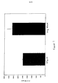
【0115】
図4は、構成的に活性のあるGPR30が293細胞においてGPR6によりもたらされるCRE-Lucレポーターの活性化を阻害することを示す。ルシフェラーゼは、発現ベクターpCMVにおいて約4.1相対光単位で測定された。内因性GPR30はルシフェラーゼを約8.5相対光単位で発現し、一方、非内因性の構成的に活性のあるGPR30(L258K)はルシフェラーゼをそれぞれ約3.8及び3.1相対光単位で発現した。1:4の比率で、内因性GPR6と内因性GPR30との共トランスフェクションは、ルシフェラーゼ発現を約104.1相対光単位まで劇的に増加した。同じ比率で、内因性GPR6と非内因性GPR30(L258K)との共トランスフェクションは、発現を劇的に減少し、これはそれぞれ約18.2及び29.5相対光単位で明白である。図5に記載するように、GPR3との共トランスフェクションに関してGPR17について同様の結果が認められた。
【0116】
実施例3
非内因性GPCRの構成的活性を測定するためのアッセイ
A.膜結合アッセイ
1.[35S]GTPγSアッセイ
Gタンパク質共役受容体がその活性状態にある場合、リガンド結合または構成的活性化のいずれかの結果として、該受容体はGタンパク質に共役し、GDPの遊離及びそれに続くGタンパク質へのGTPの結合を刺激する。Gタンパク質-受容体複合体のαサブユニットはGTPアーゼとして働き、GTPをGDPにゆっくり加水分解し、この時点で受容体は通常非活性化される。構成的に活性化された受容体は、GDPをGTPに交換し続ける。加水分解できないGTP類似体[35S]GTPγSを利用して、構成的に活性化された受容体を発現する膜への[35S]GTPγSの増大した結合を示すことができる。構成的活性化を測定するために[35S]GTPγS結合を用いる利点は:(a)それが全てのGタンパク質共役受容体に一般的に適用できること;(b)それが膜表面で近接し、細胞内カスケードに影響を及ぼす分子を捕らえる可能性を低くすることである。
【0117】
該アッセイは、問題とされる受容体を発現する膜への[35S]GTPγS結合を刺激するGタンパク質共役受容体の能力を利用する。従って、該アッセイは、既知の、オーファン及び構成的に活性化されたGタンパク質共役受容体に対して候補化合物をスクリーニングする直接同定法において用いることができる。該アッセイは一般的であり、全てのGタンパク質共役受容体での薬剤発見に適応性を有する。
【0118】
[35S]GTPγSアッセイを約0.3~約1.2nMの間の[35S]GTPγS(1.2が好ましいが、結果の最適化のためにこの量を調整することができる)及び12.5~75μgの膜タンパク質(例えば、受容体を発現するCOS-7細胞;75μgが好ましいが、最適化のためにこの量を調整することができる)及び1μMのGDP(最適化のためにこの量を変えることができる)と共に 20mM HEPES及び1~約20mMの間のMgCl2(20mMが好ましいが、結果の最適化のためにこの量を調整することができる)pH7.4、結合バッファー中で1時間インキュベーションした。次に、コムギ胚芽凝集素ビーズ(25μl;Amersham)を加え、混合物を室温でさらに30分間インキュベーションした。次に、チューブを室温で1500 x gで5分間遠心分離し、次にシンチレーションカウンターで計数した。
【0119】
大規模スクリーニングの要求を同様に満たす、かかる費用がより少ないが同等に適用できる代案が同定されている。Flash platesTM及びWallacTMシンチストリップ(scintistrip)を高処理量[35S]GTPγS結合アッセイを構成するために利用することができる。さらに、この技術を用いて、アッセイを[35S]GTPγS結合による効能をモニターするのと同時に受容体へのトリチウム化したリガンド結合を同時にモニターするために既知のGPCRに対して利用することができる。Wallac βカウンターは、トリチウムで標識したプローブ及び35Sで標識したプローブの両方を調べるためにエネルギーウインドーを切り換えることができるのでこれが可能である。このアッセイはまた、受容体活性化をもたらす他のタイプの膜活性化事象を検出するために用いることもできる。例えば、様々な受容体(Gタンパク質共役受容体及びチロシンキナーゼ受容体の両方)の32Pリン酸化をモニターするために該アッセイを用いることができる。膜がウェルの底に遠心分離されると、結合した[35S]GTPγSまたは32Pでリン酸化された受容体は、ウェルに被覆されているシンチラント(scintillant)を活性化する。ScintiRストリップ(Wallac)は、この原理を実証するために用いられている。さらに、該アッセイはまた、放射性標識したリガンドを用いて受容体へのリガンド結合を測定するためにも有用性を有する。同様に、放射性標識した結合したリガンドがウェルの底に遠心分離されると、シンチストリップ標識は放射性標識したリガンドと近接するようになり、活性化及び検出をもたらす。
【0120】
前述のプロトコルに基づいて、コントロール(pCMV)、内因性APJ及び非内因性APJを比較するグラフの代表的な結果を図6に記載する。
【0121】
2.アデニリルシクラーゼ
細胞に基づくアッセイのために設計されたFlash PlateTMアデニリルシクラーゼキット(New England Nuclear;カタログ番号SMP004A)を未精製の細胞膜での使用のために改変した。Flash Plateウェルは、cAMPを認識する特定の抗体も含有するシンチラント被覆を含有した。ウェル中に生成したcAMPをcAMP抗体への放射性cAMPトレーサーの結合に対する直接競合により定量した。以下のものは、受容体を発現する膜におけるcAMPレベルの変化の測定のための簡潔なプロトコルとして使える。
【0122】
トランスフェクション後約3日目にトランスフェクションした細胞を集めた。20mM HEPES、pH 7.4及び10mM MgCl2を含有するバッファー中での懸濁した細胞の均質化により膜を調製した。Brinkman PolytronTMを約10秒間用いて均質化を氷上で実施した。得られたホモジネートを4℃で49,000 X gで15分間遠心分離した。得られたペレットを次に20mM HEPES、pH 7.4及び0.1mM EDTAを含有するバッファー中に再懸濁し、10秒間均質化し、続いて4℃で49,000 X gで15分間遠心分離した。得られたペレットを利用するまで-80℃で保存することができる。測定の日に、膜ペレットを室温でゆっくり解凍し、20mM HEPES、pH 7.4及び10mM MgCl2(本明細書に記載する値が好ましいが、これらの量を最適化することができる)を含有するバッファー中に再懸濁し、0.60mg/mlの最終タンパク質濃度をもたらした(再懸濁した膜は使用するまで氷上に置いた)。
【0123】
cAMP基準及び(11mlの検出バッファーに対して2μCiのトレーサー[125I]cAMP(100μl)を含んでなる)検出バッファーを調製し、製造業者の説明書に従って維持した。アッセイバッファーをスクリーニングのために新しく調製し、それは20mM HEPES、pH 7.4、10mM MgCl2、20mM(Sigma)、0.1ユニット/mlクレアチンホスホキナーゼ(Sigma)、50μM GTP(Sigma)及び0.2mM ATP(Sigma)を含有し;アッセイバッファーは、利用するまで氷上で保存することができる。NEN Flashプレートに50μlのアッセイバッファーを加え、続いて50μlの膜懸濁液を加えることによりアッセイを開始した。得られたアッセイ混合物を室温で60分間インキュベーションし、続いて100μlの検出バッファーを加える。次に、プレートをさらに2-4時間インキュベーションし、続いてWallac MicroBetaシンチレーションカウンターで計数する。各アッセイプレート内に含まれる基準cAMP曲線からcAMP/ウェルの値を外挿する。前述のアッセイをMIGの分析に関して利用した。
【0124】
B.レポーターに基づくアッセイ
1.CREBレポーターアッセイ(Gs共役受容体)
Gs刺激を検出する方法は、cAMPに依存して活性化される転写因子CREBの既知の特性による。293または293T細胞においてGs共役活性をアッセイするためにPathDetect CREB trans-Reporting System(Stratagene、カタログ#219010)を利用した。哺乳類トランスフェクションキット(Stratagene、カタログ#200285)を製造業者の説明書に従って用いて細胞をこの上記の系のプラスミド成分及び内因性または突然変異体受容体をコードする示した発現プラスミドでトランスフェクションした。簡潔に言えば、400ngのpFR-Luc(Gal4認識配列を含有するルシフェラーゼレポータープラスミド)、40ngのpFA2-CREB(Gal4 DNA結合ドメインを含有するGal4-CREB融合タンパク質)、80ngの(受容体を含んでなる)CMV-受容体発現プラスミド及び20ngのCMV-SEAP(分泌されるアルカリホスファターゼ発現プラスミド;アルカリホスファターゼ活性は、サンプル間のトランスフェクション効率の変動を制御するためにトランスフェクションした細胞の培地において測定される)をリン酸カルシウム沈殿物中にキットの説明書どおりに合わせた。沈殿物の半分を96穴プレート中の3ウェル上に等しく分配し、細胞上で一晩保ち、翌朝新しい培地で置き換えた。トランスフェクションの開始後48時間で、上記GPR30系に関して記述したように細胞を処置し、ルシフェラーゼ活性についてアッセイした。このアッセイをGHSRについて用いた。
【0125】
2.AP1レポーターアッセイ(Gq共役受容体)
Gq刺激を検出する方法は、プロモーター中にAP1要素を含有する遺伝子の活性化をもたらすGq依存性ホスホリパーゼCの既知の特性による。リン酸カルシウム沈殿物の成分が410ngのpAP1-Luc、80ngの受容体発現プラスミド及び20ngのCMV-SEAPであったことを除いて、CREBレポーターアッセイに関して上に記述したプロトコルに従ってPathdetect AP-1 cis-Reporting System(Stratagene、カタログ#219073)を利用した。このアッセイをETBR-LP2について用いた。
【0126】
C.細胞内IP3蓄積アッセイ
1日目に、(内因性及び突然変異させた)セロトニン受容体を含んでなる細胞を24穴プレート上に通常1 x 10⁵細胞/ウェル平板培養した。2日目に、まず第一に50μlの無血清DMEM/ウェル中0.25μgのDNAと50μlの無血清DMEM/ウェル中2μlのリポフェクトアミンを混合することにより細胞をトランスフェクションした。これらの溶液を穏やかに混合し、室温で15-30分間インキュベーションした。細胞を0.5mlのPBSで洗浄し、400μlの無血清培地をトランスフェクション培地と混合し、細胞に加えた。次に、細胞を37℃/5% CO2で3-4時間インキュベーションし、次にトランスフェクション培地を除き、1ml/ウェルの通常の増殖培地で置き換えた。3日目に細胞を3H-ミオ-イノシトールで標識した。簡潔に言えば、培地を除き、細胞を0.5mlのPBSで洗浄した。次に、0.5mlのイノシトールを含まない/無血清培地(GIBCO BRL)/ウェルを0.25μCiの3H-ミオ-イノシトール/ウェルと共に加え、細胞を37℃/5% CO2で16-18時間インキュベーションした。4日目に細胞を0.5mlのPBSで洗浄し、イノシトールを含まない/無血清培地、10μMのパルジリン、10mMの塩化リチウムを含有する0.45mlのアッセイ培地または0.4mlのアッセイ培地と50μlの10xケタンセリン(ket)を10μMの最終濃度に加えた。次に、細胞を37℃で30分間インキュベーションした。次に、細胞を0.5mlのPBSで洗浄し、200μlの新しい/よく冷えた停止溶液(1M KOH;18mM ホウ酸Na;3.8mM EDTA)/ウェルを加えた。この溶液を5-10分間または細胞が溶解されるまで氷上に保ち、次に200μlの新しい/よく冷えた中和溶液(7.5% HCL)により中和した。次に、溶解産物を1.5mlエッペンドルフチューブ中に移し、1mlのクロロホルム/メタノール(1:2)/ウェルを加えた。この溶液を15秒間ボルテックスし、上相をBiorad AG1-X8陰イオン交換樹脂(100-200メッシュ)に添加した。まず第一に、樹脂を1:1.25 W/Vで水で洗浄し、0.9mlの上相をカラム上に載せた。カラムを10mlの5mMミオ-イノシトール及び10mlの5mMホウ酸Na/60mMギ酸Naで洗浄した。イノシトールトリスリン酸塩を2mlの0.1Mギ酸/1Mギ酸アンモニウムで10mlのシンチレーションカクテルを含有するシンチレーションバイアル中に溶出した。カラムを10mlの0.1Mギ酸/3Mギ酸アンモニウムで洗浄することにより再生し、dd H2Oで2回すすぎ、水中4℃で保存した。
【0127】
図7は、C322K突然変異を含むヒト5-HT2A受容体からのIP3生成の実例を示す。これらの結果は、プロリン突然変異アルゴリズム法がこの受容体を構成的に活性化することを示すが、可能性がある治療法の同定のためのスクリーニングにそのような受容体を用いる目的のためには、さらに確固たる差が好ましい。しかしながら、活性化された受容体は、構成的活性化の役割を理解し解明するため及びさらに調べることができる化合物の同定のために利用することができるので、この差はそれ自体ヒト5HT2A受容体の内因性及び非内因性バージョンを区別することにおいて有用であると考える。
【0128】
D.結果の要約
試験したGPCRの結果を表Eに記載し、ここで、増加%は、内因性GPCRに比較した場合に非内因性GPCRに対して認められた結果の%差を示し;これらの値の後には利用したアッセイのタイプに関する挿入表示が続く。さらに、利用したアッセイ系を挿入して記載する(そして異なる宿主細胞を用いた場合には両方を記載する)。これらの結果が示すように、ヒトGPCRの非内因性バージョンの構成的活性を測定するために様々なアッセイを利用することができる。当業者は、前述の事項に基づきそして当該技術分野が利用できる情報を参照して、研究者の特定の必要性に適合する特定のアッセイ方法を選択すること及び/または最大限に活用することができると考えられる。
【0129】
【表9】
実施例6
内因性オーファンGPCRの組織分布
市販されているヒト組織ドットブロット形態を用いて、内因性オーファンGPCRをそのような受容体が局在する領域を決定するためにプローブで調べた。以下に示す場合を除いて、(放射性標識した)全受容体cDNAをプローブとして用い:Prime-It IITMランダムプライマーラベリングキット(Stratagene、#300385)を製造業者の説明書に従って使用して(ベクターから切り出した)完全な受容体cDNAを用いて放射性標識したプローブを作製した。ヒトRNA Master BlotTM(Clontech、#7770-1)にGPCR放射性標識プローブをハイブリダイズさせ、製造業者の説明書に従ってストリンジェントな条件下で洗浄した。ブロットをKodak BioMaXオートラジオグラフィーフィルムに-80℃で一晩感光させた。
【0131】
代表的なドットブロット形態結果を図8にGPR1(8A)、GPR30(8B)及びAPJ(8C)について示し、全ての受容体について結果を表Fに要約する。
【0132】
【表10】
前述の情報に基づき、ヒトGPCRを罹病組織における分布に関しても評価することができ;次に「正常」及び罹病組織間の比較評価を利用して罹病状態における特定の受容体の過剰発現または不十分な発現の可能性を決定することができることを記す。可能性がある治療関連性の候補化合物を直接同定するスクリーニングの目的のためにヒトGPCRの非内因性バージョンを利用することが望ましい状況において、特定のヒトGPCRが過剰発現される疾病及び疾患の処置では逆アゴニストが有用であり、一方、特定のヒトGPCRが不十分に発現される疾病及び疾患の処置ではアゴニストまたは部分アゴニストが有用であることを記す。
【0134】
所望に応じて、目的の受容体が発現されるこれらの組織内の特定の細胞を同定するために、当業者に周知の技術(例えばインサイチューハイブリダイゼーション)を用いて、受容体のより詳細な細胞位置決定を利用することができる。
【0135】
本特許書類中に挙げる特許、出願及び発行された(printed)公開の各々は、これらを全部引用することにより本明細書に組み込まれるものとする。
【0136】
当業者が認識するように、本発明の精神からそれずに本発明の好ましい態様に対して多数の変更及び改変を行うことができる。全てのそのような変形は本発明の範囲内に入るものとする。
【0137】
内因性及び非内因性ヒトGPCRの両方に利用する目的のために、様々な発現ベクターを当業者は利用できるが、利用するベクターがpCMVであることが最も好ましい。このベクターは、特許手続き上の微生物の寄託の国際的承認に関するブダペスト条約の条項により1998年10月13日にAmerican Type Culture Collection(ATCC)(10801 University Blvd., Manassas, VA 20110-2209 USA)に寄託されている。このベクターは1998年 にATCCにより試験され、1998年 に生育できると確認された。ATCCはpCMVに以下の寄託番号: を付与している。
【図面の簡単な説明】
【図1】 膜貫通ヘリックス、細胞内ループ及び細胞外ループに番号を付与したGタンパク質共役受容体の一般化構造を示す。
【図2】 典型的なGタンパク質共役受容体の活性及び不活性の2つの状態並びに第二メッセンジャー伝達経路への活性状態の連結を図式的に示す。
【図3】 制限酵素部位の位置を含む、好ましいベクターpCMVの配列表である。
【図4】 pCMV、CRE-LucレポーターのGPR6によりもたらされる活性化の非内因性の構成的に活性のあるGPR30阻害をCRE-LucレポーターのGPR6によりもたらされる活性化の内因性GPR30阻害と比較する測定されたシグナルの図示である。
【図5】 pCMV、CRE-LucレポーターのGPR3によりもたらされる活性化の非内因性の構成的に活性化されたGPR17阻害をCRE-LucレポーターのGPR3によりもたらされる活性化の内因性GPR17阻害と比較する測定されたシグナルの図示である。
【図6】 コントロールのpCMV、内因性APJ及び非内因性APJを比較する測定されたシグナルの図式結果を示す。
【図7】 非内因性ヒト5-HT2A受容体からのIP3生成をこの受容体の内因性バージョンと比較した場合の実例を示す。
【図8】 GPR1(8A)、GPR30(8B)及びAPJ(8C)のドットブロット形態の結果である。
【配列表】
Commonly owned US 09 / 170,496 filed October 13, 1998, US 08 / 839,449 filed April 14, 1997 (currently abandoned), April 14, 1998 US Provisional Application No. 09 / 060,188 filed on the same day; US Provisional Application No. 60 / 090,783 filed on June 26, 1998; and US Provisional Application No. 60 / 095,677 filed on August 7, 1998. The benefits of this are claimed herein. Each of the foregoing applications is incorporated herein by reference in its entirety.
[0002]
Field of Invention
The invention disclosed in this patent document relates to transmembrane receptors, and more particularly to GPCRs that have been modified such that a modified human G protein coupled receptor (GPCR) is constitutively activated. Most preferably, a modified human GPCR is used for screening therapeutic compounds.
[0003]
Background of the Invention
Although a large number of receptor classes exist in humans, the most abundant and therapeutically relevant is clearly represented by the G protein coupled receptor (GPCR or GPCRs) class. It is estimated that there are approximately 100,000 genes in the human genome, of which approximately 2%, or 2,000 genes, are estimated to encode GPCRs. Among these are approximately 100 GPCRs in which endogenous ligands that bind to GPCRs have been identified. Due to the significant time lag that exists between the discovery of endogenous GPCRs and their endogenous ligands, the remaining 1,900 GPCRs were identified and characterized long before the endogenous ligands of these receptors were identified. Can be estimated. In fact, the rapidity with which the Human Genome Project is sequencing 100,000 human genes indicates that the remaining human GPCRs will be fully sequenced within the next few years. Nevertheless, and despite efforts to sequence the human genome, how scientists can use this information quickly, effectively and efficiently to improve and improve human symptoms It is still very unclear regarding. The present invention meets this important objective.
[0004]
Receptors for which endogenous ligands have been identified, including GPCRs, are termed “known” receptors, whereas receptors for which no endogenous ligand has been identified are termed “orphan” receptors. This distinction is not simply a meaning, especially in the case of GPCRs. GPCRs are an important area for the development of pharmaceutical products: 60% of all prescription pharmaceuticals have been developed from approximately 20 out of 100 known GPCRs. Thus, orphan GPCRs are an opportunity for the pharmaceutical industry to guide the growth, expansion, expansion and development that gold did for California in the late 19th century. However, there are significant drawbacks to orphan receptors in the development of new therapies. This is because traditional methods for pharmaceutical discovery and development require access to both the receptor and its endogenous ligand. Thus, to date, orphan GPCRs represent an undeveloped source in the art that sparks interest for pharmaceutical discovery.
[0005]
In traditional methods for the discovery of potential treatments, it can generally be said that receptors are first identified. Before drug discovery efforts can be initiated, complex, time-consuming and expensive procedures are typically performed to identify, isolate and produce the endogenous ligand of the receptor ( put into place), this procedure may require 3 to 10 years per receptor at a cost of about US $ 5 million per receptor. These times and resources should be exhausted before the traditional methods for drug discovery can begin. This is because traditional drug discovery techniques prevent compounds that bind endogenous ligands to receptors (“antagonists”) or compounds that enhance or mimic the action of ligands that bind to receptors (“agonists”). This is because, in an effort to discover either of these, putative therapeutic agents rely on a so-called “competitive binding assay”, which is “screened” against the receptor. The overall aim is to prevent or increase cellular activity that would otherwise occur when the ligand is correctly bound to the receptor (antagonists) or compounds that interfere with cell activation when the ligand is bound to the receptor (Agonist) is identified. Since endogenous ligands for orphan GPCRs have not been identified as obvious, it is unlikely that traditional drug discovery techniques can be used to discover new and unique therapies for these receptors. The present invention overcomes these and other serious limitations introduced by such traditional drug discovery techniques, as described in further detail below.
[0006]
GPCRs share a common structural motif. All of these receptors have 7 sequences of between 22-24 hydrophobic amino acids forming 7 alpha helices, each of which spans the membrane (each span is identified by a number, ie , Transmembrane-1 (TM-1), transmembrane-2 (TM-2), etc.). These transmembrane helices are located between the transmembrane-2 and transmembrane-3, the transmembrane-4 and transmembrane-5, and the transmembrane-6 and transmembrane-7 outside the cell membrane, ie on the “extracellular” side. Linked by a chain of amino acids (these are called “extracellular”
[0007]
In general, when an endogenous ligand binds to a receptor (often referred to as “activation” of the receptor), there is a change in the structure of the intracellular region, which is between the intracellular region and the intracellular “G protein”. Gives the conjugate of Although other G proteins exist, Gq, Gs, Gi and Go are currently identified G proteins. A GPCR coupled to a G protein activated by an endogenous ligand initiates a signaling cascade process (referred to as “signal transduction”). Under normal conditions, signal transduction ultimately leads to cell activation or cell inhibition. It is thought that the IC-3 loop as well as the carboxy terminus of the receptor interact with the G protein. The main focus of the present invention is directed to the transmembrane-6 (TM6) region and the intracellular-3 (IC3) region of GPCRs.
[0008]
Under physiological conditions, GPCRs exist in the cell membrane in equilibrium between two different structures: an “inactive” state and an “active” state. As shown schematically in FIG. 2, inactive receptors cannot be linked to intracellular signaling pathways to elicit biological responses. By changing the receptor structure to the active state, it is linked to the transmission pathway (via the G protein) and causes a biological response.
[0009]
Receptors can be stabilized in an active state by compounds such as endogenous ligands or drugs. Recent discoveries, including but not limited to modifications to the amino acid sequence of the receptor, provide means other than endogenous ligands or drugs to enhance and stabilize the receptor in the active state structure. These means effectively stabilize the active receptor by mimicking the action of an endogenous ligand that binds to the receptor. Stabilization by such ligand-independent means is called “constitutive receptor activation”.
[0010]
As mentioned above, the use of orphan receptors for screening purposes was not possible. This is because the traditional “principle” for compound screening requires that the ligand of the receptor be known. Therefore, it is obvious that this method is not applicable for orphan receptors. Thus, by sticking to this dogmatic approach to therapeutic discovery, the art essentially teaches to stop using orphan receptors until the receptor's endogenous ligand is discovered. And have been taught. Given that there are roughly 2,000 G protein-coupled receptors, and the majority of these are orphan receptors, such a principle represents a unique and unique method for therapeutic discovery. correct.
[0011]
Information regarding the nucleic acid and / or amino acid sequences of various GPCRs is summarized in Table A below. Since the important focus of the invention disclosed herein is directed to orphan GPCRs, many of the references described below relate to orphan GPCRs. However, this table does not imply that the invention disclosed herein is applicable only to orphan GPCRs or the specific GPCRs described below, and this table, whether legal or not. Should not be interpreted as such. In addition, certain receptors that are isolated are not the subject of the publication itself; for example, the G protein coupled receptor database (designated on the “world-wide web”) that lists GPCRs. Neither the inventor nor the assignee has anything to do with this site). Other GPCRs are the subject of patent applications owned by the assignee and are not described below (including GPR3, GPR6 and GPR12; see US Provisional 60/094879).
[0012]
[Table 1]
As described and disclosed in more detail below, the use of a mutation cassette to modify the endogenous sequence of a human GPCR results in a constitutively activated version of the human GPCR. These non-endogenous constitutively activated versions of human GPCRs can be used, inter alia, for screening candidate compounds, for example to directly identify therapeutically relevant compounds.
[0014]
Summary of invention
Disclosed herein is as the most preferred amino acid sequence region (C-terminal to N-terminal orientation) across the transmembrane-6 (TM6) and intracellular loop-3 (IC3) regions of GPCRs, and / Or (b) Most preferred nucleic acid sequence region (3'-5 'orientation):
(A) P1AA15X
here:
(1) P1Is an amino acid residue located within the TM6 region of the GPCR, where P1Is selected from the group consisting of (i) an endogenous GPCR proline residue and (ii) a non-endogenous amino acid residue other than proline;
(2) AA15Is 15 amino acids selected from the group consisting of (a) an endogenous GPCR amino acid, (b) a non-endogenous amino acid residue and (c) a combination of an endogenous GPCR amino acid and a non-endogenous amino acid, None of the 15 endogenous amino acid residues located within the TM6 region of the GPCR is proline; and
(3) X is a non-endogenous amino acid residue located in the IC3 region of the GPCR, preferably selected from the group consisting of lysine, histidine and arginine, most preferably lysine, provided that the endogenous of position X When the active amino acid is lysine, X is an amino acid other than lysine, preferably alanine
And / or
(B) Pcodon (AA-codon)15 Xcodon
here:
(1) PcodonIs the nucleic acid sequence within the TM6 region of the GPCR, where PcodonEncodes an amino acid selected from the group consisting of (i) an endogenous GPCR proline residue and (ii) a non-endogenous amino acid residue other than proline;
(2) (AA-codon)1515 encodes 15 amino acids selected from the group consisting of (a) endogenous GPCR amino acids, (b) non-endogenous amino acid residues and (c) combinations of endogenous GPCR amino acids and non-endogenous amino acids 15 Codon, except that none of the 15 endogenous codons in the TM6 region of the GPCR encode a proline amino acid residue; and
(3) XcodonIs a nucleic acid encoding a region residue located within the IC3 region of the GPCR, where XcodonEncodes a non-endogenous amino acid, preferably a non-endogenous amino acid selected from the group consisting of lysine, histidine and arginine, most preferably lysine, provided that position XcodonIf the endogenous coding region ofcodonEncodes an amino acid other than lysine, preferably alanine
Is a non-endogenous human G protein-coupled receptor comprising
The terms endogenous and non-endogenous associated with these sequence cassettes are with respect to endogenous GPCRs. For example, once an endogenous proline residue is located within the TM6 region of a particular GPCR and then the 16th amino acid is identified due to a mutation that constitutively activates the receptor, the proline residue is Most preferably it is not mutated, but it is also possible to mutate this endogenous proline residue (i.e. once the marker has been located and the 16th amino acid to be mutated has been identified) The marker itself can be mutated). Similarly, AA15Are most preferably kept in their endogenous form, but these amino acids can also be mutated. The only amino acid that must be mutated in the non-endogenous version of the human GPCR is X, i.e., as further disclosed herein, P1An endogenous amino acid that is 16 residues from cannot be kept in its endogenous form and must be mutated. Again, in non-endogenous versions of human GPCRs, P1And AA15Are preferably left in their endogenous form (ie, identical to their wild-type form), but once X is identified and mutated, P1And AA15Any and / or all of these can be mutated. This applies to nucleic acid sequences as well. When the endogenous amino acid at position X is lysine, in such a non-endogenous version of GPCR, X is an amino acid other than lysine, preferably alanine.
[0015]
Thus, as a hypothetical example, if the endogenous GPCR has the following endogenous amino acid sequence at the above position: P-AACCTTGGRRRDDDE-Q, any of the following representative hypothetical cassettes are within the scope of this disclosure ( (Describe non-endogenous amino acids in bold):
P-AACCTGGGRRRDDE-K
P-AACCTTHIGRRDDDE-K
PADEETTGGRRRDDDE-A
P-LLKFMSTWZLVAAPQ−K
A−LLKFMSTWZLVAAPQ−K
AA15It is also possible to add amino acid residues within, but such a method is not specifically presented. Indeed, in the most preferred embodiment, the only amino acid that differs in the non-endogenous version of the GPCR when compared to the endogenous version of the human GPCR is the amino acid at position X; Lead activation.
[0016]
Therefore, as a particularly preferred embodiment, P1And PcodonAre endogenous nucleic acid coding regions encoding endogenous proline and proline, respectively; and X and XcodonAre non-endogenous lysine or alanine and non-endogenous nucleic acid coding regions encoding lysine or alanine, respectively, with lysine being most preferred. Since non-endogenous versions of human GPCRs containing these mutations are most preferably incorporated into mammalian cells and used to screen candidate compounds, non-endogenous human GPCRs containing mutations are purified themselves, Although not necessarily isolated (ie, they are contained within the cell membrane of mammalian cells), such purified, isolated non-endogenous human GPCRs are well within the scope of this disclosure. Gene-targeted and transgenic non-human mammals (preferably rats and mice) including non-endogenous human GPCRs are also within the scope of the present invention; in particular, gene-targeted mammals are those animals that are non-human mammals. Most preferred in that it contains a non-endogenous version of a human GPCR in place of the region encoding the endogenous GPCR (techniques for creating such non-human mammals to replace non-human mammalian protein coding regions with human coding regions are Well known; see, for example, US Pat. No. 5,777,194).
[0017]
These mutations to the endogenous human GPCR make the GPCR constitutively active, so that, as further disclosed herein, non-endogenous constitutively activated versions of the human GPCR, among other things, endogenous It has been discovered that it can be used for direct screening of candidate compounds without the need for sex ligands. Accordingly, methods using these materials and products identified by these methods are also within the scope of the following disclosure.
[0018]
Detailed description
The scientific literature emerging on receptors employs a number of terms to describe ligands that have various actions on receptors. For clarity and consistency, the following definitions will be used throughout this patent document. If these definitions contradict other definitions of these terms, the following definitions shall govern:
An agonist shall mean a compound that, when bound to a receptor, activates an intracellular response or enhances GTP binding to the membrane.
[0019]
The amino acid abbreviations used in this specification are shown below.
[0020]
[Table 2]
A partial agonist is intended to mean a compound that, when bound to a receptor, activates an intracellular response to a lesser extent / degree than the agonist or increases GTP binding to the membrane to a lesser extent / degree than the agonist.
[0022]
An agonist competitively binds to the receptor at the same site as the agonist, but does not activate the intracellular response initiated by the active form of the receptor, thereby inhibiting the intracellular response by the agonist or partial agonist. Means a compound capable of Antagonists do not decrease the baseline intracellular response in the absence of an agonist or partial agonist.
[0023]
Candidate compounds shall mean molecules that can be easily screened (eg, chemical compounds) without limitation. Preferably, the phrase “candidate compound” is publicly known to be a compound selected from the group consisting of inverse agonists, agonists or antagonists to the receptor, as previously determined by indirect identification methods. Compounds that have been ("indirectly identified compounds") are not included; more preferably, indirectly identified compounds that have previously been determined to have therapeutic efficacy in at least one mammal And most preferably, indirectly identified compounds that have been previously determined to have therapeutic utility in humans are not included.
[0024]
Codons generally comprise a nucleoside (adenosine (A), guanosine (G), cytidine (C), uridine (U), and thymidine (T)) linked to a phosphate group and, when translated, encodes an amino acid. Means a group of three nucleotides (or nucleotide equivalents).
[0025]
Compound efficacy shall mean a measurement of the ability of a compound to inhibit or stimulate receptor functionality as opposed to receptor binding affinity. Preferred means of detecting compound efficacy are, for example, as disclosed further in the Examples section of this patent document, [35By measuring S] GTPγS binding.
[0026]
Constitutively activated receptor shall mean a receptor that undergoes constitutive receptor activation. In the present invention as disclosed herein, a non-endogenous constitutively activated human G protein coupled receptor comprises an amino acid cassette P as described in further detail below.1AA15It has been mutated to include X.
[0027]
Constitutive receptor activation shall mean stabilization of an active receptor by means other than binding of the receptor's endogenous ligand or its chemical equivalent to the receptor. Preferably, a G protein-coupled receptor subject to constitutive receptor activation of the present invention disclosed herein is responsive to signals measured for constitutive activation when compared to the endogenous form of the GPCR. A difference of at least 10% (optionally increased or decreased), more preferably about 25% difference of such comparison response, most preferably about 50% difference of such comparison response. When used for direct identification of candidate compounds, the signal difference is at least about 50% so that there is a sufficient difference between the endogenous and non-endogenous signals to distinguish between the selected candidate compounds Most preferred. In most cases, the “difference” is an increase in signal; however, for Gs-coupled GPCRs, the measured “difference” is preferably a decrease, as described in more detail below.
[0028]
Contacting or contacting shall mean bringing together at least two components, whether in vitro or in vivo.
[0029]
In connection with the phrase “candidate compound”, directly identifying or directly identified is screening candidate compounds against constitutively activated G protein coupled receptors and assessing the compound potency of such compounds Means. This term is not to be construed or interpreted as encompassing or encompassed by the term “indirectly identified” or “indirectly identified” under any circumstances.
[0030]
Endogenous shall mean a substance that is naturally produced by the genome of a species. For example, without limitation, endogenous with respect to GPCR is intended to mean that which is naturally produced by a human, insect, plant, bacterium or virus. On the contrary, the term non-endogenous in this context shall mean that which is not naturally produced by the genome of the species. For example, but not limited to, most preferably, a receptor that is not constitutively active in the endogenous form, but subsequently becomes constitutively active when mutated by using a cassette disclosed herein, Referred to herein as "non-endogenous constitutively activated receptor". Both terms can be used to describe both “in vivo” and “in vitro” systems. For example, without limitation, in a screening method, an endogenous or non-endogenous receptor can be with respect to an in vitro screening system in which the receptor is expressed on the cell surface of a mammalian cell. By way of further example, and not limitation, screening of candidate compounds by means of in vivo systems can be performed if the mammalian genome has been engineered to contain a non-endogenous constitutively activated receptor.
[0031]
Host cell is intended to mean a cell in which a plasmid and / or vector can be contained. For prokaryotic host cells, the plasmid is typically replicated as an autonomous molecule when the host cell replicates (generally, the plasmid is subsequently isolated for introduction into a eukaryotic host cell); In the case of a biological host cell, the plasmid is integrated into the cellular DNA of the host cell so that the plasmid replicates when the eukaryotic host cell replicates. Preferably, for the purposes of the invention disclosed herein, the host cell is eukaryotic, more preferably mammalian, most preferably selected from the group consisting of 293, 293T and COS-7 cells. Is done.
[0032]
Indirectly identified or indirectly identified is the identification of an endogenous ligand specific for the endogenous receptor, accepting to determine what interferes with and / or competes with the ligand-receptor interaction Means a traditional method for drug discovery process that involves screening candidate compounds against the body and assessing the efficacy of the compound with respect to affecting at least one second messenger pathway associated with the activated receptor .
[0033]
Inhibiting or inhibiting in the context of the term “response” shall mean that the response is reduced or prevented in the presence of the compound as opposed to in the absence of the compound.
[0034]
Inverse agonists bind to either the endogenous form of the receptor or a constitutively activated form of the receptor and trigger a reference intracellular response initiated by the active form of the receptor in the absence of an agonist or partial agonist It shall mean a compound that inhibits below the normal basal level of activity observed below or reduces GTP binding to the membrane. Preferably, the reference intracellular response is inhibited by at least 30%, more preferably at least 50%, most preferably at least 75% in the presence of the inverse agonist when compared to the reference response in the absence of the inverse agonist. The
[0035]
A known receptor shall mean an endogenous receptor for which an endogenous ligand specific for that receptor has been identified.
[0036]
Ligand shall mean an endogenous naturally occurring molecule that is specific for an endogenous naturally occurring receptor.
[0037]
A mutation or mutation relating to the nucleic acid and / or amino acid sequence of an endogenous receptor is such that a mutant form of the endogenous constitutively inactive receptor indicates constitutive activation of the receptor, It is intended to mean a particular mutation or mutations for such endogenous sequences. For a specific sequence equivalent, (a) when the level of constitutive activation of the next mutated form of the receptor is substantially the same as that indicated by the first mutation of the receptor; And (b) a sequence (amino acid and / or nucleic acid) homology between the next mutated form of the receptor and the first mutation of the receptor is at least about 80%, more preferably at least about 90% , Most preferably at least 95%, the next mutated form of the human receptor is considered equivalent to the first mutation of the human receptor. Ideally, and most preferred cassettes disclosed herein for performing constitutive activation are single amino acid and / or codon mutations (ie, X or X or X) between endogenous and non-endogenous forms of GPCRs.codon% Sequence homology should be at least 98%.
[0038]
An orphan receptor shall mean an endogenous receptor for which no endogenous ligand specific for that receptor has been identified or known.
[0039]
Pharmaceutical composition is intended to mean a composition comprising at least one active ingredient, whereby the composition can be easily examined for specific effective results in mammals (eg, but not limited to humans). it can. Those skilled in the art will understand and appreciate the techniques appropriate for determining whether an active ingredient has the desired effective results based on the needs of the artisan.
[0040]
A plasmid shall mean a combination of a vector and a cDNA. In general, plasmids are introduced into host cells for the purpose of replication and / or expression of cDNA as a protein.
[0041]
In the context of the term “response”, stimulating or stimulating shall mean that the response is increased in the presence of the compound as opposed to in the absence of the compound.
[0042]
Crossing or crossing with respect to either a specific nucleic acid sequence or a specific amino acid sequence shall mean that the sequence is located in at least two different specific regions. For example, in an amino acid sequence that is 10 amino acid components long, 3 of the 10 components are in the TM6 region of the GPCR and the remaining 7 components are in the IC3 region of the GPCR. The component can be described as crossing the TM6 and IC3 regions of the GPCR.
[0043]
A cDNA-related vector is intended to mean a circular DNA that can contain at least one cDNA and can be taken up into a host cell.
[0044]
The order of the following sections is set forth for representational efficiency and is not intended and should not be construed as a limitation on the following disclosure or claims.
A. Introduction
Traditional studies of receptors have always stated that endogenous ligands must first be identified before the discovery and subsequent discovery of antagonists and other molecules that can act on the receptor (historically Is based on assumptions. Even if the antagonist may be initially known, the search immediately extends to searching for an endogenous ligand. This idea persists in receptor research even after the discovery of constitutively activated receptors. What has not been recognized so far is that it is the receptor's active state that is most useful for finding receptor agonists, partial agonists and inverse agonists. For diseases caused by overactive receptors or underactive receptors, what is desired in therapeutics reduces or increases the activity of the receptor, respectively. Is not a drug that is an antagonist to an endogenous ligand. This is because compounds that reduce or enhance the activity of the active receptor state need not bind at the same site as the endogenous ligand. Thus, as taught by the methods of the present invention, any search for therapeutic compounds should begin by screening the compounds for ligand-independent activity states.
[0045]
Screening candidate compounds against non-endogenous constitutively activated GPCRs works at these cell surface receptors without the need for any prior knowledge or use of the receptor's endogenous ligand Can be directly identified. By determining the regions in the body where endogenous versions of such GPCRs are expressed and / or overexpressed, the expression of these receptors and / or related diseases / disorder conditions associated with overexpression are determined. Such methods are disclosed in this patent document.
B. Disease / disease identification and / or selection
Most preferably, the materials of the invention can be used to identify inverse agonists for non-endogenous constitutively activated GPCRs. Such inverse agonists are ideal candidates as lead compounds in drug discovery programs for treating diseases associated with these receptors. Inverse agonists, partial agonists or agonists for these receptors can be directly identified, thereby enabling the development of pharmaceutical compositions and the search for diseases and disorders associated with these receptors. is there. For example, scrutinizing both diseased and normal tissue samples for the presence of these receptors may now be performed along a path to identify endogenous ligands in the case of academic trials or orphan receptors. More than nothing. Tissue review can be performed over a wide range of healthy and diseased tissues. Such tissue review provides a favorable first step in linking diseases and / or diseases to specific receptors.
[0046]
Preferably, the endogenous GPCR DNA sequence is used to generate probes of either radiolabeled cDNA or RT-PCR identification of GPCR expression in tissue samples. Preferably, the presence of the receptor in the diseased tissue or the presence of the receptor at an increased or decreased concentration in the diseased tissue compared to normal tissue can be used to identify a correlation with the disease . This technique allows the receptor to be located equally well throughout the organ. Based on the known function of the particular tissue in which the receptor is located, the generally considered functional role of the receptor can be estimated.
C. "Human GPCR proline marker" algorithm and generation of non-endogenous constitutively active human GPCRs
Among the many challenges confronting biotechnological technology, there are unpredictable things about collecting genetic information from one species and correlating that information with another species, and this problem is everywhere in this technology It does not show as troublesome deterioration in the gene sequences encoding nucleic acids and proteins. Thus, for consistency and because of the highly unpredictable nature of this technology, the following invention is limited to human GPCRs for mammals, and the applicability of the present invention to other mammalian species is potentially It is considered to be more than just mechanical application.
[0047]
In general, when trying to apply common "rules" from one related protein sequence to another or from one species to another, the technique typically relies on sequence alignment, i.e., sequencing the sequence Then try to find areas of common nature between two or more sequences. While useful, this method does not always provide meaningful information. In the case of GPCRs, the general structural motif is the same for all GPCRs, but variations in the length of TM, EC, and IC make such alignment methods difficult from one GPCR to another at best. . Thus, for example, it may be desirable to apply a consistent method of constitutive activation from one GPCR to another, but there are significant differences in sequence length, accuracy, etc. from one GPCR to the next. Because of this, there is essentially no mutation alignment method that is generally applicable and easily successful. Analogously, such a method would cause a traveler to begin a trip at point A by giving the traveler a number of different maps to point B without a scale or distance marker anywhere on the map, and then Equivalent to asking the traveler to find the shortest and most efficient route to destination B only by using a map. In such situations, this task is easily simplified by having the ability to measure (a) a common “location marker” on each map and (b) the distance from that location marker to destination B. Thus, this allows the traveler to select the most efficient one from starting point A to destination B.
[0048]
In essence, a feature of the present invention is to provide such a position within a human GPCR that can readily create a constitutively active form of the human GPCR.
[0049]
As one skilled in the art will recognize, the transmembrane region of a cell is very hydrophobic; therefore, using standard hydrophobicity plotting techniques, one skilled in the art can easily determine the TM region of a GPCR, especially TM6. (This same method can also be applied to determine EC and IC regions of GPCRs). Within the TM6 region of human GPCRs, it was discovered that a common proline residue (generally near the middle of TM6) serves as a constitutive activation “marker”. By counting 15 amino acids from this proline marker, when the 16th amino acid (located in the IC3 loop) is mutated from its endogenous form to a non-endogenous form, it leads to constitutive activation of the receptor. For convenience, this is referred to as the “human GPCR proline marker” algorithm. The non-endogenous amino acid at this position can be any amino acid, but most preferably the non-endogenous amino acid is lysine. Without wishing to be bound by any theory, we believe that this position is unique and that mutations at this position affect the receptor to give constitutive activation.
[0050]
For example, if the endogenous amino acid at position 16 is already lysine (as is GPR4 and GPR32), for X to be a non-endogenous amino acid, it must be other than lysine; In situations where the endogenous GPCR has an endogenous lysine residue at position 16, the non-endogenous version of that GPCR preferably contains amino acids other than lysine at this position, preferably alanine, histidine and arginine. Special mention. More importantly (of further note), it was determined that GPR4 is conjugated to Gs and appears to be active in its endogenous form (data not shown).
[0051]
Since there are only 20 naturally occurring amino acids (although the use of non-naturally occurring amino acids is also feasible), the selection of a specific non-endogenous amino acid for this substitution at position 16 is feasible Therefore, it is possible to efficiently select non-endogenous amino acids that meet the needs of researchers. However, as noted, the more preferred non-endogenous amino acids at position 16 are lysine, histidine, arginine and alanine, with lysine being most preferred. One skilled in the art will readily be able to determine skilled methods of altering the sequence of codons to make the desired mutation.
[0052]
Sometimes, but not always, W2 is located in front of the proline residue marker in TM6 (ie W2P1AA15X) is also found, where W is tryptophan and 2 is any amino acid residue.
[0053]
Our findings, among other things, negate the need for unpredictable and cumbersome sequence alignment methods commonly used by the art. In fact, the advantage of our discovery is to obtain a unique and very useful end product, ie a constitutively activated version of a human GPCR, while the algorithm is in nature (while an algorithm in nature) In addition, it can be easily applied to human GPCRs with ease of handling which is skilled to those skilled in the art. Since many years and considerable amount of money is required to determine the endogenous ligands of human GPCRs as revealed by the Human Genome Project, the disclosed invention is intended to make practical use of this sequence information. Not only will it reduce the time required, it will also significantly reduce costs. This method properly demonstrates the importance of the human genome project, as it allows the use of genetic information not only to understand the role of GPCRs in disease, for example, but also to provide opportunities to improve human symptoms.
D. Candidate compound screening
1. General GPCR screening assay technology
When the G protein receptor becomes constitutively active, it couples to the G protein (eg, Gq, Gs, Gi, Go) and stimulates the release of GDP and the subsequent binding of GTP to the G protein. The G protein then acts as a GTPase, slowly hydrolyzing GTP to GDP, thereby allowing the receptor to be deactivated under normal conditions. However, constitutively activated receptors, including the non-endogenous constitutively active human GPCRs of the present invention, continue to exchange GDP for GTP. GTP non-hydrolyzable analogue [35S] GTPγS can be used to monitor increased binding to G proteins present on membranes expressing constitutively activated receptors. [35It has been reported that S] GTPγS can be used to monitor G protein binding to the membrane in the absence and presence of ligand. This monitoring example was reported by Traynor and Nahorski in 1995, among other examples well known and available to those skilled in the art. Since this assay system is generally applicable to all G protein-coupled receptors, regardless of the specific G protein that interacts with the intracellular domain of the receptor, the preferred use of this system is for initial screening of candidate compounds. It is.
[0054]
B 2. Specific GPCR screening assay technology
C Once a candidate compound is identified using a “generic” G protein-coupled receptor assay (ie, an assay for selecting compounds that are agonists, partial agonists or inverse agonists), those compounds are Further screening to confirm that the site is interacting is preferred. For example, a compound identified by a “generic” assay cannot bind to the receptor, but instead can simply “separate” the G protein from the intracellular domain.
[0055]
a. Gs and Gi.
Gs stimulates the enzyme adenylyl cyclase. On the other hand, Gi (and Go) inhibits this enzyme. Adenylyl cyclase catalyzes the conversion of ATP to cAMP; therefore, constitutively activated GPCRs coupled to Gs proteins are associated with increased cellular levels of cAMP. On the other hand, constitutively activated GPCRs coupled to Gi (or Go) proteins are associated with reduced cellular levels of cAMP. In general, “Indirect Mechanisms of Synaptic Transmission”, Chapter 8,From Neuron To Brain(3rd edition) See Nichols, J. G. et al, Sinauer Associates, Inc. (1992). Thus, an assay that detects cAMP can be utilized to determine whether a candidate compound is, for example, an inverse agonist for a receptor (ie, such a compound reduces the level of cAMP). Various methods known in the art can be utilized to measure cAMP; the most preferred method is by use of an ELISA-based form of an anti-cAMP antibody. Another type of assay that can be utilized is a whole cell second messenger reporter based assay. A promoter on a gene directs the expression of the protein encoded by that particular gene. Cyclic AMP leads to gene expression by facilitating the binding of cAMP-responsive DNA binding protein or transcription factor (CREB), which then binds to the promoter at a specific location called the cAMP responsive element, leading to gene expression . A reporter system can be constructed having a promoter containing multiple cAMP response elements in front of a reporter gene, such as β-galactosidase or luciferase. Thus, a constitutively activated Gs-linked receptor results in the accumulation of cAMP, which in turn activates the gene and expression of the reporter protein. A standard biochemical assay can then be used to detect a reporter protein such as β-galactosidase or luciferase (Chen et al. 1995). For GPCRs that couple to Gi (or Go) and thus reduce cAMP levels, for example GPR17 and GPR30 screening methods for inverse agonists based on the use of receptors that couple to Gs (and thus increase cAMP levels) Disclosed in the Examples section.
[0056]
b. Go and Gq
Go and Gq are involved in the activation of the enzyme phospholipase C, which in turn is a phospholipid PIP2Is hydrolyzed into two intracellular messengers: diacylglycerol (DAG) and
E. Medicinal chemistry
In general, though not necessarily, direct identification of candidate compounds is preferably performed with compounds made by combinatorial chemistry techniques so that thousands of compounds are randomly selected for such analysis. Prepared. In general, the results of such screening are compounds with a unique core structure; these compounds are then preferably around the preferred core structure (s) to further enhance their pharmaceutical properties. Subjected to further chemical modification. Such techniques are known to those skilled in the art and are not discussed in detail in this patent document.
F. Pharmaceutical composition
Candidate compounds selected for further development can be formulated into pharmaceutical compositions using techniques well known to those skilled in the art. One of skill in the art can use any suitable pharmaceutically acceptable carrier; see, for example, Remington's Pharmaceutical Sciences, 16th Edition, 1980, Mack Publishing Co., (Oslo et al.).
G. Other usefulness
The preferred use of the disclosed non-endogenous versions of human GPCRs is to directly identify candidate compounds as inverse agonists, agonists or partial agonists (preferably for pharmaceutical use), but study these receptors It can also be used in the environment. For example, in vivo and in vitro systems containing these receptors can be applied to further clarify and understand the role of these receptors in both normal and diseased human states and to understand the signaling cascade. It can be used to understand the role of constitutive activation. Because of their unique characteristics, the value of these non-endogenous receptors should be used to understand the role of a particular receptor in the human body before its endogenous ligand is identified Can increase their usefulness as a research tool. Other uses of the disclosed receptor will be apparent to those skilled in the art based upon, among other things, the description of this patent document.
[0057]
Example
The following examples are given for the purpose of clarifying the invention and are not limiting. Although specific nucleic acid and amino acid sequences are disclosed herein, utilizing a mutation cassette in the IC3 loop of the receptor based on position relative to the proline residue in TM6 to constitutively activate human GPCRs In accordance with the teachings of this patent document that it is possible, those skilled in the art will be able to make minor modifications to these sequences with the same or substantially similar results as reported below. Specific methods of sequence mutation are within the purview of those skilled in the art based on the particular needs of those skilled in the art.
[0058]
Example 1
Preparation of endogenous human GPCR
Various GPCRs were utilized in the examples below. Some endogenous human GPCRs were kindly provided in the expression vector (as shown below), and other endogenous human GPCRs were synthesized de novo using publicly available sequence information.
[0059]
1. GPR1 (Genbank accession number: U13666)
The human cDNA sequence for GPR1 was provided in pRcCMV by Brian O'Dowd (University of Toronto). GPR1 cDNA (1.4 kB fragment) was excised from the pRcCMV vector as an NdeI-XbaI fragment and subcloned into the NdeI-XbaI site of the pCMV vector (see FIG. 3). Thereafter, the nucleic acid (SEQ ID NO: 1) and amino acid (SEQ ID NO: 2) sequences of human GPR1 were determined and confirmed.
[0060]
2. GPR4 (Genbank accession number: L36148, U35399, U21051)
The human cDNA sequence for GPR4 was provided in pRcCMV by Brian O'Dowd (University of Toronto). GPR1 cDNA (1.4 kB fragment) is excised from the pRcCMV vector as an ApaI (smoothed) -XbaI fragment and into the HindIII (smoothed) -XbaI site of the pCMV vector (excluding most of the 5 'untranslated region) Subcloned. Thereafter, the nucleic acid (SEQ ID NO: 3) and amino acid (SEQ ID NO: 4) sequences of human GPR4 were determined and confirmed.
[0061]
3. GPR5 (Genbank accession number: L36149)
Human GPR5 cDNA was prepared as follows and cloned into the pCMV expression vector: Genomic DNA and rTth polymerase (Perkin Elmer) as template, buffer system provided by the manufacturer, 0.25 μM of each primer and 0.2 mM PCR was performed with each of the 4 nucleotides. The cycling conditions were 94 ° C for 1 minute; 64 ° C for 1 minute; and 72 ° C for 1.5 minutes for 30 cycles. 5 'PCR primer sequence:
5'-TATGAATTCAGATGCTCTAAACGTCCCTGC-3 '(SEQ ID NO: 5)
Contains an EcoRI site, and the 3 ′ primer has the sequence:
5'-TCCGGATCCACCTGCACCTGCGCCTGCACC-3 '(SEQ ID NO: 6)
Contained a BamHI site. The 1.1 kb PCR fragment was digested with EcoRI and BamHI and cloned into the EcoRI-BamHI site of the pCMV expression vector. Thereafter, the nucleic acid (SEQ ID NO: 7) and amino acid (SEQ ID NO: 8) sequences of human GPR5 were determined and confirmed.
[0062]
4). GPR7 (Genbank accession number: U22491)
Human GPR7 cDNA was prepared as follows and cloned into the pCMV expression vector: PCR conditions-Genomic DNA as template and rTth polymerase (Perkin Elmer) as buffer provided by the manufacturer, 0.25 μM of each primer PCR was carried out with 4 nucleotides of 0.2 mM each. The cycling conditions were 94 ° C for 1 minute; 62 ° C for 1 minute; and 72 ° C for 1 minute 20 seconds for 30 cycles. 5 'PCR primer sequence:
5'-GCAAGCTTGGGGGACGCCAGGTCGCCGGCT-3 '(SEQ ID NO: 9)
Contains a HindIII site, and the 3 ′ primer has the sequence:
5'-GCGGATCCGGACGCTGGGGGAGTCAGGCTGC-3 '(SEQ ID NO: 10)
Contained a BamHI site. The 1.1 kb PCR fragment was digested with HindIII and BamHI and cloned into the HindIII-BamHI site of the pCMV expression vector. Thereafter, the nucleic acid (SEQ ID NO: 11) and amino acid (SEQ ID NO: 12) sequences of human GPR7 were determined and confirmed.
[0063]
Five. GPR8 (Genbank accession number: U22492)
Human GPR8 cDNA was generated as follows and cloned into the pCMV expression vector: Genomic DNA and rTth polymerase (Perkin Elmer) as template, buffer system provided by the manufacturer, 0.25 μM of each primer and 0.2 mM PCR was performed with each of the 4 nucleotides. The cycling conditions were 94 ° C for 1 minute; 62 ° C for 1 minute; and 72 ° C for 1 minute 20 seconds for 30 cycles. 5 'PCR primer sequence:
5'-CGGAATTCGTCAACGGTCCCAGCTACAATG-3 '(SEQ ID NO: 13)
Contains an EcoRI site, and the 3 ′ primer has the sequence:
5'-ATGGATCCCAGGCCCTTCAGCACCGCAATAT-3 '(SEQ ID NO: 14)
Contained a BamHI site. The 1.1 kb PCR fragment was digested with EcoRI and BamHI and cloned into the EcoRI-BamHI site of the pCMV expression vector. All four cDNA clones sequenced contained a possible polymorphism with a mutation of amino acid 206 from Arg to Gln. Excluding this difference, the nucleic acid (SEQ ID NO: 15) and amino acid (SEQ ID NO: 16) sequences of human GPR8 were subsequently determined and confirmed.
[0064]
6). GPR9 (Genbank accession number: X95876)
Human GPR9 cDNA was generated as follows and cloned into the pCMV expression vector: clone (provided by Brian O'Dowd) as template and pfu polymerase (Stratagene) by a manufacturer supplemented with 10% DMSO. PCR was performed using the provided buffer system, 0.25 μM each primer and 0.5 mM each 4 nucleotides. Cycle conditions were 94 ° C for 1 minute; 56 ° C for 1 minute; and 72 ° C for 2.5 minutes for 25 cycles. 5 'PCR primer sequence:
5'-ACGAATTCAGCCATGGTCCTTGAGGTGAGTGACCACCAAGTGCTAAAT-3 '(SEQ ID NO: 17) contains an EcoRI site, and the 3' primer has the sequence:
5'-GAGGATCCTGGAATGCGGGGAAGTCAG-3 '(SEQ ID NO: 18)
Contained a BamHI site. The 1.2 kb PCR fragment was digested with EcoRI and cloned into the EcoRI-SmaI site of the pCMV expression vector. Thereafter, the nucleic acid (SEQ ID NO: 19) and amino acid (SEQ ID NO: 20) sequences of human GPR9 were determined and confirmed.
[0065]
7. GPR9-6 (GenBank accession number: U45982)
Human GPR9-6 cDNA was generated as follows and cloned into a pCMV expression vector: Genomic DNA and rTth polymerase (Perkin Elmer) as template, buffer system provided by the manufacturer, 0.25 μM of each primer and PCR was performed using 0.2 mM each with 4 nucleotides. Cycle conditions were 94 ° C for 1 minute; 62 ° C for 1 minute; and 72 ° C for 1 minute 20 seconds for 30 cycles. 5 'PCR primer sequence:
5'-TTAAGCTTGACCTAATGCCATCTTGTGTCC-3 '(SEQ ID NO: 21)
Kinased with, and the 3 'primer is the sequence:
5'-TTGGATCCAAAAGAACCATGCACCTCAGAG-3 '(SEQ ID NO: 22)
Contained a BamHI site. The 1.2 kb PCR fragment was digested with BamHI and cloned into the EcoRV-BamHI site of the pCMV expression vector. Thereafter, the nucleic acid (SEQ ID NO: 23) and amino acid (SEQ ID NO: 24) sequences of human GPR9-6 were determined and confirmed.
[0066]
8. GPR10 (Genbank accession number: U32672)
The human cDNA sequence for GPR10 was provided in pRcCMV by Brian O'Dowd (University of Toronto). GPR10 cDNA (1.3 kB fragment) was excised from the pRcCMV vector as an EcoRI-XbaI fragment and subcloned into the EcoRI-XbaI site of the pCMV vector. Thereafter, the nucleic acid (SEQ ID NO: 25) and amino acid (SEQ ID NO: 26) sequences of human GPR10 were determined and confirmed.
[0067]
9. GPR15 (Genebank accession number: U34806)
The human cDNA sequence for GPR15 was provided in pCDNA3 by Brian O'Dowd (University of Toronto). GPR15 cDNA (1.5 kB fragment) was excised from the pCDNA3 vector as a HindIII-Bam fragment and subcloned into the HindIII-Bam site of the pCMV vector. Thereafter, the nucleic acid (SEQ ID NO: 27) and amino acid (SEQ ID NO: 28) sequences of human GPR15 were determined and confirmed.
[0068]
Ten. GPR17 (Genbank accession number: Z94154)
Human GPR17 cDNA was generated as follows and cloned into the pCMV expression vector: Genomic DNA and rTth polymerase (Perkin Elmer) as template, buffer system provided by the manufacturer, 0.25 μM of each primer and 0. PCR was performed using 2 mM each with 4 nucleotides. The cycling conditions were 94 ° C for 1 minute; 56 ° C for 1 minute; and 72 ° C for 1 minute 20 seconds for 30 cycles. 5 'PCR primer sequence:
5'-CTAGAATTCTGACTCCAGCCAAAGCATGAAT-3 '(SEQ ID NO: 29)
Contains an EcoRI site, and the 3 ′ primer has the sequence:
5'-GCTGGATCCTAAACAGTCTGCGCTCGGCCT-3 '(SEQ ID NO: 30)
Contained a BamHI site. The 1.1 kb PCR fragment was digested with EcoRI and BamHI and cloned into the EcoRI-BamHI site of the pCMV expression vector. Thereafter, the nucleic acid (SEQ ID NO: 31) and amino acid (SEQ ID NO: 32) sequences of human GPR17 were determined and confirmed.
[0069]
11. GPR18 (Genbank accession number: L42324)
Human GPR18 cDNA was prepared as follows and cloned into the pCMV expression vector: Genomic DNA and rTth polymerase (Perkin Elmer) as template, buffer system provided by the manufacturer, 0.25 μM of each primer and 0.2 mM. PCR was performed with each of the 4 nucleotides. The cycling conditions were 94 ° C for 1 minute; 54 ° C for 1 minute; and 72 ° C for 1 minute 20 seconds for 30 cycles. 5 'PCR primer sequence:
5'-ATAAGATGATCACCCTGAACAATCAAGAT-3 '(SEQ ID NO: 33)
Kinase with, and 3 'primer sequence:
5'-TCCGAATTCATAACATTTCACTGTTTATATTGC-3 '(SEQ ID NO: 34)
It contained an EcoRI site. The 1.0 kb PCR fragment was digested with EcoRI and cloned into the blunt-EcoRI site of the pCMV expression vector. All eight sequenced cDNA clones may be accompanied by mutations at
[0070]
12. GPR20 (Genbank accession number: U66579)
Human GPR20 cDNA was prepared as follows and cloned into the pCMV expression vector: Genomic DNA and rTth polymerase (Perkin Elmer) as template, buffer system provided by the manufacturer, 0.25 μM of each primer and 0.2 mM. PCR was performed with each of the 4 nucleotides. The cycling conditions were 94 ° C for 1 minute; 62 ° C for 1 minute; and 72 ° C for 1 minute 20 seconds for 30 cycles. 5 'PCR primer sequence:
5'-CCAAGCTTCCAGGCCTGGGGTGTGCTGG-3 '(SEQ ID NO: 37)
Kinase with, and 3 'primer sequence:
5'-ATGGATCCTGACCTTCGGCCCCTGGCAGA-3 '(SEQ ID NO: 38)
Contained a BamHI site. The 1.2 kb PCR fragment was digested with BamHI and cloned into the EcoRV-BamHI site of the pCMV expression vector. Thereafter, the nucleic acid (SEQ ID NO: 39) and amino acid (SEQ ID NO: 40) sequences of human GPR20 were determined and confirmed.
[0071]
13. GPR21 (Genbank accession number: U66580)
Human GPR21 cDNA was prepared as follows and cloned into the pCMV expression vector: Genomic DNA and rTth polymerase (Perkin Elmer) as template, buffer system provided by the manufacturer, 0.25 μM of each primer and 0.2 mM PCR was performed with each of the 4 nucleotides. The cycling conditions were 94 ° C for 1 minute; 62 ° C for 1 minute; and 72 ° C for 1 minute 20 seconds for 30 cycles. 5 'PCR primer sequence:
5'-GAGAATTCACTCCTGAGCTCAAGATGAACT-3 '(SEQ ID NO: 41)
Kinase with, and 3 'primer sequence:
5'-CGGGATCCCCGTAACTGAGCCACTTCAGAT-3 '(SEQ ID NO: 42)
Contained a BamHI site. The 1.1 kb PCR fragment was digested with BamHI and cloned into the EcoRV-BamHI site of the pCMV expression vector. Thereafter, the nucleic acid (SEQ ID NO: 43) and amino acid (SEQ ID NO: 44) sequences of human GPR21 were determined and confirmed.
[0072]
14. GPR22 (Genbank accession number: U66581)
Human GPR22 cDNA was generated as follows and cloned into a pCMV expression vector: Genomic DNA and rTth polymerase (Perkin Elmer) as template, buffer system provided by the manufacturer, 0.25 μM of each primer and 0.2 mM. PCR was performed with each of the 4 nucleotides. The cycling conditions were 94 ° C for 1 minute; 50 ° C for 1 minute; and 72 ° C for 1.5 minutes for 30 cycles. 5 'PCR primer sequence:
5'-TCCCCCGGGAAAAAAACCAACTGCTCCAAA-3 '(SEQ ID NO: 45)
Kinase with, and 3 'primer sequence:
5'-TAGGATCCATTTGAATGTGGATTTGGTGAAA-3 '(SEQ ID NO: 46)
Contained a BamHI site. The 1.38 kb PCR fragment was digested with BamHI and cloned into the EcoRV-BamHI site of the pCMV expression vector. Thereafter, the nucleic acid (SEQ ID NO: 47) and amino acid (SEQ ID NO: 48) sequences of human GPR22 were determined and confirmed.
[0073]
15. GPR24 (Genbank accession number: U71092)
Human GPR24 cDNA was generated as follows and cloned into a pCMV expression vector: Genomic DNA and rTth polymerase (Perkin Elmer) as template, buffer system provided by the manufacturer, 0.25 μM of each primer and 0.2 mM PCR was performed with each of the 4 nucleotides. The cycling conditions were 94 ° C for 1 minute; 56 ° C for 1 minute; and 72 ° C for 1 minute 20 seconds for 30 cycles. 5 'PCR primer sequence:
5'-GTGAAGCTTGCCTCTGGTGCCTGCAGGAGG-3 '(SEQ ID NO: 49)
Contains a HindIII site, and the 3 ′ primer has the sequence:
5'-GCAGAATTCCCGGTGGCGTGTTGTGGTGCCC-3 '(SEQ ID NO: 50)
It contained an EcoRI site. The 1.3 kb PCR fragment was digested with HindIII and EcoRI and cloned into the HindIII-EcoRI site of the pCMV expression vector. Thereafter, the nucleic acid (SEQ ID NO: 51) and amino acid (SEQ ID NO: 52) sequences of human GPR24 were determined and confirmed.
[0074]
16. GPR30 (Genbank accession number: U63917)
Human GPR30 cDNA was generated and cloned as follows: GPR30 coding sequence (1128 bp long) as primer:
5'-GGCGGATCCATGGATGTGACTTCCCAA-3 '(SEQ ID NO: 53) and
5'-GGCGGATCCCTACACGGCACTGCTGAA-3 '(SEQ ID NO: 54)
Was amplified from genomic DNA. The amplified product was then cloned into the commercially available vector pCR2.1 (Invitrogen) using the “TOPO-TA cloning kit” (Invitrogen, # K4500-01) according to the manufacturer's instructions. Full length GPR30 insert is released by digestion with BamHI, separated from the vector by agarose gel electrophoresis, and Sephaglas BandprepTMThe kit (Pharmacia, # 27-9285-01) was purified using the manufacturer's instructions. Thereafter, the nucleic acid (SEQ ID NO: 55) and amino acid (SEQ ID NO: 56) sequences of human GPR30 were determined and confirmed.
[0075]
17. GPR31 (Genbank accession number: U65402)
Human GPR31 cDNA was generated as follows and cloned into the pCMV expression vector: Genomic DNA and rTth polymerase (Perkin Elmer) as template, buffer system provided by the manufacturer, 0.25 μM of each primer and 0.2 mM PCR was performed with each of the 4 nucleotides. The cycling conditions were 94 ° C for 1 minute; 58 ° C for 1 minute; and 72 ° C for 2 minutes for 30 cycles. 5 'PCR primer sequence:
5'-AAGGAATTCACGGCCGGGTGATGCCATTCCC-3 '(SEQ ID NO: 57)
Contains an EcoRI site, and the 3 ′ primer has the sequence:
5'-GGTGGATCCATAAACACGGGCGTTGAGGAC-3 '(SEQ ID NO: 58)
Contained a BamHI site. The 1.0 kb PCR fragment was digested with EcoRI and BamHI and cloned into the EcoRI-BamHI site of the pCMV expression vector. Thereafter, the nucleic acid (SEQ ID NO: 59) and amino acid (SEQ ID NO: 60) sequences of human GPR31 were determined and confirmed.
[0076]
18. GPR32 (Genbank accession number: AF045764)
Human GPR32 cDNA was generated as follows and cloned into a pCMV expression vector: Genomic DNA and rTth polymerase (Perkin Elmer) as template, buffer system provided by the manufacturer, 0.25 μM of each primer and 0.2 mM. PCR was performed with each of the 4 nucleotides. The cycling conditions were 94 ° C for 1 minute; 56 ° C for 1 minute; and 72 ° C for 1 minute 20 seconds for 30 cycles. 5 'PCR primer sequence:
5'-TAAGAATTCCATAAAAATTATGGAATGG-3 '(SEQ ID NO: 243)
Contains an EcoRI site, and the 3 ′ primer has the sequence:
5'-CCAGGATCCAGCTGAAGTCTTCCATCATTC-3 '(SEQ ID NO: 244)
Contained a BamHI site. The 1.1 kb PCR fragment was digested with EcoRI and BamHI and cloned into the EcoRI-BamHI site of the pCMV expression vector. Thereafter, the nucleic acid (SEQ ID NO: 245) and amino acid (SEQ ID NO: 246) sequences of human GPR32 were determined and confirmed.
[0077]
19. GPR40 (Genbank accession number: AF024687)
Human GPR40 cDNA was generated as follows and cloned into the pCMV expression vector: Genomic DNA and rTth polymerase (Perkin Elmer) as template, buffer system provided by the manufacturer, 0.25 μM of each primer and 0.2 mM. PCR was performed with each of the 4 nucleotides. The cycling conditions were 94 ° C for 1 minute; 65 ° C for 1 minute; and 72 ° C for 1 minute 10 seconds for 30 cycles. 5 'PCR primer sequence:
5'-GCAGAATTCGGCGGCCCCATGGACCTGCCCCC-3 '(SEQ ID NO: 247)
Contains an EcoRI site, and the 3 ′ primer has the sequence:
5'-GCTGGATCCCCCGAGCAGTGGCGTTACTTC-3 '(SEQ ID NO: 248)
Contained a BamHI site. The 1 kb PCR fragment was digested with EcoRI and BamHI and cloned into the EcoRI-BamHI site of the pCMV expression vector. Thereafter, the nucleic acid (SEQ ID NO: 249) and amino acid (SEQ ID NO: 250) sequences of human GPR40 were determined and confirmed.
[0078]
20. GPR41 (Genbank accession number: AF024688)
Human GPR41 cDNA was generated as follows and cloned into a pCMV expression vector: Genomic DNA and rTth polymerase (Perkin Elmer) as template, buffer system provided by the manufacturer, 0.25 μM of each primer and 0.2 mM PCR was performed with each of the 4 nucleotides. The cycling conditions were 94 ° C for 1 minute; 65 ° C for 1 minute; and 72 ° C for 1 minute 10 seconds for 30 cycles. 5 'PCR primer sequence:
5'-CTCAAGCTTACTCTCTCTCACCAGTGGCCAC-3 '(SEQ ID NO: 251)
Contains a HindIII site, and the 3 ′ primer has the sequence:
5'-CCCTCCTCCCCCGGAGGACCTAGC-3 '(SEQ ID NO: 252)
With kinase. The 1 kb PCR fragment was digested with HindIII and cloned into the HindIII-blunt site of the pCMV expression vector. Thereafter, the nucleic acid (SEQ ID NO: 253) and amino acid (SEQ ID NO: 254) sequences of human GPR41 were determined and confirmed.
[0079]
twenty one. GPR43 (Genbank accession number: AF024690)
Human GPR43 cDNA was prepared as follows and cloned into the pCMV expression vector: Genomic DNA and rTth polymerase (Perkin Elmer) as template, buffer system provided by the manufacturer, 0.25 μM of each primer and 0.2 mM PCR was performed with each of the 4 nucleotides. The cycling conditions were 94 ° C for 1 minute; 65 ° C for 1 minute; and 72 ° C for 1 minute 10 seconds for 30 cycles. 5 'PCR primer sequence:
5'-TTTAAGCTTCCCCTCCAGGATGCTGCCGGAC-3 '(SEQ ID NO: 255)
Contains a HindIII site, and the 3 ′ primer has the sequence:
5'-GGCGAATTCTGAAGGTCCAGGGAAACTGCTA-3 '(SEQ ID NO: 256)
It contained an EcoRI site. The 1 kb PCR fragment was digested with HindIII and EcoRI and cloned into the HindIII-EcoRI site of the pCMV expression vector. Thereafter, the nucleic acid (SEQ ID NO: 257) and amino acid (SEQ ID NO: 258) sequences of human GPR43 were determined and confirmed.
[0080]
twenty two. APJ (Genbank accession number: U03642)
Human APJ cDNA (in pRcCMV vector) was provided by Brian O'Dowd (University of Toronto). Human APJ cDNA was excised from the pRcCMV vector as an EcoRI-XbaI (smoothed) fragment and subcloned into the EcoRI-SmaI site of the pCMV vector. Thereafter, the nucleic acid (SEQ ID NO: 61) and amino acid (SEQ ID NO: 62) sequences of human APJ were determined and confirmed.
[0081]
twenty three. BLR1 (Genbank accession number: X68149)
Human BLR1 cDNA was generated as follows and cloned into a pCMV expression vector: thymus cDNA and rTth polymerase (Perkin Elmer) as template, buffer system provided by the manufacturer, 0.25 μM of each primer and 0. PCR was performed using 2 mM each with 4 nucleotides. The cycling conditions were 94 ° C for 1 minute; 62 ° C for 1 minute; and 72 ° C for 1 minute 20 seconds for 30 cycles. 5 'PCR primer sequence:
5'-TGAGAATTCTGGTGACTCACAGCCGGCACAG-3 '(SEQ ID NO: 63)
Contains an EcoRI site, and the 3 ′ primer has the sequence:
5'-GCCGGATCCAAGGAAAAGCAGCAATAAAAGG-3 '(SEQ ID NO: 64)
Contained a BamHI site. The 1.2 kb PCR fragment was digested with EcoRI and BamHI and cloned into the EcoRI-BamHI site of the pCMV expression vector. Thereafter, the nucleic acid (SEQ ID NO: 65) and amino acid (SEQ ID NO: 66) sequences of human BLR1 were determined and confirmed.
[0082]
twenty four. CEPR (Genbank accession number: U77827)
Human CEPR cDNA was prepared as follows and cloned into the pCMV expression vector: Genomic DNA and rTth polymerase (Perkin Elmer) as template, buffer system provided by the manufacturer, 0.25 μM of each primer and 0.2 mM PCR was performed with each of the 4 nucleotides. Cycle conditions were 94 ° C for 1 minute; 65 ° C for 1 minute; and 72 ° C for 1 minute 20 seconds for 30 cycles. 5 'PCR primer sequence:
5'-CAAAGCTTGAAAGCTGCACGGTGCAGAGAC-3 '(SEQ ID NO: 67)
Kinase with, and 3 'primer sequence:
5'-GCGGATCCCGAGTCACACCCTGGCTGGGCC-3 '(SEQ ID NO: 68)
Contained a BamHI site. The 1.2 kb PCR fragment was digested with BamHI and cloned into the EcoRV-BamHI site of the pCMV expression vector. Thereafter, the nucleic acid (SEQ ID NO: 69) and amino acid (SEQ ID NO: 70) sequences of human CEPR were determined and confirmed.
[0083]
twenty five. EBI1 (Genbank accession number: L31581)
Human EBI1 cDNA was generated as follows and cloned into the pCMV expression vector: thymus cDNA and rTth polymerase (Perkin Elmer) as template, buffer system provided by the manufacturer, 0.25 μM of each primer and 0.2 mM. PCR was performed with each of the 4 nucleotides. The cycling conditions were 94 ° C for 1 minute; 62 ° C for 1 minute; and 72 ° C for 1 minute 20 seconds for 30 cycles. 5 'PCR primer sequence:
5'-ACAGAATTCCTGTGTGGTTTTACCGCCCAG-3 '(SEQ ID NO: 71)
Contains an EcoRI site, and the 3 ′ primer has the sequence:
5'-CTCGGATCCAGGCAGAAGAGTCGCCTATGG-3 '(SEQ ID NO: 72)
Contained a BamHI site. The 1.2 kb PCR fragment was digested with EcoRI and BamHI and cloned into the EcoRI-BamHI site of the pCMV expression vector. Thereafter, the nucleic acid (SEQ ID NO: 73) and amino acid (SEQ ID NO: 74) sequences of human EBI1 were determined and confirmed.
[0084]
26. EBI2 (Genbank accession number: L08177)
Human EBI2 cDNA was generated as follows and cloned into pCMV expression vector: cDNA clone as template (kindly provided by Kevin Lynch, University of Virginia Health Sciences Center; the vector used was identified by the supplier PCR was performed using a buffer system provided by the manufacturer supplemented with 10% DMSO, 0.25 μM each primer and 0.5 mM each 4 nucleotides. The cycling conditions were 94 ° C for 1 minute; 60 ° C for 1 minute; and 72 ° C for 1 minute 20 seconds for 30 cycles. 5 'PCR primer sequence:
5'-CTGGAATTCACCTGGACCACCACCAATGGATA-3 '(SEQ ID NO: 75)
Contains an EcoRI site, and the 3 ′ primer has the sequence:
5'-CTCGGATCCTGCAAAGTTTGTCATACAGTT-3 '(SEQ ID NO: 76)
Contained a BamHI site. The 1.2 kb PCR fragment was digested with EcoRI and BamHI and cloned into the EcoRI-BamHI site of the pCMV expression vector. Thereafter, the nucleic acid (SEQ ID NO: 77) and amino acid (SEQ ID NO: 78) sequences of human EBI2 were determined and confirmed.
[0085]
27. ETBR-LP2 (Genbank accession number: D38449)
Human ETBR-LP2 cDNA was prepared as follows and cloned into the pCMV expression vector: brain cDNA and rTth polymerase (Perkin Elmer) as template, buffer system provided by the manufacturer, 0.25 μM of each primer and PCR was performed using 0.2 mM each with 4 nucleotides. The cycling conditions were 94 ° C for 1 minute; 65 ° C for 1 minute; and 72 ° C for 1.5 minutes for 30 cycles. 5 'PCR primer sequence:
5'-CTGGAATTCTCCTGCTCATCCAGCCATGCGG-3 '(SEQ ID NO: 79)
Contains an EcoRI site, and the 3 ′ primer has the sequence:
5'-CCTGGATCCCCACCCCTACTGGGGCCTCAG-3 '(SEQ ID NO: 80)
Contained a BamHI site. The 1.5 kb PCR fragment was digested with EcoRI and BamHI and cloned into the EcoRI-BamHI site of the pCMV expression vector. Thereafter, the nucleic acid (SEQ ID NO: 81) and amino acid (SEQ ID NO: 82) sequences of human ETBR-LP2 were determined and confirmed.
[0086]
28. GHSR (Genebank accession number: U60179)
Human GHSR cDNA was generated as follows and cloned into the pCMV expression vector: Hippocampal cDNA and TaqPlus Precision polymerase (Stratagene) as template, buffer system provided by the manufacturer, 0.25 μM each primer and 0.2 mM PCR was performed with each of the 4 nucleotides. The cycling conditions were 94 ° C for 1 minute; 68 ° C for 1 minute; and 72 ° C for 1 minute 10 seconds for 30 cycles. For the first round of PCR, the 5 'PCR primer sequences are:
5'-ATGTGGAACGCGACGCCCAGCG-3 '(SEQ ID NO: 83)
And the 3 'primer sequence is:
5'-TCATGTATTAATACTAGATTCT-3 '(SEQ ID NO: 84)
Met. 2 μl of the first PCR is used as a template for the second PCR, in which case the 5 ′ primer is the sequence:
5'-TACCATGTGGAACGCGACGCCCAGCGAAGAGCCGGGGT-3 '(SEQ ID NO: 85)
Kinase with, and 3 'primer sequence:
5'-CGGAATTCATGTATTAATACTAGATTCTGTCCAGGCCCG-3 '(SEQ ID NO: 86)
It contained an EcoRI site. The 1.1 kb PCR fragment was digested with EcoRI and cloned into the blunt-EcoRI site of the pCMV expression vector. Thereafter, the nucleic acid (SEQ ID NO: 87) and amino acid (SEQ ID NO: 88) sequences of human GHSR were determined and confirmed.
[0087]
29. GPCR-CNS (GenBank accession number: AFO17262)
Human GPCR-CNS cDNA was generated as follows and cloned into a pCMV expression vector: brain cDNA and rTth polymerase (Perkin Elmer) as template, buffer system provided by the manufacturer, 0.25 μM of each primer and PCR was performed using 0.2 mM each with 4 nucleotides. The cycling conditions were 94 ° C for 1 minute; 65 ° C for 1 minute; and 72 ° C for 2 minutes for 30 cycles. 5 'PCR primer sequence:
5'-GCAAGCTTGTGCCCTCACCAAGCCATGCGAGCC-3 '(SEQ ID NO: 89)
Contains a HindIII site, and the 3 ′ primer has the sequence:
5'-CGGAATTCAGCAATGAGTTCCGACAGAAGC-3 '(SEQ ID NO: 90)
It contained an EcoRI site. The 1.9 kb PCR fragment was digested with HindIII and EcoRI and cloned into the HindIII-EcoRI site of the pCMV expression vector. All nine clones sequenced contained potential polymorphisms with the S284C mutation. Excluding this difference, the nucleic acid (SEQ ID NO: 91) and amino acid (SEQ ID NO: 92) sequences of human GPCR-CNS were subsequently determined and confirmed.
[0088]
30. GPR-NGA (Genbank accession number: U55312)
Human GPR-NGA cDNA was generated as follows and cloned into the pCMV expression vector: Genomic cDNA and rTth polymerase (Perkin Elmer) as template, buffer system provided by the manufacturer, 0.25 μM of each primer and PCR was performed using 0.2 mM each with 4 nucleotides. The cycling conditions were 94 ° C for 1 minute; 56 ° C for 1 minute; and 72 ° C for 1.5 minutes for 30 cycles. 5 'PCR primer sequence:
5'-CAGAATTCAGAGAAAAAAAGTGAATATGGTTTTT-3 '(SEQ ID NO: 93)
Contains an EcoRI site, and the 3 ′ primer has the sequence:
5'-TTGGATCCCTGGTGCATAACAATTGAAAGAAT-3 '(SEQ ID NO: 94)
Contained a BamHI site. The 1.3 kb PCR fragment was digested with EcoRI and BamHI and cloned into the EcoRI-BamHI site of the pCMV expression vector. Thereafter, the nucleic acid (SEQ ID NO: 95) and amino acid (SEQ ID NO: 96) sequences of human GPR-NGA were determined and confirmed.
[0089]
31. H9 (Genbank accession number: U52219)
Human HB954 cDNA was generated as follows and cloned into the pCMV expression vector: pituitary cDNA and rTth polymerase (Perkin Elmer) as template, buffer system provided by the manufacturer, 0.25 μM of each primer and 0.2 PCR was performed with 4 mM each nucleotide. The cycle conditions were 94 ° C for 1 minute; 62 ° C for 1 minute; and 72 ° C for 2 minutes for 30 cycles. 5 'primer sequence:
5'-GGAAAGCTTAACGATCCCCAGGAGCAACAT-3 '(SEQ ID NO: 97)
Contains a HindIII site, and the 3 ′ primer has the sequence:
5'-CTGGGATCCTACGAGAGCATTTTTCACACAG-3 '(SEQ ID NO: 98)
Contained a BamHI site. The 1.9 kb PCR fragment was digested with HindIII and BamHI and cloned into the HindIII-BamHI site of the pCMV expression vector. A different isoform with a 12 bp in-frame insertion in the cytoplasmic tail when compared to the published sequence was also identified and designated “H9b”. Both isoforms contained two possible polymorphisms with mutations at amino acid P320S and amino acid G448A. Isoform H9a contains another possible polymorphism of amino acid S493N, while isoform H9b has two additional possibilities with mutations of amino acid I502T and amino acid A532T (corresponding to amino acid 528 of isoform H9a) There was a polymorphism. Subsequently, the human H9 nucleic acid (SEQ ID NO: 99) and amino acid (SEQ ID NO: 100) sequences were determined and confirmed (in the following sections, both isoforms were mutated according to the human GPCR proline marker algorithm).
[0090]
32. HB954 (GenBank accession number: D38449)
Human HB954 cDNA was prepared as follows and cloned into the pCMV expression vector: brain cDNA and rTth polymerase (Perkin Elmer) as template, buffer system provided by the manufacturer, 0.25 μM of each primer and 0.2 mM. PCR was performed with each of the 4 nucleotides. The cycling conditions were 94 ° C for 1 minute; 58 ° C for 1 minute; and 72 ° C for 2 minutes for 30 cycles. 5 'PCR primer sequence:
5'-TCCAAGCTTCGCCATGGGACATAACGGGAGCT-3 '(SEQ ID NO: 101)
Contains a HindIII site, and the 3 ′ primer has the sequence:
5'-CGTGAATTCCAAGAATTTACAATCCTTGCT-3 '(SEQ ID NO: 102)
It contained an EcoRI site. The 1.6 kb PCR fragment was digested with HindIII and EcoRI and cloned into the HindIII-EcoRI site of the pCMV expression vector. Thereafter, the nucleic acid (SEQ ID NO: 103) and amino acid (SEQ ID NO: 104) sequences of human HB954 were determined and confirmed.
[0091]
33. HG38 (Genbank accession number: AF062006)
Human HG38 cDNA was generated as follows and cloned into a pCMV expression vector: brain cDNA and rTth polymerase (Perkin Elmer) as template, buffer system provided by the manufacturer, 0.25 μM of each primer and 0.2 mM. PCR was performed with each of the 4 nucleotides. The cycling conditions were 94 ° C for 1 minute; 56 ° C for 1 minute; and 72 ° C for 1 minute 30 seconds for 30 cycles. Two PCR reactions were performed to obtain 5 ′ and 3 ′ fragments separately. For the 5 'fragment, the 5' PCR primer has the sequence:
5'-CCCAAGCTTCGGGCACCATGGACACCTCCC-3 '(SEQ ID NO: 259)
Contains a HindIII site, and the 3 ′ primer has the sequence:
5'-ACAGGATCCAAATGCACAGCACTGGTAAGC-3 '(SEQ ID NO: 260)
Contained a BamHI site. This 1.5 kb 5 'PCR fragment was digested with HindIII and BamHI and cloned into the HindIII-BamHI site of pCMV. For the 3 'fragment, the 5' PCR primer has the sequence:
5'-CTATAACTGGGTTACATGGTTTAAC-3 '(SEQ ID NO: 261)
Treated with kinase, and 3 'primer sequence:
5'-TTTGAATTCACATATTAATTAGAGACATGG-3 '(SEQ ID NO: 262)
It contained an EcoRI site. The 1.4
[0092]
34. HM74 (Genbank accession number: D10923)
Human HM74 cDNA was made as follows and cloned into the pCMV expression vector: either genomic DNA or thymic cDNA (pooled) as template and buffer provided by the manufacturer with rTth polymerase (Perkin Elmer) PCR was performed using the system, 0.25 μM of each primer and 0.2 mM of each 4 nucleotides. The cycling conditions were 94 ° C for 1 minute; 65 ° C for 1 minute; and 72 ° C for 1 minute 20 seconds for 30 cycles. 5 'PCR primer sequence:
5'-GGAGAATTCACTAGGCGAGGCGCTCCATC-3 '(SEQ ID NO: 105)
Contains an EcoRI site, and the 3 ′ primer has the sequence:
5'-GGAGGATCCAGGAAACCTTAGGCCGAGTCC-3 '(SEQ ID NO: 106)
With kinase. The 1.3 kb PCR fragment was digested with EcoRI and cloned into the EcoRI-SmaI site of the pCMV expression vector. Sequenced clones showed possible polymorphism with N94K mutation. Excluding this difference, the nucleic acid (SEQ ID NO: 107) and amino acid (SEQ ID NO: 108) sequences of human HM74 were subsequently determined and confirmed.
[0093]
35. MIG (Genbank accession number: AFO44600 and AFO44601)
Human MIG cDNA was generated and cloned into the pCMV expression vector as follows: genomic cDNA as template and TaqPlus Precision polymerase (Stratagene) for the first PCR or pfu polymerase (Stratagene) for the second PCR PCR was performed using the buffer system provided by the manufacturer, 0.25 μM of each primer and 0.2 mM (TaqPlus Precision) or 0.5 mM (pfu) of each 4 nucleotides. When using Pfu, 10% DMSO was included in the buffer. Cycle conditions were 94 ° C for 1 minute; 65 ° C for 1 minute; and 72 ° C: (a) 1 minute for the first PCR; and (b) 2 minutes for the second PCR for 30 cycles. Since there are introns in the coding region, two sets of primers were used separately to create overlapping 5 ′ and 3 ′ fragments. The 5 'fragment PCR primers are:
5'-ACCATGGCTTGCAATGGCAGTGCGGCCAGGGGGCACT-3 '(outer sense) (SEQ ID NO: 109)
as well as
5'-CGACCAGGACAAACAGCATCTTGGTCACTTGTCTCCGGC-3 '(antisense inside) (SEQ ID NO: 110)
Met. The 3 'fragment PCR primers are:
5'-GACCAAGATGCTGTTTGTCCTGGTCGTGGTGTTTGGCAT-3 '(inner sense) (SEQ ID NO: 111)
as well as
5'-CGGAATTCAGGATGGATCGGTCTCTTGCTGCGCCT-3 '(outer antisense with EcoRI site) (SEQ ID NO: 112)
Met. The 5 ′ and 3 ′ fragments were ligated together by using the first PCR as a template and the kinased outer sense primer and outer antisense primer as a template for performing the second PCR. The 1.2 kb PCR fragment was digested with EcoRI and cloned into the blunt-EcoRI site of the pCMV expression vector. Thereafter, the nucleic acid (SEQ ID NO: 113) and amino acid (SEQ ID NO: 114) sequences of human MIG were determined and confirmed.
[0094]
36. OGR1 (Genbank accession number: U48405)
Human OGR1 cDNA was prepared as follows and cloned into the pCMV expression vector: Genomic cDNA and rTth polymerase (Perkin Elmer) as template, buffer system provided by the manufacturer, 0.25 μM of each primer and 0.2 mM. PCR was performed with each of the 4 nucleotides. Cycle conditions were 94 ° C for 1 minute; 65 ° C for 1 minute; and 72 ° C for 1 minute 20 seconds for 30 cycles. 5 'PCR primer sequence:
5'-GGAAGCTTCAGGCCCAAAGATGGGGAACAT-3 '(SEQ ID NO: 115)
Kinase with, and 3 'primer sequence:
5'-GTGGATCCACCCGCGGAGGACCCAGGCTAG-3 '(SEQ ID NO: 116)
Contained a BamHI site. The 1.1 kb PCR fragment was digested with BamHI and cloned into the EcoRV-BamHI site of the pCMV expression vector. Thereafter, the nucleic acid (SEQ ID NO: 117) and amino acid (SEQ ID NO: 118) sequences of human OGR1 were determined and confirmed.
[0095]
37. Serotonin 5HT2A
Endogenous human 5HT2AThe cDNA encoding the receptor is human brain poly-A.+ RNA; 5 'primer from 5' untranslated region with XhoI restriction site:
5'-GACCTCGAGTCCTTCTACACCTCATC-3 '(SEQ ID NO: 119)
And a 3 ′ primer from the 3 ′ untranslated region containing the XbaI site:
5'-TGCTCTAGATTCCAGATAGGTGAAAACTTG-3 '(SEQ ID NO: 120)
Was obtained by RT-PCR. TaqPllusTM Precision polymerase (Stratagene) or rTthTMPCR was performed using any of the polymerases (Perkin Elmer) with the buffer system provided by the manufacturer, 0.25 μM of each primer and 0.2 mM of each 4 nucleotides. The cycling conditions were 94 ° C for 1 minute; 57 ° C for 1 minute; and 72 ° C for 2 minutes for 30 cycles. The 1.5 kb PCR fragment was digested with XbaI and subcloned into the EcoRV-XbaI site of pBluescript. The resulting cDNA clone was completely sequenced and found to encode two amino acid mutations from the published sequence. The first is a T25N mutation in the N-terminal extracellular domain; the second is an H452Y mutation. Taq polymerase from two different commercial manufacturers (TaqPllus from StratageneTMAnd rTth from Perkin ElmerTMBecause the cDNA clones obtained from two independent PCR reactions using) contained the same two mutations, these mutations appear to be sequence polymorphisms rather than PCR errors. Except these, then human 5HT2AThe nucleic acid (SEQ ID NO: 121) and amino acid (SEQ ID NO: 122) sequences were determined and confirmed.
[0096]
38. Serotonin 5HT2C
Endogenous human 5HT2CThe cDNA encoding the receptor is human brain poly-A.+ Obtained from RNA by RT-PCR. The 5 ′ and 3 ′ primers are derived from the 5 ′ and 3 ′ untranslated regions and have the following sequences:
5'-GACCTCGAGGTTGCTTAAGACTGAAGC-3 '(SEQ ID NO: 123)
5'-ATTTCTAGACATATGTAGCTTGTACCG-3 '(SEQ ID NO: 124)
Contained. Then human 5HT2CThe nucleic acid (SEQ ID NO: 125) and amino acid (SEQ ID NO: 126) sequences were determined and confirmed.
[0097]
39. V28 (Genbank accession number: U20350)
Human V28 cDNA was generated as follows and cloned into the pCMV expression vector: brain cDNA and rTth polymerase (Perkin Elmer) as template, buffer system provided by the manufacturer, 0.25 μM of each primer and 0.2 mM. PCR was performed with each of the 4 nucleotides. Cycle conditions were 94 ° C for 1 minute; 65 ° C for 1 minute; and 72 ° C for 1 minute 20 seconds for 30 cycles. 5 'PCR primer sequence:
5'-GGTAAGCTTGGCAGTCCACGCCAGGCCTTC-3 '(SEQ ID NO: 127)
Contains a HindIII site, and the 3 ′ primer has the sequence:
5'-TCCGAATTCTCTGTAGACACAAGGCTTTGG-3 '(SEQ ID NO: 128)
It contained an EcoRI site. The 1.1 kb PCR fragment was digested with HindIII and EcoRI and cloned into the HindIII-EcoRI site of the pCMV expression vector. Thereafter, the nucleic acid (SEQ ID NO: 129) and amino acid (SEQ ID NO: 130) sequences of human V28 were determined and confirmed.
[0098]
Example 2
Preparation of non-endogenous human GPCR
1. Site-directed mutagenesis
Mutagenesis based on the human GPCR proline marker method disclosed herein was performed on the aforementioned endogenous human GPCR using the Transformer site-directed mutagenesis kit (Clontech) according to the manufacturer's instructions. For this mutagenesis method, a mutation probe and a selectable marker probe (unless otherwise indicated, the probe of SEQ ID NO: 132 was the same throughout) were used to identify these sequences for a specific sequence: Listed in Table B (insert number is SEQ ID NO). For convenience, codon mutations contained in human GPCRs are also described in standard form:
[0099]
[Table 3]
[Table 4]
[Table 5]
The non-endogenous human GPCR is then sequenced and the resulting nucleic acid and amino acid sequences obtained are listed in the “Sequence Listing” attached to this patent document as summarized in Table C below:
[0103]
[Table 6]
[Table 7]
2. Alternative mutation method for use of proline marker algorithm: APJ; serotonin 5HT2A; Serotonin 5HT2C; And GPR30
Although the above-described site-directed mutagenesis methods are particularly preferred, other methods can be utilized to create such mutations; those skilled in the art will be able to generate GPCRs that meet the particular needs of those skilled in the art. It is thought that the method of mutating can be easily selected.
[0106]
a. APJ
Preparation of non-endogenous human APJ receptor was performed by mutating to L247K. Two oligonucleotides containing this mutation were synthesized:
5'-GGCTTAAGAGCATCATCGTGGTGCTGGTG-3 '(SEQ ID NO: 233)
5'-GTCACCACCAGCACCACGATGATGCTCTTAAGCC-3 '(SEQ ID NO: 234)
These two oligonucleotides were annealed and used to replace the NaeI-BstEII fragment of human endogenous APJ to create a non-endogenous version of human APJ.
[0107]
b. Serotonin 5HT2A
A cDNA containing the point mutation C322K was constructed by utilizing the restriction enzyme site SphI encompassing amino acid 322. Primer containing the C322K mutation:
5'-CAAAGAAAGTACTGGGCATCGTCTTCTTCCT-3 '(SEQ ID NO: 235)
Primer from the 3 'untranslated region of the receptor:
5'-TGCTCTAGATTCCAGATAGGTGAAAACTTG-3 '(SEQ ID NO: 236)
PCR was performed (under the conditions described above). The resulting PCR fragment was then used to generate endogenous 5HT via the SphI site smoothed with T4 polymerase.2A The 3 'end of the cDNA was replaced.
[0108]
c. Serotonin 5HT2C
A cDNA containing the S310K mutation was constructed by replacing the StyI restriction fragment containing amino acid 310 with a synthetic double-stranded oligonucleotide encoding the desired mutation. The sense strand sequences used are as follows:
5'-CTAGGGGCACCATGCAGGCTATCAACAATGAAAGAAAAGCTAAGAAAGTC-3 '(SEQ ID NO: 237)
And the antisense strand sequence utilized was the following sequence:
5'-CAAGGACTTTCTTAGCTTTTCTTTCATTGTTGATAGCCTGCATGGTGCCC-3 '(SEQ ID NO: 238)
Had.
[0109]
d. GPR30
Prior to making non-endogenous GPR30, several independent pCR2.1 / GPR30 isolates were sequenced in their entirety to identify clones that did not contain mutations caused by PCR. The endogenous GPR30 cDNA fragment is controlled by CMV by digesting clones without mutations with EcoRI and digesting pCI-Neo with EcoRI and subcloning the GPR30 fragment released with EcoRI from pCR2.1 / GPR30 Into the expression plasmid pCI-neo (Promega) to produce pCI / GPR30. Then follow the manufacturer's instructions for Quick-ChangeTM Site-directed mutagenesis kit (Stratagene, # 200518) and the following primers:
5'-CGGCGGCAGAAGGCGAAACGCATGATCCTCGCGGT-3 '(SEQ ID NO: 239) and
5'-ACCGCGAGGATCATGCGTTTCGCCTTCTGCCGCCG-3 '(SEQ ID NO: 240)
Was used to mutate leucine at codon 258 to lysine.
[0110]
Example 3
Receptor expression (endogenous and mutated)
Although various cells are available in the art for protein expression, it is most preferred to utilize mammalian cells. The main reason for this is based on practicality, i.e. the use of eg yeast cells for the expression of GPCRs is possible but may not contain evolved receptor coupling, genetic mechanisms and secretion pathways for mammalian systems Non-mammalian cells that are sexual (in fact not included in the case of yeast) are introduced into the protocol, and thus the results obtained in non-mammalian cells may be useful but obtained from mammalian cells Not as good as Of the mammalian cells, COS-7, 293 and 293T cells are particularly preferred, but the particular mammalian cell utilized can be based on the particular needs of one skilled in the art.
[0111]
Unless otherwise stated herein, the following protocols were utilized for the expression of endogenous and non-endogenous human GPCRs. Table D lists the mammalian cells for GPCR expression and the number utilized (per 150 mm plate).
[0112]
[Table 8]
On
1. Gi-coupled receptor: co-transfection with Gs-coupled receptor
In the case of GPR30, this receptor has been determined to couple to the G protein Gi. Gi is known to inhibit the enzyme adenylyl cyclase, which is required to catalyze the conversion of ATP to cAMP. Thus, non-endogenous constitutively activated forms of GPR30 are expected to be associated with decreased levels of cAMP. It is feasible to directly assay non-endogenous constitutively activated forms of GPR30 by measuring decreasing levels of cAMP, but preferably by co-use of Gs-coupled receptors Can do. For example, receptors coupled to Gs stimulate adenylyl cyclase and are therefore associated with increased cAMP. The assignee of the present application has discovered that the orphan receptor GPR6 is an endogenous constitutively activated GPCR. GPR6 is coupled to the Gs protein. Thus, when co-transfected, it is easy to confirm that the putative GPR30-mutation leads to its constitutive activation, ie endogenous constitutively activated GPR6 / endogenous constitutive Non-activated GPR30 cells show increased levels of cAMP when compared to endogenous constitutively active GPR6 / non-endogenous constitutively activated GPR30 (the latter being relatively Lower levels of cAMP). Candidate compounds are for example inverse agonists for Gs-coupled receptors (ie such compounds appear to reduce cAMP levels) or for example agonists for Gi-coupled receptors (or Go-coupled receptors) (ie such candidates) Assays that detect cAMP can be used to determine whether a compound appears to increase cAMP levels). Various methods known in the art for measuring cAMP can be utilized; the preferred method is by the use of anti-cAMP antibodies. Another method, most preferred, utilizes a whole cell second messenger reporter-based assay. A promoter on a gene directs the expression of the protein encoded by that particular gene. Cyclic AMP leads to gene expression by facilitating the binding of cAMP-responsive DNA binding protein or transcription factor (CREB), which then binds to the promoter at a specific location called the cAMP responsive element, leading to gene expression . A reporter system can be constructed having a promoter containing multiple cAMP response elements in front of a reporter gene, such as β-galactosidase or luciferase. Thus, an activated receptor such as GPR6 results in the accumulation of cAMP, which in turn activates the gene and expression of the reporter protein. Most preferably, 293 cells are cotransfected with GPR6 (or another Gs-coupled receptor) and GPR30 (or another Gi-coupled receptor) plasmid, preferably in a 1: 1 ratio, most preferably in a 1: 4 ratio. To perform. Since GPR6 is an endogenous constitutively active receptor that stimulates the production of cAMP, GPR6 strongly activates the reporter gene and its expression. A standard biochemical assay can then be used to detect a reporter protein such as β-galactosidase or luciferase (Chen et al. 1995). Co-transfection of endogenous constitutively active GPR30 with endogenous constitutively active GPR6 indicates an increase in luciferase reporter protein. Conversely, co-transfection of non-endogenous constitutively active GPR30 with endogenous constitutively active GPR6 shows a dramatic decrease in luciferase expression. Several reporter plasmids are known and can be used in the art to measure second messenger assays. It is considered well within the skill of the art to determine an appropriate reporter plasmid for a particular gene expression, mainly based on the particular needs of the artisan. A variety of cells are available for expression, but mammalian cells are most preferred, and of these types, 293 cells are most preferred. Mammalian transfectionTMKit (Stratagene, # 200285) CaPOFour293 cells were transfected with the reporter plasmid pCRE-Luc / GPR6 and non-endogenous constitutively activated GPR30 using a precipitation protocol according to the manufacturer's instructions (28 Genomics for published endogenous GPR6 sequences). 347 (1995)). This precipitate encodes 400 ng receptor, 80 ng CMV-expression plasmid (with 1: 4 GPR6: endogenous GPR30 or non-endogenous GPR30 ratio) and 20 ng CMV-SEAP (secreted alkaline phosphatase) Transfection control plasmid). 50% of the precipitate (4 x 10FourDivided into 3 wells of a 96-well tissue culture dish (containing cells / well); the remaining 50% was discarded. The next morning, the medium was changed. Lysis the cells 48 hours after the start of transfection,TMKit (Packard, catalog # 6016911) and Trilux 1450 MicrobetaTMLiquid scintillation and luminescence counters (Wallac) were examined for luciferase activity using the manufacturer's instructions. Data were analyzed using GraphPad Prism 2.0a (GraphPad Software Inc.).
[0114]
With respect to GPR17, which has also been determined to be coupled to Gi, it is based on the use of another Gs-coupled endogenous receptor GPR3 (see 23 Genomics 609 (1994) and 24 Genomics 391 (1994)), among others. A modification of the method was used. Most preferably, 293 cells are used. 2 x 10 of these cells per well in a 96-well plateFourPlated at cell density and transfected the next day using Lipofectamine Reagent (BRL) according to manufacturer's instructions. A DNA / lipid mixture was prepared for each 6-well transfection as follows: 260 ng plasmid DNA in 100 μl DMEM was gently mixed with 2 μl lipid in 100 μl DMEM (260 ng plasmid DNA was 200 ng 8xCRE-Luc reporter plasmid (see below), pCMV or pCMV only comprising 50 ng endogenous or non-endogenous receptor and 10 ng GPRS expression plasmid (consisting of GPRS in pcDNA3 (Invitrogen)). The 8XCRE-Luc reporter plasmid was prepared as follows: The vector SRIF-β-gal was cloned by cloning the rat somatostatin promoter (-71 / + 51) at the BglV-HindIII site in the pβgal-Basic vector (Clontech). Obtained. Eight copies of the cAMP response element were obtained by PCR from the adenovirus template AdpCF126CCRE8 (see 7 Human Gene Therapy 1883 (1996)), cloned into the SRIF-β-gal vector at the Kpn-BglV site, and the 8xCRE-β-gal reporter Brought vector. The 8xCRE-Luc reporter plasmid was prepared by replacing the β-galactosidase gene in the 8xCRE-β-gal reporter vector with a luciferase gene obtained from the pGL3-basic vector (Promega) at the HindIII-BamHI site. After 30 minutes incubation at room temperature, the DNA / lipid mixture was diluted with 400 μl DMEM and 100 μl of the diluted mixture was added to each well. After 4 hours incubation in a cell culture incubator, 100 μl of DMEM containing 10% FCS was added to each well. The next morning, the transfected cells were replaced with 200 μl / well DMEM containing 10% FCS. After 8 hours, after washing once with PBS, the wells were replaced with 100 μl / well DMEM without phenol red. The next day, LucLiteTMThe luciferase activity is measured using a reporter gene assay kit (Packard) according to the manufacturer's instructions and 1450 MicroBetaTMRead with scintillation and luminescence counter (Wallac).
[0115]
FIG. 4 shows that constitutively active GPR30 inhibits CRE-Luc reporter activation caused by GPR6 in 293 cells. Luciferase was measured at about 4.1 relative light units in the expression vector pCMV. Endogenous GPR30 expressed luciferase at about 8.5 relative light units, while non-endogenous constitutively active GPR30 (L258K) expressed luciferase at about 3.8 and 3.1 relative light units, respectively. In a 1: 4 ratio, co-transfection of endogenous GPR6 and endogenous GPR30 dramatically increased luciferase expression to approximately 104.1 relative light units. At the same ratio, co-transfection of endogenous GPR6 and non-endogenous GPR30 (L258K) dramatically reduced expression, which is evident at about 18.2 and 29.5 relative light units, respectively. Similar results were observed for GPR17 with respect to co-transfection with GPR3, as described in FIG.
[0116]
Example 3
Assay to measure constitutive activity of non-endogenous GPCRs
A. Membrane binding assay
1. [35S] GTPγS assay
When a G protein-coupled receptor is in its active state, as a result of either ligand binding or constitutive activation, the receptor couples to the G protein, releasing GDP and subsequent GTP binding to the G protein. Irritate. The α subunit of the G protein-receptor complex acts as a GTPase and slowly hydrolyzes GTP to GDP, at which point the receptor is normally deactivated. The constitutively activated receptor continues to exchange GDP for GTP. GTP analog that cannot be hydrolyzed [35[S] GTPγS is used to apply membranes that express constitutively activated receptors [35It can show increased binding of [S] GTPγS. To measure constitutive activation [35The advantages of using S] GTPγS binding are: (a) that it is generally applicable to all G protein-coupled receptors; (b) it can capture molecules that are close to the membrane surface and affect the intracellular cascade. Is to lower the sex.
[0117]
The assay is directed to membranes expressing the receptor of interest [35Utilizes the ability of G protein-coupled receptors to stimulate S] GTPγS binding. Thus, the assay can be used in a direct identification method to screen candidate compounds against known orphan and constitutively activated G protein coupled receptors. The assay is general and is adaptable to drug discovery at all G protein coupled receptors.
[0118]
[35S] GTPγS assay between about 0.3 and about 1.2 nM [35S] GTPγS (1.2 is preferred, but this amount can be adjusted to optimize results) and 12.5-75 μg membrane protein (eg COS-7 cells expressing the receptor; 75 μg is preferred, This amount can be adjusted for optimization) and 20 mM HEPES and between 1 and about 20 mM MgCl with 1 μM GDP (this amount can be varied for optimization)2(20 mM is preferred, but this amount can be adjusted to optimize results) Incubated in binding buffer at pH 7.4 for 1 hour. Next, wheat germ agglutinin beads (25 μl; Amersham) were added and the mixture was incubated for another 30 minutes at room temperature. The tubes were then centrifuged at 1500 xg for 5 minutes at room temperature and then counted in a scintillation counter.
[0119]
Alternatives have been identified that meet the requirements of large-scale screening as well, but are less expensive but equally applicable. Flash platesTMAnd WallacTMHigh throughput of scintistrip [35S] GTPγS binding assay can be used to construct. In addition, using this technique, assays can be [35It can be used against known GPCRs to simultaneously monitor tritiated ligand binding to the receptor while simultaneously monitoring the efficacy due to S] GTPγS binding. Wallac β counter is a tritium labeled probe and35This is possible because the energy window can be switched to examine both probes labeled with S. This assay can also be used to detect other types of membrane activation events that result in receptor activation. For example, various receptors (both G protein coupled receptors and tyrosine kinase receptors)32The assay can be used to monitor P phosphorylation. When the membrane was centrifuged at the bottom of the well, it bound [35S] GTPγS or32Receptors phosphorylated at P activate scintillant coated in the well. ScintiRStrips (Wallac) have been used to demonstrate this principle. Furthermore, the assay also has utility for measuring ligand binding to receptors using radiolabeled ligands. Similarly, when the radiolabeled bound ligand is centrifuged to the bottom of the well, the scintillation strip label becomes in close proximity to the radiolabeled ligand, resulting in activation and detection.
[0120]
Based on the aforementioned protocol, a representative result of a graph comparing control (pCMV), endogenous APJ and non-endogenous APJ is described in FIG.
[0121]
2. Adenylyl cyclase
Flash Plate designed for cell-based assaysTMThe adenylyl cyclase kit (New England Nuclear; catalog number SMP004A) was modified for use with unpurified cell membranes. Flash Plate wells contained a scintillant coating that also contained specific antibodies that recognized cAMP. The cAMP produced in the wells was quantified by direct competition for the binding of the radioactive cAMP tracer to the cAMP antibody. The following can be used as a concise protocol for measuring changes in cAMP levels in receptors expressing membranes.
[0122]
Transfected cells were collected approximately 3 days after transfection. 20 mM HEPES, pH 7.4 and 10 mM MgCl2Membranes were prepared by homogenization of suspended cells in a buffer containing. Brinkman PolytronTMWas carried out on ice for about 10 seconds. The resulting homogenate was centrifuged at 49,000 × g for 15 minutes at 4 ° C. The resulting pellet was then resuspended in a buffer containing 20 mM HEPES, pH 7.4 and 0.1 mM EDTA, homogenized for 10 seconds, followed by centrifugation at 49,000 × g for 15 minutes at 4 ° C. The resulting pellet can be stored at -80 ° C until use. On the day of measurement, the membrane pellet is slowly thawed at room temperature, 20 mM HEPES, pH 7.4 and 10 mM MgCl.2Resuspended in a buffer containing (the values described herein are preferred, but these amounts can be optimized) resulting in a final protein concentration of 0.60 mg / ml (resuspended membrane Was kept on ice until use).
[0123]
cAMP standards and (2 μCi tracer for 11 ml detection buffer [125I] Detection buffer (comprising cAMP (100 μl)) was prepared and maintained according to the manufacturer's instructions. An assay buffer is freshly prepared for screening, which is 20 mM HEPES, pH 7.4, 10 mM MgCl.220 mM (Sigma), 0.1 unit / ml creatine phosphokinase (Sigma), 50 μM GTP (Sigma) and 0.2 mM ATP (Sigma); assay buffer can be stored on ice until utilized. The assay was initiated by adding 50 μl assay buffer to the NEN Flash plate followed by 50 μl membrane suspension. The resulting assay mixture is incubated for 60 minutes at room temperature, followed by the addition of 100 μl of detection buffer. The plates are then incubated for an additional 2-4 hours, followed by counting with a Wallac MicroBeta scintillation counter. Extrapolate cAMP / well values from the reference cAMP curve included in each assay plate. The above assay was utilized for the analysis of MIG.
[0124]
B. Reporter-based assays
1. CREB reporter assay (Gs-coupled receptor)
The method of detecting Gs stimulation relies on the known properties of the transcription factor CREB, which is activated depending on cAMP. The PathDetect CREB trans-Reporting System (Stratagene, catalog # 219010) was utilized to assay Gs-coupled activity in 293 or 293T cells. Cells were transfected with the plasmid components of this above system and the indicated expression plasmids encoding endogenous or mutant receptors using a mammalian transfection kit (Stratagene, catalog # 200285) according to the manufacturer's instructions. Briefly, 400 ng pFR-Luc (luciferase reporter plasmid containing the Gal4 recognition sequence), 40 ng pFA2-CREB (Gal4-CREB fusion protein containing the Gal4 DNA binding domain), 80 ng (including the receptor) CMV-receptor expression plasmid and 20 ng CMV-SEAP (secreted alkaline phosphatase expression plasmid; alkaline phosphatase activity is measured in the media of transfected cells to control variation in transfection efficiency between samples. Were combined in the calcium phosphate precipitate as per kit instructions. Half of the precipitate was evenly distributed over 3 wells in a 96-well plate, kept on the cells overnight and replaced with fresh medium the next morning. 48 hours after the start of transfection, cells were treated and assayed for luciferase activity as described for the GPR30 system above. This assay was used for GHSR.
[0125]
2. AP1 reporter assay (Gq-coupled receptor)
The method of detecting Gq stimulation relies on the known properties of Gq-dependent phospholipase C that result in the activation of genes containing AP1 elements in the promoter. Pathdetect AP-1 cis-Reporting System according to the protocol described above for the CREB reporter assay except that the components of the calcium phosphate precipitate were 410 ng pAP1-Luc, 80 ng receptor expression plasmid and 20 ng CMV-SEAP. (Stratagene, catalog # 219073) was used. This assay was used for ETBR-LP2.
[0126]
C. Intracellular IP3 accumulation assay
On
[0127]
FIG. 7 shows human 5-HT containing the C322K mutation.2AAn example of IP3 generation from a receptor is shown. These results indicate that the proline mutation algorithm method constitutively activates this receptor, but for purposes of using such receptors in screening for the identification of potential therapies A more robust difference is preferred. However, since the activated receptor can be used to understand and elucidate the role of constitutive activation and to identify compounds that can be further investigated, this difference itself is human 5HT.2AConsidered useful in distinguishing between endogenous and non-endogenous versions of the receptor.
[0128]
D. Summary of results
The GPCR results tested are listed in Table E, where% increase indicates the% difference in results observed for non-endogenous GPCR when compared to endogenous GPCR; An insert label follows for the type of assay utilized. In addition, the assay system utilized is inserted and described (and both are described when different host cells are used). As these results indicate, various assays are available to measure the constitutive activity of non-endogenous versions of human GPCRs. Those skilled in the art can select and / or make the best use of specific assay methods that meet the specific needs of the researcher based on the foregoing and with reference to information available in the art. It is considered possible.
[0129]
[Table 9]
Example 6
Tissue distribution of endogenous orphan GPCRs
Using a commercially available human tissue dot blot format, endogenous orphan GPCRs were probed to determine the region where such receptors are localized. Except as indicated below, all receptor cDNA (radiolabeled) was used as a probe: Prime-It IITMRadiolabeled probes were made using the complete receptor cDNA (cut from the vector) using a random primer labeling kit (Stratagene, # 300385) according to the manufacturer's instructions. Human RNA Master BlotTM(Clontech, # 7770-1) was hybridized with a GPCR radiolabeled probe and washed under stringent conditions according to the manufacturer's instructions. Blots were exposed to Kodak BioMaX autoradiography film at −80 ° C. overnight.
[0131]
Representative dot blot morphology results are shown in FIG. 8 for GPR1 (8A), GPR30 (8B) and APJ (8C), and the results are summarized in Table F for all receptors.
[0132]
[Table 10]
Based on the above information, human GPCRs can also be evaluated for their distribution in diseased tissues; then a comparative evaluation between “normal” and diseased tissues is used to overexpress or inadequate specific receptors in the diseased state Note that the possibility of proper expression can be determined. Treatment of diseases and disorders in which specific human GPCRs are overexpressed in situations where it is desirable to utilize non-endogenous versions of human GPCRs for the purpose of screening directly to identify potential therapeutically relevant candidate compounds Note that inverse agonists are useful, while agonists or partial agonists are useful in the treatment of diseases and disorders in which certain human GPCRs are under-expressed.
[0134]
If desired, a more detailed cell of the receptor can be identified using techniques well known to those skilled in the art (eg, in situ hybridization) to identify specific cells within these tissues in which the receptor of interest is expressed. Positioning can be used.
[0135]
Each of the patents, applications and printed publications cited in this patent document are hereby incorporated by reference in their entirety.
[0136]
As those skilled in the art will appreciate, numerous changes and modifications can be made to the preferred embodiments of the invention without departing from the spirit of the invention. All such variations are intended to be within the scope of the present invention.
[0137]
Although various expression vectors are available to those skilled in the art for purposes of use for both endogenous and non-endogenous human GPCRs, it is most preferred that the vector utilized is pCMV. This vector was introduced to the American Type Culture Collection (ATCC) (10801 University Blvd., Manassas, VA 20110-2209 USA) on October 13, 1998 in accordance with the provisions of the Budapest Treaty on the International Approval of Deposits of Microorganisms in Patent Procedures. It has been deposited. This vector was tested by ATCC in 1998 and confirmed to grow in 1998. The ATCC has assigned the following deposit number to pCMV:
[Brief description of the drawings]
FIG. 1 shows the generalized structure of a G protein coupled receptor numbered transmembrane helix, intracellular loop and extracellular loop.
FIG. 2 schematically shows two states of activity and inactivity of a typical G protein coupled receptor and the linkage of the active state to the second messenger transmission pathway.
FIG. 3 is a sequence listing of a preferred vector pCMV including the positions of restriction enzyme sites.
FIG. 4 compares pCMV, a non-endogenous constitutively active GPR30 inhibition of activation caused by GPR6 of the CRE-Luc reporter with an endogenous GPR30 inhibition of activation caused by GPR6 of the CRE-Luc reporter. Fig. 2 is a graphical representation of measured signals.
FIG. 5. Comparison of non-endogenous constitutively activated GPR17 inhibition of activation caused by GPR3 of pCMV, CRE-Luc reporter with endogenous GPR17 inhibition of activation caused by GPR3 of CRE-Luc reporter Is an illustration of the measured signal.
FIG. 6 shows graphical results of measured signals comparing control pCMV, endogenous APJ and non-endogenous APJ.
FIG. 7 Non-endogenous human 5-HT2AIP from receptorThreeAn example is shown when the production is compared to an endogenous version of this receptor.
FIG. 8 shows the results of GPR1 (8A), GPR30 (8B) and APJ (8C) in dot blot format.
[Sequence Listing]
Claims (19)
(a)膜貫通6領域内のプロリン残基を含む内因性ヒトGPCRを選択すること;
(b)カルボキシ末端〜アミノ末端の向きに、工程(a)のプロリン残基から16番目の内因性アミノ酸残基を同定すること;
(c)工程(b)の同定されたアミノ酸残基を非内因性アミノ酸残基に改変して該内因性ヒトGPCRの非内因性の変種を作製すること;及び
(d)該内因性GPCRによって誘導されるシグナルと比較した場合の該非内因性の変種に関して測定した細胞内シグナルにおける差を測定することにより、工程(c)の内因性ヒトGPCRの非内因性の変種が構成的に活性であるかどうかを決定すること、を含んでなる方法。A method for making a constitutively active non-endogenous variant of an endogenous human G protein coupled receptor (GPCR), wherein the endogenous GPCR comprises a transmembrane 6 region and an intracellular loop 3 region. The method comprising:
(A) selecting an endogenous human GPCR comprising a proline residue in the transmembrane 6 region;
(B) identifying the 16th endogenous amino acid residue from the proline residue of step (a) in the direction from the carboxy terminus to the amino terminus;
(C) modifying the identified amino acid residues of step (b) to non-endogenous amino acid residues to create a non-endogenous variant of the endogenous human GPCR; and (d) by the endogenous GPCR The non-endogenous variant of the endogenous human GPCR of step (c) is constitutively active by measuring the difference in the intracellular signal measured with respect to the non-endogenous variant as compared to the induced signal Determining whether or not.
(e)工程(d)で同定された非内因性の構成的に活性のあるGPCRと候補化合物とを接触させること;及び
(f)該接触させた受容体での化合物効能の測定により、該化合物が該受容体の逆アゴニスト、アゴニストまたは部分アゴニストであるかどうかを決定すること、の工程を含んでなる方法。A method for directly identifying a compound selected from the group consisting of an inverse agonist, an agonist and a partial agonist for a non-endogenous constitutively activated human G protein coupled receptor, wherein the receptor is a transmembrane 6 region And an intracellular loop 3 region, the method comprising steps (a) to (d) according to claim 1, further comprising (e) a non-endogenous constitutive identified in step (d) Whether the compound is an inverse agonist, agonist or partial agonist of the receptor by measuring the compound efficacy at the contacted receptor; and A method comprising the steps of:
(a)ポリヌクレオチドを提供することであって、該ポリヌクレオチドは、内因性ヒトGPCRをコードし、該内因性GPCRは、膜貫通6領域および細胞内ループ3領域を含み、該膜貫通6領域はプロリン残基を含むこと;
(b)カルボキシ末端〜アミノ末端の向きに、工程(a)のGPCRの該プロリン残基から16番目の内因性アミノ酸残基に対応する、該ポリヌクレオチドのコドンを同定すること;
(c)工程(b)の同定されたコドンを非内因性アミノ酸残基をコードするように改変して非内因性ポリヌクレオチドを提供すること;
(d)該非内因性ポリヌクレオチドを宿主細胞において発現し、それにより、該内因性ヒトGPCRの非内因性の変種を提供すること;および
(e)該内因性GPCRによって誘導されるシグナルと比較した場合の該非内因性の変種に関して測定した細胞内シグナルにおける差を測定することにより、工程(d)の内因性ヒトGPCRの非内因性の変種が構成的に活性であるかどうかを決定すること、を含んでなる方法。A method for making a constitutively active non-endogenous variant of an endogenous human G protein coupled receptor (GPCR), wherein the endogenous GPCR comprises a transmembrane 6 region and an intracellular loop 3 region. The method comprising:
(A) providing a polynucleotide, wherein the polynucleotide encodes an endogenous human GPCR, the endogenous GPCR comprising a transmembrane 6 region and an intracellular loop 3 region, wherein the transmembrane 6 region; Contains a proline residue;
(B) identifying the codon of the polynucleotide corresponding to the 16th endogenous amino acid residue from the proline residue of the GPCR of step (a) in the direction from the carboxy terminus to the amino terminus;
(C) modifying the identified codon of step (b) to encode a non-endogenous amino acid residue to provide a non-endogenous polynucleotide;
(D) expressing the non-endogenous polynucleotide in a host cell, thereby providing a non-endogenous variant of the endogenous human GPCR; and (e) compared to a signal induced by the endogenous GPCR. Determining whether the non-endogenous variant of the endogenous human GPCR of step (d) is constitutively active by measuring the difference in the intracellular signal measured with respect to the non-endogenous variant of the case Comprising a method.
Applications Claiming Priority (3)
| Application Number | Priority Date | Filing Date | Title |
|---|---|---|---|
| US09/170,496 US6555339B1 (en) | 1997-04-14 | 1998-10-13 | Non-endogenous, constitutively activated human protein-coupled receptors |
| US09/170,496 | 1998-10-13 | ||
| PCT/US1999/023938 WO2000022129A1 (en) | 1998-10-13 | 1999-10-12 | Non-endogenous, constitutively activated human g protein-coupled receptors |
Publications (3)
| Publication Number | Publication Date |
|---|---|
| JP2003531565A JP2003531565A (en) | 2003-10-28 |
| JP2003531565A5 JP2003531565A5 (en) | 2006-09-07 |
| JP4672866B2 true JP4672866B2 (en) | 2011-04-20 |
Family
ID=22620080
Family Applications (2)
| Application Number | Title | Priority Date | Filing Date |
|---|---|---|---|
| JP2000576019A Expired - Fee Related JP4672866B2 (en) | 1998-10-13 | 1999-10-12 | Non-endogenous constitutively activated human G protein coupled receptor |
| JP2000575892A Expired - Fee Related JP4685241B2 (en) | 1998-10-13 | 1999-10-12 | Methods for identifying modulators of cell surface membrane receptors useful in the treatment of disease |
Family Applications After (1)
| Application Number | Title | Priority Date | Filing Date |
|---|---|---|---|
| JP2000575892A Expired - Fee Related JP4685241B2 (en) | 1998-10-13 | 1999-10-12 | Methods for identifying modulators of cell surface membrane receptors useful in the treatment of disease |
Country Status (15)
| Country | Link |
|---|---|
| US (2) | US6555339B1 (en) |
| EP (1) | EP1121431B9 (en) |
| JP (2) | JP4672866B2 (en) |
| KR (1) | KR100598752B1 (en) |
| CN (1) | CN1329511C (en) |
| AT (1) | ATE318311T1 (en) |
| AU (5) | AU769987B2 (en) |
| CA (1) | CA2342314A1 (en) |
| DE (3) | DE69929993T4 (en) |
| DK (1) | DK1121431T5 (en) |
| ES (1) | ES2163384T3 (en) |
| HK (1) | HK1038040A1 (en) |
| IL (2) | IL141965A0 (en) |
| NZ (1) | NZ510331A (en) |
| WO (2) | WO2000021987A2 (en) |
Families Citing this family (167)
| Publication number | Priority date | Publication date | Assignee | Title |
|---|---|---|---|---|
| US7888466B2 (en) | 1996-01-11 | 2011-02-15 | Human Genome Sciences, Inc. | Human G-protein chemokine receptor HSATU68 |
| US6555339B1 (en) * | 1997-04-14 | 2003-04-29 | Arena Pharmaceuticals, Inc. | Non-endogenous, constitutively activated human protein-coupled receptors |
| US20030017528A1 (en) | 1998-11-20 | 2003-01-23 | Ruoping Chen | Human orphan G protein-coupled receptors |
| USRE42190E1 (en) | 1998-11-20 | 2011-03-01 | Arena Pharmaceuticals, Inc. | Method of identifying a compound for inhibiting or stimulating human G protein-coupled receptors |
| US7816492B2 (en) * | 1998-11-20 | 2010-10-19 | Arena Pharmaceuticals, Inc. | Human G protein-coupled receptors |
| EP1849866B1 (en) * | 1998-11-20 | 2008-05-21 | Arena Pharmaceuticals, Inc. | Human orphan G protein-coupled receptor RUP3 |
| GB9903767D0 (en) * | 1999-02-18 | 1999-04-14 | Univ Glasgow | Receptor assay |
| AU6385200A (en) * | 1999-07-30 | 2001-02-19 | Millennium Pharmaceuticals, Inc. | Compositions, kits, and methods for prognostication, diagnosis, prevention, and treatment of bone-related disorders and other disorders |
| JP4768946B2 (en) * | 1999-11-17 | 2011-09-07 | アリーナ ファーマシューティカルズ, インコーポレイテッド | Endogenous and non-endogenous forms of the human G protein coupled receptor |
| EP1240199A2 (en) * | 1999-12-10 | 2002-09-18 | AstraZeneca AB | Method for screening of appetite control agents |
| WO2001044295A2 (en) * | 1999-12-17 | 2001-06-21 | Astrazeneca Ab | Agonists and antagonists of the receptor gpr19 and their use in appetite control |
| AU2001232132A1 (en) * | 2000-02-18 | 2001-08-27 | Glaxo Group Limited | Novel assay |
| GB0003898D0 (en) * | 2000-02-18 | 2000-04-05 | Glaxo Group Ltd | Assay |
| GB0003902D0 (en) * | 2000-02-18 | 2000-04-05 | Glaxo Group Ltd | Assay |
| AU2001241778A1 (en) * | 2000-02-29 | 2001-09-12 | Millennium Pharmaceuticals, Inc. | Methods and compositions for the diagnosis and treatment of cardiovascular, hepatic, and bone disease |
| US6436703B1 (en) * | 2000-03-31 | 2002-08-20 | Hyseq, Inc. | Nucleic acids and polypeptides |
| AU7048901A (en) * | 2000-04-26 | 2001-11-07 | Aventis Pharma Gmbh | Edg8 receptor, its preparation and use |
| EP1149907A1 (en) * | 2000-04-26 | 2001-10-31 | Aventis Pharma Deutschland GmbH | EDG8 receptor, its preparation and use |
| WO2001083550A2 (en) * | 2000-05-03 | 2001-11-08 | Astrazeneca Ab | Gpr22 modulators as appetite-control agents |
| AU2001274522A1 (en) * | 2000-06-15 | 2001-12-24 | Takeda Chemical Industries Ltd. | Novel g protein-coupled receptor protein and dna thereof |
| EP1297130A2 (en) * | 2000-06-16 | 2003-04-02 | Incyte Genomics, Inc. | G-protein coupled receptors |
| AU2001274562A1 (en) | 2000-06-21 | 2002-01-02 | Takeda Chemical Industries Ltd. | Ligand to gpr8 and dna thereof |
| WO2002002767A1 (en) * | 2000-07-04 | 2002-01-10 | Takeda Chemical Industries, Ltd. | Novel g protein-coupled receptor protein and dna thereof |
| EP1364017A2 (en) * | 2000-08-18 | 2003-11-26 | Novartis AG | Inflammation related g-protein coupled receptor |
| AU2001286945A1 (en) * | 2000-08-30 | 2002-03-13 | John Hopkins University School Of Medicine | Identification of activated receptors and ion channels |
| AU2000276780A1 (en) * | 2000-10-02 | 2001-04-23 | Biofocus Discovery Limited | Cellular receptor mutations predicted by multiple-sequence-allignment |
| US20040029178A1 (en) * | 2000-11-30 | 2004-02-12 | Yasuko Terao | Novel g protein-coupled receptor proteins and dnas thereof |
| GB0031527D0 (en) * | 2000-12-22 | 2001-02-07 | Smithkline Beecham Plc | New use |
| US7241579B2 (en) | 2000-12-22 | 2007-07-10 | Smithkline Beecham Corporation | Method of screening for GPR40 ligands |
| EP1412372A4 (en) * | 2001-02-26 | 2005-08-24 | Arena Pharm Inc | Endogenous and non-endogenous versions of human g protein-coupled receptors |
| US6809104B2 (en) | 2001-05-04 | 2004-10-26 | Tularik Inc. | Fused heterocyclic compounds |
| US6919176B2 (en) * | 2001-05-07 | 2005-07-19 | Amgen Inc. | Polypeptides and nucleic acids associated with cancer |
| DE60222124T2 (en) * | 2001-05-15 | 2008-05-29 | Takeda Pharmaceutical Co. Ltd. | SCREENING PROCESS |
| US20020177190A1 (en) * | 2001-05-24 | 2002-11-28 | Yanbin Liang | Recombinant simian GPR3 receptor |
| CA2449553A1 (en) * | 2001-06-05 | 2002-12-19 | Arena Pharmaceuticals, Inc. | Non-endogenous, constitutively activated versions of plant g protein-coupled receptor: gcr1 |
| JP2003018992A (en) * | 2001-06-28 | 2003-01-21 | Inst Of Physical & Chemical Res | Pael receptor, pael receptor expression cell and animal, amd method for screening drug for parkinson disease |
| CA2452867A1 (en) * | 2001-07-13 | 2003-01-23 | Akzo Nobel N.V. | Allelic variants of gpr50 |
| US20030109044A1 (en) * | 2001-10-16 | 2003-06-12 | Millennium Pharmaceuticals, Inc. | Methods of using 279, a human G protein-coupled protein receptor |
| EP1450666A4 (en) * | 2001-11-05 | 2008-11-12 | Millennium Pharm Inc | Methods and compositions to treat cardiovascular disease using 139,258,1261,1486,2398,2414,7660,8587,10183,10550,12680,17921,32248,60489 or 93804 |
| US6902902B2 (en) | 2001-11-27 | 2005-06-07 | Arena Pharmaceuticals, Inc. | Human G protein-coupled receptors and modulators thereof for the treatment of metabolic-related disorders |
| EP1463759B8 (en) | 2002-01-07 | 2013-07-10 | Euroscreen S.A. | Ligand for g-protein coupled receptor gpr43 and uses thereof |
| EP1336655A1 (en) * | 2002-01-12 | 2003-08-20 | Aventis Pharma Deutschland GmbH | Method of identifying protein CAMS (constitutively active mutants) |
| WO2003062396A2 (en) * | 2002-01-23 | 2003-07-31 | Arena Pharmaceuticals, Inc. | Non-endogenous versions of human g protein-coupled receptor: fshr |
| EP1471927A2 (en) * | 2002-02-06 | 2004-11-03 | Bayer Aktiengesellschaft | Diagnostics and therapeutics for diseases associated with neuromedin u1 receptor (nmu1) |
| DE60332320D1 (en) | 2002-02-14 | 2010-06-10 | Takeda Pharmaceutical | NEW SCREENING PROCEDURE |
| AU2003218234A1 (en) * | 2002-03-15 | 2003-09-29 | Arena Pharmaceuticals, Inc. | Methods of expressing non-endogenous g protein coupled receptors in cells |
| AU2003241037A1 (en) * | 2002-06-08 | 2003-12-22 | Astrazeneca Ab | Methods for the detection of polymorphisms in human gpr50 |
| JP2005534319A (en) * | 2002-08-01 | 2005-11-17 | アリーナ ファーマシューティカルズ, インコーポレイテッド | Human G protein-coupled receptor and modulators thereof for the treatment of ischemic heart disease and congestive heart failure |
| AU2003251471A1 (en) * | 2002-08-06 | 2004-02-25 | Bayer Healthcare Ag | Diagnostics and therapeutics for diseases associated with human cxc chemokine receptor 5(cxcr5) |
| AU2003250165A1 (en) * | 2002-08-06 | 2004-02-25 | Bayer Healthcare Ag | Diagnostics and therapeutics for diseases associated with g-protein coupled receptor 19 (gpr19) |
| WO2004017994A1 (en) * | 2002-08-22 | 2004-03-04 | Kyowa Hakko Kogyo Co., Ltd. | Preventive and/or therapeutic drugs for asthma |
| AU2003276106A1 (en) * | 2002-10-24 | 2004-05-13 | Bayer Healthcare Ag | Diagnostics and therapeutics for diseases associated with human g-protein coupled receptor 12 (gpr12) |
| US7056685B1 (en) | 2002-11-05 | 2006-06-06 | Amgen Inc. | Receptor ligands and methods of modulating receptors |
| US7253179B2 (en) | 2002-11-06 | 2007-08-07 | Amgen Inc. | Fused heterocyclic compounds |
| EP2385032A1 (en) | 2002-11-08 | 2011-11-09 | Takeda Pharmaceutical Company Limited | GPR40 Receptor function regulator |
| US7189524B1 (en) | 2002-11-25 | 2007-03-13 | Amgen, Inc. | Receptor ligands and methods of modulating receptors |
| AU2003297644A1 (en) * | 2002-12-04 | 2004-06-23 | Synaptic Pharmaceutical Corporation | Uses of the snorf55 receptor |
| US20040157253A1 (en) * | 2003-02-07 | 2004-08-12 | Millennium Pharmaceuticals, Inc. | Methods and compositions for use of inflammatory proteins in the diagnosis and treatment of metabolic disorders |
| EP1603585A2 (en) * | 2003-03-14 | 2005-12-14 | Bristol-Myers Squibb Company | Polynucleotide encoding a novel human g-protein coupled receptor variant of hm74, hgprbmy74 |
| WO2004083394A2 (en) * | 2003-03-18 | 2004-09-30 | Arena Pharmaceuticals Inc. | Human g protein-coupled receptors and modulators thereof for the treatment of inflammatory disorders |
| JPWO2004093912A1 (en) * | 2003-04-23 | 2006-07-13 | 協和醗酵工業株式会社 | Preventive and / or therapeutic agent for neutrophilic inflammatory disease |
| WO2004099419A2 (en) * | 2003-04-30 | 2004-11-18 | Arena Pharmaceuticals, Inc. | Methods and compositions for identifying modulators of g protein-coupled receptors |
| ATE500270T1 (en) * | 2003-05-20 | 2011-03-15 | Univ Glasgow | MATERIALS AND METHODS RELATED TO G-PROTEIN COUPLED RECEPTOR OLIGOMERS |
| US7625696B2 (en) * | 2003-09-11 | 2009-12-01 | Takeda Pharmaceutical Company Limited | Screening method |
| US20070276024A1 (en) * | 2003-10-09 | 2007-11-29 | Inverseon , Inc. | Methods for Treating Diseases and Conditions with Inverse Agonists and for Screening for Agents Acting as Inverse Agonists |
| CA2544611A1 (en) * | 2003-10-09 | 2005-04-21 | Inverseon, Inc. | Method of treating airway diseases with beta-adrenergic inverse agonists |
| WO2005040829A2 (en) * | 2003-10-21 | 2005-05-06 | Bayer Healthcare Ag | Diagnostics and therapeutics for diseases associated with g protein-coupled receptor 17 (gpr17) |
| WO2005040211A2 (en) * | 2003-10-23 | 2005-05-06 | Bayer Healthcare Ag | Diagnostics and therapeutics for diseases associated with g protein-coupled receptor 1 (gpr1) |
| WO2005040825A2 (en) * | 2003-10-24 | 2005-05-06 | Bayer Healthcare Ag | Diagnostics and therapeutics for diseases associated with g protein-coupled receptor 20 (gpr20) |
| WO2005040828A2 (en) * | 2003-10-24 | 2005-05-06 | Bayer Healthcare Ag | Diagnostics and therapeutics for diseases associated with g protein-coupled receptor 49 (gpr49) |
| BR122018071808B8 (en) | 2003-11-06 | 2020-06-30 | Seattle Genetics Inc | conjugate |
| AU2004315111B2 (en) * | 2004-01-29 | 2010-03-04 | Cellzome Ag | Treatment of neurodegenerative diseases by the use of GPR49 |
| CN1929833A (en) | 2004-02-20 | 2007-03-14 | 安万特药物公司 | Oxydecahydronaphthalene modulators of HM74 |
| WO2005101005A1 (en) * | 2004-04-15 | 2005-10-27 | Bayer Healthcare Ag | Diagnostics and therapeutics for diseases associated with g-protein coupled receptor 32 (gpr32) |
| ATE486962T1 (en) * | 2004-04-27 | 2010-11-15 | Takeda Pharmaceutical | NEW LIGAND OF A G-PROTEIN-CONJUGATE RECEPTOR PROTEIN AND USE THEREOF |
| NZ579482A (en) | 2004-06-01 | 2011-02-25 | Genentech Inc | Antibody drug conjugates and methods |
| NZ580115A (en) | 2004-09-23 | 2010-10-29 | Genentech Inc | Cysteine engineered antibody light chains and conjugates |
| US20100111856A1 (en) | 2004-09-23 | 2010-05-06 | Herman Gill | Zirconium-radiolabeled, cysteine engineered antibody conjugates |
| US7528175B2 (en) * | 2004-10-08 | 2009-05-05 | Inverseon, Inc. | Method of treating airway diseases with beta-adrenergic inverse agonists |
| GB0508988D0 (en) | 2005-05-03 | 2005-06-08 | Novartis Ag | Organic compounds |
| US8429755B2 (en) | 2005-05-26 | 2013-04-23 | Sandisk Technologies Inc. | System and method for receiving digital content |
| US8673848B2 (en) | 2012-01-27 | 2014-03-18 | Novartis Ag | Synthetic apelin mimetics for the treatment of heart failure |
| JP2009511063A (en) * | 2005-10-14 | 2009-03-19 | アリーナ ファーマシューティカルズ, インコーポレイテッド | GPR22 and related methods |
| WO2007058930A1 (en) | 2005-11-10 | 2007-05-24 | Arena Pharmaceuticals, Inc. | Human g protein-coupled receptor and modulators thereof for the treatment of obesity and conditions related thereto |
| GB2447786C (en) | 2007-03-22 | 2011-11-09 | Heptares Therapeutics Ltd | Mutant G-protein coupled receptor proteins and methods for selecting them |
| GB0724051D0 (en) | 2007-12-08 | 2008-01-16 | Medical Res Council | Mutant proteins and methods for producing them |
| GB0724860D0 (en) | 2007-12-20 | 2008-01-30 | Heptares Therapeutics Ltd | Screening |
| GB0802474D0 (en) | 2008-02-11 | 2008-03-19 | Heptares Therapeutics Ltd | Mutant proteins and methods for selecting them |
| EP2108960A1 (en) | 2008-04-07 | 2009-10-14 | Arena Pharmaceuticals, Inc. | Methods of using A G protein-coupled receptor to identify peptide YY (PYY) secretagogues and compounds useful in the treatment of conditons modulated by PYY |
| WO2010091692A2 (en) * | 2009-04-30 | 2010-08-19 | H. Lundbeck A/S | Constitutively active mutants and uses thereof |
| GB0910725D0 (en) | 2009-06-22 | 2009-08-05 | Heptares Therapeutics Ltd | Mutant proteins and methods for producing them |
| JP2013504585A (en) | 2009-09-09 | 2013-02-07 | セントローズ, エルエルシー | Extracellular targeted drug complex |
| CA2793890C (en) | 2010-04-15 | 2017-08-15 | Spirogen Developments Sarl | Pyrrolobenzodiazepines and conjugates thereof |
| KR101839163B1 (en) | 2010-06-08 | 2018-03-15 | 제넨테크, 인크. | Cysteine engineered antibodies and conjugates |
| JP5804341B2 (en) * | 2010-08-06 | 2015-11-04 | 国立大学法人山口大学 | Method for determining estrogen-related diseases |
| CA2816426A1 (en) | 2010-11-17 | 2012-06-07 | Genentech, Inc. | Alaninyl maytansinol antibody conjugates |
| JP5987053B2 (en) | 2011-05-12 | 2016-09-06 | ジェネンテック, インコーポレイテッド | Multiple reaction monitoring LC-MS / MS method for detecting therapeutic antibodies in animal samples using framework signature peptides |
| EP2750713B1 (en) | 2011-10-14 | 2015-09-16 | Spirogen Sàrl | Pyrrolobenzodiazepines and conjugates thereof |
| WO2013130093A1 (en) | 2012-03-02 | 2013-09-06 | Genentech, Inc. | Biomarkers for treatment with anti-tubulin chemotherapeutic compounds |
| PT2906296T (en) | 2012-10-12 | 2018-06-01 | Medimmune Ltd | Pyrrolobenzodiazepine-antibody conjugates |
| CN105102004B (en) | 2012-10-12 | 2019-01-25 | Adc疗法责任有限公司 | Pyrrolobenzodiazepines Zhuo-anti-CD22 antibody conjugate |
| ES2703151T3 (en) | 2012-10-12 | 2019-03-07 | Adc Therapeutics Sa | Pyrrolobenzodiazepine antibody conjugates |
| EP2906253B9 (en) | 2012-10-12 | 2019-04-10 | ADC Therapeutics SA | Pyrrolobenzodiazepine - anti-psma antibody conjugates |
| ME03486B (en) | 2012-10-12 | 2020-01-20 | Medimmune Ltd | Pyrrolobenzodiazepines and conjugates thereof |
| EP2906297B1 (en) | 2012-10-12 | 2017-12-06 | ADC Therapeutics SA | Pyrrolobenzodiazepine-antibody conjugates |
| ES2680153T3 (en) | 2012-10-12 | 2018-09-04 | Adc Therapeutics Sa | Anti-PSMA-pyrrolobenzodiazepine antibody conjugates |
| US8921307B2 (en) | 2012-11-20 | 2014-12-30 | Novartis Ag | Synthetic linear apelin mimetics for the treatment of heart failure |
| UY35144A (en) | 2012-11-20 | 2014-06-30 | Novartis Ag | APELINE SYNTHETIC LINEAR MIMETICS FOR THE CARDIAC INSUFFICIENCY TREATMENT |
| BR112015014669B1 (en) | 2012-12-21 | 2023-09-26 | Medimmune Limited | PYRROLOBENZODIAZEPINE COMPOUNDS, CONJUGATES COMPRISING THE SAME AND USE THEREOF TO TREAT A PROLIFERATIVE DISEASE |
| CN105246894A (en) | 2012-12-21 | 2016-01-13 | 斯皮罗根有限公司 | Unsymmetrical pyrrolobenzodiazepines-dimers for use in the treatment of proliferative and autoimmune diseases |
| US20160031887A1 (en) | 2013-03-13 | 2016-02-04 | Medimmune Limited | Pyrrolobenzodiazepines and conjugates thereof |
| BR112015021965B1 (en) | 2013-03-13 | 2022-05-03 | Medimmune Limited | Pyrrolobenzodiazepine conjugates and compounds, pharmaceutical composition, use thereof for the treatment of a proliferative disease and method of synthesis of said compounds |
| EP2968596B1 (en) | 2013-03-13 | 2019-03-06 | Medimmune Limited | Pyrrolobenzodiazepines and conjugates thereof |
| UY35671A (en) | 2013-07-25 | 2015-02-27 | Novartis Ag | SYNTHETIC APELINE POLYPEPTIDE BIOCONJUGATES |
| WO2015013168A1 (en) | 2013-07-25 | 2015-01-29 | Novartis Ag | Cyclic polypeptides for the treatment of heart failure |
| WO2015023355A1 (en) | 2013-08-12 | 2015-02-19 | Genentech, Inc. | 1-(chloromethyl)-2,3-dihydro-1h-benzo[e]indole dimer antibody-drug conjugate compounds, and methods of use and treatment |
| GB201317982D0 (en) | 2013-10-11 | 2013-11-27 | Spirogen Sarl | Pyrrolobenzodiazepines and conjugates thereof |
| US9950078B2 (en) | 2013-10-11 | 2018-04-24 | Medimmune Limited | Pyrrolobenzodiazepine-antibody conjugates |
| EP3054986B1 (en) | 2013-10-11 | 2019-03-20 | Medimmune Limited | Pyrrolobenzodiazepine-antibody conjugates |
| EP3054983B1 (en) | 2013-10-11 | 2019-03-20 | Medimmune Limited | Pyrrolobenzodiazepine-antibody conjugates |
| EP3082874A2 (en) | 2013-12-16 | 2016-10-26 | Genentech, Inc. | Peptidomimetic compounds and antibody-drug conjugates thereof |
| AU2014364927A1 (en) | 2013-12-16 | 2016-07-07 | Genentech, Inc. | Peptidomimetic compounds and antibody-drug conjugates thereof |
| BR112016013258A2 (en) | 2013-12-16 | 2018-01-16 | Genentech Inc | antibody-drug conjugate, pharmaceutical composition, method for treating cancer and kit |
| EP3134433A4 (en) * | 2014-04-25 | 2017-10-04 | Libramen Naturals Inc. | G-protein coupled receptor 22 transformed cell lines and uses therefor |
| EP3193940A1 (en) | 2014-09-10 | 2017-07-26 | Medimmune Limited | Pyrrolobenzodiazepines and conjugates thereof |
| GB201416112D0 (en) | 2014-09-12 | 2014-10-29 | Medimmune Ltd | Pyrrolobenzodiazepines and conjugates thereof |
| KR20170052600A (en) | 2014-09-12 | 2017-05-12 | 제넨테크, 인크. | Cysteine engineered antibodies and conjugates |
| WO2016040825A1 (en) | 2014-09-12 | 2016-03-17 | Genentech, Inc. | Anthracycline disulfide intermediates, antibody-drug conjugates and methods |
| MX2017003523A (en) | 2014-09-17 | 2017-11-08 | Genentech Inc | Pyrrolobenzodiazepines and antibody disulfide conjugates thereof. |
| BR112017011111A2 (en) | 2014-11-25 | 2017-12-26 | Adc Therapeutics Sa | pyrrolobenzodiazepine-antibody conjugates |
| KR20170086121A (en) | 2014-12-03 | 2017-07-25 | 제넨테크, 인크. | Quaternary amine compounds and antibody-drug conjugates thereof |
| GB201506411D0 (en) | 2015-04-15 | 2015-05-27 | Bergenbio As | Humanized anti-axl antibodies |
| GB201506402D0 (en) | 2015-04-15 | 2015-05-27 | Berkel Patricius H C Van And Howard Philip W | Site-specific antibody-drug conjugates |
| MA43345A (en) | 2015-10-02 | 2018-08-08 | Hoffmann La Roche | PYRROLOBENZODIAZEPINE ANTIBODY-DRUG CONJUGATES AND METHODS OF USE |
| MA43354A (en) | 2015-10-16 | 2018-08-22 | Genentech Inc | CONJUGATE DRUG CONJUGATES WITH CLOUDY DISULPHIDE |
| MA45326A (en) | 2015-10-20 | 2018-08-29 | Genentech Inc | CALICHEAMICIN-ANTIBODY-DRUG CONJUGATES AND METHODS OF USE |
| GB201601431D0 (en) | 2016-01-26 | 2016-03-09 | Medimmune Ltd | Pyrrolobenzodiazepines |
| GB201602359D0 (en) | 2016-02-10 | 2016-03-23 | Medimmune Ltd | Pyrrolobenzodiazepine Conjugates |
| GB201602356D0 (en) | 2016-02-10 | 2016-03-23 | Medimmune Ltd | Pyrrolobenzodiazepine Conjugates |
| EP3416641A1 (en) | 2016-02-16 | 2018-12-26 | Deutsches Krebsforschungszentrum Stiftung des Öffentlichen Rechts | Modulators of ccr9 for treating tumor resistance to immune responses |
| WO2017140803A1 (en) | 2016-02-16 | 2017-08-24 | Deutsches Krebsforschungszentrum Stiftung des öffentlichen Rechts | Modulators of tumor immune resistance for the treatment of cancer |
| WO2017165734A1 (en) | 2016-03-25 | 2017-09-28 | Genentech, Inc. | Multiplexed total antibody and antibody-conjugated drug quantification assay |
| GB201607478D0 (en) | 2016-04-29 | 2016-06-15 | Medimmune Ltd | Pyrrolobenzodiazepine Conjugates |
| EP3458101B1 (en) | 2016-05-20 | 2020-12-30 | H. Hoffnabb-La Roche Ag | Protac antibody conjugates and methods of use |
| US20170370906A1 (en) | 2016-05-27 | 2017-12-28 | Genentech, Inc. | Bioanalytical analysis of site-specific antibody drug conjugates |
| CN109476648B (en) | 2016-06-06 | 2022-09-13 | 豪夫迈·罗氏有限公司 | Sevelamer antibody-drug conjugates and methods of use |
| EP3496763A1 (en) | 2016-08-11 | 2019-06-19 | Genentech, Inc. | Pyrrolobenzodiazepine prodrugs and antibody conjugates thereof |
| CN110139674B (en) | 2016-10-05 | 2023-05-16 | 豪夫迈·罗氏有限公司 | Method for preparing antibody drug conjugates |
| GB201617466D0 (en) | 2016-10-14 | 2016-11-30 | Medimmune Ltd | Pyrrolobenzodiazepine conjugates |
| UA125198C2 (en) | 2017-02-08 | 2022-01-26 | Ейдісі Терапьютікс Са | Pyrrolobenzodiazepine-antibody conjugates |
| GB201702031D0 (en) | 2017-02-08 | 2017-03-22 | Medlmmune Ltd | Pyrrolobenzodiazepine-antibody conjugates |
| ES2926144T3 (en) | 2017-04-18 | 2022-10-24 | Medimmune Ltd | Pyrrolobenzodiazepine conjugates |
| JP7408396B2 (en) | 2017-04-20 | 2024-01-05 | アーデーセー セラピューティクス ソシエテ アノニム | combination therapy |
| WO2018229222A1 (en) | 2017-06-14 | 2018-12-20 | Adc Therapeutics Sa | Dosage regimes for the administration of an anti-cd19 adc |
| HRP20220311T1 (en) | 2017-08-18 | 2022-05-13 | Medimmune Limited | Pyrrolobenzodiazepine conjugates |
| BR112020004307A2 (en) | 2017-09-20 | 2020-11-10 | Ph Pharma Co., Ltd. | tailanestatin analogues |
| GB201803342D0 (en) | 2018-03-01 | 2018-04-18 | Medimmune Ltd | Methods |
| GB201806022D0 (en) | 2018-04-12 | 2018-05-30 | Medimmune Ltd | Pyrrolobenzodiazepines and conjugates thereof |
| EP3829605A4 (en) | 2018-08-01 | 2022-10-19 | ImmunityBio, Inc. | A quadricistronic system comprising a homing receptor or a cytokine, and chimeric antigen receptor for genetic modification of immunotherapies |
| GB201814281D0 (en) | 2018-09-03 | 2018-10-17 | Femtogenix Ltd | Cytotoxic agents |
| EP3870235A1 (en) | 2018-10-24 | 2021-09-01 | F. Hoffmann-La Roche AG | Conjugated chemical inducers of degradation and methods of use |
| CN113227119A (en) | 2018-12-10 | 2021-08-06 | 基因泰克公司 | Photocrosslinked peptides for site-specific conjugation to Fc-containing proteins |
| GB201901197D0 (en) | 2019-01-29 | 2019-03-20 | Femtogenix Ltd | G-A Crosslinking cytotoxic agents |
| US11230699B2 (en) | 2020-01-28 | 2022-01-25 | Immunitybio, Inc. | Chimeric antigen receptor-modified NK-92 cells targeting EGFR super-family receptors |
| GB2597532A (en) | 2020-07-28 | 2022-02-02 | Femtogenix Ltd | Cytotoxic compounds |
| CN114848822A (en) * | 2022-06-01 | 2022-08-05 | 合肥工业大学 | Application of GPR31 inhibitor in preparation of drugs for preventing and treating vascular calcification |
| WO2024044659A1 (en) * | 2022-08-24 | 2024-02-29 | Tectonic Therapeutic, Inc. | Constitutively active g protein-coupled receptor compositions and methods of use thereof |
Citations (2)
| Publication number | Priority date | Publication date | Assignee | Title |
|---|---|---|---|---|
| WO1997021731A1 (en) * | 1995-12-11 | 1997-06-19 | New England Medical Center Hospitals, Inc. | Assay for and uses of peptide hormone receptor ligands |
| WO1998038217A1 (en) * | 1997-02-27 | 1998-09-03 | Milt Teitler | Constitutively activated serotonin receptors |
Family Cites Families (44)
| Publication number | Priority date | Publication date | Assignee | Title |
|---|---|---|---|---|
| AU7492291A (en) | 1990-02-26 | 1991-09-18 | Board Of Trustees Of The Leland Stanford Junior University | Identification and expression of insect steroid receptor dna sequences |
| AU655850B2 (en) | 1991-10-01 | 1995-01-12 | United States Of America, Represented By The Secretary, Department Of Health And Human Services, The | A method of identifying ligands and antagonists of ligands |
| EP0745121B1 (en) * | 1992-05-14 | 2007-06-20 | Baylor College Of Medicine | Mutated steroid hormone receptors, methods for their use and molecular switch for gene therapy |
| CA2145182C (en) | 1992-09-25 | 2001-09-18 | Jonathan A. Bard | Dna encoding human alpha 1 adrenergic receptors and uses thereof |
| EP0612845A3 (en) | 1993-02-26 | 1994-09-21 | American Cyanamid Co | Purified opioid receptor. |
| WO1995013373A1 (en) | 1993-11-10 | 1995-05-18 | Arch Development Corporation | Ubiquitous nuclear receptor: compositions and methods |
| EP0737314A4 (en) * | 1993-12-30 | 1999-11-03 | Salk Inst For Biological Studi | Novel uses for gal4-receptor constructs |
| US5573944A (en) | 1994-07-22 | 1996-11-12 | President And Fellows Of Harvard College | Yeast cell expressing heterologous receptor kinase |
| AU4426296A (en) | 1994-08-11 | 1996-03-07 | Takeda Chemical Industries Ltd. | G protein coupled receptor protein, production, and use thereof |
| CA2135253A1 (en) | 1994-11-07 | 1996-05-08 | Michael Dennis | Compound screening based on a window of chemical-messenger-independent activity |
| JPH11506341A (en) * | 1995-06-06 | 1999-06-08 | ヒューマン ジノーム サイエンシーズ,インコーポレイテッド | G protein receptor HTNAD29 |
| AU7115896A (en) | 1995-09-20 | 1997-04-09 | Molecular Geriatrics Corporation | Yeast receptor and g-protein fusion protein |
| FR2742033B1 (en) * | 1995-12-08 | 1998-01-16 | Achart Jacques | APPARATUS FOR OPENING SHELLS OF BIVALVE MOLLUSCS |
| US5750353A (en) | 1995-12-11 | 1998-05-12 | New England Medical Center Hospitals, Inc. | Assay for non-peptide agonists to peptide hormone receptors |
| US5932779A (en) * | 1996-06-10 | 1999-08-03 | Millennium Pharmaceuticals, Inc. | Screening methods for compounds useful in the regulation of body weight |
| US6399326B1 (en) | 1996-07-02 | 2002-06-04 | President And Fellows Of Harvard College | Nucleic acids encoding neural/pancreatic receptor tyrosine phosphatase |
| AU733007B2 (en) * | 1996-09-24 | 2001-05-03 | Cadus Pharmaceutical Corporation | Methods and compositions for identifying receptor effectors |
| WO1998029441A1 (en) | 1996-12-27 | 1998-07-09 | Merck & Co., Inc. | Human galanin receptor galr2 and nucleotides encoding same |
| US6403305B1 (en) | 1997-02-06 | 2002-06-11 | Cornell Research Foundation, Inc. | Methods of identifying peptide agonists or negative antagonists of a G protein coupled receptor |
| CA2279881C (en) | 1997-04-14 | 2009-12-22 | Dominic P. Behan | A method of identifying modulators of cell surface membrane receptors useful in the treatment of disease |
| US6555339B1 (en) * | 1997-04-14 | 2003-04-29 | Arena Pharmaceuticals, Inc. | Non-endogenous, constitutively activated human protein-coupled receptors |
| US5891720A (en) | 1997-04-17 | 1999-04-06 | Millennium Pharmaceuticals, Inc. | Isolated DNA encoding a novel human G-protein coupled receptor |
| EP0878542A3 (en) | 1997-04-22 | 2000-04-19 | Smithkline Beecham Corporation | cDNA clone HMTMF81 that encodes a novel human 7-transmembrane receptor |
| AU7966098A (en) | 1997-06-12 | 1998-12-30 | Smithkline Beecham Corporation | Hm74a receptor |
| US5955308A (en) | 1997-06-18 | 1999-09-21 | Smithkline Beecham Corporation | cDNA clone HEAOD54 that encodes a human 7-transmembrane receptor |
| US6222029B1 (en) | 1997-08-01 | 2001-04-24 | Genset | 5′ ESTs for secreted proteins expressed in brain |
| ATE362983T1 (en) | 1997-11-11 | 2007-06-15 | Ono Pharmaceutical Co | HUMAN LYSOPHOSPHATIDIC ACID RECEPTOR AND USE THEREOF |
| SE9704836D0 (en) | 1997-12-22 | 1997-12-22 | Astra Pharma Inc | Novel receptor |
| JP2002507406A (en) | 1998-03-26 | 2002-03-12 | ボード オブ トラスティーズ オブ ザ レランド スタンフォード ジュニア ユニバーシティ | Novel mammalian G-protein coupled receptor with extracellular leucine-rich repeat region |
| WO1999052927A1 (en) | 1998-04-14 | 1999-10-21 | Arena Pharmaceuticals, Inc. | Non-endogenous, constitutively activated human serotonin receptors and small molecule modulators thereof |
| IL140858A0 (en) * | 1998-07-31 | 2002-02-10 | Arena Pharm Inc | Endogenous constitutively activated g protein-coupled orphan receptors |
| KR20010085753A (en) | 1998-09-03 | 2001-09-07 | 야마모토 카즈모토 | Novel Receptor Protein and Method for Diagnosing Inflammatory Diseases by Using the Same |
| US20030229216A1 (en) * | 1998-10-13 | 2003-12-11 | Ruoping Chen | Constitutively activated human G protein coupled receptors |
| CA2348688A1 (en) | 1998-10-13 | 2000-04-20 | Arena Pharmaceuticals, Inc. | Non-endogenous, constitutively activated human g protein-coupled receptors |
| CA2371821A1 (en) | 1999-02-19 | 2000-08-24 | Takeda Chemical Industries, Ltd. | Novel g protein-coupled receptor protein and dna thereof |
| WO2001009184A1 (en) | 1999-07-15 | 2001-02-08 | Solvay Pharmaceuticals B.V. | Human g-protein coupled receptor |
| EP1196572A1 (en) | 1999-07-27 | 2002-04-17 | Smithkline Beecham Plc | Axor21, a g-protein coupled receptor |
| CA2381979A1 (en) | 1999-08-17 | 2001-02-22 | Klaus Ducker | Pgpcr-3 polypeptides and dna sequences thereof |
| WO2001014577A1 (en) | 1999-08-24 | 2001-03-01 | Smithkline Beecham Corporation | Molecular cloning of a galanin like 7tm receptor (axor40) |
| WO2001016159A1 (en) | 1999-08-27 | 2001-03-08 | Smithkline Beecham Corporation | Gpcr, theant |
| GB9923889D0 (en) | 1999-10-08 | 1999-12-08 | Pfizer Ltd | Novel polypeptide |
| GB9924951D0 (en) | 1999-10-21 | 1999-12-22 | Pfizer Ltd | Novel polypeptide |
| WO2001031014A2 (en) | 1999-10-27 | 2001-05-03 | Pharmacia & Upjohn Company | G protein-coupled receptors expressed in human brain |
| EP1242448A2 (en) | 1999-11-17 | 2002-09-25 | Arena Pharmaceuticals, Inc. | Endogenous and non-endogenous versions of human g protein-coupled receptors |
-
1998
- 1998-10-13 US US09/170,496 patent/US6555339B1/en not_active Expired - Lifetime
-
1999
- 1999-10-12 WO PCT/US1999/023935 patent/WO2000021987A2/en active Search and Examination
- 1999-10-12 NZ NZ510331A patent/NZ510331A/en not_active IP Right Cessation
- 1999-10-12 IL IL14196599A patent/IL141965A0/en active IP Right Grant
- 1999-10-12 DE DE69929993T patent/DE69929993T4/en not_active Expired - Lifetime
- 1999-10-12 DE DE69929993A patent/DE69929993D1/en not_active Expired - Lifetime
- 1999-10-12 AT AT99951991T patent/ATE318311T1/en not_active IP Right Cessation
- 1999-10-12 JP JP2000576019A patent/JP4672866B2/en not_active Expired - Fee Related
- 1999-10-12 JP JP2000575892A patent/JP4685241B2/en not_active Expired - Fee Related
- 1999-10-12 KR KR1020017004587A patent/KR100598752B1/en not_active IP Right Cessation
- 1999-10-12 EP EP99951991A patent/EP1121431B9/en not_active Expired - Lifetime
- 1999-10-12 WO PCT/US1999/023938 patent/WO2000022129A1/en active Application Filing
- 1999-10-12 CN CNB99812091XA patent/CN1329511C/en not_active Expired - Fee Related
- 1999-10-12 CA CA002342314A patent/CA2342314A1/en not_active Abandoned
- 1999-10-12 DK DK99951991T patent/DK1121431T5/en active
- 1999-10-12 AU AU64307/99A patent/AU769987B2/en not_active Ceased
- 1999-10-12 ES ES99951991T patent/ES2163384T3/en not_active Expired - Lifetime
- 1999-10-12 DE DE1121431T patent/DE1121431T1/en active Pending
-
2001
- 2001-03-12 IL IL141965A patent/IL141965A/en not_active IP Right Cessation
- 2001-12-20 HK HK01108940A patent/HK1038040A1/en not_active IP Right Cessation
-
2002
- 2002-09-20 US US10/251,385 patent/US7410777B2/en not_active Expired - Fee Related
-
2004
- 2004-07-08 AU AU2004203102A patent/AU2004203102B2/en not_active Ceased
-
2008
- 2008-01-16 AU AU2008200231A patent/AU2008200231A1/en not_active Abandoned
-
2011
- 2011-01-04 AU AU2011200016A patent/AU2011200016A1/en not_active Abandoned
- 2011-02-07 AU AU2011200511A patent/AU2011200511A1/en not_active Abandoned
Patent Citations (2)
| Publication number | Priority date | Publication date | Assignee | Title |
|---|---|---|---|---|
| WO1997021731A1 (en) * | 1995-12-11 | 1997-06-19 | New England Medical Center Hospitals, Inc. | Assay for and uses of peptide hormone receptor ligands |
| WO1998038217A1 (en) * | 1997-02-27 | 1998-09-03 | Milt Teitler | Constitutively activated serotonin receptors |
Also Published As
Similar Documents
| Publication | Publication Date | Title |
|---|---|---|
| JP4672866B2 (en) | Non-endogenous constitutively activated human G protein coupled receptor | |
| US6806054B2 (en) | Non-endogenous, constitutively activated known G protein-coupled receptors | |
| WO2001036471A2 (en) | Endogenous and non-endogenous versions of human g protein-coupled receptors | |
| KR20010080882A (en) | Non-Endogenous, Constitutively Activated Human G Protein-Coupled Receptors | |
| US20130165633A1 (en) | Endogenous and Non-Endogenous Versions of Human G Protein-Coupled Receptors | |
| JP2004526441A (en) | Endogenous and non-endogenous human G protein-coupled receptors | |
| US20020193584A1 (en) | Endogenous and non-endogenous versions of human G protein-coupled receptors | |
| US8440391B2 (en) | Constitutively activated human G protein coupled receptors | |
| AU6299199A (en) | Non-endogenous, constitutively activated human g protein-coupled receptors | |
| JP4768946B2 (en) | Endogenous and non-endogenous forms of the human G protein coupled receptor | |
| US20150153327A1 (en) | Endogenous and Non-Endogenous Versions of Human G Protein-Coupled Receptors | |
| US20070072248A1 (en) | Constitutively activated human G protein coupled receptors | |
| EP1676861A2 (en) | Non-endogenous, constitutively activated human G protein-coupled receptors | |
| JP2003525018A6 (en) | Non-endogenous constitutively activated human G protein-coupled receptor | |
| JP2003525018A (en) | Non-endogenous constitutively activated human G protein-coupled receptor | |
| JP2011097950A (en) | Endogenous and non-endogenous versions of human g protein-coupled receptor | |
| EP1137776A2 (en) | Non-endogenous, constitutively activated human g protein-coupled receptors | |
| JP2011172577A (en) | Non-endogenous, constitutively activated human g protein-coupled receptor |
Legal Events
| Date | Code | Title | Description |
|---|---|---|---|
| A521 | Request for written amendment filed |
Free format text: JAPANESE INTERMEDIATE CODE: A523 Effective date: 20060721 |
|
| A621 | Written request for application examination |
Free format text: JAPANESE INTERMEDIATE CODE: A621 Effective date: 20060721 |
|
| A131 | Notification of reasons for refusal |
Free format text: JAPANESE INTERMEDIATE CODE: A131 Effective date: 20090428 |
|
| A601 | Written request for extension of time |
Free format text: JAPANESE INTERMEDIATE CODE: A601 Effective date: 20090727 |
|
| A602 | Written permission of extension of time |
Free format text: JAPANESE INTERMEDIATE CODE: A602 Effective date: 20090803 |
|
| A601 | Written request for extension of time |
Free format text: JAPANESE INTERMEDIATE CODE: A601 Effective date: 20090827 |
|
| A602 | Written permission of extension of time |
Free format text: JAPANESE INTERMEDIATE CODE: A602 Effective date: 20090903 |
|
| A601 | Written request for extension of time |
Free format text: JAPANESE INTERMEDIATE CODE: A601 Effective date: 20090925 Free format text: JAPANESE INTERMEDIATE CODE: A601 Effective date: 20090925 |
|
| A602 | Written permission of extension of time |
Free format text: JAPANESE INTERMEDIATE CODE: A602 Effective date: 20091002 |
|
| A521 | Request for written amendment filed |
Free format text: JAPANESE INTERMEDIATE CODE: A523 Effective date: 20091026 |
|
| A521 | Request for written amendment filed |
Free format text: JAPANESE INTERMEDIATE CODE: A821 Effective date: 20091026 |
|
| A131 | Notification of reasons for refusal |
Free format text: JAPANESE INTERMEDIATE CODE: A131 Effective date: 20100105 |
|
| A601 | Written request for extension of time |
Free format text: JAPANESE INTERMEDIATE CODE: A601 Effective date: 20100402 |
|
| A602 | Written permission of extension of time |
Free format text: JAPANESE INTERMEDIATE CODE: A602 Effective date: 20100409 |
|
| A601 | Written request for extension of time |
Free format text: JAPANESE INTERMEDIATE CODE: A601 Effective date: 20100430 |
|
| A602 | Written permission of extension of time |
Free format text: JAPANESE INTERMEDIATE CODE: A602 Effective date: 20100512 |
|
| A601 | Written request for extension of time |
Free format text: JAPANESE INTERMEDIATE CODE: A601 Effective date: 20100604 |
|
| A602 | Written permission of extension of time |
Free format text: JAPANESE INTERMEDIATE CODE: A602 Effective date: 20100611 |
|
| A521 | Request for written amendment filed |
Free format text: JAPANESE INTERMEDIATE CODE: A523 Effective date: 20100705 |
|
| TRDD | Decision of grant or rejection written | ||
| A01 | Written decision to grant a patent or to grant a registration (utility model) |
Free format text: JAPANESE INTERMEDIATE CODE: A01 Effective date: 20101224 |
|
| A01 | Written decision to grant a patent or to grant a registration (utility model) |
Free format text: JAPANESE INTERMEDIATE CODE: A01 |
|
| A61 | First payment of annual fees (during grant procedure) |
Free format text: JAPANESE INTERMEDIATE CODE: A61 Effective date: 20110120 |
|
| R150 | Certificate of patent or registration of utility model |
Free format text: JAPANESE INTERMEDIATE CODE: R150 |
|
| FPAY | Renewal fee payment (event date is renewal date of database) |
Free format text: PAYMENT UNTIL: 20140128 Year of fee payment: 3 |
|
| LAPS | Cancellation because of no payment of annual fees |