JP4534092B2 - Heart failure treatment - Google Patents
Heart failure treatment Download PDFInfo
- Publication number
- JP4534092B2 JP4534092B2 JP2002524538A JP2002524538A JP4534092B2 JP 4534092 B2 JP4534092 B2 JP 4534092B2 JP 2002524538 A JP2002524538 A JP 2002524538A JP 2002524538 A JP2002524538 A JP 2002524538A JP 4534092 B2 JP4534092 B2 JP 4534092B2
- Authority
- JP
- Japan
- Prior art keywords
- osf
- gene
- protein
- heart failure
- expression
- Prior art date
- Legal status (The legal status is an assumption and is not a legal conclusion. Google has not performed a legal analysis and makes no representation as to the accuracy of the status listed.)
- Expired - Fee Related
Links
- 206010019280 Heart failures Diseases 0.000 title claims abstract description 97
- 101710199268 Periostin Proteins 0.000 claims abstract description 198
- 101710102802 Runt-related transcription factor 2 Proteins 0.000 claims abstract description 198
- 230000014509 gene expression Effects 0.000 claims abstract description 89
- 239000003814 drug Substances 0.000 claims abstract description 46
- 229940124597 therapeutic agent Drugs 0.000 claims abstract description 37
- 238000004519 manufacturing process Methods 0.000 claims abstract description 26
- 102100025368 Runt-related transcription factor 2 Human genes 0.000 claims description 114
- 239000000126 substance Substances 0.000 claims description 80
- 125000003729 nucleotide group Chemical group 0.000 claims description 70
- 239000002773 nucleotide Substances 0.000 claims description 67
- 230000000692 anti-sense effect Effects 0.000 claims description 44
- 239000004480 active ingredient Substances 0.000 claims description 20
- 230000003449 preventive effect Effects 0.000 claims description 19
- 230000000069 prophylactic effect Effects 0.000 claims description 10
- 230000000295 complement effect Effects 0.000 claims description 6
- 239000013543 active substance Substances 0.000 claims 1
- 238000000034 method Methods 0.000 abstract description 79
- 108090000623 proteins and genes Proteins 0.000 abstract description 77
- 241001465754 Metazoa Species 0.000 abstract description 53
- 102000004169 proteins and genes Human genes 0.000 abstract description 29
- 230000009261 transgenic effect Effects 0.000 abstract description 27
- 238000012216 screening Methods 0.000 abstract description 26
- 230000006870 function Effects 0.000 abstract description 20
- 230000000694 effects Effects 0.000 abstract description 12
- 230000014616 translation Effects 0.000 abstract description 9
- 229940079593 drug Drugs 0.000 abstract description 7
- 230000007170 pathology Effects 0.000 abstract description 7
- 230000002401 inhibitory effect Effects 0.000 abstract description 6
- 238000012544 monitoring process Methods 0.000 abstract 1
- 241000700159 Rattus Species 0.000 description 56
- 210000004027 cell Anatomy 0.000 description 36
- 239000002299 complementary DNA Substances 0.000 description 34
- 150000001875 compounds Chemical class 0.000 description 26
- 101000857684 Rattus norvegicus Runt-related transcription factor 2 Proteins 0.000 description 24
- 239000002585 base Substances 0.000 description 24
- 239000000523 sample Substances 0.000 description 23
- 239000012634 fragment Substances 0.000 description 22
- 150000003839 salts Chemical class 0.000 description 22
- 239000000243 solution Substances 0.000 description 20
- 108020004414 DNA Proteins 0.000 description 18
- 239000013598 vector Substances 0.000 description 18
- 239000002502 liposome Substances 0.000 description 17
- 108020004999 messenger RNA Proteins 0.000 description 14
- -1 (2-methylphenyl) methyl group Chemical group 0.000 description 13
- 238000002360 preparation method Methods 0.000 description 13
- 108091008146 restriction endonucleases Proteins 0.000 description 13
- 108091032973 (ribonucleotides)n+m Proteins 0.000 description 12
- 125000004435 hydrogen atom Chemical group [H]* 0.000 description 12
- 210000005240 left ventricle Anatomy 0.000 description 12
- 201000010099 disease Diseases 0.000 description 11
- 208000037265 diseases, disorders, signs and symptoms Diseases 0.000 description 11
- 239000013612 plasmid Substances 0.000 description 11
- 230000009471 action Effects 0.000 description 10
- 239000013604 expression vector Substances 0.000 description 10
- 239000000203 mixture Substances 0.000 description 10
- 101001095308 Homo sapiens Periostin Proteins 0.000 description 9
- 101000857682 Homo sapiens Runt-related transcription factor 2 Proteins 0.000 description 9
- 230000006798 recombination Effects 0.000 description 9
- 210000001519 tissue Anatomy 0.000 description 9
- 238000011144 upstream manufacturing Methods 0.000 description 9
- 230000002861 ventricular Effects 0.000 description 9
- UPEZCKBFRMILAV-JNEQICEOSA-N Ecdysone Natural products O=C1[C@H]2[C@@](C)([C@@H]3C([C@@]4(O)[C@@](C)([C@H]([C@H]([C@@H](O)CCC(O)(C)C)C)CC4)CC3)=C1)C[C@H](O)[C@H](O)C2 UPEZCKBFRMILAV-JNEQICEOSA-N 0.000 description 8
- 108060001084 Luciferase Proteins 0.000 description 8
- 108700008625 Reporter Genes Proteins 0.000 description 8
- 125000000217 alkyl group Chemical group 0.000 description 8
- UPEZCKBFRMILAV-UHFFFAOYSA-N alpha-Ecdysone Natural products C1C(O)C(O)CC2(C)C(CCC3(C(C(C(O)CCC(C)(C)O)C)CCC33O)C)C3=CC(=O)C21 UPEZCKBFRMILAV-UHFFFAOYSA-N 0.000 description 8
- UPEZCKBFRMILAV-JMZLNJERSA-N ecdysone Chemical compound C1[C@@H](O)[C@@H](O)C[C@]2(C)[C@@H](CC[C@@]3([C@@H]([C@@H]([C@H](O)CCC(C)(C)O)C)CC[C@]33O)C)C3=CC(=O)[C@@H]21 UPEZCKBFRMILAV-JMZLNJERSA-N 0.000 description 8
- 230000006698 induction Effects 0.000 description 8
- 238000002347 injection Methods 0.000 description 8
- 239000007924 injection Substances 0.000 description 8
- 230000001575 pathological effect Effects 0.000 description 8
- 239000000047 product Substances 0.000 description 8
- 238000011699 spontaneously hypertensive rat Methods 0.000 description 8
- 208000006029 Cardiomegaly Diseases 0.000 description 7
- 108090000994 Catalytic RNA Proteins 0.000 description 7
- 102000053642 Catalytic RNA Human genes 0.000 description 7
- 101100007328 Cocos nucifera COS-1 gene Proteins 0.000 description 7
- 125000003275 alpha amino acid group Chemical group 0.000 description 7
- 210000004748 cultured cell Anatomy 0.000 description 7
- 238000009396 hybridization Methods 0.000 description 7
- 230000035772 mutation Effects 0.000 description 7
- 108091092562 ribozyme Proteins 0.000 description 7
- 206010007572 Cardiac hypertrophy Diseases 0.000 description 6
- 102100031181 Glyceraldehyde-3-phosphate dehydrogenase Human genes 0.000 description 6
- 241000699670 Mus sp. Species 0.000 description 6
- 108091081024 Start codon Proteins 0.000 description 6
- 210000000709 aorta Anatomy 0.000 description 6
- 210000004369 blood Anatomy 0.000 description 6
- 239000008280 blood Substances 0.000 description 6
- 238000006243 chemical reaction Methods 0.000 description 6
- 238000011161 development Methods 0.000 description 6
- 230000018109 developmental process Effects 0.000 description 6
- 235000005911 diet Nutrition 0.000 description 6
- 230000037213 diet Effects 0.000 description 6
- 238000002592 echocardiography Methods 0.000 description 6
- 238000002474 experimental method Methods 0.000 description 6
- 108020004445 glyceraldehyde-3-phosphate dehydrogenase Proteins 0.000 description 6
- 238000009007 Diagnostic Kit Methods 0.000 description 5
- 239000005089 Luciferase Substances 0.000 description 5
- 101001095311 Mus musculus Periostin Proteins 0.000 description 5
- 101000857683 Mus musculus Runt-related transcription factor 2 Proteins 0.000 description 5
- 239000004698 Polyethylene Substances 0.000 description 5
- 150000001413 amino acids Chemical class 0.000 description 5
- 230000003321 amplification Effects 0.000 description 5
- 238000004458 analytical method Methods 0.000 description 5
- 206010002906 aortic stenosis Diseases 0.000 description 5
- 230000037396 body weight Effects 0.000 description 5
- 238000001415 gene therapy Methods 0.000 description 5
- 230000004217 heart function Effects 0.000 description 5
- 238000003199 nucleic acid amplification method Methods 0.000 description 5
- 108090000765 processed proteins & peptides Proteins 0.000 description 5
- 108091026890 Coding region Proteins 0.000 description 4
- 238000002965 ELISA Methods 0.000 description 4
- 102000004190 Enzymes Human genes 0.000 description 4
- 108090000790 Enzymes Proteins 0.000 description 4
- 206010020880 Hypertrophy Diseases 0.000 description 4
- 241000283973 Oryctolagus cuniculus Species 0.000 description 4
- QAOWNCQODCNURD-UHFFFAOYSA-N Sulfuric acid Chemical compound OS(O)(=O)=O QAOWNCQODCNURD-UHFFFAOYSA-N 0.000 description 4
- 108091023040 Transcription factor Proteins 0.000 description 4
- 102000040945 Transcription factor Human genes 0.000 description 4
- 125000002252 acyl group Chemical group 0.000 description 4
- 125000003710 aryl alkyl group Chemical group 0.000 description 4
- 239000003153 chemical reaction reagent Substances 0.000 description 4
- 230000001447 compensatory effect Effects 0.000 description 4
- 238000001514 detection method Methods 0.000 description 4
- UQLDLKMNUJERMK-UHFFFAOYSA-L di(octadecanoyloxy)lead Chemical compound [Pb+2].CCCCCCCCCCCCCCCCCC([O-])=O.CCCCCCCCCCCCCCCCCC([O-])=O UQLDLKMNUJERMK-UHFFFAOYSA-L 0.000 description 4
- 239000000032 diagnostic agent Substances 0.000 description 4
- 229940039227 diagnostic agent Drugs 0.000 description 4
- 108010057988 ecdysone receptor Proteins 0.000 description 4
- 239000003623 enhancer Substances 0.000 description 4
- 238000010195 expression analysis Methods 0.000 description 4
- 108020001507 fusion proteins Proteins 0.000 description 4
- 102000037865 fusion proteins Human genes 0.000 description 4
- 210000005003 heart tissue Anatomy 0.000 description 4
- 230000001939 inductive effect Effects 0.000 description 4
- 238000002372 labelling Methods 0.000 description 4
- 238000005259 measurement Methods 0.000 description 4
- 102000039446 nucleic acids Human genes 0.000 description 4
- 108020004707 nucleic acids Proteins 0.000 description 4
- 150000007523 nucleic acids Chemical class 0.000 description 4
- 210000000963 osteoblast Anatomy 0.000 description 4
- 230000036961 partial effect Effects 0.000 description 4
- 238000011002 quantification Methods 0.000 description 4
- 108020003175 receptors Proteins 0.000 description 4
- 102000005962 receptors Human genes 0.000 description 4
- 238000012453 sprague-dawley rat model Methods 0.000 description 4
- 208000024891 symptom Diseases 0.000 description 4
- JGKMWMQAWOVZHJ-ZVQJTLEUSA-J tetrasodium;[[[(2r,3s,4r,5r)-5-(6-amino-2-chloropurin-9-yl)-3,4-dihydroxyoxolan-2-yl]methoxy-oxidophosphoryl]oxy-oxidophosphoryl] phosphate Chemical compound [Na+].[Na+].[Na+].[Na+].C1=NC=2C(N)=NC(Cl)=NC=2N1[C@@H]1O[C@H](COP([O-])(=O)OP([O-])(=O)OP([O-])([O-])=O)[C@@H](O)[C@H]1O JGKMWMQAWOVZHJ-ZVQJTLEUSA-J 0.000 description 4
- RYYWUUFWQRZTIU-UHFFFAOYSA-K thiophosphate Chemical compound [O-]P([O-])([O-])=S RYYWUUFWQRZTIU-UHFFFAOYSA-K 0.000 description 4
- 238000013519 translation Methods 0.000 description 4
- QTBSBXVTEAMEQO-UHFFFAOYSA-N Acetic acid Chemical compound CC(O)=O QTBSBXVTEAMEQO-UHFFFAOYSA-N 0.000 description 3
- 102000002260 Alkaline Phosphatase Human genes 0.000 description 3
- 108020004774 Alkaline Phosphatase Proteins 0.000 description 3
- 206010002091 Anaesthesia Diseases 0.000 description 3
- 102000016911 Deoxyribonucleases Human genes 0.000 description 3
- 108010053770 Deoxyribonucleases Proteins 0.000 description 3
- 102100037907 High mobility group protein B1 Human genes 0.000 description 3
- 101710168537 High mobility group protein B1 Proteins 0.000 description 3
- VEXZGXHMUGYJMC-UHFFFAOYSA-N Hydrochloric acid Chemical compound Cl VEXZGXHMUGYJMC-UHFFFAOYSA-N 0.000 description 3
- 206010020772 Hypertension Diseases 0.000 description 3
- 241000699666 Mus <mouse, genus> Species 0.000 description 3
- 108091028043 Nucleic acid sequence Proteins 0.000 description 3
- 108091005804 Peptidases Proteins 0.000 description 3
- 102000034527 Retinoid X Receptors Human genes 0.000 description 3
- 108010038912 Retinoid X Receptors Proteins 0.000 description 3
- 230000003187 abdominal effect Effects 0.000 description 3
- 230000037005 anaesthesia Effects 0.000 description 3
- 230000015572 biosynthetic process Effects 0.000 description 3
- 230000000903 blocking effect Effects 0.000 description 3
- 230000036772 blood pressure Effects 0.000 description 3
- 210000001124 body fluid Anatomy 0.000 description 3
- 239000010839 body fluid Substances 0.000 description 3
- 239000000872 buffer Substances 0.000 description 3
- 230000008859 change Effects 0.000 description 3
- 239000003795 chemical substances by application Substances 0.000 description 3
- KRKNYBCHXYNGOX-UHFFFAOYSA-N citric acid Chemical compound OC(=O)CC(O)(C(O)=O)CC(O)=O KRKNYBCHXYNGOX-UHFFFAOYSA-N 0.000 description 3
- 230000003247 decreasing effect Effects 0.000 description 3
- 238000013461 design Methods 0.000 description 3
- 238000002405 diagnostic procedure Methods 0.000 description 3
- 238000010586 diagram Methods 0.000 description 3
- 210000001035 gastrointestinal tract Anatomy 0.000 description 3
- 239000011521 glass Substances 0.000 description 3
- 125000005843 halogen group Chemical group 0.000 description 3
- 102000048431 human POSTN Human genes 0.000 description 3
- 238000011068 loading method Methods 0.000 description 3
- 230000007246 mechanism Effects 0.000 description 3
- 125000002496 methyl group Chemical group [H]C([H])([H])* 0.000 description 3
- 230000002107 myocardial effect Effects 0.000 description 3
- WEXRUCMBJFQVBZ-UHFFFAOYSA-N pentobarbital Chemical compound CCCC(C)C1(CC)C(=O)NC(=O)NC1=O WEXRUCMBJFQVBZ-UHFFFAOYSA-N 0.000 description 3
- 239000008194 pharmaceutical composition Substances 0.000 description 3
- 239000000546 pharmaceutical excipient Substances 0.000 description 3
- NBIIXXVUZAFLBC-UHFFFAOYSA-N phosphoric acid Substances OP(O)(O)=O NBIIXXVUZAFLBC-UHFFFAOYSA-N 0.000 description 3
- 230000004952 protein activity Effects 0.000 description 3
- 230000004044 response Effects 0.000 description 3
- 241000894007 species Species 0.000 description 3
- 238000011410 subtraction method Methods 0.000 description 3
- 230000004083 survival effect Effects 0.000 description 3
- 230000035488 systolic blood pressure Effects 0.000 description 3
- 238000012546 transfer Methods 0.000 description 3
- OOEMZCZWZXHBKW-SCFUHWHPSA-N (2r,3s,4r,5r)-2-(hydroxymethyl)-5-[6-[(2-methylphenyl)methylamino]purin-9-yl]oxolane-3,4-diol Chemical compound CC1=CC=CC=C1CNC1=NC=NC2=C1N=CN2[C@H]1[C@H](O)[C@H](O)[C@@H](CO)O1 OOEMZCZWZXHBKW-SCFUHWHPSA-N 0.000 description 2
- WWSJZGAPAVMETJ-UHFFFAOYSA-N 2-[4-[2-(2,3-dihydro-1H-inden-2-ylamino)pyrimidin-5-yl]-3-ethoxypyrazol-1-yl]-1-(2,4,6,7-tetrahydrotriazolo[4,5-c]pyridin-5-yl)ethanone Chemical compound C1C(CC2=CC=CC=C12)NC1=NC=C(C=N1)C=1C(=NN(C=1)CC(=O)N1CC2=C(CC1)NN=N2)OCC WWSJZGAPAVMETJ-UHFFFAOYSA-N 0.000 description 2
- FWMNVWWHGCHHJJ-SKKKGAJSSA-N 4-amino-1-[(2r)-6-amino-2-[[(2r)-2-[[(2r)-2-[[(2r)-2-amino-3-phenylpropanoyl]amino]-3-phenylpropanoyl]amino]-4-methylpentanoyl]amino]hexanoyl]piperidine-4-carboxylic acid Chemical compound C([C@H](C(=O)N[C@H](CC(C)C)C(=O)N[C@H](CCCCN)C(=O)N1CCC(N)(CC1)C(O)=O)NC(=O)[C@H](N)CC=1C=CC=CC=1)C1=CC=CC=C1 FWMNVWWHGCHHJJ-SKKKGAJSSA-N 0.000 description 2
- LRFVTYWOQMYALW-UHFFFAOYSA-N 9H-xanthine Chemical compound O=C1NC(=O)NC2=C1NC=N2 LRFVTYWOQMYALW-UHFFFAOYSA-N 0.000 description 2
- FERIUCNNQQJTOY-UHFFFAOYSA-N Butyric acid Chemical compound CCCC(O)=O FERIUCNNQQJTOY-UHFFFAOYSA-N 0.000 description 2
- OYPRJOBELJOOCE-UHFFFAOYSA-N Calcium Chemical compound [Ca] OYPRJOBELJOOCE-UHFFFAOYSA-N 0.000 description 2
- 241000282472 Canis lupus familiaris Species 0.000 description 2
- 108010035563 Chloramphenicol O-acetyltransferase Proteins 0.000 description 2
- 239000004971 Cross linker Substances 0.000 description 2
- 241000282412 Homo Species 0.000 description 2
- DGAQECJNVWCQMB-PUAWFVPOSA-M Ilexoside XXIX Chemical compound C[C@@H]1CC[C@@]2(CC[C@@]3(C(=CC[C@H]4[C@]3(CC[C@@H]5[C@@]4(CC[C@@H](C5(C)C)OS(=O)(=O)[O-])C)C)[C@@H]2[C@]1(C)O)C)C(=O)O[C@H]6[C@@H]([C@H]([C@@H]([C@H](O6)CO)O)O)O.[Na+] DGAQECJNVWCQMB-PUAWFVPOSA-M 0.000 description 2
- YQEZLKZALYSWHR-UHFFFAOYSA-N Ketamine Chemical compound C=1C=CC=C(Cl)C=1C1(NC)CCCCC1=O YQEZLKZALYSWHR-UHFFFAOYSA-N 0.000 description 2
- 239000012570 Opti-MEM I medium Substances 0.000 description 2
- 102000035195 Peptidases Human genes 0.000 description 2
- ZLMJMSJWJFRBEC-UHFFFAOYSA-N Potassium Chemical compound [K] ZLMJMSJWJFRBEC-UHFFFAOYSA-N 0.000 description 2
- 239000004365 Protease Substances 0.000 description 2
- 206010037368 Pulmonary congestion Diseases 0.000 description 2
- 229930006000 Sucrose Natural products 0.000 description 2
- CZMRCDWAGMRECN-UGDNZRGBSA-N Sucrose Chemical compound O[C@H]1[C@H](O)[C@@H](CO)O[C@@]1(CO)O[C@@H]1[C@H](O)[C@@H](O)[C@H](O)[C@@H](CO)O1 CZMRCDWAGMRECN-UGDNZRGBSA-N 0.000 description 2
- 241000282887 Suidae Species 0.000 description 2
- 208000027418 Wounds and injury Diseases 0.000 description 2
- 238000002835 absorbance Methods 0.000 description 2
- 239000012190 activator Substances 0.000 description 2
- 239000000654 additive Substances 0.000 description 2
- 229910000147 aluminium phosphate Inorganic materials 0.000 description 2
- 150000001412 amines Chemical class 0.000 description 2
- 125000003277 amino group Chemical group 0.000 description 2
- 238000010171 animal model Methods 0.000 description 2
- 210000000702 aorta abdominal Anatomy 0.000 description 2
- 108010005774 beta-Galactosidase Proteins 0.000 description 2
- 238000001574 biopsy Methods 0.000 description 2
- 239000011575 calcium Substances 0.000 description 2
- 229910052791 calcium Inorganic materials 0.000 description 2
- 238000011088 calibration curve Methods 0.000 description 2
- 125000003178 carboxy group Chemical group [H]OC(*)=O 0.000 description 2
- 230000000747 cardiac effect Effects 0.000 description 2
- 239000000969 carrier Substances 0.000 description 2
- 229910052801 chlorine Inorganic materials 0.000 description 2
- 125000001309 chloro group Chemical group Cl* 0.000 description 2
- 238000010367 cloning Methods 0.000 description 2
- 238000010276 construction Methods 0.000 description 2
- 238000007796 conventional method Methods 0.000 description 2
- 238000012258 culturing Methods 0.000 description 2
- 230000006866 deterioration Effects 0.000 description 2
- 238000003745 diagnosis Methods 0.000 description 2
- 230000003205 diastolic effect Effects 0.000 description 2
- 239000003085 diluting agent Substances 0.000 description 2
- 238000007877 drug screening Methods 0.000 description 2
- 235000013601 eggs Nutrition 0.000 description 2
- 125000001495 ethyl group Chemical group [H]C([H])([H])C([H])([H])* 0.000 description 2
- 239000013613 expression plasmid Substances 0.000 description 2
- 238000009472 formulation Methods 0.000 description 2
- 230000000004 hemodynamic effect Effects 0.000 description 2
- 238000007912 intraperitoneal administration Methods 0.000 description 2
- 238000001990 intravenous administration Methods 0.000 description 2
- 125000000654 isopropylidene group Chemical group C(C)(C)=* 0.000 description 2
- 229960003299 ketamine Drugs 0.000 description 2
- 238000002350 laparotomy Methods 0.000 description 2
- 150000002632 lipids Chemical class 0.000 description 2
- 230000007774 longterm Effects 0.000 description 2
- 210000004072 lung Anatomy 0.000 description 2
- 239000000463 material Substances 0.000 description 2
- 239000002609 medium Substances 0.000 description 2
- 239000012528 membrane Substances 0.000 description 2
- BDAGIHXWWSANSR-UHFFFAOYSA-N methanoic acid Natural products OC=O BDAGIHXWWSANSR-UHFFFAOYSA-N 0.000 description 2
- 208000010125 myocardial infarction Diseases 0.000 description 2
- 229930014626 natural product Natural products 0.000 description 2
- 239000013642 negative control Substances 0.000 description 2
- 229960001412 pentobarbital Drugs 0.000 description 2
- 239000002504 physiological saline solution Substances 0.000 description 2
- 239000013641 positive control Substances 0.000 description 2
- 229910052700 potassium Inorganic materials 0.000 description 2
- 239000011591 potassium Substances 0.000 description 2
- 238000004393 prognosis Methods 0.000 description 2
- 238000000746 purification Methods 0.000 description 2
- 238000011552 rat model Methods 0.000 description 2
- 238000003753 real-time PCR Methods 0.000 description 2
- 238000005215 recombination Methods 0.000 description 2
- 210000002254 renal artery Anatomy 0.000 description 2
- 238000011160 research Methods 0.000 description 2
- 238000012827 research and development Methods 0.000 description 2
- 210000005241 right ventricle Anatomy 0.000 description 2
- 229910052708 sodium Inorganic materials 0.000 description 2
- 239000011734 sodium Substances 0.000 description 2
- 239000003381 stabilizer Substances 0.000 description 2
- 239000005720 sucrose Substances 0.000 description 2
- 230000001629 suppression Effects 0.000 description 2
- 238000001356 surgical procedure Methods 0.000 description 2
- 238000003786 synthesis reaction Methods 0.000 description 2
- 239000003826 tablet Substances 0.000 description 2
- 230000008685 targeting Effects 0.000 description 2
- 238000013518 transcription Methods 0.000 description 2
- 230000035897 transcription Effects 0.000 description 2
- 230000014621 translational initiation Effects 0.000 description 2
- 241000701161 unidentified adenovirus Species 0.000 description 2
- 210000003462 vein Anatomy 0.000 description 2
- BPICBUSOMSTKRF-UHFFFAOYSA-N xylazine Chemical compound CC1=CC=CC(C)=C1NC1=NCCCS1 BPICBUSOMSTKRF-UHFFFAOYSA-N 0.000 description 2
- 229960001600 xylazine Drugs 0.000 description 2
- LDXJRKWFNNFDSA-UHFFFAOYSA-N 2-(2,4,6,7-tetrahydrotriazolo[4,5-c]pyridin-5-yl)-1-[4-[2-[[3-(trifluoromethoxy)phenyl]methylamino]pyrimidin-5-yl]piperazin-1-yl]ethanone Chemical compound C1CN(CC2=NNN=C21)CC(=O)N3CCN(CC3)C4=CN=C(N=C4)NCC5=CC(=CC=C5)OC(F)(F)F LDXJRKWFNNFDSA-UHFFFAOYSA-N 0.000 description 1
- 125000003163 2-(2-naphthyl)ethyl group Chemical group [H]C1=C([H])C([H])=C2C([H])=C(C([H])=C([H])C2=C1[H])C([H])([H])C([H])([H])* 0.000 description 1
- UAIUNKRWKOVEES-UHFFFAOYSA-N 3,3',5,5'-tetramethylbenzidine Chemical compound CC1=C(N)C(C)=CC(C=2C=C(C)C(N)=C(C)C=2)=C1 UAIUNKRWKOVEES-UHFFFAOYSA-N 0.000 description 1
- OSWFIVFLDKOXQC-UHFFFAOYSA-N 4-(3-methoxyphenyl)aniline Chemical compound COC1=CC=CC(C=2C=CC(N)=CC=2)=C1 OSWFIVFLDKOXQC-UHFFFAOYSA-N 0.000 description 1
- 239000005541 ACE inhibitor Substances 0.000 description 1
- 102000007469 Actins Human genes 0.000 description 1
- 108010085238 Actins Proteins 0.000 description 1
- 108020000948 Antisense Oligonucleotides Proteins 0.000 description 1
- 108020005544 Antisense RNA Proteins 0.000 description 1
- 206010060965 Arterial stenosis Diseases 0.000 description 1
- 241000283690 Bos taurus Species 0.000 description 1
- 108091003079 Bovine Serum Albumin Proteins 0.000 description 1
- 206010007558 Cardiac failure chronic Diseases 0.000 description 1
- 241000700198 Cavia Species 0.000 description 1
- 241000282693 Cercopithecidae Species 0.000 description 1
- 108020004705 Codon Proteins 0.000 description 1
- 206010010356 Congenital anomaly Diseases 0.000 description 1
- 206010056370 Congestive cardiomyopathy Diseases 0.000 description 1
- 229920002261 Corn starch Polymers 0.000 description 1
- 238000000018 DNA microarray Methods 0.000 description 1
- 206010013012 Dilatation ventricular Diseases 0.000 description 1
- 201000010046 Dilated cardiomyopathy Diseases 0.000 description 1
- 239000012591 Dulbecco’s Phosphate Buffered Saline Substances 0.000 description 1
- 241000620209 Escherichia coli DH5[alpha] Species 0.000 description 1
- 101150112014 Gapdh gene Proteins 0.000 description 1
- 108010010803 Gelatin Proteins 0.000 description 1
- 108700039691 Genetic Promoter Regions Proteins 0.000 description 1
- WQZGKKKJIJFFOK-GASJEMHNSA-N Glucose Natural products OC[C@H]1OC(O)[C@H](O)[C@@H](O)[C@@H]1O WQZGKKKJIJFFOK-GASJEMHNSA-N 0.000 description 1
- 108010001336 Horseradish Peroxidase Proteins 0.000 description 1
- 101000829171 Hypocrea virens (strain Gv29-8 / FGSC 10586) Effector TSP1 Proteins 0.000 description 1
- 108091026898 Leader sequence (mRNA) Proteins 0.000 description 1
- 208000007177 Left Ventricular Hypertrophy Diseases 0.000 description 1
- 239000012097 Lipofectamine 2000 Substances 0.000 description 1
- WHXSMMKQMYFTQS-UHFFFAOYSA-N Lithium Chemical compound [Li] WHXSMMKQMYFTQS-UHFFFAOYSA-N 0.000 description 1
- FYYHWMGAXLPEAU-UHFFFAOYSA-N Magnesium Chemical compound [Mg] FYYHWMGAXLPEAU-UHFFFAOYSA-N 0.000 description 1
- 241000124008 Mammalia Species 0.000 description 1
- 206010028980 Neoplasm Diseases 0.000 description 1
- 239000004677 Nylon Substances 0.000 description 1
- 206010030113 Oedema Diseases 0.000 description 1
- 208000031481 Pathologic Constriction Diseases 0.000 description 1
- 241001494479 Pecora Species 0.000 description 1
- QGMRQYFBGABWDR-UHFFFAOYSA-M Pentobarbital sodium Chemical compound [Na+].CCCC(C)C1(CC)C(=O)NC(=O)[N-]C1=O QGMRQYFBGABWDR-UHFFFAOYSA-M 0.000 description 1
- 108091093037 Peptide nucleic acid Proteins 0.000 description 1
- 208000002151 Pleural effusion Diseases 0.000 description 1
- 239000002202 Polyethylene glycol Substances 0.000 description 1
- 206010037423 Pulmonary oedema Diseases 0.000 description 1
- 101150078416 RXR gene Proteins 0.000 description 1
- 101000885869 Rattus norvegicus Glyceraldehyde-3-phosphate dehydrogenase Proteins 0.000 description 1
- 102000007056 Recombinant Fusion Proteins Human genes 0.000 description 1
- 108010008281 Recombinant Fusion Proteins Proteins 0.000 description 1
- 102100037486 Reverse transcriptase/ribonuclease H Human genes 0.000 description 1
- 241000283984 Rodentia Species 0.000 description 1
- FAPWRFPIFSIZLT-UHFFFAOYSA-M Sodium chloride Chemical compound [Na+].[Cl-] FAPWRFPIFSIZLT-UHFFFAOYSA-M 0.000 description 1
- 238000002105 Southern blotting Methods 0.000 description 1
- 241000251131 Sphyrna Species 0.000 description 1
- KDYFGRWQOYBRFD-UHFFFAOYSA-N Succinic acid Natural products OC(=O)CCC(O)=O KDYFGRWQOYBRFD-UHFFFAOYSA-N 0.000 description 1
- NINIDFKCEFEMDL-UHFFFAOYSA-N Sulfur Chemical group [S] NINIDFKCEFEMDL-UHFFFAOYSA-N 0.000 description 1
- 239000004098 Tetracycline Substances 0.000 description 1
- 108091036066 Three prime untranslated region Proteins 0.000 description 1
- 108700019146 Transgenes Proteins 0.000 description 1
- QWXOJIDBSHLIFI-UHFFFAOYSA-N [3-(1-chloro-3'-methoxyspiro[adamantane-4,4'-dioxetane]-3'-yl)phenyl] dihydrogen phosphate Chemical compound O1OC2(C3CC4CC2CC(Cl)(C4)C3)C1(OC)C1=CC=CC(OP(O)(O)=O)=C1 QWXOJIDBSHLIFI-UHFFFAOYSA-N 0.000 description 1
- 210000001015 abdomen Anatomy 0.000 description 1
- 125000002777 acetyl group Chemical group [H]C([H])([H])C(*)=O 0.000 description 1
- 239000002253 acid Substances 0.000 description 1
- 230000000996 additive effect Effects 0.000 description 1
- 239000002671 adjuvant Substances 0.000 description 1
- 229910052783 alkali metal Inorganic materials 0.000 description 1
- 150000001340 alkali metals Chemical class 0.000 description 1
- 229910052784 alkaline earth metal Inorganic materials 0.000 description 1
- 150000001342 alkaline earth metals Chemical class 0.000 description 1
- 125000003545 alkoxy group Chemical group 0.000 description 1
- 125000004453 alkoxycarbonyl group Chemical group 0.000 description 1
- 238000012197 amplification kit Methods 0.000 description 1
- 210000003484 anatomy Anatomy 0.000 description 1
- 229940044094 angiotensin-converting-enzyme inhibitor Drugs 0.000 description 1
- 239000003963 antioxidant agent Substances 0.000 description 1
- 230000003078 antioxidant effect Effects 0.000 description 1
- 239000000074 antisense oligonucleotide Substances 0.000 description 1
- 238000012230 antisense oligonucleotides Methods 0.000 description 1
- 239000007864 aqueous solution Substances 0.000 description 1
- 210000001367 artery Anatomy 0.000 description 1
- 125000003118 aryl group Chemical group 0.000 description 1
- 238000003556 assay Methods 0.000 description 1
- 239000003855 balanced salt solution Substances 0.000 description 1
- 125000001797 benzyl group Chemical group [H]C1=C([H])C([H])=C(C([H])=C1[H])C([H])([H])* 0.000 description 1
- 239000002876 beta blocker Substances 0.000 description 1
- 229940097320 beta blocking agent Drugs 0.000 description 1
- WQZGKKKJIJFFOK-FPRJBGLDSA-N beta-D-galactose Chemical compound OC[C@H]1O[C@@H](O)[C@H](O)[C@@H](O)[C@H]1O WQZGKKKJIJFFOK-FPRJBGLDSA-N 0.000 description 1
- 102000005936 beta-Galactosidase Human genes 0.000 description 1
- 239000011230 binding agent Substances 0.000 description 1
- 230000017531 blood circulation Effects 0.000 description 1
- 210000000988 bone and bone Anatomy 0.000 description 1
- 229940098773 bovine serum albumin Drugs 0.000 description 1
- KDYFGRWQOYBRFD-NUQCWPJISA-N butanedioic acid Chemical compound O[14C](=O)CC[14C](O)=O KDYFGRWQOYBRFD-NUQCWPJISA-N 0.000 description 1
- 238000010804 cDNA synthesis Methods 0.000 description 1
- 239000001506 calcium phosphate Substances 0.000 description 1
- 229910000389 calcium phosphate Inorganic materials 0.000 description 1
- 235000011010 calcium phosphates Nutrition 0.000 description 1
- 239000002775 capsule Substances 0.000 description 1
- 239000000496 cardiotonic agent Substances 0.000 description 1
- 230000015556 catabolic process Effects 0.000 description 1
- 230000011712 cell development Effects 0.000 description 1
- 239000003184 complementary RNA Substances 0.000 description 1
- 239000008120 corn starch Substances 0.000 description 1
- 210000004351 coronary vessel Anatomy 0.000 description 1
- 239000012228 culture supernatant Substances 0.000 description 1
- 125000004093 cyano group Chemical group *C#N 0.000 description 1
- 230000006378 damage Effects 0.000 description 1
- 230000034994 death Effects 0.000 description 1
- 231100001021 decreased hematocrit Toxicity 0.000 description 1
- 238000006731 degradation reaction Methods 0.000 description 1
- 230000003111 delayed effect Effects 0.000 description 1
- 229940068811 digitalis preparation Drugs 0.000 description 1
- 238000007865 diluting Methods 0.000 description 1
- 238000002224 dissection Methods 0.000 description 1
- 239000002552 dosage form Substances 0.000 description 1
- 239000000975 dye Substances 0.000 description 1
- 238000004520 electroporation Methods 0.000 description 1
- 230000013020 embryo development Effects 0.000 description 1
- 210000002257 embryonic structure Anatomy 0.000 description 1
- 239000000839 emulsion Substances 0.000 description 1
- 230000002255 enzymatic effect Effects 0.000 description 1
- 125000001301 ethoxy group Chemical group [H]C([H])([H])C([H])([H])O* 0.000 description 1
- 230000005713 exacerbation Effects 0.000 description 1
- 230000003090 exacerbative effect Effects 0.000 description 1
- 230000001747 exhibiting effect Effects 0.000 description 1
- 239000000796 flavoring agent Substances 0.000 description 1
- 238000001943 fluorescence-activated cell sorting Methods 0.000 description 1
- 239000007850 fluorescent dye Substances 0.000 description 1
- 238000001215 fluorescent labelling Methods 0.000 description 1
- 235000013355 food flavoring agent Nutrition 0.000 description 1
- 235000019253 formic acid Nutrition 0.000 description 1
- 238000010230 functional analysis Methods 0.000 description 1
- 238000001502 gel electrophoresis Methods 0.000 description 1
- 239000008273 gelatin Substances 0.000 description 1
- 229920000159 gelatin Polymers 0.000 description 1
- 235000019322 gelatine Nutrition 0.000 description 1
- 235000011852 gelatine desserts Nutrition 0.000 description 1
- 230000007614 genetic variation Effects 0.000 description 1
- 239000008103 glucose Substances 0.000 description 1
- 239000008187 granular material Substances 0.000 description 1
- UYTPUPDQBNUYGX-UHFFFAOYSA-N guanine Chemical class O=C1NC(N)=NC2=C1N=CN2 UYTPUPDQBNUYGX-UHFFFAOYSA-N 0.000 description 1
- 230000023597 hemostasis Effects 0.000 description 1
- 239000000833 heterodimer Substances 0.000 description 1
- 238000004128 high performance liquid chromatography Methods 0.000 description 1
- HNDVDQJCIGZPNO-UHFFFAOYSA-N histidine Natural products OC(=O)C(N)CC1=CN=CN1 HNDVDQJCIGZPNO-UHFFFAOYSA-N 0.000 description 1
- 230000001976 improved effect Effects 0.000 description 1
- 238000000338 in vitro Methods 0.000 description 1
- 230000005764 inhibitory process Effects 0.000 description 1
- 208000014674 injury Diseases 0.000 description 1
- 238000003780 insertion Methods 0.000 description 1
- 230000037431 insertion Effects 0.000 description 1
- 238000007918 intramuscular administration Methods 0.000 description 1
- 125000000959 isobutyl group Chemical group [H]C([H])([H])C([H])(C([H])([H])[H])C([H])([H])* 0.000 description 1
- 238000002955 isolation Methods 0.000 description 1
- 125000001449 isopropyl group Chemical group [H]C([H])([H])C([H])(*)C([H])([H])[H] 0.000 description 1
- 239000000644 isotonic solution Substances 0.000 description 1
- 210000005246 left atrium Anatomy 0.000 description 1
- 239000007788 liquid Substances 0.000 description 1
- 229910052744 lithium Inorganic materials 0.000 description 1
- 239000000314 lubricant Substances 0.000 description 1
- 238000003670 luciferase enzyme activity assay Methods 0.000 description 1
- 239000011777 magnesium Substances 0.000 description 1
- 229910052749 magnesium Inorganic materials 0.000 description 1
- 238000004949 mass spectrometry Methods 0.000 description 1
- 230000013011 mating Effects 0.000 description 1
- 210000004379 membrane Anatomy 0.000 description 1
- 229910052751 metal Inorganic materials 0.000 description 1
- 239000002184 metal Substances 0.000 description 1
- 125000000956 methoxy group Chemical group [H]C([H])([H])O* 0.000 description 1
- YACKEPLHDIMKIO-UHFFFAOYSA-L methylphosphonate(2-) Chemical group CP([O-])([O-])=O YACKEPLHDIMKIO-UHFFFAOYSA-L 0.000 description 1
- 238000002493 microarray Methods 0.000 description 1
- 239000003094 microcapsule Substances 0.000 description 1
- 238000000520 microinjection Methods 0.000 description 1
- 150000007522 mineralic acids Chemical class 0.000 description 1
- 238000002156 mixing Methods 0.000 description 1
- 210000004400 mucous membrane Anatomy 0.000 description 1
- 125000001624 naphthyl group Chemical group 0.000 description 1
- 210000003928 nasal cavity Anatomy 0.000 description 1
- 239000007922 nasal spray Substances 0.000 description 1
- 125000000449 nitro group Chemical group [O-][N+](*)=O 0.000 description 1
- 235000021590 normal diet Nutrition 0.000 description 1
- 210000000633 nuclear envelope Anatomy 0.000 description 1
- 229920001778 nylon Polymers 0.000 description 1
- 210000000056 organ Anatomy 0.000 description 1
- 150000007524 organic acids Chemical class 0.000 description 1
- 235000005985 organic acids Nutrition 0.000 description 1
- 150000007530 organic bases Chemical class 0.000 description 1
- 125000004430 oxygen atom Chemical group O* 0.000 description 1
- 238000007911 parenteral administration Methods 0.000 description 1
- 239000002245 particle Substances 0.000 description 1
- 230000007310 pathophysiology Effects 0.000 description 1
- 229960002275 pentobarbital sodium Drugs 0.000 description 1
- 229940021222 peritoneal dialysis isotonic solution Drugs 0.000 description 1
- 239000002953 phosphate buffered saline Substances 0.000 description 1
- 125000002467 phosphate group Chemical group [H]OP(=O)(O[H])O[*] 0.000 description 1
- 150000004713 phosphodiesters Chemical group 0.000 description 1
- 210000002826 placenta Anatomy 0.000 description 1
- 239000004033 plastic Substances 0.000 description 1
- 229920003023 plastic Polymers 0.000 description 1
- 229920001223 polyethylene glycol Polymers 0.000 description 1
- 229920000136 polysorbate Polymers 0.000 description 1
- 239000000843 powder Substances 0.000 description 1
- 230000036316 preload Effects 0.000 description 1
- 239000003755 preservative agent Substances 0.000 description 1
- 230000002335 preservative effect Effects 0.000 description 1
- 230000008569 process Effects 0.000 description 1
- 102000004196 processed proteins & peptides Human genes 0.000 description 1
- 230000001737 promoting effect Effects 0.000 description 1
- 125000001501 propionyl group Chemical group O=C([*])C([H])([H])C([H])([H])[H] 0.000 description 1
- 125000001436 propyl group Chemical group [H]C([*])([H])C([H])([H])C([H])([H])[H] 0.000 description 1
- 235000019833 protease Nutrition 0.000 description 1
- 235000019419 proteases Nutrition 0.000 description 1
- 208000005333 pulmonary edema Diseases 0.000 description 1
- 150000003212 purines Chemical class 0.000 description 1
- 150000003230 pyrimidines Chemical class 0.000 description 1
- 238000004445 quantitative analysis Methods 0.000 description 1
- 238000003127 radioimmunoassay Methods 0.000 description 1
- 238000010188 recombinant method Methods 0.000 description 1
- 210000000664 rectum Anatomy 0.000 description 1
- 230000009467 reduction Effects 0.000 description 1
- 230000001105 regulatory effect Effects 0.000 description 1
- 230000036454 renin-angiotensin system Effects 0.000 description 1
- 230000002441 reversible effect Effects 0.000 description 1
- 210000005245 right atrium Anatomy 0.000 description 1
- 238000012163 sequencing technique Methods 0.000 description 1
- 210000002966 serum Anatomy 0.000 description 1
- 238000004904 shortening Methods 0.000 description 1
- 230000019491 signal transduction Effects 0.000 description 1
- 239000011780 sodium chloride Substances 0.000 description 1
- 239000002904 solvent Substances 0.000 description 1
- 230000000392 somatic effect Effects 0.000 description 1
- 238000001179 sorption measurement Methods 0.000 description 1
- 238000010561 standard procedure Methods 0.000 description 1
- 230000036262 stenosis Effects 0.000 description 1
- 208000037804 stenosis Diseases 0.000 description 1
- 239000006190 sub-lingual tablet Substances 0.000 description 1
- 238000007920 subcutaneous administration Methods 0.000 description 1
- 239000000758 substrate Substances 0.000 description 1
- 238000009495 sugar coating Methods 0.000 description 1
- 239000011593 sulfur Substances 0.000 description 1
- 229910052717 sulfur Inorganic materials 0.000 description 1
- 239000000829 suppository Substances 0.000 description 1
- 239000000725 suspension Substances 0.000 description 1
- 210000002820 sympathetic nervous system Anatomy 0.000 description 1
- 239000006188 syrup Substances 0.000 description 1
- 235000020357 syrup Nutrition 0.000 description 1
- 125000000999 tert-butyl group Chemical group [H]C([H])([H])C(*)(C([H])([H])[H])C([H])([H])[H] 0.000 description 1
- 229960002180 tetracycline Drugs 0.000 description 1
- 229930101283 tetracycline Natural products 0.000 description 1
- 235000019364 tetracycline Nutrition 0.000 description 1
- 150000003522 tetracyclines Chemical class 0.000 description 1
- 238000009210 therapy by ultrasound Methods 0.000 description 1
- 239000003106 tissue adhesive Substances 0.000 description 1
- 238000001890 transfection Methods 0.000 description 1
- 230000007704 transition Effects 0.000 description 1
- QORWJWZARLRLPR-UHFFFAOYSA-H tricalcium bis(phosphate) Chemical compound [Ca+2].[Ca+2].[Ca+2].[O-]P([O-])([O-])=O.[O-]P([O-])([O-])=O QORWJWZARLRLPR-UHFFFAOYSA-H 0.000 description 1
- 238000005199 ultracentrifugation Methods 0.000 description 1
- 108700026220 vif Genes Proteins 0.000 description 1
- 238000005406 washing Methods 0.000 description 1
- XLYOFNOQVPJJNP-UHFFFAOYSA-N water Substances O XLYOFNOQVPJJNP-UHFFFAOYSA-N 0.000 description 1
- 229940075420 xanthine Drugs 0.000 description 1
Images


Classifications
-
- A—HUMAN NECESSITIES
- A61—MEDICAL OR VETERINARY SCIENCE; HYGIENE
- A61K—PREPARATIONS FOR MEDICAL, DENTAL OR TOILETRY PURPOSES
- A61K31/00—Medicinal preparations containing organic active ingredients
- A61K31/70—Carbohydrates; Sugars; Derivatives thereof
- A61K31/7042—Compounds having saccharide radicals and heterocyclic rings
- A61K31/7052—Compounds having saccharide radicals and heterocyclic rings having nitrogen as a ring hetero atom, e.g. nucleosides, nucleotides
- A61K31/706—Compounds having saccharide radicals and heterocyclic rings having nitrogen as a ring hetero atom, e.g. nucleosides, nucleotides containing six-membered rings with nitrogen as a ring hetero atom
- A61K31/7064—Compounds having saccharide radicals and heterocyclic rings having nitrogen as a ring hetero atom, e.g. nucleosides, nucleotides containing six-membered rings with nitrogen as a ring hetero atom containing condensed or non-condensed pyrimidines
- A61K31/7076—Compounds having saccharide radicals and heterocyclic rings having nitrogen as a ring hetero atom, e.g. nucleosides, nucleotides containing six-membered rings with nitrogen as a ring hetero atom containing condensed or non-condensed pyrimidines containing purines, e.g. adenosine, adenylic acid
-
- C—CHEMISTRY; METALLURGY
- C07—ORGANIC CHEMISTRY
- C07K—PEPTIDES
- C07K14/00—Peptides having more than 20 amino acids; Gastrins; Somatostatins; Melanotropins; Derivatives thereof
- C07K14/435—Peptides having more than 20 amino acids; Gastrins; Somatostatins; Melanotropins; Derivatives thereof from animals; from humans
- C07K14/475—Growth factors; Growth regulators
-
- A—HUMAN NECESSITIES
- A61—MEDICAL OR VETERINARY SCIENCE; HYGIENE
- A61P—SPECIFIC THERAPEUTIC ACTIVITY OF CHEMICAL COMPOUNDS OR MEDICINAL PREPARATIONS
- A61P9/00—Drugs for disorders of the cardiovascular system
- A61P9/02—Non-specific cardiovascular stimulants, e.g. drugs for syncope, antihypotensives
-
- A—HUMAN NECESSITIES
- A61—MEDICAL OR VETERINARY SCIENCE; HYGIENE
- A61P—SPECIFIC THERAPEUTIC ACTIVITY OF CHEMICAL COMPOUNDS OR MEDICINAL PREPARATIONS
- A61P9/00—Drugs for disorders of the cardiovascular system
- A61P9/04—Inotropic agents, i.e. stimulants of cardiac contraction; Drugs for heart failure
-
- C—CHEMISTRY; METALLURGY
- C07—ORGANIC CHEMISTRY
- C07K—PEPTIDES
- C07K14/00—Peptides having more than 20 amino acids; Gastrins; Somatostatins; Melanotropins; Derivatives thereof
- C07K14/435—Peptides having more than 20 amino acids; Gastrins; Somatostatins; Melanotropins; Derivatives thereof from animals; from humans
- C07K14/46—Peptides having more than 20 amino acids; Gastrins; Somatostatins; Melanotropins; Derivatives thereof from animals; from humans from vertebrates
- C07K14/47—Peptides having more than 20 amino acids; Gastrins; Somatostatins; Melanotropins; Derivatives thereof from animals; from humans from vertebrates from mammals
-
- A—HUMAN NECESSITIES
- A01—AGRICULTURE; FORESTRY; ANIMAL HUSBANDRY; HUNTING; TRAPPING; FISHING
- A01K—ANIMAL HUSBANDRY; CARE OF BIRDS, FISHES, INSECTS; FISHING; REARING OR BREEDING ANIMALS, NOT OTHERWISE PROVIDED FOR; NEW BREEDS OF ANIMALS
- A01K2217/00—Genetically modified animals
- A01K2217/05—Animals comprising random inserted nucleic acids (transgenic)
-
- A—HUMAN NECESSITIES
- A61—MEDICAL OR VETERINARY SCIENCE; HYGIENE
- A61K—PREPARATIONS FOR MEDICAL, DENTAL OR TOILETRY PURPOSES
- A61K39/00—Medicinal preparations containing antigens or antibodies
Abstract
Description
技術分野
本発明は、心不全の予防又は治療剤、当該予防又は治療剤の有効成分に係る物質のスクリーニング方法、心不全診断剤もしくは診断キット及び非ヒト形質転換動物等に関する。
背景技術
慢性心不全は、心筋収縮力の低下により、心臓が各臓器へ十分量の血液を拍出できない状態に陥る疾患であり、その治療には従来心筋収縮力を増加させるジギタリス製剤やキサンチン製剤等の強心剤が使用されてきた。しかし、これらの薬剤は心筋エネルギーの過剰消費により、長期投与においては生命予後を悪化させることが明らかにされている。最近では、心不全状態で亢進している交感神経系やレニンアンジオテンシン系による心臓への過剰な負荷を軽減させるβ遮断薬やACE阻害薬による治療が主流になってきている。しかし、依然として心不全患者の長期生命予後は不良であり、新しいメカニズムに基づく心不全治療薬の開発が望まれている。
従って、本発明における課題は、既存のメカニズムとは異なる新たな心不全の予防剤又は治療剤等の提供に関するものである。更に詳しくは、本発明は、OSF−2遺伝子又はタンパク質に起因する心不全の増悪化を抑制することによる心不全の予防剤又は治療剤、当該薬剤の有効成分に係る物質のスクリーニング方法、OSF−2遺伝子の発現や変異又はOSF−2タンパク質の産生をモニターすることからなる心不全の有用な診断方法及び心不全診断に用いる診断剤もしくは診断キット及びOSF−2遺伝子を強制的に発現しうる非ヒト形質転換動物等の提供を目的とする。
発明の開示
本発明者らは、心不全病態の進行に伴って、心臓における各種の遺伝子発現やタンパク質の発現変化がおこり、これらの変化が心不全病態と密接に関連しているとの予想のもと、適当な心不全モデルを用いて、心不全病態の進行とともに発現量の変化する遺伝子を検索した。その結果、骨芽細胞で特異的に発現する遺伝子として知られているOSF−2遺伝子が、心不全の進行とともに発現上昇することを見出した。そして、更なる検討の結果、当該遺伝子またはタンパク質が心不全の増悪化に影響を与えるものであることを見出し、その発現や機能を抑制する薬剤が新たな心不全治療剤になることから本発明を完成するに至った。
心不全治療薬の開発においては、各種の病態モデルが使われているが、特に、初期スクリーニングにおいては、モデル作製の簡便さ、解析のしやすさなどからラットのモデルが使用されることが多い。逆に、ラットモデルは、それぞれの制限はあるものの、ヒトの病態を反映するモデルとして認知されている。
心不全病態モデルラットとしては大動脈狭窄ラット、動静脈シャントラット、冠動脈結紮ラット、自然発症高血圧ラット(SHR)、ダール食塩感受性ラット等が知られている。ダールラットはSpague−Dawley系ラットにおいて、8%の高食塩含有食を負荷し、継代交配を行うことにより、3代で高血圧易発性の食塩感受性ラット(Dahl−S)と、高血圧を生じない食塩抵抗性ラット(Dahl−R)とに分離されたものである。Dahl−Sラットについては京都大学の木原らの研究により、6週齢より8%の高食塩含有食を負荷することにより、代償性左室肥大を呈した後、収縮不全を伴う左室拡大、すなわち非代償性心不全へと移行し、肺鬱血により死亡することが示されている(Am.J.Physuol.,267,H2471−2482(1994))。本発明者等はモデルの作製に特殊な手法を必要とせず、短期間で心不全を発症させることができ、また、一個体において代償肥大期と非代償心不全期に明瞭に区分でき、ヒトの高血圧性心不全と発症過程が類似していることから、心不全病態モデルとしてダール食塩感受性ラットを選択した。
ある病態において発現が変化している遺伝子を検出する方法としては、サブトラクション法、ディッファレンシャル・ディスプレイ法、ドットブロット法、マイクロアレイ法などが知られている。本発明においてはサブトラクション法を用いて、心肥大期から心不全期に移行する過程で発現が変化する遺伝子を検出することに成功した。その一つは、骨芽細胞特異的因子として報告されているマウスOSF−2遺伝子と高い相同性を有することが明らかとなった新規なラットOSF−2遺伝子であった。
OSF−2遺伝子は、マウス骨芽細胞株MC3T3−E1細胞に特異的に発現している遺伝子として分離同定された遺伝子であり(Biochem.J.294,271−278,1993、特開平5−268982)、骨芽細胞において、接着促進活性が報告されている(J.Bone.Miner.Res.14,1239−1249,1999)。また、ウサギ腹部大動脈傷害モデルにおけるディッファレンシャル・ディスプレイ法による解析においてOSF−2遺伝子の発現が増加していることが報告されている(J.Hum.Genet.43,9−13,1998)。さらに、最近、マウスを用いた実験において心肥大時にOSF−2を含む30以上の遺伝子の発現が変化していること(J.Mol.Cell.Cardiol.,32,805−815,2000)、また、ラット心筋梗塞モデルにおいてOSF−2を含む200以上の遺伝子の発現が変化していること(Circ.Res.,86,939−945,2000)が報告された。
しかしながら、心肥大又は心筋梗塞後、心不全病態への進行とともにさらなる発現誘導が起こることは、これまで知られておらず、特にOSF−2遺伝子又はOSF−2タンパク質が心不全病態の進行と共に病態の進行を促進する因子として働くのか又は病態の進行を抑制する因子として働くのかは全く知られていなかった。
そこで、心臓におけるこの遺伝子の発現と心不全病態の関わりを知ることにより新たな心不全治療剤の開発が可能になると考え、更なる検討を加えることにより本発明を完成させた。
まず、OSF−2遺伝子の変化がダール心不全モデルラット特有の現象でないことを確認するため、大動脈狭窄ラット、動静脈シャントラット、自然発症高血圧ラット(SHR)を用いて心不全病態の進行に伴うOSF−2遺伝子の発現を検討した。その結果、検討したすべてのモデルで同様の遺伝子発現の亢進が見られ、心不全病態における普遍的現象である可能性が強く示唆された。
次に、OSF−2遺伝子が心臓に対してどのような作用を示すのかを検討するため、ラット心臓への遺伝子導入を行った。まず、ラットOSF−2遺伝子はこれまで知られていないので、新たに全長遺伝子を単離し、適当な発現ベクターを構築した後、OSF−2遺伝子をラット心臓へ導入した。心エコーの測定および血行動態の測定を行った結果、OSF−2遺伝子導入ラットでは左室壁の非薄化、左室内腔の拡大および心機能の低下が観察され、拡張型心筋症様の病態が誘導されることが判り、OSF−2遺伝子又はOSF−2タンパク質が心不全の増悪化因子であることが明らかとなった。このような解析は、ラット以外の動物では難しく、ラット遺伝子を単離した意義は大きい。さらに、OSF−2遺伝子の発現を抑制するアンチセンスヌクレオチドを心不全モデルであるダールラット心臓へ導入することにより、心不全による死亡を遅延させることが判明した。此に拠り、心不全病態の進行とともに起こるOSF−2遺伝子の発現又はOSF−2タンパク質の産生が心不全を増悪化していることが判明し、OSF−2遺伝子の発現抑制又はOSF−2タンパク質の産生抑制若しくは機能抑制の作用を有する物質を有効成分とする薬剤が心不全の増悪化を抑制することが十分に示唆され本発明に至った。
即ち、本発明としては以下の事項を挙げることができる。
(1)OSF−2遺伝子の発現、OSF−2タンパク質の産生、OSF−2タンパク質の機能又はOSF−2タンパク質の標的分子の機能を抑制する物質を有効成分とする心不全の予防剤又は治療剤。
(2)OSF−2遺伝子の発現を抑制する物質を有効成分とする心不全の予防剤又は治療剤。
(3)OSF−2タンパク質の産生を抑制する物質を有効成分とする心不全の予防剤又は治療剤。
(4)OSF−2遺伝子の発現又はOSF−2タンパク質の産生を抑制する物質がアンチセンスヌクレオチドである上記(1)乃至(3)記載の心不全の予防剤又は治療剤。
(5)アンチセンスヌクレオチドが、配列番号5、19、20又は21に記載の塩基配列において連続した12個以上の塩基配列と相補的なヌクレオチドを含むものである上記(4)記載の心不全の予防剤又は治療剤。
(6)アンチセンスヌクレオチドが、配列番号12乃至16からなる群から選択された少なくともひとつの配列を含むものである上記(5)記載の心不全の予防剤又は治療剤。
(7)OSF−2遺伝子の発現を抑制する物質が、以下の一般式(I)で表わされる化合物又はその生物学的に許容される塩である上記(1)又は(2)記載の心不全の予防剤又は治療剤。

(8)OSF−2遺伝子の発現を抑制する物質が、上記一般式(I)において、R3及びR4がともに水素原子である化合物である上記(7)記載の心不全の予防剤又は治療剤。
(9)OSF−2遺伝子の発現を抑制する物質が、上記一般式(I)において、R1、R3及びR4が水素原子、R2が塩素原子、nが3である化合物である上記(7)記載の心不全の予防剤又は治療剤。
(10)OSF−2遺伝子の発現を抑制する物質が、上記一般式(1)において、R1が(2−メチルフェニル)メチル基、R3及びR4が水素原子、nが0である化合物である上記(7)記載の心不全の予防剤又は治療剤。
(11)OSF−2タンパク質の機能を抑制する物質を有効成分とする上記(1)記載の心不全の予防剤又は治療剤。
(12)OSF−2タンパク質の標的分子の機能を抑制する物質を有効成分とする上記(1)記載の心不全の予防剤又は治療剤。
(13)OSF−2タンパク質の機能又はOSF−2タンパク質の標的分子の機能を抑制する物質が、OSF−2タンパク質又はOSF−2タンパク質の標的分子に対する抗体である上記(11)又は(12)記載の心不全の予防剤又は治療剤。
(14)合成若しくは遺伝子組換え技術により得られた物質、天然由来の物質又はそれらの誘導体である物質を、(1)OSF−2遺伝子の発現制御領域及びOSF−2遺伝子若しくはレポーター遺伝子を有する細胞若しくは試験管内発現系、又は(2)OSF−2タンパク質若しくはOSF−2タンパク質の標的分子、と接触させ、OSF−2遺伝子若しくはレポーター遺伝子の発現量又はOSF−2タンパク質若しくはOSF−2タンパク質の標的分子の量を検出することを含む、心不全の予防又は治療効果を有する物質のスクリーニング方法。
(15)合成若しくは遺伝子組換え技術により得られた物質、天然由来の物質又はそれらの誘導体である物質を、OSF−2遺伝子の発現制御領域及びOSF−2遺伝子を有する細胞と接触させ、OSF−2遺伝子の発現量を検出する、ことを含む上記(14)記載の方法。
(16)OSF−2遺伝子の発現制御領域をレポーター遺伝子の翻訳領域の上流及び/又は下流に連結した発現ベクターを構築後、適当な宿主細胞に導入して培養し、当該培養細胞に合成若しくは遺伝子組換え技術により得られた物質、天然由来の物質又はそれらの誘導体である物質を添加し、一定時間後にレポーター遺伝子の発現量又はレポータータンパク質の量を検出する、ことを含む上記(14)記載の方法。
(17)合成若しくは遺伝子組換え技術により得られた物質、天然由来の物質又はそれらの誘導体である物質をOSF−2タンパク質又はOSF−2タンパク質の標的分子と接触させ、結合した又は結合しなかったOSF−2タンパク質の標的分子又はOSF−2タンパク質の量を検出する、ことを含む上記(14)記載の物質のスクリーニング方法。
(18)OSF−2タンパク質又はOSF−2タンパク質の標的分子を支持体に固定化し、当該固定化したOSF−2タンパク質又はOSF−2タンパク質の標的分子に、合成若しくは遺伝子組換え技術により得られた物質、天然由来の物質又はそれらの誘導体である物質とOSF−2タンパク質の標的分子又はOSF−2タンパク質を添加し、結合した又は結合しなかったOSF−2タンパク質の標的分子又はOSF−2タンパク質の量を検出する、ことを含む上記(17)記載の方法。
(19)生体組織及び/又は体液を採取し、当該組織中のOSF−2遺伝子の発現量を測定すること、又はOSF−2遺伝子若しくは当該制御領域に係る変異を測定することを含む心不全の診断方法。
(20)生体組織及び/又は体液を採取し、当該体液中のOSF−2タンパク質又は当該断片を測定することを含む心不全の診断方法。
(21)OSF−2遺伝子の発現量又はOSF−2タンパク質の産生量を測定する手段を含む心不全の診断剤又は診断キット。
(22)以下の(a)又は(b)のタンパク質をコードする遺伝子:
(a)配列番号6のアミノ酸配列からなるタンパク質;
(b)配列番号6のアミノ酸配列において1もしくは数個のアミノ酸が欠失、置換もしくは付加されたアミノ酸配列からなり、かつOSF−2タンパク質活性を有するタンパク質。
(23)以下の(a)又は(b)のDNAからなる遺伝子:
(a)配列番号5の塩基配列からなるDNA;
(b)配列番号5の塩基配列とストリンジェントな条件下でハイブリダイズし、かつOSF−2タンパク質活性を有するタンパク質をコードするDNA。
(24)以下の(a)又は(b)の組換えタンパク質:
(a)配列番号6のアミノ酸配列からなるタンパク質;
(b)配列番号6のアミノ酸配列において1もしくは数個のアミノ酸が欠失、置換もしくは付加されたアミノ酸配列からなり、かつOSF−2タンパク質活性を有するタンパク質。
(25)OSF−2遺伝子を過剰発現し、心不全病態モデルとして使用できる非ヒト形質転換動物。
(26)上記(25)記載の非ヒト形質転換動物を用いた心不全の予防剤又は治療剤の有効成分となりうる物質の評価方法又はスクリーニング方法。
(27)上記(14)乃至(18)又は(26)記載の方法により得られうる物質。
(28)上記(27)記載の物質を有効成分とする心不全の予防剤又は治療剤。
(29)上記(1)乃至(13)又は(27)記載の物質を投与することを含む心不全の治療方法。
(30)OSF−2遺伝子のアンチセンスヌクレオチドを直接導入すること又は遺伝子治療に適したベクター(例えば、アデノウイルス由来ベクター)もしくはリポソームに組み込んで導入することを含む上記(29)記載の心不全の治療方法。
(31)心不全の予防剤又は治療剤の製造のための上記(1)乃至(13)又は(27)記載の物質の使用。
発明を実施するための最良の形態
本発明で心不全の予防剤又は治療剤の有効成分として使用できる物質には以下の(1)〜(3)に挙げるものを使用できる。
(1)OSF−2遺伝子の発現を抑制する物質
OSF−2遺伝子の発現を抑制する物質は、合成若しくは遺伝子組換え技術により得られた化合物又は天然由来の化合物並びにそれらの誘導体のいずれでもよいが、そのような物質としては、例えばOSF−2遺伝子のプロモーターやエンハンサー領域に作用しOSF−2遺伝子のmRNAへの転写を抑制する作用を持つ物質(例えば、アンチセンスヌクレオチド(DNA、RNA、ホスホロチオエート型等の修飾ヌクレオチド及びPNA(protein nucleic acid)等を含む))、又は細胞中の何らかの因子(転写因子等)を介して同様の作用を示す物質(例えば、転写因子やco−activatorに結合し、DNAや他の転写因子、co−activatorへの結合を阻害する物質等)が挙げられる。
ここでアンチセンスヌクレオチドというときの「ヌクレオチド」の語は、天然に存在する塩基および本来のホスホジエステル結合によって結合した糖部分から生成されたヌクレオチドおよびその類似体を意味する。したがって、この用語が含む第1の群は、天然に存在する種または天然に存在するサブユニットまたはそれらの同族体から生成された合成種である。また、サブユニットとは隣接するサブユニットに対してホスホジエステル結合または他の結合によって結合した塩基−糖の組み合わせをいう。またヌクレオチドの第2の群はその類似体であり、これはヌクレオチドと同様に機能するが、天然に存在していない部分を有する残基を意味する。これらには、安定性を増加するためにリン酸基、糖部分、3’,5’末端に化学修飾を施したヌクレオチドを含む。例えば、ヌクレオチド間のホスホジエステル基の酸素原子の1つを硫黄に置換したホスホロチオエート、−CH3に置換したメチルホスホネートなどが挙げられる。さらに、ヌクレオチド類似体としては、修飾された塩基形態、すなわち天然に通常見いだされるもの以外のプリンおよびピリミジンを含む種を用いてもよい。
本発明で使用する好ましいアンチセンスヌクレオチドは、配列番号5、19、20又は21に記載の塩基配列において連続した12個以上の塩基配列と相補的なヌクレオチド、好ましくは連続した16個以上の塩基配列と相補的なヌクレオチド、より好ましくは連続した18個以上の塩基配列と相補的なヌクレオチド、を含むものである。特に好ましいアンチセンスヌクレオチドは、配列番号12乃至16からなる群から選択された少なくともひとつの配列を有する。
さらに、本発明においてはアンチセンスヌクレオチドに代えて、ペプチド核酸(Bioconjug.Chem.,4,373−378,1994)を用いることもできる。
また、OSF−2遺伝子の発現を抑制する化合物の具体例としては、以下の一般式(I)で表わされる化合物又はその生物学的に許容される塩が挙げられる。
R2は水素原子、ハロゲン原子又はアミノ基を示す。R2の好ましい例として、水素原子、塩素原子が挙げられる。
R3、R4は独立して水素原子またはアシル基を示すか、あるいはR3,R4が一緒になってイソプロピリデン基を示す。アシル基としては、アセチル基、プロピオニル基などの低級アルカノイル基が例示される。
nは、0,1,2,3を示し、0及び3が特に好ましい。尚、nが1,2,3の場合、リン酸性の水素原子の1以上がナトリウム、カリウム、カルシウム、マグネシウムなどのアルカリ金属またはアルカリ土類金属もしくは有機アミン類と塩を形成していても良い。
(2)OSF−2タンパク質の産生を抑制する物質
OSF−2タンパク質の産生を抑制する物質は、合成若しくは遺伝子組換え技術により得られた化合物又は天然由来の化合物並びにそれらの誘導体のいずれでもよいが、そのような物質としては、例えばOSF−2遺伝子のアンチセンスヌクレオチドによりOSF−2mRNAのOSF−2タンパク質への翻訳を阻害する作用を持つ物質が挙げられる。ここでアンチセンスヌクレオチドというときの「ヌクレオチド」の語は、上記(1)に記載された「ヌクレオチド」と同意である。アンチセンスヌクレオチドとしては、RNA、DNA、ホスホロチオエート型等の修飾ヌクレオチド又はPNA(protein nucleic acid)等が挙げられ、OSF−2遺伝子の構造遺伝子領域、3’−非翻訳領域、5’−非翻訳領域又はイントロン/エキソン結合部位のいずれをも標的として設計することができる。また、アンチセンス鎖の標的組織への導入方法は直接導入する方法、アンチセンスRNAに転写されるように設計されたベクターを標的組織に導入する方法のどちらでも行うことができる。
また、OSF−2 mRNAの特定の構造を認識して切断するリボザイムも用いることができる。リボザイムとは、他のRNA分子を標的塩基配列特異的様式で切断しうる酵素活性を有する核酸分子であり、例えばハンマーヘッドまたはヘアピンのモチーフで形成することができる(Nature,324,429,1986;Ann.Rep.Med.Chem.,30,285−294,1995;J.Med.Chem.,38,2023−2037,1995)。リボザイムは、標的組織中に直接導入してもよく、脂質との複合体またはリポソームの形で導入してもよい。あるいは、リボザイムを発現するDNA又はRNAベクターを標的組織に導入することができる。
(3)OSF−2タンパク質の機能を抑制する物質、OSF−2タンパク質の標的分子の機能を抑制する物質
本発明では、OSF−2タンパク質の機能を抑制する物質、又はOSF−2タンパク質の標的分子の機能を抑制する物質を心不全の予防剤又は治療剤に用いることができる。OSF−2タンパク質の標的分子とは、OSF−2タンパク質によって、その構造もしくは活性が影響を受ける分子をいい、例えば、基質、結合相手、受容体などを含むが、これに限定されない。
OSF−2タンパク質の機能を抑制する物質又はOSF−2タンパク質の標的分子の機能を抑制する物質は、合成若しくは遺伝子組換え技術により得られた化合物又は天然由来の化合物並びにそれらの誘導体のいずれでもよいが、そのような物質としては、例えばOSF−2タンパク質とOSF−2タンパク質が作用するOSF−2タンパク質の標的分子(例えば、受容体)の結合や細胞内でのシグナル伝達を阻害し、OSF−2タンパク質のもつ活性を阻害する物質が挙げられる。例えば、OSF−2タンパク質に対する抗体又はOSF−2タンパク質の標的分子に対する抗体などが挙げられる。抗体はポリクローナル抗体であっても、モノクローナル抗体であってもよい。これらの抗体の作製方法としては、例えば、これらのタンパク質を発現細胞または組換え体の培養液などから精製し、これを適当なアジュバントとともにウサギ等に免疫し、その血清より定法に従って抗体画分を得ることができる。あるいは、マウス、ラット等を用いたモノクローナル抗体の作製、遺伝子組換技術、遺伝子組換動物等を用いたヒト型抗体や1本鎖抗体の作製なども用いることができるが、これらの記載の方法に限られるものではない。また、OSF−2タンパク質やOSF−2タンパク質の標的分子を特異的に分解するプロテアーゼ、活性の無いOSF−2タンパク質(変異体を含む)やOSF−2標的分子のデコイ、可溶型受容体分子等も挙げられる。
上記(1)乃至(3)に係る物質がペプチドである場合、本願発明に係る薬剤を投与対象者に投与した後にその生体内においてペプチダーゼやプロテアーゼによる分解を受けない又は受けにくくするために、酵素の作用部位が修飾されたペプチド類似体も含まれる。例えば、当該類似体として、酵素の作用部位に係る一つ又はそれ以上のペプチド結合を他の共有結合の型で置き換えられたペプチド類似体や酵素の作用部位に係る一つ又はそれ以上のアミノ酸がD体アミノ酸であるペプチド類似体等が挙げられる。
(4)製剤
上記のような物質を有効成分として含有する製剤は、通常の製剤化の際に用いられる担体や賦形剤、その他の添加剤を用いて調製される。
本発明に係る医薬組成物の有効成分は、遊離型であっても、その医薬的に許容し得る塩であってもよい。無機酸との塩としては、例えば塩酸、硫酸、リン酸との塩が挙げられ、あるいは有機酸との塩としては、例えばギ酸、酢酸、酪酸、コハク酸、クエン酸等との酸付加塩として用いることができる。塩は、ナトリウム、カリウム、リチウム、カルシウム等の金属塩、有機塩基による塩の形態であってもよい。
有効成分は、公知の薬理学的に許容し得る担体、賦形剤、希釈剤等と混合して医薬に一般に使用されている投与方法、例えば、経口投与方法、又は静脈内投与、筋肉内投与もしくは皮下投与等の非経口投与方法によって投与するのが好ましい。本発明の医薬組成物は、例えば、有効成分を生理学的に許容される担体、香味剤、賦形剤、安定剤、希釈剤、乳濁剤、溶液剤、懸濁剤、シロップ剤等と、適宜混和することにより製造することができ、錠剤、散剤、顆粒剤、溶液剤等としてもちいることができる。錠剤などに混和することができる添加剤としては、例えばゼラチンのような結合剤、コーンスターチのような潤滑剤等を用いることができ、また糖衣又は医溶性若しくは腸溶性物質のフィルムにより被膜してもよい。カプセルの剤型である場合には、前記の組成物に更に液状担体を含有させることができる。注射のための無菌組成物も、通常の処方を適用して製造することができる。注射用の水性液としてはブドウ糖などを含む等張液などがあげられ、ポリエチレングリコールのような適当な溶解補助剤などと併用してもよい。また、緩衝剤、安定剤、保存剤、酸化防止剤、無痛化剤などと配合してもよい。経口投与において、消化管内で有効成分が分解を受け易い場合、消化管内で分解を受けにくい製剤、例えば活性成分をリポソーム中に包容したマイクロカプセル剤として経口投与することも可能である。また、直腸、鼻腔内、舌下などの消化管以外の粘膜から吸収せしめる投与方法も可能である。この場合は座剤、点鼻スプレー、舌下錠といった形態で投与することができる。
また、遺伝子治療に用いる場合はアデノウイルス等公知の遺伝子治療に適したベクターにアンチセンスヌクレオチド等を組み込んで用いることもできる。
本発明の医薬組成物の投与量は、治療に用いられる場合、治療に有効な投与量が決められるが、当該投与量は投与対象者の年齢、体重、症状の程度及び投与経路等によって異なり、個々の場合に応じて決められる。通常、経口投与による場合、成人一日当たりの投与量は0.1〜1000mg程度であり、これを1ないし数回に分けて投与すればよい。持続静脈内投与においては0.01μg/kg/min〜1.0μg/kg/minの範囲で投与することができ、0.025μg/kg/min〜0.1μg/kg/minで投与するのが好ましい。
(5)スクリーニング方法
本願発明に係る予防剤又は治療剤の有効成分として用いうる物質のスクリーニング法としては、例えば以下のような方法が考えられる。
OSF−2遺伝子の発現を抑制する物質又はOSF−2タンパク質の産生を抑制する物質のスクリーニングに用いるOSF−2遺伝子若しくはOSF−2タンパク質又はその誘導体は、何れの種由来のものであってもよく、例えばヒト、マウス、ラット、ウサギ、ブタ、イヌ、サル、モルモットなどの哺乳動物由来のものが挙げられる。即ち、今回新たに見出されたラット由来の遺伝子(配列番号5)又はタンパク質(配列番号6)の他に公知の遺伝子又はタンパク質を挙げることができる。また、OSF−2遺伝子には各種のスプライスバリアントが知られているので、本発明に係るOSF−2遺伝子及びOSF−2タンパク質はこれらのスプライスバリアント及びそこから翻訳されるタンパク質を含む。例えば、マウス由来のOSF−2遺伝子及びOSF−2タンパク質としては配列番号19に示すものが挙げられ、ヒト由来のOSF−2遺伝子及びOSF−2タンパク質としては配列番号20(胎盤由来)および21(原発性腫瘍由来)に各々示すものを挙げることができる。さらに、OSF−2遺伝子の部分配列を単独で、又は他の遺伝子とのキメラ遺伝子として用いることもできる。同様にOSF−2タンパク質の部分配列を単独で、又は他のペプチドと融合させて用いることもできる。
これらのうち、ヒトに対する予防又は治療剤の研究・開発に利用する上ではヒト由来のものを用いることが好ましい。また、動物モデル、即ち、OSF−2遺伝子を強制的に発現し心不全症状を有する非ヒト形質転換動物を用いた研究・開発の必要性からは、マウス、ラットなどの動物由来のものを用いることが好ましい。
OSF−2遺伝子の発現を抑制する物質又はOSF−2タンパク質の産生を抑制する物質のスクリーニングにおいては、レポーター遺伝子を利用した方法が一般に利用されている。レポーター遺伝子としては例えば、クロラムフェニコールアセチルトランスフェラーゼ(CAT)、β−ガラクトシダーゼ(β−Gal)、ルシフェラーゼなどが利用できる。OSF−2遺伝子の発現を抑制する物質は、例えば、OSF−2遺伝子の発現制御領域(プロモーター、エンハンサー領域等)をレポーター遺伝子の翻訳領域の上流及び/又は下流に連結した発現ベクターを構築し、適当な培養細胞等に導入し、その培養細胞に試料である物質(当該物質は、合成若しくは遺伝子組換え技術により得られた化合物又は天然由来の化合物並びにそれらの誘導体のいずれでもよい)を添加して、一定時間後にレポーター遺伝子の発現量、またはレポータータンパク質の量を測定することによりスクリーニングすることができる。OSF−2遺伝子の発現制御領域(プロモーター、エンハンサー領域等)は市販の遺伝子(genomic)ライブラリーから、OSF−2 cDNAの断片をプローブとしてプラークハイブリダイゼーションを行うことなどにより得ることができる。例えば、本発明の実施例においてはCOS−1細胞にOSF−2遺伝子のプロモーター領域をルシフェラーゼ遺伝子の上流につないだレポーターベクターを導入し、化合物を添加後ルシフェラーゼ活性を測定することにより、OSF−2遺伝子の発現を抑制する化合物を見出した。本願発明に係る物質にはOSF−2遺伝子に作用する転写因子に対するおとり型核酸医薬であるデコイ(Science,250,997−1000,1990)等も含まれる。レポータータンパク質の量は酵素活性として測定することもできるし、タンパク質の発現量として抗体などを用いて測定することもできる。
また、OSF−2タンパク質の産生を抑制するアンチセンスヌクレオチド又はリボザイム等は、OSF−2タンパク質を発現している培養細胞にアンチセンスヌクレオチド又はリボザイム等を、例えばエレクトロポレーション法やリポソーム法などによって導入し、OSF−2タンパク質の発現量を測定することによりスクリーニングすることができる。例えば、本発明の実施例においては、OSF−2タンパク質を過剰発現させたCOS−1細胞においてOSF−2の翻訳開始コドンを標的とした一連のアンチセンスヌクレオチドを導入し、OSF−2タンパク質の発現を抑制するアンチセンスヌクレオチドを見出したが、このような作用を持つアンチセンスヌクレオチドであれば、どのようなものでもよい。例えば、転写の抑制、プレmRNAのスプライシングを抑制、mRNAの核膜通過を抑制する物質等も挙げられる。また、同様の手法はリボザイムのスクリーニングにも利用できる。用いる培養細胞としては、上記のごとく、OSF−2遺伝子を導入した細胞を用いることもできるし、もともとOSF−2遺伝子を発現しているどのような細胞、たとえば、MC3T3−E1細胞(Biochem.J.294,271−278,1993)等であっても構わない。
OSF−2タンパク質の機能を抑制する物質又はOSF−2タンパク質の標的分子の機能を抑制する物質のスクリーニング法としては、例えば、OSF−2タンパク質の標的分子あるいはOSF−2タンパク質のいずれかをプレートなどに固定化した後に、試料である物質(当該物質は、合成化合物又は天然物由来の化合物若しくはそれらの誘導体のいずれでもよい)とOSF−2タンパク質又はその標的分子を添加し、結合した又は結合しなかったOSF−2タンパク質又はOSF−2タンパク質の標的分子の量をELISA,RIA、蛍光標識などにより検出することで行うことができる。また、OSF−2の標的分子を発現している細胞を用いて、標識したOSF−2と薬剤とを共存させ、一定時間後に適当な洗浄処理を行った後、FACS,吸光度、発色法等により標識細胞数、標識の程度を測定することによっても、可能である。あるいは、OSF−2の示す作用(例えば接着活性など)を指標に、培養細胞等の培養液中に、試料である物質(当該物質は、合成化合物又は天然物由来の化合物若しくはそれらの誘導体のいずれでもよい)を添加し、OSF−2の示す作用の強さを測定することによりスクリーニングすることができる。
(6)診断方法、診断剤及び診断キット
OSF−2遺伝子は、その発現量が心不全の悪化とともに上昇することが本発明者らにより明らかにされた。心不全患者のバイオプシーサンプルを用いて、OSF−2遺伝子の発現量を測定することにより、心不全の増悪化の程度を知ることができる。例えば、患者のバイオプシーサンプルからISOGEN(ニッポンジーン社)を用いて総RNAを抽出し、DNase処理を行った後に、cDNA合成を行い、適当なプライマーを用いて、PCR反応によりOSF−2遺伝子を増幅し、ゲル電気泳動によってOSF−2に相当するバンドの濃さを判定するなどの方法により、OSF−2遺伝子の発現量を測定することができる。OSF−2遺伝子発現の定量は、この方法に限らず、例えば実施例3に記載の方法やノーザンハイブリダイゼーション法、cDNAアレイ法などをはじめRNA,DNAの定量法であればどのような方法も利用できる。
また、本発明によりOSF−2遺伝子が心不全の増悪化因子であることが示されたことにより、OSF−2遺伝子及びその制御領域に何らかの変異が存在することによりOSF−2タンパク質の機能が亢進したり、遺伝子発現量が増加したりする場合には、その変異を持っている個体は、心不全になり易い、あるいは、増悪化しやすい傾向を持つことが容易に考えられる。そこで、これらの遺伝子上の変異を試験することで、リスクファクターの診断が可能となる。遺伝子上の変異を知る方法としては、例えば、患者の血液サンプルから定法に従ってDNAを分離し、実施例1−5に記載の方法により、その塩基配列を決定し、正常な配列と比較することにより行うことができる。また、一旦変異と心不全の関係が明確になれば、その変異のみを検出するDNAチップ法、SSCP法などが利用できる。
また、OSF−2は分泌蛋白であることが知られているので、遺伝子発現の上昇は心臓及び血中のOSF−2タンパク質の濃度の上昇を伴うことが容易に予想される。そこで、血液中のOSF−2タンパク質の濃度を測定することにより、心不全の増悪化の程度を知ることができる。測定方法としては、例えば、OSF−2に対する抗体を利用したELISAまたはRIA法、HPLCやマススペクトロメトリーによる定量法などが利用できる。この際、OSF−2タンパク質は完全な形である必要はなく、測定可能であれば断片化したものでも良い。
更に、本発明は上記の測定手段等を用いたOSF−2遺伝子の発現量又はOSF−2タンパク質の産生量を測定する手段を含む心不全の診断剤又は診断キットを包含する。
(7)OSF−2遺伝子導入非ヒト形質転換動物
本発明において、OSF−2遺伝子を強制的に過剰発現し心不全症状を有する非ヒト形質転換動物の宿主動物たる非ヒト動物としては、マウス、ラット、ウサギなどの小動物のほか、イヌ、ブタ、ヒツジ、ウシなどの大動物も対象となり、OSF−2遺伝子を発現し、OSF−2蛋白質の機能や生理作用を確認しうる動物であればどのような動物でも使用することができる。また、OSF−2遺伝子を発現するプロモーターとしては、構成的に発現するプロモーター(CMVプロモーター、SV40初期プロモーター等)のほか、組織特異的に発現するプロモーター(αMHCプロモーター、αアクチンプロモーター、等)、誘導発現型プロモーター(Cre−loxPシステム、テトラサイクリン誘導システム、エクダイソン誘導システム等)などを用いることができるが、遺伝子の発現を惹起するシステムであればどのようなシステムを用いても良い。
本発明では、誘導的かつ心臓組織特異的にOSF−2遺伝子を発現するトランスジェニック動物を提供する。誘導発現系としては、例えばエクダイソン誘導システムを用いることができる(Proc Natl.Acad.Ci.U,S.A.,93,3346−3351,1996;Nature,366,476−479,1993;Cell,71,63−72,1992)。この誘導発現系は、エクダイソン受容体とレチノイドX受容体(RXR受容体)を構成的に発現させ、この両者からなる受容体ヘテロダイマーが、エクダイソン存在下でエクダイソン応答領域と結合し、その下流にある目的の遺伝子の発現を誘導するようになっている。例えば、エクダイソン受容体とRXR受容体を発現する誘導用トランスジェニック動物とエクダイソン応答領域と目的の遺伝子を発現する発現用トランスジェニック動物を別々に作製し、両者の交配によりダブルトランスジェニック動物を作製する。このダブルトランスジェニック動物に、エクダイソンを投与することで目的の遺伝子を誘導することができる。
常法により、まず、エクダイソン応答領域の下流にOSF−2遺伝子を連結して得られるトランスジェニック動物製造用組換え遺伝子を導入した全能性細胞を、個体発生させて得られる動物(発現用トランスジェニック動物)及びその子孫であって、誘導的にOSF−2蛋白質を発現することを特徴とするトランスジェニック動物を作製する。
一方、心臓組織特異的に発現するプロモーターの下流にエクダイソン受容体遺伝子を連結した発現ユニットと、組織非特異的プロモーターの下流にRXR遺伝子を連結させた発現ユニットの両方を持つトランスジェニック動物製造用組換え遺伝子を導入した全能性細胞を、個体発生させて得られる動物及びその子孫(誘導用トランスジェニック動物)であって、心臓組織特異的にエクダイソン受容体蛋白質と全身にOR蛋白質を発現することを特徴とするトランスジェニック動物を作製する。
ここで全能性細胞とは、潜在的に全ての細胞に分化する能力を有する細胞をいう。全能性細胞としては、たとえばマウスの場合、受精卵や初期胚のほか、多分化能を有するES細胞のような培養細胞を対象とすることができる。培養細胞へのDNA断片の導入法としては、静電パルス法、リポソーム法、リン酸カルシウム法などが利用できる。特に受精卵へのDNA溶液の物理的注入(マイクロインジェクション)法が好ましい。
本発明に係わるトランスジェニック動物の作製方法に用いる組換え遺伝子は胚発生期に挿入することが望ましい。DNAを注入した細胞を仮親動物に移植し、当該仮親動物を飼育出産させて所望の遺伝子が導入された仔動物である目的のトランスジェニック動物(始祖トランスジェニック動物)を得ることができる。導入遺伝子の存在は、当該動物の体の一部(例えば尾部先端)を切断し、体細胞のDNAを抽出して、PCR法やサザンブロット法などにより確認することができる。
更に、各始祖トランスジェニツク動物と正常動物とを交配させてF1動物(子孫動物)を得て、当該F1動物のうち形質が発現されたトランスジェニック動物同士、または正常動物と交配させて同型または異型接合体F2動物(子孫動物)を得る工程を更に含むことにより、本発明に係わるトランスジェニック動物の子孫動物を作製することができ、本発明者らも当該各トランスジェニック動物を作製することに成功している。
発現用トランスジェニック動物と誘導用トランスジェニック動物を交配させて、その両方の遺伝子の存在を確認することにより、ダブルトランスジェニック動物を得ることができ、このダブルトランスジェニック動物にエクダイソンを投与することで、誘導的かつ心臓組織特異的にOSF−2遺伝子を発現させるトランスジェニック動物を得ることができる。
本発明のトランスジェニック動物は、OSF−2遺伝子が関与する疾患モデル(例えば心不全モデル)として、病態研究や薬物スクリーニング等に用いることができる。
また、本発明のトランスジェニック動物に候補物質を投与し、OSF−2遺伝子の発現量、OSF−2タンパク質の産生又は心機能の変化などを測定することにより、OSF−2遺伝子が関与する疾患(例えば心不全)の予防剤又は治療剤の有効成分となりうる物質の評価又はスクリーニングを行うことができる。すなわち、本発明のトランスジェニック動物を用いたOSF−2遺伝子が関与する疾患の予防剤又は治療剤の有効成分となりうる物質の評価方法又はスクリーニング方法も本発明に含まれる。
産業上の利用可能性
本発明によって提供される心不全治療剤は、作用点が明確であり、OSF−2遺伝子、タンパク質が関与する軽度〜重度の心不全に、従来とは違ったメカニズムに基づく有効性を発揮する。また、本発明のスクリーニング方法は、OSF−2遺伝子またはタンパク質が関与する心不全をはじめとする、あらゆる疾患の治療薬の開発を可能とする。また、本発明の診断法によれば、心不全の増悪化の程度や発症リスクを予想することが可能となり、生活習慣や治療に生かすことができる。さらに、本発明に記載のトランスジェニック動物は、OSF−2遺伝子が関与する疾患モデルとして、病態研究や薬物スクリーニング等に用いることができる。
本発明を以下の実施例により具体的に説明する。これらの実施例は説明のためのものであって、本発明の範囲を限定するものではない。
実施例
実施例1:サブトラクション法によるOSF−2の検索
1−1 心不全病態モデルラットの作製及び左心室サンプルの採取
雄性ダール食塩感受性ラット(Dahl−S)(清水実験材料)を6週齢より8%高食塩含有食で飼育し、心肥大期(11週齢)および心不全期(14週齢)に各3匹の左心室を採取した。
1−2 mRNAの調製
総RNAは上記左心室約500mgからISOGEN(ニッポンジーン社)を用いて説明書に記載の方法にしたがって調製した。次に心肥大期、心不全期それぞれ3匹分を合わせた総RNA約400μgよりFast Track 2.0 Kit(インビトロジェン社)を用いて説明書に記載の方法にしたがってmRNAを精製し、それぞれ約3μgのmRNAを回収した。
1−3 cDNAサブトラクション
cDNAサブトラクションはPCR−Select cDNA subtraction kit(クローンテック社)により、説明書に記載の方法にしたがって行った。すなわち、上記1−2で得られたmRNAそれぞれ2μgよりcDNAを合成し、制限酵素RsaIで消化した。次に、14週齢から合成されたcDNAをテスターcDNA、11週齢から合成されたcDNAをドライバーcDNAとし、テスターcDNAにkit添付の2種類のアダプターを別々に連結させた後、サブトラクトハイブリダイゼーションを行った。次に、アダプターに相補的なプライマーを用いてPCRを行い、発現量に違いのあるcDNA断片を特異的に増幅させ、増幅産物▲1▼を得た。
また、同様のサブトラクションを11週齢から合成されたcDNAをテスターcDNA、14週齢から合成されたcDNAをドライバーcDNAとして行い、得られた増幅産物を増幅産物▲2▼とした。
1−4 ドットブロットスクリーニング
A.ドットブロットの作製
増幅産物▲1▼をPCR IIベクター(インビトロジェン社)にTAクローニングし、挿入断片の入ったクローンを選択した。各クローンの挿入断片を増幅したPCR反応液、それぞれ1μlを熱処理後、2枚のナイロンメンブレンフィルター(ベーリンガー社)にドットブロットし、UVクロスリンカー(ストラタジーン社)により固定した。
B.cDNAプローブの作製
増幅産物▲1▼を制限酵素RsaI及びEaeI,SmaIで消化し、アダプターを除去し、DIGハイプライムDNAラベリング/検出キットII(ベーリンガー社)を用いて説明書に記載の方法に従ってDIG−dUTPでランダムプライム標識を行い、cDNAプローブ▲1▼を作製した。増幅産物▲2▼より同様にして、cDNAプローブ▲2▼を作製した。
C.スクリーニング
上記Aで作製したドットブロットメンブランの一枚をcDNAプローブ▲1▼、他の一枚をcDNAプローブ▲2▼とハイブリダイゼーションを行い(ベーリンガー社のDIGハイプライムDNAラベリング/検出キットIIの説明書に記載の方法にしたがい、DIGイージーハイブ液中42℃で一晩ハイブリダイゼーションを行い、2xSSC,0.1%SDSで室温にて5分間2回、0.1xSSC,0.1%SDSで68℃にて15分間2回洗浄)、キットに添付のブロッキングバッファー中でアルカリホスファターゼ標識DIG抗体と反応後、CSPD ready−to useを加えて化学発光を進行させ、X線フィルムを露光させた。cDNAプローブ▲1▼でのシグナルがcDNAプローブ▲2▼でのシグナルより強いクローンをポジティブクローンとして選択し、塩基配列の決定を行った。
1−5 塩基配列の決定
塩基配列は、THERMO SequenaseTMII dye terminator cycle sequencing kit(アマシャムファルマシア社)を用いて自動DNA配列読み取り装置モデル373A(PE Applied Biosystems社)で解析することにより決定した。得られた遺伝子配列をGenBankのデーターバンクに照会した結果、クローンの一つ(SF014)がマウスOSF−2(Genbank Accession No.D13664)と86%ホモロジーのある遺伝子であることが判明した。
実施例2:ラットOSF−2 cDNAのクローニング
ラットOSF−2cDNAの単離はλgt11ベクターに挿入されたrat aorta cDNA library(クローンテック社)より作製した約4000クローンのファージサブプール10個(計約4万クローン)をSF014の塩基配列を基に設計したプライマー(1)5’−GTTCATTGAAGGTGGCGATGGTC−3’(配列番号1)と(2)5’−GAGATAAAATCCCTGCATGGTCCT−3’(配列番号2)を用いてPCRスクリーニングを行い、3個のポジティブなサブプールを得た。そのうち1個のサブプールを上記PCRによって増幅された断片をAlkPhos DirectTM(アマシャムファルマシア社)を用いてアルカリフォスファターゼ標識したプローブを用いてハイブリダイゼーションによるスクリーニングを行い、1個のポジティブクローンrat OSF−2#1を得た。その挿入断片をpBluescriptII(ストラタジーン社)のEcoRI部位に組み込み、実施例1−5の方法にしたがって全塩基配列を決定した。
得られたクローンの長さは約3kbであり、マウスOSF−2(Genbank Accession No.D13664)の292番から3’−末端までに相当するものであり、5’−末端が欠けたクローンであることが示唆された。そこで、SMARTTMRACE cDNA Amplification Kit(クローンテック社)を用いて説明書に記載の方法にしたがって、rat aorta cDNAを鋳型として、上記プライマー(2)5’−GAGATAAAATCCCTGCATGGTCCT−3’(配列番号2)とrat OSF−2#1の塩基配列を基に設計したプライマー(3)5’−CACGGTCGATGACATGGACAACACC−3’(配列番号3)を用いて5’−RACE反応を行った。得られたPCR産物をインビトロジェン社のPCR IIベクターにTAクローニングし、rat OSF−2 5RACE #1と命名した。塩基配列を実施例1−5の方法にしたがって決定した。
その結果、rat OSF−2 5RACE #1は最初に得られたrat OSF−2#1より5’方向に約300bp長いクローンであり、5’−末端はマウスOSF−2(Genbank Accession No.D13664)の5’−末端より15bp長いクローンであった。さらにrat OSF−2 5RACE #1の塩基配列を基に設計したプライマー(4)5’−ACGGAGCTCAGGGCTGAAGATG−3’(配列番号4)と上記プライマー(3)5’−CACGGTCGATGACATGGACAACACC−3’(配列番号3)を用いて上記rat aorta cDNA libraryより作製した約4万クローンのファージサブプール10個(計約40万クローン)をPCRスクリーニングすることにより2個のポジティブなサブプールを得た。そのうち1個のサブプールを上記PCRによって増幅される断片をプローブとしてハイブリダイゼーションによるスクリーニングを行い1個のポジティブクローンを得、rat OSF−2#2と命名した。挿入断片をpBluescript II(ストラタジーン社)のEcoRI部位に組み込み、塩基配列を実施例1−5の方法にしたがって決定した。
得られたクローンの長さは約2.6kbであり、5’−末端は5’−RACEで得られたクローンと同一であったが、3’−末端はマウスOSF−2(Genbank Accession No.D13664)の2410番までに相当するクローンであった。また、先に得られたrat OSF−2 5RACE #1の塩基配列とrat OSF−2#2の相当する領域の塩基配列は全く同一であった。rat OSF−2#1とrat OSF−2#2よりrat OSF−2 cDNAの完全長を完成させた。この完全長cDNAの塩基配列とこの塩基配列より翻訳されるアミノ酸配列を配列番号5及び6に示す。
実施例3:各種心不全モデルラットにおけるOSF−2遺伝子発現の解析
OSF−2遺伝子は、ダールラットで心不全期に発現上昇する遺伝子として、サブトラクションにより選択された。この遺伝子が、実際にどの程度の発現変化を示すのか、また、ダールラット以外の各種の心不全モデルでも同様の変化を示すかを検証した。
3−1 モデルラットの作製及びサンプル採取
A.ダール心不全モデルラット
雄性ダール食塩感受性ラット(Dahl−S)(清水実験材料)を6週齢より8%高食塩含有食で飼育し、心肥大期(11週齢)および心不全期(14週齢)に左心室を採取した。コントロールとしては、10週齢の普通食のDahl−Sラットを用いた。
B.腹部大動脈狭窄ラット(圧負荷モデル)
B−1.圧負荷によるラット心肥大モデルの作製方法
実験にはSprague−Dawley系の9週齢雄性ラットを用いた。ペントバルビタールナトリウム(40mg/kg)の腹腔内投与によりラットを麻酔し、腹臥位に固定して開腹後、腹部大動脈を露出させ、左右の腎動脈間の部分を剥離した。21G注射針を大動脈に沿わせ、左右の腎動脈間で大動脈とともに絹糸で結紮し、その後注射針を引き抜くことにより、大動脈狭窄を行った。本モデルにおいてはこのような腹部大動脈狭窄により収縮期血圧が上昇し、心臓の後負荷が増大して、左心室の肥大が生じる。
B−2.サンプルの採取
動脈の狭窄により収縮期血圧は手術後3ヶ月目、17ヶ月目においてそれぞれ232mmHg,188mHgと通常より高い値を示した。3ヶ月目で、心重量/体重比の有意な上昇を認め、さらに、17ヶ月目のラットは3ヶ月目に比べ心機能の指標であるFractional Shortening(FS)が52%から26%に低下した。よって狭窄手術後3ヶ月目を代償性肥大期、17ヶ月目を非代償心不全期とし、それぞれの左心室を採取した。コントロールとしては、10週齢のラットを用いた。
C.腹部動静脈シャントラット(容量負荷モデル)
C−1.容量負荷によるラット心肥大モデルの作製方法
実験にはSprague−Dawley系の9週齢雄性ラットを用いた。ペントバルビタールナトリウム(40mg/kg)の腹腔内投与によりラットを麻酔し、腹臥位に固定して開腹後、腹部大動静脈を露出させ、大動脈の腎動脈分岐部及び大腿動脈分岐部において、それぞれクランプで血流を停止した。止血した部位で大動脈内に18G注射針を挿入し、大静脈へと貫通させ、動静脈シャントを作製した。注射針を引き抜き、動脈部の傷口を手術用接着剤で塞ぎ、クランプをはずした。シャント部で静脈内に動脈血が流入するのを確認した後、閉腹した。本モデルにおいてはこのような腹部大動静脈シャントの形成により、静脈圧が上昇し、心臓の前負荷が増大して、右心房、右心室、左心房、左心室の順に負荷が加わり肥大が生じる。さらに、静脈系のコンプライアンスが低いため血液が貯流し、肺うっ血を呈する。
C−2.サンプル採取
術後、3ヶ月、11ヶ月において、心エコーによる心機能測定を行った後、解剖、心重量ならびに解剖所見を確認した。手術後3ヶ月目にくらべ11ヶ月目のラットではヘマトクリット値が低下すると共に全例で肺水腫が生じており、容量負荷が進行したものと思われた。また右心系の心重量/体重比や肺重量/体重比が増加し、右心室から肺にかけての鬱血が示唆された。FSは57%から31%に低下した。したがってシャント手術後3ヶ月目を代償期、11ヶ月目を非代償心不全期と判断し、それぞれの左心室を採取した。コントロールとしては、10週齢のラットを用いた。
D.自然発症高血圧ラット(圧負荷モデル)
自然発症高血圧ラットとして知られているSHRラットを飼育し、経時的に血圧、心エコー測定を行った。3ヶ月齢のSHRは血圧および心重量/体重比が通常より高く、高血圧による心肥大を呈していた。19ヶ月齢では、立毛、うずくまりなどの外見に加え、FSは56%から32%に低下、収縮期血圧も低下し心不全を呈した。さらに、胸水、浮腫などの症状が見られた。それぞれ解剖によって左心室を採取した。
3−2 遺伝子の発現解析
遺伝子の発現解析はABI PRISM 7700(PE Applied Biosystems社)を用いたリアルタイムPCR定量システムにより定量した。
総RNAは各左心室よりISOGEN(ニッポンジーン社)を用いて説明書に記載の方法に従って調製し、DNase処理を行った。DNase処理した総RNAそれぞれ1μgより反応液50μlでTaqMan登録商標 Reverse Transcription Reagents(PE Applied Biosystems社)を用いてcDNAを合成した。OSF−2検出用のプライマーおよびTaqManプローブはプライマーデザインソフトウェアABI PRISM Primer Expressを用いてrat OSF−2 cDNAの塩基配列を基に設計した(5)5’−TGCAAAAAGAGGTCTCCAAGGT−3’(配列番号7)、(6)5’−AGGTGTGTCTCCCTGAAGCAGT−3’(配列番号8)、(7)5’−FAM ACAAAGTTCATTGAAGGTGGCGATGGTC TAMRA−3’(配列番号9)を使用した。内部標準として用いたGAPDHの検出はTaqMan登録商標 Rodent GAPDH Control Reagent(PE Applied Biosystems社)を使用した。OSF−2の検量線は実施例1記載のSF014をコントロールプラスミドとし、GAPDHの検量線はrat GAPDHの部分配列をPCRにより増幅し、増幅産物をPCR IIベクター(インビトロジェン社)にTAクローニングして塩基配列を確認したものをコントロールプラスミドとし、それぞれ段階的に希釈することにより作製した。
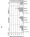
リアルタイムPCR定量反応は上記cDNA0.4〜0.8μlを鋳型として反応液40μlでTaqMan登録商標 Universal PCR Master Mix(PE Applied Biosystems社)を用いて説明書に記載の方法に従って行った。解析結果は内部標準であるGAPDHの量で標準化し図1に示した。その結果すべてのモデルにおいて心不全病態の進行に伴いOSF−2遺伝子の発現亢進が見られた。
実施例4:Myc−His−rOSF−2融合タンパク質発現ベクターの構築
実施例2で得られたrat OSF−2遺伝子のコード領域から翻訳されるタンパク質のカルボキシル末端にMycエピトープと6個のヒスチジンタグを有し、CMVプロモーターを有する発現ベクターを作製した。
まず、pTracer−CMV2ベクター(インビトロジェン社)を制限酵素EcoRIとEcoRVで消化したベクター断片に、実施例2で得られたrat OSF−2 5RACE #1を制限酵素EcoRIとHindIIIで消化して得られた約500bpの断片と実施例2で得られたrat OSF−2#1を制限酵素HindIIIとHpaIで消化して得られた約2780bpの断片をライゲーションキット(宝酒造(株))を用いて連結し、得られたプラスミドをpTracer−CMV2/rOSF−2と命名した。このようにして作製したpTracer−CMV2/rOSF−2を制限酵素EcoRIとSmaIで消化し、rat OSF−2遺伝子のコード領域を含む約2330bpの断片を得、実施例2で得られたrat OSF−2#1を鋳型としてその配列を基に設計したプライマー(8)5’−GACCCGGGAAGAACGCATCATC−3’(配列番号10)とrat OSF−2の終止コドンの直前にBstEIIサイトが挿入されるように設計したプライマー(9)5’−TGGGTGACCCTGAGAACGGCCTTCTCTTGATC−3’(配列番号11)を用いてPCRを行い、精製後、制限酵素SmaIとBstEIIで消化して約270bpの断片を得た。以上の2つの断片を発現ベクター構築用のプラスミドpcDNA4/Myc−His/type C(インビトロジェン社)を制限酵素EcoRIとBstEIIで消化したベクター断片にライゲーションキット(宝酒造(株))を用いて連結し、得られたプラスミドをpcDNA4/Myc−His/rOSF−2と命名した。挿入部分の全塩基配列は実施例1−5記載の方法により確認した。
実施例5:rOSF−2タンパク質発現ベクターの構築
実施例2で得られたrat OSF−2遺伝子の全コード領域を含みCMVプロモーターを有する発現ベクターを作製した。実施例4で得られたpTracer−CMV2/rOSF−2を制限酵素EcoRIとPmeIで消化し、rat OSF−2遺伝子のコード領域を含む約3300bpの断片を得、この断片を前記pcDNA4/Myc−His/type C(インビトロジェン社)を制限酵素EcoRIとPmeIで消化して得られたベクター断片にライゲーションキット(宝酒造(株))を用いて連結し、pcDNA4/rOSF−2と命名した。挿入部分の全塩基配列は実施例1−5記載の方法により確認した。
実施例6:ラット心臓へのOSF−2遺伝子導入による機能解析
6−1 発現プラスミドの大量調製
実施例5で作製した発現プラスミド(pcDNA4/rOSF−2)を大腸菌DH5α株に導入して増幅し、EndoFreeTMPlasmid Giga kit(QIAGEN社)を用いて精製後、生理食塩水に溶解した。
6−2 HVJ−liposomeの調製
先のプラスミド溶液200μgにHMG−1,−2mixture(1mg/ml、和光純薬(株))を50μl加え室温で1時間放置後BSS溶液を総量200μlになるよう加えた。その混合溶液を−20℃保存してある混合脂質(Avanti Polar Lipid Inc.、シグマ(株)、Hum.Gene Ther.8(17),2133−2141(1997),Cell biology,a laboratory handbook,2nd edition,vol.4,122−123(1998))の入ったガラス管に加えボルテックスミキサーにて30秒激しく振盪し37℃の恒温槽に30秒間静置した。これを8回繰り返した後、水を張った超音波発生装置にガラス管を入れ6秒間超音波処理を行い、再度30秒間ボルテックスミキサーにて振盪(120回/分)した。次に、精製したHVJ 1ml(20000HAU以上)(金田安史:HVJ−liposome法(遺伝子導入&発現解析実験法;羊土社)70−79(1997)、佐伯嘉修,金田安史:HVJ−リポソームベクター(遺伝子治療 開発研究ハンドブック:日本遺伝子治療学会編)429−438(1999)、Hum.Gene Ther.8(17),2133−2141(1997),Cell biology,a laboratory handbook,2nd edition,vol.4,122−123(1998))を直径30mmのプラスチックシャーレに分取しUVクロスリンカーに入れて蓋を取り1980mJ/cm2の紫外線を照射して不活化させた。この不活化HVJをガラス管に加え氷上で10分間置いた後BSSを1.3ml加えて37℃の振盪型恒温槽にて1時間振盪した。次に、10mlの超遠心チューブに30%ショ糖溶液を7.5ml入れHVJ−liposome液(ガラス管内の溶液)全量を静かに重層しスイングローターにて62,800×gで4℃、1.5時間超遠心した。HVJ−liposomeはショ糖溶液上に白色の粒子として集積され、これをパスツールピペットで回収しBSSにて総量3mlに調整後4℃保存した。
6−3 ラットへの遺伝子導入
12週齢のSprague−Dawleyラット(雄)の心臓左室(心尖および側尖部位)にHVJ−liposome/DNA溶液(濃度6.7μg/ml)を100μlずつ3ケ所(総量300μl)に30ゲージ注射針にて打ち込んだ(rOSF−2群)。また、コントロールとしてpcDNA4/His−Mycベクターを同様にして注入した(コントロール群)。
6−4 心エコー測定
導入後6週間目においてケタミン(50mg/ml)、キシラジン(10mg/ml)にて麻酔後、Core Vision Pro SSA 350A(プローブ:7MHz、東芝(株))を用いて心エコー測定を行った。結果は表1に示した通りでありコントロール群に比しrOSF−2群ではLVIDD(左室拡張末期内径)及びLVIDS(左室収縮末期内径)において有意な差が見られ心拡大を引き起こすことが明らかとなった。
【表1】
導入後6週間目においてペントバルビタール塩酸塩(50mg/ml)にて麻酔後心内圧および血圧の測定を行った。結果を表2に示す。コントロール群に比しrOSF−2群ではLVP(左心室圧)、LVEDP(左室拡張末期圧)、+LVdP/dT及び−LVdP/dTに有意な差が得られ心機能の低下が示唆された。
【表2】
7−1 アンチセンスヌクレオチドの設計
アンチセンスヌクレオチドは翻訳開始点のAUGを含み且つ3つの連続したグアニン(トリプレットG)を含まず且つヘアピン構造やセルファニールしない領域を選び、配列番号12乃至16に記す5種類を設計し、外部にて受託合成した。全てのアンチセンスヌクレオチドはホスホロチオエート型である。
7−2 アンチセンスヌクレオチドの選択
実施例5に記載したMyc−His−rOSF−2融合タンパク質発現ベクターpcDNA4/Myc−His/rOSF−2をCOS−1細胞に導入し、上記1で作製したアンチセンスヌクレオチドを導入してMyc−His−rOSF−2融合タンパク質の発現を阻害するアンチセンスヌクレオチドをスクリーニングした。Myc−His−rOSF−2融合タンパク質の発現量はMyc特異的モノクローナル抗体(インビトロジェン社)を用いたELISA(酵素結合免疫吸着検定法)で定量した。具体的にはCOS−1細胞を2x104細胞/ウェルになるように96ウェルプレートに播種し、16〜24時間後に320ngのDNA(pcDNA4/Myc−His/rOSF−2)をOPTI−MEM I培地(GIBCO社)に溶解し、1μlのLipofect AMINE 2000試薬(GIBCO社)を含む同量のOPTI−MEM I培地と混合し、室温で20分間処理後、COS−1細胞へ添加し、37℃においてインキュベートすることによりトランスフェクションを行った。次にDNA添加1〜4時間後に生理食塩水に溶解した一連のアンチセンスヌクレオチドを最終濃度100nMになるように各ウェルに添加し、さらに3日間37℃においてインキュベートした。
培養上清50μlをEIA/RIAプレート(コースター社)に添加し、37℃で1〜2時間放置することにより固定化し、200μlの0.1%Tweenを含むダルベッコーリン酸緩衝化食塩水(PBST)で2回洗浄した。非特異的吸着を遮断するために遮断用溶液(1%ウシ血清アルブミンを含むPBST)で37℃1時間処理し、200μlのPBSTで3回洗浄した。次に50μlの遮断用溶液で1:4000に希釈したホースラディッシュパーオキシダーゼ標識された抗Mycモノクローナル抗体(インビトロジェン社)を添加し、室温1時間放置することにより結合させ、200μlのPBSTで3回、PBSで1回洗浄した。50μlのTMB(3,3’,5,5’−テトラメチルベンジディン)試薬(KPL社)を添加し、室温にて30分放置した後、50μlの0.1N硫酸を添加することにより反応を停止し、マイクロプレートリーダーにて450nmの吸光度を測定した。
その結果、図2に示すように翻訳開始コドンを標的とした一連のアンチセンスヌクレオチドはいずれもOSF−2タンパク産生阻害活性を有したが、配列番号12のアンチセンスヌクレオチドが最も強く発現を阻害した。
次に配列番号12に対応するセンス鎖であるセンスヌクレオチド(配列番号17)及び配列番号12の5’、3’の向きを逆にしたスクランブルヌクレオチド(配列番号18)を作製し、各ヌクレオチドの最終濃度10nMにて上記と同様の実験を行った。その結果、図3に示すようにアンチセンスヌクレオチドはセンスヌクレオチド、スクランブルヌクレオチドよりも強いOSF−2タンパク産生阻害活性を有していることが判明した。
実施例8:アンチセンスヌクレオチドによるOSF−2遺伝子の発現抑制
8−1 HVJ−liposomeの調製
実施例6.1で調製したプラスミド溶液200μgと実施例7にて選別したアンチセンスヌクレオチド溶液1.4mgにHMG−1,−2mixture(1mg/ml、和光純薬(株))を50μl加え、以下実施例6に従ってHVJ−liposomeを調製した。また、センスヌクレオチド、スクランブルヌクレオチドも同様に調製した。
8−2 ラットへの遺伝子導入
13週齢のSprague−Dawleyラット(雄)の心臓左室(心尖および側尖部位)にHVJ−liposome/DNA溶液を100μlずつ3ケ所(総量300μl)に30ゲージ注射針にて打ち込んだ(rOSF−2/アンチセンス群、rOSF−2/センス群、rOSF−2/スクランブル群)。また、ポジティブコントロール群としてpcDNA4/rOSF−2をネガティブコントロール群としてpcDNA4/His−Mycベクターを同様にして注入した。
8−3 心エコー測定
導入後3週間目においてケタミン(50mg/ml)、キシラジン(10mg/ml)にて麻酔後、Core Vision Pro SSA 350A(プローブ:7MHz、東芝(株))を用いて心エコー測定を行った。結果を表3に示す。ポジティブコントロール群(rOSF−2)あるいはrOSF−2/センス群、rOSF−2/スクランブル群に比しrOSF−2/アンチセンス群ではネガティブコントロール群(コントロール)と同様LVIDD(左室拡張末期内径)及びLVIDS(左室収縮末期内径)が有意に小さく心拡大を抑制することが明らかとなった。
【表3】
9−1 HVJ−liposomeの調製
実施例7にて選別したアンチセンスヌクレオチドをBSS緩衝液にて溶かし、そのうちの1.4mgにHMG−1,−2mixture(1mg/ml、和光純薬(株))を50μl加え、以下実施例6.2に従ってHVJ−liposomeを調製した。また、センスヌクレオチドも同様に調製した。
9−2 ラットへの遺伝子導入
8週齢から8%食塩負荷飼料にて飼育した13週齢のDahl−Sラット(DIS/Eis,エーザイ、雄)の心臓左室(心尖および側尖部位)にHVJ−liposome/ヌクレオチド溶液を75μlずつ4ケ所(総量300μl)に30ゲージ注射針にて打ち込んだ(アンチセンス群13匹、センス群13匹)。導入後、引き続き8%食塩負荷飼料にて飼育し、その生存率を調べた結果、遺伝子導入後30週後の生存率は、センス群(61.5%)、アンチセンス群(100%)で、アンチセンス群で生存率の改善が見られた。
実施例10: ヒトOSF−2レポータプラスミドの作製
10−1 ヒトOSF−2遺伝子上流領域のクローニング
EMBL−3ベクターに挿入されたhuman genomic library(クローンテック社)より作製した約25,000クローンのファージサブプール10個(計約25万クローン)をヒトOSF−2遺伝子の上流領域を含むドラフト配列(Genbank Accession No.AL138679)の塩基配列を基に設計したプライマー(10)5’−AAGAATATTAAAAGTTATTTGTGGGCAGGAGACAGATG−3’(配列番号22)と(11)5’−CATTTAAAAGCCTATCAAGTGTCAGGTCCACTCTC−3’(配列番号23)を用いてPCRスクリーニングすることにより、2個のポジティブなサブプールを同定し、このサブプールを上記PCRによって増幅された断片をAlkPhos DirectTM(アマシャムファルマシア社)を用いてアルカリフォスファターゼ標識したプローブを用いてハイブリダイゼーションによるスクリーニングを行い、2個のポジティブクローンhuman OSF−2 gene#1、2を得た。human OSF−2 gene#2の挿入断片をpBluescript II(ストラタジーン社)のXhoI認識部位に組み込み、pB−hOSFg#2を得、実施例1−5の方法にしたがって部分塩基配列を決定した。得られたクローンの長さは約13.5kbであり、ヒトドラフト配列(Genbank Accession No.AL138679 version11)の16397番から30002番までに相当するものであり、ヒトOSF−2遺伝子のイントロン2から上流領域が含まれていることがわかった。また、開始コドン(ATG)から上流約2kbに存在するSpeI認識部位から開始コドンまでについては全塩基配列を決定した。この約2kbの塩基配列を配列番号24に示す。この約2kbの塩基配列は、Genbank Accession No.AL138679 version11の配列に対して4塩基において変異が存在し、各々、SpeI認識部位を1番として、764番G(T)、936番T(C)、1099番C(T)及び2106番G(C)であった((括弧内はAL138679 version11の塩基配列)。
10−2 ヒトOSF−2レポータープラスミドの作製
実施例10−1で得たhuman OSF−2 gene#2を鋳型として、開始コドンの約2kb上流から開始コドンの直前までをプライマー(12)5’−TGAGCATATATCATGCTTTC−3’(配列番号25)と(13)5’−AATCATCTCGAGTCTCTCCGTTGCAGTTAGTCCCC−3’(配列番号26)を用いたPCR法により増幅した。なお、プライマー(13)は5’−末端に制限酵素XhoI認識部位を挿入するように設計されており、この増幅断片を制限酵素SpeI及びXhoIで消化して得られる約2kbの断片を、pGL3−enhancer(プロメガ社)のルシフェラーゼ遺伝子の上流にある制限酵素NheI及びXhoIで切断したベクター断片にライゲーションキット(宝酒造(株))を用いて連結し、pGL3−hOSFgEを得た。挿入部分の塩基配列を実施例1−5の方法にしたがって決定し、実施例10−1で決定した塩基配列と同一であることを確認した。
実施例11: ヒトOSF−2遺伝子の発現に影響を与える化合物のスクリーニング
11−1 COS−1細胞を用いたレポーターアッセイ
COS−1細胞を3x10⁴細胞/ウェルになるように96ウェルプレートに播種し、16〜24時間37℃で培養後、実施例10で示したOSF−2レポータープラスミドpGL3−hOSFgEをリポフェクトアミン2000試薬(GIBCO社)を用いて一過性に導入し、24時間37℃で培養後、培地交換を行い、24時間後に候補化合物を添加し、その24時間後にルシフェラーゼ活性を測定した。ルシフェラーゼ活性の測定はルシフェラーゼアッセイシステム(プロメガ社)のプロトコールに従って行い、検出は1420マルチラベルカウンター(Wallac社)を用いた。コントロールとしてSV40プロモーターの下流にルシフェラーゼ遺伝子が連結されたpGL3−control(プロメガ社)を用いて同様の実験を行った。この方法により多数の化合物をスクリーニングした結果、2−クロロ−アデノシントリフォスフェート四ナトリウム(2−Chloroadenosine 5’−triphosphate tetrasodium)、メトリフジル(N−[(2−Methylphenyl)methyl]−adenosine)がOSF−2プロモーター特異的にルシフェラーゼ活性を抑制することが明らかになった。なお、これらの化合物は以下の化学構造式を有する。
2−クロロ−アデノシントリフォスフェート四ナトリウム(2−Chloroadenosine 5’−triphosphate tetrasodium):
本来OSF−2を発現しているマウス骨芽細胞様細胞MC3T3−E1(理研細胞開発銀行より購入)を5x10⁵細胞/ウェルになるように6ウェルプレートに播種し、24時間37℃で培養後に培地交換を行い、24時間後に上記化合物を30μMの濃度で添加し、その24時間後に細胞を回収し、細胞よりRNeasy登録商標 Mini(キアゲン社)を用いて総RNA調製し、実施例3−2の方法にしたがって、OSF−2 mRNAおよび内部標準GAPDH mRNAの発現量をPCR定量システムにより定量した(OSF−2 mRNA発現量は内部標準であるGAPDH mRNAの発現量で標準化した)。その結果、2−クロロ−アデノシントリフォスフェート四ナトリウム、メトリフジルはOSF−2 mRNAの発現を抑制することを確認した。
このように、本発明で示したスクリーニング系を用いることにより、OSF−2遺伝子の発現に影響を与える化合物を見出すことができることが示された。
【配列表】
【図面の簡単な説明】
図1は、各動物モデルにおける、GAPDH遺伝子の発現量で標準化したOSF−2遺伝子の発現量を示す図である。
図2は、配列番号12乃至16の各アンチセンスヌクレオチドに係るOSF−2タンパク質産生阻害活性を示す図である。Cはコントロールを意味する。
図3は、アンチセンスヌクレオチド(配列番号12)、センスヌクレオチド(配列番号17)及びスクランブルヌクレオチド(配列番号18)に係るOSF−2タンパク質産生阻害活性を示す図である。 Technical field
The present invention relates to a preventive or therapeutic agent for heart failure, a method for screening a substance relating to an active ingredient of the preventive or therapeutic agent, a diagnostic agent or diagnostic kit for heart failure, a non-human transformed animal, and the like.
Background art
Chronic heart failure is a disease in which the heart is unable to pump a sufficient amount of blood to each organ due to a decrease in myocardial contractile force, and in the treatment, a cardiotonic agent such as a digitalis preparation or a xanthine preparation that increases the myocardial contractile force is conventionally used. Has been used. However, these drugs have been shown to deteriorate life prognosis in long-term administration due to excessive consumption of myocardial energy. Recently, treatment with β-blockers and ACE inhibitors that reduce the excessive load on the heart caused by the sympathetic nervous system and renin-angiotensin system, which are promoted in the state of heart failure, has become mainstream. However, the long-term life prognosis of heart failure patients is still poor, and the development of therapeutic agents for heart failure based on a new mechanism is desired.
Therefore, the subject in this invention is related with provision of the preventive or therapeutic agent etc. of the new heart failure different from the existing mechanism. More specifically, the present invention relates to a prophylactic or therapeutic agent for heart failure by suppressing the deterioration of heart failure caused by the OSF-2 gene or protein, a method for screening a substance related to the active ingredient of the drug, the OSF-2 gene Diagnostic method or diagnostic kit used for heart failure diagnosis, and non-human transformed animal capable of forcibly expressing OSF-2 gene The purpose is to provide.
Disclosure of the invention
With the expectation that various gene expression and protein expression changes occur in the heart as the heart failure pathology progresses, and these changes are closely related to the heart failure pathology, Using a heart failure model, we searched for genes whose expression levels change with the progression of the heart failure condition. As a result, it was found that the expression of the OSF-2 gene, which is known as a gene specifically expressed in osteoblasts, increases with the progression of heart failure. As a result of further studies, the present inventors have found that the gene or protein affects the progression of heart failure, and completed the present invention because a drug that suppresses its expression and function becomes a new therapeutic agent for heart failure. It came to do.
In the development of therapeutic agents for heart failure, various pathological models are used. In particular, in the initial screening, rat models are often used because of the ease of model preparation and ease of analysis. On the contrary, the rat model is recognized as a model reflecting human pathology, although there are limitations on each.
Known examples of heart failure pathological model rats include aortic stenosis rats, arteriovenous shunt rats, coronary artery ligation rats, spontaneously hypertensive rats (SHR), and Dahl salt-sensitive rats. Dahl rats are high in hypertension-prone salt-sensitive rats (Dahl-S) in the third generation by loading 8% high salt-containing diet and subcrossing in Spague-Dawley rats. No salt-resistant rats (Dahl-R). For Dahl-S rats, a study by Kihara et al. Of Kyoto University revealed that the left ventricular enlargement with contractile insufficiency was observed after a compensatory left ventricular hypertrophy by loading an 8% high salt-containing diet from 6 weeks of age. That is, it has been shown that the disease shifts to decompensated heart failure and dies due to pulmonary congestion (Am. J. Physuol.,267, H2471-2482 (1994)). The inventors of the present invention do not need a special method for producing a model, can develop heart failure in a short period of time, and can be clearly divided into a compensated hypertrophy stage and a decompensated heart failure stage in one individual, and human hypertension Dal salt-sensitive rats were selected as a heart failure pathologic model because the onset process is similar to congenital heart failure.
As a method for detecting a gene whose expression is changed in a certain disease state, a subtraction method, a differential display method, a dot blot method, a microarray method, and the like are known. In the present invention, the subtraction method was used to successfully detect a gene whose expression changes during the transition from cardiac hypertrophy to heart failure. One of them was a novel rat OSF-2 gene that was found to have high homology with the mouse OSF-2 gene reported as an osteoblast-specific factor.
The OSF-2 gene is a gene that has been isolated and identified as a gene that is specifically expressed in the mouse osteoblast cell line MC3T3-E1 (Biochem. J. 294, 271-278, 1993, Japanese Patent Laid-Open No. 5-268882). ), Adhesion promoting activity has been reported in osteoblasts (J. Bone. Miner. Res.).14, 1239-1249, 1999). Further, it has been reported that the expression of OSF-2 gene is increased in the analysis by the differential display method in the rabbit abdominal aorta injury model (J. Hum. Genet.439-13, 1998). Furthermore, recently, in experiments using mice, the expression of more than 30 genes including OSF-2 has changed during cardiac hypertrophy (J. Mol. Cell. Cardiol.,32, 805-815, 2000), and in the rat myocardial infarction model, the expression of more than 200 genes including OSF-2 is changed (Circ. Res.,86939-945, 2000).
However, after cardiac hypertrophy or myocardial infarction, it has not been known that further expression induction occurs along with progression to heart failure pathology, and in particular, OSF-2 gene or OSF-2 protein progresses along with progression of heart failure pathology. It has not been known at all whether it acts as a factor that promotes the disease or as a factor that suppresses the progression of the disease state.
Thus, it was considered that the development of a new therapeutic agent for heart failure would be possible by knowing the relationship between the expression of this gene in the heart and the pathophysiology of heart failure, and the present invention was completed by adding further studies.
First, in order to confirm that the change of the OSF-2 gene is not a phenomenon peculiar to the Dahl heart failure model rat, OSF− accompanying the progression of the heart failure pathology using aortic stenosis rats, arteriovenous shunt rats, and spontaneously hypertensive rats (SHR) The expression of 2 genes was examined. As a result, the same gene expression was observed in all the models examined, strongly suggesting that it might be a universal phenomenon in heart failure.
Next, in order to examine what effect the OSF-2 gene has on the heart, gene introduction into the rat heart was performed. First, since the rat OSF-2 gene has not been known so far, a full-length gene was newly isolated, an appropriate expression vector was constructed, and then the OSF-2 gene was introduced into the rat heart. As a result of measurement of echocardiography and hemodynamics, non-thinness of the left ventricular wall, enlargement of the left ventricular cavity, and reduction of cardiac function were observed in the OSF-2 transgenic rats, and the pathological condition of dilated cardiomyopathy It was found that OSF-2 gene or OSF-2 protein is an exacerbation factor of heart failure. Such analysis is difficult in animals other than rats, and the significance of isolating rat genes is great. Furthermore, it has been found that by introducing an antisense nucleotide that suppresses the expression of the OSF-2 gene into the heart of a heart failure model, the death due to heart failure is delayed. Based on this, it was found that the expression of OSF-2 gene or the production of OSF-2 protein which occurs with the progression of the heart failure pathological condition is exacerbating heart failure, and the suppression of the expression of OSF-2 gene or the production of OSF-2 protein is suppressed. Alternatively, it has been suggested that a drug containing a substance having a function-inhibiting action as an active ingredient sufficiently suppresses the deterioration of heart failure, leading to the present invention.
That is, the following matters can be mentioned as the present invention.
(1) A preventive or therapeutic agent for heart failure comprising as an active ingredient a substance that suppresses the expression of OSF-2 gene, the production of OSF-2 protein, the function of OSF-2 protein or the function of a target molecule of OSF-2 protein.
(2) A preventive or therapeutic agent for heart failure comprising a substance that suppresses the expression of the OSF-2 gene as an active ingredient.
(3) A preventive or therapeutic agent for heart failure comprising a substance that suppresses the production of OSF-2 protein as an active ingredient.
(4) The preventive or therapeutic agent for heart failure according to (1) to (3) above, wherein the substance that suppresses the expression of OSF-2 gene or the production of OSF-2 protein is an antisense nucleotide.
(5) The preventive agent for heart failure according to (4) above, wherein the antisense nucleotide comprises a nucleotide complementary to 12 or more consecutive base sequences in the base sequence described in SEQ ID NO: 5, 19, 20 or 21 Therapeutic agent.
(6) The preventive or therapeutic agent for heart failure according to the above (5), wherein the antisense nucleotide comprises at least one sequence selected from the group consisting of SEQ ID NOs: 12 to 16.
(7) The substance for suppressing heart failure according to (1) or (2) above, wherein the substance that suppresses the expression of the OSF-2 gene is a compound represented by the following general formula (I) or a biologically acceptable salt thereof: Preventive or therapeutic agent.
(8) A substance that suppresses the expression of the OSF-2 gene is represented by R in the general formula (I)3And R4The prophylactic or therapeutic agent for heart failure according to the above (7), wherein both are compounds each having a hydrogen atom.
(9) The substance that suppresses the expression of the OSF-2 gene is represented by R in the general formula (I)1, R3And R4Is a hydrogen atom, R2The prophylactic or therapeutic agent for heart failure according to the above (7), wherein is a compound wherein is a chlorine atom and n is 3.
(10) The substance that suppresses the expression of the OSF-2 gene is represented by R in the general formula (1)1Is a (2-methylphenyl) methyl group, R3And R4The prophylactic or therapeutic agent for heart failure according to the above (7), wherein is a compound wherein is a hydrogen atom and n is 0.
(11) The preventive or therapeutic agent for heart failure according to (1) above, comprising a substance that suppresses the function of OSF-2 protein as an active ingredient.
(12) The preventive or therapeutic agent for heart failure according to the above (1), which comprises a substance that suppresses the function of the target molecule of the OSF-2 protein as an active ingredient.
(13) The above (11) or (12), wherein the substance that suppresses the function of the OSF-2 protein or the target molecule of the OSF-2 protein is an antibody against the OSF-2 protein or the target molecule of the OSF-2 protein. Preventive or therapeutic agent for heart failure.
(14) A substance obtained by a synthetic or genetic recombination technique, a substance derived from nature, or a substance that is a derivative thereof, (1) a cell having an OSF-2 gene expression control region and an OSF-2 gene or reporter gene Or in vitro expression system, or (2) OSF-2 protein or OSF-2 protein target molecule, and the expression level of OSF-2 gene or reporter gene or target molecule of OSF-2 protein or OSF-2 protein A method for screening a substance having an effect of preventing or treating heart failure, which comprises detecting the amount of.
(15) A substance obtained by a synthetic or genetic recombination technique, a substance derived from nature, or a substance that is a derivative thereof is brought into contact with a cell having the expression control region of the OSF-2 gene and the OSF-2 gene, and OSF- The method according to (14) above, comprising detecting the expression level of two genes.
(16) After constructing an expression vector in which the expression control region of the OSF-2 gene is linked upstream and / or downstream of the translation region of the reporter gene, the expression vector is introduced into a suitable host cell and cultured, and synthesized or gened into the cultured cell. The substance according to (14) above, which comprises adding a substance obtained by a recombinant technique, a substance derived from nature or a derivative thereof, and detecting the expression level of the reporter gene or the reporter protein after a certain period of time. Method.
(17) A substance obtained by a synthetic or genetic recombination technique, a substance derived from nature, or a substance that is a derivative thereof was contacted with a target molecule of OSF-2 protein or OSF-2 protein, and was or was not bound The method for screening a substance according to the above (14), comprising detecting the amount of a target molecule of OSF-2 protein or the amount of OSF-2 protein.
(18) The OSF-2 protein or the OSF-2 protein target molecule was immobilized on a support, and the immobilized OSF-2 protein or OSF-2 protein target molecule was obtained by synthesis or genetic recombination technology. A substance, a substance derived from nature or a derivative thereof, and a target molecule of OSF-2 protein or OSF-2 protein are added, and a target molecule of OSF-2 protein or OSF-2 protein that is bound or not bound The method according to (17) above, comprising detecting an amount.
(19) Diagnosis of heart failure including collecting biological tissue and / or body fluid and measuring the expression level of the OSF-2 gene in the tissue, or measuring a mutation associated with the OSF-2 gene or the control region Method.
(20) A method for diagnosing heart failure, comprising collecting a biological tissue and / or body fluid and measuring the OSF-2 protein or the fragment in the body fluid.
(21) A diagnostic agent or diagnostic kit for heart failure comprising means for measuring the expression level of OSF-2 gene or the production level of OSF-2 protein.
(22) A gene encoding the following protein (a) or (b):
(A) a protein consisting of the amino acid sequence of SEQ ID NO: 6;
(B) A protein comprising an amino acid sequence in which one or several amino acids are deleted, substituted or added in the amino acid sequence of SEQ ID NO: 6 and having OSF-2 protein activity.
(23) A gene comprising the following DNA (a) or (b):
(A) DNA consisting of the base sequence of SEQ ID NO: 5;
(B) DNA that hybridizes with the nucleotide sequence of SEQ ID NO: 5 under a stringent condition and encodes a protein having OSF-2 protein activity.
(24) The following recombinant protein (a) or (b):
(A) a protein consisting of the amino acid sequence of SEQ ID NO: 6;
(B) A protein comprising an amino acid sequence in which one or several amino acids are deleted, substituted or added in the amino acid sequence of SEQ ID NO: 6 and having OSF-2 protein activity.
(25) A non-human transformed animal that overexpresses the OSF-2 gene and can be used as a heart failure pathological model.
(26) A method for evaluating or screening a substance that can be an active ingredient of a preventive or therapeutic agent for heart failure using the non-human transformed animal according to (25).
(27) A substance obtainable by the method described in (14) to (18) or (26) above.
(28) A preventive or therapeutic agent for heart failure comprising the substance described in (27) as an active ingredient.
(29) A method for treating heart failure comprising administering the substance described in (1) to (13) or (27) above.
(30) The treatment of heart failure according to the above (29), which comprises directly introducing an antisense nucleotide of the OSF-2 gene or introducing it into a vector suitable for gene therapy (for example, an adenovirus-derived vector) or liposome. Method.
(31) Use of the substance described in (1) to (13) or (27) for the manufacture of a prophylactic or therapeutic agent for heart failure.
BEST MODE FOR CARRYING OUT THE INVENTION
In the present invention, the following substances (1) to (3) can be used as substances that can be used as an active ingredient of a preventive or therapeutic agent for heart failure.
(1) Substances that suppress the expression of the OSF-2 gene
The substance that suppresses the expression of the OSF-2 gene may be a compound obtained by a synthetic or genetic recombination technique or a naturally-derived compound and derivatives thereof. Examples of such a substance include the OSF-2 gene Substances that act on the promoter and enhancer regions of the RNA and suppress the transcription of the OSF-2 gene into mRNA (for example, antisense nucleotides (DNA, RNA, phosphorothioate type modified nucleotides, PNA (protein nucleic acid), etc.) Or a substance exhibiting the same action via some factor in the cell (such as transcription factor) (for example, binding to transcription factor or co-activator, binding to DNA, other transcription factor, or co-activator) And the like.
As used herein, the term “nucleotide” when referring to an antisense nucleotide refers to a nucleotide formed from a naturally occurring base and a sugar moiety linked by an original phosphodiester bond and analogs thereof. Thus, the first group that this term includes are naturally occurring species or synthetic species produced from naturally occurring subunits or their homologs. The subunit is a base-sugar combination bound to an adjacent subunit by a phosphodiester bond or other bond. A second group of nucleotides is also an analog thereof, which means a residue that functions like a nucleotide but has a non-naturally occurring portion. These include nucleotides that are chemically modified at the phosphate group, sugar moiety, 3 ', 5' end to increase stability. For example, phosphorothioate having one oxygen atom of a phosphodiester group between nucleotides substituted with sulfur, -CH3And methyl phosphonate substituted with. In addition, nucleotide analogs may be used that include modified base forms, ie, purines and pyrimidines other than those normally found in nature.
Preferred antisense nucleotides used in the present invention are nucleotides complementary to 12 or more consecutive nucleotide sequences in the nucleotide sequence set forth in SEQ ID NO: 5, 19, 20 or 21, preferably 16 or more consecutive nucleotide sequences. Nucleotides complementary to each other, more preferably nucleotides complementary to 18 or more consecutive base sequences. Particularly preferred antisense nucleotides have at least one sequence selected from the group consisting of SEQ ID NOs: 12-16.
Furthermore, in the present invention, instead of antisense nucleotides, peptide nucleic acids (Bioconjug. Chem.,4373-378, 1994) can also be used.
Specific examples of the compound that suppresses the expression of the OSF-2 gene include a compound represented by the following general formula (I) or a biologically acceptable salt thereof.
R2Represents a hydrogen atom, a halogen atom or an amino group. R2Preferred examples of are a hydrogen atom and a chlorine atom.
R3, R4Independently represents a hydrogen atom or an acyl group, or R represents3, R4Together represent an isopropylidene group. Examples of the acyl group include lower alkanoyl groups such as an acetyl group and a propionyl group.
n represents 0, 1, 2, 3 and 0 and 3 are particularly preferable. When n is 1, 2, or 3, one or more of the phosphoric acid hydrogen atoms may form a salt with an alkali metal such as sodium, potassium, calcium, or magnesium, an alkaline earth metal, or an organic amine. .
(2) Substances that suppress the production of OSF-2 protein
The substance that suppresses the production of OSF-2 protein may be a compound obtained by a synthetic or genetic recombination technique, or a naturally-derived compound and derivatives thereof. Examples of such a substance include the OSF-2 gene. The substance which has the effect | action which inhibits translation of OSF-2 mRNA to OSF-2 protein by the antisense nucleotide of this is mentioned. Here, the term “nucleotide” when referring to an antisense nucleotide is synonymous with “nucleotide” described in (1) above. Examples of antisense nucleotides include RNA, DNA, phosphorothioate type modified nucleotides, PNA (protein nucleic acid), etc., structural gene region of OSF-2 gene, 3′-untranslated region, 5′-untranslated region Alternatively, any intron / exon binding site can be designed as a target. The antisense strand can be introduced into the target tissue either directly or by introducing a vector designed to be transcribed into antisense RNA into the target tissue.
A ribozyme that recognizes and cleaves a specific structure of OSF-2 mRNA can also be used. A ribozyme is a nucleic acid molecule having an enzymatic activity capable of cleaving other RNA molecules in a target base sequence-specific manner, and can be formed, for example, with a hammerhead or hairpin motif (Nature,324, 429, 1986; Ann. Rep. Med. Chem. ,30, 285-294, 1995; Med. Chem. ,38, 2023-2037, 1995). Ribozymes may be introduced directly into the target tissue or in the form of complexes with lipids or liposomes. Alternatively, a DNA or RNA vector that expresses a ribozyme can be introduced into the target tissue.
(3) A substance that suppresses the function of the OSF-2 protein and a substance that suppresses the function of the target molecule of the OSF-2 protein
In the present invention, a substance that suppresses the function of the OSF-2 protein or a substance that suppresses the function of the target molecule of the OSF-2 protein can be used as a prophylactic or therapeutic agent for heart failure. The target molecule of OSF-2 protein refers to a molecule whose structure or activity is affected by OSF-2 protein, and includes, but is not limited to, a substrate, a binding partner, a receptor, and the like.
The substance that suppresses the function of the OSF-2 protein or the substance that suppresses the function of the target molecule of the OSF-2 protein may be either a compound obtained by a synthetic or genetic recombination technique, a naturally derived compound, or a derivative thereof. However, as such a substance, for example, binding of a target molecule (for example, a receptor) of OSF-2 protein on which OSF-2 protein and OSF-2 protein act, and signal transduction in a cell are inhibited, and OSF- Examples include substances that inhibit the activity of 2 proteins. For example, an antibody against OSF-2 protein or an antibody against a target molecule of OSF-2 protein can be mentioned. The antibody may be a polyclonal antibody or a monoclonal antibody. As a method for producing these antibodies, for example, these proteins are purified from expression cells or a culture broth of recombinants, immunized with a suitable adjuvant to rabbits and the like, and antibody fractions are obtained from the serum according to a standard method. Obtainable. Alternatively, production of monoclonal antibodies using mice, rats, etc., gene recombination techniques, production of human-type antibodies or single chain antibodies using genetically modified animals, etc. can also be used. It is not limited to. In addition, proteases that specifically degrade OSF-2 protein and OSF-2 protein target molecules, inactive OSF-2 proteins (including mutants), OSF-2 target molecule decoys, and soluble receptor molecules And so on.
When the substance according to the above (1) to (3) is a peptide, an enzyme is used in order to prevent or hardly undergo degradation by peptidase or protease in the living body after the drug according to the present invention is administered to the administration subject. Also included are peptide analogs with modified sites of action. For example, the analogs include peptide analogs in which one or more peptide bonds associated with the enzyme action site are replaced with other types of covalent bonds, and one or more amino acids associated with the enzyme action site. Examples thereof include peptide analogs that are D-form amino acids.
(4) Formulation
A preparation containing the above-mentioned substance as an active ingredient is prepared using carriers, excipients, and other additives used in normal preparation.
The active ingredient of the pharmaceutical composition according to the present invention may be a free form or a pharmaceutically acceptable salt thereof. Examples of salts with inorganic acids include salts with hydrochloric acid, sulfuric acid, and phosphoric acid. Examples of salts with organic acids include acid addition salts with formic acid, acetic acid, butyric acid, succinic acid, citric acid, and the like. Can be used. The salt may be in the form of a metal salt such as sodium, potassium, lithium or calcium, or a salt with an organic base.
The active ingredient is mixed with known pharmacologically acceptable carriers, excipients, diluents and the like, and is generally used in medicine, for example, oral administration, intravenous administration, intramuscular administration. Or it is preferable to administer by parenteral administration methods, such as subcutaneous administration. The pharmaceutical composition of the present invention includes, for example, a physiologically acceptable carrier, flavoring agent, excipient, stabilizer, diluent, emulsion, solution, suspension, syrup, etc. It can be produced by mixing appropriately, and can be used as tablets, powders, granules, solutions and the like. As an additive that can be mixed with tablets, for example, a binder such as gelatin, a lubricant such as corn starch, etc. can be used, and it can be coated with a sugar coating or a film of a medical or enteric substance. Good. In the case of a capsule dosage form, the composition can further contain a liquid carrier. A sterile composition for injection can also be produced by applying a normal formulation. Aqueous solutions for injection include isotonic solutions containing glucose and the like, and may be used in combination with a suitable solubilizing agent such as polyethylene glycol. Moreover, you may mix | blend with a buffer, a stabilizer, a preservative, antioxidant, a soothing agent, etc. When the active ingredient is easily degraded in the gastrointestinal tract during oral administration, it can be administered orally as a preparation that is not easily degraded in the gastrointestinal tract, for example, a microcapsule in which the active ingredient is encapsulated in liposomes. Moreover, the administration method which absorbs from mucous membranes other than digestive tracts, such as a rectum, a nasal cavity, and sublingual, is also possible. In this case, it can be administered in the form of suppositories, nasal sprays, sublingual tablets.
Moreover, when using for gene therapy, an antisense nucleotide etc. can also be integrated and used for vectors suitable for known gene therapy, such as adenovirus.
When the dosage of the pharmaceutical composition of the present invention is used for treatment, a dosage effective for treatment is determined, but the dosage varies depending on the age, body weight, degree of symptom, administration route, etc. of the administration subject, It is decided according to individual cases. Usually, in the case of oral administration, the daily dose for an adult is about 0.1 to 1000 mg, which may be administered in 1 to several divided doses. In continuous intravenous administration, it can be administered in the range of 0.01 μg / kg / min to 1.0 μg / kg / min, and it is preferably administered at 0.025 μg / kg / min to 0.1 μg / kg / min. preferable.
(5) Screening method
As a screening method for a substance that can be used as an active ingredient of the preventive agent or therapeutic agent according to the present invention, for example, the following method can be considered.
The OSF-2 gene or OSF-2 protein or derivative thereof used for screening for a substance that suppresses the expression of the OSF-2 gene or a substance that suppresses the production of OSF-2 protein may be derived from any species. Examples thereof include those derived from mammals such as humans, mice, rats, rabbits, pigs, dogs, monkeys, and guinea pigs. That is, in addition to the rat-derived gene (SEQ ID NO: 5) or protein (SEQ ID NO: 6) newly found this time, a known gene or protein can be mentioned. Since various splice variants are known for the OSF-2 gene, the OSF-2 gene and OSF-2 protein according to the present invention include these splice variants and proteins translated therefrom. For example, the mouse-derived OSF-2 gene and OSF-2 protein include those shown in SEQ ID NO: 19, and the human-derived OSF-2 gene and OSF-2 protein include SEQ ID NOs: 20 (placenta-derived) and 21 ( Primary tumor origin). Furthermore, the partial sequence of the OSF-2 gene can be used alone or as a chimeric gene with another gene. Similarly, a partial sequence of OSF-2 protein can be used alone or fused with other peptides.
Among these, it is preferable to use a human-derived one for use in research and development of preventive or therapeutic agents for humans. In addition, from the necessity of research and development using animal models, that is, non-human transformed animals that forcibly express the OSF-2 gene and have heart failure symptoms, use those derived from animals such as mice and rats. Is preferred.
In screening for a substance that suppresses the expression of the OSF-2 gene or a substance that suppresses the production of OSF-2 protein, a method using a reporter gene is generally used. As a reporter gene, for example, chloramphenicol acetyltransferase (CAT), β-galactosidase (β-Gal), luciferase and the like can be used. The substance that suppresses the expression of the OSF-2 gene, for example, constructs an expression vector in which the expression control region (promoter, enhancer region, etc.) of the OSF-2 gene is linked upstream and / or downstream of the translation region of the reporter gene, Introduce into an appropriate cultured cell, etc., and add the sample substance (the substance may be either a compound obtained by synthetic or genetic recombination techniques or a naturally derived compound or a derivative thereof). Thus, screening can be performed by measuring the expression level of the reporter gene or the amount of reporter protein after a certain period of time. The expression control region (promoter, enhancer region, etc.) of the OSF-2 gene can be obtained from a commercially available gene library by plaque hybridization using an OSF-2 cDNA fragment as a probe. For example, in the example of the present invention, a reporter vector in which the promoter region of the OSF-2 gene is connected to the upstream of the luciferase gene is introduced into COS-1 cells, and the luciferase activity is measured after adding the compound. A compound that suppresses gene expression was found. The substance according to the present invention includes a decoy (Science, a decoy nucleic acid drug against a transcription factor acting on the OSF-2 gene).250, 997-1000, 1990). The amount of the reporter protein can be measured as an enzyme activity, and the protein expression level can also be measured using an antibody or the like.
In addition, for antisense nucleotides or ribozymes that suppress the production of OSF-2 protein, antisense nucleotides or ribozymes are introduced into cultured cells expressing OSF-2 protein, for example, by electroporation or liposome method And it can screen by measuring the expression level of OSF-2 protein. For example, in the examples of the present invention, a series of antisense nucleotides targeting the translation initiation codon of OSF-2 is introduced into COS-1 cells in which OSF-2 protein is overexpressed to express OSF-2 protein. Although an antisense nucleotide that suppresses the above was found, any antisense nucleotide having such an action may be used. Examples thereof include substances that suppress transcription, suppress pre-mRNA splicing, and suppress mRNA passage through the nuclear membrane. A similar technique can also be used for ribozyme screening. As the cultured cells to be used, as described above, cells into which the OSF-2 gene has been introduced can be used, and any cell that originally expresses the OSF-2 gene, such as MC3T3-E1 cells (Biochem. J). 294, 271-278, 1993) or the like.
As a screening method for a substance that suppresses the function of the OSF-2 protein or a substance that suppresses the function of the target molecule of the OSF-2 protein, for example, either the target molecule of the OSF-2 protein or the OSF-2 protein is plated. After immobilization, the substance (which may be a synthetic compound, a natural product-derived compound or a derivative thereof) and the OSF-2 protein or a target molecule thereof are added and bound or bound. The amount of OSF-2 protein or the target molecule of OSF-2 protein that has not been detected can be detected by ELISA, RIA, fluorescent labeling, or the like. Also, using OSF-2 target molecule-expressing cells, labeled OSF-2 and a drug are allowed to coexist, and after appropriate washing treatment is performed for a certain time, FACS, absorbance, color development method, etc. It is also possible by measuring the number of labeled cells and the degree of labeling. Alternatively, using the action of OSF-2 (for example, adhesion activity) as an index, a sample substance (either a synthetic compound or a compound derived from a natural product or a derivative thereof) in a culture solution such as cultured cells And may be screened by measuring the strength of action exhibited by OSF-2.
(6) Diagnostic method, diagnostic agent and diagnostic kit
The present inventors have revealed that the expression level of the OSF-2 gene increases with worsening heart failure. By measuring the expression level of the OSF-2 gene using a biopsy sample of a heart failure patient, the degree of aggravation of heart failure can be known. For example, total RNA is extracted from a biopsy sample of a patient using ISOGEN (Nippon Gene), DNase treatment is performed, cDNA synthesis is then performed, and an OSF-2 gene is amplified by PCR reaction using an appropriate primer. The expression level of the OSF-2 gene can be measured by a method such as determining the intensity of the band corresponding to OSF-2 by gel electrophoresis. The quantification of OSF-2 gene expression is not limited to this method. For example, any method can be used as long as it is a quantification method for RNA and DNA including the method described in Example 3, the Northern hybridization method, the cDNA array method, and the like. it can.
In addition, since the present invention shows that the OSF-2 gene is an aggravating factor of heart failure, the presence of any mutation in the OSF-2 gene and its regulatory region enhances the function of the OSF-2 protein. When the gene expression level is increased or the gene expression level is increased, it is considered that an individual having the mutation is prone to heart failure or tends to deteriorate. Therefore, risk factors can be diagnosed by examining mutations on these genes. As a method of knowing the genetic variation, for example, DNA is isolated from a patient blood sample according to a conventional method, and its base sequence is determined by the method described in Example 1-5 and compared with a normal sequence. It can be carried out. Further, once the relationship between mutation and heart failure is clarified, DNA chip method, SSCP method, or the like that detects only the mutation can be used.
Further, since OSF-2 is known to be a secreted protein, it is easily expected that an increase in gene expression is accompanied by an increase in the concentration of OSF-2 protein in the heart and blood. Therefore, by measuring the concentration of OSF-2 protein in the blood, the degree of aggravation of heart failure can be known. As a measuring method, for example, an ELISA or RIA method using an antibody against OSF-2, a quantitative method by HPLC or mass spectrometry, or the like can be used. At this time, the OSF-2 protein does not need to be in a complete form, and may be fragmented if it can be measured.
Furthermore, the present invention includes a diagnostic agent or diagnostic kit for heart failure comprising means for measuring the expression level of OSF-2 gene or the production amount of OSF-2 protein using the above-described measurement means and the like.
(7) OSF-2 gene introduced non-human transformed animal
In the present invention, non-human animals as host animals of non-human transformed animals that overexpress OSF-2 gene and have heart failure symptoms include small animals such as mice, rats, rabbits, dogs, pigs, sheep Large animals such as cattle are also targeted, and any animal can be used as long as it can express the OSF-2 gene and confirm the function and physiological action of the OSF-2 protein. In addition to constitutively expressed promoters (CMV promoter, SV40 early promoter, etc.), OSF-2 gene-expressing promoters include tissue-specific promoters (αMHC promoter, α-actin promoter, etc.), induction An expression promoter (Cre-loxP system, tetracycline induction system, ecdysone induction system, etc.) and the like can be used, but any system that induces gene expression may be used.
The present invention provides a transgenic animal that expresses the OSF-2 gene in an inductive and cardiac tissue-specific manner. As the inducible expression system, for example, an ecdysone induction system can be used (Proc Natl. Acad. Ci. U, SA,933346-3351, 1996; Nature,366476-479, 1993; Cell,7163-72, 1992). This inducible expression system constitutively expresses ecdysone receptor and retinoid X receptor (RXR receptor), and a receptor heterodimer composed of both binds to the ecdysone response region in the presence of ecdysone, It is designed to induce the expression of a gene of interest. For example, an induction transgenic animal that expresses the ecdysone receptor and the RXR receptor, and an expression transgenic animal that expresses the ecdysone response region and the gene of interest are produced separately, and a double transgenic animal is produced by mating the two. . A target gene can be induced by administering ecdysone to this double transgenic animal.
According to a conventional method, first, an animal (transgenic for expression) obtained by individualizing a totipotent cell into which a recombinant gene for producing a transgenic animal obtained by ligating an OSF-2 gene downstream of an ecdysone response region is introduced. Animals) and their descendants, which are transgenically characterized by inducibly expressing the OSF-2 protein.
On the other hand, a transgenic animal production set having both an expression unit in which an ecdysone receptor gene is linked downstream of a promoter that specifically expresses heart tissue and an expression unit in which an RXR gene is linked downstream of a tissue non-specific promoter. An animal obtained by ontogenizing a totipotent cell into which a replacement gene has been introduced and its offspring (transgenic animal for induction), which expresses ecdysone receptor protein and OR protein throughout the body in a heart tissue-specific manner A transgenic animal is produced.
Here, totipotent cells refer to cells that have the potential to differentiate into potentially all cells. As totipotent cells, for example, in the case of mice, in addition to fertilized eggs and early embryos, cultured cells such as ES cells having multipotency can be targeted. As a method for introducing a DNA fragment into cultured cells, an electrostatic pulse method, a liposome method, a calcium phosphate method, or the like can be used. In particular, a method of physical injection (microinjection) of a DNA solution into a fertilized egg is preferable.
The recombinant gene used in the method for producing a transgenic animal according to the present invention is preferably inserted at the embryogenesis stage. Cells into which the DNA has been injected can be transplanted into a foster parent animal, and the foster parent animal can be bred and born to obtain a target transgenic animal (founder transgenic animal) which is a puppy into which a desired gene has been introduced. Presence of the transgene can be confirmed by cleaving a part of the animal body (for example, the tip of the tail), extracting somatic DNA, and performing PCR or Southern blotting.
Further, each founder transgenic animal and normal animal are crossed to obtain F1 animal (offspring animal), and the same or By further including the step of obtaining a heterozygote F2 animal (offspring animal), it is possible to produce offspring animals of the transgenic animals according to the present invention, and the present inventors also produce the respective transgenic animals. Has succeeded.
By crossing the transgenic animal for expression and the transgenic animal for induction and confirming the presence of both genes, a double transgenic animal can be obtained, and by administering ecdysone to this double transgenic animal A transgenic animal that expresses the OSF-2 gene in an inductive and cardiac tissue-specific manner can be obtained.
The transgenic animal of the present invention can be used as a disease model (for example, a heart failure model) involving the OSF-2 gene for pathological research or drug screening.
In addition, by administering a candidate substance to the transgenic animal of the present invention, and measuring the expression level of OSF-2 gene, the production of OSF-2 protein or the change of cardiac function, etc., a disease involving OSF-2 gene ( For example, a substance that can be an active ingredient of a prophylactic or therapeutic agent for heart failure can be evaluated or screened. That is, the present invention includes a method for evaluating or screening a substance that can be an active ingredient of a prophylactic or therapeutic agent for a disease involving the OSF-2 gene using the transgenic animal of the present invention.
Industrial applicability
The therapeutic agent for heart failure provided by the present invention has a clear point of action, and exhibits effectiveness based on a mechanism different from conventional ones for mild to severe heart failure involving the OSF-2 gene and protein. In addition, the screening method of the present invention enables the development of therapeutic agents for all diseases including heart failure involving the OSF-2 gene or protein. In addition, according to the diagnostic method of the present invention, it is possible to predict the degree of aggravation and the onset risk of heart failure, which can be utilized in lifestyle and treatment. Furthermore, the transgenic animal described in the present invention can be used for disease state studies, drug screening, and the like as a disease model involving the OSF-2 gene.
The present invention will be specifically described by the following examples. These examples are illustrative only and do not limit the scope of the invention.
Example
Example 1: OSF-2 search by subtraction method
1-1 Preparation of heart failure pathological model rat and collection of left ventricular sample
Male Dahl salt-sensitive rats (Dahl-S) (Shimizu experimental material) were bred with a diet containing 8% salt from 6 weeks of age, 3 each in the period of cardiac hypertrophy (11 weeks of age) and heart failure (14 weeks of age). The left ventricle was collected.
1-2 Preparation of mRNA
Total RNA was prepared from about 500 mg of the left ventricle using ISOGEN (Nippon Gene) according to the method described in the instructions. Next, mRNA was purified from about 400 μg of total RNA of 3 animals each in the period of cardiac hypertrophy and heart failure using Fast Track 2.0 Kit (Invitrogen) according to the method described in the instructions. mRNA was recovered.
1-3 cDNA subtraction
cDNA subtraction was performed by PCR-Select cDNA subtraction kit (Clontech) according to the method described in the instructions. That is, cDNA was synthesized from 2 μg of the mRNA obtained in 1-2 above and digested with the restriction enzyme RsaI. Next, a cDNA synthesized from 14 weeks of age is used as a tester cDNA, a cDNA synthesized from 11 weeks of age is used as a driver cDNA, and two types of adapters attached to the kit are separately ligated to the tester cDNA, followed by subtract hybridization. Went. Next, PCR was performed using a primer complementary to the adapter, and cDNA fragments having different expression levels were specifically amplified to obtain an amplification product (1).
In addition, the same subtraction was performed using cDNA synthesized from 11 weeks of age as tester cDNA and cDNA synthesized from 14 weeks of age as driver cDNA, and the obtained amplification product was designated as amplification product (2).
1-4 Dot blot screening
A. Making dot blots
Amplification product (1) was TA cloned into PCR II vector (Invitrogen) to select a clone containing the inserted fragment. 1 μl of each PCR reaction solution obtained by amplifying the inserted fragment of each clone was heat-treated, and then dot-blotted onto two nylon membrane filters (Boehringer) and fixed with a UV crosslinker (Stratagene).
B. Preparation of cDNA probe
Amplification product (1) is digested with restriction enzymes RsaI, EaeI and SmaI, adapters are removed, and DIG high-primed DNA labeling / detection kit II (Boehringer) is used to randomize with DIG-dUTP according to the method described in the instructions. Prime labeling was performed to prepare cDNA probe (1). A cDNA probe (2) was prepared in the same manner from the amplified product (2).
C. screening
One of the dot blot membranes prepared in A above was hybridized with the cDNA probe (1) and the other with the cDNA probe (2) (according to the instructions for Boehringer's DIG High Prime DNA Labeling / Detection Kit II). According to the method described above, hybridization was performed overnight at 42 ° C. in DIG easy hive solution, 2 × SSC, 0.1% SDS twice at room temperature for 5 minutes, 0.1 × SSC, 0.1% SDS at 68 ° C. Washed twice for 15 minutes), reacted with alkaline phosphatase-labeled DIG antibody in the blocking buffer attached to the kit, added with CSPD ready-to-use to advance chemiluminescence, and exposed the X-ray film. A clone in which the signal from the cDNA probe (1) was stronger than the signal from the cDNA probe (2) was selected as a positive clone, and the nucleotide sequence was determined.
1-5 Determination of nucleotide sequence
The base sequence is THERMO SequenaseTMIt was determined by analyzing with an automatic DNA sequence reader model 373A (PE Applied Biosystems) using II dye terminator cycle sequencing kit (Amersham Pharmacia). As a result of referring to the GenBank data bank for the obtained gene sequence, it was found that one of the clones (SF014) was a gene having 86% homology with mouse OSF-2 (Genbank Accession No. D13664).
Example 2: Cloning of rat OSF-2 cDNA
Isolation of rat OSF-2 cDNA was designed based on the base sequence of SF014, 10 phage subpools of about 4000 clones (total of about 40,000 clones) prepared from rat aorta cDNA library (Clontech) inserted in λgt11 vector. PCR screening was performed using the primers (1) 5′-GTTCATTGAAGGTGGCGGATGGGTC-3 ′ (SEQ ID NO: 1) and (2) 5′-GAGAATAAAATCCCTGCATGGTCCT-3 ′ (SEQ ID NO: 2), and three positive subpools were obtained. . One of the subpools was amplified by the above PCR and the AlkPhos Direct fragment was amplified.TM(Amersham Pharmacia) was used for screening by hybridization using a probe labeled with alkaline phosphatase to obtain one positive clone rat OSF-2 # 1. The inserted fragment was incorporated into the EcoRI site of pBluescript II (Stratagene), and the entire nucleotide sequence was determined according to the method of Example 1-5.
The obtained clone has a length of about 3 kb, which corresponds to mouse OSF-2 (Genbank Accession No. D13664) from position 292 to the 3′-end and lacks the 5′-end. It has been suggested. So SMARTTMUsing RACE cDNA Amplification Kit (Clontech) according to the method described in the instructions, using rat aorta cDNA as a template, the primer (2) 5′-GAGAATAAAATCCCTGCATGGTCCT-3 ′ (SEQ ID NO: 2) and rat OSF-2 5′-RACE reaction was performed using primer (3) 5′-CACGGTCGATGACATGGGAACACACC-3 ′ (SEQ ID NO: 3) designed based on the base sequence of # 1. The obtained PCR product was TA cloned into PCR II vector of Invitrogen and named rat OSF-2 5RACE # 1. The base sequence was determined according to the method of Example 1-5.
As a result, rat OSF-2 5RACE # 1 was a clone about 300 bp longer in the 5 ′ direction than the obtained rat OSF-2 # 1, and the 5′-end was mouse OSF-2 (Genbank Accession No. D13664). It was a 15 bp longer clone than the 5'-end of. Furthermore, the primer (4) 5′-ACGGAGCTCAGGGGCTGAAGATG-3 ′ (SEQ ID NO: 4) and the above primer (3) 5′-CACGGTCGATGACATGGACAACACC-3 ′ (SEQ ID NO: 3) designed based on the base sequence of rat OSF-2 5RACE # 1 Using PCR, 10 positive phage subpools (about 400,000 clones) of about 40,000 clones prepared from the rat aorta cDNA library were subjected to PCR screening to obtain two positive subpools. One of the subpools was screened by hybridization using a fragment amplified by the PCR as a probe to obtain one positive clone, which was named rat OSF-2 # 2. The inserted fragment was incorporated into the EcoRI site of pBluescript II (Stratagene), and the nucleotide sequence was determined according to the method of Example 1-5.
The length of the obtained clone was about 2.6 kb, and the 5′-end was identical to the clone obtained with 5′-RACE, but the 3′-end was mouse OSF-2 (Genbank Accession No. 5). It was a clone corresponding to No. 2410 of D13664). Moreover, the base sequence of rat OSF-2 5RACE # 1 obtained previously and the base sequence of the region corresponding to rat OSF-2 # 2 were exactly the same. The complete length of rat OSF-2 cDNA was completed from rat OSF-2 # 1 and rat OSF-2 # 2. SEQ ID NOs: 5 and 6 show the base sequence of this full-length cDNA and the amino acid sequence translated from this base sequence.
Example 3: Analysis of OSF-2 gene expression in various heart failure model rats
The OSF-2 gene was selected by subtraction as a gene whose expression is elevated in heart failure in Dahl rats. We examined how much this gene actually shows changes in expression and whether it shows similar changes in various heart failure models other than Dahl rats.
3-1 Production and sample collection of model rats
A. Dahl heart failure model rat
Male Dahl salt-sensitive rats (Dahl-S) (Shimizu experimental material) were bred with a diet containing 8% salt from 6 weeks of age. The left ventricle was placed during cardiac hypertrophy (11 weeks of age) and heart failure (14 weeks of age). Collected. As a control, Dahl-S rats of a 10-week-old normal diet were used.
B. Abdominal aortic stenosis rat (pressure load model)
B-1. Of a rat heart hypertrophy model by pressure loading
Sprague-Dawley 9-week-old male rats were used in the experiment. Rats were anesthetized by intraperitoneal administration of sodium pentobarbital (40 mg / kg), fixed in the prone position, and after laparotomy, the abdominal aorta was exposed, and the part between the left and right renal arteries was detached. An aortic stenosis was performed by placing a 21G injection needle along the aorta, ligating the left and right renal arteries with silk together with the aorta, and then withdrawing the injection needle. In this model, such abdominal aortic stenosis increases systolic blood pressure, increases the afterload of the heart, and enlarges the left ventricle.
B-2. Sample collection
Due to arterial stenosis, systolic blood pressure was higher than usual at 232 mmHg and 188 mHg at 3 and 17 months after surgery, respectively. At the third month, a significant increase in the heart weight / body weight ratio was observed, and in addition, the 17-month rat had a decrease in fractional shortening (FS), which is an index of cardiac function, from 52% to 26% compared to the third month. . Therefore, the third month after the stenosis operation was the compensatory hypertrophy stage, and the 17th month was the non-compensatory heart failure stage, and each left ventricle was collected. As a control, a 10-week-old rat was used.
C. Abdominal arteriovenous shunt rat (capacitive load model)
C-1. Production method of rat hypertrophy model by capacitive load
Sprague-Dawley 9-week-old male rats were used in the experiment. Rats were anesthetized by intraperitoneal administration of pentobarbital sodium (40 mg / kg), fixed in the prone position, and after laparotomy, the abdominal arteriovenous veins were exposed. Blood flow was stopped with a clamp. An 18G injection needle was inserted into the aorta at the site where hemostasis was performed, and penetrated into the vena cava to produce an arteriovenous shunt. The injection needle was withdrawn, the wound in the artery was closed with surgical adhesive, and the clamp was removed. After confirming the inflow of arterial blood into the vein at the shunt, the abdomen was closed. In this model, the formation of such a large abdominal arteriovenous shunt increases venous pressure, increases the preload of the heart, and increases the load in the order of the right atrium, right ventricle, left atrium, and left ventricle. . In addition, due to low venous compliance, blood accumulates and presents with pulmonary congestion.
C-2. Sample collection
At 3 months and 11 months after the operation, the cardiac function was measured by echocardiography, and then the anatomy, heart weight and anatomical findings were confirmed. Rats at 11 months compared with 3 months after surgery had decreased hematocrit levels and pulmonary edema in all cases, suggesting that the capacity load had progressed. The heart weight / body weight ratio and lung weight / body weight ratio of the right heart system increased, suggesting congestion from the right ventricle to the lungs. FS decreased from 57% to 31%. Therefore, the 3rd month after the shunt operation was judged as the compensatory period, and the 11th month was judged as the non-compensated heart failure stage, and each left ventricle was collected. As a control, a 10-week-old rat was used.
D. Spontaneously hypertensive rats (pressure overload model)
SHR rats known as spontaneously hypertensive rats were bred, and blood pressure and echocardiogram were measured over time. Three-month-old SHR had higher blood pressure and heart weight / body weight ratio than usual, and exhibited hypertrophy due to hypertension. At the age of 19 months, in addition to appearance such as napping and tingling, FS decreased from 56% to 32%, systolic blood pressure also decreased, and heart failure was exhibited. In addition, symptoms such as pleural effusion and edema were observed. The left ventricle was collected by dissection each.
3-2 Gene expression analysis
The gene expression analysis was quantified by a real-time PCR quantification system using ABI PRISM 7700 (PE Applied Biosystems).
Total RNA was prepared from each left ventricle using ISOGEN (Nippon Gene) according to the method described in the instruction manual and treated with DNase. From 1 μg of each DNase-treated total RNA, cDNA was synthesized using 50 μl of the reaction solution using TaqMan registered trademark Reverse Reagents (PE Applied Biosystems). The primer for detecting OSF-2 and the TaqMan probe were designed based on the base sequence of rat OSF-2 cDNA using primer design software ABI PRISM Primer Express (5) 5′-TGCAAAAAAGGTCCTCAGAGGT-3 ′ (SEQ ID NO: 7), (6) 5′-AGGTGTTCTCCCCTGAAGCAGT-3 ′ (SEQ ID NO: 8), (7) 5′-FAM ACAAAGTTCATTTGAAGGTGGCGATGGTC TAMRA-3 ′ (SEQ ID NO: 9) were used. For detection of GAPDH used as an internal standard, TaqMan registered trademark Rodent GAPDH Control Reagent (PE Applied Biosystems) was used. The calibration curve for OSF-2 was SF014 described in Example 1 as a control plasmid, the calibration curve for GAPDH was obtained by amplifying a partial sequence of rat GAPDH by PCR, and the amplified product was TA-cloned into a PCR II vector (Invitrogen Corp.). Those whose sequences were confirmed were used as control plasmids, and each was prepared by diluting stepwise.
The real-time PCR quantitative reaction was performed according to the method described in the instructions using TaqMan registered Universal PCR Master Mix (PE Applied Biosystems) with 40 μl of the reaction solution using 0.4 to 0.8 μl of the above cDNA as a template. The analysis results were normalized with the amount of GAPDH as an internal standard and are shown in FIG. As a result, in all models, the expression of OSF-2 gene was increased with the progression of heart failure condition.
Example 4: Construction of Myc-His-rOSF-2 fusion protein expression vector
An expression vector having a Myc epitope and 6 histidine tags at the carboxyl terminus of the protein translated from the coding region of the rat OSF-2 gene obtained in Example 2 and a CMV promoter was prepared.
First, it was obtained by digesting the pTracer-CMV2 vector (Invitrogen) with restriction enzymes EcoRI and EcoRV, and digesting rat OSF-2 5RACE # 1 obtained in Example 2 with restriction enzymes EcoRI and HindIII. A fragment of about 500 bp and a fragment of about 2780 bp obtained by digesting rat OSF-2 # 1 obtained in Example 2 with restriction enzymes HindIII and HpaI were ligated using a ligation kit (Takara Shuzo Co., Ltd.) The obtained plasmid was designated as pTracer-CMV2 / rOSF-2. PTracer-CMV2 / rOSF-2 thus prepared was digested with restriction enzymes EcoRI and SmaI to obtain a fragment of about 2330 bp containing the coding region of the rat OSF-2 gene, and rat OSF- obtained in Example 2 was obtained. Primer (8) 5′-GACCCGGAGAAACGCATCATC-3 ′ (SEQ ID NO: 10) designed based on the sequence of 2 # 1 as a template and BstEII site was inserted immediately before the stop codon of rat OSF-2 PCR was performed using the primer (9) 5′-TGGGTGGACCCTGAGAACGGCCCTTCCTTGATC-3 ′ (SEQ ID NO: 11), and after purification, digested with restriction enzymes SmaI and BstEII to obtain a fragment of about 270 bp. Using the ligation kit (Takara Shuzo Co., Ltd.), the above two fragments were ligated to a vector fragment obtained by digesting plasmid pcDNA4 / Myc-His / type C (Invitrogen) for constructing an expression vector with restriction enzymes EcoRI and BstEII. The obtained plasmid was designated as pcDNA4 / Myc-His / rOSF-2. The entire base sequence of the inserted part was confirmed by the method described in Example 1-5.
Example 5: Construction of rOSF-2 protein expression vector
An expression vector containing the entire coding region of the rat OSF-2 gene obtained in Example 2 and having a CMV promoter was prepared. The pTracer-CMV2 / rOSF-2 obtained in Example 4 was digested with the restriction enzymes EcoRI and PmeI to obtain a fragment of about 3300 bp containing the coding region of the rat OSF-2 gene. This fragment was used as the pcDNA4 / Myc-His. / Type C (Invitrogen) was ligated to a vector fragment obtained by digesting with restriction enzymes EcoRI and PmeI using a ligation kit (Takara Shuzo Co., Ltd.) and named pcDNA4 / rOSF-2. The entire base sequence of the inserted part was confirmed by the method described in Example 1-5.
Example 6: Functional analysis by OSF-2 gene introduction into rat heart
6-1 Large-scale preparation of expression plasmid
The expression plasmid (pcDNA4 / rOSF-2) prepared in Example 5 was introduced into E. coli DH5α strain and amplified, and EndoFree was used.TMAfter purification using Plasmid Giga kit (QIAGEN), it was dissolved in physiological saline.
6-2 Preparation of HVJ-liposome
50 μl of HMG-1, -2mixture (1 mg / ml, Wako Pure Chemical Industries, Ltd.) was added to 200 μg of the previous plasmid solution, and the mixture was allowed to stand at room temperature for 1 hour, and then the BSS solution was added to a total volume of 200 μl. Mixed lipids stored at −20 ° C. (Avanti Polar Lip Inc., Sigma, Hum. Gene Ther. 8 (17), 2133-2141 (1997), Cell biology, a laboratory handbook, 2nd edition, vol.4, 122-123 (1998)), and vigorously shaken for 30 seconds with a vortex mixer and allowed to stand in a thermostatic bath at 37 ° C. for 30 seconds. After repeating this 8 times, the glass tube was placed in an ultrasonic generator filled with water, subjected to ultrasonic treatment for 6 seconds, and again shaken (120 times / minute) with a vortex mixer for 30 seconds. Next, 1 ml of purified HVJ (20000 HAU or more) (Kanda Yasushi: HVJ-liposome method (gene introduction & expression analysis experiment method; Yodosha)) 70-79 (1997), Saeki Yoshiharu, Kaneda Yasushi: HVJ-liposome vector (Gene Therapy Development Research Handbook: edited by Japanese Society for Gene Therapy) 429-438 (1999), Hum. Gene Ther. 8 (17), 2133-2141 (1997), Cell biology, a laboratory handbook, 2nd edition, vol. , 122-123 (1998)) in a plastic petri dish with a diameter of 30 mm, put in a UV crosslinker, remove the lid, and 1980 mJ / cm2Were inactivated by irradiation with UV rays. This inactivated HVJ was added to a glass tube and placed on ice for 10 minutes, then 1.3 ml of BSS was added, and the mixture was shaken in a shaking thermostat at 37 ° C. for 1 hour. Next, 7.5 ml of a 30% sucrose solution was put in a 10 ml ultracentrifuge tube, and the entire amount of HVJ-liposome solution (solution in a glass tube) was gently layered, and then placed at 4 ° C. at 62,800 × g with a swing rotor. Ultracentrifugation for 5 hours. HVJ-liposome was accumulated as white particles on the sucrose solution, and this was collected with a Pasteur pipette, adjusted to a total volume of 3 ml with BSS, and stored at 4 ° C.
6-3 Gene transfer to rats
HVJ-liposome / DNA solution (concentration: 6.7 μg / ml) was injected into the left ventricle (apical and lateral apical regions) of 12-week-old Sprague-Dawley rats (male) in 3 locations (total amount: 300 μl) at 30 gauge injections. It was driven with a needle (rOSF-2 group). Moreover, pcDNA4 / His-Myc vector was similarly injected as a control (control group).
6-4 Echocardiography
Six weeks after the introduction, after anesthesia with ketamine (50 mg / ml) and xylazine (10 mg / ml), echocardiography was performed using Core Vision Pro SSA 350A (probe: 7 MHz, Toshiba Corporation). The results are as shown in Table 1. Compared to the control group, the rOSF-2 group has a significant difference in LVIDD (left ventricular end diastolic inner diameter) and LVIDS (left ventricular end systolic inner diameter), which causes cardiac expansion. It became clear.
[Table 1]
Six weeks after the introduction, intracardiac pressure and blood pressure were measured after anesthesia with pentobarbital hydrochloride (50 mg / ml). The results are shown in Table 2. Compared to the control group, the rOSF-2 group showed significant differences in LVP (left ventricular pressure), LVEDP (left ventricular end-diastolic pressure), + LVdP / dT and -LVdP / dT, suggesting a decrease in cardiac function.
[Table 2]
7-1 Design of antisense nucleotide
Antisense nucleotides include AUG as the translation start point, do not include three consecutive guanines (triplet G), and select regions that do not have hairpin structures or self-furnishing, and design five types described in SEQ ID NOS: 12 to 16, And then commissioned synthesis. All antisense nucleotides are phosphorothioate type.
7-2 Selection of antisense nucleotide
The Myc-His-rOSF-2 fusion protein expression vector pcDNA4 / Myc-His / rOSF-2 described in Example 5 was introduced into COS-1 cells, and the antisense nucleotide prepared in 1 above was introduced to introduce Myc-His. Antisense nucleotides that inhibit the expression of the rOSF-2 fusion protein were screened. The expression level of the Myc-His-rOSF-2 fusion protein was quantified by ELISA (enzyme-linked immunosorbent assay) using Myc-specific monoclonal antibody (Invitrogen). Specifically, COS-1 cells are 2 × 10⁴Cells are seeded in a 96-well plate so that cells / well are obtained. After 16 to 24 hours, 320 ng of DNA (pcDNA4 / Myc-His / rOSF-2) is dissolved in OPTI-MEM I medium (GIBCO), and 1 μl of Lipofect. The mixture was mixed with the same amount of OPTI-MEM I medium containing AMINE 2000 reagent (GIBCO), treated at room temperature for 20 minutes, added to COS-1 cells, and incubated at 37 ° C. for transfection. Next, 1 to 4 hours after addition of DNA, a series of antisense nucleotides dissolved in physiological saline were added to each well to a final concentration of 100 nM, and further incubated at 37 ° C. for 3 days.
50 μl of the culture supernatant was added to an EIA / RIA plate (Coaster Co.) and allowed to stand at 37 ° C. for 1 to 2 hours for immobilization, and 200 μl of Dulbecco's phosphate buffered saline (PBST containing 0.1% Tween). ) Twice. In order to block nonspecific adsorption, it was treated with a blocking solution (PBST containing 1% bovine serum albumin) at 37 ° C. for 1 hour, and washed 3 times with 200 μl of PBST. Next, horseradish peroxidase-labeled anti-Myc monoclonal antibody (Invitrogen) diluted 1: 4000 with 50 μl blocking solution was added and allowed to bind by allowing to stand at room temperature for 1 hour, with 200 μl PBST three times. Washed once with PBS. 50 μl of TMB (3,3 ′, 5,5′-tetramethylbenzidine) reagent (KPL) was added, left at room temperature for 30 minutes, and then reacted by adding 50 μl of 0.1 N sulfuric acid. After stopping, the absorbance at 450 nm was measured with a microplate reader.
As a result, as shown in FIG. 2, a series of antisense nucleotides targeting the translation initiation codon all had OSF-2 protein production inhibitory activity, but the antisense nucleotide of SEQ ID NO: 12 most strongly inhibited expression. .
Next, a sense nucleotide corresponding to SEQ ID NO: 12 (SEQ ID NO: 17) and a scrambled nucleotide (SEQ ID NO: 18) in which the directions of 5 ′ and 3 ′ of SEQ ID NO: 12 are reversed are prepared. The same experiment was performed at a concentration of 10 nM. As a result, as shown in FIG. 3, it was found that antisense nucleotides have stronger OSF-2 protein production inhibitory activity than sense nucleotides and scrambled nucleotides.
Example 8: Inhibition of OSF-2 gene expression by antisense nucleotides
8-1 Preparation of HVJ-liposome
To 200 μg of the plasmid solution prepared in Example 6.1 and 1.4 mg of the antisense nucleotide solution selected in Example 7, 50 μl of HMG-1, -2mixure (1 mg / ml, Wako Pure Chemical Industries, Ltd.) was added. HVJ-liposome was prepared according to Example 6. Sense nucleotides and scrambled nucleotides were also prepared in the same manner.
8-2 Gene transfer to rats
100 μl of HVJ-liposome / DNA solution was injected into the left ventricle (apical and lateral apical regions) of 13-week-old Sprague-Dawley rats (male) at three locations (total amount: 300 μl) with a 30 gauge needle (rOSF− 2 / antisense group, rOSF-2 / sense group, rOSF-2 / scramble group). Further, pcDNA4 / rOSF-2 was injected as a positive control group, and pcDNA4 / His-Myc vector was injected as a negative control group.
8-3 Echocardiography measurement
Three weeks after the introduction, after anesthesia with ketamine (50 mg / ml) and xylazine (10 mg / ml), echocardiography was performed using Core Vision Pro SSA 350A (probe: 7 MHz, Toshiba Corporation). The results are shown in Table 3. Compared to the positive control group (rOSF-2) or rOSF-2 / sense group, rOSF-2 / scramble group, the rOSF-2 / antisense group had LVIDD (left ventricular end diastolic inner diameter) and the negative control group (control) and LVIDS (left ventricular end systolic inner diameter) was found to be significantly small to suppress cardiac expansion.
[Table 3]
9-1 Preparation of HVJ-liposome
The antisense nucleotide selected in Example 7 was dissolved in BSS buffer, and 50 μl of HMG-1, -2mixure (1 mg / ml, Wako Pure Chemical Industries, Ltd.) was added to 1.4 mg of the antisense nucleotide. HVJ-liposome was prepared according to. Sense nucleotides were also prepared in the same manner.
9-2 Gene transfer to rats
75 μl of HVJ-liposome / nucleotide solution in the left ventricle (apical and lateral apical regions) of 13-week-old Dahl-S rats (DIS / Eis, Eisai, male) bred on 8% to 8% saline-loaded diet Each was injected into 4 locations (total amount: 300 μl) with a 30 gauge needle (13 in the antisense group, 13 in the sense group). After the introduction, the animals were continuously bred with an 8% salt-loaded diet, and the survival rate was examined. As a result, the survival rate after 30 weeks after gene introduction was the sense group (61.5%) and the antisense group (100%). The survival rate was improved in the antisense group.
Example 10: Production of human OSF-2 reporter plasmid
10-1 Cloning of upstream region of human OSF-2 gene
10 phage subpools of about 25,000 clones (total of about 250,000 clones) prepared from human genomic library (Clontech) inserted into EMBL-3 vector were draft sequences containing the upstream region of human OSF-2 gene ( Primer designed based on the base sequence of Genbank Accession No. AL138679) (10) 5′-AAGAATTATTAAAGTTATTTGTGGGCAGGGACAGATAGTG-3 ′ (SEQ ID NO: 22) and (11) 5′-CATTTAAAAGCTCATCAAGGTCT By screening, two positive subpools were identified and this subpool was amplified by the PCR AlkPhos pieces DirectTM(Amersham Pharmacia) was used for screening by hybridization using a probe labeled with alkaline phosphatase to obtain two positive clones human OSF-2
10-2 Preparation of human OSF-2 reporter plasmid
Using human OSF-2
Example 11: Screening of compounds that affect human OSF-2 gene expression
11-1 Reporter assay using COS-1 cells
3x10 COS-1 cells4Cells were seeded in a 96-well plate so as to form cells / well, and cultured at 37 ° C. for 16 to 24 hours, and then the OSF-2 reporter plasmid pGL3-hOSFgE shown in Example 10 was used with Lipofectamine 2000 reagent (GIBCO). Then, after 24 hours of culturing at 37 ° C., the medium was changed. After 24 hours, the candidate compound was added, and luciferase activity was measured 24 hours later. The luciferase activity was measured according to the protocol of the luciferase assay system (Promega), and the detection was performed using a 1420 multilabel counter (Wallac). As a control, the same experiment was performed using pGL3-control (Promega) in which a luciferase gene was linked downstream of the SV40 promoter. As a result of screening a large number of compounds by this method, 2-chloro-adenosine triphosphate tetrasodium (2-Chloroadenosine 5'-triphosphate tetrasodium), metrifzil (N-[(2-Methylphenyl) methyl] -adenosine) was obtained as OSF- It was revealed that luciferase activity was specifically suppressed by two promoters. These compounds have the following chemical structural formula.
2-Chloroadenosine 5'-triphosphate tetrasodium:
5 × 10 mouse osteoblast-like cells MC3T3-E1 (purchased from Riken Cell Development Bank) that originally expresses OSF-25Cells were seeded in a 6-well plate so as to be cells / well, the medium was changed after culturing at 37 ° C. for 24 hours, the compound was added at a concentration of 30 μM after 24 hours, and the cells were collected after 24 hours. Total RNA was prepared using RNeasy registered trademark Mini (Qiagen), and the expression levels of OSF-2 mRNA and internal standard GAPDH mRNA were quantified by a PCR quantification system according to the method of Example 3-2 (OSF-2 mRNA). The expression level was normalized by the expression level of GAPDH mRNA, which is an internal standard). As a result, it was confirmed that 2-chloro-adenosine triphosphate tetrasodium and metrifuzil suppress the expression of OSF-2 mRNA.
Thus, it was shown that a compound that affects the expression of the OSF-2 gene can be found by using the screening system shown in the present invention.
[Sequence Listing]
[Brief description of the drawings]
FIG. 1 is a diagram showing the expression level of the OSF-2 gene normalized by the expression level of the GAPDH gene in each animal model.
FIG. 2 is a diagram showing the OSF-2 protein production inhibitory activity relating to the antisense nucleotides of SEQ ID NOs: 12 to 16. C means control.
FIG. 3 is a diagram showing the OSF-2 protein production inhibitory activity related to antisense nucleotide (SEQ ID NO: 12), sense nucleotide (SEQ ID NO: 17) and scrambled nucleotide (SEQ ID NO: 18).
Claims (2)
Applications Claiming Priority (2)
| Application Number | Priority Date | Filing Date | Title |
|---|---|---|---|
| JP2000273644 | 2000-09-08 | ||
| PCT/JP2001/007787 WO2002020055A1 (en) | 2000-09-08 | 2001-09-07 | Remedies for heart failure |
Publications (1)
| Publication Number | Publication Date |
|---|---|
| JP4534092B2 true JP4534092B2 (en) | 2010-09-01 |
Family
ID=18759551
Family Applications (1)
| Application Number | Title | Priority Date | Filing Date |
|---|---|---|---|
| JP2002524538A Expired - Fee Related JP4534092B2 (en) | 2000-09-08 | 2001-09-07 | Heart failure treatment |
Country Status (13)
| Country | Link |
|---|---|
| US (1) | US7816511B2 (en) |
| EP (1) | EP1319408B1 (en) |
| JP (1) | JP4534092B2 (en) |
| KR (1) | KR20030069989A (en) |
| CN (1) | CN1466466A (en) |
| AT (1) | ATE475432T1 (en) |
| AU (3) | AU2001284483B2 (en) |
| CA (1) | CA2431711A1 (en) |
| DE (1) | DE60142693D1 (en) |
| ES (1) | ES2347766T3 (en) |
| HU (1) | HUP0301729A3 (en) |
| IL (1) | IL154794A0 (en) |
| WO (1) | WO2002020055A1 (en) |
Families Citing this family (87)
| Publication number | Priority date | Publication date | Assignee | Title |
|---|---|---|---|---|
| EP2452648B1 (en) | 2003-09-12 | 2016-02-10 | Vessix Vascular, Inc. | System for selectable eccentric remodeling and/or ablation of atherosclerotic material |
| US9713730B2 (en) | 2004-09-10 | 2017-07-25 | Boston Scientific Scimed, Inc. | Apparatus and method for treatment of in-stent restenosis |
| US8396548B2 (en) | 2008-11-14 | 2013-03-12 | Vessix Vascular, Inc. | Selective drug delivery in a lumen |
| US7857769B2 (en) * | 2005-05-18 | 2010-12-28 | Alejandro Ortiz-Burgos | System and method for non-invasively determining a left ventricular end-diastolic pressure |
| US8017119B2 (en) * | 2005-12-28 | 2011-09-13 | Daiichi Sankyo Company, Ltd. | Antibody against periostin, and a pharmaceutical composition comprising it for preventing or treating a disease in which periostin is involved |
| KR101363695B1 (en) | 2005-12-28 | 2014-02-14 | 고꾸리쯔 다이가꾸 호우징 오사까 다이가꾸 | An antibody against periostin, and a pharmaceutical composition comprising it for preventing or treating a disease in which periostin is involved |
| US8019435B2 (en) | 2006-05-02 | 2011-09-13 | Boston Scientific Scimed, Inc. | Control of arterial smooth muscle tone |
| WO2008049082A2 (en) | 2006-10-18 | 2008-04-24 | Minnow Medical, Inc. | Inducing desirable temperature effects on body tissue |
| CA2666661C (en) | 2006-10-18 | 2015-01-20 | Minnow Medical, Inc. | Tuned rf energy and electrical tissue characterization for selective treatment of target tissues |
| WO2008049087A2 (en) | 2006-10-18 | 2008-04-24 | Minnow Medical, Inc. | System for inducing desirable temperature effects on body tissue |
| CA2690888C (en) | 2007-06-27 | 2016-02-09 | Asubio Pharma Co., Ltd. | Cancer therapeutic composition comprising antibody against peptide encoded by exon-17 site of periostin |
| NZ588853A (en) * | 2008-03-31 | 2013-07-26 | Genentech Inc | Compositions and methods for treating and diagnosing asthma |
| WO2010056745A1 (en) | 2008-11-17 | 2010-05-20 | Minnow Medical, Inc. | Selective accumulation of energy with or without knowledge of tissue topography |
| CN101450216B (en) * | 2008-11-28 | 2012-03-14 | 北京大学第三医院 | Medicine for preventing hyperplastic scar fibroblast proliferation |
| JP2013523318A (en) | 2010-04-09 | 2013-06-17 | べシックス・バスキュラー・インコーポレイテッド | Power generation and control equipment for tissue treatment |
| US9192790B2 (en) | 2010-04-14 | 2015-11-24 | Boston Scientific Scimed, Inc. | Focused ultrasonic renal denervation |
| US8473067B2 (en) | 2010-06-11 | 2013-06-25 | Boston Scientific Scimed, Inc. | Renal denervation and stimulation employing wireless vascular energy transfer arrangement |
| US9358365B2 (en) | 2010-07-30 | 2016-06-07 | Boston Scientific Scimed, Inc. | Precision electrode movement control for renal nerve ablation |
| US9463062B2 (en) | 2010-07-30 | 2016-10-11 | Boston Scientific Scimed, Inc. | Cooled conductive balloon RF catheter for renal nerve ablation |
| US9084609B2 (en) | 2010-07-30 | 2015-07-21 | Boston Scientific Scime, Inc. | Spiral balloon catheter for renal nerve ablation |
| US9408661B2 (en) | 2010-07-30 | 2016-08-09 | Patrick A. Haverkost | RF electrodes on multiple flexible wires for renal nerve ablation |
| US9155589B2 (en) | 2010-07-30 | 2015-10-13 | Boston Scientific Scimed, Inc. | Sequential activation RF electrode set for renal nerve ablation |
| TWI556849B (en) | 2010-10-21 | 2016-11-11 | 美敦力阿福盧森堡公司 | Catheter apparatus for renal neuromodulation |
| US8974451B2 (en) | 2010-10-25 | 2015-03-10 | Boston Scientific Scimed, Inc. | Renal nerve ablation using conductive fluid jet and RF energy |
| US9220558B2 (en) | 2010-10-27 | 2015-12-29 | Boston Scientific Scimed, Inc. | RF renal denervation catheter with multiple independent electrodes |
| US9028485B2 (en) | 2010-11-15 | 2015-05-12 | Boston Scientific Scimed, Inc. | Self-expanding cooling electrode for renal nerve ablation |
| US9668811B2 (en) | 2010-11-16 | 2017-06-06 | Boston Scientific Scimed, Inc. | Minimally invasive access for renal nerve ablation |
| US9089350B2 (en) | 2010-11-16 | 2015-07-28 | Boston Scientific Scimed, Inc. | Renal denervation catheter with RF electrode and integral contrast dye injection arrangement |
| US9326751B2 (en) | 2010-11-17 | 2016-05-03 | Boston Scientific Scimed, Inc. | Catheter guidance of external energy for renal denervation |
| US9060761B2 (en) | 2010-11-18 | 2015-06-23 | Boston Scientific Scime, Inc. | Catheter-focused magnetic field induced renal nerve ablation |
| US9023034B2 (en) | 2010-11-22 | 2015-05-05 | Boston Scientific Scimed, Inc. | Renal ablation electrode with force-activatable conduction apparatus |
| US9192435B2 (en) | 2010-11-22 | 2015-11-24 | Boston Scientific Scimed, Inc. | Renal denervation catheter with cooled RF electrode |
| US20120157993A1 (en) | 2010-12-15 | 2012-06-21 | Jenson Mark L | Bipolar Off-Wall Electrode Device for Renal Nerve Ablation |
| JP6005657B2 (en) | 2010-12-16 | 2016-10-12 | ジェネンテック, インコーポレイテッド | Diagnosis and treatment related to TH2 inhibition |
| US9220561B2 (en) | 2011-01-19 | 2015-12-29 | Boston Scientific Scimed, Inc. | Guide-compatible large-electrode catheter for renal nerve ablation with reduced arterial injury |
| WO2013013156A2 (en) | 2011-07-20 | 2013-01-24 | Boston Scientific Scimed, Inc. | Percutaneous devices and methods to visualize, target and ablate nerves |
| EP2734264B1 (en) | 2011-07-22 | 2018-11-21 | Boston Scientific Scimed, Inc. | Nerve modulation system with a nerve modulation element positionable in a helical guide |
| WO2013055826A1 (en) | 2011-10-10 | 2013-04-18 | Boston Scientific Scimed, Inc. | Medical devices including ablation electrodes |
| US9420955B2 (en) | 2011-10-11 | 2016-08-23 | Boston Scientific Scimed, Inc. | Intravascular temperature monitoring system and method |
| US10085799B2 (en) | 2011-10-11 | 2018-10-02 | Boston Scientific Scimed, Inc. | Off-wall electrode device and methods for nerve modulation |
| US9364284B2 (en) | 2011-10-12 | 2016-06-14 | Boston Scientific Scimed, Inc. | Method of making an off-wall spacer cage |
| US9162046B2 (en) | 2011-10-18 | 2015-10-20 | Boston Scientific Scimed, Inc. | Deflectable medical devices |
| US9079000B2 (en) | 2011-10-18 | 2015-07-14 | Boston Scientific Scimed, Inc. | Integrated crossing balloon catheter |
| CN108095821B (en) | 2011-11-08 | 2021-05-25 | 波士顿科学西美德公司 | Orifice renal nerve ablation |
| WO2013074813A1 (en) | 2011-11-15 | 2013-05-23 | Boston Scientific Scimed, Inc. | Device and methods for renal nerve modulation monitoring |
| US9119632B2 (en) | 2011-11-21 | 2015-09-01 | Boston Scientific Scimed, Inc. | Deflectable renal nerve ablation catheter |
| US9265969B2 (en) | 2011-12-21 | 2016-02-23 | Cardiac Pacemakers, Inc. | Methods for modulating cell function |
| AU2012358143B2 (en) | 2011-12-23 | 2015-06-11 | Boston Scientific Scimed, Inc. | Expandable balloon or an electrode pad with a heat sensing device |
| US9433760B2 (en) | 2011-12-28 | 2016-09-06 | Boston Scientific Scimed, Inc. | Device and methods for nerve modulation using a novel ablation catheter with polymeric ablative elements |
| US9050106B2 (en) | 2011-12-29 | 2015-06-09 | Boston Scientific Scimed, Inc. | Off-wall electrode device and methods for nerve modulation |
| US10660703B2 (en) | 2012-05-08 | 2020-05-26 | Boston Scientific Scimed, Inc. | Renal nerve modulation devices |
| FR2992221A1 (en) * | 2012-06-22 | 2013-12-27 | Centre Nat Rech Scient | MODIFICATION OF IMMUNOMODULATORY EFFECTS OF CELLS |
| CN104540465A (en) | 2012-08-24 | 2015-04-22 | 波士顿科学西美德公司 | Intravascular catheter with a balloon comprising separate microporous regions |
| WO2014043687A2 (en) | 2012-09-17 | 2014-03-20 | Boston Scientific Scimed, Inc. | Self-positioning electrode system and method for renal nerve modulation |
| WO2014047454A2 (en) | 2012-09-21 | 2014-03-27 | Boston Scientific Scimed, Inc. | Self-cooling ultrasound ablation catheter |
| WO2014047411A1 (en) | 2012-09-21 | 2014-03-27 | Boston Scientific Scimed, Inc. | System for nerve modulation and innocuous thermal gradient nerve block |
| JP6074051B2 (en) | 2012-10-10 | 2017-02-01 | ボストン サイエンティフィック サイムド,インコーポレイテッドBoston Scientific Scimed,Inc. | Intravascular neuromodulation system and medical device |
| WO2014136910A1 (en) | 2013-03-08 | 2014-09-12 | 国立大学法人大阪大学 | ANTIBODY TO PEPTIDE ENCODED BY Exon-21 OF PERIOSTIN, AND PHARMACEUTICAL COMPOSITION FOR PREVENTION OR TREATMENT OF INFLAMMATORY DISEASE CONTAINING SAID ANTIBODY |
| WO2014143571A1 (en) | 2013-03-11 | 2014-09-18 | Boston Scientific Scimed, Inc. | Medical devices for modulating nerves |
| WO2014163987A1 (en) | 2013-03-11 | 2014-10-09 | Boston Scientific Scimed, Inc. | Medical devices for modulating nerves |
| US9808311B2 (en) | 2013-03-13 | 2017-11-07 | Boston Scientific Scimed, Inc. | Deflectable medical devices |
| CN105228546B (en) | 2013-03-15 | 2017-11-14 | 波士顿科学国际有限公司 | Utilize the impedance-compensated medicine equipment and method that are used to treat hypertension |
| US10265122B2 (en) | 2013-03-15 | 2019-04-23 | Boston Scientific Scimed, Inc. | Nerve ablation devices and related methods of use |
| US9827039B2 (en) | 2013-03-15 | 2017-11-28 | Boston Scientific Scimed, Inc. | Methods and apparatuses for remodeling tissue of or adjacent to a body passage |
| EP3010437A1 (en) | 2013-06-21 | 2016-04-27 | Boston Scientific Scimed, Inc. | Renal denervation balloon catheter with ride along electrode support |
| JP2016524949A (en) | 2013-06-21 | 2016-08-22 | ボストン サイエンティフィック サイムド,インコーポレイテッドBoston Scientific Scimed,Inc. | Medical device for renal nerve ablation having a rotatable shaft |
| US9707036B2 (en) | 2013-06-25 | 2017-07-18 | Boston Scientific Scimed, Inc. | Devices and methods for nerve modulation using localized indifferent electrodes |
| AU2014284558B2 (en) | 2013-07-01 | 2017-08-17 | Boston Scientific Scimed, Inc. | Medical devices for renal nerve ablation |
| US10660698B2 (en) | 2013-07-11 | 2020-05-26 | Boston Scientific Scimed, Inc. | Devices and methods for nerve modulation |
| EP3019106A1 (en) | 2013-07-11 | 2016-05-18 | Boston Scientific Scimed, Inc. | Medical device with stretchable electrode assemblies |
| EP3049007B1 (en) | 2013-07-19 | 2019-06-12 | Boston Scientific Scimed, Inc. | Spiral bipolar electrode renal denervation balloon |
| WO2015013205A1 (en) | 2013-07-22 | 2015-01-29 | Boston Scientific Scimed, Inc. | Medical devices for renal nerve ablation |
| JP6122217B2 (en) | 2013-07-22 | 2017-04-26 | ボストン サイエンティフィック サイムド,インコーポレイテッドBoston Scientific Scimed,Inc. | Renal nerve ablation medical device |
| CN105473093B (en) | 2013-08-22 | 2019-02-05 | 波士顿科学国际有限公司 | Flexible circuit with the improved adhesion strength to renal nerve modulation sacculus |
| CN105555218B (en) | 2013-09-04 | 2019-01-15 | 波士顿科学国际有限公司 | With radio frequency (RF) foley's tube rinsed with cooling capacity |
| EP3043733A1 (en) | 2013-09-13 | 2016-07-20 | Boston Scientific Scimed, Inc. | Ablation balloon with vapor deposited cover layer |
| US9687166B2 (en) | 2013-10-14 | 2017-06-27 | Boston Scientific Scimed, Inc. | High resolution cardiac mapping electrode array catheter |
| US11246654B2 (en) | 2013-10-14 | 2022-02-15 | Boston Scientific Scimed, Inc. | Flexible renal nerve ablation devices and related methods of use and manufacture |
| US9770606B2 (en) | 2013-10-15 | 2017-09-26 | Boston Scientific Scimed, Inc. | Ultrasound ablation catheter with cooling infusion and centering basket |
| WO2015057584A1 (en) | 2013-10-15 | 2015-04-23 | Boston Scientific Scimed, Inc. | Medical device balloon |
| WO2015057961A1 (en) | 2013-10-18 | 2015-04-23 | Boston Scientific Scimed, Inc. | Balloon catheters with flexible conducting wires and related methods of use and manufacture |
| WO2015061457A1 (en) | 2013-10-25 | 2015-04-30 | Boston Scientific Scimed, Inc. | Embedded thermocouple in denervation flex circuit |
| WO2015103617A1 (en) | 2014-01-06 | 2015-07-09 | Boston Scientific Scimed, Inc. | Tear resistant flex circuit assembly |
| US11000679B2 (en) | 2014-02-04 | 2021-05-11 | Boston Scientific Scimed, Inc. | Balloon protection and rewrapping devices and related methods of use |
| EP3102136B1 (en) | 2014-02-04 | 2018-06-27 | Boston Scientific Scimed, Inc. | Alternative placement of thermal sensors on bipolar electrode |
| US11478298B2 (en) | 2018-01-24 | 2022-10-25 | Medtronic Ardian Luxembourg S.A.R.L. | Controlled irrigation for neuromodulation systems and associated methods |
| WO2019146621A1 (en) * | 2018-01-25 | 2019-08-01 | 国立大学法人大阪大学 | Medicinal composition for treating diseases associated with increase in expression level of periostin or change in splicing variant thereof |
Citations (4)
| Publication number | Priority date | Publication date | Assignee | Title |
|---|---|---|---|---|
| JPH05268982A (en) * | 1992-03-27 | 1993-10-19 | Hoechst Japan Ltd | Novel protein having osteogenic action and its production |
| JPH07118156A (en) * | 1993-10-26 | 1995-05-09 | Nippon Zoki Pharmaceut Co Ltd | Adenosine deaminase inhibitor |
| WO1996019225A1 (en) * | 1994-12-22 | 1996-06-27 | The Regents Of The University Of California | Method for protecting the heart from ischemia |
| WO2000035473A2 (en) * | 1998-12-18 | 2000-06-22 | Scios Inc. | Methods for detection and use of differentially expressed genes in disease states |
Family Cites Families (8)
| Publication number | Priority date | Publication date | Assignee | Title |
|---|---|---|---|---|
| HUT61567A (en) | 1990-12-07 | 1993-01-28 | Sandoz Ag | Process for producing new pharmaceutical compositions comprising 2'-o-alkyladenosine derivatives and for producing 6-cyclohexyl-2'-o-methyladenosinehydrate |
| ES2352082T3 (en) * | 1997-11-21 | 2011-02-15 | Merck Serono Biodevelopment | POLLIPEPTIDE OF THE EXTERNAL MEMBRANE OF CHLAMYDIA PNEUMONIAE, FRAGMENTS OF THE SAME AND ITS USES, IN PARTICULAR FOR THE DIAGNOSIS, THE PREVENTION AND TREATMENT OF AN EXTENSIVE MEMBERSHIP OF THE EXTERNAL MEMBRANE OF CHLAMYDIA MUSIC FOR THE PNEUMONIC PARISULOSIS IN THE PNEUMONTIC PARTICULARM DIAGNOSIS, PREVENTION AND TREATMENT OF AN INFECTION. |
| US6228642B1 (en) * | 1998-10-05 | 2001-05-08 | Isis Pharmaceuticals, Inc. | Antisense oligonucleotide modulation of tumor necrosis factor-(α) (TNF-α) expression |
| US6709855B1 (en) * | 1998-12-18 | 2004-03-23 | Scios, Inc. | Methods for detection and use of differentially expressed genes in disease states |
| US6692090B1 (en) * | 1999-03-08 | 2004-02-17 | Continental Teves, Ag & Co. Ohg | Method of maintaining the effect of the engine brake |
| US6271030B1 (en) * | 2000-06-14 | 2001-08-07 | Isis Pharmaceuticals, Inc. | Antisense inhibition of C/EBP beta expression |
| US20030152956A1 (en) * | 2000-12-26 | 2003-08-14 | Noriko Ohtani | Method of examining allergic disease |
| US20030073144A1 (en) * | 2001-01-30 | 2003-04-17 | Corixa Corporation | Compositions and methods for the therapy and diagnosis of pancreatic cancer |
-
2001
- 2001-09-07 EP EP01963519A patent/EP1319408B1/en not_active Expired - Lifetime
- 2001-09-07 CA CA002431711A patent/CA2431711A1/en not_active Abandoned
- 2001-09-07 CN CNA018165311A patent/CN1466466A/en active Pending
- 2001-09-07 AU AU2001284483A patent/AU2001284483B2/en not_active Ceased
- 2001-09-07 KR KR10-2003-7003432A patent/KR20030069989A/en not_active Application Discontinuation
- 2001-09-07 AT AT01963519T patent/ATE475432T1/en not_active IP Right Cessation
- 2001-09-07 IL IL15479401A patent/IL154794A0/en unknown
- 2001-09-07 AU AU8448301A patent/AU8448301A/en active Pending
- 2001-09-07 JP JP2002524538A patent/JP4534092B2/en not_active Expired - Fee Related
- 2001-09-07 WO PCT/JP2001/007787 patent/WO2002020055A1/en active Application Filing
- 2001-09-07 ES ES01963519T patent/ES2347766T3/en not_active Expired - Lifetime
- 2001-09-07 HU HU0301729A patent/HUP0301729A3/en unknown
- 2001-09-07 DE DE60142693T patent/DE60142693D1/en not_active Expired - Lifetime
-
2003
- 2003-07-16 US US10/383,241 patent/US7816511B2/en not_active Expired - Fee Related
-
2006
- 2006-11-09 AU AU2006235936A patent/AU2006235936B2/en not_active Ceased
Patent Citations (4)
| Publication number | Priority date | Publication date | Assignee | Title |
|---|---|---|---|---|
| JPH05268982A (en) * | 1992-03-27 | 1993-10-19 | Hoechst Japan Ltd | Novel protein having osteogenic action and its production |
| JPH07118156A (en) * | 1993-10-26 | 1995-05-09 | Nippon Zoki Pharmaceut Co Ltd | Adenosine deaminase inhibitor |
| WO1996019225A1 (en) * | 1994-12-22 | 1996-06-27 | The Regents Of The University Of California | Method for protecting the heart from ischemia |
| WO2000035473A2 (en) * | 1998-12-18 | 2000-06-22 | Scios Inc. | Methods for detection and use of differentially expressed genes in disease states |
Also Published As
| Publication number | Publication date |
|---|---|
| US20040029827A1 (en) | 2004-02-12 |
| AU2006235936B2 (en) | 2009-06-04 |
| HUP0301729A3 (en) | 2007-05-29 |
| DE60142693D1 (en) | 2010-09-09 |
| AU2001284483B2 (en) | 2006-08-10 |
| AU8448301A (en) | 2002-03-22 |
| IL154794A0 (en) | 2003-10-31 |
| ES2347766T3 (en) | 2010-11-04 |
| AU2006235936A1 (en) | 2006-11-30 |
| CN1466466A (en) | 2004-01-07 |
| HUP0301729A2 (en) | 2003-09-29 |
| WO2002020055A1 (en) | 2002-03-14 |
| EP1319408A1 (en) | 2003-06-18 |
| US7816511B2 (en) | 2010-10-19 |
| KR20030069989A (en) | 2003-08-27 |
| ATE475432T1 (en) | 2010-08-15 |
| EP1319408A4 (en) | 2004-04-14 |
| EP1319408B1 (en) | 2010-07-28 |
| CA2431711A1 (en) | 2002-03-14 |
Similar Documents
| Publication | Publication Date | Title |
|---|---|---|
| JP4534092B2 (en) | Heart failure treatment | |
| Ng et al. | Cardiac myosin heavy chain mRNA expression and myocardial function in the mouse heart. | |
| Kovacic-Milivojevic et al. | Focal adhesion kinase and p130Cas mediate both sarcomeric organization and activation of genes associated with cardiac myocyte hypertrophy | |
| Wang et al. | Characterization and Expression Pattern of thefrizzledGeneFzd9, the Mouse Homolog ofFZD9Which Is Deleted in Williams–Beuren Syndrome | |
| US20060234271A1 (en) | Methods for the diagnosis and treatment of metastatic prostate tumors | |
| US20030157494A1 (en) | Smooth muscle cell promoter and uses thereof | |
| JPH09509314A (en) | Nucleic acid encoding neuropeptide Y / peptide YY (Y2) receptor, and use of the nucleic acid | |
| JP2009213490A (en) | Bone and/or joint disease-associated gene | |
| JP2004154140A (en) | Promotion or inhibition of angioplasty and cardiovascular neogenesis | |
| Okada et al. | Identification of a novel cis-acting element for fibroblast-specific transcription of the FSP1 gene | |
| JP4792582B2 (en) | Pharmaceutical composition for preventing or treating cardiac hypertrophy and heart disease resulting therefrom | |
| JP4767019B2 (en) | Pharmaceuticals for prevention and treatment of arteriosclerosis | |
| JP3779989B2 (en) | Lymphoid antigen CD30 | |
| JP2006506966A (en) | ESM-1 gene differentially expressed in angiogenesis, its antagonists and methods of using them | |
| JP2012507713A (en) | How to protect against heart failure | |
| EP1151092B1 (en) | Inhibiting formation of atherosclerotic lesions with afabp antisens nucleic acids | |
| JPH08500247A (en) | Endothelial lineage-specific transcriptional regulatory elements derived from TEK | |
| JP2006524492A (en) | Methods for identifying nucleic acid molecules associated with angiogenesis | |
| US6514935B1 (en) | Methods of treating hypertension | |
| JP2005509401A (en) | Transcriptional mediators of vascular development and endothelial differentiation | |
| US20030167479A1 (en) | Vascular endothelial zinc finger 1 gene and protein and uses thereof | |
| JP2003116560A (en) | New polypeptide and polynucleotide encoding the same | |
| JP2001522229A (en) | Antioxidant protein 2, genes and methods of using them | |
| JP4166496B2 (en) | Novel gene involved in neuralization induction, protein thereof, and method of use thereof | |
| JP2002291481A (en) | New g protein-conjugated receptor |
Legal Events
| Date | Code | Title | Description |
|---|---|---|---|
| A711 | Notification of change in applicant |
Free format text: JAPANESE INTERMEDIATE CODE: A712 Effective date: 20050415 |
|
| A521 | Request for written amendment filed |
Free format text: JAPANESE INTERMEDIATE CODE: A821 Effective date: 20050415 |
|
| A621 | Written request for application examination |
Free format text: JAPANESE INTERMEDIATE CODE: A621 Effective date: 20070319 |
|
| A131 | Notification of reasons for refusal |
Free format text: JAPANESE INTERMEDIATE CODE: A131 Effective date: 20100122 |
|
| A521 | Request for written amendment filed |
Free format text: JAPANESE INTERMEDIATE CODE: A523 Effective date: 20100318 |
|
| TRDD | Decision of grant or rejection written | ||
| A01 | Written decision to grant a patent or to grant a registration (utility model) |
Free format text: JAPANESE INTERMEDIATE CODE: A01 Effective date: 20100510 |
|
| A01 | Written decision to grant a patent or to grant a registration (utility model) |
Free format text: JAPANESE INTERMEDIATE CODE: A01 |
|
| A711 | Notification of change in applicant |
Free format text: JAPANESE INTERMEDIATE CODE: A712 Effective date: 20100526 |
|
| A61 | First payment of annual fees (during grant procedure) |
Free format text: JAPANESE INTERMEDIATE CODE: A61 Effective date: 20100601 |
|
| A521 | Request for written amendment filed |
Free format text: JAPANESE INTERMEDIATE CODE: A821 Effective date: 20100527 |
|
| FPAY | Renewal fee payment (event date is renewal date of database) |
Free format text: PAYMENT UNTIL: 20130625 Year of fee payment: 3 |
|
| R150 | Certificate of patent or registration of utility model |
Free format text: JAPANESE INTERMEDIATE CODE: R150 |
|
| LAPS | Cancellation because of no payment of annual fees |