JP4521271B2 - 変形可能な3次元対象の頭頂の動きに関する情報を表示する画像処理システム - Google Patents
変形可能な3次元対象の頭頂の動きに関する情報を表示する画像処理システム Download PDFInfo
- Publication number
- JP4521271B2 JP4521271B2 JP2004517091A JP2004517091A JP4521271B2 JP 4521271 B2 JP4521271 B2 JP 4521271B2 JP 2004517091 A JP2004517091 A JP 2004517091A JP 2004517091 A JP2004517091 A JP 2004517091A JP 4521271 B2 JP4521271 B2 JP 4521271B2
- Authority
- JP
- Japan
- Prior art keywords
- dimensional
- wall
- amplitude
- interest
- region
- Prior art date
- Legal status (The legal status is an assumption and is not a legal conclusion. Google has not performed a legal analysis and makes no representation as to the accuracy of the status listed.)
- Expired - Lifetime
Links
- 238000012545 processing Methods 0.000 title claims description 41
- 230000033001 locomotion Effects 0.000 title claims description 34
- 238000006073 displacement reaction Methods 0.000 claims description 50
- 238000000034 method Methods 0.000 claims description 34
- 210000005240 left ventricle Anatomy 0.000 claims description 22
- 210000000056 organ Anatomy 0.000 claims description 13
- 230000011218 segmentation Effects 0.000 claims description 13
- 230000014509 gene expression Effects 0.000 claims description 12
- 230000008569 process Effects 0.000 claims description 7
- 238000002604 ultrasonography Methods 0.000 claims description 6
- 238000005516 engineering process Methods 0.000 claims description 3
- 238000007689 inspection Methods 0.000 claims description 3
- 238000012935 Averaging Methods 0.000 claims description 2
- 238000004590 computer program Methods 0.000 claims description 2
- 230000000747 cardiac effect Effects 0.000 description 15
- 230000002861 ventricular Effects 0.000 description 15
- 244000309464 bull Species 0.000 description 9
- 238000004458 analytical method Methods 0.000 description 7
- 230000008602 contraction Effects 0.000 description 6
- 238000010586 diagram Methods 0.000 description 6
- 238000003384 imaging method Methods 0.000 description 6
- 230000006870 function Effects 0.000 description 4
- 230000005484 gravity Effects 0.000 description 4
- 230000002107 myocardial effect Effects 0.000 description 4
- 230000008859 change Effects 0.000 description 3
- 210000004165 myocardium Anatomy 0.000 description 3
- 238000002560 therapeutic procedure Methods 0.000 description 3
- 206010019280 Heart failures Diseases 0.000 description 2
- 238000013459 approach Methods 0.000 description 2
- 230000010247 heart contraction Effects 0.000 description 2
- 238000012633 nuclear imaging Methods 0.000 description 2
- 238000004904 shortening Methods 0.000 description 2
- 230000002123 temporal effect Effects 0.000 description 2
- 238000012285 ultrasound imaging Methods 0.000 description 2
- 206010003658 Atrial Fibrillation Diseases 0.000 description 1
- 208000001871 Tachycardia Diseases 0.000 description 1
- 230000009471 action Effects 0.000 description 1
- 206010003119 arrhythmia Diseases 0.000 description 1
- 230000006793 arrhythmia Effects 0.000 description 1
- 230000001746 atrial effect Effects 0.000 description 1
- 238000004364 calculation method Methods 0.000 description 1
- 210000004413 cardiac myocyte Anatomy 0.000 description 1
- 210000004351 coronary vessel Anatomy 0.000 description 1
- 238000001514 detection method Methods 0.000 description 1
- 238000011161 development Methods 0.000 description 1
- 230000018109 developmental process Effects 0.000 description 1
- 230000010339 dilation Effects 0.000 description 1
- 229940066220 doctor's choice Drugs 0.000 description 1
- 230000000694 effects Effects 0.000 description 1
- 208000019622 heart disease Diseases 0.000 description 1
- 238000003780 insertion Methods 0.000 description 1
- 230000037431 insertion Effects 0.000 description 1
- 208000028867 ischemia Diseases 0.000 description 1
- 230000032297 kinesis Effects 0.000 description 1
- 238000013507 mapping Methods 0.000 description 1
- 239000003550 marker Substances 0.000 description 1
- 210000003205 muscle Anatomy 0.000 description 1
- 230000010016 myocardial function Effects 0.000 description 1
- 208000010125 myocardial infarction Diseases 0.000 description 1
- 238000003909 pattern recognition Methods 0.000 description 1
- NRNCYVBFPDDJNE-UHFFFAOYSA-N pemoline Chemical compound O1C(N)=NC(=O)C1C1=CC=CC=C1 NRNCYVBFPDDJNE-UHFFFAOYSA-N 0.000 description 1
- 230000000737 periodic effect Effects 0.000 description 1
- 238000011160 research Methods 0.000 description 1
- 210000005241 right ventricle Anatomy 0.000 description 1
- 230000006641 stabilisation Effects 0.000 description 1
- 238000011105 stabilization Methods 0.000 description 1
- 230000006794 tachycardia Effects 0.000 description 1
- 208000003663 ventricular fibrillation Diseases 0.000 description 1
- 230000035899 viability Effects 0.000 description 1
Images


Classifications
-
- G—PHYSICS
- G06—COMPUTING; CALCULATING OR COUNTING
- G06T—IMAGE DATA PROCESSING OR GENERATION, IN GENERAL
- G06T11/00—2D [Two Dimensional] image generation
- G06T11/20—Drawing from basic elements, e.g. lines or circles
- G06T11/206—Drawing of charts or graphs
-
- G—PHYSICS
- G06—COMPUTING; CALCULATING OR COUNTING
- G06T—IMAGE DATA PROCESSING OR GENERATION, IN GENERAL
- G06T19/00—Manipulating 3D models or images for computer graphics
-
- G—PHYSICS
- G06—COMPUTING; CALCULATING OR COUNTING
- G06T—IMAGE DATA PROCESSING OR GENERATION, IN GENERAL
- G06T2210/00—Indexing scheme for image generation or computer graphics
- G06T2210/41—Medical
Description
Manuel D. Cerqueira外、「Standardized Myocardial Segmentation and Nomenclature for Tomographic Imaging of the heart」、The American Heart Writing Group on Myocardial Segmentation and Registration for Cardiac Imaging, Circulation 2002; 105; 549-542、インターネット<URL: http://www.circulationaha.org>
本例では、超音波検査装置を用いて、心周期に亘って、左心室の3次元画像のシーケンスが取得される。図1Bは、かかるシーケンスの1つの画像を表わす。
シーケンスの画像データは、セグメント化技術によって左心室の壁を決定するよう更に処理される。シーケンスの3次元画像をセグメント化することが可能ないかなるセグメント化技術も使用されうる。セグメント化操作の結果は、左心室の壁のボクセルの位置を見つけることを可能とする。
ここで、3次元のセグメント化された左心室の壁のシーケンスの画像を参照として選択する。セグメント化された左心室の壁の形状及び寸法が心周期に亘って変化するシーケンスの他の3次元画像について、更に1つずつ考える。
本発明によれば、上述の変位の振幅の3次元シーケンスの代わりに、又は、それに関連して、心周期中に壁の領域の最大(又は最小)の変位の振幅の情報を与える3次元の簡略表現を作成することが提案される。
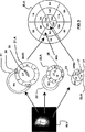
図3は、左側に、セグメント化された左心室SLVの壁の関心領域の3つのリングと、当該関心領域が示された左心室の3つの概略的な断面の間の対応する設定とを示す。これらの断面は、リング状の形状である。上側リングはBASE(底部)と示され、セグメント化された左心室SLVの上側関心領域に対応するセグメント1乃至6を有する。上側リングはまた、右心室RVを左心室LVへ挿入する2つの点24と、弁の点26とを有する。中央リングは、MIDと示され、セグメント化された左心室SLVの中央領域に対応するセグメント7乃至12を有する。中央リングはまた、2つの弁の点25を有する。下側リングは、APEX(頂点)と示され、セグメント化された左心室SLVの下側領域に対応するセグメント13乃至16を有する。下側リングは、頂点に対する点17も有する。図3の表はまた、3つのリングのセグメントと、ブルズ・アイ表現の3つのリングのセグメントとの間の対応を右側に表わす。壁の異なる領域の位置の2次元表現であるこのブルズ・アイ表現20.Aは、参照としてAHAの出版物に記載のように作成される。3つの弁の点は、BASE平面の位置を推定するのに用いられる。頂点は、BASEまでの距離を推定するために、従ってMID平面の位置を推定するために使用される。挿入点は、BASEのセグメント1とMIDのセグメント7の始まりを定義するのに用いられる。
しかしながら、この第1のブルズ・アイ表現20.Aは、心不全の場合の収縮期後の短縮又は非同期性の場合に生じうる、同じ動きの最大(最小)の値を有するが、異なる時間における、2つの領域を区別することを可能とするものではない。このために、本発明によるシステムは、図4Bに示すように、表示システムの同じ画像中に2つのブルズ・アイ表現を同時に表示する手段を有することが望ましい。この画像は、第1のブルズ・アイ表現20.A及び第2のブルズ・アイ表現20.Bを含む。
図4A、図4B、図4Cを参照するに、本発明のシステムは、関心領域の最大(又は最小)の変位の振幅を提供するために画像中に振幅ブルズ・アイ20.Aを表示する手段を有する。望ましくは、位相ブルズ・アイ20.Bは、心周期中で、所与の最大(又は最小)の変位が関心領域中で生じた時間を与えるために同じ画像中に表示される。
Claims (15)
- 検査中の変形可能な3次元対象の壁領域の変位の振幅に関連する情報を表示する画像処理システムであって、
3次元対象の壁の位置を決定し、前記3次元対象の壁の関心領域を定義し、かつ変位の振幅を時間の関数として決定するよう前記3次元対象の壁の3次元画像データを処理する、画像のシーケンス中の3次元対象の3次元データを処理する手段と、
前記3次元対象の壁の投影により前記3次元対象の壁の第1の2次元簡略表現を作成する手段とを有し、前記2次元簡略表現は前記関心領域の投影を含み、
前記画像処理システムは更に、前記関心領域の投影の中の変位の振幅の印を、前記作成された2次元簡略表現中に表示する表示手段とを有し、
前記処理する手段は、前記関心領域の変位の振幅を時間の関数として決定するために、前記3次元対象の壁の前記3次元画像データを処理し、かつ
前記表示手段は、前記第1の2次元簡略表現中の前記関心領域の投影中の前記3次元対象の壁の前記関心領域の前記変位の振幅の印を表示する、
画像処理システム。 - 前記関心領域についての最大又は最小の変位の振幅を決定するために、見つけられた振幅の値を平均化する手段、を更に有する請求項1記載の画像処理システム。
- 前記第1の2次元簡略表現(2次元簡略振幅表現と以下称す)を作成する手段は、一定時間に亘る前記関心領域の変位の最大振幅又は最小振幅の印である振幅の印を与える、請求項1記載の画像処理システム。
- 前記3次元対象の壁の前記第1の2次元簡略表現と同様の、前記関心領域の同様の投影を伴う、前記3次元対象の壁の第2の2次元簡略表現(前記第2の2次元簡略表現は2次元簡略位相表現と以下称す)を作成する手段と、
前記2次元簡略位相表現中に、前記一定時間に亘り、前記関心領域の中で変位の振幅の最大又は最小が生ずる時点の印を表示する手段とを更に有する、請求項3記載の画像処理システム。 - 前記2次元簡略振幅表現と前記2次元簡略位相表現とを一緒に同じ画像中に表示する手段を有する、請求項4記載の画像処理システム。
- 前記振幅の値及び時間の値を、色分けして夫々の2次元簡略振幅表現及び2次元簡略位相表現中に表示する手段を有する、請求項1乃至5のうちいずれか一項記載の画像処理システム。
- セグメントと称される前記関心領域の夫々の投影中の3次元対象の壁の前記関心領域の変位の振幅の印を前記作成された2次元簡略表現中に色分けして表示する手段を有し、前記変位の振幅の印は、時間の関数としての動画の2次元簡略表現を形成するよう、前記セグメント中で前記シーケンスの前記画像のレートで変化する、請求項1記載のシステム。
- 前記3次元対象の壁の前記2次元簡略表現を2次元ブルズ・アイ表現として表示する手段を有する、請求項1乃至7のうちいずれか一項記載の画像処理システム。
- 前記検査中の対象は左心室であり、前記3次元対象の壁は前記左心室の壁の内側の境界である、請求項1乃至8のうちいずれか一項記載の画像処理システム。
- 前記3次元対象の壁の位置を決定する手段は、前記検査中の3次元対象に適用されるセグメント化技術を行うセグメント化手段を有し、前記セグメント技術は、メッシュモデル技術を用いること、および前記メッシュモデルを前記検査中の3次元対象の壁へ写像するよう前記メッシュモデルの形状を変更することを含み、関心の対象である壁(対象の壁と以下称す)を有する簡略体積を提供する、請求項1乃至9のうちいずれか一項記載の画像処理システム。
- 回路手段を有する適切にプログラミングされたコンピュータ又は専用プロセッサを含むシステムであって、請求項1乃至10のうちいずれか一項記載の画像処理システムを有するシステム。
- 検査中の変形可能な3次元対象の壁領域の変位の振幅に関連する情報を表示する装置の作動方法であって、
3次元対象の壁の位置を決定し、前記3次元対象の壁の関心領域を定義し、かつ変位の振幅を時間の関数として決定するよう前記3次元対象の壁の3次元画像データを処理する、画像のシーケンス中の3次元対象の3次元データを処理する段階と、
前記3次元対象の壁の投影により前記3次元対象の壁の第1の2次元簡略表現を作成する段階とを有し、前記2次元簡略表現は前記関心領域の投影を含み、
前記関心領域の投影の中の変位の振幅の印を、前記作成された2次元簡略表現中に表示する表示段階とを有し、
前記処理する段階は、前記関心領域の変位の振幅を時間の関数として決定するために、前記3次元対象の壁の前記3次元画像データを処理し、かつ
前記表示段階は、前記第1の2次元簡略表現中の前記関心領域の投影中の前記3次元対象の壁の前記関心領域の前記変位の振幅の印を表示する、
装置の作動方法。 - 超音波3次元画像データを処理し、変形可能な3次元器官の超音波画像を前記器官の壁の動きの印と共に表示する装置の作動方法であって、
前記検査中の器官の画像シーケンスの3次元画像データを取得し、前記3次元対象の壁の位置を決定するよう前記シーケンスの画像中の前記3次元器官をセグメント化し、前記セグメント化された3次元器官の壁上に少なくとも1つの関心領域を定義し、前記関心領域の変位の振幅を時間の関数として決定するよう前記3次元画像データを処理する段階と、
3次元のセグメント化された器官の壁の投影により前記3次元のセグメント化された器官の壁の第1の2次元簡略表現を作成する段階と、前記2次元簡略表現中に関心領域の投影を含み、
前記関心領域の夫々の投影の中の前記3次元のセグメント化された器官の壁の前記関心領域の変位の振幅の印を、前記作成された2次元簡略表現中に色分けして表示する段階とを更に有する、請求項12記載の装置の作動方法。 - 前記関心領域の最大又は最小の変位の振幅の印を、一定時間に亘り表示し、この第1の2次元簡略表現は2次元簡略振幅表現と以下称される、段階と、
前記3次元のセグメント化された器官の壁の第1の2次元簡略表現と同様の、セグメントと称される関心領域の同様の投影を伴う、前記3次元のセグメント化された器官の壁の第2の2次元簡略表現を作成し、前記第2の2次元簡略表現は2次元簡略位相表現と以下称される、段階と、
前記2次元簡略位相表現中に、前記一定時間に亘り、前記関心領域の中で変位の振幅の最大又は最小が生ずる時点の印を表示する段階と、
前記2次元簡略振幅表現及び前記2次元簡略位相表現を同時に同じ画像中に表示する段階とを有する、請求項12記載の装置の作動方法。 - 請求項12、13又は14記載の装置の作動方法を実行するための一組の命令を有するコンピュータプログラム。
Applications Claiming Priority (2)
| Application Number | Priority Date | Filing Date | Title |
|---|---|---|---|
| EP02291622 | 2002-06-28 | ||
| PCT/IB2003/002643 WO2004003851A2 (en) | 2002-06-28 | 2003-06-24 | Image processing method for displaying information relating to parietal motions of a deformable 3-d object |
Publications (3)
| Publication Number | Publication Date |
|---|---|
| JP2005531352A JP2005531352A (ja) | 2005-10-20 |
| JP2005531352A5 JP2005531352A5 (ja) | 2006-08-31 |
| JP4521271B2 true JP4521271B2 (ja) | 2010-08-11 |
Family
ID=29797330
Family Applications (1)
| Application Number | Title | Priority Date | Filing Date |
|---|---|---|---|
| JP2004517091A Expired - Lifetime JP4521271B2 (ja) | 2002-06-28 | 2003-06-24 | 変形可能な3次元対象の頭頂の動きに関する情報を表示する画像処理システム |
Country Status (5)
| Country | Link |
|---|---|
| US (1) | US8538098B2 (ja) |
| EP (1) | EP1527422B1 (ja) |
| JP (1) | JP4521271B2 (ja) |
| AU (1) | AU2003239740A1 (ja) |
| WO (1) | WO2004003851A2 (ja) |
Families Citing this family (37)
| Publication number | Priority date | Publication date | Assignee | Title |
|---|---|---|---|---|
| JPH09119924A (ja) * | 1995-08-01 | 1997-05-06 | Hewlett Packard Co <Hp> | クロマトグラフィー用分離カラム |
| US7912528B2 (en) * | 2003-06-25 | 2011-03-22 | Siemens Medical Solutions Usa, Inc. | Systems and methods for automated diagnosis and decision support for heart related diseases and conditions |
| WO2006025005A2 (en) | 2004-08-31 | 2006-03-09 | Koninklijke Philips Electronics N.V. | Imaging system for displaying a structure of temporally changing configuration |
| JP2006110190A (ja) | 2004-10-15 | 2006-04-27 | Toshiba Corp | 医用画像データ解析装置及びその方法 |
| DE102005002950B4 (de) * | 2005-01-21 | 2007-01-25 | Siemens Ag | Verfahren zur automatischen Bestimmung der Position und Orientierung des linken Ventrikels und/oder angrenzender Bereiche in 3D-Bilddatensätzen des Herzens |
| DE102005002949A1 (de) | 2005-01-21 | 2006-08-03 | Siemens Ag | Verfahren zur Visualisierung von Schädigungen im Myokard |
| US7715627B2 (en) * | 2005-03-25 | 2010-05-11 | Siemens Medical Solutions Usa, Inc. | Automatic determination of the standard cardiac views from volumetric data acquisitions |
| US7706586B2 (en) * | 2005-06-22 | 2010-04-27 | General Electric Company | Real-time structure suppression in ultrasonically scanned volumes |
| DE102005037426A1 (de) * | 2005-08-08 | 2007-02-15 | Siemens Ag | Vorrichtung und Verfahren zum Fusionieren von Bilddatensätzen |
| DE102006026695A1 (de) | 2006-06-08 | 2007-12-13 | Tomtec Imaging Systems Gmbh | Verfahren, Vorrichtung und Computerprogrammprodukt zum Auswerten von dynamischen Bildern einer Kavität |
| JP5249218B2 (ja) * | 2006-08-09 | 2013-07-31 | コーニンクレッカ フィリップス エレクトロニクス エヌ ヴィ | 超音波画像形成システム |
| US20080281195A1 (en) * | 2007-05-09 | 2008-11-13 | General Electric Company | System and method for planning LV lead placement for cardiac resynchronization therapy |
| JP5523681B2 (ja) * | 2007-07-05 | 2014-06-18 | 株式会社東芝 | 医用画像処理装置 |
| ATE500574T1 (de) * | 2007-09-03 | 2011-03-15 | Koninkl Philips Electronics Nv | Visualisierung von voxel-daten |
| JP5454844B2 (ja) * | 2008-08-13 | 2014-03-26 | 株式会社東芝 | 超音波診断装置、超音波画像表示装置及び超音波画像表示プログラム |
| JP5525713B2 (ja) * | 2008-10-16 | 2014-06-18 | 株式会社東芝 | 超音波診断装置、超音波信号処理装置及び超音波信号処理プログラム |
| EP2340444A1 (en) * | 2008-10-22 | 2011-07-06 | Koninklijke Philips Electronics N.V. | 3-d ultrasound imaging |
| CN102860827B (zh) | 2009-09-18 | 2017-05-17 | 东芝医疗系统株式会社 | 磁共振成像装置以及磁共振成像方法 |
| US8581582B2 (en) | 2009-09-18 | 2013-11-12 | Kabushiki Kaisha Toshiba | MRI non-contrast time-slip angiography using variably positioned cine sub-sequence |
| GB2475722B (en) * | 2009-11-30 | 2011-11-02 | Mirada Medical | Measurement system for medical images |
| US8873817B2 (en) | 2009-12-21 | 2014-10-28 | Koninklijke Philips N.V. | Processing an image dataset based on clinically categorized populations |
| JP5619584B2 (ja) * | 2010-01-13 | 2014-11-05 | 株式会社東芝 | 超音波診断装置、超音波画像処理装置及び超音波画像処理プログラム |
| EP2552307A1 (en) * | 2010-04-01 | 2013-02-06 | Koninklijke Philips Electronics N.V. | Bullseye display for ecg data |
| JP5209025B2 (ja) * | 2010-10-27 | 2013-06-12 | ジーイー・メディカル・システムズ・グローバル・テクノロジー・カンパニー・エルエルシー | 超音波診断装置 |
| US9151815B2 (en) | 2010-11-15 | 2015-10-06 | Kabushiki Kaisha Toshiba | Magnetic resonance imaging apparatus and magnetic resonance imaging method |
| JP5651030B2 (ja) * | 2011-02-02 | 2015-01-07 | 日立アロカメディカル株式会社 | 超音波画像処理装置 |
| CN103687541B (zh) | 2011-03-02 | 2017-02-15 | 皇家飞利浦有限公司 | 用于导航引导的可视化 |
| US9691159B2 (en) * | 2012-07-18 | 2017-06-27 | Koninklijke Philips N.V. | Local contraction measurements |
| CN107072531A (zh) * | 2014-05-06 | 2017-08-18 | 塞克利心血管成像股份有限公司 | 用于心肌壁动力学的分析的方法和系统 |
| CN107106015A (zh) * | 2014-08-05 | 2017-08-29 | 英诺瓦科公司 | 心脏状态监视系统 |
| US9949643B2 (en) * | 2014-10-18 | 2018-04-24 | International Business Machines Corporation | Automatic visualization of regional functional parameters of left ventricle from cardiac imaging |
| WO2016073582A1 (en) * | 2014-11-04 | 2016-05-12 | Mayo Foundation For Medical Education And Research | Computer system and method for diagnostic data display |
| EP3423186A4 (en) | 2016-03-02 | 2019-10-23 | JP Scientific Limited | SOLID PHASE MICRO EXTRACTION COATING |
| CA3019256A1 (en) | 2016-05-10 | 2017-11-16 | Jp Scientific Limited | System and method for desorbing and detecting an analyte sorbed on a solid phase microextraction device |
| JP6740051B2 (ja) * | 2016-07-26 | 2020-08-12 | キヤノンメディカルシステムズ株式会社 | 超音波診断装置、医用画像処理装置及び医用画像処理プログラム |
| US11024100B2 (en) | 2016-08-10 | 2021-06-01 | Ucl Business Ltd | Method and apparatus for transforming physical measurement data of a biological organ |
| WO2019110295A1 (en) * | 2017-12-04 | 2019-06-13 | Koninklijke Philips N.V. | Image data processing method, device and system |
Family Cites Families (7)
| Publication number | Priority date | Publication date | Assignee | Title |
|---|---|---|---|---|
| JP2791255B2 (ja) * | 1992-10-02 | 1998-08-27 | 株式会社東芝 | 超音波カラードプラ断層装置 |
| US5431161A (en) * | 1993-04-15 | 1995-07-11 | Adac Laboratories | Method and apparatus for information acquistion, processing, and display within a medical camera system |
| US6295464B1 (en) * | 1995-06-16 | 2001-09-25 | Dimitri Metaxas | Apparatus and method for dynamic modeling of an object |
| JP4116122B2 (ja) * | 1997-11-28 | 2008-07-09 | 株式会社東芝 | 超音波診断装置及び超音波画像処理装置 |
| GB9920401D0 (en) * | 1999-08-27 | 1999-11-03 | Isis Innovation | Non-rigid motion image analysis |
| FR2802002B1 (fr) * | 1999-12-02 | 2002-03-01 | Ge Medical Syst Sa | Procede de recalage automatique d'images tridimensionnelles |
| JP2004518473A (ja) | 2001-01-30 | 2004-06-24 | コーニンクレッカ フィリップス エレクトロニクス エヌ ヴィ | 変形する3d物体の画像シーケンスを該物体の壁の動きの指示を伴って表示する画像処理方法 |
-
2003
- 2003-06-24 US US10/518,845 patent/US8538098B2/en active Active
- 2003-06-24 WO PCT/IB2003/002643 patent/WO2004003851A2/en active Application Filing
- 2003-06-24 JP JP2004517091A patent/JP4521271B2/ja not_active Expired - Lifetime
- 2003-06-24 EP EP03732929.9A patent/EP1527422B1/en not_active Expired - Lifetime
- 2003-06-24 AU AU2003239740A patent/AU2003239740A1/en not_active Abandoned
Also Published As
| Publication number | Publication date |
|---|---|
| WO2004003851A2 (en) | 2004-01-08 |
| US20060045328A1 (en) | 2006-03-02 |
| JP2005531352A (ja) | 2005-10-20 |
| EP1527422B1 (en) | 2019-05-22 |
| US8538098B2 (en) | 2013-09-17 |
| AU2003239740A8 (en) | 2004-01-19 |
| WO2004003851A3 (en) | 2004-05-13 |
| EP1527422A2 (en) | 2005-05-04 |
| AU2003239740A1 (en) | 2004-01-19 |
| WO2004003851A8 (en) | 2006-05-26 |
Similar Documents
| Publication | Publication Date | Title |
|---|---|---|
| JP4521271B2 (ja) | 変形可能な3次元対象の頭頂の動きに関する情報を表示する画像処理システム | |
| CN103229210B (zh) | 图像配准装置 | |
| JP4884528B2 (ja) | 腔部の画像を評定する方法、装置ならびにコンピュータプログラム製品 | |
| US7822246B2 (en) | Method, a system and a computer program for integration of medical diagnostic information and a geometric model of a movable body | |
| EP1430837B1 (en) | Ultrasonic diagnostic device | |
| Chen et al. | Kinematic and deformation analysis of 4-D coronary arterial trees reconstructed from cine angiograms | |
| JP2004518473A (ja) | 変形する3d物体の画像シーケンスを該物体の壁の動きの指示を伴って表示する画像処理方法 | |
| Slomka et al. | Application and translation of artificial intelligence to cardiovascular imaging in nuclear medicine and noncontrast CT | |
| US20200118264A1 (en) | Shell-constrained localization of vasculature | |
| JP2024515635A (ja) | 超音波及びカメラ画像から3d画像を再構成するためのシステム及び方法 | |
| US9558568B2 (en) | Visualization method for a human skeleton from a medical scan | |
| US7526113B2 (en) | Method for processing an image sequence of a distortable 3-D object to yield indications of the object wall deformations | |
| Kirişli et al. | Comprehensive visualization of multimodal cardiac imaging data for assessment of coronary artery disease: first clinical results of the SMARTVis tool | |
| JP2005525863A (ja) | 医療用データの統合された視覚化用の医療用視検システム及び画像処理 | |
| KR20140120236A (ko) | 심근 및 심혈관 정보의 통합 분석 방법 | |
| JP2015136480A (ja) | 3次元医用画像表示制御装置およびその作動方法並びに3次元医用画像表示制御プログラム | |
| Lee et al. | Comparative study of left ventricular low wall motion with scar tissue using 4d left ventricular cardiac images | |
| Chen | Analysis of Cardiovascular Disease using Cardiac Computed Tomography and Deep Learning | |
| Cai et al. | Detection of 3D Arterial Centerline Extraction in Spiral CT Coronary Angiography | |
| Zhang et al. | Left-ventricle segmentation in real-time 3D echocardiography using a hybrid active shape model and optimal graph search approach | |
| Usta | Image Processing Methods for Myocardial Scar Analysis from 3D Late-Gadolinium Enhanced Cardiac Magnetic Resonance Images | |
| Bruckner et al. | Vessel Maps: A Survey of Map-Like Visualizations of the Cardiovascular System | |
| JP2023135836A (ja) | 画像処理装置、画像処理方法、およびプログラム | |
| Hillier et al. | Online 3-D reconstruction of the right atrium from echocardiography data via a topographic cellular contour extraction algorithm | |
| Bravo et al. | Left ventricle segmentation and motion analysis in multislice computerized tomography |
Legal Events
| Date | Code | Title | Description |
|---|---|---|---|
| A521 | Request for written amendment filed |
Free format text: JAPANESE INTERMEDIATE CODE: A523 Effective date: 20060621 |
|
| A621 | Written request for application examination |
Free format text: JAPANESE INTERMEDIATE CODE: A621 Effective date: 20060621 |
|
| A131 | Notification of reasons for refusal |
Free format text: JAPANESE INTERMEDIATE CODE: A131 Effective date: 20090526 |
|
| A521 | Request for written amendment filed |
Free format text: JAPANESE INTERMEDIATE CODE: A523 Effective date: 20090826 |
|
| A131 | Notification of reasons for refusal |
Free format text: JAPANESE INTERMEDIATE CODE: A131 Effective date: 20091110 |
|
| A521 | Request for written amendment filed |
Free format text: JAPANESE INTERMEDIATE CODE: A523 Effective date: 20100203 |
|
| TRDD | Decision of grant or rejection written | ||
| A01 | Written decision to grant a patent or to grant a registration (utility model) |
Free format text: JAPANESE INTERMEDIATE CODE: A01 Effective date: 20100427 |
|
| A01 | Written decision to grant a patent or to grant a registration (utility model) |
Free format text: JAPANESE INTERMEDIATE CODE: A01 |
|
| A61 | First payment of annual fees (during grant procedure) |
Free format text: JAPANESE INTERMEDIATE CODE: A61 Effective date: 20100524 |
|
| FPAY | Renewal fee payment (event date is renewal date of database) |
Free format text: PAYMENT UNTIL: 20130528 Year of fee payment: 3 |
|
| R150 | Certificate of patent or registration of utility model |
Free format text: JAPANESE INTERMEDIATE CODE: R150 Ref document number: 4521271 Country of ref document: JP Free format text: JAPANESE INTERMEDIATE CODE: R150 |
|
| R250 | Receipt of annual fees |
Free format text: JAPANESE INTERMEDIATE CODE: R250 |
|
| R250 | Receipt of annual fees |
Free format text: JAPANESE INTERMEDIATE CODE: R250 |
|
| R250 | Receipt of annual fees |
Free format text: JAPANESE INTERMEDIATE CODE: R250 |
|
| R250 | Receipt of annual fees |
Free format text: JAPANESE INTERMEDIATE CODE: R250 |
|
| R250 | Receipt of annual fees |
Free format text: JAPANESE INTERMEDIATE CODE: R250 |
|
| R250 | Receipt of annual fees |
Free format text: JAPANESE INTERMEDIATE CODE: R250 |
|
| R250 | Receipt of annual fees |
Free format text: JAPANESE INTERMEDIATE CODE: R250 |
|
| R250 | Receipt of annual fees |
Free format text: JAPANESE INTERMEDIATE CODE: R250 |
|
| R250 | Receipt of annual fees |
Free format text: JAPANESE INTERMEDIATE CODE: R250 |
|
| R250 | Receipt of annual fees |
Free format text: JAPANESE INTERMEDIATE CODE: R250 |
|
| R250 | Receipt of annual fees |
Free format text: JAPANESE INTERMEDIATE CODE: R250 |
|
| EXPY | Cancellation because of completion of term |