JP4297740B2 - 放射線源を動作させる方法及び装置 - Google Patents
放射線源を動作させる方法及び装置 Download PDFInfo
- Publication number
- JP4297740B2 JP4297740B2 JP2003174483A JP2003174483A JP4297740B2 JP 4297740 B2 JP4297740 B2 JP 4297740B2 JP 2003174483 A JP2003174483 A JP 2003174483A JP 2003174483 A JP2003174483 A JP 2003174483A JP 4297740 B2 JP4297740 B2 JP 4297740B2
- Authority
- JP
- Japan
- Prior art keywords
- radiation source
- computer
- detector
- spot
- center
- Prior art date
- Legal status (The legal status is an assumption and is not a legal conclusion. Google has not performed a legal analysis and makes no representation as to the accuracy of the status listed.)
- Expired - Fee Related
Links
- 230000005855 radiation Effects 0.000 title claims description 85
- 238000000034 method Methods 0.000 title description 18
- 238000005070 sampling Methods 0.000 claims description 21
- 238000003384 imaging method Methods 0.000 claims description 12
- 238000009827 uniform distribution Methods 0.000 claims description 4
- 238000010894 electron beam technology Methods 0.000 claims description 2
- 238000002591 computed tomography Methods 0.000 description 17
- 230000004907 flux Effects 0.000 description 10
- 238000009826 distribution Methods 0.000 description 8
- 238000010586 diagram Methods 0.000 description 4
- 238000005259 measurement Methods 0.000 description 4
- 230000007246 mechanism Effects 0.000 description 4
- 238000013170 computed tomography imaging Methods 0.000 description 3
- 238000005457 optimization Methods 0.000 description 3
- 230000008569 process Effects 0.000 description 3
- 238000013459 approach Methods 0.000 description 2
- 230000002238 attenuated effect Effects 0.000 description 2
- 230000004048 modification Effects 0.000 description 2
- 238000012986 modification Methods 0.000 description 2
- 230000005540 biological transmission Effects 0.000 description 1
- 230000008859 change Effects 0.000 description 1
- 230000003247 decreasing effect Effects 0.000 description 1
- 230000000694 effects Effects 0.000 description 1
- 238000012545 processing Methods 0.000 description 1
- 238000003860 storage Methods 0.000 description 1
Images
Classifications
-
- A—HUMAN NECESSITIES
- A61—MEDICAL OR VETERINARY SCIENCE; HYGIENE
- A61B—DIAGNOSIS; SURGERY; IDENTIFICATION
- A61B6/00—Apparatus or devices for radiation diagnosis; Apparatus or devices for radiation diagnosis combined with radiation therapy equipment
- A61B6/02—Arrangements for diagnosis sequentially in different planes; Stereoscopic radiation diagnosis
- A61B6/03—Computed tomography [CT]
- A61B6/032—Transmission computed tomography [CT]
-
- A—HUMAN NECESSITIES
- A61—MEDICAL OR VETERINARY SCIENCE; HYGIENE
- A61B—DIAGNOSIS; SURGERY; IDENTIFICATION
- A61B6/00—Apparatus or devices for radiation diagnosis; Apparatus or devices for radiation diagnosis combined with radiation therapy equipment
- A61B6/40—Arrangements for generating radiation specially adapted for radiation diagnosis
- A61B6/4021—Arrangements for generating radiation specially adapted for radiation diagnosis involving movement of the focal spot
- A61B6/4028—Arrangements for generating radiation specially adapted for radiation diagnosis involving movement of the focal spot resulting in acquisition of views from substantially different positions, e.g. EBCT
-
- A—HUMAN NECESSITIES
- A61—MEDICAL OR VETERINARY SCIENCE; HYGIENE
- A61B—DIAGNOSIS; SURGERY; IDENTIFICATION
- A61B6/00—Apparatus or devices for radiation diagnosis; Apparatus or devices for radiation diagnosis combined with radiation therapy equipment
- A61B6/40—Arrangements for generating radiation specially adapted for radiation diagnosis
- A61B6/4035—Arrangements for generating radiation specially adapted for radiation diagnosis the source being combined with a filter or grating
-
- A—HUMAN NECESSITIES
- A61—MEDICAL OR VETERINARY SCIENCE; HYGIENE
- A61B—DIAGNOSIS; SURGERY; IDENTIFICATION
- A61B6/00—Apparatus or devices for radiation diagnosis; Apparatus or devices for radiation diagnosis combined with radiation therapy equipment
- A61B6/40—Arrangements for generating radiation specially adapted for radiation diagnosis
- A61B6/4064—Arrangements for generating radiation specially adapted for radiation diagnosis specially adapted for producing a particular type of beam
- A61B6/4085—Cone-beams
Landscapes
- Health & Medical Sciences (AREA)
- Life Sciences & Earth Sciences (AREA)
- Engineering & Computer Science (AREA)
- Medical Informatics (AREA)
- Heart & Thoracic Surgery (AREA)
- Molecular Biology (AREA)
- Biophysics (AREA)
- Nuclear Medicine, Radiotherapy & Molecular Imaging (AREA)
- Optics & Photonics (AREA)
- Pathology (AREA)
- Radiology & Medical Imaging (AREA)
- Biomedical Technology (AREA)
- Physics & Mathematics (AREA)
- High Energy & Nuclear Physics (AREA)
- Surgery (AREA)
- Animal Behavior & Ethology (AREA)
- General Health & Medical Sciences (AREA)
- Public Health (AREA)
- Veterinary Medicine (AREA)
- Pulmonology (AREA)
- Theoretical Computer Science (AREA)
- Apparatus For Radiation Diagnosis (AREA)
- Analysing Materials By The Use Of Radiation (AREA)
- X-Ray Techniques (AREA)
Description
【発明の属する技術分野】
本発明は一般的に云えばコンピュータ断層撮影(CT)イメージング(画像生成)法に関するものであり、より具体的に云えばCT放射線源を動作させる方法及び装置に関するものである。
【0002】
【発明の背景】
通常「反転円錐」型ジオメトリイ(幾何学的配置)と称されているものに基づいたCT走査法に最近多くの議論がなされている。反転円錐型ジオメトリイを用いる際、ほぼ全走査撮影範囲(FOV)をカバーするために大きな二次元放射線源が使用されている。また、放射線フォトンを収集するために小さな検出器が使用されている。非点源型放射線源及び線状放射線源を使用することに関連して起こり得る問題はの一つは、検出器が受けるノイズの不均一性である。
【特許文献1】
米国特許第6381298号
【0003】
【発明の概要】
本発明の一面では、放射線源を動作させる方法が提供される。本方法は、放射線源を準備する工程と、検出器を準備する工程と、検出器が実質的に均一な分布のノイズを受けるように放射線源及び検出器を動作させる工程とを含んでいる。
【0004】
本発明の別の一面では、走査型イメージング・システム上に設置された放射線源を動作させるコンピュータが提供される。該イメージング・システムは放射線源及び検出器を含んでいる。本コンピュータは、検出器が実質的に均一な分布のノイズを受けるように放射線源及び検出器を動作させるようにプログラミングされている。
【0005】
本発明の更に別の一面では、放射線源を動作させるコンピュータ断層撮影(CT)イメージング・システムが提供される。本CTシステムは放射線源と、検出器アレイと、該検出器アレイ及び放射線源に結合されたコンピュータとを含んでいる。コンピュータは、検出器が反転円錐型ジオメトリイのビームを受けるように放射線源を動作させるように構成されている。
【0006】
【発明の詳しい説明】
コンピュータ断層撮影(CT)イメージング・システムについての公知の幾つかの構成においては、放射線源が扇形ビームを投射し、このビームを一般に「イメージング平面」と呼ぶデカルト座標系のX−Y平面内に存在するようにコリメートする。放射線ビームは撮影対象物、例えば患者を通過する。対象物によって減弱した後のビームは放射線検出器アレイに入射する。減弱した放射線ビームの強度は対象物による放射線ビームの減弱度に依存する。アレイの各々の検出器素子がその検出器位置におけるビーム減弱度の測定値である電気信号を別々に発生する。全ての検出器からの減弱度測定値は別々に収集されて透過度分布を生成する。
【0007】
公知の第3世代のCTシステムでは、放射線源及び検出器アレイはイメージング平面内で撮影対象物の周りをガントリと共に回転させて、放射線ビームが対象物と交差する角度が絶えず変化するようにする。一ガントリ角度における検出器アレイからの一群の放射線減弱度測定値、すなわち、投影データは、「ビュー(view)」と呼ばれている。対象物の「スキャン(scan)」すなわち一走査が、X線源及び検出器の一回転の間に異なるガントリ角度すなわちビュー角度(撮影角度)で得られた一組のビューで構成される。
【0008】
アキシャル・スキャンでは、投影データは、対象物の二次元スライス(断層面)に対応する画像を構成するように処理される。一組の投影データから画像を再構成する一方法が、フィルタ補正逆投影法と呼ばれているものである。この方法は、スキャンからの源弱度測定値を「CTナンバー」又は「ハウンスフィールド単位」と呼ばれる整数に変換し、これらの整数を使用して陰極線管表示装置の対応する画素の輝度を制御する。
【0009】
全走査時間を短縮するために、「螺旋」走査が実行されることがある。「螺旋」走査を行うために、患者を移動させながら、所定数のスライスについてのデータが収集される。このようなシステムは、1つの扇形ビーム螺旋走査から単一のヘリックス(helix) を生成する。扇形ビームによって写像されたヘリックスは投影データを生じさせ、この投影データから各々の所定のスライスにおける画像を再構成し得る。
【0010】
本書で単に「素子」又は「要素」、或いは「工程」という用語を使用するが、特に明記しない限り、これは単数(1つ)の素子又は要素、或いは工程を表すだけでなく複数の素子又は要素、或いは工程を表していることもあることを理解されたい。更に、本発明の「一実施形態」とは、その列挙した特徴を同様に含んでいる別の実施形態の存在を排除するものとして解釈されるべきではない。
【0011】
また本書で用いる語句「画像を再構成する」は、画像を表すデータを生成するが、目に見える画像を生成しない本発明の実施形態を排除することを意図したものではない。多くの実施形態では、少なくとも1つの目に見える画像を生成する(又は、生成するように構成されている)。
【0012】
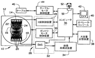
図1はCTイメージング・システム10の絵画的斜視図である。図2は、図1に示したシステム10のブロック回路図である。代表的な実施形態では、コンピュータ断層撮影(CT)イメージング・システム10が、「第3世代」のCTイメージング・システムを表すガントリ12を含むものとして示されている。ガントリ12は放射線源14を備えており、放射線源14はガントリ12の反対側にある検出器アレイ18へ向かってX線ビーム16を投射する。一実施形態では、放射線源14は二次元放射線源であり、これは放射線源14上の複数の位置(これらは、「スポット」とも呼ばれている)から検出器18へ向かって複数の円錐ビーム16を投射して、反転円錐型ジオメトリイのビームを検出器18が受けるようにする。
【0013】
検出器アレイ18は、患者22のような対象物を通過した投射X線ビームを検知する複数の検出器素子20を含む複数の検出器列(図示していない)によって形成されている。各々の検出器素子20は、入射するX線ビームの強度、従って対象物又は患者22を通過したビームの減弱度を表す電気信号を発生する。放射線投影データを収集するための走査中、ガントリ12及びそれに装着された部品は回転中心24の周りを回転する。図2は一列の検出器素子20(すなわち、一検出器列)のみを示している。しかしながら、マルチスライス検出器アレイ18は、検出器素子20の複数の並列の検出器列を含んでいて、複数の準並列又は並列のスライスに対応する投影データを走査中に同時に収集することができる。
【0014】
ガントリ12の回転及び放射線源14の動作はCTシステム10の制御機構26によって統制される。制御機構26は、放射線源14に電力及びタイミング信号を供給する放射線制御装置28と、ガントリ12の回転速度及び位置を制御するガントリ・モータ制御装置30とを含んでいる。制御機構26にはデータ収集システム(DAS)32も含まれており、該DAS32は、検出器素子20からのアナログ・データをサンプリングして、該データをその後の処理のためにディジタル信号へ変換する。画像再構成装置34はDAS32からサンプリングされディジタル化された放射線データを受け取って、高速画像再構成を実行する。再構成された画像はコンピュータ36に入力として印加され、コンピュータ36は該画像を大容量記憶装置38に格納する。
【0015】
コンピュータ36はまた、キーボ−ドを備えたコンソール40を介してオペレータから指令及び走査パラメータも受け取る。付設された陰極線管表示装置42により、オペレータはコンピュータ36からの再構成された画像やその他のデータを観察することが可能になる。コンピュータ36はオペレータから供給された指令及びパラメータを使用して、DAS32、放射線制御装置28及びガントリ・モータ制御装置30へ制御信号及び情報を供給する。加えて、コンピュータ36は、ガントリ12内に患者22を位置決めするために電動テーブル46を制御するテーブル・モータ制御装置44を動作させる。具体的に述べると、テーブル46は患者22の一部分をガントリ開口部48内へ通すように移動させる。
【0016】
一実施形態において、コンピュータ36は、フレキシブル・ディスクやCD−ROMのようなコンピュータ読出し可能な媒体52から命令及び/又はデータを読み出すためのフレキシブル・ディスク・ドライブやCD−ROMドライブのようなデバイス50を含んでいる。別の実施形態では、コンピュータ36はファームウエア(図示していない)に記憶されている命令を実行する。コンピュータ36は本書で述べる機能を実行するようにプログラミングされており、従って、本書で用いる用語「コンピュータ」とは当該技術分野でコンピュータと呼ばれている集積回路だけでなく、広義にコンピュータ、プロセッサ、マイクロプロセッサ、マイクロコンピュータ、プログラミング可能な論理制御装置、特定用途向け集積回路、及びその他のプログラミング可能な回路を表す。
【0017】
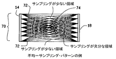
図3は、Z方向に、すなわち、患者22(図1に示す)の軸に沿って収集される不均一なX線サンプリング・パターンの断面図である。図3に示されているように、大きな二次元放射線源14がほぼ全走査撮影範囲(FOV)70をカバーするために使用され、また放射線源14よりも小さい検出器18が放射線源14から放出されたX線フォトンを収集するために使用される。例えば、両方のFOV縁部72近くの位置はFOV中心部74よりも少なくサンプリングされ、その結果として不均一なノイズ・パターンが生じる。
【0018】
図4は、放射線源14のような放射線源を動作させるための方法80の流れ図である。方法80は、反転円錐放射線源14の様な放射線を準備する工程82と、検出器18のような検出器を準備する工程84と、検出器が実質的に均一な分布のノイズを受けるように放射線源及び検出器を動作させる工程86とを含んでいる。
【0019】
図5は、不均一なノイズ・パターンを低減するのに役立つ患者前方フィルタ(プリペイシェント・フィルタ)90を使用する代表的な実施形態の断面図である。この代表的な実施形態では、患者前方フィルタ90は放射線源14と患者22との間に設置される。使用時に、患者前方フィルタ90は、均一なノイズ分布が拡大するようにX線ビーム強度を成形するのに役立つ。一実施形態では、患者前方フィルタ90はフィルタ中心部92で相対的に厚くなり且つフィルタ縁部94付近で相対的に薄くなっている。中心部92付近の厚さを増大させることは、FOV中心部74(図3に示す)付近でのサンプル量の増大を補償するのに役立つ。また、縁部94付近の厚さを減少させることは、FOV縁部72(図3に示す)付近でのサンプル量の減少を補償するのに役立つ。従って、各々の検出器18領域に達する全X線束がほぼ均一になる。患者前方フィルタ90は、FOV70(図3に示す)内の複数の線束分布を検査することによって最適化する。一実施形態では、この最適化は、反復的なアルゴリズムを使用して行われる。別の実施形態では、この最適化は、オペレータが選択したアルゴリズムを使用して行われる。
【0020】
図6は、放射線源電流を変調することによって得られる均一なサンプリング・パターンの断面図である。代表的な実施形態では、放射線源縁部96付近の放射線源電流を放射線源中心部98の放射線源電流よりも大きくする。例えば、放射線源14に対する入力電流を一定にするのではなく、むしろ放射線源スポット100の位置に基づいて変調する。使用の際、FOV縁部72付近でのサンプリング不足を補償するために、放射線源電流は放射線源縁部96付近で最大にし、そして放射線源中心部98に近づくにつれて徐々に減少させる。最初の手法と同様に、この放射線源スポット100の関数としての放射線源電流の最適化は、その結果得られたX線束を検査することによって行う。
【0021】
図7は、X線束分布を変調することによって得られる均一なサンプリング・パターンの断面図である。代表的な実施形態では、X線束分布を変調するために、放射線源に対する電流を一定に保ちながら、各々の線源スポット100上の電子ビームの滞留時間を変更する。使用の際、放射線源スポット100における滞留時間を長くすると、前に述べた放射線源電流を調節すことによって得られる効果と同様に、サンプリング位置におけるX線束が増大する。一実施形態では、両方のFOV縁部72付近でのX線スポット100についての滞留時間を、FOV中心部74付近での滞留時間よりも長くする。別の実施形態では、X線束分布変調と放射線源電流変調とを組み合わせて、均一なサンプリング・パターンを生成するようにする。X線束分布と放射線源電流とを変調することは、これらの2つの変調を別々に実施することが必要でない場合に、システム10(図1に示す)についての要件を低減するのに役立つ。例えば、CTシステム10が均一な線束分布を補償するのに充分高速に滞留時間を変更できない場合、滞留時間を部分的に変更すると共に放射線源電流を部分的に変更することによって、均一なノイズ場を達成することができる。
【0022】
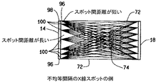
図8は、線源スポット100についてサンプリング・パターンを変えることによって得られる均一なサンプリング・パターンの断面図である。例えば、異なる放射線源スポット100相互間の距離が一定であると仮定すると、その結果得られるサンプリング・パターンはFOV中心部74付近では密度が高くなり且つFOV縁部72付近では密度が低くなる。従って、代表的な実施形態では、放射線源スポット100相互間のサンプリング距離を修正して、サンプリング距離を放射線源縁部96付近では間隔がより狭くなると共に、放射線源中心部98付近では間隔がより広くなるようにすることができる。このように放射線源スポット100相互間のサンプリング距離を修正することは、サンプリング・パターンが前に図7に示したように均一になるように、サンプリング・パターンを正規化し直すのに役立つ。
【0023】
別の代表的な実施形態では、前に述べた様々な方法を組み合わせて、前に述べたようなハードウエアについての制約を低減するのに役立てることができる。一実施形態では、患者前方フィルタ70、放射線源電流変調、放射線源線束変調及びサンプリング・パターン変更手法を組み合わせて、不均一ノイズの低減に役立てることができる。別の実施形態では、患者前方フィルタ70、放射線源電流変調、放射線源線束変調及びサンプリング・パターン変更手法のうちの少なくとも2つを組み合わせて、不均一ノイズの低減に役立てることができる。
【0024】
本発明の様々な特定の実施形態について説明したが、当業者には本発明が特許請求の範囲に記載の精神および範囲内で修正を行うことができることが認められよう。
【図面の簡単な説明】
【図1】CTイメージング・システムの絵画的斜視図である。
【図2】図1に示したシステムのブロック回路図である。
【図3】不均一なサンプリング・パターンの断面図である。
【図4】放射線源を動作させる方法の流れ図である。
【図5】患者前方フィルタの断面図である。
【図6】均一なサンプリング・パターンの断面図である。
【図7】均一なサンプリング・パターンの断面図である。
【図8】均一なサンプリング・パターンの断面図である。
【符号の説明】
10 コンピュータ断層撮影(CT)イメージング・システム
12 ガントリ
14 放射線源
16 X線ビーム
18 検出器アレイ
20 検出器素子
22 患者
24 回転中心
26 制御機構
42 陰極線管表示装置
48 ガントリ開口部
50 デバイス
52 コンピュータ読出し可能な媒体
70 撮影範囲(FOV)
72 FOV縁部
74 FOV中心部
90 患者前方フィルタ
92 フィルタ中心部
94 フィルタ縁部
96 放射線源縁部
98 放射線源中心部
100 放射線源スポット
Claims (5)
- 放射線源及び検出器を含む走査型イメージング・システム(10)の放射線源(14)を動作させるコンピュータ(36)であって、前記放射線源の縁部(96)付近の放射線源電流が前記放射線源の中心部(98)付近の放射線源電流よりも大きくなるように放射線源電流を変調し、前記検出器が実質的に均一な分布のノイズを受けるように前記放射線源及び前記検出器を動作させるようにプログラミングされているコンピュータ(36)。
- 前記放射線源(14)を動作させるに際し、前記コンピュータが、線状放射線源及び二次元放射線源のうちの少なくとも1つを動作させるように構成されている、請求項1記載のコンピュータ(36)。
- 反転円錐型ジオメトリイのビーム(16)及び非反転円錐型ジオメトリイのビームのうちの少なくとも一方を前記検出器が受けるように前記放射線源(14)を動作させるようにプログラミングされている請求項1記載のコンピュータ(36)。
- 前記放射線源(14)から放出される電子ビームの滞留時間を変調して、撮影範囲の縁部(72)付近のX線スポットにおける滞留時間が撮影範囲の中心部(74)付近のX線スポットにおける滞留時間よりも長くなるようにプログラミングされている請求項1記載のコンピュータ(36)。
- 複数のX線スポットの相互間のサンプリング距離を修正して、前記放射線源の縁部(96)付近のスポットが前記放射線源の中心部(98)付近のスポットよりも一層密な間隔で配置されようにプログラミングされている請求項1記載のコンピュータ(36)。
Applications Claiming Priority (1)
| Application Number | Priority Date | Filing Date | Title |
|---|---|---|---|
| US10/064,189 US6754300B2 (en) | 2002-06-20 | 2002-06-20 | Methods and apparatus for operating a radiation source |
Publications (3)
| Publication Number | Publication Date |
|---|---|
| JP2004024872A JP2004024872A (ja) | 2004-01-29 |
| JP2004024872A5 JP2004024872A5 (ja) | 2008-09-04 |
| JP4297740B2 true JP4297740B2 (ja) | 2009-07-15 |
Family
ID=29717753
Family Applications (1)
| Application Number | Title | Priority Date | Filing Date |
|---|---|---|---|
| JP2003174483A Expired - Fee Related JP4297740B2 (ja) | 2002-06-20 | 2003-06-19 | 放射線源を動作させる方法及び装置 |
Country Status (4)
| Country | Link |
|---|---|
| US (1) | US6754300B2 (ja) |
| EP (1) | EP1374776B1 (ja) |
| JP (1) | JP4297740B2 (ja) |
| DE (1) | DE60330207D1 (ja) |
Families Citing this family (51)
| Publication number | Priority date | Publication date | Assignee | Title |
|---|---|---|---|---|
| US7082182B2 (en) * | 2000-10-06 | 2006-07-25 | The University Of North Carolina At Chapel Hill | Computed tomography system for imaging of human and small animal |
| US7963695B2 (en) | 2002-07-23 | 2011-06-21 | Rapiscan Systems, Inc. | Rotatable boom cargo scanning system |
| US8275091B2 (en) | 2002-07-23 | 2012-09-25 | Rapiscan Systems, Inc. | Compact mobile cargo scanning system |
| US7813473B2 (en) * | 2002-07-23 | 2010-10-12 | General Electric Company | Method and apparatus for generating temporally interpolated projections |
| US6904118B2 (en) | 2002-07-23 | 2005-06-07 | General Electric Company | Method and apparatus for generating a density map using dual-energy CT |
| GB0309385D0 (en) | 2003-04-25 | 2003-06-04 | Cxr Ltd | X-ray monitoring |
| GB0309387D0 (en) | 2003-04-25 | 2003-06-04 | Cxr Ltd | X-Ray scanning |
| GB2436713B (en) * | 2003-04-25 | 2008-02-06 | Cxr Ltd | X-ray tube electron sources |
| GB0525593D0 (en) | 2005-12-16 | 2006-01-25 | Cxr Ltd | X-ray tomography inspection systems |
| US8243876B2 (en) | 2003-04-25 | 2012-08-14 | Rapiscan Systems, Inc. | X-ray scanners |
| US9113839B2 (en) | 2003-04-25 | 2015-08-25 | Rapiscon Systems, Inc. | X-ray inspection system and method |
| US8223919B2 (en) | 2003-04-25 | 2012-07-17 | Rapiscan Systems, Inc. | X-ray tomographic inspection systems for the identification of specific target items |
| US7949101B2 (en) | 2005-12-16 | 2011-05-24 | Rapiscan Systems, Inc. | X-ray scanners and X-ray sources therefor |
| GB0812864D0 (en) | 2008-07-15 | 2008-08-20 | Cxr Ltd | Coolign anode |
| GB0309379D0 (en) | 2003-04-25 | 2003-06-04 | Cxr Ltd | X-ray scanning |
| US9208988B2 (en) | 2005-10-25 | 2015-12-08 | Rapiscan Systems, Inc. | Graphite backscattered electron shield for use in an X-ray tube |
| US8451974B2 (en) | 2003-04-25 | 2013-05-28 | Rapiscan Systems, Inc. | X-ray tomographic inspection system for the identification of specific target items |
| GB0309383D0 (en) | 2003-04-25 | 2003-06-04 | Cxr Ltd | X-ray tube electron sources |
| US10483077B2 (en) | 2003-04-25 | 2019-11-19 | Rapiscan Systems, Inc. | X-ray sources having reduced electron scattering |
| US8837669B2 (en) | 2003-04-25 | 2014-09-16 | Rapiscan Systems, Inc. | X-ray scanning system |
| GB0309371D0 (en) | 2003-04-25 | 2003-06-04 | Cxr Ltd | X-Ray tubes |
| US8804899B2 (en) | 2003-04-25 | 2014-08-12 | Rapiscan Systems, Inc. | Imaging, data acquisition, data transmission, and data distribution methods and systems for high data rate tomographic X-ray scanners |
| US8094784B2 (en) | 2003-04-25 | 2012-01-10 | Rapiscan Systems, Inc. | X-ray sources |
| US6928141B2 (en) | 2003-06-20 | 2005-08-09 | Rapiscan, Inc. | Relocatable X-ray imaging system and method for inspecting commercial vehicles and cargo containers |
| US7639774B2 (en) * | 2003-12-23 | 2009-12-29 | General Electric Company | Method and apparatus for employing multiple axial-sources |
| US7333587B2 (en) * | 2004-02-27 | 2008-02-19 | General Electric Company | Method and system for imaging using multiple offset X-ray emission points |
| US7173996B2 (en) * | 2004-07-16 | 2007-02-06 | General Electric Company | Methods and apparatus for 3D reconstruction in helical cone beam volumetric CT |
| US7471764B2 (en) | 2005-04-15 | 2008-12-30 | Rapiscan Security Products, Inc. | X-ray imaging system having improved weather resistance |
| US8155262B2 (en) * | 2005-04-25 | 2012-04-10 | The University Of North Carolina At Chapel Hill | Methods, systems, and computer program products for multiplexing computed tomography |
| US9046465B2 (en) | 2011-02-24 | 2015-06-02 | Rapiscan Systems, Inc. | Optimization of the source firing pattern for X-ray scanning systems |
| US8189893B2 (en) * | 2006-05-19 | 2012-05-29 | The University Of North Carolina At Chapel Hill | Methods, systems, and computer program products for binary multiplexing x-ray radiography |
| US7486760B2 (en) * | 2006-08-15 | 2009-02-03 | Ge Security, Inc. | Compact systems and methods for generating a diffraction profile |
| US7778386B2 (en) * | 2006-08-28 | 2010-08-17 | General Electric Company | Methods for analytic reconstruction for mult-source inverse geometry CT |
| US7706499B2 (en) * | 2006-08-30 | 2010-04-27 | General Electric Company | Acquisition and reconstruction of projection data using a stationary CT geometry |
| US7835486B2 (en) * | 2006-08-30 | 2010-11-16 | General Electric Company | Acquisition and reconstruction of projection data using a stationary CT geometry |
| US7616731B2 (en) * | 2006-08-30 | 2009-11-10 | General Electric Company | Acquisition and reconstruction of projection data using a stationary CT geometry |
| US20080056432A1 (en) * | 2006-08-30 | 2008-03-06 | General Electric Company | Reconstruction of CT projection data |
| US7428292B2 (en) * | 2006-11-24 | 2008-09-23 | General Electric Company | Method and system for CT imaging using multi-spot emission sources |
| GB0803644D0 (en) | 2008-02-28 | 2008-04-02 | Rapiscan Security Products Inc | Scanning systems |
| GB0803641D0 (en) | 2008-02-28 | 2008-04-02 | Rapiscan Security Products Inc | Scanning systems |
| GB0809110D0 (en) | 2008-05-20 | 2008-06-25 | Rapiscan Security Products Inc | Gantry scanner systems |
| GB0816823D0 (en) | 2008-09-13 | 2008-10-22 | Cxr Ltd | X-ray tubes |
| US8600003B2 (en) * | 2009-01-16 | 2013-12-03 | The University Of North Carolina At Chapel Hill | Compact microbeam radiation therapy systems and methods for cancer treatment and research |
| GB0901338D0 (en) | 2009-01-28 | 2009-03-11 | Cxr Ltd | X-Ray tube electron sources |
| DE102009012631B4 (de) * | 2009-03-11 | 2011-07-28 | Bayer Schering Pharma Aktiengesellschaft, 13353 | Filter für einen Computertomographen sowie Computertomograph |
| DE102009020400B4 (de) * | 2009-05-08 | 2016-04-21 | Siemens Aktiengesellschaft | Verfahren und Vorrichtung zur Bildbestimmung aus beim Durchlaufen einer Trajektorie aufgenommenen Röntgenprojektionen |
| DE102009057066B4 (de) * | 2009-12-04 | 2021-09-30 | Siemens Healthcare Gmbh | Strahlentherapiegerät mit einer Bildgebungsvorrichtung und Verfahren zur Erzeugung eines Bildes |
| US8358739B2 (en) | 2010-09-03 | 2013-01-22 | The University Of North Carolina At Chapel Hill | Systems and methods for temporal multiplexing X-ray imaging |
| US9218933B2 (en) | 2011-06-09 | 2015-12-22 | Rapidscan Systems, Inc. | Low-dose radiographic imaging system |
| EP2952068B1 (en) | 2013-01-31 | 2020-12-30 | Rapiscan Systems, Inc. | Portable security inspection system |
| US11551903B2 (en) | 2020-06-25 | 2023-01-10 | American Science And Engineering, Inc. | Devices and methods for dissipating heat from an anode of an x-ray tube assembly |
Family Cites Families (23)
| Publication number | Priority date | Publication date | Assignee | Title |
|---|---|---|---|---|
| US4682291A (en) | 1984-10-26 | 1987-07-21 | Elscint Ltd. | Noise artifacts reduction |
| US4728893A (en) | 1985-07-31 | 1988-03-01 | The Regents Of The University Of California | Increased signal-to-noise ratio in magnetic resonance images using synthesized conjugate symmetric data |
| US5459319A (en) | 1988-02-23 | 1995-10-17 | The Boeing Company | Radiation detector circuit having a 1-bit quantized output |
| US5799111A (en) | 1991-06-14 | 1998-08-25 | D.V.P. Technologies, Ltd. | Apparatus and methods for smoothing images |
| JP3468372B2 (ja) * | 1992-09-07 | 2003-11-17 | 株式会社日立メディコ | 定位的放射線治療装置 |
| KR970009469B1 (ko) | 1993-06-05 | 1997-06-13 | 삼성전자 주식회사 | 더블스무딩(Double Smoothing) 기능을 갖는 비월/순차주사변환장치 및 그 방법 |
| DE19502576B4 (de) | 1994-02-25 | 2004-04-15 | Siemens Ag | Computertomograph mit Spiralabtastung |
| US5778046A (en) * | 1996-01-19 | 1998-07-07 | The Regents Of The University Of California | Automatic X-ray Beam Equalizer |
| US5663995A (en) * | 1996-06-06 | 1997-09-02 | General Electric Company | Systems and methods for reconstructing an image in a CT system performing a cone beam helical scan |
| US5809178A (en) | 1996-06-11 | 1998-09-15 | Apple Computer, Inc. | Elimination of visible quantizing artifacts in a digital image utilizing a critical noise/quantizing factor |
| DE69631126T2 (de) | 1996-08-08 | 2004-09-16 | Agfa-Gevaert | Verfahren zur Verbesserung der Aufzeichnungsmaterialfehler von Strahlungsbildern |
| US5818896A (en) | 1996-11-18 | 1998-10-06 | General Electric Company | Methods and apparatus for three-dimensional and maximum intensity projection image reconstruction in a computed tomography system |
| US6164847A (en) | 1997-01-28 | 2000-12-26 | Agfa Corporation | Imaging parameter detection |
| US6069979A (en) | 1997-02-25 | 2000-05-30 | Eastman Kodak Company | Method for compressing the dynamic range of digital projection radiographic images |
| SE9700772D0 (sv) | 1997-03-03 | 1997-03-03 | Ericsson Telefon Ab L M | A high resolution post processing method for a speech decoder |
| US5805663A (en) * | 1997-05-08 | 1998-09-08 | Futec, Inc. | Radiation imaging method and system |
| US6069982A (en) | 1997-12-23 | 2000-05-30 | Polaroid Corporation | Estimation of frequency dependence and grey-level dependence of noise in an image |
| DE19835296A1 (de) * | 1998-08-05 | 2000-02-10 | Philips Corp Intellectual Pty | Computertomograph mit kegelförmigen Strahlenbündel und helixförmiger Abtastbahn |
| US6280084B1 (en) * | 1998-08-25 | 2001-08-28 | General Electric Company | Methods and apparatus for indirect high voltage verification in an imaging system |
| US6215115B1 (en) | 1998-11-12 | 2001-04-10 | Raytheon Company | Accurate target detection system for compensating detector background levels and changes in signal environments |
| DE19959092A1 (de) | 1999-12-08 | 2001-06-13 | Philips Corp Intellectual Pty | Verfahren zur Kombination von Rekonstruktionsbildern |
| US6333968B1 (en) * | 2000-05-05 | 2001-12-25 | The United States Of America As Represented By The Secretary Of The Navy | Transmission cathode for X-ray production |
| DE10038328A1 (de) * | 2000-08-05 | 2002-02-14 | Philips Corp Intellectual Pty | Computertomograph mit kegelförmigen Strahlenbündel und helixförmiger Relativbewegung |
-
2002
- 2002-06-20 US US10/064,189 patent/US6754300B2/en not_active Expired - Lifetime
-
2003
- 2003-06-18 DE DE60330207T patent/DE60330207D1/de not_active Expired - Lifetime
- 2003-06-18 EP EP03253828A patent/EP1374776B1/en not_active Expired - Lifetime
- 2003-06-19 JP JP2003174483A patent/JP4297740B2/ja not_active Expired - Fee Related
Also Published As
| Publication number | Publication date |
|---|---|
| JP2004024872A (ja) | 2004-01-29 |
| DE60330207D1 (de) | 2010-01-07 |
| US6754300B2 (en) | 2004-06-22 |
| US20030235267A1 (en) | 2003-12-25 |
| EP1374776A1 (en) | 2004-01-02 |
| EP1374776B1 (en) | 2009-11-25 |
Similar Documents
| Publication | Publication Date | Title |
|---|---|---|
| JP4297740B2 (ja) | 放射線源を動作させる方法及び装置 | |
| JP4974131B2 (ja) | 複数のオフセットx線照射点を用いるイメージングの方法及びシステム | |
| JP4644785B2 (ja) | コーンビームct画像再構成におけるアーチファクトを低減するための方法及び装置 | |
| US6754299B2 (en) | Methods and apparatus for weighting of computed tomography data | |
| US6452996B1 (en) | Methods and apparatus utilizing generalized helical interpolation algorithm | |
| US5864598A (en) | Methods and apparatus for scanning an object in a computed tomography system | |
| US7409043B2 (en) | Method and apparatus to control radiation tube focal spot size | |
| US6285741B1 (en) | Methods and apparatus for automatic image noise reduction | |
| JP4576032B2 (ja) | 2回パス式コーン・ビーム画像再構成の方法及び装置 | |
| JP2007307417A (ja) | 像データの処理方法及び像データの処理装置 | |
| US6654440B1 (en) | Methods and apparatus for computed tomography scanning using a two-dimensional radiation source | |
| JP2002533150A (ja) | マルチスライス型イメージング・システム用の画像厚選択法 | |
| JP2005007182A (ja) | コンピュータ断層撮影システム用の一体形円弧状陽極x線源 | |
| US7054407B1 (en) | Methods and apparatus to facilitate reconstruction of images | |
| US6343110B1 (en) | Methods and apparatus for submillimeter CT slices with increased coverage | |
| IL138112A (en) | Methods and apparatus for pre-filtering weighting in image reconstruction | |
| JP4460853B2 (ja) | マルチスライス画像再構成のための方法及び装置 | |
| US20040179646A1 (en) | Imaging systems and methods | |
| US6654442B2 (en) | Methods and apparatus for weighting projection data | |
| US6366637B1 (en) | Methods and apparatus for generating thin-slice imaging data on a multi-slice imaging system | |
| US7254215B2 (en) | Systems and methods for reducing radiation dosage | |
| US7013034B2 (en) | Methods and apparatus for reconstructing an image of an object | |
| JPH1176226A (ja) | 物体の画像データを作成するための方法およびシステム | |
| JP2003159243A (ja) | X線コンピュータ断層撮影装置 |
Legal Events
| Date | Code | Title | Description |
|---|---|---|---|
| A521 | Request for written amendment filed |
Free format text: JAPANESE INTERMEDIATE CODE: A523 Effective date: 20060614 |
|
| A621 | Written request for application examination |
Free format text: JAPANESE INTERMEDIATE CODE: A621 Effective date: 20060614 |
|
| A521 | Request for written amendment filed |
Free format text: JAPANESE INTERMEDIATE CODE: A523 Effective date: 20080718 |
|
| A131 | Notification of reasons for refusal |
Free format text: JAPANESE INTERMEDIATE CODE: A131 Effective date: 20090217 |
|
| A521 | Request for written amendment filed |
Free format text: JAPANESE INTERMEDIATE CODE: A523 Effective date: 20090223 |
|
| TRDD | Decision of grant or rejection written | ||
| A01 | Written decision to grant a patent or to grant a registration (utility model) |
Free format text: JAPANESE INTERMEDIATE CODE: A01 Effective date: 20090317 |
|
| A01 | Written decision to grant a patent or to grant a registration (utility model) |
Free format text: JAPANESE INTERMEDIATE CODE: A01 |
|
| A61 | First payment of annual fees (during grant procedure) |
Free format text: JAPANESE INTERMEDIATE CODE: A61 Effective date: 20090414 |
|
| R150 | Certificate of patent or registration of utility model |
Ref document number: 4297740 Country of ref document: JP Free format text: JAPANESE INTERMEDIATE CODE: R150 Free format text: JAPANESE INTERMEDIATE CODE: R150 |
|
| FPAY | Renewal fee payment (event date is renewal date of database) |
Free format text: PAYMENT UNTIL: 20120424 Year of fee payment: 3 |
|
| FPAY | Renewal fee payment (event date is renewal date of database) |
Free format text: PAYMENT UNTIL: 20120424 Year of fee payment: 3 |
|
| FPAY | Renewal fee payment (event date is renewal date of database) |
Free format text: PAYMENT UNTIL: 20130424 Year of fee payment: 4 |
|
| R250 | Receipt of annual fees |
Free format text: JAPANESE INTERMEDIATE CODE: R250 |
|
| FPAY | Renewal fee payment (event date is renewal date of database) |
Free format text: PAYMENT UNTIL: 20130424 Year of fee payment: 4 |
|
| FPAY | Renewal fee payment (event date is renewal date of database) |
Free format text: PAYMENT UNTIL: 20140424 Year of fee payment: 5 |
|
| R250 | Receipt of annual fees |
Free format text: JAPANESE INTERMEDIATE CODE: R250 |
|
| R250 | Receipt of annual fees |
Free format text: JAPANESE INTERMEDIATE CODE: R250 |
|
| R250 | Receipt of annual fees |
Free format text: JAPANESE INTERMEDIATE CODE: R250 |
|
| R250 | Receipt of annual fees |
Free format text: JAPANESE INTERMEDIATE CODE: R250 |
|
| R250 | Receipt of annual fees |
Free format text: JAPANESE INTERMEDIATE CODE: R250 |
|
| R250 | Receipt of annual fees |
Free format text: JAPANESE INTERMEDIATE CODE: R250 |
|
| R250 | Receipt of annual fees |
Free format text: JAPANESE INTERMEDIATE CODE: R250 |
|
| R250 | Receipt of annual fees |
Free format text: JAPANESE INTERMEDIATE CODE: R250 |
|
| LAPS | Cancellation because of no payment of annual fees |


