JP2017158867A - Ophthalmological imaging apparatus - Google Patents
Ophthalmological imaging apparatus Download PDFInfo
- Publication number
- JP2017158867A JP2017158867A JP2016047309A JP2016047309A JP2017158867A JP 2017158867 A JP2017158867 A JP 2017158867A JP 2016047309 A JP2016047309 A JP 2016047309A JP 2016047309 A JP2016047309 A JP 2016047309A JP 2017158867 A JP2017158867 A JP 2017158867A
- Authority
- JP
- Japan
- Prior art keywords
- imaging
- range
- eye
- examined
- follow
- Prior art date
- Legal status (The legal status is an assumption and is not a legal conclusion. Google has not performed a legal analysis and makes no representation as to the accuracy of the status listed.)
- Granted
Links
Images


Classifications
-
- A—HUMAN NECESSITIES
- A61—MEDICAL OR VETERINARY SCIENCE; HYGIENE
- A61B—DIAGNOSIS; SURGERY; IDENTIFICATION
- A61B3/00—Apparatus for testing the eyes; Instruments for examining the eyes
- A61B3/10—Objective types, i.e. instruments for examining the eyes independent of the patients' perceptions or reactions
- A61B3/14—Arrangements specially adapted for eye photography
- A61B3/15—Arrangements specially adapted for eye photography with means for aligning, spacing or blocking spurious reflection ; with means for relaxing
- A61B3/152—Arrangements specially adapted for eye photography with means for aligning, spacing or blocking spurious reflection ; with means for relaxing for aligning
-
- A—HUMAN NECESSITIES
- A61—MEDICAL OR VETERINARY SCIENCE; HYGIENE
- A61B—DIAGNOSIS; SURGERY; IDENTIFICATION
- A61B3/00—Apparatus for testing the eyes; Instruments for examining the eyes
- A61B3/10—Objective types, i.e. instruments for examining the eyes independent of the patients' perceptions or reactions
- A61B3/102—Objective types, i.e. instruments for examining the eyes independent of the patients' perceptions or reactions for optical coherence tomography [OCT]
-
- A—HUMAN NECESSITIES
- A61—MEDICAL OR VETERINARY SCIENCE; HYGIENE
- A61B—DIAGNOSIS; SURGERY; IDENTIFICATION
- A61B3/00—Apparatus for testing the eyes; Instruments for examining the eyes
- A61B3/0016—Operational features thereof
- A61B3/0025—Operational features thereof characterised by electronic signal processing, e.g. eye models
-
- A—HUMAN NECESSITIES
- A61—MEDICAL OR VETERINARY SCIENCE; HYGIENE
- A61B—DIAGNOSIS; SURGERY; IDENTIFICATION
- A61B3/00—Apparatus for testing the eyes; Instruments for examining the eyes
- A61B3/0016—Operational features thereof
- A61B3/0041—Operational features thereof characterised by display arrangements
-
- A—HUMAN NECESSITIES
- A61—MEDICAL OR VETERINARY SCIENCE; HYGIENE
- A61B—DIAGNOSIS; SURGERY; IDENTIFICATION
- A61B3/00—Apparatus for testing the eyes; Instruments for examining the eyes
- A61B3/10—Objective types, i.e. instruments for examining the eyes independent of the patients' perceptions or reactions
- A61B3/113—Objective types, i.e. instruments for examining the eyes independent of the patients' perceptions or reactions for determining or recording eye movement
Abstract
Description
本発明は、被検眼の撮影を行う眼科撮影装置に関する。 The present invention relates to an ophthalmologic photographing apparatus for photographing an eye to be examined.
近年、眼部の検査は、生活習慣病や失明原因の上位を占める各種疾病の早期診断に効果的なことから、注目を集めている。中でも、眼科医院等において使用する眼底カメラや、光干渉断層計(OCT:Optical Coherence Tomography)などの眼部の断層像取得装置は、疾病の状態を定量化することが可能である。このため、これらの装置は、疾病の診断をより的確に行うのに有用であると期待されている。一般的なOCTでは、撮影者が断面画像の撮影パラメータ(例えば、対象部位、撮像範囲、詳細度、走査方法など)を決定し、その撮影パラメータに基づいて眼部の局所領域の断面画像の撮影が行われる。また、眼底カメラや走査型レーザー検眼鏡(SLO:Scanning Laser Ophthalmoscope)の正面眼底画像を表示した画面上で断面画像の撮影位置の指定を使用者から受け付けて、断面画像の撮影を行う眼科撮影装置が知られている。 In recent years, eye examinations have attracted attention because they are effective in early diagnosis of various diseases that account for the top causes of lifestyle-related diseases and blindness. Among them, a fundus camera used in an ophthalmic clinic or the like, and an ophthalmic tomographic image acquisition apparatus such as an optical coherence tomography (OCT) can quantify the state of a disease. For this reason, these apparatuses are expected to be useful for more accurately diagnosing diseases. In general OCT, a photographer determines imaging parameters (for example, a target region, imaging range, level of detail, scanning method, etc.) of a cross-sectional image, and captures a cross-sectional image of a local region of the eye based on the imaging parameters. Is done. An ophthalmologic photographing apparatus that accepts designation of a photographing position of a cross-sectional image on a screen displaying a frontal fundus image of a fundus camera or a scanning laser opthalmoscope (SLO) and captures the cross-sectional image. It has been known.
ここで、眼球の病状の進行度の観察や、薬品の処方や術後の経過観察等のような同じ患者の同一部位の状態の変化を観察する場合には、長期間にわたり、間隔をあけて同一部位の断面画像の撮影が必要となる。さらに、より少ない枚数で、広範囲、かつ、高精細な画像で病変部を捉えた同一部位の断面画像を比較することが、経時変化の比較には適している。 Here, when observing changes in the same part of the same patient, such as observation of the degree of progression of the eyeball, prescription of drugs, follow-up after surgery, etc. It is necessary to take a cross-sectional image of the same part. Furthermore, comparing cross-sectional images of the same site in which a lesion is captured in a wide range and a high-definition image with a smaller number of images is suitable for comparing changes over time.
また、眼球は注視状態においても固視微動などの視線の動きがあり、被検者の眼の位置を固定することが困難である。このため、装置の座標系において同一座標に静止させて眼を撮影することは難しい。ここで、固視標が呈示された状態の被検眼の固視状態の適否を判定し、固視状態が不適正と判断されたときに、被検眼の変位に基づいて断層画像の取得位置である撮影領域の位置を補正する追尾機能を備えたOCT装置が、特許文献1に開示されている。 In addition, the eyeball has a line-of-sight movement such as fixation fine movement even in a gaze state, and it is difficult to fix the position of the eye of the subject. For this reason, it is difficult to photograph the eye with the same coordinates in the apparatus coordinate system. Here, the suitability of the fixation state of the eye to be examined with the fixation target presented is determined, and when the fixation state is judged to be inappropriate, the tomographic image acquisition position is determined based on the displacement of the eye to be examined. An OCT apparatus having a tracking function for correcting the position of a certain imaging region is disclosed in Patent Document 1.
このように、OCT装置等の眼科撮影装置には、撮影中に被検眼の動きがあった場合でも、撮影領域の位置を変更することで、被検眼の動きに追従し、同一の部位の撮影を実現する機能がある。ここで、被検眼の動きに対して撮影領域が追従可能な範囲には、一般的に制限が存在する。これは、眼科撮影装置には、他の光学装置と同じように、光学設計によって決まる撮影保証範囲が存在するためである。すなわち、被検眼の撮影中において被検眼が動くことによって撮影領域が撮影保証範囲を超えた場合、被検眼の画像の画質が低下する可能性がある。このため、被検眼の動きに対して撮影領域が追従可能な範囲は、撮影保証範囲内とすることが好ましい。 As described above, in the case of an ophthalmic imaging apparatus such as an OCT apparatus, even if there is a movement of the eye to be examined during imaging, the position of the imaging area is changed to follow the movement of the eye to be examined and imaging of the same part. There is a function to realize. Here, there is generally a limit in the range in which the imaging region can follow the movement of the eye to be examined. This is because the ophthalmologic photographing apparatus has a photographing guaranteed range determined by the optical design, like other optical apparatuses. That is, if the imaging region exceeds the imaging guarantee range due to movement of the eye during imaging of the eye to be examined, the image quality of the image of the eye to be examined may be degraded. For this reason, the range in which the imaging region can follow the movement of the eye to be examined is preferably within the imaging guarantee range.
しかしながら、撮影範囲が撮影保証範囲を超えている場合でも、診断目的等の状況によっては被検眼の画像の画質が低下することを承知の上で、被検眼の撮影を行うことが望ましいことがある。例えば、フォローアップ撮影(経過観察を目的として、過去の画像と同一の部位の現在の画像の撮影)の場合を考える。このとき、患者によっては固視が安定しない人がいて、上述した追尾を行っていても、撮像領域が撮影保証範囲を超えた位置に存在する場合でしか撮影できないような場合がある。このような場合には、過去の撮影において、撮影領域が撮影保証範囲を超えた位置で撮影されることになる。このとき、現在の撮影時においては、被検眼の画像の画質が低下してでも、過去の撮影と同一の部位の撮影を行うことが求められる。 However, even when the imaging range exceeds the imaging guarantee range, it may be desirable to perform imaging of the subject's eye with the knowledge that the image quality of the subject's eye will deteriorate depending on the situation such as the diagnostic purpose. . For example, consider the case of follow-up imaging (imaging a current image of the same part as a past image for the purpose of follow-up observation). At this time, depending on the patient, there may be a person whose fixation is not stable, and even if the above-described tracking is performed, photographing may be performed only when the imaging region exists at a position beyond the photographing guarantee range. In such a case, in the past shooting, shooting is performed at a position where the shooting area exceeds the shooting guaranteed range. At this time, at the time of the current photographing, it is required to photograph the same part as the past photographing even if the image quality of the image of the eye to be examined is deteriorated.
本発明の目的の一つは、このような問題点に鑑みてなされたものであり、診断目的等の状況(例えば、フォローアップ撮影)に応じて好適な被検眼の画像を取得可能な構成とすることである。 One of the objects of the present invention has been made in view of such problems, and has a configuration capable of acquiring an image of a suitable eye to be examined according to a situation such as a diagnostic purpose (for example, follow-up imaging). It is to be.
本発明に係る眼科撮影装置の一つは、
被検眼の移動に関する情報を検知する検知手段と、
少なくともフォローアップ撮影モードを含む複数の撮影モードのいずれかを選択する選択手段と、
前記フォローアップ撮影モードの他の撮影モードが選択された場合には、前記検知された情報に基づいて前記被検眼を撮影する撮影領域が所定の範囲を超えるか否かを判定する判定手段と、
前記撮影領域が前記所定の範囲を超えないと判定された場合には前記検知された情報に基づいて前記撮影領域の追従を行い、前記撮影領域が前記所定の範囲を超えると判定された場合には前記撮影領域の追従を行わず、前記フォローアップ撮影モードが選択された場合には前記判定手段による判定を行わずに前記検知された情報に基づいて前記撮影領域の追従を行う追従制御手段と、を有する。
One of the ophthalmologic photographing apparatuses according to the present invention is
Detection means for detecting information relating to movement of the eye to be examined;
Selection means for selecting at least one of a plurality of shooting modes including a follow-up shooting mode;
When another imaging mode of the follow-up imaging mode is selected, a determination unit that determines whether an imaging area for imaging the eye to be examined exceeds a predetermined range based on the detected information;
When it is determined that the shooting area does not exceed the predetermined range, the shooting area is tracked based on the detected information, and when it is determined that the shooting area exceeds the predetermined range. Does not follow the shooting area, and when the follow-up shooting mode is selected, tracking control means for tracking the shooting area based on the detected information without making a determination by the determining means; Have.
本発明によれば、診断目的等の状況(例えば、フォローアップ撮影)に応じて好適な被検眼の画像を取得可能な構成とすることができる。 ADVANTAGE OF THE INVENTION According to this invention, it can be set as the structure which can acquire the image of a suitable eye to be examined according to situations (for example, follow-up imaging | photography), such as a diagnostic objective.
本実施形態の実施例について、図面を用いて詳細に説明する。 Examples of the present embodiment will be described in detail with reference to the drawings.
(本体構成)
図1は、本実施例における光干渉断層撮影装置(眼科撮影装置の一例)の側面図である。まず、100は、前眼部像、被検眼のSLO眼底像、断層画像を取得するための測定光学系である。すなわち、これらを取得するための各光学系が一部共通化されている。また、101は、測定光学系100を前後左右に移動可能としたステージ部である。また、102は、後述の分光器を内蔵するベース部である。また、103は、ステージ部の制御、アライメント動作の制御、断層画像の構成などを行うコンピュータである。104は、断層撮像用のプログラム、患者情報、撮影データ、正常データベースの統計情報などを記憶する記憶部である。また、105は、コンピュータへの指示を行う入力部であり、具体的にはキーボードとマウスから構成される。106は、モニタなどの表示部である。なお、コンピュータ103は、被検眼の画像等を表示部106に表示させる表示制御手段の一例でもある。
(Body structure)
FIG. 1 is a side view of an optical coherence tomography apparatus (an example of an ophthalmologic imaging apparatus) in the present embodiment. First,
次に、本実施例の測定光学系、及び分光器の構成について、図2を用いて説明する。まず、測定光学系100の内部について説明する。被検眼200に対向して対物レンズ201が設置され、その光軸上に第1ダイクロイックミラー202および第2ダイクロイックミラー203が配置されている。これらのダイクロイックミラーによってOCT光学系の光路250、被検眼の観察とSLO眼底像の取得とを兼ねるSLO光学系と固視灯用の光路251、及び、前眼観察用の光路252とに波長帯域ごとに分岐される。また、SLO光学系と固視灯用の光路251は、SLO走査手段204、レンズ205、206、ミラー207、第3ダイクロイックミラー208、フォトダイオード209、SLO光源210、固視灯211を有している。また、ミラー207は、穴あきミラーや中空のミラーが蒸着されたプリズムであり、SLO光源210による照明光と、被検眼からの戻り光とを分離する。第3ダイクロイックミラー208はSLO光源210および固視灯211への光路へと波長帯域ごとに分離する。
Next, the configuration of the measurement optical system and the spectroscope of this embodiment will be described with reference to FIG. First, the inside of the measurement
また、SLO走査手段204は、SLO光源210と固視灯211から発せられた光を被検眼200上で走査するものであり、X方向に走査するXスキャナ、Y方向に走査するYスキャナから構成されている。本実施形態では、Xスキャナは高速走査を行う必要があるためポリゴンミラーによって、Yスキャナはガルバノミラーによって構成されている。また、レンズ205は、SLO光学系および固視灯の焦点合わせのため、不図示のモータによって駆動される。SLO光源210は780nm付近の波長の光を発生する。フォトダイオード209は、被検眼からの戻り光を検出する。固視灯211は、可視光を発生して、被検者の固視を促すものである。このとき、被検者には、固視灯が呈示される。また、SLO光源210から発せられた光は、第3ダイクロイックミラー208で反射され、ミラー207を通過し、レンズ206、205を通り、SLO走査手段204によって、被検眼200上で走査される。被検眼200からの戻り光は、投影光と同じ経路を戻った後、ミラー207によって反射され、フォトダイオード209へと導かれ、正面画像の一例であるSLO眼底像が得られる。なお、正面画像としては、SLO眼底像の他に、例えば、OCTの3次元データから生成された積算像、Cスキャン画像、セグメンテーション(層抽出)結果に基づくEn−face画像でも良い。また、SLOの光学系に代えて、いわゆる眼底カメラタイプの光学系を適用し、眼底カメラタイプの光学系とOCT光学系とを一部共通化しても良い。
The
また、固視灯211から発せられた光は、第3ダイクロイックミラー208、ミラー207を透過し、レンズ206、205を通り、SLO走査手段204によって、被検眼200上で走査される。この時、SLO走査手段の動きに合わせて固視灯211を点滅させることによって、被検眼200上の任意の位置に任意の形状をつくり、被検者の固視を促す。
The light emitted from the
また、前眼観察用の光路252には、レンズ212、213、スプリットプリズム214、赤外光を検知する前眼部観察用のCCD215が配置されている。このCCD215は、不図示の前眼観察用照射光の波長、具体的には970nm付近に感度を持つものである。スプリットプリズム214は、被検眼200の瞳孔と共役な位置に配置されており、被検眼200に対する測定光学系100のZ方向(前後方向)の距離を、前眼部のスプリット像として検出することができる。
In addition,
また、OCT光学系の光路250は、前述の通りOCT光学系を成しており、被検眼200の断層画像を撮像するためのものである。より具体的には、断層画像を形成するための干渉信号を得るものである。216は光を被検眼上で走査するためのXYスキャナである。XYスキャナ216は1枚のミラーとして図示してあるが、XY2軸方向の走査を行うガルバノミラーである。また、217、218はレンズであり、そのうちレンズ217は、光カプラー219に接続されているファイバー224から出射するOCT光源220からの光を、被検眼200に焦点合わせするために不図示のモータによって駆動される。この焦点合わせによって、被検眼200からの戻り光は同時にファイバー224の先端に、スポット状に結像されて入射されることとなる。
Further, the
次に、OCT光源220からの光路と参照光学系、分光器の構成について説明する。220はOCT光源、221は参照ミラー、222は分散補償用ガラス、223はレンズ、219は光カプラー、224から227は光カプラーに接続されて一体化しているシングルモードの光ファイバー、230は分光器である。これらの構成によってマイケルソン干渉系を構成している。OCT光源220から出射された光は、光ファイバー225を通じ、光カプラー219を介して光ファイバー224側の測定光と、光ファイバー226側の参照光とに分割される。測定光は前述のOCT光学系光路を通じ、観察対象である被検眼200に照射され、被検眼による反射や散乱により同じ光路を通じて光カプラー219に到達する。
Next, the configuration of the optical path from the OCT
一方、参照光は、光ファイバー226、レンズ223、測定光と参照光の分散を合わせるために挿入された分散補償ガラス222を介して参照ミラー221に到達し反射される。そして同じ光路を戻り光カプラー219に到達する。光カプラー219によって、測定光と参照光は合波され干渉光となる。ここで、測定光の光路長と参照光の光路長がほぼ同一となったときに干渉を生じる。参照ミラー221は、不図示のモータおよび駆動機構によって光軸方向に調整可能に保持され、被検眼200によって変わる測定光の光路長に参照光の光路長を合わせることが可能である。干渉光は光ファイバー227を介して分光器230に導かれる。
On the other hand, the reference light reaches the
また、228は、光ファイバー224中に設けられた測定光側の偏光調整部である。229は、光ファイバー226中に設けられた参照光側の偏光調整部である。これらの偏光調整部は光ファイバーをループ状にひきまわした部分を幾つか持っている。このループ状の部分をファイバーの長手方向を中心として回動させることでファイバーに捩じりを加え、測定光と参照光の偏光状態を各々調整して合わせることが可能なものである。
また、分光器230は、レンズ232、234、回折格子233、ラインセンサ231から構成される。光ファイバー227から出射された干渉光はレンズ234を介して平行光となった後、回折格子233で分光され、レンズ232によってラインセンサ231に結像される。
The
次に、OCT光源220の周辺について説明する。OCT光源220は、代表的な低コヒーレント光源であるSLD(Super Luminescent Diode)である。中心波長は855nm、波長バンド幅は約100nmである。ここで、バンド幅は、得られる断層画像の光軸方向の分解能に影響するため、重要なパラメータである。光源の種類は、ここではSLDを選択したが、低コヒーレント光が出射できればよく、ASE(Amplified Spontaneous Emission)等を用いることができる。中心波長は眼を測定することを鑑みると近赤外光が適する。また、中心波長は得られる断層画像の横方向の分解能に影響するため、なるべく短波長であることが望ましい。双方の理由から中心波長855nmとした。なお、本実施例では、干渉系としてマイケルソン干渉系を用いたが、マッハツェンダー干渉系を用いても良い。測定光と参照光との光量差に応じて、光量差が大きい場合にはマッハツェンダー干渉系を、光量差が比較的小さい場合にはマイケルソン干渉系を用いることが望ましい。以上のような構成で得られた撮影データをフーリエ変換などの再構成処理をおこなうことで、被検眼の断層画像を取得することができる。
Next, the periphery of the OCT
(OCT検査)
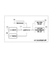
次に、本実施例における制御ブロック構成を、図3を用いて説明する。まず、移動検知部301は、SLO撮影部302の情報に基づいて、被検眼の移動を検知し、追従制御部303へ通知する。また、測定領域設定部304は、操作者からの測定領域設定指示を受け付け、指定可能な測定領域範囲内であれば追従制御部303へ通知する。また、測定領域指定可能範囲設定部305は、測定領域の指定可能な範囲(追従可能な範囲)を設定し、追従制御部303へ通知する。また、追従制御部303は、移動検知部301の結果と測定領域設定部304の結果に基づいて、被検眼の移動に追従するようにOCT撮影部306を制御する。ここで、移動検知部301は、被検眼の移動に関する情報を検知できるものであれば、何でも良く、例えば、被検眼の画像から抽出される特徴部位を用いて被検眼の移動量及び移動方向を直接検知する手法等がある。また、装置の移動量及び移動方向を検知することで、間接的に被検眼の移動量及び移動方向を検知する手法であっても良く、この場合、例えば、被検眼の画像の固定のROIがどの位置に移動したのかを検知する。なお、前者は、2つの検知結果から1つの相対位置を求める手法の一例であり、後者は、1つの相対位置として1つの検知結果を求める手法の一例である。
(OCT inspection)
Next, the control block configuration in the present embodiment will be described with reference to FIG. First, the
次に、OCT撮影時の一般的な追従動作に関して、図4を用いて説明する。図4において、401は、光学系の光学特性(光学設計等)により規定される画質の低下が発生せずに撮影できる撮影保証範囲である。すなわち、撮影保証範囲401は、光学系の光学特性に対応する。402は、撮影時に実際に撮影する測定領域(撮影領域)である。測定領域402を撮影保証範囲401より外に設定した場合、画質の低下が発生する。したがって、測定領域402は、撮影保証範囲401内でのみ指定可能とする場合がある。この場合、例えば、測定領域402が撮影保証範囲401より外に出た場合、追従制御部は、測定領域402の追従を中止し、再度測定領域402が撮影保証範囲401内に戻った時に追従を再開するように構成することができる。
Next, a general tracking operation during OCT imaging will be described with reference to FIG. In FIG. 4,
このときの処理の流れを図5のフローチャートを用いて説明する。S501において測定領域402を設定する。S502において、測定領域402が撮影保証範囲401内にあるかを判定し、S502の条件を満たす場合はS503で測定領域401の追従を実施する。S502の条件を満たさない場合はS504で測定領域401の追従を中止する。S505において、撮影指示があるかを確認し、撮影指示がある場合はS506で撮影を開始する。撮影指示がない場合はS502に戻り、追従処理を繰り返す。
The processing flow at this time will be described with reference to the flowchart of FIG. In S501, the
次に、図6(a)を用いて、本実施例の追従制御について説明を行う。401は撮影保証領域、402は測定領域である。この例では、測定領域は、視神経乳頭や黄斑部を含む形で、非常に大きな領域として指定された例であり、撮影保証領域401に対し、ほとんどマージンがない。このようなワイドスキャンの場合には、固視が安定しない被験者でなくても、ほんの少しの眼の動きによって、測定領域402が撮影保証領域401外に出てしまうことがあった。これに伴い、追従動作がそのたびに止まってしまい、同一部位を計測することが難しくなっていた。このような点を鑑みて、本実施例では、測定領域402の大きさに応じて測定領域402を指定可能な範囲を可変的に変更するように制御を行った。例えば、測定領域402の面積を算出し、面積が閾値Thaより広いと判断した場合は、測定領域指定可能範囲を広げる。広げる際に、算出した面積に応じて広げてもよいし、SLO画像を使って、追従が判定できる限界まで広げてもよかった。これにより、所望の範囲を撮影できる。具体的には、Thaとしては、撮影保証範囲の面積の60%として設定したところ、好ましく制御することができた。
Next, the follow-up control of this embodiment will be described with reference to FIG.
図7のフローチャートは、以上の処理の流れをまとめたものである。図7は、図5のフローチャートからS502を変更し、S701およびS702としている。S701では測定領域指定可能範囲を変更し、S702では測定領域指定可能範囲を基準として、以降の処理を実施する。 The flowchart of FIG. 7 summarizes the flow of the above processing. FIG. 7 changes S502 from the flowchart of FIG. 5 to S701 and S702. In S701, the measurement area specifiable range is changed, and in S702, the subsequent processing is performed based on the measurement area specifiable range.
更に、図8(a)のフローチャートは、S701を分解して説明したものであり、(a)S801、S802に分解される。S801では測定領域402の面積により処理を分岐し、S802では測定領域指定可能範囲の大きさを変更する。
Furthermore, the flowchart of FIG. 8A is an exploded view of S701, and is broken down into (a) S801 and S802. In S801, the process branches depending on the area of the
本実施例では、図9(a)に示すように、測定領域402のサイズに基づいて、測定領域指定可能範囲901を撮影保証範囲401の外側に変更する。なお、本実施例では、撮影保証範囲401は円形としたが、光学特性により決まるため、装置の光学特性によっては円形とならなくてもよい。この場合、測定領域指定可能範囲901の設定条件は、その形状に応じて変更する必要がある。
In this embodiment, as shown in FIG. 9A, the measurement
また、本実施例では、測定領域402の面積を求めて、測定領域指定可能範囲を変更していたが、これに限るものではない。例えば、図9(b)に示すように、測定領域402と撮影保証範囲401との間の最短距離dを求めて、その距離に応じて、測定領域指定可能範囲901を変更してもかまわない。このように制御することにより、測定領域402がそれほど大きな領域でなくても、撮影保証範囲401に近接している場合には、安定して同一部位を撮影することができるため、非常に好ましい。
In the present embodiment, the area of the
また、図9(c)に示すように、測定領域指定可能範囲を、撮影保証範囲401よりも広い領域に変更する場合には、ユーザーにメッセージ1401を出して、範囲の変更の要否を使用者に通知してもよい。また、測定領域指定可能範囲が、撮影保証範囲401よりも広い領域に変更する場合には、通常の測定領域指定可能範囲に対して、追従及び、追従できない状態が所定回数繰り返された後に、切り替えると更に良い。また、このようにして撮影された画像は、画質的に劣る可能性があるため、検査画像のプロパティや、画像上のアイコンなどで、その旨を表示部106に表示させると、さらに好ましい。
Also, as shown in FIG. 9C, when changing the measurement area specifiable range to an area wider than the
(ラジアルスキャン、サークルスキャン)
また、本実施例では、測定範囲402の面積を使用して測定領域指定可能範囲901を変更したが、測定領域402の形状や撮影時の走査パターンによって測定領域指定可能範囲901を変更してもよい。具体的には、測定領域402の形状がラジアルスキャンやサークルスキャンなどのような円形の場合、面積が同じであっても測定領域402が撮影保証範囲401を超える可能性は低くなる。たとえばこのような場合は、Thaが撮影保証範囲の面積の80%以上となった場合に、測定領域指定可能範囲を広げたところ非常に好ましかった。また、ボリュームスキャンのような測定領域が矩形の場合に比べ測定領域指定可能範囲901を広げる幅を狭くすることができる。
(Radial scan, circle scan)
Further, in this embodiment, the measurement
(ラインスキャン、クロススキャン)
また、被検眼上の1点に注目して観察したい場合、ラインスキャンや、クロススキャンを使用する場合がある。一般に、ボリュームスキャンは追従が行われず、撮影したい部位に対して測定領域が多少ずれても、ボリューム内に注目部位が入れば許容される可能性があるが、一方、ラインスキャンやクロススキャンは、注目している部位に対してスキャンをおこなうため、追従が行われず、同一の部位を撮影できないと再撮影になる可能性があった。
(Line scan, cross scan)
In addition, when it is desired to observe by paying attention to one point on the eye to be examined, line scan or cross scan may be used. In general, volume scanning is not followed, and even if the measurement area is slightly deviated from the part to be imaged, it may be allowed if the part of interest enters the volume, while line scanning and cross scanning are Since the region of interest is scanned, tracking is not performed, and if the same region cannot be imaged, re-imaging may occur.
ここで、図6(b)は、クロススキャンを実施した場合の、制御方法を説明するための図である。同図において、401は撮影保証範囲、901は追従範囲を拡大した場合の追従範囲、1601はクロススキャンである。ここでは、クロススキャン1601が撮影範囲に相当する。これらのスキャンの場合には、スキャン上の任意の点から、撮影保証範囲401までの最短距離dをまず求める。次に、dがある閾値よりも大きい場合には、追従範囲は通常通り撮影保証範囲とする。また、dがある閾値よりも大きい場合は、追従範囲を901のように、撮影保証範囲よりも拡大するように制御を行った。このように制御したところ、好ましく制御することができた。なお、dの算出方法としては、スキャンの中心と、スキャンの大きさから、最短距離を求めて実施してもよい。また、別の制御方法として、スキャンを含む最小の円または楕円を求め、この円または楕円と撮影保証領域401までの距離の最小値によって制御してもよい。また、図6(b)の例では、撮影保証範囲401に対し、追従範囲は上下、左右均等に広げたが、とくにこれにこだわる必要はなく、本例では、図の左側が撮影保証範囲を超える可能性が高いため、左側のみ拡大してもよい。
Here, FIG. 6B is a diagram for explaining a control method when the cross scan is performed. In the figure, 401 is a photographing guarantee range, 901 is a tracking range when the tracking range is enlarged, and 1601 is a cross scan. Here, the
次に、実施例2では、実施例1の構成の一部を変更して、フォローアップ検査時に前回検査と同一箇所を撮影する方法を説明する。実施例2では、フォローアップ検査時とフォローアップ検査以外で測定領域指定可能範囲を変更することで課題を解決する。フォローアップ検査では、前回検査(過去に実施した検査)と同一箇所を比較することにより、経時変化を確認する。そのため、スキャンサイズやスキャンモードなどの撮影条件は前回検査時と同じであることが好ましく、前回と同一箇所を撮影できることが重要である。しかし、たとえ前回検査時と同一の設定にしても、前回検査時にスキャンが撮影保証領域の近傍にある場合や、固視が定まらない場合などでは、測定領域402が撮影保証範囲内401に収まらないことがある。このような場合、従来の制御方法では、追従が行われないため、フォローアップの目的である前回と同じ箇所を撮影することが難しかった。そこで、本実施例では、フォローアップ検査時には、多少の画質の低下はあったとしても、測定領域指定可能範囲901を前回検査時よりも広く設定することで前回検査時と同一箇所を撮影できるように制御を行った。なお、フォローアップ検査は、フォローアップ撮影とも呼び、例えば、フォローアップ撮影モードを含む複数の撮影モードのいずれかを選択する選択手段(不図示)により、フォローアップ撮影モードが選択されることにより実現することができる。
Next, in the second embodiment, a method of changing a part of the configuration of the first embodiment and photographing the same part as the previous inspection at the follow-up inspection will be described. In the second embodiment, the problem can be solved by changing the range in which the measurement area can be specified at the time of the follow-up inspection and other than the follow-up inspection. In the follow-up inspection, a change with time is confirmed by comparing the same place with the previous inspection (inspection performed in the past). Therefore, the imaging conditions such as the scan size and the scan mode are preferably the same as those at the previous inspection, and it is important that the same part as that at the previous time can be imaged. However, even if the setting is the same as that in the previous examination, the
まず、初回検査時には、測定領域指定可能範囲901を撮影保証範囲401と同じサイズにしておき、フォローアップ検査時には測定領域指定可能範囲901を撮影保証範囲401よりも大きく設定して撮影する。例えば、撮影保証範囲401の面積の110%に設定してもよいし、SLO画像を使って、追従が判定できる限界まで広げてもよかった。また、初回検査時の測定領域指定可能範囲901を撮影保証範囲401よりも小さく設定してもよい。例えば撮影保証範囲401の面積を算出し、初回検査時には測定領域指定可能範囲901を撮影保証範囲401の70%に設定することで、フォローアップ検査時の測定領域指定可能範囲901を相対的に広げても良い。
First, in the first inspection, the measurement
ここで、図8(b)のフローチャートは、以上の処理の流れを反映したものあり、図7のフローチャートのS701を分解したものである。それぞれS1001ではフォローアップ検査か否かにより処理を分岐し、S1002では測定領域指定可能範囲901の大きさを変更する。以上に説明した方法によれば、OCTのフォローアップ検査時に測定領域が撮影保証範囲内に収まらない場合でも、前回と同一箇所を撮影することができた。
Here, the flowchart of FIG. 8B reflects the above-described processing flow, and is a decomposition of S701 in the flowchart of FIG. In S1001, the process branches depending on whether or not the follow-up inspection is performed, and in S1002, the size of the measurement
また、実施例2では、図8(b)のフローチャートをもとに測定領域指定可能範囲901の追従範囲に関して記載を行ったが、本発明はこれに限定されるものではなく、例えば、図10のフローチャートのように処理を行っても良い。ここで、図8(b)では、フォローアップ検査時に測定領域指定可能範囲901を広げていたが、この範囲の制限をしない。ただし、このように制限がない場合でも、SLO画像の画角や、移動検知手段が被検眼の動きを検出するために必要な面積が必要であるため、実質的な制限があることは言うまでもない。このように制御しても、同様に効果を得ることができる。また、実施例2においても、図9(c)に示したような、メッセージ1401やアイコン、プロパティ等を用いて、追従範囲を拡大したことを通知しても良い。
In the second embodiment, the follow-up range of the measurement
また、実施例3では、実施例1、2の構成の一部を変更して、課題を解決する方法を説明する。実施例3では、固視灯を移動することで課題を解決する。本実施例における装置構成を、図11を用いて説明する。本実施例では実施例1の構成に加えて、固視灯位置制御部1101は、追従制御部303の結果を受け、固視灯の位置を移動させる。
In the third embodiment, a method for solving a problem by changing a part of the configuration of the first and second embodiments will be described. In the third embodiment, the problem is solved by moving the fixation lamp. The apparatus configuration in the present embodiment will be described with reference to FIG. In the present embodiment, in addition to the configuration of the first embodiment, the fixation lamp
本実施例では、観察時に測定領域402が撮影保証範囲401の中央付近になるように固視灯を移動させる。例えば、図12に示すように、測定領域402が測定領域指定可能範囲901を超えた場合、自動的に移動させる。これにより、測定領域402は被検眼を追従し、撮影保証範囲401の中央付近になるように移動する。これにより被検眼の固視状態が変化し、測定領域402が測定領域指定可能範囲の中に納まらない場合でも、被験者の固視状態を変化させることにより、領域内におさめて撮影することができる。以上に説明した方法によれば、測定範囲を撮影保証範囲内に収まるように撮影できた。更にフォローアップ検査時においても、測定領域が撮影保証範囲外に出てしまう確率を低減することができた。
In the present embodiment, the fixation lamp is moved so that the
また、実施例4では、眼球運動を基に測定領域指定可能範囲を変更することで課題を解決する。被験者の眼球運動が激しい場合や固視がうまく定まらない場合、測定領域402を撮影保証範囲401内に設定しても、撮影中に撮影保証範囲401外に出てしまうことがある。そこで、被検眼の移動量を検知して測定領域指定可能範囲901を自動的に小さくすることで、撮影保証範囲401の中央付近で撮影を実施させる。また、撮影中には指定した箇所を撮影できることが重要視されるため、撮影完了までの間は撮影保証範囲401を越えて追従を継続することで、指定した測定領域402の撮影を実現する。初回検査時に測定領域指定可能範囲をあらかじめ小さくしておくことにより、フォローアップ検査時、眼が動いたことにより、測定領域402が撮影保証範囲401外に出る確率を低減できるため、好ましかった。
Further, in the fourth embodiment, the problem is solved by changing the measurement area specifiable range based on the eye movement. If the subject's eye movement is intense or fixation cannot be determined well, even if the
このような場合、測定領域指定範囲を狭める際に、ユーザーにメッセージを表示部106に表示させると更に好ましい。ここで、眼球運動の検知は、所定期間で固視の変位量の総計をもとに、判定すると好ましい。なお、この場合にも、図9(c)に示したようなメッセージ1401やアイコン、プロパティなどをもちいて、追従範囲を変更したことを通知してもよい。以上説明した方法によれば、被検眼の眼球運動が激しい場合や固視がうまく定まらない場合であっても、指定した測定範囲を撮影できた。
In such a case, it is more preferable to display a message on the
実施例1では、フォローアップモードでの撮影の前段階である観察状態の追従動作に関して、説明をおこなってきたが、撮影状態になった際の動作に関しては説明していなかった。観察状態が完了し、撮影状態となった際には、たとえ撮影保証範囲を超えたとしても、撮影できる方が好ましかい。図13は、図10の変形例であり、撮影指示後、撮影状態に入る前に常に追従状態に状態を切り替えた例である。このように制御したところ、撮影指示後に眼が大きく移動しても、画質が低下する可能性はあるものの、狙った領域を撮影することができ、非常に好ましかった。なお、本実施例は、図10の変形例として記載したが、これに限定されるものではなく、本発明で記載している他の追従制御との組み合わせは可能であり、同様の効果を得ることができる。 In the first embodiment, the follow-up operation of the observation state, which is the previous stage of shooting in the follow-up mode, has been described, but the operation when the shooting state is entered has not been described. When the observation state is completed and the photographing state is reached, it is preferable to be able to photograph even if the photographing guaranteed range is exceeded. FIG. 13 is a modification of FIG. 10 and is an example in which the state is always switched to the tracking state after entering the shooting state and before entering the shooting state. As a result of such control, even if the eye moved greatly after the shooting instruction, the image quality could be lowered, but the target area could be shot, which was very preferable. In addition, although the present Example was described as a modification of FIG. 10, it is not limited to this, The combination with the other follow-up control described in this invention is possible, and the same effect is acquired. be able to.
(その他の実施例)
また、本発明は、以下の処理を実行することによっても実現される。即ち、上述した実施形態の機能を実現するソフトウェア(プログラム)を、ネットワーク又は各種記憶媒体を介してシステム或いは装置に供給し、そのシステム或いは装置のコンピュータ(またはCPUやMPU等)がプログラムを読み出して実行する処理である。また、OCTの例として説明してきたが、本発明は、OCT以外にも適用可能で、例えば、WF−OCT画像上で指定した位置を撮影するAO−SLO装置等の眼科撮影装置にも適用可能である。
(Other examples)
The present invention can also be realized by executing the following processing. That is, software (program) that realizes the functions of the above-described embodiments is supplied to a system or apparatus via a network or various storage media, and a computer (or CPU, MPU, or the like) of the system or apparatus reads the program. It is a process to be executed. Although described as an example of OCT, the present invention can be applied to other than OCT, for example, an ophthalmic imaging apparatus such as an AO-SLO apparatus that images a specified position on a WF-OCT image. It is.
Claims (20)
少なくともフォローアップ撮影モードを含む複数の撮影モードのいずれかを選択する選択手段と、
前記フォローアップ撮影モードの他の撮影モードが選択された場合には、前記検知された情報に基づいて前記被検眼を撮影する撮影領域が所定の範囲を超えるか否かを判定する判定手段と、
前記撮影領域が前記所定の範囲を超えないと判定された場合には前記検知された情報に基づいて前記撮影領域の追従を行い、前記撮影領域が前記所定の範囲を超えると判定された場合には前記撮影領域の追従を行わず、前記フォローアップ撮影モードが選択された場合には前記判定手段による判定を行わずに前記検知された情報に基づいて前記撮影領域の追従を行う追従制御手段と、
を有することを特徴とする眼科撮影装置。 Detection means for detecting information relating to movement of the eye to be examined;
Selection means for selecting at least one of a plurality of shooting modes including a follow-up shooting mode;
When another imaging mode of the follow-up imaging mode is selected, a determination unit that determines whether an imaging area for imaging the eye to be examined exceeds a predetermined range based on the detected information;
When it is determined that the shooting area does not exceed the predetermined range, the shooting area is tracked based on the detected information, and when it is determined that the shooting area exceeds the predetermined range. Does not follow the shooting area, and when the follow-up shooting mode is selected, tracking control means for tracking the shooting area based on the detected information without making a determination by the determining means; ,
An ophthalmologic photographing apparatus comprising:
前記検知手段は、前記正面画像を用いて前記移動に関する情報を検知し、
前記撮影領域は、前記断層画像の撮影領域であり、
前記追従制御手段は、前記検知された情報に基づいて前記OCT光学系の走査手段を制御することにより、前記撮影領域の追従を行うことを特徴とする請求項1に記載の眼科撮影装置。 An OCT optical system for taking a tomographic image of the eye to be examined and an optical system for taking a front image of the eye to be examined are configured as a partly common optical system,
The detection means detects information about the movement using the front image,
The imaging area is an imaging area of the tomographic image,
The ophthalmic imaging apparatus according to claim 1, wherein the tracking control unit controls the scanning unit of the OCT optical system based on the detected information to track the imaging region.
前記検知された情報に基づいて、前記被検眼を撮影する撮影領域の追従を行う追従制御手段と、
前記撮影領域が追従可能な範囲を変更する変更手段と、
を有することを特徴とする眼科撮影装置。 Detection means for detecting information relating to movement of the eye to be examined;
Based on the detected information, tracking control means for tracking an imaging region for imaging the eye to be examined,
Changing means for changing a range in which the imaging region can be followed;
An ophthalmologic photographing apparatus comprising:
前記変更手段は、前記範囲の変更を行う指示に応じて、前記範囲を変更することを特徴とする請求項4に記載の光干渉断層撮影装置。 It further has a display control means for displaying a notification on the necessity of changing the range on the display means,
The optical coherence tomography apparatus according to claim 4, wherein the changing unit changes the range in response to an instruction to change the range.
前記正面画像を表示手段に表示させる表示制御手段と、
前記表示された正面画像で前記被検眼を撮影する撮影領域を指定する指定手段と、
前記撮影領域を指定可能な範囲を変更する変更手段と、
を有することを特徴とする眼科撮影装置。 Acquisition means for acquiring a front image of the eye to be examined;
Display control means for displaying the front image on a display means;
Designating means for designating an imaging region for imaging the eye to be examined in the displayed front image;
Changing means for changing a range in which the imaging region can be specified;
An ophthalmologic photographing apparatus comprising:
前記変更手段は、前記走査手段の走査パターンと撮影範囲とのうち少なくとも1つの情報に基づいて、前記範囲を変更することを特徴とする請求項4乃至10のいずれか1項に記載の眼科撮影装置。 Scanning means for scanning the eye to be examined with measurement light;
11. The ophthalmic imaging according to claim 4, wherein the changing unit changes the range based on at least one information of a scanning pattern of the scanning unit and an imaging range. apparatus.
前記検知された情報に基づいて、固視灯を呈示する位置を変更する制御手段と、
を有することを特徴とする眼科撮影装置。 Detection means for detecting information relating to movement of the eye to be examined;
Based on the detected information, control means for changing the position to present the fixation lamp;
An ophthalmologic photographing apparatus comprising:
フォローアップ撮影モードの他の撮影モードが選択された場合には、前記検知された情報に基づいて前記被検眼を撮影する撮影領域が所定の範囲を超えるか否かを判定する工程と、
前記撮影領域が前記所定の範囲を超えないと判定された場合には前記検知された情報に基づいて前記撮影領域の追従を行い、前記撮影領域が前記所定の範囲を超えると判定された場合には前記撮影領域の追従を行わず、前記フォローアップ撮影モードが選択された場合には前記判定する工程による判定を行わずに前記検知された情報に基づいて前記撮影領域の追従を行う工程と、
を有することを特徴とする眼科撮影装置の制御方法。 Detecting information about movement of the eye to be examined;
When another imaging mode of the follow-up imaging mode is selected, determining whether an imaging area for imaging the eye to be examined exceeds a predetermined range based on the detected information;
When it is determined that the shooting area does not exceed the predetermined range, the shooting area is tracked based on the detected information, and when it is determined that the shooting area exceeds the predetermined range. Does not follow the shooting area, and follows the shooting area based on the detected information without performing the determination by the determining step when the follow-up shooting mode is selected;
A control method for an ophthalmologic photographing apparatus, comprising:
前記検知された情報に基づいて、前記被検眼を撮影する撮影領域の追従を行う工程と、
前記撮影領域が追従可能な範囲を変更する工程と、
を有することを特徴とする眼科撮影装置の制御方法。 Detecting information about movement of the eye to be examined;
Based on the detected information, following the imaging region for imaging the eye to be examined; and
Changing the range in which the shooting area can be followed;
A control method for an ophthalmologic photographing apparatus, comprising:
前記正面画像を表示手段に表示させる工程と、
前記表示された正面画像で前記被検眼を撮影する撮影領域の指定を受ける工程と、
前記撮影領域を指定可能な範囲を変更する工程と、
を有することを特徴とする眼科撮影装置の制御方法。 Obtaining a front image of the eye to be examined;
Displaying the front image on a display means;
Receiving a designation of an imaging region for imaging the eye to be examined in the displayed front image;
Changing the range in which the imaging region can be specified;
A control method for an ophthalmologic photographing apparatus, comprising:
前記検知された情報に基づいて、固視灯を呈示する位置を変更する工程と、
を有することを特徴とする眼科撮影装置の制御方法。 Detecting information about movement of the eye to be examined;
Based on the detected information, changing the position to present the fixation lamp;
A control method for an ophthalmologic photographing apparatus, comprising:
Priority Applications (2)
| Application Number | Priority Date | Filing Date | Title |
|---|---|---|---|
| JP2016047309A JP6906898B2 (en) | 2016-03-10 | 2016-03-10 | Ophthalmologic imaging equipment |
| US15/450,980 US10244942B2 (en) | 2016-03-10 | 2017-03-06 | Ophthalmologic photographing apparatus, method, and storage medium |
Applications Claiming Priority (1)
| Application Number | Priority Date | Filing Date | Title |
|---|---|---|---|
| JP2016047309A JP6906898B2 (en) | 2016-03-10 | 2016-03-10 | Ophthalmologic imaging equipment |
Publications (3)
| Publication Number | Publication Date |
|---|---|
| JP2017158867A true JP2017158867A (en) | 2017-09-14 |
| JP2017158867A5 JP2017158867A5 (en) | 2019-03-14 |
| JP6906898B2 JP6906898B2 (en) | 2021-07-21 |
Family
ID=59788584
Family Applications (1)
| Application Number | Title | Priority Date | Filing Date |
|---|---|---|---|
| JP2016047309A Active JP6906898B2 (en) | 2016-03-10 | 2016-03-10 | Ophthalmologic imaging equipment |
Country Status (2)
| Country | Link |
|---|---|
| US (1) | US10244942B2 (en) |
| JP (1) | JP6906898B2 (en) |
Families Citing this family (3)
| Publication number | Priority date | Publication date | Assignee | Title |
|---|---|---|---|---|
| KR20150101915A (en) * | 2014-02-27 | 2015-09-04 | 삼성전자주식회사 | Method for displaying 3 dimension graphic user interface screen and device for performing the same |
| JP2018061621A (en) * | 2016-10-11 | 2018-04-19 | オプトス ピーエルシー | Ocular fundus imaging apparatus, ocular fundus imaging method, and ocular fundus imaging program |
| JP7117768B2 (en) * | 2018-07-18 | 2022-08-15 | 株式会社Qdレーザ | Visual field visual acuity test device and visual field visual acuity test method |
Citations (6)
| Publication number | Priority date | Publication date | Assignee | Title |
|---|---|---|---|---|
| JP2010142428A (en) * | 2008-12-18 | 2010-07-01 | Canon Inc | Photographing apparatus, photographing method, program and recording medium |
| JP2013153879A (en) * | 2012-01-27 | 2013-08-15 | Canon Inc | Image processing apparatus, image processing apparatus control method, ophthalmologic apparatus, ophthalmologic apparatus control method, ophthalmologic system, and program |
| JP2013212172A (en) * | 2012-03-30 | 2013-10-17 | Canon Inc | Optical coherence tomography imaging device, and method for controlling the same |
| JP2014147505A (en) * | 2013-01-31 | 2014-08-21 | Canon Inc | Ophthalmologic apparatus and control method |
| JP2014527434A (en) * | 2011-08-09 | 2014-10-16 | オプトビュー,インコーポレーテッド | Feature motion correction and normalization in optical coherence tomography |
| JP2014217744A (en) * | 2013-04-11 | 2014-11-20 | キヤノン株式会社 | Ophthalmologic apparatus, method of controlling the same, and program |
Family Cites Families (1)
| Publication number | Priority date | Publication date | Assignee | Title |
|---|---|---|---|---|
| JP5138977B2 (en) | 2007-05-24 | 2013-02-06 | 株式会社トプコン | Optical image measuring device |
-
2016
- 2016-03-10 JP JP2016047309A patent/JP6906898B2/en active Active
-
2017
- 2017-03-06 US US15/450,980 patent/US10244942B2/en active Active
Patent Citations (6)
| Publication number | Priority date | Publication date | Assignee | Title |
|---|---|---|---|---|
| JP2010142428A (en) * | 2008-12-18 | 2010-07-01 | Canon Inc | Photographing apparatus, photographing method, program and recording medium |
| JP2014527434A (en) * | 2011-08-09 | 2014-10-16 | オプトビュー,インコーポレーテッド | Feature motion correction and normalization in optical coherence tomography |
| JP2013153879A (en) * | 2012-01-27 | 2013-08-15 | Canon Inc | Image processing apparatus, image processing apparatus control method, ophthalmologic apparatus, ophthalmologic apparatus control method, ophthalmologic system, and program |
| JP2013212172A (en) * | 2012-03-30 | 2013-10-17 | Canon Inc | Optical coherence tomography imaging device, and method for controlling the same |
| JP2014147505A (en) * | 2013-01-31 | 2014-08-21 | Canon Inc | Ophthalmologic apparatus and control method |
| JP2014217744A (en) * | 2013-04-11 | 2014-11-20 | キヤノン株式会社 | Ophthalmologic apparatus, method of controlling the same, and program |
Also Published As
| Publication number | Publication date |
|---|---|
| JP6906898B2 (en) | 2021-07-21 |
| US20170258327A1 (en) | 2017-09-14 |
| US10244942B2 (en) | 2019-04-02 |
Similar Documents
| Publication | Publication Date | Title |
|---|---|---|
| JP6322042B2 (en) | Ophthalmic photographing apparatus, control method thereof, and program | |
| JP6652281B2 (en) | Optical tomographic imaging apparatus, control method thereof, and program | |
| JP5941761B2 (en) | Ophthalmic photographing apparatus and ophthalmic image processing apparatus | |
| JP6703839B2 (en) | Ophthalmic measuring device | |
| US20120253183A1 (en) | Image processing apparatus, ophthalmic apparatus, and ophthalmic system | |
| CN103961061A (en) | Optical tomographic imaging apparatus and method for controlling the same | |
| JP2017184874A (en) | Ophthalmic photographing apparatus | |
| JP2017158836A (en) | Ophthalmologic apparatus and imaging method | |
| JP6633468B2 (en) | Blood flow measurement device | |
| JP6906898B2 (en) | Ophthalmologic imaging equipment | |
| JP2019154988A (en) | Ophthalmologic imaging apparatus, control method therefor, program, and storage medium | |
| JP5858603B2 (en) | Ophthalmic apparatus and control method thereof | |
| US10980411B2 (en) | Information processing apparatus, information processing method, and storage medium | |
| JP2014147504A (en) | Optical interference tomographic imaging device and method for controlling the same | |
| JP2020054812A (en) | Image processing device, image processing method and program | |
| JP5653055B2 (en) | Ophthalmic apparatus and control method thereof | |
| JP6099782B2 (en) | Ophthalmic imaging equipment | |
| JP2017202369A (en) | Ophthalmologic image processing device | |
| JP2018191761A (en) | Information processing device, information processing method, and program | |
| JP2017225599A (en) | Oct apparatus | |
| JP6839310B2 (en) | Optical tomography imaging device, its control method, and program | |
| JP2019025186A (en) | Ophthalmologic apparatus and data collection method | |
| US20210204809A1 (en) | Ophthalmic imaging apparatus, controlling method of the same, and recording medium | |
| JP6701250B2 (en) | Ophthalmic photographing apparatus, control method thereof, and program | |
| JP2018000373A (en) | Ophthalmologic imaging apparatus, control method thereof, and program |
Legal Events
| Date | Code | Title | Description |
|---|---|---|---|
| A521 | Request for written amendment filed |
Free format text: JAPANESE INTERMEDIATE CODE: A523 Effective date: 20190129 |
|
| A621 | Written request for application examination |
Free format text: JAPANESE INTERMEDIATE CODE: A621 Effective date: 20190129 |
|
| A977 | Report on retrieval |
Free format text: JAPANESE INTERMEDIATE CODE: A971007 Effective date: 20191028 |
|
| A131 | Notification of reasons for refusal |
Free format text: JAPANESE INTERMEDIATE CODE: A131 Effective date: 20191112 |
|
| A521 | Request for written amendment filed |
Free format text: JAPANESE INTERMEDIATE CODE: A523 Effective date: 20200110 |
|
| A131 | Notification of reasons for refusal |
Free format text: JAPANESE INTERMEDIATE CODE: A131 Effective date: 20200630 |
|
| A521 | Request for written amendment filed |
Free format text: JAPANESE INTERMEDIATE CODE: A523 Effective date: 20200713 |
|
| A131 | Notification of reasons for refusal |
Free format text: JAPANESE INTERMEDIATE CODE: A131 Effective date: 20201117 |
|
| A521 | Request for written amendment filed |
Free format text: JAPANESE INTERMEDIATE CODE: A523 Effective date: 20201221 |
|
| TRDD | Decision of grant or rejection written | ||
| A01 | Written decision to grant a patent or to grant a registration (utility model) |
Free format text: JAPANESE INTERMEDIATE CODE: A01 Effective date: 20210601 |
|
| A61 | First payment of annual fees (during grant procedure) |
Free format text: JAPANESE INTERMEDIATE CODE: A61 Effective date: 20210630 |
|
| R151 | Written notification of patent or utility model registration |
Ref document number: 6906898 Country of ref document: JP Free format text: JAPANESE INTERMEDIATE CODE: R151 |