JP2016500251A - Enhancement of anti-cancer activity of immunomodulating Fc fusion proteins - Google Patents
Enhancement of anti-cancer activity of immunomodulating Fc fusion proteins Download PDFInfo
- Publication number
- JP2016500251A JP2016500251A JP2015545522A JP2015545522A JP2016500251A JP 2016500251 A JP2016500251 A JP 2016500251A JP 2015545522 A JP2015545522 A JP 2015545522A JP 2015545522 A JP2015545522 A JP 2015545522A JP 2016500251 A JP2016500251 A JP 2016500251A
- Authority
- JP
- Japan
- Prior art keywords
- antibody
- tumor
- ctla
- cells
- cancer
- Prior art date
- Legal status (The legal status is an assumption and is not a legal conclusion. Google has not performed a legal analysis and makes no representation as to the accuracy of the status listed.)
- Pending
Links
Images


Classifications
-
- C—CHEMISTRY; METALLURGY
- C07—ORGANIC CHEMISTRY
- C07K—PEPTIDES
- C07K14/00—Peptides having more than 20 amino acids; Gastrins; Somatostatins; Melanotropins; Derivatives thereof
- C07K14/435—Peptides having more than 20 amino acids; Gastrins; Somatostatins; Melanotropins; Derivatives thereof from animals; from humans
-
- A—HUMAN NECESSITIES
- A61—MEDICAL OR VETERINARY SCIENCE; HYGIENE
- A61P—SPECIFIC THERAPEUTIC ACTIVITY OF CHEMICAL COMPOUNDS OR MEDICINAL PREPARATIONS
- A61P35/00—Antineoplastic agents
-
- C—CHEMISTRY; METALLURGY
- C07—ORGANIC CHEMISTRY
- C07K—PEPTIDES
- C07K16/00—Immunoglobulins [IGs], e.g. monoclonal or polyclonal antibodies
- C07K16/18—Immunoglobulins [IGs], e.g. monoclonal or polyclonal antibodies against material from animals or humans
- C07K16/28—Immunoglobulins [IGs], e.g. monoclonal or polyclonal antibodies against material from animals or humans against receptors, cell surface antigens or cell surface determinants
- C07K16/2803—Immunoglobulins [IGs], e.g. monoclonal or polyclonal antibodies against material from animals or humans against receptors, cell surface antigens or cell surface determinants against the immunoglobulin superfamily
- C07K16/2818—Immunoglobulins [IGs], e.g. monoclonal or polyclonal antibodies against material from animals or humans against receptors, cell surface antigens or cell surface determinants against the immunoglobulin superfamily against CD28 or CD152
-
- C—CHEMISTRY; METALLURGY
- C07—ORGANIC CHEMISTRY
- C07K—PEPTIDES
- C07K16/00—Immunoglobulins [IGs], e.g. monoclonal or polyclonal antibodies
- C07K16/18—Immunoglobulins [IGs], e.g. monoclonal or polyclonal antibodies against material from animals or humans
- C07K16/28—Immunoglobulins [IGs], e.g. monoclonal or polyclonal antibodies against material from animals or humans against receptors, cell surface antigens or cell surface determinants
- C07K16/2875—Immunoglobulins [IGs], e.g. monoclonal or polyclonal antibodies against material from animals or humans against receptors, cell surface antigens or cell surface determinants against the NGF/TNF superfamily, e.g. CD70, CD95L, CD153, CD154
-
- C—CHEMISTRY; METALLURGY
- C07—ORGANIC CHEMISTRY
- C07K—PEPTIDES
- C07K16/00—Immunoglobulins [IGs], e.g. monoclonal or polyclonal antibodies
- C07K16/18—Immunoglobulins [IGs], e.g. monoclonal or polyclonal antibodies against material from animals or humans
- C07K16/28—Immunoglobulins [IGs], e.g. monoclonal or polyclonal antibodies against material from animals or humans against receptors, cell surface antigens or cell surface determinants
- C07K16/2878—Immunoglobulins [IGs], e.g. monoclonal or polyclonal antibodies against material from animals or humans against receptors, cell surface antigens or cell surface determinants against the NGF-receptor/TNF-receptor superfamily, e.g. CD27, CD30, CD40, CD95
-
- A—HUMAN NECESSITIES
- A61—MEDICAL OR VETERINARY SCIENCE; HYGIENE
- A61K—PREPARATIONS FOR MEDICAL, DENTAL OR TOILETRY PURPOSES
- A61K39/00—Medicinal preparations containing antigens or antibodies
- A61K2039/505—Medicinal preparations containing antigens or antibodies comprising antibodies
-
- C—CHEMISTRY; METALLURGY
- C07—ORGANIC CHEMISTRY
- C07K—PEPTIDES
- C07K2317/00—Immunoglobulins specific features
- C07K2317/40—Immunoglobulins specific features characterized by post-translational modification
- C07K2317/41—Glycosylation, sialylation, or fucosylation
-
- C—CHEMISTRY; METALLURGY
- C07—ORGANIC CHEMISTRY
- C07K—PEPTIDES
- C07K2317/00—Immunoglobulins specific features
- C07K2317/50—Immunoglobulins specific features characterized by immunoglobulin fragments
- C07K2317/52—Constant or Fc region; Isotype
-
- C—CHEMISTRY; METALLURGY
- C07—ORGANIC CHEMISTRY
- C07K—PEPTIDES
- C07K2317/00—Immunoglobulins specific features
- C07K2317/70—Immunoglobulins specific features characterized by effect upon binding to a cell or to an antigen
- C07K2317/71—Decreased effector function due to an Fc-modification
-
- C—CHEMISTRY; METALLURGY
- C07—ORGANIC CHEMISTRY
- C07K—PEPTIDES
- C07K2317/00—Immunoglobulins specific features
- C07K2317/70—Immunoglobulins specific features characterized by effect upon binding to a cell or to an antigen
- C07K2317/72—Increased effector function due to an Fc-modification
-
- C—CHEMISTRY; METALLURGY
- C07—ORGANIC CHEMISTRY
- C07K—PEPTIDES
- C07K2317/00—Immunoglobulins specific features
- C07K2317/70—Immunoglobulins specific features characterized by effect upon binding to a cell or to an antigen
- C07K2317/73—Inducing cell death, e.g. apoptosis, necrosis or inhibition of cell proliferation
-
- C—CHEMISTRY; METALLURGY
- C07—ORGANIC CHEMISTRY
- C07K—PEPTIDES
- C07K2317/00—Immunoglobulins specific features
- C07K2317/70—Immunoglobulins specific features characterized by effect upon binding to a cell or to an antigen
- C07K2317/75—Agonist effect on antigen
-
- C—CHEMISTRY; METALLURGY
- C07—ORGANIC CHEMISTRY
- C07K—PEPTIDES
- C07K2317/00—Immunoglobulins specific features
- C07K2317/70—Immunoglobulins specific features characterized by effect upon binding to a cell or to an antigen
- C07K2317/76—Antagonist effect on antigen, e.g. neutralization or inhibition of binding
-
- C—CHEMISTRY; METALLURGY
- C07—ORGANIC CHEMISTRY
- C07K—PEPTIDES
- C07K2319/00—Fusion polypeptide
- C07K2319/30—Non-immunoglobulin-derived peptide or protein having an immunoglobulin constant or Fc region, or a fragment thereof, attached thereto
Abstract
本開示は、癌または感染性物質により生じた疾患に罹患した対象のT細胞の標的、例えばリガンドの共阻害または強刺激レセプターと特異的に結合することにより、免疫調節標的の活性を変化させ、癌細胞または感染性物質に対する内因性免疫応答を増強するFc融合タンパク質の抗腫瘍効果を増強する方法であって、活性化Fcレセプター(FcR)に対する該Fc領域の結合を増強するようにFc融合タンパク質のFc領域を選択、設計、または修飾することを含む方法を提供する。該開示は、対象の癌細胞または感染性物質に対する内因性免疫応答を増強する能力を増大させる該方法により製造されるFc融合タンパク質も提供する。該開示は、さらに治療的有効量のFc融合タンパク質を対象に投与することを含む対象の治療方法であって、Fc領域の活性化FcRに対する結合が増加するようにFc融合タンパク質のFc領域が選択、設計、または修飾される方法を提供する。The present disclosure alters the activity of an immunomodulatory target by specifically binding to a target of a T cell in a subject afflicted with a disease caused by cancer or an infectious agent, such as co-inhibition of ligand or a strong stimulatory receptor, A method for enhancing the antitumor effect of an Fc fusion protein that enhances an endogenous immune response against cancer cells or infectious agents, wherein the Fc fusion protein enhances binding of the Fc region to an activated Fc receptor (FcR) A method comprising selecting, designing, or modifying the Fc region of is provided. The disclosure also provides an Fc fusion protein produced by the method that increases the ability to enhance an endogenous immune response against the subject's cancer cells or infectious agent. The disclosure further relates to a method of treating a subject comprising administering to the subject a therapeutically effective amount of an Fc fusion protein, wherein the Fc region of the Fc fusion protein is selected such that binding of the Fc region to activated FcR is increased. Provide a method that is designed, modified, or modified.
Description
本願を通して、種々の刊行物は、著者名と日付または特許番号もしくは特許公開番号を括弧で示す。これら刊行物の完全な引用は、特許請求の範囲の直前の本明細書の最後に記載している。これら刊行物の開示は本明細書の一部を構成し、本明細書および特許請求の範囲に記載の発明の日における当業者に知られた当該分野の状態をより完全に説明する。しかしながら、本明細書の参考文献の引用は、該参考文献が本発明の先行技術であると認めるものと解釈すべきではない。 Throughout this application, various publications indicate the author name and date or patent number or patent publication number in parentheses. Full citations for these publications can be found at the end of the specification immediately preceding the claims. The disclosures of these publications form part of the present specification and more fully describe the state of the art as known to those skilled in the art at the date of invention described in the specification and claims. However, citation of a reference herein should not be construed as an admission that such reference is prior art to the present invention.
本開示は、抗腫瘍治療の有効性に対する免疫調節Fc融合タンパク質のFc領域の効果、および有効性を最適化するためのFc領域の変更の技術に関する。 The present disclosure relates to the effect of the Fc region of an immunoregulatory Fc fusion protein on the efficacy of anti-tumor therapy, and techniques for altering the Fc region to optimize efficacy.
免疫系は、腫瘍の発現を調節し、腫瘍の退縮をもたらすことができる。これには、腫瘍抗原特異的T細胞の発生と活性化が必要である。多様なT細胞共刺激レセプターおよびT細胞負調節因子、または共阻害レセプターは、T細胞活性化、増殖、およびエフェクター機能の増加または損失を調節するように作用する。最も古い最も良く特徴づけられたT細胞共刺激および共阻害分子はCD28とCTLA-4である(Rudd et al.、2009)。CD28は、抗原提示細胞上のB7-1およびB7-2リガンドとの結合によりT細胞レセプターのエンゲージメントに対する共刺激シグナルをもたらすが、CTLA-4は、T細胞の増殖と機能を下方調節する負のシグナルをもたらす。B7-1(CD80)およびB7-2(CD86)リガンドとも結合し、CD28より高い親和性を有するCTLA-4は、細胞自律(または内因性)および細胞非自律(または外因性)経路を介したT細胞機能の負の調節因子として作用する。CD8およびCD4 Tエフェクター(Teff)機能の内因性調節は、T細胞活性化の結果としてのCTLA-4の誘導性表面発現と、対立細胞におけるB7リガンドの多価エンゲージメントによるT細胞増殖およびサイトカイン増殖の阻害により仲介される(Peggs et al.、2008)。 The immune system can regulate tumor development and lead to tumor regression. This requires the generation and activation of tumor antigen-specific T cells. A variety of T cell costimulatory receptors and T cell negative regulators, or co-inhibitory receptors, act to regulate T cell activation, proliferation, and an increase or loss of effector function. The oldest and best characterized T cell costimulatory and coinhibitory molecules are CD28 and CTLA-4 (Rudd et al., 2009). CD28 provides a costimulatory signal for T cell receptor engagement by binding to B7-1 and B7-2 ligands on antigen-presenting cells, whereas CTLA-4 is a negative regulator that downregulates T cell proliferation and function Brings a signal. CTLA-4, which also binds to B7-1 (CD80) and B7-2 (CD86) ligands and has a higher affinity than CD28, is mediated by cell-autonomous (or intrinsic) and cell-nonautonomous (or exogenous) pathways Acts as a negative regulator of T cell function. Intrinsic regulation of CD8 and CD4 T effector (T eff ) functions is due to inducible surface expression of CTLA-4 as a result of T cell activation and T cell and cytokine proliferation by multivalent engagement of B7 ligands in alleles (Peggs et al., 2008).
T細胞反応を調節する種々の他の共刺激および阻害レセプターおよびリガンドが同定されている。刺激レセプターの例には、誘導性T細胞共刺激因子(ICOS)、CD137(4-1BB)、CD134(OX40)、CD27、グルココルチコイド誘導TNFR関連タンパク質(GITR)、およびヘルペスウイルスエントリーメディエーター(HVEM)が含まれるが、阻害レセプターの例には、プログラムされた死(Programmed Death)-1(PD-1)、BおよびTリンパ球アテニュエーター(BTLA)、T細胞免疫グロブリン、およびムチンドメイン-3(TIM-3)、リンパ球活性化遺伝子-3(LAG-3)、アデノシンA2aレセプター(A2aR)、キラー細胞レクチン様レセプターG1(KLRG-1)、ナチュラルキラー細胞レセプター2B4(CD244)、CD160、IgおよびITIMドメインを含むT細胞イムノレセプター(TIGIT)、およびT細胞活性化のVドメインIg抑制因子(VISTA)のレセプター、(Mellman et al.、2011;Pardoll、2012b;Baitsch et al.、2012)が含まれる。これらレセプターおよびそのリガンドは、免疫応答を刺激するかまたは抑制を防ぐことにより腫瘍細胞を攻撃するよう設計された治療薬の標的を提供する(Weber、2010;Flies et al.、2011;Mellman et al.、2011;Pardoll、2012b)。刺激レセプターまたはレセプターリガンドはアゴニスト剤の標的であるが、阻害レセプターまたはレセプターリガンドは遮断剤の標的である。免疫療法的抗腫瘍活性を増強する最も有望なアプローチには、免疫系を調節し、二次的組織損傷を最小限にするために周辺組織の生理学的免疫応答の持続時間と大きさを調節し、自己トレランスを維持するのに重要な、阻害情報伝達経路の過剰を表すいわゆる「免疫チェックポイント」の遮断がある(例えば、Weber、2010;Pardoll 2012b参照)。多くの免疫チェックポイントは、リガンド−レセプター相互作用により始まるので、抗体により遮断するか、または組換え型のリガンドまたはレセプターにより調節することが容易である。 Various other costimulatory and inhibitory receptors and ligands that modulate T cell responses have been identified. Examples of stimulating receptors include inducible T cell costimulatory factor (ICOS), CD137 (4-1BB), CD134 (OX40), CD27, glucocorticoid-induced TNFR-related protein (GITR), and herpes virus entry mediator (HVEM) Examples of inhibitory receptors include Programmed Death-1 (PD-1), B and T lymphocyte attenuator (BTLA), T cell immunoglobulin, and mucin domain-3 (TIM-3), lymphocyte activation gene-3 (LAG-3), adenosine A2a receptor (A2aR), killer cell lectin-like receptor G1 (KLRG-1), natural killer cell receptor 2B4 (CD244), CD160, Ig T cell immunoreceptor (TIGIT), and the V domain Ig inhibitor of T cell activation (VISTA), (Mellman et al., 2011; Pardoll, 2012b; Baitsch et al., 2012) included. These receptors and their ligands provide therapeutic drug targets designed to attack tumor cells by stimulating or preventing suppression of immune responses (Weber, 2010; Flies et al., 2011; Mellman et al ., 2011; Pardoll, 2012b). Stimulating receptors or receptor ligands are targets for agonist agents, whereas inhibitory receptors or receptor ligands are targets for blockers. The most promising approach to enhance immunotherapeutic anti-tumor activity is to regulate the immune system and to adjust the duration and magnitude of the physiological immune response of surrounding tissues to minimize secondary tissue damage There is a block of so-called “immune checkpoints” that represent an excess of inhibitory signaling pathways, important for maintaining self-tolerance (see, for example, Weber, 2010; Pardoll 2012b). Many immune checkpoints are initiated by ligand-receptor interactions and are therefore easily blocked by antibodies or regulated by recombinant ligands or receptors.
抗CTLA-4抗体は架橋するとin vitroでT細胞機能を抑制する(KrummelおよびAllison、1995;Walunas et al.、1994)。CTLA-4を構成的に発現する調節性T細胞(Tregs)は、非細胞自律的にエフェクターT細胞(Teff)機能を調節する。CTLA-4を欠損しているTregsは、抑制能力が損なわれており(Wing et al.、2008)、CTLA-4とB7との相互作用を遮断する抗体は、Treg機能を阻害することができる(Read et al.;2000;Quezada et al.、2006)。最近になって、Teffsは、外因性経路を介してT細胞機能を調節することも示された(Corse et al.;2012;Wang et al.、2012)。TregsおよびTeffsによるT細胞機能の外因性調節は、抗原提示細胞上のB7リガンドを除去するCTLA-4陽性細胞の能力を介して生じ、それによりそれらの共刺激潜在能力を制限する(Qureshi et al.;2011;Onishi et al.、2008)。CTLA-4/B7相互作用の抗体による遮断は、CTLA-4エンゲージメントにより伝達される負のシグナルとの相互作用によりTeff活性化を促進すると考えられ、このT細胞活性化および増殖の外因性調節はTeffとTregの両方の増殖を促進することができる(KrummelおよびAllison、1995;Quezada et al.、2006)。動物モデルを用いた初期の研究において、CTLA-4の抗体による遮断は、自己免疫を悪化させることを示した(Perrin et al.、1996;Hurwitz et al.、1997)。腫瘍免疫への拡大により、樹立腫瘍の退縮をもたらす抗CTLA-4の能力は、CTLA-4遮断の治療的潜在能力の劇的な例となった(Leach et al.、1996)。 Anti-CTLA-4 antibodies suppress T cell function in vitro when cross-linked (Krummel and Allison, 1995; Walunas et al., 1994). Regulatory T cells (T regs ) that constitutively express CTLA-4 regulate effector T cell (T eff ) function non-autonomously. T regs lacking CTLA-4 have impaired ability to suppress (Wing et al., 2008), and antibodies that block the interaction between CTLA-4 and B7 inhibit T reg function (Read et al .; 2000; Quezada et al., 2006). Recently, T effs has also been shown to regulate T cell function via an extrinsic pathway (Corse et al .; 2012; Wang et al., 2012). Exogenous regulation of T cell function by T regs and T effs occurs through the ability of CTLA-4 positive cells to remove B7 ligands on antigen presenting cells, thereby limiting their costimulatory potential (Qureshi et al .; 2011; Onishi et al., 2008). Blockade of the CTLA-4 / B7 interaction by antibodies appears to promote Teff activation by interacting with negative signals transmitted by CTLA-4 engagement, and this exogenous regulation of T cell activation and proliferation Can promote the proliferation of both T eff and T reg (Krummel and Allison, 1995; Quezada et al., 2006). In early studies using animal models, CTLA-4 antibody blockade has been shown to exacerbate autoimmunity (Perrin et al., 1996; Hurwitz et al., 1997). With the expansion to tumor immunity, the ability of anti-CTLA-4 to bring about regression of established tumors has become a dramatic example of the therapeutic potential of CTLA-4 blockade (Leach et al., 1996).
ヒトCTLA-4に対するヒト抗体のイピリムマブとトレメリムマブがCTLA-4-B7相互作用を阻害するために選ばれ(Keler et al.、2003;Ribas et al.、2007)、多発性悪性腫瘍の種々の臨床試験で試験されている(Hoos et al.、2003;Acierto et al.、2011)。腫瘍の退縮と疾患の安定化がしばしば観察され、これら抗体による処置は、種々の器官系に影響を及ぼしうる炎症性浸潤を伴う有害事象を伴った。2011年に、IgG1定常領域を有するイピリムマブが、米国と欧州で、先に治療された進行性メラノーマの患者の第III相臨床試験における全体的生存の改善に基づく切除不能または転移性メラノーマの治療に承認された(Hodi et al.、2010)。 Human antibodies ipilimumab and tremelimumab against human CTLA-4 were chosen to inhibit the CTLA-4-B7 interaction (Keler et al., 2003; Ribas et al., 2007), and various clinical trials for multiple malignancies It has been tested in trials (Hoos et al., 2003; Acierto et al., 2011). Tumor regression and disease stabilization were often observed, and treatment with these antibodies was accompanied by adverse events with inflammatory infiltrates that could affect various organ systems. In 2011, ipilimumab with an IgG1 constant region was used in the US and Europe to treat unresectable or metastatic melanoma based on improved overall survival in patients with advanced melanoma previously treated in a phase III clinical trial Approved (Hodi et al., 2010).
ハムスター抗CTLA-4抗体、9H10(シリアンハムスターIgG2b [KrummelおよびAllison、1995])および4F10(アルメニアンハムスターIgG1 [Walunas et al.、1994])、およびヒトCTLA-4トランスジェニックマウスで作製したマウス抗マウスCTLA-4抗体(9D9-ネズミIgG2b)を含む種々の異なる抗体を用い、マウスモデルで抗CTLA-4遮断活性が証明された(Quezada et al.、2006;Peggs et al.、2009)。抗CTLA-4 9D9-IgG2bが、種々のマウス皮下腫瘍モデル、例えばSa1N繊維肉腫、MC38およびCT26結腸腺癌、およびB16メラノーマを用いて試験された。Sa1Nを除いて、抗CTLA-4単独療法は中等度の抗腫瘍活性を示した(Quezada et al.、2006;Mitsui et al.、2010)。ネズミIgG2bは、FcγRIIB、FcγRIII、およびFcγRIVレセプターを含む免疫グロブリンFcγレセプター(FcγR)と結合することができる(NimmerjahnおよびRavetch、2005)。したがって、T細胞およびFcγR陽性細胞と結合した9D9-IgG2bによるCTLA-4の多価エンゲージメントはアゴニスト的負のシグナルを生じ、この抗体のCTLA-4遮断効果をFcγR結合特性を持たない遮断抗体より低くする可能性がある。 Hamster anti-CTLA-4 antibodies, 9H10 (Syrian hamster IgG2b [Krummel and Allison, 1995]) and 4F10 (Armenian hamster IgG1 [Walunas et al., 1994]), and mouse anti-mouse made with human CTLA-4 transgenic mice Anti-CTLA-4 blocking activity was demonstrated in a mouse model using a variety of different antibodies including the mouse CTLA-4 antibody (9D9-murine IgG2b) (Quezada et al., 2006; Peggs et al., 2009). Anti-CTLA-4 9D9-IgG2b was tested using various mouse subcutaneous tumor models such as Sa1N fibrosarcoma, MC38 and CT26 colon adenocarcinoma, and B16 melanoma. With the exception of Sa1N, anti-CTLA-4 monotherapy showed moderate antitumor activity (Quezada et al., 2006; Mitsui et al., 2010). Murine IgG2b can bind to immunoglobulin Fcγ receptors (FcγR), including FcγRIIB, FcγRIII, and FcγRIV receptors (Nimmerjahn and Ravetch, 2005). Therefore, multivalent engagement of CTLA-4 by 9D9-IgG2b bound to T cells and FcγR positive cells produces an agonistic negative signal, and the CTLA-4 blocking effect of this antibody is lower than blocking antibodies without FcγR binding properties there's a possibility that.
マウス抗CTLA-4抗体の抗腫瘍活性における相対的効力を測定するため、FcγRsに対する親和性が異なる一連の9D9アイソタイプ変異体を作製した。本明細書に記載のデータは、調節性T細胞(Tregs)の枯渇によりCTLA-4遮断が抗腫瘍効果を仲介することを含むメカニズムを明らかにする。これらのメカニズムは、他のもの(例えばPD-1)ではなく、T細胞上のある他の標的(例えば、GITR、OX40、およびICOS)に適用することが示されている。生物学的理解の進歩は、T細胞の免疫調節標的と結合し、その活性を変化させ、免疫調節レセプターをTreg枯渇メカニズムにより有効に標的化することができることに関する予測を可能にする、Fc融合タンパク質の抗腫瘍活性の増強に関する新たな筋道を開く。 To measure the relative potency of mouse anti-CTLA-4 antibody in anti-tumor activity, a series of 9D9 isotype variants with different affinities for FcγRs were generated. The data described herein reveals a mechanism that involves CTLA-4 blockade mediates anti-tumor effects by depletion of regulatory T cells (T regs ). These mechanisms have been shown to apply to certain other targets (eg, GITR, OX40, and ICOS) on T cells rather than others (eg, PD-1). Advances in biological understanding bind to T cell immunoregulatory targets, alter their activity, and enable predictions about how immunoregulatory receptors can be effectively targeted by T reg depletion mechanisms Opens a new pathway for enhancing the antitumor activity of proteins.
(発明の要約)
本開示は、癌または感染性物質により生じた疾患に罹患した対象のT細胞上の標的(例えば免疫調節標的)と特異的に結合し、該標的の活性を変化させることにより癌細胞または感染性物質に対する内因性免疫応答を増強するFc融合タンパク質の抗腫瘍効果を増強、最適化、または最大化するための方法であって、該Fc領域の活性化Fcレセプターに対する結合を増強するためにFc融合タンパク質のFc領域を選択、設計、または修飾することを含む方法を提供する。ある好ましい態様において、該Fc融合タンパク質は、抗体、例えば、抗CTLA-4、抗GITR、抗OX40、抗ICOS、または抗CD137抗体である。他の好ましい態様において、該標的は、腫瘍部位のTregsに腫瘍部位のTeffsより高レベルで発現する。
(Summary of the Invention)
The present disclosure specifically relates to a cancer cell or infectivity by binding to a target (eg, an immunomodulating target) on a T cell of a subject suffering from a disease caused by cancer or an infectious agent and altering the activity of the target. A method for enhancing, optimizing, or maximizing the anti-tumor effect of an Fc fusion protein that enhances an endogenous immune response to a substance, wherein the Fc fusion enhances the binding of the Fc region to an activated Fc receptor Methods are provided that include selecting, designing, or modifying the Fc region of a protein. In certain preferred embodiments, the Fc fusion protein is an antibody, eg, an anti-CTLA-4, anti-GITR, anti-OX40, anti-ICOS, or anti-CD137 antibody. In another preferred embodiment, the target is expressed at a higher level in the T regs at the tumor site than at the T effs at the tumor site.
本開示は、癌または感染性物質により生じた疾患に罹患した対象のT細胞上の標的(例えば免疫調節レセプタータンパク質)と特異的に結合し、標的の活性を変化させることにより癌細胞または感染性物質に対する内因性免疫応答を増強するFc融合タンパク質であって、内因性免疫応答を増強する該抗体の能力がFc領域の活性化Fcレセプターに対する結合を増強するようにFc融合タンパク質のFc領域を選択、設計、または修飾することを含む方法により増強、最適化、または最大化する、Fc融合タンパク質を提供する。 The present disclosure relates to cancer cells or infectivity by specifically binding to a target (eg, an immunomodulatory receptor protein) on a T cell of a subject suffering from a disease caused by cancer or an infectious agent and altering the activity of the target. Fc fusion protein that enhances the endogenous immune response to the substance, wherein the antibody's ability to enhance the endogenous immune response selects the Fc region of the Fc fusion protein to enhance binding of the Fc region to an activated Fc receptor Fc fusion proteins are provided that are augmented, optimized, or maximized by methods involving, designing, or modifying.
本開示は、さらに、治療的有効量のFc融合タンパク質を該対象に投与することを含む、癌または感染性物質により生じた疾患に罹患した対象を治療するために該対象の内因性免疫応答を増強する方法であって、Fc領域の活性化Fcレセプターに対する結合を増強するためにFc融合タンパク質のFc領域が選択、設計、または修飾されている、方法を提供する。 The present disclosure further provides the subject's endogenous immune response for treating a subject afflicted with a disease caused by cancer or an infectious agent, comprising administering to the subject a therapeutically effective amount of an Fc fusion protein. A method of enhancing is provided wherein the Fc region of the Fc fusion protein is selected, designed, or modified to enhance binding of the Fc region to an activated Fc receptor.
さらに、本開示は、癌または感染性物質により生じる疾患に罹患した対象の免疫療法のための方法であって、(a)免疫療法に適した候補である対象を選択し、該選択が、(i)被験組織試料中の骨髄由来サプレッサー細胞(MDSCs)の存在を評価し、(ii)被験組織試料中のMDSCの存在に基づいて適切な候補として該対象を選択することを含む;(b)治療的有効量の免疫調節Fc融合タンパク質を選択した対象に投与する、ことを含む方法を提供する。 Further, the disclosure provides a method for immunotherapy of a subject afflicted with a disease caused by cancer or an infectious agent, comprising: (a) selecting a subject that is a candidate suitable for immunotherapy, wherein the selection comprises ( i) assessing the presence of bone marrow-derived suppressor cells (MDSCs) in the test tissue sample, and (ii) selecting the subject as an appropriate candidate based on the presence of MDSC in the test tissue sample; (b) There is provided a method comprising administering to a selected subject a therapeutically effective amount of an immunomodulatory Fc fusion protein.
本開示は、対象の癌または感染性物質により生じる疾患を治療するためのキットであって、(a)対象の癌細胞または感染性物質に対する内因性免疫応答を増強する能力の増大をもたらす用量の本開示のFc融合タンパク質、および(b)本明細書に記載の治療方法に該Fc融合タンパク質を用いるための取扱説明書を含むキットも提供する。 The present disclosure provides a kit for treating a disease caused by a subject's cancer or infectious agent, comprising: (a) a dose that results in an increased ability to enhance an endogenous immune response against the subject's cancer cells or infectious agent. Also provided is a kit comprising an Fc fusion protein of the present disclosure, and (b) instructions for using the Fc fusion protein in the therapeutic methods described herein.
本開示の他の特徴および利点は、限定と解すべきではない以下の詳細な説明および実施例から明らかであろう。本明細書で引用しているすべての参考文献、特許、および公開特許公報の内容は本明細書の一部を構成する。 Other features and advantages of the present disclosure will be apparent from the following detailed description and examples, which should not be construed as limiting. The contents of all references, patents, and published patent publications cited in this specification form part of this specification.
(発明の詳細な説明)
本開示のある局面は、癌または感染性疾患に罹患した患者におけるT細胞上の標的(例えば免疫調節標的、例えばCTLA-4、GITR、またはICOS)と結合するFc融合タンパク質(例えば抗体)の抗腫瘍効果を増強、最適化、または最大化する方法に関する。しかしながら、該標的は、免疫応答の調節に関与する免疫調節標的である必要はなく、より重要なことは、該標的は、腫瘍部位のTeffsにおける発現レベルに比べて腫瘍部位のTregsで高レベルに発現する標的である。さらに、該標的は、好ましくは、末梢のTregsおよびTeffsにおける発現レベルに比べて腫瘍部位のTregsで高レベルに発現する。ある態様において、該標的は、免疫調節レセプターまたはリガンドであり、Fc融合タンパク質の結合が標的の活性を変化させ癌細胞に対する内因性免疫応答を増強する。
(Detailed description of the invention)
Certain aspects of the present disclosure provide for anti-Fc fusion proteins (eg, antibodies) that bind to targets on T cells (eg, immunoregulatory targets such as CTLA-4, GITR, or ICOS) in patients with cancer or infectious diseases. It relates to a method for enhancing, optimizing or maximizing a tumor effect. However, the target need not be an immunomodulatory target involved in modulating the immune response, and more importantly, the target is higher in T regs at the tumor site compared to the expression level in T effs at the tumor site. It is a target expressed at the level. Furthermore, the target is preferably expressed at a high level in T regs at the tumor site compared to the expression level in peripheral T regs and T effs . In certain embodiments, the target is an immunomodulatory receptor or ligand, and binding of the Fc fusion protein alters the activity of the target and enhances the endogenous immune response against cancer cells.
開示した方法は、該Fc領域の活性化Fcレセプター(FcR)との結合を増加させるためにFc融合タンパク質のFc領域を選択、設計、または修飾することを含む。ある態様において、活性化FcRに対するこの結合増加は、例えばADCCにより腫瘍部位で選択的にTregsの減少を仲介する。腫瘍部位で選択的なTregsの選択的枯渇を含むこの作用機序は、種々のIgGアイソタイプに対応する変異体Fc領域を含む抗マウスCTLA-4抗体を用いる種々のマウス腫瘍モデルにおいて最初に実証された。該メカニズムは、共刺激レセプター GITR、OX40およびICOSを含む他の免疫調節レセプターと結合するFc融合タンパク質で機能することも示された。したがって、本方法は、抗CTLA-4抗体に限定されず、GITR、OX40およびICOSを含む広範なレセプターと結合する抗体および他のFc融合タンパク質にも適用される。このTreg枯渇現象の根本的メカニズムは、CD137およびTIGITも良い標的であるが、PD-1、LAG-3、TIM-3およびCD27を含むある種のレセプターは適切な標的ではなさそうであることを示唆する。 The disclosed methods include selecting, designing, or modifying the Fc region of an Fc fusion protein to increase binding of the Fc region to an activated Fc receptor (FcR). In certain embodiments, this increased binding to activated FcR selectively mediates a decrease in T regs at the tumor site, eg, by ADCC. This mechanism of action, including selective depletion of T regs selective at the tumor site, was first demonstrated in various mouse tumor models using anti-mouse CTLA-4 antibodies containing mutant Fc regions corresponding to various IgG isotypes It was done. The mechanism has also been shown to function with Fc fusion proteins that bind to other immunoregulatory receptors including the costimulatory receptors GITR, OX40 and ICOS. Thus, the method is not limited to anti-CTLA-4 antibodies, but also applies to antibodies and other Fc fusion proteins that bind to a wide range of receptors including GITR, OX40 and ICOS. The underlying mechanism of this T reg depletion phenomenon is that CD137 and TIGIT are also good targets, but certain receptors including PD-1, LAG-3, TIM-3 and CD27 are unlikely to be appropriate targets To suggest.
本開示は、癌または感染性物質により生じた疾患に罹患した対象のT細胞上の免疫調節標的と特異的に結合するFc融合タンパク質であって、その抗腫瘍または抗感染性物質活性が本明細書に記載の方法により増強される該Fc融合タンパク質も提供する。 The present disclosure provides an Fc fusion protein that specifically binds to an immunoregulatory target on a T cell of a subject suffering from a disease caused by cancer or an infectious agent, the antitumor or anti-infectious agent activity of the present specification Also provided are the Fc fusion proteins that are enhanced by the methods described in the literature.
本明細書は、癌または感染性物質により生じた疾患に罹患した患者の内因性免疫応答を増強することにより該患者を治療する方法であって、該患者に治療的有効量の抗腫瘍または抗感染性物質のFc融合タンパク質を投与することを含む、癌細胞に対する内因性免疫応答を増強するFc融合タンパク質または抗感染性物質の能力が本明細書に記載の方法のいずれかにより増強される方法も開示する。 The present specification provides a method of treating a patient by enhancing the endogenous immune response of the patient suffering from a disease caused by cancer or an infectious agent, wherein the patient is treated with a therapeutically effective amount of an anti-tumor or anti-tumor agent. A method wherein the ability of an Fc fusion protein or anti-infectious agent to enhance an endogenous immune response against cancer cells is enhanced by any of the methods described herein comprising administering an Fc fusion protein of the infectious agent Is also disclosed.
用語
本開示をより容易に理解することができるように、いくつかの用語を最初に定義する。特記しないかぎり、本明細書で用いている以下の各用語は、下記の意味を有する。さらなる定義は本願全体に記載する。
Terminology In order that the present disclosure may be more readily understood, some terms are first defined. Unless otherwise stated, the following terms used in this specification have the following meanings. Additional definitions are set forth throughout this application.
「投与する」は、当業者に知られた種々の方法とデリバリーシステムのいずれかを用いて対象に治療薬を含む組成物を物理的に導入することを表す。本発明の抗体の好ましい投与経路には、例えば注射または注入による静脈内、腹腔内、筋肉内、皮下、脊髄、または他の非経口投与経路が含まれる。本明細書で用いている用語「非経口投与」は、通常、注射による腸内および局所投与以外の投与方法を意味し、これには、限定されるものではないが、静脈内、腹腔内、筋肉内、動脈内、髄腔内、リンパ内、病変内、嚢内、眼窩内、心臓内、皮内、経気管、皮下、表皮下、関節内、被膜下、くも膜下、脊髄内、硬膜外、および胸骨下(intrasternal)注射および注入、およびin vivoエレクトロポレーションが含まれる。あるいはまた、本発明の抗体は、非非経口的経路、例えば局所、表皮、または粘膜投与経路で、例えば、鼻内、経口、膣内、直腸内、舌下、または局所的に投与することができる。例えば、1回、複数回、および/または1またはそれ以上の長期間にわたり投与を行うこともできる。 “Administering” refers to physically introducing a composition comprising a therapeutic agent into a subject using any of a variety of methods and delivery systems known to those skilled in the art. Preferred routes of administration of the antibodies of the invention include intravenous, intraperitoneal, intramuscular, subcutaneous, spinal, or other parenteral routes of administration, for example by injection or infusion. As used herein, the term “parenteral administration” usually means administration methods other than intestinal and topical administration by injection, including but not limited to intravenous, intraperitoneal, Intramuscular, intraarterial, intrathecal, intralymphatic, intralesional, intracapsular, intraorbital, intracardiac, intradermal, transtracheal, subcutaneous, epidermal, intraarticular, subcapsular, subarachnoid, intraspinal, epidural And intrasternal injection and infusion, and in vivo electroporation. Alternatively, the antibodies of the invention may be administered by parenteral routes such as topical, epidermal or mucosal routes of administration, for example, intranasally, orally, vaginally, rectally, sublingually or topically. it can. For example, administration can be performed once, multiple times, and / or over one or more long periods.
「抗体」(Ab)には、限定されるものではないが、抗原と特異的に結合する、ジスルフィド結合で相互に連結した少なくとも2の重(H)鎖と2の軽鎖(L)またはその抗原結合部分を含む糖タンパク質免疫グロブリンを含む。各H鎖は、重鎖可変領域(本明細書ではVHと略す)と重鎖定常領域を含む。重鎖定常領域は、3つのドメインCH1、CH2およびCH3を含む。各軽鎖は、軽鎖可変領域(本明細書ではVLと略す)と軽鎖定常領域を含む。軽鎖定常領域は1つのドメインCLからなる。VHおよびVL領域は、さらに相補性決定領域(CDR)と呼ばれる、フレームワーク領域(FR)と呼ばれるより保存された領域が組み込まれた超可変領域に細分することができる。各VHおよびVLは、FR1、CDR1、FR2、CDR2、FR3、CDR3、FR4の順でアミノ末端からカルボキシ末端に並んだ3つのCDRと4つのFRからなる。重鎖と軽鎖の可変領域は、抗原と相互作用する結合ドメインを含む。 “Antibodies” (Abs) include, but are not limited to, at least two heavy (H) chains and two light chains (L) or two or more lightly linked to each other by disulfide bonds that specifically bind antigen. Glycoprotein immunoglobulins containing antigen binding moieties are included. Each heavy chain includes a heavy chain variable region (abbreviated herein as VH) and a heavy chain constant region. The heavy chain constant region includes three domains, CH1, CH2 and CH3. Each light chain includes a light chain variable region (abbreviated herein as VL) and a light chain constant region. The light chain constant region is comprised of one domain, CL. The VH and VL regions can be further subdivided into hypervariable regions incorporating more conserved regions called framework regions (FR) called complementarity determining regions (CDRs). Each VH and VL consists of 3 CDRs and 4 FRs arranged in the order of FR1, CDR1, FR2, CDR2, FR3, CDR3, FR4 from the amino terminus to the carboxy terminus. The variable region of the heavy and light chains contains a binding domain that interacts with an antigen.
典型的には、抗体は、親和性解離定数(KD)10−5〜10−11M−1またはそれ以下の高い親和性でその同種抗原と特異的に結合する。約10−4M−1以上のあらゆるKD値は、一般的には非特異的結合を示すと考えられる。本明細書で用いている、抗原と「特異的に結合する」抗体は、抗原および実質的に同じ抗原と高親和性(KD 10−7Mまたはそれ以下、好ましくは10−8Mまたはそれ以下、より好ましくは5 x 10⁻⁹Mまたはそれ以下、および最も好ましくは10−8M〜10−10Mまたはそれ以下を有することを意味する)と結合し、無関係の抗原とは高親和性には結合しない抗体を表す。抗原は、特定の抗原と高度の配列同一性を有する場合、例えば特定の抗原の配列と少なくとも80%、少なくとも90%、好ましくは少なくとも95%、より好ましくは少なくとも97%、またはより好ましくは少なくとも99%の配列同一性を有する場合に該特定の抗原と「実質的に同じ」である。例として、ヒトCTLA-4と特異的に結合する抗体は、ある種の霊長類種由来のCTLA-4抗原と交差反応性を有することもあるが、ある種の齧歯類種由来のCTLA-4抗原またはCTLA-4抗原以外の抗原、または例えばヒトPD-1抗原と交差反応しないかもしれない。 Typically, an antibody specifically binds its cognate antigen with a high affinity with an affinity dissociation constant (KD) of 10 −5 to 10 −11 M −1 or less. Any K D values of from about 10 -4 M -1 or more is generally considered to indicate non-specific binding. As used herein, an antibody that “specifically binds” an antigen has a high affinity (K D 10 −7 M or less, preferably 10 −8 M or less) for the antigen and substantially the same antigen. Less preferably 5 × 10 −9 M or less, and most preferably means having 10 −8 M to 10 −10 M or less) and high affinity for unrelated antigens Represents an antibody that does not bind to. If the antigen has a high degree of sequence identity with a particular antigen, for example, it is at least 80%, at least 90%, preferably at least 95%, more preferably at least 97%, or more preferably at least 99% with the sequence of the particular antigen. “Substantially the same” as the particular antigen when it has% sequence identity. As an example, an antibody that specifically binds human CTLA-4 may be cross-reactive with a CTLA-4 antigen from certain primate species, but may also be CTLA- from certain rodent species. It may not cross-react with antigens other than the 4 antigen or CTLA-4 antigen, or for example the human PD-1 antigen.
免疫グロブリンは、限定されるものではないが、IgA、分泌型IgA、IgGおよびIgMを含む一般的に知られたアイソタイプのいずれか由来でありうる。IgGアイソタイプは、ある種でサブクラスに、ヒトでIgG1、IgG2、IgG3、およびIgG4、およびマウスでIgG1、IgG2a、IgG2b、およびIgG3に分けることができる。「アイソタイプ」は、重鎖定常領域遺伝子によりコードされる抗体のクラスを(例えば、IgMまたはIgG1)表す。「抗体」は、例として、天然および非天然抗体;モノクローナルおよびポリクローナル抗体;キメラおよびヒト化抗体;ヒトまたは非ヒト抗体;完全合成抗体;および1本鎖抗体を含む。「単離された抗体」は、種々の抗原特異性を有する他の抗体を実質的に含まない抗体を表す。(例えば、CTLA-4と特異的に結合する単離された抗体は、CTLA-4以外の抗原と特異的に結合する抗体を実質的に含まない)。しかしながら、CTLA-4と特異的に結合する単離された抗体は、他の抗原、例えば種々の種由来のCTLA-4分子と交差反応性を有するかもしれない。さらに、単離された抗体は、他の細胞物質および/または化学物質を実質的に含まないかもしれない。比較すると「単離された」核酸は、天然に存在する核酸と著しく異なる、すなわち、独特な化学的同一性、性質、および有用性を有する核酸組成物を表す。例えば、単離されたDNAは天然DNAと異なり、天然DNAの独立部分であり、天然にみられるより大きな構造複合体の一体部分、染色体ではない。さらに、単離されたDNAは、天然DNAと異なり、PCRプライマー、またはとりわけ疾患を診断もしくは治療効果を予測するための生物マーカー遺伝子または突然変異を検出し遺伝子発現を測定するためのハイブリダイゼーションプローブとして用いることができる。単離された核酸は、当該分野でよく知られた標準的技術を用いて、実質的に他の細胞成分または他の夾雑物、例えば他の細胞の核酸またはタンパク質を含まないように精製することもできる。 The immunoglobulin can be derived from any of the commonly known isotypes including, but not limited to, IgA, secretory IgA, IgG and IgM. IgG isotypes can be divided into subclasses in some species, IgG1, IgG2, IgG3, and IgG4 in humans, and IgG1, IgG2a, IgG2b, and IgG3 in mice. “Isotype” refers to the class of antibody (eg, IgM or IgG1) encoded by the heavy chain constant region genes. “Antibodies” include, by way of example, natural and non-natural antibodies; monoclonal and polyclonal antibodies; chimeric and humanized antibodies; human or non-human antibodies; fully synthetic antibodies; and single chain antibodies. An “isolated antibody” refers to an antibody that is substantially free of other antibodies having various antigen specificities. (For example, an isolated antibody that specifically binds to CTLA-4 is substantially free of antibodies that specifically bind to antigens other than CTLA-4). However, an isolated antibody that specifically binds CTLA-4 may be cross-reactive with other antigens, such as CTLA-4 molecules from various species. Furthermore, an isolated antibody may be substantially free of other cellular material and / or chemicals. By comparison, an “isolated” nucleic acid represents a nucleic acid composition that is significantly different from a naturally occurring nucleic acid, ie, having unique chemical identity, properties, and utility. For example, isolated DNA, unlike natural DNA, is an independent part of natural DNA, not an integral part of larger structural complexes found in nature, not a chromosome. In addition, isolated DNA differs from natural DNA in that it serves as a PCR primer or hybridization probe to detect biomarker genes or mutations, particularly for diagnosing disease or predicting therapeutic effects and measuring gene expression Can be used. The isolated nucleic acid is purified using standard techniques well known in the art to be substantially free of other cellular components or other contaminants, such as other cellular nucleic acids or proteins. You can also.
用語「抗抗原抗体」、「抗原を認識する抗体」、および「抗原に特異的な抗体」は、本明細書では用語「抗原と特異的に結合する抗体」と互換性に用いる。 The terms “anti-antigen antibody”, “antibody recognizing an antigen”, and “antibody specific for an antigen” are used interchangeably herein with the term “an antibody that binds specifically to an antigen”.
用語「モノクローナル抗体」(「mAb」)は、単一分子組成物の抗体分子、すなわち、一次配列が実質的に同一であり、特定のエピトープに対する単一結合特異性および親和性を有する抗体分子の調製物を表す。モノクローナル抗体は、ハイブリドーマ、組換え、トランスジェニック、または当業者に知られた他の技術により製造することができる。 The term “monoclonal antibody” (“mAb”) refers to an antibody molecule of a single molecule composition, ie, an antibody molecule that is substantially identical in primary sequence and has a single binding specificity and affinity for a particular epitope. Represents a preparation. Monoclonal antibodies can be produced by hybridoma, recombinant, transgenic, or other techniques known to those skilled in the art.
「ヒト」抗体(HuMAb)は、フレームワークおよびCDR領域が共にヒト生殖細胞系免疫グロブリン配列由来の可変領域を有する抗体を表す。さらに、該抗体が定常領域を含む場合は、該定常領域もヒト生殖細胞系免疫グロブリン配列由来である。本発明のヒト抗体は、ヒト生殖細胞系免疫グロブリン配列がコードしていないアミノ酸残基(例えば、in vitroで無作為または部位特異的突然変異生成によるかまたはin vivo体細胞突然変異により導入された突然変異)を含みうる。しかしながら、本明細書で用いている用語「ヒト抗体」は、別の哺乳動物種(例えばマウス)の生殖細胞系由来のCDR配列がヒトフレームワーク配列上に移植された抗体を含むことを意図しない。用語「ヒト」抗体および「完全ヒト」抗体は同義的に用いる。 A “human” antibody (HuMAb) refers to an antibody in which both the framework and CDR regions have variable regions derived from human germline immunoglobulin sequences. Furthermore, if the antibody contains a constant region, the constant region is also derived from a human germline immunoglobulin sequence. Human antibodies of the invention are introduced by amino acid residues that are not encoded by human germline immunoglobulin sequences (eg, by in vitro random or site-directed mutagenesis or by in vivo somatic mutation). Mutation). However, as used herein, the term “human antibody” is not intended to include antibodies in which a CDR sequence from the germline of another mammalian species (eg, a mouse) is grafted onto a human framework sequence. . The terms “human” antibody and “fully human” antibody are used interchangeably.
「ヒト化」抗体は、非ヒト抗体のCDRドメインの外側のアミノ酸のいくらか、ほとんど、またはすべてがヒト免疫グロブリン由来の対応するアミノ酸で置換された抗体を表す。ヒト化型の抗体のある態様において、該CDRドメインの外側のアミノ酸のいくらか、ほとんど、またはすべてがヒト免疫グロブリンのアミノ酸で置換されているが、1またはそれ以上のCDR領域内のいくらか、ほとんど、またはすべてのアミノ酸は変化していない。アミノ酸の小さな付加、欠失、挿入、置換、または修飾は、特定の抗原と結合する抗体の能力を無効にしない限り許される。「ヒト化」抗体は、元の抗体と同様の抗原特異性を保持する。「キメラ抗体」は、可変領域がある種由来であり、定常領域が別の種由来である抗体、例えば可変領域がマウス抗体由来であり、定常領域がヒト抗体由来である抗体を表す。 A “humanized” antibody refers to an antibody in which some, most or all of the amino acids outside the CDR domains of the non-human antibody are replaced with corresponding amino acids from human immunoglobulin. In certain embodiments of the humanized antibody, some, most, or all of the amino acids outside the CDR domain are replaced with amino acids of human immunoglobulin, but some, most, within one or more CDR regions, Or all amino acids are unchanged. Small additions, deletions, insertions, substitutions, or modifications of amino acids are permissible as long as they do not abrogate the antibody's ability to bind to a particular antigen. A “humanized” antibody retains the same antigenic specificity as the original antibody. “Chimeric antibody” refers to an antibody in which the variable region is derived from one species and the constant region is derived from another species, for example, an antibody in which the variable region is derived from a mouse antibody and the constant region is derived from a human antibody.
「抗体断片」は、完全抗体が結合する抗原と特異的に結合する能力を保持する完全抗体の「抗原結合部分」(「抗原結合断片」)、またはFcR結合能を保持する抗体のFc領域を含む全抗体の部分を表す。 “Antibody fragment” refers to the “antigen-binding portion” of an intact antibody (“antigen-binding fragment”) that retains the ability to specifically bind to the antigen to which the intact antibody binds, or the Fc region of an antibody that retains the ability to bind FcR Represents the portion of the whole antibody that contains.
「抗体依存性細胞介在性細胞傷害」(「ADCC」)は、FcRsを発現する非特異的細胞傷害性細胞(例えば、ナチュラルキラー(NK)細胞、マクロファージ、好中球および好酸球)が標的細胞の表面抗原と結合した抗体を認識し、実質的に標的細胞の溶解をもたらすin vitroまたはin vivo細胞介在性反応を表す。原則として、活性化FcRを有するあらゆるエフェクター細胞がADCCの仲介をもたらすことができる。 “Antibody-dependent cell-mediated cytotoxicity” (“ADCC”) targets non-specific cytotoxic cells that express FcRs (eg, natural killer (NK) cells, macrophages, neutrophils and eosinophils) Represents an in vitro or in vivo cell-mediated reaction that recognizes an antibody bound to a cell surface antigen and substantially lyses the target cell. In principle, any effector cell with activated FcR can provide ADCC mediation.
「結合タンパク質」は、特定の部分または標的と高親和性で特異的に結合するタンパク質を表す。結合タンパク質の例には、限定されるものではないが、抗体、抗体の抗原結合断片、アドネクチン、ミニボディ、アフィボディ、アフィリン、レセプターの標的結合領域、細胞接着分子、リガンド、酵素、サイトカイン、およびケモカインが含まれる。本発明の好ましい態様では、結合タンパク質は抗体のFc領域を含む。 "Binding protein" refers to a protein that specifically binds with high affinity to a particular moiety or target. Examples of binding proteins include, but are not limited to, antibodies, antigen-binding fragments of antibodies, adnectins, minibodies, affibodies, affilins, target binding regions of receptors, cell adhesion molecules, ligands, enzymes, cytokines, and Chemokines are included. In a preferred embodiment of the invention, the binding protein comprises the Fc region of an antibody.
「癌」は、体内の異常な細胞の制御不能な増殖を特徴とする種々の疾患の大きなグループを表す。制御不能な細胞分裂および増殖は、隣接組織に侵入し、リンパ系または血流を介して身体の遠位部にも転移しうる悪性腫瘍または細胞の形成をもたらす。 “Cancer” represents a large group of different diseases characterized by the uncontrolled growth of abnormal cells in the body. Uncontrolled cell division and proliferation results in the formation of malignant tumors or cells that can invade adjacent tissues and metastasize to the distal part of the body via the lymphatic system or bloodstream.
「細胞表面レセプター」は、シグナルを受取り、該シグナルを細胞の原形質膜を横切って伝達することができる分子および分子の複合体を表す。本開示の細胞表面レセプターの例には、CTLA-4、GITR、OX40、ICOS、PD-1、CD127、TIGIT、およびFcRsが含まれる。 “Cell surface receptor” refers to molecules and complex of molecules that can receive a signal and transmit the signal across the plasma membrane of a cell. Examples of cell surface receptors of the present disclosure include CTLA-4, GITR, OX40, ICOS, PD-1, CD127, TIGIT, and FcRs.
「エフェクター細胞」は、1またはそれ以上のFcRsを発現し、1またはそれ以上のエフェクター機能を仲介する免疫系の細胞を表す。好ましくは、該細胞は少なくとも1タイプの活性化Fcレセプター(例えばヒトFcγRIII)を発現し、ADCCエフェクター機能を示す。ADCCを仲介するヒト白血球の例には、末梢血単核細胞(PBMCs)、NK細胞、単核球、マクロファージ、好中球、および好酸球が含まれる。 “Effector cells” refers to cells of the immune system that express one or more FcRs and mediate one or more effector functions. Preferably, the cell expresses at least one type of activated Fc receptor (eg, human FcγRIII) and exhibits ADCC effector function. Examples of human leukocytes that mediate ADCC include peripheral blood mononuclear cells (PBMCs), NK cells, mononuclear cells, macrophages, neutrophils, and eosinophils.
「エフェクター機能」は、抗体Fc領域とFcレセプターまたはリガンドの相互作用またはそれから生じる生化学的事象を表す。典型的な「エフェクター機能」には、Clq結合、補体依存性細胞傷害(CDC)、Fcレセプター結合、FcγR介在性エフェクター機能、例えばADCCおよび抗体依存性細胞介在性貪食作用(ADCP)、および細胞表面レセプター(例えば、B細胞レセプター;BCR)の下方調節が含まれる。該エフェクター機能は、一般的には結合ドメイン(例えば抗体可変ドメイン)と結合するFc領域を必要とする。 “Effector function” refers to the interaction of an antibody Fc region with an Fc receptor or ligand or a biochemical event resulting therefrom. Typical `` effector functions '' include Clq binding, complement dependent cytotoxicity (CDC), Fc receptor binding, FcγR mediated effector functions such as ADCC and antibody dependent cell mediated phagocytosis (ADCP), and cells Includes down-regulation of surface receptors (eg, B cell receptors; BCR). The effector function generally requires an Fc region that binds to a binding domain (eg, an antibody variable domain).
本明細書において「Fc領域を含む結合タンパク質」と互換性に用いる「Fc融合タンパク質」は、その構造内にFc領域と作動可能に結合した結合タンパク質を含む。Fc融合タンパク質の非Fc部分は、標的との結合を仲介し、例えば抗体の可変領域と機能的類似性がある。Fc領域を含む結合タンパク質のよく知られた例には抗体およびイムノアドヘシンが含まれる。 As used herein, “Fc fusion protein” used interchangeably with “binding protein comprising an Fc region” includes a binding protein operably linked to the Fc region within its structure. The non-Fc portion of the Fc fusion protein mediates binding to the target and is functionally similar to, for example, the variable region of an antibody. Well known examples of binding proteins that include the Fc region include antibodies and immunoadhesins.
「Fcレセプター」または「FcR」は、免疫グロブリンのFc領域と結合するレセプターである。IgG抗体と結合するFcRsには、対立遺伝子変異体を含むFcγRファミリーのレセプターとこれらのレセプターの選択的スプライス型が含まれる。FcγRファミリーは、3つの活性化(マウスのFcγRI、FcγRIII、およびFcγRIV;ヒトのFcγRIA、FcγRIIA、およびFcγRIIIA)レセプターと1つの阻害(FcγRIIB)レセプターからなる。ヒトFcγRsの種々の特性を表1に要約する。大多数の固有のエフェクター細胞種は、1またはそれ以上の活性化FcγRおよび阻害FcγRIIBを共発現するが、ナチュラルキラー(NK)細胞は1つの活性化Fcレセプター(マウスでFcγRIIIおよびヒトでFcγRIIIA)を選択的に発現するが、マウスおよびヒトでは阻害FcγRIIBを発現しない。 “Fc receptor” or “FcR” is a receptor that binds to the Fc region of an immunoglobulin. FcRs that bind to IgG antibodies include the FcγR family of receptors including allelic variants and alternative splice forms of these receptors. The FcγR family consists of three activated (mouse FcγRI, FcγRIII, and FcγRIV; human FcγRIA, FcγRIIA, and FcγRIIIA) receptors and one inhibitory (FcγRIIB) receptor. Various properties of human FcγRs are summarized in Table 1. The majority of unique effector cell types co-express one or more activated FcγRs and inhibitory FcγRIIB, whereas natural killer (NK) cells carry one activated Fc receptor (FcγRIII in mice and FcγRIIIA in humans). It is selectively expressed but does not express inhibitory FcγRIIB in mice and humans.
「Fc領域」(断片結晶性領域)または「Fcドメイン」、または「Fc」は、免疫系の種々の細胞(例えば、エフェクター細胞)に局在するFcレセプター、または古典的補体系の第1成分(C1q)との結合を含む、免疫グロブリンと宿主組織または因子の結合を仲介する抗体の重鎖のC末端領域を表す。したがって、Fc領域は、第1定常領域免疫グロブリンドメインを除く抗体の定常領域を含むポリペプチドである。IgG、IgA、およびIgD抗体アイソタイプのFc領域は、抗体の2つの重鎖の第2(CH2)および第3(CH2)定常ドメイン由来の2つの同じタンパク質断片からなり、IgMおよびIgE Fc領域は各ポリペプチド鎖中に3つの重鎖定常ドメイン(CHドメイン2〜4)を含む。IgGでは、Fc領域は免疫グロブリンドメインCγ2およびCγ3と、Cγ1とCγ2の間にヒンジを含む。免疫グロブリン重鎖のFc領域の境界は異なるかもしれないが、ヒトIgG重鎖Fc領域は、通常、C226またはP230位のアミノ酸残基から重鎖のカルボキシ末端に伸張するように定義される(ここで、番号付けはKabatのEU指標に従う)。ヒトIgG Fc領域のCH2ドメインは、約アミノ酸231から約アミノ酸340に延長するが、CH3ドメインはFc領域のCH2ドメインのC末端側に位置する。
すなわち、IgGの約アミノ酸341から約アミノ酸447に延長する。本明細書で用いているFc領域は、天然配列Fcまたは変異体Fcでありうる。Fcは、分離して、またはFcを含むタンパク質ポリペプチド、例えば「Fc領域を含む結合タンパク質」(「Fc融合タンパク質」ともいう)(例えば、抗体またはイムノアドヘシン)の文脈でもこの領域を表しうる。
"Fc region" (fragment crystalline region) or "Fc domain", or "Fc" is the Fc receptor located in various cells of the immune system (e.g., effector cells), or the first component of the classical complement system Represents the C-terminal region of an antibody heavy chain that mediates the binding of an immunoglobulin to a host tissue or factor, including binding to (C1q). Thus, the Fc region is a polypeptide comprising the constant region of an antibody excluding the first constant region immunoglobulin domain. The Fc regions of the IgG, IgA, and IgD antibody isotypes consist of two identical protein fragments from the second (CH2) and third (CH2) constant domains of the two heavy chains of the antibody, and the IgM and IgE Fc regions are each It contains three heavy chain constant domains (CH domains 2-4) in the polypeptide chain. In IgG, the Fc region contains immunoglobulin domains Cγ2 and Cγ3 and a hinge between Cγ1 and Cγ2. Although the boundaries of the Fc region of an immunoglobulin heavy chain may vary, human IgG heavy chain Fc regions are usually defined to extend from the amino acid residue at position C226 or P230 to the carboxy terminus of the heavy chain (here And numbering follows Kabat's EU index). The CH2 domain of the human IgG Fc region extends from about amino acid 231 to about amino acid 340, while the CH3 domain is located on the C-terminal side of the CH2 domain of the Fc region.
That is, it extends from about amino acid 341 to about amino acid 447 of IgG. As used herein, an Fc region can be a native sequence Fc or a variant Fc. Fc may also represent this region in the context of isolated or Fc-containing protein polypeptides, such as “binding proteins comprising Fc regions” (also referred to as “Fc fusion proteins”) (eg, antibodies or immunoadhesins) .

「悪性血液疾患」は、リンパ腫、白血病、骨髄腫、またはリンパ性悪性疾患、および脾臓およびリンパ節の癌を含む。典型的なリンパ腫は、B細胞リンパ腫とT細胞リンパ腫を含む。B細胞リンパ腫は、ホジキンリンパ腫およびほとんどの非ホジキンリンパ腫を含む。B細胞リンパ腫の非限定的例は、びまん性大B細胞リンパ腫、濾胞性リンパ腫、粘膜関連リンパ組織リンパ腫、小細胞リンパ球性リンパ腫(慢性リンパ球性白血病と重複する)、マントル細胞リンパ腫(MCL)、バーキットリンパ腫、縦隔大細胞型B細胞リンパ腫、ヴァルデンストレームマクログロブリン血症、節性辺縁帯B細胞リンパ腫、脾辺縁帯リンパ腫、血管内大細胞型B細胞リンパ腫、原発性滲出液リンパ腫、リンパ腫様肉芽腫症を含む。T細胞リンパ腫の非限定的例は、節外性T細胞リンパ腫、皮膚T細胞リンパ腫、未分化大細胞リンパ腫、および血管免疫芽球性T細胞リンパ腫を含む。悪性血液疾患は、白血病、例えば、限定されるものではないが、二次性白血病、慢性リンパ球性白血病、急性骨髄性白血病、慢性骨髄性白血病、および急性リンパ芽球性白血病を含む。悪性血液疾患は、さらに骨髄腫、例えば、限定されるものではないが、多発性骨髄腫およびくすぶり型多発性骨髄腫を含む。他の血液学的および/またはB細胞またはT細胞関連癌が用語、悪性血液疾患に含まれる。「免疫応答」は、外来物質に対する脊椎動物内の生体反応であって、該物質およびそれによって生じる疾患から生物を保護する反応を表す。免疫応答は、免疫系の細胞(例えば、Tリンパ球、Bリンパ球、ナチュラルキラー(NK)細胞、マクロファージ、好酸球、肥満細胞、樹状細胞、または好中球)およびこれらの細胞または肝臓のいずれかが産生する可溶性高分子(抗体、サイトカイン、および補体を含む)の作用により仲介され、侵入病原体、病原体に感染した細胞または組織、癌または他の異常細胞、または自己免疫または病的炎症の場合は正常ヒト細胞または組織の選択的標的化、結合、傷害、破壊、および/またはそれらの脊椎動物体内からの排除をもたらす。 “Malignant blood disease” includes lymphoma, leukemia, myeloma, or lymphatic malignancy, and cancer of the spleen and lymph nodes. Typical lymphomas include B cell lymphoma and T cell lymphoma. B-cell lymphoma includes Hodgkin lymphoma and most non-Hodgkin lymphoma. Non-limiting examples of B-cell lymphoma include diffuse large B-cell lymphoma, follicular lymphoma, mucosa-associated lymphoid tissue lymphoma, small cell lymphocytic lymphoma (overlapping with chronic lymphocytic leukemia), mantle cell lymphoma (MCL) , Burkitt lymphoma, mediastinal large B cell lymphoma, Waldenstrom macroglobulinemia, nodal marginal zone B cell lymphoma, splenic marginal zone lymphoma, intravascular large B cell lymphoma, primary exudation Includes liquid lymphoma, lymphoma-like granulomatosis. Non-limiting examples of T cell lymphomas include extranodal T cell lymphoma, cutaneous T cell lymphoma, anaplastic large cell lymphoma, and hemangioblastic T cell lymphoma. Malignant blood diseases include leukemias such as, but not limited to, secondary leukemia, chronic lymphocytic leukemia, acute myeloid leukemia, chronic myelogenous leukemia, and acute lymphoblastic leukemia. Malignant hematological disorders further include myeloma, such as, but not limited to, multiple myeloma and smoldering multiple myeloma. Other hematological and / or B cell or T cell related cancers are included in the term malignant blood disease. An “immune response” refers to a biological response in a vertebrate to a foreign substance that protects the organism from the substance and the diseases caused thereby. The immune response is the cells of the immune system (e.g., T lymphocytes, B lymphocytes, natural killer (NK) cells, macrophages, eosinophils, mast cells, dendritic cells, or neutrophils) and these cells or liver Mediated by the action of soluble macromolecules (including antibodies, cytokines, and complement) produced by any of the following: invading pathogens, cells or tissues infected with pathogens, cancer or other abnormal cells, or autoimmune or pathological In the case of inflammation, it results in the selective targeting, binding, injury, destruction, and / or elimination of normal human cells or tissues from the vertebrate body.
「免疫調節物質(immunomodulator)」または「免疫調節剤(immunoregulator)」は、免疫応答の変調、調節、または修飾に関与する情報伝達経路の成分を表す。免疫応答を「変調する」、「調節する」、または「修飾する」は、免疫系の細胞または該細胞の活性のあらゆる変更を表す。そのような調節には、種々の細胞種の数の増加または減少、これらの細胞の活性の増加または減少、または免疫系内に生じ得る他のあらゆる変化によって表されうる免疫系の刺激または抑制が含まれる。阻害および刺激免疫調節物質はどちらも同定されており、そのあるものは腫瘍微小環境における機能を増強するかもしれない。開示した発明の好ましい態様では、該免疫調節物質はT細胞表面に局在する。「免疫調節標的」、または「免疫調節性標的」は、物質、薬剤、部分、化合物、または分子による結合の標的となり、その活性が物質、薬剤、部分、化合物、または分子と結合することにより変化する免疫調節物質である。免疫調節標的には、例えば、細胞表面のレセプター(「免疫調節レセプター」)およびレセプターリガンド(「免疫調節リガンド」)が含まれる。 “Immunomodulator” or “immunoregulator” refers to a component of a signal transduction pathway involved in the modulation, regulation, or modification of an immune response. “Modulate”, “modulate”, or “modify” an immune response refers to any change in the cells of the immune system or the activity of the cells. Such modulation includes stimulation or suppression of the immune system, which can be represented by an increase or decrease in the number of various cell types, an increase or decrease in the activity of these cells, or any other change that can occur within the immune system. included. Both inhibitory and stimulatory immunomodulators have been identified, some of which may enhance function in the tumor microenvironment. In a preferred embodiment of the disclosed invention, the immunomodulator is localized on the T cell surface. An “immunomodulating target” or “immunomodulating target” is a target for binding by a substance, agent, moiety, compound, or molecule, and its activity is altered by binding to the substance, agent, moiety, compound, or molecule Is an immunomodulator. Immunomodulating targets include, for example, cell surface receptors (“immunomodulating receptors”) and receptor ligands (“immunomodulating ligands”).
「免疫調節Fc融合タンパク質」または「免疫調節性Fc融合タンパク質」は、免疫調節物質と結合し、この結合の結果として免疫調節物質の量または活性を増加または阻害するFc融合タンパク質を表す。 An “immunomodulatory Fc fusion protein” or “immunomodulatory Fc fusion protein” refers to an Fc fusion protein that binds to an immunomodulator and increases or inhibits the amount or activity of the immunomodulator as a result of this binding.
「免疫療法」は、免疫応答を増強、抑制、または修飾することを含む方法により疾患に罹患しているか罹患するリスクがあるかまたは再発した対象を治療することをいう。 “Immunotherapy” refers to treating a subject suffering from, at risk of suffering from, or having relapsed by a method that includes enhancing, suppressing or modifying the immune response.
「内因性免疫応答を増強する」は、対象に存在する免疫応答の効果または効力を増加することを意味する。この効果および効力の増加は、例えば、内因性宿主免疫応答を抑制するメカニズムを克服し、または内因性宿主免疫応答を増強するメカニズムを刺激することにより達成しうる。「タンパク質」は、少なくとも2の連続的に結合したアミノ酸残基を含む鎖を表し、その鎖の長さの上限はない。タンパク質中の1またはそれ以上のアミノ酸残基は、限定されるものではないが、グリコシル化、リン酸化、またはジスルフィド結合形成などの修飾を含みうる。用語「タンパク質」は、本明細書では「ポリペプチド」と互換性に用いる。 “Enhancing an endogenous immune response” means increasing the effect or efficacy of an immune response present in a subject. This increase in efficacy and efficacy can be achieved, for example, by overcoming mechanisms that suppress the endogenous host immune response or by stimulating mechanisms that enhance the endogenous host immune response. “Protein” refers to a chain comprising at least two consecutively linked amino acid residues, and there is no upper limit on the length of the chain. One or more amino acid residues in a protein can include modifications such as, but not limited to, glycosylation, phosphorylation, or disulfide bond formation. The term “protein” is used herein interchangeably with “polypeptide”.
「情報伝達経路」(シグナル伝達経路または情報伝達経路)は、ある細胞から別の細胞または細胞のある部分からその細胞の別の部分への情報伝達に役割を果たす2またはそれ以上の化学物質およびそれらの間の生化学的関連を表す。 A “signal transduction pathway” (a signal transduction pathway or a signal transduction pathway) is a group of two or more chemical substances that play a role in the transmission of information from one cell to another cell or from one part of the cell to another part of the cell, and Represents the biochemical relationship between them.
「対象」には、あらゆるヒトまたは非ヒト動物が含まれる。用語「非ヒト動物」には、限定されるものではないが、脊椎動物、例えば非ヒト霊長類、ヒツジ、イヌ、ウサギ、齧歯類、例えばマウス、ラット、およびモルモット、鳥類、例えば鶏、両生類、および爬虫類が含まれる。好ましい態様において、対象は、哺乳動物、例えば、非ヒト霊長類、ヒツジ、イヌ、ネコ、ウサギ、フェレット、または齧歯類である。開示した本発明のあらゆる局面のより好ましい態様において、対象はヒトである。用語「対象」および「患者」は本明細書では互換性に用いる。 “Subject” includes any human or non-human animal. The term “non-human animal” includes, but is not limited to, vertebrates such as non-human primates, sheep, dogs, rabbits, rodents such as mice, rats, and guinea pigs, birds such as chickens, amphibians , And reptiles. In preferred embodiments, the subject is a mammal, such as a non-human primate, sheep, dog, cat, rabbit, ferret, or rodent. In more preferred embodiments of all aspects of the disclosed invention, the subject is a human. The terms “subject” and “patient” are used interchangeably herein.
本発明のFc融合タンパク質などの医薬または治療薬の「治療的有効量」または「治療的有効用量」は、単独または他の治療薬と組み合わせて用いると、疾患の症状の重症度の減少、疾患の無症状期の頻度および持続時間の増加、または疾病による欠陥または障害の予防を促進するで示される疾患の退行を促進する薬剤のあらゆる量である。薬剤の治療的有効量または用量は、疾患を発症するかまたは疾患が再発するリスクがある対象に単独または別の治療薬と組み合わせて投与すると、疾患の発症または再発を抑制する「予防的有効量」または「予防的有効用量」を含む。疾患の退行を促進するかまたは疾患の発症または再発を抑制する治療薬の能力は、当業者に知られた種々の方法を用い、例えば、臨床試験においてヒト対象で、ヒトでの効果を予測する動物モデル系で、またはin vitroアッセイで該薬剤の活性を評価することにより評価することができる。 A “therapeutically effective amount” or “therapeutically effective dose” of a pharmaceutical or therapeutic agent such as the Fc fusion protein of the present invention can be used alone or in combination with other therapeutic agents to reduce the severity of disease symptoms, disease Any amount of the drug that promotes the regression of the disease indicated by increasing the frequency and duration of asymptomatic periods, or promoting the prevention of defects or disorders due to the disease. A therapeutically effective amount or dose of a drug is a “prophylactically effective amount that suppresses the onset or recurrence of the disease when administered alone or in combination with another therapeutic agent to a subject who develops the disease or is at risk of recurrence of the disease. Or “prophylactically effective dose”. The ability of a therapeutic agent to promote disease regression or suppress the onset or recurrence of the disease is predicted by various methods known to those skilled in the art, for example, in human subjects in clinical trials. Assessment can be made by assessing the activity of the drug in an animal model system or in an in vitro assay.
例として、抗癌剤は対象での癌の退縮を促進する。好ましい態様において、治療的有効量の薬剤は、癌が除去されるまで癌の退縮を促進する。「癌の退縮を促進する」は、有効量の該薬剤の単独または抗腫瘍薬と組み合わせた投与が、腫瘍の増殖または大きさの減少、腫瘍の壊死、少なくとも1の疾患症状の重症度の減少、無疾患症状期間の頻度および持続時間の増加、疾患の苦痛による障害(impairmentまたはdisability)の予防、または患者の疾患の症状の改善をもたらすことを意味する。さらに、処置に関して用語「有効な」および「有効性」は、薬理学的有効性と生理学的安全性の両方を含む。薬理学的有効性は、患者の癌の退縮を促進する薬剤の能力を表す。生理学的安全性は、薬剤投与により生じる細胞、器官、および/または生物レベルの毒性または他の有害な生理学的作用(副作用)のレベルを表す。 By way of example, an anticancer agent promotes cancer regression in a subject. In preferred embodiments, the therapeutically effective amount of the agent promotes cancer regression until the cancer is removed. “Promoting cancer regression” means that administration of an effective amount of the agent alone or in combination with an anti-tumor agent reduces tumor growth or size, tumor necrosis, severity of at least one disease symptom Means increasing the frequency and duration of disease-free symptom periods, preventing the impairment or disability of the disease, or improving the symptoms of the patient's disease. Furthermore, the terms “effective” and “effectiveness” with respect to treatment include both pharmacological efficacy and physiological safety. Pharmacological efficacy represents the ability of the drug to promote the regression of cancer in the patient. Physiological safety represents the level of cellular, organ, and / or biological level toxicity or other adverse physiological effects (side effects) that result from drug administration.
腫瘍処置の例として、薬剤の治療的有効量または用量は、好ましくは細胞増殖または腫瘍増殖を、非処置対象に対して少なくとも約20%、より好ましくは少なくとも約40%、より好ましくは少なくとも約60%、より好ましくは少なくとも約80%阻害する。最も好ましい態様において、薬剤の治療的有効量または用量は、細胞増殖または腫瘍増殖を完全に抑制する(すなわち、好ましくは細胞増殖または腫瘍増殖を100%阻害する)。腫瘍増殖を抑制する化合物の能力は、ヒト腫瘍での有効性を予測させる本明細書に記載の動物モデル系、例えばCT26結腸腺癌、MC38結腸腺癌およびSa1N繊維肉腫マウス腫瘍モデルを用いて評価することができる。あるいはまた、組成物のこの特性は、化合物の細胞増殖を阻害する能力を試験することにより評価することができ、そのような阻害は、当業者に知られたアッセイによりin vitroで測定することができる。本発明の他の好ましい態様において、腫瘍の退縮は、少なくとも約20日間、より好ましくは少なくとも約40日間、またはより好ましくは少なくとも約60日間の期間継続し、観察されうる。
As an example of tumor treatment, a therapeutically effective amount or dose of an agent preferably causes cell or tumor growth to be at least about 20%, more preferably at least about 40%, more preferably at least about 60, relative to an untreated subject. %, More preferably at least about 80%. In most preferred embodiments, the therapeutically effective amount or dose of the agent completely inhibits cell growth or tumor growth (ie, preferably inhibits cell growth or
対象の「処置」または「治療」は、疾患に関連する症状、合併症、病状、または生化学的兆候の発症、進行、発現、重症度、または再発を逆転、緩和、改善、阻害、減速、または抑制するための対象に対して行うあらゆる種類の介入または方法、または薬剤の投与を表す。 A “treatment” or “treatment” of a subject reverses, alleviates, improves, inhibits, slows down the onset, progression, onset, severity, or recurrence of symptoms, complications, medical conditions, or biochemical signs associated with the disease, Or represents any type of intervention or method or administration of a drug to a subject for suppression.
抗腫瘍効果およびT細胞サブセットに対する抗CTLA-4抗体のアイソタイプの効果
市販のほとんどの治療的抗体は、他のヒト抗体アイソタイプに比べて強いADCCおよびCDCを誘導することができるヒトIgG1アイソタイプである。さらに、治療的IgG1抗体は、新生児Fcレセプター(FcRn)との結合を介して血液中で長期間安定性を有する。抗CD20 リツキシマブ(RITUXAN(登録商標))(Dall’Ozzo et al.、2004))、抗Her2 トラスツズマブ(HERCEPTIN(登録商標))(Gennari et al.、2004)、抗腫瘍壊死因子-α(抗TNF-α) インフリキシマブ(REMICADE(登録商標))(Louis et al.、2004)、および抗RhD(Miescher et al.、2004)を含む種々の治療的抗体の活性は、少なくとも一部がADCCにより仲介される。CDCもリツキシマブ(Idusogie et al.、2000)およびアレムツズマブ(CAMPATH(登録商標))(Crowe et al.、1992)の抗腫瘍メカニズムの可能性が考えられる。したがって、治療的抗体の有効性を改善する努力が、最近、エフェクター機能、特にADCCおよびCDCの増強に集中していることは驚くべきことではない(Natsume et al.、2009)。Fc領域にアミノ酸突然変異を導入するかまたはFc結合オリゴ糖の修飾により抗体のFc領域のFcγRIIIaまたはC1qに対する結合活性を改善することを含む有効なアプローチが報告されている。
Anti-tumor effects and effects of anti-CTLA-4 antibody isotypes on T cell subsets Most therapeutic antibodies on the market are human IgGl isotypes that can induce stronger ADCC and CDC compared to other human antibody isotypes. Furthermore, therapeutic IgG1 antibodies have long-term stability in blood through binding to neonatal Fc receptor (FcRn). Anti-CD20 rituximab (RITUXAN®) (Dall'Ozzo et al., 2004)), anti-Her2 trastuzumab (HERCEPTIN®) (Gennari et al., 2004), anti-tumor necrosis factor-α (anti-TNF -α) The activity of various therapeutic antibodies, including infliximab (REMICADE®) (Louis et al., 2004), and anti-RhD (Miescher et al., 2004) is at least partially mediated by ADCC. The CDC may also be a possible anti-tumor mechanism for rituximab (Idusogie et al., 2000) and alemtuzumab (CAMPATH®) (Crowe et al., 1992). Thus, it is not surprising that efforts to improve the effectiveness of therapeutic antibodies have recently focused on enhancing effector functions, particularly ADCC and CDC (Natsume et al., 2009). Effective approaches have been reported that include improving the binding activity of an Fc region of an antibody to FcγRIIIa or C1q by introducing amino acid mutations into the Fc region or by modifying Fc-linked oligosaccharides.
しかしながら、T細胞上の免疫調節標的と結合し、T細胞反応を増大させる抗体などの物質の場合は、ADCC、CDC、またはADCPなどを介してT細胞に細胞傷害性である抗体を利用することは、免疫応答の上方調節におけるこれらT細胞の数および活性を増加させる目的に反するので望ましくないといえる。例えば、ヒト抗CTLA-4抗体のトレメリムマブを開示する米国特許No. 6,682,736は、「細胞を殺す抗体を用いることは好ましくない」、代わりに「単にCTLA-4とそのリガンドとの結合を抑制し、T細胞の下方調節を軽減する」ことが望ましいと記載している。該特許は、さらに、CDCをもたらすことができるヒトIgG1およびIgG3を含む抗体アイソタイプとCDCを仲介しないヒトIgG2およびIgG4を含む他のアイソタイプを同定している。さらに、望ましくない抗体アイソタイプ、例えば、ヒトIgG1またはIgG3を当該分野でよく知られた常套的技術を用いて望ましいIgG2またはIgG4アイソタイプに転換することができることを開示している。これらの開示に一致して、米国特許No. 6,682,736も、トレメリムマブを含むその中で論じたCTLA-4抗体の大部分は、容易にアイソタイプ転換により望ましいIgG4アイソタイプを生じることができる望ましいヒトIgG2アイソタイプであることを開示している。 However, for substances such as antibodies that bind to immunoregulatory targets on T cells and increase T cell responses, use antibodies that are cytotoxic to T cells, such as through ADCC, CDC, or ADCP. Is undesirable because it goes against the purpose of increasing the number and activity of these T cells in upregulating the immune response. For example, U.S. Patent No. 6,682,736, which discloses the human anti-CTLA-4 antibody tremelimumab, `` It is not preferred to use an antibody that kills cells '', instead, `` simply inhibits the binding of CTLA-4 to its ligand, It is desirable to “reduce T cell down-regulation”. The patent further identifies antibody isotypes including human IgG1 and IgG3 that are capable of producing CDC and other isotypes including human IgG2 and IgG4 that do not mediate CDC. Furthermore, it discloses that undesirable antibody isotypes, eg, human IgG1 or IgG3, can be converted to the desired IgG2 or IgG4 isotype using conventional techniques well known in the art. Consistent with these disclosures, U.S. Patent No. 6,682,736 also shows that most of the CTLA-4 antibodies discussed therein, including tremelimumab, are easily human IgG2 isotypes that can easily generate the desired IgG4 isotype by isotype conversion. It is disclosed.
今では、CTLA-4は、主としてCD4+T細胞の2つの主なサブセットに対する2つの異なる効果:(1)ヘルパーT細胞活性の下方調節と(2)調節性T細胞(Tregs)の免疫抑制活性の増強によりその生物学的機能を示すと認識されている(Lenschow et al.、1996;Wing et al.、2008;Peggs et al.、2009)。Tregsは、高レベルの表面CTLA-4を構成的に発現することが知られており、それはこの分子がその調節機能に不可欠であることを示唆する(Takahashi et al.、2000;Birebent et al.、2004)。したがって、TregポピュレーションはCTLA-4遮断効果に最も感受性であるかもしれない。 CTLA-4 now has two distinct effects, mainly on two major subsets of CD4 + T cells: (1) down-regulation of helper T cell activity and (2) immunosuppression of regulatory T cells (T regs ) It is recognized that enhanced activity indicates its biological function (Lenschow et al., 1996; Wing et al., 2008; Peggs et al., 2009). T regs is known to constitutively express high levels of surface CTLA-4, suggesting that this molecule is essential for its regulatory function (Takahashi et al., 2000; Birebent et al ., 2004). Thus, the T reg population may be most sensitive to CTLA-4 blocking effects.
CTLA-4遮断がヘルパーT細胞依存性の免疫応答の広範な活性化をもたらすのに対し、TregsにおけるCTLA-4エンゲージメントはその抑制機能を増強する。したがって、CTLA4遮断の作用メカニズムを考慮すると、エフェクターCD4+ T細胞活性の増強とTreg依存性免疫抑制の阻害は共におそらく重要な因子である。1つのメカニズムは、CTLA-4遮断がCD4+および/またはCD8+細胞に直接作用してCTLA-4の阻害効果を除去することによりエフェクター機能を増強することかもしれない。あるいはまた、またはさらに、TregsにおけるCTLA-4の構成的発現は、CTLA-4遮断の臨床効果はTregsの枯渇または遮断により仲介されるかもしれない可能性を示唆する。これらの別のメカニズムを解明するための研究において、Maker et al.(2005)は、CTLA-4遮断の抗腫瘍作用はTregsの遮断または枯渇ではなくT細胞活性化の増加によると結論づけた。O’MahonyおよびJanik(2006)およびRosenberg(2006)も参照のこと。しかしながら、TeffまたはTregコンパートメントのCTLA-4遮断の独立した寄与を評価するための別の試験は、両細胞種におけるCTLA-4の遮断によるTeff機能の直接的増強とTreg活性の同時阻害の組み合わせが癌免疫療法時の抗CTLA-4抗体の完全な治療効果を仲介するのに不可欠であると結論づけた(Peggs et al.、2009)。 CTLA-4 blockade results in extensive activation of helper T cell-dependent immune responses, whereas CTLA-4 engagement in T regs enhances its suppressive function. Therefore, considering the mechanism of action of CTLA4 blockade, both enhanced effector CD4 + T cell activity and inhibition of T reg dependent immunosuppression are probably important factors. One mechanism may be that CTLA-4 blockade acts directly on CD4 + and / or CD8 + cells to enhance effector function by eliminating the inhibitory effect of CTLA-4. Alternatively, or in addition, constitutive expression of CTLA-4 in T regs suggests the possibility that clinical efficacy of CTLA-4 blockade may be mediated by depletion or blocking of T regs. In a study to elucidate these alternative mechanisms, Maker et al. (2005) concluded that the antitumor effects of CTLA-4 blockade were due to increased T cell activation rather than block or depletion of T regs . See also O'Mahony and Janik (2006) and Rosenberg (2006). However, another study to assess the independent contribution of Teff or T reg compartments to CTLA-4 blockade is a simultaneous enhancement of Teff function and block Treg activity by blocking CTLA-4 in both cell types. We concluded that the combination of inhibition was essential to mediate the full therapeutic effect of anti-CTLA-4 antibody during cancer immunotherapy (Peggs et al., 2009).
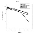
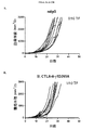
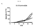
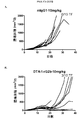
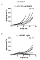
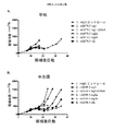
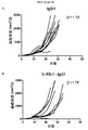
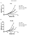
本研究のある局面は、種々のマウス腫瘍モデルにおける抗体の抗腫瘍活性に対するマウス抗CTLA-4抗体のアイソタイプの影響を評価した。最初に、IgG1、突然変異IgG1D265A、IgG2b、およびIgG2aアイソタイプに対応するマウス抗マウスCTLA-4抗体の4つの変異体を作製し、CTLA-4+細胞と同等に結合し、マウス血清において同様の薬物動態挙動を示すことを示した(実施例1)。CT26結腸腺癌腫瘍モデルにおけるマウス抗CTLA-4の4つのアイソタイプの抗腫瘍活性の試験から、IgG2a処置が処置したマウス10中9匹に完全な腫瘍拒絶をもたらすが、IgG2bアイソタイプは中等度の腫瘍増殖阻害を示し、突然変異IgG1D265AアイソタイプはコントロールマウスIgG同様に活性がわずかであることがわかった(実施例2)。 One aspect of this study evaluated the effect of mouse anti-CTLA-4 antibody isotypes on the antitumor activity of antibodies in various mouse tumor models. Initially, four variants of mouse anti-mouse CTLA-4 antibody corresponding to IgG1, mutant IgG1D265A, IgG2b, and IgG2a isotypes were created and bound equivalently to CTLA-4 + cells, and similar drugs in mouse serum It was shown to show kinetic behavior (Example 1). A study of the anti-tumor activity of the four mouse anti-CTLA-4 isotypes in the CT26 colon adenocarcinoma tumor model shows that 9 out of 10 mice treated with IgG2a resulted in complete tumor rejection, whereas the IgG2b isotype is a moderate tumor It showed growth inhibition and the mutant IgG1D265A isotype was found to have little activity as in the control mouse IgG (Example 2).
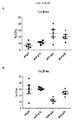
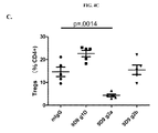
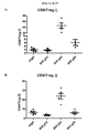
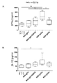
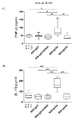
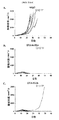
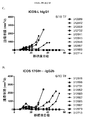
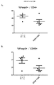
次に、腫瘍および腫瘍流入領域リンパ節におけるサブセットT細胞ポピュレーションに対する種々の抗CTLA-4アイソタイプの影響をCT26結腸腺癌マウス腫瘍モデルで評価した。抗CTLA-4抗体によるマウスの処置は腫瘍部位におけるCD8+ 細胞傷害性T細胞のポピュレーションの増加をもたらし、IgG2aおよびIgG2bアイソタイプにより誘導される増加が最も大きく(実施例4)、IgG2aアイソタイプはCD4+Tヘルパー細胞のポピュレーションの減少をもたらした。処置群間の顕著な差がTregsに対する効果で認められた。IgG2aアイソタイプによる処置は、腫瘍部位のTregsのポピュレーションを顕著に減少させたが、IgG2bは変化を示さず、IgG1D265はTreg数の増加をもたらした。CD8+ Teffsの増加はIgG2aアイソタイプを有する抗CTLA-4抗体に仲介されるTregsの減少と相まって腫瘍部位のTeff/Treg比の上昇をもたらし、強い抗腫瘍活性を示唆する。 Next, the effects of various anti-CTLA-4 isotypes on subset T cell populations in tumor and tumor draining lymph nodes were evaluated in a CT26 colon adenocarcinoma mouse tumor model. Treatment of mice with anti-CTLA-4 antibody resulted in an increased population of CD8 + cytotoxic T cells at the tumor site, with the greatest increase induced by IgG2a and IgG2b isotypes (Example 4), with the IgG2a isotype being CD4 + Resulted in a decrease in the population of T helper cells. Significant differences between treatment groups were observed in the effect on T regs . Treatment with IgG2a isotype significantly reduced the population of T regs at the tumor site, while IgG2b showed no change and IgG1D265 resulted in an increase in T reg numbers. An increase in CD8 + T effs , coupled with a decrease in T regs mediated by anti-CTLA-4 antibody with IgG2a isotype, results in an increase in the T eff / T reg ratio at the tumor site, suggesting strong anti-tumor activity.
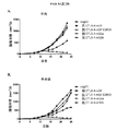
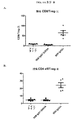
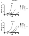
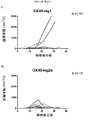
腫瘍部位のT細胞サブポピュレーションの変化と対照的にすべての抗CTLA-4アイソタイプは、腫瘍流入領域リンパ節におけるTregs数の増加に同様に作用し(実施例4)、これは先の研究から予期しないことであった(Quezada et al.、2006;Maker et al.、2005;Rosenberg、2006)。したがって、この驚くべき結果は、腫瘍部位における選択的なTregの減少を証明した。同じパターンがMC38結腸腺癌マウス腫瘍モデル(実施例5および6)および免疫原性Sa1N繊維肉腫腫瘍モデル(実施例7および8)でも証明されたので、この予期しない結果はCT26腫瘍モデルの特性でないことを示した。これらの両腫瘍モデルでは、IgG2aアイソタイプは、CD8+細胞のパーセンテージの顕著な増加とTregsレベルの同時の顕著な減少を仲介し、腫瘍増殖に対する最も顕著な阻害作用をもたらした。同じ現象がアゴニスト性抗GITR、OX40およびICOS抗体を含む他の抗体でも観察された(実施例10〜16)が、抗PD-1抗体ではみられなかった(実施例17)。リンパ節中のTreg数の増加の誘導に対して腫瘍部位のTregsの枯渇の仲介におけるある種のIgG2a抗体の種々のこの差次的作用の分子基盤は、本明細書に記載のデータから明らかにすることができる。実施例18に記載のごとく、CTLA-4に加えてICOS、GITR、OX40、およびCD137を含む種々のT細胞標的は、末梢より腫瘍部位のT細胞でより高度に発現するだけでなく、CD8およびCD4 Teffsでの発現レベルと比べてTregsで優先的に発現する。さらに、末梢と比べて腫瘍部位で活性化FcRs、特にFcγRIVを発現するマクロファージなどの細胞の存在が増加した証拠がある(Simpson et al.、2013)。該細胞は、抗CTLA-4抗体療法後の腫瘍に浸潤するTregsの枯渇に主要な役割を果たすことが示された(Simpson et al.、2013)。したがって、活性化FcRsと結合しADCCを仲介するFc融合タンパク質、例えば抗CTLA-4のマウスIgG2aアイソタイプは、腫瘍部位のCD8およびCD4 Teffsでの発現レベルと比べて高レベルのCTLA-4を差次的に発現する腫瘍部位のTregsなどの該抗体の標的を特異的に発現するT細胞の枯渇に有効である。 In contrast to changes in the T cell subpopulation at the tumor site, all anti-CTLA-4 isotypes act similarly in increasing the number of T regs in the tumor draining lymph nodes (Example 4), which is a previous study. (Quezada et al., 2006; Maker et al., 2005; Rosenberg, 2006). This surprising result therefore demonstrated a selective T reg reduction at the tumor site. This unexpected result is not characteristic of the CT26 tumor model because the same pattern was also demonstrated in the MC38 colon adenocarcinoma mouse tumor model (Examples 5 and 6) and the immunogenic Sa1N fibrosarcoma tumor model (Examples 7 and 8) Showed that. In both of these tumor models, the IgG2a isotype mediated a significant increase in the percentage of CD8 + cells and a concomitant decrease in T regs levels, resulting in the most significant inhibitory effect on tumor growth. The same phenomenon was observed with other antibodies including agonistic anti-GITR, OX40 and ICOS antibodies (Examples 10-16), but not with anti-PD-1 antibodies (Example 17). The molecular basis of various this differential action of certain IgG2a antibodies in mediating the depletion of T regs at the tumor site against the induction of an increase in the number of T regs in the lymph nodes is derived from the data described herein. Can be revealed. As described in Example 18, various T cell targets including ICOS, GITR, OX40, and CD137 in addition to CTLA-4 are not only more highly expressed in T cells at the tumor site than in the periphery, but also CD8 and It is expressed preferentially in T regs compared to the expression level in CD4 T effs . Furthermore, there is evidence that the presence of cells such as macrophages that express activated FcRs, particularly FcγRIV, is increased at the tumor site compared to the periphery (Simpson et al., 2013). The cells have been shown to play a major role in the depletion of T regs infiltrating tumors after anti-CTLA-4 antibody therapy (Simpson et al., 2013). Thus, Fc fusion proteins that bind to activated FcRs and mediate ADCC, such as the anti-CTLA-4 mouse IgG2a isotype, differ in high levels of CTLA-4 compared to the expression levels in CD8 and CD4 Teffs at the tumor site. It is effective for depletion of T cells that specifically express the target of the antibody, such as T regs at the tumor site that is subsequently expressed.
抗体のアイソタイプの違いは、抗体の生物活性に大きな影響があることが示されている(NimmerjahnおよびRavetch、2005;NimmerjahnおよびRavetch、2008;NimmerjahnおよびRavetch、2010)。腫瘍特異抗原、チロシナーゼ関連タンパク質-1(Tyrp1;gp75)に対するTA99抗体の抗腫瘍活性は、B16ネズミメラノーマモデルにおける活性化レセプターFcRIVとの結合を必要とすることを示した(NimmerjahnおよびRavetch、2005)。続く研究は逆の結論に達し、Bevaart et al.(2006)はFcγRIの必須の役割とFcγRIIIまたはFcγRIVが関与しないことをみいだし、Albanesi et al.(2012)はFcγRIおよびFcγRIIIはTA99 治療効果に寄与しFcγRIVは寄与しないと結論した。興味深いことに、本明細書に記載のCT26、MC38、およびSa1N腫瘍モデルにおける抗CTLA-4の抗腫瘍活性(実施例2、5および7)は、抗腫瘍活性における活性化Fcレセプターの必要性を示唆する。活性化レセプターに対する結合の増強と阻害レセプターに対する結合の減少は以下の順番で抗CTLA-4アイソタイプの抗腫瘍活性と関連する:mIgG2a>>mIgG2b>>mIgG1D265A。この順番は、NimmerjahnおよびRavetch(2005)により定義され、抗体介在ADCC機能について決定された免疫グロブリンFc領域の活性化Fcレセプターとの結合と阻害Fcレセプターとの結合の活性比(A/I比として知られる)に従う。 Differences in antibody isotypes have been shown to have a significant impact on antibody biological activity (Nimmerjahn and Ravetch, 2005; Nimmerjahn and Ravetch, 2008; Nimmerjahn and Ravetch, 2010). The anti-tumor activity of the TA99 antibody against the tumor-specific antigen, tyrosinase-related protein-1 (Tyrp1; gp75) has been shown to require binding to the activated receptor FcRIV in the B16 murine melanoma model (Nimmerjahn and Ravetch, 2005) . Subsequent studies reached the opposite conclusion, Bevaart et al. (2006) found the essential role of FcγRI and the absence of FcγRIII or FcγRIV, and Albanesi et al. (2012) showed that FcγRI and FcγRIII were effective in treating TA99. It was concluded that FcγRIV contributed but not. Interestingly, the anti-tumor activity of anti-CTLA-4 in the CT26, MC38, and Sa1N tumor models described herein (Examples 2, 5 and 7) demonstrates the need for an activated Fc receptor in anti-tumor activity. Suggest. Increased binding to activating receptors and decreased binding to inhibitory receptors are associated with anti-tumor activity of anti-CTLA-4 isotype in the following order: mIgG2a >> mIgG2b >> mIgG1D265A. This order is defined by Nimmerjahn and Ravetch (2005) and is determined for antibody-mediated ADCC function as the activity ratio (A / I ratio) of the binding of the immunoglobulin Fc region to the binding of the activated Fc receptor and the inhibitory Fc receptor. Follow known).
抗CTLA-4の最大抗腫瘍活性は、腫瘍部位のTregsの枯渇または排除とTeffsの同時の活性化により達成される(実施例4、6、および8)。特に、活性化T細胞はCTLA-4を発現するが、これらは排除されず、より高い構成レベルの抗CTLA-4を発現することが知られているTreg(Read et al.、2000;Takahashi et al.、2000;Birebent et al.、2004)は腫瘍部位からなくなる。したがって、ネズミ抗CTLA-4 IgG2aアイソタイプは、抗腫瘍反応を仲介する活性化Teffsを残しながら、他のアイソタイプと比べてTreg数を最大限に減少させることができる。したがって、IgG2aアイソタイプは、抗腫瘍反応を阻害する細胞のポピュレーションも特異的に減少させながら抗腫瘍エフェクター細胞の活性を増強させることができる。IgG2a 抗CTLA-4抗体により差次的に影響を受けるこれらT細胞ポピュレーションはそれぞれ腫瘍増殖の調節に不可欠である。TeffsおよびTregsの枯渇に対する特異的感受性は、エフェクター細胞の細胞表面に発現した低レベルのCTLA-4によると思われる(実施例18を参照;Selby et al.、2013も参照のこと)。 The maximum anti-tumor activity of anti-CTLA-4 is achieved by depletion or elimination of T regs at the tumor site and simultaneous activation of T effs (Examples 4, 6, and 8). In particular, activated T cells express CTLA-4, these are not excluded, it is known to express anti-CTLA-4 higher structure levels T reg (Read et al, 2000 ;. Takahashi et al., 2000; Birebent et al., 2004) disappears from the tumor site. Thus, murine anti-CTLA-4 IgG2a isotype can maximize the number of T regs compared to other isotypes while leaving activated T effs to mediate anti-tumor responses. Thus, the IgG2a isotype can enhance the activity of anti-tumor effector cells while specifically reducing the population of cells that inhibit the anti-tumor response. Each of these T cell populations that are differentially affected by IgG2a anti-CTLA-4 antibodies are essential for the regulation of tumor growth. The specific sensitivity to T effs and T regs depletion appears to be due to low levels of CTLA-4 expressed on the cell surface of effector cells (see Example 18; see also Selby et al., 2013).
この結果は、腫瘍微小環境の細胞の組成物およびそれが発現するFcレセプターが抗CTLA-4の抗腫瘍活性に関与することも示唆する。腫瘍部位に特異的に局在するTregsの数が減少し、リンパ節中のTregsは抗CTLA-4のすべてのアイソタイプにより活性化されるという知見は、抗CTLA-4の種々のアイソタイプの活性の組織特異的差を明確に示す。 This result also suggests that the composition of cells in the tumor microenvironment and the Fc receptor it expresses are involved in the anti-tumor activity of anti-CTLA-4. The finding that the number of T regs specifically localized at the tumor site is reduced and that T regs in lymph nodes are activated by all isotypes of anti-CTLA-4 is Clearly show tissue-specific differences in activity.
述べたように、抗CTLA-4-IgG2aおよびそれほどではないが抗CTLA-4-IgG2bは、それらの活性化FcγRsとの結合能と一致して腫瘍部位からのTregsの排除または枯渇を仲介する。これは、CD8+ Teffs(およびCD8+ T細胞)の同時の活性化と増殖とともに生じ、CTLA-4-B7相互作用を阻害することにより仲介されると思われる。しかしながら、本明細書に記載のデータは、Teff活性化が単にTregの枯渇の結果であることを排除しない。したがって、抗CTLA-4のネズミIgG2aアイソタイプは、他のアイソタイプと比べて、抗腫瘍反応を仲介する活性化Teffsを残しながらTreg数を強く減少させることができる。実際に、腫瘍部位におけるエフェクターサイトカイン分泌(IFNγ、TNFα、およびIL-13、およびおそらくIL-10(Emmerich et al.、2012;Mumm et al.、2011)の増強の知見は、Treg抑制の減少と活性化CD8エフェクターの増加と一致する。 As stated, anti-CTLA-4-IgG2a and to a lesser extent anti-CTLA-4-IgG2b mediate the elimination or depletion of T regs from the tumor site, consistent with their ability to bind to activated FcγRs . This appears to occur with simultaneous activation and proliferation of CD8 + T effs (and CD8 + T cells) and is mediated by inhibiting CTLA-4-B7 interactions. However, the data described herein does not exclude that T eff activation is simply the result of T reg depletion. Therefore, the anti-CTLA-4 murine IgG2a isotype can strongly reduce the number of T regs while leaving activated T effs mediating the anti-tumor response compared to other isotypes. Indeed, the finding of enhanced effector cytokine secretion (IFNγ, TNFα, and IL-13, and possibly IL-10 (Emmerich et al., 2012; Mumm et al., 2011) at the tumor site is a decrease in T reg suppression. Consistent with an increase in activated CD8 effectors.
MC38およびCT26モデルの治療的処置におけるIgG1およびIgG1-D265Aアイソタイプの抗腫瘍活性の欠如も注目に値する。CTLA-4-B7と抗CTLA-4 IgG1または抗CTLA-4 IgG1D265Aとの相互作用の阻害は、末梢におけるTregsの活性化と増殖をもたらすが、Teffsのみの遮断(すなわち、Tregの除去なし)は検出可能な抗腫瘍反応を促進するには不十分である。さらに、Treg機能を減少させることが示されたTregs上のCTLA-4の遮断も(Quezada et al.、2006;Onishi et al.、2008)、抗腫瘍活性を明らかに増強しない。これに対し、抗CTLA-4(ハムスター抗マウスCTLA-4 9H10)、GVAX療法、およびヒトまたはマウスCTLA-4を発現するT細胞サブセットによる照射レシピエントマウスの再構築を用いるB16メラノーマモデルでは、TeffsとTregsの両方で完全な抗腫瘍活性にマウスCTLA-4の発現が必要であった(Peggs et al.、2009)。しかしながら、本明細書に記載の試験と異なり、Teffを標的とした抗CTLA-4遮断のみが、このモデルで部分的抗腫瘍作用をもたらした。 Also noteworthy is the lack of anti-tumor activity of IgG1 and IgG1-D265A isotypes in therapeutic treatment of MC38 and CT26 models. Inhibition of the interaction between CTLA-4-B7 and anti-CTLA-4 IgG1 or anti-CTLA-4 IgG1D265A results in T regs activation and proliferation in the periphery, but only T effs blockade (ie, removal of T regs ) None) is insufficient to promote a detectable anti-tumor response. Furthermore, blockade of CTLA-4 on T regs that has been shown to reduce T reg function (Quezada et al., 2006; Onishi et al., 2008) also does not clearly enhance antitumor activity. In contrast, in the B16 melanoma model using anti-CTLA-4 (hamster anti-mouse CTLA-4 9H10), GVAX therapy, and reconstruction of irradiated recipient mice with T cell subsets expressing human or mouse CTLA-4, Mouse CTLA-4 expression was required for full antitumor activity in both effs and T regs (Peggs et al., 2009). However, unlike the studies described herein, only anti-CTLA-4 blockade targeting T eff resulted in partial anti-tumor effects in this model.
抗腫瘍効果に対する抗CTLA-4抗体以外のFc融合タンパク質のアイソタイプの影響
Fc領域の活性化FcγRsとの結合能に一致し、抗腫瘍効果に関連する、ある種の抗CTLA-4アイソタイプ、特にマウスIgG2aアイソタイプ、および程度が低いがマウスIgG2bが、腫瘍部位からのTregsの排除または枯渇を仲介するという証拠に従って(実施例1〜7)、抗腫瘍活性に対するさらなる抗体および他のFc融合タンパク質のアイソタイプの影響を検討した。
Effect of isotype of Fc fusion protein other than anti-CTLA-4 antibody on antitumor effect
Certain anti-CTLA-4 isotypes, particularly mouse IgG2a isotypes, and to a lesser extent mouse IgG2b, which are consistent with the ability of the Fc region to bind to activated FcγRs and are associated with antitumor effects, have T regs from the tumor site. According to the evidence that they mediate elimination or depletion (Examples 1-7), the effect of additional antibodies and other Fc fusion protein isotypes on anti-tumor activity was investigated.
種々の抗GITRアイソタイプの抗腫瘍活性を同系MC38結腸癌およびSa1N肉腫マウスモデルで評価した(実施例10〜12)。抗CTLA-4の結果と同様に、抗GITR IgG1およびIgG1D265Aアイソタイプには実質的に抗腫瘍活性がないが、マウスIgG2aおよびラットIgG2b(マウス活性化FcRsとの結合ではマウスIgG2aと同等に)アイソタイプは最も大きな腫瘍増殖阻害をもたらすことが示された(実施例11)。抗GITR mG2a、mG2bおよびrG2bアイソタイプは、腫瘍増殖阻害と関連する腫瘍環境での顕著なTregの枯渇を誘導するのに対して末梢のTregポピュレーションに対してはほとんど影響がないかまたはわずかな増加を誘導した(実施例11および12)。反対に、mIgG2aアイソタイプは腫瘍部位のCD8+細胞のパーセンテージの増加をもたらしたが、mIgG1およびrat IgG2bは、CD8+細胞のパーセンテージを全くまたはわずかしか増加しなかった。いずれのアイソタイプも末梢のCD8+細胞レベルに大きな影響を与えなかった。同様のデータを、最近、Bulliard et al.(2013)が報告した。 The anti-tumor activity of various anti-GITR isotypes was evaluated in syngeneic MC38 colon cancer and Sa1N sarcoma mouse models (Examples 10-12). Similar to anti-CTLA-4 results, anti-GITR IgG1 and IgG1D265A isotypes have virtually no anti-tumor activity, but mouse IgG2a and rat IgG2b (similar to mouse IgG2a in binding to mouse activated FcRs) isotypes It was shown to result in the greatest tumor growth inhibition (Example 11). Anti-GITR mG2a, mG2b and rG2b isotypes induce significant T reg depletion in the tumor environment associated with tumor growth inhibition while having little or no effect on peripheral T reg populations Increase was induced (Examples 11 and 12). Conversely, mIgG2a isotype resulted in an increase in the percentage of CD8 + cells at the tumor site, whereas mIgG1 and rat IgG2b increased the percentage of CD8 + cells at all or only slightly. Neither isotype had a significant effect on peripheral CD8 + cell levels. Similar data was recently reported by Bulliard et al. (2013).
一般的に同様のデータが、同系CT26腫瘍マウスモデルで試験した抗OX40アイソタイプ(実施例14)およびSa1NおよびMC38腫瘍モデルで試験した抗ICOSアイソタイプ(実施例15および16)で得られた。しかしながら、MC38腫瘍モデルでの抗PD-1アイソタイプの評価は、抗PD-1 IgG2aアイソタイプはいくらか抗腫瘍活性を示したが抗IgG1またはIgG1D265Aアイソタイプが示す活性より低かったことを示した(実施例17)。抗PD-1 IgG2aアイソタイプがIgG1およびIgG1D265Aアイソタイプと比べて抗腫瘍活性を増強しなかったこれらの結果は、抗CTLA-4、GITR、OX40、およびICOS IgG2a抗体で得られた結果と全く対照的であった。さらに、腫瘍部位のCD8+細胞のわずかな増加をもたらし、IgG2aアイソタイプと比べてTregsのパーセンテージのより小さな増加をもたらしたIgG1およびIgG1D265Aアイソタイプと対照的に、抗PD-1 IgG2aアイソタイプは腫瘍部位のCD8+細胞のパーセンテージの減少とTregsのパーセンテージの増加をもたらした(実施例17)。 Generally similar data were obtained with the anti-OX40 isotype tested in the syngeneic CT26 tumor mouse model (Example 14) and the anti-ICOS isotype tested in the Sa1N and MC38 tumor models (Examples 15 and 16). However, evaluation of the anti-PD-1 isotype in the MC38 tumor model showed that the anti-PD-1 IgG2a isotype showed some antitumor activity but was less than the activity exhibited by the anti-IgG1 or IgG1D265A isotype (Example 17 ). These results, in which the anti-PD-1 IgG2a isotype did not enhance antitumor activity compared to the IgG1 and IgG1D265A isotypes, are in stark contrast to those obtained with anti-CTLA-4, GITR, OX40, and ICOS IgG2a antibodies. there were. Furthermore, in contrast to the IgG1 and IgG1D265A isotypes, which resulted in a slight increase in CD8 + cells at the tumor site and a smaller increase in the percentage of T regs compared to the IgG2a isotype, the anti-PD-1 IgG2a isotype is This resulted in a decrease in the percentage of CD8 + cells and an increase in the percentage of T regs (Example 17).
腫瘍部位および末梢のT細胞サブセットに対する種々のレセプターの発現レベルの試験は、基礎となるメカニズムの解明を助ける。該データは、ICOS、GITR、CTLA-4、OX40、CD137、CTLA-4およびTIGITを含むある種のT細胞レセプターが、腫瘍部位のCD8およびCD4 Teffsの発現レベルと比べて、腫瘍部位のTregsを比較的高いレベルで発現することを示す。これらのレセプターも、末梢の同タイプのT細胞における発現レベルと比べて腫瘍部位のT細胞サブタイプでより高レベルで発現する。反対に、PD-1、LAG-3、およびTIM-3を含む他のレセプターは、腫瘍部位のTregsにおける発現レベルと比べて腫瘍部位のCD8および/またはCD4 Teffsでより高レベルで発現する。CD27は、腫瘍部位および末梢の種々の細胞タイプでほぼ一定レベルの発現を示す。 Testing the level of expression of various receptors on tumor sites and peripheral T cell subsets helps to elucidate the underlying mechanisms. The data indicate that certain T cell receptors, including ICOS, GITR, CTLA-4, OX40, CD137, CTLA-4 and TIGIT, showed T8 at the tumor site compared to the expression levels of CD8 and CD4 Teffs at the tumor site. It shows that regs are expressed at relatively high levels. These receptors are also expressed at higher levels in the T cell subtype at the tumor site compared to the expression levels in the same type of peripheral T cells. Conversely, other receptors, including PD-1, LAG-3, and TIM-3, are expressed at higher levels in tumor site CD8 and / or CD4 T effs compared to expression levels in tumor site T regs . CD27 exhibits an almost constant level of expression in various cell types at the tumor site and in the periphery.
したがって、活性化FcRsと結合しADCCを仲介する抗CTLA-4などのFc融合タンパク質のマウスIgG2aアイソタイプは、腫瘍部位のCD8およびCD4 Teffsの発現レベルと比べてCTLA-4を差次的に高レベルに発現する腫瘍部位のTregsなどの該抗体の標的を優先的に発現するT細胞を枯渇させるのに有効である。 Thus, mouse IgG2a isotypes of Fc fusion proteins such as anti-CTLA-4 that bind to activated FcRs and mediate ADCC differentially increase CTLA-4 relative to the expression levels of CD8 and CD4 Teffs at the tumor site. It is effective in depleting T cells that preferentially express the target of the antibody, such as T regs at the tumor site expressed at the level.
抗体などのFc融合タンパク質の抗腫瘍効果を増強するTregs枯渇メカニズムが、共刺激レセプターと結合するアゴニスト抗体と共阻害レセプターと結合するアンタゴニスト抗体の両方で機能していることは注目に値する。該データは、T細胞表面に発現したあらゆるタンパク質は、その機能に関わらず標的細胞のADCC介在性枯渇を誘導するための活性化FcRsと強い結合を示すFc融合領域、例えばマウスのIgG2aまたはヒトのIgG1と結合するための標的として働くことができる。抗腫瘍抗体などの抗腫瘍Fc融合タンパク質の場合は、標的タンパク質が、腫瘍部位のTregsの選択的な正味の枯渇と免疫応答の同時刺激があるように腫瘍部位のTregsで腫瘍部位のTeffsより高レベルで差次的に発現することが望ましい。標的タンパク質が免疫応答を刺激するTreg枯渇成分が主に腫瘍部位に限定されるように腫瘍部位のTregsで末梢の他のT細胞より高レベルに差次的に発現することも望ましい。したがって、ICOS、GITR、CTLA-4、OX40、CD137、CTLA-4、およびTIGITを標的とするFc融合タンパク質は、活性化FcRに対するFc領域の結合を増強するための本明細書に記載のFc領域を修飾することによりその抗腫瘍効果を増強するためのよい候補である。CTLA-4と特異的に結合するがその共阻害活性を遮断しない抗CTLA-4抗体などの抗CTLA-4 Fc融合タンパク質は、活性化Fcレセプター(FcR)に対する該Fc領域の結合を増強するためにFc領域の選択、設計、または修飾により抗腫瘍効果の増強を操作するためのよい候補である。そのような増強されたFc融合タンパク質は、末梢における免疫応答を刺激する副作用なしに高い抗腫瘍効果を示すかもしれない。 It is noteworthy that the T regs depletion mechanism that enhances the anti-tumor effect of Fc fusion proteins such as antibodies is working for both agonist antibodies that bind to costimulatory receptors and antagonist antibodies that bind to co-inhibitory receptors. The data show that any protein expressed on the surface of the T cell, regardless of its function, has an Fc fusion region that exhibits strong binding to activated FcRs to induce ADCC-mediated depletion of the target cell, such as mouse IgG2a or human Can serve as a target for binding to IgG1. For antitumor Fc fusion protein such as an anti-tumor antibody, the target protein, the tumor site at the tumor site T regs so that costimulation selective net depletion and immune responses T regs tumor sites T It is desirable to express differentially at a higher level than effs . It is also desirable that the target protein be differentially expressed at a higher level than other peripheral T cells in the T regs at the tumor site so that the T reg depletion component that stimulates the immune response is primarily restricted to the tumor site. Thus, Fc fusion proteins that target ICOS, GITR, CTLA-4, OX40, CD137, CTLA-4, and TIGIT are Fc regions described herein for enhancing Fc region binding to activated FcR Is a good candidate for enhancing its anti-tumor effect by modifying. Anti-CTLA-4 Fc fusion proteins, such as anti-CTLA-4 antibodies that specifically bind CTLA-4 but do not block its co-inhibitory activity, enhance the binding of the Fc region to activated Fc receptor (FcR) In particular, it is a good candidate for manipulating the enhancement of anti-tumor effects by selection, design, or modification of the Fc region. Such enhanced Fc fusion proteins may exhibit a high anti-tumor effect without the side effects of stimulating immune responses in the periphery.
反対に、標的 PD-1、LAG-3、TIM-3、およびCD27を標的とするFc融合タンパク質は、これらのレセプターは腫瘍部位のTeffsでTregsより高度に発現するので、開示した方法によりその抗腫瘍効果を増強するためのよい候補ではないと思われる。実施例17の抗PD-1を用いて得られたデータはこの考えを裏付ける。 In contrast, Fc fusion proteins targeting the target PD-1, LAG-3, TIM-3, and CD27 are more highly expressed than T regs at T effs at the tumor site, so the disclosed method It does not appear to be a good candidate for enhancing its anti-tumor effect. Data obtained with the anti-PD-1 of Example 17 confirms this idea.
免疫調節Fc融合タンパク質の抗腫瘍効果の増強方法
本明細書に記載のデータは、抗CTLA-4抗体、ならびに限定されるものではないが、癌患者の治療における共刺激および共阻害レセプターおよびレセプターリガンドを含むT細胞上の免疫調節標的と結合する他のFc融合タンパク質の活性と関連がある。これらのデータは、より効力の高い抗免疫調節抗体を設計する可能性も示唆する。
Methods for enhancing anti-tumor effects of immunomodulating Fc fusion proteins Data described herein include anti-CTLA-4 antibodies, and, without limitation, costimulatory and co-inhibitory receptors and receptor ligands in the treatment of cancer patients Is associated with the activity of other Fc fusion proteins that bind to immunomodulatory targets on T cells, including These data also suggest the possibility of designing more potent anti-immunomodulatory antibodies.
ヒト抗ヒトCTLA-4モノクローナル抗体のイピリムマブは、転移性メラノーマの治療に承認されており、他の癌では臨床試験中である(Hoos et al.、2010;Hodi et al.、2010;Pardoll、2012a)。イピリムマブは、ほとんどのヒトFcレセプターと最も良く結合するヒトIgG1アイソタイプを有し(表1;Bruhns et al.、2009)、それと結合する活性化Fcレセプターのタイプに関してネズミIgG2aと等価であると考えられる。IgG1は、ヒトNK細胞および単核球が発現する活性化レセプターCD16(FcγRIIIa)と結合するので、イピリムマブはADCCを仲介することができる。IgG1アイソタイプのイピリムマブは、最初にハイブリドーマから直接単離されたが、次いでクローニングされ、チャイニーズハムスター卵巣(CHO)細胞で発現した。ADCCおよび/またはCDCを仲介するアイソタイプは免疫応答を上方調節しようとするT細胞上のレセプターを標的とする抗体では望ましくないかもしれないにもかかわらず、該抗体のIgG1アイソタイプはカニクイザルにおけるワクチン反応を増強し、機能的であると考えられたため一部保持された。イピリムマブは、例えば、処置後CD4+およびCD8+細胞の表面のHLA-DRの発現の顕著な増加と絶対リンパ球数の増加で示される血中の活性化T細胞数の増加を示し(Ku et al.、2010;Attia et al.、2005;Maker et al.、2005;Berman et al.、2009;Hamid et al.、2009)、T細胞の枯渇がヒトの末梢で生じないことを示唆する。イピリムマブは、IL-2-活性化PBMCをエフェクター細胞として用いる活性化T細胞のADCCは中程度であった(未発表)が、Tregsを標的として使用する試験は行わなかった。イピリムマブで処置した患者の血中の末梢Treg頻度のわずかな変化がみられた(Maker et al.、2005)が、腫瘍内Tregsに対するイピリムマブの効果に関する利用可能な情報はほとんどない。しかしながら、イピリムマブで処置した患者の転移性メラノーマ病変の生検における高CD8+/Treg比と腫瘍壊死の正相関が記載されている(Hodi et al.、2008)。さらに、イピリムマブ処置膀胱癌患者の腫瘍組織では非処置膀胱癌患者の腫瘍よりCD4+ Foxp3+ T細胞のパーセンテージが低かった(Liakou et al.、2008)。これらの結果は、イピリムマブが腫瘍部位におけるTregの減少を仲介するという本明細書に記載のデータと一致する。 The human anti-human CTLA-4 monoclonal antibody ipilimumab has been approved for the treatment of metastatic melanoma and is in clinical trials in other cancers (Hoos et al., 2010; Hodi et al., 2010; Pardoll, 2012a ). Ipilimumab has a human IgG1 isotype that binds best to most human Fc receptors (Table 1; Bruhns et al., 2009) and is considered equivalent to murine IgG2a with respect to the type of activated Fc receptor that binds to it . Since IgG1 binds to the activated receptor CD16 (FcγRIIIa) expressed by human NK cells and mononuclear cells, ipilimumab can mediate ADCC. The IgG1 isotype ipilimumab was first isolated directly from the hybridoma, but then cloned and expressed in Chinese hamster ovary (CHO) cells. Although the isotype that mediates ADCC and / or CDC may not be desirable for antibodies that target receptors on T cells that attempt to upregulate the immune response, the IgG1 isotype of the antibody is responsible for the vaccine response in cynomolgus monkeys. It was enhanced and partially retained because it was considered functional. Ipilimumab, for example, shows an increase in the number of activated T cells in the blood as indicated by a marked increase in HLA-DR expression on the surface of CD4 + and CD8 + cells and an increase in absolute lymphocyte count after treatment (Ku et al., 2010; Attia et al., 2005; Maker et al., 2005; Berman et al., 2009; Hamid et al., 2009), suggesting that T cell depletion does not occur in the human periphery. Ipilimumab had moderate ADCC of activated T cells using IL-2-activated PBMC as effector cells (unpublished) but was not tested using T regs as a target. Although there was a slight change in peripheral T reg frequency in the blood of patients treated with ipilimumab (Maker et al., 2005), little information is available on the effects of ipilimumab on intratumoral T regs . However, a positive correlation between high CD8 + / T reg ratio and tumor necrosis in biopsies of metastatic melanoma lesions in patients treated with ipilimumab has been described (Hodi et al., 2008). Furthermore, the tumor tissue of patients with ipilimumab-treated bladder cancer had a lower percentage of CD4 + Foxp3 + T cells than tumors of patients with untreated bladder cancer (Liakou et al., 2008). These results are consistent with the data described herein that ipilimumab mediates T reg reduction at the tumor site.
これに対し、トレメリムマブは、FcγRIIa変異体H131を除くFcレセプターと効率的に結合しないIgG2アイソタイプである(Bruhns et al.、2009)。トレメリムマブは、CTLA-4とB7の阻害的相互作用を遮断することによりT細胞反応を増強する能力を有するが、本明細書に記載のデータは、トレメリムマブは腫瘍のTregsの枯渇の仲介に限られるかもしれず、その上でイピリムマブと比べて抗腫瘍活性の低下を示すと予期されることを示唆する。投与計画が互いに異なるのでこれら2つの抗体の臨床的活性を直接比較するのは困難であった(例えば、 Ascierto et al.、2011参照)。トレメリムマブはイピリムマブ同様に明らかな抗腫瘍活性を有する(Ribas、2010)。興味深いことに、トレメリムマブの作用メカニズムの研究は、免疫組織化学で分析した限られた数の試料において、腫瘍浸潤CD8 T細胞の増加が治療の結果として生じるが治療後の腫瘍のFoxp3+細胞数には変化がないことを示す(Comin-Anduix et al.、2008;Huang et al.、2011)。あるいはまた、Treg機能の阻害は、TLA-4/B7相互作用を遮断することにより達成されるかもしれない。 In contrast, tremelimumab is an IgG2 isotype that does not efficiently bind to Fc receptors except the FcγRIIa mutant H131 (Bruhns et al., 2009). Although tremelimumab has the ability to enhance the T cell response by blocking the inhibitory interaction between CTLA-4 and B7, the data described herein indicate that tremelimumab is limited to mediating tumor T regs depletion. Suggesting that it would be expected to show reduced anti-tumor activity compared to ipilimumab. It was difficult to directly compare the clinical activity of these two antibodies because of different dosage regimes (see, for example, Ascierto et al., 2011). Tremelimumab has obvious antitumor activity like ipilimumab (Ribas, 2010). Interestingly, a study of the mechanism of action of tremelimumab has shown that in a limited number of samples analyzed by immunohistochemistry, an increase in tumor-infiltrating CD8 T cells occurs as a result of treatment but the number of tumor Foxp3 + cells after treatment Indicates no change (Comin-Anduix et al., 2008; Huang et al., 2011). Alternatively, inhibition of T reg function may be achieved by blocking the TLA-4 / B7 interaction.
抗CTLA-4、抗GITR、抗OX40、および抗ICOS抗体を含むT細胞上の標的に対する抗体の作用メカニズムとこれら抗体の活性に対するアイソタイプの効果に関する本明細書に記載の実験データに基づいて、本開示は、癌または感染性物質により生じた疾患に罹患した対象のT細胞上の標的と特異的に結合するFc融合タンパク質の抗腫瘍効果を増強、最適化、または最大化する方法であって、活性化Fcレセプター(FcR)に対する該Fc領域の結合を増強するためにFc融合タンパク質のFc領域を修飾することを含む方法を提供する。抗体の活性化FcRとのそのような結合の増強が、ある標的において、腫瘍環境におけるTregsの枯渇と該抗体の抗腫瘍活性の増加をもたらすことが示された。好ましい態様において、該標的は免疫調節レセプターまたはリガンドであり、Fc融合タンパク質の結合は標的の活性を変化させることにより癌細胞に対する内因性免疫応答を増強する。 Based on the experimental data described herein regarding the mechanism of action of antibodies against targets on T cells including anti-CTLA-4, anti-GITR, anti-OX40, and anti-ICOS antibodies and the effect of isotypes on the activity of these antibodies, this book The disclosure provides a method for enhancing, optimizing, or maximizing the anti-tumor effect of an Fc fusion protein that specifically binds to a target on a T cell in a subject suffering from a disease caused by cancer or an infectious agent, comprising: A method is provided that includes modifying the Fc region of an Fc fusion protein to enhance binding of the Fc region to an activated Fc receptor (FcR). It has been shown that such enhanced binding of an antibody to activated FcR results in depletion of T regs in the tumor environment and increased anti-tumor activity of the antibody in certain targets. In preferred embodiments, the target is an immunomodulatory receptor or ligand, and binding of the Fc fusion protein enhances the endogenous immune response against cancer cells by altering the activity of the target.
開示した本発明の種々の局面は、抗CTLA-4、抗GITR、抗OX40、および抗ICOS抗体を用いて例示されている。しかしながら、本明細書に記載の方法は、免疫調節レセプターの標的化または抗体に限定されない。むしろ、本発明はT細胞表面に発現した種々の標的と結合する広範なFc融合タンパク質に適用可能である。したがって、ある態様において、Fc融合タンパク質は、1本鎖可変断片(scFv)、二価(divalentまたはbivalent)scFvs(di-scFvsまたはbi-scFvs)、ディアボディ、三価トリアボディ、または四価テトラボディ、ミニボディ、または単離されたCDRを含む抗体の抗原結合断片などの結合タンパク質 (さらなる詳細はHollingerおよびHudson、2005;OlafsenおよびWu、2010参照のこと);アドネクチン;アフィボディ;アフィリン;レセプターのリガンド結合領域;細胞付着分子;レセプターリガンド;酵素;サイトカイン;またはケモカインと作動可能に結合したFc領域を含む。循環から完全抗体より急速に除去される抗体の抗原結合断片のより小さな誘導体などのFc融合タンパク質の使用は、過敏性免疫応答の潜在的毒性の軽減を助け、および/または誘導剤の急速な除去を可能にするかもしれない。ある態様において、Fc融合タンパク質は、レセプターリガンドと作動可能に結合したFc領域を含む。好ましい態様において、Fc融合タンパク質は抗体である。 Various aspects of the disclosed invention have been illustrated using anti-CTLA-4, anti-GITR, anti-OX40, and anti-ICOS antibodies. However, the methods described herein are not limited to immunomodulatory receptor targeting or antibodies. Rather, the present invention is applicable to a wide range of Fc fusion proteins that bind to various targets expressed on the surface of T cells. Thus, in certain embodiments, the Fc fusion protein comprises a single chain variable fragment (scFv), a divalent or bivalent scFvs (di-scFvs or bi-scFvs), a diabody, a trivalent triabody, or a tetravalent tetra Binding proteins, such as antigen-binding fragments of antibodies, including bodies, minibodies, or isolated CDRs (see Hollinger and Hudson, 2005; Olafsen and Wu, 2010); Adnectins; Affibodies; Affilins; Receptors A ligand binding region; a cell adhesion molecule; a receptor ligand; an enzyme; a cytokine; or an Fc region operably linked to a chemokine. The use of Fc fusion proteins, such as smaller derivatives of antigen-binding fragments of antibodies that are removed more rapidly than the complete antibody from the circulation, helps reduce the potential toxicity of hypersensitivity immune responses and / or rapid removal of inducers May be possible. In certain embodiments, the Fc fusion protein comprises an Fc region operably linked to a receptor ligand. In a preferred embodiment, the Fc fusion protein is an antibody.
本発明の方法のある局面において、抗体はIgGアイソタイプである。他の局面において、抗体はモノクローナル抗体である。他の局面において、モノクローナル抗体はキメラ、ヒト化、またはヒト抗体である。ある好ましい態様において、モノクローナル抗体はヒトIgG抗体である。他の好ましい態様において、ヒトIgG抗体の活性化FcRとの結合が増強される。活性化FcRはFcγI、FcγIIa、またはFcγIIIaレセプターでありうる。ある局面において、ヒトIgG抗体のFcγI、FcγIIa、またはFcγIIIaレセプターとの結合の増強は、腫瘍部位のTregsの減少を仲介する。 In certain aspects of the methods of the invention, the antibody is an IgG isotype. In other aspects, the antibody is a monoclonal antibody. In other aspects, the monoclonal antibody is a chimeric, humanized, or human antibody. In certain preferred embodiments, the monoclonal antibody is a human IgG antibody. In other preferred embodiments, the binding of human IgG antibodies to activated FcR is enhanced. The activated FcR can be FcγI, FcγIIa, or FcγIIIa receptor. In certain aspects, enhanced binding of human IgG antibodies to FcγI, FcγIIa, or FcγIIIa receptors mediates a decrease in T regs at the tumor site.
本発明の方法のある態様において、FcγI、FcγIIa、またはFcγIIIaレセプターに対するヒトIgG抗体の結合の増強は、腫瘍部位のエフェクターT細胞(Teffs)の(a)減少を仲介しないか、または(b)増加を仲介する。ある好ましい態様において、標的は、腫瘍部位のTregsで腫瘍部位のTeffsより高レベルに発現する。他の態様において、標的は、腫瘍部位のTregsは末梢のTregsまたはTeffsより高レベルに発現する。 In certain embodiments of the methods of the invention, the enhanced binding of a human IgG antibody to FcγI, FcγIIa, or FcγIIIa receptor does not mediate (a) a decrease in effector T cells (T effs ) at the tumor site, or (b) Mediate the increase. In certain preferred embodiments, the target is expressed at a higher level at the tumor site T regs than at the tumor site T effs . In other embodiments, the target expresses T regs at the tumor site at a higher level than peripheral T regs or T effs .
さらに、本発明の方法に用いるFc融合タンパク質は、T細胞上の共刺激または共阻害免疫調節標的と結合することができる。共刺激標的との特異的結合に用いるFc融合タンパク質はアゴニストFc融合タンパク質である。例えば、アゴニストFc融合タンパク質を用いて、T細胞上のGITR、CD134(OX40)、ICOS、CD137(4-1BB)、CD27、CD28、またはHVEMなどの共刺激免疫調節物質を標的とする。アゴニストFc融合タンパク質の共刺激免疫調節物質との結合は、免疫応答、特にT細胞反応の上方調節をもたらす。本発明の方法のある態様において、Fc融合タンパク質はT細胞上の共刺激免疫調節標的の活性を増強するアゴニスト抗体である。好ましい態様において、共刺激免疫調節標的はGITR、OX40、ICOS、またはCD137である。 Furthermore, the Fc fusion proteins used in the methods of the invention can bind to costimulatory or co-inhibitory immunomodulatory targets on T cells. The Fc fusion protein used for specific binding to a costimulatory target is an agonist Fc fusion protein. For example, agonist Fc fusion proteins are used to target costimulatory immunomodulators such as GITR, CD134 (OX40), ICOS, CD137 (4-1BB), CD27, CD28, or HVEM on T cells. Binding of an agonist Fc fusion protein to a costimulatory immunomodulator results in upregulation of the immune response, particularly the T cell response. In certain embodiments of the methods of the invention, the Fc fusion protein is an agonist antibody that enhances the activity of a costimulatory immunomodulatory target on T cells. In preferred embodiments, the costimulatory immunomodulatory target is GITR, OX40, ICOS, or CD137.
反対に、共阻害標的との特異的結合に用いるFc融合タンパク質は阻害またはアンタゴニストFc融合タンパク質である。例えば、阻害Fc融合タンパク質を用いて、限定されるものではないが、T細胞上のCTLA-4、PD-1、PD-L1、BTLA、TIM-3、LAG-3、A2aR、KLRG-1、CD244、CD160、またはVISTAレセプターなどの共阻害免疫調節物質を標的化することができる。Fc融合タンパク質の抗腫瘍効果を増強するための本発明の方法のある態様において、Fc融合タンパク質は、抗CTLA-4抗体、抗BTLA抗体、抗VISTAレセプター抗体、抗A2aR抗体、抗KLRG-1抗体、抗CD244抗体、抗CD160抗体、および抗TIGIT抗体から選ばれるアンタゴニスト抗体である。好ましい態様において、Fc融合タンパク質はT細胞上の共阻害免疫調節標的の活性を遮断するアンタゴニスト抗体である。好ましい態様において、共阻害免疫調節標的はCTLAまたはTIGITである。アンタゴニストFc融合タンパク質の共阻害免疫調節物質との結合は、免疫応答、特にT細胞反応の上方調節をもたらす。 Conversely, an Fc fusion protein used for specific binding to a co-inhibited target is an inhibitory or antagonistic Fc fusion protein. For example, using an inhibitory Fc fusion protein, including but not limited to CTLA-4, PD-1, PD-L1, BTLA, TIM-3, LAG-3, A2aR, KLRG-1, on T cells, Co-inhibiting immunomodulators such as CD244, CD160, or VISTA receptors can be targeted. In certain embodiments of the methods of the invention for enhancing the anti-tumor effect of an Fc fusion protein, the Fc fusion protein comprises an anti-CTLA-4 antibody, an anti-BTLA antibody, an anti-VISTA receptor antibody, an anti-A2aR antibody, an anti-KLRG-1 antibody An antagonist antibody selected from an anti-CD244 antibody, an anti-CD160 antibody, and an anti-TIGIT antibody. In a preferred embodiment, the Fc fusion protein is an antagonist antibody that blocks the activity of a co-inhibiting immunomodulatory target on T cells. In a preferred embodiment, the co-inhibiting immunomodulating target is CTLA or TIGIT. Binding of the antagonist Fc fusion protein with a co-inhibiting immunomodulator results in upregulation of the immune response, particularly the T cell response.
ある好ましい態様において、該抗CTLA-4抗体である。さらに好ましい態様において、抗CTLA-4抗体は、イピリムマブまたはトレメリムマブ、またはFc領域と活性化FcRとの結合を増強するように修飾されたこれらの抗体変異体である。他の態様において、該抗体は抗PD-1抗体である。本開示のあらゆる方法において対象は好ましくはヒトである。 In certain preferred embodiments, the anti-CTLA-4 antibody. In a further preferred embodiment, the anti-CTLA-4 antibody is ipilimumab or tremelimumab, or these antibody variants modified to enhance binding of the Fc region to activated FcR. In other embodiments, the antibody is an anti-PD-1 antibody. In any method of the present disclosure, the subject is preferably a human.
Tregsを特異的に標的化する抗体はまだ製造されていないが、臨床試験で多くの免疫チェックポイント抗体がTregsの免疫抑制活性の遮断を伴うメカニズムにより抗腫瘍免疫を増強するかもしれない(Pardoll、2012b)。本明細書に示すように、免疫チェックポイント抗体であるマウス抗CTLA-4のIgG2aアイソタイプは、驚くべきことに予期せず腫瘍部位に選択的なTregsの枯渇を仲介するが、同時に、腫瘍流入領域リンパ節のTregsの増加と腫瘍内CD8+ Teffsの増加を仲介する(実施例4、6、および8)。Tregsは免疫調節Fc融合タンパク質により標的化されうるが、開示した本発明のある局面において、標的化細胞はTreg以外の種々のタイプの抑制性細胞である。同様に、本開示のFc融合タンパク質はCTLA-4などの共阻害免疫調節物質を標的化しうるが、開示した発明のある局面において、標的化免疫調節物質は、共刺激免疫調節物質、例えばGITR、OX40、CD137、またはICOSである。したがって、本発明のある態様において、Fc融合タンパク質、例えばアゴニストまたはアンタゴニスト抗体は、抑制性細胞ポピュレーションに発現した共刺激または共阻害標的である免疫調節標的と結合し、該細胞ポピュレーションの枯渇または除去を仲介する。好ましい態様において、Fc融合タンパク質と防御細胞ポピュレーションに発現した免疫調節標的との結合は、該免疫調節標的を発現する防御細胞ポピュレーションの活性を増強するかまたは該ポピュレーションに有害作用がない。ある態様において、Fc融合タンパク質が結合するTregまたは他の免疫抑制性T細胞上の標的は免疫調節標的ではない。 Although antibodies that specifically target T regs have not yet been produced, many immune checkpoint antibodies in clinical trials may enhance anti-tumor immunity through a mechanism that involves blocking the immunosuppressive activity of T regs ( Pardoll, 2012b). As shown herein, the IgG2a isotype of mouse anti-CTLA-4, an immune checkpoint antibody, surprisingly and unexpectedly mediates selective T regs depletion at the tumor site, but at the same time tumor influx Mediates increased regional lymph node T regs and intratumor CD8 + T effs (Examples 4, 6, and 8). Although T regs can be targeted by immunomodulatory Fc fusion proteins, in certain aspects of the disclosed invention, the targeted cells are various types of inhibitory cells other than T regs . Similarly, while the Fc fusion proteins of the present disclosure can target a co-inhibitory immunomodulator such as CTLA-4, in certain aspects of the disclosed invention, the targeted immunomodulator is a costimulatory immunomodulator, such as GITR, OX40, CD137, or ICOS. Thus, in certain embodiments of the invention, an Fc fusion protein, such as an agonist or antagonist antibody, binds to an immunoregulatory target that is a costimulatory or co-inhibitory target expressed in an inhibitory cell population and depletes the cell population or Mediate removal. In a preferred embodiment, binding of the Fc fusion protein to an immunomodulatory target expressed in the protective cell population enhances the activity of the protective cell population that expresses the immune regulatory target or has no deleterious effect on the population. In certain embodiments, the target on T reg or other immunosuppressive T cells to which the Fc fusion protein binds is not an immunomodulatory target.
免疫チェックポイントを標的とする免疫療法剤の臨床試験では、持続的臨床反応が、過度に前処置した患者でも、メラノーマ(MEL)、腎細胞癌(RCC)、扁平上皮非小細胞肺癌(NSCLC)、および非扁平上皮NSCLC患者のかなり大きな割合を含む複数の腫瘍タイプで、および肝臓、肺、リンパ節、および骨を含む種々の転移部位において観察された(例えば、 Pardoll、2012b;Topalian et al.、2012;Brahmer et al.、2012;Mellman et al.、2011;Flies et al.、2011参照)。本発明の方法のある態様において、Fc融合タンパク質の効果が増強される癌は以下から選ばれる:骨癌、膵臓癌、皮膚癌、頭頸部癌、乳癌、肺癌、皮膚、または眼内悪性メラノーマ、腎臓癌、子宮癌、卵巣癌、結直腸癌、結腸癌、直腸癌、肛門領域の癌、胃癌、精巣癌、子宮癌、卵管癌、子宮内膜癌、子宮頸癌、膣癌、外陰部癌、食道癌、小腸癌、内分泌系の癌、甲状腺癌、副甲状腺癌、副腎癌、軟部組織癌、尿道癌、陰茎癌、小児固形癌、膀胱癌、腎臓または尿管癌、腎盂癌、中枢神経系(CNS)の新生物、原発性CNSリンパ腫、腫瘍血管新生、脊髄軸腫瘍、脳幹グリオーマ、下垂体腺腫、カポジ肉腫、類表皮癌、扁平上皮癌、環境誘発癌(アスベスト誘発癌を含む)、 血液学的悪性疾患(例えば、多発性骨髄腫、B細胞リンパ腫、ホジキンリンパ腫/原発性縦隔B細胞リンパ腫、非ホジキンリンパ腫、急性骨髄性リンパ腫、慢性骨髄性白血病、慢性リンパ性白血病、濾胞性リンパ腫、びまん性大B細胞リンパ腫、バーキットリンパ腫、免疫芽球性大細胞リンパ腫、前駆Bリンパ芽球性リンパ腫、マントル細胞リンパ腫、球性リンパ芽球性白血病、菌状息肉腫、未分化大細胞リンパ腫、T細胞リンパ腫、および前駆Tリンパ芽球性リンパ腫を含む)、および該癌のあらゆる組み合わせ。本発明は、転移癌の治療にも応用できる。ある好ましい態様において、該癌は以下から選ばれる:MEL、RCC、扁平上皮NSCLC、非扁平上皮NSCLC、結直腸癌(CRC)、去勢耐性前立腺癌(CRPC)、頭頸部扁平上皮癌、および食道、卵巣、消化管、および乳房の癌、卵巣癌、胃癌、肝細胞癌、膵臓癌、および悪性血液疾患。他の好ましい態様において、該癌はMELである。他の好ましい態様において、該癌はRCCである。あらゆる他の好ましい態様において、該癌は扁平上皮NSCLCである。さらに他の好ましい態様において、該癌は非扁平上皮NSCLCである。 In clinical trials of immunotherapeutics that target immune checkpoints, persistent clinical responses have been observed in patients with excessive pretreatment, even in melanoma (MEL), renal cell carcinoma (RCC), squamous non-small cell lung cancer (NSCLC) , And multiple tumor types, including a significant proportion of non-squamous NSCLC patients, and at various metastatic sites including liver, lung, lymph nodes, and bone (eg, Pardoll, 2012b; Topalian et al. 2012; Brahmer et al., 2012; Mellman et al., 2011; Flies et al., 2011). In certain embodiments of the methods of the invention, the cancer in which the effect of the Fc fusion protein is enhanced is selected from: bone cancer, pancreatic cancer, skin cancer, head and neck cancer, breast cancer, lung cancer, skin, or intraocular malignant melanoma, Kidney cancer, uterine cancer, ovarian cancer, colorectal cancer, colon cancer, rectal cancer, anal cancer, stomach cancer, testicular cancer, uterine cancer, fallopian tube cancer, endometrial cancer, cervical cancer, vaginal cancer, vulva Cancer, esophageal cancer, small intestine cancer, endocrine cancer, thyroid cancer, parathyroid cancer, adrenal cancer, soft tissue cancer, urethral cancer, penile cancer, childhood solid cancer, bladder cancer, kidney or ureteral cancer, renal pelvic cancer, central Neoplasm of the nervous system (CNS), primary CNS lymphoma, tumor angiogenesis, spinal axis tumor, brain stem glioma, pituitary adenoma, Kaposi's sarcoma, epidermoid cancer, squamous cell carcinoma, environmentally induced cancer (including asbestos-induced cancer) Hematological malignancies (eg, multiple myeloma, B-cell lymphoma, Hodgkin lymphoma / Primary mediastinal B-cell lymphoma, non-Hodgkin lymphoma, acute myelogenous lymphoma, chronic myelogenous leukemia, chronic lymphocytic leukemia, follicular lymphoma, diffuse large B-cell lymphoma, Burkitt lymphoma, immunoblastic large cell lymphoma , Precursor B lymphoblastic lymphoma, mantle cell lymphoma, spherical lymphoblastic leukemia, mycosis fungoides, anaplastic large cell lymphoma, T cell lymphoma, and precursor T lymphoblastic lymphoma), and Any combination of cancer. The present invention can also be applied to the treatment of metastatic cancer. In certain preferred embodiments, the cancer is selected from: MEL, RCC, squamous NSCLC, non-squamous NSCLC, colorectal cancer (CRC), castration resistant prostate cancer (CRPC), head and neck squamous cancer, and esophagus, Ovarian, gastrointestinal and breast cancer, ovarian cancer, stomach cancer, hepatocellular carcinoma, pancreatic cancer, and malignant blood disease. In another preferred embodiment, the cancer is MEL. In another preferred embodiment, the cancer is RCC. In any other preferred embodiment, the cancer is squamous NSCLC. In yet another preferred embodiment, the cancer is non-squamous NSCLC.
本明細書に記載の結果は、T細胞上の免疫調節標的を標的とするFc融合タンパク質の臨床的活性をヒト患者で用いるために増強することができることを明確に示す。種々の戦略が、癌治療のためのFc融合タンパク質のADCCおよびCDCエフェクター機能を増加し、特にVおよびFアロタイプではFcRγIIIaに対するIgG1抗体の結合を増加させるために利用可能である(例えば、 Natsume et al.、2009参照のこと)。実際に、FcγIIIAに対する特異的結合を改善してADCCを増強するために治療的抗体を操作することは、改善された臨床効果を有する次世代の治療的抗体の開発において重要な役割を果たすと予期される(Natsume et al.、2009;Albanesi et al.、2012)。しかし、治療的Fc融合タンパク質の臨床効果におけるADCC、ADCP、およびCDCなどのエフェクター機能の重要性は今や広く認識されているが、そのようなエフェクター機能は、T細胞上の免疫調節標的と結合するFc融合タンパク質を用いるT細胞反応を上方調節する戦略には望ましくないと今まで考えられていた(例えば、米国特許No. 6,682,736参照)。 The results described herein clearly show that the clinical activity of Fc fusion proteins targeting immunomodulating targets on T cells can be enhanced for use in human patients. Various strategies are available to increase the ADCC and CDC effector functions of Fc fusion proteins for cancer therapy, especially for V and F allotypes to increase the binding of IgG1 antibodies to FcRγIIIa (see, for example, Natsume et al ., 2009). Indeed, engineering therapeutic antibodies to improve specific binding to FcγIIIA and enhance ADCC is expected to play an important role in the development of next generation therapeutic antibodies with improved clinical efficacy. (Natsume et al., 2009; Albanesi et al., 2012). However, the importance of effector functions such as ADCC, ADCP, and CDC in the clinical effects of therapeutic Fc fusion proteins is now widely recognized, but such effector functions bind to immunoregulatory targets on T cells It has been previously considered undesirable for strategies to upregulate T cell responses using Fc fusion proteins (see, eg, US Pat. No. 6,682,736).
ヒト治療的Fc融合タンパク質を操作するあるアプローチは、活性化FcγRに対する結合を増強する1またはそれ以上の突然変異をIgG1 Fc領域に導入することである(NimmerjahnおよびRavetch、2012)。例えば、IgG1三重突然変異体(S298A/E333A/L334A)は、FcγRIIIa結合およびADCC活性の増強を示すことがわかっている(Shields et al.、2001)。FcγRIIIaに対する親和性の最大の増加、FcγRIIb結合の減少、およびカニクイザルにおける強い細胞傷害性活性を示すS239D/I332EおよびS239D/I332E/A330L突然変異を有する変異体を含むFcγRIIIaに対する強い増強された結合を示す他のIgG1変異体が同定された(Lazar et al.、2006)。アレムツズマブ(CD52特異的)、トラスツズマブ(HER2/neu特異的)、リツキシマブ(CD20特異的)、およびセツキシマブ(EGFR特異的)などの抗体への三重突然変異の導入は、in vitroでのADCC活性の最大の増強をもたらし、S239D/I332E変異体はサルにおけるB細胞を枯渇させる能力の増強を示した(Lazar et al.、2006)。さらに、B細胞悪性腫瘍および乳癌のモデルのヒトFcγRIIIaを発現するトランスジェニックマウスでFcγRIIIaに対する結合の増強と、同時にADCC活性の増強を示すL235V、F243L、R292P、Y300L、およびP396L突然変異を含むIgG1突然変異体が同定された(Stavenhagen et al.、2007;Nordstrom et al.、2011)。 One approach to manipulating human therapeutic Fc fusion proteins is to introduce one or more mutations in the IgG1 Fc region that enhance binding to activated FcγR (Nimmerjahn and Ravetch, 2012). For example, an IgG1 triple mutant (S298A / E333A / L334A) has been shown to exhibit enhanced FcγRIIIa binding and ADCC activity (Shields et al., 2001). Shows strong increased binding to FcγRIIIa, including mutants with S239D / I332E and S239D / I332E / A330L mutations that show maximum increase in affinity for FcγRIIIa, decreased FcγRIIb binding, and strong cytotoxic activity in cynomolgus monkeys Other IgG1 variants have been identified (Lazar et al., 2006). Introduction of triple mutations into antibodies such as alemtuzumab (CD52 specific), trastuzumab (HER2 / neu specific), rituximab (CD20 specific), and cetuximab (EGFR specific) maximizes ADCC activity in vitro. The S239D / I332E mutant showed enhanced ability to deplete B cells in monkeys (Lazar et al., 2006). Furthermore, IgG1 mutations containing L235V, F243L, R292P, Y300L, and P396L mutations show enhanced binding to FcγRIIIa and simultaneously enhanced ADCC activity in transgenic mice expressing human FcγRIIIa, a model of B cell malignancies and breast cancer Mutants have been identified (Stavenhagen et al., 2007; Nordstrom et al., 2011).
新生児FcレセプターFcRnに対するIgGの親和性を増加させるようFc領域を突然変異させ、抗体のin vivo半減期の延長と抗腫瘍活性の増加をもたらすこともできる。例えば、ベバシズマブ(VEGF特異的)およびセツキシマブ(EGFR特異的)のFc領域へのM428L/N434S突然変異の導入は、サルで抗体半減期を増加し、マウスで抗腫瘍反応を改善した(Zalevsky et al.、2010)。 The Fc region can be mutated to increase the affinity of IgG for the neonatal Fc receptor FcRn, resulting in increased in vivo half-life and increased antitumor activity of the antibody. For example, the introduction of the M428L / N434S mutation into the Fc region of bevacizumab (VEGF-specific) and cetuximab (EGFR-specific) increased antibody half-life in monkeys and improved antitumor responses in mice (Zalevsky et al ., 2010).
抗体とFcγRsとの相互作用は、N297残基に各Fc断片が結合したグリカン部分を修飾することにより増強することもできる。特に、分岐フコース残基の欠如は、抗原結合またはCDCを変化させずにIgGの活性化FcγRIIIAに対する結合を改善することによりADCCを強く増強する(Natsume et al.、2009)。アフコシル化腫瘍特異抗体がマウスモデルにおけるin vivo治療活性の増強をもたらす有力な証拠がある(NimmerjahnおよびRavetch、2005;Mossner et al.、2010;実施例13参照)。 The interaction between the antibody and FcγRs can also be enhanced by modifying the glycan moiety in which each Fc fragment is bound to the N297 residue. In particular, the lack of branched fucose residues strongly enhances ADCC by improving the binding of IgG to activated FcγRIIIA without altering antigen binding or CDC (Natsume et al., 2009). There is strong evidence that afucosylated tumor-specific antibodies provide enhanced in vivo therapeutic activity in mouse models (see Nimmerjahn and Ravetch, 2005; Mossner et al., 2010; Example 13).
抗体のグリコシル化修飾は、グリコシル化機構が変化した宿主細胞で該抗体を発現させることにより達成することができる。グリコシル化機構が変化した細胞は当該分野で記載されており、本開示の組換え抗体を発現し、グリコシル化が変化した抗体を生成する宿主細胞として用いることができる。例えば、細胞株Ms704、Ms705、およびMs709は、フコシルトランスフェラーゼ遺伝子、FUT8(α-(1,6) フコシルトランスフェラーゼ;米国特許公報No. 20040110704;Yamane-Ohnuki et al.、2004参照)を欠き、これらの細胞株で発現した抗体はその炭水化物にフコースを欠く。別の例として、EP 1176195も、FUT8遺伝子が機能的に破壊された細胞株、および抗体のFc領域と結合するN-アセチルグルコサミンにフコースを付加するための活性がほとんどまたは全くない細胞株、例えば、ラット骨髄腫細胞株YB2/0(ATCC CRL 1662)を開示している。PCT公開公報WO 03/035835は、Asn(297)結合炭水化物に対するフコース結合能が低下した、宿主細胞で発現した抗体の低フコシル化ももたらす変異体CHO細胞株Lec13を開示している(Shields、et al.、2002も参照のこと)。修飾グリコシル化プロフィールを有する抗体はPCT公開公報WO 2006/089231に記載のごとくニワトリの卵で製造することもできる。あるいはまた、修飾グリコシル化プロフィールを有する抗体は植物細胞、例えばアオウキクサ(Lemna)を用いて製造することができる(例えば米国特許公開公報No. 2012/0276086参照)。PCT公開公報WO 99/54342は、糖タンパク質修飾グリコシルトランスフェラーゼ(例えば、β(1,4)-N-アセチルグルコサミニルトランスフェラーゼIII(GnTIII))を発現するよう操作した細胞株を開示しており、操作した細胞株中で発現した抗体は抗体のADCC活性の増加をもたらす二分GlcNac構造の増加を示す(Umana et al.、1999も参照のこと)。あるいはまた、該抗体のフコース残基は、フコシダーゼ酵素を用いて切断することができる。例えば、酵素α-L-フコシダーゼは抗体からフコシル残基を除去する(Tarentino et al.、1975)。 Glycosylation modifications of the antibody can be accomplished by expressing the antibody in a host cell that has altered glycosylation machinery. Cells with altered glycosylation mechanisms have been described in the art and can be used as host cells to express recombinant antibodies of the present disclosure and produce antibodies with altered glycosylation. For example, cell lines Ms704, Ms705, and Ms709 lack the fucosyltransferase gene, FUT8 (α- (1,6) fucosyltransferase; U.S. Patent Publication No. 20040110704; see Yamane-Ohnuki et al., 2004). Antibodies expressed in cell lines lack fucose in their carbohydrates. As another example, EP 1176195 also includes cell lines in which the FUT8 gene is functionally disrupted, and cell lines that have little or no activity to add fucose to N-acetylglucosamine that binds to the Fc region of an antibody, for example Rat myeloma cell line YB2 / 0 (ATCC CRL 1662) is disclosed. PCT Publication No. WO 03/035835 discloses a mutant CHO cell line Lec13 that has reduced fucose binding capacity for Asn (297) -binding carbohydrate and also results in hypofucosylation of antibodies expressed in host cells (Shields, et al., 2002). Antibodies with a modified glycosylation profile can also be produced in chicken eggs as described in PCT Publication WO 2006/089231. Alternatively, antibodies having a modified glycosylation profile can be produced using plant cells such as Lemna (see, eg, US Patent Publication No. 2012/0276086). PCT Publication WO 99/54342 discloses cell lines engineered to express glycoprotein-modified glycosyltransferases (e.g., β (1,4) -N-acetylglucosaminyltransferase III (GnTIII)), Antibodies expressed in engineered cell lines show an increase in the bisecting GlcNac structure that leads to an increase in the ADCC activity of the antibody (see also Umana et al., 1999). Alternatively, the fucose residue of the antibody can be cleaved using a fucosidase enzyme. For example, the enzyme α-L-fucosidase removes fucosyl residues from antibodies (Tarentino et al., 1975).
補体カスケードのC1q成分の細胞と結合した抗体のFc領域との結合は次の補体活性化の強度にも影響し、種々のアプローチがFc領域のC1qとの結合を増強することによりCDCを増強することに成功した。用いる戦略は、Fcまたはヒンジ領域内のアミノ酸突然変異を操作し、または重鎖定常領域内のIgG1およびIgG3配列をシャッフリングすることを含む(Natsume et al.、2009)。 Binding of the C1q component of the complement cascade to the Fc region of the antibody bound to the cells also affects the intensity of subsequent complement activation, and various approaches enhance CDC by enhancing binding to C1q of the Fc region. Succeeded in strengthening. The strategy used involves manipulating amino acid mutations within the Fc or hinge region or shuffling IgG1 and IgG3 sequences within the heavy chain constant region (Natsume et al., 2009).
本明細書に記載のin vivoデータは、T細胞上の免疫調節レセプターを標的とするT細胞反応を増強する種々のFc融合タンパク質(抗体)を修飾してIgG1のFcγRIIIaに対する結合を増加させると、抗腫瘍活性の増強が生じることを示唆する。IgG1のFcγRIIIaに対する結合を増加させるための本明細書に記載のあらゆる方法を用いることができる。本発明の方法のある態様において、該Fc融合タンパク質はIgG1アイソタイプではなく、Fc領域の修飾によりFc融合タンパク質をIgG1アイソタイプに変換する。例として、トレメリムマブ、IgG2抗CTLA-4抗体の抗腫瘍効果は、これらの抗体のFc領域を修飾して修飾Fc領域のFcγRIIIaに対する結合が増加したIgG1アイソタイプを生成することを含む方法により増強することができる。本明細書に記載のデータに基づいてそのような修飾は、腫瘍部位のTregsの枯渇とCD8+ CTLsの同時の増加を仲介し、抗腫瘍効果の増強をもたらす。 The in vivo data described herein shows that when various Fc fusion proteins (antibodies) that enhance T cell responses targeting immunomodulatory receptors on T cells are modified to increase IgG1 binding to FcγRIIIa, This suggests that enhanced antitumor activity occurs. Any method described herein for increasing the binding of IgG1 to FcγRIIIa can be used. In certain embodiments of the methods of the invention, the Fc fusion protein is not an IgG1 isotype, but the Fc fusion protein is converted to an IgG1 isotype by modification of the Fc region. As an example, the antitumor effect of tremelimumab, an IgG2 anti-CTLA-4 antibody, can be enhanced by a method that includes modifying the Fc region of these antibodies to generate an IgG1 isotype with increased binding of the modified Fc region to FcγRIIIa Can do. Based on the data described herein, such modifications mediate T regs depletion at the tumor site and a concomitant increase in CD8 + CTLs, resulting in enhanced anti-tumor effects.
別の態様において、Fc領域の選択、設計、または修飾は、Fc領域の低フコシル化または非フコシル化をもたらす。さらに別の態様において、Fc領域の選択、設計、または修飾は、Fc領域の活性化FcRレセプターとの結合の増加をもたらす少なくとも1のアミノ置換を含む。したがって、例えば本発明の方法のある態様は、S298A/E333A/L334A、S239D/I332E、S239D/I332E/A330L、L235V/F243L/R292P/Y300L/P396L、およびM428L/N434S突然変異から選ばれるアミノ酸突然変異を含むようFc領域を選択、設計、または修飾することを含む。 In another embodiment, selection, design, or modification of the Fc region results in hypofucosylation or non-fucosylation of the Fc region. In yet another embodiment, selection, design, or modification of the Fc region includes at least one amino substitution that results in increased binding of the Fc region to an activated FcR receptor. Thus, for example, certain embodiments of the methods of the invention include amino acid mutations selected from the S298A / E333A / L334A, S239D / I332E, S239D / I332E / A330L, L235V / F243L / R292P / Y300L / P396L, and M428L / N434S mutations Selecting, designing, or modifying the Fc region to include.
例として、上記アイソタイプスイッチングにより増強されうるトレメリムマブのIgG1アイソタイプの抗腫瘍効果は、さらに、Fc領域のFcγRIIIaとの結合の増強をもたらす少なくとも1のアミノ置換を導入するためにFc領域を修飾し、および/またはFcγRIIIaに対する結合の増加を示す低フコシル化または非フコシル化Fc領域を得るためにFc領域を修飾することを含む方法により増強することができる。 By way of example, the anti-tumor effect of tremelimumab IgG1 isotype that can be enhanced by isotype switching further modifies the Fc region to introduce at least one amino substitution that results in enhanced binding of the Fc region to FcγRIIIa, and This can be enhanced by a method that includes modifying the Fc region to obtain a hypofucosylated or non-fucosylated Fc region that exhibits increased binding to FcγRIIIa.
抗免疫調節物質Fc融合タンパク質
本開示のFc融合タンパク質は、T細胞上の標的と特異的かつ高親和性に結合するFc領域を含む結合タンパク質である。好ましい態様において、該Fc融合タンパク質は、末梢のTregsおよびTeffsでの発現レベルと比べて腫瘍環境のTregsで高レベルに発現する免疫調節標的と結合する。ある態様において、抗免疫調節物質のFc融合タンパク質はCTLA-4と特異的に結合する抗CTLA-4結合タンパク質である。好ましい態様において、抗CTLA-4結合タンパク質は遮断抗体である。他のある態様において、免疫調節物質のFc融合タンパク質は、GITR、OX40、またはICOSと特異的に結合する抗GITR、抗OX40、または抗ICOS結合タンパク質である。好ましい態様において、抗GITR、抗OX40、またはICOS結合タンパク質はアゴニスト抗体である。
Anti-Immunomodulator Fc Fusion Protein The Fc fusion protein of the present disclosure is a binding protein comprising an Fc region that binds specifically and with high affinity to a target on a T cell. In a preferred embodiment, the Fc fusion protein binds to an immunomodulatory target that is expressed at high levels in T regs in the tumor environment as compared to expression levels in peripheral T regs and T effs . In some embodiments, the anti-immunomodulator Fc fusion protein is an anti-CTLA-4 binding protein that specifically binds CTLA-4. In a preferred embodiment, the anti-CTLA-4 binding protein is a blocking antibody. In certain other embodiments, the immunomodulator Fc fusion protein is an anti-GITR, anti-OX40, or anti-ICOS binding protein that specifically binds GITR, OX40, or ICOS. In preferred embodiments, the anti-GITR, anti-OX40, or ICOS binding protein is an agonist antibody.
CTLA-4の細胞外ドメインを認識し結合するモノクローナル抗体が米国特許No. 5,977,318に記載されている。本開示のヒトモノクローナル抗体は、マウス系でなくヒト免疫系の部分を保持するトランスジェニックまたはトランスクロモソミックマウスを用いるか、またはファージまたは酵母ディスプレイなどのin vitroディスプレイ技術などの種々の方法を用いて作製することができる(例えば、Bradbury et al.、2011参照)。トランスジェニックおよびトランスクロモソミックマウスは、本明細書でそれぞれHUMAB MOUSE(登録商標)(Lonberg et al.、1994)およびKM MOUSE(登録商標)(WO 02/43478)というマウスを含む。本開示の典型的ヒト抗ヒトCTLA-4抗体の製造は、米国特許No. 6,984,720および7,605,238に詳述されている。これら特許で10D1で示されるヒトIgG1抗CTLA-4抗体はイピリムマブ(以前にMDX-010およびBMS-734016としても知られている)としても知られており、YERVOY(登録商標)として市販されている。本開示の他の典型的ヒト抗CTLA-4抗体は米国特許No. 6,682,736に記載されており、ヒトIgG2抗ヒトCTLA-4抗体のトレメリムマブ(以前はチシリムマブ;CP-675,206)が含まれる。 Monoclonal antibodies that recognize and bind to the extracellular domain of CTLA-4 are described in US Pat. No. 5,977,318. Human monoclonal antibodies of the present disclosure use transgenic or transchromosomic mice that retain portions of the human immune system rather than the mouse system, or using various methods such as in vitro display techniques such as phage or yeast display. (See, for example, Bradbury et al., 2011). Transgenic and transchromosomic mice include mice referred to herein as HUMAB MOUSE® (Lonberg et al., 1994) and KM MOUSE® (WO 02/43478), respectively. The production of exemplary human anti-human CTLA-4 antibodies of the present disclosure is described in detail in US Patent Nos. 6,984,720 and 7,605,238. The human IgG1 anti-CTLA-4 antibody, designated 10D1 in these patents, is also known as ipilimumab (formerly known as MDX-010 and BMS-734016) and is commercially available as YERVOY®. . Other exemplary human anti-CTLA-4 antibodies of the present disclosure are described in US Pat. No. 6,682,736 and include the human IgG2 anti-human CTLA-4 antibody tremelimumab (formerly ticilimumab; CP-675,206).
本明細書に記載の方法により増強することができる抗GITR Fc融合タンパク質には、PCT公開公報WO 2006/105021およびWO2011/028683、および日本公開公報No. 2008278814に記載の抗体が含まれる。 Anti-GITR Fc fusion proteins that can be enhanced by the methods described herein include the antibodies described in PCT Publications WO 2006/105021 and WO2011 / 028683, and Japanese Publication No. 2008278814.
本明細書に記載の方法により増強することができる抗ICOS Fc融合タンパク質には、米国特許No. 7,030,225、7,932,358、6,803,039、および7,722,872、および米国公開公報No. 2013/0142783に記載の抗体が含まれる。 Anti-ICOS Fc fusion proteins that can be enhanced by the methods described herein include antibodies described in US Patent Nos. 7,030,225, 7,932,358, 6,803,039, and 7,722,872, and US Publication No. 2013/0142783. .
本明細書に記載の方法により増強することができる抗OX40 Fc融合タンパク質には、PCT公開公報No. WO 95/12673、WO 99/42585、WO 03/106498、WO 2007/062245、WO 2009/079335、WO 2010/096418、WO 2012/027328、WO 2013/028231、WO 2013/008171、およびWO 2013/038191に記載の抗体が含まれる。 Anti-OX40 Fc fusion proteins that can be enhanced by the methods described herein include PCT Publication Nos. WO 95/12673, WO 99/42585, WO 03/106498, WO 2007/062245, WO 2009/079335 , WO 2010/096418, WO 2012/027328, WO 2013/028231, WO 2013/008171, and WO 2013/038191.
本開示は、Fc融合タンパク質のFc領域を修飾して該Fc領域と活性化Fcレセプターとの結合を増加させることを含むFc融合タンパク質の抗腫瘍効果を増強する方法を提供する。本開示は、癌または感染性物質により生じる疾患に罹患した対象のT細胞上の共阻害または共刺激免疫調節レセプターと特異的に結合し、共阻害レセプターの活性を遮断するかまたは共刺激レセプターの活性を増強することにより、癌細胞または感染性物質に対する内因性免疫応答を増強するFc融合タンパク質であって、内因性免疫応答を増強する抗体の能力が本明細書に記載の方法を用いて増強されるFc融合タンパク質も提供する。好ましい態様において、免疫応答を増強する能力が増強されたFc融合タンパク質は抗体(「増強された抗体」)である。ある態様において、この抗体はIgGアイソタイプである。ある他の態様において、該増強された抗体はモノクローナル抗体である。さらなる態様において、該増強された抗体はキメラ、ヒト化、またはヒト抗体である。 The present disclosure provides a method for enhancing the anti-tumor effect of an Fc fusion protein comprising modifying the Fc region of the Fc fusion protein to increase binding between the Fc region and an activated Fc receptor. The present disclosure specifically binds to a co-inhibitory or costimulatory immune modulatory receptor on a T cell of a subject suffering from a disease caused by cancer or an infectious agent and blocks the activity of the co-inhibitory receptor or of the costimulatory receptor. An Fc fusion protein that enhances an endogenous immune response to cancer cells or infectious agents by enhancing activity, wherein the ability of the antibody to enhance the endogenous immune response is enhanced using the methods described herein Fc fusion proteins are also provided. In a preferred embodiment, an Fc fusion protein with enhanced ability to enhance an immune response is an antibody (“enhanced antibody”). In certain embodiments, the antibody is an IgG isotype. In certain other embodiments, the enhanced antibody is a monoclonal antibody. In further embodiments, the enhanced antibody is a chimeric, humanized, or human antibody.
本発明のある局面において、該増強されたFc融合タンパク質はヒトIgG抗体である。このヒトIgG抗体のある態様では、FcγI、FcγIIa、またはFcγIIIaレセプターに対する結合が増強される。他の態様において、そのようなFcγI、FcγIIa、またはFcγIIIaレセプターに対する結合の増強はADCCの増加をもたらす。ある好ましい態様において、修飾されたFcのFcγI、FcγIIa、またはFcγIIIaレセプターとの結合の増強は腫瘍部位のTregsの減少を仲介する。このTregsの減少は、ADCC、または末梢ではなく腫瘍部位のTregsを差次的に減少させる種々のメカニズムにより仲介されるかもしれない。 In one aspect of the invention, the enhanced Fc fusion protein is a human IgG antibody. In certain embodiments of this human IgG antibody, binding to FcγI, FcγIIa, or FcγIIIa receptors is enhanced. In other embodiments, such enhanced binding to FcγI, FcγIIa, or FcγIIIa receptor results in increased ADCC. In certain preferred embodiments, enhanced binding of the modified Fc to the FcγI, FcγIIa, or FcγIIIa receptor mediates a decrease in T regs at the tumor site. This reduction in T regs might be mediated by various mechanisms differentially reduces tumor site T regs rather than ADCC or peripheral.
本発明のFc融合タンパク質のある態様において、FcγI、FcγIIa、またはFcγIIIaレセプターに対するヒトIgG抗体の結合の増強は腫瘍部位のTregsの減少を仲介する。他の態様において、FcγI、FcγIIa、またはFcγIIIaレセプターに対するヒトIgG抗体の結合の増強は、腫瘍部位のTeffsの(a)減少を仲介しないか、または(b)増加を仲介する。好ましい態様において、該標的は腫瘍部位のTregsで腫瘍部位のTeffsより高レベルで発現する。さらなる態様において、該標的は腫瘍部位のTregsで末梢のTregsまたはTeffsより高レベルで発現する。ある他の態様において、該Fc融合タンパク質は、T細胞上の共阻害免疫調節標的の活性を遮断するアンタゴニスト抗体である。さらなる態様において、該共阻害免疫調節標的はCTLAまたはTIGITである。 In certain embodiments of the Fc fusion proteins of the invention, enhanced binding of a human IgG antibody to FcγI, FcγIIa, or FcγIIIa receptor mediates a decrease in tumor site T regs . In other embodiments, enhanced binding of a human IgG antibody to FcγI, FcγIIa, or FcγIIIa receptor does not mediate (a) decrease or (b) increase increase in tumor site T effs . In a preferred embodiment, the target is expressed at a higher level in the tumor site T regs than in the tumor site T effs . In a further embodiment, the target is expressed at a higher level in tumor site T regs than in peripheral T regs or T effs . In certain other embodiments, the Fc fusion protein is an antagonist antibody that blocks the activity of a co-inhibiting immunomodulatory target on T cells. In a further embodiment, the co-inhibiting immunomodulating target is CTLA or TIGIT.
本発明のある他の局面において、増強されたFc融合タンパク質はT細胞上の共刺激免疫調節標的の活性を増強するアゴニスト抗体である。さらなる態様において、該共刺激免疫調節標的はGITR、OX40、ICOS、CD137である。他の態様において、増強された抗体は、抗CTLA-4抗体、抗GITR抗体、抗OX40抗体、抗ICOSレセプター抗体、抗KLRG-1抗体、抗CD244抗体、抗CD160抗体、または抗TIGIT抗体である。ある好ましい態様において、増強された抗体は、抗CTLA-4抗体、抗GITR抗体、抗OX40抗体、抗ICOSレセプター抗体である。抗CTLA-4抗体は、例えばイピリムマブの増強された変異体またはトレメリムマブの増強された変異体でありうる。ある態様において、そのような増強された変異体はイピリムマブまたはトレメリムマブのアフコシル化または低フコシル化変異体である。他の態様において、そのようなイピリムマブまたはトレメリムマブの増強された変異体には、S298A/E333A/L334A、S239D/I332E、S239D/I332E/A330L、L235V/F243L/R292P/Y300L/ P396L、およびM428L/N434S突然変異から選ばれるアミノ酸突然変異が含まれる。 In certain other aspects of the invention, the enhanced Fc fusion protein is an agonist antibody that enhances the activity of a costimulatory immunomodulatory target on T cells. In a further embodiment, the costimulatory immunomodulatory target is GITR, OX40, ICOS, CD137. In other embodiments, the enhanced antibody is an anti-CTLA-4 antibody, anti-GITR antibody, anti-OX40 antibody, anti-ICOS receptor antibody, anti-KLRG-1 antibody, anti-CD244 antibody, anti-CD160 antibody, or anti-TIGIT antibody. . In certain preferred embodiments, the enhanced antibody is an anti-CTLA-4 antibody, an anti-GITR antibody, an anti-OX40 antibody, an anti-ICOS receptor antibody. The anti-CTLA-4 antibody can be, for example, an enhanced variant of ipilimumab or an enhanced variant of tremelimumab. In certain embodiments, such enhanced variants are ipilimumab or tremelimumab afucosylated or hypofucosylated variants. In other embodiments, such enhanced variants of ipilimumab or tremelimumab include S298A / E333A / L334A, S239D / I332E, S239D / I332E / A330L, L235V / F243L / R292P / Y300L / P396L, and M428L / N434S Amino acid mutations selected from mutations are included.
本発明の他の局面において、増強されたFc融合タンパク質はT細胞上の共阻害免疫調節標的の活性を増強するアンタゴニスト抗体である。ある態様において、該抗体は、抗CTLA-4抗体、抗BTLA抗体、抗VISTAレセプター抗体、抗A2aR抗体、抗KLRG-1抗体、抗CD244抗体、抗CD160抗体、および抗TIGIT抗体から選ばれる。好ましい態様において、該抗体は抗CTLA-4抗体または抗TIGIT抗体である。 In another aspect of the invention, the enhanced Fc fusion protein is an antagonist antibody that enhances the activity of a co-inhibiting immunomodulatory target on T cells. In some embodiments, the antibody is selected from anti-CTLA-4 antibody, anti-BTLA antibody, anti-VISTA receptor antibody, anti-A2aR antibody, anti-KLRG-1 antibody, anti-CD244 antibody, anti-CD160 antibody, and anti-TIGIT antibody. In a preferred embodiment, the antibody is an anti-CTLA-4 antibody or an anti-TIGIT antibody.
増強されたFc融合タンパク質のある他の局面において、該対象は以下から選ばれる癌に罹患している:骨癌、膵臓癌、皮膚癌、頭頸部癌、乳癌、肺癌、皮膚、または眼内悪性メラノーマ、腎臓癌、子宮癌、卵巣癌、結直腸癌、結腸癌、直腸癌、肛門領域の癌、胃癌、精巣癌、子宮癌、卵管癌、子宮内膜癌、子宮頸癌、膣癌、外陰部癌、食道癌、小腸癌、内分泌系の癌、甲状腺癌、副甲状腺癌、副腎癌、軟部組織癌、尿道癌、陰茎癌、小児固形癌、膀胱癌、腎臓または尿管の癌、腎盂癌、中枢神経系(CNS)の新生物、原発性CNSリンパ腫,腫瘍血管新生、脊髄軸腫瘍、脳幹グリオーマ、下垂体腺腫、カポジ肉腫、類表皮癌、扁平上皮癌、環境誘発癌(アスベスト誘発癌を含む)、 血液学的悪性疾患(例えば、多発性骨髄腫、B細胞リンパ腫、ホジキンリンパ腫/原発性縦隔B細胞リンパ腫、非ホジキンリンパ腫、急性骨髄性リンパ腫、慢性骨髄性白血病、慢性リンパ性白血病、濾胞性リンパ腫、びまん性大B細胞リンパ腫、バーキットリンパ腫、免疫芽球性大細胞リンパ腫、前駆Bリンパ芽球性リンパ腫、マントル細胞リンパ腫、球性リンパ芽球性白血病、菌状息肉腫、未分化大細胞リンパ腫、T細胞リンパ腫、および前駆Tリンパ芽球性リンパ腫を含む)、および該癌のあらゆる組み合わせ。本発明は、転移癌の治療にも応用できる。好ましい態様において、該癌は、MEL、RCC、扁平上皮NSCLC、非扁平上皮NSCLC、CRC、CRPC、頭頸部扁平上皮癌、食道、卵巣、消化管、および乳房の癌、卵巣癌、胃癌、肝細胞癌、膵臓癌、および悪性血液疾患から選ばれる。 In certain other aspects of the enhanced Fc fusion protein, the subject has a cancer selected from: bone cancer, pancreatic cancer, skin cancer, head and neck cancer, breast cancer, lung cancer, skin, or intraocular malignancy. Melanoma, kidney cancer, uterine cancer, ovarian cancer, colorectal cancer, colon cancer, rectal cancer, anal area cancer, stomach cancer, testicular cancer, uterine cancer, fallopian tube cancer, endometrial cancer, cervical cancer, vaginal cancer, Vulvar cancer, esophageal cancer, small intestine cancer, endocrine cancer, thyroid cancer, parathyroid cancer, adrenal cancer, soft tissue cancer, urethral cancer, penile cancer, childhood solid cancer, bladder cancer, kidney or ureter cancer, renal pelvis Cancer, Central Nervous System (CNS) neoplasm, Primary CNS lymphoma, Tumor angiogenesis, Spinal axis tumor, Brain stem glioma, Pituitary adenoma, Kaposi's sarcoma, Epidermoid cancer, Squamous cell carcinoma, Environmentally induced cancer (Asbestos-induced cancer) Hematological malignancies (eg, multiple myeloma, B-cell lymphoma, Hodgkinri) Nampa / primary mediastinal B-cell lymphoma, non-Hodgkin lymphoma, acute myelogenous lymphoma, chronic myelogenous leukemia, chronic lymphocytic leukemia, follicular lymphoma, diffuse large B-cell lymphoma, Burkitt lymphoma, immunoblastic large Cell lymphoma, precursor B lymphoblastic lymphoma, mantle cell lymphoma, spherical lymphoblastic leukemia, mycosis fungoides, anaplastic large cell lymphoma, T cell lymphoma, and precursor T lymphoblastic lymphoma), And any combination of the cancers. The present invention can also be applied to the treatment of metastatic cancer. In preferred embodiments, the cancer is MEL, RCC, squamous NSCLC, non-squamous NSCLC, CRC, CRPC, head and neck squamous cell carcinoma, esophagus, ovarian, gastrointestinal tract, and breast cancer, ovarian cancer, stomach cancer, hepatocyte Selected from cancer, pancreatic cancer, and malignant blood disease.
本発明の抗体をコードする核酸分子
本開示の別の局面は、T細胞上の標的、例えば、免疫調節レセプターまたはリガンドと結合する本開示のFc融合タンパク質のいずれかをコードする単離された核酸分子に関する。好ましい態様において、これらの単離された核酸分子は、阻害性免疫調節レセプターを標的とし遮断する抗体をコードする。該核酸は、全細胞、細胞溶解物、または部分精製されるかもしくは実質的に純粋な形中に存在しうる。該核酸は、例えばDNAまたはRNAであり得、イントロン配列を含んでいてもいなくてもよい。ある態様において、該DNAは、ゲノムDNA、cDNA、または合成DNA、すなわち、研究室で、例えばポリメラーゼ連鎖反応または化学合成により合成されたDNAである。好ましい態様において、該核酸はcDNAである。
Nucleic Acid Molecules Encoding Antibodies of the Invention Another aspect of the present disclosure is an isolated nucleic acid encoding any of the Fc fusion proteins of the present disclosure that binds to a target on a T cell, such as an immunomodulatory receptor or ligand. Concerning molecules. In preferred embodiments, these isolated nucleic acid molecules encode antibodies that target and block inhibitory immunomodulatory receptors. The nucleic acid can be present in whole cells, cell lysates, or partially purified or substantially pure form. The nucleic acid can be, for example, DNA or RNA, and may or may not contain intron sequences. In certain embodiments, the DNA is genomic DNA, cDNA, or synthetic DNA, ie, DNA synthesized in the laboratory, for example, by polymerase chain reaction or chemical synthesis. In a preferred embodiment, the nucleic acid is cDNA.
本開示の核酸は、標準的分子生物学的技術を用いて得ることができる。本開示は、本明細書に記載のFc融合タンパク質のいずれかをコードする単離された核酸分子を提供する。本開示は、該単離された核酸分子を含む発現ベクターも提供する。本開示は、さらに、開示した発現ベクターのいずれかを含む宿主細胞を提供する。そのような宿主細胞は、当該分野でよく知られた方法を用いて、例えば、宿主細胞におけるFc融合タンパク質の発現、より好ましくは宿主細胞が増殖する培養液中への抗体の分泌を可能にするのに充分な時間、宿主細胞を培養することにより本明細書に記載のFc融合タンパク質のいずれかを製造するのに用いることができる。該Fc融合タンパク質は、当該分野でよく知られた標準的タンパク質精製法を用いて培養液から回収することができる。 The nucleic acids of the present disclosure can be obtained using standard molecular biology techniques. The present disclosure provides an isolated nucleic acid molecule that encodes any of the Fc fusion proteins described herein. The present disclosure also provides an expression vector comprising the isolated nucleic acid molecule. The present disclosure further provides a host cell comprising any of the disclosed expression vectors. Such host cells can be used, for example, to express Fc fusion protein in the host cell, more preferably secretion of the antibody into the culture medium in which the host cell grows, using methods well known in the art. Can be used to produce any of the Fc fusion proteins described herein by culturing host cells for a sufficient amount of time. The Fc fusion protein can be recovered from the culture medium using standard protein purification methods well known in the art.
医薬組成物
本発明のFc融合タンパク質は、結合タンパク質(例えば抗体またはその断片)と医薬的に許容される担体を含む組成物(例えば医薬組成物)に構成することができる。本明細書で用いている「医薬的に許容される担体」には、生物学的に互換性のあらゆるすべての溶媒、分散媒質、コーティング、抗菌剤および抗真菌剤、等張化剤および吸収遅延剤などが含まれる。好ましくは、該担体は、静脈内、皮下、筋肉内、非経口的、脊髄、または表皮投与(例えば、注射または注入による)に適している。本発明の医薬組成物は、1またはそれ以上の医薬的に許容される塩、抗酸化剤、水性および非水性担体、および/またはアジュバント、例えば保存料、湿潤剤、乳化剤、および分散剤を含みうる。
Pharmaceutical Composition The Fc fusion protein of the present invention can be constituted into a composition (eg, pharmaceutical composition) comprising a binding protein (eg, antibody or fragment thereof) and a pharmaceutically acceptable carrier. As used herein, “pharmaceutically acceptable carrier” includes any and all solvents, dispersion media, coatings, antibacterial and antifungal agents, isotonic and absorption delaying agents that are biologically compatible. Agents and the like. Preferably, the carrier is suitable for intravenous, subcutaneous, intramuscular, parenteral, spinal or epidermal administration (eg, by injection or infusion). The pharmaceutical compositions of the present invention comprise one or more pharmaceutically acceptable salts, antioxidants, aqueous and non-aqueous carriers, and / or adjuvants such as preservatives, wetting agents, emulsifying agents, and dispersing agents. sell.
投与計画は、最適な望ましい反応(例えば治療的反応)が得られ、または副作用が最小限となるよう調整される。典型的には、一定レベルの抗癌効果を達成するのに必要な本開示の増強された抗体の用量は、非修飾抗体より低い。さらに、そのような低用量は、典型的には、副作用の低い発生率または重症度をもたらす。T細胞上の標的と特異的に結合する本開示の抗体を投与するための用量は、対象の体重の約0.00001〜約100mg/kg、通常約0.0001〜約20mg/kg、より通常約0.001〜約10mg/kgの範囲である。好ましくは、該用量は、0.01〜10mg/kg体重の範囲内である。例えば、用量は、0.01、0.05、0.1、0.3、1、3、または10mg/kg体重、より好ましくは0.1、0.3、1、または3mg/kg体重でありうる。投与スケジュールは、典型的には抗体の典型的薬物動態特性に基づいてレセプターの持続的占有が達成されるように設計される。典型的治療計画は、1回/週、1回/2週、1回/3週、1回/4週、1回/月、1回/3月、または1回/3〜6月の投与を伴う。 Dosage regimens are adjusted to provide the optimum desired response (eg, a therapeutic response) or to minimize side effects. Typically, the enhanced antibody dose of the present disclosure necessary to achieve a certain level of anti-cancer effect is lower than the unmodified antibody. Furthermore, such low doses typically result in a low incidence or severity of side effects. The dose for administering an antibody of the present disclosure that specifically binds to a target on a T cell is about 0.00001 to about 100 mg / kg, usually about 0.0001 to about 20 mg / kg of the subject's body weight, more usually about 0.001 to about It is in the range of 10mg / kg. Preferably, the dose is in the range of 0.01-10 mg / kg body weight. For example, the dose can be 0.01, 0.05, 0.1, 0.3, 1, 3, or 10 mg / kg body weight, more preferably 0.1, 0.3, 1, or 3 mg / kg body weight. Dosage schedules are typically designed such that sustained occupancy of the receptor is achieved based on the typical pharmacokinetic properties of the antibody. Typical treatment regimes include once / week, once / 2 weeks, once / 3 weeks, once / 4 weeks, once / month, once / month, or once / month to June Accompanied by.
本発明の医薬組成物中の活性成分の実際の用量レベルは、患者に対する過度な毒性がなしに、特定の患者、組成物、および投与方法において望む治療反応を達成するのに有効な活性成分の量を得るために変化しうる。選択した用量レベルは、以下を含む種々の薬物動態的因子に依存するだろう:用いる本発明の特定の組成物の活性、投与経路、投与時間、用いる特定の化合物の排泄速度、治療期間、用いる特定の組成物と組み合わせ用いる他の薬剤、化合物、および/または物質、治療する患者の年齢、性別、体重、病状、一般的健康状態、および病歴、および医薬分野でよく知られた因子。当業者は、対象の大きさ、対象の症状の重症度、および選択した特定の組成物または投与経路などの因子に基づいて適切な用量を決定することができるだろう。本発明の組成物は、当該分野でよく知られた種々の方法の1またはそれ以上を用いて1またはそれ以上の投与経路を介して投与することができる。 The actual dosage level of the active ingredient in the pharmaceutical composition of the present invention is such that the active ingredient effective to achieve the desired therapeutic response in a particular patient, composition and method of administration without undue toxicity to the patient. Can vary to obtain quantity. The selected dosage level will depend on various pharmacokinetic factors including: activity of the particular composition of the invention used, route of administration, administration time, excretion rate of the particular compound used, duration of treatment, use Other drugs, compounds and / or substances used in combination with a particular composition, the age, sex, weight, medical condition, general health and history of the patient being treated, and factors well known in the pharmaceutical arts. One of ordinary skill in the art will be able to determine the appropriate dose based on factors such as the size of the subject, the severity of the subject's symptoms, and the particular composition or route of administration selected. The compositions of the invention can be administered via one or more routes of administration using one or more of a variety of methods well known in the art.
ある態様において、該Fc融合タンパク質の用量は、患者の大きさや体重に関わらず固定される固定定用量である。例えば、該Fc融合タンパク質は、患者の体重に関わらず5、20、35、75、200、350、750、または1500mgの固定用量で投与されうる。本明細書で用いている用語「定用量」、「固定用量」、および「固定定用量」は互換性に用いられ、患者の体重や体表面積と無関係に患者に投与される用量を表す。したがって、定用量または固定用量は、mg/kg用量ではなく、Fc融合タンパク質(例えば抗CTLA-4抗体)の絶対量で提供される。当業者が理解するように、投与する用量、投与経路、および/または投与方法は目的とする結果に応じて変化するだろう。 In certain embodiments, the dose of the Fc fusion protein is a fixed fixed dose that is fixed regardless of patient size and weight. For example, the Fc fusion protein can be administered at a fixed dose of 5, 20, 35, 75, 200, 350, 750, or 1500 mg regardless of patient weight. As used herein, the terms “fixed dose”, “fixed dose”, and “fixed fixed dose” are used interchangeably and refer to a dose administered to a patient regardless of the patient's weight or body surface area. Thus, fixed or fixed doses are provided in absolute amounts of Fc fusion protein (eg, anti-CTLA-4 antibody) rather than mg / kg dose. As will be appreciated by those skilled in the art, the dose administered, the route of administration, and / or the method of administration will vary depending on the intended result.
本発明の治療的使用および方法
本開示のFc融合タンパク質、組成物、および方法は、癌および感染性疾患の治療を含む多くの治療的有用性を有する。
Therapeutic uses and methods of the invention The Fc fusion proteins, compositions, and methods of the present disclosure have many therapeutic utilities, including the treatment of cancer and infectious diseases.
癌免疫療法
本開示は、治療的有効量の本明細書に記載のFc融合タンパク質のいずれかを対象に投与することを含む、癌に罹患した対象の内因性免疫応答を増強して該対象を治療する方法であって、Fc融合タンパク質のFc領域が該Fc領域と活性化Fcレセプターとの結合を増強するように選択、設計、または修飾されている方法を提供する。本明細書に記載の方法において、該Fc領域は必ずしも修飾されておらず、例えば、適切なFc領域は活性化FcRsと結合するよう選択または設計することができる。同様に、FcRに対する結合は、必ずしも増加しておらず、高いように選択または設計することができる。ある態様において、該Fc融合タンパク質は抗体である。好ましい態様において、該抗体はIgGアイソタイプである。ある他の態様において、該抗体はモノクローナル抗体である。さらなる態様において、該モノクローナル抗体はキメラ、ヒト化、またはヒト抗体である。さらに他の態様において、該モノクローナル抗体はヒトIgG抗体である。本発明の方法のある好ましい局面において、FcγI、FcγIIa、またはFcγIIIaレセプターに対するヒトIgG抗体の結合が増強される。好ましくは、ヒトIgG抗体のFcγI、FcγIIa、またはFcγIIIaレセプターとの結合の増強はADCCの増加をもたらす。ある態様において、該抗体はT細胞上の共阻害免疫調節標的の活性を遮断するアンタゴニスト抗体である。好ましい態様において、該共阻害免疫調節標的はCTLAまたはTIGITである。
Cancer immunotherapy The present disclosure enhances an endogenous immune response of a subject afflicted with cancer, comprising administering to the subject a therapeutically effective amount of any of the Fc fusion proteins described herein. Methods of treatment are provided wherein the Fc region of the Fc fusion protein is selected, designed, or modified to enhance binding between the Fc region and an activated Fc receptor. In the methods described herein, the Fc region is not necessarily modified, for example, an appropriate Fc region can be selected or designed to bind to activated FcRs. Similarly, binding to FcR is not necessarily increased and can be selected or designed to be high. In certain embodiments, the Fc fusion protein is an antibody. In a preferred embodiment, the antibody is an IgG isotype. In certain other embodiments, the antibody is a monoclonal antibody. In further embodiments, the monoclonal antibody is a chimeric, humanized, or human antibody. In yet other embodiments, the monoclonal antibody is a human IgG antibody. In certain preferred aspects of the methods of the present invention, the binding of human IgG antibodies to FcγI, FcγIIa, or FcγIIIa receptors is enhanced. Preferably, enhanced binding of human IgG antibodies to FcγI, FcγIIa, or FcγIIIa receptors results in increased ADCC. In certain embodiments, the antibody is an antagonist antibody that blocks the activity of a co-inhibiting immunomodulatory target on T cells. In a preferred embodiment, the co-inhibiting immunomodulating target is CTLA or TIGIT.
標的、例えば、T細胞上の共刺激および共阻害免疫調節標的の両方が局在する広範囲のT細胞、好ましくはTregsまたは他の免疫抑制細胞と結合するFc融合タンパク質を本発明の免疫療法に用いることができる。ある態様において、該Fc融合タンパク質はアンタゴニスト抗体、例えば抗CTLA-4抗体または抗TIGIT抗体である。ある好ましい態様において、該抗体は抗CTLA-4抗体である。さらに好ましい態様において、該抗CTLA-4抗体はイピリムマブまたはトレメリムマブである。より好ましい態様において、該抗CTLA-4抗体は、Fc融合タンパク質のFc領域が該Fc領域の活性化Fcレセプターとの結合を増強するように選択、設計、または修飾されている抗体、例えばイピリムマブまたはトレメリムマブの増強された変異体である。ある態様において、そのような増強された変異体は、イピリムマブまたはトレメリムマブのアフコシル化または低フコシル化変異体である。他の態様において、そのようなイピリムマブまたはトレメリムマブの増強された変異体には、S298A/E333A/L334A、S239D/I332E、S239D/I332E/A330L、L235V/F243L/R292P/Y300L/ P396L、およびM428L/N434S突然変異から選ばれるアミノ酸突然変異が含まれる。 Fc fusion proteins that bind to a wide range of T cells, preferably T regs or other immunosuppressive cells, where both co-stimulatory and co-inhibitory immunomodulatory targets on T cells are localized, are included in the immunotherapy of the present invention. Can be used. In certain embodiments, the Fc fusion protein is an antagonist antibody, such as an anti-CTLA-4 antibody or an anti-TIGIT antibody. In certain preferred embodiments, the antibody is an anti-CTLA-4 antibody. In a further preferred embodiment, the anti-CTLA-4 antibody is ipilimumab or tremelimumab. In a more preferred embodiment, the anti-CTLA-4 antibody is an antibody selected, designed, or modified such that the Fc region of the Fc fusion protein enhances binding of the Fc region to an activated Fc receptor, such as ipilimumab or It is an enhanced variant of tremelimumab. In certain embodiments, such enhanced variants are ipilimumab or tremelimumab afucosylated or hypofucosylated variants. In other embodiments, such enhanced variants of ipilimumab or tremelimumab include S298A / E333A / L334A, S239D / I332E, S239D / I332E / A330L, L235V / F243L / R292P / Y300L / P396L, and M428L / N434S Amino acid mutations selected from mutations are included.
他の態様において、該抗体はT細胞上の共刺激免疫調節標的の活性を増強するアゴニスト抗体である。好ましい態様において、該共刺激免疫調節標的はGITR、OX40、ICOS、またはCD137である。さらなる態様において、該抗GITR、OX40、ICOS、またはCD137抗体はアフコシル化または低フコシル化変異体である。さらなる態様において、該抗GITR、OX40、ICOS、またはCD137抗体は、S298A/E333A/L334A、S239D/I332E、S239D/I332E/A330L、L235V/F243L/R292P/Y300L/ P396L、およびM428L/N434S突然変異から選ばれる1またはそれ以上のアミノ酸突然変異を含む増強された変異体である。 In other embodiments, the antibody is an agonist antibody that enhances the activity of a costimulatory immunomodulatory target on T cells. In preferred embodiments, the costimulatory immunomodulating target is GITR, OX40, ICOS, or CD137. In further embodiments, the anti-GITR, OX40, ICOS, or CD137 antibody is an afucosylated or hypofucosylated variant. In further embodiments, the anti-GITR, OX40, ICOS, or CD137 antibody is from a S298A / E333A / L334A, S239D / I332E, S239D / I332E / A330L, L235V / F243L / R292P / Y300L / P396L, and M428L / N434S mutations. An enhanced variant comprising one or more selected amino acid mutations.
本発明の免疫療法の好ましい態様において、該対象はヒトである。 In a preferred embodiment of the immunotherapy of the present invention, the subject is a human.
本開示の免疫療法を用いて治療することができる他の癌のれいには以下のものが含まれる:骨癌、膵臓癌、皮膚癌、頭頸部癌、乳癌、肺癌、皮膚、または眼内悪性メラノーマ、腎臓癌、子宮癌、卵巣癌、結直腸癌、結腸癌、直腸癌、肛門領域の癌、胃癌、精巣癌、子宮癌、卵管癌、子宮内膜癌、子宮頸癌、膣癌、外陰部癌、食道癌、小腸癌、内分泌系の癌、甲状腺癌、副甲状腺癌、副腎癌、軟部組織癌、尿道癌、陰茎癌、悪性血液疾患、小児固形癌、リンパ球性リンパ腫、膀胱癌、腎臓または尿管の癌、腎盂癌、中枢神経系(CNS)の新生物、原発性CNSリンパ腫、腫瘍血管新生、脊髄軸腫瘍、脳幹グリオーマ、下垂体腺腫、カポジ肉腫、類表皮癌、扁平上皮癌、環境誘発癌(アスベスト誘発癌を含む)、転移癌、および該癌のあらゆる組み合わせ。好ましい態様において、該癌は、MEL、RCC、扁平上皮NSCLC、非扁平上皮NSCLC、CRC、CRPC、頭頸部扁平上皮癌、および食道、卵巣、消化管、および乳房の癌から選ばれる。本発明の方法は、転移癌の治療にも応用することができる。 Other cancer lesions that can be treated using the immunotherapy of the present disclosure include: bone cancer, pancreatic cancer, skin cancer, head and neck cancer, breast cancer, lung cancer, skin, or intraocular malignancy. Melanoma, kidney cancer, uterine cancer, ovarian cancer, colorectal cancer, colon cancer, rectal cancer, anal area cancer, stomach cancer, testicular cancer, uterine cancer, fallopian tube cancer, endometrial cancer, cervical cancer, vaginal cancer, Vulvar cancer, esophageal cancer, small intestine cancer, endocrine cancer, thyroid cancer, parathyroid cancer, adrenal cancer, soft tissue cancer, urethral cancer, penile cancer, malignant blood disease, childhood solid cancer, lymphocytic lymphoma, bladder cancer , Renal or ureteral cancer, renal pelvis cancer, central nervous system (CNS) neoplasm, primary CNS lymphoma, tumor angiogenesis, spinal axis tumor, brain stem glioma, pituitary adenoma, Kaposi's sarcoma, epidermoid carcinoma, squamous epithelium Cancer, environmentally induced cancer (including asbestos-induced cancer), metastatic cancer, and any combination of the cancerIn a preferred embodiment, the cancer is selected from MEL, RCC, squamous NSCLC, non-squamous NSCLC, CRC, CRPC, head and neck squamous cancer, and esophageal, ovarian, gastrointestinal and breast cancer. The method of the present invention can also be applied to the treatment of metastatic cancer.
本開示のFc融合タンパク質は、免疫原性物質、例えば、癌細胞、精製腫瘍抗原(組換えタンパク質、ペプチド、および炭化水素分子を含む)、細胞、および免疫刺激サイトカインをコードする遺伝子で形質移入した細胞と組み合わせることができる(He et al.、2004;Mellman et al.、2011)。用いることができる腫瘍ワクチンの非限定的例には、メラノーマ抗原のペプチド、例えば、gp100、MAGE抗原、Trp-2、MART1および/またはチロシナーゼのペプチド、またはサイトカインGM-CSFを発現するように形質移入した腫瘍細胞が含まれる。 The Fc fusion proteins of the present disclosure have been transfected with immunogenic substances such as cancer cells, purified tumor antigens (including recombinant proteins, peptides, and hydrocarbon molecules), cells, and genes encoding immunostimulatory cytokines. Can be combined with cells (He et al., 2004; Mellman et al., 2011). Non-limiting examples of tumor vaccines that can be used include melanoma antigen peptides such as gp100, MAGE antigen, Trp-2, MART1 and / or tyrosinase peptides, or transfected to express the cytokine GM-CSF Tumor cells.
これらの癌患者の治療方法のある態様では、該Fc融合タンパク質を単独療法として対象に投与するが、他の態様では、免疫調節標的の刺激または遮断を、化学療法計画、放射線療法、外科手術、ホルモン除去、および血管新生阻害剤を含む標準的癌治療と効果的に組み合わせることができる。該Fc融合タンパク質は、抗腫瘍剤(免疫コンジュゲートとして)と結合するかまたは該薬剤と別々に投与することができる。後者の場合(別々に投与)は、該抗体は該薬剤の前、後、または同時に投与するか、または他の既知の治療薬と同時に投与することができる。化学療法剤には、とりわけ以下のものが含まれる:ドキソルビシン(ADRIAMYCIN(登録商標))、シスプラチン、カルボプラチン、硫酸ブレオマイシン、カルムスチン、クロラムブシル(LEUKERAN(登録商標))、シクロホスファミド(CYTOXAN(登録商標);NEOSAR(登録商標))、レナリドミド(REVLIMID(登録商標))、ボルテゾミブ(VELCADE(登録商標))、デキサメタゾン、ミトキサントロン、エトポシド、シタラビン、ベンダムスチン(TREANDA(登録商標))、リツキシマブ(RITUXAN(登録商標))、イフォスファミド、ビンクリスチン(ONCOVIN(登録商標))、フルダラビン(FLUDARA(登録商標))、タリドミド(THALOMID(登録商標))、アレムツズマブ(CAMPATH(登録商標))、オファツムマブ(ARZERRA(登録商標))、エベロリムス(AFINITOR(登録商標)、ZORTRESS(登録商標))、およびカルフィルゾミブ(KYPROLISTM)。種々のメカニズムを介して作用する抗癌剤の同時投与は、薬剤耐性の発現または腫瘍細胞の抗原性の変化の克服を助けることができる。 In some embodiments of these methods of treating cancer patients, the Fc fusion protein is administered to a subject as a monotherapy, while in other embodiments, stimulation or blocking of an immunomodulatory target is performed in a chemotherapy regime, radiation therapy, surgery, It can be effectively combined with standard cancer treatments including hormone removal and angiogenesis inhibitors. The Fc fusion protein can be conjugated to an anti-tumor agent (as an immunoconjugate) or administered separately from the agent. In the latter case (separate administration), the antibody can be administered before, after, or simultaneously with the agent, or concurrently with other known therapeutic agents. Chemotherapeutic agents include, among others, doxorubicin (ADRIAMYCIN®), cisplatin, carboplatin, bleomycin sulfate, carmustine, chlorambucil (LEUKERAN®), cyclophosphamide (CYTOXAN®) ); NEOSAR (registered trademark)), lenalidomide (REVLIMID (registered trademark)), bortezomib (VELCADE (registered trademark)), dexamethasone, mitoxantrone, etoposide, cytarabine, bendamustine (TREANDA (registered trademark)), rituximab (RITUXAN ( (Registered trademark)), ifosfamide, vincristine (ONCOVIN (registered trademark)), fludarabine (FLUDARA (registered trademark)), thalidomide (THALOMID (registered trademark)), alemtuzumab (CAMPATH (registered trademark)), ofatumumab (ARZERRA (registered trademark)) ), Everolimus (AFINITOR®, ZORTRESS®), and carfilzomib (KYPROL) ISTM). Co-administration of anticancer agents acting through various mechanisms can help overcome the development of drug resistance or antigenic changes in tumor cells.
他の態様において、該患者は、例えば対象をサイトカインで治療することによりFcγRの発現または活性を修飾(例えば増強または阻害)する薬剤でさらに治療することができる。Fc融合タンパク質での治療時に投与する好ましいサイトカインには、顆粒球コロニー刺激因子(G-CSF)、顆粒球−マクロファージコロニー刺激因子(GM-CSF)、インターフェロン-γ(IFN-γ)および腫瘍壊死因子(TNF)が含まれる。 In other embodiments, the patient can be further treated with an agent that modifies (eg, enhances or inhibits) expression or activity of FcγR, eg, by treating the subject with a cytokine. Preferred cytokines to be administered during treatment with Fc fusion protein include granulocyte colony stimulating factor (G-CSF), granulocyte-macrophage colony stimulating factor (GM-CSF), interferon-γ (IFN-γ) and tumor necrosis factor (TNF) is included.
本発明のある局面は、癌に罹患した対象の免疫療法のための医薬を製造するための本開示のあらゆるFc融合タンパク質の使用である。医薬を製造するための本開示のあらゆるFc融合タンパク質の使用は、本明細書に記載のすべての範囲の癌に広く応用可能である。本開示は、本明細書に記載のFc融合タンパク質を用いるあらゆる治療方法に用いるための本発明のFc融合タンパク質も提供する。 One aspect of the present invention is the use of any Fc fusion protein of the present disclosure for the manufacture of a medicament for immunotherapy of a subject afflicted with cancer. The use of any Fc fusion protein of the present disclosure for the manufacture of a medicament is widely applicable to the full range of cancers described herein. The present disclosure also provides an Fc fusion protein of the invention for use in any therapeutic method using the Fc fusion proteins described herein.
感染性疾患の治療
本発明の他の方法を用い、特定の毒素または病原体に暴露した患者を治療する。したがって、本発明の別の局面は、例えば感染性疾患に罹患した対象の内因性免疫応答を増強することによる対象の感染性疾患の治療方法であって、治療的有効量の本開示のFc融合タンパク質を対象に投与することを含む、Fc融合タンパク質のFc領域が該Fc領域と活性化Fcレセプターとの結合を増強するように選択、設計、または修飾されている方法を提供する。
Treatment of Infectious Diseases Other methods of the invention are used to treat patients exposed to specific toxins or pathogens. Accordingly, another aspect of the present invention is a method of treating an infectious disease in a subject, for example by enhancing the endogenous immune response of the subject suffering from an infectious disease, comprising a therapeutically effective amount of the Fc fusion of the present disclosure. A method is provided wherein the Fc region of an Fc fusion protein is selected, designed, or modified to enhance binding between the Fc region and an activated Fc receptor, comprising administering the protein to a subject.
ある好ましい態様において、該Fc融合タンパク質は抗体である。この治療的アプローチが特に有用な感染性疾患の例には、現在、有効なワクチンがない病原体や通常のワクチンが十分に有効ではない病原体によって生じる疾患が含まれる。これには、限定されるものではないが、HIV、肝炎(A、B、およびC)、インフルエンザ、ヘルペス、ジアルディア、マラリア、リーシュマニア、黄色ブドウ球菌、および緑膿菌が含まれる。ある好ましい態様において、該病原体はウイルス病原体である。本明細書に記載の免疫療法的アプローチは、特に、感染中に抗原の変化を生じる病原体(例えばHIV)による確立した感染症に対して特に有用である。 In certain preferred embodiments, the Fc fusion protein is an antibody. Examples of infectious diseases for which this therapeutic approach is particularly useful include diseases caused by pathogens that currently do not have effective vaccines or pathogens for which conventional vaccines are not sufficiently effective. This includes, but is not limited to, HIV, hepatitis (A, B, and C), influenza, herpes, giardia, malaria, leishmania, Staphylococcus aureus, and Pseudomonas aeruginosa. In certain preferred embodiments, the pathogen is a viral pathogen. The immunotherapeutic approaches described herein are particularly useful for established infections caused by pathogens (eg, HIV) that cause antigenic changes during infection.
上記腫瘍に対する応用と同様に、本明細書に記載の免疫療法的アプローチは、単独で、またはワクチンと組み合わせてアジュバントとして病原体、毒素、および自己抗原に対する免疫応答を刺激するために用いることができる。このアプローチは、他の形の免疫療法、例えばサイトカイン処置(例えば、インターフェロン、GM-CSF、G-CSF、またはIL-2の投与)と組み合わせることができる。 Similar to the tumor applications described above, the immunotherapeutic approaches described herein can be used alone or in combination with vaccines as adjuvants to stimulate immune responses against pathogens, toxins, and self-antigens. This approach can be combined with other forms of immunotherapy, such as cytokine treatment (eg, administration of interferon, GM-CSF, G-CSF, or IL-2).
抗CTLA-4免疫療法の可能なバイオマーカー
本明細書に記載のデータは、抗CTLA-4抗体による免疫療法の候補患者の適切さを予測し、および/またはそのような抗体の抗腫瘍効果を予測するための可能なバイオマーカーも示唆する。例えば、抗CTLA-4のIgG1アイソタイプがTregの枯渇を仲介する知見は、FCRG3A(CD16)多形性が、リツキシマブ(WengおよびLevy、2003;Cartron et al.、2004)およびトラスツズマブ(HERCEPTIN(登録商標)) Musolino et al.、2008;Tamura et al.、2011)の活性で観察されたようにイピリムマブの活性と関連しうることを示唆する。FcγRIIIaとの結合がイピリムマブの活性に必要であれば、F158変異体より高い親和性でIgG1と結合するホモ接合FcγRIIIa V158変異体を有する個体は、よりよい生存および/または反応を示すと予期される(Cartron et al.、2004)。さらに、FcγRIIaとの結合がイピリムマブの活性に必要であれば、R131変異体より高い親和性でIgG1と結合するFcγRIIa H131変異体を有する個体はよりよい生存および/または反応を示すと予期される(WengおよびLevy、2003)。
Possible Biomarkers for Anti-CTLA-4 Immunotherapy The data described herein predicts the suitability of candidate patients for immunotherapy with anti-CTLA-4 antibodies and / or the anti-tumor effects of such antibodies Possible biomarkers to predict are also suggested. For example, the finding that the IgG1 isotype of anti-CTLA-4 mediates T reg depletion is that FCRG3A (CD16) polymorphisms are rituximab (Weng and Levy, 2003; Cartron et al., 2004) and trastuzumab (HERCEPTIN® (Trademark))) suggests that it may be related to the activity of ipilimumab as observed in the activity of Musolino et al., 2008; Tamura et al., 2011). If binding to FcγRIIIa is required for ipilimumab activity, individuals with a homozygous FcγRIIIa V158 variant that binds IgG1 with higher affinity than the F158 variant are expected to show better survival and / or response (Cartron et al., 2004). Furthermore, if binding to FcγRIIa is required for ipilimumab activity, individuals with FcγRIIa H131 mutants that bind IgG1 with higher affinity than R131 mutants are expected to show better survival and / or response ( Weng and Levy, 2003).
本データは、腫瘍部位に増加した数のTregsを含む腫瘍で抗CTLA-4が最も良く機能することも示唆する。Tregsの存在は、腫瘍抗原に対して現在生じている免疫応答の結果であると思われる。実際に、イピリムマブに対する反応は、腫瘍浸潤リンパ球の存在と関連があった(Hamid et al.、2011;Ji et al.、2012)。また、イピリムマブ療法は、腫瘍抗原に対する既存反応を有する患者でより有効である(例えば、NY-ESO-1(Yuan et al.、2011))。さらに、Tregの除去は、腫瘍微小環境における特定細胞種の存在に依存するかもしれない。本データは、関連Fcγレセプターを保持する単核球、マクロファージ、またはNK細胞などの腫瘍中に存在するマクロファージ様細胞または骨髄系の他の細胞が抗CTLA-4の抗腫瘍効果に必要であることを示唆する。 The data also suggests that anti-CTLA-4 works best in tumors that contain an increased number of T regs at the tumor site. The presence of T regs appears to be the result of an immune response currently occurring against tumor antigens. Indeed, response to ipilimumab was associated with the presence of tumor infiltrating lymphocytes (Hamid et al., 2011; Ji et al., 2012). Ipilimumab therapy is also more effective in patients with pre-existing responses to tumor antigens (eg, NY-ESO-1 (Yuan et al., 2011)). Furthermore, removal of T reg may depend on the presence of specific cell types in the tumor microenvironment. This data indicates that macrophage-like cells present in tumors such as mononuclear cells, macrophages, or NK cells that retain the relevant Fcγ receptor or other cells of the myeloid lineage are required for the anti-tumor effect of anti-CTLA-4 To suggest.
したがって、本開示は、マクロファージ様細胞または腫瘍中の骨髄系統の他の細胞の存在をスクリーニングすることを含む、抗CTLA-4抗体などのFc融合タンパク質を用いる免疫療法のための候補患者の適切さを予測し、および/または該抗体の抗腫瘍効果を予測する方法を提供する。 Thus, the present disclosure shows the suitability of candidate patients for immunotherapy using Fc fusion proteins such as anti-CTLA-4 antibodies, including screening for the presence of macrophage-like cells or other cells of the myeloid lineage in the tumor. And / or a method for predicting the anti-tumor effect of the antibody.
本開示は、癌に罹患した対象の免疫療法であって、(a)免疫療法の適切な候補である対象を選択し、該選択が(i)被験組織試料中のマクロファージ様細胞または骨髄系統の他の細胞の存在を評価し、(ii)被験組織試料中のマクロファージ様細胞または骨髄系統の他の細胞の存在に基づいて適切な候補として対象を選択することを含み、(b)選択した対象に治療的有効量の免疫調節Fc融合タンパク質を投与することを含む方法も提供する。ある好ましい態様において、免疫調節Fc融合タンパク質は、抗CTLA-4、抗TIGIT、抗GITR、抗OX40、抗CD137、または抗ICOS抗体である。 The present disclosure provides for immunotherapy of a subject afflicted with cancer, wherein (a) selecting a subject that is a suitable candidate for immunotherapy, wherein the selection comprises (i) macrophage-like cells or bone marrow lineage in a test tissue sample. Assessing the presence of other cells, and (ii) selecting the subject as an appropriate candidate based on the presence of macrophage-like cells or other cells of the bone marrow lineage in the test tissue sample, and (b) the selected subject Also provided is a method comprising administering a therapeutically effective amount of an immunomodulatory Fc fusion protein. In certain preferred embodiments, the immunomodulating Fc fusion protein is an anti-CTLA-4, anti-TIGIT, anti-GITR, anti-OX40, anti-CD137, or anti-ICOS antibody.
キット
本開示のあらゆるFc融合タンパク質またはその組成物と使用説明書を含むキットも本開示の範囲内である。したがって、本開示は、対象の癌または感染性物質で生じた疾患を治療するためのキットであって、(a) 1またはそれ以上の用量の、対象の癌細胞または感染性物質に対する内因性免疫応答を増強する能力が増強している本開示のあらゆるFc融合タンパク質、および(b)本明細書に記載のいずれかの治療方法に該Fc融合タンパク質を用いるための使用説明書を含むキットを提供する。例えば、ある態様において、キット中のFc融合タンパク質は、T細胞上の共阻害免疫調節標的の活性を遮断するアンタゴニストFc融合タンパク質である。さらなる態様において、共阻害免疫調節標的はCTLAまたはTIGITである。他の態様において、アンタゴニストFc融合タンパク質は抗体である。さらなる態様において、抗体は抗CTLA-4または抗TIGIT抗体である。さらなる態様において、抗CTLA-4抗体はイピリムマブまたはトレメリムマブの増強変異体である。
Kits Included within the scope of this disclosure are kits comprising any Fc fusion protein of the present disclosure or compositions thereof and instructions for use. Accordingly, the present disclosure provides a kit for treating a cancer caused by a subject's cancer or infectious agent, comprising: (a) endogenous immunity against the subject's cancer cells or infectious agent at one or more doses. Provided is a kit comprising any Fc fusion protein of the present disclosure with enhanced ability to enhance response, and (b) instructions for using the Fc fusion protein in any of the therapeutic methods described herein To do. For example, in certain embodiments, the Fc fusion protein in the kit is an antagonist Fc fusion protein that blocks the activity of a co-inhibiting immunomodulatory target on T cells. In a further embodiment, the co-inhibiting immunomodulating target is CTLA or TIGIT. In other embodiments, the antagonist Fc fusion protein is an antibody. In a further embodiment, the antibody is an anti-CTLA-4 or anti-TIGIT antibody. In a further embodiment, the anti-CTLA-4 antibody is an enhanced variant of ipilimumab or tremelimumab.
ある他の態様において、該Fc融合タンパク質はT細胞上の共刺激免疫調節標的の活性を増強するアゴニストタンパク質である。さらなる態様において、共刺激免疫調節標的はGITR、OX40、ICOS、またはCD137である。他の態様において、アゴニスト性Fc融合タンパク質は抗体である。さらなる態様において、該抗体は抗GITR、OX40、ICOS、またはCD137抗体である。 In certain other embodiments, the Fc fusion protein is an agonist protein that enhances the activity of a costimulatory immunomodulatory target on T cells. In further embodiments, the costimulatory immunomodulatory target is GITR, OX40, ICOS, or CD137. In other embodiments, the agonistic Fc fusion protein is an antibody. In further embodiments, the antibody is an anti-GITR, OX40, ICOS, or CD137 antibody.
該キットは、さらに1またはそれ以上のさらなる治療用薬剤を含みうる。例えば、癌治療では1またはそれ以上のさらなる薬剤は、免疫抑制用薬剤、化学療法剤、または放射能毒性剤、または種々の抗原を標的とする1またはそれ以上のさらなるFc融合タンパク質でありうる。 The kit may further comprise one or more additional therapeutic agents. For example, in cancer treatment, one or more additional agents can be an immunosuppressive agent, a chemotherapeutic agent, or a radiotoxic agent, or one or more additional Fc fusion proteins that target various antigens.
キットは、典型的には、キットの内容物の意図する用途を示すラベルと使用説明書を含む。用語、ラベルは、キット上またはキットと共に供給されるかまたはキットに添付したあらゆる文書または記録物を含む。医薬キットのある態様において、該Fc融合タンパク質は単位剤形の他の治療薬と同梱することができる。 The kit typically includes a label indicating the intended use of the contents of the kit and instructions for use. The term label includes any document or record supplied on or attached to the kit. In certain embodiments of the pharmaceutical kit, the Fc fusion protein can be packaged with other therapeutic agents in unit dosage form.
本発明をさらに以下の実施例により説明するが、これは限定と理解すべきではない。本明細書において引用するすべての図、すべての参考文献、特許、および特許公開公報の内容は本明細書の一部を構成する。 The invention is further illustrated by the following examples, which should not be construed as limiting. The contents of all figures, all references, patents, and patent publications cited in this specification form part of this specification.
種々の抗mCTLA-4抗体アイソタイプの作製
抗腫瘍活性における抗CTLA-4の種々のアイソタイプの相対的効力を決定するため、マウス抗マウスCTLA-4抗体、9D9の4つのアイソタイプ変異体を作製し、CHO形質移入体または親ハイブリドーマから精製した。これら抗CTLA-4変異体には、非FcγR結合突然変異体であるD265A突然変異(IgG1-D265A) を含むIgG1アイソタイプ(Clynes et al.、2000)、IgG1、IgG2b(ハイブリドーマ由来の9D9の元のアイソタイプ)、およびIgG2aが含まれた。9D9 ハイブリドーマ(J. Allison、University of Texas、MD Anderson、Houston、Texasから好意により提供された)は、ヒトCTLA-4トランスジェニックマウスをマウスCTLA-4で免疫して誘導したマウス抗マウスCTLA-4抗体である(Peggs et al.、2009)。9D9は、ネズミCTLA-4-IgのB7-1陽性細胞への結合を遮断する(データ示さず)。
Generation of various anti-mCTLA-4 antibody isotypes To determine the relative potency of various antitypes of anti-CTLA-4 in antitumor activity, four antitype mutants of mouse anti-mouse CTLA-4 antibody, 9D9 were generated, Purified from CHO transfectants or parental hybridomas. These anti-CTLA-4 variants include IgG1 isotype (Clynes et al., 2000) containing the D265A mutation (IgG1-D265A), a non-FcγR binding mutant, IgG1, IgG2b (the original 9D9 derived from hybridoma). Isotype), and IgG2a. 9D9 hybridoma (kindly provided by J. Allison, University of Texas, MD Anderson, Houston, Texas) was a mouse anti-mouse CTLA-4 derived from immunization of human CTLA-4 transgenic mice with mouse CTLA-4. It is an antibody (Peggs et al., 2009). 9D9 blocks the binding of murine CTLA-4-Ig to B7-1 positive cells (data not shown).
9D9アイソタイプを作製するため、全RNAを9D9ハイブリドーマ細胞からRNeasy Miniキット(Qiagen、Valencia、CA、USA)を用いて調製した。cDNAを、SMARTer RACE cDNA増幅およびアドバンテージ2PCRキット(Clontech Laboratories、Inc.、Mountain View、CA、USA)を用い、5’-RACEプロトコールにより調製した。可変領域は、5’RACEユニバーサルプライマー混合物と組み合わせた3’ネズミ特異的定常領域プライマーを用いて増幅した。V領域を含むPCR生成物をpCR4-TOPOベクター(Invitrogen、Carlsbad、CA、USA) にクローニングし、次いでE. coli株TOP10(Invitrogen)に形質転換した。Templiphi(GE Healthcare Biosciences、Piscataway、NJ、USA)試料を調製し、DNAシーケンシングにかけた(Sequetech、Mountain View、CA、USA)。
To generate the 9D9 isotype, total RNA was prepared from 9D9 hybridoma cells using the RNeasy Mini kit (Qiagen, Valencia, CA, USA). cDNA was prepared using the SMARTer RACE cDNA amplification and
組換え抗体(マウスIgG2a、マウスIgG1、およびマウスIgG1-D265Aアイソタイプ)を発現させるため、9D9可変領域をPCRで増幅してクローニング部位に導入し、オステオネクチンシグナル配列と望む定常領域を含むUCOE発現ベクター(EMD Millipore、Billerica、MA、USA)にクローニングした。重および軽鎖ベクターを線状化し、次いでCHO-S細胞(Invitrogen)に共遺伝子導入した。安定なプールおよび/またはクローンを選択した。マウスIgG2aについてはBALB/cアロタイプのIgG2aa(ハプロタイプIgh-1a)配列を用いた。 In order to express recombinant antibodies (mouse IgG2a, mouse IgG1, and mouse IgG1-D265A isotype), the 9D9 variable region was amplified by PCR and introduced into the cloning site, and the UCOE expression vector containing the osteonectin signal sequence and the desired constant region (EMD Millipore, Billerica, MA, USA). The heavy and light chain vectors were linearized and then co-transfected into CHO-S cells (Invitrogen). Stable pools and / or clones were selected. For mouse IgG2a, the BALB / c allotype IgG2a a (haplotype Igh-1 a ) sequence was used.
9D9ハイブリドーマまたはCHO細胞形質導入体の培養上清を抗体を製造するために回収した。抗体を標準的方法によりプロテインAまたはプロテインGを用いて精製し、PBSで透析した。すべての抗体はエンドトキシン不含(<0.05EU/mg)であり、サイズ排除クロマトグラフィ/HPLC測定では凝集物が<5%であった。各CTLA-4アイソタイプのCTLA-4(58α-β-CTLA-4/CD3ζ)を構成的に発現する細胞との結合をフローサイトメトリーで評価した。58α-β-CTLA-4/CD3ζは、CD3ζと融合したネズミCTLA-4を発現するネズミT細胞ハイブリドーマであり、ヒトCTLA-4の同様の構築物と類似している(Keler et al.、2003)。FACS緩衝液(1x DPBS [CellGro]、0.02%アジ化ナトリウム、2%FBS [Hyclone]、および1mM EDTA)中の細胞(1 x 10⁵/ウェル)を20μg/mlで出発する抗体の連続希釈で染色し、4℃で30分間インキュベーションした。細胞を洗浄し、第2抗体(R-PEロバ抗マウスIgG[Jackson ImmunoLabs])を加え、4℃で30分間インキュベーションした。次に、細胞を洗浄し、FACS緩衝液に再浮遊し、BD FACS Cantoフローサイトメトリーで分析した。 Culture supernatants of 9D9 hybridoma or CHO cell transductants were collected for antibody production. Antibodies were purified using protein A or protein G by standard methods and dialyzed against PBS. All antibodies were endotoxin free (<0.05 EU / mg) and aggregates were <5% by size exclusion chromatography / HPLC measurement. Binding to cells constitutively expressing CTLA-4 (58α-β-CTLA-4 / CD3ζ) of each CTLA-4 isotype was evaluated by flow cytometry. 58α-β-CTLA-4 / CD3ζ is a murine T cell hybridoma that expresses murine CTLA-4 fused to CD3ζ and is similar to a similar construct of human CTLA-4 (Keler et al., 2003) . In serial dilutions of antibody starting at 20 μg / ml cells (1 x 10 5 / well) in FACS buffer (1x DPBS [CellGro], 0.02% sodium azide, 2% FBS [Hyclone], and 1 mM EDTA) Stained and incubated at 4 ° C. for 30 minutes. The cells were washed, and a second antibody (R-PE donkey anti-mouse IgG [Jackson ImmunoLabs]) was added and incubated at 4 ° C. for 30 minutes. Cells were then washed, resuspended in FACS buffer and analyzed by BD FACS Canto flow cytometry.
図1に示すように、各抗CTLA-4アイソタイプ変異体はマウスCTLA-4を構成的に発現する細胞と同等に結合する。抗体標識mIgG1、mIgG2aおよびmIgG2bは市販のマウスアイソタイプコントロール抗体である。 As shown in FIG. 1, each anti-CTLA-4 isotype variant binds equally to cells that constitutively express mouse CTLA-4. Antibody labeled mIgG1, mIgG2a and mIgG2b are commercially available mouse isotype control antibodies.
上記一般的手順は、マウスGITR、OX40およびICOSレセプターと結合するアゴニスト抗体、およびマウスPD-1と結合するアンタゴニスト抗体の種々のアイソタイプ変異体を構築するのにも用いた。 The above general procedure was also used to construct various isotype variants of agonist antibodies that bind to mouse GITR, OX40 and ICOS receptors, and antagonist antibodies that bind to mouse PD-1.
薬物動態分析
抗CTLA-4抗体の薬物動態を特徴付けるため、9匹の雌C57BL/6マウスに10mg/kgの各抗CTLA-4アイソタイプ(IgG1、IgG1-D265A、IgG2a、またはIgG2b)を腹腔内注射した。血液試料を、1、6、24、48、72、120、168、336、および504hに採取し、血清をELISAで分析した。化学発光ELISAを用いて抗CTLA-4モノクローナル抗体の血清レベルを測定した。組換えマウスCTLA-4-Igをヤギ抗マウスIgG(軽鎖特異的)ポリクローナル抗体のHRPコンジュゲートと組み合わせて捕捉物として用いた。標準品、コントロール、および試料を1%BSA/PBS/0.05%Tween 20で100倍に希釈した。マウス血清試料中の抗CTLA-4抗体濃度を、対応する抗CTLA-4抗体較正装置から作成した5-パラメーターロジスティック(5-PL)較正曲線を用いてM5プレートリーダー(Molecular Devices、Sunnyvale、CA)で測定した発光強度から計算した。
Pharmacokinetic analysis To characterize the pharmacokinetics of anti-CTLA-4 antibodies, nine female C57BL / 6 mice were injected ip with 10 mg / kg of each anti-CTLA-4 isotype (IgG1, IgG1-D265A, IgG2a, or IgG2b) did. Blood samples were taken at 1, 6, 24, 48, 72, 120, 168, 336, and 504h and serum was analyzed by ELISA. Serum levels of anti-CTLA-4 monoclonal antibody were measured using a chemiluminescent ELISA. Recombinant mouse CTLA-4-Ig was used as a capture in combination with an HRP conjugate of goat anti-mouse IgG (light chain specific) polyclonal antibody. Standards, controls, and samples were diluted 100-fold with 1% BSA / PBS / 0.05
表2および図2に示すように、4アイソタイプの全身的暴露はほとんど同様であったが、IgG1-D265A(185μM・h)およびIgG2b(170μM・h)の濃度下面積対時間曲線(AUC)はIgG1(119μM・h)およびIgG2a(125μM・h)よりわずかに高かった。該抗体の最終半減期も同様であった(156〜174h)が、第2週〜第3週にIgG2aにのみ最終減衰の促進がみられ、これはBalb/c IgG2aa定常領域とここで試験したC57BL/6マウスのそれとのアロタイプの違いの結果として抗薬剤抗体が形成されたことによると思われる(Schreier et al.、1981)。したがって、抗CTLA-4アイソタイプの抗腫瘍効果の差は薬剤暴露の違いで説明できない。
As shown in Table 2 and Figure 2, the systemic exposure of the 4 isotypes was almost similar, but the area-time curve (AUC) under concentration for IgG1-D265A (185 μMh) and IgG2b (170 μMh) was It was slightly higher than IgG1 (119 μM · h) and IgG2a (125 μM · h). The final half-life of the antibody was similar (156-174h), but only enhanced IgG2a was seen from
IgG2aでは最終減衰の促進が336h〜504hでみられ、これはおそらく抗薬剤抗体の形成による(下記参照)。IgG2aアロタイプはBALB/cマウス由来であることに留意のこと。504hのタイムポイントはT1/2の評価から除外した。
In IgG2a, the enhancement of final decay is seen from 336h to 504h, probably due to the formation of anti-drug antibodies (see below). Note that the IgG2a allotype is derived from BALB / c mice. The 504h time point was excluded from the T1 / 2 evaluation.
結合親和性
各抗CTLA-4アイソタイプを、表面プラズモン共鳴による可溶型のFcγRI、FcγRIIB、FcγRIIIおよびFcγRIV、およびFcRnに対する結合で特徴づけた。種々の抗CTLA-4アイソタイプのFcγRsに対する親和性を先に記載のごとく測定した(NimmerjahnおよびRavetch、2005)。FcγRsはFcγRIを除いてR&D Systemsから入手した。FcγRIの発現については、細胞外ドメインをPCRで増幅し、UCOE発現ベクター(EMD Millipore、USA)中にオステオネクチンシグナル配列およびC末端6xHis-タグおよび終止コドンとインフレームにクローニングした。CHO-S細胞(Invitrogen)をAmaxa Nucleofector II(Lonza Group、AG)を用いて形質移入し、安定なプールおよびクローンを選択して増殖させ、次いで上清を精製用に回収した。可溶性組換えタンパク質を、固定化金属ニッケルアフィニティクロマトグラフィ(IMAC Life Technologies Corporation)ニッケル荷電樹脂カラムを用いる標準的技術を用いて精製した。
Binding affinity Each anti-CTLA-4 isotype was characterized by binding to soluble forms of FcγRI, FcγRIIB, FcγRIII and FcγRIV, and FcRn by surface plasmon resonance. The affinity of various anti-CTLA-4 isotypes for FcγRs was determined as previously described (Nimmerjahn and Ravetch, 2005). FcγRs were obtained from R & D Systems except for FcγRI. For expression of FcγRI, the extracellular domain was amplified by PCR and cloned in-frame with the osteonectin signal sequence and C-terminal 6xHis-tag and stop codon in the UCOE expression vector (EMD Millipore, USA). CHO-S cells (Invitrogen) were transfected with Amaxa Nucleofector II (Lonza Group, AG), stable pools and clones were selected and expanded, and the supernatants were then collected for purification. Soluble recombinant protein was purified using standard techniques using immobilized metal nickel affinity chromatography (IMAC Life Technologies Corporation) nickel charged resin columns.
FcγR相互作用を、CM5チップに該抗体を約1500RUの密度で直接コーティングし、平衡が達成されるまで該固定抗体上に下記8濃度のmFcRsを流して測定した。平衡反応単位(RU)をGraphPad Prismを用いるFcR濃度に応じてプロットし、平衡KDを得た。あるいはまた、6xHis標識FcRsを抗His抗体コートしたCM5表面上に捕捉し(約200RUsに)、FcγR捕捉表面上に8濃度の該抗体を流した。このアプローチで得られた平衡KDは、抗体を該表面に直接コーティングすると存在する多価結合がないため約4倍低かった。FcRn相互作用を、CM5チップに500RUsのmo FcRnをコーティングし、50mM 2-(N-モルホリノ)エタンスルホン酸、150mM NaCl操作緩衝液中、pH6.0でFcRnコーティング表面上に8濃度の抗体を流して特徴付けた。抗体結合表面をpH8.0トリス緩衝液を用いて再生した。結合親和性を表3に示す。 FcγR interaction was measured by coating the antibody directly on a CM5 chip at a density of about 1500 RU and running the following 8 concentrations of mFcRs over the immobilized antibody until equilibrium was achieved. Equilibrium reaction units (RU) were plotted in accordance with the FcR concentration using GraphPad Prism, to obtain equilibrium K D. Alternatively, 6xHis-labeled FcRs was captured on the anti-His antibody coated CM5 surface (approximately 200 RUs) and 8 concentrations of the antibody were run on the FcγR capture surface. Equilibrium K D obtained by this approach, antibody multivalent binding present the coating directly to the surface was about 4-fold lower for no. For FcRn interaction, 500RUs of mo FcRn was coated on a CM5 chip, and 8 concentrations of antibody were run over the FcRn-coated surface at pH 6.0 in 50 mM 2- (N-morpholino) ethanesulfonic acid, 150 mM NaCl working buffer. And characterized. The antibody binding surface was regenerated using pH 8.0 Tris buffer. The binding affinity is shown in Table 3.
ネズミCT26結腸腺癌腫瘍モデルにおける変異体抗CTLA-4アイソタイプの抗腫瘍活性
多くの異なる抗体を用いて、抗CTLA-4抗体(ハムスター抗CTLA-4抗体、9H10(KrummelおよびAllison、1995)および4F10(Walunas et al.、1994)、およびマウス抗マウスCTLA-4抗体、9D9(Peggs et al.、2009)を含む)の活性を証明した。抗腫瘍活性における抗CTLA-4の種々のアイソタイプの相対的有効性を決定するため、作製した抗CTLA-4抗体9D9の4つのアイソタイプ変異体のうちの3つ(実施例1に記載のごとくCTLA-4+細胞と等しく良好に結合する抗CTLA-4-γ1D265A、抗CTLA-4-γ2b、および抗CTLA-4-γ2a)の同系CT26結腸腺癌モデルにおける抗腫瘍活性をマウスIgG1アイソタイプコントロールと共に試験した。試験に用いたコントロール抗体は、マウスIgG1アイソタイプの組換えヒト抗ジフテリア毒素抗体である。10匹のBALB/cマウスに1 x 106個のCT26腫瘍細胞を第0日に皮下注射した。移植後第7日に処置を開始した。腫瘍を測定し、同等な平均腫瘍体積(45〜50mm3/2)となるよう処置群に無作為化し、次いで、示した抗体(200μg/用量)を再度第10、14、および17日に腹腔内(IP)投与した。腫瘍体積を週に2回測定した。図3に示すように、抗CTLA-4 9D9-IgG2aは処置したマウス10匹中9匹に腫瘍拒絶を生じたが、9D9-IgG2bは中等度の腫瘍増殖阻害を示し、50日後まで処置マウス10匹のうち腫瘍がなくなったものは0匹であった。驚くべきことに、抗CTLA-4 IgG1D265Aアイソタイプはほとんど活性を示さず、コントロールIgGと同等であった(図3)。
Anti-tumor activity of mutant anti-CTLA-4 isotype in a murine CT26 colon adenocarcinoma tumor model A number of different antibodies were used to produce anti-CTLA-4 antibodies (hamster anti-CTLA-4 antibodies, 9H10 (Krummel and Allison, 1995) and 4F10 (Walunas et al., 1994) and mouse anti-mouse CTLA-4 antibody, including 9D9 (Peggs et al., 2009)) were demonstrated. In order to determine the relative efficacy of various isotypes of anti-CTLA-4 in antitumor activity, three of the four isotype variants of anti-CTLA-4 antibody 9D9 that were generated (CTLA as described in Example 1) Anti-CTLA-4-γ1D265A, anti-CTLA-4-γ2b, and anti-CTLA-4-γ2a), which bind equally well to -4+ cells, were tested for antitumor activity in a syngeneic CT26 colon adenocarcinoma model with mouse IgG1 isotype control did. The control antibody used in the test is a recombinant human anti-diphtheria toxin antibody of mouse IgG1 isotype. Ten BALB / c mice were injected subcutaneously on
CT26腫瘍内T細胞サブセットおよび末梢T細胞ポピュレーションに対する抗CTLA-4アイソタイプの効果
リンパ球染色分析
種々の抗CTLA-4アイソタイプのT細胞ポピュレーションに対する効果を測定するため、種々の抗体で処置したマウスから単離したT細胞のCD8、CD4、CD45、およびFoxp3マーカーの存在について染色した。腫瘍移植後第15日にすべてのマウスを屠殺し、腫瘍と流入領域リンパ節を分析用に回収した。単細胞浮遊液を腫瘍とリンパ節を24ウェルプレート中でシリンジの後ろで解離させて調製した。細胞浮遊液を70μmフィルターに通し、ペレットにし、再浮遊させ、次いで算定した。次に、細胞を染色用に1 x 10⁶細胞/ウェルで96ウェルプレートに播いた。細胞を24G.2(BioXcell)で処理して、FcγRIIBおよびFcγRIIIに対するFcの結合を遮断し、次いでCD8に対する抗体(クローン53-6.7;Biolegend)、CD4に対する抗体(クローンGK1.5;Biolegend)、およびCD45に対する抗体(クローン30-F11;Biolegend)、またはCD11cに対する抗体(クローンN418;eBioscience)、CD45、CD8、CD11bに対する抗体(クローンM1/70 Biolegend)、およびGr-1に対する抗体(クローンRb6-8C5;eBioscience)で染色した。細胞内染色用に試料を固定し、透過化し、次いでFoxp3に対する抗体(クローンFJK-16s;eBioscience)、Ki67に対する抗体(クローンSolA15;eBioscience)、およびCTLA-4に対する抗体(クローン4F10;BD Pharmingen)で染色した。次に、試料をFACS Cantoフローサイトメーター(BD)で分析した。この一般的フローサイトメトリー法を用いて下記実施例に記載のごとく種々のT細胞のポピュレーションに対する種々のFc融合タンパク質の効果を測定した。
Effect of anti-CTLA-4 isotype on CT26 intratumoral T cell subset and peripheral T cell population
Lymphocyte staining analysis Stain for the presence of CD8, CD4, CD45, and Foxp3 markers in T cells isolated from mice treated with various antibodies to measure the effect of various anti-CTLA-4 isotypes on T cell populations did. On
CD45は、赤血球および形質細胞を除くすべての分化した造血細胞に種々の形で高レベルに発現するI型貫膜タンパク質である。T細胞レセプター(TCR)のコレセプターであるCD8は、主に細胞傷害性T細胞の表面に発現し、NK細胞、皮質胸腺細胞、および樹状細胞にもみられることがあるが、CD4は、主としてTヘルパー細胞、ならびに他の免疫細胞、例えば単核球、マクロファージ、および樹状細胞の表面にみられる糖タンパク質である。フォークヘッドまたは翼状らせんファミリーの転写因子の転写抑制因子であるFoxp3は、非特異的マーカー、例えばCD25またはCD45RB(B決定基をコードするエクソン5スプライシングを有するCD45のアイソフォーム)により先に同定されたTregsの特異マーカーとして役立つ。Foxp3は、すべてのCD4+ Tregsに発現することが解っているが、このマーカーの染色には該細胞の固定と透過化が必要である。
CD45 is a type I transmembrane protein that is expressed at high levels in various forms in all differentiated hematopoietic cells except red blood cells and plasma cells. CD8, the co-receptor of the T cell receptor (TCR), is expressed primarily on the surface of cytotoxic T cells and may be found on NK cells, cortical thymocytes, and dendritic cells, but CD4 is primarily G protein found on the surface of T helper cells, as well as other immune cells such as mononuclear cells, macrophages, and dendritic cells. Foxp3, a transcriptional repressor of the forkhead or winged helix family of transcription factors, was previously identified by non-specific markers such as CD25 or CD45RB (an isoform of CD45 with
抗CTLA-4アイソタイプのT細胞ポピュレーションに対する効果
CTLA-4は、活性化Teff/T記憶細胞およびTregサブセットの両方に発現し、機能的役割を有するので、種々の位置からの多様な細胞ポピュレーションをモニターした。以前のデータは、抗CTLA-4抗体遮断は、処置マウスのリンパ節(LN)におけるTregsの増殖をもたらすことを示した(Quezada et al.、2006)。CTLA-4抗体アイソタイプの末梢Treg増殖に対する効果を、CT26結腸腺癌腫瘍保有マウスを用い、該マウスの抗体処置後第16日の腫瘍および腫瘍流入領域リンパ節中のT細胞サブセットを分析することにより評価した。統計分析はGraphPad Prismを用いて行った。誤差バーはPrismを用いて計算した平均の標準誤差を示す。用いた具体的統計的検定は、対象のないt検定と一元配置分散分析であった。<0.05、0.01、および0.001のP値は、各図中にそれぞれ*、**、および***で示す。
Effect of anti-CTLA-4 isotype on T cell population
Since CTLA-4 is expressed in both activated T eff / T memory cells and T reg subsets and has a functional role, diverse cell populations from various locations were monitored. Previous data showed that anti-CTLA-4 antibody blockade resulted in T regs proliferation in the lymph nodes (LN) of treated mice (Quezada et al., 2006). To analyze the effects of CTLA-4 antibody isotype on peripheral T reg proliferation in CT26 colon adenocarcinoma tumor-bearing mice and analyzing T cell subsets in tumors and tumor draining lymph nodes on
すべての抗体は、脾臓、または末梢の他の部位、例えばLNまたは血液中のTregsの数を増加させた(典型的FACSプロット)。さらに、抗CTLA-4で処置した動物中のTregsも増殖のマーカーであるKi-67の高い発現を示し、CTLA-4遮断が抗体アイソタイプに関わらず阻害シグナルを除去することを示唆した。各抗CTLA-4アイソタイプで処置した非腫瘍保有マウス由来のLNの分析で同様の結果が得られた(データ示さず)。 All antibodies increased the number of T regs in the spleen or other parts of the periphery, such as LN or blood (typical FACS plot). In addition, T regs in animals treated with anti-CTLA-4 also showed high expression of proliferation marker Ki-67, suggesting that CTLA-4 blockade removes inhibitory signals regardless of antibody isotype. Similar results were obtained in analysis of LN from non-tumor bearing mice treated with each anti-CTLA-4 isotype (data not shown).
予期したように、マウスの抗CTLA-4抗体による処置は、腫瘍部位のCD8+CD45+細胞の割合の増加をもたらし、2aおよび2bアイソタイプにより誘導された増加は最も大きく、G1D265A群により誘導された増加はわずかであった(図4A)。腫瘍の総CD8+ T細胞数は、割合の変化と一致した(データ示さず)。 As expected, treatment of mice with anti-CTLA-4 antibody resulted in an increased percentage of CD8 + CD45 + cells at the tumor site, with the greatest increase induced by the 2a and 2b isotypes induced by the G1D265A group There was a slight increase (Figure 4A). The total number of tumor CD8 + T cells was consistent with the rate change (data not shown).
腫瘍内CD4+細胞の割合の分析は、2aアイソタイプの抗CTLA-4がCD4+ T細胞の減少をもたらすことを示した(図4B)。 Analysis of the percentage of CD4 + cells within the tumor showed that the 2a isotype anti-CTLA-4 resulted in a decrease in CD4 + T cells (FIG. 4B).
腫瘍内T細胞のTregsをFoxp3およびCD4を染色して分析すると、各処置群で顕著な差がみられた(図4C)。2aアイソタイプの抗CTLA-4による処置は、腫瘍におけるTregs数の顕著な減少をもたらしたが、2bは変化を示さず、IgG1D265はTreg数の増加をもたらした。 When T regs of intratumoral T cells were analyzed by staining with Foxp3 and CD4, significant differences were observed in each treatment group (FIG. 4C). Treatment with the 2a isotype anti-CTLA-4 resulted in a significant decrease in the number of T regs in the tumor, whereas 2b showed no change and IgG1D265 resulted in an increase in the T reg number.
これらの各抗CTLA-4アイソタイプ介在性のTエフェクター細胞(Teff)およびTreg数の変化は、腫瘍内CD8+ T細胞/Treg比およびCD4+Teff/Treg比の顕著な差をもたらした(図5AおよびB)。抗CTLA-IgG2aアイソタイプは最も高いTeff/Treg比を示した。高いCD8+ T細胞/Tregs比は強力な抗腫瘍活性を反映すると考えられる。 Each of these anti-CTLA-4 isotype-mediated T effector cells (T eff ) and changes in T reg counts markedly differed in the tumor CD8 + T cell / T reg ratio and the CD4 + T eff / T reg ratio. (Figures 5A and B). Anti-CTLA-IgG2a isotype showed the highest T eff / T reg ratio. A high CD8 + T cell / T regs ratio appears to reflect strong antitumor activity.
腫瘍内T細胞の分析とは対照的に、末梢におけるT細胞サブセットの評価では該アイソタイプ間の差はわずかであった。すべての抗体が腫瘍流入領域リンパ節のTregs数を増加させた(図6)。これらのデータは、抗CTLA-4が抗CTLA-4処置マウスのLN中のTregsの増殖をもたらす先の知見と一致し(Quezada et al.、2006)、一般的に、CTLA-4遮断の抗腫瘍効果が末梢Tregsの枯渇によるのではないという先の結論と一致する(Maker et al.、2005;Rosenberg、2006)。ICOSを発現するCD4+細胞の割合の増加は、すべてのCTLA-4アイソタイプで処置した動物でも観察され(データ示さず)、Teffsの活性化の増加が抗体アイソタイプに依存しないことを示唆する。まとめてこれらのデータは、Tregの損失が腫瘍部位に限られることを示す。 In contrast to the analysis of intratumoral T cells, there was little difference between the isotypes in the evaluation of T cell subsets in the periphery. All antibodies increased the number of T regs in the lymph nodes in the tumor draining area (Figure 6). These data are consistent with previous findings that anti-CTLA-4 results in proliferation of T regs in LN of anti-CTLA-4 treated mice (Quezada et al., 2006), and in general, CTLA-4 blockade Consistent with previous conclusions that the anti-tumor effect is not due to depletion of peripheral T regs (Maker et al., 2005; Rosenberg, 2006). An increase in the proportion of CD4 + cells expressing ICOS was also observed in animals treated with all CTLA-4 isotypes (data not shown), suggesting that the increase in Teffs activation is independent of antibody isotype. Collectively, these data indicate that T reg loss is limited to the tumor site.
MC38ネズミ結腸腺癌腫瘍モデルにおける変異体抗CTLA-4アイソタイプの抗腫瘍活性
実施例3に記載のCT26腫瘍モデルに加え、種々の抗CTLA-4アイソタイプの抗腫瘍活性をMC38結腸腺癌腫瘍モデルで評価した。C57BL/6マウスにそれぞれ2 x 106個のMC38腫瘍細胞を皮下注射した。7日後に腫瘍体積を測定し、マウスを平均腫瘍体積(44.7〜49.2mm3/2)が同等になるように処置群に無作為化した。PBS中で処方した4つの異なるアイソタイプ(IgG1、IgG1D265A、IgG2a、およびIgG2b)の抗CTLA-4抗体を、第7、10、および14日に容量200μl中200μg/用量でIP投与した。腫瘍体積を1週間に3回記録した。
Antitumor activity of mutant anti-CTLA-4 isotype in MC38 murine colon adenocarcinoma tumor model In addition to the CT26 tumor model described in Example 3, the antitumor activity of various anti-CTLA-4 isotypes in the MC38 colon adenocarcinoma tumor model evaluated. C57BL / 6 mice were each injected subcutaneously with 2 x 10 6 MC38 tumor cells. Tumor volume was measured after 7 days, the mean tumor volume mouse (44.7~49.2mm 3/2) are randomized into treatment groups so that the equivalent. Anti-CTLA-4 antibodies of four different isotypes (IgG1, IgG1D265A, IgG2a, and IgG2b) formulated in PBS were administered IP at 200 μg / dose in a volume of 200 μl on
図7BおよびCは、IgG1およびIgG1D265A 抗CTLA-4処置腫瘍がマウスIgG1コントロール処置腫瘍の増殖速度と同等の速度で急速に増殖したことを示す(図7A)。反対に、マウスをIgG2a 抗CTLA-4抗体で処置すると(図7D)、完全な阻害を達成するレベルに腫瘍増殖速度が劇的に低下した。IgG2b抗CTLA-4抗体も腫瘍増殖を顕著に阻害したが(図7E)、IgG2aアイソタイプより程度は低かった。 FIGS. 7B and C show that IgG1 and IgG1D265A anti-CTLA-4 treated tumors grew rapidly at a rate comparable to that of mouse IgG1 control treated tumors (FIG. 7A). Conversely, treatment of mice with IgG2a anti-CTLA-4 antibody (FIG. 7D) dramatically reduced the tumor growth rate to a level that achieved complete inhibition. IgG2b anti-CTLA-4 antibody also significantly inhibited tumor growth (FIG. 7E), but to a lesser extent than the IgG2a isotype.
種々の抗CTLA-4アイソタイプで処置したマウス群の平均腫瘍体積および腫瘍体積中央値の変化を図8AおよびBにプロットする。該プロットは、図7に示す個々のマウスのデータを確認し、抗CTLA-4抗体のIgG2aアイソタイプがMC38腫瘍増殖に対する阻害作用が最も強く、次いでIgG2bアイソタイプの順であったことを明確に示す。IgG1およびIgG1D265Aアイソタイプは、マウスIgG1コントロールと同様にほとんどまたは全く腫瘍増殖阻害を示さない。腫瘍移植後の種々の時点における4つの抗CTLA-4アイソタイプによる平均腫瘍増殖阻害パーセンテージを表4に示す。 Changes in mean tumor volume and median tumor volume for groups of mice treated with various anti-CTLA-4 isotypes are plotted in FIGS. 8A and B. The plot confirms the data for the individual mice shown in FIG. 7 and clearly shows that the anti-CTLA-4 antibody IgG2a isotype had the strongest inhibitory effect on MC38 tumor growth followed by the IgG2b isotype. The IgG1 and IgG1D265A isotypes show little or no tumor growth inhibition as does the mouse IgG1 control. Table 4 shows the average tumor growth inhibition percentage by the four anti-CTLA-4 isotypes at various time points after tumor transplantation.
まとめると、図7および8および表4のデータは、IgG1および突然変異マウスIgG1抗CTLA-4アイソタイプがこのステージの(治療的)MC38腫瘍モデルにおいてマウスIgGアイソタイプコントロールと比べて抗腫瘍活性を示さないことを示す。反対に、顕著な抗腫瘍活性がIgG2aおよびIgG2bアイソタイプで明らかであり、前者では腫瘍増殖のほぼ完全な阻害が達成され、後者よりはるかに強力であった。 In summary, the data in FIGS. 7 and 8 and Table 4 show that IgG1 and mutant mouse IgG1 anti-CTLA-4 isotypes do not show anti-tumor activity compared to mouse IgG isotype controls in this stage (therapeutic) MC38 tumor model It shows that. In contrast, significant anti-tumor activity was evident with the IgG2a and IgG2b isotypes, with almost complete inhibition of tumor growth achieved in the former and much more potent than the latter.
抗CTLA-4アイソタイプのMC38腫瘍内T細胞サブセットに対する効果
種々の抗CTLA-4アイソタイプで処置したマウスから得たMC38腫瘍浸潤リンパ球(TIL)のT細胞サブセットを分析した。IgG2aおよび突然変異IgG1D265Aアイソタイプは、マウスIgG1アイソタイプコントロールと比べてCD4+細胞の割合のわずかな増加をもたらした(図9A)。しかしながら、CD8+細胞のレベルを分析すると、IgG2a 抗CTLA-4抗体による処置は、マウスIgG1アイソタイプおよび突然変異IgG1 抗CTLA-4の両方と比べてCD8+細胞のパーセンテージの顕著(約2.5倍)増加をもたらした(図9B)。IgG2a 抗CTLA-4抗体は、IgG1アイソタイプおよび突然変異IgG1 抗CTLA-4と比べてTregsレベルの約5倍の低下ももたらした(図9C)。
Effect of anti-CTLA-4 isotype on MC38 intratumoral T cell subsets T cell subsets of MC38 tumor infiltrating lymphocytes (TIL) obtained from mice treated with various anti-CTLA-4 isotypes were analyzed. IgG2a and mutant IgG1D265A isotype resulted in a slight increase in the proportion of CD4 + cells compared to the mouse IgG1 isotype control (FIG. 9A). However, when analyzing the levels of CD8 + cells, treatment with IgG2a anti-CTLA-4 antibody significantly increased the percentage of CD8 + cells (approximately 2.5-fold) compared to both mouse IgG1 isotype and mutant IgG1 anti-CTLA-4 (FIG. 9B). IgG2a anti-CTLA-4 antibody also resulted in an approximately 5-fold reduction in T regs levels compared to IgG1 isotype and mutant IgG1 anti-CTLA-4 (FIG. 9C).
IgG2a 抗CTLA-4アイソタイプが介在するCD8+ Teffsの増加とTregsの低下の複合効果をTeff/Treg比に変換すると(図10A)、IgG1アイソタイプまたはIgG1D265A 抗CTLA-4抗体による処置で得られたTeff/Treg比よりはるかに(8倍以上)高かった(図10A)。CT26結腸腺癌モデル同様に、この高いTeff/Treg比は強い抗腫瘍活性を反映する。IgG2a抗体による処置で得られたCD4+ Teff/Treg比も該アイソタイプまたはIgG1D265A抗体で得られたTeff/Treg比より約5倍高かった(図10B)。 When the combined effect of increasing CD8 + T effs and decreasing T regs mediated by IgG2a anti-CTLA-4 isotype is converted to T eff / T reg ratio (Figure 10A), treatment with IgG1 isotype or IgG1D265A anti-CTLA-4 antibody It was much (8 times more) higher than the obtained T eff / T reg ratio (FIG. 10A). As with the CT26 colon adenocarcinoma model, this high T eff / T reg ratio reflects strong antitumor activity. The CD4 + T eff / T reg ratio obtained by treatment with IgG2a antibody was also about 5 times higher than the T eff / T reg ratio obtained with the isotype or IgG1D265A antibody (FIG. 10B).
免疫原性Sa1Nネズミ繊維肉腫腫瘍モデルにおける変異体抗CTLA-4アイソタイプの抗腫瘍活性
抗CTLA-4の抗腫瘍活性を免疫原性Sa1N繊維肉腫腫瘍モデルでも評価した。A/Jマウスに2x 106個のSa1N腫瘍細胞を皮下注射した。7日間後、腫瘍体積を測定し、平均腫瘍体積が同等になるように(132.4〜146.5mm3/2)マウスを処置群に無作為化した。IgG1、突然変異IgG1D265A、およびIgG2aアイソタイプを有する抗CTLA-4(9D9)抗体をPBSで処方し、容量200μl中200μg/用量を第7、11、および14日にIP投与した。腫瘍体積を1週間に2回記録した。
Antitumor activity of mutant anti-CTLA-4 isotype in an immunogenic Sa1N murine fibrosarcoma tumor model The antitumor activity of anti-CTLA-4 was also evaluated in an immunogenic Sa1N fibrosarcoma tumor model. A / J mice were injected subcutaneously with 2 × 10 6 Sa1N tumor cells. After 7 days, the tumor volume was measured, were randomized such that the average tumor volume becomes equal to (132.4~146.5mm 3/2) mice treated group. Anti-CTLA-4 (9D9) antibodies with IgG1, mutant IgG1D265A, and IgG2a isotypes were formulated in PBS and administered IP on
図11に示すように、IgG2a 抗CTLA-4抗体でマウスを処置すると腫瘍増殖が顕著に阻害されたが(図11B)、IgG1D265A処置腫瘍(図11C)はIgG1アイソタイプコントロール処置腫瘍の増殖が阻害されなかったのと同様に急速に増殖し続けた(図11A)。図12AおよびBに示す種々の抗CTLA-4アイソタイプおよびコントロールで処置した群のマウスの平均腫瘍体積および腫瘍体積中央値の変化は、IgG1D265AアイソタイプおよびマウスIgG1コントロールによって生じる腫瘍増殖の阻害の相対的欠如と比べて、IgG2a抗体の腫瘍増殖に対する明確な阻害効果を確認する。腫瘍移植後の種々の時点におけるIgG2aおよび突然変異IgG1D265Aアイソタイプによる腫瘍増殖阻害パーセンテージの比較を表5に示す。 As shown in Figure 11, treatment of mice with IgG2a anti-CTLA-4 antibody significantly inhibited tumor growth (Figure 11B), whereas IgG1D265A treated tumors (Figure 11C) inhibited growth of IgG1 isotype control treated tumors. It continued to grow rapidly as it did not (FIG. 11A). Changes in mean tumor volume and median tumor volume of mice treated with various anti-CTLA-4 isotypes and controls shown in FIGS. 12A and B are relative lack of inhibition of tumor growth caused by IgG1D265A isotype and mouse IgG1 control Compared to, the clear inhibitory effect of IgG2a antibody on tumor growth is confirmed. A comparison of the percentage of tumor growth inhibition by IgG2a and mutant IgG1D265A isotypes at various time points after tumor transplantation is shown in Table 5.
まとめると、図11および12および表5のデータは、抗CTLA-4抗体のIgG2aアイソタイプはIgG1アイソタイプコントロールと同様に抗腫瘍活性を欠く突然変異IgG1抗CTLA-4抗体と対照的に、このステージの(治療的)Sa1N腫瘍モデルにおいて強力な抗腫瘍活性を有することを示す。
In summary, the data in FIGS. 11 and 12 and Table 5 show that the anti-CTLA-4 antibody IgG2a isotype is in contrast to a mutant IgG1 anti-CTLA-4 antibody that lacks anti-tumor activity as well as an IgG1 isotype control. (Therapeutic) shows potent anti-tumor activity in Sa1N tumor model.
抗CTLA-4アイソタイプのSa1N腫瘍内T細胞サブセットに対する効果
IgG1アイソタイプコントロールと共にIgG2aおよび突然変異IgG1抗CTLA-4アイソタイプで処置したマウスから得たSa1N腫瘍TILsのT細胞サブセットを分析した。試験した抗体はいずれも、CD4+細胞の割合にいかなる有意な変化ももたらさなかった(図13A)。反対に、2aアイソタイプを有する抗CTLA-4による処置は、CD8+細胞(図13B)の割合の顕著な増加と、同時にTregsレベルの顕著な低下をもたらした(図13C)。IgG2a 抗CTLA-4アイソタイプにより介在されるCD8+Teffsの増加とTregsの低下をTeff/Treg比に変換すると(図14A)、IgG1アイソタイプまたはIgG1D265A 抗CTLA-4抗体による処置で得られたTeff/Treg比より顕著に高かった(少なくとも約6倍高かった)(図14A)。CT26およびMC38腫瘍モデルと一致して、この高いTeff/Treg比は強い抗腫瘍活性を示す。IgG2a抗体による処置で得られたCD4+ Teff/Treg比も、アイソタイプコントロールまたはIgG1D265A 抗CTLA-4抗体で得られたTeff/Treg比より高かった(図14B)。しかしながら、IgG2a抗体は、IgG1コントロールまたはIgG1D265Aと比べてCD4+細胞の増加をもたらさなかったので、Teff/Treg比の増加は、CD8+ Teff/Treg比のように断言されなかった。
Effect of anti-CTLA-4 isotype on Sa1N tumor T cell subset
T cell subsets of Sa1N tumor TILs obtained from mice treated with IgG2a and mutant IgG1 anti-CTLA-4 isotype with IgG1 isotype control were analyzed. None of the antibodies tested produced any significant changes in the proportion of CD4 + cells (Figure 13A). In contrast, treatment with anti-CTLA-4 with the 2a isotype resulted in a significant increase in the proportion of CD8 + cells (FIG. 13B) and at the same time a significant decrease in T regs levels (FIG. 13C). Converting the increase in CD8 + T effs and the decrease in T regs mediated by IgG2a anti-CTLA-4 isotype to T eff / T reg ratio (Figure 14A) is obtained by treatment with IgG1 isotype or IgG1D265A anti-CTLA-4 antibody It was significantly higher than the T eff / T reg ratio (at least about 6 times higher) (FIG. 14A). Consistent with CT26 and MC38 tumor models, this high T eff / T reg ratio shows strong antitumor activity. The CD4 + T eff / T reg ratio obtained by treatment with IgG2a antibody was also higher than the T eff / T reg ratio obtained with isotype control or IgG1D265A anti-CTLA-4 antibody (FIG. 14B). However, IgG2a antibodies, so did not result in an increase in CD4 + cells compared to IgG1 control or IgG1D265A, increased T eff / T reg ratio was not asserted as CD8 + T eff / T reg ratio.
CTLA-4に対する抗体(ハムスター;Leach et al.、1996)および9D9に対する抗体(アイソタイプ2b(本発明者らの未公開データ))は、単独療法で、大きな腫瘍を保有する場合でもSa1N繊維肉腫に有効であることが先に示されたので、Teffsの活性化が、免疫原性の高い腫瘍における抗CTLA-4抗体療法の抗腫瘍効果をもたらしているだけかもしれないと予期された。しかしながら、上記データは、Sa1Nモデルにおける抗CTLA-4の活性は、少なくとも一部があるアイソタイプ、特にIgG2aアイソタイプの腫瘍部位のTregs数を減少させる能力により仲介されることを示す。CD8+ Teffsの同時の増加は、Teff/Treg比の顕著な増加をもたらす。それにも関わらず、予備データ(示さず)も、負担の低い免疫原性腫瘍、例えば同系Sa1N腫瘍を保有するマウスにおける抗CTLA-4抗体による抗腫瘍反応の活性化は、アイソタイプに関わらず、CTLA-4の遮断によってのみ仲介されるかもしれないことを示唆する。この状況において、Treg数のいかなる同時の低下もなしに腫瘍を排除するのにTeffsの活性化のみで充分であるかもしれない。
Antibodies against CTLA-4 (hamsters; Leach et al., 1996) and antibodies against 9D9 (
抗CTLA-4処置の骨髄由来サプレッサー細胞に対する効果
T細胞に加えて、表面マーカーCD11bおよびGr-1の発現により定義される骨髄由来サプレッサー細胞(MDSC)を、抗CTLA-4抗体処置MC38腫瘍保有マウスの腫瘍および脾臓において分析した。MC38結腸腫瘍細胞(2x 10⁶)をC57BL/6マウスの皮下に移植した。移植後第7日に、腫瘍保有マウスを無作為化し、3日毎に3用量の抗体を腹腔内注射(10 mg/kg)した。移植後第15日に、腫瘍を回収し、手作業で解離して単細胞浮遊液とし、腫瘍内サイトカインレベルをビーズベースのサイトカインアレイで評価した(FlowCytomix;Ebioscience、San Diego、CA)。図15は、CD45+細胞中のMDSCs(CD11b+Gr-1+)数(図15A)およびインターロイキン1α(IL-1α)レベル(図15B)の評価を示す。データは、(A)>3マウス/群の2つの独立した実験、または(B)>5マウス/群/実験の3つの独立した実験の典型である。抗CTLA-4アイソタイプのいずれについても脾臓MDSCの変化はみられなかった(データ示さず)。しかしながら、これに対してMDSC数は、IgG2aアイソタイプ処置マウスの腫瘍で実質的に増加した(図15A)。
Effect of anti-CTLA-4 treatment on bone marrow-derived suppressor cells
In addition to T cells, bone marrow derived suppressor cells (MDSC) defined by expression of surface markers CD11b and Gr-1 were analyzed in tumors and spleens of anti-CTLA-4 antibody treated MC38 tumor bearing mice. MC38 colon tumor cells (2 × 10 6 ) were implanted subcutaneously in C57BL / 6 mice. On day 7 post-transplantation, tumor-bearing mice were randomized and injected intraperitoneally (10 mg / kg) with 3 doses of antibody every 3 days. On
抗CTLA-4処置に応じた腫瘍内サイトカイン発現
腫瘍浸潤Tregsの減少と付随するエフェクターCD8数の増加がT細胞機能の変化と関連があるか否かを検討するため、MC38腫瘍保有マウスにおける各処置群の腫瘍微小環境内に存在するサイトカインレベルを測定した。腫瘍を24ウェルプレート中の1mlの完全T細胞培地(10%加熱不活化ウシ胎児血清(FBS)、ペニシリン/ストレプトマイシン、およびβ-メルカプトエタノール添加RPMI-1640(Life Technologies、Grand Island、NY))中の回収し、手動で解離させて単細胞浮遊液とした。細胞およびデブリを遠心沈殿させて上清を回収し、試料をバッチ処理するために凍結した。解凍し、各試料の上清25μlについて腫瘍内IL-1α、IL-2、IL-4、IL-5、IL-6、IL-10、IL-13、IL-17A、IL-21、IL-22、IL-27、IP-10、GM-CSF、TNF-α、およびIFN-γ濃度をビーズベースのサイトカインアレイを使用説明書に従って用い(FlowCytomix;eBioscience、San Diego、CA)、デュプリケートで評価した。
Intratumoral cytokine expression in response to anti-CTLA-4 treatment To investigate whether the decrease in tumor infiltrating T regs and the accompanying increase in effector CD8 count are associated with changes in T cell function, Cytokine levels present in the tumor microenvironment of the treatment group were measured. Tumors in 1 ml complete T cell medium (RPMI-1640 with 10% heat-inactivated fetal bovine serum (FBS), penicillin / streptomycin, and β-mercaptoethanol (Life Technologies, Grand Island, NY)) in 24-well plates Were collected and manually dissociated to obtain a single cell suspension. Cells and debris were spun down and the supernatant was collected and samples were frozen for batch processing. Thaw and 25 μl of each sample supernatant in tumor IL-1α, IL-2, IL-4, IL-5, IL-6, IL-10, IL-13, IL-17A, IL-21, IL- 22, IL-27, IP-10, GM-CSF, TNF-α, and IFN-γ concentrations were evaluated in duplicate using bead-based cytokine arrays according to instructions (FlowCytomix; eBioscience, San Diego, CA) .
抗CTLA-4-IgG2a処置は、T-ヘルパー(TH)1およびTH2 サイトカイン両方の腫瘍内レベルの最も顕著な増加をもたらし、IFN-γ、TNF-α、IL-13、およびIL-10は他のアイソタイプ変異体のそれぞれに比べて顕著に増加した(図16)。興味深いことに、IL-1αのレベルも、他のすべてのアイソタイプに比べてCTLA-4-IgG2a処置したマウスの腫瘍において特異的かつ顕著に高かった(図15B)。このIL-1αの増加は同様の抗腫瘍効果とTeffsの増殖をもたらす(データ示さず)抗PD-1および抗CTLA-4-IgG2b抗体の組み合わせで処置したMC38腫瘍保有マウスではみられなかったので、この上方調節は、IgG2a変異体による処置に特有であり、腫瘍の破壊と退縮にはまったく関連がなかった。 Anti-CTLA-4-IgG2a treatment resulted in the most significant increase in intratumoral levels of both T-helper (TH) 1 and TH2 cytokines, with IFN-γ, TNF-α, IL-13, and IL-10 being the others There was a marked increase compared to each of the isotype variants (Fig. 16). Interestingly, IL-1α levels were also specifically and significantly higher in tumors of mice treated with CTLA-4-IgG2a compared to all other isotypes (FIG. 15B). This increase in IL-1α resulted in similar antitumor effects and Teffs proliferation (data not shown) was not seen in MC38 tumor-bearing mice treated with a combination of anti-PD-1 and anti-CTLA-4-IgG2b antibodies Thus, this upregulation was unique to treatment with IgG2a mutants and was not at all related to tumor destruction and regression.
種々の抗mGITR抗体アイソタイプの作製
GITR(グルココルチコイド誘導性腫瘍壊死因子(TNF)レセプター)、他のTNFレセプターファミリーメンバー(例えば、OX40、CD27、および4-1BB)との相同性を有するI型貫膜タンパク質(NocentiniおよびRiccardi、2005)。ヒトではGITRは、通常、静止CD4+Foxp3-およびCD8+ T細胞に低レベルで発現するが、CD4+CD25+Foxp3+ Tregsに高レベルで構成的に発現する。発現は、3サブポピュレーションすべてでT細胞活性化後に増加する(Cohen et al.、2010)。本発明者らのデータは、マウスではGITRがすべてのT細胞サブセットで高レベルに構成的に発現することを示す(実施例18参照)。
Generation of various anti-mGITR antibody isotypes
GITR (glucocorticoid-induced tumor necrosis factor (TNF) receptor), a type I transmembrane protein (Nocentini and Riccardi, 2005) with homology to other TNF receptor family members (eg OX40, CD27, and 4-1BB) ). In humans, GITR is usually expressed at low levels on quiescent CD4 + Foxp3 − and CD8 + T cells, but is constitutively expressed at high levels on CD4 + CD25 + Foxp3 + T regs . Expression increases after T cell activation in all three subpopulations (Cohen et al., 2010). Our data indicate that GITR is constitutively expressed at high levels in all T cell subsets in mice (see Example 18).
DTA-1は、アゴニスト性ラット抗マウスGITR抗体である(Shimizu et al.、2002;eBioscience、San Diego、CA)。このIgG2b抗体は、B16メラノーマの処置中TregsとTeffsの両方を調節することを示した。さらに、TeffsとTregsの両方によるGITR発現は、DTA-1の完全な効果に必要であった。Cohen et al.(2010)は、DTA-1によるGITRの連結はTregの抑制活性を全体的に無効にしないが、Treg腫瘍浸潤を減少させ、腫瘍内Tregs内のFoxp3の発現低下をもたらし、抑制の局所的無効化を示唆することを示した。最終結果は、腫瘍内Teff:Treg比の増加と、腫瘍内のより大きいTeff活性化および機能である。 DTA-1 is an agonistic rat anti-mouse GITR antibody (Shimizu et al., 2002; eBioscience, San Diego, CA). This IgG2b antibody has been shown to modulate both T regs and T effs during treatment of B16 melanoma. Furthermore, GITR expression by both T effs and T regs was required for the full effect of DTA-1. Cohen et al. (2010) found that ligation of GITR by DTA-1 does not totally abolish T reg suppressive activity, but reduces T reg tumor invasion and decreases expression of Foxp3 in tumor T regs . It has been shown to suggest a local disabling of suppression. The end result is an increase in the intratumor T eff : T reg ratio and greater T eff activation and function within the tumor.
DTA-1は、GITRとGITRリガンド(GITRL)の相互作用を遮断し、該可溶性抗体は、in vitroの細胞反応を促進するのに有効である。DTA-1は、種々の腫瘍モデルで腫瘍増殖を阻害するのにも有効である(例えば、Turk et al.、2004;Cohen et al.、2010参照)。実施例1に9D9について記載のごとく、DTA-1の4つのアイソタイプ変異体、すなわち、mIgG1、mIgG1-D265A、mIgG2a、およびrIgG2b(マウスIgG2aと等価なオリジナルラットアイソタイプ)を作製した。 DTA-1 blocks the interaction between GITR and GITR ligand (GITRL), and the soluble antibody is effective in promoting cellular responses in vitro. DTA-1 is also effective in inhibiting tumor growth in various tumor models (see, eg, Turk et al., 2004; Cohen et al., 2010). As described in Example 1 for 9D9, four isotype variants of DTA-1, namely, mIgG1, mIgG1-D265A, mIgG2a, and rIgG2b (original rat isotype equivalent to mouse IgG2a) were generated.
MC38腫瘍モデルにおける変異体抗GITRアイソタイプの抗腫瘍活性
実験MC38#1
種々の抗GITR(DTA-1)アイソタイプの抗腫瘍活性を、実施例4に記載のごとく段階的MC38結腸腺癌腫瘍モデルで評価した。C57BL/6マウスにそれぞれ2x 106 MC38腫瘍細胞を皮下注射した。7日後、該マウスを5処置群に無作為化し、容量200μl中200μg/用量の試験抗体を以下のごとく第7、10および14日にIP投与した:再操作:グループ1:マウスIgG1コントロール(IgG);グループ2:抗CTLA-4マウスIgG2a Ab(9D9-m2a);グループ3:抗GITRラットIgG2b Ab(DTA-r2b);グループ4:抗GITRマウスIgG1 Ab(DTA-mg1);およびグループ5:抗GITRマウスIgG2a Ab(DTA-m2a)。腫瘍および脾臓を第15日に回収した。(生物物理学的分析(SECによる)は、DTA-r2b抗体を除くすべての再設計DTA-1モノクローナル抗体の凝集性が高いことを示し、後で、下記実験MC38#2のごとくこの実験を反復することを促した。)
Antitumor activity of mutant anti-GITR isotype in MC38 tumor model
The anti-tumor activity of various anti-GITR (DTA-1) isotypes was evaluated in a graded MC38 colon adenocarcinoma tumor model as described in Example 4. C57BL / 6 mice were each injected subcutaneously with 2 × 10 6 MC38 tumor cells. Seven days later, the mice were randomized into 5 treatment groups and 200 μg / dose of test antibody in a volume of 200 μl was administered IP on
図17Bは、IgG1 抗GITR処置腫瘍はマウスIgG1 コントロールで処置した腫瘍と同程度に増殖し(図17A)、10匹のマウスはいずれもマウスのモニター終了時まで腫瘍なし(TF)ではなかったことを示す。しかしながら、IgG2aおよびIgG2b 抗CTLA-4抗体でみられたパターンと同様に、DTA-r2b(図17C)およびDTA-m2a(図17D)は、腫瘍増殖速度を顕著に低下させ、マウス10匹中それぞれ3および2匹がTFであった。 FIG. 17B shows that IgG1 anti-GITR treated tumors grew as well as tumors treated with mouse IgG1 control (FIG. 17A), and none of the 10 mice were tumor free (TF) until the end of mouse monitoring. Indicates. However, similar to the pattern seen with IgG2a and IgG2b anti-CTLA-4 antibodies, DTA-r2b (Figure 17C) and DTA-m2a (Figure 17D) significantly reduced tumor growth rates, each in 10 mice. 3 and 2 were TF.
種々の抗GITRアイソタイプで処置した群のマウスの平均腫瘍体積および腫瘍体積中央値の変化を図18AおよびBにプロットする。これらのプロットは、抗GITR抗体のIgG2bアイソタイプはMC38腫瘍増殖に対する最も強い阻害効果を示し、IgG2aアイソタイプの強さがわずかに低い、図17に示す個々のマウスのデータを確認する。IgG1アイソタイプは、腫瘍増殖の阻害をほとんど示さず、平均腫瘍体積および腫瘍体積中央値がマウスIgGコントロールで処置したマウスと同様であることを示す。 Changes in mean tumor volume and median tumor volume of mice treated with various anti-GITR isotypes are plotted in FIGS. 18A and B. FIG. These plots confirm the data for individual mice shown in FIG. 17, where the IgG2b isotype of the anti-GITR antibody has the strongest inhibitory effect on MC38 tumor growth and the strength of the IgG2a isotype is slightly lower. The IgG1 isotype shows little inhibition of tumor growth, indicating that the mean tumor volume and median tumor volume are similar to mice treated with mouse IgG control.
TILsおよび脾臓におけるMC38 T細胞サブセットに対する抗GITRアイソタイプの効果
種々の抗GITRアイソタイプで処置したマウスのMC38 TILsおよび脾臓中のT細胞サブセットのポピュレーションを比較した。脾臓ではDTA-m2aおよびDTA-r2bがCD8+細胞レベルのわずかな低下をもたらし、9D9-m2aおよびDTA-m1はCD8+ T細胞レベルを変化させなかった(図19A)。試験したアイソタイプ変異体はいずれも、脾臓中のCD4+またはCD4+Foxp3+細胞のパーセンテージに顕著な効果がなかった(図19BおよびC)。
Effect of anti-GITR isotypes on MC38 T cell subsets in TILs and spleen The population of MC38 TILs and spleen T cell subsets in mice treated with various anti-GITR isotypes was compared. In the spleen, DTA-m2a and DTA-r2b caused a slight decrease in CD8 + cell levels, and 9D9-m2a and DTA-m1 did not change CD8 + T cell levels (FIG. 19A). None of the isotype variants tested had a significant effect on the percentage of CD4 + or CD4 + Foxp3 + cells in the spleen (FIGS. 19B and C).
TILでは9D9-m2aが、実施例5の結果と一致して、両マウスIgG1コントロールに比べてCD8+細胞のパーセンテージの少なくとも2倍の増加をもたらした(図19D)。DTA-m2aは効果がそれより弱く、CD8+細胞のパーセンテージを約50%増加したが、DTA-m1およびDTA-r2bは、マウスIgG1アイソタイプコントロールに比べてCD8+細胞のパーセンテージを全くまたはわずかしか増加しなかった(図19D)。9D9-m2aは、マウスIgG1アイソタイプコントロールに比べてCD4+細胞のパーセンテージのわずかな増加をもたらしたが、DTA-m1はCD4+の変化をもたらさなかった(図19E)。反対に、DTA-m2aおよびDTA-r2bは共に、両マウスIgG1アイソタイプに比べてCD4+パーセンテージを40〜50%低下させた(図19E)。 In TIL, 9D9-m2a resulted in at least a 2-fold increase in the percentage of CD8 + cells compared to both mouse IgG1 controls, consistent with the results of Example 5 (FIG. 19D). DTA-m2a was less effective and increased the percentage of CD8 + cells by about 50%, whereas DTA-m1 and DTA-r2b increased the percentage of CD8 + cells at all or slightly compared to the mouse IgG1 isotype control Did not (Figure 19D). 9D9-m2a resulted in a slight increase in the percentage of CD4 + cells compared to the mouse IgG1 isotype control, whereas DTA-m1 did not produce a change in CD4 + (FIG. 19E). In contrast, both DTA-m2a and DTA-r2b reduced the CD4 + percentage by 40-50% compared to both mouse IgG1 isotypes (FIG. 19E).
最も劇的な効果が、TILs中のCD4+Foxp3+ Tregsレベルでみられた。DTA-m1はT細胞のこのポピュレーションに効果がなかったが、9D9-m2aおよびDTA-m2aは、IgG1アイソタイプおよびDTA-m1に比べてCD4+Foxp3+ Tregsレベルの約6倍の低下をもたらした(図19F)。これらのデータは、IgG2a 抗CTLA-4アイソタイプについての実施例5に示す効果を確認し、抗GITRのIgG2a変異体が腫瘍環境においてTregsのレベルを特異的に同様に低下させることを示す。したがって、IgG2a 抗CTLA-4アイソタイプと同様に、IgG2a 抗GITRアイソタイプも腫瘍部位におけるCD8+ Teffsの増加とTregsの減少をもたらし、これは強い抗腫瘍活性を示すTeff/Treg比の上昇に置き換えられる。DTA-r2bは、ラットIgG2b Fc領域のネズミ活性化FcγRsに対する低い結合と一致して、9D9-m2aおよびDTA-m2aがもたらすのほどの顕著な低下ではないが、IgG1 コントロールに比べてCD4+Foxp3+ Tregsレベルの顕著な低下をもたらした。これらのデータは、アゴニスト抗GITR抗体は、アンタゴニスト抗CTLA-4抗体と同様に、枯渇活性に活性化FcγRsのエンゲージメントを必要とすると考えられる。 The most dramatic effect was seen at CD4 + Foxp3 + T regs levels in TILs. DTA-m1 had no effect on this population of T cells, but 9D9-m2a and DTA-m2a resulted in an approximately 6-fold reduction in CD4 + Foxp3 + T regs levels compared to IgG1 isotype and DTA-m1 (FIG. 19F). These data confirm the effects shown in Example 5 for the IgG2a anti-CTLA-4 isotype and indicate that anti-GITR IgG2a variants specifically and similarly reduce T regs levels in the tumor environment. Thus, similar to the IgG2a anti-CTLA-4 isotype, the IgG2a anti-GITR isotype results in an increase in CD8 + T effs and a decrease in T regs at the tumor site, which increases the T eff / T reg ratio, which shows strong anti-tumor activity Is replaced by DTA-r2b is not as markedly reduced as 9D9-m2a and DTA-m2a, consistent with the low binding of rat IgG2b Fc region to murine activated FcγRs, but compared to IgG1 control, CD4 + Foxp3 + This resulted in a significant decrease in T regs levels. These data indicate that agonist anti-GITR antibodies, like antagonist anti-CTLA-4 antibodies, require engagement of activated FcγRs for depletion activity.
MC38 TILsおよび脾臓中のT細胞の種々のサブセットにおけるGITR発現レベルのフローサイトメトリー測定は、GITRが腫瘍部位のTregsで最も高く発現し、その発現レベルは、末梢のTregsまたは腫瘍部位のCD8+ Teffsより高く、同様に末梢のCD8+またはCD4+ Teffsより高い発現を示すことを示した(実施例18参照)。最も低い相対的GITR発現レベルが腫瘍部位のCD4+ Teffsにみられた。これらのデータは、Fc融合タンパク質の標的が、腫瘍部位のTregsで、腫瘍部位のTeffsにおける標的の発現に比べて高く発現し、該Fc融合タンパク質が標的細胞の枯渇を仲介する活性化FcRと結合すると、T細胞枯渇活性がT細胞反応の刺激を促進することによりFc融合タンパク質の抗腫瘍効果を増強するメカニズムを示唆する。 Flow cytometric measurements of GITR expression levels in MC38 TILs and various subsets of T cells in the spleen showed that GITR is most highly expressed in T regs at the tumor site, and the expression level is either peripheral T regs or CD8 at the tumor site. higher than + T effs, it showed that exhibit higher expression than CD8 + or CD4 + T effs peripheral as well (see example 18). The lowest relative GITR expression level was found in CD4 + T effs at the tumor site. These data indicate that the target of the Fc fusion protein is highly expressed in the T regs at the tumor site compared to the target expression in the T effs at the tumor site, and the Fc fusion protein mediates depletion of the target cells. When combined with, T cell depleting activity suggests a mechanism that enhances the anti-tumor effects of Fc fusion proteins by promoting stimulation of T cell responses.
実験MC38#2
DTA-1変異体(市販のオリジナル型のDTA-r2bを除く)で凝集がみられたため、新たな一連のアイソタイプ変異体を再操作して、凝集しないDTA-1抗体を得た。観察された凝集は、操作されたアイソタイプ変異体の軽鎖に不注意に組み込まれた余分なアミノ酸によることが突き止められ、この無関係なアミノ酸を除去することによりこの問題は軽減された。該再操作された抗体を本実験#2に用いた。再操作された抗GITR(DTA-1;GITR.7シリーズ)アイソタイプの抗腫瘍活性を段階的MC38モデルを用いて評価した。C57BL/6マウスに2x 106個のMC38細胞を皮下移植した。7日後に、平均腫瘍体積が同等(約148mm3/2)となるようにマウスを7処置群に無作為化し、試験抗体の200μg/用量を以下のごとく第7、10、および14日にIP投与した(用量200μgを投与したmIgG コントロールを除く):グループ1:マウスIgG1 コントロール(mIgG、または「アイソタイプ」);グループ2:抗GITRマウスIgG1Ab(mGITR.7.mg1);グループ3:抗GITRマウスIgG1D265Aアイソタイプ(mGITR.7.mg1-D265A);グループ4:抗GITRマウスIgG2a Ab(mGITR.7.mg2a);グループ5:抗GITRマウスIgG2b Ab(mGITR.7.mg2b);グループ6:抗GITRラットIgG2b Ab(mGITR.7.r2b、またはDTA-1-rG2b);およびグループ7:抗CTLA-4マウスIgG2a Ab(9D9-mg2a)。腫瘍および脾臓を第15日に採取した。
Since aggregation was observed with the DTA-1 mutant (except the original commercial DTA-r2b), a new series of isotype mutants was re-manipulated to obtain a DTA-1 antibody that did not aggregate. The observed aggregation was determined to be due to an extra amino acid that was inadvertently incorporated into the light chain of the engineered isotype variant, and the removal of this extraneous amino acid alleviated this problem. The re-engineered antibody was used in this
図20BおよびCは、IgG1およびIgG1-D265A 抗GITR処置腫瘍がマウスIgG1 コントロールで処置した腫瘍と同程度に増殖したことを示す(図20A)。各例で、移植後35日間のマウスのモニター終了までにマウス9匹中にTFは見られなかった。しかしながら、実験MC38#1の結果と同様に、mGITR.7.mg2a(図20D)が最大の腫瘍増殖阻害をもたらし、マウス9匹中2匹がTFであった。マウスおよびラット抗GITR-2b抗体も、同程度に腫瘍増殖速度を顕著に減少させたが(図20EおよびF)、ラット2b抗体はTFマウスを1匹生じ、マウス2b抗体は移植後35日間にTFマウスを生じなかった。
FIGS. 20B and C show that IgG1 and IgG1-D265A anti-GITR treated tumors grew to the same extent as tumors treated with mouse IgG1 control (FIG. 20A). In each case, TF was not observed in 9 mice by the end of the monitoring of mice 35 days after transplantation. However, similar to the results of
平均腫瘍体積および腫瘍体積中央値の変化を図21AおよびBに示す。傾向は、抗CTLA-4抗体で得られたデータと同様にIgG2a 抗GITRアイソタイプがMC38腫瘍増殖の最も強力な阻害剤であったことを除き、MC38実験1でみられたものと同様であり、IgG2bアイソタイプは顕著であるが、より効力の低い腫瘍増殖阻害を示した。IgG1およびIgG1-D265Aアイソタイプは、マウスIgGコントロールに比べて低レベルの腫瘍増殖阻害を示した。
Changes in mean tumor volume and median tumor volume are shown in FIGS. 21A and 21B. The trend is similar to that seen in
MC38腫瘍モデルにおけるTregポピュレーションに対する抗GITRアイソタイプの効果
処置したマウスのTILsおよび脾臓におけるTregsのポピュレーションに対する種々の抗GITRアイソタイプの効果を図22に示す。実験#1に示すように、試験したアイソタイプ変異体のいずれも脾臓中のCD4+Foxp3+ Tregsのパーセンテージに対する大きな効果を示さず、最も強い効果は、ラット抗GITR IgG2bアイソタイプによる処置で生じた40%増加より少なく、マウス抗GITR IgG2bアイソタイプはCD4+Foxp3+ Tregsのパーセンテージをわずかに低下させた。試験したその他の抗GITRアイソタイプおよび抗CTLA-4はTregsのパーセンテージをわずかに増加させた(図22A)。
Effect of anti-GITR isotypes on T reg population in MC38 tumor model The effect of various anti-GITR isotypes on TILs of treated mice and T regs population in spleen is shown in FIG. As shown in
反対に、TILsでは、アイソタイプコントロールと比べて変化がなかったIgG1アイソタイプを除き、試験した抗体すべてがTregsのパーセンテージの顕著な減少をもたらした。抗CTLA-4抗体 9D9-mG2aは、IgG1アイソタイプと比べてCD4+Foxp3+ Tregsレベルの約4倍の減少を生じ、抗GITRマウス2aおよび2bアイソタイプおよびラット2bアイソタイプはすべてTregsレベルを約2倍減少させ、IgG1-D265A突然変異体はわずかに低い減少をもたらした(図22B)。これらのデータは、抗GITR mG2a、mG2bおよびrG2bアイソタイプが腫瘍増殖阻害と関連する腫瘍環境における顕著なTregs枯渇をもたらすことを示す実験#1に示す効果を確認する。
In contrast, for TILs, all tested antibodies resulted in a significant reduction in the percentage of T regs , with the exception of the IgG1 isotype, which was unchanged compared to the isotype control. The anti-CTLA-4 antibody 9D9-mG2a produces approximately a 4-fold decrease in CD4 + Foxp3 + T regs levels compared to IgG1 isotypes, and all
実験MC38#2で得られたデータは、該抗体の凝集が抗体の活性に過度に干渉しなかったことを示唆する実験#1で得られたデータとほぼ一致する。おそらく、凝集した抗体は、マウス中で急速に流れるので、抗体の凝集は本in vivoアッセイでは大きな問題ではないかもしれない。
The data obtained in
段階的Sa1N腫瘍モデルにおける変異体抗GITRアイソタイプの抗腫瘍活性
また、抗GITRの抗腫瘍活性をA/JマウスのSa1N肉腫モデルで評価した。該マウスに2x 106 Sa1N細胞/移植を皮下注射した。7日後、腫瘍体積を測定し、マウスを平均腫瘍体積(約75mm3/2)が同等になるように処置群に無作為化した。実施例11、実験MC38#1に記載のごとく種々のアイソタイプを持つように操作した抗GITR(DTA-1)抗体を第7、10、および12日に200μg/用量でIP投与した。
Anti-tumor activity of mutant anti-GITR isotypes in a graded Sa1N tumor model The anti-tumor activity of anti-GITR was also evaluated in the Sa1N sarcoma model of A / J mice. The mice were injected subcutaneously with 2 × 10 6 Sa1N cells / transplant. After 7 days, the tumor volume was measured, the mean tumor volume of mice (approximately 75 mm 3/2) are randomized into treatment groups so that the equivalent. Anti-GITR (DTA-1) antibody engineered to have various isotypes as described in Example 11,
腫瘍増殖に対する効果を図23に示す。IgG2a抗GITR抗体による処置は腫瘍増殖を完全に阻害し、マウス10匹すべてが移植後約20日までにTFであり(図23B)、ラットIgG2bアイソタイプは約第20日までにマウス10匹中9匹がTFであり同様の効果を示した(図23C)。IgG1(図23D)およびIgG1D265A(図23E)アイソタイプは、IgG1アイソタイプコントロール処置腫瘍の増殖阻害なしに比べてある程度腫瘍を抑制したが(図23A)、これはmIgG2aおよびrIgG2bアイソタイプでみられた阻害よりはるかに少なかった。図24AおよびBでみられた平均腫瘍体積および腫瘍体積中央値の変化は、mIgG1およびmIgG1-D265Aアイソタイプでみられたはるかに低い腫瘍増殖阻害に比べてmIgG2aおよびrIgG2b抗体の腫瘍増殖に対する実質的に完全な阻害を確認する。まとめると、図23および24のデータは、はるかに低い抗腫瘍活性を示すmIgG1(およびmIgG1-D265A)アイソタイプと対照的に、抗GITR mIgG2aおよびrIgG2bアイソタイプが強い抗腫瘍活性を示すMC38腫瘍モデル(実施例11)で得られたデータを確認する。
The effect on tumor growth is shown in FIG. Treatment with IgG2a anti-GITR antibody completely inhibited tumor growth, all 10 mice were TF by about 20 days after transplantation (Figure 23B), and rat IgG2b isotype was 9/10 mice by about
Sa1N腫瘍モデルにおけるTreg ポピュレーションに対する抗GITRアイソタイプの効果
処置マウスのSa1NTILsおよび脾臓のTregポピュレーションに対する種々の抗GITRアイソタイプの効果を図25に示す。試験したすべての抗GITRアイソタイプ変異体は、脾臓におけるCD4+Foxp3+ Tregsレベルの約20〜40%の比較的小さな増加をもたらした。最も高い増加は、マウス抗GITR IgG2aアイソタイプによる処置で得られ、これは抗CTLA-4 IgG2bおよびIgG1-D265A抗体による処置と同じ増加をもたらした(図25A)。Treg枯渇がIgG2bアイソタイプで先にみられたので、このGITR試験では後者の抗CTLA-4アイソタイプを陽性コントロールとして用いた。
Effect of Anti-GITR Isotype on T reg Population in Sa1N Tumor Model The effect of various anti-GITR isotypes on treated mice Sa1NTILs and splenic T reg population is shown in FIG. All anti-GITR isotype mutants tested produced a relatively small increase of approximately 20-40% in CD4 + Foxp3 + T regs levels in the spleen. The highest increase was obtained with treatment with mouse anti-GITR IgG2a isotype, which resulted in the same increase as treatment with anti-CTLA-4 IgG2b and IgG1-D265A antibodies (FIG. 25A). Since T reg depletion was first seen in the IgG2b isotype, the latter anti-CTLA-4 isotype was used as a positive control in this GITR test.
末梢におけるTregsの効果と対照的に、抗GITR m2aおよびr2bアイソタイプおよび抗CTLA-4 2bアイソタイプはすべて腫瘍部位のTregsレベルは少なくとも3.5倍低かった(図25B)。抗GITR IgG1アイソタイプおよびIgG1-D265A突然変異体は共に、Tregsのパーセンテージの約35%のより小さな減少をもたらしたが、抗CTLA-4 IgG1-D265A突然変異体は、TILsのTregsのパーセンテージに変化を生じなかった(図25B)。したがって、MC38腫瘍モデルでみられたように、抗GITR mG2aおよびrG2bアイソタイプは、IgG1およびIgG1-D265A抗体より顕著な腫瘍環境における腫瘍増殖阻害に対応する著しいTreg枯渇をもたらす。 In contrast to the effects of T regs in the periphery, anti-GITR m2a and r2b and anti-CTLA-4 2b isotypes all had T regs levels at the tumor site that were at least 3.5 times lower (FIG. 25B). Anti GITR IgG1 isotype and IgG1-D265A mutant Both resulted in a smaller reduction of about 35% in the percentage of T regs, anti CTLA-4 IgG1-D265A mutants, the percentage of T regs of TILs There was no change (Figure 25B). Thus, as seen in the MC38 tumor model, anti-GITR mG2a and rG2b isotypes result in significant T reg depletion corresponding to tumor growth inhibition in a more prominent tumor environment than IgG1 and IgG1-D265A antibodies.
MC38腫瘍モデルにおける変異体抗CTLA-4アイソタイプの抗腫瘍活性に対するアフコシル化の効果
非フコシル化(NF)抗CTLA-4(9D9)アイソタイプの抗腫瘍活性をMC38腫瘍モデルで評価した。これらの非フコシル化変異体を、フコシルトランスフェラーゼ欠損CHO細胞株を用いて形質移入のために作製した。C57BL/6マウスに2x 106 MC38腫瘍細胞/移植を皮下注射し、11日後にマウスを平均腫瘍体積約230mm3/2を有する処置群に無作為化した。抗CTLA-4抗体の4つの種々のアイソタイプ(IgG1D265A、IgG2a、IgG2a-NF、IgG2bおよびIgG2b-NF)を、容量200μl中200μg/用量で第11、13、および15日にIP投与した。
Effect of Afucosylation on Antitumor Activity of Mutant Anti-CTLA-4 Isotype in MC38 Tumor Model The antitumor activity of nonfucosylated (NF) anti-CTLA-4 (9D9) isotype was evaluated in MC38 tumor model. These non-fucosylated mutants were generated for transfection using a fucosyltransferase deficient CHO cell line. In C57BL / 6
先に示されたように(実施例4参照)、IgG1D265A突然変異体(図26B)は、マウスIgG1コントロールに比べて腫瘍増殖阻害に対する効果がわずかであったが(図26A)、IgG2bアイソタイプは、TFマウス12匹のうち10匹に生じる腫瘍増殖速度の強い低下を示したIgG2aより程度は低いものの(図26E)、腫瘍増殖を顕著に阻害した(図27C)。IgG2bアイソタイプのアフコシル化は、その腫瘍抑制活性を顕著に増強し(図26D)、IgG2aアイソタイプでみられた活性と同様にTFマウス12匹中10匹にみられた。これらのデータは、Fc領域の活性化FcRsに対する結合を増強することが知られているアフコシル化を用いて腫瘍部位のTregsの枯渇を増加し、Treg標的化Fc融合タンパク質の抗腫瘍効果を改善することができる。非フコシル化IgG2aアイソタイプは、正常IgG2aアイソタイプ(図26E)と同様の腫瘍増殖阻害を示した(図26F)。該IgG2aアイソタイプの腫瘍増殖阻害は強く、IgG2a-NF変異体で増強はみられない。 As previously shown (see Example 4), the IgG1D265A mutant (FIG.26B) had a slight effect on tumor growth inhibition compared to the mouse IgG1 control (FIG.26A), but the IgG2b isotype was Tumor growth was significantly inhibited (FIG. 27C), although to a lesser extent than IgG2a, which showed a strong decrease in tumor growth rate occurring in 10 of 12 TF mice (FIG. 26E). Afucosylation of the IgG2b isotype markedly enhanced its tumor suppressor activity (FIG. 26D) and was seen in 10 of 12 TF mice similar to the activity seen with the IgG2a isotype. These data increase the depletion of T regs at the tumor site using afucosylation, known to enhance binding of Fc regions to activated FcRs, and demonstrate the anti-tumor effects of T reg targeted Fc fusion proteins Can be improved. Non-fucosylated IgG2a isotype showed tumor growth inhibition similar to normal IgG2a isotype (FIG. 26E) (FIG. 26F). Tumor growth inhibition of the IgG2a isotype is strong, and no enhancement is observed with the IgG2a-NF mutant.
マウスの処置群における平均および腫瘍体積の変化を図27AおよびBに示す。これらは、図26に示す個々のマウスデータを確認し、IgG1D265AおよびIgG1アイソタイプと比べてIgG2b-NF、IgG2a、およびIgG2a-NFアイソタイプの効果が高いことを示す。 The mean and tumor volume changes in the treated group of mice are shown in FIGS. 27A and B. These confirm the individual mouse data shown in FIG. 26 and show that the effects of the IgG2b-NF, IgG2a, and IgG2a-NF isotypes are higher than the IgG1D265A and IgG1 isotypes.
ネズミCT26腫瘍モデルにおける変異体抗OX40アイソタイプの抗腫瘍活性
アゴニスト性抗OX40抗体の種々のアイソタイプの相対的抗腫瘍効果を決定するため、抗OX40抗体OX86の以下の3つのアイソタイプ変異体(Al-Shamkhani et al.、1996)を操作した:抗OX40ラットIgG1(OX40-rg1)、抗OX40-マウスIgG1(OX40-mg1)、および抗OX40マウスIgG2a(OX40-mg2a)。これらのアイソタイプ変異体とマウスIgG1アイソタイプコントロール(マウスIgG1アイソタイプの組換えヒト抗ジフテリア毒素抗体)の同系CT26結腸癌マウスモデルにおける抗腫瘍活性を試験した。BALB/cマウスに1 x 106 CT26腫瘍細胞を皮下注射した。マウスを、容量200μl中200μg/用量のPBSで処方した該抗体を第3、7、および10日にIP処置した。腫瘍耐性を1週間に2回測定した。
Antitumor activity of mutant anti-OX40 isotypes in the murine CT26 tumor model To determine the relative antitumor effects of various isotypes of agonistic anti-OX40 antibodies, the following three isotype variants of the anti-OX40 antibody OX86 (Al-Shamkhani et al., 1996) were engineered: anti-OX40 rat IgG1 (OX40-rg1), anti-OX40-mouse IgG1 (OX40-mg1), and anti-OX40 mouse IgG2a (OX40-mg2a). The antitumor activity of these isotype variants and mouse IgG1 isotype control (mouse IgG1 isotype recombinant human anti-diphtheria toxin antibody) in a syngeneic CT26 colon cancer mouse model was tested. BALB / c mice were injected subcutaneously with 1 × 10 6 CT26 tumor cells. Mice were IP treated on
図28に示すように、抗OX40ラットIgG1アイソタイプはコントロールIgG(図28A)に比べて中等度の腫瘍増殖阻害(図28B)を示し、35日後までにOX40-rg1処置マウス10匹中3匹がTFであったが、OX40-mg1アイソタイプは顕著な腫瘍増殖阻害を示し、OX40-mg1処置マウス10匹中6匹がTFであった(図28C)。しかしながら、抗CTLAおよび抗GITR抗体でみられたように、OX40-m2aアイソタイプは最も強い抗腫瘍活性を示し、OX40-mg2a処置マウス10匹中8匹がTFであった(図28D)。これらのデータは、活性化マウスFcレセプターと優先的に結合する抗mOX40アイソタイプ(OX40-mg2a)は、CT26腫瘍モデルにおいてネズミ阻害Fcレセプター、FcRIIbと優先的に結合するアイソタイプ変異体(OX40-rG1およびOX40-mG1)より優れた抗腫瘍効果を示す。2 x 10⁶ MC38結腸癌細胞を皮下接種したC57BL/6マウスで同様のデータが得られた(データ示さず)。 As shown in Figure 28, anti-OX40 rat IgG1 isotype showed moderate tumor growth inhibition (Figure 28B) compared to control IgG (Figure 28A), and by 35 days, 3 out of 10 OX40-rg1-treated mice Although it was TF, the OX40-mg1 isotype showed significant tumor growth inhibition and 6 out of 10 OX40-mg1 treated mice had TF (FIG. 28C). However, as seen with anti-CTLA and anti-GITR antibodies, the OX40-m2a isotype showed the strongest antitumor activity, with 8 out of 10 OX40-mg2a treated mice having TF (FIG. 28D). These data indicate that the anti-mOX40 isotype that preferentially binds to activated mouse Fc receptor (OX40-mg2a) is an isotype variant that preferentially binds to murine inhibitory Fc receptor, FcRIIb in the CT26 tumor model (OX40-rG1 and It exhibits an antitumor effect superior to OX40-mG1). Similar data were obtained in C57BL / 6 mice subcutaneously inoculated with 2 × 10 6 MC38 colon cancer cells (data not shown).
該実験を、BALB/cに1 x 106 CT26腫瘍細胞を移植することにより段階的(治療的)モデルを用いて反復した。7日後に腫瘍体積を測定し、マウスを平均腫瘍体積(45〜50mm3/2)が同等になるように処置群に無作為化した。抗体(OX40-mG1、OX40-mG1D265A、OX40-mG1およびaマウスIgG1アイソタイプコントロール)を第7、10、および14日に200μg/用量で腹腔内投与し、腫瘍体積を1週間に2回測定した。腫瘍は抗体投与前長時間増殖することができたため、図29に示す結果は、腫瘍阻害レベルが幾分低かった以外は図28に示したものと一致する。したがって、先にみられたように、OX40-mg1アイソタイプは、コントロールIgG(図29A)に比べて中等度の腫瘍増殖阻害(図29C)を示し、マウス8匹中2匹が42日午後までTFであり、OX40-m2aアイソタイプはより強い抗腫瘍活性を示し、マウス8匹中4匹がTFであった(図28D)。これらのデータは、活性化マウスFcレセプターと優先的に結合する抗OX40アイソタイプ変異体がネズミ阻害Fcレセプターと優先的に結合するアイソタイプより強く腫瘍増殖を阻害するという知見を再確認する。さらに該データは、Fcレセプターと結合することができない変異体として再フォーマットした抗mOX40抗体(OX40-g1D265A)はIgGアイソタイプコントロール(図29A)と比べて抗腫瘍活性(図29B)を示さなかったので、マウスOX40の抗体介在性アゴニズムがFcR介在架橋に依存することを示す。
The experiment was repeated using a graded (therapeutic) model by transplanting 1 × 10 6 CT26 tumor cells into BALB / c. Tumor volume was measured after 7 days, the mean tumor volume mouse (45~50mm 3/2) are randomized into treatment groups so that the equivalent. Antibodies (OX40-mG1, OX40-mG1D265A, OX40-mG1 and a mouse IgG1 isotype control) were administered intraperitoneally at 200 μg / dose on
Sa1N腫瘍モデルにおけるICOS標的化Fc融合タンパク質のアイソタイプ依存性抗腫瘍活性
抗マウスICOS抗体17G9は、ICOSとB7hの結合を遮断するラットIgG2bアゴニストモノクローナル抗体であり、T細胞増殖およびサイトカイン産生を含むT細胞反応を増強することが知られている(McAdam et al.、2000)。ICOSリガンド(ICOSL)は、ICOSと特異的に結合し、T細胞増殖およびサイトカイン分泌の共刺激シグナルとして作用する。ネズミIgG1 Fc(ICOSL-muIgG1)またはヒトIgG1 Fc(ICOSL-hIgG1)と融合したネズミICOSLの細胞外ドメインを含むICOSL-融合タンパク質を作製した。ICOSL-hIgG1および抗体17G9はマウス活性化FcRsと優先的に相互作用するが、ICOSL-mIgG1はマウス阻害FcRと相互作用する。
Isotype-dependent antitumor activity of ICOS-targeted Fc fusion protein in Sa1N tumor model Anti-mouse ICOS antibody 17G9 is a rat IgG2b agonist monoclonal antibody that blocks the binding of ICOS and B7h, and includes T cell proliferation and cytokine production It is known to enhance the response (McAdam et al., 2000). ICOS ligand (ICOSL) specifically binds to ICOS and acts as a costimulatory signal for T cell proliferation and cytokine secretion. An ICOSL-fusion protein containing the extracellular domain of murine ICOSL fused to murine IgG1 Fc (ICOSL-muIgG1) or human IgG1 Fc (ICOSL-hIgG1) was made. ICOSL-hIgG1 and antibody 17G9 interact preferentially with mouse activated FcRs, whereas ICOSL-mIgG1 interacts with mouse inhibitory FcR.
ICOSと特異的に結合するFc融合タンパク質の種々のアイソタイプの抗腫瘍効果をSa1N肉腫モデルで検討した。A/Jマウスに2 x 106 Sa1N腫瘍細胞を皮下注射した。移植後第7日に、腫瘍保有マウスを無作為化し、10 mg/kgのFc融合タンパク質を3日毎に1度、3回IP注射(Q3D x 3)により投与した。結果を図30に示す。ICOSL-mIgG1は、コントロールマウスIgG1(図30A)と比べて顕著な抗腫瘍活性(図30B)を示さなかった。対照的に、ICOSL-hIgG1(先に抗腫瘍効果を有することが示された;Ara et al.、2003参照)および17G9はいずれも強い抗腫瘍活性を示し、それぞれ10匹中6匹がTFマウスであった(図30CおよびD)。したがって、このマウスSaN1腫瘍モデルにおける顕著な抗腫瘍活性は、マウス活性化FcRsと結合するFc融合タンパク質のFc部分の能力と関連がある。 The anti-tumor effects of various isotypes of Fc fusion proteins that specifically bind to ICOS were investigated in the Sa1N sarcoma model. A / J mice were injected subcutaneously with 2 × 10 6 Sa1N tumor cells. On day 7 after transplantation, tumor-bearing mice were randomized and 10 mg / kg Fc fusion protein was administered once every 3 days by three IP injections (Q3D x 3). The results are shown in FIG. ICOSL-mIgG1 did not show significant antitumor activity (FIG. 30B) compared to control mouse IgG1 (FIG. 30A). In contrast, ICOSL-hIgG1 (previously shown to have anti-tumor effects; see Ara et al., 2003) and 17G9 both showed strong anti-tumor activity, 6 out of 10 each of which were TF mice (Figures 30C and D). Thus, the marked antitumor activity in this mouse SaN1 tumor model is related to the ability of the Fc portion of the Fc fusion protein to bind to mouse activated FcRs.
MC38腫瘍モデルのTreg ポピュレーションに対するアゴニスト抗ICOS抗体の効果
MC38結腸腫瘍細胞(2 x 10⁶細胞/移植)をC57BL/6マウスに皮下注射した。移植後第7日に、腫瘍保有マウスを無作為化し、10mg/kgのラットIgG2b抗体17G9またはマウスIgG1コントロール抗体をIP注射Q3D x 3により投与した。移植後第15日に、腫瘍を摘出し、分離して単細胞浮遊液とし、フローサイトメトリーのために染色した(実施例3参照)。図31AおよびBに示すように、17G9による処置は、CD4+細胞のパーセンテージまたはCD45+総リンパ球のパーセンテージで表したMC38腫瘍の腫瘍部位のFoxp3+制御性細胞の減少をもたらす。
Effect of agonistic anti-ICOS antibody on T reg population in MC38 tumor model
MC38 colon tumor cells (2 × 10 6 cells / transplant) were injected subcutaneously into C57BL / 6 mice. On day 7 after transplantation, tumor-bearing mice were randomized and 10 mg / kg rat IgG2b antibody 17G9 or mouse IgG1 control antibody was administered by IP injection Q3D × 3. On
MC38腫瘍モデル中の変異体抗PD-1アイソタイプの抗腫瘍活性
実験#1
抗マウスPD-1抗体 4H2の種々のアイソタイプの抗腫瘍活性を先(実施例4)に記載のごとく段階的MC38結腸腫瘍モデルで評価した。4H2は、Fc部分をマウスIgG1アイソタイプのFc部分で置換したラットIgG2a 抗マウスPD-1抗体から構築したキメララット-マウス抗mPD-1抗体である(WO 2006/121168)。4H2は、mPD-L1およびmPD-L2のmPD-1との結合を遮断し、T細胞反応を刺激し、抗腫瘍活性を示す。C57BL/6マウスに2 x 106 MC38腫瘍細胞をそれぞれ皮下注射した。7日後にマウスを4処置群に無作為化し、被験抗体を以下のごとく容量200μlで200μg/用量でIP投与した:グループ1:マウスIgG1 コントロール(IgG);グループ2:抗PD-1 IgG1;グループ3:抗PD-1 IgG1D265A;グループ4:抗PD-1 IgG2a。
Antitumor
The anti-tumor activity of various isotypes of anti-mouse PD-1 antibody 4H2 was evaluated in a graded MC38 colon tumor model as described previously (Example 4). 4H2 is a chimeric rat-mouse anti-mPD-1 antibody constructed from a rat IgG2a anti-mouse PD-1 antibody in which the Fc portion is replaced with the Fc portion of the mouse IgG1 isotype (WO 2006/121168). 4H2 blocks mPD-L1 and mPD-L2 binding to mPD-1, stimulates T cell responses and exhibits antitumor activity. C57BL / 6 mice were each injected subcutaneously with 2 x 10 6 MC38 tumor cells. Seven days later, mice were randomized into 4 treatment groups and test antibodies were administered IP at 200 μg / dose in a volume of 200 μl as follows: group 1: mouse IgG1 control (IgG); group 2: anti-PD-1 IgG1; group 3: Anti-PD-1 IgG1D265A; Group 4: Anti-PD-1 IgG2a.
図32に示すように3つの抗PD-1アイソタイプが低レベルの抗腫瘍活性を示し、IgG1処置は11匹中2匹のTFマウスをもたらし(図32B)、IgG1D265A処置も11匹中2匹のTFマウスをもたらしたが、このアイソタイプは一般に抗腫瘍活性がやや大きいようであった(図32C)。IgG2aアイソタイプによる処置は、11匹中TFマウスをもたらさなかったが(図32D)、一般的にマウスIgG1コントロールよりわずかに大きな抗腫瘍活性を示した(図32A)。したがって、抗PD-1 IgG2aアイソタイプはある程度の抗腫瘍活性を示し、これは抗PD-1 IgG1またはIgG1D265Aアイソタイプが示すものより低かった。明らかに、抗CTLA-4、GITR、OX40、およびICOS IgG2a抗体で得られた結果とは対照的に、抗PD-1 IgG2aアイソタイプはG1およびG1D265Aアイソタイプに比べて抗腫瘍活性を増強しなかった。種々の抗PD-1アイソタイプで処置した群のマウスの平均腫瘍体積および腫瘍体積中央値の変化を図33AおよびBに示す。これらのプロットは、図32に示す個々のマウスデータを裏付け、IgG1D265Aアイソタイプが最も強いMC38腫瘍増殖阻害効果を有することを示す。 As shown in FIG. 32, the three anti-PD-1 isotypes showed low levels of antitumor activity, IgG1 treatment resulted in 2 out of 11 TF mice (FIG. 32B), and IgG1D265A treatment was also out of 2 out of 11 mice. Although yielding TF mice, this isotype generally appeared to have a slightly greater antitumor activity (FIG. 32C). Treatment with IgG2a isotype did not result in TF mice out of 11 (FIG. 32D), but generally showed slightly greater antitumor activity than mouse IgG1 control (FIG. 32A). Therefore, the anti-PD-1 IgG2a isotype showed some antitumor activity, which was lower than that exhibited by the anti-PD-1 IgG1 or IgG1D265A isotype. Clearly, in contrast to the results obtained with anti-CTLA-4, GITR, OX40, and ICOS IgG2a antibodies, anti-PD-1 IgG2a isotype did not enhance antitumor activity compared to G1 and G1D265A isotypes. Changes in mean tumor volume and median tumor volume for mice in groups treated with various anti-PD-1 isotypes are shown in FIGS. 33A and B. FIG. These plots confirm the individual mouse data shown in FIG. 32 and show that the IgG1D265A isotype has the strongest MC38 tumor growth inhibitory effect.
TILs中のMC38 T細胞サブセットに対する抗PD-1アイソタイプの効果
種々の抗PD-1アイソタイプで処置したマウスのMC38腫瘍に浸潤するT細胞サブセットのパーセンテージを比較した。図34Aは、4H2-G1およびG1D265Aアイソタイプは両マウスIgG1コントロールと比べてCD8+細胞のパーセンテージをそれぞれ約20%および50%の小さな増加をもたらしたが、IgG2aアイソタイプはCD8+細胞のパーセンテージの約50%の低下をもたらすことを示す。これら3つの4H2アイソタイプは、実質的にマウスIgG1アイソタイプコントロールと比べてCD4+細胞のパーセンテージの変化をもたらさず(図34B)、CD4+Foxp3+ Tregsのパーセンテージの小さな増加をもたらし、IgG2aアイソタイプが最も顕著な増加をもたらした(図34C)。これらの結果は、対応するアイソタイプ抗CTLA-4、GITR、OX40、およびICOS抗体で得られたものと異なる。
Effect of anti-PD-1 isotypes on MC38 T cell subsets in TILs The percentage of T cell subsets infiltrating MC38 tumors in mice treated with various anti-PD-1 isotypes was compared. FIG. 34A shows that the 4H2-G1 and G1D265A isotypes resulted in a small increase of about 20% and 50% of the CD8 + cell percentage compared to both mouse IgG1 controls, respectively, whereas the IgG2a isotype was about 50% of the CD8 + cell percentage. % Reduction. These three 4H2 isotypes do not substantially change the percentage of CD4 + cells compared to the mouse IgG1 isotype control (Fig. 34B), resulting in a small increase in the percentage of CD4 + Foxp3 + T regs , with the IgG2a isotype being the most There was a significant increase (Figure 34C). These results differ from those obtained with the corresponding isotype anti-CTLA-4, GITR, OX40, and ICOS antibodies.
MC38 TILs中のT細胞の種々のサブセットにおけるPD-1発現レベルのフローサイトメトリー測定は、CD8+ Teffsにおける発現が最も高く、TregsおよびCD4+ Teffsにおける発現レベルが徐々に低いことを示した(実施例18参照)。 Flow cytometric measurement of PD-1 expression levels in different subsets of T cells in MC38 TILs shows the highest expression in CD8 + T effs and gradually lower expression levels in T regs and CD4 + T effs (See Example 18).
実験#2
免疫モニタリング実験を除く、MC38腫瘍モデルにおける種々の抗PD-1アイソタイプの抗腫瘍活性の上記試験を反復した。図34および35に示す結果の傾向は、実験#1と同様であったが顕著であり、腫瘍増殖阻害における種々のアイソタイプ間の差をより明確に示す。抗PD-1 IgG1(図34B)およびIgG1D265A(図34C)処置は、それぞれ10匹中5および6匹のTFマウスをもたらしたが、10匹中TFマウスが0匹であったマウスIgG1 コントロール(図32A)に比べて、IgG2aアイソタイプによる処置は10匹中2匹のTFマウスをもたらした(図34D)。実験#1と実験#2の結果の相違を表6にまとめる。
The above test for anti-tumor activity of various anti-PD-1 isotypes in the MC38 tumor model, except for immune monitoring experiments, was repeated. The trend of the results shown in FIGS. 34 and 35 was similar to
図35AおよびBに示す平均腫瘍体積および腫瘍体積中央値の変化も、gG1D265AアイソタイプのMC38腫瘍増殖に対する阻害効果が最も強く、次いでIgG1アイソタイプであり、IgG2aアイソタイプの抗腫瘍活性がはるかに低いことを明確に裏付ける。 The change in mean tumor volume and median tumor volume shown in Figures 35A and B is also clear that the gG1D265A isotype has the strongest inhibitory effect on MC38 tumor growth, followed by the IgG1 isotype, and the antitumor activity of the IgG2a isotype is much lower To back up.
腫瘍保有マウスの腫瘍および脾臓中のT細胞におけるレセプターの発現分析
腫瘍の微小環境におけるTregsおよび通常のT(Tconv)細胞は、多様な共刺激および共阻害レセプターを発現する。しかしながら、Tregsに対するレセプターのエンゲージメントは、Tconvsに対する同じ標的のエンゲージメントに比べて細胞機能に対する著しく異なる効果があるかもしれない。例えば、OX40に対するアゴニスト抗体は、Treg機能を阻害しながらTconv活性化を増強する。さらに、各レセプターの発現レベルは、種々のT細胞サブセット間、および腫瘍微小環境または末梢中の同タイプのT細胞で実質的に異なり得る。
Receptor expression analysis in T cells in tumor and spleen of tumor-bearing mice T regs and normal T (Tconv) cells in the tumor microenvironment express a variety of costimulatory and co-inhibitory receptors. However, receptor engagement for T regs may have a markedly different effect on cell function compared to the same target engagement for Tconvs. For example, agonist antibodies to OX40 enhance Tconv activation while inhibiting T reg function. Furthermore, the level of expression of each receptor can be substantially different between different T cell subsets and the same type of T cells in the tumor microenvironment or periphery.
TregsおよびTconvsにおける種々の共刺激および共阻害レセプターの相対的発現レベルを腫瘍部位と脾臓で測定した。CD27、CD137、GITR、OX40、およびICOSの発現レベルを測定するため、9匹の雌の自家繁殖Foxp3-GFPマウス(C57BL/6バックグラウンド JAX#:006772)の右脇腹に100μlのPBS中の1 x 10⁶ MC38細胞を皮下注射した。MC38腫瘍細胞移植後14日にマウスを屠殺した。脾臓および腫瘍を摘出し、100μm細胞ストレーナーに押しつけて単細胞浮遊液を得た。ACK溶解緩衝液(10 mM KHCO3、1mM EDTA、150mM NH4Cl、pH7.3)を用い脾臓試料中の赤血球細胞を溶解した。細胞をカウントし、各試料から得た2 x 10⁶の生細胞を、FACS染色緩衝液(PBS+2%FBS、2mM EDTA)中の表7に示す抗体を用いて4℃で30分間染色した。細胞をFACS染色緩衝液で2回洗浄し、速やかにフローサイトメトリーで分析した。
The relative expression levels of various costimulatory and coinhibitory receptors in T regs and Tconvs were measured at the tumor site and spleen. To measure the expression levels of CD27, CD137, GITR, OX40, and ICOS, 1 female in 100 μl PBS on the right flank of 9 female self-breeding Foxp3 - GFP mice (C57BL / 6 background JAX #: 006772) x 10 6 MC38 cells were injected subcutaneously. Mice were sacrificed 14 days after MC38 tumor cell transplantation. The spleen and tumor were removed and pressed against a 100 μm cell strainer to obtain a single cell suspension. Red blood cells in spleen samples were lysed using ACK lysis buffer (10 mM KHCO 3 , 1 mM EDTA, 150
並列実験において、CD27、GITR、OX40、ICOS、CTLA-4、PD-1、LAG-3、TIM-3およびTIGITの発現レベルを、A/Jマウスに2 x 106 Sa1N肉腫細胞を皮下移植した後に測定した。移植後第7日に、腫瘍保有マウスを無作為化し、10mg/kgの抗体をIP注射、Q3D x 3により投与した。移植後第15日に腫瘍を摘出し、解離させて単細胞浮遊液を得、次いでフローサイトメトリー用に染色した。このSa1N実験のデータは、MC38腫瘍分析から得たデータを裏付け、補う。
In parallel experiments, CD27, GITR, OX40, ICOS, CTLA-4, PD-1, LAG-3, TIM-3 and TIGIT expression levels were subcutaneously transplanted into A / J mice with 2 x 10 6 Sa1N sarcoma cells It was measured later. On day 7 after transplantation, tumor-bearing mice were randomized and 10 mg / kg of antibody was administered by IP injection and Q3D × 3. On
これら2つの実験から決定した相対的発現レベルを表8にまとめている。共刺激レセプター ICOS、OX40、およびCD137については、TregsにおいてTconvsより高発現であり、腫瘍のTregsにおける発現は脾臓のTregsより高かったが、GITRおよびCD27の発現は、腫瘍および脾臓ともにCD8 TeffsおよびTregsでほぼ高かった。さらに、レセプター発現は、一般的に、腫瘍のTreg細胞で脾臓のものより高かった。共阻害レセプターについては、腫瘍の種々のT細胞サブセットでの発現は、一般的に脾臓の同じタイプの細胞より高かった。CTLA-4を除いて、共阻害レセプターの発現は、TregsまたはCD4細胞よりCD8 Teffsで高かった。TIGITの発現は、すべてのT細胞サブセットで非常に低いかまたは検出不能であった。 The relative expression levels determined from these two experiments are summarized in Table 8. Costimulatory receptor ICOS, OX40, and for CD137 is highly expressed than T Convs in T regs, but expression in tumors of T regs was higher than T regs spleen, expression of GITR and CD27 are both tumor and spleen CD8 T effs and T regs were almost higher. Furthermore, receptor expression was generally higher in tumor T reg cells than in the spleen. For co-inhibitory receptors, expression in various T cell subsets of tumors was generally higher than in the same type of cells in the spleen. With the exception of CTLA-4, co-inhibitory receptor expression was higher in CD8 T effs than T regs or CD4 cells. TIGIT expression was very low or undetectable in all T cell subsets.
参考文献
Claims (15)
(a)免疫療法に適した候補である対象を選択し、
ここで、該選択は、(i)被験組織試料中の骨髄由来サプレッサー細胞(MDSCs)の存在を評価し、(ii)被験組織試料中のMDSCの存在に基づいて適切な候補として該対象を選択することを含む;
(b)治療的有効量の免疫調節Fc融合タンパク質を選択した対象に投与する、ことを含む方法。 A method for immunotherapy of a subject afflicted with cancer, comprising:
(a) select subjects that are candidates for immunotherapy;
Here, in the selection, (i) the presence of bone marrow-derived suppressor cells (MDSCs) in the test tissue sample is evaluated, and (ii) the subject is selected as an appropriate candidate based on the presence of MDSC in the test tissue sample. Including doing;
(b) administering a therapeutically effective amount of an immunomodulatory Fc fusion protein to the selected subject.
(a)対象の癌細胞または感染性物質に対する内因性免疫応答を増強する能力の増大をもたらす用量の請求項12記載のFc融合タンパク質、および
(b)請求項13に記載の方法に該Fc融合タンパク質を用いるための取扱説明書を含むキット。 A kit for treating a cancer caused by a subject or a disease caused by an infectious substance,
(a) a dose of the Fc fusion protein of claim 12 that results in an increased ability to enhance an endogenous immune response to the subject's cancer cells or infectious agent; and
(b) A kit comprising an instruction manual for using the Fc fusion protein in the method according to claim 13.
Applications Claiming Priority (5)
| Application Number | Priority Date | Filing Date | Title |
|---|---|---|---|
| US201261732760P | 2012-12-03 | 2012-12-03 | |
| US61/732,760 | 2012-12-03 | ||
| US201361801187P | 2013-03-15 | 2013-03-15 | |
| US61/801,187 | 2013-03-15 | ||
| PCT/US2013/072918 WO2014089113A1 (en) | 2012-12-03 | 2013-12-03 | Enhancing anti-cancer activity of immunomodulatory fc fusion proteins |
Related Child Applications (1)
| Application Number | Title | Priority Date | Filing Date |
|---|---|---|---|
| JP2019002712A Division JP7132858B2 (en) | 2012-12-03 | 2019-01-10 | Enhancing Anticancer Activity of Immunomodulatory Fc Fusion Proteins |
Publications (2)
| Publication Number | Publication Date |
|---|---|
| JP2016500251A true JP2016500251A (en) | 2016-01-12 |
| JP2016500251A5 JP2016500251A5 (en) | 2017-01-19 |
Family
ID=49877017
Family Applications (4)
| Application Number | Title | Priority Date | Filing Date |
|---|---|---|---|
| JP2015545522A Pending JP2016500251A (en) | 2012-12-03 | 2013-12-03 | Enhancement of anti-cancer activity of immunomodulating Fc fusion proteins |
| JP2019002712A Active JP7132858B2 (en) | 2012-12-03 | 2019-01-10 | Enhancing Anticancer Activity of Immunomodulatory Fc Fusion Proteins |
| JP2021065226A Pending JP2021113197A (en) | 2012-12-03 | 2021-04-07 | Enhancing anti-cancer activity of immunomodulatory fc fusion proteins |
| JP2023045137A Pending JP2023078356A (en) | 2012-12-03 | 2023-03-22 | ENHANCED ANTICANCER ACTIVITY OF IMMUNOMODULATORY Fc FUSION PROTEINS |
Family Applications After (3)
| Application Number | Title | Priority Date | Filing Date |
|---|---|---|---|
| JP2019002712A Active JP7132858B2 (en) | 2012-12-03 | 2019-01-10 | Enhancing Anticancer Activity of Immunomodulatory Fc Fusion Proteins |
| JP2021065226A Pending JP2021113197A (en) | 2012-12-03 | 2021-04-07 | Enhancing anti-cancer activity of immunomodulatory fc fusion proteins |
| JP2023045137A Pending JP2023078356A (en) | 2012-12-03 | 2023-03-22 | ENHANCED ANTICANCER ACTIVITY OF IMMUNOMODULATORY Fc FUSION PROTEINS |
Country Status (15)
| Country | Link |
|---|---|
| US (4) | US20150322119A1 (en) |
| EP (2) | EP2925350B1 (en) |
| JP (4) | JP2016500251A (en) |
| CN (2) | CN116637183A (en) |
| CY (1) | CY1121557T1 (en) |
| DK (1) | DK2925350T3 (en) |
| ES (1) | ES2715673T3 (en) |
| HR (1) | HRP20190574T1 (en) |
| HU (1) | HUE043903T2 (en) |
| LT (1) | LT2925350T (en) |
| PL (1) | PL2925350T3 (en) |
| PT (1) | PT2925350T (en) |
| RS (1) | RS58528B1 (en) |
| SI (1) | SI2925350T1 (en) |
| WO (1) | WO2014089113A1 (en) |
Cited By (3)
| Publication number | Priority date | Publication date | Assignee | Title |
|---|---|---|---|---|
| JP2020509037A (en) * | 2017-02-28 | 2020-03-26 | ブリストル−マイヤーズ スクイブ カンパニーBristol−Myers Squibb Company | Use of an anti-CTLA-4 antibody with enhanced ADCC to enhance the immune response to a vaccine |
| JP2021508676A (en) * | 2017-12-30 | 2021-03-11 | ベイジーン リミテッド | Anti-TIGIT antibody and its use as a therapeutic and diagnostic agent |
| JP2021512884A (en) * | 2018-02-02 | 2021-05-20 | オンコイミューン, インコーポレイテッド | Mutant anti-CTLA-4 antibody with improved immunotherapy effect and reduced side effects |
Families Citing this family (88)
| Publication number | Priority date | Publication date | Assignee | Title |
|---|---|---|---|---|
| CA2498008C (en) | 2002-09-11 | 2014-02-04 | Genentech, Inc. | Novel composition and methods for the treatment of immune related diseases |
| MX364200B (en) | 2008-04-09 | 2019-04-16 | Genentech Inc | Novel compositions and methods for the treatment of immune related diseases. |
| WO2012073985A1 (en) | 2010-11-30 | 2012-06-07 | 中外製薬株式会社 | Cytotoxicity-inducing therapeutic agent |
| LT2925350T (en) | 2012-12-03 | 2019-04-10 | Bristol-Myers Squibb Company | Enhancing anti-cancer activity of immunomodulatory fc fusion proteins |
| ES2819209T3 (en) | 2013-07-16 | 2021-04-15 | Hoffmann La Roche | Cancer treatment procedures using PD-1 axis binding antagonists and TIGIT inhibitors |
| TW201605896A (en) | 2013-08-30 | 2016-02-16 | 安美基股份有限公司 | GITR antigen binding proteins |
| GB201403775D0 (en) | 2014-03-04 | 2014-04-16 | Kymab Ltd | Antibodies, uses & methods |
| MA39817A (en) | 2014-03-31 | 2017-02-08 | Hoffmann La Roche | Combination therapy comprising anti-angiogenesis agents and ox40 binding agonists |
| RU2722788C2 (en) | 2014-04-07 | 2020-06-03 | Чугаи Сейяку Кабусики Кайся | Immunoactivating antigen-binding molecule |
| EP3144388B1 (en) * | 2014-05-13 | 2020-07-01 | Chugai Seiyaku Kabushiki Kaisha | T cell-redirecting antigen-binding molecule for cells having immunosuppression function |
| NZ726513A (en) | 2014-05-28 | 2023-07-28 | Memorial Sloan Kettering Cancer Center | Anti-gitr antibodies and methods of use thereof |
| RS59643B1 (en) | 2014-06-06 | 2020-01-31 | Bristol Myers Squibb Co | Antibodies against glucocorticoid-induced tumor necrosis factor receptor (gitr) and uses thereof |
| CN107073108A (en) * | 2014-07-16 | 2017-08-18 | 豪夫迈·罗氏有限公司 | Use TIGIT inhibitor and the method for anti-cancer agent therapy cancer |
| TWI790593B (en) | 2014-08-19 | 2023-01-21 | 美商默沙東有限責任公司 | Anti-tigit antibodies |
| MA41044A (en) | 2014-10-08 | 2017-08-15 | Novartis Ag | COMPOSITIONS AND METHODS OF USE FOR INCREASED IMMUNE RESPONSE AND CANCER TREATMENT |
| EP3215536A1 (en) * | 2014-11-06 | 2017-09-13 | F. Hoffmann-La Roche AG | Combination therapy comprising ox40 binding agonists and tigit inhibitors |
| TWI708786B (en) * | 2014-12-23 | 2020-11-01 | 美商必治妥美雅史谷比公司 | Antibodies to tigit |
| MA41414A (en) * | 2015-01-28 | 2017-12-05 | Centre Nat Rech Scient | ICOS AGONIST BINDING PROTEINS |
| MA41460A (en) | 2015-02-03 | 2017-12-12 | Oncomed Pharm Inc | TNFRSF LIAISON AGENTS AND THEIR USES |
| RU2714157C2 (en) * | 2015-02-06 | 2020-02-12 | Хит Байолоджикс, Инк. | Vector, co-expressing molecules for vaccination and costimulatory molecules |
| US10196445B1 (en) | 2015-03-17 | 2019-02-05 | Bristol-Myers Squibb Company | Ipilimumab variant with enhanced ADCC |
| TWI715587B (en) | 2015-05-28 | 2021-01-11 | 美商安可美德藥物股份有限公司 | Tigit-binding agents and uses thereof |
| HUE061253T2 (en) | 2015-05-29 | 2023-06-28 | Bristol Myers Squibb Co | Antibodies against ox40 and uses thereof |
| CN107743586B (en) | 2015-06-03 | 2021-07-02 | 百时美施贵宝公司 | anti-GITR antibodies for cancer diagnosis |
| US20170000885A1 (en) * | 2015-06-08 | 2017-01-05 | Genentech, Inc. | Methods of treating cancer using anti-ox40 antibodies and pd-1 axis binding antagonists |
| US20180179282A1 (en) | 2015-06-12 | 2018-06-28 | Bristol-Myers Squibb Company | Treatment of cancer by combined blockade of the pd-1 and cxcr4 signaling pathways |
| AU2016294440B2 (en) | 2015-07-13 | 2022-10-13 | Cytomx Therapeutics, Inc | Anti-PD-1 antibodies, activatable anti-PD-1 antibodies, and methods of use thereof |
| CN111375066B (en) | 2015-07-16 | 2023-04-25 | 百欧肯治疗有限公司 | Compositions and methods for treating cancer |
| JP6913682B2 (en) * | 2015-07-23 | 2021-08-04 | インヒブリックス, インコーポレイテッド | Multivalent and multispecific GITR binding fusion proteins |
| US20180222990A1 (en) * | 2015-08-04 | 2018-08-09 | Glaxosmithkline Intellectual Property Development Limited | Combination Treatments and Uses and Methods Thereof |
| CN115925931A (en) | 2015-08-14 | 2023-04-07 | 默沙东公司 | anti-TIGIT antibody |
| KR20200087283A (en) | 2015-09-25 | 2020-07-20 | 제넨테크, 인크. | Anti-tigit antibodies and methods of use |
| UA125062C2 (en) | 2015-10-01 | 2022-01-05 | Потенза Терапеутікс, Інк. | ANTI-TIGIT ANTIGEN-BINDING PROTEINS AND METHODS OF THEIR APPLICATION |
| RU2761115C1 (en) | 2015-10-02 | 2021-12-06 | Ф. Хоффманн-Ля Рош Аг | Bispecific antibodies specific relatively to costimulatory tnf-receptor |
| US11207393B2 (en) | 2015-10-16 | 2021-12-28 | President And Fellows Of Harvard College | Regulatory T cell PD-1 modulation for regulating T cell effector immune responses |
| WO2017086419A1 (en) | 2015-11-18 | 2017-05-26 | 中外製薬株式会社 | Method for enhancing humoral immune response |
| WO2017086367A1 (en) | 2015-11-18 | 2017-05-26 | 中外製薬株式会社 | Combination therapy using t cell redirection antigen binding molecule against cell having immunosuppressing function |
| CN108738324B (en) | 2015-11-19 | 2022-06-21 | 百时美施贵宝公司 | Anti-glucocorticoid-induced tumor necrosis factor receptor (GITR) antibodies and uses thereof |
| CA3007233A1 (en) | 2015-12-02 | 2017-06-08 | Agenus Inc. | Antibodies and methods of use thereof |
| AU2016370648B2 (en) * | 2015-12-15 | 2023-03-09 | OncoC4, Inc. | Chimeric and humanized anti-human CTLA4 monoclonal antibodies and uses thereof |
| JP7109382B2 (en) | 2016-03-04 | 2022-07-29 | ジェイエヌ バイオサイエンシーズ エルエルシー | Antibody to TIGIT |
| US20210309965A1 (en) | 2016-03-21 | 2021-10-07 | Dana-Farber Cancer Institute, Inc. | T-cell exhaustion state-specific gene expression regulators and uses thereof |
| AU2017251116A1 (en) * | 2016-04-15 | 2018-12-06 | Zymeworks Inc. | Multi-specific antigen-binding constructs targeting immunotherapeutics |
| CN110088126A (en) * | 2016-04-15 | 2019-08-02 | 高山免疫科学股份有限公司 | ICOS ligand variant immune modulator and application thereof |
| KR102366813B1 (en) | 2016-05-27 | 2022-02-24 | 아게누스 인코포레이티드 | Anti-TIM-3 Antibodies and Methods of Using Same |
| FR3051794A1 (en) * | 2016-05-31 | 2017-12-01 | Lab Francais Du Fractionnement | ANTIBODIES FOR THE TREATMENT OF CANCERS |
| WO2018029474A2 (en) | 2016-08-09 | 2018-02-15 | Kymab Limited | Anti-icos antibodies |
| US9567399B1 (en) | 2016-06-20 | 2017-02-14 | Kymab Limited | Antibodies and immunocytokines |
| EP3494139B1 (en) | 2016-08-05 | 2022-01-12 | F. Hoffmann-La Roche AG | Multivalent and multiepitopic anitibodies having agonistic activity and methods of use |
| CN109689688B (en) | 2016-08-09 | 2023-06-13 | 科马布有限公司 | anti-ICOS antibodies |
| IL265800B2 (en) | 2016-10-11 | 2023-10-01 | Agenus Inc | Anti-lag-3 antibodies and methods of use thereof |
| US11779604B2 (en) | 2016-11-03 | 2023-10-10 | Kymab Limited | Antibodies, combinations comprising antibodies, biomarkers, uses and methods |
| MX2019006072A (en) | 2016-11-30 | 2019-08-14 | Oncomed Pharm Inc | Methods for treatment of cancer comprising tigit-binding agents. |
| MA50949B1 (en) | 2016-12-07 | 2023-12-29 | Memorial Sloan Kettering Cancer Center | ANTI-CTLA-4 ANTIBODIES AND METHODS OF USE THEREOF |
| AU2018205888B2 (en) * | 2017-01-05 | 2021-09-02 | Kahr Medical Ltd. | A PD1-41BBL fusion protein and methods of use thereof |
| PL3565828T3 (en) | 2017-01-05 | 2022-04-04 | Kahr Medical Ltd. | A sirp1 alpha-41bbl fusion protein and methods of use thereof |
| SG11201906947SA (en) | 2017-02-17 | 2019-08-27 | Bristol Myers Squibb Co | Antibodies to alpha-synuclein and uses thereof |
| JOP20190203A1 (en) | 2017-03-30 | 2019-09-03 | Potenza Therapeutics Inc | Anti-tigit antigen-binding proteins and methods of use thereof |
| TWI788340B (en) | 2017-04-07 | 2023-01-01 | 美商必治妥美雅史谷比公司 | Anti-icos agonist antibodies and uses thereof |
| WO2018191438A1 (en) | 2017-04-11 | 2018-10-18 | Inhibrx, Inc. | Multispecific polypeptide constructs having constrained cd3 binding and methods of using the same |
| EP3625260A1 (en) | 2017-05-16 | 2020-03-25 | Bristol-Myers Squibb Company | Treatment of cancer with anti-gitr agonist antibodies |
| GB201709808D0 (en) | 2017-06-20 | 2017-08-02 | Kymab Ltd | Antibodies |
| WO2019008375A1 (en) * | 2017-07-06 | 2019-01-10 | Ucl Business Plc | Method for identifying responders to cancer treatment |
| BR112020001499A2 (en) | 2017-07-27 | 2020-09-08 | Iteos Therapeutics Sa | anti-tigit antibodies |
| CN111051347B (en) | 2017-09-29 | 2021-12-21 | 江苏恒瑞医药股份有限公司 | TIGIT antibody, antigen-binding fragment thereof, and medical use thereof |
| WO2019079520A2 (en) | 2017-10-18 | 2019-04-25 | Alpine Immune Sciences, Inc. | Variant icos ligand immunomodulatory proteins and related compositions and methods |
| GB201721338D0 (en) | 2017-12-19 | 2018-01-31 | Kymab Ltd | Anti-icos Antibodies |
| EP3728314A1 (en) | 2017-12-19 | 2020-10-28 | Kymab Limited | Bispecific antibody for icos and pd-l1 |
| IT201800001315A1 (en) * | 2018-01-18 | 2019-07-18 | Univ Degli Studi Del Piemonte Orientale Amedeo Avogadro | NEW THERAPEUTIC ANTI-TUMOR AGENTS |
| AU2019216228A1 (en) * | 2018-02-02 | 2020-08-13 | Children's Research Institute, Children's National Medical Center | Methods of selecting and designing safer and more effective anti-CTLA-4 antibodies for cancer therapy |
| GB201811408D0 (en) * | 2018-07-12 | 2018-08-29 | F Star Beta Ltd | CD137 Binding Molecules |
| BR112021003156A2 (en) | 2018-08-23 | 2021-05-11 | Seagen, Inc. | composition, antibody that binds to human tigi, pharmaceutical formulation, isolated polynucleotide, vector, host cell, methods for producing an antibody that binds to human tigi and fucosylated antibodies that bind to tigi, and for the treatment of cancer , and, kit. |
| WO2020047048A1 (en) * | 2018-08-29 | 2020-03-05 | Academia Sinica | Combination of natural killer cells with cyclophosphamide compounds for the treatment of cancer |
| EP3864046A1 (en) | 2018-10-09 | 2021-08-18 | Bristol-Myers Squibb Company | Anti-mertk antibodies for treating cancer |
| MX2021007848A (en) * | 2018-12-27 | 2021-10-26 | Gigagen Inc | Anti-ctla-4 binding proteins and methods of use thereof. |
| IL294085A (en) | 2019-12-19 | 2022-08-01 | Bristol Myers Squibb Co | Combinations of dgk inhibitors and checkpoint antagonists |
| TW202144417A (en) | 2020-03-13 | 2021-12-01 | 大陸商江蘇恆瑞醫藥股份有限公司 | Pvrig binding protein and its medical uses |
| JP2023523395A (en) * | 2020-04-17 | 2023-06-05 | ハチソン メディファルマ リミテッド | Anti-OX40 antibody and use thereof |
| US11820824B2 (en) | 2020-06-02 | 2023-11-21 | Arcus Biosciences, Inc. | Antibodies to TIGIT |
| WO2022073515A1 (en) | 2020-10-11 | 2022-04-14 | 百奥泰生物制药股份有限公司 | Use of anti-pd-1 antibody in combination medication |
| EP4240769A1 (en) | 2020-11-06 | 2023-09-13 | Bristol-Myers Squibb Company | Dosing and administration of non-fucosylated anti-ctla-4 antibody as monotherapy |
| CA3219336A1 (en) | 2021-05-18 | 2022-11-24 | Kymab Limited | Uses of anti-icos antibodies |
| GB202107994D0 (en) | 2021-06-04 | 2021-07-21 | Kymab Ltd | Treatment of cancer |
| TW202328195A (en) | 2021-09-15 | 2023-07-16 | 大陸商江蘇恆瑞醫藥股份有限公司 | Proteins that specifically bind to pd-1 and the pharmaceutical use thereof |
| WO2023168404A1 (en) | 2022-03-04 | 2023-09-07 | Bristol-Myers Squibb Company | Methods of treating a tumor |
| WO2023196987A1 (en) | 2022-04-07 | 2023-10-12 | Bristol-Myers Squibb Company | Methods of treating tumor |
| WO2023222854A1 (en) | 2022-05-18 | 2023-11-23 | Kymab Limited | Uses of anti-icos antibodies |
| WO2024031032A1 (en) | 2022-08-05 | 2024-02-08 | Bristol-Myers Squibb Company | Anti-ctla-4 antibodies for treatment of kras mutant cancers |
Citations (2)
| Publication number | Priority date | Publication date | Assignee | Title |
|---|---|---|---|---|
| JP2009512694A (en) * | 2005-10-21 | 2009-03-26 | ジーティーシー バイオセラピューティックス インコーポレイテッド | Antibody with enhanced antibody-dependent cytotoxic activity, its preparation and use |
| WO2012123586A1 (en) * | 2011-03-16 | 2012-09-20 | arGEN-X BV | Antibodies to cd70 |
Family Cites Families (42)
| Publication number | Priority date | Publication date | Assignee | Title |
|---|---|---|---|---|
| US5851795A (en) | 1991-06-27 | 1998-12-22 | Bristol-Myers Squibb Company | Soluble CTLA4 molecules and uses thereof |
| US5821332A (en) | 1993-11-03 | 1998-10-13 | The Board Of Trustees Of The Leland Stanford Junior University | Receptor on the surface of activated CD4+ T-cells: ACT-4 |
| US20060034844A1 (en) * | 1996-12-04 | 2006-02-16 | The Regents Of The University Of California | Stimulation of T cells against self antigens using CTLA-4 blocking agents |
| JP3521382B2 (en) | 1997-02-27 | 2004-04-19 | 日本たばこ産業株式会社 | Cell surface molecules that mediate cell-cell adhesion and signal transduction |
| DE19821060A1 (en) | 1997-09-23 | 1999-04-15 | Bundesrepublik Deutschland Let | T cell co-stimulating polypeptide, monoclonal antibodies, and the production and use thereof |
| EP1060247B2 (en) | 1998-02-24 | 2011-10-26 | Sisters of Providence in Oregon | Ox-40 receptor binding agent for use in methods for enhancing tumour antigen-specific immune response |
| ES2434961T5 (en) | 1998-04-20 | 2018-01-18 | Roche Glycart Ag | Antibody glycosylation engineering to improve antibody-dependent cell cytotoxicity |
| US6682736B1 (en) | 1998-12-23 | 2004-01-27 | Abgenix, Inc. | Human monoclonal antibodies to CTLA-4 |
| ES2420835T3 (en) | 1999-04-09 | 2013-08-27 | Kyowa Hakko Kirin Co., Ltd. | Procedure to control the activity of immunofunctional molecules |
| US7605238B2 (en) | 1999-08-24 | 2009-10-20 | Medarex, Inc. | Human CTLA-4 antibodies and their uses |
| CA2589418A1 (en) | 1999-08-24 | 2001-03-01 | Medarex, Inc. | Human ctla-4 antibodies and their uses |
| JP3597140B2 (en) | 2000-05-18 | 2004-12-02 | 日本たばこ産業株式会社 | Human monoclonal antibody against costimulatory molecule AILIM and pharmaceutical use thereof |
| EP1916303B1 (en) | 2000-11-30 | 2013-02-27 | Medarex, Inc. | Nucleic acids encoding rearranged human immunoglobulin sequences from transgenic transchromosomal mice |
| IL149701A0 (en) * | 2001-05-23 | 2002-11-10 | Pfizer Prod Inc | Use of anti-ctla-4 antibodies |
| US7045605B2 (en) * | 2001-06-01 | 2006-05-16 | Cornell Research Foundation, Inc. | Modified antibodies to prostate-specific membrane antigen and uses thereof |
| US20030157108A1 (en) | 2001-10-25 | 2003-08-21 | Genentech, Inc. | Glycoprotein compositions |
| WO2003085107A1 (en) | 2002-04-09 | 2003-10-16 | Kyowa Hakko Kogyo Co., Ltd. | Cells with modified genome |
| NZ536746A (en) | 2002-06-13 | 2007-02-23 | Crucell Holland Bv | An agonistic antibody or fragment thereof that immunospecifically binds and stimulates the human OX40-receptor |
| US7355008B2 (en) | 2003-01-09 | 2008-04-08 | Macrogenics, Inc. | Identification and engineering of antibodies with variant Fc regions and methods of using same |
| CA2517074A1 (en) * | 2003-02-27 | 2004-09-10 | Yeda Research And Development Co., Ltd. | Nucleic acid molecules, polypeptides, antibodies and compositions containing same useful for treating and detecting influenza virus infection |
| PL1706424T3 (en) | 2004-01-12 | 2010-04-30 | Mentrik Biotech Llc | Fc region variants |
| WO2006089231A2 (en) | 2005-02-18 | 2006-08-24 | Medarex, Inc. | Monoclonal antibodies against prostate specific membrane antigen (psma) lacking in fucosyl residues |
| DK1866339T3 (en) | 2005-03-25 | 2013-09-02 | Gitr Inc | GTR-binding molecules and their applications |
| EP1896582A4 (en) | 2005-05-09 | 2009-04-08 | Ono Pharmaceutical Co | Human monoclonal antibodies to programmed death 1(pd-1) and methods for treating cancer using anti-pd-1 antibodies alone or in combination with other immunotherapeutics |
| CN101300024A (en) | 2005-11-08 | 2008-11-05 | 米德列斯公司 | Tnf-alpha blocker treatment for enterocolitis associated with immunostimulatory therapeutic antibody therapy |
| TWI461436B (en) | 2005-11-25 | 2014-11-21 | Kyowa Hakko Kirin Co Ltd | Human monoclonal antibody human cd134 (ox40) and methods of making and using same |
| US20090175886A1 (en) | 2006-01-17 | 2009-07-09 | Medarex, Inc. | Monoclonal antibodies against cd30 lacking in fucosyl and xylosyl residues |
| EP1981969A4 (en) * | 2006-01-19 | 2009-06-03 | Genzyme Corp | Gitr antibodies for the treatment of cancer |
| RU2549701C2 (en) * | 2007-05-07 | 2015-04-27 | Медиммун, Ллк | Anti-icos antibodies and their application in treatment of oncological, transplantation-associated and autoimmune diseases |
| JP2008278814A (en) | 2007-05-11 | 2008-11-20 | Igaku Seibutsugaku Kenkyusho:Kk | Release of immunoregulation by agonistic anti-human gitr antibody, and application thereof |
| PE20091269A1 (en) | 2007-12-14 | 2009-09-09 | Medarex Inc | BINDING MOLECULES TO THE HUMAN OX40 RECEIVER |
| JP2011512332A (en) | 2008-02-11 | 2011-04-21 | キュアー テック リミテッド | Monoclonal antibodies for tumor therapy |
| PE20120211A1 (en) | 2009-02-17 | 2012-03-24 | Ucb Pharma Sa | ANTIBODIES THAT HAVE HUMAN OX40 SPECIFICITY |
| RU2595409C2 (en) | 2009-09-03 | 2016-08-27 | Мерк Шарп И Доум Корп., | Anti-gitr antibodies |
| PT2609118T (en) | 2010-08-23 | 2017-03-22 | Univ Texas | Anti-ox40 antibodies and methods of using the same |
| CA2829110C (en) | 2011-03-06 | 2019-01-15 | Merck Serono S.A. | Low fucose cell lines and uses thereof |
| EP2701722A4 (en) * | 2011-04-28 | 2014-12-31 | Univ Southern California | Human myeloid derived suppressor cell cancer markers |
| AU2012282116B2 (en) | 2011-07-11 | 2016-07-07 | Ichnos Sciences SA | Antibodies that bind to OX40 and their uses |
| KR101685262B1 (en) | 2011-08-23 | 2016-12-21 | 보드 오브 리전츠, 더 유니버시티 오브 텍사스 시스템 | Anti-ox40 antibodies and methods of using the same |
| GB201116092D0 (en) | 2011-09-16 | 2011-11-02 | Bioceros B V | Antibodies and uses thereof |
| WO2013126809A1 (en) * | 2012-02-23 | 2013-08-29 | Sloan-Kettering Institute For Cancer Research | Prediction of responsiveness to treatment with immunomodulatory therapeutics and method of monitoring abscopal effects during such treatment |
| LT2925350T (en) | 2012-12-03 | 2019-04-10 | Bristol-Myers Squibb Company | Enhancing anti-cancer activity of immunomodulatory fc fusion proteins |
-
2013
- 2013-12-03 LT LTEP13811698.3T patent/LT2925350T/en unknown
- 2013-12-03 PT PT13811698T patent/PT2925350T/en unknown
- 2013-12-03 RS RS20190387A patent/RS58528B1/en unknown
- 2013-12-03 JP JP2015545522A patent/JP2016500251A/en active Pending
- 2013-12-03 HU HUE13811698A patent/HUE043903T2/en unknown
- 2013-12-03 CN CN202310218856.5A patent/CN116637183A/en active Pending
- 2013-12-03 CN CN201380072031.1A patent/CN104968364A/en active Pending
- 2013-12-03 EP EP13811698.3A patent/EP2925350B1/en active Active
- 2013-12-03 US US14/648,941 patent/US20150322119A1/en not_active Abandoned
- 2013-12-03 PL PL13811698T patent/PL2925350T3/en unknown
- 2013-12-03 WO PCT/US2013/072918 patent/WO2014089113A1/en active Application Filing
- 2013-12-03 ES ES13811698T patent/ES2715673T3/en active Active
- 2013-12-03 SI SI201331367T patent/SI2925350T1/en unknown
- 2013-12-03 DK DK13811698.3T patent/DK2925350T3/en active
- 2013-12-03 EP EP18209431.8A patent/EP3508215A3/en not_active Withdrawn
-
2018
- 2018-02-13 US US15/895,655 patent/US20180312553A1/en not_active Abandoned
-
2019
- 2019-01-10 JP JP2019002712A patent/JP7132858B2/en active Active
- 2019-03-25 HR HRP20190574TT patent/HRP20190574T1/en unknown
- 2019-04-16 CY CY20191100422T patent/CY1121557T1/en unknown
-
2020
- 2020-01-08 US US16/737,745 patent/US20200207816A1/en not_active Abandoned
-
2021
- 2021-04-07 JP JP2021065226A patent/JP2021113197A/en active Pending
-
2023
- 2023-02-27 US US18/174,892 patent/US20230287064A1/en active Pending
- 2023-03-22 JP JP2023045137A patent/JP2023078356A/en active Pending
Patent Citations (2)
| Publication number | Priority date | Publication date | Assignee | Title |
|---|---|---|---|---|
| JP2009512694A (en) * | 2005-10-21 | 2009-03-26 | ジーティーシー バイオセラピューティックス インコーポレイテッド | Antibody with enhanced antibody-dependent cytotoxic activity, its preparation and use |
| WO2012123586A1 (en) * | 2011-03-16 | 2012-09-20 | arGEN-X BV | Antibodies to cd70 |
Non-Patent Citations (2)
| Title |
|---|
| CLIN. CANCER RES., vol. 13, no. 3, JPN6017038451, 2007, pages 958 - 964, ISSN: 0003877326 * |
| YAKUGAKU ZASSHI, vol. 129, no. 1, JPN6018035664, 2009, pages 3 - 9, ISSN: 0003877327 * |
Cited By (4)
| Publication number | Priority date | Publication date | Assignee | Title |
|---|---|---|---|---|
| JP2020509037A (en) * | 2017-02-28 | 2020-03-26 | ブリストル−マイヤーズ スクイブ カンパニーBristol−Myers Squibb Company | Use of an anti-CTLA-4 antibody with enhanced ADCC to enhance the immune response to a vaccine |
| JP2021508676A (en) * | 2017-12-30 | 2021-03-11 | ベイジーン リミテッド | Anti-TIGIT antibody and its use as a therapeutic and diagnostic agent |
| JP7287963B2 (en) | 2017-12-30 | 2023-06-06 | ベイジーン リミテッド | Anti-TIGIT antibodies and their use as therapeutic and diagnostic agents |
| JP2021512884A (en) * | 2018-02-02 | 2021-05-20 | オンコイミューン, インコーポレイテッド | Mutant anti-CTLA-4 antibody with improved immunotherapy effect and reduced side effects |
Also Published As
| Publication number | Publication date |
|---|---|
| CN104968364A (en) | 2015-10-07 |
| US20230287064A1 (en) | 2023-09-14 |
| ES2715673T3 (en) | 2019-06-05 |
| SI2925350T1 (en) | 2019-04-30 |
| US20150322119A1 (en) | 2015-11-12 |
| US20180312553A1 (en) | 2018-11-01 |
| EP3508215A3 (en) | 2019-10-02 |
| HUE043903T2 (en) | 2019-09-30 |
| JP2023078356A (en) | 2023-06-06 |
| CY1121557T1 (en) | 2020-05-29 |
| EP2925350B1 (en) | 2019-01-30 |
| HRP20190574T1 (en) | 2019-05-17 |
| PL2925350T3 (en) | 2019-07-31 |
| PT2925350T (en) | 2019-03-25 |
| US20200207816A1 (en) | 2020-07-02 |
| JP2019073534A (en) | 2019-05-16 |
| RS58528B1 (en) | 2019-04-30 |
| JP7132858B2 (en) | 2022-09-07 |
| LT2925350T (en) | 2019-04-10 |
| CN116637183A (en) | 2023-08-25 |
| EP2925350A1 (en) | 2015-10-07 |
| JP2021113197A (en) | 2021-08-05 |
| DK2925350T3 (en) | 2019-05-13 |
| EP3508215A2 (en) | 2019-07-10 |
| WO2014089113A1 (en) | 2014-06-12 |
Similar Documents
| Publication | Publication Date | Title |
|---|---|---|
| JP7132858B2 (en) | Enhancing Anticancer Activity of Immunomodulatory Fc Fusion Proteins | |
| US11421027B2 (en) | CD3 binding antibodies | |
| AU2017281034B2 (en) | CD3 binding antibodies | |
| US20220127363A1 (en) | Use of anti-ctla-4 antibodies with enhanced adcc to enhance immune response to a vaccine | |
| IL296673A (en) | Anti-ccr8 antibodies for treating cancer | |
| US20220193237A1 (en) | Ipilimumab variants with enhanced specificity for binding at low ph | |
| US10196445B1 (en) | Ipilimumab variant with enhanced ADCC | |
| TW202305005A (en) | Anti-siglec compositions and uses thereof |
Legal Events
| Date | Code | Title | Description |
|---|---|---|---|
| A521 | Request for written amendment filed |
Free format text: JAPANESE INTERMEDIATE CODE: A523 Effective date: 20161128 |
|
| A621 | Written request for application examination |
Free format text: JAPANESE INTERMEDIATE CODE: A621 Effective date: 20161128 |
|
| A977 | Report on retrieval |
Free format text: JAPANESE INTERMEDIATE CODE: A971007 Effective date: 20170914 |
|
| A131 | Notification of reasons for refusal |
Free format text: JAPANESE INTERMEDIATE CODE: A131 Effective date: 20171017 |
|
| A601 | Written request for extension of time |
Free format text: JAPANESE INTERMEDIATE CODE: A601 Effective date: 20180105 |
|
| A521 | Request for written amendment filed |
Free format text: JAPANESE INTERMEDIATE CODE: A523 Effective date: 20180416 |
|
| A02 | Decision of refusal |
Free format text: JAPANESE INTERMEDIATE CODE: A02 Effective date: 20180911 |