CN115925931A - anti-TIGIT antibody - Google Patents
anti-TIGIT antibody Download PDFInfo
- Publication number
- CN115925931A CN115925931A CN202210784461.7A CN202210784461A CN115925931A CN 115925931 A CN115925931 A CN 115925931A CN 202210784461 A CN202210784461 A CN 202210784461A CN 115925931 A CN115925931 A CN 115925931A
- Authority
- CN
- China
- Prior art keywords
- antibody
- seq
- antigen
- amino acid
- acid sequence
- Prior art date
- Legal status (The legal status is an assumption and is not a legal conclusion. Google has not performed a legal analysis and makes no representation as to the accuracy of the status listed.)
- Pending
Links
Images
Classifications
-
- C—CHEMISTRY; METALLURGY
- C07—ORGANIC CHEMISTRY
- C07K—PEPTIDES
- C07K16/00—Immunoglobulins [IGs], e.g. monoclonal or polyclonal antibodies
- C07K16/18—Immunoglobulins [IGs], e.g. monoclonal or polyclonal antibodies against material from animals or humans
- C07K16/28—Immunoglobulins [IGs], e.g. monoclonal or polyclonal antibodies against material from animals or humans against receptors, cell surface antigens or cell surface determinants
- C07K16/2803—Immunoglobulins [IGs], e.g. monoclonal or polyclonal antibodies against material from animals or humans against receptors, cell surface antigens or cell surface determinants against the immunoglobulin superfamily
-
- A—HUMAN NECESSITIES
- A61—MEDICAL OR VETERINARY SCIENCE; HYGIENE
- A61K—PREPARATIONS FOR MEDICAL, DENTAL OR TOILETRY PURPOSES
- A61K39/00—Medicinal preparations containing antigens or antibodies
- A61K39/395—Antibodies; Immunoglobulins; Immune serum, e.g. antilymphocytic serum
- A61K39/39533—Antibodies; Immunoglobulins; Immune serum, e.g. antilymphocytic serum against materials from animals
- A61K39/3955—Antibodies; Immunoglobulins; Immune serum, e.g. antilymphocytic serum against materials from animals against proteinaceous materials, e.g. enzymes, hormones, lymphokines
-
- A—HUMAN NECESSITIES
- A61—MEDICAL OR VETERINARY SCIENCE; HYGIENE
- A61K—PREPARATIONS FOR MEDICAL, DENTAL OR TOILETRY PURPOSES
- A61K39/00—Medicinal preparations containing antigens or antibodies
- A61K39/395—Antibodies; Immunoglobulins; Immune serum, e.g. antilymphocytic serum
- A61K39/39533—Antibodies; Immunoglobulins; Immune serum, e.g. antilymphocytic serum against materials from animals
- A61K39/39558—Antibodies; Immunoglobulins; Immune serum, e.g. antilymphocytic serum against materials from animals against tumor tissues, cells, antigens
-
- A—HUMAN NECESSITIES
- A61—MEDICAL OR VETERINARY SCIENCE; HYGIENE
- A61K—PREPARATIONS FOR MEDICAL, DENTAL OR TOILETRY PURPOSES
- A61K39/00—Medicinal preparations containing antigens or antibodies
- A61K39/395—Antibodies; Immunoglobulins; Immune serum, e.g. antilymphocytic serum
- A61K39/39533—Antibodies; Immunoglobulins; Immune serum, e.g. antilymphocytic serum against materials from animals
- A61K39/39566—Antibodies; Immunoglobulins; Immune serum, e.g. antilymphocytic serum against materials from animals against immunoglobulins, e.g. anti-idiotypic antibodies
-
- A—HUMAN NECESSITIES
- A61—MEDICAL OR VETERINARY SCIENCE; HYGIENE
- A61P—SPECIFIC THERAPEUTIC ACTIVITY OF CHEMICAL COMPOUNDS OR MEDICINAL PREPARATIONS
- A61P31/00—Antiinfectives, i.e. antibiotics, antiseptics, chemotherapeutics
-
- A—HUMAN NECESSITIES
- A61—MEDICAL OR VETERINARY SCIENCE; HYGIENE
- A61K—PREPARATIONS FOR MEDICAL, DENTAL OR TOILETRY PURPOSES
- A61K39/00—Medicinal preparations containing antigens or antibodies
- A61K2039/57—Medicinal preparations containing antigens or antibodies characterised by the type of response, e.g. Th1, Th2
- A61K2039/572—Medicinal preparations containing antigens or antibodies characterised by the type of response, e.g. Th1, Th2 cytotoxic response
-
- C—CHEMISTRY; METALLURGY
- C07—ORGANIC CHEMISTRY
- C07K—PEPTIDES
- C07K2317/00—Immunoglobulins specific features
- C07K2317/20—Immunoglobulins specific features characterized by taxonomic origin
- C07K2317/24—Immunoglobulins specific features characterized by taxonomic origin containing regions, domains or residues from different species, e.g. chimeric, humanized or veneered
-
- C—CHEMISTRY; METALLURGY
- C07—ORGANIC CHEMISTRY
- C07K—PEPTIDES
- C07K2317/00—Immunoglobulins specific features
- C07K2317/30—Immunoglobulins specific features characterized by aspects of specificity or valency
- C07K2317/34—Identification of a linear epitope shorter than 20 amino acid residues or of a conformational epitope defined by amino acid residues
-
- C—CHEMISTRY; METALLURGY
- C07—ORGANIC CHEMISTRY
- C07K—PEPTIDES
- C07K2317/00—Immunoglobulins specific features
- C07K2317/70—Immunoglobulins specific features characterized by effect upon binding to a cell or to an antigen
- C07K2317/76—Antagonist effect on antigen, e.g. neutralization or inhibition of binding
-
- C—CHEMISTRY; METALLURGY
- C07—ORGANIC CHEMISTRY
- C07K—PEPTIDES
- C07K2317/00—Immunoglobulins specific features
- C07K2317/90—Immunoglobulins specific features characterized by (pharmaco)kinetic aspects or by stability of the immunoglobulin
- C07K2317/92—Affinity (KD), association rate (Ka), dissociation rate (Kd) or EC50 value
-
- C—CHEMISTRY; METALLURGY
- C07—ORGANIC CHEMISTRY
- C07K—PEPTIDES
- C07K2319/00—Fusion polypeptide
- C07K2319/30—Non-immunoglobulin-derived peptide or protein having an immunoglobulin constant or Fc region, or a fragment thereof, attached thereto
-
- Y—GENERAL TAGGING OF NEW TECHNOLOGICAL DEVELOPMENTS; GENERAL TAGGING OF CROSS-SECTIONAL TECHNOLOGIES SPANNING OVER SEVERAL SECTIONS OF THE IPC; TECHNICAL SUBJECTS COVERED BY FORMER USPC CROSS-REFERENCE ART COLLECTIONS [XRACs] AND DIGESTS
- Y02—TECHNOLOGIES OR APPLICATIONS FOR MITIGATION OR ADAPTATION AGAINST CLIMATE CHANGE
- Y02A—TECHNOLOGIES FOR ADAPTATION TO CLIMATE CHANGE
- Y02A50/00—TECHNOLOGIES FOR ADAPTATION TO CLIMATE CHANGE in human health protection, e.g. against extreme weather
- Y02A50/30—Against vector-borne diseases, e.g. mosquito-borne, fly-borne, tick-borne or waterborne diseases whose impact is exacerbated by climate change
Abstract
The present invention relates to anti-TIGIT antibodies. Also relates to the use of these antibodies in the treatment of diseases such as cancer and infectious diseases.
Description
The application is a divisional application of Chinese patent application 201680059753.7 "anti-TIGIT antibody" filed 2016, 8, 9.
Cross Reference to Related Applications
This application claims the benefit of U.S. provisional application No. 62/205,048 filed on 14/8/2015, and which is incorporated herein in its entirety.
Sequence listing on electronic submissions
The sequence listing of the present application was electronically submitted via EFS-Web as an ASCII formatted sequence listing having a file name of "24178wopctseq.txt", created at 29/7/2016 and having a size of 111KB. This sequence listing, filed via EFS-Web, is part of this specification and is incorporated herein by reference in its entirety.
Technical Field
The present invention relates to anti-TIGIT antibodies, and the use of these antibodies in the treatment of diseases such as cancer and infectious diseases.
Background
One key factor for achieving tumor immunotherapy emerges from the following findings: inhibitory immunoregulatory receptors (IMRs), which generally serve as immune checkpoints to maintain self-tolerance, are important to the ability of the tumor microenvironment to evade immunity. Blockade of inhibitory IMRs appears to release effective tumor-specific immune responses more efficiently than direct stimulation of tumor immunity with activated cytokines or tumor vaccines, and the approach has the potential to switch human cancer therapies. To address this potential, a significant hint and opportunity now arises to develop new antibody antagonists against other IMRs and to combine antagonistic antibodies against more than one IMR to increase the proportion of responders in oncology clinical trials and to expand oncology indications where tumor immunotherapy is therapeutically effective.
Notably, inhibitory IMRs and ligands that modulate cellular immunity are often overexpressed on tumor cells and tumor-associated macrophages (TAMs). Notably, overexpression of PD-L1 in tumors correlated with tumor-specific T cell depletion and poor prognosis. The blockade of PD-1/PD-L1 conjugation in clinical trials results in a durable tumor regression response in a substantial proportion of patients. Recent reports indicate that co-expression of PD-1 and another inhibitory IMR (TIM-3) in melanoma patient-derived tumor-specific CD8+ T cells correlates with a more dysfunctional T cell depletion phenotype compared to cells expressing either IMR alone. Furthermore, several reports using preclinical tumor models suggest that blockade of various IMRs (including PD-1, TIM-3, LAG-3, and CTLA-4) induces anti-tumor responses more effectively than antagonistic PD-1 alone. These results underscore the importance of further studies of the IMR pathway.
TIGIT (T cell immunoreceptor with Ig and ITIM domains) is an immunoregulatory receptor expressed primarily on activated T cells and NK cells. TIGIT is also known as VSIG9; VSTM3; and WUCAM. The structure shows an extracellular immunoglobulin domain, a type 1 transmembrane region and two ITIM motifs. TIGIT forms part of a costimulatory network consisting of positive (CD 226) and negative (TIGIT) immunoregulatory receptors on T cells and ligands expressed on APCs (CD 155 and CD 112).
An important feature in TIGIT architecture is the presence of an immunoreceptor tyrosine-based inhibitory motif (ITIM) in its cytoplasmic tail domain. Like PD-1 and CTLA-4, the ITIM domain in the cytoplasmic region of TIGIT is predicted to recruit tyrosine phosphatases, such as SHP-1 and SHP-2, and subsequently dephosphorylate tyrosine residues with immune receptor tyrosine-based activation motifs (ITAMs) on T Cell Receptor (TCR) subunits. Therefore, engagement of TIGIT with tumor cells or TAMS-expressed receptor-ligands CD155 and CD112 may help inhibit TCR signaling and T cell activation necessary to establish effective anti-tumor immunity. Therefore, antagonist antibodies specific for TIGIT can inhibit the suppression of CD155 and CD112 induced T cell responses and enhance anti-tumor immunity. It is an object of the invention to obtain anti-TIGIT antibodies that can be used alone or in combination with other agents for the treatment of cancer.
Disclosure of Invention
The present invention provides anti-TIGIT antibodies and antigen-binding fragments thereof comprising the structural and functional features specified below.
In one embodiment, the invention provides an antibody or antigen binding fragment thereof that binds to human TIGIT, comprising: 3, 19, 45, 46 or 47, or a light chain variable region CDR3 comprising the amino acid sequence of SEQ ID NO. In further embodiments, the antibody or antigen-binding fragment thereof optionally has at least one of the following characteristics: (i) Such as by surface plasmon resonance(e.g., BIACORE) or similar techniques (e.g., kinExa or OCTET) about 1X 10 -9 M to about 1x 10 -12 The KD value of M is combined with human TIGIT; (ii) cross-reacts with cynomolgus monkey and rhesus TIGIT; (iii) blocks binding of human TIGIT to human CD155 and human CD 112; (iv) increasing T cell activation; (v) Stimulating antigen-specific T cell production of IL-2 and IFN γ; (vi) Blocking the induction of inhibition of T cell activation induced by TIGIT engagement with cognate ligands CD155 and CD112.
In another embodiment, the invention provides an antibody or antigen binding fragment thereof that binds to human TIGIT, comprising: a light chain variable region CDR3 comprising the amino acid sequence of SEQ ID NO 6, 22, 57, 58, 59, 60, 61 or 62. In further embodiments, the antibody optionally has at least one of the following characteristics: (i) At about 1x 10 as determined by surface plasmon resonance (e.g., BIACORE) or similar techniques (e.g., kinExa or OCTET) -9 M to about 1x 10 -12 The KD value of M is combined with human TIGIT; (ii) cross-reacts with cynomolgus monkey and rhesus TIGIT; (iii) blocks binding of human TIGIT to human CD155 and human CD 112; (iv) increasing T cell activation; (v) Stimulating antigen-specific T cell production of IL-2 and IFN γ; (vi) Blocking the induction of inhibition of T cell activation induced by TIGIT engagement with cognate ligands CD155 and CD112.
In another embodiment, the invention provides an antibody or antigen binding fragment thereof that binds to human TIGIT comprising: (i) A heavy chain variable region CDR1 comprising the amino acid sequence of SEQ ID NO. 1; (ii) A heavy chain variable region CDR2 comprising the amino acid sequence of SEQ ID NO. 2; and (iii) a heavy chain variable region CDR3 comprising the amino acid sequence of SEQ ID NO 3, 45, 46 or 47. In further embodiments, the antibody or antigen-binding fragment thereof optionally has at least one of the following characteristics: (i) At about 1x 10 as determined by surface plasmon resonance (e.g., BIACORE) or similar techniques (e.g., kinExa or OCTET) -9 M to about 1x 10 -12 The KD value of M is combined with human TIGIT; (ii) cross-reacts with cynomolgus monkey and rhesus TIGIT; (iii) blocks binding of human TIGIT to human CD155 and human CD 112; (iv) increasing T cell activation; (v) Stimulating antigen-specific T cell production of IL-2 and IFN γ; (vi) Block by TInduction of inhibition of T cell activation induced by the engagement of IGIT with the cognate ligands CD155 and CD112.
In another embodiment, the invention provides an antibody or antigen binding fragment thereof that binds to human TIGIT, comprising: (i) A heavy chain variable region CDR1 comprising the amino acid sequence of SEQ ID NO. 17; (ii) A heavy chain variable region CDR2 comprising the amino acid sequence of SEQ ID NO 18, 81, 82, 83, 84, 85, 86, 87 or 88; and (iii) a heavy chain variable region CDR3 comprising the amino acid sequence of SEQ ID NO: 19. In further embodiments, the antibody or antigen-binding fragment thereof optionally has at least one of the following characteristics: (i) At about 1x 10 as determined by surface plasmon resonance (e.g., BIACORE) or similar techniques (e.g., kinExa or OCTET) -9 M to about 1x 10 -12 KD value of M binds to human TIGIT; (ii) cross-reacts with cynomolgus monkey and rhesus TIGIT; (iii) blocks binding of human TIGIT to human CD155 and human CD 112; (iv) increasing T cell activation; (v) Stimulating antigen-specific T cell production of IL-2 and IFN γ; (vi) Blocking the induction of inhibition of T cell activation induced by TIGIT engagement with cognate ligands CD155 and CD112.
In another embodiment, the invention provides an antibody or antigen binding fragment thereof that binds to human TIGIT comprising: (i) A light chain variable region CDR1 comprising the amino acid sequence of SEQ ID NO. 4, (ii) a light chain variable region CDR2 comprising the amino acid sequence of SEQ ID NO. 5; and (iii) a light chain variable region CDR3 comprising the amino acid sequence of SEQ ID NO 6, 57, 58, 59, 60, 61 or 62. In further embodiments, the antibody or antigen-binding fragment thereof optionally has at least one of the following characteristics: (i) At about 1x 10 as determined by surface plasmon resonance (e.g., BIACORE) or similar techniques (e.g., kinExa or OCTET) -9 M to about 1x 10 -12 KD value of M binds to human TIGIT; (ii) cross-reacts with cynomolgus monkey and rhesus TIGIT; (iii) blocks binding of human TIGIT to human CD155 and human CD 112; (iv) increasing T cell activation; (v) Stimulating antigen-specific T cell production of IL-2 and IFN γ; (vi) Blocking the induction of inhibition of T cell activation induced by TIGIT engagement with cognate ligands CD155 and CD112.
In another embodiment, the present invention providesAn antibody or antigen-binding fragment thereof that binds to human TIGIT, comprising: (i) A light chain variable region CDR1 comprising the amino acid sequence of SEQ ID NO:20, (ii) a light chain variable region CDR2 comprising the amino acid sequence of SEQ ID NO: 21; and (iii) a light chain variable region CDR3 comprising the amino acid sequence of SEQ ID NO: 22. In further embodiments, the antibody or antigen-binding fragment thereof optionally has at least one of the following characteristics: (i) At about 1x 10 as determined by surface plasmon resonance (e.g., BIACORE) or similar techniques (e.g., kinExa or OCTET) -9 M to about 1x 10 -12 The KD value of M is combined with human TIGIT; (ii) cross-reacts with cynomolgus monkey and rhesus TIGIT; (iii) blocks binding of human TIGIT to human CD155 and human CD 112; (iv) increasing T cell activation; (v) Stimulating antigen-specific T cell production of IL-2 and IFN γ; (vi) Blocking the induction of inhibition of T cell activation induced by TIGIT engagement with cognate ligands CD155 and CD112.
In another embodiment, the invention provides an antibody or antigen binding fragment thereof that binds to human TIGIT, comprising: (i) A heavy chain variable region CDR1 comprising the amino acid sequence of SEQ ID NO. 1; (ii) A heavy chain variable region CDR2 comprising the amino acid sequence of SEQ ID NO. 2; (iii) A heavy chain variable region CDR3 comprising the amino acid sequence of SEQ ID NO 3, 45, 46 or 47; (iv) A light chain variable region CDR1 comprising the amino acid sequence of SEQ ID NO. 4; (v) A light chain variable region CDR2 comprising the amino acid sequence of SEQ ID NO. 5; and (vi) a light chain variable region CDR3 comprising the amino acid sequence of SEQ ID NO 6, 57, 58, 59, 60, 61 or 62. In one embodiment, the antibody or antigen binding fragment thereof is humanized. In another embodiment, the antibody or antigen-binding fragment thereof comprises a heavy chain variable region having the amino acid sequence of SEQ ID NO 9; and a light chain variable region having the amino acid sequence of SEQ ID NO 13. In one embodiment, the antibody or antigen-binding fragment thereof comprises a heavy chain variable region selected from the group consisting of SEQ ID NOs 10, 11, 12, 48, 49, 50, 51, 52, 53, 54, 55, and 56; and a light chain variable region selected from the group consisting of SEQ ID NOs 14, 15, 16, 63, 64, 65, 66, 67, 68, 69, 70, 71, 72, 73, 74, 75, 76, 77, 78, 79, and 80. In one embodiment, the antibody or antigen binding fragment thereof comprises: comprises a heavy chain variable region having at least 90%, 95%, 96%, 97%, 98% or 99% identity to any one of SEQ ID NOs 10, 11, 12, 48, 49, 50, 51, 52, 53, 54, 55 or 56; and a light chain variable region comprising at least 90%, 95%, 96%, 97%, 98%, or 99% identity to any one of SEQ ID NOs 14, 15, 16, 63, 64, 65, 66, 67, 68, 69, 70, 71, 72, 73, 74, 75, 76, 77, 78, 79, or 80. In one embodiment, the antibody or antigen-binding fragment thereof comprises: comprises a heavy chain variable region having at least 90%, 95%, 96%, 97%, 98% or 99% identity to any one of SEQ ID NOs 10, 11, 12, 48, 49, 50, 51, 52, 53, 54, 55 or 56; and a light chain variable region comprising at least 90%, 95%, 96%, 97%, 98% or 99% identity to any of SEQ ID NOs 14, 15, 16, 63, 64, 65, 66, 67, 68, 69, 70, 71, 72, 73, 74, 75, 76, 77, 78, 79 or 80, wherein any sequence change occurs in the framework regions of the antibody. In a further embodiment of the foregoing, the antibody or antigen-binding fragment thereof optionally has at least one of the following characteristics: (i) At about 1x 10 as determined by surface plasmon resonance (e.g., BIACORE) or similar techniques (e.g., kinExa or OCTET) -9 M to about 1x 10 -12 The KD value of M is combined with human TIGIT; (ii) cross-reacts with cynomolgus monkey and rhesus TIGIT; (iii) blocks binding of human TIGIT to human CD155 and human CD 112; (iv) increasing T cell activation; (v) Stimulating antigen-specific T cell production of IL-2 and IFN γ; (vi) Blocking the induction of inhibition of T cell activation induced by TIGIT engagement with cognate ligands CD155 and CD112.
In another embodiment, the invention provides an antibody or antigen binding fragment thereof that binds to human TIGIT, comprising: (i) A heavy chain variable region CDR1 comprising the amino acid sequence of SEQ ID NO 17; (ii) 18, 81, 82, 83, 84, 85, 86, 87 or 88, or a light chain variable region CDR2 comprising the amino acid sequence of SEQ ID NO; (iii) A heavy chain variable region CDR3 comprising the amino acid sequence of SEQ ID NO 19; (iv) A light chain variable region CDR1 comprising the amino acid sequence of SEQ ID NO. 20; (v) Light chain variable region CDR2 comprising the amino acid sequence of SEQ ID NO. 21; and (vi) a light chain variable region CDR3 comprising the amino acid sequence of SEQ ID NO: 22. In thatIn one embodiment, the antibody or antigen binding fragment thereof is humanized. In another embodiment, the antibody or antigen-binding fragment thereof comprises a heavy chain variable region having the amino acid sequence of SEQ ID NO. 25; and a light chain variable region having the amino acid sequence of SEQ ID NO. 29. In a further embodiment, the antibody or antigen-binding fragment thereof comprises a heavy chain variable region selected from the group consisting of SEQ ID NOs 26, 27, 28, 89, 90, 91, 92, 93, 94, 95, 96, 97, 98, 99, 100, 101, 102, 103, 104, 105, 106, 107, 108, 109, 110, 111, and 112 and a light chain variable region selected from the group consisting of SEQ ID NOs 30, 31, and 32. In a further embodiment described above, the antibody or antigen-binding fragment thereof comprises: 26, 27, 28, 89, 90, 91, 92, 93, 94, 95, 96, 97, 98, 99, 100, 101, 102, 103, 104, 105, 106, 107, 108, 109, 110, 111, or 112, comprising a heavy chain variable region having at least 90%, 95%, 96%, 97%, 98%, or 99% identity to any one of SEQ ID NOs; and a light chain variable region comprising at least 90%, 95%, 96%, 97%, 98%, or 99% identity to any of SEQ ID NOs 30, 31, or 32. In a further embodiment described above, the antibody or antigen-binding fragment thereof comprises: 26, 27, 28, 89, 90, 91, 92, 93, 94, 95, 96, 97, 98, 99, 100, 101, 102, 103, 104, 105, 106, 107, 108, 109, 110, 111, or 112, comprising a heavy chain variable region having at least 90%, 95%, 96%, 97%, 98%, or 99% identity to any one of SEQ ID NOs; and a light chain variable region comprising at least 90%, 95%, 96%, 97%, 98% or 99% identity to any of SEQ ID NOs 30, 31 or 32, wherein any sequence change occurs in the framework regions of the antibody. In a further embodiment of the foregoing, the antibody or antigen-binding fragment thereof optionally has at least one of the following characteristics: (i) At about 1x 10 as determined by surface plasmon resonance (e.g., BIACORE) or similar techniques (e.g., kinExa or OCTET) -9 M to about 1x 10 -12 The KD value of M is combined with human TIGIT; (ii) cross-reacts with cynomolgus monkey and rhesus TIGIT; (iii) blocks binding of human TIGIT to human CD155 and human CD 112; (iv) increasing T cell activation; (v) Stimulation of antigen-specific T cell production of IL-2 and IFN gamma(ii) a (vi) Blocking the induction of inhibition of T cell activation induced by TIGIT engagement with cognate ligands CD155 and CD112.
In another embodiment, the invention provides an antibody or antigen binding fragment thereof that binds to human TIGIT, comprising: (i) 1, CDR1 of the heavy chain variable region comprising the amino acid sequence of SEQ ID NO; (ii) A heavy chain variable region CDR2 comprising the amino acid sequence of SEQ ID NO. 2; (iii) A heavy chain variable region CDR3 comprising the amino acid sequence of SEQ ID NO 3, 45, 46 or 47; (iv) A light chain variable region CDR1 comprising the amino acid sequence of SEQ ID NO. 4; (v) A light chain variable region CDR2 comprising the amino acid sequence of SEQ ID NO. 5; and (vi) a light chain variable region CDR3 comprising the amino acid sequence of SEQ ID NO 6, 57, 58, 59, 60, 61 or 62; wherein the antibody or antigen-binding fragment thereof comprises: a heavy chain variable region comprising at least 90%, 95%, 96%, 97%, 98%, or 99% identity to a heavy chain variable region selected from the group consisting of SEQ ID Nos 9, 10, 11, 12, 48, 49, 50, 51, 52, 53, 54, 55, and 56, and a light chain variable region comprising at least 90%, 95%, 96%, 97%, 98%, or 99% identity to a light chain variable region selected from the group consisting of SEQ ID Nos 13, 14, 15, 16, 63, 64, 65, 66, 67, 68, 69, 70, 71, 72, 73, 74, 75, 76, 77, 78, 79, and 80. In this previous embodiment, the sequence changes occur in the framework regions. In one embodiment, the antibody is present at about 1x 10 as determined by surface plasmon resonance (e.g., BIACORE) or similar techniques (e.g., kinExa or OCTET) -9 M to about 1x 10 -12 KD values for M bind to human TIGIT.
In another embodiment, the invention provides an antibody or antigen binding fragment thereof that binds to human TIGIT, comprising: (i) A heavy chain variable region CDR1 comprising the amino acid sequence of SEQ ID NO 17; (ii) A heavy chain variable region CDR2 comprising the amino acid sequence of SEQ ID NO 18, 81, 82, 83, 84, 85, 86, 87 or 86; (iii) A heavy chain variable region CDR3 comprising the amino acid sequence of SEQ ID NO 19; (iv) A light chain variable region CDR1 comprising the amino acid sequence of SEQ ID NO. 20; (v) Light chain variable region CDR2 comprising the amino acid sequence of SEQ ID NO. 21; and (vi) a light chain variable region CDR3 comprising the amino acid sequence of SEQ ID NO: 22; wherein the antibody or antigen-binding fragment thereof comprises: comprises a nucleotide sequence selected from SEQ IDHeavy chain variable region having at least 90%, 95%, 96%, 97%, 98, 99, 100, 101, 102, 103, 104, 105, 106, 107, 108, 109, 110, 111 and 112 identity to the heavy chain variable region of ID Nos. 25, 26, 27, 28, 89, 90, 91, 92, 93, 94, 95, 96, 97%, 98% or 99% and light chain variable region comprising at least 90%, 95%, 96%, 97%, 98% or 99% identity to the light chain variable region selected from SEQ ID Nos. 29, 30, 31 and 32. In this previous embodiment, the sequence changes occur in the framework regions. In one embodiment, the antibody is present at about 1x 10 as determined by surface plasmon resonance (e.g., BIACORE) or similar techniques (e.g., kinExa or OCTET) -9 M to about 1x 10 -12 KD values for M bind to human TIGIT.
In another embodiment, the invention also provides an antibody or antigen-binding fragment thereof that binds to human TIGIT, comprising: (i) A heavy chain variable region CDR1 comprising the amino acid sequence of SEQ ID NO. 1; (ii) A heavy chain variable region CDR2 comprising the amino acid sequence of SEQ ID NO. 2; (iii) A heavy chain variable region CDR3 comprising an amino acid sequence of SEQ ID NO. 3, 45, 46 or 47; (iv) A light chain variable region CDR1 comprising the amino acid sequence of SEQ ID NO. 4; (v) A light chain variable region CDR2 comprising the amino acid sequence of SEQ ID NO. 5; and (vi) a light chain variable region CDR3 comprising the amino acid sequence of SEQ ID NO 6, 57, 58, 59, 60, 61 or 62. In one embodiment, the antibody or antigen-binding fragment thereof comprises 1, 2, or 3 amino acid substitutions in the heavy chain CDR (SEQ ID NO:1, 2,3, 45, 46, or 47) and/or in the light chain CDR (SEQ ID NO:4, 5,6, 57, 58, 59, 60, 61, or 62). In one embodiment, the antibody is present at about 1x 10 as determined by surface plasmon resonance (e.g., BIACORE) or similar techniques (e.g., kinExa or OCTET) -9 M to about 1x 10 -12 KD values for M bind to human TIGIT.
In another embodiment, the invention also provides an antibody or antigen-binding fragment thereof that binds to human TIGIT, comprising: (i) A heavy chain variable region CDR1 comprising the amino acid sequence of SEQ ID NO. 17; (ii) 18, 81, 82, 83, 84, 85, 86, 87 and 88 of the amino acid sequence of CDR2; (iii) Heavy chain variable region C comprising the amino acid sequence of SEQ ID NO 19DR3; (iv) A light chain variable region CDR1 comprising the amino acid sequence of SEQ ID NO. 20; (v) A light chain variable region CDR2 comprising the amino acid sequence of SEQ ID NO. 21; and (vi) a light chain variable region CDR3 comprising the amino acid sequence of SEQ ID NO: 22. In one embodiment, the antibody or antigen-binding fragment thereof comprises 1, 2, or 3 amino acid substitutions in the heavy chain CDR (SEQ ID NO:17, 18, 19, 81, 82, 83, 84, 85, 86, 87, and 88) and/or in the light chain CDR (SEQ ID NO:20, 21, or 22). In one embodiment, the antibody is present at about 1x 10 as determined by surface plasmon resonance (e.g., BIACORE) or similar techniques (e.g., kinExa or OCTET) -9 M to about 1x 10 -12 KD values for M bind to human TIGIT.
In another embodiment, the invention provides an antibody or antigen-binding fragment thereof comprising: a variable heavy chain comprising the amino acid sequence of SEQ ID No. 7 and/or a variable light chain selected from the group consisting of the amino acid sequence comprising SEQ ID No. 8, wherein the antibody or antigen binding fragment thereof binds to human TIGIT. In one embodiment, the antibody is present at about 1x 10 as determined by surface plasmon resonance (e.g., BIACORE) or similar techniques (e.g., kinExa or OCTET) -9 M to about 1x 10 -12 KD values for M bind to human TIGIT.
In another embodiment, the invention provides an antibody or antigen-binding fragment thereof comprising: a variable heavy chain comprising the amino acid sequence of SEQ ID No. 23 and/or a variable light chain selected from the group consisting of amino acid sequences comprising SEQ ID No. 24, wherein the antibody or antigen binding fragment thereof binds to human TIGIT. In one embodiment, the antibody is present at about 1x 10 as determined by surface plasmon resonance (e.g., BIACORE) or similar techniques (e.g., kinExa or OCTET) -9 M to about 1x 10 -12 KD values for M bind to human TIGIT. In one embodiment, the invention relates to an isolated antibody or antigen binding fragment that binds to human TIGIT comprising: a heavy chain comprising the amino acid sequence of SEQ ID NO. 7 or a variant thereof comprising up to 30 amino acid substitutions and/or a light chain comprising the amino acid sequence of SEQ ID NO. 8 or a variant thereof comprising up to 12 amino acid substitutions.
In another embodiment, the invention relates to an isolated antibody or antigen binding fragment that binds to human TIGIT comprising: a heavy chain comprising the amino acid sequence of SEQ ID Nos. 10, 11, 12, 48, 49, 50, 51, 52, 53, 54, 55, 56 or a variant thereof comprising up to 12 (1, 2,3, 4,5, 6,7, 8, 9, 10, 11 or 12) amino acid substitutions and/or a light chain comprising the amino acid sequence of SEQ ID Nos. 14, 15, 16, 63, 64, 65, 66, 67, 68, 69, 70, 71, 72, 73, 74, 75, 76, 77, 78, 79, 80 or a variant thereof comprising up to 8 (1, 2,3, 4,5, 6,7 or 8) amino acid substitutions. In a further embodiment, the invention relates to an isolated antibody or antigen binding fragment that binds to human TIGIT comprising: a heavy chain comprising the amino acid sequence of SEQ ID NOs 10, 11, 12, 48, 49, 50, 51, 52, 53, 54, 55, 56 or a variant thereof comprising up to 11 amino acid substitutions and/or a light chain comprising the amino acid sequence of SEQ ID NOs 14, 15, 16, 63, 64, 65, 66, 67, 68, 69, 70, 71, 72, 73, 74, 75, 76, 77, 78, 79, 80 or a variant thereof comprising up to 8 amino acid substitutions. In further embodiments, the substitutions occur in the framework regions of the heavy and light variable chains.
In another embodiment, the invention relates to an isolated antibody or antigen binding fragment that binds to human TIGIT comprising: a heavy chain comprising the amino acid sequence of SEQ ID No. 10 or a variant thereof comprising up to 12 (1, 2,3, 4,5, 6,7, 8, 9, 10, 11 or 12) amino acid substitutions and/or a light chain comprising the amino acid sequence of SEQ ID No. 14 or a variant thereof comprising up to 8 (1, 2,3, 4,5, 6,7 or 8) amino acid substitutions. In a further embodiment, the invention relates to an isolated antibody or antigen binding fragment that binds to human TIGIT comprising: a heavy chain comprising the amino acid sequence of SEQ ID NO. 10 or a variant thereof comprising up to 11 amino acid substitutions and/or a light chain comprising the amino acid sequence of SEQ ID NO. 14 or a variant thereof comprising up to 8 amino acid substitutions. In further embodiments, the substitutions occur in the framework regions of the heavy and light variable chains.
In one embodiment, the invention relates to an isolated antibody or antigen binding fragment that binds to human TIGIT comprising: a heavy chain comprising the amino acid sequence of SEQ ID No. 23 or a variant thereof comprising up to 24 amino acid substitutions and/or a light chain comprising the amino acid sequence of SEQ ID No. 8 or a variant thereof comprising up to 12 amino acid substitutions.
In another embodiment, the invention relates to an isolated antibody or antigen binding fragment that binds to human TIGIT comprising: 26, 27, 28, 89, 90, 91, 92, 93, 94, 95, 96, 97, 98, 99, 100, 101, 102, 103, 104, 105, 106, 107, 108, 109, 110, 111, 112 or a variant thereof comprising up to 12 (1, 2,3, 4,5, 6,7, 8, 9, 10, 11 or 12) amino acid substitutions and/or a light chain comprising the amino acid sequence of SEQ ID NO 30, 31, 32 or a variant thereof comprising up to 8 (1, 2,3, 4,5, 6,7 or 8) amino acid substitutions. In a further embodiment, the invention relates to an isolated antibody or antigen binding fragment that binds to human TIGIT comprising: a heavy chain comprising the amino acid sequence of SEQ ID NOs 26, 27, 28, 89, 90, 91, 92, 93, 94, 95, 96, 97, 98, 99, 100, 101, 102, 103, 104, 105, 106, 107, 108, 109, 110, 111, 112 or a variant thereof comprising up to 11 amino acid substitutions and/or a light chain comprising the amino acid sequence of SEQ ID NOs 30, 31, 32 or a variant thereof comprising up to 8 amino acid substitutions. In further embodiments, the substitutions occur in the framework regions of the heavy and light variable chains. In a further embodiment, the invention relates to an isolated antibody or antigen binding fragment that binds to human TIGIT comprising: a heavy chain comprising the amino acid sequence of SEQ ID No. 26 or a variant thereof comprising up to 12 (1, 2,3, 4,5, 6,7, 8, 9, 10, 11 or 12) amino acid substitutions and/or a light chain comprising the amino acid sequence of SEQ ID No. 30 or a variant thereof comprising up to 8 (1, 2,3, 4,5, 6,7 or 8) amino acid substitutions. In a further embodiment, the invention relates to an isolated antibody or antigen binding fragment that binds to human TIGIT comprising: a heavy chain comprising the amino acid sequence of SEQ ID No. 26 or a variant thereof comprising up to 11 amino acid substitutions and/or a light chain comprising the amino acid sequence of SEQ ID No. 30 or a variant thereof comprising up to 4 amino acid substitutions. In further embodiments, the substitutions occur in the framework regions of the heavy and light variable chains.
In any of the above embodiments, the antibody or antigen binding fragment thereof is isolated.
In any of the above embodiments, the antibody or antigen-binding fragment thereof is a recombinant antibody.
In any of the above embodiments, the antibody or antigen-binding fragment thereof is a full-length antibody.
In any of the above embodiments, the antibody or antigen-binding fragment thereof is a humanized antibody.
In any of the above embodiments, the antibody or antigen-binding fragment thereof is a humanized antibody comprising two heavy chains and two light chains. In one embodiment, the heavy chain is of the IgG1 isotype and the light chain is a kappa light chain. In any of the above embodiments, the antibody or antigen-binding fragment thereof of the invention may comprise a variable heavy chain region consisting of: (a) Any of the variable heavy chains described above and (b) a leader peptide (e.g., the leader peptide of SEQ ID NO: 40). In any of the above embodiments, the antibody or antigen-binding fragment thereof of the invention may comprise a variable light chain region consisting of: (a) Any of the variable light chains described above and (b) a leader peptide (e.g., the leader peptide of SEQ ID NO: 41).
In any of the above embodiments, the antibody or antigen binding fragment thereof of the invention is an antibody comprising any of the variable heavy chains described above and any human heavy chain constant domain. In one embodiment, the antibody or antigen-binding fragment thereof of the invention is of the IgG isotype and comprises a human IgG1, igG2, igG3, or IgG4 human heavy chain constant domain. In one embodiment, the antibody or antigen-binding fragment thereof of the invention comprises a human heavy chain IgG1 constant domain (SEQ ID NO: 44) or a variant thereof, wherein the variant comprises up to 20 modified amino acid substitutions. In one embodiment, the antibody or antigen-binding fragment thereof of the invention is an antibody comprising a human heavy chain IgG1 constant domain comprising the amino acid sequence of SEQ ID NO. 44. In one embodiment, the antibody or antigen binding fragment thereof of the invention comprises a human heavy chain IgG1 constant domain, wherein the IgG1 constant domain is afucosylated. In one embodiment, the antibody or antigen-binding fragment thereof of the invention comprises a human heavy chain IgG4 constant domain or a variant thereof, wherein said variant comprises up to 20 modified amino acid substitutions. In another embodiment, the antibody or antigen-binding fragment thereof of the invention comprises a human heavy chain IgG4 constant domain, wherein the amino acid at position 228 (using EU numbering scheme) has been substituted from Ser to Pro. In one embodiment, the antibody or antigen-binding fragment thereof of the invention comprises a human heavy chain IgG4 constant domain comprising the amino acid sequence of SEQ ID NO. 42.
In further embodiments, the constant domain may comprise 1, 2,3, 4,5, 6,7, 8, 9, or 10 amino acid substitutions, additions, deletions, or combinations thereof, as compared to the amino acid sequence of a native heavy chain constant domain of human IgG1, igG2, igG3, or IgG4 isotype. In particular aspects, the constant domain may comprise a C-terminal lysine or may lack a C-terminal lysine.
In a further embodiment, the antibody comprises a heavy chain constant domain of a human IgG1, igG2, igG3, or IgG4 isotype or a variant thereof comprising 1, 2,3, 4,5, 6,7, 8, 9, or 10 amino acid substitutions, additions, deletions, or combinations thereof, as compared to the amino acid sequence of a native human IgG1, igG2, igG3, or IgG4 isotype, wherein the antibody or antigen binding fragment binds TIGIT. In a further aspect, the constant domain may comprise a C-terminal lysine or may lack a C-terminal lysine.
In a further embodiment, the antibody comprises a heavy chain constant domain of a human IgG1 or IgG4 isotype. In a further aspect, the heavy chain constant domain is of the IgG4 isotype and further comprises a serine to proline substitution at position 228 (EU numbering) corresponding to position 108 of SEQ ID NO:42 (serine at position 108). In a further aspect, the constant domain may comprise a C-terminal lysine or may lack a C-terminal lysine.
In a further embodiment, the antibody comprises a human IgG4 heavy chain constant domain comprising the amino acid sequence set forth in SEQ ID NO: 42. In further aspects, the constant domain may comprise 1, 2,3, 4,5, 6,7, 8, 9, or 10 amino acid substitutions, additions, deletions, or combinations thereof. In a further aspect, the constant domain may comprise a C-terminal lysine or may lack a C-terminal lysine.
In a further embodiment, the antibody comprises a human IgG1 heavy chain constant domain comprising the amino acid sequence set forth in SEQ ID NO. 44. In further aspects, the constant domain may comprise 1, 2,3, 4,5, 6,7, 8, 9, or 10 amino acid substitutions, additions, deletions, or combinations thereof. In a further aspect, the constant domain may comprise a C-terminal lysine or may lack a C-terminal lysine.
In any of the above embodiments, an antibody or antigen-binding fragment thereof of the invention can comprise any of the variable light chains described above and a human light chain constant domain. In one embodiment, an antibody or antigen-binding fragment thereof of the invention comprises a human kappa light chain constant domain or a variant thereof, wherein the variant comprises up to 20 modified amino acid substitutions. In another embodiment, the antibody or antigen-binding fragment thereof of the invention comprises a human λ light chain constant domain or a variant thereof, wherein said variant comprises up to 20 modified amino acid substitutions. In one embodiment, the antibody or antigen-binding fragment thereof of the invention comprises a human kappa light chain constant domain comprising the amino acid sequence of SEQ ID NO 43.
The invention further provides an antibody or antigen-binding fragment binding to an epitope on TIGIT comprising the amino acid sequences SSTTAQVNWEQDQL (SEQ ID NO: 113), ICN, IYHTYPGT (SEQ ID NO: 114) and GRIFL (SEQ ID NO: 115). In one embodiment, the invention further provides an antibody or antigen-binding fragment that binds to an epitope consisting essentially of the amino acid sequence SSTTAQVNWEQDQL (SEQ ID NO: 113), ICN, IYHTYPGT (SEQ ID NO: 114) and GRIFL (SEQ ID NO: 115) on TIGIT.
In a further embodiment, the antibody comprises a human IgG1, igG2, igG3 or IgG4 constant domain or a modified derivative thereof.
In further embodiments, the human IgG1, igG2, igG3, or IgG4 constant domain is a variant comprising at least 1, 2,3, 4,5, 6,7, 8, 9, or 10 amino acid substitutions, additions, deletions, or combinations thereof, as compared to the amino acid sequence of the native human IgG1, igG2, igG3, or IgG4 isotype.
In further embodiments, the IgG1, igG2, igG3, or IgG4 constant domain is a variant comprising at least 1, 2,3 amino acid substitutions, additions, deletions, or combinations thereof, as compared to the amino acid sequence of a native human IgG1, igG2, igG3, or IgG4 isotype.
In a further embodiment, the IgG4 constant domain is a variant comprising substitution of at least one of the serines at position 228 (EU numbering) or position 108 with a proline residue as set forth herein.
In a further embodiment, the IgG1, igG2, igG3 or IgG4 constant domain is a variant lacking at least a lysine at the C-terminus.
In a further embodiment, the antibody or antigen-binding fragment comprises a variable domain sequence comprising the framework features of a human antibody.
The invention further provides an antibody or antigen-binding fragment binding to an epitope comprising the amino acid sequences SSTTAQVNWEQDQL (SEQ ID NO: 113), ICN, IYHTYPGT (SEQ ID NO: 114) and GRIFL (SEQ ID NO: 115) on TIGIT, wherein the antibody or antigen-binding fragment thereof comprises a human IgG1 or IgG4 constant domain or a modified derivative thereof.
In further embodiments, the human IgG1 or IgG4 constant domain is a variant comprising at least 1, 2,3, 4,5, 6,7, 8, 9, or 10 amino acid substitutions, additions, deletions, or combinations thereof, as compared to the amino acid sequence of a native human IgG1, igG2, igG3, or IgG4 isotype.
In further embodiments, the human IgG1 or IgG4 constant domain is a variant comprising at least 1, 2,3, 4,5, 6,7, 8, 9, or 10 amino acid substitutions, additions, deletions, or combinations thereof, as compared to the amino acid sequence of a native human IgG1, igG2, igG3, or IgG4 isotype.
In a further embodiment, the IgG4 constant domain is a variant comprising substitution of at least one of the serines at position 228 (EU numbering) or position 108 with a proline residue as set forth herein.
In a further embodiment, the IgG1 or IgG4 constant domain is a variant lacking at least a lysine at the C-terminus.
In a further embodiment, the antibody or antigen-binding fragment comprises a variable domain sequence comprising the framework features of a human antibody.
In one embodiment, the anti-TIGIT antibody of the present invention comprises a tetrameric structure having two light chains and two heavy chains, wherein each light chain comprises a variable region comprising any one of SEQ ID NOs 16, 17, 18, 63, 64, 65, 66, 67, 68, 69, 70, 71, 72, 73, 74, 75, 76, 77, 78, 79 or 80 and a human kappa light chain constant region (SEQ ID NO: 43), and each heavy chain comprises a variable region comprising any one of SEQ ID NOs 9, 10, 11, 12, 48, 49, 50, 51, 52, 53, 54, 55 or 56 and a human IgG1 constant region (SEQ ID NO: 44).
In one embodiment, the anti-TIGIT antibody of the invention comprises a tetrameric structure having two light chains and two heavy chains, wherein each light chain comprises a variable region comprising any one of SEQ ID NOs 29, 30, 31 or 32 and a human kappa light chain constant region (SEQ ID NO: 43), and each heavy chain comprises a variable region comprising any one of SEQ ID NOs 25, 26, 27, 28, 89, 90, 91, 92, 93, 94, 95, 96, 97, 98, 99, 100, 101, 102, 103, 104, 105, 106, 107, 108, 109, 110, 111 or 112 and a human IgG1 constant region (SEQ ID NO: 44).
In one embodiment, the anti-TIGIT antibody of the present invention comprises a tetrameric structure having two light chains and two heavy chains, wherein each light chain comprises a variable region comprising any one of SEQ ID NOS 13-16 or 29-32 and a human kappa light chain constant region (SEQ ID NO: 43), and each heavy chain comprises a variable region comprising any one of SEQ ID NOS 9-13 or 25-28 and a human IgG1 constant region (SEQ ID NO: 44).
In any of the above embodiments, the anti-TIGIT antibody or antigen-binding fragment thereof of the invention can be conjugated to at least one therapeutic agent. In one embodiment, wherein the therapeutic agent comprises a second antibody or fragment thereof, an immunomodulator, a hormone, a cytotoxic agent, an enzyme, a radionuclide, a second antibody conjugated to at least one immunomodulator, enzyme, radiolabel, hormone, antisense oligonucleotide, or cytotoxic agent, or a combination thereof.
The invention also provides an isolated polypeptide comprising any one of the amino acid sequences of SEQ ID NOs 1, 2,3, 4,5, 6,7, 8, 9, 10, 11, 12, 13, 14, 15, 16, 17, 18, 19, 20, 21, 22, 23, 24, 25, 26, 27, 28, 29, 30, 31, 32, 45, 46, 47, 48, 49, 50, 51, 52, 53, 54, 55, 56, 57, 58, 59, 60, 61, 62, 63, 64, 65, 66, 67, 68, 69, 70, 71, 72, 73, 74, 75, 76, 77, 78, 79, 80, 81, 82, 83, 84, 85, 86, 87, 88, 89, 90, 91, 92, 93, 94, 95, 96, 97, 98, 99, 100, 101, 102, 103, 104, 105, 106, 107, 108, 109, 110, 111, 112, or a fragment of any of the sequences, fragments thereof.
The invention also provides an isolated nucleic acid encoding any one of the anti-TIGIT antibodies or antigen-binding fragments of the invention. In one embodiment, the invention provides an isolated nucleic acid encoding SEQ ID NO1, 2,3, 4,5, 6,7, 8, 9, 10, 11, 12, 13, 14, 15, 16, 17, 18, 19, 20, 21, 22, 23, 24, 25, 26, 27, 28, 29, 30, 31, 32, 45, 46, 47, 48, 49, 50, 51, 52, 53, 54, 55, 56, 57, 58, 59, 60, 61, 62, 63, 64, 65, 66, 67, 68, 69, 70, 71, 72, 73, 74, 75, 76, 77, 78, 79, 80, 81, 82, 83, 84, 85, 86, 87, 88, 89, 90, 91, 92, 93, 94, 95, 96, 97, 98, 99, 100, 101, 102, 103, 104, 105, 106, 107, 108, 109, 110, 111 or 112, wherein the polypeptide optionally comprises a leader sequence. The invention also provides an expression vector comprising a nucleic acid encoding any one of the polypeptides of SEQ ID NOs 1, 2,3, 4,5, 6,7, 8, 9, 10, 11, 12, 13, 14, 15, 16, 17, 18, 19, 20, 21, 22, 23, 24, 25, 26, 27, 28, 29, 30, 31, 32, 45, 46, 47, 48, 49, 50, 51, 52, 53, 54, 55, 56, 57, 58, 59, 60, 61, 62, 63, 64, 65, 66, 67, 68, 69, 70, 71, 72, 73, 74, 75, 76, 77, 78, 79, 80, 81, 82, 83, 84, 85, 86, 87, 88, 89, 90, 91, 92, 93, 94, 95, 96, 97, 98, 99, 100, 101, 102, 103, 104, 105, 106, 107, 108, 109, 110, 111 or 112 (wherein the polypeptide optionally comprises a leader sequence). These isolated nucleic acids and expression vectors comprising the same can be used to express the antibodies or antigen-binding fragments thereof of the invention in a recombinant host cell. Thus, the invention also provides a polypeptide comprising any one of the nucleic acids encoding SEQ ID NOs 1, 2,3, 4,5, 6,7, 8, 9, 10, 11, 12, 13, 14, 15, 16, 17, 18, 19, 20, 21, 22, 23, 24, 25, 26, 27, 28, 29, 30, 31, 32, 45, 46, 47, 48, 49, 50, 51, 52, 53, 54, 55, 56, 57, 58, 59, 60, 61, 62, 63, 64, 65, 66, 67, 68, 69, 70, 71, 72, 73, 74, 75, 76, 77, 78, 79, 80, 81, 82, 83, 84, 85, 86, 87, 88, 89, 90, 91, 92, 93, 94, 95, 96, 97, 98, 99, 100, 101, 102, 103, 104, 105, 106, 107, 108, 109, 110, 111 or 112 (optionally a leader sequence). In one embodiment, the host cell is a chinese hamster ovary cell. In one embodiment, the host cell is a yeast cell, such as a Pichia (Pichia) cell or a Pichia pastoris (Pichia pastoris) host cell.
The invention also provides a pharmaceutical composition comprising an antibody or antigen-binding fragment of the invention and a pharmaceutically acceptable carrier or diluent. In one embodiment, the composition comprises another therapeutic agent. In one embodiment, the additional therapeutic agent is selected from: an anti-PD 1 antibody or antigen-binding fragment thereof; an anti-LAG 3 antibody or antigen-binding fragment thereof; an anti-VISTA antibody or antigen binding fragment thereof; an anti-BTLA antibody or antigen-binding fragment thereof; an anti-TIM 3 antibody or antigen-binding fragment thereof; an anti-CTLA 4 antibody or antigen-binding fragment thereof; an anti-HVEM antibody or antigen-binding fragment thereof; an anti-CD 27 antibody or antigen-binding fragment thereof; an anti-CD 137 antibody or antigen-binding fragment thereof; an anti-OX 40 antibody or antigen-binding fragment thereof; an anti-CD 28 antibody or antigen-binding fragment thereof; an anti-PDL 1 antibody or antigen-binding fragment thereof; an anti-PDL 2 antibody or antigen-binding fragment thereof; an anti-GITR antibody or antigen-binding fragment thereof; an anti-ICOS antibody or antigen-binding fragment thereof; an anti-sirpa antibody or antigen-binding fragment thereof; an anti-ILT 2 antibody or antigen-binding fragment thereof; an anti-ILT 3 antibody or antigen-binding fragment thereof; an anti-ILT 4 antibody or antigen-binding fragment thereof; and an anti-ILT 5 antibody or antigen-binding fragment thereof; an anti-4-1 BB antibody or antigen-binding fragment thereof. In one embodiment, the anti-PD 1 antibody or antigen-binding fragment thereof is selected from the group consisting of: pembrolizumab (pembrolizumab) or an antigen-binding fragment thereof, and nivolumab (nivolumab) or an antigen-binding fragment thereof.
In one embodiment, the anti-TIGIT antibody or antigen-binding fragment of the invention comprises (i) a heavy chain variable region CDR1 comprising the amino acid sequence of SEQ ID NO: 1; (ii) A heavy chain variable region CDR2 comprising the amino acid sequence of SEQ ID NO. 2; (iii) A heavy chain variable region CDR3 comprising the amino acid sequence of SEQ ID NO 3, 45, 46 or 47; (iv) A light chain variable region CDR1 comprising the amino acid sequence of SEQ ID NO. 4; (v) A light chain variable region CDR2 comprising the amino acid sequence of SEQ ID NO. 5; and (vi) a light chain variable region CDR3 comprising the amino acid sequence of SEQ ID NO 6, 57, 58, 59, 60, 61 or 62. In one embodiment, the anti-TIGIT antibody or antigen-binding fragment thereof comprises (i) a heavy chain variable region CDR1 comprising the amino acid sequence of SEQ ID NO. 17; (ii) A heavy chain variable region CDR2 comprising the amino acid sequence of SEQ ID NO 18, 81, 82, 83, 84, 85, 86, 87 or 88; (iii) A heavy chain variable region CDR3 comprising the amino acid sequence of SEQ ID NO 19; (iv) A light chain variable region CDR1 comprising the amino acid sequence of SEQ ID NO. 20; (v) Light chain variable region CDR2 comprising the amino acid sequence of SEQ ID NO. 21; and (vi) a light chain variable region CDR3 comprising the amino acid sequence of SEQ ID NO: 22.
In one embodiment, the present invention provides a composition comprising: (i) an anti-TIGIT antibody or antigen-binding fragment of the invention; and (ii) an anti-PD 1 antibody comprising the heavy chain sequence of SEQ ID NO:36 and the light chain variable sequence of SEQ ID NO: 37. In another embodiment, the present invention provides a composition comprising: (a) an anti-TIGIT antibody or antigen-binding fragment of the invention; and (b) an anti-PD 1 antibody comprising the heavy chain sequence of SEQ ID NO:38 and the light chain variable sequence of SEQ ID NO: 39. In one embodiment, the anti-PDI antibody is administered prior to the administration of the anti-TIGIT antibody. In one embodiment, the anti-PD 1 antibody is administered 4-10 days prior to the administration of the anti-TIGIT antibody. In one embodiment, pretreatment of the treatment with the anti-PD 1 antibody modulates immune cells, resulting in enhanced Fc-mediated function of the anti-TIGIT antibody. In one embodiment, the anti-TIGIT antibody or antigen-binding fragment of the invention comprises (i) a heavy chain variable region CDR1 comprising the amino acid sequence of SEQ ID NO: 1; (ii) A heavy chain variable region CDR2 comprising the amino acid sequence of SEQ ID NO. 2; (iii) A heavy chain variable region CDR3 comprising the amino acid sequence of SEQ ID NO 3, 45, 46 or 47; (iv) A light chain variable region CDR1 comprising the amino acid sequence of SEQ ID NO. 4; (v) A light chain variable region CDR2 comprising the amino acid sequence of SEQ ID NO. 5; and (vi) a light chain variable region CDR3 comprising the amino acid sequence of SEQ ID NO 6, 57, 58, 59, 60, 61 or 62. In one embodiment, the anti-TIGIT antibody or antigen-binding fragment thereof comprises (i) a heavy chain variable region CDR1 comprising the amino acid sequence of SEQ ID NO: 17; (ii) A heavy chain variable region CDR2 comprising the amino acid sequence of SEQ ID NO 18, 81, 82, 83, 84, 85, 86, 87 or 88; (iii) A heavy chain variable region CDR3 comprising the amino acid sequence of SEQ ID NO 19; (iv) A light chain variable region CDR1 comprising the amino acid sequence of SEQ ID NO. 20; (v) Light chain variable region CDR2 comprising the amino acid sequence of SEQ ID NO. 21; and (vi) a light chain variable region CDR3 comprising the amino acid sequence of SEQ ID NO: 22.
The invention also includes a combination comprising an anti-TIGIT antibody or antigen-binding fragment of the invention in combination with one, two, or more therapeutic agents; wherein the second therapeutic agent is selected from: an anti-PD 1 antibody or antigen-binding fragment thereof; an anti-LAG 3 antibody or antigen-binding fragment thereof; an anti-VISTA antibody or antigen binding fragment thereof; an anti-BTLA antibody or antigen-binding fragment thereof; an anti-TIM 3 antibody or antigen-binding fragment thereof; an anti-CTLA 4 antibody or antigen-binding fragment thereof; an anti-HVEM antibody or antigen-binding fragment thereof; an anti-CD 27 antibody or antigen-binding fragment thereof; an anti-CD 137 antibody or antigen-binding fragment thereof; an anti-OX 40 antibody or antigen-binding fragment thereof; an anti-CD 28 antibody or antigen-binding fragment thereof; an anti-PDL 1 antibody or antigen-binding fragment thereof; an anti-PDL 2 antibody or antigen-binding fragment thereof; an anti-GITR antibody or antigen-binding fragment thereof; an anti-ICOS antibody or antigen-binding fragment thereof; an anti-sirpa antibody or antigen binding fragment thereof; an anti-ILT 2 antibody or antigen-binding fragment thereof; an anti-ILT 3 antibody or antigen-binding fragment thereof; an anti-ILT 4 antibody or antigen-binding fragment thereof; an anti-ILT 5 antibody or antigen-binding fragment thereof; and an anti-4-1 BB antibody or antigen-binding fragment thereof. In one embodiment, the anti-TIGIT antibody or antigen-binding fragment of the invention comprises (i) a heavy chain variable region CDR1 comprising the amino acid sequence of SEQ ID NO: 1; (ii) A heavy chain variable region CDR2 comprising the amino acid sequence of SEQ ID NO. 2; (iii) A heavy chain variable region CDR3 comprising the amino acid sequence of SEQ ID NO 3, 45, 46 or 47; (iv) A light chain variable region CDR1 comprising the amino acid sequence of SEQ ID NO. 4; (v) A light chain variable region CDR2 comprising the amino acid sequence of SEQ ID NO. 5; and (vi) a light chain variable region CDR3 comprising the amino acid sequence of SEQ ID NO 6, 57, 58, 59, 60, 61 or 62. In one embodiment, the anti-TIGIT antibody or antigen-binding fragment thereof comprises (i) a heavy chain variable region CDR1 comprising the amino acid sequence of SEQ ID NO: 17; (ii) A heavy chain variable region CDR2 comprising the amino acid sequence of SEQ ID NO 18, 81, 82, 83, 84, 85, 86, 87 or 88; (iii) A heavy chain variable region CDR3 comprising the amino acid sequence of SEQ ID NO. 19; (iv) A light chain variable region CDR1 comprising the amino acid sequence of SEQ ID NO. 20; (v) A light chain variable region CDR2 comprising the amino acid sequence of SEQ ID NO. 21; and (vi) a light chain variable region CDR3 comprising the amino acid sequence of SEQ ID NO: 22.
The invention also provides a container or an injection device comprising any one of the anti-TIGIT antibodies or antigen-binding fragments of the invention. In one embodiment, the anti-TIGIT antibody or antigen-binding fragment of the invention comprises (i) a heavy chain variable region CDR1 comprising the amino acid sequence of SEQ ID NO: 1; (ii) A heavy chain variable region CDR2 comprising the amino acid sequence of SEQ ID NO. 2; (iii) A heavy chain variable region CDR3 comprising the amino acid sequence of SEQ ID NO 3, 45, 46 or 47; (iv) A light chain variable region CDR1 comprising the amino acid sequence of SEQ ID NO. 4; (v) A light chain variable region CDR2 comprising the amino acid sequence of SEQ ID NO. 5; and (vi) a light chain variable region CDR3 comprising the amino acid sequence of SEQ ID NO 6, 57, 58, 59, 60, 61 or 62. In one embodiment, the anti-TIGIT antibody or antigen-binding fragment thereof comprises (i) a heavy chain variable region CDR1 comprising the amino acid sequence of SEQ ID NO. 17; (ii) A heavy chain variable region CDR2 comprising the amino acid sequence of SEQ ID NO 18, 81, 82, 83, 84, 85, 86, 87 or 88; (iii) A heavy chain variable region CDR3 comprising the amino acid sequence of SEQ ID NO 19; (iv) A light chain variable region CDR1 comprising the amino acid sequence of SEQ ID NO. 20; (v) Light chain variable region CDR2 comprising the amino acid sequence of SEQ ID NO. 21; and (vi) a light chain variable region CDR3 comprising the amino acid sequence of SEQ ID NO: 22.
The invention also provides a method of producing an anti-TIGIT antibody or antigen-binding fragment of the invention, comprising: culturing a host cell comprising a polynucleotide encoding a heavy and/or light chain (or antigen-binding fragment thereof) of an antibody of the invention under conditions conducive to expression of the polynucleotide; and optionally, recovering the antibody or antigen-binding fragment from the host cell and/or the culture medium. In one embodiment, the polynucleotide encoding the heavy chain and the polynucleotide encoding the light chain are in a single vector. In another embodiment, the polynucleotide encoding the heavy chain and the polynucleotide encoding the light chain are in different vectors. In one embodiment, the polynucleotide encoding the heavy chain and the polynucleotide encoding the light chain encode an antibody or antigen-binding fragment comprising: (i) A heavy chain variable region CDR1 comprising the amino acid sequence of SEQ ID NO. 1; (ii) A heavy chain variable region CDR2 comprising the amino acid sequence of SEQ ID NO. 2; (iii) A heavy chain variable region CDR3 comprising the amino acid sequence of SEQ ID NO 3, 45, 46 or 47; (iv) A light chain variable region CDR1 comprising the amino acid sequence of SEQ ID NO. 4; (v) A light chain variable region CDR2 comprising the amino acid sequence of SEQ ID NO. 5; and (vi) a light chain variable region CDR3 comprising the amino acid sequence of SEQ ID NO 6, 57, 58, 59, 60, 61 or 62. In another embodiment, the polynucleotide encoding the heavy chain and the polynucleotide encoding the light chain encode an antibody or antigen-binding fragment comprising: (i) A heavy chain variable region CDR1 comprising the amino acid sequence of SEQ ID NO 17; (ii) A heavy chain variable region CDR2 comprising the amino acid sequence of SEQ ID NO 18, 81, 82, 83, 84, 85, 86, 87 or 88; (iii) A heavy chain variable region CDR3 comprising the amino acid sequence of SEQ ID NO. 19; (iv) A light chain variable region CDR1 comprising the amino acid sequence of SEQ ID NO. 20; (v) A light chain variable region CDR2 comprising the amino acid sequence of SEQ ID NO. 21; and (vi) a light chain variable region CDR3 comprising the amino acid sequence of SEQ ID NO: 22.
The invention also provides a method of treating cancer in an individual in need thereof comprising administering to the individual an effective amount of an anti-TIGIT antibody or antigen-binding fragment of the invention, optionally in combination with another therapeutic agent or therapeutic procedure. In one embodiment, the subject treated is a human subject. In one embodiment, the other therapeutic agent is selected from: an anti-PD 1 antibody or antigen-binding fragment thereof; an anti-LAG 3 antibody or antigen-binding fragment thereof; an anti-VISTA antibody or antigen binding fragment thereof; an anti-BTLA antibody or antigen-binding fragment thereof; an anti-TIM 3 antibody or antigen-binding fragment thereof; an anti-CTLA 4 antibody or antigen-binding fragment thereof; an anti-HVEM antibody or antigen-binding fragment thereof; an anti-CD 27 antibody or antigen-binding fragment thereof; an anti-CD 137 antibody or antigen-binding fragment thereof; an anti-OX 40 antibody or antigen-binding fragment thereof; an anti-CD 28 antibody or antigen-binding fragment thereof; an anti-PDL 1 antibody or antigen-binding fragment thereof; an anti-PDL 2 antibody or antigen-binding fragment thereof; an anti-GITR antibody or antigen binding fragment thereof; an anti-ICOS antibody or antigen-binding fragment thereof; an anti-sirpa antibody or antigen-binding fragment thereof; an anti-ILT 2 antibody or antigen-binding fragment thereof; an anti-ILT 3 antibody or antigen-binding fragment thereof; an anti-ILT 4 antibody or antigen-binding fragment thereof; an anti-ILT 5 antibody or antigen-binding fragment thereof; and an anti-4-1 BB antibody or antigen-binding fragment thereof. In one embodiment, the anti-PD 1 antibody or antigen-binding fragment thereof is selected from the group consisting of: pembrolizumab or an antigen-binding fragment thereof, and nilutazumab or an antigen-binding fragment thereof. In one embodiment, the anti-TIGIT antibody or antigen-binding fragment of the invention comprises (i) a heavy chain variable region CDR1 comprising the amino acid sequence of SEQ ID NO: 1; (ii) A heavy chain variable region CDR2 comprising the amino acid sequence of SEQ ID NO. 2; (iii) A heavy chain variable region CDR3 comprising the amino acid sequence of SEQ ID NO 3, 45, 46 or 47; (iv) A light chain variable region CDR1 comprising the amino acid sequence of SEQ ID NO. 4; (v) A light chain variable region CDR2 comprising the amino acid sequence of SEQ ID NO. 5; and (vi) a light chain variable region CDR3 comprising the amino acid sequence of SEQ ID NO 6, 57, 58, 59, 60, 61 or 62. In one embodiment, the anti-TIGIT antibody or antigen-binding fragment thereof comprises (i) a heavy chain variable region CDR1 comprising the amino acid sequence of SEQ ID NO: 17; (ii) A heavy chain variable region CDR2 comprising the amino acid sequence of SEQ ID NO 18, 81, 82, 83, 84, 85, 86, 87 or 88; (iii) A heavy chain variable region CDR3 comprising the amino acid sequence of SEQ ID NO 19; (iv) A light chain variable region CDR1 comprising the amino acid sequence of SEQ ID NO. 20; (v) Light chain variable region CDR2 comprising the amino acid sequence of SEQ ID NO. 21; and (vi) a light chain variable region CDR3 comprising the amino acid sequence of SEQ ID NO: 22.
The invention also provides a method of treating cancer in an individual in need thereof comprising administering to the individual an effective amount of an anti-TIGIT antibody or antigen-binding fragment of the invention and further administering an anti-PD 1 antibody or antigen-binding fragment thereof. In one embodiment, the anti-PD 1 antibody or antigen-binding fragment thereof is selected from the group consisting of: pembrolizumab or an antigen-binding fragment thereof, and nilutazumab or an antigen-binding fragment thereof. In one embodiment, the anti-TIGIT antibody or antigen-binding fragment of the invention comprises (i) a heavy chain variable region CDR1 comprising the amino acid sequence of SEQ ID NO: 1; (ii) A heavy chain variable region CDR2 comprising the amino acid sequence of SEQ ID NO. 2; (iii) A heavy chain variable region CDR3 comprising the amino acid sequence of SEQ ID NO 3, 45, 46 or 47; (iv) A light chain variable region CDR1 comprising the amino acid sequence of SEQ ID NO. 4; (v) A light chain variable region CDR2 comprising the amino acid sequence of SEQ ID NO. 5; and (vi) a light chain variable region CDR3 comprising the amino acid sequence of SEQ ID NO 6, 57, 58, 59, 60, 61 or 62. In one embodiment, the anti-TIGIT antibody or antigen-binding fragment thereof comprises (i) a heavy chain variable region CDR1 comprising the amino acid sequence of SEQ ID NO: 17; (ii) 18, 81, 82, 83, 84, 85, 86, 87 or 88, or a light chain variable region CDR2 comprising the amino acid sequence of SEQ ID NO; (iii) A heavy chain variable region CDR3 comprising the amino acid sequence of SEQ ID NO 19; (iv) A light chain variable region CDR1 comprising the amino acid sequence of SEQ ID NO. 20; (v) Light chain variable region CDR2 comprising the amino acid sequence of SEQ ID NO. 21; and (vi) a light chain variable region CDR3 comprising the amino acid sequence of SEQ ID NO: 22.
The invention also provides a method of treating an infection or infectious disease in a subject, comprising administering to the subject an effective amount of an antibody or antigen-binding fragment of the invention, optionally in combination with another therapeutic agent or therapeutic procedure. In one embodiment, the subject treated is a human subject. In one embodiment, the additional therapeutic agent is selected from: an anti-PD 1 antibody or antigen-binding fragment thereof; an anti-LAG 3 antibody or antigen-binding fragment thereof; an anti-VISTA antibody or antigen binding fragment thereof; an anti-BTLA antibody or antigen-binding fragment thereof; an anti-TIM 3 antibody or antigen-binding fragment thereof; an anti-CTLA 4 antibody or antigen-binding fragment thereof; an anti-HVEM antibody or antigen-binding fragment thereof; an anti-CD 27 antibody or antigen-binding fragment thereof; an anti-CD 137 antibody or antigen-binding fragment thereof; an anti-OX 40 antibody or antigen-binding fragment thereof; an anti-CD 28 antibody or antigen-binding fragment thereof; an anti-PDL 1 antibody or antigen-binding fragment thereof; an anti-PDL 2 antibody or antigen-binding fragment thereof; an anti-GITR antibody or antigen-binding fragment thereof; an anti-ICOS antibody or antigen-binding fragment thereof; an anti-sirpa antibody or antigen-binding fragment thereof; an anti-ILT 2 antibody or antigen-binding fragment thereof; an anti-ILT 3 antibody or antigen-binding fragment thereof; an anti-ILT 4 antibody or antigen-binding fragment thereof; an anti-ILT 5 antibody or antigen-binding fragment thereof; and an anti-4-1 BB antibody or antigen binding fragment thereof. In one embodiment, the anti-PD 1 antibody or antigen-binding fragment thereof is selected from the group consisting of: pembrolizumab or an antigen-binding fragment thereof, and nilutazumab or an antigen-binding fragment thereof. In one embodiment, the anti-TIGIT antibody or antigen-binding fragment of the invention comprises (i) a heavy chain variable region CDR1 comprising the amino acid sequence of SEQ ID NO: 1; (ii) A heavy chain variable region CDR2 comprising the amino acid sequence of SEQ ID NO. 2; (iii) A heavy chain variable region CDR3 comprising an amino acid sequence of SEQ ID NO. 3, 45, 46 or 47; (iv) A light chain variable region CDR1 comprising the amino acid sequence of SEQ ID NO. 4; (v) A light chain variable region CDR2 comprising the amino acid sequence of SEQ ID NO. 5; and (vi) a light chain variable region CDR3 comprising the amino acid sequence of SEQ ID NO 6, 57, 58, 59, 60, 61 or 62. In one embodiment, the anti-TIGIT antibody or antigen-binding fragment thereof comprises (i) a heavy chain variable region CDR1 comprising the amino acid sequence of SEQ ID NO: 17; (ii) A heavy chain variable region CDR2 comprising the amino acid sequence of SEQ ID NO 18, 81, 82, 83, 84, 85, 86, 87 or 88; (iii) A heavy chain variable region CDR3 comprising the amino acid sequence of SEQ ID NO 19; (iv) A light chain variable region CDR1 comprising the amino acid sequence of SEQ ID NO. 20; (v) Light chain variable region CDR2 comprising the amino acid sequence of SEQ ID NO. 21; and (vi) a light chain variable region CDR3 comprising the amino acid sequence of SEQ ID NO: 22.
The invention also provides a vaccine comprising an antibody or antigen-binding fragment of the invention. In one embodiment, the anti-TIGIT antibody or antigen-binding fragment of the invention comprises (i) a heavy chain variable region CDR1 comprising the amino acid sequence of SEQ ID NO: 1; (ii) A heavy chain variable region CDR2 comprising the amino acid sequence of SEQ ID NO. 2; (iii) A heavy chain variable region CDR3 comprising an amino acid sequence of SEQ ID NO. 3, 45, 46 or 47; (iv) A light chain variable region CDR1 comprising the amino acid sequence of SEQ ID NO. 4; (v) A light chain variable region CDR2 comprising the amino acid sequence of SEQ ID NO. 5; and (vi) a light chain variable region CDR3 comprising the amino acid sequence of SEQ ID NO 6, 57, 58, 59, 60, 61 or 62. In one embodiment, the anti-TIGIT antibody or antigen-binding fragment thereof comprises (i) a heavy chain variable region CDR1 comprising the amino acid sequence of SEQ ID NO: 17; (ii) 18, 81, 82, 83, 84, 85, 86, 87 or 88, or a light chain variable region CDR2 comprising the amino acid sequence of SEQ ID NO; (iii) A heavy chain variable region CDR3 comprising the amino acid sequence of SEQ ID NO 19; (iv) A light chain variable region CDR1 comprising the amino acid sequence of SEQ ID NO. 20; (v) Light chain variable region CDR2 comprising the amino acid sequence of SEQ ID NO. 21; and (vi) a light chain variable region CDR3 comprising the amino acid sequence of SEQ ID NO: 22. In one embodiment, the vaccine further comprises an antigen.
The invention also provides a method for detecting the presence of a TIGIT peptide or fragment thereof in a sample comprising contacting the sample with an antibody or antigen-binding fragment thereof of the invention and detecting the presence of a complex of the antibody or fragment and the peptide; wherein the presence of the complex is indicative of the presence of a TIGIT peptide. In one embodiment, the anti-TIGIT antibody or antigen-binding fragment of the invention comprises (i) a heavy chain variable region CDR1 comprising the amino acid sequence of SEQ ID NO: 1; (ii) A heavy chain variable region CDR2 comprising the amino acid sequence of SEQ ID NO. 2; (iii) A heavy chain variable region CDR3 comprising the amino acid sequence of SEQ ID NO 3, 45, 46 or 47; (iv) A light chain variable region CDR1 comprising the amino acid sequence of SEQ ID NO. 4; (v) A light chain variable region CDR2 comprising the amino acid sequence of SEQ ID NO. 5; and (vi) a light chain variable region CDR3 comprising the amino acid sequence of SEQ ID NO 6, 57, 58, 59, 60, 61 or 62. In one embodiment, the anti-TIGIT antibody or antigen-binding fragment thereof comprises (i) a heavy chain variable region CDR1 comprising the amino acid sequence of SEQ ID NO. 17; (ii) 18, 81, 82, 83, 84, 85, 86, 87 or 88, or a light chain variable region CDR2 comprising the amino acid sequence of SEQ ID NO; (iii) A heavy chain variable region CDR3 comprising the amino acid sequence of SEQ ID NO. 19; (iv) A light chain variable region CDR1 comprising the amino acid sequence of SEQ ID NO. 20; (v) Light chain variable region CDR2 comprising the amino acid sequence of SEQ ID NO. 21; and (vi) a light chain variable region CDR3 comprising the amino acid sequence of SEQ ID NO: 22.
The invention also provides a method of increasing the activity of an immune cell comprising contacting an immune cell with any one of the antibodies or antigen-binding fragments of the invention. In one embodiment, the invention provides a method of increasing the activity of an immune cell comprising administering to an individual in need thereof an effective amount of an antibody or antigen-binding fragment of the invention. In one embodiment, the method is for: for the treatment of cancer, for the treatment of infections or infectious diseases or as vaccine adjuvants. In one embodiment, an increase in immune cell activity can be detected by measuring the proliferation of immune cells. For example, an increase in T cell activity can be detected by measuring the proliferation of T cells. In one embodiment, the increase in immune cell activity can be detected by measuring T cell activation ex vivo in a sample derived from the individual. In one embodiment, the increase in T cell activity is determined by: (i) Measuring mixed lymphocyte responses or direct anti-CD 3 mAb stimulation of T Cell Receptor (TCR) signaling inducing production of cytokines selected from the group consisting of: IL-2, TNF alpha, IL-17, IFN gamma, IL-1 beta, GM-CSF, RANTES, IL-6, IL-8, IL-5, and IL-13; (ii) Measuring SEB-induced production of one or more cytokines selected from the group consisting of: IL-2, TNF alpha, IL-17, IFN gamma, GM-CSF, RANTES, IL-6, IL-8, IL-5, and IL-13; or (iii) measuring TT-induced production of a cytokine selected from: IL-2, TNF alpha, IL-17, IFN gamma, GM-CSF, RANTES, IL-6, IL-8, IL-5, and IL-13. In one embodiment, the anti-TIGIT antibody or antigen-binding fragment of the invention comprises (i) a heavy chain variable region CDR1 comprising the amino acid sequence of SEQ ID NO: 1; (ii) A heavy chain variable region CDR2 comprising the amino acid sequence of SEQ ID NO. 2; (iii) A heavy chain variable region CDR3 comprising an amino acid sequence of SEQ ID NO. 3, 45, 46 or 47; (iv) A light chain variable region CDR1 comprising the amino acid sequence of SEQ ID NO. 4; (v) A light chain variable region CDR2 comprising the amino acid sequence of SEQ ID NO. 5; and (vi) a light chain variable region CDR3 comprising the amino acid sequence of SEQ ID NO 6, 57, 58, 59, 60, 61 or 62. In one embodiment, the anti-TIGIT antibody or antigen-binding fragment thereof comprises (i) a heavy chain variable region CDR1 comprising the amino acid sequence of SEQ ID NO: 17; (ii) A heavy chain variable region CDR2 comprising the amino acid sequence of SEQ ID NO 18, 81, 82, 83, 84, 85, 86, 87 or 88; (iii) A heavy chain variable region CDR3 comprising the amino acid sequence of SEQ ID NO 19; (iv) A light chain variable region CDR1 comprising the amino acid sequence of SEQ ID NO. 20; (v) A light chain variable region CDR2 comprising the amino acid sequence of SEQ ID NO. 21; and (vi) a light chain variable region CDR3 comprising the amino acid sequence of SEQ ID NO: 22.
The invention also includes a method of treating cancer or an infectious disease in an individual comprising administering to the individual an effective amount of an antagonist anti-TIGIT antibody and an antagonist anti-PD 1 antibody of the invention, wherein the anti-TIGIT antibody is afucosylated. In one embodiment, the anti-TIGIT antibody or antigen-binding fragment of the invention comprises (i) a heavy chain variable region CDR1 comprising the amino acid sequence of SEQ ID NO: 1; (ii) A heavy chain variable region CDR2 comprising the amino acid sequence of SEQ ID NO. 2; (iii) A heavy chain variable region CDR3 comprising the amino acid sequence of SEQ ID NO 3, 45, 46 or 47; (iv) A light chain variable region CDR1 comprising the amino acid sequence of SEQ ID NO. 4; (v) A light chain variable region CDR2 comprising the amino acid sequence of SEQ ID NO. 5; and (vi) a light chain variable region CDR3 comprising the amino acid sequence of SEQ ID NO 6, 57, 58, 59, 60, 61 or 62. In one embodiment, the anti-TIGIT antibody or antigen-binding fragment thereof comprises (i) a heavy chain variable region CDR1 comprising the amino acid sequence of SEQ ID NO: 17; (ii) A heavy chain variable region CDR2 comprising the amino acid sequence of SEQ ID NO 18, 81, 82, 83, 84, 85, 86, 87 or 88; (iii) A heavy chain variable region CDR3 comprising the amino acid sequence of SEQ ID NO. 19; (iv) A light chain variable region CDR1 comprising the amino acid sequence of SEQ ID NO. 20; (v) Light chain variable region CDR2 comprising the amino acid sequence of SEQ ID NO. 21; and (vi) a light chain variable region CDR3 comprising the amino acid sequence of SEQ ID NO: 22.
Drawings
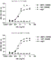
Figure 1 shows binding of the antibodies of the invention to human and cynomolgus/rhesus TIGIT.
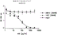
Figure 2 shows that antibodies of the invention block the interaction of hCD155 with hTIGIT as determined by a cell-based ELISA blocking assay.
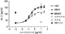
Figure 3 shows the activity of the antibodies of the invention in an in vitro T cell assay.
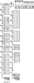
Fig. 4 shows different heatmaps of deuterium labeling of human TIGIT amino acid residues bound by mouse anti-human TIGIT 14D7 antibody.
Detailed Description
Abbreviations
In the detailed description and examples of the invention, the following abbreviations will be used:
ADCC antibody-dependent cellular cytotoxicity
CDC complement dependent cytotoxicity
Complementarity determining regions in CDR immunoglobulin variable regions, defined using the Kabat numbering system
CHO Chinese hamster ovary
ELISA enzyme-linked immunosorbent assay
FR antibody framework regions: immunoglobulin variable regions excluding CDR regions
HRP horse radish peroxidase
IFN interferon
Concentration of IC50 resulting in 50% inhibition
IgG immunoglobulin G
Kabat's immunoglobulin alignment and numbering system pioneered by Elvin A, kabat ((1991) Sequences of Proteins of Immunological Interest, published Health Service 5 th edition, national Institutes of Health, bethesda, md.)
mAb or mAb or MAb monoclonal antibody
SEB staphylococcal enterotoxin B
TT tetanus toxoid
V-region is a segment of an IgG chain with variable sequence between different antibodies. Which extends to Kabat residue 109 in the light chain and Kabat residue 113 in the heavy chain.
VH immunoglobulin heavy chain variable region
VK immunoglobulin kappa light chain variable region.
Definition of
In order that the invention may be more readily understood, certain technical and scientific terms are specifically defined below. Unless specifically defined elsewhere in this document, all other technical and scientific terms used herein have the same meaning as commonly understood by one of ordinary skill in the art to which this invention belongs.
As used herein, including the appended claims, the singular forms "a", "an", and "the" include their corresponding plural references unless the context clearly dictates otherwise.
"administration" and "treatment," when applied to an animal, human, experimental subject, cell, tissue, organ, or biological fluid, means that an exogenous drug, therapeutic agent, diagnostic agent, or composition is in contact with the animal, human, subject, cell, tissue, organ, or biological fluid. Treatment of the cells encompasses contacting the reagent with the cells, as well as contacting the reagent with a fluid, wherein the fluid is in contact with the cells. "administering" and "treating" also mean treating a cell by an agent, diagnostic agent, binding compound, or by another cell, e.g., in vitro and ex vivo.
By "treatment" or "treating" is meant internally or externally administering a therapeutic agent, such as a composition comprising any of the antibodies or antigen-binding fragments of the invention, to an individual or patient for whom an agent having one or more symptoms of a disease or suspected of having a disease has therapeutic activity. Typically, the agent is administered in an amount effective to alleviate one or more symptoms of the disease in the individual or population being treated, whether to induce regression of such symptoms by any clinically measurable degree or to inhibit their progression by any clinically measurable degree. The amount of therapeutic agent effective to alleviate any particular disease symptom may vary depending on factors such as the disease state, the age and weight of the patient, and the ability of the drug to elicit a desired response in the individual. Whether the symptoms of the disease have been alleviated can be assessed by any clinical measure commonly used by physicians or other skilled medical care providers to assess the severity or progression of symptoms.
TIGIT
The term TIGIT includes human TIGIT, cynomolgus monkey TIGIT, and rhesus TIGIT, as well as fragments thereof, such as mature fragments thereof lacking a signal peptide. In one embodiment of the invention, the amino acid sequence of human TIGIT comprises the amino acid sequence disclosed in amino acid residues 25-244 of Genbank accession No. NP 776160.2 (SEQ ID NO: 33). (amino acid residues 1-24 of SEQ ID NO:33 correspond to the leader peptide, which is removed from the mature TIGIT protein.)
In one embodiment of the invention, the amino acid sequence of a cynomolgus monkey, e.g.a cynomolgus monkey (Macaca fascicularis) TIGIT, comprises the amino acid sequence disclosed in (SEQ ID NO: 34); see also Genbank accession number XP _005548157. The amino acid sequence of rhesus TIGIT is identical to the amino acid sequence of cynomolgus TIGIT. (amino acid residues 1-24 of SEQ ID NO:34 correspond to the leader peptide, which is removed from the mature TIGIT protein.)
anti-TIGIT antibodies and antigen binding fragments thereof
The present invention provides antibodies or antigen binding fragments thereof that bind to human TIGIT, and uses of such antibodies or fragments. In some embodiments, the anti-TIGIT antibody is isolated.
As used herein, an anti-TIGIT antibody or antigen-binding fragment thereof refers to an antibody or antigen-binding fragment thereof that specifically binds to human TIGIT. An antibody or antigen-binding fragment thereof that "specifically binds human TIGIT" is one that binds human TIGIT with a KD of about 1nM or higher affinity (e.g., 1nM-2pM, 1nM, 100pM, 10pM, or 2 pM). For example, an antibody that "specifically binds" to human TIGIT does not bind human CD226, human CD155, and human CD112. As another example, an antibody or antigen-binding fragment that specifically binds to human TIGIT may bind to FLAG markers of human TIGIT but does not bind to other FLAG markers of proteins lacking human TIGIT epitopes. In one embodiment, an antibody of the invention that specifically binds to human TIGIT also cross-reacts with cynomolgus monkey and rhesus TIGIT. As used herein, "cross-reactivity" refers to the ability of an antibody to react with homologous proteins from other species, which can be determined using any assay known in the art. Whether an antibody specifically binds to human TIGIT may be determined using any assay known in the art. Examples of assays known in the art to determine binding affinity and cross-reactivity include surface plasmon resonance (e.g., BIACORE) or similar techniques (e.g., kinExa or OCTET).
The invention includes anti-TIGIT antibodies and methods of use thereof. As used herein, the term "antibody" refers to any form of antibody that exhibits a desired biological activity. Thus, it is used in the broadest sense and specifically encompasses, but is not limited to, monoclonal antibodies (including full length monoclonal antibodies comprising two light chains and two heavy chains), polyclonal antibodies, multispecific antibodies (e.g., bispecific antibodies), humanized antibodies, fully human antibodies, and chimeric antibodies.
The invention includes non-human parent (e.g., mouse and rodent) anti-TIGIT antibodies and antigen-binding fragments thereof and methods of use thereof. These antibodies may be modified for the intended use, such as humanizing the antibodies for use as human therapeutic antibodies or fragments.
The invention includes anti-TIGIT antigen binding fragments and methods of use thereof. As used herein, unless otherwise indicated, "antibody fragment" or "antigen-binding fragment" refers to an antigen-binding fragment of an antibody, i.e., an antibody fragment that retains the ability to specifically bind to an antigen to which a full-length antibody binds, e.g., a fragment that retains one or more CDR regions. Examples of antigen binding fragments include, but are not limited to, fab ', F (ab') 2 And Fv fragments; double antibodyBodies (diabodies); a linear antibody; single chain antibody molecules, such as sc-Fv; nanobodies and multispecific antibodies formed from full-length antibody chains or antibody fragments.
The invention includes anti-TIGIT Fab fragments and methods of use thereof. A "Fab fragment" comprises one light chain and one heavy chain of CH1 and variable regions. The heavy chain of a Fab molecule cannot form a disulfide bond with another heavy chain molecule. The "Fab fragment" may be the product of papain cleavage of the antibody.
The invention includes anti-TIGIT antibodies and antigen-binding fragments thereof comprising an Fc region, and methods of use thereof. The "Fc" region contains two heavy chain fragments comprising the CH1 and CH2 domains of an antibody. The two heavy chain fragments are held together by hydrophobic interactions of two or more disulfide bonds and the CH3 domain.
The invention includes anti-TIGIT Fab fragments and methods of use thereof. A "Fab fragment" comprises a part or fragment of a light chain and a heavy chain comprising the VH domain and the CH1 domain and also the region between the CH1 and CH2 domains, such that an interchain disulfide bond can form between the two heavy chains of the two Fab fragments to form F (ab') 2 A molecule.
The invention includes anti-TIGIT F (ab') 2 Fragments and methods of use thereof. "F (ab') 2 A fragment "contains two light chains and two heavy chains containing a portion of the constant region between the CH1 domain and the CH2 domain, such that an interchain disulfide bond is formed between the two heavy chains. Thus, F (ab') 2 The fragment consists of two Fab' fragments held together by a disulfide bond between the two heavy chains. "F (ab') 2 Fragments "may be the product of pepsin cleavage of an antibody.
The invention includes anti-TIGIT Fv fragments and methods of use thereof. The "Fv region" comprises the variable regions from the heavy and light chains, but lacks the constant regions.
The invention includes anti-TIGIT scFv fragments and methods of use thereof. The term "single chain Fv" or "scFv" antibody refers to an antibody fragment comprising the VH and VL domains of an antibody, wherein these domains are present in a single polypeptide chain. Typically, the Fv polypeptide further comprises a polypeptide linker between the VH and VL domains that enables the scFv to form the desired structure for antigen binding. For an overview of scFv, see Pluckthun (1994) The Pharmacology of Monoclonal Antibodies, vol.113, edited by Rosenburg and Moore, springer-Verlag, new York, pp.269-315. See also International patent application publication No. WO 88/01649 and U.S. Pat. Nos. 4,946,778 and 5,260,203.
The invention includes anti-TIGIT domain antibodies and methods of use thereof. A "domain antibody" is an immunologically functional immunoglobulin fragment containing only the variable region of a heavy chain or the variable region of a light chain. In some cases, two or more VH regions are covalently joined with a peptide linker to produce a bivalent domain antibody. The two VH regions of the bivalent domain antibody may target the same or different antigens.
The invention includes anti-TIGIT bivalent antibodies and methods of use thereof. A "bivalent antibody" comprises two antigen binding sites. In some cases, the two binding sites have the same antigen specificity. However, bivalent antibodies may be bispecific (see below).
The invention includes anti-TIGIT diabodies and methods of use thereof. The term "diabodies" as used herein refers to small antibody fragments having two antigen binding sites, which fragments comprise a heavy chain variable domain (VH) and a light chain variable domain (VL) linked thereto in the same polypeptide chain (VH-VL or VL-VH). By using a linker that is too short to allow pairing between two variable domains on the same chain, the domains are forced to pair with the complementary domains of the other chain and two antigen binding sites are created. A more complete description of diabodies is found, for example, in EP 404,097; WO 93/11161; and HolligerWait for(1993) Proc. Natl. Acad. Sci. USA6444-6448 in 90 parts. For a review of engineered antibody variants, see generally Holliger and Hudson (2005) nat. Biotechnol. 23.
Typically, an antibody or antigen-binding fragment of the invention that is modified in a manner retains at least 10% of its binding activity when the activity is expressed in molar terms (when compared to the parent). Preferably, the antibody or antigen binding fragment of the invention retains at least 20%, 50%, 70%, 80%, 90%, 95%, or 100% or more of the TIGIT binding affinity of the parent antibody. It is also contemplated that an antibody or antigen-binding fragment of the invention may include conservative or non-conservative amino acid substitutions (referred to as "conservative variants" or "function conservative variants" of the antibody) that do not substantially alter its biological activity.
The invention includes isolated anti-TIGIT antibodies and antigen-binding fragments thereof and methods of use thereof. An "isolated" antibody or antigen-binding fragment thereof is at least partially free of other biomolecules from the cell or cell culture from which it is produced. Such biomolecules include nucleic acids, proteins, lipids, carbohydrates or other substances, such as cell debris and growth media. The isolated antibody or antigen-binding fragment may further be at least partially free of expression system components, such as biomolecules from the host cell or its growth medium. Generally, the term "isolated" does not mean the complete absence of such biomolecules or the absence of water, buffers, or salts, or components of pharmaceutical preparations including antibodies or fragments.
The invention includes monoclonal anti-TIGIT antibodies and antigen-binding fragments thereof as well as monoclonal compositions comprising a plurality of isolated monoclonal antibodies. As used herein, the term "monoclonal antibody" refers to a population of substantially homogeneous antibodies, i.e., the antibody molecules comprising the population are identical in amino acid sequence, except for possible naturally occurring mutations that may be present in minor amounts. In contrast, conventional (polyclonal) antibody preparations typically include many different antibodies having different amino acid sequences in their variable domains, particularly in their CDRs, which are typically specific for different epitopes. The modifier "monoclonal" indicates that the antibody is characterized as being obtained from a substantially homogeneous population of antibodies, and is not to be construed as requiring production of the antibody by any particular method. For example, monoclonal antibodies to be used according to the invention can be prepared by the hybridoma method first described by Kohler et al (1975) Nature 256:495, or can be prepared by recombinant DNA methods (see, e.g., U.S. Pat. No. 4,816,567). "monoclonal antibodies" can also be isolated from phage antibody libraries using techniques such as those described in Clackson et al (1991) Nature 352: 624-628 and Marks et al (1991) J. Mol. Biol. 222: 581-597. See also Presta (2005) j. Allergy clin. Immunol. 116.
The invention includes anti-TIGIT chimeric antibodies (e.g., human constant domains/mouse variable domains) and methods of use thereof. As used herein, a "chimeric antibody" is an antibody having a variable domain from a first antibody and a constant domain from a second antibody, wherein the first and second antibodies are from different species. (U.S. Pat. No. 4,816,567; and Morrison et al, (1984) Proc. Natl. Acad. Sci. USA 81: 6851-6855). Typically, the variable domains are obtained from an antibody from an experimental animal such as a rodent ("parent antibody"), and the constant domain sequences are obtained from a human antibody, such that the resulting chimeric antibody will be less likely to elicit an adverse immune response in a human individual than the parent (e.g., mouse) antibody.
The invention includes anti-TIGIT humanized antibodies and antigen binding fragments thereof (e.g., humanized rat or mouse antibodies) and methods of use thereof. The invention includes any humanized version of the 14D7 antibody (comprising SEQ ID NOS: 7 and 8) or the 26B10 antibody (comprising SEQ ID NOS: 23 and 24). As used herein, the term "humanized antibody" refers to a form of an antibody that contains sequences from both human and non-human (e.g., mouse or rat) antibodies. Typically, the humanized antibody will comprise substantially all of at least one and typically two variable domains, in which all or substantially all of the hypervariable loops correspond to those of a non-human immunoglobulin and all or substantially all of the Framework (FR) regions are those of a human immunoglobulin sequence. The humanized antibody may optionally comprise at least a portion of a human immunoglobulin constant region (Fc).
Typically, the basic antibody building block comprises a tetramer. Each tetramer comprises two identical pairs of polypeptide chains, each pair having one "light" (about 25 kDa) and one "heavy" chain (about 50-70 kDa). The amino-terminal portion of each chain includes a variable region of about 100 to 110 or more amino acids primarily responsible for antigen recognition. The carboxy-terminal portion of the heavy chain may define a constant region primarily responsible for effector function. Generally, human light chains are classified as kappa and lambda light chains. Furthermore, human heavy chains are generally classified as μ, δ, γ, α or ε, and the isotype of an antibody is defined as IgM, igD, igG, igA, and IgE, respectively. Within the light and heavy chains, the variable and constant regions are joined by a "J" region of about 12 or more amino acids, with the heavy chain also including a "D" region of about 10 or more amino acids. See generally Fundamental Immunology chapter 7 (Paul, W. Ed., 2 nd edition Raven Press, N.Y. (1989).
The variable regions of each light/heavy chain pair form the antibody binding site. Thus, typically, an intact antibody has two binding sites. Except in bifunctional or bispecific antibodies, the two binding sites are typically identical.
Typically, the variable domains of both the heavy and light chains comprise three hypervariable regions, also known as Complementarity Determining Regions (CDRs), which are located within relatively conserved Framework Regions (FRs). The CDRs are typically aligned by the framework regions, enabling binding to a specific epitope. Typically, the light and heavy chain variable domains comprise, from N-terminus to C-terminus, FR1, CDR1, FR2, CDR2, FR3, CDR3 and FR4. The assignment of amino acids to the domains is generally according to the following definitions: sequences of Proteins of Immunological Interest, kabat, et al; national Institutes of Health, bethesda, md.; 5 th edition; NIH publication No. 91-3242 (1991); kabat (1978) adv. Prot. Chem. 32; kabat, et al, (1977) j. Biol. Chem. 252; chothia, et al, (1987) J mol. Biol. 196 or Chothia, et al, (1989) Nature 342.
As used herein, the term "hypervariable region" refers to the amino acid residues of an antibody or antigen-binding fragment thereof which are responsible for antigen binding. The hypervariable region comprises amino acid residues from the "complementarity determining regions" or "CDRs" (i.e., CDRL1, CDRL2 and CDRL3 in the light chain variable domain and CDRH1, CDRH2 and CDRH3 in the heavy chain variable domain). See Kabat et al (1991) Sequences of Proteins of Immunological Interest, published Health Service 5 th edition, national Institutes of Health, bethesda, md. (CDR regions of antibodies are defined by sequence); see also Chothia and Lesk (1987) J. Mol. Biol. 196:901-917 (CDR regions of antibodies are defined by structure). As used herein, the term "framework" or "FR" residues refers to those variable domain residues other than the hypervariable region residues which are defined herein as CDR residues.
An "isolated nucleic acid molecule" or "isolated polynucleotide" means a DNA or RNA of genomic, mRNA, cDNA, or synthetic origin, or some combination thereof, that is not associated with all or a portion of a polynucleotide with which the isolated polynucleotide is found in nature, or is linked to a polynucleotide to which it is not linked in nature. For the purposes of the present invention, it is understood that "a nucleic acid molecule comprising a particular nucleotide sequence" does not encompass an entire chromosome. In addition to the specified sequences, an isolated nucleic acid molecule "comprising" a specified nucleic acid sequence may comprise coding sequences of up to ten or even up to twenty or more other proteins, or parts or fragments thereof, or may comprise operably linked regulatory sequences which control the expression of the coding regions of the nucleic acid sequence, and/or may comprise vector sequences.
The phrase "control sequences" refers to DNA sequences required for expression of an operably linked coding sequence in a particular host organism. Suitable control sequences for prokaryotes include, for example, promoters, optional operator sequences and ribosome binding sites. Eukaryotic cells are known to use promoters, polyadenylation signals, and enhancers.
A nucleic acid or polynucleotide is "operably linked" when it is placed into a functional relationship with another nucleic acid sequence. For example, DNA for a presequence or secretory leader is operably linked to DNA for a polypeptide if it is expressed as a preprotein that participates in the secretion of the polypeptide; a promoter or enhancer is operably linked to a coding sequence if it affects the transcription of the sequence; or operably linked to a coding sequence if the ribosome binding site is positioned so as to facilitate translation. Generally, but not necessarily always, "operably linked" means that the DNA sequences being linked are contiguous and, in the case of secretory leader sequences, contiguous and in reading phase. However, enhancers need not be contiguous. Ligation is achieved by ligation at appropriate restriction sites. If such sites are not present, synthetic oligonucleotide aptamers or linkers are used according to conventional procedures.
As used herein, the expressions "cell," "cell line," and "cell culture" are used interchangeably and all such designations include progeny. Thus, the words "transformant" and "transformed cell" include primary individual cells and cultures derived therefrom, regardless of the number of transfers. It is also understood that not all progeny have the exact same DNA content, due to deliberate or inadvertent mutation. Mutant progeny that have the same function or biological activity as screened for in the originally transformed cell are included. Where different names are contemplated, they will be clear from the context.
As used herein, "germline sequence" refers to the sequence of the unrearranged immunoglobulin DNA sequences. Any suitable source of unrearranged immunoglobulin sequences may be used. Human germline sequences are available from, for example, the JOINSOLVER germline database on the website of the National Institute of Arthritis and Musculoskeletal and dermatological Diseases, the National Institute of Arthritis and musculoskakeleases, the National Institutes of Health. Mouse germline sequences can be obtained, for example, as described in Giudicelli et al (2005) Nucleic Acids Res. 33.
Physical and functional characteristics of exemplary anti-TIGIT antibodies
The present invention provides anti-TIGIT antibodies and antigen-binding fragments thereof having specified structural and functional characteristics, and methods of using the antibodies or antigen-binding fragments thereof for treating or preventing a disease (e.g., cancer or an infectious disease).
"anti-TIGIT antibody or antigen-binding fragment thereof of the present invention" includes: any of the antibodies or antigen-binding fragments thereof discussed herein (e.g., 14D7, 26B10, or humanized versions of these antibodies).
Cross-blocking antibodies and antigen-binding fragments thereof can be identified based on their ability to cross-compete with the antibodies of the invention in standard binding assays (e.g., BIACORE, ELISA, flow cytometry). For example, a standard ELISA assay can be used in which a recombinant TIGIT (e.g., human TIGIT) protein is immobilized on a plate, one of the antibodies is fluorescently labeled and the ability of the unlabeled antibody to compete off the binding of the labeled antibody is evaluated. Alternatively or alternatively, BIAcore analysis can be used to assess the ability of the antibodies to cross-compete. The ability of the test antibody to inhibit the binding of another antibody (e.g., antibody 14D7 or 26B 10) to TIGIT (e.g., human TIGIT) indicates that the test antibody can compete with the other antibody (e.g., 14D7 or 26B 10) for binding to TIGIT (e.g., human TIGIT) and thus can bind to the same epitope or overlapping epitope on TIGIT (e.g., human TIGIT) as antibody 14D7 or 26B10 in some cases.
As described above, antibodies and fragments that bind to the same epitope as any of the anti-TIGIT antibodies of the invention or antigen binding fragments thereof also form part of the invention. Furthermore, antibodies that bind an epitope that overlaps with the epitope bound by any of the anti-TIGIT antibodies of the invention also form part of the invention. There are several methods that can be used to locate an epitope of an antibody on a target antigen, including: H/D-Ex mass spectrometry, X-ray crystallography, pepscan analysis, and site-directed mutagenesis. For example, coupling of HDX (hydrogen deuterium exchange) to proteolysis and mass spectrometry can be used to determine the epitope of an antibody on a particular antigen Y. HDX-MS relies on accurate measurement and comparison of antigen incorporation of deuterium when incubated in D2O under itself and in the presence of its antibodies at various time intervals. Deuterium exchanges with hydrogen on the amide backbone of the protein in the exposed regions, while the regions that bind the antigen of the antibody will be protected and will show less or no exchange after LC-MS/MS analysis by proteolytic fragments.
Examples of immunoglobulin chains of anti-TIGIT antibodies of the invention and CDRs thereof include, but are not limited to, those disclosed in table 4. The invention includes any polypeptide comprising or consisting of the amino acid sequence of SEQ ID NO1, 32, 45-112, and recombinant nucleotides encoding such polypeptides.
The scope of the invention includes isolated anti-TIGIT antibodies and antigen-binding fragments thereof (e.g., humanized antibodies) comprising a variant of an immunoglobulin chain described herein, e.g., any one of SEQ ID NOs 716, 48-56, 63-80, 23-32, and 89-112; wherein the variant exhibits one or more of the following properties: (i) binding to human TIGIT; (ii) cross-reacts with cynomolgus monkey and rhesus TIGIT; (iii) blocks binding of human TIGIT to human CD155 and human CD 112; (iv) increasing T cell activation; (v) Stimulating antigen-specific T cell production of IL-2 and IFN γ; (vi) Blocking the induction of inhibition of T cell activation induced by TIGIT engagement with cognate ligands CD155 and CD112.
In other embodiments, the invention provides antibodies, or antigen-binding fragments thereof, that bind to human TIGIT (e.g., humanized antibodies) and have a VL domain and a VH domain with at least 80%, 85%, 90%, 95%, 96%, 97%, 98%, or 99% sequence identity to SEQ ID NOs 7-16, 48-56, 63-80, 23-32, and 89-112; wherein the variant exhibits desired binding and properties, e.g., (i) about 1x 10 as determined by surface plasmon resonance (e.g., BIACORE) or similar techniques (e.g., kinExa or OCTET) -9 M to about 1x 10 -12 The KD value of M is combined with human TIGIT; (ii) cross-reacts with cynomolgus monkey and rhesus TIGIT; (iii) blocks binding of human TIGIT to human CD155 and human CD 112; (iv) increasing T cell activation; (v) Stimulating antigen-specific T cell production of IL-2 and IFN γ; (vi) Blocking the induction of inhibition of T cell activation induced by TIGIT engagement with cognate ligands CD155 and CD112.
"conservatively modified variants" or "conservative substitutions" refer to the replacement of an amino acid in a protein by another amino acid having similar characteristics (e.g., charge, side chain size, hydrophobicity/hydrophilicity, backbone conformation, and rigidity, etc.) such that the changes can be made frequently without altering the biological activity of the protein. One skilled in The art recognizes that, in general, single amino acid substitutions in non-essential regions of a polypeptide do not substantially alter biological activity (see, e.g., watson et al (1987) Molecular Biology of The Gene, the Benjamin/Cummings pub. Co., page 224 (4 th edition)). In addition, substitutions of structurally or functionally similar amino acids are unlikely to destroy biological activity. Exemplary conservative substitutions are set forth in table 1.
TABLE 1 exemplary conservative amino acid substitutions
| Initial residue | Conservative substitutions |
| Ala (A) | Gly; Ser |
| Arg (R) | Lys; His |
| Asn (N) | Gln; His |
| Asp (D) | Glu; Asn |
| Cys (C) | Ser; Ala |
| Gln (Q) | Asn |
| Glu (E) | Asp; Gln |
| Gly (G) | Ala |
| His (H) | Asn; Gln |
| Ile (I) | Leu; Val |
| Leu (L) | Ile; Val |
| Lys (K) | Arg; His |
| Met (M) | Leu; Ile; Tyr |
| Phe (F) | Tyr; Met; Leu |
| Pro (P) | Ala |
| Ser (S) | Thr |
| Thr (T) | Ser |
| Trp (W) | Tyr; Phe |
| Tyr (Y) | Trp; Phe |
| Val (V) | Ile; Leu |
The invention also encompasses function-conservative variants of the antibodies of the invention. As used herein, "function-conservative variants" refer to antibodies or fragments in which one or more amino acid residues have been altered without altering a desired property, such as antigen affinity and/or specificity. Such variants include, but are not limited to, amino acid substitutions by amino acids having similar properties, such as conservative amino acid substitutions of table 1. Also provided are isolated polypeptides comprising a VL domain of an anti-TIGIT antibody of the invention (e.g., SEQ ID NOs 8, 13-16, 24, 29-33, 63-80) and isolated polypeptides comprising a VH domain of an anti-TIGIT antibody of the invention (e.g., SEQ ID NOs 7, 9-12, 24-29, 48-56, 89-112) having up to 0,1, 2,3, 4,5, 6,7, 8, 9, 10, 11, 12, 13, 14, 15, 16, 17, 18, 19, 20, 21, 22, 23, 24, 25, 26, 27, 28, 29, 30 or more amino acid substitutions.
In another embodiment, antibodies, or antigen binding fragments thereof, are provided that bind to TIGIT and have a VL domain and a VH domain that have at least 99%, 98%, 97%, 96%, 95%, 90%, 85%, 80%, or 75% sequence identity to one or more of the VL domains or VH domains described herein and exhibit specific binding to TIGIT. In another embodiment, a binding antibody or antigen-binding fragment thereof of the invention comprises VL and VH domains (with and without signal sequences) having up to 0,1, 2,3, 4,5, 6,7, 8, 9, 10, 11, 12, 13, 14, 15, 16, 17, 18, 19, 20, 21, 22, 23, 24, 25, 26, 27, 28, 29, 30 or more amino acid substitutions and exhibits specific binding to TIGIT.
Polynucleotides and polypeptides
The invention further comprises a polynucleotide encoding any one of the polypeptides or immunoglobulin chains of the anti-TIGIT antibodies and antigen-binding fragments thereof of the invention. For example, the invention includes polynucleotides encoding the amino acids set forth in SEQ ID NOS 1-32 or 45-112, as well as polynucleotides that hybridize thereto, and in addition, any polypeptides encoded by such hybridizing polynucleotides.
Typically, the polynucleotide hybridizes under low, medium, or high stringency conditions and encodes an antibody or antigen-binding fragment thereof that maintains the ability to bind TIGIT (human, rhesus monkey and/or cynomolgus monkey, e.g., cynomolgus monkey). A first polynucleotide molecule is "hybridizable" to a second polynucleotide molecule when a single-stranded form of the first polynucleotide molecule can anneal to the second polynucleotide molecule under conditions of appropriate temperature and solution ionic strength (see Sambrook et al, supra). The conditions of temperature and ionic strength determine the "stringency" of the hybridization. Typical low stringency hybridization conditions include 55 ℃, 5X SSC, 0.1% SDS, and no formamide; or 30% formamide, 5 XSSC, 0.5% SDS at 42 ℃. Typical medium stringency hybridization conditions are 40% formamide, 5X or 6X SSC, and 0.1% SDS at 42 ℃. High stringency hybridization conditions are 50% formamide, 5X or 6 XSSC, at 42 ℃ or optionally at a higher temperature (e.g., 57 ℃,59 ℃,60 ℃,62 ℃, 63 ℃, 65 ℃, or 68 ℃). Typically, SSC is 0.15M NaCl and 0.015M sodium citrate. Hybridization requires that the two polynucleotides contain complementary sequences, however, depending on the stringency of the hybridization, mismatches between bases are possible. The appropriate stringency for hybridizing polynucleotides depends on the length and degree of complementarity of the polynucleotides, variables well known in the art. The greater the degree of similarity or homology between two nucleotide sequences, the higher the stringency at which the nucleic acids can hybridize. For hybrids of greater than 100 nucleotides in length, equations for calculating the melting temperature have been derived (see Sambrook et al, supra, 9.50-9.51). For hybridization with shorter polynucleotides, such as oligonucleotides, the position of the mismatch becomes more important, and the length of the oligonucleotide determines its specificity (see Sambrook et al, supra, 11.7-11.8).
In another embodiment, an isolated polynucleotide, e.g., DNA, is provided that encodes a polypeptide chain of an isolated antibody or antigen-binding fragment described herein. In one embodiment, the isolated polynucleotide encodes an antibody or antigen-binding fragment thereof comprising at least one mature immunoglobulin light chain Variable (VL) domain according to the invention and/or at least one mature immunoglobulin heavy chain Variable (VH) domain according to the invention. In some embodiments, the isolated polynucleotides encode light and heavy chains on a single polynucleotide molecule, and in other embodiments, encode light and heavy chains on separate polynucleotide molecules. In another embodiment, the polynucleotide further encodes a signal sequence.
In one embodiment, the invention comprises an isolated polynucleotide encoding an antibody heavy chain Variable (VH) domain comprising CDR-H1 (SEQ ID NO: 1), CDR-H2 (SEQ ID NO: 2), and CDR-H3 (SEQ ID NO:3, 45, 46, or 47), or an antigen-binding fragment thereof.
In one embodiment, the invention comprises an isolated polynucleotide encoding an antibody light chain Variable (VL) domain comprising CDR-L1 (SEQ ID NO: 4), CDR-L2 (SEQ ID NO: 5), and CDR-L3 (SEQ ID NO:6, 57, 58, 59, 60, 61, or 62), or an antigen-binding fragment thereof.
In one embodiment, the invention comprises an isolated polynucleotide encoding the immunoglobulin heavy chain Variable (VH) domain of SEQ ID NO 7.
In one embodiment, the invention comprises an isolated polynucleotide encoding the immunoglobulin heavy chain Variable (VL) domain of SEQ ID NO 8.
In one embodiment, the invention comprises an isolated polynucleotide encoding an immunoglobulin heavy chain Variable (VH) domain of any one of SEQ ID NOS 9-12.
In one embodiment, the invention comprises an isolated polynucleotide encoding an immunoglobulin heavy chain Variable (VH) domain of any one of SEQ ID NOS 48-56.
In one embodiment, the invention encompasses an isolated polynucleotide encoding an immunoglobulin heavy chain Variable (VL) domain of any one of SEQ ID NOS 13-16.
In one embodiment, the invention encompasses an isolated polynucleotide encoding an immunoglobulin heavy chain Variable (VL) domain of any one of SEQ ID NOS: 63-80.
In one embodiment, the invention comprises an isolated polynucleotide encoding an antibody heavy chain Variable (VH) domain comprising CDR-H1 (SEQ ID NO: 17), CDR-H2 (SEQ ID NO:18, 81, 82, 83, 84, 85, 86, 87, or 88), and CDR-H3 (SEQ ID NO: 19), or an antigen-binding fragment thereof.
In one embodiment, the invention comprises an isolated polynucleotide encoding an antibody light chain Variable (VL) domain comprising CDR-L1 (SEQ ID NO: 4), CDR-L2 (SEQ ID NO: 5) and CDR-L3 (SEQ ID NO:6, 57, 58, 59, 60, 61 or 62), or an antigen-binding fragment thereof.
In one embodiment, the invention comprises an isolated polynucleotide encoding the immunoglobulin heavy chain Variable (VH) domain of SEQ ID NO 23.
In one embodiment, the invention comprises an isolated polynucleotide encoding the immunoglobulin heavy chain Variable (VL) domain of SEQ ID NO: 24.
In one embodiment, the invention comprises an isolated polynucleotide encoding an immunoglobulin heavy chain Variable (VH) domain of any one of SEQ ID NOS 25-28.
In one embodiment, the invention comprises an isolated polynucleotide encoding the immunoglobulin heavy chain Variable (VH) domain of any one of SEQ ID NOS 89-112.
In one embodiment, the invention encompasses an isolated polynucleotide encoding an immunoglobulin heavy chain Variable (VL) domain of any one of SEQ ID NOS: 29-32.
The invention also provides a vector, e.g., an expression vector, such as a plasmid, comprising an isolated polynucleotide of the invention, wherein the polynucleotide is operably linked to control sequences recognized by a host cell when the host cell is transfected with the vector. Also provided are host cells comprising the vectors of the invention and methods for producing the antibodies or antigen-binding fragments thereof or polypeptides disclosed herein, comprising culturing a host cell carrying an expression vector or nucleic acid encoding an immunoglobulin chain of the antibody or antigen-binding fragment thereof in a culture medium, and isolating the antibody or antigen-binding fragment thereof from the host cell or culture medium.
Also included in the invention are polypeptides, such as immunoglobulin polypeptides, whose amino acid sequence is at least about 75% identical, has 80% identical, more preferably has at least about 90% identical, and most preferably has at least about 95% (e.g., 95%, 96%, 97%, 98%, 99%, 100%) identical to the amino acid sequence of an antibody provided herein when compared by the BLAST algorithm, wherein the parameters of the algorithm are selected to obtain the maximum match between the corresponding sequences over the entire length of the corresponding reference sequence (e.g., the expected threshold: 10; word length: 3; maximum match in query range: 0 BLOSUM 62 matrix; gap cost: presence 11, extension 1; conditional composition scoring matrix adjustment).
Sequence identity refers to the degree to which the amino acids of two polypeptides are identical at equivalent positions when the two sequences are optimally aligned.
The following references refer to the BLAST algorithm often used for sequence analysis: BLAST ALGORITHMS, altschul et al (2005) FEBS J. 272 (20): 5101-5109; altschul, s.f., et al, (1990) j. Mol. Biol. 215; gish, W., et al, (1993) Nature Genet. 3; madden, t.l., et al, (1996) meth. Enzymol. 266; altschul, s.f., et al, (1997) Nucleic Acids res.25; zhang, J., et al, (1997) Genome Res.7, 649-656; wootton, J.C., et al, (1993) Comput. Chem.17; hancock, J.M. et al, (1994) Compout. Appl. Biosci. 10, 67-70; identification coding SYSTEMS, dayhoff, M.O., et al, "A model of evolution change in proteins," in Atlas of Protein sequences and Structure, (1978) Vol.5, supplement 3, M.O. Dayhoff, pp.345-352, natl. Biomed. Res. Foundation, washington, DC; schwartz, r.m., et al, "Matrices for detecting distances relationships," in Atlas of Protein sequences and structures, (1978) volume 5, supplement 3, "m.o. Dayhoff (ed.), pages 353-358, native. Biomed. Res, foundation, washington, DC; altschul, s.f. (1991) j. Mol. Biol. 219; states, d.j., et al, (1991) Methods 3; henikoff, s., et al, (1992) proc. Natl. Acad. Sci. USA 89; altschul, s.f., et al, (1993) j. Mol. Evol. 36; ALIGNMENT STATISTICS Karlin, S., et al, (1990) Proc. Natl. Acad. Sci. USA 87; karlin, s., et al, (1993) proc. Natl. Acad. Sci. USA 90; dembo, A., et al, (1994) Ann. Prob. 22; and Altschul, S.F. "Evaluating the statistical design of multiple discrete local alignment," Theoretical and Computational Methods in Genome Research (S. Suhai, eds.) (1997) pages 1-14, plenum, new York.
Binding affinity
For example and without limitation, the antibodies and antigen binding fragments disclosed herein can be at least about 1x 10 as determined by surface plasmon resonance (e.g., BIACORE) or similar techniques (e.g., kinExa or OCTET) -9 KD value of M (i.e. 1X 10) -9 KD value of M or lower) in conjunction with human TIGIT. In one embodiment, the antibodies and antigen-binding fragments disclosed herein can be at least about 1x 10 as determined by surface plasmon resonance (e.g., BIACORE) or similar techniques (e.g., kinExa or OCTET) -9 M to about 1x 10 -12 KD values for M bind to human TIGIT. In one embodiment, the antibodies and antigen binding fragments disclosed herein may be, for example, by surface plasmonAt least about 1x 10 as determined by subresonance (e.g., BIACORE) or similar techniques (e.g., kinExa or OCTET) -9 M to about 1x 10 -12 KD values for M bind to human TIGIT. In one embodiment, the antibodies and antigen binding fragments disclosed herein can bind to human TIGIT with a KD value of at least about 50pM (i.e., a KD value of about 50pM or less) as determined by BIACORE or similar techniques. In one embodiment, the antibodies and antigen binding fragments disclosed herein can bind to human TIGIT with a KD value of at least about 10pM (i.e., a KD value of about 10pM or less) as determined by BIACORE or similar techniques. In one embodiment, the antibodies and antigen binding fragments of the invention can bind to human TIGIT with a KD of about 50pM to about 1pM as determined by BIACORE or similar techniques.
Immune cell activation
In some embodiments, the antibodies or antigen binding fragments of the invention increase the activity of an immune cell. The increase in immune cell activity can be detected using any method known in the art. In one embodiment, an increase in immune cell activity can be detected by measuring the proliferation of immune cells. For example, an increase in T cell activity can be detected by measuring proliferation of T cells or signal transduction events, such as tyrosine phosphorylation of immunoreceptors or downstream kinases that transmit signals to transcriptional regulators. In other embodiments, an increase in immune cell activity can be detected by measuring CTL or NK cell cytotoxic function on a particular target cell or IFN γ cytokine response associated with stimulation of anti-tumor immunity. In still other embodiments, the increase in immune cell activity can be detected by measuring T cell activation ex vivo in a sample derived from the individual. In one embodiment, the increase in T cell activity is determined by: (i) Measuring SEB (staphylococcal enterotoxin B) induced production of one or more proinflammatory cytokines selected from the group consisting of: IL-2, TNF alpha, IL-17, IFN gamma, IL-1 beta, GM-CSF, RANTES, IL-6, IL-8, IL-5, and IL-13; or (ii) measuring mixed lymphocyte responses or direct anti-CD 3 mAb stimulation that induce T Cell Receptor (TCR) signaling of production of a cytokine selected from: IL-2, TNF alpha, IL-17, IFN gamma, IL-1 beta, GM-CSF, RANTES, IL-6, IL-8, IL-5, and IL-13. In certain embodiments, an anti-TIGIT antibody or antigen-binding fragment thereof of the invention will stimulate IL-2 and/or IFN γ antigen-specific T cells by at least 1.5 fold.
The invention includes antagonist anti-TIGIT antibodies and antigen-binding fragments thereof and methods of use thereof, e.g., humanized, antagonist anti-TIGIT antibodies and fragments. Antagonist anti-TIGIT antibodies or antigen-binding fragments thereof antagonize the activity of human TIGIT, such as by inhibiting TIGIT binding to CD155 and CD112, and inhibiting TIGIT functional ITIM signaling upon binding to CD155 and CD112. Measurement of anti-TIGIT antagonist activity can be assessed by demonstrating blockade of T cell inhibition following TCR activation induced by TIGIT engagement with cognate ligands CD155 and CD112. Thus, in one embodiment of increasing response, treatment with an antagonist anti-TIGIT antibody is capable of rescuing IL-2 responses to the levels observed in T cells not inhibited by TIGIT-induced CD155 or CD112. At a more preferred level of activation, the response may increase to a level greater than a T cell response not inhibited by CD155 or CD112 following treatment with an anti-TIGIT antagonist antibody.
anti-hTIGIT antibodies block the ability to bind to hCD155 and hCD112
In some embodiments, an anti-TIGIT antibody or antigen-binding fragment of the invention is capable of blocking human TIGIT binding to human CD155 and human CD112. The ability to block binding of human TIGIT to human CD155 and human CD112 can be determined using any method known in the art. In one embodiment, the ability of the antibody to block the binding of human TIGIT to human CD155 and human CD112 is determined using an ELISA assay as described in example 2.
Methods of making antibodies and antigen-binding fragments thereof
Hybridoma cells that produce the parent (e.g., rat or mouse) monoclonal anti-TIGIT antibodies or antigen-binding fragments thereof discussed herein can be produced by methods generally known in the art. Such isolated hybridomas are part of the present invention. These include, but are not limited to, hybridoma technology originally produced by Kohler, et al, (1975) (Nature 256. Preferably, mouse splenocytes are isolated and fused with PEG, or electrofused with a mouse myeloma cell line by standard protocols. The resulting hybridomas can then be screened to produce antigen-specific antibodies. For example, a single cell suspension of splenic lymphocytes from an immunized mouse can be fused with 50% PEG to one-sixth the number of P3X63-Ag8.653 non-secreting mouse myeloma cells (ATCC, CRL 1580). Cells can be plated at about 2X 10⁵ cells/mL in flat-bottomed microtiter plates followed by two weeks of incubation in selective media containing 20% fetal clone serum, 18% "653" conditioned media, 5% origen (IGEN), 4mM L-glutamine, 1mM sodium pyruvate, 5mM HEPES, 0.055mM 2-mercaptoethanol, 50 units/mL penicillin, 50mg/mL streptomycin, 50mg/mL gentamicin, and 1 XHAT (Sigma; HAT added 24 hours after fusion). After two weeks, the cells can be cultured in medium where HAT is replaced with HT. Individual wells can then be screened by ELISA for anti-TIGIT monoclonal IgG antibodies. Once significant hybridoma growth has occurred, the medium can generally be observed after 10-14 days. The antibody secreting hybridomas can be replated, screened again, and if still positive for human IgG, the anti-TIGIT monoclonal antibody can be subcloned at least twice by limiting dilution. The stable subclones can then be cultured in vitro to produce small amounts of antibody in tissue culture medium for characterization.
Accordingly, the invention includes a method for making an anti-TIGIT antibody or antigen-binding fragment thereof of the invention comprising culturing hybridoma cells expressing the antibody or fragment under conditions conducive to such expression, and optionally isolating the antibody or fragment from the hybridoma and/or growth medium (e.g., cell culture medium).
The anti-TIGIT antibodies disclosed herein may also be produced recombinantly (e.g., in an e.coli/T7 expression system, a mammalian cell expression system, or a lower eukaryote expression system). In this embodiment, nucleic acids encoding the antibody immunoglobulin molecules of the invention (e.g., VH or VL) can be inserted into pET-based plasmids and expressed in the e.coli/T7 system. For example, the invention includes a method for expressing an antibody or antigen-binding fragment thereof or an immunoglobulin chain thereof in a host cell (e.g., a bacterial host cell, such as e.coli, such as BL21 or BL21DE 3), comprising expressing a T7 RNA polymerase in a cell that further comprises a polynucleotide encoding an immunoglobulin chain operably linked to a T7 promoter. For example, in one embodiment of the invention, a bacterial host cell, such as e.coli, includes a polynucleotide encoding a T7 RNA polymerase gene operably linked to a lac promoter, and expression of the polymerase and chain is induced by incubation of the host cell with IPTG (isopropyl- β -D-thiogalactoside).
There are several methods known in the art for producing recombinant antibodies. One example of a method of recombinantly producing antibodies is disclosed in U.S. Pat. No. 4,816,567.
Transformation may be by any known method for introducing a polynucleotide into a host cell. Methods for introducing heterologous polynucleotides into mammalian cells are well known in the art and include dextran-mediated transfection, calcium phosphate precipitation, polybrene-mediated transfection, protoplast fusion, electroporation, encapsulation of the polynucleotide in liposomes, biolistic injection, and direct microinjection of DNA into the nucleus. Alternatively, the nucleic acid molecule may be introduced into a mammalian cell by a viral vector. Methods of cell transformation are well known in the art. See, e.g., U.S. Pat. nos. 4,399,216;4,912,040;4,740,461 and 4,959,455.
Accordingly, the invention includes a recombinant method for making an anti-TIGIT antibody or antigen-binding fragment thereof or immunoglobulin chain thereof of the invention comprising introducing a polynucleotide encoding one or more immunoglobulin chains (e.g., immunoglobulin heavy and/or light chains) of the antibody or fragment; the host cell (e.g., CHO or pichia pastoris) is cultured under conditions conducive to such expression, and optionally the antibody or fragment or chain is isolated from the host cell and/or the medium in which the host cell is grown.
anti-TIGIT antibodies can also be synthesized by any of the methods described in U.S. patent No. 6,331,415.
Eukaryotic and prokaryotic host cells, including mammalian cells, as hosts for expressing the antibodies or fragments or immunoglobulin chains disclosed herein are well known in the art and include many immortalized cell lines available from the American Type Culture Collection (ATCC). These include, inter alia, chinese Hamster Ovary (CHO) cells, NSO, SP2 cells, heLa cells, baby Hamster Kidney (BHK) cells, monkey kidney Cells (COS), human hepatocellular carcinoma cells (e.g., hep G2), A549 cells, 3T3 cells, HEK-293 cells, and many other cell lines. Mammalian host cells include human, mouse, rat, dog, monkey, pig, goat, cow, horse, and hamster cells. Particularly preferred cell lines are selected by determining which cell lines have high expression levels. Other cell lines that may be used are insect cell lines such as Sf9 cells, amphibian cells, bacterial cells, plant cells and fungal cells. Fungal cells include yeast and filamentous fungal cells, including, for example, pichia pastoris, pichia finlandica (Pichia finlandica), pichia trehalose-loving (Pichia trehala), pichia pastoris (Pichia kocrasae), pichia membranaefaciens (Pichia membranaefaciens), pichia miniata (Pichia minuta), pichia pastoris (Ogataea minuta, pichia lindrica), pichia pastoris, pichia thermotolerant (Pichia thermophilans), pichia salicicola, pichia guensis, pichia pastoris (Pichia pastoris), pichia stipitis, pichia methanolica (Pichia methanolica), pichia sp. Saccharomyces (Saccharomyces sp.), hansenula polymorpha (Hansenula polymorpha), kluyveromyces (Kluyveromyces sp.), kluyveromyces lactis (Kluyveromyces lactis), candida albicans (Candida albicans), aspergillus nidulans (Aspergillus nidulans), aspergillus niger (Aspergillus niger), aspergillus oryzae (Aspergillus oryzae), trichoderma reesei (Trichoderma reesei), chrysosporium (Chrysosporium lucknowense), fusarium (Fusarium sp.), fusarium graminum (Fusarium gramineum), fusarium (Fusarium venenatum), physcomitrella patens (Physcomitrella patens) and Neurospora crassa (Neurospora crassa). Pichia, any Saccharomyces, hansenula polymorpha, any Kluyveromyces, candida albicans, any Aspergillus, trichoderma reesei, chrysosporium, any Fusarium, yarrowia lipolytica, and Neurospora crassa. When a recombinant expression vector encoding the heavy chain or antigen-binding portion or fragment thereof, the light chain, and/or an antigen-binding fragment thereof is introduced into a mammalian host cell, the antibody is produced by culturing the host cell for a time sufficient to allow the antibody or fragment or chain to be expressed in the host cell or secreted into the medium in which the host cell is grown.
Antibodies and antigen binding fragments thereof and immunoglobulin chains can be recovered from the culture medium using standard protein purification methods. In addition, expression of the antibodies and antigen binding fragments thereof and immunoglobulin chains (or other portions therefrom) of the invention from a producing cell line can be enhanced using a number of known techniques. For example, the glutamine synthetase gene expression system (GS system) is a common method for enhancing expression under certain conditions. The GS system is discussed in whole or in part in connection with european patent nos. 0 216 846, 0 256 055, and 0 323 997 and european patent application No. 89303964.4. Thus, in one embodiment of the invention, a mammalian host cell (e.g., CHO) lacks a glutamine synthetase gene and is grown in the absence of glutamine in the culture medium, wherein, however, the polynucleotide encoding the immunoglobulin chain comprises a glutamine synthetase gene that complements the lack of genes in the host cell.
The invention includes a method for purifying an anti-TIGIT antibody or antigen-binding fragment thereof of the invention comprising introducing a sample comprising the antibody or fragment into a purification medium (e.g., cation exchange medium, anion exchange medium, hydrophobic exchange medium, affinity purification medium (e.g., protein-a, protein-G, protein-a/G, protein-L)) and collecting the purified antibody or fragment from a flow-through fraction of the sample that does not bind to medium; or discarding the flow-through fraction and eluting the bound antibody or fragment from the culture medium and collecting the eluate. In one embodiment of the invention, the culture medium is in the column to which the sample is applied. In one embodiment of the invention, the purification process is performed after recombinant expression of the antibody or fragment in the host cell, e.g. wherein the host cell is first lysed and the lysate is purified to remove insoluble material, optionally before purification on a culture medium.
Typically, a glycoprotein produced in a particular cell line or transgenic animal will have a glycosylation pattern characteristic of the glycoprotein produced in the cell line or transgenic animal. Thus, the particular glycosylation pattern of an antibody will depend on the particular cell line or transgenic animal used to produce the antibody. However, all antibodies encoded by or comprising the amino acid sequences provided herein constitute the present invention, independent of the glycosylation pattern that the antibody may have. Similarly, in particular embodiments, antibodies having glycosylation patterns that comprise only nonfucosylated N-glycans may be advantageous because these antibodies have been shown to generally exhibit more effective efficacy in vitro and in vivo than their fucosylated counterparts (see, e.g., shinkawa et al, J. Biol. Chem. 278: 3466-3473 (2003); U.S. Pat. Nos. 6,946,292 and 7,214,775). These antibodies with nonfucosylated N-glycans cannot be immunogenic because their carbohydrate structures are normal components of the population present in human serum IgG.
The invention includes polyclonal anti-TIGIT antibodies and antigen-binding fragments thereof, e.g., compositions comprising a plurality of anti-TIGIT antibodies and fragments, including one or more of the anti-TIGIT antibodies of the invention or antigen-binding fragments thereof, and methods of use thereof. Polyclonal antibodies are antibodies that are raised in or in the presence of one or more other, non-identical antibodies. Typically, polyclonal antibodies are produced by a collection of different B lymphocytes, e.g., B lymphocytes of an animal treated with an immunogen of interest, which produce a population of different antibodies, but which are all directed against the immunogen. Typically, polyclonal antibodies are obtained directly from the immunized animal, e.g., spleen, serum, or ascites fluid.
The invention includes bispecific and diabodies and antigen-binding fragments that have binding specificity for TIGIT and another antigen, such as, for example, PD-1 or PD-L1 or LAG-3, and methods of use thereof. In one embodiment of the invention, the anti-TIGIT chain comprises any one of the VH/VL sequences set forth in Table 4, and the PD1 chain comprises the amino acid sequences of SEQ ID NOS: 36 and 37 or SEQ ID NOS: 38 and 39 (or an antigen-binding fragment of any of the sequences). Bispecific or diabodies are artificial hybrid antibodies with two different heavy/light chain pairs and two different binding sites. Bispecific antibodies can be produced by a variety of methods including fusion of hybridomas or ligation of Fab' fragments. See, e.g., songsivilai, et al, (1990) Clin. Exp. Immunol. 79: 315-321, kostelny, et al, (1992) J Immunol. 148. In addition, bispecific antibodies can be formed as "diabodies" (Holliger, et al, (1993) PNAS USA 90 6444-6448) or as "Janusins" (Traunecker, et al, (1991) EMBO J. 10.
The invention further includes anti-TIGIT antigen binding fragments of the anti-TIGIT antibodies disclosed herein. Antibody fragments include F (ab) 2 Fragments, which can be produced by enzymatic cleavage of IgG by e.g. pepsin. Fab fragments can be produced by, for example, reducing F (ab) with dithiothreitol or mercaptoethylamine 2 To produce.
Immunoglobulins can be assigned to different classes based on the amino acid sequence of the constant domain of their heavy chains. There are at least five major classes of immunoglobulins: igA, igD, igE, igG, and IgM, and several of these can be further divided into subclasses (isotypes), such as IgG1, igG2, igG3, and IgG4; igA1 and IgA2. The invention encompasses antibodies and antigen-binding fragments of any of these classes or subclasses of antibodies.
In one embodiment, the antibody or antigen-binding fragment comprises a heavy chain constant region, e.g., a human constant region, such as a γ 1, γ 2, γ 3, or γ 4 human heavy chain constant region or variant thereof. In another embodiment, the antibody or antigen-binding fragment comprises a light chain constant region, e.g., a human light chain constant region, such as a λ or κ human light chain region or a variant thereof. For example and without limitation, the human heavy chain constant region can be γ 4 and the human light chain constant region can be κ. In an alternative embodiment, the Fc region of the antibody is γ 4 with the Ser228Pro mutation (Schuurman, J et al, mol. Immunol. 38: 1-8, 2001).
In one embodiment, the antibody or antigen-binding fragment comprises a heavy chain constant region of an IgG1 subtype.
In some embodiments, different constant domains may be attached to the humanized VL and VH regions derived from the CDRs provided herein. For example, if a particular intended use of an antibody (or fragment) of the invention requires alteration of effector function, a heavy chain constant domain other than human IgG1 may be used, or a hybrid IgG1/IgG4 may be utilized.
While human IgG1 antibodies provide long half-life and effector functions, such as complement activation and antibody-dependent cellular cytotoxicity, such activity may not be required for all uses of the antibody. In such cases, for example, a human IgG4 constant domain may be used. The invention includes anti-TIGIT antibodies and antigen-binding fragments thereof, e.g., antagonists, humanized anti-TIGIT antibodies and fragments, comprising an IgG4 constant domain, and methods of use thereof. In one embodiment, the IgG4 constant domain may differ from native human IgG4 constant domain at a position corresponding to position 228 in the EU system and position 241 in the KABAT system (Swiss-Prot accession number P01861.1), wherein native Ser108 is replaced with Pro to prevent potential interchain disulfide formation between Cys106 and Cys109 (corresponding to positions Cys 226 and Cys 229 in the EU system and positions Cys 239 and Cys 242 in the KABAT system) that may interfere with appropriate intrachain disulfide formation. See Angal et al (1993) mol. Imunol. 30. In other cases, modified IgG1 constant domains that have been modified to increase half-life or decrease effector function may be used.
Antibody engineering
Further included are embodiments in which the anti-TIGIT antibodies and antigen binding fragments thereof are engineered to include modifications to framework residues within the variable domain of a parent (e.g., mouse or rat) monoclonal antibody, e.g., antibodies that improve the properties of the antibody or fragment. Typically, such framework modifications are made to reduce the immunogenicity of the antibody or fragment. This is typically achieved by replacing non-CDR residues (i.e. framework residues) in the variable domains in a parent (e.g. rodent) antibody or fragment (donor antibody or fragment) with analogous residues from the immune repertoire of the species in which the antibody will be used, e.g. human residues (acceptor antibody) in the case of human therapeutics. Such antibodies or fragments are referred to as "humanized" antibodies or fragments. In some cases, it is desirable to increase or decrease the affinity or change the specificity of an engineered (e.g., humanized) antibody. The desired immunoglobulin profile can be achieved by selecting and combining framework residues from the parent and acceptor antibodies. One approach is to mutate one or more framework residues into the corresponding germline sequence. More specifically, an antibody or fragment that has undergone somatic mutation may contain framework residues that are different from the human germline sequence from which the antibody is derived. Such residues can be identified by comparing the parent antibody or fragment framework sequences to the germline sequences from which the human antibody or fragment was derived, and mutating one or more framework residues in the antibody to the corresponding human germline sequence. Another approach is to revert ("back mutation") to the original parent (e.g., rodent) residue at one or more positions of the engineered (e.g., humanized) antibody, e.g., to restore binding affinity that may be lost during the replacement of framework residues. (see, e.g., U.S. Pat. No. 5,693,762, U.S. Pat. No. 5,585,089, U.S. Pat. No. 5,530,101, WO9222653, and WO 9404679.).
In certain embodiments, the anti-TIGIT antibodies and antigen-binding fragments thereof are engineered (e.g., humanized) to include modifications in the framework and/or CDRs to improve their properties. Variations of such engineering can be based on molecular modeling. Molecular models of the variable regions of parent (non-human) antibody sequences can be constructed to understand structural characteristics of the antibody and to identify potential regions on the antibody that can interact with an antigen. Conventional CDRs are based on aligning immunoglobulin sequences and identifying variable regions. Kabat et al, (1991) Sequences of Proteins of Immunological Interest, kabat et al; national Institutes of Health, bethesda, md.; 5 th edition; NIH publication No. 91-3242; kabat (1978) adv. Prot. Chem. 32; kabat et al, (1977) j. Biol. Chem. 252 6609-6616.Chothia and colleagues carefully examined the configuration of loops in the crystal structure of antibodies and proposed hypervariable loops. Chothia et al, (1987) J mol. Biol. 196 or Chothia et al, (1989) Nature 342. There is a change between the regions classified as "CDR" and "hypervariable loop". Later studies (Raghunathan et al, (2012) j. Mol recog. 25, 3, 103-113) analyzed several antibody-antigen crystal complexes and observed that the antigen binding regions in the antibodies did not have to be strictly identical to "CDR" residues or "hypervariable" loops. Molecular modeling of the variable regions of non-human antibodies can be used to guide the selection of regions that can potentially bind antigen. Indeed, model-based potential antigen-binding regions differ from conventional "CDR" or "hypervariable" loops. Commercial scientific software such as MOE (Chemical Computing Group) can be used for molecular modeling. The human framework can be selected based on the best match in the framework and in the CDRs with non-human sequences. For FR4 in VH (framework 4), the VJ regions of the human germline were compared to the corresponding non-human regions. In the case of FR4 (framework 4) in VL, the J- κ and J- λ regions of the human germline sequences were compared to the corresponding non-human regions. Once the appropriate human framework is identified, the CDRs are grafted into the selected human framework. In some cases, certain residues in the VL-VH interface may be retained as in the non-human (parental) sequence. Molecular models can also be used to identify residues that can potentially alter CDR configurations and thus alter binding to antigen. In some cases, these residues are retained as in non-human (parental) sequences. Molecular models can also be used to identify solvent-exposed amino acids, which can lead to unwanted effects such as glycosylation, deamidation, and oxidation. A developable filter can be introduced early in the design phase to eliminate/minimize these potential problems.
Another type of framework modification involves mutating one or more residues within the framework regions or even within one or more CDR regions to remove T cell epitopes, thereby reducing the potential immunogenicity of the antibody. This method is also referred to as "deimmunization" and is described in further detail in U.S. Pat. No. 7,125,689.
In particular embodiments, it is desirable to change certain amino acids containing exposed side chains to another amino acid residue to provide greater chemical stability of the final antibody to avoid deamidation or isomerization. Deamidation of asparagine can occur on NG, DG, NG, NS, NA, NT, QG or QS sequences and results in the production of isoaspartic acid residues which introduce kinks into the polypeptide chain and reduce its stability (isoaspartic acid action). Isomerization can be carried out on DG, DS, DA or DT sequences. In certain embodiments, the antibodies of the invention do not contain deamidation or asparagine isomerization sites.
For example, an asparagine (Asn) residue can be changed to Gln or Ala to reduce the potential for formation of isoaspartic acid within any Asn-Gly sequence, particularly within the CDR. Similar problems can occur with Asp-Gly sequences. Reissner and Aswad (2003) cell. Mol. Life sci. 60. Isoaspartate formation can reduce or completely abolish binding of an antibody to its target antigen. See Presta (2005) j. Allergy clin. Immunological. 116, 731 page 734. In one embodiment, asparagine is changed to glutamine (Gln). It may also be desirable to alter the amino acids adjacent to asparagine (Asn) or glutamine (Gln) residues to reduce the likelihood of deamidation, which occurs at a greater rate when small amino acids occur adjacent to asparagine or glutamine. See Bischoff&Kolbe (1994) J. Chromalog.662. In addition, any methionine residue (typically solvent exposed Met) or tryptophan residue in the CDRs can be changed to Lys, leu, ala or Phe or other amino acids to reduce the likelihood that the methionine sulfide will oxidize, which can reduce antigen binding affinity and also contribute to molecular heterogeneity in the final antibody preparation.As above.In addition, to prevent or minimize potentially cleavable Asn-Pro peptide bonds, it may be desirable to change any Asn-Pro combination found in the CDR to Gln-Pro, ala-Pro or Asn-Ala. Antibodies with such substitutions are then screened to ensure that the substitutions do not reduce the affinity or specificity or other desired biological activity of the antibody to TIGIT to unacceptable levels.
TABLE 2 exemplary Stable CDR variants

Antibody engineering of Fc regions
The antibodies (e.g., humanized antibodies) and antigen-binding fragments thereof (e.g., 14D7, 26B10, and humanized versions thereof) disclosed herein can also be engineered to include modifications within the Fc region, typically to alter one or more properties of the antibody, such as serum half-life, complement binding, fc receptor binding, and/or effector function (e.g., antigen-dependent cellular cytotoxicity). In addition, the antibodies and antigen binding fragments thereof disclosed herein (e.g., 14D7, 26B10, and humanized versions thereof) can be chemically modified (e.g., one or more chemical moieties can be attached to the antibody) or modified to alter glycosylation thereof, again altering one or more properties of the antibody or fragment. Each of these embodiments is described in further detail below. The numbering of residues in the Fc region is that of the Kabat EU index.
The antibodies and antigen-binding fragments thereof disclosed herein (e.g., 14D7, 26B10, and humanized versions thereof) also include antibodies and fragments having a modified (or blocked) Fc region to provide altered effector function. See, e.g., U.S. Pat. nos. 5,624,821; WO2003/086310; WO2005/120571; WO2006/0057702. Such modifications may be used to enhance or suppress various responses of the immune system, which have potentially beneficial effects in diagnosis and therapy. Alterations in the Fc region include amino acid changes (substitutions, deletions and insertions), glycosylation or deglycosylation, and the addition of multiple Fc regions. Changes in Fc can also alter the half-life of the antibody in the therapeutic antibody, enabling less frequent dosing and thus increased convenience and reduced substance use. See Presta (2005) j. Allergy clin. Immunological. 116, pages 734-35 of 731.
In one embodiment, the antibody or antigen-binding fragment of the invention (e.g., 14D7, 26B10, or a humanized version thereof) is an IgG4 isotype antibody or fragment comprising a serine to proline mutation (S228P; EU index) at a position corresponding to position 228 in the hinge region of the heavy chain constant region. This mutation has been reported to eliminate the heterogeneity of inter-heavy chain disulfide bridges in the hinge region (Angal et al, supra; position 241 based on the Kabat numbering system).
In one embodiment of the invention, the hinge region of CH1 is modified such that the number of cysteine residues in the hinge region is increased or decreased. The process is further described in U.S. Pat. No. 5,677,425. The number of cysteine residues in the hinge region of CH1 is altered, for example, to facilitate assembly of the light and heavy chains or to increase or decrease the stability of the antibody.
In another embodiment, the Fc hinge region of an antibody or antigen binding fragment of the invention (e.g., 14D7, 26B10, or a humanized version thereof) is mutated to reduce the biological half-life of the antibody or fragment. More specifically, one or more amino acid mutations are introduced into the CH2-CH3 domain interface region of the Fc hinge fragment such that the antibody or fragment has impaired staphylococcal protein a (SpA) binding relative to native Fc-hinge domain SpA binding. This method is described in further detail in U.S. Pat. No. 6,165,745.
In another embodiment, an antibody or antigen-binding fragment (e.g., 14D7, 26B10, or a humanized version thereof) of the invention is modified to increase its biological half-life. Various approaches are possible. For example, one or more of the following mutations may be introduced: T252L, T254S, T256F, as described in U.S. patent No. 6,277,375. Alternatively, to increase biological half-life, antibodies can be altered within the CH1 or CL region to contain a rescue receptor that binds to an epitope taken from the two loops of the CH2 domain of the Fc region of IgG, as described in U.S. Pat. nos. 5,869,046 and 6,121,022.
In still other embodiments, the Fc region is altered by substituting at least one amino acid residue with a different amino acid residue to alter the effector function of the antibody or antigen-binding fragment. For example, one or more amino acids selected from amino acid residues 234, 235, 236, 237, 297, 318, 320 and 322 may be substituted with a different amino acid residue such that the antibody has an altered affinity for an effector ligand and retains the antigen binding ability of the parent antibody. The affinity-altered effector ligand may be, for example, an Fc receptor or the C1 component of complement. This process is described in further detail in U.S. Pat. nos. 5,624,821 and 5,648,260.
In another example, one or more amino acids selected from amino acid residues 329, 331 and 322 can be substituted with a different amino acid residue such that the antibody has altered C1q binding and/or reduced or eliminated Complement Dependent Cytotoxicity (CDC). This method is described in further detail in U.S. Pat. No. 6,194,551.
In another example, one or more amino acid residues within amino acid positions 123 and 239 are altered, thereby altering the ability of the antibody to fix complement. In another example, one or more amino acid residues within amino acid positions 123 and 239 are altered, thereby altering the ability of the antibody to fix complement. The method is further described in PCT publication WO 94/29351.
In yet another example, the Fc region is modified to reduce the ability of an antibody or antigen-binding fragment (e.g., 14D7, 26B10, or humanized versions thereof) of the invention to mediate antibody-dependent cellular cytotoxicity (ADCC) and/or reduce the affinity of the antibody or fragment for fey receptors by modifying one or more amino acids at the following positions: 238. 239, 243, 248, 249, 252, 254, 255, 256, 258, 264, 265, 267, 268, 269, 270, 272, 276, 278, 280, 283, 285, 286, 289, 290, 292, 293, 294, 295, 296, 298, 301, 303, 305, 307, 309, 312, 315, 320, 322, 324, 326, 327, 329, 330, 331, 333, 334, 335, 337, 338, 340, 360, 373, 376, 378, 382, 388, 389, 398, 414, 416, 419, 430, 434, 435, 437, 438 or 439. This method is further described in PCT publication WO 00/42072. Furthermore, binding sites for Fc γ R1, fc γ RII, fc γ RIII and FcRn on human IgG1 have been mapped and variants with improved binding have been described (see Shields et al (2001) j. Biol. Chem. 276.
In one embodiment of the invention, the Fc region is modified by modifying residues 243 and 264 to reduce the ability of the antibody of the invention (e.g., 14D7, 26B10 and humanized versions thereof) to mediate effector function and/or to increase anti-inflammatory properties. In one embodiment, the Fc region of the antibody or fragment is modified by changing the residues at positions 243 and 264 to alanine. In one embodiment, the Fc region is modified by modifying residues 243, 264, 267 and 328 to reduce the ability of the antibody or fragment to mediate effector function and/or to increase anti-inflammatory properties.
Effector function enhancement
In some embodiments, the Fc region of the anti-TIGIT antibody is modified to increase the ability of the antibody or antigen binding fragment to mediate effector function and/or increase its binding to an Fc γ receptor (Fc γ R).
As used herein, the term "effector function" means one or more of the following: antibody-dependent cell-mediated cytotoxicity (ADCC), complement-dependent cytotoxicity (CDC) -mediated responses, fc-mediated phagocytosis or antibody-dependent cellular phagocytosis (ADCP) and antibody recycling via FcRn receptors.
It is believed that the interaction between the constant region of an antigen binding protein and various Fc receptors (fcrs), including Fc γ RI (CD 64), fc γ RII (CD 32), and Fc γ RIII (CD 16), mediates effector functions of the antigen binding protein, such as ADCC and CDC. Fc receptors are also important for antibody cross-linking, which can be important for anti-tumor immunity.
Effector function can be measured in a number of ways, including for example, via binding of Fc γ RIII to natural killer cells or via binding of Fc γ RI to monocytes/macrophages to measure ADCC effector function. For example, the ADCC effector function of an antigen binding protein of the invention can be assessed in a natural killer cell assay. Examples of such assays can be found in Shields et al, 2001J. Biol. Chem., volume 276, pages 6591-6604; chappel et al, 1993J. Biol. Chem., vol.268, pp.25124-25131; lazar et al, 2006 PNAS, 103; 4005-4010.
The ADCC or CDC properties of the antibodies of the invention or their cross-linking properties can be enhanced in a number of ways.
The human IgG1 constant region, which contains a specific mutation or altered glycosylation at residue Asn297, has been shown to enhance binding to Fc receptors. In some cases, these mutations have also been shown to enhance ADCC and CDC (Lazar et al PNAS 2006, 103; 4005-4010.
In one embodiment of the invention, such mutations are in one or more positions selected from 239, 332 and 330 (IgG 1), or in equivalent positions in other IgG isotypes. Examples of suitable mutations are S239D and I332E and a330L. In one embodiment, the antigen binding proteins of the invention described herein are mutated at positions 239 and 332, e.g., S239D and I332E, or in another embodiment they are mutated at three or more positions selected from 239 and 332 and 330 (e.g., S239D and I332E and a 330L). (EU index numbering).
In an alternative embodiment of the invention, antibodies are provided that comprise a heavy chain constant region with an altered glycosylation pattern such that the antigen binding protein has enhanced effector function. For example, wherein the antibody has enhanced ADCC or enhanced CDC or wherein it has enhanced ADCC and CDC effector functions. Examples of suitable methods for producing antigen binding proteins with altered glycosylation profiles are described in WO2003011878, WO2006014679 and EP 1229125.
In another aspect, the invention provides "afucosylated" or "afucosylated" antibodies. The afucosylated antibodies have a trimannosyl core structure of complex-type N-glycans with no Fc of fucose residues. Due to the enhancement of Fc γ RIIIa binding capacity, these glycoengineered antibodies lacking core fucose residues from Fc N-glycans can exhibit greater ADCC than fucosylation equivalents.
The invention also provides a method of producing an antibody according to the invention, comprising the steps of: a) Culturing a recombinant host cell comprising an expression vector comprising an isolated nucleic acid as described herein, wherein the recombinant host cell does not comprise an alpha-1, 6-fucosyltransferase; and b) recovering the antigen binding protein. The recombinant host cell may typically not contain a gene encoding an alpha-1, 6-fucosyltransferase (e.g., a yeast host cell, such as pichia) or may have been genetically modified to inactivate the alpha-1, 6-fucosyltransferase. Recombinant host cells can be obtained that have been genetically modified to inactivate the FUT8 gene encoding alpha-1, 6-fucosyltransferase. See, e.g., the polllignet technology system available from BioWa, inc. (Princeton, n.j.), wherein CHOK1SV cells lacking a functional copy of the FUT8 gene produce monoclonal antibodies with enhanced antibody-dependent cell-mediated cytotoxicity (ADCC) activity that is increased relative to the same monoclonal antibodies produced in cells with a functional FUT8 gene. Aspects of the POLELIGENT T-cell technology systems are described in US7214775, US6946292, WO0061739 and WO 0231240. It will be apparent to those skilled in the art that such modifications may be used not only alone, but also in combination with each other to further enhance effector function.
It will be apparent to those skilled in the art that such modifications may be used not only alone, but also in combination with each other to further enhance effector function.
Production of antibodies with modified glycosylation
In yet another embodiment, the antibodies or antigen binding fragments of the invention (e.g., 14D7, 26B10 and humanized versions thereof) comprise a specific glycosylation pattern. For example, afucosylated or unglycosylated antibodies or fragments (i.e., antibodies lacking fucose or glycosylation, respectively) can be prepared. The glycosylation pattern of the antibody or fragment can be altered, for example, to increase the affinity or avidity of the antibody or fragment for TIGIT antigen. Such modifications can be achieved, for example, by altering one or more glycosylation sites within the antibody or fragment sequence. For example, one or more amino acid substitutions may be made that result in the removal of one or more variable region framework glycosylation sites, thereby eliminating glycosylation at that site. Such aglycosylation may increase the affinity or avidity of the antibody or fragment for the antigen. See, for example, U.S. Pat. nos. 5,714,350 and 6,350,861.
The antibodies and antigen-binding fragments disclosed herein (e.g., 14D7, 26B10 and humanized versions thereof) may further include antibodies and antigen-binding fragments produced in lower eukaryote host cells, particularly fungal host cells such as Yeast and filamentous fungi, that have been genetically engineered to produce glycoproteins having a mammalian or human-like glycosylation pattern (see, e.g., choi et al (2003) Proc. Natl. Acad. Sci. 100: 5022-5027 Hamilton et al, (2003) Science 301: 1244-1246 Hamilton et al, (2006) Science 313: 1441-1443 Net et al, yeast 28 (3): 237-52 (2011); hamilton et al, curr Opin Biotechnol. Oct;18 (5): 387-92 (2007)). One particular advantage of these genetically modified host cells over currently used mammalian cell lines is the ability to control the glycosylation profile of glycoproteins produced in the cell such that compositions of glycoproteins in which particular N-glycan structures predominate can be produced (see, e.g., U.S. Pat. No. 7,029,872 and U.S. Pat. No. 7,449,308). These genetically modified host cells have been used to produce antibodies having predominantly a particular N-glycan structure (see, e.g., li et al (2006) nat. Biotechnol. 24: 210-215).
In particular embodiments, the antibodies and antigen-binding fragments thereof disclosed herein (e.g., 14D7, 26B10 and humanized versions thereof) further include antibodies and antigen-binding fragments thereof produced in lower eukaryotic host cells and which comprise fucosylated and nonfucosylated hybrid and complex N-glycans, including bisected and multiantennary materials, including, but not limited to, N-glycans, such as GlcNAc (1-4) Man 3 GlcNAc 2 ;Gal (1-4) GlcNAc (1-4) Man 3 GlcNAc 2 ;NANA (1-4) Gal (1-4) GlcNAc (1-4) Man 3 GlcNAc 2 。
In particular embodiments, the antibodies and antigen-binding fragments thereof provided herein (e.g., 14D7, 26B10, and humanized versions thereof) comprise antibodies and fragments having at least one hybrid N-glycan selected from the group consisting of composite N-glycans in which at least 50%, 60%, 70%, 80%, 90%, 95%, 97%, 98%, 99%, or 100% of the composite N-glycans comprise the structure NANA 2 Gal 2 GlcNAc 2 Man 3 GlcNAc 2 Wherein such structures are afucosylated. Such structures can be produced, for example, in engineered pichia pastoris host cells.
As used herein, the terms "N-glycan" and "glycoform" are used interchangeably and refer to an N-linked oligosaccharide, for example, attached to an asparagine residue of a polypeptide by an asparagine-N-acetylglucosamine linkage. N-linked glycoproteins contain an N-acetylglucosamine residue linked to the amide nitrogen of an asparagine residue in the protein. The major sugars found on glycoproteins are glucose, galactose, mannose, fucose, N-acetylgalactosamine (GalNAc), N-acetylglucosamine (GlcNAc), and sialic acid (e.g., N-acetyl-neuraminic acid (NANA)). For N-linked glycoproteins, processing of the sugar group occurs simultaneously with translation in the lumen of the ER and continues in the golgi after translation.
The N-glycan has a Man 3 GlcNAc 2 The common pentasaccharide core ("Man" refers to mannose; "Glc" refers to glucose and "NAc" refers to N-acetyl; glcNAc refers to N-acetylglucosamine) of (I). Typically, the N-glycan structure is presented with the non-reducing end on the left and the reducing end on the right. The reducing end of the N-glycan is the end attached to an Asn residue on the protein that contains a glycosylation site. N-glycans in compositions comprising additions to Man 3 GlcNAc 2 ("Man 3") core structures (which are also referred to as "trimannose cores", "pentasaccharide cores" or "oligomannose" cores) differ in the number of branches (antennae) of the surrounding sugars (e.g., glcNAc, galactose, fucose and sialic acid). N-glycans are classified according to their branching components (e.g., high mannose, complex, or hybrid). "high mannose" type N-glycans have five or more mannose residues. "Complex" type N-glycans typically have at least one GlcNAc attached to the 1,3 mannose arm of the "trimannose" core and at least one GlcNAc attached to the 1,6 mannose arm of the "trimannose" core. The complex N-glycans may also have galactose ("Gal") or N-acetylgalactosamine ("GalNAc") residues that are optionally modified with sialic acid or derivatives (e.g., "NANA" or "NeuAc," where "Neu" refers to neuraminic acid and "Ac" refers to acetyl). Complex N-glycans may also have intrachain substitutions comprising "bisecting" GlcNAc and core fucose ("Fuc"). Complex N-glycans can also have multiple antennae on the "trimannose core," often referred to as "multiantennary glycans. "hybrid" N-glycans have at least one GlcNAc on the end of the 1,3 mannose arms of the trimannose core and zero or more mannose on the 1,6 mannose arms of the trimannose core. The various N-glycans are also known as "glycoforms".
Physical Properties of antibodies
The antibodies and antigen-binding fragments thereof disclosed herein (e.g., 14D7, 26B10, and humanized versions thereof) may further contain one or more glycosylation sites in the light or heavy chain immunoglobulin variable region. Such glycosylation sites can result in increased immunogenicity of the antibody or fragment, or alter the pK of the antibody due to altered antigen binding (Marshall et al (1972) Annu Rev Biochem 41-702 gala and Morrison (2004) J Immunol 172. Glycosylation is known to occur in motifs containing N-X-S/T sequences.
Each antibody or antigen-binding fragment (e.g., 14D7, 26B10, or humanized versions thereof) will have a unique isoelectric point (pI), which typically falls within a pH range between 6 and 9.5. The pI of IgG1 antibodies typically falls within a pH range of 7-9.5, and the pI of IgG4 antibodies typically falls within a pH range of 6-8.
Each antibody or antigen-binding fragment (e.g., 14D7, 26B10, or humanized versions thereof) will have a characteristic melting temperature, with higher melting temperatures indicating greater overall stability in vivo (Krishnamurthy R and Manning MC (2002) Curr Pharm Biotechnol 3. Generally, TM1 (initial unfolding temperature) can exceed 60 ℃, exceed 65 ℃, or exceed 70 ℃. The melting point of an antibody or fragment can be measured using differential scanning calorimetry (Chen et al (2003) Pharm Res 20, 1952-60, ghirlando et al (1999) Immunol Lett 68, 47-52) or circular dichroism (Murray et al (2002) j. Chromatogr Sci 40.
In another embodiment, antibodies and antigen-binding fragments thereof (e.g., 14D7, 26B10, and humanized versions thereof) are selected that do not degrade rapidly. Degradation of the antibody or fragment can be measured using Capillary Electrophoresis (CE) and MALDI-MS (Alexander AJ and Hughes DE (1995) Anal Chem 67.
In another embodiment, antibodies (e.g., 14D7, 26B10 and humanized versions thereof) and antigen-binding fragments thereof are selected that have minimal aggregation that can result in triggering of unwanted immune responses and/or altered or unfavorable pharmacokinetic properties. Generally, antibodies and fragments having a degree of aggregation of 25% or less, 20% or less, 15% or less, 10% or less, or 5% or less are acceptable. Aggregation can be measured by several techniques, including Size Exclusion Columns (SEC), high Performance Liquid Chromatography (HPLC), and light scattering.
Antibody conjugates
The anti-TIGIT antibodies and antigen binding fragments thereof disclosed herein (e.g., 14D7, 26B10, and humanized versions thereof) can also be conjugated to a chemical moiety. The chemical moiety may be, inter alia, a polymer, a radionuclide or a cytotoxic factor. In a specific embodiment, the chemical moiety is a polymer that increases the half-life of the antibody or fragment in a subject. Suitable polymers include, but are not limited to, hydrophilic polymers including, but not limited to, polyethylene glycol (PEG) (e.g., PEG having a molecular weight of 2kDa, 5kDa, 10kDa, 12kDa, 20kDa, 30kDa, or 40 kDa), dextran, and monomethoxypolyethylene glycol (mPEG). Lee et al (1999) (bioconj. Chem. 10, 973-981) disclose PEG-conjugated single chain antibodies. Wen et al (2001) (bioconj. Chem. 12) discloses conjugation of antibodies with PEG attached to a radiometal chelator, diethylenetriaminepentaacetic acid (DTPA).
The antibodies and antigen-binding fragments thereof disclosed herein (e.g., 14D7, 26B10, and humanized versions thereof) can also be conjugated with a label, such as 99 Tc, 90 Y、 111 In、 32 P、 14 C、 125 I、 3 H、 131 I、 11 C、 15 O、 13 N、 18 F、 35 S、 51 Cr、 57 To、 226 Ra、 60 Co、 59 Fe、 57 Se、 152 Eu、 67 CU、 217 Ci、 211 At、 212 Pb、 47 Sc、 109 Pd、 234 Th、and 40 K、 157 Gd、 55 Mn、 52 Tr and 56 Fe。
the antibodies and antigen-binding fragments disclosed herein (e.g., 14D7, 26B10, and humanized versions thereof) can also be pegylated, e.g., to increase their biological (e.g., serum) half-life. To pegylate an antibody or fragment, the antibody or fragment is typically reacted with a reactive form of polyethylene glycol (PEG), such as a reactive ester or aldehyde derivative of PEG, under conditions in which one or more PEG groups become attached to the antibody or antibody fragment. In particular embodiments, pegylation is performed via an acylation reaction or an alkylation reaction with a reactive PEG molecule (or similar reactive water-soluble polymer). As used herein, the term "polyethylene glycol" is intended to encompass any form of PEG that has been used to derivatize other proteins, such as mono (C1-C10) alkoxy-or aryloxy-polyethylene glycol or polyethylene glycol-maleimide. In certain embodiments, the antibody or fragment to be pegylated is an aglycosylated antibody or fragment. Methods for pegylating proteins are known in the art and can be applied to the antibodies of the invention. See, e.g., EP 0 154 316 and EP 0 401 384.
The antibodies and antigen-binding fragments disclosed herein (e.g., 14D7, 26B10 and humanized versions thereof) may also be conjugated to fluorescent or chemiluminescent labels, including fluorophores such as rare earth chelates, fluorescein and derivatives thereof, rhodamine and derivatives thereof, isothiocyanates, phycoerythrins, phycocyanins, allophycocyanins, phthalaldehyde, fluorescamine, 152Eu, dansyl, umbelliferone, fluorescein, luminal, isoluminal, aromatic acridinium ester, imidazole, acridinium salt, oxalate, aequorin, 2, 3-dihydrophthalazinedione, biotin/antibiotic proteins, rotational labels, and stable free radicals.
The antibodies and antigen-binding fragments thereof of the invention (e.g., 14D7, 26B10 and humanized versions thereof) may also be conjugated to cytotoxic agents such as diphtheria toxin, pseudomonas aeruginosa exotoxin a chain, ricin a chain, abrin a chain, modeccin a chain, alpha-sarcin, aleurites fordii (Aleurites fordii) proteins and compounds (e.g., fatty acids), dianthin, pokeweed (phytoiaceca americana) protein PAPI, PAPII and PAP-S, momordica charantia (momordia) inhibitors, marangonin, croton toxin, saponaria officinalis (saponaris) inhibitors, mitomycin (mitogellin), restrictocin, phenomycin and enomycin (enomycin).
Any method known in the art for conjugating the antibodies of the invention and antigen binding fragments thereof (e.g., 14D7, 26B10, and humanized versions thereof) to various moieties can be employed, including those described by Hunter et al (1962) Nature 144; david et al (1974) Biochemistry 13; pain et al (1981) j. Immunol. Meth. 40; and Nygren, j., (1982) histochem. And cytochem. 30. Methods for conjugating antibodies and fragments are conventional and well known in the art.
Therapeutic uses of anti-TIGIT antibodies
Further provided are methods for treating a subject, including a human subject, in need of treatment with an isolated antibody or antigen-binding fragment thereof (e.g., 14D7, 26B10, and humanized versions thereof) disclosed herein. In one embodiment of the invention, such individuals are suffering from an infection or infectious disease. In another embodiment of the invention, such individuals have cancer. In one embodiment, the cancer is a solid tumor infiltrated by TIGIT-expressing tumor-infiltrating lymphocytes. In one embodiment, the cancer is, for example, osteosarcoma, rhabdomyosarcoma, neuroblastoma, kidney cancer, leukemia, renal transitional cell carcinoma, bladder cancer, wilm's cancer, ovarian cancer, pancreatic cancer, breast cancer, prostate cancer, bone cancer, lung cancer (e.g., non-small cell lung cancer), stomach cancer, colorectal cancer, cervical cancer, synovial sarcoma, head and neck cancer, squamous cell cancer, multiple myeloma, renal cell cancer, retinoblastoma, hepatoblastoma, hepatocellular carcinoma, melanoma, renal rhabdoid tumor, ewing's sarcoma, chondrosarcoma, brain cancer, glioblastoma, meningioma, pituitary adenoma, vestibular schwanoma, primitive neuroectodermal tumor, medulloblastoma, astrocytoma, anaplastic astrocytoma, oligodendroglioma, ependymoma, choroidereoma, polycythemia vera polycythemia, thrombocytosis, idiopathic fibrositis, soft tissue sarcoma, thyroid cancer, hepatoma, or breast cancer. In one embodiment of the invention, the cancer is a metastatic cancer, such as a metastatic cancer of the kind described above.
In one embodiment, the invention provides a method of treating an individual with an anti-TIGIT antibody or antigen-binding fragment thereof of the invention (e.g., 14D7, 26B10, and humanized versions thereof), wherein the individual has a viral infection. In one embodiment, the viral infection is an infection with a virus selected from the group consisting of: human Immunodeficiency Virus (HIV), hepatitis virus (A, B or C), herpes virus (e.g. VZV, HSV-I, HAV-6, HSV-II and CMV, epstein-Barr virus), adenovirus, influenza virus, flavivirus, echovirus, rhinovirus, coxsackievirus, coronavirus, respiratory syncytial virus, mumps virus, rotavirus, measles virus, rubella virus, parvovirus, vaccinia virus, HTLV virus, dengue virus, papilloma virus, molluscum virus, polio virus, rabies virus, JC virus or arbovirus encephalitis virus.
In one embodiment, the invention provides a method of treating an individual with an anti-TIGIT antibody or antigen-binding fragment thereof of the invention, wherein the individual has a bacterial infection. In one embodiment, the bacterial infection is an infection with a bacterium selected from the group consisting of: chlamydia (Chlamydia), rickettsial bacteria (rickettsial bacteria), mycobacteria (mycobactria), staphylococci (staphyloccci), streptococci (streptococci), pneumococcus (pneumococcci), meningococci (menigococci) and gonococci (gonococci), klebsiella (klebsiella), proteus (proteus), serratia (serratia), pseudomonas (pseudomonad), legionella (Legionella), corynebacterium diphtheriae (Corynebacterium diphtheriae), corynebacterium diphtheriae (rickettsial), mycobacterium (Mycobacterium tuberculosis), mycobacterium (streptococcus), meningococcus (streptococcus), and gonococcus (gonococcus), and Bacillus (streptococcus) bacteria Salmonella (Salmonella), bacillus (Bacillus), vibrio cholerae (Vibrio cholerae), clostridium tetani (Clostridium tetani), clostridium botulinum (Clostridium botulinum), bacillus anthracis (Bacillus antrhritis), yersinia pestis (Yersinia pestis), mycobacterium leprae (Mycobacterium leprae), mycobacterium diffusitum (Mycobacterium leproplasma) and Borrelia burgdorferi (Borrelia).
In one embodiment, the invention provides a method of treating an individual with an anti-TIGIT antibody or antigen-binding fragment thereof of the invention, wherein the individual has a fungal infection. In one embodiment, the fungal infection is an infection by a fungus selected from the group consisting of: candida (Candida) (Candida albicans), candida krusei (Krusei), candida glabrata (glabrata), candida tropicalis (tropicalis), etc.), cryptococcus neoformans (Cryptococcus neoformans), aspergillus (Aspergillus) (Aspergillus fumigatus), aspergillus niger (niger), etc.), mucor (Genus Mucorales) (Mucor), absidia (Absidia), rhizopus (rhizopus), cytospora schenckii (Sporothrix enchii), dermatitis (Blastomyces dermatitidis), paracoccidioides brasiliensis (Paracoccus brasiliensis), coccidioides immitis (Coccidioides immitis), and Histoplasma capsulatum (Histoplasma capsulatum).
In one embodiment, the invention provides a method of treating an individual with an anti-TIGIT antibody or antigen-binding fragment thereof of the invention, wherein the individual has a parasitic infection. In one embodiment, the parasitic infection is an infection with a parasite selected from the group consisting of: entamoeba histolytica (Entamoeba histolytica), microcilia coli (Ballantidiam coli), naegleria formosana (Naegleria foeteri), acanthamoeba (Acanthamoeba), giardia lamblia (Giardia lambia), cryptosporidium (Cryptosporidium), pneumocystis carinii (Pneumocystis carinii), plasmodium vivax (Plasmodium vivax), babesia microbium (Babesia micrti), trypanosoma brucei (Trypanosoma brucei), trypanosoma cruzi (Trypanosoma cruzi), leishmania donovani (Leishmania donovani), toxoplasma gondii (Toxoplasma genii) and Nippostrophylus brasiliensis (Nippostrophylinii).
In addition, the invention provides methods of preventing or inhibiting TIGIT binding to MHC class II, enhancing antigen-specific T cell activation, or stimulating T cell production of interleukin-2 in an individual (e.g., a human), e.g., wherein the individual has cancer or an infectious disease (e.g., as discussed herein), the method comprising administering an effective amount of an anti-TIGIT antibody or antigen-binding fragment thereof (e.g., 14D7, 26B10, and humanized versions thereof), optionally in combination with another chemotherapeutic agent.
An "individual" can be a mammal, such as a human, dog, cat, horse, cow, mouse, rat, monkey (e.g., cynomolgus monkey, e.g., cynomolgus monkey), or rabbit. In a preferred embodiment of the invention, the subject is a human subject.
In particular embodiments, the antibodies or antigen-binding fragments thereof disclosed herein (e.g., 14D7, 26B10, and humanized versions thereof) can be used alone or in combination with other additional therapeutic agents and/or therapeutic procedures for treating or preventing any disease such as cancer in an individual in need of such treatment or prevention, e.g., as discussed herein. Compositions comprising such antibodies and fragments in combination with additional therapeutic agents, e.g., pharmaceutical compositions comprising a pharmaceutically acceptable carrier, are also part of the invention.
In particular embodiments, the antibodies or antigen-binding fragments thereof disclosed herein (e.g., 14D7, 26B10, and humanized versions thereof) can be used alone or in combination with a tumor vaccine.
In particular embodiments, the antibodies or antigen-binding fragments thereof disclosed herein (e.g., 14D7, 26B10, and humanized versions thereof) can be used alone or in combination with chemotherapeutic agents.
In particular embodiments, the antibodies or antigen-binding fragments thereof disclosed herein (e.g., 14D7, 26B10, and humanized versions thereof) can be used alone or in combination with radiation therapy.
In particular embodiments, the antibodies or antigen-binding fragments thereof disclosed herein (e.g., 14D7, 26B10, and humanized versions thereof) can be used alone or in combination with targeted therapies. Examples of targeted therapies include: hormone therapy, signal transduction inhibitors (e.g., EGFR inhibitors such as cetuximab (Erbitux) and erlotinib (Tarceva)); HER2 inhibitors (e.g. trastuzumab (Herceptin) and pertuzumab (Perjeta)); BCR-ABL inhibitors such as imatinib (Gleevec) and dasatinib (Sprycel); ALK inhibitors such as crizotinib (xalkorri) and ceritinib (Zykadia); BRAF inhibitors (such as virofiral (Zelboraf) and dabrafenil (tafinar)), gene expression regulators, apoptosis inducers (such as bortezomib (Velcade) and Kyprolis (Kyprolis)), angiogenesis inhibitors (such as bevacizumab (Avastin) and ramucirumab (Cyramza)), monoclonal antibodies attached to toxins (such as brentuximab vedotin (Adcetris) and adotrastuzumab emtansine (ado-trastuzumab emtansine) (Kadcyla)).
In particular embodiments, the anti-TIGIT antibodies or antigen binding fragments thereof (e.g., 14D7, 26B10 and humanized versions thereof) of the invention can be used in combination with an anti-cancer therapeutic or immunomodulatory drug, such as an immunomodulatory receptor inhibitor, e.g., an antibody or antigen binding fragment thereof that specifically binds to a receptor.
Accordingly, the invention includes compositions comprising an anti-TIGIT antibody of the invention, or an antigen-binding fragment thereof (e.g., 14D7, 26B10, and humanized versions thereof), in combination with pembrolizumab; and a method for treating or preventing cancer in an individual comprising administering to the individual an effective amount of an anti-TIGIT antibody or antigen-binding fragment thereof and pembrolizumab. Optionally, the individual is also administered another therapeutic agent.
In one embodiment of the invention, an anti-TIGIT antibody or antigen binding fragment thereof of the invention (e.g., 14D7, 26B10 and humanized versions thereof) is combined with an isolated antibody comprising an immunoglobulin heavy chain comprising the amino acid sequence of SEQ ID NO:36 and an immunoglobulin light chain comprising the amino acid sequence of SEQ ID NO: 37. SEQ ID NOS 36 and 37 encode the heavy and light chains of pembrolizumab.
In one embodiment of the invention, an anti-TIGIT antibody or antigen binding fragment thereof of the invention (e.g., 14D7, 26B10 and humanized versions thereof) is combined with an isolated antibody comprising an immunoglobulin heavy chain comprising the amino acid sequence of SEQ ID NO:38 and an immunoglobulin light chain comprising the amino acid sequence of SEQ ID NO: 39. 38 and 39 encode the heavy and light chains of nivolumab.
In one embodiment of the invention, an anti-TIGIT antibody or antigen-binding fragment thereof of the invention (e.g., 14D7, 26B10, and humanized versions thereof) is combined with one or more of: anti-PD 1 antibodies (e.g., pembrolizumab, nivolumab, pidilizumab (CT-011)), anti-PDL 1 antibodies, anti-CTLA 4 antibodies, anti-CS 1 antibodies (e.g., erlotinumab), anti-KIR 2DL1/2/3 antibodies (e.g., liriluzumab), anti-CD 137 antibodies (e.g., uulizumab), anti-GITR antibodies (e.g., TRX 518), anti-PD-L1 antibodies (e.g., BMS-936559, MSB0010718C or MPDL 3280A), anti-PD-L2 antibodies, anti-ILT 1 antibodies, anti-ILT 2 antibodies, anti-ILT 3 antibodies, anti-ILT 4 antibodies, anti-ILT 5 antibodies, and the like anti-ILT 6 antibodies, anti-ILT 7 antibodies, anti-ILT 8 antibodies, anti-CD 40 antibodies, anti-OX 40 antibodies, anti-ICOS, anti-sirpa, anti-KIR 2DL1 antibodies, anti-KIR 2DL2/3 antibodies, anti-KIR 2DL4 antibodies, anti-KIR 2DL5A antibodies, anti-KIR 2DL5B antibodies, anti-KIR 3DL1 antibodies, anti-KIR 3DL2 antibodies, anti-KIR 3DL3 antibodies, anti-NKG 2A antibodies, anti-NKG 2C antibodies, anti-NKG 2E antibodies, anti-4-1 BB antibodies (e.g., PF-05082566), anti-TSLP antibodies, anti-IL-10 antibodies, IL-10 or pegylated IL-10 or any small organic molecule inhibitor of such targets.
In one embodiment of the invention, an anti-TIGIT antibody or antigen-binding fragment thereof (e.g., 14D7, 26B10, or humanized versions thereof) of the invention is combined with an anti-PD 1 antibody. In one embodiment of the invention, an anti-TIGIT antibody or antigen-binding fragment thereof (e.g., 14D7, 26B10, or humanized versions thereof) of the invention is combined with an anti-PDL 1 antibody (e.g., BMS-936559, MSB0010718C, or MPDL 3280A). In one embodiment of the invention, an anti-TIGIT antibody or antigen-binding fragment thereof (e.g., 14D7, 26B10, or humanized versions thereof) of the invention is combined with an anti-CTLA 4 antibody. In one embodiment of the invention, an anti-TIGIT antibody or antigen-binding fragment thereof (e.g., 14D7, 26B10, or humanized versions thereof) of the invention is combined with an anti-CS 1 antibody. In one embodiment of the invention, an anti-TIGIT antibody or antigen-binding fragment thereof (e.g., 14D7, 26B10, or humanized versions thereof) of the invention is combined with an anti-KIR 2DL1/2/3 antibody. In one embodiment of the invention, an anti-TIGIT antibody or antigen-binding fragment thereof of the invention (e.g., 14D7, 26B10, or humanized versions thereof) is combined with an anti-CD 137 (e.g., ubuzumab) antibody. In one embodiment of the invention, an anti-TIGIT antibody or antigen-binding fragment thereof (e.g., 14D7, 26B10, or humanized versions thereof) of the invention is combined with an anti-GITR (e.g., TRX 518) antibody. In one embodiment of the invention, an anti-TIGIT antibody or antigen-binding fragment thereof (e.g., 14D7, 26B10, or humanized versions thereof) of the invention is combined with an anti-PD-L2 antibody. In one embodiment of the invention, an anti-TIGIT antibody or antigen-binding fragment thereof (e.g., 14D7, 26B10, or humanized versions thereof) of the invention is combined with an anti-ITL 1 antibody. In one embodiment of the invention, an anti-TIGIT antibody or antigen-binding fragment thereof (e.g., 14D7, 26B10, or humanized versions thereof) of the invention is combined with an anti-ITL 2 antibody. In one embodiment of the invention, an anti-TIGIT antibody or antigen-binding fragment thereof (e.g., 14D7, 26B10, or humanized versions thereof) of the invention is combined with an anti-ITL 3 antibody. In one embodiment of the invention, an anti-TIGIT antibody or antigen-binding fragment thereof (e.g., 14D7, 26B10, or humanized versions thereof) of the invention is combined with an anti-ITL 4 antibody. In one embodiment of the invention, an anti-TIGIT antibody or antigen-binding fragment thereof (e.g., 14D7, 26B10, or humanized versions thereof) of the invention is combined with an anti-ITL 5 antibody. In one embodiment of the invention, an anti-TIGIT antibody or antigen-binding fragment thereof (e.g., 14D7, 26B10, or humanized versions thereof) of the invention is combined with an anti-ITL 6 antibody. In one embodiment of the invention, an anti-TIGIT antibody or antigen-binding fragment thereof (e.g., 14D7, 26B10, or humanized versions thereof) of the invention is combined with an anti-ITL 7 antibody. In one embodiment of the invention, an anti-TIGIT antibody or antigen-binding fragment thereof (e.g., 14D7, 26B10, or humanized versions thereof) of the invention is combined with an anti-ITL 8 antibody. In one embodiment of the invention, an anti-TIGIT antibody or antigen-binding fragment thereof (e.g., 14D7, 26B10, or humanized versions thereof) of the invention is combined with an anti-CD 40 antibody. In one embodiment of the invention, an anti-TIGIT antibody or antigen-binding fragment thereof (e.g., 14D7, 26B10, or humanized versions thereof) of the invention is combined with an anti-OX 40 antibody. In one embodiment of the invention, an anti-TIGIT antibody or antigen-binding fragment thereof (e.g., 14D7, 26B10, or humanized versions thereof) of the invention is combined with an anti-KIR 2DL1 antibody. In one embodiment of the invention, an anti-TIGIT antibody or antigen-binding fragment thereof (e.g., 14D7, 26B10, or humanized versions thereof) of the invention is combined with an anti-KIR 2DL2/3 antibody. In one embodiment of the invention, an anti-TIGIT antibody or antigen-binding fragment thereof (e.g., 14D7, 26B10, or humanized versions thereof) of the invention is combined with an anti-KIR 2DL4 antibody. In one embodiment of the invention, an anti-TIGIT antibody or antigen-binding fragment thereof (e.g., 14D7, 26B10, or a humanized version thereof) of the invention is combined with an anti-KIR 2DL5A antibody. In one embodiment of the invention, an anti-TIGIT antibody or antigen-binding fragment thereof (e.g., 14D7, 26B10, or humanized versions thereof) of the invention is combined with an anti-KIR 2DL5B antibody. In one embodiment of the invention, an anti-TIGIT antibody or antigen-binding fragment thereof (e.g., 14D7, 26B10, or humanized versions thereof) of the invention is combined with an anti-KIR 3DL1 antibody. In one embodiment of the invention, an anti-TIGIT antibody or antigen-binding fragment thereof (e.g., 14D7, 26B10, or humanized versions thereof) of the invention is combined with an anti-KIR 3DL2 antibody. In one embodiment of the invention, an anti-TIGIT antibody or antigen-binding fragment thereof (e.g., 14D7, 26B10, or humanized versions thereof) of the invention is combined with an anti-KIR 3DL3 antibody. In one embodiment of the invention, an anti-TIGIT antibody or antigen-binding fragment thereof (e.g., 14D7, 26B10, or humanized versions thereof) of the invention is combined with an anti-NKG 2A antibody. In one embodiment of the invention, an anti-TIGIT antibody or antigen-binding fragment thereof (e.g., 14D7, 26B10, or humanized versions thereof) of the invention is combined with an anti-NKG 2C antibody. In one embodiment of the invention, an anti-TIGIT antibody or antigen-binding fragment thereof (e.g., 14D7, 26B10, or humanized versions thereof) of the invention is combined with an anti-ICOS antibody. In one embodiment of the invention, an anti-TIGIT antibody or antigen-binding fragment thereof (e.g., 14D7, 26B10, or humanized versions thereof) of the invention is combined with an anti-sirpa antibody. In one embodiment of the invention, an anti-TIGIT antibody or antigen-binding fragment thereof (e.g., 14D7, 26B10, or humanized versions thereof) of the invention is combined with an anti-4-1 BB antibody. In one embodiment of the invention, an anti-TIGIT antibody or antigen-binding fragment thereof (e.g., 14D7, 26B10, or humanized versions thereof) of the invention is combined with an anti-IL-10 antibody. In one embodiment of the invention, an anti-TIGIT antibody or antigen-binding fragment thereof (e.g., 14D7, 26B10, or humanized versions thereof) of the invention is combined with an anti-TSLP antibody. In one embodiment of the invention, an anti-TIGIT antibody or antigen-binding fragment thereof of the invention (e.g., 14D7, 26B10, or humanized versions thereof) is combined with IL-10 or pegylated IL-10.
In one embodiment of the invention, an anti-TIGIT antibody or antigen-binding fragment thereof (e.g., 14D7, 26B10, or humanized versions thereof) of the invention is combined with one or more inhibitors (e.g., small organic molecules or antibodies or antigen-binding fragments thereof) such as: MTOR (mammalian rapamycin target) inhibitors, cytotoxic agents, platinum agents, EGFR inhibitors, VEGF inhibitors, microtubule stabilizing agents, taxanes, CD20 inhibitors, CD52 inhibitors, CD30 inhibitors, RANK (receptor activator of nuclear factor kappa-B) inhibitors, RANKL (receptor activator of nuclear factor kappa-B ligand) inhibitors, ERK inhibitors, MAP kinase inhibitors, AKT inhibitors, MEK inhibitors, PI3K inhibitors, HER1 inhibitors, HER2 inhibitors, HER3 inhibitors, HER4 inhibitors, bcl2 inhibitors, CD22 inhibitors, CD79B inhibitors, erbB2 inhibitors or farnesyl protein transferase inhibitors.
In one embodiment of the invention, an anti-TIGIT antibody or antigen-binding fragment thereof of the invention (e.g., 14D7, 26B10, or humanized versions thereof) is combined with any one or more of: 13-cis-retinoic acid, (3- [5- (methylsulfonylpiperidinylmethyl) -indolyl ] -quinolone), 4-hydroxytamoxifene, 5-deoxyuridine, 5' -deoxy-5-fluorouridine, 5-fluorouracil, 6-mercaptopurine, 7-hydroxystearicin, A-443654, abiraterone acetate, abraxane, ABT-578, acolbifene, ADS-100380, ALT-110, altretamine, amifostine, aminoglutethimide, amrubicin, amsacrine, anagrelide, anastrozole, angiostatin, AP-23573, ARQ-197, azoxifene, AS-252424, AS-605240, asparaginase, AT-9263, atrasentan, axitinib, AZD1152, carbeter (Bacillus Calmette-Guerin, BCG) vaccine, batabulin, BC-210, besodutox, bevacizumab, bicalutamide, bio111, BIO140, bleomycin, BMS-214662, BMS-247550, BMS-275291, BMS-310705, bortezomib, buserelin, calcitriol, camptothecin, canertinib, capecitabine, carboplatin, carmustine, CC8490, cediranib, CG-1521, CG-781, cromidosin (chlamydocin), chlorambucil, chlorotoxin, cigerandin, cimetidine, cisplatin, cladribine, clodronate, COL-3, CP-724714, cyclophosphamide, cyproterone acetate, cytarabine (cytarabine), cytarabine (cytisine), dabigatran, dacarbazine, desloratadalazide, saratinib, stanicarbartan, tanovab, tanacitinib (tanodanib), and stanosalt-b (saratin), <xnotran> , (decatanib), , , , , , , diftitox, , , , , (edotecarin), 90 (yttrium-90 labeled-edotreotide), (edotreotide), EKB-569, EMD121974, , , enzastaurin, , B, ERA-923, erbitux, , , , , , , ficlatuzumab, , , , , , , , FOLFOX , , galeterone, , , , , , , GSK461364, GSK690693, HMR-3339, , , IC87114, , idoxyfene, , IM862, , IMC-1C11, INCB24360, INO1001, , -12, , , JNJ-16241199, , KRX-0402, , , , , , , , , , , , LY292223, LY292696, LY293646, LY293684, LY294002, LY317615, , (mechlorethamine), , , , , , , , , , , , MLN8054, (neovastat), </xnotran> Neratinib, neradaib, nilotinib, nilutitide, nolatrexed, NVP-BEZ235, oblimersen, octreotide, ofatumumab, ogorozumab, orteronel, oxaliplatin, paclitaxel, palbociclib, pamidronate, panitumumab, pazopanib, PD0325901, PD184352, PEG-interferon, pemetrexed, pentostatin, perifosmin, melphalan, PI-103, piculisib, piculinib, flunomide, and the like PIK-75, penoxfen, PKI-166, plicamycin, porphine (porfimer), prednisone, procarbazine, progesterone, PX-866, R-763, raloxifene, raltitrexed, razaxin, ridaforolimus, rituximab, romidepsin, RTA744, rubitecan, scriptaid, sdx102, seliciclib, semetinib, semaxanib (semaxanib), SF, sirolimus, SN36093, sorafenib, and combinations thereof spironolactone, squalamine, SR13668, streptozotocin, SU6668, suberoylanilide hydroxamic acid (suberoylanilide hydroxamic acid), sunitinib, synthetic estrogen, talampanel, talimogene laerapivec, tamoxifen, temozolomide, temsirolimus, teniposide, timilene, testosterone (tetrasterone), tetrandrine (tetrandrine), TGX-221, thalidomide, thioguanine, thiotepa, and teicoplasma (ticilimumab), tipifarnib, tivozanib (tivozanib), TKI-258, TLK286, topotecan, toremifene citrate, trabectedin, trastuzumab, tretinoin, trichostatin A, triciribine phosphate monohydrate, triptorelin pamoate, TSE-424, uracil mustard, valproic acid, valrubicin, vandetanib, vatalanib (vatalanib), VEGF trap, VEGF, and any of the aforementioned, vinblastine, vincristine, vindesine, vinorelbine, vitaxin, vitespan, vorinostat (vorinostat), VX-745, wortmannin, xr311, zanolimumab, ZK186619, ZK-304709, ZM336372, ZSTK474.
In one embodiment of the invention, an anti-TIGIT antibody or antigen-binding fragment thereof (e.g., 14D7, 26B10, or humanized versions thereof) of the invention is combined with one or more antiemetics, including, but not limited to: casipotan (GlaxoSmithKline), netupitant (MGI-hellinn) and other NK-1 receptor antagonists, palonosetron (sold by MGI Pharma as Aloxi), aprepitant (sold by Merck and co. As enden; rahway, NJ), diphenhydramine (sold by Pfizer as Benadryl @; new York, NY), antle (sold as Atarox by Pfizer; new York, NY), metoclopramide (sold as Reglan Co. By AH Robins Co. Richmond, VA), laraspam (sold as Ativan by Wyeth; madison, NJ), alprazolam (sold as Xanax by Pfizer; new York, NY), haloperidol (sold as Haldol by Ortho-Neil; raritan, NJ), droperidol (Inapsine), dronabinol (sold as Solvay Pharmaceuticals, sold as Meddirol; marietta, GA), dexamethasone (sold as Decahron Co. D. Raney, rawatul, methyl nylon (sold as Nyz), nylon Tokyo, nylon); properlazine (sold by Glaxosmithkline as Compuzine;. Research Triangle Park, NC), granisetron (sold by Hoffmann-La Roche Inc. as Kytril;. Nutley, NJ), ondansetron (sold by Glaxosmithkline as Zofran;. Research Triangle Park, NC), dolasetron (sold by Sanofi-Aventis as Anzemet;. New York, NY), tropisetron (sold by Novartis as Navoban;. East Hanover, NJ).
Other side effects of cancer treatment include red and white blood cell deficiency. Thus, in one embodiment of the invention, an anti-TIGIT antibody or antigen binding fragment thereof (e.g., 14D7, 26B10, or humanized versions thereof) is combined with an agent that treats or prevents such deficiencies, such as filgrastim (filgrastim), PEG-filgrastim, erythropoietin, epoetin alfa, or alfadeptin alfa.
In one embodiment of the invention, an anti-TIGIT antibody or antigen-binding fragment thereof of the invention (e.g., 14D7, 26B10, or humanized versions thereof) is administered in combination with anti-cancer radiation therapy. For example, in one embodiment of the invention, the radiation therapy is External Beam Therapy (EBT): a method for delivering a beam of high energy X-rays to a tumor site. The beam is generated outside the patient (e.g., by a linear accelerator) and targeted at the tumor site. These X-rays can destroy cancerous cells and careful treatment planning allows bypassing of surrounding normal tissue. No radioactive source is placed in the patient. In one embodiment of the invention, the radiation therapy is proton beam therapy: one type of conformal therapy bombards diseased tissue with protons instead of X-rays. In one embodiment of the invention, the radiation therapy is conformal external beam radiation therapy: procedures for adapting radiation therapy to an individual's anatomy using advanced techniques. In one embodiment of the invention, the radiation therapy is brachytherapy: radioactive materials are temporarily placed in the body and are typically used to administer an overdose or enhanced dose of radiation to an area.
In one embodiment of the invention, the surgical procedure administered in combination with an anti-TIGIT antibody or antigen-binding fragment thereof (e.g., 14D7, 26B10, or humanized versions thereof) is surgical tumorectomy.
The term "in combination" indicates that the components administered in the methods of the invention (e.g., the anti-TIGIT antibody (e.g., humanized antibody) or antigen-binding fragment thereof (e.g., 14D7, 26B10, or humanized versions thereof) along with pembrolizumab) can be formulated in a single composition for simultaneous delivery, or separately in two or more compositions (e.g., kits). Each component may be administered to the individual at a time different from the time at which the other components are administered; for example, each administration may be given non-simultaneously (e.g., separately or sequentially) over a given period of time at several time intervals. In addition, the separate components may be administered to the individual by the same or different routes.
Experimental and diagnostic uses
The anti-TIGIT antibodies and antigen binding fragments thereof disclosed herein (e.g., 14D7, 26B10, and humanized versions thereof) can be used as affinity purification reagents. In this process, anti-TIGIT antibodies and antigen binding fragments thereof are immobilized on a solid phase such as Sephadex, glass or agarose resin or filter paper using methods well known in the art. The immobilized antibody or fragment is contacted with a sample containing TIGIT protein (or fragment thereof) to be purified, and thereafter the support is washed with a suitable solvent that will remove substantially all material in the sample except the TIGIT protein bound to the immobilized antibody or fragment. Finally, the support is washed with a solvent that elutes the bound TIGIT (e.g., protein a). Such immobilized antibodies and fragments form part of the invention.
Further provided are antigens for the production of secondary antibodies that can be used, for example, in performing Western blots and other immunoassays discussed herein. In particular, polypeptides comprising the variable regions and/or CDR sequences of a therapeutic antibody disclosed herein (e.g., 14A6, 28H5, or 31C 6) and useful for generating anti-idiotypic antibodies for specifically detecting the presence of the antibody, e.g., in a therapeutic setting, are disclosed.
anti-TIGIT antibodies (e.g., humanized antibodies) and antigen-binding fragments thereof may also be used in diagnostic assays for TIGIT proteins, e.g., to detect their expression in a particular cell, tissue, or serum, e.g., a tumor cell, such as a melanoma cell. Such diagnostic methods are useful for various disease diagnostics.
The invention includes ELISA assays (enzyme linked immunosorbent assays) incorporating the use of the anti-TIGIT antibodies disclosed herein or antigen binding fragments thereof (e.g., 14A6 or humanized versions thereof).
For example, such methods include the steps of:
(a) Coating a substrate (e.g., a surface of a well of a microtiter plate, e.g., a plastic plate) with an anti-TIGIT antibody or antigen-binding fragment thereof;
(b) Applying a sample to be tested for the presence of TIGIT to a substrate;
(c) Washing the plate so as to remove unbound material from the sample;
(d) Applying a detectably labeled antibody (e.g., an enzyme-linked antibody) that is also specific for TIGIT antigen;
(e) Washing the substrate so as to remove unbound labeled antibody;
(f) Applying a chemical substance that is converted by an enzyme into a fluorescent signal if the enzyme-linked labeled antibody is present; and
(g) Detecting the presence of the labeled antibody.
Detecting the marker associated with the substrate indicates the presence of TIGIT protein.
In another embodiment, the labeled antibody or antigen-binding fragment thereof is labeled with a peroxidase that reacts with ABTS (e.g., 2' -nitrilo-bis (3-ethylbenzothiazoline-6-sulfonic acid)) or 3,3', 5' -tetramethylbenzidine to produce a detectable color change. Alternatively, the labeled antibody or fragment is labeled with a detectable radioisotope (e.g., 3H) that can be detected by scintillation counting in the presence of a scintillator.
The anti-TIGIT antibodies of the invention or antigen binding fragments thereof (e.g., 14D7, 26B10, or humanized versions thereof) can be used in Western blotting or immuno-Western blotting procedures. Such procedures form part of the present invention and include, for example:
(1) Optionally transferring proteins from a sample to be tested for the presence of TIGIT (e.g., from PAGE or SDS-PAGE electrophoretic separation of proteins in the sample) onto a membrane or other solid substrate using methods known in the art (e.g., semi-dry blotting or pot blotting); a membrane or other solid substrate to be tested for the presence of bound TIGIT or fragment thereof is contacted with an anti-TIGIT antibody or antigen-binding fragment thereof of the invention.
Such membranes may take the form of nitrocellulose or vinyl-based (e.g., polyvinylidene fluoride (PVDF)) membranes to which proteins to be tested for the presence of TIGIT in a non-denaturing PAGE (polyacrylamide gel electrophoresis) gel or SDS-PAGE (sodium dodecyl sulfate polyacrylamide gel electrophoresis) gel have been transferred (e.g., after electrophoretic separation in a gel). Prior to contacting the membrane with the anti-TIGIT antibody or fragment, the membrane is optionally blocked with, for example, skim milk powder or the like to bind non-specific protein binding sites on the membrane.
(2) Washing the membrane one or more times to remove unbound anti-TIGIT antibodies or fragments and other unbound material; and
(3) Detecting the bound anti-TIGIT antibody or fragment.
Detection of the bound antibody or fragment indicates the presence of TIGIT protein on the membrane or substrate and in the sample. Detection of the bound antibody or fragment may be by binding of the antibody or fragment to a detectably labeled secondary antibody (an anti-immunoglobulin antibody), and then detecting the presence of the secondary antibody.
The anti-TIGIT antibodies and antigen binding fragments thereof disclosed herein (e.g., 14D7, 26B10, and humanized versions thereof) may also be used for immunohistochemistry. Such methods form part of the present invention and include, for example:
(1) Contacting a cell (e.g., a tumor cell, such as a melanoma cell) to be tested for the presence of TIGIT protein with an anti-TIGIT antibody or antigen-binding fragment thereof of the invention; and
(2) Detecting the antibody or fragment on or in the cell.
If the labeled antibody or fragment itself is detectable, it can be detected directly. Alternatively, the antibody or fragment may be bound by a detectably labeled secondary antibody, which is detected.
Certain anti-TIGIT antibodies and antigen-binding fragments thereof disclosed herein (e.g., 14D7, 26B10, and humanized versions thereof) may also be used for in vivo tumor imaging. Such methods can include injecting a radiolabeled anti-TIGIT antibody or antigen-binding fragment thereof into a patient to be tested for the presence of a tumor associated with TIGIT expression (e.g., which expresses TIGIT on the surface of a tumor cell, for example), followed by nuclear imaging of the patient's body to detect the presence of the labeled antibody or fragment at a site comprising a high concentration of antibody or fragment that binds to the tumor, for example. Detection of the site indicates the presence of TIGIT + tumors and tumor cells.
Imaging techniques include SPECT imaging (single photon emission computed tomography) or PET imaging (positron emission tomography). Labels include, for example, iodine-123 ( 123 I) And technetium-99 m ( 99m Tc), e.g. in combination with SPECT imaging or 11 C、 13 N、 15 O or 18 F, for example in conjunction with PET imaging or indium-111 (see, for example, gordon et al, (2005) International Rev. Neurobiol. 67.
Pharmaceutical compositions and administration
To prepare a pharmaceutical or sterile composition of the anti-TIGIT antibodies and antigen-binding fragments of the invention (e.g., 14D7, 26B10 and humanized versions thereof), the antibodies or antigen-binding fragments thereof are admixed with a pharmaceutically acceptable carrier or excipient. See, e.g., remington's Pharmaceutical Sciences and U.S. Pharmacopeia, national Formulary, mack Publishing Company, easton, pa (1984).
Formulations of therapeutic and diagnostic agents may be prepared, for example, by mixing in The form of a lyophilized powder, slurry, aqueous solution or suspension with acceptable carriers, excipients or stabilizers (see, for example, hardman et al (2001) Goodman and Gilman's The Pharmaceutical basic of Therapeutics, mcGraw-Hill, new York, NY; gennaro (2000) Remington: the Science and Practice of Pharmacy, lippincott, williams, and Wilkins, new York, NY; avis, et al (Ed. (1993) Pharmaceutical Dosage Forms: pharmaceutical ingredients, mark Dekker, NY; lierman, et al (Ed. (1990) Pharmaceutical Dosage Forms: tablets, mark Dekker, N.Y. (1990) and Mark et al (Kokunker., inc.; company, inc.; consumer Systems, inc.; consumer and System of cosmetics, inc.; consumer electronics, inc. and drug delivery Systems, inc.; product, inc. and product, inc. by Kokunker., 1990).
Toxicity and therapeutic efficacy of an antibody of the invention administered alone or in combination with another therapeutic agent can be determined by standard pharmaceutical procedures in cell cultures or experimental animals, e.g., for determining the LD50 (the dose lethal to 50% of the population) and the ED50 (the dose therapeutically effective in 50% of the population). The dose ratio between toxic and therapeutic effects is the therapeutic index (LD 50/ED 50). The data obtained from these cell culture assays and animal studies can be used to formulate a range of dosage for use in humans. The dosage of such compounds is preferably within the range of circulating concentrations, which include the ED50 with little or no toxicity to the active. The dosage may vary within this range depending upon the dosage form employed and the route of administration.
In another embodiment, another therapeutic agent is administered to the individual in accordance with the Physicians' Desk Reference 2003 (Thomson Healthcare; 57 th edition (11/1/2002)) in combination with an anti-TIGIT antibody or antigen binding fragment thereof of the invention (e.g., 14D7, 26B 10).
The mode of administration may vary. Routes of administration include oral, rectal, transmucosal, enteral, parenteral; intramuscular, subcutaneous, intradermal, intramedullary, intrathecal, direct intraventricular, intravenous, intraperitoneal, intranasal, intraocular, inhalation, insufflation, topical, dermal, transdermal or intraarterial.
In particular embodiments, the anti-TIGIT antibodies of the invention or antigen binding fragments thereof (e.g., 14D7, 26B10, and humanized versions thereof) may be administered by an invasive route, such as by injection. In other embodiments of the invention, the anti-TIGIT antibody or antigen-binding fragment thereof or pharmaceutical composition thereof is administered intravenously, subcutaneously, intramuscularly, intraarterially, intratumorally or by inhalation, aerosol delivery. Administration by a non-invasive route (e.g., orally; e.g., in pills, capsules, or lozenges) is also within the scope of the invention.
The invention provides a container (e.g., a plastic or glass vial, e.g., with a lid or chromatography column, a hollow bore needle, or a syringe barrel) comprising any of the antibodies or antigen-binding fragments of the invention (e.g., 14D7, 26B10 and humanized versions thereof) or pharmaceutical compositions thereof. The invention also provides an injection device comprising any of the antibodies or antigen-binding fragments of the invention (e.g., 14D7, 26B10 and humanized versions thereof) or pharmaceutical compositions thereof. Injection devices are devices that introduce a substance into a patient via a parenteral route, such as intramuscularly, subcutaneously or intravenously. For example, the injection device may be a syringe (e.g. prefilled with a pharmaceutical composition, such as an auto-injector) e.g. comprising a barrel or cylinder for containing a fluid to be injected (e.g. an antibody or fragment or pharmaceutical composition thereof), a needle for piercing the skin and/or a blood vessel for injecting the fluid; and a plunger for urging fluid out of the syringe and through the needle aperture. In one embodiment of the invention, the injection device comprising the antibody or antigen-binding fragment thereof or pharmaceutical composition thereof of the invention is an Intravenous (IV) injection device. Such devices include antibodies or fragments or pharmaceutical compositions thereof in a cannula or trocar/needle that can be attached to a tube that can be attached to a bag or reservoir for containing a fluid (e.g., saline; or lactated ringer's solution comprising NaCl, sodium lactate, KCl, caCl2, and optionally including glucose) that is introduced into the patient through the cannula or trocar/needle. In one embodiment of the invention, once the trocar and cannula are inserted into a vein of an individual, the antibody or fragment or pharmaceutical composition thereof can be introduced into the device and the trocar removed from the inserted cannula. The IV device may, for example, be inserted into a peripheral vein (e.g., in the hand or arm); the superior or inferior vena cava, or within the right atrium of the heart (e.g., central IV); or the subclavian, internal jugular or femoral vein, and is advanced, for example, to the heart until it reaches the superior vena cava or right atrium (e.g., central vein). In one embodiment of the invention, the injection device is an auto-injector; jet syringes or external infusion pumps. The jet ejector uses a high pressure narrow jet of liquid that penetrates the epidermal layer to introduce the antibody or fragment or pharmaceutical composition thereof into the patient. An external infusion pump is a medical device that delivers antibodies or fragments or pharmaceutical compositions thereof in controlled amounts into a patient. The external infusion pump may be electrically or mechanically powered. Different pumps operate in different ways, e.g., a syringe pump holds fluid in a syringe reservoir and a movable piston controls fluid delivery, an elastic pump holds fluid in a stretchable balloon reservoir, and pressure from the elastic wall of the balloon drives fluid delivery. In a peristaltic pump, a set of rollers presses down on the length of flexible tubing, pushing the fluid forward. In a multi-channel pump, fluid may be delivered from multiple reservoirs at multiple rates.
The pharmaceutical compositions disclosed herein may also be administered with a needleless hypodermic injection device; such as U.S. patent nos. 6,620,135;6,096,002;5,399,163;5,383,851;5,312,335;5,064,413;4,941,880;4,790,824 or 4,596,556. Such needle-free devices comprising pharmaceutical compositions are also part of the present invention. The pharmaceutical compositions disclosed herein may also be administered by infusion. Examples of well known implants and modules for administering pharmaceutical compositions include those disclosed in: U.S. Pat. No. 4,487,603, which discloses an implantable micro-infusion pump that dispenses a drug at a controlled rate; U.S. Pat. No. 4,447,233, which discloses a drug infusion pump that delivers a drug at a precise infusion rate; U.S. Pat. No. 4,447,224, which discloses a variable flow implantable infusion device for continuous drug delivery; U.S. Pat. No. 4,439,196, which discloses an osmotic drug delivery system having multi-chambered compartments. Many other such implants, delivery systems and modules are well known to those skilled in the art, and those comprising the pharmaceutical compositions of the present invention are within the scope of the present invention.
Alternatively, an anti-TIGIT antibody or antigen-binding fragment of the invention (e.g., 14D7, 26B10 or humanized versions thereof) may be administered locally rather than systemically, e.g., via direct injection of the antibody or fragment into a tumor, e.g., a TIGIT + tumor. Furthermore, the antibody or fragment may be administered in a targeted drug delivery system, for example in liposomes coated with a tissue specific antibody targeted to, for example, a tumor, e.g., a TIGIT + tumor (e.g., by immunopathology characterization). The liposomes are targeted to and selectively absorbed by the diseased tissue. Such methods and liposomes are part of the present invention.
The administration regimen depends on several factors, including the serum or tissue turnover rate of the therapeutic antibody or antigen-binding fragment, the level of symptoms, the immunogenicity of the therapeutic antibody, and the accessibility of the target cells in the biological matrix. Preferably, the administration regimen delivers sufficient therapeutic antibody or fragment to achieve an improvement in the targeted disease condition while minimizing undesirable side effects. Thus, the amount of biological agent delivered depends in part on the particular therapeutic antibody and the severity of the condition being treated. Guidance on the selection of appropriate doses of therapeutic Antibodies or fragments is available (see, e.g., wawrzynczak (1996) Antibody Therapy, bios Scientific pub. Ltd, oxfordshire, UK; kresina (eds.) (1991) Monoclonal Antibodies, cytokines and Arthritis, marcel Dekker, new York, NY; bach (eds.) (1993) Monoclonal Antibodies and Peptide Therapy in Autoimmune Diseases, marcel Dekker, new York, NY; baert, et al (2003) New Engl. J. Med. 348; milgr et al (1999) New Engl. J. 341. 1966-1973; em. Ser. No. 24; benizon. 2000. WO 3; benizon. WO 2. J. 2000, et al.
Determination of the appropriate dosage is made by a clinician, for example, using parameters or factors known or suspected in the art to affect treatment. Typically, a dose is started in an amount slightly less than the optimal dose, and it is then increased in smaller increments until the desired or optimal effect is achieved relative to any negative side effects. Important diagnostic measures include, for example, a measure of the symptoms of inflammation or the levels of inflammatory cytokines produced. In general, it is desirable that the biological agent to be used be derived from the same species as the animal targeted for treatment, thereby minimizing any immune response to the agent. In the case of human subjects, for example, humanized and fully human antibodies may be desirable.
The antibodies or antigen-binding fragments thereof disclosed herein (e.g., 14D7, 26B10, and humanized versions thereof) can be provided by continuous infusion or by administered doses, e.g., daily, 1-7 times per week, weekly, biweekly, monthly, bimonthly, quarterly, semiannually, annually, and the like. The dosage may be provided, for example, intravenously, subcutaneously, topically, orally, nasally, rectally, intramuscularly, intracranially, intraspinally, or by inhalation. The total weekly dose is typically at least 0.05 μ g/kg body weight, more typically at least 0.2 μ g/kg, 0.5 μ g/kg, 1 μ g/kg, 10 μ g/kg, 100 μ g/kg, 0.25mg/kg, 1.0mg/kg, 2.0mg/kg, 5.0mg/ml, 10mg/kg, 25mg/kg, 50mg/kg or more body weight (see, e.g., yang et al (2003) New Engl. J. Med. 349. Dosages may also be provided to achieve a predetermined target concentration of anti-TIGIT antibody in the serum of an individual, such as 0.1, 0.3, 1,3, 10, 30, 100, 300 μ g/ml or more. In other embodiments, the anti-TIGIT antibody of the invention is administered, e.g., subcutaneously or intravenously, at 10, 20, 50, 80, 100, 200, 500, 1000, or 2500 mg per individual weekly, biweekly, "every 4 weeks," monthly, bimonthly, or quarterly.
As used herein, the term "effective amount" refers to an amount of anti-TIGIT or antigen binding fragment thereof of the present invention (e.g., humanized 14A6 or humanized 28H 5) effective to cause one or more symptoms of a disease, e.g., cancer or cancer progression, to cause a measurable improvement when administered to a cell, tissue, or individual, alone or in combination with other therapeutic agents. An effective dose further refers to an amount of the antibody or fragment sufficient to result in at least partial improvement in symptoms, such as tumor shrinkage or elimination, lack of tumor growth, increased survival time. When separate administration of the individual active ingredients is used, an effective dose refers to the individual ingredients. When applied to a combination, an effective dose refers to the combined amount of the active ingredients that results in the therapeutic effect, whether the combination is administered sequentially or simultaneously. An effective amount of the therapeutic agent will result in at least a 10% improvement in a diagnostic measure or parameter; usually at least 20%; preferably at least about 30%; more preferably at least 40% and most preferably at least 50%. In the case where a subjective measure is used to assess the severity of the disease, an effective amount may also result in an improvement in the subjective measure.
Reagent kit
Further provided are kits comprising one or more components including, but not limited to, an anti-TIGIT antibody or antigen binding fragment (e.g., humanized 14D7 or humanized 26B 10) as discussed herein in combination with one or more other components including, but not limited to, a pharmaceutically acceptable carrier and/or a therapeutic agent as discussed herein. The antibody or fragment and/or therapeutic agent may be formulated as a pure composition or combined in a pharmaceutical composition with a pharmaceutically acceptable carrier.
In one embodiment, the kit includes an anti-TIGIT antibody or antigen-binding fragment thereof of the present invention (e.g., humanized 14D7 or humanized 26B 10) or a pharmaceutical composition thereof in one container (e.g., in a sterile glass or plastic vial) and a pharmaceutical composition and/or therapeutic agent thereof in another container (e.g., in a sterile glass or plastic vial).
In another embodiment, the kit comprises a combination of the invention comprising an anti-TIGIT antibody of the invention or antigen binding fragment thereof (e.g., humanized 14D7 or humanized 26B 10) together with a pharmaceutically acceptable carrier, optionally in combination with one or more therapeutic agents, optionally formulated together in a pharmaceutical composition, in a single common container.
If the kit includes a pharmaceutical composition for parenteral administration to an individual, the kit may include a device for performing such administration. For example, the kit may include one or more hypodermic needles or other injection devices as discussed above.
The kit can include a package insert that includes information about the pharmaceutical compositions and dosage forms in the kit. Generally, such information aids patients and physicians in the effective and safe use of encapsulated pharmaceutical compositions and dosage forms. For example, the following information about the combination of the present invention can be supplied in the insert: pharmacokinetics, pharmacodynamics, clinical studies, efficacy parameters, indications and usage, contraindications, warning information, precautions, adverse reactions, overdose, proper dosage and administration, how to supply, proper storage conditions, references, manufacturer/distributor information, and patent information.
Detection kit and treatment kit
For convenience, the anti-TIGIT antibodies of the invention or antigen-binding fragments thereof (e.g., 14D7, 26B10, or humanized versions thereof) may be provided in a kit, i.e., a predetermined amount of the reagents combined with packaging for instructions for performing a diagnostic or detection assay. In the case of antibodies or fragments labeled with an enzyme, the kit will include the substrate and cofactor required by the enzyme (e.g., substrate precursors that provide a detectable chromophore or fluorophore). In addition, other additives may be included, such as stabilizers, buffers (e.g., blocking buffer or lysis buffer), and the like. The relative amounts of the various reagents may be varied widely to provide a concentration of the reagents in solution that substantially optimizes assay sensitivity. In particular, the reagent may be provided as a dry powder, which is typically lyophilized, including excipients, which, upon dissolution, will provide a reagent solution having the appropriate concentration.
Diagnostic or detection reagents and kits comprising one or more such reagents are also provided for use in a variety of detection assays, including, for example, immunoassays, such as ELISA (sandwich or competitive format). The components of the kit may be pre-attached to the solid support, or may be applied to the surface of the solid support when the kit is used. In some embodiments of the invention, the signal-generating device may be pre-associated with an antibody or fragment of the invention, or it may be desirable to combine one or more components, such as buffers, antibody-enzyme conjugates, enzyme substrates, and the like, prior to use. The kit may also include additional reagents such as blocking reagents for reducing non-specific binding to the solid phase surface, washing reagents, enzyme substrates, and the like. The solid phase surface may be in the form of a tube, bead, microtiter plate, microsphere, or other material suitable for immobilizing proteins, peptides, or polypeptides. In particular aspects, enzymes that catalyze the formation of a chemiluminescent or chromogenic product or the reduction of a chemiluminescent or chromogenic substrate are components of the signal producing device. Such enzymes are well known in the art. The kit can comprise any of the capture reagents and detection reagents described herein. Optionally, the kit may also comprise instructions for performing the methods of the invention.
Also provided are kits comprising an anti-TIGIT antibody (e.g., a humanized antibody) or antigen binding fragment thereof packaged in a container, such as a vial or bottle, and further comprising a label attached to or packaged with the container that describes the contents of the container and provides instructions for an indication and/or instructions for using the container contents to treat one or more disease states as described herein.
In one aspect, the kit is for treating cancer and comprises an anti-TIGIT antibody (e.g., a humanized antibody) or antigen-binding fragment thereof and another therapeutic agent or vaccine. The kit may optionally further comprise a syringe for parenteral, e.g. intravenous, administration. In another aspect, the kit comprises an anti-TIGIT antibody (e.g., a humanized antibody) or antigen-binding fragment thereof and a label attached to or packaged with the container that describes the use of the antibody or fragment with a vaccine or other therapeutic agent. In yet another aspect, a kit comprises a vaccine or other therapeutic agent and a label attached to or packaged with a container describing use of the vaccine or other therapeutic agent with an anti-TIGIT antibody or fragment. In certain embodiments, the anti-TIGIT antibody and the vaccine or other therapeutic agent are combined in the same pharmaceutical composition in separate vials or together.
As discussed above in the combination therapy section, the simultaneous administration of two therapeutic agents does not require that the agents be administered at the same time or by the same route, so long as there is an overlap in the time periods for the agents to exert their therapeutic effects. Simultaneous or sequential administration, such as administration on different days or weeks, is contemplated.
The treatment and detection kits disclosed herein can also be prepared comprising at least one of the antibodies, peptides, antigen-binding fragments, or polynucleotides disclosed herein and instructions for using the compositions as detection reagents or therapeutic agents. The containers used in such kits may generally comprise at least one vial, test tube, flask, bottle, syringe, or other suitable container into which one or more of the detection and/or therapeutic compositions may be placed, and preferably are appropriately aliquoted. Where a second therapeutic agent is also provided, the kit may also contain a second, different container in which the second detection and/or therapeutic composition may be placed.
Alternatively, multiple compounds may be prepared as a single pharmaceutical composition and may be packaged in a single container device such as a vial, flask, syringe, bottle, or other suitable single container. The kits disclosed herein will also typically include means for closely containing the vials for commercial sale, such as, for example, injection or blow molded plastic containers in which the desired vials are held. Where a radiolabel, chromogenic, fluorogenic or other type of detectable label or detection means is included in the kit, the labelled reagent may be provided in the same container as the detection or therapeutic composition itself, or may be placed in a second, different container means in which the second composition may be placed and appropriately aliquoted. Alternatively, the detection reagents and labels may be prepared in a single container device, and in most cases, the kits will also typically include a device for closely housing vials for commercial sale and/or for ease of packaging and delivery.
Also provided are devices or apparatus for performing the detection or monitoring methods described herein. Such an apparatus may comprise a chamber or tube into which a sample may be input, a fluid handling system optionally comprising a valve or pump to direct the sample through the device, a filter optionally separating plasma or serum from blood, a mixing chamber for adding a capture or detection reagent, and optionally a detection device to detect the amount of detectable label bound to the capture reagent immunocomplex. The sample flow may be passive (e.g. by capillary, hydrostatic or other forces that do not require further manipulation of the device once the sample is applied) or active (e.g. by applying forces generated via mechanical pumps, electroosmotic pumps, centrifugal forces or increasing air pressure) or by a combination of active and passive forces.
In further implementations, a processor, a computer readable memory, and a routine stored on the computer readable memory and adapted to be executed on the processor to perform any of the methods described herein are also provided. Examples of suitable computing systems, environments, and/or configurations include personal computers, server computers, hand-held or laptop devices, multiprocessor systems, microprocessor-based systems, set top boxes, programmable consumer electronics, network PCs, minicomputers, mainframe computers, distributed computing environments that include any of the above systems or devices, or any other system known in the art.
General procedure
Standard methods in molecular biology are described in: sambrook, fritsch and Maniatis (1982 & 1989, 2 nd edition, 2001, 3 rd edition) Molecular Cloning, A Laboratory Manual, cold Spring Harbor Laboratory Press, cold Spring Harbor, NY; sambrook and Russell (2001) Molecular Cloning, 3 rd edition, cold Spring Harbor Laboratory Press, cold Spring Harbor, NY; wu (1993) Recombinant DNA, vol.217, academic Press, san Diego, calif.). Standard methods also appear in Ausbel, et al (2001) Current Protocols in Molecular Biology, vol.1-4, john Wiley and Sons, inc. New York, NY, which describes cloning and DNA mutagenesis in bacterial cells (Vol.1), cloning in mammalian cells and yeast (Vol.2), glycoconjugates and protein expression (Vol.3), and bioinformation (Vol.4).
Methods for Protein purification are described, including immunoprecipitation, chromatography, electrophoresis, centrifugation, and crystallization (Coligan et al (2000) Current Protocols in Protein Science, vol.1, john Wiley and Sons, inc., new York). Chemical analysis, chemical modification, post-translational modification, production of fusion proteins, glycosylation of proteins is described (see, e.g., coligan et al (2000) Current Protocols in protein Science, vol.2, john Wiley and Sons, inc., new York; ausubel et al (2001) Current Protocols in Molecular Biology, vol.3, john Wiley and Sons, inc., NY, NY, pp.16.0.5-16.22.17; sigma-Aldrich, co., 2001) Products for Life Science, st. Louis, MO; pp.45-89; amersham Pharmacia Biotech (2001) Biorectory, piscatay, N.J., pp.391-384). The generation, purification and fragmentation of polyclonal and monoclonal Antibodies is described (Coligan et al (2001) Current protocols in Immunology, vol.1, john Wiley and Sons, inc., new York; harlow and Lane (1999) Using Antibodies, cold Spring Harbor Laboratory Press, cold Spring Harbor, NY; harlow and Lane, supra). Standard techniques for characterizing ligand/receptor interactions are available (see, e.g., coligan et al (2001) Current Protocols in Immunology, vol 4, john Wiley, inc., new York).
Monoclonal, polyclonal and humanized Antibodies can be prepared (see, e.g., sheperd and Dean (eds.) (2000) Monoclonal Antibodies, oxford Univ. Press, new York, NY; kontermann and Dubel (eds.) (2001) Antibody Engineering, springer-Verlag, new York; harlow and Lane (1988) Antibodies A Laboratory Manual, cold Spring Harbor Laboratory, cold Spring Harbor, NY, pp.139-371; carpenter et al (2000) J. Immunol. 165, et al (1998) J. Munol. 160.
An alternative to humanization is to use in transgenic mice a human antibody library displayed on Phage or human antibody libraries (Vaughan et al (1996) Nature Biotechnology. 14.
Single chain antibodies and diabodies are described (see, e.g., maleki et al (2002) Proc. Natl. Acad. Sci. USA 99, 213-218; conrath et al (2001) J. Biol. Chem. 276. Diabodies are provided (see, e.g., mack, et al (1995) proc. Natl. Acad. Sci. USA 92, 7021-7025, carter (2001) j. Immunol. Methods 248 7-15 volkel, et al (2001) Protein Engineering 14.
Bispecific antibodies are also provided (see e.g. Azzoni et al (1998) j. Immunological.161 3493, kita et al (1999) j. Immunological.162 6901.
Purification of the antigen is not necessary for antibody production. The animal may be immunized with cells carrying the antigen of interest. Splenocytes can then be isolated from the immunized animal and fused with a myeloma cell line to produce hybridomas (see, e.g., meyaard et al (1997) Immunity 7-283-290, wright et al (2000) Immunity13:233-242, preston et al, supra; kaithamana et al (1999) j. Immunity, 163: 5157-5164.
The antibody can be conjugated to, for example, a small drug molecule, an enzyme, a liposome, polyethylene glycol (PEG). Antibodies may be used for therapeutic, diagnostic, kit or other purposes and include antibodies conjugated to, for example, a dye, radioisotope, enzyme or metal (e.g., colloidal gold) (see, for example, le douussal et al (1991) j. Immunol. 146.
Methods for Flow Cytometry, including Fluorescence Activated Cell Sorting (FACS), are available (see, e.g., owens, et al (1994) Flow Cytometry Principles for Clinical Laboratory Practice, john Wiley and Sons, hoboken, NJ; givan (2001) Flow Cytometry, 2 nd edition; wiley-Liss, hoboken, NJ; shapiro (2003) Practical Flow Cytometry, john Wiley and Sons, hoboken, NJ). Fluorescent reagents suitable for modifying nucleic acids, including nucleic acid primers and Probes, polypeptides and antibodies, for use as, for example, diagnostic reagents are available (Molecular Probes (2003) Catalogue, molecular Probes, inc., eugene, OR; sigma-Aldrich (2003) Catalogue, st. Louis, mo.).
Standard methods of Histology of the immune system are described (see, e.g., muller-Harmelink (eds.) (1986) Human Thymus: histopathology and Pathology, springer Verlag, new York, NY; hiatt et al (2000) Color Atlas of Histology, lippincott, williams, and Wilkins, phila, PA; louis et al (2002) Basic Histology: text Atlas, mcGraw-Hill, new York, NY).
Software packages and databases for determining, for example, antigen fragments, leader sequences, protein folds, functional domains, glycosylation sites and sequence alignments are available (see, for example, genBank, vector NTI Suite (Informatx, inc, bethesda, md.); GCG Wisconsin Package (Accelrys, inc., san Diego, CA); decyclopher @ (TimeLogic Corp., crystal Bay, nevada); menne, et al (2000) Bioinformatics 16:741-742 Menne, et al (2000) Bioinformatics Applications Note 16-742 Wren, et al (2002) Compsids Nos. 13, biovon Heijne (1983) Eur. J.17-biochem. 133, hevoi 466.
Example 1
Production of mouse anti-hTIGIT antibodies
To generate antibodies against human TIGIT, balb/C mice were immunized with human Fc-labeled human TIGIT recombinant protein using RIBI adjuvant and footpad injection on a biweekly schedule. Immunized animals were bled and serum titers of CHOK1 cells transfected in conjunction with human TIGIT were determined using a cell-based ELISA (described below). Animals with the highest titers were given the recombinant protein for final boost, with drainage popliteal lymph nodes isolated four days later. Hybridomas were generated by electrofusion of isolated lymphocytes with the myeloma fusion partner P3X63-AG8.653 using the Cytopulse Hybrimmune electrofusion system. The fused cells were plated in 96-well plates in DMEM/F12, 15% BCS, HAT, IL-6, OPI supplement and gentamicin.
Hybridoma supernatants were assayed for binding to CHOK1 cells expressing human TIGIT and cross-reactivity with CHO cells expressing cynomolgus and rhesus TIGIT using a cell-based ELISA format. CHO-K1 cells expressing human TIGIT and cynomolgus/rhesus TIGIT were plated in 96-well tissue culture plates in 50. Mu.l DMEM/F12, 10% BCS and gentamicin (CHO-K1 medium). Cells were plated at 2x10⁴ cells/well two days before assay or 4x10⁴ cells/well one day before assay. Media was removed from wells and 50 μ Ι hybridoma supernatant was added before assay. Hybridoma supernatants were incubated at room temperature for 30-60 minutes and washed 3 times with PBS/0.05% Tween 20 on a Biotek EL405x Select CW plate washer using a cell ELISA washing protocol. Fifty microliters of detection antibody (HRP-conjugated goat anti-rat IgG (Southern Biotech cat # 3030-05) or HRP-conjugated goat anti-mouse IgG (Southern Biotech cat # 1043-05)) was added to CHO-K1 medium at a dilution of 1. Assay plates were washed and developed with TMB as above and stopped with TMB stop solution (KPL Cat. No. 50-85-06) or 0.1N phosphoric acid. Measuring the absorbance at 450nm-620 nm. The positive clones reacted with CHO-K1 cells transfected with human TIGIT and cynomolgus/rhesus TIGIT, and were negative for binding to parental CHO-K1 cells. In these assays, if the antibody showed binding to the parental (untransfected) CHO-K1 cells, we discarded the antibody in the screen due to no specificity for TIGIT.
Positive hybridomas were subcloned by limiting dilution or by plating hybridomas in semi-solid medium and clones were picked on ClonePix @ (Genetix). Two rounds of subcloning were performed on the parental hybridomas. The final subclones were grown in small-scale cultures in serum-free hybridoma-producing medium and purified to produce purified antibodies for further characterization.
Example 2
Characterization of anti-hTIGIT antibodies
Supernatants from positive clones were tested for their ability to block binding of recombinant human CD155-huFc protein to hTIGIT CHOK1 cells in a cell-based ELISA format. Human TIGIT-CHO-K1 cells were plated in 96-well plates as described above. The medium was removed from the plates and 50. Mu.l of hybridoma supernatant was incubated with human TIGIT CHO-K1 cells for 30 min at 4 ℃. Fifty microliters of human CD155-huFc was added to the culture plate at a final concentration of 0.5. Mu.g/ml and incubated at 4 ℃ for 30 minutes. Assay plates were washed 3 times with PBS/0.05% Tween-20 as above. Binding of human CD155-huFc to hTIGIT-CHOK1 cells was detected in CHO-K1 medium at 1 dilution using HRP-conjugate F (ab)' 2 goat anti-human IgG secondary antibody (Jackson 109-036-098). Plates were developed using TMB and stopped using TMB stop solution and assayed A450-620nm as described above.
The mouse antibody produced according to the above method was designated 14D7 and was derived from clone meb125.14d7. The mouse antibody (14D 7) is of the IgG 1/kappa isotype and comprises the heavy chain variable region of SEQ ID NO:6 and the light chain variable region of SEQ ID NO: 7. Using the methods described above, the purified 14D7 antibody binds to human TIGIT and cynomolgus/rhesus TIGIT as determined by cell-based ELISA (binding to human TIGIT and cynomolgus/rhesus TIGIT-CHOK1 cells) (fig. 1). Purified 14D7 antibody also blocked the hTIGIT and hCD155 interactions using the cell-based ELISA blocking assay (figure 2) using the methods described above.
Another mouse antibody generated according to the above method is designated 26B10 and was derived from clone meb125.26b10. The mouse antibody (26B 10) is of the IgG 1/kappa isotype and comprises the heavy chain variable region of SEQ ID NO:23 and the light chain variable region of SEQ ID NO: 24. Using the methods described above, the purified 26B10 antibody binds to human TIGIT and cynomolgus/rhesus TIGIT as determined by cell-based ELISA (binding to human TIGIT and cynomolgus/rhesus TIGIT-CHOK1 cells) (fig. 1). Purified 26B10 antibody also blocked the hTIGIT and hCD155 interactions using the cell-based ELISA blocking assay (figure 2), using the methods described above.
Affinity assay for binding of parent (non-human) anti-TIGIT antibody to human TIGIT recombinant protein: the kinetic binding activity of anti-human TIGIT antibodies 14D7 and 26B10 (prepared as described in example 1) and the commercial antibody MBSA43 was measured by surface plasmon resonance using the Biacore T200 system (Biacore, GE Healthcare, piscataway, NJ). About 5000RU anti-mouse IgG (GE Healthcare catalog No. BR-1008-38) or about 13,000RU fragment specific goat anti-rat IgG Fc γ (Jackson ImmunoResearch catalog No. 112-006-071) was immobilized to the Series S CM5 sensor chip (catalog No. BR-1005-30) via amine coupling chemistry. Mouse anti-human TIGIT clones 14D7, 26B10 and MBSA43 were each injected at 1 μ g/mL onto fixed anti-mouse surfaces at a capture level of 40RU. HBS-EP + buffer (BR-1006-69) was used as the running buffer at a flow rate of 30. Mu.L/min. Different concentrations of human TIGIT-His protein ranging from 0.29nM to 40nM were injected at a flow rate of 45 μ L/min on the antibody surface. After each mouse anti-human TIGIT injection cycle, the Series S CM5 chip surface was regenerated using one three-minute injection of 10mM glycine pH 1.7 at a flow rate of 10 μ L/min.
Background subtraction of binding sensorgrams the rate constant and equilibrium dissociation constant KD for analysis of association (ka) and dissociation (KD). The resulting data sets were fitted with a 1. Table 3 summarizes the affinity of anti-human TIGIT antibodies for human TIGIT-His protein.
Table 3: measurement of affinity of human TIGIT-His protein and TIGIT-Fc protein using anti-human TIGIT antibody from BIAcore.

In vitro assay for T cell activity of antagonist anti-hTIGIT antibodies
We developed an assay to characterize the functional outcome of blocking human TIGIT receptors using Jurkat cells, an immortalized line of human T lymphocyte cells (clone, E6-1 atcc TIB-152) engineered to overexpress human TIGIT (hTIGIT-Jurkat) co-cultured with THP-1 cells, a human monocytic cell line, in the presence or absence of one of TIGIT ligands CD155 and CD112. hTIGIT-Jurkat cells co-cultured with THP-1 cells and stimulated with plate-bound anti-CD 3 mAb produced IL-2, but when TIGIT ligand (CD 155 or CD 112) was added to the co-culture, IL-2 levels decreased in a ligand-dependent manner. Treatment with an antibody that blocks the CD 155-or CD112-TIGIT interaction, such as a commercially available anti-hTIGIT Ab, clone MBSA43 (eBioscience Cat # 12-9500-42; 10. Mu.g/ml) rescues IL-2 to a level equal to that when activated hTIGIT Jurkat cells were cultured with THP-1 in the absence of CD155 or CD112 (FIG. 3).
96-well flat-bottom plates were coated overnight at 4 ℃ with mouse anti-human CD3 antibody (1. Mu.g/ml in PBS; clone HIT3a; BD Pharmingen Cat No. 555336). The following day, hTIGIT-Jurkat cells (50,000) were plated on pre-coated plates and pre-incubated with various concentrations of mAb for 30-60 minutes. THP-1 cells (50,000) were added to the culture followed by CD155-Fc (fusion of ECD of human CD155 with human Fc; 1.0. Mu.g/ml) or CD112-Fc (fusion of ECD of human CD112 with human Fc; 0.5. Mu.g/ml). At 37 ℃ and 5.0% CO 2 After 18-24 hours of incubation, the IL-2 level in the culture supernatants was assessed by Meso Scale (human IL-2 tissue culture MESO kit: catalog No. K151 AHB-2).
As shown in FIG. 3, the titration of anti-hTIGIT clones 14D7 and 26B10 from 30. Mu.g/ml down to 0.04g/ml gave EC50 s of 0.17. Mu.g/ml and 0.19 g/ml, respectively, compared to 0.23. Mu.g/ml of MBSA43 using this assay.
Example 3
Antibody humanization
The 14D7 and 26B10 mouse antibodies were humanized using the methods described in the specification. From antibody 14D7, the following humanized variable heavy chains were constructed: 9-12 of SEQ ID NO; and the following humanized variable light chains were constructed: 13-16 of SEQ ID NO. From the mouse antibody 26B10, the following humanized variable heavy chains were constructed: 25-28 of SEQ ID NO; and the following humanized variable light chains were constructed: 29-32 of SEQ ID NO.
Example 4
Epitope mapping of human TIGIT antibodies by deuterium-hydrogen exchange mass spectrometry
The contact region between anti-TIGIT antibody 14D7 and human TIGIT was determined by analysis using hydrogen deuterium exchange mass spectrometry (HDX-MS). HDX-MS measures the exchange of deuterium with hydrogen into the amide backbone of the protein. One factor that affects the exchange rate is the exposure of hydrogen to the solvent. Comparison of the level of exchange in the antigen when binding to the antibody can identify regions of the protein in which the antibody binds.
Material
● hTIGIT-His (SEQ ID NO: 35) -comprises the extracellular domain of human TIGIT (residues 25-145 of SEQ ID NO: 33) and a histidine tag.
● Mouse anti-human TIGIT 14D7 antibody (batch # 26 AHB) (mouse x [ TIGIT _ H ] mAb (MEB125.14D7. C1) IgG 1/kappa (HY)) (comprising the heavy chain variable region of SEQ ID NO:6 and the light chain variable region of SEQ ID NO: 7)
Liquid chromatography-mass spectrometry
The mass spectrometer was a Thermo Scientific Orbitrap-Elite. To measure deuterium labeled samples, the mass spectrometer was set to acquire one full scan MS data at 120,000 resolving power, 1E6 target ion number, 500 MS maximum ion injection time and two micro-scans in the orbitrap. To acquire MS/MS data for peptide identification, the mass spectrometer was set to acquire one full scan spectrum at 120,000 resolution, followed by ten data-dependent MS/MS spectra in the ion trap.
The liquid chromatography system was a Waters nanoAcquisity for analytical column gradients and a Waters 515 isocratic pump for sample digestion and loading. For sample digestion and loading, the buffer used was 2% acetonitrile and 0.05% trifluoroacetic acid at a flow rate of 80 ul/min. For analytical gradients, the buffer was 0.1% formic acid in buffer a) water and 0.1% formic acid in buffer B) acetonitrile.
The gradient was washed at 40 μ Ι/min 2% B to 36% B over 10 min, followed by 80% B for 2 min, and re-equilibrated at 2% B for 3 min. The column was then washed by cycling through a gradient between 2% and 80% B three times, 1 minute for each step, followed by a final equilibration at 2% B for 5 minutes. The capture column was a Waters Vanguard CSH C18 1.7um guard column and the analytical column was a Waters CSH C18, 1.7um 1x50mm column.
Sample handling for deuterium labeling was performed by the Leaptec H/D-X PAL system. The labeled sample tray was set to a temperature of 25 ℃, the quench tray was set to 1.5 ℃, and the trap and analytical column chambers were set to 1.5 ℃. An immobilized Pepsin column (Enzymate BEH Pepsin, waters corporation) was maintained outside the column at room temperature.
Deuterium labeling
Mixing the human TIGIT-His with the antibody 14D7 to a final concentration of the human TIGIT-His of 30 mu M and the antibody 14D7 of 15 mu M. An unconjugated control was prepared by incubating human TIGIT-His in 50mM sodium acetate pH 5.5. The antibody-bound sample and unbound control were incubated at room temperature for 1 hour before the labeling experiment was started.
For deuterium labeling of the samples, 2. Mu.l of the sample was mixed with 25. Mu.l of PBS in deuterium oxide pH 7.6. The marker time points were 30, 300, 1500, 4500 and 9000 seconds. After the time point, 25 μ l of the labeling mixture was added to 35 μ l of cold quench buffer (8M urea, 100 mtcep). The quenched sample was incubated at 1.5 ℃ for one minute. Then 50. Mu.l was injected into the column cooling chamber, where the sample was passed through the pepsin column and the resulting peptide was loaded onto the capture column. After two minutes, the valve switch removes the pepsin column from the line and the trap is washed for another 90 seconds. The trap is then switched inline with the analytical column and analytical gradient and mass spectral data acquisition is commenced. Each time point was acquired in triplicate in randomized order.
Fully deuterated samples were generated by incubating 2 μ L of hTIGIT (60 pmol/. Mu.l) with 108 μ L of deuterated denaturing buffer (4M urea in 99.5% deuterium oxide, 100mM TCEP, 0.01% DDM). The samples were incubated overnight at room temperature. Then 50 μ l was directly injected into the column chamber and data was acquired as before.
Data analysis
LC-MS/MS data were obtained for unlabeled samples and database searches to verify successful digestion of the protein and to generate a series of peptides from pepsin digestion. Database searches were performed using the protome discover 1.4 and the sequence HT search algorithm (ThermoFisher Scientific). The protein database used was human TIGIT-His and anti-human TIGIT antibody sequences concatenated to the yeast Saccharomyces cerevisiae Uniprot (5/20/13) database.
MS data from deuterium labeling experiments were processed by hdexaamino (version 1.3, sierra Analytics). The quality and retention time selected by the software for each peptide was verified manually.
Results
Human TIGIT peptides protected by the 14D7 antibody are illustrated in the heatmap (FIG. 4) and correspond to amino acid residues 24-41 (SSTTAQVNWEQDQL, SEQ ID NO: 113), 44-46 (ICN), 85-93 (IYHTYGGT, SEQ ID NO: 114) and 96-100 (GRIFL, SEQ ID NO: 115) of human TIGIT as shown (SEQ ID NO: 33).
All references cited herein are incorporated by reference to the same extent as if each individual publication, database entry (e.g., genbank sequence or GeneID entry), patent application, or patent was specifically and individually indicated to be incorporated by reference. Applicants comply with 37 c.f.r. § 1.57 (b) (1) intended this statement incorporated by reference with respect to each and every individual publication, database entry (e.g., genbank sequence or GeneID entry), patent application or patent, each of which is specifically identified in compliance with 37 c.f.r. § 1.57 (b) (2), even though such citations are not immediately adjacent to the specific expression incorporated by reference. The inclusion of a specific expression, if any, that is incorporated by reference within this specification does not in any way weaken the general statement that is incorporated by reference. Citation of a reference herein is not intended as an admission that the reference is pertinent prior art, nor does it constitute any admission as to the contents or date of such publications or documents.
The scope of the invention is not limited by the specific embodiments described herein. Indeed, various modifications of the invention in addition to those described herein will become apparent to those skilled in the art from the foregoing description and accompanying drawings. Such modifications are intended to fall within the scope of the appended claims.
The foregoing written description is considered to be sufficient to enable one skilled in the art to practice the invention. Various modifications of the invention in addition to those shown and described herein will become apparent to those skilled in the art and fall within the scope of the appended claims.
Table 4: sequence information


Claims (10)
1. An antibody or antigen binding fragment thereof that binds to human TIGIT, wherein the antibody or antigen binding fragment is selected from the group consisting of:
a. an antibody or antigen-binding fragment comprising: a heavy chain variable region CDR1 comprising the amino acid sequence of SEQ ID NO. 1, a heavy chain variable region CDR2 comprising the amino acid sequence of SEQ ID NO. 2, a heavy chain variable region CDR3 comprising the amino acid sequence of SEQ ID NO. 3, 45, 46 or 47, a light chain variable region CDR1 comprising the amino acid sequence of SEQ ID NO. 4, a light chain variable region CDR2 comprising the amino acid sequence of SEQ ID NO. 5 and a light chain variable region CDR3 comprising the amino acid sequence of SEQ ID NO. 6, 57, 58, 59, 60, 61 or 62; and
b. an antibody or antigen-binding fragment comprising: heavy chain variable region CDR1 comprising the amino acid sequence of SEQ ID NO. 17, heavy chain variable region CDR2 comprising the amino acid sequence of SEQ ID NO. 18, 81, 82, 83, 84, 85, 86, 87, or 88, heavy chain variable region CDR3 comprising the amino acid sequence of SEQ ID NO. 19, light chain variable region CDR1 comprising the amino acid sequence of SEQ ID NO. 20, light chain variable region CDR2 comprising the amino acid sequence of SEQ ID NO. 21, and light chain variable region CDR3 comprising the amino acid sequence of SEQ ID NO. 22.
2. The antibody or antigen-binding fragment of claim 1, wherein the antibody comprises a heavy chain variable region CDR1 comprising the amino acid sequence of SEQ ID No. 1, a heavy chain variable region CDR2 comprising the amino acid sequence of SEQ ID No. 2, a heavy chain variable region CDR3 comprising the amino acid sequence of SEQ ID No. 3, 45, 46, or 47, a light chain variable region CDR1 comprising the amino acid sequence of SEQ ID No. 4, a light chain variable region CDR2 comprising the amino acid sequence of SEQ ID No. 5, and a light chain variable region CDR3 comprising the amino acid sequence of SEQ ID No. 6, 57, 58, 59, 60, 61, or 62.
3. The antibody or antigen-binding fragment of claim 1, wherein the antibody comprises a heavy chain variable region CDR1 comprising the amino acid sequence of SEQ ID No. 17, a heavy chain variable region CDR2 comprising the amino acid sequence of SEQ ID No. 18, 81, 82, 83, 84, 85, 86, 87, or 88, a heavy chain variable region CDR3 comprising the amino acid sequence of SEQ ID No. 19, a light chain variable region CDR1 comprising the amino acid sequence of SEQ ID No. 20, a light chain variable region CDR2 comprising the amino acid sequence of SEQ ID No. 21, and a light chain variable region CDR3 comprising the amino acid sequence of SEQ ID No. 22.
4. An antibody or antigen-binding fragment thereof that binds to human TIGIT comprising a light chain immunoglobulin, a heavy chain immunoglobulin, or a light chain and a heavy chain immunoglobulin selected from the group consisting of:
a. an antibody or antigen-binding fragment thereof comprising a variable heavy chain comprising the amino acid sequence of SEQ ID NO. 7 and/or a variable light chain comprising the amino acid sequence of SEQ ID NO. 8;
b. an antibody or antigen-binding fragment thereof comprising a variable heavy chain comprising the amino acid sequence of SEQ ID NO. 9 and/or a variable light chain comprising the amino acid sequence of SEQ ID NO. 13;
c. an antibody or antigen-binding fragment thereof comprising a variable heavy chain comprising the amino acid sequence of SEQ ID NO. 23 and/or a variable light chain comprising the amino acid sequence of SEQ ID NO. 24;
d. an antibody or antigen-binding fragment thereof comprising a variable heavy chain comprising the amino acid sequence of SEQ ID No. 25 and/or a variable light chain comprising the amino acid sequence of SEQ ID No. 29;
e. the antibody or antigen binding fragment thereof comprises a variable heavy chain selected from SEQ ID NOs 10, 11, 12, 48, 49, 50, 51, 52, 53, 54, 55 or 56 and/or a variable light chain selected from any one of SEQ ID NOs 14, 15, 16, 63, 64, 65, 66, 67, 68, 69, 70, 71, 72, 73, 74, 75, 76, 77, 78, 79 or 80;
f. the antibody or antigen binding fragment thereof comprises a variable heavy chain selected from the group consisting of SEQ ID NOs 26, 27, 28, 89, 90, 91, 92, 93, 94, 95, 96, 97, 98, 99, 100, 101, 102, 103, 104, 105, 106, 107, 108, 109, 110, 111 and 112 and/or a variable light chain selected from any one of SEQ ID NOs 30, 31 and 32;
g. the antibody or antigen-binding fragment thereof comprises: comprises a variable heavy chain having at least 90%, 95%, 96%, 97%, 98% or 99% identity to any of SEQ ID NOs 10, 11, 12, 48, 49, 50, 51, 52, 53, 54, 55 or 56 and/or comprises a variable light chain having at least 90%, 95%, 96%, 97%, 98% or 99% identity to any of SEQ ID NOs 14, 15, 16, 63, 64, 65, 66, 67, 68, 69, 70, 71, 72, 73, 74, 75, 76, 77, 78, 79 or 80;
h. the antibody or antigen-binding fragment thereof comprises: comprises a variable heavy chain having at least 90%, 95%, 96%, 97%, 98, 99, 100, 101, 102, 103, 104, 105, 106, 107, 108, 109, 110, 111, or 112% identity to any one of SEQ ID NOs 26, 27, 28, 89, 90, 91, 92, 93, 94, 95, 96, 97, 98, or 99% and/or comprises a variable light chain having at least 90%, 95%, 96%, 97%, 98, or 99% identity to any one of SEQ ID NOs 30, 31, or 32;
i. the antibody or antigen-binding fragment thereof comprises: comprising a variable heavy chain having at least 90%, 95%, 96%, 97%, 98% or 99% identity to any of SEQ ID NOs 10, 11, 12, 48, 49, 50, 51, 52, 53, 54, 55 or 56 and/or comprising a variable light chain having at least 90%, 95%, 96%, 97%, 98% or 99% identity to any of SEQ ID NOs 14, 15, 16, 63, 64, 65, 66, 67, 68, 69, 70, 71, 72, 73, 74, 75, 76, 77, 78, 79 or 80, wherein any sequence change occurs in the framework regions of the antibody; and
j. the antibody or antigen-binding fragment thereof comprises: comprising a variable heavy chain having at least 90%, 95%, 96%, 97%, 99, 100, 101, 102, 103, 104, 105, 106, 107, 108, 109, 110, 111 or 112% identity to any one of SEQ ID NOs 26, 27, 28, 89, 90, 91, 92, 93, 94, 95, 96, 97%, 98% or 99% and/or comprising a variable light chain having at least 90%, 95%, 96%, 97%, 98% or 99% identity to any one of SEQ ID NOs 30, 32 or 32, wherein any sequence change occurs in the framework regions of the antibody.
5. The antibody or antigen-binding fragment of any one of claims 1-5, wherein the antibody is at about 1x 10 as determined by surface plasmon resonance (e.g., BIACORE) or similar techniques (e.g., kinExa or OCTET) -9 M to about 1x 10 -12 KD values for M bind to human TIGIT.
6. The antibody or antigen-binding fragment of any one of the preceding claims, which is a humanized antibody comprising two heavy chains and two light chains.
7. The antibody or antigen-binding fragment of any one of the above claims, which is a humanized antibody, wherein the antibody comprises a human lgg 1 constant domain and a human kappa constant domain.
8. The antibody or antigen-binding fragment of any one of the above claims, which is an antibody, wherein the antibody is produced in CHO cells.
9. An isolated polypeptide comprising the amino acid sequence of any one of SEQ ID NOs 7, 8, 9, 10, 11, 12, 13, 14, 15, 16, 23, 24, 25, 26, 27, 28, 29, 30, 31, 32, 48, 49, 50, 51, 52, 53, 54, 55, 56, 63, 54, 65, 66, 67, 68, 69, 70, 71, 72, 73, 74, 75, 76, 77, 78, 79, 80, 89, 90, 91, 92, 93, 9495, 96, 97, 98, 99, 100, 101, 102, 103, 104, 105, 106, 107, 108, 109, 110, 111, or 112.
10. An isolated nucleic acid encoding: any one of the antibodies or antigen-binding fragments of claims 1-8, or any one of the polypeptides of claim 9.
Applications Claiming Priority (4)
| Application Number | Priority Date | Filing Date | Title |
|---|---|---|---|
| US201562205048P | 2015-08-14 | 2015-08-14 | |
| US62/205048 | 2015-08-14 | ||
| PCT/US2016/046100 WO2017030823A2 (en) | 2015-08-14 | 2016-08-09 | Anti-tigit antibodies |
| CN201680059753.7A CN108290936B (en) | 2015-08-14 | 2016-08-09 | anti-TIGIT antibody |
Related Parent Applications (1)
| Application Number | Title | Priority Date | Filing Date |
|---|---|---|---|
| CN201680059753.7A Division CN108290936B (en) | 2015-08-14 | 2016-08-09 | anti-TIGIT antibody |
Publications (1)
| Publication Number | Publication Date |
|---|---|
| CN115925931A true CN115925931A (en) | 2023-04-07 |
Family
ID=58051810
Family Applications (2)
| Application Number | Title | Priority Date | Filing Date |
|---|---|---|---|
| CN201680059753.7A Active CN108290936B (en) | 2015-08-14 | 2016-08-09 | anti-TIGIT antibody |
| CN202210784461.7A Pending CN115925931A (en) | 2015-08-14 | 2016-08-09 | anti-TIGIT antibody |
Family Applications Before (1)
| Application Number | Title | Priority Date | Filing Date |
|---|---|---|---|
| CN201680059753.7A Active CN108290936B (en) | 2015-08-14 | 2016-08-09 | anti-TIGIT antibody |
Country Status (7)
| Country | Link |
|---|---|
| US (1) | US10766957B2 (en) |
| EP (1) | EP3334757A4 (en) |
| JP (1) | JP6976241B2 (en) |
| CN (2) | CN108290936B (en) |
| AU (1) | AU2016307845B2 (en) |
| CA (1) | CA2994555A1 (en) |
| WO (1) | WO2017030823A2 (en) |
Families Citing this family (62)
| Publication number | Priority date | Publication date | Assignee | Title |
|---|---|---|---|---|
| CA2691357C (en) | 2007-06-18 | 2014-09-23 | N.V. Organon | Antibodies to human programmed death receptor pd-1 |
| GB201403775D0 (en) | 2014-03-04 | 2014-04-16 | Kymab Ltd | Antibodies, uses & methods |
| TWI715587B (en) | 2015-05-28 | 2021-01-11 | 美商安可美德藥物股份有限公司 | Tigit-binding agents and uses thereof |
| CA3024508A1 (en) | 2016-05-27 | 2017-11-30 | Agenus Inc. | Anti-tim-3 antibodies and methods of use thereof |
| US9567399B1 (en) | 2016-06-20 | 2017-02-14 | Kymab Limited | Antibodies and immunocytokines |
| IL263834B2 (en) | 2016-06-20 | 2024-01-01 | Kymab Ltd | Anti-pd-l1 antibodies |
| CN117736325A (en) | 2016-08-09 | 2024-03-22 | 科马布有限公司 | Isolated antibodies and uses thereof |
| EP3526256A1 (en) | 2016-10-11 | 2019-08-21 | Agenus Inc. | Anti-lag-3 antibodies and methods of use thereof |
| WO2018083248A1 (en) | 2016-11-03 | 2018-05-11 | Kymab Limited | Antibodies, combinations comprising antibodies, biomarkers, uses & methods |
| EP3548071A4 (en) | 2016-11-30 | 2020-07-15 | OncoMed Pharmaceuticals, Inc. | Methods for treatment of cancer comprising tigit-binding agents |
| SG10202103227YA (en) * | 2017-02-28 | 2021-04-29 | Seattle Genetics Inc | Anti-tigit antibodies |
| ES2952961T3 (en) * | 2017-05-01 | 2023-11-07 | Agenus Inc | Anti-TIGIT antibodies and methods of their use |
| WO2018204374A1 (en) | 2017-05-02 | 2018-11-08 | Merck Sharp & Dohme Corp. | Formulations of anti-lag3 antibodies and co-formulations of anti-lag3 antibodies and anti-pd-1 antibodies |
| JOP20190260A1 (en) | 2017-05-02 | 2019-10-31 | Merck Sharp & Dohme | Stable formulations of programmed death receptor 1 (pd-1) antibodies and methods of use thereof |
| AU2018276140A1 (en) | 2017-06-01 | 2019-12-19 | Compugen Ltd. | Triple combination antibody therapies |
| WO2018229163A1 (en) | 2017-06-14 | 2018-12-20 | King's College London | Methods of activating v delta 2 negative gamma delta t cells |
| GB201709808D0 (en) * | 2017-06-20 | 2017-08-02 | Kymab Ltd | Antibodies |
| EP3719040A1 (en) * | 2017-07-27 | 2020-10-07 | iTeos Therapeutics SA | Anti-tigit antibodies |
| CN111051347B (en) | 2017-09-29 | 2021-12-21 | 江苏恒瑞医药股份有限公司 | TIGIT antibody, antigen-binding fragment thereof, and medical use thereof |
| US11629189B2 (en) | 2017-12-19 | 2023-04-18 | Kymab Limited | Bispecific antibody for ICOS and PD-L1 |
| TWI816729B (en) * | 2017-12-30 | 2023-10-01 | 英屬開曼群島商百濟神州有限公司 | Anti-tigit antibodies and their use as therapeutics and diagnostics |
| KR102511268B1 (en) * | 2018-02-06 | 2023-03-20 | 아이-맵 바이오파마 (항저우) 컴퍼니 리미티드 | Antibodies to t cell immunoreceptor with ig and itim domains (tigit) and uses thereof |
| KR20200136454A (en) | 2018-03-27 | 2020-12-07 | 브리스톨-마이어스 스큅 컴퍼니 | Real-time monitoring of protein concentration using ultraviolet signals |
| TWI754157B (en) * | 2018-07-25 | 2022-02-01 | 大陸商信達生物製藥(蘇州)有限公司 | Anti-tigit antibody and use thereof |
| JP2021534196A (en) * | 2018-08-23 | 2021-12-09 | シージェン インコーポレイテッド | Anti-TIGIT antibody |
| CN109384846B (en) * | 2018-09-25 | 2020-03-03 | 合肥瑞达免疫药物研究所有限公司 | Antibody capable of binding TIGIT (tungsten inert gas) or antigen-binding fragment thereof and application |
| US20230071889A1 (en) | 2018-12-21 | 2023-03-09 | Ose Immunotherapeutics | Bifunctional anti-pd-1/il-7 molecule |
| WO2020165374A1 (en) | 2019-02-14 | 2020-08-20 | Ose Immunotherapeutics | Bifunctional molecule comprising il-15ra |
| WO2020172658A1 (en) | 2019-02-24 | 2020-08-27 | Bristol-Myers Squibb Company | Methods of isolating a protein |
| CN109734806B (en) * | 2019-03-15 | 2022-07-01 | 安徽安科生物工程(集团)股份有限公司 | Fully human anti-huTIGIT monoclonal antibody and application thereof |
| EP3973274A1 (en) | 2019-05-23 | 2022-03-30 | Bristol-Myers Squibb Company | Methods of monitoring cell culture media |
| CA3146986A1 (en) * | 2019-07-15 | 2021-01-21 | Shanghai Junshi Biosciences Co., Ltd. | Anti-tigit antibodies and application thereof |
| KR20220092584A (en) * | 2019-11-05 | 2022-07-01 | 메르크 파텐트 게엠베하 | Anti-TIGIT antibodies and uses thereof |
| BR112022009405A2 (en) * | 2019-11-15 | 2022-08-09 | Surface Oncology Inc | COMPOSITIONS AND METHODS FOR IMMUNOTHERAPY |
| TW202136287A (en) | 2019-12-17 | 2021-10-01 | 法商Ose免疫治療公司 | Bifunctional molecules comprising an il-7 variant |
| AU2021206202A1 (en) | 2020-01-07 | 2022-06-30 | Board Of Regents, The University Of Texas System | Improved human methylthioadenosine/adenosine depleting enzyme variants for cancer therapy |
| CN115003333A (en) | 2020-03-13 | 2022-09-02 | 江苏恒瑞医药股份有限公司 | PVRIG binding protein and medical application thereof |
| EP4147053A1 (en) | 2020-05-07 | 2023-03-15 | Institut Curie | Antxr1 as a biomarker of immunosuppressive fibroblast populations and its use for predicting response to immunotherapy |
| MX2022015881A (en) | 2020-06-18 | 2023-01-24 | Genentech Inc | Treatment with anti-tigit antibodies and pd-1 axis binding antagonists. |
| WO2021258337A1 (en) * | 2020-06-24 | 2021-12-30 | Huahui Health Ltd. | Human monoclonal antibodies against tigit for immune related diseases |
| TW202216778A (en) | 2020-07-15 | 2022-05-01 | 美商安進公司 | Tigit and cd112r blockade |
| WO2022044010A1 (en) | 2020-08-26 | 2022-03-03 | Yissum Research Development Company Of The Hebrew University Of Jerusalem Ltd. | Anti-t-cell immunoglobulin and itim domain (tigit) antibodies for the treatment of fungal infections |
| JP2023544410A (en) | 2020-10-05 | 2023-10-23 | ブリストル-マイヤーズ スクイブ カンパニー | Methods for concentrating proteins |
| TWI815220B (en) * | 2020-11-23 | 2023-09-11 | 大陸商廣東菲鵬制藥股份有限公司 | Anti-tigit antibody or antigen-binding fragment thereof, method of producing and application for the same, and nucleic acid, vector, cells, pharmacuetical composition and test kit comprising the same |
| WO2022112198A1 (en) | 2020-11-24 | 2022-06-02 | Worldwide Innovative Network | Method to select the optimal immune checkpoint therapies |
| KR20220082776A (en) * | 2020-12-10 | 2022-06-17 | 주식회사 유틸렉스 | Anti-TIGIT Antibody and Use Thereof |
| WO2022148781A1 (en) | 2021-01-05 | 2022-07-14 | Institut Curie | Combination of mcoln activators and immune checkpoint inhibitors |
| WO2022152245A1 (en) * | 2021-01-14 | 2022-07-21 | 上海君实生物医药科技股份有限公司 | Anti-tigit antibody pharmaceutical composition and application thereof |
| EP4320155A1 (en) | 2021-04-09 | 2024-02-14 | Ose Immunotherapeutics | New scaffold for bifunctional molecules with improved properties |
| EP4320156A1 (en) | 2021-04-09 | 2024-02-14 | Ose Immunotherapeutics | Scaffold for bifunctioanl molecules comprising pd-1 or cd28 and sirp binding domains |
| AR125753A1 (en) | 2021-05-04 | 2023-08-09 | Agenus Inc | ANTI-TIGIT ANTIBODIES, ANTI-CD96 ANTIBODIES AND METHODS OF USE OF THESE |
| TW202320848A (en) | 2021-07-28 | 2023-06-01 | 美商建南德克公司 | Methods and compositions for treating cancer |
| CA3227854A1 (en) * | 2021-07-28 | 2023-02-02 | Elpiscience (Suzhou) Biopharma, Ltd. | Novel anti-sirpa antibodies |
| TW202328195A (en) | 2021-09-15 | 2023-07-16 | 大陸商江蘇恆瑞醫藥股份有限公司 | Proteins that specifically bind to pd-1 and the pharmaceutical use thereof |
| WO2023056403A1 (en) | 2021-09-30 | 2023-04-06 | Genentech, Inc. | Methods for treatment of hematologic cancers using anti-tigit antibodies, anti-cd38 antibodies, and pd-1 axis binding antagonists |
| WO2023138639A1 (en) * | 2022-01-24 | 2023-07-27 | 原启生物科技(上海)有限责任公司 | Antigen-binding protein targeting tigit and use thereof |
| WO2023173011A1 (en) | 2022-03-09 | 2023-09-14 | Bristol-Myers Squibb Company | Transient expression of therapeutic proteins |
| WO2023178192A1 (en) | 2022-03-15 | 2023-09-21 | Compugen Ltd. | Il-18bp antagonist antibodies and their use in monotherapy and combination therapy in the treatment of cancer |
| WO2023240058A2 (en) | 2022-06-07 | 2023-12-14 | Genentech, Inc. | Prognostic and therapeutic methods for cancer |
| WO2024003360A1 (en) | 2022-07-01 | 2024-01-04 | Institut Curie | Biomarkers and uses thereof for the treatment of neuroblastoma |
| WO2024028386A1 (en) | 2022-08-02 | 2024-02-08 | Ose Immunotherapeutics | Multifunctional molecule directed against cd28 |
| WO2024041639A1 (en) * | 2022-08-26 | 2024-02-29 | Nanjing Legend Biotech Co., Ltd. | Antibodies targeting tigit and uses thereof |
Family Cites Families (46)
| Publication number | Priority date | Publication date | Assignee | Title |
|---|---|---|---|---|
| US7368527B2 (en) | 1999-03-12 | 2008-05-06 | Human Genome Sciences, Inc. | HADDE71 polypeptides |
| JP2002539814A (en) | 1999-03-26 | 2002-11-26 | ヒューマン ジノーム サイエンシーズ, インコーポレイテッド | 50 human secreted proteins |
| US7408041B2 (en) * | 2000-12-08 | 2008-08-05 | Alexion Pharmaceuticals, Inc. | Polypeptides and antibodies derived from chronic lymphocytic leukemia cells and uses thereof |
| EP2388265A1 (en) | 2002-02-22 | 2011-11-23 | Genentech, Inc. | Compositions and methods for the treatment of immune related diseases |
| US7193069B2 (en) | 2002-03-22 | 2007-03-20 | Research Association For Biotechnology | Full-length cDNA |
| EP1539228B1 (en) | 2002-09-11 | 2010-12-29 | Genentech, Inc. | Novel composition and methods for the treatment of immune related diseases |
| CA2497337A1 (en) | 2002-09-11 | 2004-03-25 | Genentech, Inc. | Novel composition and methods for the treatment of psoriasis |
| JP4988201B2 (en) | 2002-10-16 | 2012-08-01 | ユーロ−セルティーク エス.エイ. | Antibody binding to cell-bound CA125 / O772P and method of use thereof |
| US7312320B2 (en) * | 2003-12-10 | 2007-12-25 | Novimmune Sa | Neutralizing antibodies and methods of use thereof |
| EP2399932B1 (en) | 2005-05-12 | 2016-11-16 | ZymoGenetics, Inc. | Compositions and methods for modulating immune responses |
| BRPI0613387A2 (en) * | 2005-07-08 | 2011-01-11 | Biogen Idec Inc | isolated antibody or antigen binding fragment thereof and its use, isolated polynucleotide, composition, vector, host cell, anti-sp35 antibody and method for producing the same, isolated polypeptide, in vitro method for reducing inhibition of axonal growth and in vitro method for inhibiting cone collapse growth |
| FR2906533B1 (en) * | 2006-09-28 | 2013-02-22 | Pf Medicament | METHOD FOR GENERATING ACTIVE ANTIBODIES AGAINST A RESISTANCE ANTIGEN, ANTIBODIES OBTAINED THEREBY AND USES THEREOF |
| KR101589759B1 (en) * | 2007-04-03 | 2016-01-29 | 암젠 리서치 (뮌헨) 게엠베하 | Cross-species-specific cd3-epsilon binding domain |
| AU2015202123B2 (en) * | 2008-04-09 | 2017-06-15 | Genentech, Inc. | Novel compositions and methods for the treatment of immune related diseases |
| EP3208612B1 (en) * | 2008-04-09 | 2019-09-18 | Genentech, Inc. | Compositions and methods for the treatment of immune related diseases |
| CA2741798A1 (en) * | 2008-10-28 | 2010-05-06 | Shionogi & Co., Ltd. | Anti-muc1 antibody |
| EP2462161B1 (en) | 2009-08-06 | 2017-03-08 | Immunas Pharma, Inc. | Antibodies that specifically bind to a beta oligomers and use thereof |
| JP2013507378A (en) * | 2009-10-09 | 2013-03-04 | メルク・シャープ・エンド・ドーム・コーポレイション | Production, characterization and use of anti-HER3 antibodies |
| JP2013512866A (en) * | 2009-12-05 | 2013-04-18 | ルプレヒト−カールス−ウニヴェルジテート ハイデルベルク | Malaria vaccine based on apicomplexer Ferlin, Ferlin-like protein, and other C2 domain containing proteins |
| EP2580238A1 (en) | 2010-06-09 | 2013-04-17 | Zymogenetics, Inc. | Dimeric vstm3 fusion proteins and related compositions and methods |
| PL2609118T3 (en) | 2010-08-23 | 2017-07-31 | Board Of Regents, The University Of Texas System | Anti-ox40 antibodies and methods of using the same |
| UA112434C2 (en) * | 2011-05-27 | 2016-09-12 | Ґлаксо Ґруп Лімітед | ANTIGENCY BINDING SPECIFICALLY Binds to ALL |
| US9205144B2 (en) * | 2011-06-03 | 2015-12-08 | Dana-Farber Cancer Institute, Inc. | Identification of conserved peptide blocks in homologous polypeptides |
| KR20130062168A (en) * | 2011-12-02 | 2013-06-12 | 고려대학교 산학협력단 | Recombinant fluorescent nanoparticles |
| AP2014007921A0 (en) | 2012-02-29 | 2014-09-30 | Gilead Biologics Inc | Antibodies to matrix metalloproteinase 9 |
| EP2831104B1 (en) * | 2012-03-29 | 2021-04-21 | Biogen MA Inc. | Biomarkers for use in integrin therapy applications |
| WO2013151999A1 (en) | 2012-04-02 | 2013-10-10 | President And Fellows Of Harvard College | Cancer treatment and immune system regulation through fat10 pathway inhibition |
| US20140056890A1 (en) | 2012-06-06 | 2014-02-27 | Oncomed Pharmaceuticals, Inc. | Binding Agents That Modulate the Hippo Pathway and Uses Thereof |
| CN116637183A (en) | 2012-12-03 | 2023-08-25 | 百时美施贵宝公司 | Enhancing anti-cancer activity of immunomodulatory Fc fusion proteins |
| EP2928483A4 (en) | 2012-12-04 | 2016-06-08 | Oncomed Pharm Inc | Immunotherapy with binding agents |
| CN103073644B (en) * | 2012-12-31 | 2014-03-12 | 中国科学技术大学 | Specific anti-mouse TIGIT monoclonal antibody and preparation method, identification and application thereof |
| US20140314741A1 (en) * | 2013-04-18 | 2014-10-23 | Developmen Center For Biotechnology | Human Antibody against Interleukin-20 and Treatment for Inflammatory Diseases |
| TW202100181A (en) | 2013-07-16 | 2021-01-01 | 美商建南德克公司 | Methods of treating cancer using pd-1 axis binding antagonists and tigit inhibitors |
| EP3107937A4 (en) * | 2014-02-21 | 2017-11-15 | MedImmune, LLC | Anti-pcsk9~glp-1 fusions and methods for use |
| EA201790195A1 (en) | 2014-07-16 | 2017-09-29 | Дженентек, Инк. | METHODS OF TREATING CANCER WITH THE USE OF TIGIT INHIBITORS AND ANTI-CANCER AGENTS |
| CN107148430B (en) | 2014-08-19 | 2021-08-27 | 默沙东公司 | anti-TIGIT antibody |
| US20160152720A1 (en) | 2014-11-06 | 2016-06-02 | Genentech, Inc. | Combination therapy comprising ox40 binding agonists and tigit inhibitors |
| MX2017006323A (en) | 2014-11-21 | 2017-08-21 | Bristol Myers Squibb Co | Antibodies comprising modified heavy constant regions. |
| SG11201705063VA (en) | 2014-12-23 | 2017-07-28 | Bristol Myers Squibb Co | Antibodies to tigit |
| EP4257613A3 (en) * | 2016-06-14 | 2023-12-13 | Xencor, Inc. | Bispecific checkpoint inhibitor antibodies |
| US9567399B1 (en) * | 2016-06-20 | 2017-02-14 | Kymab Limited | Antibodies and immunocytokines |
| US10793632B2 (en) * | 2016-08-30 | 2020-10-06 | Xencor, Inc. | Bispecific immunomodulatory antibodies that bind costimulatory and checkpoint receptors |
| EP3618855A4 (en) * | 2017-05-02 | 2021-02-17 | Merck Sharp & Dohme Corp. | Stable formulations of anti-tigit antibodies alone and in combination with programmed death receptor 1 (pd-1) antibodies and methods of use thereof |
| KR20200085828A (en) * | 2017-11-08 | 2020-07-15 | 젠코어 인코포레이티드 | Bispecific and monospecific antibodies using novel anti-PD-1 sequences |
| US10981992B2 (en) * | 2017-11-08 | 2021-04-20 | Xencor, Inc. | Bispecific immunomodulatory antibodies that bind costimulatory and checkpoint receptors |
| CN113811549A (en) * | 2019-02-21 | 2021-12-17 | Xencor股份有限公司 | Non-targeted and targeted IL-10 FC fusion proteins |
-
2016
- 2016-08-09 JP JP2018507015A patent/JP6976241B2/en active Active
- 2016-08-09 AU AU2016307845A patent/AU2016307845B2/en active Active
- 2016-08-09 US US15/751,618 patent/US10766957B2/en active Active
- 2016-08-09 WO PCT/US2016/046100 patent/WO2017030823A2/en active Application Filing
- 2016-08-09 CA CA2994555A patent/CA2994555A1/en active Pending
- 2016-08-09 EP EP16837506.1A patent/EP3334757A4/en active Pending
- 2016-08-09 CN CN201680059753.7A patent/CN108290936B/en active Active
- 2016-08-09 CN CN202210784461.7A patent/CN115925931A/en active Pending
Also Published As
| Publication number | Publication date |
|---|---|
| WO2017030823A2 (en) | 2017-02-23 |
| EP3334757A4 (en) | 2019-04-03 |
| JP6976241B2 (en) | 2021-12-08 |
| JP2018527919A (en) | 2018-09-27 |
| AU2016307845B2 (en) | 2020-10-15 |
| CN108290936B (en) | 2022-07-12 |
| CN108290936A (en) | 2018-07-17 |
| US10766957B2 (en) | 2020-09-08 |
| CA2994555A1 (en) | 2017-02-23 |
| AU2016307845A1 (en) | 2018-03-08 |
| US20210017276A1 (en) | 2021-01-21 |
| WO2017030823A3 (en) | 2017-03-30 |
| EP3334757A2 (en) | 2018-06-20 |
| US20180371083A1 (en) | 2018-12-27 |
Similar Documents
| Publication | Publication Date | Title |
|---|---|---|
| CN108290936B (en) | anti-TIGIT antibody | |
| JP7066777B2 (en) | Anti-TIGIT antibody | |
| JP6843174B2 (en) | Anti-LAG3 antibody and antigen-binding fragment | |
| US11958902B2 (en) | Anti-TIGIT antibodies |
Legal Events
| Date | Code | Title | Description |
|---|---|---|---|
| PB01 | Publication | ||
| PB01 | Publication | ||
| SE01 | Entry into force of request for substantive examination | ||
| SE01 | Entry into force of request for substantive examination |