JP2015517879A - Tissue repair device or scaffold - Google Patents
Tissue repair device or scaffold Download PDFInfo
- Publication number
- JP2015517879A JP2015517879A JP2015515183A JP2015515183A JP2015517879A JP 2015517879 A JP2015517879 A JP 2015517879A JP 2015515183 A JP2015515183 A JP 2015515183A JP 2015515183 A JP2015515183 A JP 2015515183A JP 2015517879 A JP2015517879 A JP 2015517879A
- Authority
- JP
- Japan
- Prior art keywords
- scaffold
- bone
- repair device
- tissue repair
- porous
- Prior art date
- Legal status (The legal status is an assumption and is not a legal conclusion. Google has not performed a legal analysis and makes no representation as to the accuracy of the status listed.)
- Pending
Links
- 230000017423 tissue regeneration Effects 0.000 title claims abstract description 85
- 210000000988 bone and bone Anatomy 0.000 claims abstract description 216
- 230000007547 defect Effects 0.000 claims abstract description 69
- OSGAYBCDTDRGGQ-UHFFFAOYSA-L calcium sulfate Chemical compound [Ca+2].[O-]S([O-])(=O)=O OSGAYBCDTDRGGQ-UHFFFAOYSA-L 0.000 claims abstract description 52
- 238000000034 method Methods 0.000 claims abstract description 38
- 239000000945 filler Substances 0.000 claims abstract description 36
- 230000008468 bone growth Effects 0.000 claims abstract description 23
- 229940079593 drug Drugs 0.000 claims abstract description 9
- 239000003814 drug Substances 0.000 claims abstract description 9
- 102000004127 Cytokines Human genes 0.000 claims abstract description 8
- 108090000695 Cytokines Proteins 0.000 claims abstract description 8
- 239000003242 anti bacterial agent Substances 0.000 claims abstract description 8
- 229940088710 antibiotic agent Drugs 0.000 claims abstract description 8
- 230000004069 differentiation Effects 0.000 claims abstract description 8
- 239000003102 growth factor Substances 0.000 claims abstract description 8
- 239000003795 chemical substances by application Substances 0.000 claims abstract description 6
- 230000001737 promoting effect Effects 0.000 claims abstract description 5
- 210000004872 soft tissue Anatomy 0.000 claims description 20
- 238000001727 in vivo Methods 0.000 claims description 11
- 238000010146 3D printing Methods 0.000 claims description 4
- 102000008186 Collagen Human genes 0.000 claims description 3
- 108010035532 Collagen Proteins 0.000 claims description 3
- 229920001436 collagen Polymers 0.000 claims description 3
- 230000000087 stabilizing effect Effects 0.000 claims 1
- 230000000472 traumatic effect Effects 0.000 claims 1
- 210000001185 bone marrow Anatomy 0.000 abstract description 5
- 239000011148 porous material Substances 0.000 description 83
- 230000008439 repair process Effects 0.000 description 24
- 230000001054 cortical effect Effects 0.000 description 22
- 239000000463 material Substances 0.000 description 22
- QORWJWZARLRLPR-UHFFFAOYSA-H tricalcium bis(phosphate) Chemical compound [Ca+2].[Ca+2].[Ca+2].[O-]P([O-])([O-])=O.[O-]P([O-])([O-])=O QORWJWZARLRLPR-UHFFFAOYSA-H 0.000 description 20
- 238000013461 design Methods 0.000 description 19
- 239000000976 ink Substances 0.000 description 18
- 238000007639 printing Methods 0.000 description 18
- XYJRXVWERLGGKC-UHFFFAOYSA-D pentacalcium;hydroxide;triphosphate Chemical compound [OH-].[Ca+2].[Ca+2].[Ca+2].[Ca+2].[Ca+2].[O-]P([O-])([O-])=O.[O-]P([O-])([O-])=O.[O-]P([O-])([O-])=O XYJRXVWERLGGKC-UHFFFAOYSA-D 0.000 description 17
- 229910052588 hydroxylapatite Inorganic materials 0.000 description 16
- 239000007787 solid Substances 0.000 description 13
- 210000001519 tissue Anatomy 0.000 description 12
- 239000001506 calcium phosphate Substances 0.000 description 11
- 229910000389 calcium phosphate Inorganic materials 0.000 description 10
- 239000004068 calcium phosphate ceramic Substances 0.000 description 10
- 235000011010 calcium phosphates Nutrition 0.000 description 10
- 230000000278 osteoconductive effect Effects 0.000 description 10
- 230000008569 process Effects 0.000 description 10
- 235000019731 tricalcium phosphate Nutrition 0.000 description 9
- 239000000919 ceramic Substances 0.000 description 8
- 241000283973 Oryctolagus cuniculus Species 0.000 description 7
- 230000037182 bone density Effects 0.000 description 7
- 230000012010 growth Effects 0.000 description 7
- 238000004458 analytical method Methods 0.000 description 6
- 230000002051 biphasic effect Effects 0.000 description 6
- 239000012876 carrier material Substances 0.000 description 6
- 238000002513 implantation Methods 0.000 description 6
- 238000004519 manufacturing process Methods 0.000 description 6
- 230000011164 ossification Effects 0.000 description 6
- 210000003455 parietal bone Anatomy 0.000 description 6
- 238000011282 treatment Methods 0.000 description 6
- 230000010478 bone regeneration Effects 0.000 description 5
- 208000015181 infectious disease Diseases 0.000 description 5
- 230000000399 orthopedic effect Effects 0.000 description 5
- 210000002997 osteoclast Anatomy 0.000 description 5
- 230000000975 bioactive effect Effects 0.000 description 4
- 239000012620 biological material Substances 0.000 description 4
- 238000005516 engineering process Methods 0.000 description 4
- 229920003229 poly(methyl methacrylate) Polymers 0.000 description 4
- 229920000642 polymer Polymers 0.000 description 4
- 239000004926 polymethyl methacrylate Substances 0.000 description 4
- 102000012422 Collagen Type I Human genes 0.000 description 3
- 108010022452 Collagen Type I Proteins 0.000 description 3
- LFQSCWFLJHTTHZ-UHFFFAOYSA-N Ethanol Chemical compound CCO LFQSCWFLJHTTHZ-UHFFFAOYSA-N 0.000 description 3
- 241001465754 Metazoa Species 0.000 description 3
- 238000010521 absorption reaction Methods 0.000 description 3
- 239000012237 artificial material Substances 0.000 description 3
- 230000004888 barrier function Effects 0.000 description 3
- 239000002131 composite material Substances 0.000 description 3
- 238000000151 deposition Methods 0.000 description 3
- 230000008021 deposition Effects 0.000 description 3
- 210000002745 epiphysis Anatomy 0.000 description 3
- 238000010304 firing Methods 0.000 description 3
- 230000008595 infiltration Effects 0.000 description 3
- 238000001764 infiltration Methods 0.000 description 3
- 229910052500 inorganic mineral Inorganic materials 0.000 description 3
- 239000011707 mineral Substances 0.000 description 3
- 239000000203 mixture Substances 0.000 description 3
- 238000004626 scanning electron microscopy Methods 0.000 description 3
- 102000007350 Bone Morphogenetic Proteins Human genes 0.000 description 2
- 108010007726 Bone Morphogenetic Proteins Proteins 0.000 description 2
- 102000010780 Platelet-Derived Growth Factor Human genes 0.000 description 2
- 108010038512 Platelet-Derived Growth Factor Proteins 0.000 description 2
- 208000007536 Thrombosis Diseases 0.000 description 2
- 208000027418 Wounds and injury Diseases 0.000 description 2
- 230000000735 allogeneic effect Effects 0.000 description 2
- 239000011230 binding agent Substances 0.000 description 2
- 230000007321 biological mechanism Effects 0.000 description 2
- 230000015572 biosynthetic process Effects 0.000 description 2
- 210000002449 bone cell Anatomy 0.000 description 2
- 229940112869 bone morphogenetic protein Drugs 0.000 description 2
- 239000011575 calcium Substances 0.000 description 2
- 239000004568 cement Substances 0.000 description 2
- 239000011248 coating agent Substances 0.000 description 2
- 238000000576 coating method Methods 0.000 description 2
- 238000010586 diagram Methods 0.000 description 2
- 230000000694 effects Effects 0.000 description 2
- 238000002149 energy-dispersive X-ray emission spectroscopy Methods 0.000 description 2
- 238000001125 extrusion Methods 0.000 description 2
- 230000035876 healing Effects 0.000 description 2
- 239000000017 hydrogel Substances 0.000 description 2
- 239000007943 implant Substances 0.000 description 2
- 230000009545 invasion Effects 0.000 description 2
- 230000005923 long-lasting effect Effects 0.000 description 2
- OSWPMRLSEDHDFF-UHFFFAOYSA-N methyl salicylate Chemical compound COC(=O)C1=CC=CC=C1O OSWPMRLSEDHDFF-UHFFFAOYSA-N 0.000 description 2
- 238000010603 microCT Methods 0.000 description 2
- 238000011587 new zealand white rabbit Methods 0.000 description 2
- 230000008520 organization Effects 0.000 description 2
- 230000004820 osteoconduction Effects 0.000 description 2
- 239000002245 particle Substances 0.000 description 2
- 229920001432 poly(L-lactide) Polymers 0.000 description 2
- 239000002244 precipitate Substances 0.000 description 2
- 238000011160 research Methods 0.000 description 2
- 210000004761 scalp Anatomy 0.000 description 2
- 238000005245 sintering Methods 0.000 description 2
- 239000000758 substrate Substances 0.000 description 2
- 238000001356 surgical procedure Methods 0.000 description 2
- 238000012360 testing method Methods 0.000 description 2
- XLYOFNOQVPJJNP-UHFFFAOYSA-N water Substances O XLYOFNOQVPJJNP-UHFFFAOYSA-N 0.000 description 2
- 229920002101 Chitin Polymers 0.000 description 1
- 229920001661 Chitosan Polymers 0.000 description 1
- 208000032170 Congenital Abnormalities Diseases 0.000 description 1
- 206010010356 Congenital anomaly Diseases 0.000 description 1
- JVTAAEKCZFNVCJ-REOHCLBHSA-N L-lactic acid Chemical compound C[C@H](O)C(O)=O JVTAAEKCZFNVCJ-REOHCLBHSA-N 0.000 description 1
- VVQNEPGJFQJSBK-UHFFFAOYSA-N Methyl methacrylate Chemical compound COC(=O)C(C)=C VVQNEPGJFQJSBK-UHFFFAOYSA-N 0.000 description 1
- FAPWRFPIFSIZLT-UHFFFAOYSA-M Sodium chloride Chemical compound [Na+].[Cl-] FAPWRFPIFSIZLT-UHFFFAOYSA-M 0.000 description 1
- 101710117064 Trimethylamine corrinoid protein 1 Proteins 0.000 description 1
- 239000002250 absorbent Substances 0.000 description 1
- 230000002745 absorbent Effects 0.000 description 1
- 238000009825 accumulation Methods 0.000 description 1
- 230000002378 acidificating effect Effects 0.000 description 1
- 239000013543 active substance Substances 0.000 description 1
- 230000002491 angiogenic effect Effects 0.000 description 1
- 230000003416 augmentation Effects 0.000 description 1
- 230000001363 autoimmune Effects 0.000 description 1
- 230000003115 biocidal effect Effects 0.000 description 1
- 230000005540 biological transmission Effects 0.000 description 1
- 230000007698 birth defect Effects 0.000 description 1
- 210000004204 blood vessel Anatomy 0.000 description 1
- 239000000969 carrier Substances 0.000 description 1
- 230000012292 cell migration Effects 0.000 description 1
- 238000006243 chemical reaction Methods 0.000 description 1
- 239000000084 colloidal system Substances 0.000 description 1
- 238000002591 computed tomography Methods 0.000 description 1
- 210000002808 connective tissue Anatomy 0.000 description 1
- 238000013270 controlled release Methods 0.000 description 1
- 230000009650 craniofacial growth Effects 0.000 description 1
- 230000000593 degrading effect Effects 0.000 description 1
- 238000011161 development Methods 0.000 description 1
- 230000018109 developmental process Effects 0.000 description 1
- 201000010099 disease Diseases 0.000 description 1
- 208000037265 diseases, disorders, signs and symptoms Diseases 0.000 description 1
- 238000004090 dissolution Methods 0.000 description 1
- 238000009826 distribution Methods 0.000 description 1
- 239000003937 drug carrier Substances 0.000 description 1
- 238000001035 drying Methods 0.000 description 1
- 238000011156 evaluation Methods 0.000 description 1
- 230000001815 facial effect Effects 0.000 description 1
- 239000000835 fiber Substances 0.000 description 1
- 238000011049 filling Methods 0.000 description 1
- 239000010419 fine particle Substances 0.000 description 1
- 239000006260 foam Substances 0.000 description 1
- 150000004676 glycans Chemical class 0.000 description 1
- 230000003179 granulation Effects 0.000 description 1
- 238000005469 granulation Methods 0.000 description 1
- 239000010440 gypsum Substances 0.000 description 1
- 229910052602 gypsum Inorganic materials 0.000 description 1
- 210000003128 head Anatomy 0.000 description 1
- -1 i.e. Substances 0.000 description 1
- 238000003384 imaging method Methods 0.000 description 1
- 230000006872 improvement Effects 0.000 description 1
- 238000000338 in vitro Methods 0.000 description 1
- 208000014674 injury Diseases 0.000 description 1
- 238000009940 knitting Methods 0.000 description 1
- 238000002386 leaching Methods 0.000 description 1
- 230000003902 lesion Effects 0.000 description 1
- 230000007774 longterm Effects 0.000 description 1
- 230000036244 malformation Effects 0.000 description 1
- 230000005541 medical transmission Effects 0.000 description 1
- 239000002207 metabolite Substances 0.000 description 1
- 229960001047 methyl salicylate Drugs 0.000 description 1
- 238000000386 microscopy Methods 0.000 description 1
- 230000017074 necrotic cell death Effects 0.000 description 1
- 235000015097 nutrients Nutrition 0.000 description 1
- 239000002674 ointment Substances 0.000 description 1
- 239000011146 organic particle Substances 0.000 description 1
- 230000002188 osteogenic effect Effects 0.000 description 1
- 230000002138 osteoinductive effect Effects 0.000 description 1
- 230000035515 penetration Effects 0.000 description 1
- 230000003239 periodontal effect Effects 0.000 description 1
- 230000002085 persistent effect Effects 0.000 description 1
- 238000005191 phase separation Methods 0.000 description 1
- 239000004033 plastic Substances 0.000 description 1
- 229920003023 plastic Polymers 0.000 description 1
- 229920001282 polysaccharide Polymers 0.000 description 1
- 239000005017 polysaccharide Substances 0.000 description 1
- 239000003361 porogen Substances 0.000 description 1
- 238000003825 pressing Methods 0.000 description 1
- 230000008929 regeneration Effects 0.000 description 1
- 238000011069 regeneration method Methods 0.000 description 1
- 238000007665 sagging Methods 0.000 description 1
- 239000012047 saturated solution Substances 0.000 description 1
- 238000000926 separation method Methods 0.000 description 1
- 239000002002 slurry Substances 0.000 description 1
- 239000011780 sodium chloride Substances 0.000 description 1
- 239000002195 soluble material Substances 0.000 description 1
- 239000000243 solution Substances 0.000 description 1
- 238000001228 spectrum Methods 0.000 description 1
- 238000004528 spin coating Methods 0.000 description 1
- 210000000130 stem cell Anatomy 0.000 description 1
- 238000013517 stratification Methods 0.000 description 1
- 239000000126 substance Substances 0.000 description 1
- 230000002459 sustained effect Effects 0.000 description 1
- 238000013268 sustained release Methods 0.000 description 1
- 239000012730 sustained-release form Substances 0.000 description 1
- 208000011580 syndromic disease Diseases 0.000 description 1
- 229920002994 synthetic fiber Polymers 0.000 description 1
- 230000009772 tissue formation Effects 0.000 description 1
- 230000008733 trauma Effects 0.000 description 1
- 229910000391 tricalcium phosphate Inorganic materials 0.000 description 1
- 229940078499 tricalcium phosphate Drugs 0.000 description 1
- 238000002604 ultrasonography Methods 0.000 description 1
- 210000005167 vascular cell Anatomy 0.000 description 1
Images
Classifications
-
- A—HUMAN NECESSITIES
- A61—MEDICAL OR VETERINARY SCIENCE; HYGIENE
- A61L—METHODS OR APPARATUS FOR STERILISING MATERIALS OR OBJECTS IN GENERAL; DISINFECTION, STERILISATION OR DEODORISATION OF AIR; CHEMICAL ASPECTS OF BANDAGES, DRESSINGS, ABSORBENT PADS OR SURGICAL ARTICLES; MATERIALS FOR BANDAGES, DRESSINGS, ABSORBENT PADS OR SURGICAL ARTICLES
- A61L27/00—Materials for grafts or prostheses or for coating grafts or prostheses
- A61L27/50—Materials characterised by their function or physical properties, e.g. injectable or lubricating compositions, shape-memory materials, surface modified materials
- A61L27/54—Biologically active materials, e.g. therapeutic substances
-
- A—HUMAN NECESSITIES
- A61—MEDICAL OR VETERINARY SCIENCE; HYGIENE
- A61F—FILTERS IMPLANTABLE INTO BLOOD VESSELS; PROSTHESES; DEVICES PROVIDING PATENCY TO, OR PREVENTING COLLAPSING OF, TUBULAR STRUCTURES OF THE BODY, e.g. STENTS; ORTHOPAEDIC, NURSING OR CONTRACEPTIVE DEVICES; FOMENTATION; TREATMENT OR PROTECTION OF EYES OR EARS; BANDAGES, DRESSINGS OR ABSORBENT PADS; FIRST-AID KITS
- A61F2/00—Filters implantable into blood vessels; Prostheses, i.e. artificial substitutes or replacements for parts of the body; Appliances for connecting them with the body; Devices providing patency to, or preventing collapsing of, tubular structures of the body, e.g. stents
- A61F2/02—Prostheses implantable into the body
- A61F2/28—Bones
- A61F2/2875—Skull or cranium
-
- A—HUMAN NECESSITIES
- A61—MEDICAL OR VETERINARY SCIENCE; HYGIENE
- A61L—METHODS OR APPARATUS FOR STERILISING MATERIALS OR OBJECTS IN GENERAL; DISINFECTION, STERILISATION OR DEODORISATION OF AIR; CHEMICAL ASPECTS OF BANDAGES, DRESSINGS, ABSORBENT PADS OR SURGICAL ARTICLES; MATERIALS FOR BANDAGES, DRESSINGS, ABSORBENT PADS OR SURGICAL ARTICLES
- A61L27/00—Materials for grafts or prostheses or for coating grafts or prostheses
- A61L27/02—Inorganic materials
- A61L27/025—Other specific inorganic materials not covered by A61L27/04 - A61L27/12
-
- A—HUMAN NECESSITIES
- A61—MEDICAL OR VETERINARY SCIENCE; HYGIENE
- A61L—METHODS OR APPARATUS FOR STERILISING MATERIALS OR OBJECTS IN GENERAL; DISINFECTION, STERILISATION OR DEODORISATION OF AIR; CHEMICAL ASPECTS OF BANDAGES, DRESSINGS, ABSORBENT PADS OR SURGICAL ARTICLES; MATERIALS FOR BANDAGES, DRESSINGS, ABSORBENT PADS OR SURGICAL ARTICLES
- A61L27/00—Materials for grafts or prostheses or for coating grafts or prostheses
- A61L27/50—Materials characterised by their function or physical properties, e.g. injectable or lubricating compositions, shape-memory materials, surface modified materials
- A61L27/56—Porous materials, e.g. foams or sponges
-
- B—PERFORMING OPERATIONS; TRANSPORTING
- B33—ADDITIVE MANUFACTURING TECHNOLOGY
- B33Y—ADDITIVE MANUFACTURING, i.e. MANUFACTURING OF THREE-DIMENSIONAL [3-D] OBJECTS BY ADDITIVE DEPOSITION, ADDITIVE AGGLOMERATION OR ADDITIVE LAYERING, e.g. BY 3-D PRINTING, STEREOLITHOGRAPHY OR SELECTIVE LASER SINTERING
- B33Y80/00—Products made by additive manufacturing
-
- A—HUMAN NECESSITIES
- A61—MEDICAL OR VETERINARY SCIENCE; HYGIENE
- A61F—FILTERS IMPLANTABLE INTO BLOOD VESSELS; PROSTHESES; DEVICES PROVIDING PATENCY TO, OR PREVENTING COLLAPSING OF, TUBULAR STRUCTURES OF THE BODY, e.g. STENTS; ORTHOPAEDIC, NURSING OR CONTRACEPTIVE DEVICES; FOMENTATION; TREATMENT OR PROTECTION OF EYES OR EARS; BANDAGES, DRESSINGS OR ABSORBENT PADS; FIRST-AID KITS
- A61F2/00—Filters implantable into blood vessels; Prostheses, i.e. artificial substitutes or replacements for parts of the body; Appliances for connecting them with the body; Devices providing patency to, or preventing collapsing of, tubular structures of the body, e.g. stents
- A61F2/02—Prostheses implantable into the body
- A61F2/30—Joints
- A61F2/3094—Designing or manufacturing processes
-
- A—HUMAN NECESSITIES
- A61—MEDICAL OR VETERINARY SCIENCE; HYGIENE
- A61F—FILTERS IMPLANTABLE INTO BLOOD VESSELS; PROSTHESES; DEVICES PROVIDING PATENCY TO, OR PREVENTING COLLAPSING OF, TUBULAR STRUCTURES OF THE BODY, e.g. STENTS; ORTHOPAEDIC, NURSING OR CONTRACEPTIVE DEVICES; FOMENTATION; TREATMENT OR PROTECTION OF EYES OR EARS; BANDAGES, DRESSINGS OR ABSORBENT PADS; FIRST-AID KITS
- A61F2/00—Filters implantable into blood vessels; Prostheses, i.e. artificial substitutes or replacements for parts of the body; Appliances for connecting them with the body; Devices providing patency to, or preventing collapsing of, tubular structures of the body, e.g. stents
- A61F2/02—Prostheses implantable into the body
- A61F2/28—Bones
- A61F2002/2817—Bone stimulation by chemical reactions or by osteogenic or biological products for enhancing ossification, e.g. by bone morphogenetic or morphogenic proteins [BMP] or by transforming growth factors [TGF]
-
- A—HUMAN NECESSITIES
- A61—MEDICAL OR VETERINARY SCIENCE; HYGIENE
- A61F—FILTERS IMPLANTABLE INTO BLOOD VESSELS; PROSTHESES; DEVICES PROVIDING PATENCY TO, OR PREVENTING COLLAPSING OF, TUBULAR STRUCTURES OF THE BODY, e.g. STENTS; ORTHOPAEDIC, NURSING OR CONTRACEPTIVE DEVICES; FOMENTATION; TREATMENT OR PROTECTION OF EYES OR EARS; BANDAGES, DRESSINGS OR ABSORBENT PADS; FIRST-AID KITS
- A61F2/00—Filters implantable into blood vessels; Prostheses, i.e. artificial substitutes or replacements for parts of the body; Appliances for connecting them with the body; Devices providing patency to, or preventing collapsing of, tubular structures of the body, e.g. stents
- A61F2/02—Prostheses implantable into the body
- A61F2/30—Joints
- A61F2002/30001—Additional features of subject-matter classified in A61F2/28, A61F2/30 and subgroups thereof
- A61F2002/30003—Material related properties of the prosthesis or of a coating on the prosthesis
- A61F2002/30004—Material related properties of the prosthesis or of a coating on the prosthesis the prosthesis being made from materials having different values of a given property at different locations within the same prosthesis
- A61F2002/30011—Material related properties of the prosthesis or of a coating on the prosthesis the prosthesis being made from materials having different values of a given property at different locations within the same prosthesis differing in porosity
-
- A—HUMAN NECESSITIES
- A61—MEDICAL OR VETERINARY SCIENCE; HYGIENE
- A61F—FILTERS IMPLANTABLE INTO BLOOD VESSELS; PROSTHESES; DEVICES PROVIDING PATENCY TO, OR PREVENTING COLLAPSING OF, TUBULAR STRUCTURES OF THE BODY, e.g. STENTS; ORTHOPAEDIC, NURSING OR CONTRACEPTIVE DEVICES; FOMENTATION; TREATMENT OR PROTECTION OF EYES OR EARS; BANDAGES, DRESSINGS OR ABSORBENT PADS; FIRST-AID KITS
- A61F2/00—Filters implantable into blood vessels; Prostheses, i.e. artificial substitutes or replacements for parts of the body; Appliances for connecting them with the body; Devices providing patency to, or preventing collapsing of, tubular structures of the body, e.g. stents
- A61F2/02—Prostheses implantable into the body
- A61F2/30—Joints
- A61F2002/30001—Additional features of subject-matter classified in A61F2/28, A61F2/30 and subgroups thereof
- A61F2002/30108—Shapes
- A61F2002/30199—Three-dimensional shapes
- A61F2002/30224—Three-dimensional shapes cylindrical
- A61F2002/30235—Three-dimensional shapes cylindrical tubular, e.g. sleeves
-
- A—HUMAN NECESSITIES
- A61—MEDICAL OR VETERINARY SCIENCE; HYGIENE
- A61F—FILTERS IMPLANTABLE INTO BLOOD VESSELS; PROSTHESES; DEVICES PROVIDING PATENCY TO, OR PREVENTING COLLAPSING OF, TUBULAR STRUCTURES OF THE BODY, e.g. STENTS; ORTHOPAEDIC, NURSING OR CONTRACEPTIVE DEVICES; FOMENTATION; TREATMENT OR PROTECTION OF EYES OR EARS; BANDAGES, DRESSINGS OR ABSORBENT PADS; FIRST-AID KITS
- A61F2/00—Filters implantable into blood vessels; Prostheses, i.e. artificial substitutes or replacements for parts of the body; Appliances for connecting them with the body; Devices providing patency to, or preventing collapsing of, tubular structures of the body, e.g. stents
- A61F2/02—Prostheses implantable into the body
- A61F2/30—Joints
- A61F2002/30001—Additional features of subject-matter classified in A61F2/28, A61F2/30 and subgroups thereof
- A61F2002/30316—The prosthesis having different structural features at different locations within the same prosthesis; Connections between prosthetic parts; Special structural features of bone or joint prostheses not otherwise provided for
- A61F2002/30535—Special structural features of bone or joint prostheses not otherwise provided for
- A61F2002/30574—Special structural features of bone or joint prostheses not otherwise provided for with an integral complete or partial collar or flange
-
- A—HUMAN NECESSITIES
- A61—MEDICAL OR VETERINARY SCIENCE; HYGIENE
- A61F—FILTERS IMPLANTABLE INTO BLOOD VESSELS; PROSTHESES; DEVICES PROVIDING PATENCY TO, OR PREVENTING COLLAPSING OF, TUBULAR STRUCTURES OF THE BODY, e.g. STENTS; ORTHOPAEDIC, NURSING OR CONTRACEPTIVE DEVICES; FOMENTATION; TREATMENT OR PROTECTION OF EYES OR EARS; BANDAGES, DRESSINGS OR ABSORBENT PADS; FIRST-AID KITS
- A61F2/00—Filters implantable into blood vessels; Prostheses, i.e. artificial substitutes or replacements for parts of the body; Appliances for connecting them with the body; Devices providing patency to, or preventing collapsing of, tubular structures of the body, e.g. stents
- A61F2/02—Prostheses implantable into the body
- A61F2/30—Joints
- A61F2/30767—Special external or bone-contacting surface, e.g. coating for improving bone ingrowth
- A61F2002/3092—Special external or bone-contacting surface, e.g. coating for improving bone ingrowth having an open-celled or open-pored structure
-
- A—HUMAN NECESSITIES
- A61—MEDICAL OR VETERINARY SCIENCE; HYGIENE
- A61F—FILTERS IMPLANTABLE INTO BLOOD VESSELS; PROSTHESES; DEVICES PROVIDING PATENCY TO, OR PREVENTING COLLAPSING OF, TUBULAR STRUCTURES OF THE BODY, e.g. STENTS; ORTHOPAEDIC, NURSING OR CONTRACEPTIVE DEVICES; FOMENTATION; TREATMENT OR PROTECTION OF EYES OR EARS; BANDAGES, DRESSINGS OR ABSORBENT PADS; FIRST-AID KITS
- A61F2/00—Filters implantable into blood vessels; Prostheses, i.e. artificial substitutes or replacements for parts of the body; Appliances for connecting them with the body; Devices providing patency to, or preventing collapsing of, tubular structures of the body, e.g. stents
- A61F2/02—Prostheses implantable into the body
- A61F2/30—Joints
- A61F2/3094—Designing or manufacturing processes
- A61F2/30942—Designing or manufacturing processes for designing or making customized prostheses, e.g. using templates, CT or NMR scans, finite-element analysis or CAD-CAM techniques
- A61F2002/30962—Designing or manufacturing processes for designing or making customized prostheses, e.g. using templates, CT or NMR scans, finite-element analysis or CAD-CAM techniques using stereolithography
-
- A—HUMAN NECESSITIES
- A61—MEDICAL OR VETERINARY SCIENCE; HYGIENE
- A61F—FILTERS IMPLANTABLE INTO BLOOD VESSELS; PROSTHESES; DEVICES PROVIDING PATENCY TO, OR PREVENTING COLLAPSING OF, TUBULAR STRUCTURES OF THE BODY, e.g. STENTS; ORTHOPAEDIC, NURSING OR CONTRACEPTIVE DEVICES; FOMENTATION; TREATMENT OR PROTECTION OF EYES OR EARS; BANDAGES, DRESSINGS OR ABSORBENT PADS; FIRST-AID KITS
- A61F2/00—Filters implantable into blood vessels; Prostheses, i.e. artificial substitutes or replacements for parts of the body; Appliances for connecting them with the body; Devices providing patency to, or preventing collapsing of, tubular structures of the body, e.g. stents
- A61F2/02—Prostheses implantable into the body
- A61F2/30—Joints
- A61F2/3094—Designing or manufacturing processes
- A61F2002/30968—Sintering
-
- A—HUMAN NECESSITIES
- A61—MEDICAL OR VETERINARY SCIENCE; HYGIENE
- A61F—FILTERS IMPLANTABLE INTO BLOOD VESSELS; PROSTHESES; DEVICES PROVIDING PATENCY TO, OR PREVENTING COLLAPSING OF, TUBULAR STRUCTURES OF THE BODY, e.g. STENTS; ORTHOPAEDIC, NURSING OR CONTRACEPTIVE DEVICES; FOMENTATION; TREATMENT OR PROTECTION OF EYES OR EARS; BANDAGES, DRESSINGS OR ABSORBENT PADS; FIRST-AID KITS
- A61F2/00—Filters implantable into blood vessels; Prostheses, i.e. artificial substitutes or replacements for parts of the body; Appliances for connecting them with the body; Devices providing patency to, or preventing collapsing of, tubular structures of the body, e.g. stents
- A61F2/02—Prostheses implantable into the body
- A61F2/30—Joints
- A61F2/3094—Designing or manufacturing processes
- A61F2002/30985—Designing or manufacturing processes using three dimensional printing [3DP]
-
- A—HUMAN NECESSITIES
- A61—MEDICAL OR VETERINARY SCIENCE; HYGIENE
- A61F—FILTERS IMPLANTABLE INTO BLOOD VESSELS; PROSTHESES; DEVICES PROVIDING PATENCY TO, OR PREVENTING COLLAPSING OF, TUBULAR STRUCTURES OF THE BODY, e.g. STENTS; ORTHOPAEDIC, NURSING OR CONTRACEPTIVE DEVICES; FOMENTATION; TREATMENT OR PROTECTION OF EYES OR EARS; BANDAGES, DRESSINGS OR ABSORBENT PADS; FIRST-AID KITS
- A61F2310/00—Prostheses classified in A61F2/28 or A61F2/30 - A61F2/44 being constructed from or coated with a particular material
- A61F2310/00005—The prosthesis being constructed from a particular material
- A61F2310/00179—Ceramics or ceramic-like structures
-
- A—HUMAN NECESSITIES
- A61—MEDICAL OR VETERINARY SCIENCE; HYGIENE
- A61F—FILTERS IMPLANTABLE INTO BLOOD VESSELS; PROSTHESES; DEVICES PROVIDING PATENCY TO, OR PREVENTING COLLAPSING OF, TUBULAR STRUCTURES OF THE BODY, e.g. STENTS; ORTHOPAEDIC, NURSING OR CONTRACEPTIVE DEVICES; FOMENTATION; TREATMENT OR PROTECTION OF EYES OR EARS; BANDAGES, DRESSINGS OR ABSORBENT PADS; FIRST-AID KITS
- A61F2310/00—Prostheses classified in A61F2/28 or A61F2/30 - A61F2/44 being constructed from or coated with a particular material
- A61F2310/00389—The prosthesis being coated or covered with a particular material
- A61F2310/0097—Coating or prosthesis-covering structure made of pharmaceutical products, e.g. antibiotics
-
- A—HUMAN NECESSITIES
- A61—MEDICAL OR VETERINARY SCIENCE; HYGIENE
- A61F—FILTERS IMPLANTABLE INTO BLOOD VESSELS; PROSTHESES; DEVICES PROVIDING PATENCY TO, OR PREVENTING COLLAPSING OF, TUBULAR STRUCTURES OF THE BODY, e.g. STENTS; ORTHOPAEDIC, NURSING OR CONTRACEPTIVE DEVICES; FOMENTATION; TREATMENT OR PROTECTION OF EYES OR EARS; BANDAGES, DRESSINGS OR ABSORBENT PADS; FIRST-AID KITS
- A61F2310/00—Prostheses classified in A61F2/28 or A61F2/30 - A61F2/44 being constructed from or coated with a particular material
- A61F2310/00389—The prosthesis being coated or covered with a particular material
- A61F2310/00976—Coating or prosthesis-covering structure made of proteins or of polypeptides, e.g. of bone morphogenic proteins BMP or of transforming growth factors TGF
-
- A—HUMAN NECESSITIES
- A61—MEDICAL OR VETERINARY SCIENCE; HYGIENE
- A61L—METHODS OR APPARATUS FOR STERILISING MATERIALS OR OBJECTS IN GENERAL; DISINFECTION, STERILISATION OR DEODORISATION OF AIR; CHEMICAL ASPECTS OF BANDAGES, DRESSINGS, ABSORBENT PADS OR SURGICAL ARTICLES; MATERIALS FOR BANDAGES, DRESSINGS, ABSORBENT PADS OR SURGICAL ARTICLES
- A61L2430/00—Materials or treatment for tissue regeneration
- A61L2430/02—Materials or treatment for tissue regeneration for reconstruction of bones; weight-bearing implants
Landscapes
- Health & Medical Sciences (AREA)
- Chemical & Material Sciences (AREA)
- Life Sciences & Earth Sciences (AREA)
- Veterinary Medicine (AREA)
- Oral & Maxillofacial Surgery (AREA)
- Transplantation (AREA)
- Animal Behavior & Ethology (AREA)
- General Health & Medical Sciences (AREA)
- Public Health (AREA)
- Medicinal Chemistry (AREA)
- Engineering & Computer Science (AREA)
- Epidemiology (AREA)
- Dermatology (AREA)
- Biomedical Technology (AREA)
- Orthopedic Medicine & Surgery (AREA)
- Cardiology (AREA)
- Heart & Thoracic Surgery (AREA)
- Vascular Medicine (AREA)
- Dispersion Chemistry (AREA)
- Molecular Biology (AREA)
- Neurosurgery (AREA)
- Inorganic Chemistry (AREA)
- Manufacturing & Machinery (AREA)
- Materials Engineering (AREA)
- Materials For Medical Uses (AREA)
- Prostheses (AREA)
Abstract
本発明は、骨成長を助長し、骨折、骨欠損、または骨欠損症を治療するために有用な多相3次元印刷組織修復デバイスまたは足場、それを作製する方法、およびそれを使用して、骨成長を助長し、骨折、骨欠損、または骨欠損症を治療するための方法に関する。足場は、微多孔性シェルによって囲まれた相互接続された支柱を含む多孔性骨内部成長領域を有する。足場の端部には、シェルが、骨の端部間に足場を安定させるためのガイドフランジとして延長され得る。足場の中心は、空であり得、潜在的骨髄腔としての役割を果たし得る。多孔性内部成長構造は、例えば、抗生物質、成長因子、分化因子、サイトカイン、薬物、またはこれらの作用因子の組み合わせのうちの1つ以上で浸潤され得る硫酸カルシウム等の可溶性充填剤またはキャリアで浸潤され得る。The present invention relates to a multi-phase three-dimensional printed tissue repair device or scaffold that promotes bone growth and is useful for treating fractures, bone defects, or bone defects, methods of making the same, and using the same, The present invention relates to a method for promoting bone growth and treating fractures, bone defects, or bone defects. The scaffold has a porous bone ingrowth region that includes interconnected struts surrounded by a microporous shell. At the end of the scaffold, a shell can be extended as a guide flange to stabilize the scaffold between the ends of the bone. The center of the scaffold can be empty and can serve as a potential bone marrow cavity. The porous ingrowth structure is infiltrated with a soluble filler or carrier such as, for example, calcium sulfate that can be infiltrated with one or more of antibiotics, growth factors, differentiation factors, cytokines, drugs, or combinations of these agents. Can be done.
Description
本発明は、骨成長を助長し、骨折、骨欠損、または骨欠損症を治療するために有用な多相3次元印刷組織修復デバイスまたは足場、それを作製する方法、およびそれを使用して、骨成長を助長し、骨折、骨欠損、または骨欠損症を治療するための方法に関する。 The present invention relates to a multi-phase three-dimensional printed tissue repair device or scaffold that promotes bone growth and is useful for treating fractures, bone defects, or bone defects, methods of making the same, and using the same, The present invention relates to a method for promoting bone growth and treating fractures, bone defects, or bone defects.
頭蓋顔面の形成外科および整形外科分野において、骨および軟組織欠損は、多くの場合、自家組織移植片、処理されたヒト同種移植片材料、または人工(合成)材料を使用して充填されるが、それは全て、欠点を有する。自家材料は、別の外科手術部位から採取されなければならず、処理されたヒト同種移植片は、高価であり、一貫しておらず、かつ疾患伝染のリスクをもたらし得る。人工材料は、時として、良好に機能せず、時として、持続的または恒久的であり、感染する可能性がある。これらの材料は全て、複雑な部位に適合するように成形される必要があるか、または粒状の形態であり、何らかの方法で定位置に固定されなければならない。完璧な骨修復材料、すなわち、複雑な欠損に適合するようにカスタム製作され得、骨修復を刺激し、大きな骨欠損を充填し、かつ最終的に、分解および/または再建し、再生された骨のみを残す材料に関する研究が続けられている。同様の用途に利用可能ないくつかの人工材料として、Owen他、JBMR Part A 2010,Chen他、 Biomaterials 2011,Kim他、Tiss Eng Part B,2010およびFu他、Acta Biomaterialia 2011によって説明されるものが挙げられる。 In the craniofacial plastic and orthopedic field, bone and soft tissue defects are often filled using autologous tissue grafts, processed human allograft materials, or artificial (synthetic) materials, It all has drawbacks. Autologous material must be taken from another surgical site, and processed human allografts are expensive, inconsistent and can pose a risk of disease transmission. Artificial materials sometimes do not function well and sometimes are persistent or permanent and can become infected. All of these materials need to be shaped to fit complex sites or are in granular form and must be fixed in place in some way. Perfect bone repair material, ie bone that can be custom-made to fit complex defects, stimulate bone repair, fill large bone defects, and finally decompose and / or reconstruct, regenerated bone Research on materials that leave only is ongoing. Some artificial materials available for similar applications include those described by Owen et al., JBMR Part A 2010, Chen et al., Biomaterials 2011, Kim et al., Tiss Eng Part B, 2010 and Fu et al., Acta Biomaterialia 2011. Can be mentioned.
歯槽突起裂またはトリーチャー・コリンズ症候群罹患者のような複雑な頭蓋顔面修復を要する子供は、成人と異なり、頭蓋顔面成長と連動して、骨再生を可能にし得る、完全吸収性材料を要求する。骨移植術は、これらの欠損を修復するために不十分であるため、これらの子供は、骨修復技術における革新を必要としている。理想的骨修復足場は、損失または欠失3次元構造にぴったり適合するような既製品であり、および/またはそのようにカスタム製作される必要がある。全体的細孔サイズ分布を制御しながらの微粒子浸出、相分離/反転、ポロゲン法、およびスピンキャスト等の3次元発泡足場製作技法は、個々の細孔場所、細孔形態、および細孔相互接続性を制御しない。後者は、栄養素および代謝物の交換を助長し、かつ足場を通る骨および血管細胞の伝達を助長するために、その必要性が十分に裏付けられている(Lee他、J Mater Sci Mater Med 2010;21:3195−3205)。 Unlike adults, children who require complex craniofacial repair, such as those with alveolar cleft or Trier Collins syndrome, require fully resorbable materials that can allow bone regeneration in conjunction with craniofacial growth . Because bone grafting is inadequate to repair these defects, these children need innovation in bone repair technology. An ideal bone repair scaffold is off-the-shelf and / or needs to be custom made to fit the lost or missing three-dimensional structure. Three-dimensional foam scaffold fabrication techniques such as fine particle leaching, phase separation / reversal, porogen method, and spin casting while controlling the overall pore size distribution, individual pore locations, pore morphology, and pore interconnections Does not control gender. The latter is well supported by the need to facilitate the exchange of nutrients and metabolites and to facilitate the transmission of bone and vascular cells through the scaffold (Lee et al., J Mater Sci Mater Med 2010; 21: 3195-3205).
有用3次元印刷プロセスである、直接書込(DW)は、Nadkarni他、J Am Ceram Soc 2006;89:96−103によって詳述されるように、連続フィラメントとしてのコロイドインクの押出成形/堆積に基づく。DWは、骨足場のために要求される格子構造の印刷を可能にするであろう自己支持フィラメント/支柱のために、インク中に最小製作助剤(すなわち、ポリマー)を要求する。足場は、XY平面上にインクを押出成形し、底部層を「書き込み」、次いで、Z高さまで移動させ、3次元構造が形成されるまで、追加の層を書き込むことによって印刷される。印刷された素地の事後処理として、高温炉内での結合剤の完全燃焼および焼結が要求される。結果として生じる足場は、高分解能かつ非常に再現性がある。 A useful three-dimensional printing process, direct writing (DW), is used for extrusion / deposition of colloidal inks as continuous filaments, as detailed by Nadkarni et al., J Am Ceram Soc 2006; 89: 96-103. Based. DW requires minimal fabrication aids (ie polymer) in the ink for self-supporting filaments / posts that will allow printing of the lattice structure required for the bone scaffold. The scaffold is printed by extruding ink onto the XY plane, “writing” the bottom layer, then moving to the Z height and writing additional layers until a three-dimensional structure is formed. As a post-treatment of the printed substrate, complete burning and sintering of the binder in a high temperature furnace is required. The resulting scaffold is high resolution and very reproducible.
Simon他、J Biomed Mater Res 2007;83A:747−758による以前の研究は、11mmのウサギの頭頂骨トレフィン欠損をヒドロキシアパタイト(HA)で充填することから成る。ベータ型リン酸三カルシウム(βTCP)をHAに添加し、骨伝導性および再建可能であることが示されている、二相性コロイドを形成することによって、足場吸収率を増加させることが可能である。さらに、硫酸カルシウム(CS)が、一時的充填剤として、支柱間の空間を充填するために添加された。CSは、完全吸収性、骨伝導性、血管新生性、および生体適合性であることが知られており(Thomas他、J Biomed Mater Res 2009;88B:597−610)、足場内において、骨内部成長前面の直前において分解する、充填剤として作用する役割を果たす。 Previous studies by Simon et al., J Biomed Mater Res 2007; 83A: 747-758 consisted of filling 11 mm rabbit parietal bone trephine defects with hydroxyapatite (HA). Beta-type tricalcium phosphate (βTCP) can be added to HA to increase scaffold resorption rate by forming a biphasic colloid that has been shown to be osteoconductive and reconstructable . In addition, calcium sulfate (CS) was added as a temporary filler to fill the space between the struts. CS is known to be fully resorbable, osteoconductive, angiogenic, and biocompatible (Thomas et al., J Biomed Mater Res 2009; 88B: 597-610), within the scaffold, within the bone It acts as a filler that decomposes just before the growth front.
メソ細孔空間および支柱パターンが内部成長骨の形態を決定する方法を決定することは、有用となるであろう。多くの研究が、細孔サイズと骨形成との間の関係を調査するために行われているが、最適細孔サイズは、不明であり、ほとんどの研究は、100〜400μmの範囲であると示唆している(LeGeros,Clin Orthop Relat Res2002;395:81−98)。DWは、足場内に制御されたメソ細孔サイズの生成を可能にする。頭頂骨欠損のための以前の足場設計の1つは、250μm〜400μmの異なる格子間隔を備える四分円を伴う、11mmの円盤から成るものであった。生体内で8および16週間後、より小さい細孔領域は、より大きい細孔領域と異なるパターンの骨成長および足場吸収率をもたらした(Ricci他、J Craniofac Surg 2012;23:00−00;Ricci他、“Biological Mechanisms of Calcium Sulfate Replacement by Bone.”In: Bone Engineering,ed. JE Davies,Em2 Inc.,Toronto,Ont. Canada,Chapter 30,332−344,2000(非特許文献1))。 It would be useful to determine how the mesopore space and strut pattern determine the ingrowth bone morphology. Many studies have been conducted to investigate the relationship between pore size and bone formation, but the optimal pore size is unknown and most studies are in the range of 100-400 μm. (LeGeros, Clin Orthorelate Res2002; 395: 81-98). DW allows generation of controlled mesopore size within the scaffold. One of the previous scaffold designs for parietal bone defects consisted of 11 mm disks with quadrants with different lattice spacings of 250 μm to 400 μm. After 8 and 16 weeks in vivo, the smaller pore area resulted in a different pattern of bone growth and scaffold resorption rate than the larger pore area (Ricci et al., J Craniofac Surg 2012; 23: 00-00; Ricci Et al., “Biological Mechanisms of Calcium Sulfate Replacement by Bone.” In: Bone Engineering, ed. JE Davies, Em 2 Inc., Toronto, Ontario, 2000, Non-Patent Document 34.
広範に及ぶ複雑な骨修復および再生を要求する多くの臨床状況が、容認可能な解決策のない問題を提示し続けている。現在の臨床治療は、精緻かつ複雑な自家移植手技を要求する妥協案であるか、あるいは不完全な同種異系または人工治療選択肢を提示する。あらゆる場合において、これらの複雑な骨修復状況は、具体的部位のために作製されていない材料が、可能な限り良好に欠損内に適合することを要求する。複雑な欠損を修復するためにカスタム製作される潜在性を有する骨伝導性生体材料から成る、3次元足場を印刷するための新しい手段を提供することが望ましいであろう。 Many clinical situations that require extensive complex bone repair and regeneration continue to present problems without an acceptable solution. Current clinical treatment is a compromise that requires elaborate and complex autograft procedures, or presents incomplete allogeneic or artificial treatment options. In all cases, these complex bone repair situations require that material that is not made for a specific site fit as well into the defect as possible. It would be desirable to provide a new means for printing three-dimensional scaffolds consisting of osteoconductive biomaterials with the potential to be custom-made to repair complex defects.
第1の側面では、本発明は、微多孔性シェルによって囲まれた相互接続された支柱を含む多孔性骨内部成長領域を有する、組織修復デバイスまたは足場を提供する。微多孔性シェルは、軟組織に付着するが、その内部成長を制限するように機能し得る。組織修復デバイスまたは足場の端部には、シェルが、骨の端部間、骨欠損にわたって等、組織修復デバイスまたは足場を安定させるためのガイドフランジとして延長され得るか、あるいは組織修復デバイスまたは足場は、扁平骨の欠損を修復するために使用され得る。組織修復デバイスまたは足場の中心は、空であり得、潜在的骨髄腔としての役割を果たし得る。多孔性内部成長構造は、例えば、硫酸カルシウム等の可溶性充填剤またはキャリアで浸潤され得る。例えば、硫酸カルシウム等のこの可溶性充填剤またはキャリアは、抗生物質、成長因子、分化因子、サイトカイン、薬物、またはこれらの作用因子の組み合わせのうちの1つ以上で浸潤され得る。組織修復デバイスまたは足場は、長骨の皮質骨端間に適合し、大部分が骨内膜表面および骨膜表面に由来する治癒骨を伝導させ得るか、あるいは、例えば、扁平骨の骨欠損またはその近傍で使用され得る。組織修復デバイスまたは足場は、改良された骨プレートまたは骨ねじを使用して、安定され得る。組織修復デバイスまたは足場は、3次元印刷手技によって生成され得、例えば、骨伝導性セラミックから形成され得る。 In a first aspect, the present invention provides a tissue repair device or scaffold having a porous bone ingrowth region that includes interconnected struts surrounded by a microporous shell. The microporous shell adheres to soft tissue but can function to limit its ingrowth. At the end of the tissue repair device or scaffold, the shell may be extended as a guide flange to stabilize the tissue repair device or scaffold, such as between the bone ends, over the bone defect, or the tissue repair device or scaffold is Can be used to repair flat bone defects. The center of the tissue repair device or scaffold can be empty and can serve as a potential bone marrow cavity. The porous ingrowth structure can be infiltrated with a soluble filler or carrier such as, for example, calcium sulfate. For example, the soluble filler or carrier, such as calcium sulfate, can be infiltrated with one or more of antibiotics, growth factors, differentiation factors, cytokines, drugs, or combinations of these agents. The tissue repair device or scaffold fits between the cortical epiphyses of long bones and can conduct healed bone mostly derived from the endosteal and periosteal surfaces, or, for example, a bone defect of flat bone or its Can be used in the vicinity. The tissue repair device or scaffold can be stabilized using an improved bone plate or bone screw. The tissue repair device or scaffold can be generated by a three-dimensional printing procedure, for example, formed from osteoconductive ceramic.
組織修復デバイスまたは足場は、多相3次元印刷組織修復デバイスであり得る。支柱は、実質的に、円筒形であり得、例えば、約1−1,000、10−900、20−800、30−700、40−600、50−500、60−400、100−350、120−300、または約200−275μmの直径であり得る。いくつかの実施形態では、支柱は、約20−940μmの直径であり得る。いくつかの実施形態では、支柱は、骨梁の約3倍、2倍、または1.5倍以内、あるいは実質的に同一の直径である。いくつかの実施形態では、支柱は、最大100、200、300、400、500、600、700、800、900μm以上、またはさらに1.0mm以上の空間によって、縦方向に分離され得る。同様に、組織修復デバイスまたは足場は、概して、約100、75、50、30、20、10未満、またはさらに約5、4、3、2、1未満、またはさらに0.5、0.4、0.3、0.2、0.1μm未満の直径のサイズで存在し得るメソ細孔を有する多孔性であり得る。支柱は、実質的に、線形配列で配列され得る。組織修復デバイスまたは足場は、実質的に、例えば、約8、10、12、16、18、20、24週間程度、生体内に存在後、組織修復デバイスまたは足場の約5、10、15、20、25、30、35、40、45、または50%以上が吸収され得るような吸収性であり得る。組織修復デバイスまたは足場は、少なくとも約50%、60%、70%、75%、80%、85%、90%、95%、またはさらにそれ以上の多孔性であり得る。同様に、組織修復デバイスまたは足場は、約8または16週間、生体内に存在後、組織修復デバイスまたは足場の約5、10、15、20、25、30、35、40、45、または50%以上が、骨によって置換され得るように、骨成長を促進および提供するように、効率的であり得る。組織修復デバイスまたは足場は、組織修復デバイスまたは足場内あるいは組織修復デバイスまたは足場の領域または区域内において、海綿骨または皮質骨を助長または形成し得る。組織修復デバイスまたは足場は、骨を再建する、または骨の密度を局所的に制御するために使用され得る。 The tissue repair device or scaffold can be a multiphase three-dimensional printed tissue repair device. The struts may be substantially cylindrical, for example, about 1-1000, 10-900, 20-800, 30-700, 40-600, 50-500, 60-400, 100-350, The diameter can be 120-300, or about 200-275 μm. In some embodiments, the struts can be about 20-940 μm in diameter. In some embodiments, the struts are within about 3 times, 2 times, or 1.5 times, or substantially the same diameter as the trabecular bone. In some embodiments, the struts may be longitudinally separated by spaces of up to 100, 200, 300, 400, 500, 600, 700, 800, 900 μm or even 1.0 mm or more. Similarly, the tissue repair device or scaffold is generally less than about 100, 75, 50, 30, 20, 10, or even less than about 5, 4, 3, 2, 1, or even 0.5, 0.4, It can be porous with mesopores that can exist in sizes of diameters less than 0.3, 0.2, 0.1 μm. The struts can be arranged in a substantially linear array. After the tissue repair device or scaffold is substantially in the body, for example, about 8, 10, 12, 16, 18, 20, 24 weeks, about 5, 10, 15, 20 of the tissue repair device or scaffold. 25, 30, 35, 40, 45, or 50% or more can be absorbed. The tissue repair device or scaffold can be at least about 50%, 60%, 70%, 75%, 80%, 85%, 90%, 95%, or even more porous. Similarly, the tissue repair device or scaffold is about 5, 10, 15, 20, 25, 30, 35, 40, 45, or 50% of the tissue repair device or scaffold after being in vivo for about 8 or 16 weeks. The above can be efficient to promote and provide bone growth so that it can be replaced by bone. The tissue repair device or scaffold may promote or form cancellous bone or cortical bone within the tissue repair device or scaffold or within a region or area of the tissue repair device or scaffold. The tissue repair device or scaffold can be used to reconstruct bone or to locally control bone density.
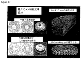
組織修復デバイスまたは足場は、3次元(X、Y、およびZ)において支柱間隔を変動させることによって形成される、メソ細孔の勾配を特徴とし得る。XおよびY次元における間隔は、例えば、100−940μmの間隔を伴う半径方向またはV形状パターンを使用して、達成され得る。Z次元における間隔は、半径方向支柱の複数の層を積層することによって、達成され得る。多孔性内部成長構造は、例えば、硫酸カルシウム等の可溶性充填剤またはキャリアで浸潤され得る。いくつかの実施形態では、多孔性内部成長構造は、例えば、リン酸カルシウムミネラルおよびI型コラーゲンタンパク質等、破骨細胞を引き付ける充填剤で浸潤され得る。いくつかの事例では、印刷された組織修復デバイスまたは足場は、約0.1−1μmの細孔サイズレベルのマイクロ/ナノ多孔性であり得る。細孔は、次いで、いくつかの事例では、可溶化コラーゲンで浸潤され得る。 The tissue repair device or scaffold may be characterized by mesopore gradients formed by varying strut spacing in three dimensions (X, Y, and Z). Spacing in the X and Y dimensions can be achieved, for example, using a radial or V-shaped pattern with a spacing of 100-940 μm. Spacing in the Z dimension can be achieved by stacking multiple layers of radial struts. The porous ingrowth structure can be infiltrated with a soluble filler or carrier such as, for example, calcium sulfate. In some embodiments, the porous ingrowth structure can be infiltrated with fillers that attract osteoclasts, such as, for example, calcium phosphate minerals and type I collagen proteins. In some cases, the printed tissue repair device or scaffold may be micro / nanoporous with a pore size level of about 0.1-1 μm. The pores can then be infiltrated with solubilized collagen in some cases.
組織修復デバイスまたは足場は、少なくとも5、10、11、12、13、14、15、18、20、25、30、35、40、50、60、70、80、90、または100ミリメートル以上の距離にわたって、骨成長を助長し、骨折、骨欠損、または骨欠損症を治療するために効果的であり得る。同様に、組織修復デバイスまたは足場は、皮質骨または皮質様骨および小柱骨または小柱様骨両方の成長を助長するために効果的であり得る。そのように成長された骨は、例えば、95%、90%、80%、75%、70%、60%、50%、40%、30%、25%、20%、10%程度の小柱骨または小柱様骨、または正反対に、すなわち、95%、90%、80%、75%、70%、60%、50%、40%、30%、25%、20%、10%程度の皮質骨または皮質様骨等、任意の好適な割合であり得る。組織修復デバイスまたは足場は、5、10、20、25、30、40、50、75、90%以上、骨欠損にわたる通常修復時間を削減または短縮するために効果的であり得る。いくつかの事例では、骨欠損は、通常要求される時間の約2分の1、3分の1、または4分の1の時間で修復され得る。多くの事例では、より大きい細孔サイズは、足場の外側部分近傍に見られ、より小さい細孔サイズは、足場の内側部分近傍に見られる。いくつかの事例では、表面積の内側半分を形成する足場の部分は、表面積の外側半分を形成する足場の部分の中間細孔直径サイズまたは面積より5、10、20、25、30、40、50、75、90%以上小さい、中間細孔直径サイズまたは面積を有し得る。いくつかの事例では、細孔サイズは、骨成長のタイプ(例えば、所望される骨密度、小柱様骨または皮質様骨)をカスタマイズするように、任意の好適なまたは望ましい構成で構造的に配列される。同様に、いくつかの事例では、組織修復デバイスまたは足場は、欠損に最適に及ぶように所望される組織または骨修復の形状をカスタマイズするように形成および成形される。さらに、いくつかの事例では、組織修復デバイスまたは足場の一部は、実質的に、中空であり得、例えば、組織修復デバイスまたは足場の内部部分の10、20、25、30、40、50、75、90%以上は、実質的に、中空であり得る。 The tissue repair device or scaffold has a distance of at least 5, 10, 11, 12, 13, 14, 15, 18, 20, 25, 30, 35, 40, 50, 60, 70, 80, 90, or 100 millimeters or more Over time, it can be effective to promote bone growth and treat fractures, bone defects, or bone defects. Similarly, tissue repair devices or scaffolds can be effective to promote the growth of both cortical or cortical bone and trabecular bone or trabecular bone. The bones so grown are, for example, 95%, 90%, 80%, 75%, 70%, 60%, 50%, 40%, 30%, 25%, 20%, 10% trabeculae Bone or trabecular bone, or diametrically, ie about 95%, 90%, 80%, 75%, 70%, 60%, 50%, 40%, 30%, 25%, 20%, 10% It can be any suitable proportion, such as cortical bone or cortical bone. The tissue repair device or scaffold may be effective to reduce or shorten the normal repair time across a bone defect, 5, 10, 20, 25, 30, 40, 50, 75, 90% or more. In some cases, the bone defect can be repaired in about one-half, one-third, or one-fourth the time normally required. In many cases, a larger pore size is found near the outer portion of the scaffold and a smaller pore size is found near the inner portion of the scaffold. In some cases, the portion of the scaffold that forms the inner half of the surface area is 5, 10, 20, 25, 30, 40, 50 greater than the intermediate pore diameter size or area of the portion of the scaffold that forms the outer half of the surface area. , 75, 90% or less, having an intermediate pore diameter size or area. In some cases, the pore size is structurally configured in any suitable or desirable configuration to customize the type of bone growth (eg, desired bone density, trabecular bone or cortical bone). Arranged. Similarly, in some cases, a tissue repair device or scaffold is formed and shaped to customize the desired tissue or bone repair shape to optimally cover the defect. Further, in some cases, a portion of the tissue repair device or scaffold may be substantially hollow, such as 10, 20, 25, 30, 40, 50, 75, 90% or more can be substantially hollow.
第2の側面では、本発明は、微多孔性シェルによって囲まれた相互接続された支柱を含む多孔性骨内部成長領域を有する、組織修復デバイスまたは足場を提供することによって、骨成長を助長する、または骨折、骨欠損、または骨欠損症を治療する方法を提供する。骨成長を助長する、または骨折、骨欠損、または骨欠損症を治療することは、骨の密度を制御する、またはそれに影響を及ぼすことを特徴とし得、あるいは骨、例えば、海綿骨または皮質骨を再建することを特徴とし得る。ほとんどの事例では、組織修復デバイスまたは足場は、骨欠損症、骨折、または骨間隙を特徴とする領域に対して生体内に提供される。微多孔性シェルは、軟組織に付着するが、その内部成長を制限するように機能し得る。組織修復デバイスまたは足場の端部には、シェルが、骨の端部間に組織修復デバイスまたは足場を安定させるためのガイドフランジとして延長され得る。組織修復デバイスまたは足場の中心は、空であり得、潜在的骨髄腔としての役割を果たし得る。多孔性内部成長構造は、例えば、硫酸カルシウム等の可溶性充填剤またはキャリアで浸潤され得る。例えば、硫酸カルシウム等のこの可溶性充填剤またはキャリアは、抗生物質、成長因子、分化因子、サイトカイン、薬物、またはこれらの作用因子の組み合わせのうちの1つ以上で浸潤され得る。組織修復デバイスまたは足場は、長骨の皮質骨端間に適合し、大部分が骨内膜表面および骨膜表面に由来する治癒骨を伝導させ得る。組織修復デバイスまたは足場は、改良された骨プレートまたは骨ねじを使用して、安定され得る。組織修復デバイスまたは足場は、3次元印刷手技によって生成され得、例えば、骨伝導性セラミックから形成され得る。 In a second aspect, the present invention promotes bone growth by providing a tissue repair device or scaffold having a porous bone ingrowth region comprising interconnected struts surrounded by a microporous shell. Or a method of treating a fracture, bone defect, or bone defect. Promoting bone growth or treating fractures, bone defects, or bone defects can be characterized by controlling or affecting bone density, or bone, eg, cancellous bone or cortical bone It can be characterized by rebuilding. In most cases, tissue repair devices or scaffolds are provided in vivo for areas characterized by bone defects, fractures, or bone gaps. The microporous shell adheres to soft tissue but can function to limit its ingrowth. At the end of the tissue repair device or scaffold, a shell can be extended as a guide flange to stabilize the tissue repair device or scaffold between the ends of the bone. The center of the tissue repair device or scaffold can be empty and can serve as a potential bone marrow cavity. The porous ingrowth structure can be infiltrated with a soluble filler or carrier such as, for example, calcium sulfate. For example, the soluble filler or carrier, such as calcium sulfate, can be infiltrated with one or more of antibiotics, growth factors, differentiation factors, cytokines, drugs, or combinations of these agents. The tissue repair device or scaffold fits between the cortical epiphyses of long bones and can conduct healed bone that is largely derived from the endosteal surface and periosteal surface. The tissue repair device or scaffold can be stabilized using an improved bone plate or bone screw. The tissue repair device or scaffold can be generated by a three-dimensional printing procedure, for example, formed from osteoconductive ceramic.
組織修復デバイスまたは足場は、多相3次元印刷組織修復デバイスであり得る。支柱は、実質的に、円筒形であり得、例えば、約1−1,000、10−900、20−800、30−700、40−600、50−500、60−400、100−350、120−300、または約200−275μmの直径であり得る。いくつかの実施形態では、支柱は、約20−940μmの直径である。いくつかの実施形態では、支柱は、骨梁の約3倍、2倍、または1.5倍以内、あるいは実質的に同一の直径である。いくつかの実施形態では、支柱は、最大100、200、300、400、500、600、700、800、900μm以上、またはさらに1.0mm以上の空間によって、縦方向に分離され得る。同様に、組織修復デバイスまたは足場は、概して、約100、75、50、30、20、10未満、またはさらに約5、4、3、2、1未満、またはさらに0.5、0.4、0.3、0.2、0.1μm未満の直径のサイズで存在し得るメソ細孔を有する多孔性であり得る。支柱は、実質的に、線形配列で配列され得る。組織修復デバイスまたは足場は、実質的に、例えば、約8、10、12、16、18、20、24週間程度、生体内に存在後、組織修復デバイスまたは足場の約5、10、15、20、25、30、35、40、45、または50%以上が吸収され得るような吸収性であり得る。組織修復デバイスまたは足場は、少なくとも約50%、60%、70%、75%、80%、85%、90%、95%、またはさらにそれ以上の多孔性であり得る。同様に、組織修復デバイスまたは足場は、約8または16週間、生体内に存在後、組織修復デバイスまたは足場の約5、10、15、20、25、30、35、40、45、または50%以上が、骨によって置換され得るように、骨成長を促進および提供するように、効率的であり得る。 The tissue repair device or scaffold can be a multiphase three-dimensional printed tissue repair device. The struts may be substantially cylindrical, for example, about 1-1000, 10-900, 20-800, 30-700, 40-600, 50-500, 60-400, 100-350, The diameter can be 120-300, or about 200-275 μm. In some embodiments, the struts are about 20-940 μm in diameter. In some embodiments, the struts are within about 3 times, 2 times, or 1.5 times, or substantially the same diameter as the trabecular bone. In some embodiments, the struts may be longitudinally separated by spaces of up to 100, 200, 300, 400, 500, 600, 700, 800, 900 μm or even 1.0 mm or more. Similarly, the tissue repair device or scaffold is generally less than about 100, 75, 50, 30, 20, 10, or even less than about 5, 4, 3, 2, 1, or even 0.5, 0.4, It can be porous with mesopores that can exist in sizes of diameters less than 0.3, 0.2, 0.1 μm. The struts can be arranged in a substantially linear array. After the tissue repair device or scaffold is substantially in the body, for example, about 8, 10, 12, 16, 18, 20, 24 weeks, about 5, 10, 15, 20 of the tissue repair device or scaffold. 25, 30, 35, 40, 45, or 50% or more can be absorbed. The tissue repair device or scaffold can be at least about 50%, 60%, 70%, 75%, 80%, 85%, 90%, 95%, or even more porous. Similarly, the tissue repair device or scaffold is about 5, 10, 15, 20, 25, 30, 35, 40, 45, or 50% of the tissue repair device or scaffold after being in vivo for about 8 or 16 weeks. The above can be efficient to promote and provide bone growth so that it can be replaced by bone.
組織修復デバイスまたは足場は、3次元(X、Y、およびZ)において支柱間隔を変動させることによって形成される、メソ細孔の勾配を特徴とし得る。XおよびY次元における間隔は、例えば、100−940μmの間隔を伴う半径方向またはV形状パターンを使用して、達成され得る。Z次元における間隔は、半径方向支柱の複数の層を積層することによって、達成され得る。多孔性内部成長構造は、例えば、硫酸カルシウム等の可溶性充填剤またはキャリアで浸潤され得る。いくつかの実施形態では、多孔性内部成長構造は、例えば、リン酸カルシウムミネラルおよびI型コラーゲンタンパク質等、破骨細胞を引き付ける充填剤で浸潤され得る。いくつかの事例では、印刷された組織修復デバイスまたは足場は、約0.1−1μmの細孔サイズレベルのマイクロ/ナノ多孔性であり得る。細孔は、次いで、いくつかの事例では、可溶化コラーゲンで浸潤され得る。 The tissue repair device or scaffold may be characterized by mesopore gradients formed by varying strut spacing in three dimensions (X, Y, and Z). Spacing in the X and Y dimensions can be achieved, for example, using a radial or V-shaped pattern with a spacing of 100-940 μm. Spacing in the Z dimension can be achieved by stacking multiple layers of radial struts. The porous ingrowth structure can be infiltrated with a soluble filler or carrier such as, for example, calcium sulfate. In some embodiments, the porous ingrowth structure can be infiltrated with fillers that attract osteoclasts, such as, for example, calcium phosphate minerals and type I collagen proteins. In some cases, the printed tissue repair device or scaffold may be micro / nanoporous with a pore size level of about 0.1-1 μm. The pores can then be infiltrated with solubilized collagen in some cases.
組織修復デバイスまたは足場は、少なくとも5、10、11、12、13、14、15、18、20、25、30、35、40、50、60、70、80、90、または100ミリメートル以上の距離にわたって、骨成長を助長し、骨折、骨欠損、または骨欠損症を治療するために効果的であり得る。同様に、組織修復デバイスまたは足場は、皮質骨または皮質様骨および小柱骨または小柱様骨の両方の成長を助長するために効果的であり得る。そのように成長された骨は、例えば、95%、90%、80%、75%、70%、60%、50%、40%、30%、25%、20%、10%程度の小柱骨または小柱様骨、または正反対に、すなわち、95%、90%、80%、75%、70%、60%、50%、40%、30%、25%、20%、10%程度の皮質骨または皮質様骨等、任意の好適な割合であり得る。組織修復デバイスまたは足場は、5、10、20、25、30、40、50、75、90%以上、骨欠損にわたる通常修復時間を削減または短縮するために効果的であり得る。いくつかの事例では、骨欠損は、通常要求される時間の約2分の1、3分の1、または4分の1の時間で修復され得る。多くの事例では、より大きい細孔サイズは、足場の外側部分近傍に見られ、およびより小さい細孔サイズは、足場の内側部分近傍に見られる。いくつかの事例では、表面積の内側半分を形成する足場の部分は、表面積の外側半分を形成する足場の部分の中間細孔直径サイズまたは面積より5、10、20、25、30、40、50、75、90%以上小さい、中間細孔直径サイズまたは面積を有し得る。いくつかの事例では、細孔サイズは、骨成長のタイプ(例えば、所望される骨密度、小柱様骨または皮質様骨)をカスタマイズするように、任意の好適なまたは望ましい構成で構造的に配列される。同様に、いくつかの事例では、組織修復デバイスまたは足場は、欠損に最適に及ぶように所望される組織または骨修復の形状をカスタマイズするように形成および成形される。 The tissue repair device or scaffold has a distance of at least 5, 10, 11, 12, 13, 14, 15, 18, 20, 25, 30, 35, 40, 50, 60, 70, 80, 90, or 100 millimeters or more Over time, it can be effective to promote bone growth and treat fractures, bone defects, or bone defects. Similarly, tissue repair devices or scaffolds can be effective to promote the growth of both cortical or cortical bone and trabecular bone or trabecular bone. The bones so grown are, for example, 95%, 90%, 80%, 75%, 70%, 60%, 50%, 40%, 30%, 25%, 20%, 10% trabeculae Bone or trabecular bone, or diametrically, ie about 95%, 90%, 80%, 75%, 70%, 60%, 50%, 40%, 30%, 25%, 20%, 10% It can be any suitable proportion, such as cortical bone or cortical bone. The tissue repair device or scaffold may be effective to reduce or shorten the normal repair time across a bone defect, 5, 10, 20, 25, 30, 40, 50, 75, 90% or more. In some cases, the bone defect can be repaired in about one-half, one-third, or one-fourth the time normally required. In many cases, a larger pore size is found near the outer portion of the scaffold and a smaller pore size is found near the inner portion of the scaffold. In some cases, the portion of the scaffold that forms the inner half of the surface area is 5, 10, 20, 25, 30, 40, 50 greater than the intermediate pore diameter size or area of the portion of the scaffold that forms the outer half of the surface area. , 75, 90% or less, having an intermediate pore diameter size or area. In some cases, the pore size is structurally configured in any suitable or desirable configuration to customize the type of bone growth (eg, desired bone density, trabecular bone or cortical bone). Arranged. Similarly, in some cases, a tissue repair device or scaffold is formed and shaped to customize the desired tissue or bone repair shape to optimally cover the defect.
第3の側面では、本発明は、微多孔性シェルによって囲まれた相互接続された支柱を含む多孔性骨内部成長領域を有する、骨成長を助長する、または骨折、骨欠損、または骨欠損症を治療するために有用な組織修復デバイスまたは足場を生成する方法を提供する。本方法は、(a)軟組織に付着するが、内部成長を制限するように機能し得る微多孔性シェルを提供することと、(b)多孔性内部成長構造を可溶性充填剤またはキャリアで浸潤することと、随意に、(c)多孔性内部成長構造を抗生物質、成長因子、分化因子、サイトカイン、薬物、またはこれらの作用因子の組み合わせのうちの1つ以上で浸潤することとを特徴とする。可溶性充填剤またはキャリアは、例えば、リン酸カルシウムミネラルおよびI型コラーゲンタンパク質等、破骨細胞を引き付ける充填剤であり得る。微多孔性シェルによって囲まれた相互接続された支柱を含む多孔性骨内部成長領域を有する、骨成長を助長する、または骨折、骨欠損、または骨欠損症を治療するために有用な組織修復デバイスまたは足場は、本発明の第1および第2の側面に関して本明細書に説明される特徴を有し得る。 In a third aspect, the present invention has a porous bone ingrowth region that includes interconnected struts surrounded by a microporous shell, promotes bone growth, or fractures, bone defects, or bone defects A method of generating a tissue repair device or scaffold useful for treating a subject is provided. The method includes (a) providing a microporous shell that adheres to soft tissue but can function to limit ingrowth; and (b) infiltrate the porous ingrowth structure with a soluble filler or carrier. And optionally (c) infiltrating the porous ingrowth structure with one or more of antibiotics, growth factors, differentiation factors, cytokines, drugs, or combinations of these agents. . Soluble fillers or carriers can be fillers that attract osteoclasts, such as, for example, calcium phosphate minerals and type I collagen proteins. A tissue repair device having a porous bone ingrowth region comprising interconnected struts surrounded by a microporous shell, useful for promoting bone growth or treating fractures, bone defects, or bone defects Or the scaffold may have the features described herein with respect to the first and second aspects of the invention.
多相3次元印刷組織修復デバイス(M3DRD)足場は、全て、深刻な短所を有し、複雑な骨欠損を修復するために必要な複雑な設計および形状で生成されることができない、現在の骨移植片技法および骨移植代替物に取って代わるために使用され得る。M3DRDは、頭蓋顔面および整形外科の骨修復のための複雑な移植用途のためにカスタム生成されることができる。 Multiphase 3D Printed Tissue Repair Device (M3DRD) scaffolds all have serious disadvantages and cannot be generated with the complex design and shape necessary to repair complex bone defects It can be used to replace graft techniques and bone graft substitutes. M3DRD can be custom generated for complex implantation applications for craniofacial and orthopedic bone repair.
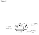
多相3次元印刷組織修復デバイス(M3DRD)は、少なくとも1つの構成要素から開始し、可能性として、3つ以上の構成要素を備えるデバイスである(図1)。主要構成要素は、(1)足場、(2)一時的充填剤/キャリア材料、および(3)充填剤/キャリア内に含まれる生体活性分子/薬物である。 A polyphase three-dimensional printed tissue repair device (M3DRD) is a device that starts with at least one component and possibly comprises three or more components (FIG. 1). The major components are (1) a scaffold, (2) a temporary filler / carrier material, and (3) a bioactive molecule / drug contained within the filler / carrier.
(足場)
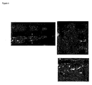
M3DRDのコアは、ロボット堆積または直接書込(DW)技術と称される3−D印刷技法を使用して生成され得る、3次元足場である(図2参照)。本技法は、コンピュータ制御された印刷プロセスおよびコロイドインクを使用し、3次元構造を形成する。これらの構造は、自己成分上に形を成すことができるか、またはトモグラフィーデータ(X線、超音波検査、またはMRI)から個々の骨欠損を充填するためにカスタム成形されることができる。
(scaffold)
The core of M3DRD is a three-dimensional scaffold that can be generated using a 3-D printing technique called robotic deposition or direct writing (DW) technology (see FIG. 2). The technique uses a computer controlled printing process and colloidal ink to form a three dimensional structure. These structures can be shaped on the self component or can be custom molded to fill individual bone defects from tomographic data (X-ray, ultrasonography, or MRI).
インク製作および印刷システム自体は、他の参考文献により詳細に説明されるが、基本的に、本システムは、印刷ノズルから流出するにつれて固体となる、水ベースの流動学的に制御されたインクを使用する。これらのインクは、コロイドインクの取り扱い特性を制御する有機化学物質を含む、水ベースのスラリー中の細かく制御されたセラミック粒子から成る。これは、支持されていない構造要素の最小垂れの有無にかかわらず、3−D格子状構造が重ねて印刷されることを可能にする。 The ink production and printing system itself is described in detail in other references, but basically the system uses a water-based rheologically controlled ink that becomes solid as it exits the printing nozzle. use. These inks consist of finely controlled ceramic particles in a water-based slurry containing organic chemicals that control the handling properties of the colloidal ink. This allows the 3-D grid structure to be printed over, with or without minimal sagging of unsupported structural elements.
本システムを使用する場合、第1の層の要素は、x−y−z制御ガントリシステムのxおよびy座標制御システムを使用して、小(約50−400μm直径)ノズルを通して、支持プレート上にインクを押しつけることによって、印刷され得る。次いで、z制御システムは、1ノズル直径未満だけ、ノズルを若干上方に移動させるために使用される。次いで、次の層が、第1の層を覆って印刷される。これは、3−D構造全体が終了するまで、層毎に継続される。 When using this system, the elements of the first layer are passed through a small (about 50-400 μm diameter) nozzle onto the support plate using the x and y coordinate control system of the xyz control gantry system. It can be printed by pressing the ink. The z control system is then used to move the nozzle slightly upward by less than one nozzle diameter. The next layer is then printed over the first layer. This continues for each layer until the entire 3-D structure is completed.
構造全体は、乾燥を防止するために、油浴中で印刷され得る。システムは、最大3つの材料が単一構造を印刷するために使用され得るように、3つのノズルおよびインクリザーバを有し得る。褪せやすいインク、すなわち、焼成の間に完全燃焼する材料から全体的になるインクもまた、印刷プロセスの一部として使用され得る。これらは、一時的支持を要求する複雑な部品のための支持構造を印刷するために使用されることができる。 The entire structure can be printed in an oil bath to prevent drying. The system can have three nozzles and an ink reservoir so that up to three materials can be used to print a single structure. Skinny inks, i.e., inks that consist entirely of materials that burn completely during firing, can also be used as part of the printing process. They can be used to print support structures for complex parts that require temporary support.
結果として生じる構造は、次いで、油浴から除去され、乾燥され、プログラム可能な炉内で焼成され、最終セラミック構造を生成する。焼成は、現在、約1100℃で約4時間行われ、これは、実質的に、有機成分を完全燃焼し、セラミック粒子を立体構造へと一緒に焼結させる。これは、精密かつ予測可能な構造を生成するために、印刷プロセスの中に計算されることができる少量の予測可能な収縮を生じさせ得る。 The resulting structure is then removed from the oil bath, dried and fired in a programmable furnace to produce the final ceramic structure. Firing currently takes place at about 1100 ° C. for about 4 hours, which substantially burns the organic components completely and sinters the ceramic particles together into a three-dimensional structure. This can result in a small amount of predictable shrinkage that can be calculated during the printing process to produce a precise and predictable structure.
印刷ノズルは、通常、円筒形であり、円筒形ロッド状の印刷された構造を生成し得る。しかしながら、非円筒形構造を生成するように成形されたノズルが、作製され得、該構造は、我々の先行表面改良特許(米国特許第6,419,491号参照)に基づいて、細胞移動、成長、および分化を制御するために設計されたサイズの表面線条を伴う。 The printing nozzle is typically cylindrical and can produce a printed structure in the form of a cylindrical rod. However, a nozzle shaped to produce a non-cylindrical structure can be made, which is based on our prior surface improvement patent (see US Pat. No. 6,419,491), cell migration, With surface striations of a size designed to control growth and differentiation.
(組成物)
リン酸カルシウムベースの足場が、恒久的、再建可能(骨再建プロセスを通して)、または可溶性の材料、またはこれらのいくつかの組み合わせに基づくインクから作製された。この時点におけるいくつかの有望材料は、ヒドロキシアパタイト(HA)セラミック、リン酸三カルシウムセラミック(TCP)、および2つの材料の組み合わせを有する二相性セラミック(HA/TCP)である。HA材料は、恒久的または非常に長持ちする足場(焼成温度に応じて)を生成し、HA/TCPの組み合わせは、長持ちする足場を生成する高HAパーセンテージを伴って変動し得、約99%TCP/1%HA足場は、破骨細胞活性を通して有意に再建することが示されている足場を生成するために使用されている。いくつかのそのような足場は、円盤の異なる領域内に可変細孔構造を伴う、約3mm厚の11mm直径多孔性円盤と、約12mm直径の約0.5mm厚の中実キャップ構造とを含む。これらは、骨および軟組織応答を試験するために、ウサギの頭頂骨(頭蓋骨)内の11mm直径トレフィン孔内に挿入された。これらの足場は、線維組織浸潤を抑制するための中実シェル構成要素と、270μm直径要素(この直径は、ノズルサイズを使用して変動されることができる)および100μm未満から1000μmの最大寸法のサイズの細孔(メソ細孔)サイズを伴う内部格子構造との組み合わせを有するように、効果的に生成され得ることが実証された。ミクロンスケールを上回り、かつミリメートルスケールを下回る、細孔および支柱サイズを伴う、これらの構造体は、メソ構造と称される。HAおよびTCP組成物による格子構造は、足場内への新しい骨の骨伝導(osteoconduction)を助長する。小有機粒子をインクに添加することによって、微多孔性(サブミクロンから約20μm細孔サイズ)足場構成要素もまた、生成されることができる。これらは、線維接続組織に付着するように設計されることができる。中実層、種々のサイズの目の粗いメソ細孔格子、マイクロ構造格子要素、および微多孔性格子要素のこれらの組み合わせを使用することによって、複雑な構造が、骨、骨髄組織、線維組織、および血管の内部成長および形成を伝導させる(conduct)ために設計および製作されることができる。長骨再生のための足場の実施例は、図1に示される。DWシステムは、足場内に2つ以上の材料を印刷することができるので、恒久的HA構成要素ならびに再建可能TCP要素を伴う足場を印刷するために適している。これは、足場の長期的強度が必要な整形外科用途において適用可能であり得る。
(Composition)
Calcium phosphate based scaffolds were made from inks based on permanent, reconstructable (through the bone reconstruction process), or soluble materials, or some combination thereof. Some promising materials at this point are hydroxyapatite (HA) ceramics, tricalcium phosphate ceramics (TCP), and biphasic ceramics (HA / TCP) with a combination of the two materials. The HA material produces a permanent or very long lasting scaffold (depending on the firing temperature), and the HA / TCP combination can vary with a high HA percentage producing a long lasting scaffold, about 99% TCP / 1% HA scaffolds have been used to generate scaffolds that have been shown to significantly reconstruct through osteoclast activity. Some such scaffolds include an 11 mm diameter porous disc about 3 mm thick with a variable pore structure in different regions of the disc and a solid cap structure about 0.5 mm thick about 12 mm diameter. . They were inserted into 11 mm diameter trephine holes in the rabbit parietal bone (cranium) to test bone and soft tissue responses. These scaffolds have a solid shell component to inhibit fibrous tissue infiltration, a 270 μm diameter element (this diameter can be varied using the nozzle size) and a maximum dimension of less than 100 μm to 1000 μm It has been demonstrated that it can be effectively generated to have a combination with an internal lattice structure with a size pore (mesopore) size. These structures with pore and strut sizes above the micron scale and below the millimeter scale are referred to as mesostructures. The lattice structure with the HA and TCP composition facilitates new bone osteoconduction into the scaffold. By adding small organic particles to the ink, microporous (submicron to about 20 μm pore size) scaffold components can also be produced. They can be designed to adhere to fiber connective tissue. By using these combinations of solid layers, open mesh mesopore lattices of various sizes, microstructured lattice elements, and microporous lattice elements, complex structures can be transformed into bone, bone marrow tissue, fibrous tissue, And can be designed and fabricated to conduct vessel ingrowth and formation. An example of a scaffold for long bone regeneration is shown in FIG. The DW system is suitable for printing scaffolds with permanent HA components as well as reconfigurable TCP elements because more than one material can be printed in the scaffold. This may be applicable in orthopedic applications where long-term strength of the scaffold is required.
(足場充填剤/キャリア材料および生体活性因子)
この充填剤/キャリア成分は、足場に浸潤し、中実またはほぼ中実(充填剤が微多孔性である場合)複合構造を生成するために使用され得る、セメント、ポリマー、または有機/天然ヒドロゲルベースの材料を有する。この充填剤/キャリア材料は、ある既知のまたは制御率において、可溶性であり、足場により大きな初期力学的強度および安定性を提供し、次いで、分解し、骨または軟組織内部成長を可能および/または刺激し得る(用途および設計に応じて)。充填剤/キャリアは、足場の外側からその中心へと内側に分解し、足場構成要素が暴露されるにつれて、および組織および血管が周囲組織から成長するにつれて、複合材が多孔性となることを可能にし得る。この構成要素はまた、足場の内部部分が、早期治癒の中に通常そこに形成され得る血栓を形成することを防止し得る。この血栓は、口腔および頭蓋顔面部位に感染し得、これらの部位は、多くの場合、非滅菌性である、あるいは肉芽/線維組織または壊死となり、いずれも、骨内部成長を妨害し得る。充填剤/キャリア材料は、本質的に、組織形成を刺激し得るか、あるいは、それは、組み込まれた薬物、成長因子、サイトカイン、または抗生物質を含み得る。
(Scaffolding filler / carrier material and bioactive factor)
This filler / carrier component infiltrates the scaffold and can be used to produce a solid or nearly solid (if the filler is microporous) composite structure, cement, polymer, or organic / natural hydrogel Having a base material. This filler / carrier material is soluble at some known or controlled rate and provides greater initial mechanical strength and stability to the scaffold and then disintegrates to enable and / or stimulate bone or soft tissue ingrowth Yes (depending on application and design). Filler / carrier breaks inward from the outside of the scaffold to its center, allowing the composite to become porous as the scaffold components are exposed and as tissues and blood vessels grow from the surrounding tissue Can be. This component may also prevent the internal portion of the scaffold from forming a thrombus that may normally form there during early healing. This thrombus can infect oral and craniofacial sites, which are often non-sterile or become granulation / fibrous tissue or necrosis, both of which can interfere with bone ingrowth. The filler / carrier material can essentially stimulate tissue formation or it can include incorporated drugs, growth factors, cytokines, or antibiotics.
いくつかの例示的充填剤/キャリア材料は、コーティングまたはヒドロゲル充填剤として使用され得る、硫酸カルシウム(焼き石こう)、徐放性硫酸カルシウム(低速溶解バージョンの硫酸カルシウム)、およびキトサン、キチンの誘導体、生物学的に誘導された多糖類である。ポリ(L−乳酸)(PLLA)のような吸収性ポリマー等の他の材料も、充填剤/キャリア材料として使用され得るが、代替として、これらは、充填剤ではなく、足場のためのコーティング材料として使用され得る。したがって、これらは、依然として、足場を強化し、分離材料として作用することができるが、足場を充填し、それを中実構造にするために利用されないであろう。 Some exemplary filler / carrier materials are calcium sulfate (baked gypsum), sustained release calcium sulfate (slow dissolving version of calcium sulfate), and chitosan, derivatives of chitin, which can be used as coating or hydrogel fillers, It is a biologically derived polysaccharide. Other materials such as absorbent polymers such as poly (L-lactic acid) (PLLA) may also be used as filler / carrier materials, but alternatively they are not fillers but are coating materials for scaffolds Can be used as Thus, they can still strengthen the scaffold and act as a separation material, but will not be utilized to fill the scaffold and make it a solid structure.
硫酸カルシウムは、充填剤および薬物キャリア材料として使用され、構造の力学的特性を向上させ、予測可能な方法において、生物学的に活性作用因子を放出し、かつ骨形成に干渉しないことが見出された。このキャリアを使用して調査された生体活性分子は、組み換え型血小板由来成長因子(PDGF)および骨形成タンパク質(BMP)を含む。 Calcium sulfate is used as a filler and drug carrier material to improve the mechanical properties of the structure, in a predictable manner, release biologically active agents and does not interfere with bone formation It was done. Bioactive molecules investigated using this carrier include recombinant platelet derived growth factor (PDGF) and bone morphogenetic protein (BMP).
(足場メソ構造を使用した足場の力学的特性、骨特性、および足場再建の制御)
頭蓋顔面骨修復において使用するために好適な力学的特性を伴い、いくつかの外部支持を伴って、整形外科修復のために適切である、足場を設計および生成することが可能である。足場メソ構造はまた、足場内に伝導される骨の構造特性および密度を制御するために使用され得る。モデルとして、ウサギの11mm直径トレフィン欠損を使用することによって、3つの異なる設計足場が、欠損を充填し、骨再生を検査するために生成された。全ての足場は、同一の材料、すなわち、99%TCP1%HAセラミックから生成され、270μmの直径の同一サイズの印刷された支柱から作製された。全ての足場はまた、医療グレードの硫酸カルシウムで充填され、中実構造として開始された。メソ構造は、足場の層内の支柱間隔を使用し(xおよびy方向)、z方向に支柱を積層することによって変動された。250×250μm、250×400μm、および400×400μmサイズ細孔と称される(xおよびy方向)開細孔を生成する3つの支柱間隔を含む、あるタイプの足場が、生成された(これらの寸法は、適切である)。「Z」間隔は、若干、1つの支柱の高さ、すなわち、230μmを下回った。マイクロコンピュータ断層撮影によって測定されるように、これらの3つのゾーンは、46、56、および70%の足場体積パーセンテージを有していた。
(Control of scaffold mechanical properties, bone properties, and scaffold reconstruction using scaffold mesostructures)
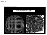
It is possible to design and generate a scaffold with suitable mechanical properties for use in craniofacial bone repair and with some external support that is suitable for orthopedic repair. The scaffold mesostructure can also be used to control the structural properties and density of the bone conducted into the scaffold. By using a rabbit 11 mm diameter trephine defect as a model, three different design scaffolds were generated to fill the defect and to examine bone regeneration. All scaffolds were made of the same material, ie, 99% TCP 1% HA ceramic, made from the same size printed struts with a diameter of 270 μm. All scaffolds were also filled with medical grade calcium sulfate and started as a solid structure. The mesostructure was varied by using strut spacing in the scaffold layer (x and y directions) and stacking struts in the z direction. A type of scaffold was created that included three strut spacings that create open pores (x and y directions) referred to as 250 × 250 μm, 250 × 400 μm, and 400 × 400 μm size pores (these Dimensions are appropriate). The “Z” spacing was slightly below the height of one strut, ie 230 μm. These three zones had scaffold volume percentages of 46, 56, and 70% as measured by microcomputed tomography.
2つの足場が、異なる間隔の同心リングと交互する半径方向支柱を使用して生成された持続的可変多孔率を有するように生成された。一方の足場は、1zおよび2z間隔の層と、55〜94%の足場体積を伴うリング形状領域とを有していた。他方の足場は、3z間隔と、41〜56%体積の領域とを有していた。したがって、41〜94%足場に及ぶ足場体積の範囲が、試験された。全足場において、骨は、8週間までに、欠損の中心まで一貫して成長可能であった(5.5mm距離にわたる)。 Two scaffolds were created with sustained variable porosity created using differently spaced concentric rings and alternating radial struts. One scaffold had 1z and 2z spaced layers and a ring-shaped region with a scaffold volume of 55-94%. The other scaffold had a 3z spacing and an area of 41-56% volume. Accordingly, a range of scaffold volumes ranging from 41 to 94% scaffold was tested. In all scaffolds, bone was able to grow consistently to the center of the defect (over a 5.5 mm distance) by 8 weeks.
この程度の一貫した骨浸潤は、他の骨伝導性(osteoconductive)足場では観察されず、これは、足場内の足場要素のサイズおよび編成によるものである。内部成長を行うために、骨梁のサイズ範囲内に多くの小支柱を使用することによって、かつ欠損にわたって骨を直線に伝導させるようにそれらを編成することによって、骨伝導のプロセスを最適化することが可能である。「有向骨伝導(directed osteoconduction)」と称される本プロセスは、このタイプの足場に対して新規である。ランダム細孔編成を伴う足場では、有向骨伝導のプロセスは、観察されず、大欠損にわたる一貫した成長は、生じるのにより長い時間がかかる。本明細書に説明される構造では、8および16週間における骨体積は、9〜40%(8週間)および10〜56%(16週間)に及んでいた。骨体積は、足場体積に反比例した。より開放した(より小さい足場体積)足場は、より大きな骨内部成長を示し、骨は、経時的に増加した。足場再建は、5%〜56%に及び、より多くの再建が、後により開放した足場において観察された。より大量の足場(より小さい細孔を伴う)は、よりコンパクトな層状構造骨を生成し、足場および骨の組み合わせは、ほとんど軟組織を示さず、皮質様構造に類似していた。対照的に、より少量の足場(より大きい細孔を伴う)は、より多孔性の無秩序な骨を生成し、骨および足場の組み合わせは、海綿骨に類似していた。足場に隣接する骨のタイプ(皮質骨または海綿骨)は、少なくとも部分的に、隣接する足場内に成長する骨に影響を及ぼした。 This degree of consistent bone infiltration is not observed with other osteoconductive scaffolds, which is due to the size and organization of the scaffold elements within the scaffold. To perform ingrowth, optimize the bone conduction process by using many small struts within the trabecular size range and knitting them to conduct the bone straight across the defect It is possible. This process, referred to as “directed osteoconduction” is novel for this type of scaffold. In scaffolds with random pore organization, the process of directed bone conduction is not observed and consistent growth over large defects takes longer to occur. In the structures described herein, bone volume at 8 and 16 weeks ranged from 9-40% (8 weeks) and 10-56% (16 weeks). Bone volume was inversely proportional to scaffold volume. The more open (smaller scaffold volume) scaffolds showed greater bone ingrowth and the bone increased over time. Scaffold reconstructions ranged from 5% to 56%, with more reconstructions observed later in the more open scaffolds. Larger scaffolds (with smaller pores) produced more compact layered structural bones, and the scaffold and bone combination showed little soft tissue and resembled cortical-like structures. In contrast, a smaller amount of scaffold (with larger pores) produced more porous and disordered bone and the bone and scaffold combination was similar to cancellous bone. The type of bone adjacent to the scaffold (cortical bone or cancellous bone) affected, at least in part, bone growing in the adjacent scaffold.
(M3DRD足場の特徴)
総括すると、このデータは、設計されたメソ構造を伴う骨伝導性足場が、広範囲の骨修復用途に好適な力学的特性とともに作製され得ることを示す。これらの足場は、骨細胞または幹細胞増強の必要なく、有意な距離にわたって、骨を再生するために使用されることができる。大欠損にわたって観察された骨伝導率は、広い距離にわたって、効率的に骨を伝導させるように、直線アレイに編成される骨梁のサイズ範囲内の多くの小支柱の使用に基づく「有向骨伝導」によるものである。
(Features of M3DRD scaffold)
Taken together, this data shows that osteoconductive scaffolds with designed mesostructures can be made with mechanical properties suitable for a wide range of bone repair applications. These scaffolds can be used to regenerate bone over a significant distance without the need for bone cell or stem cell augmentation. The bone conductivity observed over large defects is based on the use of many small struts within the trabecular size range organized in a linear array to efficiently conduct bone over large distances. This is due to “conduction”.
足場はまた、結果として生じる骨密度、構造、および足場再建率を制御するために使用されることができる。M3DRD足場は、隣接する骨に微細構造的に近似または一致する骨を再生するように設計されることができる。すなわち、海綿骨が必要とされる場合、海綿骨構造を再生することが可能であり、皮質骨が必要とされる場合、その形状を再生することも可能である。中実キャップ層のような追加の特徴は、軟組織内部成長を上手く防止し得る。CS充填剤は、一時的に、構造力学特性を向上させ、骨形成を妨害せず、感染症による線維組織内部成長および浸潤を防止し、血管新生を継続可能にし得る。 The scaffold can also be used to control the resulting bone density, structure, and scaffold rebuild rate. M3DRD scaffolds can be designed to regenerate bone that is microstructurally similar or coincident with adjacent bone. That is, when cancellous bone is required, the cancellous bone structure can be regenerated, and when cortical bone is required, its shape can be regenerated. Additional features, such as a solid cap layer, can successfully prevent soft tissue ingrowth. CS fillers may temporarily improve structural mechanical properties, do not interfere with bone formation, prevent fibrous tissue ingrowth and infiltration due to infection, and allow vascularization to continue.
CSはまた、生体活性分子の制御された放出のために使用されることができる。DW印刷システムの使用は、ミクロンスケールの精度で、複雑なメソ構造のカスタム設計および印刷を可能にする。これは、既製品印刷構造ならびにMRIまたはCTデータに基づく患者内の複雑な欠損を修復するためのカスタム印刷M3DRD足場の両方を可能にする。この技術は、頭蓋顔面および整形外科骨修復/置換分野における広範な用途を有する。 CS can also be used for controlled release of bioactive molecules. The use of a DW printing system allows for custom design and printing of complex mesostructures with micron scale accuracy. This allows for both off-the-shelf printed structures as well as custom printed M3DRD scaffolds to repair complex defects in patients based on MRI or CT data. This technology has a wide range of applications in the craniofacial and orthopedic bone repair / replacement fields.
(例示的組織修復デバイスまたは足場)
骨欠損は、現在、複雑な自家移植手技あるいは具体的部位に設計されていない不完全同種異系または人工治療によって充填される。直接書込(DW)製作は、複雑な骨欠損を修復するためにカスタム製作される潜在性を有する、骨伝導性生体材料である、複雑な多成分二相性(COMBI)リン酸カルシウム足場から成る、3−D足場を印刷することを可能にする。現在の文献は、依然として、骨再生のための最適および閾値細孔要件について討論している。我々は、臨界サイズの(そのままでは閉鎖不可能である)生体内モデルにおいて足場を試験し、骨密度、内部成長の程度、および骨/足場再建に及ぼす影響を研究した。
(Example tissue repair device or scaffold)
Bone defects are currently filled by complex autograft procedures or incomplete allogeneic or artificial treatments that are not designed for specific sites. Direct writing (DW) fabrication consists of a complex multicomponent biphasic (COMBI) calcium phosphate scaffold, an osteoconductive biomaterial with the potential to be custom fabricated to repair complex bone defects. -It is possible to print the D scaffold. The current literature still discusses optimal and threshold pore requirements for bone regeneration. We tested the scaffolds in an in vivo model of critical size (which cannot be closed as is) to study the effects on bone density, degree of ingrowth, and bone / scaffold reconstruction.
足場は、全(X、Y、およびZ)平面に可変メソ細孔間隔を伴って設計された。細孔サイズを変動させるために、z高さにおける1、2、または3つの重複層の半径方向支柱と交互する、2つの同心円の層の足場設計が、DWによって、15:85HAP/β−TCPから製作され、1100℃で焼結された。硫酸カルシウムの一時的充填剤は、軟組織侵入および/または感染症を防止した。足場は、生体内のトレフィン欠損内に埋め込まれた。8−16週間後、骨内部成長および足場および骨再建の分析が、マイクロCT(Scanco医療)によって定量化され、足場は、ポリメタクリル酸メチル(PMMA)中に埋め込まれ、次いで、光学顕微鏡を用いて組織学的に評価された。 The scaffold was designed with variable mesopore spacing in all (X, Y, and Z) planes. In order to vary the pore size, two concentric layer scaffold designs alternating with 1, 2, or 3 overlapping layer radial struts at z-height have been developed by DW, 15:85 HAP / β-TCP. And sintered at 1100 ° C. A temporary filler of calcium sulfate prevented soft tissue invasion and / or infection. The scaffold was embedded in a trephine defect in vivo. After 8-16 weeks, bone ingrowth and scaffold and bone reconstruction analysis were quantified by micro CT (Scanco Medical), the scaffolds were embedded in polymethyl methacrylate (PMMA) and then using light microscopy And histologically evaluated.
足場体積は、リング区画によって変動するように設計された。骨体積は、より開放し、すなわち、より少ない足場密度領域において大きかった。細孔は、約100〜940ミクロンに及んだ。骨は、全ての様々な高さの層内に成長したが、最大細孔サイズに到達するのはより時間がかかることが認められた。500ミクロンより大きい細孔も、依然として、以前の文献の見解とは対照的に、良好に骨で充填された。 The scaffold volume was designed to vary with the ring section. The bone volume was more open, i.e. larger in the area of less scaffold density. The pores ranged from about 100 to 940 microns. Bone grew in all different height layers, but it was found that reaching the maximum pore size took more time. Pore larger than 500 microns was still well filled with bone, in contrast to previous literature views.
使用された特定の足場は、3次元印刷されたリン酸カルシウム足場が、8週間以内に、少なくとも11mm骨間隙にわたって、骨を成長させることが可能であることを実証した。骨は、940μm程度の大きい細孔から20μm程度の小さい細孔内にまで成長することができる。骨形態は、足場設計に応じて、小柱骨状または皮質骨状であることができる。足場は、広範囲の臨床用途のための局所的に異なる生物学的および力学的特性とともに設計され得る。 The particular scaffold used has demonstrated that a three-dimensional printed calcium phosphate scaffold can grow bone over at least 11 mm bone gap within 8 weeks. Bone can grow from large pores on the order of 940 μm to small pores on the order of 20 μm. The bone morphology can be trabecular or cortical, depending on the scaffold design. Scaffolds can be designed with locally different biological and mechanical properties for a wide range of clinical applications.
(実施例)
(実施例1)
(材料および方法)
2つの足場構造、すなわち、小細孔(SP)および大細孔(LP)が、細孔幾何学形状の多様性を増加させるように設計された。両足場は、片面に層状平行支柱の中実キャップを含み、それは、生物学的に、頭皮からの軟組織内部成長を阻止するための障壁としての役割を果たすが、構造的に、Z方向における足場格子の印刷のためのベースとしての役割を果たした。このベース上に構築された足場設計は、SPおよびLP足場間で異なるが、一般に、1つ以上の半径方向(R)層と交互する、ネスト化された同心円(CC)の層から成った。Z方向における多孔率の変動は、半径方向層の1、2、または3スタックの使用に由来し、XおよびY方向における多孔率は、同一の層内の半径方向支柱間の間隔から生じた。SPおよびLP足場の具体的設計は、図4および5に図示されている。
(Example)
Example 1
(Materials and methods)
Two scaffold structures, namely small pore (SP) and large pore (LP), were designed to increase the diversity of pore geometry. Both scaffolds include a solid cap with layered parallel struts on one side, which biologically serves as a barrier to prevent soft tissue ingrowth from the scalp, but structurally a scaffold in the Z direction. Served as a base for grid printing. Scaffold designs built on this base differed between SP and LP scaffolds, but generally consisted of nested concentric (CC) layers alternating with one or more radial (R) layers. The porosity variation in the Z direction resulted from the use of 1, 2, or 3 stacks of radial layers, and the porosity in the X and Y directions resulted from the spacing between the radial struts in the same layer. Specific designs of SP and LP scaffolds are illustrated in FIGS. 4 and 5.
足場は、15:85HA/β−TCPのインクを用いて印刷され、1100℃で焼成された。足場は、次いで、マイクロCT(Scanco Medical)において、中分解能でスキャンされ、埋込前の支柱およびメソ細孔の体積を評価した。足場は、次いで、CSで充填され、キャップ上方の1mmリングの足場が、歯科用ドリルで除去され、周縁における阻止された半径方向支柱を開放した。これによって、直径11mmが残された。 The scaffold was printed using 15:85 HA / β-TCP ink and fired at 1100 ° C. The scaffold was then scanned at medium resolution in a micro CT (Scanco Medical) to assess the volume of the struts and mesopores prior to implantation. The scaffold was then filled with CS and the 1 mm ring scaffold above the cap was removed with a dental drill to release the blocked radial struts at the periphery. This left a 11 mm diameter.
(外科手術手技)
外科手術が、Institutional Animal Care and Use Committee(IACUC)承認プロトコルに従って、8匹の成獣のニュージーランドホワイトウサギにおいて行われた。3.0cm正中矢状切開が、頭部背面の皮膚および軟組織を通して作られた。左右対称11.0mm直径トレフィン欠損が、硬膜病変を伴わずに、頭頂骨内の頭蓋幹線のすぐ後方に空けられた。
(Surgery technique)
Surgery was performed on 8 adult New Zealand white rabbits according to the Institutional Animal Care and Use Committee (IACUC) approved protocol. A 3.0 cm midline sagittal incision was made through the skin and soft tissue on the back of the head. A symmetric 11.0 mm diameter trephine defect was drilled just behind the cranial trunk in the parietal bone without dural lesions.
それぞれ、右および左欠損内へのLPおよびSP足場の設置後、軟組織および皮膚は、4−0吸収性縫合糸を用いて閉鎖された。創傷は、滅菌生理食塩水で清浄され、トリプル抗生物質軟膏で処置された。感染症、疼痛、または他の合併症の徴候は、もたらされなかった。7匹が、8週間後、1匹が、16週間後、安楽死させられた。 After placement of the LP and SP scaffolds in the right and left defects, respectively, soft tissue and skin were closed using 4-0 absorbable sutures. The wound was cleaned with sterile saline and treated with triple antibiotic ointment. No signs of infection, pain, or other complications were produced. Seven were euthanized after 8 weeks and 1 after 16 weeks.
(試料分析)
切除されたインプラントが、周囲組織とともに、70%エタノール中に固定され、マイクロCTにおいて、中分解能でスキャンされた。各試料内の着目体積は、固有のメソ細孔体積の各リングによって占有された体積であった(図4および5参照)。全試料の外側体積リングは、骨が非充填欠損内にあるこの空間を充填することが分かっているため、測定されなかった(Sohn他、J Periodontal Implant Sci 2010;40:180−187)。各SP足場は、6つの固有のサイズのメソ細孔リング、すなわち、各1Zおよび2Z場所に対して3つの同心リング体積を含んでいた。各LP足場は、3Z場所内の同心リングによって形成される、2つの固有のサイズのメソ細孔体積を含んでいた。メソ細孔の固有のリング毎に、骨、足場、および軟組織/空間に対する体積パーセンテージが、2つの閾値、すなわち、足場(S)および足場+骨(SB)を設定することによって取得された。骨内部成長のパーセンテージは、SBからSを減算することによって測定され、軟組織/空間のパーセンテージは、100%からSBを減算することによって測定された。足場吸収率は、埋込前に測定された足場体積からSを減算することによって測定された(図7、9、10参照)。
(Sample analysis)
Excised implants were fixed in 70% ethanol along with the surrounding tissue and scanned at medium resolution in micro CT. The volume of interest in each sample was the volume occupied by each ring of unique mesopore volume (see FIGS. 4 and 5). The outer volume ring of all samples was not measured because it was known that the bone fills this space in an unfilled defect (Sohn et al., J Periodontal Implant Sci 2010; 40: 180-187). Each SP scaffold contained six uniquely sized mesopore rings, ie three concentric ring volumes for each 1Z and 2Z location. Each LP scaffold contained two uniquely sized mesopore volumes formed by concentric rings in the 3Z location. For each unique ring of mesopores, volume percentages for bone, scaffold, and soft tissue / space were obtained by setting two thresholds: scaffold (S) and scaffold + bone (SB). The percentage of bone ingrowth was measured by subtracting S from SB, and the percentage of soft tissue / space was measured by subtracting SB from 100%. The scaffold absorption rate was measured by subtracting S from the scaffold volume measured before implantation (see FIGS. 7, 9, and 10).
試料は、次いで、組織学のために、メタクリル酸メチル中に埋め込まれ、80μm厚のスライスが、各試料のR層を通る特定の点で調製された。SP足場毎に、水平スライス(キャップに平行)が、1Zおよび2つの2Zメソ細孔区画を通して得られた。LP足場毎に、水平スライスが、2つの3Zメソ細孔を通して得られた。さらに、垂直スライス(キャップに垂直)が、全足場の中心を通して得られた。全スライスは、染色され、デジタル的に撮影された(図6、8参照)。 Samples were then embedded in methyl methacrylate for histology, and 80 μm thick slices were prepared at specific points through the R layer of each sample. For each SP scaffold, horizontal slices (parallel to the cap) were obtained through the 1Z and two 2Z mesopore compartments. For each LP scaffold, a horizontal slice was obtained through two 3Z mesopores. In addition, a vertical slice (perpendicular to the cap) was obtained through the center of the entire scaffold. All slices were stained and photographed digitally (see FIGS. 6 and 8).
他の試料は、後方散乱電子撮像(BEI)およびEDAX評価を用いた走査型電子顕微鏡(SEM)(Hitachi)を使用して検査され、無機物組成物を分析した。以前の研究(Simon他、J Biomed Mater Res 2008; 85A:371−377)に基づいて、ここで使用された試料サイズは、0.05未満のp値で群間の15−20%差を決定するための統計的検出力を有していた。 Other samples were examined using scanning electron microscopy (SEM) (Hitachi) with backscattered electron imaging (BEI) and EDAX evaluation to analyze the inorganic composition. Based on previous studies (Simon et al., J Biomed Mater Res 2008; 85A: 371-377), the sample size used here determined a 15-20% difference between groups with a p-value less than 0.05. Had statistical power to do.
(結果)
マイクロCTおよび組織学的分析によって、全次元のメソ細孔内への骨内部成長が明らかになった。より高い骨のパーセンテージは、SPおよびLP足場の両方の周縁におけるより大きい細孔内で見られた。一般に、より高い足場支柱吸収率は、より高い骨内部成長率を伴う領域内で生じた。それにもかかわらず、SP足場では、より骨成長が少ないより小さい体積を含む、2Zメソ細孔の内側リングにおいて、支柱吸収率は、より高かった。これらのより閉鎖した領域の組織学的結果は、骨が、それらの間ではなく、直接、支柱上に成長したことを示した。1Zメソ細孔では、骨は、周縁から内向きに成長を見せなかったが、2Zメソ細孔からのCCリングの間から成長した。
(result)
Micro CT and histological analysis revealed bone ingrowth into all dimensions of mesopores. A higher bone percentage was seen in the larger pores at the periphery of both the SP and LP scaffolds. In general, higher scaffold strut resorption rates occurred in areas with higher bone ingrowth rates. Nevertheless, in the SP scaffold, the strut resorption rate was higher in the inner ring of 2Z mesopores, including smaller volumes with less bone growth. Histological results of these more closed areas indicated that the bone grew directly on the struts, not between them. In the 1Z mesopore, the bone did not grow inward from the periphery, but grew from between the CC rings from the 2Z mesopore.
CS充填剤は、骨が充填されるにつれて吸収することが認められたが、しかしながら、組織学画像は、島状の沈殿物が残っていることを示し、SEM、BEI、およびEDAXは、それがリン酸カルシウム(CaP)であることを示した。これは、他の研究にも報告されている。ほとんどの動物において、CaPは、骨成長を伝導させ、骨形成と融合することが認められたが、他の動物では、非常に高密度のCaP沈殿物の面積が、骨形成を阻止することが認められた。 CS filler was observed to absorb as the bone was filled, however, histology images showed that island deposits remained, and SEM, BEI, and EDAX It was shown to be calcium phosphate (CaP). This has been reported in other studies. In most animals, CaP has been found to conduct bone growth and fuse with bone formation, whereas in other animals, a very dense area of CaP precipitate can prevent bone formation. Admitted.
(考察)
TCPから作製され、CSで充填された可変メソ細孔足場は、皮質骨および小柱骨に類似する骨構造を再成長させるための足場の設計を可能にする。組織学の分析は、依然として、継続中であることに留意することが重要である。CaP沈殿物の大量の蓄積は、CaSが吸収されるにつれて、緊密なメソ細孔空間内に蓄積した飽和溶液Ca2+およびPO4 3−の結果であると考えられる。CaSを分解する酸性の性質はまた、周囲の足場支柱のTCPからのCa2+およびPO4 3−の放出を増加させ得る。
(Discussion)
A variable mesoporous scaffold made from TCP and filled with CS allows the design of a scaffold to regrow bone structures similar to cortical and trabecular bone. It is important to note that histological analysis is still ongoing. The massive accumulation of CaP precipitate is believed to be the result of saturated solutions Ca 2+ and PO 4 3− that have accumulated in the tight mesopore space as CaS is absorbed. The acidic nature of degrading CaS can also increase the release of Ca 2+ and PO 4 3− from the surrounding scaffold strut TCP.
8週間後のマイクロCT結果は、約68−99%の足場+骨(SB)のパーセンテージの勾配および4−14%の足場吸収率の勾配を示す。最も驚くべきことに、足場吸収率を14%近く最大化したメソ細孔体積は、体積スペクトルの両端近傍であり、より大きい方は、428−636μm×616μmであり、より小さい方は、188−253μm×410μであった。これらの2つの体積のうち、より大きい方は、67.56%(40.19%B+27.37%S)のSBをもたらし、より小さい方は、92.93%。(27.25%B+65.68%S)のSBをもたらし、それぞれ、小柱骨および皮質骨の骨パーセンテージに非常に類似していた。より小さいメソ細孔は、あまり骨を伝導させなかったが、関連付けられた破骨細胞とともに、直接、支柱表面に沿い、その骨前面を向かわせた方法のため、等しく高支柱吸収率を生じさせたことが認められた。 The micro-CT results after 8 weeks show a slope of about 68-99% scaffold + bone (SB) percentage and 4-14% scaffold resorption rate. Most surprisingly, the mesopore volume that maximized the scaffold absorption near 14% is near the ends of the volume spectrum, the larger being 428-636 μm × 616 μm and the smaller being 188− It was 253 μm × 410 μm. Of these two volumes, the larger gives 67.56% (40.19% B + 27.37% S) SB, the smaller 92.93%. This resulted in an SB of (27.25% B + 65.68% S), which was very similar to the bone percentage of trabecular bone and cortical bone, respectively. The smaller mesopores did not conduct much bone, but, along with the associated osteoclasts, resulted in equally high strut resorption due to the method of directing along the strut surface and facing the bone front. It was recognized that
これらのデータは、骨内部成長/再建と細孔体積との間の関係を実証する。今後の研究では、微小解剖学的に正確な骨を再生するために設計された細孔寸法を使用して、足場設計を試験するであろう。これらの足場の吸収率の制御は、頭蓋顔面奇形を伴う子供の顔面再構築のためのその使用を可能にするであろう。 These data demonstrate the relationship between bone ingrowth / reconstruction and pore volume. Future studies will test the scaffold design using pore sizes designed to regenerate microanatomically accurate bone. Control of the absorption rate of these scaffolds will allow its use for facial reconstruction in children with craniofacial malformations.
(実施例2)
(背景)
外傷、疾患、または先天性欠損によって損失された骨の修復は、現在、大量の構造的に複雑な骨の再生を要求する。これは、典型的には、自家骨移植を伴うが、これは、調達後の罹患率、より長い手術時間、および制限された骨可用性のため、不完全プロセスである。さらに、自家移植片の現在の代替物は各々、その独自の固有の欠点を含む。処理されたヒト死体骨および異種骨は、二次外科手術部位の必要性をなくすが、感染症を伝染させ、自己免疫反応を引き起こす危険がある。加えて、リン酸カルシウムセラミックおよびセメント等の人工材料を用いた治療は、骨伝導性および/または骨誘導性であるが、大きな欠損では、力学的に不安定であり、完全骨形成吸収は不可能である。したがって、研究チームは、力学的に安定し、かつ吸収性である、リン酸カルシウム足場を設計することに取り組んできた。
(Example 2)
(background)
Repair of bone lost due to trauma, disease, or birth defects currently requires a large amount of structurally complex bone regeneration. This typically involves autologous bone grafting, which is an incomplete process due to post-procurement morbidity, longer surgical time, and limited bone availability. In addition, each current alternative to autografts includes its own inherent disadvantages. Treated human cadaveric and heterogeneous bones eliminate the need for secondary surgical sites, but are at risk of transmitting infections and causing autoimmune reactions. In addition, treatments with artificial materials such as calcium phosphate ceramics and cements are osteoconductive and / or osteoinductive, but in large defects, they are mechanically unstable and complete osteogenic resorption is not possible. is there. Therefore, the research team has been working on designing a calcium phosphate scaffold that is mechanically stable and absorbable.
そのような足場材料の1つである、ヒドロキシアパタイト(HA)およびβ−リン酸三カルシウム(β−TCP)の二相性複合材は、一般に、二相性リン酸カルシウム(BCP)として知られている。骨移植片材料としてのBCPの開発は、HAに優るβ−TCPの優先的溶解の概念に基づくものであった。 One such scaffold material, a biphasic composite of hydroxyapatite (HA) and β-tricalcium phosphate (β-TCP), is commonly known as biphasic calcium phosphate (BCP). The development of BCP as a bone graft material was based on the concept of preferential dissolution of β-TCP over HA.
BCP足場内への骨内部成長は、欠損または創傷部位内への一次骨形成を模倣したものであり、これは、欠損内への損傷皮質骨および海綿骨の骨内膜表面または骨膜層からの新しい未発達の小柱骨の方向性成長として生じる。しかしながら、これは、足場が骨伝導のための適切な表面化学性質および微構造を有する場合のみ生じ、適切に選択された足場メソ構造(50−1000範囲)を用いて、構造的に制御された様式においてのみ生じることができる。 Bone ingrowth into the BCP scaffold mimics primary bone formation into the defect or wound site from the intimal surface or periosteal layer of damaged cortical and cancellous bone into the defect. It occurs as a directional growth of new undeveloped trabecular bone. However, this occurs only when the scaffold has the appropriate surface chemistry and microstructure for bone conduction and is structurally controlled using a properly selected scaffold mesostructure (50-1000 range). It can only occur in the form.
直接書込(DW)製作等の立体自由造形技術の使用は、大きな複雑な欠損を修復するようにカスタム製作される潜在性を有する骨伝導性生体材料から成る、3−D足場を印刷することを可能にする。複雑な多成分二相性(COMBI)足場が、DWによって生成され、生体外で研究されている。現在の文献は、依然として、骨内部成長のために要求される最適および閾値細孔サイズについて討論している。 The use of solid freeform technology, such as direct writing (DW) fabrication, to print 3-D scaffolds consisting of osteoconductive biomaterials with the potential to be custom-made to repair large complex defects Enable. Complex multicomponent biphasic (COMBI) scaffolds have been generated by DW and have been studied in vitro. The current literature still discusses the optimal and threshold pore sizes required for bone ingrowth.
COMBI足場が、全(X、Y、およびZ)平面に段階的メソ細孔間隔を含むように設計された。本研究は、左右対称の臨界サイズの(そのままでは閉鎖不可能である)ウサギの頭頂骨欠損モデル内において、本設計を生体内で試験し、細孔サイズが、骨密度、内部成長の程度、および骨/足場再建に影響をどのように及ぼすかを実証するであろう。 The COMBI scaffold was designed to include graded mesopore spacing in all (X, Y, and Z) planes. This study tested this design in vivo in a symmetric critical size (cannot be closed as is) rabbit parietal bone defect model, where the pore size is the bone density, the degree of ingrowth, And will demonstrate how it affects bone / scaffold reconstruction.
(方法)
一方がより大きい細孔サイズを伴い、一方がより小さい細孔サイズを伴う2つの固有構造の足場が、1、2、または3層の半径方向に配向された支柱を伴う同心円の層をZ平面において重ねることによって設計された。全ての足場は、一端に、軟組織侵入を防止するための中実キャップと、他端に、内側円形層の結果、開洞を伴って設計された。そのような足場は、DWによって製作され、15:85HAP/β−TCPからCOMBI構造として印刷され、次いで、1100℃で焼結された。外科手術グレードの硫酸カルシウムが、一時的充填剤として使用され、メソ細孔を通した軟組織侵入および/または感染症を防止した。全足場の外側リングが、歯科用ドリルで除去され、印刷プロセス中に半径方向支柱の層化によって作成された外側障壁を開放させた。
(Method)
Two intrinsic scaffolds, one with a larger pore size and one with a smaller pore size, divide concentric layers with 1, 2, or 3 layers of radially oriented struts into the Z plane Designed by layering. All scaffolds were designed with a solid cap at one end to prevent soft tissue invasion and an inner circular layer at the other end, resulting in an open sinus. Such a scaffold was made by DW, printed from 15:85 HAP / β-TCP as a COMBI structure, and then sintered at 1100 ° C. Surgical grade calcium sulfate was used as a temporary filler to prevent soft tissue penetration and / or infection through the mesopores. The outer ring of the entire scaffold was removed with a dental drill to open the outer barrier created by radial strut stratification during the printing process.
8匹のニュージーランドホワイトウサギにおいて、1つの大細孔および1つの小細孔足場が、ウサギのトレフィンモデルによって、左右対称に埋め込まれた。7匹のウサギでは、14の足場が、分析のために、8週間後に除去された。2つの足場は、分析のために、16週間後、残りのウサギから除去された。メソ細孔体積の割合としての骨内部成長および足場再建の量が、マイクロCT(Scanco Medical)によって定量化された。試料は、アルコールで脱水され、サルチル酸メチルで清浄され、次いで、ポリメタクリル酸メチル(PMMA)中に埋め込まれた。大細孔足場は、1つの中心垂直スライスおよび3z高さメソ細孔を通した2つの水平スライスに区分された。小細孔足場は、1つの中心垂直スライス、2zメソ細孔を通した2つの水平スライス、1zメソ細孔を通した1つの水平スライスに区分された。区画は、走査型電子顕微鏡(SEM;S−3500N、Hitachi Instruments)および光学顕微鏡(Aperio)を用いた組織学を使用して評価された。 In eight New Zealand white rabbits, one large pore and one small pore scaffold were embedded symmetrically by the rabbit trephine model. In 7 rabbits, 14 scaffolds were removed after 8 weeks for analysis. The two scaffolds were removed from the remaining rabbits after 16 weeks for analysis. The amount of bone ingrowth and scaffold reconstruction as a percentage of mesopore volume was quantified by micro CT (Scanco Medical). Samples were dehydrated with alcohol, cleaned with methyl salicylate, and then embedded in polymethyl methacrylate (PMMA). The large pore scaffold was partitioned into two horizontal slices through one central vertical slice and a 3z height mesopore. The small pore scaffold was partitioned into one central vertical slice, two horizontal slices through 2z mesopores, and one horizontal slice through 1z mesopores. The compartments were evaluated using histology using a scanning electron microscope (SEM; S-3500N, Hitachi Instruments) and a light microscope (Aperio).
(結果)
足場体積は、リング区画によって変動するように設計された。骨体積は、より開放した、より少ない足場密度の領域において大きかった。細孔は、約100〜940ミクロンに及んだ。骨は、全ての様々な高さの層内に成長したが、最大細孔サイズに到達するためにより時間がかかることが認められた。500ミクロンより大きい細孔も、依然として、以前の文献の見解とは対照的に、良好に骨で充填された。足場体積は、リング区画によって変動するように設計された。骨体積は、より開放した、より少ない足場密度の領域において大きかった。細孔は、約100〜940ミクロンに及んだ。骨は、全ての様々な高さの層内に成長したが、最大細孔サイズに到達するのはより時間がかかることが認められた。500ミクロンより大きい細孔も、依然として、以前の文献の見解とは対照的に、良好に骨で充填された。
(result)
The scaffold volume was designed to vary with the ring section. Bone volume was larger in the more open areas of less scaffold density. The pores ranged from about 100 to 940 microns. It was observed that bone grew in all different height layers, but took more time to reach the maximum pore size. Pore larger than 500 microns was still well filled with bone, in contrast to previous literature views. The scaffold volume was designed to vary with the ring section. Bone volume was larger in the more open areas of less scaffold density. The pores ranged from about 100 to 940 microns. Bone grew in all different height layers, but it was found that reaching the maximum pore size took more time. Pore larger than 500 microns was still well filled with bone, in contrast to previous literature views.
表1は、A)印刷後の8つの大細孔足場、B)8週間後の3つの大細孔足場の表在メソ細孔、およびC)8週間後の3つの大細孔足場の深部メソ細孔における総体積に対する足場体積の比率に関係する。 Table 1 shows: A) 8 macroporous scaffolds after printing, B) superficial mesopores of 3 macroporous scaffolds after 8 weeks, and C) depth of 3 macroporous scaffolds after 8 weeks. Related to the ratio of scaffold volume to total volume in mesopores.
本発明は、具体的実施形態または複数の実施形態の観点から記載されたが、本明細書に開示される本足場および方法は、当業者によって、他の構成に改良または改変され得ることを理解されるであろう。故に、本発明は、広義に解釈され、本明細書に添付の請求項の範囲および精神によってのみ限定される。 Although the invention has been described in terms of specific embodiments or embodiments, it will be appreciated that the scaffolds and methods disclosed herein may be improved or modified in other configurations by those skilled in the art. Will be done. Accordingly, the invention is to be construed broadly and limited only by the scope and spirit of the claims appended hereto.
Claims (20)
前記方法は、
(a)軟組織に付着するが、その内部成長を制限するように機能し得る微多孔性シェルを提供することと、
(b)前記多孔性内部成長構造を可溶性充填剤またはキャリアで浸潤することと
を含む、方法。 A method of generating a tissue repair device or scaffold that promotes bone growth or is useful for treating a fracture, bone defect, or bone defect, wherein the tissue repair device or scaffold is surrounded by a microporous shell A porous bone ingrowth region comprising connected interconnected struts;
The method
(A) providing a microporous shell that adheres to soft tissue but can function to limit its ingrowth;
(B) infiltrating the porous ingrowth structure with a soluble filler or carrier.
Applications Claiming Priority (3)
| Application Number | Priority Date | Filing Date | Title |
|---|---|---|---|
| US201261653019P | 2012-05-30 | 2012-05-30 | |
| US61/653,019 | 2012-05-30 | ||
| PCT/US2013/043336 WO2013181375A1 (en) | 2012-05-30 | 2013-05-30 | Tissue repair devices and scaffolds |
Publications (2)
| Publication Number | Publication Date |
|---|---|
| JP2015517879A true JP2015517879A (en) | 2015-06-25 |
| JP2015517879A5 JP2015517879A5 (en) | 2016-07-21 |
Family
ID=49673897
Family Applications (1)
| Application Number | Title | Priority Date | Filing Date |
|---|---|---|---|
| JP2015515183A Pending JP2015517879A (en) | 2012-05-30 | 2013-05-30 | Tissue repair device or scaffold |
Country Status (15)
| Country | Link |
|---|---|
| US (1) | US10945845B2 (en) |
| EP (1) | EP2854886A4 (en) |
| JP (1) | JP2015517879A (en) |
| KR (1) | KR20150015532A (en) |
| CN (1) | CN104902936A (en) |
| AU (1) | AU2013267381B2 (en) |
| BR (1) | BR112014029929A2 (en) |
| CA (1) | CA2876855A1 (en) |
| HK (1) | HK1209177A1 (en) |
| IL (1) | IL235858A0 (en) |
| IN (1) | IN2014MN02576A (en) |
| MX (1) | MX2014014656A (en) |
| RU (1) | RU2014153874A (en) |
| SG (1) | SG11201408756UA (en) |
| WO (1) | WO2013181375A1 (en) |
Cited By (1)
| Publication number | Priority date | Publication date | Assignee | Title |
|---|---|---|---|---|
| WO2017170341A1 (en) | 2016-03-28 | 2017-10-05 | 明弘 圓尾 | Tool for sustained release of antibiotic |
Families Citing this family (42)
| Publication number | Priority date | Publication date | Assignee | Title |
|---|---|---|---|---|
| US8753391B2 (en) * | 2007-02-12 | 2014-06-17 | The Trustees Of Columbia University In The City Of New York | Fully synthetic implantable multi-phased scaffold |
| JP6163104B2 (en) | 2010-11-15 | 2017-07-12 | アクセラレイテッド・バイオサイエンシズ・コーポレーション | Generation of neural stem cells from human trophoblast stem cells |
| DK3501458T3 (en) | 2011-12-23 | 2020-12-21 | The Royal Institution For The Advancement Of Learning/Mcgill Univ | BONE LEATHER REPLACEMENT IMPLANTS WITH MECHANICAL BIO-COMPATIBLE CELL MATERIAL |
| IN2014MN02576A (en) * | 2012-05-30 | 2015-07-24 | Univ New York | |
| US8843229B2 (en) * | 2012-07-20 | 2014-09-23 | Biomet Manufacturing, Llc | Metallic structures having porous regions from imaged bone at pre-defined anatomic locations |
| CN113604423A (en) | 2012-11-30 | 2021-11-05 | 艾克塞利瑞提德生物技术公司 | Methods of differentiating stem cells by modulating MIR-124 |
| WO2014089711A1 (en) * | 2012-12-11 | 2014-06-19 | Dr.H.C. Robert Mathys Stiftung | Bone substitute and method for producing same |
| DK3137125T3 (en) | 2014-05-02 | 2020-04-27 | The Royal Institution For The Advancement Of Learning / Mcgill Univ | IMPLANT CREATED BY STRUCTURAL POROSTIC BIOMATERIAL AND PROCEDURE FOR PRODUCING SAME |
| US11638640B2 (en) | 2014-06-11 | 2023-05-02 | Bard Shannon Limited | In vivo tissue engineering devices, methods and regenerative and cellular medicine employing scaffolds made of absorbable material |
| US11883275B2 (en) | 2014-06-11 | 2024-01-30 | Bard Shannon Limited | In vivo tissue engineering devices, methods and regenerative and cellular medicine employing scaffolds made of absorbable material |
| EP3197513B1 (en) * | 2014-09-25 | 2023-08-02 | Acell, Inc. | Porous foams derived from extracellular matrix, porous foam ecm medical devices, and methods of use and making thereof |
| FR3026937B1 (en) | 2014-10-10 | 2021-10-01 | 3Dceram | SYNTHESIS BLOCK INTENDED TO FILL A BONE DEFECT AND ITS MANUFACTURING PROCESS |
| JP6836990B2 (en) | 2014-11-26 | 2021-03-03 | アクセラレイテッド・バイオサイエンシズ・コーポレーション | Induced hepatocytes and their use |
| US10098746B1 (en) | 2015-02-13 | 2018-10-16 | Nextstep Arthropedix, LLC | Medical implants having desired surface features and methods of manufacturing |
| CN105919696A (en) * | 2016-05-04 | 2016-09-07 | 宁波傲骨生物科技有限公司 | Preparation method of bionic artificial bone scaffold having mechanical gradient |
| US10806586B2 (en) * | 2016-05-19 | 2020-10-20 | University Of Pittsburgh—Of The Commonwealth System Of Higer Education | Biomimetic plywood motifs for bone tissue engineering |
| US11638645B2 (en) | 2016-05-19 | 2023-05-02 | University of Pittsburgh—of the Commonwealth System of Higher Education | Biomimetic plywood motifs for bone tissue engineering |
| CN107433669A (en) * | 2016-05-26 | 2017-12-05 | 苏州拜博机电科技有限公司 | A kind of output device of ceramic powder for ceramic 3D printer |
| US10660764B2 (en) | 2016-06-14 | 2020-05-26 | The Trustees Of The Stevens Institute Of Technology | Load sustaining bone scaffolds for spinal fusion utilizing hyperbolic struts and translational strength gradients |
| US20190134276A1 (en) * | 2016-06-23 | 2019-05-09 | Drexel University | Three dimensional porous cartilage template |
| US20200316258A1 (en) * | 2016-09-29 | 2020-10-08 | University Of Southern California | Growth factor transduced cell-loaded ceramic scaffold for bone regeneration and repair |
| US11033394B2 (en) | 2016-10-25 | 2021-06-15 | Institute for Musculoskeletal Science and Education, Ltd. | Implant with multi-layer bone interfacing lattice |
| EP3532117B1 (en) * | 2016-10-28 | 2023-08-02 | CELLINK Bioprinting AB | Preparation and applications of 3d bioprinting bioinks for repair of bone defects, based on cellulose nanofibrils hydrogels with natural or synthetic calcium phosphate particles |
| CN106859817B (en) * | 2017-02-13 | 2018-04-13 | 北京大学口腔医学院 | A kind of 3D printing personalization caged dentistry bone grafting guide plate |
| EP3579790A4 (en) * | 2017-02-13 | 2020-12-23 | Rutgers, The State University of New Jersey | Polymer filament reinforced scaffold for partial meniscus regeneration |
| US20180296343A1 (en) * | 2017-04-18 | 2018-10-18 | Warsaw Orthopedic, Inc. | 3-d printing of porous implants |
| CN107997855A (en) * | 2017-11-30 | 2018-05-08 | 深圳先进技术研究院 | 3D porous supports method for establishing model, device and preparation system |
| US11883276B2 (en) | 2018-03-12 | 2024-01-30 | Bard Shannon Limited | In vivo tissue engineering devices, methods and regenerative and cellular medicine employing scaffolds made of absorbable material |
| KR102203493B1 (en) * | 2018-05-04 | 2021-01-15 | 건양대학교 산학협력단 | Patient-Specific 3D Printing Hybrid Plates |
| CN109481108B (en) * | 2018-09-28 | 2021-01-05 | 西安点云生物科技有限公司 | Biological ceramic scaffold with side pore structure and preparation method thereof |
| EP3897764A4 (en) * | 2018-12-17 | 2022-07-27 | President and Fellows of Harvard College | Bioengineered scaffolds for modulation of immune system and the uses thereof |
| CN110151362A (en) * | 2019-03-13 | 2019-08-23 | 上海交通大学医学院附属第九人民医院 | A kind of Segmental Bone Defect prosthetic device of 3D printing and preparation method thereof |
| CN110478089A (en) * | 2019-07-25 | 2019-11-22 | 中国人民解放军总医院 | A kind of vascularization neuralization osteogenic activity bracket suitable for large segmental bone defect reparation |
| CN115175640A (en) * | 2019-12-20 | 2022-10-11 | 特法公司 | Resorbable implant for bone defect reconstruction |
| WO2021194555A1 (en) | 2020-03-23 | 2021-09-30 | Bard Shannon Limited | In vivo tissue engineering devices, methods and regenerative and cellular medicine employing scaffolds made of absorbable material |
| WO2022024002A2 (en) * | 2020-07-29 | 2022-02-03 | DePuy Synthes Products, Inc. | Patient specific graft cage for craniomaxillofacial repair |
| CN112137768A (en) * | 2020-10-23 | 2020-12-29 | 上海交通大学医学院附属第九人民医院 | Layered bionic pore canal-porous composite scaffold |
| CN112552035A (en) * | 2020-12-11 | 2021-03-26 | 湖北联结生物材料有限公司 | Inducible bioactive 3D printing ceramic and preparation method thereof |
| WO2023027247A1 (en) * | 2021-08-27 | 2023-03-02 | 주식회사 나노바이오시스템 | Scaffold for bone regeneration and construction method therefor |
| WO2024159732A1 (en) * | 2023-01-30 | 2024-08-08 | 李岩峰 | Bone reconstruction scaffold for broken bone regeneration |
| CN115887069B (en) * | 2023-02-15 | 2023-05-16 | 吉林大学 | Multilayer composite bionic self-fixing prosthesis for femoral shaft |
| CN116570406B (en) * | 2023-07-12 | 2023-09-19 | 中科硅诺(太仓)生物材料科技有限公司 | Bone repair material bracket prepared by photo-curing 3D printing and preparation method thereof |
Citations (5)
| Publication number | Priority date | Publication date | Assignee | Title |
|---|---|---|---|---|
| US20060195179A1 (en) * | 2005-02-18 | 2006-08-31 | Wei Sun | Method for creating an internal transport system within tissue scaffolds using computer-aided tissue engineering |
| JP2008194968A (en) * | 2007-02-14 | 2008-08-28 | Imoto Seisakusho:Kk | Direct molding method and direct molding device of polymer material |
| JP4680771B2 (en) * | 2003-05-26 | 2011-05-11 | Hoya株式会社 | Calcium phosphate-containing composite porous body and method for producing the same |
| US20110307073A1 (en) * | 2008-10-17 | 2011-12-15 | Swee Hin Teoh | Resorbable Scaffolds For Bone Repair And Long Bone Tissue Engineering |
| US20110313538A1 (en) * | 2007-08-09 | 2011-12-22 | The Board Of Regents Of The University Of Texas System | Bi-layered bone-like scaffolds |
Family Cites Families (131)
| Publication number | Priority date | Publication date | Assignee | Title |
|---|---|---|---|---|
| US6005161A (en) * | 1986-01-28 | 1999-12-21 | Thm Biomedical, Inc. | Method and device for reconstruction of articular cartilage |
| US5904717A (en) * | 1986-01-28 | 1999-05-18 | Thm Biomedical, Inc. | Method and device for reconstruction of articular cartilage |
| US5133755A (en) * | 1986-01-28 | 1992-07-28 | Thm Biomedical, Inc. | Method and apparatus for diodegradable, osteogenic, bone graft substitute device |
| GB2215209B (en) * | 1988-03-14 | 1992-08-26 | Osmed Inc | Method and apparatus for biodegradable, osteogenic, bone graft substitute device |
| US6013853A (en) * | 1992-02-14 | 2000-01-11 | The University Of Texas System | Continuous release polymeric implant carrier |
| US5282861A (en) * | 1992-03-11 | 1994-02-01 | Ultramet | Open cell tantalum structures for cancellous bone implants and cell and tissue receptors |
| US20010039454A1 (en) * | 1993-11-02 | 2001-11-08 | John Ricci | Orthopedic implants having ordered microgeometric surface patterns |
| US6419491B1 (en) | 1993-11-02 | 2002-07-16 | Bio-Lok International, Inc. | Dental implant system with repeating microgeometric surface patterns |
| AUPN174495A0 (en) * | 1995-03-15 | 1995-04-06 | Ketharanathan, Vettivetpillai | Surgical prostheses |
| ATE250666T1 (en) * | 1996-06-04 | 2003-10-15 | Sulzer Orthopedics Ltd | METHOD FOR PRODUCING CARTILAGE TISSUE AND IMPLANTS |
| FR2766716B1 (en) * | 1997-08-01 | 2000-02-18 | Cogent Sarl | COMPOSITE PROSTHESIS FOR PREVENTION OF POST-SURGICAL ADHESIONS AND PROCESS FOR OBTAINING SAME |
| US6977095B1 (en) * | 1997-10-01 | 2005-12-20 | Wright Medical Technology Inc. | Process for producing rigid reticulated articles |
| US6187329B1 (en) * | 1997-12-23 | 2001-02-13 | Board Of Regents Of The University Of Texas System | Variable permeability bone implants, methods for their preparation and use |
| US6027744A (en) * | 1998-04-24 | 2000-02-22 | University Of Massachusetts Medical Center | Guided development and support of hydrogel-cell compositions |
| US20030225355A1 (en) * | 1998-10-01 | 2003-12-04 | Butler Charles E. | Composite material for wound repair |
| EP1121072A1 (en) * | 1998-10-12 | 2001-08-08 | Therics, Inc. | Composites for tissue regeneration and methods of manufacture thereof |
| US6283997B1 (en) * | 1998-11-13 | 2001-09-04 | The Trustees Of Princeton University | Controlled architecture ceramic composites by stereolithography |
| US8133421B2 (en) * | 1999-02-23 | 2012-03-13 | Warsaw Orthopedic, Inc. | Methods of making shaped load-bearing osteoimplant |
| US20020090725A1 (en) * | 2000-11-17 | 2002-07-11 | Simpson David G. | Electroprocessed collagen |
| US7491235B2 (en) * | 1999-05-10 | 2009-02-17 | Fell Barry M | Surgically implantable knee prosthesis |
| DE19957388A1 (en) * | 1999-11-24 | 2001-06-13 | Michael Sittinger | Chondroinductive and implantable substrates for cartilage healing and protection |
| JP4675018B2 (en) * | 1999-12-03 | 2011-04-20 | ユニバーシティ オブ リーズ | Repair damaged tissue |
| US6520997B1 (en) * | 1999-12-08 | 2003-02-18 | Baxter International Inc. | Porous three dimensional structure |
| US7635390B1 (en) * | 2000-01-14 | 2009-12-22 | Marctec, Llc | Joint replacement component having a modular articulating surface |
| US9314339B2 (en) * | 2000-03-27 | 2016-04-19 | Formae, Inc. | Implants for replacing cartilage, with negatively-charged hydrogel surfaces and flexible matrix reinforcement |
| US7001551B2 (en) * | 2000-07-13 | 2006-02-21 | Allograft Research Technologies, Inc. | Method of forming a composite bone material implant |
| US8366787B2 (en) * | 2000-08-04 | 2013-02-05 | Depuy Products, Inc. | Hybrid biologic-synthetic bioabsorbable scaffolds |
| DE60019603T2 (en) * | 2000-10-20 | 2006-04-27 | Sony International (Europe) Gmbh | Method for forming a cell pattern on a surface |
| US6599323B2 (en) * | 2000-12-21 | 2003-07-29 | Ethicon, Inc. | Reinforced tissue implants and methods of manufacture and use |
| EP1357863B1 (en) * | 2001-01-02 | 2010-11-10 | Advanced Ceramics Research, Inc. | Compositions and methods for biomedical applications |
| WO2002083194A1 (en) | 2001-04-12 | 2002-10-24 | Therics, Inc. | Method and apparatus for engineered regenerative biostructures |
| EP1492475B1 (en) * | 2001-04-16 | 2011-12-21 | Wright Medical Technology, Inc. | Dense/porous structures for use as bone substitutes |
| US6849223B2 (en) * | 2001-04-19 | 2005-02-01 | Case Western Reserve University | Fabrication of a polymeric prosthetic implant |
| WO2003000480A1 (en) | 2001-06-22 | 2003-01-03 | The Regents Of The University Of Michigan | Methods of designing and fabricating molds |
| US7914808B2 (en) * | 2001-07-16 | 2011-03-29 | Depuy Products, Inc. | Hybrid biologic/synthetic porous extracellular matrix scaffolds |
| US20030220696A1 (en) * | 2002-05-23 | 2003-11-27 | Levine David Jerome | Implantable porous metal |
| US7066962B2 (en) * | 2002-07-23 | 2006-06-27 | Porex Surgical, Inc. | Composite surgical implant made from macroporous synthetic resin and bioglass particles |
| AU2003290858A1 (en) * | 2002-11-12 | 2004-06-03 | The Regents Of The University Of California | Nano-porous fibers and protein membranes |
| US20070112434A1 (en) * | 2003-04-01 | 2007-05-17 | Olympus Corporation | Material for repairing biological tissues and process for producing the same |
| US7655047B2 (en) * | 2003-04-16 | 2010-02-02 | Porex Surgical, Inc. | Craniofacial implant |
| US7488348B2 (en) * | 2003-05-16 | 2009-02-10 | Musculoskeletal Transplant Foundation | Cartilage allograft plug |
| US7901457B2 (en) * | 2003-05-16 | 2011-03-08 | Musculoskeletal Transplant Foundation | Cartilage allograft plug |
| ES2569853T3 (en) * | 2003-06-25 | 2016-05-12 | Biedermann Technologies Gmbh & Co. Kg | Tissue integration design for fixing implants without welding |
| US20050020899A1 (en) * | 2003-07-25 | 2005-01-27 | Rubicor Medical, Inc. | Post-biopsy cavity treatmetn implants and methods |
| DE20315960U1 (en) * | 2003-10-13 | 2004-02-12 | Aesculap Ag & Co. Kg | Cartilage replacement implant |
| EP1691726B1 (en) * | 2003-11-21 | 2013-01-16 | Osteopore International Pte Ltd | Bioabsorbable plug implants |
| US6974625B2 (en) * | 2003-12-16 | 2005-12-13 | Smith & Nephew, Inc. | Oxidized zirconium on a porous structure for bone implant use |
| US20120245703A1 (en) * | 2004-01-02 | 2012-09-27 | Meredith Thomas L | Composite bone material implant and method |
| US7189263B2 (en) * | 2004-02-03 | 2007-03-13 | Vita Special Purpose Corporation | Biocompatible bone graft material |
| WO2005102404A1 (en) * | 2004-04-19 | 2005-11-03 | Kawasumi Laboratories, Inc. | Artificial dura mater and process for producing the same |
| US8221780B2 (en) * | 2004-04-20 | 2012-07-17 | Depuy Mitek, Inc. | Nonwoven tissue scaffold |
| US20070190108A1 (en) * | 2004-05-17 | 2007-08-16 | Arindam Datta | High performance reticulated elastomeric matrix preparation, properties, reinforcement, and use in surgical devices, tissue augmentation and/or tissue repair |
| US8796015B2 (en) * | 2004-11-09 | 2014-08-05 | Proxy Biomedical Limited | Tissue scaffold |
| WO2006060555A1 (en) * | 2004-11-30 | 2006-06-08 | Mansmann Kevin A | Anchoring systems and interfaces for flexible surgical implants for replacing cartilage |
| ITPD20040312A1 (en) * | 2004-12-15 | 2005-03-15 | Fidia Advanced Biopolymers Srl | PROSTHESIS AND SUPPORT FOR REPLACEMENT, REPAIR, REGENERATION OF THE MENISCUS |
| US7789888B2 (en) * | 2005-02-14 | 2010-09-07 | Bartee Chad M | PTFE composite multi-layer material |
| US8652215B2 (en) * | 2005-03-07 | 2014-02-18 | Georgia Tech Research Corporation | Nanofilament scaffold for tissue regeneration |
| EP1874367B1 (en) * | 2005-04-26 | 2011-07-06 | Arhus Universitet | Biocompatible material for surgical implants and cell guiding tissue culture surfaces |
| US20060247790A1 (en) * | 2005-04-30 | 2006-11-02 | Mckay William F | Shaped osteochondral grafts and methods of using same |
| EP1764117A1 (en) * | 2005-09-20 | 2007-03-21 | Zimmer GmbH | Implant for the repair of a cartilage defect and method for manufacturing the implant |
| WO2007062057A2 (en) * | 2005-11-18 | 2007-05-31 | Ceramatec, Inc. | Porous, load-bearing, ceramic or metal implant |
| US7427293B2 (en) * | 2006-03-28 | 2008-09-23 | Sdgi Holdings, Inc. | Osteochondral plug graft, kit and method |
| US7855062B2 (en) * | 2005-12-14 | 2010-12-21 | The Invention Science Fund I, Llc | Bone cell delivery device |
| US20070150064A1 (en) * | 2005-12-22 | 2007-06-28 | Depuy Spine, Inc. | Methods and devices for intervertebral augmentation |
| US9327056B2 (en) * | 2006-02-14 | 2016-05-03 | Washington State University | Bone replacement materials |
| JP2009529374A (en) * | 2006-03-07 | 2009-08-20 | アクスル インターナショナル | Bioactive scaffolds for treatment and adhesion prevention |
| EP1997521A4 (en) * | 2006-03-17 | 2012-09-12 | Hi Lex Corp | Medical material |
| US7572291B2 (en) * | 2006-03-28 | 2009-08-11 | Warsaw Orthopedic, Inc. | Osteochondral repair assembly including retracting spacer, kit and method |
| EP2031968B1 (en) * | 2006-04-21 | 2017-11-22 | Wake Forest University Health Sciences | Structurally modified acellular tissue engineering scaffolds and methods of production |
| US20070276506A1 (en) * | 2006-05-25 | 2007-11-29 | Biomet Manufacturing Corp. | Demineralized osteochondral plug |
| WO2008021127A2 (en) * | 2006-08-08 | 2008-02-21 | Howmedica Osteonics Corp. | Expandable cartilage implant |
| US20080058955A1 (en) * | 2006-08-29 | 2008-03-06 | Vance Products Incorporated D/B/A Cook Urological Incorporated | Prosthetic testicle |
| JP5326164B2 (en) * | 2006-09-26 | 2013-10-30 | 独立行政法人産業技術総合研究所 | Biomaterials and their production methods and applications |
| KR100805303B1 (en) * | 2006-10-27 | 2008-02-20 | 한국기계연구원 | Porous ceramic materials with various pore configuration and preparation thereof |
| EP2114474A4 (en) * | 2006-12-08 | 2013-07-31 | Graftoss Ltd | A medical preparation |
| US8753391B2 (en) * | 2007-02-12 | 2014-06-17 | The Trustees Of Columbia University In The City Of New York | Fully synthetic implantable multi-phased scaffold |
| EP1961433A1 (en) * | 2007-02-20 | 2008-08-27 | National University of Ireland Galway | Porous substrates for implantation |
| US7758643B2 (en) * | 2007-02-26 | 2010-07-20 | Biomet Sports Medicine, Llc | Stable cartilage defect repair plug |
| US20080206297A1 (en) * | 2007-02-28 | 2008-08-28 | Roeder Ryan K | Porous composite biomaterials and related methods |
| US20110052660A1 (en) | 2007-03-16 | 2011-03-03 | Board Of Regents Of The University Of Texas System | Ceramic scaffolds for bone repair |
| JP2010522620A (en) * | 2007-03-26 | 2010-07-08 | ユニヴァーシティ オブ コネチカット | Electrospun apatite / polymer nanocomposite skeleton |
| US8210852B2 (en) * | 2007-04-11 | 2012-07-03 | Pacific Research Laboratories, Inc. | Artificial bones and methods of making same |
| US20090043398A1 (en) * | 2007-08-09 | 2009-02-12 | Zimmer, Inc. | Method of producing gradient articles by centrifugation molding or casting |
| US8062655B2 (en) * | 2007-08-31 | 2011-11-22 | Phillips Plastics Corporation | Composite scaffold structure |
| WO2009036279A1 (en) * | 2007-09-14 | 2009-03-19 | Purdue Research Foundation | Demineralized cancellous bone scaffolds |
| WO2009061516A1 (en) | 2007-11-08 | 2009-05-14 | New York University School Of Medicine | Medical implants containing adenosine receptor agonists and methods for inhibiting medical implant loosening |
| US9931438B2 (en) * | 2008-01-28 | 2018-04-03 | Ngk Spark Plug Co., Ltd. | Article with foamed surface, implant and method of producing the same |
| WO2009115291A1 (en) * | 2008-03-17 | 2009-09-24 | Dsm Ip Assets B.V. | Medical component |
| US7611653B1 (en) * | 2008-04-09 | 2009-11-03 | Active Implants Corporation | Manufacturing and material processing for prosthetic devices |
| US20100136086A1 (en) * | 2008-05-12 | 2010-06-03 | Day Thomas E | Dynamic bioactive nanofiber scaffolding |
| ES2767974T3 (en) * | 2008-06-02 | 2020-06-19 | Univ Rutgers | Fibrocartilage implant obtained by tissue engineering |
| TWI394597B (en) * | 2008-06-24 | 2013-05-01 | Sunmax Biotechnology Co Ltd | Biodegradable scaffold bone graft for orthopaedic use |
| WO2010004057A1 (en) * | 2008-07-08 | 2010-01-14 | Histocell, S.L | Three-dimensional matrices of structured porous monetite for tissue engineering and osseous regeneration, and method for the preparation thereof |
| US9289302B2 (en) * | 2008-07-28 | 2016-03-22 | Zimmer, Inc. | Mosaicplasty constructs |
| US20100049322A1 (en) * | 2008-08-19 | 2010-02-25 | Warsaw Orthopedic, Inc. | Osteochondral repair implants and methods |
| GB2466073A (en) * | 2008-12-12 | 2010-06-16 | Univ Manchester | Tissue repair scaffold |
| WO2010080511A1 (en) * | 2008-12-18 | 2010-07-15 | 4-Web Spine, Inc. | Truss implant |
| US8353966B2 (en) | 2009-01-15 | 2013-01-15 | The Curators Of The University Of Missouri | Scaffold for bone and tissue repair in mammals |
| US20100247598A1 (en) * | 2009-03-31 | 2010-09-30 | Shetty Dhanuraj S | Thick foams for biomedical application and methods of making |
| US8556972B2 (en) * | 2009-04-02 | 2013-10-15 | Sevika Holding AG | Monolithic orthopedic implant with an articular finished surface |
| US20100256758A1 (en) * | 2009-04-02 | 2010-10-07 | Synvasive Technology, Inc. | Monolithic orthopedic implant with an articular finished surface |
| US20100310623A1 (en) * | 2009-06-05 | 2010-12-09 | Laurencin Cato T | Synergetic functionalized spiral-in-tubular bone scaffolds |
| FR2949667B1 (en) * | 2009-09-09 | 2011-08-19 | Obl | POROUS STRUCTURE WITH A CONTROLLED PATTERN, REPEAT IN SPACE, FOR THE PRODUCTION OF SURGICAL IMPLANTS |
| CA2779109A1 (en) * | 2009-10-29 | 2011-05-05 | Prosidyan, Inc. | Dynamic bioactive bone graft material having an engineered porosity |
| KR20120101021A (en) * | 2009-10-29 | 2012-09-12 | 프로시다이안 인코포레이티드 | Bone graft material |
| US8338498B2 (en) * | 2010-02-18 | 2012-12-25 | Doctors Research Group, Inc. | Polymeric bone defect filler |
| US8444699B2 (en) * | 2010-02-18 | 2013-05-21 | Biomet Manufacturing Corp. | Method and apparatus for augmenting bone defects |
| US8986380B2 (en) * | 2010-06-09 | 2015-03-24 | Trustees Of Tufts College | Multilayered silk scaffolds for meniscus tissue engineering |
| US20120150299A1 (en) * | 2010-06-10 | 2012-06-14 | Ergun Asli | Integrated multi-zonal cage/core implants as bone graft substitutes and apparatus and method for their fabrication |
| US20110313532A1 (en) * | 2010-06-18 | 2011-12-22 | Jessee Hunt | Bone implant interface system and method |
| CN103068413B (en) * | 2010-07-08 | 2015-12-16 | 生命细胞公司 | The method be shaped for making periplast |
| EP3733099B1 (en) * | 2011-02-28 | 2022-08-31 | DePuy Synthes Products, Inc. | Modular tissue scaffolds |
| US8936650B2 (en) * | 2011-03-23 | 2015-01-20 | The Regents Of The University Of California | Mesh enclosed tissue constructs |
| JP6177788B2 (en) * | 2011-11-03 | 2017-08-09 | フォー−ウェブ・インコーポレイテッド | How to preserve length during bone repair |
| WO2013115868A2 (en) * | 2011-11-04 | 2013-08-08 | President And Fellows Of Harvard College | Dynamic and switchable slippery surfaces |
| US9155607B2 (en) * | 2011-11-16 | 2015-10-13 | Purdue Research Foundation | Compositions and methods for repair or regeneration of soft tissue |
| EP2802363B1 (en) * | 2012-01-09 | 2018-10-24 | Zimmer, Inc. | Composite device that combines porous metal and bone stimuli |
| CA2860718C (en) * | 2012-01-09 | 2020-11-10 | Matthew E. MONAGHAN | Porous metal implants with bone cement |
| EP2804638A4 (en) * | 2012-01-16 | 2015-09-02 | Marshall University Res Corp | Nanofiber scaffolds and methods for repairing damaged cardiac tissue |
| US9180010B2 (en) * | 2012-04-06 | 2015-11-10 | Howmedica Osteonics Corp. | Surface modified unit cell lattice structures for optimized secure freeform fabrication |
| IN2014MN02576A (en) * | 2012-05-30 | 2015-07-24 | Univ New York | |
| EP2855722A1 (en) * | 2012-05-31 | 2015-04-08 | Zimmer, Inc. | Anisotropic porous scaffolds and method of making anisotropic porous scaffolds |
| CN102719391A (en) * | 2012-06-07 | 2012-10-10 | 江阴瑞康健生物医学科技有限公司 | Diphasic porous three-dimensional cell culture scaffold |
| US20140025179A1 (en) * | 2012-07-20 | 2014-01-23 | Ultramet | Brittle biocompatible composites and methods |
| WO2014055480A1 (en) * | 2012-10-02 | 2014-04-10 | Mccullen Seth | Implantable devices for musculoskeletal repair and regeneration |
| ES2582610T3 (en) * | 2012-11-09 | 2016-09-14 | Karl Leibinger Medizintechnik Gmbh & Co. Kg | Bone implant of at least two different resorbable and biodegradable materials that can be combined as hybrid or composite material |
| US9370609B2 (en) * | 2013-01-08 | 2016-06-21 | Praxis Powder Technology, Inc. | High strength injection molded orthopedic devices |
| US20140207248A1 (en) * | 2013-01-18 | 2014-07-24 | The Trustees Of The Stevens Institute Of Technology | Hierarchical multiscale fibrous scaffold via 3-d electrostatic deposition prototyping and conventional electrospinning |
| GB201301784D0 (en) * | 2013-02-01 | 2013-03-20 | Xiros Ltd | Connective tissue repair technology |
| US20140243995A1 (en) * | 2013-02-28 | 2014-08-28 | The Massachusetts Institute Of Technology | Stacked planar sheet tissue engineering scaffolds with 3d structural order |
| KR20150097087A (en) * | 2014-02-18 | 2015-08-26 | 한국기계연구원 | Fabrication method of hybrid scaffold and the scaffold made by the method |
-
2013
- 2013-05-30 IN IN2576MUN2014 patent/IN2014MN02576A/en unknown
- 2013-05-30 SG SG11201408756UA patent/SG11201408756UA/en unknown
- 2013-05-30 CN CN201380040824.5A patent/CN104902936A/en active Pending
- 2013-05-30 CA CA2876855A patent/CA2876855A1/en not_active Abandoned
- 2013-05-30 MX MX2014014656A patent/MX2014014656A/en unknown
- 2013-05-30 AU AU2013267381A patent/AU2013267381B2/en not_active Ceased
- 2013-05-30 US US14/402,737 patent/US10945845B2/en active Active
- 2013-05-30 WO PCT/US2013/043336 patent/WO2013181375A1/en active Application Filing
- 2013-05-30 RU RU2014153874A patent/RU2014153874A/en not_active Application Discontinuation
- 2013-05-30 BR BR112014029929A patent/BR112014029929A2/en not_active Application Discontinuation
- 2013-05-30 KR KR1020147036795A patent/KR20150015532A/en not_active Application Discontinuation
- 2013-05-30 EP EP13797721.1A patent/EP2854886A4/en not_active Withdrawn
- 2013-05-30 JP JP2015515183A patent/JP2015517879A/en active Pending
-
2014
- 2014-11-23 IL IL235858A patent/IL235858A0/en unknown
-
2015
- 2015-10-08 HK HK15109848.8A patent/HK1209177A1/en unknown
Patent Citations (5)
| Publication number | Priority date | Publication date | Assignee | Title |
|---|---|---|---|---|
| JP4680771B2 (en) * | 2003-05-26 | 2011-05-11 | Hoya株式会社 | Calcium phosphate-containing composite porous body and method for producing the same |
| US20060195179A1 (en) * | 2005-02-18 | 2006-08-31 | Wei Sun | Method for creating an internal transport system within tissue scaffolds using computer-aided tissue engineering |
| JP2008194968A (en) * | 2007-02-14 | 2008-08-28 | Imoto Seisakusho:Kk | Direct molding method and direct molding device of polymer material |
| US20110313538A1 (en) * | 2007-08-09 | 2011-12-22 | The Board Of Regents Of The University Of Texas System | Bi-layered bone-like scaffolds |
| US20110307073A1 (en) * | 2008-10-17 | 2011-12-15 | Swee Hin Teoh | Resorbable Scaffolds For Bone Repair And Long Bone Tissue Engineering |
Cited By (2)
| Publication number | Priority date | Publication date | Assignee | Title |
|---|---|---|---|---|
| WO2017170341A1 (en) | 2016-03-28 | 2017-10-05 | 明弘 圓尾 | Tool for sustained release of antibiotic |
| USD1009266S1 (en) | 2016-03-28 | 2023-12-26 | Akihiro MARUO | Perfusion instrument |
Also Published As
| Publication number | Publication date |
|---|---|
| KR20150015532A (en) | 2015-02-10 |
| AU2013267381A1 (en) | 2015-01-22 |
| EP2854886A4 (en) | 2016-03-23 |
| HK1209177A1 (en) | 2016-03-24 |
| CA2876855A1 (en) | 2013-12-05 |
| US10945845B2 (en) | 2021-03-16 |
| SG11201408756UA (en) | 2015-03-30 |
| IL235858A0 (en) | 2015-01-29 |
| AU2013267381B2 (en) | 2016-03-31 |
| IN2014MN02576A (en) | 2015-07-24 |
| WO2013181375A1 (en) | 2013-12-05 |
| BR112014029929A2 (en) | 2017-06-27 |
| MX2014014656A (en) | 2015-08-10 |
| RU2014153874A (en) | 2016-07-27 |
| US20150150681A1 (en) | 2015-06-04 |
| EP2854886A1 (en) | 2015-04-08 |
| CN104902936A (en) | 2015-09-09 |
Similar Documents
| Publication | Publication Date | Title |
|---|---|---|
| US10945845B2 (en) | Tissue repair devices and scaffolds | |
| Kinoshita et al. | Recent developments of functional scaffolds for craniomaxillofacial bone tissue engineering applications | |
| Karageorgiou et al. | Porosity of 3D biomaterial scaffolds and osteogenesis | |
| US10478528B2 (en) | Bone graft implants containing allograft | |
| JP5792736B2 (en) | Dynamically bioactive bone graft material with artificially created porosity | |
| Meseguer‐Olmo et al. | In‐vivo behavior of Si‐hydroxyapatite/polycaprolactone/DMB scaffolds fabricated by 3D printing | |
| US9272071B2 (en) | Osteoinductive nanocomposites | |
| Calvo‐Guirado et al. | Retracted: Histomorphometric and mineral degradation study of Ossceram®: a novel biphasic B‐tricalcium phosphate, in critical size defects in rabbits | |
| Gu et al. | Biodegradable borosilicate bioactive glass scaffolds with a trabecular microstructure for bone repair | |
| AU2014240175B2 (en) | Bioactive porous bone graft implants | |
| KR20120101021A (en) | Bone graft material | |
| Lim et al. | The effect of rhBMP-2 and PRP delivery by biodegradable β-tricalcium phosphate scaffolds on new bone formation in a non-through rabbit cranial defect model | |
| Vaquette et al. | Recent advances in vertical alveolar bone augmentation using additive manufacturing technologies | |
| Choi et al. | Development and evaluation of tetrapod-shaped granular artificial bones | |
| Li et al. | The response of host blood vessels to graded distribution of macro-pores size in the process of ectopic osteogenesis | |
| US20130195955A1 (en) | Implants Containing BMP-7 | |
| Mohammed Mohammed et al. | A comprehensive review of the effects of porosity and macro-and micropore formations in porous β-TCP scaffolds on cell responses | |
| Wang et al. | A novel hydroxyapatite fiber material for the regeneration of critical-sized rabbit calvaria defects | |
| Kiba et al. | Bone ingrowth into the parallel cylindrical tubes with different sizes of porous hydroxyapatite implanted into the rabbits | |
| Garcés-Villalá et al. | Evaluation of two highly porous microcrystalline biphasic calcium phosphate-based bone grafts for bone regeneration: an experimental study in rabbits | |
| Chen et al. | Hybrid Bone Scaffold Induces Bone Bridging in Goat Calvarial Critical Size Defects Without Growth Factor Augmentation | |
| Ku et al. | The Effect of Whitlockite as an Osteoconductive Synthetic Bone Substitute Material in Animal Bony Defect Model. Materials 2022, 15, 1921 | |
| Prananingrum et al. | Osteoblast on porous HA-TCP scaffold derived from blood cockle shells synthesis: in vivo study | |
| Junior et al. | Physical Characterization of Biphasic Materials With Different Granulation Sizes and Their Influence on Bone Repair and Inflammation in Rat Calvaria | |
| Raymond | Biomimetic bone grafts: from the lab to the clinic |
Legal Events
| Date | Code | Title | Description |
|---|---|---|---|
| A521 | Request for written amendment filed |
Free format text: JAPANESE INTERMEDIATE CODE: A523 Effective date: 20150108 |
|
| A521 | Request for written amendment filed |
Free format text: JAPANESE INTERMEDIATE CODE: A523 Effective date: 20160527 |
|
| A621 | Written request for application examination |
Free format text: JAPANESE INTERMEDIATE CODE: A621 Effective date: 20160527 |
|
| A131 | Notification of reasons for refusal |
Free format text: JAPANESE INTERMEDIATE CODE: A131 Effective date: 20170321 |
|
| A02 | Decision of refusal |
Free format text: JAPANESE INTERMEDIATE CODE: A02 Effective date: 20171019 |


