JP2014514127A - Implantable material having system surface and method for producing the same - Google Patents
Implantable material having system surface and method for producing the same Download PDFInfo
- Publication number
- JP2014514127A JP2014514127A JP2014510536A JP2014510536A JP2014514127A JP 2014514127 A JP2014514127 A JP 2014514127A JP 2014510536 A JP2014510536 A JP 2014510536A JP 2014510536 A JP2014510536 A JP 2014510536A JP 2014514127 A JP2014514127 A JP 2014514127A
- Authority
- JP
- Japan
- Prior art keywords
- implantable
- biocompatible material
- layer
- vacuum
- geometric
- Prior art date
- Legal status (The legal status is an assumption and is not a legal conclusion. Google has not performed a legal analysis and makes no representation as to the accuracy of the status listed.)
- Pending
Links
- 239000000463 material Substances 0.000 title claims description 99
- 238000004519 manufacturing process Methods 0.000 title claims description 15
- 239000000560 biocompatible material Substances 0.000 claims abstract description 47
- 238000001771 vacuum deposition Methods 0.000 claims abstract description 20
- 239000000758 substrate Substances 0.000 claims abstract description 6
- 239000010410 layer Substances 0.000 claims description 65
- 238000000034 method Methods 0.000 claims description 41
- 239000002245 particle Substances 0.000 claims description 18
- 230000027455 binding Effects 0.000 claims description 10
- 239000000203 mixture Substances 0.000 claims description 9
- 239000013590 bulk material Substances 0.000 claims description 8
- 230000007704 transition Effects 0.000 claims description 7
- 238000000151 deposition Methods 0.000 claims description 6
- 238000012876 topography Methods 0.000 claims description 6
- 238000001727 in vivo Methods 0.000 claims description 4
- 239000002344 surface layer Substances 0.000 claims description 4
- 210000001124 body fluid Anatomy 0.000 claims description 3
- 238000009826 distribution Methods 0.000 claims description 3
- 238000011282 treatment Methods 0.000 claims description 3
- 239000000356 contaminant Substances 0.000 claims description 2
- 238000011109 contamination Methods 0.000 claims description 2
- 239000012530 fluid Substances 0.000 claims description 2
- 238000010438 heat treatment Methods 0.000 claims description 2
- 238000009827 uniform distribution Methods 0.000 claims description 2
- 230000004913 activation Effects 0.000 claims 5
- 230000003213 activating effect Effects 0.000 claims 3
- 238000001020 plasma etching Methods 0.000 claims 2
- 239000010839 body fluid Substances 0.000 claims 1
- 238000003486 chemical etching Methods 0.000 claims 1
- 238000002513 implantation Methods 0.000 claims 1
- 238000002360 preparation method Methods 0.000 claims 1
- 239000002904 solvent Substances 0.000 claims 1
- 238000003860 storage Methods 0.000 claims 1
- 210000004027 cell Anatomy 0.000 description 16
- 229910052751 metal Inorganic materials 0.000 description 10
- 239000002184 metal Substances 0.000 description 10
- 210000002889 endothelial cell Anatomy 0.000 description 9
- 230000006870 function Effects 0.000 description 9
- 230000008569 process Effects 0.000 description 9
- 239000007943 implant Substances 0.000 description 7
- GWEVSGVZZGPLCZ-UHFFFAOYSA-N Titan oxide Chemical compound O=[Ti]=O GWEVSGVZZGPLCZ-UHFFFAOYSA-N 0.000 description 6
- RTAQQCXQSZGOHL-UHFFFAOYSA-N Titanium Chemical compound [Ti] RTAQQCXQSZGOHL-UHFFFAOYSA-N 0.000 description 6
- 230000015572 biosynthetic process Effects 0.000 description 6
- 238000005259 measurement Methods 0.000 description 6
- 229910001000 nickel titanium Inorganic materials 0.000 description 6
- 229910052719 titanium Inorganic materials 0.000 description 6
- 239000010936 titanium Substances 0.000 description 6
- 229910001069 Ti alloy Inorganic materials 0.000 description 5
- 239000008280 blood Substances 0.000 description 5
- 210000004369 blood Anatomy 0.000 description 5
- 230000009087 cell motility Effects 0.000 description 5
- 210000001650 focal adhesion Anatomy 0.000 description 5
- 230000005012 migration Effects 0.000 description 5
- 238000013508 migration Methods 0.000 description 5
- 230000035790 physiological processes and functions Effects 0.000 description 5
- 239000010409 thin film Substances 0.000 description 5
- 230000012292 cell migration Effects 0.000 description 4
- 238000010586 diagram Methods 0.000 description 4
- 230000000694 effects Effects 0.000 description 4
- 239000010408 film Substances 0.000 description 4
- 229920000642 polymer Polymers 0.000 description 4
- OGIDPMRJRNCKJF-UHFFFAOYSA-N titanium oxide Inorganic materials [Ti]=O OGIDPMRJRNCKJF-UHFFFAOYSA-N 0.000 description 4
- 239000000956 alloy Substances 0.000 description 3
- 230000008901 benefit Effects 0.000 description 3
- 230000004663 cell proliferation Effects 0.000 description 3
- 238000006243 chemical reaction Methods 0.000 description 3
- 239000011248 coating agent Substances 0.000 description 3
- 238000000576 coating method Methods 0.000 description 3
- 230000008021 deposition Effects 0.000 description 3
- 230000003993 interaction Effects 0.000 description 3
- VNWKTOKETHGBQD-UHFFFAOYSA-N methane Chemical compound C VNWKTOKETHGBQD-UHFFFAOYSA-N 0.000 description 3
- 239000008188 pellet Substances 0.000 description 3
- 230000035755 proliferation Effects 0.000 description 3
- 229910052710 silicon Inorganic materials 0.000 description 3
- 239000010703 silicon Substances 0.000 description 3
- 239000000126 substance Substances 0.000 description 3
- 238000009281 ultraviolet germicidal irradiation Methods 0.000 description 3
- ZHNUHDYFZUAESO-UHFFFAOYSA-N Formamide Chemical compound NC=O ZHNUHDYFZUAESO-UHFFFAOYSA-N 0.000 description 2
- FAPWRFPIFSIZLT-UHFFFAOYSA-M Sodium chloride Chemical compound [Na+].[Cl-] FAPWRFPIFSIZLT-UHFFFAOYSA-M 0.000 description 2
- 238000000137 annealing Methods 0.000 description 2
- 238000000889 atomisation Methods 0.000 description 2
- 210000000988 bone and bone Anatomy 0.000 description 2
- 239000003433 contraceptive agent Substances 0.000 description 2
- 238000005520 cutting process Methods 0.000 description 2
- 201000010099 disease Diseases 0.000 description 2
- 208000037265 diseases, disorders, signs and symptoms Diseases 0.000 description 2
- 238000005538 encapsulation Methods 0.000 description 2
- 230000003511 endothelial effect Effects 0.000 description 2
- 238000005516 engineering process Methods 0.000 description 2
- 238000005242 forging Methods 0.000 description 2
- 210000003709 heart valve Anatomy 0.000 description 2
- 239000007769 metal material Substances 0.000 description 2
- 150000002739 metals Chemical class 0.000 description 2
- 230000004048 modification Effects 0.000 description 2
- 238000012986 modification Methods 0.000 description 2
- HLXZNVUGXRDIFK-UHFFFAOYSA-N nickel titanium Chemical compound [Ti].[Ti].[Ti].[Ti].[Ti].[Ti].[Ti].[Ti].[Ti].[Ti].[Ti].[Ni].[Ni].[Ni].[Ni].[Ni].[Ni].[Ni].[Ni].[Ni].[Ni].[Ni].[Ni].[Ni].[Ni] HLXZNVUGXRDIFK-UHFFFAOYSA-N 0.000 description 2
- 230000003287 optical effect Effects 0.000 description 2
- 229920000728 polyester Polymers 0.000 description 2
- 229920001296 polysiloxane Polymers 0.000 description 2
- 230000002265 prevention Effects 0.000 description 2
- 230000001737 promoting effect Effects 0.000 description 2
- 230000007480 spreading Effects 0.000 description 2
- 238000003892 spreading Methods 0.000 description 2
- 239000010935 stainless steel Substances 0.000 description 2
- 229910001220 stainless steel Inorganic materials 0.000 description 2
- 238000005482 strain hardening Methods 0.000 description 2
- 238000000427 thin-film deposition Methods 0.000 description 2
- 210000001519 tissue Anatomy 0.000 description 2
- 210000002073 venous valve Anatomy 0.000 description 2
- XLYOFNOQVPJJNP-UHFFFAOYSA-N water Substances O XLYOFNOQVPJJNP-UHFFFAOYSA-N 0.000 description 2
- OKTJSMMVPCPJKN-UHFFFAOYSA-N Carbon Chemical compound [C] OKTJSMMVPCPJKN-UHFFFAOYSA-N 0.000 description 1
- 229920000049 Carbon (fiber) Polymers 0.000 description 1
- 208000005422 Foreign-Body reaction Diseases 0.000 description 1
- 206010061218 Inflammation Diseases 0.000 description 1
- 206010028980 Neoplasm Diseases 0.000 description 1
- CTQNGGLPUBDAKN-UHFFFAOYSA-N O-Xylene Chemical compound CC1=CC=CC=C1C CTQNGGLPUBDAKN-UHFFFAOYSA-N 0.000 description 1
- 229910052581 Si3N4 Inorganic materials 0.000 description 1
- 208000007536 Thrombosis Diseases 0.000 description 1
- 239000000654 additive Substances 0.000 description 1
- 230000000996 additive effect Effects 0.000 description 1
- 230000002411 adverse Effects 0.000 description 1
- 230000004075 alteration Effects 0.000 description 1
- 238000004458 analytical method Methods 0.000 description 1
- 230000000259 anti-tumor effect Effects 0.000 description 1
- 230000009286 beneficial effect Effects 0.000 description 1
- 230000004071 biological effect Effects 0.000 description 1
- 239000012620 biological material Substances 0.000 description 1
- 230000008512 biological response Effects 0.000 description 1
- 229910052799 carbon Inorganic materials 0.000 description 1
- 239000004917 carbon fiber Substances 0.000 description 1
- 230000015556 catabolic process Effects 0.000 description 1
- 230000021164 cell adhesion Effects 0.000 description 1
- 230000010261 cell growth Effects 0.000 description 1
- 230000001413 cellular effect Effects 0.000 description 1
- 239000003153 chemical reaction reagent Substances 0.000 description 1
- 238000005229 chemical vapour deposition Methods 0.000 description 1
- 150000001875 compounds Chemical class 0.000 description 1
- 229940124558 contraceptive agent Drugs 0.000 description 1
- 230000002254 contraceptive effect Effects 0.000 description 1
- 230000007797 corrosion Effects 0.000 description 1
- 238000005260 corrosion Methods 0.000 description 1
- 230000007423 decrease Effects 0.000 description 1
- 238000006731 degradation reaction Methods 0.000 description 1
- 238000005137 deposition process Methods 0.000 description 1
- 238000013461 design Methods 0.000 description 1
- 238000003745 diagnosis Methods 0.000 description 1
- 238000009792 diffusion process Methods 0.000 description 1
- 230000010339 dilation Effects 0.000 description 1
- 238000001312 dry etching Methods 0.000 description 1
- 239000003792 electrolyte Substances 0.000 description 1
- 210000002919 epithelial cell Anatomy 0.000 description 1
- 239000010419 fine particle Substances 0.000 description 1
- 239000011888 foil Substances 0.000 description 1
- 230000012010 growth Effects 0.000 description 1
- 230000035876 healing Effects 0.000 description 1
- 230000002209 hydrophobic effect Effects 0.000 description 1
- 230000006872 improvement Effects 0.000 description 1
- 238000000338 in vitro Methods 0.000 description 1
- 230000004054 inflammatory process Effects 0.000 description 1
- 239000004615 ingredient Substances 0.000 description 1
- 238000002347 injection Methods 0.000 description 1
- 239000007924 injection Substances 0.000 description 1
- 230000010354 integration Effects 0.000 description 1
- 238000000608 laser ablation Methods 0.000 description 1
- 230000007246 mechanism Effects 0.000 description 1
- 230000007102 metabolic function Effects 0.000 description 1
- 229910001092 metal group alloy Inorganic materials 0.000 description 1
- 210000004165 myocardium Anatomy 0.000 description 1
- 230000003647 oxidation Effects 0.000 description 1
- 238000007254 oxidation reaction Methods 0.000 description 1
- 230000002093 peripheral effect Effects 0.000 description 1
- 230000010399 physical interaction Effects 0.000 description 1
- 238000005240 physical vapour deposition Methods 0.000 description 1
- -1 polytetrafluoroethylene Polymers 0.000 description 1
- 229920001343 polytetrafluoroethylene Polymers 0.000 description 1
- 239000004810 polytetrafluoroethylene Substances 0.000 description 1
- 238000001556 precipitation Methods 0.000 description 1
- 238000004886 process control Methods 0.000 description 1
- 238000012545 processing Methods 0.000 description 1
- 238000001243 protein synthesis Methods 0.000 description 1
- 102000004169 proteins and genes Human genes 0.000 description 1
- 108090000623 proteins and genes Proteins 0.000 description 1
- 239000002994 raw material Substances 0.000 description 1
- 238000007670 refining Methods 0.000 description 1
- 230000004044 response Effects 0.000 description 1
- 239000000523 sample Substances 0.000 description 1
- 229910001285 shape-memory alloy Inorganic materials 0.000 description 1
- HQVNEWCFYHHQES-UHFFFAOYSA-N silicon nitride Chemical compound N12[Si]34N5[Si]62N3[Si]51N64 HQVNEWCFYHHQES-UHFFFAOYSA-N 0.000 description 1
- 239000011780 sodium chloride Substances 0.000 description 1
- 239000007787 solid Substances 0.000 description 1
- 238000001179 sorption measurement Methods 0.000 description 1
- 241000894007 species Species 0.000 description 1
- 238000004544 sputter deposition Methods 0.000 description 1
- 230000003068 static effect Effects 0.000 description 1
- 229920002994 synthetic fiber Polymers 0.000 description 1
- 239000012085 test solution Substances 0.000 description 1
- 239000004408 titanium dioxide Substances 0.000 description 1
- 230000014616 translation Effects 0.000 description 1
- 238000007738 vacuum evaporation Methods 0.000 description 1
- 238000007740 vapor deposition Methods 0.000 description 1
- 230000002792 vascular Effects 0.000 description 1
- 238000003466 welding Methods 0.000 description 1
- 238000001039 wet etching Methods 0.000 description 1
- 239000008096 xylene Substances 0.000 description 1
Images
Classifications
-
- A—HUMAN NECESSITIES
- A61—MEDICAL OR VETERINARY SCIENCE; HYGIENE
- A61L—METHODS OR APPARATUS FOR STERILISING MATERIALS OR OBJECTS IN GENERAL; DISINFECTION, STERILISATION OR DEODORISATION OF AIR; CHEMICAL ASPECTS OF BANDAGES, DRESSINGS, ABSORBENT PADS OR SURGICAL ARTICLES; MATERIALS FOR BANDAGES, DRESSINGS, ABSORBENT PADS OR SURGICAL ARTICLES
- A61L31/00—Materials for other surgical articles, e.g. stents, stent-grafts, shunts, surgical drapes, guide wires, materials for adhesion prevention, occluding devices, surgical gloves, tissue fixation devices
- A61L31/14—Materials characterised by their function or physical properties, e.g. injectable or lubricating compositions, shape-memory materials, surface modified materials
- A61L31/16—Biologically active materials, e.g. therapeutic substances
-
- A—HUMAN NECESSITIES
- A61—MEDICAL OR VETERINARY SCIENCE; HYGIENE
- A61F—FILTERS IMPLANTABLE INTO BLOOD VESSELS; PROSTHESES; DEVICES PROVIDING PATENCY TO, OR PREVENTING COLLAPSING OF, TUBULAR STRUCTURES OF THE BODY, e.g. STENTS; ORTHOPAEDIC, NURSING OR CONTRACEPTIVE DEVICES; FOMENTATION; TREATMENT OR PROTECTION OF EYES OR EARS; BANDAGES, DRESSINGS OR ABSORBENT PADS; FIRST-AID KITS
- A61F2/00—Filters implantable into blood vessels; Prostheses, i.e. artificial substitutes or replacements for parts of the body; Appliances for connecting them with the body; Devices providing patency to, or preventing collapsing of, tubular structures of the body, e.g. stents
- A61F2/0077—Special surfaces of prostheses, e.g. for improving ingrowth
-
- A—HUMAN NECESSITIES
- A61—MEDICAL OR VETERINARY SCIENCE; HYGIENE
- A61F—FILTERS IMPLANTABLE INTO BLOOD VESSELS; PROSTHESES; DEVICES PROVIDING PATENCY TO, OR PREVENTING COLLAPSING OF, TUBULAR STRUCTURES OF THE BODY, e.g. STENTS; ORTHOPAEDIC, NURSING OR CONTRACEPTIVE DEVICES; FOMENTATION; TREATMENT OR PROTECTION OF EYES OR EARS; BANDAGES, DRESSINGS OR ABSORBENT PADS; FIRST-AID KITS
- A61F2/00—Filters implantable into blood vessels; Prostheses, i.e. artificial substitutes or replacements for parts of the body; Appliances for connecting them with the body; Devices providing patency to, or preventing collapsing of, tubular structures of the body, e.g. stents
- A61F2/02—Prostheses implantable into the body
-
- A—HUMAN NECESSITIES
- A61—MEDICAL OR VETERINARY SCIENCE; HYGIENE
- A61P—SPECIFIC THERAPEUTIC ACTIVITY OF CHEMICAL COMPOUNDS OR MEDICINAL PREPARATIONS
- A61P43/00—Drugs for specific purposes, not provided for in groups A61P1/00-A61P41/00
-
- C—CHEMISTRY; METALLURGY
- C23—COATING METALLIC MATERIAL; COATING MATERIAL WITH METALLIC MATERIAL; CHEMICAL SURFACE TREATMENT; DIFFUSION TREATMENT OF METALLIC MATERIAL; COATING BY VACUUM EVAPORATION, BY SPUTTERING, BY ION IMPLANTATION OR BY CHEMICAL VAPOUR DEPOSITION, IN GENERAL; INHIBITING CORROSION OF METALLIC MATERIAL OR INCRUSTATION IN GENERAL
- C23C—COATING METALLIC MATERIAL; COATING MATERIAL WITH METALLIC MATERIAL; SURFACE TREATMENT OF METALLIC MATERIAL BY DIFFUSION INTO THE SURFACE, BY CHEMICAL CONVERSION OR SUBSTITUTION; COATING BY VACUUM EVAPORATION, BY SPUTTERING, BY ION IMPLANTATION OR BY CHEMICAL VAPOUR DEPOSITION, IN GENERAL
- C23C14/00—Coating by vacuum evaporation, by sputtering or by ion implantation of the coating forming material
- C23C14/04—Coating on selected surface areas, e.g. using masks
- C23C14/042—Coating on selected surface areas, e.g. using masks using masks
-
- C—CHEMISTRY; METALLURGY
- C23—COATING METALLIC MATERIAL; COATING MATERIAL WITH METALLIC MATERIAL; CHEMICAL SURFACE TREATMENT; DIFFUSION TREATMENT OF METALLIC MATERIAL; COATING BY VACUUM EVAPORATION, BY SPUTTERING, BY ION IMPLANTATION OR BY CHEMICAL VAPOUR DEPOSITION, IN GENERAL; INHIBITING CORROSION OF METALLIC MATERIAL OR INCRUSTATION IN GENERAL
- C23C—COATING METALLIC MATERIAL; COATING MATERIAL WITH METALLIC MATERIAL; SURFACE TREATMENT OF METALLIC MATERIAL BY DIFFUSION INTO THE SURFACE, BY CHEMICAL CONVERSION OR SUBSTITUTION; COATING BY VACUUM EVAPORATION, BY SPUTTERING, BY ION IMPLANTATION OR BY CHEMICAL VAPOUR DEPOSITION, IN GENERAL
- C23C14/00—Coating by vacuum evaporation, by sputtering or by ion implantation of the coating forming material
- C23C14/22—Coating by vacuum evaporation, by sputtering or by ion implantation of the coating forming material characterised by the process of coating
- C23C14/24—Vacuum evaporation
-
- C—CHEMISTRY; METALLURGY
- C23—COATING METALLIC MATERIAL; COATING MATERIAL WITH METALLIC MATERIAL; CHEMICAL SURFACE TREATMENT; DIFFUSION TREATMENT OF METALLIC MATERIAL; COATING BY VACUUM EVAPORATION, BY SPUTTERING, BY ION IMPLANTATION OR BY CHEMICAL VAPOUR DEPOSITION, IN GENERAL; INHIBITING CORROSION OF METALLIC MATERIAL OR INCRUSTATION IN GENERAL
- C23C—COATING METALLIC MATERIAL; COATING MATERIAL WITH METALLIC MATERIAL; SURFACE TREATMENT OF METALLIC MATERIAL BY DIFFUSION INTO THE SURFACE, BY CHEMICAL CONVERSION OR SUBSTITUTION; COATING BY VACUUM EVAPORATION, BY SPUTTERING, BY ION IMPLANTATION OR BY CHEMICAL VAPOUR DEPOSITION, IN GENERAL
- C23C14/00—Coating by vacuum evaporation, by sputtering or by ion implantation of the coating forming material
- C23C14/58—After-treatment
- C23C14/5873—Removal of material
-
- G—PHYSICS
- G03—PHOTOGRAPHY; CINEMATOGRAPHY; ANALOGOUS TECHNIQUES USING WAVES OTHER THAN OPTICAL WAVES; ELECTROGRAPHY; HOLOGRAPHY
- G03F—PHOTOMECHANICAL PRODUCTION OF TEXTURED OR PATTERNED SURFACES, e.g. FOR PRINTING, FOR PROCESSING OF SEMICONDUCTOR DEVICES; MATERIALS THEREFOR; ORIGINALS THEREFOR; APPARATUS SPECIALLY ADAPTED THEREFOR
- G03F7/00—Photomechanical, e.g. photolithographic, production of textured or patterned surfaces, e.g. printing surfaces; Materials therefor, e.g. comprising photoresists; Apparatus specially adapted therefor
- G03F7/20—Exposure; Apparatus therefor
-
- A—HUMAN NECESSITIES
- A61—MEDICAL OR VETERINARY SCIENCE; HYGIENE
- A61L—METHODS OR APPARATUS FOR STERILISING MATERIALS OR OBJECTS IN GENERAL; DISINFECTION, STERILISATION OR DEODORISATION OF AIR; CHEMICAL ASPECTS OF BANDAGES, DRESSINGS, ABSORBENT PADS OR SURGICAL ARTICLES; MATERIALS FOR BANDAGES, DRESSINGS, ABSORBENT PADS OR SURGICAL ARTICLES
- A61L2400/00—Materials characterised by their function or physical properties
- A61L2400/18—Modification of implant surfaces in order to improve biocompatibility, cell growth, fixation of biomolecules, e.g. plasma treatment
Landscapes
- Health & Medical Sciences (AREA)
- Chemical & Material Sciences (AREA)
- Engineering & Computer Science (AREA)
- Life Sciences & Earth Sciences (AREA)
- Veterinary Medicine (AREA)
- Public Health (AREA)
- General Health & Medical Sciences (AREA)
- Animal Behavior & Ethology (AREA)
- Vascular Medicine (AREA)
- Biomedical Technology (AREA)
- Heart & Thoracic Surgery (AREA)
- Organic Chemistry (AREA)
- Chemical Kinetics & Catalysis (AREA)
- Transplantation (AREA)
- Oral & Maxillofacial Surgery (AREA)
- Cardiology (AREA)
- Materials Engineering (AREA)
- Metallurgy (AREA)
- Mechanical Engineering (AREA)
- Medicinal Chemistry (AREA)
- Molecular Biology (AREA)
- Surgery (AREA)
- Epidemiology (AREA)
- Physics & Mathematics (AREA)
- General Physics & Mathematics (AREA)
- General Chemical & Material Sciences (AREA)
- Nuclear Medicine, Radiotherapy & Molecular Imaging (AREA)
- Pharmacology & Pharmacy (AREA)
- Bioinformatics & Cheminformatics (AREA)
- Prostheses (AREA)
- Materials For Medical Uses (AREA)
Abstract
【解決手段】移植可能な生体適合性材料は、生体適合性基材上に堆積生体適合性材料の1つ以上の真空蒸着層を含む。少なくとも最上部の真空蒸着層は、その表面に沿って分布の均一な分子パターンを含み、幾何学的な生理学的に機能的な特徴のパターン化アレイを含む。
【選択図】 図11BAn implantable biocompatible material includes one or more vacuum deposited layers of deposited biocompatible material on a biocompatible substrate. At least the top vacuum deposition layer includes a uniform molecular pattern distributed along its surface and includes a patterned array of geometric, physiologically functional features.
[Selection] FIG. 11B
Description
本発明は、埋め込み型医療装置に関し、より詳細には移植可能な医療装置の製造に適した移植可能な生体適合性材料の表面特性を制御する。 The present invention relates to implantable medical devices, and more particularly to controlling the surface properties of implantable biocompatible materials suitable for the manufacture of implantable medical devices.
埋込型医療デバイスは、それらがインビボで誘発する生物学的応答の点で準最適である材料から製造される。例えば、チタン、ポリテトラフルオロエチレン、シリコーン、炭素繊維、ポリエステル等の移植可能なデバイスを製造するために使用される多くの従来の材料が、なぜなら、その強度および生理学的に不活性な特性のために使用される。しかし、これらの材料上への組織統合は、典型的に遅く、不十分である。例えば、シリコーンおよびポリエステルのようなある種の材料は、合成材料の繊維性封入を駆動する有意な炎症、異物反応を誘発する。繊維状のカプセル化は、インプラントに著しい悪影響を有していてもよい。さらに、従来の生体材料は、体内への完全なデバイスを統合するために必要な十分な治癒反応を誘発するのに不十分であることが証明されている。例えば、このようなステントおよび血管移植片のような血液と接触装置において、内皮細胞接着を促進するために、そのようなデバイスを変更しようとするデバイスは、複数の血栓形成することの付随する効果を有し得る。 Implantable medical devices are manufactured from materials that are suboptimal in terms of the biological response they elicit in vivo. For example, many conventional materials used to manufacture implantable devices such as titanium, polytetrafluoroethylene, silicone, carbon fiber, polyester, etc. because of their strength and physiologically inert properties Used for. However, tissue integration on these materials is typically slow and inadequate. For example, certain materials, such as silicone and polyester, induce significant inflammation, foreign body reactions that drive the fibrous encapsulation of synthetic materials. Fibrous encapsulation may have a significant adverse effect on the implant. Furthermore, conventional biomaterials have proven to be insufficient to elicit sufficient healing responses necessary to integrate a complete device into the body. For example, in blood and contact devices such as stents and vascular grafts, devices that attempt to modify such devices to promote endothelial cell adhesion have the accompanying effect of multiple thrombus formation. Can have.
さらに、医療装置上の内皮層を形成するために埋め込まれたときに、内皮増殖および移動を刺激する医療装置に対する必要性が依然として存在する。さらに、このような医療装置を製造する方法の残りの必要性が存在する。 Furthermore, there remains a need for medical devices that stimulate endothelial growth and migration when implanted to form an endothelial layer on the medical device. Furthermore, there is a remaining need for methods of manufacturing such medical devices.
一実施形態では、移植可能な生体適合性材料は、生体適合性基材上に堆積生体適合性材料の1つ以上の真空蒸着層を含む。少なくとも最上部の真空蒸着層は、その表面に沿って分布の均一な分子パターンを含み、幾何学的な生理学的に機能的な特徴のパターン化アレイを含む。 In one embodiment, the implantable biocompatible material includes one or more vacuum deposited layers of deposited biocompatible material on a biocompatible substrate. At least the top vacuum deposition layer includes a uniform molecular pattern distributed along its surface and includes a patterned array of geometric, physiologically functional features.
別の態様において、移植可能な生体適合性材料は、自己支持性の積層構造に互いに上に形成された生体適合性材料の複数の層を含む。複数の層は、その表面に沿って分布の均一な分子パターンを有する真空蒸着表面層を含み、幾何学的な生理学的に機能的な特徴のパターン化アレイを含む。 In another aspect, the implantable biocompatible material includes multiple layers of biocompatible material formed on top of each other in a self-supporting laminate structure. The plurality of layers includes a vacuum deposited surface layer having a uniform molecular pattern distributed along its surface and includes a patterned array of geometric, physiologically functional features.
さらなる実施形態では、移植可能な生体適合性材料を製造するための方法が提示される。この方法は、生体内で体液の組織に接触するように意図された少なくとも1つの表面を有する移植可能な生体適合性材料を提供し、付与される結合ドメインの所定の分布と大きさと間隔に対応する開口部の規定されたパターンを有するマスクを提供する工程を含む少なくとも一面である。 In a further embodiment, a method for manufacturing an implantable biocompatible material is presented. This method provides an implantable biocompatible material having at least one surface intended to contact tissue of bodily fluids in vivo, corresponding to a predetermined distribution, size and spacing of applied binding domains Providing a mask having a defined pattern of openings to be provided.
この方法は、さらに3つの技法の少なくとも1つによってマスクを介して生体適合性材料の少なくとも一つの表面を処理する工程を含む。第一の技術は、真空蒸着層は、以下からなる材料特性の群から選択される材料特性に直ちにその下方の少なくとも一方の面と異なることを特徴とする真空が、少なくとも一つの表面上に材料の層を堆積する工程を含む:粒子サイズ、粒子相粒材料組成、表面トポグラフィー、および転移温度、及び移植可能な生体適合性材料の少なくとも一方の面上に定義された複数の結合ドメインを生成するためにマスクを除去する。第2の技術は、第二の真空蒸着層が異なる前記真空は、真空は、少なくとも一つの表面上に材料の第2の層を堆積させる、少なくとも一方の表面からマスクを除去する、少なくとも一つの表面上に犠牲材料の層を堆積する工程を含むからなる材料特性の群から選択される材料特性に直ちにその下方の少なくとも一方の面から:粒子サイズ、粒子相、穀物材料組成、表面トポグラフィー、および転移温度、及び複数の結合を生成するために犠牲材料を除去する移植可能な、生体適合性材料の少なくとも一方の面上に定義されたドメイン。第3の技術は、写真が光化学少なくとも一つの表面を改変するために、少なくとも1つの表面に照射し、移植可能な生体適合性材料の少なくとも一方の面上に定義された複数の結合ドメインを得るためにマスクを除去することを含む。 The method further includes treating at least one surface of the biocompatible material through the mask by at least one of three techniques. The first technique is that a vacuum deposited material is formed on at least one surface, characterized in that the vacuum deposited layer immediately differs from at least one surface below it by a material property selected from the group of material properties consisting of: Generating a plurality of binding domains defined on at least one side of the implantable biocompatible material, including particle size, particle phase grain material composition, surface topography, and transition temperature To remove the mask. A second technique wherein the vacuum is different from a second vacuum deposited layer, the vacuum deposits a second layer of material on at least one surface, removes the mask from at least one surface, at least one Immediately from at least one lower surface thereof to a material property selected from the group of material properties comprising the step of depositing a layer of sacrificial material on the surface: particle size, particle phase, grain material composition, surface topography, And a transition temperature and a domain defined on at least one side of the implantable, biocompatible material that removes the sacrificial material to create multiple bonds. The third technique is that the photograph irradiates at least one surface to modify at least one surface of photochemistry to obtain a plurality of binding domains defined on at least one side of the implantable biocompatible material For removing the mask.
一実施形態によれば、金属およびポリマーを含む従来の移植可能な材料の完全な内皮化のための能力は、移植可能な血液接触表面上に化学的および/または物理化学的に活性な幾何学的な生理学的に機能的なフィーチャのパターンを付与することにより強化することができる素材。本発明の移植可能なデバイスは、ポリマーから製造することができる、例えば、ステンレス鋼またはニチノールハイポチューブ、または、薄膜真空蒸着技術によって製造することができるような従来の鍛造金属材料、既存。本発明の移植可能なデバイスは、血管内ステント、ステントグラフト、グラフト、心臓弁、静脈弁、フィルター、閉塞装置、カテーテル、骨から成るインプラント、移植可能な避妊薬、移植可能な腫瘍ペレットまたはロッド、シャント、パッチ、または有する他の植え込み可能な医療装置であってもよいなどの任意の材料からなる任意の構造又は以下に説明する。医療装置は、器具、装置、インプラント、インビトロ試薬、または疾患または他の状態の診断に使用するために意図されている、または疾患の治癒、緩和、治療、または予防に他の類似または関連物品である又は本体及びそれらの構造または任意の機能に影響を与えることを意図することは、身体内又は上の化学作用を介して一次の意図された目的のいずれかを達成しない。同様に、血管内ステントの製造方法の実施形態のための改善はまた、血管内医療装置、ステントグラフト、グラフト、心臓弁、静脈弁、フィルター、閉塞装置、カテーテルの任意の型の製造に適用可能であると考えられる、骨から成るインプラント、移植可能な避妊薬、抗腫瘍移植可能なペレット又は棒、シャント、パッチ、ペースメーカー、医療用ワイヤまたは任意の医療装置の種類、または他の植え込み可能な医療装置のための医療チューブ、また後述するように。 ペースメーカ(または人工ペースメーカー。心臓の自然のペースメーカーと混同しないように)は、心臓の鼓動を調整するために、心臓の筋肉に接触する電極によって供給される電気インパルスを使用した医療機器である。電極は、チューブまたは内皮化を必要とし得る表面を含み、その溝の他の材料で覆ってもよい。 According to one embodiment, the ability for complete endothelialization of conventional implantable materials, including metals and polymers, is a chemically and / or physicochemically active geometry on the implantable blood contact surface. A material that can be enhanced by imparting a pattern of physiologically functional features. The implantable devices of the present invention can be manufactured from polymers, for example, stainless steel or Nitinol hypotube, or conventional forged metal materials, such as can be manufactured by thin film vacuum deposition techniques, existing. The implantable device of the present invention comprises an endovascular stent, stent graft, graft, heart valve, venous valve, filter, occlusion device, catheter, bone implant, implantable contraceptive, implantable tumor pellet or rod, shunt , Patches, or any other structure of any material, such as may be other implantable medical devices, or will be described below. The medical device is an instrument, device, implant, in vitro reagent, or other similar or related article intended for use in the diagnosis of a disease or other condition, or for the cure, alleviation, treatment, or prevention of a disease Intent to affect certain or bodies and their structure or any function does not achieve any of the primary intended purposes through chemistry in or on the body. Similarly, the improvements for embodiments of the method of manufacturing an intravascular stent are also applicable to the manufacture of any type of intravascular medical device, stent graft, graft, heart valve, venous valve, filter, occlusion device, catheter. Possible bone implants, implantable contraceptives, anti-tumor implantable pellets or sticks, shunts, patches, pacemakers, medical wires or any medical device type, or other implantable medical devices For medical tubes, also as described below. A pacemaker (or artificial pacemaker; not to be confused with the heart's natural pacemaker) is a medical device that uses electrical impulses supplied by electrodes in contact with the heart muscle to regulate the heartbeat. The electrode may include a tube or a surface that may require endothelialization and may be covered with other materials in the groove.
本発明の移植可能なデバイスは、金属、例えばステンレス鋼又はニチノールのハイポチューブなどのポリマー、既存の従来の鍛造金属材料から製造されてもよい、あるいは、薄膜真空蒸着技術によって製造することができる。一実施形態によれば、真空ベース注入材料のいずれかまたは両方の堆積及び化学的および/または物理化学的に活性な幾何学的な生理学的に機能的な特徴によって、本発明の移植可能な材料、得られたデバイスを製造することが好ましい。真空蒸着法は、多くの材料特性や、得られた材料や形成されたデバイスの特性を詳細に制御することを可能にする。例えば、真空蒸着法は、例えば、形状記憶合金の場合、転移温度などの粒子サイズ、粒子相、粒材組成物は、バルク材料組成、表面トポグラフィー、機械的特性の制御を可能にする。また、真空蒸着プロセスに悪影響を及ぼし、材料、埋め込まれた装置のため、機械的および/または生物学的特性の汚染物質を大量に導入することなく、より大きな材料の純度を有するデバイスの作成を可能にする。真空蒸着技術は、従来の冷間加工技術によって製造されるものよりも複雑なデバイスの製造に役立つ。例えば、このような厚さまたは表面均一性などの多層構造、複雑な幾何学的構成、材料公差にわたって極めて微細な制御は、真空蒸着処理のすべての利点である。本明細書に開示された実施形態は、潜在的にECで覆われ、完全に治癒することができなることができ、金属移植片を有するポリマー移植片を置換することができる。さらに、血流と接触している材料の不均質性は、真空堆積された材料を使用することによって制御される。 The implantable devices of the present invention may be manufactured from metals, such as polymers such as stainless steel or Nitinol hypotubes, existing conventional forged metal materials, or may be manufactured by thin film vacuum deposition techniques. According to one embodiment, the implantable material of the present invention can be obtained by deposition of either or both of the vacuum-based injection material and the chemically and / or physicochemically active geometric physiological functional features. It is preferable to manufacture the obtained device. Vacuum deposition allows for detailed control over many material properties and the properties of the resulting material and the device formed. For example, vacuum deposition methods, for example, in the case of shape memory alloys, allow control of particle size such as transition temperature, particle phase, particle composition, bulk material composition, surface topography, and mechanical properties. It also negatively impacts the vacuum deposition process and allows the creation of devices with greater material purity without introducing large quantities of contaminants of mechanical and / or biological properties due to materials and embedded equipment. to enable. Vacuum deposition techniques are useful for the manufacture of more complex devices than those produced by conventional cold working techniques. For example, multi-layer structures such as thickness or surface uniformity, complex geometries, and very fine control over material tolerances are all advantages of vacuum deposition processes. The embodiments disclosed herein can potentially be covered with EC and become fully healed and can replace polymer grafts with metal grafts. Furthermore, the inhomogeneity of the material in contact with the bloodstream is controlled by using a vacuum deposited material.
真空蒸着技術においては、材料は、所望の形状に直接形成され、例えば、平面状、管状等の真空蒸着プロセスの一般的な原理は、例えば、ペレットまたは厚いフォイルのような最小処理された形態、材料を取ることです原料として知られており、それらを霧化する。微粒化は物理蒸着場合で、又は、例えば、スパッタ堆積の場合のように、衝突処理の効果を使用するなど、熱を用いて行うことができる。蒸着の幾つかの形態において、例えば、典型的には、1つ以上の原子からなる微粒子を作成し、レーザアブレーション、などの処理を霧化を置き換えることができ、粒子当たりの原子数は、数千またはそれ以上であってもよい。ソース材料の原子又は粒子が直接所望の物体を形成するために、基板またはマンドレル上に堆積される。他の堆積手法では、周囲の真空チャンバ内に導入されたガス、すなわちガス源、堆積原子および/または粒子との間の化学反応は、堆積プロセスの一部である。堆積された材料は、固体ソースと、化学気相堆積の場合のようにガス源との反応で形成されている化合物の種を含む。ほとんどの場合、堆積した材料は、部分的にまたは完全に除去基板から、所望の生成物を形成することである。 In vacuum deposition technology, the material is formed directly into the desired shape, for example, the general principles of vacuum deposition processes such as planar, tubular, etc. are the minimum processed forms, such as pellets or thick foils, Taking ingredients is known as raw materials and atomizing them. Atomization can be performed using heat, such as in physical vapor deposition or using the effect of a collision treatment, as in, for example, sputter deposition. In some forms of vapor deposition, for example, the atomization can typically be replaced by atomizing processes such as laser ablation, typically creating fine particles of one or more atoms, and the number of atoms per particle is There may be a thousand or more. Source material atoms or particles are directly deposited on a substrate or mandrel to form the desired object. In other deposition techniques, the chemical reaction between the gas introduced into the surrounding vacuum chamber, ie the gas source, deposition atoms and / or particles, is part of the deposition process. The deposited material includes species of compounds that are formed by reaction of the solid source with a gas source as in chemical vapor deposition. In most cases, the deposited material is to partially or completely form the desired product from the removed substrate.
真空蒸着処理の第1の利点は、金属および/またはpseudometallic膜の真空蒸着が厳しいプロセス制御を可能にし、フィルムは、それらの流体接触面に沿った分布の規則的で均一な原子や分子のパターンを有するように堆積させることができるということである。これは、予測可能な酸化と有機吸着パターンの作成、表面組成の著しい変動を回避し、水、電解質、タンパク質や細胞と予測可能な相互作用を持っています。特に、EC遊走を妨げられずに移動および付着を促進するために、天然のまたは移植細胞付着部位として働く結合ドメインの均一な分布によって支持されている。 The first advantage of the vacuum deposition process is that the vacuum deposition of metal and / or pseudometallic films allows tight process control, and the films have a regular and uniform atomic and molecular pattern of distribution along their fluid contact surfaces It can be deposited to have. It creates predictable oxidation and organic adsorption patterns, avoids significant fluctuations in surface composition, and has predictable interactions with water, electrolytes, proteins and cells. In particular, it is supported by a uniform distribution of binding domains that serve as natural or transplanted cell attachment sites to facilitate migration and attachment without hindering EC migration.
第二には、単一金属または金属合金層で形成されている材料およびデバイスに加えて、本発明の移植片は、生体適合性材料の層の中に又は互いに上に形成された生体適合性材料の複数の層から構成され得る自己支持性の積層構造は、シート材料の機械的強度を増加させる、またはそのような超弾性のような特殊な性質を有する層を含むことによって特別な品質を提供するため、多層構造を、放射線不透過性、耐食性等の真空蒸着技術は、層状材料をメモリ堆積することができると形状従って、例外的な性質を有するフィルムを製造することができる。このような上部構造又は多層の層状材料は、一般に、コーティング材料の一部として、化学的、電子的、または光学的特性を利用するために堆積され、一般的な例は、光学レンズ上に反射防止コーティングである。多層膜は、薄膜、特に硬度および靭性の機械的特性を高めるために、薄膜製造の分野で使用される。 Second, in addition to materials and devices that are formed of a single metal or metal alloy layer, the implants of the present invention can be biocompatible formed in layers of biocompatible materials or on each other. A self-supporting laminate structure, which can be composed of multiple layers of material, increases the mechanical strength of the sheet material or adds special quality by including layers with special properties such as superelasticity. In order to provide a multilayer structure, vacuum deposition techniques such as radiopacity, corrosion resistance, etc., can form a layered material in memory and thus form a film with exceptional properties. Such superstructures or multi-layer layered materials are typically deposited as part of the coating material to take advantage of chemical, electronic, or optical properties, a common example being reflective on an optical lens It is a prevention coating. Multilayer films are used in the field of thin film manufacture to enhance the mechanical properties of thin films, particularly hardness and toughness.
第三に、可能な構成と本発明のグラフトの用途のための設計の可能性を大幅に真空蒸着技術を使用することによって実現される。具体的には、真空蒸着法は、従来の鍛造加工技術を用いることにより、費用対効果を達成、または全く達成いくつかの場合にすることができない潜在的に複雑な三次元形状および構造に実質的に均一に薄い材料の製造に向かって向いている添加剤技術である。従来の鍛造金属製造技術は、精錬、熱間加工、冷間加工、熱処理、高温アニール、析出焼鈍、粉砕、切除、ウェットエッチング、ドライエッチング、切削、溶接を伴うことができる。これらの処理ステップのすべては、汚染、材料特性の劣化、最終的な達成可能な構成、寸法および公差、生体適合性およびコストなどの欠点がある。例えば、従来の鍛造プロセスは、チューブが約20mmを超える直径を有するの製造には適していない、またサブミクロンの公差を下約1μmの壁厚を有する材料を製造するのに適したこのようなプロセスである。 Third, possible configurations and design possibilities for the graft application of the present invention are realized by using significantly vacuum deposition techniques. Specifically, the vacuum deposition process is substantially realized in potentially complex three-dimensional shapes and structures that cannot be achieved cost-effectively or in some cases by using conventional forging techniques. This is an additive technology that is suitable for the production of uniformly thin materials. Conventional forged metal manufacturing techniques can involve refining, hot working, cold working, heat treatment, high temperature annealing, precipitation annealing, crushing, cutting, wet etching, dry etching, cutting, welding. All of these processing steps have drawbacks such as contamination, degradation of material properties, final achievable configurations, dimensions and tolerances, biocompatibility and cost. For example, conventional forging processes are not suitable for manufacturing tubes having a diameter greater than about 20 mm, and are suitable for manufacturing materials having a wall thickness of about 1 μm below submicron tolerances. Is a process.
本明細書に開示された実施形態は、化学的または物理化学的に活性な幾何学的な生理学的に機能的な特徴は、内皮細胞移植材料の血液接触表面上に結合、増殖および移動を定義し、血液接触表面に分布し、拡張の間に発見された関係を利用する。本明細書に開示された実施形態は、拡散セルは、非拡散細胞よりも速く増殖すること、細胞の移動および足場依存性の間に焦点接着点形成を伴う。幾何学的な生理学的に機能的な特徴が追加され、その上表面に、疎水性、親水性または表面エネルギー差を有する相対的な幾何学的な生理学的に機能的な特徴のパターン配列の付加は、結合、内皮細胞の増殖および遊走の間を増強幾何学的な生理学的に機能する機能や表面を横切る。 The embodiments disclosed herein are chemically or physicochemically active geometric physiologically functional features that define binding, proliferation and migration on the blood contact surface of an endothelial cell transplant material And use the relationships found on the blood contact surface and discovered during dilation. The embodiments disclosed herein involve the formation of focal adhesion points during diffusion cells growing faster than non-diffusing cells, cell migration and anchorage dependence. The addition of a geometric physiologically functional feature and the addition of a pattern arrangement of relative geometric physiologically functional features having hydrophobic, hydrophilic or surface energy differences on the surface. Crosses the physiologically functional functions and surfaces that enhance geometric connectivity, proliferation and migration between endothelial cells.
本明細書に開示された幾何学的な生理学的に機能的な特徴では、真空蒸着又は生体適合性材料の一つ以上の層を介して、上に形成することができる。第一の実施形態では、真空の一つ以上の層は、生体適合性材料を堆積は、バルク材料の層の上に堆積される。第二の実施形態では、真空蒸着生体適合性材料の複数の層は、自己支持多層構造を形成するために互いの上に堆積される。第1及び第2の実施形態の各々は、いくつかの態様を含む。第一の態様において、幾何学的な生理学的に機能的な特徴は、真空蒸着材料の1つ以上の層の厚さに対応する非ゼロの厚さを有することができる。あるいは、他の態様では、幾何学的な生理学的に機能的な特徴は、厚さゼロ又は真空蒸着材料の1つ以上の層よりも大きい厚さを有することができる。 The geometric physiological functional features disclosed herein can be formed on via one or more layers of vacuum deposition or biocompatible material. In a first embodiment, one or more layers of vacuum deposit biocompatible material is deposited on a layer of bulk material. In a second embodiment, multiple layers of vacuum deposited biocompatible material are deposited on top of each other to form a self-supporting multilayer structure. Each of the first and second embodiments includes several aspects. In the first aspect, the geometric physiologically functional feature can have a non-zero thickness corresponding to the thickness of one or more layers of the vacuum deposited material. Alternatively, in other aspects, the geometric physiologically functional feature can have a thickness that is greater than zero thickness or one or more layers of vacuum deposited material.
厚さ約3μmの下には、内皮細胞および幾何学的な生理的に機能的特徴間の相互作用は、主に化学的、電気化学的である。幾何学的な生理学的に機能的な特徴が3μmより大きく約20μmまで、また、本明細書に開示された実施形態において用いることができる厚さを有し、それが幾何学的な生理学的に機能的な特徴の厚さが増加するにつれて減少する化学的および/または電気化学的相互作用があることが理解される幾何学的な生理学的に機能する機能および内皮細胞と増加物理的相互作用(地形指導効果)の間。 Below a thickness of about 3 μm, the interaction between endothelial cells and geometric physiologically functional features is mainly chemical and electrochemical. The geometric physiological functional feature has a thickness greater than 3 μm to about 20 μm and can be used in the embodiments disclosed herein, It is understood that there is a chemical and / or electrochemical interaction that decreases as the thickness of the functional feature increases, and a geometric physiologically functional function and increased physical interaction with the endothelial cell ( During terrain guidance effect).
さらに、 UV照射は、露出チタン酸化物の疎水性を変えるチタンまたはチタン合金の表面は、表面酸化チタンの光化学変化を酸化し、親和性結合および横切る内皮細胞の付着および増殖の移行部位として作用するために使用することができるチタンやチタン合金表面。 UV照射を用いる場合、酸化チタンの光化学的改ざん領域の厚さは、すべての実用的な目的のため、0以下のためである。従って、本出願の文脈内で、用語「生理学的に機能的な幾何学的特徴」は、物理的部材とダウンが0μmの厚さを有する厚さを有する光化学的に改変された領域の両方を含むことが意図される。 In addition, UV irradiation alters the hydrophobicity of exposed titanium oxide, and the surface of the titanium or titanium alloy oxidizes the photochemical changes of surface titanium oxide, acting as affinity binding and transitional sites of endothelial cell attachment and proliferation Can be used for titanium or titanium alloy surface. When UV irradiation is used, the thickness of the titanium oxide photochemical alteration region is for all practical purposes because it is less than zero. Thus, within the context of this application, the term “physiologically functional geometric feature” refers to both a physical member and a photochemically modified region having a thickness with a thickness of 0 μm down. It is intended to include.
図1記載の高められた幾何学的な生理学的に機能的な特徴14と表面材12を示す移植可能材料10の一部が示されている。幾何学的な生理的に機能的な特徴は、約1〜約20μmの範囲の高さに、移植可能な材料の表面から上昇している。好ましくは、約1〜約3ミクロンの幾何学的な生理学的に機能の特徴14の範囲の高さ。幾何学的な生理学的に機能的な特徴の形状は、円形、正方形、長方形、三角形、平行線、直線状または曲線の線またはこれらの任意の組み合わせのいずれかであり得る。幾何学的な生理的に機能的な特徴は、それぞれ約1〜約75μmであることが好ましく、幾何学的な生理学的に機能する機能が円形である場合には、好ましくは約1nmから50までの特徴では以下の16、または機能直径幅。幾何学的な生理学的に機能的な特徴の各々との間のギャップ距離18は、約75μmのエッジ間に約1nmの間、すなわち、等しいまたは機能よりも大きい幅約16、より小さくてもよい。
A portion of the
図2は、図1の2-2線に沿った断面図である。高められた幾何学的な生理学的に機能的な特徴14の1つは、移植可能な材料の表面12上に示されている。
FIG. 2 is a cross-sectional view taken along line 2-2 of FIG. One of the enhanced geometric physiological functional features 14 is shown on the
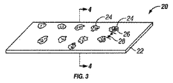
図3にて、チタンまたはチタン合金材料20の層は、材料20の表面に二酸化チタンを酸化して形成するために加熱される。一実施形態では、チタンまたはチタン合金材料20の層が自己支持多層構造で真空蒸着された材料の一つ以上の層の上に堆積される。別の実施形態では、チタンまたはチタン合金材料20の層がその上に真空蒸着して堆積された材料の1つまたは複数の層を有することができるバルク材料の上に堆積される。
In FIG. 3, the layer of titanium or
幾何学的生理学的に機能的な特徴24は、パターンマスクを介してUVに材料の層20を露出させることによって形成される。UV照射することにより化学的に幾何学的な生理学的機能フィーチャ24に対して変化させる、幾何学的な生理学的に機能的な特徴24の領域において、酸化チタンを変化材料層20の材料の周囲の表面領域22を囲む。幾何学的な生理学的に機能的な特徴の形状が円形であることができ、正方形、長方形、三角形、平行線、交差線または任意の組み合わせである。幾何学的な生理学的に機能の各機能は、約1ナノメートルから約75μmであり、幾何学的な生理学的に機能する機能が円形である場合、好ましくは、約1ナノメートルから約50μmの特徴では、16 、または機能直径幅。幾何学的な生理学的に機能的な特徴の各構成要素間のギャップ距離28に等しいまたは機能よりも大きい約26幅、よりも小さくてもよい。
The geometric physiologically
図4は、4-4線に沿った図3の断面図である。 記載の生理学的に機能的な幾何学的特徴24は、幾何学的な生理学的に機能的な特徴24が周囲面22の同じレベルにあることを示す点線で示されている。
4 is a cross-sectional view of FIG. 3 taken along line 4-4. The described physiologically functional
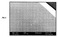
図5は、均等に移植可能な材料の少なくとも一方の面に分散している幾何学的な生理学的に機能的な特徴を示すと接触する体液、好ましくは血液。図に開示されている。 図1および図2に示すように、幾何学的な生理学的に機能的な特徴は、約1ナノメートル〜約20マイクロメートルの範囲の高さ、表面の残りの部分から上昇する。好ましくは、幾何学的な生理学的に機能的な特徴の高さは約1ナノメートル〜約3マイクロメートルの範囲である。幾何学的な生理学的に機能的な特徴の形状が示されている形状内に限定されるものではない。化学的に定義された領域の形状は、円形、正方形、長方形、三角形、平行線、交差線または上記の任意の組み合わせのいずれかであることができる。 FIG. 5 shows a bodily fluid, preferably blood, in contact with the geometric physiologically functional features distributed evenly on at least one side of the implantable material. It is disclosed in the figure. As shown in FIGS. 1 and 2, geometric physiologically functional features rise from the rest of the surface, with heights ranging from about 1 nanometer to about 20 micrometers. Preferably, the geometric physiologically functional feature height ranges from about 1 nanometer to about 3 micrometers. The shape of the geometric physiologically functional feature is not limited to the shape shown. The shape of the chemically defined region can be a circle, square, rectangle, triangle, parallel line, intersection line, or any combination of the above.
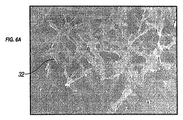
図6Aは、セル32は、親水処理されたシリコンの表面に広がって表示されます。図6Bは、直径が15ミクロンである円形のドットで、親水処理されたシリコンの表面に広がるセル32を示しています。図6B中のセルは、図6Aのものよりもはるかに焦点接着点36を有するように見える。これらの幾何学的な生理学的に機能的な特徴は、細胞付着のために提供するので、親和性ドメインとして作用し、内皮細胞のサイズに対するこれらの親和性ドメインの各々のサイズは、細胞運動のその後のラウンドに親和性ドメインの可用性を決定する。一実施形態によれば、幾何学的な生理的に機能的な特徴の個々のコンポーネントのそれぞれの好ましいサイズは約1フィーチャー幅50〜約1〜約75μmであり、好ましくはナノメートル、または直径であるかの幾何学的な生理学的に機能する機能円形である。焦点接着点形成、細胞運動および細胞増殖において重要な工程であるため、このような親水性のSi表面上のカーボンドットの幾何学的生理学的に機能的な特徴は、細胞運動を促進する。細胞の拡散は、細胞増殖、タンパク質合成、および他の細胞の代謝機能を促進する。細胞運動および細胞増殖を促進することは、最終的には幾何学的な生理学的に機能的な特徴を有する露出した表面上の内皮細胞とともに移植された移植可能な材料の被覆を促進する。幾何学的な生理学的に機能的な特徴は、図に示されているが。図6Bは、幾何学的な生理学的に機能的な特徴の形状は、この特定の実施形態に限定されないが、円形である。
Figure 6A shows
図6Cは、図6Bのイメージの一部の拡大である 。複数の焦点接着点36が再び表示されます。広い主に、円形の幾何学的な生理的に機能的な特徴に複数の焦点接着点の形成に細胞である広がり。それは細胞運動および細胞増殖を促進するため、細胞の広がりは、大規模な内皮化に向けて有益である。
FIG. 6C is an enlargement of a portion of the image of FIG. 6B. Multiple
図7は、染色された接着斑は、カーボンドットの形である幾何学的な生理的に機能的な特徴14と移植可能な材料の表面に人間aotic内皮細胞(HAEC)の36ポイントを示しています。接着斑のポイントは次の場所または幾何学的生理的に機能的な特徴14に非常に近い。これらの接着斑点は、セルの反対側の端部から収縮する電池用テンションポイントとして機能するので、細胞運動を促進する。
Figure 7 shows 36 points of human aotic endothelial cells (HAEC) on the surface of the
図8Aは、幅広い細胞32、直径25マイクロメートルののNiTiドットの形である幾何学的な生理学的に機能する機能を備えた、移植可能な材料の表面上の焦点に複数の焦点接着点36の広がりを示しています。 NiTiのドットが原因のNiTiドットと周囲のSi表面の間に弱いコントラストには見えません。
FIG. 8A shows multiple focal adhesion points 36 at the focal point on the surface of the implantable material, with a wide range of
図8Bは、図8Aに示すヒト大動脈上皮細胞32の拡大されたスライドを示す。複数の焦点接着点36は、親水性のSi表面上にパターニングされたNiTiドットをカプセル化するために示されている。図である。図9A、表面42および44を有する移植可能な材料46の一部が示されている。図である。図9Bは、一実施形態によれば、機械加工されたマスク48レーザーカット穴50〜約1nmから約75μmまでの範囲に定義されたサイズの40、好ましくは約1nmから、コート全体にパターニングさを有する少なくとも一つの表面42の移植可能な材料46とは緊密に覆われた表面42に接着されている。図である。孔40によって定義されるよう9C、材料14の薄膜が図1に見られるように、空間内に堆積した。図9Bに示すように、薄膜蒸着法により、マスク48。図である。図9Dは、堆積後、マスクを移植材料46の少なくとも一方の表面42を横切ってパターン化された幾何学的な生理学的機能フィーチャ49を明らかにするために除去される。
FIG. 8B shows an enlarged slide of the human aortic
上述したように、マスクの穴の形状には、幾何学的な生理学的に機能的な特徴について記載した形状のいずれかであり得る:円形、正方形、長方形、三角形、平行線と交差する線、またはそれらの任意の組み合わせ。の薄膜蒸着の実施形態では、幾何学的生理学的に機能的な特徴を製造する、幾何学的な生理学的に機能的な特徴は、移植可能な材料の表面から上昇している。幾何学的な生理学的に機能的な特徴の厚さは、マスクの穴の厚さに基づいて、約1〜約20マイクロメートルの範囲の厚さを有する。好ましくは、約1〜約3マイクロメートルのマスク範囲の孔の厚さ。 As noted above, the shape of the mask hole can be any of the shapes described for geometric, physiologically functional features: circular, square, rectangular, triangular, lines intersecting parallel lines, Or any combination thereof. In this thin film deposition embodiment, the geometric physiologically functional features are raised from the surface of the implantable material. The thickness of the geometric physiologically functional feature has a thickness in the range of about 1 to about 20 micrometers, based on the thickness of the mask hole. Preferably, the hole thickness in the mask range of about 1 to about 3 micrometers.
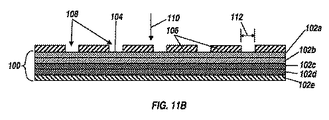
幾何学的生理学的に機能的な特徴の変化は、表面上の生体適合性材料の層または層を真空蒸着することによって移植可能な生体適合性材料の表面に添加してもよい。一実施形態では、堆積材料の層または層の形状は、幾何学的な生理学的に機能的な特徴を規定する。図2に示すように、例えば、移植材料100は、表面104を有する。一実施形態では、移植可能な生体適合性材料は、1つまたは複数の層、図11Aによって示されるように、自己支持構造内に形成された真空蒸着材料102を含んでもよい。第一の層102a、第二の層102b、第三の層102c、102dは第四層、第5層102eは図11A。別の実施形態では、移植可能な生体適合性材料は、単独で、バルク材料又は真空102a乃至102eは、生体適合性材料を堆積つ以上の層で覆われ、バルク材料のいずれか、バルク材料を含む。所望又は必要に応じしかしながら、任意の数の層が含まれていてもよい;五層の真空堆積材料の102a乃至102eのが示されている。
Changes in the geometric physiologically functional characteristics may be added to the surface of the implantable biocompatible material by vacuum deposition of the layer or layer of biocompatible material on the surface. In one embodiment, the layer or layer shape of the deposited material defines geometric, physiologically functional features. As shown in FIG. 2, for example, the
一つ以上の層102は、同一または異なる所望に応じて又は適切である厚さを有することができる。各層は、約1ナノメートルから約5マイクロメートル、又は約1ナノメートル〜約3マイクロメートルまで、約1ナノメートル〜約10マイクロメートル、約1ナノメートル〜約20マイクロメートルの範囲の厚さを有することができる。交互の層、様々な厚さの102は、幾何学的生理学的に機能的な特徴を収容するように適用されてもよい。
The one or
この実施形態では、幾何学的生理学的に機能的な特徴は、1つ以上の層を真空蒸着された材料102を添加することによって表面104に加えてもよい。例えば、図を参照。一つのプロセスにおける11B-11Eに、マスク106の穴に配置された規定サイズ通っ108を有し、コート全体にパターニングしっかりと表面104の少なくとも第1の部分に接着される。
In this embodiment, geometric physiologically functional features may be added to the
Claims (20)
A. インビボでの体液の組織に接触するように意図された少なくとも1つの表面を有する移植可能な生体適合性材料を提供する工程;
B. 少なくとも1つの表面に付与される結合ドメインの所定の分布と大きさと間隔に対応する開口部の規定されたパターンを有するマスクを提供する工程;
C .のうちの少なくとも1つによってマスクを介して生体適合性材料の少なくとも一つの表面を処理する。
I. 粒子サイズ、粒子相、結晶粒材料:少なくとも一つの表面上に材料の層を真空蒸着すると、前記真空蒸着層は、すぐにその下方からなる材料特性の群から選択される材料特性の少なくとも一方の表面とは異なる組成物は、表面トポグラフィー、および転移温度、及び移植可能な生体適合性材料の少なくとも一方の面上に定義された複数の結合ドメインを生成するためのマスクを除去する工程;
II. 真空第二の真空蒸着層が少なくとも異なる、請求真空は、少なくとも一つの表面上に材料の第2の層を堆積させる、少なくとも一方の表面からマスクを除去する、少なくとも一つの表面上に犠牲材料の層を堆積粒子サイズ、粒子相、穀物材料組成、表面トポグラフィー、および転移温度、および上で定義された複数の結合ドメインを得るために犠牲材料を除去すること:からなる材料特性の群から選択される材料特性に直ちにその下方一面少なくとも一つの移植可能な生体適合性材料の表面、又は
III. 写真は光化学的に少なくとも一つの表面を改変するために、少なくとも1つの表面に照射し、移植可能な生体適合性材料の少なくとも一方の面上に定義された複数の結合ドメインを生成するためにマスクを除去する。 A method for producing an implantable biocompatible material comprising the steps of:
A. Providing an implantable biocompatible material having at least one surface intended to contact body fluid tissue in vivo;
B. providing a mask having a defined pattern of openings corresponding to a predetermined distribution, size and spacing of binding domains imparted to at least one surface;
C. treating at least one surface of the biocompatible material through the mask with at least one of the following:
I. Particle size, particle phase, grain material: When a layer of material is vacuum deposited on at least one surface, the vacuum deposited layer immediately has at least a material property selected from the group of material properties consisting of the material layer below it. A composition different from one surface removes a mask to generate surface topography and a transition temperature and a plurality of binding domains defined on at least one side of the implantable biocompatible material. ;
II. Vacuum The second vacuum deposited layer is at least different, Claimed vacuum deposits a second layer of material on at least one surface, Removes mask from at least one surface, Sacrificial on at least one surface A group of material properties consisting of: depositing a layer of material, removing the sacrificial material to obtain particle size, particle phase, grain material composition, surface topography, and transition temperature, and multiple binding domains defined above Immediately below its material property selected from at least one surface of the implantable biocompatible material, or
III. Photography is to photochemically modify at least one surface to illuminate at least one surface to produce a plurality of binding domains defined on at least one side of the implantable biocompatible material. Remove the mask.
Applications Claiming Priority (3)
| Application Number | Priority Date | Filing Date | Title |
|---|---|---|---|
| US13/107,510 US8679517B2 (en) | 2002-09-26 | 2011-05-13 | Implantable materials having engineered surfaces made by vacuum deposition and method of making same |
| US13/107,510 | 2011-05-13 | ||
| PCT/US2012/037776 WO2012158614A2 (en) | 2011-05-13 | 2012-05-14 | Implantable materials having engineered surfaces and method of making same |
Publications (2)
| Publication Number | Publication Date |
|---|---|
| JP2014514127A true JP2014514127A (en) | 2014-06-19 |
| JP2014514127A5 JP2014514127A5 (en) | 2015-07-02 |
Family
ID=47177584
Family Applications (1)
| Application Number | Title | Priority Date | Filing Date |
|---|---|---|---|
| JP2014510536A Pending JP2014514127A (en) | 2011-05-13 | 2012-05-14 | Implantable material having system surface and method for producing the same |
Country Status (8)
| Country | Link |
|---|---|
| US (4) | US8679517B2 (en) |
| EP (1) | EP2707044A4 (en) |
| JP (1) | JP2014514127A (en) |
| CN (1) | CN103619364A (en) |
| AU (1) | AU2012255972B2 (en) |
| CA (1) | CA2835844C (en) |
| MX (1) | MX2013013139A (en) |
| WO (1) | WO2012158614A2 (en) |
Families Citing this family (17)
| Publication number | Priority date | Publication date | Assignee | Title |
|---|---|---|---|---|
| US8458879B2 (en) * | 2001-07-03 | 2013-06-11 | Advanced Bio Prosthetic Surfaces, Ltd., A Wholly Owned Subsidiary Of Palmaz Scientific, Inc. | Method of fabricating an implantable medical device |
| US8679517B2 (en) * | 2002-09-26 | 2014-03-25 | Palmaz Scientific, Inc. | Implantable materials having engineered surfaces made by vacuum deposition and method of making same |
| EP2422827B1 (en) * | 2010-08-27 | 2019-01-30 | Biotronik AG | Stent with a surface layer having a topographic modification |
| CH705978A1 (en) * | 2012-01-11 | 2013-07-15 | Qvanteq Ag | Method and device for determining a surface characteristic of stents and stent with defined surface characteristics. |
| GB201202519D0 (en) | 2012-02-13 | 2012-03-28 | Oxford Nanopore Tech Ltd | Apparatus for supporting an array of layers of amphiphilic molecules and method of forming an array of layers of amphiphilic molecules |
| GB201313121D0 (en) | 2013-07-23 | 2013-09-04 | Oxford Nanopore Tech Ltd | Array of volumes of polar medium |
| CN103861155A (en) * | 2014-03-11 | 2014-06-18 | 郑欣 | Nano-topography chip with capacity of inducing cell proliferation and differentiation |
| US10687956B2 (en) | 2014-06-17 | 2020-06-23 | Titan Spine, Inc. | Corpectomy implants with roughened bioactive lateral surfaces |
| TWI726940B (en) | 2015-11-20 | 2021-05-11 | 美商泰坦脊柱股份有限公司 | Processes for additively manufacturing orthopedic implants |
| GB201611770D0 (en) | 2016-07-06 | 2016-08-17 | Oxford Nanopore Tech | Microfluidic device |
| EP3493769B1 (en) | 2016-08-03 | 2022-03-30 | Titan Spine, Inc. | Titanium implant surfaces free from alpha case and with enhanced osteoinduction |
| CN107789666A (en) * | 2016-08-30 | 2018-03-13 | 北京航空航天大学 | A kind of inwall micro-patterning small-caliber artificial blood vessel |
| BR112019017727B1 (en) * | 2017-02-24 | 2022-12-06 | Qvanteq Ag | IMPLANT FOR INSERTION INTO A BODY LUMEN, METHOD FOR MANUFACTURING THE SAME AND IMPLANT SET |
| US10373767B2 (en) | 2017-11-21 | 2019-08-06 | Vactronix Scientific, Llc | Structural supercapacitor composite and method of making same |
| US11457932B2 (en) | 2018-03-15 | 2022-10-04 | Mako Surgical Corp. | Robotically controlled water jet cutting |
| WO2020183172A1 (en) | 2019-03-12 | 2020-09-17 | Oxford Nanopore Technologies Inc. | Nanopore sensing device and methods of operation and of forming it |
| CN111973814B (en) * | 2019-05-21 | 2024-04-19 | 中国人民解放军军事科学院军事医学研究院 | Vascular stent with composite bionic interface and preparation method thereof |
Citations (5)
| Publication number | Priority date | Publication date | Assignee | Title |
|---|---|---|---|---|
| US5849206A (en) * | 1994-08-15 | 1998-12-15 | Biotronik Mess- Und Therapiegerate Gmbh & Co. Ingenieurburo Berlin | Method of producing a biocompatible prosthesis |
| US20030028244A1 (en) * | 1995-06-07 | 2003-02-06 | Cook Incorporated | Coated implantable medical device |
| JP2004512059A (en) * | 2000-05-12 | 2004-04-22 | アドバンスト・バイオ・プロスゼティック・サーフィスズ・リミテッド | Self-supported laminated film structure material, medical device manufactured from the same, and method of manufacturing the same |
| JP2006505307A (en) * | 2002-09-26 | 2006-02-16 | アドヴァンスド バイオ プロスセティック サーフェシーズ リミテッド | Implantable material with designed surface and method of making the material |
| US20100227372A1 (en) * | 2007-07-27 | 2010-09-09 | The University Of Sydney | Biological functionalisation of substrates |
Family Cites Families (95)
| Publication number | Priority date | Publication date | Assignee | Title |
|---|---|---|---|---|
| US5387247A (en) | 1983-10-25 | 1995-02-07 | Sorin Biomedia S.P.A. | Prosthetic device having a biocompatible carbon film thereon and a method of and apparatus for forming such device |
| US4657544A (en) | 1984-04-18 | 1987-04-14 | Cordis Corporation | Cardiovascular graft and method of forming same |
| US4836884A (en) | 1986-02-17 | 1989-06-06 | Telectronics N.V. | Implantable materials |
| US5133845A (en) | 1986-12-12 | 1992-07-28 | Sorin Biomedica, S.P.A. | Method for making prosthesis of polymeric material coated with biocompatible carbon |
| IT1196836B (en) | 1986-12-12 | 1988-11-25 | Sorin Biomedica Spa | Polymeric or metal alloy prosthesis with biocompatible carbon coating |
| US5079600A (en) | 1987-03-06 | 1992-01-07 | Schnur Joel M | High resolution patterning on solid substrates |
| US5647858A (en) | 1989-07-25 | 1997-07-15 | Smith & Nephew, Inc. | Zirconium oxide and zirconium nitride coated catheters |
| US5278063A (en) | 1989-09-28 | 1994-01-11 | Board Of Regents The University Of Texas System | Chemical modification of promote animal cell adhesion on surfaces |
| US5477864A (en) | 1989-12-21 | 1995-12-26 | Smith & Nephew Richards, Inc. | Cardiovascular guidewire of enhanced biocompatibility |
| US5207709A (en) | 1991-11-13 | 1993-05-04 | Picha George J | Implant with textured surface |
| CA2087132A1 (en) | 1992-01-31 | 1993-08-01 | Michael S. Williams | Stent capable of attachment within a body lumen |
| BE1006440A3 (en) | 1992-12-21 | 1994-08-30 | Dereume Jean Pierre Georges Em | Luminal endoprosthesis AND METHOD OF PREPARATION. |
| US5607463A (en) | 1993-03-30 | 1997-03-04 | Medtronic, Inc. | Intravascular medical device |
| US5658443A (en) | 1993-07-23 | 1997-08-19 | Matsushita Electric Industrial Co., Ltd. | Biosensor and method for producing the same |
| US5645740A (en) | 1993-11-01 | 1997-07-08 | Naiman; Charles S. | System and assemblage for producing microtexturized substrates and implants |
| US20010039454A1 (en) | 1993-11-02 | 2001-11-08 | John Ricci | Orthopedic implants having ordered microgeometric surface patterns |
| EP0701803B1 (en) | 1994-02-03 | 1999-10-06 | SYNTHES AG Chur | Medical device for implantation into living bodies |
| US5733303A (en) | 1994-03-17 | 1998-03-31 | Medinol Ltd. | Flexible expandable stent |
| US5725573A (en) | 1994-03-29 | 1998-03-10 | Southwest Research Institute | Medical implants made of metal alloys bearing cohesive diamond like carbon coatings |
| JPH07284527A (en) | 1994-04-18 | 1995-10-31 | Ishikawajima Harima Heavy Ind Co Ltd | Biomedical metal and its use method |
| US5609629A (en) | 1995-06-07 | 1997-03-11 | Med Institute, Inc. | Coated implantable medical device |
| US5607475A (en) | 1995-08-22 | 1997-03-04 | Medtronic, Inc. | Biocompatible medical article and method |
| US6001622A (en) | 1995-12-21 | 1999-12-14 | Sunnybrook Health Science Centre | Integrin-linked kinase and its use |
| JPH09231821A (en) | 1995-12-22 | 1997-09-05 | Toto Ltd | Luminaire and method for maintaining illuminance |
| US5843289A (en) | 1996-01-22 | 1998-12-01 | Etex Corporation | Surface modification of medical implants |
| NZ331269A (en) | 1996-04-10 | 2000-01-28 | Advanced Cardiovascular System | Expandable stent, its structural strength varying along its length |
| US5932299A (en) | 1996-04-23 | 1999-08-03 | Katoot; Mohammad W. | Method for modifying the surface of an object |
| US5769884A (en) | 1996-06-27 | 1998-06-23 | Cordis Corporation | Controlled porosity endovascular implant |
| US6007573A (en) | 1996-09-18 | 1999-12-28 | Microtherapeutics, Inc. | Intracranial stent and method of use |
| US5895419A (en) | 1996-09-30 | 1999-04-20 | St. Jude Medical, Inc. | Coated prosthetic cardiac device |
| IT1289815B1 (en) | 1996-12-30 | 1998-10-16 | Sorin Biomedica Cardio Spa | ANGIOPLASTIC STENT AND RELATED PRODUCTION PROCESS |
| US5902475A (en) | 1997-04-08 | 1999-05-11 | Interventional Technologies, Inc. | Method for manufacturing a stent |
| US6240616B1 (en) | 1997-04-15 | 2001-06-05 | Advanced Cardiovascular Systems, Inc. | Method of manufacturing a medicated porous metal prosthesis |
| US5891507A (en) | 1997-07-28 | 1999-04-06 | Iowa-India Investments Company Limited | Process for coating a surface of a metallic stent |
| US5980564A (en) | 1997-08-01 | 1999-11-09 | Schneider (Usa) Inc. | Bioabsorbable implantable endoprosthesis with reservoir |
| US5897911A (en) | 1997-08-11 | 1999-04-27 | Advanced Cardiovascular Systems, Inc. | Polymer-coated stent structure |
| US6143370A (en) | 1997-08-27 | 2000-11-07 | Northeastern University | Process for producing polymer coatings with various porosities and surface areas |
| US6190404B1 (en) | 1997-11-07 | 2001-02-20 | Advanced Bio Prosthetic Surfaces, Ltd. | Intravascular stent and method for manufacturing an intravascular stent |
| US5955588A (en) | 1997-12-22 | 1999-09-21 | Innerdyne, Inc. | Non-thrombogenic coating composition and methods for using same |
| US6077413A (en) | 1998-02-06 | 2000-06-20 | The Cleveland Clinic Foundation | Method of making a radioactive stent |
| US6140127A (en) | 1998-02-18 | 2000-10-31 | Cordis Corporation | Method of coating an intravascular stent with an endothelial cell adhesive five amino acid peptide |
| US6280467B1 (en) | 1998-02-26 | 2001-08-28 | World Medical Manufacturing Corporation | Delivery system for deployment and endovascular assembly of a multi-stage stented graft |
| US6103320A (en) | 1998-03-05 | 2000-08-15 | Shincron Co., Ltd. | Method for forming a thin film of a metal compound by vacuum deposition |
| JP3735461B2 (en) | 1998-03-27 | 2006-01-18 | 株式会社シンクロン | Compound metal compound thin film forming method and thin film forming apparatus therefor |
| DE19916086B4 (en) | 1998-04-11 | 2004-11-11 | Inflow Dynamics Inc. | Implantable prosthesis, especially vascular prosthesis (stent) |
| US6261322B1 (en) | 1998-05-14 | 2001-07-17 | Hayes Medical, Inc. | Implant with composite coating |
| US6086773A (en) | 1998-05-22 | 2000-07-11 | Bmc Industries, Inc. | Method and apparatus for etching-manufacture of cylindrical elements |
| US6387060B1 (en) | 1998-06-17 | 2002-05-14 | Advanced Cardiovascular Systems, Inc. | Composite radiopaque intracorporeal product |
| US6096175A (en) | 1998-07-17 | 2000-08-01 | Micro Therapeutics, Inc. | Thin film stent |
| US6159239A (en) | 1998-08-14 | 2000-12-12 | Prodesco, Inc. | Woven stent/graft structure |
| WO2000018330A1 (en) | 1998-09-30 | 2000-04-06 | Impra, Inc. | Delivery mechanism for implantable stent |
| US6325825B1 (en) | 1999-04-08 | 2001-12-04 | Cordis Corporation | Stent with variable wall thickness |
| US6253441B1 (en) | 1999-04-16 | 2001-07-03 | General Electric Company | Fabrication of articles having a coating deposited through a mask |
| US6258121B1 (en) | 1999-07-02 | 2001-07-10 | Scimed Life Systems, Inc. | Stent coating |
| US6334868B1 (en) | 1999-10-08 | 2002-01-01 | Advanced Cardiovascular Systems, Inc. | Stent cover |
| US6849085B2 (en) | 1999-11-19 | 2005-02-01 | Advanced Bio Prosthetic Surfaces, Ltd. | Self-supporting laminated films, structural materials and medical devices manufactured therefrom and method of making same |
| US6537310B1 (en) | 1999-11-19 | 2003-03-25 | Advanced Bio Prosthetic Surfaces, Ltd. | Endoluminal implantable devices and method of making same |
| US6379383B1 (en) | 1999-11-19 | 2002-04-30 | Advanced Bio Prosthetic Surfaces, Ltd. | Endoluminal device exhibiting improved endothelialization and method of manufacture thereof |
| US7300457B2 (en) | 1999-11-19 | 2007-11-27 | Advanced Bio Prosthetic Surfaces, Ltd. | Self-supporting metallic implantable grafts, compliant implantable medical devices and methods of making same |
| US6936066B2 (en) * | 1999-11-19 | 2005-08-30 | Advanced Bio Prosthetic Surfaces, Ltd. | Complaint implantable medical devices and methods of making same |
| US6533905B2 (en) | 2000-01-24 | 2003-03-18 | Tini Alloy Company | Method for sputtering tini shape-memory alloys |
| ATE362382T1 (en) | 2000-03-15 | 2007-06-15 | Orbusneich Medical Inc | COATING WHICH STIMULATES ADHESION OF ENDOTHELIAL CELLS |
| US6183255B1 (en) | 2000-03-27 | 2001-02-06 | Yoshiki Oshida | Titanium material implants |
| JP4955849B2 (en) | 2000-04-06 | 2012-06-20 | 健 八尾 | Apatite structure and apatite pattern forming method |
| WO2001076525A2 (en) | 2000-04-10 | 2001-10-18 | Advanced Cardiovascular Systems, Inc. | Selectively coated stent delivery system and method of manufacture thereof |
| US8632583B2 (en) * | 2011-05-09 | 2014-01-21 | Palmaz Scientific, Inc. | Implantable medical device having enhanced endothelial migration features and methods of making the same |
| ES2277926T3 (en) | 2000-05-19 | 2007-08-01 | Advanced Bio Prosthetic Surfaces, Ltd. | PROCEDURES AND DEVICES FOR MANUFACTURING AN INTRAVASCULAR STENT. |
| US6652579B1 (en) | 2000-06-22 | 2003-11-25 | Advanced Cardiovascular Systems, Inc. | Radiopaque stent |
| JP2002017847A (en) | 2000-07-03 | 2002-01-22 | Kikuji Yamashita | Extracellular matrix bond type bioaffinity material and method for preparing the same extracellular matrix pharmaceutical preparation and method for preparing the same |
| US6569194B1 (en) | 2000-12-28 | 2003-05-27 | Advanced Cardiovascular Systems, Inc. | Thermoelastic and superelastic Ni-Ti-W alloy |
| JP3598973B2 (en) | 2001-01-10 | 2004-12-08 | ソニー株式会社 | Variable gain circuit |
| US6767360B1 (en) * | 2001-02-08 | 2004-07-27 | Inflow Dynamics Inc. | Vascular stent with composite structure for magnetic reasonance imaging capabilities |
| ATE272369T1 (en) | 2001-03-27 | 2004-08-15 | Cook William Europ | VESSEL TRANSPLANT FOR THE AORTA |
| US6527938B2 (en) | 2001-06-21 | 2003-03-04 | Syntheon, Llc | Method for microporous surface modification of implantable metallic medical articles |
| US6444318B1 (en) | 2001-07-17 | 2002-09-03 | Surmodics, Inc. | Self assembling monolayer compositions |
| US6746890B2 (en) | 2002-07-17 | 2004-06-08 | Tini Alloy Company | Three dimensional thin film devices and methods of fabrication |
| US8268340B2 (en) | 2002-09-26 | 2012-09-18 | Advanced Bio Prosthetic Surfaces, Ltd. | Implantable materials having engineered surfaces and method of making same |
| US8679517B2 (en) * | 2002-09-26 | 2014-03-25 | Palmaz Scientific, Inc. | Implantable materials having engineered surfaces made by vacuum deposition and method of making same |
| US8715771B2 (en) | 2003-02-26 | 2014-05-06 | Abbott Cardiovascular Systems Inc. | Coated stent and method of making the same |
| US8281737B2 (en) | 2003-03-10 | 2012-10-09 | Boston Scientific Scimed, Inc. | Coated medical device and method for manufacturing the same |
| ES2338560T3 (en) | 2003-05-07 | 2010-05-10 | Advanced Bio Prosthetic Surfaces, Ltd. | IMPLANTABLE METALLIC IMPLANTS AND PROCEDURES TO MANUFACTURE THEM. |
| US20050055085A1 (en) | 2003-09-04 | 2005-03-10 | Rivron Nicolas C. | Implantable medical devices having recesses |
| US7296998B2 (en) | 2003-09-22 | 2007-11-20 | Bartee Chaddick M | Hydrophilic high density PTFE medical barrier |
| US20050119723A1 (en) | 2003-11-28 | 2005-06-02 | Medlogics Device Corporation | Medical device with porous surface containing bioerodable bioactive composites and related methods |
| US8715340B2 (en) | 2004-03-31 | 2014-05-06 | Merlin Md Pte Ltd. | Endovascular device with membrane |
| CN101193666A (en) * | 2005-04-26 | 2008-06-04 | 奥胡斯大学 | Biocompatible material for surgical implants and cell guiding tissue culture surfaces |
| US20080299337A1 (en) | 2006-02-09 | 2008-12-04 | Isoflux, Inc. | Method for the formation of surfaces on the inside of medical devices |
| CN101400784B (en) | 2006-03-17 | 2013-09-11 | 学校法人近畿大学 | Biocompatible transparent sheet, method of producing the same and cell sheet |
| US8110242B2 (en) | 2006-03-24 | 2012-02-07 | Zimmer, Inc. | Methods of preparing hydrogel coatings |
| ATE488259T1 (en) | 2006-12-28 | 2010-12-15 | Boston Scient Ltd | BIOERODIBLE ENDOPROTHES AND PRODUCTION METHODS THEREOF |
| US7753962B2 (en) | 2007-01-30 | 2010-07-13 | Medtronic Vascular, Inc. | Textured medical devices |
| EP2124817B8 (en) | 2007-03-09 | 2022-02-09 | MiRus LLC | Bioabsorbable coatings for medical devices |
| DE102009023371A1 (en) | 2009-05-29 | 2010-12-02 | Acandis Gmbh & Co. Kg | Method for producing a medical functional element with a self-supporting lattice structure |
| US9186267B2 (en) | 2012-10-31 | 2015-11-17 | Covidien Lp | Wing bifurcation reconstruction device |
| US20140128901A1 (en) | 2012-11-05 | 2014-05-08 | Kevin Kang | Implant for aneurysm treatment |
-
2011
- 2011-05-13 US US13/107,510 patent/US8679517B2/en not_active Expired - Lifetime
-
2012
- 2012-05-14 CN CN201280031581.4A patent/CN103619364A/en active Pending
- 2012-05-14 CA CA2835844A patent/CA2835844C/en active Active
- 2012-05-14 AU AU2012255972A patent/AU2012255972B2/en active Active
- 2012-05-14 JP JP2014510536A patent/JP2014514127A/en active Pending
- 2012-05-14 WO PCT/US2012/037776 patent/WO2012158614A2/en active Application Filing
- 2012-05-14 MX MX2013013139A patent/MX2013013139A/en unknown
- 2012-05-14 EP EP12786428.8A patent/EP2707044A4/en active Pending
-
2014
- 2014-03-19 US US14/219,924 patent/US9272077B2/en not_active Expired - Lifetime
-
2016
- 2016-03-01 US US15/057,986 patent/US10039866B2/en not_active Expired - Lifetime
-
2018
- 2018-07-31 US US16/050,088 patent/US10729824B2/en not_active Expired - Lifetime
Patent Citations (5)
| Publication number | Priority date | Publication date | Assignee | Title |
|---|---|---|---|---|
| US5849206A (en) * | 1994-08-15 | 1998-12-15 | Biotronik Mess- Und Therapiegerate Gmbh & Co. Ingenieurburo Berlin | Method of producing a biocompatible prosthesis |
| US20030028244A1 (en) * | 1995-06-07 | 2003-02-06 | Cook Incorporated | Coated implantable medical device |
| JP2004512059A (en) * | 2000-05-12 | 2004-04-22 | アドバンスト・バイオ・プロスゼティック・サーフィスズ・リミテッド | Self-supported laminated film structure material, medical device manufactured from the same, and method of manufacturing the same |
| JP2006505307A (en) * | 2002-09-26 | 2006-02-16 | アドヴァンスド バイオ プロスセティック サーフェシーズ リミテッド | Implantable material with designed surface and method of making the material |
| US20100227372A1 (en) * | 2007-07-27 | 2010-09-09 | The University Of Sydney | Biological functionalisation of substrates |
Also Published As
| Publication number | Publication date |
|---|---|
| AU2012255972B2 (en) | 2016-06-23 |
| US10729824B2 (en) | 2020-08-04 |
| WO2012158614A3 (en) | 2013-01-10 |
| CA2835844C (en) | 2023-11-07 |
| US8679517B2 (en) | 2014-03-25 |
| US20170028109A1 (en) | 2017-02-02 |
| WO2012158614A2 (en) | 2012-11-22 |
| US9272077B2 (en) | 2016-03-01 |
| US20140294910A1 (en) | 2014-10-02 |
| EP2707044A2 (en) | 2014-03-19 |
| AU2012255972A1 (en) | 2014-01-16 |
| EP2707044A4 (en) | 2014-10-29 |
| US10039866B2 (en) | 2018-08-07 |
| MX2013013139A (en) | 2014-05-27 |
| US20190038815A1 (en) | 2019-02-07 |
| US20110274737A1 (en) | 2011-11-10 |
| CA2835844A1 (en) | 2012-11-22 |
| CN103619364A (en) | 2014-03-05 |
Similar Documents
| Publication | Publication Date | Title |
|---|---|---|
| JP2014514127A (en) | Implantable material having system surface and method for producing the same | |
| US10682443B2 (en) | Implantable biomaterials having functional surfaces | |
| JP4934269B2 (en) | Medical graft with multiple micropores | |
| AU2012253572B2 (en) | Implantable medical device having enhanced endothelial migration features and methods of making the same | |
| JP5213270B2 (en) | Medical device comprising a nanoporous coating for controlled therapeutic agent delivery | |
| JP5102029B2 (en) | Metallic drug release medical device and method for producing the same | |
| JP5143025B2 (en) | Method for producing medical device | |
| JP4799412B2 (en) | Implantable metal graft and method for producing the same | |
| US20090118821A1 (en) | Endoprosthesis with porous reservoir and non-polymer diffusion layer | |
| US20100178311A1 (en) | Implant and method for its manufacture | |
| MX2007000912A (en) | Metallic drug-releasing medical devices and method of making same |
Legal Events
| Date | Code | Title | Description |
|---|---|---|---|
| A521 | Request for written amendment filed |
Free format text: JAPANESE INTERMEDIATE CODE: A523 Effective date: 20150508 |
|
| A621 | Written request for application examination |
Free format text: JAPANESE INTERMEDIATE CODE: A621 Effective date: 20150508 |
|
| A131 | Notification of reasons for refusal |
Free format text: JAPANESE INTERMEDIATE CODE: A131 Effective date: 20160405 |
|
| A601 | Written request for extension of time |
Free format text: JAPANESE INTERMEDIATE CODE: A601 Effective date: 20160704 |
|
| A601 | Written request for extension of time |
Free format text: JAPANESE INTERMEDIATE CODE: A601 Effective date: 20160902 |
|
| A131 | Notification of reasons for refusal |
Free format text: JAPANESE INTERMEDIATE CODE: A131 Effective date: 20170207 |
|
| A02 | Decision of refusal |
Free format text: JAPANESE INTERMEDIATE CODE: A02 Effective date: 20170905 |


