JP2013516500A - Methods for treating inflammatory diseases and disorders - Google Patents
Methods for treating inflammatory diseases and disorders Download PDFInfo
- Publication number
- JP2013516500A JP2013516500A JP2012548532A JP2012548532A JP2013516500A JP 2013516500 A JP2013516500 A JP 2013516500A JP 2012548532 A JP2012548532 A JP 2012548532A JP 2012548532 A JP2012548532 A JP 2012548532A JP 2013516500 A JP2013516500 A JP 2013516500A
- Authority
- JP
- Japan
- Prior art keywords
- polypeptide
- seq
- inflammatory
- inhibitor
- administered
- Prior art date
- Legal status (The legal status is an assumption and is not a legal conclusion. Google has not performed a legal analysis and makes no representation as to the accuracy of the status listed.)
- Withdrawn
Links
Images


Classifications
-
- A—HUMAN NECESSITIES
- A61—MEDICAL OR VETERINARY SCIENCE; HYGIENE
- A61K—PREPARATIONS FOR MEDICAL, DENTAL OR TOILETRY PURPOSES
- A61K38/00—Medicinal preparations containing peptides
- A61K38/04—Peptides having up to 20 amino acids in a fully defined sequence; Derivatives thereof
- A61K38/10—Peptides having 12 to 20 amino acids
-
- A—HUMAN NECESSITIES
- A61—MEDICAL OR VETERINARY SCIENCE; HYGIENE
- A61K—PREPARATIONS FOR MEDICAL, DENTAL OR TOILETRY PURPOSES
- A61K38/00—Medicinal preparations containing peptides
- A61K38/04—Peptides having up to 20 amino acids in a fully defined sequence; Derivatives thereof
- A61K38/08—Peptides having 5 to 11 amino acids
-
- A—HUMAN NECESSITIES
- A61—MEDICAL OR VETERINARY SCIENCE; HYGIENE
- A61K—PREPARATIONS FOR MEDICAL, DENTAL OR TOILETRY PURPOSES
- A61K38/00—Medicinal preparations containing peptides
- A61K38/16—Peptides having more than 20 amino acids; Gastrins; Somatostatins; Melanotropins; Derivatives thereof
- A61K38/43—Enzymes; Proenzymes; Derivatives thereof
- A61K38/46—Hydrolases (3)
- A61K38/48—Hydrolases (3) acting on peptide bonds (3.4)
- A61K38/482—Serine endopeptidases (3.4.21)
- A61K38/4866—Protein C (3.4.21.69)
-
- A—HUMAN NECESSITIES
- A61—MEDICAL OR VETERINARY SCIENCE; HYGIENE
- A61P—SPECIFIC THERAPEUTIC ACTIVITY OF CHEMICAL COMPOUNDS OR MEDICINAL PREPARATIONS
- A61P1/00—Drugs for disorders of the alimentary tract or the digestive system
-
- A—HUMAN NECESSITIES
- A61—MEDICAL OR VETERINARY SCIENCE; HYGIENE
- A61P—SPECIFIC THERAPEUTIC ACTIVITY OF CHEMICAL COMPOUNDS OR MEDICINAL PREPARATIONS
- A61P1/00—Drugs for disorders of the alimentary tract or the digestive system
- A61P1/04—Drugs for disorders of the alimentary tract or the digestive system for ulcers, gastritis or reflux esophagitis, e.g. antacids, inhibitors of acid secretion, mucosal protectants
-
- A—HUMAN NECESSITIES
- A61—MEDICAL OR VETERINARY SCIENCE; HYGIENE
- A61P—SPECIFIC THERAPEUTIC ACTIVITY OF CHEMICAL COMPOUNDS OR MEDICINAL PREPARATIONS
- A61P1/00—Drugs for disorders of the alimentary tract or the digestive system
- A61P1/16—Drugs for disorders of the alimentary tract or the digestive system for liver or gallbladder disorders, e.g. hepatoprotective agents, cholagogues, litholytics
-
- A—HUMAN NECESSITIES
- A61—MEDICAL OR VETERINARY SCIENCE; HYGIENE
- A61P—SPECIFIC THERAPEUTIC ACTIVITY OF CHEMICAL COMPOUNDS OR MEDICINAL PREPARATIONS
- A61P11/00—Drugs for disorders of the respiratory system
-
- A—HUMAN NECESSITIES
- A61—MEDICAL OR VETERINARY SCIENCE; HYGIENE
- A61P—SPECIFIC THERAPEUTIC ACTIVITY OF CHEMICAL COMPOUNDS OR MEDICINAL PREPARATIONS
- A61P11/00—Drugs for disorders of the respiratory system
- A61P11/06—Antiasthmatics
-
- A—HUMAN NECESSITIES
- A61—MEDICAL OR VETERINARY SCIENCE; HYGIENE
- A61P—SPECIFIC THERAPEUTIC ACTIVITY OF CHEMICAL COMPOUNDS OR MEDICINAL PREPARATIONS
- A61P13/00—Drugs for disorders of the urinary system
- A61P13/12—Drugs for disorders of the urinary system of the kidneys
-
- A—HUMAN NECESSITIES
- A61—MEDICAL OR VETERINARY SCIENCE; HYGIENE
- A61P—SPECIFIC THERAPEUTIC ACTIVITY OF CHEMICAL COMPOUNDS OR MEDICINAL PREPARATIONS
- A61P17/00—Drugs for dermatological disorders
-
- A—HUMAN NECESSITIES
- A61—MEDICAL OR VETERINARY SCIENCE; HYGIENE
- A61P—SPECIFIC THERAPEUTIC ACTIVITY OF CHEMICAL COMPOUNDS OR MEDICINAL PREPARATIONS
- A61P17/00—Drugs for dermatological disorders
- A61P17/04—Antipruritics
-
- A—HUMAN NECESSITIES
- A61—MEDICAL OR VETERINARY SCIENCE; HYGIENE
- A61P—SPECIFIC THERAPEUTIC ACTIVITY OF CHEMICAL COMPOUNDS OR MEDICINAL PREPARATIONS
- A61P17/00—Drugs for dermatological disorders
- A61P17/06—Antipsoriatics
-
- A—HUMAN NECESSITIES
- A61—MEDICAL OR VETERINARY SCIENCE; HYGIENE
- A61P—SPECIFIC THERAPEUTIC ACTIVITY OF CHEMICAL COMPOUNDS OR MEDICINAL PREPARATIONS
- A61P17/00—Drugs for dermatological disorders
- A61P17/08—Antiseborrheics
-
- A—HUMAN NECESSITIES
- A61—MEDICAL OR VETERINARY SCIENCE; HYGIENE
- A61P—SPECIFIC THERAPEUTIC ACTIVITY OF CHEMICAL COMPOUNDS OR MEDICINAL PREPARATIONS
- A61P19/00—Drugs for skeletal disorders
- A61P19/02—Drugs for skeletal disorders for joint disorders, e.g. arthritis, arthrosis
-
- A—HUMAN NECESSITIES
- A61—MEDICAL OR VETERINARY SCIENCE; HYGIENE
- A61P—SPECIFIC THERAPEUTIC ACTIVITY OF CHEMICAL COMPOUNDS OR MEDICINAL PREPARATIONS
- A61P21/00—Drugs for disorders of the muscular or neuromuscular system
- A61P21/04—Drugs for disorders of the muscular or neuromuscular system for myasthenia gravis
-
- A—HUMAN NECESSITIES
- A61—MEDICAL OR VETERINARY SCIENCE; HYGIENE
- A61P—SPECIFIC THERAPEUTIC ACTIVITY OF CHEMICAL COMPOUNDS OR MEDICINAL PREPARATIONS
- A61P25/00—Drugs for disorders of the nervous system
-
- A—HUMAN NECESSITIES
- A61—MEDICAL OR VETERINARY SCIENCE; HYGIENE
- A61P—SPECIFIC THERAPEUTIC ACTIVITY OF CHEMICAL COMPOUNDS OR MEDICINAL PREPARATIONS
- A61P27/00—Drugs for disorders of the senses
- A61P27/02—Ophthalmic agents
-
- A—HUMAN NECESSITIES
- A61—MEDICAL OR VETERINARY SCIENCE; HYGIENE
- A61P—SPECIFIC THERAPEUTIC ACTIVITY OF CHEMICAL COMPOUNDS OR MEDICINAL PREPARATIONS
- A61P29/00—Non-central analgesic, antipyretic or antiinflammatory agents, e.g. antirheumatic agents; Non-steroidal antiinflammatory drugs [NSAID]
-
- A—HUMAN NECESSITIES
- A61—MEDICAL OR VETERINARY SCIENCE; HYGIENE
- A61P—SPECIFIC THERAPEUTIC ACTIVITY OF CHEMICAL COMPOUNDS OR MEDICINAL PREPARATIONS
- A61P3/00—Drugs for disorders of the metabolism
- A61P3/08—Drugs for disorders of the metabolism for glucose homeostasis
- A61P3/10—Drugs for disorders of the metabolism for glucose homeostasis for hyperglycaemia, e.g. antidiabetics
-
- A—HUMAN NECESSITIES
- A61—MEDICAL OR VETERINARY SCIENCE; HYGIENE
- A61P—SPECIFIC THERAPEUTIC ACTIVITY OF CHEMICAL COMPOUNDS OR MEDICINAL PREPARATIONS
- A61P37/00—Drugs for immunological or allergic disorders
- A61P37/02—Immunomodulators
-
- A—HUMAN NECESSITIES
- A61—MEDICAL OR VETERINARY SCIENCE; HYGIENE
- A61P—SPECIFIC THERAPEUTIC ACTIVITY OF CHEMICAL COMPOUNDS OR MEDICINAL PREPARATIONS
- A61P37/00—Drugs for immunological or allergic disorders
- A61P37/02—Immunomodulators
- A61P37/06—Immunosuppressants, e.g. drugs for graft rejection
-
- A—HUMAN NECESSITIES
- A61—MEDICAL OR VETERINARY SCIENCE; HYGIENE
- A61P—SPECIFIC THERAPEUTIC ACTIVITY OF CHEMICAL COMPOUNDS OR MEDICINAL PREPARATIONS
- A61P37/00—Drugs for immunological or allergic disorders
- A61P37/08—Antiallergic agents
-
- A—HUMAN NECESSITIES
- A61—MEDICAL OR VETERINARY SCIENCE; HYGIENE
- A61P—SPECIFIC THERAPEUTIC ACTIVITY OF CHEMICAL COMPOUNDS OR MEDICINAL PREPARATIONS
- A61P5/00—Drugs for disorders of the endocrine system
- A61P5/14—Drugs for disorders of the endocrine system of the thyroid hormones, e.g. T3, T4
-
- C—CHEMISTRY; METALLURGY
- C07—ORGANIC CHEMISTRY
- C07K—PEPTIDES
- C07K14/00—Peptides having more than 20 amino acids; Gastrins; Somatostatins; Melanotropins; Derivatives thereof
- C07K14/435—Peptides having more than 20 amino acids; Gastrins; Somatostatins; Melanotropins; Derivatives thereof from animals; from humans
- C07K14/46—Peptides having more than 20 amino acids; Gastrins; Somatostatins; Melanotropins; Derivatives thereof from animals; from humans from vertebrates
- C07K14/47—Peptides having more than 20 amino acids; Gastrins; Somatostatins; Melanotropins; Derivatives thereof from animals; from humans from vertebrates from mammals
- C07K14/4701—Peptides having more than 20 amino acids; Gastrins; Somatostatins; Melanotropins; Derivatives thereof from animals; from humans from vertebrates from mammals not used
- C07K14/4702—Regulators; Modulating activity
- C07K14/4703—Inhibitors; Suppressors
-
- C—CHEMISTRY; METALLURGY
- C12—BIOCHEMISTRY; BEER; SPIRITS; WINE; VINEGAR; MICROBIOLOGY; ENZYMOLOGY; MUTATION OR GENETIC ENGINEERING
- C12N—MICROORGANISMS OR ENZYMES; COMPOSITIONS THEREOF; PROPAGATING, PRESERVING, OR MAINTAINING MICROORGANISMS; MUTATION OR GENETIC ENGINEERING; CULTURE MEDIA
- C12N9/00—Enzymes; Proenzymes; Compositions thereof; Processes for preparing, activating, inhibiting, separating or purifying enzymes
- C12N9/10—Transferases (2.)
- C12N9/12—Transferases (2.) transferring phosphorus containing groups, e.g. kinases (2.7)
-
- C—CHEMISTRY; METALLURGY
- C12—BIOCHEMISTRY; BEER; SPIRITS; WINE; VINEGAR; MICROBIOLOGY; ENZYMOLOGY; MUTATION OR GENETIC ENGINEERING
- C12Y—ENZYMES
- C12Y207/00—Transferases transferring phosphorus-containing groups (2.7)
- C12Y207/11—Protein-serine/threonine kinases (2.7.11)
- C12Y207/11013—Protein kinase C (2.7.11.13)
Landscapes
- Health & Medical Sciences (AREA)
- Life Sciences & Earth Sciences (AREA)
- Chemical & Material Sciences (AREA)
- Engineering & Computer Science (AREA)
- Bioinformatics & Cheminformatics (AREA)
- General Health & Medical Sciences (AREA)
- Organic Chemistry (AREA)
- Medicinal Chemistry (AREA)
- Veterinary Medicine (AREA)
- Pharmacology & Pharmacy (AREA)
- Animal Behavior & Ethology (AREA)
- Public Health (AREA)
- Nuclear Medicine, Radiotherapy & Molecular Imaging (AREA)
- Chemical Kinetics & Catalysis (AREA)
- General Chemical & Material Sciences (AREA)
- Immunology (AREA)
- Gastroenterology & Hepatology (AREA)
- Proteomics, Peptides & Aminoacids (AREA)
- Genetics & Genomics (AREA)
- Zoology (AREA)
- Epidemiology (AREA)
- Biochemistry (AREA)
- Wood Science & Technology (AREA)
- Dermatology (AREA)
- Molecular Biology (AREA)
- Diabetes (AREA)
- General Engineering & Computer Science (AREA)
- Pulmonology (AREA)
- Biomedical Technology (AREA)
- Neurology (AREA)
- Rheumatology (AREA)
- Microbiology (AREA)
- Orthopedic Medicine & Surgery (AREA)
- Physical Education & Sports Medicine (AREA)
- Biophysics (AREA)
- Toxicology (AREA)
- Biotechnology (AREA)
- Endocrinology (AREA)
- Obesity (AREA)
- Transplantation (AREA)
Abstract
本開示は、PKCアイソフォーム制御因子を使用して炎症性疾患および障害を治療するための方法、組成物およびキットを提供する。 The present disclosure provides methods, compositions and kits for treating inflammatory diseases and disorders using PKC isoform regulators.
Description
本開示は、一般に疾患を治療する方法、より詳細には炎症性疾患および障害の治療に関する。 The present disclosure relates generally to methods of treating diseases, and more particularly to the treatment of inflammatory diseases and disorders.
炎症の開始は炎症反応とともに始まり、好中球、顆粒球、単球、マクロファージ、ならびにその他の免疫調節細胞の活性化をもたらす。これは、炎症性サイトカインおよびメディエーター、例えばインターロイキン類、TNFα、およびプロスタグランジン類を包含する局所性または全身性炎症カスケードを生じさせる可能性がある。この複雑な炎症媒介性カスケードは、あらゆる種類の反応、例えば細胞走化性および内皮損傷を誘発し、先天性および適応免疫系からの追加の細胞の動員をもたらす。 Initiation of inflammation begins with an inflammatory response and results in activation of neutrophils, granulocytes, monocytes, macrophages, and other immunoregulatory cells. This can result in a local or systemic inflammatory cascade involving inflammatory cytokines and mediators such as interleukins, TNFα, and prostaglandins. This complex inflammation-mediated cascade induces all kinds of reactions, such as cell chemotaxis and endothelial damage, resulting in the recruitment of additional cells from the innate and adaptive immune systems.
皮膚は、身体内部と環境との重要な境界として機能し、潜在的に有害な病原菌との接触を妨害する。抗原/病原菌が侵入した症例では、炎症反応が抗原を排除するために誘導されることが多い。この反応は、主としてT細胞、多核白血球、およびマクロファージからなる皮膚浸潤をもたらす。 The skin functions as an important boundary between the body's interior and the environment, preventing contact with potentially harmful pathogens. In cases where antigen / pathogen invades, an inflammatory response is often induced to eliminate the antigen. This reaction results in skin infiltration consisting primarily of T cells, multinucleated leukocytes, and macrophages.
炎症反応は、必ずしも外部からの刺激と関連していない場合、または非有害性環境物質によって引き起こされる場合(アレルギーの場合)がある。どちらの場合も、前炎症性サイトカインの過剰発現は、適正に制御されないと、一般に局所性および全身性炎症の特徴である炎症、ならびに様々な炎症性疾患および障害を引き起こす。炎症には、様々な障害、例えば湿疹および皮膚炎が結び付いており、例えばアトピー性皮膚炎、脂漏性皮膚炎、異汗性湿疹、貨幣状湿疹、うっ血性皮膚炎、アレルギー性皮膚炎、乾癬、掻痒症、多発性硬化症、皮膚炎症、瘢痕性類天疱瘡、強皮症、化膿性汗腺炎、中毒性表皮剥離症、座瘡、骨炎、移植片対宿主病(GvHD)、壊疽性膿皮症、およびベーチェット症候群(Behcet’s Syndrome)が包含される。 Inflammatory reactions may not necessarily be associated with external stimuli or may be caused by non-hazardous environmental substances (in the case of allergies). In both cases, overexpression of proinflammatory cytokines, if not properly controlled, causes inflammation, which is generally characteristic of local and systemic inflammation, as well as various inflammatory diseases and disorders. Inflammation is associated with various disorders such as eczema and dermatitis, such as atopic dermatitis, seborrheic dermatitis, allergic eczema, monetary eczema, congestive dermatitis, allergic dermatitis, psoriasis , Pruritus, multiple sclerosis, skin inflammation, scarring pemphigus, scleroderma, suppurative scab, toxic epidermis, acne, osteomyelitis, graft-versus-host disease (GvHD), gangrenous Pyoderma, and Behcet's Syndrome are included.
当然のことながら、前炎症性サイトカインの過剰産生は、多数の炎症性および自己免疫疾患に関係すると見なされてきた。例えば、サイトカイン類、例えばTh17細胞の生存および増殖を刺激するTNFαおよびインターロイキン(IL)−23の分泌は乾癬と高度に関連しているが、一方、IL−6がその前炎症性サイトカインとしての一般的役割に加えてTh17発生のために必要とされる。IL−12およびIP−10のような他のサイトカイン類は開始因子であり、乾癬およびその他の自己免疫疾患に典型的であるTh1経路に関与している。好酸球の産生を増加させるサイトカインであるインターロイキン5(IL−5)は、喘息において過剰発現し、結果としてアレルギー性炎症の特徴である、喘息性気管支粘膜における好酸球の蓄積を生じさせる。インターロイキン4(IL−4)およびインターロイキン13(IL−13)は、炎症性腸疾患および喘息において見いだされる平滑筋の高収縮性の公知のメディエーターである。さらに、以下で詳細に考察するように、炎症性サイトカインは、例えば、乾癬、多発性硬化症、関節炎、虚血、敗血性ショック、および臓器移植拒否反応に関係することが証明されている。 Of course, overproduction of proinflammatory cytokines has been implicated in a number of inflammatory and autoimmune diseases. For example, secretion of cytokines such as TNFα and interleukin (IL) -23, which stimulates the survival and proliferation of Th17 cells, is highly associated with psoriasis, while IL-6 as its pro-inflammatory cytokine In addition to the general role, it is required for Th17 generation. Other cytokines such as IL-12 and IP-10 are initiation factors and are involved in the Th1 pathway that is typical of psoriasis and other autoimmune diseases. Interleukin 5 (IL-5), a cytokine that increases the production of eosinophils, is overexpressed in asthma, resulting in the accumulation of eosinophils in the asthmatic bronchial mucosa that is characteristic of allergic inflammation . Interleukin 4 (IL-4) and interleukin 13 (IL-13) are well-known mediators of high contractility of smooth muscle found in inflammatory bowel disease and asthma. Further, as discussed in detail below, inflammatory cytokines have been shown to be involved in, for example, psoriasis, multiple sclerosis, arthritis, ischemia, septic shock, and organ transplant rejection.
同様に、顆粒球マクロファージコロニー刺激因子(GM−CSF)は、顆粒球およびマクロファージ系統集団の成熟の制御因子であり、多数の炎症性および自己免疫疾患における重要な因子であると関係付けられてきた。例えば、GM−CSF分泌を阻害する抗体は、自己免疫疾患を改善することが証明されている。 Similarly, granulocyte macrophage colony stimulating factor (GM-CSF) is a regulator of maturation of granulocyte and macrophage lineage populations and has been implicated as an important factor in many inflammatory and autoimmune diseases . For example, antibodies that inhibit GM-CSF secretion have been shown to ameliorate autoimmune diseases.
そこで、前炎症性サイトカインの分泌を減少させる、および/または免疫制御因子を調節する治療薬の開発は、一般に局所性および全身性炎症、ならびに本明細書で考察する炎症性および/または自己免疫疾患の宿主を改善することに有益となる。複数の一連の証拠は、PKCアイソフォームの修飾因子がこれらの結果を達成する際に有用であることを指摘している。 Thus, the development of therapeutic agents that reduce the secretion of pro-inflammatory cytokines and / or modulate immune regulators generally involves local and systemic inflammation, as well as inflammatory and / or autoimmune diseases discussed herein. It is beneficial to improve the host. Multiple lines of evidence indicate that PKC isoform modifiers are useful in achieving these results.
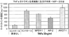
数件のin vivo試験は、Tヘルパー(Th)17細胞の関与ならびに皮膚関連細胞、例えばケラチン生成細胞、樹状細胞およびTヘルパー細胞によるサイトカイン類、例えばインターロイキン類およびTNFαの分泌が乾癬および他の自己免疫炎症性疾患の病原に関与する炎症反応の発生における重要な要素であることを証明している。本明細書で使用するin vivo(「生体内」を意味するラテン語)は生きている生体全体を用いる実験であり、部分的もしくは死んでいる生体、またはin vitro(「ガラス内」、例えば、試験管もしくはペトリ皿内)制御環境とは対照的である。Th17細胞の生存および増殖を刺激するサイトカイン類、例えばTNFαおよびインターロイキン(IL)−23の分泌もまた、これらの疾患にとって重要な主要サイトカイン制御因子として役立つ(非特許文献1)。真皮内のTh17細胞は、順にIL−17AおよびIL−22の分泌を誘導する。IL−22は、特に、ケラチン生成細胞の過剰増殖を引き出し、乾癬における炎症反応を増強する(非特許文献1)。 Several in vivo studies show that T helper (Th) 17 cell involvement and secretion of cytokines such as interleukins and TNFα by skin-related cells, such as keratinocytes, dendritic cells and T helper cells, are psoriasis and others. It has proven to be an important factor in the development of inflammatory responses involved in the pathogenesis of autoimmune inflammatory diseases. As used herein, in vivo (Latin meaning “in vivo”) is an experiment that uses the entire living organism, partially or dead organism, or in vitro (“in glass”, eg, test In contrast to the controlled environment (in tubes or petri dishes). Secretion of cytokines that stimulate the survival and proliferation of Th17 cells, such as TNFα and interleukin (IL) -23, also serve as key cytokine regulators important for these diseases (1). Th17 cells in the dermis in turn induce the secretion of IL-17A and IL-22. IL-22 particularly elicits hyperproliferation of keratinocytes and enhances the inflammatory response in psoriasis (Non-Patent Document 1).
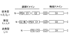
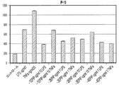
プロテインキナーゼC(PKC)ファミリーは、リン酸のATPからタンパク質のセリンおよびトレオニン残基への共有転移を触媒する1群のリン脂質依存性酵素を表している。このファミリーは、現在、カルシウムイオンおよびその他の因子によるそれらの活性化に基づいて3つの別個のカテゴリーに属する少なくとも12の個別アイソフォームから構成されると考えられている。PKCファミリーは、通常、従来型、新型および非典型PKCの3つのサブグループ(図1)に分けられる少なくとも10のメンバーからなる。特異的補因子要求、組織分布、および細胞区分化は、各アイソフォームに対する機能差および特異的シグナル伝達カスケードの調整を示唆している。そこで、特異的刺激は、それらの因子によって調節されるアイソフォーム特異的PKCシグナル伝達による特異的反応:例えば特定の生物学的状況における発現、局在化、および/またはリン酸化状態をもたらし得る。PKCアイソフォームは、様々な細胞外シグナルによって活性化され、そして順に、受容体、酵素、細胞骨格タンパク質、および転写因子を包含する細胞タンパク質の活性を修飾する。したがって、PKCファミリーは、細胞増殖、分化、生存および死の調節を包含する細胞シグナルプロセシングにおいて中心的役割を果たす。 The protein kinase C (PKC) family represents a group of phospholipid-dependent enzymes that catalyze the covalent transfer of ATP of phosphate to serine and threonine residues of proteins. This family is currently thought to be composed of at least 12 individual isoforms belonging to three distinct categories based on their activation by calcium ions and other factors. The PKC family usually consists of at least 10 members divided into three subgroups (Figure 1): conventional, novel and atypical PKC. Specific cofactor requirements, tissue distribution, and cell partitioning suggest functional differences for each isoform and regulation of specific signaling cascades. Thus, specific stimuli can lead to specific responses by isoform-specific PKC signaling regulated by those factors: for example, expression, localization, and / or phosphorylation status in certain biological contexts. PKC isoforms are activated by various extracellular signals and in turn modify the activity of cellular proteins including receptors, enzymes, cytoskeletal proteins, and transcription factors. Thus, the PKC family plays a central role in cell signal processing, including the regulation of cell proliferation, differentiation, survival and death.
皮膚内で高度に豊富であるPKCαは、表皮内で主要な従来型のCa2+反応性PKCアイソフォームであり、それは、最初はin vitroおよびin vivoにおけるケラチン生成細胞中で検出された唯一のcPKCであった(非特許文献2、非特許文献3)。このため、PKCαは、Ca2+誘導性分化における重要な因子であると提案されていた(非特許文献4、非特許文献2)。表皮内に、および主として基底層直上に限定すると(非特許文献5)、PKCαは細胞周期離脱に関与しており、主としてケラチン細胞骨格および接着斑細胞−細胞接着と関連付けられている(非特許文献6、非特許文献7)。従来型PKC活性化因子であるTPA(12−O−テトラデカノイルホルボール−13−アセテート)に曝露させると有棘マーカーが抑制されたので、PKCαは、TPA活性化の結果として有棘状から顆粒状分化への変化にとって大きな役割を果たすと考えられた(非特許文献8、非特許文献9、非特許文献10、非特許文献11)。実際に、アンチセンスオリゴヌクレオチドによるPKCα活性またはその合成のブロッキングは、顆粒マーカーを無効にし、K1およびK10のような有棘状マーカーを蘇生させると思われた。同様に、優性ネガティブPKCαの実施は、(後期)有棘状マーカーであるインボルクリンを再生すると思われた(非特許文献12)。したがって、皮膚癌における不完全な分化(非特許文献13、非特許文献14)は、in vitroの腫瘍細胞においても観察される上昇したPKCα活性と相関する(非特許文献2、非特許文献15)。しかし、正常ヒトケラチン生成細胞中のPKCαの過剰発現は、それらの分化パターンを変化させるとは思われなかった(非特許文献12)。遊走中のβ1−インテグリンの細胞内輸送および膜動員にPKCαが及ぼす影響(非特許文献16)は、創傷再上皮化および腫瘍細胞浸潤の両方を明確に促進することができる。
PKCα, which is highly abundant in the skin, is the major conventional Ca 2+ reactive PKC isoform in the epidermis, which is the only cPKC that was initially detected in keratinocytes in vitro and in vivo. (Non-patent
トランスジェニックマウスにおけるPKCαの過剰発現は、著しい炎症反応、増加した表皮肥厚および好中球浸潤と相関する浮腫、多発性微小膿瘍、ならびに炎症性サイトカイン類およびケモカイン類、例えばTNFα、MIP−2、COX−2もしくはマクロファージ炎症性タンパク質(MIP)の顕著な増加を誘導すると思われてきた。これらの結果は、表皮炎症反応にPKCαを関係付けている(非特許文献17)。TPA(PKCα活性因子)を用いた治療は、明らかに表皮過形成、表皮内炎症、および広範なアポトーシスを誘発した(非特許文献18、非特許文献6)。さらに、PKCアイソエンザイム選択的ノックアウトマウスおよびトランスジェニックマウスにおける近年のin vivo試験は、免疫系における個別PKCの際だった別個の機能を有すると思われる。これらの遺伝分析は、生化学的試験と一緒に、PKC調節シグナル伝達経路が免疫反応の多数の態様において重要な役割を果たすことを示すと思われる。例えば、PKCファミリーのメンバーは、T細胞シグナル伝達経路において極めて重要であると思われる。特に、PKCαのアイソタイプは、リンパ球特異的in vivoエフェクターの性質を決定すると思われる。PKCαは、さらにマクロファージ活性化に関与すると考察されており、明白に肥満細胞シグナル伝達に関与することが証明された(非特許文献19)。このため、PKCアイソタイプは、適応免疫において有効な薬物標的である。
Overexpression of PKCα in transgenic mice is associated with marked inflammatory responses, increased epidermal thickening and neutrophil infiltration, multiple microabscesses, and inflammatory cytokines and chemokines such as TNFα, MIP-2, COX -2 or macrophage inflammatory protein (MIP) has been thought to induce a significant increase. These results relate PKCα to the epidermal inflammatory response (Non-patent Document 17). Treatment with TPA (PKCα activator) clearly induced epidermal hyperplasia, intraepidermal inflammation, and extensive apoptosis (Non-patent
炎症性疾患の1つの例は、乾癬である。感染発生をもたらす基本病変に関する2つの主要な仮説が存在する。第1の仮説は、乾癬が主として皮膚細胞の過剰成長および再生の障害であると見なしている。第2の仮説は、乾癬を皮膚細胞の過剰再生が免疫系によって生成される因子に続発性である免疫媒介性障害であると見なしている。したがって、乾癬に対する大多数の薬物は、皮膚細胞の過剰増殖状態、または乾癬斑として存在する皮膚炎症反応のいずれかである疾患の1つの成分を標的としている。 One example of an inflammatory disease is psoriasis. There are two main hypotheses regarding the basic lesions that lead to infection. The first hypothesis considers psoriasis as a primary disorder of skin cell overgrowth and regeneration. The second hypothesis regards psoriasis as an immune-mediated disorder in which over-regeneration of skin cells is secondary to factors generated by the immune system. Thus, the vast majority of drugs for psoriasis target one component of the disease that is either a hyperproliferative state of skin cells or a skin inflammatory response present as psoriatic plaques.
近年のデータは、どちらの経路も皮膚細胞と免疫学的環境(環境、周囲、位置および/または状況を網羅する)とのクロストークを通して疾患の病理学の基礎をなすという概念を支持している。従来型の全ゲノム連鎖解析は、乾癬感受性1〜9(PSORS1〜PSORS9)遺伝子座と命名された乾癬を発生する傾向と関連する様々な染色体上の9つの場所(遺伝子座)を同定した。これらの場所では、数個の遺伝子が特性付けられ、表皮細胞内で発現するタンパク質、例えば表皮の顆粒層および角化層内で発現し、乾癬においてはアップレギュレートされるコルネオデスモシンをコードすることが見いだされた。他方では、他の乾癬関連遺伝子は、インターロイキン−12Bを発現する染色体5q上で例えば1L−12Bのように特性付けられた免疫系の調節に関与するタンパク質をコードする(非特許文献20)。 Recent data support the notion that both pathways underlie the pathology of the disease through crosstalk between skin cells and the immunological environment (covering the environment, surroundings, location and / or context). . Conventional genome-wide linkage analysis identified nine locations (locus) on various chromosomes associated with a propensity to develop psoriasis, designated psoriasis susceptibility 1-9 (PSORS1-PSORS9) loci. In these locations, several genes have been characterized and encode proteins expressed in epidermal cells, such as corneodesmosine, which is expressed in the granular and keratinized layers of the epidermis and is up-regulated in psoriasis I found something. On the other hand, other psoriasis-related genes encode proteins involved in the regulation of the immune system characterized as, for example, 1L-12B, on chromosome 5q expressing interleukin-12B (Non-patent Document 20).
炎症性疾患のまた別の例は、多発性硬化症(MS)である。MSは、一般に青少年に影響を及ぼす脳および脊髄に影響を及ぼす可能性があるCNSの慢性的かつ予測不能な炎症性疾患である(非特許文献21)。MSは、現在は青少年の最も一般的な神経系疾患であると言われており、通常、20〜40歳で始まり、男性に比較して女性においてほぼ2倍の確率で発生する傾向がある。 Another example of an inflammatory disease is multiple sclerosis (MS). MS is a chronic and unpredictable inflammatory disease of the CNS that can affect the brain and spinal cord, which generally affects adolescents (21). MS is now said to be the most common neurological disease of adolescents, usually starting at 20-40 years of age and tending to occur almost twice as often in women compared to men.
MSでは、神経細胞を取り囲んで保護する物質である髄鞘、および/またはその産生能力が損傷され、これは「脱髄」と呼ばれている。この損傷は、脳と身体との間のメッセージを緩徐化または遮断する作用を有し、MSとともに観察される症状をもたらす。脳および/または脊髄内に散在する領域内の脱髄および瘢痕もしくはその他の病変は、この疾患に特徴的であると考えられている(非特許文献22)。これらの病変は、神経伝導を変化させ、CNSにおける脱髄性プラークの場所に伴って変化する廃疾神経学的欠損を誘導すると思われる。(非特許文献24)。その臨床徴候および症状は様々で、それが影響するCNSの部分に依存し、運動、感覚、自律神経および認知障害が含まれる可能性がある。(非特許文献23)。 In MS, the myelin sheath, which is a substance that surrounds and protects nerve cells, and / or its production ability is damaged, which is called “demyelination”. This damage has the effect of slowing or blocking messages between the brain and the body, resulting in symptoms observed with MS. Demyelination and scarring or other lesions in areas scattered within the brain and / or spinal cord are believed to be characteristic of the disease (22). These lesions are likely to alter nerve conduction and induce debilitating neurological deficits that change with the location of demyelinating plaques in the CNS. (Non-patent document 24). Its clinical signs and symptoms vary and depend on the part of the CNS that it affects and can include motor, sensory, autonomic and cognitive impairments. (Non-Patent Document 23).
MSの一部の一般的症状には以下が含まれる:1)極度の疲労の感覚、2)平衡歩行および協調運動障害、3)視覚的問題−複視および失明、4)手足における麻痺および刺痛、5)軽度および重度両方の疼痛、筋力低下、6)筋肉の硬直および痙攣、7)気分変動−抑うつおよび不安、8)記憶および集中力の欠如、会話障害(非特許文献24)。 Some common symptoms of MS include: 1) Extreme fatigue sensation, 2) Balanced gait and coordination disorders, 3) Visual problems-double vision and blindness, 4) Paralysis and stinging in limbs Pain, 5) Both mild and severe pain, muscle weakness, 6) Muscle stiffness and convulsions, 7) Mood fluctuations-depression and anxiety, 8) Lack of memory and concentration, speech impairment (24).
進行性身体障害は、特に25年間見通しが包含される場合、MSを有する大多数の患者の宿命である。MS患者の半数は、疾患発症の15年間以内には歩行に杖を必要とするようになる。MSは、若年および中年成人における神経性身体障害の主要原因であり、過去10年間までは、公知の有益な治療を有していなかった。MSは非特異的な臨床所見のために診断を下すのが困難であるが、このことがMRIスキャン、誘発電位、および脳脊髄液(CSF)試験からなる幾つかの技術的進歩を包含する高度に構造化された診断基準の開発をもたらした。診断基準は、一般には様々な時点に発生するが、他の病因、例えば感染、血管障害、または自己免疫障害によって説明されない中心体白質内の散在性病変の一般的原理に依存している。 Progressive disability is the fate of the majority of patients with MS, especially when the 25-year outlook is included. Half of MS patients will require walking sticks within 15 years of disease onset. MS is a leading cause of neurological disability in young and middle-aged adults and has not had a known beneficial treatment until the past decade. Although MS is difficult to make a diagnosis due to nonspecific clinical findings, this is an advanced that includes several technological advances consisting of MRI scans, evoked potentials, and cerebrospinal fluid (CSF) testing Has led to the development of structured diagnostic criteria. Diagnostic criteria generally depend on the general principle of disseminated lesions within the central body white matter that occur at various times but are not accounted for by other etiologies such as infection, vascular disorders, or autoimmune disorders.
MSは、それによって未知の薬剤がT細胞媒介性炎症性発作を引き起こし、CNS(中枢神経系)組織の脱髄を誘発する自己免疫疾患であると広く考察されている(非特許文献25)。ミエリンを標的とする自己免疫反応についての証拠は強力であるが、決定的ではない。例えば、リンパ球またはミエリン食作用の非存在下では、早期多発性硬化症病変におけるマイクログリア細胞活性化による原発性オリゴデンドロサイトアポトーシスの開示がある(非特許文献26)。 MS is widely considered to be an autoimmune disease whereby an unknown agent causes T cell-mediated inflammatory seizures and induces demyelination of CNS (central nervous system) tissue (25). The evidence for an autoimmune response targeting myelin is strong but not definitive. For example, in the absence of lymphocyte or myelin phagocytosis, there is a disclosure of primary oligodendrocyte apoptosis due to microglial cell activation in early multiple sclerosis lesions (Non-patent Document 26).
MSは、特徴的に4つの疾患パターン:再発寛解型MS(RRMS)、一次進行型MS(PPMS)、進行再発型MS(PRMS)、および二次進行型MS(SPMS)を有すると報告されている。RRMSを備える患者の推定50%は10年以内にSPMSを発生し、RRMSの90%までは最後にはSPMSを発生することになる。疾患の各パターンは、軽度、中等度または重度として現れることがある。RRMSを患うヒトは、悪化する神経機能の明確な発作を提示する。これらの発作には、その間疾患進行が発生しない部分または完全回復期(緩解期)が続く(約85%の人々は、最初はRRMSであると診断される)。PPMSは、最初から緩徐に悪化する神経機能を特徴とし、顕著な再発または緩解を伴わない(約10%のヒトはPPMSと診断される)。SPMSでは、RRMSの原初期に続いて、多くの人はこの疾患がより確実に悪化する二次進行型疾患経過をたどる(RRMSを備えるヒトの約50%が10年間以内にこの疾患のこの形態を発生する)。PRMSでは、ヒトは最初から確実に、しかし途中で悪化する神経機能の明白な発作を伴って悪化する疾患症状を経験し、他方ではこの疾患は緩解を伴わずに進行すると思われる(5%)(非特許文献26)。 MS is characteristically reported to have four disease patterns: relapsing-remitting MS (RRMS), primary progressive MS (PPMS), progressive relapsing MS (PRMS), and secondary progressive MS (SPMS) Yes. An estimated 50% of patients with RRMS will develop SPMS within 10 years and up to 90% of RRMS will eventually develop SPMS. Each pattern of disease may manifest as mild, moderate or severe. Humans with RRMS present a clear seizure of deteriorating neural function. These seizures are followed by a portion during which disease progression does not occur or a complete recovery phase (remission phase) (approximately 85% of people are initially diagnosed with RRMS). PPMS is characterized by neurological function that deteriorates slowly from the beginning, with no significant relapse or remission (approximately 10% of humans are diagnosed with PPMS). In SPMS, following the early stages of RRMS, many have a secondary progressive disease course in which the disease is more reliably aggravated (about 50% of people with RRMS have this form of the disease within 10 years. Generated). In PRMS, humans experience disease symptoms that worsen from the outset, but with obvious attacks of neurological function that worsen along the way, while the disease appears to progress without remission (5%). (Non-patent document 26).
現時点ではMSに対する治療薬はないが、疾患活性および疾患進行の低下を試みる幾つかの治療薬を利用できる。合衆国では、4つのクラスの6種の薬物がMSの治療のために承認されている。FDA承認の疾患治療薬には、以下のインターフェロンクラス、IFN−β−1a(REBIF(登録商標)およびAVONEX(登録商標))およびIFN−β−1b(BETASERON(登録商標))、酢酸グラチラマー(COPAXONE(登録商標))、ポリペプチド、ナタリズマブ(TYSABRI(登録商標))、ミトキサントロン(NOVANTRONE(登録商標))、細胞毒性薬、が包含される。コルチコステロイド剤、メソトレキセート、シクロホスファミド、アザチオプリン、および静脈内(IV)イムノグロブリンを包含する様々な成功度を示す他の薬物が使用されてきた。現在承認されている治療薬の利点は、MSにおける、相当小さな再発率、そして身体障害の予防である。 There are currently no therapeutics for MS, but several therapeutics are available that attempt to reduce disease activity and disease progression. In the United States, four classes of six drugs are approved for the treatment of MS. FDA-approved disease treatments include the following interferon classes: IFN-β-1a (REBIF® and AVONEX®) and IFN-β-1b (BETASERON®), glatiramer acetate (COPAXONE) (Registered trademark)), polypeptide, natalizumab (TYSABRI (registered trademark)), mitoxantrone (NOVANTRONE (registered trademark)), and cytotoxic drugs. Other drugs with varying degrees of success have been used, including corticosteroids, methotrexate, cyclophosphamide, azathioprine, and intravenous (IV) immunoglobulins. The benefits of currently approved therapeutics are a fairly small relapse rate and prevention of disability in MS.
REBIF(登録商標)(インターフェロンβ1a)は、ヒトの身体において見いだされるものと同一のインターフェロンβを産生するバイオ技術プロセスによって製造された医薬品である。REBIF(登録商標)は、週3回皮下投与すると報告されている(REBIF(登録商標)についてのFDA承認医療用医薬品情報から)。 REBIF® (interferon β1a) is a pharmaceutical product manufactured by a biotechnological process that produces the same interferon β that is found in the human body. REBIF® has been reported to be administered subcutaneously three times a week (from FDA approved prescription drug information for REBIF®).
AVONEX(登録商標)(インターフェロンβ1a)は、ヒトの身体において見いだされるものと同一のインターフェロンβを産生するバイオ技術プロセスによって製造された医薬品である。AVONEX(登録商標)は、週1回筋肉内注射として投与すると報告されている(AVONEX(登録商標)についてのFDA承認医療用医薬品情報から)。 AVONEX® (interferon β1a) is a pharmaceutical product produced by a biotechnological process that produces the same interferon β that is found in the human body. AVONEX® has been reported to be administered as an intramuscular injection once a week (from the FDA approved prescription drug information for AVONEX®).
BETASERON(登録商標)(インターフェロンβ1b)は、ヒトの身体において見いだされるものと同一のインターフェロンβを産生するバイオ技術プロセスによって製造された医薬品である。BETASERON(登録商標)は、1日おきに皮下注射すると報告されている(BETASERON(登録商標)についてのFDA承認医療用医薬品情報から)。 BETASERON® (interferon β1b) is a pharmaceutical product produced by a biotechnological process that produces the same interferon β that is found in the human body. BETASERON® has been reported to be injected subcutaneously every other day (from FDA approved ethical drug information about BETASERON®).
COPAXONE(登録商標)(酢酸グラチラマー)は、ミエリン塩基性タンパク質を刺激する合成タンパク質である。完全には理解されていない機序を通して、この薬物はミエリンデコイとして作用することによりミエリン損傷性T細胞をブロックすると思われる。COPAXONE(登録商標)は、1日1回皮下注射すると報告されている(COPAXONE(登録商標)についてのFDA承認医療用医薬品情報から)。 COPAXONE® (glatilamarate acetate) is a synthetic protein that stimulates myelin basic protein. Through a mechanism that is not fully understood, this drug appears to block myelin-damaging T cells by acting as a myelin decoy. COPAXONE (R) is reported to be injected subcutaneously once a day (from FDA approved ethical drug information for COPAXONE (R)).
TYSABRI(登録商標)(ナタリズマブ)は、実験室で生成されたモノクローナル抗体である。TYSABRI(登録商標)は、潜在的損傷免疫細胞が血流から「血液脳関門」を越えて脳および脊髄内に移動するのを阻止するように設計されている。TYSABRI(登録商標)は、4週に1回、点滴静脈によって投与すると報告されている(TYSABRI(登録商標)についてのFDA承認医療用医薬品情報から)。 TYSABRI® (natalizumab) is a monoclonal antibody produced in the laboratory. TYSABRI® is designed to prevent potentially damaged immune cells from moving from the bloodstream across the “blood brain barrier” into the brain and spinal cord. TYSABRI® is reported to be administered by infusion vein once every 4 weeks (from FDA approved ethical drug information for TYSABRI®).
NOVANTRONE(登録商標)(ミトキサントロン)は、抗悪性腫瘍薬と呼ばれる医薬品の一般群に属する。NOVANTRONE(登録商標)は、所定の形態の癌を治療するために使用されてきた。NOVANTRONE(登録商標)は、MS治療において、ミエリン鞘上での発作をもたらすと推定されているT細胞、B細胞およびマクロファージの活性を抑制することによって作用すると報告されている(NOVANTRONE(登録商標)についてのFDA承認医療用医薬品情報から)。 NOVANTRON® (mitoxantrone) belongs to a general group of pharmaceuticals called anti-neoplastic agents. NOVANTRON (R) has been used to treat certain forms of cancer. NOVANTRON (R) has been reported to act in MS treatment by inhibiting the activity of T cells, B cells and macrophages that are presumed to cause seizures on the myelin sheath (NOVANTRON (R)) From FDA approved ethical drug information).
炎症性疾患と闘うための既存の治療法は、概して病因の複数の成分を標的とする多成分アプローチを提供することはできていない。例えば、自己免疫疾患のための多数の治療法は、炎症を阻止するために、細胞増殖を阻止すること、または免疫反応を抑制することのいずれかによる疾患の単一成分を標的とする工程を包含する。結果として、PCKアイソフォーム活性を標的として調節することによる、炎症性疾患の病因の複数の成分を標的とする効果的治療薬を提供するという強い必要性が存在する。特異的PKCアイソフォームを選択的に阻害または活性化することのできる特異的に標的化された治療薬が不可欠であり、そのような治療薬は、例えば局所投与された場合、副作用を低レベルに維持しながら、炎症性疾患の病因の複数の成分を標的とする治療アプローチを提供することになる。そこで、前炎症性サイトカインの分泌を減少させる、および/またはPKCアイソフォームの調節によって免疫制御因子を調節する治療薬の開発は、局所性および全身性炎症、ならびに本明細書で考察する炎症性および/または自己免疫疾患の宿主の改善に有益となる。 Existing therapies for combating inflammatory diseases are generally unable to provide a multi-component approach that targets multiple components of pathogenesis. For example, many therapies for autoimmune diseases involve targeting a single component of the disease by either inhibiting cell proliferation or suppressing the immune response to prevent inflammation. Include. As a result, there is a strong need to provide effective therapeutic agents that target multiple components of the pathogenesis of inflammatory diseases by modulating PCK isoform activity as a target. Specific targeted therapeutics that can selectively inhibit or activate specific PKC isoforms are essential and such therapeutics, for example when administered topically, have low side effects. It would provide a therapeutic approach that targets multiple components of the pathogenesis of inflammatory diseases while maintaining. Thus, the development of therapeutic agents that reduce secretion of pro-inflammatory cytokines and / or modulate immunoregulatory factors by modulating PKC isoforms has led to local and systemic inflammation, as well as the inflammatory and This is useful for improving the host of autoimmune disease.
本発明は、炎症性疾患および障害の治療であって、被験体にPKCの制御因子、例えばPKCεもしくはPKCηの阻害因子またはPKCδの活性化剤を投与する工程による治療に関する。 The present invention relates to the treatment of inflammatory diseases and disorders, comprising the step of administering to a subject a PKC regulator, such as an inhibitor of PKCε or PKCη or an activator of PKCδ.
したがって1つの態様では、本開示は、被験体において炎症性疾患または障害を治療する方法を提供する。本方法は、被験体にPKCの阻害因子を投与する工程を包含し、それにより該被験体における炎症性疾患または障害を治療する。典型的な実施形態では、阻害因子は、PKCα、PKCεまたはPKCηを選択的に阻害するポリペプチド、例えば配列番号1〜29のポリペプチドである。 Accordingly, in one aspect, the present disclosure provides a method of treating an inflammatory disease or disorder in a subject. The method includes administering to the subject an inhibitor of PKC, thereby treating an inflammatory disease or disorder in the subject. In an exemplary embodiment, the inhibitor is a polypeptide that selectively inhibits PKCα, PKCε, or PKCη, eg, a polypeptide of SEQ ID NOs: 1-29.
また別の態様では、本開示は、被験体において炎症性疾患または障害を治療する方法を提供する。本方法は、被験体にPKCδの活性化因子を投与する工程を包含し、それにより該被験体における炎症性疾患または障害を治療する。様々な実施形態では、活性化因子は、PKCδを選択的に活性化するポリペプチド、例えば配列番号30〜37のポリペプチドである。 In yet another aspect, the present disclosure provides a method of treating an inflammatory disease or disorder in a subject. The method includes the step of administering to the subject an activator of PKCδ, thereby treating an inflammatory disease or disorder in the subject. In various embodiments, the activator is a polypeptide that selectively activates PKCδ, eg, a polypeptide of SEQ ID NOs: 30-37.
また別の態様では、本開示は、被験体において掻痒症を治療する方法を提供する。本方法は、被験体にPKCの阻害因子を投与する工程を包含し、それにより該被験体における掻痒症を治療する。様々な実施形態において、阻害因子は、PKCα、PKCεまたはPKCηの阻害因子である。典型的な実施形態では、阻害因子は、PKCα、PKCεまたはPKCηを選択的に阻害するポリペプチド、例えば配列番号1〜29のポリペプチドである。 In yet another aspect, the present disclosure provides a method of treating pruritus in a subject. The method includes administering to the subject an inhibitor of PKC, thereby treating pruritus in the subject. In various embodiments, the inhibitor is an inhibitor of PKCα, PKCε or PKCη. In an exemplary embodiment, the inhibitor is a polypeptide that selectively inhibits PKCα, PKCε, or PKCη, eg, a polypeptide of SEQ ID NOs: 1-29.
また別の態様では、本開示は、被験体において掻痒症を治療する方法を提供する。本方法は、被験体にPKCδの活性化因子を投与する工程を包含し、それにより該被験体における掻痒症を治療する。様々な実施形態では、活性化因子は、PKCδを選択的に活性化するポリペプチド、例えば配列番号30〜37のポリペプチドである。 In yet another aspect, the present disclosure provides a method of treating pruritus in a subject. The method includes the step of administering to the subject an activator of PKCδ, thereby treating pruritus in the subject. In various embodiments, the activator is a polypeptide that selectively activates PKCδ, eg, a polypeptide of SEQ ID NOs: 30-37.
また別の態様では、本開示は、被験体において多発性硬化症を治療する方法を提供する。本方法は、被験体にPKCα、PKCη、PKCε、またはPKCεの阻害因子を投与する工程を包含し、それにより該被験体における多発性硬化症を治療する。典型的な実施形態では、阻害因子は、PKCαまたはPKCηを選択的に阻害するポリペプチド、例えば配列番号1〜13および26〜29のポリペプチドである。 In yet another aspect, the present disclosure provides a method of treating multiple sclerosis in a subject. The method includes administering to the subject an inhibitor of PKCα, PKCη, PKCε, or PKCε, thereby treating multiple sclerosis in the subject. In an exemplary embodiment, the inhibitor is a polypeptide that selectively inhibits PKCα or PKCη, such as the polypeptides of SEQ ID NOs: 1-13 and 26-29.
様々な態様において、本開示は、本開示の方法を実施するためのキットを提供する。一実施形態では、本キットは、PKCの阻害因子、例えばPKCα、PKCεもしくはPKCηの阻害因子、またはPKCδの活性化因子、ならびに該阻害因子または活性化因子を投与するための取扱説明書を包含する。 In various aspects, the present disclosure provides kits for performing the methods of the present disclosure. In one embodiment, the kit includes an inhibitor of PKC, eg, an inhibitor of PKCα, PKCε or PKCη, or an activator of PKCδ, and instructions for administering the inhibitor or activator. .
また別の態様では、本開示は、配列番号3の単離ポリペプチド、または、その生理学的に許容される塩を含む医薬組成物を提供し、ここで該ポリペプチドは、N−ミリストイル化されている。典型的な実施形態では、該ポリペプチドは、配列番号12である。 In yet another aspect, the disclosure provides a pharmaceutical composition comprising an isolated polypeptide of SEQ ID NO: 3, or a physiologically acceptable salt thereof, wherein the polypeptide is N-myristoylated. ing. In an exemplary embodiment, the polypeptide is SEQ ID NO: 12.
本開示は、配列番号3のポリペプチド、または、その生理学的に許容される塩を含む医薬組成物を提供し、ここで該ポリペプチドは、N−ミリストイル化され、そして薬学的に許容される溶媒(vehicle)を包含する医薬組成物をさらに提供する。 The present disclosure provides a pharmaceutical composition comprising a polypeptide of SEQ ID NO: 3, or a physiologically acceptable salt thereof, wherein the polypeptide is N-myristoylated and pharmaceutically acceptable. Further provided is a pharmaceutical composition comprising a vehicle.
また別の態様では、本開示は、配列番号4のアミノ酸配列を包含する単離ポリペプチド、または、その生理学的に許容される塩を提供する。典型的な実施形態では、該単離ポリペプチドは、配列番号10または配列番号13のポリペプチドである。 In yet another aspect, the disclosure provides an isolated polypeptide comprising the amino acid sequence of SEQ ID NO: 4, or a physiologically acceptable salt thereof. In an exemplary embodiment, the isolated polypeptide is the polypeptide of SEQ ID NO: 10 or SEQ ID NO: 13.
本開示は、配列番号4のアミノ酸配列を包含する単離ポリペプチド、または、その生理学的に許容される塩を包含する医薬組成物をさらに提供する。 The disclosure further provides a pharmaceutical composition comprising an isolated polypeptide comprising the amino acid sequence of SEQ ID NO: 4, or a physiologically acceptable salt thereof.
また別の態様では、本開示は、配列番号30〜33から選択される単離ポリペプチド、または、その生理学的に許容される塩を提供する。典型的な実施形態では、該単離ポリペプチドは、配列番号34〜37のポリペプチドである。 In yet another aspect, the disclosure provides an isolated polypeptide selected from SEQ ID NOs: 30-33, or a physiologically acceptable salt thereof. In an exemplary embodiment, the isolated polypeptide is a polypeptide of SEQ ID NOs: 34-37.
本開示は、配列番号30〜33のアミノ酸配列を包含する単離ポリペプチド、または、その生理学的に許容される塩を包含する医薬組成物をさらに提供する。 The present disclosure further provides a pharmaceutical composition comprising an isolated polypeptide comprising the amino acid sequence of SEQ ID NOs: 30-33, or a physiologically acceptable salt thereof.
本開示は、PKCアイソフォームの修飾因子を炎症性疾患および障害のための効果的治療薬として投与できるという独創性に富む発見に基づいている。皮膚細胞の主要細胞プロセス、ならびに免疫系の多数の成分におけるPKCアイソフォームの関与は、PKCアイソフォームが炎症性病理状態を治療するための潜在的標的であることを特徴付けている。本明細書に提示したデータは、PKCファミリーのアイソフォームが炎症および炎症性疾患と関連する皮膚細胞および免疫細胞内での活性化プロセスを調節することを証明している。 The present disclosure is based on the inventive discovery that modulators of PKC isoforms can be administered as effective therapeutic agents for inflammatory diseases and disorders. The involvement of PKC isoforms in the major cellular processes of skin cells, as well as many components of the immune system, characterizes PKC isoforms as potential targets for treating inflammatory pathological conditions. The data presented herein demonstrates that the PKC family of isoforms regulates activation processes in skin cells and immune cells associated with inflammation and inflammatory diseases.
本開示は、本明細書に記載した特定の組成物、方法、および実験条件には限定されず、そのような方法および条件は変動する可能性があることを理解すべきである。さらにまた、本明細書で使用する専門用語は特定の実施形態を説明することだけを目的としており、限定することは意図しておらず、本開示の範囲は、添付の特許請求の範囲の記載においてのみ限定されることもまた理解すべきである。 It is to be understood that this disclosure is not limited to the specific compositions, methods, and experimental conditions described herein, and such methods and conditions may vary. Furthermore, the terminology used herein is for the purpose of describing particular embodiments only and is not intended to be limiting, the scope of the present disclosure being set forth in the appended claims. It should also be understood that this is limited to only.
本開示による方法の原理および操作は、図面および添付の説明を参照することでより明確に理解することができる。 The principles and operation of a method according to the present disclosure may be better understood with reference to the drawings and accompanying descriptions.
本明細書および添付の特許請求の範囲において使用する単数形「1つの」および「その」は、状況が明白に他のことを指示しない限り、複数の言及を包含する。そこで、例えば、「該方法」との言及は、本開示を読むなどすると当業者には明白になる、1つ以上の方法および/または本明細書に記載したタイプの工程を包含する。 As used in this specification and the appended claims, the singular forms “a” and “the” include plural referents unless the context clearly dictates otherwise. Thus, for example, reference to “the method” includes one or more methods and / or steps of the type described herein that will become apparent to those of ordinary skill in the art upon reading the present disclosure and the like.
他に規定されない限り、本明細書で使用する全ての技術用語および科学用語は、本開示が属する当業者によって一般に理解される意味と同一の意味を有する。本明細書に記載した方法と類似する、または同等の任意の方法および材料は本開示の実践または試験において使用できるが、以下では一部の好ましい方法および材料を記載する。 Unless defined otherwise, all technical and scientific terms used herein have the same meaning as commonly understood by one of ordinary skill in the art to which this disclosure belongs. Although any methods and materials similar or equivalent to those described herein can be used in the practice or testing of the present disclosure, some preferred methods and materials are described below.
本明細書で使用する用語「被験体」は、哺乳動物被験体を意味する。そこで、他の哺乳動物における任意の動物の治療が想定されている。そのような動物には、ウマ、ネコ、イヌ、ウサギ、マウス、ヤギ、ヒツジ、非ヒト霊長類およびヒトが包含されるがそれらに限定されない。そこで、本開示の方法は、獣医学的用途における使用ならびにヒト使用のために企図されている。 As used herein, the term “subject” means a mammalian subject. Thus, treatment of any animal in other mammals is envisioned. Such animals include, but are not limited to, horses, cats, dogs, rabbits, mice, goats, sheep, non-human primates and humans. Thus, the disclosed method is contemplated for use in veterinary applications as well as human use.
本明細書の被験体の「処置」は、治療的処置および予防的もしくは防止的手段を意味する。処置を必要とする被験体には、すでに炎症性疾患または障害を備える被験体ならびにそれが防止されなければならない被験体が包含される。そこで、該被験体は、炎症性疾患または障害を有すると診断されている、または炎症性疾患もしくは障害に罹りやすい、または感受性である可能性がある。 “Treatment” of a subject herein means therapeutic treatment and prophylactic or preventative measures. Subjects in need of treatment include subjects that already have an inflammatory disease or disorder as well as subjects that must be prevented. Thus, the subject may have been diagnosed with an inflammatory disease or disorder, or may be susceptible or susceptible to an inflammatory disease or disorder.
本明細書で使用する「炎症性疾患または障害」は、PKCファミリーのアイソフォーム調節と関連する病因を有する任意の疾患および障害を包含することが意図されている。そのような疾患には、掻痒症、皮膚炎症、乾癬、多発性硬化症、関節リウマチ、変形性関節症、全身性紅斑性狼瘡、橋本甲状腺炎、重症筋無力症、I型もしくはII型糖尿病、喘息、炎症性肺障害、炎症性肝障害、炎症性糸球体障害、アトピー性皮膚炎、アレルギー性接触皮膚炎、刺激性接触皮膚炎、脂漏性皮膚炎、シェーグレン(Sjoegren)症候群、角結膜炎、ブドウ膜炎、炎症性腸疾患、クローン(Crohn)病、潰瘍性大腸炎、関節、皮膚、もしくは筋肉の炎症性疾患、急性もしくは慢性特発性炎症性関節炎、筋炎、脱髄疾患、慢性閉塞性肺疾患、間質性肺疾患、間質性腎炎および慢性活動性肝炎が包含されるがそれらに限定されない。 As used herein, “inflammatory disease or disorder” is intended to encompass any disease and disorder having an etiology associated with PKC family isoform modulation. Such diseases include pruritus, skin inflammation, psoriasis, multiple sclerosis, rheumatoid arthritis, osteoarthritis, systemic lupus erythematosus, Hashimoto's thyroiditis, myasthenia gravis, type I or type II diabetes, Asthma, inflammatory lung disorder, inflammatory liver disorder, inflammatory glomerular disorder, atopic dermatitis, allergic contact dermatitis, irritant contact dermatitis, seborrheic dermatitis, Sjogren's syndrome, keratoconjunctivitis, Uveitis, inflammatory bowel disease, Crohn's disease, ulcerative colitis, joint, skin or muscle inflammatory disease, acute or chronic idiopathic inflammatory arthritis, myositis, demyelinating disease, chronic obstructive lung Diseases, interstitial lung disease, interstitial nephritis and chronic active hepatitis are included but are not limited to them.
炎症性疾患または障害の「症状」は、該被験体が経験した、または炎症性疾患もしくは障害の指標となる任意の病的徴候または構造、機能、もしくは感覚における正常からの逸脱である。 A “symptom” of an inflammatory disease or disorder is any deviation from normal in any pathological sign or structure, function, or sensation experienced by the subject or indicative of an inflammatory disease or disorder.
文言「有効量」は、PKCアイソフォームの阻害因子もしくは活性化因子、例えば配列番号1〜37のポリペプチドの炎症性疾患または障害を予防する、緩和する、または治療するために有効である量を意味する。そのような有効量は、一般には炎症性疾患または障害の徴候、症状および/またはその他の指標における改善を生じさせる。例えば、皮膚炎症では、有効量は腫脹および/または炎症の減少および/または発赤の除去を生じさせる。掻痒症に対しては、有効量は発赤および/または掻痒の除去を生じさせることができる。MSに対しては、有効量は、再発率の低下、身体障害の防止、脳MRI病変の数および/または容積の減少、25フィート歩行時間の改善、無進行期間の延長などを生じさせることができる。 The phrase “effective amount” refers to an amount that is effective to prevent, alleviate or treat an inflammatory disease or disorder of an inhibitor or activator of a PKC isoform, eg, a polypeptide of SEQ ID NOs: 1-37. means. Such an effective amount generally results in an improvement in the signs, symptoms and / or other indicators of an inflammatory disease or disorder. For example, in skin inflammation, an effective amount results in swelling and / or reduction of inflammation and / or elimination of redness. For pruritus, an effective amount can cause redness and / or removal of pruritus. For MS, an effective dose may result in reduced recurrence rates, prevention of disability, reduction in the number and / or volume of brain MRI lesions, improvement in 25-foot walking time, prolongation of progression-free time, etc. it can.
本明細書で使用する用語「PKCアイソフォーム」は、全てのPKCアイソフォームを含み、PKCα、PKCβ、PKCδ、PKCε、PKCη、PKCζ、PKCγ、PKCθ、およびPKCλを包含する。 The term “PKC isoform” as used herein includes all PKC isoforms and includes PKCα, PKCβ, PKCδ, PKCε, PKCη, PKCζ, PKCγ, PKCθ, and PKCλ.
文言「PKCアイソフォームの発現および/または活性を調節する」は、PKCアイソフォームの増加または減少した発現および/または活性に関する。発現の増加は、PKCアイソフォームの産生増加をもたらす。 The phrase “modulates PKC isoform expression and / or activity” relates to increased or decreased expression and / or activity of a PKC isoform. Increased expression results in increased production of PKC isoforms.
用語「活性化因子」は、本明細書ではPKCアイソフォームの発現および/または活性を増強する分子を記載するために使用される。用語「阻害因子」は、本明細書ではPKCアイソフォームの発現および/または活性を阻害する分子を記載するために使用される。特に、リン酸基転移領域、偽基質ドメイン、ホルボールエステル結合配列、およびリン酸化部位は、アイソエンザイム特異的PKC活性の調節のための標的となることがある。 The term “activator” is used herein to describe a molecule that enhances the expression and / or activity of a PKC isoform. The term “inhibitor” is used herein to describe a molecule that inhibits the expression and / or activity of a PKC isoform. In particular, transphosphorylation regions, pseudosubstrate domains, phorbol ester binding sequences, and phosphorylation sites may be targets for modulation of isoenzyme specific PKC activity.
PKCアイソフォームの「偽基質領域」もしくは自己阻害ドメインは、本明細書では、本質的にリン酸化可能な残基を備えていないキナーゼのための基質のコンセンサス配列であると規定されている。偽基質ドメインは調節領域に基づいており、認識部位を阻害してリン酸化を防止する基質認識モチーフに酷似している。そこで、PKCアイソフォームの阻害性ペプチド、例えば本開示のポリペプチドは、セリン(S)またはチロシン(T)のリン酸化可能な残基をアラニン(A)で置換することによって得られる。PKCδは、PKCδの保存ドメイン2であるC2ドメイン上でのこのアイソフォームの活性化を可能にする追加の結合部位を有することが公知である唯一のPKCアイソフォームである。
The “pseudosubstrate region” or autoinhibitory domain of a PKC isoform is defined herein as the substrate consensus sequence for a kinase that is essentially free of phosphorylable residues. The pseudosubstrate domain is based on a regulatory region and closely resembles a substrate recognition motif that inhibits the recognition site and prevents phosphorylation. Thus, an inhibitory peptide of a PKC isoform, such as a polypeptide of the present disclosure, is obtained by substituting a phosphorylable residue of serine (S) or tyrosine (T) with alanine (A). PKCδ is the only PKC isoform known to have an additional binding site that allows activation of this isoform on the C2 domain, conserved
PKCは、ケラチン生成細胞の増殖、遊走および分化を媒介する主要シグナル伝達経路である。多数のPKCアイソフォームは、皮膚組織中で発現することが公知であり、それらの発現/活性は、細胞増殖および/または細胞遊走および/または細胞分化において役割を果たすと思われる。しかし、炎症性疾患の治療を達成するためのそれらの発現および活性の特異的調節は以前は未知であったが、本開示において証明されている。 PKC is a major signaling pathway that mediates keratinocyte proliferation, migration and differentiation. A number of PKC isoforms are known to be expressed in skin tissue, and their expression / activity appears to play a role in cell proliferation and / or cell migration and / or cell differentiation. However, the specific regulation of their expression and activity to achieve treatment of inflammatory diseases was previously unknown but has been demonstrated in this disclosure.
総体的に、本明細書に提示した結果は、様々なPKCアイソフォームの発現および/または活性を調節することが炎症および炎症性疾患の治療において効果的であることを実証している。 Overall, the results presented herein demonstrate that modulating the expression and / or activity of various PKC isoforms is effective in the treatment of inflammation and inflammatory diseases.
そこで、1つの態様では、本開示は、被験体において炎症性疾患または障害を治療する方法を提供する。本方法は、被験体にPKCの阻害因子を投与する工程を包含し、それにより該被験体における炎症性疾患または障害を治療する。典型的な実施形態では、阻害因子は、PKCα、PKCεまたはPKCηを選択的に阻害するポリペプチド、例えば配列番号1〜29のポリペプチドである。 Thus, in one aspect, the present disclosure provides a method of treating an inflammatory disease or disorder in a subject. The method includes administering to the subject an inhibitor of PKC, thereby treating an inflammatory disease or disorder in the subject. In an exemplary embodiment, the inhibitor is a polypeptide that selectively inhibits PKCα, PKCε, or PKCη, eg, a polypeptide of SEQ ID NOs: 1-29.
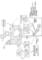
実施例において開示するように、PKCアイソフォーム阻害因子の投与は、多種多様性のある皮膚細胞タイプ(皮膚細胞だけではない。つまり、マクロファージは、他の組織において見いだされ、活性である)における前炎症性サイトカイン、ケモカインおよびTh1サイトカインの分泌を減少させることが証明されている。さらに、PKCアイソフォームの投与は、活性化因子、例えばケラチン生成細胞および内皮細胞上のICAM−1およびマクロファージ上のmac−2の発現を減少させる。さらに、PKCα阻害因子は、皮膚炎症の治療において、および乾癬の炎症性皮膚モデルにおける炎症症状を軽減するために有効であることが見いだされている。実施例でさらに考察するように、PCKアイソフォームの阻害因子の作用機序は、炎症性疾患または障害のための効果的療法としてのそれらの使用に関係することが解明されてきた。例えば、PCKアイソフォームのペプチド阻害因子は、例えば、図30に要約したように、炎症性プロセスの様々な工程において様々な細胞タイプの動員および活性化へ特異的に影響を及ぼすことによって:1)最終分化を減少させることにより表皮分化マーカーの発現を正常化する、2)異常過剰増殖を軽減する、3)皮膚構造を調節し、皮膚強度を増強する、および/または4)炎症をダウンレギュレートすることが証明されてきた。 As disclosed in the Examples, administration of a PKC isoform inhibitor prior to a wide variety of skin cell types (not only skin cells, ie macrophages are found and active in other tissues). It has been demonstrated to reduce secretion of inflammatory cytokines, chemokines and Th1 cytokines. Furthermore, administration of PKC isoforms reduces the expression of activators such as ICAM-1 on keratinocytes and endothelial cells and mac-2 on macrophages. Furthermore, PKCα inhibitors have been found to be effective in the treatment of skin inflammation and to reduce inflammatory symptoms in an inflammatory skin model of psoriasis. As further discussed in the Examples, it has been elucidated that the mechanism of action of inhibitors of PCK isoforms is related to their use as effective therapies for inflammatory diseases or disorders. For example, peptide inhibitors of PCK isoforms, for example, by specifically affecting the recruitment and activation of various cell types at various steps of the inflammatory process, as summarized in FIG. 30: 1) Normalize the expression of epidermal differentiation markers by reducing terminal differentiation, 2) reduce abnormal hyperproliferation, 3) modulate skin structure, enhance skin strength, and / or 4) down-regulate inflammation Has been proven to do.
さらに実施例において開示するように、PKCδの活性化因子は、多種多様性のある皮膚細胞タイプにおいて前炎症性サイトカインの分泌を減少させることもまた証明されている。そこで、また別の態様では、本開示は、被験体において炎症性疾患または障害を治療する方法であって、該被験体にPKCδの活性化因子を投与し、それにより該被験体において該炎症性疾患または障害を治療する方法を提供する。様々な実施形態では、活性化因子は、PKCδを選択的に活性化するポリペプチド、例えば配列番号30〜37のポリペプチドである。 Further, as disclosed in the Examples, activators of PKCδ have also been demonstrated to reduce secretion of proinflammatory cytokines in a wide variety of skin cell types. Thus, in yet another aspect, the present disclosure is a method of treating an inflammatory disease or disorder in a subject, wherein the subject is administered an activator of PKCδ, thereby causing the inflammatory condition in the subject. Methods of treating a disease or disorder are provided. In various embodiments, the activator is a polypeptide that selectively activates PKCδ, eg, a polypeptide of SEQ ID NOs: 30-37.
さらに、実施例において開示するように、PKCα阻害因子およびPKCη阻害因子の投与は、MSの症状を軽減することが見いだされてきた。したがって、また別の態様では、本開示は、被験体において多発性硬化症を治療する方法を提供する。本方法は、被験体にPKCαまたはPKCηの阻害因子を投与する工程を包含し、それにより該被験体における多発性硬化症を治療する。 Furthermore, as disclosed in the Examples, administration of a PKCα inhibitor and a PKCη inhibitor has been found to reduce the symptoms of MS. Accordingly, in yet another aspect, the present disclosure provides a method of treating multiple sclerosis in a subject. The method includes administering to the subject an inhibitor of PKCα or PKCη, thereby treating multiple sclerosis in the subject.
さらに、PKCアイソフォーム阻害因子の投与は、掻痒症の治療において有効であることが見いだされている。したがって、また別の態様では、本開示は、被験体において掻痒症を治療する方法を提供する。本方法は、被験体にPKCの阻害因子を投与する工程を包含し、それにより該被験体における掻痒症を治療する。 Furthermore, administration of PKC isoform inhibitor has been found to be effective in the treatment of pruritus. Accordingly, in yet another aspect, the present disclosure provides a method of treating pruritus in a subject. The method includes administering to the subject an inhibitor of PKC, thereby treating pruritus in the subject.
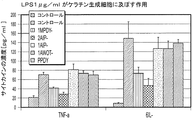
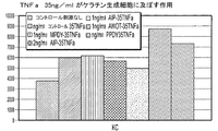
実施例および図面は、PKCδの活性化因子が、主要な前炎症性サイトカイン、例えばIL−1、IL−6およびTNFαの分泌を阻害する能力を示すデータを提示している。類似のデータは、PKCα、PKCεおよびPKCηを包含する様々なPKCアイソフォーム阻害因子について示されている。実施例において示したように、本開示のPKC阻害因子および活性化因子を包含する製剤は、主要前炎症性サイトカインの分泌を阻害することが証明されてきた。乾癬に関しては、特定の理論に連関しなくても、前炎症性物質のレベルを減少させると、隣接血管内の内皮細胞の活性化、したがって好中球、マクロファージおよびT細胞の乾癬斑への動員を防止すると考えられている。さらに、TH1およびTH17細胞は、炎症を強化する、またはケラチン生成細胞過剰増殖を各々駆動すると思われる特異的サイトカインの分泌によって、乾癬の病因に関与することが証明された。上述した前炎症性サイトカインは、これらのTH17細胞の発生(Mangan et al.(2006)Nature 441:231−234;Bettelli et al.(2006)Nature 441:235−238)およびTH1細胞活性に不可欠であると思われる。PKC阻害因子および活性化因子によるそれらの分泌の減少は、炎症性障害および掻痒症の効果的治療におけるそれらの使用を関係付けている。 The examples and figures present data showing the ability of PKCδ activators to inhibit the secretion of key pro-inflammatory cytokines such as IL-1, IL-6 and TNFα. Similar data has been shown for various PKC isoform inhibitors, including PKCα, PKCε and PKCη. As demonstrated in the examples, formulations comprising PKC inhibitors and activators of the present disclosure have been demonstrated to inhibit the secretion of major proinflammatory cytokines. With respect to psoriasis, without being linked to a specific theory, reducing the level of pro-inflammatory substances activates endothelial cells in adjacent blood vessels and thus mobilizes neutrophils, macrophages and T cells to psoriatic plaques It is thought to prevent. Furthermore, TH1 and TH17 cells have been shown to be involved in the pathogenesis of psoriasis by the secretion of specific cytokines that appear to enhance inflammation or drive keratinocyte hyperproliferation, respectively. The pro-inflammatory cytokines described above are essential for the development of these TH17 cells (Mangan et al. (2006) Nature 441: 231-234; Bettelli et al. (2006) Nature 441: 235-238) and TH1 cell activity. It appears to be. The decrease in their secretion by PKC inhibitors and activators has implicated their use in the effective treatment of inflammatory disorders and pruritus.
様々な実施形態では、PKCアイソフォームの阻害因子は、PKCの偽基質領域の阻害因子であり、ポリペプチドであるが、PKCアイソフォームの活性化因子もまたポリペプチドである。用語「ポリペプチド」、「ペプチド」または「タンパク質」は、本明細書では、隣接残基のαアミノ基とカルボキシ基とのペプチド結合によって1つを他の残基に結合させた直鎖系列のアミノ酸残基を指定するために互換的に使用される。 In various embodiments, the inhibitor of PKC isoform is an inhibitor of the PKC pseudosubstrate region and is a polypeptide, but the activator of PKC isoform is also a polypeptide. The term “polypeptide”, “peptide” or “protein” as used herein refers to a linear series in which one is linked to another residue by a peptide bond between the α-amino group and the carboxy group of adjacent residues. Used interchangeably to designate amino acid residues.
様々な実施形態では、使用可能なペプチドPKC活性化因子および阻害因子の例には、制限なく、表1に記載の配列番号1〜5、14〜19、26、27および30〜33のペプチドまたはそれらの生理学的に許容される塩、ならびに特定の修飾もしくは末端保護基を有すると示されている表1の配列番号6〜13、20〜25、28、29、34〜37のペプチドが包含される。 In various embodiments, examples of peptide PKC activators and inhibitors that can be used include, without limitation, peptides of SEQ ID NOs: 1-5, 14-19, 26, 27 and 30-33 listed in Table 1 or These physiologically acceptable salts, as well as the peptides of SEQ ID NOs: 6-13, 20-25, 28, 29, 34-37 of Table 1 indicated as having specific modifications or terminal protecting groups are included. The

様々な実施形態では、ペプチドPKC阻害因子または活性化因子は、典型的には6〜12アミノ酸を含有するが、長さがもっと長くても短くてもよい。様々な実施形態では、ペプチドPKC阻害因子または活性化因子は長さが6〜45、6〜40、6〜35、6〜30、6〜25、6〜20、6〜15、または6〜10個のアミノ酸の範囲に及んでよい。一実施形態では、該ペプチドは、6、7、8、9、10、11、12、13、14、または15個のアミノ酸を包含する。 In various embodiments, peptide PKC inhibitors or activators typically contain 6-12 amino acids, but may be longer or shorter. In various embodiments, the peptide PKC inhibitor or activator is 6-45, 6-40, 6-35, 6-30, 6-25, 6-20, 6-15, or 6-10 in length. It may span a range of individual amino acids. In one embodiment, the peptide comprises 6, 7, 8, 9, 10, 11, 12, 13, 14, or 15 amino acids.
様々な実施形態では、ペプチドPKC阻害因子または活性化因子は、好ましくはC12−C20脂肪酸、例えばC14アシル(ミリストイル)またはC16アシル(パルミトイル)に由来するアシル基によってN−アセチル化されてよい。 In various embodiments, the peptide PKC inhibitors or activators are preferably C 12 -C 20 fatty acids, for example C 14 acyl (myristoyl) or N- acetylated with acyl groups derived from C 16 acyl (palmitoyl) It's okay.
一般に、ペプチドPKCα阻害因子には、共通モチーフ配列Phe−Ala−Arg−Lys−Gly−Ala(配列番号1)が包含される。または、また別の実施形態では、PKCα阻害因子には、共通モチーフ配列Thr−Leu−Asn−Pro−Gln−Trp−Glu−Ser(配列番号5)が包含される。 In general, peptide PKCα inhibitors include the common motif sequence Phe-Ala-Arg-Lys-Gly-Ala (SEQ ID NO: 1). Alternatively, in yet another embodiment, the PKCα inhibitor includes the common motif sequence Thr-Leu-Asn-Pro-Gln-Trp-Glu-Ser (SEQ ID NO: 5).
ペプチドPKC阻害因子および活性化因子は正確な配列またはモチーフ配列によって規定できるが、当業者であれば、類似の配列を有するペプチドは類似機能を有する可能性があることを理解できる。このため、実質的に同一の配列を有する、または実質的に表1に記載のPKC阻害因子または活性化因子と全く同一もしくは類似する配列を有するペプチドは、含まれることが意図されている。本明細書で使用する用語「実質的に同一の配列」には、配列番号1〜37によって規定された配列と少なくとも60+%(60%以上を意味する)、好ましくは70+%、より好ましくは80+%、および最も好ましくは90+%、95+%、または98+%配列同一性を有する配列を包含する、およびPKCアイソフォーム活性を阻害または活性化するペプチドが包含される。 Peptide PKC inhibitors and activators can be defined by exact sequences or motif sequences, but those skilled in the art will understand that peptides having similar sequences may have similar functions. For this reason, it is intended to include peptides having substantially the same sequence, or substantially the same or similar sequences to the PKC inhibitors or activators listed in Table 1. As used herein, the term “substantially identical sequence” includes at least 60 +% (meaning 60% or more), preferably 70 +%, more preferably 80+, with the sequence defined by SEQ ID NOs: 1-37. Peptides that include%, and most preferably 90 +%, 95 +%, or 98 +% sequence identity, and that inhibit or activate PKC isoform activity are included.
2つのポリペプチドが実質的に同一であるというまた別の指摘は、1つのポリペプチドが第2のポリペプチドと免疫学的に交差反応性であることである。そこで、ポリペプチドは、例えば2つのペプチドが保存的置換によってのみ相違する場合、典型的には第2ポリペプチドと実質的に同一である。 Another indication that two polypeptides are substantially identical is that one polypeptide is immunologically cross-reactive with a second polypeptide. Thus, a polypeptide is typically substantially identical to a second polypeptide, eg, if the two peptides differ only by conservative substitutions.
用語「保存的置換」は、タンパク質またはペプチドに関連して分子の活性(例えば、抗菌活性)を実質的には変化させないアミノ酸置換を示すために使用される。典型的な保存的アミノ酸置換は、1つのアミノ酸と類似の化学特性(例えば、電荷または疎水性)を備える別のアミノ酸との置換を包含する。以下の6つの群は、各々相互に対して典型的な保存的置換であるアミノ酸を含有する、1)アラニン(A)、セリン(S)、トレオニン(T)、2)アスパラギン酸(D)、グルタミン酸(E)、3)アスパラギン(N)、グルタミン(Q)、4)アルギニン(R)、リシン(K)、5)イソロイシン(I)、ロイシン(L)、メチオニン(M)、バリン(V)、ならびに6)フェニルアラニン(F)、チロシン(Y)、およびトリプトファン(W)。 The term “conservative substitution” is used to indicate an amino acid substitution that does not substantially alter the activity (eg, antimicrobial activity) of a molecule in relation to a protein or peptide. Typical conservative amino acid substitutions include substitution with one amino acid for another amino acid with similar chemical properties (eg, charge or hydrophobicity). The following six groups each contain amino acids that are typical conservative substitutions for each other: 1) alanine (A), serine (S), threonine (T), 2) aspartic acid (D), Glutamic acid (E), 3) asparagine (N), glutamine (Q), 4) arginine (R), lysine (K), 5) isoleucine (I), leucine (L), methionine (M), valine (V) And 6) phenylalanine (F), tyrosine (Y), and tryptophan (W).
用語「アミノ酸」は、極めて広い意味では、天然型アミノ酸ならびにアミノ酸アナログを包含する非天然型アミノ酸を包含するために使用される。この広い定義を考慮すると、当業者であれば、本明細書でのアミノ酸に関する言及には、例えば、天然型のタンパク生成(L)−アミノ酸、(D)−アミノ酸、化学修飾アミノ酸、例えばアミノ酸アナログ、天然型の非タンパク生成アミノ酸、例えばノルロイシン、および当分野においてアミノ酸に特徴的である公知の特性を有する化学合成化合物が包含されることが公知である。本明細書で使用する用語「タンパク生成(proteogenic)」は、アミノ酸を代謝経路を通して細胞内のタンパク質内に組み込めることを示している。 The term “amino acid” is used in a very broad sense to include natural amino acids as well as unnatural amino acids, including amino acid analogs. In view of this broad definition, those skilled in the art will refer to amino acids herein, for example, naturally occurring proteinogenic (L) -amino acids, (D) -amino acids, chemically modified amino acids, such as amino acid analogs. Naturally occurring non-proteinogenic amino acids such as norleucine, and chemically synthesized compounds having known properties characteristic of amino acids in the art are known to be included. The term “proteogenic” as used herein indicates that amino acids can be incorporated into proteins within cells through metabolic pathways.
2つのポリペプチド配列の状況における用語「同一」または「同一性率」(%)は、配列比較アルゴリズムを使用して、もしくは視覚的検定によって測定した場合、比較して最大対応についてアラインメントされると同一である、または規定パーセンテージの同一であるアミノ酸残基を有する2つ以上の配列または部分配列に関する。 The terms “identical” or “percent identity” (%) in the context of two polypeptide sequences are compared and aligned for maximum correspondence when measured using a sequence comparison algorithm or by visual test. It relates to two or more sequences or subsequences having the same or a specified percentage of identical amino acid residues.
2つのポリペプチド配列の状況における文言「実質的に同一」は、配列比較アルゴリズムを使用して、もしくは視覚的検査によって測定した場合、最大一致で、比較及びアラインメントされると、少なくとも60+%、好ましくは80+%、最も好ましくは90〜95+%のアミノ酸残基同一性を有する2つ以上の配列または部分配列に関する。 The phrase “substantially identical” in the context of two polypeptide sequences is at least 60 +%, preferably at least 60 +% when compared and aligned, with maximum match, as measured using a sequence comparison algorithm or by visual inspection. Refers to two or more sequences or subsequences having 80 +%, most preferably 90-95 +% amino acid residue identity.
当分野において一般に公知であるように、比較のための最適な配列アラインメントは、例えばSmith & Waterman((1981)Adv Appl Math 2:482)の局所ホモロジーアルゴリズムによって、Needleman & Wunsch((1970)J Mol Biol 48:443)のホモロジーアラインメントアルゴリズムによって、Pearson & Lipman((1988)Proc Natl Acad Sci USA 85:2444)の類似性検索方法によって、視覚的検定によるこれらのアルゴリズムのコンピュータ化実行、またはその他の効果的方法によって実施することができる。 As is generally known in the art, an optimal sequence alignment for comparison can be found, for example, by the Needleman & Wunsch ((1970) J Mols) by the local homology algorithm of Smith & Waterman ((1981) Adv Appl Math 2: 482). Biol 48: 443) by the homology alignment algorithm of Pearson & Lipman ((1988) Proc Natl Acad Sci USA 85: 2444), by computerized implementation of these algorithms by visual testing, or other effects Can be carried out by a conventional method.
ペプチドPKC阻害因子または活性化因子は、修飾アミノ酸配列または非天然型末端修飾を有する可能性がある。ペプチド配列への修飾には、例えば、そのような修飾によって生成されたペプチドがPKCα阻害因子活性を保持することを前提に、アミノ酸の付加、欠失または置換を包含することができる。さらに、ペプチドは、遊離末端もしくはアミノ保護(例えば、N−保護)および/またはカルボキシ保護(例えば、C−保護)末端を備える形態で存在してよい。保護基には、(a)ベンジルオキシカルボニル、2−クロロベンジルオキシカルボニル、9−フルオレニルメチルオキシカルボニル、イソニコチニルオキシカルボニルおよび4−メトキシベンジルオキシカルボニルを包含する芳香族ウレタン型保護基、(b)t−ブトキシカルボニル、t−アミルオキシカルボニル、イソプロピルオキシカルボニル、2−(4−ビフェニル)−2−プロピルオキシカルボニル、アリルオキシカルボニルおよびメチルスルホニルエトキシカルボニルを包含する脂肪族ウレタン型保護基、(c)アダマンチルオキシカルボニル、シクロペンチルオキシカルボニル、シクロヘキシルオキシカルボニルおよびイソボルニルオキシカルボニルを包含するシクロアルキルウレタン型保護基、(d)アシル保護基またはスルホニル保護基が包含される。追加の保護基には、ベンジルオキシカルボニル、t−ブトキシカルボニル、アセチル、2−プロピルペンタノイル、4−メチルペンタノイル、t−ブチルアセチル、3−シクロヘキシルプロピオニル、n−ブタンスルホニル、ベンジルスルホニル、4−メチルベンゼンスルホニル、2−ナフタレンスルホニル、3−ナフタレンスルホニルおよび1−カンファースルホニルが包含される。 A peptide PKC inhibitor or activator may have a modified amino acid sequence or a non-natural terminal modification. Modifications to the peptide sequence can include, for example, amino acid additions, deletions or substitutions, provided that the peptide produced by such modifications retains PKCα inhibitor activity. Furthermore, the peptides may exist in a form with a free terminus or amino protected (eg N-protected) and / or carboxy protected (eg C-protected) terminus. Protecting groups include (a) aromatic urethane type protecting groups including benzyloxycarbonyl, 2-chlorobenzyloxycarbonyl, 9-fluorenylmethyloxycarbonyl, isonicotinyloxycarbonyl and 4-methoxybenzyloxycarbonyl, (B) an aliphatic urethane type protecting group including t-butoxycarbonyl, t-amyloxycarbonyl, isopropyloxycarbonyl, 2- (4-biphenyl) -2-propyloxycarbonyl, allyloxycarbonyl and methylsulfonylethoxycarbonyl; (C) a cycloalkylurethane type protecting group including adamantyloxycarbonyl, cyclopentyloxycarbonyl, cyclohexyloxycarbonyl and isobornyloxycarbonyl, (d) an acyl protecting group or Sulfonyl protecting groups are included. Additional protecting groups include benzyloxycarbonyl, t-butoxycarbonyl, acetyl, 2-propylpentanoyl, 4-methylpentanoyl, t-butylacetyl, 3-cyclohexylpropionyl, n-butanesulfonyl, benzylsulfonyl, 4- Methylbenzenesulfonyl, 2-naphthalenesulfonyl, 3-naphthalenesulfonyl and 1-camphorsulfonyl are included.
様々な実施形態では、ペプチドPKCアイソフォーム阻害因子および活性化因子は、被験体を治療するために局所、非経口、皮下、腹腔内、肺内、鼻腔内、静脈内、および/または病巣内投与を包含する任意の適切な手段によって投与することができる。しかし、典型的な実施形態では、ペプチドは局所投与のため、例えば液剤、クリーム剤、ゲル剤、軟膏剤、泡スプレーなどの形態で調製される。 In various embodiments, the peptide PKC isoform inhibitor and activator are administered topically, parenterally, subcutaneously, intraperitoneally, intrapulmonary, intranasally, intravenously, and / or intralesionally to treat the subject. Can be administered by any suitable means, including However, in typical embodiments, the peptides are prepared for topical administration, eg in the form of solutions, creams, gels, ointments, foam sprays and the like.
本開示によって使用されるPKCアイソフォーム阻害因子または活性化因子の治療用製剤は、例えば、所望の純度を有するPKCアイソフォーム阻害因子または活性化因子を任意の薬学的に許容される担体、賦形剤および/または安定剤とともに混合する工程によって調製される(例えば、Remington’s Pharmaceutical Sciences, 16th edition, Osol, A. Ed.(1980)を参照されたい)。許容される担体、賦形剤、または安定剤は、使用される用量および濃度で受容者に非毒性であり、緩衝剤、例えばリン酸塩、クエン酸塩、および他の有機酸類、アスコルビン酸およびメチオニンを包含する抗酸化物質、保存料(例えば、塩化オクタデシルジメチルベンジルアンモニウム、塩化ヘキサメトニウム、塩化ベンズアルコニウム、塩化ベンゼトニウム、フェノール、ブチルもしくはベンジルアルコール、アルキルパラベン類、例えばメチルもしくはプロピルパラベン、カテコール、レゾルシノール、シクロヘキサノール、3−ペンタノール、およびm−クレゾール)、低分子量(約10残基未満)ポリペプチド、タンパク質、例えば血清アルブミン、ゼラチン、もしくはイムノグロビン類、親水性ポリマー類、例えばポリビニルピロリドン、アミノ酸、例えばグリシン、グルタミン、アスパラギン、ヒスチジン、アルギニン、もしくはリシン、単糖類、二糖類、およびその他のグルコース、マンノース、もしくはデキストリン類を包含する炭水化物、キレート剤、例えばEDTA、糖類、例えば、スクロース、マンニトール、トレハロースもしくはソルビトール、塩形成対イオン、例えばナトリウム、金属錯体(例えば、Zn−タンパク質錯体)、および/または非イオン性界面活性化因子、例えばTWEEN(商標)、PLURONICS(商標)またはポリエチレングリコール(PEG)を包含することができる。 The therapeutic formulation of a PKC isoform inhibitor or activator used by the present disclosure can be any pharmaceutically acceptable carrier, excipient, eg, a PKC isoform inhibitor or activator having a desired purity. Prepared by mixing with agents and / or stabilizers (see, eg, Remington's Pharmaceutical Sciences, 16th edition, Osol, A. Ed. (1980)). Acceptable carriers, excipients, or stabilizers are nontoxic to recipients at the dosages and concentrations used, and buffers such as phosphate, citrate, and other organic acids, ascorbic acid and Antioxidants, including methionine, preservatives (eg octadecyldimethylbenzylammonium chloride, hexamethonium chloride, benzalkonium chloride, benzethonium chloride, phenol, butyl or benzyl alcohol, alkyl parabens such as methyl or propyl paraben, catechol , Resorcinol, cyclohexanol, 3-pentanol, and m-cresol), low molecular weight (less than about 10 residues) polypeptides, proteins such as serum albumin, gelatin, or immunoglobins, hydrophilic polymers such as polyvinyl pyro Don, amino acids such as glycine, glutamine, asparagine, histidine, arginine, or lysine, monosaccharides, disaccharides, and other carbohydrates, chelating agents such as EDTA, sugars such as sucrose, including glucose, mannose, or dextrins , Mannitol, trehalose or sorbitol, salt-forming counterions such as sodium, metal complexes (eg Zn-protein complexes), and / or nonionic surfactants such as TWEEN ™, PLURONICS ™ or polyethylene glycol (PEG) can be included.
典型的な実施形態では、PKCアイソフォーム阻害因子または活性化因子は、クリーム剤に調製される。PKCアイソフォームの阻害因子および活性化因子は、皮膚炎症および他の炎症性障害の局所治療のために理想的であるが、それはPKC酵素の活性を特異的に標的とすることができるからである。特異的PKC酵素の阻害または活性化は、他のPKCアイソフォームに影響を及ぼさずに低濃度でPKCアイソフォームを選択的に調節する能力によって達成される。 In an exemplary embodiment, the PKC isoform inhibitor or activator is prepared in a cream. Inhibitors and activators of PKC isoforms are ideal for local treatment of skin inflammation and other inflammatory disorders because they can specifically target the activity of PKC enzymes . Inhibition or activation of specific PKC enzymes is achieved by the ability to selectively modulate PKC isoforms at low concentrations without affecting other PKC isoforms.
局所投与の代表的な製剤は、ペプチドMPDY−1(配列番号6)が局所投与のためのクリーム剤として調製される実施例4に開示されている。しかし、当業者であれば、本製剤は、クリーム剤の本質的特性、例えば粘度、安定性、非毒性などを保持しながら変更を加えられることを理解するであろう。さらに、当業者であれば、本製剤を本開示のペプチドPKC阻害因子または活性化因子のいずれかのための溶媒として使用可能なことを認識するであろう。 A representative formulation for topical administration is disclosed in Example 4 where peptide MPDY-1 (SEQ ID NO: 6) is prepared as a cream for topical administration. However, those skilled in the art will appreciate that the formulation can be modified while retaining the essential properties of the cream, such as viscosity, stability, non-toxicity, and the like. Furthermore, those skilled in the art will recognize that the formulations can be used as solvents for either the peptide PKC inhibitors or activators of the present disclosure.
また別の実施形態では、1つの製造品、例えば本開示の治療方法を実施するために有用な材料を含有するキットが提供される。様々な実施形態では、本キットは、PKCアイソフォーム活性化因子または阻害因子、つまり本明細書に開示したペプチドPKCアイソフォーム阻害因子または活性化因子、および該被験体に該活性化因子または阻害因子を投与するための取扱説明書を包含する。 In yet another embodiment, a kit is provided that contains one article of manufacture, eg, a material useful for performing the therapeutic methods of the present disclosure. In various embodiments, the kit comprises a PKC isoform activator or inhibitor, ie, a peptide PKC isoform inhibitor or activator disclosed herein, and the activator or inhibitor on the subject. Instructions for administering are included.
用語「取扱説明書」または「添付文書」は、適応、用法、用量、投与、禁忌、包装された製品と結合すべき他の治療用製品および/またはそのような治療用製品の使用に関する警告についての情報を含有する、治療用製品の商業包装内に慣習的に包含される取扱説明書を意味するために使用される。 The term “instruction manual” or “package insert” refers to indications, usage, doses, administration, contraindications, other therapeutic products to be combined with the packaged product and / or warnings regarding the use of such therapeutic products. Is used to mean instructions that are conventionally included in commercial packaging of therapeutic products.
本明細書で開示するPKCαの阻害因子は、特定投与経路のために調製することができる。したがって、本キットは、適切な容器、例えばチューブ、ボトル、バイアル、シリンジなどの中に含有されるPKCαの阻害因子を包含する製剤を包含することができる。容器は、様々な材料、例えばガラスまたはプラスチックから形成されてよい。容器は、炎症性疾患を治療するために有効である組成物を保持または含有し、無菌アクセスポートを有することができる(例えば、容器は、静脈注射溶液バッグまたは皮下注射針によって穿刺可能なストッパーを有するバイアルであってよい)。製剤中の少なくとも1つの成分は、PKCアイソフォームの阻害因子または活性化因子である。ラベルまたは添付文書は、PKCアイソフォームの阻害因子または活性化因子を包含する製剤を提供するために投与量および投与間隔に関する特別の手引きを用いて、本組成物が炎症性疾患を患う患者において治療するために使用されることを示している。本製造品は、商業的観点および利用者の観点から望ましい、他の緩衝剤、希釈剤、フィルター、ニードル、およびシリンジを包含する他の材料をさらに包含することができる。 The inhibitors of PKCα disclosed herein can be prepared for a specific route of administration. Thus, the kit can include a formulation that includes an inhibitor of PKCα contained in a suitable container, such as a tube, bottle, vial, syringe, or the like. The container may be formed from a variety of materials, such as glass or plastic. The container holds or contains a composition that is effective for treating an inflammatory disease and may have a sterile access port (eg, the container may have a stopper pierceable by an intravenous solution bag or hypodermic needle. Vials). At least one component in the formulation is an inhibitor or activator of the PKC isoform. The label or package insert treats the composition in patients suffering from inflammatory diseases using special guidance on dosage and dosing intervals to provide a formulation comprising an inhibitor or activator of PKC isoform Shows that it is used to. The article of manufacture can further include other materials, including other buffers, diluents, filters, needles, and syringes, which are desirable from a commercial and user standpoint.
当然ながら、治療を必要とする任意の特定被験体のための特定用量レベルおよび投与頻度は変動してよく、使用されるPKCアイソフォームの阻害因子または活性化因子の活性、その化合物の代謝安定性および作用の長さ、年齢、体重、全身健康状態、性別、食事、投与様式および時刻、特定状態の重症度、および療法を受ける宿主に依存することになる。しかし一般に、用量は、特異的PKCアイソフォーム阻害因子または活性化因子の公知の投与方法に典型的な用量に近似する。当業者であれば、最適用量、投与方法および反復頻度を容易に決定することができる。正確な処方および用量は、患者の状態に照らして個々の医師が選択することができる(Fingl et al. “The Pharmacological Basis of Therapeutics”, Ch.1 p.1(1975))。 Of course, the particular dose level and frequency of administration for any particular subject in need of treatment may vary and the activity of the PKC isoform inhibitor or activator used, the metabolic stability of the compound And the length of action, age, weight, general health, sex, diet, mode of administration and time, severity of the particular condition, and the host receiving therapy. In general, however, the dose will approximate that typical for known methods of administration of specific PKC isoform inhibitors or activators. Persons of ordinary skill can easily determine optimum dosages, dosing methodologies and repetition rates. The exact formulation and dosage can be chosen by the individual physician in light of the patient's condition (Fingl et al. “The Pharmaceutical Basis of Therapeutics”, Ch. 1 p. 1 (1975)).
そこで、治療対象の状態の重症度および反応性に依存して、投与は単回、または治癒するまで、または障害の減少が達成されるまで治療過程が数日間から数週間持続する反復投与であってよい。 Thus, depending on the severity and responsiveness of the condition being treated, administration may be single or repeated, with the course of treatment lasting days to weeks until healing or reduction of the disorder is achieved. It's okay.
PKCアイソフォーム阻害因子または活性化因子がペプチドである様々な実施形態では、該ペプチドは組成物中で0.001〜100μg/mlの濃度で提供される。例えば、濃度は、0.001〜100、0.01〜50、0.01〜10、0.01〜1、および0.01〜0.5μg/mlであってよい。 In various embodiments where the PKC isoform inhibitor or activator is a peptide, the peptide is provided in the composition at a concentration of 0.001-100 μg / ml. For example, the concentration may be 0.001 to 100, 0.01 to 50, 0.01 to 10, 0.01 to 1, and 0.01 to 0.5 μg / ml.
1つの投与プロトコールでは、本方法は、ペプチドPKCアイソフォーム阻害因子または活性化因子を該被験体へ局所的に、例えばクリーム剤として投与する工程を含んでいる。ペプチドは、約1μg/ml〜約1,000μg/ml、1μg/ml〜約500μg/ml、1μg/ml〜約100μg/ml、1μg/ml〜約10μg/ml、または10μg/ml〜約100μg/mlの濃度で局所投与される。ペプチドは、その状態が治療されるまで少なくとも1日1回投与される。 In one administration protocol, the method includes administering a peptide PKC isoform inhibitor or activator locally to the subject, eg, as a cream. The peptide may be from about 1 μg / ml to about 1,000 μg / ml, 1 μg / ml to about 500 μg / ml, 1 μg / ml to about 100 μg / ml, 1 μg / ml to about 10 μg / ml, or 10 μg / ml to about 100 μg / ml. Topically administered at a concentration of ml. The peptide is administered at least once daily until the condition is treated.
また別の投与プロトコールでは、本方法は、ペプチドPKCアイソフォーム阻害因子または活性化因子を該被験体へ非経口、皮下または静脈内投与する工程を含んでいる。ペプチドは、約1μg/ml〜約1,000μg/ml、1μg/ml〜約500μg/ml、1μg/ml〜約100μg/ml、1μg/ml〜約10μg/ml、または10μg/ml〜約100μg/mlの濃度で投与される。ペプチドは、その状態が治療されるまで少なくとも1日1回、週1回、2週に1回、または月1回投与される。 In yet another administration protocol, the method comprises parenterally, subcutaneously or intravenously administering the peptide PKC isoform inhibitor or activator to the subject. The peptide may be from about 1 μg / ml to about 1,000 μg / ml, 1 μg / ml to about 500 μg / ml, 1 μg / ml to about 100 μg / ml, 1 μg / ml to about 10 μg / ml, or 10 μg / ml to about 100 μg / ml. Administered at a concentration of ml. The peptide is administered at least once a day, once a week, once every two weeks, or once a month until the condition is treated.
以下の実施例は、本発明の実施形態をさらに具体的に示すために提供するが、範囲を限定することは意図されていない。これらの実施例は使用可能な実施例の典型ではあるが、あるいはまた当業者には公知の他の手順、方法、または技術を使用することができる。 The following examples are provided to more specifically illustrate embodiments of the present invention, but are not intended to limit the scope. While these examples are representative of examples that can be used, other procedures, methods, or techniques known to those skilled in the art can also be used.
PKCαの阻害は炎症性皮膚障害である乾癬に特徴的なケラチン生成細胞構造統合性(structure integrity)を調節する
PKCαの阻害は、乾癬に特徴的なケラチン生成細胞構造統合性を調節することが証明された。皮膚組織をパラフィン包埋し、H&E(ヘマトキシリンおよびエオシン)一般組織学的染色のため、または基底層に対するケラチン14(K14)、有棘層に対するケラチン1(K1)、ケラチン生成細胞遊走に対するケラチン6(K6)およびケラチン生成細胞増殖に対するPCNAを包含する様々な皮膚層に対する別のマーカーについて染色した。結果は、PKCα阻害後の皮膚特性の正常化を証明している(図2)。
Inhibition of PKCα regulates the keratinocyte structure integrity characteristic of psoriasis, an inflammatory skin disorder. Inhibition of PKCα proves to modulate the keratinocyte structure integrity characteristic of psoriasis It was done. Skin tissue is embedded in paraffin and H & E (hematoxylin and eosin) for general histological staining or keratin 14 (K14) for basal layer, keratin 1 (K1) for spiny layer,
乾癬モデルによる炎症のin vivoおよびex vivo治療を評価するためのモデル
これまで極めて多数の動物モデルが乾癬を試験するために使用されてきたが、これらのモデルはいずれも、過剰な皮膚産生、新規血管の形成、および重度免疫機能障害を特徴とするヒト疾患病理を適正に模倣するために十分ではなかった。一般に、乾癬の有用なモデルであると見なすには、モデルは、乾癬との一部の組織病理学的特徴を共有し、類似の病因および/または疾患機序を示し、そしてこの疾患を治療するための治療薬に同様に応答しなければならない。既存モデルは、表皮肥厚、表皮分化の変化、脈管新生の増加、および白血球/T細胞浸潤を包含する幾つかの特徴を示す。しかし、特に既存のマウスモデルの中では、既存薬物および療法に応答するマウスは多くない。したがって、既存モデルを使用して、以下の実施例において利用された乾癬治療を評価するための新規のin vitro、ex vivoおよびin vivoモデルが開発されてきた。
Models for assessing in vivo and ex vivo treatment of inflammation with psoriasis models A large number of animal models have so far been used to test psoriasis, but all of these models are It was not sufficient to properly mimic the formation of blood vessels and human disease pathology characterized by severe immune dysfunction. In general, to be considered a useful model of psoriasis, the model shares some histopathological features with psoriasis, exhibits similar etiology and / or disease mechanism, and treats the disease In order to respond to therapeutic drugs as well. Existing models exhibit several features including epidermal thickening, altered epidermal differentiation, increased angiogenesis, and leukocyte / T cell infiltration. However, among existing mouse models, not many mice respond to existing drugs and therapies. Thus, using existing models, new in vitro, ex vivo and in vivo models have been developed to evaluate the psoriasis treatment utilized in the following examples.
in vitroモデル
開発されたモデルは、細胞系および皮膚由来細胞ならびに免疫細胞の一次培養物を使用し、STAT3およびPKCα媒介性シグナル伝達経路を過剰発現させて不活性化するための構築体およびツールを利用する細胞培養試験を包含していた。皮膚細胞の増殖、遊走、分化、炎症およびシグナル伝達を試験するための膨大な一連の技術が利用され、乾癬の発生機序を試験し、乾癬におけるPKCαの阻害の治療作用を試験するのに有用であることが証明された。
In Vitro Model The developed model uses constructs and tools to overexpress and inactivate STAT3 and PKCα-mediated signaling pathways using primary cultures of cell lines and skin-derived cells and immune cells. The cell culture test utilized was included. A vast array of techniques for testing skin cell proliferation, migration, differentiation, inflammation and signal transduction are utilized to test the pathogenesis of psoriasis and to test the therapeutic effects of PKCα inhibition in psoriasis Proved to be
in vivoモデル
PKCα過剰発現およびノックアウトマウスモデルを使用した。K5−PKCαトランスジェニックマウスを使用したケラチン生成細胞内でのPKCαの過剰発現は、例えば膿疱性乾癬などの状態を模倣する重度の表皮内好中球浸潤および表皮の破壊を示すことが証明された。トランスジェニックマウスのPKCαおよびDN形態の両方が確立され、これは、皮下投与によってin vivoで試験された。さらに、皮膚構造および機能にPKCα不活性化が及ぼす作用を試験するために、PKCαノックアウトマウスを使用した。
In vivo model A PKCα overexpression and knockout mouse model was used. Overexpression of PKCα in keratinocytes using K5-PKCα transgenic mice has been demonstrated to show severe intraepidermal neutrophil infiltration and epidermal destruction that mimic conditions such as pustular psoriasis . Both PKCα and DN forms of transgenic mice were established and were tested in vivo by subcutaneous administration. In addition, PKCα knockout mice were used to test the effect of PKCα inactivation on skin structure and function.
STAT3を過剰発現するマウスモデルを使用した。中でも、乾癬、ヒト乾癬に対する類似性に関するリードマウスモデルは、Stat3が表皮ケラチン生成細胞内で過剰発現するトランスジェニックマウスである。これらのマウスは、乾癬状の表皮での棘細胞増殖を発生し、真皮内では主としてCD4+であり、表皮内ではCD8+である皮膚リンパ球浸潤を有するが、これらは全てヒトにおける乾癬に類似する特徴である。 A mouse model overexpressing STAT3 was used. Among them, the lead mouse model for similarity to psoriasis and human psoriasis is a transgenic mouse in which Stat3 is overexpressed in epidermal keratinocytes. These mice develop spinal cell proliferation in the psoriatic-like epidermis and have skin lymphocyte infiltration that is primarily CD4 + in the dermis and CD8 + in the epidermis, all of which are similar to psoriasis in humans It is.
皮膚炎症および過形成のためのモデルとしての創傷。創傷状況における皮膚病変内の炎症を検出して定量的に評価するために、様々な皮膚区画における皮膚炎症応答を追跡し、この応答に影響を及ぼす薬剤の同定を可能にするスクリーニング方法が開発された。 Wound as a model for skin inflammation and hyperplasia. In order to detect and quantitatively assess inflammation within skin lesions in the wound situation, screening methods have been developed that allow for the identification of drugs that track the skin inflammatory response in various skin compartments and affect this response. It was.
ex vivoモデル
ニワトリ絨毛尿膜(CAM)上での乾癬性皮膚の移植。ニワトリ絨毛尿膜(CAM)上での乾癬性皮膚移植の技術は、ex vivo治療用途を試験する目的で開発された。この技術は皮膚腫瘍試験および血管新生実験において一般に使用されるが、これを乾癬試験のために採用して使用した。この最初のアプローチは、ヒト乾癬性皮膚への新規薬物の直接的な投与を可能にするため、そこで感染を治療するための新規薬物のより臨床的に意義のある試験を作り出す。移植後、乾癬性ヒト皮膚を利用して様々な製剤における様々な治療の有効性およびタイミングを確定し、形態学的、組織学的および生化学的分析を使用して分析する。
ex vivo model Transplantation of psoriatic skin on chicken chorioallantoic membrane (CAM). The technique of psoriatic skin transplantation on the chick chorioallantoic membrane (CAM) has been developed with the aim of testing ex vivo therapeutic applications. This technique is commonly used in skin tumor testing and angiogenesis experiments, but it was adopted and used for psoriasis testing. This first approach allows for the direct administration of new drugs to human psoriatic skin, thus creating a more clinically relevant test for new drugs to treat infections there. After transplantation, psoriatic human skin is utilized to determine the efficacy and timing of various treatments in various formulations and analyzed using morphological, histological and biochemical analyses.
PKCαノックアウトマウスにおける瘢痕形成の軽減
PKCαノックアウトマウスモデルを開発し、皮膚構造および機能にPKCα不活性化の作用が及ぼす作用を試験するために利用した。図3および4に示したように、落屑の軽減がPCKαノックアウトマウスにおいて観察された。図3は、平均落屑重症度が、コントロールと比較してPCKαノックアウトマウスにおいて50%超減少したことを示しているヒストグラムであり、PKCαの阻害が乾癬治療の重要な要件であることを証明している。これは、相違するマウスにおける落屑を比較している一連の図である図4においても証明されている。
Reduction of scar formation in PKCα knockout mice A PKCα knockout mouse model was developed and used to test the effects of PKCα inactivation on skin structure and function. As shown in FIGS. 3 and 4, desquamation reduction was observed in PCKα knockout mice. FIG. 3 is a histogram showing that mean desquamation severity was reduced by more than 50% in PCKα knockout mice compared to controls, demonstrating that PKCα inhibition is an important requirement for psoriasis treatment. Yes. This is also demonstrated in FIG. 4, which is a series of figures comparing desquamation in different mice.
局所的PKCα阻害因子製剤
局所的PKCα阻害因子製剤を開発し、乾癬の治療における有効性について評価した。ペプチドPKCα阻害因子MPDY−1(配列番号6)をクリーム剤に調製し(本明細書では、HO/02/10と称する)、その成分は表2に示した。
Topical PKCα inhibitor formulation A local PKCα inhibitor formulation was developed and evaluated for its effectiveness in treating psoriasis. Peptide PKCα inhibitor MPDY-1 (SEQ ID NO: 6) was prepared in a cream (referred to herein as HO / 02/10) and the ingredients are shown in Table 2.
PKCα阻害因子がin vitro表皮分化に及ぼす作用
実施例4の製剤(HO/02/10)をin vitroで表皮分化を制御するために決定した。基底ケラチン生成細胞は、分化してK1/K10ケラチンを特徴とする有棘層、ロリクリン/フィラグリンを特徴とする顆粒層および角層を形成する。ロリクリンおよびフィラグリン線維の発現および取込みの欠損には、乾癬を包含する様々な免疫学的皮膚疾患が関連している。そこで、HO/02/10が皮膚分化および増殖に及ぼす作用を評価した。図5および6に示したように、HO/02/10は、皮膚増殖(PCNA)(図6)を正常化し、ロリクリンおよびフィラグリンの発現を減少させることによって皮膚分化を調節したが、その間、有棘層は影響を受けないままであった(図5)。乾癬性皮膚ケラチン生成細胞は急速に分化して顆粒細胞および主として大量の角膜細胞(鱗屑)を生成するが、有棘層は薄化するので、HO/02/10は、皮膚特性を正常表現型に向けて修正することによって乾癬性皮膚を正常化するように機能した。
Effect of PKCα inhibitory factor on in vitro epidermal differentiation The preparation of Example 4 (HO / 02/10) was determined in order to control epidermal differentiation in vitro. Basal keratinocytes differentiate to form a spiny layer characterized by K1 / K10 keratin, a granular layer and a stratum corneum characterized by loricrin / filaggrin. Defects in the expression and uptake of loricrin and filaggrin fibers are associated with various immunological skin diseases, including psoriasis. Therefore, the effect of HO / 02/10 on skin differentiation and proliferation was evaluated. As shown in FIGS. 5 and 6, HO / 02/10 normalized skin proliferation (PCNA) (FIG. 6) and regulated skin differentiation by reducing the expression of loricrin and filaggrin, while The spinous layer remained unaffected (Figure 5). Psoriatic cutaneous keratinocytes rapidly differentiate to produce granule cells and predominantly large amounts of corneal cells (scales), but the spiny layer is thinned so that HO / 02/10 has a normal phenotype of skin properties Functioned to normalize psoriatic skin by correcting towards.
図5は、HO/02/10がin vitroでの表皮顆粒分化を制御することを図示している。C57BL/6Jマウスに由来するケラチン生成細胞は、ケラチン生成細胞分化を誘導するためにCa2+を含有する培地中でインキュベートした。次に細胞をHO/02/10(1μg/ml)の存在下でインキュベートした。細胞を採取し、SDS PAGEゲル上でランし、抗フィラグリン(Fil)、抗ロリクリン(Lor)および抗ケラチン1(K1)抗体を使用してイムノブロッティングした。 FIG. 5 illustrates that HO / 02/10 regulates epidermal granule differentiation in vitro. Keratinocytes derived from C57BL / 6J mice were incubated in medium containing Ca 2+ to induce keratinocyte differentiation. The cells were then incubated in the presence of HO / 02/10 (1 μg / ml). Cells were harvested, run on SDS PAGE gels and immunoblotted using anti-filaggrin (Fil), anti-loricrin (Lor) and anti-keratin 1 (K1) antibodies.
図6は、HO/02/10がin vitroおよびin vivoでのケラチン生成細胞増殖を減少させたことを図示している。2日齢Balb/cマウス由来の一次マウスケラチン生成細胞は、0.05mM Ca2+MEM培地中で完全コンフルエンスに達するまで5日間増殖させた。HO/02/10処置(10−6Mおよび10−5M)は、分化誘導の6時間前に適用した。細胞を採取し、SDS PAGEゲル上でランし、抗PCNA抗体を使用して免疫ブロッティングした。結果は、図6の上図に示した。8〜10週齢のC57Blackマウスは、表皮リモデリングおよび分化を誘導するために、上背領域内での全層創傷形成を受けさせた。創傷形成後、マウスは1日1回7日間HO/02/10(範囲:40〜4,000mg/kg/日)で処置した。終了時点に、マウスを安楽死させ、上背皮膚サンプルを4%パラホルムアルデヒド溶液中で固定し、パラフィン包埋し、スライド標本作製した。皮膚サンプルを次にPCNA抗体を利用して免疫組織学的染色にかけた(n=18)。結果は、図6の下図に示した。 FIG. 6 illustrates that HO / 02/10 reduced keratinocyte proliferation in vitro and in vivo. Primary mouse keratinocytes derived from 2 day old Balb / c mice were grown for 5 days in 0.05 mM Ca 2+ MEM medium until full confluence was reached. HO / 02/10 treatments (10 −6 M and 10 −5 M) were applied 6 hours before differentiation induction. Cells were harvested, run on SDS PAGE gels and immunoblotted using anti-PCNA antibody. The results are shown in the upper diagram of FIG. 8-10 week old C57Black mice underwent full-thickness wounding in the upper dorsal region to induce epidermal remodeling and differentiation. After wound formation, mice were treated with HO / 02/10 (range: 40-4,000 mg / kg / day) once daily for 7 days. At the end, mice were euthanized and upper back skin samples were fixed in 4% paraformaldehyde solution, embedded in paraffin, and slide preparations were made. The skin samples were then subjected to immunohistological staining using PCNA antibody (n = 18). The results are shown in the lower diagram of FIG.
図7および8は、MPDY−1(配列番号6)を利用したケラチン生成細胞内の追加の発現データならびにPKCα阻害因子AIP−1(配列番号9)、AIP−2(配列番号8)、AWOT−1(配列番号7)およびPPDY−1(配列番号10)についてのデータを示している。図7は、様々なペプチドPKCα阻害因子を用いて処置されたケラチン生成細胞内での抗PCNA、抗フィラグリン(Fil)、抗ロリクリン(Lor)、抗ケラチン1(K1)および抗ケラチン14(K14)抗体を利用した免疫組織学的染色を示している。図8は、様々なペプチドPKCα阻害因子に対するケラチン生成細胞内での発現データの概要を示している。 Figures 7 and 8 show additional expression data in keratinocytes utilizing MPDY-1 (SEQ ID NO: 6) and the PKCα inhibitors AIP-1 (SEQ ID NO: 9), AIP-2 (SEQ ID NO: 8), AWOT- Data for 1 (SEQ ID NO: 7) and PPDY-1 (SEQ ID NO: 10) are shown. FIG. 7 shows anti-PCNA, anti-filaggrin (Fil), anti-loricrin (Lor), anti-keratin 1 (K1) and anti-keratin 14 (K14) in keratinocytes treated with various peptide PKCα inhibitors. The immunohistological staining using the antibody is shown. FIG. 8 shows a summary of expression data in keratinocytes for various peptide PKCα inhibitors.
皮膚の強度および弾性を試験するために、破裂チャンバを使用して、皮膚サンプルを破裂(皮膚弾性および耐久性の測定可能な指標)させるために必要とする圧力を測定した。図9における結果は、HO/02/10処置皮膚が皮膚強度を増強したことを証明した。そこで、PKCαの阻害は、皮膚の統合性を増強して乾癬病変の破裂を防止することが証明されたため、乾癬性皮膚にとって有益な可能性がある。 To test the strength and elasticity of the skin, a burst chamber was used to measure the pressure required to rupture the skin sample (measurable indicator of skin elasticity and durability). The results in FIG. 9 demonstrated that HO / 02/10 treated skin enhanced skin strength. Thus, inhibition of PKCα may be beneficial for psoriatic skin, as it has been demonstrated to enhance skin integrity and prevent psoriatic lesion rupture.
図9は、HO/02/10が皮膚強度を劇的に強化したことを証明している。マウス皮膚を14日間HO/02/10を用いて処置し、続いて破裂圧分析にかけた。破裂チャンバデバイスは、一端は閉鎖して、制御弁および圧力計によって高圧CO2容器に接続した固定容積の金属製シリンダーから構成された。チャンバの他方の端部では、試験対象の皮膚組織を載荷して適所に保持するために、調節可能なフレームを据え付けた。ガスを徐々にチャンバ内に流入させ、内部の圧力は、試験対象組織の破裂が発生するまで持続的に監視した。 FIG. 9 demonstrates that HO / 02/10 dramatically enhanced skin strength. Mouse skin was treated with HO / 02/10 for 14 days followed by burst pressure analysis. Rupture chamber device, one end closed and is composed of a metallic cylinder fixed volume connected to the high-pressure CO 2 vessels by the control valve and pressure gauge. At the other end of the chamber, an adjustable frame was installed to load and hold the skin tissue to be tested in place. The gas was allowed to flow gradually into the chamber and the internal pressure was continuously monitored until rupture of the tissue under test occurred.
PKCアイソフォーム阻害因子および活性化因子が皮膚炎症に及ぼす作用
創傷状況における皮膚病変内の炎症を検出して定量的に評価するために、様々な皮膚区画における皮膚炎症応答を追跡し、この応答に影響を及ぼす薬剤を(予備スクリーニングとして)同定することを可能にする方法が開発された。炎症応答は、以下の3つの状態の内2つが明白であった場合に重度と見なした。(1)膿瘍形成、(2)過度の白血球増加症(固定視野×200内で>100細胞)、(3)血管内の>20%のWBC含量が固定視野×200内で示されている、血管内での高WBC/RBC比。免疫学的応答の作用的特性は、特異的免疫学的細胞の浸潤および活性化を同定するためのマーカーを利用して試験する。そのようなマーカーについての例は、ICAM−1(マーカー活性化基底細胞および内皮細胞として)、MAC−2(活性化マクロファージのためのマーカーとして)およびCD3(T細胞マーカー)である。この定量的方法を使用すると、数種の動物モデルにおける異なる細胞タイプおよびプロセスにおける無傷皮膚および皮膚病変内でのHO/02/10およびその他のペプチドPCKα阻害因子が及ぼす強力な抗炎症性作用を証明することが可能であった。
Effects of PKC isoform inhibitors and activators on skin inflammation To detect and quantitatively evaluate inflammation within skin lesions in wound situations, we track and respond to skin inflammatory responses in various skin compartments. A method has been developed that makes it possible to identify the influencing agent (as a preliminary screen). The inflammatory response was considered severe if two of the following three conditions were evident: (1) Abscess formation, (2) Excessive leukocytosis (> 100 cells in fixed visual field × 200), (3)> 20% WBC content in blood vessels is shown in fixed visual field × 200, High WBC / RBC ratio in blood vessels. The functional properties of the immunological response are tested using markers to identify specific immunological cell infiltration and activation. Examples for such markers are ICAM-1 (as marker activated basal cells and endothelial cells), MAC-2 (as a marker for activated macrophages) and CD3 (T cell marker). Using this quantitative method, it demonstrates the potent anti-inflammatory effects of HO / 02/10 and other peptide PCKα inhibitors in intact skin and skin lesions in different cell types and processes in several animal models It was possible to do.
下記の代表的な結果は、創傷の4および9日後にB57BL/6Jマウスにおける皮膚創傷にHO/02/10が及ぼす抗炎症作用を証明している(図10)。図10は、C57BL/6Jマウスにおける炎症にHO/02/10が及ぼす用量反応を示している。C57BL/6Jマウスの皮膚は、1日1回、HO/02/10(4μg/kg/日)または(40μg/kg/日)の投与によって処置した(6マウス/群)。処置は局所的に投与した。生検標本は、創傷の4および9日後に採取した。組織は、組織学および免疫組織化学によって炎症を評価するために安楽死させた動物から切除した。
The representative results below demonstrate the anti-inflammatory effect of HO / 02/10 on skin wounds in B57BL /
HO/02/10は、LPS活性化脾細胞からの前炎症性サイトカイン分泌を減少させることもまた証明された。in vitroでの一般的抗炎症作用を評価するために、マウス由来一次脾細胞を免疫学的モデルとして利用した。脾細胞は、C57BL/6Jマウスに由来し、赤血球を溶解し、細胞は96ウエルプレート内で500,000/ウエルでインキュベートした。LPSを加え(IL−1およびTNFα試験のために1μg/ml、およびIL−6試験のために0.2ng/ml)、そして細胞をMPDY−1(1μg/ml)またはPBSで処置した。陰性コントロールサンプルにはLPSを加えなかった。培地を2日後に収集し、分泌されたサイトカインの量はELISAを使用して定量した。 HO / 02/10 has also been demonstrated to reduce proinflammatory cytokine secretion from LPS activated splenocytes. To evaluate general anti-inflammatory effects in vitro, mouse-derived primary splenocytes were used as an immunological model. Splenocytes were derived from C57BL / 6J mice, erythrocytes were lysed, and cells were incubated at 500,000 / well in 96 well plates. LPS was added (1 μg / ml for IL-1 and TNFα testing and 0.2 ng / ml for IL-6 testing) and cells were treated with MPDY-1 (1 μg / ml) or PBS. LPS was not added to the negative control sample. The medium was collected after 2 days and the amount of secreted cytokine was quantified using ELISA.
図11、ならびに図17〜27、43および50は、HO/02/10が、主要な全炎症性サイトカイン、例えばTNFα、IL−1およびIL−6の活性化ケラチン生成細胞からの分泌を劇的に減少させる能力を証明している。詳細には、IL−6が乾癬の病因に関与するTH17細胞の発達に必須であることが証明され、IL−1およびTNFαについては増強作用が証明された。TNFαおよびIL−6は、乾癬療法にとって公知の標的である。図11は、1μg/mlのHO/02/10の作用を証明している。 FIG. 11 and FIGS. 17-27, 43 and 50 show that HO / 02/10 dramatically secretes major total inflammatory cytokines such as TNFα, IL-1 and IL-6 from activated keratinocytes. Prove the ability to decrease. Specifically, IL-6 has been demonstrated to be essential for the development of TH17 cells involved in the pathogenesis of psoriasis, and an enhancing effect has been demonstrated for IL-1 and TNFα. TNFα and IL-6 are known targets for psoriasis therapy. FIG. 11 demonstrates the effect of 1 μg / ml HO / 02/10.
HO/02/10は、in vivoで基底ケラチン生成細胞および内皮細胞の免疫学的活性化を阻害することもまた証明された。ICAMは、炎症性病変内への白血球浸潤を許容する接着分子である。詳細には、皮内で、基底ケラチン生成細胞が免疫学的活性化されるとICAM−1を発現し、これは乾癬の特徴の1つである好中球およびCD8−T細胞の表皮内への浸潤を増強する可能性がある。そこで、HO/02/10が皮膚内でのICAM発現に及ぼす作用をin vivoでの創傷炎症性状況において免疫組織化学によって試験した。 HO / 02/10 has also been demonstrated to inhibit immunological activation of basal keratinocytes and endothelial cells in vivo. ICAM is an adhesion molecule that allows leukocyte infiltration into inflammatory lesions. Specifically, intradermally, ICAM-1 is expressed when basal keratinocytes are immunologically activated, which is one of the features of psoriasis and into the epidermis of neutrophils and CD8-T cells. May increase the infiltration of Therefore, the effect of HO / 02/10 on ICAM expression in the skin was examined by immunohistochemistry in an in vivo wound inflammatory situation.
皮膚炎症において活性化されたケラチン生成細胞および内皮細胞のダウンレギュレーション(ICAM−1染色)が観察された。2cmの縦切開をC57BL/6Jマウスの上背部で実施した。創傷後、このマウスの皮膚に無菌パッドを縫合した。動物は、毎日、HO/02/10を用いて処置した(n=12)。創傷の5日後、炎症期がそのピークに達した時点で、マウスを犠牲にし、皮膚組織をパラフィン包埋し、抗ICAM−1抗体を利用して免疫組織化学的染色を実施した。 Down-regulation of keratinocytes and endothelial cells (ICAM-1 staining) activated in skin inflammation was observed. A 2 cm longitudinal incision was made in the upper back of C57BL / 6J mice. After wounding, a sterile pad was sutured to the skin of the mouse. Animals were treated daily with HO / 02/10 (n = 12). Five days after wounding, when the inflammatory phase reached its peak, mice were sacrificed, skin tissue was embedded in paraffin, and immunohistochemical staining was performed using anti-ICAM-1 antibody.
図12に示したように、HO/02/10は、皮膚の血管内の基底ケラチン生成細胞および内皮細胞上でのICAM発現を劇的に減少させる。この作用は、用量依存性であることが証明され、最大作用は10μg/mlで証明された。 As shown in FIG. 12, HO / 02/10 dramatically reduces ICAM expression on basal keratinocytes and endothelial cells within the blood vessels of the skin. This effect was proven to be dose-dependent and the maximum effect was demonstrated at 10 μg / ml.
図13は、皮膚炎症における活性化されたケラチン生成細胞および内皮細胞のダウンレギュレーション(ICAM−1染色)を示す追加の染色を示している。上述したように、2cmの縦切開をC57BL/6Jマウスの上背部で実施した。創傷後、このマウスの皮膚に無菌パッドを縫合した。動物は、毎日回MPDY−1を用いて処置した(n=6)。創傷の5日後、炎症期がそのピークに達した時点で、マウスを犠牲にし、皮膚組織をパラフィン包埋し、抗ICAM−1抗体を利用して免疫組織化学的染色を実施した。 FIG. 13 shows additional staining showing down-regulation of activated keratinocytes and endothelial cells (ICAM-1 staining) in skin inflammation. As described above, a 2 cm longitudinal incision was made in the upper back of C57BL / 6J mice. After wounding, a sterile pad was sutured to the skin of the mouse. Animals were treated daily with MPDY-1 (n = 6). Five days after wounding, when the inflammatory phase reached its peak, mice were sacrificed, skin tissue was embedded in paraffin, and immunohistochemical staining was performed using anti-ICAM-1 antibody.
図14は、両創縁部で陽性ICAM−1染色を示すマウスのパーセントを比較しているヒストグラムである。 FIG. 14 is a histogram comparing the percentage of mice showing positive ICAM-1 staining at both wound edges.
MPDY−1がマクロファージ浸潤に及ぼす作用もまたIba−1染色によって証明された。Iba−1は、マクロファージのための一般的マーカーである。図15は、Iba−1陽性染色細胞の1視野当たりの細胞数を比較しているヒストグラムである。上述したように、2cmの縦切開をC57BL/6Jマウスの上背部で実施した。創傷後、このマウスの皮膚に無菌パッドを縫合した。動物は、毎日MPDY−1を用いて処置した(n=6)。創傷の5日後、炎症期がそのピークに達した時点で、マウスを犠牲にし、皮膚組織をパラフィン包埋し、抗Iba−1抗体を利用して免疫組織化学的染色を実施した。MPDY−1がマクロファージ浸潤に及ぼす用量依存性作用が観察された。 The effect of MPDY-1 on macrophage infiltration was also demonstrated by Iba-1 staining. Iba-1 is a general marker for macrophages. FIG. 15 is a histogram comparing the number of cells per field of Iba-1 positive stained cells. As described above, a 2 cm longitudinal incision was made in the upper back of C57BL / 6J mice. After wounding, a sterile pad was sutured to the skin of the mouse. Animals were treated daily with MPDY-1 (n = 6). Five days after wounding, when the inflammatory phase reached its peak, mice were sacrificed, skin tissue was embedded in paraffin, and immunohistochemical staining was performed using anti-Iba-1 antibody. A dose-dependent effect of MPDY-1 on macrophage infiltration was observed.
MPDY−1がマクロファージ活性化に及ぼす作用もまたMAC−2染色によって証明された。MAC−2は、活性化されたマクロファージのための特異的マーカーである。図16は、一連のMAC−2染色および陽性MAC−2染色を示している1視野当たりの細胞数を比較しているヒストグラムである。上述したように2cmの縦切開を実施した。動物は、毎日、DPBS−/−(コントロール)または規定濃度のMPDY−1を用いて処置した(n=6)。5日後に、免疫組織化学的染色は、抗MAC−2抗体を利用して実施した。バー1μm。(*p(コントロール対MPDY−1 10μg)=0.0028)。マクロファージの活性化は、MPDY−1処置後、有意に阻害された。
The effect of MPDY-1 on macrophage activation was also demonstrated by MAC-2 staining. MAC-2 is a specific marker for activated macrophages. FIG. 16 is a histogram comparing the number of cells per field showing a series of MAC-2 staining and positive MAC-2 staining. A 2 cm longitudinal incision was performed as described above. Animals were treated daily with DPBS − / − (control) or defined concentrations of MPDY-1 (n = 6). After 5 days, immunohistochemical staining was performed utilizing anti-MAC-2 antibody.
MPDY−1はさらに、図32に示したように、用量依存様式によりケラチン生成細胞内でTNFα誘導性IKK活性化を有意に減少させることも証明された。マウス一次ケラチン生成細胞は、低Ca+2MEM中で完全コンフルエンスまで4日間増殖させた。細胞は、TNFα誘導に先行して、図に記載したように1時間にわたり指定濃度のMPDY−1を用いて前処置した。MPDY−1前処置後、細胞は15分間TNFα 35ng/mlとともにインキュベートした。反応は、氷温dPBS−/−を添加することにより停止させ、ケラチン生成細胞は、RIPA緩衝液中でホモジナイズした。サンプルは、リン−IKKa/b(Ser176/180抗体)を利用して、SDS PAGEウエスタンブロット(Western Blot)分析にかけた。MPDY−1を用いた前処置はケラチン生成細胞内のTNFα誘導性IKK活性化を用量依存様式で有意に減少させたが、この時、最低MPDY−1濃度(0.1mg/ml)は最強の阻害を示し、したがってNFkB活性化を抑制した。 MPDY-1 was further demonstrated to significantly reduce TNFα-induced IKK activation in keratinocytes in a dose-dependent manner, as shown in FIG. Mouse primary keratinocytes were grown for 4 days in low Ca +2 MEM until full confluence. Cells were pretreated with designated concentrations of MPDY-1 for 1 hour as described in the figure prior to TNFα induction. After MPDY-1 pretreatment, cells were incubated with 35 ng / ml TNFα for 15 minutes. The reaction was stopped by adding ice-cold dPBS − / − and keratinocytes were homogenized in RIPA buffer. Samples were subjected to SDS PAGE Western blot (Western Blot) analysis using phosphorus-IKKa / b (Ser176 / 180 antibody). Pretreatment with MPDY-1 significantly reduced TNFα-induced IKK activation in keratinocytes in a dose-dependent manner, with the lowest MPDY-1 concentration (0.1 mg / ml) being the strongest Inhibited and thus suppressed NFkB activation.
上記で考察したように、HO/02/10はさらに活性化されたケラチン生成細胞およびマクロファージからのサイトカイン分泌を減少させることも証明された。近年、免疫成分および皮膚成分の両方が乾癬の病因の基礎にある周期へ同等に寄与することが見いだされた。常在皮膚細胞および免疫学的細胞(常在細胞および浸潤細胞の両方)は、細胞−細胞相互作用およびサイトカイン分泌によって炎症性乾癬プロセスにおいて相互作用する。そこで、HO/02/10がケラチン生成細胞および免疫細胞、例えばマクロファージおよび樹状細胞両方からの前炎症性、化学走化性および免疫学的経路関連サイトカインの分泌へ及ぼす直接作用を試験した。図17および18に示した結果は、HO/02/10がケラチン生成細胞およびマクロファージからの免疫関連サイトカイン類、例えばIL−6、IL−1α、GM−CSF、MIP−2およびKCの分泌をダウンレギュレートすることを証明している。 As discussed above, HO / 02/10 has also been shown to reduce cytokine secretion from activated keratinocytes and macrophages. In recent years, it has been found that both immune and skin components contribute equally to the cycle underlying the pathogenesis of psoriasis. Resident skin cells and immunological cells (both resident and infiltrating cells) interact in the inflammatory psoriasis process by cell-cell interactions and cytokine secretion. Thus, the direct effect of HO / 02/10 on the secretion of pro-inflammatory, chemotaxis and immunological pathway-related cytokines from both keratinocytes and immune cells such as macrophages and dendritic cells was examined. The results shown in FIGS. 17 and 18 show that HO / 02/10 reduces the secretion of immune related cytokines such as IL-6, IL-1α, GM-CSF, MIP-2 and KC from keratinocytes and macrophages. Prove to regulate.
図17の結果は、ケラチン生成細胞内のサイトカイン分泌にHO/02/10が及ぼす作用を示している。ケラチン生成細胞は、新生仔C57BL/6Jマウスに由来した。細胞は、24ウエルプレート内で5日間インキュベートした。細胞は、次にDPBS−/−、LPS(100ng/ml)、またはHO/02/10(1μg/ml)+LPS(100ng/ml)を用いて処置した。分泌されたサイトカインを含有する培地を48時間後に収集し、Luminexシステムを使用して分析した。 The results in FIG. 17 show the effect of HO / 02/10 on cytokine secretion in keratinocytes. Keratinocytes were derived from newborn C57BL / 6J mice. Cells were incubated for 5 days in 24-well plates. Cells were then treated with DPBS − / − , LPS (100 ng / ml), or HO / 02/10 (1 μg / ml) + LPS (100 ng / ml). Medium containing secreted cytokines was collected after 48 hours and analyzed using the Luminex system.
図18の結果は、マクロファージ内のサイトカイン分泌をHO/02/10がダウンレギュレートすることを示している。骨髄細胞は、B6マウスに由来した。細胞は、GM−CSF(20ng/ml)の存在下で6日間インキュベートし、次にDPBS−/−、LPS(100ng/ml)またはHO/02/10+LPS(各々、1μg/mlおよび100ng/ml)を用いて処置した。 The results in FIG. 18 show that HO / 02/10 down-regulates cytokine secretion in macrophages. Bone marrow cells were derived from B6 mice. Cells are incubated for 6 days in the presence of GM-CSF (20 ng / ml), then DPBS − / − , LPS (100 ng / ml) or HO / 02/10 + LPS (1 μg / ml and 100 ng / ml, respectively) Was used to treat.
その他のペプチドPKCα阻害因子は、さらに活性化されたケラチン生成細胞およびマクロファージからのサイトカイン分泌を減少させることも証明された。図19〜23は、ペプチド阻害因子MPDY−1(配列番号6)、MPDY−1sh(配列番号12)およびPDY−1(配列番号13)は、LPSおよびTNFα活性化ケラチン生成細胞からのサイトカイン分泌を減少させることを示している。図24〜27は、ペプチド阻害因子MPDY−1(配列番号6)、MPDY−1sh(配列番号12)およびPDY−1(配列番号13)は、IL−17A活性化ケラチン生成細胞からのサイトカイン分泌を減少させることを示している。 Other peptide PKCα inhibitors have also been demonstrated to reduce cytokine secretion from activated keratinocytes and macrophages. Figures 19-23 show that peptide inhibitors MPDY-1 (SEQ ID NO: 6), MPDY-1sh (SEQ ID NO: 12) and PDY-1 (SEQ ID NO: 13) inhibit cytokine secretion from LPS and TNFα activated keratinocytes. It shows that it decreases. FIGS. 24-27 show that peptide inhibitors MPDY-1 (SEQ ID NO: 6), MPDY-1sh (SEQ ID NO: 12) and PDY-1 (SEQ ID NO: 13) show cytokine secretion from IL-17A activated keratinocytes. It shows that it decreases.
表3は、HO/02/10についてのサイトカインの役割および起源に関する結果を要約している。 Table 3 summarizes the results regarding the role and origin of cytokines for HO / 02/10.
様々なその他のPKCα阻害因子は、さらに活性化されたケラチン生成細胞におけるサイトカイン分泌を減少させることも証明された。それらの作用を決定するために、ケラチン生成細胞を、新生仔BALB/Cマウス皮膚に由来した。細胞は、24ウエルプレート内で5日間インキュベートした。細胞は次に、コントロールとしてのPBS−/−とともにインキュベートした、またはLPS、TNFα、もしくはIL−17によって刺激した。PKCα阻害因子は指示どおりに加えた。分泌されたサイトカインを含有する培地を48時間後に収集し、ELISAを使用して分析した。図50は、サイトカイン分泌の表にまとめた概要である。PKCα阻害因子MPDY−1(配列番号6)、AIP−2(配列番号8)、AIP−1(配列番号9)、AWOT(配列番号7)およびPPDY−1(配列番号10)は、全てケラチン生成細胞内でのサイトカイン分泌の減少に効果的であることが証明された。 Various other PKCα inhibitors have also been demonstrated to reduce cytokine secretion in activated keratinocytes. To determine their action, keratinocytes were derived from neonatal BALB / C mouse skin. Cells were incubated for 5 days in 24-well plates. Cells were then incubated with PBS − / − as controls or stimulated with LPS, TNFα, or IL-17. PKCα inhibitors were added as indicated. Medium containing secreted cytokines was collected 48 hours later and analyzed using ELISA. FIG. 50 is a summary summarized in the cytokine secretion table. The PKCα inhibitory factors MPDY-1 (SEQ ID NO: 6), AIP-2 (SEQ ID NO: 8), AIP-1 (SEQ ID NO: 9), AWOT (SEQ ID NO: 7) and PPDY-1 (SEQ ID NO: 10) all produce keratin. It has proven effective in reducing intracellular cytokine secretion.
HO/02/10もまた、皮膚へのT細胞浸潤を軽減することが証明された。HO/02/10がT細胞浸潤に及ぼす作用は、抗CD3特異的染色を使用してin vivoで試験した。 HO / 02/10 has also been shown to reduce T cell infiltration into the skin. The effect of HO / 02/10 on T cell invasion was tested in vivo using anti-CD3 specific staining.
図28から明らかなように、HO/02/10は、炎症期中に真皮および表皮へのT細胞浸潤をダウンレギュレートした。詳細には、HO/02/10は、同様に乾癬斑に特徴的な更なる抗炎症特性を示す表皮内へのT細胞浸潤を阻害した。上述したように2cmの縦切開を実施した。動物は、毎日HO/02/10を用いて処置した(n=12)。9日後、免疫組織化学的染色は、抗CD3抗体を利用して実施した。図28Bは、CD3に対して陽性染色された1視野当たりの細胞数を比較しているヒストグラムである。この作用は、1μg/mlおよび10μg/mlの濃度で統計的有意であったが、1μg/mlの処置は10μg/mlより強度の作用を証明している。 As is apparent from FIG. 28, HO / 02/10 down-regulated T cell infiltration into the dermis and epidermis during the inflammatory phase. Specifically, HO / 02/10 inhibited T cell infiltration into the epidermis, which also exhibits additional anti-inflammatory properties characteristic of psoriatic plaques. A 2 cm longitudinal incision was performed as described above. Animals were treated daily with HO / 02/10 (n = 12). Nine days later, immunohistochemical staining was performed using anti-CD3 antibody. FIG. 28B is a histogram comparing the number of cells per field that stained positive for CD3. This effect was statistically significant at concentrations of 1 μg / ml and 10 μg / ml, but the 1 μg / ml treatment demonstrates a stronger effect than 10 μg / ml.
HO/02/10もまた、皮膚への好中球浸潤を軽減することが証明された(図31)。HO/02/10が好中球浸潤に及ぼす作用は、好中球特異的染色を使用してin vivoで試験した。上述したように2cmの縦切開を実施した。動物は、毎日、DPBS−/−(コントロール)または規定濃度のPKCαを用いて処置した(n=6)。5日後、マウスを犠牲にし、皮膚組織をパラフィン中に包埋し、好中球に対する免疫組織化学的染色を実施した。用量依存性傾向が観察されたが、結果は統計的有意ではなかった。 HO / 02/10 has also been shown to reduce neutrophil infiltration into the skin (FIG. 31). The effect of HO / 02/10 on neutrophil infiltration was tested in vivo using neutrophil specific staining. A 2 cm longitudinal incision was performed as described above. Animals were treated daily with DPBS − / − (control) or a defined concentration of PKCα (n = 6). Five days later, the mice were sacrificed, the skin tissue was embedded in paraffin, and immunohistochemical staining for neutrophils was performed. A dose-dependent trend was observed, but the results were not statistically significant.
PKCδ活性化因子もまたケラチン生成細胞および脾細胞に及ぼす抗炎症作用を有することが証明された。ケラチン生成細胞は、新生仔BALB/Cマウス皮膚に由来した。細胞は、24ウエルプレート内で5日間インキュベートした。細胞は次に、コントロールとしてのPBS−/−とともにインキュベートした、またはLPSもしくはTNFαによって刺激した。PKCδ阻害因子DAP−1(配列番号34)を加えた。分泌されたサイトカインを含有する培地を48時間後に収集し、ELISAを使用して分析した。図43は、LPSを用いて刺激したケラチン生成細胞内でのサイトカイン分泌を示している表による概要である。図44は、TNFαを用いて刺激した脾細胞内でのサイトカイン分泌を示している表による概要である。DAP−1は、ケラチン生成細胞および脾細胞の両方で炎症性サイトカインの分泌を有意に減少させることが証明された。 PKCδ activator has also been shown to have an anti-inflammatory effect on keratinocytes and splenocytes. Keratinocytes were derived from neonatal BALB / C mouse skin. Cells were incubated for 5 days in 24-well plates. Cells were then incubated with PBS − / − as a control or stimulated with LPS or TNFα. The PKCδ inhibitor DAP-1 (SEQ ID NO: 34) was added. Medium containing secreted cytokines was collected 48 hours later and analyzed using ELISA. FIG. 43 is an overview from a table showing cytokine secretion in keratinocytes stimulated with LPS. FIG. 44 is a summary from a table showing cytokine secretion in splenocytes stimulated with TNFα. DAP-1 has been shown to significantly reduce inflammatory cytokine secretion in both keratinocytes and splenocytes.
PKCε阻害因子もまた、ケラチン生成細胞に抗炎症作用を有することが証明された。ケラチン生成細胞は、新生仔BALB/Cマウス皮膚に由来した。細胞は、24ウエルプレート内で5日間インキュベートした。細胞は次に、コントロールとしてのPBS−/−とともにインキュベートした、またはLPSもしくはTNFαによって刺激した。PKCε阻害因子EPIP−1(配列番号20)、EPIP−2(配列番号21)、またはEPIP−4(配列番号23)を加えた。分泌されたサイトカインを含有する培地を48時間後に収集し、ELISAを使用して分析した。図45〜48は特異的サイトカインの分泌についての結果を示しており、図49は様々なPKCε阻害因子についてのサイトカイン分泌の表による概要である。PKCε阻害因子の幾つかは、ケラチン生成細胞における炎症性サイトカインの分泌を有意に減少させることが証明された。 PKCε inhibitors have also been shown to have anti-inflammatory effects on keratinocytes. Keratinocytes were derived from neonatal BALB / C mouse skin. Cells were incubated for 5 days in 24-well plates. Cells were then incubated with PBS − / − as a control or stimulated with LPS or TNFα. The PKCε inhibitor EPIP-1 (SEQ ID NO: 20), EPIP-2 (SEQ ID NO: 21), or EPIP-4 (SEQ ID NO: 23) was added. Medium containing secreted cytokines was collected 48 hours later and analyzed using ELISA. Figures 45-48 show the results for the secretion of specific cytokines, and Figure 49 is a tabular summary of cytokine secretion for various PKCε inhibitors. Some of the PKCε inhibitors have been shown to significantly reduce the secretion of inflammatory cytokines in keratinocytes.
要約すると、炎症および炎症性疾患のための効果的療法としてのそれらの使用に関係しているPKCアイソフォーム阻害因子および活性化因子の作用機序を決定した。そのようなペプチドは、炎症性プロセスの様々な工程において様々な細胞タイプ動員および活性化に示差的に影響を及ぼすことによって、1)末端分化を減少させることにより表皮分化マーカーの発現を正常化する、2)異常過剰増殖を軽減する、3)皮膚構造を調節し、皮膚強度を増強する、および/または4)炎症をダウンレギュレートすることが証明された。 In summary, the mechanism of action of PKC isoform inhibitors and activators related to their use as effective therapies for inflammation and inflammatory diseases was determined. Such peptides normalize the expression of epidermal differentiation markers by 1) reducing terminal differentiation by differentially affecting various cell type mobilization and activation at various steps of the inflammatory process. 2) reduce abnormal hyperproliferation, 3) modulate skin structure, enhance skin strength, and / or 4) down-regulate inflammation.
図30は、本開示のPKCアイソフォーム阻害因子および活性化因子が皮膚炎症および乾癬関連経路に及ぼす全体的作用を示すスキームを図示している。このスキームは、阻害因子および活性化因子が皮膚内の様々な細胞タイプおよび炎症段階に及ぼす阻害作用を要約している。PKCアイソフォーム阻害因子および活性化因子は、常在皮膚免疫細胞による前炎症性サイトカイン(例えば、IL−1、IL−6およびTNFα)の分泌を阻害する。したがって、内皮細胞およびケラチン生成細胞活性化における減少が達成され、結果、ICAM−1発現の有意な減少、ケモカイン分泌および好中球、マクロファージ、およびT細胞を包含する炎症部位への白血球浸潤の減少が生じる。どちらも乾癬における主要経路であるTh1およびTh17経路の発達および進行に関与するサイトカインもまた、ダウンレギュレートされた。 FIG. 30 illustrates a scheme showing the overall effect of PKC isoform inhibitors and activators of the present disclosure on skin inflammation and psoriasis-related pathways. This scheme summarizes the inhibitory effects of inhibitors and activators on various cell types and inflammatory stages in the skin. PKC isoform inhibitors and activators inhibit the secretion of pro-inflammatory cytokines (eg IL-1, IL-6 and TNFα) by resident skin immune cells. Thus, a decrease in endothelial cell and keratinocyte activation is achieved, resulting in a significant decrease in ICAM-1 expression, chemokine secretion and leukocyte infiltration into inflammatory sites including neutrophils, macrophages, and T cells. Occurs. Cytokines involved in the development and progression of the Th1 and Th17 pathways, both of which are the main pathway in psoriasis, were also down-regulated.
PKCαおよびPKCη阻害因子を用いた多発性硬化症の治療
多発性硬化症の動物モデル:CNS組織は容易にはサンプリングされないため、疾患機序に関する情報を獲得するために、多数のモデルが開発され、文献において一般に使用されている。これらのモデルには、報告によれば、MSの臨床状態および病理を模倣するミエリン突然変異体、化学的に誘導された病変、ウイルスおよび自己免疫モデルが包含される(Baker et al.(2007)ACNR 6(6):10−12)。モデルの中でも、実験的アレルギー性脳脊髄炎(EAE)は、報告によればMSのために最も一般的に使用されるモデルである(Baker et al.(2007)ACNR 6(6):10−12)。
Treatment of multiple sclerosis using PKCα and PKCη inhibitors Animal models of multiple sclerosis: Since the CNS tissue is not easily sampled, a number of models have been developed to gain information on the disease mechanism, Commonly used in the literature. These models reportedly include myelin mutants, chemically induced lesions, viruses and autoimmune models that mimic the clinical state and pathology of MS (Baker et al. (2007). ACNR 6 (6): 10-12). Among the models, experimental allergic encephalomyelitis (EAE) is reportedly the most commonly used model for MS (Baker et al. (2007) ACNR 6 (6): 10- 12).
多発性硬化症の自己免疫モデル:実験的アレルギー性脳脊髄炎(EAE)は、明らかにMS疾患および進行のモデルとして最も注目を受けており、MSのための治療戦略を試験する際にルーチン的に使用されている(Baker et al.(2007)ACNR 6(6):10−12)。 Autoimmune model of multiple sclerosis: Experimental allergic encephalomyelitis (EAE) is clearly the most notable model for MS disease and progression and is routine when testing therapeutic strategies for MS (Baker et al. (2007) ACNR 6 (6): 10-12).
この疾患モデルはMSの多数の臨床的および組織学的特徴を示し、報告によればCNSにおいて自然に、または人工的のいずれかで発現する抗原への自己免疫の誘導によって誘発される(Lavi et al.(2005)ISBN 0−387−25517−6;およびOwens et al.(2006)Adv Neurol 98:77−89)。ミエリン抗原への感作後、動物は、四肢麻痺を典型とする疾患を発生すると思われる。これには、血液脳関門機能障害、結果として神経伝達障害を生じさせるCNSおよび伝導ブロック内への単核球浸潤が関連している。 This disease model shows numerous clinical and histological features of MS and is reportedly triggered by induction of autoimmunity to antigens expressed either naturally or artificially in the CNS (Lavi et al. (2005) ISBN 0-387-25517-6; and Owens et al. (2006) Adv Neurol 98: 77-89). After sensitization to the myelin antigen, the animal appears to develop a disease typical of limb paralysis. This is associated with blood brain barrier dysfunction, resulting in CNS resulting in impaired neurotransmission and mononuclear cell infiltration into the conduction block.
EAEは、感受性および臨床経過が免疫抗原および調査対象の動物の血統/種に依存して変動すると思われる多遺伝子性である。小さなミエリンタンパク質であるMOGは、C57BL/6マウスにおいて慢性麻痺性EAEを誘導すると思われる。EAEは、単一モデルではなく、MSと類似する程度の様々な病理を提示する多数のモデルである(Lavi et al.(2005)ISBN 0−387−25517−6)。 EAE is a polygenic that appears to vary in sensitivity and clinical course depending on the immune antigen and the breed / species of the animal under investigation. MOG, a small myelin protein, appears to induce chronic paralytic EAE in C57BL / 6 mice. EAE is not a single model, but a number of models that present various pathologies similar to MS (Lavi et al. (2005) ISBN 0-387-25517-6).
数種のPKC阻害因子がEAEマウスの発達および臨床状態に及ぼす作用を試験するために、以下の実験プロトコールを利用する様々な試験を実施した。詳細には、ペプチドPKCα阻害因子MPDY−1(配列番号6)およびペプチドPKCη阻害因子MPE−1(配列番号28)を評価した。 In order to test the effects of several PKC inhibitors on the development and clinical status of EAE mice, various tests were conducted utilizing the following experimental protocol. Specifically, the peptide PKCα inhibitor MPDY-1 (SEQ ID NO: 6) and the peptide PKCη inhibitor MPE-1 (SEQ ID NO: 28) were evaluated.
試験には、雌性8〜10週齢C57BL/6Jマウスを使用した。群の総数は7であった(n=7/群)。動物の総数は49匹であった。麻酔後、マウスをMOG35−55/CFAにより免疫した。マウスは、300μgの結核菌(tuberculosis)(Mt)H37RA(Difco)を補給した200μgのMOG35−55/CFAを用いて側腹部への皮下(s.c.)注射により免疫した。百日咳毒素(Pertussis Toxin:PTX)は、免疫時および48時間後に静脈内(i.v.)注射した。追加免疫の必要性は、合成MOG35−55ペプチドを用いた予備キャリブレーション実験(マウス20匹)に基づいて決定した(処置実験に先行してキャリブレーション手順として実施した)。 For the test, female 8-10 week old C57BL / 6J mice were used. The total number of groups was 7 (n = 7 / group). The total number of animals was 49. After anesthesia, the mice were immunized with MOG35-55 / CFA. Mice were immunized by subcutaneous (sc) injection in the flank with 200 μg MOG35-55 / CFA supplemented with 300 μg tuberculosis (Mt) H37RA (Difco). Pertussis toxin (PTX) was injected intravenously (iv) at the time of immunization and 48 hours later. The need for booster was determined based on preliminary calibration experiments (20 mice) with synthetic MOG35-55 peptide (performed as a calibration procedure prior to treatment experiments).
処置は、以下の通りに投与した。免疫当日に開始し、マウスは週3回、腹腔内(i.p.)注射(200μl/注射)によって処置した。 Treatment was administered as follows. Beginning on the day of immunization, mice were treated three times per week by intraperitoneal (ip) injection (200 μl / injection).
臨床観察およびスコア判定は、49日間の観察期間、6日間/週で実施した。体重は、免疫前およびその後は週2回測定した。有効EAEは、以下の表4に示したようにマウスの定量可能な臨床所見に依存して0〜6のスケール上でスコア付けした。 Clinical observations and scoring were performed at a 49-day observation period, 6 days / week. Body weight was measured before immunization and twice a week thereafter. Effective EAE was scored on a scale of 0-6 depending on the quantifiable clinical findings of the mice as shown in Table 4 below.
マウス群は、表5に示したように処置した。 Mice groups were treated as shown in Table 5.
結果は、図33〜37に提示し、要約した。 The results are presented and summarized in FIGS.
図33から明らかなように、MPDY−1 0.1mg/kgを用いて処置されたマウスは、コントロール群より2日後、実験の第13日目に疾病の徴候を示し始めた。MPDY−1 0.1mg/kg群のマウスは、ほとんどの実験期間中にコントロール群のマウスと比較して低いスコアを示した。さらにMPDY−1 0.1mg/kg群およびMPE−1 0.1mg/kg群のマウスは実験期間中に1匹も死ななかったが(スコア6)、他の全群では時間の経過に伴ってマウスが死んだ(コントロール群−最初のマウスは実験第34日目に死亡し、もう1匹は第35日目に死んだ)。全マウスの死亡の要約は、以下の表6に提示した。
As is apparent from FIG. 33, mice treated with MPDY-1 0.1 mg / kg began to show signs of disease on
特定時点での図33〜37から引き出されたスコアの概要は、表7に示した。 A summary of the scores drawn from FIGS. 33-37 at specific times is shown in Table 7.
処置の第4群および第6群は、実験の第49日目までの無死亡数を包含する、疾患の臨床症状の発生への耐性を示している。0.1μg/ml(第4群)の濃度でのMPDY−1およびMPE処置はEAEの重症度を低下させ、コントロール動物における疾患の後期に観察された致死性EAEからマウスを保護したので、そこで実験終了時(第42日目)での平均群スコア(1.93のMPDY−1、2.00のMPE−1)の平均コントロール群スコア(2.86)より下方への低下を生じさせた。0.1μg/mlの濃度でのMPDY−1処置は、コントロール群と比較してEAE比較の発生の臨床状態の2日間の遅延を誘導した。このため、MPDY−1およびMPEは、MSの治療のための効果的薬剤であると証明されたと思われた。
掻痒症治療のin vivo評価
図38に示したように、掻痒症を評価するためにヒスタミンを利用する皮刺試験モデルを開発した。個々の被験体の前腕に、ヒスタミン溶液およびプラセボを注射した。実施例4の製剤は、様々な濃度でMPDY−1とともに局所的に適用し、図39〜42に示したように掻痒症を時間の経過に伴って評価した。
In vivo evaluation of pruritus treatment As shown in FIG. 38, a skin prick test model utilizing histamine was developed to evaluate pruritus. The forearm of each subject was injected with histamine solution and placebo. The formulation of Example 4 was applied topically with MPDY-1 at various concentrations, and pruritus was evaluated over time as shown in FIGS.
この試験は、可能性のあるアレルゲンを含有する溶液の液滴を皮膚上に配置し、一連の引っ掻き傷または針穿刺により溶液の皮内進入を可能にする。抽出液は、細針(例えば、26Gディスポーザブル針)を使用して皮膚の外層(表皮)内に進入させる。この試験は非有痛性であり、一般には針は皮膚の表面を引っ掻くだけであるので、出血を伴うことがない。皮膚が赤く盛り上がった痒い領域(膨疹と呼ばれる)を発生した場合、これはそのアレルゲンに対するアレルギー反応の結果である。これは陽性反応と呼ばれている。抽出物の液滴は細針(例えば、No.26のディスポーザブル針)を通して導入する。この試験は不快感を引き起こさず、生じさせる外傷は極めて小さいので、コントロールおよび陰性試験は、たとえ示しても穿刺部位しか示さない。 This test places a drop of solution containing a potential allergen on the skin and allows the solution to enter the skin through a series of scratches or needle punctures. The extract is allowed to enter the outer layer (skin) of the skin using a fine needle (for example, a 26G disposable needle). This test is non-painful and generally does not accompany bleeding because the needle only scratches the surface of the skin. If the skin develops an ugly area that rises red (called wheal), this is the result of an allergic reaction to that allergen. This is called a positive reaction. Extract droplets are introduced through a fine needle (eg, No. 26 disposable needle). Since this test does not cause discomfort and the resulting trauma is very small, the control and negative tests show only the puncture site, if any.
26G針を利用してヒスタミンストック液(Histatrol陽性コントロールヒスタミン、1mg/ml、コード番号HIST14999V、Trupharm社)を導入した。本試験は、二重盲験無作為化試験において健常志願者を対象に実施した。製剤は、前腕に適用された。3カ所の処置領域を選択し、マーキングした(肘から手首までの前腕の表面を近位、中部および遠位3分の1に横に分割した)、これらの領域を下記の処置前に穿刺した。 A histamine stock solution (Histatol positive control histamine, 1 mg / ml, code number HIST14999V, Trupharm) was introduced using a 26G needle. This study was conducted in healthy volunteers in a double-blind randomized study. The formulation was applied to the forearm. Three treatment areas were selected and marked (the forearm surface from the elbow to the wrist was divided laterally into proximal, middle and distal one third) and these areas were punctured before the following procedure .
1つの領域は、二重盲験法により活性製剤で10分間処置し、Aとマーキングした。 One area was treated with the active formulation for 10 minutes by double-blind method and marked A.
1つの領域は、二重盲験法によりプラセボで10分間処置し、Cとマーキングした。 One area was treated with a placebo for 10 minutes by double-blind method and marked C.
カラー写真は、0(T0)、10、20、30分後の時点に撮影した。 Color photographs were taken at 0 (T 0 ), 10, 20, and 30 minutes later.
掻痒症のアンケートには、処置の5および15分後に被験体に回答してもらった。 The pruritus questionnaire was asked by subjects to answer 5 and 15 minutes after treatment.
1つの試験では、3人の被験体を表8および9に示した処置で試験した。 In one study, three subjects were tested with the treatments shown in Tables 8 and 9.
被験体に掻痒感覚記入様式を提供し、様々な時間間隔で感じた掻痒レベルを0(反応なし)〜4(制御不能な掻痒症)で示すように求めた。結果は、下記の表10に示した。 Subjects were provided with a pruritus sensation entry form and were asked to show the pruritus level felt at various time intervals from 0 (no response) to 4 (uncontrollable pruritus). The results are shown in Table 10 below.
さらに、図39〜42で明白なように、MPDY−1の投与は、コントロールと経時的に比較して、発赤、炎症および掻痒を有意に軽減した。 Furthermore, as is evident in FIGS. 39-42, administration of MPDY-1 significantly reduced redness, inflammation and pruritus compared to controls over time.
本開示の課題を上記の実施例を参照して記載してきたが、変更態様および変形態様が本開示の真意および範囲内に含まれることを理解されたい。したがって、本開示は以下の特許請求の範囲の記載によってのみ限定される。 While the subject of this disclosure has been described with reference to the above examples, it should be understood that modifications and variations are encompassed within the spirit and scope of this disclosure. Accordingly, the present disclosure is limited only by the following claims.
インスリンおよびPKCα阻害因子を組み合わせると、インスリン単独処置によって誘発される有害な副作用を回避する。 Combining insulin and PKCα inhibitor avoids the adverse side effects induced by insulin alone treatment.
創傷は、8〜10週齢のC57BLマウスの背部に切開によって作製し、7日間にわたり1日1回ビヒクル(PBS)コントロールまたは1μMインスリン(Humulin、Eli Lilly社、米国)もしくは1μMインスリンと1μMの配列番号6のPKCα阻害因子との混合物のいずれかで処置した。創傷の7日後、全マウスを犠牲にし、処置した創傷を表皮の増殖能力(増殖性細胞核抗原−PCNA)、血管新生、炎症、表皮細胞および創傷間隙でのリモデリングプロセスについて組織学的に分析した。
Wounds were created by incision in the back of 8-10 week old C57BL mice, vehicle (PBS) control or 1 μM insulin (Humulin, Eli Lilly, USA) once per day for 7 days or 1 μM insulin and 1 μM sequence Treated with any of the mixtures with
表11に示したように、インスリン単独処置は、PBSコントロールと比較して、創傷領域内の異常な血管新生の発生率の実質的増加を誘発した(各々、60%および25%)。創傷治癒プロセスは急速に増殖する表皮細胞を包含するので、そのような増加した血管新生は正常顆粒組織の形成を遅延させることによる創閉鎖欠陥のリスクを増加させる可能性もある。他方、インスリンが配列番号6のPKCα阻害因子と結合された場合、処置された創傷領域内で異常な血管新生は観察されなかった。 As shown in Table 11, insulin alone treatment induced a substantial increase in the incidence of abnormal angiogenesis within the wound area compared to the PBS control (60% and 25%, respectively). Since the wound healing process involves rapidly proliferating epidermal cells, such increased angiogenesis may also increase the risk of wound closure defects by delaying the formation of normal granular tissue. On the other hand, when insulin was combined with the PKCα inhibitor of SEQ ID NO: 6, no abnormal angiogenesis was observed in the treated wound area.
さらにインスリン単独処置は、増加した炎症、表皮細胞の過形成、表皮細胞の有棘層の遅延性分化および瘢痕形成の増加を生じさせた。インスリン単独処置の結果として生じた有害な副作用は、PKCα阻害因子がインスリンと組み合わされた場合、全く観察されなかった。 Furthermore, insulin alone treatment resulted in increased inflammation, epidermal cell hyperplasia, delayed differentiation of the spinous layer of epidermal cells and increased scar formation. No adverse side effects resulting from insulin alone treatment were observed when a PKCα inhibitor was combined with insulin.
PKCα阻害因子は創傷炎症を減少させる。 PKCα inhibitors reduce wound inflammation.
創傷における後期および重度の炎症反応は治癒プロセスを抑制する可能性があるため、そこでそのような炎症の発生の予防は創傷治癒プロセスを促進できる。そこで、PKCα阻害因子HO/02およびインスリンが創傷炎症に及す作用を下記の実験において試験した。 Since late and severe inflammatory responses in wounds can inhibit the healing process, prevention of the occurrence of such inflammation can accelerate the wound healing process. Therefore, the effect of PKCα inhibitor HO / 02 and insulin on wound inflammation was tested in the following experiment.
創傷はC57BLマウスの背部上で切開によって作製し、7日間にわたり毎日、(i)PBS、コントロール、(ii)1μMの配列番号6のPKCα阻害因子、(iii)1μMのインスリン(Humulin、Eli Lilly社、米国)、または(iv)1μM PKCα阻害因子と1μMインスリンとの混合物を用いて処置した。創傷の7日後、全マウスを犠牲にし、処置した創傷を顕微鏡下で炎症について観察した。創傷領域で観察された、結果として生じた重度炎症の発生率は、表12に要約した。 Wounds were created by incision on the back of C57BL mice and daily for 7 days: (i) PBS, control, (ii) 1 μM PKCα inhibitor of SEQ ID NO: 6, (iii) 1 μM insulin (Humulin, Eli Lilly) , USA), or (iv) treated with a mixture of 1 μM PKCα inhibitor and 1 μM insulin. Seven days after wounding, all mice were sacrificed and the treated wounds were observed for inflammation under a microscope. The resulting incidence of severe inflammation observed in the wound area is summarized in Table 12.
図12に示したように、創傷へのPKCα阻害因子の投与は、コントロールと比較して、重度創傷炎症発生率の実質的な(33.3%の)減少を引き起こした。インスリン単独は、実験条件下で抗炎症作用を有していなかった。 As shown in FIG. 12, administration of a PKCα inhibitor to the wound caused a substantial (33.3%) decrease in the incidence of severe wound inflammation compared to the control. Insulin alone had no anti-inflammatory effect under experimental conditions.
これらの結果は、PKCα阻害因子を療法に使用すると、創傷の重度炎症を制御できることを示している。配列番号6のPKCα阻害因子が炎症を減少させる実証済みの能力は、表皮閉鎖、皮膚閉鎖および表皮細胞の空間分化を促進する能力と結合すると、創傷治癒のために潜在的に最も効果的な治療薬となる。 These results indicate that the use of PKCα inhibitors in therapy can control severe inflammation of the wound. The proven ability of the PKCα inhibitor of SEQ ID NO: 6 to reduce inflammation, combined with the ability to promote epidermal closure, skin closure and spatial differentiation of epidermal cells, is potentially the most effective treatment for wound healing Become a medicine.
STZ−誘導性糖尿病マウスの創傷治癒におけるPKCa阻害因子HO/02と組み合わせたインスリンの有効性
全層皮膚切開(20mm)は、麻酔をかけて、ストレプトゾシン(STZ)注射(175mg/kg(体重))糖尿病性、クエン酸緩衝液緩衝液注射(6マウス/群)非糖尿病性、および未注射(6マウス/群)C57BL/6Jマウスの上背部上で実施した。定期的血糖モニタリングは、STZ注射後の尾静脈からの血糖値を測定することによって実施した。450mg/dL超の血糖レベルを示すマウスだけを試験に取り入れた。切開後、STZ誘導性糖尿病性創傷は、毎日PBS(7/6マウス/群)、インスリン(0.1単位/ml)(6マウス/群)、配列番号6のPKCa阻害因子(1μg/ml)(7/5マウス/群)またはインスリン+配列番号6のN−ミリストイル化PKCα阻害因子を含有する製剤(本明細書ではHO/03/03と命名した)(7/6マウス/群)の外科的包帯材への直接的適用によって処置した。生検標本は、創傷の9日後に採取した。創傷は、組織学および免疫組織化学検査によって創傷−治癒パラメータを評価するために安楽死させた動物から切除した。
Efficacy of insulin in combination with PKCa inhibitor HO / 02 in wound healing of STZ-induced diabetic mice Full thickness skin incision (20 mm) is anesthetized and injected with streptozocin (STZ) (175 mg / kg body weight) ) Diabetic, citrate buffer buffer injected (6 mice / group) non-diabetic, and uninjected (6 mice / group) performed on the upper back of C57BL / 6J mice. Periodic blood glucose monitoring was performed by measuring blood glucose levels from the tail vein after STZ injection. Only mice showing blood glucose levels above 450 mg / dL were included in the study. After incision, STZ-induced diabetic wounds were daily PBS (7/6 mice / group), insulin (0.1 units / ml) (6 mice / group), PKCa inhibitor of SEQ ID NO: 6 (1 μg / ml). Surgery of (7/5 mice / group) or a formulation containing N + myristoylated PKCα inhibitor of SEQ ID NO: 6 (designated herein as HO / 03/03) (7/6 mice / group) Treatment by direct application to the dressing. Biopsy specimens were taken 9 days after wounding. Wounds were excised from euthanized animals to assess wound-healing parameters by histology and immunohistochemistry.
創傷は、ケラチン14染色によって表皮閉鎖について評価した。創傷は、完全表皮染色が創傷間隙を越えて観察された場合に閉鎖したと見なした。H&E染色を利用して固定視野×100の倍率で両方の皮膚の縁を視認できた場合に皮膚収縮と見なした。上皮分化は、ケラチン1染色によって評価した。全創傷間隙を越えて陽性染色を示した創傷は、分化している(K1陽性である)と見なした。
Wounds were evaluated for epidermal closure by
表13に示したように、STZ誘導性糖尿病性動物は、創傷治癒欠陥であった。糖尿病性処置、未処置および非糖尿病性マウスにおける重要な創傷治癒パラメータの比較は、製剤HO/03/03処置群が相乗的治癒作用を示すことを解明した。相乗作用は、表皮閉鎖(71%対17%(糖尿病性コントロール、p<0.05)、表皮分化(28%対0%)および皮膚収縮(33%対0%)を包含する全ての臨床的治癒段階において明白であった。 As shown in Table 13, STZ-induced diabetic animals had a wound healing defect. Comparison of key wound healing parameters in diabetic treated, untreated and non-diabetic mice revealed that the formulation HO / 03/03 treated group shows a synergistic healing effect. Synergy is all clinical including epidermal closure (71% vs 17% (diabetic control, p <0.05), epidermal differentiation (28% vs 0%) and skin contraction (33% vs 0%). It was obvious at the healing stage.
皮下組織の組織化を、両方の創縁部の皮下組織の存在(または非存在)によって評価した。顆粒組織形成は、創傷床における線維芽細胞およびコラーゲン線維の存在によって評価した。創傷は、顆粒組織の連続相が創傷間隙内に存在した場合に顆粒組織形成に対して陽性であると見なした。さらに、本発明者らは、糖尿病性創傷の重度炎症を特徴付ける3種の特異的組織学的パラメータを規定した。(i)創傷領域での膿瘍形成、(ii)過度の白血球増加症(固定視野内で>100細胞(×200))、および(iii)固定視野内で示された血管内での高い白血球(WBC)/赤血球(RBC)比(×200)。創傷間隙にこれらのパラメータ中少なくとも2つが存在する場合、創傷は重度に炎症していると見なされる。 Subcutaneous tissue organization was assessed by the presence (or absence) of subcutaneous tissue at both wound edges. Granule tissue formation was assessed by the presence of fibroblasts and collagen fibers in the wound bed. Wounds were considered positive for granule tissue formation when a continuous phase of granule tissue was present in the wound gap. In addition, we have defined three specific histological parameters that characterize the severe inflammation of diabetic wounds. (I) abscess formation in the wound area, (ii) excessive leukocytosis (> 100 cells in fixed field (× 200)), and (iii) high white blood cells in the blood vessels shown in the fixed field ( WBC) / red blood cell (RBC) ratio (x200). A wound is considered severely inflamed if there are at least two of these parameters in the wound gap.
結果は、各群における創傷のパーセントとして提示した。 Results were presented as a percentage of wounds in each group.
結果は、各群における創傷のパーセントとして提示した。 Results were presented as a percentage of wounds in each group.
表14に示したように、糖尿病性動物は、他の創傷治癒パラメータにおける障害、例えば創縁部での皮下組織の組織化、炎症および顆粒組織形成を示した。糖尿病関連の治癒パラメータ障害は、創傷間隙縁での皮下組織の組織化(43%対25%)および顆粒組織形成(86%対42%)によって証明されるように、製剤HO/03/03を用いた処置によって補正された。HO/03/03を用いた処置は、創傷間隙における炎症反応(28%対67%)を減少させた。製剤単独(インスリンまたは配列番号6のペプチド)の治癒有効性は、部分的治癒作用しか示さなかった。 As shown in Table 14, diabetic animals showed impairments in other wound healing parameters such as subcutaneous tissue organization, inflammation and granule tissue formation at the wound margin. Diabetes-related healing parameter impairment is demonstrated by formulation HO / 03/03, as evidenced by subcutaneous tissue organization (43% vs 25%) and granule tissue formation (86% vs 42%) at the wound gap. Corrected by the treatment used. Treatment with HO / 03/03 reduced the inflammatory response in the wound gap (28% vs. 67%). The healing efficacy of the formulation alone (insulin or the peptide of SEQ ID NO: 6) showed only a partial healing effect.
上記に要約した結果は、製剤HO/03/03が多数の治癒パラメータにおける糖尿病関連治癒障害の克服に相乗作用を示すことを証明している。 The results summarized above demonstrate that the formulation HO / 03/03 is synergistic in overcoming diabetes-related healing disorders in a number of healing parameters.
ブタ皮膚モデルにおけるin vivoでの創傷治癒におけるPKCα阻害因子HO/02と組み合わせたインスリンの有効性
数件の創傷治癒試験は、創傷治癒プロセスならびに製剤HO/03/03の治癒作用についてさらに理解するためにブタモデル系で実施した。
Efficacy of insulin in combination with PKCα inhibitor HO / 02 in wound healing in vivo in a porcine skin model. Several wound healing trials to further understand the wound healing process and the healing effect of the formulation HO / 03/03 In the pig model system.
全層(full tickness)の35〜40mmの皮膚切開は、麻酔した雌性ブタ(5カ月齢、60〜70kg)の背部上で実施した。10カ所の対称的切開を背骨から等間隔で背部領域の両側で実施した(計20創傷)。創傷は毎日、1日2回、1mlのPBS(10例/群)またはインスリン0.1単位および配列番号6のN−ミリストイル化PKCα阻害因子1μg/ml(10例/群)を含有する製剤(本明細書ではHO/03/03と称した)の創傷領域への直接投与によって処置した。創傷は犠牲にした動物から創傷7および22日後に切除し、形態学的、組織学的および免疫組織化学的評価を実施した。
A full tickness 35-40 mm skin incision was performed on the back of an anesthetized female pig (5 months old, 60-70 kg). Ten symmetrical incisions were made on both sides of the dorsal area at equal intervals from the spine (20 wounds total). Wounds were formulated twice daily with 1 ml PBS (10 cases / group) or 0.1 unit insulin and N-
表15に示したように、製剤HO/03/03を用いた処置は、治癒の早期に表皮の移動および顆粒組織の形成に影響を及ぼすことによって創傷治癒を促進する。さらに、これらの創傷は大きく、環境病原菌からの感染(動物は無菌状態には維持されない)に曝される可能性があるが、HO/03/03は創傷間隙での炎症反応の軽減を促進し、したがって、創傷治癒を促進する。 As shown in Table 15, treatment with formulation HO / 03/03 promotes wound healing by affecting epidermal migration and granule tissue formation early in healing. Furthermore, although these wounds are large and may be exposed to infection from environmental pathogens (animals are not maintained in aseptic conditions), HO / 03/03 promotes a reduction in the inflammatory response in the wound gap. And therefore promote wound healing.
進行した治癒段階を試験すると、HO/03/03処置創傷は、加速された治癒をさらに示す。表16に示したように、表皮閉鎖では差は認められなかったが、皮膚収縮および表皮分化は処置した創傷において有意に高かった。これらの創傷が同一動物上で実施され、このため処置が顕著な治癒作用に寄与したことを強調することは重要である。 When testing the advanced healing phase, the HO / 03/03 treated wound further exhibits accelerated healing. As shown in Table 16, no difference was observed with epidermal closure, but skin contraction and epidermal differentiation were significantly higher in the treated wounds. It is important to emphasize that these wounds were performed on the same animal and thus the treatment contributed to a significant healing effect.
上記に要約した結果から、製剤HO/03/03が、創傷治癒段階の早期ならびに後期における治癒パラメータに影響を及ぼして、ブタ皮膚モデルにおける加速された治癒を促進することは容易に理解される。 From the results summarized above, it is readily understood that formulation HO / 03/03 affects the healing parameters early and late in the wound healing phase to promote accelerated healing in the porcine skin model.
Claims (33)
a)PKCの阻害因子、および
b)前記被験体に前記PKC阻害因子を投与するための取扱説明書を含むキット。 A kit for treating an inflammatory disease or disorder in a subject comprising:
a kit comprising: a) an inhibitor of PKC; and b) instructions for administering the PKC inhibitor to the subject.
Applications Claiming Priority (5)
| Application Number | Priority Date | Filing Date | Title |
|---|---|---|---|
| US29379410P | 2010-01-11 | 2010-01-11 | |
| US61/293,794 | 2010-01-11 | ||
| US40550910P | 2010-10-21 | 2010-10-21 | |
| US61/405,509 | 2010-10-21 | ||
| PCT/IL2011/000034 WO2011083483A2 (en) | 2010-01-11 | 2011-01-11 | Method for treatment of inflammatory disease and disorder |
Related Child Applications (1)
| Application Number | Title | Priority Date | Filing Date |
|---|---|---|---|
| JP2013139038A Division JP2013231052A (en) | 2010-01-11 | 2013-07-02 | Pharmaceutical composition and kit for treatment of inflammatory disease and disorder |
Publications (1)
| Publication Number | Publication Date |
|---|---|
| JP2013516500A true JP2013516500A (en) | 2013-05-13 |
Family
ID=44305895
Family Applications (2)
| Application Number | Title | Priority Date | Filing Date |
|---|---|---|---|
| JP2012548532A Withdrawn JP2013516500A (en) | 2010-01-11 | 2011-01-11 | Methods for treating inflammatory diseases and disorders |
| JP2013139038A Pending JP2013231052A (en) | 2010-01-11 | 2013-07-02 | Pharmaceutical composition and kit for treatment of inflammatory disease and disorder |
Family Applications After (1)
| Application Number | Title | Priority Date | Filing Date |
|---|---|---|---|
| JP2013139038A Pending JP2013231052A (en) | 2010-01-11 | 2013-07-02 | Pharmaceutical composition and kit for treatment of inflammatory disease and disorder |
Country Status (8)
| Country | Link |
|---|---|
| US (2) | US8349793B2 (en) |
| EP (3) | EP2523677A2 (en) |
| JP (2) | JP2013516500A (en) |
| CN (2) | CN103083669A (en) |
| AU (1) | AU2011204425A1 (en) |
| CA (2) | CA2786854A1 (en) |
| RU (2) | RU2012133148A (en) |
| WO (3) | WO2011083481A2 (en) |
Families Citing this family (8)
| Publication number | Priority date | Publication date | Assignee | Title |
|---|---|---|---|---|
| JP4668902B2 (en) | 2003-08-07 | 2011-04-13 | ヒーラー リミテッド | Pharmaceutical composition and method for promoting wound healing |
| NZ566083A (en) | 2005-08-29 | 2012-11-30 | Healor Ltd | Methods and compositions for prevention and treatment of diabetic and aged skin |
| CA2812164A1 (en) * | 2010-11-08 | 2012-05-18 | Healor Ltd. | Buffered ophthalmic compositions and methods of use thereof |
| WO2014041537A1 (en) * | 2012-09-13 | 2014-03-20 | Healor Ltd. | Protein kinase c (pkc) alpha inhibitors for treatment and prevention of toxicities caused by radiation |
| US20160075753A1 (en) * | 2013-04-30 | 2016-03-17 | La Jolla Institute For Allergy And Immunology | MODULATION OF REGULATORY T CELL FUNCTION VIA PROTEIN KINASE C-eta |
| KR20170040132A (en) | 2014-06-02 | 2017-04-12 | 라보라토이레 프란카이즈 듀 프락티온네먼트 에트 데스 바이오테크놀로지스 | Production of fc fragments |
| US10328114B2 (en) * | 2014-10-28 | 2019-06-25 | University Of Iowa Research Foundation | Lung injury repair compositions and methods |
| WO2019116096A1 (en) | 2017-12-15 | 2019-06-20 | Laboratoire Francais Du Fractionnement Et Des Biotechnologies | Production of fc fragments |
Family Cites Families (104)
| Publication number | Priority date | Publication date | Assignee | Title |
|---|---|---|---|---|
| US3549747A (en) | 1968-02-20 | 1970-12-22 | Flow Pharma Inc | Contact lens wetting solution and method of using same |
| US3947573A (en) | 1969-12-01 | 1976-03-30 | Burton, Parsons And Company, Inc. | Opthalmic solution |
| US3856919A (en) | 1970-06-08 | 1974-12-24 | Burton Parsons Chemicals Inc | Ophthalmic solution |
| US3767788A (en) | 1970-06-08 | 1973-10-23 | Burton Parsons Chemicals Inc | Ophthalmic solution |
| US3767789A (en) | 1971-06-21 | 1973-10-23 | Burton Parsons & Co Inc | Method of providing a synthetic mucus in vivo |
| US3987163A (en) | 1973-07-27 | 1976-10-19 | Burton, Parsons And Company, Inc. | Polystyrene sulfonate containing opthalmic solutions |
| US3907985A (en) | 1973-07-27 | 1975-09-23 | Burton Parsons And Company Inc | Polystyrene sulfonate containing opthalmic solutions |
| US4029817A (en) | 1973-09-24 | 1977-06-14 | Allergan Pharmaceuticals | Soft contact lens preserving solutions |
| US3920810A (en) | 1974-04-23 | 1975-11-18 | Burton Parsons And Company Inc | Polyacrylamide containing ophthalmic solutions |
| US4120949A (en) | 1977-10-05 | 1978-10-17 | Cooper Laboratories, Inc. | Ophthalmic solution |
| US4131651A (en) | 1977-10-25 | 1978-12-26 | Barnes-Hind Pharmaceuticals, Inc. | Treatment of dry eye |
| US4409205A (en) | 1979-03-05 | 1983-10-11 | Cooper Laboratories, Inc. | Ophthalmic solution |
| US4558033A (en) | 1983-06-06 | 1985-12-10 | Amgen | Potentiation of the effects of insulin by peptides |
| US4673649A (en) | 1983-07-15 | 1987-06-16 | University Patents, Inc. | Process and defined medium for growth of human epidermal keratinocyte cells |
| EP0178321A1 (en) | 1984-04-30 | 1986-04-23 | The Trustees Of Columbia University In The City Of New York | Topical treatment of diabetes with insulin and penetrant enhancer applied to the skin and covered by a patch |
| EP0223977B1 (en) | 1985-11-25 | 1990-10-10 | Wako Pure Chemical Industries, Ltd. | Colour developing method in clinical examinations |
| US4833257A (en) | 1986-07-28 | 1989-05-23 | Arizona Board Of Regents | Compositions of matter and methods of using same |
| IL80298A (en) | 1986-10-14 | 1993-01-31 | Res & Dev Co Ltd | Eye drops |
| JPH0739508B2 (en) | 1986-11-11 | 1995-05-01 | 株式会社林原生物化学研究所 | Pullulan / polyethylene glycol aggregate, its production method and use |
| US4885163A (en) | 1987-02-24 | 1989-12-05 | Eli Lilly And Company | Topical use of IGF-II for wound healing |
| US4808402A (en) | 1987-05-29 | 1989-02-28 | Northwestern University | Method and compositions for modulating neovascularization |
| JPS63303929A (en) | 1987-06-04 | 1988-12-12 | Kanji Izumi | Wound treating agent |
| US5700450A (en) | 1988-03-30 | 1997-12-23 | The Trustees Of Boston University | Methods for enhancing melanin synthesis in melanocytes using diacyglycerols and uses thereof |
| ZA892928B (en) | 1988-04-25 | 1991-01-30 | Pro Neuron Inc | Pharmaceutical compositions containing deoxyribonucleosides for wound healing |
| US5137734A (en) | 1989-03-22 | 1992-08-11 | Dana Farber Cancer Institute | Angiogenic monoglycerides |
| US5019400A (en) | 1989-05-01 | 1991-05-28 | Enzytech, Inc. | Very low temperature casting of controlled release microspheres |
| US5158935A (en) | 1989-05-12 | 1992-10-27 | Chiron Corporation | Human epidermal growth factor having substitution at position 11 |
| US5723119A (en) | 1989-07-28 | 1998-03-03 | Schering Corporation | Method for enhancing wound healing/repair with IL-4 |
| US5145679A (en) | 1989-10-05 | 1992-09-08 | Hinson Joan B | Topical emollient for prevention and treatment of circulatory induced lesions |
| US5423778A (en) | 1989-12-14 | 1995-06-13 | Elof Eriksson | System and method for transplantation of cells |
| US5981606A (en) | 1991-03-01 | 1999-11-09 | Warner-Lambert Company | Therapeutic TGF-beta-wound healing compositions and methods for preparing and using same |
| EP0508792A1 (en) | 1991-04-11 | 1992-10-14 | Schering Corporation | Anti-tumor and anti-psoriatic agents |
| EP0580778B1 (en) | 1991-04-19 | 1999-08-11 | LDS Technologies, Inc. | Convertible microemulsion formulations |
| JPH0543453A (en) | 1991-08-20 | 1993-02-23 | Sumitomo Pharmaceut Co Ltd | Local sustained-release preparation for accelerating wound curing |
| HUT67319A (en) | 1991-08-30 | 1995-03-28 | Life Medical Sciences Inc | Compositions for treating wounds |
| US5591709A (en) | 1991-08-30 | 1997-01-07 | Life Medical Sciences, Inc. | Compositions and methods for treating wounds |
| AU3062392A (en) | 1991-11-04 | 1993-06-07 | Novo Nordisk A/S | Pdgf gel formulation |
| US5948898A (en) * | 1992-03-16 | 1999-09-07 | Isis Pharmaceuticals, Inc. | Methoxyethoxy oligonucleotides for modulation of protein kinase C expression |
| US6537973B1 (en) | 1992-03-16 | 2003-03-25 | Isis Pharmaceuticals, Inc. | Oligonucleotide inhibition of protein kinase C |
| DE4208552A1 (en) | 1992-03-17 | 1993-09-23 | Liedtke Pharmed Gmbh | TOPICAL MEDICINE FORMS WITH INSULIN |
| GB9210574D0 (en) | 1992-05-18 | 1992-07-01 | Ca Nat Research Council | Biotherapeutic cell-coated microspheres for wound/burn and prothesis implant applications |
| CA2125060C (en) | 1993-07-02 | 1999-03-30 | Henry P. Dabrowski | Ophthalmic solution for artificial tears |
| EP0679402B1 (en) | 1994-04-25 | 2003-09-10 | Japan Science and Technology Corporation | Biologically active substance-secreting hybrid gel |
| US6028118A (en) | 1996-08-08 | 2000-02-22 | Les Laboratoires Aeterna Inc. | Methods of using extracts of shark cartilage |
| JP3414539B2 (en) | 1994-05-11 | 2003-06-09 | 有限会社ドット | Composition for nasal absorption |
| RU2198900C2 (en) | 1994-05-24 | 2003-02-20 | Еда Рисерч энд Дивелопмент Ко., Лтд. | Improved copolymer-1 and method of preparation thereof |
| JPH07316066A (en) | 1994-05-26 | 1995-12-05 | Mochida Pharmaceut Co Ltd | Wound healing agent |
| US5474978A (en) | 1994-06-16 | 1995-12-12 | Eli Lilly And Company | Insulin analog formulations |
| US5631245A (en) | 1995-06-06 | 1997-05-20 | Biodynamics Pharmaceuticals, Inc. | Method for medicating the inflammatory controlling system and adverse inflammatory reactions and for making compounds for treating the pathology of adverse inflammatory reactions |
| JP2000504220A (en) | 1996-01-23 | 2000-04-11 | ライガル ファーマシューティカルズ インコーポレイテッド | Method for screening trans-dominant intracellular factor peptide and RNA molecule |
| WO1997045397A1 (en) * | 1996-05-30 | 1997-12-04 | Hoechst Marion Roussel, Inc. | Alkyloxyamino substituted fluorenones and their use as protein kinase c inhibitors |
| US5869037A (en) | 1996-06-26 | 1999-02-09 | Cornell Research Foundation, Inc. | Adenoviral-mediated gene transfer to adipocytes |
| JPH10158188A (en) | 1996-11-29 | 1998-06-16 | Senju Pharmaceut Co Ltd | Composition for treating cornea |
| GB9702943D0 (en) | 1997-02-13 | 1997-04-02 | Univ Manchester | Wound healing |
| JPH10265405A (en) | 1997-03-19 | 1998-10-06 | Masaaki Nanbara | Skin preparation containing insulin for external use |
| US6274712B1 (en) | 1997-12-23 | 2001-08-14 | 3-Dimensional Pharmaceuticals, Inc. | Analogs of human basic fibroblast growth factor mutated at one or more of the positions glutamute 89, aspartate 101 or leucine 137 |
| FR2773075B1 (en) | 1997-12-31 | 2000-05-05 | Cird Galderma | USE OF PPAR-GAMMA ACTIVATORS IN DERMATOLOGY |
| DE69924232D1 (en) | 1998-01-09 | 2005-04-21 | Novo Nordisk As | STABILIZED INSULIN PREPARATIONS |
| US6489306B2 (en) | 1998-02-23 | 2002-12-03 | University Of South Florida | Method of intranasal gene transfer for protection against respiratory infection |
| US6335201B1 (en) | 1998-03-06 | 2002-01-01 | The Regents Of The University Of California | Method and apparatus for detecting enzymatic activity using molecules that change electrophoretic mobility |
| US6376467B1 (en) | 1998-10-09 | 2002-04-23 | The Regents Of The University Of California | Use of inhibitors of protein kinase C epsilon to treat pain |
| US6096288A (en) | 1998-10-12 | 2000-08-01 | Mobil Oil Corporation | Synthesis of the cubic mesoporous molecular sieve MCM-48 |
| WO2000030628A2 (en) | 1998-11-20 | 2000-06-02 | Genentech, Inc. | Method of inhibiting angiogenesis |
| US7261881B1 (en) | 1999-05-20 | 2007-08-28 | Yale University | Modulation of angiogenesis and wound healing |
| US6541447B1 (en) | 1999-09-01 | 2003-04-01 | B & M Healthcare Technologies, Inc. | Wound healing composition and method for use thereof |
| ATE358718T1 (en) | 2000-02-25 | 2007-04-15 | Immunex Corp | INTEGRIN ANTAGONISTS |
| WO2001071042A2 (en) * | 2000-03-23 | 2001-09-27 | Pe Corporation (Ny) | Detection kits, such as nucleic acid arrays, for detecting the expression of 10,000 or more drosophila genes and uses thereof |
| WO2001076650A1 (en) | 2000-04-06 | 2001-10-18 | University Technology Corporation | Compositions and methods for promoting wound healing |
| US20060258562A1 (en) * | 2000-07-31 | 2006-11-16 | Healor Ltd. | Methods and pharmaceutical compositions for healing wounds |
| EP1383540B1 (en) | 2000-07-31 | 2009-04-29 | Bar-Ilan University | Methods and pharmaceutical compositions for healing wounds |
| US20030148924A1 (en) | 2002-07-09 | 2003-08-07 | Tamar Tennenbaum | Methods and pharmaceutical compositions of healing wounds |
| US20100129332A1 (en) | 2000-07-31 | 2010-05-27 | Tamar Tennenbaum | Methods and pharmaceutical compositions for healing wounds |
| US20040037828A1 (en) | 2002-07-09 | 2004-02-26 | Bar-Ilan University | Methods and pharmaceutical compositions for healing wounds |
| US6673603B2 (en) | 2000-09-01 | 2004-01-06 | Modex Therapeutiques, S.A. | Cell paste comprising keratinocytes and fibroblasts |
| GB2369572A (en) | 2000-11-29 | 2002-06-05 | Raft Trustees Ltd | Wound treatment composition comprising insulin |
| US20020119914A1 (en) | 2000-12-26 | 2002-08-29 | Deguang Zhu | New uses of insulin and pancreatin |
| JP2002198443A (en) | 2000-12-26 | 2002-07-12 | Nec Corp | Semiconductor device and its fabricating method |
| GB0103877D0 (en) | 2001-02-16 | 2001-04-04 | King S College London | Novel Drug Delivery system |
| DE10109280A1 (en) * | 2001-02-26 | 2002-09-05 | Peter Mayser | Indole derivatives with inhibitory effects on protein kinases |
| US6750234B2 (en) | 2001-03-08 | 2004-06-15 | University Of Kentucky Research Foundation | Methods for increasing leptin levels using nicotinic acid compounds |
| JP2002272831A (en) | 2001-03-14 | 2002-09-24 | Fumio Kamiyama | Wound coating tacky adhesive sheet |
| JP2004532230A (en) | 2001-04-30 | 2004-10-21 | アラクノーバ・セラピューティックス・リミテッド | Treatment of scarring and related conditions using PPAR-gamma activator |
| AU2002319847A1 (en) | 2001-05-23 | 2002-12-03 | Institut National De La Sante Et De La Recherche Medicale (Inserm) | New nuclear receptor cofactors and related modulators |
| ES2184623B1 (en) | 2001-06-29 | 2004-09-16 | Centro De Investigaciones Energeticas, Medioambientales Y Tecnologicas (C.I.E.M.A.T.) | ARTIFICIAL SKIN AUTOLOGA SECRETORA DE LEPTINA AND METHOD OF OBTAINING. |
| US20030124503A1 (en) | 2001-12-28 | 2003-07-03 | Olivencia-Yurvati Albert H. | Pyruvate cardioplegia solutions for administration to the heart during cardiopulmonary surgery and methods of use thereof |
| US6967018B2 (en) | 2002-01-11 | 2005-11-22 | Applied Genetic Technologies Corporation | Adiponectin gene therapy |
| RU2249467C2 (en) | 2002-11-25 | 2005-04-10 | ООО Научно-производственное предприятие "ЭРЛОН", Лтд. | Medicinal material and products based upon this material |
| US20040175384A1 (en) | 2003-12-12 | 2004-09-09 | Mohapatra Shyam S. | Protein kinase C as a target for the treatment of respiratory syncytial virus |
| US7960148B2 (en) * | 2003-07-02 | 2011-06-14 | Verenium Corporation | Glucanases, nucleic acids encoding them and methods for making and using them |
| WO2005009437A1 (en) | 2003-07-21 | 2005-02-03 | Bethesda Pharmaceuticals, Inc. | Design and synthesis of optimized ligands for ppar |
| JP4668902B2 (en) | 2003-08-07 | 2011-04-13 | ヒーラー リミテッド | Pharmaceutical composition and method for promoting wound healing |
| CA2539132A1 (en) | 2003-09-16 | 2005-03-24 | Garvan Institute Of Medical Research | Methods for identifying modulators of protein kinase c-epsilon (pkc.epsilon.) and method of treatment of aberrant glucose metabolism associated therewith |
| TW200533357A (en) * | 2004-01-08 | 2005-10-16 | Millennium Pharm Inc | 2-(amino-substituted)-4-aryl pyrimidines and related compounds useful for treating inflammatory diseases |
| WO2006014579A2 (en) * | 2004-07-08 | 2006-02-09 | The Regents Of California | Enhancing class i antigen presentation with synthetic sequences |
| WO2006086681A2 (en) * | 2005-02-09 | 2006-08-17 | Beth Israel Deaconess Medical Center, Inc. | Methods of inhibiting smooth muscle cell migration and proliferation |
| US8158586B2 (en) * | 2005-04-11 | 2012-04-17 | Pharmagap Inc. | Inhibitors of protein kinases and uses thereof |
| EP1922086A4 (en) * | 2005-08-05 | 2011-08-03 | Pharmagap Inc | Targeted protein kinase c inhibitors and uses thereof |
| NZ566083A (en) | 2005-08-29 | 2012-11-30 | Healor Ltd | Methods and compositions for prevention and treatment of diabetic and aged skin |
| WO2007075911A2 (en) | 2005-12-22 | 2007-07-05 | Yale University | Inhibition of glycogen synthase kinase and methods of treating autoimmune or immune inflammatory disease |
| EP2043621A4 (en) * | 2006-06-20 | 2009-08-26 | Metaproteomics Llc | Hexahydro-isoalpha acid based protein kinase modulation cancer treatment |
| US20100310542A1 (en) * | 2007-07-30 | 2010-12-09 | Healor Ltd. | Pharmaceutical Compositions for treating wouds and related methods |
| US20110004955A1 (en) * | 2008-01-30 | 2011-01-06 | Monsanto Technology Llc | Transgenic plants with enhanced agronomic traits |
| US8506944B2 (en) | 2008-05-07 | 2013-08-13 | The Regents Of The University Of California | Replenishment and enrichment of ocular surface lubrication |
| KR101397912B1 (en) | 2009-02-24 | 2014-05-29 | 힐로 리미티드 | Visfatin therapeutic agents for the treatment of acne and other conditions |
-
2011
- 2011-01-11 WO PCT/IL2011/000032 patent/WO2011083481A2/en active Application Filing
- 2011-01-11 CA CA2786854A patent/CA2786854A1/en not_active Abandoned
- 2011-01-11 EP EP11731747A patent/EP2523677A2/en not_active Withdrawn
- 2011-01-11 RU RU2012133148/15A patent/RU2012133148A/en unknown
- 2011-01-11 WO PCT/IL2011/000033 patent/WO2011083482A2/en active Application Filing
- 2011-01-11 JP JP2012548532A patent/JP2013516500A/en not_active Withdrawn
- 2011-01-11 CA CA2789972A patent/CA2789972A1/en not_active Abandoned
- 2011-01-11 CN CN2012103111308A patent/CN103083669A/en active Pending
- 2011-01-11 AU AU2011204425A patent/AU2011204425A1/en not_active Abandoned
- 2011-01-11 CN CN2011800057108A patent/CN102724994A/en active Pending
- 2011-01-11 EP EP12008309.2A patent/EP2599494A1/en not_active Withdrawn
- 2011-01-11 WO PCT/IL2011/000034 patent/WO2011083483A2/en active Application Filing
- 2011-01-11 RU RU2012134041/15A patent/RU2012134041A/en unknown
- 2011-01-11 EP EP11731746.1A patent/EP2536421A4/en not_active Withdrawn
- 2011-07-13 US US13/181,687 patent/US8349793B2/en not_active Expired - Fee Related
-
2012
- 2012-12-10 US US13/709,727 patent/US20130274196A1/en not_active Abandoned
-
2013
- 2013-07-02 JP JP2013139038A patent/JP2013231052A/en active Pending
Also Published As
| Publication number | Publication date |
|---|---|
| RU2012134041A (en) | 2014-02-20 |
| EP2536421A2 (en) | 2012-12-26 |
| WO2011083481A3 (en) | 2012-03-22 |
| US20130274196A1 (en) | 2013-10-17 |
| EP2523677A2 (en) | 2012-11-21 |
| CN103083669A (en) | 2013-05-08 |
| WO2011083482A3 (en) | 2012-03-08 |
| RU2012133148A (en) | 2014-02-20 |
| WO2011083481A2 (en) | 2011-07-14 |
| CN102724994A (en) | 2012-10-10 |
| EP2599494A1 (en) | 2013-06-05 |
| WO2011083483A2 (en) | 2011-07-14 |
| US20120190611A1 (en) | 2012-07-26 |
| WO2011083482A2 (en) | 2011-07-14 |
| JP2013231052A (en) | 2013-11-14 |
| EP2536421A4 (en) | 2013-10-09 |
| CA2786854A1 (en) | 2011-07-14 |
| AU2011204425A1 (en) | 2012-08-02 |
| WO2011083483A3 (en) | 2011-11-17 |
| CA2789972A1 (en) | 2011-07-14 |
| US8349793B2 (en) | 2013-01-08 |
Similar Documents
| Publication | Publication Date | Title |
|---|---|---|
| JP2013231052A (en) | Pharmaceutical composition and kit for treatment of inflammatory disease and disorder | |
| Wang et al. | The natural compound notopterol binds and targets JAK2/3 to ameliorate inflammation and arthritis | |
| KR102579789B1 (en) | C1-inh compositions and methods for the prevention and treatment of disorders associated with c1 esterase inhibitor deficency | |
| US20220117967A1 (en) | Compositions and Methods for the Treatment of Prader-Willi Syndrome | |
| US20220047671A1 (en) | Protein for treatment of inflammatory diseases | |
| WO2023184986A1 (en) | Use of cd36 inhibitor in preparing medicament for inhibiting scarring after spinal cord injury | |
| CN110564842B (en) | Application of cytochrome enzyme CYP26A1 in preparation of medicine for treating neuropathic pain | |
| JP2009508847A (en) | Compounds for improving nutritional status, cognition and survival | |
| US11628206B2 (en) | Method for inhibiting STAT3 activity comprising administering Ssu72 | |
| WO2020094002A1 (en) | Antiepileptic toxin martentoxin and use thereof | |
| US20130296224A1 (en) | Method for treatment of inflammatory disease and disorder | |
| US20220160838A1 (en) | Compositions and methods for promoting islet viability and enhancing insulin secretion | |
| KR20140091904A (en) | Composition for treatment or prevention of autoimmune diseases comprising HtrA2 | |
| US20130296250A1 (en) | Method for treatment of psoriasis | |
| AU2012216534A1 (en) | Method for treatment of inflammatory disease and disorder | |
| US20180325850A1 (en) | Valproic acid for treatment of myocardial ischemia | |
| US20210113652A1 (en) | Maspin-based inhibition of osteoclast activity and promotion of bone formation | |
| EP2646047A2 (en) | Use of prokaryotic sphingosine-1-phosphate lyases and of sphingosine-1-phosphate lyases lacking a transmembrane domain for treating hyperproliferative and other diseases | |
| WO2011109874A1 (en) | Inhibition of glutathione transferase zeta | |
| US20090291887A1 (en) | Proteins of the SDF-1-Family for the Manufacturing of a Medicament | |
| WO2023010132A1 (en) | Ptprs in autoimmunity | |
| CN117737068A (en) | Small nucleic acid molecules for the treatment of cutaneous melanoma |
Legal Events
| Date | Code | Title | Description |
|---|---|---|---|
| A761 | Written withdrawal of application |
Free format text: JAPANESE INTERMEDIATE CODE: A761 Effective date: 20131122 |
|
| A521 | Request for written amendment filed |
Free format text: JAPANESE INTERMEDIATE CODE: A821 Effective date: 20131125 |