JP2013158372A - 医用画像処理装置、医用画像処理方法及びx線撮影装置 - Google Patents
医用画像処理装置、医用画像処理方法及びx線撮影装置 Download PDFInfo
- Publication number
- JP2013158372A JP2013158372A JP2012020391A JP2012020391A JP2013158372A JP 2013158372 A JP2013158372 A JP 2013158372A JP 2012020391 A JP2012020391 A JP 2012020391A JP 2012020391 A JP2012020391 A JP 2012020391A JP 2013158372 A JP2013158372 A JP 2013158372A
- Authority
- JP
- Japan
- Prior art keywords
- image data
- ray
- contrast image
- marker
- ray contrast
- Prior art date
- Legal status (The legal status is an assumption and is not a legal conclusion. Google has not performed a legal analysis and makes no representation as to the accuracy of the status listed.)
- Pending
Links
- 238000012545 processing Methods 0.000 title claims abstract description 54
- 238000003672 processing method Methods 0.000 title claims description 6
- 239000003550 marker Substances 0.000 claims abstract description 90
- 238000012937 correction Methods 0.000 claims abstract description 38
- 238000003384 imaging method Methods 0.000 claims description 58
- 238000001514 detection method Methods 0.000 claims description 32
- 230000015572 biosynthetic process Effects 0.000 claims description 18
- 238000003786 synthesis reaction Methods 0.000 claims description 18
- 239000002131 composite material Substances 0.000 abstract description 5
- 210000000709 aorta Anatomy 0.000 description 23
- 210000001765 aortic valve Anatomy 0.000 description 21
- 239000002872 contrast media Substances 0.000 description 21
- 238000000034 method Methods 0.000 description 16
- 210000004204 blood vessel Anatomy 0.000 description 8
- 230000010349 pulsation Effects 0.000 description 8
- 238000002347 injection Methods 0.000 description 7
- 239000007924 injection Substances 0.000 description 7
- 230000007246 mechanism Effects 0.000 description 7
- 238000010586 diagram Methods 0.000 description 5
- 230000008569 process Effects 0.000 description 5
- 210000005240 left ventricle Anatomy 0.000 description 4
- 238000013480 data collection Methods 0.000 description 3
- 210000000056 organ Anatomy 0.000 description 3
- 210000000988 bone and bone Anatomy 0.000 description 2
- 210000004351 coronary vessel Anatomy 0.000 description 2
- 230000029058 respiratory gaseous exchange Effects 0.000 description 2
- 230000009471 action Effects 0.000 description 1
- 239000008280 blood Substances 0.000 description 1
- 210000004369 blood Anatomy 0.000 description 1
- 230000017531 blood circulation Effects 0.000 description 1
- 230000008859 change Effects 0.000 description 1
- 238000006243 chemical reaction Methods 0.000 description 1
- 238000006073 displacement reaction Methods 0.000 description 1
- 230000000694 effects Effects 0.000 description 1
- 238000002513 implantation Methods 0.000 description 1
- 239000000463 material Substances 0.000 description 1
- 238000012986 modification Methods 0.000 description 1
- 230000004048 modification Effects 0.000 description 1
- 239000013558 reference substance Substances 0.000 description 1
- 230000000241 respiratory effect Effects 0.000 description 1
- 239000000126 substance Substances 0.000 description 1
- 238000006467 substitution reaction Methods 0.000 description 1
- 230000001360 synchronised effect Effects 0.000 description 1
Images


Classifications
-
- G—PHYSICS
- G06—COMPUTING; CALCULATING OR COUNTING
- G06T—IMAGE DATA PROCESSING OR GENERATION, IN GENERAL
- G06T7/00—Image analysis
- G06T7/30—Determination of transform parameters for the alignment of images, i.e. image registration
- G06T7/33—Determination of transform parameters for the alignment of images, i.e. image registration using feature-based methods
-
- G—PHYSICS
- G06—COMPUTING; CALCULATING OR COUNTING
- G06T—IMAGE DATA PROCESSING OR GENERATION, IN GENERAL
- G06T7/00—Image analysis
- G06T7/0002—Inspection of images, e.g. flaw detection
- G06T7/0012—Biomedical image inspection
- G06T7/0014—Biomedical image inspection using an image reference approach
-
- G—PHYSICS
- G06—COMPUTING; CALCULATING OR COUNTING
- G06T—IMAGE DATA PROCESSING OR GENERATION, IN GENERAL
- G06T2207/00—Indexing scheme for image analysis or image enhancement
- G06T2207/10—Image acquisition modality
- G06T2207/10116—X-ray image
- G06T2207/10121—Fluoroscopy
-
- G—PHYSICS
- G06—COMPUTING; CALCULATING OR COUNTING
- G06T—IMAGE DATA PROCESSING OR GENERATION, IN GENERAL
- G06T2207/00—Indexing scheme for image analysis or image enhancement
- G06T2207/30—Subject of image; Context of image processing
- G06T2207/30004—Biomedical image processing
- G06T2207/30021—Catheter; Guide wire
-
- G—PHYSICS
- G06—COMPUTING; CALCULATING OR COUNTING
- G06T—IMAGE DATA PROCESSING OR GENERATION, IN GENERAL
- G06T2207/00—Indexing scheme for image analysis or image enhancement
- G06T2207/30—Subject of image; Context of image processing
- G06T2207/30004—Biomedical image processing
- G06T2207/30048—Heart; Cardiac
-
- G—PHYSICS
- G06—COMPUTING; CALCULATING OR COUNTING
- G06T—IMAGE DATA PROCESSING OR GENERATION, IN GENERAL
- G06T2207/00—Indexing scheme for image analysis or image enhancement
- G06T2207/30—Subject of image; Context of image processing
- G06T2207/30204—Marker
Abstract
【解決手段】実施形態に係る医用画像処理装置は、X線画像取得手段、マーカ検出手段、造影画像生成手段及び表示画像生成手段を備える。X線画像取得手段は、X線造影画像データ及びX線透視画像データを取得する。マーカ検出手段は、前記X線造影画像データ、又は前記X線造影画像データ及び前記X線透視画像データからデバイスに取り付けられたマーカの複数の位置を検出する。造影画像生成手段は、前記マーカの複数の位置が同一とみなせる位置となるようにする動き補正を伴って合成用のX線造影画像データを生成する。表示画像生成手段は、前記合成用のX線造影画像データと前記X線透視画像データとを合成することによって表示用のX線画像データを生成する。
【選択図】 図4
Description
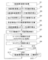
また、本発明の実施形態に係る医用画像処理方法は、X線造影画像データ及びX線透視画像データを取得するステップと、前記X線造影画像データ、又は前記X線造影画像データ及び前記X線透視画像データからデバイスに取り付けられたマーカの複数の位置を検出するステップと、前記マーカの複数の位置が同一とみなせる位置となるようにする動き補正を伴って合成用のX線造影画像データを生成するステップと、前記合成用のX線造影画像データと前記X線透視画像データとを合成することによって表示用のX線画像データを生成するステップとを有する。
また、本発明の実施形態に係るX線撮影装置は、X線画像収集手段、X線画像収集手段、造影画像生成手段及び表示画像生成手段を備える。X線画像収集手段は、X線造影画像データ及びX線透視画像データを収集する。マーカ検出手段は、前記X線造影画像データ、又は前記X線造影画像データ及び前記X線透視画像データからデバイスに取り付けられたマーカの複数の位置を検出する。造影画像生成手段は、前記マーカの複数の位置が同一とみなせる位置となるようにする動き補正を伴って合成用のX線造影画像データを生成する。表示画像生成手段は、前記合成用のX線造影画像データと前記X線透視画像データとを合成することによって表示用のX線画像データを生成する。
2 撮影系
3 制御系
4 データ処理系
5 X線照射部
6 X線検出器
7 駆動機構
8 寝台
9 高電圧発生装置
10 撮影位置制御装置
11 造影剤注入装置
12 入力装置
13 表示装置
14 A/D変換器
15 医用画像処理装置(コンピュータ)
16 X線画像生成部
17 マーカ検出部
18 マスク画像生成部
18A マーカ基準動き補正部
19 ロードマップ画像生成部
20 表示処理部
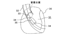
30 心臓
31 左室(LV: left ventricle)
32 大動脈
33 大動脈弁
34 マーカ
35 人工弁
O 被検体
Claims (7)
- X線造影画像データ及びX線透視画像データを取得するX線画像取得手段と、
前記X線造影画像データ、又は前記X線造影画像データ及び前記X線透視画像データからデバイスに取り付けられたマーカの複数の位置を検出するマーカ検出手段と、
前記マーカの複数の位置が同一とみなせる位置となるようにする動き補正を伴って合成用のX線造影画像データを生成する造影画像生成手段と、
前記合成用のX線造影画像データと前記X線透視画像データとを合成することによって表示用のX線画像データを生成する表示画像生成手段と、
を備える医用画像処理装置。 - 前記マーカ検出手段は、複数フレーム分のX線造影画像データ上における前記マーカの複数の位置を検出するように構成され、
前記造影画像生成手段は、前記マーカの複数の位置が同一とみなせる位置となるように前記複数フレーム分のX線造影画像データの動き補正を行い、前記動き補正後における前記複数フレーム分のX線造影画像データに基づいて前記合成用のX線造影画像データを生成するように構成される請求項1記載の医用画像処理装置。 - 前記マーカ検出手段は、前記X線造影画像データ及び前記X線透視画像データからそれぞれ前記マーカの位置を検出するように構成され、
前記造影画像生成手段は、前記マーカの複数の位置が同一とみなせる位置となるように前記X線造影画像データと前記X線透視画像データとの間における動き補正を伴って前記合成用のX線造影画像データを生成するように構成される請求項1又は2記載の医用画像処理装置。 - 前記マーカ検出手段は、前記デバイスの操作期間よりも前に収集されたフレームに対応するX線透視画像データから前記マーカの位置を検出するように構成される請求項1又は3記載の医用画像処理装置。
- 前記X線画像取得手段は、ライブ像データとして順次収集される複数フレーム分のX線透視画像データをリアルタイムに取得するように構成される請求項1乃至4のいずれか1項に記載の医用画像処理装置。
- X線造影画像データ及びX線透視画像データを取得するステップと、
前記X線造影画像データ、又は前記X線造影画像データ及び前記X線透視画像データからデバイスに取り付けられたマーカの複数の位置を検出するステップと、
前記マーカの複数の位置が同一とみなせる位置となるようにする動き補正を伴って合成用のX線造影画像データを生成するステップと、
前記合成用のX線造影画像データと前記X線透視画像データとを合成することによって表示用のX線画像データを生成するステップと、
を有する医用画像処理方法。 - X線造影画像データ及びX線透視画像データを収集するX線画像収集手段と、
前記X線造影画像データ、又は前記X線造影画像データ及び前記X線透視画像データからデバイスに取り付けられたマーカの複数の位置を検出するマーカ検出手段と、
前記マーカの複数の位置が同一とみなせる位置となるようにする動き補正を伴って合成用のX線造影画像データを生成する造影画像生成手段と、
前記合成用のX線造影画像データと前記X線透視画像データとを合成することによって表示用のX線画像データを生成する表示画像生成手段と、
を備えるX線撮影装置。
Priority Applications (3)
| Application Number | Priority Date | Filing Date | Title |
|---|---|---|---|
| JP2012020391A JP2013158372A (ja) | 2012-02-01 | 2012-02-01 | 医用画像処理装置、医用画像処理方法及びx線撮影装置 |
| US13/751,728 US9189848B2 (en) | 2012-02-01 | 2013-01-28 | Medical image processing apparatus, medical image processing method and x-ray imaging apparatus |
| CN201310042570.2A CN103239246B (zh) | 2012-02-01 | 2013-02-01 | 医用图像处理装置、医用图像处理方法以及x射线摄影装置 |
Applications Claiming Priority (1)
| Application Number | Priority Date | Filing Date | Title |
|---|---|---|---|
| JP2012020391A JP2013158372A (ja) | 2012-02-01 | 2012-02-01 | 医用画像処理装置、医用画像処理方法及びx線撮影装置 |
Publications (2)
| Publication Number | Publication Date |
|---|---|
| JP2013158372A true JP2013158372A (ja) | 2013-08-19 |
| JP2013158372A5 JP2013158372A5 (ja) | 2015-02-26 |
Family
ID=48870258
Family Applications (1)
| Application Number | Title | Priority Date | Filing Date |
|---|---|---|---|
| JP2012020391A Pending JP2013158372A (ja) | 2012-02-01 | 2012-02-01 | 医用画像処理装置、医用画像処理方法及びx線撮影装置 |
Country Status (3)
| Country | Link |
|---|---|
| US (1) | US9189848B2 (ja) |
| JP (1) | JP2013158372A (ja) |
| CN (1) | CN103239246B (ja) |
Cited By (5)
| Publication number | Priority date | Publication date | Assignee | Title |
|---|---|---|---|---|
| WO2015178745A1 (ko) * | 2014-05-23 | 2015-11-26 | 주식회사 바텍 | 깊이 카메라를 이용한 의료영상 촬영장치 및 의료영상 보정방법 |
| JP2015217052A (ja) * | 2014-05-15 | 2015-12-07 | 株式会社東芝 | 医用画像処理装置、x線診断装置及び医用画像処理プログラム |
| WO2016158222A1 (ja) * | 2015-03-30 | 2016-10-06 | 国立大学法人大阪大学 | カテーテル・シミュレーター用容器、及びこの容器内に収容される心臓モデル |
| US9888898B2 (en) | 2014-03-27 | 2018-02-13 | Toshiba Medical Systems Corporation | X-ray diagnostic apparatus |
| JPWO2016158222A1 (ja) * | 2015-11-09 | 2018-04-26 | 国立大学法人大阪大学 | カテーテル・シミュレーター用容器、及びこの容器内に収容される心臓モデル |
Families Citing this family (5)
| Publication number | Priority date | Publication date | Assignee | Title |
|---|---|---|---|---|
| JP2013236919A (ja) | 2012-04-19 | 2013-11-28 | Toshiba Corp | X線撮影装置及び医用画像処理装置 |
| US9427200B2 (en) * | 2014-03-21 | 2016-08-30 | Siemens Aktiengesellschaft | Determination of physiological cardiac parameters as a function of the heart rate |
| US10143438B2 (en) | 2015-08-06 | 2018-12-04 | Xiang Zhang | System for 3D object modeling and tracking in X-ray imaging |
| WO2018190291A1 (ja) * | 2017-04-13 | 2018-10-18 | 株式会社島津製作所 | X線撮影装置 |
| CN108703764B (zh) * | 2018-05-29 | 2021-11-05 | 北京东软医疗设备有限公司 | 血管造影方法、装置、系统、设备及存储介质 |
Citations (2)
| Publication number | Priority date | Publication date | Assignee | Title |
|---|---|---|---|---|
| WO2011039681A1 (en) * | 2009-09-29 | 2011-04-07 | Koninklijke Philips Electronics N.V. | Live registration for vessel treatment |
| WO2011117789A1 (en) * | 2010-03-24 | 2011-09-29 | Koninklijke Philips Electronics N.V. | System and method for producing an image of a physical object |
Family Cites Families (14)
| Publication number | Priority date | Publication date | Assignee | Title |
|---|---|---|---|---|
| US6501981B1 (en) * | 1999-03-16 | 2002-12-31 | Accuray, Inc. | Apparatus and method for compensating for respiratory and patient motions during treatment |
| US7225012B1 (en) * | 2000-09-18 | 2007-05-29 | The Johns Hopkins University | Methods and systems for image-guided surgical interventions |
| EP1628575B1 (en) * | 2003-05-21 | 2010-11-17 | Philips Intellectual Property & Standards GmbH | Apparatus for navigating a catheter |
| US9623208B2 (en) * | 2004-01-12 | 2017-04-18 | Varian Medical Systems, Inc. | Instruments with location markers and methods for tracking instruments through anatomical passageways |
| US8542900B2 (en) * | 2007-03-08 | 2013-09-24 | Sync-Rx Ltd. | Automatic reduction of interfering elements from an image stream of a moving organ |
| US9521961B2 (en) * | 2007-11-26 | 2016-12-20 | C. R. Bard, Inc. | Systems and methods for guiding a medical instrument |
| US20090198126A1 (en) * | 2008-02-05 | 2009-08-06 | Klaus Klingenbeck-Regn | Imaging system |
| JP5523791B2 (ja) | 2008-10-27 | 2014-06-18 | 株式会社東芝 | X線診断装置および画像処理装置 |
| FR2942124B1 (fr) * | 2009-02-17 | 2017-05-12 | Gen Electric | Procede et dispositif d'imagerie radiologique |
| JP5439054B2 (ja) | 2009-06-25 | 2014-03-12 | 株式会社東芝 | X線診断装置 |
| EP2482726B1 (en) | 2009-09-29 | 2015-07-15 | Koninklijke Philips N.V. | Vascular roadmapping |
| US9189851B2 (en) * | 2010-01-07 | 2015-11-17 | Siemens Aktiengesellschaft | Method of motion compensation for trans-catheter aortic valve implantation |
| JP5725719B2 (ja) | 2010-02-09 | 2015-05-27 | 株式会社東芝 | X線撮影装置 |
| WO2013126659A1 (en) * | 2012-02-22 | 2013-08-29 | Veran Medical Technologies, Inc. | Systems, methods, and devices for four dimensional soft tissue navigation |
-
2012
- 2012-02-01 JP JP2012020391A patent/JP2013158372A/ja active Pending
-
2013
- 2013-01-28 US US13/751,728 patent/US9189848B2/en active Active
- 2013-02-01 CN CN201310042570.2A patent/CN103239246B/zh active Active
Patent Citations (2)
| Publication number | Priority date | Publication date | Assignee | Title |
|---|---|---|---|---|
| WO2011039681A1 (en) * | 2009-09-29 | 2011-04-07 | Koninklijke Philips Electronics N.V. | Live registration for vessel treatment |
| WO2011117789A1 (en) * | 2010-03-24 | 2011-09-29 | Koninklijke Philips Electronics N.V. | System and method for producing an image of a physical object |
Cited By (6)
| Publication number | Priority date | Publication date | Assignee | Title |
|---|---|---|---|---|
| US9888898B2 (en) | 2014-03-27 | 2018-02-13 | Toshiba Medical Systems Corporation | X-ray diagnostic apparatus |
| JP2015217052A (ja) * | 2014-05-15 | 2015-12-07 | 株式会社東芝 | 医用画像処理装置、x線診断装置及び医用画像処理プログラム |
| WO2015178745A1 (ko) * | 2014-05-23 | 2015-11-26 | 주식회사 바텍 | 깊이 카메라를 이용한 의료영상 촬영장치 및 의료영상 보정방법 |
| WO2016158222A1 (ja) * | 2015-03-30 | 2016-10-06 | 国立大学法人大阪大学 | カテーテル・シミュレーター用容器、及びこの容器内に収容される心臓モデル |
| US10937337B2 (en) | 2015-03-30 | 2021-03-02 | Osaka University | Container for catheter simulator and heart model accommodated in said container |
| JPWO2016158222A1 (ja) * | 2015-11-09 | 2018-04-26 | 国立大学法人大阪大学 | カテーテル・シミュレーター用容器、及びこの容器内に収容される心臓モデル |
Also Published As
| Publication number | Publication date |
|---|---|
| CN103239246B (zh) | 2015-10-07 |
| US20130195343A1 (en) | 2013-08-01 |
| CN103239246A (zh) | 2013-08-14 |
| US9189848B2 (en) | 2015-11-17 |
Similar Documents
| Publication | Publication Date | Title |
|---|---|---|
| JP2013158372A (ja) | 医用画像処理装置、医用画像処理方法及びx線撮影装置 | |
| US20050089143A1 (en) | X-ray diagnosis apparatus and method for creating image data | |
| US9936928B2 (en) | Medical image processing apparatus and X-ray diagnostic apparatus | |
| JP4744941B2 (ja) | X線画像診断装置及びその診断支援方法 | |
| CN106175805B (zh) | 图像处理装置、x射线诊断装置以及图像处理方法 | |
| US9384545B2 (en) | X-ray image diagnosis apparatus | |
| US9613289B2 (en) | X-ray diagnosis apparatus and image processing apparatus | |
| US10765392B2 (en) | X-ray imaging apparatus and x-ray image display method | |
| CN103476341B (zh) | X射线摄影装置、医用图像处理装置、x射线摄影方法以及医用图像处理方法 | |
| JP2005137798A (ja) | X線撮像システム及びx線画像データ表示方法 | |
| JP2020171482A (ja) | X線撮影装置 | |
| JP6381897B2 (ja) | X線診断装置及びインジェクター | |
| JP4939743B2 (ja) | X線撮像装置 | |
| JP5342628B2 (ja) | X線撮像装置 | |
| US11138697B2 (en) | X-ray imaging apparatus | |
| JP2014030506A (ja) | 医用画像処理装置、x線撮影装置及び医用画像処理プログラム | |
| JP2008220641A (ja) | X線撮影装置およびカテーテル挿入案内方法 | |
| CN111166361B (zh) | 放射线摄影装置 | |
| JP7413972B2 (ja) | X線撮影装置、および、画像処理方法 | |
| WO2021033291A1 (ja) | X線撮影方法およびx線撮影システム | |
| JP2012040204A (ja) | X線画像撮影装置 | |
| JPWO2021033291A5 (ja) | ||
| CN114631834A (zh) | X射线透视摄影装置和x射线图像处理方法 | |
| JP2018175854A (ja) | X線撮影装置 |
Legal Events
| Date | Code | Title | Description |
|---|---|---|---|
| A521 | Request for written amendment filed |
Free format text: JAPANESE INTERMEDIATE CODE: A523 Effective date: 20150107 |
|
| A621 | Written request for application examination |
Free format text: JAPANESE INTERMEDIATE CODE: A621 Effective date: 20150107 |
|
| A711 | Notification of change in applicant |
Free format text: JAPANESE INTERMEDIATE CODE: A712 Effective date: 20150612 |
|
| A977 | Report on retrieval |
Free format text: JAPANESE INTERMEDIATE CODE: A971007 Effective date: 20150930 |
|
| A131 | Notification of reasons for refusal |
Free format text: JAPANESE INTERMEDIATE CODE: A131 Effective date: 20151027 |
|
| A521 | Request for written amendment filed |
Free format text: JAPANESE INTERMEDIATE CODE: A523 Effective date: 20151222 |
|
| A711 | Notification of change in applicant |
Free format text: JAPANESE INTERMEDIATE CODE: A711 Effective date: 20160510 |
|
| A02 | Decision of refusal |
Free format text: JAPANESE INTERMEDIATE CODE: A02 Effective date: 20160628 |