JP2010090129A - Glp-1, exendin-4, peptide-analog and use thereof - Google Patents
Glp-1, exendin-4, peptide-analog and use thereof Download PDFInfo
- Publication number
- JP2010090129A JP2010090129A JP2009262568A JP2009262568A JP2010090129A JP 2010090129 A JP2010090129 A JP 2010090129A JP 2009262568 A JP2009262568 A JP 2009262568A JP 2009262568 A JP2009262568 A JP 2009262568A JP 2010090129 A JP2010090129 A JP 2010090129A
- Authority
- JP
- Japan
- Prior art keywords
- seq
- polypeptide
- glp
- pharmaceutical composition
- group
- Prior art date
- Legal status (The legal status is an assumption and is not a legal conclusion. Google has not performed a legal analysis and makes no representation as to the accuracy of the status listed.)
- Granted
Links
Images


Classifications
-
- C—CHEMISTRY; METALLURGY
- C07—ORGANIC CHEMISTRY
- C07K—PEPTIDES
- C07K14/00—Peptides having more than 20 amino acids; Gastrins; Somatostatins; Melanotropins; Derivatives thereof
- C07K14/435—Peptides having more than 20 amino acids; Gastrins; Somatostatins; Melanotropins; Derivatives thereof from animals; from humans
- C07K14/575—Hormones
-
- A—HUMAN NECESSITIES
- A61—MEDICAL OR VETERINARY SCIENCE; HYGIENE
- A61P—SPECIFIC THERAPEUTIC ACTIVITY OF CHEMICAL COMPOUNDS OR MEDICINAL PREPARATIONS
- A61P19/00—Drugs for skeletal disorders
- A61P19/08—Drugs for skeletal disorders for bone diseases, e.g. rachitism, Paget's disease
-
- A—HUMAN NECESSITIES
- A61—MEDICAL OR VETERINARY SCIENCE; HYGIENE
- A61P—SPECIFIC THERAPEUTIC ACTIVITY OF CHEMICAL COMPOUNDS OR MEDICINAL PREPARATIONS
- A61P21/00—Drugs for disorders of the muscular or neuromuscular system
- A61P21/02—Muscle relaxants, e.g. for tetanus or cramps
-
- A—HUMAN NECESSITIES
- A61—MEDICAL OR VETERINARY SCIENCE; HYGIENE
- A61P—SPECIFIC THERAPEUTIC ACTIVITY OF CHEMICAL COMPOUNDS OR MEDICINAL PREPARATIONS
- A61P25/00—Drugs for disorders of the nervous system
-
- A—HUMAN NECESSITIES
- A61—MEDICAL OR VETERINARY SCIENCE; HYGIENE
- A61P—SPECIFIC THERAPEUTIC ACTIVITY OF CHEMICAL COMPOUNDS OR MEDICINAL PREPARATIONS
- A61P25/00—Drugs for disorders of the nervous system
- A61P25/02—Drugs for disorders of the nervous system for peripheral neuropathies
-
- A—HUMAN NECESSITIES
- A61—MEDICAL OR VETERINARY SCIENCE; HYGIENE
- A61P—SPECIFIC THERAPEUTIC ACTIVITY OF CHEMICAL COMPOUNDS OR MEDICINAL PREPARATIONS
- A61P25/00—Drugs for disorders of the nervous system
- A61P25/14—Drugs for disorders of the nervous system for treating abnormal movements, e.g. chorea, dyskinesia
- A61P25/16—Anti-Parkinson drugs
-
- A—HUMAN NECESSITIES
- A61—MEDICAL OR VETERINARY SCIENCE; HYGIENE
- A61P—SPECIFIC THERAPEUTIC ACTIVITY OF CHEMICAL COMPOUNDS OR MEDICINAL PREPARATIONS
- A61P25/00—Drugs for disorders of the nervous system
- A61P25/28—Drugs for disorders of the nervous system for treating neurodegenerative disorders of the central nervous system, e.g. nootropic agents, cognition enhancers, drugs for treating Alzheimer's disease or other forms of dementia
-
- A—HUMAN NECESSITIES
- A61—MEDICAL OR VETERINARY SCIENCE; HYGIENE
- A61P—SPECIFIC THERAPEUTIC ACTIVITY OF CHEMICAL COMPOUNDS OR MEDICINAL PREPARATIONS
- A61P3/00—Drugs for disorders of the metabolism
- A61P3/08—Drugs for disorders of the metabolism for glucose homeostasis
- A61P3/10—Drugs for disorders of the metabolism for glucose homeostasis for hyperglycaemia, e.g. antidiabetics
-
- A—HUMAN NECESSITIES
- A61—MEDICAL OR VETERINARY SCIENCE; HYGIENE
- A61P—SPECIFIC THERAPEUTIC ACTIVITY OF CHEMICAL COMPOUNDS OR MEDICINAL PREPARATIONS
- A61P39/00—General protective or antinoxious agents
- A61P39/02—Antidotes
-
- A—HUMAN NECESSITIES
- A61—MEDICAL OR VETERINARY SCIENCE; HYGIENE
- A61P—SPECIFIC THERAPEUTIC ACTIVITY OF CHEMICAL COMPOUNDS OR MEDICINAL PREPARATIONS
- A61P43/00—Drugs for specific purposes, not provided for in groups A61P1/00-A61P41/00
-
- A—HUMAN NECESSITIES
- A61—MEDICAL OR VETERINARY SCIENCE; HYGIENE
- A61P—SPECIFIC THERAPEUTIC ACTIVITY OF CHEMICAL COMPOUNDS OR MEDICINAL PREPARATIONS
- A61P5/00—Drugs for disorders of the endocrine system
- A61P5/48—Drugs for disorders of the endocrine system of the pancreatic hormones
- A61P5/50—Drugs for disorders of the endocrine system of the pancreatic hormones for increasing or potentiating the activity of insulin
-
- A—HUMAN NECESSITIES
- A61—MEDICAL OR VETERINARY SCIENCE; HYGIENE
- A61P—SPECIFIC THERAPEUTIC ACTIVITY OF CHEMICAL COMPOUNDS OR MEDICINAL PREPARATIONS
- A61P7/00—Drugs for disorders of the blood or the extracellular fluid
- A61P7/12—Antidiuretics, e.g. drugs for diabetes insipidus
-
- A—HUMAN NECESSITIES
- A61—MEDICAL OR VETERINARY SCIENCE; HYGIENE
- A61P—SPECIFIC THERAPEUTIC ACTIVITY OF CHEMICAL COMPOUNDS OR MEDICINAL PREPARATIONS
- A61P9/00—Drugs for disorders of the cardiovascular system
-
- A—HUMAN NECESSITIES
- A61—MEDICAL OR VETERINARY SCIENCE; HYGIENE
- A61P—SPECIFIC THERAPEUTIC ACTIVITY OF CHEMICAL COMPOUNDS OR MEDICINAL PREPARATIONS
- A61P9/00—Drugs for disorders of the cardiovascular system
- A61P9/10—Drugs for disorders of the cardiovascular system for treating ischaemic or atherosclerotic diseases, e.g. antianginal drugs, coronary vasodilators, drugs for myocardial infarction, retinopathy, cerebrovascula insufficiency, renal arteriosclerosis
-
- C—CHEMISTRY; METALLURGY
- C07—ORGANIC CHEMISTRY
- C07K—PEPTIDES
- C07K14/00—Peptides having more than 20 amino acids; Gastrins; Somatostatins; Melanotropins; Derivatives thereof
- C07K14/435—Peptides having more than 20 amino acids; Gastrins; Somatostatins; Melanotropins; Derivatives thereof from animals; from humans
- C07K14/575—Hormones
- C07K14/57563—Vasoactive intestinal peptide [VIP]; Related peptides
-
- A—HUMAN NECESSITIES
- A61—MEDICAL OR VETERINARY SCIENCE; HYGIENE
- A61K—PREPARATIONS FOR MEDICAL, DENTAL OR TOILETRY PURPOSES
- A61K38/00—Medicinal preparations containing peptides
-
- A—HUMAN NECESSITIES
- A61—MEDICAL OR VETERINARY SCIENCE; HYGIENE
- A61K—PREPARATIONS FOR MEDICAL, DENTAL OR TOILETRY PURPOSES
- A61K38/00—Medicinal preparations containing peptides
- A61K38/16—Peptides having more than 20 amino acids; Gastrins; Somatostatins; Melanotropins; Derivatives thereof
- A61K38/17—Peptides having more than 20 amino acids; Gastrins; Somatostatins; Melanotropins; Derivatives thereof from animals; from humans
- A61K38/22—Hormones
- A61K38/2278—Vasoactive intestinal peptide [VIP]; Related peptides (e.g. Exendin)
-
- A—HUMAN NECESSITIES
- A61—MEDICAL OR VETERINARY SCIENCE; HYGIENE
- A61K—PREPARATIONS FOR MEDICAL, DENTAL OR TOILETRY PURPOSES
- A61K38/00—Medicinal preparations containing peptides
- A61K38/16—Peptides having more than 20 amino acids; Gastrins; Somatostatins; Melanotropins; Derivatives thereof
- A61K38/17—Peptides having more than 20 amino acids; Gastrins; Somatostatins; Melanotropins; Derivatives thereof from animals; from humans
- A61K38/22—Hormones
- A61K38/26—Glucagons
Abstract
Description
関連出願の相互参照
当該出願は、2001年7月31日に出願された米国特許出願番号第60/309,076号に対して優先権を主張する。
CROSS REFERENCE TO RELATED APPLICATIONS This application claims priority to US Patent Application No. 60 / 309,076, filed July 31, 2001.
本願発明は、グルカゴン様ペプチド-1(GLP-1)、exendin-4、及びそれらのペプチド・アナログに広く関する。本発明は、糖尿病及び神経変性症状の治療におけるそれらの使用にも関する。 The present invention relates broadly to glucagon-like peptide-1 (GLP-1), exendin-4, and their peptide analogs. The invention also relates to their use in the treatment of diabetes and neurodegenerative conditions.
本発明の背景
膵臓のベータ細胞機能不全及び付随するインシュリン産生の減少は、糖尿病をもたらす恐れがある。1型糖尿病において、ベータ細胞は、免疫系によって完全に破壊され、インシュリン産生細胞の欠如をもたらす(インシュリン依存型[1型]糖尿病に対する内科医のガイド:Diagnosis and Treatment, American Diabetes Association, 1988(非特許文献1))。2型糖尿病において、ターゲット組織がグルコース摂取においてインシュリンの効果に抵抗性をもつようになると、ベータ細胞が徐々に能率的でなくなる。よって、ベータ細胞は、1型糖尿病の人々において不存在であり、そして2型糖尿病の人々において機能的に十分な機能を果たしていない。
BACKGROUND OF THE INVENTION Pancreatic beta cell dysfunction and the concomitant reduction in insulin production can lead to diabetes. In
現在、ベータ細胞機能不全は、いくつかのの異なる方法で治療される。1型糖尿病又は末期の2型糖尿病は、インシュリン補充療法が必要である。たとえ連続的な注入又は複数の注射が複雑な投薬計画で使用されたとしても、インスリン療法、あるいは救命療法は、正常血糖を回復させない。例えば、インシュリン補充療法を受けている個人において、食後のグルコース・レベルは、非常に高いままである。よって、インスリン療法は、複数の毎日の注射又は連続的な注入によってなされなければならなくて、かつ、その効果は、高血糖、低血糖、代謝性アシドーシス、及びケトーシスを避けるために慎重に監視されなければならない。
Currently, beta cell dysfunction is treated in several different ways.
2型糖尿病の人々は、ベータ細胞からのインシュリン産生及び分泌を刺激する、及び/又はインシュリン感受性を改善する薬物を用いて一般に治療される。しかし、これらの薬の主な問題点は、インシュリン産生及び分泌が血糖値にかかわらず促進されることである。よって、食物摂取は低血糖又は高血糖を避けるために、インシュリン産生及び分泌の促進に対して釣り合わせなければならない。ここ数年で、いくつかの新しい薬剤が2型糖尿病の治療に利用可能になった。これらは、メトフォルミン、ロシグリタゾン、ピオグリタゾン、及びアクラボス(Bressler and Johnson, 1997(非特許文献2)を参照のこと)を含んでいる。しかし、これらの新しい薬剤によって得られたヘモグロビンA1cの低下は、あまり十分ではなく、それらが糖尿病の長期のコントロールを改善しないことを示唆している(Ghazzi et al., 1997(非特許文献3))。
People with
食物に応答して腸神経内分泌細胞によって通常分泌されているホルモンである、グルカゴン様ペプチド-1(GLP-1)が、2型糖尿病の新しい治療として提案された(Gutniak et al., 1992(非特許文献4); Nauck et al., J. Clin. Invest., 1993(非特許文献5))。それは、長年の2型糖尿病の患者であっても、ベータ細胞によるインシュリン分泌分泌を増加させる(Nauck et al., Diabetologia, 1993(非特許文献6))。GLP-1が内性インシュリン分泌を刺激し、それが血糖値が下がったときに止まるので、GLP-1治療は、インスリン療法を上回る利点がある(Nauck et al., Diabetologia, 1993(非特許文献6); Elahi et al., 1994(非特許文献7))。GLP-1は、インシュリンの分泌及び合成を増加させることによって正常血糖を促進するので、グルカゴン分泌を抑え、そして胃内容排出を減少させる(Nauck et al., Diabetologia, 1993(非特許文献6); Elahi et al., 1994(非特許文献7); Wills et al., 1996(非特許文献8); Nathan et al., 1992(非特許文献9); De Ore et al., 1997(非特許文献10))。GLP-1は、ヘキソキナー・mRNAレベルの増加をも誘発する(Wang et al., Endocrinology 1995(非特許文献11); Wang et al., 1996(非特許文献12))。
Glucagon-like peptide-1 (GLP-1), a hormone normally secreted by enteric neuroendocrine cells in response to food, has been proposed as a new treatment for
GLP-1は、ベータ細胞においてインシュリン分泌効果をもつこと(Thorens and Waeber, 1993(非特許文献13); Orskov, 1992(非特許文献14))、及びインシュリン分泌細胞系に加えられて24時間、インシュリン生合成及びプロインシュリン遺伝子発現を増加させることが知られている(Dracker et al., 1987(非特許文献15); Fehmann and Habener, 1992(非特許文献16))。RIN1046-38細胞を使った研究において、GLP-1による24時間の処理が、GLP-1が取り除かれた後1時間、及びこの細胞の数回の洗浄後でさえ、グルコース反応性を増加させた(Montrose-Rafizadeh et al., 1994(非特許文献17))。よって、GLP-1は、系から代謝された後でもβ細胞に対する生物学的な効果をもつことが知られているインシュリン分泌刺激剤である。GLP-1は、プログルカゴンの転写後修飾の産物である。GLP-1の配列、並びにその活性断片GLP-1(7-37)及びGLP-1(7-36)アミドが当該技術分野で知られている(Fehmann et al., 1995(非特許文献18))。GLP-1は、糖尿病の治療における治療薬として提案されたが、それは、たとえ皮下にボーラスによって与えられるとしても(Ritzel et al., 1995(非特許文献19))、短い生物学的半減期をもつ(De Ore et al., 1997(非特許文献10))。1つは、GLP-1(及びGLP-1(7-36)アミド)の分解は、第8アミノ酸と第9アミノ酸(アラニンとグルタミン酸)の間でポリペプチドを切断する酵素ジペプチジル・ペプチダーゼ(DPP1V)によってである。 GLP-1 has an insulin-secreting effect in beta cells (Thorens and Waeber, 1993 (Non-Patent Document 13); Orskov, 1992 (Non-Patent Document 14)), and added to the insulin-secreting cell line for 24 hours. It is known to increase insulin biosynthesis and proinsulin gene expression (Dracker et al., 1987 (Non-Patent Document 15); Fehmann and Habener, 1992 (Non-Patent Document 16)). In studies using RIN1046-38 cells, treatment with GLP-1 for 24 hours increased glucose reactivity for 1 hour after GLP-1 was removed and even after several washes of the cells. (Montrose-Rafizadeh et al., 1994 (Non-Patent Document 17)). Therefore, GLP-1 is an insulin secretion stimulator known to have a biological effect on β cells even after being metabolized from the system. GLP-1 is the product of post-transcriptional modification of proglucagon. The sequence of GLP-1 and its active fragments GLP-1 (7-37) and GLP-1 (7-36) amide are known in the art (Fehmann et al., 1995). ). GLP-1 has been proposed as a therapeutic agent in the treatment of diabetes, but it has a short biological half-life, even if given subcutaneously by a bolus (Ritzel et al., 1995). (De Ore et al., 1997 (non-patent document 10)). One is the degradation of GLP-1 (and GLP-1 (7-36) amide), an enzyme dipeptidyl peptidase (DPP1V) that cleaves the polypeptide between the 8th and 9th amino acids (alanine and glutamic acid) By.
exendin-4は、アメリカ・ドクトカゲトカゲの唾液腺で産生されるポリペプチドである(Goke et al., 1993(非特許文献20))。exendin-4のアミノ酸配列は、当該技術分野で知られている(Fehmannet al., 1995(非特許文献18))。それが特殊な非哺乳類の遺伝子の産物であり、かつ、唾液腺だけで発現されると思われたが(Chen and Drucker, 1997(非特許文献21))、exendin-4は、GLP-1と52%のアミノ酸配列相同性をもち、そして哺乳動物においてGLP-1受容体と相互作用する(Goke et al., 1993(非特許文献20); Thorens et al., 1993(非特許文献22))。インビトロにおいて、インシュリン産生細胞によるインシュリン分泌を促進すし、そして等モルの分量で与えられると、exendin-4は、GLP-1より強力にインシュリン産生細胞からのインシュリン放出を引き起こすことが示された。さらに、exendin-4が、齧歯動物及びヒトの両方で、強力にインシュリン分泌を促進して血漿グルコース・レベルを下げ、そしてそれがGLP-1より長く作用する。しかし、通常哺乳動物において生じないので、Exendin-4は、GLP-1を欠く哺乳動物において潜在的な抗原特性を必ずもっている。 exendin-4 is a polypeptide produced in the salivary glands of the American lizard (Goke et al., 1993 (Non-patent Document 20)). The amino acid sequence of exendin-4 is known in the art (Fehmannet al., 1995 (Non-patent Document 18)). Although it was a product of a special non-mammalian gene and expressed only in the salivary glands (Chen and Drucker, 1997 (Non-Patent Document 21)), exendin-4 It has% amino acid sequence homology and interacts with the GLP-1 receptor in mammals (Goke et al., 1993 [20]; Thorens et al., 1993 [22]). In vitro, it has been shown that exendin-4 induces insulin release from insulin-producing cells more potently than GLP-1 when stimulated by insulin-producing cells and given in equimolar amounts. Furthermore, exendin-4 potently promotes insulin secretion and lowers plasma glucose levels in both rodents and humans, and it acts longer than GLP-1. However, since it does not normally occur in mammals, Exendin-4 always has potential antigenic properties in mammals lacking GLP-1.
糖尿病で生じるインシュリン産生の減少に加えて、末梢神経障害が、糖尿病に一般に関係する。全糖尿病患者の20〜30パーセントが、いつかは末梢神経障害を患う。さらに、心臓病、脳卒中、高血圧症、及び糖尿病によるアルツハイマー症の高い危険性が報告されている(Moceri et al., 2000(非特許文献23); Ott et al., 1999(非特許文献24))。よって、糖尿病は神経変性疾患に関係する病気でもある。 In addition to the decrease in insulin production that occurs with diabetes, peripheral neuropathy is commonly associated with diabetes. 20-30 percent of all diabetics will eventually suffer from peripheral neuropathy. Furthermore, a high risk of Alzheimer's disease due to heart disease, stroke, hypertension, and diabetes has been reported (Moceri et al., 2000 (Non-patent Document 23); Ott et al., 1999 (Non-patent Document 24)). ). Thus, diabetes is also a disease related to neurodegenerative diseases.
多数の研究が、GLP-1受容体が、齧歯動物(Jin et al., 1988(非特許文献25)、Shughrue et al., 1996(非特許文献26))及びヒト(Wei and Mojsov 1995(非特許文献27)、Satoh et al., 2000(非特許文献28))の両方の脳にあることを証明した。分布の化学的構成(chemoarchitecture)は、視床下部、視床、脳幹、外側中隔、脳弓下器官、及び最後野であり、一般にたくさんのペプチド受容体が配置されている全ての脳室周囲区域に大部分は限られているように思われる。しかし、GLP-1のための特定の結合部位は、低密度ではあるが、被殻-尾状核、皮質、及び小脳の全体にわたって検出された(Campos et al. 1994(非特許文献29), Calvo et al. 1995(非特許文献30), Goke et al. 1995(非特許文献20))。 Numerous studies have shown that the GLP-1 receptor has been identified in rodents (Jin et al., 1988, Shughrue et al., 1996) and humans (Wei and Mojsov 1995). Non-patent document 27) and Satoh et al., 2000 (non-patent document 28)). The chemoarchitecture of the distribution is the hypothalamus, thalamus, brainstem, lateral septum, subfornical organ, and terminal cortex, generally in all periventricular areas where many peptide receptors are located. Most seem to be limited. However, specific binding sites for GLP-1 were detected throughout the putamen-caudate nucleus, cortex, and cerebellum, albeit at low density (Campos et al. 1994, 29), Calvo et al. 1995 (Non-patent document 30), Goke et al. 1995 (Non-patent document 20)).
当該技術分野で必要とされているのは、糖尿病の治療及び神経障害、例えばアルツハイマー病及びパーキンソン病の治療、並びに2型糖尿病に関連した末梢神経障害の治療において治療的に価値のあるポリペプチドである。
What is needed in the art are polypeptides of therapeutic value in the treatment of diabetes and neurological disorders such as Alzheimer's and Parkinson's disease, and in the treatment of peripheral neuropathy associated with
本発明の概要
本願発明の目的に従って、本明細書中に含まれ、かつ、全体的に記載されているとおり、1の側面において、本願発明は、GLP-1及びexendin-4の新規ポリペプチド・アナログに関する。好ましい態様において、前記ポリペプチドは、インシュリン分泌刺激性及び長期作用性である。好ましくは、前記ポリペプチドのインシュリン分泌刺激効果は、等モル量のGLP-1又はexendin-4の効果に匹敵するか又はそれを超える。
In accordance with the purpose of the present invention, as included and generally described herein, in one aspect, the present invention provides a novel polypeptide of GLP-1 and exendin-4. Related to analog. In a preferred embodiment, the polypeptide is insulin secretagogue and long acting. Preferably, the effect of stimulating insulin secretion of the polypeptide is comparable to or exceeding the effect of equimolar amounts of GLP-1 or exendin-4.
本発明は、さらに、精製されたポリペプチド、配列番号5、配列番号6、配列番号7、配列番号8、配列番号42、配列番号43、配列番号44、配列番号45、配列番号46、配列番号47、配列番号48、配列番号9、配列番号10、配列番号11、配列番号12、配列番号13、配列番号14、配列番号15、配列番号25又は配列番号33を含むアミノ酸配列に関する。 The present invention further includes a purified polypeptide, SEQ ID NO: 5, SEQ ID NO: 6, SEQ ID NO: 7, SEQ ID NO: 8, SEQ ID NO: 42, SEQ ID NO: 43, SEQ ID NO: 44, SEQ ID NO: 45, SEQ ID NO: 46, SEQ ID NO: 47, SEQ ID NO: 48, SEQ ID NO: 9, SEQ ID NO: 10, SEQ ID NO: 11, SEQ ID NO: 12, SEQ ID NO: 13, SEQ ID NO: 14, SEQ ID NO: 15, SEQ ID NO: 25 or SEQ ID NO: 33.
他の側面において、本発明は、糖尿病の患者の治療方法であって、インシュリン分泌刺激効果がある量で本発明のポリペプチドを上記患者に投与することを含む方法に関する。 In another aspect, the invention relates to a method for treating a patient with diabetes, comprising administering to the patient a polypeptide of the invention in an amount that has an insulinotropic effect.
本発明のさらなる利点は、以下の記載によりある程度説明され、そしてある程度記載から明らかであるか、又は本発明の実施によって確認されうる。本発明の利点は、特に添付の請求項で指摘された要素及び組み合わせによって実現及び達成される。先の概要及び以下の詳細な説明は共に、請求される本発明の単なる典型及び解説であり、本発明を制限するものではないことは、理解されている。 Further advantages of the present invention will be explained in part by the following description, and in part will be apparent from the description, or may be learned by practice of the invention. The advantages of the invention will be realized and attained by means of the elements and combinations particularly pointed out in the appended claims. It is understood that both the foregoing summary and the following detailed description are merely exemplary and explanatory of the claimed invention and are not intended to limit the invention.
本発明は、GLP-1及びexendin-4の新規ポリペプチド・アナログに関する。前記ポリペプチドは、インシュリン分泌刺激性及び長期作用性である。好ましくは、前記ポリペプチドのインシュリン分泌刺激効果は、等モル量のGLP-1又はexendin-4の効果に匹敵するか又はそれを超える。したがって、本発明は、糖尿病の治療及び神経障害、例えばアルツハイマー病及びパーキンソン病の治療、並びに2型糖尿病に関連した末梢神経障害の治療において治療的に価値のあるポリペプチドを提供する。
The present invention relates to novel polypeptide analogs of GLP-1 and exendin-4. The polypeptide is insulin secretagogue and long acting. Preferably, the effect of stimulating insulin secretion of the polypeptide is comparable to or exceeding the effect of equimolar amounts of GLP-1 or exendin-4. Thus, the present invention provides polypeptides of therapeutic value in the treatment of diabetes and neurological disorders such as Alzheimer's disease and Parkinson's disease, and in the treatment of peripheral neuropathy associated with
好ましい態様の説明
本発明は、以下の、本発明の好ましい態様の詳細な説明及びそこに含まれる実施例、並びに図面及びそれらの上記及び下記の説明を参照することによってより容易に理解される。
DESCRIPTION OF THE PREFERRED EMBODIMENTS The invention will be more readily understood by reference to the following detailed description of preferred embodiments of the invention and the examples contained therein, as well as the drawings and their foregoing and following descriptions.
当該化合物、組成物、物品、デバイス、及び/又は方法が開示及び記載される前に、本願発明は、それらは当然変化するので、特定の合成方法、特定の投与計画、又は特別な精製手順に制限されないと理解されるべきである。本明細書中に使用された用語が、特定の態様のみを記載する目的のためのものであって、制限されることを意図しないと理解されもする。 Before the compounds, compositions, articles, devices, and / or methods are disclosed and described, the present invention is subject to specific synthetic methods, specific dosage regimes, or special purification procedures, as they will of course vary. It should be understood that it is not limited. It is also to be understood that the terminology used herein is for the purpose of describing particular embodiments only and is not intended to be limiting.
明細書及び添付した請求項で使われる場合、単数形「a」、「an」、及び「the」は、前後関係が別段の明確な指示をしない限り、複数の指示物を含む。
よって、例えば、「a polypeptide」への言及は、複数のポリペプチドの混合物を含み、「a pharmaceutical carrier」への言及は、2以上のそのような担体の混合物を含む、そのほかも同様。
As used in the specification and the appended claims, the singular forms “a”, “an”, and “the” include plural referents unless the context clearly dictates otherwise.
Thus, for example, reference to “a polypeptide” includes a mixture of polypeptides, reference to “a pharmaceutical carrier” includes a mixture of two or more such carriers, and the like.
範囲は、「約」一方の特有の値から、及び/又は「約」他方の特有の値まで、のように本明細書中で表される。そのような範囲が表現されるとき、他の態様は、一方の特有の値から、及び/又は他方の特有の値までを含む。同様に、値が前例の「約」の使用によって、近似値として表現されるとき、特有の値が他の態様を形成することが知られる。各々の範囲の終了点が、他の終了点との関係、及び他の終了点からの独立の両方において重要であることはさらに理解されるであろう。本明細書中に使用されるとき、「約」は、任意の値±10%を表す。 Ranges are expressed herein as from “about” one particular value and / or from “about” the other particular value. When such a range is expressed, other aspects include from one particular value and / or to the other unique value. Similarly, when a value is expressed as an approximation by use of the “about” in the previous example, it is known that the unique value forms another aspect. It will be further appreciated that the end point of each range is important both in relation to the other end points and independent of the other end points. As used herein, “about” represents any value ± 10%.
本願明細書及び後に続く請求項において、言及は、以下の意味をもつと規定される多数の用語に対して成される:
「任意に」又は「場合により」は、その後に記載された事件又は情況が生じるかもしれず、そしてこの記載は、上記事件又は状況が生じる場合と生じない場合を含むことを意味する。
In this specification and in the claims that follow, reference will be made to a number of terms that are defined to have the following meanings:
“Optionally” or “optionally” means that the event or situation described thereafter may occur, and this description includes the case where the event or situation occurs or not.
全体にわたり使用されるとき、「患者」は、個人を意味する。好ましくは、患者は哺乳動物、例えば霊長類、より好ましくは、ヒトである。よって、「患者」は、飼い慣らされた動物、例えば猫、犬等、家畜(例えば、牛、馬、豚、羊、ヤギ等)、及び実験動物(例えば、マウス、ウサギ、ラット、モルモット等)を含みうる。 As used throughout, “patient” means an individual. Preferably, the patient is a mammal, such as a primate, more preferably a human. Thus, `` patient '' refers to domesticated animals such as cats, dogs, domestic animals (e.g., cattle, horses, pigs, sheep, goats) and laboratory animals (e.g., mice, rabbits, rats, guinea pigs, etc.). May be included.
用語「ポリペプチド」は、用語「ペプチド」と互換性をもって本明細書中で使われる。「ポリペプチド」も「ペプチド」もペプチド結合によって接続された一連の天然又は非天然アミノ酸を含んでいる。 The term “polypeptide” is used herein interchangeably with the term “peptide”. Both “polypeptides” and “peptides” comprise a series of natural or unnatural amino acids connected by peptide bonds.
「単離されたポリペプチド」又は「精製されたポリペプチド」は、上記ポリペプチドに通常天然又は培養において付随する物質を実質的に含まないポリペプチドを意味する。本発明のポリペプチドは、例えば利用可能であれば(例えば、哺乳類細胞)天然起源からの抽出によって、上記ポリペプチドをコードしている組み換え核酸の発現によって(例えば、細胞又は細胞を含まない翻訳系により)、又は上記ポリペプチドの化学合成によって得ることができる。さらに、ポリペプチドは、完全長のポリペプチドの切断によって得ることができる。ポリペプチドが大きな天然のポリペプチドの断片であるとき、単離されたポリペプチドは短く、そして断片の完全長の、天然のポリペプチドを除外する。 By “isolated polypeptide” or “purified polypeptide” is meant a polypeptide that is substantially free from the substances that normally accompany it in nature or culture. The polypeptides of the present invention can be produced, for example, by extraction from natural sources, if available (e.g., mammalian cells), by expression of recombinant nucleic acids encoding the polypeptides (e.g., cells or cell-free translation systems). Or by chemical synthesis of the above polypeptide. Further, the polypeptide can be obtained by cleavage of the full length polypeptide. When the polypeptide is a fragment of a large natural polypeptide, the isolated polypeptide is short and excludes the full length natural polypeptide of the fragment.
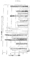
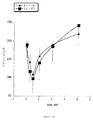
本発明は、GLP-1及びexendin-4の新規ポリペプチド・アナログに関する。本明細書中に使用されるとき、「GLP-1」は、完全なGLP-1配列の第7〜36残基のアミド化型であるGLP-1 7-36アミド、及びGLP-1 7-37と同義的に使われる。exendin-4の残基は、GLP-1、第7〜36残基とアラインされ、GLP-1残基の番号付けに従って番号付けされる。そのような残基番号付けの慣習が、全体にわたり使われる。図1を参照のこと。
The present invention relates to novel polypeptide analogs of GLP-1 and exendin-4. As used herein, “GLP-1” refers to GLP-1 7-36 amide, which is the amidated form of
好ましい態様において、前記ポリペプチドはインシュリン分泌刺激性である。「インシュリン分泌刺激性」は、前記ポリペプチドが、グルコースのみに対する応答の基礎分泌レベルに比べて、グルコースに依存した様式でインシュリン合成、放出又は分泌を増やすことを意味する。好ましくは、インシュリン分泌のそのような増加は、基礎分泌よりも少なくとも1.15、1.25、1.5、2.0、2.5、3.0、4.5、5.0、5.5、6.0、6.5、7.0、7.5、8.0、8.5、9.0、9.5、10.0、10.5、11.0、11.5、12.0、12.5、13.0、13.5、14.0、14.5、15.0、15.5、16.0、16.5、17.0、17.5、18.0、18.5、19.0、19.5又は20.0倍多い。インシュリン分泌の増加は、本技術分野に知られる分析方法を使って、直接的に(例えば、インシュリン・レベルの増加を示すことによって)、又は間接的に(例えば、グルコース・レベルの減少、又はcAMPレベルの増加を示すことによって)、インビボで(例えば、血糖値をアッセイすることによって)、又はインビトロで(例えば、培地中のインシュリン・レベルをアッセイすることによって)示される。 In a preferred embodiment, the polypeptide is insulin secretagogue. “Insulin secretion stimulatory” means that the polypeptide increases insulin synthesis, release or secretion in a glucose dependent manner compared to the basal secretion level of the response to glucose alone. Preferably, such an increase in insulin secretion is at least 1.15, 1.25, 1.5, 2.0, 2.5, 3.0, 4.5, 5.0, 5.5, 6.0, 6.5, 7.0, 7.5, 8.0, 8.5, 9.0, 9.5 over basal secretion. , 10.0, 10.5, 11.0, 11.5, 12.0, 12.5, 13.0, 13.5, 14.0, 14.5, 15.0, 15.5, 16.0, 16.5, 17.0, 17.5, 18.0, 18.5, 19.0, 19.5 or 20.0 times more. Increases in insulin secretion can be directly (e.g., by showing an increase in insulin levels) or indirectly (e.g., decreased glucose levels, or cAMP) using analytical methods known in the art. It is indicated in vivo (for example by assaying blood glucose levels) or in vitro (for example by assaying insulin levels in the medium).
インシュリン分泌刺激効果は、例えばインシュリン陽性細胞数の増加を含むいくつかの機構のいずれか1つによるかもしれない。インシュリン分泌刺激性ポリペプチドは、例えば幹細胞の、インシュリン陽性細胞への分化を促進することによって、並びに非幹細胞のより分化されていない状態への脱分化を促進し、そしてインシュリン陽性細胞への分化を促進することによって、インシュリンの放出を促進する。2番目の態様として、インシュリン分泌刺激効果は、任意の期間において各々のインシュリン陽性細胞で合成された及び/又は放出されたインシュリンの量の増加によって引き起こされる。インシュリン陽性細胞の数が増やされて、そして同様に、各々細胞によって分泌されるインシュリンの量が増やされる場合、合わせたインシュリン分泌刺激効果が、生じることもある。 The insulin secretion stimulating effect may be due to any one of several mechanisms including, for example, an increase in the number of insulin positive cells. Insulin secretion stimulating polypeptides promote, for example, the differentiation of stem cells into insulin-positive cells, as well as the dedifferentiation of non-stem cells to a less differentiated state, and the differentiation into insulin-positive cells. Promotes insulin release by promoting. As a second aspect, the insulin secretion stimulating effect is caused by an increase in the amount of insulin synthesized and / or released in each insulin positive cell in any period of time. If the number of insulin positive cells is increased, and likewise the amount of insulin secreted by each cell is increased, a combined insulin secretion stimulating effect may occur.
「基礎分泌」は、第2の放出剤の不存在下で、グルコース刺激に応答して放出されたインシュリンの量を意味する。 “Basal secretion” means the amount of insulin released in response to glucose stimulation in the absence of a second release agent.
「インシュリン陽性細胞」は、例えば膵臓の島細胞、例えばベータ細胞、又は細胞株、例えばRIN1048-36細胞を含むインシュリンを放出することが示されたあらゆる細胞、インシュリンを放出するように設計されたあらゆる細胞(例えば、インシュリンを含む遺伝学的に修飾された細胞)、又はインシュリンを含んでいるあらゆる細胞を意味する。 An “insulin positive cell” is any cell designed to release insulin, eg any cell that has been shown to release insulin, including pancreatic islet cells, eg beta cells, or cell lines, eg RIN1048-36 cells. By a cell (eg, a genetically modified cell containing insulin) or any cell containing insulin.
「GLP-1又はexendin-4のアナログ」は、アゴニスト特性を示す(すなわち、GLP-1又はexendin-4の1以上の生物活性を示す)修飾されたGLP-1及びexendinアミノ酸配列を意味する。そのような修飾は、GLP-1中に存在する1以上のアミノ酸残基、又はexendin-4中に存在する1以上のアミノ酸残基を含むキメラ・ポリペプチドを含む。修飾は、GLP-1又はexendin-4、あるいはキメラ・ポリペプチドの切詰めをも含む。例えば、切詰められたキメラ・ポリペプチドは、GLP-1の36位のRと交換された36位のGをもつexendin-4 7-36である。本発明のポリペプチドは、GLP-1又はexendin-4と比べて機能上の活性のかなりの損失のない、GLP-1又はexendin-4の、1以上の追加のアミノ酸(すなわち、挿入又は付加)、アミノ酸の削除、あるいはアミノ酸配列の置換を含む。例えば、削除は、現在の規定された識別活性に不可欠ではないアミノ酸から成り、そして置換は、保存的であるか(すなわち、塩基性、親水性又は疎水性アミノ酸を同じもので置換する)、又は非保存的である。よって、所望の修飾及び変更は、GLP-1及びexendin-4のアミノ酸配列で作製され、そしてタンパク質は、依然として観察される似た特徴をもつと理解されている。生物学的な有用性又は活性のかなりの喪失、及びそのような有用性又は活性の増加の可能性なしに、様々な変更が、GLP-1アミノ酸配列又はexendin-4アミノ酸配列(又は基礎をなす核酸配列)で作製される。 By “GLP-1 or exendin-4 analog” is meant a modified GLP-1 and exendin amino acid sequence that exhibits agonist properties (ie, exhibits one or more biological activities of GLP-1 or exendin-4). Such modifications include chimeric polypeptides comprising one or more amino acid residues present in GLP-1 or one or more amino acid residues present in exendin-4. Modifications also include truncation of GLP-1 or exendin-4, or chimeric polypeptides. For example, the truncated chimeric polypeptide is exendin-4 7-36 with G at position 36 exchanged for R at position 36 of GLP-1. The polypeptides of the present invention have one or more additional amino acids (ie, insertions or additions) of GLP-1 or exendin-4 that do not have a significant loss of functional activity compared to GLP-1 or exendin-4 Including deletion of amino acids, or substitution of amino acid sequences. For example, deletions consist of amino acids that are not essential for the current defined discriminating activity, and substitutions are conservative (i.e., replace basic, hydrophilic or hydrophobic amino acids with the same), or Non-conservative. Thus, the desired modifications and changes are made with the amino acid sequences of GLP-1 and exendin-4, and the protein is understood to have similar characteristics still observed. Various changes may be made to the GLP-1 amino acid sequence or the exendin-4 amino acid sequence (or the underlying) without significant loss of biological utility or activity, and the possibility of such increased utility or activity. Nucleic acid sequence).
本明細書中に使用されるとき、GLP-1又はexendin-4、あるいはそれと実質的に一致するアミノ酸配列をもつポリペプチドに関する用語「断片」又は「切詰め」は、GLP-1又はexendin-4、あるいはそれと実質的に一致しているアミノ酸配列をもつポリペプチドのいずれかの少なくとも5つの隣接したアミノ酸のポリペプチド配列を意味し、ここで、上記ポリペプチド配列はインシュリン分泌刺激機能をもつ。 As used herein, the term “fragment” or “truncated” with respect to GLP-1 or exendin-4, or a polypeptide having an amino acid sequence substantially identical thereto, refers to GLP-1 or exendin-4 Or a polypeptide sequence of at least 5 contiguous amino acids of any polypeptide having an amino acid sequence substantially identical thereto, wherein said polypeptide sequence has an insulin secretion stimulating function.
他の修飾は、アミノ酸残基の少なくとも1つの天然L立体配位が、アミノ酸残基のD立体配位と置き換えられている、D-エナンチオマーを含む。 Other modifications include D-enantiomers in which at least one natural L configuration of an amino acid residue is replaced with a D configuration of the amino acid residue.
本発明は、スペーサ、例えばラテラル・スペーサの使用を検討する。用語「ラテラル・スペーサ」は、化学結合によってアミノ酸配列の中に組み込まれる化合物と規定され、それにより上記化合物は、その位置での又はその付近でのアミノ酸配列の切断(例えば、DPP1Vによる)を減らすか又は除くために、2つ以上のアミノ酸残基の間の距離を延ばす。例えば、配列A-X-B(式中、AとBがアミノ酸残基であり、Xがラテラル・スペーサである)において、酵素による配列の切断は、ラテラル・スペーサの存在しない配列(A-B)に比べて、減らされるか又は除かれる。好ましくは、1〜4の化合物が、ラテラル・スペーサとしてのアミノ酸配列に組み込まれることができる。よって、1,2,3,又は4の化合物が、様々な態様において挿入される。 The present invention contemplates the use of spacers, such as lateral spacers. The term “lateral spacer” is defined as a compound that is incorporated into an amino acid sequence by a chemical bond, thereby reducing the cleavage of the amino acid sequence at or near that position (eg, by DPP1V). To increase or decrease the distance between two or more amino acid residues. For example, in the sequence AXB (where A and B are amino acid residues and X is a lateral spacer), the cleavage of the sequence by the enzyme is reduced compared to the sequence (AB) without the lateral spacer. Or removed. Preferably, 1-4 compounds can be incorporated into the amino acid sequence as a lateral spacer. Thus, 1,2,3, or 4 compounds are inserted in various embodiments.
一般的に、ラテラル・スペーサは、アミノ酸とペプチド結合を形成することができる、すなわち少なくとも1つのアミノ基と少なくとも1つのカルボキシル基(CO2 -)を含むあらゆる化合物であり、ここで、上記カルボキシル基は、カルボン酸、又はそのエステル若しくはその塩であるかもしれない。1の態様において、ラテラル・スペーサは、式、H2N-R1-CO2H(I){式中、R1は、置換された若しくは置換されていない、分岐若しくは直鎖C1-C20アルキル基、アルケニル基、又はアルキニル基;置換された又は置換されていないC3-C8シクロアルキル基;置換された又は置換されていないC6-C20アリール基;あるいは置換されたか又は置換されていないC4-C20ヘテロアリール基である。}によって表される。他の態様において、R1が、式(CH2)n{式中、nが1〜10である。}によって表されることができる。好ましい態様において、R1は、(CH2)3(3-アミノプロピオン酸)又は(CH2)5(6-アミノヘキサン酸)である。 Generally, lateral spacer can form an amino acid and a peptide bond, i.e. at least one amino group and at least one carboxyl group (CO 2 -) is any compound containing, where the carboxyl group May be a carboxylic acid, or an ester or salt thereof. In one embodiment, the lateral spacer is of the formula H 2 NR 1 —CO 2 H (I) {wherein R 1 is a substituted or unsubstituted, branched or straight chain C 1 -C 20 alkyl. Group, alkenyl group, or alkynyl group; substituted or unsubstituted C 3 -C 8 cycloalkyl group; substituted or unsubstituted C 6 -C 20 aryl group; or substituted or substituted There are no C 4 -C 20 heteroaryl groups. }. In another embodiment, R 1 is of the formula (CH 2 ) n where n is 1-10. }. In a preferred embodiment, R 1 is (CH 2 ) 3 (3-aminopropionic acid) or (CH 2 ) 5 (6-aminohexanoic acid).
本発明は、精製されたポリペプチドを提供する、ここで、上記ポリペプチドは、GLP-1の第7と第8残基(例えば、GLP-1の場合にAhaスペーサで指示された、例えば「GLP-1Aha8」)、又は第8と第9残基(GLP-1の場合にAhaスペーサで指示された、例えば「GLP-1Aha9」)に相当するアミノ酸残基の間にスペーサにより、修飾されたGLP-1又はexendin-4配列、あるいはそのアナログを含む。1の態様において、ラテラル・スペーサは、1以上のアミノプロピオン酸残基である。1の態様において、スペーサは、6-アミノヘキサン酸スペーサ、及び4つ未満の6-アミノヘキサン酸残基を含む6-アミノヘキサン酸スペーサである。前記ポリペプチドは、例えば第7と第8残基の間に1以上の6-アミノヘキサン酸残基を有するGLP-1 7-36(すなわち、GLP-1Aha8)を含むことができるか、又は第8と第9残基の間に1以上の6-アミノヘキサン酸残基を有するGLP-1 7-36を含むことができる。前記ポリペプチドは、第7と第8残基の間に2以上の6-アミノヘキサン酸残基を有するGLP-1 7-36(すなわち、GLP-1Aha8)を含むことができるか、又は第8と第9残基の間に2以上の6-アミノヘキサン酸残基を有するGLP-1 7-36を含むことができる。前記ポリペプチドは、例えば第7と第8残基の間に3以上の6-アミノヘキサン酸残基を有するGLP-1 7-36(すなわち、GLP-1Aha8)を含むことができるか、又は第8と第9残基の間に3以上の6-アミノヘキサン酸残基を有するGLP-1 7-36を含むことができる。より具体的には、1の態様において、ポリペプチドは、配列番号8、配列番号22又は配列番号23のアミノ酸配列を含む。 The present invention provides a purified polypeptide, wherein the polypeptide is the seventh and eighth residues of GLP-1 (e.g., indicated with an Aha spacer in the case of GLP-1, e.g. `` GLP-1Aha 8 ''), or modified by a spacer between amino acid residues corresponding to the 8th and 9th residues (indicated in the case of GLP-1 with an Aha spacer, e.g. `` GLP-1Aha 9 '') Containing a GLP-1 or exendin-4 sequence, or an analog thereof. In one embodiment, the lateral spacer is one or more aminopropionic acid residues. In one embodiment, the spacer is a 6-aminohexanoic acid spacer and a 6-aminohexanoic acid spacer comprising less than four 6-aminohexanoic acid residues. The polypeptide can include, for example, GLP-1 7-36 (ie, GLP-1Aha 8 ) having one or more 6-aminohexanoic acid residues between the seventh and eighth residues, or GLP-1 7-36 having one or more 6-aminohexanoic acid residues between the eighth and ninth residues can be included. The polypeptide can comprise GLP-1 7-36 (i.e., GLP-1Aha 8 ) having two or more 6-aminohexanoic acid residues between the seventh and eighth residues, or the second GLP-1 7-36 having two or more 6-aminohexanoic acid residues between 8 and 9 residues can be included. The polypeptide can comprise, for example, GLP-1 7-36 having 3 or more 6-aminohexanoic acid residues between the seventh and eighth residues (ie, GLP-1Aha 8 ), or GLP-1 7-36 having 3 or more 6-aminohexanoic acid residues between the 8th and 9th residues can be included. More specifically, in one embodiment, the polypeptide comprises the amino acid sequence of SEQ ID NO: 8, SEQ ID NO: 22 or SEQ ID NO: 23.
他の態様において、ポリペプチドは、配列番号42、配列番号43、配列番号44、配列番号45、配列番号46、配列番号47、配列番号48又は配列番号49のアミノ酸配列を含む。代わる態様において、ポリペプチドは、配列番号5、配列番号6、配列番号7、配列番号8、配列番号42、配列番号43、配列番号44、配列番号45、配列番号46、配列番号47、配列番号48、配列番号9、配列番号10、配列番号11、配列番号12、配列番号13、配列番号14、配列番号15、配列番号25、配列番号33のアミノ酸配列を含み、ここで、上記アミノ酸配列が、GLP-1の第7と第8残基、又は第8と第9残基に相当するアミノ酸残基の間にスペーサを含む。 In other embodiments, the polypeptide comprises the amino acid sequence of SEQ ID NO: 42, SEQ ID NO: 43, SEQ ID NO: 44, SEQ ID NO: 45, SEQ ID NO: 46, SEQ ID NO: 47, SEQ ID NO: 48 or SEQ ID NO: 49. In an alternative embodiment, the polypeptide is SEQ ID NO: 5, SEQ ID NO: 6, SEQ ID NO: 7, SEQ ID NO: 8, SEQ ID NO: 42, SEQ ID NO: 43, SEQ ID NO: 44, SEQ ID NO: 45, SEQ ID NO: 46, SEQ ID NO: 47, SEQ ID NO: 48, SEQ ID NO: 9, SEQ ID NO: 10, SEQ ID NO: 11, SEQ ID NO: 12, SEQ ID NO: 13, SEQ ID NO: 14, SEQ ID NO: 15, SEQ ID NO: 25, SEQ ID NO: 33, wherein the amino acid sequence is And a spacer between amino acid residues corresponding to the seventh and eighth residues or the eighth and ninth residues of GLP-1.
好ましい態様において、本発明のポリペプチドは、等モルの量のGLP-1の効果に匹敵するインシュリン分泌刺激効果をもち、より好ましい態様において、等モルの量のexendin-4の効果に匹敵するインシュリン分泌刺激効果をもつ。「効果に匹敵する」は、GLP-1又はexendin-4の効果の約10〜15%以内の効果を意味する。さらにより好ましい態様において、ポリペプチドは、GLP-1又はexendin-4のいずれかのインシュリン分泌刺激効果を超えるインシュリン分泌刺激効果をもつ。GLP-1又はexendin-4の「効果を超える」は、GLP-1又はexendin-4と比べてインシュリン分泌刺激効果の増加を、好ましくは、GLP-1又はexendin-4の効果の約10%超である増加を意味する。よって、好ましい態様において、本発明のポリペプチドは、GLP-1又はexendin-4と同程度に強力であり、より好ましい態様においては、GLP-1のそれより強力であり、場合によりexendin-4のそれより強力である。 In a preferred embodiment, the polypeptides of the invention have an insulin secretion stimulating effect comparable to that of an equimolar amount of GLP-1, and in a more preferred embodiment, an insulin comparable to the effect of an equimolar amount of exendin-4. Has a secretory stimulating effect. “Comparing to effect” means an effect within about 10-15% of the effect of GLP-1 or exendin-4. In an even more preferred embodiment, the polypeptide has an insulinotropic effect that exceeds the insulinotropic effect of either GLP-1 or exendin-4. “Over the effect” of GLP-1 or exendin-4 means an increase in the insulin secretion stimulating effect compared to GLP-1 or exendin-4, preferably more than about 10% of the effect of GLP-1 or exendin-4 Means an increase. Thus, in a preferred embodiment, the polypeptide of the invention is as potent as GLP-1 or exendin-4, and in a more preferred embodiment is more potent than that of GLP-1, and optionally exendin-4. More powerful than that.
好ましい態様において、本発明のポリペプチドは、GLP-1より長時間作用する。より好ましい態様において、このポリペプチドは、exendin-4と同程度に長時間作用する。よりさらに好ましい態様において、このポリペプチドは、exendin-4より長時間作用する。「長時間作用する」は、ポリペプチドが少なくとも1つの分解酵素に対して、GLP-1又はexendin-4より抵抗性であることを意味する。例えば、本発明のポリペプチドの好ましい態様は、GLP-1より酵素、ジペプチジル・ジペプチダーゼ(DPP1V)による分解に抵抗性であり、場合によりexendin-4より抵抗性である。1以上の分解酵素に対するそのような抵抗性は、分解物の量(例えば、N末端分解物の量)、又は分解されなかったポリペプチドの量を検出することによって直接的に評価されることができる。あるいは、1以上の分解酵素に対する抵抗性は、本発明のポリペプチドの投与後の長期にわたるインシュリン分泌刺激効果の減少を評価することによって間接的に検出することができる。例えば、分解酵素が本発明のポリペプチドを切断する場合、血漿インシュリン・レベルは、単回投与後に下るはずである。好ましい態様において、この低下は、GLP-1に関するより緩慢であり、そしておそらくexendin-4より緩慢でさえある。 In a preferred embodiment, the polypeptides of the invention act longer than GLP-1. In a more preferred embodiment, the polypeptide acts as long as exendin-4. In an even more preferred embodiment, the polypeptide acts longer than exendin-4. “Long acting” means that the polypeptide is more resistant to at least one degrading enzyme than GLP-1 or exendin-4. For example, a preferred embodiment of the polypeptide of the present invention is more resistant to degradation by the enzyme dipeptidyl dipeptidase (DPP1V) than GLP-1, and in some cases more resistant than exendin-4. Such resistance to one or more degrading enzymes can be assessed directly by detecting the amount of degradation product (e.g., the amount of N-terminal degradation product) or the amount of polypeptide that was not degraded. it can. Alternatively, resistance to one or more degrading enzymes can be indirectly detected by assessing a long-term decrease in insulin secretion stimulating effect after administration of the polypeptide of the invention. For example, if the degrading enzyme cleaves a polypeptide of the invention, plasma insulin levels should drop after a single dose. In a preferred embodiment, this decrease is slower than for GLP-1 and probably even slower than exendin-4.
好ましい態様において、ポリペプチドは、exendin-4と比べて抗原性を低減した。抗原性は、慣用法、例えば中和抗体及びポリペプチド・クリアランスを評価するために設計された生物学的アッセイ法を使って評価されることができる。 In a preferred embodiment, the polypeptide has reduced antigenicity compared to exendin-4. Antigenicity can be assessed using conventional methods such as neutralizing antibodies and biological assays designed to assess polypeptide clearance.
好ましい態様において、ポリペプチドは、GLP-1受容体に対するGLP-1の結合親和性より高い、GLP-1受容体に対する結合親和性をもつ。より好ましい態様において、ポリペプチドは、GLP-1受容体に対するexendin-4の結合親和性より高い、GLP-1受容体に対する結合親和性を有する。 In a preferred embodiment, the polypeptide has a binding affinity for the GLP-1 receptor that is higher than the binding affinity of GLP-1 for the GLP-1 receptor. In a more preferred embodiment, the polypeptide has a binding affinity for the GLP-1 receptor that is higher than the binding affinity of exendin-4 for the GLP-1 receptor.
好ましい態様において、ポリペプチドは、GLP-1よりも、基礎レベルを上回る細胞内cAMPレベルを促進する。より更に好ましい態様において、ポリペプチドは、exendin-4よりも、基礎レベルを上回る細胞内cAMPレベルを促進する。 In a preferred embodiment, the polypeptide promotes intracellular cAMP levels above basal levels over GLP-1. In an even more preferred embodiment, the polypeptide promotes intracellular cAMP levels above basal levels over exendin-4.
特に、本発明は、そのアミノ酸配列が、配列番号5、配列番号6、配列番号7、配列番号8、配列番号42、配列番号43、配列番号44、配列番号45、配列番号46、配列番号47、配列番号48、配列番号9、配列番号10、配列番号11、配列番号12、配列番号13、配列番号14、配列番号15、配列番号25、配列番号33を含む、精製されたポリペプチドを提供する。より具体的には、本発明は、そのアミノ酸配列が、配列番号5、配列番号6、配列番号7、配列番号8、配列番号42、配列番号43、配列番号44、配列番号45、配列番号46、配列番号47、配列番号48、配列番号9、配列番号10、配列番号11、配列番号12、配列番号13、配列番号14、配列番号15、配列番号25、配列番号33から本質的に成る、精製されたポリペプチドを提供する。さらにより特に、本発明は、そのアミノ酸配列が、配列番号5、配列番号6、配列番号7、配列番号8、配列番号42、配列番号43、配列番号44、配列番号45、配列番号46、配列番号47、配列番号48、配列番号9、配列番号10、配列番号11、配列番号12、配列番号13、配列番号14、配列番号15、配列番号25、配列番号33から成る、精製されたポリペプチドを提供する。 In particular, according to the present invention, the amino acid sequence is SEQ ID NO: 5, SEQ ID NO: 6, SEQ ID NO: 7, SEQ ID NO: 8, SEQ ID NO: 42, SEQ ID NO: 43, SEQ ID NO: 44, SEQ ID NO: 45, SEQ ID NO: 46, SEQ ID NO: 47. Provided purified polypeptide comprising: SEQ ID NO: 48, SEQ ID NO: 9, SEQ ID NO: 10, SEQ ID NO: 11, SEQ ID NO: 12, SEQ ID NO: 13, SEQ ID NO: 14, SEQ ID NO: 15, SEQ ID NO: 25, SEQ ID NO: 33 To do. More specifically, in the present invention, the amino acid sequence is SEQ ID NO: 5, SEQ ID NO: 6, SEQ ID NO: 7, SEQ ID NO: 8, SEQ ID NO: 42, SEQ ID NO: 43, SEQ ID NO: 44, SEQ ID NO: 45, SEQ ID NO: 46. Consisting essentially of SEQ ID NO: 47, SEQ ID NO: 48, SEQ ID NO: 9, SEQ ID NO: 10, SEQ ID NO: 11, SEQ ID NO: 12, SEQ ID NO: 13, SEQ ID NO: 14, SEQ ID NO: 15, SEQ ID NO: 25, SEQ ID NO: 33. A purified polypeptide is provided. Even more particularly, the present invention provides that the amino acid sequence is SEQ ID NO: 5, SEQ ID NO: 6, SEQ ID NO: 7, SEQ ID NO: 8, SEQ ID NO: 42, SEQ ID NO: 43, SEQ ID NO: 44, SEQ ID NO: 45, SEQ ID NO: 46, sequence A purified polypeptide consisting of SEQ ID NO: 47, SEQ ID NO: 48, SEQ ID NO: 9, SEQ ID NO: 10, SEQ ID NO: 11, SEQ ID NO: 12, SEQ ID NO: 13, SEQ ID NO: 14, SEQ ID NO: 15, SEQ ID NO: 25, SEQ ID NO: 33 I will provide a.
同様に、本発明は、そのアミノ酸配列が、配列番号3、4、16、17、18、19、20、21、22、23、24、26、27、28、29、30、31、32、34、35、36、37、38、39、40又は41を含む、精製されたポリペプチドを提供する。より具体的には、本発明は、そのアミノ酸配列が、配列番号3、4、16、17、18、19、20、21、22、23、24、26、27、28、29、30、31、32、34、35、36、37、38、39、40又は41から本質的に成る、精製されたポリペプチドを提供する。さらにより特に、本発明は、そのアミノ酸配列が、配列番号3、4、16、17、18、19、20、21、22、23、24、26、27、28、29、30、31、32、34、35、36、37、38、39、40又は41から成る、精製されたポリペプチドを提供する。 Similarly, the present invention relates to the amino acid sequence of SEQ ID NOs: 3, 4, 16, 17, 18, 19, 20, 21, 22, 23, 24, 26, 27, 28, 29, 30, 31, 32, Purified polypeptides comprising 34, 35, 36, 37, 38, 39, 40 or 41 are provided. More specifically, the present invention relates to the amino acid sequence of SEQ ID NOs: 3, 4, 16, 17, 18, 19, 20, 21, 22, 23, 24, 26, 27, 28, 29, 30, 31. , 32, 34, 35, 36, 37, 38, 39, 40, or 41. Even more particularly, the invention provides that the amino acid sequence is SEQ ID NO: 3, 4, 16, 17, 18, 19, 20, 21, 22, 23, 24, 26, 27, 28, 29, 30, 31, 32. , 34, 35, 36, 37, 38, 39, 40 or 41 is provided.
本発明のポリペプチドは、溶液法及び固相法を含めた当業者に周知の多数の化学的なポリペプチド合成技術のいずれかを使用して調製できる。ポリペプチド配列のC末端アミノ酸が、不溶性の支持体に結合され、続いて配列中の残りのアミノ酸を順次的に付加する固相合成は、ポリペプチドを調製するための合成法の1つである。固相合成に関する技術は、Merrifield et al, J. Am. Chem. Soc. 55:2149-2156 (1963)によって説明されている。固相ペプチド合成を実施するための多くの自動化されたシステムが、市販されている。 The polypeptides of the present invention can be prepared using any of a number of chemical polypeptide synthesis techniques well known to those of skill in the art, including solution methods and solid phase methods. Solid phase synthesis, in which the C-terminal amino acid of a polypeptide sequence is attached to an insoluble support, followed by sequential addition of the remaining amino acids in the sequence, is one synthetic method for preparing polypeptides. . Techniques for solid phase synthesis are described by Merrifield et al, J. Am. Chem. Soc. 55: 2149-2156 (1963). Many automated systems for performing solid phase peptide synthesis are commercially available.
固相合成は、好適な固形支持体へのそのカルボキシル基を介した保護アミノ酸のカップリングによって、ポリペプチドのカルボキシ末端(すなわち、C末端)から開始される。使用される固形支持体は、重要な特徴ではない、但しペプチド合成手順に利用される試薬に対してそれが実質的に不活性なままでありながらカルボキシル基に結合できなければならない。例えば、開始材料は、クロロメチル化樹脂若しくはヒドロキシメチル樹脂にベンジルエステル結合を介して、又はベンズヒドリルアミン(BHA)樹脂若しくはp-メチルベンズヒロリルアミン(MBHA)樹脂にアミド結合を介してアミノ-保護されたアミノ酸を結合することによって調製されることができる。固形支持体として使用するのに好適な材料は、当業者に周知であり、そしてこれだけに制限されることなく、以下の:クロロメチル樹脂若しくはブロモメチル樹脂のようなハロメチル樹脂;ヒドロキシメチル樹脂;4-(a-[2,4-ジメトキシフェニル]-Fmoc-アミノメチル)フェノキシ樹脂のようなフェノール樹脂;tert-アルキルオキシカルボニル-ヒドラジド化樹脂;等を含む。そのような樹脂は、市販されており、それらの調製法は、当業者に知られている。 Solid phase synthesis is initiated from the carboxy terminus of the polypeptide (ie, the C terminus) by coupling of the protected amino acid through its carboxyl group to a suitable solid support. The solid support used is not a critical feature, but must be capable of binding to the carboxyl group while remaining substantially inert to the reagents utilized in the peptide synthesis procedure. For example, the starting material can be amino-protected via a benzyl ester bond to a chloromethylated or hydroxymethyl resin, or via an amide bond to a benzhydrylamine (BHA) resin or p-methylbenzhydrylamine (MBHA) resin. Can be prepared by conjugating the amino acids formed. Suitable materials for use as a solid support are well known to those skilled in the art and are not limited to the following: halomethyl resins such as chloromethyl resins or bromomethyl resins; hydroxymethyl resins; phenolic resins such as (a- [2,4-dimethoxyphenyl] -Fmoc-aminomethyl) phenoxy resin; tert-alkyloxycarbonyl-hydrazide resins; Such resins are commercially available and their preparation is known to those skilled in the art.
ペプチドの酸性型は、固形支持体としてベンジルエステル樹脂を使った固相ペプチド合成によって調製される。対応するアミドは、固形支持体としてベンズヒドリルアミン樹脂又はメチルベンズヒドリルアミン樹脂を使うことによって製造されうる。当業者は、BHA又はMBHA樹脂が使用されるとき、固形支持体体からペプチドを切断するための無水フッ化水素酸処理が、末端にアミド基をもつペプチドを産生することを認識している。 The acidic form of the peptide is prepared by solid phase peptide synthesis using benzyl ester resin as the solid support. Corresponding amides can be made by using benzhydrylamine resin or methylbenzhydrylamine resin as a solid support. Those skilled in the art recognize that when a BHA or MBHA resin is used, anhydrous hydrofluoric acid treatment to cleave the peptide from the solid support yields a peptide with an amide group at the end.
合成で使われた各々のアミノ酸のα-アミノ基は、反応性α-アミノ機能に影響を及ぼす副作用を防ぐためにカップリング反応の間保護されるべきである。特定のアミノ酸は、同様に、ペプチド合成の間、化学反応がそれらの部位で生じることを防ぐための適当な保護基により、保護されなければならない反応性側鎖官能基(例えば、スルフィドリル、アミノ、カルボキシル、ヒドロキシル等)をも含む。保護基は、当業者に周知である。例えば、The Peptides: Analysis, Synthesis, Biology, Vol. 3: Protection of Functional Groups in Peptide Synthesis (Gross and Meienhofer (eds.), Academic Press, N.Y. (1981))を参照のこと。 The α-amino group of each amino acid used in the synthesis should be protected during the coupling reaction to prevent side effects that affect reactive α-amino function. Certain amino acids are similarly reactive side chain functional groups (e.g., sulfhydryl, amino acids) that must be protected by appropriate protecting groups to prevent chemical reactions from occurring at those sites during peptide synthesis. , Carboxyl, hydroxyl, etc.). Protecting groups are well known to those skilled in the art. See, for example, The Peptides: Analysis, Synthesis, Biology, Vol. 3: Protection of Functional Groups in Peptide Synthesis (Gross and Meienhofer (eds.), Academic Press, N.Y. (1981)).
適当に選ばれたα-アミノ保護基は、カップリング反応の間、不活性なα-アミノ機能を与え、側鎖保護基を取り除かない条件下、カップリング後に容易に除去でき、ペプチド断片の構造を変えることなく、そしてカップリング直前の活性化によるラセミ化を防ぐ。同様に、側鎖保護基は、合成の間、不活性な側鎖官能基を提供するように選ばれる必要があり、α-アミノ保護基を取り除くための条件下で安定している必要があり、そしてペプチドの構造を変えることのない条件下のペプチド合成の完了後に、除去されることができる必要がある。 Appropriately chosen α-amino protecting groups provide an inert α-amino function during the coupling reaction and can be easily removed after coupling under conditions that do not remove the side chain protecting groups, and the structure of the peptide fragment Prevent racemization without activation and by activation just before coupling. Similarly, the side chain protecting group must be chosen to provide an inert side chain functional group during synthesis and must be stable under conditions to remove the α-amino protecting group. And must be able to be removed after completion of peptide synthesis under conditions that do not alter the structure of the peptide.
アミノ酸のカップリングは、当業者に知られた種々の技術によって達成されうる。典型的なアプローチは、ペプチド断片の遊離のN末端アミノ基との反応に対しより感受性があるカルボキシル基を提供する、誘導体へのアミノ酸の転換、又は好適なカップリング剤例えば、N,N'-ジシクロヘキシルカルボジミド(DCC)、又はN,N'-ジイソプロピルカルボジイミド(DIPCDI)の使用を伴う。しばしば、ヒドロキシベンゾトリアゾール(HOBt)が、これらのカップリング反応の触媒として利用される。 Amino acid coupling can be achieved by various techniques known to those skilled in the art. A typical approach is to convert the amino acid to a derivative that provides a carboxyl group that is more sensitive to reaction with the free N-terminal amino group of the peptide fragment, or a suitable coupling agent such as N, N′- This involves the use of dicyclohexylcarbodiimide (DCC) or N, N′-diisopropylcarbodiimide (DIPCDI). Often, hydroxybenzotriazole (HOBt) is utilized as a catalyst for these coupling reactions.
一般的に、ペプチドの合成は、まず、保護基、例えばフルオレニルメチルオキシカルボニル(Fmoc)を用いて、N-アミノの位置で保護されるC末端アミノ酸を固形支持体に連結することによって開始される。Fmoc-Asnの連結前に、Fmoc残基が、重合体から取り除かれなければならない。例えば、Fmoc-Asnは、撹拌しながら、約25℃で約2時間、N,N'-ジシクロヘキシルカルボジミド(DCC)及びヒドロキシベンゾトリアゾール(HOBt)を使って、4-(a-[2,4-dジメトキシフェニル]-Fmoc-アミノ-メチル)フェノキシ樹脂に連結される。前記樹脂支持体へのFmoc-保護アミノ酸の連結に続いて、室温にて、DMF中20%のピペリジンを使って、α-アミノ保護基が取り除かれる。 In general, peptide synthesis begins by first linking the C-terminal amino acid protected at the N-amino position to a solid support using a protecting group such as fluorenylmethyloxycarbonyl (Fmoc). Is done. Prior to Fmoc-Asn ligation, the Fmoc residue must be removed from the polymer. For example, Fmoc-Asn can be prepared using 4- (a- [2, N, N′-dicyclohexylcarbodiimide (DCC) and hydroxybenzotriazole (HOBt) with stirring at about 25 ° C. for about 2 hours. Linked to 4-ddimethoxyphenyl] -Fmoc-amino-methyl) phenoxy resin. Following ligation of the Fmoc-protected amino acid to the resin support, the α-amino protecting group is removed using 20% piperidine in DMF at room temperature.
α-アミノ保護基の除去後に、残りのFmoc-保護アミノ酸が、所望の順序で段階的に連結される。適切に保護されたアミノ酸は、多数の供給業者(例えば、Novartis (Switzerland)又はBachem (Torrance, CA))から市販されている。個々のアミノ酸の段階的な付加のための選択肢として、1以上のアミノ酸から成る適切に保護されたペプチド断片が、「成長している」ペプチドに連結されることもできる。先で説明されたとおり、適当な連結試薬の選択は、当業者に周知である。 After removal of the α-amino protecting group, the remaining Fmoc-protected amino acids are linked stepwise in the desired order. Appropriately protected amino acids are commercially available from a number of suppliers (eg, Novartis (Switzerland) or Bachem (Torrance, CA)). As an option for the stepwise addition of individual amino acids, a suitably protected peptide fragment consisting of one or more amino acids can be linked to a “growing” peptide. As explained above, the selection of suitable ligation reagents is well known to those skilled in the art.
各々の保護アミノ酸又はアミノ酸配列が、過剰に固相反応器内に導入されて、そして連結が、ジメチルホルムアミド(DMF)、塩化メチレン(CH2Cl2)、又はその混合物から成る媒質中で実行される。連結が不完全である場合、N-アミノ基の脱保護及び次のアミノ酸の添加前に、連結反応が繰り返される。連結効率は、当業者に周知の多数の手段によって観察されうる。連結効率の観察の好ましい方法は、ニンヒドリン反応によってである。ペプチド合成反応は、多数の市販のペプチド合成機、例えばBiosearch 9500(商標)合成機(Biosearch, San Raphael, CA)を使って自動的に実施されうる。 Each protected amino acid or amino acid sequence is introduced in excess into the solid phase reactor and ligation is carried out in a medium consisting of dimethylformamide (DMF), methylene chloride (CH 2 Cl 2 ), or mixtures thereof. The If the ligation is incomplete, the ligation reaction is repeated before deprotection of the N-amino group and addition of the next amino acid. The coupling efficiency can be observed by a number of means well known to those skilled in the art. A preferred method for observing ligation efficiency is by the ninhydrin reaction. Peptide synthesis reactions can be performed automatically using a number of commercially available peptide synthesizers, such as the Biosearch 9500 ™ synthesizer (Biosearch, San Raphael, Calif.).
0℃で約20〜90分間、好ましくは60分間、アニソール及びジメチルスルフィドの存在下、無水の液体フッ化水素(HF)中、不溶性担体又は固形支持体を撹拌することによって;選択された保護基に依存して、約室温で60〜360分間、トリフルオロ酢酸(TFA)中、樹脂の1 mg/10 mLの懸濁液をを通して、連続的に臭化水素(HBr)をバブリングすることによって;あるいは、又は固相合成のために使った反応カラム内の固形支持体を、90%のトリフルオロ酢酸、5%の水、及び5%のトリエチルシランと一緒に約30〜60分間インキュベートすることによって、ペプチドが切断され、前記保護基が除去される。当業者に周知の他の脱保護法が使われもする。 By stirring the insoluble carrier or solid support in anhydrous liquid hydrogen fluoride (HF) in the presence of anisole and dimethyl sulfide for about 20-90 minutes, preferably 60 minutes at 0 ° C; the selected protecting group Depending on, by continuously bubbling hydrogen bromide (HBr) through a 1 mg / 10 mL suspension of resin in trifluoroacetic acid (TFA) at about room temperature for 60-360 minutes; Alternatively, or by incubating the solid support in the reaction column used for solid phase synthesis with 90% trifluoroacetic acid, 5% water, and 5% triethylsilane for about 30-60 minutes. The peptide is cleaved and the protecting group is removed. Other deprotection methods well known to those skilled in the art may also be used.
前記ペプチドは、当業者に周知のペプチド精製によって、前記反応混合物から分離され、精製されることができる。例えば、ペプチドは、既知のクロマトグラフィー手順、例えば逆相HPLC、ゲル透過、イオン交換、サイズ排除、親和性、分配、又は向流分配を使って精製されうる。 The peptides can be separated from the reaction mixture and purified by peptide purification well known to those skilled in the art. For example, peptides can be purified using known chromatographic procedures such as reverse phase HPLC, gel permeation, ion exchange, size exclusion, affinity, partitioning, or countercurrent partitioning.
本発明のポリペプチドは、他の手段、例えば組み換え技術を含む他の手順によっても調製できかもしれない。適当なクローニング、及び配列決定技術、そして多くのクローニングの訓練を経験した熟練者に向けられるのに十分な指示が、Sambrook et al. (1989) Molecular Cloning - A Laboratory Manual (2nd ed.) Vol. 1-3, Cold Spring Harbor Laboratory, Cold Spring Harbor Press, NY, (Sambrook)中に見られる。 The polypeptides of the present invention may also be prepared by other means, including other procedures including recombinant techniques. Proper cloning and sequencing techniques, and instructions sufficient to be directed to skilled personnel who have experienced many cloning trainings are described in Sambrook et al. (1989) Molecular Cloning-A Laboratory Manual (2nd ed.) Vol. 1-3, Cold Spring Harbor Laboratory, Cold Spring Harbor Press, NY, (Sambrook).
本発明は、糖尿病患者を治療する方法をさらに提供し、この方法は、インシュリン分泌刺激効果がある量の本発明のポリペプチドを、上記患者に投与することを含む。「糖尿病(diabetes)」は、糖尿病(diabetes mellitus)を意味する。本発明の方法は、2型糖尿病の患者の治療に役立つと考えられる。前記ポリペプチドが非インシュリン産生細胞のインシュリン産生を促進する場合、本発明の方法は、(例えば、1型糖尿病を含む)他の型の糖尿病に有用であるかもしれない。
The present invention further provides a method of treating a diabetic patient, the method comprising administering to the patient an amount of a polypeptide of the present invention that has an insulinotropic effect. “Diabetes” means diabetes mellitus. The methods of the invention are believed to be useful for treating patients with
本発明のポリペプチドは、神経系に使用される。1の態様において、前記ポリペプチドは、神経栄養性(すなわち、増殖、分化又は神経突起伸長を促進する)、又は神経保護性(すなわち、ニューロン細胞を救う若しくはニューロン細胞死を減少させる)である。よって、本発明は、1以上のニューロンを、GLP-1、exendin-4、又は神経保護性又は神経栄養性GLP-1又はexendin-4アナログを含むポリペプチドと接触させることを含むニューロンの死滅を減少させる方法にさらに関する。ニューロンの死滅は、例えば機械的な傷害(例えば、損傷若しくは手術)、中毒性傷害、神経変性疾患、アポトーシス、及び末梢神経障害により生じるかもしれない。当業者は、ニューロンを救うこと(すなわち、細胞死の徴候を示す細胞の生存を促進すること)、及びニューロンの死滅を減らすこと(すなわち、細胞死の徴候を示していない細胞の生存を促進すること)が望まれることを認識している。例えば、続く移植の前に、ニューロンの死滅を減らした化合物を用いた処理が、ニューロン細胞の外植片又は培養物の処理に有用である。同様に、そのような処理は、脳卒中、脳若しくは脊髄損傷、神経損傷、又は神経毒性損傷に続くニューロンを救い、そしてニューロンの死滅を減少させるために使用されうる。さらに、ニューロンを救うこと又はニューロンの死滅を減少させることは、神経変性症状、あるいは、例えばアルツハイマー症、パーキンソン病、ハンチントン病、筋萎縮性側索硬化症、多発性硬化症、及び末梢神経障害を含む病気の治療に有用である。 The polypeptides of the present invention are used in the nervous system. In one embodiment, the polypeptide is neurotrophic (ie, promotes proliferation, differentiation or neurite outgrowth), or neuroprotective (ie, rescues neuronal cells or reduces neuronal cell death). Thus, the present invention relates to neuronal death comprising contacting one or more neurons with a polypeptide comprising GLP-1, exendin-4, or a neuroprotective or neurotrophic GLP-1 or exendin-4 analog. Further relates to the method of reducing. Neuronal death may be caused by, for example, mechanical injury (eg, injury or surgery), toxic injury, neurodegenerative disease, apoptosis, and peripheral neuropathy. Those skilled in the art will save neurons (i.e., promote the survival of cells that show signs of cell death) and reduce neuronal death (i.e., promote the survival of cells that do not show signs of cell death) Recognize that it is desirable. For example, treatment with a compound that reduces neuronal death prior to subsequent transplantation is useful for treating explants or cultures of neuronal cells. Similarly, such treatment can be used to rescue neurons following stroke, brain or spinal cord injury, nerve injury, or neurotoxic injury and reduce neuronal death. In addition, saving neurons or reducing neuronal death can be associated with neurodegenerative symptoms or, for example, Alzheimer's disease, Parkinson's disease, Huntington's disease, amyotrophic lateral sclerosis, multiple sclerosis, and peripheral neuropathy. Useful for the treatment of diseases including.
本発明は、ニューロンの分化又は増殖の促進方法であって、1以上のニューロン又はニューロン前駆細胞を、GLP-1、exendin-4、又は分化を誘発するか若しくは増殖を誘発するGLP-1又はexendin-4アナログを含むポリペプチドと、接触させることを含む方法にも関する。分化は、細胞がニューロンの特性を欠いている細胞状態(例えば、明瞭な核小体、ニューロンの突起、幅広い粗面小胞体、ニューロン・マーカーの発現のような特性の欠如)から、ニューロンの表現型によって特徴づけられた細胞状態への移行を伴う。ニューロンの増殖は、幹細胞又はニューロン系統の細胞が分化し、及び/又はニューロンに区別されることを意味する。分化又は増殖の効果は、ニューロン数の増加である。「ニューロン数の増加」は、存在している全てのニューロン総数へのニューロンの追加を意味する。よって、ニューロン細胞死の速度は、分化又は増殖の速度を超えるかもしれないが、しかし新しいニューロンの追加が、全ニューロンを上回る増加であると考えられるので、生存しているニューロンの総数の増加がない状態であっても、そのような数の増加は、それでも治療的な利点をもつ。 The present invention relates to a method for promoting differentiation or proliferation of neurons, wherein one or more neurons or neuron progenitor cells are GLP-1, exendin-4, or GLP-1 or exendin that induces differentiation or induces proliferation. It also relates to a method comprising contacting with a polypeptide comprising a -4 analog. Differentiation is the expression of neurons from a cellular state in which the cells lack neuronal characteristics (e.g., lack of characteristics such as distinct nucleoli, neuronal processes, broad rough endoplasmic reticulum, expression of neuronal markers). With a transition to a cellular state characterized by type. Neuronal proliferation means that stem cells or cells of the neuronal lineage are differentiated and / or differentiated into neurons. The effect of differentiation or proliferation is an increase in the number of neurons. “Increasing the number of neurons” means adding neurons to the total number of all existing neurons. Thus, the rate of neuronal cell death may exceed the rate of differentiation or proliferation, but since the addition of new neurons is thought to be an increase over all neurons, there is an increase in the total number of surviving neurons. Even in the absence, such an increase in number still has a therapeutic benefit.
本発明は、アミロイドβタンパク質の形成又は蓄積を減少させる方法にも関し、上記方法は、1以上のニューロンを、GLP-1、exendin-4、又はβ-アミロイド前駆体タンパク質代謝に影響するGLP-1又はexendin-4アナログを含むポリペプチドと、接触させることを含む。そのような方法は、アミロイド・タンパク質のレベルを下げるか、又はアルツハイマー症の患者において老人斑で観察されるアミロイド・タンパク質の沈着を防ぐために有用であるかもしれない。本発明の方法は、β-アミロイド前駆体タンパク質のプロセッシングの様々なポイントにおける作用によってアミロイドβタンパク質の形成又は蓄積を減少させうるであろう。例えば、前記ポリペプチドは、β-アミロイド前駆体タンパク質の合成を減少させるか、アミロイドβタンパク質領域内のβ-アミロイド前駆体タンパク質の切断を促進するか、アミロイドβタンパク質の分泌を減少させることにより、可溶性β-アミロイド前駆体タンパク質の分泌を増やすか、又はアミロイドβタンパク質の分解を増やすかもしれない。 The present invention also relates to a method of reducing the formation or accumulation of amyloid β protein, wherein the method involves inducing one or more neurons to GLP-1, exendin-4, or β-amyloid precursor protein metabolism. Contacting with a polypeptide comprising 1 or an exendin-4 analog. Such methods may be useful for reducing amyloid protein levels or preventing amyloid protein deposition observed in senile plaques in Alzheimer's patients. The methods of the invention could reduce the formation or accumulation of amyloid β protein by acting at various points in the processing of β-amyloid precursor protein. For example, the polypeptide reduces β-amyloid precursor protein synthesis, promotes cleavage of β-amyloid precursor protein in the amyloid β protein region, or decreases secretion of amyloid β protein, May increase secretion of soluble β-amyloid precursor protein or increase degradation of amyloid β protein.
本発明は、ニューロン突起の増殖を促進する方法にも関し、この方法は、1以上のニューロンを、GLP-1、exendin-4、又は突起を促進するGLP-1又はexendin-4アナログを含むポリペプチドと、接触させることを含む。「ニューロン突起の成長」は、体細胞からのニューロン突起の数、ニューロン突起の複雑さの増加(通常、軸索又は樹状突起の分岐点の数の増加による)、又は突起の長さの増加をも意味する。ニューロン突起の成長は、例えば、再生能力の最適化が望まれる、末梢神経損傷又は中枢神経系への傷害の後を含む多くの状況で望まれる。同様に、神経変性症状において、存在しているニューロンは、突起の豊富な領域におけるニューロナル死滅を埋め合わせることができるかもしれない。 The present invention also relates to a method for promoting proliferation of neuronal processes, wherein the method comprises associating one or more neurons with GLP-1, exendin-4, or a GLP-1 or exendin-4 analog that promotes processes. Contacting with the peptide. “Neuron growth” refers to an increase in the number of neuronal processes from somatic cells, the complexity of neuronal processes (usually due to an increase in the number of axon or dendritic branch points), or an increase in process length Also means. Neuronal growth is desired in many situations, including, for example, after peripheral nerve injury or injury to the central nervous system where optimization of regenerative capacity is desired. Similarly, in neurodegenerative conditions, existing neurons may be able to compensate for neuronal death in the process-rich areas.
本発明は、神経変性症状の患者を治療する、又は神経変性症状の1以上の徴候を減らす方法にも関し、上記方法は、GLP-1、exendin-4、又は治療として有効なGLP-1又はexendin-4アナログを含むポリペプチドを、治療として有効な量で患者に投与することを含む。より特に、前記治療は、アルツハイマー症、パーキンソン病、ハンチントン病、筋萎縮性側索硬化症、脳卒中、多発性硬化症、脳の損傷、脊髄損傷、及び末梢神経障害から成る群から選ばれる神経変性症状に向けられる。 The present invention also relates to a method of treating a patient with neurodegenerative symptoms or reducing one or more signs of neurodegenerative symptoms, said method comprising GLP-1, exendin-4, or GLP-1 or therapeutically effective administering a polypeptide comprising an exendin-4 analog to the patient in a therapeutically effective amount. More particularly, said treatment is neurodegeneration selected from the group consisting of Alzheimer's disease, Parkinson's disease, Huntington's disease, amyotrophic lateral sclerosis, stroke, multiple sclerosis, brain injury, spinal cord injury, and peripheral neuropathy Directed to symptoms.
同様に、神経毒性損傷の患者を治療する方法又は患者の神経毒性損傷による1以上の徴候を減らす方法が提供され、この方法は、GLP-1、exendin-4、又は治療として有効なGLP-1又はexendin-4アナログを含むポリペプチドを、治療として有効な量で上記患者に投与すること含む。そのような投与は、神経毒に晒される前、その間、又はその後であるかもしれない。神経毒は、神経毒性型のアミロイドβ-ペプチド、カンプトセシン、グルタミン酸、エトポシド、制癌剤、ビンカアルカロイド、3-nitrognognonic acid、MPTP、ドウモイ酸、カイニン酸、及びイボテン酸を含む。 Similarly, a method of treating a patient with neurotoxic injury or a method of reducing one or more symptoms due to neurotoxic injury in a patient is provided, wherein the method comprises GLP-1, exendin-4, or therapeutically effective GLP-1 Or administering a polypeptide comprising an exendin-4 analog to the patient in a therapeutically effective amount. Such administration may be before, during or after exposure to the neurotoxin. Neurotoxins include neurotoxic forms of amyloid β-peptide, camptothecin, glutamic acid, etoposide, anticancer agents, vinca alkaloids, 3-nitrognognonic acid, MPTP, domoic acid, kainic acid, and ibotenic acid.
これらの神経系の方法における接触ステップは、所望した効果に依存してインビボ又はインビトロで実施される。例えば、培養状態のニューロンは、ニューロンの死滅をに引き起こすかもしれない培養中の操作より前に、又はその後に処理されることができる。また、神経系のin situニューロンは、ニューロンの死滅を引き起こすトリガーに晒されるより前に又はその後で処理されうる。移植組織の典型において、例えば、移植されるドナーのニューロンは、培養物の状態で処理されるかもしれず、次に脳又は脊髄の移植部位が、移植者のニューロン及び移植されたニューロンのニューロンの死滅を防ぐために処理されうる。 The contacting step in these neural methods is performed in vivo or in vitro depending on the desired effect. For example, cultured neurons can be treated before or after manipulation in culture that may cause neuronal death. Also, in situ neurons of the nervous system can be processed before or after being exposed to triggers that cause neuronal death. In a typical transplant tissue, for example, transplanted donor neurons may be processed in culture, and then the transplantation site of the brain or spinal cord is the dead of the transplanter's neurons and the neurons of the transplanted neurons. Can be processed to prevent.
神経系への使用に関係するポリペプチドは、GLP-1、exendin-4、及び生物学的に活性なそのアナログ又はそのアゴニストを含むポリペプチドを含む。好ましくは、前記アナログは、GLP-1/exendin-4受容体に結合し、活性化する。前記ポリペプチドは、例えば配列番号1、2、5、6、7、8、9、10、11、12、13、14、15、25、33、42、43、44、45、46、47又は48のアミノ酸配列を含むポリペプチドを含む。他の例は、配列番号:3、4、16、17、18、19、20、21、22、23、24、26、27、28、29、30、31、32、34、35、36、37、38、39、40又は41のアミノ酸配列をもつポリペプチドを含む。 Polypeptides of interest for use in the nervous system include GLP-1, exendin-4, and polypeptides comprising biologically active analogs or agonists thereof. Preferably, the analog binds to and activates the GLP-1 / exendin-4 receptor. Said polypeptide is for example SEQ ID NO: 1, 2, 5, 6, 7, 8, 9, 10, 11, 12, 13, 14, 15, 25, 33, 42, 43, 44, 45, 46, 47 or Includes polypeptides comprising 48 amino acid sequences. Other examples are SEQ ID NOs: 3, 4, 16, 17, 18, 19, 20, 21, 22, 23, 24, 26, 27, 28, 29, 30, 31, 32, 34, 35, 36, Polypeptides having an amino acid sequence of 37, 38, 39, 40 or 41 are included.
同様に、医薬として許容される担体と組み合わせて、GLP-1、xendin-4、及び生物学的に活性なそのアナログ又はそのアゴニストを含む本発明のポリペプチドを含む医薬組成物が本発明によって提供される。 Similarly, a pharmaceutical composition comprising a polypeptide of the invention comprising GLP-1, xendin-4, and a biologically active analog or agonist thereof in combination with a pharmaceutically acceptable carrier is provided by the invention. Is done.
当業者は、治療の有効性をどのように観察するか、およびそれに沿ってどのように治療を調節すべきか認識しているであろう。例えば、血糖値は、治療の最的な効果である正常血糖により観察されうるであろう。血糖値が好ましいレベルより高ければ、その後投与されるポリペプチドの量は減らされ、そして血糖値が好ましいレベルより低ければ、投与されるポリペプチドの量が増やされる。 Those skilled in the art will recognize how to observe the effectiveness of a treatment and how to adjust the treatment accordingly. For example, blood glucose levels could be observed by normoglycemia, which is the most effective treatment. If the blood glucose level is higher than the preferred level, the amount of polypeptide subsequently administered is reduced, and if the blood glucose level is lower than the preferred level, the amount of polypeptide administered is increased.
好ましくは、本発明のインビボでの方法で使用されるポリペプチドの投与量は、継続的な投与については、約0.1 pmoles/kg/分〜約100 nmoles/kg/分、そしてボーラス注射については約0.01 nmoles/kg〜約400 nmoles/kgにおよぶ。好ましくは、インビボでの方法におけるポリペプチドの投与量は、約0.01 nmoles/kg/分〜約10 nmoles/kg/分におよぶ。必要とされる厳密な量は、患者の種、年齢、及び全身症状、治療される病気の重さ、使用される特定のポリペプチド、その投与様式等に依存してポリペプチドごとに、そして患者ごとに変わる。よって、厳密な「インシュリン分泌刺激量」又はニューロンの病気又は損傷を治療するために有用な量を指定することは不可能である。しかし、適当な量は、日常的な実験だけを使い、当業者によって決定される。 Preferably, the dosage of the polypeptide used in the in vivo methods of the invention is about 0.1 pmoles / kg / min to about 100 nmoles / kg / min for continuous administration and about bolus injection. It ranges from 0.01 nmoles / kg to about 400 nmoles / kg. Preferably, the dosage of the polypeptide in the in vivo method ranges from about 0.01 nmoles / kg / min to about 10 nmoles / kg / min. The exact amount required will depend on the patient species, age, and systemic symptoms, the severity of the illness being treated, the specific polypeptide used, its mode of administration, etc., for each polypeptide and patient It changes every time. Thus, it is not possible to specify a precise “insulin secretion stimulating amount” or an amount useful for treating neuronal disease or damage. However, the appropriate amount is determined by one of ordinary skill in the art using only routine experimentation.
好都合なことに、前記ポリペプチドは、医薬として許容される担体に関連した1以上の化合物から成る医薬組成物中に処方される。前記組成物は、経口的に、静中に、筋中に、腹腔内に、局所的に(topically, locally)、経皮的に、全身的に、脳室内に、脳内に、硬膜下に、又はくも膜下に投与される。当業者は、投与の様式、薬理担体、又はインシュリン分泌刺激効果を最適化するための他の指標を修飾することを知っているであろう。もちろん、投与されている活性化合物の量は、治療される患者、患者の体重、投与の様式、及び処方する内科医の判断に依存する。 Conveniently, the polypeptide is formulated in a pharmaceutical composition consisting of one or more compounds associated with a pharmaceutically acceptable carrier. The composition is orally, statically, intramuscularly, intraperitoneally, topically, locally, transdermally, systemically, intraventricularly, intracerebral, subdurally. Or subarachnoid. Those skilled in the art will know to modify the mode of administration, the pharmacological carrier, or other indicators to optimize the insulin secretagogue effect. Of course, the amount of active compound being administered will depend on the patient being treated, the weight of the patient, the mode of administration, and the judgment of the prescribing physician.
意図された投与の方式に依存して、医薬組成物は、固体、半固体、又は液体の剤形、例えば錠剤、坐剤、丸剤、カプセル、散剤、液剤、懸濁液剤、ローション剤、クリーム、ゲル等の形態で、好ましくは正確な投与量の単一投与に好適な単位剤形である。先に指摘されたとおり、前記組成物は、有効な量の選ばれた薬物を、医薬として許容される担体と組み合わせて含み、さらに、他の薬剤、医薬品、担体、補助剤、希釈剤等を含むかもしれない。例えば、典型的な担体、及びポリペプチドの製剤の準備に一緒に使用されうる医薬組成物を調製する従来法を開示する、例えばRemington's Pharmaceutical Sciences, latest edition, by E.W. Martin Mack Pub. Co., Easton, PAを参照のこと、そしてここで上記文献を本明細書中に援用する。 Depending on the intended mode of administration, the pharmaceutical composition may be in solid, semi-solid or liquid dosage forms, such as tablets, suppositories, pills, capsules, powders, solutions, suspensions, lotions, creams. A unit dosage form suitable for single administration of a precise dose, preferably in the form of a gel, etc. As pointed out above, the composition comprises an effective amount of the selected drug in combination with a pharmaceutically acceptable carrier, and further contains other drugs, pharmaceuticals, carriers, adjuvants, diluents, etc. May include. For example, conventional carriers and conventional methods of preparing pharmaceutical compositions that can be used together in preparation of polypeptide formulations are disclosed, eg, Remington's Pharmaceutical Sciences, latest edition, by EW Martin Mack Pub. Co., Easton , PA, and the above references are hereby incorporated by reference.
固体組成物について、従来の無毒性固体担体は、例えば医薬グレードのマンニトール、ラクトース、スターチ、ステアリン酸マグネシウム、サッカリン・ナトリウム、滑石、セルロース、グルコース、ショ糖、炭酸マグネシウム等を含む。例えば、液体の医薬として投与可能な組成物は、本明細書中に記載の活性化合物、並びにそれによって溶液又は懸濁を形成するための、賦形剤の中の任意の医薬補助剤、例えば水、水性デキストロース生理食塩水、グリセロール、エタノール等を溶かす、分散させる等によって調製されることができる。所望であれば、投与される医薬組成物は、小量の無毒性補助物質、例えば湿潤剤又は乳化剤、pH緩衝化剤等、例えば酢酸ナトリウム、ソルビタン・モノラウレート、トリエタノールアミン酢酸ナトリウム、トリエタノールアミン・オレエーと等を含むかもしれない。そのような剤形を調製する実際の方法は、当業者に知られているか、又は明らかである;例えば先に参照したRemington's Pharmaceutical Sciencesを参照のこと。 For solid compositions, conventional non-toxic solid carriers include, for example, pharmaceutical grades of mannitol, lactose, starch, magnesium stearate, sodium saccharine, talc, cellulose, glucose, sucrose, magnesium carbonate, and the like. For example, a liquid pharmaceutically administrable composition can be an active compound as described herein, as well as any pharmaceutical adjuvants in excipients, such as water, thereby forming a solution or suspension. It can be prepared by dissolving, dispersing or the like in aqueous dextrose saline, glycerol, ethanol or the like. If desired, the pharmaceutical composition to be administered may contain small amounts of non-toxic auxiliary substances such as wetting or emulsifying agents, pH buffering agents such as sodium acetate, sorbitan monolaurate, sodium triethanolamine acetate, May contain ethanolamine, oleate, etc. Actual methods of preparing such dosage forms are known or apparent to those skilled in the art; see, for example, Remington's Pharmaceutical Sciences, referenced above.
経口投与のために、細末又は顆粒剤が希釈剤、懸濁化剤、及び/又は界面活性剤を含み、そして水中若しくはシロップ剤中に、乾燥状態でカプセル若しくはサシェ(sachet)中に、又は非水性溶液若しくは懸濁化剤が含まれる懸濁液で、結合剤及び潤滑剤が含まれる錠剤で、あるいは水中若しくはシロップ中の懸濁液で提供される。所望の又は必要であれば、調味料、保存料、懸濁化剤、増粘剤、又は乳化剤が含まれる。錠剤及び顆粒剤は、好ましい経口投与形態であり、これらはコートされる。 For oral administration, powders or granules contain diluents, suspending agents, and / or surfactants, and in water or syrup, in capsules or sachets in the dry state, or Provided in suspensions containing non-aqueous solutions or suspending agents, tablets containing binders and lubricants, or suspensions in water or syrup. If desired or necessary, seasonings, preservatives, suspending agents, thickening agents, or emulsifying agents are included. Tablets and granules are the preferred oral dosage forms and these are coated.
使用される場合、非経口投与は、一般に注射を特徴とする。注射剤は、液体溶液若しくは懸濁液、注射の前に液体による溶液若しくは懸濁液として好適な固体形態、又は乳液として、従来の形態で調製されうる。非経口投与に関する、ごく最近修正されたアプローチは、一定レベルの投与量が維持されるように、徐放システム(slow release or sustained release system)の使用を伴う。例えば、米国特許番号第3,710,795号を参照のこと、そしてここで上記文献を本明細書中に援用する。 When used, parenteral administration is generally characterized by injection. Injectables can be prepared in conventional forms, either as liquid solutions or suspensions, solid forms suitable as solutions or suspensions in liquid prior to injection, or as emulsions. A very recently modified approach for parenteral administration involves the use of a slow release or sustained release system so that a constant level of dosage is maintained. See, for example, US Pat. No. 3,710,795, which is hereby incorporated by reference.
局所投与のために、活性化合物が皮膚の表面にデリバリーされうる限り、液剤、懸濁液剤、ローション剤、クリーム、ゲル等が使用されうる。 For topical administration, solutions, suspensions, lotions, creams, gels and the like can be used as long as the active compound can be delivered to the surface of the skin.
実験1
以下の実施例は、本明細書中で主張される化合物、組成物、物品、デバイス、及び/又は方法がどのように作製され、そして評価されるのかについての完璧な開示及び説明を当業者に提供するためになされ、そして本発明の純粋な例示として意図され、当該発明者が彼ら発明とみなすものの範囲を制限することはない。数(例えば、数量、温度等)に関して正確度を確保するために努力がなされたが、しかしいくつかの誤り及び偏りが明らかにされるであろう。別段の指示のない限り、部分は、重量による部分であり、温度は、℃又は気温であり、圧力は、大気又はその付近である。
The following examples provide those skilled in the art with a complete disclosure and explanation of how the compounds, compositions, articles, devices, and / or methods claimed herein are made and evaluated. It is made for the purpose of providing and is intended as a pure illustration of the present invention and does not limit the scope of what the inventors regard as their invention. Efforts have been made to ensure accuracy with respect to numbers (eg, quantity, temperature, etc.) but some errors and bias will be made apparent. Unless indicated otherwise, parts are parts by weight, temperature is in degrees Celsius or air temperature, and pressure is at or near air.
実施例1.ペプチドの設計及び合成
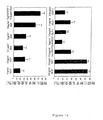
exendin-4及びGLP-1の基本的な特徴を取り入れた一連のキメラ・ペプチドを、設計した。35のペプチドの配列を、GLP-1(第7〜第36残基)、及びexendin-4(番号付けされたGLP-1残基を用いたexendin-4のアラインメントに従って番号を付けた第7〜第45残基)の配列とともに図1に示す。それらは、(i)第8と第9アミノ酸の間のDDP1Vの切断作用を最小限にし、(ii)インシュリン分泌刺激作用のための最小限の要件を評価し、そして(iii)exendin-4とGLP-1の間のアミノ酸の差異がGLP-1に対する効果における前者の13倍増の原因となることを評価するように設計した。図1に示されたペプチドは、それらの合成にL-及びD-アミノ酸を利用した。
Example 1. Peptide design and synthesis
A series of chimeric peptides were designed that incorporated the basic features of exendin-4 and GLP-1. The sequences of the 35 peptides were numbered according to the alignment of GLP-1 (residues 7-36) and exendin-4 (exendin-4 with the numbered GLP-1 residues) It is shown in FIG. 1 together with the sequence of the 45th residue). They minimize (i) the cleavage of DDP1V between the 8th and 9th amino acids, (ii) assess the minimum requirements for stimulating insulin secretion, and (iii) exendin-4 It was designed to evaluate that amino acid differences between GLP-1 cause a 13-fold increase in the effect on GLP-1 over the former. The peptides shown in FIG. 1 utilized L- and D-amino acids for their synthesis.
ペプチドは、脱保護のためにピペリジン-ジメチルホルムアミドを、そしてカップリングのためにHOBt/HBTUを使用した、Applied Biosystems(Foster, CA)の自動ペプチド合成機によって、アミノ酸のFmoc-誘導体を使いペグ-ポリスチレン樹脂上で合成した。完成したペプチドを、トリフルオロ酢酸(TFA)を使って樹脂から切断して、エーテルを用いて沈澱させて、アセトニトリル勾配を使った0.1%のTFA中、C-18疎水性樹脂を用いた逆相HPLCを使った精製に共した。最終物質の純度は、逆相HPLCを使って確認し、このペプチドの質量は、質量分析を使って確認した。全てのペプチドが、95%超の純度であった。
他のペプチドを、2-アミノヘキサン酸(6-アミノヘキサン酸)を使ってDPP1Vによる切断を減らすように設計した。表2を参照のこと。
Other peptides were designed to reduce cleavage by DPP1V using 2-aminohexanoic acid (6-aminohexanoic acid). See Table 2.
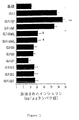
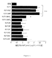
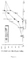
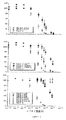
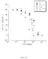
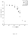
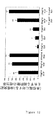
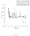
実施例2.インビトロでのインシュリン分泌
Dr. Samuel A. Clark(Bio Hybrid Technologies, Shrewsbury, MA)から贈与物であるRIN1048-36細胞を、インシュリン分泌に対するGLP-1、exendin-4、及びアナログの作用を観察するために使用した。細胞を、12ウェル皿の底に置いたガラス・カバースリップ上に、2.5×10⁵細胞/cm2の密度で播種し、48時間培養した。その後、それらを、37℃の加湿インキュベータ内で、それぞれグルコース不含緩衝液(mMの:140 NaCl、5 KCl、1 NaPO4、1 MgSO4、2 CaCl、20 HEPES緩衝液(pH 7.4)及び0.1%のウシ血清アルブミンを含む)を用いて30分で2期間プレインキュベートした。その後、細胞を、5 mMのグルコース及びペプチド(1×10⁻⁸ M)を伴う同じ緩衝液1 mLの存在下、37℃で1時間インキュベートした。GLP-1とexendin-4(1×10⁻⁸ M)を、全てのアッセイの標準として使用した。1時間後、培地を取り除き、そしてEIA(Crystal Chem, Dhicago II)によるインシュリンレベルの定量化の前に−80℃で保存し、そして標準としてウシγ-グロブリンを用いたブラッドフォード法(Bio-Rad, Richmond, CA)を使った総タンパク質の測定のために、HCl(300 μl、0.1 M、20分、RT)を用いて細胞を溶解した。
Example 2 Insulin secretion in vitro
RIN1048-36 cells, a gift from Dr. Samuel A. Clark (Bio Hybrid Technologies, Shrewsbury, MA), were used to observe the effects of GLP-1, exendin-4, and analogs on insulin secretion. Cells were seeded at a density of 2.5 × 10 5 cells / cm 2 on glass coverslips placed at the bottom of a 12-well dish and cultured for 48 hours. They were then placed in a 37 ° C. humidified incubator with glucose-free buffer (mM: 140 NaCl, 5 KCl, 1 NaPO 4 , 1 MgSO 4 , 2 CaCl, 20 HEPES buffer (pH 7.4) and 0.1 respectively. 2% bovine serum albumin) for 30 min. The cells were then incubated for 1 hour at 37 ° C. in the presence of 1 mL of the same buffer with 5 mM glucose and peptide (1 × 10 −8 M). GLP-1 and exendin-4 (1 × 10 −8 M) were used as standards for all assays. After 1 hour, the medium was removed and stored at −80 ° C. prior to quantification of insulin levels by EIA (Crystal Chem, Dhicago II), and Bradford method (Bio-Rad) using bovine γ-globulin as a standard. , Richmond, CA), cells were lysed using HCl (300 μl, 0.1 M, 20 min, RT) for total protein determination.
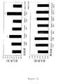
図2に示されるように、アミノ酸修飾のいくつかが、GLP-1又はexendin-4による誘発に匹敵するか又は超える様式でインシュリン分泌を誘発した。いくつかの修飾が、第8と第9アミノ酸残基の間の切断部位についての、DDP1Vによる認識力を減らすために使用された。しかし、第8と第9アミノ酸残基付近のL-アミノ酸の、D型による置換は、このペプチドがインシュリン分泌を誘発することができないので、第2、第3及び第5〜第7ペプチドによって示されたとおり効果がないことを証明した。ペプチド4(図1に示されない)、Dアミノ酸である第7〜第14残基をもつGLP-1配列は、同様にインシュリン分泌を誘発することができなかった。アミノ酸スペーサを第8と第9残基の前又はその間に組み込んだとき、ペプチド11(配列番号8)(第8残基の前に4つのアミノ酸スペーサを持つ)は、強力にインシュリン分泌を誘発したのに対して、ペプチド25(配列番号22)( 第8と第9残基の間に4つのアミノ酸スペーサをもつ)、及びペプチド26(配列番号23)(第8と第9残基の間に8つのアミノ酸スペーサをもつ)は、インシュリン分泌を誘発しなかった。GLP-1の第8アミノ酸(アラニン:A)の、小さい中性アミノ酸であるexendin-4の対応する位置のペプチド(すなわちグリシン:G) による置換は、exendin-4よりわずかにインシュリン分泌を誘発した。GLP-1Gly8(配列番号3)を参照のこと。
As shown in FIG. 2, some of the amino acid modifications elicited insulin secretion in a manner comparable to or exceeding that induced by GLP-1 or exendin-4. Several modifications were used to reduce the recognition by DDP1V for the cleavage site between the 8th and 9th amino acid residues. However, substitution of the L-amino acids near the 8th and 9th amino acid residues by the D form is indicated by the 2nd, 3rd and 5th-7th peptides, since this peptide cannot induce insulin secretion. Proved to be ineffective. Peptide 4 (not shown in FIG. 1), the GLP-1 sequence with
GLP-1アミノ酸残基の、exendin-4残基によるさらなる置換は、インシュリン分泌を誘発する能力を保ったペプチドをもたらした。例えば、ペプチド8(配列番号5)(8位でのA→Gの置換と、16位でのV→Lの置換をもつ)、ペプチド9(配列番号6)( 8、16、18、19及び20位での、それぞれA→G、V→L、S→K、Y→Q及びL→Mの置換をもつ)、ペプチド10(Ex-WOT;配列番号7)(ペプチド9と同じ置換をもち、さらに22、23、25、26、27、30、33、34、36位での、それぞれG→E、Q→E、A→V、K→R、E→L、A→E、V→K、K→N及びR→Gの置換をもつ)は、全てインシュリン分泌を誘発する能力を保った。実際、ペプチド8(配列番号5)は、GLP-1又はexendin-4のいずれかよりも、インシュリン分泌に対して実質的に大きな効果をもった。
Further substitution of GLP-1 amino acid residues with exendin-4 residues resulted in peptides that retained the ability to induce insulin secretion. For example, peptide 8 (SEQ ID NO: 5) (with an A → G substitution at
ペプチド12(配列番号9)のような、exendin-4に存在する末端の第8〜第9アミノ酸の、GLP-1への付加が、GLP-1又はexendin-4よりインシュリン分泌に対して実質的に大きな効果をもつペプチドをもたらした。exendin-4の第8残基(すなわち、グリシン:G)を、GLP-1の第8残基(すなわち、アラニン:A)で置換し、そしてペプチド13(配列番号10)のようにexendin-4の末端の第9アミノ酸が維持されるか、又はペプチド14(配列番号11)のように除去された場合、どちらのペプチドもインシュリン分泌を誘発する能力を維持した;しかし、ペプチド13は、修飾なしのexendin-4より実質的に強い効果をもった。
The addition of terminal 8th to 9th amino acids present in exendin-4, such as peptide 12 (SEQ ID NO: 9), to GLP-1 is substantially more effective for insulin secretion than GLP-1 or exendin-4. Resulted in a peptide with a great effect. The 8th residue of exendin-4 (ie, glycine: G) is replaced with the 8th residue of GLP-1 (ie, alanine: A) and exendin-4 as in peptide 13 (SEQ ID NO: 10) Both peptides maintained the ability to induce insulin secretion when the terminal 9th amino acid was retained or removed as in peptide 14 (SEQ ID NO: 11); however,
インシュリン分泌を誘発するそれらの能力について、exendin-4の切詰めを試験しもした。ペプチド15〜24(配列番号12〜21)を参照のこと。32残基以上を含むペプチド(すなわち、ペプチド15〜18(配列番号12〜15))だけが、インシュリン分泌を誘発した。インシュリン分泌を誘発したそれらの切詰めペプチドについて、ペプチド15(配列番号12)(第43残基以下の残基を含む)、及びペプチド18(配列番号15)(第34残基以下の残基を含む)が、exendin-4又はGLP-1のそれを超えた誘発効果をもつ唯一のものである。
Exendin-4 truncation was also tested for their ability to induce insulin secretion. See peptides 15-24 (SEQ ID NOs: 12-21). Only peptides containing more than 32 residues (ie, peptides 15-18 (SEQ ID NOs: 12-15)) induced insulin secretion. For those truncated peptides that induced insulin secretion, peptide 15 (SEQ ID NO: 12) (including
ペプチドの荷電に影響するように考えられた修飾が、行われもした。GLP-1は、7位、26位、34位及び36位の塩基性アミノ酸に関連する合計4+の電荷、及び9位、15位、21位及び27位の酸性アミノ酸に関連する合計4-の電荷をもつ、正味中性荷電をもつ。Exendin-4は、21〜23位の塩基性ドメインに関連する正味負の電荷をもつ。Exendin-4は、合計4+の電荷(7位、18位、26位、33位)、及び合計6-の電荷(9位、15位、21位、22位、23位、30位)をもつ一方で、その9つのアミノ酸の末端は中性である。ペプチド34(配列番号31)のような、exendin-4(すなわち、36位での、小さい中性のグリシン、Gの、より大きなアルギニン、Rによる置換)の末端に(正電荷を提供する)単一の塩基性アミノ酸の付加は、インビトロのインシュリン分泌におけるペプチドの不活性をもたらす。アルギニン、Rは、GLP-1の36位でよく許容され、そして維持されるか又はGLP-1において中性のグリシン、Gであるexendin-4末端によって置換されるとき(ペプチド12(配列番号9)、それは、活性を維持し、それどころかexendin-4のそれを超える活性をもつ。
Modifications that were thought to affect the charge of the peptides were also made. GLP-1 has a total of 4+ charges associated with basic amino acids at
興味深いことには、27位が負電荷をもつ場合に、30位が酸性のグルタミン酸、Eから中性のアラニン、Aに修飾されるとき(ペプチド36(配列番号33))、アルギニン、Rは、exendin-4の36位でよく許容される。同様に、第30ペプチド(配列番号27)と比べて、18位(ペプチド1(配列番号3))に正電荷を導入するための、中性のセリン、Sの、塩基性のリジン、Kによる置換は、活性の喪失をもたらす;しかし、隣接する(19、20位の)中性のアミノ酸、チロシン、Y及びロイシン、Lの、中性のアミノ酸、グルタミン、Q及びメチオニン、Mによる置換が、活性を回復させる(ペプチド9(配列番号6)をペプチド30(配列番号27)と比べて)。
Interestingly, when position 27 is negatively charged and
GLP-1と6-アミノヘキサン酸含有ペプチドのインシュリン分泌効果を比較した場合、GLP-1Aha8(ペプチド11;配列番号8)は、GLP-1と同程度の有効性を示した。GLP-1Aha8(ペプチド11;配列番号8)は、基礎レベルの約1.2倍を上回るインシュリン分泌を誘発した。図3を参照のこと。しかし、ペプチド25及び26(配列番号22〜23)にあるとおり、追加の6-アミノヘキサン酸残基の挿入は、インシュリン分泌を誘発するペプチドの能力を阻害した。
When comparing the insulin secretion effect of GLP-1 and a 6-aminohexanoic acid-containing peptide, GLP-1Aha 8 (
実施例3.細胞内cAMPの定量
ヒトGLP-1受容体を安定的にトランスフェクトされたCHO細胞、GLP-1R細胞を、12ウェル・プレート上に60〜70%集密まで育て、クレブズ-リンガー・リン酸塩緩衝物質(KRP)で3回洗浄し、そして加湿気インキュベータ内、37℃で2時間、0.1%のウシ血清アルブミン含有KRP(BSA)1 mlと一緒にインキュベートした。次に細胞を、研究中のペプチドの存在又は不存在下、イソブチルメチルキサンチン(IBMX)(1 mM;Calbiochem, La Jolla, CA)を伴う、0.1%のBSAを補った1 mlのKRP中でインキュベートした。氷冷リン酸緩衝生理食塩水(PBS)で無傷の細胞を3回洗浄することによって30分後に反応を止めた。氷冷過塩素酸(0.6 M、1 ml、5分)中で細胞をインキュベートすることによって細胞内のcAMPを抽出した。炭酸カリウム(5M、84 μl)を使ってサンプルのpHを7に調整した後、試料管をボルテックスし、形成された沈殿を、遠心分離(5分、2000×g、4℃)で沈殿させた。上清を、真空で乾燥させ、4 mMのEDTA含有0.05 MのTris(pH7.5)(300 μl)中に可溶化した。炭酸ナトリウム(0.15 μM)と硫酸亜鉛(0.15 μM)を加えたサンプルを、次に氷上で15分間インキュベートした。得られた塩沈殿物を、遠心分離(5分、2000×g、4℃)によって除去した。サンプルを、[3H]cAMP競合的タンパク質結合アッセイ・キット(Amersham, Philadelphia, PA)を使って、二重のアリコート(50 μl)でアッセイした。
Example 3 Quantification of intracellular cAMP CHO cells and GLP-1R cells stably transfected with human GLP-1 receptor are grown to 60-70% confluence on 12-well plates, and Krebs-Ringer phosphate Washed 3 times with buffer (KRP) and incubated with 1 ml of 0.1% bovine serum albumin-containing KRP (BSA) for 2 hours at 37 ° C. in a humidified incubator. Cells are then incubated in 1 ml KRP supplemented with 0.1% BSA with isobutylmethylxanthine (IBMX) (1 mM; Calbiochem, La Jolla, Calif.) In the presence or absence of the peptide under study. did. The reaction was stopped after 30 minutes by washing intact cells three times with ice-cold phosphate buffered saline (PBS). Intracellular cAMP was extracted by incubating the cells in ice-cold perchloric acid (0.6 M, 1 ml, 5 min). After adjusting the pH of the sample to 7 using potassium carbonate (5M, 84 μl), the sample tube was vortexed and the precipitate formed was precipitated by centrifugation (5 min, 2000 × g, 4 ° C.) . The supernatant was dried in vacuo and solubilized in 0.05 M Tris (pH 7.5) (300 μl) containing 4 mM EDTA. Samples added with sodium carbonate (0.15 μM) and zinc sulfate (0.15 μM) were then incubated on ice for 15 minutes. The resulting salt precipitate was removed by centrifugation (5 min, 2000 × g, 4 ° C.). Samples were assayed in duplicate aliquots (50 μl) using the [ 3 H] cAMP competitive protein binding assay kit (Amersham, Philadelphia, PA).
cAMPレベルを、GLP-1、6-アミノヘキサン酸含有ペプチド、又はD-アミノ酸含有ペプチドにより処理された細胞で計測した。GLP-1アナログによって産生された細胞内cAMPレベルは、当初10 nM(最大のcAMP産生の濃度は、GLP-1で見られる)のペプチド濃度で評価した。データを図4に示す。前記ペプチドを、TBMXの存在下、CHO/GLP-1R細胞と一緒に37℃で30分間インキュベートした。インビトロでのインシュリン・アッセイからの結果と一致して、GLP-1分子全体にわたるD-アミノ酸置換は、基礎レベルを上回るわずかな増加だけに終わった、すなわち、それらは、IBMXによってのみ得られた。同様に、インシュリン分泌刺激性化合物GLP-1Gly8(配列番号3)及びGLP-1Aha8(配列番号8)と比べたとき、GLP-1(Aha9)4(配列番号22)及びGLP-1(Aha9)8(配列番号23)は不活性であった。GLP-1、GLP-1Gly8、又はGLP-1Aha8の濃度の変化に対するcAMP誘導における応答を計測した。図5を参照のこと。表3は、3つの化合物全てのED50値を示す。GLP-1Aha8(0.5 nM)は、基礎を4倍上回るまで細胞内cAMP産生を刺激したが、しかしそれは、GLP-1及びGLP-1Gly8と比べたとき、より高いED50を示した。 cAMP levels were measured in cells treated with GLP-1, 6-aminohexanoic acid containing peptides, or D-amino acid containing peptides. Intracellular cAMP levels produced by GLP-1 analogs were initially assessed at a peptide concentration of 10 nM (the maximum cAMP production concentration is found in GLP-1). The data is shown in FIG. The peptide was incubated with CHO / GLP-1R cells for 30 minutes at 37 ° C. in the presence of TBMX. Consistent with the results from the in vitro insulin assay, D-amino acid substitutions throughout the GLP-1 molecule were only marginally increased above the basal level, ie they were only obtained by IBMX. Similarly, when compared to the insulin secretion stimulating compounds GLP-1Gly 8 (SEQ ID NO: 3) and GLP-1 Aha 8 (SEQ ID NO: 8), GLP-1 (Aha 9 ) 4 (SEQ ID NO: 22) and GLP-1 ( Aha 9 ) 8 (SEQ ID NO: 23) was inactive. The response in cAMP induction to changes in the concentration of GLP-1, GLP-1Gly 8 or GLP-1Aha 8 was measured. See FIG. Table 3 shows the ED50 values for all three compounds. GLP-1Aha 8 (0.5 nM) stimulated intracellular cAMP production up to 4 times above basal, but it showed a higher ED50 when compared to GLP-1 and GLP-1Gly 8 .
実施例4.完全な細胞のGLP-1受容体へのペプチドの競合的結合
結合研究を、Montrose-Rafizadeh et al., (1997b) J. Biol. Chem. 272:21201-206のやり方で実施した。要するに、CHO/GLP-1R細胞を、12ウェル・プレート上で集密まで育て、実験の2時間前に無血清Ham F-12培地で洗浄した。0.5 mlの結合緩衝液(10)により2回洗浄した後、細胞を、2% BSA、17 mg/LのDiprotin A(Bachem, Torence, CA)、10 mMのグルコース、1〜1000 nMのGLP-1又は他のペプチド、及び30,000 cpmのSI-GLP-1(Amersham, Philadelphia, PA)を含む0.5 mlの緩衝液と一緒に、4℃で一晩インキュベートした。インキュベーション終了後に、上清を捨て、細胞を氷冷PBSで3回洗浄し、そして0.5 mlの0.5 NのNaOH及び0.1%のドデシル硫酸ナトリウムと一緒に室温で10分間インキュベートした。細胞ライセートの放射活性を、ICN Apec-Series g-counterで計測した。総結合から、大過剰の非標識GLP-1(1 μM)の存在下でインキュベートした細胞に関する放射活性を差し引いて、特定の結合を決定した。
Example 4 Competitive binding binding studies of peptides to the complete cellular GLP-1 receptor were performed in the manner of Montrose-Rafizadeh et al., (1997b) J. Biol. Chem. 272: 21201-206. In short, CHO / GLP-1R cells were grown to confluence on 12-well plates and washed with serum-free Ham F-12 medium 2 hours before the experiment. After washing twice with 0.5 ml binding buffer (10), the cells were washed with 2% BSA, 17 mg / L Diprotin A (Bachem, Torence, CA), 10 mM glucose, 1-1000 nM GLP- Incubate overnight at 4 ° C. with 0.5 ml buffer containing 1 or other peptide and 30,000 cpm SI-GLP-1 (Amersham, Philadelphia, PA). At the end of the incubation, the supernatant was discarded, the cells were washed 3 times with ice-cold PBS and incubated for 10 minutes at room temperature with 0.5 ml 0.5 N NaOH and 0.1% sodium dodecyl sulfate. The radioactivity of the cell lysate was measured with an ICN Apec-Series g-counter. Specific binding was determined by subtracting radioactivity for cells incubated in the presence of a large excess of unlabeled GLP-1 (1 μM) from total binding.
次に、ヒトGLP-1受容体に対する競合的結合によって[125I]GLP-1を置き換えるこれらのGLP-1アナログの能力を検査した。濃度を変えたペプチドの不存在及び存在下、CHO/GLP-1R細胞を[125I]標識GLP-1と一緒にインキュベートした。図6を参照のこと。 Next, the ability of these GLP-1 analogs to replace [ 125 I] GLP-1 by competitive binding to the human GLP-1 receptor was examined. CHO / GLP-1R cells were incubated with [ 125 I] -labeled GLP-1 in the absence and presence of varying concentrations of peptide. See FIG.
GLP-1受容体に競合的に結合したそれらの化合物について得られたIC50値を、表3に示す。6-アミノヘキサン酸部分の挿入は、受容体への結合の減少をもたらした。9位のスペーサー、6-アミノヘキサン酸基の長さの増加により、GLP-1受容体に対する親和性の劇的な減少があった。D-アミノ酸置換された化合物で見られた生物活性の欠損は、それらのGLP-1受容体に対する結合能力の著しい減少によって説明されうる。化合物GLP-1D21(ペプチド6)及びGLP-1A11D(ペプチド7)は、標識されたGLP-1を置き換えなかったように、D-アミノ酸置換の増加するほど受容体認識の累進的な減少があった。 The IC 50 values obtained for those compounds that competitively bound to the GLP-1 receptor are shown in Table 3. Insertion of the 6-aminohexanoic acid moiety resulted in decreased binding to the receptor. An increase in the length of the 9-position spacer, 6-aminohexanoic acid group, resulted in a dramatic decrease in affinity for the GLP-1 receptor. The loss of biological activity seen with D-amino acid substituted compounds can be explained by a significant decrease in their ability to bind to the GLP-1 receptor. Compounds GLP-1D21 (peptide 6) and GLP-1A11D (peptide 7) had a progressive decrease in receptor recognition with increasing D-amino acid substitution, as did not replace labeled GLP-1. .
実施例5.インビボにおける急性活性
急性最大インシュリン応答を、静中ペプチド投与後のズッカーラットにおける血漿インシュリン・レベルの定量によって測定した。特に、一晩の絶食後の体重約400 gの糖尿病雄ラットを、50 mg/kgのペントバルビタールにより麻酔し、そして採血のためにカテーテルをそれらの右大腿動脈に結んだ。その後、exendin-4、GLP-1、又はペプチド(0.4 nmol/kg)のボーラスを、それらの左伏在静脈中に30秒で投与した(1つのペプチドにつきN=6)。ペプチド投与前、及びその後5、15、30、60及び90分で採集した血液を、インシュリン定量のために、EDTA及びアプロチニンを含むヘパリン処理管内に注いだ。血漿を、分離し、取り除き、そして直ちに-70℃に冷凍した。次にラット・インシュリンELISAキット(Crystal Chem Inc., Chicago, IL)を使ってインシュリン・レベルを定量した。
Example 5. In vivo acute active acute maximal insulin response was measured by quantification of plasma insulin levels in Zucker rats after intravenous peptide administration. In particular, diabetic male rats weighing about 400 g after an overnight fast were anesthetized with 50 mg / kg pentobarbital and catheters were tied to their right femoral artery for blood collection. A bolus of exendin-4, GLP-1, or peptide (0.4 nmol / kg) was then administered in their left saphenous vein in 30 seconds (N = 6 per peptide). Blood collected prior to peptide administration and at 5, 15, 30, 60 and 90 minutes thereafter was poured into heparinized tubes containing EDTA and aprotinin for insulin quantification. Plasma was separated, removed and immediately frozen at -70 ° C. Insulin levels were then quantified using a rat insulin ELISA kit (Crystal Chem Inc., Chicago, IL).
先のインビトロにおける研究からの2例の効能をもつペプチド、ペプチド番号10(Ex-4WOT;配列番号7)、及びペプチド1(GLP-1Gly8;配列番号3)の急性インビボ活性を、インシュリン分泌を誘発について、絶食、糖尿病ズッカーラットにおいて評価した。ペプチド(0.4 nmol/kg)の等モル投与の後の、ピークの血漿インシュリン濃度を、図7に示し、等モルのexendin-4及びGLP-1の後に達成されたものと比較する。Ex-4WOTもGly-8も、強力に血漿インシュリン濃度を増加させた。 The acute in vivo activity of two potent peptides from previous in vitro studies, Peptide No. 10 (Ex-4WOT; SEQ ID No. 7), and Peptide 1 (GLP-1Gly 8 ; SEQ ID No. 3) are shown to inhibit insulin secretion Induction was evaluated in fasted, diabetic Zucker rats. Peak plasma insulin concentrations after equimolar administration of the peptide (0.4 nmol / kg) are shown in FIG. 7 and compared to those achieved after equimolar exendin-4 and GLP-1. Both Ex-4WOT and Gly-8 potently increased plasma insulin levels.
図7で説明されるように、それらのi.v.投与の後に、ペプチド1(GLP-1Gly8;配列番号3)及びペプチド10(Ex-4WOT;配列番号7)によって例証されるように、RIN1048-36細胞においてインシュリン分泌を誘発するペプチドのインビトロにおける作用は、絶食、糖尿病ズッカーラットにおける血漿インシュリン濃度を急激性に高めるインビボにおける活性と相関関係がある。特に注目すべきことは、exendin-4の末端第9アミノ酸を欠いているEx-4が等モルのexendin-4がしたよりさらに強力であることが証明されたことである。同様に、ペプチド1(GLP-1Gly8)が、等モルのGLP-1より効さらに強力であることを証明した。 As illustrated in FIG. 7, after their iv administration, as illustrated by peptide 1 (GLP-1Gly 8 ; SEQ ID NO: 3) and peptide 10 (Ex-4WOT; SEQ ID NO: 7), RIN1048-36 The in vitro effects of peptides that induce insulin secretion in cells correlate with the in vivo activity of rapidly increasing plasma insulin levels in fasted, diabetic Zucker rats. Of particular note is that Ex-4, which lacks the terminal 9th amino acid of exendin-4, proved to be more potent than did equimolar exendin-4. Similarly, peptide 1 (GLP-1Gly 8 ) proved to be more potent than equimolar GLP-1.
実施例6.インビボにおける活性の継続時間
インシュリン分泌刺激作用の時間依存的な継続時間を、腹膜内(i.p.)ペプチド投与後のズッカーラットにおける血漿インシュリン及びグルコース・レベルの定量によって評価した。具体的には、一晩に絶食した、体重約400gの糖尿病雄ラットを、50 mg/kgのペントバルビタールで麻酔し、そして採血のためにカテーテルを右大腿動脈に結んだ。その後、exendin-4、GLP-1、又はペプチド(0.4 nmol/kg)のボーラスをi.p.投与した。(1ペプチドにつきN≧2)。ペプチド投与前、及びその後30及び60分、そして2、4、6及び24時間に採集した血液を、インシュリン定量のために、EDTA及びアプロチニンを含むヘパリン処理管内に注ぎ、そして分離したサンプルで、グルコースを計測することにした。血漿を分離し、取り除き、そして直ちに-70℃に冷凍した。次にラット・インシュリンELISAキット(Crystal Chem Inc., Chicago, IL)を使ってインシュリン・レベルを定量し、血漿グルコースをグルコース酸化酵素法によって定量した。
Example 6 Duration of activity in vivo The time-dependent duration of insulin secretagogue was assessed by quantification of plasma insulin and glucose levels in Zucker rats after intraperitoneal (ip) peptide administration. Specifically, diabetic male rats weighing approximately 400 g, fasted overnight, were anesthetized with 50 mg / kg pentobarbital, and a catheter was tied to the right femoral artery for blood collection. Thereafter, exodin-4, GLP-1, or a bolus of peptide (0.4 nmol / kg) was administered ip. (N ≧ 2 per peptide). Blood collected prior to peptide administration and at 30 and 60 minutes and then 2, 4, 6 and 24 hours was poured into heparinized tubes containing EDTA and aprotinin for insulin quantification and separated samples with glucose I decided to measure. Plasma was separated, removed and immediately frozen at -70 ° C. Next, insulin levels were quantified using a rat insulin ELISA kit (Crystal Chem Inc., Chicago, IL), and plasma glucose was quantified by the glucose oxidase method.
図8で示されるように、特定のアミノ酸修飾は、インビボにおいてインシュリンに対して長期間の作用を提供する。この件に関して、血漿インシュリン・レベルに対して、ポリペプチド10(Ex-4WOT;配列番号7)は、exendin-4のように、長く作用することが分かった。さらに、急性研究と同様に、糖尿病ラットにおいて、ポリペプチド10が、等モルのexendin-4よりさらに強力であることを証明した。対照的に、ポリペプチド1(GLP-1Gly8;配列番号3)及びポリペプチド11(GLP-1Aha8;配列番号8)が、GLP-1とexendin-4の中間であった;前者より長いが、後者より短い、時間依存的なインシュリン応答に対する作用をもつことが分かった。さらに、急性研究と同様に、糖尿病ラットにおいて、ポリペプチド1及び11が、等モルのGLP-1よりさらに強力であることを証明した。
As shown in FIG. 8, certain amino acid modifications provide long-term effects on insulin in vivo. In this regard, it was found that polypeptide 10 (Ex-4WOT; SEQ ID NO: 7) acts on plasma insulin levels as long as exendin-4. Furthermore, as in acute studies,
実施例7.MALDI質量スペクトル法
GLP-1(2 μM)とGLP-1Aha8(配列番号8)(2 μM)を、PBS中、5 mUの組み換えDPP1V(Calbiochem, La Jolla, CA)と一緒に、37℃でそれぞれ10分間と2時間インキュベートした。両化合物(100 μM、100 μl)を、同等の量のヒト血清中、37℃で2時間インキュベートした。全てのケースについて、酸素反応を、トリフルオロ酢酸(0.1%v/vの終濃度)の添加によって止めた。マトリックス支援レーザー脱離イオン化法−飛行時間型質量分析法(MALDI-TOF)を使って、サンプルを直ちに分析した。MicromassMALDI-TOF(Micromass, Beverly, MA)リフレクトロン機器を、1スペクトルにつき5レーザー・ショットを用いて、1000〜6000 Daの質量範囲にわたり15〜25%のレーザー・エネルギーで使用した。α-シアノ-4-ヒドロキシシンナミック酸(Sigma, St.Louis, MO)を、マトリックスとして使用し、8 mg/mlの炭酸アンモニウム(Sigma, St.Louis, MO)バッファー中、10 mg/mlの濃度に調製した。MALDIプレートに移す前に、1 μlのサンプルを、マトリックスで50/50 v/vに希釈した。
Example 7 MALDI mass spectrometry
GLP-1 (2 μM) and GLP-1Aha 8 (SEQ ID NO: 8) (2 μM) were combined with 5 mU of recombinant DPP1V (Calbiochem, La Jolla, Calif.) In PBS for 10 minutes each at 37 ° C. Incubated for 2 hours. Both compounds (100 μM, 100 μl) were incubated for 2 hours at 37 ° C. in equivalent amounts of human serum. In all cases, the oxygen reaction was stopped by the addition of trifluoroacetic acid (0.1% v / v final concentration). Samples were analyzed immediately using matrix-assisted laser desorption ionization-time of flight mass spectrometry (MALDI-TOF). A MicromassMALDI-TOF (Micromass, Beverly, MA) reflectron instrument was used with a laser energy of 15-25% over a mass range of 1000-6000 Da using 5 laser shots per spectrum. α-Cyano-4-hydroxycinnamic acid (Sigma, St. Louis, MO) was used as a matrix and 10 mg / ml in 8 mg / ml ammonium carbonate (Sigma, St. Louis, MO) buffer. Concentration was adjusted. Prior to transfer to MALDI plates, 1 μl of sample was diluted to 50/50 v / v with matrix.
GLP-1Aha8(配列番号8)の安定性を、DPP1V及びヒト血清の存在下でGLP-1と比較した。37℃で、10分間のDPP1V(5 mU)による、又は2時間の100%血清によるGLP-1(2 μM)の処理は、MALDIによって計測されるように、N末端を切り取られた産物の量の顕著な増加を引き起こした(Mr=3089 gmol-1)。対して、GLP-1Aha8(2 μM)は、いずれの治療に対しても抵抗力があるように思われた。 The stability of GLP-1Aha 8 (SEQ ID NO: 8) was compared to GLP-1 in the presence of DPP1V and human serum. Treatment of GLP-1 (2 μM) with DPP1V (5 mU) for 10 minutes at 37 ° C. or with 100% serum for 2 hours is the amount of N-terminal truncated product as measured by MALDI. (Mr = 3089 gmol -1 ). In contrast, GLP-1Aha 8 (2 μM) appeared to be resistant to either treatment.
実施例8.インビボにおけるGLP-1Aha 8 の生物活性の測定
6ヶ月齢の雄ズッカーfa/faラット(Harlan, Indianapolis, IN)、及び6ヶ月齢のウィスターラットを、この研究で使用した。それらを、食べ物と水を自由摂取させ、12時間の明、12時間の暗のサイクル(午前7時に点灯)の状態にした。ズッカーラットの床敷きは、紙ベースの製品「Carefresh」(Absorption Co., Belingham、WA)であった。実験の前一晩、床敷きの不存在の状態で、ズッカーラットをワイヤー上で絶食させた。ウィスターラットを、それらの通常の床敷き上で絶食させた。ペントバルビタール(50mg/kg)の腹膜内の注射によって全身麻酔を誘発した。採血のために大腿動脈にカニューレを取り付け、そしてポリペプチド(GLP-1Aha8(配列番号8)及びGLP-1Gly8(配列番号3)、24 nmol/kg)を、動物の首筋に皮下注射した(各処置群についてn=5)。血糖値を、Glucometer Elite(Bayer Corp. Diagnostics, Tarrytown, NY)を使っやグルコース酸化酵素法によって計測した。
Example 8 Measurement of biological activity of GLP-1Aha 8 in vivo
Six month old male Zucker fa / fa rats (Harlan, Indianapolis, IN) and 6 month old Wistar rats were used in this study. They had free access to food and water and were in a 12 hour light, 12 hour dark cycle (lights on at 7 am). Zuckerrat's flooring was the paper-based product “Carefresh” (Absorption Co., Belingham, WA). One night before the experiment, Zucker rats were fasted on the wire in the absence of bedding. Wistar rats were fasted on their normal bedding. General anesthesia was induced by intraperitoneal injection of pentobarbital (50 mg / kg). The femoral artery was cannulated for blood collection and polypeptides (GLP-1Aha 8 (SEQ ID NO: 8) and GLP-1Gly 8 (SEQ ID NO: 3), 24 nmol / kg) were injected subcutaneously into the neck of the animals ( N = 5) for each treatment group. The blood glucose level was measured by Glucometer Elite (Bayer Corp. Diagnostics, Tarrytown, NY) or by glucose oxidase method.
GLP-1Aha8がインビボにおいて生物活性をもつことを確認するために、前記ポリペプチドを、絶食ズッカー脂肪(fa/fa)ラット及びウィスターラットに皮下投与した(24 nmol/kg)。ズッカーラットの他の群は、同投与量のGLP-1Gly8を受けた。そして次の8時間の間、血糖値を観察した。図9において、結果は、両化合物ともに、急速に血糖を下げたことを示す。ズッカーラットにおいて、グルコース絶食は、その群の中では低かったが、しかし低下の傾斜及び大きさは両化合物に類似していた事実から、血糖の減少は、GLP-1Gly8によってより顕著だった。このクラスの化合物によるインシュリン分泌刺激のグルコース依存性を証明した、低血糖の範囲への血糖の低下から、インシュリン分泌は、GLP-1Gly8により弱められた。GLP-1Aha8によって処理したズッカーラットが低血糖にならなかった場合、インシュリン分泌刺激応答は、撤回されずに、そして長期間の効果が見られることができた。絶食状態で高血糖ではないウィスターラットにおいて、インシュリン・レベルは、GLP-1Aha8によって急激に高められ、低血糖、そして再度のインシュリン分泌刺激応答の迅速な減衰をもたらした。 To confirm that GLP-1Aha 8 has biological activity in vivo, the polypeptide was administered subcutaneously to fasted Zucker fat (fa / fa) and Wistar rats (24 nmol / kg). The other group of Zucker rats received the same dose of GLP-1Gly 8 . The blood glucose level was observed for the next 8 hours. In FIG. 9, the results show that both compounds rapidly lowered blood glucose. In Zucker rats, glucose fasting was lower in the group, but the decrease in blood glucose was more pronounced with GLP-1Gly 8 due to the fact that the slope and magnitude of the decline were similar to both compounds. Insulin secretion was attenuated by GLP-1Gly 8 from a decrease in blood glucose to the hypoglycemic range, which demonstrated the glucose dependence of insulin secretion stimulation by this class of compounds. If Zucker rats treated with GLP-1Aha 8 did not become hypoglycemic, the insulin secretory response could not be withdrawn and a long-term effect could be seen. In fasted and non-hyperglycemic Wistar rats, insulin levels were rapidly increased by GLP-1Aha 8 , resulting in hypoglycemia and a rapid decay of the insulin secretagogue response.
実施例9.Exendin-4の切詰め、及びExendin-4の第9アミノ酸C末端の生物学的な重要性
この研究は、Ex-4の生物活性に対する、第9C末端アミノ酸の重要性を判断するために実施した。切詰められたEx-4アナログの配列と、第9C末端配列を付加したGLP-1アナログを当該研究に使用した。
Example 9 Exendin-4 truncation and biological importance of the 9th amino acid C-terminus of Exendin-4 This study was conducted to determine the importance of the 9C-terminal amino acid for the biological activity of Ex-4 . The truncated Ex-4 analog sequence and the GLP-1 analog with the 9C terminal sequence added were used in the study.
材料及び細胞系
先に記載のとおり、ペプチドを合成した。全てのペプチドが、95%超の純度であった。表4は、研究したGLP-1及びexendin-4アナログの配列を示す。イソブチルメチルキサンチン(IBMX)は、Calbiochem(La Jolla, CA)から購入した。Exendin-4及びGLP-1-(7-36)アミドを、Bachem(Torrance, CA)から入手した。クローン化したラット・インスリノーマ細胞系RIN1046-38は、Dr. Samuel A. Clark(BioHybrid Technologies, Shrewsbury, MA)からの贈与物であり、37℃の加湿5%CO2-95%エアー・インキュベーター内で、グルコース(11 mM)、50 U/mlのペニシリン、50 μg/mlのストレプトマイシン及びグルタミン(2 mM)を補充したEarle's塩(Mediatech, Inc., Herndon, VA)を伴うM199中で、日常的に培養された。GLP-1受容体遺伝子により安定的にトランスフェクトされたチャイニーズハムスター卵巣(CHO)細胞、CHO/GLP-1R細胞を、先に記載した。血漿インシュリン・レベルをELISAによって計測した(Crystal Chem Inc., Chicago ILL)。HbA1cを、Greig et al., 1999に記載のとおり計測した。血糖値を、Glucometer Elite(Bayer Diagnostics, Tarrytown, NY)を使って計測した。
下線を引かれたアミノ酸が、試験されるexendin-4配列由来のアミノ酸を表す。
Peptides were synthesized as described above in materials and cell lines . All peptides were more than 95% pure. Table 4 shows the sequences of GLP-1 and exendin-4 analogs studied. Isobutylmethylxanthine (IBMX) was purchased from Calbiochem (La Jolla, CA). Exendin-4 and GLP-1- (7-36) amide were obtained from Bachem (Torrance, CA). The cloned rat insulinoma cell line RIN1046-38 was a gift from Dr. Samuel A. Clark (BioHybrid Technologies, Shrewsbury, Mass.) And was placed in a humidified 5% CO 2 -95% air incubator at 37 ° C. In M199 with Earle's salt (Mediatech, Inc., Herndon, VA) supplemented with glucose (11 mM), 50 U / ml penicillin, 50 μg / ml streptomycin and glutamine (2 mM) Incubated. Chinese hamster ovary (CHO) cells and CHO / GLP-1R cells stably transfected with the GLP-1 receptor gene were described above. Plasma insulin levels were measured by ELISA (Crystal Chem Inc., Chicago ILL). HbA1c was measured as described in Greig et al., 1999. Blood glucose levels were measured using a Glucometer Elite (Bayer Diagnostics, Tarrytown, NY).
Underlined amino acids represent amino acids from the exendin-4 sequence being tested.
動物
6ヶ月齢の雄ズッカー(fa/fa)ラット(Harlan, Indianapolis, IN)、2ヶ月齢のC57BLKS/J-Leprdb/Leprdbマウス(Jackson Laboratories, Bar Haror, MA)、及び2ヶ月齢のフィッシャーラット(Harlan, Indianapolis, IN)を、急性及び慢性の実験で使用した。全ての動物に、食べ物と水を自由摂取させた。動物を、12時間の明-暗いサイクル(午前7時に点灯)の状態のおいた。ズッカーラット及びdb/dbマウスのための床敷きは、紙ベースの製品、Carefresh(Absorption Co., Belingham, WA)であり、そしてフィッシャーラットを、通常の床敷きの上に収容した。
animal
6 month old male Zucker (fa / fa) rats (Harlan, Indianapolis, IN), 2 month old C57BLKS / J-Leprdb / Leprdb mice (Jackson Laboratories, Bar Haror, Mass.), And 2 month old Fisher rats ( Harlan, Indianapolis, IN) was used in acute and chronic experiments. All animals had free access to food and water. Animals were placed in a 12 hour light-dark cycle (lights on at 7 am). The bedding for Zucker rats and db / db mice was a paper-based product, Carefresh (Absorption Co., Belingham, WA), and the Fisher rats were housed on a normal bedding.
細胞内cAMPの定量
12ウェル・プレート上で60から70%集密まで培養したRIN1046-38細胞を、実施例3に記載のとおり処理し、そして適宜にcAMP定量を実施した。用量反応曲線を図21に示す。
Quantification of intracellular cAMP
RIN1046-38 cells cultured from 60 to 70% confluence on 12-well plates were treated as described in Example 3 and cAMP quantification was performed as appropriate. A dose response curve is shown in FIG.
完全な細胞のGLP-1受容体へのペプチドの競合的な結合
実施例4に記載のとおり結合試験を実施した。表5は、それぞれCHO GLP-1R細胞における競合的結合及びRIN 1046-38細胞におけるcAMPアッセイから得たIC50及びEC50値を示す。
統計分析
全ての値を平均±SEMとして示し、群間の差を、ANOVAを使って分析した。図1及び2の曲線を、反復コンピュータプログラム(20)を使って4因子S字状ロジスティック回帰方程式に適合させ、そして表2のEC50及びIC50値を適合データから計算した。
Statistical analysis All values were presented as mean ± SEM and differences between groups were analyzed using ANOVA. The curves in FIGS. 1 and 2 were fitted to a 4-factor sigmoidal logistic regression equation using an iterative computer program (20) and the EC 50 and IC 50 values in Table 2 were calculated from the fitted data.
ズッカーfa/faラットにおける急性経時変化実験
10 nmol/kgのペプチドを、0.1%のBSAを含むPBS溶液で投与したことを除き、実施例5で先に記載したとおり、6ヶ月齢の雄ズッカーfa/faラット(Harlan, Indianapolis, IN)をこの研究で使用した。Ex(1-36)及びEx(1-35)のsc注射後の血糖値を、図22に示す。
Acute time course experiment in Zucker fa / fa rats
6 month old male Zucker fa / fa rats (Harlan, Indianapolis, IN) as previously described in Example 5 except that 10 nmol / kg peptide was administered in a PBS solution containing 0.1% BSA. Were used in this study. The blood glucose level after sc injection of Ex (1-36) and Ex (1-35) is shown in FIG.
db/dbマウスにおけるEx(1-30)を用いた慢性研究
動物を、実験開始前にそれらの環境順化を容易にするために、2.5ヶ月間我々の施設に収容した。処理の最初の20日間、毎日午前9時に、動物にEx(1-30)(1 nmol/kg)を腹膜内(ip)注射で与えた。その後、次の32日間、午前9時と午後9時位に、動物は、ipでEx(1-30)(1 nmol/kg)を受けた。処理プリトコールの最初の20日の終了時点で、血糖値及びHbA1cを計測した。食物摂取及び動物体重を、毎日午前9時の注射時に測定した。スケジュールの中で失敗した日はない。磁気共鳴像(MRI)を、51日目で取り、そしてIPGTTを、52日目に実施した。
Chronic research animals using Ex (1-30) in db / db mice were housed in our facility for 2.5 months to facilitate their acclimation before the start of the experiment. Animals were given Ex (1-30) (1 nmol / kg) by intraperitoneal (ip) injection at 9 am daily for the first 20 days of treatment. The animals then received Ex (1-30) (1 nmol / kg) ip at 9am and 9pm for the next 32 days. Blood glucose levels and HbA1c were measured at the end of the first 20 days of treated protocall. Food intake and animal weight were measured daily at 9am injection. There are no failed days in the schedule. Magnetic resonance images (MRI) were taken on day 51 and IPGTT was performed on
20日目におけるHbA1cは、Ex(1-30)処理マウスについて7.8±0.4、生理食塩液処理マウスについては7.7±0.3であった。絶食血糖値は、Ex(1-30)処理マウスについて412±92 mg/dl、が生理食塩液処理マウスについては600±1 mg/dlであった。
HbA1c on
磁気共鳴映像法
1.9 T、ボア31 cmのBruker BioSpecシステム(Bruker Medizintechnik GmbH, Ettlingen, Germany)、内径20 cmのシールド型グラジエント・セット、及び直径5 cmのボリューム共鳴器を使って、磁気共鳴イメージを得た。慢性の研究で使用した動物を、イソフルオラン知覚麻痺下に置き、標準T1強調多重スライス・スピン−エコー法(TR=500 ms、TE=8.5 ms)を、腹部全体を含む領域をカバーする、各2.1mmの厚さの20の隣接するスライスにわたって得られた。視野は、128×128画素より高く5×5 cmであった。各々のイメージを、約9分の総撮像時間にわたり、8回の収集を使って収集した。全ての動物において、撮像を2つの時点(0日目と51日目)で実施した。
Magnetic resonance imaging
Magnetic resonance images were obtained using a Bruker BioSpec system (Bruker Medizintechnik GmbH, Ettlingen, Germany) with a 1.9 T, 31 cm bore, a shielded gradient set with an inner diameter of 20 cm, and a volume resonator with a diameter of 5 cm. The animals used in the chronic study were placed under isofluorane numbness and standard T1-weighted multi-slice spin-echo method (TR = 500 ms, TE = 8.5 ms) covering each area including the entire abdomen, 2.1. Obtained over 20 adjacent slices of mm thickness. The field of view was 5 x 5 cm higher than 128 x 128 pixels. Each image was collected using 8 acquisitions over a total imaging time of about 9 minutes. In all animals, imaging was performed at two time points (
内臓と皮下部位の分離を、各々のスライスについて、着目の図面領域(ROIs)によって実施した(Bruker Paravisionソフトウェア)。各ROIから得られる強度ヒストグラムを使って、正常な組織からの脂肪の区分けを達成した(NTH Image software, National Institutes of Health, USA)。このヒストグラムは、各ROIにおいて脂肪組織含量が合計できるようにする分界点としてそれらの間の谷を使用して分けられた、2つの十分に分離されたピークを一般的に示した。 Separation of internal organs and subcutaneous sites was performed for each slice by drawing area of interest (ROIs) (Bruker Paravision software). The intensity histogram obtained from each ROI was used to achieve the segmentation of fat from normal tissues (NTH Image software, National Institutes of Health, USA). This histogram generally showed two well-separated peaks separated using the valley between them as a demarcation point allowing the adipose tissue content to be summed at each ROI.
結果を図23に示す。両セットの動物(対照及びEx(1-30)投与)で体重が減少したが、Ex(1-30)処理した動物は、内臓脂肪沈着の減少を示した。処理された動物は、薬物を受けなかった対照と同ほど速く体重を失わなかった。よって、処理は糖尿病を軽くして、処置した動物はより健康的だった。 The results are shown in FIG. Both sets of animals (control and Ex (1-30) administration) lost weight, while Ex (1-30) treated animals showed reduced visceral fat deposition. Treated animals did not lose weight as quickly as controls that did not receive the drug. Thus, treatment reduced diabetes and the treated animals were healthier.
実施例10.β-アミロイド前駆体タンパク質(βAPP)の代謝に対するGLP-1とGLP-1アナログの効果
アルツハイマー症(AD)の重要な病理的な特徴の1つは、主にアミロイド-βペプチド(Aβ)より成る老人斑の脳血管沈着である。Aβは、より大きなグリコシル化膜結合タンパク質β-アミロイド前駆体タンパク質(βAPP)から誘導される。大部分のβAPPは、Aβドメインにタンパク質分解によって切断されて、アミロイド生成性断片の形成を防ぐ可溶性誘導体(sAPP)を産生する。この研究は、β-アミロイド前駆体タンパク質のプロセッシングに対するGLP-1及び2つのアナログの効果を測定するために実施した。
Example 10 Effect of GLP-1 and GLP-1 Analogues on Metabolism of β-Amyloid Precursor Protein (βAPP) One of the important pathological features of Alzheimer's disease (AD) consists mainly of amyloid-β peptide (Aβ) Cerebrovascular deposition of senile plaques. Aβ is derived from the larger glycosylated membrane-bound protein β-amyloid precursor protein (βAPP). Most βAPP is proteolytically cleaved into the Aβ domain to produce a soluble derivative (sAPP) that prevents the formation of amyloidogenic fragments. This study was conducted to determine the effect of GLP-1 and two analogs on the processing of β-amyloid precursor protein.
PC12細胞を、10%の熱不活化ウマ血清、5%の熱不活化胎児ウシ血清、25 mMのHepesバッファー、及び1×抗生物質−抗真菌性溶液を補充したRPMI1640中で培養した。(全ての培地及び血清を、MediaTech Inc.から入手した(Herndon VA)。処理を、低血清中、GLP-1(3.3、33及び330 μg/ml)(Bachem, Torrance, CA)、並びに2つのアナログexendin-4(0.1、1.0及び10 μg/ml)及びexendin-4-WOT(ペプチド10(Ex-4WOT;配列番号7))(0.1及び1.0 μg/ml)の存在下で実行した。PC12細胞によって調整培地中により多くのsAPPの分泌をもたらす分泌経路を刺激することが示されているNGF(5、10、25及び50 ng/ml)(Promega, Madison, WI)を正の対照として使用した。処置の3日後、未処理(低血清培地のみ)及び処理細胞の調整培地、及び細胞ライセートを、モノクローナル抗体、22C11(Roche Molecular Biochemicals, Indianapolis, IN)を使って免疫ブロット分析に共した。そのエピトープ領域が外形質のシステイン含有ドメインのβAPP66-81に割り当てられた、E.コリ(E.coli)産生βAPPに対して産生された、抗体は、細胞膜中に見つかったβAPPの全ての成熟型、並びに調整培地中に分泌されたカルボキシ切詰め可溶性型を認識する。処理又は未処理細胞からの調整培地又は細胞ライセートの典型的な免疫ブロットで、複数の高分子量タンパク質の結合(Mr 100〜140 kDa)が、明白だった。免疫ブロットにおける免疫反応性バンドの特徴で観察される差異は、ゲル中へのタンパク質の不均一な添加によるものでなく膜上へのタンパク質のむらがある移行によるものでもなかった。等量の総タンパク質を、ゲルの各々のレーンに添加し、5%の酢酸中、0.1%のPonceau Sにより膜を染色することによって、電気泳動移行の効率を観察した。 PC12 cells were cultured in RPMI 1640 supplemented with 10% heat-inactivated horse serum, 5% heat-inactivated fetal bovine serum, 25 mM Hepes buffer, and 1 × antibiotic-antifungal solution. (All media and serum were obtained from MediaTech Inc. (Herndon VA). Treatment was performed in low serum, GLP-1 (3.3, 33 and 330 μg / ml) (Bachem, Torrance, CA), and two PC12 cells were run in the presence of analog exendin-4 (0.1, 1.0 and 10 μg / ml) and exendin-4-WOT (peptide 10 (Ex-4WOT; SEQ ID NO: 7)) (0.1 and 1.0 μg / ml). NGF (5, 10, 25 and 50 ng / ml) (Promega, Madison, WI), shown to stimulate the secretion pathway leading to more sAPP secretion in conditioned medium, was used as a positive control Three days after treatment, untreated (low serum medium only) and conditioned medium of treated cells and cell lysate were subjected to immunoblot analysis using a monoclonal antibody, 22C11 (Roche Molecular Biochemicals, Indianapolis, IN). epitope region has been assigned to 3APP 66-81 cysteine rich domain of outer membrane, raised against E. coli (E. coli) producing 3APP, anti Recognizes all mature forms of βAPP found in the cell membrane, as well as the soluble form of the carboxy truncation secreted into the conditioned medium, in a typical immunoblot of conditioned medium or cell lysate from treated or untreated cells. The binding of multiple high molecular weight proteins (M r 100-140 kDa) was evident, the difference observed in the immunoreactive band characteristics in the immunoblot is due to the heterogeneous addition of protein in the gel Neither was it due to uneven transfer of protein onto the membrane, but an equal amount of total protein was added to each lane of the gel and the membrane was stained with 0.1% Ponceau S in 5% acetic acid. The efficiency of electrophoretic transfer was observed.
上方のバンドの濃度測定による定量化は、NGF処理後にβAPPの細胞内レベルの急激な上昇を明らかにした(図11A、棒グラフ1及び2;図11B、棒グラフ1及び2)。細胞培養実験間の特有の変化は、1組の研究の中の5 ng/mlと10 ng/mlのNGFによる(図面11AとC)、並びに異なる組の研究の中の25 ng/mlと50 ng/mlのNGFによる(図11BとD)処理後の分化の程度の差異を考慮する。NGFとは対照的に、GLP-1及びアナログは、βAPPの細胞内レベルを減少させた(図11A、棒グラフ3〜5;図11B棒グラフ3〜7)。NGF及びexendin-4の組み合わせ物は、未処理細胞に対して細胞内βAPPを増加させたが、しかし2つの単独の処理条件のそれの間のレベルであった(図11A、棒グラフ6)。
Quantification by measuring the concentration of the upper band revealed a sharp rise in intracellular levels of βAPP after NGF treatment (FIG. 11A,
図11C及びDに示されるように、全ての投与量の神経成長因子処理が、調整培地で検出されることができた分泌された、可溶性βAPP誘導体の急激な上昇をもたらした(図11C、棒グラフ1及び2;図11D、棒グラフ1及び2)。細胞ライセートからの細胞内βAPPレベルの類似したパターンに続いて、GLP-1、exendin-4及びexendin-4-WOTの全ての投与量による処理は、調整培地中への検出可能なレベルのsAPPの減少を引き起こした(図11、棒グラフ3〜5;図11D、棒グラフ3〜7)。NGFとexendin-4の組み合わせ物は、未処理細胞に対してsAPPレベルの少しの変化も引き起こさなかった(図11C、棒グラフ6)。
As shown in FIGS. 11C and D, all doses of nerve growth factor treatment resulted in a sharp rise in secreted, soluble βAPP derivative that could be detected in conditioned medium (FIG. 11C, bar graph). 1 and 2; FIG. 11D,
Sigma Co.からの乳酸デヒドロゲナーゼ(LDH)キットを使って、使用した処理及び未処理細胞の両方の、調整培地及び細胞ライセート・サンプルにおいて以下に記載のとおりLDHのアッセイを実施した。使用した条件下で処理及び未処理細胞間のLDHレベルで著しい変化は観察されなかった。使用された投与量でのGLP-1及びアナログによる処理の結果としての毒性の可能性は外される。 The LDH assay was performed as described below in conditioned media and cell lysate samples of both used and untreated cells using a lactate dehydrogenase (LDH) kit from Sigma Co. No significant changes were observed in LDH levels between treated and untreated cells under the conditions used. The potential for toxicity as a result of treatment with GLP-1 and analogs at the doses used is eliminated.
データは、12細胞におけるGLP-1処理後に、βAPPの分泌されている誘導体と成熟型のレベルを減少させることを示す。sAPP分泌のこれらの減少は、減少したβAPP合成の分泌の結果である。 The data show that after GLP-1 treatment in 12 cells, the levels of secreted derivatives and mature forms of βAPP are reduced. These reductions in sAPP secretion are the result of decreased secretion of βAPP synthesis.
実施例11.GLP-1及びアナログの神経増殖と分化の促進
ラット褐色細胞腫(PC12)細胞系において、ニューロンの増殖と分化、及びニューロンタンパク質の代謝に対するGLP-1とその長期作用性アナログ2種類、exendin-4、及びexendin-4 WOTの効果を、試験した。GLP-1及びexendin-4は、神経突起の伸長を誘発し、それは、選択的GLP-1受容体拮抗薬、exendin(9-39)との同時インキュベーションによって覆された。さらに、exendin-4は、神経成長因子(NGF)によって開始された分化を促進し、かつ、NGF介在撤回に続く細胞の変性を救う。
Example 11 Promotion of nerve growth and differentiation of GLP-1 and analogs In rat pheochromocytoma (PC12) cell line, GLP-1 and two long-acting analogs of neuronal proliferation and differentiation, and neuronal protein metabolism, exendin-4 The effects of exendin-4 WOT were tested. GLP-1 and exendin-4 induced neurite outgrowth, which was reversed by co-incubation with a selective GLP-1 receptor antagonist, exendin (9-39). Furthermore, exendin-4 promotes differentiation initiated by nerve growth factor (NGF) and rescues cellular degeneration following NGF-mediated withdrawal.
材料
7S NGFを、Promega(Madison, WI)から購入した。GLP-1及びexendin(9-39)を、Bachem(Torrance, CA)から入手した。Exendin-4及びそのアナログ、exendin-4WOTを合成し、先に記載のとおりHPLC分析によって>95%の純度であることを評価した。全ての他の化学薬品は、別段の記載のない限り、高純度であって、Sigma Chemicals(St.Louis, MO)から入手した。
material
7S NGF was purchased from Promega (Madison, WI). GLP-1 and exendin (9-39) were obtained from Bachem (Torrance, CA). Exendin-4 and its analog, exendin-4WOT, were synthesized and evaluated to be> 95% pure by HPLC analysis as described above. All other chemicals were of high purity and were obtained from Sigma Chemicals (St. Louis, MO) unless otherwise stated.
データ分析
統計分析を、必要に応じて実施した。結果を平均±SEM(SEM=平均間の差異の標準誤差)と表現した。SPSSバージョンVIIを使って、分散分析(ANOVA)を実行し、p<0.05を、統計的に有意であるとみなした。有意な主効果に続いて、Tukey's Honestly Significant Difference test(テューキーのHSD)を使って計画比較を行った。
Data analysis Statistical analysis was performed as needed. The results were expressed as mean ± SEM (SEM = standard error of difference between means). Analysis of variance (ANOVA) was performed using SPSS version VII and p <0.05 was considered statistically significant. Following significant main effects, planned comparisons were made using Tukey's Honestly Significant Difference test (Tukey's HSD).
培養条件
褐色細胞腫細胞を、Dr. D. K. Lahiri(Indianapolis)から入手し、RIN1046-38細胞(クローン・ラット・インスリノーマ細胞株)は、Dr. Samuel A. Clark(Bio Hybrid Technology, Shrewsbury, MA)からの贈与物である。PC12細胞を、10%の熱不活化ウマ血清、5%の熱不活化胎児ウシ血清、25 mMのHepesバッファー、及び1×抗生物質−抗真菌溶液を補ったRPMI1640中で培養した。RIN1046-38細胞を、12 mMのグルコースを含み、5%の熱不活化胎児ウシ血清、0.03%のグルタミン、50 U/mlのペニシリン、及び50 mg/mlのストレプトマイシンを補った培地199中で培養した。細胞培養培地及び血清を、MediaTech(Cellgro) Inc.(Herndon, VA)から入手した。細胞を、5.0%のCO2を含む加湿雰囲気中で培養した。それらを、1枚の60 mm皿につき約2.0×10⁶細胞にて播種した。PC12細胞を、ラットの尾のコラーゲンで覆われた培養器(Roche Molecular Biochemicals, Indianapolis)上で培養した。7S NGFを、100 mg/mlの濃度で成長培地中への希釈によって調製し、-20℃で保存した。GLP-1及びアナログの原液を、滅菌水中で新たに作製し、-20℃で保存した。
Culture conditions Pheochromocytoma cells were obtained from Dr. DK Lahiri (Indianapolis), and RIN1046-38 cells (clone rat insulinoma cell line) were obtained from Dr. Samuel A. Clark (Bio Hybrid Technology, Shrewsbury, MA). Is a gift. PC12 cells were cultured in RPMI 1640 supplemented with 10% heat-inactivated horse serum, 5% heat-inactivated fetal bovine serum, 25 mM Hepes buffer, and 1 × antibiotic-antifungal solution. RIN1046-38 cells were cultured in medium 199 containing 12 mM glucose and supplemented with 5% heat-inactivated fetal bovine serum, 0.03% glutamine, 50 U / ml penicillin, and 50 mg / ml streptomycin. did. Cell culture media and serum were obtained from MediaTech (Cellgro) Inc. (Herndon, VA). Cells were cultured in a humidified atmosphere containing 5.0% CO 2. They were seeded at approximately 2.0 × 10 6 cells per 60 mm dish. PC12 cells were cultured on rat tail collagen covered incubators (Roche Molecular Biochemicals, Indianapolis). 7S NGF was prepared by dilution in growth medium at a concentration of 100 mg / ml and stored at -20 ° C. GLP-1 and analog stock solutions were freshly prepared in sterile water and stored at -20 ° C.
各々の処理条件について3枚の皿を、準備した。播種後24時間、いったん細胞が十分に接着すれば、処理を開始する。培地を吸引し、そして(0.5%の胎児ウシ血清だけを含む)3 mlの新鮮な低血清培地を、適当な化合物と一緒に加えた。 Three dishes were prepared for each treatment condition. Treatment begins once cells are fully attached 24 hours after seeding. The medium was aspirated and 3 ml of fresh low serum medium (containing only 0.5% fetal bovine serum) was added along with the appropriate compound.
細胞ライセートの準備
イムノブロッティング法によるタンパク質分析のために、調整培地及び細胞ペレットを毎日採集した。細胞ライセートを以下のとおり調製した。プレートからの細胞を、そっと回収し、10分間800 gで遠心分離した。細胞ペレットを、10 mMのTris-HCl(pH7.4)、1%のSDS、0.174 mg/mlのフッ化フェニルメチルスルホニル(PMSF)、各々1 mg/mlのアプロチニン、ロイペプチン、ペプスタチンA、及び45.98 mg/mlバナジン酸ナトリウムと10.5 mg/mlフッ化ナトリウムの混合物4 mlを含む溶解バッファー中に懸濁した。懸濁した細胞を、粉砕し、15分間14,000 gで遠心分離した。上清溶液のタンパク質(細胞ライセート)を、計測し(Bradford, 1976)、イムノブロッティング法によって分析した。
Preparation of cell lysates Conditioned media and cell pellets were collected daily for protein analysis by immunoblotting. Cell lysates were prepared as follows. Cells from the plate were gently collected and centrifuged at 800 g for 10 minutes. Cell pellets were treated with 10 mM Tris-HCl (pH 7.4), 1% SDS, 0.174 mg / ml phenylmethylsulfonyl fluoride (PMSF), 1 mg / ml aprotinin, leupeptin, pepstatin A, and 45.98 each. It was suspended in a lysis buffer containing 4 ml of a mixture of mg / ml sodium vanadate and 10.5 mg / ml sodium fluoride. Suspended cells were crushed and centrifuged at 14,000 g for 15 minutes. The protein (cell lysate) in the supernatant solution was counted (Bradford, 1976) and analyzed by immunoblotting.
ウェスタンブロッティングによるタンパク質分析
2.6%のビスアクリルアミド(Novex、サンディエゴCA)を含む10%のTris−グリシン・ゲルを使って、各細胞ライセート及び調整培地からのタンパク質10 μgを用いて、ウェスタンブロット分析を実施した。タンパク質を、PVDF紙上にブロットした。5%の酢酸(Sigma)中、0.1%のPonceau S溶液を用いて膜を染色することによって、移したタンパク質を可視化した。
Protein analysis by Western blotting
Western blot analysis was performed using 10 μg of protein from each cell lysate and conditioned medium using 10% Tris-glycine gel containing 2.6% bisacrylamide (Novex, San Diego CA). Protein was blotted onto PVDF paper. Transferred proteins were visualized by staining the membrane with 0.1% Ponceau S solution in 5% acetic acid (Sigma).
Exendin-4及びGLP-1の介在した神経突起の伸長
PC12細胞を、上記のとおり60 mm皿上で培養し、4日間培養した。この間神経突起の伸長を毎日定量した。1枚の皿につき、5つの無作為な細胞領域を評価し、そして神経突起をもつ細胞の割合が測定した。1つの領域につき約100細胞を、細胞体のそれと同等以上の長さの神経突起について記録した。1つの細胞につき1以上の突起をもっていたかもしれないが、1つの細胞を一度だけ記録した。
Neurite outgrowth mediated by Exendin-4 and GLP-1
PC12 cells were cultured on 60 mm dishes as described above and cultured for 4 days. During this time, neurite outgrowth was quantified daily. Five random cell areas were evaluated per dish and the percentage of cells with neurites was determined. Approximately 100 cells per area were recorded for neurites that were at least as long as that of the cell body. One cell was recorded only once, although it may have more than one process per cell.
神経栄養性化合物の存在しない完全培地で培養された培養されるとき、PC12細胞はニューロン細胞型の特徴を全く示さなかった。低血清培地中でNGFに晒されたとき、細胞は、分裂を止め、そして交感神経ニューロンに類似した形態的特徴を発現させた。細胞は、長い突起を伸ばし、あるものは、低血清培地のみで培養した細胞においてより、さらに扁平な外観を示す細胞体を伴って高度に分岐するようになった。 When cultured in complete medium without neurotrophic compounds, PC12 cells showed no neuronal cell type characteristics. When exposed to NGF in low serum medium, cells stopped dividing and expressed morphological features similar to sympathetic neurons. Cells extended long protrusions, and some became more highly branched with cell bodies showing a flatter appearance than in cells cultured only in low serum medium.
低血清培地中でのGLP-1及びexendin-4による処理は、NGFによって誘発されたものへの分化に対して類似した効果を引き起こした。GLP-1及びexendin-4の誘発した神経突起は、NGFによる処理を受けて産まれた神経突起より、一般に長さが短かくて、分岐が少ない。対照的に、exendin-4との組み合わせでGLP-1拮抗薬、exendin(9-39)は、神経突起伸長を開始できない。 Treatment with GLP-1 and exendin-4 in low serum medium caused a similar effect on differentiation to that induced by NGF. GLP-1 and exendin-4 induced neurites are generally shorter in length and less branched than neurites produced by treatment with NGF. In contrast, the GLP-1 antagonist exendin (9-39) in combination with exendin-4 cannot initiate neurite outgrowth.
神経突起の発達に関する毎日の定量化をも実施した。図12で示した結果は、処理3日目に得たカウントを表し、そして未処理対照のパーセンテージとして表される。低血清培地の存在下、PC12細胞の培養は、5〜10%の細胞が神経突起の突起を伸長する結果となった。分析は、処理条件の有意な主効果を明らかにした(F=263.5、df=8,89、p<0.001)。期待されるように、NGF処理は、本明細書中で試験した3つの投与量;10、30及び100 ng/mlでニューロン表現型を有意に誘発した(全てp<0.01)。例えば、10及び30 ng/mlのNGFによる細胞の処理は、対照から神経突起の突起の、それぞれ550及び720%の増加を引き起こした。同じ条件下で、PC12細胞をexendin-4で処理した場合、有意な神経突起の伸長は、1 μg/ml(未処理に対して98%の増加、p<0.05)、及び10 μg/ml(未処理に対して160%の増加、p<0.01)の化合物でも観察された。しかし、exendin-4による神経突起の伸長は、NGF処理細胞のそれほどは顕著ではなかった。2つの化合物の相乗効果を測定するために、組み合わせ処理の系列を試した。
exendin-4(100 ng/ml)を、10 ng/ml又は30 ng/mlのNGFのいずれかと同時処理したとき、神経突起伸長の有意な増加を、未処理対照細胞に対して観察した(それぞれ、596%及び819%増加、ともにp<0.01)。NGF処理単独に対する神経突起伸長の増加は、30 ng/mlで有意であるのみだった(p<0.01)。類似した結果を、単独で又はNGFと組み合わせて、他の投与量のexendin-4によって観察した。これらのデータは、exendin-4が分化を開始することができて、かつ、NGFに誘発された分化を促進することができることを示唆している。
Daily quantification of neurite development was also performed. The results shown in FIG. 12 represent the counts obtained on the third day of treatment and are expressed as a percentage of the untreated control. In the presence of low serum medium, culturing PC12 cells resulted in 5-10% of cells extending neurite processes. Analysis revealed a significant main effect of treatment conditions (F = 263.5, df = 8,89, p <0.001). As expected, NGF treatment significantly induced a neuronal phenotype at all three doses tested herein; 10, 30, and 100 ng / ml (all p <0.01). For example, treatment of cells with 10 and 30 ng / ml NGF caused a 550 and 720% increase in neurite processes from controls, respectively. When PC12 cells were treated with exendin-4 under the same conditions, significant neurite outgrowth was 1 μg / ml (98% increase over untreated, p <0.05), and 10 μg / ml ( A 160% increase over untreated, p <0.01) was also observed for compounds. However, exendin-4 neurite outgrowth was not as pronounced in NGF-treated cells. To measure the synergistic effect of the two compounds, a series of combination treatments was tried.
When exendin-4 (100 ng / ml) was co-treated with either 10 ng / ml or 30 ng / ml NGF, a significant increase in neurite outgrowth was observed relative to untreated control cells (respectively 596% and 819% increase, both p <0.01). The increase in neurite outgrowth relative to NGF treatment alone was only significant at 30 ng / ml (p <0.01). Similar results were observed with other doses of exendin-4 alone or in combination with NGF. These data suggest that exendin-4 can initiate differentiation and promote NGF-induced differentiation.
NGFが介在する細胞死に対するExendin-4の効果
50 ng/mlのNGFの存在/不存在下、又はexendin-4(1又は5 mg/ml)存在/不存在下、PC12細胞を、完全培地(RPMI1640+5%のウシ胎仔血清+10%のウマ血清)中で培養した。4又は7日後に、細胞を採集し、続いてさらに3日間、通常培地中で回復させた。最終日に、細胞を採集し、生細胞の割合を測定するためにMTTアッセイを実施した。2番目の組の実験(阻止)において、4又は7日間、細胞を、50 ng/mlのNGF及びexendin-4(1又は5 mg/ml)の存在下で培養した。細胞を採集し、前記のとおり回復させた。3番目の組の実験(救済)において、細胞を、50 ng/mlのNGFの存在下で4日間培養した。さらに3日間、5 mg/mlのExendin-4を、培地に加えた。7日目に細胞を採集し、前記のとおり回復させた。
Effect of Exendin-4 on NGF-mediated cell death
PC12 cells in complete medium (RPMI1640 + 5% fetal calf serum + 10% horse serum) in the presence / absence of 50 ng / ml NGF or in the presence / absence of exendin-4 (1 or 5 mg / ml) Incubated in. After 4 or 7 days, cells were harvested and subsequently allowed to recover in normal medium for an additional 3 days. On the last day, cells were harvested and an MTT assay was performed to determine the percentage of viable cells. In the second set of experiments (blocking), cells were cultured in the presence of 50 ng / ml NGF and exendin-4 (1 or 5 mg / ml) for 4 or 7 days. Cells were harvested and allowed to recover as described above. In a third set of experiments (rescue), cells were cultured for 4 days in the presence of 50 ng / ml NGF. For a further 3 days, 5 mg / ml Exendin-4 was added to the medium. Cells were harvested on
4番目の組の実験(救済)において、細胞をNGFの存在下で培養した。4日目に、さらに3日間、5 mg/mlのexendin-4を加えた。7日目に細胞を採集し、前記のとおり回復させた。以下に記載のとおり、4及び7日目に、トリパンブルー排除法、及びにMTTアッセイを実施することによって、細胞を各々のプレート(4プレート/処理条件)についてカウントした。
In a fourth set of experiments (rescue), cells were cultured in the presence of NGF. On the fourth day, 5 mg / ml exendin-4 was added for an additional 3 days. Cells were harvested on
これらの実験で、4日後のNGF処理中止は、大量の細胞死を引き起こさずに、そして大部分の細胞はほとんど完全に回復することができた。Exendin-4同時処理は有意な効果を示さなかった。処理の7日後のNGF処理中止は、細胞の生存度の15〜20%の減少を引き起こし、そしてこの細胞は、完全に回復することができなかった(図13、棒グラフ2)。この場合、exendin-4の同時処理は、低用量(図13、棒グラフ4)又は高用量(図13、棒グラフ6)のいずれも、あるいはNGF処理の4日に添加された場合にも(図13棒グラフ7)細胞死を防がなかった。しかしexendin-4処理をNGF処理中止に続いて実施した場合、回復過程が促進された。例えば、PC12細胞をNGFの存在下で4日間培養し、4〜7日目にNGFを処理中止し、exendin-4を加えたとき(図13、棒グラフ8及び9)、細胞生存度は、対照値に達した(>95%)。これは、高用量(5 mg/ml)及び低用量(1 mg/ml)のexendin-4の両方についてであった。
In these experiments, withdrawal of NGF treatment after 4 days did not cause massive cell death and most cells were able to recover almost completely. Exendin-4 co-treatment showed no significant effect. Discontinuation of NGF treatment after 7 days of treatment caused a 15-20% decrease in cell viability and the cells could not be fully recovered (FIG. 13, bar graph 2). In this case, co-treatment of exendin-4 is either low dose (Figure 13, bar graph 4) or high dose (Figure 13, bar graph 6), or when added on
MTT分析
Promega(Madison, WI)製のCellTiter96(登録商標)Aqueous One Solution Cell Proliferation Assay Reagentを、修飾MTTアッセイにおける生細胞数の測定のための比色分析手順で使用した。この試薬は、新規テトラゾリウム化合物[3-(4,5-ジメチル-2-yl)-5-(3-カルボキシメトキシフェニル)-2-(4-スルフォフェニル)-2H-テトラゾリウム、内部塩;MTS]、及び電子共役試薬(フェナジン・エトスルフェート;PES)を含む。PESは化学安定性を高め、それはMTSとの組み合わせを可能にし、安定した溶液を形成した。MTSテトラゾリウム化合物は、組織培養培地に溶解する、有色のホルマザン産物へと細胞によって生物学的に還元される。代謝的に活性な細胞中のデヒドロゲナーゼ酵素によって産生されるNADPH又はNADHによって、この転換は恐らく達成される(Berridge and Tan, 1993)。アッセイを、直接的に培養したウェルへの少量の試薬の追加、1〜4時間のインキュベーション、続く96ウェルプレート・リーダーによる490 nmの吸光度によって実施される。490 nmの吸光度の値によって計測されるホルマザン産物の量を、培養物中の生細胞の数に直接に比例している。MTSホルマザン産物は組織培地に溶解するので、本手順は、MTTのようなテトラゾリウム成分を使う手順よりわずかなステップしか必要としない。
MTT analysis
The CellTiter96® Aqueous One Solution Cell Proliferation Assay Reagent from Promega (Madison, Wis.) Was used in a colorimetric procedure for determination of viable cell count in a modified MTT assay. This reagent is a novel tetrazolium compound [3- (4,5-dimethyl-2-yl) -5- (3-carboxymethoxyphenyl) -2- (4-sulfophenyl) -2H-tetrazolium, inner salt; MTS And an electron conjugating reagent (phenazine etosulphate; PES). PES increased chemical stability, which allowed combination with MTS to form a stable solution. MTS tetrazolium compounds are biologically reduced by cells into colored formazan products that dissolve in tissue culture media. This conversion is probably achieved by NADPH or NADH produced by dehydrogenase enzymes in metabolically active cells (Berridge and Tan, 1993). The assay is performed by the addition of a small amount of reagent to the directly cultured wells, incubation for 1-4 hours, followed by absorbance at 490 nm with a 96-well plate reader. The amount of formazan product as measured by the absorbance value at 490 nm is directly proportional to the number of living cells in the culture. Since the MTS formazan product dissolves in tissue culture medium, this procedure requires fewer steps than a procedure using a tetrazolium component such as MTT.
PKA阻害薬による神経突起伸長の部分的な抑制
GLP-1及びexendin-4誘発神経突起伸長の機構を決定するために、分化した培養物を、それぞれERK MAPKとPI3-Kを阻害する50 μMのPD98059又は40 μMのLY294002で処理する。cAMPに依存したMAPKリン酸化がPKAによって制御されたかどうかを決定するために、GLP-1及びNGFによって誘発した神経突起をPKAの特異的阻害薬、H89で処理した。
Partial inhibition of neurite outgrowth by PKA inhibitors
To determine the mechanism of GLP-1 and exendin-4 induced neurite outgrowth, differentiated cultures are treated with 50 μM PD98059 or 40 μM LY294002, which inhibits ERK MAPK and PI3-K, respectively. To determine whether cAMP-dependent MAPK phosphorylation was regulated by PKA, neurites induced by GLP-1 and NGF were treated with a specific inhibitor of PKA, H89.
具体的には、培養物を、GLP-1アンタゴニストであるexendin(9-39);PI3キナーゼ阻害薬であるLY294002(40 μM);MAPキナーゼ阻害薬であるPD98059(50 μM)、又はPKA阻害薬であるH89(20 μM)で48時間処理した。細胞を、約1×10⁵細胞/mlで60 mm皿上に播種し、そして前記の化合物のそれぞれとともに10 nMのGLP-1又は0.3 μMのexendin-4で処理した。50 ng/mlのNGF及びフォルスコリン(PKAアクチベーター)(20pM)を、これらの処理の正の対照として使用した。 Specifically, the culture was treated with exendin (9-39), a GLP-1 antagonist; LY294002 (40 μM), a PI3 kinase inhibitor; PD98059 (50 μM), a MAP kinase inhibitor, or a PKA inhibitor Was treated with H89 (20 μM) for 48 hours. Cells were seeded on 60 mm dishes at approximately 1 × 10 5 cells / ml and treated with 10 nM GLP-1 or 0.3 μM exendin-4 with each of the above compounds. 50 ng / ml NGF and forskolin (PKA activator) (20 pM) were used as positive controls for these treatments.
PD98059及びLY294002の両者は、細胞のGLP-1及びexendin-4の誘発した神経突起伸長を減少させた。同様に、NGFに誘発された神経突起の伸長が、PD98059及びLY294002の処理にを受けて減少した。よって、ERK MAPキナーゼ及びPI3キナーゼ・シグナル伝達経路の関与が、PC12細胞におけるGLP-1及びexendin-4が介在した神経突起産生に関係する。H89での処理が、GLP-1及びNGFによって誘発された神経突起伸長に対する多少の阻害効果を証明した。これらのデータは、PKAがMAPキナーゼ・シグナル伝達経路の調節に関与しているが、しかし他のシグナル伝達経路も関与していることを示唆する。 Both PD98059 and LY294002 decreased cellular GLP-1 and exendin-4 induced neurite outgrowth. Similarly, NGF-induced neurite outgrowth was reduced following treatment with PD98059 and LY294002. Thus, involvement of ERK MAP kinase and PI3 kinase signaling pathways is related to neurite production mediated by GLP-1 and exendin-4 in PC12 cells. Treatment with H89 demonstrated some inhibitory effect on neurite outgrowth induced by GLP-1 and NGF. These data suggest that PKA is involved in the regulation of the MAP kinase signaling pathway, but other signaling pathways are also involved.
シナプトフィジン及びBETA-2/NeuroDの発現
GLP-1、exendin-4又はexendin-WOTの誘発したPC12細胞の分化の間に生じている分子の変化を検討するために、シナプス小胞膜でよく発現されているリン酸化された37 kDaのタンパク質である、シナプトフィジンの特徴を試験した。PC12細胞の神経分泌液胞を染色するシナプトフィジン・モノクローナル抗体(Oncogene Research Products, San Diego, CA)を使用した。膜を、20 mMのTris、500 mMのNaCl pH7.4、1%(w/v)のカゼイン(BioRad, San Diego, CA)によって37℃で1時間ブロックした。一次抗体を、ブロック中に希釈し、そして4℃で一晩タンパク質と一緒にインキュベートした。前記膜を、20 mMのTris pH7.4、150 mMのNaCl、及び0.05%のTween-20(TBST)で3回、室温で15分間強く洗浄した。ブロック中、ペルオキシダーゼ連結二次抗体を、膜と一緒に室温で2時間インキュベートした。ペルオキシダーゼ連結抗マウスIgM(Chemicon, Tenecula, CA)を、シナプトフィジンに対する二次抗体として使用した。過剰な抗体を、TBSTで3回強く洗い落とし、その後ECLプラス(Amersham, Philadelphia, PA)中で5分間インキュベーションした。続いて膜を写真用フィルムに焼き付けた。Molecular Analystソフトウェア(BioRad, Hercules, CA)を使ってタンパク質バンドの濃度測定による定量化を実施した。
Synaptophysin and BETA-2 / NeuroD expression
To investigate the molecular changes occurring during differentiation of PC12 cells induced by GLP-1, exendin-4 or exendin-WOT, a phosphorylated 37 kDa well expressed in synaptic vesicle membranes The characteristics of synaptophysin, a protein, were tested. Synaptophysin monoclonal antibody (Oncogene Research Products, San Diego, CA) that stains neurosecretory vacuoles of PC12 cells was used. Membranes were blocked with 20 mM Tris, 500 mM NaCl pH 7.4, 1% (w / v) casein (BioRad, San Diego, CA) for 1 hour at 37 ° C. Primary antibody was diluted in block and incubated with protein overnight at 4 ° C. The membrane was washed vigorously 3 times with 20 mM Tris pH 7.4, 150 mM NaCl, and 0.05% Tween-20 (TBST) for 15 minutes at room temperature. During blocking, peroxidase-linked secondary antibody was incubated with the membrane for 2 hours at room temperature. Peroxidase-conjugated anti-mouse IgM (Chemicon, Tenecula, CA) was used as a secondary antibody against synaptophysin. Excess antibody was washed off three times with TBST and then incubated for 5 minutes in ECL plus (Amersham, Philadelphia, PA). Subsequently, the membrane was baked onto photographic film. Protein band quantification was performed using Molecular Analyst software (BioRad, Hercules, CA).
シナプトフィジン抗体を使った細胞ライセート・サンプルのウエスタン免疫ブロット分析は、約37 kDaの分子量バンドを明らかにした。NGF、GLP-1及びGLP-1アナログによる処理は、未処理細胞と比べてのシナプトフィジン・タンパク質の発現を著しく減少させた。タンパク質バンドの濃度測定による定量化は、未処理に対して、全ての処理条件について有意な減少を示し(図14、全てp<0.01)、それらは、投与量に依存していると考えられる。PC12細胞由来の調整培地サンプルの中に、免疫反応性バンドは検出されなかった。 Western immunoblot analysis of cell lysate samples using synaptophysin antibody revealed a molecular weight band of approximately 37 kDa. Treatment with NGF, GLP-1 and GLP-1 analogues significantly reduced synaptophysin protein expression compared to untreated cells. Quantification of protein bands by concentration measurement shows a significant decrease for all treatment conditions compared to untreated (FIG. 14, all p <0.01), which are thought to be dose dependent. No immunoreactive band was detected in the conditioned medium sample from PC12 cells.
NGF処理の結果としてのPC12細胞における高度の分化は、未処理対照細胞に対してシナプトフィジン発現の著しい減少によって明らかとなった。神経成長因子は、対照細胞に対して約70%の最大減少を引き起こす、細胞性シナプトフィジン発現の投与量に関係した変化を証明した。GLP-1及びアナログは、NGFが介在した分化に対して神経突起の伸長への類似した効果を示したが、しかしより低頻度に対してシナプトフィジン発現の比較的わずかな減少しか示さなかった。興味深いことに、組み合わせたNGFとexendin-4は、相加的な形態的効果を反映して、いずれか単独の化合物よりもシナプトフィジン発現の大きな減少を引き起こした(図14)。総じて、exendin-4は、シナプトフィジン発現の点で、PC12細胞の分化を、GLP-1又はexendin-4WOTのいずれかがしたより顕著な誘導を示した。 The high degree of differentiation in PC12 cells as a result of NGF treatment was revealed by a marked decrease in synaptophysin expression relative to untreated control cells. Nerve growth factor demonstrated a dose-related change in cellular synaptophysin expression that caused a maximal decrease of about 70% relative to control cells. GLP-1 and analogs showed a similar effect on neurite outgrowth on NGF-mediated differentiation, but showed a relatively slight decrease in synaptophysin expression for lower frequencies. Interestingly, the combined NGF and exendin-4 caused a greater reduction in synaptophysin expression than either compound alone, reflecting an additive morphological effect (FIG. 14). Overall, exendin-4 showed more pronounced induction of synaptophysin expression than PC12 cells differentiated by either GLP-1 or exendin-4WOT.
GLP-1の誘発したPC12の分化における転写因子BETA-2/NeuroDの役割を調査するために、細胞ライセートを、NeuroDポリクローナル抗体(Santa Cruz Biotechnology Inc., Santa Cruz, CA)によって調査した。BETA-2/NeuroDは、ニューロンと膵臓の内分泌の発達に重要な役割をもつ。NeuroDの発現は、ニューロン分化の間の、末梢神経系の知覚及び運動ニューロン、感覚器、並びに脳及び脊髄の一部において短期的であると考えられるが;しかし成人の脳における検出は、成熟したニューロンにおける二次的な役割を示唆する(Lee et al., 1997)。膵臓の内分泌の細胞、腸及び脳におけるBETA-2発現は、インシュリン遺伝子の転写を活性化して、ニューロンの分化を促進しうる。機能的なBETA-2遺伝子を欠く突然変異マウスは、インシュリンを産生するベータ細胞数の顕著な減少を有し、成熟した島の発達を欠き、そして結果としてしばしば周産期死亡をもたらす重度の糖尿病を患う(Naya et al., 1997)。よって、BETA-2/NeuroDは、インビボの膵臓の発達、及びニューロンの分化に不可欠である。 To investigate the role of the transcription factor BETA-2 / NeuroD in GLP-1 induced PC12 differentiation, cell lysates were investigated with a NeuroD polyclonal antibody (Santa Cruz Biotechnology Inc., Santa Cruz, Calif.). BETA-2 / NeuroD has an important role in the development of endocrine neurons and pancreas. NeuroD expression is thought to be short-term in peripheral nervous system sensory and motor neurons, sensory organs, and parts of the brain and spinal cord during neuronal differentiation; but detection in adult brain is mature Suggest a secondary role in neurons (Lee et al., 1997). BETA-2 expression in endocrine cells, intestine and brain of the pancreas can activate insulin gene transcription and promote neuronal differentiation. Mutant mice lacking a functional BETA-2 gene have severe reductions in the number of beta cells producing insulin, lack of mature island development, and often result in perinatal death (Naya et al., 1997). Thus, BETA-2 / NeuroD is essential for in vivo pancreas development and neuronal differentiation.
ペルオキシダーゼ連結抗ヤギIgG(Santa Cruz Biotechnology Inc.)を二次抗体として使用したことを除いて、シナプトフィジン抗体について先に記載のとおり抗NeuroD抗体を使って、NeuroD産生をウェスタンブロット分析によって測定した。未処理、及びGLP-1で処理したPC12細胞ライセートの両方ではっきり見える43kDaバンドを検出し、それは、GLP-1処理により増加した。BETA-2/NeuroD発現は、GLP-1での処理により増加し、このインシュリン分泌刺激ポリペプチドのニューロン分化特性のさらなる証拠を提供する。予想されるように、低血清培地のみに晒された培養物は、BETA-2/NeuroDの通常の発現を示した。実際、Noma et al.,(1999)は、NGFのような分化を誘発する薬剤なしに、神経突起に似た突起、及びシナプスに似た構造体のような形態的変化が誘発されたトランスフェクトPC12細胞におけるNeuroDの過剰発現を示した。 NeuroD production was measured by Western blot analysis using an anti-NeuroD antibody as previously described for synaptophysin antibodies, except that peroxidase-conjugated anti-goat IgG (Santa Cruz Biotechnology Inc.) was used as a secondary antibody. A clearly visible 43 kDa band was detected in both untreated and GLP-1 treated PC12 cell lysates, which was increased by GLP-1 treatment. BETA-2 / NeuroD expression is increased by treatment with GLP-1, providing further evidence for neuronal differentiation properties of this insulin secretagogue polypeptide. As expected, cultures exposed only to low serum medium showed normal expression of BETA-2 / NeuroD. In fact, Noma et al., (1999) showed transfection in which morphological changes such as neurite-like processes and synapse-like structures were induced without agents that induce differentiation like NGF. It showed overexpression of NeuroD in PC12 cells.
PC12細胞におけるGLP-1受容体の存在の証明
PC12細胞を、35 mm培養皿内のポリ-L-リジン・コート・ガラス・カバースリップ上に移し、(先に記載のとおり)標準的な条件下で培養した。細胞を、30分間、0.25%のグルタルアルデヒドを用いて固定した。内性ペルオキシダーゼ活性を、0.3%のH2O2でクエンチし、GLP-1受容体のN末端に対して産生された一次ポリクローナル抗体(Dr. Joel F. Habener, Massachusetts GeneralHospital, MAからの贈与物)(希釈係数1:1500)中でのインキュベーションを、室温で1時間実行した。可視化は、ビオチン化抗ウサギIgG二次抗体中でのインキュベーションに続く、ジアミノベンジジン・ジヒドロクロライド(DAB)による発色を伴う、アビジン−ビオチン・ペルオキシダーゼ法を使った。
Evidence for the presence of the GLP-1 receptor in PC12 cells
PC12 cells were transferred onto poly-L-lysine coated glass coverslips in 35 mm culture dishes and cultured under standard conditions (as described above). Cells were fixed with 0.25% glutaraldehyde for 30 minutes. Endogenous peroxidase activity was quenched with 0.3% H 2 O 2 and a primary polyclonal antibody raised against the N-terminus of the GLP-1 receptor (a gift from Dr. Joel F. Habener, Massachusetts General Hospital, MA) ) (Dilution factor 1: 1500) was carried out for 1 hour at room temperature. Visualization used the avidin-biotin peroxidase method with incubation with diaminobenzidine dihydrochloride (DAB) followed by incubation in biotinylated anti-rabbit IgG secondary antibody.
PC12細胞におけるGLP-1R陽性免疫反応性染色の存在は、GLP-1受容体の存在を確かにした。より特に、染色は、細胞体上にあり、そして神経突起末端上により少ない。ほとんど全て細胞が、陽性に見えたが、しかし全てのPC12細胞が同程度に陽性免疫反応性染色を表したわけではない。 The presence of GLP-1R positive immunoreactive staining in PC12 cells confirmed the presence of the GLP-1 receptor. More particularly, staining is on the cell body and less on the neurite ends. Almost all cells looked positive, but not all PC12 cells displayed positive immunoreactive staining to the same extent.
逆転写酵素ポリメラーゼ連鎖反応(RT-PCR)
RT-PCRを、GLP-1受容体mRNAに関する高感度アッセイとして実施した。ラット・インスリノーマ細胞(RIN細胞)を、正の対照として使用した。全RNAを、ChomczynskiとSaccchi(1987)の方法を使ってPC12細胞から分離した。2.5 mgのRNAを我々のRT-PCR反応に使用した。50 mMのKCl、10 mMのTris-HCl、3.5 mMのMgCl2、200 mMのdNTP、並びに各々0.4 mMのラットGLP-1Rセンス(5'ACAGGTCTCTTCTGCAACC 3')及びアンチセンス(5'AAGATGACTTCATGCGTGCC 3')オリゴヌクレオチド・プライマー(膵臓のGLP-1受容体配列の5'及び3'末端)を含むバッファー50 mlの量でRT-PCRを行った。[a-32P]dCTPの存在下、30サイクルで増幅を行った。ラット島細胞を、正の対照として使用した。RT-PCR産物(10 ml)を、適当なサイズマーカーと一緒に4〜20%のポリアクリルアミドゲルで分離した。続いて、真空下80℃で1時間ゲルを乾燥させて、そしてX線フィルムに焼き付けた。
Reverse transcriptase polymerase chain reaction (RT-PCR)
RT-PCR was performed as a sensitive assay for GLP-1 receptor mRNA. Rat insulinoma cells (RIN cells) were used as a positive control. Total RNA was isolated from PC12 cells using the method of Chomczynski and Saccchi (1987). 2.5 mg RNA was used in our RT-PCR reaction. 50 mM KCl, 10 mM Tris-HCl, 3.5 mM MgCl 2 , 200 mM dNTP, and 0.4 mM rat GLP-1R sense (5′
GLP-1受容体について予測されるサイズのRT-PCR産物を得た。ラット島mRNA及びPC12細胞mRNA中の928-bpのクリア・バンドは、PC12細胞上のGLP-1受容体の存在を支持した。 An RT-PCR product of the expected size for the GLP-1 receptor was obtained. The 928-bp clear band in rat islet mRNA and PC12 cell mRNA supported the presence of the GLP-1 receptor on PC12 cells.
cAMPの測定
cAMPの測定の前に、PC12細胞を、3日間、33 μg/mlのGLP-1で処理した。トリプリケートの培養物を、処理開始後、合計期間30分の間、5分の間隔で採集した。処理開始時(ゼロ分)に採集した細胞を、cAMPの基礎レベルに使用した。サイクリックAMPを、Monte-Rafizadeh et al.,(1997a)の方法に従って計測した。
cAMP measurement
Prior to cAMP measurements, PC12 cells were treated with 33 μg / ml GLP-1 for 3 days. Triplicate cultures were collected at 5 minute intervals for a total period of 30 minutes after the start of treatment. Cells collected at the start of treatment (zero minutes) were used for basal levels of cAMP. Cyclic AMP was measured according to the method of Monte-Rafizadeh et al., (1997a).
GLP-1受容体の活性化は、細胞内のcAMPの増加をもたらす、アデニルシクラーゼを刺激することを示した。サイクリックAMPを、33 mg/mlのGLP-1でのPC12細胞の処理に続いて30分間アッセイした。刺激の15分以内にcAMPレベルで最大1200倍増加し、それは30分以内にベースライン近くに戻った。これらの所見は、PC12細胞上のGLP-1受容体の存在及び活性を証明する。 Activation of the GLP-1 receptor has been shown to stimulate adenyl cyclase resulting in an increase in intracellular cAMP. Cyclic AMP was assayed for 30 minutes following treatment of PC12 cells with 33 mg / ml GLP-1. Within 15 minutes of stimulation, cAMP levels increased up to 1200-fold, which returned to near baseline within 30 minutes. These findings demonstrate the presence and activity of the GLP-1 receptor on PC12 cells.
毒性分析
exendin-4の考えられる毒性効果を、インビトロにて2つの方法:LDH分析及びトリパンブルー排除法によって試験した。LDHアッセイを、Sigmaキットを使って実施した。処理後に異なる時間間隔で回集した調整培地サンプルを、高感度乳酸デヒドロゲナーゼ(LDH)アッセイに供した。LDHアッセイは、総細胞質内LDHを通して又は培地中に放出された細胞質内LDHの量の関数としての膜の完全性によって、細胞数の尺度を提供した。放出されたLDHの測定は、LDHの作用によるNADの減少に基づいた。得られた減少したNAD(NADH)を、テトラゾリウム色素の化学量論的変換に利用した。最終有色化合物を、分光光学的にに計測した。培地をアッセイするより前に細胞が溶解された場合、細胞数の増加又は減少が、変換された基質の量の付随する変化をもたらす。これは、試験物質によって引き起こされた胞傷害反応又は膜損傷(細胞毒性)の程度を示した。
Toxicity analysis
The possible toxic effects of exendin-4 were tested in vitro by two methods: LDH analysis and trypan blue exclusion. The LDH assay was performed using a Sigma kit. Conditioned media samples collected at different time intervals after treatment were subjected to a sensitive lactate dehydrogenase (LDH) assay. The LDH assay provided a measure of cell number through total cytoplasmic LDH or by membrane integrity as a function of the amount of cytoplasmic LDH released into the medium. The measurement of released LDH was based on the reduction of NAD due to the action of LDH. The resulting reduced NAD (NADH) was utilized for stoichiometric conversion of tetrazolium dyes. The final colored compound was measured spectrophotometrically. If the cells are lysed prior to assaying the medium, an increase or decrease in cell number results in a concomitant change in the amount of substrate converted. This indicated the degree of cystic injury reaction or membrane damage (cytotoxicity) caused by the test substance.
処理に続いて生細胞数の著しい変化はなく、我々の化合物が、検討した条件下で、PC12細胞に無毒であることを示唆した。図15を参照のこと。処理の間の細胞膜の完全性を測定するために、LDHレベルを、2つの別個の組の実験において、3日目に同じ条件下の対照及び処理細胞からの調整培地で計測した。期待されるように、LDHレベルは、培地標準(サンプルを処理開始時に採取した)に対して上昇した。しかし、10 ng/mlのNGF及び10 μg/mlのexendin-4を唯一の特例として、いかなる処理の投与も未処理細胞対照を越えてLDHレベルを大幅に高めなかった。10 μg/mlのExendin-4が、対照のそれの1.65倍までレベルを高め(p<0.001)、そしてlO ng/mlのNGFが、1.38倍の増加を示した(p<0.05)。
There was no significant change in viable cell count following treatment, suggesting that our compounds were non-toxic to PC12 cells under the conditions studied. See FIG. To measure cell membrane integrity during treatment, LDH levels were measured in conditioned media from control and treated cells under the same conditions on
ブロモデオキシウリジンの取り込みによって測定されたPC12細胞における細胞ターンオーバー
GLP-1が培養においてPC12細胞の増殖に影響するかどうかを測定するために、5-ブロモ-2'-デオキシウリジン(BrdU)の取り込みを観察することによって低血清培地中の細胞増殖を評価した。標識後の抗BrdU抗体を用いた免疫細胞化学を、標識の時点で活発にDNAを複製していた細胞を確認するために使用した。PC12細胞を、33 μg/mlのGLP-1又は50 ng/mlのNGFの存在下又は不存在下で3日間培養した。細胞DNAを標識するために、4%のパラホルムアルデヒド中に固定する6時間前に、10 μMのBrdUを培地に加えた。方法の残りの部分は、増殖キット(Roche, Indianapolis, IN)に従って行った。増殖している細胞(BrdU標識の時点でDNA複製を受けてた細胞)は、クロモゲン反応により暗染された核を示した。BrdU取り込みを、処理の1、2及び3日目に定量した。各々の処理条件について3枚の皿をカウントし、細胞総数に対する標識された細胞のパーセンテージとして表した。PC12細胞は、NGFによる処理(未処理に対して9%の上昇)及びGLP-1による処理(未処理に対して18%)後、1日目にBrdUの取り込みの増加を示した。
Cell turnover in PC12 cells measured by bromodeoxyuridine incorporation.
To determine whether GLP-1 affects PC12 cell growth in culture, cell growth in low serum media was assessed by observing 5-bromo-2'-deoxyuridine (BrdU) incorporation. . Immunocytochemistry using labeled anti-BrdU antibody was used to confirm cells that were actively replicating DNA at the time of labeling. PC12 cells were cultured for 3 days in the presence or absence of 33 μg / ml GLP-1 or 50 ng / ml NGF. To label the cellular DNA, 10 μM BrdU was added to the medium 6 hours prior to fixation in 4% paraformaldehyde. The rest of the method was performed according to the proliferation kit (Roche, Indianapolis, IN). Proliferating cells (cells that had undergone DNA replication at the time of BrdU labeling) showed nuclei darkened by the chromogen reaction. BrdU incorporation was quantified on
実施例12.グルカゴン様ペプチド-1及びExendin-4による、興奮毒性神経損傷の保護及び回復
グルタミン酸によって誘発した細胞死から培養海馬ニューロンを保護する、並びに、成体ラットイにおけるボテン酸で誘発したコリン作用性マーカー欠損を軽減する、GLP-1及びその長期作用性アナログ、exendin-4の能力を試験した。
Example 12 Glucagon-like peptide-1 and Exendin-4 protect and restore excitotoxic neuronal damage Protect cultured hippocampal neurons from glutamate-induced cell death and reduce cholinergic marker deficiency induced by botenoic acid in adult rats The ability of GLP-1 and its long-acting analog, exendin-4, was tested.
培養条件
海馬ニューロン培養物を、前記(Mattson et al., 1995)に類似した方法を使って18日齢の胎仔Sprague Dawleyラットから調製した。要するに、細胞を、軽いトリプシン処理及び粉砕によって解離させ、そして10%のFBS及び1%の抗生物質溶液(104 U/mlのペニシリンG、10 mg/mlのストレプトマイシン及び25 μg/mlのアムホテリシンB;Sigma Chemicals, St. Louis, MO)を含む最小必須培地中に蒔いた。海馬ニューロンを、25 mmの直径のポリ-D-リジン・コート・ガラス・カバースリップ上に、100,000細胞/mlの密度で蒔いた。細胞を蒔いた3時間後に、培地を、1%のB27-サプリメント(Gibco/Life Technologies, Carlsbad, CA)を含む無血清Neurobasal培地によって交換した。
Culture Conditions Hippocampal neuronal cultures were prepared from 18 day old fetal Sprague Dawley rats using a method similar to that described previously (Mattson et al., 1995). In short, cells were dissociated by light trypsinization and trituration and 10% FBS and 1% antibiotic solution (10 4 U / ml penicillin G, 10 mg / ml streptomycin and 25 μg / ml amphotericin B). Plated in minimal essential medium containing Sigma Chemicals, St. Louis, MO). Hippocampal neurons were plated on a 25 mm diameter poly-D-lysine coated glass coverslip at a density of 100,000 cells / ml. Three hours after plating, the medium was replaced with serum-free Neurobasal medium containing 1% B27-supplement (Gibco / Life Technologies, Carlsbad, CA).
MAP-2(ニューロン)とGFAP(神経膠星状細胞)について染色した免疫螢光法が、細胞の98%以上がニューロンであり、そして残りが主に神経膠星状細胞であることを示した。培養物を、プレーティングの7〜10日以内に使用した。 Immunofluorescence staining for MAP-2 (neurons) and GFAP (astrocytes) showed that more than 98% of the cells were neurons and the rest were mainly astrocytes . Cultures were used within 7-10 days of plating.
結合の検討
結合の検討を、Montrose-Rafizadeh(1997b)に記載のとおり実施した。デュプリケートの海馬ニューロン培養物を、0.5 mlの結合バッファーで洗浄し、続いて
2%のBSA、17 mg/lのdiprotinA(Bachem, Torrance, CA)、10 mMのグルコース、0.001〜1000 nMのGLP-1、及び30,000 cpmの125I-GLP-1(Amersham Pharmacia Biotech, Little Chalfont, UK)を含む0.5 mlのバッファー中、4℃で一晩インキュベートした。インキュベーション終了の時点で、上清を捨て、そして細胞を氷冷PBSで3回洗浄し、そして0.5mlの、0.5MのNaOH及び0.1%のSDS中、室温で10分間インキュベートした。細胞ライセート中の放射活性を、Apecシリーズγカウンター(ICN Biomedicals, Inc., Costa Mesa, CA)えにより計測した。総結合から、大過剰の非標識GLP-1(1 μM)の存在でインキュベートした細胞に関する放射活性を差し引いたものとして、特異的結合を測定した。50%の結合、EC50に関係しているGLP-1濃度を、ロジット・プロット分析によって測定した。
Binding studies Binding studies were performed as described in Montrose-Rafizadeh (1997b). Duplicate hippocampal neuronal cultures were washed with 0.5 ml binding buffer followed by
2% BSA, 17 mg / l diprotin A (Bachem, Torrance, CA), 10 mM glucose, 0.001-1000 nM GLP-1, and 30,000 cpm 125 I-GLP-1 (Amersham Pharmacia Biotech, Little Chalfont , UK) and incubated overnight at 4 ° C in 0.5 ml buffer. At the end of the incubation, the supernatant was discarded and the cells were washed 3 times with ice-cold PBS and incubated in 0.5 ml of 0.5 M NaOH and 0.1% SDS for 10 minutes at room temperature. Radioactivity in the cell lysate was measured with an Apec series γ counter (ICN Biomedicals, Inc., Costa Mesa, Calif.). Specific binding was measured as total binding minus the radioactivity for cells incubated in the presence of a large excess of unlabeled GLP-1 (1 μM). GLP-1 concentration related to 50% binding, EC 50 was determined by logit plot analysis.
培養海馬ニューロンへの125IGLP-1の結合は、非標識GLP-1によって濃度依存的に置き換えられた(図16A)。結合した125IGLP-1の50%を置き換えるために必要とされるGLP-1濃度を、ロジット・プロット分析によって決定し、培養海馬ニューロンにおいて14 nMのGLP-1(r=-0.999)の濃度を必要とした。 The binding of 125 IGLP-1 to cultured hippocampal neurons was replaced by unlabeled GLP-1 in a concentration-dependent manner (FIG. 16A). The GLP-1 concentration required to replace 50% of bound 125 IGLP-1 was determined by logit plot analysis, and the concentration of 14 nM GLP-1 (r = 0.999) in cultured hippocampal neurons was determined. I needed it.
cAMPの測定
機能性GLP-1受容体の存在を証明するために、サイクリックAMPを、Montrose-Rafizadeh et al.,の方法に従って計測した(1997a)。トリプリケートの海馬ニューロンの細胞培養を、10 nMのGLP-1で処理し、合計で30分間、薬物処理開始後5分間隔で採集した。薬物処理の開始時(ゼロ分)で採集された細胞を、cAMPの基礎レベルに使用した。
Measurement of cAMP To prove the presence of functional GLP-1 receptor, cyclic AMP was measured according to the method of Montrose-Rafizadeh et al., (1997a). Triplicate hippocampal neuronal cell cultures were treated with 10 nM GLP-1 and collected for a total of 30 minutes at 5-minute intervals after the start of drug treatment. Cells harvested at the start of drug treatment (zero minutes) were used for basal levels of cAMP.
10 nMのGLP-1による培養海馬ニューロンの処理は、cAMP産生の増加を引き起こした(図16B)。刺激の15分以内にcAMPレベルの最大で2〜3倍の上昇があり、30分以内にベースラインの近くに戻った。一元ANOVAは、cAMP産生に対する処理の有意な主効果(F=9.45、df=6,20、p<0.001)を証明した。次のチューキーのHSD検定を使った連続した多重比較(subsequent multiple comparisons)は、10(p<0.01)及び15(p<0.001)分後のcAMP産生の有意な増加を明らかにした。これらのデータは、初代海馬ニューロンが、機能性GLP-1受容体を発現し、それを用いてこれらのペプチドの潜在的な保護及び栄養効果を検討するのに適当なインビトロのシステムとなっていることを説明する。 Treatment of cultured hippocampal neurons with 10 nM GLP-1 caused an increase in cAMP production (FIG. 16B). There was a maximal 2-3 fold increase in cAMP levels within 15 minutes of stimulation and returned near baseline within 30 minutes. One-way ANOVA demonstrated a significant main effect of treatment on cAMP production (F = 9.45, df = 6,20, p <0.001). Subsequent multiple comparisons using the following Tukey HSD test revealed a significant increase in cAMP production after 10 (p <0.01) and 15 (p <0.001) minutes. These data provide a suitable in vitro system for primary hippocampal neurons to express functional GLP-1 receptors and use them to study the potential protective and trophic effects of these peptides Explain that.
アポトーシス細胞死
蛍光性DNA結合色素Hoescht 33342を、アポトーシス細胞死を計測するために使用した。ニューロンを、グルタミン酸(10 μM)の存在又は不存在下、GLP-1(10 nM)又はexendin-4(0.3 μM)を伴ったLocke'sバッファー中で16時間インキュベートした。使用したGLP-1の濃度は、cAMPの放出を刺激することが証明され、かつ、我々の先のニューロン細胞研究において細胞死を引き起こすことなく分化を誘発した、結合実験によって得られたEC50値を基にした。細胞を、PBS中、4%のパラホルムアルデヒド溶液中で固定し、膜を0.2%のTritionX-100によって透過処理した。Hoechst 33342(1 μM)との30分間のインキュベーションに続いて、40×油浸対物レンズを使ってエピ蛍光照明(340 nm励起、510 nmバリア・フィルター)下で核を可視化した。約200の細胞を、各々の処理条件について少なくとも3枚の別々の皿でカウントし、そして実験を少なくとも2度繰り返した。核DNAが断片化されるか又は凝集された場合、細胞はアポトーシスであると考えられ、それに対しより拡散した、及び均一な分布の核DNAをもつ細胞は生き残ったものとみなした。カウントの時点で、調査員は処理群の正体に気付かなかった。凝集又は断片化された核をもつ細胞のパーセンテージを、各々の培養物で測定した。
The apoptotic cell death fluorescent DNA binding dye Hoescht 33342 was used to measure apoptotic cell death. Neurons were incubated for 16 hours in Locke's buffer with GLP-1 (10 nM) or exendin-4 (0.3 μM) in the presence or absence of glutamate (10 μM). The concentration of GLP-1 used proved to stimulate the release of cAMP and EC 50 values obtained by binding experiments that induced differentiation without causing cell death in our previous neuronal cell studies Based on. Cells were fixed in 4% paraformaldehyde solution in PBS and membranes were permeabilized with 0.2% TritionX-100. Following a 30 minute incubation with Hoechst 33342 (1 μM), nuclei were visualized under epifluorescence illumination (340 nm excitation, 510 nm barrier filter) using a 40 × oil immersion objective. Approximately 200 cells were counted in at least 3 separate dishes for each treatment condition and the experiment was repeated at least twice. Cells were considered apoptotic when nuclear DNA was fragmented or aggregated, whereas cells with more diffuse and even distribution of nuclear DNA were considered to survive. At the time of the count, the investigator was unaware of the identity of the treatment group. The percentage of cells with aggregated or fragmented nuclei was measured in each culture.
初代海馬ニューロンを、10 μMのグルタミン酸で一晩処理した。固定後、細胞をHoechst 33342で染色し、、アポトーシス細胞の数がカウントした。培地のみで培養した細胞において、ニューロンの23%がアポトーシス核を示した。グルタミン酸処理は、73%のアポトーシスを生じた(図16C)。10 nMのGLP-1(24%のアポトーシス細胞)又は0.3 μMのexendin-4(25%のアポトーシス細胞)のいずれかによる同時処理が、細胞死から完全に保護した(図16B)。GLP-1又はexendin-4単独での処理は、対照レベルのそれを越える、アポトーシス細胞(それぞれ20%と23%)のパーセンテージの少しの増加も引き起こさなかった。値は、6つの個々の実験の合併平均を表す。各々の処理条件の結果としてアポトーシスを経験した細胞のパーセンテージを、StatView統計ソフトウェア(Cary, NC)を使ってANOVAに供した。有意な主効果に続いて、処理、対、投与の帰納的な比較を、チューキーのHSD(Tukey’s Honestly Significant Difference)検定、合併ANOVA誤差項、及び自由度を使って得た。一元ANOVAは、各々の障害間の細胞死の程度の著しい有意差を証明し(F=35.31、df=5,36、p<0.001)、そしてチューキーのHSD検定(Tc=14.91及び18.165)を使った連続した多重比較が、グルタミン酸処理後のアポトーシス細胞のパーセンテージについての統計的に有意な増加を明らかにした(p<0.01、対照と比べて)。GLP-1又はexendin-4での培養物の同時処理は、グルタミン酸によって誘発した細胞死から有意に保護した(両方ともp<0.01、グルタミン酸のみと比べて)。同時グルタミン酸/ペプチド培養物と対照間の有意な差は存在せず、グルタミン酸の効果に対するニューロンの完璧な保護を証明した。 Primary hippocampal neurons were treated overnight with 10 μM glutamate. After fixation, the cells were stained with Hoechst 33342 and the number of apoptotic cells was counted. In cells cultured in medium alone, 23% of neurons showed apoptotic nuclei. Glutamate treatment resulted in 73% apoptosis (FIG. 16C). Simultaneous treatment with either 10 nM GLP-1 (24% apoptotic cells) or 0.3 μM exendin-4 (25% apoptotic cells) completely protected from cell death (FIG. 16B). Treatment with GLP-1 or exendin-4 alone did not cause any increase in the percentage of apoptotic cells (20% and 23%, respectively) beyond that of the control level. Values represent the merged average of 6 individual experiments. The percentage of cells that experienced apoptosis as a result of each treatment condition was subjected to ANOVA using StatView statistical software (Cary, NC). Following a significant main effect, an inductive comparison of treatment, vs. administration was obtained using Tukey's Honestly Significant Difference (HSD) test, combined ANOVA error terms, and degrees of freedom. One-way ANOVA demonstrated a significant difference in the degree of cell death between each disorder (F = 35.31, df = 5,36, p <0.001) and using Tukey's HSD test (Tc = 14.91 and 18.165) Successive multiple comparisons revealed a statistically significant increase in the percentage of apoptotic cells after glutamate treatment (p <0.01 compared to control). Co-treatment of cultures with GLP-1 or exendin-4 significantly protected from glutamate-induced cell death (both p <0.01 compared to glutamate alone). There was no significant difference between simultaneous glutamate / peptide cultures and controls, demonstrating complete protection of neurons against the effects of glutamate.
動物及び外科的処理
各々約300gの体重がある35匹の生体雄フィッシャー-344ラットを、食物及び水を自由摂取できる、制御された明/暗、及び温度条件下で収容した。ラットを、ケタミン(90 mg/kg)とアセプロマジン(0.91 mg/kg)で麻酔をかけた。先に記載のとおり定位の手術が実行した。0.1 Mのリン酸緩衝生理食塩水(PBS)中に溶解したイボテン酸を、10 μg/μl(0.5 μl、2箇所)で、内側前脳束の左側支部に注入した;Paxinos & Watson(1998)によって大脳核と呼ばれた、参考文献を参照のこと。毒素のこの特定の一団の効能に関する先の試験的な調査が、この投与量が、皮質への突起の同程度の減少と共に、脳基底におけるコリン・アセチルトランスフェラーゼ(ChAT)陽性免疫反応性の60%減少を引き起こしたことを証明した。媒質の注入を受けた2番目のシリーズを、対照として使用した。各々の注入を、2.5分にわたり行い、そしてさらに2.5分がカニューレを引き抜く前の拡散のために与えられた。2週間後、動物を、再度麻酔し、脳室内カニューレを定位的に右側脳室に植え込んだ(AP=-0.8 mm、L=+1.4 mm、V=-4.0mm)。
Animals and
このカニューレを、カテーテルを介して浸透性ミニポンプ(ALZA Pharmaceutical, Mountain View, CA)に付けた。ポンプを、2×10⁻⁸ MのGLP-1、2×10⁻⁹ Mのexendin-4、又は媒質(人工脳脊髄液)で満たした。両ペプチドを媒質で希釈した。このポンプを、14日間0.25 μl/hを送り届けるように設定した(合計0.8 nM/kg/分で5.54 ngのGLP-1及び0.08 nM/kg/分で0.7 ngのexendin-4)。脳輸液キットを、挿入術の5〜6時間前に組み立て、そして37℃で滅菌生理食塩液中に放置した。ミニポンプを、肩甲骨の間の皮下ポケットに挿入し、傷を縫合し、そして動物を回復させた。GLP-1又はexendin-4の注入を受けた動物は、ペプチドのインシュリン分泌刺激性質の結果として緩やかに失語症(aphagic)及び無飲症(adipsic)となり、体重のわずかな低下をもたらした。 The cannula was attached to an osmotic minipump (ALZA Pharmaceutical, Mountain View, CA) via a catheter. The pump was filled with 2 × 10 −8 M GLP-1, 2 × 10 −9 M exendin-4 or medium (artificial cerebrospinal fluid). Both peptides were diluted with medium. The pump was set to deliver 0.25 μl / h for 14 days (5.54 ng GLP-1 at a total of 0.8 nM / kg / min and 0.7 ng exendin-4 at 0.08 nM / kg / min). The brain infusion kit was assembled 5-6 hours prior to insertion and left in sterile saline at 37 ° C. A minipump was inserted into the subcutaneous pocket between the scapulae, the wound was sutured, and the animal was allowed to recover. Animals that received an infusion of GLP-1 or exendin-4 slowly became aphagic and adipsic as a result of the insulinotropic properties of the peptide, resulting in a slight loss in body weight.
これは、毎日2度の流体(0.9%の生理食塩液)と柔らかい食事の適用により3〜4日以内に回復し、屠殺のときまで、体重の群間差はなかった。ミニポンプ(14日)の終了時に、動物に0.1 mg/kgのペントバルビトン・ナトリウムで最後の麻酔をかけ、そして15〜20分間、100 mm水銀の一定の圧力で、100〜150 mlのPBS(pH 7.4)、その後250〜350 mlの、PBS中、4%のパラホルムアルデヒド溶液を心臓経由で灌流した。脳を、免疫細胞化学的評価、並びに病変によって誘発した損害及び前脳基底核のコリン作用性の分担に対するペプチド注入に関して得られた効果の全ての定量化のために採取した。 This recovered within 3-4 days with the application of twice daily fluid (0.9% saline) and a soft meal, with no difference in body weight between groups until the time of sacrifice. At the end of the minipump (day 14), the animals are finally anesthetized with 0.1 mg / kg sodium pentobarbitone and 100-150 ml PBS (at a constant pressure of 100 mm mercury for 15-20 minutes). pH 7.4), then 250-350 ml of 4% paraformaldehyde solution in PBS was perfused via the heart. The brains were collected for immunocytochemical evaluation and all quantification of the effects obtained with peptide injections on lesion-induced damage and cholinergic sharing of the forebrain basal ganglia.
免疫組織化学
隣接する前頭面に沿った脳切片を、病変部位を通して40 μmの厚さで得、そして処理したChATについて;1:100希釈のポリクローナル・ヤギ抗ChAT抗体(Chemicon、International Inc., Temecula, CA)を使って、及びグリア繊維酸性タンパク質(GFAP)について;1:750希釈のポリクローナル・ウサギ抗GFAP抗体(Chemicon)を使って、比較的自由に処理した。アビジン-ビオチン/セイヨウワサビペルオキシダーゼ・プロトコルを使って、陽性の免疫反応性の可視化を行った。さらに、一組の切片を、Geula and Mesulam, 1989によって、修飾された方法を使って前脳基底核のコリン作用性ニューロンについて細胞化学的マーカーとしてのアセチルコリンエステラーゼ(AChE)活性について染色した。追加の一組の切片を、ゼラチン・コートされたスライド上へのせ、クレシル・バイオレットで染色して細胞体を可視化した。
Immunohistochemistry Brain sections along the adjacent frontal plane were obtained at 40 μm thickness through the lesion site and for treated ChAT; 1: 100 dilution of polyclonal goat anti-ChAT antibody (Chemicon, International Inc., Temecula , CA) and for glial fibrillary acidic protein (GFAP); processed relatively freely using polyclonal rabbit anti-GFAP antibody (Chemicon) diluted 1: 750. Visualization of positive immunoreactivity was performed using the avidin-biotin / horseradish peroxidase protocol. In addition, a set of sections were stained for acetylcholinesterase (AChE) activity as a cytochemical marker for cholinergic neurons of the forebrain basal ganglia using a modified method by Geula and Mesulam, 1989. An additional set of sections were placed on gelatin-coated slides and stained with cresyl violet to visualize cell bodies.
前脳部部分のChAT陽性免疫反応性細胞体を、×100の倍率下で可視化し、両側について手作業でカウントした。未加工のカウントを、この部分内の細胞体の総数の評価のためのAbercrombie(1946)の公式で補正した。イボテン酸病変の結果としての前脳基底核における細胞損失の特徴づけを、右(注入側)に対する左(病変側)カウント、及びパーセント変化として示されたデータの比較によって行われた。細胞のカウントを実験条件に外観的に導かないように動物の名前をコード化した。差異を、分散分析、及び先の通りチューキーのHSD検定を使って帰納的な比較に供した。有意性を、全ての統計分析についてp<0.05で受け入れた。 ChAT-positive immunoreactive cell bodies in the forebrain part were visualized under x100 magnification and counted manually on both sides. The raw count was corrected with the Abercrombie (1946) formula for evaluation of the total number of cell bodies in this part. Characterization of cell loss in the forebrain basal ganglia as a result of ibotenic acid lesions was done by comparison of data presented as left (lesion side) counts versus right (infusion side) and percent change. The name of the animal was encoded so that cell counts were not visually guided to experimental conditions. Differences were subjected to inductive comparisons using analysis of variance and Tukey's HSD test as before. Significance was accepted at p <0.05 for all statistical analyses.
イボテン酸で誘発されたコリン作用性マーカーの欠損
コリン・アセチルトランスフェラーゼ免疫反応性を、前脳基底核全体にわたるコリン作用性ニューロンのマーカーとして使用した。ChAT抗体は、類似したサイズとアセチルコリンエステラーゼ(AChE)陽性細胞への分布をもつ非常に多数の大きな多極ニューロンを染色した。免疫細胞化学的染色は、低いバックグラウンドをもち、かつ、細胞形態の明確な写真を提供した(図17)。媒質の連続注入を伴うイボテン酸の注入は、左大脳核の注射部位から約1 mm半径のChAT-免疫反応性ニューロンの大幅な(43%)損失をもたらした(図18、棒グラフ4)。媒質注入を受けたシャム手術対照群は、右大脳核に対する左大脳核のChAT陽性細胞体のパーセンテージの増加を示した(図18、棒グラフ1)。GLP-1又はexendin-4注入を受けたイボテン酸損傷動物は、媒質の注入を受けたそれらの損傷動物に対して左大脳核におけるChAT-免疫反応性細胞体の損失の減少をもたらした。より特に、exendin-4の注入は、媒質注入に続いて現れた43%から大脳核のそれのたった24%未満まで、左大脳核のChAT-免疫反応性細胞体の損失の減少をもたらした(図18、棒グラフ5)。さらに、GLP-1注入は、右大脳核のそれのたった6%未満まで左大脳核のChAT陽性免疫反応性細胞体の損失を減少させる、より多くの顕著な逆転を成し遂げた(図18、棒グラフ6)。標準ANOVAは、処理条件についての全体的な有意な効果を証明した(F=21.363、df=5,28、p<0.001)。ペプチド、対、媒質処理の多重比較(Tc=14.14及び19.71)は、イボテン酸による病変の後のexendin-4(p<0.05)、及びGLP-1(p<0.01)の注入を受けて左大脳核におけるChAT免疫反応性の有意な改善を明らかにした。イボテン酸による病変後のGLP-1注入であっても、右側とほぼ対等の値を実現するまで、左大脳核のChAT陽性細胞の損失を減少させ、全体的なパーセント差異は、シャム媒質群より有意に低いままであった(p<0.001)。媒質注入を受けたシャム群(sham group)のChAT-免疫反応性のかなりの増加のため、これは適当である(図18、棒グラフ1)。
The cholinergic marker-deficient choline acetyltransferase immunoreactivity induced by ibotenic acid was used as a marker for cholinergic neurons throughout the forebrain basal ganglia. ChAT antibody stained a large number of large multipolar neurons with similar size and distribution to acetylcholinesterase (AChE) positive cells. Immunocytochemical staining had a low background and provided a clear picture of cell morphology (Figure 17). Infusion of ibotenic acid with continuous infusion of medium resulted in a significant (43%) loss of ChAT-immunoreactive neurons about 1 mm radius from the injection site in the left cerebral nucleus (Figure 18, bar graph 4). The Sham-operated control group that received vehicle injection showed an increase in the percentage of ChAT-positive cell bodies in the left cerebral nucleus relative to the right cerebral nucleus (FIG. 18, bar graph 1). Ibotenic acid-injured animals that received GLP-1 or exendin-4 infusion resulted in reduced loss of ChAT-immunoreactive cell bodies in the left cerebral nucleus relative to those injured animals that received medium infusion. More specifically, exendin-4 infusion resulted in a reduction in loss of ChAT-immunoreactive cell bodies in the left cerebral nucleus, from 43% that appeared following medium infusion to less than only 24% of that in the cerebral nucleus ( Figure 18, bar graph 5). Furthermore, GLP-1 injection achieved a more significant reversal that reduced the loss of ChAT-positive immunoreactive cell bodies in the left cerebral nucleus to less than only 6% of that in the right cerebral nucleus (Figure 18, bar graph). 6). Standard ANOVA demonstrated an overall significant effect on treatment conditions (F = 21.363, df = 5,28, p <0.001). Multiple comparisons of peptide versus vehicle treatment (Tc = 14.14 and 19.71) showed left cerebrum receiving exendin-4 (p <0.05) and GLP-1 (p <0.01) infusion after ibotenic acid lesions A significant improvement in ChAT immunoreactivity in the nucleus was revealed. Even with GLP-1 infusion after ibotenic acid lesions, the loss of ChAT-positive cells in the left cerebral nucleus is reduced until a value almost equal to that on the right side is achieved, and the overall percent difference is greater than that of the sham medium group It remained significantly low (p <0.001). This is appropriate because of the considerable increase in ChAT-immunoreactivity of sham groups that received vehicle injection (FIG. 18, bar graph 1).
シャム媒質群(表6)に関して左と右のChAT陽性免疫反応性ニューロンのカウントを分けることは、左(586±32)と右(478±40)の大脳核ChAT-陽性細胞カウント(p<0.01)の間の有意差を明らかにした。カニューレ挿入術及び処理デリバリーからの圧力の影響は、右大脳核のChAT免疫反応性の見かけ上の減少の原因である。これらの観測は、組織完全性のあらゆる妨害が、緩やかであるか又は非特異的に、ChAT免疫反応性の機能性崩壊を引き起こしうることを示唆する。さらに、そのような効果が、媒質注入を受けたイボテン酸群におけるChAT陽性免疫反応性の予想したパーセント損失より低い原因である(図18、棒グラフ4)。群の「中」(すなわち、左対右)を比較するより、さらにこれを検査するために、群の「間」の比較を、シャムaCSFとイボテン酸aCSF群(それぞれ、586±32及び260±28)について右大脳核カウント(F=6.136、df=5,28、p<0.001;表6)を実施した。この群間比較は、ChAT免疫反応性の56%の損失を明らかにした。これらのデータは、右大脳核の非特異的な損傷が、比較を個々の実験群内で行った場合に全体的なパーセント損失に影響するChAT免疫反応性細胞体の数の減少を引き起こしたことを示す。さらに、右大脳核のChAT陽性細胞カウントを別々に分析したとき、群間の有意な違いがなく(F=0.512、df=5,28、p>0.05;表6)、組織完全性のそのような崩壊が等しく全ての実験群に影響したことを示唆した。
実施例13.グルカゴン様ペプチド-1は、アミロイド-βペプチド(Aβ)産生を減少させ、かつ、Aβ及び鉄によって誘発された死からニューロンを保護する
Aβ1-42又はFe2+で誘発した細胞死に続くGLP-1及び/又はexendin-4の保護効果、並びにインビトロにおける、分泌型と細胞内型の間のβAPPプロセッシングに対するGLP-1、exendin-4、及びexendin-4WOTの効果、そして最終的に、対照マウスにおけるインビボでのAβペプチドのレベルを試験した。
Example 13. Glucagon-like peptide-1 reduces amyloid-β peptide (Aβ) production and protects neurons from death induced by Aβ and iron
Protective effects of GLP-1 and / or exendin-4 following cell death induced by Aβ1-42 or Fe2 +, and GLP-1, exendin-4, and exendin against βAPP processing between secreted and intracellular types in vitro The effect of -4WOT and finally the level of Aβ peptide in vivo in control mice was tested.
培養条件
他の箇所で記載のとおり、PC12細胞を、10%の熱不活化ウマ血清、5%の熱不活化ウシ胎仔血清(FBS)、及び25 mMのHepesバッファーを補ったRPMI 1640中で培養した(Labiri et. al., 2000)。全ての培地及び血清を、MediaTech Inc. (Herndon, VA)から入手した。細胞を、ラットの尾のコラーゲンでコートした培養器(Roche Molecular Biochemicals, Indianapolis, IN)に、約2.0×10⁶細胞/60 mmで蒔いた。細胞が十分に接着するとすぐに、播種した24時間後に処理をトリプリケートで開始した。前記培地を吸引し、適当な化合物を伴った0.5%のFBSを含む、3 mlの新鮮な低血清培地を加えた。細胞を、GLP-1(3.3、33及び330 μg/ml)(Bachem, Torrence, CA)、及び2つのGLP-1アナログ、exendin-4(0.1、1.0及び10 μg/ml)及びexendin4-WOT(0.1及び1.0 μg/ml)で処理した。NGF(5、10、25及び50 ng/ml)(Promega, Madison, WI)によるPC12細胞の処理を、正の対照として使用した。さらに、5 ng/mlのNGFと0.1 μg/mlのexendin-4を、組み合わせて同時に加えた。Exendin-4及びそのアナログのexendin4-WOTを、合成し、そして高速液体クロマトグラフィー分析によって95%超純粋であると評価した。
Culture conditions PC12 cells were cultured in RPMI 1640 supplemented with 10% heat-inactivated horse serum, 5% heat-inactivated fetal bovine serum (FBS), and 25 mM Hepes buffer as described elsewhere. (Labiri et. Al., 2000). All media and serum were obtained from MediaTech Inc. (Herndon, VA). The cells were seeded at approximately 2.0 × 10 6 cells / 60 mm in an incubator coated with rat tail collagen (Roche Molecular Biochemicals, Indianapolis, Ind.). As soon as the cells were fully attached, treatment was started in
毒性分析
処理が誘発した細胞毒性を、処理及び未処理のPC12細胞由来の調整培地の両方で(Sigma, St.Louis, MO製のLDHキットを使って)分泌された乳酸デヒドロゲナーゼ(LDH)レベルの分析によって検査した。予想どおり、LDHレベルは、薬物処理の開始時点で採取した培地標準に対して上昇した(図19A)。分散分析は、LDH分泌に対する処理の全体的な有意な効果を証明した(F=2.22、df=11,35、p<0.001)。チューキーのHSD検定(Tc=0.42,及び0.51)を使った対照との帰納的な連続した多重比較が、10 μg/mlのEx4処理に続くLDH分泌の単一の有意な増加を明らかにした(1.65倍上昇;p<0.01)。使用した投与量及び時点での、GLP-1及びアナログでの処理の結果としての毒性の可能性は;10 μg/mlのexendin-4を除いて、外されうる。これは、合計細胞数で全く有意な変化を証明しなかったGLP-1処理前後に実行した(トリパンブルーによる染色後の)細胞カウントによってさらに実証された。
Toxicity induced by the toxicity analysis treatment was measured at levels of lactate dehydrogenase (LDH) secreted in conditioned medium from both treated and untreated PC12 cells (using the LDH kit from Sigma, St. Louis, MO). Inspected by analysis. As expected, LDH levels were elevated relative to the media standard collected at the start of drug treatment (FIG. 19A). Analysis of variance demonstrated an overall significant effect of treatment on LDH secretion (F = 2.22, df = 11,35, p <0.001). Inductive sequential multiple comparisons with controls using Tukey's HSD test (Tc = 0.42, and 0.51) revealed a single significant increase in LDH secretion following 10 μg / ml Ex4 treatment ( 1.65-fold increase; p <0.01). The potential toxicity as a result of treatment with GLP-1 and analogs at the dose and time point used; can be ruled out except for 10 μg / ml exendin-4. This was further demonstrated by cell counts (after staining with trypan blue) performed before and after GLP-1 treatment that did not demonstrate any significant change in total cell number.
ウエスタン分析
3日間の処理に続いて、未処理(低血清培地のみ)及び処理PC12細胞由来の調整培地及び(前記のとおり調製された)細胞ライセートを、モノクローナル抗体、22C11(Roche Molecular Biochemicals, Indianapolis, IN)を使ったウエスタン免疫ブロット分析に供した。そのエピトープ部位が外形質のシステイン含有ドメイン内の第66〜81残基に割り当てられたE.コリ(E.coli)産生βAPPに対して産生された抗体は、細胞膜で見出されるβAPPの全ての成熟型、及び調整培地中に分泌されるカルボキシル切り詰め可溶性型及びβAPP様タンパク質(APLP)を認識する。免疫反応産物の可視化を、化学発光によって行い、ゆえに分子量マーカーは写真用フィルム上で見えなかった。PVDF膜上に目に見える標準分子量マーカーを重ねることによって発光性の産物の分子量の特定を達成した。NIHイメージを使ってタンパク質バンドの濃度測定による定量化を実施した。
Western analysis
Following 3 days of treatment, conditioned medium from untreated (low serum medium only) and treated PC12 cells and cell lysate (prepared as described above) were added to a monoclonal antibody, 22C11 (Roche Molecular Biochemicals, Indianapolis, IN). And subjected to Western immunoblot analysis using An antibody raised against E. coli produced βAPP whose epitope site is assigned to residues 66-81 within the cysteine-containing domain of the ectoplasm is responsible for all maturation of βAPP found in the cell membrane Recognizes the type and carboxyl truncation soluble form and βAPP-like protein (APLP) secreted into the conditioned medium. Visualization of the immune reaction product was performed by chemiluminescence, so the molecular weight marker was not visible on the photographic film. Identification of the molecular weight of the luminescent product was achieved by overlaying visible standard molecular weight markers on the PVDF membrane. Quantification was performed by measuring the concentration of protein bands using NIH images.
処理又は未処理細胞由来の細胞ライセートのウエスタン免疫ブロットは、成熟したβAPP(βAPP695-βAPP770)の異なるイソフォーム及び/又はそれらの翻訳後修飾された誘導体を表すであろう、複数のより高分子量タンパク質バンド(100〜140 kDa)を明らかにした。分泌されたAPPを、α-又はβ-セクレターゼによって産み出されたβAPP誘導体を表すであろう、110〜120 kDaのタンパク質バンドとして処理及び未処理細胞由来の調整培地中に検出された。細胞内タンパク質のイムノブロットにおける免疫反応性バンドの特徴で観察されている差異は、同じブロットにおける(ヒトβ-アクチンのカルボキシル末端の特定の部位に対して産生されたポリクローナルβ-アクチン抗体;Santa Cruz Biotechnology, CAを使った)同等のβ-アクチン免疫反応性染色によって証明されるとおり、ゲル内のタンパク質の不均一な添加のためでも、膜上へのタンパク質の移行のむらがあるためでもない。可視反応産物を、ビオチン化二次抗体を使ってPVDF膜上に直接産生した。これにより、標準分子量マーカーが目視でき、42 kDaの単一のタンパク質バンドとしてβ-アクチンを確認した。等量の総タンパク質を、ゲルの各々のレーンに添加し、5%の酢酸中、0.1%のPonceauSにより膜を染色することによって電気泳動トランスファーの効率を観察した。 Western immunoblots of cell lysates from treated or untreated cells show multiple higher molecular weight proteins that may represent different isoforms of mature βAPP (βAPP695-βAPP770) and / or their post-translationally modified derivatives A band (100-140 kDa) was revealed. Secreted APP was detected in a conditioned medium from treated and untreated cells as a 110-120 kDa protein band that would represent a βAPP derivative produced by α- or β-secretase. The differences observed in the immunoreactive band characteristics in the immunoblots of intracellular proteins are the same in the same blot (polyclonal β-actin antibody raised against a specific site at the carboxyl terminus of human β-actin; Santa Cruz Neither due to non-uniform addition of proteins within the gel, nor to uneven transfer of proteins onto the membrane, as evidenced by equivalent β-actin immunoreactive staining (using Biotechnology, CA). Visible reaction products were produced directly on PVDF membranes using biotinylated secondary antibodies. As a result, the standard molecular weight marker was visible, and β-actin was confirmed as a single protein band of 42 kDa. An equal amount of total protein was added to each lane of the gel and the efficiency of electrophoretic transfer was observed by staining the membrane with 0.1% Ponceau S in 5% acetic acid.
βAPP及びsAPPブロットの濃度測定による定量化を、それぞれ図19B及びCに示す。定量化βAPPレベル(上の2個の高分子量バンド)の一元ANOVAは、処理の全体的な有意な効果を証明した(F=2.24;df=11,34;p<0.001)。対照との多重比較を、チューキーのHSD検定(Tc=47.0及び55.75)を使って行ない、両投与量;5 ng/ml(図19B、棒グラフ1;p<0.01)及び10 ng/ml(図19B、棒グラフ2;p<0.01)のNGFでの処理に続くβAPPの細胞内レベルの増加を明らかにした。NGFとは対照的に、GLP-1及びアナログは、βAPPの細胞内レベルを大幅に減少させた(図19B);10 ng/mlのEx4、棒グラフ5(p<0.01)、1.0 ng/mlのEx4-WOT、棒グラフ10(p<0.01)、3.3 ng/mlのGLP-1、棒グラフ11(p<0.01)、33 μg/mlのGLP-1、棒グラフ12(p<0.05)、及び330 ng/mlのGLP-1、棒グラフ13(p<0.01)。5 ng/mlのNGFと0.1 ng/mlのEx4の組み合わせ処理は、未処理細胞に対して細胞内βAPPレベルを大幅に増加させた(図19B、棒グラフ6;p<0.01)。
Densitometric quantification of βAPP and sAPP blots is shown in Figures 19B and C, respectively. A one-way ANOVA of quantified βAPP levels (top two high molecular weight bands) demonstrated an overall significant effect of treatment (F = 2.24; df = 11,34; p <0.001). Multiple comparisons with controls were performed using Tukey's HSD test (Tc = 47.0 and 55.75), both doses; 5 ng / ml (Figure 19B,
調整培地中に検出されたβAPPタンパク質の定量化された分泌可溶性誘導体の分析は、処理の全体的な有意な効果を明らかにした(F=2.22、df=11,35、p<0.001)。連続した多重比較(Tc=20.11及び23.91)は、両投与量のNGF処理がsAPPの有意な増加をもたらすことを明らかにした(5 ng/mlと10 ng/ml(両方p<0.01);図19C、棒グラフ1-2)。細胞内βAPPに類似したパターンに続く、全ての投与量のGLP-1(3.3 μg/ml(p>0.05)、33 μg/ml(p<0.01)及び330 μg/ml(p<0.01);図19C、棒グラフ9-11)、Ex4(0.1 μg/ml、1.0 μg/ml及び10 μg/ml(全てp<0.01);図19C、棒グラフ3-5)、並びにEx4-WOT(0.1 μg/ml及び1.0 μg/ml(両方p<0.01);図19C、棒グラフ7-8)が調整培地中のsAPPの検出可能なレベルの減少を引き起こした。NGFとEx4の組み合わせ処理は、対照細胞のそれからの、sAPPレベルのあらゆる有意な変化を引き起こすことはなかった(図19C、棒グラフ6)。 Analysis of the quantified secreted soluble derivative of βAPP protein detected in the conditioned medium revealed an overall significant effect of the treatment (F = 2.22, df = 11,35, p <0.001). Successive multiple comparisons (Tc = 20.11 and 23.91) revealed that both doses of NGF treatment resulted in a significant increase in sAPP (5 ng / ml and 10 ng / ml (both p <0.01); 19C, bar chart 1-2). All doses of GLP-1 (3.3 μg / ml (p> 0.05), 33 μg / ml (p <0.01) and 330 μg / ml (p <0.01)) followed by a pattern similar to intracellular βAPP; 19C, bar graph 9-11), Ex4 (0.1 μg / ml, 1.0 μg / ml and 10 μg / ml (all p <0.01); FIG. 19C, bar graph 3-5), and Ex4-WOT (0.1 μg / ml and 1.0 μg / ml (both p <0.01); FIG. 19C, bar graph 7-8) caused a decrease in detectable levels of sAPP in conditioned medium. NGF and Ex4 combination treatment did not cause any significant change in sAPP levels from that of control cells (FIG. 19C, bar graph 6).
GLP-1処理に続くβAPPレベルの低下が分泌Aβ1-40レベルの低下を推定させることができるかどうか調査するために、調整培地サンプルをAβ1-40についてアッセイした。我々は、先にPC12細胞におけるAβ1-42の非常に低い基礎分泌レベルを証明し、そして一般に検出可能な処理の誘発した効果は有意性に至らない。さらに、が分泌した主なAβペプチドは、Aβ1-40であり、そして神経芽細胞腫細胞を使った類似した研究で(Lahiri et. al., 1998)、Aβ1-40レベルがAβ1-42に対する効果を反映し、Aβ1-42の特異的な変化を捜す必要をなくしていた。定量化されたAβ1-40レベルを、一元ANOVAに供し、処理の全体的な有意な効果を説明した(F=2.22、df=11,35、p<0.001)。チューキーのHSD検定(Tc=69.91及び83.13)を使った多重比較が、NGFのみの(両方p<0.01)又はexendin-4との組み合わせでの(p<0.01)処理に続くAβ1-40レベルの有意な増加を明らかにした。GLP-1、exendin-4、又はexendin-4 WOTのみの処理は、対照細胞のそれからAβ1-40産生レベルを大幅に変えなかった。 To investigate whether a decrease in βAPP levels following GLP-1 treatment could cause a decrease in secreted Aβ1-40 levels, conditioned media samples were assayed for Aβ1-40. We have previously demonstrated a very low basal secretion level of Aβ1-42 in PC12 cells, and the generally elicited effects of detectable treatment are not significant. Furthermore, the main Aβ peptide secreted by Aβ1-40 is Aβ1-40, and in a similar study using neuroblastoma cells (Lahiri et. Al., 1998), Aβ1-40 levels have an effect on Aβ1-42. This eliminates the need to look for specific changes in Aβ1-42. Quantified Aβ1-40 levels were subjected to a one-way ANOVA to account for the overall significant effect of treatment (F = 2.22, df = 11,35, p <0.001). Multiple comparisons using Tukey's HSD test (Tc = 69.91 and 83.13) showed significant Aβ1-40 levels following treatment with NGF alone (both p <0.01) or in combination with exendin-4 (p <0.01). Revealed an increase. Treatment with GLP-1, exendin-4, or exendin-4 WOT alone did not significantly change the level of Aβ1-40 production from that of control cells.
正常な対照マウスにおけるGLP-1、exendin-4、NGF、又は媒質の脳室を経由した注入に続くAβ1-40レベルについて、脳全体のホモジェネートをアッセイした。動物を、食物と水を自由摂取可能な、明/暗及び温度条件の管理下に収容した。痩せたdb+/db+対照雄マウス(n=24)を、50 mg/kgペントバルビトンで麻酔し、側頭骨カップホルダーを使ったマウス・アダプターをもつ定位の外科フレーム(David Kopf Instruments, Tujunga, CA)に置いた。GLP-1(3.3 μg;n=3及び6.6 μg;n=4)、exendin-4(0.2 μg;n=3)、NGF(2 μg;n=5)又は媒質(人工脳脊髄液;n=9)を、4分間にわたり0.25 μl/mlで両側の側脳室内に注入した(AP=-0.2 mm、L=±1.0mm、V=-2.5 mm)。カニューレが引き抜く前に、追加の4分間で拡散させ、動物を縫合し、そして回復させた。48時間後に、全ての動物を、頸部脱臼によって屠殺し、脳を取り出し、そして液体窒素中で急冷した。脳を粉砕し、Aβについてアッセイする前に-80℃で保存した。 Whole brain homogenates were assayed for Aβ1-40 levels following injection through GLP-1, exendin-4, NGF, or vehicle ventricle in normal control mice. Animals were housed under controlled light / dark and temperature conditions with free access to food and water. Thin db + / db + control male mice (n = 24) are anesthetized with 50 mg / kg pentobarbitone and stereotactic surgical frame with mouse adapter using temporal bone cup holder (David Kopf Instruments, Tujunga, CA) ). GLP-1 (3.3 μg; n = 3 and 6.6 μg; n = 4), exendin-4 (0.2 μg; n = 3), NGF (2 μg; n = 5) or medium (artificial cerebrospinal fluid; n = 9) was injected into the lateral ventricle at 0.25 μl / ml for 4 minutes (AP = −0.2 mm, L = ± 1.0 mm, V = −2.5 mm). Before the cannula was withdrawn, it was allowed to diffuse for an additional 4 minutes, the animal was sutured and allowed to recover. After 48 hours, all animals were sacrificed by cervical dislocation, brains were removed and quenched in liquid nitrogen. Brains were crushed and stored at −80 ° C. prior to assaying for Aβ.
Aβ分析
サンドイッチELISAを使って等量の調整培地及び脳全体のホモジェネートを、Aβ1-40についてアッセイした(Suzuki et. al., 1994)。モノクローナル抗体BAN50(Aβ1-16に対して産生された)を全ての種類のAβ(Aβ1-40とAβ1-42)のための捕獲抗体として使用し、モノクローナル抗体BA27を、Aβ1-40レベルの特異的な検出のために使用した。アッセイと並行して行なわれた適当な標準曲線から推論されるように、Aβ1-40のレベルを、調整培地サンプルに関するpM濃度で、そしてマウス脳ホモジェネートに関してfmol/gで表した。
An equal volume of conditioned medium and whole brain homogenate was assayed for Aβ1-40 using an Aβ assay sandwich ELISA (Suzuki et. Al., 1994). Monoclonal antibody BAN50 (produced against Aβ1-16) is used as a capture antibody for all types of Aβ (Aβ1-40 and Aβ1-42) and monoclonal antibody BA27 is specific for Aβ1-40 levels Used for accurate detection. As inferred from an appropriate standard curve performed in parallel with the assay, the level of Aβ1-40 was expressed in pM concentration for conditioned medium samples and in fmol / g for mouse brain homogenate.
全ての処理が、媒質と比べてAβ1-40のレベルを減少させた(図20)。処理の有意な主な効果に続く多重比較(F=10.577、df=4,19、p<0.001)が、Aβ1-40レベルが6.6 μgのGLP-1(36%、p<0.01)及び2 μgのNGF(40%、p<0.01)処理に続いて減少させたことを証明した。全ての他の比較は、有意性に至らなかった;3.3 μgのGLP-1(16%)、0.2 μgのexendin-4(23%)(Tc=139.00及び172.34、独特な群サイズに関する調和平均補正を使った)。 All treatments reduced the level of Aβ1-40 compared to the medium (FIG. 20). Multiple comparisons (F = 10.577, df = 4,19, p <0.001) following a significant main effect of treatment show that GLP-1 (36%, p <0.01) and 2 μg with Aβ1-40 levels of 6.6 μg Of NGF (40%, p <0.01) followed by reduction. All other comparisons were not significant; 3.3 μg GLP-1 (16%), 0.2 μg exendin-4 (23%) (Tc = 139.00 and 172.34, harmonic mean correction for unique group sizes Used).
初代海馬細胞培養
海馬を発生初期18日目のSprague-Dawleyラットから取り出し、細胞を軽いトリプシン処理及び粉砕によって分離し、そして約150細胞/mm2培養面積の密度で、ポリエチレンイミンによって表面をコートしたプラスチック製の直径35 mmの皿上に播種した。培養物を、37℃で、6%のCO2/94%の大気雰囲気中、B-27サプリメン(GibcoBRL ,Carlsbad, CA)、2 mMのL-グルタミン、1 mMのHepes、及び0.001%の硫酸ゲンタマイシン(Sigma, St.Louis, MO)を含むNeurobasal培地中で維持した。これらの条件下で維持されるとき、ニューロン抗原NeuN及び神経膠星状細胞タンパク質グリア繊維酸性タンパク質に対する抗体で免疫染色することによって測定されるように、この海馬培養物は、約95%のニューロンと5%の神経膠星状細胞から成る。Aβ1-42を、Bachem(Torrance, CA)から購入し、滅菌水中に1 mMの原液として調製した。FeSO4を、滅菌水中に200 μMの原液として調製した。GLP-1及びexendin-4を、生理食塩液中に500×の原液として調製した。
Primary hippocampal cell culture hippocampus was removed from early day 18 Sprague-Dawley rats, cells were separated by light trypsinization and grinding, and surface coated with polyethyleneimine at a density of approximately 150 cells / mm 2 culture area Seedings were made on plastic 35 mm diameter dishes. Cultures at 37 ° C. in 6% CO 2 /94% air atmosphere, B-27 supplement (GibcoBRL, Carlsbad, Calif.), 2 mM L-glutamine, 1 mM Hepes, and 0.001% sulfuric acid Maintained in Neurobasal medium containing gentamicin (Sigma, St. Louis, MO). When maintained under these conditions, this hippocampal culture contains approximately 95% neurons as measured by immunostaining with antibodies against the neuronal antigen NeuN and the astrocyte protein glial fibrillary acidic protein. Consists of 5% astrocytes. Aβ1-42 was purchased from Bachem (Torrance, CA) and prepared as a 1 mM stock solution in sterile water. FeSO 4 was prepared as a 200 μM stock solution in sterile water. GLP-1 and exendin-4 were prepared as 500 × stock solutions in physiological saline.
先に記載のとおり、ニューロンの生存を定量化した(Mark et. al., 1997)。要するに、前もって指示された領域(10×対物レンズ)中の生存ニューロンを、実験的な処理の前、及びその後の所定の時点でカウントした。海馬ニューロンを、GLP-1(0、1、5、10及び20 nM)又はexendin-4(0、50、100、200及び500 nM)によって2時間前処理した。次に、培養物を、2 μMのAβ1-42又は1 μMのFe2+に24時間晒した。均一な直径の無傷な神経突起をもつニューロンと、滑らかな丸い外観をもつ細胞体を、生細胞とみなし、それに対し断片化された神経突起をもつニューロンと、空胞化した細胞体を死細胞とみなした。カウントは、培養物の処理歴の知識なしに行った。 Neuronal survival was quantified as previously described (Mark et. Al., 1997). In short, viable neurons in the pre-indicated area (10 × objective) were counted before experimental treatment and at predetermined time points thereafter. Hippocampal neurons were pretreated with GLP-1 (0, 1, 5, 10 and 20 nM) or exendin-4 (0, 50, 100, 200 and 500 nM) for 2 hours. The cultures were then exposed to 2 μM Aβ1-42 or 1 μM Fe 2+ for 24 hours. Neurons with intact neurites of uniform diameter and cell bodies with a smooth round appearance are considered live cells, whereas neurons with fragmented neurites and vacuolated cell bodies are dead cells. I saw it. Counts were performed without knowledge of the culture processing history.
Aβ1-42又は鉄(ヒドロキシルラジカル産生と膜脂質過酸化を誘発する)への培養海馬細胞の暴露は、24時間の間のニューロンの55〜75%の死をもたらした。GLP-1又はexendin-4がAβ1-42及び/又はFe2+によって誘発された細胞死からニューロンを保護することができるかを判断するために、高濃度のGLP-1及びexendin-4で細胞を前処理し、そしてAβ1-42又はFe2+に晒した。GLP-1は、Aβ1-42及びFe2+によって誘発された死からニューロンを保護し、最大の効果は10 nMのGLP-1で生じた。同様に、Exendin-4は、Aβ1-42及びFe2+によって誘発された死からニューロンを保護したが、しかし作用は弱く、最大の効果は200 nMのexendin-4で生じた。 Exposure of cultured hippocampal cells to Aβ1-42 or iron (which induces hydroxyl radical production and membrane lipid peroxidation) resulted in 55-75% death of neurons during 24 hours. To determine whether GLP-1 or exendin-4 can protect neurons from cell death induced by Aβ 1-42 and / or Fe 2+ cells at high concentrations of GLP-1 and exendin-4 Was pretreated and exposed to Aβ 1-42 or Fe 2+ . GLP-1 protected neurons from death induced by Aβ1-42 and Fe 2+ with the greatest effect occurring at 10 nM GLP-1. Similarly, Exendin-4 protected neurons from death induced by Aβ1-42 and Fe 2+ , but the action was weak and the greatest effect occurred with 200 nM exendin-4.
本願出願全体にわたって、様々な刊行物が参照される。これらの刊行物の開示の全体を、本願発明に関する最先端のものをより完全に記載するために本願出願中に援用する。 Throughout this application, various publications are referenced. The entire disclosures of these publications are incorporated into this application to more fully describe the state of the art relating to the present invention.
本発明の範囲又は本質から逸脱することなく様々な修飾及び変異が本発明によって作製されうることは、当業者にとって明白である。本発明の他の態様は、当業者にとって明細書の考慮及び本明細書中に開示された本発明の実施から明白である。前記明細書及び実施例は典型としてのみ考慮されることを意図し、真の本発明の範囲及び本質は請求項によって示される。 It will be apparent to those skilled in the art that various modifications and variations can be made by the present invention without departing from the scope or spirit of the invention. Other aspects of the invention will be apparent to those skilled in the art from consideration of the specification and practice of the invention disclosed herein. The foregoing specification and examples are intended to be considered exemplary only, with the true scope and nature of the invention being indicated by the following claims.
この明細書に組み入れられ、かつ、その一部を構成する添付図面は、本発明のいくつかの態様を説明し、そして記載と一緒に本発明の原理を明らかにする役割をもつ。 The accompanying drawings, which are incorporated in and constitute a part of this specification, illustrate several aspects of the present invention and, together with the description, serve to clarify the principles of the invention.
本発明は、糖尿病や神経変性疾患の治療薬の製造等の分野に利用できる。 The present invention can be used in fields such as the manufacture of therapeutic agents for diabetes and neurodegenerative diseases.
Claims (51)
Applications Claiming Priority (2)
| Application Number | Priority Date | Filing Date | Title |
|---|---|---|---|
| US30907601P | 2001-07-31 | 2001-07-31 | |
| US60/309,076 | 2001-07-31 |
Related Parent Applications (1)
| Application Number | Title | Priority Date | Filing Date |
|---|---|---|---|
| JP2003517083A Division JP2005501058A (en) | 2001-07-31 | 2002-07-30 | GLP-1, EXENDIN-4, peptide analogue thereof and use thereof |
Related Child Applications (1)
| Application Number | Title | Priority Date | Filing Date |
|---|---|---|---|
| JP2013007734A Division JP5695097B2 (en) | 2001-07-31 | 2013-01-18 | GLP-1, EXENDIN-4, peptide analogue thereof and use thereof |
Publications (2)
| Publication Number | Publication Date |
|---|---|
| JP2010090129A true JP2010090129A (en) | 2010-04-22 |
| JP5631578B2 JP5631578B2 (en) | 2014-11-26 |
Family
ID=23196575
Family Applications (3)
| Application Number | Title | Priority Date | Filing Date |
|---|---|---|---|
| JP2003517083A Revoked JP2005501058A (en) | 2001-07-31 | 2002-07-30 | GLP-1, EXENDIN-4, peptide analogue thereof and use thereof |
| JP2009262568A Expired - Lifetime JP5631578B2 (en) | 2001-07-31 | 2009-11-18 | GLP-1, EXENDIN-4, peptide analogue thereof and use thereof |
| JP2013007734A Expired - Lifetime JP5695097B2 (en) | 2001-07-31 | 2013-01-18 | GLP-1, EXENDIN-4, peptide analogue thereof and use thereof |
Family Applications Before (1)
| Application Number | Title | Priority Date | Filing Date |
|---|---|---|---|
| JP2003517083A Revoked JP2005501058A (en) | 2001-07-31 | 2002-07-30 | GLP-1, EXENDIN-4, peptide analogue thereof and use thereof |
Family Applications After (1)
| Application Number | Title | Priority Date | Filing Date |
|---|---|---|---|
| JP2013007734A Expired - Lifetime JP5695097B2 (en) | 2001-07-31 | 2013-01-18 | GLP-1, EXENDIN-4, peptide analogue thereof and use thereof |
Country Status (8)
| Country | Link |
|---|---|
| US (4) | US7576050B2 (en) |
| EP (3) | EP2275117B1 (en) |
| JP (3) | JP2005501058A (en) |
| AT (2) | ATE536881T1 (en) |
| AU (2) | AU2002317599B2 (en) |
| CA (1) | CA2455963C (en) |
| DE (1) | DE60228972D1 (en) |
| WO (1) | WO2003011892A2 (en) |
Cited By (2)
| Publication number | Priority date | Publication date | Assignee | Title |
|---|---|---|---|---|
| KR20180096733A (en) * | 2015-12-23 | 2018-08-29 | 더 존스 홉킨스 유니버시티 | A long-acting GLF-1R agonist as a treatment for the nervous system and neurodegenerative conditions |
| WO2020175570A1 (en) * | 2019-02-28 | 2020-09-03 | 新田ゼラチン株式会社 | Brain function regulator, and food or beverage containing same |
Families Citing this family (135)
| Publication number | Priority date | Publication date | Assignee | Title |
|---|---|---|---|---|
| US20050272652A1 (en) * | 1999-03-29 | 2005-12-08 | Gault Victor A | Peptide analogues of GIP for treatment of diabetes, insulin resistance and obesity |
| US7084243B2 (en) * | 2000-06-16 | 2006-08-01 | Eli Lilly And Company | Glucagon-like peptide-1 analogs |
| EP2275117B1 (en) * | 2001-07-31 | 2016-10-26 | The Government of the United States of America, as represented by the Secretary of the Department of Health and Human Services | GLP-1, exendin-4, peptide analogs and uses thereof |
| EP2277888A3 (en) | 2001-12-21 | 2011-04-27 | Human Genome Sciences, Inc. | Fusion proteins of albumin and erythropoietin |
| US7482321B2 (en) | 2002-01-08 | 2009-01-27 | Eli Lilly And Company | Extended glucagon-like peptide-1 analogs |
| GB0205693D0 (en) * | 2002-03-09 | 2002-04-24 | Astrazeneca Ab | Chemical compounds |
| US7731947B2 (en) | 2003-11-17 | 2010-06-08 | Intarcia Therapeutics, Inc. | Composition and dosage form comprising an interferon particle formulation and suspending vehicle |
| DK1605897T3 (en) * | 2003-03-19 | 2012-08-20 | Lilly Co Eli | POLYETHELEN-GLYCOL-LINK-GLP-1 compounds |
| ES2383752T3 (en) | 2003-05-15 | 2012-06-26 | Trustees Of Tufts College | GLP-1 stable analogs |
| US7329725B1 (en) | 2003-10-29 | 2008-02-12 | Nastech Pharmaceutical Company Inc. | Phage displayed Trp cage ligands |
| EP1709071A4 (en) * | 2004-01-08 | 2007-05-30 | Theratechnologies Inc | Glucagon-like peptide-1 analogs with long duration of action |
| US8076288B2 (en) | 2004-02-11 | 2011-12-13 | Amylin Pharmaceuticals, Inc. | Hybrid polypeptides having glucose lowering activity |
| JP5638177B2 (en) | 2004-02-11 | 2014-12-10 | アミリン・ファーマシューティカルズ, リミテッド・ライアビリティ・カンパニーAmylin Pharmaceuticals, Llc | Pancreatic polypeptide family motif and polypeptide containing the motif |
| WO2006037810A2 (en) | 2004-10-07 | 2006-04-13 | Novo Nordisk A/S | Protracted glp-1 compounds |
| WO2006037811A2 (en) | 2004-10-07 | 2006-04-13 | Novo Nordisk A/S | Protracted exendin-4 compounds |
| US20070021336A1 (en) * | 2004-12-24 | 2007-01-25 | Christen Anderson | Use of GLP-1 and agonists thereof to prevent cardiac myocyte apoptosis |
| US11246913B2 (en) | 2005-02-03 | 2022-02-15 | Intarcia Therapeutics, Inc. | Suspension formulation comprising an insulinotropic peptide |
| WO2006083761A2 (en) | 2005-02-03 | 2006-08-10 | Alza Corporation | Solvent/polymer solutions as suspension vehicles |
| WO2007022123A2 (en) * | 2005-08-11 | 2007-02-22 | Amylin Pharmaceuticals, Inc. | Hybrid polypeptides with selectable properties |
| US8263545B2 (en) | 2005-02-11 | 2012-09-11 | Amylin Pharmaceuticals, Inc. | GIP analog and hybrid polypeptides with selectable properties |
| EP1853627A2 (en) | 2005-02-11 | 2007-11-14 | Amylin Pharmaceuticals, Inc. | Gip analog and hybrid polypeptides with selectable properties |
| WO2007017892A2 (en) * | 2005-05-05 | 2007-02-15 | Cadila Healthcare Limited | Novel compounds as glp-i agonists |
| CN100429227C (en) | 2005-06-29 | 2008-10-29 | 常州制药厂有限公司 | Exendin 4 polypeptide segment |
| MX2008002028A (en) * | 2005-08-11 | 2008-03-27 | Amylin Pharmaceuticals Inc | Hybrid polypeptides with selectable properties. |
| CA2623412A1 (en) * | 2005-09-08 | 2007-03-15 | Uutech Limited | Analogs of gastric inhibitory polypeptide as a treatment for age related decreased pancreatic beta cell function |
| EP1943274A2 (en) * | 2005-09-08 | 2008-07-16 | Uutech Limited | Treatment of diabetes related obesity |
| KR20080052649A (en) * | 2005-09-08 | 2008-06-11 | 트러스티즈 오브 터프츠 칼리지 | Stabilized glp-1 analogs |
| PL1942898T5 (en) | 2005-09-14 | 2014-10-31 | Takeda Pharmaceuticals Co | Dipeptidyl peptidase inhibitors for treating diabetes |
| US8222411B2 (en) | 2005-09-16 | 2012-07-17 | Takeda Pharmaceutical Company Limited | Dipeptidyl peptidase inhibitors |
| EP1767545B1 (en) | 2005-09-22 | 2009-11-11 | Biocompatibles UK Limited | GLP-1 (Glucagon-like peptide-1) fusion polypeptides with increased peptidase resistance |
| US8338368B2 (en) | 2005-11-07 | 2012-12-25 | Indiana University Research And Technology Corporation | Glucagon analogs exhibiting physiological solubility and stability |
| WO2007112347A1 (en) | 2006-03-28 | 2007-10-04 | Takeda Pharmaceutical Company Limited | Dipeptidyl peptidase inhibitors |
| JP2009532422A (en) * | 2006-04-03 | 2009-09-10 | ノボ・ノルデイスク・エー/エス | GLP-1 peptide agonist |
| EP2574624A1 (en) * | 2006-04-20 | 2013-04-03 | Amgen Inc. | GLP-1 compounds |
| EP1854455B1 (en) | 2006-05-10 | 2009-10-07 | Biocompatibles UK Limited | Spherical microcapsules comprising GLP-1 peptides, their production and use |
| CN101453982B (en) | 2006-05-30 | 2011-05-04 | 精达制药公司 | Two-piece, internal-channel osmotic delivery system flow modulator |
| ES2422864T3 (en) | 2006-08-09 | 2013-09-16 | Intarcia Therapeutics, Inc | Osmotic release systems and piston units |
| US8497240B2 (en) | 2006-08-17 | 2013-07-30 | Amylin Pharmaceuticals, Llc | DPP-IV resistant GIP hybrid polypeptides with selectable properties |
| US8324383B2 (en) | 2006-09-13 | 2012-12-04 | Takeda Pharmaceutical Company Limited | Methods of making polymorphs of benzoate salt of 2-[[6-[(3R)-3-amino-1-piperidinyl]-3,4-dihydro-3-methyl-2,4-dioxo-1(2H)-pyrimidinyl]methyl]-benzonitrile |
| TW200838536A (en) | 2006-11-29 | 2008-10-01 | Takeda Pharmaceutical | Polymorphs of succinate salt of 2-[6-(3-amino-piperidin-1-yl)-3-methyl-2,4-dioxo-3,4-dihydro-2H-pyrimidin-1-ylmethy]-4-fluor-benzonitrile and methods of use therefor |
| ES2628063T3 (en) | 2007-01-05 | 2017-08-01 | Indiana University Research And Technology Corporation | Glucagon analogs showing greater solubility in physiological pH buffers |
| RU2413528C2 (en) | 2007-01-18 | 2011-03-10 | Открытое Акционерное Общество "Валента Фармацевтика" | Exenatide and dalargin drug for treating diabetes, adminisration and therapy |
| JP6017754B2 (en) | 2007-02-15 | 2016-11-02 | インディアナ ユニバーシティー リサーチ アンド テクノロジー コーポレーションIndiana University Research And Technology Corporation | Glucagon / GLP-1 receptor co-agonist |
| US8093236B2 (en) | 2007-03-13 | 2012-01-10 | Takeda Pharmaceuticals Company Limited | Weekly administration of dipeptidyl peptidase inhibitors |
| EP1972349A1 (en) * | 2007-03-21 | 2008-09-24 | Biocompatibles UK Limited | GLP-1 fusion peptides conjugated to polymer(s), their production and use |
| EP1975176A1 (en) * | 2007-03-27 | 2008-10-01 | Biocompatibles UK Limited | Novel glp-1 fusion peptides, their production and use |
| RU2440097C2 (en) | 2007-04-23 | 2012-01-20 | Интарсия Терапьютикс, Инк. | Method of treating insulin-independent diabetes and obesity, osmotic delivery system and method for making it |
| ES2550363T3 (en) | 2007-09-05 | 2015-11-06 | Novo Nordisk A/S | Truncated GLP-1 derivatives and their therapeutic use |
| CN101842109B (en) * | 2007-09-05 | 2014-01-29 | 诺沃-诺迪斯克有限公司 | Peptides derivatized with A-B-C-D- and their therapeutical use |
| ES2672770T3 (en) | 2007-09-05 | 2018-06-18 | Novo Nordisk A/S | Derivatives of glucagon-like peptide-1 and its pharmaceutical use |
| GB0717399D0 (en) | 2007-09-07 | 2007-10-17 | Uutech Ltd | Use of GLP-1 analogues for the treatment of disorders associated with dysfunctional synaptic transmission |
| GB0717388D0 (en) * | 2007-09-07 | 2007-10-17 | Uutech Ltd | Use of GIP for the treatment of disorders associated with dysfunctional synaptic transmission |
| CA2707861A1 (en) | 2007-10-30 | 2009-05-07 | Indiana University Research And Technology Corporation | Glucagon antagonists |
| US8980830B2 (en) | 2007-10-30 | 2015-03-17 | Indiana University Research And Technology Corporation | Peptide compounds exhibiting glucagon antagonist and GLP-1 agonist activity |
| EP2240155B1 (en) | 2008-02-13 | 2012-06-06 | Intarcia Therapeutics, Inc | Devices, formulations, and methods for delivery of multiple beneficial agents |
| CA2726903A1 (en) * | 2008-06-03 | 2009-12-10 | Trustees Of Tufts College | Long-acting glp-1 derivatives, and methods of treating cardiac dysfunction |
| WO2009155257A1 (en) | 2008-06-17 | 2009-12-23 | Indiana University Research And Technology Corporation | Glucagon analogs exhibiting enhanced solubility and stability physiological ph buffers |
| CN102105159B (en) | 2008-06-17 | 2015-07-08 | 印第安纳大学研究及科技有限公司 | GIP-based mixed agonists for treatment of metabolic disorders and obesity |
| ES2579502T3 (en) | 2008-06-17 | 2016-08-11 | Indiana University Research And Technology Corporation | Glucagon / GLP-1 receptor coagonists |
| NZ592283A (en) | 2008-10-17 | 2012-09-28 | Sanofi Aventis Deutschland | Combination of an insulin and the GLP-1 agonist AVE0010 |
| KR20110110174A (en) | 2008-12-19 | 2011-10-06 | 인디애나 유니버시티 리서치 앤드 테크놀로지 코퍼레이션 | Amide based glucagon superfamily peptide prodrugs |
| EP2384338A4 (en) * | 2008-12-29 | 2013-01-16 | Panacea Biotec Ltd | Glp-1 analogs and uses thereof |
| EP2413955A4 (en) | 2009-04-01 | 2012-12-26 | Amylin Pharmaceuticals Inc | N-terminus conformationally constrained glp-1 receptor agonist compounds |
| WO2010118034A2 (en) * | 2009-04-06 | 2010-10-14 | Board Of Regents, The University Of Texas System | Cyclic peptide analogues for non-invasive imaging of pancreatic beta-cells |
| WO2010148089A1 (en) | 2009-06-16 | 2010-12-23 | Indiana University Research And Technology Corporation | Gip receptor-active glucagon compounds |
| US20120148586A1 (en) * | 2009-08-27 | 2012-06-14 | Joyce Ching Tsu Chou | Glucagon-like protein-1 receptor (glp-1r) agonists for treating autoimmune disorders |
| EP3323423B1 (en) | 2009-09-28 | 2020-06-17 | Intarcia Therapeutics, Inc | Rapid establishment and/or termination of substantial steady-state drug delivery |
| MX2012005184A (en) | 2009-11-13 | 2012-06-08 | Sanofi Aventis Deutschland | Pharmaceutical composition comprising a glp-1 agonist and methionine. |
| MX2012005186A (en) | 2009-11-13 | 2012-06-08 | Sanofi Aventis Deutschland | Pharmaceutical composition comprising a glp-1 agonist, an insulin, and methionine. |
| US8703701B2 (en) | 2009-12-18 | 2014-04-22 | Indiana University Research And Technology Corporation | Glucagon/GLP-1 receptor co-agonists |
| RU2012136450A (en) | 2010-01-27 | 2014-03-10 | Индиана Юниверсити Рисерч Энд Текнолоджи Корпорейшн | CONJUGATES GLUCAGON ANTAGONIST - GIP AGONIST AND COMPOSITIONS FOR TREATMENT OF METABOLIC DISORDERS AND OBESITY |
| CN103179976A (en) | 2010-05-13 | 2013-06-26 | 印第安纳大学研究及科技有限公司 | Glucagon superfamily peptides exhibiting g protein-coupled receptor activity |
| WO2011143209A1 (en) | 2010-05-13 | 2011-11-17 | Indiana University Research And Technology Corporation | Glucagon superfamily peptides exhibiting nuclear hormone receptor activity |
| CN103179979A (en) | 2010-06-24 | 2013-06-26 | 印第安纳大学研究及科技有限公司 | Amide based glucagon superfamily peptide prodrugs |
| JP6199186B2 (en) | 2010-08-30 | 2017-09-20 | サノフィ−アベンティス・ドイチュラント・ゲゼルシャフト・ミット・ベシュレンクテル・ハフツング | Use of AVE0010 for the manufacture of a medicament for the treatment of type 2 diabetes |
| US9023986B2 (en) | 2010-10-25 | 2015-05-05 | Hoffmann-La Roche Inc. | Glucose-dependent insulinotropic peptide analogs |
| RS60321B1 (en) | 2010-12-16 | 2020-07-31 | Novo Nordisk As | Solid compositions comprising a glp-1 agonist and a salt of n-(8-(2- hydroxybenzoyl)amino)caprylic acid |
| KR20130132931A (en) | 2010-12-22 | 2013-12-05 | 인디애나 유니버시티 리서치 앤드 테크놀로지 코퍼레이션 | Glucagon analogs exhibiting gip receptor activity |
| ES2610362T3 (en) | 2011-01-25 | 2017-04-27 | Viviabiotech, S.L. | 1,2,4-Oxadiazole derivatives as receptor modulating drugs for the glp-1 peptide |
| US20120208755A1 (en) | 2011-02-16 | 2012-08-16 | Intarcia Therapeutics, Inc. | Compositions, Devices and Methods of Use Thereof for the Treatment of Cancers |
| CA2832811A1 (en) | 2011-04-12 | 2012-10-18 | Novo Nordisk A/S | Double-acylated glp-1 derivatives |
| WO2012145523A2 (en) | 2011-04-20 | 2012-10-26 | The United States Of America, As Represented By The Secretary, Department Of Health & Human Services | Aav mediated exendin-4 gene transfer to salivary glands to protect subjects from diabetes or obesity |
| US9821032B2 (en) | 2011-05-13 | 2017-11-21 | Sanofi-Aventis Deutschland Gmbh | Pharmaceutical combination for improving glycemic control as add-on therapy to basal insulin |
| US9309301B2 (en) | 2011-06-22 | 2016-04-12 | Indiana University Research And Technology Corporation | Glucagon/GLP-1 receptor co-agonists |
| US9156902B2 (en) | 2011-06-22 | 2015-10-13 | Indiana University Research And Technology Corporation | Glucagon/GLP-1 receptor co-agonists |
| SI2750699T1 (en) | 2011-08-29 | 2015-11-30 | Sanofi-Aventis Deutschland Gmbh | Pharmaceutical combination for use in glycemic control in diabetes type 2 patients |
| TWI559929B (en) * | 2011-09-01 | 2016-12-01 | Sanofi Aventis Deutschland | Pharmaceutical composition for use in the treatment of a neurodegenerative disease |
| KR20140097151A (en) | 2011-11-17 | 2014-08-06 | 인디애나 유니버시티 리서치 앤드 테크놀로지 코퍼레이션 | Glucagon superfamily peptides exhibiting glucocorticoid receptor activity |
| US9388212B2 (en) | 2013-02-21 | 2016-07-12 | Chemical & Biopharmaceutical Laboratories Of Patras S.A. | Solid phase peptide synthesis via side chain attachment |
| TR201903918T4 (en) | 2012-03-22 | 2019-04-22 | Novo Nordisk As | Compositions containing a dispersing agent and their preparation. |
| MX353067B (en) | 2012-03-22 | 2017-12-19 | Novo Nordisk As | Compositions of glp-1 peptides and preparation thereof. |
| WO2013189988A1 (en) | 2012-06-20 | 2013-12-27 | Novo Nordisk A/S | Tablet formulation comprising a peptide and a delivery agent |
| WO2013192129A1 (en) | 2012-06-21 | 2013-12-27 | Indiana University Research And Technology Corporation | Glucagon analogs exhibiting gip receptor activity |
| UA116217C2 (en) | 2012-10-09 | 2018-02-26 | Санофі | Exendin-4 derivatives as dual glp1/glucagon agonists |
| EP3287172A1 (en) * | 2012-12-06 | 2018-02-28 | Stealth Peptides International, Inc. | Combinations of peptide therapeutics and methods for using same |
| EP2935318A1 (en) * | 2012-12-19 | 2015-10-28 | Novo Nordisk A/S | Novel glp-1 receptor agonists with cholesterol efflux activity |
| PL2934567T3 (en) | 2012-12-21 | 2018-10-31 | Sanofi | Exendin-4 derivatives as dual glp1/gip- or trigonal glp1/gip/glucagon agonists |
| JP6382488B2 (en) * | 2013-02-21 | 2018-08-29 | ケミカル アンド バイオファーマシューティカル ラボラトリーズ オブ パトラ エス.エー. | Solid phase peptide synthesis via side chain bonds |
| US10028902B2 (en) | 2013-11-08 | 2018-07-24 | Baylor Research Institute | Nuclear localization of GLP-1 stimulates myocardial regeneration and reverses heart failure |
| CN105934257B (en) | 2013-12-06 | 2020-10-09 | 韩捷 | Bioreversible introducing group for nitrogen and hydroxyl-containing drugs |
| WO2015086730A1 (en) | 2013-12-13 | 2015-06-18 | Sanofi | Non-acylated exendin-4 peptide analogues |
| WO2015086728A1 (en) | 2013-12-13 | 2015-06-18 | Sanofi | Exendin-4 peptide analogues as dual glp-1/gip receptor agonists |
| EP3080149A1 (en) | 2013-12-13 | 2016-10-19 | Sanofi | Dual glp-1/glucagon receptor agonists |
| TW201609799A (en) | 2013-12-13 | 2016-03-16 | 賽諾菲公司 | Dual GLP-1/GIP receptor agonists |
| TW201625668A (en) | 2014-04-07 | 2016-07-16 | 賽諾菲公司 | Exendin-4 derivatives as peptidic dual GLP-1/glucagon receptor agonists |
| TW201625670A (en) | 2014-04-07 | 2016-07-16 | 賽諾菲公司 | Dual GLP-1/glucagon receptor agonists derived from EXENDIN-4 |
| TW201625669A (en) | 2014-04-07 | 2016-07-16 | 賽諾菲公司 | Peptidic dual GLP-1/glucagon receptor agonists derived from Exendin-4 |
| US9932381B2 (en) | 2014-06-18 | 2018-04-03 | Sanofi | Exendin-4 derivatives as selective glucagon receptor agonists |
| GB201415598D0 (en) | 2014-09-03 | 2014-10-15 | Univ Birmingham | Elavated Itercranial Pressure Treatment |
| US9889085B1 (en) | 2014-09-30 | 2018-02-13 | Intarcia Therapeutics, Inc. | Therapeutic methods for the treatment of diabetes and related conditions for patients with high baseline HbA1c |
| JP6970615B2 (en) | 2014-12-12 | 2021-11-24 | サノフィ−アベンティス・ドイチュラント・ゲゼルシャフト・ミット・ベシュレンクテル・ハフツング | Insulin glargine / lixisenatide fixed ratio prescription |
| AU2015372818A1 (en) | 2014-12-30 | 2017-07-27 | Hanmi Pharm. Co., Ltd. | Glucagon derivatives with improved stability |
| TWI748945B (en) | 2015-03-13 | 2021-12-11 | 德商賽諾菲阿凡提斯德意志有限公司 | Treatment type 2 diabetes mellitus patients |
| TW201705975A (en) | 2015-03-18 | 2017-02-16 | 賽諾菲阿凡提斯德意志有限公司 | Treatment of type 2 diabetes mellitus patients |
| KR101661332B1 (en) * | 2015-05-28 | 2016-09-29 | (의료)길의료재단 | Pharmaceutical composition for treating muscle atrophy comprising glucagon like-peptide-1 receptor agonist |
| US10993993B2 (en) | 2015-05-28 | 2021-05-04 | Immunoforge Co., Ltd. | Pharmaceutical composition for treating muscle atrophy or sarcopenia including glucagon-like peptide (GLP-1) or GLP-1 receptor agonist |
| EP3302354B1 (en) | 2015-06-03 | 2023-10-04 | i2o Therapeutics, Inc. | Implant placement systems |
| AR105319A1 (en) | 2015-06-05 | 2017-09-27 | Sanofi Sa | PROPHARMS THAT INCLUDE A DUAL AGONIST GLU-1 / GLUCAGON CONJUGATE HIALURONIC ACID CONNECTOR |
| EP4324519A2 (en) | 2015-06-30 | 2024-02-21 | Hanmi Pharm. Co., Ltd. | Glucagon derivative and a composition comprising a long acting conjugate of the same |
| AR105284A1 (en) | 2015-07-10 | 2017-09-20 | Sanofi Sa | DERIVATIVES OF EXENDINA-4 AS SPECIFIC DUAL PEPTIDE AGONISTS OF GLP-1 / GLUCAGÓN RECEPTORS |
| TWI731015B (en) | 2015-12-31 | 2021-06-21 | 南韓商韓美藥品股份有限公司 | Long-acting conjugate of triple glucagon/glp-1/gip receptor agonist |
| WO2017200943A1 (en) | 2016-05-16 | 2017-11-23 | Intarcia Therapeutics, Inc. | Glucagon-receptor selective polypeptides and methods of use thereof |
| USD860451S1 (en) | 2016-06-02 | 2019-09-17 | Intarcia Therapeutics, Inc. | Implant removal tool |
| USD840030S1 (en) | 2016-06-02 | 2019-02-05 | Intarcia Therapeutics, Inc. | Implant placement guide |
| IL263934B2 (en) | 2016-06-29 | 2023-10-01 | Hanmi Pharm Ind Co Ltd | Glucagon derivative, conjugate thereof, composition comprising same and therapeutic use thereof |
| CA3040906A1 (en) | 2016-10-20 | 2018-04-26 | Peptron, Inc. | Methods of delivering a neuroprotective polypeptide to the central nervous system |
| KR20190104039A (en) | 2017-01-03 | 2019-09-05 | 인타르시아 세라퓨틱스 인코포레이티드 | Methods Including Continuous Administration of GLP-1 Receptor Agonists and Co-administration of Drugs |
| CN111683676A (en) | 2018-02-02 | 2020-09-18 | 诺和诺德股份有限公司 | Solid compositions comprising a GLP-1 agonist, a salt of N- (8- (2-hydroxybenzoyl) amino) octanoic acid, and a lubricant |
| CN110452952B (en) * | 2018-05-08 | 2024-01-16 | 宜昌东阳光长江药业股份有限公司 | Method for detecting bioactivity of GLP-1 analogue |
| KR101921085B1 (en) | 2018-05-21 | 2018-11-22 | 이뮤노포지 주식회사 | Pharmaceutical composition for treating muscle atrophy comprising glucagon like-peptide-1, GLP-1 derived peptide, or GLP-1 degradation inhibitor |
| KR102119188B1 (en) * | 2018-11-13 | 2020-06-08 | 이뮤노포지 주식회사 | Pharmaceutical composition for treating muscle atrophy comprising glucagon like-peptide-1, GLP-1 derived peptide, or GLP-1 degradation inhibitor |
| WO2020214013A1 (en) * | 2019-04-19 | 2020-10-22 | 한미약품 주식회사 | Therapeutic use, for hyperlipideamia, of triple agonist having activity with respect to all of glucagon, glp-1, and gip receptors, or conjugate thereof |
| WO2020214012A1 (en) * | 2019-04-19 | 2020-10-22 | 한미약품 주식회사 | Preventive or therapeutic pharmaceutical composition for hyperlipidemia comprising triple agonist acting on all of glucagon, glp-1 and gip receptors, or conjugate thereof, and preventive or therapeutic method |
| TW202120521A (en) | 2019-08-16 | 2021-06-01 | 美商應用分子運輸公司 | Compositions, formulations, and interleukin production and purification |
| CA3164565A1 (en) * | 2020-01-13 | 2021-07-22 | Seon Myeong Lee | Therapeutic use of long-acting conjugate of triple agonist having activities to all of glucagon/glp-1/gip receptors for lung disease |
Citations (1)
| Publication number | Priority date | Publication date | Assignee | Title |
|---|---|---|---|---|
| JP2013139477A (en) | 2005-09-28 | 2013-07-18 | Ventria Bioscience | Formulation for oral administration |
Family Cites Families (36)
| Publication number | Priority date | Publication date | Assignee | Title |
|---|---|---|---|---|
| US3710795A (en) | 1970-09-29 | 1973-01-16 | Alza Corp | Drug-delivery device with stretched, rate-controlling membrane |
| US5424286A (en) * | 1993-05-24 | 1995-06-13 | Eng; John | Exendin-3 and exendin-4 polypeptides, and pharmaceutical compositions comprising same |
| CA2137206A1 (en) * | 1993-12-09 | 1995-06-10 | John A. Galloway | Glucagon-like insulinotropic peptides, compositions and methods |
| GB9409496D0 (en) | 1994-05-12 | 1994-06-29 | London Health Ass | Method for improving glycaemic control in diabetes |
| US5574008A (en) * | 1994-08-30 | 1996-11-12 | Eli Lilly And Company | Biologically active fragments of glucagon-like insulinotropic peptide |
| US6767887B1 (en) * | 1996-06-05 | 2004-07-27 | Roche Diagnostics Gmbh | Exendin analogues, processes for their preparation and medicaments containing them |
| DE122007000044I2 (en) | 1997-01-07 | 2011-05-05 | Amylin Pharmaceuticals Inc | USE OF EXEDINES AND THEIR ANTAGONISTS TO REDUCE FOOD CONSUMPTION |
| US6268479B1 (en) * | 1997-03-12 | 2001-07-31 | The Trustees Of Columbia University In The City Of New York | Intracellular amyloid-beta peptide binding (ERAB) polypeptide |
| US6803357B1 (en) * | 1998-02-02 | 2004-10-12 | New England Medical Center Hospitals, Inc. | Method of regulating glucose metabolism, and reagents related thereto |
| DE69936446T2 (en) * | 1998-02-13 | 2008-03-06 | Amylin Pharmaceuticals, Inc., San Diego | INOTROPIC AND DIETIC EFFECTS OF EXENDIN AND GLP-1 |
| EP1056774A1 (en) | 1998-02-27 | 2000-12-06 | Novo Nordisk A/S | N-terminally truncated glp-1 derivatives |
| JP4394279B2 (en) | 1998-03-09 | 2010-01-06 | ジーランド ファーマ アクティーゼルスカブ | Pharmacologically active peptide conjugates with reduced propensity to enzymatic hydrolysis |
| DE19828113A1 (en) | 1998-06-24 | 2000-01-05 | Probiodrug Ges Fuer Arzneim | Prodrugs of Dipeptidyl Peptidase IV Inhibitors |
| EP2028269B1 (en) | 1998-08-10 | 2015-10-21 | THE GOVERNMENT OF THE UNITED STATES OF AMERICA, as represented by THE SECRETARY, DEPARTMENT OF HEALTH AND HUMAN SERVICES | Differentiation of non-insulin producing cells into insulin producing cells by GLP-1 or Exendin-4 and uses thereof |
| MY155270A (en) * | 1998-09-24 | 2015-09-30 | Lilly Co Eli | Use of glp-1 or analogs in treatment of stroke |
| US6429197B1 (en) * | 1998-10-08 | 2002-08-06 | Bionebraska, Inc. | Metabolic intervention with GLP-1 or its biologically active analogues to improve the function of the ischemic and reperfused brain |
| CZ295891B6 (en) * | 1998-12-07 | 2005-11-16 | Societe De Conseils De Recherches Et D'application | GLP-1 analogues substituted in position 35, their use and pharmaceutical compositions in which the analogues are comprised |
| CA2352573C (en) | 1998-12-07 | 2012-04-10 | Societe De Conseils De Recherches Et D'applications Scientifiques (S.C.R | Glp-1 analogues |
| JP3136356B2 (en) | 1998-12-15 | 2001-02-19 | 農林水産省家畜衛生試験場長 | DNA encoding modified signal peptide |
| CA2356706C (en) * | 1999-01-14 | 2014-09-30 | Amylin Pharmaceuticals, Inc. | Novel exendin agonist formulations and methods of administration thereof |
| ATE482717T1 (en) | 1999-02-10 | 2010-10-15 | Curis Inc | PEPTIDE YY (PYY) FOR THE TREATMENT OF GLUCOSE METABOLIC DISEASES |
| WO2000069911A1 (en) * | 1999-05-17 | 2000-11-23 | Conjuchem, Inc. | Long lasting insulinotropic peptides |
| US6506724B1 (en) | 1999-06-01 | 2003-01-14 | Amylin Pharmaceuticals, Inc. | Use of exendins and agonists thereof for the treatment of gestational diabetes mellitus |
| EP1076066A1 (en) * | 1999-07-12 | 2001-02-14 | Zealand Pharmaceuticals A/S | Peptides for lowering blood glucose levels |
| CN1423566A (en) * | 1999-11-10 | 2003-06-11 | 武田药品工业株式会社 | Body weight gain inhibitor |
| US6278018B1 (en) | 1999-12-14 | 2001-08-21 | Surmodics, Inc. | Surface coating agents |
| ES2227115T5 (en) * | 2000-01-10 | 2014-10-30 | Amylin Pharmaceuticals, Inc. | Use of exendins and their agonists for the treatment of hypertriglyceridemia |
| AU2001232337A1 (en) * | 2000-02-18 | 2001-08-27 | Kanagawa Academy Of Science And Technology | Pharmaceutical composition, reagent and method for intracerebral delivery of pharmaceutically active ingredient or labeling substance |
| EP1136071A3 (en) * | 2000-03-22 | 2003-03-26 | Pfizer Products Inc. | Use of glycogen phosphorylase inhibitors |
| EP1313767A2 (en) * | 2000-08-24 | 2003-05-28 | Thomas Jefferson University | Peptide with effects on cerebral health |
| PT1355942E (en) * | 2000-12-07 | 2008-11-21 | Lilly Co Eli | Glp-1 fusion proteins |
| EP2275117B1 (en) | 2001-07-31 | 2016-10-26 | The Government of the United States of America, as represented by the Secretary of the Department of Health and Human Services | GLP-1, exendin-4, peptide analogs and uses thereof |
| WO2003100021A2 (en) * | 2002-05-24 | 2003-12-04 | Restoragen, Inc. | Methods and dna constructs for high yield production of polypeptides |
| US6969702B2 (en) * | 2002-11-20 | 2005-11-29 | Neuronova Ab | Compounds and methods for increasing neurogenesis |
| DOP2006000008A (en) * | 2005-01-10 | 2006-08-31 | Arena Pharm Inc | COMBINED THERAPY FOR THE TREATMENT OF DIABETES AND RELATED AFFECTIONS AND FOR THE TREATMENT OF AFFECTIONS THAT IMPROVE THROUGH AN INCREASE IN THE BLOOD CONCENTRATION OF GLP-1 |
| JP2013007734A (en) | 2011-01-17 | 2013-01-10 | Nippon Dempa Kogyo Co Ltd | External force detection device and external force detection sensor |
-
2002
- 2002-07-30 EP EP10177860.3A patent/EP2275117B1/en not_active Expired - Lifetime
- 2002-07-30 US US10/485,140 patent/US7576050B2/en active Active
- 2002-07-30 CA CA2455963A patent/CA2455963C/en not_active Expired - Lifetime
- 2002-07-30 JP JP2003517083A patent/JP2005501058A/en not_active Revoked
- 2002-07-30 AT AT08162544T patent/ATE536881T1/en active
- 2002-07-30 DE DE60228972T patent/DE60228972D1/en not_active Expired - Lifetime
- 2002-07-30 WO PCT/US2002/024141 patent/WO2003011892A2/en active Application Filing
- 2002-07-30 EP EP08162544A patent/EP2022505B1/en not_active Expired - Lifetime
- 2002-07-30 AU AU2002317599A patent/AU2002317599B2/en not_active Expired
- 2002-07-30 EP EP02748271A patent/EP1411968B1/en not_active Expired - Lifetime
- 2002-07-30 AT AT02748271T patent/ATE408414T1/en not_active IP Right Cessation
-
2008
- 2008-07-01 AU AU2008202893A patent/AU2008202893B2/en not_active Expired
- 2008-12-18 US US12/317,042 patent/US8278272B2/en not_active Expired - Lifetime
-
2009
- 2009-11-18 JP JP2009262568A patent/JP5631578B2/en not_active Expired - Lifetime
-
2012
- 2012-08-24 US US13/594,313 patent/US8853160B2/en not_active Expired - Lifetime
-
2013
- 2013-01-18 JP JP2013007734A patent/JP5695097B2/en not_active Expired - Lifetime
-
2014
- 2014-08-27 US US14/470,528 patent/US10941187B2/en active Active
Patent Citations (1)
| Publication number | Priority date | Publication date | Assignee | Title |
|---|---|---|---|---|
| JP2013139477A (en) | 2005-09-28 | 2013-07-18 | Ventria Bioscience | Formulation for oral administration |
Non-Patent Citations (4)
| Title |
|---|
| JPN4007010041; Jpn. J. Pharmacol. Vol. 85, No. Supplement 1, 20010301, p. 276P * |
| JPN4007010042; 第22回日本神経科学大会プログラム抄録集 , 1999, p. 146 * |
| JPN4007010043; Brain Research Vol. 878, 2000, p. 194-198 * |
| JPN6013015849; The Journal of Biological Chemistry 272, 34, 1997, p.21201-21206 |
Cited By (4)
| Publication number | Priority date | Publication date | Assignee | Title |
|---|---|---|---|---|
| KR20180096733A (en) * | 2015-12-23 | 2018-08-29 | 더 존스 홉킨스 유니버시티 | A long-acting GLF-1R agonist as a treatment for the nervous system and neurodegenerative conditions |
| JP7026044B2 (en) | 2015-12-23 | 2022-02-25 | ザ・ジョンズ・ホプキンス・ユニバーシティー | Long-acting GLP-1r agonist for the treatment of neurological and neurodegenerative conditions |
| KR102508651B1 (en) | 2015-12-23 | 2023-03-13 | 더 존스 홉킨스 유니버시티 | Long-acting GLF-1R agonists as therapy of the nervous system and neurodegenerative conditions |
| WO2020175570A1 (en) * | 2019-02-28 | 2020-09-03 | 新田ゼラチン株式会社 | Brain function regulator, and food or beverage containing same |
Also Published As
| Publication number | Publication date |
|---|---|
| US20090253625A1 (en) | 2009-10-08 |
| JP2005501058A (en) | 2005-01-13 |
| EP1411968A2 (en) | 2004-04-28 |
| EP2275117B1 (en) | 2016-10-26 |
| EP1411968B1 (en) | 2008-09-17 |
| WO2003011892A3 (en) | 2004-02-26 |
| WO2003011892A2 (en) | 2003-02-13 |
| US8278272B2 (en) | 2012-10-02 |
| ATE536881T1 (en) | 2011-12-15 |
| CA2455963C (en) | 2017-07-04 |
| US20150072930A1 (en) | 2015-03-12 |
| EP1411968A4 (en) | 2005-06-15 |
| EP2022505B1 (en) | 2011-12-14 |
| JP2013139447A (en) | 2013-07-18 |
| US20120329715A1 (en) | 2012-12-27 |
| AU2002317599B2 (en) | 2008-04-03 |
| US8853160B2 (en) | 2014-10-07 |
| EP2275117A1 (en) | 2011-01-19 |
| ATE408414T1 (en) | 2008-10-15 |
| AU2008202893B2 (en) | 2012-01-12 |
| CA2455963A1 (en) | 2003-02-13 |
| JP5631578B2 (en) | 2014-11-26 |
| US20040242853A1 (en) | 2004-12-02 |
| US7576050B2 (en) | 2009-08-18 |
| US10941187B2 (en) | 2021-03-09 |
| AU2008202893A1 (en) | 2008-07-24 |
| DE60228972D1 (en) | 2008-10-30 |
| EP2022505A3 (en) | 2009-05-27 |
| JP5695097B2 (en) | 2015-04-01 |
| EP2022505A2 (en) | 2009-02-11 |
Similar Documents
| Publication | Publication Date | Title |
|---|---|---|
| JP5631578B2 (en) | GLP-1, EXENDIN-4, peptide analogue thereof and use thereof | |
| AU2002317599A1 (en) | GLP-1 exendin-4 peptide analogs and uses thereof | |
| EP1988100B1 (en) | Derivatives of glucagon-like peptide-2 and their therapeutic use | |
| US5424286A (en) | Exendin-3 and exendin-4 polypeptides, and pharmaceutical compositions comprising same | |
| US7049284B2 (en) | Glucagon-like peptide-2 and its therapeutic use | |
| EP1932535A2 (en) | Stimulation of beta cell profileration | |
| JP2002522068A (en) | Differentiation of non-insulin producing cells into insulin producing cells by GLP-1 or Exendin-4 and use thereof | |
| AU2014277804B2 (en) | Glp-1 exendin-4 peptide analogs and uses thereof | |
| AU2012202081B2 (en) | GLP-1 Exendin-4 peptide analogs and uses thereof | |
| CN112759640B (en) | GLP-1/gastrin receptor dual agonist and application thereof | |
| Class et al. | Patent application title: GLP-1, EXENDIN-4, PEPTIDE ANALOGS AND USES THEREOF Inventors: Nigel H. Greig (Phoenix, MD, US) Nigel H. Greig (Phoenix, MD, US) Josephine M. Egan (Baltimore, MD, US) Maire Doyle (Baltimore, MD, US) Harold W. Holloway (Middle River, MD, US) Tracy Perry (Baltimore, MD, US) | |
| Greig et al. | GLP-1 exendin-4 peptide analogs and uses thereof | |
| WO2008062420A2 (en) | Use of glucagon and insulin in methods and compositions for the treatment of acute brain injury and neurodegenerative disorders |
Legal Events
| Date | Code | Title | Description |
|---|---|---|---|
| A521 | Request for written amendment filed |
Free format text: JAPANESE INTERMEDIATE CODE: A523 Effective date: 20100106 |
|
| RD04 | Notification of resignation of power of attorney |
Free format text: JAPANESE INTERMEDIATE CODE: A7424 Effective date: 20120309 |
|
| A131 | Notification of reasons for refusal |
Free format text: JAPANESE INTERMEDIATE CODE: A131 Effective date: 20120529 |
|
| A02 | Decision of refusal |
Free format text: JAPANESE INTERMEDIATE CODE: A02 Effective date: 20120918 |
|
| A521 | Request for written amendment filed |
Free format text: JAPANESE INTERMEDIATE CODE: A523 Effective date: 20130118 |
|
| A911 | Transfer to examiner for re-examination before appeal (zenchi) |
Free format text: JAPANESE INTERMEDIATE CODE: A911 Effective date: 20130318 |
|
| A912 | Re-examination (zenchi) completed and case transferred to appeal board |
Free format text: JAPANESE INTERMEDIATE CODE: A912 Effective date: 20130405 |
|
| A61 | First payment of annual fees (during grant procedure) |
Free format text: JAPANESE INTERMEDIATE CODE: A61 Effective date: 20141008 |
|
| R150 | Certificate of patent or registration of utility model |
Ref document number: 5631578 Country of ref document: JP Free format text: JAPANESE INTERMEDIATE CODE: R150 |
|
| R250 | Receipt of annual fees |
Free format text: JAPANESE INTERMEDIATE CODE: R250 |
|
| R250 | Receipt of annual fees |
Free format text: JAPANESE INTERMEDIATE CODE: R250 |
|
| R250 | Receipt of annual fees |
Free format text: JAPANESE INTERMEDIATE CODE: R250 |
|
| R250 | Receipt of annual fees |
Free format text: JAPANESE INTERMEDIATE CODE: R250 |
|
| R250 | Receipt of annual fees |
Free format text: JAPANESE INTERMEDIATE CODE: R250 |
|
| EXPY | Cancellation because of completion of term |