JP2005291951A - Fixing method of polymer particle - Google Patents
Fixing method of polymer particle Download PDFInfo
- Publication number
- JP2005291951A JP2005291951A JP2004108167A JP2004108167A JP2005291951A JP 2005291951 A JP2005291951 A JP 2005291951A JP 2004108167 A JP2004108167 A JP 2004108167A JP 2004108167 A JP2004108167 A JP 2004108167A JP 2005291951 A JP2005291951 A JP 2005291951A
- Authority
- JP
- Japan
- Prior art keywords
- fine particles
- polymer fine
- group
- chargeable
- thin film
- Prior art date
- Legal status (The legal status is an assumption and is not a legal conclusion. Google has not performed a legal analysis and makes no representation as to the accuracy of the status listed.)
- Withdrawn
Links
Images

Abstract
Description
本発明は、高分子微粒子の固定方法に係り、特にDNAチップの製造等に利用可能な高分子微粒子の固定方法に関する。 The present invention relates to a method for immobilizing polymer fine particles, and more particularly to a method for immobilizing polymer fine particles that can be used in the production of DNA chips and the like.
ゲノムプロジェクトの進歩に伴って、大量の遺伝子情報を一度に処理、解析する必要性が高まり、このようなニーズを満たすための1つの有力な手段として、DNAマイクロアレイまたはDNAチップ(以下、DNAマイクロアレイと称する。)が開発され、実用化されている。 With the progress of the genome project, the need to process and analyze a large amount of genetic information at once increases. As one effective means for satisfying such needs, DNA microarrays or DNA chips (hereinafter referred to as DNA microarrays) Have been developed and put into practical use.
DNAマイクロアレイは、多数のcDNA、DNA断片、オリゴヌクレオチド等(以下、DNAプローブと称する。)をガラス基板またはシリコン基板上に固定したものであり、このDNAマイクロアレイに固定されたDNAプローブとこれに相補的なDNA断片試料とのハイブリダイゼーションを利用することにより、試料に含まれるDNAの状態を定量的または定性的に解析するものである。 A DNA microarray has a large number of cDNAs, DNA fragments, oligonucleotides (hereinafter referred to as DNA probes) fixed on a glass substrate or a silicon substrate, and is complementary to the DNA probes fixed on the DNA microarray. By utilizing hybridization with a typical DNA fragment sample, the state of the DNA contained in the sample is quantitatively or qualitatively analyzed.
DNAマイクロアレイを製造するには、基板表面に多数のDNAプローブを高密度かつ安定に整列させ固定することが必要であり、その作製方法としては、従来から、基板上でオリゴヌクレオチドを合成する方法と、予め調製したDNAプローブを基板上に固定する2つの方法が一般に用いられている。 In order to manufacture a DNA microarray, it is necessary to arrange and fix a large number of DNA probes on a substrate surface with high density and stability. As a method for producing the DNA microarray, conventionally, a method of synthesizing oligonucleotides on a substrate is used. Two methods for fixing a DNA probe prepared in advance on a substrate are generally used.
前者の基板上で合成を行う方法は、基板表面に反応性保護基を有する化学リンカーを導入し、半導体製造で用いるフォトリソグラフィーの技術を用いて脱保護した後、固相合成の手法により保護基を有するヌクレオチドと反応させ、これを繰り返すことによって直接オリゴヌクレオチドを合成していく方法である(例えば、特許文献1参照。)。 In the former method of synthesis on a substrate, a chemical linker having a reactive protecting group is introduced on the surface of the substrate, deprotection is performed using a photolithography technique used in semiconductor manufacturing, and then the protecting group is synthesized by a solid-phase synthesis technique. This is a method of directly synthesizing an oligonucleotide by reacting with a nucleotide having a nucleotide and repeating this (for example, see Patent Document 1).
また、後者の予め調製したDNAプローブを基板上に固定する方法には、基板表面がプラスの電荷を有するように表面処理を行い、DNAプローブのもつ電荷を利用して担体表面に静電結合させる方法(例えば、特許文献2参照。)と、合成オリゴヌクレオチドに反応活性基を導入する一方、基板にも反応性基を有するように表面処理を行い、オリゴヌクレオチドを基板表面に共有結合させる方法(例えば、特許文献3参照。)とが知られている。 In the latter method, the DNA probe prepared in advance is fixed on the substrate by performing surface treatment so that the surface of the substrate has a positive charge and electrostatically binding to the surface of the carrier using the charge of the DNA probe. A method (for example, refer to Patent Document 2), and a method in which a reactive group is introduced into a synthetic oligonucleotide and a surface treatment is performed so that the substrate also has a reactive group, and the oligonucleotide is covalently bonded to the substrate surface ( For example, see Patent Document 3).
それぞれ一長一短があり、前者では、人工的に合成されたオリゴヌクレオチドが基板表面に共有結合で固定されるため、再現性に優れた測定を行うことができる利点があり、一方、後者では、前者に比べ再現性等に課題を残すものの、スポット用のDNAプローブが用意できれば比較的低コストで製造することができ、また、使用者がカスタマイズを容易に行うことができるという利点を有する。
ところで、前述したように、DNAマイクロアレイにおいては、多くのDNAプローブを基板にいかに高密度にかつ安定に固定させるかが非常に重要である。高密度にかつ安定に固定させることにより、測定感度を高め、信頼性の高い解析が可能となる。そして、上述した従来の技術はいずれも、基本的に平らな基板に直接DNAプローブを固定するものであるため、高密度化には限度があった。そこで、最近では、DNAプローブを表面積の大きい微粒子に固定し、さらにこれを基板に固定することにより、その密度を実質的に増大させることが検討されている。しかしながら、DNAプローブを固定した、あるいは、DNAプローブを固定可能な微粒子を基板上に安定に固定する技術は未だ確立されていない。 By the way, as described above, in a DNA microarray, it is very important how many DNA probes are fixed to a substrate with high density and stability. By fixing at a high density and stably, the measurement sensitivity can be increased and a highly reliable analysis can be performed. All of the above-described conventional techniques basically fix a DNA probe directly on a flat substrate, and thus there is a limit to increasing the density. Therefore, recently, it has been studied to substantially increase the density by fixing the DNA probe to a fine particle having a large surface area and further fixing the DNA probe to a substrate. However, a technique for fixing DNA probes fixedly or stably fixing fine particles capable of fixing DNA probes on a substrate has not been established yet.
本発明はこのような従来技術の課題に対処してなされたもので、DNAプローブ等の生体関連物質を固定もしくは固定可能な微粒子を基板に安定に固定することができ、より感度の高いマイクロアレイアッセイを可能とする高分子微粒子の固定方法を提供することを目的とする。 The present invention has been made in response to such a problem of the prior art, and is capable of stably immobilizing a microparticle capable of immobilizing or immobilizing a biological substance such as a DNA probe on a substrate, and a highly sensitive microarray assay. It is an object of the present invention to provide a method for immobilizing polymer fine particles that makes it possible.
本発明者は、上記の目的を達成するため鋭意研究を重ねた結果、分子中にアミノ基またはその塩とカルボキシル基またはその塩とを有する高分子化合物を用いることにより、基板の表面にカチオン性またはアニオン性の電荷を有する薄膜を選択的に形成することができ、その結果、生体関連物質を導入可能な官能基を有する荷電性高分子微粒子をその電荷の種類に関わらず静電結合により基板上に固定することが可能になることを見出し、本発明を完成するに至った。 As a result of intensive studies to achieve the above-mentioned object, the present inventor uses a polymer compound having an amino group or a salt thereof and a carboxyl group or a salt thereof in the molecule, whereby a cationic property is formed on the surface of the substrate. Alternatively, a thin film having an anionic charge can be selectively formed, and as a result, charged polymer fine particles having a functional group capable of introducing a biological substance can be formed by electrostatic bonding regardless of the type of charge. The inventors have found that it is possible to fix it on the top, and have completed the present invention.
すなわち、本願の請求項1に記載の発明の高分子微粒子の固定方法は、生体関連物質を導入可能な官能基を有する荷電性高分子微粒子を基板上に固定する方法であって、分子中にアミノ基またはその塩とカルボキシル基またはその塩とを有する高分子化合物を用いて前記基板の表面に前記荷電性高分子微粒子と逆の電荷を有する荷電性薄膜を形成した後、この荷電性薄膜上に前記荷電性高分子微粒子の分散液を塗布して前記荷電性高分子微粒子を前記荷電性薄膜上に静電結合により固着させることを特徴とするものである。
That is, the method for immobilizing a polymer fine particle according to
請求項2に記載の発明は、請求項1記載の高分子微粒子の固定方法において、前記荷電性薄膜の形成は、前記基板の表面に前記分子中にアミノ基またはその塩とカルボキシル基またはその塩とを有する高分子化合物を溶解させた酸性またはアルカリ性溶液を塗布し乾燥させることにより行われることを特徴とするものである。 According to a second aspect of the present invention, there is provided the method for immobilizing polymer fine particles according to the first aspect, wherein the formation of the chargeable thin film includes an amino group or a salt thereof and a carboxyl group or a salt thereof in the molecule on the surface of the substrate. It is performed by applying and drying an acidic or alkaline solution in which a polymer compound having
請求項3に記載の発明は、請求項1記載の高分子微粒子の固定方法において、前記荷電性薄膜の形成は、前記基板の表面に前記分子中にアミノ基またはその塩とカルボキシル基またはその塩とを有する高分子化合物からなる薄膜を形成した後、この薄膜を酸性またはアルカリ性溶液で処理することにより行われることを特徴とするものである。 According to a third aspect of the present invention, there is provided the method for immobilizing polymer fine particles according to the first aspect, wherein the formation of the chargeable thin film includes an amino group or a salt thereof and a carboxyl group or a salt thereof in the molecule on the surface of the substrate. It is performed by forming a thin film made of a polymer compound having the following, and then treating the thin film with an acidic or alkaline solution.
請求項4に記載の発明は、請求項1乃至3のいずれか1項記載の高分子微粒子の固定方法において、前記生体関連物質を導入可能な官能基に生体関連物質が結合していることを特徴とするものである。 According to a fourth aspect of the present invention, in the method for immobilizing polymer fine particles according to any one of the first to third aspects, the biological substance is bonded to a functional group capable of introducing the biological substance. It is a feature.
請求項5に記載の発明は、請求項1乃至3のいずれか1項記載の高分子微粒子の固定方法において、前記生体関連物質を導入可能な官能基が、カルボキシル基、スルホン酸基、リン酸基、水酸基、グリシジル基、アミノ基、アルデヒド基、トシルオキシ基および酸クロライド基からなる群より選ばれること特徴とするものである。
The invention according to claim 5 is the method for immobilizing polymer fine particles according to any one of
請求項6に記載の発明は、請求項1乃至5のいずれか1項記載の高分子微粒子の固定方法において、前記荷電性高分子微粒子は、識別のための蛍光色素をさらに含有していることを特徴とするものである。
The invention according to claim 6 is the method for immobilizing polymer fine particles according to any one of
請求項7に記載の発明は、請求項1乃至6のいずれか1項記載の高分子微粒子の固定方法は、前記荷電性高分子微粒子の平均粒径が、0.01μm〜100μmであることを特徴とするものである。 A seventh aspect of the present invention is the polymer fine particle fixing method according to any one of the first to sixth aspects, wherein an average particle diameter of the chargeable fine polymer particles is 0.01 μm to 100 μm. It is what.
請求項8に記載の発明は、請求項1乃至7のいずれか1項記載の高分子微粒子の固定方法において、前記荷電性薄膜の厚さが、5nm〜100nmであることを特徴とするものである。
The invention according to claim 8 is the method for fixing polymer fine particles according to any one of
本発明の高分子微粒子の固定方法によれば、基板の表面に荷電性高分子微粒子と逆の電荷を有する荷電性薄膜を形成し、この薄膜に生体関連物質を導入可能な官能基を有する荷電性高分子微粒子を静電結合により固着させるため、生体関連物質を導入可能な官能基を有する荷電性高分子微粒子をその電荷の種類によらず安定に基板上に固定させることができる。これにより、基板上へのDNAプローブ等の生体関連物質の固定密度の増大を図ることができ、より感度の高いマイクロアレイアッセイが可能となる。 According to the method for immobilizing a polymer fine particle of the present invention, a charged thin film having a charge opposite to that of the charged polymer fine particle is formed on the surface of the substrate, and a charge having a functional group capable of introducing a biological substance into this thin film. Since the conductive polymer fine particles are fixed by electrostatic bonding, the chargeable polymer fine particles having a functional group capable of introducing a biological substance can be stably fixed on the substrate regardless of the type of charge. As a result, it is possible to increase the fixation density of biological substances such as DNA probes on the substrate, and a more sensitive microarray assay is possible.
以下、本発明を詳細に説明する。 Hereinafter, the present invention will be described in detail.
本発明において、生体関連物質としては、核酸(DNAおよびRNA)、アミノ酸、ペプチド、たんぱく質、抗体等が挙げられる。これらの生体関連物質には、次述する荷電性高分子微粒子が有する官能基と結合可能な官能基が導入されていてもよい。 In the present invention, examples of the biological substance include nucleic acids (DNA and RNA), amino acids, peptides, proteins, antibodies and the like. A functional group capable of binding to the functional group of the charged polymer fine particle described below may be introduced into these biological materials.
本発明において固定の対象とする荷電性高分子微粒子は、表面に生体関連物質を導入可能な官能基を有するものであり、その基材となる高分子微粒子としては、アクリル系樹脂、ポリスチレン、メタクリル酸−スチレン共重合体、アクリル酸−スチレン共重合体、アクリロニトリル−スチレン共重合体、アクリロニトリル−ブタジエン−スチレン共重合体、ポリビニルピロリドン、ポリ塩化ビニル、ポリ塩化ビニリデン、脂肪族ポリアミド、ポリエチレン、ポリプロピレン、エチレン−プロピレン共重合体、ポリビニルアルコール、ポリ酢酸ビニル、ポリアクリロニトリル、ポリアクロレイン、ポリブタジエン、ポリウレタン、ポリイソプレン、ポリジビニルベンゼン、ポリビニルピリジン、ポリジメチルシロキサン、ポリエステル系樹脂等の樹脂を主体とする微粒子が挙げられる。これらのなかでも、ポリスチレン、メタクリル酸−スチレン共重合体のようなスチレンと(メタ)アクリル酸(エステル)との共重合体が好ましい。 The charged polymer fine particles to be fixed in the present invention have functional groups capable of introducing biological substances on the surface. Examples of the polymer fine particles used as the base material include acrylic resin, polystyrene, methacrylic resin. Acid-styrene copolymer, acrylic acid-styrene copolymer, acrylonitrile-styrene copolymer, acrylonitrile-butadiene-styrene copolymer, polyvinylpyrrolidone, polyvinyl chloride, polyvinylidene chloride, aliphatic polyamide, polyethylene, polypropylene, Ethylene-propylene copolymer, polyvinyl alcohol, polyvinyl acetate, polyacrylonitrile, polyacrolein, polybutadiene, polyurethane, polyisoprene, polydivinylbenzene, polyvinylpyridine, polydimethylsiloxane, polyester resin, etc. It includes fine particles of resin mainly. Among these, a copolymer of styrene and (meth) acrylic acid (ester) such as polystyrene and a methacrylic acid-styrene copolymer is preferable.
これらの微粒子には、必要に応じて、識別のための蛍光色素が内包されていてもよい。このような蛍光色素としては、1,3−ビス[(1,3−ジヒドロ−1,3,3−トリメチル−2H−インドール−2−イリデン)メチル]−2,4−ジヒドロキシ−シクロブテンジイリウムおよびその塩、2−(3,5−ジメチルピロール−2−イル)−4−(3,5−ジメチル−2H−ピロール−2−イリデン)−3−ヒドロキシ−2−シクロブテン−1−オン、3,6−ジアミノ−2,5−ピラジンカルボニトリル等が例示される。これらの蛍光色素は1種を単独で使用してもよく、2種以上を適宜混合して使用してもよい。 These fine particles may include a fluorescent dye for identification as required. Such fluorescent dyes include 1,3-bis [(1,3-dihydro-1,3,3-trimethyl-2H-indole-2-ylidene) methyl] -2,4-dihydroxy-cyclobutenedidilium And salts thereof, 2- (3,5-dimethylpyrrol-2-yl) -4- (3,5-dimethyl-2H-pyrrol-2-ylidene) -3-hydroxy-2-cyclobuten-1-one, 3 1,6-diamino-2,5-pyrazinecarbonitrile and the like. These fluorescent dyes may be used alone or in a suitable mixture of two or more.
また、生体関連物質を導入可能な官能基は、前記のような生体関連物質が結合することができる官能基であれば特に限定されるものではなく、例えば、カルボキシル基、スルホン酸基、リン酸基、水酸基、グリシジル基、アミノ基、ウレタン基、アミド基、ヒドラジド基、ウレア基、アルデヒド基、ケトン基、トシルオキシ基、酸クロライド基、エステル基等が挙げられる。これらの官能基のなかでも、結合の強さ、導入の容易さ等の観点から、カルボキシル基、スルホン酸基、リン酸基、水酸基、グリシジル基、アミノ基、アルデヒド基、トシルオキシ基、酸クロライド基が好ましい。 The functional group into which the biological substance can be introduced is not particularly limited as long as it is a functional group to which the biological substance as described above can be bonded. For example, a carboxyl group, a sulfonic acid group, a phosphoric acid group Group, hydroxyl group, glycidyl group, amino group, urethane group, amide group, hydrazide group, urea group, aldehyde group, ketone group, tosyloxy group, acid chloride group, ester group and the like. Among these functional groups, a carboxyl group, a sulfonic acid group, a phosphoric acid group, a hydroxyl group, a glycidyl group, an amino group, an aldehyde group, a tosyloxy group, and an acid chloride group from the viewpoint of bond strength and ease of introduction. Is preferred.
表面に生体関連物質を導入可能な官能基を有する荷電性高分子微粒子を得るには、例えば、上記のような基材微粒子を製造する際に、重合成分として生体関連物質を導入可能な官能基を有するものを用いるようにすればよい。 In order to obtain charged polymer fine particles having a functional group capable of introducing a biological substance on the surface, for example, a functional group capable of introducing a biological substance as a polymerization component when manufacturing the above-described base particle. What is necessary is just to use what has.
なお、荷電性高分子微粒子の表面の生体関連物質を導入可能な官能基には、生体関連物質が結合されていてもよい。 In addition, the biological substance may be bonded to the functional group capable of introducing the biological substance on the surface of the charged polymer fine particle.
このような荷電性高分子微粒子は、表面に結合した官能基やこの官能基に結合した生体関連物質、さらには微粒子製造時に用いた重合開始剤の断片、界面活性剤、保護コロイド等の電荷によって荷電性を示す。例えば、表面にカルボキシル基、スルホン酸基、水酸基等の官能基や、アニオン性界面活性剤等が結合した高分子微粒子はアニオン性を示す。また、表面にアミノ基、4級アンモニウム塩基等の官能基や、アニオン性界面活性剤が結合した高分子微粒子はカチオン性を示す。なお、荷電性高分子微粒子には、必要に応じて、電荷補強剤や電荷制御剤を配合することができる。 Such charged polymer fine particles are formed by the charge of functional groups bonded to the surface, biological materials bonded to the functional groups, fragments of the polymerization initiator used during the production of the fine particles, surfactants, protective colloids, etc. Shows chargeability. For example, polymer fine particles in which a functional group such as a carboxyl group, a sulfonic acid group, or a hydroxyl group, an anionic surfactant, or the like is bonded to the surface exhibit an anionic property. In addition, polymer fine particles having functional groups such as amino groups and quaternary ammonium bases on the surface and anionic surfactants are cationic. In addition, a charge reinforcing agent and a charge control agent can be mix | blended with chargeable polymer microparticles as needed.
荷電性高分子微粒子の平均粒径は、一般に0.01μm〜100μmとすることが可能であるが、できるだけばらつきを小さくすることが好ましい。平均粒径は、0.1μm〜50μmの範囲が好ましく、1μm〜20μmの範囲がより好ましい。 The average particle diameter of the chargeable polymer fine particles can generally be 0.01 μm to 100 μm, but it is preferable to reduce the variation as much as possible. The average particle size is preferably in the range of 0.1 μm to 50 μm, more preferably in the range of 1 μm to 20 μm.
次に、このような荷電性高分子微粒子を固定する基板の材料としては、シリコン、ガラス、樹脂、セラミック等、従来よりマイクロアレイ用基板の材料として使用されているものが挙げられる。基板の形状や大きさ等は特に限定されるものではないが、表面は十分に平滑であることが好ましい。 Next, examples of the substrate material for fixing such charged polymer fine particles include silicon, glass, resin, ceramic, and the like conventionally used as a material for a microarray substrate. The shape and size of the substrate are not particularly limited, but the surface is preferably sufficiently smooth.
本発明においては、まず、上記基板の表面に、分子中にアミノ基またはその塩とカルボキシル基またはその塩とを有する高分子化合物を用いて固定する荷電性高分子微粒子と逆の電荷を有する荷電性薄膜、好ましくは5nm〜100nm厚の荷電性薄膜を形成する。荷電性薄膜を形成する分子中にアミノ基またはその塩とカルボキシル基またはその塩とを有する高分子化合物の具体例としては、例えば、下記式[I]に示すビニル系ポリマーやポリアミノ酸等が挙げられる。下記式[I]において、Rがメチレン基で、mが5〜10,000の整数、nが0または1であるものが、合成の容易さの観点から特に好ましい。

荷電性薄膜は、(a)このような分子中にアミノ基またはその塩とカルボキシル基またはその塩とを有する高分子化合物を酸性またはアルカリ性溶液に溶解させ、これを基板の表面に塗布する方法、(a)分子中にアミノ基またはその塩とカルボキシル基またはその塩とを有する高分子化合物を溶解した水溶液を塗布する等の方法で、分子中にアミノ基またはその塩とカルボキシル基またはその塩とを有する高分子化合物からなる薄膜を基板の表面に形成した後、この薄膜を酸性またはアルカリ性溶液で処理する方法等により形成することができる。 The chargeable thin film comprises: (a) a method in which a polymer compound having an amino group or a salt thereof and a carboxyl group or a salt thereof in such a molecule is dissolved in an acidic or alkaline solution and applied to the surface of the substrate; (A) by applying an aqueous solution in which a polymer compound having an amino group or a salt thereof and a carboxyl group or a salt thereof is dissolved in the molecule; After forming a thin film made of a polymer compound having the above on the surface of the substrate, the thin film can be formed by a method of treating with an acidic or alkaline solution.
高分子化合物の溶解に使用する溶液、あるいは薄膜の処理に使用する溶液は、固定の対象とする荷電性高分子微粒子の荷電性によって選択され、固定の対象がアニオン性の荷電性高分子微粒子である場合には、酸性溶液を使用し、固定の対象がカチオン性の荷電性高分子微粒子である場合には、アルカリ性溶液を使用する。すなわち、酸性溶液を使用して形成した荷電性薄膜はカチオン性薄膜となり、アルカリ性溶液を使用して形成した荷電性薄膜はアニオン性の薄膜となる。したがって、固定の対象がアニオン性の荷電性高分子微粒子である場合には、酸性溶液を用い、固定の対象がカチオン性の荷電性高分子微粒子である場合には、アルカリ性溶液を用いる。これにより、基板の表面に固定すべき荷電性高分子微粒子と逆の電荷を有する薄膜が形成され、荷電性高分子微粒子を静電結合により固定することが可能になる。 The solution used for dissolving the polymer compound or the solution for treating the thin film is selected depending on the chargeability of the charged polymer fine particles to be fixed, and the target to be fixed is anionic charged polymer fine particles. In some cases, an acidic solution is used, and when the object to be fixed is cationic charged polymer fine particles, an alkaline solution is used. That is, a charged thin film formed using an acidic solution becomes a cationic thin film, and a charged thin film formed using an alkaline solution becomes an anionic thin film. Therefore, an acidic solution is used when the fixation target is anionic charged polymer fine particles, and an alkaline solution is used when the fixation target is cationic charged polymer fine particles. Thereby, a thin film having a charge opposite to that of the charged polymer fine particles to be fixed to the surface of the substrate is formed, and the charged polymer fine particles can be fixed by electrostatic coupling.
上記(a)法における酸性溶液およびアルカリ性溶液には、それぞれpHが1〜4および8.5〜13になるように、塩酸等の酸化合物あるいは水酸化ナトリウム等のアルカリ化合物を添加して調製した水溶液が好ましく使用される。酸性溶液およびアルカリ性溶液のpHは、それぞれ1〜3および10〜12であることがより好ましい。溶液を塗布後、真空乾燥、あるいは、室温または加熱雰囲気下で、静置または空気や窒素、アルゴン等の不活性ガスを送風するなどして、薄膜を乾燥させる。 The acidic solution and the alkaline solution in the above method (a) include an aqueous solution prepared by adding an acid compound such as hydrochloric acid or an alkali compound such as sodium hydroxide so that the pH is 1 to 4 and 8.5 to 13, respectively. Preferably used. More preferably, the pH of the acidic solution and the alkaline solution is 1 to 3 and 10 to 12, respectively. After applying the solution, the thin film is dried by vacuum drying, or standing at room temperature or in a heated atmosphere, or blowing an inert gas such as air, nitrogen, or argon.
また、(b)法における薄膜の酸性またはアルカリ性溶液による処理は、薄膜を形成した基板を酸性またはアルカリ性溶液に浸漬することにより行うことが好ましい。酸性溶液およびアルカリ性溶液には、上記と同様の酸性溶液およびアルカリ性溶液が使用される。処理時間は、特に制限されないが、通常5分程度の処理で十分である。酸性またはアルカリ性溶液による処理後、真空乾燥、あるいは、室温または加熱雰囲気下で、静置または空気や窒素、アルゴン等の不活性ガスを送風するなどして、薄膜を乾燥させる。 Moreover, it is preferable to perform the process by the acidic or alkaline solution of the thin film in (b) method by immersing the board | substrate in which the thin film was formed in acidic or alkaline solution. For the acidic solution and the alkaline solution, the same acidic solution and alkaline solution as described above are used. The treatment time is not particularly limited, but a treatment of about 5 minutes is usually sufficient. After the treatment with an acidic or alkaline solution, the thin film is dried by vacuum drying, or standing at room temperature or in a heated atmosphere, or blowing an inert gas such as air, nitrogen, or argon.
なお、(a)法では、上記高分子化合物を溶解させた酸性溶液およびアルカリ性溶液を交互に塗布し、最上層に固定する荷電性高分子微粒子と逆の電荷を有する薄膜を形成するようにしてもよい。 In the method (a), an acidic solution and an alkaline solution in which the above polymer compound is dissolved are alternately applied to form a thin film having a charge opposite to that of the charged polymer fine particles fixed to the uppermost layer. Also good.
また、上記の酸性およびアルカリ性溶液中には、金属塩を添加させてもよい。金属塩を共存させることにより、荷電性薄膜のアニオン性またはカチオン性を高めることができ、添加しない場合に比べ荷電性高分子微粒子の固着速度や固着強度を向上させることができる。金属塩としては、NaCl、NaBr、NaI、KCl、KBr、KI等が使用される。 Moreover, you may add a metal salt in said acidic and alkaline solution. By allowing the metal salt to coexist, the anionic property or cationic property of the charged thin film can be increased, and the fixing speed and fixing strength of the charged polymer fine particles can be improved as compared with the case where it is not added. As the metal salt, NaCl, NaBr, NaI, KCl, KBr, KI and the like are used.
このように荷電性薄膜を基板表面に形成した後、荷電性高分子微粒子を分散させた分散液を荷電性薄膜の表面に塗布する。塗布にあたっては、基板を荷電性高分子微粒子を分散させた分散液に浸漬させたり、荷電性高分子微粒子を分散させた分散液を荷電性薄膜上に垂らすなどの方法を用いることができる。分散液中の荷電性高分子微粒子はそれ自身の電荷と逆の電荷を有する荷電性薄膜に静電作用により引き付けられ固着される。 After forming the chargeable thin film on the surface of the substrate in this way, a dispersion liquid in which charged polymer fine particles are dispersed is applied to the surface of the chargeable thin film. In coating, a method of immersing the substrate in a dispersion liquid in which charged polymer fine particles are dispersed, or suspending the dispersion liquid in which charged polymer fine particles are dispersed on the chargeable thin film can be used. The charged polymer fine particles in the dispersion are attracted and fixed to the charged thin film having a charge opposite to its own charge by electrostatic action.
なお、荷電性高分子微粒子は、荷電性薄膜上に、単層で、しかもできるだけ高密度に固着されることが好ましい。このため、荷電性高分子微粒子を分散させる分散媒としては、水、特に純水が好ましく、分散濃度は、約1×10⁵個/ml〜約1×1015個/mlの範囲が好ましい。また、浸漬温度は15℃〜40℃程度が好ましく、浸漬時間は0.5時間〜2時間程度が好ましい。 The charged polymer fine particles are preferably fixed on the chargeable thin film as a single layer and as densely as possible. Therefore, the dispersion medium for dispersing the charged polymer fine particles is preferably water, particularly pure water, and the dispersion concentration is preferably in the range of about 1 × 10 5 particles / ml to about 1 × 10 15 particles / ml. The immersion temperature is preferably about 15 ° C to 40 ° C, and the immersion time is preferably about 0.5 hours to 2 hours.
荷電性薄膜を形成した基板を荷電性高分子微粒子を分散させた分散液に浸漬させた後は、基板を分散液から取り出し、室温または加熱雰囲気下で、静置または空気や窒素、アルゴン等の不活性ガスを送風して乾燥させる。また、荷電性高分子微粒子を分散させた分散液を荷電性薄膜上に垂らした後、室温または加熱雰囲気下で、静置または空気や窒素、アルゴン等の不活性ガスを送風して乾燥させる。 After immersing the substrate on which the chargeable thin film is formed in a dispersion in which charged polymer fine particles are dispersed, the substrate is taken out of the dispersion and left at room temperature or in a heated atmosphere, or it can be air, nitrogen, argon, etc. Blow dry with inert gas. In addition, after the dispersion liquid in which the charged polymer fine particles are dispersed is dropped on the chargeable thin film, it is allowed to stand or dried by blowing an inert gas such as air, nitrogen, or argon at room temperature or in a heated atmosphere.
このような高分子微粒子の固定方法においては、基板の表面に荷電性高分子微粒子と逆の電荷を有する荷電性薄膜を適宜選択に形成することができるため、生体関連物質を導入可能な官能基を有する荷電性高分子微粒子をその電荷の種類によらず安定に基板上に固定させることができる。これにより、基板上へのDNAプローブ等の生体関連物質の固定密度の増大を図ることができ、より感度の高いマイクロアレイアッセイが可能となる。 In such a method for immobilizing the polymer fine particles, a charged thin film having a charge opposite to that of the charged polymer fine particles can be appropriately selected on the surface of the substrate, so that a functional group capable of introducing a biological substance It is possible to stably fix the chargeable polymer fine particles having a surface on the substrate regardless of the kind of the charge. As a result, it is possible to increase the fixation density of biological substances such as DNA probes on the substrate, and a more sensitive microarray assay is possible.
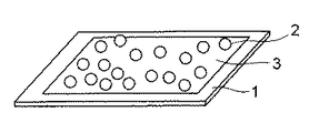
すなわち、図1は上記方法を適用して製造したマイクロアレイを模式的に示した斜視図である。 That is, FIG. 1 is a perspective view schematically showing a microarray manufactured by applying the above method.
図1に示すように、このマイクロアレイは、基板1と、この基板1上に固定された多数の前記した表面に生体関連物質を導入可能な官能基を有する荷電性高分子微粒子2とから構成されている。基板1の表面には前述したような方法で分子中にアミノ基とカルボキシル基を有する高分子化合物により荷電性高分子微粒子2と逆の電荷を有する荷電性薄膜3が形成されており、荷電性高分子微粒子2はこの荷電性薄膜3上に静電結合によって固定されている。
As shown in FIG. 1, the microarray is composed of a
なお、この例は、測定用の生体関連物質を適宜結合させる操作を行うことで、使用者がカスタマイズできる構成をとっているが、図2に示すように、すぐに測定を行うことができるように、予め所定の生体関連物質4を荷電性高分子微粒子2に固定させた構成としてもよい。生体関連物質4は、生体関連物質固定用粒子2の表面に設けられた官能基と生体関連物質が含有するもしくは導入された官能基を反応させ結合させることにより、荷電性高分子微粒子2に固定することができる。例えば、生体関連物質4としてオリゴヌクレオチドを、表面にカルボキシル基を有する荷電性高分子微粒子2に固定する場合、オリゴヌクレオチド末端にはアミノ基を導入しておき、これらの基を反応させて塩を形成させたり、あるいは縮合剤を共存させることによりアミド結合を形成させたりすることにより、オリゴヌクレオチドを荷電性高分子微粒子2上に固定することができる。
Note that, in this example, the user can customize the structure by appropriately performing the operation of binding the biological substance for measurement. However, as shown in FIG. 2, the measurement can be performed immediately. In addition, a configuration may be adopted in which a predetermined biological substance 4 is fixed to the
生体関連物質4は荷電性高分子微粒子2を基板1に固定する前に予め固定しておいてもよい。
The biological material 4 may be fixed in advance before the charged polymer
また、使用にあたって、生体関連物質4を荷電性高分子微粒子2に予め固定しておくだけでなく、緩衝液に荷電性高分子微粒子2を懸濁した状態で、予め固定した生体関連物質4と解析対象の生体関連物質とを反応させた後、基板1上に固定化するようにしてもよい。このような手法を用いることにより、解析対象の生体関連物質との反応を促進し、反応効率を高めることができる。
In use, not only the biologically relevant substance 4 is fixed in advance to the charged polymer
次に本発明の具体的な実施例を記載するが、本発明はこれらの実施例に限定されるものでないことはいうまでもない。なお、以下の記載において「部」は「重量部」を意味する。 Next, although the specific Example of this invention is described, it cannot be overemphasized that this invention is not limited to these Examples. In the following description, “part” means “part by weight”.
実施例1
窒素雰囲気下、スチレン100部、ポリアクリル酸10部およびスチレンスルホン酸ナトリウム5部を、イソプロピルアルコール800部および蒸留水20部の混合溶媒に溶解した後、この溶液にアゾビスイソブチロニトリル5部を加え、80℃で12時間反応させた。反応終了後、ろ過、次いで減圧下で乾燥し、平均粒径3μmのアニオン性高分子微粒子を得た。
Example 1
In a nitrogen atmosphere, 100 parts of styrene, 10 parts of polyacrylic acid and 5 parts of sodium styrenesulfonate were dissolved in a mixed solvent of 800 parts of isopropyl alcohol and 20 parts of distilled water, and then 5 parts of azobisisobutyronitrile was added to this solution. And reacted at 80 ° C. for 12 hours. After completion of the reaction, filtration and then drying under reduced pressure yielded anionic polymer fine particles having an average particle diameter of 3 μm.
また、ガラス基板(50mm×90mm×1mm)上に、前記式[I]においてRがメチレン基で、nが1であるビニル系ポリマー(Mw=70,000)を0.02mol%溶解させた塩酸水溶液(pH=2.0)を塗布し風乾させて、約6nm厚さのカチオン性薄膜を形成した。 Further, a hydrochloric acid aqueous solution (pH) in which 0.02 mol% of a vinyl polymer (Mw = 70,000) in which R is a methylene group and n is 1 in the formula [I] is dissolved on a glass substrate (50 mm × 90 mm × 1 mm). = 2.0) and air-dried to form a cationic thin film having a thickness of about 6 nm.
この基板上に、上記アニオン性高分子微粒子を濃度が1wt%となるように純水に分散させた分散液を垂らし、室温に約1時間静置して、アニオン性高分子微粒子を基板上に固定させた。その後、基板を風乾した。アニオン性高分子微粒子の固定量は2500個であった。 On this substrate, a dispersion liquid in which the above-mentioned anionic polymer fine particles are dispersed in pure water so as to have a concentration of 1 wt% is hung and allowed to stand at room temperature for about 1 hour to place the anionic polymer fine particles on the substrate. Fixed. Thereafter, the substrate was air-dried. The fixed amount of the anionic polymer fine particles was 2500.
次いで、末端にアミノ基を導入したプローブDNAを、0.15M塩化ナトリウムおよび15mMクエン酸水溶液に、その濃度が0.5mg/mlとなるように溶解した溶液に、上記で得られたアニオン性高分子微粒子を固定した基板を浸漬して、75℃で1時間インキュベートし、さらに、0.1wt%ドデシル硫酸ナトリウム水溶液、純水およびエタノールに順に浸漬した後、室温で乾燥して、DNAマイクロアレイを作製した。 Subsequently, the anionic polymer fine particles obtained above were dissolved in a solution in which the probe DNA having an amino group introduced at the end was dissolved in 0.15 M sodium chloride and 15 mM citric acid aqueous solution so that the concentration was 0.5 mg / ml. The substrate on which was fixed was immersed, incubated at 75 ° C. for 1 hour, further immersed in a 0.1 wt% sodium dodecyl sulfate aqueous solution, pure water and ethanol in that order, and then dried at room temperature to prepare a DNA microarray.
得られたDNAマイクロアレイ上に、ターゲットDNAとしてその配列が先に固定したプローブDNAに相補的な配列を有し、蛍光色素(フルオロセイン)で標識したオリゴヌクレオチドを0.15M塩化ナトリウムおよび15mMクエン酸水溶液(pH=7.0)に、その濃度が200ng/mlとなるように溶解した溶液をスポットし、45℃で約24時間インキュベートした。その後、0.15M塩化ナトリウムおよび15mMクエン酸水溶液で洗浄し、室温で乾燥させた。基板上の蛍光強度を蛍光スキャニング装置で計測したところ、蛍光が確認され、ハイブリダイズ体が得られたことが確認された。 On the obtained DNA microarray, oligonucleotides having a sequence complementary to the probe DNA previously fixed as target DNA and labeled with a fluorescent dye (fluorescein) are 0.15 M sodium chloride and 15 mM citric acid aqueous solution. A solution dissolved so as to have a concentration of 200 ng / ml was spotted on (pH = 7.0) and incubated at 45 ° C. for about 24 hours. Then, it was washed with 0.15M sodium chloride and 15mM citric acid aqueous solution and dried at room temperature. When the fluorescence intensity on the substrate was measured with a fluorescence scanning device, the fluorescence was confirmed and it was confirmed that a hybrid was obtained.
実施例2
ガラス基板(50mm×90mm×1mm)を、前記式[I]においてRがメチレン基で、nが1であるビニル系ポリマー(Mw=70,000)を0.02mol%溶解させた塩酸水溶液(pH=2.0)と、同ビニル系ポリマーを0.02mol%溶解させた水酸化ナトリウム水溶液(pH=9)に、室温(20℃)で交互にそれぞれ3回および2回浸漬し風乾させて、約12nm厚さの表面がカチオン性を示す薄膜を形成した。なお、それぞれの溶液には2MのNaClを添加した。
Example 2
A hydrochloric acid aqueous solution (pH = 2.0) in which 0.02 mol% of a vinyl polymer (Mw = 70,000) in which R is a methylene group and n is 1 is dissolved in a glass substrate (50 mm × 90 mm × 1 mm) And a sodium hydroxide aqueous solution (pH = 9) in which 0.02 mol% of the same vinyl polymer is dissolved, alternately and three times and twice at room temperature (20 ° C), and air-dried to obtain a surface of about 12 nm thickness. Formed a thin film showing a cationic property. In addition, 2M NaCl was added to each solution.
この基板を、実施例1の場合と同様に製造したアニオン性高分子微粒子を濃度が1wt%となるように純水に分散させた分散液中に室温で約1時間浸漬して、アニオン性高分子微粒子を基板上に固定させた。浸漬後、基板を液中から引き上げ風乾した。アニオン性高分子微粒子の固定量は3500個であった。 This substrate was immersed in a dispersion in which anionic polymer fine particles produced in the same manner as in Example 1 were dispersed in pure water so as to have a concentration of 1 wt% at room temperature for about 1 hour. Molecular fine particles were fixed on the substrate. After immersion, the substrate was pulled up from the solution and air-dried. The fixed amount of the anionic polymer fine particles was 3,500.
次いで、末端にアミノ基を導入したプローブDNAを、0.15M塩化ナトリウムおよび15mMクエン酸水溶液に、その濃度が0.5mg/mlとなるように溶解した溶液に、上記で得られたアニオン性高分子微粒子を固定した基板を浸漬して、75℃で1時間インキュベートし、さらに、0.1wt%ドデシル硫酸ナトリウム水溶液、純水およびエタノールに順に浸漬した後、室温で乾燥して、DNAマイクロアレイを作製した。 Subsequently, the anionic polymer fine particles obtained above were dissolved in a solution in which the probe DNA having an amino group introduced at the end was dissolved in 0.15 M sodium chloride and 15 mM citric acid aqueous solution so that the concentration was 0.5 mg / ml. The substrate on which was fixed was immersed, incubated at 75 ° C. for 1 hour, further immersed in a 0.1 wt% sodium dodecyl sulfate aqueous solution, pure water and ethanol in that order, and then dried at room temperature to prepare a DNA microarray.
得られたDNAマイクロアレイ上に、ターゲットDNAとしてその配列が先に固定したプローブDNAに相補的な配列を有し、蛍光色素(フルオロセイン)で標識したオリゴヌクレオチドを0.15M塩化ナトリウムおよび15mMクエン酸水溶液(pH=7.0)に、その濃度が200ng/mlとなるように溶解した溶液をスポットし、45℃で約24時間インキュベートした。その後、0.15M塩化ナトリウムおよび15mMクエン酸水溶液で洗浄し、室温で乾燥させた。基板上の蛍光強度を蛍光スキャニング装置で計測したところ、蛍光が確認され、ハイブリダイズ体が得られたことが確認された。 On the obtained DNA microarray, oligonucleotides having a sequence complementary to the probe DNA previously fixed as target DNA and labeled with a fluorescent dye (fluorescein) are 0.15 M sodium chloride and 15 mM citric acid aqueous solution. A solution dissolved so as to have a concentration of 200 ng / ml was spotted on (pH = 7.0) and incubated at 45 ° C. for about 24 hours. Then, it was washed with 0.15M sodium chloride and 15mM citric acid aqueous solution and dried at room temperature. When the fluorescence intensity on the substrate was measured with a fluorescence scanning device, the fluorescence was confirmed and it was confirmed that a hybrid was obtained.
実施例3
ポリエステル樹脂100部、正電荷補強剤として第4級アンモニウム塩単位を9wt%含むスチレン−アクリル系共重合樹脂(第4級アンモニウム塩/スチレン=9/74)4部および正電荷制御剤としてニグロシン染料1部を混合し、加熱ローラで溶融混練した。冷却後、エアージェットミルで微粉砕し、分級して、平均粒径9μmのカチオン性高分子微粒子を得た。
Example 3
100 parts of polyester resin, 4 parts of styrene-acrylic copolymer resin (quaternary ammonium salt / styrene = 9/74) containing 9 wt% of quaternary ammonium salt unit as positive charge reinforcing agent, and nigrosine dye as positive charge control agent One part was mixed and melt-kneaded with a heating roller. After cooling, the mixture was finely pulverized with an air jet mill and classified to obtain cationic polymer fine particles having an average particle diameter of 9 μm.
また、ガラス基板(50mm×90mm×1mm)を、前記式[I]においてRがメチレン基で、nが1であるビニル系ポリマー(Mw=70,000)を0.02mol%溶解させた水酸化ナトリウム水溶液(pH=9)と、同ビニル系ポリマーを0.02mol%溶解させた塩酸水溶液(pH=2.0)に、室温(20℃)で交互にそれぞれ3回および2回浸漬し風乾して、約12nm厚さの表面がアニオン性を示す薄膜を形成した。なお、それぞれの溶液には2MのNaClを添加した。 In addition, an aqueous solution of sodium hydroxide in which 0.02 mol% of a vinyl polymer (Mw = 70,000) in which R is a methylene group and n is 1 in the formula [I] is dissolved in a glass substrate (50 mm × 90 mm × 1 mm) ( pH = 9) and an aqueous hydrochloric acid solution (pH = 2.0) in which 0.02 mol% of the same vinyl polymer is dissolved, alternately immersed at room temperature (20 ° C.) three times and twice, and then air-dried to obtain a thickness of about 12 nm. A thin film having an anionic surface was formed. In addition, 2M NaCl was added to each solution.
この基板を、上記カチオン性高分子微粒子を濃度が1wt%となるように純水に分散させた分散液中に室温で約1時間浸漬して、カチオン性高分子微粒子を基板上に固定させた。浸漬後、基板を液中から引き上げ風乾した。カチオン性高分子微粒子の固定量は3700個であった。 The substrate was immersed in a dispersion obtained by dispersing the cationic polymer fine particles in pure water so that the concentration was 1 wt% at room temperature for about 1 hour to fix the cationic polymer fine particles on the substrate. . After immersion, the substrate was pulled up from the solution and air-dried. The fixed amount of the cationic polymer fine particles was 3,700.
次いで、末端にアミノ基を導入したプローブDNAを、0.15M塩化ナトリウムおよび15mMクエン酸水溶液に、その濃度が0.5mg/mlとなるように溶解した溶液に、上記で得られたカチオン性高分子微粒子を固定した基板を浸漬して、75℃で1時間インキュベートし、さらに、0.1wt%ドデシル硫酸ナトリウム水溶液、純水およびエタノールに順に浸漬した後、室温で乾燥して、DNAマイクロアレイを作製した。 Subsequently, the cationic polymer fine particles obtained above were dissolved in a solution in which the probe DNA having an amino group introduced at the end was dissolved in an aqueous solution of 0.15 M sodium chloride and 15 mM citric acid so that the concentration was 0.5 mg / ml. The substrate on which was fixed was immersed, incubated at 75 ° C. for 1 hour, further immersed in a 0.1 wt% sodium dodecyl sulfate aqueous solution, pure water and ethanol in this order, and then dried at room temperature to prepare a DNA microarray.
得られたDNAマイクロアレイ上に、ターゲットDNAとしてその配列が先に固定したプローブDNAに相補的な配列を有し、蛍光色素(フルオロセイン)で標識したオリゴヌクレオチドを0.15M塩化ナトリウムおよび15mMクエン酸水溶液(pH=7.0)に、その濃度が200ng/mlとなるように溶解した溶液をスポットし、45℃で約24時間インキュベートした。その後、0.15M塩化ナトリウムおよび15mMクエン酸水溶液で洗浄し、室温で乾燥させた。基板上の蛍光強度を蛍光スキャニング装置で計測したところ、蛍光が確認され、ハイブリダイズ体が得られたことが確認された。 On the obtained DNA microarray, oligonucleotides having a sequence complementary to the probe DNA previously fixed as target DNA and labeled with a fluorescent dye (fluorescein) are 0.15 M sodium chloride and 15 mM citric acid aqueous solution. A solution dissolved so as to have a concentration of 200 ng / ml was spotted on (pH = 7.0) and incubated at 45 ° C. for about 24 hours. Then, it was washed with 0.15M sodium chloride and 15mM citric acid aqueous solution and dried at room temperature. When the fluorescence intensity on the substrate was measured with a fluorescence scanning device, the fluorescence was confirmed and it was confirmed that a hybrid was obtained.
実施例4
赤蛍光染料1,3−ビス[(1,3−ジヒドロ−1,3,3−トリメチル−2H−インドール−2−イリデン)メチル]−2,4−ジヒドロキシシクロブテンジイリウムと、オレンジ染料2−(3,5−ジメチルピロール−2−イル)−4−(3,5−ジメチル−2H−ピロール−2−イリデン)−3−ヒドロキシ−2−シクロブテン−1−オンとを異なる比率でクロロホルムとエタノールの混合溶媒(混合比 2:3)に溶解して、2種の染料の混合比の異なる64種の混合染料液を調製した。
Example 4
Red
これらの各混合染料液0.6mlに、実施例3の場合と同様に製造したカチオン性高分子微粒子60mgをそれぞれ懸濁させ、2時間の超音波処理を行った。処理後、各懸濁液から微粒子を分離し、1.2mlのメタノールに懸濁させた後、さらに超音波処理およびメタノール洗浄を行い、64個(種)の微粒子ライブラリー(同種の蛍光染料を異なる比率で配合したもの)を作成した。 60 mg of the cationic polymer fine particles produced in the same manner as in Example 3 was suspended in 0.6 ml of each of these mixed dye solutions, and subjected to ultrasonic treatment for 2 hours. After the treatment, the fine particles are separated from each suspension, suspended in 1.2 ml of methanol, further subjected to ultrasonic treatment and methanol washing, and 64 (species) fine particle libraries (different from the same type of fluorescent dye) (Mixed in proportion).
得られた64個の微粒子ライブラリーをそれぞれ緩衝液に濃度が1wt%となるように懸濁させた後、これらの各懸濁液に、末端にアミノ基を導入したプローブDNAを、0.15M塩化ナトリウムおよび15mMクエン酸水溶液に、その濃度が0.5mg/mlとなるように溶解した溶液を混合して、75℃で1時間インキュベートし、微粒子にプローブDNAを導入した。この後、微粒子を分離し、0.1%ドデシル硫酸ナトリウム水溶液、純水およびエタノールに順に浸漬した後、純水に濃度が1wt%となるように懸濁させた。なお、64個の微粒子ライブラリーのうちの1つに、目的遺伝子検出のためのプローブDNAを導入し、その他には目的遺伝子を有さないプローブDNAを導入した。 Each of the obtained 64 microparticle libraries was suspended in a buffer solution to a concentration of 1 wt%, and then each of these suspensions was probed with 0.15M chloride of probe DNA having an amino group introduced at the end. A solution dissolved in sodium and 15 mM aqueous citric acid so as to have a concentration of 0.5 mg / ml was mixed and incubated at 75 ° C. for 1 hour to introduce probe DNA into the microparticles. Thereafter, the fine particles were separated and immersed in a 0.1% sodium dodecyl sulfate aqueous solution, pure water and ethanol in this order, and then suspended in pure water to a concentration of 1 wt%. In addition, probe DNA for detecting the target gene was introduced into one of the 64 fine particle libraries, and probe DNA having no target gene was introduced into the other.
次いで、これらのプローブDNAを導入した64個の微粒子ライブラリーを含む懸濁液を混合し、この混合液中に、実施例3の場合と同様にして、ガラス基板(50mm×90mm×1mm)上に約12nm厚さの表面がアニオン性を示す薄膜を形成した基板を、室温で約1時間浸漬して、プローブDNAを導入したカチオン性高分子微粒子を基板上に固定させた。浸漬後、基板を液中から引き上げ風乾した。 Next, a suspension containing 64 microparticle libraries into which these probe DNAs were introduced was mixed, and this mixture was mixed with a suspension on a glass substrate (50 mm × 90 mm × 1 mm) in the same manner as in Example 3. A substrate on which a thin film having an anionic surface with a thickness of about 12 nm was formed was immersed at room temperature for about 1 hour to immobilize the cationic polymer fine particles into which the probe DNA was introduced on the substrate. After immersion, the substrate was pulled up from the solution and air-dried.
この後、上記基板上に、ターゲットDNAとしてその配列が先に固定した目的遺伝子検出のためのプローブDNAに相補的な配列を有し、蛍光色素(フルオレセイン)で標識したオリゴヌクレオチドを0.15M塩化ナトリウムおよび15mMクエン酸水溶液に、その濃度が200ng/mlとなるように溶解した溶液をスポットし、45℃で約24時間インキュベートした。その後、0.15M塩化ナトリウムおよび15mMクエン酸水溶液で洗浄し、乾燥させた。基板上の蛍光強度を蛍光スキャニング装置で計測したところ、フルオロセイン蛍光が確認され、ハイブリダイズ体が得られたことが確認された。また、微粒子の染色蛍光によって目的遺伝子が確認された。 Thereafter, an oligonucleotide labeled with a fluorescent dye (fluorescein) having a sequence complementary to a probe DNA for detecting a target gene whose sequence was previously immobilized on the substrate as a target DNA, was labeled with 0.15M sodium chloride. Then, a solution dissolved in a 15 mM aqueous citric acid solution so as to have a concentration of 200 ng / ml was spotted and incubated at 45 ° C. for about 24 hours. Then, it was washed with 0.15M sodium chloride and 15mM citric acid aqueous solution and dried. When the fluorescence intensity on the substrate was measured with a fluorescence scanning apparatus, fluorescein fluorescence was confirmed, and it was confirmed that a hybrid was obtained. In addition, the target gene was confirmed by staining fluorescence of fine particles.
実施例5
赤蛍光染料1,3−ビス[(1,3−ジヒドロ−1,3,3−トリメチル−2H−インドール−2−イリデン)メチル]−2,4−ジヒドロキシシクロブテンジイリウムと、オレンジ染料2−(3,5−ジメチルピロール−2−イル)−4−(3,5−ジメチル−2H−ピロール−2−イリデン)−3−ヒドロキシ−2−シクロブテン−1−オンと、黄緑蛍光染料3,6−ジアミノ−2,5−ピラジンカルボニトリルとを、異なる比率でクロロホルムとエタノールの混合溶媒(混合比 2:3)に溶解して、3種の染料の混合比の異なる128種の混合染料液を調製した。
Example 5
Red
これらの各混合染料液0.6mlに、実施例3の場合と同様に製造したカチオン性高分子微粒子60mgをそれぞれ懸濁させ、2時間の超音波処理を行った。処理後、各懸濁液から微粒子を分離し、1.2mlのメタノールに懸濁させた後、さらに超音波処理およびメタノール洗浄を行い、128個(種)の微粒子ライブラリー(同種の蛍光染料を異なる比率で配合したもの)を作成した。 60 mg of the cationic polymer fine particles produced in the same manner as in Example 3 was suspended in 0.6 ml of each of these mixed dye solutions, and subjected to ultrasonic treatment for 2 hours. After the treatment, the fine particles are separated from each suspension, suspended in 1.2 ml of methanol, further subjected to ultrasonic treatment and methanol washing, and 128 fine particle libraries (separate fluorescent dyes of different types) (Mixed in proportion).
得られた128個の微粒子ライブラリーをそれぞれ緩衝液に濃度が1wt%となるように懸濁させた後、これらの各懸濁液に、末端にアミノ基を導入したプローブDNAを、0.15M塩化ナトリウムおよび15mMクエン酸水溶液に、その濃度が0.5mg/mlとなるように溶解した溶液を混合して、75℃で1時間インキュベートし、微粒子にプローブDNAを導入した。この後、微粒子を分離し、0.1%ドデシル硫酸ナトリウム水溶液、純水およびエタノールに順に浸漬した後、純水に濃度が1wt%となるように懸濁させた。なお、128個の微粒子ライブラリーのうちの1つに、目的遺伝子検出のためのプローブDNAを導入し、その他には目的遺伝子を有さないプローブDNAを導入した。 Each of the 128 fine particle libraries obtained was suspended in a buffer solution to a concentration of 1 wt%, and then each of these suspensions was probed with 0.15 M chloride DNA having an amino group introduced at the end. A solution dissolved in sodium and 15 mM aqueous citric acid so as to have a concentration of 0.5 mg / ml was mixed and incubated at 75 ° C. for 1 hour to introduce probe DNA into the microparticles. Thereafter, the fine particles were separated and immersed in a 0.1% sodium dodecyl sulfate aqueous solution, pure water and ethanol in this order, and then suspended in pure water to a concentration of 1 wt%. A probe DNA for detecting a target gene was introduced into one of 128 fine particle libraries, and a probe DNA having no target gene was introduced into the other.
次いで、これらのプローブDNAを導入した128個の微粒子ライブラリーを含む懸濁液を混合し、この混合液中に、実施例3の場合と同様にして、ガラス基板(50mm×90mm×1mm)上に約12nm厚さの表面がアニオン性を示す薄膜を形成した基板を、室温で約1時間浸漬して、プローブDNAを導入したカチオン性高分子微粒子を基板上に固定させた。浸漬後、基板を液中から引き上げ風乾した。 Next, a suspension containing 128 microparticle libraries into which these probe DNAs were introduced was mixed, and this mixture was mixed on a glass substrate (50 mm × 90 mm × 1 mm) in the same manner as in Example 3. A substrate on which a thin film having an anionic surface with a thickness of about 12 nm was formed was immersed at room temperature for about 1 hour to immobilize the cationic polymer fine particles into which the probe DNA was introduced on the substrate. After immersion, the substrate was pulled up from the solution and air-dried.
この後、上記基板上に、ターゲットDNAとしてその配列が先に固定した目的遺伝子検出のためのプローブDNAに相補的な配列を有し、蛍光色素(フルオレセイン)で標識したオリゴヌクレオチドを0.15M塩化ナトリウムおよび15mMクエン酸水溶液に、その濃度が200ng/mlとなるように溶解した溶液をスポットし、45℃で約24時間インキュベートした。その後、0.15M塩化ナトリウムおよび15mMクエン酸水溶液で洗浄し、乾燥させた。基板上の蛍光強度を蛍光スキャニング装置で計測したところ、フルオロセイン蛍光が確認され、ハイブリダイズ体が得られたことが確認された。また、微粒子の染色蛍光によって目的遺伝子が確認された。 Thereafter, an oligonucleotide labeled with a fluorescent dye (fluorescein) having a sequence complementary to a probe DNA for detecting a target gene whose sequence was previously immobilized on the substrate as a target DNA, was labeled with 0.15M sodium chloride. Then, a solution dissolved in a 15 mM aqueous citric acid solution so as to have a concentration of 200 ng / ml was spotted and incubated at 45 ° C. for about 24 hours. Then, it was washed with 0.15M sodium chloride and 15mM citric acid aqueous solution and dried. When the fluorescence intensity on the substrate was measured with a fluorescence scanning apparatus, fluorescein fluorescence was confirmed, and it was confirmed that a hybrid was obtained. In addition, the target gene was confirmed by staining fluorescence of fine particles.
1…基板、2…荷電性高分子微粒子、3…荷電性薄膜、4…生体関連物質
DESCRIPTION OF
Claims (8)
分子中にアミノ基またはその塩とカルボキシル基またはその塩とを有する高分子化合物を用いて前記基板の表面に前記荷電性高分子微粒子と逆の電荷を有する荷電性薄膜を形成した後、この荷電性薄膜上に前記荷電性高分子微粒子の分散液を塗布して前記荷電性高分子微粒子を前記荷電性薄膜上に静電結合により固着させることを特徴とする高分子微粒子の固定方法。 A method of fixing charged polymer fine particles having a functional group capable of introducing a biological substance on a substrate,
A chargeable thin film having a charge opposite to that of the chargeable polymer fine particles is formed on the surface of the substrate using a polymer compound having an amino group or a salt thereof and a carboxyl group or a salt thereof in the molecule, and then charged. A method for fixing polymer fine particles, comprising: applying a dispersion liquid of the chargeable polymer fine particles on a conductive thin film, and fixing the chargeable polymer fine particles on the chargeable thin film by electrostatic bonding.
Priority Applications (1)
| Application Number | Priority Date | Filing Date | Title |
|---|---|---|---|
| JP2004108167A JP2005291951A (en) | 2004-03-31 | 2004-03-31 | Fixing method of polymer particle |
Applications Claiming Priority (1)
| Application Number | Priority Date | Filing Date | Title |
|---|---|---|---|
| JP2004108167A JP2005291951A (en) | 2004-03-31 | 2004-03-31 | Fixing method of polymer particle |
Publications (1)
| Publication Number | Publication Date |
|---|---|
| JP2005291951A true JP2005291951A (en) | 2005-10-20 |
Family
ID=35325036
Family Applications (1)
| Application Number | Title | Priority Date | Filing Date |
|---|---|---|---|
| JP2004108167A Withdrawn JP2005291951A (en) | 2004-03-31 | 2004-03-31 | Fixing method of polymer particle |
Country Status (1)
| Country | Link |
|---|---|
| JP (1) | JP2005291951A (en) |
Cited By (6)
| Publication number | Priority date | Publication date | Assignee | Title |
|---|---|---|---|---|
| JP2007218824A (en) * | 2006-02-20 | 2007-08-30 | Sumitomo Bakelite Co Ltd | Base material for bioassay |
| JP2011220723A (en) * | 2010-04-06 | 2011-11-04 | Kao Corp | Measuring method for surface roughness of nanoparticles |
| WO2012161256A1 (en) * | 2011-05-24 | 2012-11-29 | コニカミノルタアドバンストレイヤー株式会社 | Sensor chip comprising charged layer and application therefor |
| JP2013029370A (en) * | 2011-07-27 | 2013-02-07 | Konica Minolta Holdings Inc | Local-field optical sensor chip having ligand highly densely disposed thereon |
| JP2013029372A (en) * | 2011-07-27 | 2013-02-07 | Konica Minolta Advanced Layers Inc | Ionic functional group modification sensor chip, and intermolecular interaction measurement method using ligand-carrying charged fine particle |
| JP2013029369A (en) * | 2011-07-27 | 2013-02-07 | Konica Minolta Holdings Inc | Ionic functional group-modified local-field optical sensor chip and method for detecting analyte using ligand-carrying charged particle |
-
2004
- 2004-03-31 JP JP2004108167A patent/JP2005291951A/en not_active Withdrawn
Cited By (6)
| Publication number | Priority date | Publication date | Assignee | Title |
|---|---|---|---|---|
| JP2007218824A (en) * | 2006-02-20 | 2007-08-30 | Sumitomo Bakelite Co Ltd | Base material for bioassay |
| JP2011220723A (en) * | 2010-04-06 | 2011-11-04 | Kao Corp | Measuring method for surface roughness of nanoparticles |
| WO2012161256A1 (en) * | 2011-05-24 | 2012-11-29 | コニカミノルタアドバンストレイヤー株式会社 | Sensor chip comprising charged layer and application therefor |
| JP2013029370A (en) * | 2011-07-27 | 2013-02-07 | Konica Minolta Holdings Inc | Local-field optical sensor chip having ligand highly densely disposed thereon |
| JP2013029372A (en) * | 2011-07-27 | 2013-02-07 | Konica Minolta Advanced Layers Inc | Ionic functional group modification sensor chip, and intermolecular interaction measurement method using ligand-carrying charged fine particle |
| JP2013029369A (en) * | 2011-07-27 | 2013-02-07 | Konica Minolta Holdings Inc | Ionic functional group-modified local-field optical sensor chip and method for detecting analyte using ligand-carrying charged particle |
Similar Documents
| Publication | Publication Date | Title |
|---|---|---|
| JP4420020B2 (en) | How to stir the solution | |
| JP3883539B2 (en) | Method for producing hydrogel biochip using radial polyethylene glycol derivative having epoxy group | |
| JP3398366B2 (en) | Method for producing microarray for DNA analysis | |
| JP2001108683A (en) | Dna fragment fixing solid-phase carrier, dna fragment fixing method, and nucleic-acid fragment detecting method | |
| US20020182603A1 (en) | Uniformly functionalized surfaces for microarrays | |
| JP2005291951A (en) | Fixing method of polymer particle | |
| JP2001128683A (en) | Method for fixing dna fragment and method for detecting dna chip and nucleic acid fragment | |
| JP2009085840A (en) | Single probe molecular element, and manufacturing method of same | |
| KR100450822B1 (en) | A method of preparing a hydrogel biochip by using satar-like PEG derivatives | |
| EP1258731B1 (en) | Reactive solid support for DNA fragment detection | |
| JP3568197B2 (en) | Reactive solid support and DNA fragment detection tool | |
| JP3342695B2 (en) | Reactive solid support and DNA fragment detection tool | |
| JP2005291952A (en) | Particle for fixing biological substance and microarray | |
| JP3857075B2 (en) | Reactive solid phase carrier and DNA fragment detection tool | |
| JP3975042B2 (en) | Method for immobilizing DNA fragment on solid support surface and DNA chip | |
| JP2001178460A (en) | Method for immobilizing dna fragment to surface of solid- phase carrier and dna chip | |
| EP1256805B1 (en) | Biological material chip | |
| KR20040055441A (en) | Nucleic acid microarray and manufacturing method thereof | |
| JP3808389B2 (en) | Reactive solid phase carrier and DNA fragment detection tool | |
| JP2001017166A (en) | Dna chip, verifying kit of dna chip and verification of dna chip | |
| JP2005291974A (en) | Magnetic organic particle having feldazyl group, particle fixing method and microarray | |
| JP4054499B2 (en) | Method for immobilizing DNA fragment on solid support surface and DNA chip | |
| JP2001178458A (en) | Method for immobilizing dna fragment to surface of solid- phase carrier and dna chip | |
| JP2002365295A (en) | Surface active sheet roll for manufacturing bioactive substance analyzing element | |
| JP2001178474A (en) | Method for immobilizing dna fragment to surface of solid- phase carrier and dna chip |
Legal Events
| Date | Code | Title | Description |
|---|---|---|---|
| A300 | Withdrawal of application because of no request for examination |
Free format text: JAPANESE INTERMEDIATE CODE: A300 Effective date: 20070605 |