JP2004529621A - Pluripotent adult stem cells, their origin, methods of obtaining and maintaining them, methods of differentiating them, methods of their use, and cells derived therefrom - Google Patents
Pluripotent adult stem cells, their origin, methods of obtaining and maintaining them, methods of differentiating them, methods of their use, and cells derived therefrom Download PDFInfo
- Publication number
- JP2004529621A JP2004529621A JP2002565063A JP2002565063A JP2004529621A JP 2004529621 A JP2004529621 A JP 2004529621A JP 2002565063 A JP2002565063 A JP 2002565063A JP 2002565063 A JP2002565063 A JP 2002565063A JP 2004529621 A JP2004529621 A JP 2004529621A
- Authority
- JP
- Japan
- Prior art keywords
- cells
- cell
- masc
- mascs
- disease
- Prior art date
- Legal status (The legal status is an assumption and is not a legal conclusion. Google has not performed a legal analysis and makes no representation as to the accuracy of the status listed.)
- Withdrawn
Links
Images


Classifications
-
- A—HUMAN NECESSITIES
- A61—MEDICAL OR VETERINARY SCIENCE; HYGIENE
- A61K—PREPARATIONS FOR MEDICAL, DENTAL OR TOILETRY PURPOSES
- A61K39/00—Medicinal preparations containing antigens or antibodies
- A61K39/0005—Vertebrate antigens
- A61K39/001—Preparations to induce tolerance to non-self, e.g. prior to transplantation
-
- A—HUMAN NECESSITIES
- A01—AGRICULTURE; FORESTRY; ANIMAL HUSBANDRY; HUNTING; TRAPPING; FISHING
- A01K—ANIMAL HUSBANDRY; CARE OF BIRDS, FISHES, INSECTS; FISHING; REARING OR BREEDING ANIMALS, NOT OTHERWISE PROVIDED FOR; NEW BREEDS OF ANIMALS
- A01K67/00—Rearing or breeding animals, not otherwise provided for; New breeds of animals
- A01K67/027—New breeds of vertebrates
- A01K67/0271—Chimeric animals, e.g. comprising exogenous cells
-
- A—HUMAN NECESSITIES
- A61—MEDICAL OR VETERINARY SCIENCE; HYGIENE
- A61P—SPECIFIC THERAPEUTIC ACTIVITY OF CHEMICAL COMPOUNDS OR MEDICINAL PREPARATIONS
- A61P1/00—Drugs for disorders of the alimentary tract or the digestive system
-
- A—HUMAN NECESSITIES
- A61—MEDICAL OR VETERINARY SCIENCE; HYGIENE
- A61P—SPECIFIC THERAPEUTIC ACTIVITY OF CHEMICAL COMPOUNDS OR MEDICINAL PREPARATIONS
- A61P1/00—Drugs for disorders of the alimentary tract or the digestive system
- A61P1/16—Drugs for disorders of the alimentary tract or the digestive system for liver or gallbladder disorders, e.g. hepatoprotective agents, cholagogues, litholytics
-
- A—HUMAN NECESSITIES
- A61—MEDICAL OR VETERINARY SCIENCE; HYGIENE
- A61P—SPECIFIC THERAPEUTIC ACTIVITY OF CHEMICAL COMPOUNDS OR MEDICINAL PREPARATIONS
- A61P1/00—Drugs for disorders of the alimentary tract or the digestive system
- A61P1/18—Drugs for disorders of the alimentary tract or the digestive system for pancreatic disorders, e.g. pancreatic enzymes
-
- A—HUMAN NECESSITIES
- A61—MEDICAL OR VETERINARY SCIENCE; HYGIENE
- A61P—SPECIFIC THERAPEUTIC ACTIVITY OF CHEMICAL COMPOUNDS OR MEDICINAL PREPARATIONS
- A61P13/00—Drugs for disorders of the urinary system
- A61P13/10—Drugs for disorders of the urinary system of the bladder
-
- A—HUMAN NECESSITIES
- A61—MEDICAL OR VETERINARY SCIENCE; HYGIENE
- A61P—SPECIFIC THERAPEUTIC ACTIVITY OF CHEMICAL COMPOUNDS OR MEDICINAL PREPARATIONS
- A61P13/00—Drugs for disorders of the urinary system
- A61P13/12—Drugs for disorders of the urinary system of the kidneys
-
- A—HUMAN NECESSITIES
- A61—MEDICAL OR VETERINARY SCIENCE; HYGIENE
- A61P—SPECIFIC THERAPEUTIC ACTIVITY OF CHEMICAL COMPOUNDS OR MEDICINAL PREPARATIONS
- A61P15/00—Drugs for genital or sexual disorders; Contraceptives
-
- A—HUMAN NECESSITIES
- A61—MEDICAL OR VETERINARY SCIENCE; HYGIENE
- A61P—SPECIFIC THERAPEUTIC ACTIVITY OF CHEMICAL COMPOUNDS OR MEDICINAL PREPARATIONS
- A61P17/00—Drugs for dermatological disorders
-
- A—HUMAN NECESSITIES
- A61—MEDICAL OR VETERINARY SCIENCE; HYGIENE
- A61P—SPECIFIC THERAPEUTIC ACTIVITY OF CHEMICAL COMPOUNDS OR MEDICINAL PREPARATIONS
- A61P19/00—Drugs for skeletal disorders
- A61P19/04—Drugs for skeletal disorders for non-specific disorders of the connective tissue
-
- A—HUMAN NECESSITIES
- A61—MEDICAL OR VETERINARY SCIENCE; HYGIENE
- A61P—SPECIFIC THERAPEUTIC ACTIVITY OF CHEMICAL COMPOUNDS OR MEDICINAL PREPARATIONS
- A61P19/00—Drugs for skeletal disorders
- A61P19/08—Drugs for skeletal disorders for bone diseases, e.g. rachitism, Paget's disease
-
- A—HUMAN NECESSITIES
- A61—MEDICAL OR VETERINARY SCIENCE; HYGIENE
- A61P—SPECIFIC THERAPEUTIC ACTIVITY OF CHEMICAL COMPOUNDS OR MEDICINAL PREPARATIONS
- A61P21/00—Drugs for disorders of the muscular or neuromuscular system
-
- A—HUMAN NECESSITIES
- A61—MEDICAL OR VETERINARY SCIENCE; HYGIENE
- A61P—SPECIFIC THERAPEUTIC ACTIVITY OF CHEMICAL COMPOUNDS OR MEDICINAL PREPARATIONS
- A61P25/00—Drugs for disorders of the nervous system
-
- A—HUMAN NECESSITIES
- A61—MEDICAL OR VETERINARY SCIENCE; HYGIENE
- A61P—SPECIFIC THERAPEUTIC ACTIVITY OF CHEMICAL COMPOUNDS OR MEDICINAL PREPARATIONS
- A61P25/00—Drugs for disorders of the nervous system
- A61P25/02—Drugs for disorders of the nervous system for peripheral neuropathies
-
- A—HUMAN NECESSITIES
- A61—MEDICAL OR VETERINARY SCIENCE; HYGIENE
- A61P—SPECIFIC THERAPEUTIC ACTIVITY OF CHEMICAL COMPOUNDS OR MEDICINAL PREPARATIONS
- A61P27/00—Drugs for disorders of the senses
- A61P27/02—Ophthalmic agents
-
- A—HUMAN NECESSITIES
- A61—MEDICAL OR VETERINARY SCIENCE; HYGIENE
- A61P—SPECIFIC THERAPEUTIC ACTIVITY OF CHEMICAL COMPOUNDS OR MEDICINAL PREPARATIONS
- A61P3/00—Drugs for disorders of the metabolism
-
- A—HUMAN NECESSITIES
- A61—MEDICAL OR VETERINARY SCIENCE; HYGIENE
- A61P—SPECIFIC THERAPEUTIC ACTIVITY OF CHEMICAL COMPOUNDS OR MEDICINAL PREPARATIONS
- A61P3/00—Drugs for disorders of the metabolism
- A61P3/08—Drugs for disorders of the metabolism for glucose homeostasis
- A61P3/10—Drugs for disorders of the metabolism for glucose homeostasis for hyperglycaemia, e.g. antidiabetics
-
- A—HUMAN NECESSITIES
- A61—MEDICAL OR VETERINARY SCIENCE; HYGIENE
- A61P—SPECIFIC THERAPEUTIC ACTIVITY OF CHEMICAL COMPOUNDS OR MEDICINAL PREPARATIONS
- A61P31/00—Antiinfectives, i.e. antibiotics, antiseptics, chemotherapeutics
-
- A—HUMAN NECESSITIES
- A61—MEDICAL OR VETERINARY SCIENCE; HYGIENE
- A61P—SPECIFIC THERAPEUTIC ACTIVITY OF CHEMICAL COMPOUNDS OR MEDICINAL PREPARATIONS
- A61P31/00—Antiinfectives, i.e. antibiotics, antiseptics, chemotherapeutics
- A61P31/04—Antibacterial agents
-
- A—HUMAN NECESSITIES
- A61—MEDICAL OR VETERINARY SCIENCE; HYGIENE
- A61P—SPECIFIC THERAPEUTIC ACTIVITY OF CHEMICAL COMPOUNDS OR MEDICINAL PREPARATIONS
- A61P31/00—Antiinfectives, i.e. antibiotics, antiseptics, chemotherapeutics
- A61P31/10—Antimycotics
-
- A—HUMAN NECESSITIES
- A61—MEDICAL OR VETERINARY SCIENCE; HYGIENE
- A61P—SPECIFIC THERAPEUTIC ACTIVITY OF CHEMICAL COMPOUNDS OR MEDICINAL PREPARATIONS
- A61P31/00—Antiinfectives, i.e. antibiotics, antiseptics, chemotherapeutics
- A61P31/12—Antivirals
-
- A—HUMAN NECESSITIES
- A61—MEDICAL OR VETERINARY SCIENCE; HYGIENE
- A61P—SPECIFIC THERAPEUTIC ACTIVITY OF CHEMICAL COMPOUNDS OR MEDICINAL PREPARATIONS
- A61P35/00—Antineoplastic agents
-
- A—HUMAN NECESSITIES
- A61—MEDICAL OR VETERINARY SCIENCE; HYGIENE
- A61P—SPECIFIC THERAPEUTIC ACTIVITY OF CHEMICAL COMPOUNDS OR MEDICINAL PREPARATIONS
- A61P37/00—Drugs for immunological or allergic disorders
-
- A—HUMAN NECESSITIES
- A61—MEDICAL OR VETERINARY SCIENCE; HYGIENE
- A61P—SPECIFIC THERAPEUTIC ACTIVITY OF CHEMICAL COMPOUNDS OR MEDICINAL PREPARATIONS
- A61P37/00—Drugs for immunological or allergic disorders
- A61P37/02—Immunomodulators
- A61P37/06—Immunosuppressants, e.g. drugs for graft rejection
-
- A—HUMAN NECESSITIES
- A61—MEDICAL OR VETERINARY SCIENCE; HYGIENE
- A61P—SPECIFIC THERAPEUTIC ACTIVITY OF CHEMICAL COMPOUNDS OR MEDICINAL PREPARATIONS
- A61P39/00—General protective or antinoxious agents
- A61P39/02—Antidotes
-
- A—HUMAN NECESSITIES
- A61—MEDICAL OR VETERINARY SCIENCE; HYGIENE
- A61P—SPECIFIC THERAPEUTIC ACTIVITY OF CHEMICAL COMPOUNDS OR MEDICINAL PREPARATIONS
- A61P41/00—Drugs used in surgical methods, e.g. surgery adjuvants for preventing adhesion or for vitreum substitution
-
- A—HUMAN NECESSITIES
- A61—MEDICAL OR VETERINARY SCIENCE; HYGIENE
- A61P—SPECIFIC THERAPEUTIC ACTIVITY OF CHEMICAL COMPOUNDS OR MEDICINAL PREPARATIONS
- A61P43/00—Drugs for specific purposes, not provided for in groups A61P1/00-A61P41/00
-
- A—HUMAN NECESSITIES
- A61—MEDICAL OR VETERINARY SCIENCE; HYGIENE
- A61P—SPECIFIC THERAPEUTIC ACTIVITY OF CHEMICAL COMPOUNDS OR MEDICINAL PREPARATIONS
- A61P7/00—Drugs for disorders of the blood or the extracellular fluid
-
- A—HUMAN NECESSITIES
- A61—MEDICAL OR VETERINARY SCIENCE; HYGIENE
- A61P—SPECIFIC THERAPEUTIC ACTIVITY OF CHEMICAL COMPOUNDS OR MEDICINAL PREPARATIONS
- A61P7/00—Drugs for disorders of the blood or the extracellular fluid
- A61P7/04—Antihaemorrhagics; Procoagulants; Haemostatic agents; Antifibrinolytic agents
-
- A—HUMAN NECESSITIES
- A61—MEDICAL OR VETERINARY SCIENCE; HYGIENE
- A61P—SPECIFIC THERAPEUTIC ACTIVITY OF CHEMICAL COMPOUNDS OR MEDICINAL PREPARATIONS
- A61P7/00—Drugs for disorders of the blood or the extracellular fluid
- A61P7/06—Antianaemics
-
- A—HUMAN NECESSITIES
- A61—MEDICAL OR VETERINARY SCIENCE; HYGIENE
- A61P—SPECIFIC THERAPEUTIC ACTIVITY OF CHEMICAL COMPOUNDS OR MEDICINAL PREPARATIONS
- A61P7/00—Drugs for disorders of the blood or the extracellular fluid
- A61P7/08—Plasma substitutes; Perfusion solutions; Dialytics or haemodialytics; Drugs for electrolytic or acid-base disorders, e.g. hypovolemic shock
-
- A—HUMAN NECESSITIES
- A61—MEDICAL OR VETERINARY SCIENCE; HYGIENE
- A61P—SPECIFIC THERAPEUTIC ACTIVITY OF CHEMICAL COMPOUNDS OR MEDICINAL PREPARATIONS
- A61P9/00—Drugs for disorders of the cardiovascular system
-
- A—HUMAN NECESSITIES
- A61—MEDICAL OR VETERINARY SCIENCE; HYGIENE
- A61P—SPECIFIC THERAPEUTIC ACTIVITY OF CHEMICAL COMPOUNDS OR MEDICINAL PREPARATIONS
- A61P9/00—Drugs for disorders of the cardiovascular system
- A61P9/10—Drugs for disorders of the cardiovascular system for treating ischaemic or atherosclerotic diseases, e.g. antianginal drugs, coronary vasodilators, drugs for myocardial infarction, retinopathy, cerebrovascula insufficiency, renal arteriosclerosis
-
- C—CHEMISTRY; METALLURGY
- C12—BIOCHEMISTRY; BEER; SPIRITS; WINE; VINEGAR; MICROBIOLOGY; ENZYMOLOGY; MUTATION OR GENETIC ENGINEERING
- C12N—MICROORGANISMS OR ENZYMES; COMPOSITIONS THEREOF; PROPAGATING, PRESERVING, OR MAINTAINING MICROORGANISMS; MUTATION OR GENETIC ENGINEERING; CULTURE MEDIA
- C12N15/00—Mutation or genetic engineering; DNA or RNA concerning genetic engineering, vectors, e.g. plasmids, or their isolation, preparation or purification; Use of hosts therefor
- C12N15/09—Recombinant DNA-technology
- C12N15/87—Introduction of foreign genetic material using processes not otherwise provided for, e.g. co-transformation
- C12N15/873—Techniques for producing new embryos, e.g. nuclear transfer, manipulation of totipotent cells or production of chimeric embryos
-
- C—CHEMISTRY; METALLURGY
- C12—BIOCHEMISTRY; BEER; SPIRITS; WINE; VINEGAR; MICROBIOLOGY; ENZYMOLOGY; MUTATION OR GENETIC ENGINEERING
- C12N—MICROORGANISMS OR ENZYMES; COMPOSITIONS THEREOF; PROPAGATING, PRESERVING, OR MAINTAINING MICROORGANISMS; MUTATION OR GENETIC ENGINEERING; CULTURE MEDIA
- C12N5/00—Undifferentiated human, animal or plant cells, e.g. cell lines; Tissues; Cultivation or maintenance thereof; Culture media therefor
- C12N5/06—Animal cells or tissues; Human cells or tissues
- C12N5/0602—Vertebrate cells
- C12N5/0607—Non-embryonic pluripotent stem cells, e.g. MASC
-
- C—CHEMISTRY; METALLURGY
- C12—BIOCHEMISTRY; BEER; SPIRITS; WINE; VINEGAR; MICROBIOLOGY; ENZYMOLOGY; MUTATION OR GENETIC ENGINEERING
- C12N—MICROORGANISMS OR ENZYMES; COMPOSITIONS THEREOF; PROPAGATING, PRESERVING, OR MAINTAINING MICROORGANISMS; MUTATION OR GENETIC ENGINEERING; CULTURE MEDIA
- C12N5/00—Undifferentiated human, animal or plant cells, e.g. cell lines; Tissues; Cultivation or maintenance thereof; Culture media therefor
- C12N5/06—Animal cells or tissues; Human cells or tissues
- C12N5/0602—Vertebrate cells
- C12N5/0618—Cells of the nervous system
- C12N5/0619—Neurons
-
- C—CHEMISTRY; METALLURGY
- C12—BIOCHEMISTRY; BEER; SPIRITS; WINE; VINEGAR; MICROBIOLOGY; ENZYMOLOGY; MUTATION OR GENETIC ENGINEERING
- C12N—MICROORGANISMS OR ENZYMES; COMPOSITIONS THEREOF; PROPAGATING, PRESERVING, OR MAINTAINING MICROORGANISMS; MUTATION OR GENETIC ENGINEERING; CULTURE MEDIA
- C12N5/00—Undifferentiated human, animal or plant cells, e.g. cell lines; Tissues; Cultivation or maintenance thereof; Culture media therefor
- C12N5/06—Animal cells or tissues; Human cells or tissues
- C12N5/0602—Vertebrate cells
- C12N5/0618—Cells of the nervous system
- C12N5/0622—Glial cells, e.g. astrocytes, oligodendrocytes; Schwann cells
-
- C—CHEMISTRY; METALLURGY
- C12—BIOCHEMISTRY; BEER; SPIRITS; WINE; VINEGAR; MICROBIOLOGY; ENZYMOLOGY; MUTATION OR GENETIC ENGINEERING
- C12N—MICROORGANISMS OR ENZYMES; COMPOSITIONS THEREOF; PROPAGATING, PRESERVING, OR MAINTAINING MICROORGANISMS; MUTATION OR GENETIC ENGINEERING; CULTURE MEDIA
- C12N5/00—Undifferentiated human, animal or plant cells, e.g. cell lines; Tissues; Cultivation or maintenance thereof; Culture media therefor
- C12N5/06—Animal cells or tissues; Human cells or tissues
- C12N5/0602—Vertebrate cells
- C12N5/067—Hepatocytes
-
- A—HUMAN NECESSITIES
- A01—AGRICULTURE; FORESTRY; ANIMAL HUSBANDRY; HUNTING; TRAPPING; FISHING
- A01K—ANIMAL HUSBANDRY; CARE OF BIRDS, FISHES, INSECTS; FISHING; REARING OR BREEDING ANIMALS, NOT OTHERWISE PROVIDED FOR; NEW BREEDS OF ANIMALS
- A01K2217/00—Genetically modified animals
- A01K2217/05—Animals comprising random inserted nucleic acids (transgenic)
-
- A—HUMAN NECESSITIES
- A01—AGRICULTURE; FORESTRY; ANIMAL HUSBANDRY; HUNTING; TRAPPING; FISHING
- A01K—ANIMAL HUSBANDRY; CARE OF BIRDS, FISHES, INSECTS; FISHING; REARING OR BREEDING ANIMALS, NOT OTHERWISE PROVIDED FOR; NEW BREEDS OF ANIMALS
- A01K2217/00—Genetically modified animals
- A01K2217/07—Animals genetically altered by homologous recombination
- A01K2217/075—Animals genetically altered by homologous recombination inducing loss of function, i.e. knock out
-
- A—HUMAN NECESSITIES
- A01—AGRICULTURE; FORESTRY; ANIMAL HUSBANDRY; HUNTING; TRAPPING; FISHING
- A01K—ANIMAL HUSBANDRY; CARE OF BIRDS, FISHES, INSECTS; FISHING; REARING OR BREEDING ANIMALS, NOT OTHERWISE PROVIDED FOR; NEW BREEDS OF ANIMALS
- A01K2227/00—Animals characterised by species
- A01K2227/10—Mammal
- A01K2227/105—Murine
-
- A—HUMAN NECESSITIES
- A01—AGRICULTURE; FORESTRY; ANIMAL HUSBANDRY; HUNTING; TRAPPING; FISHING
- A01K—ANIMAL HUSBANDRY; CARE OF BIRDS, FISHES, INSECTS; FISHING; REARING OR BREEDING ANIMALS, NOT OTHERWISE PROVIDED FOR; NEW BREEDS OF ANIMALS
- A01K2227/00—Animals characterised by species
- A01K2227/10—Mammal
- A01K2227/106—Primate
-
- A—HUMAN NECESSITIES
- A61—MEDICAL OR VETERINARY SCIENCE; HYGIENE
- A61K—PREPARATIONS FOR MEDICAL, DENTAL OR TOILETRY PURPOSES
- A61K35/00—Medicinal preparations containing materials or reaction products thereof with undetermined constitution
- A61K35/12—Materials from mammals; Compositions comprising non-specified tissues or cells; Compositions comprising non-embryonic stem cells; Genetically modified cells
-
- A—HUMAN NECESSITIES
- A61—MEDICAL OR VETERINARY SCIENCE; HYGIENE
- A61K—PREPARATIONS FOR MEDICAL, DENTAL OR TOILETRY PURPOSES
- A61K48/00—Medicinal preparations containing genetic material which is inserted into cells of the living body to treat genetic diseases; Gene therapy
-
- C—CHEMISTRY; METALLURGY
- C12—BIOCHEMISTRY; BEER; SPIRITS; WINE; VINEGAR; MICROBIOLOGY; ENZYMOLOGY; MUTATION OR GENETIC ENGINEERING
- C12N—MICROORGANISMS OR ENZYMES; COMPOSITIONS THEREOF; PROPAGATING, PRESERVING, OR MAINTAINING MICROORGANISMS; MUTATION OR GENETIC ENGINEERING; CULTURE MEDIA
- C12N2501/00—Active agents used in cell culture processes, e.g. differentation
- C12N2501/10—Growth factors
- C12N2501/11—Epidermal growth factor [EGF]
-
- C—CHEMISTRY; METALLURGY
- C12—BIOCHEMISTRY; BEER; SPIRITS; WINE; VINEGAR; MICROBIOLOGY; ENZYMOLOGY; MUTATION OR GENETIC ENGINEERING
- C12N—MICROORGANISMS OR ENZYMES; COMPOSITIONS THEREOF; PROPAGATING, PRESERVING, OR MAINTAINING MICROORGANISMS; MUTATION OR GENETIC ENGINEERING; CULTURE MEDIA
- C12N2501/00—Active agents used in cell culture processes, e.g. differentation
- C12N2501/10—Growth factors
- C12N2501/113—Acidic fibroblast growth factor (aFGF, FGF-1)
-
- C—CHEMISTRY; METALLURGY
- C12—BIOCHEMISTRY; BEER; SPIRITS; WINE; VINEGAR; MICROBIOLOGY; ENZYMOLOGY; MUTATION OR GENETIC ENGINEERING
- C12N—MICROORGANISMS OR ENZYMES; COMPOSITIONS THEREOF; PROPAGATING, PRESERVING, OR MAINTAINING MICROORGANISMS; MUTATION OR GENETIC ENGINEERING; CULTURE MEDIA
- C12N2501/00—Active agents used in cell culture processes, e.g. differentation
- C12N2501/10—Growth factors
- C12N2501/115—Basic fibroblast growth factor (bFGF, FGF-2)
-
- C—CHEMISTRY; METALLURGY
- C12—BIOCHEMISTRY; BEER; SPIRITS; WINE; VINEGAR; MICROBIOLOGY; ENZYMOLOGY; MUTATION OR GENETIC ENGINEERING
- C12N—MICROORGANISMS OR ENZYMES; COMPOSITIONS THEREOF; PROPAGATING, PRESERVING, OR MAINTAINING MICROORGANISMS; MUTATION OR GENETIC ENGINEERING; CULTURE MEDIA
- C12N2501/00—Active agents used in cell culture processes, e.g. differentation
- C12N2501/10—Growth factors
- C12N2501/117—Keratinocyte growth factors (KGF-1, i.e. FGF-7; KGF-2, i.e. FGF-12)
-
- C—CHEMISTRY; METALLURGY
- C12—BIOCHEMISTRY; BEER; SPIRITS; WINE; VINEGAR; MICROBIOLOGY; ENZYMOLOGY; MUTATION OR GENETIC ENGINEERING
- C12N—MICROORGANISMS OR ENZYMES; COMPOSITIONS THEREOF; PROPAGATING, PRESERVING, OR MAINTAINING MICROORGANISMS; MUTATION OR GENETIC ENGINEERING; CULTURE MEDIA
- C12N2501/00—Active agents used in cell culture processes, e.g. differentation
- C12N2501/10—Growth factors
- C12N2501/119—Other fibroblast growth factors, e.g. FGF-4, FGF-8, FGF-10
-
- C—CHEMISTRY; METALLURGY
- C12—BIOCHEMISTRY; BEER; SPIRITS; WINE; VINEGAR; MICROBIOLOGY; ENZYMOLOGY; MUTATION OR GENETIC ENGINEERING
- C12N—MICROORGANISMS OR ENZYMES; COMPOSITIONS THEREOF; PROPAGATING, PRESERVING, OR MAINTAINING MICROORGANISMS; MUTATION OR GENETIC ENGINEERING; CULTURE MEDIA
- C12N2501/00—Active agents used in cell culture processes, e.g. differentation
- C12N2501/10—Growth factors
- C12N2501/12—Hepatocyte growth factor [HGF]
-
- C—CHEMISTRY; METALLURGY
- C12—BIOCHEMISTRY; BEER; SPIRITS; WINE; VINEGAR; MICROBIOLOGY; ENZYMOLOGY; MUTATION OR GENETIC ENGINEERING
- C12N—MICROORGANISMS OR ENZYMES; COMPOSITIONS THEREOF; PROPAGATING, PRESERVING, OR MAINTAINING MICROORGANISMS; MUTATION OR GENETIC ENGINEERING; CULTURE MEDIA
- C12N2501/00—Active agents used in cell culture processes, e.g. differentation
- C12N2501/10—Growth factors
- C12N2501/135—Platelet-derived growth factor [PDGF]
-
- C—CHEMISTRY; METALLURGY
- C12—BIOCHEMISTRY; BEER; SPIRITS; WINE; VINEGAR; MICROBIOLOGY; ENZYMOLOGY; MUTATION OR GENETIC ENGINEERING
- C12N—MICROORGANISMS OR ENZYMES; COMPOSITIONS THEREOF; PROPAGATING, PRESERVING, OR MAINTAINING MICROORGANISMS; MUTATION OR GENETIC ENGINEERING; CULTURE MEDIA
- C12N2501/00—Active agents used in cell culture processes, e.g. differentation
- C12N2501/20—Cytokines; Chemokines
- C12N2501/23—Interleukins [IL]
- C12N2501/235—Leukemia inhibitory factor [LIF]
-
- C—CHEMISTRY; METALLURGY
- C12—BIOCHEMISTRY; BEER; SPIRITS; WINE; VINEGAR; MICROBIOLOGY; ENZYMOLOGY; MUTATION OR GENETIC ENGINEERING
- C12N—MICROORGANISMS OR ENZYMES; COMPOSITIONS THEREOF; PROPAGATING, PRESERVING, OR MAINTAINING MICROORGANISMS; MUTATION OR GENETIC ENGINEERING; CULTURE MEDIA
- C12N2501/00—Active agents used in cell culture processes, e.g. differentation
- C12N2501/20—Cytokines; Chemokines
- C12N2501/237—Oncostatin M [OSM]
-
- C—CHEMISTRY; METALLURGY
- C12—BIOCHEMISTRY; BEER; SPIRITS; WINE; VINEGAR; MICROBIOLOGY; ENZYMOLOGY; MUTATION OR GENETIC ENGINEERING
- C12N—MICROORGANISMS OR ENZYMES; COMPOSITIONS THEREOF; PROPAGATING, PRESERVING, OR MAINTAINING MICROORGANISMS; MUTATION OR GENETIC ENGINEERING; CULTURE MEDIA
- C12N2502/00—Coculture with; Conditioned medium produced by
- C12N2502/08—Coculture with; Conditioned medium produced by cells of the nervous system
-
- C—CHEMISTRY; METALLURGY
- C12—BIOCHEMISTRY; BEER; SPIRITS; WINE; VINEGAR; MICROBIOLOGY; ENZYMOLOGY; MUTATION OR GENETIC ENGINEERING
- C12N—MICROORGANISMS OR ENZYMES; COMPOSITIONS THEREOF; PROPAGATING, PRESERVING, OR MAINTAINING MICROORGANISMS; MUTATION OR GENETIC ENGINEERING; CULTURE MEDIA
- C12N2502/00—Coculture with; Conditioned medium produced by
- C12N2502/30—Coculture with; Conditioned medium produced by tumour cells
-
- C—CHEMISTRY; METALLURGY
- C12—BIOCHEMISTRY; BEER; SPIRITS; WINE; VINEGAR; MICROBIOLOGY; ENZYMOLOGY; MUTATION OR GENETIC ENGINEERING
- C12N—MICROORGANISMS OR ENZYMES; COMPOSITIONS THEREOF; PROPAGATING, PRESERVING, OR MAINTAINING MICROORGANISMS; MUTATION OR GENETIC ENGINEERING; CULTURE MEDIA
- C12N2503/00—Use of cells in diagnostics
-
- C—CHEMISTRY; METALLURGY
- C12—BIOCHEMISTRY; BEER; SPIRITS; WINE; VINEGAR; MICROBIOLOGY; ENZYMOLOGY; MUTATION OR GENETIC ENGINEERING
- C12N—MICROORGANISMS OR ENZYMES; COMPOSITIONS THEREOF; PROPAGATING, PRESERVING, OR MAINTAINING MICROORGANISMS; MUTATION OR GENETIC ENGINEERING; CULTURE MEDIA
- C12N2506/00—Differentiation of animal cells from one lineage to another; Differentiation of pluripotent cells
- C12N2506/03—Differentiation of animal cells from one lineage to another; Differentiation of pluripotent cells from non-embryonic pluripotent stem cells
-
- C—CHEMISTRY; METALLURGY
- C12—BIOCHEMISTRY; BEER; SPIRITS; WINE; VINEGAR; MICROBIOLOGY; ENZYMOLOGY; MUTATION OR GENETIC ENGINEERING
- C12N—MICROORGANISMS OR ENZYMES; COMPOSITIONS THEREOF; PROPAGATING, PRESERVING, OR MAINTAINING MICROORGANISMS; MUTATION OR GENETIC ENGINEERING; CULTURE MEDIA
- C12N2517/00—Cells related to new breeds of animals
- C12N2517/02—Cells from transgenic animals
Abstract
本発明は、哺乳類多能性成体幹細胞(MASC)に関し、より詳細には、MASCを入手し、維持し、そして分化させる方法に関する。疾患の治療におけるMASCの使用もまた提供される。The present invention relates to mammalian pluripotent adult stem cells (MASCs), and more particularly, to methods for obtaining, maintaining, and differentiating MASCs. Also provided is the use of MASC in the treatment of a disease.
Description
【技術分野】
【0001】
関連特許
本出願は2001年10月25日提出の米国仮特許出願第60/343,386号、2001年8月7日提出の米国仮特許出願第60/310,625号、2001年2月15日提出の米国仮特許出願第60/269,062号、2001年2月14日提出の米国仮特許出願第60/268,786号の利益を主張するものであり、これらは参照により本開示に含まれる。出願人はまた国際公開第01/11011号、第60/147,324号および第60/164,650号に基づく優先権を主張し、これらの出願は参照により本文に含まれる;その中の任意の教示を本発明の実施に用いることができる。本出願は、付録1に添付しまた本出願の一部である、国際公開第01/11011号の一部継続出願である。参照により本文に含まれる資料は先行技術とは認められない。
【0002】
発明の分野
本発明は概して、哺乳類多能性成体幹細胞(MASC)に関し、より詳細には、MASCを入手し、維持し、そして分化させる方法に関する。疾患の治療におけるMASCの使用もまた提供される。
【背景技術】
【0003】
発明の背景
幹細胞からの器官および組織の作成、および続くそれらの移植は、いくつかの疾患に有望な治療法を提供し、幹細胞を多くの分野で研究の中心としている。幹細胞技術は、数例を挙げれば糖尿病、パーキンソン病、肝疾患、心疾患、そして自己免疫疾患に有望な新しい治療法を提供する。しかし、器官および組織移植に付随する少なくとも二つの大きな問題がある。
【0004】
第一に、提供器官および組織の不足がある。米国だけで移植に必要な器官のわずか5パーセントしか被提供者は入手できない(Evans, et al. 1992)。米国心臓協会によると、1997年に新しい心臓を必要とした米国人40,000人のうち2,300人しか心臓を受け取っていない。米国肝臓協会は、毎年肝不全で死亡する30,000人近い患者に対して提供者は3,000人に満たないと報告する。
【0005】
第二の大きな問題は、被提供者の免疫系と移植組織との不適合の可能性である。提供器官または組織は宿主免疫系によって異物と認識されるため、金額的にも身体的にもかなりのコストで免疫抑制薬を患者に与えなければならない。
【0006】
異種移植、すなわち別の生物種からの組織または器官の移植は、ヒト器官および組織の不足を克服する別の方法を提供できる。異種移植には先行計画という利点がある。器官を健康なうちに採取することができ、また患者は移植手術に先立って任意の有益な前処置を受けることができる。残念ながら、異種移植は、組織不適合の問題を克服しないばかりか、むしろそれを悪化させる。さらに、疾病対策センターによると、有害なウイルスが種間障壁を越えるという証拠がある。ブタは、器官および組織提供者として有力候補になっているが、しかしブタからヒトへの1種以上のウイルスの種間感染が記録されている。たとえば、70名を超えるヒトが感染し死亡した疾患であるヘンドラウイルスの発生を封じ込めるため、100万頭を越えるブタが近年マレーシアで屠畜された(Butler, D. 1999)。
【0007】
幹細胞:定義および用途
移植のための器官および組織の最も期待される起源は、したがって、幹細胞技術の開発にある。理論上、幹細胞は自己再生する細胞分裂を行って表現型および遺伝子型の同一な娘細胞を無限回生じることができ、最終的に少なくとも一つの最終細胞型に分化することができる。患者自身の幹細胞から組織または器官を作成することによって、または被提供者の免疫系が異物と認識しないように異種細胞を遺伝子組み換えすることによって、移植組織を作成し、感染または組織拒絶という付随する危険なしに異種移植に伴う利点を提供することができる。
【0008】
幹細胞はまた、遺伝子治療の結果を改善する見込みを提供する。患者自身の幹細胞をin vitroで遺伝子組み換えし、それからin vivoで再導入して目的の遺伝子産物を産生することができる。これらの遺伝子組み換え幹細胞は、体の特定の部位に埋め込むための、または全身投与のための、多数の細胞型を形成するように分化誘導される潜在力を有する。あるいは、異種幹細胞を遺伝子組み換えして被提供者の主要組織適合複合体(MHC)抗原を発現させるかまたはMHC抗原を全く発現させないようにすることができ、付随する拒絶の危険なしに提供者から被提供者へ細胞を移植することが可能になる。
【0009】
幹細胞は、広範囲な増殖可能性を有し、いくつかの細胞系統に分化して、移植時に組織に再集合(repopulate)する細胞と定義される。典型的な幹細胞は胚性幹(ES)細胞であり、それはこれが無限の自己再生および多能性分化可能性を有するからである(Thomson, J. et al. 1995; Thomson, J.A. et al. 1998; Shamblott, M. et al. 1998; Williams, R.L. et al. 1988; Orkin, S. 1998; Reubinoff, B.E., et al. 2000)。これらの細胞は胚盤胞の内細胞塊に由来する(Thomson, J. et al. 1995; Thomson, J.A. et al. 1998; Martin, G.R.1981)かまたは、移植後胚由来の始原生殖細胞から得ることができる(胚性生殖細胞すなわちEG細胞)。ESおよびEG細胞はマウスから得られており、より最近は非ヒト霊長類およびヒトからも得られている。マウス胚盤胞に導入されたとき、ES細胞は当該マウス(動物)のすべての組織に寄与することができる(Orkin, S. 1998)。マウスES細胞はしたがって全能性である。出生後の動物に移植されたとき、 ESおよびEG細胞は奇形腫を生じ、これはまたそれらの多能性を示す。ES(およびEG)細胞は、発生段階特異的胎児性抗原(SSEA)1および4に対する抗体を用いた陽性染色法で同定することができる。
【0010】
分子レベルでは、ESおよびEG細胞は、これらの未分化細胞に高度に特異的ないくつかの転写因子を発現している。これらには、oct-4とRex1、白血病抑制因子受容体(LIF-R)が含まれる。転写因子sox-2およびRox-1は、ESおよび非ES細胞の両方で発現している。oct-4は原腸未形成胚、卵割初期胚、胚盤胞の内細胞塊の細胞、および胚性癌(EC)細胞で発現している。成体動物では、oct-4は生殖細胞だけに見られる。
【0011】
oct-4は、Rox-1と共同して、亜鉛フィンガータンパク質Rex-1の転写活性化を引き起こし、またESを未分化状態に維持するためにも必要である。oct-4遺伝子は、細胞がin vitroで分化誘導されるときダウンレギュレートされる。いくつかの研究が、oct-4がES細胞の未分化の表現型を維持するのに必要であることと、胚発生および分化の初期段階の決定において大きな役割を果たすことを示している。Sox-2は、ES/ECの未分化状態を保ち、マウスES細胞を維持するのに、oct-4と共に必要であるが、ヒトES細胞の維持には必要でない。ヒトまたはマウス始原生殖細胞は、LIFの存在を必要とする。ES細胞のもう一つの顕著な特徴は、高濃度のテロメラーゼの存在であり、それはこれらの細胞に無限の自己再生能力をin vitroで提供する。
【0012】
幹細胞は大部分の器官または組織で特定されている。最もよく特徴づけられているのは造血幹細胞(HSC)である。この中胚葉由来細胞は、細胞表面マーカーおよび機能上の特徴に基づいて精製されている。骨髄(BM)、血液、臍帯血、胎児肝臓および卵黄嚢から単離されたHSCは、血液細胞を生じる前駆細胞であって、すなわち続く翻訳が複数の造血系統を再び開始し、被提供者の生命のために造血を再び開始することができる。(Fei, R., et al., 特許文献1;McGlave, et al, 特許文献2;Simmons, P., et al, 特許文献3;Tsukamoto, et al, 特許文献4;Schwartz, et al, 特許文献5;DiGuisto, et al, 特許文献6;Tsukamoto, et al,特許文献7;Hill, B., et al, 1996を参照) 致死量の放射線を受けた動物またはヒトに移植されたとき、HSCは赤芽球、好中球-マクロファージ、巨核球およびリンパ球造血細胞プールに新たに定着することができる。In vitroで、造血幹細胞を少なくとも数度自己再生する細胞分裂を行うよう誘導することができ、そしてin vivoでみられるように同一の系統に分化するよう誘導することができる。したがって、この細胞は幹細胞の基準を満たす。造血系統の細胞を形成するようにのみ分化する幹細胞は、しかし、他の損傷した組織、たとえば、高用量の化学療法薬物によって損傷した心臓または肺の組織を修復するための細胞の起源を提供することはできない。
【0013】
幅広く研究されてきたもう一つの幹細胞は神経幹細胞(NSC)である(Gage F.H. 2000;SvendsenC.N. et al, 1999; Okabe S. et al, 1996)。NSCは初め胎児脳の脳室下領域および嗅球で見出された。最近まで、成体脳は幹細胞の能力を有する細胞を含まないと信じられていた。しかし、げっ歯類、より最近はまた非ヒト霊長類およびヒトにおけるいくつかの研究が、幹細胞が成体脳に存在し続けることを示している。これらの幹細胞はin vivoで増殖でき、そして連続的に少なくとも一部の神経細胞をin vivoで再生する。ex vivoで培養したとき、NSCは増殖するように誘導することができ、またさまざまな型の神経細胞およびグリア細胞に分化するように誘導することができる。脳に移植されたとき、NSCは定着し神経細胞およびグリア細胞を生じることができる。したがって、この細胞も、造血幹細胞でも、幹細胞の定義を満たす。
【0014】
Clarke et al.は、マウス胚盤胞またはニワトリ胚に注入されたLac-Z遺伝子導入マウス由来のNSCは、そのキメラマウスまたはニワトリ胚のいくつかの組織に寄与することを報告した(Clarke, D. L. et al. 2000)。LacZ発現細胞は、中枢神経系だけでなく、中胚葉派生物、また肝臓および腸の上皮細胞でさまざまな程度のモザイクで見出されたが、造血機構を含む他の組織ではみられなかった。これらの研究はしたがって、成体NSCは以前知られていたよりも顕著に大きな分化可能性を有しうるが、ESまたはFurcht et al. (国際出願番号PCT/USOO/21387)およびここに記載された、成体から得られた多能性成体幹細胞(MASC)の全能性は持たないことを示唆した。MASC、MAPCおよびMFCの用語は、成体から得られた多能性成体幹細胞を記載するために交換して使用可能である。
【0015】
変性および外傷性脳疾患の治療は、細胞置換療法を用いて大きく進歩しうる。NSC は成体哺乳類脳の脳室下領域(SVZ)および海馬で見出されており(Ciccolini et al, 1998;Morrison et al., 1999;Palmer et al., 1997;Reynolds and Weiss, 1992;Vescovi et al., 1999)、上衣および脳の他の非神経性とされる領域にもまた存在しうる(Doetsch et al, 1999;Johansson et al, 1999;Palmer et al, 1999)。胎児または成体脳由来NSCは、ex vivoで増殖させ星状細胞、稀突起膠細胞および機能する神経細胞に分化するよう誘導することができる (Ciccolini et al, 1998; Johansson et al, 1999; Palmer et al, 1999;Reynolds et al, 1996;Ryder et al, 1990;Studer et al, 1996; Vescovi et al, 1993)。In vivoでは、時間を変えて培養した未分化のNSCはグリア細胞、GABA性およびドーパミン作用性神経細胞に分化する(Flax et al, 1998;Gage et al, 1995;Suhonen et al, 1996)。NSCの最も広く用いられている起源は同種異系胎児脳であり、これは免疫学的および倫理的問題を共に提起する。代案としては、NSCは自家脳から得ることができる。事前の神経病状がNSCの、培養され神経細胞およびグリア細胞にex vivoで分化するよう誘導される能力に影響するかどうかは知られていないため、また既に疾患のある脳における追加の手術が基礎疾患を悪化させる可能性があるため、この手法は魅力がより低い。
【0016】
置換戦略のための神経細胞およびグリアの理想的な起源は、成体の、容易に利用可能な脳以外の自己組織から採取しうる細胞で、またin vitroで増殖可能でありex vivoまたはin vivoで患者に欠けている細胞型に分化しうるものである。近年の報告は、BM由来細胞はin vitroでNSC条件下で培養したとき、または中枢神経系に入ったとき、神経外胚葉性細胞の表現型の特徴を獲得することを示唆している(Sanchez-Ramos et al., 2000;Woodbury et al, 2000)。この能力を有するBM細胞の表現型は知られていない。神経外胚葉性の性質を獲得する細胞の他の細胞型への分化のための能力もまた未知である。
【0017】
幹細胞の性質を有するもう一つの組織特異的細胞が間葉性幹細胞(MSC)で、Fridenshtein(1982)によって最初に記載された。MSCは当初は胚性中胚葉から得られそして成体BMから単離され、分化して筋、骨、軟骨、脂肪、骨髄間質、および腱を形成することができる。胚発生の間、中胚葉は肢芽中胚葉、骨、軟骨、脂肪、骨格筋そしておそらく内皮を生じる組織に発展する。中胚葉はまた器官中胚葉に分化し、これは心筋、平滑筋、または内皮および造血前駆細胞から成る血島を生じうる。初期中胚葉つまりMSCはしたがって、いくつかの細胞および組織型の起源を提供しうる。いくつかのMSCが単離されている。(たとえば、 Caplan, A., et al,特許文献8;Young, H., et al, 特許文献9;Caplan, A., et al, 特許文献10;Bruder, S., et al.特許文献11;Caplan, A., et al.,特許文献12;Masinovsky, B.,特許文献13;Pittenger, M.,特許文献14;Jaiswal, N., et al.,1997,;Cassiede P., et al, 1996;Johnstone, B., et al, 1998;Yoo, et al, 1998;
Gronthos, S., 1994を参照)。
【0018】
記載されている多くのMSCのうち、すべてが、一般に間葉起源と考えられている細胞を形成する限られた分化を示している。現在まで、報告された最も多能性のMSCはPittenger, et alによって単離された、SH2+SH4+CD29+CD44+CD71+CD90+CD106+CD120a+CD124-CD14-CD34-CD45-表現型を発現する細胞である。この細胞は分化して間葉起源のいくつかの細胞型を形成する能力があるが、明らかに、分化可能性は間葉性系統の細胞に限られていて、この細胞を単離したチームが注目したように拡大培養中に造血細胞は決して見つからなかった(Pittenger, et al, 1999)。
【0019】
消化管幹細胞(Potten, C. 1998)、上皮幹細胞(Watt, F. 1997)、そして円形細胞ともいう肝幹細胞(Alison, M. et al. 1998)を含む他の組織特異的幹細胞が同定されている。これらの大部分は比較してよく特徴づけられていない。
【0020】
ES細胞に比べて、組織特異的幹細胞の有する自己再生能力はより小さく、そして複数の系統に分化するが全能性ではない。組織特異的細胞が、ES細胞にみられると上記に記載されたマーカーを発現しているかどうかを扱った研究は無い。さらに、組織特異的または系統決定した幹細胞におけるテロメラーゼ活性の程度は、理由の一部としてこれらの細胞の多数の高度に増幅した集団を得るのは困難であるため、十分に調べられていない。最近まで、組織特異的幹細胞は同一の組織の細胞にだけ分化することができると考えられていた。いくつかの最近の文献は、成体器官特異的幹細胞にはさまざまな組織の細胞に分化する能力がありうると示唆している。しかし、これらの型の細胞の真の性質は完全には解っていない。いくつかの研究は、BM移植時に移植された細胞は骨格筋に分化しうることを示している(Ferrari 1998;Gussoni1999)。このことは、骨髄に存在する間葉性細胞にありうる分化可能性の範囲内と考えることができる。Jacksonは、筋サテライト細胞は造血細胞に分化しうると発表し、これも胚の内臓中胚葉内での表現型の転換である(Jackson 1999)。他の研究は、ある胚葉(たとえば内臓中胚葉)由来の幹細胞が、胚発生中に別の胚葉から生じると考えられる組織に分化することができることを示している。たとえば、骨髄移植を受けたヒトまたは動物に検出される内皮細胞またはその前駆細胞は、少なくとも一部は骨髄提供者に由来する(Takahashi, 1999;Lin, 2000)。このように、内臓神経中胚葉でなく内臓中胚葉に由来する、MSCのような性質の子孫が、注入される骨髄と共に移植される。さらに驚くべきことには、げっ歯類とヒトの両方で提供者骨髄に由来する肝上皮細胞と胆管上皮細胞が被提供者に見られることを示す報告がある(Petersen, 1999;Theise, 2000; Theise, 2000)。同様に、NSCは造血細胞に分化しうることを3つのグループが示している。最後に、Clarke et al. は、胚盤胞に注入されたとき組織キメラマウスのすべての組織に寄与できる細胞をNSCと呼ぶことを報告している(Clarke et al, 2000)。
【0021】
これらの研究の大半は単一の細胞が異なる器官の組織に分化できることを決定的に示していないことを指摘する必要がある。また、任意の器官から単離された幹細胞は、必ずしも系統が決定した細胞ではない可能性がある。実際、大部分の研究者は開始細胞の表現型を特定しなかった。例外はWeissmanとGrompeによる研究で、彼らは、肝臓に定着した細胞はHSC中に高度に濃縮されているLin-Thy1LowSca1 +骨髄細胞に存在したことを示した。同様に、MulliganグループはHSCで高度に濃縮されている骨髄Sp細胞は筋および内皮に分化できることを示し、またJackson et al.は筋Sp細胞が造血再構成を担うことを示した(Gussoni et al., 1999)。
【0022】
異種ES細胞から作成された組織および器官の移植には、移植拒絶から保護するため、ある種の細胞表面マーカーの発現を阻害するように細胞にさらに遺伝子組み換えを行うか、または化学療法の免疫抑制剤の使用を続けるかが必要である。このように、ES細胞研究は移植のための器官の供給が限られているという問題に有望な新しい解決法を提供するが、異種細胞または組織の移植を維持するための免疫抑制の必要に伴う問題と危険は残る。人口の大多数を対照とする治療法に備えて免疫学的に互換性のある細胞を提供するためには、ES細胞の免疫学的に異なる株を推定20株確立する必要がある。
【0023】
自家または組織型別の一致する同種異系幹細胞の起源として、胚でなく成長した個体からの細胞を用いることは、移植ES細胞の使用に伴う組織不適合の問題を緩和するかまたは克服し、またES細胞研究に伴う倫理的二律背反も解決する。自家幹細胞を組織移植に用いることに伴う最大の短所は、これまでのところその比較的限られた分化可能性にある。
【0024】
成熟した生物とくにヒトからいくつかの幹細胞が単離されているが、これらの細胞は、多能性であることが報告されているにもかかわらず、複数の細胞型に分化する可能性は限られていることを示している。
【特許文献1】
米国特許第5,635,387号明細書
【特許文献2】
米国特許第5,460,964号明細書
【特許文献3】
米国特許第5,677,136号明細書
【特許文献4】
米国特許第5,750,397号明細書
【特許文献5】
米国特許第5,759,793号明細書
【特許文献6】
米国特許第5,681,599号明細書
【特許文献7】
米国特許第5,716,827号明細書
【特許文献8】
米国特許第5,486,359号明細書
【特許文献9】
米国特許第5,827,735号明細書
【特許文献10】
米国特許第5,811,094号明細書
【特許文献11】
米国特許第5,736,396号明細書
【特許文献12】
米国特許第5,837,539号明細書
【特許文献13】
米国特許第5,837,670号明細書
【特許文献14】
米国特許第5,827,740号明細書
【発明の開示】
【発明が解決しようとする課題】
【0025】
このように、複数の分化可能性を有する幹細胞が以前に他の研究者らおよび本発明者らによって単離されているとはいえ、繊維芽細胞、肝、骨芽細胞、軟骨細胞、脂肪細胞、骨格筋、内皮、間質、平滑筋、心筋そして造血細胞を含むさまざまな系統の幅広い細胞型へ分化する可能性を有する前駆細胞は記載されていない。細胞および組織移植と遺伝子治療が、期待される医療の進歩を提供するなら、最大またはもっとも広範囲の分化可能性を有する幹細胞または前駆細胞が求められる。必要なのは、ES細胞と成体において同等なものである。
【0026】
BM、筋そして脳は、以前思われていたより大きな見かけの可塑性が見出された3つの組織である。BMは、いくつかの中胚葉性(Ferrari G. et al, 1998; Gussoni E. et al, 1999;Rafii S. et al, 1994;Asahara T. et al., 1997;Lin Y. et al, 2000;Orlic D. et al, 2001 ;Jackson K. et al, 2001)、内胚葉性(Petersen B.E. et al, 1999;Theise, N.D. et al, 2000;Lagasse E. et al, 2000; Krause D. et al, 2001)、そして神経外胚葉性(Mezey D.S. et al, 2000; Brazelton T.R., et al, 2000;Sanchez Ramos J. et al, 2000;Kopen G. et al, 1999)、および皮膚(Krause, D. et al, 2001)の構造に寄与しうる細胞を含む。筋由来の細胞は造血機構に寄与しうる(Jackson K. et al, 1999;Scale P. et al, 2000)。NSCが造血細胞 (Bjornson C. et al, 1999;Shih C. et al., 2001)、平滑筋筋芽細胞(Tsai R.Y. et al, 2000) に分化しうること、またマウス胚盤胞に注入されたときNSCがいくつかの細胞型を生じることの証拠もある(Clarke, D.L. et al, 2000)。
【0027】
本研究は、多能性成体前駆細胞の特徴を有する細胞を、複数の異なる器官すなわちBM、筋そして脳から培養して単離することができることを示す。その細胞は同じ形態、表現型、in vitro分化能力を有し、非常に似通った発現遺伝子プロファイルを有する。
【課題を解決するための手段】
【0028】
本発明は、哺乳類、好ましくはマウス、ラットまたはヒトから単離された多能性成体幹細胞(MASC)である。当該細胞は胚以外の器官または組織に由来し、分化して中胚葉、外胚葉そして内胚葉起源の少なくとも一つの分化細胞型を形成するよう誘導することのできる能力を有する。好適な一実施形態においては、MASCが単離された器官または組織は骨髄、筋、脳、臍帯血または胎盤である。
【0029】
MASCから得ることのできる分化細胞の例は、骨芽細胞、軟骨細胞、脂肪細胞、繊維芽細胞、骨髄間質、骨格筋、平滑筋、心筋、眼、内皮、上皮、肝、膵、造血、グリア、神経細胞または稀突起膠細胞である。分化はin vivoまたはex vivoで誘導することができる。
【0030】
本発明のMASCはまた、oct4および高濃度のテロメラーゼを構成的に発現し、CD44、MHCクラスIおよびMHCクラスII発現は陰性である細胞と要約される。治療法としては、治療上有効な量で患者に投与されたこの細胞である。この治療の驚くべき利点は、奇形腫がin vivoで形成されないことである。
【0031】
本発明の目的は、MASCを含む正常な非ヒト動物を作成することである。好ましくは、当該動物はキメラである。
【0032】
本発明の別の一実施形態は、MASCの集団とそのMASCの集団を拡大させる培地を含む組成物である。一部の場合には、培地が上皮成長因子(EOF)、血小板由来成長因子(PDGF)および白血病抑制因子(LIF)を含むことが有益である。
【0033】
本発明はまた、完全にまたは部分的に精製したMASCの子孫細胞の集団を含む組成物を提供する。子孫細胞はさらに分化する能力を有していてもよく、最終分化していてもよい。
【0034】
好ましい一実施形態においては、子孫細胞は骨芽細胞、軟骨細胞、脂肪細胞、繊維芽細胞、骨髄間質、骨格筋、平滑筋、心筋、眼、内皮、上皮、肝、膵、造血、グリア、神経細胞または稀突起膠細胞細胞型である。
【0035】
本発明はまた、哺乳類から組織を入手し、接着細胞の集団を確立し、その集団からCD45+細胞を除去し、CD45-細胞を回収して拡大条件下で培養して拡大した細胞集団を得ることによって、MASCを単離し増殖させる方法を提供する。本発明の目的はしたがって、この方法によって拡大した細胞集団を作成することである。
【0036】
本発明の一つの態様は、MASCを単離して増殖させ、次いで増殖した細胞を目的の分化因子の存在下で増殖させることによって、MASCをex vivoで分化させる方法である。好ましい分化因子は、塩基性繊維芽細胞成長因子(bFGF)、血管内皮成長因子(VEGF)、ジメチルスルホキシド(DMSO)およびイソプロテレノール;あるいは繊維芽細胞成長因子4(FGF4)および肝細胞成長因子(HGF)である。本発明の別の態様は分化細胞それ自体である。
【0037】
本発明は、MASCを単離して拡大し、次いで拡大した細胞集団を哺乳類宿主に投与し、そこで前記細胞集団が定着しin vivoで組織特異的細胞に分化し、それによって傷害、遺伝疾患、後天性疾患または医原性処置のために欠陥のある細胞または器官の機能が補強され、再構成され、あるいは初めて提供されるように、MASCをin vivoで分化させる方法を含む。この方法を用いて、MASCはin vivoで自己再生することができる。
【0038】
本発明のさらにもう一つの態様は、ex vivoまたはin vivo分化で得られた分化細胞である。好適な一実施形態においては、分化細胞は外胚葉、中胚葉または内胚葉である。好適な別の一実施形態においては、分化細胞は骨芽細胞、軟骨細胞、脂肪細胞、繊維芽細胞、骨髄間質、骨格筋、平滑筋、心筋、眼、内皮、上皮、肝、膵、造血、グリア、神経細胞または稀突起膠細胞細胞型である。
【0039】
この技術の重要な用途は、MASCまたはその子孫の治療上有効な量を投与することによって患者を治療する方法である。子孫細胞はさらに分化する能力を有していてもよく、最終分化していてもよい。この手法の予期しなかった利点は、放射線照射、化学療法、免疫抑制剤または他の薬物または処置を用いた患者の前処置および/または後処置の必要が減少または消滅することである。処置前または処置中の耐性の誘導もまた必要でない。
【0040】
そのような治療法は、癌、心血管疾患、代謝疾患、肝疾患、糖尿病、肝炎、血友病、変性または外傷性神経疾患、自己免疫疾患、遺伝的欠陥、結合組織疾患、貧血、感染症および移植拒絶を含むさまざまな疾患および症状を治療することができる。
【0041】
MASCまたはその子孫は、カテーテル投与、全身注射、非経口投与、経口投与、または胚への子宮内注射を含む局所注射を介して投与される。投与は医薬として許容される基材と共に行うことができ、基材は生分解性であってもよい。
【0042】
患者に投与されたMASCまたはその子孫は、ウイルス、細菌または真菌感染に抵抗するよう免疫系を改変する。
【0043】
驚くべきことに、MASCまたはその子孫が患者に投与されたとき、奇形腫は形成されない。
【0044】
患者に投与されたとき、MASCまたはその子孫はまた、傷害、遺伝疾患、後天性疾患または医原性処置のために欠陥のある細胞または器官の機能を補強し、再構成し、あるいは初めて提供することもできる。器官は骨髄、血液、脾臓、肝臓、肺、腸管、脳、免疫系、循環系、骨、結合組織、筋、心臓、血管、膵臓、中枢神経系、末梢神経系、腎臓、膀胱、皮膚、上皮付属器、乳房-乳腺、脂肪組織、および口、食道、膣および肛門を含む粘膜のうち任意のものである。この方法によって治療可能な疾患の例は、癌、心血管疾患、代謝疾患、肝疾患、糖尿病、肝炎、血友病、変性または外傷性神経疾患、自己免疫疾患、遺伝的欠陥、結合組織疾患、貧血、感染症および移植拒絶である。
【0045】
MASCまたはその子孫は患者の中で一またはそれ以上の器官にホーミングし、それによって傷害、遺伝疾患、後天性疾患または医原性処置のために欠陥のある細胞または器官の機能が補強され、再構成され、あるいは初めて提供されるように、そこで定着するが、これは驚くべきことでまた予期しなかったことである。好適な一実施形態においては、傷害は虚血または炎症である。
【0046】
好適な別の一実施形態においては、MASCまたはその子孫は血管新生を促進する。
【0047】
本発明の別の態様では、MASCまたはその子孫は、治療薬、好ましくは抗血管新生剤を輸送するように遺伝子組み換えされている。
【0048】
本発明は、MASCが骨髄、血液、脾臓、肝臓、肺、腸管、脳、免疫系、骨、結合組織、筋、心臓、血管、膵臓、中枢神経系、腎臓、膀胱、皮膚、上皮付属器、乳房-乳腺、脂肪組織、および口、食道、膣および肛門を含む粘膜から成る群から選択された組織を産生するのに有効な量で存在するMASCおよび医薬として許容される担体を含む治療用組成物を提供する。
【0049】
本発明はさらに、哺乳類提供者からMASCを取り出して未分化細胞の拡大集団を形成し、拡大した細胞を患者に投与し、そこで器官、組織または細胞の機能が回復される工程を含む、器官、組織または細胞の機能を患者に回復させるための治療法を提供する。回復される機能は酵素的または遺伝子的でありうる。好適な一実施形態においては、当該哺乳類提供者は患者である。
【0050】
本発明はMASCにex vivoで操作のためプロモーターに結合した被提供者のMHC抗原をコードする核酸配列を導入し、そこでMHC抗原がMASCによって発現され、そしてMASCを患者に移植し、そこでMHC抗原が移植されたMASCの拒絶を阻害するのに十分なレベルで発現される工程を含む、患者に移植された異種MASCの拒絶を阻害する方法を提供する。患者はMASCの提供者と同一種であるかまたは別の哺乳類種である。
【0051】
患者に移植された異種MASCの拒絶を阻害する別の方法は、遺伝子導入によってMASCにおいてMHC抗原の発現をノックアウトし、そして遺伝子導入MASCを患者に移植することを含む。MHC抗原はMASCによって発現されておらず、移植細胞の拒絶は阻害される。
【0052】
本発明の一つの目的は、MASCを単離しMASCを分化させて血液または血液成分を形成させる過程によって、血液または個別の血液成分をex vivoで作成する方法である。好ましくは、その個別の血液成分は赤血球、白血球または血小板である。
【0053】
本発明の別の態様は、MASCまたはその子孫のゲノムまたはプロテオーム構成を分析し、その分析をバイオインフォマティクスおよび/またはアルゴリズムを用いたコンピュータ分析を介して使用し、新規データを集め既知データベースと比較してこれらを比較対照する工程を含む薬物発見の方法である。
【0054】
さらなる態様は、MASCまたはその子孫のゲノムまたはプロテオーム構成を分析し、MASCの分化をin vitroまたはin vivoで誘導し、分化経路の中間細胞のゲノムまたはプロテオーム構成を分析し、分化経路の最終分化細胞のゲノムまたはプロテオーム構成を分析し、バイオインフォマティクスおよび/またはアルゴリズムを用いてMASCおよびその子孫のゲノムまたはプロテオーム構成を特徴づけ、そして得られたデータを比較して経路の構成要素を同定する工程を含む、分化経路の構成要素を同定する方法である。この方法を用いて、in vitroで起こる分化とin vivoで起こる分化とを比較することができ、それによってその二つの系の基本的な差異を特徴づけることができる。
【0055】
本発明は、MASC中の産物を同定し、当該産物をMASCから単離することによって、治療上、診断上または研究上の有用性を有する産物をin vitroで作成する方法を提供する。好適な一実施形態においては、当該産物はタンパク質、脂質、複合糖質、DNAまたはRNAである。
【0056】
本発明には、哺乳類において、前記哺乳類に投与された抗原に対する耐性を誘導する方法が含まれ、その方法は、続いての前記抗原を用いた感作に対する前記哺乳類の体液性免疫反応が抑制されるように、前記抗原の投与の後またはそれと同時にMASCまたはその子孫の有効量を前記哺乳類に投与する工程を含む。
【0057】
血液をex vivoでMASC由来細胞と接触させることを含み、ここで前記細胞が中空の繊維性の器具の内面を覆っている、患者の血液から毒素を除去する方法もまた含まれる。好適な一実施形態においては、当該細胞は腎または肝細胞である。
【0058】
本発明の一つの目的は、前記患者の血液をex vivoでMASCまたはその子孫と接触させることを含み、ここで前記MASCまたはその子孫は治療薬を輸送するように遺伝子組み換えされている、治療用産物を患者へ輸送するための方法である。さらなる一つの目的は、MASCまたはその子孫をex vivoで前記薬物と接触させ細胞生存を観察することを含む、薬物の毒性を試験するための方法である。好適な一実施形態においては、当該子孫細胞は肝、内皮、上皮および腎細胞より成る群から選択される。
【発明を実施するための最良の形態】
【0059】
下記の詳細な説明は、説明の目的で提示され、しかし本発明を記載の特定の実施例に限定することを意図せず、本願に引用して援用する添付の図面と共に理解することができる。
【0060】
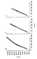
図1は、骨髄(BM)、筋および脳由来MASCの拡大可能性を示す図である。
【0061】
図2は、遺伝子発現の(A)筋および脳MASCそして(B)骨髄および筋MASCにおける遺伝子発現を示す散布図である。
【0062】
図3は、未分化MASCおよびVEGFとともに培養したMASCのFACS分析を示す図である。プロットはイソ型対照IgG染色プロファイル(細線)に対し特異的抗体染色プロファイル(太線)を示す。図Aは未分化MASCの表現型を示す。MASCは低レベルのp2ミクログロブリン、Flk1、Fit 1およびAC 133を発現しているが、他のどの抗内皮マーカーでも染色されない;図Bは14日間、10ng/mLのVEGFとともに培養したMASCの表現型を示す。MASCは内皮細胞に伴う低レベルの大部分のマーカーを発現しているが、AC 133の発現は失っている;そして図Cは3〜9日間10ng/mLのVEGFとともに培養したMASCの表現型を示す。MASCはVEGFとともに培養した3日目までにAC 133の発現を失い、TekおよびVE-カドヘリンの発現を3日目までに獲得し、CD34およびHIP12を9日目までに獲得している。
【0063】
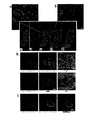
図4はmMASCの定着とin vivo分化の顕微鏡写真である。スライドは蛍光または共焦点顕微鏡法で検査した。図A、G、J、N、QおよびSは、mMASCを注射されていない対照NOD-SCID動物の同一に染色された組織を示す。図A〜Fは、抗β-gal-FITC抗体および各種造血抗原に対するPE-結合抗体で染色した、対照(A)および試験(B〜F)動物からの骨髄(BM)サイトスピンの顕微鏡写真を示す。A〜B:CD45、C:CD19、D:MAC1、E:GR1、F:TER119およびDAPI;図G〜Iは、抗β-gal-FITC抗体および抗CD45-PE抗体で染色した、対照(G)および試験動物(H、I)からの脾臓断面の顕微鏡写真を示す。提供者由来抗β-gal+細胞が塊になって見られる。Hは10倍でIは60倍の倍率である;図J〜Mは、抗β-gal-FITCで染色した、対照マウス(J)および試験動物(K〜M)からの肝臓断面の顕微鏡写真を示す。J〜Lはマウス抗CK-18/抗マウス-Cy5に加えてCD45-PEと、そしてMはマウス抗アルブミン/抗マウスCy3抗体と同時染色されている。J〜K、LおよびMはそれぞれ20倍、60倍および10倍の倍率である;図N〜Pは、抗β-gal-FITCに加えてマウス抗pan-CK/抗マウス-Cy5抗体(N〜P)で染色された、対照マウスおよび試験動物(O〜P)からの腸断面の顕微鏡写真を示す。NおよびPはCD45-PE抗体で同時染色した。β-gal+Pan-CK+CD45-上皮細胞が絨毛の周囲の50%を覆っている(黒矢印、図P)。CD45について同時染色した(P)絨毛の中心のPan-CK-/β-gal+細胞(白矢印-図O); 図Q〜Rは抗β-gal-FITCに加えてマウス抗pan-CK/抗マウス-Cy5およびCD45-PE抗体で染色した対照マウス(Q)および試験動物(R)からの肺断面の顕微鏡写真を示す。いくつかのβ-gal+pan-CK+提供者細胞が被提供者動物の肺胞の内面を覆っているのが見られる(R)。造血起源のCD45+/pan-CK-細胞が上皮細胞から明らかに見られる;そして図S〜Tは抗β-gal-FITC、抗vWF-PEおよびTO-PRO3で染色した対照マウス(S)および試験動物で成長した移植後16週間の胸腺リンパ腫(T)の血管断面の顕微鏡写真を示す。
【0064】
β-gal+提供者細胞は、腫瘍細胞は抗β-Gal抗体で染色されなかったので被提供者起源である胸腺リンパ腫内でvWF+内皮細胞に分化した。
【0065】
図5はMASC由来内皮細胞の共焦点蛍光顕微鏡法を用いた免疫組織化学的評価を示す図である。(a)VEGF中で14日間培養したMASC。接着受容体αvβ5について、および接着結合タンパク質ZO-1、β-およびγ-カテニンについて、典型的な膜染色が見られる。目盛り棒=50μm。(b)VEGF処理後0日目(上図)および21日目(下図)のMASCの明視野での形態。棒=25μm。
【0066】
図6はMASC由来内皮細胞の顕微鏡写真である。図AはMASC由来内皮からのvWFのヒスタミン媒介放出を示す。ミオシンに対する抗体を用いた染色が、ミオシンストレスファイバーの数の増殖、およびギャップ結合の拡大(矢印)を伴う細胞骨格の変化を示す(実験3回の代表例)。目盛り棒=60μm;図BはMASC由来内皮がa-LDLを取り込むのを示す。7日間後、細胞はTie-1を発現したが、今度はa-LDLを取り込まなかった。しかし、9日目のvvWF発現の獲得はaLDL取り込みを伴った(実験10回の代表例)。目盛り棒=100μm;そして図CはMASC由来内皮による血管形成を示す。6時間後、典型的な血管をみることができた(実験6回の代表例)。目盛り棒=200μm。
【0067】
図7はMASC由来内皮細胞のFACS分析を示す図である。プロットはイソ型対照IgG染色プロファイル(細線)に対する特異的抗体染色プロファイル(太線)を示す(実験>3回の代表例)。プロットの上の数字は対照IgG染色および特異的抗体染色についての平均蛍光強度(MFI)である。図Aはフローサイトメトリー分析で、MASC由来内皮細胞で低酸素がFlk1およびTek発現をアップレギュレートするのを示す;図BはMASC由来内皮細胞によるVEGF産生を低酸素がアップレギュレートするのを示す。VEGFレベルはELISAで測定し、結果は実験6回の平均±SEMで示す;そして図CはIL-1aがクラスIIHLA抗原の発現を誘導し接着受容体の発現を増加させることを示す。プロットはイソ型対照IgG染色プロファイル(細線)に対する特異的抗体染色プロファイル(太線)を示す(実験3回の代表例)。プロットの上の数字は対照IgG染色および特異的抗体染色についてのMFIを示す。
【0068】
図8はヒトMASC由来内皮細胞の顕微鏡写真である。図C〜Fは抗ヒトβ2-ミクログロブリン-FITC(図C)または抗マウス-CD31-FITC(図D)のいずれか、および両者の融合(図E)、抗vWF-Cy3(図F)および3つの染色パターンの融合(図G)の3次元再構成図を示す。図AおよびBは抗ヒトβ2-ミクログロブリン-FITCおよび抗vWF-Cy3、または抗マウスCD31-Cy5および抗vWF-Cy3、のいずれかで染色した一つの切片の共焦点像を示す。目盛り棒=100μm。図HはヒトMASC由来内皮細胞(上図)またはヒト包皮繊維芽細胞s(下図)を注射したマウスにおいて抗β2-ミクログロブリン-FITCおよび抗vWFで染色した穿孔した耳で創傷治癒が結果として高度に血管新生した部分を生じたのを示す。目盛り棒=20μm。C=軟骨。D=真皮。図Iは腫瘍血管新生がin vivoでMASCから生じた内皮細胞に由来し、結果として抗β2-ミクログロブリン-FITC、抗vWFおよびTOPRO-3で染色した腫瘍中に高度に血管申請した部分を生じるのを示す。目盛り棒=20μm。
【0069】
図9はhMSC由来神経細胞様細胞におけるスパイク生成および発現した電位依存性ナトリウム電流を表す図である。図Aはスパイク生成および発現した電位依存性ナトリウム電流を示した培養hMSC由来神経細胞の顕微鏡写真を示す(ピペットの影が細胞を指す)。図BはhMSC由来神経細胞から得た電流固定記録を示す図である。図Cは同じhMSC由来神経細胞から得たリークを差し引いた電流記録を示す図である。
【0070】
図10は肝細胞様表現型を確認した定量的RT-PCRおよびウェスタンブロット分析を示す図である。図AおよびBはFGF4およびHGF、またはFGF4単独と、それぞれ21および28日間Matrigel(商標)で培養したmMASC(A)およびhMASC(B)を示す。αFP、Cyp2b9 およびCyp2bl3については、未分化MASCでは転写物が検出されなかったため、ブロットの下の数字は肝臓から得たmRNAとの相対比である。Li=マウスまたはヒト肝臓mRNA;NT=テンプレート無し。マウス5例とヒト1例の試験の代表例として、図CはFGF4およびHGF、またはFGF4単独と、21日間Matrigelで培養したhMASC(B)を示す。FH=Matrigel上のFGF4およびHGF誘導性hMASC、Huh=対照として用いたHuh7細胞株。
【0071】
図11は肝細胞様細胞の顕微鏡写真を示す。FGF4によって誘導されたMASCはグリコーゲンを産生する。グリコーゲン貯蔵は染色濃部の集積として見られる(試験3回の代表例)。目盛り棒=25μm。
【0072】
定義
ここでは、以下の用語は以下の意味を持つものとする:
「拡大」は分化なしでの細胞の増殖を意味することとする。
【0073】
「中間細胞」は、MASCの分化の間に生じる、MASCまたはその最終分化した子孫の一部の特徴を有するがすべての特徴は有しない細胞である。中間細胞は特定の経路に決定された前駆細胞であってもよいが、特定の細胞型に決定されていてはならない。
【0074】
「正常」は病気、突然変異、または奇形を有しない動物、すなわち健康な動物を意味することとする。
【0075】
「自己再生」は外的刺激を加えられることなしに増殖する細胞の能力を意味することとする。組織または器官で局所的に産生されるサイトカインまたは他の成長因子の存在は外的刺激とならないこととする。
【0076】
「ホーミング」は、追加の細胞が必要である部位に特異的に移動する、一部のMASCまたはその子孫の能力を意味することとする。
【0077】
「発現のノックアウト」は、特定の遺伝子の機能の消失を意味することとする。
【0078】
ここでは、「ゲノムまたはプロテオーム構成」は任意の細胞の遺伝子またはタンパク質構成要素を意味することとする。
【0079】
「高レベルのテロメラーゼ活性」はヒト不死細胞株MCF7で観察されるレベルの2倍と関連づけることができる。Soule et al. (1973) J. Cancer Inst.
51:1409-1416。
【0080】
技術用途
MASC技術は哺乳類の体内の損傷細胞、疾患細胞、機能不全細胞または死細胞に置き換えるのに用いることができる。さらにこれらの細胞は、自家または同種異系細胞を用いて、天然または人工の担体、基材、または高分子とともにあるいはこれら無しで、たとえば鎌状赤血球病、血友病または、たとえばゴーシェ病、ニーマン・ピック病、ムコ多糖症などのように処理の欠陥のために産物が体内に蓄積する「貯蔵病」といったタンパク質機能に影響する遺伝子突然変異のように遺伝的な、失われた細胞、異常な機能または細胞または器官を修正するために、宿主に注射することができる。瀕死細胞または死細胞の回復の例は、黄斑変性症および他の神経変性疾患の治療におけるMASCまたはその分化した子孫の用途となりうる。
【0081】
これらのMASCを宿主動物の器官/組織に「ホーミング」させて組み込み増殖および分化する能力を考えると、MASCはおそらく虚血心および心筋細胞自体にも新しい内皮細胞を提供するのに用いることができる可能性があり、多数の他の例が存在する。
【0082】
組織または器官の機能への一時的な利益が望ましい効果を有しうる医学的状況があり得る。たとえば、正常な肝機能が回復できるようにするために十分なバイオ人工肝臓を接続し、肝移植の必要を防いだ肝障害患者の例がある。これはたとえば、C型肝炎というひとつの肝疾患だけでも、重大で満たされていない医療上の必要である。現在C型肝炎に感染している米国人は4〜5百万人存在し、これらの人々の50%が将来肝硬変になり肝移植を必要とするという推定がある。このことはぜひ救済を要する公衆衛生上の大問題である。自家または同種異系MASCに由来する肝細胞は、C型肝炎または他の肝疾患において移植できる。そのような移植は、提供者自身の肝細胞が回復できるように一時的に肝機能を提供するか、または永久的に損傷肝臓に再定着し提供者細胞によって正常な肝機能が回復するようにすることができる。
【0083】
未分化のMASCがヒトまたは他の哺乳類に投与されその後被提供者の中で特定の細胞に分化する多くの細胞療法に加えて、MASCの子孫細胞はex vivoで分化させてその後に精製した細胞または細胞の混合物として治療上の利益を提供するために投与することができる。これらの未分化状態のMASCはまた、治療上の利益を有する薬物または分子を輸送する担体または媒体として使用することができる。これは、癌、心血管疾患、炎症性疾患、免疫疾患、感染症、などを含むがこれに限定されないいくつかの疾患のうち任意のものを治療するのに用いることができる。そこで例として、細胞、おそらく新規のまたは高いレベルの血管新生分子を発現している内皮細胞を、患者に投与することができ、それは既存の血管に組み込まれて血管新生をたとえば心臓で促進する;同様に、血管新生を抑制する分子を産生し、血液細胞に組み込まれてそれ以上の形成をたとえば糖尿病性網膜症または新たな血管形成が疾患の病因、蔓延および程度の鍵となる癌において抑制する内皮細胞を得ることができる。
【0084】
BMに定着して血液をex vivoで形成する能力は、影で重要な医療上の用途を有する。たとえば、血液のex vivo産生に関して、輸血および血液製剤の注射は世界中でまだ行われていて、その安全性は病原体の感染のために変わりやすい。輸血は、HIV、BおよびC型肝炎、そして現在は狂牛病またはCJD、クロイツフェルト-ヤコブ病の差し迫った危険に繋がってきた。血液、特に赤血球をin vitroで産生する能力は、血液をヒトから集めることの安全かつ信頼できる代替法を提供しうる。提供者からの採血を完全に代替することは決して無いと思われる。hMASCまたはその造血性の子孫を、たとえばヒツジといった動物に胎内で注入し、ヒト造血細胞を形成しヒト血液成分または医療上の用途のあるタンパク質の起源として使うことができる。同じことが肝細胞、膵島または多くの他の細胞型についていえるが、ヒト細胞をin vitroで産生することの代替案を提供し、動物を細胞の工場として用いる。このことは発生が予測される血液不足を補助することができる。hMASCはまた、考えられる限りでは、いくつかの欠陥のうち任意の一つを修正するためにヒト胚に移植することができる。
【0085】
これらのMASCは特異的に分化した細胞のクローン集団を生じることができるため、これらは薬物発見の豊かな基盤である。これにはこの周辺の、遺伝子を発現させること、遺伝子発現を分析すること、活性化の新規の遺伝子活性化パターンの発見、プロテオミクスおよびタンパク質発現および修飾のパターンが含まれる。これはデータベースと、これらのデータを公開または自己所有のデータベースと比較したこれらのデータを分析するためのアルゴリズムとを用いて、バイオインフォマティクスで分析される。既知の薬物または物質がどのように挙動するかの情報を、MASC、その分化した子孫、および利用可能なヒトの集団から得られた情報と比較することができる。経路、標的、および受容体を同定することができる。新薬、抗体または他の化合物が生物学的に望ましい反応を生じるのを発見することができる。同様に、MASCおよびその分化した子孫を、データベース、バイオインフォマティクスおよびアルゴリズムと組み合わせて、望ましくない反応についてのモニターとして用いることができる。
【0086】
ヒト、マウス、ラットまたは他の哺乳類に由来するこれらのMASCは現在までに知られた唯一の、正常の、非-悪性の、継代の遅い細胞でさえ非常に高レベルのテロメラーゼを発現する体細胞(非-生殖細胞)であるように見える。テロメアはMASCで伸長していて核型的には正常である。哺乳類に注射されたMASCは、複数の器官へホーミングするので、特定の器官に新たに到着したMASCは自己再生できる可能性がある。このように、MASCは自分自身だけでなく、損傷したか、死亡したか、でなければ遺伝的または後天性疾患のために異常な機能を有した細胞型である、自己再生する分化した細胞型でも、器官に再定着する可能性を有する。
【0087】
たとえば、I型糖尿病では膵島のインシュリン産生ベータ細胞が進行性に失われる。各種の腎疾患では機能の進行性消失と、一部の場合では糸球体の消滅がある。もし糖尿病の場合には、MASCまたは分化した子孫が膵臓にホーミングして、それ自身でまたは膵臓の中で内因性細胞との相互作用によって、膵島が形成されるように誘導しうる。これは糖尿病を改善する効果を有しうる。最終的には、MASCの自己再生する能力をin vivoで制御する、すなわち促進または抑制し、そして分化した子孫、たとえば、膵島前駆細胞、肝細胞前駆細胞、血球前駆細胞、神経および/または心前駆細胞への移行を制御する、すなわち促進または抑制する、条件、物質または薬物が見出されうる。MASCを用いて、増殖し分化する内因性前駆細胞を器官の中で誘導しうる、経路、活性化の方法および制御を見出しうる。
【0088】
細胞組織または器官の区画に再定着し自己再生しそしてまた分化するこの同じ能力は、多数の用途と、医療上の満たされていない深い必要性を満たす前例の無い効用を有しうる。たとえば、酵素欠損症が存在するある種の遺伝疾患は、BM移植によって治療されている。これはしばしば、疾患の後遺症が脳または骨または他の場所に残りうる疾患の合併症に助けとなりうるが治癒はしない。MASCおよび遺伝子組み換えMASCは、多数の遺伝的および後天性疾患を改善する希望をもたらす。これらはまた、診断上または研究上の目的および薬物発見に有用となる。
【0089】
本発明はまた、MASCのゲノムまたはプロテオーム構成を分析し、バイオインフォマティクスによるその分析、アルゴリズムによるコンピュータ分析と組み合わせて、新たなデータベースを収集し既知のデータベースと比較しこれらを対照することを含む;薬物発見、ゲノミクス、プロテオミクス、および経路の同定のための方法を提供する。これによって、治療上の利益を有する可能性のある新規化合物、抗体、タンパク質、小分子有機化合物、または他の生物活性分子に関する標的の定義に繋がりうる、主要構成要素、経路、新規遺伝子および/または遺伝子とタンパク質発現とタンパク質修飾の新たなパターン(プロテオミクス)が同定できる。
【0090】
以下の実施例は本発明を説明するために提供され、しかし本発明を限定しない。
【実施例1】
【0091】
マウス多能性成体幹細胞(mMASC)の選択、培養および特徴づけ
細胞単離と拡大
すべての組織はミネソタ大学学内動物実験委員会からのガイドラインに従って得た。BM単核細胞(BMMNC)はBMのフィコール・ハイパック分離によって得た。BMは5〜6週齢のROSA26マウスまたはC57/BL6マウスから得た。あるいは、筋および脳組織を3日齢のマウス129匹から得た。前肢および後肢の近位部からの筋を摘出しよく挽いた。その組織を0.2%コラゲナーゼ(Sigma Chemical Co, St Louis, MO)を用いて1時間、37℃で処理し、次いで0.1%トリプシン(Invitrogen, Grand Island, NY)で45分間処理した。細胞をその後よく磨砕し、70μmフィルターを通した。細胞懸濁液を回収して、10分間、1600rpmで遠心分離した。脳組織は切開してよく挽いた。細胞を0.1% トリプシンおよび0.1% DNAse(Sigma)とともに30分間、37℃でインキュベートして解離させた。細胞をその後よく磨砕し、70μmフィルターを通した。細胞懸濁液を回収して、10分間、1600rpmで遠心分離した。
【0092】
BMMNCまたは筋または脳懸濁液を、1xl05/cm2で拡大培地[2%PCS添加低グルコースDulbecco最小必須培地(LG-DMEM)に、血小板由来成長因子(PDGF)と上皮成長因子(EGF)および白血病抑制因子(LIF)を各10ng/mL]に平板播種し、5xl03/cm2で維持した。3〜4週間後、トリプシン/EDTAで回収した細胞からCD45+/グリコフォリン(Gly)-A+細胞を磁気マイクロビーズを用いて除去した。結果として得られたCD45-/Gly-A-細胞を10細胞/ウェルでFN被覆96穴プレートに再播種し、0.5〜1.5x103/cm2の間の細胞密度で拡大した。MASCの拡大可能性は由来する組織にかかわらず同様であった(図1)。
【0093】
MASC の特徴づけ
表現型的には、BM、筋および脳由来の、FNで培養したmMASCは、CD13+、CD44-、CD45-、クラスIおよびクラスII組織適合抗原-、低Flk1およびcKifで、国際出願番号PCT/US00/21387に記載されたhMASCの特徴と同一であった。細胞をIV型コラーゲン、ラミニンまたはMatrigelで培養したときのほうが、最初の2〜3ヶ月間の細胞拡大はより大きかったが、細胞はMSCの表現型の特徴を有した、すなわち、CD44を発現しCD13を発現しなかった。ヒト細胞と同様に、FNで培養したmMASCはoct-4の転写物とLIF-Rとを発現した。
【0094】
10個のCD45-/GlyA-細胞を播種したウェルの約1%から、連続的に成長する培養が得られた。これは、培養MASCを開始しうる細胞が稀であっておそらくCD45-/GlyA-細胞の1/1,000より少ないことを示唆する。FNで培養したmMASC は直径8〜10μmで大きい核と少ない細胞質を有する。いくつかの集団が> 100 PDの間培養されている。細胞の形態と表現型は培養期間を通じて変化しないままであった。
【0095】
40および102 PDを経過したmMASCを採取し、テロメア長を評価した。テロメア長はPharmingen(New Jersey, USA)のテロメア長分析キットを用いて取扱説明書に従って測定した。40 PDの間培養したmMASCの平均テロメア長(ATL)は27Kbであった。102PD後に再試験したとき、ATLは変化しないままであった。mMASCの核型分析には、以前に記載した通り細胞を採取前に1:2希釈で12時間継代培養し、トリプシン-EDTAを用いて回収し、1.5時間コルセミド処理し、続いて低張KClを用いて細胞溶解し、酸/アルコールで固定した(Verfaillie et al., 1992)。細胞遺伝学的分析は月単位で行い、45 PD後に高二倍体となった、試験には使われなくなった一つの集団を除いて正常核型を示した。
【0096】
46から>80PD後に得られたマウスMASCを、ES細胞の未分化状態を維持するのに重要な二つの転写因子であるoct4およびRex1の発現レベルについて、定量的(Q)-RT-PCRによって試験した。マウスMASC、神経外胚葉性に分化した子孫(bFGFの添加後1〜7日目)およびマウスES細胞からRNAを抽出した。RNAを逆転写し、結果として得られたCDNAを下記の反応条件で40回増幅した(ABI PRISM 7700, Perkin Elmer/Applied Biosystems): 2μLのDNA溶液と、1X TaqMan SYBR Green Universal Mix PCR反応緩衝液を用いた最初の変性(95℃で10分間)後、二段階PCR(95℃で15秒間、60℃で60秒間)を40サイクル。プライマーは表1に列記する。
【表1】

mRNAレベルはGAPDHをハウスキーピング遺伝子として用いて標準化し、マウスES細胞中のレベルと比較した。oct4およびRex1 mRNAは、BM、筋および脳由来MASC中に同様のレベルで存在した。Rex1 mRNAレベルはmMASCおよびmES細胞で同様であった一方、oct4 mRNAレベルはMASCでES細胞より約1,000倍低かった。
【0098】
マウス BM 、筋および脳由来 MASC の発現遺伝子プロファイルは非常に類似している
異なる組織に由来するMASCが同様であるかどうかさらに評価するために、BM、筋および脳由来MASCの発現遺伝子プロファイルをU74A Affimetrix遺伝子アレイを用いて検討した。要約すると、45回細胞分裂の間培養した2〜3xl06個のBM、筋または脳由来MASCからmRNAを抽出した。CDNAの調製、6,000個のマウス遺伝子と6,000個のESTクラスターを含むU74Aアレイとのハイブリッド形成、およびデータ取り込みは取扱説明書に従って行った(すべてAffimetrix, Santa Clara, CAより)。データ分析はGeneChip(登録商標)ソフトウェア(Affimetrix)を用いて行った。2.2倍の因数での発現増加または減少(Iyer V.R. et al., 1999;ScherfU. et al., 2000;Alizadeh A.A. et al, 2000)を有意とみなし、r2値を直線回帰分析を用いて決定した(図2)。
【0099】
3種類の組織に由来するMASCの発現遺伝子プロファイルの比較は、BM由来のMASCで筋由来と比べて遺伝子の1%未満が2.2倍より大きく異なるレベルで発現していたことを示した。同様に、BM由来のMASCで脳由来と比べて遺伝子の1%未満だけが2.2倍より大きく異なるレベルで発現していた。異なるMASC集団間の相関係数は>0.975であったため、異なる組織に由来するMASCは非常に相同であり、上記の表現型および実施例5に記載の分化の特徴と合致する。
【0100】
マウス特異的培養条件を用いて、mMASC培養を100細胞分裂以上維持している。mMASC培養は、-HMG-LacZについて遺伝子導入したC57B1/6マウス、ROSA26マウスおよびC57BL/6マウス由来の骨髄を用いて開始した。
【実施例2】
【0101】
ラット多能性成体幹細胞(VMASC)の選択と培養
Sprague DawleyまたはWistarラット由来のBMおよびMNCをmMASCについてと同様の条件下で得て播種した。21〜28日間後、細胞からCD45+細胞を除去し、結果として得られたCD45-細胞を10細胞/ウェルで継代培養した。
【0102】
mMASCと同様に、rMASCは>100PDの間培養して拡大している。ラットMASC培養の拡大 条件には、EGF、PDGF-BBおよびLIFの添加と、I型コラーゲン、ラミニンまたはMatrigelでなくFNでの培養が必要であった。rMASCはCD44、CD45およびMHCクラスIおよびIIについて陰性で、高レベルのテロメラーゼを発現した。100細胞分裂を超えて生育する正常細胞の能力は前例が無く、予想外で、20年以上の従来の定説に反する。
【0103】
42PD、72 PD、80 PD、および100 PDを経過したラット MASCを採取してテロメア長を評価した。42 PD、72 PD、80 PD、and 100 PD後のサザンブロット分析で測定されたように、テロメアは培養中に短縮しなかった。ラットMASCの毎月の細胞遺伝学的分析では正常核型が見られた。
【実施例3】
【0104】
ヒト多能性成体幹細胞(hMASC)の選択と培養
BMは健康なボランティア提供者(2〜50歳)から、研究におけるヒト被験者の使用に関するミネソタ大学委員会からのガイドラインを用いたインフォームド・コンセント後に得られた。BMMNCはフィコール・パック密度勾配遠心分離によって得られ、depleted of CD45+およびグリコフォリン-A+細胞を磁気マイクロビーズ(Miltenyii Biotec, Sunnyvale, CA)を用いて除去した。
【0105】
拡大条件: 5x10³個のCD45-/GlyA-細胞を200uLの拡大培地[1Xインシュリン-トランスフェリン-セレン(ITS)、1Xリノール酸ウシ血清アルブミン(LA-BSA)、10-8Mデキサメタゾン、10-4Mアスコルビン酸-2-リン酸(すべてSigma)、100Uペニシリンおよび1,000Uストレプトマイシン(Gibco)を添加した58%DMEM-LG、40%MCDB-201(Sigma Chemical Co, St Louis, MO)に、10 ng/mlのEOF(Sigma)および10ng/mlのPDGF-BB(R&D Systems, Minneapolis, MN)を含む胎児ウシ血清(PCS)(Hyclone Laboratories, Logan, UT)を0〜10%添加]で希釈し、5ng/mlのFN(Sigma)で被覆した96穴プレートのウェルに播種した。培地は4〜6日毎に交換した。ウェルの>40〜50%に密集成長したら、接着細胞を0.25%トリプシン-EDTA(Sigma)を用いて剥離して、MASC拡大培地に1:4希釈で5ng/mlのFNで被覆したより大きな培養容器に再播種し、2〜8x103細胞/cm2の間の細胞密度を維持した。
【0106】
未分化のMASCはCD31、CD34、CD36、CD44、CD45、CD62-E、CD62-L、CD62-P、HLA-クラスIおよびII、cKit、Tie、Tek、αvβ3、VE-カドヘリン、血管内皮細胞接着分子(VCAM)、細胞内接着分子(ICAM)-1を発現しなかった。MASCは低い/非常に低いレベルのβ2-ミクログロブリン、αvβ5、CDw90、AC133、Flk1およびFlt1、そして高レベルのCD13とCD49bを発現した(図3)。
【実施例4】
【0107】
免疫表現型分析
免疫蛍光
1.培養細胞を4%パラホルムアルデヒドおよびメタノールメタノールを用いて室温で固定し、一次抗体と、そして二次抗体とあるいは二次抗体無しで、連続して各30分間インキュベートした。各工程の間に、スライドをPBS/BSAで洗浄した。細胞は蛍光顕微鏡法(Zeiss Axiovert; Carl Zeiss, Inc., Thornwood, NY)および共焦点蛍光顕微鏡法(共焦点1024顕微鏡; Olympus AX70, Olympus Optical Co. LTD, Japan)によって検査した。任意の培養中の異なる細胞型の頻度を評価するために、任意の抗体について染色陽性となった細胞を4つの視野(視野あたり細胞50〜200個)で計数した。2. 採取組織 :血液およびBMのサイトスピン標本をアセトン(Fisher Chemicals)で10分間、室温で固定した。固形器官については、組織の5μm厚の新鮮凍結切片をスライドグラスに乗せ、ただちにアセトン中で10分間、室温で固定した。イソ型血清で20分間インキュベート後、サイトスピン調製物または組織切片を、組織特異的抗原、β-galおよび核対比染色剤(DAPIまたはTO-PRO-3)について連続的に染色した。
【0108】
カバースリップはSlowfade-antifadeキット(Molecular Probes Inc., Eugene, OR, USA)を用いて乗せた。スライドは蛍光顕微鏡法および共焦点蛍光顕微鏡法によって検査した。
【0109】
3. 抗体:細胞は4%パラホルムアルデヒドを用いて室温で、またはメタノールを用いて-20℃で固定し、一次抗体、およびFITCまたはCy3結合抗マウス-または抗ウサギ-IgG抗体を用いて連続的に各30分間インキュベートした。各工程の間にスライドをPBS+1%BSAを用いて洗浄した。PEまたはFITC結合抗CD45、抗CD31、抗CD62E、抗Mac1、抗Gr1、抗CD19、抗CD3、および抗Ter119抗体はBD Pharmingenより入手した。GFAP(クローンG-A-5、1:400)、ガラクトセレブロシド(GalC)(ポリクローナル、1:50)、MBP(ポリクローナル、1:50)、GABA(クローンGB-69、1:100)、パルブアルブミン(クローンPARV-19、1:2000)、TuJl(クローンSDL.3D10、1:400)、NF-68(クローンNR4、1:400)、NF-160(cloneNN18、1:40)、そしてNF-200(クローンN52、1:400)、NSE(ポリクローナル、1:50)、MAP2-AB(クローンAP20、1:400)、Tau(ポリクローナル、1:400)、TH(クローンTH-2、1:1000)、DDC(クローンDDC-109、1:100)、TrH(クローンWH-3、1:1000)、セロトニン(ポリクローナル、1:2000)、グルタミン酸(クローンGLU-4、1:400)、速筋ミオシン(クローンMY-32;1:400希釈)に対する抗体はSigmaから入手した。DAPIおよびTOPRO-3はMolecular Probesから入手した。vWF(ポリクローナル;1:50)、Neuro-D(ポリクローナル、1:50)、c-ret(ポリクローナル、1:50)andNurrI(ポリクローナル、1:50)に対する抗体はSanta Cruz Biotechnology Inc., Santa Cruz, CAから入手した。PSA-NCAM (ポリクローナル、 1 :500)に対する抗体はPhanmingen, San Diego, CAから、そしてセロトニン輸送体(クローンMAB1564、1:400)、DTP(ポリクローナル、1:200)、Naゲート電位チャンネル(ポリクローナル、1:100)、グルタミン酸-受容体-5、-6および-7(クローン371、1:500)およびNMDA(ポリクローナル1:400)受容体に対する抗体はChemicon International, Temecula, CAから入手した。抗ネスチン(1:400)抗体はスウェーデンのLund大学のDr. U. Lendahlより寄贈を受けた。NSE(1:50)pan-サイトケラチン(カタログ番号C-2562;1:100)、CK-18(C-8541;1:300)、アルブミン(A-6684;1:100)に対する抗体はすべてSigmaから入手した。Flk1、Flt1、Tek、HNF-1pに対するポリクローナル抗体はSanta Cruz Biotechnology Inc., Santa Cruz, CAから入手した。抗ネスチン(1:400)抗体はスウェーデンのLund大学のDr. U. Lendahlより寄贈を受けた。対照-マウス、-ウサギまたは-ラットIgGとFITC/PE/Cy3-およびCy5-標識二次抗体はSigmaから入手した。ウサギ抗β-gal-FITC抗体はRockland Immunochemicals, USAから入手した。TO-PRO-3 Molecular Probes Inc.から入手し、DAPはSigmaから入手した。
【0110】
B.X-GAL 染色;組織切片はβ-ガラクトシダーゼ酵素活性についてInvitrogenのβ-gal染色キットを用いて染色した(pH7.4)。組織切片を10分間のかわりに5分間インキュベートした固定工程以外は取扱説明書に従った。
【0111】
C.FACS:FACSについては、未分化MASCを剥離して、抗CD44、CD45、CD13、cKit、MHC-クラスIおよびII、またはb2-ミクログロブリン(BD Pharmingen)および二次FITCまたはPE結合抗体を用いて連続的に染色し、FACS-Calibur(Becton Dickinson)を使用した分析まで2%パラホルムアルデヒドで固定した。
【実施例5】
【0112】
MASCからの分化系統の単一細胞起源
mMASCまたはrMASCの分化能力を、hMASCまたはES細胞の中胚葉、神経外胚葉、および内胚葉への分化について何が記載されているかに基づいて選択した分化因子(サイトカイン)を加えることによって試験した。分化には、EOF、PDGF-BBおよびLIFを含まないが、系統特異的サイトカインを含む無血清培地に、l〜2x10⁴細胞/cm2で細胞を再播種することを要した。分化は組織特異的マーカー[遅筋ミオシンおよびMyoD(筋)、von-Willebrand因子(vWF)およびTek(内皮)、NF200およびMAP2(神経外胚葉性)、そしてサイトケラチン-18およびアルブミン(内胚葉性)]についての免疫組織化学、RT-PCR、および機能試験によって判定した。
【0113】
神経外胚葉性細胞への MASC 分化
Palmer et al.は神経前駆細胞をPDGF-BBを用いて培養で拡大できること、およびPDGFの除去と分化因子としてbFGFの添加によってその分化を誘導できることを示した。これらの研究およびhMASCを用いて行われた研究に基づき、mMASCおよびrMASCを、PDGF-BBおよびEGFを含まないが100ng/mLのbFGFを含むFN被覆ウェルに播種した。神経細胞様細胞の成熟の進行が培養期間を通じて見られた。7日間後、細胞の大多数がネスチンを発現した。14日間後、MASCの15〜20%が星状細胞の(GFAP+)、15〜20%が稀突起膠細胞の(ガラクトセレブロシド(GalC)+)、そして50〜60%が神経細胞の(神経フィラメント-200(NF-200)+)形態的および表現型の特徴を獲得した。NF200、GFAPまたはGalCは同一の細胞には見つからず、神経細胞様細胞が神経細胞のマーカーを不適切に発現したhMASCまたはグリア細胞である可能性は低いことを示唆した。神経細胞様細胞はまたTau、MAP2およびNSEも発現した。神経細胞の約50%がガンマ-アミノ酪酸(GABA)およびパルブアルブミンを、30%がチロシンヒドロキシラーゼおよびドーパ-デカルボキシラーゼ(DDC)を、そして20%がセロトニンおよびトリプトファンヒドロキシラーゼを発現した。MASCを40PD、または90PD以上拡大したとき、分化は同様であった。実施例1で記載の通り実施したQ-RT-PCRによって、神経外胚葉性マーカーの発現が確認された: 2日目にMASCはotx1およびotx2 mRNAを発現し、7日間後にはネスチンmRNAが検出された。
【0114】
繊維芽細胞成長因子(FGF)-8bの分化因子としての効果を次に試験した。これはin vivoで中脳の発生のために重要であり、in vitroでマウスES細胞からhMASCについてドーパミン作用性およびセロトニン作用性神経細胞を誘導するのに用いられる。密集成長したhMASC(n=8)を10ng/mLのFGF-8b+EGFと培養したとき、神経細胞マーカーについて染色陽性であるが稀突起膠細胞および星状細胞について陽性でない細胞への分化が見られた。神経細胞はGABA性(GABA+;40±4%)、ドーパミン作用性(DOPA、TH、DCCおよびDTP+、26±5%)そしてセロトニン作用性(TrH、セロトニンおよびセロトニン-輸送体+、34±6%)神経細胞の特徴を有した。DOPA+神経細胞はNurr1に対する抗体で染色され、中脳DA神経細胞への分化を示唆した。FGF-8b誘導性神経細胞は成熟神経細胞の電気生理学的特徴を有しなかった。したがって、FGF-8bで支持された3週齢の細胞を、グリア芽腫細胞株であるU-87と、FGF-8bと共にさらに2〜3週間培養した。
【0115】
神経細胞は細胞の大きさおよび数、長さ、および神経突起の複雑さの増大したより成熟した形態を獲得し、そして成熟神経細胞の電気生理学的特徴(1μMテトロドトキシン(TTX)によって可逆的に遮断される一時的な内向き電流は、一時的なタイムコースと内向き電流の電位依存活性化とともに、成熟神経細胞にだけ見られる電位活性化ナトリウム電流に典型的である)を獲得した。
【0116】
hMASC(n=13)を10ng/mの脳由来神経分化誘導因子(BDNF)+EOFと共に培養したとき、分化はDOPA、TH、DCC、DTPおよびNurr1陽性神経細胞に限定された。BDNFはES細胞およびNSCからの神経分化を支援するが(Peault, 1996;Choi et al. 1998)、DA様神経細胞への限定的分化を示した研究は無い。
【0117】
同様の結果が、bFGFで誘導されたmMASCとbFGFおよびBDNFで誘導されたrMASCについて見られた。MASC由来神経細胞についてのさらなる研究が実施例10に示されている。
【0118】
内皮細胞への MASC 分化
中胚葉の例として、内皮への分化を誘導した。未分化のmMASCまたはrMASCは内皮マーカーCD31、CD62E、TekまたはvWFを発現しなかったが、低レベルのFlk1を発現した。mMASCまたはrMASCをFN被覆ウェルで10 ng/mLの内皮分化因子VEGF-Bと共に培養した。14日間のVEGF処理後、MASCの>90%が、経過したPD数にかかわらず、内皮分化に合致してFlt1、CD31、vWFまたはCD62を発現した。初代内皮細胞のように、MASC由来内皮細胞はMatrigelに再播種後6時間以内に血管を形成した。
【0119】
同様に、hMASCはFlk1およびFlt1を発現するが、CD34、Muc18(P1H12)、PECAM、E-およびP-セレクチン、CD36、またはTie/Tekは発現しない。hMASCを2xl04細胞/cm2、20ng/mLの血管内皮成長因子(VEGF)を含む無血清培地で培養したとき、細胞はCD34、VE-カドヘリン、VCAMおよびMuc-18を7日目以降に発現した。14日目に、細胞はTie、Tek、Flk1およびFlt1、PECAM、P-セレクチンおよびE-セレクチン、CD36、vWF、そしてコネキシン-40も発現した。さらに、細胞は低比重リポタンパク質(LDL)を取り込むことができた。組織化学染色からの結果はウェスタンブロットによって確認された。血管形成を誘導するために、14日間VEGFと共に培養したMASCを、10ng/mLVEGF-Bを含むMatrigelに再播種して6時間経過した。>2%PCSで培養されたhMASCを用いたときは、内皮分化は見られなかった。さらに、PCSが分化の間に培地中に残されたとき、内皮細胞は生じなかった。
【0120】
hMASCを継代培養したとき少なくとも 1 000-倍の拡大が得られ、hMASCから生じた内皮 前駆細胞が顕著な増殖能力を持ち続けていることを示唆した。細胞の拡大は、PCSが7日後に培養に添加されたときさらに大きかった。hMASC由来内皮細胞が皮下にヒト大腸癌を移植されたNOD-SCIマウスに静脈注射で投与されたとき、ヒト内皮細胞が腫瘍で血管新生に寄与するのが見られた。したがって、治療上の利益を得るため、すなわち、たとえば癌において血管新生を阻害するため、または肢あるいはたとえば心臓のようなその他の器官において血管新生を促進するために、遺伝子組み換え内皮細胞を組み込むことが可能である。MASC由来内皮細胞に関するさらなる研究は実施例9に示されている。
【0121】
内胚葉への MASC 分化
mMASCまたはrMASCが内胚葉細胞に分化できるかどうかを試験した。分化因子であるケラチノサイト成長因子(KGF)、肝細胞成長因子(HGF)およびFGF-4と共に、ラミニン、コラーゲン、FNまたはMatrigel被覆ウェルのいずれかでの培養を含むいくつかの異なる培養条件を試験した。10ng/mLのFGF4+10ng/mLのHGFを含むMatrigelに再播種したとき、MASCの約70%が肝細胞様細胞の形態的および表現型の特徴を獲得した。細胞は類上皮細胞になり、細胞の約10%が二核になり、そして細胞の約70%がアルブミン、サイトケラチン(CK)-18、およびHNF-1Pについて染色陽性となった。
【0122】
FGF4およびHGFを含む培養で生じた内胚葉性様細胞もまた、未分化のMASCとFGF4およびHGF誘導性MASCの上清中の尿素レベルをSigma尿素窒素キット640を用いて取扱説明書に従って測定することによって判定された、肝細胞の機能的特徴を有した。未分化のMASC培養においては尿素は検出されなかった。尿素産生はFGF4およびHGFの添加14日後に10μg/細胞/時間であり、25日目まで同様のレベルで検出可能であった。これは単層で生育した初代ラット肝細胞と同等である。アルブミンの存在は、尿素産生と共にMASCからin vitroの肝細胞分化の概念を支持する。MASC由来肝細胞に関するさらなる研究は実施例11に示されている。
【0123】
内胚葉系統前駆細胞が存在する可能性が高いことを考えると、MASCは肝臓と膵臓の外分泌および内分泌構成部分の両方、および他の内胚葉由来細胞組織系統のさまざまな細胞を形成する細胞を生じる可能性が高い。
【0124】
筋または脳に由来するMASCは、BM由来MASCについて上に記載した方法を用いて、中胚葉(内皮細胞)、神経外胚葉(星状細胞および神経細胞)および内胚葉(肝細胞様細胞)に分化するよう誘導された。
【0125】
形質導入
分化した細胞が単一細胞由来でありMASCが実際に「クローン」多能性細胞であることを示すため、MASCにレトロウイルスベクターを導入した培養細胞を作成し、そして未分化の細胞とその子孫はゲノムの同一部位にレトロウイルス挿入配列を有することが見出された。
【0126】
40から>90PDの間拡大した、二つの独立して得られたROSA26MASC、二つのC57BL/6MASCおよび一つのrMASC集団を用いて、またeGFPを導入した「クローン」マウスおよび「クローン」rMASCについて、試験を行った。eGFP導入細胞および非導入細胞の間に差異は見られなかった。注目すべきこととして、eGFP発現は分化したMASCで持続した。
【0127】
具体的には、EGF, PDGF-BBおよびLIFを含むFNで3週間培養したマウスおよびラットBMMNCに連続2日間、eGFP腫瘍レトロウイルスベクターを導入した。その後、CD45+およびGlyA+細胞を除去し、細胞を10細胞/ウェルで継代培養した。eGFP導入ラットBMMNCを85PDの間拡大した。または、80PDSの間拡大したマウスMASCを用いた。未分化MASCの継代培養は、75PDの間維持した培養から100個のMASCを播種してそれを>5xl06個に再拡大することによって作成した。拡大したMASCはin vitroで内皮、神経外胚葉および内胚葉に分化するよう誘導した。系統分化は、実施例4に記載の通り、これらの細胞型に特異的な抗体で染色することによって示した。
【0128】
中胚葉性および神経外胚葉性子孫細胞の単一細胞起源
中胚葉性および神経外胚葉性の分化した子孫細胞が単一細胞起源であることを証明するため、レトロウイルスマーキングを用いた(Jordan et al, 1990;Nolta et al., 1996)。20PD後に得たhMASCの画分にMFG-eGFPレトロウイルスを形質導入した。eGFP+hMASCを、同一提供者由来の非導入MASCで希釈し、導入細胞の終濃度を〜5%とした。これらの混合物を100細胞/ウェルで播種し、培養を>2xl07細胞が得られるまで拡大した。それぞれ5xl06個のMASCを、骨格筋筋芽細胞、内皮および神経外胚葉性系統に分化するよう誘導した。分化条件下で14日後、細胞を採取してレトロウイルス組み込み部位、およびeGFPと神経外胚葉性、筋および内皮マーカーとの同時発現を同定するために用いた。
【0129】
筋芽細胞分化については、hMASCを2xl04細胞/cm2で2%PCS、EGFおよびPDGF含有拡大培地に播種し、3μMの5-アザシチジンを用いて同じ培地中で24時間処理した。その後、細胞は2%PCS、EGFおよびPDGF-BBを含む拡大培地中で維持した。内皮分化については、hMASCを2xl04細胞/cm2で、EGFおよびPDGFを含まないが10ng/mlのVEGF-Bを含む無血清拡大培地で14日間培養した。
【0130】
免疫蛍光評価は、培養中の細胞の5〜10%が5-アザシチジンでeGFPおよび骨格筋アクチンについて染色陽性に分化するよう誘導され、細胞の5〜10%がeGFPおよびvWFについて同時染色される内皮に分化するよう誘導され、そして細胞の5〜10%が、eGFP、およびNF-200かGFAPかMBPのいずれかについて同時染色される神経外胚葉に分化するよう誘導されたことを示した。レトロウイルス挿入部位を定義するため、MASCおよび分化した子孫の宿主ゲノム隣接領域を配列決定した。別々の集団におけるレトロウイルス挿入配列の数は1から7の間であった。表2に示す通り、集団A16で染色体7にマッピングされた、筋、内皮および神経外胚葉性細胞でレトロウイルス挿入配列に隣接する、単一の同一の配列が同定された。
【表2】
3'LTRに特異的なプライマーを設計し、また隣接するゲノム配列に特異的なプライマーは表3に示す通りで、そしてリアルタイムPCRを用いて、レトロウイルス挿入部位は未分化細胞と分化した細胞とで同一であることが確認された。これらの結果は、隣接配列とeGFP DNA配列とが同様の量で存在することを証明した。クローンA12はそれぞれ染色体1と7に位置する2個のレトロウイルス挿入配列を含み、両方の隣接配列をhMASCだけでなく筋、内皮および神経外胚葉性系統にも検出することができた。これが2個のレトロウイルス要素を有する単一細胞の子孫かまたは2個の細胞の子孫のどちらを示すのか判定するため、リアルタイムPCRを用いて染色体1および7のeGFP隣接配列の相対量を比較した。hMASC、筋、内皮および神経外胚葉性細胞に両方の隣接配列が同様の量存在したことが見出され、2個のレトロウイルス挿入配列を有する単一細胞がeGFP陽性hMASCおよび分化した子孫の元になった可能性が高いことを示唆した。3個以上のレトロウイルス挿入配列を含む他の集団においては、我々は挿入配列が単一細胞の複数の挿入部位に由来するのかそれともeGFP陽性画分に寄与する複数の細胞に由来するのか判定することはできなかった。にもかかわらず、2個の集団において筋、内皮および神経外胚葉に分化した子孫細胞は単一のBM由来前駆細胞に由来するという我々の発見は、中胚葉性系統および神経外胚葉の3つの異なる系統の細胞において単一細胞レベルで分化するBMから原始細胞を培養することが可能であることを初めて決定的に証明する。
【表3】
【0132】
体内の多くの器官への哺乳類MASCのホーミングと定着
定着してin vivoで組織特異的細胞に分化する能力を有するかどうか調べるため、mMASCを試験した。mMASCをLacZ遺伝子導入C57Black6、ROSA26マウスから実施例1に記載の通り育成した。LacZマウス由来の106個のmMASCを、注射の4〜6時間前に250Radの全身照射ありまたは無しで、NOD-SCIDマウス尾静脈に静脈注射した。動物は注射後4〜24週間で頸椎脱臼により屠殺した。
【0133】
組織採取
血液および骨髄: 0.5〜1mlの血液を動物の屠殺時に得た。大腿骨および脛骨を洗浄してBMを回収した。表現型分析については、血液およびBM中の赤血球を氷冷塩酸アンモニウムを用いて除去し(Stem Cell Technologies Inc., Vancouver, Canada)、105個の細胞をサイトスピン遠心分離に用いた。連続移植については、大腿骨2本および脛骨2本からの5xl07個の細胞を個々の二次被提供者に尾静脈注射によって移植した。二次被提供者は7〜10週間後に屠殺した。
【0134】
固形器官:肺はPBSで1:4希釈したOCT化合物(Sakura-Finetek Inc, USA)の1mlで膨張させた。被提供者動物の脾臓、肝臓、肺、腸、骨格筋、心筋、腎臓および脳の標本を採取しOCT中で-80℃にて、および定量的PCR用にRNA Later(Ambion Inc., Austin, TX, USA)中で-20℃にて低温保存した。
【0135】
mMASC は定着して in vivo で組織特異的細胞に分化する
β-gal/ネオマイシン(NEO)導入遺伝子含有細胞の定着(Zambrowicz et al., 1997)は、β-galについて免疫組織化学によって、およびNEOについてQPCRによって試験された。免疫組織化学およびQ-PCRは実施例5および1にそれぞれ記載された通り実施した。プライマーは表1に列記されている。
【0136】
1%を超える抗β-gal細胞の検出として定義される定着が、すべての被提供者 動物の造血組織(血液、BMおよび脾臓)ならびに肺、肝臓および腸の上皮に表4および図4に示す通り見られた。
【表4】
BM(図4B〜F)および脾臓(図4H〜I)のβ-gal+細胞は、抗CD45、抗CD19、抗Mad、抗Gr1および抗TER119抗体と同時染色された。同様の結果が末梢血について見られた。注目すべきこととして、β-gal+CD3+T細胞はキメラマウスで見られたにもかかわらず、血液、BMまたは脾臓のいずれにもβ-gal+CD3+T細胞は見られなかった。この理由は現在のところ不明である。
【0138】
脾臓での定着は主に提供者細胞の塊として起こり、MASCが脾臓にホーミングするとき、CFU-Sと同様に、MASCは局部的に増殖し分化して提供者細胞のコロニーを形成するという仮説と合致した。mMASCの造血細胞へのin vivo分化は、mMASCにHSCが混入したことが原因ではないと考えられる。最初に、BMMNCはmMASC培養を開始する前にカラム選択によってCD45細胞を除去されている。次に、brachyury(Robertson et al, 2000)、GATA-2およびGATA-1(Weiss et al., 1995)を含む初期中胚葉性または造血転写因子は、CDNAアレイ分析によって示される通り、未分化のmMASCでは発現していない。第三に、mMASCに用いた培養条件は、HCSについては支持的でない。第四に、hMASCを造血支持細胞とサイトカインと共に培養することによってhMASCからin vitroで造血分化を誘導する試みはどれも成功しなかった(Reyes et al., 2001)。
【0139】
顕著なレベルのmMASC定着がまた肝臓、腸および肺でも見られた。三色染色免疫組織化学を用いて、上皮(CK+)および造血(CD45+)細胞を肝臓、腸および肺の同一組織切片において同定した。肝臓では、β-gal+提供者由来細胞は肝細胞(CK18+CD45+またはアルブミン+)の束を形成し、任意の5μmの切片の約5〜10%を占めた(図4K〜M)。被提供者起源のいくつかのCK18+CD45+β-gal-造血細胞は上皮細胞から明瞭に同定された。Albumin+β-gal+およびCK18+β-gal+細胞は、肝幹細胞および円形細胞からの肝再生でみられるパターンで、門脈路周囲の肝細胞束に定着した(Alison et al., 1998;Petersen et al., 1999)。このことと、切片20枚のうち5枚のみが提供者細胞を含んでいたこととは、幹細胞は肝臓のすべての部分でなく一部分に定着し、そこで増殖して肝細胞に分化するという概念と合致する。
【0140】
腸における定着もまた腸上皮幹細胞について知られていることと合致した。腸では、個々の陰窩は4〜5個の長寿命の幹細胞を含む(Potten, 1998)。これらの幹細胞の子孫は陰窩の中部および上部で数回の分裂を経て上皮細胞を生じ、これは上方へ、陰窩外へ出て、隣接する絨毛へ移動する。提供者由来のβ-gal+panCK+CD45-上皮細胞がいくつかの絨毛を完全に覆っていた(図4O〜P)。一部の絨毛では、β-gal+panCK+CD45-細胞は周囲の50%だけを構成し(黒矢印、図4P)、定着が両方の陰窩でなく一方であったことを示唆した。いくつかのβ-gal+panCK-細胞が腸絨毛の中心に明瞭に見られた(白矢印、図4O)。これらの細胞はCD45について同時染色され(図4P)、これらが提供者由来造血細胞であることを示した。肺では、提供者細胞の大多数がβ-gal+panCK+CD45-肺胞上皮細胞を生じた一方、大部分の造血細胞は被提供者起源であった(panCK-CD45+β-gal-)(図4R)。
【0141】
免疫組織化学によって検出された定着のレベルは、NEOについてQ-PCRで測定されたレベルと一致した(表4)。定着レベルは、MASCの静脈注射の4〜24週間後に分析した動物で同様であった(表4)。
【0142】
骨格筋または心筋には寄与は見られなかった。上皮組織および造血機構とは対照的に、組織傷害の非存在下で骨格筋または心筋において細胞の交代はわずかであるかまたは全く見られなかった。したがって、これらの組織への幹細胞の顕著な寄与は期待されない。しかし、上皮細胞が迅速な交代を受ける2つの器官である皮膚および腎臓において定着は見られなかった。胚盤胞注射実験(実施例8)において、mMASCはこれらの細胞型に分化できることが示される;出生後の被提供者におけるこれらの器官で定着が見られないことの可能な説明は、mMASCはこれらの器官へホーミングしないというもので、現在評価中の仮説である。mMASCは神経外胚葉様細胞にex vivoで分化したが、脳においてmMASCの顕著な定着は見られず、脳で見つかった稀な提供者細胞は神経外胚葉性マーカーで同時標識されなかった。2つの最近の論文が、BM移植を受けた動物の脳において神経外胚葉の特徴を有する提供者由来細胞を検出することができることを示した。しかし、血液脳関門の破壊に伴う条件である、移植前の完全除去の準備方法または新生動物への移植が用いられた。細胞は、血液脳関門が完全であるかまたは最小の損傷しか受けていない、照射を受けていない成体動物、または低用量の照射を受けた動物に注入された。このことはCNSにおいてmMASC定着が見られなかったことを説明しうる。
【0143】
密集 MASCはin vivoで分化しない
対照として、注射前にROSA26-MASCを注入し密集となるまで培養した。密集を形成させたMASCは、ex vivoで中胚葉外の細胞の中で分化する能力を失い、古典的MSCのように挙動する(Reyes, M. et al. 2001)。106個の密集mMASCの注射によって顕著なレベルの提供者細胞定着は生じなかった。β-gal+細胞はBM中に少数しか見られなかったが、これらの細胞は抗CD45抗体で同時標識されず、MSCは組織に定着しうるが局所的信号に答えて組織特異的細胞に分化することはもうできないことを示している。
【0144】
マウス骨髄中の MASC 由来細胞は連続的に移植できる
ROSA26MASCを植え付けたマウスに由来するBMを、二次被提供者に定着する細胞を含むかどうか試験した。mMASCの静脈注射から11週間後に一次被提供者から回収した1.5x10⁷個のBM細胞を、放射線照射を受けた二次NOD-SCID 被提供者に移植した(図4:動物SR-1およびSR-2)。7および10週間後、二次被提供者を屠殺し、被提供者動物の血液、BM、脾臓、肝臓、肺および腸を、ROSA26提供者細胞の定着に関して、NEO遺伝子について免疫組織化学およびQ-PCRによって分析した。一次被提供者でのように、定着の同様なパターンが二次被提供者に見られた。BM、脾臓およびPB細胞の4〜8%がβ-gal+CD45+であった;腸上皮細胞の6および8%がβ-gal+pan-CK+で、そして肺上皮細胞の4および5%がβ-gal+pan-CK+であった。二次被提供者の肝臓における定着のレベルは一次被提供者より低かった(1および3%に対し5および8%のβ-gal+CK18+)。これは、mMASCが一次被提供者のBM中に持続し、二次被提供者に移植されたとき造血細胞ならびに上皮細胞に分化しうることを示唆する。
【0145】
MASC由来細胞はインシュリンをin vivoで産生することができる。ROSA26マウス由来MASCを放射線照射したNOD-SCIDマウスに、ここに記載の通り注射した。結果として得られるMASC由来細胞は、ストレプトゾトシン糖尿病モデルでLacZおよびインシュリンについて同時染色する。
【0146】
要約
「幹細胞可塑性」における決定的な疑問の一つは、定着し分化した提供者mMASCが機能するかどうかである。ここに記載した結果は、老齢NOD-SCIDマウスに普通見られるように、一個体の動物が16週間後に胸腺および脾臓にリンパ腫を生じたことを示す(Prochazka et al., 1992)。表現型分析はこのB細胞リンパ腫が宿主由来であることを示した:CD19+細胞はβ-gal-であった。腫瘍の脈管構造中のCD45-vWF+細胞の約40%が抗β-gal抗体で染色され、腫瘍中の血管新生が一部は提供者mMASCに由来することを示した(図4T)。このことはMASCが機能する子孫をin vivoで生じることを示唆する。同様に、たとえば造血機構および腸上皮のような放射線感受性の器官において、低用量放射線照射後にmMASC定着および分化のレベルがより高いことは(表4、p<0.001)、mMASCが宿主組織に機能的に寄与している可能性を示唆する。
【0147】
これらの結果は、哺乳類MASCを精製し、ex vivoで拡大し、そして静脈注射し、体内のさまざまな部位にホーミングさせ、多数の器官に定着させられること、および細胞がこれらの各種器官中で1ヶ月またはより長期間生存することを示した。そのような提供者細胞、未分化の子孫、および分化した子孫は、蛍光マーカーによって、BM、脾臓、肝臓および肺を含むがこれに限定されない器官に見出される。これらの細胞は、一またはそれ以上の区画に再定着させ、細胞または器官の機能を強化または回復するのに用いることができる。
【実施例7】
【0148】
in vitro造血および赤血球産生の実証
ヒトBM由来のMASCは、単一細胞レベルで、神経外胚葉性、内胚葉性、および内皮細胞を含む多くの中胚葉性系統に分化する。内皮および血液は個体発生において非常に密接に関連しているため、MASCが造血細胞に分化できると仮説を立てることができる。GlyA、CD45およびCD34陰性であるeGFP導入ヒトMASC(n=20)を、マウス卵黄嚢中胚葉性細胞株YSM5と、細胞凝集塊懸濁液として6日間、10ng/mLのbFGFおよびVEGFを添加した無血清培地中で共に培養した。6日後、eGFP+細胞(すなわちMASC子孫)だけが残りYSM5細胞は死亡していた。
【0149】
残った細胞を、10%胎児ウシ血清を含む、10ng/mL骨形成タンパク質(BMP)4、VEGF、bFGF、幹細胞因子(SCF)、Flt3L、hyperIL6、トロンボポエチン(TPO)、およびエリスロポエチン(EPO)を添加したメチルセルロース培養に移し2週間培養した。これらの培養中に、接着性eGFP+細胞、および接着性細胞に付着した多数のコロニーを形成する小さく丸い非接着性細胞の両方が検出された。非接着性および接着性画分を別々に回収し、10ng/mLのVEGFおよびbFGFを添加した10%FCS含有培地で7日間培養した。接着性細胞はvWFについて染色陽性で、ECMに播種されたとき血管を形成し、a-LDLを取り込むことができ、内皮の性質を示した。非接着性細胞の5〜50%がヒト特異的GlyAおよびHLA-クラスIについてフローサイトメトリーによって染色陽性であった。Gly-A+/HLAクラスI+細胞をFACSによって選択した。ライト-ギムザ染色で、これらの細胞は原始赤芽球の特徴的形態および染色パターンを示した。細胞は免疫ペルオキシダーゼでベンジジン+およびヒトH+であった。RT-PCRでこれらの細胞はヒト特異的Hb-eを発現したが、しかしHb-aは発現しなかった。
【0150】
20%FCSおよびEPOを含むメチルセルロース分析に再播種したとき、10日後に小さい赤芽球コロニーがみられ、これらのコロニーの100%がヒト特異的GlyAおよびHbについて染色陽性であった。MASCの選択はBMからのCD45+およびGlyA+細胞の除去に依存し、そして培養MASCはCD45-およびGlyA-でありFACSおよびCDNAアレイ分析の両方を用いて常に検査されているため、MASCへの造血細胞の混入は非常に可能性が低い。
【実施例8】
【0151】
細胞の胚盤胞注射後の、複数の器官のキメラによって示されたMASCの多能性性質のin vivoの証拠
これらの細胞の治療への応用に重要なのは、MASCの増殖して適当な細胞型にin vivoで分化する能力である。これまで、体内で組織および器官の群全体に寄与する能力のある唯一の細胞はES細胞である。MASCがES細胞の完全な能力を示すことができるかどうか分析するため、MASCを初期胚盤胞に導入してその子孫細胞の運命を観察することによって、各種組織の形成への寄与を判定するため分析した。
【0152】
MASCはβ-ガラクトシダーゼ(β-gal)遺伝子について遺伝子導入したROSA26マウスの骨髄から作成し(Rafii, S., et al. 1994, Blood 84: 10-13)、実施例1に記載の通り拡大した。55〜65PD後に得られた1または10〜12個のROSA26 MASCを88および40個の3.5日目のC57BL/6胚盤胞にそれぞれ微量注入した。胚盤胞(8個/母体)を16匹の仮母に移植し、マウスを表5に示す通り発生および出生させた。
【表5】
7匹の仮母が出産し、この期間の他の研究でみられる出産率と合致した。一腹当たりの個体数は1〜8の間で変動し、合計37匹であった。微量注入した胚盤胞から生まれた個体は正常個体と同様の大きさで、明白な異常は示さなかった。
【0154】
4週間後、尾を切断して、NEOについてQ-PCRによってβ-gal/NEO導入遺伝子含有細胞の尾への寄与を評価することで、キメラ現象について動物を評価した。キメラ現象のパーセントは、試験検体中のNEOコピー数を、ROSA26マウス由来の組織中のコピー数と、取扱説明書に従って比較することによって測定した(7700 ABI PRISM Detector Software 1.6)。10〜12個のMASCが注入された胚盤胞由来のマウスの70%、および1個のMASCが微量注入された胚盤胞由来のマウスの50%でキメラ現象を検出することができた(図5)。キメラ現象の程度は0.1%〜>45%の幅に分布した。6〜20週間後、動物を屠殺した。一部のマウスを液体窒素で凍結して記載の通りに薄切片を切断した。全身マウス切片はX-Galで染色した。その後、各切片を完全に含むデジタル画像1000組を集め、個々の全身マウス切片の合成画像を生成した。単一のMASCが注入された胚盤胞に由来する代表的な非キメラ個体(NEOについてのQ-PCRによって)では、X-Gal染色は見られなかった。対照的に、その個体は尾切断分析によるNEOについてのR-PCRによると45%キメラであり、単一のROSA26由来MASCがすべての体細胞組織に寄与していた。
【0155】
他の個体については、複数の器官を採取して、MASC由来細胞の存在に関してX-GAL染色、抗β-gal-FITC抗体を用いた染色、およびNEOについてのQ-PCRによって分析した。NEO+細胞を尾切断部に有した個体は、X-GAL染色および抗β-gal-FITC抗体を用いた染色によって示された通り、ROSA26由来MASCが脳、網膜、肺、心筋および骨格筋、肝臓、腸、腎臓、脾臓、BM、血液、および皮膚を含むすべての組織に寄与していた。
【0156】
ROSA26 MASCを微量注入された胚盤胞から生じた個体では、キメラ現象はX-GAL染色および抗β-gal染色によって検出した。β-gal+細胞は、組み込まれた組織に典型的なマーカーを発現した。中枢神経系でNF200およびGFAPについて、また骨格筋でジストロフィンについて、β-gal+細胞は抗β-gal+FITCおよび抗NF200、またはGFAPおよびTOPRO3と同時染色した(倍率20倍で観察)。肺組織は肺胞および気管支で抗β-gal-FITCおよびpan-CKについて染色された(またTOPRO3も)(倍率20倍で観察)。骨格筋は抗β-gal-FITC、ジストロフィン-PEおよびTOPRO3で染色され、倍率20倍で観察された。心臓は抗β-gal-FITC、心トロポニン-I-Cy3およびTOPRO3で染色され、倍率20倍で観察された。肝臓は抗β-gal-FITCおよびpan-CK-PEおよびTOPRO3で染色された(倍率40倍および10倍で観察した)。腸は抗β-gal-FITC、pan-CK-PEおよびTOPRO3で染色され、倍率20倍で観察した。腎臓は抗β-gal-FITCで染色され(糸球体、尿細管)、倍率20倍で観察された。骨髄染色は抗β-gal-FITCおよびCD45-PE、GR1-PEおよびMAC1-PEについて観察された。脾臓染色は抗β-gal-FITCおよびCD45-PE、CD3-PEおよびCD19-PEについて観察された。NEOについてQ-PCRを用いて推定した定着のレベルはX-GALおよび抗β-gal-FITC染色によって推定されたレベルと一致した。
【0157】
要約
これらのデータは、BM由来の単一MASCが発生中のマウスに一体化し、さまざまな運命の細胞を生じ、マウスの3つの胚葉すべての組織および器官の生成に寄与することを初めて実証する。すべての生きた個体は、キメラ現象の程度にかかわらず、盛況な機能する期間を有したので、これらの試験はまた、MASCはin vivoで3つの胚葉の機能する細胞に分化することができることも示唆する。胚盤胞に注入されたときまたは生後に注入されたとき、MASCが生殖細胞に寄与するかどうかは、まだ試験されていない。
【実施例9】
【0158】
血管内皮前駆細胞の起源
血管芽細胞と呼ばれる原始内皮前駆細胞が内皮細胞に分化し凝集して一次毛管網となるin situ 分化である脈管形成は、胚発生中の脈管系の発達を担っている(Hirashima et al, 1999)。対照的に、既存の血管から発芽する過程による新しい血管の形成と定義される血管新生は、発生中と出生後生活の両方に起こる(Holash et al, 1999;Yang et al., 2001)。最近まで、出生後生活における血管形成は、既存の血管からの内皮細胞の発芽に媒介されると考えられていた。しかし、近年の研究は、内皮「幹細胞」が成体生活まで持続し、そこで新しい血管の形成に寄与する可能性があることを示唆していて(Peichev et al, 2000;Lin et al, 2000;Gehling et al., 2000;Asahara et al., 1997;Shi et al, 1998)、成体における血管新生は発生中のように少なくとも部分的に脈管形成の過程に依存することを示唆する。内皮細胞の前駆細胞はBMおよび末梢血から単離されている(Peichev et al., 2000;Watt et al., 1995)。これらの血管内皮前駆細胞の個体発生は未知である。
【0159】
発生中に、内皮細胞は中胚葉に由来する。VEGF受容体2であるFlk1は、大動脈−生殖隆起−中腎領域(Medvinsky et al., 1996;Fong et al., 1999;Peault, 1996)および胎児肝臓(Fong et al., 1999)にみられる両能性の幹細胞である血管芽細胞を特徴づけ、そして胚様体の血管芽細胞への決定はFlk1の発現を伴う(Ghoi et al., 1998;Choi, 1998)。血管芽細胞が成体生活に持続するかどうかは不明であり、HSCおよび血管内皮前駆細胞だけが記載されている。血管芽細胞のように、血管内皮前駆細胞はFlk1を発現し(Peichev et al., 2000)、一つの論文はHSCが出生後生活においてFlk1を発現することを示唆した(Ziegler et al., 1999)。発生の間、血管芽細胞の内皮系統への決定は、VE-カドヘリン、CD31、そして少し後にCD34の連続的発現によって特徴づけられる(Nishikawa et al, 1998;Yamashita et al, 2000)。出生後生活において、血管内皮前駆細胞はBMおよび血液から、AC133、Flk1、CD34に対する抗体、およびH1P12抗体を用いて選択されている(Peichev et al, 2000;Lin et al, 2000;Gehling et al, 2000)。AC133はまた、NSCs(Uchida et al, 2000)および消化管上皮細胞(Corbeil et al, 2000)を含む他の細胞にも見つかっている。成熟内皮への分化に当たって、AC133受容体は速やかに失われる(Peichev et al, 2000;Gehling et al, 2000)。循環内皮細胞上にみられる別の受容体は、ムチンであるMUC18で、H1P12抗体によって認識される(Lin et al, 2000)。MUC18は血管内皮前駆細胞の内皮への分化時に失われる。CD34は血管内皮前駆細胞ならびに造血前駆細胞(Peichev et al., 2000; Baumhueter et al, 1994)および肝円形細胞(Crosby et al, 2001 )で発現されている。この抗原もまた血管内皮前駆細胞の内皮への分化時に失われる。大部分の成熟内皮細胞は、微小血管内皮細胞を除き、もうCD34を発現しない。
【0160】
in vivoで定着してMASCからの血管新生に寄与する多数の内皮細胞のin vitro生成は、ここで初めて記載される。MASCは培養して>80 PDの間拡大することができ、MASCから生じた内皮細胞は少なくともさらに20 PD拡大することができる。MASCはしたがって臨床治療のための内皮細胞の理想的な起源となりうる。さらに、MASCは個体発生上「血管芽細胞」より成熟していないため、このモデルは内皮の決定および分化を特徴づけるのに有用と思われる。
【0161】
hMASC は内皮の表現型の特徴を有する細胞に分化する
MASCは実施例3に記載の通り採取し培養した。内皮分化を誘導するため、MASCを2x10⁴細胞/cm2でFN-被覆ウェル中に、EGFおよびPDGF-BBを含まないが10ng/mL VEGFを含む無血清拡大培地に再播種した。一部、PCSを添加した。培養は4〜5日間毎に培地交換して維持した。ある場合には、細胞は9日後に1:4希釈で同一の培養条件下で20+ PDの間継代培養した。
【0162】
MASCからの内皮分化をより広範囲に定義するため、3〜18日後に細胞のFACSおよび免疫組織化学的分析を実施した。未分化MASC上のFlk1およびFlt1の発現は低く、9日目に最大で、18日目まで持続した。BMまたは血管内皮前駆細胞に存在するVE-カドヘリン(Peichev et al., 2000;Nishikawa et al., 1998)は未分化MASCでは発現されておらず、しかしVEGFを加えた培養の3日後に発現し18日目まで持続した。MASCは内皮ならびに造血前駆細胞に見出されるAC133を低レベルで発現したが(Peichev et al., 2000;Gehling et al., 2000)、しかし3日後にはもう検出不能であった。内皮および造血前駆細胞に存在するCD34(Peichev et al., 2000;Asahara et al., 1997;Rafii et al., 30 1994)は、未分化MASCには存在せず(図4A)しかし9日目から18日目まで発現された。抗体H1P12によって認識されるムチンであるMUC18は、血管内皮前駆細胞を血液から生成するのに用いられている(Lin et al., 2000)。MASCはH1P12では染色されなかったが、VEGFで9日間処理したMASCは染色陽性であり、しかし発現は18日目までに失われた。
【0163】
内皮特異的インテグリンであるαvβ3(Eliceiri et al, 2000)は、未分化MASCには存在せず、一方αvβ5は非常に低いレベルで発現されていた。インテグリンの発現は分化の間次第に増加し、14日目までに最大となった(図5)。血管新生に重要であるが内皮細胞分化には重要でないチロシンキナーゼ受容体TieおよびTek(Partanen et al, 1999)はMASCでは発現していなかった。Tekの発現は3日後、Tieは7日後にみられた(図6)。MASCはまたvWFも発現していなかったが、vWFは9日目以降発現された(Rosenberg et al, 1998;Wagner et al, 1982)。CD31、CD36、CD62-Pを含む他の成熟内皮マーカー(Tedder et al, 1995)(図7)、および接着結合タンパク質ZO-1 、p-カテニン、そしてy-カテニン(図5)が14日後検出された(Li et al, 1990;Van Rijen et al,.1997;Petzelbauer et al, 2000)。VCAMまたはCD62-Eは、下記のように内皮がIL-1αで活性化されない限り、分化のどの時点でも高いレベルでは発現されなかった。内皮への分化は、β2-ミクログロブリンおよび、HLA-クラスIIでなく低レベルのHLA-クラスI抗原の獲得を伴った。
【0164】
より高い濃度のFCSは、骨芽細胞、軟骨芽細胞および脂肪細胞にだけ分化する古典的MSCの生育を支持するため(Reyes et al, 2001; Pittenger et al, 1999)、内皮分化は、2%PCSまたはそれ以下を用いて拡大されたMASCからだけ得られるが、10%PCSを用いて拡大したときには得られないことは以前に報告されている(Reyes et al, 2001)。FCSが分化の最初の7日間に存在したとき、内皮分化は誘導することができない。非密集MASC(<lx104細胞/cm2)が分化するよう誘導されたとき、内皮はみられなかった。MASCがVEGFへの曝露の9日後に、10ng/mLVEGFを含む無血清培地を用いて継代培養されたとき、細胞は少なくともさらに12PDを経ることができた。10%FCSおよび10ng/mLのVEGFが継代培養用の培地に添加されたとき、MASC由来内皮細胞は、MASCの経過したPDの数に関わらず、さらに20+PDを経ることができた。
【0165】
未分化のMASCと比較して、内皮細胞はより大きく、核/細胞質比はより低かった。結果はMASCが20(n=30)または50+(n=25)PDを経た培養から用いられたときと同様であった。
【0166】
MASC 由来内皮の機能的特徴
hMASCのVEGFに誘導されて分化した子孫細胞が内皮細胞の機能的特徴を有するかどうか試験した。内皮細胞は、VEGFならびにVEGF受容体Flk1および血管新生受容体Tie-1およびTekの発現をアップレギュレートすることによって低酸素に応答する(Kourembanas et al, 1998)。hMASCおよびhMASC由来内皮細胞を、37℃で、20%または10%O2中で24時間インキュベートした。細胞をFlk1、Flt1、Tekに対する抗体およびIgG対照を用いて染色し、2%パラホルムアルデヒドで固定し、フローサイトメトリーで分析した。さらに、培養上清中のVEGF濃度をELISAキット(AP biotech, Piscataway, NJ)を用いて測定した。MASC由来内皮細胞および未分化のMASCは低酸素条件に24時間曝露された。
【0167】
Flk1およびTekの発現は、低酸素に曝露されたMASC由来内皮細胞で顕著に増加した(図7)一方、これらの受容体のレベルは未分化のMASCでは変化しなかった。さらに、低酸素内皮培養の培養上清中のVEGFレベルは4倍に増加した(図7B)一方、低酸素に曝露されたMASC培養中のVEGFレベルは変化しなかった。
【0168】
次に、MASC由来内皮細胞がHLA-抗原および細胞接着リガンドの発現を、たとえばIL-laのような炎症性サイトカインに応じてアップレギュレートするかどうか試験した(Meager, 1999;Steeber et al, 2001)。106個のMASCおよびMASC由来内皮細胞を、75ng/mlのIL-1a(R&DSystems)と共に無血清培地中で24時間インキュベートした。細胞を2%パラホルムアルデヒドで固定し、HLA-クラスI、クラスII、β2-ミクログロブリン、vWF、CD31、VCAM、CD62EおよびCD62Pに対する抗体、または対照抗体で染色し、FACScalibur(BectonDickinson)を用いて分析した。
【0169】
顕著に上昇したレベルのHLA-クラスIおよびII、β2-ミクログロブリン、VCAM、ECAM、CD62E、CD62PがFACS分析によって内皮細胞で見られた(図7C)。
【0170】
対照的に、未分化のMASCではFlkのアップレギュレーションのみがみられた。
【0171】
内皮細胞の別の特徴は、LDLを取り込むことである(Steinberg et al, 1985)これはVEGFを用いて分化するよう誘導されたMASCを2, 3, 5, 7, 9, 12および15および21、LDL-dil-acilと共にインキュベートして試験した。dil-Ac-LDL染色キットはBiomedical Technologies(Stoughton, MA)から購入した。分析は取扱説明書のように実施した。細胞は抗Tek、-Tie-1または-vWF抗体のいずれかを用いて同時標識した。3日後、Tekの発現が検出されたがa-LDLの取り込みは検出されなかった。7日後、細胞はTie-1を発現したが、a-LDLの顕著な量を取り込むことはなかった。しかし、9日目のvWFの発現の獲得はaLDLの取り込みを伴った(図6B)。
【0172】
内皮細胞は、Weibel Pallade体に貯蔵され内皮が活性化されるときin vivoで放出されるvWFを含む(Wagner et al, 1982)。これは細胞をヒスタミンで刺激することによってin vitroで誘導することができ(Rosenberg et al, 1998)、そのことはまた結果として細胞の細胞内骨格の活性化を生じる(Vischer et al, 2000)。MASC由来内皮細胞をFN被覆チャンバースライドに高密度で(104細胞/cm2)播種した。24時間後、細胞を10μMヒスタミン(Sigma)を用いて無血清培地中で25分間処理し、vWFおよびミオシンに対する抗体で染色した。未処理および処理細胞をメタノールで-20℃にて2分間固定し、vWFおよびミオシンに対する抗体で染色し、蛍光および/または共焦点顕微鏡法を用いて分析した。vWFは未処理内皮細胞の細胞質全体に存在した。ヒスタミン処理した内皮細胞の細胞質は顕著により少ない量のvWFを含み、vWFは核周囲領域でだけ検出可能であって、小胞体に存在するvWFを示している可能性が高かった(図6A)。ヒスタミン処理によって、ギャップ結合の拡大と、細胞骨格のミオシンストレスファイバーの数が増加する変化が生じた(図6A)。
【0173】
最後に、Matrigelまたは細胞外マトリクス(ECM)に播種されたとき「血管」を形成することができるかどうか判定するため内皮細胞を試験した(Haralabopoulos et al, 1997)。ウェル当たり0.5mlの細胞外マトリクス(Sigma)を24穴プレートに加え、3時間37℃でインキュベートした。ウェル当たり104個のMASCおよびMASC由来内皮細胞を0.5 mlのVEGF添加無血清培地に加え、37℃でインキュベートした。図6Cに示す通り、MASC由来内皮細胞のECM上での培養の結果、6時間以内に血管形成が起こった。
【0174】
hMASC 由来内皮細胞が腫瘍 - 血管新生に in vivo で寄与する
NOD-SCIDマウスの繁殖コロニーをJackson Laboratories(Bar Harbor, ME)から入手したマウスから確立した。マウスは特定病原体フリーの条件に保ち、酸性水とオートクレーブ処理飼料とで維持した。水100ml当たりトリメトプリム60mgおよびスルファメトキサゾール300mg(Hoffmann-La Roche Inc.,Nutley, NJ)を週二回与えた。
【0175】
3個のルイス肺癌球状体を肩の皮下に移植した。腫瘍の移植の3および5日後、マウスに0.25x10⁶個のヒトMASC由来内皮細胞またはヒト包皮繊維芽細胞を尾静脈注射によって注射した。14日後、動物を屠殺し、腫瘍を取り出してOTC化合物(Santura Finetek USA Inc, Torrance, CA)を用いて−80℃にて低温保存した。さらに、マウスを標識するために切目を入れた耳もまた切除してOTC化合物)を用いて−80℃にて低温保存した。組織の5μm厚の切片をスライドガラスに乗せ、下記の通り固定して染色した。
【0176】
ヒトおよびマウス内皮細胞の両方を認識する、抗ヒト-β2-ミクログロブリンまたはHLA-クラスI抗体を、抗マウス-抗CD31抗体と抗vWFか抗Tekか抗Tie-1抗体と組み合わせて用い、腫瘍それぞれの切片5枚について計数した分枝の長さと数のコンピュータを用いた分析は、マウスの中のヒトMASC由来内皮細胞を受けた腫瘍は、対照マウスの腫瘍より1.45±0.04倍大きい血管質量を有したことを示した。これらの最初の試験は、腫瘍中の一部の血管は、vWFかTieかTekのいずれかについて同時標識されたがマウス-CD31については標識されない抗ヒト-β2-ミクログロブリンHLA-クラスI陽性細胞を含んだことを示し、ヒトMASC由来内皮細胞が腫瘍血管新生にin vivoで寄与したことを示した。
【0177】
寄与の程度をより良く定めるため、連続した5μmスライドを得て、抗ヒト-β2-ミクログロブリン-FITCまたは抗マウス-CD31-Cy5のいずれか、および抗vWF-Cy3を用いた別の方法で染色した。すべてスライドを共焦点顕微鏡法で検査した。異なる図をその後集めて3次元とし、腫瘍血管へのヒトおよびマウス内皮細胞の相対的な寄与を測定した。ヒトMASC由来内皮細胞を注射した動物から得られた腫瘍を分析したとき、腫瘍血管の約35%が抗ヒト-β2-ミクログロブリンおよびvWFについて陽性であり、一方、内皮細胞の約40%が抗マウスCD31抗体について染色陽性であった(図8A-G)。内皮細胞を受けなかったかまたはヒト 繊維芽細胞を受けた動物中の腫瘍は、抗β2-ミクログロブリンまたは抗HLA-クラス-I抗体で陽性に染色される内皮細胞を含まなかった。
【0178】
MASC由来内皮細胞がまた創傷治癒血管新生に寄与するかどうかも分析した。マウスを標識するために切目を入れた耳の領域をその後調べた。耳の傷における血管新生は、一部はMASC由来内皮細胞に由来した。腫瘍中の血管と同様に、創傷の治癒した皮膚に存在するヒト内皮細胞の割合は30〜45%であった(図9H)。
【0179】
未分化の hMASC は内皮細胞に in vivo で分化する
106個の未分化MASCを6週齢のNOD-SCIDマウスの静脈に注射した。動物は12週間飼育してその後屠殺した。1個体において、胸腺腫瘍が検出されたが、これは老齢NOD-SCIDマウスに普通に見られる(Prochazka et al., 1992)。胸腺を切除してOTC化合物中に-80℃にて低温保存した。組織の10um厚の切片をスライドグラスに乗せ、下記の通り固定し染色した。
【0180】
すべての造血細胞はマウスCD45について染色陽性であったがヒトCD45についてはそうでなく、これらがマウス起源であったことを示す。腫瘍はその後抗ヒトβ2-ミクログロブリン-FITC抗体とヒトおよびマウス内皮細胞の両方を認識する抗vWF-Cy3抗体とを用いて染色した。脈管構造の約12%がhMASC由来であった(図9I)。ヒトβ2-ミクログロブリンが血管構造の外側で検出されなかったため、この造血要素はヒト起源でないことをこれらの研究はさらに確認した。
【0181】
免疫組織化学とデータ分析
in vitro 培養: 未分化のMASCまたは内皮に分化するよう2-18日間誘導されたMASCをFN被覆chamber スライドに播種し、2% パラホルムアルデヒド (Sigma)を用いて4分間、室温で固定した。細胞骨格染色については、チャンバースライドをメタノールを用いて2分間、-20℃で固定した。細胞内リガンドについては、細胞を0.1 Triton-X(Sigma)を用いて10分間透過処理し、一次抗体(抗体)、およびFITC、PEまたはCy5結合抗マウス-、ヤギ-またはウサギ-IgG抗体と連続的に各30時間インキュベートした。それぞれの工程の間に、スライドをPBS+1%BSAで洗浄した。CD31、CD34、CD36、CD44、HLA-クラスIおよびII、β2-ミクログロブリンに対する一次抗体は1:50希釈で使用した。VCAM、ICAM、VE-カドヘリン、セレクチン、HIP12、ZO-1、コネキシン-40、コネキシン-43、MUC18、avb3、avb5、B-カテニンおよびy-カテニン(Chemicon)およびTek、Tie、vWF(Santa Cruz)に対する一次抗体は、1:50希釈で使用した。ストレスファイバーsはミオシンに対する抗体(軽鎖20kD、クローンno.MY-21;1:200)を用いて染色した。二次抗体はSigmaから購入し、下記の希釈度で使用した:抗ヤギIgG-Cy-3(1:40)、抗ヤギIgG-FITC(1:160)、抗マウスIgG-Cy-3(1:150)そして抗マウスIgG-FITC(1:320)、抗ウサギ-FITC(1:160)および抗ウサギ-Cy-3(1:30)。TOPRO-3はSigmaから購入した。細胞は、蛍光顕微鏡法によってZeiss Axiovert顕微鏡(Carl Zeiss, Inc., Thomwood, NY)を用いて、また共焦点蛍光顕微鏡法によって共焦点1024顕微鏡(Olympus AX70, 5 Olympus Optical Co. LTD, Japan)を用いて検査した。
【0182】
腫瘍または正常組織:組織はクリオスタットを用いて5〜10μm厚の切片に切断した。切片はアセトンを用いて10分間、室温で固定し、0.1 Triton Xで5分間透過処理した。スライドをvWF、TieまたはTekについて一夜インキュベートし、次いでFITC、PEまたはCy5結合抗マウス-、ヤギ-またはウサギ-IgG抗体を用いた二次インキュベーション、そしてマウスCD45-PEまたはヒトCD45-FITC、ヒトβ2-ミクログロブリン-FITC、マウスCD31-FITCまたはTOPRO-3に対する抗体を用いた連続インキュベーションを60分間行った。各工程の間に、スライドをPBS+1%BSAで洗浄した。スライドは、蛍光顕微鏡法によってZeiss Axiovert顕微鏡を用いて、また共焦点蛍光顕微鏡法によって共焦点1024顕微鏡を用いて検査した。3次元再構成は、5μm厚のスライドからの連続0.5μm共焦点写真の、合計350枚の写真の集合から成った。スライドはvWF-Cy3を用いて染色し、またはヒトβ2-ミクログロブリン-FITCあるいはマウスCD31-FITCを用いて二重染色した。各スライドからの写真はMetamorphソフトウェア(Universal Imaging Corp)で次のスライドと並べ、3次元再構成は3D Doctorソフトウェア(Able software Corp)で行った。
【0183】
ヒト(緑)およびマウス内皮細胞(青で彩色)の3次元の血管への相対的な寄与量を各色の立方画素として計算した。各色の面積も、寄与の正確な割合を得るため、スライドを通じた4つの血管について平方画素として計算した。4つの異なる主要の別のスライドについても各色の面積を計算した。
【0184】
要約
この研究の中心となる発見は、BMからMSCと共に精製される細胞は、in vitroで成熟内皮細胞と識別できない機能上の特徴を有する内皮細胞に分化する能力を持つことである。そのような内皮細胞は創傷治癒および腫瘍形成の状況下で血管新生にin vivoで寄与すること、また未分化のMASCはin vivoの局所的な信号に応答して腫瘍血管新生に寄与する内皮細胞に分化できることも示されている。
【0185】
内皮に分化する同じ細胞が他の中胚葉性細胞型、また非中胚葉性起源の細胞にも分化するように、ここで定義される細胞は血管芽細胞に先行し、および個体発生における血管芽細胞にさえ先行する。
【0186】
MASCは内皮細胞のマーカーを発現する細胞に分化することも示されているが、VEGFが内皮細胞のようなMASC機能を誘導することが証明された。内皮細胞は動脈壁内の輸送中にリポタンパク質を修飾する(Adams et al, 2000)。内皮細胞は、血流力学的な力、またはたとえばヒスタミンのような血管作用薬に曝露されると幅が広がる細胞間接着を通しての透過障壁を維持する(Rosenberg et al, 1998; Li et al, 1990;Van Rijen et al, 1997;Vischer et al, 2000)。内皮細胞はたとえばvWF、組織因子およびプラスミノーゲン活性化因子阻害因子といった出血を止めるための凝固前期分子を放出し(Vischer et al, 2000)、炎症に反応して接着分子の発現レベルを変化させることによって白血球が出て行くのを調節する(Meager, 1999;Steeber et al, 2001)。内皮はまた、血管密度を増大させることを目的にVEGFを産生しVEGF受容体を発現することによって低酸素に反応する(Kourembanas et al, 1998)。したがって、MASCから生じた内皮細胞が、in vitroで試験したとき、これらのすべての仕事を行うことができることが実証されている。
【0187】
最後に、in vitroで生じたMASC由来内皮細胞が、in vivoでの創傷治癒と同様に、腫瘍部位に移動して腫瘍血管新生に寄与することによって血管新生刺激に応答することが証明されている。この発見は、MASCから生じた内皮細胞が成熟内皮のすべての機能上の特徴を有することを確認する。内皮細胞が腫瘍血管新生および血管新生に寄与する程度は35〜45%で、内皮細胞の他の起源について記載されているのと同様のレベルである(Conway et al, 2001 ;Ribatti et al, 2001)。さらに、in vivoで腫瘍の微環境にもたらされた血管新生刺激は、MASCを腫瘍床へ集め、腫瘍の脈管構造に寄与する内皮細胞への分化を誘導するのに十分であることがわかった。これらの試験はしたがって、骨髄に存在する細胞が、脈管形成と類似の過程において、新しい血管形成に寄与できることを示した他のグループによって報告されている研究を拡張し(Peichev et al, 2000;Lin et al, 2000;Gehling et al, 2000;Asahara et al, 1997)、この過程を担う前駆細胞は同定されている。これは、出生後BMに存在する細胞を内皮細胞のごく初期の前駆細胞と同定する、我々の知る限り最初の報告である。
【実施例10】
【0188】
神経細胞の誘導
中胚葉性細胞型の他に、機能する神経細胞、ならびに星状細胞および稀突起膠細胞にex vivoで分化することができるかどうか判定するために、単一の成体BM由来hMASCまたはmMASCを試験した。前に実施例1および3にそれぞれ記載した通り、mMASCおよびhMASCを選択し培養して拡大した。ヒト神経前駆細胞(hNPC)はClonetics(San Diego, CA)から購入した。hNPCを取扱説明書のように培養し分化させた。
【0189】
電気生理学 :標準の全細胞パッチクランプ記録を用いてMASC由来神経細胞の生理学的性質を検討した。電位固定および電流固定記録を、Dagan3911拡張ユニットで改装したDagan 3900Aパッチクランプアンプ(Dagan Corporation, Minneapolis)を用いて得た。ホウケイ酸ガラス製のパッチピペットをナリシゲ社ピペット製作器(型番PP-83)で引き延ばし、ナリシゲ社マイクロフォージ(型番MF-83)を用いて研磨した。パッチピペットを、(単位mM)142.0KF、7.0Na2S04、3.0MgS04、1.0CaCl2、5.0HEPES、ll.0EGTA、1.0グルタチオン、2.0グルコース、1.0ATP(マグネシウム塩)、0.5GTP(ナトリウム塩)を含む細胞内液で満たした。大部分の記録について、蛍光顕微鏡法を用いて全細胞パッチ記録の設定が達成されていることを目視で確認するために、蛍光色素5,6-カルボキシフルオレセイン(0.5mm; Eastman Kodak Chemicals)もピペット溶液に添加した。ピペット抵抗は11〜24MΩの幅であった。記録用標準細胞外液は以下を含む(単位mM): 155NaCl、5.0KC1、CaCl2、1.0MgCl2、10HEPES、グルコース。一部の実験については1μMのTTXを標準対照溶液に添加した。記録用細胞内液および細胞外液のpHは、NaOHを用いてそれぞれ7.4と7.8に調整した。すべての試薬はSigmaから入手した。実験の実施とデータ回収および保存にはPClamp 8.0(Axon Instruments, Foster City)を用いた。示したデータは8.4mVの液相界面電位について補正し、これはJPCALCソフトウェアパッケージを用いて計算した。データのオフライン分析と画像表示はClamp fit 8.0(Axon Instruments, Foster City)およびPrism(Graphpad, San Diego)を用いて構成した。
【0190】
形質導入 :レトロウイルス上清は、オランダ、ロッテルダム大学のDr. G.Wagemakerより提供されたMFG-eGFP含有PG13細胞を(Bierhuizen et al., 1997)MASC拡大培地と48時間インキュベートして作成し、濾過して-80℃で凍結した。MASCをレトロウイルス上清および8μg/mlプロタミン(Sigma)と6時間インキュベートした。これを24時間後に繰り返した。形質導入効率はFACSによって分析した。
【0191】
5遺伝子マイクロアレイ分析 :RNAをhMASC、bFGFまたはFGF-8b+EGF誘導細胞からRNeasy miniキット(Qiagene)を用いて単離し、DNase I(Promega)を用いて37℃にて1時間消化し、RNeasyを用いて再精製した。取扱説明書に従って作成した[32P]dATP標識CDNAプローブをヒトNeurobiology Atlas Array(Clonetech # 7736- 1 , Clonetech Laboratories, Palo Alto, CA, USA)と68℃にて18〜20時間ハイブリッド形成させ、その後、2xSSC、1%SDSで68℃にて各30分間4回、0.lxSSC、0.5%SDSで68℃にて30分間、そして1回2xSSCで室温にて5分間洗浄した。アレイはホスホイメージャースクリーンスキャナ(Molecular Dynamics, Storm 860)で読み取り、Atlas Image 1.0(Clontech)を用いて分析した。未分化の細胞と分化した細胞の間の2倍より大きい差を有意とみなした。
【0192】
レトロウイルス挿入配列の PCR 分析 :PCRを用いて、ヒトゲノムDNA中のMFGベクターの3' LTRから隣接配列3'を増幅した。106個のMASCまたは内皮か筋芽細胞か神経外胚葉に分化した子孫からDNAを、細胞から標準法によって調製した。300ngのゲノムDNAをAscIで消化し、splinkeretteリンカーを5'末端に結合させた(Devon R. S. et al., 1995)。splinkeretteリンカーに用いた2種のオリゴヌクレオチドは下記の通りであった: aattTAGCGGCCGCTTGAATTtttttttgcaaaaa、(ヘアピンループ形成配列は小文字で、大文字は第二のsplinkeretteオリゴヌクレオチドの相補部分である)、およびagtgtgagtcacagtagtctcgcgttcgAATTAAGCGGCCGCTA、(下線の配列はPCRおよびRT工程で用いたリンカー特異的プライマー(LSプライマー)の配列でもある)。eGFP遺伝子中の配列を認識する5'-ビオチン-T7結合プライマー[ビオチン-ggc-cag-tga-att-gta-ata-cga-ctc-act-ata-ggc-tgg-CAC-ATG-GTC-CTG-CTG-GAG-TTC-GTG-AC;下線部は効率的なin vitro転写に必要な最小のプロモーター配列を示し、大文字はeGFP特異的配列である]およびLSプライマーを用いて、隣接領域を10回、Advantage2ポリメラーゼ(Clontech)を使用して増幅した。ビオチン標識増幅産物はストレプトアビジン-磁性ビーズ(ストレプトアビジン磁性粒子; Roche)を用いて捕捉し、結果として生じる産物をさらにT7RNAポリメラーゼを用いて約1,000倍に増幅し、それからDNAase1処理した。結果として生じる産物をagtgtgagtcacagtagtctcgcgttcsplinkeretteプライマーを用いてsuperscriptIIの説明書(Gibco)に従って逆転写し、続いて30回のnested PCRによって3'LTR用のプライマー[ggc caa gaa cag atg gaa cag ctg aat atg]を用いて増幅した。内皮、筋、および神経外胚葉に分化した細胞と未分化のMASC由来のヒトゲノム中の隣接配列を配列決定した。
【0193】
複数の分化した子孫に同一の挿入部位が存在することを実証するため、宿主隣接ゲノム中に特異的プライマーを作成した。リアルタイムPCR増幅(ABI PRISM 7700, Perkin Elmer/Applied Biosystems)を、eGFP配列と比較して隣接配列を定量するために用いた。増幅のための反応条件は下記の通りであった: 2μLのDNA溶液、1X TaqMan SYBR Green Universal Mix(Perkin Elmer/Applied Biosystems)PCR反応緩衝液を用いた最初の変性(95℃で10分間)後、二段階PCR(95℃で15秒間、60℃で60秒間)を40サイクル。使用したプライマーは下記の通りであった:クローンA16:LTRプライマー=CCA-ATA-AAC-CCT-CTT-GCA-GTT-G;隣接配列染色体7=TCC-TGC-CAC-CAG-AAA-TAA-CC;クローンA12染色体7配列:LTRプライマー=GGA-GGG-TCT-CCT-CTG-AGT-GAT-T、隣接配列=CAG-TGG-CCA-GAT-CTC-ATC-TCA-C;クローンA12染色体1配列:LTR=GGA-GGG-TCT-CCT-CTG-AGT-GAT-T;隣接配列=GCA-GAC-GAG-GTA-GGC-ACT-TG。隣接配列の相対量は、eGFP配列と比較して取扱説明書に従って7700 ABI PRISM検出ソフトウェア1.6を用いて計算した。
【0194】
神経移植 :新生(生後1日目〜3日目)雄Sprague Dawleyラット(Charles River Laboratories)をこの試験に用いた。ラットは低温麻酔によって麻酔した。改良定位頭部固定器を用いて頭蓋を固定し、頭皮を反転して頭蓋を露出した。hMASCは0.25%トリプシン/EDTAを用いて採取し、2回洗浄し、PBSに再懸濁した。hMASCの生存能力は85%より大であった。リン酸 緩衝液に0.7x10⁴細胞/μlの濃度で懸濁したhMASCの2μlの量を、Hamiltonシリンジに付けた先細ガラスマイクロピペットで、下記の座標を用いて定位固定的に脳室内に注入した(ブレグマからのmm):AP-0.6、ML0.8、DV2.1、toothbarは-1に設定した。注射後、頭皮を縫合し仔ラットを回復させた。
【0195】
移植の4および12週間後、ラットを抱水クロラール(350mg/kg、i.p.)で麻酔し、断頭して脳を摘出し、粉末ドライアイス中で凍結し、-80℃で保存した。新鮮凍結脳をクリオスタットを用いて切断し、染色の直前に4%パラホルムアルデヒドで20分間固定した。切片を、2%正常ロバ血清(Jackson Immuno Labs)および0.3% triton Xを含むブロッキング/透過処理溶液を用いて1時間室温にてインキュベートした。以降の工程のために、一次および二次抗体を同じブロッキング/透過処理溶液で希釈した。一次抗体(Chemicon製マウス抗ヒト核(1:25)、抗ヒト角膜(1:25)および抗NeuN(1:200);DAKO製ウサギ抗GFAP(1:250)、Sigma製ウサギ抗NF200(1:300)を4℃にて一夜インキュベートし、各10分間3回PBSで洗浄し、次いで二次Cy3(1:200)抗体およびFITC(1:100)抗体(すべてJackson Immuno Labs)と2時間室温でインキュベートした。スライドはZeiss Axiovert顕微鏡を用いた蛍光顕微鏡法および共焦点1024顕微鏡を用いた共焦点蛍光顕微鏡法によって検査した。
【0196】
hMASC は bFGF と共に培養したとき神経細胞、星状細胞および稀突起膠細胞表現型を獲得する。
神経外胚葉性分化を実施例5に記載の通りに行った。要約すると、細胞をFN被覆チャンバースライドまたは培養平板で、60%DMEM-LG、40%MCDB-201(Sigma Chemical Co, St Louis, MO)を含み1X ITS、1X LA-BSA、10-8Mデキサメタゾン、10-4Mアスコルビン酸-2-リン酸(AA)(すべてSigma)、100Uペニシリンおよび1,000Uストレプトマイシン(Gibco)を添加した無血清培地を用いて培養した。一部の培養には、我々はl00ng/mLのbFGFを添加した一方、別の培養には10ng/mLのEOF+10ng/mLのFGF-8bを添加した(すべてR&D Systems)。細胞は継代培養しなかったが、培地は3〜5日間毎に交換した。
【0197】
bFGFと共に再播種してから2週間後、表6に示す通り、細胞の26±4%が星状細胞(GFAP+)、28±3%が稀突起膠細胞(MBP+)そして46±5%が神経細胞(NF200+)マーカーを発現した。
【表6】
hMASCをより高い細胞密度(2x10⁴細胞/cm2)で再播種して分化を誘導したとき神経外胚葉性の表現型を有する細胞は検出されず、細胞-細胞相互作用がbFGF誘導性の 神経外胚葉性分化を阻害することを示した。
【0199】
20または60PDを経たhMASCを用いて分化を誘導したとき、星状細胞様、稀突起膠細胞様および神経細胞様細胞の分布に差は無かった。しかし、20 PDの間拡大したhMASCをbFGFと共に培養したとき、細胞の>50%が死亡した一方、>30 PD の間拡大したhMASC培養の>70%が生存して、神経細胞様、星状細胞様または稀突起膠細胞様の表現型を獲得した。このことは、すべてのhMASCを神経の特徴を獲得するように誘導することはできないが、in vitroで長期間生存するhMASCの部分集団が神経細胞の分化を担っている可能性があることを示唆する。hMASCの核型は培養期間にかかわらず正常であることが示されている(Reyes et al, 2001)。hMASCの神経外胚葉性様細胞への分化はしたがって、長期間の培養後のMASCの変質が原因である可能性は低い。
【0200】
大部分の星状細胞様および稀突起膠細胞様細胞は3週間後に死亡した。神経細胞様細胞の漸進的な成熟が培養期間を通じてみられた。1週間後、bFGF処理hMASCはNeuroD、ネスチン、ポリシアル酸付加型神経細胞接着分子(PSA-NCAM)、およびチューブリン-β-III(TuJI)について染色陽性となった(表6)。2週間後、bFGF処理細胞はNF68、-160、および-200、NSE、MAP2-AB、そしてTauについて染色陽性となった。bFGF誘導性神経細胞はGABA作用性、セロトニン作用性またはドーパミン作用性神経細胞のマーカーを発現しなかったが、グルタミン酸およびグルタミン酸受容体-5、-6および-7とN-メチル-D-アスパラギン酸(NMDA)受容体、およびNa+ゲート電位チャンネルを発現した。
【0201】
神経外胚葉性分化のさらなる確認は、14日間、100ng/mLのbFGFを用いて分化を誘導された2つの独立したhMASC集団のCDNAアレイ分析から得られた。ネスチン、otx 1
およびotx2の発現レベルは免疫組織化学的特徴づけと一致し、mRNAの>2倍の上昇がネスチン、GFAP、グルタミン酸-受容体4、5、および6、そしてグルタミン酸、およびいくつかのナトリウムゲート電位チャンネルについて検出されたが、しかしTHまたはTrH mRNAレベルに上昇は見られなかった。mRNAレベルの>2倍の上昇は、脳だけに見られる転写因子である哺乳類achaete-scute相同体1(MASH I)mRNA(Franco Del Arno et al., 1993)、およびephrin-A5 mRNA(O'Leary and Wilkinson, 1999)についても見られた。星状細胞特異的マーカーのGFAPおよびS100A5、そして稀突起膠細胞特異的マーカーのミエリン-稀突起膠細胞糖タンパク前駆体およびミエリンタンパク質ゼロ(PMZ)、ならびにハンチンチン、そして主要プリオンタンパク質前駆体mRNAが、bFGFへの曝露後、>2倍高く発現された。2倍より大きい上昇は、いくつかのグリシン受容体、GABA受容体、ヒドロキシトリプトファン受容体-Aおよび神経細胞のアセチルコリン受容体、グリシン輸送体タンパク質、シナプトブレビンおよびシナプトソーム補助タンパク質(SNAP)25についても見られた。最後に、bFGFはBDNFおよびグリア由来神経分化誘導因子(GDNF)の発現を誘導した。
【0202】
hMASC のように、 mMASC は bFGF と共に培養したとき神経細胞、星状細胞および稀突起膠細胞表現型を獲得する。
【0203】
他の種に由来するMASCを、同様の結果が得られるかどうか判定するために試験した。40-90 PDの間拡大したmMASCを、104細胞/cm2でhMASCに用いたのと同一の条件で再播種した。14日後、mMASCは星状細胞(GFAP+)、稀突起膠細胞(MBP+)および神経細胞(NF-200+、NSE+およびTau)の形態的および表現型の特徴を獲得した。NF200とGFAPまたはMBPは同一の細胞では決して見つからなかった。未分化のmMASCとは対照的に、bFGF処理mMASは顕著に大きく、神経突起が>40μm延長した。
【0204】
神経細胞様細胞が神経細胞の機能的特徴を有するかどうか、そしてbFGF誘導性細胞が電位依存性Na+電流を示すかどうか判定するため、パッチクランプを用いた。一部の細胞はカルシウム電流を発現し、4個の細胞ではカルシウム電流に媒介されるスパイク生成の証拠があったにもかかわらず、mMASC由来神経細胞様細胞(n=59)のどれにも、ナトリウム電流または速いスパイク生成は見られなかった。このように、ナトリウムゲート電位チャンネルmRNAおよびタンパク質の発現にもかかわらず、bFGF誘導性細胞は機能する電位依存性Na+電流を有しなかった。
【0205】
hMASC は EGF および FGF-8b と共に培養したとき、中脳ドーパミン作用性、セロトニン作用性またはおよび GABA 性表現型を獲得する。
中脳後脳境界および吻側前脳に発現されるFGF-8bは、中脳および前脳のドーパミン作用性神経細胞と後脳のセロトニン作用性神経細胞の分化を誘導する(Ye et al, 1998)。In vitroでは、FGF-8bはドーパミン作用性およびセロトニン作用性神経細胞をマウスES細胞から誘導するのに用いられている(Lee et al., 2000)。
【0206】
20〜60PDの間拡大したhMASC(n=8)を2x10⁴細胞/cm2でFNに、ITSとAAと10ng/mLのFGF-8bおよび10ng/mLのEGFを含む無血清培地に再播種した。80%を超える細胞が3週間生存した。FGF-8bおよびEGFは神経細胞のマーカーについて陽性染色する細胞への分化を誘導した(図6)(7日目:PSA-NCAM、ネスチンおよびTuJI;14日目:NF-68、NF-160、NF-200;21日目:MAP2-AB、NSE、Tau、およびNa+ゲート電位チャンネル)が、稀突起膠細胞および星状細胞へは誘導しなかった。bFGFに誘導される分化についての我々の観察とは対照的に、104細胞/cm2でEGFおよびFGF-8bと共に播種した細胞は分化しなかった。2〜3週間後、細胞はGABA性(GABA+、パルブアルブミン+)、ドーパミン作用性(TH+、DCC+、およびDTP+)およびセロトニン作用性(TrH+およびセロトニン+)神経細胞の特徴を有した(表6)。細胞はまたGABA-A受容体とグルタミン酸受容体も発現した。ドーパミン作用性 表現型を有する細胞はまた、中脳ドーパミン作用性神経細胞でのみ発現される核転写因子Nurr1(Saucedo-Cardenas et al., 1998)ならびに、ドーパミン作用性神経細胞で発現されるグリア細胞系統由来の神経分化誘導因子(GDNF)受容体複合体の構成要素である膜結合受容体タンパク質チロシンキナーゼである癌原遺伝子cRet(Trupp et al., 1996)に対する抗体について陽性に染色された。このことは、FGF-8bが中脳ドーパミン作用性神経細胞と一致する表現型を誘導することを示唆する。
【0207】
ここでも、免疫組織化学的試験からの結果は、14日間FGF-8b+EGFと共に分化を誘導されたhMASCについてのCDNAアレイ分析によって確認された。免疫組織化学的特徴づけと一致して、mRNAの>2倍の上昇がTH、TrH、グルタミン酸、いくつかのグルタミン酸受容体、およびナトリウムゲート電位チャンネルについて検出された。パルブアルブミンとGABAはこのアレイ上に存在しないため、それらの発現はmRNA分析では確認できなかった。免疫組織化学によってみられるほぼ限定的な神経分化と一致して、tGFAP、S100A5mRNA、あるいは稀突起膠細胞特異的マーカーであるPMZのmRNAの発現に上昇は見られなかった。FGF-8b+EGFに誘導された細胞は>2倍多いチロシンキナーゼ受容体(Trk)A、BDNFとGDNF、いくつかのグリシン-、GABA-およびヒドロキシトリプタミン-受容体、そしていくつかのシナプスタンパク質を発現した。
【0208】
グリア芽腫細胞株 U87 との共培養は神経細胞の成熟を促進する
使用した培養条件にかかわらず、hMASC由来神経細胞は培養中で3〜4週間を超えて生存しない。どの培養も3週間後にグリア細胞を含まなかったため、グルタミン酸とグルタミン酸受容体の両方を発現する神経細胞がグルタミン酸の毒性のために死亡した可能性がある(Anderson and Swanson, 2000)。あるいは、神経細胞の生存、分化、成熟に必要な、グリア細胞によって供給される因子が培養中に存在しない可能性がある(Blondel et al., 2000; Daadi and Weiss, 1999; Wagner et al., 1999)。この仮説を検証するために、3週齢のFGF-8b+EGF培養からの細胞を、グリア芽腫細胞株U-87と、FGF-8b+EGFを添加した無血清培地でさらに2週間共培養した。
【0209】
神経膠腫細胞株U-87[American Tissue Cell Collection (Rockville, MD)]はDMEM+10%FCS中に維持した(Hyclone Laboratories, Logan, UT)。3週齢のFGF-8b+EGF含有培養からの細胞を、親油性色素PKH26(Sigma)で取扱説明書の通りに標識した。標識細胞をFN被覆チャンバースライドにFGF-8b+EGF含有無血清培地中に1,000個のU-87細胞と共に再播種し、培地を3〜5日間毎に交換しながらさらに2〜3週間維持した。MASC由来細胞中に存在するPKH26がU-87細胞株に移動しないのを確実にするため、U-87細胞をBSA含有培地および20μlのPKH26色素で7日間培養した。神経膠腫細胞の標識は検出されなかった。
【0210】
これらの無血清条件下で、U-87細胞は増殖をやめたが生存した。hMASC由来細胞を蛍光顕微鏡法で確認できるようにするため、hMASC由来神経細胞はU-87細胞との共培養前に膜色素PKH26で標識した。U-87細胞と同じサイトカインと後3週間共培養したFGF-8b+EGF誘導神経細胞は、少なくともさらに2週間生存した。神経細胞は、細胞の大きさが増大し、また神経突起の数、長さと複雑さが増大した、より成熟した形態を獲得した。電流注入後の全細胞電流固定および電位固定によって、U-87細胞との共培養後の、hMASC由来のPKH26標識神経細胞の電気生理学的特徴を評価した(図9B)。電流固定によって、FGF-8b+EGF/U-87培養中に存在する8個中4個のKH26標識hMASC由来細胞が、注入電流に対するスパイク生成を示した。スパイク生成および非スパイク生成細胞の静止膜電位はそれぞれ-64.9±5.5mVおよび29.7±12.4mVであった。試験した個々の細胞について、スパイク生成および非スパイク生成細胞の入力抵抗はそれぞれ194.3(37.3)および216.3(52.5)MΩであった。図9Bにスパイク生成が観察された細胞の一つの例を示す。上図はスパイク生成細胞から対照 条件下で得られた一群の電圧記録を示す。DC電流を最初に細胞に注入して-100〜-120 mVの範囲に保つ。電流注入手順を、中図に示す通り、次いで膜電位を異なるレベルにするのに用いた。この例に示す通り、細胞を活動電位の閾値にするのに十分大きな脱分極電流の段階が、単一のスパイクを引き起こす。下図は、細胞のスパイク生成が1μmTTXでブロックされうることを示し、活動電位がNaゲート電位チャンネルによって媒介されていることを示唆する。同じ細胞の電位固定モードから得られた、リークを差し引いた電流記録(図9C)は、タイムコースの中で一過性でありまた-58mVよりも正である電位で活性化される内向き電流、および外向き電流を示した。一過性の内向き電流は1μmTTXで可逆的にブロックされた。この薬理は、内向き電流の一過性のタイムコースおよび電位依存性の活性化と共に、成熟神経細胞および骨格筋細胞だけにみられる電位依存性Na+電流に典型的である(Sah et al., 1997;Whittemore et al., 1999)。これらの神経細胞様細胞中に骨格筋マーカーは検出されなかった。これらの試験は、FGF-8b+EGFでの処理と神経膠腫との共培養はTTX感受性電位依存性Na+電流という興奮可能な神経細胞の基本的な特徴を細胞に与える結果になることを示唆する。
【0211】
ラット新生仔の脳室に移植された hMASC は星状細胞および神経細胞のマーカーを発現する細胞に分化する
1.4xl04個のhMASCを定位的に生後1日目〜3日目のSprague Dawleyラットの側脳室に注射した。4および12週間後、動物を屠殺し、ヒト細胞の存在およびhMASCが神経外胚葉に分化した証拠について分析した。ヒト核またはヒト核膜に対する抗体を用いた染色によって同定されたヒト細胞は、SVZに、4週間後には分析した個体の側脳室から最大400μm離れて観察することができ、12週間後には、ヒト細胞は、たとえば海馬中におよび脳弓に沿って、脳実質のより深くにもみることもできた。一部のヒト細胞は典型的な星状細胞の形態を有し抗GFAP抗体で陽性染色された一方、別の細胞は抗Neu-N抗体、NF-200および抗ヒト核膜抗体で陽性染色された。三重染色は、ヒト核抗原陽性Neu-N陽性細胞はGFAPを同時発現しなかったことを示した。
【0212】
要約
この試験の中心的な発見は、単一の出生後BMに由来するMASCを中胚葉性細胞型だけでなく成熟神経細胞の特徴ならびに星状細胞と稀突起膠細胞の特徴を有する細胞に分化するよう誘導することができるということである。MASCの神経外胚葉性の性質を有する細胞への時間依存性ならびに培養方法依存性の成熟が示された。二重染色は、神経細胞またはグリア細胞は真正であり結果は不適切に発現された神経細胞のまたはグリアのマーカーが原因でないことを決定的に実証した。これらの結果はmRNAレベルで確認された。レトロウイルスマーキング試験を用いて、レトロウイルスベクターに隣接する宿主細胞ゲノム領域配列はすべての系統で同一であったため、神経細胞、星状細胞および稀突起膠細胞が、筋および内皮にも分化する単一のMASCに由来することが示された。hMASCは成熟神経細胞の表現型だけでなく電気生理学的特徴すなわちTTX感受性電位依存性Na+電流も獲得した。最後に、MASCはin vivoで神経細胞および星状細胞のマーカーを発現する細胞にも分化できることが示された。
【0213】
レトロウイルス挿入配列の3'-LTRに隣接するゲノム配列のPCRに基づく配列決定と組み合わせてhMASCのレトロウイルスマーキングを用いて、神経細胞は星状細胞および稀突起膠細胞、ならびに内皮および筋に分化するのと同一のhMASCに由来することが示された(Jordan et al, 1990)このことはMASCが単一細胞レベルで、中胚葉性および神経外胚葉性の系統の細胞に分化できることを決定的に示す。中胚葉性細胞型だけでなく神経外胚葉性細胞型にも分化する能力を有する細胞である多能性成体幹細胞すなわちMASCは新たに命名された。Sanchez-Ramos et al.(Sanchez-Ramos et al, 2000)およびWoodbury et al. (Woodbury et al., 2000)は、ヒトまたはげっ歯類のMSC集団はin vitroで星状細胞および神経細胞のマーカーを発現することができるが稀突起膠細胞のマーカーを発現することはできないことを示した。しかし、どちらの試験も、神経外胚葉性マーカーを獲得した同じ細胞が中胚葉性細胞にも分化できるという証拠は提供しなかった。さらに、どちらの試験も、神経細胞のマーカーを発現する細胞が機能する神経細胞の特徴も獲得したことは示さなかった。このように、神経分化について示唆に富むが、これらの報告はMSCからの神経およびグリア分化を決定的に示さなかった。
【0214】
ラット新生仔の脳室に移植されたhMASCは神経脳室下領域中および海馬中へ移動でき、そこで局所的信号に応答して星状細胞および神経細胞のマーカーを発現する細胞に分化することも示された。脳の非神経性領域より胚性領域に行われるほうが、また移植が成体動物と比べて新生仔に行われるほうが、NSCの特定の神経細胞の 表現型への移動と分化はずっと大きな程度で起こるため、このモデルが選択された(Bjorklund and Lindvall, 2000;Svendsen and Caldwell, 2000)。hMASCは多能性であり神経外胚葉の外側で細胞に分化するにもかかわらず、hMASCは奇形腫を形成しなかった。脳室下領域の外側に移動した細胞の数は4週間後では少なく、しかし12週間後では増加した。
【0215】
MASCを出生後のBMから単離し、拡大して星状細胞、稀突起膠細胞または神経細胞の細胞型に分化するよう誘導するのが容易に可能であることによって、NSC移植における主要な問題の一つ、すなわち適当な提供者組織の入手可能性を回避しうる。
【実施例11】
【0216】
肝細胞様細胞へのMASC分化
胚発生中に、肝臓の形態形成の最初の兆候は腹側内胚葉性上皮の肥厚であり、マウスでは胎生7.5〜8.5日目の間に起こる(Zaret K.S., 2001)。最初の内胚葉形成とそれに続く内胚葉決定に関わるシグナルについてはあまり知られていない。原腸胚形成の初期には(e6-e7)内胚葉は前後方向さえ決定されていない(Melton D., 1997)。しかし、近年の研究は、ex vivoでの内胚葉のFGF4への曝露は、このとき肝マーカーを発現する能力のある初期内胚葉を後側化することを示した(Wells J.M. et al.t 1999)。マウスで胎生8.5日目までに、最終的な内胚葉が腸管を形成しHNF3Pを発現する(Zaret K.S., 2000)。前腸内胚葉は、ともに隣接する心中胚葉に産生される酸性(a)FGFおよびbFGFによって肝細胞系統に誘導され(Zaret K.S., 2001)、これらは既定の膵の運命でなく肝の運命を誘導するために必要である(Zaret K.S., 2001)。transversum間葉に産生される基礎形態形成タンパク質(BMP)も、これらが内胚葉がFGFに応答する能力を高めるGATA4 転写因子のレベルを上昇させるのでまた必要である(Zaret K.S., 2001)GATA4およびHNF3pが肝決定に必要であり、またこれらは中でもアルブミンといった肝細胞特異的発現のマーカーをアップレギュレートするので、肝細胞分化に繋がる下流の事象の重要なエフェクターである。
【0217】
大部分の場合、成熟肝細胞は数度の細胞分裂を経ることができ肝細胞置換を担っている。結果として、肝臓幹細胞の存在および機能に関して大きな議論が存在している。化学的傷害による広範囲の肝臓壊死の間、または肝細胞をその増殖をブロックする化学物質で処理したとき、円形細胞と呼ばれる円形のより小さな細胞の集団が出現し増殖する(Petersen, B.E., 2001 )。これらの円形細胞は肝臓における「幹細胞」部分を構成する可能性がある。円形細胞は、へリング管と呼ばれる胆道の最小単位内に存在し、そこから肝実質へ移動する(Theise N.D., et al., 1999)。円形細胞は両能性であり、in vitroおよびin vivoで肝細胞と胆道上皮の両方を生じる。円形細胞はいくつかの造血マーカーたとえばThy1.1、CD34、Flt3-受容体、およびcKitを発現し、そしてまたαFP、CK19、γ-グルタミル-トランスフェラーゼ、そしてOV-6を発現する。円形細胞の起源は不明である(Petersen, B.E., 2001 ;Kim T.H. el al, 1997;Petersen, B.E., 2001)。
【0218】
最近まで、肝細胞は内胚葉性起源の細胞およびその前駆細胞からだけ得られると信じられていた。しかし、近年の研究は非内胚葉細胞もまた肝細胞をin vivoおよび in vitroで形成することを示唆する(Petersen, B.E., 2001;Pittenger M.F. et al., 1999)。骨髄(BM)移植後、円形細胞は提供者BMに由来する(Theise N.D., et al., 1999)。I型チロシン血症の動物モデルであるFAH-/-マウスへの濃縮した造血幹細胞(HSC)の移植は、多数の提供者LacZ+肝細胞の増殖という結果になり、動物は肝臓の生化学機能を回復した(Lagasse E. et al., 2000)。さらに、単一HSCは造血機構に再定着するだけでなく肺、皮膚、肝臓および腸の上皮に寄与する可能性がある(Krause D.S. et al., 2001)。in vitroでオンコスタチンM(OSM)の存在下または非存在下でデキサメタゾン(Dex)処理した外分泌性膵腫瘍細胞は肝細胞表現型を獲得しうる(Shen C.N. et al., 2000)。最後に、マウス胚性幹(ES)細胞は自発的に肝細胞表現型を獲得し、この過程はaFGF、HGF、OSM、およびDexを用いた処理によって促進される(Hamazaki T. et al., 2001)。
【0219】
単一HSCはin vitroで中胚葉性および神経外胚葉性細胞に分化するだけでなく、肝細胞の形態的、表現型および機能的特徴を有する細胞にも分化しうることが個々に実証された。
【0220】
mMASC 、 rMASC 、および hMASC は FGF4 および/または HGF と共に培養されたとき肝細胞様表現型を獲得する。
mMASC、rMASCおよびhMASCを記載した通りに選択し培養した。肝細胞様 細胞へのMASCの分化のための最適条件を決定するため、さまざまな細胞外マトリクス(ECM)成分、およびin vivoまたはES細胞から肝細胞分化を誘導することが知られているサイトカイン(Zaret K.S., 2001)の、mMASCまたはrMASCの肝細胞への分化に対する効果を試験した。分化には細胞周期停止が必要であるため、細胞密度の効果もまた試験した。肝細胞様細胞への分化を実証するため、14日後に細胞をアルブミン、CK18、およびHNF3pに対する抗体を用いて染色した。
【0221】
mMASCまたはrMASCのアルブミン、CK18、およびHNF3p陽性類上皮細胞への最適分化は、表7Aに示すようにMASCが21.5x10³細胞/cm2で10ng/mlのFGF4および20ng/mlのHGF存在下でMatrigelに播種したときに見られた。14日後、アルブミン、CK18、およびHNF3p陽性類上皮細胞の割合は61.4±4.7%であり、細胞の17.3%が二核であった。FNに播種したときも、細胞の53. 1±6.3%だけが染色され二核細胞がより少なかった(10.9%)が、CK18およびHNF3p陽性類上皮細胞への分化がみられた。
【0222】
FGF4またはHGFのいずれかとの培養によってアルブミン、CK18、およびHNF3p陽性類上皮細胞が得られたが、しかし表7Aに示すようにmMASCまたはrMASCをFGF4とHGFの両方で処理したときのほうがアルブミン、CK18、およびHNF3p陽性細胞の割合はより高かった。aFGF、bFGF、FGF7、BMP、またはOSMの添加では肝細胞マーカーについて陽性である細胞の割合は上昇しなかった一方、1%DMSOおよび0.1mM〜10mM酪酸ナトリウムは肝細胞マーカーについて陽性である細胞へのmMASCまたはrMASCの分化を支持しなかった。
【0223】
2.5〜25x103細胞/cm2の細胞密度を試験したとき、肝細胞マーカーを有する細胞の割合の最高値は、21.5x103細胞/cm2で播種した細胞について見られた。細胞を<12.5x103細胞/cm2で播種したときは肝細胞分化は見られなかった。
【0224】
hMASCを3-30x103細胞/cm2で、aFGF、bFGF、FGF7、1%DMSO、HGF、および/またはFGF4を含む、10ng/mLFNまたは1%Matrigelに播種した。10ng/mlのFGF4単独、20ng/mlのHGF単独、または両者の組で処理した細胞だけがアルブミン、CK18、およびHNF3pを発現する類上皮細胞に分化した。15-30x103細胞/cm2で播種したhMASCは類上皮細胞に分化した一方、3x103細胞/cm2で播種したhMASCは死亡した。mMASCまたはrMASCのように、アルブミン、CK18、およびHNF3p陽性類上皮細胞の割合はhMASCをMatrigel(91.3%±4.4)で培養したときのほうがFN(89.5%±5.4)で培養したときよりも高く、そして表7Bに示す通り、二核細胞の割合はMatrigel(31.3%)でのほうがFN(28.7%)でよりも高かった。
【表7】
肝細胞様細胞への MASC 分化の表現型の特徴づけ
肝細胞分化を免疫蛍光および共焦点顕微鏡法によって肝細胞分化の初期(HNF3P、GATA4、CK19、αFP)および後期(CK18、アルブミン、HNF1α)マーカーについて経時的に、さらに評価した。FGF4およびHGFを含むMatrigelに播種したmMASCまたはrMASCは、表8Aに示す通り、直径8μmから15μmに大きくなった。21〜28日目に、細胞の約60%は類上皮細胞で、より小さな丸型または紡錘型細胞に囲まれていた。未分化のmMASCまたはrMASCは肝臓特異的転写因子または細胞質マーカーのどれも発現しなかった。4日後、細胞はHNF3J3、GATA4およびαFP、低レベルのCK19を発現し、ごく少数の細胞がHNF1α、アルブミンまたはCK18について染色陽性であった。7日目に、その大きな類上皮細胞はHNF30、GATA4、HNF1αについて染色陽性であり、アルブミンおよびCK18について染色が強まった。少数の細胞だけがαFPを発現した。14、21および28日後、その大きな類上皮細胞はGATA4、HNF30、HNF1α、CK18およびアルブミンについて染色陽性であったが、しかしαFPまたはCK19についてはそうでなかった。類上皮細胞の小塊を取り囲むより小さな細胞はCK19およびαFPについて染色陽性であった。
【0226】
FGF4およびHGF、またはFGF4単独を含むMatrigelに播種したhMASCは、21日目までに直径10〜12μmから20〜30μmに大きくなった。7日後、細胞はHNF3p、GATA4および低レベルのCK19を発現した一方、アルブミンまたはCK18について染色陽性の細胞は少数しかなかった。14および21日後、類上皮細胞の>90%がGATA4、HNF3p、HNF1α、HNF4、CK18およびアルブミンについて染色陽性であった一方、図10Bに示す通り、少数の細胞だけがαFPまたはCK19について染色陽性であった。
【表8】
肝細胞様細胞は神経外胚葉および内胚葉に分化したのと同じ単一 hMASC に由来する
単一のmMASCまたはrMASCが内皮、神経外胚葉と、CK18およびアルブミン陽性内胚葉細胞に分化することが示されている。単一のhMASCが中胚葉および神経外胚葉に分化することも示されている。同じ単一のhMASCを、肝細胞様細胞に分化することができるかどうか試験した。MASCは記載の通りに得て培養して拡大した。分化のためには、mMASCまたはrMASCを25x103細胞/cm2で0.01〜10μg/mlのフィブロネクチン(FN)、0.01〜8μg/mlのコラーゲン(Sigma Chemical Co, St. Louis, MO)または1%Matrigel(Becton-Dickinson)に、無血清培地[60%低グルコースDMEM(DMEM-LG; Gibco-BRL, Grand Island, NY)、40%MCDB-201(Sigma)に、1Xインシュリン/トランスフェリン/セレン、4.7μg/mlリノール酸、1mg/mlウシ血清アルブミン(BSA)、10-8M デキサメタゾン, lO-4Mアスコルビン酸-2-リン酸(すべてSigma)、100U/mlペニシリン、100U/mlストレプトマイシン(Gibco)を添加]に2% PCS(Hyclone, Logan Utah)および各10 ng/mLの上皮成長因子(EGF)(Sigma)、白血病抑制因子(LIF; Chemicon, Temecula, CA)と血小板由来成長因子(PDGF-BB; R&D Systems, Minneapolis, MN)を加えた中に播種した。hMASCを15-30x103細胞/cm2で0.1μg/ml FNまたは1% Matrigelに、LIFを含まない同じ培地中に播種した(Reyes M., 2002)。8〜12時間後、培地を除去し、細胞をリン酸緩衝液(PBS)(Fischer)で2回洗浄し、そして5-50ng/ml HGF、aFGF、bFGF、FGF4、FGF7、またはOSM;または10mg/mlジメチルスルホキシド(DMSO)、または0.1〜1mM 酪酸ナトリウムを添加した無血清培地中で培養した。
【0228】
hMASCへのeGFPの形質導入はレトロウイルス含有eGFP-CDNAを用いて行い、>5X10⁶細胞に拡大した。20%は筋、内皮、神経外胚葉および内胚葉に分化するよう誘導された。クローンA16については単一のレトロウイルス挿入部位が未分化のMASCならびに中胚葉性と神経外胚葉性の分化した細胞に存在し、またEGFP+クローンA16MASCはCK18およびアルブミン陽性細胞に分化した。同じ挿入部位が、同じ細胞集団から作成したFGF4処理MASCに存在し(5'-TAG CGGCCGCTTGAATTCGAACGCGAGACTACTGTGACT CACACT-3'、染色体7)、単一のhMASCが、中胚葉および神経外胚葉以外に、内胚葉に分化することを証明した。
【0229】
定量的 RT-PCR は FGF4 および HGF が肝細胞決定および分化を誘導することを示す。
定量的RT-PCRによって肝細胞分化は肝細胞分化の初期(HNFSp, GATA4, CK1 9, αFP)および(CK18、アルブミン、HNF1α、シトクロムP450)マーカーについて確認された。RNAを3xl05個のMASCまたは肝細胞に分化するよう誘導されたMASCから抽出した。mRNAを逆転写し、CDNAを下記の通り増幅した:ABI PRISM 7700 (Perkin Elmer/Applied Biosystems)を使用して、2μLのDNA溶液、1X TaqMan SYBR Green Universal Mix PCR反応緩衝液を用いた最初の変性(95℃で10分間)後、二段階PCR(95℃で15秒間、60℃で60秒間)を40サイクル。増幅に用いたプライマーは表9に列記する。
【表9】
mRNAレベルはβ-アクチン(マウスおよびヒト)または18S(ラット)をハウスキーピング遺伝子として用いて標準化し、そして新規に単離されたラットまたはマウス肝細胞、7日間培養したラット肝細胞中のmRNAレベル、またはClontech, Palo Alto, Californiaより購入したヒト成体肝臓RNA由来のmRNAと比較した。
【0231】
0日目には、mMASCおよびrMASCは低レベルのアルブミン、αFP、CK18、CK19、TTR、HNF3P、HNF1αおよびGATA4 mRNAを発現したが、CYP2B9とCYP2B13(マウス)またはCYP2B1(ラット)mRNAは発現しなかった(図10)。mMASCまたはrMASCの、FGF4とHGFを用いた処理後、HNF3J5およびGATA4 mRNAの発現は2日目に増加し、4日目までに最大となり、わずかに減少して14日目までに一定となった。αFP、およびCK19のmRNAは2日目以降増加し、4日目までに最大となり、以降は減少した。TTR mRNAは4日目以降増加し、7日目までに最大となり一定となった。CK18、アルブミン、HNF1αおよびP450酵素mRNAは7日目以降増加し、14日目に最大となった。14日目と21日目の間に、FGF4およびHGFに誘導されたMASCはアルブミン、TTR、CK18、CYP2B9およびCYP2B13(マウス)とCYP2B1(ラット)mRNAを発現した。
【0232】
未分化のhMASCは低レベルのアルブミン、CK18、およびCK19、CYP1B1を発現したが、しかしαFP(図10)とCYP2B6 mRNAは発現しなかった。アルブミン、CK18、CK19、CYP1B1 mRNAのレベルは、0日目(MASC)培養と比較して、FGF4単独とまたはFGF4およびHGFと14日間培養したhMASCでは顕著に増加した。アルブミン、CK18およびCYP1B1 mRNAのレベルは増加を続け、28日目にはより高かった。CYP1B1は特異的肝細胞マーカーではないが、そのアップレギュレーションは肝細胞決定および成熟を示唆する。新鮮肝臓mRNAの0.5%〜1.0%である低いレベルのCYP2B6を14日目と21日目に見ることができたが0日目には見られなかった。未成熟肝細胞マーカー(CK19とαFP)のmRNAレベルは分化が進行するにつれ減少し、FGF4単独で誘導した培養でより高かった一方、成熟肝細胞(CK18とアルブミン)のmRNAレベルはFGF4とHGF誘導性hMASCでより高かった。
【0233】
ウェスタンブロットは FGF4 + HGF が肝細胞決定と分化を誘導することを実証する
発現 of 肝細胞特異的遺伝子の発現は、ウェスタンブロットによっても確認され、Reyes et al.(2000)に記載された通り実施した。αFP、ヒト アルブミン、CK18に対する抗体をブロッキング緩衝液で1:1000に希釈した。ヤギ抗β-actin(1:1000)はSanta Cruzより入手した。二次抗体はHRP結合ヤギ抗マウスおよびHRP結合ロバ抗ヤギであった(Amersham, Arlington Heights)。ECLは取扱説明書に従って実施した(Amersham)。未分化のhMASCはCK18、アルブミン、またはαFPタンパク質を発現しなかった(図1OB)。FGF4単独でまたはFGF4およびHGFで35日間処理後、hMASCはアルブミンとCK18を発現したが、αFPを発現せず、組織化学的分析と一致した。
【0234】
mMASC 、 rMASC および hMASC は肝細胞の機能する活性を獲得する
5種類の異なる分析を用いて、肝細胞の形態的および表現型の特徴を有する細胞が肝細胞の機能的特性も有するかどうかを判定した。
【0235】
肝細胞様細胞による尿素産生と分泌を分化の全期間を通じてさまざまな時点で測定した。尿素濃度は比色分析(Sigma型番640-1)によって取扱説明書の通りに測定した。単層で生育したラット肝細胞と、胎児マウス肝臓原基を陽性対照として、そして培地を陰性対照として用いた。この分析法は最低10mg/mlの尿素濃度を検出できる。この分析法はアンモニアも測定するため、試料はウレアーゼの添加前と後に評価した。
【0236】
尿素またはアンモニアは培地単独の中には検出されなかった。未分化のMASCは尿素を産生しなかった。FGF4とHGFを用いた処理後、MASCによる尿素産生は時間依存的に増加した。尿素産生の時間的経過はマウスとラット培養で同様であった。FGF4とHGFを用いて処理したhMASCについては、尿素は4日目には検出されず、マウスおよびラット培養と12日目まで同様で、マウスおよびラット培養中の尿素を21日目に上回った。MASC由来肝細胞に産生された尿素のレベルは、ラット初代肝細胞の単層培養でのレベルと同様であった。三つの種すべてについて、Matrigel上で分化した細胞はFNと比較して顕著に多い尿素を産生した。
【0237】
アルブミン産生を分化の全期間を通じてさまざまな時点で測定した。ラットアルブミン濃度は以前に記載した競合的酵素結合免疫検定法(ELISA)によって測定した(Tzanakakis E.S., et al., 2001 ;Wells J.M et al.y 2000)。ヒトおよびマウスアルブミン濃度は、適切な場合にヒトまたはマウスアルブミンおよび抗ヒトまたは抗マウスアルブミン抗体をラット成分と替えて、同様のELISA法を用いて測定した。ペルオキシダーゼ結合抗ヒトアルブミンと対照ヒトアルブミンはCappelから入手した。ペルオキシダーゼ結合アフィニティ精製抗マウスアルブミンと対照マウスアルブミンはBethyl Laboratories (Montgomery, Texas)から入手した。ELISAの特異性を確実にするため、ヒト、マウス、およびラット抗体は、2時間、37℃にて3%BSAの蒸留水(dH2O)溶液中でインキュベートした。ELISAは少なくとも1 ng/mlの感度を有した。
【0238】
未分化のMASCはアルブミンを分泌しなかった。FGF4とHGFを用いた処理後、mMASC、rMASCおよびhMASCはアルブミンを時間依存性に産生した。尿素産生について見られたように、Matrigel上で分化した細胞はFN上で培養したときと比較してより多量のアルブミンを産生した。アルブミンはヒト培養で3日目に検出されなかったにもかかわらず、マウス、ラット、およびヒト細胞は同様のレベルのアルブミンを分泌した。マウス、ラットおよびヒトMASC由来肝細胞に産生されたアルブミンのレベルは、ラット初代肝細胞の単層培養でみられるレベルと同様であった。
【0239】
次にシトクロムP450活性を、MASC由来肝細胞の凝集塊とラット初代肝臓肝細胞の球状コロニーでPROD分析を用いて評価した。mMASC-肝細胞凝集塊は、14日目のFGF4およびHGF処理mMASCを5x10⁴細胞/cm2で非-組織培養プレートに播種し、これを振とう器に毎分10回転で5時間かけて作成した。細胞凝集塊はPrimariaシャーレに移し、1mMフェノバルビタールの存在下または非存在下で4日間緊密化させた。hMASC-肝細胞凝集塊はハンギングドロップ法で作成した。要約すると、FGF4およびHGFで24日間処理したhMASCを103個、1mMフェノバルビタールの存在下または非存在下で100μLの液滴に入れた。4日後、凝集塊を回収し、シトクロムP450活性をPROD分析によって評価した。ペントキシレゾルフィン(PROD)(Molecular Probes, Eugene, Oregon)はシトクロムP450によってO-脱アルキル化され、非蛍光化合物から蛍光化合物レゾルフィンに変化する(Tzanakakis E.S. et al., 2001 )。PROD代謝によって生じた蛍光強度から結果としてシトクロムP450(CYP)活性を推定する。insituでのレゾルフィンの測定および検出は、記載の通り共焦点顕微鏡法を用いて実施した(Tzanakakis E.S. et al., 2001 )。
【0240】
未分化のmMASCまたはhMASCの凝集塊にPROD活性はみられなかった。しかし、凝集塊を形成するよう誘導されたmMASC(FGF4およびHGFと18日間)およびhMASC(28日間、FGF4単独)は顕著なPROD活性を有した。MASC由来肝細胞凝集塊のPROD活性はラット初代肝細胞凝集塊の活性と同様であった。いくつかの異なる細胞がP450活性を有するが、P450活性のフェノバルビタールによるアップレギュレーションは肝細胞にだけ見られる。したがって、P450もまたフェノバルビタールの存在下または非存在下で試験した。フェノバルビタール無しでは、いくつかのP450酵素が部分的にPROD代謝に加わり、上昇した蛍光値をこれらの検体に与える。対照的に、フェノバルビタールに誘導された凝集塊では、PROD活性は、マウスシトクロムCyp2b9、Cyp2blO、およびCyp2bl3、ラットシトクロムCyp2bl/2(Tzanakakis E.S. et al, 2001)、そしてヒトではCYP2B6によってほぼ完全に代謝された。したがって増加した蛍光活性は、規定のシトクロムP450酵素のタンパク質発現における実際の増加より小さい。凝集塊を96時間フェノバルビタールと共に培養したとき、PROD 活性に32%〜39%の増加がみられ、機能する肝細胞特異的Cyp2b9、Cyp2blO、およびCyp2bl3がmMASCに、およびCYP2B6がhMASC由来肝細胞に存在することを示唆した。
【0241】
MASC由来肝細胞を、FGF4処理hMASCをLDL-dil-acilとインキュベートすることによって、LDLを取り込む能力について評価した。細胞を抗CKl8または抗Pan-CKとHNF-3pまたはGATA4抗体を用いて同時標識した。7日間後、a-LDLの低レベルの取り込みが検出され、増加して21日目に最大となった。
【0242】
肝細胞のもう一つの代謝機能はグリコーゲン産生または糖新生である。グリコーゲン貯蔵のレベルは、FGF4およびHGF誘導マウスMASCとFGF誘導hMASCを3日目、7日目、14日目、21日目に、過ヨウ素酸シッフ(PAS)染色によって分析した。PASのためには、スライドを1%過ヨウ素酸で5分間酸化しdH2Oで3回洗浄した。その後、スライドをシッフ試薬で15分間処理し、dH2O中で5-10分間洗浄し、Mayer's hematoxylinを用いて1分間染色してdH2O中で洗浄した。グリコーゲン貯蔵は最初に4日目までに見られ、最大レベルは21日目以降に見られた(図ll)。
【0243】
肝細胞単離と培養
肝細胞は4-6週齢雄Sprague-Dawleyラットから記載の通り採取した(Seglen P.O., 1976)。採取後の肝細胞の生存数は90〜95%の幅であった。肝細胞は記載の通り培養した(Tzanakakis E.S. et al., 2001 ;Tzanakakis E.S. et al, 2001)。単層を形成するためには、肝細胞を;8μg/cm2コラーゲン(Cohesion Technologies, Palo Alto, CA)で被覆した35 mM Fischer平板培地(Fischer Scientific, Pittsburgh, PA)上で培養した。spheroidsを形成するためには、肝細胞を35-mm Primaria(商標)シャーレで培養した。(Becton Dickinson)。両方の条件下で、播種密度は5x10⁴細胞/cm2であった。最初の平板培養の12時間後、培地を交換して死細胞および付着していない細胞を除去した。培地はその後48時間毎に交換した。
【0244】
要約
出生後のマウス、ラットおよびヒトBM由来の単一のMASCはin vitroで肝細胞の表現型および機能を有する内胚葉性細胞型へ分化できることが示されている。細胞分化条件下で培養されたMASCは、原始および成熟肝細胞マーカーを時間依存性に発現し、マーカーは二重および三重標識細胞の免疫蛍光顕微鏡法によって示された。非肝細胞マーカーは肝細胞抗原と同時発現されなかったため、タンパク質発現プロファイルは肝細胞特異的であって擬似的ではなかった。免疫組織化学からの結果はウェスタンブロットによって確認された。さらに、RT-PCRは、内胚葉決定に重要であることが判っている転写因子HNF3βおよびGATA4、および続いての肝細胞分化に必要なたとえばHNF3βといった転写因子、そしてたとえばCK19、CK18、αFPおよびアルブミンといった細胞質タンパク質、のアップレギュレーションを確認した。
【0245】
FGF4単独、またはFGF4およびHGFの両方が、MASCを肝細胞の形態的および表現型の特徴を有する細胞に分化させたことが示されたが、肝細胞の機能上の特徴の獲得を示すことができない限り、これだけでは細胞が肝細胞に分化したことは証明されない。したがって、機能する肝細胞を同定するため、いくつかの機能試験が組み合わせて行われた。mMASC、rMASCまたはhMASCは尿素およびアルブミンを産生し、フェノバルビタール誘導性シトクロムP450活性を含み、Dil-acil-LDLを取り込むことができ、グリコーゲン顆粒を含んだ。尿素産生は肝細胞活性に特異的であるが、腎尿細管上皮もまた尿素を産生する(Hedlund E. et al., 2001)。対照的に、アルブミン産生は肝細胞の存在および代謝活性についての特異的試験である(Hedlund E. et al., 2001)。シトクロム P450は、肝細胞にみられるが、多くの他の細胞型にも存在する(Jarukamjorn K. et al., 1999)。
【0246】
しかし、ラットにおけるCyp2bl活性(Tzanakakis E.S. et al., 2001)、マウスにおけるCyp2b9およびCyp2bl3(Li-Masters T. et al., 2001;Zelko I. Et al., 2000)、そしてヒトにおけるCYP2B6は比較的に肝細胞特異的と考えられている。P450のこれらの形の存在はRT-PCRによって示された。P450活性がフェノバルビタールによって誘導可能な場合、示した通り、肝細胞に関する特異性はさらに向上する(Rader D.J. et al., 2000)。
【0247】
LDL取り込みは肝細胞で見られるが(Oh S.H. et al., 2000)、たとえば内皮といった他の細胞も同様の能力を有する(Avital I. et al., 2001)。最後に、肝細胞だけがグリコーゲンを産生し貯蔵することができる。総合すると、これらの機能試験は、in vitroでFGF4およびHGFを用いて処理したマウス、ラットまたはヒト由来MASCは肝細胞マーカーを発現するだけでなく肝細胞代謝活性に合致する機能的特徴も有することを示す。
【0248】
いくつかの研究は、BM由来細胞は肝細胞様細胞にin vivoおよびin vitroで分化しうることを示している(Petersen B.E. et al., 1999;Theise N.D. et al.,2000;Krause D.S. et al., 2001;Pittenger M.F. et al., 1999;Wang S. et al., 2001;Lagasse E. et al., 2000)。しかし、大部分の研究は、肝細胞表現型を有する細胞に分化するBM細胞の表現型について取り組んでいない。肝細胞マーカーについて染色陽性となる細胞が肝細胞の機能的特徴を有するかどうか、および肝細胞に分化する細胞がたとえば造血細胞のような中胚葉性細胞にも分化できるかどうかはは未知である。Lagasse et al.は、マウスBM中に存在するcKit+Thy1 lowSca1+Lin+-細胞は肝細胞の表現型だけでなく肝細胞の機能を有する細胞に分化することを示した(Lagasse E. et al., 2000)。わずか50個の細胞が移植されたときにそのような結果を見ることができるにもかかわらず、この研究では造血細胞に分化するのと同一の細胞が肝細胞にも分化することは証明されなかった。Krause et al.は単一の細胞が造血機構に再定着し稀な肝細胞を生じることができることを示した。しかし、肝細胞の機能的評価は行われなかった(Krause D.S. et al.,2001)。Avita et al.は、マウスBM中のThy-1+細胞であるβ2m-がアルブミン、HNF4、C/EBPct、およびシトクロム P450 3A2 mRNAおよびタンパク質(Wilmut I., et al., 1997)、通常は肝臓でみられる肝細胞前駆細胞の表現型を発現することを最近報告した。このように、BM中にそのような肝細胞前駆細胞が存在することは、最近の研究で注目されたin vivoでの骨髄の肝細胞への分化を説明しうる(Krause D.S. et al, 2001;Lagasse E. et al., 2000)。
【0249】
機能する肝細胞様細胞を生じる細胞が他の細胞型も生じるかどうかという疑問に対処するため、レトロウイルスマーキングが用いられた(Reyes M. et al., 2001;Jiang Y., 2002)。アルブミン、GK18およびHNF1αを発現している細胞を、内皮および神経外胚葉性の表現型を有する細胞に分化するのと同じmMASCおよびrMASCから生じうることが最近示されている(Jiang Y.,2002)。hMASCについても同様の結果がみられることが確認されている。中胚葉性および神経外胚葉性の表現型とsingle hMASCからの機能を有する細胞を得ることを示した最近発表された研究を拡張して(Reyes M., 2002)、同じsingle hMASCがまた肝細胞の形態および表現型を有する細胞に分化することがここに示される。このように、肝細胞マーカーを発現せず機能する肝細胞活性を有しないMASCがBM中に存在し、培養条件に従って、これが肝細胞の表現型および肝細胞の機能的特徴、または中胚葉性および神経外胚葉性細胞の表現型のおよび機能の特徴を獲得することが初めて示される。
【実施例12】
【0250】
血友病マウスを治療するためのLacZ遺伝子導入MASCの移植
MASCはβ-gal/NEO導入遺伝子を含むROSA26マウスから得て(106細胞/マウス)、先に放射線照射することなく血友病マウス(N=5)に静脈注射した。動物はMASC移植後1ヶ月(N=2)および2ヶ月(N=3)で屠殺した。骨髄サイトスピンおよび肝臓、脾臓、骨格筋、心臓、肺および腸の凍結断面を、FITC-結合抗β-gal抗体とpan-サイトケラチンまたはCD45を用いてβ-gal抗原の存在について染色した。組織はまたQ-PCRによってβ-gal遺伝子について実施例6に記載の通り分析した。
【0251】
予備分析では、注射後2ヶ月で分析した3個体(M3)のうち1個体が提供者細胞に由来する肺上皮細胞を0.1%有することが免疫組織化学およびQ-PCRによって示された。免疫組織化学は、個体M5で脾臓、骨髄および腸に、<1%のCD45+提供者細胞が定着していることを示した。個体M4の組織は免疫組織化学的にいくらかの提供者由来細胞を有していた;この個体についてのPCRデータは結果待ちである。
【0252】
個別に参照により含まれるように、すべての公開、特許および特許出願は参照により本開示に含まれる。前期の明細書において本発明はその特定の好ましい実施形態に関連して記載され、説明の目的で多くの詳細が示されている一方、本発明は他の実施形態を許し、またここに記載された詳細のあるものは本発明の基本原理を離れることなく相当に変化させうることは、当業者に明らかとなる。
【参考文献】
【0253】
【図面の簡単な説明】
【0254】
【図1】図1は、骨髄(BM)、筋および脳由来MASCの拡大可能性を示す図である。
【図2】図2は、遺伝子発現の(A)筋および脳MASCそして(B)骨髄および筋MASCにおける遺伝子発現を示す散布図である。
【図3】図3は、未分化MASCおよびVEGFとともに培養したMASCのFACS分析を示す図である。
【図4】図4は、mMASCの定着とin vivo分化の顕微鏡写真である。
【図5】図5は、MASC由来内皮細胞の共焦点蛍光顕微鏡法を用いた免疫組織化学的評価を示す図である。
【図6】図6は、MASC由来内皮細胞の顕微鏡写真である。
【図7】図7は、MASC由来内皮細胞のFACS分析を示す図である。
【図8】図8は、ヒトMASC由来内皮細胞の顕微鏡写真である。
【図9】図9は、hMSC由来神経細胞様細胞におけるスパイク生成および発現した電位依存性ナトリウム電流を表す図である。
【図10】図10は、肝細胞様表現型を確認した定量的RT-PCRおよびウェスタンブロット分析を示す図である。
【図11】図11は、肝細胞様細胞の顕微鏡写真を示す。【Technical field】
[0001]
Related patents
No. 60 / 343,386 filed on Oct. 25, 2001, and US Provisional Patent Application No. 60 / 310,625 filed on Aug. 7, 2001, filed on Feb. 15, 2001. No. 60 / 269,062, filed Feb. 14, 2001, which claims the benefit of US Provisional Patent Application No. 60 / 268,786, which is incorporated herein by reference. . Applicants also claim priority under WO 01/11011, 60 / 147,324 and 60 / 164,650, the contents of which are incorporated herein by reference; Can be used in the practice of the present invention. This application is a continuation-in-part of WO 01/11011, attached to
[0002]
Field of the invention
The present invention relates generally to mammalian pluripotent adult stem cells (MASCs), and more particularly to methods for obtaining, maintaining, and differentiating MASCs. Also provided is the use of MASC in the treatment of a disease.
[Background Art]
[0003]
Background of the Invention
The creation of organs and tissues from stem cells, and their subsequent transplantation, offers promising treatments for some diseases, and stem cells have been the focus of research in many areas. Stem cell technology offers promising new therapies for diabetes, Parkinson's disease, liver disease, heart disease, and autoimmune diseases, to name a few. However, there are at least two major problems associated with organ and tissue transplantation.
[0004]
First, there is a shortage of donor organs and tissues. In the United States alone, only five percent of the organs needed for transplant are available to recipients (Evans, et al. 1992). According to the American Heart Association, only 2,300 of the 40,000 Americans who needed a new heart in 1997 received it. The American Liver Association reports less than 3,000 donors for nearly 30,000 patients who die of liver failure each year.
[0005]
The second major problem is the possibility of a mismatch between the recipient's immune system and the transplanted tissue. Because donor organs or tissues are recognized as foreign by the host immune system, patients must be given immunosuppressive drugs at significant cost, both physically and physically.
[0006]
Xenotransplantation, ie, transplantation of a tissue or organ from another species, can provide another method of overcoming the shortage of human organs and tissues. Xenografts have the advantage of pre-planning. The organ can be harvested while healthy and the patient can undergo any beneficial pre-treatment prior to the transplant operation. Unfortunately, xenografts do not only overcome the problem of tissue incompatibility, but rather exacerbate it. In addition, there is evidence that harmful viruses can cross species barriers, according to the Centers for Disease Control. Pigs are a good candidate for organ and tissue donors, but interspecies transmission of one or more viruses from pigs to humans has been documented. For example, over one million pigs have recently been slaughtered in Malaysia to contain the outbreak of Hendra virus, a disease that has killed and infected more than 70 humans (Butler, D. 1999).
[0007]
Stem cells: definitions and uses
The most promising source of organs and tissues for transplantation therefore lies in the development of stem cell technology. In theory, stem cells can undergo a self-renewing cell division to produce an infinite number of daughter cells of the same phenotype and genotype and eventually differentiate into at least one final cell type. By creating a tissue or organ from the patient's own stem cells or by genetically modifying the xenogeneic cells so that the recipient's immune system does not recognize it as foreign, the concomitant effect of infection or tissue rejection is created. The benefits associated with xenotransplantation can be provided without danger.
[0008]
Stem cells also offer promise for improving the results of gene therapy. The patient's own stem cells can be genetically modified in vitro and then reintroduced in vivo to produce the gene product of interest. These transgenic stem cells have the potential to be induced to differentiate into a number of cell types for implantation at specific sites in the body or for systemic administration. Alternatively, the heterologous stem cells can be genetically engineered to express the recipient's major histocompatibility complex (MHC) antigen or not express the MHC antigen at all, without the risk of concomitant rejection from the donor. The cells can be transplanted into the recipient.
[0009]
Stem cells are defined as cells that have a wide range of proliferation potential, differentiate into several cell lineages, and repopulate into tissues at the time of transplantation. A typical stem cell is an embryonic stem (ES) cell because it has unlimited self-renewal and pluripotent differentiation potential (Thomson, J. et al. 1995; Thomson, JA et al. 1998). Shamblott, M. et al. 1998; Williams, RL et al. 1988; Orkin, S. 1998; Reubinoff, BE, et al. 2000). These cells are derived from the inner cell mass of the blastocyst (Thomson, J. et al. 1995; Thomson, JA et al. 1998; Martin, GR1981) or obtained from primordial germ cells from post-transplant embryos (Embryonic germ cells or EG cells). ES and EG cells have been obtained from mice, and more recently from non-human primates and humans. When introduced into mouse blastocysts, ES cells can contribute to all tissues of the mouse (animal) (Orkin, S. 1998). Mouse ES cells are therefore totipotent. When transplanted into postnatal animals, ES and EG cells give rise to teratomas, which are also indicative of their pluripotency. ES (and EG) cells can be identified by positive staining using antibodies against developmental stage-specific fetal antigens (SSEA) 1 and 4.
[0010]
At the molecular level, ES and EG cells express several transcription factors that are highly specific for these undifferentiated cells. These include oct-4 and Rex1, the leukemia inhibitory factor receptor (LIF-R). The transcription factors sox-2 and Rox-1 are expressed in both ES and non-ES cells. Oct-4 is expressed in pre-gastrulation embryos, early cleavage embryos, blastocyst inner cell mass cells, and embryonic carcinoma (EC) cells. In adult animals, oct-4 is found only in germ cells.
[0011]
Oct-4, in conjunction with Rox-1, causes transcriptional activation of the zinc finger protein Rex-1, and is also required to maintain ES in an undifferentiated state. The oct-4 gene is down-regulated when cells are induced to differentiate in vitro. Several studies have shown that oct-4 is required to maintain the undifferentiated phenotype of ES cells and plays a major role in determining early stages of embryonic development and differentiation. Sox-2 is required with oct-4 to maintain ES / EC undifferentiated state and maintain mouse ES cells, but is not required to maintain human ES cells. Human or mouse primordial germ cells require the presence of LIF. Another salient feature of ES cells is the presence of high concentrations of telomerase, which provides these cells with infinite self-renewal capacity in vitro.
[0012]
Stem cells have been identified in most organs or tissues. The best characterized are hematopoietic stem cells (HSC). The mesoderm-derived cells have been purified based on cell surface markers and functional characteristics. HSCs isolated from bone marrow (BM), blood, cord blood, fetal liver and yolk sac are progenitor cells that give rise to blood cells, meaning that subsequent translation re-initiates multiple hematopoietic lineages, Hematopoiesis can start again for life. (Fei, R., et al.,
[0013]
Another stem cell that has been studied extensively is the neural stem cell (NSC) (Gage F.H. 2000; Svendsen C.N. et al, 1999; Okabe S. et al, 1996). NSCs were first found in the subventricular region of the fetal brain and in the olfactory bulb. Until recently, it was believed that the adult brain did not contain cells with the potential of stem cells. However, several studies in rodents, more recently also non-human primates and humans have shown that stem cells continue to be present in the adult brain. These stem cells can grow in vivo and continuously regenerate at least some neurons in vivo. When cultured ex vivo, NSCs can be induced to proliferate and to differentiate into various types of neuronal and glial cells. When transplanted into the brain, NSCs can colonize and give rise to neural and glial cells. Therefore, both this cell and the hematopoietic stem cell meet the definition of a stem cell.
[0014]
Clarke et al. Reported that NSCs from Lac-Z transgenic mice injected into mouse blastocysts or chicken embryos contributed to some tissues of the chimeric mice or chicken embryos (Clarke, DL et al. 2000). LacZ-expressing cells were found in the central nervous system, as well as in mesodermal derivatives, and to varying degrees of mosaicism in liver and intestinal epithelial cells, but not in other tissues, including the hematopoietic machinery. These studies therefore indicate that adult NSCs may have significantly greater differentiation potential than previously known, but that ES or Furcht et al. (International Application No. PCT / USOO / 21387) and described herein. This suggests that pluripotent adult stem cells (MASC) obtained from adults do not have totipotency. The terms MASC, MAPC and MFC can be used interchangeably to describe pluripotent adult stem cells obtained from adults.
[0015]
Treatment of degenerative and traumatic brain diseases can be greatly advanced with cell replacement therapy. NSCs have been found in the subventricular zone (SVZ) of the adult mammalian brain and in the hippocampus (Ciccolini et al, 1998; Morrison et al., 1999; Palmer et al., 1997; Reynolds and Weiss, 1992; Vescovi et al., 1999), and may also be present in ependymal and other non-neural areas of the brain (Doetsch et al, 1999; Johansson et al, 1999; Palmer et al, 1999). NSCs from fetal or adult brain can be expanded ex vivo and induced to differentiate into astrocytes, oligodendrocytes and functional neurons (Ciccolini et al, 1998; Johansson et al, 1999; Palmer et al, 1999; Reynolds et al, 1996; Ryder et al, 1990; Studer et al, 1996; Vescovi et al, 1993). In vivo, undifferentiated NSCs cultured at varying times differentiate into glial cells, GABAergic and dopaminergic neurons (Flax et al, 1998; Gage et al, 1995; Suhonen et al, 1996). The most widely used source of NSCs is the allogeneic fetal brain, which raises both immunological and ethical issues. Alternatively, the NSC can be obtained from your own brain. It is not known whether prior neurological conditions affect the ability of NSCs to induce cultured and differentiated neurons and glial cells ex vivo, and additional surgery in already diseased brain is fundamental This approach is less attractive because it can exacerbate the disease.
[0016]
The ideal source of neurons and glia for replacement strategies are cells that can be obtained from adult, readily available autologous tissues other than the brain and that can be grown in vitro and ex vivo or in vivo. It is capable of differentiating into a cell type that the patient lacks. Recent reports suggest that BM-derived cells acquire the phenotypic characteristics of neuroectodermal cells when cultured under NSC conditions in vitro or when they enter the central nervous system (Sanchez -Ramos et al., 2000; Woodbury et al, 2000). The phenotype of BM cells having this ability is not known. The ability of cells that acquire neuroectodermal properties to differentiate into other cell types is also unknown.
[0017]
Another tissue-specific cell with stem cell properties is mesenchymal stem cells (MSCs), first described by Fridenshtein (1982). MSCs are initially obtained from embryonic mesoderm and isolated from adult BM and can be differentiated to form muscle, bone, cartilage, fat, bone marrow stroma, and tendons. During embryonic development, mesoderm develops into tissue that gives rise to limb-bud mesoderm, bone, cartilage, fat, skeletal muscle and possibly endothelium. The mesoderm also differentiates into organ mesoderm, which can give rise to cardiac muscle, smooth muscle, or blood islands composed of endothelium and hematopoietic progenitor cells. Early mesoderm, or MSC, may therefore provide a source of several cell and tissue types. Several MSCs have been isolated. (For example, Caplan, A., et al,
Gronthos, S., 1994).
[0018]
Of the many MSCs described, all show limited differentiation forming cells that are generally considered to be of mesenchymal origin. To date, the most pluripotent MSCs reported have been isolated by Pittenger, et al, SH2+SH4+CD29+CD44+CD71+CD90+CD106+CD120a+CD124-CD14-CD34-CD45-A cell that expresses a phenotype. Although the cells are capable of differentiating to form several cell types of mesenchymal origin, apparently the differentiation potential is limited to cells of the mesenchymal lineage and the team that isolated the cells As noted, hematopoietic cells were never found during expansion (Pittenger, et al, 1999).
[0019]
Other tissue-specific stem cells have been identified, including gastrointestinal stem cells (Potten, C. 1998), epithelial stem cells (Watt, F. 1997), and hepatic stem cells, also called round cells (Alison, M. et al. 1998). I have. Most of these are relatively poorly characterized.
[0020]
Tissue-specific stem cells have less self-renewal capacity than ES cells, and differentiate into multiple lineages but are not totipotent. No studies have addressed whether tissue-specific cells express the markers described above when found in ES cells. In addition, the extent of telomerase activity in tissue-specific or lineage-determined stem cells has not been adequately explored because, in part, it is difficult to obtain large, highly amplified populations of these cells. Until recently, it was thought that tissue-specific stem cells could only differentiate into cells of the same tissue. Several recent publications suggest that adult organ-specific stem cells may be capable of differentiating into cells of various tissues. However, the true nature of these types of cells is not completely understood. Several studies have shown that transplanted cells during BM transplantation can differentiate into skeletal muscle (Ferrari 1998; Gussoni 1999). This can be considered as being within the range of possible differentiation potential of mesenchymal cells present in the bone marrow. Jackson states that muscle satellite cells can differentiate into hematopoietic cells, which is also a phenotypic shift in the visceral mesoderm of the embryo (Jackson 1999). Other studies have shown that stem cells from one germ layer (eg, visceral mesoderm) can differentiate during embryogenesis into tissues that are thought to arise from another germ layer. For example, endothelial cells or their progenitors detected in humans or animals that have undergone bone marrow transplantation are at least partially derived from bone marrow donors (Takahashi, 1999; Lin, 2000). Thus, MSC-like progeny that are derived from visceral mesoderm rather than visceral nerve mesoderm are transplanted with the injected bone marrow. Even more surprising is the report showing that hepatic and bile duct epithelial cells from donor bone marrow are found in donors in both rodents and humans (Petersen, 1999; Theise, 2000; Theise, 2000). Similarly, three groups have shown that NSCs can differentiate into hematopoietic cells. Finally, Clarke et al. Report that cells that can contribute to all tissues of tissue chimeric mice when injected into blastocysts are called NSCs (Clarke et al, 2000).
[0021]
It should be pointed out that most of these studies do not conclusively indicate that a single cell can differentiate into tissues of different organs. Also, stem cells isolated from any organ may not necessarily be lineage-determined cells. In fact, most researchers did not specify the phenotype of the starting cell. The exception is a study by Weissman and Grompe, who found that cells colonized in the liver were highly enriched in HSC-Thy1LowSca1 +Showed that it was present in bone marrow cells. Similarly, the Mulligan group showed that bone marrow Sp cells that are highly enriched in HSCs can differentiate into muscle and endothelium, and Jackson et al. Showed that muscle Sp cells are responsible for hematopoietic reconstitution (Gussoni et al. ., 1999).
[0022]
For transplantation of tissues and organs made from xenogeneic ES cells, cells may be further genetically modified to inhibit the expression of certain cell surface markers, or immunosuppressed by chemotherapy, to protect against transplant rejection It is necessary to continue using the agent. Thus, ES cell research offers a promising new solution to the problem of limited organ supply for transplantation, but involves the need for immunosuppression to maintain xenogeneic cell or tissue transplantation Problems and dangers remain. In order to provide immunologically compatible cells for therapies with the majority of the population as a control, it is necessary to establish an estimated 20 immunologically distinct lines of ES cells.
[0023]
The use of cells from grown individuals rather than embryos as a source of autologous or tissue-type-matched allogeneic stem cells alleviates or overcomes the problem of tissue incompatibility associated with the use of transplanted ES cells, and Resolve the ethical contradiction associated with ES cell research. The greatest disadvantage of using autologous stem cells for tissue transplantation is their relatively limited differentiation potential so far.
[0024]
Several stem cells have been isolated from mature organisms, especially humans, but these cells have limited potential to differentiate into multiple cell types, despite being reported to be pluripotent. It is shown that it is.
[Patent Document 1]
U.S. Pat. No. 5,635,387
[Patent Document 2]
U.S. Pat. No. 5,460,964
[Patent Document 3]
U.S. Pat. No. 5,677,136
[Patent Document 4]
U.S. Pat. No. 5,750,397
[Patent Document 5]
U.S. Pat. No. 5,759,793
[Patent Document 6]
U.S. Pat. No. 5,681,599
[Patent Document 7]
U.S. Pat. No. 5,716,827
[Patent Document 8]
U.S. Pat. No. 5,486,359
[Patent Document 9]
U.S. Pat. No. 5,827,735
[Patent Document 10]
U.S. Pat. No. 5,811,094
[Patent Document 11]
U.S. Pat. No. 5,736,396
[Patent Document 12]
U.S. Pat. No. 5,837,539
[Patent Document 13]
U.S. Pat. No. 5,837,670
[Patent Document 14]
U.S. Pat. No. 5,827,740
DISCLOSURE OF THE INVENTION
[Problems to be solved by the invention]
[0025]
Thus, although fibroblasts, liver, osteoblasts, chondrocytes, adipocytes, although stem cells with multiple differentiating potentials have previously been isolated by other researchers and the present inventors. No progenitor cells have been described that have the potential to differentiate into a wide variety of cell types of various lineages, including skeletal muscle, endothelium, stroma, smooth muscle, cardiac muscle and hematopoietic cells. If cell and tissue transplantation and gene therapy offer promising medical advances, stem or progenitor cells with the greatest or broadest potential for differentiation are sought. What is needed is an equivalent in ES cells and adults.
[0026]
The BM, muscle and brain are the three tissues where greater apparent plasticity has been found than previously thought. BM has several mesodermal (Ferrari G. et al, 1998; Gussoni E. et al, 1999; Rafii S. et al, 1994; Asahara T. et al., 1997; Lin Y. et al, 2000 Orlic D. et al, 2001; Jackson K. et al, 2001), endodermal (Petersen BE et al, 1999; Theise, ND et al, 2000; Lagasse E. et al, 2000; Krause D. et al. , 2001) and neuroectodermal (Mezey DS et al, 2000; Brazelton TR, et al, 2000; Sanchez Ramos J. et al, 2000; Kopen G. et al, 1999), and skin (Krause, D. et al, 2001). Muscle-derived cells can contribute to the hematopoietic mechanism (Jackson K. et al, 1999; Scale P. et al, 2000). NSCs can differentiate into hematopoietic cells (Bjornson C. et al, 1999; Shih C. et al., 2001) and smooth muscle myoblasts (Tsai RY et al, 2000) and are injected into mouse blastocysts. There is also evidence that NSCs give rise to several cell types when they occur (Clarke, DL et al, 2000).
[0027]
This study shows that cells with the characteristics of pluripotent adult progenitor cells can be cultured and isolated from several different organs: BM, muscle and brain. The cells have the same morphology, phenotype, ability to differentiate in vitro, and have a very similar expressed gene profile.
[Means for Solving the Problems]
[0028]
The present invention is a pluripotent adult stem cell (MASC) isolated from a mammal, preferably a mouse, rat or human. The cells are derived from an organ or tissue other than the embryo and have the ability to differentiate to form at least one differentiated cell type of mesoderm, ectoderm and endoderm origin. In a preferred embodiment, the organ or tissue from which the MASC has been isolated is bone marrow, muscle, brain, cord blood or placenta.
[0029]
Examples of differentiated cells obtainable from MASC include osteoblasts, chondrocytes, adipocytes, fibroblasts, bone marrow stromal, skeletal muscle, smooth muscle, myocardium, eye, endothelium, epithelium, liver, pancreas, hematopoiesis, Glial, neuronal or oligodendrocyte. Differentiation can be induced in vivo or ex vivo.
[0030]
The MASCs of the present invention also constitutively express oct4 and high concentrations of telomerase and are summarized as cells that are negative for CD44, MHC class I and MHC class II expression. Therapeutic methods include the cells being administered to a patient in a therapeutically effective amount. A surprising benefit of this treatment is that teratomas do not form in vivo.
[0031]
It is an object of the present invention to create a normal non-human animal containing MASC. Preferably, the animal is chimeric.
[0032]
Another embodiment of the present invention is a composition comprising a population of MASCs and a medium that expands the population of MASCs. In some cases, it is beneficial that the medium contains epidermal growth factor (EOF), platelet-derived growth factor (PDGF) and leukemia inhibitory factor (LIF).
[0033]
The present invention also provides compositions comprising a population of fully or partially purified MASC progeny cells. Progeny may have the ability to further differentiate and may have terminally differentiated.
[0034]
In one preferred embodiment, the progeny cells are osteoblasts, chondrocytes, adipocytes, fibroblasts, bone marrow stromal, skeletal muscle, smooth muscle, myocardium, eye, endothelium, epithelium, liver, pancreas, hematopoiesis, glia, Neuronal or oligodendrocyte cell type.
[0035]
The invention also relates to obtaining tissue from a mammal, establishing a population of adherent cells, and obtaining CD45 from the population.+Remove cells and remove CD45-Methods are provided for isolating and expanding MASCs by harvesting cells and culturing them under expansion conditions to obtain expanded cell populations. It is therefore an object of the present invention to generate expanded cell populations by this method.
[0036]
One embodiment of the present invention is a method of ex vivo differentiation of MASCs by isolating and growing MASCs and then growing the expanded cells in the presence of a differentiation factor of interest. Preferred differentiation factors are basic fibroblast growth factor (bFGF), vascular endothelial growth factor (VEGF), dimethyl sulfoxide (DMSO) and isoproterenol; or fibroblast growth factor 4 (FGF4) and hepatocyte growth factor ( HGF). Another aspect of the present invention is the differentiated cell itself.
[0037]
The present invention provides for isolating and expanding MASCs and then administering the expanded cell population to a mammalian host, where said cell population colonizes and differentiates in vivo into tissue-specific cells, thereby causing injury, genetic disease, Includes methods of differentiating MASCs in vivo such that the function of defective cells or organs is enhanced, reconstituted, or provided for the first time for an acquired disease or iatrogenic treatment. Using this method, MASCs can self-renew in vivo.
[0038]
Yet another embodiment of the present invention is a differentiated cell obtained by ex vivo or in vivo differentiation. In a preferred embodiment, the differentiated cells are ectoderm, mesoderm or endoderm. In another preferred embodiment, the differentiated cells are osteoblasts, chondrocytes, adipocytes, fibroblasts, bone marrow stroma, skeletal muscle, smooth muscle, myocardium, eye, endothelium, epithelium, liver, pancreas, hematopoiesis. , Glial, neuronal or oligodendrocyte cell types.
[0039]
An important use of this technology is in treating patients by administering a therapeutically effective amount of MASC or its progeny. Progeny may have the ability to further differentiate and may have terminally differentiated. An unexpected advantage of this approach is that the need for pre- and / or post-treatment of the patient with irradiation, chemotherapy, immunosuppressants or other drugs or treatments is reduced or eliminated. Neither the induction of tolerance before or during treatment is also necessary.
[0040]
Such treatments include cancer, cardiovascular disease, metabolic disease, liver disease, diabetes, hepatitis, hemophilia, degenerative or traumatic neurological disease, autoimmune disease, genetic deficiency, connective tissue disease, anemia, infectious disease And a variety of diseases and conditions, including transplant rejection.
[0041]
MASCs or progeny thereof are administered via catheterization, systemic injection, parenteral administration, oral administration, or local injection, including intrauterine injection into the embryo. Administration can be with a pharmaceutically acceptable substrate, which can be biodegradable.
[0042]
MASCs or progeny thereof administered to a patient alter the immune system to resist viral, bacterial or fungal infections.
[0043]
Surprisingly, teratomas do not form when MASCs or their progeny are administered to patients.
[0044]
When administered to a patient, MASC or its progeny also reinforce, reconstitute, or provide for the first time the function of defective cells or organs for injury, genetic disease, acquired disease or iatrogenic treatment You can also. Organs are bone marrow, blood, spleen, liver, lung, intestinal tract, brain, immune system, circulatory system, bone, connective tissue, muscle, heart, blood vessels, pancreas, central nervous system, peripheral nervous system, kidney, bladder, skin, epithelium Any of the appendages, breast-mammary glands, adipose tissue, and mucous membranes including the mouth, esophagus, vagina and anus. Examples of diseases treatable by this method include cancer, cardiovascular disease, metabolic disease, liver disease, diabetes, hepatitis, hemophilia, degenerative or traumatic neurological disease, autoimmune disease, genetic defects, connective tissue disease, Anemia, infection and transplant rejection.
[0045]
MASC or its progeny home to one or more organs in a patient, thereby augmenting and rebuilding the function of defective cells or organs for injury, genetic disease, acquired disease or iatrogenic treatment. It is established there or established for the first time, but this is surprising and unexpected. In one preferred embodiment, the injury is ischemia or inflammation.
[0046]
In another preferred embodiment, MASCs or progeny thereof promote angiogenesis.
[0047]
In another aspect of the invention, the MASCs or progeny thereof have been genetically modified to deliver a therapeutic agent, preferably an anti-angiogenic agent.
[0048]
The present invention relates to the use of MASC in bone marrow, blood, spleen, liver, lung, intestinal tract, brain, immune system, bone, connective tissue, muscle, heart, blood vessel, pancreas, central nervous system, kidney, bladder, skin, epithelial appendage, A therapeutic composition comprising MASC and a pharmaceutically acceptable carrier present in an amount effective to produce breast-mammary gland, adipose tissue, and tissue selected from the group consisting of mucous membranes including the mouth, esophagus, vagina and anus. Offer things.
[0049]
The invention further comprises removing MASCs from a mammalian donor to form an expanded population of undifferentiated cells and administering the expanded cells to a patient, wherein the function of the organ, tissue or cells is restored, comprising: Therapeutic methods for restoring tissue or cell function to a patient are provided. The function restored can be enzymatic or genetic. In one preferred embodiment, the mammal donor is a patient.
[0050]
The present invention introduces a nucleic acid sequence encoding a donor MHC antigen linked to a promoter for ex vivo manipulation into a MASC, where the MHC antigen is expressed by the MASC and the MASC is transplanted into a patient, where the MHC antigen is Provides a method of inhibiting rejection of xenogeneic MASCs transplanted into a patient, comprising the step of expressing at a level sufficient to inhibit rejection of the transplanted MASCs. The patient is of the same species as the donor of the MASC or is of a different mammalian species.
[0051]
Another method of inhibiting rejection of a heterologous MASC transplanted into a patient involves knocking out the expression of MHC antigens in the MASC by gene transfer and transplanting the transgenic MASC into the patient. MHC antigens are not expressed by MASCs and rejection of transplanted cells is inhibited.
[0052]
One object of the present invention is a method of producing blood or individual blood components ex vivo by the process of isolating MASCs and differentiating the MASCs to form blood or blood components. Preferably, the individual blood components are red blood cells, white blood cells or platelets.
[0053]
Another aspect of the invention is to analyze the genomic or proteomic organization of MASCs or their progeny and use that analysis via computer analysis with bioinformatics and / or algorithms to gather new data and compare it to known databases. This is a method of drug discovery including a step of comparing and controlling these.
[0054]
Further embodiments include analyzing the genomic or proteomic organization of MASCs or progeny thereof, inducing MASC differentiation in vitro or in vivo, analyzing the genomic or proteomic organization of intermediate cells of the differentiation pathway, and analyzing the terminally differentiated cells of the differentiation pathway. Analyzing the genomic or proteomic organization of the MASCs and their progeny using bioinformatics and / or algorithms and comparing the resulting data to identify pathway components , A method for identifying components of the differentiation pathway. This method can be used to compare differentiation occurring in vitro and in vivo, thereby characterizing the fundamental differences between the two systems.
[0055]
The present invention provides a method for identifying products in MASCs and isolating the products from MASCs to produce products having therapeutic, diagnostic or research utility in vitro. In one preferred embodiment, the product is a protein, lipid, glycoconjugate, DNA or RNA.
[0056]
The present invention includes a method for inducing tolerance in a mammal against an antigen administered to the mammal, the method comprising inhibiting a humoral immune response of the mammal to subsequent sensitization with the antigen. Administering an effective amount of MASC or progeny thereof to said mammal after or simultaneously with administration of said antigen.
[0057]
Also included is a method of removing toxins from a patient's blood, comprising contacting blood ex vivo with MASC-derived cells, wherein said cells line the interior surface of a hollow fibrous device. In a preferred embodiment, the cells are kidney or hepatocytes.
[0058]
One object of the present invention comprises contacting said patient's blood ex vivo with MASCs or progeny thereof, wherein said MASCs or progeny are genetically modified to deliver a therapeutic agent. A method for delivering products to a patient. A further object is a method for testing the toxicity of a drug, comprising contacting MASC or progeny thereof ex vivo with said drug and observing cell survival. In a preferred embodiment, the progeny cells are selected from the group consisting of liver, endothelium, epithelium and kidney cells.
BEST MODE FOR CARRYING OUT THE INVENTION
[0059]
The following detailed description is presented for purposes of explanation, but is not intended to limit the invention to the particular embodiments described, and may be understood by reference to the accompanying drawings, which are incorporated herein by reference.
[0060]
FIG. 1 is a diagram showing the expandability of MASCs derived from bone marrow (BM), muscle and brain.
[0061]
FIG. 2 is a scatter plot showing gene expression in (A) muscle and brain MASCs and (B) bone marrow and muscle MASCs.
[0062]
FIG. 3 shows a FACS analysis of MASCs cultured with undifferentiated MASCs and VEGF. The plot shows the specific antibody staining profile (thick line) against the isotype control IgG staining profile (thin line). Panel A shows the phenotype of undifferentiated MASC. MASCs express low levels of p2 microglobulin, Flk1,
[0063]
FIG. 4 is a photomicrograph of mMASC colonization and in vivo differentiation. Slides were examined by fluorescence or confocal microscopy. Figures A, G, J, N, Q and S show identically stained tissues of control NOD-SCID animals not injected with mMASC. Figures AF show photomicrographs of bone marrow (BM) cytospin from control (A) and test (BF) animals stained with anti-β-gal-FITC antibody and PE-binding antibodies to various hematopoietic antigens. Show. A-B: CD45, C: CD19, D: MAC1, E: GR1, F: TER119 and DAPI; FIGS. GI show control (G) stained with anti-β-gal-FITC and anti-CD45-PE antibodies. ) And micrographs of cross sections of spleen from test animals (H, I). Donor-derived anti-β-gal+Cells are seen in clumps. H is 10 × and I is 60 × magnification; FIGS. JM are micrographs of liver sections from control mice (J) and test animals (KM) stained with anti-β-gal-FITC. Is shown. JL co-stained with mouse anti-CK-18 / anti-mouse-Cy5 plus CD45-PE, and M co-stained with mouse anti-albumin / anti-mouse Cy3 antibody. J-K, L and M are 20-fold, 60-fold and 10-fold magnification, respectively; Figures N-P show mouse anti-pan-CK / anti-mouse-Cy5 antibody (
[0064]
β-gal+Donor cells were identified as vWF in thymic lymphomas of donor origin because tumor cells were not stained with anti-β-Gal antibody.+Differentiated into endothelial cells.
[0065]
FIG. 5 shows immunohistochemical evaluation of MASC-derived endothelial cells using confocal fluorescence microscopy. (A) MASC cultured in VEGF for 14 days. Typical membrane staining is seen for the adhesion receptor αvβ5 and for the adhesion junction proteins ZO-1, β- and γ-catenin. Scale bar = 50 μm. (B) Bright field morphology of MASC on day 0 (top) and day 21 (bottom) after VEGF treatment. Bar = 25 μm.
[0066]
FIG. 6 is a micrograph of MASC-derived endothelial cells. FIG. A shows histamine-mediated release of vWF from MASC-derived endothelium. Staining with an antibody to myosin shows an increase in the number of myosin stress fibers and a change in the cytoskeleton with expansion of the gap junction (arrow) (representative example of three experiments). Scale bar = 60 μm; FIG. B shows MASC-derived endothelium uptakes a-LDL. After 7 days, the cells expressed Tie-1, but this time did not take up a-LDL. However, acquisition of vvWF expression on
[0067]
FIG. 7 shows FACS analysis of MASC-derived endothelial cells. The plot shows the specific antibody staining profile (thick line) against the isotype control IgG staining profile (thin line) (experiment> 3 representatives). The numbers above the plots are the mean fluorescence intensity (MFI) for control IgG staining and specific antibody staining. Panel A shows flow cytometry analysis showing hypoxia up-regulates Flk1 and Tek expression in MASC-derived endothelial cells; Panel B shows hypoxia up-regulates VEGF production by MASC-derived endothelial cells. Show. VEGF levels were measured by ELISA and the results are shown as the mean ± SEM of six experiments; and FIG. C shows that IL-1a induces class II HLA antigen expression and increases adhesion receptor expression. The plot shows the specific antibody staining profile (thick line) against the isotype control IgG staining profile (thin line) (representative example of three experiments). The numbers above the plot indicate the MFI for control IgG staining and specific antibody staining.
[0068]
FIG. 8 is a photomicrograph of human MASC-derived endothelial cells. Figures CF show either anti-human β2-microglobulin-FITC (Figure C) or anti-mouse-CD31-FITC (Figure D), and a fusion of both (Figure E), anti-vWF-Cy3 (Figure F) and A three-dimensional reconstruction of the fusion of the three staining patterns (FIG. G). Figures A and B show confocal images of one section stained with either anti-human β2-microglobulin-FITC and anti-vWF-Cy3, or anti-mouse CD31-Cy5 and anti-vWF-Cy3. Scale bar = 100 μm. Figure H shows high wound healing in mice injected with human MASC-derived endothelial cells (top) or human foreskin fibroblasts (bottom) stained with anti-β2-microglobulin-FITC and anti-vWF in perforated ears Shows that an angiogenesis occurred. Scale bar = 20 μm. C = cartilage. D = dermis. Figure I shows that tumor angiogenesis is derived from endothelial cells generated from MASCs in vivo, resulting in highly vascularized sections in tumors stained with anti-β2-microglobulin-FITC, anti-vWF and TOPRO-3 Is shown. Scale bar = 20 μm.
[0069]
FIG. 9 is a diagram showing spike generation and expressed voltage-dependent sodium current in hMSC-derived neuron-like cells. Panel A shows a photomicrograph of cultured hMSC-derived neurons showing spiked and expressed voltage-dependent sodium currents (pipet shadow indicates cells). FIG. B shows current-clamp recordings obtained from hMSC-derived neurons. FIG. C is a diagram showing current recordings obtained by subtracting leaks obtained from the same hMSC-derived neurons.
[0070]
FIG. 10 shows quantitative RT-PCR and Western blot analysis confirming a hepatocyte-like phenotype. Figures A and B show FMA4 and HGF, or FGF4 alone, and mMASC (A) and hMASC (B) cultured on Matrigel ™ for 21 and 28 days, respectively. For αFP, Cyp2b9 and Cyp2bl3, no transcript was detected in undifferentiated MASCs, so the numbers below the blot are relative to mRNA obtained from liver. Li = mouse or human liver mRNA; NT = no template. As a representative example of a test with 5 mice and 1 human, FIG. C shows hMASC (B) cultured on Matrigel for 21 days with FGF4 and HGF, or FGF4 alone. FH = FGF4 and HGF inducible hMASC on Matrigel, Huh = Huh7 cell line used as control.
[0071]
FIG. 11 shows a micrograph of hepatocyte-like cells. MASCs induced by FGF4 produce glycogen. Glycogen storage is seen as an accumulation of the stained areas (typical of three tests). Scale bar = 25 μm.
[0072]
Definition
Here, the following terms shall have the following meanings:
“Expansion” shall mean the growth of cells without differentiation.
[0073]
An "intermediate cell" is a cell that has some, but not all, characteristics of MASC or its terminally differentiated progeny that arises during the differentiation of MASC. Intermediate cells may be precursor cells that have been committed to a particular pathway, but must not be committed to a particular cell type.
[0074]
"Normal" shall mean an animal without disease, mutation or malformation, ie a healthy animal.
[0075]
"Self-renewal" shall mean the ability of a cell to proliferate without external stimuli. The presence of cytokines or other growth factors produced locally in the tissue or organ should not be an external stimulus.
[0076]
"Homing" shall mean the ability of some MASCs or their progeny to specifically migrate to sites where additional cells are needed.
[0077]
"Knockout of expression" shall mean the loss of function of a particular gene.
[0078]
As used herein, "genomic or proteomic organization" shall mean the gene or protein component of any cell.
[0079]
"High levels of telomerase activity" can be associated with twice the level observed in the human immortal cell line MCF7. Soule et al. (1973) J. Cancer Inst.
51: 1409-1416.
[0080]
Technical applications
MASC technology can be used to replace damaged, diseased, dysfunctional or dead cells in the mammalian body. Furthermore, these cells can be obtained using autologous or allogeneic cells, with or without natural or artificial carriers, substrates, or macromolecules, such as sickle cell disease, hemophilia, or, for example, Gaucher disease, Niemann disease. -Genetic, lost cells, abnormalities such as genetic mutations that affect protein function, such as "storage disease" in which products accumulate in the body due to processing defects such as Pick's disease and mucopolysaccharidosis. The host can be injected to modify function or cells or organs. An example of the recovery of dying or dead cells can be the use of MASC or its differentiated progeny in the treatment of macular degeneration and other neurodegenerative diseases.
[0081]
Given the ability of these MASCs to "hom" into host animal organs / tissues to integrate, proliferate and differentiate, MASCs could be used to provide new endothelial cells, possibly to the ischemic heart and cardiomyocytes themselves There are many other examples possible.
[0082]
There may be medical situations where a temporary benefit to tissue or organ function may have a desired effect. For example, there is an example of a hepatic disorder patient who has connected enough bioartificial liver so that normal liver function can be restored, thereby avoiding the need for liver transplantation. This is a serious and unmet medical need, for example, for just one liver disease, hepatitis C. There are currently 4-5 million Americans infected with hepatitis C, and there is an estimate that 50% of these people will have cirrhosis in the future and require liver transplantation. This is a major public health problem that must be rescued. Hepatocytes from autologous or allogeneic MASCs can be transplanted in hepatitis C or other liver diseases. Such transplants may provide temporary liver function so that the donor's own hepatocytes can recover, or may permanently repopulate the damaged liver and restore normal liver function with the donor cells. can do.
[0083]
In addition to many cell therapies in which undifferentiated MASCs are administered to humans or other mammals and then differentiate into specific cells in the recipient, MASC progeny are cells that have been differentiated ex vivo and subsequently purified Alternatively, they can be administered as a mixture of cells to provide a therapeutic benefit. These undifferentiated MASCs can also be used as carriers or vehicles to deliver drugs or molecules with therapeutic benefit. It can be used to treat any of several diseases including, but not limited to, cancer, cardiovascular disease, inflammatory disease, immune disease, infectious disease, and the like. Thus, by way of example, cells, possibly endothelial cells expressing new or high levels of angiogenic molecules, can be administered to a patient, which is incorporated into existing blood vessels to promote angiogenesis, for example in the heart; Similarly, it produces molecules that suppress angiogenesis and are incorporated into blood cells to suppress further formation, for example, in diabetic retinopathy or cancer in which new blood vessel formation is the key to the etiology, spread and extent of the disease. Endothelial cells can be obtained.
[0084]
The ability to colonize BM and form blood ex vivo has important medical uses in the shadow. For example, for ex vivo production of blood, blood transfusions and injections of blood products are still taking place worldwide, and their safety is variable due to pathogen infection. Transfusions have led to the immediate danger of HIV, hepatitis B and C, and now mad cow disease or CJD, Creutzfeldt-Jakob disease. The ability to produce blood, especially red blood cells, in vitro may provide a safe and reliable alternative to collecting blood from humans. It is unlikely that blood donors will be completely replaced. hMASCs or hematopoietic progeny thereof can be injected in utero into animals such as sheep, for example, to form human hematopoietic cells and to be used as a source of human blood components or medically useful proteins. The same is true for hepatocytes, islets or many other cell types, but offers an alternative to producing human cells in vitro and uses animals as a factory of cells. This can help with anticipated blood deficiencies. hMASC can also be implanted into human embryos to correct any one of several defects, if possible.
[0085]
Because these MASCs can give rise to clonal populations of specifically differentiated cells, they are a rich base for drug discovery. This includes gene expression, analysis of gene expression, discovery of novel gene activation patterns of activation, proteomics and patterns of protein expression and modification around this. This is analyzed in bioinformatics using a database and an algorithm to analyze these data comparing these data to public or proprietary databases. Information about how a known drug or substance behaves can be compared to information obtained from the MASC, its differentiated progeny, and the available human population. Pathways, targets, and receptors can be identified. New drugs, antibodies or other compounds can be found to produce a biologically desirable response. Similarly, MASCs and their differentiated progeny can be used as monitors for unwanted reactions in combination with databases, bioinformatics and algorithms.
[0086]
These MASCs, derived from humans, mice, rats or other mammals, are the only bodies known to date that express very high levels of telomerase, even in normal, non-malignant, late passage cells. Cells appear to be (non-germ cells). Telomeres are elongated in MASCs and are karyotypically normal. MASCs injected into mammals home to multiple organs, so MASCs newly arriving at a particular organ may be able to self-renew. Thus, MASCs are not only self-renewed, differentiated cell types that are damaged, dead, or otherwise function abnormally due to genetic or acquired disease. However, it has the potential to re-establish the organ.
[0087]
For example, in type I diabetes, insulin-producing beta cells in the islets are progressively lost. Various renal diseases have a progressive loss of function and, in some cases, glomerular loss. If diabetic, MASCs or differentiated progeny can home to the pancreas and induce it to form islets, either by itself or by interaction with endogenous cells in the pancreas. This may have the effect of improving diabetes. Ultimately, it modulates the ability of MASCs to self-renew in vivo, ie, promotes or suppresses, and differentiates progeny, such as islet progenitors, hepatocyte progenitors, hematopoietic progenitors, neural and / or cardiac progenitors. Conditions, substances or drugs may be found which control, ie promote or suppress, the transfer to the cell. MASCs can be used to find pathways, activation methods and controls that can induce proliferating and differentiating endogenous progenitor cells in organs.
[0088]
This same ability to re-establish, self-renew, and also differentiate into compartments of tissue or organs may have numerous applications and unprecedented utility to meet the unmet and profound medical needs. For example, certain genetic disorders in which enzyme deficiency is present have been treated by BM transplantation. This often helps but does not cure the complications of the disease, where the sequelae of the disease may remain in the brain or bones or elsewhere. MASCs and recombinant MASCs offer hope to ameliorate a number of genetic and acquired diseases. They are also useful for diagnostic or research purposes and drug discovery.
[0089]
The present invention also includes analyzing the genomic or proteomic organization of MASCs, combining that analysis with bioinformatics, and computer analysis with algorithms, collecting new databases, comparing them to known databases and comparing them; Methods are provided for discovery, genomics, proteomics, and pathway identification. This may lead to the definition of targets for novel compounds, antibodies, proteins, small molecule organic compounds, or other bioactive molecules that may have therapeutic benefit, key components, pathways, novel genes and / or New patterns (proteomics) of gene and protein expression and protein modification can be identified.
[0090]
The following examples are provided to illustrate, but not limit, the invention.
[0091]
Selection, culture and characterization of mouse pluripotent adult stem cells (mMASC)
Cell isolation and expansion
All tissues were obtained according to guidelines from the University of Minnesota Institutional Animal Care and Use Committee. BM mononuclear cells (BMMNC) were obtained by Ficoll-Hypaque separation of BM. BM was obtained from 5-6 week old ROSA26 or C57 / BL6 mice. Alternatively, muscle and brain tissue was obtained from 129 3-day-old mice. Muscles from the proximal parts of the forelimbs and hindlimbs were removed and ground well. The tissues were treated with 0.2% collagenase (Sigma Chemical Co, St Louis, MO) for 1 hour at 37 ° C, and then treated with 0.1% trypsin (Invitrogen, Grand Island, NY) for 45 minutes. The cells were then triturated well and passed through a 70 μm filter. The cell suspension was collected and centrifuged at 1600 rpm for 10 minutes. The brain tissue was dissected and ground well. Cells were dissociated by incubation with 0.1% trypsin and 0.1% DNAse (Sigma) for 30 minutes at 37 ° C. The cells were then triturated well and passed through a 70 μm filter. The cell suspension was collected and centrifuged at 1600 rpm for 10 minutes.
[0092]
BMMNC or muscle or brain suspension, 1xl0Five/cmTwoPlate on expansion medium [Low glucose Dulbecco minimum essential medium (LG-DMEM) supplemented with 2% PCS, platelet-derived growth factor (PDGF), epidermal growth factor (EGF) and leukemia inhibitory factor (LIF) at 10 ng / mL each] Seed and 5xl0Three/cmTwoMaintained. After 3-4 weeks, trypsin / EDTA recovered cells from CD45+/ Glycophorin (Gly) -A+Cells were removed using magnetic microbeads. The resulting CD45-/ Gly-A-Cells were replated at 10 cells / well in FN-coated 96-well plates and 0.5-1.5x10Three/cmTwoExpanded at a cell density between MASC scalability was similar regardless of the tissue from which it originated (Figure 1).
[0093]
MASC Characterization of
Phenotypically, FMA-cultured mMASCs from BM, muscle and brain were CD13+, CD44-, CD45-, Class I and class II histocompatibility antigens, low Flk1 and cKif, were identical to the characteristics of hMASC described in International Application No. PCT / US00 / 21387. When cells were cultured in type IV collagen, laminin or Matrigel, cell expansion during the first 2-3 months was greater, but the cells had MSC phenotypic characteristics, i.e., they expressed CD44. It did not express CD13. Similar to human cells, mMASCs cultured in FN expressed transcripts of oct-4 and LIF-R.
[0094]
10 CD45-/ GlyA-Continuously growing cultures were obtained from approximately 1% of the wells seeded with cells. This is because rare cells are able to initiate cultured MASCs and probably CD45-/ GlyA-Suggests less than 1/1000 of cells. MMASCs cultured in FN have a large nucleus and few cytoplasms with a diameter of 8-10 μm. Some populations have been cultured for> 100 PD. Cell morphology and phenotype remained unchanged throughout the culture period.
[0095]
MMASCs aged 40 and 102 PD were harvested and telomere length assessed. Telomere length was measured using a telomere length analysis kit from Pharmingen (New Jersey, USA) according to the instruction manual. The average telomere length (ATL) of the mMASCs cultured for 40 PDs was 27 Kb. When retested after 102 PD, the ATL remained unchanged. For karyotyping of mMASC, cells were subcultured at 1: 2 dilution for 12 hours prior to harvest, harvested using trypsin-EDTA, corsemide-treated for 1.5 hours, followed by hypotonic KCl as described previously. The cells were lysed and fixed with acid / alcohol (Verfaillie et al., 1992). Cytogenetic analysis was performed monthly and showed a normal karyotype, except for one population that became hyperdiploid after 45 PD and was no longer used in the study.
[0096]
Mouse MASCs obtained after 46 to> 80 PD are tested by quantitative (Q) -RT-PCR for the expression levels of two transcription factors, oct4 and Rex1, important in maintaining the undifferentiated state of ES cells did. RNA was extracted from mouse MASCs, neuroectodermal differentiated progeny (1-7 days after addition of bFGF) and mouse ES cells. RNA was reverse transcribed and the resulting cDNA was amplified 40 times under the following reaction conditions (ABI PRISM 7700, Perkin Elmer / Applied Biosystems): 2 μL of DNA solution and 1X TaqMan SYBR Green Universal Mix PCR reaction buffer After the first denaturation used (95 ° C for 10 minutes), 40 cycles of two-step PCR (95 ° C for 15 seconds, 60 ° C for 60 seconds). Primers are listed in Table 1.
[Table 1]
mRNA levels were normalized using GAPDH as a housekeeping gene and compared to levels in mouse ES cells. Oct4 and Rex1 mRNA were present at similar levels in BM, muscle and brain derived MASCs. Rex1 mRNA levels were similar in mmASC and mES cells, while oct4 mRNA levels were approximately 1,000-fold lower in MASC than in ES cells.
[0098]
mouse BM From muscle and brain MASC Expression gene profiles are very similar
To further evaluate whether MASCs from different tissues were similar, the expressed gene profiles of BM, muscle and brain-derived MASCs were examined using the U74A Affimetrix gene array. In summary, 2-3xl0 cultured during 45 cell divisions6MRNA was extracted from individual BM, muscle or brain-derived MASCs. Preparation of cDNA, hybridization of the U74A array containing 6,000 mouse genes and 6,000 EST clusters, and data acquisition were performed according to the manufacturer's instructions (all from Affimetrix, Santa Clara, CA). Data analysis was performed using GeneChip® software (Affimetrix). An increase or decrease in expression by a factor of 2.2 (Iyer V.R. et al., 1999; ScherfU. Et al., 2000; Alizadeh A.A. et al, 2000) was considered significant and rTwoValues were determined using linear regression analysis (FIG. 2).
[0099]
A comparison of the expressed gene profiles of MASCs from the three tissues showed that less than 1% of the genes were expressed at 2.2-fold different levels in BM-derived MASCs than in muscle-derived MASCs. Similarly, in BM-derived MASCs, only less than 1% of genes were expressed at 2.2-fold different levels than in brain-derived MASCs. Since the correlation coefficient between the different MASC populations was> 0.975, MASCs from different tissues were very homologous, consistent with the phenotype described above and the differentiation characteristics described in Example 5.
[0100]
Using mouse-specific culture conditions, the mMASC culture has been maintained for more than 100 cell divisions. mMASC culture was started using bone marrow from C57B1 / 6, ROSA26 and C57BL / 6 mice transfected for -HMG-LacZ.
[0101]
Selection and culture of rat pluripotent adult stem cells (VMASC)
BM and MNC from Sprague Dawley or Wistar rats were obtained and seeded under the same conditions as for mmASC. After 21-28 days, CD45 from cells+Cells are removed and the resulting CD45-Cells were subcultured at 10 cells / well.
[0102]
Like mMASC, rMASC has been cultured and expanded for> 100 PD. Expansion conditions for rat MASC culture required the addition of EGF, PDGF-BB and LIF, and culture in FN rather than type I collagen, laminin or Matrigel. rMASC was negative for CD44, CD45 and MHC classes I and II and expressed high levels of telomerase. The ability of normal cells to grow beyond 100 cell divisions is unprecedented, unexpected and contrary to conventional wisdom for over 20 years.
[0103]
Rat MASCs aged 42 PD, 72 PD, 80 PD, and 100 PD were collected to assess telomere length. Telomeres did not shorten during culture as determined by Southern blot analysis after 42 PD, 72 PD, 80 PD, and 100 PD. Monthly cytogenetic analysis of rat MASCs showed a normal karyotype.
[0104]
Selection and culture of human pluripotent adult stem cells (hMASC)
BM was obtained from healthy volunteer donors (2-50 years) after informed consent using guidelines from the University of Minnesota Committee on the use of human subjects in the study. BMMNC was obtained by Ficoll-Pak density gradient centrifugation and depleted of CD45+And glycophorin-A+Cells were removed using magnetic microbeads (Miltenyii Biotec, Sunnyvale, CA).
[0105]
Expansion condition: 5x10ThreePieces of CD45-/ GlyA-Cells were grown in 200 uL of expansion medium [1X insulin-transferrin-selenium (ITS), 1X linoleic bovine serum albumin (LA-BSA), 10-8M dexamethasone, 10-Four10 ng in 58% DMEM-LG, 40% MCDB-201 (Sigma Chemical Co, St Louis, MO) supplemented with M ascorbic acid-2-phosphate (all Sigma), 100 U penicillin and 1,000 U streptomycin (Gibco). fetal bovine serum (PCS) (Hyclone Laboratories, Logan, UT) containing 1 / ml EOF (Sigma) and 10 ng / ml PDGF-BB (R & D Systems, Minneapolis, Minn.) was added with 0-10%, The cells were seeded in wells of a 96-well plate coated with 5 ng / ml FN (Sigma). The medium was changed every 4-6 days. Once confluent to> 40-50% of the wells, the adherent cells were detached using 0.25% trypsin-EDTA (Sigma) and larger cultures coated with 5 ng / ml FN at 1: 4 dilution in MASC expansion medium Reseeding the container, 2-8x10ThreeCells / cmTwoThe cell density was maintained during
[0106]
Undifferentiated MASCs include CD31, CD34, CD36, CD44, CD45, CD62-E, CD62-L, CD62-P, HLA-class I and II, cKit, Tie, Tek, αvβ3, VE-cadherin, vascular endothelial cell adhesion Molecule (VCAM) and intracellular adhesion molecule (ICAM) -1 were not expressed. MASCs expressed low / very low levels of β2-microglobulin, αvβ5, CDw90, AC133, Flk1 and Flt1, and high levels of CD13 and CD49b (FIG. 3).
[0107]
Immunophenotyping analysis
Immunofluorescence
1. The cultured cells were fixed with 4% paraformaldehyde and methanol / methanol at room temperature, and incubated continuously for 30 minutes each with the primary antibody and with or without the secondary antibody. Between each step, slides were washed with PBS / BSA. Cells were examined by fluorescence microscopy (Zeiss Axiovert; Carl Zeiss, Inc., Thornwood, NY) and confocal fluorescence microscopy (confocal 1024 microscope; Olympus AX70, Olympus Optical Co. LTD, Japan). To assess the frequency of different cell types in any culture, cells that stained positive for any antibody were counted in four fields (50-200 cells per field).2. Collection tissue :Blood and BM cytospin specimens were fixed with acetone (Fisher Chemicals) for 10 minutes at room temperature. For solid organs, 5 μm thick fresh frozen sections of tissue were placed on glass slides and immediately fixed in acetone for 10 minutes at room temperature. After incubation with isotype sera for 20 minutes, cytospin preparations or tissue sections were stained sequentially for tissue-specific antigen, β-gal and nuclear counterstain (DAPI or TO-PRO-3).
[0108]
Cover slips were mounted using the Slowfade-antifade kit (Molecular Probes Inc., Eugene, OR, USA). Slides were examined by fluorescence microscopy and confocal fluorescence microscopy.
[0109]
3. antibody:Cells are fixed at room temperature with 4% paraformaldehyde or at -20 ° C with methanol and continuously for 30 min each with primary antibody and FITC or Cy3-conjugated anti-mouse- or anti-rabbit-IgG antibody Incubated. Slides were washed with PBS + 1% BSA between each step. PE or FITC conjugated anti-CD45, anti-CD31, anti-CD62E, anti-Mac1, anti-Gr1, anti-CD19, anti-CD3, and anti-Ter119 antibodies were obtained from BD Pharmingen. GFAP (clone GA-5, 1: 400), galactocerebroside (GalC) (polyclonal, 1:50), MBP (polyclonal, 1:50), GABA (clone GB-69, 1: 100), parvalbumin (clone PARV-19, 1: 2000), TuJl (clone SDL.3D10, 1: 400), NF-68 (clone NR4, 1: 400), NF-160 (cloneNN18, 1:40), and NF-200 (clone N52, 1: 400), NSE (polyclonal, 1:50), MAP2-AB (clone AP20, 1: 400), Tau (polyclonal, 1: 400), TH (clone TH-2, 1: 1000), DDC (Clone DDC-109, 1: 100), TrH (clone WH-3, 1: 1000), serotonin (polyclonal, 1: 2000), glutamic acid (clone GLU-4, 1: 400), fast muscle myosin (clone MY -32; 1: 400 dilution) were obtained from Sigma. DAPI and TOPRO-3 were obtained from Molecular Probes. Antibodies against vWF (polyclonal; 1:50), Neuro-D (polyclonal, 1:50), c-ret (polyclonal, 1:50) and NurrI (polyclonal, 1:50) were obtained from Santa Cruz Biotechnology Inc., Santa Cruz, Obtained from CA. Antibodies to PSA-NCAM (polyclonal, 1: 500) were obtained from Phanmingen, San Diego, CA and serotonin transporter (clone MAB1564, 1: 400), DTP (polyclonal, 1: 200), Na gated potential channel (polyclonal, 1: 100), glutamate-receptors-5, -6 and -7 (clone 371, 1: 500) and antibodies to the NMDA (polyclonal 1: 400) receptor were obtained from Chemicon International, Temecula, CA. Anti-nestin (1: 400) antibody was a gift from Dr. U. Lendahl of Lund University, Sweden. Antibodies against NSE (1:50) pan-cytokeratin (Catalog No. C-2562; 1: 100), CK-18 (C-8541; 1: 300), albumin (A-6684; 1: 100) are all Sigma Obtained from. Polyclonal antibodies to Flk1, Flt1, Tek, HNF-1p were obtained from Santa Cruz Biotechnology Inc., Santa Cruz, CA. Anti-nestin (1: 400) antibody was a gift from Dr. U. Lendahl of Lund University, Sweden. Control-mouse, -rabbit or -rat IgG and FITC / PE / Cy3- and Cy5-labeled secondary antibodies were obtained from Sigma. Rabbit anti-β-gal-FITC antibody was obtained from Rockland Immunochemicals, USA. TO-PRO-3 was obtained from Molecular Probes Inc. and DAP was from Sigma.
[0110]
BX-GAL staining;Tissue sections were stained for β-galactosidase enzyme activity using Invitrogen's β-gal staining kit (pH 7.4). The instructions were followed except for the fixation step where the tissue sections were incubated for 5 minutes instead of 10 minutes.
[0111]
C.FACS:For FACS, undifferentiated MASCs were stripped and serially treated with anti-CD44, CD45, CD13, cKit, MHC-class I and II, or b2-microglobulin (BD Pharmingen) and secondary FITC or PE-conjugated antibodies. And fixed with 2% paraformaldehyde until analysis using a FACS-Calibur (Becton Dickinson).
[0112]
Single cell origin of differentiation lineage from MASC
The differentiation ability of mMASC or rMASC was tested by adding differentiation factors (cytokines) selected based on what was described for differentiation into mesoderm, neuroectoderm, and endoderm of hMASC or ES cells. Differentiation was performed in serum-free medium without EOF, PDGF-BB and LIF, but with lineage-specific cytokines at 1-2x10x.FourCells / cmTwoRequired reseeding the cells. Differentiation is achieved by tissue-specific markers [slow myosin and MyoD (muscle), von-Willebrand factor (vWF) and Tek (endothelium), NF200 and MAP2 (neuroectodermal), and cytokeratin-18 and albumin (endodermal) )] Were determined by immunohistochemistry, RT-PCR, and functional tests.
[0113]
To neuroectodermal cells MASC Differentiation
Palmer et al. Showed that neural progenitor cells could be expanded in culture with PDGF-BB and that their differentiation could be induced by removal of PDGF and addition of bFGF as a differentiation factor. Based on these studies and studies performed with hMASC, mMASC and rMASC were seeded in FN-coated wells without PDGF-BB and EGF but with 100 ng / mL bFGF. Progression of neuronal-like cell maturation was observed throughout the culture period. After 7 days, the majority of the cells expressed nestin. After 14 days, 15-20% of MASCs have astrocytes (GFAP+), 15-20% of oligodendrocytes (galactocerebroside (GalC)+), And 50-60% of the neurons (neural filament-200 (NF-200)+) Acquired morphological and phenotypic characteristics. NF200, GFAP or GalC were not found in the same cells, suggesting that the neuron-like cells were unlikely to be hMASCs or glial cells that improperly expressed neuronal markers. Neuronal-like cells also expressed Tau, MAP2 and NSE. About 50% of the neurons expressed gamma-aminobutyric acid (GABA) and parvalbumin, 30% expressed tyrosine hydroxylase and dopa-decarboxylase (DDC), and 20% expressed serotonin and tryptophan hydroxylase. Differentiation was similar when MASCs were expanded by more than 40 PD, or 90 PD. Q-RT-PCR performed as described in Example 1 confirmed expression of neuroectodermal markers: on
[0114]
The effect of fibroblast growth factor (FGF) -8b as a differentiation factor was next tested. This is important for midbrain development in vivo and is used to induce dopaminergic and serotonergic neurons for hMASC from mouse ES cells in vitro. When confluent hMASCs (n = 8) were cultured with 10 ng / mL FGF-8b + EGF, differentiation into cells stained positive for neuronal markers but not oligodendrocytes and astrocytes was seen . Nerve cells are GABAergic (GABA+; 40 ± 4%), dopaminergic (DOPA, TH, DCC and DTP+, 26 ± 5%) and serotonergic (TrH, serotonin and serotonin-transporter+, 34 ± 6%). DOPA+Nerve cells were stained with an antibody against Nurr1 suggesting differentiation into midbrain DA neurons. FGF-8b-induced neurons did not have the electrophysiological characteristics of mature neurons. Therefore, 3-week-old cells supported by FGF-8b were cultured for another 2-3 weeks with the glioblastoma cell line U-87 and FGF-8b.
[0115]
Nerve cells acquire a more mature form with increased cell size and number, length, and neurite complexity, and are reversibly blocked by the electrophysiological features of mature neurons (1 μM tetrodotoxin (TTX)) The transient inward current obtained, together with the transient time course and voltage-dependent activation of the inward current, acquired a voltage-activated sodium current that is only found in mature neurons (typical).
[0116]
When hMASC (n = 13) was cultured with 10 ng / m brain-derived neural differentiation inducing factor (BDNF) + EOF, differentiation was limited to DOPA, TH, DCC, DTP and Nurr1-positive neurons. BDNF supports neural differentiation from ES cells and NSCs (Peault, 1996; Choi et al. 1998), but no studies have shown limited differentiation into DA-like neurons.
[0117]
Similar results were seen for mMASC induced with bFGF and rMASC induced with bFGF and BDNF. Further studies on MASC-derived neurons are shown in Example 10.
[0118]
To endothelial cells MASC Differentiation
As an example of mesoderm, differentiation to endothelium was induced. Undifferentiated mMASC or rMASC did not express the endothelial markers CD31, CD62E, Tek or vWF, but did express low levels of Flk1. mMASC or rMASC were cultured in FN-coated wells with 10 ng / mL endothelial differentiation factor VEGF-B. After 14 days of VEGF treatment,> 90% of MASCs expressed Flt1, CD31, vWF or CD62 consistent with endothelial differentiation, regardless of the number of PDs that had elapsed. Like primary endothelial cells, MASC-derived endothelial cells formed blood vessels within 6 hours after re-seeding on Matrigel.
[0119]
Similarly, hMASC expresses Flk1 and Flt1, but not CD34, Muc18 (P1H12), PECAM, E- and P-selectin, CD36, or Tie / Tek. hMASC 2xl0FourCells / cmTwoWhen cultured in serum-free medium containing 20 ng / mL vascular endothelial growth factor (VEGF), cells expressed CD34, VE-cadherin, VCAM and Muc-18 from day 7 onwards. On
[0120]
At least 1,000-fold expansion was obtained when hMASCs were subcultured, suggesting that the endothelial progenitor cells generated from hMASCs continued to have significant proliferative capacity. Cell expansion was even greater when PCS was added to the culture after 7 days. When hMASC-derived endothelial cells were administered intravenously to NOD-SCI mice implanted with human colon cancer subcutaneously, human endothelial cells were found to contribute to angiogenesis in tumors. Thus, it may be desirable to incorporate the engineered endothelial cells for therapeutic benefit, i.e., to inhibit angiogenesis in cancer, or to promote angiogenesis in the limb or other organs, such as the heart. It is possible. Further studies on MASC-derived endothelial cells are provided in Example 9.
[0121]
To endoderm MASC Differentiation
It was tested whether mMASC or rMASC could differentiate into endoderm cells. Several different culture conditions were tested, including cultivation in either laminin, collagen, FN or Matrigel-coated wells with the differentiation factors keratinocyte growth factor (KGF), hepatocyte growth factor (HGF) and FGF-4 . When replated on Matrigel containing 10 ng / mL FGF4 + 10 ng / mL HGF, approximately 70% of the MASCs acquired the morphological and phenotypic characteristics of hepatocyte-like cells. The cells became epithelioid cells, about 10% of the cells became binuclear, and about 70% of the cells stained positive for albumin, cytokeratin (CK) -18, and HNF-1P.
[0122]
Endoderm-like cells generated in cultures containing FGF4 and HGF are also measured for urea levels in undifferentiated MASCs and supernatants of FGF4 and HGF-inducible MASCs using Sigma Urea Nitrogen Kit 640 according to the manufacturer's instructions Had the functional characteristics of hepatocytes, as judged by Urea was not detected in undifferentiated MASC cultures. Urea production was 10 μg / cell /
[0123]
Given the likely presence of endoderm lineage progenitors, MASCs give rise to cells that form both exocrine and endocrine components of the liver and pancreas, and various cells of other endoderm-derived cell lineages Probability is high.
[0124]
Muscle or brain-derived MASCs are converted to mesoderm (endothelial cells), neuroectoderm (astrocytes and neurons) and endoderm (hepatocell-like cells) using the methods described above for BM-derived MASCs. Induced to differentiate.
[0125]
Transduction
To show that the differentiated cells are derived from a single cell and that the MASCs are indeed `` clonal '' pluripotent cells, we created cultured cells in which the retroviral vector was introduced into MASCs, and undifferentiated cells and their progeny Was found to have a retroviral insertion sequence at the same site in the genome.
[0126]
Tested with two independently obtained ROSA26 MASCs, two C57BL / 6 MASCs and one rMASC population, expanded between 40 and> 90 PD, and for "clone" mice and "clone" rMASCs transfected with eGFP Was done. No difference was seen between eGFP transfected cells and non-transfected cells. Of note, eGFP expression persisted in differentiated MASCs.
[0127]
Specifically, mouse and rat BMMNC cultured in FN containing EGF, PDGF-BB and LIF for 3 weeks were transfected with eGFP tumor retrovirus vector for 2 consecutive days. Then CD45+And GlyA+Cells were removed and cells were subcultured at 10 cells / well. eGFP transfected rat BMMNC was expanded for 85 PD. Alternatively, mouse MASCs expanded during 80 PDS were used. Subculture of undifferentiated MASCs involves seeding 100 MASCs from cultures maintained for 75 PD and6Created by re-expanding into pieces. The expanded MASCs were induced to differentiate into endothelium, neuroectoderm and endoderm in vitro. Lineage differentiation was demonstrated by staining with antibodies specific for these cell types, as described in Example 4.
[0128]
Single-cell origin of mesodermal and neuroectodermal progeny cells
Retroviral marking was used to demonstrate that mesodermal and neuroectodermal differentiated progeny cells were of single cell origin (Jordan et al, 1990; Nolta et al., 1996). The hMASC fraction obtained after 20 PD was transduced with MFG-eGFP retrovirus. eGFP+hMASCs were diluted with non-transfected MASCs from the same donor to a final concentration of transduced cells of 55%. These mixtures were seeded at 100 cells / well and cultures were> 2xl07Expanded until cells were obtained. 5xl0 each6MASCs were induced to differentiate into skeletal myoblasts, endothelial and neuroectodermal lineages. After 14 days under differentiation conditions, cells were harvested and used to identify retroviral integration sites and co-expression of eGFP with neuroectodermal, muscle and endothelial markers.
[0129]
For myoblast differentiation, hMASC was 2xl0FourCells / cmTwoWas seeded on an expansion medium containing 2% PCS, EGF and PDGF, and treated with 3 μM of 5-azacytidine in the same medium for 24 hours. Thereafter, cells were maintained in expansion medium containing 2% PCS, EGF and PDGF-BB. For endothelial differentiation, hMASC was converted to 2xl0FourCells / cmTwoFor 14 days in serum-free expansion medium without EGF and PDGF but with 10 ng / ml VEGF-B.
[0130]
Immunofluorescence evaluation indicates that 5-10% of cells in culture are induced to differentiate positively for 5-GFP and eGFP and skeletal muscle actin, and 5-10% of cells are co-stained for eGFP and vWF. And showed that 5-10% of the cells were induced to differentiate into neuroectoderm co-stained for eGFP and either NF-200 or GFAP or MBP. To define the retroviral insertion site, the host genome flanking regions of MASCs and differentiated progeny were sequenced. The number of retroviral inserts in separate populations was between 1 and 7. As shown in Table 2, a single identical sequence was identified in muscle, endothelial and neuroectodermal cells flanking the retroviral insert, mapped to chromosome 7 in population A16.
[Table 2]
Primers specific to the 3 'LTR were designed, primers specific to the flanking genomic sequence were as shown in Table 3, and using real-time PCR, the retrovirus insertion site was determined to match undifferentiated cells and differentiated cells. Was confirmed to be the same. These results demonstrated that flanking sequences and eGFP DNA sequences were present in similar amounts. Clone A12 contained two retroviral inserts located on
[Table 3]
[0132]
Homing and settlement of mammalian MASC to many organs in the body
MMASCs were tested to determine if they had the ability to colonize and differentiate into tissue-specific cells in vivo. mMASC was grown as described in Example 1 from LacZ gene-introduced C57Black6 and ROSA26 mice. 10 from LacZ mouse6MMASCs were injected intravenously into the tail vein of NOD-SCID mice with or without 250 Rad total body irradiation 4-6 hours prior to injection. Animals were sacrificed by cervical dislocation 4-24 weeks after injection.
[0133]
Tissue sampling
Blood and bone marrow: 0.5-1 ml of blood was obtained at the time of sacrifice of the animals. The BM was collected by washing the femur and tibia. For phenotypic analysis, red blood cells in blood and BM were removed using ice-cold ammonium chloride (Stem Cell Technologies Inc., Vancouver, Canada).FiveIndividual cells were used for cytospin centrifugation. For continuous transplantation, 5xl0 from two femurs and two tibias7Individual cells were implanted into individual secondary recipients by tail vein injection. Secondary recipients were sacrificed after 7-10 weeks.
[0134]
Solid organs: Lungs were inflated with 1 ml of OCT compound (Sakura-Finetek Inc, USA) diluted 1: 4 in PBS. Specimens of spleen, liver, lung, intestine, skeletal muscle, myocardium, kidney and brain of recipient animals were collected at -80 ° C in OCT, and RNA Later (Ambion Inc., Austin, TX, USA) at -20 ° C.
[0135]
mMASC Is established in vivo Differentiates into tissue-specific cells
Colonization of cells containing the β-gal / neomycin (NEO) transgene (Zambrowicz et al., 1997) was tested by immunohistochemistry for β-gal and by QPCR for NEO. Immunohistochemistry and Q-PCR were performed as described in Examples 5 and 1, respectively. Primers are listed in Table 1.
[0136]
Colonization, defined as the detection of more than 1% of anti-β-gal cells, is shown in Table 4 and FIG. 4 in hematopoietic tissues (blood, BM and spleen) and lung, liver and intestinal epithelium of all recipient animals Seen through the street.
[Table 4]
Β-gal of BM (FIGS. 4B-F) and spleen (FIGS. 4H-I)+Cells were co-stained with anti-CD45, anti-CD19, anti-Mad, anti-Gr1 and anti-TER119 antibodies. Similar results were seen for peripheral blood. Notably, β-gal+CD3+Although T cells were found in chimeric mice, β-gal was not found in either blood, BM, or spleen.+CD3+No T cells were found. The reason for this is currently unknown.
[0138]
The hypothesis that colonization in the spleen occurs primarily as clumps of donor cells, and when MASCs home to the spleen, like MAY CFU-S, MASCs proliferate and differentiate locally to form donor cell colonies. Matched. The in vivo differentiation of mMASC into hematopoietic cells does not appear to be due to the contamination of mMASC with HSC. First, BMMNCs have been depleted of CD45 cells by column selection before starting the MAMASC culture. Next, early mesoderm or hematopoietic transcription factors, including brachyury (Robertson et al, 2000), GATA-2 and GATA-1 (Weiss et al., 1995), were undifferentiated as shown by cDNA array analysis. Not expressed in mMASC. Third, the culture conditions used for mMASC are not supportive for HCS. Fourth, none of the attempts to induce hematopoietic differentiation from hMASCs in vitro by culturing hMASCs with hematopoietic feeders and cytokines have been unsuccessful (Reyes et al., 2001).
[0139]
Significant levels of mMASC colonization were also found in liver, intestine and lung. Epithelial (CK) using three-color staining immunohistochemistry+) And hematopoiesis (
[0140]
Intestinal colonization was also consistent with what is known about intestinal epithelial stem cells. In the gut, each crypt contains 4-5 long-lived stem cells (Potten, 1998). The progeny of these stem cells undergo several divisions in the middle and upper part of the crypt to give rise to epithelial cells, which move upward, out of the crypt and into the adjacent villi. Β-gal from donor+panCK+CD45-Epithelial cells completely covered some villi (FIG. 40-P). In some villi, β-gal+panCK+CD45-Cells constituted only the surrounding 50% (black arrow, FIG. 4P), suggesting that colonization was in one but not both crypts. Some β-gal+panCK-Cells were clearly visible in the center of the intestinal villi (white arrow, FIG. 40). These cells were co-stained for CD45 (FIG. 4P), indicating that they were donor-derived hematopoietic cells. In the lung, the majority of donor cells are β-gal+panCK+CD45-Most hematopoietic cells were of donor origin, while giving rise to alveolar epithelial cells (panCK-CD45+β-gal-) (Figure 4R).
[0141]
The level of colonization detected by immunohistochemistry was consistent with the level measured by Q-PCR for NEO (Table 4). Settlement levels were similar in animals analyzed 4-24 weeks after intravenous injection of MASC (Table 4).
[0142]
No contribution was seen for skeletal or cardiac muscle. In contrast to epithelial tissue and hematopoietic mechanisms, there was little or no cell turnover in skeletal or cardiac muscle in the absence of tissue injury. Therefore, no significant contribution of stem cells to these tissues is expected. However, no colonization was seen in the two organs where epithelial cells undergo rapid alteration, skin and kidney. Blastocyst injection experiments (Example 8) show that mmASC can differentiate into these cell types; a possible explanation for the lack of colonization in these organs in postnatal recipients is that It is a hypothesis that is currently being evaluated because it does not home to these organs. Although mMASC differentiated ex vivo into neuroectodermal cells, there was no significant colonization of mMASCs in the brain, and rare donor cells found in the brain were not co-labeled with neuroectodermal markers. Two recent papers have shown that donor-derived cells with neuroectodermal features can be detected in the brains of animals that have undergone BM transplantation. However, preparation methods for complete removal prior to transplantation or transplantation into newborn animals, which are conditions associated with disruption of the blood-brain barrier, were used. Cells were injected into unirradiated adult or low-dose irradiated animals with complete or minimal damage to the blood-brain barrier. This may explain the absence of mMASC colonization in the CNS.
[0143]
Dense MASCDoes not differentiate in vivo
As a control, ROSA26-MASC was injected before injection and cultured until confluent. Confluent MASCs lose the ability to differentiate among cells outside the mesoderm ex vivo and behave like classical MSCs (Reyes, M. et al. 2001). Ten6Injection of single confluent mMASC did not result in significant levels of donor cell colonization. β-gal+Although only a small number of cells were found in the BM, these cells were not co-labeled with anti-CD45 antibodies and MSCs could colonize tissues but could no longer differentiate into tissue-specific cells in response to local signals. Indicates that it is not possible.
[0144]
In mouse bone marrow MASC Derived cells can be transplanted continuously
BM from ROSA26 MASC-inoculated mice were tested for containing cells that colonized secondary recipients. 1.5 × 10 7 BM cells collected from primary recipients 11 weeks after intravenous injection of mMASC were transplanted into irradiated secondary NOD-SCID recipients (FIG. 4: animals SR-1 and SR- 2). After 7 and 10 weeks, the secondary recipients were sacrificed and the donor animals' blood, BM, spleen, liver, lungs and intestine were immunohistochemically analyzed for NEO gene and Q- Analyzed by PCR. Similar to the primary recipient, a similar pattern of fixation was seen in the secondary recipient. 4-8% of BM, spleen and PB cells are β-gal+CD45+6 and 8% of intestinal epithelial cells were β-gal+pan-CK+And 4 and 5% of lung epithelial cells are β-gal+pan-CK+Met. The level of colonization in the liver of secondary recipients was lower than in primary recipients (5 and 8% β-gal versus 1 and 3%)+CK18+). This suggests that mMASCs persist in the BM of primary recipients and can differentiate into hematopoietic cells as well as epithelial cells when transplanted into secondary recipients.
[0145]
MASC-derived cells can produce insulin in vivo. NOD-SCID mice irradiated with MASCs from ROSA26 mice were injected as described herein. The resulting MASC-derived cells are co-stained for LacZ and insulin in a streptozotocin diabetes model.
[0146]
wrap up
One of the definitive questions in "stem cell plasticity" is whether the established and differentiated donor mmASC works. The results described here indicate that one animal developed lymphoma in the thymus and spleen after 16 weeks, as is commonly seen in aged NOD-SCID mice (Prochazka et al., 1992). Phenotypic analysis showed that this B-cell lymphoma was of host origin: CD19+Cells are β-gal-Met. CD45 in tumor vasculature-vWF+Approximately 40% of the cells were stained with anti-β-gal antibody, indicating that angiogenesis in the tumor was partly derived from the donor mMASC (FIG. 4T). This suggests that MASCs produce functional offspring in vivo. Similarly, in radiosensitive organs such as the hematopoietic machinery and intestinal epithelium, higher levels of mMASC colonization and differentiation after low-dose irradiation (Table 4, p <0.001) indicate that mMASCs are functional in host tissues. Suggest that it may have contributed to
[0147]
These results indicate that mammalian MASCs were purified, expanded ex vivo, and injected intravenously, homed to various sites in the body, settled in many organs, and cells were found to be active in these various organs. It has been shown to survive months or longer. Such donor cells, undifferentiated progeny, and differentiated progeny are found by fluorescent markers in organs, including but not limited to BM, spleen, liver and lung. These cells can be repopulated in one or more compartments and used to enhance or restore cell or organ function.
Embodiment 7
[0148]
Demonstration of in vitro hematopoiesis and erythropoiesis
At the single cell level, human BM-derived MASCs differentiate into many mesodermal lineages, including neuroectodermal, endodermal, and endothelial cells. Because the endothelium and blood are so closely related in ontogeny, it can be hypothesized that MASCs can differentiate into hematopoietic cells. GlyA, CD45 and CD34 negative eGFP transfected human MASCs (n = 20) were added to the mouse yolk sac mesoderm cell line YSM5 and 10 ng / mL bFGF and VEGF as cell aggregate suspension for 6 days Co-cultured in serum-free medium. 6 days later, eGFP+Only cells (ie, MASC progeny) remained and YSM5 cells died.
[0149]
Add 10 ng / mL bone morphogenetic protein (BMP) 4, VEGF, bFGF, stem cell factor (SCF), Flt3L, hyperIL6, thrombopoietin (TPO), and erythropoietin (EPO) containing 10% fetal bovine serum to the remaining cells The cells were transferred to a methylcellulose culture and cultured for 2 weeks. During these cultures, adherent eGFP+Both cells and small, round, non-adherent cells forming a large number of colonies attached to adherent cells were detected. Non-adherent and adherent fractions were separately collected and cultured in a medium containing 10% FCS supplemented with 10 ng / mL VEGF and bFGF for 7 days. Adherent cells stained positive for vWF, formed blood vessels when seeded with ECM, were able to take up a-LDL, and exhibited endothelial properties. 5-50% of the non-adherent cells stained positive for human-specific GlyA and HLA-class I by flow cytometry. Gly-A+/ HLA Class I+Cells were selected by FACS. On Wright-Giemsa staining, these cells exhibited the characteristic morphology and staining pattern of primitive erythroblasts. Cells are benzidine with immunoperoxidase+And human H+Met. By RT-PCR, these cells expressed human-specific Hb-e, but not Hb-a.
[0150]
When replated on a methylcellulose assay containing 20% FCS and EPO, small erythroid colonies were seen after 10 days, and 100% of these colonies stained positive for human-specific GlyA and Hb. MASC selection is CD45 from BM+And GlyA+Depends on cell removal, and cultured MASCs have CD45-And GlyA-Therefore, contamination of MASCs with hematopoietic cells is very unlikely, as they are always tested using both FACS and cDNA array analysis.
[0151]
In vivo evidence of the pluripotent nature of MASCs displayed by multiple organ chimeras after blastocyst injection of cells
Critical to the therapeutic application of these cells is the ability of MASCs to proliferate and differentiate into appropriate cell types in vivo. To date, the only cells capable of contributing to the entire group of tissues and organs in the body are ES cells. To analyze whether MASCs can demonstrate the full potential of ES cells, determine their contribution to tissue formation by introducing MASCs into early blastocysts and observing the fate of their progeny Analyzed.
[0152]
MASCs were generated from the bone marrow of ROSA26 mice transfected for the β-galactosidase (β-gal) gene (Rafii, S., et al. 1994, Blood 84: 10-13) and expanded as described in Example 1. . One or 10-12 ROSA26 MASCs obtained after 55-65 PD were microinjected into 88 and 40 3.5 day C57BL / 6 blastocysts, respectively. Blastocysts (8 / maternal) were implanted into 16 foster mothers and mice were developed and born as shown in Table 5.
[Table 5]
Seven foster mothers gave birth, consistent with birth rates seen in other studies during this period. The number of individuals per litter varied between 1-8, for a total of 37 animals. Individuals born from microinjected blastocysts were similar in size to normal individuals and showed no obvious abnormalities.
[0154]
Four weeks later, the animals were evaluated for chimerism by cutting the tail and assessing the contribution of β-gal / NEO transgene-containing cells to the tail by Q-PCR for NEO. The percent chimerism was determined by comparing the NEO copy number in the test sample with the copy number in tissue from ROSA26 mice according to the manufacturer's instructions (7700 ABI PRISM Detector Software 1.6). Chimerism could be detected in 70% of blastocyst-derived mice injected with 10-12 MASCs and 50% of blastocyst-derived mice injected with one MASC microinjected ( Figure 5). The degree of chimerism ranged from 0.1% to> 45%. After 6-20 weeks, the animals were sacrificed. Some mice were frozen in liquid nitrogen and sectioned as described. Whole body mouse sections were stained with X-Gal. Thereafter, 1000 sets of digital images that completely contained each section were collected to generate a composite image of individual whole body mouse sections. X-Gal staining was not seen in a representative non-chimeric individual (by Q-PCR for NEO) from a blastocyst injected with a single MASC. In contrast, the individual was 45% chimeric by R-PCR for NEO by tail-cut analysis, with a single ROSA26-derived MASC contributing to all somatic tissues.
[0155]
For other individuals, multiple organs were harvested and analyzed for the presence of MASC-derived cells by X-GAL staining, staining with anti-β-gal-FITC antibody, and Q-PCR for NEO. NEO+Individuals that had cells at the tail section had ROSA26-derived MASCs with brain, retina, lung, myocardium and skeletal muscle, liver, as shown by X-GAL staining and staining with anti-β-gal-FITC antibody. It contributed to all tissues, including intestine, kidney, spleen, BM, blood, and skin.
[0156]
In individuals resulting from blastocysts microinjected with ROSA26 MASC, chimerism was detected by X-GAL staining and anti-β-gal staining. β-gal+The cells expressed markers typical of the integrated tissue. Β-gal for NF200 and GFAP in the central nervous system and dystrophin in skeletal muscle+Cells are anti-β-gal+Co-stained with FITC and anti-NF200 or GFAP and TOPRO3 (observed at 20x magnification). Lung tissue was stained in the alveoli and bronchi for anti-β-gal-FITC and pan-CK (also TOPRO3) (observed at 20 × magnification). Skeletal muscle was stained with anti-β-gal-FITC, dystrophin-PE and TOPRO3 and observed at 20 × magnification. Hearts were stained with anti-β-gal-FITC, cardiac troponin-I-Cy3 and TOPRO3 and observed at 20 × magnification. Livers were stained with anti-β-gal-FITC and pan-CK-PE and TOPRO3 (observed at 40 × and 10 × magnification). The intestine was stained with anti-β-gal-FITC, pan-CK-PE and TOPRO3 and observed at 20 × magnification. Kidneys were stained with anti-β-gal-FITC (glomerular, tubular) and observed at 20 × magnification. Bone marrow staining was observed for anti-β-gal-FITC and CD45-PE, GR1-PE and MAC1-PE. Spleen staining was observed for anti-β-gal-FITC and CD45-PE, CD3-PE and CD19-PE. The level of colonization estimated using Q-PCR for NEO was consistent with the level estimated by X-GAL and anti-β-gal-FITC staining.
[0157]
wrap up
These data demonstrate for the first time that a single MASC from the BM integrates into developing mice, gives rise to cells of various fate, and contributes to the generation of tissues and organs in all three germ layers of the mouse. Since all living individuals had a thriving functional period, regardless of the degree of chimerism, these studies also indicate that MASCs can differentiate into three germ layer functional cells in vivo. Suggest. Whether MASCs contribute to germ cells when injected into blastocysts or when injected postnatally has not yet been tested.
[0158]
Origin of vascular endothelial progenitor cells
Angiogenesis, an in situ differentiation of primitive endothelial progenitor cells called hemangioblasts that differentiate into endothelial cells and aggregate to form a primary capillary network, is responsible for the development of the vascular system during embryonic development (Hirashima et al. , 1999). In contrast, angiogenesis, defined as the formation of new blood vessels by the process of sprouting from existing blood vessels, occurs both during development and in postnatal life (Holash et al, 1999; Yang et al., 2001). Until recently, angiogenesis in postnatal life was thought to be mediated by endothelial cell sprouting from pre-existing blood vessels. However, recent studies have suggested that endothelial "stem cells" may persist into adult life, where they may contribute to the formation of new blood vessels (Peichev et al, 2000; Lin et al, 2000; Gehling et al., 2000; Asahara et al., 1997; Shi et al, 1998), suggesting that angiogenesis in adults depends at least in part on the angiogenic process as during development. Endothelial cell progenitors have been isolated from BM and peripheral blood (Peichev et al., 2000; Watt et al., 1995). The ontogeny of these vascular endothelial progenitor cells is unknown.
[0159]
During development, endothelial cells are derived from the mesoderm. Flk1, a
[0160]
The in vitro generation of a large number of endothelial cells that colonize in vivo and contribute to angiogenesis from MASCs is described here for the first time. MASCs can be cultured and expanded for> 80 PD, and endothelial cells generated from MASCs can be expanded for at least another 20 PD. MASC may therefore be an ideal source of endothelial cells for clinical treatment. Furthermore, since MASCs are less mature than ontogenies than "hemangioblasts", this model may be useful for characterizing endothelial determination and differentiation.
[0161]
hMASC Differentiates into cells with endothelial phenotypic characteristics
MASCs were collected and cultured as described in Example 3. MASC 2x10 to induce endothelial differentiationFourCells / cmTwoWere re-seeded in FN-coated wells in serum-free expansion medium without EGF and PDGF-BB but with 10 ng / mL VEGF. Partly, PCS was added. Cultures were maintained with medium changes every 4-5 days. In some cases, cells were passaged 9 days later at 1: 4 dilution under the same culture conditions for 20+ PD.
[0162]
To more extensively define endothelial differentiation from MASCs, FACS and immunohistochemical analysis of the cells was performed after 3-18 days. Expression of Flk1 and Flt1 on undifferentiated MASCs was low, maximal on
[0163]
The endothelium-specific integrin αvβ3 (Eliceiri et al, 2000) was absent in undifferentiated MASCs, while αvβ5 was expressed at very low levels. Integrin expression gradually increased during differentiation and peaked by day 14 (FIG. 5). The tyrosine kinase receptors Tie and Tek (Partanen et al, 1999), which are important for angiogenesis but not for endothelial cell differentiation, were not expressed on MASC. Tek expression was seen after 3 days and Tie after 7 days (Figure 6). MASC also did not express vWF, but vWF was expressed after day 9 (Rosenberg et al, 1998; Wagner et al, 1982). Other mature endothelial markers including CD31, CD36, CD62-P (Tedder et al, 1995) (Figure 7), and the adhesion junction proteins ZO-1, p-catenin, and y-catenin (Figure 5) detected after 14 days (Li et al, 1990; Van Rijen et al, 1997; Petzelbauer et al, 2000). VCAM or CD62-E was not expressed at high levels at any point of differentiation unless the endothelium was activated with IL-1α as described below. Endothelial differentiation was accompanied by the acquisition of β2-microglobulin and low levels of HLA-class I but not HLA-class II antigen.
[0164]
Since higher concentrations of FCS support the growth of classical MSCs that differentiate only into osteoblasts, chondroblasts and adipocytes (Reyes et al, 2001; Pittenger et al, 1999), endothelial differentiation is 2%. It has been previously reported that it was only obtained from MASCs expanded using PCS or less, but not when expanded using 10% PCS (Reyes et al, 2001). When FCS is present during the first seven days of differentiation, endothelial differentiation cannot be induced. Non-dense MASC (<lx10FourCells / cmTwo) Was induced to differentiate, no endothelium was seen. When MASCs were subcultured 9 days after exposure to VEGF using serum-free medium containing 10 ng / mL VEGF, cells were able to undergo at least an additional 12 PD. When 10% FCS and 10 ng / mL VEGF were added to the subculture medium, MASC-derived endothelial cells were able to undergo an additional 20 + PD, regardless of the number of PDs that had passed MASC.
[0165]
Endothelial cells were larger and had a lower nuclear / cytoplasmic ratio compared to undifferentiated MASCs. The results were similar when MASCs were used from cultures through 20 (n = 30) or 50+ (n = 25) PDs.
[0166]
MASC Functional characteristics of derived endothelium
It was tested whether hMASC VEGF-induced differentiated progeny cells have the functional characteristics of endothelial cells. Endothelial cells respond to hypoxia by up-regulating the expression of VEGF and the VEGF receptor Flk1 and the angiogenic receptors Tie-1 and Tek (Kourembanas et al, 1998). hMASC and hMASC-derived endothelial cells were incubated at 37 ° C. in 20% or 10
[0167]
Flk1 and Tek expression was significantly increased in MASC-derived endothelial cells exposed to hypoxia (FIG. 7), whereas levels of these receptors were not altered in undifferentiated MASCs. Furthermore, VEGF levels in culture supernatants of hypoxic endothelial cultures increased 4-fold (FIG. 7B), while VEGF levels in MASC cultures exposed to hypoxia did not change.
[0168]
Next, it was tested whether MASC-derived endothelial cells up-regulate HLA-antigen and cell adhesion ligand expression in response to inflammatory cytokines such as IL-la (Meager, 1999; Steeber et al, 2001). ). Ten6MASCs and MASC-derived endothelial cells were incubated for 24 hours in serum-free medium with 75 ng / ml IL-1a (R & DSystems). Cells are fixed with 2% paraformaldehyde, stained with antibodies against HLA-Class I, Class II, β2-microglobulin, vWF, CD31, VCAM, CD62E and CD62P, or control antibodies and analyzed using FACScalibur (BectonDickinson) did.
[0169]
Significantly elevated levels of HLA-class I and II, β2-microglobulin, VCAM, ECAM, CD62E, CD62P were found on endothelial cells by FACS analysis (FIG. 7C).
[0170]
In contrast, undifferentiated MASCs showed only up-regulation of Flk.
[0171]
Another characteristic of endothelial cells is the uptake of LDL (Steinberg et al, 1985), which converts MASCs induced to differentiate using VEGF into 2, 3, 5, 7, 9, 12 and 15 and 21. And incubated with LDL-dil-acil. The dil-Ac-LDL staining kit was purchased from Biomedical Technologies (Stoughton, MA). The analysis was performed as in the instruction manual. Cells were co-labeled with either anti-Tek, -Tie-1 or -vWF antibodies. Three days later, Tek expression was detected, but no incorporation of a-LDL. After 7 days, the cells expressed Tie-1, but did not take up significant amounts of a-LDL. However, acquisition of vWF expression on
[0172]
Endothelial cells contain vWF, which is stored in the Weibel Pallade body and released in vivo when the endothelium is activated (Wagner et al, 1982). It can be induced in vitro by stimulating cells with histamine (Rosenberg et al, 1998), which also results in activation of the cell's cytoskeleton (Vischer et al, 2000). MASC-derived endothelial cells were placed on FN-coated chamber slides at high density (10FourCells / cmTwo) Seeded. Twenty-four hours later, cells were treated with 10 μM histamine (Sigma) in serum-free medium for 25 minutes and stained with antibodies to vWF and myosin. Untreated and treated cells were fixed with methanol at −20 ° C. for 2 minutes, stained with antibodies to vWF and myosin, and analyzed using fluorescence and / or confocal microscopy. vWF was present throughout the cytoplasm of untreated endothelial cells. The cytoplasm of histamine-treated endothelial cells contained significantly lower amounts of vWF, which was only detectable in the perinuclear region and was more likely to indicate vWF present in the endoplasmic reticulum (FIG. 6A). Histamine treatment resulted in an increase in gap junctions and an increase in the number of myosin stress fibers in the cytoskeleton (FIG. 6A).
[0173]
Finally, endothelial cells were tested to determine if they could form "vessels" when seeded on Matrigel or extracellular matrix (ECM) (Haralabopoulos et al, 1997). 0.5 ml of extracellular matrix (Sigma) per well was added to the 24-well plate and incubated for 3 hours at 37 ° C. 10 per wellFourMASCs and MASC-derived endothelial cells were added to 0.5 ml of serum-free medium with VEGF and incubated at 37 ° C. As shown in FIG. 6C, culturing MASC-derived endothelial cells on ECM resulted in angiogenesis within 6 hours.
[0174]
hMASC Derived endothelial cells are tumor - For angiogenesis in vivo Contribute in
Breeding colonies of NOD-SCID mice were established from mice obtained from Jackson Laboratories (Bar Harbor, ME). Mice were kept free of specific pathogens and maintained on acidic water and autoclaved diet. Trimethoprim 60 mg and sulfamethoxazole 300 mg (Hoffmann-La Roche Inc., Nutley, NJ) were given twice a week per 100 ml of water.
[0175]
Three Lewis lung carcinoma spheroids were implanted subcutaneously on the shoulder. Three and five days after tumor implantation, mice received 0.25x10⁶Individual human MASC-derived endothelial cells or human foreskin fibroblasts were injected by tail vein injection. Fourteen days later, animals were sacrificed and tumors were removed and cryopreserved at -80 ° C using OTC compounds (Santura Finetek USA Inc, Torrance, CA). In addition, the notched ears for labeling the mice were also excised and cryopreserved at −80 ° C. using an OTC compound). A 5 μm thick section of the tissue was placed on a glass slide, fixed and stained as described below.
[0176]
Using anti-human-β2-microglobulin or HLA-class I antibodies, which recognize both human and mouse endothelial cells, in combination with anti-mouse-anti-CD31 antibodies and anti-vWF, anti-Tek or anti-Tie-1 antibodies, Computer analysis of the branch length and number counted for each of the five sections showed that tumors in mice that received human MASC-derived endothelial cells had 1.45 ± 0.04 times greater vascular mass than tumors in control mice. Has been shown. These first studies showed that some blood vessels in the tumor were anti-human-β2-microglobulin HLA-class I positive cells co-labeled for either vWF or Tie or Tek but not for mouse-CD31 , Indicating that human MASC-derived endothelial cells contributed to tumor angiogenesis in vivo.
[0177]
To better define the extent of the contribution, a series of 5 μm slides were obtained and stained with another method using either anti-human-β2-microglobulin-FITC or anti-mouse-CD31-Cy5, and anti-vWF-Cy3 did. All slides were examined by confocal microscopy. Different figures were then assembled into three dimensions to determine the relative contribution of human and mouse endothelial cells to tumor vessels. When analyzing tumors obtained from animals injected with human MASC-derived endothelial cells, about 35% of the tumor vessels are positive for anti-human-β2-microglobulin and vWF, while about 40% of the endothelial cells are anti-human Staining was positive for mouse CD31 antibody (FIGS. 8A-G). Tumors in animals that did not receive endothelial cells or received human fibroblasts did not contain endothelial cells that stained positively with anti-β2-microglobulin or anti-HLA-class-I antibodies.
[0178]
Whether MASC-derived endothelial cells also contribute to wound healing angiogenesis was analyzed. The area of the incised ear to label the mice was then examined. Angiogenesis in ear wounds was derived in part from MASC-derived endothelial cells. Similar to the blood vessels in the tumor, the percentage of human endothelial cells present in the healed skin of the wound was 30-45% (FIG. 9H).
[0179]
Undifferentiated hMASC Is to endothelial cells in vivo Differentiate by
Ten6Undifferentiated MASCs were injected intravenously into 6-week-old NOD-SCID mice. Animals were housed for 12 weeks and then sacrificed. In one individual, a thymic tumor was detected, which is common in aged NOD-SCID mice (Prochazka et al., 1992). Thymus was excised and cryopreserved in OTC compound at -80 ° C. A 10 μm thick section of the tissue was placed on a slide glass, fixed and stained as described below.
[0180]
All hematopoietic cells stained positive for mouse CD45 but not for human CD45, indicating that they were of mouse origin. Tumors were then stained with an anti-human β2-microglobulin-FITC antibody and an anti-vWF-Cy3 antibody that recognizes both human and mouse endothelial cells. About 12% of the vasculature was derived from hMASC (FIG. 9I). These studies further confirmed that this hematopoietic component was not of human origin, since human β2-microglobulin was not detected outside the vasculature.
[0181]
Immunohistochemistry and data analysis
In vitro culture: to differentiate into undifferentiated MASC or endothelium2-MASCs induced for 18 days were seeded on FN-coated chamber slides and fixed with 2% paraformaldehyde (Sigma) for 4 minutes at room temperature. For cytoskeletal staining, chamber slides were fixed with methanol at −20 ° C. for 2 minutes. For intracellular ligands, cells were permeabilized with 0.1 Triton-X (Sigma) for 10 minutes and serially incubated with primary antibody (antibody) and FITC, PE or Cy5-conjugated anti-mouse-, goat- or rabbit-IgG antibodies. Incubated for 30 hours each. Between each step, slides were washed with PBS + 1% BSA. Primary antibodies against CD31, CD34, CD36, CD44, HLA-class I and II, β2-microglobulin were used at a 1:50 dilution. VCAM, ICAM, VE-cadherin, selectin, HIP12, ZO-1, connexin-40, connexin-43, MUC18, avbThree, AvbFive, B-catenin and y-catenin (Chemicon) and primary antibodies to Tek, Tie, vWF (Santa Cruz) were used at a 1:50 dilution. Stress fibers s were stained with an antibody against myosin (
[0182]
Tumor or normal tissue: Tissue was cut into 5-10 μm thick sections using a cryostat. Sections were fixed with acetone for 10 minutes at room temperature and permeabilized with 0.1 Triton X for 5 minutes. Slides were incubated overnight for vWF, Tie or Tek, followed by secondary incubation with FITC, PE or Cy5-conjugated anti-mouse-, goat- or rabbit-IgG antibodies, and mouse CD45-PE or human CD45-FITC, human β2 -Continuous incubation with antibodies against microglobulin-FITC, mouse CD31-FITC or TOPRO-3 was performed for 60 minutes. Between each step, slides were washed with PBS + 1% BSA. The slides were examined by fluorescence microscopy using a Zeiss Axiovert microscope and confocal fluorescence microscopy using a confocal 1024 microscope. The three-dimensional reconstruction consisted of a collection of a total of 350 photographs, of consecutive 0.5 μm confocal photographs from 5 μm thick slides. Slides were stained with vWF-Cy3 or double stained with human β2-microglobulin-FITC or mouse CD31-FITC. Photos from each slide were aligned with the next slide in Metamorph software (Universal Imaging Corp), and 3D reconstruction was performed in 3D Doctor software (Able software Corp).
[0183]
The relative contribution of human (green) and mouse endothelial cells (colored in blue) to three-dimensional blood vessels was calculated as cubic pixels of each color. The area of each color was also calculated as a square pixel for the four vessels through the slide to get the exact percentage of contribution. The area of each color was also calculated for four different primary slides.
[0184]
wrap up
The central finding of this study is that cells purified from BM with MSCs have the ability to differentiate in vitro into endothelial cells with functional characteristics that are indistinguishable from mature endothelial cells. Such endothelial cells contribute to angiogenesis in vivo in the context of wound healing and tumorigenesis, and undifferentiated MASCs contribute to tumor angiogenesis in response to local signals in vivo. It has also been shown that they can be differentiated into
[0185]
Cells defined herein precede hemangioblasts, and hemangioblasts in ontogeny, such that the same cells that differentiate into the endothelium differentiate into other mesodermal cell types, as well as cells of non-mesoderm origin. Even precedes the cell.
[0186]
Although MASCs have also been shown to differentiate into cells that express markers of endothelial cells, it has been demonstrated that VEGF induces MASC function like endothelial cells. Endothelial cells modify lipoproteins during transport in the arterial wall (Adams et al, 2000). Endothelial cells maintain a permeation barrier through hemodynamic forces or cell-cell adhesion that expands when exposed to vasoactive drugs such as histamine (Rosenberg et al, 1998; Li et al, 1990). Van Rijen et al, 1997; Vischer et al, 2000). Endothelial cells release procoagulant molecules to stop bleeding, such as vWF, tissue factor and plasminogen activator inhibitor (Vischer et al, 2000), and alter expression levels of adhesion molecules in response to inflammation Regulates the egress of leukocytes (Meager, 1999; Steeber et al, 2001). The endothelium also responds to hypoxia by producing VEGF and expressing VEGF receptors to increase blood vessel density (Kourembanas et al, 1998). Thus, it has been demonstrated that endothelial cells generated from MASCs can perform all of these tasks when tested in vitro.
[0187]
Finally, it has been demonstrated that MASC-derived endothelial cells generated in vitro respond to angiogenic stimuli by migrating to tumor sites and contributing to tumor angiogenesis, similar to wound healing in vivo . This finding confirms that endothelial cells generated from MASCs have all the functional characteristics of mature endothelium. Endothelial cells contribute 35% to 45% to tumor angiogenesis and angiogenesis, at levels similar to those described for other sources of endothelial cells (Conway et al, 2001; Ribatti et al, 2001). ). Furthermore, angiogenic stimuli brought into the tumor microenvironment in vivo proved to be sufficient to recruit MASCs to the tumor bed and induce differentiation into endothelial cells that contribute to the tumor vasculature. Was. These studies therefore extend the work reported by other groups showing that cells present in the bone marrow can contribute to new blood vessel formation in a process similar to angiogenesis (Peichev et al, 2000; Lin et al, 2000; Gehling et al, 2000; Asahara et al, 1997), the progenitor cells responsible for this process have been identified. This is the first report to our knowledge that identifies cells present in postnatal BM as the very early precursors of endothelial cells.
[0188]
Induction of nerve cells
In addition to mesodermal cell types, a single adult BM-derived hMASC or mMASC is tested to determine whether it can differentiate into functional neurons and astrocytes and oligodendrocytes ex vivo did. As described previously in Examples 1 and 3, respectively, mmASC and hMASC were selected, cultured and expanded. Human neural progenitor cells (hNPC) were purchased from Clonetics (San Diego, CA). hNPCs were cultured and differentiated as instructed.
[0189]
Electrophysiology :The physiological properties of MASC-derived neurons were examined using standard whole-cell patch-clamp recordings. Voltage clamp and current clamp recordings were obtained using a Dagan 3900A patch clamp amplifier (Dagan Corporation, Minneapolis) modified with a Dagan3911 extension unit. A patch pipette made of borosilicate glass was stretched with a Narishige pipette making machine (model number PP-83) and polished using a Narishige microforge (model number MF-83). Using a patch pipette, (unit: mM) 142.0KF, 7.0NaTwoS0Four, 3.0MgS0Four, 1.0CaClTwo, 5.0 HEPES, II. 0EGTA, 1.0 glutathione, 2.0 glucose, 1.0 ATP (magnesium salt), and 0.5 GTP (sodium salt). For most of the recordings, the
[0190]
Transduction :Retrovirus supernatants were prepared by incubating PG13 cells containing MFG-eGFP, provided by Dr. G. Wagemaker of the University of Rotterdam, The Netherlands, with MASC expansion medium (Bierhuizen et al., 1997) for 48 hours and filtering. Frozen at -80 ° C. MASCs were incubated with retroviral supernatant and 8 μg / ml protamine (Sigma) for 6 hours. This was repeated after 24 hours. Transduction efficiency was analyzed by FACS.
[0191]
FiveGene microarray analysis :RNA was isolated from hMASC, bFGF or FGF-8b + EGF induced cells using the RNeasy mini kit (Qiagene), digested with DNase I (Promega) at 37 ° C. for 1 hour, and repurified using RNeasy. The [32P] dATP-labeled cDNA probe prepared according to the instruction manual was hybridized with a human Neurobiology Atlas Array (Clonetech # 7736-1, Clonetech Laboratories, Palo Alto, Calif., USA) at 68 ° C. for 18 to 20 hours. Washed 4 times for 30 minutes each at 68 ° C. with 2 × SSC, 1% SDS, 30 minutes at 68 ° C. with 0.1 × SSC, 0.5% SDS, and once 5 minutes at room temperature with 2 × SSC. Arrays were read on a phosphoimager screen scanner (Molecular Dynamics, Storm 860) and analyzed using Atlas Image 1.0 (Clontech). Differences greater than 2-fold between undifferentiated and differentiated cells were considered significant.
[0192]
Retroviral insertion sequence PCR analysis :Using PCR, the adjacent sequence 3 'was amplified from the 3' LTR of the MFG vector in human genomic DNA. Ten6DNA from individual MASCs or progeny that differentiated into endothelium, myoblasts, or neuroectoderm was prepared from cells by standard methods. 300 ng of genomic DNA was digested with AscI and a splinkerette linker was attached to the 5 'end (Devon R. S. et al., 1995). The two oligonucleotides used for the splinkerette linker were: aattTAGCGGCCGCTTGAATTtttttttgcaaaaa, (hairpin loop forming sequence is lower case, upper case is the complement of the second splinkerette oligonucleotide), andagtgtgagtcacagtagtctcgcgttcgAATTAAGCGGCCGCTA, (The underlined sequence is also the sequence of the linker-specific primer (LS primer) used in the PCR and RT steps). 5'-biotin-T7 binding primer that recognizes a sequence in the eGFP gene [biotin-ggc-cag-tga-att-gta-ata-cga-ctc-act-ata-ggc-tgg-CAC-ATG-GTC-CTG-CTG-GAG-TTC-GTG-AC; underlined indicates the minimum promoter sequence required for efficient in vitro transcription, uppercase letters are eGFP-specific sequences] The adjacent region was amplified 10 times using Advantage2 polymerase (Clontech) using and LS primers. The biotin-labeled amplification product was captured using streptavidin-magnetic beads (streptavidin magnetic particles; Roche), and the resulting product was further amplified approximately 1,000-fold using T7 RNA polymerase and then treated with DNAase1. The resulting productagtgtgagtcacagtagtctcgcgttcReverse transcription was performed using the splinkerette primer according to the instructions of superscript II (Gibco), followed by amplification by 30 times of nested PCR using the primer for 3 ′ LTR [ggc caa gaa cag atg gaa cag ctg aat atg]. Flanking sequences in the human genome from cells differentiated into endothelium, muscle, and neuroectoderm and undifferentiated MASCs were sequenced.
[0193]
To demonstrate that the same insertion site was present in multiple differentiated progeny, specific primers were created in the host flanking genome. Real-time PCR amplification (ABI PRISM 7700, Perkin Elmer / Applied Biosystems) was used to quantify flanking sequences compared to eGFP sequences. The reaction conditions for amplification were as follows: 2 μL of DNA solution, after initial denaturation (95 ° C. for 10 minutes) using 1 × TaqMan SYBR Green Universal Mix (Perkin Elmer / Applied Biosystems) PCR reaction buffer. 40 cycles of two-step PCR (95 ° C for 15 seconds, 60 ° C for 60 seconds). The primers used were as follows: Clone A16: LTR primer = CCA-ATA-AAC-CCT-CTT-GCA-GTT-G; Flanking sequence chromosome 7 = TCC-TGC-CAC-CAG-AAA-TAA- CC; clone A12 chromosome 7 sequence: LTR primer = GGA-GGG-TCT-CCT-CTG-AGT-GAT-T, adjacent sequence = CAG-TGG-CCA-GAT-CTC-ATC-TCA-C;
[0194]
Nerve transplant :Newborn (1-3 days old) male Sprague Dawley rats (Charles River Laboratories) were used in this study. Rats were anesthetized by cryoanesthesia. The skull was fixed using a modified stereotaxic head fixator, and the scalp was inverted to expose the skull. hMASCs were harvested using 0.25% trypsin / EDTA, washed twice and resuspended in PBS. The viability of hMASC was greater than 85%. 0.7x10 in phosphate bufferFourA volume of 2 μl of hMASC suspended at a concentration of cells / μl was injected stereotactically into the ventricle using a tapered glass micropipette attached to a Hamilton syringe using the coordinates below (mm from bregma): AP -0.6, ML0.8, DV2.1, toothbar were set to -1. After the injection, the scalp was sutured and the pups recovered.
[0195]
Four and twelve weeks after transplantation, rats were anesthetized with chloral hydrate (350 mg / kg, i.p.), decapitated, brains removed, frozen in dry powder ice, and stored at -80 ° C. Fresh frozen brains were cut using a cryostat and fixed with 4% paraformaldehyde for 20 minutes immediately before staining. Sections were incubated with a blocking / permeabilization solution containing 2% normal donkey serum (Jackson Immuno Labs) and 0.3% triton X for 1 hour at room temperature. The primary and secondary antibodies were diluted in the same blocking / permeabilization solution for subsequent steps. Primary antibodies (Chemicon mouse anti-human nucleus (1:25), anti-human cornea (1:25) and anti-NeuN (1: 200); DAKO rabbit anti-GFAP (1: 250), Sigma rabbit anti-NF200 (1 : 300) overnight at 4 ° C., washed three times for 10 minutes each with PBS, then with secondary Cy3 (1: 200) and FITC (1: 100) antibodies (all Jackson Immuno Labs) for 2 hours at room temperature The slides were examined by fluorescence microscopy using a Zeiss Axiovert microscope and confocal fluorescence microscopy using a confocal 1024 microscope.
[0196]
hMASC Is bFGF Acquires neuronal, astrocyte, and oligodendrocyte phenotypes when cultured with.
Neuroectodermal differentiation was performed as described in Example 5. Briefly, cells were plated on FN-coated chamber slides or culture plates containing 60% DMEM-LG, 40% MCDB-201 (Sigma Chemical Co, St Louis, MO) and 1X ITS, 1X LA-BSA, 10%.-8M dexamethasone, 10-FourCulture was performed using a serum-free medium supplemented with M ascorbic acid-2-phosphate (AA) (all Sigma), 100 U penicillin and 1,000 U streptomycin (Gibco). For some cultures we added 100 ng / mL bFGF, while others added 10 ng / mL EOF + 10 ng / mL FGF-8b (all R & D Systems). The cells were not subcultured, but the medium was changed every 3-5 days.
[0197]
Two weeks after replating with bFGF, as shown in Table 6, 26 ± 4% of the cells were astrocytes (GFAP+), 28 ± 3% of oligodendrocytes (MBP+) And 46 ± 5% are neurons (NF200+) Marker expressed.
[Table 6]
hMASC at higher cell density (2x10FourCells / cmTwoNo cells with a neuroectodermal phenotype were detected when differentiation was induced by reseeding in (1), indicating that cell-cell interactions inhibit bFGF-induced neuroectodermal differentiation.
[0199]
There was no difference in the distribution of astrocyte-like, oligodendrocyte-like and neuronal-like cells when differentiation was induced with hMASCs that had undergone 20 or 60 PD. However, when hMASC expanded for 20 PD were cultured with bFGF,> 50% of the cells died, while> 70% of the hMASC culture expanded for> 30 PD survived, producing neuronal, stellate cells. A cell-like or oligodendrocyte-like phenotype was acquired. This suggests that not all hMASCs can be induced to acquire neural characteristics, but that a long-lived subpopulation of hMASCs in vitro may be responsible for neuronal differentiation. I do. The karyotype of hMASC has been shown to be normal regardless of culture period (Reyes et al, 2001). Differentiation of hMASC into neuroectodermal-like cells is therefore unlikely to be due to MASC degradation after prolonged culture.
[0200]
Most astrocyte-like and oligodendrocyte-like cells died after 3 weeks. Progressive maturation of neuronal-like cells was seen throughout the culture period. One week later, bFGF-treated hMASC stained positive for NeuroD, nestin, polysialyated neuronal cell adhesion molecule (PSA-NCAM), and tubulin-β-III (TuJI) (Table 6). Two weeks later, bFGF-treated cells stained positive for NF68, -160, and -200, NSE, MAP2-AB, and Tau. bFGF-inducible neurons did not express markers for GABAergic, serotonergic or dopaminergic neurons, but glutamate and glutamate receptors-5, -6 and -7 and N-methyl-D-aspartate (NMDA) receptor, and Na+A gate potential channel was developed.
[0201]
Further confirmation of neuroectodermal differentiation was obtained from cDNA array analysis of two independent hMASC populations that were induced to differentiate with 100 ng / mL bFGF for 14 days. Nestin,
And otx2 expression levels were consistent with immunohistochemical characterization, with> 2-fold increases in mRNA indicating nestin, GFAP, glutamate-
[0202]
hMASC like, mMASC Is bFGF Acquire neuronal, astrocyte, and oligodendrocyte phenotypes when cultured with.
[0203]
MASCs from other species were tested to determine if similar results were obtained. MmASC expanded between 40-90 PD, 10FourCells / cmTwoAnd reseeded under the same conditions as those used for hMASC. 14 days later, the mMASCs were stellate cells (GFAP+), Oligodendrocytes (MBP+) And nerve cells (NF-200+, NSE+And Tau) acquired morphological and phenotypic characteristics. NF200 and GFAP or MBP were never found in the same cells. In contrast to undifferentiated mMASC, bFGF-treated mMAS was significantly larger, with neurites extended> 40 μm.
[0204]
Whether the neuron-like cells have the functional characteristics of neurons, and that bFGF-inducible cells+A patch clamp was used to determine if it showed current. Some cells express calcium currents and none of the mMASC-derived neuron-like cells (n = 59), despite the evidence of spike generation mediated by calcium currents in four cells, No sodium current or fast spike generation was seen. Thus, despite expression of sodium-gated voltage channel mRNA and protein, bFGF-inducible cells function with voltage-dependent Na+Had no current.
[0205]
hMASC Is EGF and FGF-8b When cultured with, midbrain dopaminergic, serotonergic or GABA Acquire a sexual phenotype.
FGF-8b expressed at the midbrain-hindbrain border and rostral forebrain induces differentiation of midbrain and forebrain dopaminergic neurons and hindbrain serotonergic neurons (Ye et al, 1998) ). In vitro, FGF-8b has been used to derive dopaminergic and serotonergic neurons from mouse ES cells (Lee et al., 2000).
[0206]
2x10 hMASC (n = 8) expanded between 20-60PDFourCells / cmTwoThe cells were replated in a serum-free medium containing ITS, AA, 10 ng / mL FGF-8b and 10 ng / mL EGF. More than 80% of cells survived for 3 weeks. FGF-8b and EGF induced differentiation into cells that stained positive for neuronal markers (FIG. 6) (Day 7: PSA-NCAM, Nestin and TuJI; Day 14: NF-68, NF-160, NF-200; Day 21: MAP2-AB, NSE, Tau, and Na+Gated potential channel) did not induce oligodendrocytes and astrocytes. In contrast to our observations on bFGF-induced differentiation, 10FourCells / cmTwoThe cells seeded with EGF and FGF-8b did not differentiate. After 2-3 weeks, the cells become GABAergic (GABA+, Parvalbumin+), Dopaminergic (TH+, DCC+, And DTP+) And serotonin agonist (TrH+And serotonin+) Neuronal characteristics (Table 6). The cells also expressed GABA-A and glutamate receptors. Cells with a dopaminergic phenotype also include the nuclear transcription factor Nurr1 (Saucedo-Cardenas et al., 1998), which is expressed only in midbrain dopaminergic neurons, and glial cells, which are expressed in dopaminergic neurons. It stained positive for antibodies to the proto-oncogene cRet (Trupp et al., 1996), a membrane-bound receptor protein tyrosine kinase that is a component of the lineage-derived neuronal differentiation factor (GDNF) receptor complex. This suggests that FGF-8b induces a phenotype consistent with midbrain dopaminergic neurons.
[0207]
Again, results from immunohistochemical studies were confirmed by cDNA array analysis on hMASCs that had been induced to differentiate with FGF-8b + EGF for 14 days. Consistent with immunohistochemical characterization,> 2-fold increases in mRNA were detected for TH, TrH, glutamate, some glutamate receptors, and sodium-gated voltage channels. Since parvalbumin and GABA were not present on this array, their expression could not be confirmed by mRNA analysis. Consistent with almost limited neuronal differentiation seen by immunohistochemistry, there was no increase in expression of tGFAP, S100A5 mRNA, or the mRNA for the oligodendrocyte-specific marker PMZ. Cells induced by FGF-8b + EGF expressed> 2 times more tyrosine kinase receptor (Trk) A, BDNF and GDNF, some glycine-, GABA- and hydroxytryptamine-receptors, and some synaptic proteins .
[0208]
Glioblastoma cell line U87 Co-culture promotes neuronal maturation
Regardless of the culture conditions used, hMASC-derived neurons do not survive in culture for more than 3-4 weeks. Since none of the cultures contained glial cells after 3 weeks, neurons expressing both glutamate and the glutamate receptor could have died from glutamate toxicity (Anderson and Swanson, 2000). Alternatively, glial-supplied factors required for neuronal survival, differentiation, and maturation may not be present in culture (Blondel et al., 2000; Daadi and Weiss, 1999; Wagner et al., 1999). To test this hypothesis, cells from 3-week old FGF-8b + EGF cultures were co-cultured with glioblastoma cell line U-87 in serum-free medium supplemented with FGF-8b + EGF for another 2 weeks.
[0209]
The glioma cell line U-87 [American Tissue Cell Collection (Rockville, MD)] was maintained in DMEM + 10% FCS (Hyclone Laboratories, Logan, UT). Cells from 3-week old FGF-8b + EGF containing cultures were labeled with the lipophilic dye PKH26 (Sigma) as per the manufacturer's instructions. Labeled cells were replated on FN-coated chamber slides with 1,000 U-87 cells in serum-free medium containing FGF-8b + EGF and maintained for another 2-3 weeks, changing the medium every 3-5 days. To ensure that PKH26 present in MASC-derived cells did not migrate to the U-87 cell line, U-87 cells were cultured for 7 days in medium containing BSA and 20 μl of PKH26 dye. No labeling of glioma cells was detected.
[0210]
Under these serum-free conditions, U-87 cells stopped growing but survived. To enable hMASC-derived cells to be identified by fluorescence microscopy, hMASC-derived neurons were labeled with the membrane dye PKH26 prior to co-culture with U-87 cells. FGF-8b + EGF-induced neurons co-cultured with the same cytokines as U-87 cells for the next three weeks survived for at least another two weeks. Nerve cells have acquired a more mature morphology with increased cell size and increased neurite number, length and complexity. The electrophysiological characteristics of hMASC-derived PKH26-labeled neurons after co-culture with U-87 cells were evaluated by whole-cell current clamp and voltage clamp after current injection (FIG. 9B). By current-clamping, 4 out of 8 KH26-labeled hMASC-derived cells present in FGF-8b + EGF / U-87 cultures showed spike generation against the injected current. The resting membrane potential of spiked and non-spiked cells was -64.9 ± 5.5 mV and 29.7 ± 12.4 mV, respectively. For the individual cells tested, the input resistance of spiked and non-spiked cells was 194.3 (37.3) and 216.3 (52.5) MΩ, respectively. FIG. 9B shows one example of cells in which spike generation was observed. The upper figure shows a group of voltage recordings obtained from spiked cells under control conditions. DC current is first injected into the cells and kept in the range of -100 to -120 mV. The current injection procedure was then used to bring the membrane potential to different levels, as shown in the middle figure. As shown in this example, a step in the depolarizing current that is large enough to bring the cell to the action potential threshold causes a single spike. The lower panel shows that cell spike generation can be blocked with 1 μm TTX, suggesting that action potentials are mediated by Na-gated voltage channels. Leak-subtracted current recordings from voltage-clamp mode of the same cell (FIG. 9C) show inward currents that are transient in the time course and activated at a potential that is more positive than -58 mV. , And outgoing current. Transient inward currents were reversibly blocked with 1 μm TTX. This pharmacology, together with the transient time course and voltage-dependent activation of inward currents, together with the voltage-dependent Na found only in mature neurons and skeletal muscle cells+It is typical of current (Sah et al., 1997; Whittemore et al., 1999). No skeletal muscle markers were detected in these neuron-like cells. These studies show that treatment with FGF-8b + EGF and co-culture with gliomas+It suggests that the result is to give the cells the basic characteristics of excitable neurons, the electric current.
[0211]
Transplanted into the ventricle of a newborn rat hMASC Differentiates into Cells Expressing Astrocyte and Neuronal Markers
1.4xl0FourHMASCs were stereotactically injected into the lateral ventricle of Sprague Dawley rats between days 1-3. After 4 and 12 weeks, animals were sacrificed and analyzed for the presence of human cells and evidence that hMASC had differentiated into neuroectoderm. Human cells identified by staining with antibodies to the human nucleus or human nuclear envelope can be observed in SVZ up to 400 μm away from the lateral ventricle of the analyzed individual after 4 weeks, and after 12 weeks, Human cells could also be found deeper into the brain parenchyma, for example, in the hippocampus and along the fornix. Some human cells have a typical astrocyte morphology and stain positively with anti-GFAP antibody, while others stain positively with anti-Neu-N antibody, NF-200 and anti-human nuclear membrane antibody Was. Triple staining showed that human nuclear antigen positive Neu-N positive cells did not co-express GFAP.
[0212]
wrap up
The central finding of this study is that MASCs derived from a single postnatal BM differentiate into cells with mesenchymal cell types as well as mature neuronal characteristics as well as astrocytes and oligodendrocytes It is possible to be guided. Time-dependent and culture method-dependent maturation of MASCs to neuroectodermal cells was demonstrated. Double staining conclusively demonstrated that the neuronal or glial cells were authentic and that the results were not due to improperly expressed neuronal or glial markers. These results were confirmed at the mRNA level. Using the retroviral marking test, the sequence of the host cell genomic region adjacent to the retroviral vector was identical in all strains, so that neurons, astrocytes and oligodendrocytes were simply differentiated into muscle and endothelium. It was shown to be from one MASC. hMASC is not only a phenotype of mature neurons but also an electrophysiological feature: TTX-sensitive voltage-gated Na+I also got the current. Finally, it was shown that MASCs can also differentiate into cells that express neuronal and astrocyte markers in vivo.
[0213]
Using hMASC retroviral marking in combination with PCR-based sequencing of the genomic sequence adjacent to the 3'-LTR of the retroviral insert, neural cells differentiate into astrocytes and oligodendrocytes, and endothelium and muscle (Jordan et al, 1990), demonstrating that MASCs can differentiate at the single-cell level into cells of mesodermal and neuroectodermal lineages. Shown in Pluripotent adult stem cells, or MASCs, are cells that have the ability to differentiate not only into mesodermal cell types but also into neuroectodermal cell types. Sanchez-Ramos et al. (Sanchez-Ramos et al., 2000) and Woodbury et al. (Woodbury et al., 2000) report that human or rodent MSC populations are markers of astrocytes and neurons in vitro. , But not a marker for oligodendrocytes. However, neither test provided any evidence that the same cells that acquired the neuroectodermal marker could also differentiate into mesodermal cells. Furthermore, neither test showed that cells expressing the neuronal marker also acquired functional neuronal characteristics. Thus, although suggestive of neural differentiation, these reports did not conclusively demonstrate neural and glial differentiation from MSCs.
[0214]
HMASCs implanted in the neonatal rat ventricle can migrate into the sub-ventricular region and into the hippocampus where they can differentiate into cells that express astrocytes and neuronal markers in response to local signals Indicated. The migration and differentiation of NSCs to specific neuronal phenotypes occurs to a much greater extent in embryonic regions than in non-neural regions of the brain and in transplants in neonates compared to adult animals Therefore, this model was selected (Bjorklund and Lindvall, 2000; Svendsen and Caldwell, 2000). Although hMASC was pluripotent and differentiated into cells outside the neuroectoderm, hMASC did not form teratomas. The number of cells that migrated outside the subventricular zone was small after 4 weeks, but increased after 12 weeks.
[0215]
A major problem in NSC transplantation is that MASCs can be easily isolated from postnatal BM and expanded to induce them to differentiate into astrocyte, oligodendrocyte or neuronal cell types. One may avoid the availability of a suitable donor organization.
Embodiment 11
[0216]
MASC differentiation into hepatocyte-like cells
During embryonic development, the first sign of liver morphogenesis is thickening of the ventral endodermal epithelium, which occurs in mice between embryonic days 7.5 and 8.5 (Zaret K.S., 2001). Little is known about the signals involved in initial endoderm formation and subsequent endoderm determination. In the early stages of gastrulation (e6-e7), the endoderm is not even determined in the anterior-posterior direction (Melton D., 1997). However, recent studies have shown that ex vivo exposure of endoderm to FGF4 then posteriorizes early endoderm capable of expressing liver markers (Wells JM et al.t 1999). ). By 8.5 days of embryonic development in mice, the final endoderm forms the intestinal tract and expresses HNF3P (Zaret K.S., 2000). Foregut endoderm is induced in hepatocyte lineages by acidic (a) FGF and bFGF, both produced in adjacent cardiac mesoderm (Zaret KS, 2001), which induce hepatic fate rather than defined pancreatic fate Necessary to do so (Zaret KS, 2001). Basal morphogenetic proteins (BMPs) produced in transversum mesenchyme are also required because they increase the level of GATA4 transcription factor, which enhances the ability of endoderm to respond to FGF (Zaret KS, 2001) GATA4 and HNF3p Are important for liver decisions and are important effectors of downstream events leading to hepatocyte differentiation because they upregulate markers of hepatocyte-specific expression, such as albumin, among others.
[0217]
In most cases, mature hepatocytes can undergo several cell divisions and are responsible for hepatocyte replacement. As a result, there has been considerable debate regarding the presence and function of liver stem cells. During extensive liver necrosis due to chemical injury, or when hepatocytes are treated with chemicals that block their growth, a smaller population of round cells called round cells appears and proliferates (Petersen, BE, 2001). . These round cells can make up the "stem cell" part of the liver. Round cells reside within the smallest unit of the biliary tract, called the herring duct, and travel from there to the liver parenchyma (Theise N.D., et al., 1999). Round cells are bipotent and give rise to both hepatocytes and biliary epithelium in vitro and in vivo. Round cells express several hematopoietic markers such as Thy1.1, CD34, Flt3-receptor, and cKit, and also express αFP, CK19, γ-glutamyl-transferase, and OV-6. The origin of the round cells is unknown (Petersen, B.E., 2001; Kim T.H. elal, 1997; Petersen, B.E., 2001).
[0218]
Until recently, it was believed that hepatocytes were obtained only from cells of endodermal origin and their progenitors. However, recent studies suggest that non-endoderm cells also form hepatocytes in vivo and in vitro (Petersen, B.E., 2001; Pittenger M.F. et al., 1999). After bone marrow (BM) transplantation, round cells are derived from donor BM (Theise N.D., et al., 1999). FAH is an animal model for type I tyrosinemia-/-Transplantation of enriched hematopoietic stem cells (HSC) into mice has been performed by a number of donors, LacZ+The animals restored liver biochemical function, resulting in hepatocyte proliferation (Lagasse E. et al., 2000). In addition, single HSCs may not only repopulate the hematopoietic machinery but also contribute to the lung, skin, liver and intestinal epithelium (Krause D.S. et al., 2001). Exocrine pancreatic tumor cells treated with dexamethasone (Dex) in the presence or absence of oncostatin M (OSM) in vitro may acquire a hepatocyte phenotype (Shen C.N. et al., 2000). Finally, mouse embryonic stem (ES) cells spontaneously acquire the hepatocyte phenotype, a process that is facilitated by treatment with aFGF, HGF, OSM, and Dex (Hamazaki T. et al., 2001).
[0219]
Individually demonstrated that single HSCs can differentiate not only into mesodermal and neuroectodermal cells in vitro, but also into cells with morphological, phenotypic, and functional characteristics of hepatocytes .
[0220]
mMASC , rMASC ,and hMASC Is FGF4 And / or HGF Acquire a hepatocyte-like phenotype when cultured with
mMASC, rMASC and hMASC were selected and cultured as described. To determine the optimal conditions for the differentiation of MASCs into hepatocyte-like cells, various extracellular matrix (ECM) components and cytokines known to induce hepatocyte differentiation in vivo or from ES cells ( Zaret KS, 2001) was tested for the effect of mMASC or rMASC on hepatocyte differentiation. Since differentiation requires cell cycle arrest, the effect of cell density was also tested. To demonstrate differentiation into hepatocyte-like cells, cells were stained 14 days later with antibodies to albumin, CK18, and HNF3p.
[0221]
Optimal differentiation of mMASC or rMASC into albumin, CK18, and HNF3p positive epithelioid cells was 21.5 × 10 5 MASC as shown in Table 7A.3Cells / cmTwoWas seen when seeded on Matrigel in the presence of 10 ng / ml FGF4 and 20 ng / ml HGF. After 14 days, the percentage of albumin, CK18, and HNF3p positive epithelioid cells was 61.4 ± 4.7%, with 17.3% of the cells being binuclear. When seeded on FN, only 53.1 ± 6.3% of the cells stained and had less binucleate cells (10.9%), but differentiated into CK18 and HNF3p positive epithelioid cells.
[0222]
Cultures with either FGF4 or HGF resulted in albumin, CK18, and HNF3p positive epithelioid cells, but albumin, CK18, when the mMASCs or rMASCs were treated with both FGF4 and HGF, as shown in Table 7A. And the percentage of HNF3p positive cells was higher. Addition of aFGF, bFGF, FGF7, BMP, or OSM did not increase the percentage of cells positive for hepatocyte markers, while 1% DMSO and 0.1 mM to 10 mM sodium butyrate to cells positive for hepatocyte markers. Did not support mMASC or rMASC differentiation.
[0223]
2.5-25x103Cells / cmTwoThe highest percentage of cells with hepatocyte markers when tested for cell density of 21.5 x 10³Cells / cmTwoSeen for cells seeded in. Cells <12.5x10³Cells / cmTwoNo hepatocyte differentiation was observed when seeded with.
[0224]
hMASC 3-30x103Cells / cmTwoAt 10 ng / mL FN or 1% Matrigel containing aFGF, bFGF, FGF7, 1% DMSO, HGF, and / or FGF4. Only cells treated with 10 ng / ml FGF4 alone, 20 ng / ml HGF alone, or a combination of both differentiated into epithelioid cells expressing albumin, CK18, and HNF3p. 15-30x103Cells / cmTwoHMASCs seeded at 3x10³Cells / cmTwoHMASC sown in, died. Like mMASC or rMASC, the proportion of albumin, CK18, and HNF3p-positive epithelioid cells is higher when hMASC is cultured in Matrigel (91.3% ± 4.4) than in FN (89.5% ± 5.4), And as shown in Table 7B, the percentage of binuclear cells was higher in Matrigel (31.3%) than in FN (28.7%).
[Table 7]
To hepatocyte-like cells MASC Characterization of differentiation phenotype
Hepatocyte differentiation was further assessed over time for early (HNF3P, GATA4, CK19, αFP) and late (CK18, albumin, HNF1α) markers of hepatocyte differentiation by immunofluorescence and confocal microscopy. The mMASC or rMASC seeded on Matrigel containing FGF4 and HGF increased in diameter from 8 μm to 15 μm as shown in Table 8A. On days 21-28, approximately 60% of the cells were epithelioid cells, surrounded by smaller round or spindle cells. Undifferentiated mMASC or rMASC did not express any liver-specific transcription factors or cytoplasmic markers. After 4 days, cells expressed HNF3J3, GATA4 and αFP, low levels of CK19, and very few cells stained positive for HNF1α, albumin or CK18. On day 7, the large epithelioid cells stained positive for HNF30, GATA4, HNF1α, and increased staining for albumin and CK18. Only a few cells expressed αFP. After 14, 21 and 28 days, the large epithelioid cells stained positive for GATA4, HNF30, HNF1α, CK18 and albumin, but not αFP or CK19. Smaller cells surrounding the nodule of epithelioid cells stained positive for CK19 and αFP.
[0226]
HMASC seeded on Matrigel containing FGF4 and HGF or FGF4 alone grew from 10-12 μm in diameter to 20-30 μm by day 21. After 7 days, the cells expressed HNF3p, GATA4 and low levels of CK19, while few cells stained positive for albumin or CK18. After 14 and 21 days,> 90% of epithelioid cells stained positive for GATA4, HNF3p, HNF1α, HNF4, CK18 and albumin, while only a few cells stained positive for αFP or CK19, as shown in FIG.10B. there were.
[Table 8]
Hepatocyte-like cells are the same as those that differentiated into neuroectoderm and endoderm hMASC Derived from
A single mMASC or rMASC has been shown to differentiate into endothelium, neuroectoderm and CK18 and albumin positive endoderm cells. It has also been shown that a single hMASC differentiates into mesoderm and neuroectoderm. The same single hMASC was tested for its ability to differentiate into hepatocyte-like cells. MASCs were obtained, cultured and expanded as described. For differentiation, 25x10 mmASC or rMASC3Cells / cmTwoIn fibronectin (FN) at 0.01 to 10 μg / ml, collagen (0.01 to 8 μg / ml from Sigma Chemical Co, St. Louis, MO) or 1% Matrigel (Becton-Dickinson) in serum-free medium [60% low glucose DMEM (DMEM-LG; Gibco-BRL, Grand Island, NY), 40% MCDB-201 (Sigma), 1X insulin / transferrin / selenium, 4.7 μg / ml linoleic acid, 1 mg / ml bovine serum albumin (BSA), 10%-8M dexamethasone, lO-FourM ascorbic acid-2-phosphate (all Sigma), 100 U / ml penicillin, 100 U / ml streptomycin (Gibco) were added] and 2% PCS (Hyclone, Logan Utah) and 10 ng / mL each of epidermal growth factor (EGF ) (Sigma), leukemia inhibitory factor (LIF; Chemicon, Temecula, CA) and platelet-derived growth factor (PDGF-BB; R & D Systems, Minneapolis, MN). hMASC 15-30x103Cells / cmTwoAt 0.1 μg / ml FN or 1% Matrigel in the same medium without LIF (Reyes M., 2002). After 8-12 hours, the medium is removed, the cells are washed twice with phosphate buffered saline (PBS) (Fischer), and 5-50 ng / ml HGF, aFGF, bFGF, FGF4, FGF7, or OSM; or 10 mg The cells were cultured in a serum-free medium supplemented with / ml dimethyl sulfoxide (DMSO) or 0.1 to 1 mM sodium butyrate.
[0228]
Transduction of hMASC with eGFP was performed using retrovirus-containing eGFP-CDNA,> 5 × 10⁶Expanded into cells. 20% were induced to differentiate into muscle, endothelium, neuroectoderm and endoderm. For clone A16, a single retroviral insertion site is present in undifferentiated MASCs and in mesodermal and neuroectodermal differentiated cells, and EGFP+Clone A16MASC differentiated into CK18 and albumin positive cells. The same insertion site is present in FGF4-treated MASCs generated from the same cell population (5'-TAG CGGCCGCTTGAATTCGAACGCGAGACTACTGTGACT CACACT-3 ', chromosome 7) and a single hMASC is present in the endoderm in addition to the mesoderm and neuroectoderm. Prove to differentiate.
[0229]
quantitative RT-PCR Is FGF4 and HGF Induces hepatocyte determination and differentiation.
Hepatocyte differentiation was confirmed by quantitative RT-PCR for the early (HNFSp, GATA4, CK19, αFP) and (CK18, albumin, HNF1α, cytochrome P450) markers of hepatocyte differentiation. RNA was extracted from 3xl05 MASCs or MASCs induced to differentiate into hepatocytes. The mRNA was reverse transcribed and the cDNA was amplified as follows: Using ABI PRISM 7700 (Perkin Elmer / Applied Biosystems), 2 μL of DNA solution, first denaturation with 1X TaqMan SYBR Green Universal Mix PCR reaction buffer ( After 10 minutes at 95 ° C), 40 cycles of two-step PCR (95 ° C for 15 seconds, 60 ° C for 60 seconds). The primers used for amplification are listed in Table 9.
[Table 9]
mRNA levels were normalized using β-actin (mouse and human) or 18S (rat) as a housekeeping gene, and mRNA levels in newly isolated rat or mouse hepatocytes, rat hepatocytes cultured for 7 days Or mRNA derived from human adult liver RNA purchased from Clontech, Palo Alto, California.
[0231]
On
[0232]
Undifferentiated hMASC expressed low levels of albumin, CK18 and CK19, CYP1B1, but not αFP (FIG. 10) and CYP2B6 mRNA. Albumin, CK18, CK19, and CYP1B1 mRNA levels were significantly increased in hMASC cultured for 14 days with FGF4 alone or with FGF4 and HGF compared to day 0 (MASC) culture. Albumin, CK18 and CYP1B1 mRNA levels continued to increase and were higher on day 28. CYP1B1 is not a specific hepatocyte marker, but its up-regulation suggests hepatocyte determination and maturation. Low levels of CYP2B6, 0.5% to 1.0% of fresh liver mRNA, could be seen on
[0233]
Western blot FGF4 + HGF Induces Hepatocyte Determination and Differentiation
Expression of hepatocyte-specific gene expression was also confirmed by Western blot and performed as described in Reyes et al. (2000). Antibodies to αFP, human albumin, CK18 were diluted 1: 1000 in blocking buffer. Goat anti-β-actin (1: 1000) was obtained from Santa Cruz. Secondary antibodies were HRP-conjugated goat anti-mouse and HRP-conjugated donkey anti-goat (Amersham, Arlington Heights). ECL was performed according to the instruction manual (Amersham). Undifferentiated hMASC did not express CK18, albumin, or αFP protein (FIG. 1OB). After treatment with FGF4 alone or with FGF4 and HGF for 35 days, hMASCs expressed albumin and CK18 but not αFP, consistent with histochemical analysis.
[0234]
mMASC , rMASC and hMASC Acquires functional activity of hepatocytes
Five different analyzes were used to determine whether cells with hepatocyte morphological and phenotypic characteristics also had hepatocyte functional properties.
[0235]
Urea production and secretion by hepatocyte-like cells was measured at various time points throughout the differentiation. The urea concentration was measured by colorimetry (Sigma Model No. 640-1) according to the instruction manual. Rat hepatocytes grown in monolayer and fetal mouse liver primordium were used as positive controls and medium was used as a negative control. This assay can detect urea concentrations as low as 10 mg / ml. The samples were evaluated before and after the addition of urease because this assay also measures ammonia.
[0236]
Urea or ammonia was not detected in the medium alone. Undifferentiated MASC did not produce urea. After treatment with FGF4 and HGF, urea production by MASCs increased in a time-dependent manner. The time course of urea production was similar in mouse and rat cultures. For hMASCs treated with FGF4 and HGF, urea was not detected on
[0237]
Albumin production was measured at various time points throughout the differentiation. Rat albumin concentrations were determined by a competitive enzyme-linked immunosorbent assay (ELISA) as described previously (Tzanakakis E.S., et al., 2001; Wells J.M et al.y 2000). Human and mouse albumin concentrations were measured using a similar ELISA method, where appropriate, replacing human or mouse albumin and anti-human or anti-mouse albumin antibodies with rat components. Peroxidase-conjugated anti-human albumin and control human albumin were obtained from Cappel. Peroxidase-conjugated affinity purified anti-mouse albumin and control mouse albumin were obtained from Bethyl Laboratories (Montgomery, Texas). To ensure the specificity of the ELISA, human, mouse, and rat antibodies were purified from 3% BSA in distilled water (dHTwoO) Incubated in solution. The ELISA had a sensitivity of at least 1 ng / ml.
[0238]
Undifferentiated MASCs did not secrete albumin. After treatment with FGF4 and HGF, mMASC, rMASC and hMASC produced albumin in a time-dependent manner. As seen for urea production, cells differentiated on Matrigel produced higher amounts of albumin than when cultured on FN. Mouse, rat, and human cells secreted similar levels of albumin, even though albumin was not detected on
[0239]
Cytochrome P450 activity was then evaluated using PROD analysis on MASC-derived hepatocyte clumps and globular colonies of primary rat hepatocytes. mMASC-hepatocyte clumps were 5x10 with FGF4 and HGF treated mMASCs on day 14.4Cells / cmTwoThe cells were inoculated on a non-tissue culture plate, and prepared on a shaker at 10 rotations per minute for 5 hours. Cell clumps were transferred to a Primaria Petri dish and allowed to compact for 4 days in the presence or absence of 1 mM phenobarbital. hMASC-hepatocyte aggregates were prepared by the hanging drop method. In summary, 10 days of hMASC treated with FGF4 and HGF for 24 daysThreeEach was placed in a 100 μL droplet in the presence or absence of 1 mM phenobarbital. After 4 days, aggregates were collected and cytochrome P450 activity was assessed by PROD analysis. Pentoxyresorufin (PROD) (Molecular Probes, Eugene, Oregon) is O-dealkylated by cytochrome P450 and changes from a non-fluorescent compound to the fluorescent compound resorufin (Tzanakakis E.S. et al., 2001). The resulting cytochrome P450 (CYP) activity is estimated from the fluorescence intensity generated by PROD metabolism. In situ measurement and detection of resorufin was performed using confocal microscopy as described (Tzanakakis E.S. et al., 2001).
[0240]
PROD activity was not observed in undifferentiated aggregates of mMASC or hMASC. However, mMASC (18 days with FGF4 and HGF) and hMASC (28 days, FGF4 alone) induced to form aggregates had significant PROD activity. The PROD activity of MASC-derived hepatocyte aggregates was similar to that of primary rat hepatocyte aggregates. Although several different cells have P450 activity, phenobarbital up-regulation of P450 activity is only found in hepatocytes. Therefore, P450 was also tested in the presence or absence of phenobarbital. Without phenobarbital, some P450 enzymes partially participate in PROD metabolism, giving these specimens increased fluorescence values. In contrast, in phenobarbital-induced aggregates, PROD activity is almost completely metabolized by mouse cytochromes Cyp2b9, Cyp2blO, and Cyp2bl3, rat cytochrome Cyp2bl / 2 (Tzanakakis ES et al, 2001), and in humans by CYP2B6. Was done. Thus, the increased fluorescent activity is less than the actual increase in protein expression of the defined cytochrome P450 enzyme. When the aggregates were cultured with phenobarbital for 96 hours, there was a 32% to 39% increase in PROD activity, with functional hepatocyte-specific Cyp2b9, Cyp2blO, and Cyp2bl3 in mMASCs, and CYP2B6 in hMASC-derived hepatocytes. Suggested to be present.
[0241]
MASC-derived hepatocytes were evaluated for their ability to take up LDL by incubating FGF4-treated hMASC with LDL-dil-acil. Cells were co-labeled with anti-CK18 or anti-Pan-CK and HNF-3p or GATA4 antibodies. After 7 days, low levels of uptake of a-LDL were detected and increased to a maximum on day 21.
[0242]
Another metabolic function of hepatocytes is glycogen production or gluconeogenesis. Glycogen storage levels were analyzed by periodate Schiff (PAS) staining of FGF4 and HGF-induced mouse MASCs and FGF-induced hMASCs on
[0243]
Hepatocyte isolation and culture
4 hepatocytes-Collected as described from 6-week-old male Sprague-Dawley rats (Seglen P.O., 1976). Survival numbers of hepatocytes after collection ranged from 90-95%. Hepatocytes were cultured as described (Tzanakakis E.S. et al., 2001; Tzanakakis E.S. et al, 2001). To form a monolayer, hepatocytes should be: 8 μg / cmTwoCultured on 35 mM Fischer plate medium (Fischer Scientific, Pittsburgh, PA) coated with collagen (Cohesion Technologies, Palo Alto, CA). To form spheroids, hepatocytes were cultured in 35-mm Primaria ™ Petri dishes. (Becton Dickinson). Under both conditions, seeding density is 5x10⁴Cells / cmTwoMet. Twelve hours after the first plating, the medium was changed to remove dead and non-adhered cells. The medium was changed every 48 hours thereafter.
[0244]
wrap up
It has been shown that single MASCs from postnatal mouse, rat and human BM can differentiate in vitro into endodermal cell types with hepatocyte phenotype and function. MASCs cultured under cell differentiation conditions expressed primitive and mature hepatocyte markers in a time-dependent manner, which markers were demonstrated by immunofluorescence microscopy of double and triple labeled cells. Because the non-hepatocyte marker was not co-expressed with the hepatocyte antigen, the protein expression profile was hepatocyte-specific and not sham. Results from immunohistochemistry were confirmed by Western blot. In addition, RT-PCR is a transcription factor known to be important for endoderm determination, HNF3β and GATA4, and subsequent transcription factors such as HNF3β required for hepatocyte differentiation, and for example, CK19, CK18, αFP and albumin Up-regulation of such cytoplasmic proteins was confirmed.
[0245]
FGF4 alone, or both FGF4 and HGF, were shown to have differentiated MASCs into cells with hepatocyte morphological and phenotypic characteristics, but could show gain of functional characteristics of hepatocytes Unless possible, this alone does not prove that the cells have differentiated into hepatocytes. Therefore, several functional tests were performed in combination to identify functional hepatocytes. mMASC, rMASC or hMASC produced urea and albumin, contained phenobarbital-induced cytochrome P450 activity, could take up Dil-acil-LDL, and contained glycogen granules. Urea production is specific for hepatocyte activity, but renal tubular epithelium also produces urea (Hedlund E. et al., 2001). In contrast, albumin production is a specific test for the presence and metabolic activity of hepatocytes (Hedlund E. et al., 2001). Cytochrome P450 is found in hepatocytes but is also present in many other cell types (Jarukamjorn K. et al., 1999).
[0246]
However, Cyp2bl activity in rats (Tzanakakis ES et al., 2001), Cyp2b9 and Cyp2bl3 in mice (Li-Masters T. et al., 2001; Zelko I. Et al., 2000), and CYP2B6 in humans are relatively Is considered to be hepatocyte-specific. The presence of these forms of P450 was demonstrated by RT-PCR. When P450 activity is inducible by phenobarbital, specificity for hepatocytes is further improved as shown (Rader D.J. et al., 2000).
[0247]
LDL uptake is found in hepatocytes (Oh S.H. et al., 2000), but other cells, such as endothelium, have similar capabilities (Avital I. et al., 2001). Finally, only hepatocytes can produce and store glycogen. Taken together, these functional studies show that mouse, rat or human MASCs treated with FGF4 and HGF in vitro not only express hepatocyte markers but also have functional characteristics consistent with hepatocyte metabolic activity. Is shown.
[0248]
Several studies have shown that BM-derived cells can differentiate into hepatocyte-like cells in vivo and in vitro (Petersen BE et al., 1999; Theise ND et al., 2000; Krause DS et al. , 2001; Pittenger MF et al., 1999; Wang S. et al., 2001; Lagasse E. et al., 2000). However, most studies do not address the phenotype of BM cells that differentiate into cells with a hepatocyte phenotype. It is unknown whether cells that stain positive for hepatocyte markers have the functional characteristics of hepatocytes and whether cells that differentiate into hepatocytes can also differentiate into mesodermal cells such as hematopoietic cells . Lagasse et al. Reported that cKit present in mouse BM+Thy1 lowSca1+Lin+-Cells were shown to differentiate not only into hepatocyte phenotypes but also into cells with hepatocyte functions (Lagasse E. et al., 2000). Despite seeing such results when only 50 cells are transplanted, this study does not prove that the same cells that differentiate into hematopoietic cells also differentiate into hepatocytes Was. Krause et al. Have shown that single cells can repopulate the hematopoietic machinery and give rise to rare hepatocytes. However, no functional evaluation of hepatocytes was performed (Krause D.S. et al., 2001). Avita et al. Reported that Thy-1 in mouse BM+Β2m cells-Recently expressed albumin, HNF4, C / EBPct, and cytochrome P450 3A2 mRNA and protein (Wilmut I., et al., 1997), a phenotype of hepatocyte precursor cells normally found in the liver. Thus, the presence of such hepatocyte progenitors in the BM may explain the in vivo differentiation of bone marrow into hepatocytes, noted in recent studies (Krause DS et al, 2001; Lagasse E. et al., 2000).
[0249]
Retroviral markings have been used to address the question of whether cells that give rise to functional hepatocyte-like cells also give rise to other cell types (Reyes M. et al., 2001; Jiang Y., 2002). It has recently been shown that cells expressing albumin, GK18 and HNF1α can arise from the same mMASC and rMASC that differentiate into cells with endothelial and neuroectodermal phenotypes (Jiang Y., 2002 ). Similar results have been confirmed for hMASC. Extending a recently published study that demonstrated obtaining cells with a mesodermal and neuroectodermal phenotype and function from a single hMASC (Reyes M., 2002), the same single hMASC also It is shown here to differentiate into cells having the same morphology and phenotype. Thus, MASCs that do not express hepatocyte markers and do not have hepatocyte activity that function are present in the BM and, depending on the culture conditions, this is the phenotype of hepatocytes and the functional characteristics of hepatocytes, or mesodermal and It is shown for the first time to acquire phenotypic and functional characteristics of neuroectodermal cells.
Embodiment 12
[0250]
Transplantation of LacZ transgenic MASC to treat hemophilia mice
MASCs were obtained from ROSA26 mice containing the β-gal / NEO transgene (106Cells / mouse) were injected intravenously into hemophilia mice (N = 5) without prior irradiation. Animals were sacrificed one month (N = 2) and two months (N = 3) after MASC implantation. Bone marrow cytospin and frozen sections of liver, spleen, skeletal muscle, heart, lung and intestine were stained for the presence of β-gal antigen using FITC-conjugated anti-β-gal antibody and pan-cytokeratin or CD45. The tissues were also analyzed by Q-PCR for the β-gal gene as described in Example 6.
[0251]
Preliminary analysis showed by immunohistochemistry and Q-PCR that one out of three individuals (M3) analyzed two months after injection had 0.1% of lung epithelial cells from donor cells. Immunohistochemistry showed <1% CD45 in spleen, bone marrow and intestine in individual M5.+The donor cells showed that they had settled. The tissue of individual M4 had some donor-derived cells immunohistochemically; PCR data for this individual is awaiting results.
[0252]
All publications, patents and patent applications are hereby incorporated by reference as if individually incorporated by reference. While the invention has been described in connection with specific preferred embodiments thereof in the foregoing specification, and numerous details have been set forth for purposes of explanation, the invention allows for other embodiments and has been described herein. It will be apparent to those skilled in the art that certain details can be varied considerably without departing from the basic principles of the invention.
[References]
[0253]
[Brief description of the drawings]
[0254]
FIG. 1 is a diagram showing the expandability of bone marrow (BM), muscle and brain-derived MASCs.
FIG. 2 is a scatter plot showing gene expression in (A) muscle and brain MASCs and (B) bone marrow and muscle MASCs for gene expression.
FIG. 3 shows FACS analysis of MASCs cultured with undifferentiated MASCs and VEGF.
FIG. 4 is a photomicrograph of mMASC colonization and in vivo differentiation.
FIG. 5 is a diagram showing immunohistochemical evaluation of MASC-derived endothelial cells using confocal fluorescence microscopy.
FIG. 6 is a micrograph of MASC-derived endothelial cells.
FIG. 7 is a diagram showing a FACS analysis of MASC-derived endothelial cells.
FIG. 8 is a micrograph of human MASC-derived endothelial cells.
FIG. 9 is a diagram showing spike generation and expressed voltage-dependent sodium current in hMSC-derived neuron-like cells.
FIG. 10 shows quantitative RT-PCR and Western blot analysis confirming a hepatocyte-like phenotype.
FIG. 11 shows a micrograph of hepatocyte-like cells.
Claims (101)
(b)(a)の胚盤胞を代理母に移植する;そして
(c)仔を発生および出生させる;
各工程を含むことを特徴とする、正常な非ヒト動物を作成する方法。(A) introducing the cell according to claim 1 into a blastocyst;
(B) transferring the blastocyst of (a) into a surrogate mother; and (c) developing and giving birth to the offspring;
A method for producing a normal non-human animal, comprising the steps of:
(b)接着性細胞の集団を確立する;
(c)前記集団からCD45+細胞を除去する;
(d)CD45-細胞を回収する;そして
(e)CD45-細胞を拡大条件下で培養し、拡大した細胞集団を得る;
各工程を含むことを特徴とする、請求項1記載の細胞を単離および増殖するための方法。(A) obtaining tissue from a mammal;
(B) establishing a population of adherent cells;
(C) removing CD45 + cells from the population;
(D) recovering the CD45 - cells; and (e) culturing the CD45 - cells under expansion conditions to obtain an expanded cell population;
The method for isolating and growing cells according to claim 1, comprising the steps of:
(b)MASCを拡大して未分化細胞の拡大集団を形成する;そして
(c)拡大した細胞を患者に投与して、器官、組織または細胞の機能を回復させる;各工程を含むことを特徴とする、必要とする患者において器官、組織または細胞の機能を回復させる治療方法。(A) removing MASCs from mammalian donors;
(B) expanding the MASC to form an expanded population of undifferentiated cells; and (c) administering the expanded cells to a patient to restore organ, tissue or cell function; A method for restoring organ, tissue or cell function in a patient in need thereof.
(b)MHC抗原が移植されたMASCの拒絶を阻害するのに十分なレベルで発現されるように、MASCを患者に移植する;
各工程を含むことを特徴とする、患者に移植された異種MASCの拒絶を阻害する方法。(A) ex vivo introducing a nucleic acid sequence encoding the donor's MHC antigen operably linked to the promoter into the MASC to allow the MHC antigen to be expressed by the MASC; and (b) the MASC into which the MHC antigen has been transplanted. Transplanting the MASC into the patient so that it is expressed at a level sufficient to inhibit rejection of
A method for inhibiting rejection of a heterologous MASC transplanted into a patient, comprising the steps of:
(b)前記遺伝子導入MASCを患者に移植する;各工程を含み、
前記MHC抗原がMASCによって発現されず、それによって移植されたMASCの拒絶が阻害されることを特徴とする、患者に移植された異種MASCの拒絶を阻害する方法(A) knocking out the expression of the MHC antigen in the MASC by gene transfer; and (b) transplanting the transgenic MASC into a patient;
A method of inhibiting rejection of a heterologous MASC transplanted into a patient, wherein the MHC antigen is not expressed by MASC, thereby inhibiting rejection of the transplanted MASC.
(b)MASCを分化させて血液または血液成分を形成させる;
各工程を含むことを特徴とする、血液または個々の血液成分をex vivoで作成する方法。(A) isolating the MASC; and (b) differentiating the MASC to form blood or blood components;
A method for producing blood or individual blood components ex vivo, comprising the steps of:
(b)バイオインフォマティクスおよび/またはアルゴリズムを用いたコンピュータ分析を介して前記分析を利用する;そして
(c)新規データを集め、さらに該新規データを既知データベースと比較して、新薬を発見する;
各工程を含むことを特徴とする薬物発見の方法。(A) analyzing the genomic or proteomic organization of MASC or its progeny;
(B) utilizing said analysis via computer analysis using bioinformatics and / or algorithms; and (c) collecting new data and comparing the new data to known databases to discover new drugs;
A drug discovery method comprising the steps of:
(b)MASCの分化を誘導する;
(c)分化経路の中間細胞のゲノムまたはプロテオーム構成を分析する;
(d)分化経路の最終分化細胞のゲノムまたはプロテオーム構成を分析する;
(e)バイオインフォマティクスおよび/またはアルゴリズムを用いて、MASCおよびその子孫細胞のゲノムまたはプロテオーム構成を特徴づける;そして
(f)(e)で得られたデータを比較して、経路の構成要素を同定する;
各工程を含むことを特徴とする、分化経路の構成要素を同定する方法。(A) analyze the genomic or proteomic organization of MASCs;
(B) induce MASC differentiation;
(C) analyzing the genomic or proteomic organization of intermediate cells of the differentiation pathway;
(D) analyzing the genomic or proteomic organization of the terminally differentiated cells of the differentiation pathway;
(E) characterize the genomic or proteomic organization of MASCs and their progeny using bioinformatics and / or algorithms; and (f) compare the data obtained in (e) to identify pathway components Do;
A method for identifying a component of a differentiation pathway, comprising the steps of:
(b)疾患または傷害を罹患している提供者から単離されたMASCのゲノムまたはプロテオーム構成を分析する;
(c)バイオインフォマティクスおよび/またはアルゴリズムを用いて(a)からのデータを特徴づける;
(d)バイオインフォマティクスおよび/またはアルゴリズムを用いて(b)からのデータを特徴づける;
(e)(c)で得られたデータと(d)で得られたデータとを比較して、疾患または傷害の分子構成要素を同定する;
各工程を含むことを特徴とする、疾患または傷害の分子構成要素を同定する方法。(A) analyze the genomic or proteomic makeup of MASCs isolated from healthy donors;
(B) analyzing the genomic or proteomic makeup of MASCs isolated from a donor suffering from a disease or injury;
(C) characterizing the data from (a) using bioinformatics and / or algorithms;
(D) characterizing the data from (b) using bioinformatics and / or algorithms;
(E) comparing the data obtained in (c) with the data obtained in (d) to identify molecular components of the disease or injury;
A method for identifying a molecular component of a disease or an injury, comprising the steps of:
(b)前記産物をMASCから単離する;
各工程を含むことを特徴とする、治療上、診断上または研究上の有用性を有する産物をin vitroで作成する方法。(A) identifying the product in MASC; and (b) isolating the product from MASC;
A method for producing a product having therapeutic, diagnostic or research utility in vitro, comprising the steps of:
Applications Claiming Priority (5)
| Application Number | Priority Date | Filing Date | Title |
|---|---|---|---|
| US26878601P | 2001-02-14 | 2001-02-14 | |
| US26906201P | 2001-02-15 | 2001-02-15 | |
| US31062501P | 2001-08-07 | 2001-08-07 | |
| US34338601P | 2001-10-25 | 2001-10-25 | |
| PCT/US2002/004652 WO2002064748A2 (en) | 2001-02-14 | 2002-02-14 | Multipotent adult stem cells, sources thereof, methods of obtaining and maintaining same, methods of differentiation thereof, methods of use thereof and cells derived thereof |
Related Child Applications (3)
| Application Number | Title | Priority Date | Filing Date |
|---|---|---|---|
| JP2007151050A Division JP2007295931A (en) | 2001-02-14 | 2007-06-06 | Multifunctional adult stem cell, origin thereof, methods for obtaining and maintaining the same, method for differentiating the same, method for using the same, and cell derived therefrom |
| JP2007151048A Division JP2007325593A (en) | 2001-02-14 | 2007-06-06 | Multipotent adult stem cell, source thereof, method of obtaining and maintaining same, method of differentiation thereof, method of use thereof and cell derived thereof |
| JP2007151049A Division JP2007289197A (en) | 2001-02-14 | 2007-06-06 | Multipotent adult stem cells, sources thereof, methods of obtaining and maintaining same, methods of differentiation thereof, methods of use thereof and cells derived thereof |
Publications (2)
| Publication Number | Publication Date |
|---|---|
| JP2004529621A true JP2004529621A (en) | 2004-09-30 |
| JP2004529621A5 JP2004529621A5 (en) | 2007-09-20 |
Family
ID=27500956
Family Applications (1)
| Application Number | Title | Priority Date | Filing Date |
|---|---|---|---|
| JP2002565063A Withdrawn JP2004529621A (en) | 2001-02-14 | 2002-02-14 | Pluripotent adult stem cells, their origin, methods of obtaining and maintaining them, methods of differentiating them, methods of their use, and cells derived therefrom |
Country Status (6)
| Country | Link |
|---|---|
| US (3) | US7838289B2 (en) |
| EP (2) | EP1491093B1 (en) |
| JP (1) | JP2004529621A (en) |
| CA (1) | CA2438501C (en) |
| IL (4) | IL157332A0 (en) |
| WO (1) | WO2002064748A2 (en) |
Cited By (7)
| Publication number | Priority date | Publication date | Assignee | Title |
|---|---|---|---|---|
| JP2008514214A (en) * | 2004-09-29 | 2008-05-08 | セルアーティス アーベー | Method for generating hepatocyte-like cells from human blastocyst-derived stem cells (hBS) |
| JP2009509911A (en) * | 2005-05-05 | 2009-03-12 | リージェンツ オブ ザ ユニバーシティ オブ ミネソタ | Use of MAPCs or their progeny to establish lymphatic hematopoietic tissue |
| JP2009514979A (en) * | 2005-11-09 | 2009-04-09 | アサーシス,インコーポレーテッド | Immunomodulatory properties of pluripotent adult progenitor cells and their use |
| JP2012107062A (en) * | 2005-05-05 | 2012-06-07 | Regents Of The Univ Of Minnesota | Use of mapc or progeny therefrom to populate lymphohematopoietic tissue |
| JP2012519005A (en) * | 2009-02-27 | 2012-08-23 | セルラー ダイナミクス インターナショナル, インコーポレイテッド | Pluripotent cell differentiation |
| JP2014510758A (en) * | 2011-03-30 | 2014-05-01 | ボード・オブ・リージエンツ,ザ・ユニバーシテイ・オブ・テキサス・システム | Methods and compositions for targeting adipocytes in mammals |
| JP2022031487A (en) * | 2016-06-14 | 2022-02-18 | リージェンツ オブ ザ ユニバーシティ オブ ミネソタ | Genetically modified cells, tissues and organs for treating disease |
Families Citing this family (153)
| Publication number | Priority date | Publication date | Assignee | Title |
|---|---|---|---|---|
| US20040242520A1 (en) * | 1997-09-06 | 2004-12-02 | Mayo Foundation For Medical Education And Research, A Minnesota Corporation | Expression of immonogenic substances |
| US7670628B2 (en) * | 1999-07-07 | 2010-03-02 | Angioblast Systems, Inc. | Mesenchymal precursor cell |
| AU2003901668A0 (en) * | 2003-03-28 | 2003-05-01 | Medvet Science Pty. Ltd. | Non-haemopoietic precursor cells |
| US20050158289A1 (en) * | 1999-07-07 | 2005-07-21 | Simmons Paul J. | Mesenchymal precursor cell and use thereof in the repair of bone defects and fractures in mammals |
| AUPQ147799A0 (en) * | 1999-07-07 | 1999-07-29 | Medvet Science Pty. Ltd. | Mesenchymal precursor cell |
| US8062675B2 (en) | 1999-07-07 | 2011-11-22 | Angioblast Systems, Inc. | Mesenchymal precursor cell |
| WO2005113748A2 (en) * | 2004-04-21 | 2005-12-01 | Regents Of The University Of Minnesota | Mapc generation of lung tissue |
| US7015037B1 (en) | 1999-08-05 | 2006-03-21 | Regents Of The University Of Minnesota | Multiponent adult stem cells and methods for isolation |
| US8609412B2 (en) | 1999-08-05 | 2013-12-17 | Regents Of The University Of Minnesota | Mapc generation of lung tissue |
| US8252280B1 (en) | 1999-08-05 | 2012-08-28 | Regents Of The University Of Minnesota | MAPC generation of muscle |
| US10638734B2 (en) | 2004-01-05 | 2020-05-05 | Abt Holding Company | Multipotent adult stem cells, sources thereof, methods of obtaining and maintaining same, methods of differentiation thereof, methods of use thereof and cells derived thereof |
| US7291597B2 (en) * | 2000-04-06 | 2007-11-06 | Franco Wayne P | Growth factor therapy mobilization of stem cells into the peripheral blood |
| US7311905B2 (en) * | 2002-02-13 | 2007-12-25 | Anthrogenesis Corporation | Embryonic-like stem cells derived from post-partum mammalian placenta, and uses and methods of treatment using said cells |
| ES2522890T3 (en) | 2000-12-06 | 2014-11-19 | Anthrogenesis Corporation | Method to collect placental stem cells |
| CA2438501C (en) | 2001-02-14 | 2014-09-16 | Leo T. Furcht | Multipotent adult stem cells, sources thereof, methods of obtaining and maintaining same, methods of differentiation thereof, methods of use thereof and cells derived thereof |
| IL157350A0 (en) * | 2001-02-14 | 2004-02-19 | Anthrogenesis Corp | Post-partum mammalian placenta, its use and placental stem cells therefrom |
| WO2002063962A1 (en) | 2001-02-14 | 2002-08-22 | Hariri Robert J | Renovation and repopulation of decellularized tissues and cadaveric organs by stem cells |
| DE10144326B4 (en) * | 2001-09-10 | 2005-09-22 | Siemens Ag | Method and system for monitoring a tire air pressure |
| US9969980B2 (en) * | 2001-09-21 | 2018-05-15 | Garnet Biotherapeutics | Cell populations which co-express CD49c and CD90 |
| EP1471918B1 (en) | 2002-01-14 | 2017-07-19 | The Board Of Trustees Of The University Of Illinois | Use of modified pyrimidine compounds to promote stem cell migration and proliferation |
| US20030228287A1 (en) * | 2002-06-07 | 2003-12-11 | Regents Of The University Of California | Maintenance of islet cells |
| US20040052771A1 (en) * | 2002-07-12 | 2004-03-18 | Lim Sai Kiang | Hemangioblast progenitor cells |
| US20060165666A1 (en) * | 2002-07-12 | 2006-07-27 | Lim Sai K | Hemangioblast progenitor cells |
| US7422736B2 (en) | 2002-07-26 | 2008-09-09 | Food Industry Research And Development Institute | Somatic pluripotent cells |
| EP1531838A2 (en) * | 2002-07-29 | 2005-05-25 | Asahi Kasei Kabushiki Kaisha | Stem cells for treating pancreatic damage |
| ES2560847T3 (en) * | 2002-07-31 | 2016-02-23 | Yves Saint-Laurent Parfums | Stem cells from adipose tissue and differentiated cells from these cells |
| US9969977B2 (en) * | 2002-09-20 | 2018-05-15 | Garnet Biotherapeutics | Cell populations which co-express CD49c and CD90 |
| US7074561B2 (en) * | 2002-10-22 | 2006-07-11 | Biomerieux, Inc. | Isothermal amplification based assay for the detection and quantitation of alpha-fetoprotein mRNA |
| WO2004087896A2 (en) * | 2003-03-31 | 2004-10-14 | Pfizer Products Inc. | Hepatocyte differentiation of stem cells |
| DE10362002B4 (en) | 2003-06-23 | 2006-10-12 | Fraunhofer-Gesellschaft zur Förderung der angewandten Forschung e.V. | Adult pluripotent stem cells |
| WO2005002601A1 (en) * | 2003-06-25 | 2005-01-13 | Badylak Stephen F | Conditioned matrix compositions for tissue restoration |
| US8790637B2 (en) | 2003-06-27 | 2014-07-29 | DePuy Synthes Products, LLC | Repair and regeneration of ocular tissue using postpartum-derived cells |
| US7875272B2 (en) | 2003-06-27 | 2011-01-25 | Ethicon, Incorporated | Treatment of stroke and other acute neuraldegenerative disorders using postpartum derived cells |
| US9572840B2 (en) | 2003-06-27 | 2017-02-21 | DePuy Synthes Products, Inc. | Regeneration and repair of neural tissue using postpartum-derived cells |
| AU2004252567B2 (en) | 2003-06-27 | 2011-10-06 | Ethicon, Incorporated | Repair and regeneration of ocular tissue using postpartum-derived cells |
| US9592258B2 (en) | 2003-06-27 | 2017-03-14 | DePuy Synthes Products, Inc. | Treatment of neurological injury by administration of human umbilical cord tissue-derived cells |
| WO2005003317A2 (en) * | 2003-07-01 | 2005-01-13 | Regents Of The University Of Minnesota | Engineered blood vessels |
| SG146691A1 (en) | 2003-10-08 | 2008-10-30 | Vet Stem Inc | Methods of preparing and using stem cell compositions and kits comprising the same |
| GB0329449D0 (en) * | 2003-12-19 | 2004-01-28 | Omnicyte Ltd | Stem cells |
| WO2005095588A1 (en) * | 2004-03-31 | 2005-10-13 | Kyoto University | Cortex glutamatergic neuron precursor providing cortex glutamatergic neuron and cortex glutamatergic neuron precursor alone in vivo |
| US7622108B2 (en) * | 2004-04-23 | 2009-11-24 | Bioe, Inc. | Multi-lineage progenitor cells |
| US7670596B2 (en) * | 2004-04-23 | 2010-03-02 | Bioe, Inc. | Multi-lineage progenitor cells |
| EP1753439A2 (en) | 2004-05-17 | 2007-02-21 | Regents Of The University Of Minnesota | Use of umbilical cord blood stem cells to treat ischemic event |
| WO2005114178A1 (en) * | 2004-05-21 | 2005-12-01 | Fraunhofer-Gesellschaft zur Förderung der angewandten Forschung e.V. | Multi-cellular test systems |
| ES2364878T3 (en) * | 2004-09-03 | 2011-09-15 | Moraga Biotechnology Inc. | NON-EMBRYONARY TOTIPOTENTIAL BLASTOMER-TYPE STEM CELLS AND THE SAME PROCEDURES. |
| EP2360242A1 (en) * | 2004-09-24 | 2011-08-24 | Angioblast Systems, Inc. | Method of enhancing proliferation and/or survival of mesenchymal procursor cells (MPC) |
| EP1824965B1 (en) * | 2004-10-27 | 2011-10-05 | Vrije Universiteit Brussel | Hepatic differentiation of stem cells |
| JP2008029201A (en) * | 2004-11-05 | 2008-02-14 | Hidechika Okada | Method for preparing stem cell and tissue remedy |
| US7883535B2 (en) | 2004-11-09 | 2011-02-08 | Institut National D'optique | Device and method for transmitting multiple optically-encoded stimulation signals to multiple cell locations |
| JP5425399B2 (en) | 2004-12-23 | 2014-02-26 | エシコン・インコーポレイテッド | Treatment of Parkinson's disease and related disorders using postpartum-derived cells |
| EP1859028B1 (en) | 2005-02-10 | 2015-12-02 | Regents Of The University Of Minnesota | Vascular endothelial cells |
| CA2607218C (en) * | 2005-05-05 | 2016-08-23 | Regents Of The University Of Minnesota | Use of mapc or progeny therefrom to populate lymphohematopoietic tissues |
| DK1893747T3 (en) | 2005-06-16 | 2014-12-01 | Univ Ramot | ISOLATED CELLS AND POPULATIONS INCLUDING SUCH CELLS FOR TREATING CNS DISEASES |
| WO2007117262A2 (en) * | 2005-07-29 | 2007-10-18 | Athersys, Inc. | Culture of non-embryonic cells at high cell density |
| US20090035257A1 (en) | 2005-08-25 | 2009-02-05 | Repair Technologies, Inc. | Devices, compositions and methods for the protection and repair of cells and tissues |
| EP1957633B1 (en) | 2005-10-13 | 2013-12-18 | Anthrogenesis Corporation | Immunomodulation using placental stem cells |
| US10117900B2 (en) * | 2005-11-09 | 2018-11-06 | Athersys, Inc. | MAPC treatment of brain injuries and diseases |
| US11000546B2 (en) | 2005-11-09 | 2021-05-11 | Athersys, Inc. | Immunomodulatory properties of MAPCs and uses thereof |
| KR100697326B1 (en) * | 2005-12-02 | 2007-03-20 | 재단법인서울대학교산학협력재단 | Multipotent Adult Stem Cells Having an Ability of Oct4 Expression Derived from Umbilical Cord Blood and Method for Preparing the Same |
| JP5289970B2 (en) | 2005-12-16 | 2013-09-11 | エシコン・インコーポレイテッド | Compositions and methods for inhibiting reverse immune responses in histocompatible incompatible transplants |
| US9125906B2 (en) | 2005-12-28 | 2015-09-08 | DePuy Synthes Products, Inc. | Treatment of peripheral vascular disease using umbilical cord tissue-derived cells |
| CN103146640B (en) | 2005-12-29 | 2015-09-09 | 人类起源公司 | Placental stem cell populations |
| KR20080081088A (en) | 2005-12-29 | 2008-09-05 | 안트로제네시스 코포레이션 | Co-culture of placental stem cells and stem cells from a second source |
| CN104027359A (en) | 2006-01-23 | 2014-09-10 | 阿特西斯公司 | MAPC Therapeutics Without Adjunctive Immunosuppressive Treatment |
| EP1981515B1 (en) | 2006-01-23 | 2013-04-24 | Athersys, Inc. | Mapc treatment of brain injuries and diseases |
| KR101505202B1 (en) | 2006-03-07 | 2015-03-27 | 지타 쉬로프 | Compositions comprising human embryonic stem cells and their derivatives, methods of use, and methods of preparation |
| WO2007106351A2 (en) * | 2006-03-10 | 2007-09-20 | The University Of Rochester | Porous silicon materials and devices |
| KR100871984B1 (en) | 2006-04-12 | 2008-12-05 | 주식회사 알앤엘바이오 | Multipotent Stem Cell Derived from Placenta Tissue and Cellular Therapeutic Agents Comprising the Same |
| EP2019858B1 (en) * | 2006-04-17 | 2012-06-13 | BioE LLC | Differentiation of multi-lineage progenitor cells to respiratory epithelial cells |
| WO2007127454A2 (en) * | 2006-04-28 | 2007-11-08 | Cythera, Inc. | Hepatocyte lineage cells |
| KR100792184B1 (en) * | 2006-05-12 | 2008-01-07 | 재단법인서울대학교산학협력재단 | Mutipotent Adult Stem Cell Derived from Canine Umbilical Cord Blood, Placenta and Canine Fetus Heart, Method for Preparing the Same and Cellular Therapeutics Containing the Same |
| WO2007146106A2 (en) * | 2006-06-05 | 2007-12-21 | Cryo- Cell International, Inc. | Procurement, isolation and cryopreservation of maternal placental cells |
| US20080050814A1 (en) * | 2006-06-05 | 2008-02-28 | Cryo-Cell International, Inc. | Procurement, isolation and cryopreservation of fetal placental cells |
| KR20090031895A (en) * | 2006-06-09 | 2009-03-30 | 안트로제네시스 코포레이션 | Placental niche and use thereof to culture stem cells |
| US7875453B2 (en) * | 2006-06-14 | 2011-01-25 | Bioe Llc | Differentiation of multi-lineage progenitor cells to hepatocytes |
| US7993918B2 (en) * | 2006-08-04 | 2011-08-09 | Anthrogenesis Corporation | Tumor suppression using placental stem cells |
| KR100818213B1 (en) | 2006-09-22 | 2008-04-01 | 재단법인서울대학교산학협력재단 | Human Cord Blood Multipotent Stem Cell Having Enhanced Proliferation Ability With Osteoclast-based Niche-like Structure and Method for Preparing the Same |
| KR100818214B1 (en) | 2006-09-29 | 2008-04-01 | 재단법인서울대학교산학협력재단 | Multipotent stem cell derived from amnion or decidua isolated from human term placenta and method for preparing the same |
| US8172784B2 (en) * | 2006-10-11 | 2012-05-08 | The General Hospital Corporation | Compositions, methods, and devices for treating liver disease |
| CN101631852A (en) | 2006-10-23 | 2010-01-20 | 人类起源公司 | Methods and compositions for treatment of bone defects with placental cell populations |
| US20100111905A1 (en) * | 2006-11-08 | 2010-05-06 | Aldagen,Inc. | Methods for improved engraftment following stem cell transplantation |
| EP2087101A2 (en) * | 2006-11-24 | 2009-08-12 | Regents of the University of Minnesota | Endodermal progenitor cells |
| NZ578819A (en) | 2007-02-12 | 2012-02-24 | Anthrogenesis Corp | Treatment of inflammatory diseases using placental stem cells |
| US20100143476A1 (en) * | 2007-02-14 | 2010-06-10 | March Keith L | Composition for stimulating formation of vascular structures |
| US20100291042A1 (en) | 2007-05-03 | 2010-11-18 | The Brigham And Women's Hospital, Inc. | Multipotent stem cells and uses thereof |
| US8574567B2 (en) * | 2007-05-03 | 2013-11-05 | The Brigham And Women's Hospital, Inc. | Multipotent stem cells and uses thereof |
| TWM322542U (en) * | 2007-05-23 | 2007-11-21 | Universal Scient Ind Co Ltd | Testing machine |
| EP2171043B1 (en) * | 2007-06-15 | 2015-03-18 | Garnet BioTherapeutics, Inc | Treatment of diseases and disorders using self-renewing colony forming cells cultured and expanded in vitro |
| US8273526B2 (en) | 2007-06-18 | 2012-09-25 | Children's Hospital & Research Center At Oakland | Method of isolating stem and progenitor cells from placenta |
| CA2693827A1 (en) * | 2007-07-25 | 2009-01-29 | Bioe, Inc. | Differentiation of multi-lineage progenitor cells to chondrocytes |
| ES2530995T3 (en) | 2007-09-28 | 2015-03-09 | Anthrogenesis Corp | Tumor suppression using human placental perfusate and intermediate natural killer cells that come from human placenta |
| WO2009061442A1 (en) * | 2007-11-06 | 2009-05-14 | Children's Medical Center Corporation | Method to produce induced pluripotent stem (ips) cells form non-embryonic human cells |
| WO2009092092A1 (en) | 2008-01-18 | 2009-07-23 | Regents Of The University Of Minnesota | Stem cell aggregates and methods for making and using |
| US20090233993A1 (en) * | 2008-03-06 | 2009-09-17 | Burnham Institute For Medical Research | Compositions and methods for inhibiting gsk3 activity and uses thereof |
| EP2294185A2 (en) * | 2008-05-08 | 2011-03-16 | Coretherapix Slu | Multipotent adult stem cell population |
| WO2009143241A2 (en) * | 2008-05-21 | 2009-11-26 | Bioe, Inc. | Differentiation of multi-lineage progenitor cells to pancreatic cells |
| DK2620493T3 (en) | 2008-05-28 | 2019-07-01 | Univ Ramot | Mesenchymal stem cells for the treatment of CNS diseases |
| US10104880B2 (en) | 2008-08-20 | 2018-10-23 | Celularity, Inc. | Cell composition and methods of making the same |
| DK2329012T3 (en) | 2008-08-20 | 2020-08-24 | Celularity Inc | Treatment of stroke using isolated placental cells |
| MX339068B (en) | 2008-08-22 | 2016-05-10 | Anthrogenesis Corp | Methods and compositions for treatment of bone defects with placental cell populations. |
| GB0818725D0 (en) | 2008-10-13 | 2008-11-19 | Habib Nagy A | Pharmaceutical composition |
| NZ602455A (en) | 2008-11-19 | 2014-03-28 | Anthrogenesis Corp | Amnion derived adherent cells |
| US10179900B2 (en) | 2008-12-19 | 2019-01-15 | DePuy Synthes Products, Inc. | Conditioned media and methods of making a conditioned media |
| CA2747794C (en) | 2008-12-19 | 2018-10-30 | Advanced Technologies And Regenerative Medicine, Llc | Treatment of lung and pulmonary diseases and disorders |
| US8771677B2 (en) | 2008-12-29 | 2014-07-08 | Vladimir B Serikov | Colony-forming unit cell of human chorion and method to obtain and use thereof |
| KR100950195B1 (en) * | 2009-03-20 | 2010-03-29 | 서울대학교산학협력단 | Method for isolation of umbilical cord blood derived-pluripotent stem cell expressing znf281 |
| BRPI1013409A2 (en) | 2009-03-26 | 2018-01-16 | Advanced Tech And Regenerative Medicine Llc | human umbilical cord tissue cells as therapy for alzheimer's disease |
| AU2010235161B2 (en) | 2009-04-09 | 2015-01-22 | Sangamo Therapeutics, Inc. | Targeted integration into stem cells |
| WO2010140464A1 (en) * | 2009-06-05 | 2010-12-09 | 国立大学法人 熊本大学 | Method for induction of cell differentiation |
| US20110002897A1 (en) * | 2009-06-11 | 2011-01-06 | Burnham Institute For Medical Research | Directed differentiation of stem cells |
| CA2767825A1 (en) * | 2009-07-09 | 2011-01-13 | Janssen Biotech, Inc. | Cardiac tissue-derived cells |
| SG10201913618WA (en) | 2009-07-21 | 2020-03-30 | Abt Holding Co | Use of stem cells to reduce leukocyte extravasation |
| DK2456853T3 (en) | 2009-07-21 | 2021-02-01 | Abt Holding Co | USE OF STEM CELLS TO REDUCE LEUCOCYT EXTRAVASATION |
| US20120294834A1 (en) * | 2009-12-31 | 2012-11-22 | Cedars-Sinai Medical Center | Regeneration of, reestablishing function in and replacing microvasculature in organs and tissues |
| US9121007B2 (en) | 2010-01-26 | 2015-09-01 | Anthrogenesis Corporatin | Treatment of bone-related cancers using placental stem cells |
| JP2013520509A (en) | 2010-02-25 | 2013-06-06 | エイビーティー ホールディング カンパニー | Regulation of angiogenesis |
| SG183497A1 (en) | 2010-02-25 | 2012-09-27 | Abt Holding Co | Modulation of macrophage activation |
| KR20190076060A (en) | 2010-04-07 | 2019-07-01 | 안트로제네시스 코포레이션 | Angiogenesis using placental stem cells |
| US8562973B2 (en) | 2010-04-08 | 2013-10-22 | Anthrogenesis Corporation | Treatment of sarcoidosis using placental stem cells |
| WO2011143415A1 (en) | 2010-05-12 | 2011-11-17 | Abt Holding Company | Modulation of splenocytes in cell therapy |
| WO2011158125A2 (en) | 2010-06-17 | 2011-12-22 | Katholieke Universiteit Leuven | Methods for differentiating cells into hepatic stellate cells and hepatic sinusoidal endothelial cells, cells produced by the methods, and methods for using the cells |
| RU2445106C1 (en) * | 2010-07-01 | 2012-03-20 | Закрытое Акционерное Общество "Фарм-Синтез" | Pharmaceutical composition for treating central and peripheral nervous system disorders of cardiovascular, traumatic, toxic, hypoxic and autoimmune genesis |
| RU2428196C1 (en) * | 2010-07-01 | 2011-09-10 | Закрытое Акционерное Общество "Фарм-Синтез" | Method for preparing biologically active complex and biologically active protein-polypeptide complex |
| EP2593542B1 (en) | 2010-07-13 | 2018-01-03 | Anthrogenesis Corporation | Methods of generating natural killer cells |
| AU2011293440B2 (en) | 2010-08-24 | 2016-05-05 | Katholieke Universiteit Leuven | Non-static suspension culture of cell aggregates |
| WO2012048275A2 (en) | 2010-10-08 | 2012-04-12 | Caridianbct, Inc. | Configurable methods and systems of growing and harvesting cells in a hollow fiber bioreactor system |
| US8574899B2 (en) | 2010-12-22 | 2013-11-05 | Vladimir B Serikov | Methods for augmentation collection of placental hematopoietic stem cells and uses thereof |
| AU2011352036A1 (en) | 2010-12-31 | 2013-07-18 | Anthrogenesis Corporation | Enhancement of placental stem cell potency using modulatory RNA molecules |
| PL2663638T3 (en) * | 2011-01-12 | 2020-02-28 | Oligogen Inc. | Culture method to obtain and maintain a pure or enriched population of mammalian neural stem cells and/or neural progenitor cells that are prone to differentiate into oligodendrocyte-lineage cells in vitro |
| US9040035B2 (en) | 2011-06-01 | 2015-05-26 | Anthrogenesis Corporation | Treatment of pain using placental stem cells |
| SG195299A1 (en) | 2011-06-06 | 2013-12-30 | ReGenesys BVBA | Expansion of stem cells in hollow fiber bioreactors |
| US9925221B2 (en) | 2011-09-09 | 2018-03-27 | Celularity, Inc. | Treatment of amyotrophic lateral sclerosis using placental stem cells |
| JP6301263B2 (en) | 2011-12-23 | 2018-03-28 | デピュイ・シンセス・プロダクツ・インコーポレイテッド | Detection of human umbilical cord tissue-derived cells |
| WO2013151725A1 (en) | 2012-04-05 | 2013-10-10 | The Regents Of The University Of California | Regenerative sera cells and mesenchymal stem cells |
| US9763983B2 (en) | 2013-02-05 | 2017-09-19 | Anthrogenesis Corporation | Natural killer cells from placenta |
| CA2909267C (en) | 2013-04-12 | 2022-10-25 | Saverio LA FRANCESCA | Improving organs for transplantation |
| WO2014184666A2 (en) | 2013-04-30 | 2014-11-20 | Katholieke Universiteit Leuven | Cell therapy for myelodysplastic syndromes |
| US10633625B2 (en) | 2013-11-16 | 2020-04-28 | Terumo Bct, Inc. | Expanding cells in a bioreactor |
| EP3122866B1 (en) | 2014-03-25 | 2019-11-20 | Terumo BCT, Inc. | Passive replacement of media |
| JP6830059B2 (en) | 2014-09-26 | 2021-02-17 | テルモ ビーシーティー、インコーポレーテッド | Scheduled cell feeding |
| CN108431209B (en) | 2015-06-12 | 2023-02-10 | 新加坡科技研究局 | Derived and mature hepatocyte types of liver stem cells and uses thereof |
| WO2017004592A1 (en) | 2015-07-02 | 2017-01-05 | Terumo Bct, Inc. | Cell growth with mechanical stimuli |
| US10842822B2 (en) | 2015-10-05 | 2020-11-24 | Orig3N, Inc. | Diagnosis and treatment of parkinson's disease based on identification and amelioration of liver dysfunction |
| JP6918003B2 (en) | 2016-01-21 | 2021-08-11 | エイビーティー ホールディング カンパニー | Stem cells for wound healing |
| EP3464565A4 (en) | 2016-05-25 | 2020-01-01 | Terumo BCT, Inc. | Cell expansion |
| US11685883B2 (en) | 2016-06-07 | 2023-06-27 | Terumo Bct, Inc. | Methods and systems for coating a cell growth surface |
| US11104874B2 (en) | 2016-06-07 | 2021-08-31 | Terumo Bct, Inc. | Coating a bioreactor |
| US11624046B2 (en) | 2017-03-31 | 2023-04-11 | Terumo Bct, Inc. | Cell expansion |
| WO2018184028A2 (en) | 2017-03-31 | 2018-10-04 | Terumo Bct, Inc. | Cell expansion |
| CA3119259A1 (en) * | 2018-11-09 | 2020-05-14 | Figene, Llc | Regenerative abscopal effects |
| US20200246390A1 (en) | 2019-02-01 | 2020-08-06 | Abt Holding Company | Multipotent adult projenitor cells for treatment of ich |
| US11697799B2 (en) * | 2019-04-15 | 2023-07-11 | Ossium Health, Inc. | System and method for extraction and cryopreservation of bone marrow |
| KR20230010625A (en) | 2020-03-11 | 2023-01-19 | 비트 바이오 리미티드 | How liver cells are created |
Family Cites Families (82)
| Publication number | Priority date | Publication date | Assignee | Title |
|---|---|---|---|---|
| US4714680B1 (en) * | 1984-02-06 | 1995-06-27 | Univ Johns Hopkins | Human stem cells |
| US5130144B1 (en) * | 1984-02-06 | 1995-08-15 | Univ Johns Hopkins | Human stem cells and monoclonal antibodies |
| US4965204A (en) * | 1984-02-06 | 1990-10-23 | The Johns Hopkins University | Human stem cells and monoclonal antibodies |
| US5087570A (en) * | 1988-05-10 | 1992-02-11 | Weissman Irving L | Homogeneous mammalian hematopoietic stem cell composition |
| US5635386A (en) * | 1989-06-15 | 1997-06-03 | The Regents Of The University Of Michigan | Methods for regulating the specific lineages of cells produced in a human hematopoietic cell culture |
| US5061620A (en) * | 1990-03-30 | 1991-10-29 | Systemix, Inc. | Human hematopoietic stem cell |
| US5733542A (en) * | 1990-11-16 | 1998-03-31 | Haynesworth; Stephen E. | Enhancing bone marrow engraftment using MSCS |
| US5197985A (en) * | 1990-11-16 | 1993-03-30 | Caplan Arnold I | Method for enhancing the implantation and differentiation of marrow-derived mesenchymal cells |
| US5486359A (en) * | 1990-11-16 | 1996-01-23 | Osiris Therapeutics, Inc. | Human mesenchymal stem cells |
| WO1994000484A1 (en) * | 1992-06-22 | 1994-01-06 | Young Henry E | Scar inhibitory factor and use thereof |
| US5672499A (en) * | 1992-07-27 | 1997-09-30 | California Institute Of Technology | Immoralized neural crest stem cells and methods of making |
| US5589376A (en) * | 1992-07-27 | 1996-12-31 | California Institute Of Technology | Mammalian neural crest stem cells |
| US5654183A (en) * | 1992-07-27 | 1997-08-05 | California Institute Of Technology | Genetically engineered mammalian neural crest stem cells |
| GB9308271D0 (en) * | 1993-04-21 | 1993-06-02 | Univ Edinburgh | Method of isolating and/or enriching and/or selectively propagating pluripotential animal cells and animals for use in said method |
| NZ314644A (en) | 1993-05-24 | 2000-11-24 | Immunex Corp | Use of flt3-ligands as a growth stimulator of stem cells in the transplantation of tissue |
| WO1995003062A1 (en) * | 1993-07-21 | 1995-02-02 | Cellpro, Incorporated | Methods and compositions for preventing immune rejection of solid organ grafts |
| WO1995010599A1 (en) | 1993-10-15 | 1995-04-20 | The University Of Melbourne | Embryonic stem cell-like cells |
| US5602301A (en) * | 1993-11-16 | 1997-02-11 | Indiana University Foundation | Non-human mammal having a graft and methods of delivering protein to myocardial tissue |
| US6015671A (en) * | 1995-06-07 | 2000-01-18 | Indiana University Foundation | Myocardial grafts and cellular compositions |
| US5486659A (en) * | 1994-03-08 | 1996-01-23 | Rosenbush; Stuart W. | Stethoscope protection device and method for using same |
| DE4441327C1 (en) * | 1994-11-22 | 1995-11-09 | Inst Pflanzengenetik & Kultur | Embryonic heart muscle cells, their production and their use |
| US5648248A (en) * | 1994-12-30 | 1997-07-15 | Boehringer Ingelheim International Gmbh | Methods for producing differentiated cells from immature hematopoietic cells |
| US5843780A (en) * | 1995-01-20 | 1998-12-01 | Wisconsin Alumni Research Foundation | Primate embryonic stem cells |
| US5736396A (en) * | 1995-01-24 | 1998-04-07 | Case Western Reserve University | Lineage-directed induction of human mesenchymal stem cell differentiation |
| GB9502022D0 (en) * | 1995-02-02 | 1995-03-22 | Abuljadayel Ilham M S | A method for preparing lymphohaematopoietic progenitor cells |
| US5906934A (en) | 1995-03-14 | 1999-05-25 | Morphogen Pharmaceuticals, Inc. | Mesenchymal stem cells for cartilage repair |
| US6653134B2 (en) * | 1995-03-28 | 2003-11-25 | Cp Hahnemann University | Isolated stromal cells for use in the treatment of diseases of the central nervous system |
| US6306575B1 (en) * | 1995-06-16 | 2001-10-23 | Stemcell Technologies, Inc. | Methods for preparing enriched human hematopoietic cell preparations |
| CA2191655C (en) | 1995-12-01 | 2010-01-12 | Terry E. Thomas | Novel antibody compositions for preparing enriched human hematopoietic and tumor cell preparations |
| US20010012513A1 (en) * | 1996-08-19 | 2001-08-09 | University Of Massachusetts | Embryonic or stem-like cell lines produced by cross species nuclear transplantation |
| US6787355B1 (en) | 1996-08-26 | 2004-09-07 | Mcgill University | Multipotent neural stem cells from peripheral tissues and uses thereof |
| JP2002511732A (en) * | 1996-10-11 | 2002-04-16 | ザ・テキサス・エイ・アンド・エム・ユニヴァーシティ・システム | Compositions and methods for generating transgenic animals |
| US6090622A (en) * | 1997-03-31 | 2000-07-18 | The Johns Hopkins School Of Medicine | Human embryonic pluripotent germ cells |
| US5968829A (en) | 1997-09-05 | 1999-10-19 | Cytotherapeutics, Inc. | Human CNS neural stem cells |
| WO1999015629A1 (en) | 1997-09-25 | 1999-04-01 | Cytomatrix, Llc | Methods and devices for the long-term culture of hematopoietic progenitor cells |
| US6093531A (en) | 1997-09-29 | 2000-07-25 | Neurospheres Holdings Ltd. | Generation of hematopoietic cells from multipotent neural stem cells |
| WO1999027076A1 (en) | 1997-11-25 | 1999-06-03 | Arc Genomic Research | Pluripotent embryonic stem cells and methods of obtaining them |
| IL137101A0 (en) | 1998-01-12 | 2001-06-14 | Cold Spring Harbor Lab | Extension of cellular lifespan, methods and reagents |
| EP1068295A4 (en) | 1998-04-09 | 2002-08-21 | Bresagen Ltd | Cell differentiation/proliferation and maintenance factor and uses thereof |
| WO2001051611A1 (en) | 2000-01-14 | 2001-07-19 | Bresagen Limited | Cell production |
| EP1078042A1 (en) * | 1998-05-22 | 2001-02-28 | Osiris Therapeutics, Inc. | Production of megakaryocytes by co-culturing human mesenchymal stem cells with cd34+ cells |
| DE19833476B4 (en) * | 1998-07-24 | 2005-08-25 | Huss, Ralf, Dr. | Genetically modified CD34 negatives, adherently growing hematopoietic stem cells and their use in gene therapy |
| EP1117764A1 (en) | 1998-09-01 | 2001-07-25 | Wisconsin Alumni Research Foundation | Primate embryonic stem cells with compatible histocompatibility genes |
| TR200101538T2 (en) | 1998-11-30 | 2001-12-21 | Ivf Sciences Colorado, Inc. | In vitro fertilization system and sequential culture medium. |
| US6777231B1 (en) * | 1999-03-10 | 2004-08-17 | The Regents Of The University Of California | Adipose-derived stem cells and lattices |
| US20050169896A1 (en) * | 1999-05-14 | 2005-08-04 | Henry Ford Health System | Bone marrow transplantation for treatment of stroke |
| AUPQ147799A0 (en) | 1999-07-07 | 1999-07-29 | Medvet Science Pty. Ltd. | Mesenchymal precursor cell |
| CA2379683A1 (en) | 1999-07-20 | 2001-01-25 | University Of Southern California | Identification of pluripotent pre-mesenchymal, pre-hematopoietic progenitor cell |
| WO2001008691A1 (en) | 1999-07-29 | 2001-02-08 | Steven Baranowitz | Methods for transdifferentiation of body tissues |
| US8147824B2 (en) * | 1999-08-05 | 2012-04-03 | Athersys, Inc. | Immunomodulatory properties of multipotent adult progenitor cells and uses thereof |
| US7927587B2 (en) * | 1999-08-05 | 2011-04-19 | Regents Of The University Of Minnesota | MAPC administration for the treatment of lysosomal storage disorders |
| US8609412B2 (en) * | 1999-08-05 | 2013-12-17 | Regents Of The University Of Minnesota | Mapc generation of lung tissue |
| US8075881B2 (en) * | 1999-08-05 | 2011-12-13 | Regents Of The University Of Minnesota | Use of multipotent adult stem cells in treatment of myocardial infarction and congestive heart failure |
| US7015037B1 (en) * | 1999-08-05 | 2006-03-21 | Regents Of The University Of Minnesota | Multiponent adult stem cells and methods for isolation |
| EP2348104A1 (en) | 1999-08-05 | 2011-07-27 | Mcl Llc | Multipotent adult stem cells and methods for isolation |
| WO2001021766A2 (en) | 1999-09-23 | 2001-03-29 | Cell Science Therapeutics | Methods and devices for obtaining non-hematopoietic lineage cells from hematopoietic progenitor cells |
| WO2001021767A2 (en) | 1999-09-24 | 2001-03-29 | Morphogen Pharmaceuticals, Inc. | Pluripotent embryonic-like stem cells, compositions, methods and uses thereof |
| WO2001023528A1 (en) | 1999-09-27 | 2001-04-05 | University Of Florida Research Foundation | Reversal of insulin-dependent diabetes by islet-producing stem cells, islet progenitor cells and islet-like structures |
| AU782385B2 (en) | 1999-10-15 | 2005-07-21 | Advanced Cell Technology, Inc. | Methods of producing differentiated progenitor cells and lineage-defective embryonic stem cells |
| US6280718B1 (en) | 1999-11-08 | 2001-08-28 | Wisconsin Alumni Reasearch Foundation | Hematopoietic differentiation of human pluripotent embryonic stem cells |
| IL149933A0 (en) * | 1999-12-06 | 2002-11-10 | Gen Hospital Corp | Pancreatic stem cells and their use in transplantation |
| US6602711B1 (en) | 2000-02-21 | 2003-08-05 | Wisconsin Alumni Research Foundation | Method of making embryoid bodies from primate embryonic stem cells |
| ES2300320T3 (en) * | 2000-02-26 | 2008-06-16 | Artecel, Inc. | PLURIPOTENT MOTHER CELLS GENERATED FROM STROMA CELLS DERIVED FROM ADIPOSE TISSUE AND ITS USES. |
| US7005252B1 (en) | 2000-03-09 | 2006-02-28 | Wisconsin Alumni Research Foundation | Serum free cultivation of primate embryonic stem cells |
| EP1263932A4 (en) | 2000-03-14 | 2004-06-23 | Es Cell Int Pte Ltd | Embryonic stem cells and neural progenitor cells derived therefrom |
| EP1176189A1 (en) | 2000-07-21 | 2002-01-30 | Fornix Biosciences N.V. | Stem cell-like cells |
| WO2002009650A2 (en) * | 2000-07-31 | 2002-02-07 | New York Medical College | Methods and compositions for the repair and/or regeneration of damaged myocardium |
| GB0026252D0 (en) | 2000-10-26 | 2000-12-13 | Univ Edinburgh | Pluripotential stem cells |
| US7560280B2 (en) * | 2000-11-03 | 2009-07-14 | Kourion Therapeutics Gmbh | Human cord blood derived unrestricted somatic stem cells (USSC) |
| US7311905B2 (en) * | 2002-02-13 | 2007-12-25 | Anthrogenesis Corporation | Embryonic-like stem cells derived from post-partum mammalian placenta, and uses and methods of treatment using said cells |
| ES2522890T3 (en) * | 2000-12-06 | 2014-11-19 | Anthrogenesis Corporation | Method to collect placental stem cells |
| IL157350A0 (en) * | 2001-02-14 | 2004-02-19 | Anthrogenesis Corp | Post-partum mammalian placenta, its use and placental stem cells therefrom |
| CA2438501C (en) * | 2001-02-14 | 2014-09-16 | Leo T. Furcht | Multipotent adult stem cells, sources thereof, methods of obtaining and maintaining same, methods of differentiation thereof, methods of use thereof and cells derived thereof |
| US7056738B2 (en) * | 2001-03-23 | 2006-06-06 | Tulane University | Early stage multipotential stem cells in colonies of bone marrow stromal cells |
| KR100449141B1 (en) * | 2001-04-19 | 2004-09-21 | (주)라이프코드 | Method for differentiating a mesenchymal stem cell into neural cells |
| US20030003090A1 (en) * | 2001-05-31 | 2003-01-02 | Prockop Darwin J. | Directed in vitro differentiation of marrow stromal cells into neural cell progenitors |
| US9969980B2 (en) * | 2001-09-21 | 2018-05-15 | Garnet Biotherapeutics | Cell populations which co-express CD49c and CD90 |
| AU2003298016A1 (en) * | 2002-11-27 | 2004-06-23 | Regents Of The University Of Minnesota | Homologous recombination in multipotent adult progenitor cells |
| US20040235165A1 (en) * | 2003-05-19 | 2004-11-25 | Darwin Prockop | In vitro differentiation of adult stem cells |
| WO2005003317A2 (en) * | 2003-07-01 | 2005-01-13 | Regents Of The University Of Minnesota | Engineered blood vessels |
| WO2005003320A2 (en) * | 2003-07-02 | 2005-01-13 | Regents Of The University Of Minnesota | Neuronal differentiation of stem cells |
| CA2536909A1 (en) * | 2003-08-29 | 2005-03-10 | Regents Of The University Of Minnesota | Kidney derived stem cells and methods for their isolation, differentiation and use |
-
2002
- 2002-02-14 CA CA2438501A patent/CA2438501C/en not_active Expired - Lifetime
- 2002-02-14 WO PCT/US2002/004652 patent/WO2002064748A2/en active Application Filing
- 2002-02-14 JP JP2002565063A patent/JP2004529621A/en not_active Withdrawn
- 2002-02-14 IL IL15733202A patent/IL157332A0/en unknown
- 2002-02-14 EP EP04023157.3A patent/EP1491093B1/en not_active Expired - Lifetime
- 2002-02-14 EP EP02718998A patent/EP1367899A4/en not_active Withdrawn
- 2002-02-14 US US10/467,963 patent/US7838289B2/en active Active
-
2003
- 2003-08-11 IL IL214623A patent/IL214623A/en active IP Right Grant
- 2003-08-11 IL IL157332A patent/IL157332A/en active IP Right Grant
-
2005
- 2005-03-21 US US11/084,809 patent/US20050283844A1/en not_active Abandoned
-
2011
- 2011-03-07 US US13/042,205 patent/US20110177595A1/en not_active Abandoned
- 2011-08-11 IL IL214623A patent/IL214623A0/en unknown
Cited By (9)
| Publication number | Priority date | Publication date | Assignee | Title |
|---|---|---|---|---|
| JP2008514214A (en) * | 2004-09-29 | 2008-05-08 | セルアーティス アーベー | Method for generating hepatocyte-like cells from human blastocyst-derived stem cells (hBS) |
| JP2009509911A (en) * | 2005-05-05 | 2009-03-12 | リージェンツ オブ ザ ユニバーシティ オブ ミネソタ | Use of MAPCs or their progeny to establish lymphatic hematopoietic tissue |
| JP2012107062A (en) * | 2005-05-05 | 2012-06-07 | Regents Of The Univ Of Minnesota | Use of mapc or progeny therefrom to populate lymphohematopoietic tissue |
| JP2014005294A (en) * | 2005-05-05 | 2014-01-16 | Regents Of The Univ Of Minnesota | Use of mapc or progeny therefrom to populate lymphohematopoietic tissues |
| JP2009514979A (en) * | 2005-11-09 | 2009-04-09 | アサーシス,インコーポレーテッド | Immunomodulatory properties of pluripotent adult progenitor cells and their use |
| JP2012519005A (en) * | 2009-02-27 | 2012-08-23 | セルラー ダイナミクス インターナショナル, インコーポレイテッド | Pluripotent cell differentiation |
| JP2014510758A (en) * | 2011-03-30 | 2014-05-01 | ボード・オブ・リージエンツ,ザ・ユニバーシテイ・オブ・テキサス・システム | Methods and compositions for targeting adipocytes in mammals |
| US9539341B2 (en) | 2011-03-30 | 2017-01-10 | Board Of Regents Of The University Of Texas System | Methods and compositions for targeting adipose cells in mammals |
| JP2022031487A (en) * | 2016-06-14 | 2022-02-18 | リージェンツ オブ ザ ユニバーシティ オブ ミネソタ | Genetically modified cells, tissues and organs for treating disease |
Also Published As
| Publication number | Publication date |
|---|---|
| EP1491093B1 (en) | 2013-07-31 |
| WO2002064748A8 (en) | 2003-04-24 |
| CA2438501C (en) | 2014-09-16 |
| EP1491093A2 (en) | 2004-12-29 |
| EP1491093A3 (en) | 2005-05-04 |
| IL214623A0 (en) | 2011-09-27 |
| WO2002064748A3 (en) | 2003-08-21 |
| EP1367899A4 (en) | 2004-07-28 |
| WO2002064748A2 (en) | 2002-08-22 |
| IL214623A (en) | 2012-10-31 |
| US20110177595A1 (en) | 2011-07-21 |
| CA2438501A1 (en) | 2002-08-22 |
| EP1367899A2 (en) | 2003-12-10 |
| IL157332A0 (en) | 2004-02-19 |
| IL157332A (en) | 2011-11-30 |
| US20040107453A1 (en) | 2004-06-03 |
| US7838289B2 (en) | 2010-11-23 |
| US20050283844A1 (en) | 2005-12-22 |
Similar Documents
| Publication | Publication Date | Title |
|---|---|---|
| JP2004529621A (en) | Pluripotent adult stem cells, their origin, methods of obtaining and maintaining them, methods of differentiating them, methods of their use, and cells derived therefrom | |
| JP6272628B2 (en) | Pluripotent adult stem cell and method for isolating the same | |
| JP2004529621A5 (en) | ||
| US10638734B2 (en) | Multipotent adult stem cells, sources thereof, methods of obtaining and maintaining same, methods of differentiation thereof, methods of use thereof and cells derived thereof | |
| WO2002064748A9 (en) | Multipotent adult stem cells, sources thereof, methods of obtaining and maintaining same, methods of differentiation thereof, methods of use thereof and cells derived thereof | |
| US7160724B2 (en) | Human cord blood as a source of neural tissue for repair of the brain and spinal cord | |
| US20050181502A1 (en) | Multipotent adult stem cells and methods for isolation | |
| AU2001243464A1 (en) | Human cord blood as a source of neural tissue for repair of the brain and spinal cord | |
| JP2009017891A (en) | Multipotent adult stem cell, source thereof, method of obtaining and maintaining the same, method of differentiation thereof, method of use thereof, and cells derived therefrom | |
| AU2011203281B2 (en) | Multipotent adult stem cells, sources thereof, methods of obtaining and maintaining same, methods of differentiation thereof, methods of use thereof and cells derived thereof | |
| AU2008201936B2 (en) | Multipotent adult stem cells, sources thereof, methods of obtaining and maintaining same, methods of differentiation thereof, methods of use thereof and cells derived thereof | |
| Furcht | Multipotent adult stem cells, sources thereof, methods of obtaining same, methods of differentiation thereof, methods of use thereof and cells derived thereof | |
| Class et al. | Patent application title: Multipotent Adult Stem Cells, Sources Thereof, Methods of Obtaining and Maintaining Same, Methods of Differentiation Thereof, Methods of Use Thereof and Cells Derived Thereof Inventors: Leo T. Furcht (Minneapolis, MN, US) Catherine M. Verfaillie (Leuven, BE) Morayma Reyes (Minneapolis, MN, US) Assignees: ABT Holding Company Regents of the University of Minnesota | |
| AU2002250106A1 (en) | Multipotent adult stem cells, sources thereof, methods of obtaining and maintaining same, methods of differentiation thereof, methods of use thereof and cells derived thereof | |
| Khoo | In Vitro and In Vivo neuronal differentiation capacity of human adult bone marrow-derived mesenchymal stem cells |
Legal Events
| Date | Code | Title | Description |
|---|---|---|---|
| A521 | Request for written amendment filed |
Free format text: JAPANESE INTERMEDIATE CODE: A523 Effective date: 20050214 |
|
| A621 | Written request for application examination |
Free format text: JAPANESE INTERMEDIATE CODE: A621 Effective date: 20050214 |
|
| A521 | Request for written amendment filed |
Free format text: JAPANESE INTERMEDIATE CODE: A523 Effective date: 20050307 |
|
| A131 | Notification of reasons for refusal |
Free format text: JAPANESE INTERMEDIATE CODE: A131 Effective date: 20060704 |
|
| A601 | Written request for extension of time |
Free format text: JAPANESE INTERMEDIATE CODE: A601 Effective date: 20061004 |
|
| A602 | Written permission of extension of time |
Free format text: JAPANESE INTERMEDIATE CODE: A602 Effective date: 20061012 |
|
| A521 | Request for written amendment filed |
Free format text: JAPANESE INTERMEDIATE CODE: A523 Effective date: 20070104 |
|
| A02 | Decision of refusal |
Free format text: JAPANESE INTERMEDIATE CODE: A02 Effective date: 20070206 |
|
| A521 | Request for written amendment filed |
Free format text: JAPANESE INTERMEDIATE CODE: A523 Effective date: 20070507 |
|
| A521 | Request for written amendment filed |
Free format text: JAPANESE INTERMEDIATE CODE: A523 Effective date: 20070606 |
|
| A524 | Written submission of copy of amendment under article 19 pct |
Free format text: JAPANESE INTERMEDIATE CODE: A524 Effective date: 20070606 |
|
| RD02 | Notification of acceptance of power of attorney |
Free format text: JAPANESE INTERMEDIATE CODE: A7422 Effective date: 20070606 |
|
| RD04 | Notification of resignation of power of attorney |
Free format text: JAPANESE INTERMEDIATE CODE: A7424 Effective date: 20070724 |
|
| A521 | Request for written amendment filed |
Free format text: JAPANESE INTERMEDIATE CODE: A821 Effective date: 20070816 |
|
| A911 | Transfer to examiner for re-examination before appeal (zenchi) |
Free format text: JAPANESE INTERMEDIATE CODE: A911 Effective date: 20070926 |
|
| A912 | Re-examination (zenchi) completed and case transferred to appeal board |
Free format text: JAPANESE INTERMEDIATE CODE: A912 Effective date: 20071116 |
|
| A601 | Written request for extension of time |
Free format text: JAPANESE INTERMEDIATE CODE: A601 Effective date: 20100216 |
|
| A602 | Written permission of extension of time |
Free format text: JAPANESE INTERMEDIATE CODE: A602 Effective date: 20100219 |
|
| A521 | Request for written amendment filed |
Free format text: JAPANESE INTERMEDIATE CODE: A821 Effective date: 20100315 |
|
| A761 | Written withdrawal of application |
Free format text: JAPANESE INTERMEDIATE CODE: A761 Effective date: 20101028 |