ES2741806T3 - A method for direct differentiation of pluripotent stem cells into functional heart muscle - Google Patents
A method for direct differentiation of pluripotent stem cells into functional heart muscle Download PDFInfo
- Publication number
- ES2741806T3 ES2741806T3 ES14767019T ES14767019T ES2741806T3 ES 2741806 T3 ES2741806 T3 ES 2741806T3 ES 14767019 T ES14767019 T ES 14767019T ES 14767019 T ES14767019 T ES 14767019T ES 2741806 T3 ES2741806 T3 ES 2741806T3
- Authority
- ES
- Spain
- Prior art keywords
- stem cells
- basal medium
- cells
- bhm
- pluripotent stem
- Prior art date
- Legal status (The legal status is an assumption and is not a legal conclusion. Google has not performed a legal analysis and makes no representation as to the accuracy of the status listed.)
- Active
Links
Classifications
-
- C—CHEMISTRY; METALLURGY
- C12—BIOCHEMISTRY; BEER; SPIRITS; WINE; VINEGAR; MICROBIOLOGY; ENZYMOLOGY; MUTATION OR GENETIC ENGINEERING
- C12N—MICROORGANISMS OR ENZYMES; COMPOSITIONS THEREOF; PROPAGATING, PRESERVING, OR MAINTAINING MICROORGANISMS; MUTATION OR GENETIC ENGINEERING; CULTURE MEDIA
- C12N5/00—Undifferentiated human, animal or plant cells, e.g. cell lines; Tissues; Cultivation or maintenance thereof; Culture media therefor
- C12N5/06—Animal cells or tissues; Human cells or tissues
- C12N5/0602—Vertebrate cells
- C12N5/0652—Cells of skeletal and connective tissues; Mesenchyme
- C12N5/0657—Cardiomyocytes; Heart cells
-
- C—CHEMISTRY; METALLURGY
- C12—BIOCHEMISTRY; BEER; SPIRITS; WINE; VINEGAR; MICROBIOLOGY; ENZYMOLOGY; MUTATION OR GENETIC ENGINEERING
- C12N—MICROORGANISMS OR ENZYMES; COMPOSITIONS THEREOF; PROPAGATING, PRESERVING, OR MAINTAINING MICROORGANISMS; MUTATION OR GENETIC ENGINEERING; CULTURE MEDIA
- C12N2500/00—Specific components of cell culture medium
- C12N2500/05—Inorganic components
- C12N2500/10—Metals; Metal chelators
- C12N2500/20—Transition metals
- C12N2500/24—Iron; Fe chelators; Transferrin
- C12N2500/25—Insulin-transferrin; Insulin-transferrin-selenium
-
- C—CHEMISTRY; METALLURGY
- C12—BIOCHEMISTRY; BEER; SPIRITS; WINE; VINEGAR; MICROBIOLOGY; ENZYMOLOGY; MUTATION OR GENETIC ENGINEERING
- C12N—MICROORGANISMS OR ENZYMES; COMPOSITIONS THEREOF; PROPAGATING, PRESERVING, OR MAINTAINING MICROORGANISMS; MUTATION OR GENETIC ENGINEERING; CULTURE MEDIA
- C12N2500/00—Specific components of cell culture medium
- C12N2500/90—Serum-free medium, which may still contain naturally-sourced components
-
- C—CHEMISTRY; METALLURGY
- C12—BIOCHEMISTRY; BEER; SPIRITS; WINE; VINEGAR; MICROBIOLOGY; ENZYMOLOGY; MUTATION OR GENETIC ENGINEERING
- C12N—MICROORGANISMS OR ENZYMES; COMPOSITIONS THEREOF; PROPAGATING, PRESERVING, OR MAINTAINING MICROORGANISMS; MUTATION OR GENETIC ENGINEERING; CULTURE MEDIA
- C12N2501/00—Active agents used in cell culture processes, e.g. differentation
- C12N2501/10—Growth factors
- C12N2501/115—Basic fibroblast growth factor (bFGF, FGF-2)
-
- C—CHEMISTRY; METALLURGY
- C12—BIOCHEMISTRY; BEER; SPIRITS; WINE; VINEGAR; MICROBIOLOGY; ENZYMOLOGY; MUTATION OR GENETIC ENGINEERING
- C12N—MICROORGANISMS OR ENZYMES; COMPOSITIONS THEREOF; PROPAGATING, PRESERVING, OR MAINTAINING MICROORGANISMS; MUTATION OR GENETIC ENGINEERING; CULTURE MEDIA
- C12N2501/00—Active agents used in cell culture processes, e.g. differentation
- C12N2501/10—Growth factors
- C12N2501/155—Bone morphogenic proteins [BMP]; Osteogenins; Osteogenic factor; Bone inducing factor
-
- C—CHEMISTRY; METALLURGY
- C12—BIOCHEMISTRY; BEER; SPIRITS; WINE; VINEGAR; MICROBIOLOGY; ENZYMOLOGY; MUTATION OR GENETIC ENGINEERING
- C12N—MICROORGANISMS OR ENZYMES; COMPOSITIONS THEREOF; PROPAGATING, PRESERVING, OR MAINTAINING MICROORGANISMS; MUTATION OR GENETIC ENGINEERING; CULTURE MEDIA
- C12N2501/00—Active agents used in cell culture processes, e.g. differentation
- C12N2501/10—Growth factors
- C12N2501/16—Activin; Inhibin; Mullerian inhibiting substance
-
- C—CHEMISTRY; METALLURGY
- C12—BIOCHEMISTRY; BEER; SPIRITS; WINE; VINEGAR; MICROBIOLOGY; ENZYMOLOGY; MUTATION OR GENETIC ENGINEERING
- C12N—MICROORGANISMS OR ENZYMES; COMPOSITIONS THEREOF; PROPAGATING, PRESERVING, OR MAINTAINING MICROORGANISMS; MUTATION OR GENETIC ENGINEERING; CULTURE MEDIA
- C12N2501/00—Active agents used in cell culture processes, e.g. differentation
- C12N2501/30—Hormones
- C12N2501/38—Hormones with nuclear receptors
- C12N2501/39—Steroid hormones
-
- C—CHEMISTRY; METALLURGY
- C12—BIOCHEMISTRY; BEER; SPIRITS; WINE; VINEGAR; MICROBIOLOGY; ENZYMOLOGY; MUTATION OR GENETIC ENGINEERING
- C12N—MICROORGANISMS OR ENZYMES; COMPOSITIONS THEREOF; PROPAGATING, PRESERVING, OR MAINTAINING MICROORGANISMS; MUTATION OR GENETIC ENGINEERING; CULTURE MEDIA
- C12N2501/00—Active agents used in cell culture processes, e.g. differentation
- C12N2501/40—Regulators of development
- C12N2501/415—Wnt; Frizzeled
-
- C—CHEMISTRY; METALLURGY
- C12—BIOCHEMISTRY; BEER; SPIRITS; WINE; VINEGAR; MICROBIOLOGY; ENZYMOLOGY; MUTATION OR GENETIC ENGINEERING
- C12N—MICROORGANISMS OR ENZYMES; COMPOSITIONS THEREOF; PROPAGATING, PRESERVING, OR MAINTAINING MICROORGANISMS; MUTATION OR GENETIC ENGINEERING; CULTURE MEDIA
- C12N2501/00—Active agents used in cell culture processes, e.g. differentation
- C12N2501/70—Enzymes
- C12N2501/72—Transferases (EC 2.)
- C12N2501/727—Kinases (EC 2.7.)
-
- C—CHEMISTRY; METALLURGY
- C12—BIOCHEMISTRY; BEER; SPIRITS; WINE; VINEGAR; MICROBIOLOGY; ENZYMOLOGY; MUTATION OR GENETIC ENGINEERING
- C12N—MICROORGANISMS OR ENZYMES; COMPOSITIONS THEREOF; PROPAGATING, PRESERVING, OR MAINTAINING MICROORGANISMS; MUTATION OR GENETIC ENGINEERING; CULTURE MEDIA
- C12N2506/00—Differentiation of animal cells from one lineage to another; Differentiation of pluripotent cells
- C12N2506/02—Differentiation of animal cells from one lineage to another; Differentiation of pluripotent cells from embryonic cells
-
- C—CHEMISTRY; METALLURGY
- C12—BIOCHEMISTRY; BEER; SPIRITS; WINE; VINEGAR; MICROBIOLOGY; ENZYMOLOGY; MUTATION OR GENETIC ENGINEERING
- C12N—MICROORGANISMS OR ENZYMES; COMPOSITIONS THEREOF; PROPAGATING, PRESERVING, OR MAINTAINING MICROORGANISMS; MUTATION OR GENETIC ENGINEERING; CULTURE MEDIA
- C12N2506/00—Differentiation of animal cells from one lineage to another; Differentiation of pluripotent cells
- C12N2506/45—Differentiation of animal cells from one lineage to another; Differentiation of pluripotent cells from artificially induced pluripotent stem cells
Abstract
Un método para producir músculo cardíaco de bioingeniería a partir de células madre pluripotentes, que comprende las etapas de (i) cultivar células madre pluripotentes en un medio basal que comprende una cantidad efectiva de (a) BMP4, Activina A, FGF2, un inhibidor de GSK3, y (b) un suplemento libre de suero que tiene como resultado una concentración final de 0,5 - 50 mg/ml de albúmina, 1 - 100 μg/ml de transferrina, 0,1 - 10 μg/ml de etanolamina, 0,003 - 0,3 μg/ml de selenito sódico, 0,4 - 40 μg/ml de L-carnitina HCl, 0,1 - 10 μg/ml de hidrocortisona, 0,05 - 5 μl/ml de suplemento de ácido graso, 0,0001 - 0,1 μg/ml de triyodo-L-tironina (T3), induciendo así la diferenciación mesodérmica de dichas células madre pluripotentes; (ii) cultivar las células obtenidas en la etapa (i) en un medio basal que comprende una cantidad eficaz de un inhibidor de la ruta de señalización Wnt y un suplemento libre de suero como se define en (i), induciendo así la diferenciación cardíaca de las células; y (iii) cultivar las células obtenidas en la etapa (ii) en un medio basal que comprende una cantidad eficaz de un suplemento libre de suero como en (i), bajo estimulación mecánica, promoviendo así la maduración cardíaca.A method of producing bioengineered heart muscle from pluripotent stem cells, comprising the steps of (i) culturing pluripotent stem cells in a basal medium comprising an effective amount of (a) BMP4, Activin A, FGF2, an inhibitor of GSK3, and (b) a serum-free supplement resulting in a final concentration of 0.5 - 50 mg / ml albumin, 1 - 100 µg / ml transferrin, 0.1 - 10 µg / ml ethanolamine, 0.003 - 0.3 µg / ml sodium selenite, 0.4 - 40 µg / ml L-carnitine HCl, 0.1 - 10 µg / ml hydrocortisone, 0.05 - 5 µl / ml fatty acid supplement , 0.0001 - 0.1 µg / ml triiodo-L-tironina (T3), thus inducing mesodermal differentiation of said pluripotent stem cells; (ii) culturing the cells obtained in step (i) in a basal medium comprising an effective amount of a Wnt signaling pathway inhibitor and a serum-free supplement as defined in (i), thereby inducing cardiac differentiation of cells; and (iii) culturing the cells obtained in step (ii) in a basal medium comprising an effective amount of a serum-free supplement as in (i), under mechanical stimulation, thus promoting cardiac maturation.
Description
DESCRIPCIÓNDESCRIPTION
Un método para la diferenciación directa de células madre pluripotentes en músculo cardíaco funcional A method for direct differentiation of pluripotent stem cells into functional heart muscle
Fundamento de la invenciónFoundation of the invention
Las células madre pluripotentes humanas (hPSCs) se usan ahora mucho para proporcionar un suministro teóricamente sin fin y también grande de cardiomiocitos humanos (Kehat et al. J Clin Invest 108, 407 - 414 (2001); Takahashi et al. Cell 131, 861 - 872 (2007); Zhang et al., Circ Res. 104, e30 - 41 (2009)). Los cardiomiocitos humanos se han derivado de células madre embrionarias humanas (hESCs) (Thomson et al. Science 282, 1145 - 1147 (1998)) y de células madre pluripotentes inducidas (hIPSC) (Takahashi et al., Cell 131, 861 - 872 (2007)) y tienen un uso demostrado para múltiples fines, incluyendo los modelos de desarrollo (Lian et al. Stem Cells 2012 (2012)), la eficacia de los medicamentos y/o los cribados de seguridad (Schaaf et al. PLoS ONE 6, 20 (2011)), modelación de hipertrofia y aplicaciones regenerativas. Además, con los recientes avances en la tecnología de las hIPSC, se pueden generar in vitro los cardiomiocitos que exhiben fenotipos hereditarios de enfermedades genéticas (Carvajal-Vergara, X. et al. Nature 465, 808 - 812 (2010); Itzhaki et al., Nature 471, 225 - 229 (2011); Malan y otros, Circ Res (2011); Moretti y otros, N Engl J Med 363, 1397- 1409 (2010); Yazawa y otros Nature 471,230-234 (2010)).Human pluripotent stem cells (hPSCs) are now widely used to provide a theoretically endless and also large supply of human cardiomyocytes (Kehat et al. J Clin Invest 108, 407-414 (2001); Takahashi et al. Cell 131, 861 - 872 (2007); Zhang et al., Circ Res. 104, e30-41 (2009)). Human cardiomyocytes have been derived from human embryonic stem cells (hESCs) (Thomson et al. Science 282, 1145-1147 (1998)) and from induced pluripotent stem cells (hIPSC) (Takahashi et al., Cell 131, 861-872 (2007)) and have proven use for multiple purposes, including development models (Lian et al. Stem Cells 2012 (2012)), the efficacy of medications and / or safety screening (Schaaf et al. PLoS ONE 6, 20 (2011)), hypertrophy modeling and regenerative applications. In addition, with recent advances in hIPSC technology, cardiomyocytes that exhibit hereditary phenotypes of genetic diseases can be generated in vitro (Carvajal-Vergara, X. et al. Nature 465, 808-812 (2010); Itzhaki et al ., Nature 471, 225-229 (2011); Malan et al., Circ Res (2011); Moretti et al., N Engl J Med 363, 1397-1409 (2010); Yazawa et al. Nature 471,230-234 (2010)) .
Ahora se acepta universalmente que el cultivo 2D de baja densidad de cardiomiocitos humanos derivados de una biopsia conduce a cambios rápidos en el fenotipo y la morfología de los cardiomiocitos (Bird et al. Cardiovasc Res. 58, 423 - 434 (2003)) y hace que sea difícil extrapolar los resultados a la situación in vivo. Para obtener un fenotipo de cardiomiocito más representativo de las condiciones in vivo, se ha utilizado la ingeniería de tejidos cardíacos (Eschenhagen et al. Fa SEB J 11, 683 - 694 (1997); Zimmermann et al. Biotechnol Bioeng 68, 106 - 114 (2000), Zimmermann et al. Circ Res 90, 223 - 230 (2002); Tulloch et al. Circ Res 109, 47 - 59 (2011); Tiburcy et al. Circ Res 109, 1105- 1114 (2011); Eschenhagen et al. Am J Physiol Heart Circ Physiol 303, 11 (2012)) para generar constructos con propiedades similares a las del tejido cardíaco nativo.It is now universally accepted that 2D low-density culture of human cardiomyocytes derived from a biopsy leads to rapid changes in the phenotype and morphology of cardiomyocytes (Bird et al. Cardiovasc Res. 58, 423-434 (2003)) and does make it difficult to extrapolate the results to the situation in vivo. To obtain a more representative cardiomyocyte phenotype of conditions in vivo, cardiac tissue engineering has been used (Eschenhagen et al. Fa SEB J 11, 683-694 (1997); Zimmermann et al. Biotechnol Bioeng 68, 106-114 (2000), Zimmermann et al. Circ Res 90, 223-230 (2002); Tulloch et al. Circ Res 109, 47-59 (2011); Tiburcy et al. Circ Res 109, 1105-1114 (2011); Eschenhagen et al. Am J Physiol Heart Circ Physiol 303, 11 (2012)) to generate constructs with properties similar to those of native heart tissue.
La ideología actual de la ingeniería de tejidos es generar/aislar los tipos de células requeridos y sembrarlos en un entorno diseñado para promover su diferenciación y generar tejidos similares a los del tipo in vivo. Por lo tanto, la ingeniería tisular puede considerarse como un proceso ineficaz por dos razones: 1) la desasociación de un tejido / cultivo de diferenciación destruye el entorno extracelular destruyendo así la información del desarrollo (por ejemplo, interconectividad célula-célula, posicionamiento geométrico de las células, conectividad célula-ECM), esto precisa aumentos muy grandes en la producción de la matriz extracelular (ECM) con el fin de reconstruir el entorno (Hudson et al. Tissue Eng Part A 17, 2279 - 2289 (2011)), y 2) el proceso de desasociación es variable entre las líneas de hPSC y puede conducir a una muerte celular considerable.The current ideology of tissue engineering is to generate / isolate the required cell types and plant them in an environment designed to promote their differentiation and generate tissues similar to those of the in vivo type . Therefore, tissue engineering can be considered as an ineffective process for two reasons: 1) disassociation of a tissue / culture of differentiation destroys the extracellular environment thus destroying development information (for example, cell-cell interconnectivity, geometric positioning of cells, cell-ECM connectivity), this requires very large increases in the production of the extracellular matrix (ECM) in order to reconstruct the environment (Hudson et al. Tissue Eng Part A 17, 2279 - 2289 (2011)), and 2) the process of disassociation is variable between the hPSC lines and can lead to considerable cell death.
Otros protocolos reportados en la literatura pueden requerir la modificación del protocolo para permitir eficiencias de cardiomiocitos similares en múltiples líneas de hPSC. Sin embargo, los resultados del autor de la presente invención demuestran que los cambios en el protocolo de diferenciación pueden afectar en gran medida al fenotipo cardiomiocitario (por ejemplo, se demuestra que la dorsomorfina puede afectar en gran medida al músculo cardíaco de bioingeniería (BHM: Bioengineered Heart Muscle)). Esto puede conducir a cambios en las propiedades miocárdicas en el tejido construido que pueden enmascarar los efectos de diferentes condiciones experimentales o modelos genéticos de enfermedades, por lo tanto, se debe tener cuidado al usar diferentes protocolos en diferentes líneas. Other protocols reported in the literature may require modification of the protocol to allow efficiencies of similar cardiomyocytes in multiple hPSC lines. However, the results of the author of the present invention demonstrate that changes in the differentiation protocol can greatly affect the cardiomyocyte phenotype (for example, it is shown that dorsomorphine can greatly affect the bioengineered heart muscle (BHM: Bioengineered Heart Muscle)). This can lead to changes in myocardial properties in the constructed tissue that can mask the effects of different experimental conditions or genetic models of diseases, therefore, care should be taken when using different protocols in different lines.
Algunos protocolos publicados recientemente pueden permitir que se use el mismo protocolo para múltiples líneas, también producen cardiomiocitos con una pureza muy alta. Sin embargo, los cardiomiocitos puros no facilitan la formación de miocardio construido con ingeniería tisular funcional y se requieren tanto cardiomiocitos como células del estroma para la formación de miocardio con tejido funcional (Naito et al. Circulation 114, 172- 78 (2006), Hudson et al. Tissue Eng Part A 17, 2279 - 2289 (2011)).Some recently published protocols may allow the same protocol to be used for multiple lines, they also produce cardiomyocytes with a very high purity. However, pure cardiomyocytes do not facilitate myocardial formation constructed with functional tissue engineering and both cardiomyocytes and stroma cells are required for myocardial formation with functional tissue (Naito et al. Circulation 114, 172-78 (2006), Hudson et al. Tissue Eng Part A 17, 2279-2299 (2011)).
Los documentos WO 2013/056072 A1, WO 2013/111875 A1 y US 2013/177535 describen métodos para producir cardiomiocitos pero no de un músculo cardíaco construido de bioingeniería.WO 2013/056072 A1, WO 2013/111875 A1 and US 2013/177535 describe methods for producing cardiomyocytes but not of a cardiac muscle constructed from bioengineering.
Por lo tanto, en la técnica existe la necesidad de métodos para producir miocardio humano de bioingeniería, que sean capaces de superar los anteriores inconvenientes.Therefore, in the art there is a need for methods to produce bioengineered human myocardium, which are capable of overcoming the above drawbacks.
El desarrollo de un protocolo de diferenciación robusto es un paso muy importante que permite la producción consistente de BHM. En este estudio se produjeron n > 140 BHM en > 18 experimentos independientes y cada uno exhibió actividad de latido espontáneo. Adicionalmente, el protocolo permite producir BHM a partir de múltiples líneas de hPSC utilizando el mismo protocolo. Además, podían eliminarse todas las etapas de desasociación y las hPSCs se diferenciaron directamente en miocardio de bioingeniería, conservando así la memoria de desarrollo del tejido, previniendo cualquier respuesta de recreación de tejido y proporcionando un modelo in vitro más preciso del desarrollo del miocardio humano. The development of a robust differentiation protocol is a very important step that allows the consistent production of BHM. In this study, n> 140 BHM were produced in> 18 independent experiments and each exhibited spontaneous heartbeat activity. Additionally, the protocol allows to produce BHM from multiple hPSC lines using the same protocol. In addition, all stages of disassociation could be eliminated and hPSCs were directly differentiated in bioengineered myocardium, thus preserving tissue development memory, preventing any tissue recreation response and providing a more precise in vitro model of human myocardial development.
Sumario de la invenciónSummary of the invention
La presente invención se refiere a un método para producir músculo cardíaco de bioingeniería a partir de células madre pluripotentes, que comprende las etapas deThe present invention relates to a method for producing bioengineered heart muscle from pluripotent stem cells, which comprises the steps of
(i) cultivar células madre pluripotentes en un medio basal que comprende una cantidad efectiva de (a) BMP4, Activina A, FGF2, un inhibidor de GSK3, y (b) un suplemento libre de suero que tiene como resultado una concentración final de 0,5 - 50 mg /ml de albúmina, 1 - 100 pg/ml de transferrina, 0,1 - 10 pg/ml de etanol amina, 0,003 - 0,3 pg/ml de selenito de sodio, 0,4 - 40 pg/ml de L-carnitina HCl, 0,1 - 10 pg/ml de hidrocortisona, 0,05 - 5 pl/ml de suplemento de ácido graso, 0,0001 - 0,1 pg/ml de triyodo-L-tironina (T3), induciendo así la diferenciación mesodérmica de dichas células madre pluripotentes;(i) culturing pluripotent stem cells in a basal medium comprising an effective amount of (a) BMP4, Activin A, FGF2, a GSK3 inhibitor, and (b) a serum-free supplement that results in a final concentration of 0 , 5-50 mg / ml albumin, 1-100 pg / ml transferrin, 0.1-10 pg / ml ethanol amine, 0.003-0.3 pg / ml sodium selenite, 0.4-40 pg / ml of L-carnitine HCl, 0.1 - 10 pg / ml of hydrocortisone, 0.05 - 5 pl / ml of fatty acid supplement, 0.0001 - 0.1 pg / ml of triiodo-L-thyronine ( T3), thus inducing mesodermal differentiation of said pluripotent stem cells;
(ii) cultivar las células obtenidas en la etapa (i) en un medio basal que comprende una cantidad eficaz de un inhibidor de la ruta de señalización Wnt y un suplemento libre de suero como se define en la etapa (i), induciendo así la diferenciación cardíaca de las células; y(ii) culturing the cells obtained in step (i) in a basal medium comprising an effective amount of a Wnt signaling pathway inhibitor and a serum free supplement as defined in step (i), thereby inducing the cardiac cell differentiation; Y
(iii) cultivar las células obtenidas en la etapa (ii) en un medio basal que comprende una cantidad eficaz de un suplemento libre de suero como se define en la etapa (i), bajo estimulación mecánica, promoviendo así la maduración cardíaca.(iii) culturing the cells obtained in stage (ii) in a basal medium comprising an effective amount of a serum-free supplement as defined in stage (i), under mechanical stimulation, thus promoting cardiac maturation.
Llevando a cabo el método descrito en el presente texto el músculo cardiaco de bioingeniería (BHM) derivado de células madre pluripotentes humanas (hPSC) es generado por la formación directa del tejido de hPSCs en hidrogeles de colágeno. Para formar BHM, se imitó el desarrollo in vivo utilizando un protocolo de inducción libre de suero dirigida que hace que el tejido progrese a través de distintas etapas de desarrollo conocidas, a través de la pluripotencia, el mesodermo temprano, el progenitor cardiaco, los cardiomiocitos inmaduros y, finalmente, el tejido cardiaco más maduro, compuesto de 50% de cardiomiocitos, siendo el resto predominantemente una fracción de células estromales. Los autores de la presente invención optimizaron su protocolo de BHM libre de suero y encontraron que las propiedades individuales del BHM son muy dependientes de estímulos particulares, indicando así que se requieren múltiples estímulos exógenos para propiedades de BHM óptimas. Al final, se produjo BHM rítmicamente contráctil con una fuerza contráctil medible, capacidad de estimulación e inotropía en respuesta al aumento de la longitud de reposo, la concentración de calcio y la estimulación p-adrenérgica. Este protocolo BHM, sin modificaciones, fue capaz de producir constantemente b Hm a partir de múltiples líneas de hPSC (realizado en cada BHM en cada experimento). Los datos presentes sugieren que el protocolo BHM descrito en el presente texto es una forma robusta, libre de suero y reproducible para producir miocardio humano para múltiples aplicaciones. Por ejemplo, se demuestra también que el BHM es un modelo potencial de desarrollo del miocardio humano y se demuestra que la inhibición de la señalización de BMP conduce a un fenotipo cardíaco más inmaduro con una fuerza contráctil reducida.By carrying out the method described herein, the bioengineered cardiac muscle (BHM) derived from human pluripotent stem cells (hPSC) is generated by the direct formation of hPSCs tissue in collagen hydrogels. To form BHM, development was mimicked in vivo using a directed serum-free induction protocol that causes tissue to progress through different known stages of development, through pluripotence, early mesoderm, cardiac progenitor, cardiomyocytes immature and, finally, the most mature cardiac tissue, composed of 50% cardiomyocytes, the rest being predominantly a fraction of stromal cells. The authors of the present invention optimized their serum-free BHM protocol and found that the individual properties of BHM are highly dependent on particular stimuli, thus indicating that multiple exogenous stimuli are required for optimal BHM properties. In the end, rhythmically contractile BHM was produced with a measurable contractile force, stimulation capacity and inotropy in response to increased resting length, calcium concentration and p-adrenergic stimulation. This BHM protocol, without modifications, was able to consistently produce b Hm from multiple hPSC lines (performed on each BHM in each experiment). The present data suggest that the BHM protocol described herein is a robust, serum-free and reproducible way to produce human myocardium for multiple applications. For example, it is also demonstrated that BHM is a potential model of human myocardial development and it is shown that inhibition of BMP signaling leads to a more immature cardiac phenotype with reduced contractile strength.
Por consiguiente, la presente invención también está dirigida a un BHM producido por el método de acuerdo con la invención, como se define en las reivindicaciones con más detalle.Accordingly, the present invention is also directed to a BHM produced by the method according to the invention, as defined in the claims in more detail.
También se contempla el uso de BHM de acuerdo con la invención en un modelo in vitro para el cribado de la toxicidad de fármacos. En otras palabras, la presente invención también se dirige a un método para el cribado de la toxicidad del fármaco, que comprende la etapa de poner en contacto un BHM de acuerdo con la invención, con un fármaco que se desea cribar.The use of BHM according to the invention in an in vitro model for drug toxicity screening is also contemplated. In other words, the present invention is also directed to a method for screening drug toxicity, which comprises the step of contacting a BHM according to the invention, with a drug that is desired to be screened.
Además, la presente invención está dirigida al uso de BHM de acuerdo con la invención en un método in vitro para ensayar la modulación de la función cardíaca por agentes farmacológicos candidatos. Así pues, también se describe un método para probar la modulación de la función cardíaca, que comprende la etapa de poner en contacto un BHM de acuerdo con la invención con un agente farmacológico candidato.In addition, the present invention is directed to the use of BHM according to the invention in an in vitro method for testing the modulation of cardiac function by candidate pharmacological agents. Thus, a method for testing the modulation of cardiac function is also described, which comprises the step of contacting a BHM according to the invention with a candidate pharmacological agent.
Finalmente, la presente invención también se dirige al uso de BHM según la invención como herramienta de investigación, así como a un BHM de acuerdo con la invención para uso en medicina.Finally, the present invention is also directed to the use of BHM according to the invention as a research tool, as well as to a BHM according to the invention for use in medicine.
Descripción detallada de las realizaciones preferidasDetailed description of preferred embodiments
Un método para producir músculo cardíaco de bioingeniería a partir de células madre pluripotentes, que comprende los pasos deA method for producing bioengineered heart muscle from pluripotent stem cells, which comprises the steps of
(i) cultivar células madre pluripotentes en un medio basal que comprende una cantidad efectiva de (a) BMP4, Activina A, FGF2, un inhibidor de GSK3, y (b) un suplemento libre de suero que da como resultado una concentración final de 0,5 - 50 mg /ml de albúmina, 1 - 100 pg/ml de transferrina, 0,1 - 10 pg/ml de etanolamina, 0,003 - 0,3 pg/ml de selenito de sodio, 0,4 - 40 pg/ml de L-carnitina HCl, 0,1 - 10 pg/ml de hidrocortisona, 0,05 - 5 pl/ml de suplemento de ácido graso, 0,0001 - 0,1 pg/ml de triyodo-L-tironina (T3), induciendo así la diferenciación mesodérmica de dichas células madre pluripotentes; (i) culturing pluripotent stem cells in a basal medium comprising an effective amount of (a) BMP4, Activin A, FGF2, a GSK3 inhibitor, and (b) a serum-free supplement that results in a final concentration of 0 , 5-50 mg / ml albumin, 1-100 pg / ml transferrin, 0.1-10 pg / ml ethanolamine, 0.003-0.3 pg / ml sodium selenite, 0.4-40 pg / ml of L-carnitine HCl, 0.1 - 10 pg / ml of hydrocortisone, 0.05 - 5 pl / ml of fatty acid supplement, 0.0001 - 0.1 pg / ml of triiodo-L-thyronine (T3 ), thereby inducing mesodermal differentiation of said pluripotent stem cells;
(ii) cultivar las células obtenidas en la etapa (i) en un medio basal que comprende una cantidad eficaz de un inhibidor de la ruta de señalización Wnt y un suplemento libre de suero como se define en la etapa (i), induciendo así la diferenciación cardíaca de las células; y(ii) culturing the cells obtained in step (i) in a basal medium comprising an effective amount of a Wnt signaling pathway inhibitor and a serum free supplement as defined in step (i), thereby inducing the cardiac cell differentiation; Y
(iii) cultivar las células obtenidas en la etapa (ii) en un medio basal que comprende una cantidad eficaz de un suplemento libre de suero como se define en la etapa (i), bajo estimulación mecánica, promoviendo así la maduración cardíaca.(iii) culturing the cells obtained in stage (ii) in a basal medium comprising an effective amount of a serum-free supplement as defined in stage (i), under mechanical stimulation, thus promoting cardiac maturation.
En una realización preferida, las células madre pluripotentes son células madre pluripotentes de origen primate, más preferiblemente las células madre pluripotentes son células madre pluripotentes humanas. Las células madre pluripotentes pueden diferenciarse en cada tipo de célula del cuerpo. Como tales, las células madre pluripotentes humanas ofrecen la oportunidad única de obtener células cardiacas humanas genuinas. Actualmente, las células pluripotentes más utilizadas son las células madre embrionarias (ESC) o las células madre pluripotentes inducidas (iPSC). Las líneas de ESC humanas fueron establecidas por primera vez por Thomson y colaboradores (Thomson et al., Science 282: 1145 - 1147 (1998). La investigación de ESC humana permitió recientemente el desarrollo de una nueva tecnología para reprogramar las células del cuerpo en una célula similar a ES. Esta tecnología fue iniciada por Yamanaka y colaboradores en 2006 (Takahashi & Yamanaka Cell 126: 663 - 676 (2006). Las células pluripotentes inducidas resultantes (iPSC) muestran un comportamiento muy similar al de ESC y, lo que es más importante, también son capaces de diferenciarse en cada célula del cuerpo. Además, se publicó que también es probable que las células madre partenogenéticas sean adecuadas para la producción de BHM (Didie et al. J Clin Invest. 123, 1285 - 1298 (2013). Por consiguiente, las células madre pluripotentes pueden seleccionarse entre células madre embrionarias, células madre pluripotentes inducidas y células madre partenogenéticas. En el contexto de la presente invención, dichas células madre pluripotentes, sin embargo, no se producen utilizando un proceso que implica modificar la identidad genética de la línea germinal de los seres humanos o que implica el uso de un embrión humano para fines industriales o comerciales.In a preferred embodiment, pluripotent stem cells are pluripotent stem cells of primate origin, more preferably pluripotent stem cells are human pluripotent stem cells. Pluripotent stem cells can differentiate into each type of body cell. As such, human pluripotent stem cells offer the unique opportunity to obtain genuine human heart cells. Currently, the most commonly used pluripotent cells are embryonic stem cells (ESC) or induced pluripotent stem cells (iPSC). The human ESC lines were first established by Thomson et al. (Thomson et al., Science 282: 1145-1147 (1998). The investigation of human ESC recently allowed the development of a new technology to reprogram the body's cells in a cell similar to ES. This technology was initiated by Yamanaka et al. in 2006 (Takahashi & Yamanaka Cell 126: 663-676 (2006). The resulting induced pluripotent cells (iPSC) show a behavior very similar to that of ESC and, what more importantly, they are also able to differentiate into each cell of the body.In addition, it was published that parthenogenetic stem cells are also likely to be suitable for the production of BHM (Didie et al. J Clin Invest. 123, 1285-1298 ( 2013) Accordingly, pluripotent stem cells can be selected from embryonic stem cells, induced pluripotent stem cells and parthenogenetic stem cells. xto of the present invention, said pluripotent stem cells, however, are not produced using a process that involves modifying the genetic identity of the germ line of human beings or involving the use of a human embryo for industrial or commercial purposes.
El medio basal utilizado en la etapa (i) se puede seleccionar entre DMEM/F12, StemPro, medio de Iscove, aMEM, DMEM y RPMI. Preferiblemente, el medio basal usado en la etapa (i) está suplementado en RPMI con piruvato. Sin embargo, se puede usar en el método cualquier medio basal adecuado. Los medios basales están disponibles comercialmente o pueden prepararse de acuerdo con recetas que están a disposición pública, por ejemplo, en catálogos de la ATCC. Si se considera apropiado, el medio basal puede suplementarse con aminoácidos no esenciales. Si se usa aMEM como medio basal, el medio basal no necesita ser suplementado adicionalmente con aminoácidos no esenciales. Los aminoácidos no esenciales están disponibles comercialmente como un suplemento combinado. Dicho suplemento, por ejemplo, comprende 750 mg/L de glicina, 890 mg/L de L-alanina, 1320 mg/L de L-asparagina, 1330 mg/L de ácido L-aspártico, 1470 mg/L de ácido L-glutámico, 1150 mg/L de L-prolina, y 1050 mg/L de L-serina.The basal medium used in step (i) can be selected from DMEM / F12, StemPro, Iscove medium, aMEM, DMEM and RPMI. Preferably, the basal medium used in step (i) is supplemented in RPMI with pyruvate. However, any suitable basal medium can be used in the method. Baseline media are commercially available or can be prepared according to recipes that are publicly available, for example, in ATCC catalogs. If deemed appropriate, the basal medium can be supplemented with nonessential amino acids. If aMEM is used as a basal medium, the basal medium does not need to be supplemented additionally with nonessential amino acids. Nonessential amino acids are commercially available as a combined supplement. Said supplement, for example, comprises 750 mg / L of glycine, 890 mg / L of L-alanine, 1320 mg / L of L-asparagine, 1330 mg / L of L-aspartic acid, 1470 mg / L of L-acid glutamic, 1150 mg / L of L-proline, and 1050 mg / L of L-serine.
Como se estableció anteriormente, el medio basal de la etapa (i) comprende una cantidad efectiva de BMP4, Activina A, FGF2 y un inhibidor de GSK3. Por ejemplo, dicho medio basal comprende 1 - 20 ng/ml de BMP4, preferiblemente 2 - 15 ng/ml, más preferiblemente 2,5 -10 ng/ml, más preferiblemente 3 -8 ng/ml, lo más preferiblemente 4 -6 ng/ml, e incluso lo más preferiblemente aproximadamente 5 ng/ml;As stated above, the basal medium of step (i) comprises an effective amount of BMP4, Activin A, FGF2 and a GSK3 inhibitor. For example, said basal medium comprises 1-20 ng / ml of BMP4, preferably 2-15 ng / ml, more preferably 2.5 -10 ng / ml, more preferably 3-8 ng / ml, most preferably 4-6 ng / ml, and even most preferably about 5 ng / ml;
0,1 -10 ng/ml de FGF2, preferiblemente 1 - 9 ng/ml, más preferiblemente 2 - 8 ng/ml, aún más preferiblemente 3 -7 ng/ml, lo más preferiblemente 4 -6 ng/ml, e incluso lo más preferiblemente aproximadamente 5 ng/ml;0.1-10 ng / ml of FGF2, preferably 1-9 ng / ml, more preferably 2-8 ng / ml, even more preferably 3-7 ng / ml, most preferably 4-6 ng / ml, and even most preferably about 5 ng / ml;
1 - 20 ng/ml de Activina A, preferiblemente 2 ,5-18 ng/ml, más preferiblemente 5 - 16 ng/ml, aún más preferiblemente 7,5 - 14 ng/ml, aún más preferiblemente 8 - 12 ng/ml, lo más preferiblemente 8,5 - 10 ng/ml, e incluso lo más preferiblemente aproximadamente 9 ng/ml.1-20 ng / ml of Activin A, preferably 2-5-18 ng / ml, more preferably 5-16 ng / ml, even more preferably 7.5-14 ng / ml, even more preferably 8-12 ng / ml , most preferably 8.5-10 ng / ml, and even most preferably about 9 ng / ml.
El inhibidor de GSK3 en el medio basal de la etapa (i) se puede seleccionar, por ejemplo, entre el grupo que consiste en CHIR99021, CHIR98014, SB216763, TWS119, Tide-glusib, SB415286 y LY2090314. Sin embargo, se puede aplicar cualquier inhibidor de GSK3 adecuado en el método de la invención. En una realización preferida, el inhibidor de GSK3 en el medio basal de la etapa (i) es CHIR99021.The GSK3 inhibitor in the basal medium of step (i) can be selected, for example, from the group consisting of CHIR99021, CHIR98014, SB216763, TWS119, Tide-glusib, SB415286 and LY2090314. However, any suitable GSK3 inhibitor can be applied in the method of the invention. In a preferred embodiment, the GSK3 inhibitor in the basal medium of step (i) is CHIR99021.
Un experto en la materia entenderá que la concentración de una cantidad eficaz de un inhibidor de GSK3 varía con la disponibilidad y la constante de inhibición del inhibidor en cuestión. En el contexto de la presente invención, la expresión "cantidad eficaz" como se usa en el presente documento en el contexto de un inhibidor de GSK3 pretende significar una concentración inactivadora de enzima. Por ejemplo, en el caso de CHIR99021, el medio basal en la etapa (i) comprende 0,1 -10 pM de CHIR99021, preferiblemente 0,2 - 9 pM, más preferiblemente 0,3 - 8 pM, aún más preferiblemente 0,4 - 7 pM, aún más preferiblemente 0,5 - 6 pM, más preferiblemente 0,6 - 5 pM, más preferiblemente 0,7 - 4 pM, más preferiblemente 0,8 - 3 pM, lo más preferiblemente 0,9 - 2 pM, e incluso lo más preferiblemente aproximadamente 1 pM CHIR99021. Se entenderá que una concentración eficaz de cualquier receptor/agonista o inhibidor enzimático varía con la disponibilidad y la actividad biológica del compuesto respectivo. El suplemento libre de suero aplicado en las etapas (i), (ii) y (iii) del método está formulado para que resulte una concentración final de 0,5 - 50 mg/ml de albúmina (preferiblemente 1 - 40 mg/ml, más preferiblemente 2 - 30 mg/ml, aún más preferiblemente 3 - 20 mg/ml, lo más preferiblemente 4 - 10 mg/ml, e incluso lo más preferiblemente 4,5 - 7,5 mg/ml (aproximadamente 5 mg/ml),One skilled in the art will understand that the concentration of an effective amount of a GSK3 inhibitor varies with the availability and inhibition constant of the inhibitor in question. In the context of the present invention, the term "effective amount" as used herein in the context of a GSK3 inhibitor is intended to mean an inactivating enzyme concentration. For example, in the case of CHIR99021, the basal medium in step (i) comprises 0.1 -10 pM of CHIR99021, preferably 0.2-9 pM, more preferably 0.3-8 pM, even more preferably 0, 4-7 pM, even more preferably 0.5-6 pM, more preferably 0.6-5 pM, more preferably 0.7-4 pM, more preferably 0.8-3 pM, most preferably 0.9-2 pM, and even most preferably about 1 pM CHIR99021. It will be understood that an effective concentration of any enzyme receptor / agonist or inhibitor varies with the availability and biological activity of the respective compound. The serum-free supplement applied in steps (i), (ii) and (iii) of the method is formulated to result in a final concentration of 0.5-50 mg / ml of albumin (preferably 1-40 mg / ml, more preferably 2-30 mg / ml, even more preferably 3-20 mg / ml, most preferably 4-10 mg / ml, and even most preferably 4.5-7.5 mg / ml (approximately 5 mg / ml),
1 -100 |jg/ml transferrina (preferiblemente 2 - 90 jg/ml, más preferiblemente 3 - 80 jg/ml, aún más preferiblemente 4 - 70 jg/ml, aún más preferiblemente 5 - 60 jg/ml, más preferiblemente 6 - 50 jg/m l, más preferiblemente 7 - 40 jg/ml, más preferiblemente 8 - 30 jg/ml, más preferiblemente 9 - 20 mg/ml, tal como aproximadamente 10 jg/ml),1-100 | jg / ml transferrin (preferably 2-90 jg / ml, more preferably 3-80 jg / ml, even more preferably 4-70 jg / ml, even more preferably 5-60 jg / ml, more preferably 6 - 50 jg / ml, more preferably 7-40 jg / ml, more preferably 8-30 jg / ml, more preferably 9-20 mg / ml, such as about 10 jg / ml),
0,1 - 10 jg/m l etanolamina (preferiblemente 0,2 - 9 jg/ml, más preferiblemente 0,3 - 8 jg/ml, incluso más preferiblemente 0,4 - 7 jg/ml, aún más preferiblemente 0,5 - 6 jg/ml, más preferiblemente 0,6 - 5 jg/ml, más preferiblemente 0,7 - ![]()
![]()
0,003 - 0,3 jg/m l de selenito sódico (preferiblemente 0,005 - 0,2 jg/ml, más preferiblemente 0,01 - 0,1 jg/ml, aún más preferiblemente 0,02 - 0,05 jg/ml, y lo más preferiblemente aproximadamente 0,03 jg/ml, tal como aproximadamente 0,032 jg/ml),0.003-0.3 jg / ml sodium selenite (preferably 0.005-0.2 jg / ml, more preferably 0.01-0.1 jg / ml, even more preferably 0.02-0.05 jg / ml, and most preferably about 0.03 jg / ml, such as about 0.032 jg / ml),
0,4 - 40 jg/m l de L-carnitina HCl (preferiblemente 0,5 - 30 jg/ml, más preferiblemente 1 - 20 jg/ml, aún más preferiblemente 2 - 10 jg/ml, más preferiblemente 3 -5 jg/ml, e incluso lo más preferiblemente alrededor de 4 jg/ml), 0.4 - 40 jg / ml of L-carnitine HCl (preferably 0.5 - 30 jg / ml, more preferably 1 - 20 jg / ml, even more preferably 2 - 10 jg / ml, more preferably 3 - 5 jg / ml, and even most preferably about 4 jg / ml),
0,1 - 10 jg /l de hidrocortisona (preferiblemente 0,2 - 9 jg/ml, más preferiblemente 0,3 - 8 jg/ml, aún más preferiblemente 0,4 - 7 jg/ml, aún más preferiblemente 0,5 - 6 jg/ml, más preferiblemente 0,6 - 5 jg/ml, más preferiblemente 0,7 - 4 jg/ml, más preferiblemente 0,8 - 3 jg/ml, más preferiblemente 0,9 - 2 jg/ml, tal como aproximadamente 1 jg/ml),0.1-10 jg / l hydrocortisone (preferably 0.2-9 jg / ml, more preferably 0.3-8 jg / ml, even more preferably 0.4-7 jg / ml, even more preferably 0.5 - 6 jg / ml, more preferably 0.6-5 jg / ml, more preferably 0.7-4 jg / ml, more preferably 0.8-3 jg / ml, more preferably 0.9-2 jg / ml, such as about 1 jg / ml),
0,05 - 5 jl/m l de suplemento de ácido graso (preferiblemente 0 , 1 - 4 jl/m l, más preferiblemente 0,2 - 3 jl/m l, aún más preferiblemente 0,3 - 3 jl/m l, lo más preferiblemente 0,4 - 2 jl/m l, e incluso lo más preferiblemente 0,45 - 1 jl/m l, tal como alrededor de 0,5 jl/m l), y0.05-5 ml / ml of fatty acid supplement (preferably 0.1-4 ml / ml, more preferably 0.2-3 ml / ml, even more preferably 0.3-3 ml / ml, most preferably 0.4-2 ml / ml, and even more preferably 0.45-1 ml / ml, such as about 0.5 ml / ml), and
0,0001 - 0,1 jg/m l de triyodo-L-tironina (T3) (preferiblemente 0,001 - 0,01 jg/ml, más preferiblemente 0,002 - 0,0075 jg/ml, aún más preferiblemente 0,003 - 0,005 jg/ml, y lo más preferiblemente aproximadamente 0,004 jg/ml).0.0001 - 0.1 jg / ml triiodine-L-thyronine (T3) (preferably 0.001 - 0.01 jg / ml, more preferably 0.002 - 0.0075 jg / ml, even more preferably 0.003 - 0.005 jg / ml , and most preferably about 0.004 jg / ml).
Además, el suplemento libre de suero puede comprender además uno o más componentes seleccionados entre el grupo que consiste en vitamina A, D-galactosa, L-carnitina, ácido linoleico, ácido linolénico, progesterona y putrescina.In addition, the serum-free supplement may further comprise one or more components selected from the group consisting of vitamin A, D-galactose, L-carnitine, linoleic acid, linolenic acid, progesterone and putrescine.
Estos componentes son favorables para la viabilidad de las células. Las concentraciones adecuadas de los componentes respectivos son conocidas por el experto en la materia o pueden determinarse fácilmente usando medidas de rutina.These components are favorable for the viability of the cells. Suitable concentrations of the respective components are known to the person skilled in the art or can be easily determined using routine measurements.
El suplemento libre de suero mencionado en la etapa (i) está también disponible comercialmente. Por ejemplo, se puede usar suplemento B27® o suplemento B27® menos insulina. En una realización preferida, el suplemento B27® o el suplemento B27® menos insulina usado en la etapa (i) del método anterior se aplica en una cantidad de 0,1 -10% de B27® o B27® menos insulina, preferiblemente 0,5 - 8%, más preferiblemente 1 - 6%, incluso más preferiblemente 1,5-4 %, y lo más preferiblemente aproximadamente 2% de B27® o B27® menos insulina.The serum-free supplement mentioned in step (i) is also commercially available. For example, B27® supplement or B27® supplement minus insulin can be used. In a preferred embodiment, supplement B27® or supplement B27® less insulin used in step (i) of the above method is applied in an amount of 0.1 -10% B27® or B27® less insulin, preferably 0, 5-8%, more preferably 1-6%, even more preferably 1.5-4%, and most preferably about 2% B27® or B27® less insulin.
Como se demuestra en los ejemplos que siguen, se ha probado que es ventajoso incluir una cantidad eficaz de ácido ascórbico o una sal del mismo en el medio basal de la etapa (i). En una realización preferida, el medio basal de la etapa (i) comprende 10 - 1000 jM , preferiblemente 50 - 400 jM , más preferiblemente 100 - 300 jM , aún más preferiblemente 150 - 250 jM , y lo más preferiblemente aproximadamente 200 jM de ácido ascórbico o una sal del mismo. El ácido ascórbico se puede administrar en forma libre o como una sal. Dado que el ascorbato es el ingrediente activo, se puede usar cualquier sal o derivado del ácido ascórbico, que proporcione el ascorbato a las células, siempre y cuando el ion contrario no tenga un efecto perjudicial sobre las células. Como se muestra en los ejemplos, una sal o derivado adecuado del ácido ascórbico es el ascorbato-2-fosfato.As demonstrated in the following examples, it has been proven that it is advantageous to include an effective amount of ascorbic acid or a salt thereof in the basal medium of step (i). In a preferred embodiment, the basal medium of step (i) comprises 10-1000 jM, preferably 50-400 jM, more preferably 100-300 jM, even more preferably 150-250 jM, and most preferably about 200 jM acid ascorbic or a salt thereof. Ascorbic acid can be administered in free form or as a salt. Since ascorbate is the active ingredient, any salt or derivative of ascorbic acid that provides ascorbate to the cells can be used, as long as the opposite ion does not have a detrimental effect on the cells. As shown in the examples, a suitable salt or derivative of ascorbic acid is ascorbate-2-phosphate.
La longitud de la etapa (i) y la concentración de factores como BMP4, Activina A, FGF2 y el inhibidor de GSK3 pueden optimizarse monitorizando la eficiencia de la inducción de la diferenciación del mesodermo. Esto se puede lograr controlando la expresión de la superficie celular o los marcadores de pluripotencia, es decir, mediante (a) una disminución de las células positivas para TRA-1-60 y OCT4 (células madre pluripotentes) y (b) un aumento de las células positivas MIXL1 y Mesp1 (mesodermo) (véase también la Fig. 4f en el presente texto).The length of stage (i) and the concentration of factors such as BMP4, Activin A, FGF2 and the GSK3 inhibitor can be optimized by monitoring the efficiency of the induction of mesoderm differentiation. This can be achieved by controlling cell surface expression or pluripotency markers, that is, by (a) a decrease in TRA-1-60 and OCT4 positive cells (pluripotent stem cells) and (b) an increase in MIXL1 and Mesp1 (mesoderm) positive cells (see also Fig. 4f in this text).
Brevemente, las células se fijan usando etanol, se bloquean usando protocolos estándar y luego se tiñen con anticuerpos primarios dirigidos contra TRA-1-60, OCT4, MlXL1 y/o Mesp1 (véase la Tabla 2 a continuación) en un tampón de bloqueo durante 45 minutos, seguido opcionalmente por anticuerpos secundarios (si el anticuerpo primario no está marcado con fluorescencia) en tampón de bloqueo y Hoechst durante 30 minutos a 4 °C (véase la Tabla 2 a continuación). Se utiliza un LSRII BD para el análisis de citometría de flujo (BD Biosystems). Para las células vivas, las poblaciones se clasifican basándose en los perfiles de dispersión frontal y lateral. El software BD FACSDiva (BD Bioscience) o Cyflologic v1.2.1 (Cyflo Ltd) se utilizan para el análisis. La inducción de la diferenciación del mesodermo está indicada si Briefly, the cells are fixed using ethanol, blocked using standard protocols and then stained with primary antibodies directed against TRA-1-60, OCT4, MlXL1 and / or Mesp1 (see Table 2 below) in a blocking buffer during 45 minutes, optionally followed by secondary antibodies (if the primary antibody is not labeled with fluorescence) in blocking buffer and Hoechst for 30 minutes at 4 ° C (see Table 2 below). A LSRII BD is used for flow cytometry analysis (BD Biosystems). For living cells, populations are classified based on frontal and lateral scattering profiles. BD FACSDiva software (BD Bioscience) or Cyflologic v1.2.1 (Cyflo Ltd) are used for analysis. The induction of mesoderm differentiation is indicated if
(a) menos del 50%, preferiblemente menos del 40%, más preferiblemente menos del 30%, aún más preferiblemente menos del 20%, lo más preferiblemente menos del 10%, e incluso más preferiblemente menos del 5% de las células de la población de células vivas son positivas para TRA-1-60; y/o menos del 50%, preferiblemente menos del 40%, más preferiblemente menos del 30%, incluso más preferiblemente menos del 20%, lo más preferiblemente menos del 10%, y aún más preferiblemente menos del 5% de las células de la población de células vivas es positiva para OCT4; y(a) less than 50%, preferably less than 40%, more preferably less than 30%, even more preferably less than 20%, most preferably less than 10%, and even more preferably less than 5% of the cells of the Live cell population are positive for TRA-1-60; and / or less than 50%, preferably less than 40%, more preferably less than 30%, even more preferably less than 20%, most preferably less than 10%, and even more preferably less than 5% of the cells of the Live cell population is positive for OCT4; Y
(b) más del 20%, preferiblemente más del 30%, más preferiblemente más del 40%, incluso más preferiblemente más del 50%, y más preferiblemente más del 60%, de las células de la población de células vivas son positivas para MIXL1; y/o más del 20%, preferiblemente más del 30%, más preferiblemente más del 40%, incluso más preferiblemente más del 50%, y más preferiblemente más del 60% de las células de la población viva son positivas para Mesp1.(b) more than 20%, preferably more than 30%, more preferably more than 40%, even more preferably more than 50%, and more preferably more than 60%, of the living cell population cells are positive for MIXL1 ; and / or more than 20%, preferably more than 30%, more preferably more than 40%, even more preferably more than 50%, and more preferably more than 60% of the cells of the living population are positive for Mesp1.
Normalmente, la etapa (i) se realiza durante 48 - 96 h. Preferiblemente, la etapa (i) se lleva a cabo durante 60 - 84 h, y más preferiblemente la etapa (i) se lleva a cabo durante 66 - 78 h.Normally, step (i) is performed for 48-96 h. Preferably, step (i) is carried out for 60-84 h, and more preferably step (i) is carried out for 66-78 h.
El medio basal utilizado en la etapa (ii) se puede seleccionar entre DMEM/F12, StemPro, medio de Iscove, aMEM, DME y RPMI. Preferiblemente, el medio basal usado en la etapa (ii) es RPMI suplementado con piruvato. Sin embargo, se puede usar en el método cualquier medio basal adecuado.The basal medium used in step (ii) can be selected from DMEM / F12, StemPro, Iscove medium, aMEM, DME and RPMI. Preferably, the basal medium used in step (ii) is RPMI supplemented with pyruvate. However, any suitable basal medium can be used in the method.
Si se considera apropiado, el medio basal de la etapa (ii) puede suplementarse con aminoácidos no esenciales. Si se usa aMEM como medio basal en la etapa (ii), el medio basal no necesita suplementarse adicionalmente con aminoácidos no esenciales. Los aminoácidos no esenciales son disponibles comercialmente como un suplemento combinado. Dicho suplemento, por ejemplo, comprende 750 mg/L de glicina, 890 mg/L de L-alanina, 1320 mg/L de L-asparagina, 1330 mg/L de ácido L-aspártico, 1470 mg/L de ácido L-glutámico, 1150 mg/L de L-prolina, y 1050 mg/L de L-serina.If deemed appropriate, the basal medium of step (ii) can be supplemented with nonessential amino acids. If aMEM is used as the basal medium in step (ii), the basal medium does not need to be supplemented additionally with nonessential amino acids. Nonessential amino acids are commercially available as a combined supplement. Said supplement, for example, comprises 750 mg / L of glycine, 890 mg / L of L-alanine, 1320 mg / L of L-asparagine, 1330 mg / L of L-aspartic acid, 1470 mg / L of L-acid glutamic, 1150 mg / L of L-proline, and 1050 mg / L of L-serine.
El medio basal en la etapa (ii) puede seleccionarse independientemente del medio basal aplicado en la etapa (i). Sin embargo, en una realización preferida, el medio basal en los pasos (i) y (ii) es el mismo.The basal medium in stage (ii) can be independently selected from the basal medium applied in stage (i). However, in a preferred embodiment, the basal medium in steps (i) and (ii) is the same.
El inhibidor de la ruta de señalización Wnt en el medio basal de la etapa (ii) puede ser cualquier inhibidor de la ruta de señalización Wnt, que puede aplicarse adecuadamente en el método de la invención. Preferiblemente, dicho inhibidor de la ruta de señalización Wnt se selecciona entre el grupo que consiste en IWP4, IWP2, IWR-1, IWP1, IWP3, IWR-2, IWR-3, IWR-4, IWR-5, XAV939, DKK1, quercetina, ICG-001, pirvinio, CCT031374, iCRT-3,5,14, CPG049090 y NC043. Más preferiblemente, dicho inhibidor de la ruta de señalización Wnt se selecciona entre el grupo que consiste en IWP4, IWP2, IWR-1, IWP1, IWP3, IWR-2, IWR-3, IWR-4, IWR-5, XAV939, DKK1. Como se demuestra en los ejemplos IWR-2, IWR-3, IWR-4, IWR-5, XAV939, DKK1. Como se demuestra en los ejemplos que siguen, un inhibidor particularmente útil de la vía de señalización Wnt en el medio basal de la etapa (ii) es IWP4.The inhibitor of the Wnt signaling pathway in the basal medium of step (ii) can be any inhibitor of the Wnt signaling pathway, which can be suitably applied in the method of the invention. Preferably, said Wnt signaling pathway inhibitor is selected from the group consisting of IWP4, IWP2, IWR-1, IWP1, IWP3, IWR-2, IWR-3, IWR-4, IWR-5, XAV939, DKK1, quercetin, ICG-001, pyrvinium, CCT031374, iCRT-3,5,14, CPG049090 and NC043. More preferably, said Wnt signaling pathway inhibitor is selected from the group consisting of IWP4, IWP2, IWR-1, IWP1, IWP3, IWR-2, IWR-3, IWR-4, IWR-5, XAV939, DKK1 . As demonstrated in examples IWR-2, IWR-3, IWR-4, IWR-5, XAV939, DKK1. As demonstrated in the following examples, a particularly useful inhibitor of the Wnt signaling pathway in the basal medium of step (ii) is IWP4.
El suplemento libre de suero mencionado en la etapa (ii) es como se define para la etapa (i) anterior. Los suplementos libres de suero aplicados en las etapas (i) y (ii) pueden ser iguales o no. Del mismo modo, el suplemento B27® o el suplemento B27® menos insulina se pueden usar en la etapa (ii). En una realización preferida, el suplemento B27® o el suplemento B27® menos insulina usado en la etapa (ii) del método anterior se aplica en una cantidad de 0,1 -10% de b27® o B27® menos insulina, preferiblemente 0,5 - 8%, más preferiblemente 1 - 6%, aún más preferiblemente 1,5 - 4 %, y lo más preferiblemente aproximadamente 2% de B27® o B27® menos insulina.The serum-free supplement mentioned in step (ii) is as defined for step (i) above. The serum-free supplements applied in stages (i) and (ii) may be the same or not. Similarly, supplement B27® or supplement B27® minus insulin can be used in step (ii). In a preferred embodiment, supplement B27® or supplement B27® less insulin used in step (ii) of the above method is applied in an amount of 0.1 -10% of b27® or B27® less insulin, preferably 0, 5-8%, more preferably 1-6%, even more preferably 1.5-4%, and most preferably about 2% B27® or B27® less insulin.
El profesional experto en la técnica entenderá que la concentración de una cantidad eficaz de un inhibidor de la ruta de señalización Wnt varía con la disponibilidad y la constante de inhibición del inhibidor en cuestión. Por ejemplo, en el caso de IWP4, el medio basal de la etapa (ii) puede comprender 0,1 -10 pM IWP4, preferiblemente 1 - 9 pM, más preferiblemente 2 - 8 pM, aún más preferiblemente 3 - 7 pM, aún más preferiblemente 4 - 6 pM, y lo más preferiblemente aproximadamente 5 pM de IWP4. Se entenderá que una concentración eficaz de cualquier receptor/enzima agonista o inhibidor varía con la disponibilidad y la actividad biológica del compuesto respectivo. The skilled artisan will understand that the concentration of an effective amount of a Wnt signaling pathway inhibitor varies with the availability and inhibition constant of the inhibitor in question. For example, in the case of IWP4, the basal medium of step (ii) may comprise 0.1-10 pM IWP4, preferably 1-9 pM, more preferably 2-8 pM, even more preferably 3-7 pM, even more preferably 4-6 pM, and most preferably about 5 pM of IWP4. It will be understood that an effective concentration of any agonist receptor / enzyme or inhibitor varies with the availability and biological activity of the respective compound.
Como se demuestra en los ejemplos más adelante, se ha probado que es ventajoso incluir una cantidad eficaz de ácido ascórbico o una sal del mismo en el medio basal de la etapa (ii). En una realización preferida, el medio basal de la etapa (ii) comprende 10 - 1000 pM, preferiblemente 50 - 400 pM, más preferiblemente 100 - 300 pM, aún más preferiblemente 150 - 250 pM, y lo más preferiblemente aproximadamente 200 pM de ácido ascórbico o una sal del mismo. El ácido ascórbico se puede suministrar en forma libre o como una sal. Dado que el ascorbato es el ingrediente activo, se puede usar cualquier sal del ácido ascórbico, que proporciona el ascorbato a las células siempre que el ión contrario no tenga un efecto perjudicial sobre las células. Como se muestra en los ejemplos, una sal o derivado adecuado de ácido ascórbico para uso en el medio basal en la etapa (ii) es ascorbato-2-fosfato.As demonstrated in the examples below, it has been proven that it is advantageous to include an effective amount of ascorbic acid or a salt thereof in the basal medium of step (ii). In a preferred embodiment, the basal medium of step (ii) comprises 10-1000 pM, preferably 50-400 pM, more preferably 100-300 pM, even more preferably 150-250 pM, and most preferably about 200 pM acid ascorbic or a salt thereof. Ascorbic acid can be supplied in free form or as a salt. Since ascorbate is the active ingredient, any salt of ascorbic acid, which provides ascorbate to the cells can be used as long as the opposite ion does not have a detrimental effect on the cells. As shown in the examples, a suitable salt or derivative of ascorbic acid for use in the basal medium in step (ii) is ascorbate-2-phosphate.
La longitud de la etapa (ii) y la concentración de los restantes constituyentes, tal como el inhibidor de la ruta de señalización Wnt, pueden optimizarse controlando la eficiencia de la inducción de la diferenciación cardíaca de las células. Esto puede conseguirse controlando la expresión de los marcadores de diferenciación, es decir, mediante un aumento de Nkx2.5 y actinina. The length of stage (ii) and the concentration of the remaining constituents, such as the Wnt signaling pathway inhibitor, can be optimized by controlling the efficiency of the induction of cardiac cell differentiation. This can be achieved by controlling the expression of differentiation markers, that is, by increasing Nkx2.5 and actinin.
Brevemente, las células se fijan con etanol, se bloquean y luego se tiñen con anticuerpos primarios dirigidos contra Nkx2.5 y/o actinina (véase la Tabla 2 a continuación) en tampón de bloqueo durante 45 minutos, seguido opcionalmente por anticuerpos secundarios (si el anticuerpo primario no está marcado con fluorescencia) en tampón de bloqueo y Hoechst durante 30 min a 4 °C (véase la Tabla 2 a continuación). Se utiliza un BD LSRII para el análisis de citometría de flujo (BD Biosystems). Para las células vivas, las poblaciones se clasifican según los perfiles de dispersión frontal y lateral. El software BD FACSDiva (BD Bioscience) o Cyflologic v1.2.1 (Cyflo Ltd) se utilizan para el análisis. La inducción de la diferenciación cardíaca está indicada si más del 20%, preferiblemente más del 30%, más preferiblemente más del 40%, incluso más preferiblemente más del 50%, y lo más preferiblemente más del 60% de las células de la población de células vivas son positivas para Nkx2.5; y/o más del 20%, preferiblemente más del 30%, más preferiblemente más del 40%, incluso más preferiblemente más del 50%, y lo más preferiblemente más del 60% de las células de la población de células vivas son positivas para la actinina (véanse también las figuras 4d y 4f del presente documento).Briefly, the cells are fixed with ethanol, blocked and then stained with primary antibodies directed against Nkx2.5 and / or actinin (see Table 2 below) in blocking buffer for 45 minutes, optionally followed by secondary antibodies (if the primary antibody is not fluorescently labeled) in blocking buffer and Hoechst for 30 min at 4 ° C (see Table 2 below). A BD LSRII is used for flow cytometry analysis (BD Biosystems). For living cells, populations are classified according to the frontal and lateral dispersion profiles. BD FACSDiva software (BD Bioscience) or Cyflologic v1.2.1 (Cyflo Ltd) are used for analysis. Induction of cardiac differentiation is indicated if more than 20%, preferably more than 30%, more preferably more than 40%, even more preferably more than 50%, and most preferably more than 60% of the cells of the population of Live cells are positive for Nkx2.5; and / or more than 20%, preferably more than 30%, more preferably more than 40%, even more preferably more than 50%, and most preferably more than 60% of the cells of the living cell population are positive for actinine (see also figures 4d and 4f of this document).
Por lo general, la etapa (ii) se lleva a cabo durante 8 - 12 días. Preferiblemente, la etapa (ii) se lleva a cabo durante 9 -11 días, y lo más preferiblemente la etapa (ii) se lleva a cabo durante 10 días.Usually, step (ii) is carried out for 8-12 days. Preferably, step (ii) is carried out for 9-11 days, and most preferably step (ii) is carried out for 10 days.
El medio basal utilizado en la etapa (iii) se puede seleccionar entre DMEM/F12, StemPro, medio de Iscove, aMEM, DMEM y RPMI. Preferiblemente, el medio basal usado en la etapa (iii) está suplementado en RPMI con piruvato. Sin embargo, se puede usar en el método cualquier medio basal adecuado.The basal medium used in step (iii) can be selected from DMEM / F12, StemPro, Iscove medium, aMEM, DMEM and RPMI. Preferably, the basal medium used in step (iii) is supplemented in RPMI with pyruvate. However, any suitable basal medium can be used in the method.
Si se considera apropiado, el medio basal de la etapa (iii) puede suplementarse con aminoácidos no esenciales. Si se utiliza aMEM como medio basal en la etapa (iii), el medio basal no necesita suplementarse adicionalmente con aminoácidos no esenciales. Los aminoácidos no esenciales están disponibles comercialmente como un suplemento combinado. Dicho suplemento, por ejemplo, comprende 750 mg/L de glicina, 890 mg/L de L-alanina, 1320 mg/L de L-asparagina, 1330 mg/L de ácido L-aspártico, 1470 mg/L de ácido L-glutámico, 1150 mg/L de L-prolina, y 1050 mg/L de L-serina.If deemed appropriate, the basal medium of step (iii) can be supplemented with nonessential amino acids. If aMEM is used as the basal medium in step (iii), the basal medium does not need to be supplemented additionally with nonessential amino acids. Nonessential amino acids are commercially available as a combined supplement. Said supplement, for example, comprises 750 mg / L of glycine, 890 mg / L of L-alanine, 1320 mg / L of L-asparagine, 1330 mg / L of L-aspartic acid, 1470 mg / L of L-acid glutamic, 1150 mg / L of L-proline, and 1050 mg / L of L-serine.
El medio basal en la etapa (iii) puede seleccionarse independientemente del medio basal aplicado en las etapas (i) y/o (ii). Sin embargo, en una realización preferida, el medio basal en los pasos (ii) y (iii) es el mismo. Más preferiblemente, el medio basal en las etapas (i), (ii) y (iii) es el mismo.The basal medium in stage (iii) can be independently selected from the basal medium applied in stages (i) and / or (ii). However, in a preferred embodiment, the basal medium in steps (ii) and (iii) is the same. More preferably, the basal medium in steps (i), (ii) and (iii) is the same.
Como se demuestra en los ejemplos que siguen, se ha probado que es ventajoso incluir una cantidad eficaz de ácido ascórbico o una sal del mismo en el medio basal de la etapa (iii). En una realización preferida, el medio basal de la etapa (iii) comprende 10 - 1000 pM, preferiblemente 50 - 400 pM, más preferiblemente 100 - 300 pM, aún más preferiblemente 150 - 250 pM, y lo más preferiblemente aproximadamente 200 pM de ácido ascórbico o una sal del mismo. El ácido ascórbico se puede administrar en forma libre o como una sal. Dado que el ascorbato es el ingrediente activo, se puede usar cualquier sal o derivado del ácido ascórbico, que proporciona el ascorbato a las células, siempre que el ion contrario no tenga un efecto perjudicial sobre las células. Como se muestra en los ejemplos, una sal adecuada de ácido ascórbico para uso en el medio basal en la etapa (iii) es ascorbato-2-fosfato.As demonstrated in the examples that follow, it has been proven that it is advantageous to include an effective amount of ascorbic acid or a salt thereof in the basal medium of step (iii). In a preferred embodiment, the basal medium of step (iii) comprises 10-1000 pM, preferably 50-400 pM, more preferably 100-300 pM, even more preferably 150-250 pM, and most preferably about 200 pM acid ascorbic or a salt thereof. Ascorbic acid can be administered in free form or as a salt. Since ascorbate is the active ingredient, any salt or derivative of ascorbic acid, which provides ascorbate to the cells, can be used, provided that the opposite ion does not have a detrimental effect on the cells. As shown in the examples, a suitable salt of ascorbic acid for use in the basal medium in step (iii) is ascorbate-2-phosphate.
El suplemento libre de suero al que se hace referencia en la etapa (iii) es un suplemento sin suero como se define para la etapa (i) anterior. Los suplementos libres de suero aplicados en los pasos (i), (ii) y (iii) pueden ser iguales o no. Del mismo modo, el suplemento B27® o el suplemento B27® menos insulina se pueden usar en la etapa (iii). En una realización preferida, el suplemento B27® o el suplemento B27® menos insulina usado en la etapa (iii) del método anterior se aplica en una cantidad de 0,1 - 10% de B27® o B27® menos insulina, preferiblemente 0,5 - 8%, más preferiblemente 1 - 6%, aún más preferiblemente 1,5-4 %, y lo más preferiblemente aproximadamente 2% de B27® o B27® menos insulina.The serum-free supplement referred to in step (iii) is a serum-free supplement as defined for stage (i) above. The serum-free supplements applied in steps (i), (ii) and (iii) may be the same or not. Similarly, supplement B27® or supplement B27® minus insulin can be used in step (iii). In a preferred embodiment, supplement B27® or supplement B27® less insulin used in step (iii) of the above method is applied in an amount of 0.1-10% of B27® or B27® less insulin, preferably 0, 5-8%, more preferably 1-6%, even more preferably 1.5-4%, and most preferably about 2% B27® or B27® less insulin.
El medio basal de la etapa (iii) comprende además una cantidad eficaz de TGFp1. Por ejemplo, el medio basal de la etapa (iii) puede comprender 0,1 - 10 ng/ml de TGFp1, preferiblemente 0,2 - 9 ng/ml, más preferiblemente 0,3 - 8 ng/ml, incluso más preferiblemente 0,4 - 7 ng/ml, aún más preferiblemente 0,5 - 6 ng/ml, más preferiblemente 0,6 - 5 ng/ml, más preferiblemente 0,7 - 4 ng/ml, más preferiblemente 0,8 - 3 ng/ml, lo más preferiblemente 0,9 - 2 ng/ml, e incluso lo más preferiblemente aproximadamente 1 ng/ml de TGFp1.The basal medium of step (iii) further comprises an effective amount of TGFp1. For example, the basal medium of step (iii) may comprise 0.1-10 ng / ml of TGFp1, preferably 0.2-9 ng / ml, more preferably 0.3-8 ng / ml, even more preferably 0 , 4-7 ng / ml, even more preferably 0.5-6 ng / ml, more preferably 0.6-5 ng / ml, more preferably 0.7-4 ng / ml, more preferably 0.8-3 ng / ml, most preferably 0.9-2 ng / ml, and even most preferably about 1 ng / ml of TGFp1.
Como se muestra en los ejemplos, para la maduración cardíaca es ventajoso que el medio basal de la etapa (iii) no comprenda una cantidad efectiva de FGF2. Al contrario que esto, se ha demostrado que el calcio mejora la maduración cardíaca. Por consiguiente, en una realización preferida, el medio basal de la etapa (iii) comprende 0,5 - 3 mM de Ca2+, preferiblemente 0,5 - 2,75 mM de Ca2+, más preferiblemente 1 - 2,25 mM de Ca2+, incluso más preferiblemente 1-1,5 mM mM de Ca2+, y lo más preferiblemente aproximadamente 1,2 mM de Ca2+.As shown in the examples, for cardiac maturation it is advantageous that the basal medium of step (iii) does not comprise an effective amount of FGF2. Unlike this, calcium has been shown to improve cardiac maturation. Accordingly, in a preferred embodiment, the basal medium of step (iii) comprises 0.5-3mM Ca2 +, preferably 0.5-2.75mM Ca2 +, more preferably 1-2.25mM Ca2 +, even more preferably 1-1.5 mM mM of Ca2 +, and most preferably about 1.2 mM of Ca2 +.
Normalmente, la etapa (iii) del método de la invención se lleva a cabo bajo estimulación mecánica, por ejemplo, en un dispositivo de estiramiento, como se conoce generalmente en la técnica. Preferiblemente, el dispositivo de estiramiento aplica un estiramiento estático, fásico o dinámico al BHM. Más específicamente, el estiramiento mecánico puede ser (a) estático, (b) dinámico o (c) flexible contra una carga resiliente. Preferiblemente, la estimulación mecánica en la etapa (iii) es una estimulación mecánica dinámica o un estiramiento estático. En una realización más preferida, la estimulación mecánica en la etapa (iii) es una estimulación mecánica dinámica contra una carga resiliente para facilitar las contracciones auxotónicas.Normally, step (iii) of the method of the invention is carried out under mechanical stimulation, for example, in a stretching device, as is generally known in the art. Preferably, the stretching device applies a static, phase or dynamic stretching to the BHM. More specifically, mechanical stretching can be (a) static, (b) dynamic or (c) flexible against a resilient load. Preferably, the mechanical stimulation in step (iii) is a dynamic mechanical stimulation or static stretching. In a more preferred embodiment, the Mechanical stimulation in stage (iii) is a dynamic mechanical stimulation against a resilient load to facilitate auxotonic contractions.
Se puede probar si se promueve la maduración cardíaca mediante inspección óptica para observar contracciones espontáneas o estimuladas eléctricamente. Preferiblemente, la maduración cardíaca se controla mediante un experimento de contracción isométrica, en el que un desarrollo de fuerza de contracción de > 0,01 mN es indicativo de la maduración cardíaca.You can test whether cardiac maturation is promoted by optical inspection to observe spontaneous or electrically stimulated contractions. Preferably, cardiac maturation is controlled by an isometric contraction experiment, in which a contraction force development of> 0.01 mN is indicative of cardiac maturation.
Brevemente, los experimentos de contracción se realizan en baños de órganos a 37 °C bajo burbujeo constante con 5% de CO2 y 95% de O2 para mantener un pH fisiológico en la solución de Tyrode que contiene (todo en mM): 120 NaCl, 1 MgCl2, 0,2 CaCl2, 5,4 KCl, 22,6 NaHCO3, 4,2 NaH2PO4, 5,6 glucosa y 0,56 ascorbato. El calcio se ajusta utilizando una solución de cloruro de calcio 0,2 M. Todos los BHM se analizan a 3 Hz con pulsos cuadrados de 5 ms de 200 mA de corriente eléctrica para estimar aproximadamente la frecuencia cardíaca embrionaria. La frecuencia de estimulación se altera para confirmar la respuesta correcta de fuerza-frecuencia (mecanismo de Bowditch). Los BHM se estiran mecánicamente a intervalos de 125 pm hasta que se observa la fuerza máxima de contracción (respuesta de fuerza-longitud; mecanismo de Frank-Starling).Briefly, the contraction experiments are carried out in organ baths at 37 ° C under constant bubbling with 5% CO2 and 95% O2 to maintain a physiological pH in the Tyrode solution it contains (all in mM): 120 NaCl, 1 MgCl2, 0.2 CaCl2, 5.4 KCl, 22.6 NaHCO3, 4.2 NaH2PO4, 5.6 glucose and 0.56 ascorbate. Calcium is adjusted using a 0.2 M calcium chloride solution. All BHMs are analyzed at 3 Hz with 5 ms square pulses of 200 mA of electrical current to estimate approximately embryonic heart rate. The stimulation frequency is altered to confirm the correct force-frequency response (Bowditch mechanism). BHMs are stretched mechanically at 125 pm intervals until the maximum contraction force is observed (force-length response; Frank-Starling mechanism).
Normalmente, la etapa (iii) se realiza durante al menos 72 h. Aunque no hay un límite superior particular para la longitud de la etapa (iii), dicha etapa se lleva a cabo generalmente durante menos de 100 días. En realizaciones específicas, la etapa (iii) puede llevarse a cabo durante 4 - 50 días, tal como durante aproximadamente 15 días.Normally, step (iii) is performed for at least 72 h. Although there is no particular upper limit for the length of stage (iii), said stage is generally carried out for less than 100 days. In specific embodiments, step (iii) can be carried out for 4-50 days, such as for approximately 15 days.
La etapa (i) del método de la invención puede estar precedida por una etapa de siembra, en donde dichas células madre pluripotentes se siembran en una relación de (2,5 - 6 x 10⁶ células/1 mg de colágeno)/1 ml de medio en un molde adecuado. Preferiblemente, la etapa de siembra se lleva a cabo 18-30 h antes de la etapa (i).Step (i) of the method of the invention may be preceded by a seeding stage, wherein said pluripotent stem cells are seeded in a ratio of (2.5-6 x 10⁶ cells / 1 mg of collagen) / 1 ml of medium in a suitable mold. Preferably, the sowing stage is carried out 18-30 h before stage (i).
El medio utilizado en la etapa de siembra generalmente comprende 0,2 - 2 mg/ml de colágeno (preferiblemente 0,3 -1,9 mg/ml, más preferiblemente 0,4 - 1,8 mg/ml, aún más preferiblemente 0,4 - 1,7 mg/ml, aún más preferiblemente 0,5 -1,6 mg/ml, más preferiblemente 0,6 -1,5 mg/ml, más preferiblemente 0,7 -1,4 mg/ml, más preferiblemente 0,8 -1,3 mg/ml, más preferiblemente 0,9 -1,2 mg/ml, tal como aproximadamente 1 mg/ml). Preferiblemente el colágeno es de grado médico y se selecciona entre el grupo que consiste en colágeno de tipo I, colágeno de tipo III, colágeno de tipo V y una mezcla de los mismos. En una realización más preferida, al menos el 90% de dicho colágeno es colágeno de tipo I. Sin embargo, dicho colágeno también puede comprender además uno o más componentes de la matriz extracelular seleccionados entre el grupo que consiste en elastina, laminina, entactina, nidógeno, proteoglicano y fibronectina. Normalmente, la composición exacta del colágeno dependerá del origen del que se deriva. El colágeno es preferiblemente de origen humano, pero también se contempla el origen bovino o porcino, o el origen marino, tal como a partir de algas o peces. Alternativamente, también se puede usar colágeno recombinante.The medium used in the sowing step generally comprises 0.2-2 mg / ml of collagen (preferably 0.3-1.9 mg / ml, more preferably 0.4-1.8 mg / ml, even more preferably 0 , 4 - 1.7 mg / ml, even more preferably 0.5-1.6 mg / ml, more preferably 0.6-1.5 mg / ml, more preferably 0.7 -1.4 mg / ml, more preferably 0.8-1.3 mg / ml, more preferably 0.9 -1.2 mg / ml, such as about 1 mg / ml). Preferably the collagen is medical grade and is selected from the group consisting of type I collagen, type III collagen, type V collagen and a mixture thereof. In a more preferred embodiment, at least 90% of said collagen is type I collagen. However, said collagen can also further comprise one or more extracellular matrix components selected from the group consisting of elastin, laminin, entactin, Nidogen, proteoglycan and fibronectin. Normally, the exact composition of the collagen will depend on the origin from which it is derived. The collagen is preferably of human origin, but bovine or porcine origin, or marine origin, such as from algae or fish, is also contemplated. Alternatively, recombinant collagen can also be used.
Para lograr alcanzar celulares adecuadas, para algunas líneas celulares pluripotentes puede ser útil suplementar el medio utilizado en la etapa de siembra con un inhibidor de ROCK. Por lo tanto, en una realización preferida, el medio usado en la etapa de siembra comprende además un inhibidor de ROCK. El inhibidor de ROCK puede ser cualquier inhibidor de ROCK que pueda aplicarse adecuadamente en el método de la invención. Preferiblemente, dicho inhibidor de ROCK se selecciona entre Y27632, H-1152P, Tiazovivina, Fasudil, Hidroxifasudil, GSK429286A, y RKI-1447, preferiblemente seleccionado entre Y27632, H-1152P, Tiazovivina, Fasudil, Hidroxifasudil, y más preferiblemente el inhibidor de ROCK se selecciona entre Y27632 o H-1152P. Como se demuestra en los ejemplos a continuación, un inhibidor de ROCK particularmente útil es Y27632.In order to achieve adequate cell lines, for some pluripotent cell lines it may be useful to supplement the medium used in the seeding stage with a ROCK inhibitor. Therefore, in a preferred embodiment, the medium used in the sowing step further comprises a ROCK inhibitor. The ROCK inhibitor can be any ROCK inhibitor that can be properly applied in the method of the invention. Preferably, said ROCK inhibitor is selected from Y27632, H-1152P, Thiazovivin, Fasudil, Hydroxifasudil, GSK429286A, and RKI-1447, preferably selected from Y27632, H-1152P, Thiazovivin, Fasudil, Hydroxifasudil, and more preferably the ROCK inhibitor is selected from Y27632 or H-1152P. As demonstrated in the examples below, a particularly useful ROCK inhibitor is Y27632.
El experto en la técnica entenderá que la concentración de una cantidad eficaz de un inhibidor de ROCK varía con la disponibilidad y la constante de inhibición del inhibidor en cuestión. Por ejemplo, en el caso de Y27632, el medio utilizado en la etapa de siembra puede comprender 1 - 50 pM, preferiblemente 2,5 - 40 pM, más preferiblemente 5 -30 pM, incluso más preferiblemente 7,5 - 20 pM, lo más preferiblemente 8 - 12 pM, y lo más preferiblemente aproximadamente 10 pM Y27632.The person skilled in the art will understand that the concentration of an effective amount of a ROCK inhibitor varies with the availability and inhibition constant of the inhibitor in question. For example, in the case of Y27632, the medium used in the sowing step may comprise 1-50 pM, preferably 2.5-40 pM, more preferably 5-30 pM, even more preferably 7.5-20 pM, so more preferably 8-12 pM, and most preferably about 10 pM Y27632.
Se entenderá que una concentración eficaz de cualquier agonista o inhibidor de receptor/enzima varía con la disponibilidad y la actividad biológica del compuesto respectivo.It will be understood that an effective concentration of any agonist or receptor / enzyme inhibitor varies with the availability and biological activity of the respective compound.
Aparte del método descrito anteriormente, la invención se refiere además a un BHM producido por dicho método, como se define en las reivindicaciones. A pesar del aumento de madurez observada en el protocolo BHM de los presentes autores, también se debe tener en cuenta que el BHM es todavía un tejido relativamente inmaduro. En comparación con el tejido cardíaco adulto, el BHM todavía tiene una relación ¡3-MHC/a-MHC inferior, y una expresión baja pero todavía retenida de genes progenitores (por ejemplo, ISL1). Sin embargo, el cultivo prolongado bajo condiciones de cultivo apropiadas con estimulación biofísica puede incrementar aún más la madurez. Ya hay evidencia morfológica que sugiere que esto también puede ser el caso en el sistema BHM.Apart from the method described above, the invention further relates to a BHM produced by said method, as defined in the claims. Despite the increase in maturity observed in the BHM protocol of the present authors, it should also be borne in mind that BHM is still a relatively immature tissue. Compared to adult heart tissue, BHM still has a lower 3-MHC / a-MHC ratio, and a low but still retained expression of parent genes (for example, ISL1). However, prolonged cultivation under appropriate culture conditions with biophysical stimulation can further increase maturity. There is already morphological evidence that suggests that this may also be the case in the BHM system.
El BHM obtenido por el método descrito en el presente texto presenta las siguientes características: Puede ser estimulado a múltiples frecuencias hasta al menos 3 Hz, exhibe un valor de EC50 de calcio superior a 0,2 mM, preferiblemente en el rango fisiológico de 4 - 8 mM, y una tensión decontracción de más de 200 pN. La tensión de contracción aumenta en respuesta al aumento de la longitud de reposo y la tensión de reposo. En respuesta a isoprenalina 1 j M, el BHM exhibe una respuesta inotrópica de más de 40 j N en condiciones estimuladas a 0,6 mM de calcio, preferiblemente más de 45 j N, más preferiblemente más de 50 j N. En resumen, todos los experimentos de contracción se realizan en baños de órganos a 37 °C y pH fisiológico en solución de Tyrode que contiene (todo en mM): 120 NaCl, 1 MgCh/0,2 CaCh, 5,4 KCl, 22,6 NaHCOa, 4,2 NaH2PO4, 5,6 glucosa y 0,56 ascorbato. El calcio se ajusta utilizando una solución de cloruro de calcio 0,2 M. Todos los BHM se analizan a 3 Hz con pulsos cuadrados de 5 ms de corriente eléctrica de 200 mA para poder estimar aproximadamente la frecuencia cardíaca embrionaria. La frecuencia de estimulación se altera para confirmar la respuesta correcta frecuencia - fuerza (mecanismo de Bowditch). Los BHM se estiran mecánicamente a intervalos de 125 jm hasta que se observa la fuerza de contracción máxima a 2 mM de calcio (mecanismo de Frank-Starling). Posteriormente, los BHM se someten a diferentes concentraciones de calcio (0,2, 0,4, 0,8, 1,2, 1,6, 2,0, 2,4 mM) y se registra la fuerza de contracción. Para los experimentos con isoprenalina, la concentración de calcio se ajusta a 0,6 mM, y posteriormente la concentración de isoprenalina se ajusta a 1 j M.The BHM obtained by the method described in this text has the following characteristics: It can be stimulated at multiple frequencies up to at least 3 Hz, exhibits a calcium EC50 value greater than 0.2 mM, preferably in the physiological range of 4 - 8 mM, and a contraction voltage of more than 200 pN. The tension of contraction increases in response to the increase in resting length and resting tension. In response to 1 jM isoprenaline, BHM exhibits an inotropic response of more than 40 j N under conditions stimulated to 0.6 mM calcium, preferably more than 45 j N, more preferably more than 50 j N. In summary, all contraction experiments are performed in organ baths at 37 ° C and physiological pH in Tyrode solution containing (all in mM): 120 NaCl, 1 MgCh / 0.2 CaCh, 5.4 KCl, 22.6 NaHCOa, 4.2 NaH2PO4, 5.6 glucose and 0.56 ascorbate. Calcium is adjusted using a 0.2 M calcium chloride solution. All BHMs are analyzed at 3 Hz with 5 ms square pulses of 200 mA electrical current to estimate approximately embryonic heart rate. The stimulation frequency is altered to confirm the correct frequency - force response (Bowditch mechanism). The BHM are stretched mechanically at intervals of 125 jm until the maximum contraction force is observed at 2 mM calcium (Frank-Starling mechanism). Subsequently, the BHM are subjected to different calcium concentrations (0.2, 0.4, 0.8, 1.2, 1.6, 2.0, 2.4 mM) and the contraction force is recorded. For experiments with isoprenaline, the calcium concentration is adjusted to 0.6 mM, and subsequently the isoprenaline concentration is adjusted to 1 jM.
Otra característica de la BHM obtenida por el método descrito en el presente texto es que comprende células estromales CD90+. La expresión de CD90 se puede determinar utilizando citometría de flujo. Brevemente, las células se fijan usando etanol, se bloquean y luego se tiñen con anticuerpos primarios dirigidos contra CD90 (véase la Tabla 2 a continuación) en tampón de bloqueo durante 45 minutos, seguido opcionalmente por anticuerpos secundarios (si el anticuerpo primario no está marcado con fluorescencia) en tampón de bloqueo y Hoechst durante 30 min a 4 °C (véase la Tabla 2 a continuación). Se utiliza un BD LSRII para el análisis de citometría de flujo (BD Biosystems). Para las células vivas, las poblaciones se clasifican basándose en los perfiles de dispersión frontal y lateral. El software BD FACSDIva (BD Bioscience) o Cyflologic v1.2.1 (Cyfio Ltd) se utilizan para el análisis.Another feature of the BHM obtained by the method described herein is that it comprises CD90 + stromal cells. The expression of CD90 can be determined using flow cytometry. Briefly, the cells are fixed using ethanol, blocked and then stained with primary antibodies directed against CD90 (see Table 2 below) in blocking buffer for 45 minutes, optionally followed by secondary antibodies (if the primary antibody is not labeled with fluorescence) in blocking buffer and Hoechst for 30 min at 4 ° C (see Table 2 below). A BD LSRII is used for flow cytometry analysis (BD Biosystems). For living cells, populations are classified based on frontal and lateral scattering profiles. BD FACSDIva (BD Bioscience) or Cyflologic v1.2.1 (Cyfio Ltd) software is used for analysis.
El BHM puede proporcionar un buen sistema modelo para investigar mecanismos impulsores de la maduración en un entorno libre de suero, y los autores de la presente invención ya han demostrado que con el aumento de los periodos de cultivo la madurez puede aumentar (mostraron una mayor sensibilidad a la isoprenalina y mejoraron la morfología del tejido). La capacidad del cultivo BHM a largo plazo sin pérdida de función (al menos 63 días) también sugiere que es posible realizar experimentos de eficacia y seguridad farmacológica a largo plazo. Por lo tanto, en una realización preferida, el BHM obtenido por el método descrito aquí se puede mantener durante al menos 63 días.BHM can provide a good model system to investigate ripening-promoting mechanisms in a serum-free environment, and the authors of the present invention have already shown that with increasing culture periods maturity can increase (they showed greater sensitivity to isoprenaline and improved tissue morphology). The capacity of long-term BHM culture without loss of function (at least 63 days) also suggests that it is possible to carry out long-term efficacy and pharmacological safety experiments. Therefore, in a preferred embodiment, the BHM obtained by the method described herein can be maintained for at least 63 days.
Usando el planteamiento tradicional de diferenciación seguido de la ingeniería de tejidos, los cultivos de diferenciación requieren extensos protocolos de digestión para producir células individuales o pequeños grupos necesarios para las aplicaciones de ingeniería de tejido cardíaco. Estos protocolos de digestión destruyen el entorno extracelular y la distribución espacial formada durante el desarrollo y, por tanto, pueden tener un efecto inhibitorio difícil de controlar en el protocolo de diferenciación cardíaca.Using the traditional differentiation approach followed by tissue engineering, differentiation cultures require extensive digestion protocols to produce individual cells or small groups necessary for cardiac tissue engineering applications. These digestion protocols destroy the extracellular environment and the spatial distribution formed during development and, therefore, may have an inhibitory effect that is difficult to control in the cardiac differentiation protocol.
Utilizando el BHM como modelo, los presentes autores demostraron que los factores que afectan el desarrollo temprano (ASC-2-P, dorsomorfina) y el desarrollo tardío (estimulación mecánica, FGF2, TGFp1 y concentración de calcio) tuvieron un profundo impacto sobre la función y las propiedades de BHM. Por lo tanto, el protocolo BHM de los presentes autores puede ser una herramienta útil en el estudio de los procesos de desarrollo que no solo rigen la cardiogénesis sino también la formación de tejidos y sus propiedades.Using the BHM as a model, the present authors demonstrated that the factors that affect early development (ASC-2-P, dorsomorphine) and late development (mechanical stimulation, FGF2, TGFp1 and calcium concentration) had a profound impact on function and the properties of BHM. Therefore, the BHM protocol of the present authors can be a useful tool in the study of development processes that not only govern cardiogenesis but also the formation of tissues and their properties.
Por consiguiente, el BHM obtenido por el método descrito en el presente documento puede usarse adecuadamente como herramienta de investigación. Por ejemplo, se contempla el uso del BHM obtenido por el método descrito en el presente documento en un modelo in vitro para el cribado de la toxicidad de medicamentos. En otras palabras, se contempla un método para el cribado de la toxicidad del fármaco, que comprende la etapa de poner en contacto un BHM obtenido mediante el método descrito en el presente documento con el fármaco a examinar. Alternativamente, el BHM obtenido por el método descrito en el presente documento puede usarse en un método in vitro para probar la modulación de la función cardíaca por agentes candidato farmacológicos. Por lo tanto, también se describe un método para probar la modulación de la función cardíaca, que comprende la etapa de poner en contacto un BHM de acuerdo con la invención con un agente candidato farmacológico.Therefore, the BHM obtained by the method described herein can be suitably used as a research tool. For example, the use of BHM obtained by the method described herein is contemplated in an in vitro model for drug toxicity screening. In other words, a method for screening drug toxicity is contemplated, which comprises the step of contacting a BHM obtained by the method described herein with the drug to be examined. Alternatively, the BHM obtained by the method described herein can be used in an in vitro method to test the modulation of cardiac function by pharmacological candidate agents. Therefore, a method for testing the modulation of cardiac function is also described, which comprises the step of contacting a BHM according to the invention with a pharmacological candidate agent.
Finalmente, el BHM obtenido por el método descrito aquí se puede usar en medicina. Simplemente a modo de ejemplo, se contempla que el BHM obtenido por el método descrito en el presente documento se puede usar ventajosamente en la reparación del corazón.Finally, the BHM obtained by the method described here can be used in medicine. Simply by way of example, it is contemplated that the BHM obtained by the method described herein can be used advantageously in the repair of the heart.
La invención se describe adicionalmente mediante las siguientes realizaciones:The invention is further described by the following embodiments:
1. Un método para producir músculo cardíaco de bioingeniería a partir de células madre pluripotentes, que comprende las etapas de1. A method for producing bioengineered heart muscle from pluripotent stem cells, which comprises the stages of
(i) cultivar células madre pluripotentes en un medio basal que comprende una cantidad efectiva de (a) BMP4, Activina A, FGF2, un inhibidor de GSK3, y (b) un suplemento libre de suero que tenga como resultado una concentración final de 0,5 - 50 mg/ml de albúmina, 1 -100 jg/m l de transferrina, 0,1 -10 jg/m l de etanolamina, 0,003 - 0,3 jg/m l de selenito de sodio, 0,4 - 40 jg/m l de L-carnitina HCl, 0,1 - 10 jg/m l de hidrocortisona, 0,05 - 5 jl/m l suplemento de ácido graso, 0,0001 - 0,1 jg/m l de triyodo-L-tironina (T3), induciendo así la diferenciación mesodérmica de dichas células madre pluripotentes;(i) culturing pluripotent stem cells in a basal medium comprising an effective amount of (a) BMP4, Activin A, FGF2, a GSK3 inhibitor, and (b) a serum-free supplement that results in a final concentration of 0 , 5 - 50 mg / ml albumin, 1 -100 jg / ml transferrin, 0.1 -10 jg / ml ethanolamine, 0.003 - 0.3 jg / ml sodium selenite, 0.4 - 40 jg / ml of L-carnitine HCl, 0.1 - 10 jg / ml of hydrocortisone, 0.05 - 5 jl / ml fatty acid supplement, 0.0001 - 0.1 jg / ml of triiodine-L-thyronine (T3), thus inducing mesodermal differentiation of said pluripotent stem cells;
(ii) cultivar las células obtenidas en la etapa (i) en un medio basal que comprende una cantidad eficaz de un inhibidor de la ruta de señalización Wnt y un suplemento libre de suero como en (i), induciendo así la diferenciación cardíaca de las células; y(ii) culturing the cells obtained in step (i) in a basal medium comprising an effective amount of a Wnt signaling pathway inhibitor and a serum-free supplement as in (i), thus inducing cardiac differentiation of cells; Y
(iii) cultivar las células obtenidas en la etapa (ii) en un medio basal que comprende una cantidad efectiva de un suplemento libre de suero como en (i), bajo estimulación mecánica, promoviendo así la maduración cardíaca.(iii) culturing the cells obtained in step (ii) in a basal medium comprising an effective amount of a serum-free supplement as in (i), under mechanical stimulation, thus promoting cardiac maturation.
El método de la realización 1, en el que las células madre pluripotentes son células madre pluripotentes de origen primate, preferiblemente células madre pluripotentes humanas.The method of embodiment 1, wherein the pluripotent stem cells are pluripotent stem cells of primate origin, preferably human pluripotent stem cells.
El método de la realización 1 o 2, en el que las células madre pluripotentes se seleccionan entre células madre embrionarias, células madre pluripotentes inducidas y células madre partenogenéticas.The method of embodiment 1 or 2, wherein the pluripotent stem cells are selected from embryonic stem cells, induced pluripotent stem cells and parthenogenetic stem cells.
El método de una cualquiera de las realizaciones 1 - 3 , en el que el medio basal de la etapa (i) comprende 10 - 1000 |jM, preferiblemente 50 - 400 j M, más preferiblemente 100 - 300 j M, incluso más preferiblemente 150 - 250 j M, y lo más preferiblemente aproximadamente 200 j M de ácido ascórbico o una sal del mismo. El método de la realización 4, en el que la sal de ácido ascórbico es ascorbato-2-fosfato.The method of any one of embodiments 1-3, wherein the basal medium of step (i) comprises 10-1000 | j M, preferably from 50 to 400 j M, more preferably 100-300 j M, even more preferably 150-250 µM, and most preferably about 200 µM of ascorbic acid or a salt thereof. The method of embodiment 4, wherein the ascorbic acid salt is ascorbate-2-phosphate.
El método de una cualquiera de las realizaciones 1-5 , en el que la etapa (i) se lleva a cabo durante 48 - 96 h, preferiblemente en la que la etapa (i) se lleva a cabo durante 60 - 84 h, más preferiblemente en el que la etapa (i) se lleva a cabo durante 66 a 78 h.The method of any one of embodiments 1-5, wherein step (i) is carried out for 48-96 h, preferably in which step (i) is carried out for 60-84 h, more preferably in which step (i) is carried out for 66 to 78 h.
El método de una cualquiera de las realizaciones 1-6 , en el que el medio basal en la etapa (i) comprende 1 - 20 ng/ml de BMP4, preferiblemente 2 - 15 ng/ml, más preferiblemente 2,5 - 10 ng/ml, más preferiblemente 3 -8 ng/ml, más preferiblemente 4 -6 ng/ml, e incluso lo más preferiblemente aproximadamente 5 ng/ml. El método de una cualquiera de las realizaciones 1 - 7 , en el que el medio basal en la etapa (i) comprende 0,1 - 10 ng/ml de FGF2, preferiblemente 1 - 9 ng/ml, más preferiblemente 2 - 8 ng/ml, incluso más preferiblemente 3 - 7 ng/ml, más preferiblemente 4 - 6 ng/ml, e incluso lo más preferiblemente aproximadamente 5 ng/ml.The method of any one of embodiments 1-6, wherein the basal medium in step (i) comprises 1-20 ng / ml BMP4, preferably 2-15 ng / ml, more preferably 2.5-10 ng / ml, more preferably 3-8 ng / ml, more preferably 4-6 ng / ml, and even most preferably about 5 ng / ml. The method of any one of embodiments 1-7, wherein the basal medium in step (i) comprises 0.1-10 ng / ml of FGF2, preferably 1-9 ng / ml, more preferably 2-8 ng / ml, even more preferably 3-7 ng / ml, more preferably 4-6 ng / ml, and even most preferably about 5 ng / ml.
El método de una cualquiera de las realizaciones 1 - 8, en el que el medio basal en la etapa (i) comprende 1 - 20 ng/ml de Activina A, preferiblemente 2,5 - 18 ng/ml, más preferiblemente 5 - 16 ng/ml, incluso más preferiblemente 7 ,5-14 ng/ml, aún más preferiblemente 8 - 12 ng/ml, lo más preferiblemente 8 ,5-10 ng/ml, e incluso lo más preferiblemente aproximadamente 9 ng/ml.The method of any one of embodiments 1-8, wherein the basal medium in step (i) comprises 1-20 ng / ml of Activin A, preferably 2.5-18 ng / ml, more preferably 5-16 ng / ml, even more preferably 7, 5-14 ng / ml, even more preferably 8-12 ng / ml, most preferably 8, 5-10 ng / ml, and even most preferably about 9 ng / ml.
El método de una cualquiera de las realizaciones 1-9 , en el que el inhibidor de GSK3 en el medio basal de la etapa (i) se selecciona entre el grupo que consiste en CHIR99021, CHIR98014, SB216763, TWS119, Tideglusib, SB415286 y LY2090314.The method of any one of embodiments 1-9, wherein the GSK3 inhibitor in the basal medium of step (i) is selected from the group consisting of CHIR99021, CHIR98014, SB216763, TWS119, Tideglusib, SB415286 and LY2090314 .
El método de la realización 10, en el que el inhibidor de GSK3 en el medio basal de la etapa (i) es CHIR99021. El método de la realización 11, en el que el medio basal en la etapa (i) comprende 0,1 -10 j M CHIR99021, preferiblemente 0,2 - 9 j M, más preferiblemente 0,3 - 8 j M, incluso más preferiblemente 0,4 - 7 j M, aún más preferiblemente 0,5 - 6 j M, más preferiblemente 0,6 - 5 j M, más preferiblemente 0,7 - 4 j M, más preferiblemente 0,8 - 3 j M, más preferiblemente 0,9 - 2 j M, e incluso lo más preferiblemente aproximadamente 1 j l CHR99021.The method of embodiment 10, wherein the GSK3 inhibitor in the basal medium of step (i) is CHIR99021. The method of embodiment 11, wherein the basal medium in step (i) comprises 0.1 -10 jM CHIR99021, preferably 0.2-9 jM, more preferably 0.3-8 jM, even more preferably 0.4-7 jM, even more preferably 0.5-6 jM, more preferably 0.6-5 jM, more preferably 0.7-4 jM, more preferably 0.8-3 jM, more preferably 0.9-2 jM, and even more preferably about 1 jl CHR99021.
El método de una cualquiera de las realizaciones 1 - 12, en el que el suplemento libre de suero en la etapa (i) comprende 0,1 - 10% B27 o B27 menos insulina, preferiblemente 0,5 - 8%, más preferiblemente 1 - 6%, incluso más preferiblemente 1,5-4 %, y lo más preferiblemente aproximadamente 2% de B27 o B27 menos insulina.The method of any one of embodiments 1-12, wherein the serum-free supplement in step (i) comprises 0.1-10% B27 or B27 less insulin, preferably 0.5-8%, more preferably 1 - 6%, even more preferably 1.5-4%, and most preferably about 2% of B27 or B27 less insulin.
El método de una cualquiera de las realizaciones 1-13, en el que el medio basal usado en la etapa (i), es DMEM/F12, StemPro, medio de Iscove, aMEM, DMEM y RPMI.The method of any one of embodiments 1-13, wherein the basal medium used in step (i), is DMEM / F12, StemPro, Iscove medium, aMEM, DMEM and RPMI.
El método de la reivindicación 14, en el que el medio basal usado en la etapa (i) es RPMI suplementado con piruvato.The method of claim 14, wherein the basal medium used in step (i) is RPMI supplemented with pyruvate.
El método de una cualquiera de las realizaciones 1-15, en el que el inhibidor de la ruta de señalización Wnt en el medio basal de la etapa (ii) se selecciona entre el grupo que consiste en IWP4, IWP2, IWR-1, IWP1, IWP3, IWR-2, IWR-3, IWR-4, IWR-5, XAV939, DKK1, quercetina, ICG-001, pirvinio, CCT031374, iCRT-3,5,14, CPG049090, NC043; preferiblemente seleccionado entre el grupo que consiste en IWP4, IWP2, IWR-1, IWP1, IWP3, IWR-2, IWR-3, IWR-4, IWR-5, XAV939, DKK1.The method of any one of embodiments 1-15, wherein the Wnt signaling pathway inhibitor in the basal medium of step (ii) is selected from the group consisting of IWP4, IWP2, IWR-1, IWP1 , IWP3, IWR-2, IWR-3, IWR-4, IWR-5, XAV939, DKK1, quercetin, ICG-001, pyrvinium, CCT031374, iCRT-3,5,14, CPG049090, NC043; preferably selected from the group consisting of IWP4, IWP2, IWR-1, IWP1, IWP3, IWR-2, IWR-3, IWR-4, IWR-5, XAV939, DKK1.
El método de la realización 16, en el que el inhibidor de la ruta de señalización Wnt en el medio basal de la etapa (ii) es IWP4.The method of embodiment 16, wherein the Wnt signaling pathway inhibitor in the basal medium of step (ii) is IWP4.
El método de la realización 17, en el que el medio basal de la etapa (ii) comprende 0 , 1 - 10 pM de IWP4, preferiblemente 1 - 9 pM, más preferiblemente 2 - 8 pM, incluso más preferiblemente 3 - 7 pM, aún más preferiblemente 4 -6 pM , y lo más preferiblemente aproximadamente 5 pM de IWP4.The method of embodiment 17, wherein the basal medium of step (ii) comprises 0.1-10 pM of IWP4, preferably 1-9 pM, more preferably 2-8 pM, even more preferably 3-7 pM, even more preferably 4-6 pM, and most preferably about 5 pM of IWP4.
El método de una cualquiera de las realizaciones 1-18, en el que el medio basal de la etapa (ii) comprende 10 - 1000 pM, preferiblemente 50 - 400 pM, más preferiblemente 100 -300 pM, aún más preferiblemente 150 - 250 pM, y lo más preferiblemente aproximadamente 200 pM de ácido ascórbico o una sal del mismo. The method of any one of embodiments 1-18, wherein the basal medium of step (ii) comprises 10-1000 pM, preferably 50-400 pM, more preferably 100-300 pM, even more preferably 150-250 pM , and most preferably about 200 pM of ascorbic acid or a salt thereof.
El método de la realización 19, en el que la sal o derivado del ácido ascórbico es ascorbato-2-fosfato.The method of embodiment 19, wherein the salt or derivative of ascorbic acid is ascorbate-2-phosphate.
El método de una cualquiera de las realizaciones 1 - 20, en el que la etapa (ii) se lleva a cabo durante 8 - 12 días, preferiblemente en la que la etapa (ii) se lleva a cabo durante 9 - 11 días, lo más preferiblemente en la que se realiza la etapa (ii) durante 10 días.The method of any one of embodiments 1-20, wherein step (ii) is carried out for 8-12 days, preferably in which step (ii) is carried out for 9-11 days, more preferably in which step (ii) is performed for 10 days.
El método de una cualquiera de las realizaciones 1-21, en el que el suplemento libre de suero en la etapa (ii) comprende 0,1 - 10% de B27 o B27 menos insulina, preferiblemente 0,5 - 8%, más preferiblemente 1 -6%, incluso más preferiblemente 1,5 - 4%, y lo más preferiblemente alrededor de 2% de B27 o B27 menos insulina.The method of any one of embodiments 1-21, wherein the serum free supplement in step (ii) comprises 0.1-10% of B27 or B27 less insulin, preferably 0.5-8%, more preferably 1-6%, even more preferably 1.5-4%, and most preferably about 2% B27 or B27 less insulin.
El método de una cualquiera de las realizaciones 1 - 22, en el que el medio basal usado en la etapa (ii), es DMEM/F12, StemPro, medio de Iscove, aMEM, DMEM y RPMI.The method of any one of embodiments 1-22, wherein the basal medium used in step (ii), is DMEM / F12, StemPro, Iscove medium, aMEM, DMEM and RPMI.
El método de la realización 23, en el que el medio basal usado en la etapa (ii) es RPMI suplementado con piruvato.The method of embodiment 23, wherein the basal medium used in step (ii) is RPMI supplemented with pyruvate.
El método de una cualquiera de las realizaciones 1 - 24, en el que el medio basal de la etapa (iii) comprende además 10 - 1000 pM, preferiblemente 50 - 400 pM, más preferiblemente 100 - 300 pM, aún más preferiblemente 150 - 250 pM, y lo más preferiblemente aproximadamente 200 pM de ácido ascórbico o una sal del mismo.The method of any one of embodiments 1-24, wherein the basal medium of step (iii) further comprises 10-1000 pM, preferably 50-400 pM, more preferably 100-300 pM, even more preferably 150-250 pM, and most preferably about 200 pM of ascorbic acid or a salt thereof.
El método de la realización 25, en el que la sal del ácido ascórbico es ascorbato-2-fosfato.The method of embodiment 25, wherein the salt of ascorbic acid is ascorbate-2-phosphate.
El método de una cualquiera de las realizaciones 1 - 26, en el que la etapa (iii) se lleva a cabo durante al menos 72 h, preferiblemente en el que la etapa (iii) se lleva a cabo durante menos de 100 días, más preferiblemente en el que se realiza la etapa (iii) durante 4 - 50 días, y más preferiblemente en el que la etapa (iii) se lleva a cabo durante aproximadamente 15 días.The method of any one of embodiments 1-26, wherein step (iii) is carried out for at least 72 h, preferably in which step (iii) is carried out for less than 100 days, plus preferably in which stage (iii) is performed for 4-50 days, and more preferably in which stage (iii) is carried out for approximately 15 days.
El método de una cualquiera de las realizaciones 1 - 27, en el que el suplemento libre de suero en la etapa (iii) comprende 0,1 - 10% de B27 o B27 menos insulina, preferiblemente 0,5 - 8%, más preferiblemente 1 -6% 7, incluso más preferiblemente 1,5 - 4%, y lo más preferiblemente aproximadamente 2% de B27 o B27 menos insulina.The method of any one of embodiments 1-27, wherein the serum-free supplement in step (iii) comprises 0.1-10% of B27 or B27 less insulin, preferably 0.5-8%, more preferably 1-6% 7, even more preferably 1.5-4%, and most preferably about 2% B27 or B27 less insulin.
El método de una cualquiera de las realizaciones 1 - 28, en el que el medio basal usado en la etapa (iii), es DMEM/F12, StemPro, medio de Iscove, aMEM, DMEM y RPMI.The method of any one of embodiments 1-28, wherein the basal medium used in step (iii), is DMEM / F12, StemPro, Iscove medium, aMEM, DMEM and RPMI.
El método de la realización 29, en el que el medio basal usado en la etapa (i) es RPMI suplementado con piruvato.The method of embodiment 29, wherein the basal medium used in step (i) is RPMI supplemented with pyruvate.
El método de una cualquiera de las realizaciones 1 - 30, en el que el medio basal de la etapa (iii) comprende además 0,1 - 10 ng/ml de TGFp1, preferiblemente 0,2 - 9 ng/ml, más preferiblemente 0,3 - 8 ng/ml, incluso más preferiblemente 0,4 - 7 ng/ml, aún más preferiblemente 0,5 - 6 ng/ml, más preferiblemente 0,6 - 5 ng/ml, más preferiblemente 0,7 - 4 ng/ml, más preferiblemente 0,8 - 3 ng/ml, más preferiblemente 0,9 - 2 ng/ml, e incluso lo más preferiblemente aproximadamente 1 ng/ml de TGFp1.The method of any one of embodiments 1-30, wherein the basal medium of step (iii) further comprises 0.1-10 ng / ml of TGFp1, preferably 0.2-9 ng / ml, more preferably 0 , 3-8 ng / ml, even more preferably 0.4-7 ng / ml, even more preferably 0.5-6 ng / ml, more preferably 0.6-5 ng / ml, more preferably 0.7-4 ng / ml, more preferably 0.8-3 ng / ml, more preferably 0.9-2 ng / ml, and even most preferably about 1 ng / ml of TGFp1.
El método de una cualquiera de las realizaciones 1 - 31, en el que el medio basal de la etapa (iii) no comprende una cantidad efectiva de FGF2. The method of any one of embodiments 1-31, wherein the basal medium of step (iii) does not comprise an effective amount of FGF2.
33. El método de una cualquiera de las realizaciones 1 - 32, en donde el medio basal de la etapa (Ni) comprende 0,5 - 3 mM Ca2+, preferiblemente 0,5 - 2,75 mM Ca2+, más preferiblemente 1 - 2,25 mM Ca2+, incluso más preferiblemente 1-1,5 mM Ca2+, y lo más preferiblemente aproximadamente 1,2 mM Ca2+.33. The method of any one of embodiments 1-32, wherein the basal medium of step (Ni) comprises 0.5-3mM Ca2 +, preferably 0.5-2.75mM Ca2 +, more preferably 1-2 , 25 mM Ca2 +, even more preferably 1-1.5 mM Ca2 +, and most preferably about 1.2 mM Ca2 +.
34. El método de una cualquiera de las realizaciones 1 - 33, en el que la estimulación mecánica en la etapa (iii) es estimulación mecánica dinámica o estiramiento estático.34. The method of any one of embodiments 1-33, wherein the mechanical stimulation in step (iii) is dynamic mechanical stimulation or static stretching.
35. El método de la realización 34, en el que la estimulación mecánica en la etapa (iii) es estimulación mecánica dinámica.35. The method of embodiment 34, wherein the mechanical stimulation in step (iii) is dynamic mechanical stimulation.
36. El método de una cualquiera de las realizaciones 1 - 35, que comprende antes de la etapa (i) una etapa de siembra, en donde dichas células madre pluripotentes se siembran en una relación de (2,5 - 6 x 10⁶ células/1 mg de colágeno)/ml de medio en un molde adecuado.36. The method of any one of embodiments 1-35, comprising prior to step (i) a seeding stage, wherein said pluripotent stem cells are seeded in a ratio of (2.5-6 x 10⁶ cells / 1 mg of collagen) / ml of medium in a suitable mold.
37. El método de la realización 36, en el que el colágeno es colágeno I.37. The method of embodiment 36, wherein the collagen is collagen I.
38. El método de una cualquiera de las realizaciones 36 - 37, en el que dicho colágeno es de origen humano, de origen bovino o de origen marino.38. The method of any one of embodiments 36-37, wherein said collagen is of human origin, of bovine origin or of marine origin.
39. El método de una cualquiera de las realizaciones 36 - 38, en el que el medio usado en la etapa de siembra comprende además un inhibidor de ROCK.39. The method of any one of embodiments 36-38, wherein the medium used in the sowing step further comprises a ROCK inhibitor.
40. El método de la realización 39, en el que el inhibidor de ROCK se selecciona entre Y27632, H-1152P, tiazovivina, fasudil, hidroxifasudil, GSK429286A y RKI-1447, preferiblemente seleccionado entre Y27632, H-1152P, tiazovivina, fasudil, hidroxifasudil, y más preferiblemente el inhibidor de ROCK es Y27632 o H-1152P, y lo más preferiblemente el inhibidor de ROCK es Y27632.40. The method of embodiment 39, wherein the ROCK inhibitor is selected from Y27632, H-1152P, thiazovivin, fasudil, hydroxyfasudil, GSK429286A and RKI-1447, preferably selected from Y27632, H-1152P, thiazovivin, fasudil, hydroxyifasudil, and more preferably the ROCK inhibitor is Y27632 or H-1152P, and most preferably the ROCK inhibitor is Y27632.
41. El método de la realización 40, en el que el medio utilizado en la etapa de siembra comprende 1 - 50 pM, preferiblemente 2,5 - 40 pM, más preferiblemente 5 - 30 pM, incluso más preferiblemente 7,5 - 20 pM, lo más preferiblemente 8-12 pM, y lo más preferiblemente aproximadamente 10 pM Y27632.41. The method of embodiment 40, wherein the medium used in the sowing step comprises 1-50 pM, preferably 2.5-40 pM, more preferably 5-30 pM, even more preferably 7.5-20 pM , most preferably 8-12 pM, and most preferably about 10 pM Y27632.
42. El método de una cualquiera de las realizaciones 36 - 41, en el que la etapa de siembra se lleva a cabo 18 -30 h antes de la etapa (i).42. The method of any one of embodiments 36-41, wherein the sowing stage is carried out 18-30 h before stage (i).
Descripción de las figurasDescription of the figures
Figura 1: Optimización de la diferenciación cardíaca temprana para una diferenciación cardíaca robusta y eficiente. (A) Esquema del protocolo de diferenciación cardíaca desarrollado. (B, C) Efecto de la adición de FGF-2 sobre la diferenciación cardíaca en el cultivo 2D. (D, E) Efecto de la variación de la concentración de BMP4 sobre la diferenciación cardíaca en el cultivo 2D mientras CHIR está presente. (F, G) Efecto de la eliminación de cada factor individualmente del protocolo de diferenciación cardíaca 2D, todos los factores se añadieron diariamente desde los días 0 - 3 , excepto IWP4, que fue añadido cada 2 - 3 días desde los días 3 -13. (H, I, J) Análisis de la presencia de tipos de células contaminantes utilizando qPCR. (K) Inmuno-tinción para marcadores de cardiomiocitos. (L) Citometría de flujo para cardiomiocitos (n = 6 experimentos). (M) Inmuno-tinción para marcadores de células estromales. (N) Citometría de flujo para células estromales (n = 6 experimentos). Todos los datos son n = 3 experimentos a menos que se indique otra cosa. Los datos de qPCR (MESP-1, OCT4, s Ox 17 y NEUROD1) se normalizan a GAPDH. * Indica una diferencia estadísticamente significativa (P < 0,05) al usar ANOVA con la prueba post-Hoc de comparación múltiple de Tukey. ** Indica una diferencia estadísticamente significativa entre las muestras suplementadas sin factores. *** Indica una diferencia estadísticamente significativa de las muestras suplementadas sin factores, todos los factores menos BMP4, todos los factores menos ACT-A y todos los factores menos IWP-4. Figure 1 : Optimization of early cardiac differentiation for robust and efficient cardiac differentiation. (A) Scheme of the cardiac differentiation protocol developed. (B, C) Effect of the addition of FGF-2 on cardiac differentiation in 2D culture. (D, E) Effect of variation of BMP4 concentration on cardiac differentiation in 2D culture while CHIR is present. (F, G) Effect of the elimination of each factor individually from the 2D cardiac differentiation protocol, all factors were added daily from days 0 - 3, except IWP4, which was added every 2-3 days from days 3 -13 . (H, I, J) Analysis of the presence of contaminating cell types using qPCR. (K) Immuno-staining for cardiomyocyte markers. (L) Flow cytometry for cardiomyocytes (n = 6 experiments). (M) Immuno-staining for stromal cell markers. (N) Flow cytometry for stromal cells (n = 6 experiments). All data are n = 3 experiments unless otherwise indicated. The qPCR data (MESP-1, OCT4, s Ox 17 and NEUROD1) are normalized to GAPDH. * Indicates a statistically significant difference (P <0.05) when using ANOVA with the Tukey post-Hoc multiple comparison test. ** Indicates a statistically significant difference between the samples supplemented without factors. *** Indicates a statistically significant difference of the supplemented samples without factors, all factors except BMP4, all factors except ACT-A and all factors except IWP-4.
Figura 2: BHM puede formarse directamente partir de hPSC. (A) BHM a los 22 días de diferenciación. (B) inmunotinción whole-mount. (C) Tensión de contracción isométrica (fuerza de contracción) en respuesta a la variación de la concentración de calcio, n = 7 a partir de 4 experimentos. (D) Perfiles de citometría de flujo de pluripotencia (TRA-1-60/OCT4) y marcadores cardíacos (a-actinina), n = 3 -4 experimentos. (E) Citometría de flujo de marcadores de células estromales en el día 22, n = 3 experimentos. (F) perfiles de expresión qPCR de marcadores para pluripotencia, diferenciación mesodérmica y diferenciación cardíaca; los datos se normalizan a la expresión de GAPDH, n = 3 experimentos. * Indica una diferencia estadísticamente significativa (P < 0,05) en comparación con -1 días usando ANOVA con la prueba post-hoc de comparación múltiple de Tukey. Figure 2 : BHM can be formed directly from hPSC. (A) BHM at 22 days of differentiation. (B) whole-mount immunostaining . (C) Isometric contraction tension (contraction force) in response to the variation in calcium concentration, n = 7 from 4 experiments. (D) Pluripotence flow cytometry profiles (TRA-1-60 / OCT4) and cardiac markers (a-actinin), n = 3 -4 experiments. (E) Flow cytometry of stromal cell markers on day 22, n = 3 experiments. (F) qPCR expression profiles of markers for pluripotency, mesodermal differentiation and cardiac differentiation; Data are normalized to GAPDH expression, n = 3 experiments. * Indicates a statistically significant difference (P <0.05) compared to -1 days using ANOVA with the Tukey post-hoc multiple comparison test.
Figura 3: La optimización de la condición de cultivo BHM revela diferentes parámetros que responden específicamente a diferentes estímulos. (A) La suplementación con ASC-2-P mejora el BHM, la tensión de contracción isométrica (fuerza de contracción) en respuesta a la concentración variable de calcio, n = 8 - 9 de 3 experimentos, * indica una diferencia estadísticamente significativa (P < 0,05) en comparación con el control usando ANOVA de dos vías con pruebas post hoc de Bonferroni. (B) Análisis de citometría de flujo de experimentos de ASC-2-P para marcadores de cardiomiocitos y células estromales, n = 7 - 8 de 3 experimentos. (C) La estimulación mecánica mejora la función de BHM, la tensión de contracción isométrica (fuerza de contracción) en respuesta a la concentración variable de calcio, n = 9-11 en 4 experimentos, * indica una diferencia estadísticamente significativa (P < 0,05) para ambos regímenes de estimulación mecánica en comparación con el control utilizando pruebas post hoc ANOVA de dos vías de Bonferroni. (D) Dispositivos de estimulación mecánica. (E) Inmuno-tinción whole-mount de BHM bajo control y regímenes de estimulación mecánica. (F) Factores de crecimiento (FGF2: 10 ng/ml y TGFbl: 1 ng/ml) añadidos durante la maduración cardíaca regulan la función de BHM, tensión de contracción isométrica (fuerza de contracción) en respuesta a la concentración variable de calcio, n = 9 - 11 desde 4 experimentos. (G) Análisis de tamaño de células de cardiomiocitos para los experimentos del factor de crecimiento utilizando citometría de flujo, n = 6 de 3 experimentos, * indica una diferencia estadísticamente significativa (P < 0,05) en comparación con el control usando la prueba t de Student. (H) La expresión qPCR de la relación p-MHC/a-MHC para los experimentos de factor de crecimiento, n = 3 - 6 experimentos, * indica una diferencia estadísticamente significativa (P < 0,05) en comparación con el control que utiliza ANOVA con la prueba post hoc de comparación múltiple de Tukey. (I) Expresión de qPCR de ANP y Sk Act para los experimentos del factor de crecimiento, n = 3 experimentos. (J) El ajuste del calcio a 1,2 mmol/L durante la maduración cardíaca mejora la función de BHM, la tensión de contracción isométrica (fuerza de contracción) en respuesta a la concentración variable de calcio, n = 10 - 11 en 4 experimentos, * indica una diferencia estadísticamente significativa (P < 0,05) en comparación con el control utilizando las pruebas post hoc de ANOVA de dos vías de Bonferroni. (K) tensión de reposo de BHM para los experimentos de calcio, n = 10-11 de 4 experimentos. (L) Módulo elástico de BHM para los experimentos de calcio, n = 10-11 de 4 experimentos, * indica una diferencia estadísticamente significativa (P < 0,05) en comparación con el control que utiliza ANOVA con la prueba post hoc de comparación múltiple de Tukey. Figure 3 : The optimization of the BHM culture condition reveals different parameters that respond specifically to different stimuli. (A) Supplementation with ASC-2-P improves BHM, isometric contraction tension (contraction force) in response to the variable concentration of calcium, n = 8 - 9 of 3 experiments, * indicates a statistically significant difference ( P <0.05) compared to the control using two-way ANOVA with Bonferroni post hoc tests. (B) Flow cytometry analysis of ASC-2-P experiments for markers of cardiomyocytes and stromal cells, n = 7-8 of 3 experiments. (C) Mechanical stimulation improves the function of BHM, the isometric contraction tension (contraction force) in response to the variable calcium concentration, n = 9-11 in 4 experiments, * indicates a statistically significant difference (P <0.05) for both mechanical stimulation regimens in comparison with the control using Bonferroni two-way ANOVA post hoc tests. (D) Mechanical stimulation devices. (E) BHM whole-mount immuno-staining under control and mechanical stimulation regimens. (F) Growth factors (FGF2: 10 ng / ml and TGFbl: 1 ng / ml) added during cardiac maturation regulate the function of BHM, isometric contraction tension (contraction force) in response to the variable calcium concentration, n = 9 - 11 from 4 experiments. (G) Cardiomyocyte cell size analysis for growth factor experiments using flow cytometry, n = 6 of 3 experiments, * indicates a statistically significant difference (P <0.05) compared to the control using the test Student's t (H) The qPCR expression of the p-MHC / a-MHC ratio for growth factor experiments, n = 3-6 experiments, * indicates a statistically significant difference (P <0.05) compared to the control that use ANOVA with Tukey's post hoc multiple comparison test. (I) Expression of qPCR from ANP and Sk Act for growth factor experiments, n = 3 experiments. (J) Adjusting calcium at 1.2 mmol / L during cardiac maturation improves BHM function, isometric contraction tension (contraction force) in response to the variable calcium concentration, n = 10 - 11 in 4 experiments, * indicates a statistically significant difference (P <0.05) compared to the control using the two-way ANOVA Bonferroni post hoc tests. (K) BHM resting voltage for calcium experiments, n = 10-11 of 4 experiments. (L) BHM elastic modulus for calcium experiments, n = 10-11 of 4 experiments, * indicates a statistically significant difference (P <0.05) compared to the control used by ANOVA with the post hoc comparison test Tukey's multiple.
Figura 4: El BHM producido utilizando el protocolo optimizado exhibe propiedades de tipo in vivo. (A) BHM puede ser estimulado eléctricamente a varias velocidades. (B) BHM responde con mayor tensión de contracción (fuerza de contracción) al aumento de longitud (mecanismo de Frank-Starling). (C) Comparación de la tensión de contracción (fuerza de contracción) de BHM de 22 días de edad (de datos anteriores) y BHM de 29 - 30 días, n = 11 de 4 experimentos durante 22 días y n = 7 de 2 experimentos de 29 - 30 días. (D) Respuesta inotrópica de BHM de 22 días a la isoprenalina (1 pmol/L) en condiciones estimuladas a 0,6 mM de calcio. (E) Respuesta inotrópica de BHM de 29 -30 días a la isoprenalina (1 pmol/L) bajo condiciones estimuladas a calcio 0,6 mM. (F) Comparación de la respuesta inotrópica a la isoprenalina (1 pmol/L) con la edad, n = 11 de 4 experimentos durante 22 días y n = 7 de 2 experimentos durante 29 - 30 días, * indica una diferencia estadísticamente significativa (p < 0,05) utilizando la prueba t de Student. Figure 4 : The BHM produced using the optimized protocol exhibits type properties in vivo. (A) BHM can be electrically stimulated at various speeds. (B) BHM responds with greater contraction tension (contraction force) to the increase in length (Frank-Starling mechanism). (C) Comparison of the contraction tension (contraction force) of BHM 22 days old (from previous data) and BHM 29-30 days, n = 11 of 4 experiments for 22 days and n = 7 of 2 experiments of 29-30 days. (D) 22-day inotropic BHM response to isoprenaline (1 pmol / L) under conditions stimulated at 0.6 mM calcium. (E) 29 -30-day inotropic BHM response to isoprenaline (1 pmol / L) under conditions stimulated to 0.6 mM calcium. (F) Comparison of the inotropic response to isoprenaline (1 pmol / L) with age, n = 11 of 4 experiments for 22 days and n = 7 of 2 experiments for 29-30 days, * indicates a statistically significant difference (p <0.05) using Student's t-test.
Figura 5 : El protocolo BHM se puede usar en todas las líneas de PSC probadas. (A, B, C) Datos de HES3-BHM; (D, E, F) Datos de hIPS-G1-BHM. (A, D) Tensión de contracción isométrica (fuerza de contracción) en respuesta a la concentración variable de calcio, n = 4 para cada línea. (B, E) inmunotinción whole mount. (C, F) Análisis de citometría de flujo de cardiomiocitos y células estromales, n = 3 por línea. Figure 5 : The BHM protocol can be used on all PSC lines tested. (A, B, C) HES3-BHM data; (D, E, F) hIPS-G1-BHM data. (A, D) Isometric contraction tension (contraction force) in response to the variable concentration of calcium, n = 4 for each line. (B, E) whole mount immunostaining . (C, F) Flow cytometry analysis of cardiomyocytes and stromal cells, n = 3 per line.
Figura 6: diferenciación cardiaca 2D de líneas múltiples de hPSC. Análisis de citometría de flujo de marcadores de células cardiacas (a-actinina, SIRPA) y estromales (PDGFRa, a-SMA, colágeno I). Figure 6 : 2D cardiac differentiation of multiple hPSC lines. Flow cytometry analysis of cardiac cell markers (a-actinin, SIRPA) and stromal (PDGFRa, a-SMA, collagen I).
Figura 7 : El BHM se puede usar para modelar procesos de desarrollo, utilizando la inhibición de señalización de BMP como ejemplo. (A) Análisis de qPCR de marcadores múltiples en el día 13 de la formación de BHM. (B) Análisis del ciclo celular usando clasificación por citometría de flujo en células a-actinina+. (C) Número de cardiomiocitos por BHM. (D) Tensión de contracción isométrica (fuerza de contracción) en respuesta a concentraciones de calcio variables. * indica una diferencia estadísticamente significativa (P < 0,05) en comparación con el control usando A B) prueba t de Student (n = 3 - 4 ) o D) post test de comparación múltiple ANOVA de dos vías de Sidak. Figure 7 : BHM can be used to model development processes, using BMP signaling inhibition as an example. (A) Analysis of multiple marker qPCR on day 13 of BHM formation. (B) Analysis of the cell cycle using flow cytometry classification in a-actinin + cells. (C) Number of cardiomyocytes per BHM. (D) Isometric contraction tension (contraction force) in response to varying calcium concentrations. * indicates a statistically significant difference (P <0.05) compared to the control using AB) Student's t-test (n = 3-4) or D) Sidak two-way ANOVA two-way post-test.
Figura 8: Resumen de los protocolos utilizados para la diferenciación cardíaca 2D y la formación de BHM. (A) Protocolo utilizado para los experimentos que se muestran en la Figura 1. (B) Protocolo utilizado para los experimentos que se muestran en la Figura 2. (C) Protocolo utilizado para los experimentos que se muestran en la Figura 3 - adición de ácido ascórbico. (D) Protocolo utilizado para los experimentos mostrados en la Figura 3 - estimulación mecánica y factores de crecimiento. (E) Protocolo utilizado para los experimentos que se muestran en la Figura 3: adición de calcio. (F) Protocolo utilizado para los experimentos que se muestran en la Figura 5. (G) Protocolo utilizado para los experimentos que se muestran en la Figura 6. (H) Protocolo utilizado para los experimentos que se muestran en la Figura 7. Figure 8 : Summary of the protocols used for 2D cardiac differentiation and BHM formation. (A) Protocol used for the experiments shown in Figure 1. (B) Protocol used for the experiments shown in Figure 2. (C) Protocol used for the experiments shown in Figure 3 - addition of ascorbic acid. (D) Protocol used for the experiments shown in Figure 3 - mechanical stimulation and growth factors. (E) Protocol used for the experiments shown in Figure 3: calcium addition. (F) Protocol used for the experiments shown in Figure 5. (G) Protocol used for the experiments shown in Figure 6. (H) Protocol used for the experiments shown in Figure 7.
Figura 9: Suplemento personalizado para reemplazar B27®. (A) Fuerza contráctil de BHM (hES2) obtenida con B27® o suplemento personalizado (CMS) a 2 mM de calcio extracelular, n = 2/grupo. (B) Número total de CM en BHM hecho con B27® o suplemento personalizado (CMS), n = 2/grupo. Figure 9 : Custom supplement to replace B27®. (A) BHM contractile force (hES2) obtained with B27® or customized supplement (CMS) at 2 mM extracellular calcium, n = 2 / group. (B) Total number of CM in BHM made with B27® or customized supplement (CMS), n = 2 / group.
Se entiende que los ejemplos que siguen ilustran más, pero no limitan la invención. Los ejemplos comprenden varias características técnicas, y se apreciará que la invención también se refiere a combinaciones de las características técnicas presentadas en esta sección de ejemplo.It is understood that the following examples illustrate more, but do not limit the invention. The examples comprise several technical characteristics, and it will be appreciated that the invention also relates to combinations of the technical characteristics presented in this example section.
Figura 10: la suplementación del medio de cultivo con TGFp-1 durante la fase de maduración cardíaca mejora la función contráctil de BHM (FOC: fuerza de contracción) de una manera dependiente de la concentración (0,3 - 10 ng/ml probado; n = 5 - 7BHMs/condición). Figure 10 : Supplementation of the culture medium with TGFp-1 during the cardiac maturation phase improves the contractile function of BHM (FOC: contraction force) in a concentration-dependent manner (0.3-10 ng / ml tested; n = 5 - 7BHMs / condition).
Ejemplos. Examples
La diferenciación cardiaca requiere la optimización de la inducción del mesodermo cardiaco temprano. Se ha demostrado que las fracciones de células no miocíticas o las células estromales son esenciales para la función de los tejidos cardíacos de ingeniería. Por esta razón, en primer lugar se requirió un protocolo de diferenciación cardiaca que producía de forma consistente cardiomiocitos y fibroblastos/células estromales. Los autores de la presente invención optimizaron su protocolo de diferenciación cardíaca (Fig. 1a) tanto para el rendimiento como para la consistencia, basándose en un protocolo de diferenciación de hPSC 2D libre de suero previamente publicado (Hudson et al. Stem Cells Dev 21, 1513 - 1523 (2012)). Se razonó que la robustez y la eficiencia podrían mejorarse si la actividad de WNT se estabilizaba durante la fase de inducción del mesodermo. Como marcador sustituto para la expresión de MESP1, la inducción del mesodermo se analizó mediante qPCR en el día 3 de cultivo; esto fue seguido por citometría de flujo para a-actinina (marcador de cardiomiocitos) en el día 16, que se encontró que estaban muy bien correlacionados con la cantidad de actividad de latido. Los pasos más importantes para la progresión del protocolo publicado anteriormente al nuevo protocolo se resumen en la Fig. 1.Cardiac differentiation requires optimization of early cardiac mesoderm induction. It has been shown that non-myocyte cell fractions or stromal cells are essential for the function of engineering cardiac tissues. For this reason, a cardiac differentiation protocol was first required that consistently produced cardiomyocytes and fibroblasts / stromal cells. The authors of the present invention optimized their cardiac differentiation protocol (Fig. 1a) for both performance and consistency, based on a previously published serum-free 2D hPSC differentiation protocol (Hudson et al. Stem Cells Dev 21, 1513-1523 (2012)). It was reasoned that robustness and efficiency could be improved if WNT activity stabilized during the induction phase of the mesoderm. As a substitute marker for the expression of MESP1, the induction of the mesoderm was analyzed by qPCR on day 3 of culture; This was followed by flow cytometry for a-actinin (cardiomyocyte marker) on day 16, which was found to be very well correlated with the amount of heartbeat activity. The most important steps for the progression of the previously published protocol to the new protocol are summarized in Fig. 1.
El protocolo anterior usó la inducción del mesodermo cardíaco con BMP4 y Activina-A durante los primeros 3 días, seguido de una especificación cardíaca con un inhibidor IWP4 de WNT (Hudson et al Stem Cells Dev 21, 1513 -1523 (2012)). De acuerdo con el requisito esencial de FGF2 para la formación temprana de mesodermo en estudios recientes que utilizan hPSCs y desarrollo in vivo, la adición de 5 ng/ml de FGF2 durante los primeros 3 días de diferenciación produjo una tendencia al aumento de la expresión de MESP1 (Fig. 1b) y posteriormente una tendencia a un aumento de a-actinina (Fig. 1c). Sin embargo, se aplicó la señalización WNT que ayudó a mejorar la consistencia y la diferenciación, en consonancia con su papel esencial en la inducción del mesodermo temprano. Para aplicar la señalización WNT se utilizó CHIR99021, un inhibidor de molécula pequeña de GSK3p que induce la señalización WNT incluso en presencia de inhibidores de WNT canónicos. CHIR solo o con los factores de diferenciación utilizados en la Fig. 1b,c no pudo inducir ninguna actividad de latido. Cuando se varió la concentración de BMP4, se encontró una inducción óptima y consistente de MESP1 (Fig. 1d) y la expresión de a-actinina (Fig. 1e) a una concentración de BMP4 de 5 ng/ml. Cada factor del protocolo de diferenciación se eliminó después individualmente para demostrar su requerimiento en la inducción eficiente y consistente de MESP1 (Fig. 1f) y, posteriormente, la expresión de a-actinina (Fig. 1g).The previous protocol used the induction of the cardiac mesoderm with BMP4 and Activin-A during the first 3 days, followed by a cardiac specification with an IWP4 inhibitor of WNT (Hudson et al Stem Cells Dev 21, 1513-1523 (2012)). According to the essential requirement of FGF2 for the early formation of mesoderm in recent studies using hPSCs and in vivo development , the addition of 5 ng / ml of FGF2 during the first 3 days of differentiation produced a tendency to increase the expression of MESP1 (Fig. 1b) and subsequently a tendency to increase a-actinin (Fig. 1c). However, WNT signaling was applied that helped improve consistency and differentiation, consistent with its essential role in inducing early mesoderm. CHIR99021, a small molecule inhibitor of GSK3p that induces WNT signaling even in the presence of canonical WNT inhibitors, was used to apply WNT signaling. CHIR alone or with the differentiation factors used in Fig. 1b, c could not induce any heartbeat activity. When the BMP4 concentration was varied, an optimal and consistent induction of MESP1 (Fig. 1d) and the expression of a-actinin (Fig. 1e) was found at a BMP4 concentration of 5 ng / ml. Each factor of the differentiation protocol was then removed individually to demonstrate its requirement in the efficient and consistent induction of MESP1 (Fig. 1f) and, subsequently, the expression of a-actinine (Fig. 1g).
Para formar BHM también es importante que estén presentes las poblaciones de células estromales. Por tanto, se investigó si las células estromales u otros tipos de células potencialmente contaminantes estaban presentes en el protocolo de diferenciación optimizado. Se encontraron niveles muy bajos de poblaciones de células potencialmente contaminantes, analizadas utilizando qPCR para hPSCs (OCT4 también conocida como POU5F1) (Fig. 1 h), endodermo (SOX17) (Fig. 1i), neural (NEUROD1) (Fig. 1j), y mesodermo temprano (MESP1) (datos no mostrados). Se encontró una expresión muy alta de NKX2-5 y ¡5-MHC (también conocida como MYH7) en el cultivo de diferenciación cardíaca de la invención en comparación con las otras condiciones (sin factores o sin IWP4, datos no mostrados). Adicionalmente, se encontró que estaban presentes (Fig. 1m, n) tanto los cardiomiocitos (Fig. 1 k-l) como los diferentes tipos de células estromales, incluyendo: células positivas a actina del músculo liso (a-SMA+), células positivas al colágeno (COLI+), y células a-SMA+ COLI+. En conjunto, estos datos sugieren que el protocolo de diferenciación cardiaca de la invención produce eficientemente cardiomiocitos, siendo el resto de las células predominantemente células estromales, proporcionando así la composición celular requerida para aplicaciones de ingeniería de tejidos.To form BHM it is also important that stromal cell populations be present. Therefore, it was investigated whether stromal cells or other types of potentially contaminating cells were present in the optimized differentiation protocol. Very low levels of potentially contaminating cell populations were found, analyzed using qPCR for hPSCs ( OCT4 also known as POU5F1) (Fig. 1 h), endoderm ( SOX17) (Fig. 1i), neural ( NEUROD1) (Fig. 1j) , and early mesoderm ( MESP1) (data not shown). Very high expression of NKX2-5 and ¡5-MHC (also known as MYH7) was found in the cardiac differentiation culture of the invention compared to the other conditions (no factors or no IWP4, data not shown). Additionally, it was found that both cardiomyocytes (Fig. 1 kl) and different types of stromal cells were present (Fig. 1m, n), including: smooth muscle actin positive cells (a-SMA +), collagen positive cells (COLI +), and a-SMA + COLI + cells. Together, these data suggest that the cardiac differentiation protocol of the invention efficiently produces cardiomyocytes, the rest of the cells being predominantly stromal cells, thus providing the cellular composition required for tissue engineering applications.
Formación dirigida de BHM.Directed training of BHM.
Después de la optimización del protocolo de diferenciación cardíaca, se probó la hipótesis de si se podría formar BHM directamente a partir de hPSCs. El nuevo protocolo de diferenciación cardíaca libre de suero (Fig. 1) se utilizó con una etapa de maduración adicional en la que los anillos se retiraron de los moldes y se colocaron en camillas estáticas (+ 10% de la longitud de holgura) en un medio que contenía 5 ng/ml de FGF2 y 200 pM de ascorbato-2-fosfato (ASC-2-P). Se encontró que este protocolo era efectivo en la formación de BHM (Fig. 2a). El BHM comenzó a contraerse espontáneamente en diferentes áreas el día 13 y el día 15-17, las contracciones se hicieron síncronas y rítmicas, y continuaron hasta el análisis el día 22 (datos no mostrados). Los cardiomiocitos en el BHM tenían una morfología alargada y estriada (Fig. 2b) y el BHM podía estimularse eléctricamente y tenía una fuerza de contracción medible con capacidad de respuesta a la concentración de calcio (Fig. 2c).After optimization of the cardiac differentiation protocol, the hypothesis of whether BHM could be formed directly from hPSCs was tested. The new serum-free cardiac differentiation protocol (Fig. 1) was used with an additional maturation stage in which the rings were removed from the molds and placed on static stretchers (+ 10% of the slack length) in a medium containing 5 ng / ml FGF2 and 200 pM ascorbate-2-phosphate (ASC-2-P). This protocol was found to be effective in the formation of BHM (Fig. 2a). The BHM began to contract spontaneously in different areas on day 13 and day 15-17, the contractions became synchronous and rhythmic, and continued until the analysis on day 22 (data not shown). The cardiomyocytes in the BHM had an elongated and striated morphology (Fig. 2b) and the BHM could be electrically stimulated and had a measurable contraction force capable of responding to calcium concentration (Fig. 2c).
El desarrollo del BHM siguió las rutas de desarrollo conocidas. Las hPSCs se diferenciaron en gran medida por el día 3, indicado por una disminución en las células TRA-1-60+/OCT4+ y la expresión de OCT4 (Fig. 2d, f) y una expresión concomitante de los marcadores mesodérmicos tempranos MIXL1 y MESP1 (Fig. 2f). En el día 8 hubo una pérdida de la expresión de MIXL1 y MESP1 (Fig. 2f), con un aumento simultáneo en las células a-actinina+ (Fig. 2d) y la expresión de marcadores de células progenitoras de cardiomiocitos (Fig. 2f). Hubo picos en la expresión de múltiples factores de transcripción implicados en la cardiogénesis, que incluyen: TBX5 (pico en el día 13), lSL1 (pico en el día 8) y NKX2-5 (pico en el día 13) (Fig. 2f). Esto vino seguido por la expresión de marcadores cardíacos más maduros a-MHC (también conocido como MYH6), ¡5-MHC, ANP (también conocido como NPPA) y MLC2v (también conocido como MYL2) (Fig. 2f). Curiosamente, la expresión de a-MHC alcanzó su punto máximo en el día 13, seguida por un gran aumento en ¡3-MHC en el día 22 y, por lo tanto, un aumento de la relación 3-MHC/a-MHC (Fig. 2f). Además, hubo poca expresión de marcadores endodérmicos y neurales en el día 22 (Fig. 2f). En conjunto, estos datos sugieren no solo que el desarrollo de la BHM siguió las rutas de desarrollo conocidas, sino también que la maduración cardíaca se produce según lo indicado por la caída de la expresión del gen progenitor, el aumento de la expresión de ¡3-MHC y la relación de la expresión de ¡3-MHC/a-MHC, y aumento de la expresión de MLC2v (indica la madurez, véase Tiburcy et al. Circ Res 109, 1105- 1114 (2011)). Además, en el día 22, se encontró que el BHM estaba compuesto por 30 ± 6% (n = 4 experimentos) de cardiomiocitos y una gran proporción de células estromales (Fig. 2d). En la Fig. 5 se muestran gráficos representativos de citometría de flujo.The development of the BHM followed the known development paths. HPSCs differed greatly by day 3, indicated by a decrease in TRA-1-60 + / OCT4 + cells and OCT4 expression (Fig. 2d, f) and concomitant expression of the early mesodermal markers MIXL1 and MESP1 (Fig. 2f). On day 8 there was a loss of the expression of MIXL1 and MESP1 (Fig. 2f), with a simultaneous increase in a-actinin + cells (Fig. 2d) and the expression of markers of cardiomyocyte progenitor cells (Fig. 2f) . There were peaks in the expression of multiple transcription factors involved in cardiogenesis, including: TBX5 (peak on day 13), lSL1 (peak on day 8) and NKX2-5 (peak on day 13) (Fig. 2f ). This was followed by the expression of more mature cardiac markers a-MHC (also known as MYH6), 5-MHC, ANP (also known as NPPA) and MLC2v (also known as MYL2) (Fig. 2f). Interestingly, the expression of a-MHC peaked on day 13, followed by a large increase in ¡3-MHC on day 22 and, therefore, an increase in the 3-MHC / a-MHC ratio (Fig. 2f). In addition, there was little expression of endodermal and neural markers on day 22 (Fig. 2f). Together, these data suggest not only that the development of BHM followed the known development pathways, but also that cardiac maturation occurs as indicated by the fall in the expression of the parent gene, the increase in the expression of ¡3 -MHC and the ratio of the expression of ¡3-MHC / a-MHC, and increased expression of MLC2v (indicates maturity, see Tiburcy et al. Circ Res 109, 1105-1114 (2011)). In addition, on day 22, BHM was found to consist of 30 ± 6% (n = 4 experiments) of cardiomyocytes and a large proportion of stromal cells (Fig. 2d). Fig. 5 shows representative flow cytometry graphs.
Optimización de la funcionalidad de BHM.BHM functionality optimization.
Aunque el protocolo BHM expuesto en la Fig. 2 representa el primer proceso totalmente libre de suero de expansión de hPSC y formación de miocardio de bioingeniería, los inventores plantearon la hipótesis de que la optimización podría generar tejidos con mayor funcionalidad y mayor consistencia. Para estos experimentos se usó la fuerza contráctil (tensión de contracción/fuerza de contracción) como un determinante primario de la función, ya que la tensión de contracción depende de una amplia variedad de propiedades miocárdicas que incluyen: número y fenotipo de los cardiomiocitos, número y fenotipo de fibroblastos, conectividad tisular, composición de ECM, conectividad célula-célula y conectividad ECM-célula. Como factores secundarios, los autores de la presente invención utilizaron, 1) la tensión de reposo, debido a que refleja la función de las células estromales y la biología de la matriz extracelular, 2) el tamaño de los cardiomiocitos, debido a estímulos como los farmacológicos, y 3) la composición de las células (células cardiomiocitos:estroma), que es un determinante importante para el rendimiento contráctil. Los parámetros que cambiaron se muestran en la Fig. 3.Although the BHM protocol set forth in Fig. 2 represents the first totally free process of hPSC expansion serum and bioengineering myocardial formation, the inventors hypothesized that optimization could generate tissues with greater functionality and greater consistency. For these experiments the contractile force (contraction tension / contraction force) was used as a primary determinant of function, since the contraction tension depends on a wide variety of myocardial properties that include: number and phenotype of cardiomyocytes, number and fibroblast phenotype, tissue connectivity, ECM composition, cell-cell connectivity and ECM-cell connectivity. As secondary factors, the authors of the present invention used, 1) resting tension, because it reflects the function of the stromal cells and the biology of the extracellular matrix, 2) the size of the cardiomyocytes, due to stimuli such as pharmacological, and 3) the composition of the cells (cardiomyocyte cells: stroma), which is an important determinant for contractile performance. The parameters that changed are shown in Fig. 3.
ASC-2-P mejora la funcionalidad de BHM.ASC-2-P improves the functionality of BHM.
El ascorbato (vitamina C) desempeña un papel importante en la síntesis adecuada de colágeno y es un antioxidante. Por ello se planteó la hipótesis de que el ascorbato (en la forma más estable de ASC-2-P) suplementado durante el cultivo BHM temprano, los días 0 -13 (ya se agregó durante los días 13-22 en la Fig. 2), tendría una influencia positiva sobre la funcionalidad de BHM dada la importancia de los colágenos durante el desarrollo. Se encontró que ASC-2-P mejoró significativamente la tensión de contracción BHM/fuerza de contracción (Fig. 3a) e indujo una tendencia al aumento de la fracción de cardiomiocitos sin cambio en la fracción de células estromales (Fig. 3b). También se encontró que la suplementación de ASC-2-P durante todo el protocolo de diferenciación mejoró la diferenciación en el protocolo 2D del autor de la invención aumentando significativamente el número de células sin cambios en la eficiencia de la diferenciación cardíaca (datos no mostrados). Por tanto, en formatos tanto 2D como BHM, ASC-2-P puede haber aumentado la supervivencia celular y/o la proliferación progenitora como se ha propuesto en un estudio reciente (Cao et al. Cell Res 22, 219 - 236 (2012)).Ascorbate (vitamin C) plays an important role in the proper synthesis of collagen and is an antioxidant. Therefore, it was hypothesized that ascorbate (in the most stable form of ASC-2-P) supplemented during early BHM culture, days 0-13 (already added during days 13-22 in Fig. 2 ), would have a positive influence on the functionality of BHM given the importance of collagen during development. It was found that ASC-2-P significantly improved the contraction tension BHM / contraction force (Fig. 3a) and induced a tendency to increase the cardiomyocyte fraction without change in the stromal cell fraction (Fig. 3b). It was also found that ASC-2-P supplementation throughout the differentiation protocol improved differentiation in the 2D protocol of the author of the invention by significantly increasing the number of cells without changes in the efficiency of cardiac differentiation (data not shown) . Therefore, in both 2D and BHM formats, ASC-2-P may have increased cell survival and / or progenitor proliferation as proposed in a recent study (Cao et al. Cell Res 22, 219-236 (2012) ).
La función de BHM depende del régimen de estimulación mecánica.The function of BHM depends on the mechanical stimulation regime.
A continuación, se evaluó cómo el estiramiento estático y la estimulación mecánica dinámica influyeron en la función de BHM. Los dispositivos utilizados para el estiramiento estático y la estimulación mecánica dinámica se muestran en la Fig. 3d. Tanto el estiramiento estático como la estimulación mecánica dinámica aumentaron significativamente la tensión de contracción/fuerza de contracción del BHM y los regímenes de estimulación mecánica dieron como resultado una tensión de contracción del BHM similar (Fig. 3c). Ambos regímenes de estimulación mecánica mejoraron la morfología de los cardiomiocitos en el BHM, haciendo que se formasen haces de músculos compactos, alargados y estriados (Fig. 3d). La estimulación mecánica dinámica se prefirió al estiramiento estático, porque facilita las contracciones auxotónicas (Zimmermann et al., Nat Med 12, 452 - 458 (2006)).Next, it was assessed how static stretching and dynamic mechanical stimulation influenced BHM function. The devices used for static stretching and dynamic mechanical stimulation are shown in Fig. 3d. Both static stretching and dynamic mechanical stimulation significantly increased the contraction tension / contraction force of the BHM and the mechanical stimulation regimens resulted in a similar contraction tension of the BHM (Fig. 3c). Both mechanical stimulation regimens improved the morphology of cardiomyocytes in the BHM, causing them to form bundles of compact, elongated and striated muscles (Fig. 3d). Dynamic mechanical stimulation was preferred to static stretching, because it facilitates auxotonic contractions (Zimmermann et al., Nat Med 12, 452-458 (2006)).
Las propiedades de los cardiomiocitos dependen de Factores de Crecimiento Exógenos.The properties of cardiomyocytes depend on Exogenous Growth Factors.
Había una tendencia a disminuir la tensión de contracción/fuerza de contracción al añadir FGF2 y una tendencia a aumentar la tensión de contracción al añadir TGFp1 (Fig. 3f). Por lo tanto, en los experimentos previos en los que se agregó FGF2 durante la maduración cardíaca, esto podría haber sido en realidad una función perjudicial de BHM. Se encontró que tanto el FGF2 como el TGFp1 inducían un aumento en el tamaño de los cardiomiocitos (Fig. 3g). La adición de TGFp1 dio como resultado una relación de expresión 3-MHC/a-MHC más madura (corazón humano = 9) (Fig. 3h) mientras que los marcadores de hipertrofia patológica ANP (también conocidos como NPPA) disminuyeron (Fig. 3i). La adición de FGF2 no cambió la relación de expresión 3-MHC/a-MHC (Fig. 3h), sino que indujo (de forma variable) el marcador de hipertrofia patológica ANP (Fig. 3i). En conjunto, esto indica que ambos factores inducen hipertrofia, lo que es consistente con los resultados in vivo. Sin embargo, el FGF2 puede considerarse como un inductor de hipertrofia patológica, mientras que TGFp1 puede considerarse como un inductor de hipertrofia fisiológica. There was a tendency to decrease the contraction tension / contraction force when adding FGF2 and a tendency to increase the contraction tension when adding TGFp1 (Fig. 3f). Therefore, in previous experiments in which FGF2 was added during cardiac maturation, this could actually have been a detrimental function of BHM. It was found that both FGF2 and TGFp1 induced an increase in cardiomyocyte size (Fig. 3g). The addition of TGFp1 resulted in a more mature 3-MHC / a-MHC expression ratio (human heart = 9) (Fig. 3h) while ANP pathological hypertrophy markers (also known as NPPA) decreased (Fig. 3i ). The addition of FGF2 did not change the 3-MHC / a-MHC expression ratio (Fig. 3h), but instead induced (in a variable way) the pathological ANP hypertrophy marker (Fig. 3i). Together, this indicates that both factors induce hypertrophy, which is consistent with the results in vivo. However, FGF2 can be considered as an inducer of pathological hypertrophy, while TGFp1 can be considered as an inducer of physiological hypertrophy.
En otro conjunto de experimentos, los autores de la presente invención investigaron si la suplementación del medio de cultivo con el aumento de TGFp-1 durante la fase de maduración cardíaca tiene influencia en la función contráctil de BHM. Los autores de la presente invención observaron una mejora de la función contráctil de BHM de una manera dependiente de la concentración (Fig. 10). In another set of experiments, the authors of the present invention investigated whether the supplementation of the culture medium with the increase of TGFp-1 during the cardiac maturation phase has an influence on the contractile function of BHM. The authors of the present invention observed an improvement in the contractile function of BHM in a concentration dependent manner (Fig. 10).
En los experimentos anteriores, los autores de la presente invención encontraron una gran reducción en las células positivas para la a actina del músculo liso y para el colágeno I en el BHM en comparación con el protocolo 2D (Fig. 1 n vs. Fig. 2e). Esto puede ser un reflejo de diferencias sutiles en la diferenciación de miofibroblastos/fibroblastos en cultivos 2D y BHM. Para detectar más homogéneamente poblaciones similares a cardíacas/fibroblastos en cultivos de diferenciación cardíaca 2D y BHM derivadas de hPSC, se emplearon en subsiguientes experimentos anticuerpos contra CD90 canónica (también conocida como THY1).In the previous experiments, the authors of the present invention found a great reduction in positive cells for smooth muscle a actin and for collagen I in BHM compared to the 2D protocol (Fig. 1 n vs. Fig. 2e ). This may be a reflection of subtle differences in the differentiation of myofibroblasts / fibroblasts in 2D and BHM cultures. To more homogeneously detect cardiac / fibroblast-like populations in 2D and BHM cardiac differentiation cultures derived from hPSC, antibodies against canonical CD90 (also known as THY1) were used in subsequent experiments.
El ajuste del calcio extracelular a concentraciones fisiológicas mejoraron la función de BHM.Adjusting extracellular calcium at physiological concentrations improved BHM function.
La concentración de calcio está firmemente regulada en suero humano con concentraciones fisiológicas de calcio de 2,25 - 2,75 mM y 1,0 - 1,2 mM, para el calcio total y el calcio ionizado, respectivamente. Como la concentración de calcio en el medio RPMI es bastante baja (0,42 mM) en comparación con el calcio fisiológico, se evaluó si el ajuste de la concentración de calcio libre mejoró tanto la funcionalidad como la maduración de BHM. El ajuste del calcio a 1,2 mM (utilizando una solución de CaCh 0,2 M) aumentó considerablemente la tensión de contracción del BHM (Fig. 3j). Además, se observó un aumento en la tensión de reposo (Fig. 3k) y el módulo de elasticidad (Fig. 3l). Se ha demostrado que el módulo de elasticidad óptimo mejora el trabajo mecánico realizado por los cardiomiocitos durante las contracciones, que puede aumentar la fuerza de contracción. El mecanismo detrás del aumento del módulo de elasticidad en respuesta al aumento de calcio es actualmente desconocido. Es importante destacar que se debe tener en cuenta que no hubo cambios en las proteínas de manipulción del calcio que se evaluaron utilizando qPCR (CASQ2, PLN, ATP2A2 y RYR2, datos no mostrados, n = 3 experimentos).The calcium concentration is firmly regulated in human serum with physiological calcium concentrations of 2.25 - 2.75 mM and 1.0 - 1.2 mM, for total calcium and ionized calcium, respectively. As the concentration of calcium in the RPMI medium is quite low (0.42 mM) compared to physiological calcium, it was evaluated whether the adjustment of the concentration of free calcium improved both the functionality and the maturation of BHM. Adjusting the calcium to 1.2 mM (using a 0.2 M CaCh solution) significantly increased the contraction tension of the BHM (Fig. 3j). In addition, an increase in resting tension (Fig. 3k) and the modulus of elasticity (Fig. 3l) were observed. It has been shown that the optimal elastic modulus improves the mechanical work performed by the cardiomyocytes during contractions, which can increase the force of contraction. The mechanism behind the increase in the modulus of elasticity in response to the increase in calcium is currently unknown. Importantly, it should be noted that there were no changes in calcium manipulation proteins that were evaluated using qPCR ( CASQ2, PLN, ATP2A2 and RYR2, data not shown, n = 3 experiments).
BHM producido utilizando el protocolo optimizado muestra propiedades similares a in vivo. BHM contactó espontánea y coherentemente, y pudo ser estimulado eléctricamente a múltiples frecuencias hasta al menos 3 Hz (Fig. 4a), cubriendo el margen de frecuencia de latido observado durante el desarrollo del corazón. BHM también aumentó la tensión de contracción (fuerza de contracción) en respuesta al aumento de la longitud de reposo (y la tensión de reposo) consistente con el mecanismo de Frank-Starling (Fig.4b). Con el aumento del tiempo de cultivo, no se encontró ningún cambio en la tensión de contracción de BHM en comparación con los datos previos, pero se observó un aumento de EC50 de calcio de 0,2 a 0,7 mmol/L (Fig. 4c). El cultivo extendido también dio lugar a un aumento de la respuesta inotrópica a isoprenalina 1 pM, lo que indica una madurez mejorada en comparación con los formatos de cultivo tradicionales donde no se ha observado ningún aumento (Fig. 4d-f). Usando inmunotinción whole-mount, se encontró que el BHM también tiene tanto células estromales como células endoteliales presentes en los haces musculares (datos no mostrados). Es importante destacar que el cultivo de BHM también se podría mantener a largo plazo (por lo menos hasta el día 63) bajo las condiciones de maduración con una mejora observada en la apariencia morfológica (datos no mostrados).BHM produced using the optimized protocol shows similar properties to in vivo. BHM contacted spontaneously and coherently, and could be electrically stimulated at multiple frequencies up to at least 3 Hz (Fig. 4a), covering the beat frequency range observed during the development of the heart. BHM also increased the contraction tension (contraction force) in response to the increase in resting length (and resting tension) consistent with the Frank-Starling mechanism (Fig. 4b). With the increase in culture time, no change in the contraction tension of BHM was found compared to the previous data, but an increase in calcium EC50 of 0.2 to 0.7 mmol / L was observed (Fig. 4c). The extended culture also resulted in an increase in the inotropic response to 1 pM isoprenaline, indicating improved maturity compared to traditional culture formats where no increase has been observed (Fig. 4d-f). Using whole-mount immunostaining , BHM was also found to have both stromal cells and endothelial cells present in muscle bundles (data not shown). It is important to note that the culture of BHM could also be maintained in the long term (at least until day 63) under ripening conditions with an observed improvement in morphological appearance (data not shown).
El protocolo BHM no modificado trabaja en múltiples líneas de células madre pluripotentes humanas. A continuación, se demostró que los BHM optimizados (Fig. 5) y 2D (Fig. 6) trabajan en múltiples líneas de hPSC. Para estos análisis se utilizaron las líneas HES2, HES3 y hIPS-G1 (fibroblastos dentales reprogramados utilizando el kit de reprogramación Cytotune libre de vectores). Es importante tener en cuenta que para los protocolos 2D y BHM se encontró que la modificación del número de células de siembra o el uso de un inhibidor de la proteína quinasa asociado a Rho (10 pM, Y-27632) se requería para conseguir densidades celulares similares en todas las líneas después de la fase inicial de siembra de 24 h (datos no mostrados). Cuando se usa el protocolo de siembra requerido para una línea de células en particular, los protocolos 2D y BHM podrían usarse sin modificaciones para esas líneas.The unmodified BHM protocol works on multiple human pluripotent stem cell lines. Next, it was demonstrated that the optimized BHM (Fig. 5) and 2D (Fig. 6) work on multiple hPSC lines. For these analyzes, the HES2, HES3 and hIPS-G1 lines (reprogrammed dental fibroblasts using the vector-free Cytotune reprogramming kit ) were used. It is important to keep in mind that for 2D and BHM protocols it was found that the modification of the number of seeding cells or the use of a Rho-associated protein kinase inhibitor (10 pM, Y-27632) was required to achieve cell densities similar in all lines after the initial planting phase of 24 h (data not shown). When the sowing protocol required for a particular cell line is used, the 2D and BHM protocols could be used without modifications to those lines.
Se encontró que tanto la línea HES3 como la línea hIPS produjeron BHM con una tensión de contracción más baja (Fig. 5a, d) en comparación con la línea HES2 (Fig. 3j). Sin embargo, tanto HES3 como el hIPS BHM tenían morfologías similares (Fig. 5b, e). Las fracciones de cardiomiocitos para el BHM HES3 fueron similares (Fig. 5c) y para BHM hIPS fueron más bajas (Fig. 5f), en comparación con BHM HES2. La disminución de funcionalidad en el b HM de estas líneas es lo más probable a partir de un número de células más bajo en el BHM HES3 (0,50 ± 0,03 x 10⁶ células, n = 3) y BHM hIPS (0,55 ± 0,05 x 10⁶ células, n = 3), en comparación con el BHM HES2 (0,74 ± 0,13 x 10⁶ células, n = 6 de 3 experimentos). Por lo tanto, se debe tener cuidado de excluir las diferencias causadas por los cambios en el número y composición de las células al evaluar diferentes líneas celulares en vez de diferentes tratamientos en la misma línea.It was found that both the HES3 line and the hIPS line produced BHM with a lower contraction voltage (Fig. 5a, d) compared to the HES2 line (Fig. 3j). However, both HES3 and hIPS BHM had similar morphologies (Fig. 5b, e). Cardiomyocyte fractions for BHM HES3 were similar (Fig. 5c) and for BHM hIPS were lower (Fig. 5f), compared to BHM HES2. The decrease in functionality in the b HM of these lines is most likely from a lower number of cells in the BHM HES3 (0.50 ± 0.03 x 10⁶ cells, n = 3) and BHM hIPS (0, 55 ± 0.05 x 10⁶ cells, n = 3), compared to the BHM HES2 (0.74 ± 0.13 x 10⁶ cells, n = 6 of 3 experiments). Therefore, care should be taken to exclude differences caused by changes in the number and composition of cells when evaluating different cell lines instead of different treatments on the same line.
El BHM como modelo de desarrollo revela que se requiere señalización de BPM para la diferenciación terminal de cardiomiocitos humanos.The BHM as a development model reveals that BPM signaling is required for terminal differentiation of human cardiomyocytes.
La inhibición de la señalización de BMP es embrionariamente letal incluso cuando los efectos están restringidos (o al menos parcialmente restringidos) al corazón en desarrollo utilizando CRE impulsado por varios genes (para una revisión, véase Kruithof et al. Differentiation 84, 89-102 (2012)). En estos estudios se han atribuido múltiples procesos a la señalización del BMP, incluidos defectos estructurales, propiedades miocárdicas como la estructura de las trabéculas y el grosor de la pared, y un fenotipo celular que incluye la desregulación de los genes progenitores y la reducción de la transformación epitelial a mesenquimatosa (EMT). Para determinar el efecto de la señalización de BMP meramente en el desarrollo del miocardio sin influencias sistémicas y limitaciones anatómicas, se consideró que el BHM era un buen sistema modelo. The inhibition of BMP signaling is embryonic lethal even when the effects are restricted (or at least partially restricted) to the developing heart using several genes-driven CRE (for a review, see Kruithof et al. Differentiation 84, 89-102 ( 2012)). In these studies, multiple processes have been attributed to BMP signaling, including structural defects, myocardial properties such as trabeculae structure and wall thickness, and a cellular phenotype that includes the deregulation of the parent genes and the reduction of epithelial to mesenchymal transformation (EMT). To determine the effect of BMP signaling merely on myocardial development without systemic influences and anatomical limitations, the BHM was considered a good model system.
En estos experimentos, se añadieron 2 pmol/L del inhibidor de señalización del receptor de BMP dorsomorfina con cada cambio de medio desde el día 6 en adelante. En el día 13, el BHM tratado con dorsomorfina no logró regular el ISL1 mientras que la expresión de otros marcadores cardíacos más maduros NKX2-5 y a-MHC no se alteraron (Fig. 7a). Cuando se investigaron los genes asociados a EMT, se encontró que no había alteración en la expresión de CDH1, CHD2, SNAIL1 o TGFp2, lo que indica que EMT o los factores que regulan EMT no se modificaron en BHM después de 13 días (Fig. 7a). Utilizando citometría de flujo después de 22 días se encontró que había más cardiomiocitos en el ciclo celular activo en el grupo tratado con dorsomorfina (Fig. 7b). Sin embargo, se encontró que el tratamiento con dorsomorfina no alteraba el número de cardiomiocitos (Fig. 7c). También se encontró que el número total de células por BHM y las fracciones de cardiomiocitos (a-actinina+) y células estromales (CD90+) no cambiaron en el grupo tratado con dorsomorfina (datos no mostrados). A pesar de estas similitudes, hubo una gran reducción en la tensión de contracción/fuerza de contracción al 47% del grupo de control en el BHM tratado con dorsomorfina (Fig. 7d). Es interesante destacar que no hubo ni un cambio en la capacidad de respuesta de BHM a la isoprenalina ni un cambio en el tamaño de los cardiomiocitos en el BHM tratado con dorsomorfina (datos no mostrados).In these experiments, 2 pmol / L of the dorsomorphine BMP receptor signaling inhibitor was added with each media change from day 6 onwards. On day 13, the BHM treated with dorsomorphine failed to regulate ISL1 while the expression of other more mature cardiac markers NKX2-5 and a-MHC were not altered (Fig. 7a). When the genes associated with EMT were investigated, it was found that there was no alteration in the expression of CDH1, CHD2, SNAIL1 or TGFp2, indicating that EMT or the factors regulating EMT were not modified in BHM after 13 days (Fig. 7a). Using flow cytometry after 22 days it was found that there were more cardiomyocytes in the active cell cycle in the dorsomorphine treated group (Fig. 7b). However, it was found that dorsomorphine treatment did not alter the number of cardiomyocytes (Fig. 7c). It was also found that the total number of cells per BHM and the fractions of cardiomyocytes (a-actinin +) and stromal cells (CD90 +) did not change in the dorsomorphine-treated group (data not shown). Despite these similarities, there was a large reduction in contraction tension / contraction force to 47% of the control group in the BHM treated with dorsomorphine (Fig. 7d). It is interesting to note that there was neither a change in the responsiveness of BHM to isoprenaline nor a change in the size of cardiomyocytes in the BHM treated with dorsomorphine (data not shown).
Como el aumento de la actividad del ciclo celular no condujo a un mayor número de cardiomiocitos y redujo la tensión de contracción, se calculó si la concentración de oxígeno puede limitar el número de cardiomiocitos en el BHM. Para determinar si era este el caso, los perfiles de difusión de oxígeno modelados matemáticamente se basaron en los modelos publicados en la bibliografía y los parámetros en las diferentes condiciones de BHM (número de células, fracción y tamaño de cardiomiocitos). Se encontró que al usar los parámetros para el BHM de control no había región hipóxica, incluso aunque el número de cardiomiocitos aumentaba a 125% (datos no mostrados).As the increase in cell cycle activity did not lead to a greater number of cardiomyocytes and reduced the contraction tension, it was calculated if the oxygen concentration can limit the number of cardiomyocytes in the BHM. To determine if this was the case, mathematically modeled oxygen diffusion profiles were based on the models published in the literature and the parameters in the different conditions of BHM (number of cells, fraction and size of cardiomyocytes). It was found that when using the parameters for the control BHM there was no hypoxic region, even though the number of cardiomyocytes increased to 125% (data not shown).
En conjunto, los presentes datos sugieren que la inhibición de BMP usando dorsomorfina produce un aumento del estado proliferativo, un resultado que es consistente con los experimentos in vivo con ratones. Sin embargo, no hubo aumento en el número de cardiomiocitos, lo que indica que hay un aumento de la apoptosis o que los cardiomiocitos son bi-nucleantes. Al margen del mecanismo, la inhibición de la señalización de BMP resultó reproducible en un fenotipo de tejido que produce menos fuerza contráctil (y también menor fuerza por cardiomiocito) y tejido miocárdico inferior.Together, the present data suggest that BMP inhibition using dorsomorphine causes an increase in proliferative status, a result that is consistent with experiments in vivo with mice. However, there was no increase in the number of cardiomyocytes, indicating that there is an increase in apoptosis or that the cardiomyocytes are bi-nucleating. Apart from the mechanism, inhibition of BMP signaling was reproducible in a tissue phenotype that produces less contractile force (and also less force due to cardiomyocyte) and lower myocardial tissue.
Suplemento personalizado para reemplazar B27®.Custom supplement to replace B27®.
Los BHMs se generaron a partir de hESC no diferenciadas bajo condiciones libres de suero, de acuerdo con el protocolo estándar de BHM. El protocolo estándar incluye suplemento B27®. En este experimento, el suplemento B27® fue reemplazado por un suplemento definido y personalizado (CMS, Tabla 4).BHMs were generated from undifferentiated hESC under serum-free conditions, in accordance with the standard BHM protocol. The standard protocol includes B27® supplement. In this experiment, the B27® supplement was replaced by a defined and personalized supplement (CMS, Table 4).
Los resultados muestran que B27® puede ser reemplazado por CMS. La fuerza es similar, también es comparable el número de cardiomiocitos generados en el BHM (Fig. 9).The results show that B27® can be replaced by CMS. The force is similar, the number of cardiomyocytes generated in the BHM is also comparable (Fig. 9).
Conclusiónconclusion
Usando la diferenciación dirigida de PSCs en hidrogeles de colágeno I, la presente solicitud demuestra que es posible guiar el ensamblaje de BHM bajo condiciones libres de suero. El BHM tiene múltiples aplicaciones, incluyendo estudios farmacológicos, estudios de procesos de desarrollo, procesos de maduración cardíaca y también aplicaciones regenerativas potenciales.Using the directed differentiation of PSCs in collagen I hydrogels, the present application demonstrates that it is possible to guide the assembly of BHM under serum-free conditions. BHM has multiple applications, including pharmacological studies, studies of developmental processes, cardiac maturation processes and also potential regenerative applications.
En estos ejemplos, la robustez del protocolo recientemente desarrollado se demostró usando múltiples formatos de cultivo y múltiples líneas para la diferenciación. Sin embargo, se observará que si bien la eficiencia de diferenciación experimento-experimento fue consistente, la eficiencia varió cuando se usaron diferentes lotes de reactivos. Por lo tanto, es prudente establecer un estricto control de calidad de los reactivos con el fin de producir BHM con propiedades consistentes y definidas para aplicaciones terapéuticas tanto in vitro como potenciales.In these examples, the robustness of the newly developed protocol was demonstrated using multiple culture formats and multiple lines for differentiation. However, it will be noted that while the experiment-experiment differentiation efficiency was consistent, the efficiency varied when different batches of reagents were used. Therefore, it is prudent to establish a strict quality control of the reagents in order to produce BHM with consistent and defined properties for both in vitro and potential therapeutic applications.
Métodos.Methods
Cultivo de PSC.PSC cultivation.
Se obtuvieron células HES2-ROSA26-RFP (Irion et al. Nat Biotechnol 25, 1477 - 1482 (2007)) de Gordon Keller y se obtuvieron células HES3 de Embryonic Stem Cell International (ESI, Singapur). Se generaron hIPS a partir de fibroblastos derivados de biopsia gingival humana utilizando el kit Cytotune Reprogramming (Applied Biosystems) siguiendo las instrucciones del fabricante.HES2-ROSA26-RFP cells (Irion et al. Nat Biotechnol 25, 1477-1482 (2007)) were obtained from Gordon Keller and HES3 cells were obtained from Embryonic Stem Cell International (ESI, Singapore). HIPS were generated from fibroblasts derived from human gingival biopsy using the Cytotune Reprogramming kit (Applied Biosystems) following the manufacturer's instructions.
Para la generación de IPS, 6 días después de la transducción viral se pusieron fibroblastos en fibroblastos embrionarios de ratón irradiados, en medio para fibroblastos (DMEM alto en glucosa, 2 mmol/L de glutamina, FBS al 10% (PAA), 100 UI/ml de penicilina, 100 pg/ml de estreptomicina, todos Gibco excepto donde se indique otra cosa). Al día siguiente, el medio se cambió por medio de PSC (DMEM knock-out (Gibco) suplementado con 20% de reemplazo de suero Knock-out (KSR, Gibco), 2 mmol/L de glutamina, 100 UI/ml de penicilina, 100 pg/ml de estreptomicina, 1% de aminoácidos no esenciales (Gibco) y 10 ng/mL de FGF2 (Miltenyi Biotec)). Las colonias iPS emergentes se recogieron mecánicamente y se expandieron por pases semanales utilizando 1 mg/ml de colagenasa NB6 (Cresent Chemical Company).For the generation of IPS, 6 days after viral transduction, fibroblasts were placed in irradiated mouse embryonic fibroblasts, in fibroblast media (high glucose DMEM, 2 mmol / L glutamine, 10% FBS (PAA), 100 IU / ml of penicillin, 100 pg / ml of streptomycin, all Gibco except where indicated otherwise). The next day, the medium was changed by means of PSC (DMEM knock-out (Gibco) supplemented with 20% serum replacement Knock-out (KSR, Gibco), 2 mmol / L glutamine, 100 IU / ml penicillin , 100 pg / ml streptomycin, 1% non-essential amino acids (Gibco) and 10 ng / mL FGF2 (Miltenyi Biotec)). IPS colonies Emerging were collected mechanically and expanded by weekly passes using 1 mg / ml of NB6 collagenase (Cresent Chemical Company).
Para los experimentos, las hPSCs se adaptaron a una sola célula y se cultivaron en fibroblastos de prepucio humano irradiado (HFF) en medio de PSC con cambios de medio diarios y pases semanales usando tratamiento de TrypLE (Gibco) de 3 minutos (Ellerstrom et al., Stem Cells 25, 1690 - 1696 (2007)). Antes de los experimentos de caracterización o diferenciación, las hPSCs se pusieron en 1:30 Matrigel (Millipore) en placas recubiertas con PBS (Gibco), a 2,5 x 10⁴ células/cm2 para HES2 o 5 x 10⁴ células/cm2 para líneas h ES3 y HIPs , y se cultivaron durante 3 días en medio PSC 1:1 menos FGF-2 y medio acondicionado con HFF (HFF-CM - recogido de cultivos de HFF irradiados confluentes de 5 días) con 10 ng/mL de FGF2. La línea hIPS recibió también 10 pmol/L de Y-27632 (Stemgent). Se recogieron hPSCs para experimentos pasándolos usando un tratamiento con TrypLE de 3 minutos y después se cultivaron en el formato apropiado.For the experiments, hPSCs were adapted to a single cell and cultured in irradiated human foreskin fibroblasts (HFF) in PSC medium with daily medium changes and weekly passes using 3-minute TrypLE (Gibco) treatment (Ellerstrom et al. ., Stem Cells 25, 1690-1696 (2007)). Prior to characterization or differentiation experiments, hPSCs were placed at 1:30 Matrigel (Millipore) on PBS-coated plates (Gibco), at 2.5 x 10⁴ cells / cm2 for HES2 or 5 x 10⁴ cells / cm2 for lines h ES3 and HIPs, and were grown for 3 days in PSC 1: 1 medium minus FGF-2 and HFF conditioned medium (HFF-CM - collected from 5-day confluent irradiated HFF cultures) with 10 ng / mL of FGF2. The hIPS line also received 10 pmol / L of Y-27632 (Stemgent). HPSCs were collected for experiments by passing them using a 3-minute TrypLE treatment and then grown in the appropriate format.
Las líneas de células madre pluripotentes se analizaron regularmente en relación con el micoplasma utilizando un kit de prueba (Lonza) y se caracterizaron utilizando ensayos estándar. Los marcadores de pluripotencia se evaluaron mediante PCR (OCT4, SOX2, KLF4, MYC endógeno), qPCR (OCT4, NANOG, REX1, DNMT3B) e inmunotinción (OCT4, NANOG, TRA-1-60) (Chan y otros. Nat Biotechnol 27, 1033 - 1037 (2009)). La desmetilación del promotor de OCT4 se confirmó mediante secuenciación con bisulfito (Freberg et al. Mol Biol Cell 18, 1543 - 1553 (2007)). Se usó el cariotipado para determinar si existían anomalías genéticas (Campos et al. J Vis Exp 4 (2009)). La pluripotencia se confirmó mediante la formación de teratoma en ratones SCID mediante inyección en el flanco de 4 -6 x 10⁶ células. Pluripotent stem cell lines were analyzed regularly in relation to mycoplasma using a test kit (Lonza) and characterized using standard assays. Pluripotency markers were evaluated by PCR ( OCT4, SOX2, KLF4, endogenous MYC ), qPCR ( OCT4, NANOG, REX1, DNMT3B) and immunostaining (OCT4, NANOG, TRA-1-60) (Chan et al. Nat Biotechnol 27 , 1033-1037 (2009)). Demethylation of the OCT4 promoter was confirmed by bisulfite sequencing (Freberg et al. Mol Biol Cell 18, 1543-1553 (2007)). Karyotyping was used to determine if there were genetic abnormalities (Campos et al. J Vis Exp 4 (2009)). Pluripotence was confirmed by teratoma formation in SCID mice by injection in the flank of 4-6 x 10⁶ cells.
Medio de diferenciación.Means of differentiation.
Para los experimentos de diferenciación, las hPSCs se cultivaron en RPMI 1640 suplementado con 1 mmol/L de piruvato de sodio, 100 UI de penicilina, 100 pg/ml de estreptomicina y 2% de suplemento de B27 (medio SF, todos de Gibco) y diversos factores como se indica. Los factores utilizados en este estudio incluyeron: sal hidrato de ácido L-ascórbico 2 fosfato de sesquimagnesio (Sigma), BMP4 (R&D Systems), activina A (R&D Systems), FGF2 (Miltenyi Biotec), dorsomorfina (Stemgent), CHIR99021 (Stemgent), IWP4 (Stemgent), y TGF l (Peprotech).For differentiation experiments, hPSCs were cultured in RPMI 1640 supplemented with 1 mmol / L sodium pyruvate, 100 IU penicillin, 100 pg / ml streptomycin and 2% B27 supplement (SF medium, all Gibco) and various factors as indicated. Factors used in this study included: L-ascorbic acid 2 sesquimagnesium phosphate (Sigma), BMP4 (R&D Systems), activin A (R&D Systems), FGF2 (Miltenyi Biotec), dorsomorphine (Stemgent), CHIR99021 (Stemgent ), IWP4 (Stemgent), and TGF l (Peprotech).
Diferenciación cardiaca 2D.2D cardiac differentiation.
La diferenciación cardíaca se optimizó en la línea HES2. Las líneas hPSCs de HES2 se pusieron en placas a razón de 5 x 10⁴ células/cm2 (1 x 10⁵ células/cm2 para las líneas HES3 y hIPS) en placas recubiertas con Matrigel/PBS 1:30 y se cultivaron en medio 1:1 de PSC menos FGF-2 y medio acondicionado con HFF (HFF-CM - recolectado de cultivos de HFF irradiados confluentes de 5 días) con 10 ng/mL de FGF2. Para la línea hIPS, se añadió Y-27632 10 pM a este medio. Después de 1 día, las células se aclararon con medio RPMI, luego se diferenciaron como se indica en cada figura con 0,5 ml de medio en cada pocillo de una placa de 24 pocillos. Los detalles del protocolo para cada figura se resumen en la Fig. 8.Cardiac differentiation was optimized in the HES2 line. The hPSCs of HES2 lines were plated at a rate of 5 x 10⁴ cells / cm2 (1 x 10⁵ cells / cm2 for the HES3 and hIPS lines) on 1:30 Matrigel / PBS coated plates and cultured in 1: 1 medium PSC minus FGF-2 and HFF conditioned medium (HFF-CM - collected from 5-day confluent irradiated HFF cultures) with 10 ng / mL of FGF2. For the hIPS line, 10 pM Y-27632 was added to this medium. After 1 day, the cells were rinsed with RPMI medium, then differentiated as indicated in each figure with 0.5 ml of medium in each well of a 24-well plate. The details of the protocol for each figure are summarized in Fig. 8.
Formación de BHM.BHM formation.
La formación de BHM fue optimizada en la línea HES2. Las hPSCs de HES2 se suspendieron 1:1 en medio PSC menos FGF-2 y medio acondicionado con HFF (HFF-CM - recogido de cultivos de HFF irradiados con confluencia de 5 días) con 10 ng/ml de FGF2 y se mezclaron con un hidrogel de colágeno I. Para HES3 y líneas HIPS se añadió también al medio Y-27632 10 pM. La matriz de colágeno I se formuló con colágeno I bovino solubilizado en ácido (Devro) con un volumen equivalente de 2X DMEM (Gibco) y se neutralizó utilizando hidróxido de sodio 0,1 M. La matriz de hPSC/colágeno I se formuló para dar una concentración final de colágeno I de 1 mg/ml y 5 x 10⁵ hPSC por 170 pl. Para las líneas HES3 y hIPS, se utilizaron 1 x 10⁶ y 0,5 x 10⁶ células, respectivamente, por 170 pl. Para cada BHM, se pipetearon 170 pl de la matriz hPSC/colágeno I en moldes circulares (d. int. = 4 mm, d. ext. = 10 mm) fabricados usando poli(dimetilsiloxano) (Sylgard, Dow Corning). Después de 10 minutos de cultivo en la incubadora a 37 °C, se añadió el colágeno gelificado y 1,25 ml de medio acondicionado con fibroblastos de prepucio humano 1:1 con 10 ng/ml de FGF2 por BHM. Al día siguiente, los BHMs se aclararon con medio RPMI y luego se diferenciaron como se indica en cada figura, con 1,25 ml de medio por BHM. En el día 13, los BHM se transfirieron a estimuladores mecánicos como se indica. Los detalles del protocolo para cada figura se resumen en la Fig. 8.BHM formation was optimized in the HES2 line. The hPSCs of HES2 were suspended 1: 1 in PSC medium minus FGF-2 and HFF conditioned medium (HFF-CM - collected from irradiated HFF cultures with confluence of 5 days) with 10 ng / ml of FGF2 and mixed with a Collagen I hydrogel. For HES3 and HIPS lines 10 pM Y-27632 was also added to the medium. The collagen I matrix was formulated with acid-solubilized bovine collagen I (Devro) with an equivalent volume of 2X DMEM (Gibco) and was neutralized using 0.1 M sodium hydroxide. The hPSC / collagen I matrix was formulated to give a final concentration of collagen I of 1 mg / ml and 5 x 10⁵ hPSC per 170 pl. For the HES3 and hIPS lines, 1 x 10⁶ and 0.5 x 10⁶ cells were used, respectively, for 170 pl. For each BHM, 170 pl of the hPSC / collagen I matrix was pipetted into circular molds (d. Int. = 4 mm, d. Ext. = 10 mm) made using poly (dimethylsiloxane) (Sylgard, Dow Corning). After 10 minutes of culture in the incubator at 37 ° C, the gelled collagen and 1.25 ml of 1: 1 human foreskin fibroblast conditioned medium with 10 ng / ml of FGF2 per BHM were added. The next day, the BHMs were rinsed with RPMI medium and then differentiated as indicated in each figure, with 1.25 ml of medium per BHM. On day 13, the BHMs were transferred to mechanical stimulators as indicated. The details of the protocol for each figure are summarized in Fig. 8.
Desasociación celular.Cell disassociation
Los cultivos 2D se desasociaron mediante un aclarado con PBS seguido de incubación durante 1 h en 1 mg/ml de colagenasa tipo I (Sigma) con suero bovino fetal al 20% (FBS, Applied Bio-systems) en PBS. Las células se recogieron después en un tubo, se aclararon con PBS y se incubaron con 0,25% de tripsina-EDTA (Applied Biosystems) durante 5 min, seguido por el aclarado con un medio que contenía FBS.The 2D cultures were disassociated by a PBS rinse followed by incubation for 1 h in 1 mg / ml type I collagenase (Sigma) with 20% fetal bovine serum (FBS, Applied Bio-systems) in PBS. The cells were then collected in a tube, rinsed with PBS and incubated with 0.25% trypsin-EDTA (Applied Biosystems) for 5 min, followed by rinsing with a medium containing FBS.
Para el protocolo de digestión de BHM inicial, los BHM se desasociaron en 0,025 mg/ml de Liberase TM (Roche), 2,3-butanodiona monoxima 30 mM a 37 °C durante 60 minutos en PBS. Para preservar los marcadores de la superficie celular, el BHM se desasoció utilizando el mismo protocolo que para la digestión 2D. For the initial BHM digestion protocol, the BHMs were disassociated in 0.025 mg / ml of Liberase ™ (Roche), 30 mM 2,3-butanedione monoxima at 37 ° C for 60 minutes in PBS. To preserve cell surface markers, BHM was disassociated using the same protocol as for 2D digestion.
PCR cuantitativa (qPCR).Quantitative PCR (qPCR).
Se recogieron células, BHMs o biopsias de corazón humano y se almacenaron a -80 °C hasta la extracción de ARN utilizando Trizol siguiendo las instrucciones del fabricante (Applied Biosystems). Luego se trató 1 |jg de ARN con ADNasa (Roche), seguido de síntesis de ADNc utilizando un kit de transcripción inversa de ADNc de alta capacidad (Applied Biosystems).Cells, BHMs or human heart biopsies were collected and stored at -80 ° C until RNA extraction using Trizol following the manufacturer's instructions (Applied Biosystems). Then 1 | jg of RNA was treated with DNase (Roche), followed by cDNA synthesis using a high capacity cDNA reverse transcription kit (Applied Biosystems).
La qPCR se realizó utilizando Fast SYBR Green Master Mix (Applied Biosystems) en un formato de 384 pocillos AB7900 HT (Applied Biosystems). La expresión génica se normalizó utilizando 2-ñCt o 2'ññCt utilizando GAPDH como gen de mantenimiento, que se encontró que se expresaba consistentemente entre las condiciones en todos los experimentos de los autores de la presente invención. Los detalles básicos se dan en la Tabla 1 que sigue.The qPCR was performed using Fast SYBR Green Master Mix (Applied Biosystems) in a 384-well AB7900 HT (Applied Biosystems) format. Gene expression was normalized using 2-ñCt or 2'ññCt using GAPDH as a maintenance gene, which was found to be consistently expressed between the conditions in all experiments of the authors of the present invention. The basic details are given in Table 1 below.
Tabla 1Table 1


man, et al. Cell Stem Cell 8, 228 - 240 (2011) goe, et al. Circulation 106, 1333 - 1341 (2002). , et al. Nature 451, 141 - 146 (2008).man, et al. Cell Stem Cell 8, 228-240 (2011) goe, et al. Circulation 106, 1333-1341 (2002). , et al. Nature 451, 141-146 (2008).
erg, et al. Mol Biol Cell 18, 1543 - 1553 (2007). Erg, et al. Mol Biol Cell 18, 1543-1553 (2007).
Inmunotinción.Immunostaining
Se pusieron células de diferenciación cardiaca digeridas en placas sobre cubreobjetos de vidrio recubiertas con gelatina al 0,1% durante 24 h en FBS al 20% (Gibco) en RPMI 1640 suplementado con 1 mmol/L de piruvato de sodio, 100 Ul/ml de penicilina y 100 |jg/ml de estreptomicina. Las células se fijaron luego en Histofix (Roti) durante 10 minutos a temperatura ambiente. Las células se bloquearon después durante 30 minutos en FBS al 5%, albúmina de suero bovino al 1% (Sigma) y Triton X-100 al 0,5% (Sigma) en PBS (tampón de bloqueo). Luego se tiñeron las células con anticuerpos primarios en el tampón de bloqueo durante 90 minutos, seguido de los anticuerpos secundarios en el tampón de bloqueo y Hoechst durante 60 minutos a temperatura ambiente (Tabla 2). Se obtuvieron imágenes de las células teñidas utilizando un microscopio confocal Zeiss 710.Digested cardiac differentiation cells were plated on glass coverslips coated with 0.1% gelatin for 24 h in 20% FBS (Gibco) in RPMI 1640 supplemented with 1 mmol / L sodium pyruvate, 100 Ul / ml of penicillin and 100 | jg / ml streptomycin. The cells were then fixed in Histofix (Roti) for 10 minutes at room temperature. The cells were then blocked for 30 minutes in 5% FBS, 1% bovine serum albumin (Sigma) and 0.5% Triton X-100 (Sigma) in PBS (blocking buffer). The cells were then stained with primary antibodies in the blocking buffer for 90 minutes, followed by the secondary antibodies in the blocking buffer and Hoechst for 60 minutes at room temperature (Table 2). Images of the stained cells were obtained using a Zeiss 710 confocal microscope.
Tabla 2 Anticuerpos y tinciones (FC - Citometría de flujo, IF - Inmunofluorescencia).Table 2 Antibodies and stains (FC - Flow cytometry, IF - Immunofluorescence).

Immuno-tinción Whole-Mount. Whole-Mount Immuno-Staining .
Los BHMs se fijaron en Histofix durante 2 - 4 horas a 4 °C. Los BHMs se tiñeron con anticuerpos primarios durante 2 - 3 días, seguidos de anticuerpos secundarios y Phalloidin 546/Hoechst durante 2 -3 días a 4 °C (Tabla 2). Se obtuvo la imagen de los BHMs teñidos con un microscopio confocal Zeiss 710.The BHMs were fixed in Histofix for 2 - 4 hours at 4 ° C. BHMs were stained with primary antibodies for 2-3 days, followed by secondary antibodies and Phalloidin 546 / Hoechst for 2-3 days at 4 ° C (Table 2). The image of the stained BHMs was obtained with a Zeiss 710 confocal microscope.
Citometría de flujo.Flow cytometry.
Las células se tiñeron en vivo o fijadas usando Histofix durante 10 minutos a temperatura ambiente o en etanol. Las células se tiñeron en PBS al 5% en FBS (tampón de bloqueo de membrana) para marcadores de superficie celular (excluyendo TRA-1-60) y tampón de bloqueo para marcadores internos. Luego, las células se tiñeron con anticuerpos primarios en un tampón de bloqueo durante 45 min, seguido por anticuerpos secundarios en un tampón de bloqueo y Hoechst durante 30 min a 4 °C (Tabla 2). Se utilizó un BD LSRII para el análisis de citometría de flujo (BD Biosystems). Las poblaciones de células vivas se clasificaron basándose en los perfiles de dispersión frontal y lateral; se dividieron las poblaciones de células fijadas basándose en la tinción de Hoechst. El software BD FACSDiva (BD Bioscience) o Cyflologic v1.2.1 (Cyflo Ltd) se usaron para el análisis.The cells were stained live or fixed using Histofix for 10 minutes at room temperature or in ethanol. The cells were stained in 5% PBS in FBS (membrane blocking buffer) for cell surface markers (excluding TRA-1-60) and blocking buffer for internal markers. Then, the cells were stained with primary antibodies in a blocking buffer for 45 min, followed by secondary antibodies in a blocking buffer and Hoechst for 30 min at 4 ° C (Table 2). A BD LSRII was used for flow cytometry analysis (BD Biosystems). Live cell populations were classified based on frontal and lateral scattering profiles; populations of fixed cells were divided based on Hoechst staining. BD FACSDiva software (BD Bioscience) or Cyflologic v1.2.1 (Cyflo Ltd) were used for the analysis.
Se realizaron experimentos de contracción en baños de órganos a 37 °C y burbujeo continuo con 5% de CO2/95% de O2 para mantener un pH fisiológico en la solución de Tyrode que contiene (todo en mM): 120 NaCl, 1 MgCh, 0,2 CaCl2, 5,4 KCl, 22,6 NaHCOa, 4,2 NaH2PO4, 5,6 glucosa y 0,56 ascorbato. El calcio se ajustó utilizando una solución de cloruro de calcio 0,2 M. Todos los BHM se analizaron primero a 3 Hz con pulsos cuadrados de 5 ms de 200 mA para latir el ritmo de aproximadamente la frecuencia cardíaca embrionaria. Los BHM se estiraron mecánicamente a intervalos de 125 pm hasta Lmax, es decir, la longitud del tejido era la tensión de contracción/fuerza de contracción máxima se registró en presencia de concentraciones de calcio máximas activas inotrópicamente (2 mmol/L; mecanismo de Frank-Starling). Posteriormente, se sometieron BHM a diferentes concentraciones de calcio (0,2, 0,4, 0,8, 1,2, 1,6, 2,0, 2,4 mM) y se registró la fuerza de contracción. Para los experimentos con isoprenalina, la concentración de calcio se ajustó a 0,6 mM y, posteriormente, la concentración de isoprenalina se ajustó a 1 pM. Perfil de difusión de oxígeno.Contraction experiments were performed in organ baths at 37 ° C and continuous bubbling with 5% CO2 / 95% O2 to maintain a physiological pH in the Tyrode solution it contains (all in mM): 120 NaCl, 1 MgCh, 0.2 CaCl2, 5.4 KCl, 22.6 NaHCOa, 4.2 NaH2PO4, 5.6 glucose and 0.56 ascorbate. Calcium was adjusted using a 0.2 M calcium chloride solution. All BHMs were first analyzed at 3 Hz with 5 ms 200 mA square pulses to beat the rhythm of approximately embryonic heart rate. The BHM were mechanically stretched at intervals of 125 pm to Lmax, that is, the tissue length was the maximum contraction tension / contraction force recorded in the presence of inotropically active maximum calcium concentrations (2 mmol / L; Frank mechanism -Starling). Subsequently, BHM were subjected to different calcium concentrations (0.2, 0.4, 0.8, 1.2, 1.6, 2.0, 2.4 mM) and the contraction force was recorded. For the experiments with isoprenaline, the calcium concentration was adjusted to 0.6 mM and, subsequently, the isoprenaline concentration was adjusted to 1 pM. Oxygen diffusion profile.
El perfil de difusión de oxígeno se generó usando el análisis numérico de una aproximación en estado pseudoestacionario de la difusión del cilindro con la dependencia del consumo de oxígeno en la concentración (Ec.The oxygen diffusion profile was generated using the numerical analysis of an approximation in the pseudo-stationary state of the diffusion of the cylinder with the dependence of oxygen consumption on the concentration (Ec.
1). Se usaron los parámetros de la bibliografía (Brown et al. Biotechnol Bioeng 97, 962 - 075 (2007)) y como se determinó en experimentos previos (Tabla 3). El análisis numérico y las representaciones gráficas se realizaron utilizando MATLAb V12 (Mathworks) utilizando el solucionador bvp4c y la opción Singular Term.one). The parameters of the literature were used (Brown et al. Biotechnol Bioeng 97, 962-075 (2007)) and as determined in previous experiments (Table 3). The numerical analysis and graphical representations were performed using MATLAb V12 (Mathworks) using the bvp4c solver and the Singular Term option.
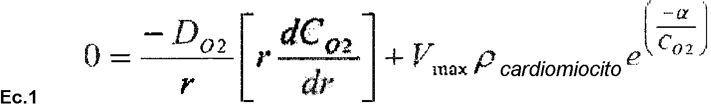
Co2 - concentración de oxígeno en función de la posición radial, r - posición radial en el cilindro, Do2 - constante de difusión de oxígeno, Vmax - velocidad de generación de oxígeno máxima por los cardiomiocitos, pcardiomiocito - densidad de cardiomiocitos, a - constante para la dependencia de la velocidad de generación de oxígeno en la concentración de oxígeno.Co2 - oxygen concentration as a function of radial position, r - radial position in the cylinder, Do2 - oxygen diffusion constant, Vmax - maximum oxygen generation rate by cardiomyocytes, pcardiomyocyte - cardiomyocyte density, a - constant for the dependence of the oxygen generation rate on the oxygen concentration.
Tabla 3: Parámetros del modelo de difusión de oxígeno.Table 3: Parameters of the oxygen diffusion model.

5. Brown, et al. Biotechnol Bioeng 97, 962 - 975 (2007).5. Brown, et al. Biotechnol Bioeng 97, 962-975 (2007).
Análisis estadístico.Statistic analysis.
Todos los datos se presentan como media ± s. e. m. Se usaron análisis estadísticos apropiados para cada conjunto de datos como se indica en las leyendas de las Figuras usando Graph Pad Prism o Microsoft Excell.All data are presented as mean ± s. and. m. Appropriate statistical analyzes were used for each data set as indicated in the legends of the Figures using Graph Pad Prism or Microsoft Excell.
Suplemento personalizado para reemplazar B27®.Custom supplement to replace B27®.
Tabla 4: Suplemento personalizado (CMS) para reemplazar B27. Table 4: Custom Supplement (CMS) to replace B27.

Preparar 25x en agua apta para cultivo de células.Prepare 25x in water suitable for cell culture.
LISTA DE REFERENCIASREFERENCE LIST
Kehat, I. et al. Human embryonic stem cells can differentiate into myocytes with structural and functional properties of cardiomyocytes. J Clin Invest 108, 407 -414 (2001).Kehat, I. et al. Human embryonic stem cells can differentiate into myocytes with structural and functional properties of cardiomyocytes. J Clin Invest 108, 407-414 (2001).
Takahashi, K. et al. Induction of pluripotent stem cells from adult human fibroblasts by defined factors. Cell 131, 861 -872 (2007).Takahashi, K. et al. Induction of pluripotent stem cells from adult human fibroblasts by defined factors. Cell 131, 861-872 (2007).
Zhang, J. et al. Functional cardiomyocytes derived from human induced pluripotent stem cells. Ore Res 104, e30 - 41 (2009).Zhang, J. et al. Functional cardiomyocytes derived from human induced pluripotent stem cells. Pray Res 104, e30-41 (2009).
Thomson, J.A. et al. Embryonic stem cell lines derived from human blastocysts. Sci- ence 282, 1145 - 1147 (1998).Thomson, J.A. et al. Embryonic stem cell lines derived from human blastocysts. Sci-en 282, 1145-1147 (1998).
Lian, X., Zhang, J., Zhu, K., Kamp, TJ. & Palecek, S.P. Insulin Inhibits Cardiac Mesoderm, not Mesendoderm, formation during Cardiac Differentiation of Human Pluripotent Stem Cells and Modulation of Canonical Wnt Signaling Can Rescue this Inhibition. Stem Cells 31{3), 447 - 457 (2013).Lian, X., Zhang, J., Zhu, K., Kamp, TJ. & Palecek, S.P. Insulin Inhibits Cardiac Mesoderm, not Mesendoderm, formation during Cardiac Differentiation of Human Pluripotent Stem Cells and Modulation of Canonical Wnt Signaling Can Rescue this Inhibition. Stem Cells 31 {3), 447-457 (2013).
Schaaf, S. et al. Human engineered heart tissue as a versatile tool in basic research and preclinical toxicology. PLoS ONE 6, 20 (2011).Schaaf, S. et al. Human engineered heart tissue as a versatile tool in basic research and preclinical toxicology. PLoS ONE 6, 20 (2011).
Carvajal-Vergara, X. et al. Patient-specific induced pluripotent stem-cell-derived models of LEOPARD syndrome. Nature 465, 808-812 (2010).Carvajal-Vergara, X. et al. Patient-specific induced pluripotent stem-cell-derived models of LEOPARD syndrome. Nature 465, 808-812 (2010).
Itzhaki, I. et al. Modelling the long QT syndrome with induced pluripotent stem cells. Nature 471, 225 - 229 (2011).Itzhaki, I. et al. Modeling the long QT syndrome with induced pluripotent stem cells. Nature 471, 225-229 (2011).
Malan, D., Friedrichs, S., Fleischmann, B.K. & Sasse, P. Cardiomyocytes obtained from induced pluripotent stem cells with long-QT syndrome 3 recapitulate typical disease-specific features In Vitro. Ore Res 109(8), 841-847 (2011).Malan, D., Friedrichs, S., Fleischmann, B.K. & Sasse, P. Cardiomyocytes obtained from induced pluripotent stem cells with long-QT syndrome 3 recapitulate typical disease-specific features In Vitro. Pray Res 109 (8), 841-847 (2011).
Moretti, A. et al. Patient-specific induced p!uri potent stem-cell models for long-QT syndrome. N Engl J Med363, 1397 - 1409 (2010).Moretti, A. et al. Patient-specific induced p! Uri potent stem-cell models for long-QT syndrome. N Engl J Med363, 1397-1409 (2010).
Yazawa, M. et al. Using induced pluripotent stem cells to investigate cardiac phenotypes in Timothy syndrome. Nature 471, 230-234 (2010).Yazawa, M. et al. Using induced pluripotent stem cells to investigate cardiac phenotypes in Timothy syndrome. Nature 471, 230-234 (2010).
Bird, S.D. et al. The human adult cardiomyocyte phenotype. Cardlovasc Res 58, 423 - 434 (2003).Bird, S.D. et al. The human adult cardiomyocyte phenotype. Cardlovasc Res 58, 423-434 (2003).
Eschenhagen, T. et al. Three-dimensional reconstitution of embryonic cardiomyocytes in a collagen matrix: a new heart muscle model system. FASEB J 11,683 - 694 (1997).Eschenhagen, T. et al. Three-dimensional reconstitution of embryonic cardiomyocytes in a collagen matrix: a new heart muscle model system. FASEB J 11,683-694 (1997).
Zimmermann, W.H. et al. Tissue engineering of a differentiated cardiac muscle construct. Ore Res 90, 223-230 (2002). Tulloch, N.L et al. Growth of engineered human myocardium with mechanical loading and vascular coculture. Ore Res 109, 47 - 59 (2011).Zimmermann, W.H. et al. Tissue engineering of a differentiated cardiac muscle construct. Pray Res 90, 223-230 (2002). Tulloch, N.L et al. Growth of engineered human myocardium with mechanical loading and vascular coculture. Pray Res 109, 47-59 (2011).
Tiburcy, M. et al. Terminal differentiation, advanced organotypic maturation, and modeling of hypertrophic growth in engineered heart tissue. Ore Res 109, 1105- 1114 (2011).Tiburcy, M. et al. Terminal differentiation, advanced organotypic maturation, and modeling of hypertrophic growth in engineered heart tissue. Pray Res 109, 1105-1114 (2011).
Eschenhagen, T., Eder, A., Vollert, I. & Hansen, A. Physiological aspects of cardiac tissue engineering. Am J Physiol Heart Circ Physiol 303, 11 (2012). Eschenhagen, T., Eder, A., Vollert, I. & Hansen, A. Physiological aspects of cardiac tissue engineering. Am J Physiol Heart Circ Physiol 303, 11 (2012).
Hudson, J.E., Brooke, G., Blair, C, Wolvetang, E. & Cooper-White, J.J. Development of myocardial constructs using modulus-matched acrylated polypropylene glycol triol substrate and different nonmyocyte cell populations. Tissue Eng Part A 17, 2279- 2289 (2011).Hudson, J.E., Brooke, G., Blair, C, Wolvetang, E. & Cooper-White, J.J. Development of myocardial constructs using modulus-matched acrylated polypropylene glycol triol substrate and different nonmyocyte cell populations. Tissue Eng Part A 17, 2279-2899 (2011).
Hudson, J., Titmarsh, D., Hidalgo, A., Wolvetang, E. & Cooper-White, J. Primitive cardiac cells from human embryonic stem cells. Stem Cells Dev 21, 1513- 1523 (2012).Hudson, J., Titmarsh, D., Hidalgo, A., Wolvetang, E. & Cooper-White, J. Primitive cardiac cells from human embryonic stem cells. Stem Cells Dev 21, 1513-1523 (2012).
Cao, N. et al. Ascorbic acid enhances the cardiac differentiation of induced pluripotent stem cells through promoting the proliferation of cardiac progenitor cells. Cell Res 22, 219 - 236 (2012).Cao, N. et al. Ascorbic acid enhances the cardiac differentiation of induced pluripotent stem cells through promoting the proliferation of cardiac progenitor cells. Cell Res 22, 219-236 (2012).
Kruithof, B.P., Duim, S.N., Moerkamp, AT. & Goumans, M.J. TGFbeta and BMP signaling in cardiac cushion formation: lessons from mice and chicken. Differentiation 84, 89- 102 (2012).Kruithof, B.P., Duim, S.N., Moerkamp, AT. & Goumans, M.J. TGFbeta and BMP signaling in cardiac cushion formation: lessons from mice and chicken. Differentiation 84, 89-102 (2012).
Brown, D.A. et al. Analysis of oxygen transport in a diffusion-limited model of engineered heart tissue. Biotechnol Bioeng 97, 962 - 975 (2007).Brown, D.A. et al. Analysis of oxygen transport in a diffusion-limited model of engineered heart tissue. Biotechnol Bioeng 97, 962-975 (2007).
Ellerstrom, C, Strehl, R., Noaksson, K., Hyllner, J. & Semb, H. Facilitated expansion of human embryonic stem cells by single-cell enzymatic dissociation. Stem Cells 25, 1690 - 1696 (2007).Ellerstrom, C, Strehl, R., Noaksson, K., Hyllner, J. & Semb, H. Facilitated expansion of human embryonic stem cells by single-cell enzymatic dissociation. Stem Cells 25, 1690-1696 (2007).
Chan, E.M. et al. Live cell imaging distinguishes bona fide human iPS cells from partially reprogrammed cells. Nat Biotechnol 27, 1033 - 1037 (2009).Chan, E.M. et al. Live cell imaging distinguishes bona fide human iPS cells from partially reprogrammed cells. Nat Biotechnol 27, 1033-1037 (2009).
Freberg, C.T., Dahl, J.A., Timoskainen, S. & Collas, P. Epigenetic reprogramming of OCT4 and NANOG regulatory regions by embryonal carcinoma cell extract. Mol Biol Cell 18, 1543 - 1553 (2007).Freberg, C.T., Dahl, J.A., Timoskainen, S. & Collas, P. Epigenetic reprogramming of OCT4 and NANOG regulatory regions by embryonal carcinoma cell extract. Mol Biol Cell 18, 1543-1553 (2007).
Campos, P.B., Sartore, R.C., Abdalla, S.N. & Rehen, S.K. Chromosomal spread preparation of human embryonic stem cells for karyotyping. J Vis Exp 4 (2009).Campos, P.B., Sartore, R.C., Abdalla, S.N. & Rehen, S.K. Chromosomal spread preparation of human embryonic stem cells for karyotyping. J Vis Exp 4 (2009).
Kattman, S.J. et al. Stage-specific optimization of activin/nodal and BMP signaling promotes cardiac differentiation of mouse and human pluripotent stem cell lines. Cell Stem Cell, 8, 228 - 240 (2011).Kattman, S.J. et al. Stage-specific optimization of activin / nodal and BMP signaling promote cardiac differentiation of mouse and human pluripotent stem cell lines. Cell Stem Cell, 8, 228-240 (2011).
Neagoe, C. et al. Titin isofomn switch in ischemic human heart disease. Circulation 106, 1333 - 1341 (2002).Neagoe, C. et al. Titin isofomn switch in ischemic human heart disease. Circulation 106, 1333-1341 (2002).
Park, I.H. et al. Reprogramming of human somatic cells to pluripotency with defined factors. Nature 451, 141 - 146 (2008).Park, I.H. et al. Reprogramming of human somatic cells to pluripotency with defined factors. Nature 451, 141-146 (2008).
Thomson, J.A., et al. Embryonic stem cell lines derived from human blastocysts. Science 282, 1145 - 1147 (1998).Thomson, J.A., et al. Embryonic stem cell lines derived from human blastocysts. Science 282, 1145-1147 (1998).
Takahashi, K. & Yamanaka, S. Induction of pluripotent stem cells from mouse embryonic and adult fibroblast cultures by defined factors. Cell 126, 663 - 676 (2006).Takahashi, K. & Yamanaka, S. Induction of pluripotent stem cells from mouse embryonic and adult fibroblast cultures by defined factors. Cell 126, 663-676 (2006).
Didié et al. Parthenogenetic stem cells for tissue-engineered heart repair. J Clin Invest. 123, 1285 - 1298 (2013).Didié et al. Parthenogenetic stem cells for tissue-engineered heart repair. J Clin Invest. 123, 1285-1298 (2013).
Irion, S et al. Identification and targeting of the ROSA26 locus in human embryonic stem cells. Nat Biotechnol 25, 1477 - 1482 (2007)Irion, S et al. Identification and targeting of the ROSA26 locus in human embryonic stem cells. Nat Biotechnol 25, 1477-1482 (2007)
Naito, H. et al. Optimizing engineered heart tissue for therapeutic applications as surrogate heart muscle. Circulation 114, 172-78 (2006).Naito, H. et al. Optimizing engineered heart tissue for therapeutic applications as surrogate heart muscle. Circulation 114, 172-78 (2006).
Zimmermann W.H. et al. Three-dimensional engineered heart tissue from neonatal rat cardiac myocytes. Biotechnol Bioeng 68, 106- 114 (2000).Zimmermann W.H. et al. Three-dimensional engineered heart tissue from neonatal rat cardiac myocytes. Biotechnol Bioeng 68, 106-114 (2000).
Zimmermann W.H. et al. Engineered heart tissue grafts improve systolic and diastolic function in infarcted rat hearts. Nat Med 12, 452-458 (2006). Zimmermann W.H. et al. Engineered heart tissue grafts improve systolic and diastolic function in infarcted rat hearts. Nat Med 12, 452-458 (2006).
Claims (12)
Applications Claiming Priority (2)
| Application Number | Priority Date | Filing Date | Title |
|---|---|---|---|
| EP13185344 | 2013-09-20 | ||
| PCT/EP2014/069951 WO2015040142A1 (en) | 2013-09-20 | 2014-09-19 | A method to direct differentiation of pluripotent stem cells into functional heart muscle |
Publications (1)
| Publication Number | Publication Date |
|---|---|
| ES2741806T3 true ES2741806T3 (en) | 2020-02-12 |
Family
ID=49261445
Family Applications (1)
| Application Number | Title | Priority Date | Filing Date |
|---|---|---|---|
| ES14767019T Active ES2741806T3 (en) | 2013-09-20 | 2014-09-19 | A method for direct differentiation of pluripotent stem cells into functional heart muscle |
Country Status (10)
| Country | Link |
|---|---|
| US (3) | US10329532B2 (en) |
| EP (2) | EP3047019B1 (en) |
| JP (2) | JP6681825B2 (en) |
| CN (2) | CN105849253B (en) |
| AU (1) | AU2014323098B2 (en) |
| CA (1) | CA2924816C (en) |
| DK (1) | DK3047019T3 (en) |
| ES (1) | ES2741806T3 (en) |
| SG (3) | SG11201601798RA (en) |
| WO (1) | WO2015040142A1 (en) |
Families Citing this family (32)
| Publication number | Priority date | Publication date | Assignee | Title |
|---|---|---|---|---|
| CA2949171A1 (en) | 2014-05-16 | 2015-11-19 | Koninklijke Nederlandse Akademie Van Wetenschappen | Improved culture method for organoids |
| EP3964565A1 (en) * | 2015-09-01 | 2022-03-09 | Ncardia B.V. | An in vitro method of differentiating a human pluripotent stem cell population into a cardiomyocyte cell population |
| KR101781693B1 (en) | 2015-09-24 | 2017-09-26 | 서울대학교병원 | Process for differentiation of cardiovascular cells from induced pluripotent stem cells prepared by human circulation multipotent adult stem cells(CiMS) and uses thereof |
| GB201603569D0 (en) * | 2016-03-01 | 2016-04-13 | Koninklijke Nederlandse Akademie Van Wetenschappen | Improved differentiation method |
| EP3430127A4 (en) * | 2016-03-18 | 2019-09-04 | Institute of Biophysics Chinese Academy of Sciences | A cell culture medium and culture medium supplement |
| CN106244526B (en) * | 2016-08-23 | 2020-09-11 | 北京赛贝生物技术有限公司 | Myocardial differentiation kit and culture method of myocardial cells |
| CN106244527B (en) * | 2016-08-29 | 2019-10-08 | 广东依浦赛生物科技有限公司 | Source of people iPS stem cell in vitro directed differentiation is the kit and method of cardiac muscle cell |
| US20190336537A1 (en) * | 2016-12-04 | 2019-11-07 | University Health Network | Generating atrial and ventricular cardiomyocyte lineages from human pluripotent stem cells |
| GB201621594D0 (en) * | 2016-12-19 | 2017-02-01 | Univ Leuven Kath | Novel cell based methods for healing of bone, cartilage and joint disorders |
| US20210130785A1 (en) | 2017-01-26 | 2021-05-06 | Osaka University | Medium for inducing differentiation of stem cells into mesodermal cells and method for producing mesodermal cells |
| CN106867961A (en) * | 2017-03-27 | 2017-06-20 | 广州润虹医药科技有限公司 | A kind of induced multi-potent stem cell forms mesoblastemic inducing culture and abductive approach |
| CN108456654A (en) * | 2018-02-22 | 2018-08-28 | 上海产业技术研究院 | Serum free culture system for promoting source of human stem cell cardiac muscle cell maturation |
| EP3540046A1 (en) * | 2018-03-13 | 2019-09-18 | Medizinische Hochschule Hannover | Process for producing cardiac organoids |
| CA3105897A1 (en) | 2018-07-13 | 2020-01-16 | Lothar Germeroth | Non-immunogenic engineered tissue and methods of producing and using the same |
| CN111214703B (en) * | 2018-11-25 | 2021-06-15 | 中国科学院大连化学物理研究所 | iPS-derived myocardial cell composite patch and preparation and application thereof |
| CN109619087A (en) * | 2018-11-27 | 2019-04-16 | 山东奥博森生物药业股份有限公司 | A kind of small intestinal mucosa stem cell cryopreserving liquid and cryopreservation methods |
| KR20200102329A (en) * | 2019-02-21 | 2020-08-31 | 고려대학교 산학협력단 | Method for Differentiation of Stem Cells to Mature Subtype-specific Cardiomyocytes Using TGFβ1 |
| CN113728092A (en) | 2019-02-21 | 2021-11-30 | 斯特姆生物系统有限公司 | Method for maturation of cardiomyocytes on amniotic fluid cell derived ECM, cell construct and use for cardiotoxicity and proarrhythmia screening of pharmaceutical compounds |
| CN109749986B (en) * | 2019-03-13 | 2021-04-02 | 武汉大学 | Method for obtaining pancreatic precursor cells and pancreatic beta cells by differentiation of human pluripotent stem cells |
| CN114144515A (en) * | 2019-07-12 | 2022-03-04 | 基因组生物制品Ug有限责任公司 | Method for producing cardiac tissue mimetics |
| CN110511904B (en) * | 2019-08-21 | 2021-03-19 | 云南中科灵长类生物医学重点实验室 | Culture medium, culture method and application of pluripotent stem cells |
| EP3835403A1 (en) | 2019-12-11 | 2021-06-16 | Sartorius Stedim Biotech GmbH | Automated medium exchange strategy for suspension cells |
| KR102291265B1 (en) * | 2020-03-19 | 2021-08-18 | 연세대학교 산학협력단 | Cardiac organoids, method for preparing the same and method for evaluating drug toxicity using the same |
| WO2021197334A1 (en) * | 2020-03-31 | 2021-10-07 | 浙江养生堂天然药物研究所有限公司 | Pharmaceutical combination and use thereof |
| EP3945133A1 (en) | 2020-07-29 | 2022-02-02 | Georg-August-Universität Göttingen | Mass production of human pluripotent stem cell derived cardiac stromal cell |
| DK3988138T3 (en) | 2020-10-22 | 2024-02-26 | Georg August Univ Goettingen Stiftung Oeffentlichen Rechts Univsmedizin | MULTILAYERED ARTIFICIAL HEART MUSCLE |
| JP2024504334A (en) | 2021-01-21 | 2024-01-31 | ザルトリウス ステディム バイオテック ゲーエムベーハー | How to change the culture medium of a culture using a spin filter |
| CN116888470A (en) | 2021-01-21 | 2023-10-13 | 赛多利斯斯泰帝生物技术有限公司 | Application of dielectric constant measurement probe in suspension culture aggregate containing cell aggregate |
| CN113249310B (en) * | 2021-05-17 | 2022-10-11 | 湖北大学 | Method for induced differentiation of expansion pluripotent stem cells into myocardial cells and application |
| CN113337459B (en) * | 2021-06-02 | 2022-09-06 | 呈诺再生医学科技(珠海横琴新区)有限公司 | Method for improving differentiation efficiency of pluripotent stem cells |
| US20230147883A1 (en) * | 2021-11-28 | 2023-05-11 | Fatemeh Etezadi | Method for prodcing cardiomyocytes from induced pluripotent stem cells in an integrated process |
| WO2024008773A1 (en) * | 2022-07-05 | 2024-01-11 | Repairon Gmbh | Differentiation of ipscs in bioreactors |
Family Cites Families (14)
| Publication number | Priority date | Publication date | Assignee | Title |
|---|---|---|---|---|
| US20040014209A1 (en) * | 2002-01-23 | 2004-01-22 | Lassar Andrew B. | Compositions and methods for modulating cell differentiation |
| EP1783206A1 (en) * | 2005-11-08 | 2007-05-09 | Universitätsklinikum Hamburg-Eppendorf | Multiring engineered heart tissue construct |
| JP2008307007A (en) * | 2007-06-15 | 2008-12-25 | Bayer Schering Pharma Ag | Human pluripotent stem cell induced from human tissue-originated undifferentiated stem cell after birth |
| CN101918541A (en) * | 2007-07-18 | 2010-12-15 | 塞拉提斯股份公司 | Cardiomyocyte-like cell clusters derived from hBS cells |
| EP2100954A1 (en) * | 2008-03-10 | 2009-09-16 | Assistance Publique - Hopitaux de Paris | Method for generating primate cardiac progenitor cells for clinical use from primate embryonic stem cells, and their applications |
| AU2009274517B2 (en) * | 2008-07-25 | 2015-03-26 | The University Of Georgia Research Foundation, Inc. | Compositions for Mesoderm derived ISL1+ Multipotent cells (IMPs), epicardial progenitor cells (EPCs) and multipotent CXCR4+CD56+ cells (C56Cs) and methods of use |
| US8765117B2 (en) * | 2009-06-10 | 2014-07-01 | The General Hospital Corporation | Generation of vascularized human heart tissue and uses thereof |
| CN101935635A (en) * | 2010-07-30 | 2011-01-05 | 中国人民解放军第二军医大学 | In vitro amplification method of human marrow pluripotent stem cells and method for directionally differentiating same into dopaminergic neuron |
| US9023340B2 (en) * | 2011-07-02 | 2015-05-05 | Chemregen, Inc. | Wnt inhibitors for human stem cell differentiation |
| US9395354B2 (en) | 2011-07-21 | 2016-07-19 | The Board Of Trustees Of The Leland Stanford Junior University | Cardiomyocytes from induced pluripotent stem cells from patients and methods of use thereof |
| WO2013056072A1 (en) * | 2011-10-13 | 2013-04-18 | Wisconsin Alumni Research Foundation | Generation of cardiomyocytes from human pluripotent stem cells |
| WO2013063305A2 (en) * | 2011-10-25 | 2013-05-02 | Mount Sinai School Of Medicine | Directed cardiomyocyte differentiation of stem cells |
| WO2013111875A1 (en) * | 2012-01-27 | 2013-08-01 | 国立大学法人京都大学 | Method for inducing differentiation of pluripotent stem cell into cardiac muscle |
| ES2615396T3 (en) * | 2013-08-22 | 2017-06-07 | Georg-August-Universität Göttingen Stiftung Öffentlichen Rechts Universitätsmedizin | Method to produce cardiac muscle by engineering (EHM) |
-
2014
- 2014-09-19 CN CN201480063250.8A patent/CN105849253B/en active Active
- 2014-09-19 CA CA2924816A patent/CA2924816C/en active Active
- 2014-09-19 JP JP2016515530A patent/JP6681825B2/en active Active
- 2014-09-19 SG SG11201601798RA patent/SG11201601798RA/en unknown
- 2014-09-19 CN CN202010891948.6A patent/CN112175900A/en active Pending
- 2014-09-19 SG SG10201802167TA patent/SG10201802167TA/en unknown
- 2014-09-19 US US14/917,103 patent/US10329532B2/en active Active
- 2014-09-19 DK DK14767019.4T patent/DK3047019T3/en active
- 2014-09-19 EP EP14767019.4A patent/EP3047019B1/en active Active
- 2014-09-19 EP EP19183577.6A patent/EP3575393A1/en not_active Withdrawn
- 2014-09-19 ES ES14767019T patent/ES2741806T3/en active Active
- 2014-09-19 WO PCT/EP2014/069951 patent/WO2015040142A1/en active Application Filing
- 2014-09-19 AU AU2014323098A patent/AU2014323098B2/en active Active
- 2014-09-19 SG SG10201910753RA patent/SG10201910753RA/en unknown
-
2018
- 2018-09-21 US US16/138,101 patent/US20190010460A1/en not_active Abandoned
-
2019
- 2019-05-20 US US16/416,580 patent/US20190300858A1/en not_active Abandoned
-
2020
- 2020-03-24 JP JP2020052526A patent/JP2020115866A/en active Pending
Also Published As
| Publication number | Publication date |
|---|---|
| AU2014323098B2 (en) | 2020-09-17 |
| US20190300858A1 (en) | 2019-10-03 |
| EP3047019A1 (en) | 2016-07-27 |
| CA2924816A1 (en) | 2015-03-26 |
| US20190010460A1 (en) | 2019-01-10 |
| CA2924816C (en) | 2023-09-05 |
| EP3047019B1 (en) | 2019-07-03 |
| CN112175900A (en) | 2021-01-05 |
| AU2014323098A1 (en) | 2016-05-05 |
| EP3575393A1 (en) | 2019-12-04 |
| CN105849253A (en) | 2016-08-10 |
| JP2020115866A (en) | 2020-08-06 |
| JP2016536975A (en) | 2016-12-01 |
| US20160215264A1 (en) | 2016-07-28 |
| US10329532B2 (en) | 2019-06-25 |
| DK3047019T3 (en) | 2019-08-26 |
| CN105849253B (en) | 2020-10-09 |
| WO2015040142A1 (en) | 2015-03-26 |
| SG10201910753RA (en) | 2020-01-30 |
| SG11201601798RA (en) | 2016-04-28 |
| SG10201802167TA (en) | 2018-04-27 |
| JP6681825B2 (en) | 2020-04-15 |
Similar Documents
| Publication | Publication Date | Title |
|---|---|---|
| ES2741806T3 (en) | A method for direct differentiation of pluripotent stem cells into functional heart muscle | |
| US20210032602A1 (en) | Ex vivo proliferation of epithelial cells | |
| ES2688019T3 (en) | Methods and compositions for accelerated production of retinal pigmented epithelial cells from pluripotent cells | |
| ES2677319T3 (en) | Medium and method of stem cell culture | |
| EP2765188B1 (en) | Method for producing corneal endothelial cell | |
| EP3347450B1 (en) | Ex vivo proliferation of epithelial cells | |
| US20110165130A1 (en) | Methods for Preparing Human Skin Substitutes from Human Pluripotent Stem Cells | |
| CA3007107A1 (en) | Methods of differentiating retinal cells | |
| US20200283726A1 (en) | Process for continuous cell culture of gpscs | |
| CN116769695A (en) | Culture medium and method for producing human cells and tissues from teratomas, organoids and embryoid bodies |