EP3047019B1 - A method to direct differentiation of pluripotent stem cells into functional heart muscle - Google Patents
A method to direct differentiation of pluripotent stem cells into functional heart muscle Download PDFInfo
- Publication number
- EP3047019B1 EP3047019B1 EP14767019.4A EP14767019A EP3047019B1 EP 3047019 B1 EP3047019 B1 EP 3047019B1 EP 14767019 A EP14767019 A EP 14767019A EP 3047019 B1 EP3047019 B1 EP 3047019B1
- Authority
- EP
- European Patent Office
- Prior art keywords
- bhm
- stem cells
- basal medium
- cells
- pluripotent stem
- Prior art date
- Legal status (The legal status is an assumption and is not a legal conclusion. Google has not performed a legal analysis and makes no representation as to the accuracy of the status listed.)
- Active
Links
- 238000000034 method Methods 0.000 title claims description 94
- 230000004069 differentiation Effects 0.000 title claims description 61
- 210000001778 pluripotent stem cell Anatomy 0.000 title claims description 38
- 210000004165 myocardium Anatomy 0.000 title claims description 16
- 210000004027 cell Anatomy 0.000 claims description 117
- 239000007640 basal medium Substances 0.000 claims description 87
- CIWBSHSKHKDKBQ-JLAZNSOCSA-N Ascorbic acid Chemical compound OC[C@H](O)[C@H]1OC(=O)C(O)=C1O CIWBSHSKHKDKBQ-JLAZNSOCSA-N 0.000 claims description 70
- 239000013589 supplement Substances 0.000 claims description 58
- 230000000747 cardiac effect Effects 0.000 claims description 52
- 239000003112 inhibitor Substances 0.000 claims description 51
- NOESYZHRGYRDHS-UHFFFAOYSA-N insulin Chemical compound N1C(=O)C(NC(=O)C(CCC(N)=O)NC(=O)C(CCC(O)=O)NC(=O)C(C(C)C)NC(=O)C(NC(=O)CN)C(C)CC)CSSCC(C(NC(CO)C(=O)NC(CC(C)C)C(=O)NC(CC=2C=CC(O)=CC=2)C(=O)NC(CCC(N)=O)C(=O)NC(CC(C)C)C(=O)NC(CCC(O)=O)C(=O)NC(CC(N)=O)C(=O)NC(CC=2C=CC(O)=CC=2)C(=O)NC(CSSCC(NC(=O)C(C(C)C)NC(=O)C(CC(C)C)NC(=O)C(CC=2C=CC(O)=CC=2)NC(=O)C(CC(C)C)NC(=O)C(C)NC(=O)C(CCC(O)=O)NC(=O)C(C(C)C)NC(=O)C(CC(C)C)NC(=O)C(CC=2NC=NC=2)NC(=O)C(CO)NC(=O)CNC2=O)C(=O)NCC(=O)NC(CCC(O)=O)C(=O)NC(CCCNC(N)=N)C(=O)NCC(=O)NC(CC=3C=CC=CC=3)C(=O)NC(CC=3C=CC=CC=3)C(=O)NC(CC=3C=CC(O)=CC=3)C(=O)NC(C(C)O)C(=O)N3C(CCC3)C(=O)NC(CCCCN)C(=O)NC(C)C(O)=O)C(=O)NC(CC(N)=O)C(O)=O)=O)NC(=O)C(C(C)CC)NC(=O)C(CO)NC(=O)C(C(C)O)NC(=O)C1CSSCC2NC(=O)C(CC(C)C)NC(=O)C(NC(=O)C(CCC(N)=O)NC(=O)C(CC(N)=O)NC(=O)C(NC(=O)C(N)CC=1C=CC=CC=1)C(C)C)CC1=CN=CN1 NOESYZHRGYRDHS-UHFFFAOYSA-N 0.000 claims description 40
- 239000002609 medium Substances 0.000 claims description 35
- 235000010323 ascorbic acid Nutrition 0.000 claims description 34
- 239000011668 ascorbic acid Substances 0.000 claims description 34
- 230000000638 stimulation Effects 0.000 claims description 34
- 108090000379 Fibroblast growth factor 2 Proteins 0.000 claims description 33
- 241000282414 Homo sapiens Species 0.000 claims description 30
- 229960005070 ascorbic acid Drugs 0.000 claims description 23
- 150000003839 salts Chemical class 0.000 claims description 22
- 102000004877 Insulin Human genes 0.000 claims description 20
- 108090001061 Insulin Proteins 0.000 claims description 20
- 229940125396 insulin Drugs 0.000 claims description 20
- 230000035800 maturation Effects 0.000 claims description 20
- 239000006144 Dulbecco’s modified Eagle's medium Substances 0.000 claims description 17
- 238000010899 nucleation Methods 0.000 claims description 17
- 102100024505 Bone morphogenetic protein 4 Human genes 0.000 claims description 16
- 102000008186 Collagen Human genes 0.000 claims description 16
- 108010035532 Collagen Proteins 0.000 claims description 16
- 101000762379 Homo sapiens Bone morphogenetic protein 4 Proteins 0.000 claims description 16
- 229920001436 collagen Polymers 0.000 claims description 16
- 210000003716 mesoderm Anatomy 0.000 claims description 15
- 239000006146 Roswell Park Memorial Institute medium Substances 0.000 claims description 14
- 230000004156 Wnt signaling pathway Effects 0.000 claims description 14
- IDDDVXIUIXWAGJ-DDSAHXNVSA-N 4-[(1r)-1-aminoethyl]-n-pyridin-4-ylcyclohexane-1-carboxamide;dihydrochloride Chemical compound Cl.Cl.C1CC([C@H](N)C)CCC1C(=O)NC1=CC=NC=C1 IDDDVXIUIXWAGJ-DDSAHXNVSA-N 0.000 claims description 13
- AQGNHMOJWBZFQQ-UHFFFAOYSA-N CT 99021 Chemical compound CC1=CNC(C=2C(=NC(NCCNC=3N=CC(=CC=3)C#N)=NC=2)C=2C(=CC(Cl)=CC=2)Cl)=N1 AQGNHMOJWBZFQQ-UHFFFAOYSA-N 0.000 claims description 13
- 108010023082 activin A Proteins 0.000 claims description 12
- JYGXADMDTFJGBT-VWUMJDOOSA-N hydrocortisone Chemical compound O=C1CC[C@]2(C)[C@H]3[C@@H](O)C[C@](C)([C@@](CC4)(O)C(=O)CO)[C@@H]4[C@@H]3CCC2=C1 JYGXADMDTFJGBT-VWUMJDOOSA-N 0.000 claims description 12
- 210000000130 stem cell Anatomy 0.000 claims description 12
- HZAXFHJVJLSVMW-UHFFFAOYSA-N 2-Aminoethan-1-ol Chemical compound NCCO HZAXFHJVJLSVMW-UHFFFAOYSA-N 0.000 claims description 10
- 210000001671 embryonic stem cell Anatomy 0.000 claims description 10
- 230000003068 static effect Effects 0.000 claims description 10
- -1 αMEM Substances 0.000 claims description 9
- 102000003974 Fibroblast growth factor 2 Human genes 0.000 claims description 8
- 230000001939 inductive effect Effects 0.000 claims description 8
- AWDORCFLUJZUQS-ZDUSSCGKSA-N (S)-2-methyl-1-(4-methylisoquinoline-5-sulfonyl)-1,4-diazepane Chemical compound C[C@H]1CNCCCN1S(=O)(=O)C1=CC=CC2=CN=CC(C)=C12 AWDORCFLUJZUQS-ZDUSSCGKSA-N 0.000 claims description 7
- LCTONWCANYUPML-UHFFFAOYSA-M Pyruvate Chemical compound CC(=O)C([O-])=O LCTONWCANYUPML-UHFFFAOYSA-M 0.000 claims description 7
- 210000004263 induced pluripotent stem cell Anatomy 0.000 claims description 7
- 238000004519 manufacturing process Methods 0.000 claims description 7
- 108010088751 Albumins Proteins 0.000 claims description 6
- 102000009027 Albumins Human genes 0.000 claims description 6
- 102100030074 Dickkopf-related protein 1 Human genes 0.000 claims description 6
- 101000864646 Homo sapiens Dickkopf-related protein 1 Proteins 0.000 claims description 6
- REFJWTPEDVJJIY-UHFFFAOYSA-N Quercetin Chemical compound C=1C(O)=CC(O)=C(C(C=2O)=O)C=1OC=2C1=CC=C(O)C(O)=C1 REFJWTPEDVJJIY-UHFFFAOYSA-N 0.000 claims description 6
- 102000004338 Transferrin Human genes 0.000 claims description 6
- 108090000901 Transferrin Proteins 0.000 claims description 6
- KLGQSVMIPOVQAX-UHFFFAOYSA-N XAV939 Chemical compound N=1C=2CCSCC=2C(O)=NC=1C1=CC=C(C(F)(F)F)C=C1 KLGQSVMIPOVQAX-UHFFFAOYSA-N 0.000 claims description 6
- JXXCENBLGFBQJM-FYZOBXCZSA-N [(2r)-3-carboxy-2-hydroxypropyl]-trimethylazanium;chloride Chemical compound [Cl-].C[N+](C)(C)C[C@H](O)CC(O)=O JXXCENBLGFBQJM-FYZOBXCZSA-N 0.000 claims description 6
- 235000014113 dietary fatty acids Nutrition 0.000 claims description 6
- BVTBRVFYZUCAKH-UHFFFAOYSA-L disodium selenite Chemical compound [Na+].[Na+].[O-][Se]([O-])=O BVTBRVFYZUCAKH-UHFFFAOYSA-L 0.000 claims description 6
- 229930195729 fatty acid Natural products 0.000 claims description 6
- 239000000194 fatty acid Substances 0.000 claims description 6
- 150000004665 fatty acids Chemical class 0.000 claims description 6
- 229960000890 hydrocortisone Drugs 0.000 claims description 6
- 239000011781 sodium selenite Substances 0.000 claims description 6
- 229960001471 sodium selenite Drugs 0.000 claims description 6
- 235000015921 sodium selenite Nutrition 0.000 claims description 6
- 239000012581 transferrin Substances 0.000 claims description 6
- BLPUCTSDPCUHQQ-NEAWWFDSSA-N 4dvi Chemical compound C1=CC=C2C(C)=CC=NC2=C1NC(=O)C(C=C1)=CC=C1N1C(=O)[C@@H]2[C@H](C=C3)C[C@H]3[C@@H]2C1=O BLPUCTSDPCUHQQ-NEAWWFDSSA-N 0.000 claims description 5
- ZGSXEXBYLJIOGF-ALFLXDJESA-N IWR-1-endo Chemical compound C=1C=CC2=CC=CN=C2C=1NC(=O)C(C=C1)=CC=C1N1C(=O)[C@@H]2[C@H](C=C3)C[C@H]3[C@@H]2C1=O ZGSXEXBYLJIOGF-ALFLXDJESA-N 0.000 claims description 5
- WRKPZSMRWPJJDH-UHFFFAOYSA-N N-(6-methyl-1,3-benzothiazol-2-yl)-2-[(4-oxo-3-phenyl-6,7-dihydrothieno[3,2-d]pyrimidin-2-yl)thio]acetamide Chemical compound S1C2=CC(C)=CC=C2N=C1NC(=O)CSC1=NC=2CCSC=2C(=O)N1C1=CC=CC=C1 WRKPZSMRWPJJDH-UHFFFAOYSA-N 0.000 claims description 5
- 229940031098 ethanolamine Drugs 0.000 claims description 5
- NGOGFTYYXHNFQH-UHFFFAOYSA-N fasudil Chemical compound C=1C=CC2=CN=CC=C2C=1S(=O)(=O)N1CCCNCC1 NGOGFTYYXHNFQH-UHFFFAOYSA-N 0.000 claims description 5
- 229960002435 fasudil Drugs 0.000 claims description 5
- ZAVGJDAFCZAWSZ-UHFFFAOYSA-N hydroxyfasudil Chemical compound C1=CC=C2C(O)=NC=CC2=C1S(=O)(=O)N1CCCNCC1 ZAVGJDAFCZAWSZ-UHFFFAOYSA-N 0.000 claims description 5
- DOBKQCZBPPCLEG-UHFFFAOYSA-N n-benzyl-2-(pyrimidin-4-ylamino)-1,3-thiazole-4-carboxamide Chemical compound C=1SC(NC=2N=CN=CC=2)=NC=1C(=O)NCC1=CC=CC=C1 DOBKQCZBPPCLEG-UHFFFAOYSA-N 0.000 claims description 5
- 230000001776 parthenogenetic effect Effects 0.000 claims description 5
- 230000001737 promoting effect Effects 0.000 claims description 4
- HQWTUOLCGKIECB-XZWHSSHBSA-N (6S,9aS)-6-[(4-hydroxyphenyl)methyl]-8-(1-naphthalenylmethyl)-4,7-dioxo-N-(phenylmethyl)-3,6,9,9a-tetrahydro-2H-pyrazino[1,2-a]pyrimidine-1-carboxamide Chemical compound C1=CC(O)=CC=C1C[C@H]1C(=O)N(CC=2C3=CC=CC=C3C=CC=2)C[C@H]2N1C(=O)CCN2C(=O)NCC1=CC=CC=C1 HQWTUOLCGKIECB-XZWHSSHBSA-N 0.000 claims description 3
- GDVRVPIXWXOKQO-UHFFFAOYSA-N 1-[(3-hydroxyphenyl)methyl]-3-(4-pyridin-4-yl-1,3-thiazol-2-yl)urea Chemical compound OC1=CC=CC(CNC(=O)NC=2SC=C(N=2)C=2C=CN=CC=2)=C1 GDVRVPIXWXOKQO-UHFFFAOYSA-N 0.000 claims description 3
- XSLHNXBPPDZDAU-UHFFFAOYSA-M 2-[(e)-2-(2,5-dimethyl-1-phenylpyrrol-3-yl)ethenyl]-n,n,1-trimethylquinolin-1-ium-6-amine;hydroxide Chemical compound [OH-].C1=CC2=CC(N(C)C)=CC=C2[N+](C)=C1\C=C\C(=C1C)C=C(C)N1C1=CC=CC=C1 XSLHNXBPPDZDAU-UHFFFAOYSA-M 0.000 claims description 3
- JCSGFHVFHSKIJH-UHFFFAOYSA-N 3-(2,4-dichlorophenyl)-4-(1-methyl-3-indolyl)pyrrole-2,5-dione Chemical compound C12=CC=CC=C2N(C)C=C1C(C(NC1=O)=O)=C1C1=CC=C(Cl)C=C1Cl JCSGFHVFHSKIJH-UHFFFAOYSA-N 0.000 claims description 3
- VPVLEBIVXZSOMQ-UHFFFAOYSA-N 3-[[6-(3-aminophenyl)-7H-pyrrolo[2,3-d]pyrimidin-4-yl]oxy]phenol Chemical compound NC1=CC=CC(C=2NC3=NC=NC(OC=4C=C(O)C=CC=4)=C3C=2)=C1 VPVLEBIVXZSOMQ-UHFFFAOYSA-N 0.000 claims description 3
- MDZCSIDIPDZWKL-UHFFFAOYSA-N CHIR-98014 Chemical compound C1=C([N+]([O-])=O)C(N)=NC(NCCNC=2N=C(C(=CN=2)N2C=NC=C2)C=2C(=CC(Cl)=CC=2)Cl)=C1 MDZCSIDIPDZWKL-UHFFFAOYSA-N 0.000 claims description 3
- HRJWTAWVFDCTGO-UHFFFAOYSA-N LY-2090314 Chemical compound C1CN(C=23)C=C(C=4C(NC(=O)C=4C=4N5C=CC=CC5=NC=4)=O)C3=CC(F)=CC=2CN1C(=O)N1CCCCC1 HRJWTAWVFDCTGO-UHFFFAOYSA-N 0.000 claims description 3
- OLIIUAHHAZEXEX-UHFFFAOYSA-N N-(6-fluoro-1H-indazol-5-yl)-6-methyl-2-oxo-4-[4-(trifluoromethyl)phenyl]-3,4-dihydro-1H-pyridine-5-carboxamide Chemical compound C1C(=O)NC(C)=C(C(=O)NC=2C(=CC=3NN=CC=3C=2)F)C1C1=CC=C(C(F)(F)F)C=C1 OLIIUAHHAZEXEX-UHFFFAOYSA-N 0.000 claims description 3
- 241000288906 Primates Species 0.000 claims description 3
- ZVOLCUVKHLEPEV-UHFFFAOYSA-N Quercetagetin Natural products C1=C(O)C(O)=CC=C1C1=C(O)C(=O)C2=C(O)C(O)=C(O)C=C2O1 ZVOLCUVKHLEPEV-UHFFFAOYSA-N 0.000 claims description 3
- HWTZYBCRDDUBJY-UHFFFAOYSA-N Rhynchosin Natural products C1=C(O)C(O)=CC=C1C1=C(O)C(=O)C2=CC(O)=C(O)C=C2O1 HWTZYBCRDDUBJY-UHFFFAOYSA-N 0.000 claims description 3
- PQCXVIPXISBFPN-UHFFFAOYSA-N SB 415286 Chemical compound C1=C(Cl)C(O)=CC=C1NC1=C(C=2C(=CC=CC=2)[N+]([O-])=O)C(=O)NC1=O PQCXVIPXISBFPN-UHFFFAOYSA-N 0.000 claims description 3
- MWDZOUNAPSSOEL-UHFFFAOYSA-N kaempferol Natural products OC1=C(C(=O)c2cc(O)cc(O)c2O1)c3ccc(O)cc3 MWDZOUNAPSSOEL-UHFFFAOYSA-N 0.000 claims description 3
- 229960002778 pyrvinium Drugs 0.000 claims description 3
- 229960001285 quercetin Drugs 0.000 claims description 3
- 235000005875 quercetin Nutrition 0.000 claims description 3
- PMJIHLSCWIDGMD-UHFFFAOYSA-N tideglusib Chemical compound O=C1SN(C=2C3=CC=CC=C3C=CC=2)C(=O)N1CC1=CC=CC=C1 PMJIHLSCWIDGMD-UHFFFAOYSA-N 0.000 claims description 3
- 229950005284 tideglusib Drugs 0.000 claims description 3
- 108020004414 DNA Proteins 0.000 description 80
- 238000002474 experimental method Methods 0.000 description 62
- 210000004413 cardiac myocyte Anatomy 0.000 description 55
- 238000003753 real-time PCR Methods 0.000 description 46
- OYPRJOBELJOOCE-UHFFFAOYSA-N Calcium Chemical compound [Ca] OYPRJOBELJOOCE-UHFFFAOYSA-N 0.000 description 41
- 239000011575 calcium Substances 0.000 description 41
- 229910052791 calcium Inorganic materials 0.000 description 41
- 230000001965 increasing effect Effects 0.000 description 31
- 230000036461 convulsion Effects 0.000 description 30
- 230000014509 gene expression Effects 0.000 description 30
- 230000008602 contraction Effects 0.000 description 29
- 102100035290 Fibroblast growth factor 13 Human genes 0.000 description 25
- 210000002536 stromal cell Anatomy 0.000 description 23
- 230000004044 response Effects 0.000 description 21
- 210000001519 tissue Anatomy 0.000 description 21
- 238000000684 flow cytometry Methods 0.000 description 19
- 230000018109 developmental process Effects 0.000 description 17
- MIJPAVRNWPDMOR-ZAFYKAAXSA-N L-ascorbic acid 2-phosphate Chemical compound OC[C@H](O)[C@H]1OC(=O)C(OP(O)(O)=O)=C1O MIJPAVRNWPDMOR-ZAFYKAAXSA-N 0.000 description 16
- 102100035423 POU domain, class 5, transcription factor 1 Human genes 0.000 description 16
- 101710126211 POU domain, class 5, transcription factor 1 Proteins 0.000 description 15
- 108010022452 Collagen Type I Proteins 0.000 description 14
- 102000012422 Collagen Type I Human genes 0.000 description 14
- 238000011161 development Methods 0.000 description 14
- 102000010825 Actinin Human genes 0.000 description 13
- 108010063503 Actinin Proteins 0.000 description 13
- 230000000903 blocking effect Effects 0.000 description 13
- 239000000872 buffer Substances 0.000 description 13
- LFQSCWFLJHTTHZ-UHFFFAOYSA-N Ethanol Chemical compound CCO LFQSCWFLJHTTHZ-UHFFFAOYSA-N 0.000 description 12
- 238000004458 analytical method Methods 0.000 description 12
- 230000006870 function Effects 0.000 description 12
- 230000006698 induction Effects 0.000 description 12
- 229910052760 oxygen Inorganic materials 0.000 description 12
- 239000001301 oxygen Substances 0.000 description 12
- JWZZKOKVBUJMES-UHFFFAOYSA-N (+-)-Isoprenaline Chemical compound CC(C)NCC(O)C1=CC=C(O)C(O)=C1 JWZZKOKVBUJMES-UHFFFAOYSA-N 0.000 description 11
- 229940072107 ascorbate Drugs 0.000 description 11
- 230000015572 biosynthetic process Effects 0.000 description 11
- XHBVYDAKJHETMP-UHFFFAOYSA-N dorsomorphin Chemical compound C=1C=C(C2=CN3N=CC(=C3N=C2)C=2C=CN=CC=2)C=CC=1OCCN1CCCCC1 XHBVYDAKJHETMP-UHFFFAOYSA-N 0.000 description 11
- 230000000694 effects Effects 0.000 description 11
- 229960001317 isoprenaline Drugs 0.000 description 11
- 230000011664 signaling Effects 0.000 description 11
- 101000629402 Homo sapiens Mesoderm posterior protein 1 Proteins 0.000 description 10
- 101000843569 Homo sapiens Transcription factor HES-3 Proteins 0.000 description 10
- 102100026822 Mesoderm posterior protein 1 Human genes 0.000 description 10
- 102100026260 Titin Human genes 0.000 description 10
- 102100030773 Transcription factor HES-3 Human genes 0.000 description 10
- QVGXLLKOCUKJST-UHFFFAOYSA-N atomic oxygen Chemical compound [O] QVGXLLKOCUKJST-UHFFFAOYSA-N 0.000 description 10
- 235000020776 essential amino acid Nutrition 0.000 description 10
- 239000003797 essential amino acid Substances 0.000 description 10
- 230000007246 mechanism Effects 0.000 description 10
- 101000843572 Homo sapiens Transcription factor HES-2 Proteins 0.000 description 9
- 241000699666 Mus <mouse, genus> Species 0.000 description 9
- 102100030772 Transcription factor HES-2 Human genes 0.000 description 9
- 210000002950 fibroblast Anatomy 0.000 description 9
- 102100024208 Homeobox protein MIXL1 Human genes 0.000 description 8
- 101001052462 Homo sapiens Homeobox protein MIXL1 Proteins 0.000 description 8
- 230000005764 inhibitory process Effects 0.000 description 8
- 230000000284 resting effect Effects 0.000 description 8
- UCSJYZPVAKXKNQ-HZYVHMACSA-N streptomycin Chemical compound CN[C@H]1[C@H](O)[C@@H](O)[C@H](CO)O[C@H]1O[C@@H]1[C@](C=O)(O)[C@H](C)O[C@H]1O[C@@H]1[C@@H](NC(N)=N)[C@H](O)[C@@H](NC(N)=N)[C@H](O)[C@H]1O UCSJYZPVAKXKNQ-HZYVHMACSA-N 0.000 description 8
- UXVMQQNJUSDDNG-UHFFFAOYSA-L Calcium chloride Chemical compound [Cl-].[Cl-].[Ca+2] UXVMQQNJUSDDNG-UHFFFAOYSA-L 0.000 description 7
- 102100027875 Homeobox protein Nkx-2.5 Human genes 0.000 description 7
- 101000632197 Homo sapiens Homeobox protein Nkx-2.5 Proteins 0.000 description 7
- 101000800116 Homo sapiens Thy-1 membrane glycoprotein Proteins 0.000 description 7
- 102100033523 Thy-1 membrane glycoprotein Human genes 0.000 description 7
- 239000012091 fetal bovine serum Substances 0.000 description 7
- 210000005003 heart tissue Anatomy 0.000 description 7
- 238000001727 in vivo Methods 0.000 description 7
- 239000000203 mixture Substances 0.000 description 7
- 230000000144 pharmacologic effect Effects 0.000 description 7
- MTCFGRXMJLQNBG-REOHCLBHSA-N (2S)-2-Amino-3-hydroxypropansäure Chemical compound OC[C@H](N)C(O)=O MTCFGRXMJLQNBG-REOHCLBHSA-N 0.000 description 6
- ZAINTDRBUHCDPZ-UHFFFAOYSA-M Alexa Fluor 546 Chemical compound [H+].[Na+].CC1CC(C)(C)NC(C(=C2OC3=C(C4=NC(C)(C)CC(C)C4=CC3=3)S([O-])(=O)=O)S([O-])(=O)=O)=C1C=C2C=3C(C(=C(Cl)C=1Cl)C(O)=O)=C(Cl)C=1SCC(=O)NCCCCCC(=O)ON1C(=O)CCC1=O ZAINTDRBUHCDPZ-UHFFFAOYSA-M 0.000 description 6
- 102100031181 Glyceraldehyde-3-phosphate dehydrogenase Human genes 0.000 description 6
- DHMQDGOQFOQNFH-UHFFFAOYSA-N Glycine Chemical compound NCC(O)=O DHMQDGOQFOQNFH-UHFFFAOYSA-N 0.000 description 6
- 101001053263 Homo sapiens Insulin gene enhancer protein ISL-1 Proteins 0.000 description 6
- 206010020880 Hypertrophy Diseases 0.000 description 6
- 102100024392 Insulin gene enhancer protein ISL-1 Human genes 0.000 description 6
- DCXYFEDJOCDNAF-REOHCLBHSA-N L-asparagine Chemical compound OC(=O)[C@@H](N)CC(N)=O DCXYFEDJOCDNAF-REOHCLBHSA-N 0.000 description 6
- WHUUTDBJXJRKMK-VKHMYHEASA-N L-glutamic acid Chemical compound OC(=O)[C@@H](N)CCC(O)=O WHUUTDBJXJRKMK-VKHMYHEASA-N 0.000 description 6
- TWRXJAOTZQYOKJ-UHFFFAOYSA-L Magnesium chloride Chemical compound [Mg+2].[Cl-].[Cl-] TWRXJAOTZQYOKJ-UHFFFAOYSA-L 0.000 description 6
- UIIMBOGNXHQVGW-UHFFFAOYSA-M Sodium bicarbonate Chemical compound [Na+].OC([O-])=O UIIMBOGNXHQVGW-UHFFFAOYSA-M 0.000 description 6
- FAPWRFPIFSIZLT-UHFFFAOYSA-M Sodium chloride Chemical compound [Na+].[Cl-] FAPWRFPIFSIZLT-UHFFFAOYSA-M 0.000 description 6
- 102000013814 Wnt Human genes 0.000 description 6
- 108050003627 Wnt Proteins 0.000 description 6
- 230000008859 change Effects 0.000 description 6
- 238000009792 diffusion process Methods 0.000 description 6
- 108020004445 glyceraldehyde-3-phosphate dehydrogenase Proteins 0.000 description 6
- 239000003102 growth factor Substances 0.000 description 6
- 230000002107 myocardial effect Effects 0.000 description 6
- 238000005457 optimization Methods 0.000 description 6
- 230000008569 process Effects 0.000 description 6
- 229940076788 pyruvate Drugs 0.000 description 6
- 238000003757 reverse transcription PCR Methods 0.000 description 6
- 238000012360 testing method Methods 0.000 description 6
- KFVINGKPXQSPNP-UHFFFAOYSA-N 4-amino-2-[2-(diethylamino)ethyl]-n-propanoylbenzamide Chemical compound CCN(CC)CCC1=CC(N)=CC=C1C(=O)NC(=O)CC KFVINGKPXQSPNP-UHFFFAOYSA-N 0.000 description 5
- 108010085238 Actins Proteins 0.000 description 5
- 102000007469 Actins Human genes 0.000 description 5
- 102000010834 Extracellular Matrix Proteins Human genes 0.000 description 5
- 108010037362 Extracellular Matrix Proteins Proteins 0.000 description 5
- 101000780028 Homo sapiens Natriuretic peptides A Proteins 0.000 description 5
- 101000652324 Homo sapiens Transcription factor SOX-17 Proteins 0.000 description 5
- 101150079937 NEUROD1 gene Proteins 0.000 description 5
- 102100034296 Natriuretic peptides A Human genes 0.000 description 5
- 108700020297 NeuroD Proteins 0.000 description 5
- 102100032063 Neurogenic differentiation factor 1 Human genes 0.000 description 5
- 102100027732 Sarcoplasmic/endoplasmic reticulum calcium ATPase 2 Human genes 0.000 description 5
- 102100030243 Transcription factor SOX-17 Human genes 0.000 description 5
- 239000003814 drug Substances 0.000 description 5
- 210000002744 extracellular matrix Anatomy 0.000 description 5
- 230000000297 inotrophic effect Effects 0.000 description 5
- 210000003205 muscle Anatomy 0.000 description 5
- 108090000623 proteins and genes Proteins 0.000 description 5
- 239000011435 rock Substances 0.000 description 5
- 238000012745 whole-mount immunostaining Methods 0.000 description 5
- 102100036364 Cadherin-2 Human genes 0.000 description 4
- 102100035602 Calsequestrin-2 Human genes 0.000 description 4
- 108091007854 Cdh1/Fizzy-related Proteins 0.000 description 4
- 102000038594 Cdh1/Fizzy-related Human genes 0.000 description 4
- 102100024810 DNA (cytosine-5)-methyltransferase 3B Human genes 0.000 description 4
- 101710123222 DNA (cytosine-5)-methyltransferase 3B Proteins 0.000 description 4
- 208000030453 Drug-Related Side Effects and Adverse reaction Diseases 0.000 description 4
- 102000004190 Enzymes Human genes 0.000 description 4
- 108090000790 Enzymes Proteins 0.000 description 4
- WQZGKKKJIJFFOK-GASJEMHNSA-N Glucose Natural products OC[C@H]1OC(O)[C@H](O)[C@@H](O)[C@@H]1O WQZGKKKJIJFFOK-GASJEMHNSA-N 0.000 description 4
- 101000714537 Homo sapiens Cadherin-2 Proteins 0.000 description 4
- 101000947118 Homo sapiens Calsequestrin-2 Proteins 0.000 description 4
- 101000958741 Homo sapiens Myosin-6 Proteins 0.000 description 4
- 101001030243 Homo sapiens Myosin-7 Proteins 0.000 description 4
- 101000936922 Homo sapiens Sarcoplasmic/endoplasmic reticulum calcium ATPase 2 Proteins 0.000 description 4
- 101000976622 Homo sapiens Zinc finger protein 42 homolog Proteins 0.000 description 4
- 101000702691 Homo sapiens Zinc finger protein SNAI1 Proteins 0.000 description 4
- 102100038319 Myosin-6 Human genes 0.000 description 4
- 102100038934 Myosin-7 Human genes 0.000 description 4
- 241000283973 Oryctolagus cuniculus Species 0.000 description 4
- 229930182555 Penicillin Natural products 0.000 description 4
- JGSARLDLIJGVTE-MBNYWOFBSA-N Penicillin G Chemical compound N([C@H]1[C@H]2SC([C@@H](N2C1=O)C(O)=O)(C)C)C(=O)CC1=CC=CC=C1 JGSARLDLIJGVTE-MBNYWOFBSA-N 0.000 description 4
- KPKZJLCSROULON-QKGLWVMZSA-N Phalloidin Chemical compound N1C(=O)[C@@H]([C@@H](O)C)NC(=O)[C@H](C)NC(=O)[C@H](C[C@@](C)(O)CO)NC(=O)[C@H](C2)NC(=O)[C@H](C)NC(=O)[C@@H]3C[C@H](O)CN3C(=O)[C@@H]1CSC1=C2C2=CC=CC=C2N1 KPKZJLCSROULON-QKGLWVMZSA-N 0.000 description 4
- 102000004912 RYR2 Human genes 0.000 description 4
- 108060007241 RYR2 Proteins 0.000 description 4
- 108010014480 T-box transcription factor 5 Proteins 0.000 description 4
- 102100024755 T-box transcription factor TBX5 Human genes 0.000 description 4
- 206010070863 Toxicity to various agents Diseases 0.000 description 4
- 102100023550 Zinc finger protein 42 homolog Human genes 0.000 description 4
- 102100030917 Zinc finger protein SNAI1 Human genes 0.000 description 4
- 238000000540 analysis of variance Methods 0.000 description 4
- 238000010009 beating Methods 0.000 description 4
- 239000001110 calcium chloride Substances 0.000 description 4
- 229910001628 calcium chloride Inorganic materials 0.000 description 4
- 239000003795 chemical substances by application Substances 0.000 description 4
- 239000003636 conditioned culture medium Substances 0.000 description 4
- 230000001419 dependent effect Effects 0.000 description 4
- 230000001627 detrimental effect Effects 0.000 description 4
- 230000029087 digestion Effects 0.000 description 4
- 229940088598 enzyme Drugs 0.000 description 4
- 239000008103 glucose Substances 0.000 description 4
- 230000004217 heart function Effects 0.000 description 4
- 238000012744 immunostaining Methods 0.000 description 4
- 238000000338 in vitro Methods 0.000 description 4
- 239000003550 marker Substances 0.000 description 4
- 238000012544 monitoring process Methods 0.000 description 4
- 229940049954 penicillin Drugs 0.000 description 4
- 238000010149 post-hoc-test Methods 0.000 description 4
- DAEPDZWVDSPTHF-UHFFFAOYSA-M sodium pyruvate Chemical compound [Na+].CC(=O)C([O-])=O DAEPDZWVDSPTHF-UHFFFAOYSA-M 0.000 description 4
- 239000000243 solution Substances 0.000 description 4
- 229960005322 streptomycin Drugs 0.000 description 4
- 230000009469 supplementation Effects 0.000 description 4
- 238000011282 treatment Methods 0.000 description 4
- 238000007492 two-way ANOVA Methods 0.000 description 4
- 102100026656 Actin, alpha skeletal muscle Human genes 0.000 description 3
- 238000010152 Bonferroni least significant difference Methods 0.000 description 3
- 241000283690 Bos taurus Species 0.000 description 3
- 102100033601 Collagen alpha-1(I) chain Human genes 0.000 description 3
- 102100031611 Collagen alpha-1(III) chain Human genes 0.000 description 3
- 102100022145 Collagen alpha-1(IV) chain Human genes 0.000 description 3
- 102100031457 Collagen alpha-1(V) chain Human genes 0.000 description 3
- CKLJMWTZIZZHCS-UHFFFAOYSA-N D-OH-Asp Natural products OC(=O)C(N)CC(O)=O CKLJMWTZIZZHCS-UHFFFAOYSA-N 0.000 description 3
- QNAYBMKLOCPYGJ-UHFFFAOYSA-N D-alpha-Ala Natural products CC([NH3+])C([O-])=O QNAYBMKLOCPYGJ-UHFFFAOYSA-N 0.000 description 3
- 241000178951 Endomyces Species 0.000 description 3
- 239000004471 Glycine Substances 0.000 description 3
- 101000834207 Homo sapiens Actin, alpha skeletal muscle Proteins 0.000 description 3
- 101000993285 Homo sapiens Collagen alpha-1(III) chain Proteins 0.000 description 3
- 101000901150 Homo sapiens Collagen alpha-1(IV) chain Proteins 0.000 description 3
- 101000941708 Homo sapiens Collagen alpha-1(V) chain Proteins 0.000 description 3
- 101000635958 Homo sapiens Transforming growth factor beta-2 proprotein Proteins 0.000 description 3
- QNAYBMKLOCPYGJ-UWTATZPHSA-N L-Alanine Natural products C[C@@H](N)C(O)=O QNAYBMKLOCPYGJ-UWTATZPHSA-N 0.000 description 3
- CKLJMWTZIZZHCS-UWTATZPHSA-N L-Aspartic acid Natural products OC(=O)[C@H](N)CC(O)=O CKLJMWTZIZZHCS-UWTATZPHSA-N 0.000 description 3
- ONIBWKKTOPOVIA-BYPYZUCNSA-N L-Proline Chemical compound OC(=O)[C@@H]1CCCN1 ONIBWKKTOPOVIA-BYPYZUCNSA-N 0.000 description 3
- QNAYBMKLOCPYGJ-REOHCLBHSA-N L-alanine Chemical compound C[C@H](N)C(O)=O QNAYBMKLOCPYGJ-REOHCLBHSA-N 0.000 description 3
- CKLJMWTZIZZHCS-REOHCLBHSA-N L-aspartic acid Chemical compound OC(=O)[C@@H](N)CC(O)=O CKLJMWTZIZZHCS-REOHCLBHSA-N 0.000 description 3
- 229930182821 L-proline Natural products 0.000 description 3
- 102100034710 Laminin subunit gamma-1 Human genes 0.000 description 3
- 101150114527 Nkx2-5 gene Proteins 0.000 description 3
- 239000012979 RPMI medium Substances 0.000 description 3
- HEMHJVSKTPXQMS-UHFFFAOYSA-M Sodium hydroxide Chemical compound [OH-].[Na+] HEMHJVSKTPXQMS-UHFFFAOYSA-M 0.000 description 3
- 238000000692 Student's t-test Methods 0.000 description 3
- 102100030737 Transforming growth factor beta-2 proprotein Human genes 0.000 description 3
- 101100460507 Xenopus laevis nkx-2.5 gene Proteins 0.000 description 3
- 239000004480 active ingredient Substances 0.000 description 3
- 239000000556 agonist Substances 0.000 description 3
- 229960003767 alanine Drugs 0.000 description 3
- 108010029483 alpha 1 Chain Collagen Type I Proteins 0.000 description 3
- 229960001230 asparagine Drugs 0.000 description 3
- 229960005261 aspartic acid Drugs 0.000 description 3
- 230000004071 biological effect Effects 0.000 description 3
- 238000001574 biopsy Methods 0.000 description 3
- 230000022131 cell cycle Effects 0.000 description 3
- 150000001875 compounds Chemical class 0.000 description 3
- 230000008828 contractile function Effects 0.000 description 3
- 230000003247 decreasing effect Effects 0.000 description 3
- 229940079593 drug Drugs 0.000 description 3
- 238000005516 engineering process Methods 0.000 description 3
- 229960002989 glutamic acid Drugs 0.000 description 3
- 239000000017 hydrogel Substances 0.000 description 3
- 238000010874 in vitro model Methods 0.000 description 3
- 150000002500 ions Chemical class 0.000 description 3
- 108010090909 laminin gamma 1 Proteins 0.000 description 3
- 230000007774 longterm Effects 0.000 description 3
- 229910001629 magnesium chloride Inorganic materials 0.000 description 3
- 239000011159 matrix material Substances 0.000 description 3
- 230000004048 modification Effects 0.000 description 3
- 238000012986 modification Methods 0.000 description 3
- 210000000056 organ Anatomy 0.000 description 3
- 230000001575 pathological effect Effects 0.000 description 3
- 229960002429 proline Drugs 0.000 description 3
- 102000005962 receptors Human genes 0.000 description 3
- 108020003175 receptors Proteins 0.000 description 3
- 230000002829 reductive effect Effects 0.000 description 3
- 238000011160 research Methods 0.000 description 3
- 238000012216 screening Methods 0.000 description 3
- 229960001153 serine Drugs 0.000 description 3
- 229910000030 sodium bicarbonate Inorganic materials 0.000 description 3
- 239000011780 sodium chloride Substances 0.000 description 3
- AJPJDKMHJJGVTQ-UHFFFAOYSA-M sodium dihydrogen phosphate Chemical compound [Na+].OP(O)([O-])=O AJPJDKMHJJGVTQ-UHFFFAOYSA-M 0.000 description 3
- 229910000162 sodium phosphate Inorganic materials 0.000 description 3
- WQZGKKKJIJFFOK-SVZMEOIVSA-N (+)-Galactose Chemical compound OC[C@H]1OC(O)[C@H](O)[C@@H](O)[C@H]1O WQZGKKKJIJFFOK-SVZMEOIVSA-N 0.000 description 2
- 108091003079 Bovine Serum Albumin Proteins 0.000 description 2
- 108060005980 Collagenase Proteins 0.000 description 2
- 102000029816 Collagenase Human genes 0.000 description 2
- 241000766026 Coregonus nasus Species 0.000 description 2
- 101000863873 Homo sapiens Tyrosine-protein phosphatase non-receptor type substrate 1 Proteins 0.000 description 2
- 208000026350 Inborn Genetic disease Diseases 0.000 description 2
- 108010009711 Phalloidine Proteins 0.000 description 2
- 102000001393 Platelet-Derived Growth Factor alpha Receptor Human genes 0.000 description 2
- 108010068588 Platelet-Derived Growth Factor alpha Receptor Proteins 0.000 description 2
- RJKFOVLPORLFTN-LEKSSAKUSA-N Progesterone Chemical compound C1CC2=CC(=O)CC[C@]2(C)[C@@H]2[C@@H]1[C@@H]1CC[C@H](C(=O)C)[C@@]1(C)CC2 RJKFOVLPORLFTN-LEKSSAKUSA-N 0.000 description 2
- 239000012980 RPMI-1640 medium Substances 0.000 description 2
- 102100029948 Tyrosine-protein phosphatase non-receptor type substrate 1 Human genes 0.000 description 2
- 230000005587 bubbling Effects 0.000 description 2
- 239000003153 chemical reaction reagent Substances 0.000 description 2
- 229940096422 collagen type i Drugs 0.000 description 2
- 229960002424 collagenase Drugs 0.000 description 2
- 210000003953 foreskin Anatomy 0.000 description 2
- 208000016361 genetic disease Diseases 0.000 description 2
- 230000002068 genetic effect Effects 0.000 description 2
- ZDXPYRJPNDTMRX-UHFFFAOYSA-N glutamine Natural products OC(=O)C(N)CCC(N)=O ZDXPYRJPNDTMRX-UHFFFAOYSA-N 0.000 description 2
- 239000001963 growth medium Substances 0.000 description 2
- 239000000411 inducer Substances 0.000 description 2
- 108010082117 matrigel Proteins 0.000 description 2
- 230000000877 morphologic effect Effects 0.000 description 2
- 230000001537 neural effect Effects 0.000 description 2
- 108010008217 nidogen Proteins 0.000 description 2
- 230000037361 pathway Effects 0.000 description 2
- KIDHWZJUCRJVML-UHFFFAOYSA-N putrescine Chemical compound NCCCCN KIDHWZJUCRJVML-UHFFFAOYSA-N 0.000 description 2
- 230000009467 reduction Effects 0.000 description 2
- 230000001172 regenerating effect Effects 0.000 description 2
- 230000001105 regulatory effect Effects 0.000 description 2
- 230000008439 repair process Effects 0.000 description 2
- 230000008672 reprogramming Effects 0.000 description 2
- 230000004043 responsiveness Effects 0.000 description 2
- 210000002966 serum Anatomy 0.000 description 2
- 229940054269 sodium pyruvate Drugs 0.000 description 2
- 230000002269 spontaneous effect Effects 0.000 description 2
- 238000007619 statistical method Methods 0.000 description 2
- 239000000126 substance Substances 0.000 description 2
- 230000009772 tissue formation Effects 0.000 description 2
- 231100000041 toxicology testing Toxicity 0.000 description 2
- 230000003442 weekly effect Effects 0.000 description 2
- OYHQOLUKZRVURQ-NTGFUMLPSA-N (9Z,12Z)-9,10,12,13-tetratritiooctadeca-9,12-dienoic acid Chemical compound C(CCCCCCC\C(=C(/C\C(=C(/CCCCC)\[3H])\[3H])\[3H])\[3H])(=O)O OYHQOLUKZRVURQ-NTGFUMLPSA-N 0.000 description 1
- PHIQHXFUZVPYII-ZCFIWIBFSA-N (R)-carnitine Chemical compound C[N+](C)(C)C[C@H](O)CC([O-])=O PHIQHXFUZVPYII-ZCFIWIBFSA-N 0.000 description 1
- 101150084750 1 gene Proteins 0.000 description 1
- RGPUSZZTRKTMNA-UHFFFAOYSA-N 1-benzofuran-7-carbaldehyde Chemical compound O=CC1=CC=CC2=C1OC=C2 RGPUSZZTRKTMNA-UHFFFAOYSA-N 0.000 description 1
- FPIPGXGPPPQFEQ-UHFFFAOYSA-N 13-cis retinol Natural products OCC=C(C)C=CC=C(C)C=CC1=C(C)CCCC1(C)C FPIPGXGPPPQFEQ-UHFFFAOYSA-N 0.000 description 1
- PRDFBSVERLRRMY-UHFFFAOYSA-N 2'-(4-ethoxyphenyl)-5-(4-methylpiperazin-1-yl)-2,5'-bibenzimidazole Chemical compound C1=CC(OCC)=CC=C1C1=NC2=CC=C(C=3NC4=CC(=CC=C4N=3)N3CCN(C)CC3)C=C2N1 PRDFBSVERLRRMY-UHFFFAOYSA-N 0.000 description 1
- RHUJMHOIQBDFQR-UHFFFAOYSA-N 2-[[3-(2-methoxyphenyl)-4-oxo-6,7-dihydrothieno[3,2-d]pyrimidin-2-yl]sulfanyl]-n-(6-methyl-1,3-benzothiazol-2-yl)acetamide Chemical compound COC1=CC=CC=C1N1C(=O)C(SCC2)=C2N=C1SCC(=O)NC1=NC2=CC=C(C)C=C2S1 RHUJMHOIQBDFQR-UHFFFAOYSA-N 0.000 description 1
- PMUNIMVZCACZBB-UHFFFAOYSA-N 2-hydroxyethylazanium;chloride Chemical compound Cl.NCCO PMUNIMVZCACZBB-UHFFFAOYSA-N 0.000 description 1
- FWMNVWWHGCHHJJ-SKKKGAJSSA-N 4-amino-1-[(2r)-6-amino-2-[[(2r)-2-[[(2r)-2-[[(2r)-2-amino-3-phenylpropanoyl]amino]-3-phenylpropanoyl]amino]-4-methylpentanoyl]amino]hexanoyl]piperidine-4-carboxylic acid Chemical compound C([C@H](C(=O)N[C@H](CC(C)C)C(=O)N[C@H](CCCCN)C(=O)N1CCC(N)(CC1)C(O)=O)NC(=O)[C@H](N)CC=1C=CC=CC=1)C1=CC=CC=C1 FWMNVWWHGCHHJJ-SKKKGAJSSA-N 0.000 description 1
- 241000251468 Actinopterygii Species 0.000 description 1
- 239000012103 Alexa Fluor 488 Substances 0.000 description 1
- 239000012583 B-27 Supplement Substances 0.000 description 1
- 102000001893 Bone Morphogenetic Protein Receptors Human genes 0.000 description 1
- 108010040422 Bone Morphogenetic Protein Receptors Proteins 0.000 description 1
- 241000283707 Capra Species 0.000 description 1
- 102100022344 Cardiac phospholamban Human genes 0.000 description 1
- 108010069502 Collagen Type III Proteins 0.000 description 1
- 102000001187 Collagen Type III Human genes 0.000 description 1
- 108010022514 Collagen Type V Proteins 0.000 description 1
- 102000012432 Collagen Type V Human genes 0.000 description 1
- 241000195493 Cryptophyta Species 0.000 description 1
- ZZZCUOFIHGPKAK-UHFFFAOYSA-N D-erythro-ascorbic acid Natural products OCC1OC(=O)C(O)=C1O ZZZCUOFIHGPKAK-UHFFFAOYSA-N 0.000 description 1
- 102100037713 Down syndrome cell adhesion molecule Human genes 0.000 description 1
- 102000016942 Elastin Human genes 0.000 description 1
- 108010014258 Elastin Proteins 0.000 description 1
- 241000283074 Equus asinus Species 0.000 description 1
- 108700039887 Essential Genes Proteins 0.000 description 1
- 102100037362 Fibronectin Human genes 0.000 description 1
- 108010067306 Fibronectins Proteins 0.000 description 1
- 108010010803 Gelatin Proteins 0.000 description 1
- 102000019058 Glycogen Synthase Kinase 3 beta Human genes 0.000 description 1
- 108010051975 Glycogen Synthase Kinase 3 beta Proteins 0.000 description 1
- 238000010867 Hoechst staining Methods 0.000 description 1
- 101000620629 Homo sapiens Cardiac phospholamban Proteins 0.000 description 1
- 101000777079 Homo sapiens Chromodomain-helicase-DNA-binding protein 2 Proteins 0.000 description 1
- 101000880945 Homo sapiens Down syndrome cell adhesion molecule Proteins 0.000 description 1
- 101001139134 Homo sapiens Krueppel-like factor 4 Proteins 0.000 description 1
- 101001030211 Homo sapiens Myc proto-oncogene protein Proteins 0.000 description 1
- 101000629029 Homo sapiens Myosin regulatory light chain 2, ventricular/cardiac muscle isoform Proteins 0.000 description 1
- 101001094700 Homo sapiens POU domain, class 5, transcription factor 1 Proteins 0.000 description 1
- 101000713275 Homo sapiens Solute carrier family 22 member 3 Proteins 0.000 description 1
- 101000687905 Homo sapiens Transcription factor SOX-2 Proteins 0.000 description 1
- 206010021143 Hypoxia Diseases 0.000 description 1
- 102100020677 Krueppel-like factor 4 Human genes 0.000 description 1
- 108010085895 Laminin Proteins 0.000 description 1
- 102000007547 Laminin Human genes 0.000 description 1
- 108010052014 Liberase Proteins 0.000 description 1
- 102100038895 Myc proto-oncogene protein Human genes 0.000 description 1
- 241000204031 Mycoplasma Species 0.000 description 1
- 102100026925 Myosin regulatory light chain 2, ventricular/cardiac muscle isoform Human genes 0.000 description 1
- 102100037369 Nidogen-1 Human genes 0.000 description 1
- 108010067787 Proteoglycans Proteins 0.000 description 1
- 239000005700 Putrescine Substances 0.000 description 1
- 238000002123 RNA extraction Methods 0.000 description 1
- 101100016889 Rattus norvegicus Hes2 gene Proteins 0.000 description 1
- 238000011579 SCID mouse model Methods 0.000 description 1
- 101710109123 Sarcoplasmic/endoplasmic reticulum calcium ATPase 2 Proteins 0.000 description 1
- 206010043276 Teratoma Diseases 0.000 description 1
- AUYYCJSJGJYCDS-LBPRGKRZSA-N Thyrolar Chemical compound IC1=CC(C[C@H](N)C(O)=O)=CC(I)=C1OC1=CC=C(O)C(I)=C1 AUYYCJSJGJYCDS-LBPRGKRZSA-N 0.000 description 1
- 108091023040 Transcription factor Proteins 0.000 description 1
- 102000040945 Transcription factor Human genes 0.000 description 1
- 102100024270 Transcription factor SOX-2 Human genes 0.000 description 1
- 229920004890 Triton X-100 Polymers 0.000 description 1
- 239000013504 Triton X-100 Substances 0.000 description 1
- 102100036859 Troponin I, cardiac muscle Human genes 0.000 description 1
- 101710128251 Troponin I, cardiac muscle Proteins 0.000 description 1
- FPIPGXGPPPQFEQ-BOOMUCAASA-N Vitamin A Natural products OC/C=C(/C)\C=C\C=C(\C)/C=C/C1=C(C)CCCC1(C)C FPIPGXGPPPQFEQ-BOOMUCAASA-N 0.000 description 1
- 229930003268 Vitamin C Natural products 0.000 description 1
- DLVAMZWIILNQTD-IEKAXWOWSA-H [O-]P([O-])(OC(C(O[C@@H]1[C@H](CO)O)=O)=C1O)=O.[O-]P([O-])(OC(C(O[C@@H]1[C@H](CO)O)=O)=C1O)=O.[O-]P([O-])(OC(C(O[C@@H]1[C@H](CO)O)=O)=C1O)=O.OC[C@@H]([C@H](C(O)=C1OP(O)(O)=O)OC1=O)O.OC[C@@H]([C@H](C(O)=C1OP(O)(O)=O)OC1=O)O.OC[C@@H]([C@H](C(O)=C1OP(O)(O)=O)OC1=O)O.O.O.[Mg+2].[Mg+2].[Mg+2] Chemical compound [O-]P([O-])(OC(C(O[C@@H]1[C@H](CO)O)=O)=C1O)=O.[O-]P([O-])(OC(C(O[C@@H]1[C@H](CO)O)=O)=C1O)=O.[O-]P([O-])(OC(C(O[C@@H]1[C@H](CO)O)=O)=C1O)=O.OC[C@@H]([C@H](C(O)=C1OP(O)(O)=O)OC1=O)O.OC[C@@H]([C@H](C(O)=C1OP(O)(O)=O)OC1=O)O.OC[C@@H]([C@H](C(O)=C1OP(O)(O)=O)OC1=O)O.O.O.[Mg+2].[Mg+2].[Mg+2] DLVAMZWIILNQTD-IEKAXWOWSA-H 0.000 description 1
- 230000005856 abnormality Effects 0.000 description 1
- FPIPGXGPPPQFEQ-OVSJKPMPSA-N all-trans-retinol Chemical compound OC\C=C(/C)\C=C\C=C(/C)\C=C\C1=C(C)CCCC1(C)C FPIPGXGPPPQFEQ-OVSJKPMPSA-N 0.000 description 1
- DTOSIQBPPRVQHS-PDBXOOCHSA-N alpha-linolenic acid Chemical compound CC\C=C/C\C=C/C\C=C/CCCCCCCC(O)=O DTOSIQBPPRVQHS-PDBXOOCHSA-N 0.000 description 1
- 235000020661 alpha-linolenic acid Nutrition 0.000 description 1
- 230000004075 alteration Effects 0.000 description 1
- 239000003963 antioxidant agent Substances 0.000 description 1
- 230000003078 antioxidant effect Effects 0.000 description 1
- 235000006708 antioxidants Nutrition 0.000 description 1
- 230000006907 apoptotic process Effects 0.000 description 1
- 238000013459 approach Methods 0.000 description 1
- 238000003556 assay Methods 0.000 description 1
- 238000001369 bisulfite sequencing Methods 0.000 description 1
- 229940098773 bovine serum albumin Drugs 0.000 description 1
- 238000010804 cDNA synthesis Methods 0.000 description 1
- 230000030833 cell death Effects 0.000 description 1
- 230000011712 cell development Effects 0.000 description 1
- 230000003915 cell function Effects 0.000 description 1
- 238000012512 characterization method Methods 0.000 description 1
- 239000000470 constituent Substances 0.000 description 1
- 230000002596 correlated effect Effects 0.000 description 1
- 230000017858 demethylation Effects 0.000 description 1
- 238000010520 demethylation reaction Methods 0.000 description 1
- 238000010790 dilution Methods 0.000 description 1
- 239000012895 dilution Substances 0.000 description 1
- 238000010494 dissociation reaction Methods 0.000 description 1
- 230000005593 dissociations Effects 0.000 description 1
- 238000009826 distribution Methods 0.000 description 1
- 230000008482 dysregulation Effects 0.000 description 1
- 229920002549 elastin Polymers 0.000 description 1
- 210000001900 endoderm Anatomy 0.000 description 1
- 210000002889 endothelial cell Anatomy 0.000 description 1
- 230000002255 enzymatic effect Effects 0.000 description 1
- 230000001747 exhibiting effect Effects 0.000 description 1
- 239000008273 gelatin Substances 0.000 description 1
- 229920000159 gelatin Polymers 0.000 description 1
- 235000019322 gelatine Nutrition 0.000 description 1
- 235000011852 gelatine desserts Nutrition 0.000 description 1
- 210000004602 germ cell Anatomy 0.000 description 1
- 210000004195 gingiva Anatomy 0.000 description 1
- 239000011521 glass Substances 0.000 description 1
- 210000002064 heart cell Anatomy 0.000 description 1
- 230000009067 heart development Effects 0.000 description 1
- 238000010842 high-capacity cDNA reverse transcription kit Methods 0.000 description 1
- 230000001146 hypoxic effect Effects 0.000 description 1
- 238000010166 immunofluorescence Methods 0.000 description 1
- 230000006872 improvement Effects 0.000 description 1
- 230000000415 inactivating effect Effects 0.000 description 1
- 238000011534 incubation Methods 0.000 description 1
- 230000002401 inhibitory effect Effects 0.000 description 1
- 238000002347 injection Methods 0.000 description 1
- 239000007924 injection Substances 0.000 description 1
- 238000007689 inspection Methods 0.000 description 1
- 229940043355 kinase inhibitor Drugs 0.000 description 1
- 231100000518 lethal Toxicity 0.000 description 1
- 230000001665 lethal effect Effects 0.000 description 1
- KQQKGWQCNNTQJW-UHFFFAOYSA-N linolenic acid Natural products CC=CCCC=CCC=CCCCCCCCC(O)=O KQQKGWQCNNTQJW-UHFFFAOYSA-N 0.000 description 1
- 229960004488 linolenic acid Drugs 0.000 description 1
- 210000001161 mammalian embryo Anatomy 0.000 description 1
- 238000005259 measurement Methods 0.000 description 1
- 239000012528 membrane Substances 0.000 description 1
- 210000000107 myocyte Anatomy 0.000 description 1
- 210000000651 myofibroblast Anatomy 0.000 description 1
- 230000003287 optical effect Effects 0.000 description 1
- 230000036284 oxygen consumption Effects 0.000 description 1
- 229920000435 poly(dimethylsiloxane) Polymers 0.000 description 1
- 229960003387 progesterone Drugs 0.000 description 1
- 239000000186 progesterone Substances 0.000 description 1
- 230000002062 proliferating effect Effects 0.000 description 1
- 230000035755 proliferation Effects 0.000 description 1
- 230000002035 prolonged effect Effects 0.000 description 1
- 239000003909 protein kinase inhibitor Substances 0.000 description 1
- 102000004169 proteins and genes Human genes 0.000 description 1
- 238000003908 quality control method Methods 0.000 description 1
- 230000000717 retained effect Effects 0.000 description 1
- 238000012552 review Methods 0.000 description 1
- 230000001020 rhythmical effect Effects 0.000 description 1
- 230000035945 sensitivity Effects 0.000 description 1
- 150000003384 small molecules Chemical class 0.000 description 1
- 238000010186 staining Methods 0.000 description 1
- 238000012289 standard assay Methods 0.000 description 1
- 210000003699 striated muscle Anatomy 0.000 description 1
- 230000007847 structural defect Effects 0.000 description 1
- 230000004083 survival effect Effects 0.000 description 1
- 230000001360 synchronised effect Effects 0.000 description 1
- 238000003786 synthesis reaction Methods 0.000 description 1
- 230000009885 systemic effect Effects 0.000 description 1
- 230000001225 therapeutic effect Effects 0.000 description 1
- 230000026683 transduction Effects 0.000 description 1
- 238000010361 transduction Methods 0.000 description 1
- 230000009466 transformation Effects 0.000 description 1
- 230000035899 viability Effects 0.000 description 1
- 230000003612 virological effect Effects 0.000 description 1
- 235000019155 vitamin A Nutrition 0.000 description 1
- 239000011719 vitamin A Substances 0.000 description 1
- 235000019154 vitamin C Nutrition 0.000 description 1
- 239000011718 vitamin C Substances 0.000 description 1
- 229940045997 vitamin a Drugs 0.000 description 1
- XLYOFNOQVPJJNP-UHFFFAOYSA-N water Substances O XLYOFNOQVPJJNP-UHFFFAOYSA-N 0.000 description 1
Images


Classifications
-
- C—CHEMISTRY; METALLURGY
- C12—BIOCHEMISTRY; BEER; SPIRITS; WINE; VINEGAR; MICROBIOLOGY; ENZYMOLOGY; MUTATION OR GENETIC ENGINEERING
- C12N—MICROORGANISMS OR ENZYMES; COMPOSITIONS THEREOF; PROPAGATING, PRESERVING, OR MAINTAINING MICROORGANISMS; MUTATION OR GENETIC ENGINEERING; CULTURE MEDIA
- C12N5/00—Undifferentiated human, animal or plant cells, e.g. cell lines; Tissues; Cultivation or maintenance thereof; Culture media therefor
- C12N5/06—Animal cells or tissues; Human cells or tissues
- C12N5/0602—Vertebrate cells
- C12N5/0652—Cells of skeletal and connective tissues; Mesenchyme
- C12N5/0657—Cardiomyocytes; Heart cells
-
- C—CHEMISTRY; METALLURGY
- C12—BIOCHEMISTRY; BEER; SPIRITS; WINE; VINEGAR; MICROBIOLOGY; ENZYMOLOGY; MUTATION OR GENETIC ENGINEERING
- C12N—MICROORGANISMS OR ENZYMES; COMPOSITIONS THEREOF; PROPAGATING, PRESERVING, OR MAINTAINING MICROORGANISMS; MUTATION OR GENETIC ENGINEERING; CULTURE MEDIA
- C12N2500/00—Specific components of cell culture medium
- C12N2500/05—Inorganic components
- C12N2500/10—Metals; Metal chelators
- C12N2500/20—Transition metals
- C12N2500/24—Iron; Fe chelators; Transferrin
- C12N2500/25—Insulin-transferrin; Insulin-transferrin-selenium
-
- C—CHEMISTRY; METALLURGY
- C12—BIOCHEMISTRY; BEER; SPIRITS; WINE; VINEGAR; MICROBIOLOGY; ENZYMOLOGY; MUTATION OR GENETIC ENGINEERING
- C12N—MICROORGANISMS OR ENZYMES; COMPOSITIONS THEREOF; PROPAGATING, PRESERVING, OR MAINTAINING MICROORGANISMS; MUTATION OR GENETIC ENGINEERING; CULTURE MEDIA
- C12N2500/00—Specific components of cell culture medium
- C12N2500/90—Serum-free medium, which may still contain naturally-sourced components
-
- C—CHEMISTRY; METALLURGY
- C12—BIOCHEMISTRY; BEER; SPIRITS; WINE; VINEGAR; MICROBIOLOGY; ENZYMOLOGY; MUTATION OR GENETIC ENGINEERING
- C12N—MICROORGANISMS OR ENZYMES; COMPOSITIONS THEREOF; PROPAGATING, PRESERVING, OR MAINTAINING MICROORGANISMS; MUTATION OR GENETIC ENGINEERING; CULTURE MEDIA
- C12N2501/00—Active agents used in cell culture processes, e.g. differentation
- C12N2501/10—Growth factors
- C12N2501/115—Basic fibroblast growth factor (bFGF, FGF-2)
-
- C—CHEMISTRY; METALLURGY
- C12—BIOCHEMISTRY; BEER; SPIRITS; WINE; VINEGAR; MICROBIOLOGY; ENZYMOLOGY; MUTATION OR GENETIC ENGINEERING
- C12N—MICROORGANISMS OR ENZYMES; COMPOSITIONS THEREOF; PROPAGATING, PRESERVING, OR MAINTAINING MICROORGANISMS; MUTATION OR GENETIC ENGINEERING; CULTURE MEDIA
- C12N2501/00—Active agents used in cell culture processes, e.g. differentation
- C12N2501/10—Growth factors
- C12N2501/155—Bone morphogenic proteins [BMP]; Osteogenins; Osteogenic factor; Bone inducing factor
-
- C—CHEMISTRY; METALLURGY
- C12—BIOCHEMISTRY; BEER; SPIRITS; WINE; VINEGAR; MICROBIOLOGY; ENZYMOLOGY; MUTATION OR GENETIC ENGINEERING
- C12N—MICROORGANISMS OR ENZYMES; COMPOSITIONS THEREOF; PROPAGATING, PRESERVING, OR MAINTAINING MICROORGANISMS; MUTATION OR GENETIC ENGINEERING; CULTURE MEDIA
- C12N2501/00—Active agents used in cell culture processes, e.g. differentation
- C12N2501/10—Growth factors
- C12N2501/16—Activin; Inhibin; Mullerian inhibiting substance
-
- C—CHEMISTRY; METALLURGY
- C12—BIOCHEMISTRY; BEER; SPIRITS; WINE; VINEGAR; MICROBIOLOGY; ENZYMOLOGY; MUTATION OR GENETIC ENGINEERING
- C12N—MICROORGANISMS OR ENZYMES; COMPOSITIONS THEREOF; PROPAGATING, PRESERVING, OR MAINTAINING MICROORGANISMS; MUTATION OR GENETIC ENGINEERING; CULTURE MEDIA
- C12N2501/00—Active agents used in cell culture processes, e.g. differentation
- C12N2501/30—Hormones
- C12N2501/38—Hormones with nuclear receptors
- C12N2501/39—Steroid hormones
-
- C—CHEMISTRY; METALLURGY
- C12—BIOCHEMISTRY; BEER; SPIRITS; WINE; VINEGAR; MICROBIOLOGY; ENZYMOLOGY; MUTATION OR GENETIC ENGINEERING
- C12N—MICROORGANISMS OR ENZYMES; COMPOSITIONS THEREOF; PROPAGATING, PRESERVING, OR MAINTAINING MICROORGANISMS; MUTATION OR GENETIC ENGINEERING; CULTURE MEDIA
- C12N2501/00—Active agents used in cell culture processes, e.g. differentation
- C12N2501/40—Regulators of development
- C12N2501/415—Wnt; Frizzeled
-
- C—CHEMISTRY; METALLURGY
- C12—BIOCHEMISTRY; BEER; SPIRITS; WINE; VINEGAR; MICROBIOLOGY; ENZYMOLOGY; MUTATION OR GENETIC ENGINEERING
- C12N—MICROORGANISMS OR ENZYMES; COMPOSITIONS THEREOF; PROPAGATING, PRESERVING, OR MAINTAINING MICROORGANISMS; MUTATION OR GENETIC ENGINEERING; CULTURE MEDIA
- C12N2501/00—Active agents used in cell culture processes, e.g. differentation
- C12N2501/70—Enzymes
- C12N2501/72—Transferases (EC 2.)
- C12N2501/727—Kinases (EC 2.7.)
-
- C—CHEMISTRY; METALLURGY
- C12—BIOCHEMISTRY; BEER; SPIRITS; WINE; VINEGAR; MICROBIOLOGY; ENZYMOLOGY; MUTATION OR GENETIC ENGINEERING
- C12N—MICROORGANISMS OR ENZYMES; COMPOSITIONS THEREOF; PROPAGATING, PRESERVING, OR MAINTAINING MICROORGANISMS; MUTATION OR GENETIC ENGINEERING; CULTURE MEDIA
- C12N2506/00—Differentiation of animal cells from one lineage to another; Differentiation of pluripotent cells
- C12N2506/02—Differentiation of animal cells from one lineage to another; Differentiation of pluripotent cells from embryonic cells
-
- C—CHEMISTRY; METALLURGY
- C12—BIOCHEMISTRY; BEER; SPIRITS; WINE; VINEGAR; MICROBIOLOGY; ENZYMOLOGY; MUTATION OR GENETIC ENGINEERING
- C12N—MICROORGANISMS OR ENZYMES; COMPOSITIONS THEREOF; PROPAGATING, PRESERVING, OR MAINTAINING MICROORGANISMS; MUTATION OR GENETIC ENGINEERING; CULTURE MEDIA
- C12N2506/00—Differentiation of animal cells from one lineage to another; Differentiation of pluripotent cells
- C12N2506/45—Differentiation of animal cells from one lineage to another; Differentiation of pluripotent cells from artificially induced pluripotent stem cells
Definitions
- Human pluripotent stem cells are now widely used to provide a theoretically endless and also large supply of human cardiomyocytes ( Kehat et al. J Clin Invest 108, 407-414 (2001 ); Takahashi et al. Cell 131, 861-872 (2007 ); Zhang et al., Circ Res 104, e30-41 (2009 )).
- Human cardiomyocytes have been derived from human embryonic stem cells (hESCs) ( Thomson et al.
- hIPSCs induced pluripotent stem cells
- Circ Res 109, 47-59 2011
- Tiburcy et al. Circ Res 109, 1105-1114 2011
- tissue engineering may therefore be considered as an inefficient process for two reasons, 1) disassociation of a tissue/differentiation culture destroys the extracellular environment thus destroying developmental information (eg. cell-cell interconnectivity, geometric cell positioning, cell-ECM connectivity), this necessitates very large increases in extracellular matrix (ECM) production in order to re-build the environment ( Hudson et al. Tissue Eng Part A 17, 2279-2289 (2011 )), and 2) the disassociation process is variable between hPSC lines and can lead to considerable cell death.
- developmental information eg. cell-cell interconnectivity, geometric cell positioning, cell-ECM connectivity
- Some recently published protocols may enable the same protocol to be used for multiple lines, they also produce cardiomyocytes with very high purity.
- pure cardiomyocytes do not facilitate the formation of functional tissue engineered myocardium and both cardiomyocytes and stromal cells are required for the formation of functional tissue engineered myocardium ( Naito et al. Circulation 114, 172-78 (2006 ), Hudson et al. Tissue Eng Part A 17, 2279-2289 (2011 )).
- WO 2013/056072 A1 disclose methods for producing cardiomyocytes, but not of a bioengineered heart muscle.
- the present invention relates to a method for producing bioengineered heart muscle from pluripotent stem cells, comprising the steps of
- hPSC human pluripotent stem cell
- BHM bioengineered heart muscle
- BHM protocol disclosed herein is a robust, serum-free and reproducible way to produce human myocardium for multiple applications.
- BHM is a potential model of human myocardium development, and shown that inhibition of BMP signalling leads to a more immature cardiac phenotype with reduced contractile strength.
- the present invention is also directed to a BHM produced by the method according to the invention, as further defined in the claims.
- the present invention is also directed to a method for screening drug toxicity, comprising the step of contacting a BHM according to the invention with a drug to be screened.
- the present invention is directed to the use of the BHM according to the invention in an in vitro method for testing of cardiac function modulation by pharmacological candidate agents.
- a method for testing of cardiac function modulation comprising the step of contacting a BHM according to the invention with a pharmacological candidate agent.
- the present invention is also directed to the use of the BHM according to the invention as a research tool, as well as to a BHM according to the invention for use in medicine.
- a method for producing bioengineered heart muscle from pluripotent stem cells comprising the steps of
- the pluripotent stem cells are pluripotent stem cells of primate origin, more preferably the pluripotent stem cells are human pluripotent stem cells.
- Pluripotent stem cells are able to differentiate into every cell type of the body.
- human pluripotent stem cells offer the unique opportunity to obtain bona fide human heart cells.
- the most utilized pluripotent cells are embryonic stem cells (ESC) or induced pluripotent stem cells (iPSC).
- ESC-lines were first established by Thomson and coworkers ( Thomson et al., Science 282: 1145-1147 (1998 )). Human ESC research recently enabled the development of a new technology to reprogram cells of the body into a ES-like cell.
- the pluripotent stem cells can be selected from embryonic stem cells, induced pluripotent stem cells, and parthenogenetic stem cells.
- said pluripotent stem cells are however not produced using a process which involves modifying the germ line genetic identity of human beings or which involves use of a human embryo for industrial or commercial purposes.
- the basal medium used in step (i) can be selected from DMEM/F12, StemPro, Is-cove's medium, ⁇ MEM, DMEM, and RPMI.
- the basal medium used in step (i) is RPMI supplemented with pyruvate.
- Basal mediums are commercially available or may be prepared according to recipes which are publicly available, e.g. from catalogues of the ATCC.
- the basal medium may be supplemented with non-essential amino acids. If ⁇ MEM is used as the basal medium, the basal medium need not be supplemented additionally with non-essential amino acids.
- the non-essential amino acids are commercially available as a combined supplement.
- Such a supplement for example comprises 750 mg/L glycine, 890 mg/L L-alanine, 1320 mg/L L-asparagine, 1330 mg/L L-aspartic acid, 1470 mg/L L-glutamic acid, 1150 mg/L L-proline, and 1050 mg/L L-serine.
- the basal medium of step (i) comprises an effective amount of BMP4, Activin A, FGF2, and a GSK3-inhibitor.
- a basal medium comprises 1-20 ng/ml BMP4, preferably 2-15 ng/ml, more preferably 2.5-10 ng/ml, more preferably 3-8 ng/ml, most preferably 4-6 ng/ml, and even most preferably about 5 ng/ml; 0.1-10 ng/ml FGF2, preferably 1-9 ng/ml, more preferably 2-8 ng/ml, even more preferably 3-7 ng/ml, most preferably 4-6 ng/ml, and even most preferably about 5 ng/ml; 1-20 ng/ml Activin A, preferably 2.5-18 ng/ml, more preferably 5-16 ng/ml, even more preferably 7.5-14 ng/ml, still more preferably 8-12 ng/ml, most preferably 8.5-10 ng/ml
- the GSK3-inhibitor in the basal medium of step (i) can be selected, for example, from the group consisting of CHIR99021, CHIR98014, SB216763, TWS119, Tideglusib, SB415286, and LY2090314.
- any GSK3-inhibitor suitable in the method of the invention can be applied.
- the GSK3-inhibitor in the basal medium of step (i) is CHIR99021.
- an effective amount of a GSK3-inhibitor varies with the availability and inhibition constant of the inhibitor in question.
- the term "effective amount" as used herein in the context of a GSK3-inhibitor is intended to mean an enzyme inactivating concentration.
- the basal medium in step (i) comprises 0.1-10 ⁇ M CHIR99021, preferably 0.2-9 ⁇ M, more preferably 0.3-8 ⁇ M, even more preferably 0.4-7 ⁇ M, still more preferably 0.5-6 ⁇ M, more preferably 0.6-5 ⁇ M, more preferably 0.7-4 ⁇ M, more preferably 0.8-3 ⁇ M, most preferably 0.9-2 ⁇ M, and even most preferably about 1 ⁇ M CHIR99021.
- an effective concentration of any receptor/enzyme agonist or inhibitor varies with the availability and biological activity of the respective compound.
- the serum-free supplement applied in step (i), (ii) and (iii) of the method is formulated to result in a final concentration of 0.5-50 mg/ml albumin (preferably 1-40 mg/ml, more preferably 2-30 mg/ml, still more preferably 3-20 mg/ml, most preferably 4-10 mg/ml, and even most preferably 4.5-7.5 mg/ml such as about 5 mg/ml), 1-100 ⁇ g/ml transferrin (preferably 2-90 ⁇ g/ml, more preferably 3-80 ⁇ g/ml, even more preferably 4-70 ⁇ g/ml, still more preferably 5-60 ⁇ g/ml, more preferably 6-50 ⁇ g/ml, more preferably 7-40 ⁇ g/ml, more preferably 8-30 ⁇ g/ml, more preferably 9-20 ⁇ g/ml, such as about 10 ⁇ g/ml), 0.1-10 ⁇ g/ml ethanol amine (preferably 0.2-9 ⁇ g/ml, more preferably
- the serum-free supplement may further comprise one or more components selected from the group consisting of vitamin A, D-galactose, L-carnitine, linoleic acid, linolenic acid, progesterone, and putrescine. These components are conducive for the viability of the cells. Suitable concentrations of the respective components are known to the skilled person or can be easily determined using routine measures.
- the serum-free supplement referred to in step (i) is also commercially available.
- B27® supplement or B27® supplement minus insulin can be used.
- the B27® supplement or B27® supplement minus insulin used in step (i) of the above method is applied in an amount of 0.1-10 % B27® or B27® minus insulin, preferably 0.5-8 %, more preferably 1-6 %, even more preferably 1.5-4% , and most preferably about 2% B27® or B27® minus insulin.
- the basal medium of step (i) comprises 10-1000 ⁇ M, preferably 50-400 ⁇ M, more preferably 100-300 ⁇ M, even more preferably 150-250 ⁇ M, and most preferably about 200 ⁇ M of ascorbic acid or a salt thereof.
- the ascorbic acid may be delivered in the free form or as a salt. Since ascorbate is the active ingredient, any salt of ascorbic acid may be used, which provides the ascorbate to the cells, provided the counter ion has no detrimental effect on the cells. As shown in the examples, one suitable salt of ascorbic acid is ascorbate-2-phosphate.
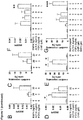
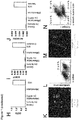
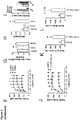
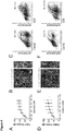
- step (i) and the concentration of factors such as BMP4, Activin A, FGF2, and the GSK3-inhibitor may be optimized by monitoring the efficiency of induction of mesoderm differentiation. This can be achieved by monitoring the expression of cell surface or pluripotency markers, i.e. by (a) a decrease of TRA-1-60 and OCT4 positive cells (pluripotent stem cells) and (b) an increase of MIXL1 and Mesp1 positive cells (mesoderm) (see also Fig 4f herein).
- cells are fixed using ethanol, blocked using standard protocols, and then stained with primary antibodies directed against TRA-1-60, OCT4, MIXL1 and/or Mesp1 (cf. Table 2 below) in blocking buffer for 45 min, optionally followed by secondary antibodies (if the primary antibody is not fluorescence labelled) in blocking buffer and Hoechst for 30 min at 4°C (cf. Table 2 below).
- a BD LSRII is used for flow cytometry analysis (BD Biosystems). For live cells populations are gated based on forward-side scatter profiles. BD FACSDiva Software (BD Bioscience) or Cyflologic v1.2.1 (Cyflo Ltd) are used for analysis. Induction of mesoderm differentiation is indicated if
- step (i) is carried out for 48-96 h.
- step (i) is carried out for 60-84 h, and more preferably step (i) is carried out for 66-78 h.
- the basal medium used in step (ii) can be selected from DMEM/F12, StemPro, Is-cove's medium, ⁇ MEM, DMEM, and RPMI.
- the basal medium used in step (ii) is RPMI supplemented with pyruvate.
- any suitable basal medium may be used in the method.
- the basal medium of step (ii) may be supplemented with non-essential amino acids. If ⁇ MEM is used as the basal medium in step (ii), the basal medium need not be supplemented additionally with non-essential amino acids.
- the non-essential amino acids are commercially available as a combined supplement.
- Such a supplement for example comprises 750 mg/L glycine, 890 mg/L L-alanine, 1320 mg/L L-asparagine, 1330 mg/L L-aspartic acid, 1470 mg/L L-glutamic acid, 1150 mg/L L-proline, and 1050 mg/L L-serine.
- the basal medium in step (ii) may be independently selected from the basal medium applied in step (i). However, in a preferred embodiment, the basal medium in steps (i) and (ii) is the same.
- the inhibitor of the Wnt-signaling pathway in the basal medium of step (ii) may be any inhibitor of the Wnt-signaling pathway, which can be suitably applied in the method of the invention.
- said inhibitor of the Wnt-signaling pathway is selected from the group consisting of IWP4, IWP2, IWR-1, IWP1, IWP3, IWR-2, IWR-3, IWR-4, IWR-5, XAV939, DKK1, quercetin, ICG-001, pyrvinium, CCT031374, iCRT-3,5,14, CPG049090, and NC043.
- said inhibitor of the Wnt-signaling pathway is selected from the group consisting of IWP4, IWP2, IWR-1, IWP1, IWP3, IWR-2, IWR-3, IWR-4, IWR-5, XAV939, DKK1. As demonstrated in the examples be-IWR-2, IWR-3, IWR-4, IWR-5, XAV939, DKK1. As demonstrated in the examples below, one particularly useful inhibitor of the Wnt-signaling pathway in the basal medium of step (ii) is IWP4.
- the serum-free supplement referred to in step (ii) is as defined for step (i) above.
- the serum-free supplements applied in step (i) and (ii) may be the same or not.
- B27® supplement or B27® supplement minus insulin can be used in step (ii).
- the B27® supplement or B27® supplement minus insulin used in step (ii) of the above method is applied in an amount of 0.1-10 % B27® or B27® minus insulin, preferably 0.5-8 %, more preferably 1-6 %, even more preferably 1.5-4% , and most preferably about 2% B27® or B27® minus insulin.
- the concentration of an effective amount of an inhibitor of the Wnt-signaling pathway varies with the availability and inhibition constant of the inhibitor in question.
- the basal medium of step (ii) may comprise 0.1-10 ⁇ M IWP4, preferably 1-9 ⁇ M, more preferably 2-8 ⁇ M, even more preferably 3-7 ⁇ M, still more preferably 4-6 ⁇ M, and most preferably about 5 ⁇ M IWP4.
- an effective concentration of any receptor/enzyme agonist or inhibitor varies with the availability and biological activity of the respective compound.
- the basal medium of step (ii) comprises 10-1000 ⁇ M, preferably 50-400 ⁇ M, more preferably 100-300 ⁇ M, even more preferably 150-250 ⁇ M, and most preferably about 200 ⁇ M of ascorbic acid or a salt thereof.
- the ascorbic acid may be delivered in the free form or as a salt. Since ascorbate is the active ingredient, any salt of ascorbic acid may be used, which provides the ascorbate to the cells, provided the counter ion has no detrimental effect on the cells.
- one suitable salt of ascorbic acid for use in the basal medium in step (ii) is ascorbate-2-phosphate.
- step (ii) and the concentration of the remaining constituents such as the inhibitor of the Wnt-signaling pathway may be optimized by monitoring the efficiency of induction of cardiac differentiation of the cells. This can be achieved by monitoring the expression of differentiation markers, i.e. by an increase of Nkx2.5 and actinin.
- cells are fixed using ethanol, blocked, and then stained with primary antibodies directed against Nkx2.5 and/or actinin (cf. Table 2 below) in blocking buffer for 45 min, optionally followed by secondary antibodies (if the primary antibody is not fluorescence labelled) in blocking buffer and Hoechst for 30 min at 4°C (cf. Table 2 below).
- a BD LSRII is used for flow cytometry analysis (BD Biosystems). For live cells populations are gated based on forward-side scatter profiles. BD FACSDiva Software (BD Bioscience) or Cyflologic v1.2.1 (Cyflo Ltd) are used for analysis.
- Induction of cardiac differentiation is indicated if more than 20%, preferably more than 30%, more preferably more than 40%, even more preferably more than 50%, and most preferably more than 60%, of the cells of the live cells population are positiv for Nkx2.5; and/or more than 20%, preferably more than 30%, more preferably more than 40%, even more preferably more than 50%, and most preferably more than 60% of the cells of the live cells population are positive for actinin (see also Fig 4d and 4f herein).
- step (ii) is carried out for 8-12 days.
- step (ii) is carried out for 9-11 days, and most preferably step (ii) is carried out for 10 days.
- the basal medium used in step (iii) can be selected from DMEM/F12, StemPro, Is-cove's medium, ⁇ MEM, DMEM, and RPMI.
- the basal medium used in step (iii) is RPMI supplemented with pyruvate.
- any suitable basal medium may be used in the method.
- the basal medium of step (iii) may be supplemented with non-essential amino acids. If ⁇ MEM is used as the basal medium in step (iii), the basal medium need not be supplemented additionally with non-essential amino acids.
- the non-essential amino acids are commercially available as a combined supplement.
- Such a supplement for example comprises 750 mg/L glycine, 890 mg/L L-alanine, 1320 mg/L L-asparagine, 1330 mg/L L-aspartic acid, 1470 mg/L L-glutamic acid, 1150 mg/L L-proline, and 1050 mg/L L-serine.
- the basal medium in step (iii) may be independently selected from the basal medium applied in steps (i) and/or (ii). However, in a preferred embodiment, the basal medium in steps (ii) and (iii) is the same. More preferably, the basal medium in steps (i), (ii) and (iii) is the same.
- the basal medium of step (iii) comprises 10-1000 ⁇ M, preferably 50-400 ⁇ M, more preferably 100-300 ⁇ M, even more preferably 150-250 ⁇ M, and most preferably about 200 ⁇ M of ascorbic acid or a salt thereof.
- the ascorbic acid may be delivered in the free form or as a salt. Since ascorbate is the active ingredient, any salt of ascorbic acid may be used, which provides the ascorbate to the cells, provided the counter ion has no detrimental effect on the cells.
- one suitable salt of ascorbic acid for use in the basal medium in step (iii) is ascorbate-2-phosphate.
- the serum-free supplement referred to in step (iii) is a serum-free supplement as defined for step (i) above.
- the serum-free supplements applied in steps (i), (ii) and (iii) may be the same or not.
- B27® supplement or B27® supplement minus insulin can be used in step (iii).
- the B27® supplement or B27® supplement minus insulin used in step (iii) of the above method is applied in an amount of 0.1-10 % B27® or B27® minus insulin, preferably 0.5-8 %, more preferably 1-6 %, even more preferably 1.5-4% , and most preferably about 2% B27® or B27® minus insulin.
- the basal medium of step (iii) further comprises an effective amount of TGF ⁇ 1.
- the basal medium of step (iii) may comprise 0.1-10 ng/ml TGF ⁇ 1, preferably 0.2-9 ng/ml, more preferably 0.3-8 ng/ml, even more preferably 0.4-7 ng/ml, still more preferably 0.5-6 ng/ml, more preferably 0.6-5 ng/ml, more preferably 0.7-4 ng/ml, more preferably 0.8-3 ng/ml, most preferably 0.9-2 ng/ml, and even most preferably about 1 ng/ml TGF ⁇ 1.
- the basal medium of step (iii) comprises 0.5-3 mM Ca 2+ , preferably 0.5-2.75 mM Ca 2+ , more preferably 1-2.25 mM Ca 2+ , even more preferably 1-1.5 mM mM Ca 2+ , and most preferably about 1.2 mM Ca 2+ .
- step (iii) of the method of the invention is carried out under mechanical stimulation, e.g. on a stretch device, as generally known in the art.
- the stretch device applies a static, phasic or dynamic stretch to the BHM.
- mechanical stretching can be (a) static, (b) dynamic, or (c) flexible against a resilient load.
- the mechanical stimulation in step (iii) is dynamic mechanical stimulation or static stretch.
- the mechanical stimulation in step (iii) is dynamic mechanical stimulation against a resilient load to facilitate auxotonic contractions.
- cardiac maturation is promoted can be tested by optical inspection for spontaneous or electrically stimulated contractions.
- cardiac maturation is monitored by an isometric contraction experiment, wherein a twitch force development of > 0.01 mN is indicative for cardiac maturation.
- step (iii) is carried out for at least 72 h. Although there is no particular upper limit for the length of step (iii), said step is usually carried out for less than 100 days. In specific embodiments, step (iii) may be carried out for 4-50 days, such as for about 15 days.
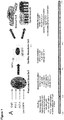
- Step (i) of the method of the invention may be preceded by a seeding step, wherein said pluripotent stem cells are seeded in a ratio of (2.5-6 x 10 6 cells /1 mg collagen) / 1 ml medium in a suitable mould.
- the seeding step is carried out 18-30 h prior to step (i).
- the medium used in the seeding step usually comprises 0.2-2 mg/ml collagen (preferably 0.3-1.9 mg/ml, more preferably 0.4-1.8 mg/ml, even more preferably 0.4-1.7 mg/ml, still more preferably 0.5-1.6 mg/ml, more preferably 0.6-1.5 mg/ml, more preferably 0.7-1.4 mg/ml, more preferably 0.8-1.3 mg/ml, more preferably 0.9-1.2 mg/ml, such as about 1 mg/ml).
- the collagen is preferably of medical grade and selected from the group consisting of collagen type I, collagen type III, collagen type V, and a mixture thereof. In a more preferred embodiment, at least 90% of said collagen is collagen type I.
- said collagen may also further comprises one or more extracellular matrix components selected from the group consisting of elastin, laminin, entactin, nidogen, proteoglycan, and fibronectin.
- extracellular matrix components selected from the group consisting of elastin, laminin, entactin, nidogen, proteoglycan, and fibronectin.
- the collagen is preferably of human origin, but bovine or porcine origin, or marine origin, such as from algae or fish origin, is also contemplated. Alternatively, recombinant collagen may also be used.
- the medium used in the seeding step further comprises a ROCK-inhibitor.
- the ROCK-inhibitor may be any ROCK-inhibitor, which can be suitably applied in the method of the invention.
- said ROCK inhibitor is selected from Y27632, H-1152P, Thiazovivin, Fasudil, Hydroxyfasudil, GSK429286A, and RKI-1447, preferably selected from Y27632, H-1152P, Thiazovivin, Fasudil, Hydroxyfasudil, and more preferably the ROCK inhibitor is selected from Y27632 or H-1152P.
- one particularly useful ROCK-inhibitor is Y27632.
- the concentration of an effective amount of a ROCK-inhibitor varies with the availability and inhibition constant of the inhibitor in question.
- the medium used in the seeding step may comprise 1-50 ⁇ M, preferably 2.5-40 ⁇ M, more preferably 5-30 ⁇ M, even more preferably 7.5-20 ⁇ M, most preferably 8-12 ⁇ M, and most preferably about 10 ⁇ M Y27632.
- the invention further relates to a BHM produced by said method, as defined in the claims.
- the BHM is still a relatively immature tissue. Compared to adult heart tissue the BHM still has an inferior ⁇ -MHC / ⁇ -MHC ratio, and low but still retained expression of progenitor genes (e.g. ISL1 ).
- progenitor genes e.g. ISL1
- prolonged culture under appropriate culture conditions with biophysical stimulation may further increase maturity. There is already morphological evidence suggesting that this may also be the case in the BHM system.
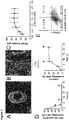
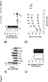
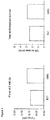
- the BHM obtained by the method disclosed herein exhibits the following characteristics: It can be paced at multiple frequencies up to at least 3 Hz, exhibits a calcium EC 50 of higher than 0.2 mM being preferably in the physiological range 4-8 mM, and a twitch tension of more than 200 ⁇ N. The twitch tension is increased in response to increased resting length and resting tension. In response to 1 ⁇ M isoprenaline, the BHM exhibits an inotropic response of more than 40 ⁇ N under paced conditions at 0.6 mM calcium, preferably more than 45 ⁇ N, more preferably more than 50 ⁇ N.
- BHM are mechanically stretched at intervals of 125 ⁇ m until the maximum twitch force is observed at 2 mM calcium (Frank-Starling mechanism). Subsequently, BHM are subjected to different calcium concentrations (0.2, 0.4, 0.8, 1.2, 1.6, 2.0, 2.4 mM) and the twitch force is recorded. For isoprenaline experiments the calcium concentration is adjusted to 0.6 mM and subsequently the isoprenaline concentration is adjusted to 1 ⁇ M.
- Another characteristic of the BHM obtained by the method disclosed herein is that it comprises CD90 + stromal cells.
- Expression of CD90 can be determined using flow cytometry. Briefly, cells are fixed using ethanol, blocked, and then stained with primary antibodies directed against CD90 (cf. Table 2 below) in blocking buffer for 45 min, optionally followed by secondary antibodies (if the primary antibody is not fluorescence labelled) in blocking buffer and Hoechst for 30 min at 4°C (cf. Table 2 below).
- a BD LSRII is used for flow cytometry analysis (BD Biosystems). For live cells populations are gated based on forward-side scatter profiles. BD FACSDiva Software (BD Bioscience) or Cyflologic v1.2.1 (Cyflo Ltd) are used for analysis.
- the BHM may provide a good model system for investigating mechanisms driving maturation in a serum-free environment, and we have already demonstrated that with increased culture periods maturity may be increased (we showed increase isoprenaline sensitivity and improve tissue morphology).
- maturity may be increased (we showed increase isoprenaline sensitivity and improve tissue morphology).
- the capability of long term BHM culture without loss of function (at least 63 days) also suggests that long term pharmacological safety and efficacy experiments are possible.
- the BHM obtained by the method disclosed herein can be maintained for at least 63 days.
- the differentiation cultures require extensive digestion protocols in order to yield single cell/small clumps required for cardiac tissue engineering applications. These digestion protocols destroy the extracellular environment and spatial distribution formed during development and may hence have a difficult to control inhibitory effect on the cardiac differentiation protocol.
- BHM protocol may be a useful tool in the study of developmental processes not only governing cardiogenesis, but also tissue formation and properties.
- the BHM obtained by the method disclosed herein can be suitably used as a research tool.
- the use of the BHM obtained by the method disclosed herein in an in vitro-model for drug toxicity screening is contemplated.
- a method for screening drug toxicity is contemplated, comprising the step of contacting a BHM obtained by the method disclosed herein with a drug to be screened.
- the BHM obtained by the method disclosed herein may be used in an in vitro method for testing of cardiac function modulation by pharmacological candidate agents.
- a method for testing of cardiac function modulation comprising the step of contacting a BHM according to the invention with a pharmacological candidate agent.
- the BHM obtained by the method disclosed herein can be used in medicine.
- it is contemplated that the BHM obtained by the method disclosed herein can be advantageously used in heart repair.
- a cardiac differentiation protocol was firstly required which consistently produced cardiomyocytes and fibroblasts/stromal cells.
- the inventors optimized their cardiac differentiation protocol ( Fig. 1a ) for both yield and consistency, based on a previously published serum-free 2D hPSC differentiation protocol ( Hudson et al. Stem Cells Dev 21, 1513-1523 (2012 )). It was reasoned that robustness and efficiency could be enhanced if WNT activity was stabilized during the mesoderm induction phase.
- MESP1 expression was analysed by qPCR on culture day 3; this was followed by flow cytometry for ⁇ -actinin (cardiomyocyte marker) at day 16, which were found correlated very well with the amount of beating activity. The most important steps for the progression from the previously published protocol to the new protocol are summarised in Fig. 1 .
- stromal cells or other potentially contaminating cell types were present in the optimized differentiation protocol. Very low levels of potentially contaminating cell populations were found, analysed using qPCR for hPSCs ( OCT4 also known as P0U5F1 ) ( Fig. 1h ), endoderm ( SOX17 ) ( Fig. 1i ), neural ( NEUROD1 ) ( Fig. 1j ), and early mesoderm ( MESP1 ) (data not shown).
- OCT4 also known as P0U5F1
- SOX17 Fig. 1i
- NEUROD1 Fig. 1j
- MESP1 early mesoderm
- NKX2-5 and ⁇ -MHC also known as MYH7
- ⁇ -MHC also known as MYH7
- cardiomyocytes Fig. 1k -l
- different stromal cell types including: ⁇ -smooth muscle actin positive cells ( ⁇ -SMA + ), collagen I positive cells (COLI + ) cells, and ⁇ -SMA + COLI + cells were present ( Fig. 1m ,n ).
- the new serum-free cardiac differentiation protocol ( Fig. 1 ) was used with an additional maturation step where the rings were removed from the molds and put onto static stretchers (+10% of slack length) in medium containing 5 ng/ml FGF2 and 200 ⁇ M ascorbate-2-phosphate (ASC-2-P). It was found that this protocol was effective in forming BHM ( Fig. 2a ).
- the BHM started to contract spontaneously in different areas by day 13 and by day 15-17 the contractions became synchronous and rhythmic, and continued until analysis at day 22 (data not shown).
- the cardiomyocytes in the BHM had an elongated and striated morphology ( Fig. 2b ) and the BHM could be electrically paced and had a measurable force of contraction with responsiveness to calcium concentration ( Fig. 2c ).
- the development of the BHM followed known developmental pathways.
- the hPSCs were largely differentiated by day 3 indicated by a decrease in TRA-1-60 + /OCT4 + cells and OCT4 expression ( Fig. 2d ,f ) and a concomitant expression of early mesodermal markers MIXL1 and MESP1 ( Fig. 2f ).
- day 8 there was a loss of MIXL1 and MESP1 expression ( Fig. 2f ), with a concurrent increase in ⁇ -actinin + cells ( Fig. 2d ) and the expression of cardiomyocyte progenitor cell markers ( Fig. 2f ).
- ⁇ -MHC also known as MYH6
- ⁇ -MHC ⁇ -MHC
- ANP also known as NPPA
- MLC2v also known as MYL2
- twitch tension/force of contraction was used as a primary determinant of function as twitch tension depends on a wide variety of myocardial properties including: cardiomyocyte number and phenotype, fibroblast number and phenotype, tissue connectivity, ECM composition, cell-cell connectivity and ECM-cell connectivity.
- Ascorbate (vitamin C) plays a major role in the proper synthesis of collagen and is an antioxidant. It was therefore hypothesized that ascorbate (in the more stable form of ASC-2-P) supplemented during early BHM culture, days 0-13 (it was already added during days 13-22 in Fig. 2 ), would have a positive influence on BHM functionality given the importance of collagens during development. It was found that ASC-2-P significantly improved BHM twitch tension/force of contraction ( Fig. 3a ) and induced a trend to increased cardiomyocyte fraction with no change in the stromal cell fraction ( Fig. 3b ).
- ASC-2-P may have increased cell survival and/or progenitor proliferation as proposed in a recent study ( Cao et al. Cell Res 22, 219-236 (2012 )).
- FGF2 did not change ⁇ -MHC / ⁇ -MHC expression ratio ( Fig. 3h ), but induced (variably) pathological hypertrophy marker ANP ( Fig. 3i ). Together this indicates both factors induce hypertrophy, which is consistent with in vivo results. However, FGF2 may be considered as an inducer of pathological hypertrophy, while TGF ⁇ 1 may be considered as an inducer of physiological hypertrophy.
- the inventors investigated if supplementation of the culture medium with increasing TGFB-1 during cardiac maturation phase has an influence in contractile function of BHM.
- the inventors observed an enhancement of contractile function of BHM in a concentration dependent manner ( Fig. 10 ).
- BHM Produced Using the Optimized Protocol Display In-Vivo Like Properties
- BHM contacted spontaneously and coherently, and could be paced electrically at multiple frequencies up to at least 3 Hz ( Fig. 4a ), covering the range of beating frequency observed during heart development.
- BHM also increased twitch tension (force of contraction) in response to increased resting length (and resting tension) consistent with the Frank-Starling mechanism ( Fig. 4b ). With increased culture time no change in BHM twitch tension compared to the previous data was found, but an increase in calcium EC 50 from 0.2 to 0.7 mmol/L was observed ( Fig. 4c ).
- Extended culture also resulted in an increased inotropic response to 1 ⁇ M isoprenaline indicating improved maturity over traditional culture formats where no increase has been observed ( Fig. 4d -f ).
- BHM also has both stromal cells and endothelial cells present in the muscle bundles (data not shown).
- BHM culture could also be maintained for long term (until at least day 63) under the maturation conditions with an observed improvement in morphological appearance (data not shown).
- both the HES3 line and the hIPS line produced BHM with a lower twitch tension ( Fig. 5a ,d ) compared to the HES2 line ( Fig. 3j ).
- both the HES3 and hIPS BHM had similar morphologies ( Fig. 5b ,e ).
- the cardiomyocyte fractions for the HES3 BHM was similar ( Fig. 5c ) and for the hIPS BHM was lower ( Fig. 5f ), compared to the HES2 BHM.
- BHM as a Developmental Model Reveals that BMP Signalling is Required for Terminal Differentiation of Human Cardiomyocytes Inhibition of BMP signalling is embryonically lethal even when effects are restricted (or at least partially restricted) to the developing heart using CRE driven by various genes (for a review please see Kruithof et al. Differentiation 84, 89-102 (2012 )). In these studies there have been multiple processes ascribed to BMP signalling including structural defects, myocardial properties including trabeculae structure and wall thickness, and cell phenotype including dysregulation of progenitor genes and reduced epithelial-to-mesenchymal transformation (EMT).
- EMT epithelial-to-mesenchymal transformation
- BHM was therefore deemed to be a good model system.
- 2 ⁇ mol/L of the BMP receptor signalling inhibitor dorsomorphin were added with each medium exchange from day 6 onwards.
- the dorsomorphin treated BHM failed to down regulate ISL1 while the expression of other more mature cardiac markers NKX2-5 and ⁇ -MHC were not altered ( Fig. 7a ).
- EMT associated genes it was found that there was no alteration in CDH1, CHD2, SNAIL1 or TGF ⁇ 2 expression indicating that EMT or factors regulating EMT were not altered in BHM after 13 days ( Fig. 7a ).
- BHMs were generated from undifferentiated hESC under serum-free conditions according to standard BHM protocol.
- the standard protocol includes B27® supplement.
- B27® supplement was replaced by a defined, custom-made supplement (CMS, Table 4 ).
- BHM has multiple applications including pharmacological studies, study of developmental processes, heart maturation processes and also potential regenerative applications.
- HES2-ROSA26-RFP Irion et al. Nat Biotechnol 25, 1477-1482 (2007 ) cells were obtained from Gordon Keller and HES3 cells were obtained from Embryonic Stem Cell International (ESI, Singapore). hIPS were generated from human gingiva biopsy-derived fibroblasts using the Cytotune Reprogramming Kit (Applied Biosystems) following the manufacturer's instructions.
- fibroblast medium DMEM high glucose, 2 mmol/L glutamine, 10% FBS (PAA), 100 IU/ml Penicillin, 100 ⁇ g/ml Streptomycin, all Gibco except where indicated.
- hPSCs were single cell adapted and cultured on irradiated human foreskin fibroblasts (HFF) in PSC-medium with daily medium changes and weekly passaging using 3 min TrypLE (Gibco) treatment ( Ellerstrom et al., Stem Cells 25, 1690-1696 (2007 )).
- the hPSCs were plated on 1:30 Matrigel (Millipore) in PBS (Gibco) coated-plates, at 2.5 x 10 4 cells/cm 2 for HES2 or 5x10 4 cells/cm 2 for HES3 and hIPS lines, and cultured for 3 days in 1:1 PSC-medium minus FGF-2 and HFF-conditioned medium (HFF-CM - harvested from 5 day confluent irradiated HFF cultures) with 10 ng/mL FGF2.
- the hIPS line also received 10 ⁇ mol/L Y-27632 (Stemgent). hPSCs were harvested for experiments by passaging using 3 min TrypLE treatment and then cultured in the appropriate format.
- Pluripotent stem cell lines were regularly tested for mycoplasma using a test kit (Lonza) and characterized using standard assays. Pluripotency markers were assessed via PCR (endogenous OCT4, SOX2, KLF4, MYC ), qPCR ( OCT4, NANOG, REX1, DNMT3B ) and immunostaining (OCT4, NANOG, TRA-1-60) ( Chan et al. Nat Biotechnol 27, 1033-1037 (2009 )). Demethylation of the OCT4 promoter was confirmed via bisulfite sequencing ( Freberg et al. Mol Biol Cell 18, 1543-1553 (2007 )). Karyotyping was used to determine if there were any genetic abnormalities ( Campos et al. J Vis Exp 4 (2009 )). Pluripotency was confirmed via teratoma formation in SCID mice via flank injection of 4-6 x 10 6 cells.
- hPSCs were then cultured in RPMI 1640 supplemented with 1 mmol/L sodium pyruvate, 100 IU Penicillin, 100 ⁇ g/ml Streptomycin and 2% B27 supplement (SF medium, all Gibco) and various factors as indicated.
- Factors used in this study included: L-ascorbic acid 2 phosphate sesquimagnesium salt hydrate (Sigma), BMP4 (R&D Systems), Activin A (R&D Systems), FGF2 (Miltenyi Biotec), dorsomorphin (Stemgent), CHIR99021 (Stemgent), IWP4 (Stemgent), and TGF ⁇ 1 (Peprotech).
- HES2 hPSCs were plated at 5x10 4 cells/cm 2 (1x10 5 cells/cm 2 for HES3 and hIPS lines) on 1:30 Matrigel/PBS coated-plates and cultured in 1:1 PSC-medium minus FGF-2 and HFF-conditioned medium (HFF-CM - harvested from 5 day confluent irradiated HFF cultures) with 10 ng/mL FGF2.
- HFF-CM HFF-conditioned medium
- 10 ⁇ M Y-27632 was added to this medium. After 1 day the cells were rinsed with RPMI medium, then differentiated as indicated in each figure with 0.5 ml of medium in each well of a 24 well plate. The protocol details for each figure are outlined in Fig. 8 .
- HES2 hPSCs were suspended 1:1 in PSC-medium minus FGF-2 and HFF-conditioned medium (HFF-CM - harvested from 5 day confluent irradiated HFF cultures) with 10 ng/mL FGF2 and mixed with a collagen I hydrogel.
- HFF-CM HFF-conditioned medium
- HFF-CM HFF-conditioned medium
- hIPS lines 10 ⁇ M Y-27632 was also added to the medium.
- the collagen I matrix was formulated with acid-soluabilized bovine collagen I (Devro) with an equi-volume of 2X DMEM (Gibco) and neutralized using 0.1 M sodium hydroxide.
- the hPSC/collagen I matrix was formulated to give a final collagen I concentration of 1 mg/ml and 5 x 10 5 hPSC per 170 ⁇ l.
- 1 x 10 6 and 0.5 x 10 6 cells were used respectively per 170 ⁇ l.
- 2D cultures were disassociated by rinsing with PBS followed by incubation for 1 h in 1 mg/ml collagenase type I (Sigma) with 20% fetal bovine serum (FBS, Applied Biosystems) in PBS. The cells were then collected in a tube, rinsed with PBS and incubated with 0.25% Trypsin-EDTA (Applied Biosystems) for 5 min followed by rinsing with FBS containing medium.
- FBS fetal bovine serum
- BHMs were disassociated in 0.025 mg/ml Liberase TM (Roche), 30 mM 2,3-butanedione monoxime at 37 °C for 60 min in PBS. To preserve cell surface markers, BHM was disassociated using the same protocol as for the 2D digestion.
- RNA extraction using Trizol following manufacturer's instructions (Applied Biosystems). 1 ⁇ g of RNA was then treated with DNAse (Roche) followed by cDNA synthesis using a High Capacity cDNA Reverse Transcription Kit (Applied Biosystems).
- qPCR was performed using Fast SYBR Green Master Mix (Applied Biosystems) on a 384-well format AB7900 HT (Applied Biosystems). Gene expression was normalized using 2 - ⁇ Ct or 2 - ⁇ Ct using GAPDH as the housekeeping gene which we found to be consistently expressed between conditions in all of our experiments. Primer details are given in below Table 1 .
- Digested cardiac differentiation cells were plated on 0.1% gelatin coated glass co-verslips for 24 h in 20% FBS (Gibco) in RPMI 1640 supplemented with 1 mmol/L sodium pyruvate, 100 IU/ml Penicillin and 100 ⁇ g/ml Streptomycin. The cells were then fixed in Histofix (Roti) for 10 min at room temperature. The cells were then blocked for 30 min in 5% FBS, 1% bovine serum albumin (Sigma) and 0.5% Triton X-100 (Sigma) in PBS (blocking buffer). The cells were then stained with primary antibodies in blocking buffer for 90 min followed by secondary antibodies in blocking buffer and Hoechst for 60 min at room temperature ( Table 2 ).
- BHMs were fixed in Histofix for 2-4 h at 4°C.
- the BHMs were then stained with primary antibodies for 2-3 days followed by secondary antibodies and Phalloidin 546/Hoechst for 2-3 days at 4°C ( Table 2 ). Stained BHMs were imaged using a Zeiss 710 confocal microscope.
- Cells were stained either live or fixed using Histofix for 10 min at room temperature or ethanol. The cells were stained in 5% FBS in PBS (membrane blocking buffer) for cell surface markers (excluding TRA-1-60) and blocking buffer for internal markers. The cells were then stained with primary antibodies in blocking buffer for 45 min followed by secondary antibodies in blocking buffer and Hoechst for 30 min at 4°C ( Table 2 ). A BD LSRII was used for flow cytometry analysis (BD Biosystems). Live cells populations were gated based on forward-side scatter profiles; fixed cells populations were gated based on Hoechst staining. BD FACSDiva Software (BD Bioscience) or Cyflologic v1.2.1 (Cyflo Ltd) were used for analysis.
- the tissue length were maximum twitch tension/force of contraction was recorded in the presence of maxi-mally inotropically active calcium concentrations (2 mmol/L; Frank-Starling mechanism). Subsequently, BHM were subjected to different calcium concentrations (0.2, 0.4, 0.8, 1.2, 1.6, 2.0, 2.4 mM) and the twitch force recorded.
- maxi-mally inotropically active calcium concentrations (2 mmol/L; Frank-Starling mechanism).
- BHM were subjected to different calcium concentrations (0.2, 0.4, 0.8, 1.2, 1.6, 2.0, 2.4 mM) and the twitch force recorded.
- the calcium concentration was adjusted to 0.6 mM and subsequently the isoprenaline concentration was adjusted to 1 ⁇ M.
- CMS Custom-made supplement
- Substance Final concentration 25x Supplier Albumin 5 mg/ml 125 mg/ml Sigma, A9511 Transferrin 10 ⁇ g/ml 250 ⁇ g/ml Sigma, T0665 EthanolamineHCl 2 ⁇ g/ml 50 ⁇ g/ml Sigma, E6133 Sodium selenite 0.032 ⁇ g/ml 0.8 ⁇ g/ml Sigma, S5361 L-CarnitineHCl 4 ⁇ g/ml 100 ⁇ g/ml Sigma, C0283 Hydrocortisone 1 ⁇ g/ml 25 ⁇ g/ml Sigma, H2270 Fatty acid supplement 0.5 ⁇ l/ml 12.5 ⁇ l/ml Sigma, F7050 Triiodo-L-thyronine 0.004 ⁇ g/ml 0.1 ⁇ g/ml Sigma, T 6397 Prepare 25x in cell culture-qualified water.
Description
- Human pluripotent stem cells (hPSCs) are now widely used to provide a theoretically endless and also large supply of human cardiomyocytes (Kehat et al. J Clin Invest 108, 407-414 (2001); Takahashi et al. Cell 131, 861-872 (2007); Zhang et al., Circ Res 104, e30-41 (2009)). Human cardiomyocytes have been derived from human embryonic stem cells (hESCs) (Thomson et al. Science 282, 1145-1147 (1998)) and induced pluripotent stem cells (hIPSCs) (Takahashi et al., Cell 131, 861-872 (2007)) and have a demonstrated use for multiple purposes including developmental models (Lian et al. Stem Cells 2012 (2012)), drug efficacy and/or safety screening (Schaaf et al. PLoS ONE 6, 20 (2011)), hypertrophy modelling and regenerative applications. Additionally, with recent advances in hIPSC technology, cardiomyocytes exhibiting heritable genetic disease phenotypes can be generated in vitro (Carvajal-Vergara, X. et al. Nature 465, 808-812 (2010); Itzhaki et al., Nature 471, 225-229 (2011); Malan et al. Circ Res (2011); Moretti et al., N Engl J Med 363, 1397-1409 (2010); Yazawa et al. Nature 471, 230-234 (2010)).
- It is now widely accepted that the
low density 2D culture of biopsy-derived human cardiomyocytes leads to rapid changes in cardiomyocyte phenotype and morphology (Bird et al. Cardiovasc Res 58, 423-434 (2003)) and making it difficult to extrapolate results to the in vivo situation. In order to obtain a cardiomyocyte phenotype more representative of in vivo conditions, cardiac tissue engineering has been used (Eschenhagen et al. FASEB J 11, 683-694 (1997); Zimmermann et al. Biotechnol Bio-eng 68, 106-114 (2000), Zimmermann et al. Circ Res 90, 223-230 (2002); Tulloch et al. Circ Res 109, 47-59 (2011); Tiburcy et al. Circ Res 109, 1105-1114 (2011); Eschenhagen et al. Am J Physiol Heart Circ Physiol 303, 11 (2012)) to generate constructs with similar properties to the native heart tissue. - The current ideology of tissue engineering is to generate/isolate the required cell type(s), and seed them into an engineered environment to promote their differentiation and generate in vivo-like tissues. Tissue engineering may therefore be considered as an inefficient process for two reasons, 1) disassociation of a tissue/differentiation culture destroys the extracellular environment thus destroying developmental information (eg. cell-cell interconnectivity, geometric cell positioning, cell-ECM connectivity), this necessitates very large increases in extracellular matrix (ECM) production in order to re-build the environment (Hudson et al. Tissue Eng Part A 17, 2279-2289 (2011)), and 2) the disassociation process is variable between hPSC lines and can lead to considerable cell death.
- Other protocols reported in the literature may require modification of the protocol to enable similar cardiomyocyte efficiencies in multiple hPSC lines. However, the inventor's results demonstrate that changes in differentiation protocol may greatly affect the cardiomyocyte phenotype (e.g. it is shown that dorsomorphin may greatly affect the bioengineered heart muscle (BHM)). This may lead to changes in tissue engineered myocardial properties which may mask the effects of different experimental conditions or genetic disease models, therefore care must be taken when using different protocols in different lines.
- Some recently published protocols may enable the same protocol to be used for multiple lines, they also produce cardiomyocytes with very high purity. However, pure cardiomyocytes do not facilitate the formation of functional tissue engineered myocardium and both cardiomyocytes and stromal cells are required for the formation of functional tissue engineered myocardium (Naito et al. Circulation 114, 172-78 (2006),Hudson et al. Tissue Eng Part A 17, 2279-2289 (2011)).
-
WO 2013/056072 A1 ,WO 2013/111875 A1 andUS 2013/177535 disclose methods for producing cardiomyocytes, but not of a bioengineered heart muscle. - Hence, there is a need in the art for methods for producing bioengineered human myocardium, which are capable of overcoming the above disadvantages.
- The development of a robust differentiation protocol is a very important step allowing the consistent production of BHM. In this study n >140 BHM in >18 independent experiments were produced and every one exhibited spontaneous beating activity. Additionally, the protocol enables to produce BHM from multiple hPSC lines using the same protocol. In addition, all disassociation steps could be eliminated and hPSCs were differentiated directly into bioengineered myocardium, thus retaining the developmental memory of the tissue, prevent any tissue recreation response and provide a more accurate in vitro model of human myocardial development.
- The present invention relates to a method for producing bioengineered heart muscle from pluripotent stem cells, comprising the steps of
- (i) cultivating pluripotent stem cells in a basal medium comprising an effective amount of (a) BMP4, Activin A, FGF2, a GSK3-inhibitor, and (b) a serum-free supplement resulting in a final concentration of 0.5-50 mg/ml albumin, 1-100 µg/ml transferrin, 0.1-10 µg/ml ethanol amine, 0.003-0.3 µg/ml sodium selenite, 0.4-40 µg/ml L-Carnitine HCl, 0.1-10 µg/ml Hydrocortisone, 0.05-5 µl/ml Fatty acid supplement, 0.0001-0.1 µg/ml triodo-L-thyronine (T3), thereby inducing mesoderm differentiation of said pluripotent stem cells;
- (ii) cultivating the cells obtained in step (i) in a basal medium comprising an effective amount of an inhibitor of the Wnt-signaling pathway and a serum-free supplement as defined in step (i), thereby inducing cardiac differentiation of the cells; and
- (iii) cultivating the cells obtained in step (ii) in a basal medium comprising an effective amount of a serum-free supplement as defined in step (i), under mechanical stimulation, thereby promoting cardiac maturation.
- Carrying out the method disclosed herein human pluripotent stem cell (hPSC)-derived bioengineered heart muscle (BHM) is generated by directed tissue formation of hPSCs in collagen hydrogels. To form BHM, in vivo development was mimicked using a directed serum-free induction protocol causing the tissue to progress through distinct, known developmental stages, through pluripotency, early mesoderm, cardiac progenitor, immature cardiomyocytes and finally to more mature cardiac tissue comprised of 50 % cardiomyocytes, with the rest being predominately a stromal cell fraction. The inventors optimized their serum-free BHM protocol and found that individual BHM properties are highly dependent on particular stimuli, thus indicating that multiple exogenous stimuli are required for optimal BHM properties. In the end rhythmically contractile BHM was produced with measurable contractile force, ability for pacing and inotropy in response to increased resting length, calcium concentration and β-adrenergic stimulation. This BHM protocol, without modification, was capable of consistently producing BHM from multiple hPSC lines (in every BHM in every experiment conducted).
- The present data suggests that the BHM protocol disclosed herein is a robust, serum-free and reproducible way to produce human myocardium for multiple applications. For example, it is also demonstrated that BHM is a potential model of human myocardium development, and shown that inhibition of BMP signalling leads to a more immature cardiac phenotype with reduced contractile strength.
- Accordingly, the present invention is also directed to a BHM produced by the method according to the invention, as further defined in the claims.
- Further contemplated is the use of the BHM according to the invention in an in vitro-model for drug toxicity screening. In other words, the present invention is also directed to a method for screening drug toxicity, comprising the step of contacting a BHM according to the invention with a drug to be screened.
- Moreover, the present invention is directed to the use of the BHM according to the invention in an in vitro method for testing of cardiac function modulation by pharmacological candidate agents. Thus, also described is a method for testing of cardiac function modulation, comprising the step of contacting a BHM according to the invention with a pharmacological candidate agent.
- Finally, the present invention is also directed to the use of the BHM according to the invention as a research tool, as well as to a BHM according to the invention for use in medicine.
- A method for producing bioengineered heart muscle from pluripotent stem cells, comprising the steps of
- (i) cultivating pluripotent stem cells in a basal medium comprising an effective amount of (a) BMP4, Activin A, FGF2, a GSK3-inhibitor, and (b) a serum-free supplement resulting in a final concentration of 0.5-50 mg/ml albumin, 1-100 µg/ml transferrin, 0.1-10 µg/ml ethanol amine, 0.003-0.3 µg/ml sodium selenite, 0.4-40 µg/ml L-Carnitine HCl, 0.1-10 µg/ml Hydrocortisone, 0.05-5 µl/ml Fatty acid supplement, 0.0001-0.1 µg/ml triodo-L-thyronine (T3), thereby inducing mesoderm differentiation of said pluripotent stem cells;
- (ii) cultivating the cells obtained in step (i) in a basal medium comprising an effective amount of an inhibitor of the Wnt-signaling pathway and a serum-free supplement as defined in step (i), thereby inducing cardiac differentiation of the cells; and
- (iii) cultivating the cells obtained in step (ii) in a basal medium comprising an effective amount of a serum-free supplement as in (i), under mechanical stimulation, thereby promoting cardiac maturation.
- In a preferred embodiment, the pluripotent stem cells are pluripotent stem cells of primate origin, more preferably the pluripotent stem cells are human pluripotent stem cells. Pluripotent stem cells are able to differentiate into every cell type of the body. As such, human pluripotent stem cells offer the unique opportunity to obtain bona fide human heart cells. Currently, the most utilized pluripotent cells are embryonic stem cells (ESC) or induced pluripotent stem cells (iPSC). Human ESC-lines were first established by Thomson and coworkers (Thomson et al., Science 282: 1145-1147 (1998)). Human ESC research recently enabled the development of a new technology to reprogram cells of the body into a ES-like cell. This technology was pioneered by Yamanaka and coworkers in 2006 (Takahashi & Yamanaka Cell 126: 663-676 (2006)). Resulting induced pluripotent cells (iPSC) show a very similar behavior as ESC and, importantly, are also able to differentiate into every cell of the body. Moreover, it was reported that also parthenogenetic stem cells are likely to be suitable for BHM-production (Didié et al. J Clin Invest. 123, 1285-1298 (2013)). Accordingly, the pluripotent stem cells can be selected from embryonic stem cells, induced pluripotent stem cells, and parthenogenetic stem cells. In the context of the present invention, said pluripotent stem cells are however not produced using a process which involves modifying the germ line genetic identity of human beings or which involves use of a human embryo for industrial or commercial purposes.
- The basal medium used in step (i), can be selected from DMEM/F12, StemPro, Is-cove's medium, αMEM, DMEM, and RPMI. Preferably, the basal medium used in step (i) is RPMI supplemented with pyruvate. However any suitable basal medium may be used in the method. Basal mediums are commercially available or may be prepared according to recipes which are publicly available, e.g. from catalogues of the ATCC.
- If deemed appropriate, the basal medium may be supplemented with non-essential amino acids. If αMEM is used as the basal medium, the basal medium need not be supplemented additionally with non-essential amino acids. The non-essential amino acids are commercially available as a combined supplement. Such a supplement for example comprises 750 mg/L glycine, 890 mg/L L-alanine, 1320 mg/L L-asparagine, 1330 mg/L L-aspartic acid, 1470 mg/L L-glutamic acid, 1150 mg/L L-proline, and 1050 mg/L L-serine.
- As set out above, the basal medium of step (i) comprises an effective amount of BMP4, Activin A, FGF2, and a GSK3-inhibitor. For example, such a basal medium comprises 1-20 ng/ml BMP4, preferably 2-15 ng/ml, more preferably 2.5-10 ng/ml, more preferably 3-8 ng/ml, most preferably 4-6 ng/ml, and even most preferably about 5 ng/ml;
0.1-10 ng/ml FGF2, preferably 1-9 ng/ml, more preferably 2-8 ng/ml, even more preferably 3-7 ng/ml, most preferably 4-6 ng/ml, and even most preferably about 5 ng/ml;
1-20 ng/ml Activin A, preferably 2.5-18 ng/ml, more preferably 5-16 ng/ml, even more preferably 7.5-14 ng/ml, still more preferably 8-12 ng/ml, most preferably 8.5-10 ng/ml, and even most preferably about 9 ng/ml. - The GSK3-inhibitor in the basal medium of step (i) can be selected, for example, from the group consisting of CHIR99021, CHIR98014, SB216763, TWS119, Tideglusib, SB415286, and LY2090314. However, any GSK3-inhibitor suitable in the method of the invention can be applied. In a preferred embodiment, the GSK3-inhibitor in the basal medium of step (i) is CHIR99021.
- It will be understood by the skilled person that the concentration of an effective amount of a GSK3-inhibitor varies with the availability and inhibition constant of the inhibitor in question. In the context of the present invention, the term "effective amount" as used herein in the context of a GSK3-inhibitor is intended to mean an enzyme inactivating concentration. For example, in case of CHIR99021, the basal medium in step (i) comprises 0.1-10 µM CHIR99021, preferably 0.2-9 µM, more preferably 0.3-8 µM, even more preferably 0.4-7 µM, still more preferably 0.5-6 µM, more preferably 0.6-5 µM, more preferably 0.7-4 µM, more preferably 0.8-3 µM, most preferably 0.9-2 µM, and even most preferably about 1 µM CHIR99021. It will be understood that an effective concentration of any receptor/enzyme agonist or inhibitor varies with the availability and biological activity of the respective compound.
- The serum-free supplement applied in step (i), (ii) and (iii) of the method is formulated to result in a final concentration of 0.5-50 mg/ml albumin (preferably 1-40 mg/ml, more preferably 2-30 mg/ml, still more preferably 3-20 mg/ml, most preferably 4-10 mg/ml, and even most preferably 4.5-7.5 mg/ml such as about 5 mg/ml),
1-100 µg/ml transferrin (preferably 2-90 µg/ml, more preferably 3-80 µg/ml, even more preferably 4-70 µg/ml, still more preferably 5-60 µg/ml, more preferably 6-50 µg/ml, more preferably 7-40 µg/ml, more preferably 8-30 µg/ml, more preferably 9-20 µg/ml, such as about 10 µg/ml),
0.1-10 µg/ml ethanol amine (preferably 0.2-9 µg/ml, more preferably 0.3-8 µg/ml, even more preferably 0.4-7 µg/ml, still more preferably 0.5-6 µg/ml, more preferably 0.6-5 µg/ml, more preferably 0.7-4 µg/ml, more preferably 0.8-3 µg/ml, more preferably 1-2.5 µg/ml, such as about 2 µg/ml),
0.003-0.3 µg/ml sodium selenite (preferably 0.005-0.2 µg/ml, more preferably 0.01-0.1 µg/ml, even more preferably 0.02-0.05 µg/ml, and most preferably about 0.03 µg/ml, such as about 0.032 µg/ml),
0.4-40 µg/ml L-Carnitine HCl (preferably 0.5-30 µg/ml, more preferably 1-20 µg/ml, even more preferably 2-10 µg/ml, most preferably 3-5 µg/ml, and even most preferably about 4 µg/ml),
0.1-10 µg/ml Hydrocortisone (preferably 0.2-9 µg/ml, more preferably 0.3-8 µg/ml, even more preferably 0.4-7 µg/ml, still more preferably 0.5-6 µg/ml, more preferably 0.6-5 µg/ml, more preferably 0.7-4 µg/ml, more preferably 0.8-3 µg/ml, more preferably 0.9-2 µg/ml, such as about 1 µg/ml),
0.05-5 µl/ml Fatty acid supplement (preferably 0.1-4 µl/ml, more preferably 0.2-3 µl/ml, even more preferably 0.3-3 µl/ml, most preferably 0.4-2 µl/ml, and even most preferably 0.45-1 µl/ml, such as about 0.5 µl/ml), and
0.0001-0.1 µg/ml triodo-L-thyronine (T3) (preferably 0.001-0.01 µg/ml, more preferably 0.002-0.0075 µg/ml, even more preferably 0.003-0.005 µg/ml, and most preferably about 0.004 µg/ml). - In addition, the serum-free supplement may further comprise one or more components selected from the group consisting of vitamin A, D-galactose, L-carnitine, linoleic acid, linolenic acid, progesterone, and putrescine. These components are conducive for the viability of the cells. Suitable concentrations of the respective components are known to the skilled person or can be easily determined using routine measures.
- The serum-free supplement referred to in step (i) is also commercially available. For example, B27® supplement or B27® supplement minus insulin can be used. In a preferred embodiment, the B27® supplement or B27® supplement minus insulin used in step (i) of the above method is applied in an amount of 0.1-10 % B27® or B27® minus insulin, preferably 0.5-8 %, more preferably 1-6 %, even more preferably 1.5-4% , and most preferably about 2% B27® or B27® minus insulin.
- As demonstrated in the examples below, it has been proven advantageous to include an effective amount of ascorbic acid or a salt thereof into the basal medium of step (i). In a preferred embodiment, the basal medium of step (i) comprises 10-1000 µM, preferably 50-400 µM, more preferably 100-300 µM, even more preferably 150-250 µM, and most preferably about 200 µM of ascorbic acid or a salt thereof. The ascorbic acid may be delivered in the free form or as a salt. Since ascorbate is the active ingredient, any salt of ascorbic acid may be used, which provides the ascorbate to the cells, provided the counter ion has no detrimental effect on the cells. As shown in the examples, one suitable salt of ascorbic acid is ascorbate-2-phosphate.
- The length of step (i) and the concentration of factors such as BMP4, Activin A, FGF2, and the GSK3-inhibitor may be optimized by monitoring the efficiency of induction of mesoderm differentiation. This can be achieved by monitoring the expression of cell surface or pluripotency markers, i.e. by (a) a decrease of TRA-1-60 and OCT4 positive cells (pluripotent stem cells) and (b) an increase of MIXL1 and Mesp1 positive cells (mesoderm) (see also
Fig 4f herein). - Briefly, cells are fixed using ethanol, blocked using standard protocols, and then stained with primary antibodies directed against TRA-1-60, OCT4, MIXL1 and/or Mesp1 (cf. Table 2 below) in blocking buffer for 45 min, optionally followed by secondary antibodies (if the primary antibody is not fluorescence labelled) in blocking buffer and Hoechst for 30 min at 4°C (cf. Table 2 below). A BD LSRII is used for flow cytometry analysis (BD Biosystems). For live cells populations are gated based on forward-side scatter profiles. BD FACSDiva Software (BD Bioscience) or Cyflologic v1.2.1 (Cyflo Ltd) are used for analysis. Induction of mesoderm differentiation is indicated if
- (a) less than 50%, preferably less than 40%, more preferably less than 30%, even more preferably less than 20%, most preferably less than 10%, and even most preferably less than 5% of the cells of the live cells population are positive for TRA-1-60; and/or less than 50%, preferably less than 40%, more preferably less than 30%, even more preferably less than 20%, most preferably less than 10%, and even most preferably less than 5% of the cells of the live cells population are positive for OCT4; and
- (b) more than 20%, preferably more than 30%, more preferably more than 40%, even more preferably more than 50%, and most preferably more than 60%, of the cells of the live cells population are positiv for MIXL1; and/or more than 20%, preferably more than 30%, more preferably more than 40%, even more preferably more than 50%, and most preferably more than 60% of the cells of the live cells population are positive for Mesp1.
- Usually, step (i) is carried out for 48-96 h. Preferably, step (i) is carried out for 60-84 h, and more preferably step (i) is carried out for 66-78 h.
- The basal medium used in step (ii), can be selected from DMEM/F12, StemPro, Is-cove's medium, αMEM, DMEM, and RPMI. Preferably, the basal medium used in step (ii) is RPMI supplemented with pyruvate. However any suitable basal medium may be used in the method.
- If deemed appropriate, the basal medium of step (ii) may be supplemented with non-essential amino acids. If αMEM is used as the basal medium in step (ii), the basal medium need not be supplemented additionally with non-essential amino acids. The non-essential amino acids are commercially available as a combined supplement. Such a supplement for example comprises 750 mg/L glycine, 890 mg/L L-alanine, 1320 mg/L L-asparagine, 1330 mg/L L-aspartic acid, 1470 mg/L L-glutamic acid, 1150 mg/L L-proline, and 1050 mg/L L-serine.
- The basal medium in step (ii) may be independently selected from the basal medium applied in step (i). However, in a preferred embodiment, the basal medium in steps (i) and (ii) is the same.
- The inhibitor of the Wnt-signaling pathway in the basal medium of step (ii) may be any inhibitor of the Wnt-signaling pathway, which can be suitably applied in the method of the invention. Preferably, said inhibitor of the Wnt-signaling pathway is selected from the group consisting of IWP4, IWP2, IWR-1, IWP1, IWP3, IWR-2, IWR-3, IWR-4, IWR-5, XAV939, DKK1, quercetin, ICG-001, pyrvinium, CCT031374, iCRT-3,5,14, CPG049090, and NC043. More preferably said inhibitor of the Wnt-signaling pathway is selected from the group consisting of IWP4, IWP2, IWR-1, IWP1, IWP3, IWR-2, IWR-3, IWR-4, IWR-5, XAV939, DKK1. As demonstrated in the examples be-IWR-2, IWR-3, IWR-4, IWR-5, XAV939, DKK1. As demonstrated in the examples below, one particularly useful inhibitor of the Wnt-signaling pathway in the basal medium of step (ii) is IWP4.
- The serum-free supplement referred to in step (ii) is as defined for step (i) above. The serum-free supplements applied in step (i) and (ii) may be the same or not. Likewise, B27® supplement or B27® supplement minus insulin can be used in step (ii). In a preferred embodiment, the B27® supplement or B27® supplement minus insulin used in step (ii) of the above method is applied in an amount of 0.1-10 % B27® or B27® minus insulin, preferably 0.5-8 %, more preferably 1-6 %, even more preferably 1.5-4% , and most preferably about 2% B27® or B27® minus insulin.
- It will be understood by the skilled person that the concentration of an effective amount of an inhibitor of the Wnt-signaling pathway varies with the availability and inhibition constant of the inhibitor in question. For example, in case of IWP4, the basal medium of step (ii) may comprise 0.1-10 µM IWP4, preferably 1-9 µM, more preferably 2-8 µM, even more preferably 3-7 µM, still more preferably 4-6 µM, and most preferably about 5 µM IWP4. It will be understood that an effective concentration of any receptor/enzyme agonist or inhibitor varies with the availability and biological activity of the respective compound.
- As demonstrated in the examples below, it has been proven advantageous to include an effective amount of ascorbic acid or a salt thereof into the basal medium of step (ii). In a preferred embodiment, the basal medium of step (ii) comprises 10-1000 µM, preferably 50-400 µM, more preferably 100-300 µM, even more preferably 150-250 µM, and most preferably about 200 µM of ascorbic acid or a salt thereof. The ascorbic acid may be delivered in the free form or as a salt. Since ascorbate is the active ingredient, any salt of ascorbic acid may be used, which provides the ascorbate to the cells, provided the counter ion has no detrimental effect on the cells. As shown in the examples, one suitable salt of ascorbic acid for use in the basal medium in step (ii) is ascorbate-2-phosphate.
- The length of step (ii) and the concentration of the remaining constituents such as the inhibitor of the Wnt-signaling pathway may be optimized by monitoring the efficiency of induction of cardiac differentiation of the cells. This can be achieved by monitoring the expression of differentiation markers, i.e. by an increase of Nkx2.5 and actinin.
- Briefly, cells are fixed using ethanol, blocked, and then stained with primary antibodies directed against Nkx2.5 and/or actinin (cf. Table 2 below) in blocking buffer for 45 min, optionally followed by secondary antibodies (if the primary antibody is not fluorescence labelled) in blocking buffer and Hoechst for 30 min at 4°C (cf. Table 2 below). A BD LSRII is used for flow cytometry analysis (BD Biosystems). For live cells populations are gated based on forward-side scatter profiles. BD FACSDiva Software (BD Bioscience) or Cyflologic v1.2.1 (Cyflo Ltd) are used for analysis. Induction of cardiac differentiation is indicated if more than 20%, preferably more than 30%, more preferably more than 40%, even more preferably more than 50%, and most preferably more than 60%, of the cells of the live cells population are positiv for Nkx2.5; and/or more than 20%, preferably more than 30%, more preferably more than 40%, even more preferably more than 50%, and most preferably more than 60% of the cells of the live cells population are positive for actinin (see also
Fig 4d and 4f herein). - Usually, step (ii) is carried out for 8-12 days. Preferably, step (ii) is carried out for 9-11 days, and most preferably step (ii) is carried out for 10 days.
- The basal medium used in step (iii), can be selected from DMEM/F12, StemPro, Is-cove's medium, αMEM, DMEM, and RPMI. Preferably, the basal medium used in step (iii) is RPMI supplemented with pyruvate. However any suitable basal medium may be used in the method.
- If deemed appropriate, the basal medium of step (iii) may be supplemented with non-essential amino acids. If αMEM is used as the basal medium in step (iii), the basal medium need not be supplemented additionally with non-essential amino acids. The non-essential amino acids are commercially available as a combined supplement. Such a supplement for example comprises 750 mg/L glycine, 890 mg/L L-alanine, 1320 mg/L L-asparagine, 1330 mg/L L-aspartic acid, 1470 mg/L L-glutamic acid, 1150 mg/L L-proline, and 1050 mg/L L-serine.
- The basal medium in step (iii) may be independently selected from the basal medium applied in steps (i) and/or (ii). However, in a preferred embodiment, the basal medium in steps (ii) and (iii) is the same. More preferably, the basal medium in steps (i), (ii) and (iii) is the same.
- As demonstrated in the examples below, it has been proven advantageous to include an effective amount of ascorbic acid or a salt thereof into the basal medium of step (iii). In a preferred embodiment, the basal medium of step (iii) comprises 10-1000 µM, preferably 50-400 µM, more preferably 100-300 µM, even more preferably 150-250 µM, and most preferably about 200 µM of ascorbic acid or a salt thereof. The ascorbic acid may be delivered in the free form or as a salt. Since ascorbate is the active ingredient, any salt of ascorbic acid may be used, which provides the ascorbate to the cells, provided the counter ion has no detrimental effect on the cells. As shown in the examples, one suitable salt of ascorbic acid for use in the basal medium in step (iii) is ascorbate-2-phosphate.
- The serum-free supplement referred to in step (iii) is a serum-free supplement as defined for step (i) above. The serum-free supplements applied in steps (i), (ii) and (iii) may be the same or not. Likewise, B27® supplement or B27® supplement minus insulin can be used in step (iii). In a preferred embodiment, the B27® supplement or B27® supplement minus insulin used in step (iii) of the above method is applied in an amount of 0.1-10 % B27® or B27® minus insulin, preferably 0.5-8 %, more preferably 1-6 %, even more preferably 1.5-4% , and most preferably about 2% B27® or B27® minus insulin.
- The basal medium of step (iii) further comprises an effective amount of TGFβ1. For example, the basal medium of step (iii) may comprise 0.1-10 ng/ml TGFβ1, preferably 0.2-9 ng/ml, more preferably 0.3-8 ng/ml, even more preferably 0.4-7 ng/ml, still more preferably 0.5-6 ng/ml, more preferably 0.6-5 ng/ml, more preferably 0.7-4 ng/ml, more preferably 0.8-3 ng/ml, most preferably 0.9-2 ng/ml, and even most preferably about 1 ng/ml TGFβ1.
- As shown in the examples, it is advantageous for cardiac maturation if the basal medium of step (iii) does not comprise an effective amount of FGF2. In contrast thereto, calcium has been shown to improve cardiac maturation. Accordingly, in a preferred embodiment, the basal medium of step (iii) comprises 0.5-3 mM Ca2+, preferably 0.5-2.75 mM Ca2+, more preferably 1-2.25 mM Ca2+, even more preferably 1-1.5 mM mM Ca2+, and most preferably about 1.2 mM Ca2+.
- Usually, step (iii) of the method of the invention is carried out under mechanical stimulation, e.g. on a stretch device, as generally known in the art. Preferably, the stretch device applies a static, phasic or dynamic stretch to the BHM. More specifically, mechanical stretching can be (a) static, (b) dynamic, or (c) flexible against a resilient load. Preferably, the mechanical stimulation in step (iii) is dynamic mechanical stimulation or static stretch. In a more preferred embodiment, the mechanical stimulation in step (iii) is dynamic mechanical stimulation against a resilient load to facilitate auxotonic contractions.
- Whether cardiac maturation is promoted can be tested by optical inspection for spontaneous or electrically stimulated contractions. Preferably, cardiac maturation is monitored by an isometric contraction experiment, wherein a twitch force development of > 0.01 mN is indicative for cardiac maturation.
- Briefly, contraction experiments are performed in organ baths at 37°C under constant bubbling with 5% CO2 and 95% O2 to maintain a physiological pH in Tyrode's solution containing (all in mM): 120 NaCl, 1 MgCl2, 0.2 CaCl2, 5.4 KCl, 22.6 NaHCO3, 4.2 NaH2PO4, 5.6 glucose and 0.56 ascorbate. Calcium is adjusted using a 0.2 M calcium chloride solution. All BHM are analysed at 3 Hz with 5 ms square pulses of 200 mA electrical current in order to pace at approximately the embryonic heart rate. Stimulation frequency is altered to confirm proper force-frequency response (Bowditch mechanism). BHM are mechanically stretched at intervals of 125 µm until the maximum twitch force is observed (force-length response; Frank-Starling mechanism).
- Usually, step (iii) is carried out for at least 72 h. Although there is no particular upper limit for the length of step (iii), said step is usually carried out for less than 100 days. In specific embodiments, step (iii) may be carried out for 4-50 days, such as for about 15 days.
- Step (i) of the method of the invention may be preceded by a seeding step, wherein said pluripotent stem cells are seeded in a ratio of (2.5-6
x 106 cells /1 mg collagen) / 1 ml medium in a suitable mould. Preferably, the seeding step is carried out 18-30 h prior to step (i). - The medium used in the seeding step usually comprises 0.2-2 mg/ml collagen (preferably 0.3-1.9 mg/ml, more preferably 0.4-1.8 mg/ml, even more preferably 0.4-1.7 mg/ml, still more preferably 0.5-1.6 mg/ml, more preferably 0.6-1.5 mg/ml, more preferably 0.7-1.4 mg/ml, more preferably 0.8-1.3 mg/ml, more preferably 0.9-1.2 mg/ml, such as about 1 mg/ml). The collagen is preferably of medical grade and selected from the group consisting of collagen type I, collagen type III, collagen type V, and a mixture thereof. In a more preferred embodiment, at least 90% of said collagen is collagen type I. However, said collagen may also further comprises one or more extracellular matrix components selected from the group consisting of elastin, laminin, entactin, nidogen, proteoglycan, and fibronectin. Usually, the exact composition of the collagen will depend on the origin, from where it is derived from. The collagen is preferably of human origin, but bovine or porcine origin, or marine origin, such as from algae or fish origin, is also contemplated. Alternatively, recombinant collagen may also be used.
- In order to achieve suitable cell densities, for some pluripotent cell lines it may be helpful to supplement the medium used in the seeding step with a ROCK-inhibitor. Therefore, in a preferred embodiment, the medium used in the seeding step further comprises a ROCK-inhibitor. The ROCK-inhibitor may be any ROCK-inhibitor, which can be suitably applied in the method of the invention. Preferably, said ROCK inhibitor is selected from Y27632, H-1152P, Thiazovivin, Fasudil, Hydroxyfasudil, GSK429286A, and RKI-1447, preferably selected from Y27632, H-1152P, Thiazovivin, Fasudil, Hydroxyfasudil, and more preferably the ROCK inhibitor is selected from Y27632 or H-1152P. As demonstrated in the examples below, one particularly useful ROCK-inhibitor is Y27632.
- It will be understood by the skilled person that the concentration of an effective amount of a ROCK-inhibitor varies with the availability and inhibition constant of the inhibitor in question. For example, in case of Y27632, the medium used in the seeding step may comprise 1-50 µM, preferably 2.5-40 µM, more preferably 5-30 µM, even more preferably 7.5-20 µM, most preferably 8-12 µM, and most preferably about 10 µM Y27632.
- It will be understood that an effective concentration of any receptor/enzyme agonist or inhibitor varies with the availability and biological activity of the respective compound.
- Apart from the above disclosed method, the invention further relates to a BHM produced by said method, as defined in the claims. Despite the increased maturity observed in our BHM protocol, it should also be noted that the BHM is still a relatively immature tissue. Compared to adult heart tissue the BHM still has an inferior β-MHC/α-MHC ratio, and low but still retained expression of progenitor genes (e.g. ISL1). However, prolonged culture under appropriate culture conditions with biophysical stimulation may further increase maturity. There is already morphological evidence suggesting that this may also be the case in the BHM system.
- The BHM obtained by the method disclosed herein exhibits the following characteristics: It can be paced at multiple frequencies up to at least 3 Hz, exhibits a calcium EC50 of higher than 0.2 mM being preferably in the physiological range 4-8 mM, and a twitch tension of more than 200 µN. The twitch tension is increased in response to increased resting length and resting tension. In response to 1 µM isoprenaline, the BHM exhibits an inotropic response of more than 40 µN under paced conditions at 0.6 mM calcium, preferably more than 45 µN, more preferably more than 50 µN.
- Briefly, all contraction experiments are performed in organ baths at 37°C and physiological pH in Tyrode's solution containing (all in mM): 120 NaCl, 1 MgCl2, 0.2 CaCl2, 5.4 KCl, 22.6 NaHCO3, 4.2 NaH2PO4, 5.6 glucose and 0.56 ascorbate. Calcium is adjusted using a 0.2 M calcium chloride solution. All BHM are analysed at 3 Hz with 5 ms square pulses of 200 mA electrical current in order to pace at approximately the embryonic heart rate. Stimulation frequency is altered to confirm proper force-frequency response (Bowditch mechanism). BHM are mechanically stretched at intervals of 125 µm until the maximum twitch force is observed at 2 mM calcium (Frank-Starling mechanism). Subsequently, BHM are subjected to different calcium concentrations (0.2, 0.4, 0.8, 1.2, 1.6, 2.0, 2.4 mM) and the twitch force is recorded. For isoprenaline experiments the calcium concentration is adjusted to 0.6 mM and subsequently the isoprenaline concentration is adjusted to 1 µM.
- Another characteristic of the BHM obtained by the method disclosed herein is that it comprises CD90+ stromal cells. Expression of CD90 can be determined using flow cytometry. Briefly, cells are fixed using ethanol, blocked, and then stained with primary antibodies directed against CD90 (cf. Table 2 below) in blocking buffer for 45 min, optionally followed by secondary antibodies (if the primary antibody is not fluorescence labelled) in blocking buffer and Hoechst for 30 min at 4°C (cf. Table 2 below). A BD LSRII is used for flow cytometry analysis (BD Biosystems). For live cells populations are gated based on forward-side scatter profiles. BD FACSDiva Software (BD Bioscience) or Cyflologic v1.2.1 (Cyflo Ltd) are used for analysis.
- The BHM may provide a good model system for investigating mechanisms driving maturation in a serum-free environment, and we have already demonstrated that with increased culture periods maturity may be increased (we showed increase isoprenaline sensitivity and improve tissue morphology). The capability of long term BHM culture without loss of function (at least 63 days) also suggests that long term pharmacological safety and efficacy experiments are possible. Hence, in a preferred embodiment, the BHM obtained by the method disclosed herein can be maintained for at least 63 days.
- Using the traditional approach of differentiation followed by tissue engineering the differentiation cultures require extensive digestion protocols in order to yield single cell/small clumps required for cardiac tissue engineering applications. These digestion protocols destroy the extracellular environment and spatial distribution formed during development and may hence have a difficult to control inhibitory effect on the cardiac differentiation protocol.
- Using the BHM as a model we demonstrated that factors affecting early development (ASC-2-P, dorsomorphin) and later development (mechanical stimulation, FGF2, TGFβ1, and calcium concentration) had a profound impact on BHM function and properties. Therefore, our BHM protocol may be a useful tool in the study of developmental processes not only governing cardiogenesis, but also tissue formation and properties.
- Accordingly, the BHM obtained by the method disclosed herein can be suitably used as a research tool. For example, the use of the BHM obtained by the method disclosed herein in an in vitro-model for drug toxicity screening is contemplated. In other words, a method for screening drug toxicity is contemplated, comprising the step of contacting a BHM obtained by the method disclosed herein with a drug to be screened. Alternatively, the BHM obtained by the method disclosed herein may be used in an in vitro method for testing of cardiac function modulation by pharmacological candidate agents. Thus, also described is a method for testing of cardiac function modulation, comprising the step of contacting a BHM according to the invention with a pharmacological candidate agent.
- Finally, the BHM obtained by the method disclosed herein can be used in medicine. Merely as an example, it is contemplated that the BHM obtained by the method disclosed herein can be advantageously used in heart repair.
- The invention is further described by the following embodiments:
- 1. A method for producing bioengineered heart muscle from pluripotent stem cells, comprising the steps of
- (i) cultivating pluripotent stem cells in a basal medium comprising an effective amount of (a) BMP4, Activin A, FGF2, a GSK3-inhibitor, and (b) a serum-free supplement resulting in a final concentration of 0.5-50 mg/ml albumin, 1-100 µg/ml transferrin, 0.1-10 µg/ml ethanol amine, 0.003-0.3 µg/ml sodium selenite, 0.4-40 µg/ml L-Carnitine HCl, 0.1-10 µg/ml Hydrocortisone, 0.05-5 µl/ml Fatty acid supplement, 0.0001-0.1 µg/ml triodo-L-thyronine (T3), thereby inducing mesoderm differentiation of said pluripotent stem cells;
- (ii) cultivating the cells obtained in step (i) in a basal medium comprising an effective amount of an inhibitor of the Wnt-signaling pathway and a serum-free supplement as in (i), thereby inducing cardiac differentiation of the cells; and
- (iii) cultivating the cells obtained in step (ii) in a basal medium comprising an effective amount of a serum-free supplement as in (i), under mechanical stimulation, thereby promoting cardiac maturation.
- 2. The method of
embodiment 1, wherein the pluripotent stem cells are pluripotent stem cells of primate origin, preferably human pluripotent stem cells. - 3. The method of
embodiment - 4. The method of any one of embodiment 1-3, wherein the basal medium of step (i) comprises 10-1000 µM, preferably 50-400 µM, more preferably 100-300 µM, even more preferably 150-250 µM, and most preferably about 200 µM of ascorbic acid or a salt thereof.
- 5. The method of
embodiment 4, wherein the salt of ascorbic acid is ascorbate-2-phosphate. - 6. The method of any one of embodiment 1-5, wherein step (i) is carried out for 48-96 h, preferably wherein step (i) is carried out for 60-84 h, most preferably wherein step (i) is carried out for 66-78 h.
- 7. The method of any one of embodiment 1-6, wherein the basal medium in step (i) comprises 1-20 ng/ml BMP4, preferably 2-15 ng/ml, more preferably 2.5-10 ng/ml, more preferably 3-8 ng/ml, most preferably 4-6 ng/ml, and even most preferably about 5 ng/ml.
- 8. The method of any one of embodiment 1-7, wherein the basal medium in step (i) comprises 0.1-10 ng/ml FGF2, preferably 1-9 ng/ml, more preferably 2-8 ng/ml, even more preferably 3-7 ng/ml, most preferably 4-6 ng/ml, and even most preferably about 5 ng/ml.
- 9. The method of any one of embodiment 1-8, wherein the basal medium in step (i) comprises 1-20 ng/ml Activin A, preferably 2.5-18 ng/ml, more preferably 5-16 ng/ml, even more preferably 7.5-14 ng/ml, still more preferably 8-12 ng/ml, most preferably 8.5-10 ng/ml, and even most preferably about 9 ng/ml.
- 10. The method of any one of embodiment 1-9, wherein the GSK3-inhibitor in the basal medium of step (i) is selected from the group consisting of CHIR99021, CHIR98014, SB216763, TWS119, Tideglusib, SB415286, and LY2090314.
- 11. The method of
embodiment 10, wherein the GSK3-inhibitor in the basal medium of step (i) is CHIR99021. - 12. The method of embodiment 11, wherein the basal medium in step (i) comprises 0.1-10 µM CHIR99021, preferably 0.2-9 µM, more preferably 0.3-8 µM, even more preferably 0.4-7 µM, still more preferably 0.5-6 µM, more preferably 0.6-5 µM, more preferably 0.7-4 µM, more preferably 0.8-3 µM, most preferably 0.9-2 µM, and even most preferably about 1 µM CHIR99021.
- 13. The method of any one of embodiment 1-12, wherein the serum-free supplement in step (i) comprises 0.1-10 % B27 or B27 minus insulin, preferably 0.5-8 %, more preferably 1-6 %, even more preferably 1.5-4% , and most preferably about 2% B27 or B27 minus insulin.
- 14. The method of any one of embodiment 1-13, wherein the basal medium used in step (i), is DMEM/F12, StemPro, Iscove's medium, αMEM, DMEM, and RPMI.
- 15. The method of claim 14, wherein the basal medium used in step (i) is RPMI supplemented with pyruvate.
- 16. The method of any one of embodiment 1-15, wherein the inhibitor of the Wnt-signaling pathway in the basal medium of step (ii) is selected from the group consisting of IWP4, IWP2, IWR-1, IWP1, IWP3, IWR-2, IWR-3, IWR-4, IWR-5, XAV939, DKK1, quercetin, ICG-001, pyrvinium, CCT031374, iCRT-3,5,14, CPG049090, NC043; preferably selected from the group consisting of IWP4, IWP2, IWR-1, IWP1, IWP3, IWR-2, IWR-3, IWR-4, IWR-5, XAV939, DKK1.
- 17. The method of embodiment 16, wherein the inhibitor of the Wnt-signaling pathway in the basal medium of step (ii) is IWP4.
- 18. The method of embodiment 17, wherein the basal medium of step (ii) comprises 0.1-10 µM IWP4, preferably 1-9 µM, more preferably 2-8 µM, even more preferably 3-7 µM, still more preferably 4-6 µM, and most preferably about 5 µM IWP4.
- 19. The method of any one of embodiment 1-18, wherein the basal medium of step (ii) comprises 10-1000 µM, preferably 50-400 µM, more preferably 100-300 µM, even more preferably 150-250 µM, and most preferably about 200 µM of ascorbic acid or a salt thereof.
- 20. The method of
embodiment 19, wherein the salt of ascorbic acid is ascorbate-2-phosphate. - 21. The method of any one of embodiment 1-20, wherein step (ii) is carried out for 8-12 days, preferably wherein step (ii) is carried out for 9-11 days, most preferably wherein step (ii) is carried out for 10 days.
- 22. The method of any one of embodiment 1-21, wherein the serum-free supplement in step (ii) comprises 0.1-10 % B27 or B27 minus insulin, preferably 0.5-8 %, more preferably 1-6 %, even more preferably 1.5-4%, and most preferably about 2% B27 or B27 minus insulin.
- 23. The method of any one of embodiment 1-22, wherein the basal medium used in step (ii), is DMEM/F12, StemPro, Iscove's medium, αMEM, DMEM, and RPMI.
- 24. The method of embodiment 23, wherein the basal medium used in step (ii) is RPMI supplemented with pyruvate.
- 25. The method of any one of embodiment 1-24, wherein the basal medium of step (iii) further comprises 10-1000 µM, preferably 50-400 µM, more preferably 100-300 µM, even more preferably 150-250 µM, and most preferably about 200 µM of ascorbic acid or a salt thereof.
- 26. The method of
embodiment 25, wherein the salt of ascorbic acid is ascorbate-2-phosphate. - 27. The method of any one of embodiment 1-26, wherein step (iii) is carried out for at least 72 h, preferably wherein step (iii) is carried out for less than 100 days, more preferably wherein step (iii) is carried out for 4-50 days, and most preferably wherein step (iii) is carried out for about 15 days.
- 28. The method of any one of embodiment 1-27, wherein the serum-free supplement in step (iii) comprises 0.1-10 % B27 or B27 minus insulin, preferably 0.5-8 %, more preferably 1-6 %7, even more preferably 1.5-4%, and most preferably about 2% B27 or B27 minus insulin.
- 29. The method of any one of embodiment 1-28, wherein the basal medium used in step (iii), is DMEM/F12, StemPro, Iscove's medium, αMEM, DMEM, and RPMI.
- 30. The method of
embodiment 29, wherein the basal medium used in step (i) is RPMI supplemented with pyruvate. - 31. The method of any one of embodiment 1-30, wherein the basal medium of step (iii) further comprises 0.1-10 ng/ml TGFβ1, preferably 0.2-9 ng/ml, more preferably 0.3-8 ng/ml, even more preferably 0.4-7 ng/ml, still more preferably 0.5-6 ng/ml, more preferably 0.6-5 ng/ml, more preferably 0.7-4 ng/ml, more preferably 0.8-3 ng/ml, most preferably 0.9-2 ng/ml, and even most preferably about 1 ng/ml TGFβ1.
- 32. The method of any one of embodiment 1-31, wherein the basal medium of step (iii) does not comprise an effective amount of FGF2.
- 33. The method of any one of embodiment 1-32, wherein the basal medium of step (iii) comprises 0.5-3 mM Ca2+, preferably 0.5-2.75 mM Ca2+, more preferably 1-2.25 mM Ca2+, even more preferably 1-1.5 mM mM Ca2+, and most preferably about 1.2 mM Ca2+.
- 34. The method of any one of embodiment 1-33, wherein the mechanical stimulation in step (iii) is dynamic mechanical stimulation or static stretch.
- 35. The method of
embodiment 34, wherein the mechanical stimulation in step (iii) is dynamic mechanical stimulation. - 36. The method of any one of embodiment 1-35, comprising prior to step (i) a seeding step, wherein said pluripotent stem cells are seeded in a ratio of (2.5-6
x 106 cells / 1 mg collagen) / 1 ml medium in a suitable mould. - 37. The method of embodiment 36, wherein the collagen is collagen I.
- 38. The method of any one of embodiment 36-37, wherein said collagen is of human origin, bovine origin, or marine origin.
- 39. The method of any one of embodiment 36-38, wherein the medium used in the seeding step further comprises a ROCK-inhibitor.
- 40. The method of
embodiment 39, wherein the ROCK inhibitor is selected from Y27632, H-1152P, Thiazovivin, Fasudil, Hydroxyfasudil, GSK429286A, and RKI-1447, preferably selected from Y27632, H-1152P, Thiazovivin, Fasudil, Hydroxyfasudil, and more preferably the ROCK inhibitor is Y27632 or H-1152P, and most preferably the ROCK inhibitor is Y27632. - 41. The method of
embodiment 40, wherein the medium used in the seeding step comprises 1-50 µM, preferably 2.5-40 µM, more preferably 5-30 µM, even more preferably 7.5-20 µM, most preferably 8-12 µM, and most preferably about 10 µM Y27632. - 42. The method of any one of embodiment 36-41, wherein the seeding step is carried out 18-30 h prior to step (i).
-
-
Figure 1 : Optimization of early cardiac differentiation for robust and efficient cardiac differentiation. (A) Schematic of the developed cardiac differentiation protocol. (B,C) Effect of FGF-2 addition on cardiac differentiation in 2D culture. (D,E) Effect of varying BMP4 concentration on cardiac differentiation in 2D culture whilst CHIR is present. (F,G) Effect of removing each factor individually from the 2D cardiac differentiation protocol, all factors were added daily from days 0-3 except IWP4 which was added every 2-3 days from days 3-13. (H,I,J) Assay for the presence of contaminating cell types using qPCR. (K) Immuno-staining for cardiomyocyte markers. (L) Flow cytometry for cardiomyocytes (n = 6 experiments). (M) Immuno-staining for stromal cell markers. (N) Flow cytometry for stromal cells (n = 6 experiments). All data is n = 3 experiments unless otherwise noted. qPCR data (MESP-1, OCT4, SOX17 and NEUROD1) is normalized to GAPDH. * Indicates statistically significant difference (P < 0.05) using ANOVA with Tukey's Multiple Comparison Post Hoc test. ** Indicates statistically significant difference from samples supplemented with no factors. *** Indicates statistically significant difference from samples supplemented with no factors, all factors minus BMP4, all factors minus ACT-A and all factors minus IWP-4. -
Figure 2 : BHM can be formed directly from hPSC. (A) BHM at 22 days of differentiation. (B) Whole-mount immunostaining. (C) Isometric twitch tension (force of contraction) in response to varying calcium concentration, n = 7 from 4 experiments. (D) Flow cytometry profiles of pluripotency (TRA-1-60/OCT4) and cardiac markers (a-actinin), n = 3-4 experiments. (E) Flow cytometry of stromal cell markers atday 22, n = 3 experiments. (F) qPCR expression profiles of markers for pluripotency, mesodermal differentiation, and cardiac differentiation; data is normalized to GAPDH expression, n = 3 experiments. * Indicates statistically significant difference (P < 0.05) compared to -1 days using ANOVA with Tukey's Multiple Comparison Post Hoc test. -
Figure 3 : Optimization of BHM culture condition reveals different parameters respond specifically to different stimuli. (A) ASC-2-P supplementation improves BHM, isometric twitch tension (force of contraction) in response to varying calcium concentration, n =8-9 from 3 experiments, * indicates statistically significant difference (P < 0.05) compared to control using two-way ANOVA with Bonferroni post hoc tests. (B) Flow cytometry analysis of ASC-2-P experiments for cardiomyocyte and stromal cell markers, n = 7-8 from 3 experiments. (C) Mechanical stimulation improves BHM function, isometric twitch tension (force of contraction) in response to varying calcium concentration, n =9-11 from 4 experiments, * indicates statistically significant difference (P < 0.05) for both mechanical stimulation regimes compared to control using two-way ANOVA Bonferroni post hoc tests. (D) Mechanical stimulation devices. (E) Whole-mount immuno-staining of BHM under control and mechanical stimulation regimes. (F) Growth factors (FGF2:10 ng/mL and TGFb1: 1 ng/mL) added during cardiac maturation regulate BHM function, isometric twitch tension (force of contraction) in response to varying calcium concentration, n =9-11 from 4 experiments. (G) Cardiomyocyte cell size analysis for the growth factor experiments using flow cytometry, n = 6 from 3 experiments, * indicates statistically significant difference (P < 0.05) compared to control using Student's t-test. (H) qPCR expression of β-MHC/α-MHC ratio for the growth factor experiments, n = 3-6 experiments, * indicates statistically significant difference (P < 0.05) compared to control using ANOVA with Tukey's Multiple Comparison Post Hoc test. (I) qPCR expression of ANP and Sk Act for the growth factor experiments, n = 3 experiments. (J) Adjusting calcium to 1.2 mmol/L during cariac maturation improves BHM function, isometric twitch tension (force of contraction) in response to varying calcium concentration, n =10-11 from 4 experiments, * indicates statistically significant difference (P < 0.05) compared to control using two-way ANOVA Bonferroni post hoc tests. (K) resting tension of BHM for the calcium experiments, n =10-11 from 4 experiments. (L) Elastic modulus of BHM for the calcium experiments, n =10-11 from 4 experiments, * indicates statistically significant difference (P < 0.05) compared to control using ANOVA with Tukey's Multiple Comparison Post Hoc test. -
Figure 4 : BHM produced using the optimized protocol exhibit in-vivo-like properties. (A) BHM can be paced electrically at various rates. (B) BHM respond with increased twitch tension (force of contraction) to increased length (Frank-Starling mechanism). (C) Twitch tension (force of contraction) comparison of 22 day old (from previous data) and 29-30 day old BHM, n = 11 from 4 experiments for 22 days and n = 7 from 2 experiments for 29-30 days. (D) Inotropic response of 22 day old BHM to isoprenaline (1 µmol/L) under paced conditions at 0.6 mM calcium. (E) Inotropic response of 29-30 day old BHM to isoprenaline (1 µmol/L) under paced conditions at 0.6 mM calcium. (F) Comparison of inotropic response to isoprenaline (1 µmol/L) with age, n = 11 from 4 experiments for 22 days and n = 7 from 2 experiments for 29-30 days, * indicates statistically significant difference (p <0.05) using Student's t-test. -
Figure 5 : The BHM protocol can be used in all tested PSC lines. (A,B,C) Data from HES3-BHM; (D,E,F) Data from hIPS-G1-BHM.. (A,D) Isometric twitch tension (force of contraction) in response to varying calcium concentration, n = 4 for each line. (B,E) Whole-mount immuno-staining. (C,F) Flow cytometry analysis of cardimoycytes and stromal cells, n = 3 per line. -
Figure 6 :2D cardiac differentiation of multiple hPSC lines. Flow cytometry analysis of cardiac (α-actinin, SIRPA) and stromal cell markers (PDGFRα, α-SMA, collagen I). -
Figure 7 : The BHM can be used to model developmental processes, using BMP signalling inhibition as an example. (A) qPCR analysis of multiple markers atday 13 of BHM formation. (B) Cell cycle analysis using flow cytometry gating on α-actinin+ cells. (C) Cardiomyocyte number per BHM. (D) Isometric twitch tension (force of contraction) in response to varying calcium concentrations. * indicates statistically significant difference (P < 0.05) compared to control using A+B) Student's t-test (n = 3-4) or D) two-way ANOVA Sidak's Multiple Comparison post test. -
Figure 8 : Summary of protocols used for 2D cardiac differentiation and BHM formation. (A) Protocol used for experiments shown inFigure 1 . (B) Protocol used for experiments shown inFigure 2 . (C) Protocol used for experiments shown inFigure 3 - addition of ascorbic acid. (D) Protocol used for experiments shown inFigure 3 - mechanical stimulation and growth factors. (E) Protocol used for experiments shown inFigure 3 - addition of calcium. (F) Protocol used for experiments shown inFigure 5 . (G) Protocol used for experiments shown inFigure 6 . (H) Protocol used for experiments shown inFigure 7 . -
Figure 9 : Custom-made supplement to replace B27®. (A) Contractile force of BHMs (hES2) made with B27® or custom-made supplement (CMS) at 2 mM extracellular calcium, n=2/group. (B) Total CM number in BHM made with B27® or custom-made supplement (CMS), n=2/group.
The following examples are meant to further illustrate, but not limit the invention. The examples comprise various technical features, and it will be appreciated that the invention also relates to combinations of the technical features presented in this exemplifying section. -
Figure 10 : Supplementation of the culture medium with TGFG-1 during the cardiac maturation phase enhances contractile function of BHM (FOC: force of contraction) in a concentration dependent manner (0.3-10 ng/ml tested; n=5-7BHMs/condition). - It has been demonstrated that non-myocyte cell fractions or stromal cells are essential for the function of engineered heart tissues. For this reason a cardiac differentiation protocol was firstly required which consistently produced cardiomyocytes and fibroblasts/stromal cells. The inventors optimized their cardiac differentiation protocol (
Fig. 1a ) for both yield and consistency, based on a previously published serum-free 2D hPSC differentiation protocol (Hudson et al. )). It was reasoned that robustness and efficiency could be enhanced if WNT activity was stabilized during the mesoderm induction phase. As surrogate marker for mesoderm induction MESP1 expression was analysed by qPCR onculture day 3; this was followed by flow cytometry for α-actinin (cardiomyocyte marker) at day 16, which were found correlated very well with the amount of beating activity. The most important steps for the progression from the previously published protocol to the new protocol are summarised inFig. 1 . - The earlier protocol used cardiac mesoderm induction with BMP4 and Activin-A for the first 3 days followed by cardiac specification using a WNT inhibitor IWP4 (Hudson et al )). Consistent with the essential requirement of FGF2 for early mesoderm formation in recent studies using hPSCs and in vivo development, the addition of 5 ng/ml FGF2 in during the first 3 days of differentiation resulted in a trend to increased MESP1 expression (
Fig. 1b ) and subsequently a trend to increased α-actinin (Fig. 1c ). However, it was enforced WNT signalling that helped improve consistency and differentiation, consistent with its essential role in induction of early mesoderm. To enforce WNT signalling CHIR99021 was used, a small molecule inhibitor of GSK3β which induces WNT signalling even in the presence of canonical WNT inhibitors. CHIR alone or with the differentiation factors used inFig. 1b,c was unable to induce any beating activity. When the BMP4 concentration was varied, optimal and consistent induction of MESP1 (Fig. 1d ) and α-actinin expression (Fig. 1e ) was found at a BMP4 concentration of 5 ng/ml. Each factor from the differentiation protocol was then individually removed to demonstrate its requirement in the efficient and consistent induction of MESP1 (Fig. 1f ) and subsequently α-actinin expression (Fig. 1g ). - In order to form BHM it is also important that stromal cell populations are present. It was therefore investigated whether stromal cells or other potentially contaminating cell types were present in the optimized differentiation protocol. Very low levels of potentially contaminating cell populations were found, analysed using qPCR for hPSCs (OCT4 also known as P0U5F1) (
Fig. 1h ), endoderm (SOX17) (Fig. 1i ), neural (NEUROD1) (Fig. 1j ), and early mesoderm (MESP1) (data not shown). Very high expression of NKX2-5 and β-MHC (also known as MYH7) were found in the cardiac differentiation culture of the invention compared to the other conditions (with no factors or without IWP4, data not shown). Additionally it was found that both cardiomyocytes (Fig. 1k -l) and different stromal cell types, including: α-smooth muscle actin positive cells (α-SMA+), collagen I positive cells (COLI+) cells, and α-SMA+COLI+ cells were present (Fig. 1m ,n). Together this data suggests that the cardiac differentiation protocol of the invention efficiently produces cardiomyocytes with the rest of the cells being predominantly stromal cells, thus providing the required cell composition for tissue engineering applications. - After optimization of the cardiac differentiation protocol the hypothesis was tested whether one could form BHM directly from hPSCs. The new serum-free cardiac differentiation protocol (
Fig. 1 ) was used with an additional maturation step where the rings were removed from the molds and put onto static stretchers (+10% of slack length) in medium containing 5 ng/ml FGF2 and 200 µM ascorbate-2-phosphate (ASC-2-P). It was found that this protocol was effective in forming BHM (Fig. 2a ). The BHM started to contract spontaneously in different areas byday 13 and by day 15-17 the contractions became synchronous and rhythmic, and continued until analysis at day 22 (data not shown). The cardiomyocytes in the BHM had an elongated and striated morphology (Fig. 2b ) and the BHM could be electrically paced and had a measurable force of contraction with responsiveness to calcium concentration (Fig. 2c ). - The development of the BHM followed known developmental pathways. The hPSCs were largely differentiated by
day 3 indicated by a decrease in TRA-1-60+/OCT4+ cells and OCT4 expression (Fig. 2d ,f) and a concomitant expression of early mesodermal markers MIXL1 and MESP1 (Fig. 2f ). Byday 8 there was a loss of MIXL1 and MESP1 expression (Fig. 2f ), with a concurrent increase in α-actinin+ cells (Fig. 2d ) and the expression of cardiomyocyte progenitor cell markers (Fig. 2f ). There were peaks in the expression of multiple transcription factors involved in cardiogenesis, including: TBX5 (peak at day 13), ISL1 (peak at day 8), and NKX2-5 (peak at day 13) (Fig. 2f ). This was followed by the expression of more mature cardiac markers α-MHC (also known as MYH6), β-MHC, ANP (also known as NPPA), and MLC2v (also known as MYL2) (Fig. 2f ). Interestingly, the expression of α-MHC peaked atday 13, followed by a large increase in β-MHC byday 22 and hence an increased β-MHC/α-MHC ratio (Fig. 2f ). Furthermore, there was little expression of endodermal and neural markers at day 22 (Fig. 2f ). Together this data suggests not only that the development of the BHM followed known developmental pathways, but also that cardiac maturation occurs as indicated by the drop in progenitor gene expression, increased β-MHC expression and β-MHC/α-MHC expression ratio, and increased expression of MLC2v (indicates maturity, see Tiburcy et al. Circ Res 109, 1105-1114 (2011)). Additionally, atday 22 it was found that the BHM was comprised of 30 ± 6% (n = 4 experiments) cardiomyocytes and a large proportion of stromal cells (Fig. 2d ). Representative flow cytometry plots are shown inFig. 5 . - While the BHM protocol outlined in
Fig. 2 represents the first entirely serum-free process of hPSC expansion and formation bioengineered myocardium, the inventors hypothesized that optimization could generate tissues with higher functionality and greater consistency. For these experiments contractile strength (twitch tension/force of contraction) was used as a primary determinant of function as twitch tension depends on a wide variety of myocardial properties including: cardiomyocyte number and phenotype, fibroblast number and phenotype, tissue connectivity, ECM composition, cell-cell connectivity and ECM-cell connectivity. As secondary factors the inventors used, 1) resting tension, because it reflects stroma cell function and extracellular matrix biology, 2) cardiomyocyte size, because of stimuli such as pharmacological stimuli, and 3) cell composition (cardiomyocytes:stroma cells), which is an important determinant for contractile performance. The parameters which changed are shown inFig. 3 . - Ascorbate (vitamin C) plays a major role in the proper synthesis of collagen and is an antioxidant. It was therefore hypothesized that ascorbate (in the more stable form of ASC-2-P) supplemented during early BHM culture, days 0-13 (it was already added during days 13-22 in
Fig. 2 ), would have a positive influence on BHM functionality given the importance of collagens during development. It was found that ASC-2-P significantly improved BHM twitch tension/force of contraction (Fig. 3a ) and induced a trend to increased cardiomyocyte fraction with no change in the stromal cell fraction (Fig. 3b ). It was also found that supplementation of ASC-2-P during the entire differentiation protocol improved differentiation in the inventor's 2D protocol by significantly increasing the number of cells without changes in cardiac differentiation efficiency (data not shown). Therefore, in both 2D and BHM formats, ASC-2-P may have increased cell survival and/or progenitor proliferation as proposed in a recent study (Cao et al. )). - Next, it was assessed how static stretch and dynamic mechanical stimulation influenced BHM function. The devices used for static stretch and dynamic mechanical stimulation are shown in
Fig. 3d . Both static stretch and dynamic mechanical stimulation significantly increased twitch tension/force of contraction of the BHM and both the mechanical stimulation regimes resulted in a similar BHM twitch tension (Fig. 3c ). Both mechanical stimulation regimes improved the morphology of the cardiomyocytes in the BHM, causing compact, elongated and striated muscle bundles to form (Fig. 3d ). Dynamic mechanical stimulation were preferred to static stretch, because it facilitates auxotonic contractions (Zimmermann et al, )). - There was a trend to decreased twitch tension/force of contraction when adding FGF2 and a trend to increased twitch tension when adding TGFβ1 (
Fig. 3f ). Therefore, in the previous experiments where FGF2 was added during cardiac maturation, this may have actually been detrimental BHM function. It was found that both FGF2 and TGFβ1 induced an increase in cardiomyocyte size (Fig. 3g ). The addition of TGFβ1 resulted in a more mature β-MHC/α-MHC expression ratio (human heart = 9) (Fig. 3h ) while pathological hypertrophy markers ANP (also known as NPPA) was decreased (Fig. 3i ). The addition of FGF2 did not change β-MHC/α-MHC expression ratio (Fig. 3h ), but induced (variably) pathological hypertrophy marker ANP (Fig. 3i ). Together this indicates both factors induce hypertrophy, which is consistent with in vivo results. However, FGF2 may be considered as an inducer of pathological hypertrophy, while TGFβ1 may be considered as an inducer of physiological hypertrophy. - In a further set of experiments the inventors investigated if supplementation of the culture medium with increasing TGFB-1 during cardiac maturation phase has an influence in contractile function of BHM. The inventors observed an enhancement of contractile function of BHM in a concentration dependent manner (
Fig. 10 ). - In the earlier experiments the inventors found a large reduction in α-smooth muscle actin and collagen I positive cells in the BHM when compared with the 2D protocol (
Fig. 1n vs.Fig. 2e ). This may be a reflection of subtle differences in myofibro-blast/fibroblast differentiation in 2D and BHM cultures. To more homogeneously detect cardiac-fibroblast-like populations in hPSC-derived 2D and BHM cardiac differentiation cultures antibodies against the canonical CD90 (also known as THY1) were employed in subsequent experiments. - Calcium concentration is tightly regulated in human serum with physiological calcium concentrations of 2.25-2.75 mM and 1.0-1.2 mM, for total calcium and ionized calcium, respectively. Because the concentration of calcium in RPMI medium is quite low (0.42 mM) compared to physiological calcium it was therefore assessed whether adjustment of free calcium concentration improved both BHM maturation and functionality. Adjustment of calcium to 1.2 mM (using a 0.2 M CaCl2 solution) greatly increased the twitch tension of the BHM (
Fig. 3j ). Moreover, an increase in resting tension (Fig. 3k ) and elastic modulus (Fig. 3l ) was observed. Optimal elastic modulus has been demonstrated to improve the mechanical work done by cardiomyocytes during contractions which may increase contraction force. The mechanism behind the increased elastic modulus in response to increased calcium is currently unknown. Importantly, it should be noted that there were no changes in calcium handling proteins that were assessed using qPCR (CASQ2, PLN, ATP2A2, and RYR2, data not shown, n = 3 experiments). - BHM Produced Using the Optimized Protocol Display In-Vivo Like Properties BHM contacted spontaneously and coherently, and could be paced electrically at multiple frequencies up to at least 3 Hz (
Fig. 4a ), covering the range of beating frequency observed during heart development. BHM also increased twitch tension (force of contraction) in response to increased resting length (and resting tension) consistent with the Frank-Starling mechanism (Fig. 4b ). With increased culture time no change in BHM twitch tension compared to the previous data was found, but an increase in calcium EC50 from 0.2 to 0.7 mmol/L was observed (Fig. 4c ). Extended culture also resulted in an increased inotropic response to 1 µM isoprenaline indicating improved maturity over traditional culture formats where no increase has been observed (Fig. 4d -f). Using whole-mount immunostaining, it was found that the BHM also has both stromal cells and endothelial cells present in the muscle bundles (data not shown). Importantly BHM culture could also be maintained for long term (until at least day 63) under the maturation conditions with an observed improvement in morphological appearance (data not shown). - The Unmodified BHM Protocol Works on Multiple Human Pluripotent Stem Cell Lines Next, it was demonstrated that the optimized BHM (
Fig. 5 ) and2D (Fig. 6 ) protocols work on multiple hPSC lines. For these analyses HES2, HES3 and hIPS-Gl lines (reprogrammed dental fibroblasts using vector-free Cytotune reprogramming kit) were used. It is important to note that for both the 2D and BHM protocols it was found that modification of the seeding cell number or the use of a Rho-associated protein kinase inhibitor (10 µM, Y-27632) was required to achieve similar cell densities in all lines after the initial 24 h seeding phase (data not shown). When using the required seeding protocol for a particular cell line the 2D and BHM protocols could be used un-modified for those lines. - It was found that both the HES3 line and the hIPS line produced BHM with a lower twitch tension (
Fig. 5a ,d) compared to the HES2 line (Fig. 3j ). However, both the HES3 and hIPS BHM had similar morphologies (Fig. 5b ,e). The cardiomyocyte fractions for the HES3 BHM was similar (Fig. 5c ) and for the hIPS BHM was lower (Fig. 5f ), compared to the HES2 BHM. The decreased functionality in the BHM from these lines is most likely from a lower cell number in the HES3 BHM (0.50 ± 0.03 x 10⁶ cells, n = 3) and hIPS BHM (0.55 ± 0.05 x 10⁶ cells, n = 3), compared to the HES2 BHM (0.74 ± 0.13 x 10⁶ cells, n = 6 from 3 experiments). Therefore, care must be taken to exclude differences caused by changes in cell number and composition when assessing different cell lines rather than different treatments on the same line. - BHM as a Developmental Model Reveals that BMP Signalling is Required for Terminal Differentiation of Human Cardiomyocytes
Inhibition of BMP signalling is embryonically lethal even when effects are restricted (or at least partially restricted) to the developing heart using CRE driven by various genes (for a review please see Kruithof et al. Differentiation 84, 89-102 (2012)). In these studies there have been multiple processes ascribed to BMP signalling including structural defects, myocardial properties including trabeculae structure and wall thickness, and cell phenotype including dysregulation of progenitor genes and reduced epithelial-to-mesenchymal transformation (EMT). In order to determine the effect of BMP signalling purely on myocardial development without systemic influences and anatomical limitations, BHM was therefore deemed to be a good model system.
In theseexperiments 2 µmol/L of the BMP receptor signalling inhibitor dorsomorphin were added with each medium exchange fromday 6 onwards. Atday 13 the dorsomorphin treated BHM failed to down regulate ISL1 while the expression of other more mature cardiac markers NKX2-5 and α-MHC were not altered (Fig. 7a ). When investigating EMT associated genes, it was found that there was no alteration in CDH1, CHD2, SNAIL1 or TGFβ2 expression indicating that EMT or factors regulating EMT were not altered in BHM after 13 days (Fig. 7a ). Using flow cytometry after 22 days it was found that there were more cardiomyocytes in the active cell cycle in the dorsomorphin treated group (Fig. 7b ). However, it was found that treatment with dorsomorphin did not alter the cardiomyocyte number (Fig. 7c ). It was also found that the total cell number per BHM and the fractions of cardiomyocytes (α-actinin+) and stromal cells (CD90+) did not change in the dorsomorphin treated group (data not shown). Despite these similarities there was a large reduction in twitch tension/force of contraction to 47 % of the control group in the dorsomorphin treated BHM (Fig. 7d ). Interestingly, there was neither a change in BHM responsiveness to isoprenaline nor a change in cardiomyocyte size in the dorsomorphin treated BHM (data not shown).
As increased cell cycle activity did not lead to increased cardiomyocyte number and reduced twitch tension, it was calculated whether oxygen concentration may limit cardiomyocyte number in the BHM. To determine if this was the case, mathematically modelled oxygen diffusion profiles were based on models reported in the literature and the parameters in the different BHM conditions (cell number, cardiomyocyte fraction and size). It was found that when using the parameters for the control BHM there was no hypoxic region, even if the number of cardiomyocytes increased to 125 % (data not shown).
Together the present data suggests that BMP inhibition using dorsomorphin results in an increased proliferative state, a result which is consistent with mouse in vivo experiments. However, there was no increase in cardiomyocyte number indicating that either there is increased apoptosis or the cardioymocytes are bi-nucleating. Regardless of the mechanism, inhibition of BMP signalling resulted reproducibly in a tissue phenotype that produces less contractile force (and also lower force per cardiomyocyte) and inferior myocardial tissue. - BHMs were generated from undifferentiated hESC under serum-free conditions according to standard BHM protocol. The standard protocol includes B27® supplement. In this experiment B27® supplement was replaced by a defined, custom-made supplement (CMS, Table 4).
- The results show that B27® can be replaced by CMS. The force is similar, also the number of cardiomyocytes generated within the BHM is comparable (
Fig. 9 ). - Using directed differentiation of PSCs in collagen I hydrogels the present application demonstrates that it is possible to guide BHM assembly under serum-free conditions. BHM has multiple applications including pharmacological studies, study of developmental processes, heart maturation processes and also potential regenerative applications.
- In these examples, the robustness of the newly developed protocol was demonstrated by using multiple culture formats and multiple lines for differentiation. It will however be noted that while the experiment-experiment differentiation efficiency was consistent, the efficiency did vary when different batches of reagents were used. It is therefore prudent to establish strict reagent quality control in order to produce BHM with consistent and defined properties for both in vitro and potential therapeutic applications.
- HES2-ROSA26-RFP (Irion et al. )) cells were obtained from Gordon Keller and HES3 cells were obtained from Embryonic Stem Cell International (ESI, Singapore). hIPS were generated from human gingiva biopsy-derived fibroblasts using the Cytotune Reprogramming Kit (Applied Biosystems) following the manufacturer's instructions.
- For IPS generation, 6 days after viral transduction fibroblasts were plated on irradiated mouse embryonic fibroblasts in fibroblast medium (DMEM high glucose, 2 mmol/L glutamine, 10% FBS (PAA), 100 IU/ml Penicillin, 100 µg/ml Streptomycin, all Gibco except where indicated). The next day the medium was exchanged to PSC-medium (Knock-out DMEM (Gibco) supplemented with 20% Knock-Out Serum Replacement (KSR, Gibco), 2 mmol/L glutamine, 100 IU/ml Penicillin, 100 µg/ml Streptomycin, 1% non-essential amino acids (Gibco), and 10 ng/mL FGF2 (Miltenyi Biotec)). Emerging iPS colonies were mechanically picked and expanded by weekly passaging using 1 mg/ml Collagenase NB6 (Cresent Chemical Company).
- For experiments, hPSCs were single cell adapted and cultured on irradiated human foreskin fibroblasts (HFF) in PSC-medium with daily medium changes and weekly passaging using 3 min TrypLE (Gibco) treatment (Ellerstrom et al., )). Before characterization or differentiation experiments, the hPSCs were plated on 1:30 Matrigel (Millipore) in PBS (Gibco) coated-plates, at 2.5 x 10⁴ cells/cm2 for HES2 or 5x10⁴ cells/cm2 for HES3 and hIPS lines, and cultured for 3 days in 1:1 PSC-medium minus FGF-2 and HFF-conditioned medium (HFF-CM - harvested from 5 day confluent irradiated HFF cultures) with 10 ng/mL FGF2. The hIPS line also received 10 µmol/L Y-27632 (Stemgent). hPSCs were harvested for experiments by passaging using 3 min TrypLE treatment and then cultured in the appropriate format.
- Pluripotent stem cell lines were regularly tested for mycoplasma using a test kit (Lonza) and characterized using standard assays. Pluripotency markers were assessed via PCR (endogenous OCT4, SOX2, KLF4, MYC), qPCR (OCT4, NANOG, REX1, DNMT3B) and immunostaining (OCT4, NANOG, TRA-1-60) (Chan et al. )). Demethylation of the OCT4 promoter was confirmed via bisulfite sequencing (Freberg et al. Mol Biol Cell 18, 1543-1553 (2007)). Karyotyping was used to determine if there were any genetic abnormalities (Campos et al. J Vis Exp 4 (2009)). Pluripotency was confirmed via teratoma formation in SCID mice via flank injection of 4-6
x 106 cells. - For differentiation experiments the hPSCs were then cultured in RPMI 1640 supplemented with 1 mmol/L sodium pyruvate, 100 IU Penicillin, 100 µg/ml Streptomycin and 2% B27 supplement (SF medium, all Gibco) and various factors as indicated. Factors used in this study included: L-
ascorbic acid 2 phosphate sesquimagnesium salt hydrate (Sigma), BMP4 (R&D Systems), Activin A (R&D Systems), FGF2 (Miltenyi Biotec), dorsomorphin (Stemgent), CHIR99021 (Stemgent), IWP4 (Stemgent), and TGFβ1 (Peprotech). - Cardiac differentiation was optimized on the HES2 line. HES2 hPSCs were plated at 5x10⁴ cells/cm2 (1x10⁵ cells/cm2 for HES3 and hIPS lines) on 1:30 Matrigel/PBS coated-plates and cultured in 1:1 PSC-medium minus FGF-2 and HFF-conditioned medium (HFF-CM - harvested from 5 day confluent irradiated HFF cultures) with 10 ng/mL FGF2. For the
hIPS line 10 µM Y-27632 was added to this medium. After 1 day the cells were rinsed with RPMI medium, then differentiated as indicated in each figure with 0.5 ml of medium in each well of a 24 well plate. The protocol details for each figure are outlined inFig. 8 . - BHM formation was optimized on the HES2 line. HES2 hPSCs were suspended 1:1 in PSC-medium minus FGF-2 and HFF-conditioned medium (HFF-CM - harvested from 5 day confluent irradiated HFF cultures) with 10 ng/mL FGF2 and mixed with a collagen I hydrogel. For the HES3, and
hIPS lines 10 µM Y-27632 was also added to the medium. The collagen I matrix was formulated with acid-soluabilized bovine collagen I (Devro) with an equi-volume of 2X DMEM (Gibco) and neutralized using 0.1 M sodium hydroxide. The hPSC/collagen I matrix was formulated to give a final collagen I concentration of 1 mg/ml and 5 x 10⁵ hPSC per 170 µl. For the HES3 and hIPS lines 1 x 10⁶ and 0.5 x 10⁶ cells were used respectively per 170 µl. For each BHM, 170 µl of the hPSC/collagen I matrix was pipette into circular molds (i.d. = 4 mm, o.d. = 10 mm) fabricated using poly(dimethylsiloxane) (Sylgard, Dow Corning). After 10 min of culture in the incubator at 37°C the collagen gelled and 1.25 ml of 1:1 human foreskin fibroblast-conditioned medium with 10 ng/ml FGF2 was added per BHM. The following day the BHMs were rinsed with RPMI medium and then differentiated as indicated in each figure, with 1.25 ml of medium per BHM. Atday 13 the BHM were transferred onto mechanical stimulators as indicated. The protocol details for each figure are outlined inFig. 8 . - 2D cultures were disassociated by rinsing with PBS followed by incubation for 1 h in 1 mg/ml collagenase type I (Sigma) with 20% fetal bovine serum (FBS, Applied Biosystems) in PBS. The cells were then collected in a tube, rinsed with PBS and incubated with 0.25% Trypsin-EDTA (Applied Biosystems) for 5 min followed by rinsing with FBS containing medium.
- For the initial BHM digestion protocol, BHMs were disassociated in 0.025 mg/ml Liberase TM (Roche), 30
mM 2,3-butanedione monoxime at 37 °C for 60 min in PBS. To preserve cell surface markers, BHM was disassociated using the same protocol as for the 2D digestion. - Cells, BHMs or human heart biopsies were harvested and stored at -80 °C until RNA extraction using Trizol following manufacturer's instructions (Applied Biosystems). 1 µg of RNA was then treated with DNAse (Roche) followed by cDNA synthesis using a High Capacity cDNA Reverse Transcription Kit (Applied Biosystems).
- qPCR was performed using Fast SYBR Green Master Mix (Applied Biosystems) on a 384-well format AB7900 HT (Applied Biosystems). Gene expression was normalized using 2-ΔCt or 2-ΔΔCt using GAPDH as the housekeeping gene which we found to be consistently expressed between conditions in all of our experiments. Primer details are given in below Table 1.
Table 1 Gene (Acc #) F R Purpose Size (bp) GAPDH (NM_002046.3) 
ATGTTCTGGAGAGCCCCGC (SEQ ID NO: 2) qPCR RT-PCR 189 OCT4 (NM_002701, NM_203289, NM_001173531) 

qPCR 161 NANOG (NM_024865) 

qPCR 111 REX1 (NM_174900.3) 

qPCR 83 DNMT3B (NM_175848.1 NM_175849.1 NM_006892.3) 

qPCR 168 

MIXL1 (NM_031944) 

qPCR 58 NKX2-5 (NM_001166175.1, NM_001166176.1, NM_004387.3) 

qPCR 82 TBX5 (NM_000192.3, NM_080717.2, NM_181486.1, NM_080718.1) 

qPCR 152 ISL1 (NM_002202.2) 

qPCR 147 MYH6 (α-MHC) (NM_002471) 

qPCR 85 MYH7 (β-MHC) (NM_000257) 

qPCR 63 MLC2v (NM_000432.3) 

qPCR 149 ACTA1 (Skeletal actin) (NM_001100.3) 

qPCR 143 NPPA (ANP) (NM_006172.3) 

qPCR 156 CASQ2 (NM_001232.3) 

qPCR 205 NEUROD1 (NM_002500.3) 

qPCR 143 CDH1 (ECAD) (NM_004360.3) 

qPCR 118 CDH2 (NCAD) (NM_001792.3) 

qPCR 104 SNAIL1 (NM_005985.3) 

qPCR 136 MESP11 (NM_018670.3) 

qPCR 82 SOX171 (NM_022454.3) 

qPCR 111 ATP2A2 (SERCA2) (NM_001681.3 NM_170665,3) 

qPCR 110 PLN (NM_002667.3) 

qPCR 114 RYR2 (NM_001035.2) 

qPCR 125 COL1A1 (NM_000088.3) 

qPCR 128 COL3A1 (NM_000090.3) 

qPCR 103 COL5A1 (NM_000093.3) 

qPCR 72 COL4A1 (NM_001845.4) 

qPCR 145 LAMC1 (NM_002293.3) 

qPCR 88 TGFB2 (NM_001135599.2, NM_003238.3) 

qPCR 91 TTN Ex49-50 (ALL TTN)2 (NM_001267550.1 NM_001256850.1 NM_133437.3 NM_133432.3 NM_003319.4) 

qPCR 68 TTN Ex107-108 (N2BA)2 (NM_001267550.1 NM_001256850.1 NM_133378.4) 

qPCR 110 TTN Ex50-219 (N2B)2 (NM_133437.3 NM_133432.3 NM_003319.4) 

qPCR 93 endoOCT43 (NM_002701, NM_203289, NM_001173531) 

RT-PCR 164 endoSOX23 (NM_003106.3) 

RT-PCR 151 endoKLF43 (NM_004235.4) 

RT-PCR 145 endoMYC3 (NM_002467.4) 

RT-PCR 192 SeV 

RT-PCR 181 

BS OCT4-24 

BS 296 BS OCT4-44 

BS 406 1. Kattman, et al. ).
2. Neagoe, et al. Circulation 106, 1333-1341 (2002).
3. Park, et al. Nature 451, 141-146 (2008).
4. Freberg, et al. Mol Biol Cell 18, 1543-1553 (2007). - Digested cardiac differentiation cells were plated on 0.1% gelatin coated glass co-verslips for 24 h in 20% FBS (Gibco) in RPMI 1640 supplemented with 1 mmol/L sodium pyruvate, 100 IU/ml Penicillin and 100 µg/ml Streptomycin. The cells were then fixed in Histofix (Roti) for 10 min at room temperature. The cells were then blocked for 30 min in 5% FBS, 1% bovine serum albumin (Sigma) and 0.5% Triton X-100 (Sigma) in PBS (blocking buffer). The cells were then stained with primary antibodies in blocking buffer for 90 min followed by secondary antibodies in blocking buffer and Hoechst for 60 min at room temperature (Table 2). Stained cells were imaged using a Zeiss 710 confocal microscope.
Table 2 Antibodies and stains (FC - Flow cytometry, IF - Immunofluorescence) Antibody/Stain Supplier Cat No. Marker Dilution IgG1 RnD Systems MAB002 Control 1:100 FC 1:100 IF TRA-1-60-FITC (mouse) BD Pharmingen 560173 Pluripotency 1:100 FC 1:100 IF OCT3/4 (rabbit) Santa Cruz Biotechnology sc-9081 Pluripotency 1:200 FC 1:200 IF NANOG (goat) RnD Systems AF1997 Pluripotency 1:20 IF α-smooth muscle actin (mouse) Sigma A2547 Stromal cell (used predominately in this study) 1:4000 FC 1:400 IF α-smooth muscle actin (rabbit) Abcam ab32575 Stromal cell (used only with α-actinin co-staining) 1:50 FC Collagen I (rabbit) Abcam ab34710 Stromal cell 1:2000 FC 1:500 IF NKX2-5 (rabbit) Santa Cruz Biotechnology H-114 Cardiomyocyte/cardiac progenitor 1:200 IF α-actinin (mouse) Sigma A7811 Cardiomyocyte 1:4000 FC 1:1000 IF Cardiac Troponin I (mouse) Millipore MAB1691 Cardiomyocyte 1:200 IF SIRPA (mouse) Biolegend 323802 Cardiomyocyte 1:100 FC PDGFRα (mouse) RnD Systems MAB1264 Mesodermal progenitor 1:25 FC CD90 (mouse) RnD Systems MAB2067 Stromal cell 1:125 IF 1:500 FC Donkey anti-goat Alexafluor 546 Invitrogen A-11056 NA 1:400 IF Goat anti-mouse Alexafluor 488 Invitrogen A-11001 NA 1:1000 FC 1:400 IF Goat anti-rabbit Alexafluor 546 Invitrogen A-11010 NA 1:1000 FC 1:400 IF Phalloidin Alexafluor 546 Invitrogen A22283 NA 1:50 IF Hoechst33342 Invitrogen H3570 NA 1:1000 FC 1:1000 IF - BHMs were fixed in Histofix for 2-4 h at 4°C. The BHMs were then stained with primary antibodies for 2-3 days followed by secondary antibodies and Phalloidin 546/Hoechst for 2-3 days at 4°C (Table 2). Stained BHMs were imaged using a Zeiss 710 confocal microscope.
- Cells were stained either live or fixed using Histofix for 10 min at room temperature or ethanol. The cells were stained in 5% FBS in PBS (membrane blocking buffer) for cell surface markers (excluding TRA-1-60) and blocking buffer for internal markers. The cells were then stained with primary antibodies in blocking buffer for 45 min followed by secondary antibodies in blocking buffer and Hoechst for 30 min at 4°C (Table 2). A BD LSRII was used for flow cytometry analysis (BD Biosystems). Live cells populations were gated based on forward-side scatter profiles; fixed cells populations were gated based on Hoechst staining. BD FACSDiva Software (BD Bioscience) or Cyflologic v1.2.1 (Cyflo Ltd) were used for analysis.
- Contraction experiments were performed in organ baths at 37°C and continuous bubbling with 5% CO2/95% O2 to maintain a physiological pH in Tyrode's solution containing (all in mM): 120 NaCl, 1 MgCl2, 0.2 CaCl2, 5.4 KCl, 22.6 NaHCO3, 4.2 NaH2PO4, 5.6 glucose and 0.56 ascorbate. Calcium was adjusted using a 0.2 M calcium chloride solution.. All BHM were first analysed at 3 Hz with 5 ms square pulses of 200 mA in order to pace at approximately the embryonic heart rate. BHM were mechanically stretched at intervals of 125 µm until Lmax, i.e. the tissue length were maximum twitch tension/force of contraction was recorded in the presence of maxi-mally inotropically active calcium concentrations (2 mmol/L; Frank-Starling mechanism). Subsequently, BHM were subjected to different calcium concentrations (0.2, 0.4, 0.8, 1.2, 1.6, 2.0, 2.4 mM) and the twitch force recorded. For isoprenaline experiments the calcium concentration was adjusted to 0.6 mM and subsequently the isoprenaline concentration was adjusted to 1 µM.
- The oxygen diffusion profile was generated using numerical analysis of a pseudo-steady-state approximation of cylinder diffusion with oxygen consumption dependence on concentration (Eq. 1). Parameters from the literature (Brown et al. Biotechnol Bioeng 97, 962-075 (2007)) and as determined in previous experiments were used (Table 3). Numerical analysis and plotting was performed using MATLAB V12 (Mathworks) using the solver bvp4c and the Singular Term option.

- CO2 - oxygen concentration as a function of radial position, r - radial position in cylinder, DO2 - oxygen diffusion constant, Vmax - maximal oxygen generation rate by cardiomyocytes, ρcardiomyocyte - density of cardiomyocytes, α - constant for oxygen generation rate dependence on oxygen concentration.
Table 3: Oxygen diffusion model parameters Model Parameter Value DO2 2 x 105 cm2/s Pcardiomyocyte 
Vmax -5.44x10⁻⁸ nmol/cell/s α Calculated to fit oxygen consumption-concentration relationship from 5; 1.14 µM Bounding Conditions At r = 0 ; 
At r = R ; CO2 = C* R =0.5 mm ; C* = 61 µM - All data is presented as mean ± s.e.m. Appropriate statistical analyses were used for each data set as indicated in the Figure legends using Graph Pad Prism or Microsoft Excel.
-
Table 4: Custom-made supplement (CMS) to replace B27. Substance Final concentration 25x Supplier Albumin 5 mg/ml 125 mg/ml Sigma, A9511 Transferrin 10 µg/ml 250 µg/ml Sigma, T0665 EthanolamineHCl 2 µg/ ml 50 µg/ml Sigma, E6133 Sodium selenite 0.032 µg/ml 0.8 µg/ml Sigma, S5361 L- CarnitineHCl 4 µg/ ml 100 µg/ml Sigma, C0283 Hydrocortisone 1 µg/ ml 25 µg/ml Sigma, H2270 Fatty acid supplement 0.5 µl/ml 12.5 µl/ml Sigma, F7050 Triiodo-L-thyronine 0.004 µg/ml 0.1 µg/ml Sigma, T 6397 Prepare 25x in cell culture-qualified water. -
- Kehat, I. et al. Human embryonic stem cells can differentiate into myocytes with structural and functional properties of cardiomyocytes. J Clin Invest 108, 407-414 (2001).
- Takahashi, K. et al. Induction of pluripotent stem cells from adult human fibroblasts by defined factors. Cell 131, 861-872 (2007).
- Zhang, J. et al. Functional cardiomyocytes derived from human induced pluripotent stem cells. Circ Res 104, e30-41 (2009).
- Thomson, J.A. et al. Embryonic stem cell lines derived from human blastocysts. Science 282, 1145-1147 (1998).
- Lian, X., Zhang, J., Zhu, K., Kamp, TJ. & Palecek, S.P. Insulin Inhibits Cardiac Mesoderm, not Mesendoderm, formation during Cardiac Differentiation of Human Pluripotent Stem Cells and Modulation of Canonical Wnt Signaling Can Rescue this Inhibition. Stem Cells 31(3), 447-457 (2013).
- Schaaf, S. et al. Human engineered heart tissue as a versatile tool in basic research and preclinical toxicology. PLoS ONE 6, 20 (2011).
- Carvajal-Vergara, X. et al. Patient-specific induced pluripotent stem-cell-derived models of LEOPARD syndrome. Nature 465, 808-812 (2010).
- Itzhaki, I. et al. Modelling the long QT syndrome with induced pluripotent stem cells. Nature 471, 225-229 (2011).
- Malan, D., Friedrichs, S., Fleischmann, B.K. & Sasse, P. Cardiomyocytes obtained from induced pluripotent stem cells with long-).
- Moretti, A. et al. Patient-specific induced pluripotent stem-cell models for long-QT syndrome. N Engl J Med 363, 1397-1409 (2010).
- Yazawa, M. et al. Using induced pluripotent stem cells to investigate cardiac phenotypes in Timothy syndrome. Nature 471, 230-234 (2010).
- Bird, S.D. et al. The human adult cardiomyocyte phenotype. Cardiovasc Res 58, 423-434 (2003).
- Eschenhagen, T. et al. Three-dimensional reconstitution of embryonic cardiomyocytes in a collagen matrix: a new heart muscle model system. FASEB J 11, 683-694 (1997).
- Zimmermann, W.H. et al. Tissue engineering of a differentiated cardiac muscle construct. Circ Res 90, 223-230 (2002).
- Tulloch, N.L. et al. Growth of engineered human myocardium with mechanical loading and vascular coculture. Circ Res 109, 47-59 (2011).
- Tiburcy, M. et al. Terminal differentiation, advanced organotypic maturation, and modeling of hypertrophic growth in engineered heart tissue. Circ Res 109, 1105-1114 (2011).
- Eschenhagen, T., Eder, A., Vollert, I. & Hansen, A. Physiological aspects of cardiac tissue engineering. Am J Physiol Heart Circ Physiol 303, 11 (2012).
- Hudson, J.E., Brooke, G., Blair, C., Wolvetang, E. & Cooper-White, J.J. Development of myocardial constructs using modulus-matched acrylated polypropylene glycol triol substrate and different nonmyocyte cell populations. Tissue Eng Part A 17, 2279-2289 (2011)
- Hudson, J., Titmarsh, D., Hidalgo, A., Wolvetang, E. & Cooper-White, J. Primitive cardiac cells from human embryonic stem cells. ).
- Cao, N. et al. Ascorbic acid enhances the cardiac differentiation of induced pluripotent stem cells through promoting the proliferation of cardiac progenitor cells. ).
- Kruithof, B.P., Duim, S.N., Moerkamp, A.T. & Goumans, M.J. TGFbeta and BMP signaling in cardiac cushion formation: lessons from mice and chicken. Differentiation 84, 89-102 (2012).
- Brown, D.A. et al. Analysis of oxygen transport in a diffusion-limited model of engineered heart tissue. Biotechnol Bioeng 97, 962-975 (2007).
- Ellerstrom, C., Strehl, R., Noaksson, K., Hyllner, J. & Semb, H. Facilitated expansion of human embryonic stem cells by single-cell enzymatic dissociation. ).
- Chan, E.M. et al. Live cell imaging distinguishes bona fide human iPS cells from partially reprogrammed cells. ).
- Freberg, C.T., Dahl, J.A., Timoskainen, S. & Collas, P. Epigenetic reprogramming of OCT4 and NANOG regulatory regions by embryonal carcinoma cell extract. Mol Biol Cell 18, 1543-1553 (2007).
- Campos, P.B., Sartore, R.C., Abdalla, S.N. & Rehen, S.K. Chromosomal spread preparation of human embryonic stem cells for karyotyping. J Vis Exp 4 (2009).
- Kattman, S.J. et al. Stage-specific optimization of activin/nodal and BMP signaling promotes cardiac differentiation of mouse and human pluripotent stem cell lines. ).
- Neagoe, C. et al. Titin isoform switch in ischemic human heart disease. Circulation 106, 1333-1341 (2002).
- Park, I.H. et al. Reprogramming of human somatic cells to pluripotency with defined factors. Nature 451, 141-146 (2008).
- Thomson, J.A., et al. Embryonic stem cell lines derived from human blastocysts. Science 282, 1145-1147 (1998).
- Takahashi, K. & Yamanaka, S. Induction of pluripotent stem cells from mouse embryonic and adult fibroblast cultures by defined factors. Cell 126, 663-676 (2006). Didié et al. Parthenogenetic stem cells for tissue-engineered heart repair. J Clin Invest. 123, 1285-1298 (2013).
- Irion, S et al. Identification and targeting of the ROSA26 locus in human embryonic stem cells. )
- Naito, H. et al. Optimizing engineered heart tissue for therapeutic applications as surrogate heart muscle. Circulation 114, 172-78 (2006).
- Zimmermann W.H. et al. Three-dimensional engineered heart tissue from neonatal rat cardiac myocytes. ).
- Zimmermann W.H. et al. Engineered heart tissue grafts improve systolic and diastolic function in infarcted rat hearts. ).
-
- <110> Georg-August-Universität Göttingen Stiftung Öffentlichen Rechts, Universitätsmedizin
- <120> A Method to Direct Differentiation of Pluripotent Stem Cells into Functional Heart Muscle
- <130> 30A-126 263
- <150>
EP13185344.2
<151> 2013-09-20 - <160> 80
- <170> PatentIn version 3.5
- <210> 1
<211> 22
<212> DNA
<213> Artificial - <220>
<223> Primer GAPDH F - <400> 1
cctcaagatc atcagcaatgcc 22 - <210> 2
<211> 19
<212> DNA
<213> Artificial - <220>
<223> Primer GAPDH R - <400> 2
atgttctgga gagccccgc 19 - <210> 3
<211> 19
<212> DNA
<213> Artificial - <220>
<223> Primer OCT4 F - <400> 3
cagtgcccga aacccacac 19 - <210> 4
<211> 20
<212> DNA
<213> Artificial - <220>
<223> Primer OCT4 R - <400> 4
ggagacccag cagcctcaaa 20 - <210> 5
<211> 20
<212> DNA
<213> Artificial - <220>
<223> Primer NANOG F - <400> 5
cagaaggcct cagcacctac 20 - <210> 6
<211> 20
<212> DNA
<213> Artificial - <220>
<223> Primer NANOG R - <400> 6
attgttccag gtctggttgc 20 - <210> 7
<211> 23
<212> DNA
<213> Artificial - <220>
<223> Primer REX1 F - <400> 7
caccgcctcc cttgggaatt cag 23 - <210> 8
<211> 24
<212> DNA
<213> Artificial - <220>
<223> Primer REX1 R - <400> 8
tgttctgttc acacaggctc cagc 24 - <210> 9
<211> 23
<212> DNA
<213> Artificial - <220>
<223> Primer DNMT3B F - <400> 9
ggcccaagta aacctagctc ggc 23 - <210> 10
<211> 23
<212> DNA
<213> Artificial - <220>
<223> Primer DNMT3B R - <400> 10
atgcctggtg tctcccttca tgc 23 - <210> 11
<211> 20
<212> DNA
<213> Artificial - <220>
<223> Primer MIXL1 F - <400> 11
ccgagtccag gatccaggta 20 - <210> 12
<211> 19
<212> DNA
<213> Artificial - <220>
<223> Primer MIXL1 R - <400> 12
ctctgacgcc gagacttgg 19 - <210> 13
<211> 21
<212> DNA
<213> Artificial - <220>
<223> Primer NKX2-5 F - <400> 13
acaacttcgt gaacttcggcg 21 - <210> 14
<211> 22
<212> DNA
<213> Artificial - <220>
<223> Primer NKX2-5 R - <400> 14
gtggacactc ccgagttgctct 22 - <210> 15
<211> 20
<212> DNA
<213> Artificial - <220>
<223> Primer TBX5 F - <400> 15
tcataaccaa ggctggaagg 20 - <210> 16
<211> 20
<212> DNA
<213> Artificial - <220>
<223> Primer TBX5 R - <400> 16
gcccgtcaca gaccatttat 20 - <210> 17
<211> 20
<212> DNA
<213> Artificial - <220>
<223> Primer ISL1 F - <400> 17
cgccttgcag agtgacatag 20 - <210> 18
<211> 20
<212> DNA
<213> Artificial - <220>
<223> Primer ISL1 R - <400> 18
ggactggcta ccatgctgtt 20 - <210> 19
<211> 20
<212> DNA
<213> Artificial - <220>
<223> Primer MYH6 F - <400> 19
ctcctcctac gcaactgccg 20 - <210> 20
<211> 21
<212> DNA
<213> Artificial - <220>
<223> Primer MYH6 R - <400> 20
cgacaccgtc tggaaggatg a 21 - <210> 21
<211> 20
<212> DNA
<213> Artificial - <220>
<223> Primer MYH7 F - <400> 21
gaccagatga atgagcaccg 20 - <210> 22
<211> 21
<212> DNA
<213> Artificial - <220>
<223> Primer MYH7 R - <400> 22
ggtgaggtcg ttgacagaacg 21 - <210> 23
<211> 20
<212> DNA
<213> Artificial - <220>
<223> Primer MLC2v F - <400> 23
ggcgccaact ccaacgtgtt 20 - <210> 24
<211> 20
<212> DNA
<213> Artificial - <220>
<223> Primer MLC2v R - <400> 24
acgttcactc gcccaagggc 20 - <210> 25
<211> 21
<212> DNA
<213> Artificial - <220>
<223> Primer ACTA1 F - <400> 25
acccagatca tgtttgagacc 21 - <210> 26
<211> 20
<212> DNA
<213> Artificial - <220>
<223> Primer ACTA1 R - <400> 26
tcataaatgg gcacgttgtg 20 - <210> 27
<211> 20
<212> DNA
<213> Artificial - <220>
<223> Primer NPPA F - <400> 27
tctgccctcc taaaaagcaa 20 - <210> 28
<211> 20
<212> DNA
<213> Artificial - <220>
<223> Primer NPPA R - <400> 28
tgtcctccct ggctgttatc 20 - <210> 29
<211> 21
<212> DNA
<213> Artificial - <220>
<223> Primer CASQ2 F - <400> 29
tcttgcaggg cagaagagggg 21 - <210> 30
<211> 21
<212> DNA
<213> Artificial - <220>
<223> Primer CASQ2 R - <400> 30
ggacctgggc cacaagctca a 21 - <210> 31
<211> 20
<212> DNA
<213> Artificial - <220>
<223> Primer NEUROD1 F - <400> 31
agccacggat caatcttctc 20 - <210> 32
<211> 20
<212> DNA
<213> Artificial - <220>
<223> Primer NEUROD1 R - <400> 32
gcgtgcctct aatcatgaaa 20 - <210> 33
<211> 20
<212> DNA
<213> Artificial - <220>
<223> Primer CDH1 F - <400> 33
gaacgcattg ccacatacac 20 - <210> 34
<211> 20
<212> DNA
<213> Artificial - <220>
<223> Primer CDH1 R - <400> 34
attcgggctt gttgtcattc 20 - <210> 35
<211> 20
<212> DNA
<213> Artificial - <220>
<223> Primer CDH2 F - <400> 35
cctggaacgc agtgtacaga 20 - <210> 36
<211> 20
<212> DNA
<213> Artificial - <220>
<223> CDH2 R - <400> 36
tggtttgacc acggtgacta 20 - <210> 37
<211> 20
<212> DNA
<213> Artificial - <220>
<223> Primer SNAIL1 F - <400> 37
agcgagctgc aggactctaa 20 - <210> 38
<211> 20
<212> DNA
<213> Artificial - <220>
<223> Primer SNAIL1 R - <400> 38
ggacagagtc ccagatgagc 20 - <210> 39
<211> 23
<212> DNA
<213> Artificial - <220>
<223> Primer MESP1 F - <400> 39
agcccaagtg acaagggaca act 23 - <210> 40
<211> 24
<212> DNA
<213> Artificial - <220>
<223> MESP1 R - <400> 40
aaggaaccac ttcgaaggtg ctga 24 - <210> 41
<211> 24
<212> DNA
<213> Artificial - <220>
<223> Primer SOX17 F - <400> 41
aggaaatcct cagactcctg ggtt 24 - <210> 42
<211> 24
<212> DNA
<213> Artificial - <220>
<223> Primer SOX17 R - <400> 42
cccaaactgt tcaagtggca gaca 24 - <210> 43
<211> 20
<212> DNA
<213> Artificial - <220>
<223> Primer ATP2A2 F - <400> 43
acctcatctc gtccaacgtc 20 - <210> 44
<211> 20
<212> DNA
<213> Artificial - <220>
<223> Primer ATP2A2 R - <400> 44
tgtcaccaga ttgacccaga 20 - <210> 45
<211> 20
<212> DNA
<213> Artificial - <220>
<223> Primer PLN F - <400> 45
acagctgcca aggctaccta 20 - <210> 46
<211> 20
<212> DNA
<213> Artificial - <220>
<223> PLN R - <400> 46
tccatgatac cagcaggaca 20 - <210> 47
<211> 20
<212> DNA
<213> Artificial - <220>
<223> Primer RYR2 F - <400> 47
tgcaagactc accgaagatg 20 - <210> 48
<211> 20
<212> DNA
<213> Artificial - <220>
<223> Primer RYR2 R - <400> 48
ccacccagac attagcaggt 20 - <210> 49
<211> 20
<212> DNA
<213> Artificial - <220>
<223> Primer COL1A1 F - <400> 49
gtgctaaagg tgccaatggt 20 - <210> 50
<211> 20
<212> DNA
<213> Artificial - <220>
<223> Primer COL1A1 R - <400> 50
accaggttca ccgctgttac 20 - <210> 51
<211> 20
<212> DNA
<213> Artificial - <220>
<223> Primer COL3A1 F - <400> 51
ccaggagcta acggtctcag 20 - <210> 52
<211> 20
<212> DNA
<213> Artificial - <220>
<223> Primer COL3A1 R - <400> 52
cagggtttcc atctcttcca 20 - <210> 53
<211> 20
<212> DNA
<213> Artificial - <220>
<223> Primer COL5A1 F - <400> 53
gacacctcca actcctccaa 20 - <210> 54
<211> 20
<212> DNA
<213> Artificial - <220>
<223> Primer COL5A1 R - <400> 54
agtgaactcc ccctccaagt 20 - <210> 55
<211> 20
<212> DNA
<213> Artificial - <220>
<223> Primer COL4A1 F - <400> 55
gttggtctac cgggactcaa 20 - <210> 56
<211> 20
<212> DNA
<213> Artificial - <220>
<223> Primer COL4A1 R - <400> 56
gttcacctct gatcccctga 20 - <210> 57
<211> 20
<212> DNA
<213> Artificial - <220>
<223> Primer LAMC1 F - <400> 57
gtgagaggtg ccgagagaac 20 - <210> 58
<211> 20
<212> DNA
<213> Artificial - <220>
<223> Primer LAMC1 R - <400> 58
gtgcttagag agcccacagg 20 - <210> 59
<211> 20
<212> DNA
<213> Artificial - <220>
<223> Primer TGFB2 F - <400> 59
cgaacccaaa gggtacaatg 20 - <210> 60
<211> 20
<212> DNA
<213> Artificial - <220>
<223> Primer TGFB2 R - <400> 60
taagctcagg accctgctgt 20 - <210> 61
<211> 22
<212> DNA
<213> Artificial - <220>
<223> Primer TTN Ex49-50 F - <400> 61
gtaaaaagag ctgccccagtga 22 - <210> 62
<211> 21
<212> DNA
<213> Artificial - <220>
<223> Primer TTN Ex49-50 R - <400> 62
gctaggtggc ccagtgctact 21 - <210> 63
<211> 19
<212> DNA
<213> Artificial - <220>
<223> Primer TTN Ex107-108 F - <400> 63
cagcagaact cagaatcga 19 - <210> 64
<211> 21
<212> DNA
<213> Artificial - <220>
<223> Primer TTN Ex107-108 R - <400> 64
atcaaaggac acttcacactc 21 - <210> 65
<211> 21
<212> DNA
<213> Artificial - <220>
<223> Primer TTN Ex50-219 F - <400> 65
ccaatgagta tggcagtgtc a 21 - <210> 66
<211> 21
<212> DNA
<213> Artificial - <220>
<223> Primer TTN Ex50-219 R - <400> 66
tacgttccgg aagtaatttgc 21 - <210> 67
<211> 20
<212> DNA
<213> Artificial - <220>
<223> Primer endoOCT4 F - <400> 67
cctcacttca ctgcactgta 20 - <210> 68
<211> 20
<212> DNA
<213> Artificial - <220>
<223> Primer endoOCT4 R - <400> 68
caggttttct ttccctagct 20 - <210> 69
<211> 19
<212> DNA
<213> Artificial - <220>
<223> Primer endoSOX2 F - <400> 69
cccagcagac ttcacatgt 19 - <210> 70
<211> 20
<212> DNA
<213> Artificial - <220>
<223> Primer endoSOX2 R - <400> 70
cctcccattt ccctcgtttt 20 - <210> 71
<211> 20
<212> DNA
<213> Artificial - <220>
<223> Primer endoKLF4 F - <400> 71
gatgaactga ccaggcacta 20 - <210> 72
<211> 20
<212> DNA
<213> Artificial - <220>
<223> Primer endoKLF4 R - <400> 72
gtgggtcata tccactgtct 20 - <210> 73
<211> 20
<212> DNA
<213> Artificial - <220>
<223> Primer endoMYC F - <400> 73
tgcctcaaat tggactttgg 20 - <210> 74
<211> 22
<212> DNA
<213> Artificial - <220>
<223> Primer endoMYC R - <400> 74
gattgaaatt ctgtgtaact gc 22 - <210> 75
<211> 22
<212> DNA
<213> Artificial - <220>
<223> Primer SeV F - <400> 75
ggatcactag gtgatatcga gc 22 - <210> 76
<211> 29
<212> DNA
<213> Artificial - <220>
<223> Primer SeV R - <400> 76
accagacaag agtttaagag atatgtatc 29 - <210> 77
<211> 25
<212> DNA
<213> Artificial - <220>
<223> Primer BS OCT4-2 F - <400> 77
ttaggaaaat gggtagtagg gattt 25 - <210> 78
<211> 30
<212> DNA
<213> Artificial - <220>
<223> Primer BS OCT4-2 R - <400> 78
tacccaaaaa acaaataaat tataaaacct 30 - <210> 79
<211> 27
<212> DNA
<213> Artificial - <220>
<223> Primer BS OCT4-4 F - <400> 79
ggatgttatt aagatgaaga tagttgg 27 - <210> 80
<211> 25
<212> DNA
<213> Artificial - <220>
<223> Primer BS OCT4-4 R - <400> 80
cctaaactcc ccttcaaaat ctatt 25
Claims (12)
- A method for producing bioengineered heart muscle from pluripotent stem cells, comprising the steps of(i) cultivating pluripotent stem cells in a basal medium comprising an effective amount of (a) BMP4, Activin A, FGF2, a GSK3-inhibitor, and (b) a serum-free supplement resulting in a final concentration of 0.5-50 mg/ml albumin, 1-100 µg/ml transferrin, 0.1-10 µg/ml ethanol amine, 0.003-0.3 µg/ml sodium selenite, 0.4-40 µg/ml L-Carnitine HCl, 0.1-10 µg/ml Hydrocortisone, 0.05-5 µl/ml Fatty acid supplement, 0.0001-0.1 µg/ml triodo-L-thyronine (T3), thereby inducing mesoderm differentiation of said pluripotent stem cells;(ii) cultivating the cells obtained in step (i) in a basal medium comprising an effective amount of an inhibitor of the Wnt-signaling pathway and a serum-free supplement as in (i), thereby inducing cardiac differentiation of the cells; and(iii) cultivating the cells obtained in step (ii) in a basal medium comprising an effective amount of a serum-free supplement as in (i), under mechanical stimulation, thereby promoting cardiac maturation.
- The method of claim 1, wherein the pluripotent stem cells are selected from embryonic stem cells, induced pluripotent stem cells, and parthenogenetic stem cells; and/or wherein the pluripotent stem cells are pluripotent stem cells of primate origin, preferably human pluripotent stem cells.
- The method of any one of claims 1-2, wherein the basal medium in step (i) comprises1-20 ng/ml BMP4, preferably about 5 ng/ml BMP4; and0.1-10 ng/ml FGF2, preferably about 5 ng/ml FGF2; and1-20 ng/ml Activin A, preferably about 9 ng/ml Activin A.
- The method of any one of claims 1-3, wherein the GSK3-inhibitor in the basal medium of step (i) is selected from the group consisting of CHIR99021, CHIR98014, SB216763, TWS119, Tideglusib, SB415286, and LY2090314; preferably wherein the basal medium in step (i) comprises 0.1-10 µM CHIR99021.
- The method of claim 1, wherein the inhibitor of the Wnt-signaling pathway in the basal medium of step (ii) is selected from the group consisting of IWP4, IWP2, IWR-1, IWP1, IWP3, IWR-2, IWR-3, IWR-4, IWR-5, XAV939, DKK1, quercetin, ICG-001, pyrvinium, CCT031374, iCRT-3,5,14, CPG049090, NC043; preferably wherein the basal medium of step (ii) comprises 0.1-10 µM IWP4.
- The method of any one of claims 1-5, wherein the basal medium of step (iii)a) further comprises 0.1-10 ng/ml TGFβ1, most preferably about 1 ng/ml TGFβ1; and/orb) does not comprise an effective amount of FGF2; and/orc) comprises 0.5-3 mM Ca2+, most preferably about 1.2 mM Ca2+.
- The method of any one of claims 1-6, wherein the mechanical stimulation in step (iii) is dynamic mechanical stimulation or static stretch, preferably wherein the mechanical stimulation in step (iii) is dynamic mechanical stimulation.
- The method of any one of claims 1-7, wherein the basal medium of step (i), (ii) and/or (iii) comprises 10-1000 µM, preferably 50-400 µM, more preferably 100-300 µM, even more preferably 150-250 µM, and most preferably about 200 µM of ascorbic acid or a salt thereof.
- The method of any one of claims 1-8, wherein the serum-free supplement in step (i), (ii) and/or (iii) comprises 0.1-10 % B27 or B27 minus insulin, preferably 0.5-8 %, more preferably 1-6 %, even more preferably 1.5-4%, and most preferably about 2% B27 or B27 minus insulin.
- The method of any one of claims 1-9, wherein the basal medium used in step (i), (ii) and/or (iii) is DMEM/F12, StemPro, Iscove's medium, αMEM, DMEM, and RPMI, preferably wherein the basal medium used in step (i), (ii) and/or (iii) is RPMI supplemented with pyruvate.
- The method of claim 1, comprising prior to step (i) a seeding step, wherein said pluripotent stem cells are seeded in a ratio of (2.5-6 x 10⁶ cells / 1 mg collagen) / 1 ml medium in a suitable mould; preferably wherein the seeding step is carried out 18-30 h prior to step (i).
- The method of claim 11, wherein the medium used in the seeding step further comprises a ROCK-inhibitor, preferably selected from Y27632, H-1152P, Thiazovivin, Fasudil, Hydroxyfasudil, GSK429286A, and RKI-1447, more preferably wherein the medium used in the seeding step comprises 1-50 µM Y27632.
Priority Applications (2)
| Application Number | Priority Date | Filing Date | Title |
|---|---|---|---|
| EP14767019.4A EP3047019B1 (en) | 2013-09-20 | 2014-09-19 | A method to direct differentiation of pluripotent stem cells into functional heart muscle |
| EP19183577.6A EP3575393A1 (en) | 2013-09-20 | 2014-09-19 | A method to direct differentiation of pluripotent stem cells into functional heart muscle |
Applications Claiming Priority (3)
| Application Number | Priority Date | Filing Date | Title |
|---|---|---|---|
| EP13185344 | 2013-09-20 | ||
| EP14767019.4A EP3047019B1 (en) | 2013-09-20 | 2014-09-19 | A method to direct differentiation of pluripotent stem cells into functional heart muscle |
| PCT/EP2014/069951 WO2015040142A1 (en) | 2013-09-20 | 2014-09-19 | A method to direct differentiation of pluripotent stem cells into functional heart muscle |
Related Child Applications (1)
| Application Number | Title | Priority Date | Filing Date |
|---|---|---|---|
| EP19183577.6A Division EP3575393A1 (en) | 2013-09-20 | 2014-09-19 | A method to direct differentiation of pluripotent stem cells into functional heart muscle |
Publications (2)
| Publication Number | Publication Date |
|---|---|
| EP3047019A1 EP3047019A1 (en) | 2016-07-27 |
| EP3047019B1 true EP3047019B1 (en) | 2019-07-03 |
Family
ID=49261445
Family Applications (2)
| Application Number | Title | Priority Date | Filing Date |
|---|---|---|---|
| EP14767019.4A Active EP3047019B1 (en) | 2013-09-20 | 2014-09-19 | A method to direct differentiation of pluripotent stem cells into functional heart muscle |
| EP19183577.6A Withdrawn EP3575393A1 (en) | 2013-09-20 | 2014-09-19 | A method to direct differentiation of pluripotent stem cells into functional heart muscle |
Family Applications After (1)
| Application Number | Title | Priority Date | Filing Date |
|---|---|---|---|
| EP19183577.6A Withdrawn EP3575393A1 (en) | 2013-09-20 | 2014-09-19 | A method to direct differentiation of pluripotent stem cells into functional heart muscle |
Country Status (10)
| Country | Link |
|---|---|
| US (3) | US10329532B2 (en) |
| EP (2) | EP3047019B1 (en) |
| JP (2) | JP6681825B2 (en) |
| CN (2) | CN112175900A (en) |
| AU (1) | AU2014323098B2 (en) |
| CA (1) | CA2924816C (en) |
| DK (1) | DK3047019T3 (en) |
| ES (1) | ES2741806T3 (en) |
| SG (3) | SG10201802167TA (en) |
| WO (1) | WO2015040142A1 (en) |
Cited By (1)
| Publication number | Priority date | Publication date | Assignee | Title |
|---|---|---|---|---|
| WO2024008773A1 (en) * | 2022-07-05 | 2024-01-11 | Repairon Gmbh | Differentiation of ipscs in bioreactors |
Families Citing this family (31)
| Publication number | Priority date | Publication date | Assignee | Title |
|---|---|---|---|---|
| EP3143126A1 (en) | 2014-05-16 | 2017-03-22 | Koninklijke Nederlandse Akademie van Wetenschappen | Improved culture method for organoids |
| ES2895676T3 (en) * | 2015-09-01 | 2022-02-22 | Ncardia B V | An in vitro method to differentiate a population of human pluripotent stem cells from a population of cardiomyocyte cells |
| KR101781693B1 (en) | 2015-09-24 | 2017-09-26 | 서울대학교병원 | Process for differentiation of cardiovascular cells from induced pluripotent stem cells prepared by human circulation multipotent adult stem cells(CiMS) and uses thereof |
| GB201603569D0 (en) * | 2016-03-01 | 2016-04-13 | Koninklijke Nederlandse Akademie Van Wetenschappen | Improved differentiation method |
| WO2017156762A1 (en) * | 2016-03-18 | 2017-09-21 | Institute Of Biophysics, Chinese Academy Of Sciences | A cell culture medium and culture medium supplement |
| CN106244526B (en) * | 2016-08-23 | 2020-09-11 | 北京赛贝生物技术有限公司 | Myocardial differentiation kit and culture method of myocardial cells |
| CN106244527B (en) * | 2016-08-29 | 2019-10-08 | 广东依浦赛生物科技有限公司 | Source of people iPS stem cell in vitro directed differentiation is the kit and method of cardiac muscle cell |
| US20190336537A1 (en) * | 2016-12-04 | 2019-11-07 | University Health Network | Generating atrial and ventricular cardiomyocyte lineages from human pluripotent stem cells |
| GB201621594D0 (en) * | 2016-12-19 | 2017-02-01 | Univ Leuven Kath | Novel cell based methods for healing of bone, cartilage and joint disorders |
| EP3575392A4 (en) | 2017-01-26 | 2020-08-26 | Osaka University | Medium for inducing differentiation of stem cells into mesodermal cells and method for producing mesodermal cells |
| CN106867961A (en) * | 2017-03-27 | 2017-06-20 | 广州润虹医药科技有限公司 | A kind of induced multi-potent stem cell forms mesoblastemic inducing culture and abductive approach |
| CN108456654A (en) * | 2018-02-22 | 2018-08-28 | 上海产业技术研究院 | Serum free culture system for promoting source of human stem cell cardiac muscle cell maturation |
| EP3540046A1 (en) * | 2018-03-13 | 2019-09-18 | Medizinische Hochschule Hannover | Process for producing cardiac organoids |
| AU2019301894A1 (en) * | 2018-07-13 | 2021-01-28 | Lothar Germeroth | Non-immunogenic engineered tissue and methods of producing and using the same |
| CN111214703B (en) * | 2018-11-25 | 2021-06-15 | 中国科学院大连化学物理研究所 | iPS-derived myocardial cell composite patch and preparation and application thereof |
| CN109619087A (en) * | 2018-11-27 | 2019-04-16 | 山东奥博森生物药业股份有限公司 | A kind of small intestinal mucosa stem cell cryopreserving liquid and cryopreservation methods |
| WO2020171345A1 (en) * | 2019-02-21 | 2020-08-27 | 고려대학교 산학협력단 | METHOD FOR DIFFERENTIATION OF STEM CELL INTO MATURE CARDIOMYOCYTE ACCORDING TO TYPE BY USING TGFβ1 |
| EP3927814A1 (en) | 2019-02-21 | 2021-12-29 | Stembiosys, Inc. | Methods for the maturation of cardiomyocytes on amniotic fluid cell-derived ecm, cellular constructs, and uses for cardiotoxicity and proarrhythmic screening of drug compounds |
| CN109749986B (en) * | 2019-03-13 | 2021-04-02 | 武汉大学 | Method for obtaining pancreatic precursor cells and pancreatic beta cells by differentiation of human pluripotent stem cells |
| US20220275336A1 (en) * | 2019-07-12 | 2022-09-01 | Genome Biologics Ug | Method for generating cardiac tissue mimetics |
| CN110511904B (en) * | 2019-08-21 | 2021-03-19 | 云南中科灵长类生物医学重点实验室 | Culture medium, culture method and application of pluripotent stem cells |
| EP3835403A1 (en) | 2019-12-11 | 2021-06-16 | Sartorius Stedim Biotech GmbH | Automated medium exchange strategy for suspension cells |
| KR102291265B1 (en) * | 2020-03-19 | 2021-08-18 | 연세대학교 산학협력단 | Cardiac organoids, method for preparing the same and method for evaluating drug toxicity using the same |
| CN115427032A (en) * | 2020-03-31 | 2022-12-02 | 浙江养生堂天然药物研究所有限公司 | Pharmaceutical composition and use thereof |
| EP3945133A1 (en) | 2020-07-29 | 2022-02-02 | Georg-August-Universität Göttingen | Mass production of human pluripotent stem cell derived cardiac stromal cell |
| DK3988138T3 (en) | 2020-10-22 | 2024-02-26 | Georg August Univ Goettingen Stiftung Oeffentlichen Rechts Univsmedizin | MULTILAYERED ARTIFICIAL HEART MUSCLE |
| US20240093155A1 (en) | 2021-01-21 | 2024-03-21 | Sartorius Stedim Biotech Gmbh | Method of changing culture medium of a culture using spinfilters |
| WO2022157282A1 (en) | 2021-01-21 | 2022-07-28 | Sartorius Stedim Biotech Gmbh | Application of permittivity measurement probes in an suspension culture aggregate comprising cell aggregates |
| CN113249310B (en) * | 2021-05-17 | 2022-10-11 | 湖北大学 | Method for induced differentiation of expansion pluripotent stem cells into myocardial cells and application |
| CN113337459B (en) * | 2021-06-02 | 2022-09-06 | 呈诺再生医学科技(珠海横琴新区)有限公司 | Method for improving differentiation efficiency of pluripotent stem cells |
| US20230147883A1 (en) * | 2021-11-28 | 2023-05-11 | Fatemeh Etezadi | Method for prodcing cardiomyocytes from induced pluripotent stem cells in an integrated process |
Family Cites Families (14)
| Publication number | Priority date | Publication date | Assignee | Title |
|---|---|---|---|---|
| US20040014209A1 (en) | 2002-01-23 | 2004-01-22 | Lassar Andrew B. | Compositions and methods for modulating cell differentiation |
| EP1783206A1 (en) * | 2005-11-08 | 2007-05-09 | Universitätsklinikum Hamburg-Eppendorf | Multiring engineered heart tissue construct |
| JP2008307007A (en) * | 2007-06-15 | 2008-12-25 | Bayer Schering Pharma Ag | Human pluripotent stem cell induced from human tissue-originated undifferentiated stem cell after birth |
| JP5722628B2 (en) * | 2007-07-18 | 2015-05-27 | セレクティス アーベー | hBS cell-derived cardiomyocyte-like cell cluster |
| EP2100954A1 (en) * | 2008-03-10 | 2009-09-16 | Assistance Publique - Hopitaux de Paris | Method for generating primate cardiac progenitor cells for clinical use from primate embryonic stem cells, and their applications |
| WO2010011352A2 (en) * | 2008-07-25 | 2010-01-28 | The University Of Georgia Research Foundation, Inc. | Compositions for mesoderm derived isl1+ multipotent cells (imps), epicardial progenitor cells (epcs) and multipotent cxcr4+cd56+ cells (c56cs) and methods of use |
| US8765117B2 (en) * | 2009-06-10 | 2014-07-01 | The General Hospital Corporation | Generation of vascularized human heart tissue and uses thereof |
| CN101935635A (en) * | 2010-07-30 | 2011-01-05 | 中国人民解放军第二军医大学 | In vitro amplification method of human marrow pluripotent stem cells and method for directionally differentiating same into dopaminergic neuron |
| US9023340B2 (en) * | 2011-07-02 | 2015-05-05 | Chemregen, Inc. | Wnt inhibitors for human stem cell differentiation |
| CN103987854A (en) | 2011-07-21 | 2014-08-13 | 小利兰·斯坦福大学托管委员会 | Cardiomyocytes from induced pluripotent stem cells from patients and methods of use |
| WO2013056072A1 (en) * | 2011-10-13 | 2013-04-18 | Wisconsin Alumni Research Foundation | Generation of cardiomyocytes from human pluripotent stem cells |
| WO2013063305A2 (en) * | 2011-10-25 | 2013-05-02 | Mount Sinai School Of Medicine | Directed cardiomyocyte differentiation of stem cells |
| KR101996343B1 (en) * | 2012-01-27 | 2019-07-05 | 고쿠리츠 다이가쿠 호진 교토 다이가쿠 | Method For Inducing Differentiation of Pluripotent Stem Cell Into Cardiac Muscle |
| DK2840132T3 (en) * | 2013-08-22 | 2017-02-20 | Georg-August-Universität Göttingen Stiftung Öffenlichen Rechts Universitätsmedizin | Method of Manufacturing Constructed Heart Muscle (EHM) |
-
2014
- 2014-09-19 AU AU2014323098A patent/AU2014323098B2/en active Active
- 2014-09-19 WO PCT/EP2014/069951 patent/WO2015040142A1/en active Application Filing
- 2014-09-19 DK DK14767019.4T patent/DK3047019T3/en active
- 2014-09-19 CN CN202010891948.6A patent/CN112175900A/en active Pending
- 2014-09-19 CA CA2924816A patent/CA2924816C/en active Active
- 2014-09-19 JP JP2016515530A patent/JP6681825B2/en active Active
- 2014-09-19 CN CN201480063250.8A patent/CN105849253B/en active Active
- 2014-09-19 EP EP14767019.4A patent/EP3047019B1/en active Active
- 2014-09-19 US US14/917,103 patent/US10329532B2/en active Active
- 2014-09-19 EP EP19183577.6A patent/EP3575393A1/en not_active Withdrawn
- 2014-09-19 SG SG10201802167TA patent/SG10201802167TA/en unknown
- 2014-09-19 SG SG10201910753RA patent/SG10201910753RA/en unknown
- 2014-09-19 SG SG11201601798RA patent/SG11201601798RA/en unknown
- 2014-09-19 ES ES14767019T patent/ES2741806T3/en active Active
-
2018
- 2018-09-21 US US16/138,101 patent/US20190010460A1/en not_active Abandoned
-
2019
- 2019-05-20 US US16/416,580 patent/US20190300858A1/en not_active Abandoned
-
2020
- 2020-03-24 JP JP2020052526A patent/JP2020115866A/en active Pending
Non-Patent Citations (1)
| Title |
|---|
| None * |
Cited By (1)
| Publication number | Priority date | Publication date | Assignee | Title |
|---|---|---|---|---|
| WO2024008773A1 (en) * | 2022-07-05 | 2024-01-11 | Repairon Gmbh | Differentiation of ipscs in bioreactors |
Also Published As
| Publication number | Publication date |
|---|---|
| AU2014323098A1 (en) | 2016-05-05 |
| EP3047019A1 (en) | 2016-07-27 |
| ES2741806T3 (en) | 2020-02-12 |
| AU2014323098B2 (en) | 2020-09-17 |
| CN105849253A (en) | 2016-08-10 |
| SG10201910753RA (en) | 2020-01-30 |
| CA2924816C (en) | 2023-09-05 |
| CN105849253B (en) | 2020-10-09 |
| US20190010460A1 (en) | 2019-01-10 |
| SG11201601798RA (en) | 2016-04-28 |
| WO2015040142A1 (en) | 2015-03-26 |
| US20190300858A1 (en) | 2019-10-03 |
| CN112175900A (en) | 2021-01-05 |
| JP6681825B2 (en) | 2020-04-15 |
| SG10201802167TA (en) | 2018-04-27 |
| JP2020115866A (en) | 2020-08-06 |
| JP2016536975A (en) | 2016-12-01 |
| US20160215264A1 (en) | 2016-07-28 |
| US10329532B2 (en) | 2019-06-25 |
| EP3575393A1 (en) | 2019-12-04 |
| DK3047019T3 (en) | 2019-08-26 |
| CA2924816A1 (en) | 2015-03-26 |
Similar Documents
| Publication | Publication Date | Title |
|---|---|---|
| EP3047019B1 (en) | A method to direct differentiation of pluripotent stem cells into functional heart muscle | |
| ES2677319T3 (en) | Medium and method of stem cell culture | |
| EP2765188B1 (en) | Method for producing corneal endothelial cell | |
| US10017734B2 (en) | Method for producing dopaminergic neurons | |
| ES2747803T3 (en) | Differentiation method to human embryonic stem cell nociceptors and their uses | |
| US20110165130A1 (en) | Methods for Preparing Human Skin Substitutes from Human Pluripotent Stem Cells | |
| JP6581655B2 (en) | Generation of pluripotent stem cell-derived keratinocytes and maintenance of keratinocyte culture | |
| KR101861171B1 (en) | Cardiomyocyte medium with dialyzed serum | |
| KR20110094348A (en) | Generation and maintenance of stem cells | |
| EP3699275A1 (en) | Organ organoid and method for producing same | |
| US20180142206A1 (en) | Reversion of primed pluripotent stem cells to naive pluripotent stem cells | |
| US20220195383A1 (en) | Method for producing pluripotent stem cells | |
| EP4317418A1 (en) | Method for producing ovarian somatic cell-like cells, and method for inducing differentiation of primate pluripotent stem cells into ovarian somatic cell-like cells | |
| Hudson et al. | Directed self-organization of human and non-human primate heart muscle organoids from pluripotent stem cells | |
| US20200283726A1 (en) | Process for continuous cell culture of gpscs | |
| JP2022528737A (en) | How to Obtain or Maintain ABCG2-Positive Corneal Ring Stem Cells | |
| Rajala | Development of human stem cell culture conditions for clinical cell therapy | |
| Krivega et al. | It was originally isolated from cultures of various species of Actinomyces due to its ability to inhibit pepsin at picomolar concentrations. Menu |
Legal Events
| Date | Code | Title | Description |
|---|---|---|---|
| PUAI | Public reference made under article 153(3) epc to a published international application that has entered the european phase |
Free format text: ORIGINAL CODE: 0009012 |
|
| 17P | Request for examination filed |
Effective date: 20160222 |
|
| AK | Designated contracting states |
Kind code of ref document: A1 Designated state(s): AL AT BE BG CH CY CZ DE DK EE ES FI FR GB GR HR HU IE IS IT LI LT LU LV MC MK MT NL NO PL PT RO RS SE SI SK SM TR |
|
| AX | Request for extension of the european patent |
Extension state: BA ME |
|
| DAX | Request for extension of the european patent (deleted) | ||
| STAA | Information on the status of an ep patent application or granted ep patent |
Free format text: STATUS: EXAMINATION IS IN PROGRESS |
|
| 17Q | First examination report despatched |
Effective date: 20170621 |
|
| GRAP | Despatch of communication of intention to grant a patent |
Free format text: ORIGINAL CODE: EPIDOSNIGR1 |
|
| STAA | Information on the status of an ep patent application or granted ep patent |
Free format text: STATUS: GRANT OF PATENT IS INTENDED |
|
| INTG | Intention to grant announced |
Effective date: 20190304 |
|
| GRAS | Grant fee paid |
Free format text: ORIGINAL CODE: EPIDOSNIGR3 |
|
| GRAA | (expected) grant |
Free format text: ORIGINAL CODE: 0009210 |
|
| STAA | Information on the status of an ep patent application or granted ep patent |
Free format text: STATUS: THE PATENT HAS BEEN GRANTED |
|
| AK | Designated contracting states |
Kind code of ref document: B1 Designated state(s): AL AT BE BG CH CY CZ DE DK EE ES FI FR GB GR HR HU IE IS IT LI LT LU LV MC MK MT NL NO PL PT RO RS SE SI SK SM TR |
|
| REG | Reference to a national code |
Ref country code: GB Ref legal event code: FG4D |
|
| REG | Reference to a national code |
Ref country code: CH Ref legal event code: EP Ref country code: AT Ref legal event code: REF Ref document number: 1151015 Country of ref document: AT Kind code of ref document: T Effective date: 20190715 |
|
| REG | Reference to a national code |
Ref country code: DE Ref legal event code: R096 Ref document number: 602014049504 Country of ref document: DE |
|
| REG | Reference to a national code |
Ref country code: IE Ref legal event code: FG4D |
|
| REG | Reference to a national code |
Ref country code: DK Ref legal event code: T3 Effective date: 20190823 |
|
| REG | Reference to a national code |
Ref country code: NL Ref legal event code: FP |
|
| REG | Reference to a national code |
Ref country code: SE Ref legal event code: TRGR |
|
| REG | Reference to a national code |
Ref country code: CH Ref legal event code: NV Representative=s name: VALIPAT S.A. C/O BOVARD SA NEUCHATEL, CH |
|
| REG | Reference to a national code |
Ref country code: LT Ref legal event code: MG4D |
|
| PG25 | Lapsed in a contracting state [announced via postgrant information from national office to epo] |
Ref country code: HR Free format text: LAPSE BECAUSE OF FAILURE TO SUBMIT A TRANSLATION OF THE DESCRIPTION OR TO PAY THE FEE WITHIN THE PRESCRIBED TIME-LIMIT Effective date: 20190703 Ref country code: PT Free format text: LAPSE BECAUSE OF FAILURE TO SUBMIT A TRANSLATION OF THE DESCRIPTION OR TO PAY THE FEE WITHIN THE PRESCRIBED TIME-LIMIT Effective date: 20191104 Ref country code: LT Free format text: LAPSE BECAUSE OF FAILURE TO SUBMIT A TRANSLATION OF THE DESCRIPTION OR TO PAY THE FEE WITHIN THE PRESCRIBED TIME-LIMIT Effective date: 20190703 Ref country code: BG Free format text: LAPSE BECAUSE OF FAILURE TO SUBMIT A TRANSLATION OF THE DESCRIPTION OR TO PAY THE FEE WITHIN THE PRESCRIBED TIME-LIMIT Effective date: 20191003 Ref country code: FI Free format text: LAPSE BECAUSE OF FAILURE TO SUBMIT A TRANSLATION OF THE DESCRIPTION OR TO PAY THE FEE WITHIN THE PRESCRIBED TIME-LIMIT Effective date: 20190703 Ref country code: NO Free format text: LAPSE BECAUSE OF FAILURE TO SUBMIT A TRANSLATION OF THE DESCRIPTION OR TO PAY THE FEE WITHIN THE PRESCRIBED TIME-LIMIT Effective date: 20191003 Ref country code: CZ Free format text: LAPSE BECAUSE OF FAILURE TO SUBMIT A TRANSLATION OF THE DESCRIPTION OR TO PAY THE FEE WITHIN THE PRESCRIBED TIME-LIMIT Effective date: 20190703 |
|
| REG | Reference to a national code |
Ref country code: ES Ref legal event code: FG2A Ref document number: 2741806 Country of ref document: ES Kind code of ref document: T3 Effective date: 20200212 |
|
| PG25 | Lapsed in a contracting state [announced via postgrant information from national office to epo] |
Ref country code: RS Free format text: LAPSE BECAUSE OF FAILURE TO SUBMIT A TRANSLATION OF THE DESCRIPTION OR TO PAY THE FEE WITHIN THE PRESCRIBED TIME-LIMIT Effective date: 20190703 Ref country code: IS Free format text: LAPSE BECAUSE OF FAILURE TO SUBMIT A TRANSLATION OF THE DESCRIPTION OR TO PAY THE FEE WITHIN THE PRESCRIBED TIME-LIMIT Effective date: 20191103 Ref country code: AL Free format text: LAPSE BECAUSE OF FAILURE TO SUBMIT A TRANSLATION OF THE DESCRIPTION OR TO PAY THE FEE WITHIN THE PRESCRIBED TIME-LIMIT Effective date: 20190703 Ref country code: GR Free format text: LAPSE BECAUSE OF FAILURE TO SUBMIT A TRANSLATION OF THE DESCRIPTION OR TO PAY THE FEE WITHIN THE PRESCRIBED TIME-LIMIT Effective date: 20191004 Ref country code: LV Free format text: LAPSE BECAUSE OF FAILURE TO SUBMIT A TRANSLATION OF THE DESCRIPTION OR TO PAY THE FEE WITHIN THE PRESCRIBED TIME-LIMIT Effective date: 20190703 |
|
| PG25 | Lapsed in a contracting state [announced via postgrant information from national office to epo] |
Ref country code: TR Free format text: LAPSE BECAUSE OF FAILURE TO SUBMIT A TRANSLATION OF THE DESCRIPTION OR TO PAY THE FEE WITHIN THE PRESCRIBED TIME-LIMIT Effective date: 20190703 |
|
| PG25 | Lapsed in a contracting state [announced via postgrant information from national office to epo] |
Ref country code: PL Free format text: LAPSE BECAUSE OF FAILURE TO SUBMIT A TRANSLATION OF THE DESCRIPTION OR TO PAY THE FEE WITHIN THE PRESCRIBED TIME-LIMIT Effective date: 20190703 Ref country code: RO Free format text: LAPSE BECAUSE OF FAILURE TO SUBMIT A TRANSLATION OF THE DESCRIPTION OR TO PAY THE FEE WITHIN THE PRESCRIBED TIME-LIMIT Effective date: 20190703 Ref country code: EE Free format text: LAPSE BECAUSE OF FAILURE TO SUBMIT A TRANSLATION OF THE DESCRIPTION OR TO PAY THE FEE WITHIN THE PRESCRIBED TIME-LIMIT Effective date: 20190703 |
|
| PG25 | Lapsed in a contracting state [announced via postgrant information from national office to epo] |
Ref country code: SM Free format text: LAPSE BECAUSE OF FAILURE TO SUBMIT A TRANSLATION OF THE DESCRIPTION OR TO PAY THE FEE WITHIN THE PRESCRIBED TIME-LIMIT Effective date: 20190703 Ref country code: IS Free format text: LAPSE BECAUSE OF FAILURE TO SUBMIT A TRANSLATION OF THE DESCRIPTION OR TO PAY THE FEE WITHIN THE PRESCRIBED TIME-LIMIT Effective date: 20200224 Ref country code: SK Free format text: LAPSE BECAUSE OF FAILURE TO SUBMIT A TRANSLATION OF THE DESCRIPTION OR TO PAY THE FEE WITHIN THE PRESCRIBED TIME-LIMIT Effective date: 20190703 Ref country code: MC Free format text: LAPSE BECAUSE OF FAILURE TO SUBMIT A TRANSLATION OF THE DESCRIPTION OR TO PAY THE FEE WITHIN THE PRESCRIBED TIME-LIMIT Effective date: 20190703 |
|
| REG | Reference to a national code |
Ref country code: DE Ref legal event code: R097 Ref document number: 602014049504 Country of ref document: DE |
|
| PLBE | No opposition filed within time limit |
Free format text: ORIGINAL CODE: 0009261 |
|
| STAA | Information on the status of an ep patent application or granted ep patent |
Free format text: STATUS: NO OPPOSITION FILED WITHIN TIME LIMIT |
|
| PG2D | Information on lapse in contracting state deleted |
Ref country code: IS |
|
| 26N | No opposition filed |
Effective date: 20200603 |
|
| PG25 | Lapsed in a contracting state [announced via postgrant information from national office to epo] |
Ref country code: SI Free format text: LAPSE BECAUSE OF FAILURE TO SUBMIT A TRANSLATION OF THE DESCRIPTION OR TO PAY THE FEE WITHIN THE PRESCRIBED TIME-LIMIT Effective date: 20190703 |
|
| REG | Reference to a national code |
Ref country code: AT Ref legal event code: UEP Ref document number: 1151015 Country of ref document: AT Kind code of ref document: T Effective date: 20190703 |
|
| PG25 | Lapsed in a contracting state [announced via postgrant information from national office to epo] |
Ref country code: CY Free format text: LAPSE BECAUSE OF FAILURE TO SUBMIT A TRANSLATION OF THE DESCRIPTION OR TO PAY THE FEE WITHIN THE PRESCRIBED TIME-LIMIT Effective date: 20190703 |
|
| PG25 | Lapsed in a contracting state [announced via postgrant information from national office to epo] |
Ref country code: HU Free format text: LAPSE BECAUSE OF FAILURE TO SUBMIT A TRANSLATION OF THE DESCRIPTION OR TO PAY THE FEE WITHIN THE PRESCRIBED TIME-LIMIT; INVALID AB INITIO Effective date: 20140919 Ref country code: MT Free format text: LAPSE BECAUSE OF FAILURE TO SUBMIT A TRANSLATION OF THE DESCRIPTION OR TO PAY THE FEE WITHIN THE PRESCRIBED TIME-LIMIT Effective date: 20190703 |
|
| PG25 | Lapsed in a contracting state [announced via postgrant information from national office to epo] |
Ref country code: MK Free format text: LAPSE BECAUSE OF FAILURE TO SUBMIT A TRANSLATION OF THE DESCRIPTION OR TO PAY THE FEE WITHIN THE PRESCRIBED TIME-LIMIT Effective date: 20190703 |
|
| P01 | Opt-out of the competence of the unified patent court (upc) registered |
Effective date: 20230530 |
|
| PGFP | Annual fee paid to national office [announced via postgrant information from national office to epo] |
Ref country code: NL Payment date: 20230704 Year of fee payment: 10 |
|
| PGFP | Annual fee paid to national office [announced via postgrant information from national office to epo] |
Ref country code: LU Payment date: 20230918 Year of fee payment: 10 Ref country code: IE Payment date: 20230918 Year of fee payment: 10 Ref country code: GB Payment date: 20230704 Year of fee payment: 10 Ref country code: AT Payment date: 20230919 Year of fee payment: 10 |
|
| PGFP | Annual fee paid to national office [announced via postgrant information from national office to epo] |
Ref country code: SE Payment date: 20230919 Year of fee payment: 10 Ref country code: FR Payment date: 20230704 Year of fee payment: 10 Ref country code: DK Payment date: 20230921 Year of fee payment: 10 Ref country code: BE Payment date: 20230918 Year of fee payment: 10 |
|
| PGFP | Annual fee paid to national office [announced via postgrant information from national office to epo] |
Ref country code: ES Payment date: 20231019 Year of fee payment: 10 |
|
| PGFP | Annual fee paid to national office [announced via postgrant information from national office to epo] |
Ref country code: IT Payment date: 20230929 Year of fee payment: 10 Ref country code: DE Payment date: 20230927 Year of fee payment: 10 Ref country code: CH Payment date: 20231001 Year of fee payment: 10 |