EP4574181A2 - Bioadhäsiv für die hornhautreparatur - Google Patents
Bioadhäsiv für die hornhautreparatur Download PDFInfo
- Publication number
- EP4574181A2 EP4574181A2 EP25172054.6A EP25172054A EP4574181A2 EP 4574181 A2 EP4574181 A2 EP 4574181A2 EP 25172054 A EP25172054 A EP 25172054A EP 4574181 A2 EP4574181 A2 EP 4574181A2
- Authority
- EP
- European Patent Office
- Prior art keywords
- concentration
- methacryloyl
- corneal
- eosin
- prepolymer solution
- Prior art date
- Legal status (The legal status is an assumption and is not a legal conclusion. Google has not performed a legal analysis and makes no representation as to the accuracy of the status listed.)
- Pending
Links
Images








Classifications
-
- A—HUMAN NECESSITIES
- A61—MEDICAL OR VETERINARY SCIENCE; HYGIENE
- A61L—METHODS OR APPARATUS FOR STERILISING MATERIALS OR OBJECTS IN GENERAL; DISINFECTION, STERILISATION OR DEODORISATION OF AIR; CHEMICAL ASPECTS OF BANDAGES, DRESSINGS, ABSORBENT PADS OR SURGICAL ARTICLES; MATERIALS FOR BANDAGES, DRESSINGS, ABSORBENT PADS OR SURGICAL ARTICLES
- A61L27/00—Materials for grafts or prostheses or for coating grafts or prostheses
- A61L27/36—Materials for grafts or prostheses or for coating grafts or prostheses containing ingredients of undetermined constitution or reaction products thereof, e.g. transplant tissue, natural bone, extracellular matrix
- A61L27/38—Materials for grafts or prostheses or for coating grafts or prostheses containing ingredients of undetermined constitution or reaction products thereof, e.g. transplant tissue, natural bone, extracellular matrix containing added animal cells
- A61L27/3804—Materials for grafts or prostheses or for coating grafts or prostheses containing ingredients of undetermined constitution or reaction products thereof, e.g. transplant tissue, natural bone, extracellular matrix containing added animal cells characterised by specific cells or progenitors thereof, e.g. fibroblasts, connective tissue cells, kidney cells
-
- A—HUMAN NECESSITIES
- A61—MEDICAL OR VETERINARY SCIENCE; HYGIENE
- A61L—METHODS OR APPARATUS FOR STERILISING MATERIALS OR OBJECTS IN GENERAL; DISINFECTION, STERILISATION OR DEODORISATION OF AIR; CHEMICAL ASPECTS OF BANDAGES, DRESSINGS, ABSORBENT PADS OR SURGICAL ARTICLES; MATERIALS FOR BANDAGES, DRESSINGS, ABSORBENT PADS OR SURGICAL ARTICLES
- A61L27/00—Materials for grafts or prostheses or for coating grafts or prostheses
- A61L27/14—Macromolecular materials
- A61L27/22—Polypeptides or derivatives thereof, e.g. degradation products
- A61L27/222—Gelatin
-
- A—HUMAN NECESSITIES
- A61—MEDICAL OR VETERINARY SCIENCE; HYGIENE
- A61L—METHODS OR APPARATUS FOR STERILISING MATERIALS OR OBJECTS IN GENERAL; DISINFECTION, STERILISATION OR DEODORISATION OF AIR; CHEMICAL ASPECTS OF BANDAGES, DRESSINGS, ABSORBENT PADS OR SURGICAL ARTICLES; MATERIALS FOR BANDAGES, DRESSINGS, ABSORBENT PADS OR SURGICAL ARTICLES
- A61L27/00—Materials for grafts or prostheses or for coating grafts or prostheses
- A61L27/36—Materials for grafts or prostheses or for coating grafts or prostheses containing ingredients of undetermined constitution or reaction products thereof, e.g. transplant tissue, natural bone, extracellular matrix
- A61L27/38—Materials for grafts or prostheses or for coating grafts or prostheses containing ingredients of undetermined constitution or reaction products thereof, e.g. transplant tissue, natural bone, extracellular matrix containing added animal cells
- A61L27/3839—Materials for grafts or prostheses or for coating grafts or prostheses containing ingredients of undetermined constitution or reaction products thereof, e.g. transplant tissue, natural bone, extracellular matrix containing added animal cells characterised by the site of application in the body
-
- A—HUMAN NECESSITIES
- A61—MEDICAL OR VETERINARY SCIENCE; HYGIENE
- A61L—METHODS OR APPARATUS FOR STERILISING MATERIALS OR OBJECTS IN GENERAL; DISINFECTION, STERILISATION OR DEODORISATION OF AIR; CHEMICAL ASPECTS OF BANDAGES, DRESSINGS, ABSORBENT PADS OR SURGICAL ARTICLES; MATERIALS FOR BANDAGES, DRESSINGS, ABSORBENT PADS OR SURGICAL ARTICLES
- A61L27/00—Materials for grafts or prostheses or for coating grafts or prostheses
- A61L27/50—Materials characterised by their function or physical properties, e.g. injectable or lubricating compositions, shape-memory materials, surface modified materials
- A61L27/52—Hydrogels or hydrocolloids
-
- A—HUMAN NECESSITIES
- A61—MEDICAL OR VETERINARY SCIENCE; HYGIENE
- A61P—SPECIFIC THERAPEUTIC ACTIVITY OF CHEMICAL COMPOUNDS OR MEDICINAL PREPARATIONS
- A61P27/00—Drugs for disorders of the senses
- A61P27/02—Ophthalmic agents
-
- A—HUMAN NECESSITIES
- A61—MEDICAL OR VETERINARY SCIENCE; HYGIENE
- A61L—METHODS OR APPARATUS FOR STERILISING MATERIALS OR OBJECTS IN GENERAL; DISINFECTION, STERILISATION OR DEODORISATION OF AIR; CHEMICAL ASPECTS OF BANDAGES, DRESSINGS, ABSORBENT PADS OR SURGICAL ARTICLES; MATERIALS FOR BANDAGES, DRESSINGS, ABSORBENT PADS OR SURGICAL ARTICLES
- A61L2430/00—Materials or treatment for tissue regeneration
- A61L2430/16—Materials or treatment for tissue regeneration for reconstruction of eye parts, e.g. intraocular lens, cornea
Definitions
- tissue adhesives for use in repairing corneal injuries and defects.
- These tissue adhesives comprise elastic biopolymers which are biocompatible, biodegradable, transparent, strongly adhesive to corneal tissue, and have a smooth surface and biomechanical properties similar to the cornea.
- Ocular trauma is common, accounting for nearly 5% of blindness in the general population. 1
- corneal sutures are foreign bodies that can serve as risk factors for microbial entrapment and infection, inflammation, and neovascularization;
- corneal sutures often induce regular or irregular astigmatism, leading to impaired visual acuity;
- corneal transplantation and patch grafting require donor tissues, which may not be readily available; and
- use of allogeneic tissues for grafting carries a high risk for immune reactions in acutely injured inflamed eyes.
- the structural repair is adequate with the current standards of care, the visual outcomes are often not satisfactory.
- (5) application of any of the currently available glue/adhesive technologies for temporizing injured eyes until more definitive care can be offered has its own inherent limitations.
- the methacryloyl-substituted gelatin comprises methacrylamide substitution and methacrylate substitution, and the ratio of methacrylamide substitution to methacrylate substitution is between 80:20 and 99:1, between 90:10 and 98:2, or between 93:7 and 97:3.
- the methacryloyl-substituted gelatin is present at a concentration between 5% and 25% (w/v), between 17 % and 55% (w/v), between 17 % and 23 % (w/v), between 5% and 15% (w/v), between 8% and 12% (w/v), of about 20% (w/v) or of about 10% (w/v).
- the visible light activated photoinitiator is selected from the group consisting of: Eosin Y, triethanolamine, vinyl caprolactam, dl-2,3-diketo-1,7,7-trimethylnorcamphane (CQ), 1-phenyl-1,2-propadione (PPD), 2,4,6-trimethylbenzoyl-diphenylphosphine oxide (TPO), bis(2,6-dichlorobenzoyl)-(4-propylphenyl)phosphine oxide (Ir819), 4,4'-bis(dimethylamino)benzophenone, 4,4'-bis(diethylamino)benzophenone, 2-chlorothioxanthen-9-one, 4-(dimethylamino)benzophenone, phenanthrenequinone, ferrocene, diphenyl(2,4,6 trimethylbenzoyl)phosphine oxide / 2-hydroxy-2-methylpropiophenone (50),
- the visible light activated photoinitiator comprises a mixture of Eosin Y, triethanolamine, and vinyl caprolactam.
- the concentration of Eosin Y is between 0.0125 and 0.5 mM
- the concentration of triethanolamine is between 0.1 and 2 % w/v
- the concentration of vinyl caprolactam is between 0.05 and 1.5 % w/v.
- the concentration of Eosin Y is between 0.025 and 0.15 mM, and/or the concentration of triethanolamine is between 0.2 and 1.6 % w/v, and/or and the concentration of vinyl caprolactam is between 0.09 and 0.8 % w/v. In some embodiments of the photoinitiator mixture, the concentration of Eosin Y is between 0.025 and 0.15 mM, and/or the concentration of triethanolamine is between 0.2 and 1.6 % w/v, and/or the concentration of vinyl caprolactam is between 0.09 and 0.8 % w/v.
- the concentration of Eosin Y is between 0.05 and 0.08 mM, and/or the concentration of triethanolamine is between 0.4 and 0.8 % w/v, and/or the concentration of vinyl caprolactam is between 0.18 and 0.4 % w/v. In some embodiments of the photoinitiator mixture, the concentration of Eosin Y is about 0.05 mM, and/or the concentration of triethanolamine is about 0.4 % w/v, and/or the concentration of vinyl caprolactam is about 0.4 % w/v.
- the concentration of Eosin Y is between 0.5 and 0.5 mM, and/or the concentration of triethanolamine is between 0.5 and 2 % w/v, and/or the concentration of vinyl caprolactam is between 0.5 and 1.5 % w/v. In some embodiments of the photoinitiator mixture, the concentration of Eosin Y is about 0.1 mM, the concentration of triethanolamine is about 0.5 % w/v, and the concentration of vinyl caprolactam is about 0.5 % w/v.
- the composition further comprises corneal cells.
- corneal cells include, but are not limited to, epithelial cells, endothelial cells, keratocytes, and any combinations thereof.
- the composition further comprises a therapeutic agent.
- exemplary therapeutic agents for inclusion in the compositions include, but are not limited to, an antibacterial, an anti-fungal, an anti-viral, an anti-acanthamoebal, an anti-inflammatory, an immunosuppressive, an anti-glaucoma, an anti-VEGF, a growth factor, or any combination thereof.
- compositions for corneal reconstruction comprising a crosslinked methacryloyl-substituted gelatin hydrogel and a pharmaceutically acceptable carrier, wherein the crosslinked methacryloyl-substituted gelatin hydrogel has a degree of methacryloyl substitution between 30% and 85% and a concentration between 5% and 25% (w/v) in the pharmaceutically acceptable carrier.
- cross-linked compositions herein.
- Gel-CORE compositions for corneal reconstruction
- the crosslinked methacryloyl-substituted gelatin hydrogel has a degree of methacryloyl substitution between 60% and 85% and a concentration between 8% and 12% (w/v), or a degree of methacryloyl substitution between 70% and 80% and a concentration of about 10% (w/v). In some embodiments, the crosslinked methacryloyl-substituted gelatin hydrogel has a degree of methacryloyl substitution between 60% and 85% and a concentration between 17% and 25% (w/v), or a degree of methacryloyl substitution between 70% and 80% and a concentration of about 20% (w/v).
- the cross-linked composition has a Young's Modulus of 190-260 kPa. In some embodiments, the cross-linked composition has a Young's Modulus of 110-140 kPa.
- the cross-linked composition has an elastic modulus of 5-50 kPa.
- the cross-linked composition has a compressive modulus of 5-320 kPa. In some embodiments, the composition has a compressive modulus of 5-160 kPa. In still some other embodiments, the composition has a compressive modulus of 125-175 kPa.
- the cross-linked composition has wound closure strength of ⁇ 40 kPa.
- the cross-linked composition has a shear resistance strength of ⁇ 10 kPa. In some embodiments, the cross-linked composition has a shear resistance strength of ⁇ 100 kPa.
- the cross-linked composition has a burst pressure of ⁇ 15 kPa.
- the cross-linked composition further comprises a therapeutic agent.
- therapeutic agents are an antibacterial, an anti-fungal, an anti-viral, an anti-acanthamoebal, an anti-inflammatory, an immunosuppressive, an anti-glaucoma, an anti-VEGF, a growth factor, or any combination thereof.
- the cross-linked composition further comprises corneal cells.
- Preferred corneal cells include endothelial cells, keratocytes, or a combination thereof.
- the cross-linked composition is substantially transparent.
- the visible light activated photoinitiator is selected from the group consisting of: Eosin Y, triethanolamine, vinyl caprolactam, dl-2,3-diketo-1,7,7-trimethylnorcamphane (CQ), 1-phenyl-1,2-propadione (PPD), 2,4,6-trimethylbenzoyl-diphenylphosphine oxide (TPO), bis(2,6-dichlorobenzoyl)-(4-propylphenyl)phosphine oxide (Ir819), 4,4'-bis(dimethylamino)benzophenone, 4,4'-bis(diethylamino)benzophenone, 2-chlorothioxanthen-9-one, 4-(dimethylamino)benzophenone, phenanthrenequinone, ferrocene, diphenyl(2,4,6 trimethylbenzoyl)phosphine oxide / 2-hydroxy-2-methylpropiophen
- the visible light activated photoinitiator comprises a mixture of Eosin Y, triethanolamine, and vinyl caprolactam.
- the concentration of Eosin Y is between 0.0125 and 0.5 mM, and/or the concentration of triethanolamine is between 0.1 and 2 % (w/v), and/or the concentration of vinyl caprolactam is between 0.05 and 1.5 % (w/v).
- the composition further comprises a therapeutic agent, preferably an antibacterial, an anti-fungal, an anti-viral, an anti-acanthamoebal, an anti-inflammatory, an immunosuppressive, an anti-glaucoma, an anti-VEGF, a growth factor, or any combination thereof.
- the composition further comprises corneal cells, preferably epithelial cells, endothelial cells, keratocytes, or a combination thereof.
- the composition is substantially transparent. In some embodiments, the method does not comprise suturing the cornea.
- the composition is a composition for corneal reconstruction described herein.
- GelMA comprises modified natural extracellular matrix components that can be crosslinked via visible light exposure to create an elastic and biodegradable hydrogel for corneal reconstruction and repair (Gel-CORE).
- Natural extracellular matrix components may include gelatin derived from animals including, but not limited to, pig, cow, horse, chicken, fish, etc.
- the gelatin can be harvested under sterile conditions from animals in pathogen-free barrier facilities to eliminate the risk of transmission of disease (e.g, hepatitis C, human immunodeficiency virus, etc.)
- In situ photopolymerization of GelMA facilitates easy delivery to technically demanding locations such as the cornea, and allows for curing of the bioadhesive exactly according to the required geometry of the tissue to be sealed, which is an advantage over pre-formed materials, as e.g., scaffolds or sheets.
- gelatin offers additional options to interact with tissues in defect areas. Since gelatin contains multiple domains that bind to cell-surface receptors and extracellular matrix proteins, initial connection of the bioadhesive to corneal tissue as well as subsequent cell attachment to and cell growth on the bioadhesive are promoted.
- methacryloyl gelatin is defined as gelatin having free amines and/or free hydroxyls that have been substituted with at least one methacrylamide group and/or at least one methacrylate group.
- Gelatin comprises amino acids, some of which have side chains that terminate in amines (e.g., lysine, arginine, asparagine, glutamine) or hydroxyls (e.g., serine, threonine, aspartic acid, glutamic acid).
- amines e.g., lysine, arginine, asparagine, glutamine
- hydroxyls e.g., serine, threonine, aspartic acid, glutamic acid.
- methacryloyl gelatin comprising methacrylamide and/or methacrylate groups, respectively.
- the methacryloyl groups on one gelatin molecule can react with the methacryloyl groups on another gelatin molecule to crosslink the methacryloyl gelatin and produce a hydrogel.
- the gelatin may be functionalized with methacryloyl groups by reacting gelatin with suitable reagents including, but not limited to, methacrylic anhydride, methacryloyl chloride, etc.
- Photoinitiator refers to any chemical compound, or a mixture of compounds, that decomposes into free radicals when exposed to light.
- the photoinitiator produces free radicals when exposed to visible light.
- Exemplary ranges of visible light useful for exciting a visible light photoinitiator include green, blue, indigo, and violet.
- the visible light has a wavelength in the range of 450-550 nm.
- photoinitiators include, but are not limited to, Eosin Y, triethanolamine, vinyl caprolactam, dl-2,3-diketo-1,7,7-trimethylnorcamphane (CQ), 1-phenyl-1,2-propadione (PPD), 2,4,6-trimethylbenzoyl-diphenylphosphine oxide (TPO), bis(2,6-dichlorobenzoyl)-(4-propylphenyl)phosphine oxide (Ir819), 4,4'-bis(dimethylamino)benzophenone, 4,4'-bis(diethylamino)benzophenone, 2-chlorothioxanthen-9-one, 4-(dimethylamino)benzophenone, phenanthrenequinone, ferrocene, diphenyl(2,4,6 trimethylbenzoyl)phosphine oxide / 2-hydroxy-2-methylpropiophenone (50/50 blend), dibenzosuber
- the photoinitiator is a mixture of Eosin Y, triethanolamine, and vinyl caprolactam.
- the concentration of Eosin Y is between 0.0125 and 0.5 mM, and/or the concentration of triethanolamine is between 0.1 and 2 % (w/v), and/or the concentration of vinyl caprolactam is between 0.05 and 1.5 % (w/v).
- the concentration of Eosin Y is between 0.025 and 0.15 mM, and/or the concentration of triethanolamine is between 0.2 and 1.6 % (w/v), and/or and the concentration of vinyl caprolactam is between 0.09 and 0.8 % (w/v).
- the concentration of Eosin Y is about 0.05 mM, and/or the concentration of triethanolamine is about 0.4 % (w/v), and/or the concentration of vinyl caprolactam is about 0.4 % (w/v). In some embodiments, the concentration of Eosin Y is between 0.5 and 0.5 mM, and/or the concentration of triethanolamine is between 0.5 and 2 % (w/v), and/or the concentration of vinyl caprolactam is between 0.5 and 1.5 % (w/v). In some embodiments, the concentration of Eosin Y is about 0.1 mM, the concentration of triethanolamine is about 0.5 % (w/v), and the concentration of vinyl caprolactam is about 0.5 % (w/v).
- the mechanical properties of Gel-CORE can be tuned for various applications by changing the degree of methacryloyl substitution, GelMA concentration, amount of photoinitiators, and light exposure time.
- the degree of methacryloyl substitution is defined as the percentage of free amines or hydroxyls in the gelatin that have been substituted with methacryloyl groups.
- methacryloyl-substituted gelatin has a degree of methacryloyl substitution between 20% and 90%, 30% and 85%, 50% and 90%, 60% and 85%, 65% and 75%, or 70 and 80%.
- the methacryloyl-substituted gelatin comprises methacrylamide substitution and methacrylate substitution, and the ratio of methacrylamide substitution to methacrylate substitution is between 80:20 and 99:1, between 90:10 and 98:2, or between 93:7 and 97:3.
- the concentration of methacryloyl-substituted gelatin is defined as the weight of methacryloyl-substituted gelatin divided by the volume of solvent (w/v), expressed as a percentage.
- the solvent may be a pharmaceutically acceptable carrier.
- the methacryloyl-substituted gelatin is present at a concentration between 5% and 25% (w/v), between 17% and 25% (w/v), between 17% and 23% (w/v), or about 20% (w/v).
- the methacryloyl-substituted gelatin is present at a concentration between 5% and 15% (w/v), between 8% and 12% (w/v), or about 10% (w/v).
- the methacryloyl-substituted gelatin is present at a concentration between 10% and 40% (w/v), 15% and 35% (w/v), 20% and 30% (w/v), or about 5%, 10%, 15%, 20%, or 25% (w/v).
- the methacryloyl-substituted gelatin has a combination of any of the above degrees of methacryloyl substitution and any of the above concentrations, e.g., a degree of methacryloyl substitution between 50% and 90% and a concentration between 10% and 40% (w/v), a degree of methacryloyl substitution between 60% and 85% and a concentration between 20% and 30% (w/v), a degree of methacryloyl substitution between 70% and 80% and a concentration of 25% (w/v), degree of methacryloyl substitution between 30% and 85% and a concentration between 5% and 15% (w/v), a degree of methacryloyl substitution between 60% and 85% and a concentration between 8% and 12% (w/v), or a degree of methacryloyl substitution between 70% and 80% and a concentration of about 10% (w/v).
- a degree of methacryloyl substitution between 50% and 90% and a concentration between 10% and 40% e.g., a degree of
- Certain exemplary embodiments of the present invention comprise a pharmaceutically acceptable carrier.
- “Pharmaceutically acceptable carrier” as used herein refers to a pharmaceutically acceptable material, composition, or vehicle that is involved in carrying or transporting a compound of interest from one tissue, organ, or portion of the body to another tissue, organ, or portion of the body.
- the carrier may be a liquid or solid filler, diluent, excipient, solvent, or encapsulating material, or a combination thereof.
- Each component of the carrier must be “pharmaceutically acceptable” in that it must be compatible with the other ingredients of the formulation and is compatible with administration to a subject, for example a human.
- pharmaceutically acceptable carriers include, but are not limited to, a solvent or dispersing medium containing, for example, water, pH buffered solutions (e.g., phosphate buffered saline (PBS), HEPES, TES, MOPS, etc.), isotonic saline, Ringer's solution, polyol (for example, glycerol, propylene glycol, liquid polyethylene glycol, and the like), alginic acid, ethyl alcohol, and suitable mixtures thereof.
- the pharmaceutically acceptable carrier can be a pH buffered solution (e.g. PBS) or water.
- Corneal cells may be incorporated in or on the surface of the bioadhesive in order to promote corneal tissue formation and healing.
- the GelMA or Gel-CORE composition further comprises corneal cells, preferably epithelial cells, endothelial cells, keratocytes, or a combination thereof.
- Epithelial and/or endothelial cells are preferably seeded on the surface of the composition, while keratocytes are preferably mixed into the composition prior to photopolymerization.
- compositions of the present invention may further comprise a therapeutic agent.
- therapeutic agents include an antibacterial, an anti-fungal, an anti-viral, an anti-acanthamoebal, an anti-inflammatory, an immunosuppressive, an anti-glaucoma, an anti-VEGF, a growth factor, or any combination thereof.
- Non-limiting examples of antibacterial agents include: penicillins, cephalosporins, penems, carbapenems, monobactams, aminoglycosides, sulfonamides, macrolides, tetracyclins, lincosides, quinolones, chloramphenicol, vancomycin, metronidazole, rifampin, isoniazid, spectinomycin, trimethoprim sulfamethoxazole, chitosan, ansamycins, daptomycin, nitrofurans, oxazolidinones, bacitracin, colistin, polymixin B, and clindamycin.
- Non-limiting examples of anti-fungal agents include: amphotericin B, natamycin, candicin, filipin, hamycin, nystatin, rimocidin, voriconazole, imidazoles, triazoles, thiazoles, allylamines, echinocandins, benzoic acid, ciclopirox, flucytosine, griseofulvin, haloprogin, tolnaftate, undecylenic acid, and povidone-iodine.
- Non-limiting examples of anti-viral agents include: acyclovir, valacyclovir, famciclovir, penciclovir, trifluridine, and vidarabine.
- Non-limiting examples of immunosuppressive agents include: alkylating agents, antimetabolites, mycophenolate, cyclosporine, tacrolimus, and rapamycin.
- Non-limiting examples of anti-glaucoma agents include: prostaglandin analogs, beta blockers, adrenergic agonists, carbonic anhydrase inhibitors, parasympathomimetic (miotic) agents.
- Non-limiting examples of anti-vascular endothelial growth factor (anti-VEGF) agents include: bevacizumab, ranibizumab, and aflibercept.
- the crosslinked methacryloyl-substituted gelatin hydrogel has a degree of methacryloyl substitution between 60% and 80% and a concentration between 10% and 40% (w/v) in the pharmaceutically acceptable carrier, a degree of methacryloyl substitution between 65% and 75% and a concentration between 20% and 30% (w/v), a degree of methacryloyl substitution between 68% and 72% and a concentration of 25% (w/v), a degree of methacryloyl substitution between 30% and 85% and a concentration between 5% and 15% (w/v), a degree of methacryloyl substitution between 60% and 85% and a concentration between 8% and 12% (w/v), or a degree of methacryloyl substitution between 70% and 80% and a concentration of about 10% (w/v).
- the physical properties (degradation and mechanical properties, etc.) of Gel-CORE can be modified so that different compositions of the bioadhesive can be made for different purposes, e.g., a bioadhesive with either short or long retention time, appropriate for different clinical scenarios.
- a bioadhesive with either short or long retention time appropriate for different clinical scenarios.
- Gel-CORE for temporary sealing of the injured eye.
- Gel-CORE with short retention time may also be used to cover the epithelial defect.
- Gel-CORE can be formulated in a way that it retains for prolonged periods.
- the composition has a Young's Modulus of 95-100 kPa, 110-140 kPa, or 190-260 kPa. In some embodiments, the composition has an elastic modulus of 5-10 kPa, 10-20 kPa, 25-80 kPa, 5-50 kPa, 5-28 kPa, 10-22 kPa, or 14-18 kPa.
- the composition has a compressive modulus of 1-55 kPa, 3-160 kPa, 5-320 kPa, 10-250 kPa, 25-200kPa, 50-175 kPa or 75-150 kPa. In some embodiments, the composition has a wound closure strength of ⁇ 40 kPa, ⁇ 50 kPa, ⁇ 60 kPa, ⁇ 70 kPa, ⁇ 80 kPa, ⁇ 90 kPa or ⁇ 100 kPa measured using the Wound Closure test (ASTM F2458 - 05).
- the mechanical properties of Gel-CORE can be tuned for various applications by changing the visible light exposure time. Without being bound by theory, longer visible light exposure time produces more crosslinkage in the methacryloyl-substituted gelatin, providing a hydrogel with improved mechanical properties, such as adhesion strength, shear strength, compressive strength, tensile strength, etc.
- the composition is exposed to visible light for a time period between 30 seconds and 6 minutes, between 1 minute and 5 minutes, between 2 minutes and 4 minutes, or 3 minutes. In some embodiments, the composition is exposed to visible light for a time period of less than one minute, within 10-60 seconds, 15-45 seconds, 20 seconds, or 30 seconds.
- the composition is exposed to visible light for a time period between 20 and 120 seconds, or between 30 and 60 seconds. In some embodiments, the composition can be exposed to visible light for a time period between 60 seconds and 240 seconds. In some embodiments, the composition can be exposed to visible light for a time period of about 60 seconds, about 120 seconds, about 180 seconds or about 240 seconds.
- the method does not comprise suturing the cornea.
- Exemplary ranges of visible light useful for crosslinking the Gel-MA composition include green, blue, indigo, and violet.
- the visible light has a wavelength in the range of 450-550 nm.
- Gel-CORE which strongly seals the corneal, limbal, or scleral wounds can be tuned to provide the sealing for a desired longer time.
- Gel-CORE can provide adhesion for closure of corneal wounds. In such conditions, it allows sealing the wound without the need for suturing.
- Gel-CORE can provide adhesion for closure of limbal and scleral wounds. In such conditions, it allows sealing the wound without the need for suturing.
- Temporary protection of intraocular structures in cases with corneal or scleral lacerations and prolapse of intraocular structures The only available treatment option for such cases is surgical repositioning of intraocular structures with suturing the wounds which should be performed by a skilled surgeon in an equipped facility. However, this will delay the surgical procedure which predisposes the patient to intraocular infection.
- the use of Gel-CORE provides a temporary support for cornea/sclera and intraocular structures while preventing infection. A permanent repair can later be performed without imposing a high risk of intraocular infection.
- Gel-CORE can be used in cases with extensive corneal/scleral injuries associated with iris/choroid-retina prolapse as a therapeutic agent (e.g. antibiotic, etc.)-containing biologic patch to cover intraocular structures. In such cases, it protects the intraocular structures and prevents infection before a permanent surgical procedure is performed.
- Corneal infections with or without significant thinning The current standard of care for corneal infection includes frequent instillation of eye drops, which is cumbersome for the patient. To avoid this drawback, currently there are some studies using contact lenses with slow-release antibiotic. However, such technologies do not provide any structural support for the cornea. In contrast, Gel-CORE not only can provide extended release of antibiotic(s) but also provide the structural support for the cornea with infectious keratitis. Gel-CORE can be used in cases of corneal infections with and without significant thinning as an antibiotic-containing patch which provides an extended release of antibiotic(s) in addition to a structural support for the cornea.
- Gel-CORE shows a high degree of migration and integration of native corneal cells into the biomaterial.
- Gel-CORE can be used as a replacement of corneal tissue in lamellar corneal transplantation instead of using donor corneal tissue.
- Gel-CORE can also be used as a replacement of corneal tissue in full-thickness corneal transplantation (similar to artificial cornea).
- GelMA was synthesized as previously described. 14 Concentrations of gelatin and methacrylic anhydride may be varied to produce GelMA having the ranges of methacryloyl substitution disclosed herein.
- 10% (w/v) porcine gelatin (Sigma-Aldrich, St. Louis, MO, USA) was dissolved in phosphate-buffered saline (PBS) and heated at 60°C for 20 minutes.
- PBS phosphate-buffered saline
- methacrylic anhydride Sigma-Aldrich, St. Louis, MO, USA
- GelMA was stored at -80°C until experimental use.
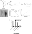
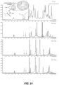
- FIG. 3A-3E a Fe(III)-hydroxamic acid-based assay was used to determine the amount of methacrylate groups in different GelMA samples ( FIG. 3A-3E ).
- Hydroxamic acid forms a brown-red complex with Fe(III) ions, which can serve as a qualitative test for hydroxamic acid species ( FIG. 3A ).
- This class of complexes has an absorption peak centered at around 500 nm ( FIGS. 3B and 3C ).
- Fe(III)-hydroxamic acid complexes has been used to quantify the reactivity of amine groups and hydroxyl groups of lysozymes with several different carboxylic acid anhydrides, 44 along with other analytic applications such as the quantification of ester group residues in poly(vinyl alcohol). 45 Acetohydroxamic acid (AHA) was used as the standard to establish the working curve and it is assumed that the complex of acetohydroxamic acid and Fe(III) ions (FeAHA) have similar spectrophotometric properties with that of N-hydroxymethacrylamide and Fe(III) ions (FeHMA).
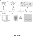
- Visible light crosslinkable Gel-CORE was made by using Eosin-Y (Sigma-Aldrich) as a visible-light activated initiator, triethanolamine (TEOA) (Sigma-Aldrich) as a co-initiator, and vinyl caprolactam (VC) (Sigma-Aldrich) as a catalyst. 15,16 Using this crosslinking system, polymerization of methacryloyl groups on GelMA was initiated through exposure to blue light (450-550 nm, Xenon source) at 100 mW/cm 2 ( FIGS. 4A-4C ).
- Eosin-Y Sigma-Aldrich
- TEOA triethanolamine
- VC vinyl caprolactam
- Eosin-Y is a water-soluble xanthene dye and is a common stain for collagen, the main component of the cornea and sclera.
- This visible light system has a well-established track record of biocompatibility in a range of applications 15-19 and has gained FDA approval for use in the human body in the polyethylene glycol (PEG)-based sealant FocalSeal ® (Genzyme Biosurgical, Cambridge, MA).
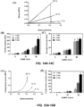
- Visible light crosslinkable Gel-CORE hydrogels show tunable physical properties ( FIGS. 5A-5B ); the elastic modulus of Gel-CORE hydrogels using visible light crosslinking could be tuned from 5-28 kPa ( FIGS. 5A-5B ).
- the swelling ratio could be changed from 7% to 13% (w/w) ( FIG. 6B ).
- GelMA with different degrees of methacryloyl modification (30%-85%) can be used. 14 Then, different concentrations of GelMA prepolymer solutions (5-15 %w/v) can be prepared in phosphate buffered saline (PBS) containing Eosin-Y (0.1-0.5 mM), TEOA (0.5-2 %w/v), and VC (0.5-1.5 %w/v).
- PBS phosphate buffered saline
- Eosin-Y 0.1-0.5 mM
- TEOA 0.1-0.5 mM
- TEOA TEOA
- VC 0.5-1.5 %w/v
- the formulated GelMA prepolymer solutions can be photocrosslinked by exposure to blue light for 20-120 seconds, which matches the absorption spectrum of Eosin-Y ( FIG. 4B ). Varying the GelMA concentration, Eosin-Y/TEOA/VC concentrations, and light exposure time varies the physical properties of the engineered hydrogels.
- the Gel-CORE hydrogels should have similar elasticity and stiffness to the native cornea (Young Modulus: 250-350 kPa).
- the swelling ratio of the hydrogel should be optimized to obtain swelling ratio of ⁇ 20% to ensure that the adhesive preserves its shape after being applying in the corneal defect.
- the swelling ratio can affect the shape, curvature and the smoothness of the sealed defect.
- freeze-dried GelMA produced according to Example 2 was dissolved in PBS at a concentration of 10 % (w/v).
- a photoinitiator mixture of 0.1 % (w/v) Eosin Y, 0.5% (w/v) triethanolamine, and 0.5 % (w/v) vinyl caprolacatam and dissolving at 80°C the prepolymer solution was photocrosslinked to a hydrogel (Gel-CORE) by visible light irradiation (450-550 nm, Xenon source, 100 mW/cm 2 ).
- Gel-CORE hydrogels used explanted rabbit cornea tissues.
- GelMA prepolymer was first applied on incision created on the explanted cornea and then photocrosslinked by exposure to visible light using optimized light exposure time. The burst pressure was then measured by using a pressure sensor after air inflation into the cornea.
- a rabbit cornea was sealed with Gel-CORE (10 % (w/v), prepolymer concentration, 5 mM Eosin-Y, and exposure time of 120 sec.
- the incision created on rabbit cornea was tightly and completely sealed with Gel-CORE and the tissue could be pressurized up to around 3.5 kPa (26 mmHg), which is double of the pressure of healthy eye.
- Gel-CORE samples have a burst pressure higher than 15 kPa (>110 mm Hg), 20-21 a lap shear strength > 100 kPa, adhesion strength > 40 kPa, and photopolymerize in ⁇ 60 seconds of light exposure.
- the burst pressure testing of sealants was adapted from the ASTM standard F2392-04 (standard test method for burst strength of surgical sealants).
- Porcine skin sheets 40 mm * 40 mm
- a circular defect (3 mm in diameter) was created in the center of a pig skin sheet that was placed between two Teflon sheets (35 mm * 35 mm).
- the top Teflon sheet contained a hole (10 mm in diameter) to allow for application of the desired adhesive over the circular defect in the porcine skin sheet ( FIG. 7A ).
- GelMA the prepolymer was irradiated with visible light.
- the collagen sheet was removed and placed into the burst pressure testing system, consisting of pressure detection and recording unit and a syringe pump, which applied air with continuously increasing pressure towards the samples ( FIG. 7B ) .
- the burst pressure testing system consisting of pressure detection and recording unit and a syringe pump, which applied air with continuously increasing pressure towards the samples ( FIG. 7B ) .
- Each tested adhesive group contained five samples.
- porcine skin was blotted dry to remove excess liquid, and each end of the skin strip was fixed onto two poly(methyl methacrylate) slides (30 mm * 60 mm) with Krazy glue (Westerville, OH, USA), leaving a 6 mm section of skin between the slides.
- the porcine skin strip was then cut apart using a razor blade ( FIG. 8A ), and petroleum jelly was applied with a syringe to the ends of the desired adhesive application area. Afterwards, 40 ⁇ l of the adhesive was applied across the 6 mm * 5 mm skin section and, in the case of GelMA, irradiated with visible light ( FIG . 8A ).
- FIG. 8A After 1 hour of incubation in PBS, the two plastic slides were placed into the Instron system grips for tensile testing ( FIG. 8A ). The adhesive strength of a sealant sample was determined at the point of tearing. Each tested adhesive group contained four to seven samples and results are summarized in FIG. 8B .
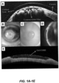
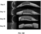
- Gel-CORE was applied to a 3-mm >50%-deep corneal defect (10 % (w/v) Gel-CORE solution containing 0.01% (w/v) Eosin-Y, 0.5% (w/v) TEA, and 0.5% (w/v) VC was used). The solution was exposed to blue light for 120 seconds to form a hydrogel layer on the damaged cornea. After the procedure, eyes were kept in PBS at 4°C. Changes in Gel-CORE over time were assessed using serial evaluations with slit lamp biomicroscopy and OCT. It was noted that for at least 11 days, the bioadhesive remained uncompromised (full thickness and spread retained) and stayed completely attached to the cornea in all tested eyes.
- FIGS. 9A and 9C Slit lamp biomicroscopy showed that during this time, the bioadhesive remained clear with a smooth surface without any biomicroscopic signs of changes in shape or contour ( FIGS. 9A and 9C ).
- OCT confirmed no change in the thickness or shape of Gel-CORE ( FIGS. 9B and 9D ).
- the corneal tissue in PBS started to degrade (as expected from necrosis due to prolonged storage in PBS), at which point the Gel-CORE attachment to the cornea began to get compromised.
- a corneal injury model in New Zealand white rabbits was used by creating a 50%-deep corneal defect. After general anesthesia of the rabbit using intramuscular injection of ketamine and xylazine, a circular 50%-deep corneal defect was created in the right eye by a 3-mm biopsy punch. Then, a surgical crescent knife was used to perform a lamellar keratectomy. After removing the anterior lamella, the defect surface was dried using a surgical microsponge. Then, 10 ⁇ l of the bioadhesive solution was instilled to fill the corneal defect. A microsponge was then used to smooth over the extra solution.
- bioadhesive transparency which measures optical degradation (as evaluated by slit lamp biomicroscopy using Fantes grading scale, 22 which is based on visibility of iris details); and (ii) bioadhesive thickness (as measured by OCT, described below).
- Retention is a function of two parameters, degradation and adhesiveness. Either degradation and/or suboptimal adhesiveness can lead to loss of gel retention.
- OCT technology was used, as described below, to evaluate (i) the presence of the bioadhesive covering the corneal defect; and (ii) the thickness of any gap between the bioadhesive and corneal epithelium or stroma over 4 weeks of follow-up.
- OCT optical Coherence Tomography
- a spectral-domain OCT (Spectralis, Heidelberg Engineering, GmbH, Germany), which has an axial resolution of 3.9-7 ⁇ m, was used.
- Line scans (8 mm long) were performed at 0, 45, 90, and 135 degrees in the central cornea ( FIG. 10A ).
- the thickness (in microns) of the bioadhesive (in the operated eye) and of the cornea (in the unoperated fellow eye) was measured in the center of the cornea and at 1 mm away from the center in both directions ( FIG. 10B ).
- the thickness of any gap between the bioadhesive and corneal tissue was measured in microns.
- the optimal bioadhesive for cornea repair is not only non-toxic for corneal cells, but also permits cells to integrate into the biomaterial for long-term integration and to prevent extrusion.
- the in vitro cytocompatibility and integrative capacity of Gel-CORE was determined by using the two most abundant cell types in the cornea including keratocytes and corneal epithelial cells. Keratocytes and corneal epithelial cells were cultured using 2D and 3D culture systems. The biocompatibility and integrative capacity of Gel-CORE was assessed in vivo by investigating the effects of the bioadhesive on corneal cells, as well as the migration of corneal cells into the bioadhesive over time.
- FIGS. 11A-11G Corneal keratocytes were incorporated within a representative Gel-CORE composition, showing ⁇ 95% cell survival using 2D culture systems, as well as proliferation and migration of the keratocytes when grown either on top or within the Gel-CORE construct ( FIG. 11G ).
- Gel-CORE was constructed following exposure to visible light as detailed herein. Then, the gels were seeded with the epithelial cells at cell densities ranging from 1 ⁇ 10 6 to 1 ⁇ 10 8 cells/mL and were incubated for 14 days. Media was changed every other day. Cell viability was evaluated on days 1, 4, 7 and 14 by using calcein-AM/ethidium homodimer Live/Dead assays. 23 Actin/DAPI staining was used to assess cellular attachment and spreading as explained previously. 13,24-26 In addition, the metabolic activities of the cells were assessed by using PrestoBlue assay followed by absorbance readings on a microplate spectrophotometer on days 1, 4, 7 and 14.
- keratocytes were mixed with GelMA prepolymer solution at concentrations ranging from 1 ⁇ 10 6 -1 ⁇ 10 8 cells/mL. The mixture was then exposed to light to form cell-laden Gel-CORE adhesive. The gels was then washed 3 times with PBS and incubated for 14 days in medium in a culture incubator at 37 °C. Cellular viability (Live/Dead assay), cell attachment and spreading (Actin/DAPI), proliferation (Picogreen assay), collagen deposition (Picrosirius Red), and corneal tissue formation (Haematoxylin and eosin staining) was assessed on days 1, 4, 7, and 14. Based on these 3D studies, Gel-CORE was shown to be cytocompatible and will promote cornea tissue formation.
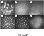
- Slit lamp biomicroscopy and IVCM were performed under general anesthesia. As described herein, slit lamp examination and photography were used to assess transparency of the cornea and the bioadhesive. In addition, to assess the migration of epithelium over the bioadhesive, slit lamp photography with fluorescein staining was performed, and the area of corneal epithelial defect over the bioadhesive was measured using ImageJ's Measure Area tool for each time point. In Vivo Confocal Microscopy (IVCM) was employed to evaluate cellular changes and migration in the same rabbits over time without sacrificing the animal. This is a non-invasive imaging modality which provides high-resolution images at the cellular level from the cornea in live animals ( FIGS . 12A-12F ).
- a laser scanning IVCM Heidelberg Retina Tomograph 3 with Rostock Cornea Module, Heidelberg, Germany
- a 670 nm diode laser and has a resolution of 1 ⁇ m. It provides images that represent a corneal area of 400 x 400 ⁇ m.
- IVCM-based readouts the following were scaned and examined: (i) the 1.5 mm-central cornea (over the bioadhesive, cyanoacrylate glue, or original defect in the control group); and (ii) the corneal tissue surrounding the 1 mm circumference of the adhesive in 4 quadrants (superior, nasal, inferior, and temporal).
- Sequence Mode was used which automatically acquires 100 consecutive images per sequence.
- corneal layers were imaged in each scan.
- Two Sequence Mode scans were obtained in each of five locations (central, superior, nasal, inferior, and temporal).
- image analysis in the central cornea 5 images were randomly selected from different depths in each scan (totally 10 images).
- image analysis in the cornea around the adhesive one randomly selected image from each corneal layer (epithelium, subbasal layer, and stroma) was selected from each individual sequence scan (totally 8 images per layer).
- the density of epithelial cells, subbasal inflammatory cells, and stromal keratocytes were measured by a masked observer using Image J software as previously reported. 30-35
- the density of corneal nerves was also assessed by a masked observer using NeuronJ software as previously reported. 30, 36, 37
- H&E histologic evaluation using hematoxylin and eosin staining was performed on cryosections of the harvested corneas. From each cornea, 5 sections were obtained from the central cornea containing both the defect/adhesive location and the surrounding corneal tissue. For image analysis, all sections were evaluated by a masked observer. Migration of corneal epithelial cells over the adhesive was determined. In addition, the density of stromal keratocytes and inflammatory cells was determined in 10 randomly selected areas within the adhesive (both in the center and periphery of the adhesive) in addition to 10 randomly selected areas of the surrounding cornea at 200 ⁇ m from the margin of the adhesive.
- Immunohistochemical staining was also performed on cryosections of the harvested cornea with antibodies against the following: ⁇ -tubulin III (2G10 Ab; Abcam), beta 2 (CD18) integrin (L13/64 for inflammatory cells; GeneTex), and CD31 (polyclonal anti-CD31 for blood vessels; Abcam).
- the density of these cells was determined by a masked observer in both the adhesive-applied and surrounding corneal matrix as described for the H&E staining. For this, serial sections from 10 randomly selected areas within the adhesive as well as 10 randomly selected areas of the surrounding cornea within 200 ⁇ m of the adhesive were used for analysis. Mean and Standard Deviation (SD) was measured for each metric.
- SD Standard Deviation
- the slit lamp, IVCM, histologic, and immunohistochemical findings were compared between the three groups at each time point to determine the biocompatibility and integrative capacity of Gel-CORE for filling corneal defects.
- These comparisons between the Gel-CORE group and the control group helped determine whether Gel-CORE caused more or less inflammation and tissue damage than expected from secondary- intention healing (which is also included as a control).
- comparisons between the Gel-CORE group and the cyanoacrylate group showed whether the potential tissue damage is less in the Gel-CORE group compared to the current standard of care adhesive.
- comparisons between different time points showed whether the integration of corneal cells into Gel-CORE developed over time and whether any potential tissue damage caused by the adhesive subsided or aggravated over time.
- the critical mechanical properties of the hydrogel can be precisely controlled to derive formulations with tensile and compressive moduli that are comparable to the native cornea ( FIGS. 14A-14C , 15A and 15B ).
- optimized concentrations of Eosin Y (0.05mM), TEOA (0.4% w/v), and VC (0.4% w/v), are used for the following experiments.
- Adhesive properties of engineered bioadhesives were used to obtain a comprehensive estimation of the sealing ability of 20%w/v visible light crosslinked GelMA hydrogels formed at various visible light exposure times. The ex vivo tests were performed to measure the burst pressures of rabbit corneas with 2-mm full-thickness incisions ( FIGS . 16A and 16B ). The burst pressure of the engineered GelMA was higher than 200mmHg, almost 10 times the pressure of a healthy eye, and significantly higher than the burst pressure of the commercial control, ReSure ® ( FIG. 16B ).
Landscapes
- Health & Medical Sciences (AREA)
- Life Sciences & Earth Sciences (AREA)
- Chemical & Material Sciences (AREA)
- Engineering & Computer Science (AREA)
- General Health & Medical Sciences (AREA)
- Biomedical Technology (AREA)
- Medicinal Chemistry (AREA)
- Veterinary Medicine (AREA)
- Public Health (AREA)
- Animal Behavior & Ethology (AREA)
- Epidemiology (AREA)
- Oral & Maxillofacial Surgery (AREA)
- Transplantation (AREA)
- Dermatology (AREA)
- Chemical Kinetics & Catalysis (AREA)
- Botany (AREA)
- Cell Biology (AREA)
- Zoology (AREA)
- Ophthalmology & Optometry (AREA)
- Dispersion Chemistry (AREA)
- Urology & Nephrology (AREA)
- Vascular Medicine (AREA)
- Bioinformatics & Cheminformatics (AREA)
- General Chemical & Material Sciences (AREA)
- Nuclear Medicine, Radiotherapy & Molecular Imaging (AREA)
- Organic Chemistry (AREA)
- Pharmacology & Pharmacy (AREA)
- Medicinal Preparation (AREA)
- Materials For Medical Uses (AREA)
- Prostheses (AREA)
- Acyclic And Carbocyclic Compounds In Medicinal Compositions (AREA)
Applications Claiming Priority (3)
| Application Number | Priority Date | Filing Date | Title |
|---|---|---|---|
| US201662292752P | 2016-02-08 | 2016-02-08 | |
| EP17750660.7A EP3413940B1 (de) | 2016-02-08 | 2017-02-08 | Bioadhäsiv für die hornhautreparatur |
| PCT/US2017/016917 WO2017139318A1 (en) | 2016-02-08 | 2017-02-08 | Bioadhesive for corneal repair |
Related Parent Applications (1)
| Application Number | Title | Priority Date | Filing Date |
|---|---|---|---|
| EP17750660.7A Division EP3413940B1 (de) | 2016-02-08 | 2017-02-08 | Bioadhäsiv für die hornhautreparatur |
Publications (2)
| Publication Number | Publication Date |
|---|---|
| EP4574181A2 true EP4574181A2 (de) | 2025-06-25 |
| EP4574181A3 EP4574181A3 (de) | 2025-09-10 |
Family
ID=59563503
Family Applications (2)
| Application Number | Title | Priority Date | Filing Date |
|---|---|---|---|
| EP17750660.7A Active EP3413940B1 (de) | 2016-02-08 | 2017-02-08 | Bioadhäsiv für die hornhautreparatur |
| EP25172054.6A Pending EP4574181A3 (de) | 2016-02-08 | 2017-02-08 | Bioadhäsiv für die hornhautreparatur |
Family Applications Before (1)
| Application Number | Title | Priority Date | Filing Date |
|---|---|---|---|
| EP17750660.7A Active EP3413940B1 (de) | 2016-02-08 | 2017-02-08 | Bioadhäsiv für die hornhautreparatur |
Country Status (6)
| Country | Link |
|---|---|
| US (3) | US11058800B2 (de) |
| EP (2) | EP3413940B1 (de) |
| JP (3) | JP6887435B2 (de) |
| AU (3) | AU2017217454B2 (de) |
| ES (1) | ES3027197T3 (de) |
| WO (1) | WO2017139318A1 (de) |
Families Citing this family (23)
| Publication number | Priority date | Publication date | Assignee | Title |
|---|---|---|---|---|
| ES3027197T3 (en) * | 2016-02-08 | 2025-06-13 | Brigham & Womens Hospital Inc | Bioadhesive for corneal repair |
| CN108187130B (zh) * | 2017-09-15 | 2020-08-18 | 海宁侏罗纪生物科技有限公司 | 一种用于生物损伤修复或止血的试剂及其应用 |
| CA3111444A1 (en) | 2018-09-04 | 2020-03-12 | Massachusetts Eye And Ear Infirmary | Light activated adhesive scaffold |
| CA3115998A1 (en) * | 2018-10-16 | 2020-04-23 | The Schepens Eye Research Institute, Inc. | Bioadhesive for soft tissue repair |
| CN109821075B (zh) * | 2019-01-07 | 2020-09-08 | 华中科技大学 | 一种生物材料及其制备方法与作为骨缺损修复材料的应用 |
| AU2020280018B2 (en) * | 2019-05-20 | 2023-12-07 | The Regents Of The University Of California | Methods for improving the tissue sealing properties of hydrogels and the use thereof |
| US20220273772A1 (en) * | 2019-07-26 | 2022-09-01 | Pandorum Technologies Private Limited | Bioengineered formulation, process for preparing and implementations thereof |
| CN114502110A (zh) * | 2019-09-13 | 2022-05-13 | 霍利斯蒂克医疗公司 | 医疗植入物、递送装置、制作医疗植入物的方法以及递送医疗植入物的方法 |
| CA3157214A1 (en) * | 2019-10-08 | 2021-07-01 | Massachusetts Eye And Ear Infirmary | Ocular sealants and methods of using the same |
| WO2021183474A1 (en) * | 2020-03-09 | 2021-09-16 | University Of Houston System | Inflammatory bowel disease stem cells, agents which target ibd stem cells, and uses related thereto |
| WO2021195129A1 (en) | 2020-03-25 | 2021-09-30 | The Regents Of The University Of California | Gelatin methacryloyl-based microneedle patches for delivery of water-insoluble drugs |
| BE1028324B1 (nl) * | 2020-05-19 | 2021-12-22 | Rousselot | Ge(meth)acryleerd gelatine laag in endotoxines |
| US11918703B2 (en) * | 2020-08-13 | 2024-03-05 | Universidad De Los Andes | Extrudable photocrosslinkable hydrogel and method for its preparation |
| CN113456895B (zh) * | 2021-07-20 | 2022-05-06 | 广州市朴道联信生物科技有限公司 | 一种GelMA-胶原双网络抗菌角膜修复材料及其制备方法和应用 |
| EP4401802A1 (de) | 2021-09-15 | 2024-07-24 | Gelmedix, Inc. | Gelmapolymerzusammensetzungen und verwendungen davon |
| TW202327656A (zh) | 2021-09-15 | 2023-07-16 | 美商蓋爾密迪斯公司 | 包含皮質類固醇之gelma聚合物組合物 |
| EP4511075A1 (de) | 2022-04-20 | 2025-02-26 | Gelmedix, Inc. | Gelma-polymerzusammensetzungen mit zellen |
| US20260061091A1 (en) * | 2022-08-19 | 2026-03-05 | Purdue Research Foundation | Mussel-inspired tissue adhesives and methods of use thereof |
| CN116196464B (zh) * | 2023-03-07 | 2024-04-16 | 北京大学人民医院 | 一种可见光交联的水凝胶胶粘剂及其制备方法和应用 |
| TW202529823A (zh) | 2023-10-18 | 2025-08-01 | 美商蓋爾密迪斯公司 | 包含細胞之丙烯酸化明膠聚合物組合物 |
| KR20250062144A (ko) * | 2023-10-30 | 2025-05-08 | 주식회사 바이오브릭스 | 세포외기질 기반 접착제 조성물 |
| WO2025114517A1 (en) * | 2023-11-30 | 2025-06-05 | Universiteit Antwerpen | Crosslinkable liquid composition for use in a method of myopia management in a subject |
| KR20260046579A (ko) * | 2024-09-30 | 2026-04-07 | 주식회사 바이오브릭스 | 메타크릴로일 치환 젤라틴을 포함하는 세포외기질 기반 접착제 조성물 |
Family Cites Families (25)
| Publication number | Priority date | Publication date | Assignee | Title |
|---|---|---|---|---|
| US5064430A (en) | 1985-10-31 | 1991-11-12 | Uab Research Foundation | Polynonapeptide bioelastomers having an increased elastic modulus |
| GB9110783D0 (en) | 1991-05-18 | 1991-07-10 | Ciba Geigy | Adhesives |
| US7871637B2 (en) | 1996-08-27 | 2011-01-18 | Baxter International Inc. | Dry hemostatic compositions and methods for their preparation |
| US7435425B2 (en) | 2001-07-17 | 2008-10-14 | Baxter International, Inc. | Dry hemostatic compositions and methods for their preparation |
| DE69720243T2 (de) | 1996-11-05 | 2004-02-12 | Challenge Bioproducts Co., Ltd. | Chemische modifikation von biomedizinischen materialien mit genipin |
| EP0986408B1 (de) | 1997-06-03 | 2005-05-11 | Innogenetics N.V. | Neue arzneimittel auf der basis von polymeren aus mit methacrylamid modifizierter gelatine |
| AUPP939299A0 (en) | 1999-03-23 | 1999-04-15 | University Of Melbourne, The | Polymer gels and methods for their preparation |
| US20040110439A1 (en) * | 2001-04-20 | 2004-06-10 | Chaikof Elliot L | Native protein mimetic fibers, fiber networks and fabrics for medical use |
| US7303757B2 (en) | 2002-08-06 | 2007-12-04 | Matrix Medical, Llc | Biocompatible phase invertable proteinaceous compositions and methods for making and using the same |
| JP2005154477A (ja) | 2003-11-20 | 2005-06-16 | Tottori Univ | 紫外線硬化型キトサン誘導体を用いた医療用接着剤および医療用被覆剤 |
| WO2006078629A2 (en) | 2005-01-18 | 2006-07-27 | Duke University | In-situ crosslinkable elastin-like polypeptides for defect filling in cartilaginous tissue repair |
| EP1973513B1 (de) | 2005-12-28 | 2010-11-10 | DENTSPLY International Inc. | Lichthärtbares knochenwachstumsmaterial zur behandlung von zahnknochendefekten |
| US7854923B2 (en) | 2006-04-18 | 2010-12-21 | Endomedix, Inc. | Biopolymer system for tissue sealing |
| DE102006033167A1 (de) | 2006-07-10 | 2008-01-24 | Gelita Ag | Verwendung von Gelatine und einem Vernetzungsmittel zur Herstellung eines vernetzenden medizinischen Klebers |
| US8314211B2 (en) | 2009-04-07 | 2012-11-20 | George Falus | Tissue sealant for use in non compressible hemorrhage |
| JP5796860B2 (ja) | 2009-12-22 | 2015-10-21 | ライフボンド リミテッドLifebond Ltd | 架橋マトリックスの特性を調節するための酵素的架橋剤の改変 |
| EP2523656A4 (de) | 2010-01-15 | 2013-12-11 | Univ Utah Res Found | Vernetzte hydrogele sowie verfahren zu ihrer herstellung und verwendung |
| MX352875B (es) | 2010-06-01 | 2017-12-13 | Baxter Healthcare Sa | Proceso para elaborar composiciones hemostaticas secas y estables. |
| AU2011260258B2 (en) | 2010-06-01 | 2015-07-09 | Baxter Healthcare S.A. | Process for making dry and stable hemostatic compositions |
| US10052350B2 (en) * | 2011-09-16 | 2018-08-21 | Wake Forest University Health Sciences | Fabrication of gelatin hydrogel sheet for the transplantation of corneal endothelium |
| CN104349797B (zh) | 2012-06-12 | 2017-10-27 | 弗罗桑医疗设备公司 | 干止血组合物 |
| WO2014026142A1 (en) | 2012-08-10 | 2014-02-13 | Arizona Board Of Regents For And On Behalf Of Arizona State University | Methods and compostions for tissue adhesives |
| US10814032B2 (en) | 2014-08-08 | 2020-10-27 | The Brigham And Women's Hospital, Inc. | Elastic biopolymer and use as a tissue adhesive |
| KR101562366B1 (ko) | 2014-11-25 | 2015-10-23 | 부산대학교 산학협력단 | 메타크릴레이트화된 젤라틴을 포함하는, 심근세포분화 유도용 세포 지지체 |
| ES3027197T3 (en) * | 2016-02-08 | 2025-06-13 | Brigham & Womens Hospital Inc | Bioadhesive for corneal repair |
-
2017
- 2017-02-08 ES ES17750660T patent/ES3027197T3/es active Active
- 2017-02-08 WO PCT/US2017/016917 patent/WO2017139318A1/en not_active Ceased
- 2017-02-08 EP EP17750660.7A patent/EP3413940B1/de active Active
- 2017-02-08 US US16/070,643 patent/US11058800B2/en active Active
- 2017-02-08 AU AU2017217454A patent/AU2017217454B2/en active Active
- 2017-02-08 JP JP2018541314A patent/JP6887435B2/ja active Active
- 2017-02-08 EP EP25172054.6A patent/EP4574181A3/de active Pending
-
2021
- 2021-05-18 JP JP2021083614A patent/JP2021130012A/ja active Pending
- 2021-06-08 US US17/342,409 patent/US20210353829A1/en not_active Abandoned
- 2021-08-10 AU AU2021215131A patent/AU2021215131B2/en active Active
-
2023
- 2023-05-16 JP JP2023080509A patent/JP7626793B2/ja active Active
- 2023-08-23 AU AU2023219872A patent/AU2023219872B2/en active Active
- 2023-09-21 US US18/471,748 patent/US20240269347A1/en active Pending
Non-Patent Citations (46)
| Title |
|---|
| ALLEYENE, C. H., JR. ET AL.: "Efficacy and biocompatibility of a photopolymerized, synthetic, absorbable hydrogel as a dural sealant in a canine craniotomy model", J NEUROSURG, vol. 88, 1998, pages 308 - 313 |
| ANDERMANN G, ZIMMERMANN G, SCHILLING E.: " Application of iron(III)-hydroxamic acid complexes in the spectrophotometric determination of poly(vinyl alcohol) in pharmaceutical preparations.", ANALYST, vol. 105, 1980, pages 575 - 80, XP093020677, DOI: 10.1039/AN9800500575 |
| ANNABI, N. ET AL.: "Engineered cell-laden human protein-based elastomer", BIOMATERIALS, vol. 34, 2013, pages 5496 - 5505, XP028535320, DOI: 10.1016/j.biomaterials.2013.03.076 |
| ANNABI, N. ET AL.: "Highly Elastic Micropatterned Hydrogel for Engineering Functional Cardiac Tissue", ADV FUNCT MATER, vol. 23, 2013, pages 4950 - 4959, XP072773008, DOI: 10.1002/adfm.201300570 |
| ANNABI, N. ET AL.: "Synthesis of highly porous crosslinked elastin hydrogels and their interaction with fibroblasts in vitro", BIOMATERIALS, vol. 30, 2009, pages 4550 - 4557, XP026501291, DOI: 10.1016/j.biomaterials.2009.05.014 |
| ANNABI, N.MITHIEUX, S. M.WEISS, A. S.DEHGHANI, F.: "Cross-linked open-pore elastic hydrogels based on tropoelastin, elastin and high pressure CO", BIOMATERIALS, vol. 31, 2010, pages 1655 - 1665, XP026827523 |
| ANNABI, N.MITHIEUX, S. M.WEISS, A. S.DEHGHANI, F.: "The fabrication of elastin-based hydrogels using high pressure CO", BIOMATERIALS, vol. 30, 2009, pages 1 - 7, XP025561254, DOI: 10.1016/j.biomaterials.2008.09.031 |
| BERNAD ANIETO MAVIOQUE APALACIÁAN E.: "Modification of the amino and hydroxyl groups of lysozyme with carboxylic acid anhydrides: a comparative study", BIOCHIMICA ET BIOPHYSICA ACTA (BBA) - PROTEIN STRUCTURE AND MOLECULAR ENZYMOLOGY, vol. 873, 1986, pages 350 - 5, XP025210097, DOI: 10.1016/0167-4838(86)90083-X |
| CAMCI-UNAL, G. ET AL.: "Hydrogel surfaces to promote attachment and spreading of endothelial progenitor cells", JOURNAL OF TISSUE ENGINEERING AND REGENERATIVE MEDICINE, vol. 7, 2013, pages 337 - 347 |
| CAMCI-UNAL, G.CUTTICA, D.ANNABI, N.DEMARCHI, D.KHADEMHOSSEINI, A.: "Synthesis and characterization of hybrid hyaluronic acid-gelatin hydrogels", BIOMACROMOLECULES, vol. 14, 2013, pages 1085 - 1092, XP093071947, DOI: 10.1021/bm3019856 |
| CARLSON, A. N.WILHELMUS, K. R.: "Giant papillary conjunctivitis associated with cyanoacrylate glue", AMERICAN JOURNAL OF OPHTHALMOLOGY, vol. 104, 1987, pages 437 - 438 |
| CARNAHAN, M. A.MIDDLETON, C.KIM, J.KIM, T.GRINSTAFF, M. W.: "Hybrid dendritic-linear polyester-ethers for in situ photopolymerization", J AM CHEM SOC, vol. 124, 2002, pages 5291 - 5293, XP002602720, DOI: 10.1021/ja025576y |
| CAVANAUGH, T. B. GOTTSCH, J. D.: "Infectious keratitis and cyanoacrylate adhesive.", AMERICAN JOURNAL OF OPHTHALMOLOGY, vol. 111, 1991, pages 466 - 472 |
| CHA CSHIN SRGAO XANNABI NDOKMECI MRTANG XSKHADEMHOSSEINI A.: "Controlling mechanical properties of cell-laden hydrogels by covalent incorporation of graphene oxide", SMALL, vol. 10, no. 3, 11 October 2013 (2013-10-11), pages 514 - 23 |
| COLYER, M. H.CHUN, D. W.BOWER, K. S.DICK, J. S.WEICHEL, E. D.: "Perforating globe injuries during operation Iraqi Freedom", OPHTHALMOLOGY, vol. 115, 2008, pages 2087 - 2093 |
| ELISSEEFF, J.: "Transdermal photopolymerization for minimally invasive implantation.", PROC NATL ACAD SCI, vol. 96, 1999, pages 3104 - 3107, XP001009855, DOI: 10.1073/pnas.96.6.3104 |
| FANTES, F. E. ET AL.: "Wound healing after excimer laser keratomileusis (photorefractive keratectomy) in monkeys", ARCHIVES OF OPHTHALMOLOGY, vol. 108, 1990, pages 665 - 675 |
| FOGLE, J. A.KENYON, K. R.FOSTER, C. S.: "Tissue adhesive arrests stromal melting in the human cornea", AMERICAN JOURNAL OF OPHTHALMOLOGY, vol. 89, 1980, pages 795 - 802, XP002909981 |
| HARIPRASAD, S. M.SINGH, A.: "Polyethylene glycol hydrogel polymer sealant for vitrectomy surgery: an in vitro study of sutureless vitrectomy incision closure", ARCHIVES OF OPHTHALMOLOGY, vol. 129, 2011, pages 322 - 325 |
| HIDA, T.SHETA, S. M.PROIA, A. D.MCCUEN, B. W.: "Retinal toxicity of cyanoacrylate tissue adhesive in the rabbit", RETINA, vol. 8, 1988, pages 148 - 153 |
| HJORTNAES JCAMCI-UNAL GHUTCHESON JDJUNG SMSCHOEN FJKLUIN JAIKAWA EKHADEMHOSSEINI A: "Directing Valvular Interstitial Cell Myofibroblast-Like Differentiation in a Hybrid Hydrogel Platform", ADV HEALTHC MATER, 24 June 2014 (2014-06-24) |
| J. MATER, CHEM. B, vol. 4, 2016, pages 2304 - 2313 |
| JUN, E. J.KIM, J. H.PURCELL, T. L.SCHANZLIN, D. J.: "Comparison of bursting pressure after scleral tunnel incision sealed with sutures or an adherent ocular bandage in human globes", THE JOURNAL OF INTERNATIONAL MEDICAL RESEARCH, vol. 40, 2012, pages 756 - 760 |
| KHEIRKHAH, A. ET AL.: "Comparison of Standard Versus Wide-Field Composite Images of the Corneal Subbasal Layer by In Vivo Confocal Microscopy", INVESTIGATIVE OPHTHALMOLOGY & VISUAL SCIENCE, vol. 56, 2015, pages 5801 - 5807 |
| KHEIRKHAH, A. ET AL.: "Corneal Epithelial Immune Dendritic Cell Alterations in Subtypes of Dry Eye Disease: A Pilot In Vivo Confocal Microscopic Study", INVESTIGATIVE OPHTHALMOLOGY & VISUAL SCIENCE, vol. 56, 2015, pages 7179 - 7185 |
| KHEIRKHAH, A. ET AL.: "Effects of corneal nerve density on the response to treatment in dry eye disease", OPHTHALMOLOGY, vol. 122, 2015, pages 662 - 668 |
| KHEIRKHAH, A. ET AL.: "Reduced Corneal Endothelial Cell Density in Patients With Dry Eye Disease", AMERICAN JOURNAL OF OPHTHALMOLOGY, vol. 159, 2015, pages 1022 - 1026 |
| KHEIRKHAH, A.SABOO, U. S.MARMALIDOU, A.DANA, R.: "Overestimation of Corneal Endothelial Cell Density in Smaller Frame Sizes in In Vivo Confocal Microscopy", CORNEA, 2015 |
| LEAHEY, A. B.GOTTSCH, J. D.STARK, W. J.: "Clinical experience with N-butyl cyanoacrylate (Nexacryl) tissue adhesive", OPHTHALMOLOGY, vol. 100, 1993, pages 173 - 180 |
| MONZYK BCRUMBLISS AL.: "Mechanism of ligand substitution on high-spin iron(III) by hydroxamic acid chelators. Thermodynamic and kinetic studies on the formation and dissociation of a series of monohydroxamatoiron(III) complexes", JOURNAL OF THE AMERICAN CHEMICAL SOCIETY, vol. 101, 1979, pages 6203 - 13 |
| MUNOZ ZSHIH HLIN C-C.: "Gelatin hydrogels formed by orthogonal thiol-norbornene photochemistry for cell encapsulation", BIOMATERIALS SCIENCE, vol. 2, 2014, pages 1063 - 72, XP055520286, DOI: 10.1039/C4BM00070F |
| NAKAYAMA, Y, KAMEO, T.OHTAKA, A.HIRANO, Y: "Enhancement of visible light-induced gelation of photocurable gelatin by addition of polymeric amine", JOURNAL OF PHOTOCHEMISTRY AND PHOTOBIOLOGY A: CHEMISTRY, vol. 177, 2006, pages 205 - 211, XP028008550, DOI: 10.1016/j.jphotochem.2005.05.030 |
| NICHOL, J. W.: "Cell-laden microengineered gelatin methacrylate hydrogels", BIOMATERIALS, vol. 31, 2010, pages 5536 - 5544, XP002725666, DOI: 10.1016/j.biomaterials.2010.03.064 |
| NIKKHAH MESHAK NZORLUTUNA PANNABI NCASTELLO MKIM KDOLATSHAHI-PIROUZ AEDALAT FBAE HYANG Y: "Directed endothelial cell morphogenesis in micropatterned gelatin methacrylate hydrogels", BIOMATERIALS, vol. 33, no. 35, 24 September 2012 (2012-09-24), pages 9009 - 18 |
| ORBAN, J. M.FAUCHER, K. M.DLUHY, R. A.CHAIKOF, E. L.: "Cytomimetic Biomaterials. 4. In-Situ Photopolymerization of Phospholipids on an Alkylated Surface", MACROMOLECULES, vol. 33, 2000, pages 4205 - 4212, XP002369180, DOI: 10.1021/ma9915780 |
| PAPATHEOFANIS, F. J.: "Prothrombotic cytotoxicity of cyanoacrylate tissue adhesive", JOURNAL OF SURGICAL RESEARCH, vol. 47, 1989, pages 309 - 312, XP026300359, DOI: 10.1016/0022-4804(89)90140-6 |
| PATEL, D. V. MCGHEE, C. N.: "Quantitative analysis of in vivo confocal microscopy images: ", SURVEY OF OPHTHALMOLOGY, vol. 58, 2013, pages 466 - 475 |
| PETROLL, W. M.ROBERTSON, D. M.: "In Vivo Confocal Microscopy of the Cornea: New Developments in Image Acquisition, Reconstruction, and Analysis Using the HRT-Rostock Corneal Module", THE OCULAR SURFACE, vol. 13, 2015, pages 187 - 203 |
| RSC ADV., vol. 4, 2014, pages 21997 |
| SHIN SRBAE HCHA JMMUN JYCHEN YCTEKIN HSHIN HFARSHCHI SDOKMECI MRTANG S: "Carbon nanotube reinforced hybrid microgels as scaffold materials for cell encapsulation", ACS NANO, vol. 6, no. 1, 20 December 2011 (2011-12-20), pages 362 - 72, XP055487228, DOI: 10.1021/nn203711s |
| SIEGAL, J. E.ZAIDMAN, G. W.: "Surgical removal of cyanoacrylate adhesive after accidental instillation in the anterior chamber", OPHTHALMIC SURGERY, vol. 20, 1989, pages 179 - 181 |
| THACH, A. B.: "Intraocular foreign body injuries during Operation Iraqi Freedom", OPHTHALMOLOGY, vol. 112, 2005, pages 1829 - 1833, XP005104108, DOI: 10.1016/j.ophtha.2005.04.024 |
| VISSER JGAWLITTA DBENDERS KETOMA SMPOURAN BVAN WEEREN PRDHERT WJMALDA J.: "Endochondral bone formation in gelatin methacrylamide hydrogel with embedded cartilage-derived matrix particles", BIOMATERIALS, vol. 37, 23 October 2014 (2014-10-23), pages 174 - 182 |
| WEISS, J. L.WILLIAMS, P.LINDSTROM, R. L.DOUGHMAN, D. J.: "The use of tissue adhesive in corneal perforations", OPHTHALMOLOGY, vol. 90, 1983, pages 610 - 615 |
| WHITCHER, J. P., SRINIVASAN, M. UPADHYAY, M. P.: " Corneal blindness: a global perspective.", BULLETIN OF THE WORLD HEALTH ORGANIZATION, vol. 79, 2001, pages 214 - 221 |
| ZHAO, X: "Photocrosslinkable Gelatin Hydrogel for Epidermal Tissue Engineering.", ADVANCED HEALTHCARE MATERIALS, 2015 |
Also Published As
| Publication number | Publication date |
|---|---|
| EP4574181A3 (de) | 2025-09-10 |
| JP6887435B2 (ja) | 2021-06-16 |
| WO2017139318A1 (en) | 2017-08-17 |
| AU2023219872B2 (en) | 2025-12-11 |
| EP3413940A4 (de) | 2020-07-15 |
| JP2023100990A (ja) | 2023-07-19 |
| US20190022280A1 (en) | 2019-01-24 |
| JP7626793B2 (ja) | 2025-02-04 |
| AU2017217454B2 (en) | 2021-05-27 |
| AU2023219872A1 (en) | 2023-09-14 |
| ES3027197T3 (en) | 2025-06-13 |
| JP2019512283A (ja) | 2019-05-16 |
| EP3413940A1 (de) | 2018-12-19 |
| US20210353829A1 (en) | 2021-11-18 |
| US11058800B2 (en) | 2021-07-13 |
| JP2021130012A (ja) | 2021-09-09 |
| AU2021215131A1 (en) | 2021-09-23 |
| CA3013782A1 (en) | 2017-08-17 |
| EP3413940C0 (de) | 2025-05-07 |
| EP3413940B1 (de) | 2025-05-07 |
| AU2021215131B2 (en) | 2023-07-20 |
| AU2017217454A1 (en) | 2018-07-26 |
| US20240269347A1 (en) | 2024-08-15 |
Similar Documents
| Publication | Publication Date | Title |
|---|---|---|
| AU2023219872B2 (en) | Bioadhesive for corneal repair | |
| Shen et al. | Dual-crosslinked regenerative hydrogel for sutureless long-term repair of corneal defect | |
| AU2019361962B2 (en) | Bioadhesive for soft tissue repair | |
| US12121630B2 (en) | Light activated adhesive scaffold | |
| US10814032B2 (en) | Elastic biopolymer and use as a tissue adhesive | |
| US20240050621A1 (en) | Ocular sealants and methods of using the same | |
| US20240424163A1 (en) | Functionalized, decellularized corneal extracellular matrix hydrogels for ocular tissue treatment | |
| HK40126739A (en) | Bioadhesive for corneal repair | |
| CA3013782C (en) | Bioadhesive for corneal repair | |
| US20250195446A1 (en) | Antibacterial drug-eluting compositions and methods | |
| WO2025184657A1 (en) | Ha-dendrimer cornea formulation for regeneration of corneal tissue | |
| Pires Moedas De Almeida Barroso | Development of photosensitive biopolymers for ocular tissue engineering |
Legal Events
| Date | Code | Title | Description |
|---|---|---|---|
| PUAI | Public reference made under article 153(3) epc to a published international application that has entered the european phase |
Free format text: ORIGINAL CODE: 0009012 |
|
| STAA | Information on the status of an ep patent application or granted ep patent |
Free format text: STATUS: THE APPLICATION HAS BEEN PUBLISHED |
|
| AC | Divisional application: reference to earlier application |
Ref document number: 3413940 Country of ref document: EP Kind code of ref document: P |
|
| AK | Designated contracting states |
Kind code of ref document: A2 Designated state(s): AL AT BE BG CH CY CZ DE DK EE ES FI FR GB GR HR HU IE IS IT LI LT LU LV MC MK MT NL NO PL PT RO RS SE SI SK SM TR |
|
| REG | Reference to a national code |
Ref country code: DE Ref legal event code: R079 Free format text: PREVIOUS MAIN CLASS: A61L0027380000 Ipc: A61L0027520000 |
|
| PUAL | Search report despatched |
Free format text: ORIGINAL CODE: 0009013 |
|
| AK | Designated contracting states |
Kind code of ref document: A3 Designated state(s): AL AT BE BG CH CY CZ DE DK EE ES FI FR GB GR HR HU IE IS IT LI LT LU LV MC MK MT NL NO PL PT RO RS SE SI SK SM TR |
|
| RIC1 | Information provided on ipc code assigned before grant |
Ipc: A61L 27/52 20060101AFI20250806BHEP Ipc: A61L 27/22 20060101ALI20250806BHEP Ipc: A61L 27/38 20060101ALI20250806BHEP |
|
| REG | Reference to a national code |
Ref country code: HK Ref legal event code: DE Ref document number: 40126739 Country of ref document: HK |