EP4527414A2 - Composés de ganoderma lucidum biologiquement actifs, et synthèse de dérivés anticancéreux ; sondes de peroxyde d'ergostérol pour localisation cellulaire - Google Patents
Composés de ganoderma lucidum biologiquement actifs, et synthèse de dérivés anticancéreux ; sondes de peroxyde d'ergostérol pour localisation cellulaire Download PDFInfo
- Publication number
- EP4527414A2 EP4527414A2 EP25151630.8A EP25151630A EP4527414A2 EP 4527414 A2 EP4527414 A2 EP 4527414A2 EP 25151630 A EP25151630 A EP 25151630A EP 4527414 A2 EP4527414 A2 EP 4527414A2
- Authority
- EP
- European Patent Office
- Prior art keywords
- cells
- cell
- cancer
- ganoderma lucidum
- compounds
- Prior art date
- Legal status (The legal status is an assumption and is not a legal conclusion. Google has not performed a legal analysis and makes no representation as to the accuracy of the status listed.)
- Pending
Links
Images










Classifications
-
- A—HUMAN NECESSITIES
- A61—MEDICAL OR VETERINARY SCIENCE; HYGIENE
- A61K—PREPARATIONS FOR MEDICAL, DENTAL OR TOILETRY PURPOSES
- A61K47/00—Medicinal preparations characterised by the non-active ingredients used, e.g. carriers or inert additives; Targeting or modifying agents chemically bound to the active ingredient
- A61K47/50—Medicinal preparations characterised by the non-active ingredients used, e.g. carriers or inert additives; Targeting or modifying agents chemically bound to the active ingredient the non-active ingredient being chemically bound to the active ingredient, e.g. polymer-drug conjugates
- A61K47/51—Medicinal preparations characterised by the non-active ingredients used, e.g. carriers or inert additives; Targeting or modifying agents chemically bound to the active ingredient the non-active ingredient being chemically bound to the active ingredient, e.g. polymer-drug conjugates the non-active ingredient being a modifying agent
- A61K47/54—Medicinal preparations characterised by the non-active ingredients used, e.g. carriers or inert additives; Targeting or modifying agents chemically bound to the active ingredient the non-active ingredient being chemically bound to the active ingredient, e.g. polymer-drug conjugates the non-active ingredient being a modifying agent the modifying agent being an organic compound
- A61K47/542—Carboxylic acids, e.g. a fatty acid or an amino acid
-
- A—HUMAN NECESSITIES
- A61—MEDICAL OR VETERINARY SCIENCE; HYGIENE
- A61K—PREPARATIONS FOR MEDICAL, DENTAL OR TOILETRY PURPOSES
- A61K31/00—Medicinal preparations containing organic active ingredients
- A61K31/56—Compounds containing cyclopenta[a]hydrophenanthrene ring systems; Derivatives thereof, e.g. steroids
- A61K31/575—Compounds containing cyclopenta[a]hydrophenanthrene ring systems; Derivatives thereof, e.g. steroids substituted in position 17 beta by a chain of three or more carbon atoms, e.g. cholane, cholestane, ergosterol, sitosterol
-
- A—HUMAN NECESSITIES
- A61—MEDICAL OR VETERINARY SCIENCE; HYGIENE
- A61K—PREPARATIONS FOR MEDICAL, DENTAL OR TOILETRY PURPOSES
- A61K36/00—Medicinal preparations of undetermined constitution containing material from algae, lichens, fungi or plants, or derivatives thereof, e.g. traditional herbal medicines
- A61K36/06—Fungi, e.g. yeasts
- A61K36/07—Basidiomycota, e.g. Cryptococcus
- A61K36/074—Ganoderma
-
- A—HUMAN NECESSITIES
- A61—MEDICAL OR VETERINARY SCIENCE; HYGIENE
- A61K—PREPARATIONS FOR MEDICAL, DENTAL OR TOILETRY PURPOSES
- A61K47/00—Medicinal preparations characterised by the non-active ingredients used, e.g. carriers or inert additives; Targeting or modifying agents chemically bound to the active ingredient
- A61K47/06—Organic compounds, e.g. natural or synthetic hydrocarbons, polyolefins, mineral oil, petrolatum or ozokerite
- A61K47/20—Organic compounds, e.g. natural or synthetic hydrocarbons, polyolefins, mineral oil, petrolatum or ozokerite containing sulfur, e.g. dimethyl sulfoxide [DMSO], docusate, sodium lauryl sulfate or aminosulfonic acids
-
- A—HUMAN NECESSITIES
- A61—MEDICAL OR VETERINARY SCIENCE; HYGIENE
- A61P—SPECIFIC THERAPEUTIC ACTIVITY OF CHEMICAL COMPOUNDS OR MEDICINAL PREPARATIONS
- A61P35/00—Antineoplastic agents
-
- G—PHYSICS
- G01—MEASURING; TESTING
- G01N—INVESTIGATING OR ANALYSING MATERIALS BY DETERMINING THEIR CHEMICAL OR PHYSICAL PROPERTIES
- G01N33/00—Investigating or analysing materials by specific methods not covered by groups G01N1/00 - G01N31/00
- G01N33/48—Biological material, e.g. blood, urine; Haemocytometers
- G01N33/50—Chemical analysis of biological material, e.g. blood, urine; Testing involving biospecific ligand binding methods; Immunological testing
- G01N33/58—Chemical analysis of biological material, e.g. blood, urine; Testing involving biospecific ligand binding methods; Immunological testing involving labelled substances
- G01N33/582—Chemical analysis of biological material, e.g. blood, urine; Testing involving biospecific ligand binding methods; Immunological testing involving labelled substances with fluorescent label
Definitions
- Natural products from different sources such as fungi or plants continue to provide hit compounds with anti-cancer potential.
- Secondary metabolites from GLE include triterpenoids belonging to the ganoderic acid family, sterols (i.e. lanosterol, ergosterol, ergosterol peroxide), lipids, flavonoids, and lignans.
- the ganoderic acids and the sterols share a similar molecular scaffold with the latter compounds displaying sparse higher oxidation states.
- GLE Ganoderma lucidum extract
- GLE cancer prevention by GLE
- the ergosterol peroxide (EP) natural product (NP) is a compound isolated from plants, algae, lichens, anemonae, corals, and mushrooms such as Ganoderma lucidum among others. Ergosterol peroxide has been reported as an anti-tumor agent, and displays proapoptotic, anti-inflammatory/immunosuppressive effects, anti-mycobacterial, and anti-proliferative activity against cancer cell lines.
- EP The biological activity of an EP rich extract of Ganoderma lucidum induces apoptosis in a dose dependent manner against cancer cellular models.
- EP belongs to the steroidal family of NPs, sharing a cholesterol core with an endoperoxide bridge as the reactive center of the molecule. Although the source of activity presumably arises from this functionality, and several studies have been reported regarding its potential mode of action, the underlying molecular mechanism of EP is not fully understood in cancer.
- TNBC Triple negative breast cancer
- HER-2 hormone epidermal growth factor receptor 2
- TNBC is associated with advanced disease stage, higher-grade tumors at diagnosis and is associated with an increased recurrence risk and poor five-year survival rate (relative to other breast cancer subtypes).
- Most cancer-related deaths are a result of metastasis, and thus the importance of identifying new biological targets for therapy cannot be understated for aggressive cancers such as TNBC.
- ROS reactive oxygen species
- Endoperoxides are known for generating radicals. It is hypothesized that homolytic cleavage of the peroxide would take place to generate the expected oxyl radicals, which ultimately leads to the more stable carbon-carbon radical that would presumably bind to specific proteins. Although such reactivity would be expected to be mediated by iron dependency or iron for its mediated action upon co-treatment studies of EP with Iron. EP induces ROS in TNBC cells, but the EP cellular accumulating site, and its specific biological target(s) are unknown.
- IBC triple negative inflammatory breast cancer
- 5,6-dehydroergosterol a bioactive compound was tested in inflammatory breast cancer (SUM-149) and non-cancerous (BJ) cells.
- 5,6-dehydroergosterol was purified from Ganoderma lucidum extract, and structure is disclosed.
- Ergosterol peroxide (EP) as a single component is the most biologically active compound against IBC models with promising EC50 values in the low micromolar range (SUM-149) and ample therapeutic index in normal cells (BJ, HMEC).
- GA-01 GA-01-ME
- ergosterol ergosterol sulfonamide
- 5,6-dehydroergosterol 5,6-dehydroergosterol sulfonamide
- EP EP sulfonamide
- Compounds are disclosed that do not affect normal cells while having anti-cancer effects.
- the biotin-EP probe (3k) was used for target identification through affinity-based proteomic target identification in two TNBC cell lines. Two targets were identified: two potential biological targets, Rab interacting lysosomal protein like 1 (RIPL1) and E3 ubiquitin-protein ligase (UBR4), were identified in both SUM-149 and MDA-MB-231 cell lines. These proteins are known to be distributed across the cytosol and the plasma membrane, which agrees with the co-localisation results.
- RIPL1 is involved in regulating the cell shape and polarity via cellular protein transport, Although UBR4 is involved in ubiquitination and subsequent degradation of certain proteins, and interacts with the retinoblastoma-associated protein in the nucleus, calcium-bound calmodulin is in the cytoplasm and regulates integrin-mediated signaling.
- Ergosterol peroxide conjugates of fluorescent dyes enable live-cell imaging to explore their subcellular interactions. Furthermore, the fluorescent probes were rationally designed to be spectrally orthogonal to organelle-fluorescent trackers for co-localisation studies using fluorescence microscopy.
- probe 3m The antiproliferative activity of probe 3m was comparable to that of ergosterol peroxide.
- Probe 3n with a longer linker between the borane core and ergosterol peroxide showed accumulation in the mitochondria.
- esters and ketone derivative can't be "obviously predicted by those individuals knowledge of the field" to have improved effects on cancer cells from the parental compound.
- the present disclosure relates the chemical constituents of GLE responsible for its biological activity and characterizes their efficacy as single agents in various cancer cell models, particularly in inflammatory breast cancer.
- GLE whole mushroom ReishiMax ®
- NMR studies X-ray crystallography and analogue derivatization are disclosed.
- the in vitro efficacy of these compounds, which include triterpenes, and sterols, is demonstrated in various cancer models.
- improved derivatives were synthesized which display superior potency against aggressive models of breast cancer.
- ergosterol peroxide displays anti-proliferative effects through G1 phase cell cycle arrest, apoptosis induction via caspase 3/7 activation, and PARP cleavage.
- EP decreased migratory and invasive effects of cancer cells while inhibiting the expression of total AKT1, AKT2, BCL-XL, Cyclin D1 and c-Myc in the tested IBC cells. These compounds induce reactive oxygen species, compromising cell fate.
- a superior derivative, ergosterol peroxide sulfonamide was generated.
- prodrugs were used to optimize the absorption, distribution, metabolism, excretion, and unwanted toxicity of the parent compounds.
- the choice of sulfonamides was not an obvious approach to overcome solubility properties, but internal information on the properties of such compounds were discovered in cell models.
- Characteristics and the structure of 5,6-dehydroergosterol, a bioactive compound tested in breast cancer (SUM-149) and normal (BJ) cells, are disclosed. Improved derivatives include Ganoderic acid A - methyl ester, Ergosterol sulfonamide, 5,6-dihydroergosterol sulfonamide, and EP sulfonamide. 5,6-dehydroergosterol was purified from Ganoderma lucidum extract and its chemical structure is provided. Ergosterol peroxide (EP) organelle localization and efficacy against solid and blood tumors is demonstrated. The compounds described herein do not affect normal cells, while having anti-cancer effects.
- GA-01 and EP do not act on cellular proliferation by inhibiting protein synthesis. Rather cell death is mediated by caspase activation through the modulation of cancer dependent mechanisms (e.g. AKT pro-survival pathway).
- AKT pro-survival pathway e.g. AKT pro-survival pathway.
- results demonstrate that EP modulates AKT, with subsequent reduction of proteins involved in cancer cell survival, proliferation, and progression (e.g. Cyclin D1, c-Myc).
- more potent compounds, such as EP sulfonamide were developed.
- EP Ergosterol peroxide
- the EP-chemical probes were generated to study subcellular co-localization in order to recover its potential mechanism in breast cancer cell models.
- An efficient synthetic approach to EP and its analogues from ergosterol was developed.
- the resultant EP compounds were evaluated for cytotoxicity/apoptosis via CellTiterGlo assay, and propidium iodide assay.
- EP probes The synthesis of EP probes was performed to evaluate potential biological target(s).
- the EP analogues provided information regarding structure-activity-relationship, aiding in the development of improved ergosterol peroxide derivatives. These chemical tools are useful for live cellular studies across cell lines and to delineate the exact mechanism of action of EP in cancer cells, rendering these chemical probes an ideal molecular scaffold for potential anticancer agents.
- a short synthesis of EP probes for live cell studies was disclosed. EP was found distributed across the cytosol with significant accumulation in the ER. EP probes show promising anti-proliferation activity against solid tumour cell models, providing information on the structure-activity-relationship and potential biological targets of this natural product.
- the constructed probes appear to use "routine chemistry," synthesis of the fluorescent probes via esterification of EP with a Bodipy chromophore as the source of fluorescence.
- the present approach strategically reduced the size of the linker to minimize potential effects of the Bodipy tag (influencing the cellular accumulation of EP in the cell).
- Results include the generation of the corresponding Bodipy tag controls to demonstrate that the probes were not directed by this fluorescent tag.
- the corresponding cellular models (stably and transiently transfected) were generated to be orthogonal to the fluorescent probes making the approach unique and not routine.
- the results obtained indicate that EP accumulates in the ER and the cytosol. This narrows down the potential targets in the cell because ergosterol, the parental source of EP in nature can integrate itself across the membranes of the cell including internal organelle membranes.
- Medicinal mushrooms such as Ganoderma lucidum also contain a variety of bioactive molecules, namely triterpenes, steroids, phenols, nucleotides, glycoproteins, and polysaccharides. Triterpenes, polysaccharides, and peptidoglycans are the 3 major physiologically active constituents in Ganoderma lucidum.
- Initial screening revealed that only a few fractions induced selective cell death in cancer cell lines. However, additional unidentified compounds in the fractions at minute quantities might also possess anti-cancer activity or interact synergistically with the identified compounds.
- Ganoderenic acid A and Ganoderenic acid D were isolated in minute quantities because they overlap with other isomers, co-eluted in the silica gel column, and did not yield enough material for subsequent biological studies.
- Repeated column chromatography provided 3 sterols; their spectroscopic properties ( 1 H- and 13 C-NMR) confirmed that they were ergosterol (compound 4 ), 5,6-dehydroergosterol (compound 5 ), and ergosterol peroxide (compound 6 ), see FIG. 1 .
- Ergosterol is the major fungal membrane sterol that regulates fluidity and plasma membrane biogenesis and function. Importantly, the structures of ergosterol and 5-6-dihydroergosterol were further confirmed by subjecting them to X-ray crystallography. Although several X-ray crystal structures have been reported for ergosterol, the results described herein report the X-ray structure of 5-6-dihydroergosterol, which has not been previously reported.
- the ORTEP structures of the dimeric unit cell are depicted in FIG. 2 ; ball-and-stick representations of the X-ray molecular structures are shown in Crystallographic data were summarized by detailed parameters.
- GLE compounds exert anti-cancer activities
- cell viability assays were performed in breast cancer (MDA-MB-231 and MCF-7) and inflammatory breast cancer (IBC, SUM-149 and SUM-190) models.
- the compounds' activity was also evaluated in human and murine leukemia (KOPN8, BCR-ABL, UoCB-1, SUP-B15, NALM06) cell lines.
- KOPN8 human and murine leukemia
- BJ normal human skin fibroblasts
- MCF10A non-cancerous mammary epithelial cells
- EP showed the greatest anti-cancer activity.
- EP induces a time- and concentration-dependent decrease (P ⁇ 0.05) in SUM-149 TNBC/IBC cell viability, with reported EC 50 values of 34 DM and 20 DM at 24 and 72 h, respectively ( FIG. 4A-D ).
- MDA-MB-231 TNBC cells and SUM-190 IBC cells also show inhibition of viability by EP in a concentration-dependent manner, with the EC 50 being 19 ⁇ M and 43 DM, respectively ( FIG. 4E ).
- TNBC cells might display higher sensitivity to EP.
- caspase 3/7 activity assays were performed as an alternate method of apoptosis detection.
- Both GA-01 and EP ( FIG. 5C ) increase caspase 3/7 activity and decrease viability of both SUM-149 and MDA-MB-231 TNBC cells, substantiating the death-inducing effects of GLE chemical bioactive constituents.
- a concentration-dependent decrease in the number and size of colonies formed upon EP treatment of SUM-149 cells ( FIG. 5D ), were shown further confirming the cell-cycle arrest and death-inducing effects of EP.
- ROS reactive oxygen species
- reaction mixture was monitored by NMR and LC-MS and no observable olefin migration or opening of the endoperoxide was detected.
- EP to induce ROS could potentially be via a different mechanism from that of TBHP in MCF-7 cells.
- AKT1 and AKT3 are associated with breast cancer invasion, and PTEN-deficient tumors depend on AKT2 for maintenance and survival.
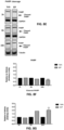
- EP treatment significantly decreases the expression of p-AKT1 (Ser 473, P ⁇ 0.01) and decreases total AKT1 (P ⁇ 0.05) and AKT2 levels (P ⁇ 0.001) ( FIG. 8C and 8D ), while not affecting AKT3 levels in SUM-149 cells ( S6 ).
- Regulation of AKT isoform expression by EP is extremely similar to what the inventors reported with whole mushroom GLE in the same breast cancer cell lines.
- EP induced apoptosis its effects on the expression on anti-apoptosis proteins BCL-2 and BCL-XL, which block the release of cytochrome C from the mitochondria were investigated.
- BCL-XL P ⁇ 0.001
- Cyclin D1 P ⁇ 0.05
- c-Myc P ⁇ 0.001
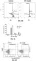
- Results disclosed herein demonstrate EP significantly reduces total PARP levels (P ⁇ 0.0001) and induces PARP cleavage (P ⁇ 0.02) after 24h of treatment in SUM-149 cells. These combined results demonstrate the inhibitory effects of EP against cancer cells, affecting migratory and death-inducing processes, as well as the expression of key proteins that play vital roles in cancer progression.
- Ionized compounds may have reduced permeability across lipid bilayers relative to that of un-ionized compounds.
- the neutral carbamate derivatives ergosterol sulfonamide, 5-6-dihydroergosterol sulfonamide, and EP sulfonamide should have improved solubility and cross the cell membrane, resulting in a favorable, slow intracellular accumulation.
- the triterpene content in GLE ranged from 1-3%, but the lipid content remained consistent in 3 different batches. Triterpene content variations can occur due to differences in terpene production by mushroom or extraction methods.
- the seven most abundant compounds in GLE which included ganoderic acid (GA-01), ganoderenic acid A, ganoderenic acid D, ergosterol, 5,6-dehydroergosterol, ergosterol peroxide (EP), and palmitic acid, are presented. Although previous crystal structures of ergosterol have been reported, in the present study for the first time the X-ray structure of 5-6-dihydroergosterol from Ganoderma lucidum was provided.
- MDA-MB-231 and SUM-149 are triple-negative breast cancer (TNBC) cells that is, they lack the estrogen receptor (ER), progesterone receptor (PR), and human epidermal growth factor receptor 2 (HER2) and represent two distinct breast cancer subtypes: mesenchymal-like breast cancer and epithelial-like inflammatory breast cancer, respectively.
- SUM-190 is an IBC cell line that is ER/PR-negative but expresses the HER2 oncogene; and MCF-7 cells express ER/PR receptors (ER/PR-positive/HER2-negative).
- TNBC and IBC triple-negative breast cancer
- EP displayed the greatest selective anti-cancer activity in the cell lines tested.
- EP features a cyclic peroxide (-O-O-), and these compounds are collectively known as endoperoxides.
- a classic example of this class of compounds is featured in the antimalarial drug, artemisinin, which has aid in reducing the widespread of drug-resistant parasites and has demonstrated cytotoxic activity.
- EP has been shown to have anti-cancer cell growth properties via regulation of various signaling pathways (e.g. AKT and c-myc) in a hepatocellular carcinoma cell model.
- EP reduces the expression of pro-survival p-AKT1, as well as levels of total AKT1 and AKT2 without affecting p-AKT2. It is possible that EP decreases total AKT2 without affecting p-AKT2 via a feedback mechanism, which induces signals to overcome the inhibitory effects obtained on total protein. Although p-AKT2 is not significantly upregulated, the phosphorylated levels might be stabilized to compensate for loss of total AKT2. Further studies are underway to understand the cell signaling regulatory effects of EP.
- EP treatment of SUM-149 cells reduced cell viability and induced apoptosis detected by an increase in caspase-3/7 activity and PARP cleavage after 24h of treatment.
- EP's cytotoxicity profile in SUM-149 (TNBC/IBC) cells is consistent with that of cells exposed to a compound that induces cell-cycle arrest and early-phase apoptosis.
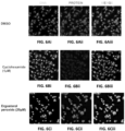
- the combined results suggest the death-inducing effects of EP on the two TNBC cell lines occurs by different mechanisms. All the tested compounds induce ROS in breast cancer cells albeit at different levels.
- EP increases ROS formation with greater capacity in SUM-149 cells, and the effects where reduced when co-treated with NAC. Therefore, intracellular ROS might be an additional contributing factor for the observed apoptotic effects of EP.
- the results are consistent with the apoptosis-inducing effects of EP that have been previously obtained from distinct sources and demonstrated in various in vitro cancer models, including colorectal, prostate, and leukemia cells.
- Additional cancer cell properties that increase their pathogenicity include their ability to migrate and invade to distant organ sites.
- the regulatory effects of non-lethal doses of EP on SUM-149 cell motility and invasion were investigated.
- EP displayed a reduction in cell migration and invasion, and these results are consistent with previous studies with GLE wherein reduced invasion potential of breast cancer cells was demonstrated.
- present results are the first to demonstrate the anti-migratory effects of EP in SUM-149 breast cancer cells, and demonstrate anti-invasion effects at lower dosages than those reported in the literature in breast cancer lines.
- EP is sharing its core with cholesterol, an amphipathic molecule with part-water soluble, part-water insoluble character.
- cholesterol an amphipathic molecule with part-water soluble, part-water insoluble character.
- EP's solubility properties are poor in DMSO or water unless warmed up to 30°C.
- EP's derivative, EP sulfonamide was soluble at 10mM in DMSO.
- EP was generated from ergosterol as the starting material by treating with oxygen in the presence of radical initiator (catalytic) in MeOH via hetero Diels-Alder reaction in excellent yields (93% yield, see SI) as a single compound after purification.
- the free hydroxyl group at C3 offers a handle for further functionalization.
- several EP-analogues ( 3a-3k ) ( FIG. 12 . ) were generated to evaluate the influence of the length and nature (polar groups) of the linker to the C3-hydroxyl group in terms of cytotoxicity.
- benzyl carbamates of EP were observed to display improved biological activity presumably due to improved solubility properties.
- Oxidation of compound 3 with TPAP/NMO at RT provided the enone system 3a.
- synthesis of 3b-3c, 3h, 3i, 3j and 3k involved esterification of EP with the corresponding carboxylic acid, acyl chloride, or anhydride (See SI for detailed information).
- Synthesis of 3d-3g involved S N 2 type of reactions.
- Synthesis of the biotinylated compound involved the coupling of EP with the mixed anhydride of biotin in good yield.
- the synthesized compounds were characterized by 1 H and 13 C (1D and 2D) NMR and mass spectrometry.
- the generation of the EP fluorescent probes ( 3l-3n ) was generated for live cell analysis including organelle detection and studies on function ( FIG.
- the fluorescent reagents can affect the motility of the parental compound, as well as the site of accumulation, minimizing the size of the linker was minimized to maintain the molecular weight range compared to the parental EP.
- the rhodamine probe tetramethylrhodamine, TAMRA
- structure is shown in FIG. 15 .
- the BODIPY derived probes were generated by treatment of BODIPY propianate with activating 2,4,6-trichlorobenzoyl chloride in the presence of Et 3 N, and subsequently addition of EP resulting in compound 3m.
- EP compound 3
- the oxidized compound 3a displayed improved activity, which was partially attributed to improved solubility properties.
- the corresponding amine ( 3b ), chloride ( 3c ), azide ( 3d ) showed promising activity against SUM-149 with little or no activity observed at the tested concentrations in MDA-MB-231 and T47D.
- the corresponding alkynyl ether 3e showed better activity, while the compound 3j containing the longer chain showed superior activity across cell lines.
- the phosphonate ester 3f showed improved activity while the corresponding phosphonic acid ( 3g ), the phosphocholine ( 3h ) and glutaric ester ( 3i ) displayed poor activity, presumably due to the charged nature of these compounds.
- the biotinylated compound 3k showed comparable cytotoxicity to the parental compound 3.
- the TAMRA-probe 3l showed poor solubility properties and precipitated readily in DMSO or PBS. Washout experiments revealed that the compound 3l did not accumulate in the cell at all. It is possible that the observed cytotoxicity arises from interfering with nutrient intake, but not via apoptosis due to the short, if any residency time inside the cell.
- the fluorescent probes 3m demonstrated comparable cytotoxicity to the parental compound 3. However, 3n showed superior activity.
- EP is an endoperoxide
- EP would likely accumulate in the peroxisome.
- Probe 3l was first evaluated and as previously mentioned, no cell staining was observed as the compound had difficulties penetrating the cell. As shown in the washout experiments of probe 3m, indicated that the compound distributes across the cytosol. Then, compound 3m was evaluated in the Ruby-red peroxisome stably transfect cells ( FIG. 14A . ), no co-localization was observed. The same observation was determined for golgi-RFP cells ( FIG. 14B . ) . To access whether 3m accumulated in the lysosome, lysotracker was used ( FIG. 14C . ), and no co-localization was recorded.
- the deep-red EP probe ( 3n ) was generated and evaluated to determine whether a similar localization pattern would be observed.
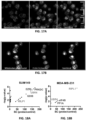
- the GFP-peroxisome cell lines a different pattern was observed from 3m as shown in FIG. 5A .
- green mitotracker was used to investigate the compound distribution in the cell.
- 3n selectively accumulated in the mitochondria. Such finding was congruent with our finding that this compound was the most potent of all evaluated derivatives across cell lines, as it can induce more cellular damage if it resides in the mitochondria.
- RLP1 also known as RILPL1
- RILPL1 Rab Interacting Lysosomal Protein Like 1
- E3 ubiquitin-protein ligase interacts with the retinoblastoma-associated protein in the nucleus and with calcium-bound calmodulin in the cytoplasm.
- UBR4 is also a component of the N-end rule pathway, which recognizes and binds to proteins bearing specific N-terminal residues that are destabilizing according to the N-end rule, leading to their ubiquitination and subsequent degradation.
- This protein coupled with clathrin forms meshwork structures involved in membrane morphogenesis and cytoskeletal organization and regulates integrin-mediated signaling. Further validation studies are warranted to elucidate the exact mode of action of this natural product.
- EP has potential as a hit compound for further therapeutic development, and chemical modifications can lead to superior potency by accumulating in specific organelles to induce greater cellular effect.
- mice Female severe combined immunodeficient mice (SCID) mice (21 d of age) were purchased from Charles River Laboratories International Inc. (Wilmington, MA) and housed under specific pathogen free conditions. The mice received autoclaved AIN 76-A phytoestrogen-free diet (Tek Global, Harlan Teklad, Madison, WI) and water ad libitum. This protocol was approved by the Universidad Central del Caribe IACUC. Cell inoculations were performed as previously described by the inventors (PMID: 20517637, PMID: 23468988, PMID: 26958085, PMID: 30542507, PMID: 31658643).
- vehicle 10% ETOH in 1X PBS
- BW ergosterol peroxide 100 mg/kg BW ergosterol peroxide in a 100 uL volume three times per week.
- mice were imaged following tumor establishment, and once per week thereafter. Tumor progression was monitored by fluorescence image analysis taken on an UVP iBox Explorer (Analytik Jena, Upland, CA) and quantified as in PMID: 20517637. Relative tumor growth was calculated as the fluorescence intensity of each tumor on day of imaging. Quantification for tumor progression was calculated using the fluorescence intensity of each treated tumor relative to the fluorescence intensity of the same tumor on d 01 of treatment administration (week 5) using ImageJ software (National Institutes of Health, Bethesda, MD).
- Capsules 500 mg of commercially available whole mushroom ReishiMax GLp TM (Pharmanex Inc., Provo, UT), consisting of powdered Ganoderma lucidum extract (GLE) fruiting body and cracked spores were used [12-14]. All manipulations were carried out under inert gas atmosphere unless otherwise noted.
- Anhydrous tetrahydrofuran (THF), diethyl ether (Et 2 O), dichloromethane (CH 2 Cl 2 ), toluene (PhCH 3 ), acetonitrile (CH 3 CN), methanol (MEOH), and dimethylformamide (DMF) were obtained from a solvent drying system.
- Reagents of the highest available quality were purchased commercially and used without further purification unless otherwise stated.
- Title compounds were purified by flash column chromatography using E. Merck silica gel (60, particle size 0.040-0.063 mmol) or Biotage Isolera Four with normal-phase silica gel. Reactions were monitored by thin-layer chromatography (TLC) on 0.25 mmol E. Merck silica gel plates (60F-254), using UV light for visualization and an ethanolic solution of anisaldehyde, or PMA, CAM solutions and heat as developing agents. Reactions were also monitored by using Agilent 1100 series LCMS and low-resonance electrospray ionization (ESI) model with UV detection at 254 nm.
- High-resolution mass spectra were recorded on an Agilent ESI-TOF (time of flight) mass spectrometer using matrix-assisted laser desorption ionization (MALDI) or electrospray ionization (ESI) or on a Waters Xevo G2 Q-ToF mass spectrometer.
- MALDI matrix-assisted laser desorption ionization
- ESI electrospray ionization
- Waters Xevo G2 Q-ToF mass spectrometer Compounds were analyzed by using ESI in positive-ion mode. The purity of each synthesized compound was determined on a Waters ACQUITY UPLC-PDA-ELSD-MS system using a C 18 reverse phase column and 0.1% formic acid/water - 0.1% formic acid/acetonitrile as the solvents. All synthesized compounds were at least 95% pure based on analytical HPLC and NMR. Chemical yields refer to purified compounds ( 1 H-NMR).
- the mobile phase consisted of water (A) and Acetonitrile (B): 0 min, 98:2; 0.5 min, 98:2; 6.5 min, 0:100; 12.3 min, 0:100; 12.5 min, 98:2; 12.95 min, stop.
- the flow rate was 25 mL/min.
- fractions (A - fractions 1-6, B - fractions 7-9, C - fractions 10-17, D - fractions 18-31, E-fractions 32-41, F-fractions 42-100) were collected and combined based on mass spectra data; their TLC profiles and biological properties were evaluated.
- BJ CRL-2522, normal human foreskin fibroblast
- MCF-7 HMB-22, human breast carcinoma
- EMEM medium Life Technologies, Carlsbad, CA
- FBS FBS
- MDA-MB-231 HMB-26, human breast carcinoma, triple-negative breast cancer cells
- DMEM medium Life Technologies, Carlsbad, CA
- Suspension cells KOPN8 infant human B cell precursor acute lymphoblastic leukemia with MLL-MLLt1/ENL fusion
- UoC-B1 (kindly gifted by Dr. William Evans of St.
- BCR-ABL murine B cell precursor acute lymphoblastic leukemia of pediatric relapsed ALL with BCR-ABL fusion
- RPMI media supplemented with 10% FBS.
- Cells were tested with MycoAlert TM Mycoplasma detection kit (Lonza) by using the manufacturer's conditions and were deemed negative.
- the cell lines were authenticated by IDEXX BioResearch (Columbia, MO).
- Either 1 ⁇ 10 3 -4.8 ⁇ 10 3 or 4 ⁇ 10 2 -1.2 ⁇ 10 3 cells were seeded to each well of 96- or 384-well white polystyrene flat-bottomed plates (3610 or 8804BC, Corning) in 100 DL or 30 DL media per well, respectively.
- the concentrations used were experimentally determined to ensure logarithmic growth during the duration of the experiment and prevent adverse effects on cell growth by DMSO exposure.
- the plates were incubated at 37°C in 5% CO 2 for 24 h before treatment.
- Stock solutions of test compounds (10 mM in DMSO) in nine 3-fold serial dilutions were dispensed via pintool (Biomek). The final concentration of DMSO was 0.3% (v/v) in each well.
- the plates were incubated for 72 h at 37°C in 5% CO 2 and then quenched with CellTiter-Glo ® reagent (50 ⁇ L/well in 96-well plate, 30 ⁇ L/well in 384-well plate) at RT.
- the positive controls included staurosporine (10 ⁇ M), gambogic acid (10 ⁇ M), and a toxic quinoline generated in-house. Plates were then incubated at RT for 20 min and centrifuged at 1000 rpm for 1 min.
- the cytotoxicity assay was performed by using the CellTiterGlo Luminescent Cell Viability Assay kit (G7570, Promega, Madison, WI) according to the manufacturer's instructions. Luminescence was recorded in an Envision plate reader (Perkin Elmer).
- SUM-149 cells (1.5 ⁇ 10 6 cells/well) were plated on a 6-well plate. Two days later, cell media was changed to 5% FBS, and cells were incubated at 37°C in a 5% CO 2 atmosphere for 1 h before vehicle (0.1% DMSO) or EP (5-40 ⁇ M) was added. After 72 h of treatment, cells were trypsinized and reseeded at 200 cells/mL per well in 24-well plates. After 10 d of culture, colonies were visualized by crystal violet staining, and the colony numbers were counted under a microscope.
- Truncated polypeptides are rapidly turned over by the proteasome and can be detected based on the subsequent click reaction with a fluorescent azide.
- the reaction was conducted according to the manufacturer's protocol.
- Cells were imaged at 20X with a Nikon C2 scanning confocal microscope, and the whole image montage was quantified by using Gen5 Software and Lionheart TM FX Automated Microscope (BioTek, Winooski, VT).
- SUM-149 or MDA-MB-231 were plated in 96 well plates at a density of 2 ⁇ 10 4 and 1 ⁇ 10 4 cells/well, respectively in black 96-well clear bottom tissue culture plates and incubated for 24 h at 37°C.
- the mRuby-Mito-7 plasmid was obtained from Addgene (#55874 from Michael Davidson).
- a stable clone of MCF-7 was generated by using Fugene 6 (Roche) to transfect cells (2 ⁇ 10 5 ) with plasmid (2.0 ⁇ g) under neomycin selection.
- a stable cell line was selected by using G418 (300 ⁇ g/mL) for 2 weeks, sorted by flow cytometry, and maintained under G418 thereafter.
- Transfected MCF-7 cells were plated at a density of 2 ⁇ 10 4 cells/well in black 96-well clear bottom tissue culture plates and incubated overnight at 37°C.
- cells were replenished with fresh PBS with 2% FBS and treated with the desired compounds for 1 h at 37°C.
- DMSO 0.5%) was used as a negative control, while Menadione (Mena, at 10 ⁇ M) or t-Butyl hydroperoxide (TBHP) at 100 ⁇ M final concentration were used as a positive control.
- the free radical scavenger N -acetyl cysteine (NAC) was used.
- Cells were treated with or without NAC (500 ⁇ M or 100 ⁇ M) for 1 h, then CellROX ® green reagent (Molecular Probes #C10444) was added to a final concentration of 5 ⁇ M, and cells were incubated for an additional 30 min at 37°C. Cells were then washed 2x with PBS, followed by fixation with 4% paraformaldehyde (v/v) for 15 min, and 2 more washes with PBS. Fluorescence intensity was measured in a Clariostar ® plate reader (BMG LABTECH, Cary, NC) at 535 nm.
- SUM-149 cells were treated for 48 h with EP, then collected and washed in 1X PBS.
- the cells (5 ⁇ 10 5 ) were fixed and permeabilized in 70% ethanol at -20°C until further analysis.
- To measure the DNA content the cells were washed and resuspended in 150 ⁇ L of PBS buffer then incubated in 100 ⁇ L of 100 ⁇ g/mL RNase A (MilliporeSigma, Burlington, MA) for 15 min at RT, then 250 ⁇ L of 50 ⁇ g/mL Propidium Iodide (MilliporeSigma) was added.
- RNase A MilliporeSigma, Burlington, MA
- SUM-149 cells (5 ⁇ 10 5 ) were seeded and treated either with vehicle (0.1% DMSO) or with 20 ⁇ M of EP or with puromycin (200 ng/mL) as positive control for 48 h. After treatment floating cells were collected, and all cells were harvested and counted. Treated cells were analyzed for cell death by using the FITC Annexin-V / 7AAD Apoptosis detection kit (product #640922, Biolegend, San Diego, CA) according to the manufacturer's instructions. Briefly, cells were resuspended in Annexin Binding Buffer, stained with Annexin V-FITC and/or 7-AAD, and incubated for 30 min at 4°C. Data were acquired on a BD FACS Canto II cytometer (BD Biosciences, San Jose, CA) and analyzed by using FlowJo software (V10, FlowJo LLC, Ashland, OR).
- FITC Annexin-V / 7AAD Apoptosis detection kit
- EC 50 values were calculated by Pipeline Pilot Software (Accelrys, Enterprise Platform, CA, USA). EC 50 values ( ⁇ M) from the viability assay were calculated from dose response curve-fitting via non-linear regression by using GraphPad Prism (Version 7.0 San Diego, CA).
Landscapes
- Health & Medical Sciences (AREA)
- Life Sciences & Earth Sciences (AREA)
- Chemical & Material Sciences (AREA)
- Engineering & Computer Science (AREA)
- General Health & Medical Sciences (AREA)
- Medicinal Chemistry (AREA)
- Natural Medicines & Medicinal Plants (AREA)
- Pharmacology & Pharmacy (AREA)
- Animal Behavior & Ethology (AREA)
- Public Health (AREA)
- Veterinary Medicine (AREA)
- Epidemiology (AREA)
- Mycology (AREA)
- Urology & Nephrology (AREA)
- Molecular Biology (AREA)
- Microbiology (AREA)
- Immunology (AREA)
- Hematology (AREA)
- Biotechnology (AREA)
- Biomedical Technology (AREA)
- Chemical Kinetics & Catalysis (AREA)
- General Chemical & Material Sciences (AREA)
- Botany (AREA)
- Medical Informatics (AREA)
- General Physics & Mathematics (AREA)
- Organic Chemistry (AREA)
- Analytical Chemistry (AREA)
- Physics & Mathematics (AREA)
- Food Science & Technology (AREA)
- Cell Biology (AREA)
- Pathology (AREA)
- Alternative & Traditional Medicine (AREA)
- Biochemistry (AREA)
- Nuclear Medicine, Radiotherapy & Molecular Imaging (AREA)
- Oil, Petroleum & Natural Gas (AREA)
- Bioinformatics & Cheminformatics (AREA)
- Pharmaceuticals Containing Other Organic And Inorganic Compounds (AREA)
- Steroid Compounds (AREA)
- Medicines Containing Plant Substances (AREA)
- Medicines Containing Antibodies Or Antigens For Use As Internal Diagnostic Agents (AREA)
Applications Claiming Priority (3)
| Application Number | Priority Date | Filing Date | Title |
|---|---|---|---|
| US201962802525P | 2019-02-07 | 2019-02-07 | |
| PCT/US2020/017053 WO2020163626A1 (fr) | 2019-02-07 | 2020-02-06 | Composés de ganoderma lucidum biologiquement actifs, et synthèse de dérivés anticancéreux ; sondes de peroxyde d'ergostérol pour localisation cellulaire |
| EP20752180.8A EP3920948B1 (fr) | 2019-02-07 | 2020-02-06 | Composés de ganoderma lucidum biologiquement actifs, et synthèse de dérivés anticancéreux ; sondes de peroxyde d'ergostérol pour localisation cellulaire |
Related Parent Applications (2)
| Application Number | Title | Priority Date | Filing Date |
|---|---|---|---|
| EP20752180.8A Division EP3920948B1 (fr) | 2019-02-07 | 2020-02-06 | Composés de ganoderma lucidum biologiquement actifs, et synthèse de dérivés anticancéreux ; sondes de peroxyde d'ergostérol pour localisation cellulaire |
| EP20752180.8A Division-Into EP3920948B1 (fr) | 2019-02-07 | 2020-02-06 | Composés de ganoderma lucidum biologiquement actifs, et synthèse de dérivés anticancéreux ; sondes de peroxyde d'ergostérol pour localisation cellulaire |
Publications (2)
| Publication Number | Publication Date |
|---|---|
| EP4527414A2 true EP4527414A2 (fr) | 2025-03-26 |
| EP4527414A3 EP4527414A3 (fr) | 2025-06-11 |
Family
ID=71947918
Family Applications (2)
| Application Number | Title | Priority Date | Filing Date |
|---|---|---|---|
| EP20752180.8A Active EP3920948B1 (fr) | 2019-02-07 | 2020-02-06 | Composés de ganoderma lucidum biologiquement actifs, et synthèse de dérivés anticancéreux ; sondes de peroxyde d'ergostérol pour localisation cellulaire |
| EP25151630.8A Pending EP4527414A3 (fr) | 2019-02-07 | 2020-02-06 | Composés de ganoderma lucidum biologiquement actifs, et synthèse de dérivés anticancéreux ; sondes de peroxyde d'ergostérol pour localisation cellulaire |
Family Applications Before (1)
| Application Number | Title | Priority Date | Filing Date |
|---|---|---|---|
| EP20752180.8A Active EP3920948B1 (fr) | 2019-02-07 | 2020-02-06 | Composés de ganoderma lucidum biologiquement actifs, et synthèse de dérivés anticancéreux ; sondes de peroxyde d'ergostérol pour localisation cellulaire |
Country Status (5)
| Country | Link |
|---|---|
| US (2) | US12263177B2 (fr) |
| EP (2) | EP3920948B1 (fr) |
| JP (2) | JP7537684B2 (fr) |
| CA (1) | CA3129092A1 (fr) |
| WO (1) | WO2020163626A1 (fr) |
Families Citing this family (5)
| Publication number | Priority date | Publication date | Assignee | Title |
|---|---|---|---|---|
| US11838375B2 (en) * | 2020-11-12 | 2023-12-05 | Harman International Industries, Incorporated | Universal software communication bus |
| CN115124583A (zh) * | 2021-03-25 | 2022-09-30 | 华东师范大学 | 一种通过选择性还原或氧化反应合成灵芝酸的方法 |
| US12005092B2 (en) * | 2021-12-22 | 2024-06-11 | Laura Mann Kevehazi | Herbal composition for breast cancer prevention |
| CN115201164B (zh) * | 2022-05-16 | 2025-09-26 | 山东第一医科大学(山东省医学科学院) | 一种基于香豆素-共轭桥-芳香受体的双标记探针及其在新药开发领域的应用 |
| WO2025231452A1 (fr) * | 2024-05-02 | 2025-11-06 | Martinez Montemayor Michelle | Dérivés de peroxyde d'ergostérol et leurs utilisations |
Family Cites Families (10)
| Publication number | Priority date | Publication date | Assignee | Title |
|---|---|---|---|---|
| JP2006022017A (ja) | 2004-07-06 | 2006-01-26 | Univ Nihon | 発癌予防剤 |
| JP2006298853A (ja) * | 2005-04-22 | 2006-11-02 | Hitoshi Ito | 抗腫瘍性飲食品及び抗腫瘍剤 |
| JPWO2007083823A1 (ja) * | 2006-01-17 | 2009-06-18 | 国立大学法人九州大学 | マンネンタケ処理物のアンドロゲン受容体結合活性 |
| GB0800134D0 (en) | 2008-01-04 | 2008-02-13 | Univ Nottingham | Ganoderma lucidum extracts for use in the treatment of cell neoplasia, cell proliferation and angiogenesis |
| KR101090196B1 (ko) | 2010-01-29 | 2011-12-07 | 학교법인 선목학원 | 에르고스타-7,22-디엔-3베타,5알파,6알파-트리올을 포함하는 항암 조성물 |
| TWI606835B (zh) | 2010-04-23 | 2017-12-01 | 中央研究院 | 基於靈芝多醣之組合物及治療癌症之方法 |
| KR101289854B1 (ko) | 2011-11-08 | 2013-07-26 | 건국대학교 산학협력단 | 영지버섯 유래 신규 화합물 및 그 응용 |
| CN104083382B (zh) | 2014-07-02 | 2016-06-08 | 内蒙古大学 | 麦角甾醇过氧化物与紫杉醇在杀伤Hela癌细胞方面的协同作用及其应用 |
| CN108703979A (zh) | 2018-07-26 | 2018-10-26 | 浙江中医药大学 | 一种基于无柄灵芝提取物的乳腺癌肿瘤治疗用药 |
| CN109758486A (zh) | 2019-03-25 | 2019-05-17 | 广州白云山汉方现代药业有限公司 | 灵芝提取物在制备抗肿瘤多药耐药药物中的应用 |
-
2020
- 2020-02-06 EP EP20752180.8A patent/EP3920948B1/fr active Active
- 2020-02-06 JP JP2021569252A patent/JP7537684B2/ja active Active
- 2020-02-06 EP EP25151630.8A patent/EP4527414A3/fr active Pending
- 2020-02-06 CA CA3129092A patent/CA3129092A1/fr active Pending
- 2020-02-06 WO PCT/US2020/017053 patent/WO2020163626A1/fr not_active Ceased
- 2020-02-06 US US17/428,812 patent/US12263177B2/en active Active
-
2024
- 2024-03-21 JP JP2024044814A patent/JP2024069632A/ja active Pending
-
2025
- 2025-03-18 US US19/082,898 patent/US20250241930A1/en active Pending
Non-Patent Citations (96)
| Title |
|---|
| ADAMEC, J., J SEP SCI., vol. 32, 2009, pages 4052 - 4058 |
| ANGEL RAMOS-LIGONIO ET AL., PHYTOTHER. RES., vol. 26, 2012, pages 938 - 943 |
| BERNAL, J. D. ET AL., CAMBRIDGE UNIVERSITY PRESS, 1940 |
| BERNAL, J. D.CROWFOOT, D. Y., JOURNAL OF THE CHEMICAL SOCIETY, vol. 93, 1935 |
| BOH, B. ET AL., BIOTECHNOL ANNU REV., vol. 13, 2007, pages 265 - 301 |
| BORCHERS, A. T. ET AL., PROCEEDINGS OF THE SOCIETY FOR EXPERIMENTAL BIOLOGY AND MEDICINE, vol. 221, 1999, pages 281 - 293 |
| BRANDES, L. J. ET AL., CANCER RES., vol. 43, 1983, pages 2831 - 2835 |
| BU, M. ET AL., CHEM MED CHEM., vol. 12, 2017, pages 466 - 474 |
| C. FLOREAN ET AL., FREE RADIC BIOL MED., vol. 0891-5849, no. 18, 2019, pages 32332 - 3 |
| C. JIMÉNEZ ET AL., J NAT PROD., vol. 52, 1989, pages 619 - 622 |
| C. S. MCKAYM. G. FINN, CHEM BIOL., vol. 21, 2014, pages 1075 - 101 |
| CANTRELL, C. ET AL., PLANTA MEDICA, vol. 65, 1999, pages 732 - 734 |
| CASTILLO-PICHARDO, L., CLIN EXP METASTASIS, vol. 26, 2009, pages 505 - 516 |
| CHEN, X. ET AL., INTERNATIONAL IMMUNOPHARMACOLOGY, vol. 6, 2006, pages 499 - 508 |
| CHIN, Y. ET AL., CANCER DISCOVERY, vol. 4, 2014, pages 942 - 955 |
| CRISTOFANILLI, M. ET AL., CANCER, vol. 110, 2007, pages 1436 - 1444 |
| D. B. GRACA SGARBI ET AL., MYCOPATHOLOGIA, vol. 139, 1997, pages 9 - 14 |
| D. CHIABRANDO ET AL., FRONT. PHARMACOL., vol. 5, 2014, pages 1 - 24 |
| DAI, J. ET AL., ONCOTARGET, vol. 8, 2017, pages 13770 - 13781 |
| ETHIER, S. P. ET AL., CANCER RES., vol. 56, 1996, pages 899 - 907 |
| FERNANDEZ, S. V. ET AL., BREAST CANCER RES TREAT., vol. 140, 2013, pages 23 - 33 |
| G. BATTOGTOKH ET AL., ACTA PHARMACEUTICA SINICA B., vol. 8, 2018, pages 862 - 880 |
| GAO, Y. ET AL., IMMUNOLOGICAL INVESTIGATIONS, vol. 32, 2003, pages 201 - 215 |
| GAO, Y. ET AL., JOURNAL OF MEDICINAL FOOD, vol. 8, 2005, pages 159 - 168 |
| GUO, D. ET AL., NATURE GENETICS, vol. 36, 2004, pages 837 - 841 |
| H. M. SCHIPPER ET AL., EXP CELL RES., vol. 207, 1993, pages 62 - 7 |
| H. Y. WU ET AL., SCI REP., vol. 8, 2018, pages 17956 |
| HAY, R. J. ET AL., ATCC QUALITY CONTROL METHODS FOR CELL LINES, 1992 |
| HE, L. ET AL., INTERNATIONAL JOURNAL OF MOLECULAR SCIENCES, vol. 19, 2018, pages 3935 |
| HOOK, B.SCHAGAT, T., PROFILING COMPOUND EFFECTS ON CELL HEALTH IN A TIME COURSE USING A MULTIPLEXED SAME-WELL ASSAY, 2012 |
| HUIE, C.DI, X., J CHROMATOGR B., vol. 812, 2004, pages 241 - 257 |
| HULL, S. E.WOOLFSON, M. M., ACTA CRYSTALLOGRAPHICA SECTION B., vol. 32, 1976, pages 2370 - 2373 |
| I. FERNANDEZA. ROBERT, ORG. BIOMOL. CHEM., vol. 9, 2011, pages 4098 - 4107 |
| J. M. GAO ET AL., PHYTOMEDICINE, vol. 14, 2007, pages 821 - 824 |
| JIANG, J. ET AL., INT J MOL MED., vol. 21, 2008, pages 577 - 584 |
| JIN, X. ET AL., COCHRANE DATABASE SYST REV., vol. 4, 2016, pages CD007731 |
| JONG, S. C.DONOVICK, R., ADV APPL MICROBIOL., vol. 34, 1989, pages 183 - 261 |
| JOSEPH, S. ET AL., ACTA PHARMACEUTICA, vol. 61, pages 335 - 342 |
| K. J. CHAVEZV. SIREESHAS. V. GARIMELLAS. LIPKOWITZ, BREAST DIS., vol. 32, 2010, pages 35 - 48 |
| K. W. HUH ET AL., PROC NATL ACAD SCI U S A., vol. 102, 2005, pages 11492 - 7 |
| K. YASUKAWA ET AL., BIOL PHARM BULL., vol. 19, 1996, pages 573 - 576 |
| KANG, J. H. ET AL., J ETHNOPHARMACOLOGY, vol. 173, 2015, pages 303 - 312 |
| KOBORI, M. ET AL., BR J PHARMACOL., vol. 150, 2007, pages 209 - 219 |
| L. MA ET AL., FOOD CHEM, vol. 139, 2013, pages 503 - 508 |
| LEE, S., PLANTA MED., vol. 64, 1998, pages 303 - 308 |
| LEHMAN, H. L. ET AL., MOLECULAR CANCER RESEARCH, vol. 10, 2012, pages 1306 - 1318 |
| LI, X. ET AL., ONCOTARGET, vol. 6, 2015, pages 17832 - 17846 |
| LI, X. ET AL., ONCOTARGET, vol. 7, 2016, pages 33948 - 33959 |
| LIN, S. B. ET AL., LIFE SCI., vol. 72, 2003, pages 2381 - 2390 |
| LIN, Z. B., J PHARMACOL SCI., vol. 99, 2005, pages 144 - 153 |
| LING, T. ET AL., EUR J MED CHEM., vol. 146, 2018, pages 501 - 510 |
| LIU, J. ET AL., PROC NATL ACAD SCI U S A., vol. 109, 2012, pages 413 - 418 |
| LIU, X. ET AL., CANCER LETT., vol. 182, 2002, pages 155 - 161 |
| M. AXELROD ET AL., ONCOTARGET, vol. 4, 2013, pages 622 - 635 |
| M. BU ET AL., BIOORG MED CHEM LETT., vol. 27, 2017, pages 3856 - 3861 |
| M. BU ET AL., CHEMMEDCHEM., vol. 12, 2017, pages 466 - 474 |
| M. C. DEGHRIGUE ET AL., J PHARMACEUT SCI., vol. 22, 2014, pages 64 |
| M. M. MARTINEZ-MONTEMAYOR ET AL., ACCEPTED TO FRONTIERS IN PHARMACOLOGY, 10 January 2019 (2019-01-10) |
| MARTINEZ-MONTEMAYOR, M. M. ET AL., NUTRITION AND CANCER, vol. 63, 2011, pages 1085 - 1094 |
| MIN, B. ET AL., CHEM PHARM BULL., vol. 48, 2000, pages 1026 - 1033 |
| N. DUARTE ET AL., PHYTOTHER. RES., vol. 21, 2007, pages 601 - 604 |
| NAM, K. ET AL., LIFE SCI., vol. 69, 2001, pages 229 - 237 |
| PATERSON, R. R., PHYTOCHEMISTRY, vol. 67, 2006, pages 1985 - 2001 |
| POSNER, G. H. ET AL., J. MED. CHEM., vol. 47, 2004, pages 1299 - 1301 |
| R. S. AMES ET AL., EXPERT OPIN DRUG DISCOV., vol. 12, 2007, pages 1669 - 81 |
| ROBERTSON, F. ET AL., CLINICAL & EXPERIMENTAL PATHOLOGY, vol. 2, 2012, pages 119 |
| RUSSO, A. ET AL., CHEMIC-BIOLOGICAL INTERACTIONS, vol. 184, 2010, pages 352 - 358 |
| S. M. MENSEL. ZHANG, CELL RES., vol. 16, 2006, pages 681 - 692 |
| SIDDIQA, A. ET AL., BMC CANCER, vol. 8, 2008, pages 129 |
| SILVERA, D. ET AL., NAT CELL BIOL., vol. 11, 2009, pages 903 - 908 |
| SLIVA, D., MINI REV MED CHEM., vol. 4, 2004, pages 873 - 879 |
| SMINA, T. P. ET AL., MUTAT RES., vol. 726, 2011, pages 188 - 194 |
| SUAREZ-ARROYO, I. ET AL., MEDICINES, vol. 4, 2017, pages 15 |
| SUAREZ-ARROYO, I. J. ET AL., J CANCER, vol. 7, 2016, pages 500 - 511 |
| SUAREZ-ARROYO, I. J. ET AL., PLOS ONE, vol. 8, 2013, pages e57431 |
| T. BÖTTCHER ET AL., ANGEW CHEM INT ED ENGL., vol. 49, 2010, pages 2680 - 98 |
| T. LING ET AL., EURJ MED CHEM., vol. 146, 2018, pages 501 - 510 |
| T. NAKANISHI ET AL., J NAT MED., vol. 52, 1998, pages 521 - 526 |
| T. WANG ET AL., MOL BIOL CELL., vol. 15, 2004, pages 815 - 26 |
| TAKEI, T. ET AL., BIOTECHNOL. BIOCHEM., vol. 69, 2005, pages 212 - 215 |
| THYAGARAJAN, A. ET AL., NUTR CANCER, vol. 62, 2010, pages 630 - 640 |
| THYAGARAJAN-SAHU, A. ET AL., BMC COMPLEMENT ALTERN MED., vol. 11, 2011, pages 74 |
| TU, Y. ET AL., J. NAT. PROD., vol. 73, 2010, pages 751 - 754 |
| W. D. FOULKES ET AL., N ENGL J MED., vol. 363, 2010, pages 1938 - 1948 |
| W. QING-PING ET AL., PLOS ONE, vol. 7, 2012, pages 44579 |
| WANG, Y. Y. ET AL., BIOORG MED CHEM., vol. 10, 2002, pages 1057 - 1062 |
| WASSER, S. P.WEIS, A. L., INTJ MED MUSHROOMS, vol. 1, 1999, pages 31 - 62 |
| XIE, Y. ET AL., ENZYME MICROB TECHNOL, vol. 40, 2006, pages 177 - 185 |
| Y. HAN ET AL., NAT COMMUN., vol. 8, 2017, pages 1307 |
| Y. TAKAHASHI ET AL., PHYTOCHEMISTRY, vol. 30, 1991, pages 4117 - 4120 |
| YAO, D. ET AL., MOLECULAR CANCER RESEARCH, vol. 9, 2011, pages 1608 - 1620 |
| Z. ZHANG ET AL., REDOX BIOL., vol. 2213-2317, no. 18, 2018, pages 30895 - 4 |
| ZHANG, D. ET AL., CLINICAL CANCER RESEARCH, vol. 15, 2009, pages 6639 - 6648 |
| ZHANG, G. L. ET AL., WORLD J GASTROENTEROL., vol. 8, 2002, pages 728 - 733 |
| ZHANG, Y. Q.RAO, R., VIRULENCE, vol. 1, 2010, pages 551 - 554 |
| ZHU, X. L. ET AL., J ETHNOPHARMACOL., vol. 111, 2007, pages 219 - 226 |
Also Published As
| Publication number | Publication date |
|---|---|
| EP3920948A4 (fr) | 2022-08-03 |
| WO2020163626A1 (fr) | 2020-08-13 |
| US20250241930A1 (en) | 2025-07-31 |
| US12263177B2 (en) | 2025-04-01 |
| JP7537684B2 (ja) | 2024-08-21 |
| EP4527414A3 (fr) | 2025-06-11 |
| EP3920948B1 (fr) | 2025-04-09 |
| EP3920948A1 (fr) | 2021-12-15 |
| JP2022520132A (ja) | 2022-03-28 |
| CA3129092A1 (fr) | 2020-08-13 |
| US20220125805A1 (en) | 2022-04-28 |
| JP2024069632A (ja) | 2024-05-21 |
Similar Documents
| Publication | Publication Date | Title |
|---|---|---|
| US20250241930A1 (en) | Biologically active ganoderma lucidum compounds and synthesis of anticancer derivatives; ergosterol peroxide probes for cellular localization | |
| Martínez-Montemayor et al. | Identification of biologically active Ganoderma lucidum compounds and synthesis of improved derivatives that confer anti-cancer activities in vitro | |
| Nakase et al. | Transferrin receptor-dependent cytotoxicity of artemisinin–transferrin conjugates on prostate cancer cells and induction of apoptosis | |
| Liu et al. | Cytotoxic and pro-apoptotic effects of novel ganoderic acid derivatives on human cervical cancer cells in vitro | |
| KS et al. | Anti-proliferative effect of biogenic gold nanoparticles against breast cancer cell lines (MDA-MB-231 & MCF-7) | |
| Lee et al. | Bioactivity-guided isolation and chemical characterization of antiproliferative constituents from morel mushroom (Morchella esculenta) in human lung adenocarcinoma cells | |
| Hu et al. | Chaetocochin J, an epipolythiodioxopiperazine alkaloid, induces apoptosis and autophagy in colorectal cancer via AMPK and PI3K/AKT/mTOR pathways | |
| Farrell et al. | Cytotoxicity, DNA strand breakage and DNA–protein crosslinking by a novel transplatinum compound in human A2780 ovarian and MCF-7 breast carcinoma cells | |
| Ling et al. | Development of ergosterol peroxide probes for cellular localisation studies | |
| Guo et al. | Amlexanox-modified platinum (IV) complex triggers apoptotic and autophagic bimodal death of cancer cells | |
| Calderón-Montaño et al. | Alpha, beta-unsaturated lactones 2-furanone and 2-pyrone induce cellular DNA damage, formation of topoisomerase I-and II-DNA complexes and cancer cell death | |
| Gopalakrishnan et al. | Encapsulation of a Ru (η 6-p-cymene) complex of the antibacterial drug trimethoprim into a polydiacetylene-phospholipid assembly to enhance its in vitro anticancer and antibacterial activities | |
| Archer et al. | Syrbactin class proteasome inhibitor-induced apoptosis and autophagy occurs in association with p53 accumulation and Akt/PKB activation in neuroblastoma | |
| CN104395318B (zh) | 一类多氧孕甾烷化合物及用途 | |
| Lee et al. | Inhibition of cell growth and induction of apoptosis by Antrodia camphorata in HER‐2/neu‐overexpressing breast cancer cells through the induction of ROS, depletion of HER‐2/neu, and disruption of the PI3K/Akt signaling pathway | |
| US10947249B2 (en) | Nimbolide analogs as anti-cancer agents and preparation thereof | |
| Marczak et al. | Cytotoxicity and induction of apoptosis by formamidinodoxorubicins in comparison to doxorubicin in human ovarian adenocarcinoma cells | |
| Myobatake et al. | Pyrenocine A induces monopolar spindle formation and suppresses proliferation of cancer cells | |
| US12594338B2 (en) | Cisplatin analogue with potent anti-cancer effects and synthesis thereof | |
| US8637679B2 (en) | Process for the isolation of organic compounds useful for the treatment of cancer | |
| Guo et al. | Synthesis and in-vitro study of a novel ligustrazine diselenide as a potential chemotherapy drug for lung adenocarcinoma | |
| Shuai et al. | Isolation, Characterization, and Anticancer Evaluation of Alkaloids from Eumachia montana (Rubiaceae) | |
| WO2018055492A1 (fr) | Extraits de plantes herbacées d'arabie saoudite, procédé anticancéreux utilisant de tels extraits, et profilage cytologique utilisant la technique d'imagerie automatisée à haut débit | |
| Wang et al. | 19-(Benzyloxy)-19-oxojolkinolide B (19-BJB), an ent-abietane diterpene diepoxide, inhibits the growth of bladder cancer T24 cells through DNA damage | |
| WO2022087173A1 (fr) | Composés de phosphaplatine utilisés en tant qu'agents thérapeutiques ciblant sélectivement des cellules tumorales hautement glycolytiques et leurs procédés |
Legal Events
| Date | Code | Title | Description |
|---|---|---|---|
| PUAI | Public reference made under article 153(3) epc to a published international application that has entered the european phase |
Free format text: ORIGINAL CODE: 0009012 |
|
| STAA | Information on the status of an ep patent application or granted ep patent |
Free format text: STATUS: THE APPLICATION HAS BEEN PUBLISHED |
|
| AC | Divisional application: reference to earlier application |
Ref document number: 3920948 Country of ref document: EP Kind code of ref document: P |
|
| AK | Designated contracting states |
Kind code of ref document: A2 Designated state(s): AL AT BE BG CH CY CZ DE DK EE ES FI FR GB GR HR HU IE IS IT LI LT LU LV MC MK MT NL NO PL PT RO RS SE SI SK SM TR |
|
| REG | Reference to a national code |
Ref country code: DE Ref legal event code: R079 Free format text: PREVIOUS MAIN CLASS: A61K0047540000 Ipc: A61K0036074000 |
|
| PUAL | Search report despatched |
Free format text: ORIGINAL CODE: 0009013 |
|
| AK | Designated contracting states |
Kind code of ref document: A3 Designated state(s): AL AT BE BG CH CY CZ DE DK EE ES FI FR GB GR HR HU IE IS IT LI LT LU LV MC MK MT NL NO PL PT RO RS SE SI SK SM TR |
|
| RIC1 | Information provided on ipc code assigned before grant |
Ipc: G01N 33/58 20060101ALI20250507BHEP Ipc: A61K 47/54 20170101ALI20250507BHEP Ipc: A61P 35/00 20060101ALI20250507BHEP Ipc: A61K 31/045 20060101ALI20250507BHEP Ipc: A61K 31/575 20060101ALI20250507BHEP Ipc: A61K 31/56 20060101ALI20250507BHEP Ipc: A61K 36/074 20060101AFI20250507BHEP |
|
| STAA | Information on the status of an ep patent application or granted ep patent |
Free format text: STATUS: REQUEST FOR EXAMINATION WAS MADE |
|
| 17P | Request for examination filed |
Effective date: 20251209 |
|
| STAA | Information on the status of an ep patent application or granted ep patent |
Free format text: STATUS: EXAMINATION IS IN PROGRESS |