EP4502170A1 - Verfahren zur qualitätsbeurteilung mesenchymaler stammzellen - Google Patents
Verfahren zur qualitätsbeurteilung mesenchymaler stammzellen Download PDFInfo
- Publication number
- EP4502170A1 EP4502170A1 EP23779505.9A EP23779505A EP4502170A1 EP 4502170 A1 EP4502170 A1 EP 4502170A1 EP 23779505 A EP23779505 A EP 23779505A EP 4502170 A1 EP4502170 A1 EP 4502170A1
- Authority
- EP
- European Patent Office
- Prior art keywords
- cells
- mesenchymal stem
- stem cells
- passage
- consumptions
- Prior art date
- Legal status (The legal status is an assumption and is not a legal conclusion. Google has not performed a legal analysis and makes no representation as to the accuracy of the status listed.)
- Pending
Links
Images


Classifications
-
- C—CHEMISTRY; METALLURGY
- C12—BIOCHEMISTRY; BEER; SPIRITS; WINE; VINEGAR; MICROBIOLOGY; ENZYMOLOGY; MUTATION OR GENETIC ENGINEERING
- C12N—MICROORGANISMS OR ENZYMES; COMPOSITIONS THEREOF; PROPAGATING, PRESERVING, OR MAINTAINING MICROORGANISMS; MUTATION OR GENETIC ENGINEERING; CULTURE MEDIA
- C12N5/00—Undifferentiated human, animal or plant cells, e.g. cell lines; Tissues; Cultivation or maintenance thereof; Culture media therefor
- C12N5/06—Animal cells or tissues; Human cells or tissues
- C12N5/0602—Vertebrate cells
- C12N5/0652—Cells of skeletal and connective tissues; Mesenchyme
- C12N5/0662—Stem cells
-
- A—HUMAN NECESSITIES
- A61—MEDICAL OR VETERINARY SCIENCE; HYGIENE
- A61K—PREPARATIONS FOR MEDICAL, DENTAL OR TOILETRY PURPOSES
- A61K35/00—Medicinal preparations containing materials or reaction products thereof with undetermined constitution
- A61K35/12—Materials from mammals; Compositions comprising non-specified tissues or cells; Compositions comprising non-embryonic stem cells; Genetically modified cells
- A61K35/28—Bone marrow; Haematopoietic stem cells; Mesenchymal stem cells of any origin, e.g. adipose-derived stem cells
-
- C—CHEMISTRY; METALLURGY
- C12—BIOCHEMISTRY; BEER; SPIRITS; WINE; VINEGAR; MICROBIOLOGY; ENZYMOLOGY; MUTATION OR GENETIC ENGINEERING
- C12N—MICROORGANISMS OR ENZYMES; COMPOSITIONS THEREOF; PROPAGATING, PRESERVING, OR MAINTAINING MICROORGANISMS; MUTATION OR GENETIC ENGINEERING; CULTURE MEDIA
- C12N5/00—Undifferentiated human, animal or plant cells, e.g. cell lines; Tissues; Cultivation or maintenance thereof; Culture media therefor
- C12N5/06—Animal cells or tissues; Human cells or tissues
- C12N5/0602—Vertebrate cells
- C12N5/0652—Cells of skeletal and connective tissues; Mesenchyme
- C12N5/0662—Stem cells
- C12N5/0667—Adipose-derived stem cells [ADSC]; Adipose stromal stem cells
-
- A—HUMAN NECESSITIES
- A61—MEDICAL OR VETERINARY SCIENCE; HYGIENE
- A61P—SPECIFIC THERAPEUTIC ACTIVITY OF CHEMICAL COMPOUNDS OR MEDICINAL PREPARATIONS
- A61P19/00—Drugs for skeletal disorders
-
- A—HUMAN NECESSITIES
- A61—MEDICAL OR VETERINARY SCIENCE; HYGIENE
- A61P—SPECIFIC THERAPEUTIC ACTIVITY OF CHEMICAL COMPOUNDS OR MEDICINAL PREPARATIONS
- A61P21/00—Drugs for disorders of the muscular or neuromuscular system
-
- A—HUMAN NECESSITIES
- A61—MEDICAL OR VETERINARY SCIENCE; HYGIENE
- A61P—SPECIFIC THERAPEUTIC ACTIVITY OF CHEMICAL COMPOUNDS OR MEDICINAL PREPARATIONS
- A61P37/00—Drugs for immunological or allergic disorders
- A61P37/02—Immunomodulators
- A61P37/06—Immunosuppressants, e.g. drugs for graft rejection
-
- C—CHEMISTRY; METALLURGY
- C12—BIOCHEMISTRY; BEER; SPIRITS; WINE; VINEGAR; MICROBIOLOGY; ENZYMOLOGY; MUTATION OR GENETIC ENGINEERING
- C12N—MICROORGANISMS OR ENZYMES; COMPOSITIONS THEREOF; PROPAGATING, PRESERVING, OR MAINTAINING MICROORGANISMS; MUTATION OR GENETIC ENGINEERING; CULTURE MEDIA
- C12N2500/00—Specific components of cell culture medium
- C12N2500/30—Organic components
- C12N2500/32—Amino acids
-
- G—PHYSICS
- G01—MEASURING; TESTING
- G01N—INVESTIGATING OR ANALYSING MATERIALS BY DETERMINING THEIR CHEMICAL OR PHYSICAL PROPERTIES
- G01N33/00—Investigating or analysing materials by specific methods not covered by groups G01N1/00 - G01N31/00
- G01N33/48—Biological material, e.g. blood, urine; Haemocytometers
- G01N33/50—Chemical analysis of biological material, e.g. blood, urine; Testing involving biospecific ligand binding methods; Immunological testing
- G01N33/68—Chemical analysis of biological material, e.g. blood, urine; Testing involving biospecific ligand binding methods; Immunological testing involving proteins, peptides or amino acids
- G01N33/6803—General methods of protein analysis not limited to specific proteins or families of proteins
- G01N33/6806—Determination of free amino acids
- G01N33/6812—Assays for specific amino acids
- G01N33/6815—Assays for specific amino acids containing sulfur, e.g. cysteine, cystine, methionine, homocysteine
Definitions
- the present invention relates to a method for evaluating quality of mesenchymal stem cells using the mitochondrial size as an indicator.
- the present invention also relates to a method for producing mesenchymal stem cells evaluated for the quality using this evaluation method, a method for producing mesenchymal cells using the mesenchymal stem cells produced using this production method, and the like.
- the present invention also relates to a method for evaluating a medium for mesenchymal stem cells using the mitochondrial size as an indicator.
- the present invention also relates to a method for evaluating quality of mesenchymal stem cells using the consumptions of pyruvic acid, cystine, and serine in the culture medium as indicators.
- the present invention also relates to a method for producing mesenchymal stem cells evaluated for the quality using this evaluation method, a method for producing mesenchymal cells using the mesenchymal stem cells produced using this production method, and the like.
- the present invention also relates to a method for evaluating a medium for mesenchymal stem cells using the consumptions of pyruvic acid, cystine, and serine in the culture medium as indicators.
- MSCs Mesenchymal stem cells
- MSCs Mesenchymal stem cells
- quality control on the mesenchymal stem cells is very important.
- the cell quality greatly affects not only patients' safety but also the reliability and stability of therapeutic effects.
- the cell quality has a great impact on the success and reproducibility of experiments.
- mesenchymal stem cells when mesenchymal stem cells are used for transplantation, it is necessary to conduct expansion culture of the mesenchymal stem cells and repeat passaging in order to prepare the required number of cells for the cell transplantation. Because the mesenchymal stem cells constitute a non-homogeneous population, the stem cells constitute a heterogeneous cell population, and their quality (differentiation potential, proliferative capacity, undifferentiation properties, etc.) greatly varies depending on the culture conditions, the cell passage number, and the like.
- Patent Literature 1 a method in which the existence ratio of cells expressing Ror2 or Fzd5 in a cultured cell population is used as an indicator (Patent Literature 1) and other methods have been reported as methods for evaluating quality of mesenchymal stem cells, but are still not entirely satisfactory. Therefore, there is demand for a more reliable, simpler, and quicker method for evaluating quality of mesenchymal stem cells.
- Patent Literature 1 WO 2016/017795
- the inventors of the present invention focused on oxidative stress, which is considered to be one of the factors that deteriorate the quality and the like of mesenchymal stem cells during expansion culture of the stem cells.
- the inventors focused on the production of reactive oxygen species, which is one type of oxidative stress, and conducted research on novel indicators for evaluation of the quality of mesenchymal stem cells.
- the inventors focused on the presence of mitochondria, which is one of the causes of the production of reactive oxygen species, and came up with an idea that there is some kind of relationship between the culture period of mesenchymal stem cells and the mitochondria in these cells.
- the inventors surprisingly confirmed that, as mesenchymal stem cells were repeatedly passaged, mitochondria in the cell fused to each other and increased in size, which led to fatigue (deterioration), and found that there was relationship between the mitochondrial sizes in mesenchymal stem cells and the passage number. As a result of further research on this result, the inventors found that the quality of mesenchymal stem cells could be evaluated based on the mitochondrial size or a change in mitochondrial size. Also, the inventors confirmed that mitochondrial fatigue varied depending on the types of media in which mesenchymal stem cells were cultured.
- the inventors also found that a medium suitable for culture of mesenchymal stem cells could be screened based on the mitochondrial size or a change in mitochondrial size. As a result of further research based on these findings, the inventors have achieved the present invention in which the mitochondrial size is used as an indicator.
- the inventors focused on removal of reactive oxygen species in a living organism in addition to the production of the species and the like focused on in the description above, and came up with an idea that there is some kind of relationship between the culture period of mesenchymal stem cells and pathways (e.g., TCA cycle) and substances (e.g., antioxidative substances (such as glutathione)) involved in the production and removal of reactive oxygen species.
- TCA cycle mesenchymal stem cells and pathways
- substances e.g., antioxidative substances (such as glutathione)
- the inventors surprisingly found that, as mesenchymal stem cells were repeatedly passaged, pyruvic acid, cystine, and serine were excessively taken up by the cells. Since pyruvic acid is a substance involved in the TCA cycle and cystine and serine are substances involved in glutathione synthesis in a living organism, the inventors confirmed that the idea above was correct.
- the inventors found that the quality of mesenchymal stem cells could be evaluated based on changes in amounts of pyruvic acid, cystine, and serine in the culture medium of the cells.
- the inventors confirmed that changes in amounts of pyruvic acid, cystine, and serine in the culture medium of the mesenchymal stem cells varied depending on the types of media in which the cells were cultured.
- the inventors also found that a medium suitable for culture of mesenchymal stem cells could be screened based on the changes in amounts of pyruvic acid, cystine, and serine.
- the inventors have achieved the present invention in which the consumptions of pyruvic acid, cystine, and serine in the culture medium are used as indicators.
- the present invention is as follows.
- mesenchymal stem cells with stable quality (differentiation potential, proliferative capacity, undifferentiation properties, etc.). Differentiating mesenchymal stem cells with stable quality also makes it possible to produce mesenchymal cells with stable quality more efficiently and reproducibly.
- mesenchymal stem cells and mesenchymal cells are important to the cell transplantation therapy and are required to have stable quality, the production method of the present invention makes it easy to secure the required number of cells for the cell transplantation treatment.
- the mesenchymal stem cells and the mesenchymal cells obtained using the production methods of the present invention have stable quality and are reproducibly produced, and are thus particularly suitable for application to the cell transplantation treatment.
- the mesenchymal stem cells obtained using the production method of the present invention can be suitably used for treatment (cell transplantation therapy) of a disease with an injury to a bone, cartilage, or muscle.
- the present invention relates to a method for evaluating quality of mesenchymal stem cells, the method including a step of evaluating the cells based on measurements of mitochondrial sizes in the cells.
- an aspect of the present invention is a method for evaluating quality of mesenchymal stem cells, the method including a step of evaluating the cells based on measurements of mitochondrial sizes in one cell.
- another aspect is a method for evaluating quality of mesenchymal stem cells, the method including a step of evaluating the cells based on the percentage of cells that have a size larger than or equal to a certain size and have mitochondria with a size larger than or equal to a particular size, with respect to total cells.
- mitochondria size as used herein may be an absolute value or a relative value (relative to some reference value) as long as it can directly or indirectly indicate the sizes of mitochondria in the mesenchymal stem cells and can be used to evaluate the quality of the mesenchymal stem cells.
- the "mitochondrial size” include the total volume and the total area of mitochondria in one cell.
- the total volume or the total area of mitochondria in a single cell may be a value obtained by measuring the total volume and the total area of mitochondria in particular one cell, or an average value of values obtained by measuring the total volumes and the total areas of mitochondria in a particular cell population (e.g., all the cultured cells or a portion of the cultured cells).
- Another specific example of the "mitochondrial size” is a value obtained by dividing the total volume or the total area of mitochondria in one cell by the size (e.g., the volume, the area, or the like) of the cell. Accordingly, an aspect of the present invention relates to a method for evaluating quality of mesenchymal stem cells, the method including a step of evaluating the cells based on measurements of mitochondrial sizes in one cell.
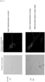
- the volumes or areas (or the total volume or area) of mitochondria or cells may be measured (computed) based on images of the cells or mitochondria captured by, for example, a fluorescence microscope or confocal laser microscope (which will be described later) after staining of the mitochondria and the like with a fluorescent dye or the like (which will be also described later).
- images of the cells are captured, one image per cell may be captured, or images of a plurality of divided portions of one cell may be captured.
- all or a portion of the images may be used to measure (compute) the volumes or areas (or the total volume or area) of the mitochondria or cells.
- the saving format of the captured images is not particularly limited as long as it can be used in an ordinary image analysis, and examples thereof include the TIFF format, the GIF format, the PNG format, and the JPEG format.
- mitochondrial size is fluorescence intensity obtained by staining cells or mitochondria with a fluorescent dye or the like and measuring the stained portions.
- a mitochondrion is an organelle that produces energy (ATP), and has membrane potential formed by proton gradient generated through oxidation-reduction reaction. Accordingly, when a fluorescent dye capable of staining mitochondria in a membrane potential-dependent manner (i.e., in a charge amount-dependent manner) is used, a measured fluorescence intensity can be used as a relative indicator of the mitochondrial size.
- the membrane potential of a mitochondrion can be evaluated by measuring an electric potential difference between the outer mitochondrial membrane and the inner mitochondrial membrane, which is also used to produce ATPs.
- the membrane potential can be measured using a known reagent or the like for detecting mitochondrial membrane potential.
- the reagent may be, for example, a fluorescence reagent, and more specifically JC-1 (DOJINDO LABORATORIES), AIE Mitochondria Red (Funakoshi Co., Ltd.), or the like.
- a fluorescence microscope e.g., BZ-X series; KEYENCE CORPORATION
- a confocal laser microscope e.g., VK-X series; KEYENCE CORPORATION
- a (two-dimensional or three-dimensional) high-content confocal imaging system e.g., Operetta CLS; PerkinElmer, Inc., Cell Voyger CV8000; Yokogawa Electric Corporation
- a (microplate) fluorescence spectrophotometer e.g., FP series; JASCO Corporation
- flowcytometry FACS (registered trademark); BD Biosciences
- a specific example of the "mitochondrial size" is the percentage of cells that have a size larger than or equal to a predetermined size and have mitochondria with a size larger than or equal to a predetermined mitochondrial size (e.g., the above-described fluorescence intensity, total volume, total area, or the like of the mitochondria), with respect to a particular cell population (e.g., all the cultured cells or a portion of the cultured cells).
- another aspect of the present invention is a method for evaluating quality of mesenchymal stem cells, the method including a step of evaluating the cells based on the percentage of cells that have a size larger than or equal to a predetermined size and have mitochondria with a size larger than or equal to a predetermined size, with respect to a particular cell population.
- the number, percentage, or the like of mesenchymal stem cells in a (cultured) cell population that are used in the evaluation method of the present invention can be obtained using a method known per se. Specifically, the number, percentage, or the like can be determined through flow cytometry or the like using a surface antigen (e.g., CD73, CD90, or CD105) of mesenchymal stem cells as an indicator.
- a surface antigen e.g., CD73, CD90, or CD105
- the "step of evaluating the cells based on measurements of mitochondrial sizes in the cells" of the present invention is a step of determining whether or not the cells fall within the mitochondrial size range serving as a predetermined reference (value) (or cutoff value).
- a predetermined reference value (or cutoff value) for the mitochondrial size is, for example, as follows.
- a predetermined reference value (or cutoff value) for the average value of the mitochondrial sizes in cells with a size of 100 K or more or cells with a diameter of (about) 30 ⁇ m or more may be 3300, 3345, 3400, 3500, 3568, 3600, 3700, 3800, 3900, 4000, 4100, 4200, 4300, 4400, 4500, 4600, 4616, 4700, 4800, 4900, 4924, or 5000.
- the fluorescence intensity or the average value of the fluorescence intensities of mesenchymal stem cells or a mesenchymal stem cell population used in the evaluation method of the present invention is greater than or equal to the reference value (cutoff value) above, the cells or cell population can be determined to have poor quality and be unsuitable for, for example, medical applications (e.g., cell transplantation) and research applications.
- Another predetermined reference value may be a value obtained by comparing the fluorescence intensities above before and after a passage.
- "Comparing the fluorescence intensities before and after a passage” may be: (i) comparing the fluorescence intensity at a passage when the evaluation method of the present invention is conducted, with the fluorescence intensity at a predetermined reference passage (e.g., zeroth, first, second, or third passage); or (ii) comparing the fluorescence intensity at the n th passage when the evaluation method of the present invention is conducted, with the fluorescence intensity at the n-1 th passage, where n-1 is the reference passage number.
- a predetermined reference value is a value (after passage / before passage) that is obtained by dividing the fluorescence intensity or the average value of the fluorescence intensities after the passage by that before the passage, and that indicates that the fluorescence intensity or the average value of the fluorescence intensities tends to increase after the passage compared with before the passage, and may be, for example, 1.1 times, 1.2 times, 1.3 times, 1.4 times, 1.5 times, 1.6 times, 1.7 times, 1.8 times, 1.9 times, 2.0 times, 2.1 times, 2.2 times, 2.3 times, 2.4 times, 2.5 times, 2.6 times, 2.7 times, 2.8 times, 2.9 times, or 3.0 times.
- the cells or cell population can be determined to have poor quality and be unsuitable for, for example, medical applications (e.g., cell transplantation) and research applications.
- the cells or cell population may be determined to have poor quality and be unsuitable for, for example, medical applications (e.g., cell transplantation) and research applications.
- a reference value (cutoff value) therefor may be 10.0%, 10.4%, 10.7%, 11.0%, 12.0%, 13.0%, 13.2%, 14.0%, 15.0%, 15.4%, 16.0%, 17.0%, 18.0%, 19.0%, 20.0%, 21.0%, 22.0%, 22.6%, 23.0%, 24.0%, 25.0%, 26.0%, 26.3%, 27.0%, 28.0%, 29.0%, or 30.0%.
- the percentage of cells that have a size larger than or equal to a predetermined size and have mitochondria with a fluorescence intensity (charge) larger than or equal to a predetermined fluorescence intensity (charge) with respect to a particular cell population (e.g., all the cultured cells or a portion of the cultured cells) used in the evaluation method of the present invention is greater than or equal to the reference value (cutoff value) above, the cells or cell population can be determined to have poor quality and be unsuitable for, for example, medical applications (e.g., cell transplantation) and research applications.
- the evaluation method of the present invention may include a step of selecting cells with a coefficient of variation, a CV (Coefficient of Variation) value, smaller than or equal to a predetermined value in addition to the steps described above.
- the "CV value” as used herein is a value obtained by dividing a standard deviation by an average value.
- a reference value (cutoff value) therefor may be 40.0, 41.0, 42., 43.0, 43.5, 44.0, 45.0, 46.0, 46.4, 47.0, 48.0, 49.0, 50.0, 50.5, 51.0, 52.0, 52.2, 53.0, 53.1, 54.0, 54.8, 55.0, 55.3, 56.0, 56.6, 57.0, 58.0, 59.0, 60.0, 61.0, 62.0, 63.0, 64.0, 64.3, 65.0, 66.0, 67.0, 68.0, 69.0, or 70.0.
- Selecting cells or a cell population with a CV value smaller than or equal to the reference value (cutoff value) above makes it possible to more reliably select cells or a cell population that has good quality and is suitable for medical applications (e.g., cell transplantation), research applications, and the like.
- the evaluation method of the present invention may include a step of selecting cells with a mesenchymal stem cell proliferation ratio larger than or equal to a predetermined value in addition to the steps described above.
- a reference value (cutoff value) therefor may be 5 times, 6 times, 7 times, 8 times, 9 times, 10 times, 11 times, 12 times, 13 times, 14 times, or 15 times.
- Selecting cells or a cell population with a proliferation ratio larger than or equal to the reference value (cutoff value) above makes it possible to more reliably select cells or a cell population that has good quality and is suitable for medical applications (e.g., cell transplantation), research applications, and the like.
- cells may include “cell population” unless otherwise stated.
- the cell population may be composed of one type of cell or two or more types of cells.
- examples of the source species include rodents such as a rat, a mouse, a hamster, and a guinea pig, lagomorphs such as rabbit, ungulates such as a pig, a cow, a goat, and a sheep, carnivores such as a dog and a cat, and primates such as a human, a monkey, a rhesus monkey, a marmoset, an orangutan, and a chimpanzee.
- rodents such as a rat, a mouse, a hamster, and a guinea pig
- lagomorphs such as rabbit
- ungulates such as a pig, a cow, a goat, and a sheep
- carnivores such as a dog and a cat
- primates such as a human, a monkey, a rhesus monkey, a marmoset, an orangutan, and a chimpanzee
- the present invention relates to a method for producing mesenchymal stem cells evaluated for quality, the method including a step of evaluating the cells (a step of evaluating quality) based on measurements of mitochondrial sizes in the cells, and also relates to mesenchymal stem cells obtained using this production method. More specifically, the method for producing mesenchymal stem cells according to the present invention includes: (A) a step of preparing mesenchymal stem cells to be subjected to expansion culture; and (B) a step of conducting expansion culture of the prepared cells, and the step of evaluating quality may be conducted before the step (A), during the step (A), after the step (A), during the step (B), or after the step (B).
- the step of evaluating quality may be conducted once in a particular period or a plurality of times.
- the present invention includes: (1) a step of preparing mesenchymal stem cells evaluated for quality; and (2) a step of culturing the mesenchymal stem cells.
- a source species of cells that can be used in the production method of the present invention
- examples of the species include rodents such as a rat, a mouse, a hamster, and a guinea pig, lagomorphs such as rabbit, ungulates such as a pig, a cow, a goat, and a sheep, carnivores such as a dog and a cat, and primates such as a human, a monkey, a rhesus monkey, a marmoset, an orangutan, and a chimpanzee.
- the age and the sex of the source species there is no particular limitation on the age and the sex of the source species.
- mesenchymal stem cells are produced using the production method of the present invention for the purpose of administration to humans, it is preferable to use, as a material, a cell population collected from a donor in which the type of histocompatibility antigen is the same as or similar to that of a recipient. It is more preferable to subject a cell population collected from a recipient himself/herself to production of mesenchymal stem cells.
- Mesenchymal stem cells that are prepared and subjected to expansion culture in the production method of the present invention may be primary cells that are directly separated from a biological tissue containing mesenchymal stem cells, or mesenchymal stem cells differentiated or induced from an established mesenchymal stem cell strain, embryonic stem cells, or induced pluripotent stem cells, or cryopreserved products of these cells.
- directly means that an in-vitro culturing/proliferation step is not involved.
- a method for obtaining mesenchymal stem cells directly separated from a biological tissue containing mesenchymal stem cells a method known per se such as the method disclosed in WO 2017/094879 is used to obtain desired cells.
- Examples of the above-described biological tissue containing mesenchymal stem cells include a bone marrow, an adipose tissue, blood, a placenta, an umbilical cord, and a dental pulp.
- the adipose tissue is a preferable source for collecting the cells above because the adipose tissue can be collected through liposuction or adipose tissue excision and this collection method is less likely to cause dysfunction in the living body.
- An adipose tissue is one type of biological tissue composed of adipocytes.
- the adipose tissue is used as a collection source of the cells above, there is no particular limitation on the site of the adipose tissue, but examples thereof include subcutaneous fat, visceral fat, intramuscular fat, and intermuscular fat. Out of these, subcutaneous fat can be said to be preferable because it can be easily collected under local anesthesia and burden on a donor during the collection can be reduced.
- the adipose tissue can be prepared from a tissue fragment sucked through liposuction surgery during cosmetic surgery or from an excised adipose tissue contained in a tissue excised from a living body during a surgical operation or the like.
- Fat-derived stem cells are present around thick blood vessels, and thus more fat-derived stem cells can be obtained from an excised adipose tissue than from a fat aspirate.
- stem cells are prepared from a fat aspirate, the size of an operation scar can be made smaller, and burden on a donor can be reduced.
- one type of adipose tissue is commonly used, but two or more types of adipose tissues may be used together. Also, adipose tissues collected a plurality of times may be mixed and used.
- the collection amount of the adipose tissue can be determined as appropriate in consideration of the type of donor, the type of tissue, or the amount of required mesenchymal stem cells.
- mesenchymal stem cells can be obtained from 0.3 to 20 g of the adipose tissue, but the present invention is not limited thereto.
- a larger amount of the adipose tissue can be used as a starting material through a scale-up or by performing the operation a plurality of times.
- the collected adipose tissue is subjected to removal of blood components attached thereto and fragmentation as needed, and is then subjected to enzyme treatment (protease treatment), which will be described later.
- enzyme treatment prote treatment
- the enzyme treatment is conducted by digesting the adipose tissue with a protease such as collagenase, trypsin, or Dispase.
- This enzyme treatment can be conducted using, for example, a method known per se such as the method described in R. Ian Freshney, Culture of Animal Cells: A Manual of Basic Technique, Fourth Edition, A John Wiley & Sones Inc ., Publication.
- the enzyme-treated adipose tissue includes two principal cell populations (stromal vascular fraction and mature adipocytes).
- the stromal vascular fraction is a cell mixture containing preadipocytes, mature endothelial cells, endothelial precursor cells, vascular smooth muscle cells, pericytes, wall cells, macrophages, fibroblasts, and fat-derived stem cells.
- Fat-derived stem cells are mesenchymal stem cells that can easily differentiate into adipocytes, osteoblasts, chondrocytes, and the like. The types and ratios of cells included in the cell population depend on the source and type of the adipose tissue used.
- the enzyme-treated adipose tissue is then subjected to centrifugation and is thus separated into two cell populations, and the precipitate (that includes the stromal vascular fraction) is collected.
- the conditions of the centrifugation vary depending on the types and amounts of the cells, the centrifugation is conducted, for example, at 300 to 2000 ⁇ g for 1 to 15 minutes.
- the enzyme-treated cell population may be subjected to filtration or the like to remove the tissue that has not been undigested by the enzyme, and the like from the cell population.
- a filter with a pore diameter of 50 to 2000 ⁇ m and preferably a filter with a pore diameter of 100 ⁇ m can be preferably used for the filtration
- Mesenchymal stem cells can be selectively proliferated by culturing the above-described stromal vascular fraction containing mesenchymal stem cells in the presence of a fibronectin fragment in accordance with a method known per se such as the method disclosed in WO 2017/094879 .
- the mesenchymal stem cells proliferated as described above (corresponding to mesenchymal stem cells prepared in the step (A) of the production method of the present invention) may be used in the expansion culture step (B) of the production method of the present invention.
- a medium for selective proliferation of mesenchymal stem cells can be prepared using, as a basal medium, a medium used to culture normal animal cells.
- a favorable example of the medium is a medium that does not contain a xenogeneic component such as fetal bovine serum (FBS) (or fetal calf serum (FCS)) or sheep serum.
- FBS fetal bovine serum
- FCS fetal calf serum
- sheep serum a xenogeneic component-free (xeno-free) medium can be prepared as appropriate, a medium known per se or a commercially available medium may be used as it is or after being modified.
- Examples of the commercially available xeno-free medium include Cellartis (registered trademark) DEF-CS500 XF (TAKARA), Mesenchymal Stem Cell Growth Medium DXF (PromoCell), CiMS-BM (NIPRO), StemPro MSC SFM XenoFree (Thermo Fisher Scientific), and KBM ADSC-4 (KOHJIN BIO). These media may also be employed as a medium to be used in the expansion culture step (B) of the production method of the present invention, which will be described later.
- Additives known per se can be added to the medium.
- the additives include growth factors (e.g., insulin), iron sources (e.g., transferrin), polyamines (e.g., putrescine), minerals (e.g., sodium selenate), saccharides (e.g., glucose), organic acids (e.g., pyruvic acid and lactic acid), amino acids (e.g., L-glutamine), reducing agents (e.g., 2-mercaptoethanol), vitamins (e.g., ascorbic acid and d-biotin), steroids (e.g., ⁇ -estradiol and progesterone), antibiotics (e.g., streptomycin, penicillin, and gentamicin), buffering agents (e.g., HEPES), lipids (e.g., linoleic acid), and nucleic acids (e.g., o growth factor (e.g., insulin), iron sources (e.g., transferrin), poly
- additives known per se that have been conventionally used for culture of mesenchymal stem cells may be added as appropriate. It is preferable that the concentrations of the contained additives are within ranges known per se. These additives may also be employed as additives for a medium to be used in the expansion culture step (B) of the production method of the present invention, which will be described later.
- the medium may contain allogeneic serum or may be a serum-free medium.
- the medium is an allogeneic serum-containing medium or serum-free medium and contains no xenogeneic components.
- the allogeneic serum is preferably autologous serum.
- autologous serum and autologous plasma which will be described later, respectively mean serum and plasma obtained from the blood collected from a donor who is identical to a donor of a cell population to be cultured.
- a medium containing autologous plasma may be used.
- inactivated autologous plasma is added to the medium.
- cells are cultured in a culture solution containing inactivated autologous plasma at a concentration of 10% (V/V) or less, preferably 5% (V/V) or less, and more preferably 2% (V/V) or less.
- the serum and the plasma may also be employed as serum and plasma for a medium to be used in the expansion culture step (B) of the production method of the present invention, which will be described later.
- Cell culture conditions can be set as appropriate.
- the culture temperature is not particularly limited but can be about 30 to 40°C and preferably about 37°C.
- the CO 2 concentration can be about 1 to 10% and preferably about 2 to 5%.
- the oxygen concentration can be 1 to 20% and preferably 1 to 10%. It is preferable to replace the medium containing an active component at appropriate intervals during the culture.
- These culture conditions may also be employed as culture conditions for the expansion culture step (B) of the production method of the present invention, which will be described later.
- the culture vessel used for the culture of mesenchymal stem cells as long as mesenchymal stem cells can be cultured therein, and examples thereof include a flask, a tissue culture flask, a dish, a petri dish, a tissue culture dish, a multi-dish, a microplate, a microwell plate, a multi-plate, a multi-well plate, a microslide, a chamber slide, a laboratory dish, a tube, a tray, a culture bag, and a roller bottle.
- These culture vessels may also be employed as culture vessels to be used in the expansion culture step (B) of the production method of the present invention, which will be described later.
- cells may be cultured, for example, in the presence of a fibronectin fragment as described in WO 2017/094879 (e.g., RetroNectin (registered trademark) (Takara Bio Inc.)) containing a functional domain of fibronectin, that is, a cell adhesion domain or a heparin-binding domain.
- a fibronectin fragment as described in WO 2017/094879 (e.g., RetroNectin (registered trademark) (Takara Bio Inc.)) containing a functional domain of fibronectin, that is, a cell adhesion domain or a heparin-binding domain.
- a specific culturing method is as follows: a stromal vascular fraction containing mesenchymal stem cells is cultured for, for example, 4 to 14 days and preferably 7 to 10 days using a xenogeneic component-free medium supplemented with 5% autologous plasma in a vessel coated with a fibronectin fragment with the medium being replaced every 3 to 4 days.
- This culture enables selective proliferation of mesenchymal stem cells.
- Mesenchymal stem cells make up 80% or more and preferably 90% or more of the cell population obtained through this culture.
- This culture method conducted in the presence of a fibronectin fragment may also be employed as a culture method to be used in the expansion culture step (B) of the production method of the present invention, which will be described later.
- the cell concentration at the start of the culture is not particularly limited, but is, for example, 0. 1 to 10 ⁇ 10 5 cells/ml, preferably 0.3 to 5 ⁇ 10 5 cells/ml, and more preferably 0.5 to 2 ⁇ 10 ⁇ 10 5 cells/ml. These cell concentrations may also be employed as a cell concentration in the expansion culture step (B) of the production method of the present invention, which will be described later.
- Expansion culture of mesenchymal stem cells may be conducted using a method known per se as long as the desired number of cells with excellent quality can be secured.
- cells may be cultured using a medium for mesenchymal stem cells containing reduced amounts of particular non-essential amino acids (glycine, alanine, serine, proline, asparagine, aspartic acid, and glutamic acid), as described in WO 2016/027850 .
- non-essential amino acids glycine, alanine, serine, proline, asparagine, aspartic acid, and glutamic acid
- cells may be cultured in the presence of a fibronectin fragment as described in WO 2017/094879 (e.g., RetroNectin (registered trademark) (Takara Bio Inc.)) containing a functional domain of fibronectin, that is, a cell adhesion domain or a heparin-binding domain.
- a fibronectin fragment as described in WO 2017/094879 (e.g., RetroNectin (registered trademark) (Takara Bio Inc.)) containing a functional domain of fibronectin, that is, a cell adhesion domain or a heparin-binding domain.
- the expansion culture may be conducted using a method as follows.
- the expansion culture step (B) is a step of extensively proliferating mesenchymal stem cells through expansion culture of mesenchymal stem cells in the presence of a fibronectin fragment.
- mesenchymal stem cells subjected to the culture of this step (B) include mesenchymal stem cells produced as described in "Proliferation Culture for Preparation of Mesenchymal Stem Cells" above, and mesenchymal stem cells differentiated or induced from an established mesenchymal stem cell strain, embryonic stem cells, and induced pluripotent stem cells.
- a method known per se may be used to separate the mesenchymal stem cells produced as described in "Proliferation Culture for Preparation of Mesenchymal Stem Cells" above from the culture vessel.
- Specific examples of the method include a physical method, a method in which a chelating agent is used, an enzymatic method in which a separating liquid having protease activity and/or collagenase activity (e.g., Accutase (registered trademark) and Accumax (registered trademark) (both available from Innovative Cell Technologies, Inc.), and the like) is used, and combinations thereof.
- a sheet of mesenchymal stem cells is separated using an enzymatic method, and then the cells are finely dispersed using a physical method.
- the separated cells may be used as they are, or may be cryopreserved until use. Also, a subset of cells separated based on a particular cell surface marker, such as isolated mesenchymal stem cells, may be used in this step.
- expansion culture of the mesenchymal stem cells cultured through the proliferation culture for preparation of mesenchymal stem cells is conducted using a xenogeneic component-free medium containing no serum in a vessel coated with a fibronectin fragment for, for example, 4 to 14 days and preferably 6 to 12 days with the medium being replaced every 3 to 4 days while the cells are passaged as appropriate.
- This culture enables extensive proliferation of mesenchymal stem cells.
- the cell concentration at the start of the culture is not particularly limited as long as mesenchymal stem cells can be proliferated, but it is typically, for example, 1.0 ⁇ 10 1 to 1.0 ⁇ 10 6 cells/cm 2 , preferably 1.0 ⁇ 10 2 to 1.0 ⁇ 10 5 cells/cm 2 , more preferably 1.0 ⁇ 10 3 to 1.0 ⁇ 10 5 cells/cm 2 , even more preferably 5.0 ⁇ 10 3 to 5.0 ⁇ 10 4 cells/cm 2 .
- Mesenchymal stem cells can be obtained using the above-described method for producing mesenchymal stem cells according to the present invention.
- the mesenchymal stem cells obtained using the production method of the present invention can be confirmed by detecting a molecule (e.g., an enzyme, a receptor, or a low-molecular compound) characteristic of mesenchymal stem cells.
- a molecule characteristic of mesenchymal stem cells include CD73, CD90, CD105, and CD166, which are cell surface markers (positive markers), but there is no limitation thereto.
- CD19, CD34, CD45, HLA-DR, CD11b, CD14, and the like are known as negative markers, which are not expressed in mesenchymal stem cells, and can be used for confirmation of mesenchymal stem cells.
- an immunological method can be used to detect the molecules above, the molecules may be detected through quantification of the mRNA levels of the molecules.
- the mesenchymal stem cells obtained using the production method of the present invention express the positive marker above.
- the negative marker expression ratio in the mesenchymal stem cells obtained using the production method of the present invention is, for example, 5% or less, preferably 1% or less, and more preferably lower than or equal to the detection limit.
- the mesenchymal stem cells obtained using the production method of the present invention may be further isolated, separated, and purified using an antibody that recognizes the molecule characteristic of the cells.
- the present invention relates to a method for producing mesenchymal cells (differentiated cells), the method including a step of inducing differentiation of mesenchymal stem cells obtained using the method for producing mesenchymal stem cells (mitochondrial size, etc.) according to the present invention, and also relates to mesenchymal cells obtained using this production method. More specifically, the present invention includes: (1) a step of preparing mesenchymal stem cells obtained using the method for producing mesenchymal stem cells according to the present invention; and (2) a step of inducing differentiation into the mesenchymal cells. Examples of such mesenchymal cells include osteocytes, chondrocytes, and adipocytes. It is preferable to conduct all the steps of the method for producing mesenchymal cells according to the present invention under the feeder-free and xeno-free conditions.
- an osteocyte differentiation-inducing medium examples include a basal medium (e.g., ⁇ MEM) containing 10% FBS, 0.1 ⁇ M dexamethasone, 50 ⁇ g/ml ascorbic acid, and 10 mM ⁇ -glycerophosphate, and a commercially available xeno-free osteocyte differentiation-inducing medium (e.g., MSCgo (trademark) Rapid Osteogenic Differentiation Medium (Biological Industries)).
- MSCgo commercially available xeno-free osteocyte differentiation-inducing medium
- the osteocyte differentiation method is conducted as follows, for example: 4 ⁇ 10 4 mesenchymal stem cells are seeded in a 12-well plate coated with gelatin and are cultured in the above-described osteocyte differentiation-inducing medium for 30 days. Differentiation into osteocytes may be confirmed by detecting calcified nodules through alizarin red staining.
- chondrocyte differentiation-inducing medium examples include a basal medium (e.g., DMEM/F12) containing 1%(v/v) ITS+ premix, 0.17 mM AA2P, 0.35 mM proline, 0.1 mM dexamethasone, 0.15% (v/v) glucose, 1 mM sodium pyruvate, 2 mM GlutaMAX, 40 ng/ml PDGF-BB, 100 ng/mL TGF- ⁇ 3, 10 ng/ml BMP4, and 1% (v/v) FBS, and a commercially available xeno-free chondrocyte differentiation-inducing medium (e.g., MSCgo (trademark) Chondrogenic XF (Biological Industries)).
- a basal medium e.g., DMEM/F12
- ITS+ premix 0.17 mM AA2P
- 0.35 mM proline 0.35 mM proline
- the chondrocyte differentiation method is conducted as follows, for example: 5 ⁇ l of a mesenchymal stem cell suspension is spotted on a plate coated with fibronectin, followed by 1-hour culture, and 1 ml of the above-described differentiation-inducing medium is added thereto after 1 hour, followed by 14-day culture. Differentiation into chondrocytes may be confirmed through alcian blue staining.
- an adipocyte differentiation-inducing medium examples include a basal medium containing 60 ⁇ M indomethacin, 0.5 mM IBMX, and 0.5 ⁇ M hydrocortisone, and a commercially available xeno-free adipocyte differentiation-inducing medium (e.g., hMSC-Human Mesenchymal Stem Cell Adipogenic Differentiation Medium (Lonza) or MSCgo (treademark) Adipogenic XF (Biological Industries)).
- the adipocyte differentiation method is conducted as follows, for example: 4 ⁇ 10 4 mesenchymal stem cells are seeded on a plate coated with gelatin and are cultured in the above-described adipocyte differentiation-inducing medium for 32 days. Differentiation into adipocytes may be confirmed through oil red O staining.
- the mesenchymal stem cells obtained using the method for producing mesenchymal stem cells (mitochondrial size, etc.) according to the present invention and the mesenchymal cells (differentiated cells) obtained using the method for producing mesenchymal cells (mitochondrial size, etc.) according to the present invention can have excellent repairing ability when transplanted to a defect site of the bone, cartilage, muscle, or the like, compared with cells obtained using conventional methods. Accordingly, the mesenchymal cells of the present invention can be favorably used in cell transplantation therapy.
- the present invention relates to a cell transplantation therapy agent containing the mesenchymal cells of the present invention.
- the present invention also encompasses a method for treating an injury (including defect) of a tissue (e.g., a bone tissue, a cartilage tissue, an adipose tissue, and a muscle tissue (e.g., skeletal muscle tissue)) or a disease, the method including administering or transplanting an effective amount of the mesenchymal cells of the present invention to a mammal (e.g., a human, a mouse, a rat, a monkey, a cow, a horse, a pig, or a dog) to be treated.
- a mammal e.g., a human, a mouse, a rat, a monkey, a cow, a horse, a pig, or a dog
- the "treatment of an injury of a tissue” encompasses regeneration of an injured tissue.
- the purpose of the transplantation of the mesenchymal cells of the invention to a living body may be direct regeneration of an injured tissue or an indirect effect (e.g., paracrine effect) by a factor secreted by the mesenchymal cells of the present invention.
- the therapeutic effect of mesenchymal stem cells can be obtained in patients affected with acute myocardial infarction, a stroke, multiple system atrophy (MSA), a graft-versus-host disease, Crohn's disease, ischemic cardiomyopathy, spinal cord injury, and the like.
- the mesenchymal cells of the present invention are used in cell transplantation therapy, it is desirable to use cells derived from an individual with the same or substantially the same HLA genotype as that of a transplantation target individual or cells derived from iPS cells established from somatic cells of the individual, from the viewpoint of preventing rejection.
- substantially the same means that the HLA genotypes are identical to an extent that the immunological reaction to the transplanted cells can be suppressed using an immunosuppressive agent, and, for example, somatic cells with the HLA type in which three genetic loci (HLA-A, HLA-B, and HLA-DR) are respectively identical or four genetic loci (HLA-C in addition to the three loci above) are respectively identical are used.
- the cells can be embedded in a capsule made of polyethylene glycol or silicone, a porous vessel, or the like for the purpose of avoiding rejection and then transplanted.
- a parenteral preparation such as an injection, a suspension, or a drip is produced by, for example, mixing the mesenchymal cells of the present invention with a pharmaceutically acceptable carrier in accordance with a conventional procedure.
- a method for producing a cell transplantation therapy agent is also provided, the method including a step of formulating the mesenchymal cells of the present invention.
- This production method may include a step of preparing the mesenchymal cells of the present invention.
- the production method can also include a step of preserving the mesenchymal cells of the present invention.
- aqueous liquids for injection such as a physiological saline solution and an isotonic solution containing glucose and other adjuvants (e.g., D-sorbitol, D-mannitol, and sodium chloride).
- the cell transplantation therapy agent of the present invention may be blended with a buffering agent (e.g., a phosphate buffer solution or sodium acetate buffer solution), a soothing agent (e.g., benzalkonium chloride or procaine hydrochloride), a stabilizer (e.g., human serum albumin or polyethylene glycol), a preservative, an antioxidant, and the like.
- a buffering agent e.g., a phosphate buffer solution or sodium acetate buffer solution
- a soothing agent e.g., benzalkonium chloride or procaine hydrochloride
- a stabilizer e.g., human serum albumin or polyethylene glycol
- preservative an antioxidant, and the like.
- the cell transplantation therapy agent of the present invention is formulated as an aqueous suspension, it is only necessary to suspend the cells into the above-described aqueous liquid such that, for example, the cell concentration is about 1 ⁇ 10 6 to about 1 ⁇ 10 8 cells/ml.
- the dosage or transplantation amount and the administration frequency or transplantation frequency of the mesenchymal cells or pharmaceutical composition of the present invention can be determined as appropriate depending on the age, weight, symptom, and the like of a mammal to which the mesenchymal cells or pharmaceutical composition is administered.
- the cell transplantation therapy agent of the present invention can be used as follows: it is provided in a state of being cryopreserved under the conditions commonly used for cryopreservation of cells, and is thawed before use.
- the cell transplantation therapy agent may further contain serum or a substitute thereof, an organic solvent (e.g., DMSO), and the like.
- the concentration of the serum or the substitute thereof is not particularly limited, but can be about 1 to about 30% (v/v) and preferably about 5 to about 20% (v/v).
- the concentration of the organic solvent is not particularly limited, but can be 0 to about 50% (v/v) and preferably about 5 to about 20% (v/v).
- the present invention relates to a method for evaluating a medium for mesenchymal stem cells, the method including a step of evaluating the medium based on measurements of mitochondrial sizes in the cells.
- an aspect of the present invention relates to a method for evaluating a medium for mesenchymal stem cells (evaluation method 1 of the present invention), the method including:
- Another aspect relates to a method for evaluating a medium for mesenchymal stem cells (evaluation method 2 of the present invention), the method including:
- a predetermined reference value (or cutoff value) for the average value of the mitochondrial sizes in cells with a size of 100 K or more or cells with a diameter of (about) 30 ⁇ m or more may be 3300, 3345, 3400, 3500, 3568, 3600, 3700, 3800, 3900, 4000, 4100, 4200, 4300, 4400, 4500, 4600, 4616, 4700, 4800, 4900, 4924, or 5000 after a predetermined number of passages (e.g., 1, 2, 3, 4, 5, 6, 7, 8, 9, 10, 11, 12, 13, 14, 15, 16, 17, 18, 19, or 20 passages).
- the medium can be determined to be unsuitable as a medium for mesenchymal stem cells.
- Another predetermined reference value may be a value obtained by comparing the fluorescence intensities above before and after the culture. "Comparing the fluorescence intensities before and after the culture” may mean comparison of the fluorescence intensity before the start of the culture and the fluorescence intensity after a predetermined number of passages (e.g., 1, 2, 3, 4, 5, 6, 7, 8, 9, 10, 11, 12, 13, 14, 15, 16, 17, 18, 19, or 20 passages).
- a predetermined reference value is a value (after passage / before passage) that is obtained by dividing the fluorescence intensity or the average value of the fluorescence intensities after the culture by that before the culture, and that indicates that the fluorescence intensity or the average value of the fluorescence intensities tends to increase after the culture compared with before the culture, and may be, for example, 1.1 times, 1.2 times, 1.3 times, 1.4 times, 1.5 times, 1.6 times, 1.7 times, 1.8 times, 1.9 times, 2.0 times, 2.1 times, 2.2 times, 2.3 times, 2.4 times, 2.5 times, 2.6 times, 2.7 times, 2.8 times, 2.9 times, or 3.0 times.
- the medium can be determined to be unsuitable as a medium for mesenchymal stem cells.
- a reference value (or cutoff value) therefor may be 10.0%, 10.4%, 10.7%, 11.0%, 12.0%, 13.0%, 13.2%, 14.0%, 15.0%, 15.4%, 16.0%, 17.0%, 18.0%, 19.0%, 20.0%, 21.0%, 22.0%, 22.6%, 23.0%, 24.0%, 25.0%, 26.0%, 26.3%, 27.0%, 28.0%, 29.0%, or 30.0% after a predetermined number of passages (e.g., 1, 2,
- the medium can be determined to be unsuitable as a medium for mesenchymal stem cells.
- a coefficient of variation a CV (Coefficient of Variation) value, indicating the variation in the cell size may be used as a reference value in addition to the above-described reference value (cutoff value).
- the "CV value” as used herein is a value obtained by dividing a standard deviation by an average value.
- a predetermined reference value (or cutoff value) therefor may be 40.0, 41.0, 42.0, 43.0, 43.5, 44.0, 45.0, 46.0, 46.4, 47.0, 48.0, 49.0, 50.0, 50.5, 51.0, 52.0, 52.2, 53.0, 53.1, 54.0, 54.8, 55.0, 55.3, 56.0, 56.6, 57.0, 58.0, 59.0, 60.0, 61.0, 62.0, 63.0, 64.0, 64.3, 65.0, 66.0, 67.0, 68.0, 69.0, or 70.0 after a predetermined number of passages (e.g., 1, 2, 3, 4, 5, 6, 7, 8, 9, 10, 11, 12, 13, 14, 15, 16, 17, 18, 19, or 20 passages).
- a predetermined number of passages e.g., 1, 2, 3, 4, 5, 6, 7, 8, 9, 10, 11, 12, 13, 14, 15, 16, 17, 18, 19, or 20 passages.
- Selecting a medium from which cells or a cell population with a CV value smaller than or equal to the reference value (cutoff value) above is obtained after a predetermined number of passages (or removing a medium) with which cells or a cell population with a CV value larger than or equal to the reference value (cutoff value) above is obtained makes it possible to more reliably select a medium suitable as a medium for mesenchymal stem cells.
- the present invention relates to a method for evaluating quality of mesenchymal stem cells, the method including a step of evaluating the cells based on measurements of consumptions of pyruvic acid, cystine, and serine by the cells.
- an aspect of the present invention relates to a method for evaluating quality of mesenchymal stem cells, the method including a step of evaluating the cells based on measurements of consumptions of pyruvic acid, cystine, and serine by one cell.
- the "consumptions of pyruvic acid, cystine, and serine by the cells" can be calculated based on differences in abundances of pyruvic acid, cystine, and serine in the medium between before and after the start of the culture of mesenchymal stem cells.
- Before the start of the culture may mean “before the start of the first passage (maintenance) culture (or at the time when the first passage (maintenance) culture is started) (P0)", or “before the start of any of the passages (e.g., P1, P2, P3, P4, P5, P6, P7, P8, P9, P10, P11, P12, P13, P14, or P15) (or at the time when any of the passages is started)”.
- the abundances of pyruvic acid, cystine, and serine in the medium may be represented by the concentrations, or the measurement values obtained by measuring these three substances using an apparatus or a method as described later.
- the "differences in abundances” may by differences in concentrations of pyruvic acid, cystine, and serine in the medium between before and after the start of the culture, or differences in measurement values as described above.
- a method and an apparatus for measuring the abundances of pyruvic acid, cystine, and serine in the medium as long as the three substances can be measured with desired detection limits, and the abundances thereof can be measured using a method known per se.
- a liquid chromatograph/tandem mass spectrometer (LC/MS/MS), a liquid chromatograph/mass spectrometer (LC/MS), an immunological measurement method (e.g., ELISA) can be used.
- these three substances may be simultaneously measured using the same apparatus or method, or may be separately measured using the same apparatus or method, or may be separately measured using different apparatuses or methods.
- the "consumptions of pyruvic acid, cystine, and serine by the cells" which are used as indicators in the present invention, can be calculated (computed) as a negative area ratio per cell ( ⁇ area/cell) by measuring, using LC/MS/MS, the absorbances of pyruvic acid, cystine, and serine in the medium after the culture of mesenchymal stem cells is started and the passaging is repeated a predetermined times, relative to the absorbances of a fresh medium containing no cells (i.e., the medium before the start of the culture), and then dividing the absorbances by the number of cells collected after the passaging is repeated the predetermined times.
- the number, percentage, or the like of mesenchymal stem cells in a (cultured) cell population that are used in the evaluation method of the present invention can be obtained using a method known per se. Specifically, the number, percentage, or the like can be determined through flow cytometry or the like using a surface antigen (e.g., CD73, CD90, or CD105) of mesenchymal stem cells as an indicator.
- a surface antigen e.g., CD73, CD90, or CD105
- the "step of evaluating the cells based on measurements of consumptions of pyruvic acid, cystine, and serine by the cells" of the present invention is a step of determining whether or not the cells fall within the consumption ranges of pyruvic acid, cystine, and serine serving as predetermined references (reference values) (or cutoff values).
- the consumptions of at least two of pyruvic acid, cystine, and serine and preferably the consumptions of at least two of these substances by one cell, or the consumptions of all of these substances and preferably the consumptions of all of these substances by one cell tend to increase (or acceleratingly increase), for example, over any period of the culture of mesenchymal stem cells (e.g., between the n th and n+1 th passages, between the n th and n+2 th passages, or between the n th and n+3 th passages), the cells or cell population can be determined to have poor quality after the n th passage and be unsuitable for, for example, medical applications (e.g., cell transplantation) and research applications.
- medical applications e.g., cell transplantation
- predetermined reference values (or cutoff values) of the consumptions of pyruvic acid, cystine, and serine are, for example, as follows.
- the consumptions of pyruvic acid, cystine, and serine by the cells can be measured and calculated (computed) using, for example, the same method as that in Examples, which will be described later, and in this case, the predetermined reference values (cutoff values) therefor may be instantaneous consumptions, consumptions during a particular period (e.g., while the passaging is repeated 1, 2, or 3 times), or values obtained by comparing the consumptions before and after the passage (differences in the consumptions between before and after the passage) (e.g., division or subtraction of ⁇ area/cell before and after the passage).
- "Comparing the consumptions before and after the passage” may be as follows:
- values obtained by dividing the consumptions of pyruvic acid, cystine, and serine by the cells at the time when the evaluation method of the present invention is conducted after the passaging is repeated by the consumptions of pyruvic acid, cystine, and serine by the cells after the passaging is repeated reference times, or values obtained by subtracting the consumptions after the passaging is repeated reference times from the consumptions at the time when the evaluation method of the present invention is conducted after the passaging is repeated are used.
- predetermined reference values are values (after passage / before passage) that are obtained by dividing the consumptions of all of pyruvic acid, cystine, and serine after the passage by those before the passage, and preferably dividing the consumptions of all of pyruvic acid, cystine, and serine by one cell after the passage by those before the passage, and that indicate that the consumptions tend to increase after the passage compared with before the passage, and may be, for example, 1.1 times, 1.2 times, 1.3 times, 1.4 times, 1.5 times, 1.6 times, 1.7 times, 1.8 times, 1.9 times, 2.0 times, 2.1 times, 2.2 times, 2.3 times, 2.4 times, 2.5 times, 2.6 times, 2.7 times, 2.8 times, 2.9 times, 3.0 times, 3.1 times, 3.2 times, 3.3 times, 3.4 times, 3.5 times, 3.6 times, 3.7 times, 3.8 times, 3.9 times, 4.0 times, 4.1 times, 4.2 times, 4.3 times, 4.4 times, 4.5 times,
- the cells or cell population can be determined to have poor quality and be unsuitable for, for example, medical applications (e.g., cell transplantation) and research applications.
- predetermined reference values may be values obtained by comparing the consumptions before and after the passage (e.g., subtraction of ⁇ area/cell before and after the passage).
- predetermined reference values are values (after passage - before passage) that are obtained by subtracting the consumptions of at least two of pyruvic acid, cystine, and serine before the passage from those after the passage, and preferably subtracting the consumptions of at least two of pyruvic acid, cystine, and serine by one cell before the passage from those after the passage, or subtracting the consumptions of all of these substances before the passage from those after the passage, and preferably subtracting the consumptions of all of these substances by one cell before the passage from those after the passage, and that indicate that the consumptions tend to increase after the passage compared with before the passage, and may be, for example, -1.0, -1.1, -1.2, -1.3, -1.4, -1.5, -1.6, -1.7, -1.8, -1.9, -2.0, -2.1, -2.2, -2.3, -2.4, -2.5, -2.6, -2.7, -2.8, -2.9, -3.0, -3.1
- the cells or cell population can be determined to have poor quality and be unsuitable for, for example, medical applications (e.g., cell transplantation) and research applications.
- the above-described reference values when values obtained by comparing the consumptions of all of pyruvic acid, cystine, and serine before and after the passage are greater than or equal to (in the case of "after passage / before passage") or smaller than or equal to (in the case of "after passage - before passage”) the above-described reference value (cutoff value) two or more times in a row (e.g., the evaluation method of the present invention is conducted after the n th passage (the reference passage number is n-1) and the n+1 th passage (the reference passage number is n)), the cells or cell population may be determined to have poor quality and be unsuitable for, for example, medical applications (e.g., cell transplantation) and research applications.
- medical applications e.g., cell transplantation
- examples of the source species include rodents such as a rat, a mouse, a hamster, and a guinea pig, lagomorphs such as rabbit, ungulates such as a pig, a cow, a goat, and a sheep, carnivores such as a dog and a cat, and primates such as a human, a monkey, a rhesus monkey, a marmoset, an orangutan, and a chimpanzee.
- rodents such as a rat, a mouse, a hamster, and a guinea pig
- lagomorphs such as rabbit
- ungulates such as a pig, a cow, a goat, and a sheep
- carnivores such as a dog and a cat
- primates such as a human, a monkey, a rhesus monkey, a marmoset, an orangutan, and a chimpanzee
- the present invention relates to a method for producing mesenchymal stem cells evaluated for quality, the method including a step of evaluating the cells (a step of evaluating quality) based on measurements of consumptions of pyruvic acid, cystine, and serine by the cells, and also relates to mesenchymal stem cells obtained using this production method. More specifically, the method for producing mesenchymal stem cells according to the present invention includes: (A) a step of preparing mesenchymal stem cells to be subjected to expansion culture; and (B) a step of conducting expansion culture of the prepared cells, and the step of evaluating quality may be conducted before the step (A), during the step (A), after the step (A), during the step (B), or after the step (B).
- the step of evaluating quality may be conducted once in a particular period or a plurality of times.
- the present invention includes: (1) a step of preparing mesenchymal stem cells evaluated for quality; and (2) a step of culturing the mesenchymal stem cells.
- the present invention relates to a method for producing mesenchymal cells (differentiated cells), the method including a step of inducing differentiation of mesenchymal stem cells obtained using the method for producing mesenchymal stem cells (consumptions of pyruvic acid, cystine, and serine, etc.) according to the present invention, and also relates to mesenchymal cells obtained using this production method. More specifically, the present invention includes: (1) a step of preparing mesenchymal stem cells obtained using the method for producing mesenchymal stem cells according to the present invention; and (2) a step of inducing differentiation into the mesenchymal cells. Examples of such mesenchymal cells include osteocytes, chondrocytes, and adipocytes. It is preferable to conduct all the steps of the method for producing mesenchymal cells according to the present invention under the feeder-free and xeno-free conditions.
- the mesenchymal stem cells obtained using the method for producing mesenchymal stem cells (consumptions of pyruvic acid, cystine, and serine, etc.) according to the present invention and the mesenchymal cells (differentiated cells) obtained using the method for producing mesenchymal cells (consumptions of pyruvic acid, cystine, and serine, etc.) according to the present invention can have excellent repairing ability when transplanted to a defect site of the bone, cartilage, muscle, or the like, compared with cells obtained using conventional methods. Accordingly, the mesenchymal cells of the present invention can be favorably used in cell transplantation therapy.
- the present invention relates to a cell transplantation therapy agent containing the mesenchymal cells of the present invention.
- the present invention also encompasses a method for treating an injury (including defect) of a tissue (e.g., a bone tissue, a cartilage tissue, an adipose tissue, and a muscle tissue (e.g., skeletal muscle tissue)) or a disease, the method including administering or transplanting an effective amount of the mesenchymal cells of the present invention to a mammal (e.g., a human, a mouse, a rat, a monkey, a cow, a horse, a pig, or a dog) to be treated.
- a mammal e.g., a human, a mouse, a rat, a monkey, a cow, a horse, a pig, or a dog
- the "treatment of an injury of a tissue” encompasses regeneration of an injured tissue.
- the present invention relates to a method for evaluating a medium for mesenchymal stem cells, the method including a step of evaluating the medium based on measurements of consumptions of pyruvic acid, cystine, and serine by the cells.
- an aspect of the present invention relates to a method for evaluating a medium for mesenchymal stem cells (evaluation method 1' of the present invention), the method including:
- predetermined reference values may be instantaneous consumptions, consumptions during a particular period (e.g., while the passaging is repeated 1, 2, or 3 times), or values obtained by comparing the consumptions before and after the passage (e.g., division or subtraction of ⁇ area/cell before and after the passage).
- predetermined reference values are values (after passage / before passage) that are obtained by dividing the consumptions of all of pyruvic acid, cystine, and serine after the passage by those before the passage, and preferably dividing the consumptions of all of pyruvic acid, cystine, and serine by one cell after the passage by those before the passage, and that indicate that the consumptions tend to increase after the passage compared with before the passage, and may be, for example, 1.1 times, 1.2 times, 1.3 times, 1.4 times, 1.5 times, 1.6 times, 1.7 times, 1.8 times, 1.9 times, 2.0 times, 2.1 times, 2.2 times, 2.3 times, 2.4 times, 2.5 times, 2.6 times, 2.7 times, 2.8 times, 2.9 times, 3.0 times, 3.1 times, 3.2 times, 3.3 times, 3.4 times, 3.5 times, 3.6 times, 3.7 times, 3.8 times, 3.9 times, 4.0 times, 4.1 times, 4.2 times, 4.3 times
- the medium can be determined to be unsuitable as a medium for mesenchymal stem cells.
- predetermined reference values may be values obtained by comparing the consumptions before and after the passage (e.g., subtraction of ⁇ area/cell before and after the passage).
- predetermined reference values are values (after passage - before passage) that are obtained by subtracting the consumptions of at least two of pyruvic acid, cystine, and serine before the passage from those after the passage, and preferably subtracting the consumptions of at least two of pyruvic acid, cystine, and serine by one cell before those after the passage, or subtracting the consumptions of all of these substances before the passage from those after the passage, and preferably subtracting the consumptions of all of these substances by one cell before the passage from those after the passage, and that indicate that the consumptions tend to increase after the passage compared with before the passage, and may be, for example, -1.0, -1.1, -1.2, -1.3, -1.4, -1.5, -1.6, -1.7, -1.8, -1.9, -2.0, -2.1, -2.2, -2.3, -2.4, -2.5, -2.6, -2.7, -2.8, -2.9, -3.0, -3.1, -
- the medium can be determined to be unsuitable as a medium for mesenchymal stem cells.
- a 6-well plate was coated with VTN-N, was left to stand at room temperature for 1 hour, and was then stored in the refrigerator.
- a 6-well plate was coated with VTN-N, was left to stand at room temperature for 1 hour, and was then stored in the refrigerator.
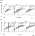
- Mitochondrial changes depending on the passage number of MSC-1 CiMS are shown with the vertical axis illustrating the mitochondrial charge and the horizontal axis illustrating the cell size ( FIGS. 4 and 6 ).
- the region of a mitochondrial charge of 5.0 K or more and the cell size of 120 K or more in this region, CiMS shows a mitochondrial charge of 5.0 K and a cell size of 120 K when FACS is set to "PE: voltage 290" and the cell population at P3 is used as a reference
- the mitochondrial size started to change at approximately P6 (10.4% ( FIG. 4 ), 7.7% ( FIG. 6 )), and this result tended to correspond to the proliferation ratio of MSC-1 CiMS in FIG. 1 .
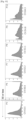
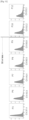
- mitochondrial changes depending on the passage number of MSC-1 Cellartis particularly in the region of a mitochondrial charge of 2.5 K or more and the cell size of 90 K or more (in this region, Cellartis shows a mitochondrial charge of 2.5 K and a cell size of 90 K when FACS is set to "PE: voltage 290" and the cell population at P3 is used as a reference)
- the mitochondrial size started to change at approximately P15 (22.6% ( FIG. 5 )) or P11 (11.0% ( FIG. 7 )), and this result tended to correspond to the proliferation ratio of MSC-1 Cellartis in FIG. 2 . It was found from these results that the quality of mesenchymal stem cells can be evaluated using, as an indicator, the mitochondrial charge (size) or the mitochondrial charge (size) in cells with a size larger than or equal to a particular size.
- Tables 4 and 5 show the results of the membrane potential analysis.
- FIGS. 10 and 11 show the results of examination of the CV values (the Coefficient of Variation is a normalized Standard Deviation), namely StdDev (standard deviation, variation) / Mean (mean for standard distribution), obtained from the measurement results of flow cytometry. It is understood from these diagrams that using "CV value ⁇ 50.0" as an indicator is effective in evaluation of the quality of mesenchymal stem cells.
- Example 5 Viability Test Performed Using Animal with MSC Passage Number Being Varied
- MSC-1 fat-derived MSC cells
- CiMS Nipro
- VTN Thermo
- TAKARA Cellartis
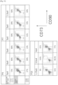
- the transplanted MSCs (CD90-CD73 double positive cells) made up about 54% at Passage 3 (P3) but about 0.5% at Passage (P8) ( FIG. 14 ).
- the transplanted MSCs (CD90-CD73 double positive cells) made up about 75% at Passage 4 (P4), about 48% at Passage 7 (P7), and about 10% at Passage 10 (P10) ( FIG. 15 ).
- LC/MS/MS liquid chromatograph/tandem mass spectrometer
- LCMS-8050 and LC/MS/MS method package cell culture profiling SHIMADZU CORPORATION
- LC/MS/MS liquid chromatograph/tandem mass spectrometer
- SHIMADZU CORPORATION LC/MS/MS method package cell culture profiling
- Tables 6, 7 and 8 (CiMS) and Tables 9, 10, and 11 (Cellartis) below show the consumptions of pyruvic acid, cystine, and serine by one cell at every passage, differences in the consumptions between before and after the passage (after passage / before passage, and after passage - before passage), and differences in consumptions with respect to the passage P3 (after passage / P3, and after passage - P3).
- the TCA cycle becomes predominant over the glycolytic pathway to, for example, secure energy in a living body and thus a larger amount of pyruvic acid is consumed, resulting in the generation of a large amount of reactive oxygen species.
- glutathione which is an antioxidative substance
- the quality of mesenchymal stem cells can be evaluated using, as indicators, the consumptions of pyruvic acid, cystine, and serine in the medium by the cells.
- changes in the consumptions of these three substances vary depending on the medium in which the cells are cultured, and a medium for mesenchymal stem cells can also be evaluated using the consumptions of these substances as indicators.
- the results of the determination of whether the passage number is appropriate obtained based on the consumptions of pyruvic acid, cystine, and serine above (after the seventh passage (P7) when using CiMS, and after the eleventh passage (P11) when using Cellartis), significantly corresponded to the results of the cell transplantation test using mice in Example 5. Therefore, from the viewpoint of the results of the animal experiment as well, it is understood that the consumptions of pyruvic acid, cystine, and serine by the cells used as indicators in the present invention are excellent indicators for evaluation of the quality of MSCs.
- the present invention makes it possible to more reliably, more simply, and more quickly evaluate the quality of mesenchymal stem cells, which is thus useful. Using this evaluation method makes it possible to produce mesenchymal stem cells with stable quality (differentiation potential, proliferative capacity, undifferentiation properties, etc.), which is thus useful. Differentiating mesenchymal stem cells with stable quality also makes it possible to produce mesenchymal cells with stable quality more efficiently and reproducibly, which is thus useful. Furthermore, the mesenchymal stem cells and the mesenchymal cells obtained using the production method of the present invention have stable quality and are reproducibly produced, and are thus particularly suitable for application to the cell transplantation treatment and are useful.
Landscapes
- Health & Medical Sciences (AREA)
- Life Sciences & Earth Sciences (AREA)
- Engineering & Computer Science (AREA)
- Biomedical Technology (AREA)
- Zoology (AREA)
- Developmental Biology & Embryology (AREA)
- Biotechnology (AREA)
- Chemical & Material Sciences (AREA)
- Organic Chemistry (AREA)
- Genetics & Genomics (AREA)
- Wood Science & Technology (AREA)
- Bioinformatics & Cheminformatics (AREA)
- Cell Biology (AREA)
- General Health & Medical Sciences (AREA)
- Biochemistry (AREA)
- Immunology (AREA)
- Microbiology (AREA)
- Rheumatology (AREA)
- General Engineering & Computer Science (AREA)
- Medicinal Chemistry (AREA)
- Virology (AREA)
- Hematology (AREA)
- Pharmacology & Pharmacy (AREA)
- Epidemiology (AREA)
- Animal Behavior & Ethology (AREA)
- Public Health (AREA)
- Veterinary Medicine (AREA)
- Micro-Organisms Or Cultivation Processes Thereof (AREA)
- Measuring Or Testing Involving Enzymes Or Micro-Organisms (AREA)
Applications Claiming Priority (3)
| Application Number | Priority Date | Filing Date | Title |
|---|---|---|---|
| JP2022059920 | 2022-03-31 | ||
| JP2022196791 | 2022-12-09 | ||
| PCT/JP2023/009633 WO2023189485A1 (ja) | 2022-03-31 | 2023-03-13 | 間葉系幹細胞の品質評価方法 |
Publications (2)
| Publication Number | Publication Date |
|---|---|
| EP4502170A1 true EP4502170A1 (de) | 2025-02-05 |
| EP4502170A4 EP4502170A4 (de) | 2025-09-10 |
Family
ID=88200903
Family Applications (1)
| Application Number | Title | Priority Date | Filing Date |
|---|---|---|---|
| EP23779505.9A Pending EP4502170A4 (de) | 2022-03-31 | 2023-03-13 | Verfahren zur qualitätsbeurteilung mesenchymaler stammzellen |
Country Status (3)
| Country | Link |
|---|---|
| EP (1) | EP4502170A4 (de) |
| JP (1) | JPWO2023189485A1 (de) |
| WO (1) | WO2023189485A1 (de) |
Family Cites Families (10)
| Publication number | Priority date | Publication date | Assignee | Title |
|---|---|---|---|---|
| ES2927112T3 (es) | 2014-08-01 | 2022-11-02 | Purec Co Ltd | Método para evaluar la calidad de células madre mesenquimatosas humanas y un anticuerpo monoclonal para su uso en dicho método |
| CN106715683A (zh) | 2014-08-21 | 2017-05-24 | 味之素株式会社 | 间充质干细胞用培养基 |
| JP6519727B2 (ja) * | 2014-09-17 | 2019-05-29 | 国立大学法人山口大学 | 間葉系幹細胞の培養方法、および肝機能障害用治療剤の製造方法 |
| WO2017068727A1 (ja) * | 2015-10-23 | 2017-04-27 | 株式会社島津製作所 | 細胞の分化状態の評価方法 |
| JP2017104091A (ja) * | 2015-11-27 | 2017-06-15 | タカラバイオ株式会社 | 間葉系細胞の製造方法 |
| KR102676761B1 (ko) | 2015-12-03 | 2024-06-18 | 다카라 바이오 가부시키가이샤 | 중간엽 줄기세포의 제조방법 |
| JP6909454B2 (ja) * | 2017-01-26 | 2021-07-28 | 日本メナード化粧品株式会社 | 幹細胞の品質を評価する方法及び幹細胞の品質評価用キット |
| JP7336143B2 (ja) * | 2017-11-28 | 2023-08-31 | セルツーイン、 インコーポレイテッド | リアルタイムなグルタチオンの測定による治療用細胞の品質向上方法 |
| CN112111448B (zh) * | 2020-08-21 | 2022-06-10 | 深圳市弘际生物科技有限责任公司 | 改良的间充质干细胞培养基、骨髓间充质干细胞及其培养方法和应用 |
| JP7506571B2 (ja) | 2020-10-02 | 2024-06-26 | レーザーテック株式会社 | 照明光学系、照明方法、及び検査装置 |
-
2023
- 2023-03-13 WO PCT/JP2023/009633 patent/WO2023189485A1/ja not_active Ceased
- 2023-03-13 JP JP2024511695A patent/JPWO2023189485A1/ja active Pending
- 2023-03-13 EP EP23779505.9A patent/EP4502170A4/de active Pending
Also Published As
| Publication number | Publication date |
|---|---|
| WO2023189485A1 (ja) | 2023-10-05 |
| JPWO2023189485A1 (de) | 2023-10-05 |
| EP4502170A4 (de) | 2025-09-10 |
Similar Documents
| Publication | Publication Date | Title |
|---|---|---|
| Levi et al. | Concise review: adipose-derived stromal cells for skeletal regenerative medicine | |
| Krampera et al. | Mesenchymal stem cells: from biology to clinical use | |
| Kusuma et al. | Decellularized extracellular matrices produced from immortal cell lines derived from different parts of the placenta support primary mesenchymal stem cell expansion | |
| Lin et al. | Characterisation of multipotent stem cells from human peripheral blood using an improved protocol | |
| JP6890549B2 (ja) | 間葉系幹細胞の製造方法 | |
| Danišovič et al. | Comparative analysis of mesenchymal stromal cells from different tissue sources in respect to articular cartilage tissue engineering | |
| CA3126154A1 (en) | Fibroblast regenerative cells | |
| Banas | Purification of adipose tissue mesenchymal stem cells and differentiation toward hepatic-like cells | |
| Qureshi et al. | Human adipose-derived stromal/stem cell isolation, culture, and osteogenic differentiation | |
| Mantripragada et al. | Improved biological performance of human cartilage-derived progenitors in platelet lysate xenofree media in comparison to fetal bovine serum media | |
| Liu et al. | Isolating and characterizing adipose-derived stem cells | |
| Muñoz-Domínguez et al. | Optimisation of processing methods to improve success in the derivation of human multipotent mesenchymal stromal cells from cryopreserved umbilical cord tissue fragments | |
| JP7011827B2 (ja) | 移植に用いる間葉系幹細胞(msc)を増殖させる方法 | |
| Shi et al. | The effect of extended passaging on the phenotype and osteogenic potential of human umbilical cord mesenchymal stem cells | |
| Yoon et al. | Frozen-thawed gelatin-induced osteogenic cell sheets of canine adipose-derived mesenchymal stromal cells improved fracture healing in canine model | |
| Doğan et al. | Mesenchymal stem cell isolation from pulp tissue and co-culture with cancer cells to study their interactions | |
| EP4502170A1 (de) | Verfahren zur qualitätsbeurteilung mesenchymaler stammzellen | |
| Lotfi et al. | High yield generation of hepatocyte like cells from adipose derived stem cells | |
| Le Clainche et al. | The disc-shaped microcarriers: A new tool for increasing harvesting of adipose-derived mesenchymal stromal cells | |
| Çiçek et al. | Examination of adipose tissue-derived mesenchymal stem cell surface markers in a hypoxic environment | |
| Tasto et al. | Towards a Continuous Production of Human Mesenchymal Stromal Cells in a Chemically Defined Medium: Opportunities and Challenges for a Robust and Scalable Expansion Process | |
| Tasto et al. | and Scalable Expansion Process | |
| Ghoneim et al. | Isolation of bone Marrow and adipose-derived mesenchymal stromal cells | |
| Marklein et al. | Biomanufacturing of Mesenchymal Stromal Cells for Therapeutic Applications | |
| Nishio et al. | Chondrocyte differentiation of human buccal fat pad-derived dedifferentiated fat cells and adipose stem cells using an atelocollagen sponge |
Legal Events
| Date | Code | Title | Description |
|---|---|---|---|
| STAA | Information on the status of an ep patent application or granted ep patent |
Free format text: STATUS: THE INTERNATIONAL PUBLICATION HAS BEEN MADE |
|
| PUAI | Public reference made under article 153(3) epc to a published international application that has entered the european phase |
Free format text: ORIGINAL CODE: 0009012 |
|
| STAA | Information on the status of an ep patent application or granted ep patent |
Free format text: STATUS: REQUEST FOR EXAMINATION WAS MADE |
|
| 17P | Request for examination filed |
Effective date: 20241015 |
|
| AK | Designated contracting states |
Kind code of ref document: A1 Designated state(s): AL AT BE BG CH CY CZ DE DK EE ES FI FR GB GR HR HU IE IS IT LI LT LU LV MC ME MK MT NL NO PL PT RO RS SE SI SK SM TR |
|
| DAV | Request for validation of the european patent (deleted) | ||
| DAX | Request for extension of the european patent (deleted) | ||
| A4 | Supplementary search report drawn up and despatched |
Effective date: 20250811 |
|
| RIC1 | Information provided on ipc code assigned before grant |
Ipc: C12Q 1/02 20060101AFI20250805BHEP Ipc: A61K 35/28 20150101ALI20250805BHEP Ipc: A61L 27/38 20060101ALI20250805BHEP Ipc: C12N 5/077 20100101ALI20250805BHEP Ipc: C12N 5/0775 20100101ALI20250805BHEP Ipc: G01N 21/64 20060101ALI20250805BHEP Ipc: G01N 33/58 20060101ALI20250805BHEP Ipc: A61P 19/00 20060101ALI20250805BHEP Ipc: A61P 21/00 20060101ALI20250805BHEP Ipc: A61P 37/06 20060101ALI20250805BHEP |