EP4501362A2 - Zusammensetzung und verfahren zur regulierung der chondrozytenproliferation und erhöhung der knorpelmatrixproduktion - Google Patents
Zusammensetzung und verfahren zur regulierung der chondrozytenproliferation und erhöhung der knorpelmatrixproduktion Download PDFInfo
- Publication number
- EP4501362A2 EP4501362A2 EP24219712.7A EP24219712A EP4501362A2 EP 4501362 A2 EP4501362 A2 EP 4501362A2 EP 24219712 A EP24219712 A EP 24219712A EP 4501362 A2 EP4501362 A2 EP 4501362A2
- Authority
- EP
- European Patent Office
- Prior art keywords
- liraglutide
- cartilage
- composition
- study
- group
- Prior art date
- Legal status (The legal status is an assumption and is not a legal conclusion. Google has not performed a legal analysis and makes no representation as to the accuracy of the status listed.)
- Pending
Links
Images


Classifications
-
- A—HUMAN NECESSITIES
- A61—MEDICAL OR VETERINARY SCIENCE; HYGIENE
- A61K—PREPARATIONS FOR MEDICAL, DENTAL OR TOILETRY PURPOSES
- A61K38/00—Medicinal preparations containing peptides
- A61K38/16—Peptides having more than 20 amino acids; Gastrins; Somatostatins; Melanotropins; Derivatives thereof
- A61K38/17—Peptides having more than 20 amino acids; Gastrins; Somatostatins; Melanotropins; Derivatives thereof from animals; from humans
- A61K38/22—Hormones
- A61K38/26—Glucagons
-
- A—HUMAN NECESSITIES
- A61—MEDICAL OR VETERINARY SCIENCE; HYGIENE
- A61K—PREPARATIONS FOR MEDICAL, DENTAL OR TOILETRY PURPOSES
- A61K38/00—Medicinal preparations containing peptides
- A61K38/16—Peptides having more than 20 amino acids; Gastrins; Somatostatins; Melanotropins; Derivatives thereof
- A61K38/17—Peptides having more than 20 amino acids; Gastrins; Somatostatins; Melanotropins; Derivatives thereof from animals; from humans
- A61K38/38—Albumins
-
- A—HUMAN NECESSITIES
- A61—MEDICAL OR VETERINARY SCIENCE; HYGIENE
- A61K—PREPARATIONS FOR MEDICAL, DENTAL OR TOILETRY PURPOSES
- A61K47/00—Medicinal preparations characterised by the non-active ingredients used, e.g. carriers or inert additives; Targeting or modifying agents chemically bound to the active ingredient
- A61K47/06—Organic compounds, e.g. natural or synthetic hydrocarbons, polyolefins, mineral oil, petrolatum or ozokerite
- A61K47/26—Carbohydrates, e.g. sugar alcohols, amino sugars, nucleic acids, mono-, di- or oligo-saccharides; Derivatives thereof, e.g. polysorbates, sorbitan fatty acid esters or glycyrrhizin
-
- A—HUMAN NECESSITIES
- A61—MEDICAL OR VETERINARY SCIENCE; HYGIENE
- A61K—PREPARATIONS FOR MEDICAL, DENTAL OR TOILETRY PURPOSES
- A61K47/00—Medicinal preparations characterised by the non-active ingredients used, e.g. carriers or inert additives; Targeting or modifying agents chemically bound to the active ingredient
- A61K47/30—Macromolecular organic or inorganic compounds, e.g. inorganic polyphosphates
- A61K47/42—Proteins; Polypeptides; Degradation products thereof; Derivatives thereof, e.g. albumin, gelatin or zein
-
- A—HUMAN NECESSITIES
- A61—MEDICAL OR VETERINARY SCIENCE; HYGIENE
- A61K—PREPARATIONS FOR MEDICAL, DENTAL OR TOILETRY PURPOSES
- A61K9/00—Medicinal preparations characterised by special physical form
- A61K9/0012—Galenical forms characterised by the site of application
- A61K9/0019—Injectable compositions; Intramuscular, intravenous, arterial, subcutaneous administration; Compositions to be administered through the skin in an invasive manner
-
- A—HUMAN NECESSITIES
- A61—MEDICAL OR VETERINARY SCIENCE; HYGIENE
- A61P—SPECIFIC THERAPEUTIC ACTIVITY OF CHEMICAL COMPOUNDS OR MEDICINAL PREPARATIONS
- A61P19/00—Drugs for skeletal disorders
- A61P19/02—Drugs for skeletal disorders for joint disorders, e.g. arthritis, arthrosis
Definitions
- the present invention relates to novel pharmaceutical compositions comprising GLP-1 and GLP-1 analogues inducing increase in chondrocyte regeneration and a decrease in cartilage degeneration including anabolic and catabolic cytokines modulation for use in the treatment of cartilage disease.
- OA Osteoarthritis
- OA is the most prevalent chronic joint disease. OA affects nearly 50% of people >65 years of age and occurs in younger individuals following joint injury. Worldwide, 250 million people suffer from OA and this disease has major economic and social impacts on patients and health care systems.
- OA is a disease of the whole joint, characterized by structural degradation, periarticular bone, synovial joint lining and adjacent supporting connective tissue elements. Destruction of articular cartilage is a result of chondrocytes failure to maintain the balance between synthesis and degradation of the extracellular cartilage matrix.
- Pro-inflammatory cytokines such as interleukin-1 ⁇ (IL-1 ⁇ ) produced by macrophages, monocytes, synovial cells, and chondrocytes play an important role in the development of the disease.
- IL-1 ⁇ interleukin-1 ⁇
- Glucagon Like Peptide-1 (GLP-1) is a post-translational product of the preproglucagon gene.
- the action of GLP-1 on pancreatic ⁇ -cell includes: increase of glucose transporter 2 expression, secretion of insulin in response to increased glucose level.
- GLP-1 reduces the secretion of proinflammatory cytokines such as interleukin-6, tumor necrosis factor- ⁇ , and interferon-c.
- GLP-1 analogues are available marketed drugs prescribed to patients for the treatment of type 2 diabetes.
- International Patent application WO 2017/149070 discloses some derivatives and analogues of glucagon-like peptide 1 (GLP-1), their preparation, and their pharmaceutical use. The document relates to specific derivatives and analogues of GLP-1 for use in prevention and the treatment of all forms of diabetes.
- osteoarthritis has been considered to be a disease of articular cartilage, but recent research has indicated that the condition involves the entire joint.
- the loss of articular cartilage has been thought to be the primary change, but a combination of cellular changes and biomechanical stresses causes several secondary changes, including subchondral bone remodeling, the formation of osteophytes, the development of bone marrow lesions, change in the synovium, joint capsule, ligaments and periarticular muscles, and meniscal tears and extrusion.
- interstitial growth in which cells differentiated, into chondrocytes and surrounded by the cartilage matrix proliferate through cell division. Each chondrocyte secretes a matrix, and cartilage tissue is then enlarged.
- the other growth pattern is appositional growth caused by the perichondrium.
- Cartilage tissue is covered with a perichondrium except for the articular surface of the articular cartilage.
- the strong perichondrium is constituted of fibroblasts, but is similar to chondrocytes in the inner layer, thus the difference between fibroblasts and chondrocytes is unclear.
- Perichondrium cells in the inner layer proliferate while gradually changing into circular forms, and such cells further a secrete cartilage matrix and grow outwardly.
- European patent EP 2 890 390 B1 relates to an incretin hormone or an analogue thereof for use in the treatment of osteoarthritis. More particularly, the patent discloses use of GLP-1 and GLP-1 analogues, by example Liraglutide, for use in the treatment of osteoarthritis. Peptides disclosed in EP 2 890 390 B1 patent may be administered via any known administration route, including in particular systemically (parenterally, intravenously, etc.), orally, rectally, topically or subcutaneously. This patent does not disclose nor the role of GLP-1 in cartilage degradation neither demonstrates his relationship with chondrocytes.
- the normal turnover of the cartilage matrix is mediated by the chondrocytes, which synthetize these components and the proteolytic enzymes responsible for their breakdown.
- Chondrocytes are, in turn, influenced by a number of factors, including polypeptide growth factors and cytokines, structural and physical stimuli and even the components of the matrix itself.
- Osteoarthritis result from failure of chondrocytes to maintain homeostasis between anabolism and catabolism of these extracellular matrix components. It is not well-known what initiates the imbalance between the degradation and the repair of cartilage. Trauma causing a microfracture or inflammation causing a slight increase in enzymatic activity may allow the formation of wear particles, which could be then engulfed by resident macrophages. At some point in time, the production of these wear particles overwhelms the ability of the system to eliminate them and they become mediators of inflammation, stimulating the chondrocyte to release degradative enzymes.
- cytokines from breakdown of collagen and proteoglycan also taken up by synovial macrophages, cause release of proinflammatory cytokines, like TNF ⁇ , IL-1 and IL-6.
- cytokine expression There is a close relationship between cytokine expression and OA.
- Interleukin-1 (IL-1) and tumor necrosis factor-alpha (TNF-alpha) can induce the production of interleukin-6 (IL-6) and interleukin-8 (IL-8) by synovial cells and chondrocytes.
- IL-1 Interleukin-1
- TNF-alpha tumor necrosis factor-alpha
- IL-8 interleukin-8
- chondrocytes Anabolic stimulation of chondrocytes is measured in vitro by the stimulation of synthesis of proteoglycans and collagen. It has been demonstrated that cytokines such as GM-CSF, Granulocyte-Macrophage Colony Stimulating Factor ( Quinetro et al., 2008 cytokine 44(3):366-72 ), and CXCL10/IP10 ( Neidlin et al. 2018, annals of biomedical engineering, volume 46, ISSUE 2 pp 345_353 ) stimulate anabolic processes of chondrocytes.
- cytokines such as GM-CSF, Granulocyte-Macrophage Colony Stimulating Factor ( Quinetro et al., 2008 cytokine 44(3):366-72 )
- CXCL10/IP10 Neidlin et al. 2018, annals of biomedical engineering, volume 46, ISSUE 2 pp 345_353
- the present invention discloses improved pharmaceutical formulation which induces enhancement of chondrocyte anabolic processes including chondrocyte proliferation and decreases of catabolic processes including decrease of cartilage matrix loss and cartilage degradation for use in the treatment of cartilage disease.
- the present invention further discloses improved pharmaceutical formulation and composition which induces enhancement of chondrocyte anabolic processes including chondrocyte differentiation for cartilage regeneration and decreases of catabolic processes including decrease of cartilage matrix loss and cartilage degradation for use in the treatment of cartilage disease.
- compositions are useful for the treatment of osteoarthritis and to prevent alleviation or reduction of joint irritation or for the reduction of worsening of existing joint inflammation.
- the present invention relates to a pharmaceutical composition which induces anabolic stimulation of chondrocytes, including chondrocyte proliferation, and decreasing catabolic activity, including a decrease in cartilage matrix loss and cartilage degeneration for use in the treatment of cartilage disease.
- the present invention relates to a pharmaceutical composition which induces anabolic stimulation of chondrocytes, including chondrocyte proliferation and/or stem cell differentiation into chondrocytes for cartilage regeneration, and decreasing catabolic activity, including a decrease in cartilage matrix loss and cartilage degeneration for use in the treatment of cartilage disease.
- the invention relates to a pharmaceutical composition which induces anabolic stimulation of chondrocytes for use in the treatment of cartilage disease comprising Glucagon Like Peptide-1 analogue as the active ingredient therein.
- a pharmaceutical composition which induces anabolic stimulation of chondrocytes, including chondrocyte proliferation, and/or decreasing catabolic activity, including a decrease in cartilage matrix loss for use in the treatment of cartilage disease comprising Glucagon Like Peptide-1 analogue as the active ingredient therein.
- the Glucagon Like Peptide-1 (GLP-1) analogue is liraglutide.
- the concentration of liraglutide is between 1 ng/ml and 10 mg/ml.
- the concentration of liraglutide is between 0.1 and 10 mg/ml.
- the formulation of the pharmaceutical composition for use of the present invention provides a therapeutically effective amount of GLP-1 analogue and a gel comprising a polymer selected from the group consisting of non-ionic surfactant, cellulose, polyether, glucan, glycerophospholipids, polysaccharides, proteins, and combinations thereof.
- the formulation of the pharmaceutical composition for use of the present invention provides a therapeutically effective amount of GLP-1 analogue and an excipient comprising a polymer selected from the group consisting of non-ionic surfactant, cellulose, polyether, glucan, glycerophospholipids, polysaccharides, proteins, and combinations thereof.
- the present invention is not limited to gel formulation, liquid and semisolid pharmaceutical forms suitable for topical administration, such as liquid, solutions, creams, gels or transdermal patches, are preferred; in particular, forms suitable for intra-articular injection, such as liquid, solutions, and transdermal application, such as semisolid forms like creams or gels and transdermal patches.
- the pharmaceutical form can also consist of a form wherein some or all of the components are in a dry form, possibly lyophilized, to be reconstituted with an aqueous solution or other suitable vehicle before use.
- Said formulations can be produced by methods well-known in the state of the art using known excipients such as binders, disintegrants, fillers, stabilisers, diluents and colorants. They can also include delayed- or slow-release forms made with suitable polymers known in pharmaceutical technology.
- compositions suitable for injectable use are preferred.
- pharmaceutically acceptable carriers/excipients such as solvents, preservatives such as antioxidants and/or chelating agents and antimicrobials, isotonicity regulators, and buffer systems are preferred for the preparation of liquid forms suitable for injectable use.
- Water is preferable as solvent, possibly with co-solvents such as glycols, or polyalcohols such as ethylene glycol.
- Sodium chloride or mannitol are particularly preferred as isotonicity regulators.
- the preferred buffer systems can be the complex of salts for the phosphate and citrate buffer, preferably in the form of sodium or potassium salts.
- compositions suitable for nebulisation are preferred as solvents, with preservatives such as antioxidants and/or chelating agents and antimicrobials, isotonicity regulators, and buffer systems.
- the GLP-1 analogue is liraglutide and the gel comprises albumin.
- the GLP-1 analogue is liraglutide and comprises albumin.
- the albumin concentration is about 0.1% to about 10% (wt/wt), preferably 5% (wt/wt), of the formulation.
- the alphal-acid glycoprotein (A1AGP) concentration is about 0.1% to about 10% (wt/wt), preferably 5% (wt/wt), of the formulation.
- the pharmaceutical composition for comprises 1ng/ml and 10mg/ml of Liraglutide and 5% (wt/wt) of albumin.
- the pharmaceutical composition for comprises 6 mg/ml of Liraglutide and 5% (wt/wt) of albumin.
- the cartilage disease is selected from the group consisting of cartilage defect caused by external injuries or surgical treatment, osteochondritis dissecans, osteoarthritis, congenital cartilage disease, and cartilage injury.
- a method of increasing anabolic cytokine secretion or production in chondrocytes in a patient comprising administering to the patient a composition according to the invention.
- said anabolic cytokine is GMCSF and/or CXCL10/IP10.
- a method of decreasing catabolic cytokine secretion or production in chondrocytes in a patient comprising administering to the patient a composition according to the invention.
- said catabolic cytokine is selected from the group consisting MMP3, MMP13, PGE2, IL7, MCP1.
- the composition is administered to the subject via intra-articular injection.
- liraglutide for treating a cartilage disease by increasing anabolic cytokine secretion or production and lowering cartilage loss and/or regeneration through stimulation of chondrocyte proliferation.
- liraglutide in the manufacture of a medicament for treating a cartilage disease by increasing chondrocyte anabolic function.
- the cartilage disease is selected from the group consisting of cartilage defect caused by external injuries or surgical treatment, osteochondritis dissecans, osteoarthritis, congenital cartilage disease, and cartilage injury.
- liraglutide for treating a cartilage disease by decreasing catabolic cytokine secretion or production and lowering cartilage loss and/or repair through stimulation of chondrocyte proliferation.
- liraglutide for treating a cartilage disease by decreasing catabolic cytokine secretion or production and lowering cartilage loss and/or regeneration through stimulation of chondrocyte proliferation.
- the cartilage disease is selected from the group consisting of cartilage defect caused by external injuries or surgical treatment, osteochondritis dissecans, osteoarthritis, congenital cartilage disease, and cartilage injury.
- a method of promoting the proliferation of chondrocyte cells comprising contacting the at least one chondrocyte cell with a composition according to the present invention.
- the cells are mammalian cells.
- the cells are human cells.
- a method for treating a patient diagnosed with or at risk of developing an immunoinflammatory disorder comprising administering to the patient a composition according to the invention.
- a method of treating an inflammatory pathology in a subject comprising administering to the subject a composition according to the invention.
- the inflammatory pathology is an inflammatory condition occurring in the joint and joint space, and degeneration of the cartilage matrix and osteoarthritis.
- the composition is administered to the subject via intra-articular injection.
- the composition is administered via intra-articular injection to the fat pad of the joint.
- liraglutide as the active ingredient in the manufacture of a pharmaceutical formulation for the alleviation or reduction of joint irritation or for the reduction of worsening of existing joint inflammation in a mammalian subject.
- liraglutide as the active ingredient for use in a method for the alleviation or reduction of joint irritation or for the reduction of worsening of existing joint inflammation in a mammalian subject, wherein the formulation is to be administered via intra-articular injection to the fat pad of the joint.
- liraglutide as the active ingredients in the manufacture of an injectable pharmaceutical formulation for the alleviation or reduction of joint irritation or for the reduction of worsening of existing joint inflammation in a mammalian subject, wherein the liraglutide is formulated together with at least a second therapeutic agent, wherein the second therapeutic agent comprises an anti-inflammatory agent, an antioxidant, a vitamin, a polyol or a combination thereof.
- composition for use as an agent for differentiating mesenchymal stem cells into chondrocytes comprising liraglutide as the active ingredient therein.
- the concentration of liraglutide is between 1ng/ml and 10mg/ml.
- a method for differentiating mesenchymal stem cells into chondrocyte comprising the steps of :
- a method for differentiating mesenchymal stem cells into chondrocyte comprising the steps of :
- the Glucagon Like Peptide-1 (GLP-1) analogue is liraglutide.
- the concentration of liraglutide is between 0.1nM and 625 microM.
- the cell culture medium further contains MesenPRO RS growth supplement and 1% L-Glutamine.
- a SOX9 expression enhancing peptide in the manufacture of a medicament comprising said SOX9 expression enhancing peptide for the treatment or prevention of inflammation wherein the SOX9 expression enhancing peptide is GLP-1 analogue which selectively targets a SOX9 gene.
- the GLP-1 analogue is liraglutide.
- the concentration of liraglutide is between 1ng/ml and 10mg/ml.
- a pharmaceutical composition for use in the treatment or prevention of inflammation comprising a pharmaceutically acceptable carrier and a SOX9 expression enhancing peptide wherein the SOX9 expression enhancing peptide is a GLP-1 analogue which selectively targets a SOX9 gene.
- the SOX9 expression enhancing peptide is liraglutide which selectively targets a SOX9 gene.
- the concentration of liraglutide is between 1ng/ml and 10mg/ml.
- the pharmaceutical composition for use in the treatment or prevention of arthrosis comprising a pharmaceutically acceptable carrier and a SOX9 expression enhancing peptide according to the invention provides a therapeutically effective amount of liraglutide and a gel comprising a polymer selected from the group consisting of non-ionic surfactant, cellulose, polyether, glucan, glycerophospholipids, polysaccharides, proteins, and combinations thereof.
- the gel comprises albumin and wherein the albumin concentration is about 0.1% to about 10% (wt/wt), preferably 5% (wt/wt), of the formulation.
- the gel comprises alphal-acid glycoprotein and wherein the alphal-acid glycoprotein concentration is about 0.1% to about 10% (wt/wt), preferably 5% (wt/wt), of the formulation.
- the pharmaceutical composition for use in the treatment or prevention of inflammation comprising a pharmaceutically acceptable carrier and a SOX9 expression enhancing peptide according to the invention provides a therapeutically effective amount of liraglutide and a gel comprising a polymer selected from the group consisting of non-ionic surfactant, cellulose, polyether, glucan, glycerophospholipids, polysaccharides, proteins, and combinations thereof.
- the gel comprises albumin and wherein the albumin concentration is about 0.1% to about 10% (wt/wt), preferably 5% (wt/wt), of the formulation.
- composition for use in cartilage regeneration which comprises Glucagon Like Peptide-1 analogue.
- Glucagon Like Peptide-1 analogue is selected from the group consisting of xenatide, liraglutide, lixisenatide, albiglutide dulaglutide, semaglutide or liraglutide.
- Glucagon Like Peptide-1 analogue is liraglutide.
- the concentration of said Glucagon Like Peptide-1 is of about 0.1nM to 625 ⁇ M.
- composition for use in cartilage regeneration further comprises at least 5% of more weight of a pharmaceutically acceptable formulation vehicle to be used in combination.
- composition for use in cartilage regeneration is selected from the group consisting of albumin or alphal-acid glycoprotein.
- the pharmaceutically acceptable formulation vehicle concentration is about 0.1% to about 10% (wt/wt), preferably 5% (wt/wt), of the formulation.
- the pharmaceutically acceptable formulation vehicle concentration is 5% (wt/wt) of the formulation.
- the pharmaceutically acceptable formulation vehicle is albumin.
- the pharmaceutically acceptable formulation vehicle is alphal-acid glycoprotein.
- the composition for use in cartilage regeneration is intended to be administered orally, subcutaneously, intravenously or intra-articularly.
- the composition for use in cartilage regeneration is administered by intra-articular injection to cartilage injury
- the composition for use in cartilage regeneration induces anabolic stimulation of chondrocytes, including chondrocyte proliferation and/or stem cell differentiation into chondrocytes.
- the invention has utility where stimulation of chondrocyte proliferation or growth or chondrocyte formation from mesenchymal stem cells is viewed as desirable, including cartilage repair.
- the invention therefore has utility in any application where stimulation of chondrocyte proliferation or growth is viewed as desirable, including cartilage repair and/or regeneration.
- the term regeneration includes chondrocyte anabolic function/proliferation in the cartilage in the absence of fibrosis leading to functional and histological improvement of the articular joint.
- the present invention provides a pharmaceutical composition which induces enhancement of chondrocyte proliferation, increase of cartilage repair and a decrease cartilage matrix for use in the treatment of cartilage disease which is selected from the group consisting of cartilage defect caused by external injuries or surgical treatment, osteochondritis dissecans, osteoarthritis, congenital cartilage disease, and cartilage injury comprising Glucagon Like Peptide-1 analogue as the active ingredient therein.
- the present invention provides a pharmaceutical composition which induces enhancement of chondrocyte proliferation, increase of cartilage regeneration and a decrease cartilage matrix loss for use in the treatment of cartilage disease which is selected from the group consisting of cartilage defect caused by external injuries or surgical treatment, osteochondritis dissecans, osteoarthritis, congenital cartilage disease, and cartilage injury comprising Glucagon Like Peptide-1 analogue as the active ingredient therein.
- One embodiment of the present invention comprises specific composition for inducing enhancement of chondrocyte differentiation and chondrocyte proliferation and increase of cartilage matrix production for use in the treatment of cartilage disease and osteoarthritis.
- Such compositions can be directly administered to the affected joint, preferably by direct injection into the closed cavity of the joint (intraarticular injection).
- an element means one element or more than one element or "a protein” means more than one protein.
- chondrocyte refers to cells isolated from cartilage.
- cartilage or “articular cartilage” or “cartilage matrix” refers to elastic, translucent connective tissue in mammals, including human and other species.
- Cartilage is composed predominantly of chondrocytes, type II collagen, small amounts of other collagen types, other noncollagenous proteins, proteoglycans and water, and is usually surrounded by a perichondrium, made up of fibroblasts, in a matrix of type I and type II collagen as well as other proteoglycans.
- chondrocytes are the only type of cell in this tissue.
- active agent active excipient
- active ingredient active ingredient
- pharmacologically active excipient are used interchangeably herein to refer to a chemical material or compound that induces a desired pharmacological, physiological effect, and include agents that are therapeutically effective, prophylactically effective.
- the terms also encompass pharmaceutically acceptable, pharmacologically active derivatives and analogs of those active agents specifically mentioned herein, including, but not limited to, salts, esters, amides, prodrugs, active metabolites, inclusion complexes, analogs, and the like.
- an effective amount or "a therapeutically effective amount” of a pharmacologically active agent or active excipient is intended to mean a nontoxic but sufficient amount of the agent or excipient to provide the desired therapeutic effect.
- the amount that is "effective” will vary from subject to subject. Thus, it is not always possible to specify an exact “effective amount.” However, an appropriate “effective” amount in any individual case may be determined by one of ordinary skill in the art using routine experimentation.
- the exact “effective” amount of an active agent incorporated into a composition or dosage form of the invention is not critical, so long as the concentration is within a range sufficient to permit ready application of the formulation so as to deliver an amount of the active agent that is within a therapeutically effective range.
- Preferred routes of administration are local, including topical, or application to an injured cartilage site or a site of (surgical) intervention at or near to cartilage, preferably in the form of a fluid, in a hydrogel or collagen matrix or an artificial scaffold (matrix).
- the present invention is also directed to pharmaceutical compositions comprising the compounds of the present invention. More particularly, such compounds can be formulated as pharmaceutical compositions using standard pharmaceutically acceptable carriers, fillers, solubilizing agents and stabilizers known to those skilled in the art.
- the invention encompasses the preparation and use of pharmaceutical compositions comprising a compound useful for treatment of the diseases disclosed herein as an active ingredient.
- a pharmaceutical composition may consist of the active ingredient alone, in a form suitable for administration to a subject, or the pharmaceutical composition may comprise the active ingredient and one or more pharmaceutically acceptable carriers, one or more additional ingredients, or some combination of these.
- the active ingredient may be present in the pharmaceutical composition in the form of a physiologically acceptable ester or salt, such as in combination with a physiologically acceptable cation or anion, as is well known in the art.
- physiologically acceptable ester or salt means an ester or salt form of the active ingredient which is compatible with any other ingredients of the pharmaceutical composition, which is not deleterious to the subject to which the composition is to be administered.
- compositions of the invention will vary, depending upon the identity, size, and condition of the subject treated and further depending upon the route by which the composition is to be administered.
- composition may comprise between 0.1 % and 100% (w/w) active ingredient.
- a pharmaceutical composition of the invention may further comprise one or more additional pharmaceutically active agents.
- additional ingredients include, but are not limited to, one or more of the following: excipients; surface active agents; dispersing agents; inert diluents; granulating and disintegrating agents; binding agents; lubricating agents; sweetening agents; flavoring agents; coloring agents; preservatives; physiologically degradable compositions such as gelatin; aqueous vehicles and solvents; oily vehicles and solvents; suspending agents; dispersing or wetting agents; emulsifying agents, demulcents; buffers; salts; thickening agents; fillers; emulsifying agents; antioxidants; antibiotics; antifungal agents; stabilizing agents; and pharmaceutically acceptable polymeric or hydrophobic materials.
- the composition may also comprise one or more substances used in the treatment of osteoarthritis, in particular one or more inhibitors of the dipeptidyl peptidase IV enzyme, preferably chosen from the group consisting of sitagliptin, saxagliptin, vildagliptin, alogliptin and linagliptin, or other substances such as analgesics, non-steroidal anti-inflammatories, steroidal anti-inflammatories and slow-acting anti-arthritic agents.
- one or more inhibitors of the dipeptidyl peptidase IV enzyme preferably chosen from the group consisting of sitagliptin, saxagliptin, vildagliptin, alogliptin and linagliptin, or other substances such as analgesics, non-steroidal anti-inflammatories, steroidal anti-inflammatories and slow-acting anti-arthritic agents.
- analgesics comprising paracetamol; acetylsalicylic acid, lysine acetylsalicylate, phenylbutazone, sulindac, diclofenac potassium or sodium, aceclofenac, tiaprofenic acid, ibuprofen, ketoprofen, alminoprofen, fenoprofen, naproxen, flurbiprofen, indomethacin, mefenamic acid, niflumic acid, tenoxicam, meloxicam, piroxicam, and selective cyclooxygenase-2 inhibitors such as celecoxib and etoricoxib, betamethasone, dexamethasone, prednisolone, prednisone, tixocortol or triamcinolone; chondroitin, chondroitin sulphate (Structum, Chondrosulf), glucosamine or glucosamine
- additional ingredients which may be included in the pharmaceutical compositions of the invention are known in the art.
- compositions described herein may be prepared by any method known or hereafter developed in the art of pharmacology.
- preparatory methods include the step of bringing the active ingredient into association with a carrier or one or more other accessory ingredients, and then, if necessary or desirable, shaping or packaging the product into a desired single- or multi- dose unit.
- the subject to be treated, or patient is an animal, preferably a mammal.
- the subject to be treated is an animal selected from the group consisting of a dog, a cat, a horse, a cow, a sheep, a pig and a non-human primate.
- the subject to be treated is a human, preferably an adult, and particularly preferably an adult over the age of 50.
- composition according to the invention may be administered via any known administration route, including in particular systemically (parenterally, intravenously, etc.), orally, rectally, topically or subcutaneously.
- the composition may also be administered by intra-articular injection, preferably into the arthritic joint. In this case, it may be administered in combination with other locally acting substances such as hyaluronic acid, albumin, alpha-1 glycoprotein, or analgesic substances.
- the compound may be administered to an animal as frequently as several times daily, or it may be administered less frequently, such as once a day, once a week, once every two weeks, once a month, or even less frequently, such as once every several months or even once a year or less.
- the frequency of the dose will be readily apparent to the skilled artisan and will depend upon any number of factors, such as, but not limited to, the type and severity of the condition or disease being treated, the type and age of the animal, etc.
- a pharmaceutical composition of the invention may be prepared, packaged, or sold in bulk, as a single unit dose, or as a plurality of single unit doses.
- unit dose is discrete amount of the pharmaceutical composition comprising a predetermined amount of the active ingredient.
- the amount of the active ingredient is generally equal to the dosage of the active ingredient which would be administered to a subject or a convenient fraction of such a dosage such as, for example, one-half or one-third of such a dosage.
- the invention is also directed to methods of administering the compounds of the invention to a subject.
- the invention provides a method of treating a subject by administering compounds identified using the methods of the invention.
- treatment refers to any action which makes it possible to reduce, suppress or delay the symptoms associated with a pathological condition. It comprises both a curative treatment and a prophylactic treatment for a disease.
- a curative treatment is defined by a treatment resulting in a cure or a treatment which relieves, improves and/or eliminates, reduces and/or stabilizes the symptoms of a disease or the suffering that it causes.
- a prophylactic treatment comprises both a treatment resulting in the prevention of a disease and a treatment which reduces and/or delays the incidence of a disease or the risk of it occurring.
- treatment refers more particularly to the inhibition or the slowing down of the arthritic destruction of cartilage.
- terapéuticaally effective dose refers to the amount required to observe a therapeutic or preventive activity on the osteoarthritis, in particular the amount required to observe an inhibition or a slowing down of the arthritic cartilage destruction.
- the amount of peptide to be administered and the duration of the treatment are evaluated by those skilled in the art according to the physiological condition of the subject to be treated, the nature of the arthritic joint(s) to be treated,
- the composition according to the invention can also be used in the treatment of a primary osteoarthritis (without anatomical or traumatic cause) or secondary osteoarthritis.
- the osteoarthritis treated may affect any joint, in particular the joints of the hip (coxarthrosis), the knee (gonarthrosis), the ankle, the foot, the hand, the wrist, the elbow, the shoulder or the rachis, preferably the joints of the hip, the knee, the hand and the rachis.
- the present invention also relates to the use of liraglutide as the active ingredient in the manufacture of a pharmaceutical formulation for the alleviation or reduction of joint irritation or for the reduction of worsening of existing joint inflammation in a mammalian subject.
- the present invention also relates to a method for increase the chondrocyte proliferation in a patient, said method comprising the administration to said patient of a therapeutically effective dose of a composition consisting of liraglutide between 1ng/ml to 10 mg/ml and albumin between 0.1% to 10%.
- a method of treating inflammatory pathology a subject in need of such treatment comprises administering a pharmaceutical composition comprising at least one compound of the present invention to a subject in need thereof.
- Compounds identified by the methods of the invention can be administered with known compounds or other medications as well.
- Reference Items Water for injection.
- DMEM Fetal Bovine Serum
- Penicillin Streptomycin Phosphate Buffered Saline
- Liberase Blendzyme IL-1 ⁇ .
- Test system MMP3 ELISA kit, MMP13 ELISA kit, PGE2 ELISA kit, Cytokine 30-Plex Panel Assay. ELISA and multiplex assays were performed according to manufacturer's instructions.
- Victoza stock solution is at 6 mg/ml. Molecular weight is 3751.202 g/mol.
- Formulation of cell culture medium 15% FBS, 2% L-Glutamine, 1% Penicillin/Streptomycin in DMEM with 4.5 g/L of glucose.
- Cartilages were isolated from four patients with osteoarthritis undergoing knee surgery with implementation of a prosthesis at Hospital Saint Antoine. Isolation, seeding, culture and activation of chondrocytes and sample preparation were performed. ELISA and multiplex analysis were performed afterwards.
- day 1 Day of isolation of human articular cartilage was considered as “day 1" and study termination as “day 14".
- cartilages were isolated from patients with osteoarthritis undergoing knee surgery with implementation of a prosthesis. Cartilage from one patient was processed at a time. Freshly isolated cartilages were cut in pieces of 2-3 mm diameter, placed in a 50 ml tube and rinsed with PBS. The pieces of cartilage were incubated for 45 minutes in 40 ml of liberase at 0.52 U/ml in DMEM. After 45 minutes, liberase was removed and a new solution of liberase at 0.52 U/ml was added for 45 minutes. Then, the solution was removed and the pieces of cartilage were incubated overnight in 40 ml of liberase (0.13 U/ml).
- chondrocytes were pre-incubated with 5 doses of Victoza ® (5 nM, 25 nM, 50 nM, 125 nM, 625 nM) or vehicle for 2 hours and then stimulated with IL-1 ⁇ (5 ng/ml) for 24 hours according to study design (Table 1) and schedule (Table 2). Table 1.
- Study design Group Treatment Time 1 Vehicle with IL-1 ⁇ 24H 2 Victoza 5 nM with IL-1 ⁇ 3 Victoza 25 nM with IL-1 ⁇ 4 Victoza 50 nM with IL-1 ⁇ 5 Victoza 125 nM with IL-1 ⁇ 6 Victoza 625 nM with IL-1 ⁇ Table 2.
- Study schedule Study day* Procedure 1 Isolation of cartilage and chondrocytes 2 Seeding and culture of chondrocytes 12 Replacement by medium without FBS 13 Pretreatment with Victoza and activation with IL-1 ⁇ 14 Recuperation of cell supernatants
- PGE2 assay the assays were based on competition between free PGE2 and a PGE2-acetylcholinesterase conjugate (PGE2 Tracer) for a limited amount of PGE2 monoclonal antibody.
- concentration of PGE2 Tracer was held constant whereas free PGE2 varied in each sample.
- the amount of PGE2 Tracer which bound the monoclonal antibody was inversely proportional to the amount of free PGE2 in the sample.
- the enzymatic reaction involved acetylcholinesterase substrate so the color was little intense in the PGE2 high-concentrated samples and very intense in PGE2 low-concentrated samples.
- concentrations were calculated after determination of %B/B0, where B0 represent the absorbance obtained from the reading of the wells where the maximum amount of the PGE2 Tracer is bound (in the absence of free PGE2) and B the absorbance obtained for each standard or sample well.
- B0 represent the absorbance obtained from the reading of the wells where the maximum amount of the PGE2 Tracer is bound (in the absence of free PGE2) and B the absorbance obtained for each standard or sample well.
- supernatants from IL-1 ⁇ treated wells were diluted 1:1000 and others were diluted 1:500 before proceeding with the test.
- the range of measurement of PGE2 was 7.8 to 1000 pg/ml.
- MMP3 assay The assay was based on a classical sandwich ELISA coupled with colorimetric peroxidase system detection. In order to obtain accurate results, supernatants from IL-1 ⁇ treated wells were diluted 1:1000 and others were diluted 1:500 before proceeding with the test. The range of measurement of MMP3 was 0.156 to 10 ng/ml.
- MMP13 assay The assay was based on a classical sandwich ELISA coupled with colorimetric biotin-streptavidin system detection. In order to obtain accurate results, supernatants from IL-1 ⁇ treated wells were diluted 1:100 and others were diluted 1:10 before proceeding with the test. The range of measurement of MMP13 was 8.23 to 6000 pg/ml.
- Luminex technology is based on the coupling of ELISA sandwich technique and mix of fluorescent polystyrene beads which represent the solid phase for detection. Each bead is coupled with a specific antibody and a different fluorochrome. This assay allows to quantify various cytokines in the same sample with minimal volume utilization.
- the Human Cytokine Magnetic 30-plex from Life Technologies was used to quantify EGF, Eotaxin, FGF basic, GCSF, GMCSF, HGF, IFN- ⁇ , IFN- ⁇ , IL-1RA, IL-1 ⁇ , IL-2, IL-2R, IL-4, IL-5, IL-6, IL-7, IL-8, IL-10, IL-12 (p40/p70), IL-13, IL-15, IL-17, IP-10, MCP1, MIG, MIP1, MIP1 ⁇ , RANTES, TNF- ⁇ and VEGF.
- the samples were assayed without dilution according to manufacturer's instructions.
- a basal inflammatory profile for each patient was evaluated by cumulating calculated concentration mean for each detected cytokine.
- a Victoza ® response inflammatory profile for each patient was evaluated by comparing the ratio for each cytokine calculated concentration mean before and after Victoza ® treatment.
- the general results show a different response per patient.
- a comparable response to Victoza ® emerges for four cytokines in three out of four patients' chondrocytes treated with Victoza ® in IL-1 ⁇ context; GMCSF and CXCL10/IP-10 (anabolic cytokines) calculated concentration mean, were increased while IL7, MCP1 (catabolic cytokines) were decreased ( Figure 1 ).
- Liraglutide increases some anabolic cytokine secretion and decrease some catabolic cytokine secretion in chondrocytes from OA patients.
- EXAMPLE 2 PREPARATION OF INTRAARTICULAR FORMULATIONS OF LIRAGLUTIDE WITH PROLONGED RELEASING - DETERMINATION OF RELEASE PROFILES
- Test material Liraglutide.
- Artificial synovial fluid formulation to formulates the artificial synovial fluid, 3.5 mg of sodium hyaluronate, 9 mg of albumin and 3.5 mg of ⁇ -globulin were resuspended for every 1 ml in PBS pH 7.4. The volume of artificial synovial fluid prepared was of 10 ml for each experiment.
- composition of artificial synovial fluid was chosen on the basis of many information sources, e.g. Biological Performance of Materials: Fundamentals of Biocompatibility. Fourth Edition, Jonathan Black, CRC Press, 20 gru 2005 ; Synovial Fluid Composition and Functions. Dr Arun Pal Singh, http://boneandspine.com/synovial-fluid/ ; Concentration of Hyaluronic Acid in Synovial Fluid. Barry Decker et al. Clinical Chemistry 1959, 5(5):465-469 .
- Liraglutide concentrations samples from artificial synovial fluid were diluted 1:400 before proceeding with the ELISA staining. All collected samples from the first five time points were analyzed. Additionally, also eight samples from the last time point were analyzed based on the expectations and consistence of formulations at the beginning of the study.
- EXAMPLE 3 EFFICACY STUDY OF THREE LIRAGLUTIDE BASED-FORMULATIONS UTILIZING OSTEOARTHRITIS SURGICALLY INDUCED MODEL IN RATS
- the purpose of the study was to evaluate 3 Liraglutide-based formulations efficacy utilizing a surgically-induced model of osteoarthritis in rats.
- Formulations were prepared on the day of the treatment for each of the three cycles. For each formulation, 4mg of Liraglutide were dissolved in 2ml of formulation solutions or PBS (for non-formulated Liraglutide) to reach 0.18mg/kg dose level in 25 ⁇ l for intra-articular injection. Assuming a mean BW of 280 g, the dose administered for each rat was 50 ⁇ g. Liraglutide was used for animals dosing within one hour after preparation.
- Anesthesia was induced for each rat by a chamber induction technique using inhalation anesthesia (Isoflurane at 4.0%).
- Isoflurane inhalation anesthesia
- the animal was maintained with Isoflurane at a level between 1.5 and 2.5% with an oxygen flow rate of 1-2 liters/minute.
- Ophthalmic ointment was applied to the eyes to prevent drying of the tissue during the anesthetic period.
- the right leg skin surface was clipped free of hair using electric animal clippers.
- the skin was disinfected with iodine and a para patellar skin incision was made on the medial side of the joint. An incision on the medial side of the joint space was made.
- the medial ligament was transected and the medial meniscus was resected using a microsurgical knife.
- the wound was closed with vicryl 5/0 braided absorbable suture. All operation procedures were performed using a surgical microscope.
- Group Allocation is show in Table 6 and study timeline in Table 7. Table 6. Group allocation.
- Study timeline Study Day/week Procedure Remarks Once a week Body weight Day 1 OA induction Day 7 Treatment with Liraglutide Single IA administration Days -1, 14, 28 and 35 Von Frey test Days -1, 14, 28 and 35 Weight bearing test Day 36 Termination Right knee collection and fixation Left knee collection and fixation
- Weight-bearing changes in the rats with OA were measured using an incapacitance tester. Postural imbalance, which reportedly indicates a change in the pain threshold and weight distribution of the limbs, is decreased. Each rat was placed so that each hind paw rested on a separate force plate on the incapacitance apparatus, and the weight borne by each hind limb was measured for 5 s. The ratio of the weight borne by the right to left hind limb is calculated. The mean of 5 consecutive measurements for each rat was recorded. Weight bearing function (Incapacitance test) was performed at baseline (Day -1), day 14, day 28 and day 35: total of four times. The experimenter(s) were blind regarding to the groups.
- Rats were sacrificed via CO2 asphyxiation on Day 36.
- the knee articular structure was fixed in 4% buffered formalin solution for further histological analysis.
- Contralateral (non-injured) knees were also fixed in 4% buffered formalin solution.
- Weight-bearing changes in the rats with OA were assessed using an incapacitance meter that independently measures the weight that the animal distributes to each hind paw. All animals before OA induction distributed the weight equally on both hind paws. On day 14, a significant increase in weight bearing differences (R/L weight percent) was observed between control group and group 3M with a prolonged release of liraglutide ( figure 3 )
- Cartilage matrix loss width (%) was lowest in group 3M (35.8%) compared to vehicle treated control grouplM (43.6%).
- the study objective was to evaluate 3 Liraglutide-based formulations efficacy utilizing a surgically-induced model of osteoarthritis in rats.
- Liraglutide formulated in formulation 6 has chondroprotective effect in vivo compared to the other formulations or non-formulated liraglutide.
- the principle of the test was based on the evaluation of albumin-formulated Liraglutide on disease parameters measurements in a rat OA model.
- the test item is Liraglutide
- the positive control is Dexamethasone (and non-formulated liraglutide Victoza ® from Novo Nordisk (Injectable solution at 6 mg/ml).
- Vehicle formulation excipient
- albumin resuspended in phosphate buffer saline for intra-articular injection (25 ⁇ l) to groups 1M and 7M.
- Liraglutide high, medium and low doses
- Liraglutide supplied as powder
- 0.18 mg/kg for groups 2M and 8M
- 0.06 mg/kg for group 3M
- 0.02 mg/kg for group 4M
- Formulations were prepared on the day of the treatment for each of the three cycles.
- the positive control that was used for group 5M is dexamethasone.
- the human clinical dose for knee treatment is 4 mg/injection which corresponds to 0.4 mg/kg for a rat.
- the dexamethasone injectable solution was supplied "ready-to-use” at a concentration of 4 mg/ml.
- the volume administered to the rats was 25-50 ⁇ l depending on rat mean BW at each treatment day.
- Non-formulated Liraglutide (Victoza ® ) was supplied as a stock solution of 6 mg/ml.
- the human clinical "starting" dose for diabetic patients is 0.6 mg/day (supplied as repeated SC injections) which corresponds to 0.06 mg/kg for a rat.
- the stock solution of Victoza ®® was diluted 1000 times in injectable saline to reach the final concentration of 0.006 mg/ml for SC injections to group 6M (10 ml/kg).
- Study initiation and termination definition OA induction day was defined as "day 1". For the main study, study termination was at "day 36". For the study with satellite groups, study termination was at "day 57”.
- Rats were allocated randomly into one of the eight groups according to body weight.
- Study Design and Time Line The study was conducted in three cycles according to Table 1 for study design and Table 2 for study timeline.
- Anesthesia was induced in a chamber induction technique using inhalation anesthesia (Isoflurane at 5.0%).
- Isoflurane inhalation anesthesia
- the animal was maintained under Isoflurane at a level between 1.5 and 3.5% with air flow rate of 1-2 liters/minute.
- Ophthalmic ointment was applied on the eyes to prevent drying of the tissue during the anesthetic period.
- the right leg skin surface was clipped free of hair using electric animal clippers.
- the skin was disinfected with iodine and a para patellar skin incision was made on the medial side of the joint.
- Study timeline Study Day/week Procedure Remarks Once a week Body weight Once a week Knee diameters measurement And with a measurement the day before and the day after surgery (Not for groups 7M-8M) Day -1 Start of treatment Repeated IA administrations once a week for 5 weeks (groups 1M-5M, 7M-8M) or 5 days a week SC injection for 2 weeks (group 6M) Day 1 OA induction Days -1, 14, 28 a Weight bearing test Not for groups 7M-8M Days 7 and 21 Von Frey test Not for groups 7M-8M Day 36 Termination for groups 1M-6M Bleeding and plasma separation Right knee (diseased) collection and fixation Left knee (healthy) collection and fixation Day 57 Termination for groups 7M-8M Right knee (diseased) collection and fixation Left knee (healthy) collection and fixation Day 57 Termination for groups 7M-8M Right knee (diseased) collection and fixation Left knee (healthy) collection and fixation
- Knee diameters measurement was performed to infer joint swelling as an indicator of inflammation. Measurement of the diameter of both knees was performed with a digital caliper upon anesthesia of the rats. Measurement was performed the day before surgery (for baseline), the day after surgery and then once a week until study termination. The experimenter(s) were blind regarding the groups.
- Weight bearing test Weight-bearing changes in the rats following OA induction was monitored using an incapacitance testing system. Postural imbalance, which reportedly indicates a change in the pain threshold and weight distribution of the limbs was followed. Each rat was placed so that each hind paw rests on a separate force plate on the incapacitance apparatus, and the weight borne by each hind limb was measured for 5 sec. The ratio of the weight borne by the right to left hind limb was calculated. The mean of three consecutive measurements for each rat was recorded. Weight bearing function (Incapacitance test) was performed on the animals at baseline (Day -1), day 14, day 28 and day 35: total of four times. The experimenter(s) were blind regarding the groups.
- Histology analysis was performed. Knee joint sections were stained with hematoxylin-eosin or toluidine blue staining to evaluate the extent of pathological lesions. Slides were scored as in N. Gerwin et al., Osteoarthritis and Cartilage 18 (2010) S24-S34 .
- Knee measurements were recorded before surgery, the day after surgery and once a week thereafter. For each group, means of left and right knee diameter in the two dimensions were calculated.
- Rats from groups 2M treated IA with Liraglutide in high dose width and thickness
- rats from groups 3M treated IA with Liraglutide in medium dose thickness
- Rats from group 6M treated SC with Victoza ® thickness had also significantly diminished knee measurements compared to vehicle-treated group 1M as shown in figure 5 .
- Weight bearing test Weight-bearing changes in the rats with OA were assessed using the incapacitance meter that measures the weight that the animal distributes on each hind paw. Incapacitance test was performed on day -1 (baseline) and on days 14, 28 and 35. A significant increase in R/L ratio in % was observed for the group 6M treated with Victoza ® on day 14. This result, although non-significant, could be observed also during the other measurement periods (days 28 and 35). Slight non-significant trend in weight bearing differences was observed between control and Liraglutide IA treated animals with the medium dose (days 14 and 35 after OA induction) or the high dose (day 35).
- the left hind limbs (from animals 1M1, 1M9, 1M26, 1M31 and 1M57) were provided as controls. As expected, knee articulations did not display any lesion.
- the lesions that have been observed are from marked to severe.
- the typical observed pattern was a focally extensive tearing of the cartilage generally involving the whole medial tibial plate.
- the margins of the ulceration were generally characterized by cartilaginous fibrillation) and/or necrosis with complete loss of the proteoglycan matrix.
- the subchondral bone was generally interlaced with large fibrocollagenous bundles (fibrosis).
- fibrosis fibrocollagenous bundles
- subchondral bone displayed necrosis (cells showing hyperacidophilia, caryorrexis, pycnosis with surrounding fibrin).
- Dexamethasone IA treated group (Group 5M) and Victoza ® SC treated group (Group 6M) tend to show a lower cartilage loss.
- Dexamethasone IA treated group (Group 5M) and Victoza ® SC treated group (Group 6M) tend to show a lower cartilage loss.
- fibrotic tissue forming synechia between medial meniscus remnants, femoral cartilage and synovial membrane as shown in figure 6 .
- the lesions that have been observed are marked for both groups.
- the typical observed pattern was a focally extensive tearing of the cartilage involving a large part of the medial tibial plate.
- the margins of the ulceration were characterized by cartilaginous fibrillation and/or necrosis with complete loss of the proteoglycan matrix.
- the high dose treated group exhibited a more important synovial repair observed by membrane thickening compared to control group ( Figure 10 and 11 ).
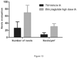
- FIG. 12 Examples of nests figures are presented in Figure 12 . Significantly more chondrocytes nests were observed within the high dose test item treated group (5 animals/6; 30.6 ⁇ 7.0 nests/ ⁇ m 2 ) compared to the vehicle group (2 animals/5; 8.1 ⁇ 4.1 nests/ ⁇ m 2 ) as evaluated in figure 13 .
- the synovial membrane repair changes are more intense in the IA treated group with high dose Liraglutide compared to vehicle group. Moreover, the cartilage degeneration changes were less marked in Liraglutide treated group compared with the vehicle group, but not statistically different.
- Dexamethasone IA and Victoza ®® SC treatments were able to mitigate several OA associated deficits (knee swelling, incapacitance, histology findings related to cartilage loss, however the repair was accompanied by fibrosis that was not observed in IA treated group.
- the inventors tested the effect of Liraglutide on chondrogenesis in an in vitro differentiation model of human Mesenchymal Stem Cells (hMSC) and evaluate whether or not Liraglutide promotes chondrogenesis.
- hMSC human Mesenchymal Stem Cells
- Test material Liraglutide.
- Test system human mesenchymal stem cells (StemPro BM, Cat A15652, ThermoFisher Scientific).
- Basal medium MesenPRO RS basal medium (ThermoFisher Scientific) supplemented with MesenPRO RS growth supplement, L-Glutamine (1%) and Gentamicin (10mg/ml, 50 ⁇ l for 100 ml of medium).
- StemPRO chondrogenesis differentiation medium (ThermoFisher Scientific) supplemented with StemPRO chondrogenesis differentiation supplement and Gentamicin (10mg/ml, 50 ⁇ l for 100 ml of medium)
- Mesenchymal stem cells between 60-80% of confluence were used. The cells were detached from their support and a cell suspension at 1.6 x 10⁷ cells per milliliter in basal medium (MesenPRO RS basal medium + supplement) was prepared.
- basal medium MesenPRO RS basal medium + supplement
- Liraglutide on sphere formation was evaluated by microscope observation 5 days per week. Moreover, Alcian blue staining was performed at three time points (e.g. after 7, 14 and 21 days of treatment). This dye incorporation reflects the presence of sulfated glycosaminoglycans (GAG) and confirms the formation of chondrocytes spheroids.
- GAG glycosaminoglycans
- Liraglutide alone induces hMSC to be committed to the chondrogenesis pathway and generates chondrocytes.
- This Liraglutide anabolic feature would allow to target the resident stem cell population in the articular region to stimulate cartilage repair via chondrocyte differentiation, which is considered as a promising approach for OA treatment.
- the objective of present study was to assess liraglutide effect on cell viability using murine primary chondrocytes.
- Immature murine chondrocytes were derived from newborn mice (5-6 days old C57Bl/6). This work was done in a sterile flow hood. After euthanizing mice by cutting the head with scissors, the animals were fixed in face-down position and the anterior legs were fixed with needles. The skin was removed on the hind limbs using scissors and pincer. The hind limbs were cut along the spine. The limbs were rid of their remains of skin and muscles. The paw was flattened with curved forceps, to release a small translucent and hard sphere, corresponding to femoral heads. When the sphere was isolated, it was placed in 30ml of 1X PBS. The rest of the paw was cleared of muscles and other tissues.
- Femoral condyles and tibial plateau were placed also in 30ml of 1X PBS.
- Pieces of cartilage were incubated twice in 10ml of digestion solution (DMEM, 2mM L-Glutamine + 1% P/S + Collagenase 3mg/ml) for 45min in incubator at 37°C with 5% CO2 in a petri dish 100mm. Between the two digestions, pieces of cartilage were retrieved using 25 ml pipette and placed in a new petri dish. After the two digestions, a dispersion of the aggregates was made using 25ml pipette.
- DMEM 2mM L-Glutamine + 1% P/S + Collagenase 3mg/ml
- Pieces of cartilage were incubated in 10ml DMEM, 2mM L-Glutamine + 1% P/S with collagenase D solution at 0.5mg/ml (diluted to 1/6) overnight in incubator at 37°C with 5% CO2.
- the cells were centrifuged for 10 min at 400g at 20°C, the PBS was removed and 15ml of DMEM 2mM L-Glutamine + 10% FBS + 1% P/S were added.
- the chondrocytes were counted in a Neubauer hemocytometer and observed to assess the viability of extracted cells. Chondrocytes were seeded at density of 40x103 cells in 2 ml of DMEM 2mM L-Glutamine + 10% FBS + 1% P/S per well in 12-well plates. The culture was maintained under sterile conditions in incubator at 37°C with 5% CO2.
- Immature murine articular chondrocytes were confluent after 6-7 days.
- the medium culture was changed after 3 days of culture.
- the DMEM medium containing 10% FBS was removed, the wells were rinsed twice with 1ml of PBS and 1ml of DMEM, 2mM L-Glutamine + 1% P/S + 0.1% BSA was added.
- the medium was removed and treatment with 12 different concentrations of Liraglutide was performed in 500 ⁇ l of DMEM, 2mM L-Glutamine + 1% P/S + 0.1% BSA per well (Table 14).
- the plates were incubated at 37°C + 5% CO2 for 24 hours. Study timeline is presented in Table 15. Table 14. Study design.
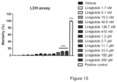
- Lactate dehydrogenase secretion into culture medium was measured by LDH assay (Abcam). 100 ⁇ l of supernatant were used for measurement of Lactate dehydrogenase levels secreted by damaged cells.
- LDH assay was performed according to procedure detailed in instructions for specific LDH Assay kit and was analyzed by Plate Reader (96-well) (Multiskan FC, Thermo Fisher). The wavelength to measure absorbance was 450 nm. The average optical density (OD) of read blank wells was subtracted from each reading.
- Lactate Dehydrogenase is a stable enzyme, present in all cell types and rapidly released into the cell culture medium upon damage of plasma membrane.
- the LDH enzyme has been detected using enzymatic coupling reaction and measured by SkanIt software for microplate reader, Thermo Fisher.
- LDH oxidizes lactate to generate NADH, which then reacts with WST substrate to generate yellow color. The intensity of color correlates directly with the cell number lysed.
- LDH activity has been quantified by spectrophotometer at OD450nm.
- LDH Activity has been measured following 24h of incubation with 12 doses of liraglutide (1.7nM-300 ⁇ M). A positive control was used, where 5 ⁇ l of LDH enzyme was put directly in the wells.
- the % of cytotoxicity has been calculated by this formula: ((Test sample - Low control) / (High control - Low control)) x 100.
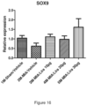
- EXAMPLE 6 SOX9 EXPRESSION IN KNEE JOINT OF MONOIODOACETATE-INJECTED MICE FOLLOWING INTRA ARTICULAR ADMINISTRATION OF ALBUMIN-BASED FORMULATION OF LIRAGLUTIDE
- SOX9 is a pivotal transcription factor in developing and adult cartilage. Its gene is expressed from the multipotent skeletal progenitor stage and is active throughout chondrocyte differentiation. While it is repressed in hypertrophic chondrocytes in cartilage growth plates, it remains expressed throughout life in permanent chondrocytes of healthy articular cartilage. SOX9 is required for chondrogenesis: it secures chondrocyte lineage commitment, promotes cell survival, and transcriptionally activates the genes for many cartilage-specific structural components and regulatory factors.
- the objective of present study was to study SOX9 expression in knee joint of monoiodoacetate (MIA)-injected mice following intra-articular administration of albumin-based formulation of Liraglutide.
- MIA monoiodoacetate
- MIA Monoiodoacetate
- MIA as powder was resuspended in injectable saline to inject into knee joint 0.75mg in 5 ⁇ l per mouse for groups 2M, 3M, 4M, 5M.
- Vehicle formulation excipient consisted of albumin human 5% resuspended in phosphate buffer saline (PBS) for intra-articular injection (5 ⁇ l) to groups 1M and 2M.
- PBS phosphate buffer saline
- Albumin-based formulation of liraglutide Liraglutide (supplied as powder) was dissolved in the appropriate volume of vehicle to inject into knee joint 10 ⁇ g, 20 ⁇ g or 30 ⁇ g in 5 ⁇ l per mouse for groups 3M, 4M and 5M, respectively.
- MIA induction day was defined in this study as “DAY 1" and study termination as “DAY 11".
- mice were anesthetized via a chamber induction technique using inhalation anesthesia (Isoflurane at 5%). During the procedure, the animals were maintained under Isoflurane at a level between 1.5 and 3% with an air flow rate of 1-2 liters/minute. The area surrounding the knee joint was wiped with alcohol. MIA was injected intra-articularly (IA) through the patellar tendon with 5 ⁇ l containing 0.75mg. A 30-gauge, 0.5-inch needle that was fitted with cannulation tubing was used such that only 2 to 3 mm of the needle were allowed to puncture the joint. After injection, the knee was massaged to ensure even distribution of the solution. Animals were injected once on day 1 (groups 2M, 3M, 4M, 5M). For group 1M (sham control), 5 ⁇ l of injectable saline were injected into knee joint.
- group 1M sham control
- mice were allocated randomly to group 1M on day 1.
- group allocation was performed on day 3 based on mice BW. Table 17.
- Study timeline Study Day/week Procedure Remarks twice a wee Body weight D1 MIA injection, 0.75mg in 5 ⁇ l, IA Except for group 1M (sham control), 5 ⁇ l injectable saline, IA D3 Treatment (Vehicle or Liraglutide) IA single injection, 5 ⁇ l D11 Termination Right knee (diseased) collection for SOX9 RTqPCR analyses
- mice On day 11 (termination day), mice were euthanized. The knee articular structure (including synovium) was harvested and snap frozen into liquid nitrogen. RNA extraction was performed using SV total RNA isolation system kit (Promega) according to manufacturer's recommendations. RT-q-PCR analyses were performed for SOX9 marker. SOX9 is identified as the first transcription factor that is essential for chondrocyte differentiation and cartilage formation.
- the study objective was to perform RTqPCR for SOX9 into knee joint of monoiodoacetate-injected mice following intra-articular administration of formulated liraglutide.
- the monoiodoacetate (MIA) model has become a standard for modelling joint disruption in osteoarthritis (OA) in both rats and mice.
- OA osteoarthritis
- a single injection of MIA is delivered to the knee joint, which disrupts chondrocyte glycolysis by inhibiting glyceraldehyde-3-phosphatase dehydrogenase, inducing notably chondrocyte death.
- Chondrocytes which differentiate following the condensation of mesenchymal stem cells, are responsible for the secretion of extracellular matrix molecules, such as collagens and proteoglycans.
- Transcription factor SOX9 is critical for chondrocyte differentiation and function. Using this animal model of OA, we showed that SOX9 expression is decreased following MIA injection, while intra-articular injection of formulated liraglutide restored or increased SOX9 relative expression compared to sham healthy controls.
- EXAMPLE 7 EFFICACY STUDY OF LIRAGLUTIDE ALPHA1-ACID GLYCOPROTEIN BASED-FORMULATION UTILIZING COLLAGENASE TYPE II INDUCED OSTEOARTHRITIS MODEL IN RATS
- the objective of present study was to perform an efficacy study using alpha1-acid glycoprotein-based formulation of Liraglutide utilizing a collagenase-induced model of osteoarthritis in rats.
- Collagenase type II Collagenase type II was dissolved in PBS in concentration of 20 000 U/ml to deliver 500U in 25 ⁇ l.
- Liraglutide (supplied as powder) was dissolved in the appropriate volume of alpha1-acid glycoprotein vehicle to reach 0.18 mg/kg dose level in 25 ⁇ l for intra-articular injection.
- OA induction day was defined in this study as "DAY 1" and study termination as "DAY 43".
- Body weight was recorded upon arrival, before study initiation and once a week thereafter.
- Rat knees immersed in buffered 3.7% formalin were delivered to a subcontractor for histology analysis.
- the histological analysis was performed by a veterinarian (DVM, DESV-Anatomic Pathology) who was blind to the group's treatment and the protocol during the whole analysis procedure. Knee joint sections were scored according to Osteoarthritis Cartilage. 2010 Oct;18 Suppl 3:S24-34 .
- a total joint score was calculated based upon the sum of the following sub section ( de Visser et al, PLoS One. 2018 Apr 23;13(4):e0196308 ): cartilage matrix loss width (0-2), cartilage degeneration (0-5), cartilage degeneration width (0-4), osteophytes (0-4), calcified cartilage and subchondral bone damage (0-5) and synovial membrane inflammation (0-4).
Landscapes
- Health & Medical Sciences (AREA)
- Life Sciences & Earth Sciences (AREA)
- Chemical & Material Sciences (AREA)
- Public Health (AREA)
- Veterinary Medicine (AREA)
- Medicinal Chemistry (AREA)
- Pharmacology & Pharmacy (AREA)
- Animal Behavior & Ethology (AREA)
- General Health & Medical Sciences (AREA)
- Epidemiology (AREA)
- Engineering & Computer Science (AREA)
- Bioinformatics & Cheminformatics (AREA)
- Immunology (AREA)
- Proteomics, Peptides & Aminoacids (AREA)
- Zoology (AREA)
- Gastroenterology & Hepatology (AREA)
- Chemical Kinetics & Catalysis (AREA)
- General Chemical & Material Sciences (AREA)
- Organic Chemistry (AREA)
- Physical Education & Sports Medicine (AREA)
- Rheumatology (AREA)
- Nuclear Medicine, Radiotherapy & Molecular Imaging (AREA)
- Endocrinology (AREA)
- Orthopedic Medicine & Surgery (AREA)
- Inorganic Chemistry (AREA)
- Dermatology (AREA)
- Biochemistry (AREA)
- Molecular Biology (AREA)
- Oil, Petroleum & Natural Gas (AREA)
- Medicines That Contain Protein Lipid Enzymes And Other Medicines (AREA)
- Medicinal Preparation (AREA)
- Medicines Containing Material From Animals Or Micro-Organisms (AREA)
Applications Claiming Priority (3)
| Application Number | Priority Date | Filing Date | Title |
|---|---|---|---|
| PCT/IB2018/059100 WO2020104833A1 (en) | 2018-11-19 | 2018-11-19 | Composition and methods for regulating chondrocyte proliferation and increasing of cartilage matrix production |
| EP19806339.8A EP3883594B1 (de) | 2018-11-19 | 2019-11-18 | Zusammensetzung und verfahren zur regulierung der proliferation von chondrozyten und erhöhung der knorpelmatrixproduktion |
| PCT/IB2019/059889 WO2020104917A1 (en) | 2018-11-19 | 2019-11-18 | Composition and methods for regulating chondrocyte proliferation and increasing of cartilage matrix production |
Related Parent Applications (2)
| Application Number | Title | Priority Date | Filing Date |
|---|---|---|---|
| EP19806339.8A Division EP3883594B1 (de) | 2018-11-19 | 2019-11-18 | Zusammensetzung und verfahren zur regulierung der proliferation von chondrozyten und erhöhung der knorpelmatrixproduktion |
| EP19806339.8A Division-Into EP3883594B1 (de) | 2018-11-19 | 2019-11-18 | Zusammensetzung und verfahren zur regulierung der proliferation von chondrozyten und erhöhung der knorpelmatrixproduktion |
Publications (2)
| Publication Number | Publication Date |
|---|---|
| EP4501362A2 true EP4501362A2 (de) | 2025-02-05 |
| EP4501362A3 EP4501362A3 (de) | 2025-02-19 |
Family
ID=64650445
Family Applications (2)
| Application Number | Title | Priority Date | Filing Date |
|---|---|---|---|
| EP19806339.8A Active EP3883594B1 (de) | 2018-11-19 | 2019-11-18 | Zusammensetzung und verfahren zur regulierung der proliferation von chondrozyten und erhöhung der knorpelmatrixproduktion |
| EP24219712.7A Pending EP4501362A3 (de) | 2018-11-19 | 2019-11-18 | Zusammensetzung und verfahren zur regulierung der chondrozytenproliferation und erhöhung der knorpelmatrixproduktion |
Family Applications Before (1)
| Application Number | Title | Priority Date | Filing Date |
|---|---|---|---|
| EP19806339.8A Active EP3883594B1 (de) | 2018-11-19 | 2019-11-18 | Zusammensetzung und verfahren zur regulierung der proliferation von chondrozyten und erhöhung der knorpelmatrixproduktion |
Country Status (12)
| Country | Link |
|---|---|
| US (3) | US20210401944A1 (de) |
| EP (2) | EP3883594B1 (de) |
| JP (2) | JP7661222B2 (de) |
| KR (1) | KR20210093252A (de) |
| CN (1) | CN113038966A (de) |
| AU (2) | AU2019383576B2 (de) |
| BR (1) | BR112021009232A2 (de) |
| CA (1) | CA3119940A1 (de) |
| ES (1) | ES3042788T3 (de) |
| HR (1) | HRP20251230T1 (de) |
| PL (1) | PL3883594T3 (de) |
| WO (2) | WO2020104833A1 (de) |
Families Citing this family (6)
| Publication number | Priority date | Publication date | Assignee | Title |
|---|---|---|---|---|
| WO2022269001A1 (en) | 2021-06-23 | 2022-12-29 | 4Moving Biotech | Pharmaceutical compositions comprising glp-1r agonists |
| EP4108252A1 (de) | 2021-06-23 | 2022-12-28 | 4Moving Biotech | Pharmazeutische zusammensetzungen mit glp-1r-agonisten |
| CN114934012B (zh) * | 2022-05-16 | 2023-11-10 | 同济大学 | 一种纳米层状双氢氧化物在软骨再生中的应用及其制备 |
| CN117701569B (zh) * | 2023-12-15 | 2024-10-18 | 陕西彦奎生物科技有限公司 | 一种软骨细胞对半月板损伤与骨关节修复的组合物及其制备方法及应用 |
| WO2025219601A1 (en) * | 2024-04-18 | 2025-10-23 | 4Moving Biotech | New glp-1 receptor agonists for the treatment of joint diseases |
| CN118845994B (zh) * | 2024-06-21 | 2025-06-03 | 超越健康科技(浙江)有限公司 | 一种用于骨关节退行性疾病的药物 |
Citations (2)
| Publication number | Priority date | Publication date | Assignee | Title |
|---|---|---|---|---|
| EP2890390B1 (de) | 2012-08-30 | 2016-11-16 | Université Pierre et Marie Curie (Paris 6) | Behandlung von osteoarthritis mit inkretinhormonen oder analoga davon |
| WO2017149070A1 (en) | 2016-03-03 | 2017-09-08 | Novo Nordisk A/S | Glp-1 derivatives and uses thereof |
Family Cites Families (7)
| Publication number | Priority date | Publication date | Assignee | Title |
|---|---|---|---|---|
| WO2005034961A1 (en) * | 2003-10-01 | 2005-04-21 | Optimer Pharmaceuticals, Inc. | Treatment of a condition in a mammal with adminisration of aminosugar and uses thereof |
| EP2494983B1 (de) * | 2004-11-12 | 2019-04-24 | Novo Nordisk A/S | Stabile formulierung von glp-1 |
| WO2008023063A2 (en) * | 2006-08-25 | 2008-02-28 | Ares Trading S.A. | Treatment of cartilage disorders with fgf-18 |
| CN102271705A (zh) * | 2008-12-22 | 2011-12-07 | 墨尔本大学 | 骨关节炎治疗 |
| CN101987868B (zh) * | 2009-07-30 | 2013-09-04 | 江苏豪森医药集团有限公司 | Glp-1类似物的衍生物或其可药用盐和用途 |
| US8716204B2 (en) * | 2010-07-27 | 2014-05-06 | Zimmer, Inc. | Synthetic synovial fluid compositions and methods for making the same |
| US20150150983A1 (en) * | 2013-12-02 | 2015-06-04 | Depuy Mitek, Llc | Intra-articular Formulations and Methods for Treatment of Osteoarthritis |
-
2018
- 2018-11-19 WO PCT/IB2018/059100 patent/WO2020104833A1/en not_active Ceased
-
2019
- 2019-11-18 KR KR1020217014573A patent/KR20210093252A/ko active Pending
- 2019-11-18 BR BR112021009232-3A patent/BR112021009232A2/pt unknown
- 2019-11-18 WO PCT/IB2019/059889 patent/WO2020104917A1/en not_active Ceased
- 2019-11-18 AU AU2019383576A patent/AU2019383576B2/en active Active
- 2019-11-18 JP JP2021526734A patent/JP7661222B2/ja active Active
- 2019-11-18 CN CN201980075184.9A patent/CN113038966A/zh active Pending
- 2019-11-18 EP EP19806339.8A patent/EP3883594B1/de active Active
- 2019-11-18 EP EP24219712.7A patent/EP4501362A3/de active Pending
- 2019-11-18 ES ES19806339T patent/ES3042788T3/es active Active
- 2019-11-18 CA CA3119940A patent/CA3119940A1/en active Pending
- 2019-11-18 HR HRP20251230TT patent/HRP20251230T1/hr unknown
- 2019-11-18 US US17/294,079 patent/US20210401944A1/en active Pending
- 2019-11-18 PL PL19806339.8T patent/PL3883594T3/pl unknown
-
2025
- 2025-01-10 US US19/016,974 patent/US20250144181A1/en active Pending
- 2025-04-02 JP JP2025060915A patent/JP2025118608A/ja active Pending
- 2025-08-29 AU AU2025223913A patent/AU2025223913A1/en active Pending
- 2025-11-17 US US19/391,871 patent/US20260069661A1/en active Pending
Patent Citations (2)
| Publication number | Priority date | Publication date | Assignee | Title |
|---|---|---|---|---|
| EP2890390B1 (de) | 2012-08-30 | 2016-11-16 | Université Pierre et Marie Curie (Paris 6) | Behandlung von osteoarthritis mit inkretinhormonen oder analoga davon |
| WO2017149070A1 (en) | 2016-03-03 | 2017-09-08 | Novo Nordisk A/S | Glp-1 derivatives and uses thereof |
Non-Patent Citations (8)
| Title |
|---|
| BARRY DECKER ET AL.: "Concentration of Hyaluronic Acid in Synovial Fluid", CLINICAL CHEMISTRY, vol. 5, no. 5, 1959, pages 465 - 469 |
| DE VISSER ET AL., PLOS ONE, vol. 13, no. 4, 23 April 2018 (2018-04-23), pages e0196308 |
| DR ARUN PAL SINGH, SYNOVIAL FLUID COMPOSITION AND FUNCTIONS, Retrieved from the Internet <URL:http://boneandspine.com/synovial-fluid> |
| JONATHAN BLACK: "Biological Performance of Materials: Fundamentals of Biocompatibility", 2005, CRC PRESS |
| N. GERWIN ET AL., OSTEOARTHRITIS AND CARTILAGE, vol. 18, 2010, pages S24 - S34 |
| NEIDLIN ET AL., ANNALS OF BIOMEDICAL ENGINEERING, vol. 46, no. 2, 2018, pages 345 - 353 |
| OSTEOARTHRITIS CARTILAGE, vol. 18, no. 3, October 2010 (2010-10-01), pages 524 - 34 |
| QUINETRO ET AL., CYTOKINE, vol. 44, no. 3, 2008, pages 366 - 72 |
Also Published As
| Publication number | Publication date |
|---|---|
| JP2025118608A (ja) | 2025-08-13 |
| AU2019383576B2 (en) | 2025-06-19 |
| WO2020104833A1 (en) | 2020-05-28 |
| AU2025223913A1 (en) | 2025-09-18 |
| EP4501362A3 (de) | 2025-02-19 |
| BR112021009232A2 (pt) | 2021-08-10 |
| CN113038966A (zh) | 2021-06-25 |
| PL3883594T3 (pl) | 2025-11-12 |
| JP7661222B2 (ja) | 2025-04-14 |
| US20250144181A1 (en) | 2025-05-08 |
| EP3883594A1 (de) | 2021-09-29 |
| KR20210093252A (ko) | 2021-07-27 |
| CA3119940A1 (en) | 2020-05-28 |
| WO2020104917A1 (en) | 2020-05-28 |
| EP3883594C0 (de) | 2025-07-09 |
| JP2022507614A (ja) | 2022-01-18 |
| US20260069661A1 (en) | 2026-03-12 |
| AU2019383576A1 (en) | 2021-06-03 |
| ES3042788T3 (en) | 2025-11-24 |
| EP3883594B1 (de) | 2025-07-09 |
| HRP20251230T1 (hr) | 2025-12-05 |
| US20210401944A1 (en) | 2021-12-30 |
Similar Documents
| Publication | Publication Date | Title |
|---|---|---|
| EP3883594B1 (de) | Zusammensetzung und verfahren zur regulierung der proliferation von chondrozyten und erhöhung der knorpelmatrixproduktion | |
| US9987296B2 (en) | Compositions including anthocyanin or anthocyanidin for the prevention or treatment of Articular cartilage-associated conditions | |
| RU2700582C2 (ru) | Схема применения соединения fgf-18 | |
| US9777048B2 (en) | Use of PEDF-derived polypeptides for treating osteoarthritis | |
| US11466066B2 (en) | Variants of human BMP7 protein | |
| US20240293514A1 (en) | Pharmaceutical compositions comprising glp-1r agonists | |
| TWI453030B (zh) | 抑制及/或回復細胞終端分化的組成物 | |
| EP4108252A1 (de) | Pharmazeutische zusammensetzungen mit glp-1r-agonisten | |
| HK40123727A (en) | Composition and methods for regulating chondrocyte proliferation and increasing of cartilage matrix production | |
| TWI491407B (zh) | 色素上皮衍生因子衍生之多胜肽於治療骨性關節炎之用途 | |
| US20170231934A1 (en) | Compositions including anthocyanin or anthocyanidin metabolites for the prevention or treatment of articular cartilage-associated conditions | |
| WO2018204621A1 (en) | Compositions including protocatechuic acid for the prevention or treatment of osteoarthritis | |
| HK40085325A (en) | Pharmaceutical compositions comprising glp-1r agonists | |
| McCoy | Advances in the treatment of osteoarthritis in horses | |
| 玉行傅 et al. | The effect of umbilical cord blood mononucleated cells on knee osteoarthritis in rabbits | |
| CN118436659A (zh) | Hemin通过抑制软骨细胞凋亡防治软骨受损疾病 |
Legal Events
| Date | Code | Title | Description |
|---|---|---|---|
| PUAI | Public reference made under article 153(3) epc to a published international application that has entered the european phase |
Free format text: ORIGINAL CODE: 0009012 |
|
| STAA | Information on the status of an ep patent application or granted ep patent |
Free format text: STATUS: THE APPLICATION HAS BEEN PUBLISHED |
|
| REG | Reference to a national code |
Ref country code: DE Ref legal event code: R079 Free format text: PREVIOUS MAIN CLASS: A61K0047380000 Ipc: A61K0038260000 |
|
| PUAL | Search report despatched |
Free format text: ORIGINAL CODE: 0009013 |
|
| AC | Divisional application: reference to earlier application |
Ref document number: 3883594 Country of ref document: EP Kind code of ref document: P |
|
| AK | Designated contracting states |
Kind code of ref document: A2 Designated state(s): AL AT BE BG CH CY CZ DE DK EE ES FI FR GB GR HR HU IE IS IT LI LT LU LV MC MK MT NL NO PL PT RO RS SE SI SK SM TR |
|
| AK | Designated contracting states |
Kind code of ref document: A3 Designated state(s): AL AT BE BG CH CY CZ DE DK EE ES FI FR GB GR HR HU IE IS IT LI LT LU LV MC MK MT NL NO PL PT RO RS SE SI SK SM TR |
|
| RIC1 | Information provided on ipc code assigned before grant |
Ipc: A61K 47/38 20060101ALI20250114BHEP Ipc: A61K 47/36 20060101ALI20250114BHEP Ipc: A61K 47/64 20170101ALI20250114BHEP Ipc: A61K 8/04 20060101ALI20250114BHEP Ipc: A61P 19/02 20060101ALI20250114BHEP Ipc: A61K 38/26 20060101AFI20250114BHEP |
|
| STAA | Information on the status of an ep patent application or granted ep patent |
Free format text: STATUS: REQUEST FOR EXAMINATION WAS MADE |
|
| 17P | Request for examination filed |
Effective date: 20250819 |
|
| REG | Reference to a national code |
Ref country code: HK Ref legal event code: DE Ref document number: 40123727 Country of ref document: HK |