EP4488366A2 - Durch bisse aktivierte car-t-zellen - Google Patents
Durch bisse aktivierte car-t-zellen Download PDFInfo
- Publication number
- EP4488366A2 EP4488366A2 EP24208814.4A EP24208814A EP4488366A2 EP 4488366 A2 EP4488366 A2 EP 4488366A2 EP 24208814 A EP24208814 A EP 24208814A EP 4488366 A2 EP4488366 A2 EP 4488366A2
- Authority
- EP
- European Patent Office
- Prior art keywords
- cell
- cells
- cancer
- car
- tumor
- Prior art date
- Legal status (The legal status is an assumption and is not a legal conclusion. Google has not performed a legal analysis and makes no representation as to the accuracy of the status listed.)
- Pending
Links
Images


Classifications
-
- A—HUMAN NECESSITIES
- A61—MEDICAL OR VETERINARY SCIENCE; HYGIENE
- A61K—PREPARATIONS FOR MEDICAL, DENTAL OR TOILETRY PURPOSES
- A61K39/00—Medicinal preparations containing antigens or antibodies
- A61K39/395—Antibodies; Immunoglobulins; Immune serum, e.g. antilymphocytic serum
- A61K39/39533—Antibodies; Immunoglobulins; Immune serum, e.g. antilymphocytic serum against materials from animals
- A61K39/3955—Antibodies; Immunoglobulins; Immune serum, e.g. antilymphocytic serum against materials from animals against proteinaceous materials, e.g. enzymes, hormones, lymphokines
-
- A—HUMAN NECESSITIES
- A61—MEDICAL OR VETERINARY SCIENCE; HYGIENE
- A61K—PREPARATIONS FOR MEDICAL, DENTAL OR TOILETRY PURPOSES
- A61K40/00—Cellular immunotherapy
- A61K40/10—Cellular immunotherapy characterised by the cell type used
- A61K40/11—T-cells, e.g. tumour infiltrating lymphocytes [TIL] or regulatory T [Treg] cells; Lymphokine-activated killer [LAK] cells
-
- A—HUMAN NECESSITIES
- A61—MEDICAL OR VETERINARY SCIENCE; HYGIENE
- A61K—PREPARATIONS FOR MEDICAL, DENTAL OR TOILETRY PURPOSES
- A61K40/00—Cellular immunotherapy
- A61K40/30—Cellular immunotherapy characterised by the recombinant expression of specific molecules in the cells of the immune system
- A61K40/31—Chimeric antigen receptors [CAR]
-
- A—HUMAN NECESSITIES
- A61—MEDICAL OR VETERINARY SCIENCE; HYGIENE
- A61K—PREPARATIONS FOR MEDICAL, DENTAL OR TOILETRY PURPOSES
- A61K40/00—Cellular immunotherapy
- A61K40/40—Cellular immunotherapy characterised by antigens that are targeted or presented by cells of the immune system
- A61K40/41—Vertebrate antigens
- A61K40/42—Cancer antigens
- A61K40/4202—Receptors, cell surface antigens or cell surface determinants
- A61K40/421—Immunoglobulin superfamily
- A61K40/4211—CD19 or B4
-
- A—HUMAN NECESSITIES
- A61—MEDICAL OR VETERINARY SCIENCE; HYGIENE
- A61K—PREPARATIONS FOR MEDICAL, DENTAL OR TOILETRY PURPOSES
- A61K40/00—Cellular immunotherapy
- A61K40/40—Cellular immunotherapy characterised by antigens that are targeted or presented by cells of the immune system
- A61K40/41—Vertebrate antigens
- A61K40/42—Cancer antigens
- A61K40/4202—Receptors, cell surface antigens or cell surface determinants
- A61K40/4224—Molecules with a "CD" designation not provided for elsewhere
-
- A—HUMAN NECESSITIES
- A61—MEDICAL OR VETERINARY SCIENCE; HYGIENE
- A61K—PREPARATIONS FOR MEDICAL, DENTAL OR TOILETRY PURPOSES
- A61K45/00—Medicinal preparations containing active ingredients not provided for in groups A61K31/00 - A61K41/00
- A61K45/06—Mixtures of active ingredients without chemical characterisation, e.g. antiphlogistics and cardiaca
-
- A—HUMAN NECESSITIES
- A61—MEDICAL OR VETERINARY SCIENCE; HYGIENE
- A61P—SPECIFIC THERAPEUTIC ACTIVITY OF CHEMICAL COMPOUNDS OR MEDICINAL PREPARATIONS
- A61P35/00—Antineoplastic agents
-
- C—CHEMISTRY; METALLURGY
- C12—BIOCHEMISTRY; BEER; SPIRITS; WINE; VINEGAR; MICROBIOLOGY; ENZYMOLOGY; MUTATION OR GENETIC ENGINEERING
- C12N—MICROORGANISMS OR ENZYMES; COMPOSITIONS THEREOF; PROPAGATING, PRESERVING, OR MAINTAINING MICROORGANISMS; MUTATION OR GENETIC ENGINEERING; CULTURE MEDIA
- C12N5/00—Undifferentiated human, animal or plant cells, e.g. cell lines; Tissues; Cultivation or maintenance thereof; Culture media therefor
- C12N5/06—Animal cells or tissues; Human cells or tissues
- C12N5/0602—Vertebrate cells
- C12N5/0634—Cells from the blood or the immune system
- C12N5/0636—T lymphocytes
-
- G—PHYSICS
- G01—MEASURING; TESTING
- G01N—INVESTIGATING OR ANALYSING MATERIALS BY DETERMINING THEIR CHEMICAL OR PHYSICAL PROPERTIES
- G01N33/00—Investigating or analysing materials by specific methods not covered by groups G01N1/00 - G01N31/00
- G01N33/48—Biological material, e.g. blood, urine; Haemocytometers
- G01N33/50—Chemical analysis of biological material, e.g. blood, urine; Testing involving biospecific ligand binding methods; Immunological testing
- G01N33/5005—Chemical analysis of biological material, e.g. blood, urine; Testing involving biospecific ligand binding methods; Immunological testing involving human or animal cells
- G01N33/5008—Chemical analysis of biological material, e.g. blood, urine; Testing involving biospecific ligand binding methods; Immunological testing involving human or animal cells for testing or evaluating the effect of chemical or biological compounds, e.g. drugs, cosmetics
- G01N33/5011—Chemical analysis of biological material, e.g. blood, urine; Testing involving biospecific ligand binding methods; Immunological testing involving human or animal cells for testing or evaluating the effect of chemical or biological compounds, e.g. drugs, cosmetics for testing antineoplastic activity
-
- G—PHYSICS
- G01—MEASURING; TESTING
- G01N—INVESTIGATING OR ANALYSING MATERIALS BY DETERMINING THEIR CHEMICAL OR PHYSICAL PROPERTIES
- G01N33/00—Investigating or analysing materials by specific methods not covered by groups G01N1/00 - G01N31/00
- G01N33/48—Biological material, e.g. blood, urine; Haemocytometers
- G01N33/50—Chemical analysis of biological material, e.g. blood, urine; Testing involving biospecific ligand binding methods; Immunological testing
- G01N33/5005—Chemical analysis of biological material, e.g. blood, urine; Testing involving biospecific ligand binding methods; Immunological testing involving human or animal cells
- G01N33/5008—Chemical analysis of biological material, e.g. blood, urine; Testing involving biospecific ligand binding methods; Immunological testing involving human or animal cells for testing or evaluating the effect of chemical or biological compounds, e.g. drugs, cosmetics
- G01N33/502—Chemical analysis of biological material, e.g. blood, urine; Testing involving biospecific ligand binding methods; Immunological testing involving human or animal cells for testing or evaluating the effect of chemical or biological compounds, e.g. drugs, cosmetics for testing non-proliferative effects
- G01N33/5023—Chemical analysis of biological material, e.g. blood, urine; Testing involving biospecific ligand binding methods; Immunological testing involving human or animal cells for testing or evaluating the effect of chemical or biological compounds, e.g. drugs, cosmetics for testing non-proliferative effects on expression patterns
-
- G—PHYSICS
- G01—MEASURING; TESTING
- G01N—INVESTIGATING OR ANALYSING MATERIALS BY DETERMINING THEIR CHEMICAL OR PHYSICAL PROPERTIES
- G01N33/00—Investigating or analysing materials by specific methods not covered by groups G01N1/00 - G01N31/00
- G01N33/48—Biological material, e.g. blood, urine; Haemocytometers
- G01N33/50—Chemical analysis of biological material, e.g. blood, urine; Testing involving biospecific ligand binding methods; Immunological testing
- G01N33/5005—Chemical analysis of biological material, e.g. blood, urine; Testing involving biospecific ligand binding methods; Immunological testing involving human or animal cells
- G01N33/5008—Chemical analysis of biological material, e.g. blood, urine; Testing involving biospecific ligand binding methods; Immunological testing involving human or animal cells for testing or evaluating the effect of chemical or biological compounds, e.g. drugs, cosmetics
- G01N33/5044—Chemical analysis of biological material, e.g. blood, urine; Testing involving biospecific ligand binding methods; Immunological testing involving human or animal cells for testing or evaluating the effect of chemical or biological compounds, e.g. drugs, cosmetics involving specific cell types
- G01N33/5047—Cells of the immune system
- G01N33/505—Cells of the immune system involving T-cells
-
- A—HUMAN NECESSITIES
- A61—MEDICAL OR VETERINARY SCIENCE; HYGIENE
- A61K—PREPARATIONS FOR MEDICAL, DENTAL OR TOILETRY PURPOSES
- A61K39/00—Medicinal preparations containing antigens or antibodies
- A61K2039/80—Vaccine for a specifically defined cancer
- A61K2039/804—Blood cells [leukemia, lymphoma]
-
- A—HUMAN NECESSITIES
- A61—MEDICAL OR VETERINARY SCIENCE; HYGIENE
- A61K—PREPARATIONS FOR MEDICAL, DENTAL OR TOILETRY PURPOSES
- A61K2239/00—Indexing codes associated with cellular immunotherapy of group A61K40/00
- A61K2239/46—Indexing codes associated with cellular immunotherapy of group A61K40/00 characterised by the cancer treated
- A61K2239/48—Blood cells, e.g. leukemia or lymphoma
-
- A—HUMAN NECESSITIES
- A61—MEDICAL OR VETERINARY SCIENCE; HYGIENE
- A61K—PREPARATIONS FOR MEDICAL, DENTAL OR TOILETRY PURPOSES
- A61K2239/00—Indexing codes associated with cellular immunotherapy of group A61K40/00
- A61K2239/46—Indexing codes associated with cellular immunotherapy of group A61K40/00 characterised by the cancer treated
- A61K2239/57—Skin; melanoma
-
- C—CHEMISTRY; METALLURGY
- C12—BIOCHEMISTRY; BEER; SPIRITS; WINE; VINEGAR; MICROBIOLOGY; ENZYMOLOGY; MUTATION OR GENETIC ENGINEERING
- C12N—MICROORGANISMS OR ENZYMES; COMPOSITIONS THEREOF; PROPAGATING, PRESERVING, OR MAINTAINING MICROORGANISMS; MUTATION OR GENETIC ENGINEERING; CULTURE MEDIA
- C12N2510/00—Genetically modified cells
Definitions
- the disclosure relates to three novel approaches using bispecific antibodies (BiTE)-activated T Cells.
- One is to generate chimeric antigen receptor (CAR) T cells using these BiTE-activated T cells as the source of T cells. These new CAR-T cells may be a better cellular therapy treatment for cancer patients.
- a second approach is a method to identify which immune check point inhibitors are responsible for resistance to these BiTE-activated T cells. This can be helpful to personalize immunotherapy treatments to cancer patients. This may also be helpful for other immunotherapy treatments, such as CAR-T cells, independently of the BiTE-activated T cells.
- a third approach is to identify patients less susceptible to suffer Cytokine-Release Syndrome. This can also be helpful to personalize immunotherapy treatments to cancer patients. This may also be helpful for other immunotherapy treatments, such as CAR-T cells, independently of the BiTE-activated T cells.
- Adoptive cell therapy is a process involving collection of immune cells from a patient, expansion of the cells, and reintroduction of the cells into the same patient or a different patient.
- CTLs human cytotoxic T lymphocytes
- Examples of ACT include cultured tumor infiltrating lymphocytes (TILs), isolated and expanded T cell clones, and genetically engineered lymphocytes (e.g., T cells) that express conventional T cell receptors or chimeric antigen receptors.
- TILs tumor infiltrating lymphocytes
- TILs tumor infiltrating lymphocytes
- T cells genetically engineered lymphocytes
- the genetically engineered lymphocytes are designed to eliminate cancer cells expressing specific antigen(s) and are expanded and delivered to a patient.
- Another example of an ACT is the isolation and use of T cells from a patient's blood after administration of a cancer vaccine.
- ACT can provide tumor specific lymphocytes (e.g., T cells) that lead to a reduction in tumor cells in
- CAR-T cells are generated using peripheral blood naive T cells.
- a limitation of these standard CAR-T cells is that they can only recognize the tumor antigen of the CAR construct.
- tumor cells can be heterogeneous with some clones not expressing the CAR antigen leading to resistance to such CAR-T cells. Relapsed patients treated with CAR-T cells are showing this resistance mechanisms.
- MILs marrow-infiltrating lymphocytes
- the method of producing CAR-T cells often by transducing a CAR with a lentivirus, generates an heterogenous population of T Cells.
- the CAR construct may insert at different positions into the genome, resulting in different activity of the ensuing CAR-T cells; e.g. different levels of expression could affect activity, or disrupting different genes.
- the different types of T cells present in the mixed T cell population used as a source for producing CAR-T cells may result in different activities; e.g. memory T cells versus naive T cells, highly proliferating versus terminally proliferating T cells.
- Cytokine Storm also called Cytokine Release Syndrome
- Cytokine Release Syndrome has been recognized as a major toxicity challenge for CAR-T treatments ( Park et al. N Engl J Med. 2018 Feb 1;378(5):449-459 ). It also a major toxicity for bispecific antibodies. However, there are no methods to identify patient most likely to suffer this toxicity when treated with CAR-T cells.
- ICHK immune check point inhibitors
- Bispecific T cell engager antibody (BiTE)-activated T-cells are potent and selective anti-tumor cells.
- BiTE-activated T cells are the target for grafting CAR molecules.
- BiTE-activated T cells combine the potency of the transfected CAR construct while retaining their ability to recognize and kill tumor cells expressing different, CAR-resistant antigens.
- the use of these T-Cells for Adoptive Cell Therapy can also be enhanced by using them as the source of CAR-T cells, transfecting CAR constructs into them prior to adoptive cell therapy.
- bispecific T cell engager antibody (BiTE) to activate and thus identify these selective antitumor effector T-cells offers unique advantages for hematological malignancies.
- these selective antitumor effector T cells are part of the T cell population that consists of many sub-types of T cells that reside in hematological tissues such as bone marrow, and it is not known how to identify them in most of these malignancies.
- T cell receptor is a disulfide-linked heterodimer consisting of one ⁇ and one ⁇ chain expressed in complex with invariant CD3 chains ( ⁇ , ⁇ , ⁇ , and ⁇ ). TCR recognizes intracellular or extracellular proteins presented as peptides by MHC molecules. Costimulation of CD28 through its ligands, CD80/CD86, is required for optimal activation of the receptor and for production of interleukin-2 (IL-2) and other cytokines. While most hematological tumors express costimulatory molecules, solid tumor cells as well as antigen presenting cells in the tumor microenvironment usually lack such molecules.
- IL-2 interleukin-2
- Chimeric Antigen Receptors are recombinant receptors that recognize surface antigens in an MHC unrestricted manner.
- CARs are fusion proteins between single-chain variable fragments (scFv) from a monoclonal antibody and one or more T cell receptor intracellular signaling domains.
- scFv single-chain variable fragments
- TM transmembrane domains are used to link the recognition (antigen binding) and the signaling activation moiety.
- first generation CARs signaled through the CD3 chain only include a signaling domain from a costimulatory molecule, for example, CD28, 4-1BB, OX40, CD27, DAP10, or ICOS.
- T-cell therapy There are several strategies to improve CAR-T-cell therapy that involve higher safety, better trafficking of T-cells to tumor sites, increase persistence and overcome the immunosuppressive factors in the tumor microenvironment. Improvements in T-cell selections also represent a good approach to enhance the cancer treatment efficacy.
- Activated T cells generated after BiTE exposure represent a novel source of T cells that can be genetically engineered.
- TAA tumor associated antigen
- T cells would combine the advantages of both methods and should provide a highly effective cytotoxic T-cells that would be able to trigger a T cell mediated tumor cell lysis in a T cell receptor (TCR) and MHC-independent manner.
- TCR T cell receptor
- Another approach exploits recent technologies through exome-guided neoantigen identification that can dissect the immune response to patient-specific neoantigens. Incorporation of these neoantigens expressed in cancer cells to the CAR, would enhance the selectively T cell reactivity against this class of antigens.
- MILs in bone marrow of hematological malignancies is different than TILs in solid tumors, in that bone marrow always has T cells present and nobody knows which ones are TILs.
- the tumor-specific T cells are believed to be present at much higher frequencies among MILs compared to peripheral blood but are often dysfunctional (exhausted/anergic) and require potent stimulation in order to recover their anti-cancer cytotoxic functions.
- These Tumor-Specific T cells in patient bone marrow samples can be identified pharmacologically, by activating them with bispecific antibodies (BiTEs). It is though that BiTEs induce T cells to kill tumor cells by proximity independent of the antigen recognition.
- the present invention provides that in many patient samples when the BiTE joins a tumor cell with an immunosuppressed TSA T Cell (TIL), it can also activate these TILs, which kills tumor cells independently of the BiTE.
- TIL TSA T Cell
- Cells may be sorted, BiTE may be washed, cells may be grown, and cells retain the cytotoxic efficacy against tumor cells of the same patient.
- These reactivated TILs can be identified because they have a great killing efficacy, where one activated T cell can kill on average 30-100 tumor cells.
- normal T cells incubated with a BiTE can only kill tumor cells 1:1.
- CAR-T cells of the present invention are more potent, and also that they can kill clonal populations that do not express the antigen on the CAR because they retain the native TCR recognition of other cancer antigens.
- the CAR-T cells described herein can provide highly effective therapies for diverse cancer types, e.g., solid cancers, hematological cancers, and metastatic forms thereof. Therapies using the CAR-T cells disclosed herein are also suited for treating cancers that typically do not elicit a strong immune response in a subject, e.g., a cancer other than a melanoma.
- the cancer therapies disclosed herein can be tailored or personalized to a given subject, e.g., by generating CAR-T cells (e.g., autologous CAR-T cells) that selectively and effectively target the subject's cancer.
- compositions comprising such immune cells; methods of using the cells (e.g., methods of treatment); methods of selecting optimal agents for enhancing the target cell killing activity, e.g., by enhancing the proximity, e.g., spatial proximity, between the target cell and the immune cell, e.g., T cell; methods of selecting an optimized (e.g., highest activity fractions/clones) immune cell, e.g., T cell; and methods of using this approach to evaluate patient responsiveness to other cancer therapies.
- methods of using the cells e.g., methods of treatment
- methods of selecting optimal agents for enhancing the target cell killing activity e.g., by enhancing the proximity, e.g., spatial proximity, between the target cell and the immune cell, e.g., T cell
- methods of selecting an optimized (e.g., highest activity fractions/clones) immune cell e.g., T cell
- methods of using this approach to evaluate patient responsiveness to other cancer therapies.
- an in vitro method of producing a genetically engineered T cell expressing Chimeric Antigen Receptors (a CAR-T cell) or a CAR-T cell preparation:
- an in vitro method of producing a genetically engineered T cell expressing Chimeric Antigen Receptors (a CAR-T cell) or a CAR-T cell preparation:
- an in vitro method of producing a genetically engineered T cell expressing Chimeric Antigen Receptors (a CAR-T cell) or a CAR-T cell preparation:
- the selecting and/or enriching step (a) comprises using fluorescence activated cell sorting (FACS).
- the selecting and/or enriching step (a) comprises using a bead (e.g., magnetic bead) coated with an antibody or fragment thereof that binds to i) one or more cancer antigens or ii) one or more markers of activated T cells, or both i) and ii).
- FACS fluorescence activated cell sorting
- the cancer-killing T cell preparation is enriched or purified and comprises trogocytotic cancer-killing T cells, e.g., at a concentration of at least 50% (e.g., at least 50%, 55%, 60%, 65%, 70%, 75%, 80%, 85%, 90%, 95%, 99%, or greater) of the total number of cells in the preparation.
- at least 50% e.g., at least 50%, 55%, 60%, 65%, 70%, 75%, 80%, 85%, 90%, 95%, 99%, or greater
- the ex vivo reaction mixture further comprises one or multiple agents that enhance T cell activity.
- the agents that enhance T cell activity are selected from one or more of a chemotherapy drug, a targeted anti-cancer therapy, an oncolytic drug, a cytotoxic agent, an immune-based therapy, a cytokine, an agonist of T cells (e.g., agonistic antibody or fragment thereof or an activator of a costimulatory molecule), an inhibitor of an inhibitory molecule (e.g., immune checkpoint inhibitor), an immunomodulatory agent, a vaccine, or a cellular immunotherapy.
- a chemotherapy drug e.g., a targeted anti-cancer therapy, an oncolytic drug, a cytotoxic agent, an immune-based therapy, a cytokine, an agonist of T cells (e.g., agonistic antibody or fragment thereof or an activator of a costimulatory molecule), an inhibitor of an inhibitory molecule (e.g., immune checkpoint inhibitor), an immunomodulatory agent, a vaccine,
- the agents enhancing T cell activity is selected from an agonist of T cells (e.g., an agonistic antibody or fragment thereof or an activator of a costimulatory molecule), and/or an inhibitor of an immune checkpoint inhibitor.
- the inhibitors of the immune checkpoint inhibitor is an inhibitor of one or more of: PDL-1, PDL-2, B7-1 (CD80), B7-2 (CD86), 4-1BBL, Galectin, ICOSL, GITRL, OX40L, CD155, B7-H3, PD1, CTLA-4, 4-1BB, TIM-3, ICOS, GITR, LAG-3, KIR, OX40, TIGIT, CD160, 2B4, B7-H4 (VTCN1), HVEM (TNFRSF14 or CD270), BTLA, KIR, MHC class I, MHC class II, GAL9, VISTA, LAIR1, and A2aR.
- the inhibitors of the immune checkpoint inhibitor comprise one or more of: ipilimumab, tremelimumab, MDX-1106, MK3475, CT-011, AMP-224, MDX-1105, IMP321, or MGA271.
- the agents enhancing T cell activity comprises molecules (e.g. antibodies) constructed combining fragments of these molecules enhancing T cell activity, e.g. bispecific or multispecific antibody formats combining recognition arms of several immune checkpoint inhibitors, including but not limited to PD1-PDL1, PD1-PDL2, PD1-LAG3, PD1-TIM3.
- the agonist of T cells comprises an antibody or fragment thereof to CD137, CD40, and/or glucocorticoid-induced TNF receptor (GITR).
- the immunomodulatory agent comprises/is lenalidomide, ibrutinib or bortezomib.
- the agent enhancing T cell activity enhances and/or restores the immunocompetence of T cells.
- the immunomodulatory agent is an inhibitor of MDSCs and/or Treg cells.
- the immunomodulatory agent activates an immune response to a tumor specific antigen, e.g., it is a vaccine (e.g., a vaccine against targets such as gp100, MUC1 or MAGEA3.
- the immunomodulatory agent is a cytokine, e.g., a recombinant cytokine chosen from one or more of GM-CSF, IL-7, IL-12, IL-15, IL-18 or IL-21.
- the immunomodulatory agent is a modulator of a component (e.g., enzyme or receptor) associated with amino acid catabolism, signalling of tumor-derived extracellular ATP, adenosine signalling, adenosine production, chemokine and chemokine receptor, recognition of foreign organisms, or kinase signalling activity.
- a component e.g., enzyme or receptor
- the immunomodulatory agent is an inhibitor (e.g., small molecule inhibitor) of IDO, COX2, ARG1, ArG2, iNOS, or phosphodiesterase (e.g., PDE5); a TLR agonist, or a chemokine antagonist.
- the sample is a cancer sample chosen from a hematological cancer, a solid cancer, a metastatic cancer (e.g., a CTC, a primary, secondary or additional metastatic cancer), or a combination thereof.
- a metastatic cancer e.g., a CTC, a primary, secondary or additional metastatic cancer
- the sample is a T cell sample chosen from a blood sample (e.g., peripheral blood sample), a bone marrow sample, a lymph node sample, a spleen sample, a tumor sample comprising a CTL, a TIL, or a combination thereof.
- a blood sample e.g., peripheral blood sample
- a bone marrow sample e.g., a bone marrow sample
- a lymph node sample e.g., a lymph node sample
- a spleen sample e.g., a tumor sample comprising a CTL, a TIL, or a combination thereof.
- substantially no components e.g., cells
- the sample substantially maintains the microenvironment from the tissue of origin, e.g., substantially maintains the structure of the tumor or immune microenvironment.
- the sample comprises a tumor-specific T cell.
- tumor-antigen specific T cells can be immunosuppressed, e.g., when present in the tumor microenvironment.
- the immunosuppressed tumor-antigen specific T cell is activated under the conditions described herein, e.g., upon contact with the cancer cell and a bispecific T cell engager antibody (BiTE).
- BiTE bispecific T cell engager antibody
- the sample or samples comprise the cancer cell and the T cell.
- the sample may be from a hematological cancer (e.g., bone marrow, lymph-node derived cancer) that includes a T cell (e.g., a tumor-antigen specific CTL).
- the hematological sample may also comprise cancer cells, e.g., leukemic or lymphoma blast cells (e.g., a blast cell expressing one or more markers chosen from CD19, CD123, CD20 or others).
- addition of the bispecific T cell engager antibody (BiTE) to the sample promotes an interaction between the T cell and the cancer cell that activates the T cell (e.g., activates the tumor-antigen specific CTL).
- the activated T cell acquires a cell surface marker from the cancer cell, e.g., becomes a trogocytotic T cell.
- the cancer is a solid tumor.
- the sample may comprise a tumor-antigen specific T cell (e.g., a CTL or a TIL) as described herein and a cancer cell.
- a tumor-antigen specific T cell e.g., a CTL or a TIL
- addition of the bispecific T cell engager antibody (BiTE) to the sample promotes an interaction between the T cell and the cancer cell that activates the T cell (e.g., activates the tumor-antigen specific CTL or TIL).
- the activated T cell acquires a cell surface marker from the cancer cell, e.g., becomes a trogocytotic T cell.
- the sample comprises a metastatic sample, e.g., a sample derived from a subject with a metastatic cancer.
- the metastatic sample comprises a CTC.
- the CTC is a tumor cell found in the peripheral blood of a subject with a cancer, e.g., a solid tumor.
- An ex vivo reaction mixture can be formed comprising a metastatic cancer cell and a T cell.
- the T cell can be obtained from the metastatic cancer sample (e.g., a primary tumor sample or a secondary tumor sample, or a combination thereof).
- the ex vivo reaction mixture comprises a tumor-antigen specific T cell (e.g., a CTL or a TIL) that targets the metastatic sample (e.g., that targets the CTC, the primary tumor sample or a secondary tumor sample, or a combination thereof).
- the tumor-antigen specific T cell is activated in the presence of the bispecific T cell engager antibody (BiTE) and the metastatic sample (e.g., the CTC, the primary tumor sample or the secondary tumor sample, or a combination thereof).
- BiTE bispecific T cell engager antibody
- the metastatic sample e.g., the CTC, the primary tumor sample or the secondary tumor sample, or a combination thereof.
- tumor growth may occur in tissues different from the primary tumor site, e.g., referred to herein as secondary tumors. Cancer cells from the primary tumor may be different from secondary or other metastatic sites.
- bone marrow tumor infiltration may occur in a solid tumor.
- metastatic tumor cells from a solid cancer e.g., pancreas or breast cancer, that grow in the bone marrow can be biologically different from the tumor cells in the primary tumor.
- activation of a T cell in the presence of the bispecific T cell engager antibody (BiTE) can be repeated in every tissue affected by the tumor cells in the subject.
- the activated T cells e.g., the activated tumor-antigen specific T cells
- the sample comprises a CTC.
- An ex vivo reaction mixture can be formed with the CTC-containing sample with a sample from the primary and secondary tumors present in the subject, thereby producing activated T cells (e.g., the activated tumor-antigen specific T cells) selective against the CTCs, the primary and secondary tumors present in the subject.
- activated T cells e.g., the activated tumor-antigen specific T cells
- an ex vivo method for testing cellular responsiveness of primary cell populations to a genetically engineered T cell expressing Chimeric Antigen Receptors that comprises:
- composition includes CAR-T cells, which term includes activated tumor antigen-specific T cells, including, but not limited to, effector memory T cells, cytotoxic T lymphocytes (CTLs), helper T cells, tumor infiltrating lymphocytes (TILs) and trogocytotic T cells, and pharmaceutical compositions thereof.
- CTLs cytotoxic T lymphocytes
- TILs tumor infiltrating lymphocytes
- trogocytotic T cells and pharmaceutical compositions thereof.
- composition comprising a CAR-T cell or CAR-T cell preparation thereof obtainable according to the method of producing a CAR-T cell.
- an ex vivo reaction mixture comprising a T cell, a cancer cell, and a bispecific T cell engager antibody (BiTE), where the T cell and the cancer cell are in a sample, e.g., a blood sample (e.g., whole blood, peripheral blood); a sample from a hematological cancer; a sample from a bone marrow, a sample from a lymph node; or a sample from a spleen, a sample from a solid tumor; a sample from a metastatic cancer (e.g., a CTC); where substantially no components (e.g., cells) have been removed or isolated from the sample.
- a blood sample e.g., whole blood, peripheral blood
- a sample from a hematological cancer e.g., a bone marrow, a sample from a lymph node; or a sample from a spleen, a sample from a solid tumor
- a metastatic cancer e.g., a C
- the sample is from a subject having a cancer, e.g., a hematological cancer, a solid cancer or a metastatic cancer.
- a cancer e.g., a hematological cancer, a solid cancer or a metastatic cancer.
- the sample substantially maintains the microenvironment, e.g., substantially maintains the structure of the tumor microenvironment.
- the sample comprises a tumor-antigen specific T cell (e.g., a CTL or a TIL).
- the tumor-antigen specific T cell can be immunosuppressed, e.g., when present in the tumor microenvironment.
- the immunosuppressed tumor-antigen specific T cell can be activated under the conditions described herein, e.g., upon contact with the cancer cell and the bispecific T cell engager antibody (BiTE).
- the immunosuppressed tumor-antigen specific T cell can be activated under conditions adding to the BiTE one of multiple agents enhancing T cell activity that further facilitate T cell activation, where such agents can be drugs or drug candidates or known biological agents, and they can be added one by one on in combination, especially where multiple are combined at the same time with the BiTE to further promote T cell activation.
- agents can be drugs or drug candidates or known biological agents, and they can be added one by one on in combination, especially where multiple are combined at the same time with the BiTE to further promote T cell activation.
- An example would be immune check point inhibitors, that we and other have shown that adding them to the incubation conditions results in more activated T cells and sometimes better cancer-cell killing.
- ex vivo assays can exploit the effects of multiple T cell enhancing agents, for example adding all possible immune check point inhibitors, to facilitate activation of the tumor-specific T cell, while in a patient only 1-3 immunotherapies can be combined given their toxicity.
- a composition e.g., a pharmaceutical composition, comprising a CAR-T cell produced by a method described herein and a pharmaceutically acceptable carrier, e.g., a Good Manufacturing Practices (GMP)- acceptable carrier.
- GMP Good Manufacturing Practices
- the disclosure features a composition (e.g., a purified preparation).
- the composition includes:
- the composition further comprises a pharmaceutically acceptable carrier, e.g., a GMP-acceptable carrier.
- a pharmaceutically acceptable carrier e.g., a GMP-acceptable carrier.
- about 2 to 75% (e.g., about 2 to 70%, 2 to 60%, 2 to 50%, or 2 to 40%) of the total T cells in the reaction mixture express one or more cancer cell surface markers, including cell membrane dyes used to measure trogocytosis (e.g., one or more leukemic cell cancer markers).
- the CAR-T cell is enriched or purified.
- the enriched or purified CAR-T cell population comprises at least 80%, 90%, 95%, 99% or 100% CAR-T cells, wherein the CAR-T cells comprise one or more cancer cell surface markers.
- composition comprising the composition and a pharmaceutically acceptable carrier.
- provided herein is a method for treating a subject having cancer comprising providing a CAR-T cell or a CAR-T cell preparation thereof obtainable according to the method of producing a CAR-T cell or the composition, and administering an effective amount of the CAR-T cell, the preparation or composition to the subject.
- the disclosure features a method of treating a subject having cancer (e.g., a hematological cancer, a solid cancer, or a metastatic cancer as described herein).
- the method includes providing a preparation comprising CAR-T cells made by a method described herein; and administering the preparation to the subject.
- the CAR-T cells are administered without substantial expansion. In other embodiments, the CAR-T cells are administered after cell expansion, e.g., after expansion of individual cells.
- the number of activated (e.g., cancer-killing) T cells, e.g., in the sample, administered to the subject is at least 5-1,000,000 (e.g., 5, 10, 100, 1000, 10,000, 100,000, 1,000,000 or more). In some embodiment of any of the aforesaid methods, the number of activated (e.g., cancer-killing) T cells, e.g., in the sample, administered to the subject is at least 1 billion (e.g., 10 9 , 10 10 , 10 11 , 10 12 , 10 13 or more).
- a method of, or assay for, evaluating the potency of a BiTE-generated activated T cell or preparation thereof includes:
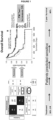
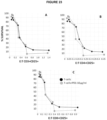
- a basal E:T ratio is obtained.
- the basal E:T is the ratio between the cytotoxic T cells and the cancer cells before BiTE and/or immunomodulatory agent exposure.
- an Effective E:T ratio is obtained.
- the Effective E:T ratio is the ratio between the activated T cells generated and the cancer cells killed after bispecific T cell engager antibody (BiTE) and/or immunomodulatory agent exposure.
- the Effective E:T ratio can be calculated at one or more predetermined concentrations of the bispecific T cell engager antibody (BiTE).
- the predetermined concentration of the bispecific T cell engager antibody (BiTE) is optimized for calculating the Effective E:T ratio.
- the E:T ratio is calculated using the numbers of tumor and activated T cells when exposed to the maximum concentration of bispecific T cell engager antibody (BiTE).
- the E:T ratio is calculated using the numbers of tumor and activated T cells when exposed to the concentration of the bispecific T cell engager antibody (BiTE) that generate a maximum peak in the number of activated T cells.
- the E:T ratio is calculated using the numbers of tumor and activated T cells that correspond to the EC50 concentration of the respective dose response curves.
- the Effective E:T ratio can also be expressed as the Effective T:E ratio (e.g., ratio between cancer cells killed to the activated T cells generated).
- the CAR-T cell produced by a method described herein is provided.
- the CAR-T cell is a trogocytotic T cell.
- the CAR-T cell is a activated T cell with a high killing activity, e.g a high Effective E:T Ratio.
- the CAR-T cell is a CD8+CD25+ T cell.
- the CAR-T cell is a CD4+CD25+ T cell.
- the trogocytotic T cell is believed to be a more effective cancer cell killer, although the cytotoxic T cells, e.g., CD8+ T cells and activated CD4+ T cells also have cancer cell killing activity. Accordingly, all activated T cell types can be included in the Effective E:T ratio.
- the method or assay includes detecting, e.g., counting, the number of newly generated CAR cytotoxic T cells, and the number of targets cells that have been killed under the same conditions, e.g., in the same well.
- the ratio of these values is the Effective E:T ratio.
- the ratio is a ratio between two subtractions, one subtraction is the number of targets after incubation with a BiTE relative to control well without the BiTE also after incubation (i.e., to measure the number of target cells killed in such condition), and the other subtraction is the number of activated T cells after incubation with a BiTE relative to control wells without the BiTE also after incubation (i.e., to measure the number of cytotoxic t cells that kill the target cells in such condition).
- the subtraction equals the total number for activated T cells (e.g., total number of CD8+CD25+ T cells or total number of CD4+CD25+ T cells).
- a decrease in the level or amount of cancer cells is indicative of increased cancer cell killing.
- a reduced change or no substantial change in the level or amount of cancer cells is indicative of decreased cancer cell killing.
- a high level of target cell killing relative to the newly generated target killing T cells indicates that the activated T cell or preparation thereof is an effective killer of cancer cells.
- the target to T cell ratio is compared to a reference ratio. For example, a ratio of 1 (T cell) to 10, 20, 30, 40, 50, 75, 100, 500 or higher (target cells) is indicative of potent T cell killing activity.
- the ratio T cell: target cells ranges 1:100, or higher.
- a subject having T cells having potent cell killing activity can be identified as being a strong responder to the bispecific T cell engager antibody (BiTE) and/or immunomodulatory agent.
- the reference ratios are the ratio between two subtractions:
- the Effective E:T Ratio represents an estimate of the activity of the generated activated T cell in killing cancer target cells. Without wishing to be bound by theory, it is equivalent to the activity of a drug in killing cancer cells, because the activated T cell is indeed an active medicament for treating a subject, e.g., a cancer patient.
- the Effective E:T Ratio can rank the activity of activated T cells from different patients thus stratifying those patients. This ranking or stratification can be very different than the ranking or stratification derived from the standard method of measuring the efficacy in killing cancer target cells.
- a very efficacious activated T cell with a 1:100 Effective E:T Ratio that eliminates 100 target cells per activated T cell, may not be able to kill all cancer cells if that patient has a very large density of cancer target cells. Leaving alive many cancer cells would normally be considered a sign of low activity for the activated T cell in a standard chemotherapy activity measurement; in this case, it would miss the true high activity of the activated T cell generated by the bispecific T cell engager antibody (BiTE), the problem being some cancer cells are immunosuppressed and resistant to the otherwise high activity CAR activated T cells generated.
- BiTE bispecific T cell engager antibody
- the Effective E:T ratio can identify the most active activated T cells, e.g., those activated T cells better suited to be administered to the patient, and to be used as a source to transfect a CAR making a CAR-T product.
- a low level of Effective E:T Ratio is indicative of a poor T cell killing activity.
- a ratio activated T cells:target cells of 1:1 is indicative of poor T cell killing activity.
- a subject having T cells having reduced cell killing activity can be identified as being a poor responder to the bispecific T cell engager antibody (BiTE) and/or immunomodulatory agent.
- the level of target cells and/or activated T cells is determined at one or more time intervals after step (c). In exemplary embodiments, the level of target cells and/or activated T cells is determined at time 0, at time 1 - 168 hours (e.g., 1, 2, 4, 8, 16, 24, 48, 72, 96, 120, 144, or 168 hours) or several days or weeks after step (c).
- the contacting step further comprises addition of a bispecific T cell engager antibody (BiTE) and/or immunomodulatory agent at different doses (e.g., increasing dosages) of the bispecific T cell engager antibody (BiTE) and/or immunomodulatory agent, e.g., to generate a dose-response curve.
- a bispecific T cell engager antibody (BiTE) and/or immunomodulatory agent at different doses (e.g., increasing dosages) of the bispecific T cell engager antibody (BiTE) and/or immunomodulatory agent, e.g., to generate a dose-response curve.
- the difference between the level of T cells or cancer cells at a dose zero or at control level (e.g., a threshold dose) and a saturated dose of the bispecific T cell engager antibody (BiTE) and/or immunomodulatory agent is determined.
- the difference in the level of T cells or cancer cells at the saturated dose vs.
- the Effective E:T ratio as used herein is the ratio of the difference in the level of T cells relative to the difference in the level of cancer cells. In embodiments, the Effective E:T ratio as used herein is the ratio of the number of T cells and target cells at their respective EC50 concentration.
- the method is performed using an automated platform, e.g., an automated fluorescence-based platform, e.g., the ExviTech ® platform described herein.
- an automated platform e.g., an automated fluorescence-based platform, e.g., the ExviTech ® platform described herein.
- the activity of the bispecific T cell engager antibody (BiTE) and/or immunomodulatory agent is determined using an ex vivo / in vitro assay to measure dose response curves, whose mathematical fitting enable quantitative parameters to estimate the activity, selected from at least one from EC50, Effective E:T ratio, basal E:T ratios, Emax or kinetics.
- the activity of the bispecific T cell engager antibody (BiTE) and/or immunomodulatory agent assessed by step (e) is different from an activity assessment using a dose response of the bispecific T cell engager antibody (BiTE) and/or immunomodulatory agent activity, e.g., compared to a standard depletion dose response curve.
- the reference ratio is a predetermined ratio, e.g., about 1:3 to 1:10, e.g., about 1:3, 1:4, 1:5, 1:6, 1:7, 1:8, 1:9, or 1:10.
- the T cell to high target cell ratio from step (e) is about 1:4 - 1:500 (e.g., 1:4, 1:5, 1:6, 1:7, 1:8, 1:9, 1:10, 1:15, 1:20, 1:25, 1:30, 1:35, 1:40, 1:45, 1:50, 1:75, 1:100, 1:500, or higher).
- step (c) comprises forming ex vivo mixtures of the activated T cell or the preparation thereof with target cells, e.g., cancer cells.
- the cancer cell is a cell chosen from a hematological cancer, a solid cancer, a metastatic cancer (e.g., a CTC, or a combination thereof).
- the cancer cell is a leukemic or lymphoma blast cell (e.g., a blast cell expressing one or more markers chosen from CD19, CD123, CD20 or others).
- the T cell is a cell chosen from a blood sample (e.g., peripheral blood sample), a bone marrow sample, a lymph node sample, a spleen sample, a tumor sample comprising a CTL and/or a TIL, or a combination thereof).
- the T cell expresses CD8 and/or CD25 (e.g., it is a CD8+CD25+ T cell).
- the T cell expresses CD4 and/or CD25 (e.g. it is a CD4+CD25+ T cell).
- the CAR-T cell or preparation thereof is produced using a method that comprises use of a bispecific T cell engager antibody (BiTE) and/or immunomodulatory agent, e.g., a bispecific T cell engager antibody (BiTE) and/or immunomodulatory agent described herein.
- a bispecific T cell engager antibody BiTE
- immunomodulatory agent e.g., a bispecific T cell engager antibody (BiTE) and/or immunomodulatory agent described herein.
- the CAR-T cell or preparation thereof comprises a T cell, e.g., CTL, that is CD8+ and CD25+, or a CD4+ and CD25+, or both.
- the candidate bispecific T cell engager antibody (BiTE) and/or immunomodulatory agent is administered at different dosages (e.g., at increasing dosages).
- an increase in the cell killing activity of the T cells in the presence of the candidate bispecific T cell engager antibody (BiTE) and/or immunomodulatory agent is indicative of high efficacy of the bispecific T cell engager antibody (BiTE) and/or immunomodulatory agent.
- a small change or no substantial change in the cell killing activity of the T cells in the presence of the candidate bispecific T cell engager antibody (BiTE) and/or immunomodulatory agent is indicative of low efficacy of the bispecific T cell engager antibody (BiTE) and/or immunomodulatory agent.
- the cancer-killing activity of different T cell therapies can be evaluated on the same patient sample ex vivo, where the T cells can be selected from the group consisting of a tumor infiltrated lymphocyte (TIL), marrow infiltrated lymphocytes (MILs), a genetically engineered T cell, a CAR-T cell including comparing different CAR constructs, an activated T cell obtainable according to step (c) of the method of producing a CAR-T cell and a genetically engineered T cell expressing Chimeric Antigen Receptors obtainable according to step (e) of the method of producing a CAR-T cell.
- TIL tumor infiltrated lymphocyte
- MILs marrow infiltrated lymphocytes
- CAR-T cell including comparing different CAR constructs
- an activated T cell obtainable according to step (c) of the method of producing a CAR-T cell
- a genetically engineered T cell expressing Chimeric Antigen Receptors obtainable according to step (e) of the method
- an important comparison is the activated T cell generated incubating with a BiTE, with the same activated T cells transfected with a CAR, because the BiTE-generated T cell would be safer and thus a preferred treatment than the CAR transfected T cell if the CAR transfected T cell is not substantially better.
- the activity of these different T cell therapies are first evaluated against at least 30 patient samples of the same cancer type that represent the patient population, and afterwards the activity of each T cell therapy is compared with the activity across the population of patient samples, deriving a sensitivity ranking.
- Combinations of these different T cell therapies with other drugs can be also evaluated to guide patient treatment, where drugs that can be combined for each disease include approved drugs for said disease, and especially other immunotherapies such as immune check point inhibitors, immunomodulatory drugs, etc...
- Flow cytometry is the method chosen for the diagnosis and monitoring of patients with hematological malignances. Additionally, it has been validated for the study of cellular death or apoptosis processes induced by drugs.
- the ExviTech ® platform allows the escalation of flow cytometry technology, with the ability to measure the effect of a high number of drugs and combinations selectively in pathological cells (identified in a similar manner than in the diagnosis of the disease) of an individual patient's sample.
- the patient's bone marrow sample is received, and a small aliquot is first analyzed to determine the number of live pathological cells present in the sample.
- the rest of the sample is diluted with a culture medium, and is divided into 96 well plates, containing the drug treatments (monotherapies and combinations) to be studied. 8 concentrations are studied for each treatment (drug or combination), duly adjusted to cover each treatment's range of pharmacological activity tested in multiple patient samples.
- the plates are later incubated at control temperature for certain time, from 12 to 48 hours.
- the sample is marked with the specific monoclonal antibodies to identify the leukemic cells, together with Annexin V. The presence of this last marker indicates that the cell has entered into apoptosis or programmed death. Therefore, cells that present the phenotype of a leukemic cell and the absence of Annexin V are identified as live leukemic cells (LLC).
- LLC live leukemic cells
- the proportion of the number of live leukemic cells after the incubation present in the control wells (without drugs) compared to the wells containing each of the treatments or, which is equivalent, the percentage "survival index”, is the measure of efficacy of the tested treatments for the specific patient that PM Test measures. PM Test then ranks treatments in order of efficacy based on the "survival index" measured for each treatment. The lower the "survival index" (the lesser number of leukemic cells alive), the more efficient the treatment will be.
- PM incorporates modern pharmacokinetic and pharmacodynamic population modelling technologies, increasingly used in clinical trials for new drugs, to analyze the test's flow cytometry data. This enables making very accurate estimates in complex multiple-variable systems subject to high variability.
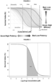
- ExviTech ® generates dose-response models that evaluate the patient's cellular response to increasing drug concentrations in the patient's bone marrow sample, measured as cellular death or depletion.
- the final model estimated is characterized by a set of pharmacological parameters that describe the effect of the drug or combination.
- population models enables to analyze typical population values to put the patient's individual data in context of a patient population, inter-individual variability data associated to each parameter, and relative standard error individually associated to each estimation.
- pharmacodynamics models based on Hill equation are represented by typical sigmoidal curves of measured effect at increasing drug concentrations. These graphs allow a quick interpretation of drug biological effect and a direct comparison with population typical behavior. Individual model functions can be summarized with the value of the Area Under the Curve (AUC) that it is used as a general activity marker.
- AUC Area Under the Curve
- Treatments scores are calculated using the AUC values of dose-response model function of each individual drug included in a clinical treatment, together with the contribution of the synergy from binary combinations which is estimated from sophisticated drugs interaction surface models.
- the key to interpret the ex-vivo activity of individual drugs in a patient sample is not just the absolute value of the pharmacological variables, but their reference rank compared to a statistically representative patient population. This is why the results of PM Test are expressed in population terms, normalized to a reference activity range of the patient population.in terms of cellular efficacy of a treatment in terms of tumor cell killing for the individual patient compared with the cell killing efficacy of the same treatment in a reference patient population.
- two different methods are used to identify the immune check point molecule appropriate for each patient:
- an in vitro method of identifying subjects susceptible to immune checkpoint immunotherapy treatment comprising:
- an in vitro method of identifying subjects susceptible to immune checkpoint immunotherapy treatment comprising:
- an in vitro method of identifying subjects susceptible to immune checkpoint immunotherapy treatment to be combined with a bispecific T cell engager antibody (BiTE) immunotherapy, for decreasing resistance of said subject to said BiTE immunotherapy comprising:
- an in vitro method of identifying subjects susceptible to immune checkpoint immunotherapy treatment to be combined with a bispecific T cell engager antibody (BiTE) immunotherapy, for decreasing resistance of said subject to said BiTE immunotherapy comprising:
- an in vitro method of identifying subjects susceptible to immune checkpoint immunotherapy treatment to be combined with a cellular immunotherapy such a CAR-T to treat a subject, for decreasing resistance of said subject to said cellular immunotherapy comprising:
- the immune check point molecules are added either from the beginning of the incubation or sequentially after a certain amount of time sufficient for the T cells to become activated killing tumor cells.
- different incubation times are evaluated, and any single incubation time can be used to identify subjects susceptible to immune check point immunotherapy, alone or in combination with other drugs.
- a method for treating a subject having cancer comprising providing a bispecific T cell engager antibody (BiTE) or a T cell selected from the group consisting of a tumor infiltrated lymphocyte (TIL), a genetically engineered T cell, a CAR-T cell, an activated T cell obtainable according to step (c) of the method of producing a CAR-T cell and a genetically engineered T cell expressing Chimeric Antigen Receptors obtainable according to step (e) of the method of producing a CAR-T cell, in combination with an inhibitor of at least one immune checkpoint molecule selected in the method of identifying immune checkpoint molecules as target for decreasing resistance to a cancer therapy.
- BiTE bispecific T cell engager antibody
- TIL tumor infiltrated lymphocyte
- the method (e.g., of producing) further comprises producing a CAR-T cell preparation, e.g., a pharmaceutical preparation.
- the method (e.g., of producing) further comprises detecting the presence of the CAR-T cell.
- the method further comprises purifying the CAR-T cell from the bispecific T cell engager antibody (BiTE).
- the bispecific T cell engager antibody (BiTE) is present, e.g., in the preparation, at a concentration of less than 10% by weight, e.g., less than 10%, 9%, 8%, 7%, 6%, 5%, 4%, 3%, 2%, 1%, 0.5%, 0.1%, 0.05%, 0.01%, 0.005 or less (e.g., but no less than 0.001%).
- the bispecific T cell engager antibody (BiTE) is present in the preparation at a concentration of 0.005% to 10% by weight.
- the reaction mixture contains in volume a few nanoliters (e.g., less than 10 nl, about 1 to 5 nanoliters) of bispecific T cell engager antibody (BiTE) are added to over 50 microliters (e.g., about 60 microliters) of cell suspension.
- a few nanoliters e.g., less than 10 nl, about 1 to 5 nanoliters
- BiTE bispecific T cell engager antibody
- the preparation comprises bispecific T cell engager antibody (BiTE), e.g., a level of bispecific T cell engager antibody (BiTE), detectable by immune assay.
- BiTE bispecific T cell engager antibody
- a level of bispecific T cell engager antibody (BiTE) detectable by immune assay.
- the selecting and/or enriching step comprises using a fluorescently labeled molecule (e.g., a cell surface label, e.g., a fluorescently labeled antibody or fragment thereof, or a cell tracker dye) that diffuses into the cancer cell membrane or binds to i) one or more cancer antigens or ii) one or more markers of activated T cells, or both i) and ii).
- a fluorescently labeled molecule e.g., a cell surface label, e.g., a fluorescently labeled antibody or fragment thereof, or a cell tracker dye
- the selecting and/or enriching step comprises using fluorescence activated cell sorting (FACS).
- the selecting and/or enriching comprises using a bead (e.g., magnetic bead) coated with an antibody or fragment thereof that binds to i) one or more cancer antigens or ii) one or more markers of activated T cells, or both i) and ii).
- a bead e.g., magnetic bead
- the selecting and/or enriching step comprises the sequential addition of a low, e.g., an insufficient, number of cancer cells.
- the methods of producing described above can generate different clones of cytotoxic T cells.
- selection of the cytotoxic T cell clones that are the most efficient or most potent at killing cancer cells can be achieved by sequentially adding low, e.g., insufficient, amounts of cancer cells.
- a low, or insufficient, number or amount of cancer cells that can be added to a reaction comprising CAR-T cells is 50% or less, e.g., 30%, 10%, 1%, 0.1%, or 0.01% or less, of the number of activated T cells.
- the low, or insufficient, number of cancer cells can be added to CAR-T cells (e.g., a reaction comprising cancer cells, T cells, and/or a bispecific T cell engager antibody (BiTE)) one or more times, e.g., 2, 3, 4, 5, 6, 7, 8, 9 or 10, times.
- the low, or insufficient, number of cancer cells is added every 6 hours, 12 hours, 24 hours, 36 hours, or 48 hours.
- the low, or insufficient, number of cancer cells that are added are cancer cells from the patient. In an embodiment, the low, or insufficient, number of cancer cells that are added are not cancer cells from the patient. In an embodiment, the low, or insufficient, number of cancer cells that are added are cancer cells from a cancer cell line.
- the CAR-T cells are expanded.
- the expansion of the CAR-T cells comprises increasing the number of CAR-T cells, e.g., in a preparation, e.g., by at least about 2-fold (e.g., at least about 3-, 4-, 5-, 6-, 7-, 8-, 9-, 10-, 15-, 20-, 50-, 100-, 1000-, 10 4 -, 10 5 -, 10 6 -fold, or more).
- the CAR-T cells are not substantially expanded.
- the CAR-T cell preparation comprises a fluorescently labeled molecule (e.g., a cell surface label, e.g., a fluorescently labeled antibody or fragment thereof or a cell tracker dye) and/or the bispecific T cell engager antibody (BiTE), e.g., wherein the fluorescently labeled molecule and/or the bispecific T cell engager antibody (BiTE) are present at trace amounts (e.g., less than 5% by weight, e.g., less than 5%, 4%, 3%, 2%, 1%, 0.5%, 0.25%, 0.1%, 0.05%, 0.01%, 0.005%, 0.001% by weight, or less).
- a fluorescently labeled molecule e.g., a cell surface label, e.g., a fluorescently labeled antibody or fragment thereof or a cell tracker dye
- BiTE bispecific T cell engager antibody
- the CAR-T cell preparation (prior to purification or expansion) comprises CAR-T cells at a concentration of 5% or less of the total number of cells in the preparation.
- a purified or enriched CAR-T cell preparation comprises CAR-T cells at a concentration of at least 50% (e.g., at least 50%, 55%, 60%, 65%, 70%, 75%, 80%, 85%, 90%, 95%, 99%, or greater) of the total number of cells in the preparation.
- a purified or enriched CAR-T cell preparation comprises activated CAR-T cells, e.g., at a concentration of at least 50% (e.g., at least 50%, 55%, 60%, 65%, 70%, 75%, 80%, 85%, 90%, 95%, 99%, or greater) of the total number of cells in the preparation.
- activated CAR-T cells e.g., at a concentration of at least 50% (e.g., at least 50%, 55%, 60%, 65%, 70%, 75%, 80%, 85%, 90%, 95%, 99%, or greater) of the total number of cells in the preparation.
- a purified or enriched CAR-T cell preparation comprises trogocytotic CAR-T cells, e.g., at a concentration of at least 50% (e.g., at least 50%, 55%, 60%, 65%, 70%, 75%, 80%, 85%, 90%, 95%, 99%, or greater) of the total number of cells in the preparation.
- at least 50% e.g., at least 50%, 55%, 60%, 65%, 70%, 75%, 80%, 85%, 90%, 95%, 99%, or greater
- the CAR-T cell or preparation comprises one or more CD8+ T cells. In embodiments, the CAR-T cell or preparation comprises one or more CD4+ T cells. In embodiments, the CAR-T cell or preparation comprises one or more CD25+ T cells. In embodiments, the CAR-T cell or preparation comprises one or more CD8+/CD25+ CTLs. In embodiments, the CAR-T cell or preparation comprises one or more CD4+/CD25+ T cells. In embodiments, the CAR-T cell or preparation comprises one or more cytotoxic T lymphocytes (CTLs), e.g., cancer antigen-specific CTLs. In embodiments, the CAR-T cell or preparation comprises one or more effector memory T cells. In embodiments, the CAR-T cell preparation does not comprise a substantial number of regulatory T cells (Tregs).
- CTLs cytotoxic T lymphocytes
- the CAR-T cell preparation comprises one or more effector memory T cells. In embodiments, the CAR-T cell preparation does not comprise
- the method further comprises reducing the number of Tregs in the CAR-T cell preparation.
- the bispecific T cell engager antibody (BiTE) selectively expands the CAR-T cells, thus increasing the Effective E:T ratio of CAR-T cells:Tregs.
- method further comprises removing (e.g., depleting) Tregs by physical separation, e.g., using a bead (e.g., a magnetic bead) attached to a Treg cell surface marker.
- the CAR-T cell preparation comprises Tregs at a concentration of less than 10% (e.g., less than 9%, 8%, 7%, 6%, 5%, 4%, 3%, 2%, 1% or less) of the total number of cells in the preparation.
- the CAR-T cell preparation does not comprise a substantial number of naive T cells.
- the CAR-T cell preparation comprises naive T cells at a concentration of less than 10% (e.g., less than 9%, 8%, 7%, 6%, 5%, 4%, 3%, 2%, 1% or less) of the total number of cells in the preparation.
- the naive T cells express CD45RA, CD62L, CCR7, CD27, CD28 and/or CD57.
- the CAR-T cell preparation comprises more than one clone of CAR-T cells.
- the method (e.g., of producing) further comprises separating individual clones from the CAR-T cell preparation.
- the separating step comprises clonal expansion of single cells (e.g., (i) separating the preparation of CAR-T cells into single cells (e.g., a single cell per well or container) and (ii) expanding the single cells to generate one or more preparations of CAR-T cells, wherein each preparation comprises a single clone).
- single cells e.g., (i) separating the preparation of CAR-T cells into single cells (e.g., a single cell per well or container) and (ii) expanding the single cells to generate one or more preparations of CAR-T cells, wherein each preparation comprises a single clone).
- the separating step comprises flow cytometry or limited dilution.
- the method (e.g., of producing) further comprises determining the cancer-killing activity of the CAR-T cell preparation, and optionally, selecting the preparation based on a parameter chosen from one or more of: increased cancer cell killing activity, reduced toxicity, reduced off-target effect, increased viability, increased proliferation, or Effective E:T ratio for cancer cell killing.
- the CAR-T cell preparation comprises cells having high cancer-killing activity and/or low toxicity.
- the cells comprised in the CAR-T cell preparation with low toxicity are cells which kill significantly less non-pathological cells, i.e. they kill more selectively.
- the CAR-T cell preparation comprises cells having low toxicity because they generate less cytokines in the supernatant and/or intracellularly.
- the CAR-T cell preparation comprises cells having both and simultaneously higher cancer-killing activity and low toxicity, because they generate less cytokines in the supernatant and/or intracellularly per unit of CAR-T cell, that is once the types and/or levels of cytokines released is normalized by the quantitative estimation of cancer cell killing activity such as Effective E:T Ratios, basal E:T ratios, EC50, Emax, kinetics, or a combination of these factors.
- the CAR-T cell preparation comprises cells that effectively kill cancer cells at a high target cell per T cell.
- a T cell to high target cell ratio is about 1:4 to 1:100 (e.g., 1:4, 1:5, 1:6, 1:7, 1:8, 1:9, 1:10, 1:15, 1:20, 1:25, 1:30, 1:35, 1:40, 1:45, 1:50, 1:75, 1:100, or higher).
- the CAR-T cell preparation comprises a population of cells consisting of less than 10 clones of CAR-T cells. In embodiments, 10, 9, 8, 7, 6, 5, 4, 3, 2 or 1 clone of CAR-T cells is present in the preparation. In one embodiment, 2-4 clones are present in the preparation. In other embodiments, a single clone of CAR-T cells.
- the T cell or T cell sample of the method e.g., of producing
- the cancer cell or cancer cell sample of the method e.g., of producing
- the T cell or T cell sample of the method (e.g., of producing) and the cancer cell or cancer cell sample of the method (e.g., of producing) are from a different subject.
- the CAR-T cell or preparation is administered to the subject, e.g., wherein the subject is the same subject as the subject from whom the T cells (and/or the cancer cells) were obtained.
- the CAR-T cell or preparation is autologous.
- the CAR-T cell or preparation is administered to the subject, e.g., wherein the subject is a different subject from the subject from whom the T cells (and/or the cancer cells) were obtained.
- the CAR-T cell or preparation is allogeneic.
- the method comprises providing a sample comprising the T cell. In embodiments, method (e.g., of producing) comprises providing a sample comprising the cancer cell.
- the T cell and the cancer cell of the method are from the same sample.
- the T cell and the cancer cell of the method are from different samples.
- the sample is derived from a tissue with a microenvironment, e.g., a bone marrow, a lymph node, a primary tumor, or a metastasis.
- a tissue with a microenvironment e.g., a bone marrow, a lymph node, a primary tumor, or a metastasis.
- the sample comprises blood (e.g., whole blood, peripheral blood, or bone marrow), a solid tumor (e.g., a sample resected from a primary tumor or a metastasis), a lymph node, or spleen of the subject.
- the sample is a blood sample e.g., whole blood, peripheral blood, or bone marrow, wherein substantially no components (e.g., cells or plasma) have been removed or isolated from the blood sample.
- the sample is diluted, e.g., with a physiologically compatible buffer or media, e.g., prior to and/or during step (c).
- the method comprises providing a T cell from a blood sample from the subject, e.g., where the T cell is not purified from other components, e.g., cells or plasma, in the blood sample.
- the blood sample is a bone marrow sample, a peripheral blood sample, or a whole blood sample.
- the method comprises providing a cancer cell from a blood sample from the subject, e.g., wherein the cancer cell is not purified from other components, e.g., cells or plasma, in the blood sample.
- the blood sample is a bone marrow sample, a whole blood sample, or a peripheral blood sample.
- the cancer cell of the method comprises a circulating cancer cell, e.g., from a blood sample, e.g., peripheral blood sample, of the subject.
- the method comprises providing a cancer cell from a tissue sample, e.g., a biopsy, e.g., of a tumor or metastasis, from the subject.
- a tissue sample e.g., a biopsy, e.g., of a tumor or metastasis
- the method comprises providing a sample, e.g., blood sample (e.g., bone marrow, peripheral blood, or whole blood sample), that comprises both the T cell and the cancer cell.
- a sample e.g., blood sample (e.g., bone marrow, peripheral blood, or whole blood sample)
- blood sample e.g., bone marrow, peripheral blood, or whole blood sample
- the subject is an adult or a pediatric subject.
- the cancer is a hematological cancer, e.g., a B-cell or T cell malignancy.
- the cancer is a Hodgkin's lymphoma, Non-Hodgkin's lymphoma (e.g., B cell lymphoma, diffuse large B cell lymphoma, follicular lymphoma, chronic lymphocytic leukemia, mantle cell lymphoma, marginal zone B-cell lymphoma, Burkitt lymphoma, lymphoplasmacytic lymphoma, hairy cell leukemia), acute myeloid leukemia, chronic myeloid leukemia, myelodysplastic syndrome, multiple myeloma, or acute lymphocytic leukemia.
- B cell lymphoma diffuse large B cell lymphoma
- follicular lymphoma chronic lymphocytic leukemia
- mantle cell lymphoma mantle cell lymphoma
- marginal zone B-cell lymphoma marginal zone B-cell lymphoma
- Burkitt lymphoma Burkitt lymphoma
- the cancer is a solid cancer, e.g., wherein the solid cancer comprises ovarian cancer, rectal cancer, stomach cancer, testicular cancer, cancer of the anal region, uterine cancer, colon cancer, rectal cancer, renal-cell carcinoma, liver cancer, non-small cell carcinoma of the lung, cancer of the small intestine, cancer of the esophagus, melanoma, Kaposi's sarcoma, cancer of the endocrine system, cancer of the thyroid gland, cancer of the parathyroid gland, cancer of the adrenal gland, bone cancer, pancreatic cancer, skin cancer, cancer of the head or neck, cutaneous or intraocular malignant melanoma, uterine cancer, brain stem glioma, pituitary adenoma, epidermoid cancer, carcinoma of the cervix squamous cell cancer, carcinoma of the fallopian tubes, carcinoma of the endometrium, carcinoma of the vagina, sarcoma of soft tissue, cancer of the urethra
- the cancer is not melanoma.
- the method does not comprise labelling the cancer cell (e.g., cancer cell membrane) with a fluorescent molecule prior to contacting the sample with the bispecific T cell engager antibody (BiTE).
- the cancer cell e.g., cancer cell membrane
- BiTE bispecific T cell engager antibody
- the subject :
- the period of time is 12 to 120 hours (e.g., 12-24 hours, 24-48 hours, 48-36 hours, 36-60 hours, 60-90 hours, or 90-120 hours) or 1-7 days (e.g., 1, 2, 3, 4, 5, 6, or 7 days).
- the method described here further comprises repeating the sample or cell providing step, ex vivo reaction formation step and/or the enrichment step (e.g., steps (a)-(d) of the methods of producing) using a different sample of T cells and cancer cells, e.g., wherein each repeat of steps uses a different sample of T cells and cancer cells.
- the different sample of T cells and cancer cells comprises a sample derived from a tissue with a microenvironment, e.g., a bone marrow, a lymph node, a primary tumor, or a metastasis.
- the CAR-T cell produced from each repeat of steps is pooled to form a mixture of CAR-T cells.
- the T cell comprises a CTC, and the T cell is from a sample (e.g., blood (e.g., whole blood, peripheral blood, or bone marrow), lymph node, primary tumor, or metastasis) from the subject.
- the T cell is enriched for the CTC.
- the T cell is purified, e.g., purified from other types of cells, e.g., from a blood sample from the subject (e.g., whole blood, peripheral blood, or bone marrow).
- the method further comprises repeating the sample or cell providing step, ex vivo reaction formation step and/or the enrichment step (e.g., steps (a)-(d) of the methods of producing) using a different sample of T cells from the subject, e.g., wherein each repeat of steps uses a different sample of T cells from the subject.
- the different sample of T cells comprises a sample derived from a cancer-containing tissue from the subject, e.g., a primary tumor, one or more metastases, a lymph node, a lymph sample, or a blood sample (e.g., whole blood, peripheral blood, or bone marrow).
- a cancer-containing tissue from the subject
- e.g., a primary tumor, one or more metastases, a lymph node, a lymph sample, or a blood sample e.g., whole blood, peripheral blood, or bone marrow.
- the CAR-T cell produced from each repeat of the sample or cell providing step, ex vivo reaction formation step and/or the enrichment step e.g., steps (a)-(d) of the methods of producing
- the enrichment step e.g., steps (a)-(d) of the methods of producing
- the method (e.g., of producing) further comprises evaluating the cancer-killing activity of the CAR-T cell.
- the evaluating comprises:
- the evaluating comprises:
- the evaluating comprises using a first patient sample, e.g., containing T cells and cancer cells, to generate a CAR-T cell, e.g., using a method described herein.
- the CAR-T cells are purified, sorted, enriched, expanded, and/or selected.
- the evaluating comprises subsequently mixing a second sample from the same patient with the CAR-T cells generated using the first patient sample.
- various concentrations of CAR-T cells can be mixed with the second sample, e.g., where the second sample is at a fixed concentration, e.g., to generate a dose response curve.
- the evaluating comprises:
- the level of activity of the CAR-T cells is measured by Effective E:T Ratios, basal E:T ratios, EC50s, Emax, kinetics, or a combination of these factors.
- step (c) comprises contacting the cancer cells with the CAR-T cells at a plurality of ratios, e.g., Effective E:T ratios.
- step (c) comprises mixing different amounts of CAR-T cells with a fixed amount of cancer cells.
- an Effective E:T ratio is obtained.
- the Effective E:T is the ratio between the CAR-T cells and the cancer cells after bispecific T cell engager antibody (BiTE).
- a decrease in the level or amount of cancer cells is indicative of increased cancer cell killing.
- a reduced change or no substantial change in the level or amount of cancer cells is indicative of decreased cancer cell killing.
- a high level of target cell relative to T cell indicates that the CAR-T cell or preparation thereof is an effective killer of cancer cells.
- the target to T cell ratio is compared to a reference ratio.
- an Effective E:T ratio of 1 (CAR-T cell) to 100 (e.g., 10, 20, 30, 40, 50, 75, 100 or higher) (target cells) is indicative of potent T cell killing activity.
- a subject having T cells having potent cell killing activity can be identified as being a strong responder to the bispecific T cell engager antibody (BiTE).
- a low level of target cell relative to T cell is indicative of a poor T cell killing activity.
- the target to T cell ratio is compared to a reference ratio.
- an Effective E:T ratio of 1 (CAR-T cell) to 5 (target cells) is indicative of poor T cell killing activity.
- a subject having T cells having reduced cell killing activity can be identified as being a poor responder to the bispecific T cell engager antibody (BiTE).
- the level of target cells and/or CAR-T cells is determined at one or more time intervals after step (c). In exemplary embodiments, the level of target cells and/or CAR-T cells is determined at time 0, at time of 1-75 hours (e.g., 1, 2, 4, 8, 16, 24, 36 or 72 hours) or several days after step (c).
- the contacting step further comprises addition of a bispecific T cell engager antibody (BiTE) at different doses (e.g., increasing dosages) of the bispecific T cell engager antibody (BiTE), e.g., to generate a dose response curve.
- a bispecific T cell engager antibody BiTE
- the difference between the level of CAR-T cells or cancer cells at a dose zero or at control level (e.g., a threshold dose) and a saturated dose of the bispecific T cell engager antibody (BiTE) is determined.
- the difference in the level of CAR-T cells or cancer cells at the saturated dose vs. threshold dose is determined.
- the Effective E:T ratio as used herein is the ratio of the difference in the level of CAR-T cells relative to the difference in the level of cancer cells.
- method is performed using an automated platform, e.g., an automated fluorescence-based platform, e.g., the ExviTech ® platform described herein.
- an automated platform e.g., an automated fluorescence-based platform, e.g., the ExviTech ® platform described herein.
- the activity of the bispecific T cell engager antibody (BiTE) and/or immunomodulatory agent is determined using an ex vivo / in vitro assay to measure dose response curves, whose mathematical fitting enable quantitative parameters to estimate the activity, selected from at least one from EC50, Effective E:T ratio, basal E:T ratios, Emax or kinetics.
- the activity of the bispecific T cell engager antibody (BiTE) assessed by step (e) is different from an activity assessment using a dose response of the bispecific T cell engager antibody (BiTE) activity, e.g., compared to a standard depletion dose response curve.
- the reference ratio is a predetermined ratio, e.g., about 1:3 to 1:10, e.g., about 1:3, 1:4, 1:5, 1:6, 1:7, 1:8, 1:9, or 1:10.
- the high target cell to T cell ratio from step (e) is about 1:4 to 1:100 (e.g., 1:4, 1:5, 1:6, 1:7, 1:8, 1:9, 1:10, 1:15, 1:20, 1:25, 1:30, 1:35, 1:40, 1:45, 1:50, 1:75, 1:100, or higher).
- step (c) comprises forming ex vivo mixtures of the CAR-T cell or the preparation thereof with target cells, e.g., cancer cells.
- the cancer cell is a cell chosen from a hematological cancer, a solid cancer, a metastatic cancer (e.g., a CTC, or a combination thereof).
- the cancer cell is a leukemic or lymphoma blast cell (e.g., a blast cell expressing one or more markers chosen from CD19, CD123, CD20 or others).
- the T cell is a cell chosen from a blood sample (e.g., peripheral blood sample), a bone marrow sample, a lymph node sample, a tumor sample comprising a CTL and/or a TIL, or a combination thereof).
- the T cell expresses CD8 and/or CD25 (e.g., it is a CD8+CD25+ T cell).
- the CAR-T cell or preparation thereof is produced using a method that comprises use of a bispecific T cell engager antibody (BiTE), e.g., a bispecific T cell engager antibody (BiTE) described herein.
- a bispecific T cell engager antibody e.g., a bispecific T cell engager antibody (BiTE) described herein.
- the CAR-T cell or preparation thereof comprises a T cell, e.g., CTL, that is CD8+ and CD25+.
- the CAR-T cell is a trogocytotic T cell.
- the CAR-T cell is a CD28+CD25+ T cell.
- the CAR-T cell (i) has cytotoxic activity toward a cancer cell, and (ii) comprises a cell surface marker derived from the cancer cell, e.g., at least 90-500 copies of a cell surface marker (e.g., 90, 100, 200, 300, 400, or 500 copies, e.g., one or more cancer cell surface markers).
- a cell surface marker e.g., at least 90-500 copies of a cell surface marker (e.g., 90, 100, 200, 300, 400, or 500 copies, e.g., one or more cancer cell surface markers).
- about 2 to 75% (e.g., about 2 to 70%, 2 to 60%, 2 to 50%, or 2 to 40%) of the total T cells in the reaction mixture express one or more cancer cell surface markers (e.g., one or more leukemic cell cancers).
- the CAR-T cell is enriched or purified.
- the enriched or purified CAR-T cell population comprises at least 80%-100% CAR-T cells (e.g., 80%, 90%, 95%, 99% or 100%), wherein the CAR-T cells comprise one or more cancer cell surface markers.
- the ex vivo reaction mixture is prepared according to Good Manufacturing Practice (GMP).
- one or more of the expansion, selection and/or enrichment of the CAR-T cells is according Good Manufacturing Practice (GMP).
- the method further comprises sending the produced CAR-T cell, e.g., to a hospital, a health care provider.
- the method further comprises receiving the T cell, the cancer cell, or both, e.g., from a hospital, a health care provider.
- the method further comprises administering a second therapeutic agent or procedure.
- the second therapeutic agent or procedure is chosen from one or more of chemotherapy, a targeted anti-cancer therapy, an oncolytic drug, a cytotoxic agent, an immune-based therapy, a cytokine, a surgical procedure, a radiation procedure, an agonist of T cells (e.g., agonistic antibody or fragment thereof or an activator of a costimulatory molecule), an inhibitor of an inhibitory molecule (e.g., immune checkpoint inhibitor), an immunomodulatory agent, a vaccine, or a cellular immunotherapy.
- chemotherapy e.g., a targeted anti-cancer therapy
- an oncolytic drug e.g., cytotoxic agent, an immune-based therapy, a cytokine, a surgical procedure, a radiation procedure, an agonist of T cells (e.g., agonistic antibody or fragment thereof or an activator of a costimulatory molecule), an inhibitor of an inhibitory molecule (e.g., immune checkpoint
- the second therapeutic agent is an agonist of T cells (e.g., an agonistic antibody or fragment thereof or an activator of a costimulatory molecule) or an immune checkpoint inhibitor.
- the immune checkpoint inhibitor is an inhibitor of one or more of: CTLA4, PD1, PDL1, PDL2, B7-H3, B7-H4, TIM3, LAG3, BTLA, CD80, CD86, or HVEM.
- the immune checkpoint inhibitor comprises one or more of: ipilimumab, tremelimumab, MDX-1106, MK3475, CT-011, AMP-224, MDX-1105, IMP321, or MGA271.
- the agonist of T cells comprises an antibody or fragment thereof to CD137, CD40, and/or glucocorticoid-induced TNF receptor (GITR).
- GITR glucocorticoid-induced TNF receptor
- the immunomodulatory agent is an inhibitor of MDSCs and/or Treg cells. In embodiments, the immunomodulatory agent comprises/is lenalidomide.
- the second therapeutic agent enhances and/or restores the immunocompetence of T cells.
- the immunomodulatory agent activates an immune response to a tumor specific antigen, e.g., it is a vaccine (e.g., a vaccine against targets such as gp100, MUC1 or MAGEA3).
- the immunomodulatory agent is a cytokine, e.g., a recombinant cytokine chosen from one or more of GM-CSF, IL-7, IL-12, IL-15, IL-18 or IL-21.
- the immunomodulatory agent is an autologous T cell, e.g., a tumor-targeted extracellular and intracellular tumor-specific antigen (e.g., a CAR-T cell or a TCR T cell).
- the immunomodulatory agent is a modulator of a component (e.g., enzyme or receptor) associated with amino acid catabolism, signalling of tumor-derived extracellular ATP, adenosine signalling, adenosine production, chemokine and chemokine receptor, recognition of foreign organisms, or kinase signalling activity.
- a component e.g., enzyme or receptor
- exemplary agents include an inhibitor (e.g., small molecule inhibitor) of IDO, COX2, ARG1, ArG2, iNOS, or phosphodiesterase (e.g., PDE5); a TLR agonist, or a chemokine antagonist. Additional examples of immunomodulatory agents are described herein.
- the bispecific T cell engager antibody comprises an antibody molecule, e.g., a bi-specific antibody or fragment thereof, e.g., a bispecific immunoglobulin (BsIgG), an immunoglobulin operatively linked to additional antigen-binding molecule, a bispecific antibody (BsAb) fragment, a bispecific fusion protein, or a BsAb conjugate.
- Bispecific antibodies can also be named DART, DutaFab, Duobodies, Biparatopic, Adaptir.
- a BiTE includes multispecific constructs with more than 2 recognition arms, a common development in the field of bispecific antibodies, and a natural extension of the same concept.
- multispecific constructs can add more recognition fragments of the same type, or include fragments with different recognition properties.
- the bispecific T cell engager antibody is a bi-specific antibody selected from the list consisting of BsMAb CD123/CD3, BsMAb CD19/CD3 and EpCAM/CD3.
- the bispecific T cell engager antibody (BiTE) is present at a detectable amount, e.g., a concentration of less than 10% by weight, e.g., less than 10%, 9%, 8%, 7%, 6%, 5%, 4%, 3%, 2%, 1%, 0.5%, 0.1%, 0.05%, 0.01%, 0.001% or less (but no less than 0.0001%), e.g., in a composition described herein.
- the bispecific T cell engager antibody (BiTE) is present at a level of less than 1%.
- the bispecific T cell engager antibody (BiTE) is present in the preparation at a concentration of 0.005% to 10% by weight.
- the CAR-T cell comprises an activated T cell.
- the CAR-T cell comprises a cell that has undergone trogocytosis, e.g., a cell that comprises a portion of a cell surface membrane from the cancer cell.
- the CAR-T cell is a T cell, e.g., a cytotoxic T lymphocyte, e.g., a CD8+ T cell.
- the composition or preparation does not comprise a substantial number of cancer cells, e.g., comprising cancer cells at a concentration of less than 30% (e.g., less than 30%, 25%, 20%, 15%, 10%, 5%, 2.5%, 1%, 0.5%, 0.1%, or less) of the total number of cells in the composition or preparation.
- the composition or preparation does not comprise a substantial number of regulatory T cells (Tregs), e.g., comprising Tregs at a concentration of less than 30% (e.g., less than 30%, 25%, 20%, 15%, 10%, 5%, 2.5%, 1%, 0.5%, 0.1%, or less) of the total number of cells in the composition or preparation.
- Tregs regulatory T cells
- the composition or preparation does not comprise a substantial number of naive T cells, e.g., comprising naive T cells at a concentration of less than 30% (e.g., less than 30%, 25%, 20%, 15%, 10%, 5%, 2.5%, 1%, 0.5%, 0.1%, or less) of the total number of cells in the composition or preparation.
- the composition or preparation does not comprise a substantial number of red blood cells, e.g., comprising red blood cells at a concentration of less than 30% (e.g., less than 30%, 25%, 20%, 15%, 10%, 5%, 2.5%, 1%, 0.5%, 0.1%, or less) of the total number of cells in the composition or preparation.
- the composition or preparation does not comprise a substantial number of non-immune cells, e.g., comprising non-immune cells at a concentration of less than 30% (e.g., less than 30%, 25%, 20%, 15%, 10%, 5%, 2.5%, 1%, 0.5%, 0.1%, or less) of the total number of cells in the composition or preparation.
- the composition or preparation comprises activated T cells at a concentration of at least 30%, (e.g., at least 30%, 40%, 50%, 60%, 70%, 80%, 90%, 95%, 99%, or more) of the total number of cells in the composition or preparation.
- the composition or preparation comprises trogocytotic T cells at a concentration of at least 30%, (e.g., at least 30%, 40%, 50%, 60%, 70%, 80%, 90%, 95%, 99%, or more) of the total number of cells in the composition or preparation.
- a preparation of CAR-T cells for use in (e.g., use in preparation of a medicament for) treating a cancer (e.g., a hematological cancer, a solid cancer or a metastatic cancer) in a subject.
- a cancer e.g., a hematological cancer, a solid cancer or a metastatic cancer
- a CAR-T cell for use in (e.g., use in preparation of a medicament for) treating a cancer (e.g., a hematological cancer, a solid cancer or a metastatic cancer) in a subject, where the CAR-T cell is produced by a method comprising:
- a CAR-T cell for use in (e.g., use in preparation of a medicament for) treating a cancer (e.g., a hematological cancer or a solid cancer) in a subject, where the CAR-T cell is produced by a method comprising:
- an in vitro method of evaluating susceptibility of a subject to develop Cytokine-Release Syndrome (CRS) for an immunotherapy treatment is provided herein.
- the immunotherapy treatment is a BiTE
- the ex vivo assay includes incubating with said BiTE.
- the immunotherapy treatment is a T cell therapy, such as a CAR-T therapy, and the ex vivo assay does not include a BiTE.
- the immunotherapy treatment is any other immunotherapy treatment that produces CRS in patients.
- the treatment is a combination of immunotherapy treatments, or a combination of immunotherapy and non-immunotherapy treatments.
- an in vitro method of evaluating susceptibility of a subject to develop Cytokine-Release Syndrome (CRS) to a bispecific T cell engager antibody (BiTE) immunotherapy treatment comprising:
- an in vitro method of evaluating susceptibility of a subject to develop Cytokine-Release Syndrome (CRS) to a Cellular therapy such as a CAR-T therapy comprising:
- the cytokines are selected from the group consisting of IL-1a, IL1 ⁇ , IL-1Ra, IL-2, IL-3, IL-4, IL-5, IL6, IL-7, IL-8, IL-9, IL-10, IL-12, IL12p70, IL-13, IL-15, IL-16, IL-17A, IL-17F, IL-18, IL-22, IP10, IFN- ⁇ , TNF- ⁇ .
- the pharmacological parameter is Area Under the Curve (AUC) and levels of cytokine for IL-10 and/or INF- ⁇ , and their relationship is non-linear enabling selection of subjects with high cancer cell killing activity and moderate cytokine release.
- the pharmacological parameter is Area Under the Curve (AUC) and levels of cytokine for IL-10 and/or INF- ⁇ , and their relationship is non-linear enabling selection of lower doses for subjects predicted with high cancer cell killing activity and high cytokine release, whereby such lower doses decrease the probability of suffering Cytokine Release Syndrome.
- the pharmacological parameter is high Effective E:T Ratio coinciding with high levels of cytokine IL-13, an anti-inflammatory cytokine, indicative of high cancer-killing activity and low probability of cytokine release syndrome, and high levels of IL-2.
- sequential time measurements identify dependent processes, such as cytokines induced by other cytokines, or short time vs longer time cytokine level variations, where any of these parameters (e.g. shorter time cytokines) may have higher clinical prediction capacity.
- the method is performed using an automated fluorescence-based platform. In another embodiment, the method is performed using flow cytometry.
- the bispecific T cell engager antibody has a first element providing affinity for the T cell and a second element having affinity for the cancer cell, wherein the first element binds to a T cell and does not bind to a substantial number of cancer cells and wherein the second element binds to a cancer cell and does not bind to a substantial number of T cells.
- the first element binding to T cell comprises one or more of the following cell receptors: CD8, CD3, CD4, ⁇ / ⁇ T cell receptor (TCR), CD45RO, and/or CD45RA.
- the second element binds to one or more of the following cell receptors: CD20, CD28, CD30, CD33, CD52; EpCAM, CEA, gpA33, mucin, TAG-72, carbonic anhydrase IX, PSMA, folate binding protein; one or more of a ganglioside selected from: GD2, GD3, or GM2; Lewis-Y2, VEGF, VEGFR, ⁇ V ⁇ 3, ⁇ 5 ⁇ 1, ErbB1/EGFR, ErbB2/HER2, ERbB3, c-MET, IGF1R, EphA3, TRAIL-R1, TRAIL-R2, RANKL, FAP, tenascin, CD123, CD19, and/or BCMA.
- the T cell engager antibody (BiTE) is selected from the group consisting of BsMAb CD19/CD3, BsMAb CD123/CD3, CD3/CD28 and EpCAM/CD3.
- Chimeric Antigen Receptors recognize a neoantigen of a cancer cell.
- the sample of step (a) and the sample of step (b) are from the same subject.
- step (a) and step (b) comprise providing one sample comprising both the cancer cell and the T cell.
- the sample (a) is derived from a tissue with a microenvironment, wherein substantially no components have been removed or isolated from the sample, selected from: whole blood, peripheral blood, bone marrow, lymph node, a biopsy of a primary tumor, or a biopsy of a metastasis or spleen.
- the subject is an adult or a pediatric subject.
- the cancer of sample (b) is a hematological cancer selected from: Hodgkin's lymphoma, Non-Hodgkin's lymphoma (B cell lymphoma, diffuse large B cell lymphoma, follicular lymphoma, chronic lymphocytic leukemia, mantle cell lymphoma, marginal zone B-cell lymphoma, Burkitt lymphoma, lymphoplasmacytic lymphoma, hairy cell leukemia), acute myeloid leukemia, chronic myeloid leukemia, myelodysplastic syndrome, multiple myeloma, or acute lymphocytic leukemia.
- B cell lymphoma diffuse large B cell lymphoma
- follicular lymphoma chronic lymphocytic leukemia
- mantle cell lymphoma mantle cell lymphoma
- marginal zone B-cell lymphoma Burkitt lymphoma
- lymphoplasmacytic lymphoma hairy cell leukemia
- the cancer is a solid cancer selected from: ovarian cancer, rectal cancer, stomach cancer, testicular cancer, cancer of the anal region, uterine cancer, colon cancer, rectal cancer, renal-cell carcinoma, liver cancer, non-small cell carcinoma of the lung, cancer of the small intestine, cancer of the esophagus, melanoma, Kaposi's sarcoma, cancer of the endocrine system, cancer of the thyroid gland, cancer of the parathyroid gland, cancer of the adrenal gland, bone cancer, pancreatic cancer, skin cancer, cancer of the head or neck, cutaneous or intraocular malignant melanoma, uterine cancer, brain stem glioma, pituitary adenoma, epidermoid cancer, carcinoma of the cervix squamous cell cancer, carcinoma of the fallopian tubes, carcinoma of the endometrium, carcinoma of the vagina, sarcoma of soft tissue, cancer of the urethra, carcinoma of the vulva, cancer of
- AE Artificial Environment
- AE Artificial Environment
- a patient e.g. a bone marrow sample
- the NE or microenvironment is the environment in which the tumor exists, including surrounding blood vessels, immune cells, fibroblasts, stromal cells, the extracellular matrix (ECM), soluble factors (e.g. tumor derived exosomes, signaling molecules. growth factors, micro RNA, chemokines, cytokines and any soluble molecule derived from tumor or non-tumor cells), all of which affect tumor cell dynamics.
- a whole sample that includes the Microenvironment or NE consisting of all components of a patient sample without separation or isolation of any parts of the patient sample, as one of the components in any of the methods of the invention.
- all technical and scientific terms used herein have the same meaning as commonly understood by one of ordinary skill in the art to which this invention belongs. Although methods and materials similar or equivalent to those described herein can be used in the practice or testing of the present invention, suitable methods and materials are described below. All publications, patent applications, patents, and other references mentioned herein are incorporated by reference in their entirety. In the case of conflict, the present specification, including definitions, will control.
- the materials, methods, and examples are illustrative only and are not intended to be limiting.
- the present disclosure relates, at least in part, to a personalized medicine approach to generating and/or selecting immune effector cells that have enhanced cytotoxic activity toward undesired target cells, e.g., cancer cells.
- a method for producing immune effector cells e.g., T cells, e.g., CTLs, e.g. CAR-Ts
- the method comprises bringing immune effector cells (e.g., T cells, e.g., cytotoxic T lymphocytes (CTLs), e.g. CAR-Ts) in spatial proximity with target cells, e.g., cancer cells.
- T cells e.g., cytotoxic T lymphocytes (CTLs)
- CTLs cytotoxic T lymphocytes
- Trogocytotic T cells can comprise a number of memory T cells that include tumor-specific T cells and are poised and highly sensitized to kill the specific target cells (e.g., cancer cells) to which they are exposed during the method described herein.
- TILs tumor infiltrating lymphocytes
- CTLs cancer antigen-specific CTLs
- Basal E:T Ratio the basal ratio of effector to target cells in a solid tumor, as a key immuno-oncology variable.
- Basal E:T Ratio the basal ratio of effector to target cells in a solid tumor.
- TSA Tumor-Specific Antigen
- Basal E:T Ratios are calculated following the same approach as in solid tumors, the ratio of total T cells to cancer cells, and the % of TSA T cells is very low, these Basal E:T Ratios may grossly overestimate the number of T cells with the innate capacity to kill effectively cancer cells (TSA) but immunosuppressed.
- TSA innate capacity to kill effectively cancer cells
- the "Effective E:T Ratio" discovered herein captures this same concept of the ratio of the number of effector T cells with capacity to kill cancer cells effectively, divided by the number of cancer cells; it is an objective measurement in the presence of a BiTE of the number of activated, CAR-T cells newly generated (the only ones that could kill cancer cells), and the number of cancer cells that have been killed, both relative to control conditions.
- the overwhelming use of the Basal E:T Ratio as a key variable in publications of bispecific antibodies incubated with samples of hematological malignancies indicates the lack of appreciation of the heterogeneity in T cells of
- the CTLs e.g., cancer antigen-specific CTLs
- the CTLs may be a preferred starting material for generating CTLs having enhanced cancer killing activity, e.g., by incubating a sample (containing cancer cells and the CTLs) with a bispecific T cell engager antibody (BiTE).
- the sample can be a microenvironment having a 3-dimensional structure, e.g., solid tumor, bone marrow, or lymph node.
- the sample can be a more fluid microenvironment, such as peripheral blood.
- CTLs e.g., cancer antigen-specific CTLs
- their microenvironment e.g., the bone marrow or solid tumor.
- a bispecific T cell engager antibody can promote the activation and/or proliferation of the CTL (e.g., cancer antigen-specific CTL).
- the activation of the CTL may release the CTL from its immunosuppressed state and induce its strong proliferation.
- the bispecific T cell engager antibody may also facilitate the trogocytosis of the CTLs.
- the trogocytotic T cells in the mixture tend to be those CTLs that have high efficacy of killing specific cancer cells.
- the population of CTLs having a high efficacy of killing specific cancer cells is also referred to herein as trogocytotic T cells.
- the use of a bispecific T cell engager antibody (BiTE) ex vivo can lead to the generation of such high killing efficacy CTLs even from a sample containing very few cancer antigen-specific CTLs.
- the bispecific T cell engager antibody provides a more efficient method of generating immune effector cells (e.g., T cells) having enhanced target cell killing activity (and method for generating greater numbers of such cells) than previously available techniques, e.g., previously available ACTs.
- bispecific T cell engager antibody (BiTE) and trogocytosis and methods to measure their activity and recognize high efficacy T cells are described in greater detail below. Constructing CAR-T cells using these BiTE-activated T cells is likely to generate a better T cell therapy, combining the higher potency of the CAR construct directed toward one antigen, with the broader antigen recognition of these BiTE activated T cells enriched in tumor-specific antigen T cells.
- compositions comprising immune effector cells (e.g., T cells, e.g., CTLs) that have enhanced cytotoxic activity toward cancer cells (e.g., CAR-T cells, e.g., trogocytotic T cells).
- immune effector cells e.g., T cells, e.g., CTLs
- cancer cells e.g., CAR-T cells, e.g., trogocytotic T cells.
- therapies comprising the immune effector cells (e.g., CAR-T cells) described herein are surprisingly effective in killing a variety of cancers, ranging from solid cancers to hematological cancers. This is unlike many previously available ACTs, such as isolated/expanded tumor-infiltrating lymphocytes, which tend to be effective primarily only in highly immunogenic cancers, e.g., melanomas.
- CAR-T cells immune effector cells
- the immune effector cells e.g., CAR-T cells
- CAR-T cells immune effector cells
- kill and treat cancers in which there typically is a low/minimal immune response against the cancer cells (e.g., unlike melanomas, which are thought to have a higher mutation rate than other cancer types and may thus be more immunogenic).
- the immune effector cells e.g., CAR-T cells, e.g., trogocytotic T cells, described herein, the method of producing same, and the methods of use (e.g., as treatment) can have one or more of the following advantages.
- the CAR-T cells described herein can target (and eliminate/reduce) multiple types of cancer cells.
- the CAR-T cells described herein can be produced without having to identify specific antigens against which to direct the T cells.
- CAR-T cells described herein can be produced without pre-labeling of cancer cells, e.g., pre-labeling cancer cell membranes with a detectable marker or pre-labeling cancer cells with a specific antigen.
- the CAR-T cells described herein can be produced without pre-activating T cells with an antigen before exposure/incubation with cancer cells.
- the CAR-T cells described herein can be produced by incubating of bispecific T cell engager antibody (BiTE) with a blood sample (e.g., bone marrow, whole blood, or peripheral blood) from a subject without having to separate any cells from the blood sample.
- a blood sample e.g., bone marrow, whole blood, or peripheral blood
- the blood sample may contain both the target cells (e.g., cancer cells) and the immune effector cells (e.g., T cells, e.g., CTLs) starting material that is to be targeted to the target cells, such that separate preparations of the cancer cells and the starting immune effector cells are not required.
- Another advantage of the methods and compositions herein includes a safety advantage of the activated tumor-antigen specific T cells, e.g., trogocytotic T cells or high activity cancer-killing T cells.
- the activated tumor-antigen specific T cells described herein e.g., produced using a method described herein, preferentially recognize cancer cells expressing a specific cancer antigen and have reduced reactivity to other cells that do not express the specific cancer antigen, e.g., normal cells. This can confer a safety advantage to these activated tumor-antigen specific T cells, as they would preferentially kill the cancer cells.
- BiTE bispecific T cell engager antibody
- bispecific T cell engager antibodies e.g., bispecific T cell engager antibodies (BiTE) having optimal activity in generating immune effector cells (e.g., T cells, e.g., CTLs) that have enhanced cytotoxic activity toward cancer cells (e.g., CAR-T cells, e.g., trogocytotic T cells).
- the candidate bispecific T cell engager antibodies (BiTE) are new compounds/molecules not previously described, and these methods are used for drug discovery.
- the FDA has approved for the first time a new drug, the immune check point molecule PD-1, for all solid cancers with MSI-H or dMMR mutations, present in about 5-10% of all solid tumors.
- immune check point molecules can be very important, Vivia has discovered a novel approach where the expression of these immune check point molecules should be measured not only in the patient samples at baseline, but comparing with the same patient sample after incubating with a BiTE, that activates T cell killing tumor cells, in the subset of resistant tumor cells, whenever present.
- BiTE resistance may be due to expression of immune check point molecules.
- These ex vivo assays identify the subset of tumor cells resistant to activated T-cells. Hence, we can measure in these resistant immunosuppressed populations which immune check point proteins are expressed.
- An example is shown in Figure 3 , where PDL1 expression was found in 4 samples that were resistant to BiTE in these assays. Selection of appropriate Immune checkpoint inhibitors (e.g. PDL1 for these samples) for each sample could improve BiTE activity. This can be tested in these samples measuring the activity of a BiTE in combination with ICHKs. Effective combinations could become a follow-up therapeutic option for patients that show resistance in clinical trials, for example, following a basket trial design.
- CRS Cytokine Release Syndrome
- This method can help preventing patients from suffering CRS, by including those unlikely to suffer CRS, and suggest lower doses for those predicted to suffer CRS.
- assays predicting CRS are leveraging similar ex vivo assays as shown above using BiTE-activated T cells.
- BiTE-activated T cells cancer-killing activity is associated with toxicity in terms of cytokine released has been validated in preliminary results.
- the non-linear relationship observed, if validated, may enable patient selection and dosage selection to prevent CRS.
- the articles “a” and “an” refer to one or more than one, e.g ., to at least one, of the grammatical object of the article.
- the use of the words “a” or “an” when used in conjunction with the term “comprising” herein may mean “one,” but it is also consistent with the meaning of "one or more,” “at least one,” and “one or more than one.”
- autologous refers to any material derived from the same individual to whom it is later to be re-introduced into the individual.
- allogeneic refers to any material derived from a different animal of the same species as the individual to whom the material is introduced.
- composition for the purpose of present specification, the term composition includes “CAR-T cells,” which term includes activated tumor antigen-specific T cells, including, but not limited to, effector memory T cells, cytotoxic T lymphocytes (CTLs), helper T cells, tumor infiltrating lymphocytes (TILs) and trogocytotic T cells, and pharmaceutical composition thereof.
- CTLs cytotoxic T lymphocytes
- TILs tumor infiltrating lymphocytes
- trogocytotic T cells and pharmaceutical composition thereof.
- an "effective amount" of the compound of interest is employed in treatment.
- the dosage of compounds used in accordance with the invention varies depending on the compound and the condition being treated for example the age, weight, and clinical condition of the recipient patient. Other factors include: the route of administration, the patient, the patient's medical history, the severity of the disease process, and the potency of the particular compound.
- the dose should be sufficient to ameliorate symptoms or signs of the disease treated without producing unacceptable toxicity to the patient.
- an effective amount of the compound is that which provides either subjective relief of symptoms or an objectively identifiable improvement as noted by the clinician or other qualified observer.
- the BiTE binds (e.g., directly binds) to each of the immune effector cell and the target cell.
- the BiTE is an antibody molecule, e.g., a bispecific antibody molecule that has a first binding specificity for the immune effector cell (e.g., T cell, e.g., CTL) and a second binding specificity for the target cell.
- a BiTE can aid the sensitization and/or activation of a cytotoxic T cell (CTL), which in turn, is capable of recognizing and/or eliminating a tumor cell.
- CTL cytotoxic T cell
- the BiTE increases a population of trogocytotic immune effector cells (e.g., T cells) by at least 0.5%, 1%, 5%, 10%, 25%, 30% or more, e.g., relative to the population of trogocytotic immune effector cells (e.g., T cells, e.g., CTLs) generated from a mixture of immune effector cells and cancer cells in the absence of the BiTE.
- T cells e.g., T cells
- CTLs e.g., CTLs
- Trogocytosis refers to a process in which a portion of the cell membrane of a target cell (e.g., antigen presenting cell, e.g., cancer cell) is transferred to an immune effector cell (e.g., T cell, e.g., CTL), thereby forming a "trogocytotic" immune effector cell comprising a portion of the cell membrane from the target cell.
- a target cell e.g., antigen presenting cell, e.g., cancer cell
- T cell e.g., CTL
- the portion of the cell membrane of the target cell comprises one or more target cell antigens.
- trogocytotic immune effector cells can comprise one or more target cell antigens on their cell surface.
- the portion of the cell membrane of the target cell comprises membrane fluorescent dyes.
- trogocytotic immune effector cells aberrantly express cancer cell surface markers or membrane dyes previously used to stain cancer cells.
- immune effector cells e.g., T cells, e.g., CTLs
- trogocytosis e.g., captured one or more target cell antigens
- Immuno effector cell refers to a cell that is involved in an immune response, e.g., in the promotion of an immune effector response.
- immune effector cells include, but are not limited to, T cells, e.g., CD4+ and CD8+ T cells, alpha/beta T cells and gamma/delta T cells, B cells, natural killer (NK) cells, natural killer T (NKT) cells, and mast cells.
- Neive T cells refer to T cells that comprise antigen-inexperienced cells, e.g., that are precursors of memory cells.
- naive T cells are younger T cells, i.e., that comprise a less differentiated phenotype.
- naive T cells are characterized by expression of CD62L, CD27, CCR7, CD45RA, CD28, and CD127, and the absence of expression of CD57, CD95, CD122, KLRG-1, or CD45RO.
- naive T cells are characterized by long telomere length. For example, phenotypic markers associated with naive T cells are described, e.g., in Maus M (2014), incorporated by reference herein.
- CTLs Cytotoxic T lymphocytes
- CD8+ T cells become CTLs once they are activated by recognition of an antigen on a target cell.
- CTL activation occurs when two steps occur: 1) an interaction between an antigen-bound MHC molecule on the target cell and a T cell receptor on the CTL is made; and 2) a costimulatory signal is made by engagement of costimulatory molecules on the T cell and the target cell.
- CTLs then recognize specific antigens on target cells and induce the destruction of these target cells, e.g., by cell lysis.
- CTLs target and kill cancer cells and cells that are infected, e.g., with a virus, or that are damaged in other ways.
- CD4+ T cells can also kill target cells, and thus, "CTL" as used herein can also refer to CD4+ T cells.
- TILs Tumor infiltrating lymphocytes
- TILs can be cells at different stages of maturation or differentiation, e.g., TILs can include CTLs, Tregs, and/or effector memory T cells, among other types of lymphocytes.
- the TILs include CTLs that are cancer antigen-specific, i.e., they recognize specific cancer antigens.
- TILs have tumor killing activity.
- TILs may include a different composition or different populations of cells compared to lymphocytes isolated from a sample other than a tumor.
- an effector memory T cell refers to T cells that respond at a fast timescale to the presence of antigen, e.g., by rapidly producing effector cytokines. For example, upon contact with an antigen, the effector memory T cell secretes a large amount of inflammatory cytokines.
- an effector memory T cell has the following cell surface phenotype: CD62L low , CD44, TCR, CD3, IL-7R (CD127), IL-15R, and CCR7 low .
- Effective ratio refers to the ratio between the activated T cells and the target cancer cells after exposure to a BiTE and/or an immunomodulatory agent. Effective E:T ratio is calculated using the number of activated T cells (E) and the number of target cancer cells (T) after exposure to a BiTE and/or an immunomodulatory agent. In other embodiments, Effective E:T ratio can be calculated for different concentrations of BiTE, e.g., at maximum concentration of BiTE, at a concentration of BiTE that generates a maximum peak in the number of activated or cytotoxic, activated T cells, or at the EC50 concentration of the respective dose response curves.
- the Effective E:T ratio can also be expressed as the Effective T:E ratio.
- Basal E:T ratio is defined as the ratio between the total number of effector T cells, without specifying their subtype, versus total number of target cells.
- Basal E:T ratio differs from the "Effective E:T ratio", as Basal E:T ratio refers to the ratio between the total number of T cells and the target cancer cells in the absence of, or before exposure to, a BiTE and/or an immunomodulatory agent.
- Tregs regulatory T cells
- Tregs refers to T cells generated in the thymus that mediate immunosuppression and tolerogenic responses, e.g., through contact-independent and contact-dependent mechanisms.
- Some Tregs are inducible Tregs, which are generated from naive T cells in the periphery.
- Tregs maintain tolerance to self-antigens and help to reduce autoimmunity.
- Tregs suppress and/or downregulate proliferation and induction of effectorT cells.
- Tregs express one or more of the following markers on the cell surface: ⁇ T cell receptor (TCR), CD3, CD4, CD25, CTLA4, and/or glucocorticoid-induced TNF receptor (GITR).
- Tregs secrete one or more of the following molecules: IL-10, TGF ⁇ , and/or IL-35.
- a "clone” as used herein refers to a population of cells that are derived from the same ancestor cell. In embodiments, the cells within a clone of cells share the same phenotype(s) and genotype(s).
- Antibody molecule refers to a protein, e.g., an immunoglobulin chain or fragment thereof, comprising at least one immunoglobulin variable domain sequence.
- An antibody molecule encompasses antibodies (e.g., full-length antibodies) and antibody fragments.
- a full-length antibody is an immunoglobulin (Ig) molecule (e.g., an IgG antibody) that is naturally occurring or formed by normal immunoglobulin gene fragment recombinatorial processes).
- an antibody molecule refers to an immunologically active, antigen-binding portion of an immunoglobulin molecule, such as an antibody fragment.
- an antibody fragment e.g., functional fragment
- an antibody e.g., Fab, Fab', F(ab') 2 , F(ab) 2 , variable fragment (Fv), domain antibody (dAb), or single chain variable fragment (scFv).
- a functional antibody fragment binds to the same antigen as that recognized by the intact (e.g., full-length) antibody.
- the terms "antibody fragment” or “functional fragment” also include isolated fragments consisting of the variable regions, such as the "Fv” fragments consisting of the variable regions of the heavy and light chains or recombinant single chain polypeptide molecules in which light and heavy variable regions are connected by a peptide linker ("scFv proteins").
- an antibody fragment does not include portions of antibodies without antigen binding activity, such as Fc fragments or single amino acid residues.
- Exemplary antibody molecules include full length antibodies and antibody fragments, e.g., dAb (domain antibody), single chain, Fab, Fab', and F(ab') 2 fragments, and single chain variable fragments (scFvs).
- an antibody molecule is monospecific, e.g., it comprises binding specificity for a single epitope.
- an antibody molecule is multispecific, e.g., it comprises a plurality of immunoglobulin variable domain sequences, where a first immunoglobulin variable domain sequence has binding specificity for a first epitope and a second immunoglobulin variable domain sequence has binding specificity for a second epitope.
- an antibody molecule is a bispecific antibody molecule.
- Bispecific antibody molecule refers to an antibody molecule that has specificity for more than one (e.g., two, three, four, or more) epitope and/or antigen.
- a bispecific antibody molecule can encompass a variety of formats and is described in greater detail in the Bispecific antibody molecules section herein.
- Antigen refers to a molecule that can provoke an immune response, e.g., involving activation of certain immune cells and/or antibody generation. Any macromolecule, including almost all proteins or peptides, can be an antigen. Antigens can also be derived from genomic recombinant or DNA. For example, any DNA comprising a nucleotide sequence or a partial nucleotide sequence that encodes a protein capable of eliciting an immune response encodes an "antigen". In embodiments, an antigen does not need to be encoded solely by a full-length nucleotide sequence of a gene, nor does an antigen need to be encoded by a gene at all. In embodiments, an antigen can be synthesized or can be derived from a biological sample, e.g., a tissue sample, a tumor sample, a cell, or a fluid with other biological components.
- a biological sample e.g., a tissue sample, a tumor sample, a cell, or a fluid with
- the "antigen-binding site,” or “binding portion” of an antibody molecule refers to the part of an antibody molecule, e.g., an immunoglobulin (Ig) molecule, that participates in antigen binding.
- the antigen binding site is formed by amino acid residues of the variable (V) regions of the heavy (H) and light (L) chains.
- V variable regions of the heavy and light chains
- hypervariable regions Three highly divergent stretches within the variable regions of the heavy and light chains, referred to as hypervariable regions, are disposed between more conserved flanking stretches called “framework regions,” (FRs).
- FRs are amino acid sequences that are naturally found between, and adjacent to, hypervariable regions in immunoglobulins.
- the three hypervariable regions of a light chain and the three hypervariable regions of a heavy chain are disposed relative to each other in three-dimensional space to form an antigen-binding surface, which is complementary to the three-dimensional surface of a bound antigen.
- the three hypervariable regions of each of the heavy and light chains are referred to as "complementarity-determining regions", or "CDRs".
- the framework region and CDRs have been defined and described, e.g., in Kabat EA (1991) and Chothia C (1987).
- Each variable chain (e.g., variable heavy chain and variable light chain) is typically made up of three CDRs and four FRs, arranged from amino-terminus to carboxy-terminus in the amino acid order: FR1, CDR1, FR2, CDR2, FR3, CDR3, and FR4.
- MRD Minimal residual disease
- MRD can be a source of cells that causes relapse of the disease, e.g., cancer, in a patient.
- MRD can be detected using flow cytometry, protein, DNA, or RNA-based assays capable of measuring small numbers of diseased cells in patient samples, e.g., tissue samples.
- cancer as used herein can encompass all types of oncogenic processes and/or cancerous growths.
- cancer includes primary tumors as well as metastatic tissues or malignantly transformed cells, tissues, or organs.
- cancer encompasses all histopathologies and stages, e.g., stages of invasiveness/severity, of a cancer.
- cancer includes relapsed and/or resistant cancer.
- cancer and “tumor” can be used interchangeably.
- sample refers to a biological sample obtained from a tissue or bodily fluid of a subject or patient.
- the source of the tissue sample can be solid tissue as from a fresh, frozen and/or preserved organ, tissue sample, biopsy, or aspirate; blood or any blood constituents ( e . g ., serum, plasma); bone marrow or any bone marrow constituents; bodily fluids such as urine, cerebral spinal fluid, whole blood, plasma and serum.
- the sample can include a non-cellular fraction ( e . g ., urine, plasma, serum, or other non-cellular body fluid).
- the body fluid from which the sample is obtained from an individual comprises blood ( e . g ., whole blood).
- the sample is a whole blood sample, a whole bone marrow sample, a whole peripheral blood sample, or a whole tumor sample obtained from the subject.
- a "whole" sample e.g., when referring to a whole blood sample, whole bone marrow sample, or a whole peripheral blood sample, is a sample where substantially no components (e.g., cells) have been removed or isolated from the sample.
- the sample e.g., blood sample, is diluted (e.g., with a physiologically compatible buffer or media) prior to use in the remaining steps of the method.
- a "whole" sample e.g., a whole tissue sample or whole tumor sample, substantially maintains the microenvironment from the tissue of origin, e.g., substantially maintains the structure of the tumor or immune microenvironment.
- the sample e.g., tumor sample
- is processed into smaller pieces e.g., ground, chopped, blended, pulverized, etc.
- diluted e.g., with a physiologically compatible buffer or media
- Cell Surface Label refers to an agent that interacts, e.g., specifically and/or non-specifically to, a cell surface component, e.g., a cell surface protein, a glycan, a cell membrane.
- the agent comprises a detectable signal that functions to label the cell surface or the cell itself.
- the detectable signal is a chemical molecule that emits fluorescence at a known wavelength, e.g., a fluorochrome.
- a cell surface label is an antibody that selectively recognizes one or more cell surface targets, wherein the antibody is attached, e.g., chemically attached, to a fluorophore molecule, e.g., also referred to herein as a "fluorescently labeled antibody".
- the cell surface label is another macromolecule that can recognize one or more cell surface targets, such as an aptamer.
- the cell surface label is a cell tracker dye.
- a cell tracker dye is a molecule containing a fluorescent molecule, e.g., a fluorochrome, that can distribute or diffuse throughout the cell surface membrane in a non-specific manner.
- a cell tracker dye can be amphiphilic, e.g., distributing to the membrane-water interface, lipophilic, or hydrophobic, e.g., covalently attached to lipids that reside in the membrane bilayer.
- immune checkpoint molecule refers to molecules that can, in some cases, reduce the ability of immune cells, including a CAR-expressing cell to mount an immune effector response.
- exemplary checkpoint molecules include but are not limited to PDL-1, PDL-2, B7-1, B7-2, 4-1BBL, Galectin, ICOSL, GITRL, MHCII, OX40L, CD155, B7-H3, PD1, CTLA-4, 4-1BB, TIM-3, ICOS, GITR, LAG-3, KIR, OX40, TIGIT, CD160, 2B4, CD80, CD86, B7-H4 (VTCN1), HVEM (TNFRSF14 or CD270), BTLA, KIR, MHC class I, MHC class II, GAL9, VISTA, BTLA, TIGIT, LAIR1, and A2aR. See, e.g., Pardoll DM (2012), incorporated herein by reference.
- CRS Cytokine-Release Syndrome
- CAR Chimeric Antigen Receptors
- T cell immune effector cell
- CAR-T cells genetically engineered T cells
- Neoantigen refers to a newly formed antigen that has not been previously recognized by the immune system. Neoantigens are often associated with tumor antigens and are found in oncogenic cells. Neoantigens can be formed when a protein undergoes further modification within a biochemical pathway such as glycosylation, phosphorylation or proteolysis. This can alter the structure of the protein, and produce new epitopes that are called neoantigenic determinants as they give rise to new antigenic determinants. Recognition requires separate, specific antibodies.
- AE Artificial Environment
- AE fraction refers to fraction or mixture of fractions isolated from a peripheral blood, or bone marrow, or lymph node sample from a donor after density gradient centrifugation excluding leukocyte fraction (AE leukocyte-free). Residual leukocytes could still remain in the AE.
- AE can be only the plasma fraction, only the erythrocyte fraction, or a combination of the two fractions at any ratio (e.g. 1:1, 1 :2, 2:1, etc.).
- AE Leukocyte-free refers to fraction or sample without leukocytes, or with a residual number of leukocytes, defined as less than 100 leukocytes per ⁇ l of AE.
- Primary tumor cells refers to tumor cells taken directly from living tissue (e.g. bone marrow, peripheral blood, lymph nodes, spleen, or tumor biopsy), isolated and established for ex vivo growth. Primary tumor cells may have been previously extracted and cryopreserved and thawed before use, or may be recently extracted and used without cryopreservation.
- living tissue e.g. bone marrow, peripheral blood, lymph nodes, spleen, or tumor biopsy
- Primary tumor cells may have been previously extracted and cryopreserved and thawed before use, or may be recently extracted and used without cryopreservation.
- Primary cell population refers to cells (non-diseased) taken directly from living tissue (e.g. bone marrow, peripheral blood, lymph nodes, spleen, or tumor biopsy) that are established for ex vivo growth.
- living tissue e.g. bone marrow, peripheral blood, lymph nodes, spleen, or tumor biopsy
- Erythrocyte fraction - AE comprising mainly erythrocytes.
- a sample of peripheral blood, bone marrow or lymph node is separated into various component parts by density gradient centrifugation, this is the bottom fraction as see in Fig. 1 . Residual leukocytes could still remain in this fraction, but at a concentration of less than 100 leukocytes/ ⁇ l AE.
- Whole sample e.g. whole peripheral blood, whole bone marrow or whole lymph node
- lymphocytes isolated from a bone marrow sample are not considered whole sample.
- NE Native Environment
- ECM extracellular matrix
- soluble factors e.g. tumor derived exosomes, signaling molecules. growth factors, micro RNA, chemokines, cytokines and any soluble molecule derived from tumor or non-tumor cells, all of which affect tumor cell dynamics.
- the terms “Native Environment (NE)” and “microenvironment” can be used interchangeably.
- CAR-T Chimeric Antigen Receptors
- Drug Cyclophosphamide
- Drug Fludarabine
- CLL Chronic Lymphocytic Leukemia
- JCAR015 (CD19-targeted CAR T cells) Juno Therapeutics, Inc.
- a CAR-T cell expressing Chimeric Antigen Receptors (a CAR-T cell) or a CAR-T cell preparation:
- a CAR-T cell expressing Chimeric Antigen Receptors (a CAR-T cell) or a CAR-T cell preparation:
- a CAR-T cell expressing Chimeric Antigen Receptors (a CAR-T cell) or a CAR-T cell preparation:
- the bispecific T cell engager antibody has a first element providing affinity for the T cell and a second element having affinity for the cancer cell, wherein the first element binds to a T cell and does not bind to a substantial number of cancer cells and wherein the second element binds to a cancer cell and does not bind to a substantial number of T cells.
- the first element binding to T cell comprises one or more of the following cell receptors: CD8, CD3, CD4, ⁇ / ⁇ T cell receptor ( ⁇ / ⁇ TCR), CD45RO, and/or CD45RA.
- CD refers to cluster of differentiation (CD) cell surface molecules, that can be used as markers for the immunophenotyping of cells. They are used for the diagnosis and identification of hematological malignancies (e.g., leukemia, multiple myeloma, lymphoma) and of leukocytes. CD markers are also used to identify and diagnose solid tumors.
- TCR refers to T cell receptor.
- CD45RO refers to a membrane glycoprotein. It is a splice variant of tyrosine phosphatase CD45, lacking the A, B, and C determinants. The CD45RO isoform is expressed on activated and memory T cells, some B cell subsets, activated monocytes/macrophages, and granulocytes.
- the second element binds to one or more of the following cell receptors: CD20, CD28, CD30, CD33, CD52; EpCAM, CEA, gpA33, mucin, TAG-72, carbonic anhydrase IX, PSMA, folate binding protein; one or more of a ganglioside selected from: GD2, GD3, or GM2; Lewis-Y2, VEGF, VEGFR, ⁇ V ⁇ 3, ⁇ 5 ⁇ 1, ErbB1/EGFR, ErbB2/HER2, ERbB3, c-MET, IGF1R, EphA3, TRAIL-R1, TRAIL-R2, RANKL, FAP, tenascin, CD123, CD19, and/or BCMA.
- a ganglioside selected from: GD2, GD3, or GM2
- Lewis-Y2 VEGF, VEGFR, ⁇ V ⁇ 3, ⁇ 5 ⁇ 1, ErbB1/EGFR, ErbB2/HER2, ERbB3,
- EpCAM refers to Epithelial cell adhesion molecule. Is a transmembrane glycoprotein mediating Ca 2+ -independent homotypic cell-cell adhesion in epithelia.
- CEA carcinoembryonic antigen. It encompasses a set of highly related glycoproteins involved in cell adhesion.
- gpA33 refers to cell surface A33 antigen. Is a protein that in humans is encoded by the GPA33 gene. The glycoprotein encoded by this gene is a cell surface antigen that is expressed in greater than 95% of human colon cancers.
- TAG-72 refers to tumor-associated glycoprotein 72. Is a glycoprotein found on the surface of many cancer cells, including ovary, breast, colon, lung, and pancreatic cancers. Is a tumor marker TAG-72 is also the target of the anti-cancer drugs anatumomab, mafenatox and minretumomab.
- PSMA prostate-specific membrane antigen, also known as glutamate carboxypeptidase II (GCPII), N-acetyl-L-aspartyl-L-glutamate peptidase I (NAALADase I), NAAG peptidase. Is an enzyme that in humans is encoded by the FOLH1 (folate hydrolase 1) gene.
- VEGF refers to vascular endothelial growth factor, originally known as vascular permeability factor (VPF). Is a signal protein produced by cells that stimulates the formation of blood vessels. To be specific, VEGF is a sub-family of growth factors, the platelet-derived growth factor family of cystine-knot growth factors. They are important signaling proteins involved in both vasculogenesis (the de novo formation of the embryonic circulatory system) and angiogenesis (the growth of blood vessels from pre-existing vasculature).
- VEGFR refers to receptors for vascular endothelial growth factor (VEGF).
- ⁇ V ⁇ 3 refers to a type of integrin that is a receptor for vitronectin. Is expressed by platelets and is a receptor for phagocytosis on macrophages or dendritic cells.
- ⁇ 5 ⁇ 1 refers to an integrin that binds to matrix macromolecules and proteinases and thereby stimulates angiogenesis. It is the primary receptor for fibronectin.
- ErbB1/EGFR refers to epidermal growth factor receptor (EGFR; ErbB-1; HER1 in humans). Is a transmembrane protein that is a receptor for members of the epidermal growth factor family (EGF family) of extracellular protein ligands.
- ERP3 refers to receptor tyrosine-protein kinase, also known as HER3 (human epidermal growth factor receptor 3). Is a membrane bound protein and is a member of the epidermal growth factor receptor (EGFR/ERBB) family of receptor tyrosine kinases.
- EGFR/ERBB epidermal growth factor receptor
- c-MET refers to tyrosine-protein kinase Met or hepatocyte growth factor receptor (HGFR). It possesses tyrosine kinase activity. Is a single pass tyrosine kinase receptor essential for embryonic development, organogenesis and wound healing.
- IGF1R insulin-like growth factor 1 receptor. Is a protein found on the surface of human cells. It is a transmembrane receptor that is activated by a hormone called insulin-like growth factor 1 (IGF-1) and by a related hormone called IGF-2. It belongs to the large class of tyrosine kinase receptors.
- EPHA3 refers to EPH receptor A3 (ephrin type-A receptor 3). It is a protein. It belongs to the ephrin receptor subfamily of the protein-tyrosine kinase family. EPH and EPH-related receptors have been implicated in mediating developmental events, particularly in the nervous system.
- TRAIL-R1 refers to death receptor DR4 (TRAIL-R1 receptor).
- TRAIL refers to TNF-related apoptosis-inducing ligand, is a protein functioning as a ligand that induces the process of cell death called apoptosis.
- TRAIL is a cytokine that is produced and secreted by most normal tissue cells, causes apoptosis primarily in tumor cells, by binding to certain death receptors.
- TRAIL and its receptors have been used as the targets of several anti-cancer therapeutics since the mid-1990s, such as Mapatumumab.
- TRAIL has also been designated CD253 (cluster of differentiation 253) and TNFSF10 (tumor necrosis factor (ligand) superfamily, member 10).
- TRAIL-R2 refers to death receptor DR5 (TRAIL-R2 receptor).
- RANKL refers to receptor activator of nuclear factor kappa-B ligand (RANKL), also known as tumor necrosis factor ligand superfamily member 11 (TNFSF11), TNF-related activation-induced cytokine (TRANCE), osteoprotegerin ligand (OPGL), and osteoclast differentiation factor (ODF), is a protein that in humans is encoded by the TNFSF11 gene. It is known as a type II membrane protein and is a member of the tumor necrosis factor (TNF) superfamily. It has been identified to affect the immune system and control bone regeneration and remodeling. RANKL is an apoptosis regulator gene, a binding partner of osteoprotegerin (OPG), a ligand for the receptor RANK and controls cell proliferation by modifying protein levels of Id4, Id2 and cyclin D1.
- RNG osteoprotegerin
- FAP refers to fibroblast activation protein alpha. It is a melanoma membrane-bound gelatinase, protein. It is selectively expressed in reactive stromal fibroblasts of epithelial cancers, granulation tissue of healing wounds, and malignant cells of bone and soft tissue sarcomas. This protein is thought to be involved in the control of fibroblast growth or epithelial-mesenchymal interactions during development, tissue repair, and epithelial carcinogenesis.
- BCMA B-cell maturation antigen
- TNFRSF17 tumor necrosis factor receptor superfamily member 17
- the T cell engager antibody (BiTE) is selected from the group consisting of BsMAb CD19/CD3, BsMAb CD123/CD3, BsMAb CD3/CD28 and BsMAb EpCAM/CD3, BsMAb CD20/CD3, BsMAb CD22/CD3, BsMAb CD33/CD3, BsMAb BCMA/CD3.
- the ex vivo reaction mixture further comprises one or multiple agents that enhance T cell activity.
- the agents that enhance T cell activity are selected from one or more of a chemotherapy drug, a targeted anti-cancer therapy, an oncolytic drug, a cytotoxic agent, an immune-based therapy, a cytokine, an agonist of T cells (e.g., agonistic antibody or fragment thereof or an activator of a costimulatory molecule), an inhibitor of an inhibitory molecule (e.g., immune checkpoint inhibitor), an immunomodulatory agent, a vaccine, or a cellular immunotherapy.
- a chemotherapy drug e.g., a targeted anti-cancer therapy
- an oncolytic drug e.g., a cytotoxic agent, an immune-based therapy, a cytokine, an agonist of T cells (e.g., agonistic antibody or fragment thereof or an activator of a costimulatory molecule), an inhibitor of an inhibitory molecule (e.g., immune checkpoint inhibitor), an immunomodulatory agent, a vaccine, or a cellular immunotherapy.
- the agents enhancing T cell activity is selected from an agonist of T cells (e.g., an agonistic antibody or fragment thereof or an activator of a costimulatory molecule), and/or an inhibitor of an immune checkpoint inhibitor.
- an agonist of T cells e.g., an agonistic antibody or fragment thereof or an activator of a costimulatory molecule
- an inhibitor of an immune checkpoint inhibitor e.g., an inhibitor of an immune checkpoint inhibitor.
- the inhibitors of the immune checkpoint inhibitor is an inhibitor of one or more of: PDL-1, PDL-2, B7-1 (CD80), B7-2 (CD86), 4-1BBL, Galectin, ICOSL, GITRL, OX40L, CD155, B7-H3, PD1, CTLA-4, 4-1BB, TIM-3, ICOS, GITR, LAG-3, KIR, OX40, TIGIT, CD160, 2B4, B7-H4 (VTCN1), HVEM (TNFRSF14 or CD270), BTLA, KIR, MHC class I, MHC class II, GAL9, VISTA, LAIR1, and A2aR
- the inhibitors of the immune checkpoint inhibitor comprise one or more of: ipilimumab, tremelimumab, MDX-1106, MK3475, CT-011, AMP-224, MDX-1105, IMP321, or MGA271.
- the agents enhancing T cell activity comprises molecules (e.g. antibodies) constructed combining fragments of these molecules enhancing T cell activity, e.g. bispecific or multispecific antibody formats combining recognition arms of several immune checkpoint inhibitors, including but not limited to PD1-PDL1, PD1-PDL2, PD1-LAG3, PD1-TIM3.
- molecules e.g. antibodies constructed combining fragments of these molecules enhancing T cell activity, e.g. bispecific or multispecific antibody formats combining recognition arms of several immune checkpoint inhibitors, including but not limited to PD1-PDL1, PD1-PDL2, PD1-LAG3, PD1-TIM3.
- the agonist of T cells comprises an antibody or fragment thereof to CD137, CD40, and/or glucocorticoid-induced TNF receptor (GITR).
- GITR glucocorticoid-induced TNF receptor
- the immunomodulatory agent comprises/is lenalidomide, ibrutinib or bortezomib.
- the agent enhancing T cell activity enhances and/or restores the immunocompetence of T cells.
- the immunomodulatory agent is an inhibitor of MDSCs and/or Treg cells.
- the immunomodulatory agent activates an immune response to a tumor specific antigen, e.g., it is a vaccine (e.g., a vaccine against targets such as gp100, MUC1 or MAGEA3.
- a vaccine e.g., a vaccine against targets such as gp100, MUC1 or MAGEA3.
- the immunomodulatory agent is a cytokine, e.g., a recombinant cytokine chosen from one or more of GM-CSF, IL-7, IL-12, IL-15, IL-18 or IL-21.
- a cytokine e.g., a recombinant cytokine chosen from one or more of GM-CSF, IL-7, IL-12, IL-15, IL-18 or IL-21.
- the immunomodulatory agent is a modulator of a component (e.g., enzyme or receptor) associated with amino acid catabolism, signalling of tumor-derived extracellular ATP, adenosine signalling, adenosine production, chemokine and chemokine receptor, recognition of foreign organisms, or kinase signalling activity.
- a component e.g., enzyme or receptor
- the immunomodulatory agent is an inhibitor (e.g., small molecule inhibitor) of IDO, COX2, ARG1, ArG2, iNOS, or phosphodiesterase (e.g., PDE5); a TLR agonist, or a chemokine antagonist.
- selecting the activated T cell in step (d) comprises
- the selecting and/or enriching step (a) comprises using fluorescence activated cell sorting (FACS).
- the selecting and/or enriching step (a) comprises using a bead (e.g., magnetic bead) coated with an antibody or fragment thereof that binds to i) one or more cancer antigens or ii) one or more markers of activated T cells, or both i) and ii).
- FACS fluorescence activated cell sorting
- the cancer-killing T cell preparation is enriched or purified and comprises trogocytotic cancer-killing T cells, e.g., at a concentration of at least 50% (e.g., at least 50%, 55%, 60%, 65%, 70%, 75%, 80%, 85%, 90%, 95%, 99%, or greater) of the total number of cells in the preparation.
- at least 50% e.g., at least 50%, 55%, 60%, 65%, 70%, 75%, 80%, 85%, 90%, 95%, 99%, or greater
- the method comprises one, two or all of the following in vitro steps:
- Chimeric Antigen Receptors recognize a neoantigen of a cancer cell.
- the activated T cell is transfected to produce Chimeric Antigen Receptors (CAR) on the surface of said activated T cell.
- CAR Chimeric Antigen Receptors
- Various genetic methods are used to transfer a specific gene into human T lymphocytes, described in Morgan et al. 2016. There are described two types of methods including viral and nonviral. The advantages or drawbacks of each one are related to the expression levels, stability and their clinical safety.
- the more frequent viral approach to transduction on tumors includes Gamma Retrovirus vectors, Lentiviral Vectors and Alpha retroviral vectors, that present higher infection rate.
- the nonviral approach include transposons, and mRNA Electroporation that are easier to produce and have less clinical risk but with less efficacy.
- the expansion of the CAR-T cell comprises increasing the number of CAR-T cells by to 2-fold to 10 6 -fold or more.
- the selection of the activated T cell is based on a parameter chosen from one or more of: increased cancer cell killing activity, reduced toxicity, reduced off-target effect, increased viability, increased proliferation, or Effective E:T ratio.
- the selecting step (d) comprises using a fluorescently labeled compound that binds to i) one or more cancer antigens, or diffuses into the cancer cell membrane or ii) one or more markers of activated T cells, or both i) and ii); or comprises using a bead coated with an antibody or fragment thereof that binds to i) one or more cancer antigens or ii) one or more markers of activated T cells, or both i) and ii).
- the CAR-T cell preparation comprises trogocytotic CAR-T cells at a concentration of at least 50% of the total number of cells in the CAR-T cell preparation.
- the CAR-T cell or CAR-T cell preparation comprises one or more CD8+ T cells and/or one or more CD25+ T cells, and/or one or more CD8+/CD25+ T cells and/or one or more CD4+/CD25+ T cells, and or one or more cytotoxic T lymphocytes (CTLs) or one or more tumor infiltrating lymphocytes (TILs) and/or one or more trogocytotic T cells.
- CTLs cytotoxic T lymphocytes
- TILs tumor infiltrating lymphocytes
- the CAR-T cell preparation comprises regulatory T cells (Tregs) at a concentration of less than 10% of the total number of cells in the CAR-T cell preparation; and/or naive T cells at a concentration of less than 10% of the total number of cells in the CAR-T cell preparation.
- Tregs regulatory T cells
- the method further comprises separating individual clones from the CAR-T cell preparation, wherein the separating step comprises clonal expansion of single cells by:
- the sample of step (a) and the sample of step (b) are from the same subject.
- step (a) and step (b) comprise providing one sample comprising both the at least one cancer cell and the at least one T cell.
- the sample (a) is selected from: whole blood, peripheral blood, bone marrow, lymph node, spleen, a primary tumor and a metastasis.
- the sample (a) is derived from a tissue with a microenvironment, wherein substantially no components have been removed or isolated from the sample.
- the subject is an adult or a pediatric subject.
- the cancer of the sample (b) is a hematological cancer selected from: Hodgkin's lymphoma, Non-Hodgkin's lymphoma (B cell lymphoma, diffuse large B cell lymphoma, follicular lymphoma, mantle cell lymphoma, marginal zone B-cell lymphoma, Burkitt lymphoma, lymphoplasmacytic lymphoma, hairy cell leukemia), acute myeloid leukemia, chronic myeloid leukemia, myelodysplastic syndrome, multiple myeloma, chronic lymphocytic leukemia or acute lymphocytic leukemia.
- B cell lymphoma diffuse large B cell lymphoma
- follicular lymphoma mantle cell lymphoma
- marginal zone B-cell lymphoma marginal zone B-cell lymphoma
- Burkitt lymphoma Burkitt lymphoma
- lymphoplasmacytic lymphoma hairy cell leukemia
- the cancer is a solid cancer selected from: ovarian cancer, rectal cancer, stomach cancer, testicular cancer, cancer of the anal region, uterine cancer, colon cancer, rectal cancer, renal-cell carcinoma, liver cancer, non-small cell carcinoma of the lung, cancer of the small intestine, cancer of the esophagus, melanoma, Kaposi's sarcoma, cancer of the endocrine system, cancer of the thyroid gland, cancer of the parathyroid gland, cancer of the adrenal gland, bone cancer, pancreatic cancer, skin cancer, cancer of the head or neck, cutaneous or intraocular malignant melanoma, uterine cancer, brain stem glioma, pituitary adenoma, epidermoid cancer, carcinoma of the cervix squamous cell cancer, carcinoma of the fallopian tubes, carcinoma of the endometrium, carcinoma of the vagina, sarcoma of soft tissue, cancer of the urethra, carcinoma of the vulva, cancer of
- the cancer is not melanoma.
- the subject providing sample (a) and/or sample (b):
- the method further comprises repeating steps (a)-(e) using a sample of T cells and cancer cells different from the sample used in previous steps (a)-(e).
- the CAR-T cells produced from each repeat of steps (a)-(e) is pooled to a form a mixture of CAR-T cells.
- the method further comprises evaluating the activity of the CAR-T cell or CAR-T cell preparation.
- evaluating comprises:
- step (c) additionally comprises adding a bispecific T cell engager antibody (BiTE) at increasing dosages.
- BiTE bispecific T cell engager antibody
- the activity of the CAR-T cell is determined by dose response and/or pharmacodynamic parameters of CAR-T cells and cancer cells, selected from EC50, Emax, Effective E:T ratio, or kinetic parameters.
- a decrease in the level or amount of cancer cells, relative to a reference level is indicative of increased cell killing activity, or wherein a reduced change or no substantial change in the level or amount of cancer cells relative to a reference level, is indicative of decreased cell killing activity.
- a high Effective E:T ratio indicates that the CAR-T cell or CAR-T cell preparation thereof is an effective killer of cancer cells, and wherein a low level of cancer cell relative to CAR-T cell, defined as a low ratio of cancer cell to CAR-T cell, is indicative of a poor CAR-T cell killing activity.
- an Effective E:T ratio of 1:10 or higher is indicative of potent CAR-T cell killing activity and a ratio of 1:1, 1:3, or 1:5 of is indicative of poor CAR-T cell killing activity.
- the level of cancer cells and/or CAR-T cells is determined at time 0 to 72 hours, or several days after step (c).
- the method is performed using an automated fluorescence-based platform.
- the method is performed using flow cytometry.
- immune effector cells e.g., T cells, e.g., CTLs
- cancer-killing activity e.g., CAR-T cells
- the method involves providing a T cell and a cancer cell from a subject (e.g., the same subject for both the T cell and the cancer cell or a different subject for the T cell versus the cancer cell).
- a subject e.g., the same subject for both the T cell and the cancer cell or a different subject for the T cell versus the cancer cell.
- the T cell and cancer cell are provided in the form of a sample from a subject.
- the sample can be a blood sample, e.g., a whole blood, peripheral blood, or bone marrow sample.
- the sample is from a solid tumor (e.g., sample resected from a primary tumor or a metastasis), a lymph node, or a spleen.
- substantially no components have been removed or isolated from the sample.
- the sample e.g., blood sample
- the sample is diluted (e.g., with a physiologically compatible buffer or media) prior to use in the remaining steps of the method.
- the sample e.g., tumor sample
- the sample is processed into smaller pieces (e.g., ground, chopped, blended, pulverized, etc.) and diluted (e.g., with a physiologically compatible buffer or media) prior to use in the remaining steps of the method.
- the T cell and the cancer cell are provided in the different samples from a subject.
- the T cell is provided in the form of a blood sample, e.g., a whole blood, peripheral blood, or bone marrow sample.
- the T cell is provided in the form of a tumor sample (e.g., sample resected from a primary tumor or a metastasis), e.g., where the T cell comprises a tumor-infiltrating T cell.
- the cancer cell is provided in the form of a blood sample, e.g., a whole blood, peripheral blood, or bone marrow sample, e.g., where the cancer cell comprises a circulating tumor cell (CTC).
- the cancer cell is provided in the form of a sample from a solid tumor (e.g., sample resected from a primary tumor or a metastasis), a lymph node, or a spleen.
- the method further involves forming an ex vivo reaction mixture with the T cell and the cancer cell, along with a bispecific T cell engager antibody (BiTE).
- a bispecific T cell engager antibody BiTE
- Any BiTE described herein can be used in the method. BiTE are described in greater detail in the "Bispecific T cell engager antibody (BiTE)" section herein.
- the ex vivo reaction mixture is formed under conditions, such as for a period of time, sufficient to allow the T cell to acquire a cell surface marker from the cancer cell (e.g., to allow the T cell to undergo trogocytosis). The method thereby produces an activated T cell.
- metastases can contain tumor cells that have different characteristics, e.g., expression patterns (e.g., different antigen expression patterns), from tumor cells within the primary tumor site.
- the method can include using bispecific T cell engager antibody (BiTE) to activate cancer-specific CTLs within each tissue that is affected by cancer cells in a subject's body.
- BiTE bispecific T cell engager antibody
- the sample is derived from a primary solid tumor from the subject, is derived from a metastasis from the subject, and/or is a blood (e.g., whole blood, bone marrow, or peripheral blood) or lymph sample from the subject.
- a blood e.g., whole blood, bone marrow, or peripheral blood
- lymph sample from the subject.
- a method of producing/generating CAR-T cells described herein is repeated using different samples from a given subject, where each repetition includes using a different sample of cancer cell, e.g., primary solid tumor, metastases, blood (e.g., whole blood, bone marrow, or peripheral blood), or lymph.
- a different sample of cancer cell e.g., primary solid tumor, metastases, blood (e.g., whole blood, bone marrow, or peripheral blood), or lymph.
- the method further comprises pooling the CAR-T cells generated using each of these different cancer cell samples.
- such a method will generate CAR-T cells effective against the different kinds of cancer cells that may be present in different tissues of a given subject.
- a method can advantageously kill cancer cells throughout a subject's body (e.g., both at a primary tumor and at metastases and perhaps also circulating in the blood) instead of only killing cancer cells at one site within the body.
- CTCs tumor cells found in peripheral blood of cancer patients, typically solid tumor cancer patients
- the methods described herein include incubation of a bispecific T cell engager antibody (BiTE) ex vivo with a peripheral blood sample (containing CTCs and T cells), thereby bringing into proximity CTCs with their cognate cancer antigen-specific T cells in order to generate activated T cells.
- BiTE bispecific T cell engager antibody
- a sample from a 3-dimensional microenvironment e.g., bone marrow, tumor, metastasis
- a method can include incubating ex vivo a bispecific T cell engager antibody (BiTE) and an isolated CTC with a bone marrow, tumor, or metastasis sample (containing cancer antigen-specific T cells) from a subject.
- BiTE bispecific T cell engager antibody
- This ex vivo mixture enables the bispecific T cell engager antibody (BiTE) to bring into spatial proximity the CTCs with the cancer antigen-specific T cells that match those antigens on the CTCs, thereby activating the appropriate T cells to generate trogocytotic T cells.
- BiTE bispecific T cell engager antibody
- such a method can be repeated using each tissue of the subject affected by cancer, e.g., to maximize the matching of the CTCs with the appropriate cancer antigen-specific T cells.
- a CAR-T cell a genetically engineered T cell expressing Chimeric Antigen Receptors (a CAR-T cell) that comprises:
- the CAR-T cells can be further selected, enriched, purified, and/or expanded.
- the CAR-T cells described herein are selected, e.g., from a reaction mixture.
- the reaction mixture contains a T cell, a cancer cell, and a bispecific T cell engager antibody (BiTE).
- the CAR-T cells described herein are purified away from the T cell(s) and/or the bispecific T cell engager antibody (BiTE).
- the CAR-T cells described herein are enriched from the mixture of cells (e.g., cancer cells and/or various types of T cells) in the reaction mixture.
- the selection step, purification, and/or the enrichment step comprises using flow cytometry (e.g., fluorescence activated cell sorting (FACS)) or other separation methods such as beads (e.g., magnetic beads).
- flow cytometry e.g., fluorescence activated cell sorting (FACS)
- beads e.g., magnetic beads
- beads can be coated with an antibody or fragment thereof that binds to one or more cancer antigens and/or one or more markers of activated T cells (e.g., CTLs).
- activated T cells e.g., CTLs
- These cells are likely trogocytotic T cells with enhanced cancer-killing activity.
- cells expressing both markers of activated T cells e.g., CTLs
- markers of cancer cells e.g., likely trogocytotic T cells
- Negative or positive selection methods can be used for the selection step, purification, and/or the enrichment step.
- the CAR-T cells described herein are expanded, e.g., to generate an amount of CAR-T cells for administration into a subject.
- a preparation of CAR-T cells described herein is expanded.
- the expansion is performed prior to selection, enrichment, or purification of certain T cell populations. In other embodiments, the expansion is performed after selection, enrichment, or purification of certain T cell populations.
- Exemplary methods of expanding cells includes those described in US 8,034,334 , US 2012/0244133 and Montes M (2005), incorporated herein by reference.
- expansion of the CAR-T cells comprises increasing the number of CAR-T cells, e.g., in a preparation, e.g., by at least about 2-fold (e.g., at least about 3-, 4-, 5-, 6-, 7-, 8-, 9-, 10-, 15-, 20-, 50-, 100-, 1000-, 10 4 -, 10 5 -, 10 6 -fold, or more).
- the expansion is performed over the course of at least 2 days, e.g., at least 2, 3, 4, 5, 6, 7, 8, 9, 10, 11, 12, 13, 14, or more days.
- the cells are expanded by culturing the cells in the presence of a cytokine such as IL-2 and/or IL-15, optionally with the addition of an agent that stimulates the T-cell receptor, e.g., an anti-CD3 antibody or fragment thereof.
- the selection step, purification step, and/or expansion step comprises the sequential addition of a low, e.g., an insufficient, number of cancer cells.
- a low e.g., an insufficient, number of cancer cells.
- the methods described herein comprising incubating cancer cells, T cells, and a bispecific T cell engager antibody (BiTE) to generate a cytotoxic T cell can generate different clones of cytotoxic T cells.
- selection of the cytotoxic T cell clones that are the most efficient or most potent at killing cancer cells can be achieved by sequentially adding low, e.g., insufficient, amounts of cancer cells.
- a low, or insufficient, number or amount of cancer cells that can be added to a reaction comprising CAR-T cells is 50% or less, e.g., 30%, 10%, 1%, 0.1%, or 0.01% or less of the number of activated T cells.
- the low, or insufficient, number of cancer cells can be added to the mixture (e.g., comprising cancer cells, T cells, and/or a bispecific T cell engager antibody (BiTE)) at least one or more times (e.g., 2, 3, 4, 5, 6, 7, 8, 9 or 10, times).
- the low, or insufficient, number of cancer cells is added every 6-48 hours (e.g., 6 hours, 12 hours, 24 hours, 36 hours, or 48 hours).
- the low, or insufficient, number of cancer cells that are added are cancer cells from the patient. In an embodiment, the low, or insufficient, number of cancer cells that are added are not cancer cells from the patient. In an embodiment, the low, or insufficient, number of cancer cells that are added are cancer cells from a cancer cell line.
- Recognition of the cancer cell can occur, for example, through selective recognition and binding of its TCR to the cancer antigen expressed in the cancer cell surface; and the T cell clone with the highest affinity, or fastest k on kinetic constant, to the cancer antigen can bind more strongly and/or faster to the newly added cancer cells, thereby resulting in elimination of the cancer cells and activation of proliferation of the CAR-T cell clone. Due to the direct competition between the CAR-T cells, the subset of the CAR-T cells that are more efficacious will be activated and will proliferate, while the remainder of the CAR-T cells that do not recognize the newly added cancer cells will continue the process of exhaustion and self-elimination, thereby leaving on the more efficacious CAR-T cells.
- the subset of CAR-T cells that kills the newly added cancer cells are believed to be the best killers, e.g., the most active or potent CAR-T cells.
- repeating this process of adding a low, e.g., insufficient, number of cancer cells is believed to impose an evolutionary selective pressure towards the most active and more efficacious or potent CAR-T cells to preferentially or selectively activate and proliferate. Accordingly, sequential addition of a low, e.g., insufficient, number of cancer cells is useful for the selection and enrichment of the most active and efficacious CAR-T cell clones.
- the selected, purified, enriched, and/or expanded cells can form a preparation of CAR-T cells.
- Bispecific T cell engager antibody (BiTE)
- bispecific antibody molecules can comprise more than one antigen-binding site, where different sites are specific for different antigens. In embodiments, bispecific antibody molecules can bind more than one (e.g., two or more) epitopes on the same antigen. In embodiments, bispecific antibody molecules comprise an antigen-binding site specific for a target cell (e.g., cancer cell) and a different antigen-binding site specific for an immune effector cell (e.g., a T cell, e.g., CTL).
- a target cell e.g., cancer cell
- an immune effector cell e.g., a T cell, e.g., CTL
- Bispecific antibody molecules can be classified into five different structural groups: (i) bispecific immunoglobulin G (BsIgG); (ii) IgG appended with an additional antigen-binding moiety; (iii) bispecific antibody fragments; (iv) bispecific fusion proteins; and (v) bispecific antibody conjugates.
- a BiTE includes multispecific constructs with more than 2 recognition arms, a common development in the field of bispecific antibodies, and a natural extension of the same concept.
- multispecific constructs can add more recognition fragments of the same type, or include fragments with different recognition properties.
- Bispecific antibodies can also be named DART, DutaFab, Duobodies, Biparatopic, Adaptir.
- other immunomodulatory agents can be used in addition to a bispecific T cell engager antibody (BiTE) to enhance T cell activity, e.g., trogocytosis.
- These immunomodulatory agents include, but are not limited to, immune checkpoint inhibitors, agonists of T cells, and other immunomodulatory drugs.
- other agents for enhancing T cell activity, e.g., trogocytosis e.g., use of immunomodulatory agents, such as immune checkpoint inhibitors, agonists of T cells, and other immunomodulatory drugs
- a BiTE bispecific T cell engager antibody
- methods described herein comprise use of an immune checkpoint inhibitor, e.g., in a reaction mixture with a cancer cell and an immune effector cell (e.g., T cell, e.g., CTL), e.g., in addition to or instead of a bispecific T cell engager antibody (BiTE).
- methods described herein comprise contacting a cancer cell and an immune effector cell (e.g., T cell, e.g., CTL) with an immune checkpoint inhibitor. The methods can also be used in a therapeutic protocol in vivo.
- an immune checkpoint inhibitor inhibits a checkpoint molecule.
- Checkpoint molecules can, in some cases, reduce the ability of a CAR-expressing cell to mount an immune effector response.
- Exemplary checkpoint molecules include but are not limited to CTLA4, PD1, PD-L1, PD-L2, TIM3, LAG3, CD160, 2B4, CD80, CD86, B7-H3 (CD276), B7-H4 (VTCN1), HVEM (TNFRSF14 or CD270), BTLA, KIR, MHC class I, MHC class II, GAL9, VISTA, BTLA, TIGIT, LAIR1, and A2aR. See, e.g., Pardoll DM (2012), incorporated herein by reference.
- the immune checkpoint inhibitor is a PD-1 inhibitor, e.g., an anti-PD-1 antibody such as Nivolumab, Pembrolizumab or Pidilizumab.
- Nivolumab also called MDX- 1106, MDX-1106-04, ONO-4538, or BMS-936558
- Pembrolizumab (also called Lambrolizumab, MK-3475, MK03475, SCH-900475 or KEYTRUDA ® ; Merck) is a humanized IgG4 monoclonal antibody that binds to PD-1. See, e.g., Hamid O (2013), US 8,354,509 and WO2009/114335 .
- Pidilizumab (also called CT-011 or Cure Tech) is a humanized IgG1k monoclonal antibody that binds to PD1. See, e.g., WO2009/101611 .
- the inhibitor of PD-1 is an antibody molecule having a sequence substantially identical or similar thereto, e.g., a sequence at least 85%, 90%, 95% identical or higher to the sequence of Nivolumab, Pembrolizumab or Pidilizumab.
- Additional anti-PD1 antibodies e.g., AMP 514 (Amplimmune), are described, e.g., in US 8,609,089 , US 2010/028330 , and/or US 2012/0114649 .
- the PD-1 inhibitor is an immunoadhesin, e.g., an immunoadhesin comprising an extracellular/PD-1 binding portion of a PD-1 ligand (e.g., PD-L1 or PD-L2) that is fused to a constant region (e.g., an Fc region of an immunoglobulin).
- a PD-1 ligand e.g., PD-L1 or PD-L2
- a constant region e.g., an Fc region of an immunoglobulin.
- the PD-1 inhibitor is AMP-224 (B7-DClg, e.g., described in WO2011/066342 and WO2010/027827 ), a PD-L2 Fc fusion soluble receptor that blocks the interaction between B7-H1 and PD-1.
- the immune checkpoint inhibitor is a PD-L1 inhibitor, e.g., an antibody molecule.
- the PD-L1 inhibitor is YW243.55.S70, MPDL3280A, MEDI-4736, MSB-0010718C, or MDX-1105.
- the anti-PD-L1 antibody is MSB0010718C (also called A09-246-2; Merck Serono), which is a monoclonal antibody that binds to PD-L1.
- Exemplary humanized anti-PD-L1 antibodies are described, e.g., in WO2013/079174 .
- the PD-L1 inhibitor is an anti-PD-L1 antibody, e.g., YW243.55.S70.
- the YW243.55.S70 antibody is described, e.g., in WO 2010/077634 .
- the PD-L1 inhibitor is MDX-1105 (also called BMS-936559), which is described, e.g., in WO2007/005874 .
- the PD-L1 inhibitor is MDPL3280A (Genentech / Roche), which is a human Fc-optimized IgG1 monoclonal antibody against PD-L1. See, e.g., US 7,943,743 and US 2012/0039906 .
- the inhibitor of PD-L1 is an antibody molecule having a sequence substantially identical or similar thereto, e.g., a sequence at least 85%, 90%, 95% identical or higher to the sequence of YW243.55.S70, MPDL3280A, MEDI-4736, MSB-0010718C, or MDX-1105.
- the immune checkpoint inhibitor is a PD-L2 inhibitor, e.g., AMP-224 (which is a PD-L2 Fc fusion soluble receptor that blocks the interaction between PD1 and B7-H1. See, e.g., WO2010/027827 and WO2011/066342 .
- the immune checkpoint inhibitor is a LAG-3 inhibitor, e.g., an anti-LAG-3 antibody molecule.
- the anti-LAG-3 antibody is BMS-986016 (also called BMS986016; Bristol-Myers Squibb). BMS-986016 and other humanized anti-LAG-3 antibodies are described, e.g., in US 2011/0150892 , WO2010/019570 , and WO2014/008218 .
- the immune checkpoint inhibitor is a TIM-3 inhibitor, e.g., anti-TIM3 antibody molecule, e.g., described in US 8,552,156 , WO 2011/155607 , EP2581113 and US 2014/044728 .
- the immune checkpoint inhibitor is a CTLA-4 inhibitor, e.g., anti-CTLA-4 antibody molecule.
- CTLA-4 inhibitor e.g., anti-CTLA-4 antibody molecule.
- anti-CTLA4 antibodies include Tremelimumab (lgG2 monoclonal antibody from Pfizer, formerly known as ticilimumab, CP-675,206); and Ipilimumab (also called MDX-010, CAS No. 477202-00-9 ).
- Tremelimumab lagG2 monoclonal antibody from Pfizer, formerly known as ticilimumab, CP-675,206
- Ipilimumab also called MDX-010, CAS No. 477202-00-9
- Other exemplary anti-CTLA-4 antibodies are described, e.g., in US 5,811,097 .
- Agonists of a T cell e.g., Agonistic antibody
- compositions and methods described herein comprise use of an agonist of T cells (e.g., agonistic antibody), e.g., in a reaction mixture with a cancer cell and an immune effector cell (e.g., T cell, e.g., CTL), e.g., in addition to or instead of a BiTE.
- methods described herein comprise contacting a cancer cell and an immune effector cell (e.g., T cell, e.g., CTL) with an agonist of T cells (e.g., agonistic antibody).
- the agonist of T cells is an agonistic antibody or fragment thereof or an activator/agonist of a costimulatory molecule.
- the agonist of T cells comprises or is a costimulatory molecule.
- a costimulatory molecule is a cell surface molecule required for an efficient response of a lymphocyte, e.g., T cell, to an antigen.
- a costimulatory molecule is a molecule other than an antigen receptor or its ligands.
- costimulation is believed to enhance expansion, survival, and effector function of T cells (e.g., enhance T cell persistence and/or anti-cancer activity. See, e.g., Song DJ (2012).
- Exemplary costimulatory molecules include but are not limited to CD28, ICOS (CD278), BTLA, LIGHT, HVEM (LIGHTR), CD160 (BY55), OX40, CD27, CD2, CD7, CD40, CD30, 4-1BB (CD137), ICAM-1, B7-1, a toll-like receptor, LFA-1 (CD11a/CD18), GITR, BAFFR, B7-H3, a signalling lymphocytic activation molecules (SLAM protein), SLAMF7, SLAM (SLAMF1, CD150, IPO-3), SLAMF4 (CD244, 2B4), an integrin, IL2R beta, ITGA4, a MHC class I molecule, a TNF receptor, CD49D, CD49f, LFA-1, CD29, CD18, TNFR2, CD84, RANKL, CD229, CD69, CD100 (SEMA4D), and SLAMF6 (NTB-A, Ly108).
- SLAM protein
- the agonist of T cells is an agonistic antibody or fragment thereof to CD137, GITR, or CD40.
- agonistic antibodies are described, e.g., in Scott AM (2012), incorporated herein by reference.
- compositions and methods described herein comprise an immunomodulatory drug, e.g., lenalidomide, e.g., in a reaction mixture with a cancer cell and an immune effector cell (e.g., T cell, e.g., CTL), e.g., in addition to or instead of a bispecific T cell engager antibody (BiTE).
- methods described herein comprise contacting a cancer cell and an immune effector cell (e.g., T cell, e.g., CTL) with an agonist of T cells (e.g., agonistic antibody).
- the immunomodulatory agent is an inhibitor of MDSCs and/or Treg cells.
- MDSCs and regulatory T (Treg) cells are important components of the immune suppressive tumor microenvironment.
- Treg regulatory T
- Experimental evidence has revealed that MDSCs can modulate the development and induction of Treg cells.
- MDSCs can suppress T cell effector functions in various ways.
- Several factors can modulate the expression levels of Arginine, NADPH oxidase and NOS in MDSC subsets, with the final effect on the microenvironment including depletion of I-arginine, release of RNS and ROS (with ONOO-and H2O2 being the most prevalent molecules, respectively) or unopposed production of high NO levels.
- I-cysteine can be sequestered by MDSCs. All of these molecules influence the intracellular signaling pathways that control T cell proliferation after antigen stimulation. MDSC-mediated immune suppression can also be associated with the expansion of Treg cell populations, inhibition of the T-cell proliferation and promotion of T-cell apoptosis.
- the immunomodulatory agent is lenalidomide.
- the immunomodulatory agent activates an immune response to a tumor specific antigen, e.g., it is a vaccine (e.g., a vaccine against targets such as gp100, MUC1 or MAGEA3.
- a vaccine e.g., a vaccine against targets such as gp100, MUC1 or MAGEA3.
- the immunomodulatory agent is a cytokine, e.g., a recombinant cytokine chosen from one or more of GM-CSF, IL-7, IL-12, IL-15, IL-18 or IL-21.
- a cytokine e.g., a recombinant cytokine chosen from one or more of GM-CSF, IL-7, IL-12, IL-15, IL-18 or IL-21.
- the immunomodulatory agent is an autologous T cell, e.g., a tumor-targeted extracellular and intracellular tumor-specific antigen (e.g., a CAR-T cell or a TCR T cell).
- a tumor-targeted extracellular and intracellular tumor-specific antigen e.g., a CAR-T cell or a TCR T cell.
- the immunomodulatory agent is a modulator of a component (e.g., enzyme or receptor) associated with amino acid catabolism, signalling of tumor-derived extracellular ATP, adenosine signalling, adenosine production, chemokine and chemokine receptor, recognition of foreign organisms, or kinase signalling activity.
- a component e.g., enzyme or receptor
- exemplary agents include an inhibitor (e.g., small molecule inhibitor) of IDO, COX2, ARG1, ArG2, iNOS, or phosphodiesterase (e.g., PDE5); a TLR agonist, or a chemokine antagonist.
- IDO inhibitors include INCB24360, 1-Methyl tryptophan inhibitor, and NLG919.
- Exemplary ARG1/ARG2 inhibitors include Compound 9, NCX-4016, and AT38.
- Exemplary PDE5 inhibitors include Tadalafil.
- Exemplary agents that modulate tumor extracellular ATP include agonist or antagonist of P2X7, and antagonist of P2Y 11 .
- Exemplary agents that modulate adenosine signalling include antagonists of A 2A receptor (e.g., SCH58261 and SCH420814), and antagonists of A 2B receptor (e.g., PSB1115).
- Modulators of chemokines and chemokine receptors include, but are not limited to, CXCR2-specific antibodies, Plerixafor, PF-4136309 and Maraviroc.
- Modulators of TLRs such as TLR4 (e.g., OM-174, a TLR4 agonist), TLR7 (e.g., Imiquimod, 852A, a TLR7/8 agonist), TLR8 (e.g., VTX-2337, a TLR8 agonist) and TLR9 (e.g., IMO-2055, a TLR9 agonist).
- Exemplary kinase inhibitors include, but are not limited to, inhibitors of ALK, BRAF, RON, CSF1, PI3K-delta and PI3K-gamma.
- immunomodulatory agents are further described in, e.g., Adams JL (2015) and Serafini P (2008), incorporated here by reference.
- CAR-T cells e.g., preparations of CAR-T cells, e.g., selected, purified, enriched, and/or expanded cells
- CAR-T cells can be characterized or evaluated in a number of ways.
- the CAR-T cells can be characterized for expression of various cancer cell and/or effector T cell markers, e.g., with panels of antibodies, e.g., monoclonal antibodies.
- the cells are characterized for expression of markers such as PD-1 and TIM-3, among other immune checkpoint molecules (e.g., immune checkpoint molecules described herein).
- the presence of expression of PD-1 and/or TIM-3 on the cells can indicate that the CAR-T cells are more tumor immunoreactive.
- the CAR-T cells can be evaluated for their reactivity to cancer cells, e.g., in vitro or ex vivo.
- cancer cells e.g., in vitro or ex vivo
- reactivity to cancer cells is a measure of how effective the CAR-T cells will be at killing cancer cells in vivo.
- reactivity to cancer cells can be assessed by contacting the CAR-T cells with cancer cells, e.g., cancer-derived cell lines or primary cancer samples.
- the primary cancer samples include cells isolated from a hematological malignancy in a subject, e.g., isolated from a blood sample (e.g., peripheral blood or bone marrow) of a subject having a hematological malignancy.
- the CAR-T cells and the cancer cells are contacted by co-culturing, e.g., at a predetermined T cell:cancer cell ratio.
- Exemplary T cell:cancer cell ratios include about 1:4 to 1:100 (e.g., 1:4, 1:5, 1:6, 1:7, 1:8, 1:9, 1:10, 1:15, 1:20, 1:25, 1:30, 1:35, 1:40, 1:45, 1:50, 1:75, 1 :100, or higher).
- the reactivity can be assessed a period of time of 1-36 hours after co-culture (e.g., 1-36 hours, 1-6 hours, 6-12 hours, 12-24 hours, or 24-36 hours). Reactivity can be assessed by quantifying the amount of interferon-gamma released by the cells and/or the percentage of T cells that express 4-1BB.
- a higher level of interferon-gamma and/or 4-1BB compared to a control level indicates that the CAR-T cells are reactive to the cancer cells.
- markers for specific tumor lineages can be evaluated, including but not limited to, CD107a, granzyme B, perforin, and other specific lineage tumours markers.
- reactivity is assessed by first labeling the cancer cells with a marker (e.g., radioactive marker, e.g., 51 Cr, a fluorescent marker, or other molecule) prior to co-culture with the CAR-T cells.
- a marker e.g., radioactive marker, e.g., 51 Cr, a fluorescent marker, or other molecule
- the amount of the marker released into the media is a measure of the extent of cancer cell death.
- the amount of radioactive marker, e.g., 51 Cr can be quantified by using any method to detect and quantify radioactivity.
- the amount of a fluorescent marker can be quantified using any method to detect and quantify fluorescence.
- the amount of a marker e.g., a fluorescent marker or other molecule, can also be quantified using an antibody-based assay (e.g., ELISA).
- an antibody-based assay e.g., ELISA
- a higher level of the release marker from the cancer cells compared to a control level indicates that the CAR-T cells are reactive to the cancer cells.
- control level can be a level of interferon-gamma and/or 4-1 BB, and/or marker generated in a similar assay in the absence of a CAR-T cell, in the absence of cancer cells, or in the absence of labeled cancer cells.
- kits for measuring the trogocytosis of the CAR-T cells using cell surface labeling can be useful in the selection, characterization, and evaluation of the CAR-T cells produced by any of the methods described herein.
- Assays for measuring trogocytosis can also be useful in the screening assays.
- trogocytosis is assessed by 1) contacting a cancer cell with a cell surface label, e.g., a fluorescently-labelled antibody or fragment thereof, or a cell tracker dye, thereby labeling the cancer cell; 2) contacting a T cell with the labelled cancer cell; and 3) measuring the trogocytosis by determining the T cells that have incorporated the cell surface label from the cancer cell.
- the cell surface label is a fluorescently labeled antibody or fragment thereof that specifically binds to a target antigen, e.g., a cancer cell surface marker.
- the cell surface label is a cell tracker dye that non-specifically diffuses throughout and/or distributes within the cell membrane.
- trogocytosis when trogocytosis occurs, there is extensive contact of the cell membrane surface between a CAR-T cell and a target cancer cell in the immune synapse created between the cells, prior to the T cell inserting its toxic factors into the cancer. This contact involves deep overlap of the respective cell membranes involving patches of membrane across both cells. In the trogocytosis, the T cells take up some of these membrane patches, along with any cell surface labels, e.g., fluorescently-labelled antibodies or cell tracker dyes, present in these membrane patches.
- any cell surface labels e.g., fluorescently-labelled antibodies or cell tracker dyes
- An advantage to using fluorescently-labelled antibodies includes the identification and tracking of trogocytotic T cells that have incorporated a specific cell surface marker from a cancer cell.
- use of fluorescently-labelled antibodies may not be able to detect trogocytosis.
- the number of cancer cell fluorescent antibodies taken up in the T cell can depend on their relative numbers on the membrane patches of the immune synapse.
- the density or number of cell surface markers on the target cell is too low for detection of any trogocytosis that may occur, e.g., there is not enough labelled antibody that recognizes a target for a detectable signal or not enough labelled antibody that is incorporated into the T cell after a trogocytotic event to be detected.
- the fluorochromes linked to the antibody do not emit sufficient detectable signal, and thus, cannot be detected in a trogocytotic event where a small fraction of the labelled antibody targets is taken up by the CAR-T cell.
- a fluorescently-labelled antibody bound to a cell surface marker may become internalized, which can result in substantially lowering the fluorescence signal to below the detection limit.
- Cell tracker dyes include lipophilic or amphiphilic fluorochromes that do not stay in the aqueous medium, but rather distribute throughout the hydrophobic surface membrane of the cells.
- any patches of membrane of the cancer cell taken by the T cells will carry the fluorescent molecules and can be detectable.
- the number of fluorescent molecules that is incorporated into the cell membrane of the T cells by trogocytosis can be higher, e.g., substantially higher, than the number of fluorescently labelled antibodies to specific cancer cell targets.
- the cell tracker dye is added selectively only to the cancer cells, e.g., to the cancer cells in the absence of T cells or CAR-T cells. In embodiments where cell tracker dyes are used and where the samples contain both cancer cells and T cells, the cell tracker dye is not added to the sample directly.
- the cancer cells, T cells, and a bispecific T cell engager antibody are provided under the conditions described herein to generate CAR-T cells.
- the bispecific T cell engager antibody can be washed away from the cancer cells and T cells, e.g., CAR-T cells.
- a labelled cancer cell, or a population of labelled cancer cells can be added to the newly generated CAR-T cells.
- the labelled cancer cell or a population of labelled cancer cells can be cancer cells from the patient (e.g., directly from the patient or from a cryopreserved and thawed sample) or from a cancer cell line that has been labelled with cell tracker dye.
- addition of the labelled cancer cells with the generated CAR-T cells can reactivate the CAR-T cells and can induce proliferation, and accordingly, trogocytosis can be measured by detecting the signal emitted from the cell tracker dye.
- kits for selecting the most effective CAR-T cells e.g., trogocytotic T cells, e.g., for a specific patient.
- the methods described herein comprise evaluating the CAR-T cell or preparation thereof for its likelihood to be efficacious in vivo, e.g., as an adoptive cell therapy.
- the methods comprise determining one or more of the following parameters: increased cancer cell killing activity, reduced toxicity, reduced off-target effect, increased viability, or increased proliferation.
- CAR-T cells (or preparations thereof) that have increased cancer cell killing activity, reduced toxicity, reduced off-target effect, increased viability, and/or increased proliferation are more likely to be efficacious in vivo, e.g., as an adoptive cell therapy.
- the reduced toxicity of the CAR-T cell or preparation thereof are cells which kill significantly less non-pathological cells, i.e. they kill more selectively. This can be measured by labeling non-pathological cells and showing more selective cancer cell killing when compared to a reference, wherein said reference can be either different patient samples for the same cancer type, or different cell subsets (e.g. clones) within the same patient sample (e.g. trogocytotic).
- Cytokine Storm also known as Cytokine-Release Syndrome, cytokine cascade and hypercytokinemia. It is a potentially fatal immune reaction that arises when the cytokines released by BiTE-activated T cells or CAR-T cells in the process of killing by cell lysis cancer cells are released outside the cells, resulting in highly elevated levels of various cytokines.
- the BiTE-activated T cells or CAR-T cell or preparation thereof comprises cells having reduced toxicity because they generate less cytokines in the supernatant and/or intracellularly.
- the BiTE-activated T cells or CAR-T cell or preparation thereof comprises cells having both and simultaneously higher cancer-killing activity and reduced toxicity, because they generate less cytokines in the supernatant and/or intracellularly per unit of cancer cell killing, that is once the types and/or levels of cytokines released is normalized by the quantitative estimation of cancer cell killing activity such as Effective E:T Ratios, basal E:T ratios, EC50, Emax, kinetics, or a combination of these factors.
- methods described herein further comprise determining the cancer-killing activity of a CAR-T cell (e.g., selected, enriched, purified, and/or expanded) CAR-T cell or preparation thereof.
- Cancer-killing activity can be determined by methods such as those comprising the following:
- a decrease in the number of target cells after the contacting step compared to the number of target cells before the contacting step indicates that the CAR-T cells are effective in killing cancer cells.
- the activity of the CAR-T cells is tested against cancer cells from the same patient as those from which the T cells were isolated.
- the activity of the CAR-T cells is tested against cancer cells from a different patient (i.e., patient other than the one from which the T cells were isolated), e.g., that has the same type of cancer as the patient from which the T cells were isolated.
- the activity of the CAR-T cells is tested against cells lines derived from the same type of cancer as that in the patient from which the T cells were isolated.
- the method can further comprise determining the number of CAR-T cells after step (a). In embodiments, an increase in the number of CAR-T cells compared to the number of CAR-T cells before the contacting step indicates that the CAR-T cells have increased viability and/or proliferation and may be more effective in killing cancer cells.
- the evaluation and/or determination steps can be performed before and/or after a selection, enrichment, purification, or expansion step described herein.
- the CAR-T cells (or preparations thereof) that are determined to be more likely to be efficacious in vivo are expanded.
- additional expansion of the CAR-T cells can be achieved by contacting the CAR-T cells with cancer cells, e.g. cancer-derived cell lines or primary cancer samples.
- cancer cells e.g. cancer-derived cell lines or primary cancer samples.
- a low, or insufficient, number of cancer cells are added to the CAR-T cells as described herein.
- the cancer cells are added to the CAR-T cells one or more times, e.g., several times, e.g., sequentially.
- each addition of the cancer cells is performed when all or some portion of the cancer cells used in that contacting step are eliminated, e.g., killed.
- each addition of the cancer cells induces further expansion of the CAR-T cells and/or selective expansion of a particular CAR-T cell clone.
- CAR-T cells described herein (or preparations thereof), e.g., produced by methods described herein, contain more than one clone of a T cell. In embodiments, some clones may have higher cancer-killing activity than others.
- clones of CAR-T cells containing the greatest activity are selected.
- clones can be separated by using limiting dilution or flow cytometry methods, e.g., to separate single cells from each other, e.g., plated into separate wells.
- the single cells are expanded to produce populations of each clone.
- Each clone can be evaluated for its likelihood to be efficacious in vivo, e.g., by determining cancer-killing activity and optionally, other parameters described herein.
- those clones exhibiting highest cancer-killing activity are further expanded and/or stored.
- the most active CAR-T cell clones are selected by adding a low, e.g., insufficient, number of cancer cells, e.g., from the same patient or a cancer cell line, sequentially, e.g., each time the existing cancer cells are eliminated.
- the most active T cell clone is among the first T cells that recognize, e.g., bind, and eliminate the few cancer cells, there by preferentially inducing the proliferation of the most active T cell clone. Accordingly, without wishing to be bound by theory, the aforementioned method is believed to enrich for the most active CAR-T cells of the T cell pool over time.
- multiple clones exhibiting high cancer-killing activity are pooled together and, e.g., further expanded and/or stored.
- CAR-T cells described herein contain a single clone of a T cell.
- CAR-T cells described herein are prepared according to Good Manufacturing Practice (GMP).
- GMP Good Manufacturing Practice
- the ex vivo reaction mixture described herein, e.g., used in the production of CAR-T cells is prepared according Good Manufacturing Practice (GMP).
- the CAR-T cells are prepared using an automated flow cytometry platform embedded in a GMP system or facility.
- the T cell, the cancer cell, or both, used in the ex vivo reaction mixture are obtained from a hospital or a health care provider.
- the expansion, selection, purification, and/or enrichment of the CAR-T cells is performed according to Good Manufacturing Practice (GMP).
- methods described herein further comprise sending the CAR-T cell (e.g., produced by GMP) to a hospital or a health care provider.
- composition comprising a CAR-T cell or CAR-T cell preparation thereof obtainable according to the method of producing a CAR-T cell.
- the CAR-T cell (i) has cytotoxic activity toward a cancer cell, and (ii) comprises at least 100 copies of the cancer cell surface marker; and comprises a detectable amount of a bispecific T cell engager antibody (BiTE).
- BiTE bispecific T cell engager antibody
- the CAR-T cell is a cytotoxic T lymphocyte or a helper T cell selected from a CD8+ T cell or a CD4+ T cell.
- the composition comprises cancer cells at a concentration of less than 30% the total number of cells in the composition or preparation, and comprises Tregs at a concentration of less than 30% of the total number of cells in the composition or preparation, and comprises naive T cells at a concentration of less than 30% of the total number of cells in the composition or preparation, and comprises red blood cells at a concentration of less than 30% of the total number of cells in the composition or preparation, and/or comprises non-immune cells at a concentration of less than 30% of the total number of cells in the composition or preparation.
- the composition comprises CAR-T cells at a concentration of at least 30% of the total number of cells in the composition or preparation.
- composition comprising the composition and a pharmaceutically acceptable carrier.
- compositions for use in Adoptive Cancer Therapy for treating a subject wherein the subject is the same subject as that of step (a) and/or wherein the subject is the same subject as that of step (b) and/or wherein the subject is different from the subject as that as step (a) or (b).
- the pharmaceutical composition is for use in Adoptive Cancer Therapy for treating a subject suffering (i) an hematological cancer selected from: Hodgkin's lymphoma, Non-Hodgkin's lymphoma (B cell lymphoma, diffuse large B cell lymphoma, follicular lymphoma, mantle cell lymphoma, marginal zone B-cell lymphoma, Burkitt lymphoma, lymphoplasmacytic lymphoma, hairy cell leukemia), acute myeloid leukemia, chronic myeloid leukemia, myelodysplastic syndrome, multiple myeloma, chronic lymphocytic leukemia or acute lymphocytic leukemia, or (ii) a solid cancer selected from: ovarian cancer, rectal cancer, stomach cancer, testicular cancer, cancer of the anal region, uterine cancer, colon cancer, rectal cancer, renal-cell carcinoma, liver cancer, non-small cell carcinoma of the lung, cancer of the small intestine, cancer of the
- Also provided herein is a method for treating a subject having cancer comprising providing a CAR-T cell or a CAR-T cell preparation thereof obtainable according to the method of producing a CAR-T cell or the composition, and administering an effective amount of the CAR-T cell, the CAR-T cell preparation or composition to the subject.
- the method comprises:
- selecting the activated T cell in step (c) comprises
- the selecting and/or enriching step (a) comprises using fluorescence activated cell sorting (FACS).
- the selecting and/or enriching step (a) comprises using a bead (e.g., magnetic bead) coated with an antibody or fragment thereof that binds to i) one or more cancer antigens or ii) one or more markers of activated T cells, or both i) and ii).
- FACS fluorescence activated cell sorting
- the cancer-killing T cell preparation is enriched or purified and comprises trogocytotic cancer-killing T cells, e.g., at a concentration of at least 50% (e.g., at least 50%, 55%, 60%, 65%, 70%, 75%, 80%, 85%, 90%, 95%, 99%, or greater) of the total number of cells in the preparation.
- at least 50% e.g., at least 50%, 55%, 60%, 65%, 70%, 75%, 80%, 85%, 90%, 95%, 99%, or greater
- the method further comprises administering a second therapeutic agent or procedure.
- the second therapeutic agent or procedure is chosen from one or more of: chemotherapy, a targeted anti-cancer therapy, an oncolytic drug, a cytotoxic agent, an immune-based therapy, a cytokine, a surgical procedure, a radiation procedure, an agonist of T cells (agonistic antibody or fragment thereof or an activator of a costimulatory molecule), an inhibitor of an inhibitory molecule (immune checkpoint inhibitor), an immunomodulatory agent, a vaccine, or a cellular immunotherapy.
- compositions disclosed herein can comprise a CAR-T cell, includes activated tumor antigen-specific T cells, including, but not limited to, effector memory T cells, cytotoxic T lymphocytes (CTLs), helper T cells, tumor infiltrating lymphocytes (TILs) and trogocytotic T cells or preparation thereof, as described herein, in combination with one or more physiologically or pharmaceutically acceptable carriers, diluents, or excipients.
- the pharmaceutical composition can comprise buffers (e.g., neutral buffered saline, phosphate buffered saline; polypeptides/amino acids (e.g., glycine); anticoagulants (e.g.
- the pharmaceutical composition is substantially free of a contaminant, such as mycoplasma, endotoxin, lentivirus or components thereof, magnetic beads, bacteria, fungi, bovine serum albumin, bovine serum, and/or plasmid or vector components.
- the pharmaceutical composition comprises CAR-T cells that are prepared according to Good Manufacturing Practice (GMP).
- the pharmaceutical composition is a purified preparation.
- the pharmaceutical composition is substantially free of, e.g., there are no detectable levels of a contaminant.
- the contaminant comprises endotoxin, mycoplasma, p24, VSV-G nucleic acid, HIV gag, replication competent lentivirus (RCL), residual BiTE, antibodies, bovine serum albumin, bovine serum, pooled human serum, culture media components, vector packaging cell or plasmid components, a bacterium or fungus.
- the bacterium is at least one selected from the group consisting of Alcaligenes faecalis, Candida albicans , Escherichia coli , Haemophilus influenza , Neisseria meningitides , Pseudomonas aeruginosa , Staphylococcus aureus , Streptococcus pneumonia , and Streptococcus pyogenes group A.
- the pharmaceutical composition comprises a detectable (e.g., trace) amount of a bispecific T cell engager antibody (BiTE), e.g., a bispecific T cell engager antibody (BiTE) described herein.
- the BiTE is present at a concentration of less than 10% by weight, e.g., less than 10%, 9%, 8%, 7%, 6%, 5%, 4%, 3%, 2%, 1%, 0.5%, 0.1%, 0.05%, 0.01%, or less by weight (e.g., but no less than 0.0001% by weight).
- the pharmaceutical composition comprises a detectable (e.g., trace) amount of a cell surface label, e.g., a fluorescent cell surface label, such as an antibody cell surface label or a cell tracker dye as described herein.
- a cell surface label e.g., a fluorescent cell surface label
- the cell surface label e.g., fluorescent cell surface label
- the cell surface label is present at a concentration of less than 10% by weight, e.g., less than 10%, 9%, 8%, 7%, 6%, 5%, 4%, 3%, 2%, 1%, 0.5%, 0.1%, 0.05%, 0.01%, or less by weight (e.g., but no less than 0.0001% by weight).
- the pharmaceutical composition comprises a CAR-T cell (or preparation thereof) prepared using a method described herein.
- Methods described herein include treating a cancer in a subject by using a CAR-T cell (or preparation thereof) described herein, e.g., using a pharmaceutical composition described herein. Also provided are methods for reducing or ameliorating a symptom of a cancer in a subject as well as methods for inhibiting the growth of a cancer and/or killing one or more cancer cells. In embodiments, the methods described herein decrease the size of a tumor and/or decrease the number of cancer cells in a subject administered with a CAR-T cell described herein or a pharmaceutical composition described herein.
- the cancer is a hematological cancer.
- the hematological cancer is a leukemia or a lymphoma.
- Exemplary hematological cancers include but are not limited to a Hodgkin's lymphoma, Non-Hodgkin's lymphoma (e.g., B cell lymphoma, diffuse large B cell lymphoma, follicular lymphoma, chronic lymphocytic leukemia, mantle cell lymphoma, marginal zone B-cell lymphoma, Burkitt lymphoma, lymphoplasmacytic lymphoma, hairy cell leukemia), acute myeloid leukemia, chronic myeloid leukemia, myelodysplastic syndrome, multiple myeloma, or acute lymphocytic leukemia.
- the cancer is other than acute myeloid leukemia (AML).
- the cancer is a solid cancer.
- Exemplary solid cancers include but are not limited to ovarian cancer, rectal cancer, stomach cancer, testicular cancer, cancer of the anal region, uterine cancer, colon cancer, rectal cancer, renal-cell carcinoma, liver cancer, non-small cell carcinoma of the lung, cancer of the small intestine, cancer of the esophagus, melanoma, Kaposi's sarcoma, cancer of the endocrine system, cancer of the thyroid gland, cancer of the parathyroid gland, cancer of the adrenal gland, bone cancer, pancreatic cancer, skin cancer, cancer of the head or neck, cutaneous or intraocular malignant melanoma, uterine cancer, brain stem glioma, pituitary adenoma, epidermoid cancer, carcinoma of the cervix squamous cell cancer, carcinoma of the fallopian tubes, carcinoma of the endometrium, carcinoma of the vagina, sarcoma of soft tissue, cancer of the urethra,
- the cancer is not a melanoma.
- the subject to be treated by CAR-T cells is the same as the subject from which T cells and/or cancer cells were isolated for the production of the CAR-T cells. In embodiments, the subject to be treated by CAR-T cells is different from the subject from which T cells and/or cancer cells were isolated for the production of the CAR-T cells. In embodiments, both subjects have or have had the same type of cancer.
- the CAR-T cells are administered in a manner appropriate to the disease to be treated or prevented.
- the quantity and frequency of administration will be determined by such factors as the condition of the patient, and the type and severity of the patient's disease. Appropriate dosages may be determined by clinical trials. For example, when “an effective amount” or "a therapeutic amount” is indicated, the precise amount of the pharmaceutical composition (or CAR-T cells) to be administered can be determined by a physician with consideration of individual differences in tumor size, extent of infection or metastasis, age, weight, and condition of the subject.
- the pharmaceutical composition described herein can be administered at a dosage of 10 4 to 10 9 cells/kg body weight, e.g., 10 5 to 10 6 cells/kg body weight, including all integer values within those ranges. In embodiments, the pharmaceutical composition described herein can be administered multiple times at these dosages. In embodiments, the pharmaceutical composition described herein can be administered using infusion techniques described in immunotherapy (see, e . g ., Rosenberg SA (1988)).
- the CAR-T cells (or preparations thereof) or pharmaceutical composition is administered to the subject parenterally.
- the cells are administered to the subject intravenously, subcutaneously, intratumorally, intranodally, intramuscularly, intradermally, or intraperitoneally.
- the cells are administered, e.g., injected, directly into a tumor or lymph node.
- the cells are administered as an infusion (e.g., as described in Rosenberg SA (1988)) or an intravenous push.
- the cells are administered as an injectable depot formulation.
- a single dose of CAR-T cells is administered to a subject.
- multiple doses of CAR-T cells are administered to a subject.
- the time period between each dose is at least 12 hours, e.g., at least 12, 24, 36, 48, 72, 96 h or more, or at least 1, 2, 3, 4, 5, 6, 7 days or more, or at least 1, 2, 3, 4 weeks or more.
- a single dose comprises 10 3 to 10 11 CAR-T cells (e.g., 10 3 to 10 4 , 10 4 to 10 5 , 10 5 to 10 6 , 10 6 to 10 7 , 10 7 to 10 8 , 10 8 to 10 9 , 10 9 to 10 10 , or 10 10 to 10 11 CAR-T cells).
- 10 3 to 10 4 , 10 4 to 10 5 , 10 5 to 10 6 , 10 6 to 10 7 , 10 7 to 10 8 , 10 8 to 10 9 , 10 9 to 10 10 , or 10 10 to 10 11 CAR-T cells e.g., 10 3 to 10 4 , 10 4 to 10 5 , 10 5 to 10 6 , 10 6 to 10 7 , 10 7 to 10 8 , 10 8 to 10 9 , 10 9 to 10 10 , or 10 10 to 10 11 CAR-T cells).
- each dose of a multiple dose regimen comprises 10 3 to 10 11 CAR-T cells (e.g., 10 3 to 10 4 , 10 4 to 10 5 , 10 5 to 10 6 ,10 6 to 10 7 , 10 7 to 10 8 , 10 8 to 10 9 , 10 9 to 10 10 , or 10 10 to 10 11 CAR-T cells).
- 10 3 to 10 4 , 10 4 to 10 5 , 10 5 to 10 6 ,10 6 to 10 7 , 10 7 to 10 8 , 10 8 to 10 9 , 10 9 to 10 10 , or 10 10 to 10 11 CAR-T cells e.g., 10 3 to 10 4 , 10 4 to 10 5 , 10 5 to 10 6 ,10 6 to 10 7 , 10 7 to 10 8 , 10 8 to 10 9 , 10 9 to 10 10 , or 10 10 to 10 11 CAR-T cells).
- the CAR-T cells or preparations thereof decrease the number of or percentage of cancer cells in a subject, e.g., by at least 25%, at least 30%, at least 40%, at least 50%, at least 65%, at least 75%, at least 85%, at least 95%, or at least 99% relative to a negative control.
- the subject is a mammal.
- the subject is a human, monkey, pig, dog, cat, cow, sheep, goat, rabbit, rat, or mouse.
- the subject is a human.
- the subject is a pediatric subject, e.g., less than 18 years of age, e.g., less than 17, 16, 15, 14, 13, 12, 11, 10, 9, 8, 7, 6, 5, 4, 3, 2, 1 or less years of age.
- the subject is an adult, e.g., at least 18 years of age, e.g., at least 19, 20, 21, 22, 23, 24, 25, 25-30, 30-35, 35-40, 40-50, 50-60, 60-70, 70-80, or 80-90 years of age.
- an effector T cell described herein e.g., effector T cell population described herein (e.g., trogocytotic T cell) can be used in combination with a second therapeutic agent or procedure.
- effector T cell population described herein e.g., trogocytotic T cell
- the effector T cell and the second therapeutic agent or procedure are administered/performed after a subject has been diagnosed with a cancer, e.g., before the cancer has been eliminated from the subject.
- the effector T cell and the second therapeutic agent or procedure are administered/performed simultaneously or concurrently.
- the delivery of one treatment is still occurring when the delivery of the second commences, e.g., there is an overlap in administration of the treatments.
- the effector T cell and the second therapeutic agent or procedure are administered/performed sequentially. For example, the delivery of one treatment ceases before the delivery of the other treatment begins.
- combination therapy leads to more effective treatment, e.g., more effective killing of cancer cells.
- the combination of the first and second treatment is more effective (e.g., leads to a greater reduction in symptoms and/or cancer cells) than the first or second treatment alone.
- the combination therapy permits use of a lower dose of the first or the second treatment compared to the dose of the first or second treatment normally required to achieve similar effects when administered as a monotherapy.
- the combination therapy has a partially additive effect, wholly additive effect, or greater than additive effect.
- the second therapeutic agent or procedure includes a therapy described in the "Other methods of enhancing T cell activity" section herein.
- the second therapeutic agent includes an immune checkpoint inhibitor (e.g., an immune checkpoint inhibitor described herein), an agonist of a T cell (e.g., an agonist of a T cell described herein), and/or another immunomodulatory drug (e.g., lenalidomide) as described herein.
- an immune checkpoint inhibitor e.g., an immune checkpoint inhibitor described herein
- an agonist of a T cell e.g., an agonist of a T cell described herein
- another immunomodulatory drug e.g., lenalidomide
- an in vitro method of identifying subjects susceptible to immune checkpoint immunotherapy treatment comprising:
- an in vitro method of identifying subjects susceptible to immune checkpoint immunotherapy treatment comprising:
- BiTE bispecific T cell engager antibody
- BiTE bispecific T cell engager antibody
- an in vitro method of identifying subjects susceptible to immune checkpoint immunotherapy treatment to be combined with a cellular immunotherapy such a CAR-T to treat a subject, for decreasing resistance of said subject to said cellular immunotherapy comprising:
- an in vitro method of identifying subjects susceptible to immune checkpoint immunotherapy treatment to be combined with a cellular immunotherapy such a CAR-T to treat a subject, for decreasing resistance of said subject to said cellular immunotherapy comprising:
- the immune check point molecules are added either from the beginning of the incubation or sequentially after a certain amount of time sufficient for the T cells to become activated killing tumor cells.
- different incubation times are evaluated, and any single incubation time can be used to identify subjects susceptible to immune check point immunotherapy, alone or in combination with other drugs.
- the immune checkpoint molecule is selected from the group consisting of PDL-1, PDL-2, B7-1 (CD80), B7-2 (CD86), 4-1BBL, Galectin, ICOSL, GITRL, OX40L, CD155, B7-H3, PD1, CTLA-4, 4-1BB, TIM-3, ICOS, GITR, LAG-3, KIR, OX40, TIGIT, CD160, 2B4, B7-H4 (VTCN1), HVEM (TNFRSF14 or CD270), BTLA, KIR, MHC class I, MHC class II, GAL9, VISTA, LAIR1, and A2aR
- PD-1 refers to programmed cell death protein 1, also known as CD279 (cluster of differentiation 279). Is a cell surface receptor that plays an important role in down-regulating the immune system and promoting self tolerance by suppressing T cell inflammatory activity. PD-1 is an immune checkpoint and guards against autoimmunity through a dual mechanism of promoting apoptosis (programmed cell death) in antigen specific T-cells in lymph nodes while simultaneously reducing apoptosis in regulatory T cells (anti-inflammatory, suppressive T cells).
- PDL-1 refers to programmed cell death-ligand 1, also known as cluster of differentiation 274 (CD274) or B7 homolog 1 (B7-H1). It is a transmembrane protein that play a major role in suppressing the immune system during particular events such as pregnancy, tissue allografts, autoimmune disease and other disease states such as hepatitis.
- PDL-2 refers to programmed cell death-ligand 2 (also known as B7-DC or CD273 (cluster of differentiation 273).
- B7-1 refers to cluster of differentiation 80 (also CD80) and is a protein found on dendritic cells, activated B cells and monocytes that provides a costimulatory signal necessary for T cell activation and survival. It is the ligand for two different proteins on the T cell surface: CD28 (for autoregulation and intercellular association) and CTLA-4 (for attenuation of regulation and cellular disassociation). CD80 works in tandem with CD86 to prime T cells.
- B7-2 refers to cluster of differentiation 86 (also known as CD86) and is a protein expressed on antigen-presenting cells that provides costimulatory signals necessary for T cell activation and survival. It is the ligand for two different proteins on the T cell surface: CD28 (for autoregulation and intercellular association) and CTLA-4 (for attenuation of regulation and cellular disassociation). CD86 works in tandem with CD80 to prime T cells.
- 4-1BB refers to a type 2 transmembrane glycoprotein belonging to the TNF superfamily, expressed on activated T Lymphocytes.
- 4-1BBL refers to 4-1BB ligand.
- ICOS Inducible T-cell costimulator. It is also known as CD278 and is a CD28-superfamily costimulatory molecule that is expressed on activated T cells. It is thought to be important for Th2 cells in particular.
- ICOSL refers to ICOS ligand. It is a protein and it has also been designated as CD275 (cluster of differentiation 275).
- GITR refers to glucocorticoid-induced TNFR-related protein, also known as tumor necrosis factor receptor superfamily member 18 (TNFRSF18) activation-inducible TNFR family receptor (AITR). GITR is currently of interest in immunotherapy as a co-stimulatory immune checkpoint molecule.
- TNFRSF18 tumor necrosis factor receptor superfamily member 18 activation-inducible TNFR family receptor
- GITRL refers to GITR ligand.
- TNFRSF4 tumor necrosis factor receptor superfamily, member 4 (TNFRSF4), also known as CD134. Is a member of the TNFR-superfamily of receptors which is not constitutively expressed on resting naive T cells, unlike CD28.
- OX40L refers to OX40 ligand.
- B7-H3 refers to CD276 (cluster of differentiation 276).
- CTL-4" refers to cytotoxic T-lymphocyte-associated protein 4, also known as CD152 (cluster of differentiation 152). Is a protein receptor that downregulates immune responses. Is constitutively expressed in regulatory T cells but only upregulated in conventional T cells after activation. It acts as an "off” switch when bound to CD80 or CD86 on the surface of antigen-presenting cells.
- TIM-3 refers to T-cell immunoglobulin and mucin-domain containing-3, also known as hepatitis A virus cellular receptor 2 (HAVCR2).
- LAG-3 refers to lymphocyte-activation gene 3. It is also known as CD223 (cluster of differentiation 223). It is a cell surface molecule with diverse biologic effects on T cell function.
- the immune checkpoint molecule is PD-1.
- the method is performed using an automated fluorescence based platform.
- the method is performed using flow cytometry.
- the bispecific T cell engager antibody has a first element providing affinity for the T cell and a second element having affinity for the cancer cell, wherein the first element binds to a T cell and does not bind to a substantial number of cancer cells and wherein the second element binds to a cancer cell and does not bind to a substantial number of T cells.
- the first element binding to T cell comprises one or more of the following cell receptors: CD8, CD3, CD4, ⁇ / ⁇ T cell receptor ( ⁇ / ⁇ TCR), CD45RO, and/or CD45RA.
- the second element binds to one or more of the following cell receptors: CD20, CD28, CD30, CD33, CD52; EpCAM, CEA, gpA33, mucin, TAG-72, carbonic anhydrase IX, PSMA, folate binding protein; ganglioside selected from: GD2, GD3, or GM2; Lewis-Y2, VEGF, VEGFR, ⁇ V ⁇ 3, ⁇ 5 ⁇ 1, ErbB1/EGFR, ErbB2/HER2, ERbB3, c-MET, IGF1R, EphA3, TRAIL-R1, TRAIL-R2, RANKL, FAP, tenascin, CD123, CD19, and/or BCMA.
- CD20, CD28, CD30, CD33, CD52 EpCAM, CEA, gpA33, mucin, TAG-72, carbonic anhydrase IX, PSMA, folate binding protein
- ganglioside selected from: GD2, GD3, or
- the T cell engager antibody (BiTE) is selected from the group consisting of BsMAb CD19/CD3, BsMAb CD123/CD3, BsMAb CD3/CD28 and BsMAb EpCAM/CD3.
- Chimeric Antigen Receptors recognize a neoantigen of a cancer cell.
- the sample of step (a) and the sample of step (b) are from the same subject.
- step (a) and step (b) comprise providing one sample comprising both the cancer cell and the T cell.
- the sample (a) is selected from: whole blood, peripheral blood, bone marrow, lymph node, spleen, a primary tumor and a metastasis.
- the sample (a) is derived from a tissue with a microenvironment, wherein substantially no components have been removed or isolated from the sample.
- the subject is an adult or a pediatric subject.
- the cancer of sample (b) is a hematological cancer selected from: Hodgkin's lymphoma, Non-Hodgkin's lymphoma (B cell lymphoma, diffuse large B cell lymphoma, follicular lymphoma, mantle cell lymphoma, marginal zone B-cell lymphoma, Burkitt lymphoma, lymphoplasmacytic lymphoma, hairy cell leukemia), acute myeloid leukemia, chronic myeloid leukemia, myelodysplastic syndrome, multiple myeloma, chronic lymphocytic leukemia or acute lymphocytic leukemia.
- B cell lymphoma diffuse large B cell lymphoma
- follicular lymphoma mantle cell lymphoma
- marginal zone B-cell lymphoma marginal zone B-cell lymphoma
- Burkitt lymphoma Burkitt lymphoma
- lymphoplasmacytic lymphoma hairy cell leukemia
- the cancer is a solid cancer selected from: ovarian cancer, rectal cancer, stomach cancer, testicular cancer, cancer of the anal region, uterine cancer, colon cancer, rectal cancer, renal-cell carcinoma, liver cancer, non-small cell carcinoma of the lung, cancer of the small intestine, cancer of the esophagus, melanoma, Kaposi's sarcoma, cancer of the endocrine system, cancer of the thyroid gland, cancer of the parathyroid gland, cancer of the adrenal gland, bone cancer, pancreatic cancer, skin cancer, cancer of the head or neck, cutaneous or intraocular malignant melanoma, uterine cancer, brain stem glioma, pituitary adenoma, epidermoid cancer, carcinoma of the cervix squamous cell cancer, carcinoma of the fallopian tubes, carcinoma of the endometrium, carcinoma of the vagina, sarcoma of soft tissue, cancer of the urethra, carcinoma of the vulva, cancer of
- the cancer is not melanoma.
- the subject providing sample (a) and/or sample (b):
- a method for treating a subject having cancer comprising providing a bispecific T cell engager antibody (BiTE) or a T cell selected from the group consisting of a tumor infiltrated lymphocyte (TIL), a genetically engineered T cell, a CAR-T cell, an activated T cell obtainable according to the step (c) of the method of claim 1 and a genetically engineered T cell expressing Chimeric Antigen Receptors obtainable according to step (e) of the method of producing a CAR-T cell, in combination with an inhibitor of at least one immune checkpoint molecule selected in the method of identifying immune checkpoint molecules as target for decreasing resistance to a cancer therapy.
- BiTE bispecific T cell engager antibody
- TIL tumor infiltrated lymphocyte
- the inhibitor of at least one immune checkpoint molecule is selected from the group consisting of Nivolumab, Pembrolizumab and Pidilizumab.
- the inhibitor of at least one immune checkpoint molecule is Nivolumab.
- the method further comprises administering a third therapeutic agent or procedure.
- the third therapeutic agent or procedure is chosen from one or more of: chemotherapy, a targeted anti-cancer therapy, an oncolytic drug, a cytotoxic agent, an immune-based therapy, a cytokine, a surgical procedure, a radiation procedure, an agonist of T cells (agonistic antibody or fragment thereof or an activator of a costimulatory molecule), an inhibitor of an inhibitory molecule (immune checkpoint inhibitor), an immunomodulatory agent, a vaccine, or a cellular immunotherapy.
- CRS Cytokine-Release Syndrome
- BiTE bispecific T cell engager antibody
- CRS Cytokine-Release Syndrome
- the treatment evaluated for susceptibility of a subject to develop Cytokine-Release Syndrome is a combination among BiTEs, Cellular Therapies, and other immunotherapies or other non-immuno therapies.
- the cytokine is selected from the group consisting of IL-1a, IL1 ⁇ , IL-1 Ra, IL-2, IL-3, IL-4, IL-5, IL6, IL-7, IL-8, IL-9, IL-10, IL-12, IL12p70, IL-13, IL-15, IL-16, IL-17A, IL-17F, IL-18, IL-22, IP10, IFN-y, TNF- ⁇ .
- the pharmacological parameter is Area Under the Curve (AUC) and levels of cytokine for IL-10 and/or INF- ⁇ , and their relationship is non-linear enabling selection of subjects with high cancer cell killing activity and moderate cytokine release.
- AUC Area Under the Curve
- the pharmacological parameter is Area Under the Curve (AUC) and levels of cytokine for IL-10 and/or INF- ⁇ , and their relationship is non-linear enabling selection of lower doses for subjects predicted with high cancer cell killing activity and high cytokine release, whereby such lower doses decrease the probability of suffering Cytokine Release Symdrome.
- AUC Area Under the Curve
- levels of cytokine for IL-10 and/or INF- ⁇ are non-linear enabling selection of lower doses for subjects predicted with high cancer cell killing activity and high cytokine release, whereby such lower doses decrease the probability of suffering Cytokine Release Symdrome.
- the pharmacological parameter is high Effective E:T Ratio coinciding with high levels of cytokine IL-13, an anti-inflammatory cytokine, indicative of high cancer-killing activity and low probability of cytokine release syndrome.
- sequential time measurements identify dependent processes, such as cytokines induced by other cytokines, or short time vs longer time cytokine level variations, where any of these parameters (e.g. shorter time cytokines) may have higher clinical prediction capacity.
- the method is performed using an automated fluorescence-based platform.
- the method is performed using flow cytometry.
- the bispecific T cell engager antibody has a first element providing affinity for the T cell and a second element having affinity for the cancer cell, wherein the first element binds to a T cell and does not bind to a substantial number of cancer cells and wherein the second element binds to a cancer cell and does not bind to a substantial number of T cells.
- the first element binding to T cell comprises one or more of the following cell receptors: CD8, CD3, CD4, ⁇ / ⁇ T cell receptor (TCR), CD45RO, and/or CD45RA.
- the second element binds to one or more of the following cell receptors: CD20, CD28, CD30, CD33, CD52; EpCAM, CEA, gpA33, mucin, TAG-72, carbonic anhydrase IX, PSMA, folate binding protein; one or more of a ganglioside selected from: GD2, GD3, or GM2; Lewis-Y2, VEGF, VEGFR, ⁇ V ⁇ 3, ⁇ 5 ⁇ 1, ErbB1/EGFR, ErbB2/HER2, ERbB3, c-MET, IGF1R, EphA3, TRAIL-R1, TRAIL-R2, RANKL, FAP, tenascin, CD123, CD19, and/or BCMA.
- a ganglioside selected from: GD2, GD3, or GM2
- Lewis-Y2 VEGF, VEGFR, ⁇ V ⁇ 3, ⁇ 5 ⁇ 1, ErbB1/EGFR, ErbB2/HER2, ERbB3,
- the T cell engager antibody (BiTE) is selected from the group consisting of BsMAb CD19/CD3, BsMAb CD123/CD3, CD3/CD28 and EpCAM/CD3.
- Chimeric Antigen Receptors recognize a neoantigen of a cancer cell.
- the sample of step (a) and the sample of step (b) are from the same subject.
- step (a) and step (b) comprise providing one sample comprising both the cancer cell and the T cell.
- the sample (a) is derived from a tissue with a microenvironment, wherein substantially no components have been removed or isolated from the sample, selected from: whole blood, peripheral blood, bone marrow, lymph node, a biopsy of a primary tumor, or a biopsy of a metastasis or spleen.
- the subject is an adult or a pediatric subject.
- the cancer of sample (b) is a hematological cancer selected from: Hodgkin's lymphoma, Non-Hodgkin's lymphoma (B cell lymphoma, diffuse large B cell lymphoma, follicular lymphoma, chronic lymphocytic leukemia, mantle cell lymphoma, marginal zone B-cell lymphoma, Burkitt lymphoma, lymphoplasmacytic lymphoma, hairy cell leukemia), acute myeloid leukemia, chronic myeloid leukemia, myelodysplastic syndrome, multiple myeloma, or acute lymphocytic leukemia.
- B cell lymphoma diffuse large B cell lymphoma
- follicular lymphoma chronic lymphocytic leukemia
- mantle cell lymphoma mantle cell lymphoma
- marginal zone B-cell lymphoma Burkitt lymphoma
- lymphoplasmacytic lymphoma hairy cell leukemia
- the cancer is a solid cancer selected from: ovarian cancer, rectal cancer, stomach cancer, testicular cancer, cancer of the anal region, uterine cancer, colon cancer, rectal cancer, renal-cell carcinoma, liver cancer, non-small cell carcinoma of the lung, cancer of the small intestine, cancer of the esophagus, melanoma, Kaposi's sarcoma, cancer of the endocrine system, cancer of the thyroid gland, cancer of the parathyroid gland, cancer of the adrenal gland, bone cancer, pancreatic cancer, skin cancer, cancer of the head or neck, cutaneous or intraocular malignant melanoma, uterine cancer, brain stem glioma, pituitary adenoma, epidermoid cancer, carcinoma of the cervix squamous cell cancer, carcinoma of the fallopian tubes, carcinoma of the endometrium, carcinoma of the vagina, sarcoma of soft tissue, cancer of the urethra, carcinoma of the vulva, cancer of
- the cancer is not melanoma.
- the subject providing sample (a) and/or sample (b):
- Methods herein include screening of multiple bispecific T cell engager antibodies (BiTE) and/or immunomodulator candidates and/or their combinations in order to identify the most effective set of bispecific T cell engager antibodies (BiTE) and/or immunomodulators for a specific tumor/cancer type of a specific patient.
- the methods comprise a cell-based assay and can involve an automated sample preparation and automated evaluation, e.g., by flow cytometry, e.g., using the ExviTech ® platform. See, e.g., US 8,703,491 , US 2013/0109101A1 , US 2010/0298255A1 , US 8,313,948 and Bennett TA (2014), incorporated herein by reference.
- an automated platform e.g., automated flow cytometry platform
- BiTE bispecific T cell engager antibodies
- Immune cells can be stained with antibodies that bind to cell type specific cell surface markers.
- Target cancer cells can be stained with cell surface labels, e.g., antibodies that bind to cell type-specific cell surface markers or cell tracker dyes that distribute in the target cell membrane.
- Cells can also be stained for molecules present in the interior of a cell, allowing for the characterization of cells by their production of proteins, e.g., interleukins or interferons.
- candidate bispecific T cell engager antibodies (BiTE) and/or immunomodulators can be screened using an automated flow cytometry platform, such as the ExviTech ® platform. See Id.
- This platform permits the determination of the cancer-killing (e.g. trogocytotic) potential of hundreds or thousands of bispecific T cell engager antibodies (BiTE) and/or immunomodulators.
- the cancer-killing potential of bispecific T cell engager antibodies (BiTE) and/or immunomodulators can also be measured by ratios of target cancer cells to CAR-T cells as described herein.
- the platform also allows for the screening of many combinations of the bispecific T cell engager antibodies (BiTE) and/or immunomodulators.
- the screening method comprises incubating one or more candidate bispecific T cell engager antibodies (BiTE) and/or immunomodulators with cancer cells and T cells.
- the method comprises incubating one or more candidate bispecific T cell engager antibodies (BiTE) and/or immunomodulators with a sample, e.g., blood sample, where the blood sample contains both cancer cells and T cells.
- the method comprises incubating one or more candidate bispecific T cell engager antibodies (BiTE) and/or immunomodulators with a tumor sample, where the tumor sample contains both cancer cells and T cells.
- the cancer cells and T cells are from different samples, e.g., the cancer cells are from a tumor sample and the T cells are from a blood sample.
- the sample comprises a blood sample, e.g., whole blood sample, peripheral blood, or bone marrow.
- the sample is obtained from a lymph node or a spleen.
- the sample is obtained from any other tissue that is involved in a malignancy, e.g., hematological malignancy or solid cancer.
- samples are used in the method described herein soon after they are obtained.
- samples may be treated with a chemical to avoid coagulation and analyzed at a later time point.
- a blood sample is treated with heparin to avoid coagulation.
- a blood sample is treated with EDTA to avoid coagulation.
- a blood sample is treated with an anticoagulant, including but not limited to a thrombin inhibitor, to avoid coagulation.
- an anticoagulant including but not limited to a thrombin inhibitor
- the sample is used without purification or separation steps, e.g., so that the cellular environment is more similar to the in vivo environment.
- the incubation time is sufficient for the T cell to acquire a cell surface marker from the cancer cell, e.g., to undergo trogocytosis to form a trogocytotic T cell, e.g., that kills the cancer cell.
- the incubation time is sufficient for the bispecific T cell engager antibodies (BiTE) and/or immunomodulators to induce a significantly higher Effective E:T ratio between eliminated cancer cells and CD8 and/or CD4 activated T cells.
- the incubation time is at least 12 hours (e.g., at least 12, 24, 36, 48, 72, 96, 120 h, or more).
- a second or subsequent sets of cancer cells are added after the first reaction mixture of cancer cells, T cells and a bispecific T cell engager antibody (BiTE) generates CAR-T cells ex vivo, and in these cases the incubation time is shorter, e.g., at least 1 hour (e.g. at least 2, 3, 4, 5, 6, 7, 8, 9, 10, 11, 12, 24 h, or more).
- BiTE bispecific T cell engager antibody
- ⁇ ел ⁇ ество ⁇ о ⁇ е ⁇ е ⁇ ество ⁇ о ⁇ е ⁇ е ⁇ е ⁇ е ⁇ ество ⁇ о ⁇ е ⁇ е ⁇ е ⁇ е ⁇ е ⁇ е ⁇ е ⁇ е ⁇ е ⁇ е ⁇ е ⁇ е ⁇ е ⁇ е ⁇ е ⁇ е ⁇ е ⁇ е ⁇ е ⁇ е ⁇ е ⁇ е ⁇ е ⁇ е ⁇ е ⁇ е ⁇ е ⁇ е ⁇ е ⁇ е ⁇ е ⁇ е ⁇ е ⁇ е ⁇ е ⁇ е ⁇ е ⁇ ⁇ ⁇ е ⁇ е ⁇ е ⁇ е ⁇ и ⁇ ⁇ ⁇ ⁇ ⁇ ⁇ ⁇ ⁇ ⁇ ⁇ ⁇ ⁇ ⁇ ⁇ ⁇ ⁇ ⁇ ⁇ ⁇ ⁇ ⁇ ⁇ ⁇ ⁇ ⁇ ⁇ ⁇ ⁇ ⁇ ⁇ ⁇ ⁇ ⁇ ⁇ ⁇ ⁇ ⁇ ⁇ ⁇ ⁇ ⁇ ⁇ ⁇ ⁇ ⁇ ⁇ ⁇ ⁇ ⁇ ⁇ ⁇ ⁇ ⁇ ⁇ ⁇ ⁇ ⁇ ⁇ ⁇ ⁇ ⁇ ⁇ ⁇ ⁇ ⁇
- the method analyzes about 5-500 aliquots (e.g., 5, 10, 15, 20, 25, 30, 35, 40, 45, 50, 100, 200, 500, or more) (optionally per candidate bispecific T cell engager antibodies (BiTE) and/or immunomodulator), or a range defined by any two of the preceding values. In another embodiment, the method analyzes about 96 or more aliquots. Additionally, the number of candidate bispecific T cell engager antibodies (BiTE) and/or immunomodulators can vary along with the number of aliquots.
- both the number of aliquots and the number of different candidate bispecific T cell engager antibodies (BiTE) and/or immunomodulators are each greater than about 5 (e.g., 5, 10, 15, 20, 25, 30, 35, or 40), or a range defined by any two of the preceding values. In another embodiment, both the number of aliquots and the number of different candidate bispecific T cell engager antibodies (BiTE) and/or immunomodulators are each greater than about 50. In another embodiment, both the number of aliquots and the number of different candidate bispecific T cell engager antibodies (BiTE) and/or immunomodulators are each greater than about 96.
- the active ingredients of approved drugs known in the art can be screened in the assays described herein to identify potential bispecific T cell engager antibodies (BiTE) and/or immunomodulators.
- BiTE bispecific T cell engager antibodies
- the screening method comprises identifying (e.g., and quantifying) the target cell (e.g., cancer cell) population.
- the screening method comprises identifying the effector cell population (e.g., trogocytotic T cell population).
- Cell populations can be identified by using antibodies, e.g., monoclonal antibodies, directed toward specific cell surface or intracellular markers, e.g., that are conjugate to detection labels, such as fluorescent tags.
- cell surface markers include cluster of differentiation (CD) markers, which are used for the identification of hematological malignancies (e.g., leukemia, multiple myeloma, lymphoma) and of leukocytes. CD markers are also used to identify and diagnose solid tumors. Flow cytometry can be used for detection and quantification of cell populations.
- Cell surface labels e.g., cell tracker dyes, can also be used to label the surface membrane of cancer cells to measure trogocytosis by CAR-T cells. Immunohistochemistry can also be used to detect certain cell markers, e.g., to identify cell populations.
- the effect of a candidate bispecific T cell engager antibody (BiTE) and/or immunomodulator on the population(s) and subpopulation(s) of effector cells is determined.
- the sample may contain various types of T cells before incubation with the bispecific T cell engager antibody (BiTE) and/or the immunomodulator and may contain different combinations of or different levels of various T cell types after incubation with the bispecific T cell engager antibody (BiTE) and/or immunomodulator.
- incubation with the bispecific T cell engager antibody (BiTE) and/or immunomodulator can lead to formation of and/or increase in the numbers of trogocytotic T cells.
- the percentage of T cells that become trogocytotic (and express markers from both effector T cells and cancer cells) after incubation with the bispecific T cell engager antibody (BiTE) and/or immunomodulator is measured.
- the effect of a candidate bispecific T cell engager antibody (BiTE) and/or immunomodulator on the target cell (e.g., cancer cell) population is measured.
- the measurement can involve measuring cell depletion, e.g., quantifying the cell counts in the well(s) containing bispecific T cell engager antibody (BiTE) or immunomodulator to the well(s) containing a negative control.
- the effect of a candidate bispecific T cell engager antibody (BiTE) and/or immunomodulator on the effector cell (e.g., trogocytotic T cell) population is measured.
- the measurement can involve cell proliferation analysis, e.g., comparing the cell counts in the well(s) containing bispecific T cell engager antibody (BiTE) and/or immunomodulator to the well(s) containing a negative control.
- candidate bispecific T cell engager antibody (BiTE) and/or immunomodulators that lead to (i) depletion of target (e.g., cancer) cells; (ii) formation of or increase in levels of trogocytotic T cells (e.g., that contain markers from cancer cells and markers from effector T cells, e.g., CTLs), and/or (iii) proliferation of effector T cells (e.g., CTLs) are identified as effective bispecific T cell engager antibodies (BiTE) and/or immunomodulators, e.g., effective in generating T cells with enhanced cancer-killing activity.
- target e.g., cancer
- trogocytotic T cells e.g., that contain markers from cancer cells and markers from effector T cells, e.g., CTLs
- proliferation of effector T cells e.g., CTLs
- candidate bispecific T cell engager antibodies (BiTE) and/or immunomodulators are evaluated in comparison with a reference, e.g., a bispecific T cell engager antibodies (BiTE) and/or immunomodulator described herein.
- a candidate bispecific T cell engager antibody (BiTE) that leads to a similar or greater depletion of target (e.g., cancer) cells compared to the reference is identified as an effective bispecific T cell engager antibody (BiTE) and/or immunomodulator.
- a candidate bispecific T cell engager antibody (BiTE) that leads to a similar or greater formation of or increase in levels of trogocytotic T cells is identified as an effective bispecific T cell engager antibody (BiTE) and/or immunomodulator.
- a candidate bispecific T cell engager antibody (BiTE) and/or immunomodulator that leads to a similar or greater extent of proliferation of the CAR-T cells is identified as effective bispecific T cell engager antibody (BiTE) and/or immunomodulator.
- the activity of a candidate bispecific T cell engager antibody (BiTE) and/or immunomodulator, and the T cells generated is determined using an ex vivo / in vitro assay to measure dose response curves, whose mathematical fitting enable quantitative parameters to estimate the activity, selected from at least one from EC50, Effective E:T ratio, basal E:T ratios, Emax or kinetics.
- the CAR-T cell preparation comprises cells having less toxicity ex vivo / in vitro because they kill significantly less non-pathological cells, i.e. they kill more selectively. This can be measured by labeling non-pathological cells and showing more selective cancer cell killing when compared to a reference, wherein said reference can be either different patient samples for the same cancer type, or different cell subsets (e.g. clones) within the same patient sample (e.g. trogocytotic).
- Cytokine Storm also known as Cytokine-Release Syndrome, cytokine cascade and hypercytokinemia. It is a potentially fatal immune reaction that arises when the cytokines released by CAR-T cells in the process of killing by cell lysis cancer cells are released outside the cells, resulting in highly elevated levels of various cytokines.
- the CAR-T cell preparation comprises cells having less toxicity ex vivo / in vitro because they generate less cytokines in the supernatant and/or intracellularly.
- the CAR-T cell preparation comprises cells having both and simultaneously higher cancer-killing activity and less toxicity ex vivo / in vitro, because they generate less cytokines in the supernatant and/or intracellularly per unit of cancer cell killing, that is once the types and/or levels of cytokines released is normalized by the quantitative estimation of cancer cell killing activity such as Effective E:T Ratios, basal E:T ratios, EC50, Emax, kinetics, or a combination of these factors.
- the efficacy, e.g., potency, activity, of a candidate bispecific T cell engager antibody (BiTE) and/or immunomodulator is determined using an ex vivo / in vitro assay using different ratios of CAR-T cell:target cell, e.g., where a target cell can be a cancer cell.
- the assay involves providing a CAR-T cell or a preparation thereof, e.g., produced according to a method described herein.
- the assay further involves a step (a) forming a plurality of ex vivo reaction mixtures comprising a candidate bispecific T cell engager antibody (BiTE)(s) and/or immunomodulator(s), a target cell (e.g., cancer cell), and the CAR-T cell or preparation thereof under conditions (e.g., for a period of time and for certain concentrations of the candidate bispecific T cell engager antibody (BiTE) and/or immunomodulatory agent) sufficient to allow the CAR-T cells to kill the target cells.
- the ex vivo reaction mixtures comprise a plurality of target cell to T cell ratios.
- the assay can also involve a step (b) for each target cell to T cell ratio, determining the number of target cells after step (a), and optionally determining the number of CAR-T cells after step (a).
- the assay further comprises a step (c) correlating the target cell to T cell ratio from step (a) with the number of target cells in step (b).
- a high target cell to T cell ratio from step (a) indicates that the candidate bispecific T cell engager antibody (BiTE) and/or immunomodulator is an effective bispecific T cell engager antibody (BiTE) and/or immunomodulator (e.g., a potent bispecific T cell engager antibody (BiTE) and/or immunomodulator) for use in producing a CAR-T cell from the subject.
- the candidate bispecific T cell engager antibody (BiTE) and/or immunomodulator is an effective bispecific T cell engager antibody (BiTE) and/or immunomodulator (e.g., a potent bispecific T cell engager antibody (BiTE) and/or immunomodulator) for use in producing a CAR-T cell from the subject.
- the reference ratio is a predetermined ratio, e.g., about 1:3 to 1:10, e.g., about 1:3, 1:4, 1:5, 1:6, 1:7, 1:8, 1:9, or 1:10.
- the high target cell to T cell ratio from step (b) is about 1:4 to 1:100 (e.g., 1:4, 1:5, 1:6, 1:7, 1:8, 1:9, 1:10, 1:15, 1:20, 1:25, 1:30, 1:35, 1:40, 1:45, 1:50, 1:75, 1:100, or higher).
- fewer target cells after step (a) is indicated by a lower number of target cells in step (b) compared to a control value (e.g., lower by at least 1.5-fold, e.g., at least 2-, 3-, 4-, 6-, 8-, 10-, 25-, 50-, 100-, 150-, 200-, 500-, 1000-, or more).
- the control value is the number of target cells before the formation of the ex vivo mixtures, or the number of target cells in the ex vivo mixtures after a period of time insufficient to allow the CAR-T cells to kill the target cells.
- control value is the number of target cells in the ex vivo mixtures incubated for the same period of time without a bispecific T cell engager antibody (BiTE), or with a null control of the bispecific T cell engager antibody (BiTE) that contains only the T cell interacting arm (e.g. CD3).
- the CAR-T cell or preparation thereof comprises a T cell, e.g., CTL, that is CD8+ and CD25+ and/or a T cell that is CD4+ and CD25+.
- the method e.g., step (b) of the method is performed in an automated platform, e.g., an automated flow cytometry platform described herein, e.g., the ExviTech ® platform described herein.
- an automated platform e.g., an automated flow cytometry platform described herein, e.g., the ExviTech ® platform described herein.
- effective candidate bispecific T cell engager antibodies (BiTE) and/or immunomodulators are used in a method described herein, e.g., method of producing CAR-T cells described herein, method of treatment described herein, method of evaluating cancer treatments described herein, and/or method of identifying patients responsive to CAR-T cells described herein.
- the methods of evaluating include methods of screening for cancer treatments that would likely be effective in a particular patient.
- a number of types of cancer treatments e.g. a chemotherapy, a targeted anti-cancer therapy, an oncolytic drug, a cytotoxic agent, an immune-based therapy, a cytokine, an agonist of T cells (e.g., agonistic antibody or fragment thereof or an activator of a costimulatory molecule), an inhibitor of an inhibitory molecule (e.g., immune checkpoint inhibitor), an immunomodulatory agent, a vaccine, or a cellular immunotherapy) can be evaluated.
- a cancer treatment to be evaluated includes but is not limited to an immune checkpoint inhibitor, e.g., an inhibitor of one or more of: CTLA4, PD1, PDL1, PDL2, B7-H3, B7-H4, TIM3, LAG3, BTLA, CD80, CD86, or HVEM.
- an immune checkpoint inhibitor e.g., an inhibitor of one or more of: CTLA4, PD1, PDL1, PDL2, B7-H3, B7-H4, TIM3, LAG3, BTLA, CD80, CD86, or HVEM.
- Exemplary immune checkpoint inhibitors include ipilimumab, tremelimumab, MDX-1106, MK3475, CT-011, AMP-224, MDX-1105, IMP321, or MGA271.
- a cancer treatment to be evaluated includes an agonist of T cells, e.g., an antibody or fragment thereof to CD137, CD40, and/or glucocorticoid-induced TNF receptor (GITR).
- a cancer treatment to be evaluated includes an immunomodulatory agent such as lenolidomide. Any of the immunomodulatory agents described herein can be evaluated.
- the method of evaluating comprises: (a) providing a T cell from a subject having a cancer (e.g., a hematological cancer or a solid cancer); (b) providing a cancer cell, e.g., from the subject; (c) forming an ex vivo reaction mixture comprising the T cell, the cancer cell, and a bispecific T cell engager antibody (BiTE), e.g., under conditions (e.g., for a period of time) sufficient to allow the T cell to acquire a cell surface marker from the cancer cell; and (d) contacting the ex vivo reaction mixture with a candidate cancer treatment or combination of cancer treatments.
- the method further comprises determining one or more parameters indicating effectiveness of the candidate cancer treatment(s) in killing cancer cells in the particular patient.
- bispecific T cell engager antibodies are described in detail in the "Bispecific T cell engager antibody (BiTE)" section herein.
- the T cell and the cancer cell are from the same sample, e.g., from the patient to be evaluated (for responsiveness to cancer treatment).
- a blood sample e.g., comprising both the cancer cell and the T cell
- a tumor sample e.g., comprising both the cancer cell and the T cell, e.g., tumor infiltrating T cell
- the method does not comprise removing any components (e.g., cell components) from the sample, e.g., blood sample or the tumor sample, before forming the ex vivo reaction mixture.
- the blood sample or tumor sample can be a freshly isolated sample or a frozen and thawed sample.
- the sample comprises a blood sample, e.g., whole blood sample, peripheral blood, or bone marrow.
- the sample is obtained from a lymph node or a spleen.
- the sample is obtained from any other tissue that is involved in a malignancy, e.g., hematological malignancy or solid cancer.
- samples are used in the method described herein soon after they are obtained.
- samples may be treated with a chemical to avoid coagulation and analyzed at a later time point.
- a blood sample is treated with heparin to avoid coagulation.
- a blood sample is treated with EDTA to avoid coagulation.
- a blood sample is treated with an anticoagulant, including but not limited to a thrombin inhibitor, to avoid coagulation.
- an anticoagulant including but not limited to a thrombin inhibitor
- the sample is used without purification or separation steps, e.g., so that the cellular environment is more similar to the in vivo environment.
- the reaction mixture is carried out in a container, e.g., a well of a multi-well dish or plate (e.g., a microplate, e.g., comprising 6, 12, 24, 48, or 96 wells), or an assay tube.
- a container e.g., a well of a multi-well dish or plate (e.g., a microplate, e.g., comprising 6, 12, 24, 48, or 96 wells), or an assay tube.
- the method (e.g., by including the bispecific T cell engager antibody (BiTE)) generates a population of trogocytotic T cells that have enhanced cancer-killing activity.
- BiTE bispecific T cell engager antibody
- the method of evaluating can be performed in a high throughput matter, e.g., can involve screening for cancer treatments that would likely be effective in a particular patient.
- screening methods comprise a cell-based assay and can involve an automated sample preparation and automated evaluation, e.g., by flow cytometry, e.g., using the ExviTech ® platform.
- flow cytometry e.g., using the ExviTech ® platform.
- use of an automated platform e.g., automated flow cytometry platform, can enable the evaluation of hundreds or thousands of different cancer treatments, and this evaluation can be made ex vivo.
- candidate cancer treatments can be screened using an automated flow cytometry platform, such as the ExviTech platform. The platform also allows for the screening of many combinations of the cancer treatments.
- the methods described herein are capable of analyzing large numbers of candidate cancer treatments (e.g., combinations of candidate cancer treatments) at various concentrations in the form of aliquots to assess a large number of variables.
- the method analyzes about 5 - 500 aliquots (e.g., 5, 10, 15, 20, 25, 30, 35, 40, 45, 50, 100, 200, 500, or more) (optionally per cancer treatment), or a range defined by any two of the preceding values.
- the method analyzes about 96 or more aliquots.
- the number of cancer treatments can vary along with the number of aliquots.
- both the number of aliquots and the number of different candidate cancer treatments are each greater than about 5 - 40 (e.g., 5, 10, 15, 20, 25, 30, 35, or 40), or a range defined by any two of the preceding values. In another embodiment, both the number of aliquots and the number of different candidate cancer treatments are each greater than about 50. In another embodiment, both the number of aliquots and the number of different candidate cancer treatments are each greater than about 96.
- cancer-killing activity is determined by measuring the Effective E:T ratio between target cancer cells eliminated and activated T cells (CAR-T cells), as described herein and referred to as Effective E:T ratio.
- cancer cells can be identified by detection of cancer-specific cell markers, e.g., by using flow cytometry, and then quantified.
- a candidate cancer treatment that leads to a greater extent of cancer-killing e.g., lower numbers of cancer cells after treatment than before, or lower numbers of cancer cells compared to samples containing a negative control treatment
- a greater extent of cancer-killing e.g., lower numbers of cancer cells after treatment than before, or lower numbers of cancer cells compared to samples containing a negative control treatment
- lower numbers of cancer cells by at least 10% e.g., 10%, 20%, 30%, 40%, 50%, 60%, 70%, 80%, 90%, 1.5-fold, 2-fold, 5-fold, 10-fold, 20-fold, 50-fold, 100-fold, or more
- the cancer treatment is likely to effectively kill cancer cells and/or reduce tumor burden in the patient.
- a candidate cancer treatment that leads to a greater extent of cancer-killing e.g., lower numbers of cancer cells after treatment than before, or lower numbers of cancer cells compared to samples containing a negative control treatment
- a greater extent of cancer-killing e.g., lower numbers of cancer cells after treatment than before, or lower numbers of cancer cells compared to samples containing a negative control treatment
- lower numbers of cancer cells by at least 10% e.g., 10%, 20%, 30%, 40%, 50%, 60%, 70%, 80%, 90%, 1.5-fold, 2-fold, 5-fold, 10-fold, 20-fold, 50-fold, 100-fold, or more
- the cancer treatment(s) are likely to be effective in treating the cancer in the patient.
- the method further comprises preparing and/or providing a report of the responsiveness of the patient to various candidate cancer treatments.
- the report is provided to a patient or to another person or entity, e.g., a caregiver, e.g., a physician, e.g., an oncologist, a hospital, clinic, third-party payor, insurance company or government office.
- the report is provided to a party responsible for interpreting or determining the effect of the candidate cancer treatment on cancer cells (e.g., extent of cancer-killing).
- the report can be in an electronic, web-based, or paper form.
- the report can include an output from the method, e.g., the identification of cancer cells, the quantification of cancer cells, and the extent of cancer cell killing corresponding to each cancer treatment or combination of cancer treatments.
- a report is generated, such as in paper or electronic form, which identifies the extent of cancer cell death and the associated cancer treatment that led to the effect.
- Such information can include information on potential or suggested cancer treatments.
- the report can include information on the likely effectiveness of a cancer treatment, the acceptability of a cancer treatment, or the advisability of applying the cancer treatment to the patient.
- the report can include information, or a recommendation on, the administration of a cancer treatment, e.g., the administration at a preselected dosage or in a preselected treatment regimen, e.g., in combination with other drugs, to the patient.
- not all candidate cancer treatments tested in the method are identified in the report.
- the report can be limited to cancer treatments likely to be effective in the patient.
- the report can omit cancer treatment unlikely to be effective in the patient.
- the report can be delivered, e.g., to an entity described herein, within 3 - 21 days (e.g., 3, 4, 5, 6, 7, 14, or 21 days) from receipt of the sample by the entity practicing the method.
- Methods for activating T cells or for evaluating activated T cells or CAR-Ts are performed by assay systems comprised of ex vivo 3D cell culture constructs built to mimic the microenvironment architecture of solid tumors. This is achieved for example by culturing primary tissues or established cell lines within spheroids, extracellular matrix gels, synthetic scaffolds, rotary cell culture systems, or on low/non-adherent culture plastics. Examples of ex vivo 3D systems are further described in, e.g., Costa EC et al., (2017), Benien P et al., (2014), Fennema E et al., (2013) and Nam KH et al., (2015), incorporated here by reference. In embodiments, provided herein is the use of any referenced ex vivo 3D system as one of the components in any of the methods of the invention.
- 3D cell culture constructs built to mimic the microenvironment architecture of solid tumors selected from: spheroids, extracellular matrix gels, synthetic scaffolds, rotary cell culture systems, or on low/non-adherent culture plastics.
- AE Artificial Environment
- AE Artificial Environment
- Some of these methods select highest activity fractions/clones of CAR-T cells or to evaluate patient responsiveness to CAR-T alone or combined with other cancer therapies can also be applied to normal CAR-T using other types of T cells than BiTE-activated T cells.
- the BiTE-activated T cells represent normal, standard T cells, such as those commonly used to make CARTs.
- An example is peripheral blood (PB) T cells, the most common source of T cells for CARTs.
- PB peripheral blood
- These CART-PB could be such as those described in the Examples, where the same type of T cell is present in PB and BM of the same patient and thus the same T cell type can be present in CART-PB and CART-ICT (derived from BM).
- PB peripheral blood
- CART-PB peripheral blood
- CART-PB peripheral blood
- CART-PB PB and BM of the same patient and thus the same T cell type can be present in CART-PB and CART-ICT (derived from BM).
- This is likely to occur because BiTE activates all types of T cells by proximity to the tumor cell, and transduction of a CAR into a T cell can be performed in either resting (standard method) or activated (e.g. BiTE-activated) T cells. Therefore, in an embodiment
- the cancer-killing T cell is a CART generated on a tumor-specific antigen T cell.
- the cancer-killing T cell is a standard CART on a standard type of T cell, such as PB T cells.
- the trogocytotic CART cells include singlets and doblets, as defined in the Examples 6 and 7, shown in Figures 11 and 12 .
- Figure 11 shows that among the trogocytotic CART cells there is a substantial population of doblets, representing a leukemic cell attached to a CART cell.
- Panel C shows a forward scatter vs pulse width plot where doblets are identified as the vertical group of dots shifted to the right. Doblets presumably arise when the CART-CD19 forms an immune synapse with the leukemic cell, after which the T cell delivers the toxic cytokines to the intracellular component of the leukemic cell which kills it by cell lysis.
- Example 7 Figure 12 shows another CART on an AML sample where most trogocytotic CARTs are singlets not doblets.
- singlets or doblets may be detected depending on factors such as sample, CART type, effector:target (T cell to tumor cell) ratios, cell density, etc.
- the cancer-killing T cell is a CART generated on a tumor-specific antigen T cell.
- the cancer-killing T cell is a standard CART on a standard type of T cell, such as PB T cells.
- Example 15 shows the use of Effective E:T Ratios to analyze the relationship of cytokines in supernatant vs activity of these normal CART-NKG2D on PB T Cells. Notably, this example shows that using the more standard AUC (Area Under the Curve) values there is much less correlation between CART activity and cytokine secretion than using Effective E:T Ratios. This example shows how to apply Effective E:T Ratios to normal CART activity assessment.
- the Pharma Flow test is the "Native Environment" element, in which the whole bone marrow sample is used. Specifically, the sample is incubated for 72 hours with the monotherapy drugs and combinations of treatment protocols, enabling a realistic ex-vivo analysis.
- the PM test can identify patients as sensitive or resistant to anthracyclines because it measures individualized efficacy of the anthracyclines rather than average efficacy. Historically, treatment has deemed anthracyclines comparable in their efficacy because they perform similarly when clustered into amorphous averages. Thus, because 30% of patients exhibit an extreme response (very sensitive or very resistant), the PM test can help identify the appropriate treatment. The promising results suggested by the theory translate in practice to real results. PharmaFlow PM achieved high clinical correlation in 1st line AML patients treated with CYT+IDA, demonstrating the effectiveness of the test. Figure 1 shows clinical correlation achieved by the PM Test for 1st line CYT+IDA in AML.
- Pharma Flow PM AML for the treatment of Acute Myeloid Leukemia (“AML"), is a Laboratory Developed Test (LDT) that consists of analyzing, directly in a patient's bone marrow sample, the effect of monotherapy drugs and combinations of treatment protocols that are regularly used in clinical practice for the treatment of the disease.
- PharmaFlow PM analyzes the pharmacological effect (in terms of dose-response) of these treatments in the pathological cells of the patient's fresh, recently extracted, bone marrow sample. In doing so, the test generates a complete pharmacological profile for the individual patient.
- PharmaFlow PM analyzes the efficacy of the treatments by measuring "cellular depletion" of leukemic cells induced by the given monotherapy or combination treatment.
- PharmaFlow PM identifies treatments to which the patient's cellular response is particularly sensitive or resistant in comparison to the response of the representative patient population to the same treatment. This helps the specialist to identify, prior to treatment, potentially effective therapeutic options.
- PharmaFlow analyzes the response (in terms of cell sensitivity and synergistic effect of drugs) of the leukemic cells in a sample of bone marrow taken from an AML patient to several drugs and drug combinations.
- the patient's "ex vivo" pharmacological profiles which are generated according to the test specifications and methodology, identify the drugs and combinatorial treatments to which the patient's pathological cells are especially sensitive or resistant, by comparing to the cellular response to the same drugs and treatments of the representative patient population in which the same test has been previously performed.
- the test provides a new, potentially useful tool to inform and provide support to physicians in their treatment decision.
- Flow cytometry is the method chosen for the diagnosis and monitoring of patients with hematological malignances. Additionally, it has been validated for the study of cellular death or apoptosis processes induced by drugs.
- the PharmaFlow Test allows the escalation of flow cytometry technology, with the ability to measure the effect of a high number of drugs and combinations selectively in pathological cells (identified in a similar manner than in the diagnosis of the disease) of an individual patient's sample.
- the patient's bone marrow sample is received, and a small aliquot is first analyzed to determine the number of live leukemic cells (LLC) present in the sample.
- LLC live leukemic cells
- the rest of the sample is diluted with a culture medium, and is divided into 96 well plates, containing the drug treatments (monotherapies and combinations) to be studied. 8 concentrations are studied for each treatment (drug or combination), adjusted to cover each treatment's range of pharmacological activity as tested in multiple patient samples.
- the plates are later incubated at 37°C and 5% CO2 for 72 hours.
- the sample is marked with the specific monoclonal antibodies to identify the leukemic cells, together with Anexin V. The presence of this last marker indicates that the cell has entered into apoptosis or programmed death. Therefore, cells that present the phenotype of a leukemic cell and the absence of Anexin V are identified as LLC.
- Final output from flow cytometry analysis consists of an accurate count of LLC on each individual well position in the plate.
- the effect of each drug concentration or combination mixture is primarily estimated from the number of LLC that remains after incubation. This analyte is used further on in the pharmacological analysis of drugs or combinations effect.
- PharmaFlow PM incorporates modern pharmacokinetic and pharmacodynamic population modelling technologies, increasingly used in clinical trials for new drugs, to analyze the test's flow cytometry data. This yields very accurate estimates in complex multiple-variable systems subject to high variability.
- PharmaFlow PM generates dose-response models that evaluate the patient's cellular response to increasing drug concentrations in the patient's bone marrow sample, measured as cellular death or depletion.
- the final model estimated is characterized by a set of pharmacological parameters that describe the effect of the drug or combination.
- population models offer the analysis of typical population values to put the patient's individual data in context of a patient population, inter-individual variability data associated to each parameter, and relative standard error individually associated to each estimation.
- Figure 2 illustrates how an individual's performance can be contextualized within a statistically representative population. The graph shows how an individual who requires lower concentrations of cytarabine to lower the number of LLC can be labeled as sensitive to cytarabine, while the inverse can be evaluated as resistant. The ability to compare responses offers an additional tool to select the appropriate treatment for an individual.
- PharmaFlow PM generates a report of the ex-vivo activity of single drugs agents and combinations which are regularly used in clinical practice for the treatment of AML.
- Treatments scores are calculated using normalized values of the AUC from dose-response model functions of each individual drug included in a clinical treatment, together with the contribution of the synergy from binary combinations which is estimated from sophisticated drugs interaction surface models.
- the PharmaFlow PM test classifies treatments in 5 categories using a color scale range from higher to lower ex vivo activity.
- the classification is based on the score mentioned above and is done separately for treatments with different numbers of drugs included.
- the classification includes a lineal factor to compensate the lower probability of getting highest scores for treatments with higher number of drugs.
- the report includes a section of detailed results on page 4 where individual drug results and synergy parameter values are graphically displayed together with associated confidence interval.
- the estimation of accurate residual errors and confidence intervals associated with the parameters allows for the application of quality control criteria to the results provided by the test. Thus, estimations associated to high error levels are automatically discarded.
- Figure 6 shows a case example of result details section showing individual drugs activity marker (AUC) and confidence interval on the right side and synergy parameter values (alpha) on the right chart also together with associated confidence intervals.
- AUC individual drugs activity marker
- alpha synergy parameter values
- the patient is sensitive to the ones marked green as the test yielded a potent dose-response curve and resistant to the ones in red because the test showed very limited activity of the drug lowering the number of LLC.
- the right side of figure 6 shows the synergy of combinations, which refers to the efficacy of the drugs being used together for the patient.
- test will recommend treating the patient with green color treatment options, and the avoidance of red color treatments.
- Treatments ranked orange have average efficacy (not extreme profiles), and consequently the test information is deemed to be less reliable in these cases, as other factors are more likely to prevail over the cellular efficacy.
- the number and tumor-killing activity of trogocytotic T cells is considered instead of total T cells for ther embodmients mentioned above regarding ICHKs.
- trogocytotic means those T cells that acquire fluorescent probes from the tumor cells, either antibodies or membrane dyes, including without limitation singlets and doublets as described in Examples 6 and 7.
- trogocytotic T cells are isolated (e.g. by FACS sorting) and their tumor-killing activity measured independently of other T cells in the mixture, as shown in Example 8.
- the combination with ICHK measures the increase in numbers and/or increase in tumor-killing activity (e.g. the Effective E:T Ratio) of trogocytotic T cells.
- the T cells that represents a cell therapy is not a CART cell.
- the T cells that represents a cell therapy are ICT (Immuno coaching T cells, i.e. a BiTE-activated T cell).
- the T cells that represents a cell therapy are Tumor-specific antigen T cells.
- the T cells that represents a cell therapy are selected by surface markers such as CD4, CD8, CD25, CD69, NKG2D.
- the T cells that represents a cell therapy are CD8+ and NKG2D+ and CD25+.
- the selecting and/or enriching step comprises using fluorescence activated cell sorting (FACS) to isolate trogocytotic T cells.
- FACS fluorescence activated cell sorting
- the CAR-T cell or preparation comprises one or more NKG2D T cells. In embodiments, the CAR-T cell or preparation comprises one or more trogocytotic T cells.
- the separating step comprises isolation of trogocytotic CAR-T cells. In embodiments, the separating step comprises isolation of trogocytotic CAR-T cells that contain the CART clones with higher tumor-killing activity.
- the CAR-T cell preparation comprises cells that effectively kill cancer cells at a high target cell per T cell whereby the T cell counted is only a trogocytotic T cell.
- the CAR-T cell purified, sorted, enriched, expanded, and/or selected are trogocytotic CAR-T cells.
- the tumor cells are labelled with a fluorochome that enbales measuring trogocytotic CAR-T cells as a measure of tumor-killing activity.
- a PM Test ex vivo can be developed by evaluation of the tumor-killing activity of these drugs and combinations mentioned above.
- the PM Test ex vivo can follow the methodology and format of the PM Test ex vivo for AML chemotherapy described above.
- the CAR-T cell is a T cell, e.g., a cytotoxic T lymphocyte, e.g., a CD8+ T cell e.g. a NKG2D+ T cell.
- the CAR-T cells are trogocytotic CAR-T cells, purified away from other cells in the mixture.
- selection, purification, and/or enrichment of trogocytotic CAR-T cells may include doblets formed by a CAR-T cell attached to a cancer cell with cancer cell markers.
- the CAR-T cells are not expanded and are administered directly to patients without expansion.
- CAR-T cells comprises a detectable amount of a immunomodulatory agent such as immune check point antibodies.
- Effective E:T Ratio represents a T cell cancer-killing ratio, while the term “basal E:T ratio merely reflects an initial stoichiometry of a patient sample without any relationship to the actual activity of said T cell in killing said cancer cells".
- Example 1 describes generating CAR-T on BiTE-activated T cells.
- Examples 2-5 describe different methods for testing cellular responsiveness of primary cell populations to cellular immunotherapies such as CAR-Ts.
- Examples 2 and 3 describe measurement of the efficacy and activity of CART-ICT on B cell malignancies and AML, respectively.
- Example 4 describes measurement of the efficacy and activity of CART cells of NKG2D in a solid tumor, melanoma.
- Example 5 describes the development of a Precision Medicine Test ex vivo for a NKG2D CART in AML samples.
- Figures 1-6 we have described an existing PM Test ex vivo for chemotherapies in AML we have developed and is currently being used to guide AML patient treatment.
- Examples 6 and 7 describe the identification of trogocytotic CAR-T cells in ALL and AML, respectively. Examples 6 and 7 describe how selecting trogocytotic T cells as those T cells that have cancer cell markers can select for both singlets and doblets. Without wishing to be bound by theory, it is believed that these doblets are actually a T cell attached to a cancer cell by means of an immune synapse. Such doblets would represent a step in the cancer cell killing by the T cell, in which the T cell inserts some toxins into the the cancer cell cytoplasm that kill the cancer cell by cell lysis. Thus, these doblets may represent a part of the best cancer killer T cells that are already in the process of killing these cancer cells.
- Example 8 describes identification and FACS sorting of trogocytotic vs non-trogocytotic CAR-T cells in AML, evaluating the tumor-killing activity of each subpopulation validating an enhanced killing activity of trogocytotic vs non-trogocytotic CAR-T cells.
- Example 8 describes a case sorting by FACS trogocytotic CAR-T cells that are later confirmed to have enhanced cancer-killing activity.
- Example 8 describes identification and FACS sorting of trogocytotic vs non-trogocytotic CAR-T cells in AML, evaluating the tumor-killing activity of each subpopulation validating an enhanced killing activity of trogocytotic vs non-trogocytotic CAR-T cells.
- Examples 9-13 describe combinations of immune check points with either BiTEs or CAR-T cells, in either hematological or solid tumors.
- Examples 9 and 10 describe ex vivo incubation of combinations of BiTE-activated T cells with immune check point inhibitors.
- Examples 12 and 13 describe combinations of CAR-T cells with immune check point inhibitors. Incubating with immune check point inhibitors may increase the number and/or cancer-killing activity of CAR-T cells.
- at least one and maybe multiple immunomodulatory agents such as immune check point inhibitors are added to the incubation mixture to facilitate generating best cancer-killing CAR-T cells, for subsequent use in cellular therapy.
- Example 13 describes the combination of a NKG2D CART with immune check point inhibitors in a solid tumor melanoma sample, specifically with a PDL1 ( Figure 23 ).
- Example 14 shows that the BiTE-incubated AML sample with a high Effective E:T Ratio, has a unique phenotype in that it shows high levels of IL13 and IL2 in the supernatant. It is only one sample, but IL13 is interesting because it is involved in an anti-inflammatory response, which could lower the CRS symptoms after an initial T cell killing of cancer cells.
- Example 9 describes the combination with isolated FACS sorted BiTE-activated T cells, that has been washed 5 times and should not have any BiTE left, mixed with new leukemic cells from the same AML sample never exposed to the BiTE before; In this case the BiTE is a reagent producing activated T cells, and it represents a method of identifying subjects susceptible to monotherapy immune check point therapy.
- Example 10 describes combining during all the ex vivo incubation a BiTE with an immune check point PD1. It is known that BiTE induces increased expression of immune check points due to releasing interferon-gamma to the medium.
- this example represents a method of identifying subjects susceptible to combination therapy of a BiTE with an immune check point.
- Example 11 is similar to Example 10 in its set up, but adding more immune check points and immunophenotyping, and thus also represents a method of identifying subjects susceptible to combination therapy of a BiTE with an immune check point.
- Examples 12 and 13 describes combining a CAR-T cell with immune check points, and thus represents a method of identifying subjects susceptible to combination therapy of a CAR-T with one or more immune check points.
- Examples 9-13 as described above describes the different alternatives to study ex vivo combination therapies with immune check points; combining with BiTE o CAR-T or activated T cells, in hematological or solid tumors, studying one or multiple immune check points, even studying combining many or all immune check points to generate a better cancer-killer CAR-T cell.
- Examples 15 and 16 describes such a method of evaluating susceptibility to Cytokine-Release Syndrome (CRS) for a CAR-T, either in hematological malignancies or solid tumors.
- Example 15 shows the CRS prediction assay for a CART-NKG2D in hematological malignancies.
- Example 16 shows the CRS prediction assay for the same CART-NKG2D in a solid tumor, melanoma. In both examples, the prediction assay consists in combining cytokines levels in supernatant with tumor-killing activity for every sample.
- the AUC (Area Under the Curve) parameterto calculate tumor-killing activity does not correlate with supernatant cytokines, probably because we have only 3 concentrations and the error for AUC calculaton is too large.
- Calculating the Effective E:T Ratio the number of tumor cells killed by a single CAR-T on average, results in a significant correlation where higher tumor-killing activity correlates with higher levels of cytokines in supernatant.
- Example 17 describes the benefits of AE vs no AE for evaluating the tumor-killing activity of a CAR-T.
- Examples 18 and 19 describe the benefits of AE vs no AE for 2 different BiTEs.
- articles such as “a,” “an,” and “the” may mean one or more than one unless indicated to the contrary or otherwise evident from the context. Claims or descriptions that include “or” between one or more members of a group are considered satisfied if one, more than one, or all of the group members are present in, employed in, or otherwise relevant to a given product or process unless indicated to the contrary or otherwise evident from the context.
- the invention includes embodiments in which exactly one member of the group is present in, employed in, or otherwise relevant to a given product or process.
- the invention includes embodiments in which more than one, or all of the group members are present in, employed in, or otherwise relevant to a given product or process.
- the invention encompasses all variations, combinations, and permutations in which one or more limitations, elements, clauses, and descriptive terms from one or more of the listed claims are introduced into another claim.
- any claim that is dependent on another claim can be modified to include one or more limitations found in any other claim that is dependent on the same base claim.
- elements are presented as lists, e.g., in Markush group format, each subgroup of the elements is also disclosed, and any element(s) can be removed from the group. It should be understood that, in general, where the invention, or aspects of the invention, is/are referred to as comprising particular elements and/or features, certain embodiments of the invention or aspects of the invention consist, or consist essentially of, such elements and/or features.
- ICTs immune coaching T-cells
- CAR chimeric antigen receptor
- ii) demonstrate that the generation of a CAR- T cell with BiTE activated T-cells may generate a more potent response that could combine the targeted antibody efficacy of the CAR and the native tumor antibodies present on the BiTE derived activated T-cells
- B-ALL samples were included and Blinatumomab (CD3-CD19 BiTE) was used as the immune coaching factor. Both PB and BM sources will be collected.
- the PB sample will be used to isolate the mononuclear cell population by Ficoll gradient (Histopaque-1077, Sigma. Ref: H8889) and frozen in FBS (Gibco, Ref.10500-6) + 10% DMSO (Sigma, Ref. D4540), which will be cryopreserved and sent to Clinic BCN. There it will be thawed, activated, transfected, expanded and frozen, generating the CART-PB as previously described.
- the leukemic cells of the BM counterpart will also be isolated by Ficoll gradient. One frozen vial will be used for cytototoxicity assays to evaluate the killing capacity of the bispecific antibody Blinatumomab in an 8 dose-response curve concentration after 120 hours incubation in the same manner as previously described in detail. Prior to analysis, the leukemic cells will labeled with Annexin V FITC (Immunostep, Ref: ANXVF-400T), CD19-PE (clone HIB19, e-Bioscience, Ref. 12-0199-42), CD4-PerCP (clone OKT4, (Biolegend, Ref.
- Annexin V FITC Immunostep, Ref: ANXVF-400T
- CD19-PE clone HIB19, e-Bioscience, Ref. 12-0199-42
- CD4-PerCP clone OKT4, (Biolegend, Ref.
- CD5-PECy7 (clone UCHT2, Biologend, Ref. 300622), CD45-PO (Life Technologies, Ref. MHCD4530), CD25-APC (Biolegend, Ref: 302610), CD8-APCCY7 (Biolegend, Ref. 344714).
- the cryopreserved sample will be exposed to a concentration of 15ng/mL of the CD3-CD19 bispecific antibody for 120 hours.
- the resultant cells will be pooled and collected into one aliquot and labeled with CD19-PE, CD5-PECy7, CD45-PO, CD25-APC, and Annexin V-FITC (to monitor the level of apoptosis).
- the labeled cells Prior to sorting, the labeled cells will be suspended in Binding Buffer with 2% of FBS, 2% Hepes (Sigma, Ref. H3537) and 1% ZellShield (Minerva Biolabs, Ref. 13-0050) at 15 ⁇ 10 6 cells/mL.
- the sorted cells will be collected in RPMI-1640 (Sigma, Ref. R0883), 50% FBS, 2% Hepes and 1% ZellShield. Phenotypically, the sorted cells will be CD19-/ CD5+/ CD45+/CD25+/ Annexin V-. To confirm the amount of sorted cells and calculate the number of ICTs, a cell count will be performed using CD19-PE, CD4-PerCP, CD5-PECy7, CD45-PO, CD25-APC, CD8-APCCy7 and Annexin V-CF Blue to monitor the apoptosis.
- the autologous previously frozen BM or infiltrated PB will be used to evaluate the B-cell killing activity by the three constructs (CART-PB, CART-ICT and only ICTs).
- the 3 different T cell Effectors will be added at different ratios against the B-cell target as previously described.
- the PharmaFlow platform will quantify the activity of these T cells in killing tumor cells by an effective E:T ratio that measures how many tumor cells are killed by every T cell (CD4+ or CD8+).
- ICTs BiTE activated T cells
- a self-inactivating (SIN) lentiviral vector was generated by a third-generation packaging system in which 293T cells were transiently transfected with the transfer, helpers (pMD.Lg/pRRE) and envelope (pMD2.VGVg) plasmids, obtaining VSV-G-pseudo-typed lentiviral particles.
- the pMD2.VSVg and the helper pRSV.REV plasmids used were obtained from PlasmidFactory (Bielefeld, Germany).
- Transfections were conducted in 293T cells at 50-70% confluence in 150 mm diameter plates following the CaCl 2 DNA precipitation method. Culture medium was replaced with fresh media two hours before transfection.
- the amounts of plasmids used for a 150 mm plate of 293T cells were: 36 ⁇ g of the corresponding transfer plasmid, 9 ⁇ g of the pMD2.VSV.G envelope plasmid, 12.5 ⁇ g of the pMD.Lg/pRRE helper, 6.25 ⁇ g of the pRSV.REV plasmid and 15 ⁇ g of pAdVantage plasmid (Promega, Fitchburg, Wisconsin, United States).
- the pAdVantage plasmid is described that enhances transient protein expression by increasing translation initiation.
- This mixture was prepared in a final volume of 1,100 ⁇ L of 0.1x Tris-EDTA buffer/dH2O (2:1) per plate and then 150 ⁇ L of 2.5 M CaCl2 were added. After 15 minutes of incubation at room temperature (RT) in agitation to allow the correct homogenization of the mixture; 1,250 ⁇ L of 2X HBS buffer (100 mM HEPES, 281 mM NaCl, 1.5 mM Na2HPO4, pH 7.15) were added dropwise while vortexing at full speed, allowing the formation of Ca2+/DNA- precipitates.
- 2X HBS buffer 100 mM HEPES, 281 mM NaCl, 1.5 mM Na2HPO4, pH 7.15
- Viral titers were determined by transduction of 293T cells with serial dilutions of the supernatants. 7.5x10⁴ cells/well were seeded in 6-well tissue culture plates the day before. The same day of the titration, cell number in each well was determined. Serial dilutions of the LV supernatants were prepared in IMDM-based complete medium starting from 10-3 to 10-7 and then used to transduce 293T cells. After 10-15 days, cells were collected and analyzed by FACS.
- PB Peripheral blood
- ICTs Immune coached T cells
- PB or BM samples obtained from hematological patients were plated with their corresponding bispecific antibodies at 8 different concentrations for 120h hours as explained before. All samples were from adult patients, over 18 years of age, who gave informed consent for study participation.
- the hematological samples were ALL, CLL and AML and the bispecific antibodies used were those that target CD19 malignant cells in ALL and CLL and CD123 pathological population in AML while CD3 targeted the CTLs in each of these 3 hematological malignancies.
- PB or BM samples were diluted with culture media and plated into the 96-well plates containing the bispecific antibodies.
- PBMCs Peripheral blood mononuclear cells
- PBMCs Peripheral blood mononuclear cells
- the PBMCs were isolated as previously described and activated by the use of magnetic beads conjugated with CD3 and CD28 antibodies. These cells were subsequently genetically engineered by viral transduction to express the CAR under good clinical manufacturing practice. These activated T cells were then expanded ex vivo for 10-14 days and frozen.
- the frozen CAR-T cells together with the previously cryopreserved autologous B-cells were thawed and co-culture in a medium containing AIM-V supplemented with 20% FBS at 6 hours, 24 hours and 48 hours.
- the TOM-1 B-ALL CD19+ cell line was used as a positive control of the CAR-T efficacy.
- different numbers of effector (CAR-T or Activated T-Cells) in a dose-response manner to a fixed number of target (autologous B-cells or TOM-1 CD19+ cell line) B-Cells were used.
- the X axes represents the absolute number of activated CD25+ T Cells for both the CAR-T cell population and the activated T-Cell population and the Y axes display the absolute number of TOM-1 B-Cells ( Figures 8A, 8C and 8E ) or the absolute number of patient's autologous B-Cells ( Figures 8B, 8D and 8F ).
- the CAR-T Cells (dotted line) deplete the B-Cells from both the TOM-1cell line and the autologous B Cells from the patient sample more completely and faster than the activated T-Cells without transfection (solid line).
- the CAR-T Cells are effective as removing all of the TOM-1 cells even at 6 hours, and were active against the Autologous B cells at this time point, eliminating them at 24 hours.
- Figure 10 shows the dose response curves for each sample of the 3 cell therapies that could be generated for each of the 4 AML samples.
- the only possible comparison of CART-ICT is on the leftmost sample (PMTDD02192) with the corresponding ICTs, and both show a similar dose response curve.
- PMTDD02202 and PMTDD04048 the dose response curves for CART-PB are more active in tumor killing than ICTs, because there are shifted to the left killing tumor cells towards lower Effector:Target ratios (equivalent to a standard concentration parameter).
- Example 4 Measurement of the efficacy and activity of CART cells in solid tumors; melanoma
- Example 2 The same CART-NKG2D lentiviral vector described in Example 1 was generated following the same methodology, but transduced on a different healthy donor peripheral blood sample T cells.
- a solid tumor melanoma sample was thawed from a cryopreserved sample obtained from the biobank of Molecular Responses ( www.molecularresponses.com ).
- Left column shows the number of tumor cells in control wells and right column displays the CART-NKG2D T cells.
- the following 8 categories show 8 subsequent dilutions of T cells from highest (dilution 1) to lowest (dilution 8).
- Automated T NKG2D CAR cell transduction was performed on the CliniMACS Prodigy using the Tubing Set TS520 and the TCT process. Where not specified otherwise, reagents and materials were obtained from Miltenyi Biotec. Cell processing was begun within 24 h of product collection. Fresh non-mobilized leukapheresis from healthy donors were obtained and washed with PBS/EDTA + 0,5% HAS (Grifols). Cell labeling with magnetic beads was performed using CliniMACS CD4 Reagent and CliniMACS CD8 Reagent (Miltenyi Biotec) for 30 min at 4-8° C and magnetically selected.
- the NKG2D CAR construct contains full-length NKG2D ectodomain fused to 41BB and the intracellular CD3 ⁇ domains, under the control of EF1 ⁇ internal promoter and including a mutated woodchuck post-regulatory element (WPRE) and human immunodeficiency virus (HIV) central polypurine tract (cPPT).Vector was pseudotyped with vesicular stomatitis virus (VSV) and concentrated by ultracentrifugation.
- WPRE woodchuck post-regulatory element
- HV human immunodeficiency virus
- VSV vesicular stomatitis virus
- the ultra-concentrated lentiviral vector thawed at room temperature was diluted in 10 ml of X-VIVO 15 media in a 50 mL transfer bag 200-074-400 CryoMACS, which was then attached to the CliniMACS Prodigy by sterile welding.
- the vector was automatically transferred in the culture chamber and the vector bag was further rinsed with 20 mL.
- the temperature and atmosphere is maintained at 37°C with 5% CO2.
- culture wash was automatically performed 2 days after stimulation, and culture was switched from static culture to agitated culture. Cultivation volume was increased subsequently through automated feeding to 250 mL.
- Figure 13 shows the tumor-killing activity of the same CART-NKG2D from a healthy human donor when incubated with the sample of 4 different AML patients, at different incubation times and E:T ratios (Effector:Target). This is an allogenic mixture because the CART cells are derived form a different person than the AML patient samples.
- the CART-NKG2D kills leukemic cells in all 4 samples at 24 h, but only in 2 samples at 4 h, with little effect at 1 and 2 hours.
- This visual gradation can be converted to a quantitative ranking of activities by using pharmacodynamic parameters derived from the dose response curve fitting.
- An example is to calculate the AUC (Area Under the Curve) to estimate the overall activity of the CART on each sample, ranking the patient samples according to their AUC, as shown in Figure 15 right panel. This ranking would correspond to a probability of predicting the clinical response of the patient; the lowest AUC value represents the predicted most sensitive patient, while the highest AUC value represents the predicted most resistant patient.
- Figure 16 shows the dose response curves at increasing incubation time points of the same CART-NKG2D on the same AML samples. There is a clear pattern of increasing tumor-killing activity at increasing incubation times. This means that the numbers of both the CART effector T cell population, as the leukemic target cell population, are dynamic, i.e. change over time. This means the dose response curves as shown overlapped in Figure 15 for 24 h represent only a given time point, that may not reflect accurately the clinical response in patients treated with this CART. The pharmacokinetics of the CART will also affect the clinical response.
- the CART cells can attack in proximity the tumor cells for a short-limited amount of time, then the samples for which the CART can kill tumor cells quickly, at a short time, would represent patients most likely to be sensitive to this treatment. Conversely, if the CART can attack the tumor cells of the patient for a long amount of time inside the patient, e.g. long residence time in bone marrow for AML, then fast acting and slower acting CART cells may show a similar clinical response. Given the dynamic nature of these CART effects ex vivo, the most appropriate method of data analysis would be measuring activity at multiple time points and applying dynamic models to fit the experimental results. There are dynamic models that use differential equations known in the art that can describe the behavior of both CART and tumor cell populations overtime.
- PB peripheral blood
- BM bone marrow
- B-ALL B- Acute Lymphoblastic Leukemia
- the PB CART-CD19 and the B-ALL BM vial were thawed and an initial evaluation was performed using the following monoclonal antibodies cocktails: Annexin V-FITC, CD19-PE, CD45-PECy7 and CD5-PerCP-Cy5.5, for B-ALL BM leukemic cells, and GFP (FITC), Annexin-PB, CD19-PE, CD45-PECy7 and CD5-PerCP-Cy5.5, for the CART-CD19 T cells.
- BM leukemic cells A fixed number of BM leukemic cells was stained with DiD membrane dye and then were mixed with an increasing number of CART-CD19 in a 1:2 dilution. Evaluation of trogocytosis and activity after 1 hour and 24 hours of incubation at 37°C in 5% CO 2 , respectively, was evaluated with the following staining: GPF (FITC), CD19-PE, CD5-PerCPCy5.5, Annexin V-PB and DiD (APC and APCCy7) in the PharmaFlow platform.
- GPF FITC
- CD19-PE CD5-PerCPCy5.5
- Annexin V-PB Annexin V-PB
- DiD APC and APCCy7
- Figure 17A shows the number of CART-CD19 T Cells and leukemic cells, showing that at higher number of CART-CD19 they kill B-ALL autologous leukemic cells at 24h.
- the autologous BM sample was stained with DiD membrane dye.
- Panel (B) flow cytometry dot plot shows marker CD5 of T cells, including CART-CD19, versus the DiD dye (y axis). There are 3 areas delineated by rectangles; R6 represent leukemic cells labelled with DiD and CD5 negative. Among the CD5+ T cells there are two subgroups, R6 captures most T cells that are CD5+ and DiD-, while R4 represents trogocytotic CART-CD19 T cells CD5+ and also DiD+.
- trogocytotic markers also include doblets, since both classes of CART cells are supposed to include the best tumor-killing CARTs cells.
- a CART-NKG2D was generated from a healthy donor PB sample following the methods described in Example 4. This CART-NKG2D showed good activity at killing the leukemic cells of AML sample TDD10021 from example 4 and was selected for this experiment.
- a cryopreserved vial of AML sample TDD02641 was thawed and labelled with the membrane dye DiD.
- a cryopreserved bag containing 600 million CART-NKG2D was thawed and incubated with the AML sample for 1 hour. An E:T ratio of 5:1 was used for the experiments.
- the following antibody panel was used to discriminate each population: CD8-FITC, CD33-PE, CD5-PerCP Cya5.5, NKG2D PE-Cya7, Annexin-V PB, CD45PO and the DiD.
- Blast cells were identified as DiD+/CD33++/CD45 weak.
- CART-NKG2D were identified as DiD-/CD33-/CD45++/CD5+/NKG2D++
- Trogocytotic CART-NKG2D T cells were observed after 1 hour incubation, as shown in R7 gate of Figure 18 , where a population of cells CD5+ NKG2D+ and DID+ can be observed.
- Right panel shows that almost all of these Trogocytotic CART-NKG2D are singlets with very few doblets. These doblets may represent a leukemic cell attached to a CART cell.
- the same CART-NKG2D from Example 4 was used, from the same healthy donor cells and produced with the same methods.
- This CART-NKG2D showed good activity at killing the leukemic cells of AML sample TDD02641 from example 3 and was selected for this experiment.
- a cryopreserved vial of AML sample TDD02641 was thawed and labelled with the membrane dye DiD.
- a cryopreserved bag containing 600 million CART-NKG2D was thawed and incubated with the AML sample for 1 hour. An E:T ratio of 5:1 was used for the experiments.
- the following antibody panel was used to discriminate each population: CD8-FITC, CD33-PE, CD5-PerCP Cya5.5, NKG2D PE-Cya7, Annexin-V PB, CD45PO and the DiD.
- Blast cells were identified as DiD+/CD33++/CD45 weak.
- CART-NKG2D were identified as DiD-/CD33-/CD45++/CD5+/NKG2D++.
- Figure 19 left shows the tumor-killing timeline, where there is no killing detected at 1 h. Nonetheless, trogocytotic CART-NKG2D T cells were observed after 1 hour incubation, as shown in Figure 19 middle, where a population of cells CD5+ NKG2D+ and DID+ can be observed.
- the DID+ population contained 42% doblets, while the DID- contain only 10% doblets.
- Trogocytotic T cells (DID+ CD5+ +NKG2D+) were sorted in a FACSAria Fusion cell sorter (BD) based on their positivity for both DID and CD5 ( Figure 19 right) as well as FSC/SSC.
- BD FACSAria Fusion cell sorter
- there is a 36% leukemic cells in the sorted trogocytotic CART population which may correspond to the doblets shown in middle panel lower plot: these doblets are composed of paired leukemic cells with CART cells, attached supposedly by an immune synapse that is step in the tumor killing process by T cells.
- FIG. 20 shows the activity of the non-trogocytotic DID- CART cells. Tumor cells decrease as CART cells increase at 36 h (left bottom) but not at 12 h (left top), showing cytotoxic activity. The number of CART-NKG2D+ DID- T cells reaches 14.000 at 36 h (middle bottom), and at this condition there is still a minor population of 80 NKG2D+ DID+ T cells (right bottom).
- trogocytotic CART-NKG2D+ DID+ T cells FACS sorting of trogocytotic CART-NKG2D+ DID+ T cells was contaminated by doblets of CART-NKG2D attached to leukemic cells. Doblets presumably arise when the CART-NKG2D forms an immune synapse with the leukemic cell, after which the T cell delivers the toxic cytokines to the intracellular component of the leukemic cell which kills it by cell lysis. It is interesting that trogocytotic markers also include doblets, since both classes of CART cells are supposed to include the best tumor-killing CARTs cells. In the sorted CART-NKG2D T cells there were a 30% of leukemic cells.
- FIG. 21 left panels shows the number of leukemic cells increasing at higher number of CART-NKG2D+DID+ T cells after 12 and 36 h incubation. This is contrary to the expectation that these trogocytotic CARTs are better killers.
- Example 9 Measurement of activity of purified activated T cells in presence and absence of an immune checkpoint inhibitor PD1 in AML
- This example describes the use of a bispecific antibody, CD3xCD123 (Creative Biolabs), as BiTE, on blast cells from an AML sample.
- the sample was from an adult patient, over 18 years of age, who gave informed consent for study participation.
- T-Lymphocytes were generated from a frozen AML sample after 120-hour incubation at 37°C in humidified air containing 5% CO2 and the presence of the CD3xCD123 BITE. After this period, activated (CD25+) T-cells (CD8+ and CD4+) were sorted by fluorescence activated cell sorting (FACS sorting) using a FACS Aria III flow cytometer (BD). The purity of the sorted cell populations was higher than 99%. Once the effector T-cells were purified, A new vial from the same patient was thawed to evaluate the cytotoxicity of the sorted populations.
- FACS sorting fluorescence activated cell sorting
- the effector T-cells (CD8+CD25+ or CD4+CD25+) were mixed with a constant number of blast cells at different effector:target (E:T) ratios in presence of absence of Nivolumab (Anti-PD1 antibody) to generate a dose response curve at different E:T ratios.
- E:T effector:target
- Both cell types (effectors and targets) were seeded for another 24h incubation at 37°C in humidified air containing 5% CO2 without the presence of the CD123xCD3 BITE.
- Figure 23 illustrates the results obtained.
- the X axes represents the Effector:Target ratio of the activated CD25+CD3+ T cells ( Figure 23A ), CD4+CD25+ T cells ( Figure 23B ) and CD8+CD25+ T cells ( Figure 23C ) and the Y axes display the normalized percentage of survival of the leukemic cells.
- the activated T cells in presence of 10 ⁇ g/ml of Nivolumab grey line
- leave fewer cells than activated T cells alone black line at equal E:T ratios.
- the addition of the anti PD-1 antibody can inhibit the negative T-cell regulation by PD-1.
- This example describes the use of Blinatumumab as the BiTE, tested in combination with Nivolumab (an Anti-PD1 antibody) on B-cells from a CLL sample.
- the sample was from an adult patient, over 18 years of age, who gave informed consent for study participation.
- FIG 24 shows how in a CLL (Chronic Lymphatic Leukemia) PB (peripheral blood) sample that was resistant to blinatumomab (CD3-CD19 BiTE) the addition of an anti-PD1 antibody (Nivolumab) increased the number of CD8+ (panel A) and CD4+ (panel B) activated T-cells.
- the solid lines represent the BiTE alone and the dashed lines are the BiTE plus Nivolumab (BiTE+PD1). It can be seen how the presence of nivolumab increased the number of both CD4+ and CD8+ cell populations over the BiTE alone.
- ICHKs immune check points
- Figure 25 shows a dose response curve for a BiTE (black) inducing depletion of leukemic cells (Y axis).
- BiTE black
- leukemic cells Y axis
- Y axis the CD3xCD123 Blinatumumab on a CLL sample.
- We tested this hypothesis by adding anti-PD1 (Nivolumab) throughout the BiTE incubation, resulting in a dose response curve (grey) that reverted the resistance of the BiTE only curve (black) killing most of the resistant leukemic cells.
- Incubation with BiTEs is known to induce expression of ICHKs due to secretion of interferon-gamma, that may not be expressed initially
- this method represents a Precision Medicine Test to predict ICHK-BiTE combinations.
- the method consists on measuring the activity of the BiTE in a patient sample alone, adding each potential ICHK and also their combinations that represent drugs or drug candidates accessible to the patient. Minimally each ICHK should be added at a single high dose. In each of these conditions, the expression levels of these ICHKs should be measured in control untreated wells, and wells incubated with BiTE alone or BiTE+ICHK combinations. The dual criteria mentioned above can then be applied to the results.
- Right panel shows the dose response curve for activated (CD25+) T cells for each condition. Note that they are all similar, except adding TIM3 that increases the proliferation of activated T cells, but without increasing their tumor-killing activity (middle panel).
- TIM3 that increases the proliferation of activated T cells, but without increasing their tumor-killing activity
- the combination PD1+TIM3 does not change the number of activated T cells (right panel), but it does reverse the resistant tumor cells inducing nearly complete tumor killing (left and middle panels).
- the combination of these 2 ICHKs should induce a higher killing activity on the same number of activated T cells induced by the BiTE.
- Figure 27 shows an AML patient sample for which none of the ICHK tested, alone or in combination, can reverse the BiTE-resistant leukemic cells.
- the dose response curves also show similar overlapped dose response curves for all conditions adding multiple single and double ICHKs. This represents a patient sample where this approach cannot find any suitable combination of the BiTE with ICHKs.
- Example 11 The same approach described in Example 11 above for BiTEs can be used for CARTs, another immunotherapy suitable to be combined with ICHKs.
- the same CART-NKG2D from Example 4 was used, from the same healthy donor cells and produced with the same methods.
- Figure 28 shows 2 AML samples incubated with CART-NKG2D already described in Example 3. The tumor-killing activity of these 2 AML samples was evaluated already in Example 6.
- the ICHKs were PD1, PDL1, CTLA4, CD80, CD86, TIM-3 and NKG2A.
- the CART had a partial effect, that further enhance albeit also only partially by all ICHKs.
- the same CART-NKG2D from Example 4 was used, from the same healthy donor cells and produced with the same methods.
- the CART-NKG2D was used in combination with several ICHK's in a solid tumor melanoma sample, the same used in Example 4 evaluating its tumor-killing activity.
- the same vial of the solid tumor from melanoma sample was used and a fixed number of 2000 tumor cells per well was incubated at 37°C and 5% CO 2 in RPMI + 20%FBS for 24 hours with the same number of CART-NKG2D T cells (ratio 1:1).
- the tumor cells alone were used as control.
- Figure 29 panel A shows the percentage of tumor cell survival (Y-axis) in control wells (only tumor cells, target cells) vs CART-NKG2D (effector) and tumor cells (target) (E:T ratio 1:1).
- FIG. 29 panel C shows other aliquot of the same tumor sample that was incubated for 24 hours with a fixed number of 2000 tumor cells and CART-NKG2D T cells, in a ratio 1:1, along with a fixed final concentration of 10 ⁇ g/ml of PD-1, PDL-1, CTL4, CD80, CD86 and TIM3 ICHK's either alone or all together per well.
- the fixed 1:1 ratio of tumor cells and CART-NKG2D T cells was used as control.
- the plate was processed, labeled with Annexin V-FITC, CD45-PO, EpCAM-PE, 7-AAD, NKG2D-PECy7, CD5-APC and PDL1-PB and analyzed in the PharmaFlow platform.
- the control condition was normalized to better perform the analyses of the rest of conditions with ICHK's.
- Figure 30 shows the levels of interferon-gamma versus the BiTE activity calculated by the AUC (Area Under the Curve) from the dose response curves of the BiTE incubation in each sample.
- AUC Average Under the Curve
- Figure 31 shows the levels of cytokines IL-13 and IL-2 versus the Effective E:T Ratios for the same samples discussed for Figure 30 .
- High Effective E:T Ratio means the BiTE-activated T cell for that sample kills many tumor cells very efficiently.
- Figure 31 shows that that sample kills 16 leukemic cells per activated T cell, compared with the rest of samples that kill less than 4 leukemic cell per activated T cell.
- these high tumor-killing activated T cells are no other than professional killers, the patient's own tumor-selective T cells trained and optimized to kill those tumor cells, albeit immunosuppressed by the tumor microenvironment. If this hypothesis is right, we may expect a lesser CRS for these patients because these are the native T cell killers and not artificial CARTs.
- Example 4 The same CART-NKG2D from Example 4 was used, from the same healthy donor cells and produced with the same methods.
- the CART-NKG2D and 4 AML BM samples described in Example 5 were incubated to measure both tumor-killing activity and cytokines releases in the supernatant.
- the CART was produced from a healthy donor, and used at three different Effector to Target (E:T) ratios and the corresponding control against 4 AML. After 24h incubation, the supernatant of each experiment was recovered and tested with the LEGENDplex TM Human CD8/NK Panel.
- E:T Effector to Target
- each row corresponds to a different AML sample and each column to a different cytokine.
- the 3 different CART concentrations are represented corresponding the diamond to the E:T of 5:1, the inverted triangle to the E:T of 1:1, the triangle to the E:T of 0,5:1 and the cross the control.
- the X-axis is represented the % survival, the percentage of the number of live cells versus the control. The control represented 100% of the live cells and this number gradually decrease with the increasing concentrations of the CART NKG2D.
- the Y-axis represents the concentrations of each of the proteins in pg/ml detected by Flow cytometry.
- Example 4 The same CART-NKG2D from Example 4 was used, from the same healthy donor cells and produced with the same methods.
- the same CART NKG2D and the same melanoma sample described in Example 4 were incubated to measure both tumor-killing activity and cytokines releases in the supernatant.
- the CART was used at four different Effector to Target (E:T) ratios and the corresponding control against the melanoma sample. After 24h incubation, the supernatant of each experiment was recovered and tested with the LEGENDplex TM Human CD8/NK Panel.
- E:T Effector to Target
- each column corresponds to a different cytokine.
- the 4 different CART concentrations correspond to the 4 smaller dilutions (dilutions 1-4, with higher CART proportions) from Example 4 because these are the supernatants from Example 4.
- the X-axis is represented the % survival, the percentage of the number of live cells versus the control.
- the control represented 100% of the live cells and this number gradually decrease with the increasing concentrations of the CART NKG2D (i.e. decreasing dilutions).
- the Y-axis represents the concentrations of each of the proteins in pg/ml detected by Flow cytometry.
- a CART-CD19 generated in an equivalent manner to Example 1 was incubated with a B-ALL sample already described in Example 6 at a 1:1 CART to tumor cell ratio, with or without AE (Artificial Environment).
- AE were added in combination at a concentration of 1 ⁇ l/60 ⁇ l.
- Figure 34 compares the median delta leukemic cells and the median number of CARTs for both conditions. Black bars represent the difference between the absolute number of blast cells between the basal level 0h and the 24h post-incubation and the grey bars represent the number of CART-CD19.
- the bispecific antibody Blinatumomab (CD3xCD19) was incubated with or without Artificial Environment, at a single high dose at different time points (6, 12, 24, 72, 120 h). Blinatumomab-induced activation of T cells and the concomitant depletion of tumor cells was measured. A healthy donor buffy coat for T cells and a tumor cell line were used. The results are shown below in Figures 35 and 36 , respectively.
- Figure 35 shows the absolute number of activated T Cells (CD5+CD25+) over time.
- the left panel represents the control wells with only PBS incubating with Artificial Environment (AE, grey) and without AE (black). Note that in this sample there are activated T cells expanding over time, prior to adding Blinatumomab. There is a difference at 24 h where activated T cells expand better without AE (black).
- the middle panel represents the Blinatumomab incubated activated T cells. There is a difference at 24 h where activated T cells expand better without AE (black).
- the right panel shows the ratio of activated T cells incubating with Blinatumomab vs control PBS, the fold over of T cell activation induced by Blinatumomab.
- Blinatumomab is more active with AE.
- Figure 36 shows also a significant difference with vs without AE at short times, 6 h, likely related to the increased viability of tumor cells with AE we have observed.
- the ex vivo activity of a bispecific antibody (a CD3xCD123 bispecific antibody, specific for AML) is artificially modified by removing AE.
- the mechanism of action of this antibody is similar to Blinatumomab, both sharing the CD3, but Blinatumomab uses a CD19 arm that recognizes ALL; and CD3xcD123 uses a CD123 arm that recognizes AML. Dose response curves were measured at different times, comparing AE with removing AE using a Ficoll.
- Figure 37 shows the AE for AML samples show consistently a lower activity than removing the AE with a Ficoll. Adding IL15 to Ficoll does not fix this problem. Thus, removing AE artificially alters the ex vivo activity of this bispecific antibody.
Landscapes
- Health & Medical Sciences (AREA)
- Life Sciences & Earth Sciences (AREA)
- Engineering & Computer Science (AREA)
- Immunology (AREA)
- General Health & Medical Sciences (AREA)
- Biomedical Technology (AREA)
- Chemical & Material Sciences (AREA)
- Animal Behavior & Ethology (AREA)
- Public Health (AREA)
- Veterinary Medicine (AREA)
- Epidemiology (AREA)
- Hematology (AREA)
- Bioinformatics & Cheminformatics (AREA)
- Medicinal Chemistry (AREA)
- Cell Biology (AREA)
- Biotechnology (AREA)
- Molecular Biology (AREA)
- Urology & Nephrology (AREA)
- Microbiology (AREA)
- Biochemistry (AREA)
- Organic Chemistry (AREA)
- Zoology (AREA)
- Genetics & Genomics (AREA)
- Wood Science & Technology (AREA)
- Pharmacology & Pharmacy (AREA)
- Analytical Chemistry (AREA)
- Pathology (AREA)
- Tropical Medicine & Parasitology (AREA)
- Toxicology (AREA)
- General Physics & Mathematics (AREA)
- Food Science & Technology (AREA)
- Physics & Mathematics (AREA)
- General Engineering & Computer Science (AREA)
- Endocrinology (AREA)
- Mycology (AREA)
- Chemical Kinetics & Catalysis (AREA)
- General Chemical & Material Sciences (AREA)
- Nuclear Medicine, Radiotherapy & Molecular Imaging (AREA)
- Medicines Containing Material From Animals Or Micro-Organisms (AREA)
- Medicines Containing Antibodies Or Antigens For Use As Internal Diagnostic Agents (AREA)
Applications Claiming Priority (3)
| Application Number | Priority Date | Filing Date | Title |
|---|---|---|---|
| US201762573751P | 2017-10-18 | 2017-10-18 | |
| EP18795957.2A EP3697434A1 (de) | 2017-10-18 | 2018-10-18 | Bite-aktivierte car-t-zellen |
| PCT/EP2018/078607 WO2019077062A1 (en) | 2017-10-18 | 2018-10-18 | C-CELLS ACTIVATED BY BIT |
Related Parent Applications (1)
| Application Number | Title | Priority Date | Filing Date |
|---|---|---|---|
| EP18795957.2A Division EP3697434A1 (de) | 2017-10-18 | 2018-10-18 | Bite-aktivierte car-t-zellen |
Publications (2)
| Publication Number | Publication Date |
|---|---|
| EP4488366A2 true EP4488366A2 (de) | 2025-01-08 |
| EP4488366A3 EP4488366A3 (de) | 2025-03-12 |
Family
ID=64051529
Family Applications (2)
| Application Number | Title | Priority Date | Filing Date |
|---|---|---|---|
| EP24208814.4A Pending EP4488366A3 (de) | 2017-10-18 | 2018-10-18 | Durch bisse aktivierte car-t-zellen |
| EP18795957.2A Withdrawn EP3697434A1 (de) | 2017-10-18 | 2018-10-18 | Bite-aktivierte car-t-zellen |
Family Applications After (1)
| Application Number | Title | Priority Date | Filing Date |
|---|---|---|---|
| EP18795957.2A Withdrawn EP3697434A1 (de) | 2017-10-18 | 2018-10-18 | Bite-aktivierte car-t-zellen |
Country Status (4)
| Country | Link |
|---|---|
| US (1) | US20210189336A1 (de) |
| EP (2) | EP4488366A3 (de) |
| CA (1) | CA3079310A1 (de) |
| WO (1) | WO2019077062A1 (de) |
Families Citing this family (18)
| Publication number | Priority date | Publication date | Assignee | Title |
|---|---|---|---|---|
| EP3802611A2 (de) | 2018-06-01 | 2021-04-14 | Novartis AG | Bindungsmoleküle gegen bcma und deren verwendungen |
| CU20210096A7 (es) | 2019-05-21 | 2022-06-06 | Novartis Ag | Moléculas de unión a cd19 |
| WO2020234830A1 (en) * | 2019-05-22 | 2020-11-26 | Geistlich Pharma Ag | Oxathiazin_dioxide for treating, preventing, inhibiting or reducing cytokine release |
| CN115916818A (zh) | 2019-12-11 | 2023-04-04 | 库利南肿瘤股份有限公司 | 抗cd19抗体和多特异性结合蛋白 |
| WO2021119531A1 (en) * | 2019-12-11 | 2021-06-17 | Cullinan Management, Inc. | Anti-serum albumin antibodies |
| BR112022021551A2 (pt) | 2020-04-28 | 2023-01-03 | Lyell Immunopharma Inc | Métodos para cultivar células |
| IL299245A (en) | 2020-06-22 | 2023-02-01 | Ngm Biopharmaceuticals Inc | LAIR-1 binding agents and methods of using them |
| AU2021307614B2 (en) * | 2020-07-15 | 2024-10-03 | Bioheng Therapeutics Limited | Engineered immune cell for allotransplantation |
| JP2023550651A (ja) * | 2020-11-24 | 2023-12-04 | デューク ユニバーシティ | 液滴オルガノイドに基づく癌免疫学アッセイ及び其れを使用する方法 |
| US20240174768A1 (en) * | 2021-03-03 | 2024-05-30 | Chemotherapeutisches Forschungsinstitut Georg-Speyer-Haus | Bispecific antibodies enhancing cell mediated immune responses |
| JP2024525815A (ja) * | 2021-07-15 | 2024-07-12 | ブイクリエイト・インコーポレイテッド | 新規のt細胞受容体を作製するための組成物および方法 |
| CN114672461B (zh) * | 2022-02-28 | 2023-11-14 | 中国人民解放军西部战区总医院 | 细胞表面表达抗人DSG3 scFv的模式细胞及其构建方法 |
| GB2617409B (en) * | 2022-04-27 | 2024-06-26 | Cancertain Ltd | Method for predicting responsiveness to therapy |
| CN114645022B (zh) * | 2022-05-13 | 2022-09-02 | 首都医科大学宣武医院 | 靶向CD5的CAR-γ δ T细胞及其制备方法与应用 |
| US20250333695A1 (en) * | 2022-05-17 | 2025-10-30 | The Trustees Of The University Of Pennsylvania | Methods for optimizing t cell immunotherapeutic effector and memory function |
| EP4497821A1 (de) * | 2023-07-24 | 2025-01-29 | Vivia Biotech, S.L. | Verfahren zur herstellung von immunzellen zur immuntherapie |
| WO2025021860A1 (en) * | 2023-07-24 | 2025-01-30 | Vivia Biotech, S.L. | Method for producing immune cells for immunotherapy |
| CN117288933A (zh) * | 2023-09-21 | 2023-12-26 | 桂林医学院附属医院 | 检测car-t细胞杀伤力的方法 |
Citations (20)
| Publication number | Priority date | Publication date | Assignee | Title |
|---|---|---|---|---|
| US5811097A (en) | 1995-07-25 | 1998-09-22 | The Regents Of The University Of California | Blockade of T lymphocyte down-regulation associated with CTLA-4 signaling |
| WO2006121168A1 (en) | 2005-05-09 | 2006-11-16 | Ono Pharmaceutical Co., Ltd. | Human monoclonal antibodies to programmed death 1(pd-1) and methods for treating cancer using anti-pd-1 antibodies alone or in combination with other immunotherapeutics |
| WO2007005874A2 (en) | 2005-07-01 | 2007-01-11 | Medarex, Inc. | Human monoclonal antibodies to programmed death ligand 1 (pd-l1) |
| WO2009101611A1 (en) | 2008-02-11 | 2009-08-20 | Curetech Ltd. | Monoclonal antibodies for tumor treatment |
| WO2009114335A2 (en) | 2008-03-12 | 2009-09-17 | Merck & Co., Inc. | Pd-1 binding proteins |
| US20100028330A1 (en) | 2002-12-23 | 2010-02-04 | Medimmune Limited | Methods of upmodulating adaptive immune response using anti-pd1 antibodies |
| WO2010019570A2 (en) | 2008-08-11 | 2010-02-18 | Medarex, Inc. | Human antibodies that bind lymphocyte activation gene-3 (lag-3), and uses thereof |
| WO2010027827A2 (en) | 2008-08-25 | 2010-03-11 | Amplimmune, Inc. | Targeted costimulatory polypeptides and methods of use to treat cancer |
| WO2010077634A1 (en) | 2008-12-09 | 2010-07-08 | Genentech, Inc. | Anti-pd-l1 antibodies and their use to enhance t-cell function |
| US20100298255A1 (en) | 2009-05-19 | 2010-11-25 | Vivia Biotech S.L. | Methods for providing personalized medicine test ex vivo for hematological neoplasms |
| WO2011066342A2 (en) | 2009-11-24 | 2011-06-03 | Amplimmune, Inc. | Simultaneous inhibition of pd-l1/pd-l2 |
| US8034334B2 (en) | 2002-09-06 | 2011-10-11 | The United States Of America As Represented By The Secretary, Department Of Health And Human Services | Immunotherapy with in vitro-selected antigen-specific lymphocytes after non-myeloablative lymphodepleting chemotherapy |
| WO2011155607A1 (ja) | 2010-06-11 | 2011-12-15 | 協和発酵キリン株式会社 | 抗tim-3抗体 |
| US20120039906A1 (en) | 2009-02-09 | 2012-02-16 | INSER (Institut National de la Recherche Medicale) | PD-1 Antibodies and PD-L1 Antibodies and Uses Thereof |
| US20120114649A1 (en) | 2008-08-25 | 2012-05-10 | Amplimmune, Inc. Delaware | Compositions of pd-1 antagonists and methods of use |
| US20120244133A1 (en) | 2011-03-22 | 2012-09-27 | The United States of America, as represented by the Secretary, Department of Health and | Methods of growing tumor infiltrating lymphocytes in gas-permeable containers |
| US8313948B2 (en) | 2006-03-10 | 2012-11-20 | Vivia Biotech S.L. | Methods for multiplexed sample analysis |
| US8354509B2 (en) | 2007-06-18 | 2013-01-15 | Msd Oss B.V. | Antibodies to human programmed death receptor PD-1 |
| WO2013079174A1 (en) | 2011-11-28 | 2013-06-06 | Merck Patent Gmbh | Anti-pd-l1 antibodies and uses thereof |
| WO2014008218A1 (en) | 2012-07-02 | 2014-01-09 | Bristol-Myers Squibb Company | Optimization of antibodies that bind lymphocyte activation gene-3 (lag-3), and uses thereof |
Family Cites Families (5)
| Publication number | Priority date | Publication date | Assignee | Title |
|---|---|---|---|---|
| CA3205348A1 (en) * | 2013-10-23 | 2015-04-30 | The United States Of America, As Represented By The Secretary, Department Of Health And Human Services | Hla-a24 agonist epitopes of muc1-c oncoprotein and compositions and methods of use |
| US11890301B2 (en) * | 2015-08-28 | 2024-02-06 | The Trustees Of The University Of Pennsylvania | Methods and compositions for cells expressing a chimeric intracellular signaling molecule |
| WO2017087945A1 (en) * | 2015-11-20 | 2017-05-26 | Acea Biosciences, Inc. | Cell-substrate impedance monitoring of cancer cells |
| WO2017178572A1 (en) * | 2016-04-13 | 2017-10-19 | Vivia Biotech, S.L | Ex vivo bite-activated t cells |
| MX2021015877A (es) * | 2019-06-27 | 2022-04-18 | Us Health | Receptores de celulas t que reconocen la mutacion r175h o y220c en p53. |
-
2018
- 2018-10-18 EP EP24208814.4A patent/EP4488366A3/de active Pending
- 2018-10-18 WO PCT/EP2018/078607 patent/WO2019077062A1/en not_active Ceased
- 2018-10-18 CA CA3079310A patent/CA3079310A1/en active Pending
- 2018-10-18 EP EP18795957.2A patent/EP3697434A1/de not_active Withdrawn
- 2018-10-18 US US16/757,522 patent/US20210189336A1/en active Pending
Patent Citations (29)
| Publication number | Priority date | Publication date | Assignee | Title |
|---|---|---|---|---|
| US5811097A (en) | 1995-07-25 | 1998-09-22 | The Regents Of The University Of California | Blockade of T lymphocyte down-regulation associated with CTLA-4 signaling |
| US8034334B2 (en) | 2002-09-06 | 2011-10-11 | The United States Of America As Represented By The Secretary, Department Of Health And Human Services | Immunotherapy with in vitro-selected antigen-specific lymphocytes after non-myeloablative lymphodepleting chemotherapy |
| US20100028330A1 (en) | 2002-12-23 | 2010-02-04 | Medimmune Limited | Methods of upmodulating adaptive immune response using anti-pd1 antibodies |
| WO2006121168A1 (en) | 2005-05-09 | 2006-11-16 | Ono Pharmaceutical Co., Ltd. | Human monoclonal antibodies to programmed death 1(pd-1) and methods for treating cancer using anti-pd-1 antibodies alone or in combination with other immunotherapeutics |
| US8008449B2 (en) | 2005-05-09 | 2011-08-30 | Medarex, Inc. | Human monoclonal antibodies to programmed death 1 (PD-1) and methods for treating cancer using anti-PD-1 antibodies alone or in combination with other immunotherapeutics |
| WO2007005874A2 (en) | 2005-07-01 | 2007-01-11 | Medarex, Inc. | Human monoclonal antibodies to programmed death ligand 1 (pd-l1) |
| US7943743B2 (en) | 2005-07-01 | 2011-05-17 | Medarex, Inc. | Human monoclonal antibodies to programmed death ligand 1 (PD-L1) |
| US8313948B2 (en) | 2006-03-10 | 2012-11-20 | Vivia Biotech S.L. | Methods for multiplexed sample analysis |
| US8703491B2 (en) | 2006-03-10 | 2014-04-22 | Vivia Biotech S.L. | Methods for multiplexed sample analysis |
| US20130109101A1 (en) | 2006-03-10 | 2013-05-02 | Vivia Biotech S.L | Methods for multiplexed sample analysis |
| US8354509B2 (en) | 2007-06-18 | 2013-01-15 | Msd Oss B.V. | Antibodies to human programmed death receptor PD-1 |
| WO2009101611A1 (en) | 2008-02-11 | 2009-08-20 | Curetech Ltd. | Monoclonal antibodies for tumor treatment |
| WO2009114335A2 (en) | 2008-03-12 | 2009-09-17 | Merck & Co., Inc. | Pd-1 binding proteins |
| US20110150892A1 (en) | 2008-08-11 | 2011-06-23 | Medarex, Inc. | Human antibodies that bind lymphocyte activation gene-3 (lag-3) and uses thereof |
| WO2010019570A2 (en) | 2008-08-11 | 2010-02-18 | Medarex, Inc. | Human antibodies that bind lymphocyte activation gene-3 (lag-3), and uses thereof |
| WO2010027827A2 (en) | 2008-08-25 | 2010-03-11 | Amplimmune, Inc. | Targeted costimulatory polypeptides and methods of use to treat cancer |
| US20120114649A1 (en) | 2008-08-25 | 2012-05-10 | Amplimmune, Inc. Delaware | Compositions of pd-1 antagonists and methods of use |
| US8609089B2 (en) | 2008-08-25 | 2013-12-17 | Amplimmune, Inc. | Compositions of PD-1 antagonists and methods of use |
| WO2010077634A1 (en) | 2008-12-09 | 2010-07-08 | Genentech, Inc. | Anti-pd-l1 antibodies and their use to enhance t-cell function |
| US20120039906A1 (en) | 2009-02-09 | 2012-02-16 | INSER (Institut National de la Recherche Medicale) | PD-1 Antibodies and PD-L1 Antibodies and Uses Thereof |
| US20100298255A1 (en) | 2009-05-19 | 2010-11-25 | Vivia Biotech S.L. | Methods for providing personalized medicine test ex vivo for hematological neoplasms |
| WO2011066342A2 (en) | 2009-11-24 | 2011-06-03 | Amplimmune, Inc. | Simultaneous inhibition of pd-l1/pd-l2 |
| EP2581113A1 (de) | 2010-06-11 | 2013-04-17 | Kyowa Hakko Kirin Co., Ltd. | Anti-tim-3-antikörper |
| US8552156B2 (en) | 2010-06-11 | 2013-10-08 | Kyowa Hakko Kirin Co., Ltd | Anti-TIM-3 antibody |
| WO2011155607A1 (ja) | 2010-06-11 | 2011-12-15 | 協和発酵キリン株式会社 | 抗tim-3抗体 |
| US20140044728A1 (en) | 2010-06-11 | 2014-02-13 | Kyushu University, National University Corporation | Anti-tim-3 antibody |
| US20120244133A1 (en) | 2011-03-22 | 2012-09-27 | The United States of America, as represented by the Secretary, Department of Health and | Methods of growing tumor infiltrating lymphocytes in gas-permeable containers |
| WO2013079174A1 (en) | 2011-11-28 | 2013-06-06 | Merck Patent Gmbh | Anti-pd-l1 antibodies and uses thereof |
| WO2014008218A1 (en) | 2012-07-02 | 2014-01-09 | Bristol-Myers Squibb Company | Optimization of antibodies that bind lymphocyte activation gene-3 (lag-3), and uses thereof |
Non-Patent Citations (33)
| Title |
|---|
| ADAMS JL ET AL.: "Big opportunities for small molecules in immuno-oncology", NAT REV DRUG DISCOV, vol. 14, no. 9, 2015, pages 603 - 22 |
| BENIEN, PSWAMI, A: "3D tumor models: history, advances and future perspectives", FUTURE ONCOL, vol. 10, 2014, pages 1311 - 1327 |
| BENNETT TA ET AL.: "Pharmacological profiles of acute myeloid leukemia treatments in patient samples by automated flow cytometry: a bridge to individualized medicine", CLIN. LYMPHOMA MYELOMA LEUK, vol. 14, no. 4, 2014, pages 305 - 18, XP055333568, DOI: 10.1016/j.clml.2013.11.006 |
| BORELLO I: "Marrow-Infiltrating Lymphocytes - Role in Biology and Cancer Therapy", FRONT IMMUNOL, vol. 7, 2016, pages 112 |
| BUIL-BRUNA N ET AL.: "Bringing Model-Based Prediction to Oncology Clinical Practice: A Review of Pharmacometrics Principles and Applications", THE ONCOLOGIST, vol. 21, no. 2, 2016, pages 220 - 32 |
| COSTA EC: "3D tumor spheroids: an overview on the tools and techniques used for their analysis", BIOTECHNOL ADV, vol. 34, no. 8, 2017, pages 1427 - 1441 |
| COTHIA C: "Canonical structures for the hypervariable regions of immunoglobulins", J MOL BIOL, vol. 196, no. 4, 1987, pages 901 - 17 |
| DAI H ET AL.: "Chimeric Antigen Receptors Modified T-Cells for Cancer Therapy", J NATL CANCER INST., vol. 108, no. 7, 2016, XP055372001, DOI: 10.1093/jnci/djv439 |
| EASTWOOD D ET AL.: "Severity of the TGN1412 trial disaster cytokine storm correlated with IL-2 release", BR J CLIN PHARMACOL, vol. 76, no. 2, 2013, pages 299 - 315, XP071601268, DOI: 10.1111/bcp.12165 |
| FENNEMA, E ET AL.: "Spheroid culture as a tool for creating 3D complex tissues", TRENDS BIOTECHNOL, vol. 31, 2013, pages 108 - 115, XP055086215, DOI: 10.1016/j.tibtech.2012.12.003 |
| FINCO D ET AL.: "Cytokine release assays: current practices and future directions", CYTOKINE, vol. 66, no. 2, 2014, pages 143 - 55, XP055964902, DOI: 10.1016/j.cyto.2013.12.009 |
| FRAIETTA ET AL., NATURE, vol. 558, no. 7709, June 2018 (2018-06-01), pages 307 - 312 |
| HAMID 0 ET AL.: "Safety and tumor responses with lambrolizumab (anti-PD-1) in melanoma", N ENGL J MED, vol. 369, no. 2, 2013, pages 134 - 44, XP055182016, DOI: 10.1056/NEJMoa1305133 |
| KABAT EA ET AL.: "Sequences of Proteins of Immunological Interest", 1991, US DEPARTMENT OF HEALTH AND HUMAN SERVICES |
| LARBI AFULOP T: "From ''truly naive'' to ''exhausted senescent'' T cells: when markers predict functionality", CYTOMETRY, vol. 85, no. 1, 2014, pages 25 - 35, XP072328717, DOI: 10.1002/cyto.a.22351 |
| MARTIN F ET AL.: "The affinity-selection of a minibody polypeptide inhibitor of human interleukin-6", EMBO J, vol. 13, no. 22, 1994, pages 5303 - 9, XP002076260 |
| MAUS MV ET AL.: "Adoptive immunotherapy for cancer or viruses", ANNU REV IMMUNOL, vol. 32, 2014, pages 189 - 225, XP002781210 |
| MCCONNELL SJ ET AL.: "Tendamistat as a scaffold for conformationally constrained peptide libraries", J MOL. BIOL, vol. 250, no. 4, 1995, pages 460 - 70, XP000985692, DOI: 10.1006/jmbi.1995.0390 |
| MONTES M ET AL.: "Optimum in vitro expansion of human antigen-specific CD8 T cells for adoptive transfer therapy", CLIN EXP IMMUNOL, vol. 142, no. 2, 2005, pages 292 - 302, XP055793641, DOI: 10.1111/j.1365-2249.2005.02914.x |
| MORGAN RA ET AL.: "Genetic Modification of T Cells", BIOMEDICINES, vol. 20, no. 2, 2016 |
| MOULD DR: "Basic Concepts in Population Modeling, Simulation, and Model-Based Drug Development - Part 2: Introduction to Pharmacokinetics Modeling Methods", PHARMACOMETRICS SYST PHARMACOL, vol. 2, no. 4, 2013, pages 38 |
| NAM KH: "Biomimetic 3D Tissue Models for Advanced High-Throughput Drug Screening ", J LAB AUTOM, vol. 20, 2015, pages 201 - 215, XP055333740, DOI: 10.1177/2211068214557813 |
| OLBRICH H ET AL.: "Reconstructing the immune system with lentiviral vectors", VIRUS GENES, 2017 |
| PARDOLL DM: "The blockade of immune checkpoints in cancer immunotherapy", NAT REV, vol. 12, no. 4, 2012, pages 252 - 64, XP037114946, DOI: 10.1038/nrc3239 |
| PARK ET AL., N ENGL J MED, vol. 378, no. 5, 1 February 2018 (2018-02-01), pages 449 - 459 |
| ROSENBERG SA ET AL.: "Use of tumor-infiltrating lymphocytes and interleukin-2 in the immunotherapy of patients with metastatic melanoma. A preliminary report", N ENG J MED, vol. 19, no. 25, 1988, pages 1676 - 80, XP009032797 |
| SCOTT AM ET AL.: "Monoclonal antibodies in cancer therapy", CANCER IMMUN, vol. 12, 2012, pages 14, XP002779675 |
| SERAFINI P ET AL.: "Myeloid-derived suppressor cells promote cross-tolerance in B-cell lymphoma by expanding regulatory T cells", CANCER RES, vol. 68, no. 13, 2008, pages 5439 - 49, XP002689487, DOI: 10.1158/0008-5472.CAN-07-6621 |
| SONG DG ET AL.: "CD27 costimulation augments the survival and antitumor activity of redirected human T cells in vivo", BLOOD, vol. 119, no. 3, 2012, pages 696 - 706, XP055868971, DOI: 10.1182/blood-2011-03-344275 |
| SPIESS C ET AL.: "Alternative molecular formats and therapeutic applications for bispecific antibodies", MOL IMMUNOL, vol. 67, 2015, pages 95 - 106, XP029246892, DOI: 10.1016/j.molimm.2015.01.003 |
| TRAMONTANO A ET AL.: "The making of the minibody: an engineered beta-protein for the display of conformationally constrained peptides", J MOL. RECOGNIT, vol. 7, no. 1, 1994, pages 9 - 24 |
| UPTON RN: "Basic Concepts in Population Modeling, Simulation, and Model-Based Drug Development: Part 3-Introduction to Pharmacodynamic Modeling Methods", PHARMACOMETRICS SYST PHARMACOL, vol. 3, no. 1, 2014, pages e88, XP055607360, DOI: 10.1038/psp.2013.71 |
| VESSILLIER S: " Cytokine release assays for the prediction of therapeutic mAb safety in first-in man trials--Whole blood cytokine release assays are poorly predictive for TGN1412 cytokine storm", J IMMUNOL METHODS, vol. 424, 2015, pages 43 - 52, XP029262461, DOI: 10.1016/j.jim.2015.04.020 |
Also Published As
| Publication number | Publication date |
|---|---|
| WO2019077062A1 (en) | 2019-04-25 |
| EP3697434A1 (de) | 2020-08-26 |
| US20210189336A1 (en) | 2021-06-24 |
| EP4488366A3 (de) | 2025-03-12 |
| CA3079310A1 (en) | 2019-04-25 |
Similar Documents
| Publication | Publication Date | Title |
|---|---|---|
| EP4488366A2 (de) | Durch bisse aktivierte car-t-zellen | |
| AU2023203601B2 (en) | Ex vivo bite-activated T cells | |
| EP4461825A2 (de) | Verwendung von cd39 und cd103 zur identifizierung von menschlichen tumorreaktiven zellen zur behandlung von krebs | |
| AU2018379094A1 (en) | Phenotypic markers for cell therapy and related methods | |
| AU2017355544A1 (en) | Combination therapy of a T cell therapy and a BTK inhibitor | |
| US20240390495A1 (en) | Chimeric receptor polypeptides in combination with trans metabolism molecules that re-direct glucose metabolites out of the glycolysis pathway and therapeutic uses thereof | |
| TW201837175A (zh) | 用於黑色素瘤之嵌合抗原受體及其用途 | |
| US20240277842A1 (en) | Cxcr5, pd-1, and icos expressing tumor reactive cd4 t cells and their use | |
| EP4606385A1 (de) | Therapeutikum für t-zelltumor | |
| CA3268821A1 (en) | Therapeutic agent for t cell malignancies | |
| WO2023212675A1 (en) | Methods for donor cell analysis | |
| HK40001935A (en) | Ex vivo bite-activated t cells |
Legal Events
| Date | Code | Title | Description |
|---|---|---|---|
| PUAI | Public reference made under article 153(3) epc to a published international application that has entered the european phase |
Free format text: ORIGINAL CODE: 0009012 |
|
| STAA | Information on the status of an ep patent application or granted ep patent |
Free format text: STATUS: THE APPLICATION HAS BEEN PUBLISHED |
|
| AC | Divisional application: reference to earlier application |
Ref document number: 3697434 Country of ref document: EP Kind code of ref document: P |
|
| AK | Designated contracting states |
Kind code of ref document: A2 Designated state(s): AL AT BE BG CH CY CZ DE DK EE ES FI FR GB GR HR HU IE IS IT LI LT LU LV MC MK MT NL NO PL PT RO RS SE SI SK SM TR |
|
| REG | Reference to a national code |
Ref country code: DE Ref legal event code: R079 Free format text: PREVIOUS MAIN CLASS: C12N0005078300 Ipc: A61K0039000000 |
|
| PUAL | Search report despatched |
Free format text: ORIGINAL CODE: 0009013 |
|
| AK | Designated contracting states |
Kind code of ref document: A3 Designated state(s): AL AT BE BG CH CY CZ DE DK EE ES FI FR GB GR HR HU IE IS IT LI LT LU LV MC MK MT NL NO PL PT RO RS SE SI SK SM TR |
|
| RIC1 | Information provided on ipc code assigned before grant |
Ipc: C12N 5/0783 20100101ALI20250205BHEP Ipc: A61K 40/42 20250101ALI20250205BHEP Ipc: A61K 40/31 20250101ALI20250205BHEP Ipc: A61K 40/11 20250101ALI20250205BHEP Ipc: A61K 39/00 20060101AFI20250205BHEP |
|
| STAA | Information on the status of an ep patent application or granted ep patent |
Free format text: STATUS: REQUEST FOR EXAMINATION WAS MADE |
|
| 17P | Request for examination filed |
Effective date: 20250910 |