EP4480902A2 - Nanoparticules modifiées par un polymère réticulé - Google Patents
Nanoparticules modifiées par un polymère réticulé Download PDFInfo
- Publication number
- EP4480902A2 EP4480902A2 EP24194999.9A EP24194999A EP4480902A2 EP 4480902 A2 EP4480902 A2 EP 4480902A2 EP 24194999 A EP24194999 A EP 24194999A EP 4480902 A2 EP4480902 A2 EP 4480902A2
- Authority
- EP
- European Patent Office
- Prior art keywords
- nanoconstruct
- nanoparticle
- sirna
- nanoconstructs
- cationic polymer
- Prior art date
- Legal status (The legal status is an assumption and is not a legal conclusion. Google has not performed a legal analysis and makes no representation as to the accuracy of the status listed.)
- Pending
Links
Images


Classifications
-
- A—HUMAN NECESSITIES
- A61—MEDICAL OR VETERINARY SCIENCE; HYGIENE
- A61K—PREPARATIONS FOR MEDICAL, DENTAL OR TOILETRY PURPOSES
- A61K49/00—Preparations for testing in vivo
- A61K49/001—Preparation for luminescence or biological staining
- A61K49/0063—Preparation for luminescence or biological staining characterised by a special physical or galenical form, e.g. emulsions, microspheres
- A61K49/0069—Preparation for luminescence or biological staining characterised by a special physical or galenical form, e.g. emulsions, microspheres the agent being in a particular physical galenical form
- A61K49/0089—Particulate, powder, adsorbate, bead, sphere
- A61K49/0091—Microparticle, microcapsule, microbubble, microsphere, microbead, i.e. having a size or diameter higher or equal to 1 micrometer
- A61K49/0093—Nanoparticle, nanocapsule, nanobubble, nanosphere, nanobead, i.e. having a size or diameter smaller than 1 micrometer, e.g. polymeric nanoparticle
-
- A—HUMAN NECESSITIES
- A61—MEDICAL OR VETERINARY SCIENCE; HYGIENE
- A61K—PREPARATIONS FOR MEDICAL, DENTAL OR TOILETRY PURPOSES
- A61K31/00—Medicinal preparations containing organic active ingredients
- A61K31/70—Carbohydrates; Sugars; Derivatives thereof
- A61K31/7088—Compounds having three or more nucleosides or nucleotides
- A61K31/713—Double-stranded nucleic acids or oligonucleotides
-
- A—HUMAN NECESSITIES
- A61—MEDICAL OR VETERINARY SCIENCE; HYGIENE
- A61K—PREPARATIONS FOR MEDICAL, DENTAL OR TOILETRY PURPOSES
- A61K38/00—Medicinal preparations containing peptides
- A61K38/16—Peptides having more than 20 amino acids; Gastrins; Somatostatins; Melanotropins; Derivatives thereof
-
- A—HUMAN NECESSITIES
- A61—MEDICAL OR VETERINARY SCIENCE; HYGIENE
- A61K—PREPARATIONS FOR MEDICAL, DENTAL OR TOILETRY PURPOSES
- A61K47/00—Medicinal preparations characterised by the non-active ingredients used, e.g. carriers or inert additives; Targeting or modifying agents chemically bound to the active ingredient
- A61K47/50—Medicinal preparations characterised by the non-active ingredients used, e.g. carriers or inert additives; Targeting or modifying agents chemically bound to the active ingredient the non-active ingredient being chemically bound to the active ingredient, e.g. polymer-drug conjugates
- A61K47/51—Medicinal preparations characterised by the non-active ingredients used, e.g. carriers or inert additives; Targeting or modifying agents chemically bound to the active ingredient the non-active ingredient being chemically bound to the active ingredient, e.g. polymer-drug conjugates the non-active ingredient being a modifying agent
- A61K47/54—Medicinal preparations characterised by the non-active ingredients used, e.g. carriers or inert additives; Targeting or modifying agents chemically bound to the active ingredient the non-active ingredient being chemically bound to the active ingredient, e.g. polymer-drug conjugates the non-active ingredient being a modifying agent the modifying agent being an organic compound
- A61K47/55—Medicinal preparations characterised by the non-active ingredients used, e.g. carriers or inert additives; Targeting or modifying agents chemically bound to the active ingredient the non-active ingredient being chemically bound to the active ingredient, e.g. polymer-drug conjugates the non-active ingredient being a modifying agent the modifying agent being an organic compound the modifying agent being also a pharmacologically or therapeutically active agent, i.e. the entire conjugate being a codrug
- A61K47/551—Medicinal preparations characterised by the non-active ingredients used, e.g. carriers or inert additives; Targeting or modifying agents chemically bound to the active ingredient the non-active ingredient being chemically bound to the active ingredient, e.g. polymer-drug conjugates the non-active ingredient being a modifying agent the modifying agent being an organic compound the modifying agent being also a pharmacologically or therapeutically active agent, i.e. the entire conjugate being a codrug one of the codrug's components being a vitamin, e.g. niacinamide, vitamin B3, cobalamin, vitamin B12, folate, vitamin A or retinoic acid
-
- A—HUMAN NECESSITIES
- A61—MEDICAL OR VETERINARY SCIENCE; HYGIENE
- A61K—PREPARATIONS FOR MEDICAL, DENTAL OR TOILETRY PURPOSES
- A61K47/00—Medicinal preparations characterised by the non-active ingredients used, e.g. carriers or inert additives; Targeting or modifying agents chemically bound to the active ingredient
- A61K47/50—Medicinal preparations characterised by the non-active ingredients used, e.g. carriers or inert additives; Targeting or modifying agents chemically bound to the active ingredient the non-active ingredient being chemically bound to the active ingredient, e.g. polymer-drug conjugates
- A61K47/51—Medicinal preparations characterised by the non-active ingredients used, e.g. carriers or inert additives; Targeting or modifying agents chemically bound to the active ingredient the non-active ingredient being chemically bound to the active ingredient, e.g. polymer-drug conjugates the non-active ingredient being a modifying agent
- A61K47/56—Medicinal preparations characterised by the non-active ingredients used, e.g. carriers or inert additives; Targeting or modifying agents chemically bound to the active ingredient the non-active ingredient being chemically bound to the active ingredient, e.g. polymer-drug conjugates the non-active ingredient being a modifying agent the modifying agent being an organic macromolecular compound, e.g. an oligomeric, polymeric or dendrimeric molecule
- A61K47/59—Medicinal preparations characterised by the non-active ingredients used, e.g. carriers or inert additives; Targeting or modifying agents chemically bound to the active ingredient the non-active ingredient being chemically bound to the active ingredient, e.g. polymer-drug conjugates the non-active ingredient being a modifying agent the modifying agent being an organic macromolecular compound, e.g. an oligomeric, polymeric or dendrimeric molecule obtained otherwise than by reactions only involving carbon-to-carbon unsaturated bonds, e.g. polyureas or polyurethanes
- A61K47/60—Medicinal preparations characterised by the non-active ingredients used, e.g. carriers or inert additives; Targeting or modifying agents chemically bound to the active ingredient the non-active ingredient being chemically bound to the active ingredient, e.g. polymer-drug conjugates the non-active ingredient being a modifying agent the modifying agent being an organic macromolecular compound, e.g. an oligomeric, polymeric or dendrimeric molecule obtained otherwise than by reactions only involving carbon-to-carbon unsaturated bonds, e.g. polyureas or polyurethanes the organic macromolecular compound being a polyoxyalkylene oligomer, polymer or dendrimer, e.g. PEG, PPG, PEO or polyglycerol
-
- A—HUMAN NECESSITIES
- A61—MEDICAL OR VETERINARY SCIENCE; HYGIENE
- A61K—PREPARATIONS FOR MEDICAL, DENTAL OR TOILETRY PURPOSES
- A61K47/00—Medicinal preparations characterised by the non-active ingredients used, e.g. carriers or inert additives; Targeting or modifying agents chemically bound to the active ingredient
- A61K47/50—Medicinal preparations characterised by the non-active ingredients used, e.g. carriers or inert additives; Targeting or modifying agents chemically bound to the active ingredient the non-active ingredient being chemically bound to the active ingredient, e.g. polymer-drug conjugates
- A61K47/51—Medicinal preparations characterised by the non-active ingredients used, e.g. carriers or inert additives; Targeting or modifying agents chemically bound to the active ingredient the non-active ingredient being chemically bound to the active ingredient, e.g. polymer-drug conjugates the non-active ingredient being a modifying agent
- A61K47/68—Medicinal preparations characterised by the non-active ingredients used, e.g. carriers or inert additives; Targeting or modifying agents chemically bound to the active ingredient the non-active ingredient being chemically bound to the active ingredient, e.g. polymer-drug conjugates the non-active ingredient being a modifying agent the modifying agent being an antibody, an immunoglobulin or a fragment thereof, e.g. an Fc-fragment
- A61K47/6835—Medicinal preparations characterised by the non-active ingredients used, e.g. carriers or inert additives; Targeting or modifying agents chemically bound to the active ingredient the non-active ingredient being chemically bound to the active ingredient, e.g. polymer-drug conjugates the non-active ingredient being a modifying agent the modifying agent being an antibody, an immunoglobulin or a fragment thereof, e.g. an Fc-fragment the modifying agent being an antibody or an immunoglobulin bearing at least one antigen-binding site
- A61K47/6849—Medicinal preparations characterised by the non-active ingredients used, e.g. carriers or inert additives; Targeting or modifying agents chemically bound to the active ingredient the non-active ingredient being chemically bound to the active ingredient, e.g. polymer-drug conjugates the non-active ingredient being a modifying agent the modifying agent being an antibody, an immunoglobulin or a fragment thereof, e.g. an Fc-fragment the modifying agent being an antibody or an immunoglobulin bearing at least one antigen-binding site the antibody targeting a receptor, a cell surface antigen or a cell surface determinant
-
- A—HUMAN NECESSITIES
- A61—MEDICAL OR VETERINARY SCIENCE; HYGIENE
- A61K—PREPARATIONS FOR MEDICAL, DENTAL OR TOILETRY PURPOSES
- A61K47/00—Medicinal preparations characterised by the non-active ingredients used, e.g. carriers or inert additives; Targeting or modifying agents chemically bound to the active ingredient
- A61K47/50—Medicinal preparations characterised by the non-active ingredients used, e.g. carriers or inert additives; Targeting or modifying agents chemically bound to the active ingredient the non-active ingredient being chemically bound to the active ingredient, e.g. polymer-drug conjugates
- A61K47/51—Medicinal preparations characterised by the non-active ingredients used, e.g. carriers or inert additives; Targeting or modifying agents chemically bound to the active ingredient the non-active ingredient being chemically bound to the active ingredient, e.g. polymer-drug conjugates the non-active ingredient being a modifying agent
- A61K47/68—Medicinal preparations characterised by the non-active ingredients used, e.g. carriers or inert additives; Targeting or modifying agents chemically bound to the active ingredient the non-active ingredient being chemically bound to the active ingredient, e.g. polymer-drug conjugates the non-active ingredient being a modifying agent the modifying agent being an antibody, an immunoglobulin or a fragment thereof, e.g. an Fc-fragment
- A61K47/6835—Medicinal preparations characterised by the non-active ingredients used, e.g. carriers or inert additives; Targeting or modifying agents chemically bound to the active ingredient the non-active ingredient being chemically bound to the active ingredient, e.g. polymer-drug conjugates the non-active ingredient being a modifying agent the modifying agent being an antibody, an immunoglobulin or a fragment thereof, e.g. an Fc-fragment the modifying agent being an antibody or an immunoglobulin bearing at least one antigen-binding site
- A61K47/6851—Medicinal preparations characterised by the non-active ingredients used, e.g. carriers or inert additives; Targeting or modifying agents chemically bound to the active ingredient the non-active ingredient being chemically bound to the active ingredient, e.g. polymer-drug conjugates the non-active ingredient being a modifying agent the modifying agent being an antibody, an immunoglobulin or a fragment thereof, e.g. an Fc-fragment the modifying agent being an antibody or an immunoglobulin bearing at least one antigen-binding site the antibody targeting a determinant of a tumour cell
- A61K47/6855—Medicinal preparations characterised by the non-active ingredients used, e.g. carriers or inert additives; Targeting or modifying agents chemically bound to the active ingredient the non-active ingredient being chemically bound to the active ingredient, e.g. polymer-drug conjugates the non-active ingredient being a modifying agent the modifying agent being an antibody, an immunoglobulin or a fragment thereof, e.g. an Fc-fragment the modifying agent being an antibody or an immunoglobulin bearing at least one antigen-binding site the antibody targeting a determinant of a tumour cell the tumour determinant being from breast cancer cell
-
- A—HUMAN NECESSITIES
- A61—MEDICAL OR VETERINARY SCIENCE; HYGIENE
- A61K—PREPARATIONS FOR MEDICAL, DENTAL OR TOILETRY PURPOSES
- A61K47/00—Medicinal preparations characterised by the non-active ingredients used, e.g. carriers or inert additives; Targeting or modifying agents chemically bound to the active ingredient
- A61K47/50—Medicinal preparations characterised by the non-active ingredients used, e.g. carriers or inert additives; Targeting or modifying agents chemically bound to the active ingredient the non-active ingredient being chemically bound to the active ingredient, e.g. polymer-drug conjugates
- A61K47/69—Medicinal preparations characterised by the non-active ingredients used, e.g. carriers or inert additives; Targeting or modifying agents chemically bound to the active ingredient the non-active ingredient being chemically bound to the active ingredient, e.g. polymer-drug conjugates the conjugate being characterised by physical or galenical forms, e.g. emulsion, particle, inclusion complex, stent or kit
- A61K47/6921—Medicinal preparations characterised by the non-active ingredients used, e.g. carriers or inert additives; Targeting or modifying agents chemically bound to the active ingredient the non-active ingredient being chemically bound to the active ingredient, e.g. polymer-drug conjugates the conjugate being characterised by physical or galenical forms, e.g. emulsion, particle, inclusion complex, stent or kit the form being a particulate, a powder, an adsorbate, a bead or a sphere
- A61K47/6923—Medicinal preparations characterised by the non-active ingredients used, e.g. carriers or inert additives; Targeting or modifying agents chemically bound to the active ingredient the non-active ingredient being chemically bound to the active ingredient, e.g. polymer-drug conjugates the conjugate being characterised by physical or galenical forms, e.g. emulsion, particle, inclusion complex, stent or kit the form being a particulate, a powder, an adsorbate, a bead or a sphere the form being an inorganic particle, e.g. ceramic particles, silica particles, ferrite or synsorb
-
- A—HUMAN NECESSITIES
- A61—MEDICAL OR VETERINARY SCIENCE; HYGIENE
- A61K—PREPARATIONS FOR MEDICAL, DENTAL OR TOILETRY PURPOSES
- A61K47/00—Medicinal preparations characterised by the non-active ingredients used, e.g. carriers or inert additives; Targeting or modifying agents chemically bound to the active ingredient
- A61K47/50—Medicinal preparations characterised by the non-active ingredients used, e.g. carriers or inert additives; Targeting or modifying agents chemically bound to the active ingredient the non-active ingredient being chemically bound to the active ingredient, e.g. polymer-drug conjugates
- A61K47/69—Medicinal preparations characterised by the non-active ingredients used, e.g. carriers or inert additives; Targeting or modifying agents chemically bound to the active ingredient the non-active ingredient being chemically bound to the active ingredient, e.g. polymer-drug conjugates the conjugate being characterised by physical or galenical forms, e.g. emulsion, particle, inclusion complex, stent or kit
- A61K47/6921—Medicinal preparations characterised by the non-active ingredients used, e.g. carriers or inert additives; Targeting or modifying agents chemically bound to the active ingredient the non-active ingredient being chemically bound to the active ingredient, e.g. polymer-drug conjugates the conjugate being characterised by physical or galenical forms, e.g. emulsion, particle, inclusion complex, stent or kit the form being a particulate, a powder, an adsorbate, a bead or a sphere
- A61K47/6927—Medicinal preparations characterised by the non-active ingredients used, e.g. carriers or inert additives; Targeting or modifying agents chemically bound to the active ingredient the non-active ingredient being chemically bound to the active ingredient, e.g. polymer-drug conjugates the conjugate being characterised by physical or galenical forms, e.g. emulsion, particle, inclusion complex, stent or kit the form being a particulate, a powder, an adsorbate, a bead or a sphere the form being a solid microparticle having no hollow or gas-filled cores
- A61K47/6929—Medicinal preparations characterised by the non-active ingredients used, e.g. carriers or inert additives; Targeting or modifying agents chemically bound to the active ingredient the non-active ingredient being chemically bound to the active ingredient, e.g. polymer-drug conjugates the conjugate being characterised by physical or galenical forms, e.g. emulsion, particle, inclusion complex, stent or kit the form being a particulate, a powder, an adsorbate, a bead or a sphere the form being a solid microparticle having no hollow or gas-filled cores the form being a nanoparticle, e.g. an immuno-nanoparticle
-
- A—HUMAN NECESSITIES
- A61—MEDICAL OR VETERINARY SCIENCE; HYGIENE
- A61K—PREPARATIONS FOR MEDICAL, DENTAL OR TOILETRY PURPOSES
- A61K49/00—Preparations for testing in vivo
- A61K49/06—Nuclear magnetic resonance [NMR] contrast preparations; Magnetic resonance imaging [MRI] contrast preparations
- A61K49/18—Nuclear magnetic resonance [NMR] contrast preparations; Magnetic resonance imaging [MRI] contrast preparations characterised by a special physical form, e.g. emulsions, microcapsules, liposomes
- A61K49/1818—Nuclear magnetic resonance [NMR] contrast preparations; Magnetic resonance imaging [MRI] contrast preparations characterised by a special physical form, e.g. emulsions, microcapsules, liposomes particles, e.g. uncoated or non-functionalised microparticles or nanoparticles
- A61K49/1821—Nuclear magnetic resonance [NMR] contrast preparations; Magnetic resonance imaging [MRI] contrast preparations characterised by a special physical form, e.g. emulsions, microcapsules, liposomes particles, e.g. uncoated or non-functionalised microparticles or nanoparticles coated or functionalised microparticles or nanoparticles
- A61K49/1824—Nuclear magnetic resonance [NMR] contrast preparations; Magnetic resonance imaging [MRI] contrast preparations characterised by a special physical form, e.g. emulsions, microcapsules, liposomes particles, e.g. uncoated or non-functionalised microparticles or nanoparticles coated or functionalised microparticles or nanoparticles coated or functionalised nanoparticles
- A61K49/1827—Nuclear magnetic resonance [NMR] contrast preparations; Magnetic resonance imaging [MRI] contrast preparations characterised by a special physical form, e.g. emulsions, microcapsules, liposomes particles, e.g. uncoated or non-functionalised microparticles or nanoparticles coated or functionalised microparticles or nanoparticles coated or functionalised nanoparticles having a (super)(para)magnetic core, being a solid MRI-active material, e.g. magnetite, or composed of a plurality of MRI-active, organic agents, e.g. Gd-chelates, or nuclei, e.g. Eu3+, encapsulated or entrapped in the core of the coated or functionalised nanoparticle
- A61K49/1851—Nuclear magnetic resonance [NMR] contrast preparations; Magnetic resonance imaging [MRI] contrast preparations characterised by a special physical form, e.g. emulsions, microcapsules, liposomes particles, e.g. uncoated or non-functionalised microparticles or nanoparticles coated or functionalised microparticles or nanoparticles coated or functionalised nanoparticles having a (super)(para)magnetic core, being a solid MRI-active material, e.g. magnetite, or composed of a plurality of MRI-active, organic agents, e.g. Gd-chelates, or nuclei, e.g. Eu3+, encapsulated or entrapped in the core of the coated or functionalised nanoparticle having a (super)(para)magnetic core coated or functionalised with an organic macromolecular compound, i.e. oligomeric, polymeric, dendrimeric organic molecule
- A61K49/1857—Nuclear magnetic resonance [NMR] contrast preparations; Magnetic resonance imaging [MRI] contrast preparations characterised by a special physical form, e.g. emulsions, microcapsules, liposomes particles, e.g. uncoated or non-functionalised microparticles or nanoparticles coated or functionalised microparticles or nanoparticles coated or functionalised nanoparticles having a (super)(para)magnetic core, being a solid MRI-active material, e.g. magnetite, or composed of a plurality of MRI-active, organic agents, e.g. Gd-chelates, or nuclei, e.g. Eu3+, encapsulated or entrapped in the core of the coated or functionalised nanoparticle having a (super)(para)magnetic core coated or functionalised with an organic macromolecular compound, i.e. oligomeric, polymeric, dendrimeric organic molecule the organic macromolecular compound being obtained otherwise than by reactions only involving carbon-to-carbon unsaturated bonds, e.g. PLGA
- A61K49/186—Nuclear magnetic resonance [NMR] contrast preparations; Magnetic resonance imaging [MRI] contrast preparations characterised by a special physical form, e.g. emulsions, microcapsules, liposomes particles, e.g. uncoated or non-functionalised microparticles or nanoparticles coated or functionalised microparticles or nanoparticles coated or functionalised nanoparticles having a (super)(para)magnetic core, being a solid MRI-active material, e.g. magnetite, or composed of a plurality of MRI-active, organic agents, e.g. Gd-chelates, or nuclei, e.g. Eu3+, encapsulated or entrapped in the core of the coated or functionalised nanoparticle having a (super)(para)magnetic core coated or functionalised with an organic macromolecular compound, i.e. oligomeric, polymeric, dendrimeric organic molecule the organic macromolecular compound being obtained otherwise than by reactions only involving carbon-to-carbon unsaturated bonds, e.g. PLGA the organic macromolecular compound being polyethyleneglycol [PEG]
-
- A—HUMAN NECESSITIES
- A61—MEDICAL OR VETERINARY SCIENCE; HYGIENE
- A61K—PREPARATIONS FOR MEDICAL, DENTAL OR TOILETRY PURPOSES
- A61K49/00—Preparations for testing in vivo
- A61K49/06—Nuclear magnetic resonance [NMR] contrast preparations; Magnetic resonance imaging [MRI] contrast preparations
- A61K49/18—Nuclear magnetic resonance [NMR] contrast preparations; Magnetic resonance imaging [MRI] contrast preparations characterised by a special physical form, e.g. emulsions, microcapsules, liposomes
- A61K49/1818—Nuclear magnetic resonance [NMR] contrast preparations; Magnetic resonance imaging [MRI] contrast preparations characterised by a special physical form, e.g. emulsions, microcapsules, liposomes particles, e.g. uncoated or non-functionalised microparticles or nanoparticles
- A61K49/1821—Nuclear magnetic resonance [NMR] contrast preparations; Magnetic resonance imaging [MRI] contrast preparations characterised by a special physical form, e.g. emulsions, microcapsules, liposomes particles, e.g. uncoated or non-functionalised microparticles or nanoparticles coated or functionalised microparticles or nanoparticles
- A61K49/1824—Nuclear magnetic resonance [NMR] contrast preparations; Magnetic resonance imaging [MRI] contrast preparations characterised by a special physical form, e.g. emulsions, microcapsules, liposomes particles, e.g. uncoated or non-functionalised microparticles or nanoparticles coated or functionalised microparticles or nanoparticles coated or functionalised nanoparticles
- A61K49/1827—Nuclear magnetic resonance [NMR] contrast preparations; Magnetic resonance imaging [MRI] contrast preparations characterised by a special physical form, e.g. emulsions, microcapsules, liposomes particles, e.g. uncoated or non-functionalised microparticles or nanoparticles coated or functionalised microparticles or nanoparticles coated or functionalised nanoparticles having a (super)(para)magnetic core, being a solid MRI-active material, e.g. magnetite, or composed of a plurality of MRI-active, organic agents, e.g. Gd-chelates, or nuclei, e.g. Eu3+, encapsulated or entrapped in the core of the coated or functionalised nanoparticle
- A61K49/1875—Nuclear magnetic resonance [NMR] contrast preparations; Magnetic resonance imaging [MRI] contrast preparations characterised by a special physical form, e.g. emulsions, microcapsules, liposomes particles, e.g. uncoated or non-functionalised microparticles or nanoparticles coated or functionalised microparticles or nanoparticles coated or functionalised nanoparticles having a (super)(para)magnetic core, being a solid MRI-active material, e.g. magnetite, or composed of a plurality of MRI-active, organic agents, e.g. Gd-chelates, or nuclei, e.g. Eu3+, encapsulated or entrapped in the core of the coated or functionalised nanoparticle coated or functionalised with an antibody
-
- B—PERFORMING OPERATIONS; TRANSPORTING
- B82—NANOTECHNOLOGY
- B82Y—SPECIFIC USES OR APPLICATIONS OF NANOSTRUCTURES; MEASUREMENT OR ANALYSIS OF NANOSTRUCTURES; MANUFACTURE OR TREATMENT OF NANOSTRUCTURES
- B82Y30/00—Nanotechnology for materials or surface science, e.g. nanocomposites
-
- B—PERFORMING OPERATIONS; TRANSPORTING
- B82—NANOTECHNOLOGY
- B82Y—SPECIFIC USES OR APPLICATIONS OF NANOSTRUCTURES; MEASUREMENT OR ANALYSIS OF NANOSTRUCTURES; MANUFACTURE OR TREATMENT OF NANOSTRUCTURES
- B82Y5/00—Nanobiotechnology or nanomedicine, e.g. protein engineering or drug delivery
Definitions
- RNA therapeutics represent a promising class of compounds for modulating gene expression.
- RNA oligonucleotides such as small interfering RNA (siRNA) and micro RNA (miRNA)
- siRNA small interfering RNA
- miRNA micro RNA
- peptidic therapeutics such as small proteins, antibodies and antigen-binding fragments thereof, and peptides, as well as a variety of small molecules.
- a variety of particle-based technologies have been developed with the aim of producing agents capable of improving the biological delivery and stability of the above molecules.
- organic and inorganic nanoparticle materials such as viral-capsids, cyclodextrin, cationic polymers, gold nanoparticles, peptides (R. Kanasty et al., 2013; D. Haussecker, 2012) and mesoporous silica nanoparticles (MSNP) (C. Argyo et al., 2013; F.
- nanoparticle constructs capable of improving the delivery and stability of therapeutic compounds in vivo, such as oligonucleotides (e.g., siRNA or miRNA), small proteins, peptides, and small molecules.
- oligonucleotides e.g., siRNA or miRNA
- the invention features a nanoconstruct that contains a cationic polymer bound to an exterior surface of a nanoparticle, wherein the cationic polymer is cross-linked.
- the nanoconstruct may further include a stabilizer bound to the cationic polymer or nanoparticle, for instance, to prevents aggregation of the nanoconstruct in solution.
- the nanoparticle is mesoporous, such as a mesoporous silica nanoparticle.
- the nanoparticle may be a silica nanoparticle, a silicon nanoparticle, an iron oxide nanoparticle, a gold nanoparticle, a silver nanoparticle, or a carbon nanotube, e.g., a mesoporous silica nanoparticle or an iron oxide nanoparticle.
- the nanoconstruct may have a hydrodynamic diameter of from about 10 to about 200 nm. In some embodiments, the nanoparticle has a diameter of 5 to 90 nm. In some embodiments, the exterior surface of the nanoparticle includes thiol, amine, carboxylate, or phosphonate functional groups.
- the cationic polymer is from about 5% to about 30% by weight of the nanoconstruct. In some embodiments, the cationic polymer is from about 10% to about 25% by weight of the nanoconstruct.
- the cationic polymer may be polyethylenimine (PEI), chitosan, polypropyleneimine, polylysine, polyamidoamine, poly(allylamine), poly(diallyldimethylammonium chloride), poly(N-isopropyl acrylamide-co-acrylamide), poly(N-isopropyl acrylamide-co-acrylic acid), diethylaminoethyl-dextran, poly-(N-ethyl-vinylpyridinium bromide), poly(dimethylamino)ethyl methacrylate, and/or polyethylene glycol)-co-poly(trimethylaminoethylmethacrylate chloride).
- PEI polyethylenimine
- chitosan polypropylene
- the polyethylenimine has a molecular weight of from about 0.8 kDa to about 10 kDa.
- the cationic polymer may be cross-linked by reacting cationic polymer on the surface of the nanoparticle with a cross-linker in the presence of cationic polymer in solution, e.g., to prevent or reduce aggregation of nanoconstructs.
- the stabilizer is from about 1% to about 30% by weight of the nanoconstruct.
- the stabilizer may be from about 5% to about 25% by weight of the nanoconstruct.
- the stabilizer may be polyethylene glycol (PEG), dextran, polysialic acid, hyaluronic acid (HA), polyvinyl pyrrolidone (PVP), polyvinyl alcohol (PVA), and polyacrylamide (PAM).
- the polyethylene glycol may have a molecular weight of from about 1 kDa to about 20 kDa.
- the nanoconstruct includes at least one type of oligonucleotide, e.g., siRNA, miRNA, miRNA mimic, or antisense oligomer, electrostatically bound to the cationic polymer.
- the at least one type of oligonucleotide is siRNA, e.g., that targets one or more genes selected from the group consisting of HER2, AKT1, AKT2, AKT3, EPS8L1, GRB7, AR, Myc, VEGF, VEGF-R1, RTP801, proNGF, Keratin K6A, Bcl-2, PLK1, LMP2, LMP7, MECL1, RRM2, PKN3, Survivin, HIF1 ⁇ , Furin, KSP, eiF-4E, p53, ⁇ -catenin, ApoB, PCSK9, HSP47, CFTR, CTGF, SNALP, RSV nucleocapsids, CD47, PD-L1, and
- the at least one type of oligonucleotide may be from about 1% to about 15% by weight of the nanoconstruct. In some embodiments, the at least one type of oligonucleotide is from about 1% to about 5% by weight of the nanoconstruct. The at least one type of oligonucleotide may include two or more different siRNAs loaded onto the nanoconstruct.
- the nanoconstruct further includes a small molecule or a protein, e.g., a cytokine.
- the small molecule or protein is from about 0.5% to about 30% by weight of the nanoconstruct.
- the small molecule is a chemotherapeutic agent, small molecule inhibitor, or a polypeptide.
- the small molecule is a label.
- the label is a lanthanide, a fluorescent dye, a gold nanoparticle, a quantum dot, a positron emission tomography (PET) tracer, or a magnetic resonance imaging (MRI) contrast agent.
- the nanoconstruct may also include a targeting agent, such as an antibody, a scFv antibody, an affibody, an aptamer, a peptide, or small targeting molecule.
- a targeting agent such as an antibody, a scFv antibody, an affibody, an aptamer, a peptide, or small targeting molecule.
- the targeting agent is from about 0.1% to about 10% by weight of the nanoconstruct, e.g., from about 0.3% to about 5% by weight of the nanoconstruct.
- the small targeting molecule is a carbohydrate or ligand.
- the nanoconstruct is lyophilized, for instance, with a sugar, such as trehalose, or other lyoprotectant.
- a sugar such as trehalose, or other lyoprotectant.
- the sugar e.g., trehalose, or other lyoprotectant may be from about 1% to about 10% by weight of the nanoconstruct.
- the invention also provides a pharmaceutical composition including an effective amount of a nanoconstruct of the invention and a pharmaceutically acceptable carrier.
- the invention features a method of delivering an agent to a site in a human or other mammalian subject.
- the method may include administering an effective amount of a nanoconstruct of the invention containing the agent to the human or other mammalian subject.
- the administration is performed under conditions to deliver the nanoconstruct to the site, such as a cell or tumor.
- the nanoconstruct is administered under conditions that the nanoconstruct is internalized by the cell.
- the nanoconstruct may be administered subcutaneously, topically, systemically, intravesically, orally, intratumorally, or intraperitoneally.
- the subject is, for example, suffering from a disease or condition characterized by overexpression of one or more genes relative to expression of the one or more genes in a healthy subject.
- the disease or condition is AMD, macular edema, chronic optic nerve atrophy, pachyonychia congenital, chronic lymphocytic leukemia, metastatic lymphoma, metastatic cancer, solid tumors, acute kidney injury, delayed graft function, familia adenomatous polyposis, hypercholesterolemia, liver fibrosis, cystic fibrosis, dermal scarring, Ebola infection, RSV infection, or inflammation.
- the nanoconstruct is administered in an amount sufficient to treat the subject having the disease or condition.
- the agent is a label, such as a lanthanide, a gold nanoparticle, a quantum dot, a fluorescent dye, a PET tracer, or a MRI contrast agent.
- the agent is a therapeutic agent, such as a nucleic acid capable of modulating expression of a target protein.
- the nucleic acid may be a siRNA, miRNA, miRNA mimic, or antisense oligomer.
- expression of the target gene is reduced.
- the therapeutic agent is a chemotherapeutic agent, a small molecule inhibitor, an antibody, a peptide, and/or a cytokine.
- the subject is diagnosed with cancer.
- the effective amount is a therapeutically effective amount.
- the cancer is resistant to a monoclonal antibody or a small molecule inhibitor.
- the therapeutic agent may be an oligonucleotide that targets expression of a protein inhibited by the monoclonal antibody or the small molecule inhibitor.
- the subject is diagnosed with or is at risk for fibrosis or inflammation.
- the nanoconstruct reduces reactive oxygen species, bioavailable copper, and/or NOX expression level in the subject.
- the agent is administered in an amount sufficient to reduce tumor migration or inflammation in the human or other mammalian subject.
- the nanoconstruct modulates an adverse effect of one or more cytokines.
- the invention features a method of making a nanoconstruct including providing a nanoparticle coated with a cationic polymer and cross-linking the cationic polymer to make the nanoconstruct.
- the nanoparticle may be a silica nanoparticle, a silicon nanoparticle, an iron oxide nanoparticle, a gold nanoparticle, a silver nanoparticle, or a carbon nanotube, e.g., a mesoporous silica nanoparticle or an iron oxide nanoparticle.
- the cationic polymer is cross-linked in the presence of free cationic polymer.
- the cationic polymer is polyethylenimine, chitosan, polypropyleneimine, polylysine, polyamidoamine, poly(allylamine), poly(diallyldimethylammonium chloride), poly(N-isopropyl acrylamide-co-acrylamide), poly(N-isopropyl acrylamide-co-acrylic acid), diethylaminoethyl-dextran, poly-(N-ethyl-vinylpyridinium bromide), poly(dimethylamino)ethyl methacrylate, and/or polyethylene glycol)-co-poly(trimethylaminoethylmethacrylate chloride).
- the polyethylenimine may have a molecular weight of from about 0.8 kDa to about 10 kDa.
- the cationic polymer may be cross-linked using dithiobis[succinimidyl propionate] (DSP), 3, 3'-dithiobis(sulfosuccinimidyl propionate (DTSSP), or dimethyl 3, 3'-dithiobispropionimidate (DTBP).
- DSP dithiobis[succinimidyl propionate]
- DTSSP 3, 3'-dithiobis(sulfosuccinimidyl propionate
- DTBP dimethyl 3, 3'-dithiobispropionimidate
- the cationic polymer is cross-linked using DSP.
- the method includes attaching a stabilizer to the nanoconstruct.
- the stabilizer may be selected from the group consisting of polyethylene glycol, dextran, polysialic acid, HA, PVP, PVA, and PAM.
- the polyethylene glycol has a molecular weight of from about 1 kDa to about 20 kDa.
- the method includes incubating maleimide-polyethylene glycol-N-hydroxysuccinimidyl ester (Mal-PEG-NHS) with the nanoconstruct at a weight ratio of from about 0.5:1 to about 5:1.
- the method further includes attaching a targeting agent to the nanoconstruct, e.g., to the nanoparticles, cationic polymer, or stabilizer.
- the method may include admixing the nanoconstruct with at least one type of oligonucleotide, e.g., a siRNA, miRNA, miRNA mimic, or antisense oligomer, that binds noncovalently to the cationic polymer.
- the method includes admixing a small molecule or protein with the nanoparticle or the nanoconstruct so that the small molecule or protein binds to the nanoconstruct, e.g., to the nanoparticles, cationic polymer, or stabilizer.
- the small molecule or protein may be a chemotherapeutic agent, a label, a peptide, and/or a cytokine.
- the method includes lyophilizing the nanoconstruct, e.g., with a sugar, e.g., trehalose, or other lyoprotectant.
- a sugar e.g., trehalose, or other lyoprotectant.
- the invention features a method of labeling a target by contacting a nanoconstruct of the invention with the target under conditions to bind the nanoconstruct to the target.
- the target is a cell or protein.
- the nanoconstruct may be internalized by the cell.
- the nanoconstruct binds to the exterior of the cell.
- the nanoparticle of the nanoconstruct is, for example, a silica nanoparticle, a silicon nanoparticle, an iron oxide nanoparticle, a gold nanoparticle, a silver nanoparticle, or a carbon nanotube, e.g., a mesoporous silica nanoparticle or an iron oxide nanoparticle.
- the label is a lanthanide, a fluorescent dye, a gold nanoparticle, a quantum dot, a PET tracer, or a MRI contrast agent.
- the method may include quantifying the amount of target by detecting the label after the nanoconstruct binds to the target.
- the method includes administering the labeled target to a subject and detecting the location of the target after the labeled target is administered.
- the nanoconstruct further includes a therapeutic agent.
- the detecting is by fluorescence, magnetic resonance, or PET.
- the invention features a multilayer nanoconstruct that contains a mesoporous silica nanoparticle between about 10 nm to about 90 nm in diameter and a cationic polymer electrostatically bound to an exterior surface of the mesoporous silica nanoparticle.
- the cationic polymer is cross-linked.
- the nanoconstruct additionally contains a stabilizer covalently attached to an amine of the cationic polymer, as well as a targeting agent covalently attached to the stabilizer. The stabilizer may prevent aggregation of the nanoconstruct in solution.
- the cationic polymer is cross-linked with a cleavable bond.
- the mesoporous silica nanoparticle is about 30 nm to about 60 nm in diameter.
- the mesoporous silica nanoparticle is an antioxidant.
- the hydrodynamic size of the nanoconstruct is about 80 nm to about 200 nm, e.g., from about 90 nm to about 120 nm.
- the nanoconstruct may include at least one type of oligonucleotide electrostatically bound to the cationic polymer.
- the at least one type of oligonucleotide is a siRNA, miRNA, miRNA mimic, or antisense oligomer.
- the mass ratio of the mesoporous silica nanoparticle to the at least one type of oligonucleotide is about 10:1 to about 100:1.
- the at least one type of oligonucleotide may be a siRNA that silences expression of PLK1, AKT1/BCL2, HER2, EPS8L1, and HSP47.
- the at least one type of oligonucleotide may be miR-342-5p.
- the at least one type of oligonucleotide includes two or more different siRNAs loaded onto the nanoconstruct, such as two or more different siRNAs loaded onto the nanoconstruct that target different tumor genes.
- the two or more different siRNAs loaded onto the nanoconstruct are selected from siPLK1, siAKT1/BCL2, siHER2, siEPS8L1, or siHSP47.
- the stabilizer protects the at least one type of oligonucleotide from serum enzymatic degradation for at least 24 hours.
- the mesoporous silica nanoparticle may be porous.
- the pores may be from about 1 nm to about 6 nm in diameter.
- the pore has a first opening at a first location on an exterior surface of the mesoporous silica nanoparticle and a second different opening at a second location on the exterior surface of the mesoporous silica nanoparticle.
- the nanoconstruct includes at least one label, such as a lanthanide or fluorescent dye.
- the label is attached to both an inner surface of the pore and the exterior surface of the mesoporous silica nanoparticle.
- the label is bound to the cationic polymer.
- the nanoconstruct may include a small molecule.
- the small molecule is attached on an inside of a pore.
- the small molecule may be attached to the exterior surface of the mesoporous silica nanoparticle.
- the small molecule is attached to the cationic polymer.
- the small molecule is about 0.5% to about 30% by weight of the mesoporous silica nanoparticle.
- the small molecule is a chemotherapeutic agent, such as doxorubicin, paclitaxel, docetaxel, cisplatin, carboplatin, rapamycin, or camptothecin.
- the cationic polymer is about 5% to about 40% by weight of the mesoporous silica nanoparticle.
- the cationic polymer may be polyethylenimine, such as branched polyethylenimine. In some embodiments, the polyethylenimine is about 0.8kDa to about 10kDa. In some embodiments, the polyethylenimine is about 5% to about 40% by weight of the mesoporous silica nanoparticle.
- the stabilizer is between about 5 to about 40% by weight of the mesoporous silica nanoparticle.
- the stabilizer may be polyethylene glycol, such as polyethylene glycol that is from about 1 kDa to about 20kDa. In some embodiments, the polyethylene glycol is about 5% to about 40% by weight of the mesoporous silica nanoparticle.
- the nanoconstruct does not trigger cytokine release from peripheral blood mononuclear cells.
- the targeting agent is about 0.5% to about 10% by weight of the mesoporous silica nanoparticle.
- the targeting agent may be a monoclonal antibody, a scFv antibody, an aptamer, a peptide, or small targeting molecule agent.
- the monoclonal antibody may be an anti-HER2 antibody, anti-EGFR antibody, anti-CD20 antibody, anti-VEGF-A antibody, anti-CD33 antibody, anti-CD52 antibody, or anti-TNF ⁇ antibody.
- the targeting agent is a therapeutic agent, such as an anti-HER2 antibody.
- the nanoconstruct is lyophilized, e.g., with trehalose.
- the trehalose is from about 5% to about 10% by weight of the mesoporous silica nanoparticle.
- the invention features a method of making a nanoconstruct.
- the method may include combining a first surfactant with a second different surfactant to form a first mixture and adding a silica precursor to the first mixture to form a second mixture. This can result in the synthesis of a mesoporous nanoparticle.
- the method may include removing the first and second surfactants from the mesoporous silica nanoparticle and coating an exterior surface of the mesoporous silica nanoparticle with a cationic polymer to form a mesoporous silica nanoparticle-cationic polymer.
- the method may include cross-linking the cationic polymer in the presence of free cationic polymer, conjugating a stabilizer to an amine of the cationic polymer to form a mesoporous silica nanoparticle-cationic polymer-stabilizer, and conjugating a targeting agent to a maleimide group of the stabilizer to form the nanoconstruct.
- the first surfactant is cetyltrimethylammonium chloride.
- the second different surfactant is triethanolamine.
- the first mixture may be heated prior to adding the silica precursor.
- the second mixture is heated prior to recovering the mesoporous silica nanoparticles.
- the method includes adding organosilanes to the second mixture after the solution is heated.
- coating the cationic polymer on the exterior surface of the mesoporous silica nanoparticle includes mixing the cationic polymer with the mesoporous silica nanoparticle in the presence of a solvent to form a third mixture.
- the mass ratio of the cationic polymer to the mesoporous silica nanoparticle is about 1:1 to about 1:4.
- the cationic polymer is polyethylenimine. In some embodiments, the polyethylenimine is about 0.8kDa to about 10kDa.
- the cationic polymer is cross-linked using DSP (Dithiobis[succinimidyl propionate], DTSSP (3, 3'-dithiobis(sulfosuccinimidyl propionate), or DTBP (dimethyl 3, 3'-dithiobispropionimidate).
- DSP Dithiobis[succinimidyl propionate]
- DTSSP dithiobis(sulfosuccinimidyl propionate)
- DTBP dimethyl 3, 3'-dithiobispropionimidate
- the cationic polymer is cross-linked using dithiobis succinimidyl propionate.
- the stabilizer is added to the mesoporous silica nanoparticle-cationic polymer in the presence of a PBS buffer in an amount from about 5:1 to about 1:1 of the mesoporous silica nanoparticle.
- the stabilizer is polyethylene glycol. In some embodiments, the polyethylene glycol is about 1kDa to about 20kDa. In some embodiments, the polyethylene glycol is about 5kDa.
- the targeting agent is a monoclonal antibody, a scFv antibody, an aptamer, a peptide, or a small molecule targeting agent.
- the method includes admixing a mesoporous silica nanoparticle-cationic polymer-stabilizer-targeting agent construct with at least one type of oligonucleotide.
- the at least one type of oligonucleotide electrostatically binds to the nanoconstruct.
- the at least one type of oligonucleotide is siRNA, miRNA, miRNA mimics, DNA, or an antisense oligomer. In some embodiments, the at least one type of oligonucleotide is siPLK1, siAKT1/BCL2, siHER2, siEPS8L1, or siHSP47. In some embodiments, the at least one type of oligonucleotide is miR-342-5p. In some embodiments, the oligonucleotide is admixed with the mesoporous silica nanoparticle-cationic polymer-stabilizer-targeting agent construct at a mass ratio of nanoparticle per oligonucleotide of about 10:1 to about 100:1.
- the at least one type of oligonucleotide is siRNA.
- the siRNA is against HER2 (siHER2).
- the siRNA is admixed with the mesoporous silica nanoparticle-cationic polymer-stabilizer-targeting agent construct at a mass ratio of nanoparticle per oligonucleotide of about 25:1 to about 50:1.
- the method includes combining the first mixture with a small molecule prior to coating the mesoporous silica nanoparticle with the cationic polymer. In other embodiments, the method includes combining the first mixture with a small molecule after coating the mesoporous silica nanoparticle with the cationic polymer.
- the small molecule is a chemotherapeutic agent. In some embodiments, the chemotherapeutic agent is doxorubicin, paclitaxel, docetaxel, cisplatin, carboplatin, rapamycin, or camptothecin.
- the method includes admixing at least one label.
- the label is added to the first mixture.
- the label is added to the mesoporous silica nanoparticle.
- the label is added to the mesoporous silica nanoparticle-cationic polymer.
- the label is added to the mesoporous silica nanoparticle-cationic polymer-stabilizer.
- the label is a lanthanide.
- the label is a fluorescent dye.
- the fluorescent dye is conjugated to both an inner surface of the pore and the exterior surface of the mesoporous silica nanoparticle using an amine-NHS ester reaction.
- the method includes lyophilizing the nanoconstruct, e.g., with trehalose.
- the amount of the trehalose may be 1-10% by weight of the mesoporous silica nanoparticle.
- the invention features a method of treating cancer in a human or other mammalian subject by administering an effective amount of a nanoconstruct that contains a mesoporous silica nanoparticle between about 30 nm to about 90 nm in diameter and a cross-linked cationic polymer electrostatically bound to an exterior surface of the mesoporous silica nanoparticle.
- the nanoconstruct may contain a stabilizer covalently attached to an amine of the cationic polymer, a targeting agent covalently attached to the stabilizer, and at least one type of oligonucleotide electrostatically bound to the cationic polymer on the exterior surface of the mesoporous silica nanoparticle.
- the at least one type of oligonucleotide may be protected by the stabilizer, e.g., from enzymatic degradation.
- the cancer is resistant to a monoclonal antibody, and the at least one type of oligonucleotide may target the same gene as the monoclonal antibody. In some embodiments, the cancer is resistant to a small molecule inhibitor, and the at least one type of oligonucleotide may target the same gene as the small molecule inhibitor. In some embodiments, the cancer is HER2+, and may be resistant to trastuzumab and/or lapatinib. In some embodiments, one dose of the nanoconstruct reduces the HER2 protein levels by at least 40%. In some embodiments, the at least one type of oligonucleotide can target two or more genes. In some embodiments, the at least one type of oligonucleotide is a siRNA duplex against both AKT1 and BCL2.
- the mesoporous silica nanoparticle is an antioxidant.
- the mesoporous silica nanoparticle may be therapeutic.
- the at least one type of oligonucleotide is a siRNA, miRNA, miRNA mimic, or antisense oligomer.
- delivery of the siRNA on the nanoconstruct may increase the rate of cancer cell death by at least 15% over delivery of the siRNA with a transfection reagent.
- the nanoconstruct includes a chemotherapeutic agent.
- the addition of the chemotherapeutic agent does not negatively impact the gene silencing efficacy of the at least one type of oligonucleotide.
- co-delivery of the siRNA and the chemotherapeutic agent improves cancer cell death caused by chemotherapeutic agents by at least 10%.
- the invention features a method of diagnosing cancer in a mammalian subject.
- the method includes administering a nanoconstruct that contains a porous mesoporous silica nanoparticle between about 10nm to about 80 nm in diameter and a cross-linked cationic polymer electrostatically bound to an exterior surface of the mesoporous silica nanoparticle.
- the nanoconstruct may contain a stabilizer covalently attached to an amine of the cationic polymer, a targeting agent covalently attached to the stabilizer, and a label attached to the nanoconstruct. The use of the label allows imaging of tumors and quantification of tumor proteins targeted by the targeting agent.
- the exterior surface of the mesoporous silica nanoparticle includes thiol, amine, or phosphonate functional groups.
- the label e.g., a fluorescent dye or a lanthanide, may be attached to functional groups on the surface of the mesoporous silica nanoparticle.
- the nanoconstruct enables tumor detection by MRI.
- the label is gadolinium.
- the label may be attached on both an inner surface of the pore in the mesoporous silica nanoparticle and on the exterior surface of the mesoporous silica nanoparticle.
- the label is attached to the cationic polymer electrostatically bound to the exterior surface of the mesoporous silica nanoparticle.
- the targeting agent is a monoclonal antibody, a scFv antibody, an aptamer, a peptide, or a small targeting molecule agent.
- the targeting agent is a monoclonal antibody.
- the targeting agent is an anti-HER2 antibody, anti-EGFR antibody, anti-CD20 antibody, anti-VEGF-A antibody, anti-CD33 antibody, anti-CD52 antibody, or anti-TNF ⁇ antibody.
- the invention features a method of characterizing targeted protein in a tissue specimen.
- the method includes staining the targeted protein with a nanoconstruct containing a mesoporous silica nanoparticle between about 10 nm to about 80 nm in diameter and a cross-linked a cationic polymer electrostatically bound to an exterior surface of the mesoporous silica nanoparticle.
- the nanoconstruct may contain a stabilizer covalently attached to an amine of the cationic polymer, a targeting agent covalently attached to the stabilizer, and a label attached to the mesoporous silica nanoparticle.
- the label allows an amount of the targeted protein to be quantified, e.g., by imaging.
- the exterior surface of the mesoporous silica nanoparticle includes thiol, amine, or phosphonate functional groups.
- the label may be attached to the functional groups on the exterior surface of the mesoporous silica nanoparticle.
- the label is attached to the cationic polymer on the exterior surface of the mesoporous silica nanoparticle.
- the label is lanthanide, such as gadolinium, or a fluorescent dye.
- the targeted protein may be quantified by mass spectrometry.
- the invention features a method of treating cancer, inflammation, and fibrosis in a human or other mammalian subject.
- the method includes administering an effective amount of a nanoconstruct containing a porous mesoporous silica nanoparticle between about 30 nm to about 90 nm in diameter and a cross-linked cationic polymer electrostatically bound to an exterior surface of the mesoporous silica nanoparticle.
- the nanoconstruct contains a stabilizer covalently attached to an amine of the cationic polymer and at least one type of oligonucleotide electrostatically bound to the cationic polymer on the exterior surface of the mesoporous silica nanoparticle via electrostatic interaction.
- the at least one type of oligonucleotide may be protected by the stabilizer, e.g., from enzymatic degradation.
- the nanoconstruct may be an antioxidant. In some embodiments, the nanoconstruct reduces reactive oxygen species and NOX4 expression level. The nanoconstruct may modulate the adverse effect of cytokines. In some embodiments, use of the nanoconstruct decreases a fibrotic marker. In some embodiments, the fibrotic marker is COL I or alpha-SMA.
- the at least one type of oligonucleotide may be a siRNA, miRNA, miRNA mimic, or an antisense oligomer, such as a siRNA against HSP47 (siHSP47).
- the mesoporous silica nanoparticle carrier is therapeutic.
- the nanoconstruct may include a small molecule inhibitor selected from dasatinib, imatinib, and nilotinib.
- the small molecule inhibitor is attached to both an inner surface of the pore in the mesoporous silica nanoparticle and the exterior surface of the mesoporous silica nanoparticle.
- the small molecule inhibitor may be attached to the cationic polymer.
- the nanoconstruct delivers the small molecule inhibitor to target cells.
- the nanoconstruct is administered subcutaneously. In some embodiments, the nanoconstruct is administered topically.
- the nanoconstruct may include a stabilizer, e.g., PEG, and a targeting agent, e.g., an antibody.
- the nanoparticles is, for example, mesoporous silica or iron oxide, and cationic polymer is, for example, PEI.
- Such nanoconstructs may further include an oligonucleotide, a small molecule, and/or a label.
- nanoconstructs for the treatment or diagnosis of disease including cancer, inflammation and fibrosis.
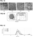
- the nanoconstruct contains a nanoparticle, such as a mesoporous silica nanoparticles (MSNP), a gold nanoparticle, a silver nanoparticle, an iron oxide nanoparticle, or a carbon nanotube, optionally loaded with a variety of additional agents including, but not limited to, cationic polymers, stabilizers, targeting agents, small molecules or proteins, labels, and/or oligonucleotides. Combinations of various additional agents are also contemplated.
- the nanoconstruct includes a cationic polymer, stabilizer, targeting agent, and small molecule, label, and/or oligonucleotide.
- Nanoconstructs may also include more than one type of cationic polymer, stabilizer, targeting agent, small molecule, protein, label, and/or oligonucleotide.
- nanoconstructs may include multiple, different oligonucleotides and/or small molecules or proteins that act on the same or different targets. The use of such additional agents may provide an additive or synergistic effect for disease treatment when delivered together on a nanoconstruct.
- Nanoparticles useful with the compositions and methods of the invention include, without limitation, mesoporous silica nanoparticles (e.g., MSNPs), iron oxide nanoparticles, silver nanoparticles, gold nanoparticles, and carbon nanotubes. Nanoparticles may or may not be porous.
- Exemplary sizes for the nanoparticle cores are from about 5 nm to about 200 nm, about 5 to about 20 nm, about 30 nm to about 100 nm, about 30 nm to about 80 nm, about 30 nm to about 60 nm, about 40 nm to about 80 nm, about 70 nm to about 90 nm, or about 5 nm, about 10 nm, about 20 nm, about 30 nm, about 40 nm, about 50 nm, about 60 nm, about 70 nm, about 80 nm, about 90 nm, or about 100 nm.
- the nanoparticle cores are spherical, although other shapes, such as rods and discs, may also be used.
- Additional components may be attached to nanoparticles by various mechanisms, covalently or noncovalently.
- cationic polymers may be attached to nanoparticles by charge, e.g., for silica or iron oxide nanoparticles.
- the surfaces of the nanoparticles may be altered to include reactive moieties for conjugation to cationic polymers and/or other components, or the cationic polymers or other components may include a moiety that binds to the nanoparticles.
- nanoparticles such as MSNPs
- cationic polymers or other compounds are coated with cationic polymers or other compounds.
- the cationic polymer may bind to the surface of the nanoparticle using any appropriate means.
- the cationic polymer binds to the nanoparticle via electrostatic interaction.
- the cationic polymer may be any polymer with a positive charge, such as, but not limited to, polyethylenimine (PEI) (see, e.g., Example III), polyamidoamine, poly(allylamine), poly(diallyldimethylammonium chloride), chitosan, poly(N-isopropyl acrylamide-co-acrylamide), poly(N-isopropyl acrylamide-co-acrylic acid), poly(L-lysine), diethylaminoethyl-dextran, poly-(N-ethyl-vinylpyridinium bromide), poly(dimethylamino)ethyl methacrylate), or polyethylene glycol)-co-poly(trimethylaminoethylmethacrylate chloride).
- PEI polyethylenimine
- cationic polymers will be apparent to those of skill in the art, and may be found, for example, in Polymer Handbook, 4th Edition, Edited by: Brandrup, E.H. Immergut, and E.A. Grukle; John Wiley & Sons, 2003).
- the cationic polymers may be linear or branched. In some embodiments, the cationic polymers may range in size from about 500 Da to 25kDa and may be branched or linear. For example, branched PEI with an average size of 1.8kDa to 10kDa may be loaded onto the nanoparticle core. The ratio of cationic polymer to nanoparticle may be varied depending on the desired result.
- the cationic polymer may be present from 1 to 50 wt. % of polymer per nanoconstruct, e.g., 5 to 40, 10 to 30%, 20 to 30%, 5 to 15%, 5 to 20%, 5 to 25%, 5 to 30%, 10 to 20%, 10 to 25%, or 25 to 40%, e.g., about 5, 10, 15, 20, 25, 30, or 35%.
- PEI per MSNP at a weight ratio of 1:4 during initial coating and at a weight ratio of 1:4 during cross-linking resulted in 14 wt.% PEI per nanoconstruct if 10-kDa PEI was used or 16 wt.% if 1.8-kDa PEI was used (see, e.g., Table 5).
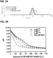
- Endolysosomal escape and gene silencing efficacy is increased by increasing buffering capacity of the nanoparticles.
- Cross-linking the cationic polymers yields greater buffering capacity as shown in Figure 2B .
- the cationic polymers may be cross-linked, e.g., with a cleavable disulfide bond, pre or post coating on the nanoparticle.
- the attached cationic polymers were cross-linked after binding to the nanoparticles, e.g., MSNP, using, for example, DSP (dithiobis[succinimidyl propionate]), DTSSP (3,3'-dithiobis(sulfosuccinimidyl propionate), and DTBP (dimethyl 3,3'-dithiobispropionimidate).
- DSP dithiobis[succinimidyl propionate]
- DTSSP dithiobis(sulfosuccinimidyl propionate)
- DTBP dimethyl 3,3'-dithiobispropionimidate
- a stabilizer may be conjugated to the nanoparticle and/or the cationic polymer, e.g., by any appropriate means.
- a stabilizer is conjugated to an amine or other reactive group of a cross-linked cationic polymer coated on the nanoparticle (e.g., a MSNP).
- exemplary stabilizers include, but are not limited to, polyethylene glycol (PEG), dextran, polysialic acid, hyaluronic acid (HA), polyvinyl pyrrolidone (PVP), polyvinyl alcohol (PVA), and polyacrylamide (PAM).
- Stabilizers may have multiple chemically reactive groups, e.g., for attachment to the nanoparticle, cationic polymer, and/or other component.
- reactive stabilizer e.g., PEG
- PEG derivatives may have two electrophilic moieties, such as maleimide-PEG-N-hydroxysuccinimidyl ester (Mal-PEG-NHS), which contains both a Michael acceptor and an activated ester.
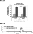
- the stabilizer, e.g., PEG used in conjunction with the compositions and methods of the invention generally has a molecular weight ranging between 500Da-40kDa, e.g., 2-10kDa as shown in Figure 3A .
- the stabilizer may be present from 1 to 50 wt. % of stabilizer per nanoconstruct, e.g., 5 to 30 wt. %, 10 to 20%, 10 to 25%, 5 to 15%, 5 to 20%, 5 to 25%, or 1 to 10%, e.g., about 5, 10, 15, 20, 25, 35, 40 or 45%.
- Example IV Mal-PEG(5-kDa)-NHS per MSNP is used at a weight ratio of 1:1 to 5:1 during the synthesis ( Figure 3B ), which results in about 6-23% of PEG per nanoconstruct (see Table 5).
- the nanoconstruct may be labeled, e.g., with a lanthanide or fluorescent dye, e.g., as shown in Example XXIV and Figure 37 .
- a label may be any substance capable of aiding a machine, detector, sensor, device, column, or enhanced or unenhanced human eye from differentiating a labeled composition from unlabeled compositions.
- labels include, but are not limited to, radioactive isotopes (e.g., PET tracers), dyes, stains, quantum dots, gold nanoparticles, enzymes, nonradioactive metals (e.g., MRI contrast agents), magnets, biotin, protein tags, any antibody epitope, or any combination thereof.
- Exemplary fluorescent dyes include, but are not limited to, FITC, RITC, Cy TM dyes, amine-reactive Dylight ® dyes, and amine-reactive Alexa Fluor ® dyes.
- lanthanides can be loaded onto hydroxyl, thiol, amine or phosphonate groups of nanoparticles, e.g., MSNPs, by covalent bonding or adsorption, e.g., as shown in Figure 37A .
- Lanthanides can facilitate sample detection with high sensitivity and resolution, e.g., by mass spectrometry ( Figure 37A ), while fluorescent dyes permit sample quantification by fluorescent imaging techniques, e.g., as shown in Figure 37B .
- Nanoconstructs containing lanthanides such as gadolinium can also serve as MRI contrast agents for imaging disease sites.
- the labels such as fluorescent dyes
- the labels may be loaded inside the pores of nanoparticles, e.g., amine-MSNPs via nucleophilic acyl substitution, e.g., between one or more nanoparticle-bound amines and an activated ester moiety (such as an NHS ester) appended to a fluorescent dye.
- an activated ester moiety such as an NHS ester
- Such labels produce nanoconstructs for fluorescence imaging applications as shown in Figure 37B ).
- Such a label may be added prior to or after loading of the cationic polymer and/or stabilizer.
- the label may be attached to the cationic polymer, stabilizer, or other component prior to or after their attachment to the nanoparticle by any appropriate means.
- Nanoconstructs may be delivered specifically or non-specifically. Nanoconstructs may be delivered to a site, e.g., for therapy, analysis, or diagnosis, such as a cell or tissue. The site may be in vivo or ex vivo. In some embodiments, the nanoconstructs may be delivered to tumors via the leaky vasculature of tumors.
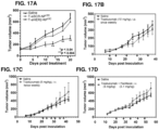
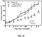
- the affinity of the cell or tissue, e.g., tumors, for cationic particles may permit nanoconstructs endowed with a positive charge due to the presence of cationic polymers to accumulate at the site, e.g., tumors and disease sites (see, e.g., Figure 19 showing efficacy of siHER2-nanoparticle without targeting agent in tumor xenografts in mice).
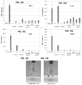
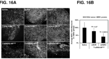
- the nanoconstructs may further include a targeting agent, e.g., for specific delivery of the nanoconstructs to sites such as tumors (see, e.g., Figures 16 , 17 , and 20 showing the efficacy of trastuzumab-conjugated siHER2-nanoparticle in tumor xenografts in mice).
- a targeting agent e.g., for specific delivery of the nanoconstructs to sites such as tumors (see, e.g., Figures 16 , 17 , and 20 showing the efficacy of trastuzumab-conjugated siHER2-nanoparticle in tumor xenografts in mice).
- Targeting agents may be used to target a site and optionally to aid or induce internalization into a cell.
- Exemplary targeting agents include, but are not limited to, monoclonal antibodies, single chain variable fragment (scFv) antibodies, other antigen binding fragments of antibodies, aptamers, small targeting molecules (e.g., ligands that bind to cell surface receptors such as N-acetylgalactosamine, mannose, transferrin, and folic acid), aptamers, carbohydrates, and peptides that have binding affinity to a cell or tissue, e.g., a tumor.
- the targeting agents may be attached to the nanoparticles, cationic polymer, or stabilizer by any appropriate means.
- the targeting agents are trastuzumab ( Figures 4 and 8 ), cetuximab ( Figure 8I ), or HER2 scFV ( Figures 21A-21E ), which may be attached to a stabilizer, e.g., PEG, that is already attached to the nanoparticles.
- the targeting agents such as folic acid and transferrin, are first attached to a stabilizer, e.g., PEG, prior to attachment on the nanoparticle ( Figures 21D-21E for folic acid (FA) conjugated nanoparticle).
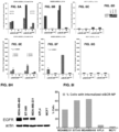
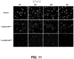
- the targeting agents may have a therapeutic effect (see, e.g., Figures 9 , 11 , and 28 ).
- exemplary monoclonal antibodies include, but are not limited to, anti-HER2 antibody, anti-EGFR antibody, anti-CD20 antibody, anti-VEGF-A antibody, anti-CD33 antibody, anti-CD52 antibody, and anti-TNF ⁇ antibody, such as alemtuzumab, bevacizumab, cetuximab, gemtuzumab, panitumumab, rituximab, infliximab, tositumomab, pertuzumab, and trastuzumab.
- the targeting agents may be attached to the nanoconstructs by any means, and suitable conjugation chemistries are known in the art and described herein.
- the targeting agent is thiolated and subsequently conjugated with Mal-PEG-PEI-MSNP via a thiol-maleimide reaction.
- the targeting agents are first attached to the PEG stabilizer (e.g., FA or transferrin on PEG-NHS, commercially available) prior to conjugation to the nanoparticle by reaction of an NHS ester and an amine.
- the targeting agent may be present from 0.1 to 10 wt.
- the MSNP per trastuzumab may be at the weight ratio of 50:1 to 1:1 (as shown in Figures 4A-4B ) during the synthesis, resulting in 0.5 to 6 wt.% of trastuzumab per nanoconstruct (Table 5 showing 3 wt.% antibody if 10:1 ratio is used).
- 1-4 wt.% of HER2 scFV per MSNP during the synthesis results in 0.3-0.5 wt.% of HER2 scFV per nanoconstruct.
- the nanoconstruct may be loaded with small molecules, proteins (i.e., other than targeting agents), or other therapeutic agents.
- Small molecules are molecules, typically with a molecular weight less than about 1000 Daltons, or in some embodiments, less than about 500 Daltons, wherein the molecule is capable of modulating, to some measurable extent, an activity of a target molecule.
- Exemplary small molecules such as peptides, small molecule inhibitors, chemotherapeutics, and other drugs may increase the therapeutic effects of the nanoconstruct, e.g., as shown in Figures 28-30 .
- the small molecules may be located on the exterior of the nanoparticles, e.g., on PEI, and/or within the pores of the nanoparticle core, e.g., MSNP.
- Small molecules such as small molecule inhibitors and other chemotherapeutic agents, may be selected based on the efficacy and specificity, e.g., in killing cancer cells over non-target cells.
- small proteins such as cytokines with molecular weights less than about 50 kDa can also be loaded on the nanoparticle in the same manner as small molecules.
- chemotherapeutic agents such as paclitaxel, docetaxel, and doxorubicin may be loaded on nanoconstructs by hydrogen bonding with nanoparticle, e.g., MSNP, surface moieties (e.g., hydroxyl or silanol) and/or cationic polymer, e.g., PEI.
- Hydrophobic drugs e.g., paclitaxel and docetaxel
- hydrophilic drugs e.g., doxorubicin
- PEG stabilizer coating
- the small molecule may be present from 0.01 to 50 wt. % of small molecule per nanoparticle, e.g., 0.1 to 30%, 1 to 30%, 1 to 20%, 1 to 10%, 1 to 5%, 0.1 to 1%, 0.5 to 5%, or 0.5 to 10%.
- small molecule per MSNP is used at a weight ratio of 1:10 to 1:1, which results in 0.1 to 30% by weight of drug per nanoconstruct.
- chemotherapeutic agents include, but are not limited to, methotrexate; plicamycin (mithramycin); mitotane; mercaptopolylysine; pyrimidine analogs such as fluorouracil; anthracyclic antibiotics such as doxorubicin; maytansinoids such as ansamitocin; D-arabinosyl nucleosides, such as arabinosyl adenine; alkylating agents such as PAM, I-PAM, altretamine, procarbazine, busulfan, dacarbazine, temozolomide, thiotepa and dacarbazine; purine antagonists such as mercaptopurine; actinomycins such as dactinomycin; mitomycins such as mitomycin C; anti-steroids such as aminoglutethimide; anti-microtubules such as estramustine and vinblastine; anti-androgens such as
- one or more oligonucleotides may be attached to the nanoconstruct including, but not limited to, siRNA, miRNA, miRNA mimics, or antisense oligomers.
- the oligonucleotides will be capable of altering, e.g., reducing, expression of a target protein, e.g., by RNAi or antisense effect.
- the oligonucleotide may act as a probe in a cell or tissue of interest.
- oligonucleotides include, but are not limited to, oligonucleotides that silence the expression of PLK1, AKT1/BCL2, HER2, EPS8L1, or HSP47 such as siPLK1, siAKT1/BCL2, siHER2, siEPS8L1, siHSP47, or miR-342-5P (see, e.g., Example XII and XX). Other targets are described herein.
- the oligonucleotide may be attached by any means.
- the negatively charged siRNA is attached to the positively charged cationic polymer on the nanoparticle, e.g., MSNP, using an electrostatic interaction.
- the oligonucleotides may target one or more genes expressed in a cancer cell, such as one or more genes encoding a protein that promotes cell growth, tumor vascularization, or escape from apoptosis.
- a single oligonucleotide may target a plurality of genes with varying potency.
- a plurality of oligonucleotides may target a single gene.
- a plurality of oligonucleotides may target a plurality of genes.
- Oligonucleotides may be present from about 1% to 10% by weight, e.g., about 2% to about 4% by weight.
- MSNP per siRNA NP/siRNA
- NP/siRNA MSNP per siRNA
- Optimal gene knock down efficacy is achieved at NP/siRNA of 25-50 as shown in Figures 5A-5B .
- the MSNP-PEI-PEG (with or without a targeting agent) may be loaded with one or more siRNA sequences directed towards the target genes such as, but not limited to, siHER2, siAKT (isoforms 1, 2, 3), siBCL2, siAKT1/BCL2 (an siRNA duplex in which one strand can knock down AKT1 and the other can knock down BCL2, Table 1), siPLK1, siGRB7, and siEPS8L1.
- the screenings of these siRNA are shown in Figures 24-26 .
- the nanoparticles may also be loaded with miRNA such as miR-342-5p under the same conditions with siRNA ( Figure 27A ).
- siRNA siRNA sequences used herein are siHER2, siAKT1/BCL2, siRNA control designated siSCR, and a siRNA against luciferase designated siLUC. Specific sequences are shown in Table 1.
- Table 1 Exemplary siRNA sequences siRNA siRNA sequence siHER2 Sense: 5' CACGUUUGAGUCCAUGCCCAAUU 3' (SEQ ID NO. 1) Antisense: 5' UUGGGCAUGGACUCAAACGUGUU 3' (SEQ ID NO. 2) siLUC Sense: 5' CGGAUUACCAGGGAUUUCAtt 3' (SEQ ID NO. 3) Antisense: 5' UGAAAUCCCUGGUAAUCCGtt 3' (SEQ ID NO.
- siSCR Sense 5' UGGUUUACAUGUCGACUAA 3' (SEQ ID NO. 5)
- Antisense 5' UUAGUCGACAUGUAAACCA 3' (SEQ ID NO. 6)
- Anti-Akt1 5' AUUCAGUUUCACAUUGCUUGGUGAC 3'(SEQ ID NO. 7)
- Anti-Bcl2 5' GUCACCAAGAACUGUGACACAGAAGGG 3'(SEQ ID NO. 8)
- the exemplary siHER2 was selected by measuring the efficacy and specificity with which it reduced HER2 mRNA levels and growth in HER2 + cell lines but not HER2 - cell lines.
- the dose of the siHER2 required to inhibit cell growth by 50% (GI50) was ⁇ 5nM in 19 out of 20 HER2+ cell lines (14 out of 20 cells did not respond to 30 ⁇ g/ml trastuzumab).
- the exemplary siHER2 selected was bound to MSNP-PEI-PEG-Trastuzumab (T-NP) to create a nanoconstruct.
- T-NP MSNP-PEI-PEG-Trastuzumab
- the construct increases siRNA protection against enzymatic degradation in blood ( Figures 6A-6E ), enhances tumor-specific cellular uptake ( Figures 8A-8I ), and achieves over 80% of HER2 knockdown efficacy ( Figure 9A ).
- the optimized nanoconstruct produced apoptotic death in HER2 positive (HER2+) breast cancer cells grown in vitro ( Figure 9C-D ), but not in HER2 negative (HER2-) breast cancer or non-breast cells ( Figure 10 ).
- one dose of the siHER2-nanoparticles reduced HER2 protein levels by 60% in trastuzumab-resistant HCC1954 xenografts in mice and multiple intravenous doses administered over 3 weeks significantly inhibited tumor growth (p ⁇ 0.004) ( Figures 17A-17D ).
- the material also had a therapeutic impact in another tumor model shown to be resistant to trastuzumab ( Figure 20 ).
- Components may be bound to nanoparticles or other components by any means including covalent and electrostatic binding.
- Various conjugation chemistries are known in the art and described herein.
- one or more of the components are bound to the surface of the nanoparticles.
- one or more of the components are bound within the pores of the nanoparticle (e.g., MSNP).
- one or more of the components are bound to each other.
- a targeting agent may be covalently bound to a stabilizer, which is covalently bound to the cationic polymer (e.g., via an amine), which is in turn electrostatically bound to the exterior of the nanoparticle, and a small molecule is bound to the interior surface of a pore.
- the pore has a first opening at a first location on the exterior surface of the nanoparticle (e.g., MSNP) and a second, different opening at a second location on the exterior surface of the nanoparticle. Additional components may be bound anywhere along the length of the inside of the pore.
- MSNPs are formed by combining a first surfactant with a second, different surfactant to form a first mixture, heating up the first mixture and adding a silica precursor to the first mixture to form a second mixture, holding the temperature for a period of time to generate MSNPs, and recovering the MSNPs by centrifugation.
- Surfactants can be removed by mixing the MSNP in acidic solvent under reflux conditions.
- the first mixture may be heated prior to adding the silica precursor.
- the first mixture may be at room temperature and the second mixture may be heated.
- the resulting MSNPs may have uniform or non-uniform particle size with high porosity, e.g., as shown in Figure 1A .
- cetyltrimethylammonium chloride may be combined with triethanolamine (TEA) in water, and heated to 95°C, while tetraethyl orthosilicate is added as shown in Example I.
- Variation of the amount of TEA while holding the amount of CTAC constant can be used to alter the size of the resulting MSNPs.
- the amount of TEA is between about 100 to about 600 ⁇ L, about 200 to about 450 ⁇ L, or about 200 to about 350 ⁇ L.
- Non-uniform MSNPs may be created using a strong base, such as NaOH.
- cetyltrimethyl ammonium bromide CTAB
- NaOH may be used as the base catalyst as shown in Example II.
- Iron oxide nanoparticles can be purchased (e.g., Feraheme) or synthesized, e.g., as described in Example XXV.
- Gold and silver nanoparticles can be synthesized following various published protocols or purchased from vendors such as Sigma Aldrich, Nanocs, nanoComposix.
- Carbon nanotubes can be synthesized following various published protocols or purchased from vendors such as Sigma Aldrich, US Research nanomaterial, and American Elements.
- functional groups such as, but not limited to, thiol, amine, carboxylate, or phosphonate may be added to the nanoparticles, e.g., MSNPs, during synthesis through the use of one or more reagents, e.g., organosilanes such as, but not limited to, (3-aminopropyl)triethoxysilane and (3-aminopropyl)trimethoxysilane). Organosilanes may be added before or after the surfactants are removed from the MSNPs.
- Analogous reagents and other organic reagents such as glutathione, mercaptopropionic acid, DMSA, PEG-thiol, oleic acid, and dextran may be employed to modify iron oxide nanoparticles, silver nanoparticles, gold nanoparticles, and carbon nanotubes.
- Functionalized nanoparticles can also be purchased directly, e.g., carbon nanotubes having surface modified with carboxylic acid, amide, polyaminobenzene sulfonic acid, octadecylamine, and PEG can be purchased from Sigma Aldrich.
- the resulting nanoparticles may be of any appropriate size, e.g., from about 20 nm to about 200 nm, about 30 nm to about 100 nm, about 40 nm to about 200 nm, about 50 nm to about 200 nm, about 30 nm to about 80 nm, 40 nm to about 80 nm, about 30 nm, about 40 nm, about 30 nm to about 60 nm, about 50 nm, or about 60 nm.
- Exemplary hydrodynamic sizes are shown in Figures 1C , 2A , 3A , and 21D for final nanoconstructs with MSNP cores and Figure 38A for final nanoconstructs with iron oxide cores.
- Nanoconstructs may be formulated, as is known in the art, for therapeutic, diagnostic, or research use. Typically, such formulations for therapy or diagnosis include the nanoconstructs suspended in a pharmaceutically acceptable carrier. Nanoconstructs may be employed for in vivo or ex vivo use. Effects of the agents contained in the nanoconstruct may occur intracellularly or extracellularly. In particular, nanoconstructs may be employed to deliver oligonucleotides to cells to modulate, e.g., reduce, expression of a gene. Nanoconstructs may also be employed to delivery labels or other agents intracellularly or within an extracellular site.
- the nanoconstructs may be used immediately upon formulation or may be stored.
- the nanoconstructs may be lyophilized into dry states using a lyoprotectant, such as a sugar like trehalose.
- a lyoprotectant such as a sugar like trehalose.
- Optimal trehalose and lyophilization conditions may preserve the nanoconstruct in terms of particle size and charge ( Figures 22A-B ) and efficacy in terms of gene knock down efficacy ( Figures 22C-D ) compared to freshly made material.
- Nanoconstructs of the invention are stable for at least 6 months when lyophilized ( Figures 23A-23E ).
- compositions of the invention comprising a disease treating effective amount of a nanoconstruct will be routinely adjusted on an individual basis, depending on such factors as weight, age, gender, and condition of the individual, the acuteness of the disease and/or related symptoms, whether the administration is prophylactic or therapeutic, and on the basis of other factors known to effect drug delivery, absorption, pharmacokinetics including half-life, and efficacy.
- Formulations of the invention will ordinarily be selected to approximate a minimal dosing regimen that is necessary and sufficient to substantially prevent or alleviate the symptoms of the disease including cancer, fibrosis and inflammation in the mammalian subject, including humans.
- Therapeutic dosage and administration protocol will often include repeated dosing over a course of several days or even one or more weeks or years.
- An effective treatment regimen may also involve prophylactic dosage administered on a day or multi-dose per day basis lasting over the course of days, weeks, months or even years.
- compositions of the present invention may further include a pharmaceutically acceptable carrier appropriate for the particular mode of administration being employed.
- Dosage forms of the compositions of the present invention include excipients recognized in the art of pharmaceutical compounding as being suitable for the preparation of dosage units as discussed above.
- excipients include, without intended limitation, binders, fillers, lubricants, emulsifiers, suspending agents, sweeteners, flavorings, preservatives, buffers, wetting agents, disintegrants, effervescent agents and other conventional excipients and additives and/or other additives that may enhance stability, delivery, absorption, half-life, efficacy, pharmacokinetics, and/or pharmacodynamics, reduce adverse side effects, or provide other advantages for pharmaceutical use.
- nanoconstructs of the invention are designed for parenteral administration, e.g. to be administered intravenously, intramuscularly, intratumorally, subcutaneously, or intraperitoneally, including aqueous and non-aqueous sterile injectable solutions which, like many other contemplated compositions of the invention, may optionally contain anti-oxidants, buffers, bacteriostats and/or solutes which render the formulation isotonic with the blood of the mammalian subject; and aqueous and non-aqueous sterile suspensions which may include suspending agents and/or thickening agents.
- the formulations may be presented in unit-dose or multi-dose containers. Additional compositions and formulations of the invention may include polymers for extended release following parenteral administration.
- parenteral preparations may be solutions, dispersions or emulsions suitable for such administration.
- the subject agents may also be formulated into polymers for extended release following parenteral administration.
- Pharmaceutically acceptable formulations and ingredients will typically be sterile or readily sterilizable, biologically inert, and easily administered. Such materials are well known to those of ordinary skill in the pharmaceutical compounding arts.
- Parenteral preparations typically contain buffering agents and preservatives, and injectable fluids that are pharmaceutically and physiologically acceptable such as water, physiological saline, balanced salt solutions, aqueous dextrose, glycerol or the like.
- Extemporaneous injection solutions, emulsions and suspensions may be prepared from sterile powders, granules and tablets of the kind previously described.
- Preferred unit dosage formulations are those containing a daily dose or unit, daily sub-dose, as described herein above, or an appropriate fraction thereof, of the active ingredient(s).
- the topical carrier used to deliver the nanoconstruct is an emulsion, gel or ointment.
- the therapeutic compounds as described herein may be formulated in a spray formulation.
- Emulsions such as creams and lotions are a dispersed system comprising at least two immiscible phases, one phase dispersed in the other as droplets ranging in diameter from 0.1 ⁇ m to 100 ⁇ m.
- An emulsifying agent is typically included to improve stability.
- water is the dispersed phase and an oil is the dispersion medium
- the emulsion is termed a water-in-oil emulsion.
- an oil is dispersed as droplets throughout the aqueous phase as droplets
- the emulsion is termed an oil-in-water emulsion.
- Emulsions, such as creams and lotions that can be used as topical carriers and their preparation are disclosed in Remington: The Science and Practice of Pharmacy (Loyd V. Allen 22nd ed. 2012 ), hereby incorporated herein by reference.
- Ointments may be homogeneous, viscous, semi-solid preparation, most commonly a greasy, thick oil (oil 80% - water 20%) with a high viscosity.
- the ointment can be used as an emollient or for the application of active ingredients to the skin for protective, therapeutic, or prophylactic purposes where a degree of occlusion is desired.
- a cream is an emulsion of oil and water in approximately equal proportions. It penetrates the stratum corneum outer layer of skin quite well. Cream is generally thinner than ointment, and maintains its shape when removed from its container.
- the vehicle of an ointment/cream is known as the ointment base.
- the choice of a base depends upon the clinical indication for the ointment.
- the different types of ointment bases include, but are not limited to: hydrocarbon bases, e.g. hard paraffin, soft paraffin, microcrystalline wax and ceresine; absorption bases, e.g. wool fat, beeswax; Water soluble bases, e.g., macrogols 200, 300, and 400; Emulsifying bases, e.g. emulsifying wax, Vegetable oils, e.g. olive oil, coconut oil, sesame oil, almond oil and peanut oil.
- the therapeutic compounds are dispersed in the base and later get divided after the drug penetrates into the wound.
- Ointments/creams can be formulated incorporating hydrophobic, hydrophilic, or water-emulsifying bases to provide preparations that are immiscible, miscible, or emulsifiable with skin secretions. They can also be derived from fatty hydrocarbon, absorption, water-removable, or water-soluble bases.
- a cream/ointment base can contain the active agent, white petrolatum, water, allantoin, EDTA, Stearyl alcohol, Brij 721, Brij 72, methylcelluloses, isopropyl myristate, Sorbitan monooleate, Polyoxyl 40 stearate, butylated hydroxytoluene, propylene glycol, methylparaben, propylparaben, deionized water to 100%, and buffer to neutral pH among other ingredients.
- the active agent white petrolatum, water, allantoin, EDTA, Stearyl alcohol, Brij 721, Brij 72, methylcelluloses, isopropyl myristate, Sorbitan monooleate, Polyoxyl 40 stearate, butylated hydroxytoluene, propylene glycol, methylparaben, propylparaben, deionized water to 100%, and buffer to neutral pH among other ingredients.
- the topical carrier used to deliver a compound of the invention is a gel, for example, a two-phase gel or a single-phase gel.
- Gels are semisolid systems consisting of suspensions of small inorganic particles or large organic molecules interpenetrated by a liquid. When the gel mass comprises a network of small discrete inorganic particles, it is classified as a two-phase gel.
- the liquid may be water or another aqueous media and the gel mass is defined as a hydrogel.
- Hydrogels can include, but are not limited to, alginates, polyacrylates, polyalkylene oxides, and/or poly N-vinyl pyrrolidone. The hydrogel may also be amorphous, i.e.
- a viscous gel as opposed to a solid such as a formulation of carboxymethylcellulose containing a humectant such as propylene glycol or glycerin.
- exemplary amorphous hydrogels include, but are not limited to, maltodextra-beta glucan, acemannan, carboxymethylcellulose, pectin, xanthan gum, collagen, keratin and honey.
- Nanoconstructs may be packaged into biodegradable capsules for oral administration.
- a nanoconstruct suspension may be installed inside the bladder. This is similar to intravesical chemotherapy, in which the drug administered to the bladder will come into direct contact with cancer cells in the bladder lining.
- the invention disclosed herein will also be understood to encompass diagnostic compositions for diagnosing the risk level, presence, severity, or treatment indicia of, or otherwise managing diseases including, but not limited to, neoplastic diseases by contacting a labeled (e.g., isotopically labeled, fluorescent labeled or otherwise labeled to permit detection of the labeled compound using conventional methods) nanoconstruct to a mammalian subject (e.g., to a cell, tissue, organ, or individual) at risk or presenting with one or more symptom(s) of a cell proliferation disease, such as a cancer, and thereafter detecting the presence, location, metabolism, and/or binding state (e.g., detecting binding to an unlabeled binding partner involved in malignant cell receptor physiology/metabolism) of the labeled compound using any of a broad array of known assays and labeling/detection methods.
- a labeled e.g., isotopically labeled, fluorescent labeled or otherwise labele
- a nanoconstruct is isotopically-labeled by having one or more atoms replaced by an atom having a different atomic mass or mass number.
- isotopes that can be incorporated into the disclosed compounds include isotopes of hydrogen, carbon, nitrogen, oxygen, phosphorous, fluorine and chlorine, such as 2 H, 3 H, 13 C, 14 C, 15 N, 18 O, 17 O, 31 P, 32 P, 35 S, 18 F, and 36 Cl, respectively.
- the isotopically-labeled compound is then administered to an individual or other subject and subsequently detected as described above, yielding useful diagnostic and/or therapeutic management data, according to conventional techniques.
- Nanoconstructs may include various therapeutic moieties, such as targeting agents, oligonucleotides, and/or small molecules, and may have intrinsic therapeutic properties.
- the nanoconstruct e.g., a nanoconstruct containing a MSNP
- the antioxidant nanoconstruct can deliver siRNA that can reduce pro-fibrotic genes such as HSP47 and NOX4 as shown in Figures 31A-31D , decreasing the adverse effects of pro-inflammatory cytokines such as TGF-beta. This has proven to be effective at decreasing fibrotic markers such as COL I and alpha-SMA in vitro and in mouse models of fibrotic disease.
- Suitable effective unit dosage amounts of the active compounds for administration to mammalian subjects, including humans, can be readily determined by one of skill in the art.
- the amount may range from about 0.01 to about 1 mg siRNA/kg, about 0.1 to about 0.75 mg siRNA/kg, about 0.1 to about 1 mg siRNA/kg per dose and any subset thereof.
- Nanoconstructs containing a lanthanide and/or a fluorescent dye may be used as fluorescent, mass spectrometry, PET, and/or MRI probes for both in vitro and in vivo applications.
- such nanoconstructs can be used to detect or quantify target proteins in tissue specimens or cells by immunofluorescence, flow cytometry, or mass cytometry (see, e.g., Example XXIV).
- the targeting agents on the nanoconstruct will bind specifically with the protein receptors on the cells (e.g., EGFR or HER2 on cancer cells).
- the nanoconstructs labeled with PET tracers, gadolinium chelates, or containing an iron oxide nanoparticle core can be used as PET or MRI contrast agents (see, e.g., Example XXVI) to detect diseases (e.g., cancer or inflammation). Additionally, nanoconstructs can be transfected into cells of interest (e.g., stem cells or immune cells) before such cells are infused to patients. This allows monitoring of these nanoconstruct-contained cells over time by non-invasive methods, such as fluorescence imaging, PET, and MRI.
- non-invasive methods such as fluorescence imaging, PET, and MRI.
- Nanoconstructs made from iron oxide, gold, and silver nanoparticles can be used in hyperthermia treatment. Alternating magnetic fields and irradiation have been shown to heat up such nanoparticles, increasing temperature of tumors (e.g., up to 40 °C) in which the nanoconstructs are accumulated. This can enhance cargo release and/or induce cancer cell death.
- the intrinsic properties of the nanoconstructs may have therapeutic benefits for cancer treatment.
- the antioxidant property of the nanoconstruct may modulate reactive oxygen species in cancer cells, resulting in decreased migration and invasion of cancer cells (see, e.g., Figures 34A-34H ). This effect was also confirmed in a mouse model of metastasis, as tumor spreading was observed to be attenuated in nanoconstruct-treated mice (see, e.g., Figures 35A-35H ).
- amine groups e.g., from PEI on nanoconstructs
- copper is believed to be a cofactor for angiogenesis and hence metastasis in cancer (Brewer et al., 2000).
- the PEI-nanoconstruct may provide a therapeutic benefit similar to that observed for tetrathiomolybdate (TM, a copper chelator) in its clinical trial on metastatic solid tumor patients (Brewer et al, 2000).
- TM tetrathiomolybdate
- five out of six TM-treated patients had mild copper deficiency (e.g. as assessed on the basis of a lower serum concentration of ceruloplasmin, a biomarker for total body copper status) and achieved stable disease.
- We also observed reduced serum ceruloplasmin in agreement with attenuated cancer metastasis and tumor burden with our nanoconstruct see, e.g., Example XXIII).
- the nanoconstructs are capable of delivering oligonucleotides to cells to alter gene expression (e.g., via RNA interference) and can thus also be used to treat other diseases involving aberrant gene expression.
- exemplary siRNAs that can be incorporated into nanoconstructs of the invention may target genes listed in Table 2, e.g., to treat the corresponding diseases shown therein.
- the nanoconstructs may be used in the treatment of cancer, e.g., breast cancer.
- the tumors may be resistant to monoclonal antibodies, small molecule inhibitors, and/or other chemotherapeutic agents, but still respond to siRNA delivered by nanoconstructs.
- the siRNA targets the same genes/proteins that monoclonal antibodies and small molecule inhibitors target, but gene/protein suppression with siRNA is superior to that of antibodies or small molecule inhibitors and overcomes cancer resistance to the antibodies and small molecule inhibitors (see, e.g., Example XIII).
- FIGS 28A-28B show the synergistic or additive effect of co-delivery of trastuzumab (HER2 antibody), chemotherapeutic paclitaxel (PTX), and siHER2 in terms of killing JIMT1, a multiple drug resistant HER2+ breast cancer cell.
- chemotherapeutic agents such as paclitaxel (PTX) or doxorubicin (DOX) does not negatively impact the ability of siRNA on the same nanoconstruct to silence the targeted genes.
- Figure 30 shows the effective inhibition of HCC1954 tumor growth in mice upon the treatment with nanoconstruct containing the three agents (T-siHER2-NP (PTX)).
- treat refers to reducing or ameliorating a disorder and/or symptoms associated therewith. It will be appreciated that, although not precluded, treating a disorder or condition does not require that the disorder, condition or symptoms associated therewith be completely eliminated.
- an effective amount is meant the amount of an agent required to ameliorate the symptoms of a disease or arrest the progression of a disease relative to an untreated subject or to label a target, e.g., protein, cell, or tissue, sufficiently for detection.
- a target e.g., protein, cell, or tissue
- the effective amount of an active therapeutic agent for the treatment of a disease or injury varies depending upon the manner of administration, the age, body weight, and general health of the subject.
- CBC complete blood count
- WBC white blood cell count
- RBC red blood cell count
- hematocrit the amount of hemoglobin.
- Some signs of cancer which are visible in a CBC include a low hematocrit, a sharp decrease in the number of blood platelets, and a low level of neutrophils.
- An effective amount of a composition of the present invention may increase the levels measured in a complete blood count by 10%, 20%, 30%, 50% or greater increase, up to a 75-90%, or 95% or greater in comparison to individuals who have not been treated by the compositions described herein.
- the compositions as described herein may be effective in increasing all or some parts of the complete blood count in comparison to those treated with placebo. Effective amounts may also move the blood protein of an individual towards the optimal category for each type of protein.
- Effectiveness of the compositions and methods herein in the treatment of cancer or other disease may be evaluated by screening for markers in the blood depending on the specific cancer.
- combinatorial disease treating formulations and coordinate administration methods employ an effective amount of a nanoconstruct and one or more secondary or adjunctive agent(s) that is/are combinatorially formulated or coordinately administered with the nanoconstruct to yield a combined, multi-active disease treating composition or coordinate treatment method.
- Exemplary combinatorial formulations and coordinated treatment methods in this context employ the nanoconstruct, in combination with one or more secondary anti-tumor agent(s), or with one or more adjunctive therapeutic agent(s) that is/are useful for treatment or prophylaxis of the targeted (or associated) disease, condition and/or symptom(s) in the selected combinatorial formulation or coordinate treatment regimen.
- a nanoconstruct is formulated, or coordinately administered, in combination with one or more adjunctive therapeutic agent(s), to yield a combined formulation or coordinate treatment method that is combinatorially effective or coordinately useful to treat neoplastic diseases and one or more symptom(s) of a secondary disease or condition in the subject.
- Exemplary combinatorial formulations and coordinate treatment methods in this context employ a nanoconstruct, in combination with one or more secondary or adjunctive therapeutic agents selected from, e.g., chemotherapeutic agents, anti-inflammatory agents, doxorubicin, vitamin D3, cytarabine, daunorubicin, cyclophosphamide, gemtuzumab ozogamicin, idarubicin, mercaptopurine, mitoxantrone, thioguanine, aldesleukin, asparaginase, carboplatin, etoposide phosphate, fludarabine, methotrexate, etoposide, dexamethasone, and choline magnesium trisalicylate.
- secondary therapies can include, but are not limited to, radiation treatment, hormone therapy and surgery.
- a nanoconstruct and another agent will each be present in a disease treating/preventing amount (i.e., in singular dosage which will alone elicit a detectable alleviation of symptoms in the subject).
- the combinatorial formulation may comprise one or both nanoconstruct and another agent (wherein the other agent is not a nanoconstruct) in sub-therapeutic singular dosage amount(s), wherein the combinatorial formulation comprising both agents features a combined dosage of both agents that is collectively effective in eliciting the desired response.
- MSNP dry size was measured with TEM (Phillips/FEI CM120/Biotwin TEM, Hillsboro, OR) and hydrodynamic size with Zetasizer (ZS-90/Malvern, Malvern, U.K.).
- Non-uniform MSNPs (O-87) were synthesized in the presence of a strong base. 6 mM cetyltrimethyl ammonium bromide (CTAB) was dissolved in 240 mL of aqueous solution of pH 11.0 (adjusted by 2 M NaOH). When the temperature stabilized at 80°C, 2.5 mL of TEOS was added, and the reaction continued for 2 hours. After mixing, the pellets were recovered from suspension by centrifugation, washed with a copious amount of ethanol, and dried overnight. The particles were then resuspended and refluxed in acidic methanol (0.6 M HCl in methanol) overnight to remove CTAB.
- CTAB cetyltrimethyl ammonium bromide
- MSNPs were then washed with ethanol and dried in a desiccator overnight.
- MSNP (dry) size was measured with TEM (Phillips/FEI CM120/Biotwin TEM, Hillsboro, OR) and hydrodynamic size with Zetasizer (ZS-90/Malvern, Malvern, U.K.). These non-uniform nanoparticle cores were 87 ⁇ 14nm in size (see, e.g., Figure 1A ).
- MSNPs prepared from Examples I and II were then coated layer-by-layer with cross-linked or non-cross-linked PEI, PEG, and targeting antibody.
- the nanoconstructs were then loaded with siRNA cargo as entailed in Examples III-VI.
- the characterization of compositions, size, and charge of the nanoconstructs are summarized in Example VII.
- Figure 1B provides a schematic illustration of such surface decoration
- Figure 1C shows the size of various nanoconstructs using different MSNP cores; all of which were surface modified with 10-kDa PEI (cross-linked for S-34, S-47, and S-61 MSNPs; non-cross-linked for O-87 MSNP), 5-kDa PEG, trastuzumab (HER2 antibody), and siRNA, respectively.
- Figure 1C indicates that S-47 core yielded the most desirable hydrodynamic size of the final nanoconstruct.
- PEI Polyethylenimine
- MSNPs prepared as described in Examples I and II were coated with PEI by shaking 10 mg MSNPs and 2.5 mg PEI in ethanol solution for 3 hours at room temperature. Next, the PEI-MSNP, was pelleted down. The material then underwent PEG attachment in Example IV or PEI cross-linking first prior to PEG attachment.
- the PEI-MSNP was re-suspended in ethanol solution containing 0 or 2.0 mg PEI (10 kDa) and 0.1-0.5 mg/ml DSP (DSP; dithiobis [succinimidyl propionate]; Lomant's Reagent) as a crosslinker. The solution was shaken for another 40 minutes.
- Mal-PEG-NHS was used to attach PEG on the nanoparticles (via an NHS ester).
- 50 mg of mal-PEG-NHS was conjugated to the primary amine of PEI-MSNP (10 mg as MSNP) from Example III (using either cross-linked or non-cross-linked PEI-MSNP) in PBS buffer under shaking (20 hr, RT).
- Mal-PEG-NHS can be first dissolved in DMF or added as a dry powder directly to the PEI-MSNP suspension in PBS to limit the hydrolysis of NHS and enhance the PEG loading efficacy and reduce the reaction time. This reduces the Mal-PEG-NHS by 5 fold to a 1:1 weight ratio of Mal-PEG-NHS per MSNP and reduces the reaction time from 20 hr to 1.5 hr.
- 10 mg of PEI-MSNP were re-suspended in 1 ml PBS.
- 10 mg of Mal-PEG-NHS (as a powder) were added into the PEI-MSNP solution.
- 5-kDa PEG has around three times higher loading on the nanoconstructs than the 7.5-kDa or 10-kDa counterparts.
- Low molar loading of higher-MW PEGs suggests the PEG conformation is not brush-like at higher-MW PEG chains.
- 5-kDa PEG provided the highest PEG loading, suggesting an optimal brush-like stealth condition for our nanoconstructs and was used throughout the studies.
- Antibody conjugation of MSNP-PEI-PEG utilized a thiol-maleimide reaction modified from the literature (P. Yousefpour et al., 2011, M. N. Koopaei et al., 2011).
- antibody e.g., trastuzumab (T), cetuximab (C), or rituximab (R)
- T trastuzumab
- C cetuximab
- R rituximab
- FIG. 4A shows the cell killing effect (method outlined in Example XII) upon delivering siHER2 (siRNA against HER2) with T-NP having different wt.% of trastuzumab.
- Figure 4B shows the degree of cellular uptake (method outlined in Example XI) of T-NP having different wt.% of trastuzumab in BT474 (HER2+) cell line.
- antibody: nanoparticle mass ratio of 1:20 and 1:10 can equally enhance cellular uptake of T-NP, but cell killing started to reduce if too little antibody was used.
- 1:10 ratio was selected and used throughout unless specified otherwise.
- Antibody fragment can be conjugated to the nanoparticle in a similar manner with antibody.
- Bacteria expressing a HER2 scFv clone (Poul et al., 2000) were grown overnight followed by expansion on the second day and stimulation with IPTG (thiogalactopyranoside) to produce scFv protein.
- IPTG thiogalactopyranoside
- the culture was subsequently pooled, and periplasmic bound scFv was extracted and purified on a Ni-NTA column according to manufacturer's protocol. Concentration of scFv was determined using a Nanodrop UV-Vis spectrometer, and purity was verified by gel electrophoresis.
- Figure 21A shows the successful purification of HER2-scFv as indicated by the specific 25kD band of scFv in the purified sample compared to the non-purified and wash eluent.
- Figure 21B shows additional verification of scFv identity by probing with an antibody that specifically recognizes the 6x histidine tag present at the c-terminal of the scFv molecule.
- Figure 21C shows HER2 scFV to be specific to HER2+ cells (BT474, SKBR3, JIMT1) over HER2- cells (MCF7 and MDAMB468).
- the scFv are attached to the end of the PEG chain on nanoconstructs by coupling cysteine of the scFv to the maleimide group on MSNP-PEI-PEG. Protein characterization indicated 0.4 wt.% of scFV per nanoparticle.
- Figure 21D shows the size of HER2 scFV conjugated nanoparticles loaded with 3 wt.% siRNA; the size was measured in PBS.
- Figure 21E shows the luciferase silencing efficacy (experiment outlined in Example VIII) of nanoparticles loaded with different amount of scFV in solution (1% or 4%, both of which resulted in 0.4 wt.% loading of scFv per nanoparticle).
- NHS ester-PEG-Folic acid was obtained commercially from Nanocs. NHS-PEG-FA was attached on the nanoparticles (via an NHS ester) in a similar manner as Example IV. 2.5 mg of NHS-PEG(5kD)-FA was conjugated to the primary amine of cross-linked PEI-MSNP (10 mg) from Example III in PBS buffer under shaking (1.5 hours, RT). NHS-PEG(5kD) (5-20 mg) was added to further stabilize (bind with) the remaining amine active surface for another hour.
- Figure 21D shows the hydrodynamic size of FA-siHER2-NP
- Figure 21E shows the luciferase silencing efficacy of the material.
- the loading of siRNA was achieved by mixing trastuzumab-conjugated MSNP-PEI-PEG (designated as T-NP) or MSNP-PEI-PEG (NP) and siRNA at nanoparticle/siRNA (NP/siRNA) mass ratio of 10 to 100 in PBS solution (0.5 to 1 hour, room temp, 200 rpm shaking), which resulted in complete binding (no siRNA left in the supernatant) as monitored by fluorescent method and gel electrophoresis (see Example VII).
- T-NP trastuzumab-conjugated MSNP-PEI-PEG
- NP/siRNA nanoparticle/siRNA
- the nanoconstructs had a hydrodynamic size of -100 nm for the three uniform-sized core materials (S-34, S-47, S-61) and 200 nm for the non-uniform-sized core material (O-87) in water as shown in Table 4 and the size distribution histograms are shown in Figure 1C . All materials are positively charged due to the PEI. All materials have fairly narrow size distribution (PDI about 0.2) with the exception of S-34. The PEI and PEG loadings were analyzed by thermogravimetric analysis (TGA Q50, TA Instruments, DE).
- Table 5 Composition of T-siRNA-NP (all reported as percent by mass of nanoparticle) Material Surface modification % PEI (by TGA) % PEG (by TGA) % Antibody (by BCA) NP/siRNA mass ratio (fluorescent method) S-47 T-NP 10C 13.5 18.2 3 Complete at NP/siRNA of 10 and above S-47 T-NP 1.8C 15.9 6.1 3
- NP/siRNA ratio The materials with NP/siRNA of 50 offered better gene silencing efficacy (per same dose of siLUC) ( Figure 5B ) than those with NP/siRNA of 25 ( Figure 5A ) due to the greater number of nanoparticles to which the cells were exposed. Materials with an NP/siRNA mass ratio of 50 were then used throughout unless specified otherwise.
- cross-linking indeed increased the silencing efficacy (e.g., T-NP 10C vs. T-NP 10 on S-61).
- the smaller core size (S-34) resulted in aggregation ( Figure 1C ) and less silencing efficacy ( Figure 5A ).
- the best hydrodynamic size ( Figure 1C ) and best silencing efficacy ( Figure 5 ) was achieved with S-47, modified with 10-kDa-PEI and with cross-linking (see T-NP 10C on S-47) and is used throughout and referred to as "T-NP" unless specified otherwise.
- T-NP when compared to the PEI-siRNA polyplex (without MSNP core), T-NP was superior in terms of size and luciferase silencing efficacy, as shown in Figure 7A and B , respectively.
- siRNAs Internalized nanoparticles ultimately end up in perinuclear lysosomal vesicles. siRNAs must escape from these vesicles to the cytosol to silence expression. Increasing the buffering capacity of small nanoparticles in order to increase siRNA endosomal release based on the proton sponge effect principle was achieved by PEI cross-linking.
- the optimal PEI cross-linking condition was 0.1 mg/ml of DSP with 2 mg (free) PEI added to the binding solution.
- the nanoparticles were suspended at 0.2 mg/mL in 150 mM NaCl (pH 9, pH adjusted with 0.05 M NaOH).
- pH 9.0 pH adjusted with 0.05 M NaOH.
- 5 ⁇ L of 0.05 M HCl was added and the solution was continuously stirred.
- the pH was recorded and the acid was added again. The process was repeated until the pH plateaued at around 3.0.
- the solution pH was then reported as a function of the amount of acid added.
- FIG. 2B shows the buffering capacities of nanoparticles with cross-linked 1.8-kDa PEI (T-NP 1.8C ), cross-linked 10-kDa PEI (T-NP 10C ), and non-cross-linked 10-kDa PEI (T-NP 10 ) in 150 mM NaCl.
- the nanoparticles had buffering capacity in the order of T-NP 10C > T-NP 10 > T-NP 1.8C .
- the cross-linking of the PEI on the nanoconstruct creates more secondary and tertiary amines yielding greater buffering capacity than primary amines, and promoting higher endosomal escape of siRNA based on proton sponge effect theory. This agrees well with the superior gene silencing efficacy of the cross-linked materials outlined in Example VIII.
- siRNA-nanoparticles were incubated with 50 v/v% human serum in PBS for a specified period of time (0, 0.5, 1, 2, 4, 8, 24, and 48 hours) at 37°C under continuous shaking. At the end of each time point, the sample was mixed with proteinase K (200 ⁇ g/mL) and frozen at -80°C to stop the enzymatic reaction. For the analysis, samples were thawed and mixed with 1.0 wt.% SDS in order to release siRNA from the nanoparticles. The sample was then mixed with an equal amount of 2X loading buffer and loaded into a 15% TBE-urea gel (BioRad). The gel ran at 100 V for the first 20 minutes and 150 V for another hour.
- TBE-urea gel BioRad
- the gel was then stained with SyBR Gold (Life Technologies) following the manufacturer's protocol and viewed in a UV chamber.
- the band intensity was analyzed by Imaged software (National Institutes of Health, Bethesda, MD).
- the fraction of intact siRNA was reported as a function of time that the siRNA-nanoparticles were in 50% serum.
- Figure 6A shows the amount of intact siHER2 that survived enzymatic degradation as measured by gel electrophoresis.
- the corresponding siHER2 quantification based on the band intensity and location is shown in Figure 6B .
- naked siHER2 was degraded within 0.5 hour (observed as bands shifted toward lower molecular weight), and its half-life was about 1 hour, in agreement with previous reports for other siRNAs (A. Mantei et al., 2008; D. M. Dykxhoorn et al., 2006).
- T-siHER2-NP 1.8C fully protected siHER2 at least 8 hours
- T-siHER2-NP 10C fully protected siHER2 at least 24 hours.
- T-siHER2-NP 10C The higher siRNA protection for T-siHER2-NP 10C compared to T-siHER2-NP 1.8C is likely due to the higher PEG content of T-siHER2-NP 10C , e.g., 18.2% vs. 6.1% (Table 5).
- PEG provides a steric blocking effect (Z. Zhang et al., 2007; R. Gref, 2000) that reduces enzymatic degradation of siRNA (S. Mao et al., 2006).
- siRNA on PEI-MSNP degraded faster than naked siRNA since positively charged PEI recruited more negatively charged enzymes to degrade siRNA.
- Figure 6D The steric effect of PEG also reduces binding of blood proteins to the nanoparticles (R. Gref, 2000).
- Cells were harvested and resuspended in 1 million cells/150 ⁇ L/tube. Each tube was mixed with 150 ⁇ L of siSCR (tagged with Alexa-488) nanoconstructs in PBS (containing 100 ⁇ g nanoparticle). Upon nanoconstruct addition, cells were placed on a rocker in the cell incubator (37°C, 5% CO 2 ) for 0.5 or 2.0 hours. Cells were then washed (centrifuge at 115g, 5 min) with 1 mL FACS buffer (1X Phosphate Buffered Saline (Ca/Mg++ free) + 1mM EDTA + 25mM HEPES pH 7.0 + 1% FBS (Heat-Inactivated) three times.
- FACS buffer (1X Phosphate Buffered Saline (Ca/Mg++ free) + 1mM EDTA + 25mM HEPES pH 7.0 + 1% FBS (Heat-Inactivated) three times.
- Cells were then resuspended in 550 ⁇ L of FACS buffer, transferred to a 5-mL tube, and kept on ice until analysis.
- antibody labeling was performed on ice and under rocking conditions.
- Cells were stained with primary antibody (trastuzumab or rituximab: 2 ⁇ g per tube) for an hour, washed with PBS, stained with secondary antibody (Anti-human Alexa 488: 2 ⁇ g per tube) for 45 minutes, then washed 2 times with PBS, and re-suspended in 550 ⁇ L of FACS buffer before analysis.
- All tubes were counter-stained for cellular DNA with 2 ⁇ L of 5 mM DRAQ5 (Cell Signaling) for 15 minutes on ice. All tubes (except antibody-labeled cells for gating purpose) were then incubated with 500 ⁇ L of Trypan Blue (0.4% in PBS) to quench fluorescence outside of the cells, and subjected to flow cytometry analysis. 10,000 events (cells) were analyzed for each sample. The intensity was processed with FlowJo software (NIH, Bethesda, MD).
- the nanoconstruct contained a scrambled siRNA and conjugated with trastuzumab (designated hereafter as T-siSCR-NP 1.8C and T-siSCR-NP 10C ) or with rituximab targeting CD20 (designated as R-siSCR-NP 1.8C and R-siSCR-NP 10C ).
- T-siSCR-NP 10C and T-siSCR-NP 1.8C in HER2 + breast cancer cells, BT474 and SKBR3, and the HER2- cell line MCF-7 was measured at 0.5 or 2.0 hours post exposure to the nanoconstructs.
- the siSCR was tagged with the fluorescent reporter, Alexa 488, for these experiments to enable quantitative analysis of siSCR uptake.
- R-siSCR-NP 10C and R-siSCR-NP 1.8C served as a negative control since BT474, SKBR3 and MCF-7 cells do not over-express CD20.
- the amount of Alexa 488-tagged siSCR inside individual cells was measured using flow cytometry.
- FIG. 8A-C shows that T-siSCR-NP 10C were taken up effectively (>90%) into HER2+ cells (BT474 and SKBR3), but not HER2- cells (MCF7) and that uptake increased by extending the exposure time from 0.5 hr to 2 hr. Furthermore, uptake of T-siSCR-NP 10C was greater than T-siSCR-NP 1.8C . R-siSCR-NP 10C and R-siSCR-NP 1.8C were not taken up efficiently by any of the cell lines, indicating the ability of T to promote cellular uptake to HER2+ cells.
- Figure 8D illustrates HER2 protein expression in the three cell lines being evaluated.
- Figure 8E-G show the average intensity of Alexa 488-tagged siSCR signal per cell and the same trend can be observed. This confirms that trastuzumab-conjugated nanoconstructs enter cells primarily by a HER2-receptor mediated endocytosis mechanism and not by adsorptive endocytosis of positively charged particles as reported for PEI-MSNP (H. Zhang, et al., 2011).
- HER2+ breast cancer cells (BT474, SKBR3 and HCC1954) were seeded in a 96-well plate for 24 hours prior to treatment.
- Nanoconstructs were loaded with siHER2 or siSCR at NP/siRNA 50.
- siRNA dose was fixed at 60 nM.
- Media was switched to complete media after overnight incubation.
- Three days after treatment with nanoconstructs, cells were fixed and analyzed for HER2 protein expression by immunofluorescence.
- HER2 mRNA and ⁇ -actin mRNA levels were analyzed at 48 hours post-treatment using the Quantigene 2.0 Reagent System (Panomics) following the manufacturer's protocol.
- the HER2 mRNA level was then normalized with ⁇ -actin mRNA (housekeeping gene) and reported as the percentage of the siSCR control.
- Cell viability and apoptosis were analyzed four days post-treatment using the CellTiter-Glo ® Luminescent Assay (Promega) and the Caspase-Glo ® 3/7 Assay Systems (Promega), respectively. Caspase activity was normalized with the cell viability. Both were reported as a percentage of the untreated control.
- T-siHER2-NP 10C The efficiency of T-siHER2-NP 10C in inhibiting HER2 mRNA levels and HER2 protein expression in the HER2+ breast cancer cell lines, BT474, SKBR3, and HCC1954 was assessed.
- T-siHER2-NP 10C reduced HER2 levels by 81-93% compared to T-siSCR-NP 10C .
- Figure 9B there was 44% reduction in HER2 mRNA relative to siSCR control.
- the cleaved Caspase 3 and 7 assay for apoptotic markers shows that apoptotic activity was three-fold greater after treatment with T-siHER2-NP 10C than with T-siSCR-NP 10C ( Figure 9C ). This is consistent with reduced cell viability (using a cellular ATP level assay) shown in Figure 9D .
- T-siSCR-NP 10C Treatment with T-siSCR-NP 10C reduced HER2 levels and killed HER2+ breast cancer cells, indicating that the antibody (T) on the nanoconstruct not only serves as a cell targeting agent but also has therapeutic effect.
- Figure 11 shows that HER2 levels in BT474 were reduced by 41% with T-siSCR-NP 10C (due to T effect) and by 87% with the T-siHER2-NP 10C (due to T and siHER2 effect) compared to untreated controls.
- Figure 9D shows that cell viability was reduced 59% by T-siSCR-NP 10C and 86% by T-siHER2-NP 10C .
- FIG. 10 shows that treatment with T-siHER2-NP 10C greatly reduced viability of HER2+ breast cancer cells (BT474, SKBR3, HCC1954 and JIMT-1), while having little impact on HER2- breast cells (MCF-7, MDA-MB-231, MDA-MB-468, MCF-10a), and HER2- non-breast cells (HepG2 and HEK-293).
- the cell killing effect was dependent on the HER2 protein levels of the cells (Inset of Figure 10 ).
- FIG 9A shows that T-siHER2-NP 10C was more effective than T-siHER2-NP 1.8C ( Figure 12A ) at equivalent siRNA dose.
- Figure 12B shows that doubling the dose of T-siHER2-NP 1.8C could reduce HER2 protein levels by 79-83% in SKBR3 and HCC1954.
- T-siHER2-NP 10C demonstrated better HER2 knock-down and cancer cell killing efficacy than T-siHER2-NP 1.8C .
- the T-siHER2-NP 10C ( Figure 9A ) outperformed commercially available DharmaFECT in all cell lines ( Figure 12C ).
- T-siHER2-NP 10C in intrinsically trastuzumab-resistant HER2+ cell lines (HCC1954 and JIMT1), in a parental HER2+ cell line (BT474) that responds to trastuzumab and lapatinib, and in BT474-R, a derivative cell line that was made lapatinib-resistant by long-term treatment with 1 ⁇ M lapatinib was also assessed.
- Figure 13A shows that the BT474-R cells were much less responsive to trastuzumab compared to parental BT474.
- Figure 13B shows that both trastuzumab-sensitive and resistant cell lines responded similarly with respect to siHER2 action (see T-siHER2-NP 10C vs.
- T-siSCR-NP 10C control This indicates that siHER2 on the nanoconstruct can overcome cancer resistance to trastuzumab.
- Figure 13C shows that BT474-R was less responsive to T-siSCR-NP 10C than BT474, indicating that the BT474-R cells were indeed resistant to trastuzumab (on the nanoconstructs).
- T-siHER2-NP 1.8C and T-siHER2-NP 10C were assessed for hemolysis, thrombogenesis, and platelet aggregation, and the results were benchmarked with FDA-approved nanoparticle products: Abraxane (Paclitaxel-albumin nanoparticles) and Feraheme (iron oxide nanoparticles used as a MRI contrast agent). Nanoparticles were tested at 1X and 5X of the intended human blood level. Studies of blood compatibility were performed following or with minor modification from the Nanotechnology Characterization Laboratory's (Frederick, MD) published protocols.
- PPP Platelet-poor plasma
- Platelet aggregation assay Platelet-rich plasma (PRP) was obtained following centrifugation of isolated blood (diluted in 3.2% sodium citrate, 1 :10). The isolated blood was centrifuged at 200g for 20 minutes. The supernatant (which contains PRP) was collected and maintained at room temperature prior to treatment. Following a 1-min incubation at 37°C (baseline), reactions were initiated by addition of nanoparticle (70 or 350 ⁇ g/mL) or collagen related peptide (CRP; 100 ⁇ g/ml) and monitored for three minutes for optical density via an aggregometer (Chrono-log Corp). Abraxane at 1X and 5X as entailed above was also used as a benchmark. Figure 14C shows that the nanoparticles and Abraxane did not trigger platelet aggregation while a collagen related peptide used as a positive control triggered aggregation immediately.
- PRP Platelet-rich plasma
- PBMC peripheral blood mononuclear cell
- a PBMC cytokine release assay was conducted according to the recommendations and method by the Nanotechnology Characterization Lab (NCL) of NCl for immunological studies of nanoparticles.
- the in vitro cell based assay evaluated cytokine production by PBMCs (200,000 cells/well) following a 24-hour exposure to the test materials.
- Test materials included nanoconstructs with and without siHER2 to investigate the potential impact of siRNA mediated immune response.
- cell culture supernatants were collected and analyzed for IL-1 ⁇ , IL-6, IFN- ⁇ , and TNF- ⁇ by a cytometry bead array (Milliplex Magnetic Bead) following the manufacturer's protocol.
- Abraxane and Feraheme were used as drug benchmarks since there is no siRNA based nanoconstruct drug in the market.
- T-siHER2-NP 1/8C and T-siHER2-NP 10C were evaluated upon treating peripheral blood mononuclear cells (PBMCs) isolated from human blood with these nanoconstructs.
- PBMCs peripheral blood mononuclear cells isolated from human blood with these nanoconstructs.
- PBMCs peripheral blood mononuclear cells isolated from human blood with these nanoconstructs.
- PBMCs peripheral blood mononuclear cells
- FIGS 15A-F show that neither T-siHER2-NP 1.8C nor T-siHER2-NP 10C increased the levels of IL-6 and TNF- ⁇ at either the 1X or 5X level, while Abraxane significantly increased both cytokines at the 5X level. Both nanoconstructs increased the levels of IFN- ⁇ and IL-1 ⁇ somewhat, but not to the extent observed for Abraxane for IL-1 ⁇ and Feraheme for IFN- ⁇ . The immune response was not significantly different for nanoconstructs with or without siRNA, suggesting that the response was not siRNA specific. Lastly, the PBMC immunological response to T-siHER2-NP 10C was not significantly different than that to T-siHER2-NP 1.8C .
- T-siHER2-NP 1.8C and T-siHER2-NP 10C were tested for lipopolysaccharides or LPS (endotoxin), produced by gram-negative bacterial contamination. About 35% of clinically relevant nanoparticles have been found to carry this contaminant (R. M. Crist et al., 2013). The two nanoconstructs were tested along with Abraxane as an FDA drug benchmark. All were negative for endotoxin ( Figures 15E and F ). This suggests an advantage of layer-by-layer modification, as sequential washing steps promote sterilization of the resulting product.
- LPS endotoxin
- FIG 17A shows that 5 intravenous tail vein injections of T-siHER2-NP 10C (1.25 mg siHER2/kg), over a period of three weeks significantly inhibited tumor growth, while T-siSCR-NP 10C produced little effect.
- HCC1954 has been established as resistant to cisplatin (I. Beyer et al., 2012), trastuzumab (I. Beyer et al., 2012), and pertuzumab (F. Henjes, et al., 2012) in vitro and/or in mice.
- HCC1954 resistance in vivo to trastuzumab and a trastuzumab/ paclitaxel combination was confirmed in our lab in Figures 17B-D .
- the nanoconstructs without trastuzumab also inhibit tumor growth in the HCC1954 tumor model as shown in Figure 19 .
- EPR enhanced permeability and retention
- BT474-TRgf (BT474 variant developed to be resistant to trastuzumab by serial passaging the cells in mice, Francia et al., 2012) was used to evaluate the efficacy of T-siHER2-NP. While the BT474-TRgf cells were resistant to trastuzumab in vitro ( Figure 20A ) and in a tumor mouse model ( Figure 20B ), they responded to T-siHER2-NP treatment in the tumor model ( Figure 20B ). siHER2 dose was 1.25-2.5 mg/siRNA and 5 animals per treatment group.
- Nanoconstruct suspensions (T-NP or NP) in 0.1 M Tris HCl (pH 7.4) were added to a solution of trehalose to achieve a final concentration of 10 mg/mL MSNP and 0-25 weight% of trehalose per nanoparticle.
- the well-mixed suspension was frozen slowly from room temperature to -55 °C in a freeze dryer. This was followed by primary and secondary drying. Primary drying (-40 °C, 100 ⁇ Bar, 24 hr) was to sublimate the ice crystals formed during the freezing step. Secondary drying (20 °C, 20 ⁇ Bar, 12 hr) was to eliminate bound water molecules on the nanoconstruct surface.
- the finished products were in the form of powder in sealed and storable bottles.
- T-NP lyophilized with 5% trehalose and stored at -20 °C to 37 °C were monitored for their properties over a 24-week storage time.
- Figures 23A-23E shows that when stored at -20 °C, T-NP maintained (A) hydrodynamic size, (B) zeta potential, (C) siRNA loading, (D) luciferase silencing efficacy, and (E) cancer cell killing efficacy similar to freshly made nanoparticles (Fresh) from the same batch. Data indicates that -20 °C is the best storage temperature for preserving all properties of the fresh material for at least 24 weeks (6 months). Similar outcomes were achieved with NP without T.
- siSCR 1 61.1 ⁇ 0.7 115.8 ⁇ 4.0 25.0 ⁇ 0.1 75.7 ⁇ 4.0% 2 58.1 ⁇ 0.6 117.4 ⁇ 0.5 24.9 ⁇ 0.1 80.5 ⁇ 2.8% 3 59.7 ⁇ 0.5 114.5 ⁇ 7.1 25.0 ⁇ 0.1 76.1 ⁇ 2.4% 4 57.7 ⁇ 0.9 123.8 ⁇ 3.3 25.0 ⁇ 0.1 76.6 ⁇ 3.9% 5 60.8 ⁇ 0.8 113.2 ⁇ 2.3 25.0 ⁇ 0.1 76.2 ⁇ 2.8% 6 58.8 ⁇ 0.3 115.6 ⁇ 1.3 25.0 ⁇ 0.1 77.0 ⁇ 2.2% Average 59.4 116.7 25.0 77.0 % Relative SD 2.4 3.2 0.2 2.3
- the initial screen sought to assess the efficacy of individual siRNAs in HER2 + breast cancer.
- SiRNA against AKT isoforms 1, 2, 3
- BCL2, PLK1, GRB7, and EPS8L1 all were benchmarked against the previously optimized siHER2 sequence.
- Four cell lines were chosen, representing trastuzumab-resistant (HCC1569, HCC1954, JIMT1) and sensitive (BT474) HER2+ breast cancer.
- siRNA sequences were also varied (Hs_AKT1_5 FlexiTube siRNA (SI002991450), Hs_AKT1_8 FlexiTube siRNA (SI00287742), Hs_AKT1_10 FlexiTube siRNA (SI02757244) and Hs_AKT1_11 FlexiTube siRNA (SI02758406) for AKT1 shown to be efficacious by Qiagen) (Qiagen, Valencia, CA, USA) as possible.
- Representative data for BT474 is shown in Figure 24A .
- FIGS. 24A-24B confirm the cell killing efficacy (A) and protein knockdown activity (B) of the dual-targeting siRNA.
- siRNA targeting PLK1 proved to be the most potent of all siRNAs in each respective cell line; the IC50 (dose inhibiting 50% growth) of siPLK1 is about 0.2 nM for 3 out of 4 cell lines studied.
- the cell panel was expanded to include triple negative breast cancer, ER+, and non-breast cells.
- siPLK1 is still the most potent among the three siRNAs evaluated. ( Figure 26 ).
- siPLK1 was toxic to non-target cells (e.g., non-targeted cells in Figure 26 ) when delivered with commercial DharmaFECT.
- Treatment specificity may be achieved for breast cancer cells (HCC1954) over normal breast epithelial cells (MCF-10A) when the siPLK1 is delivered with the nanoconstructs (T-NP), compared to DharmaFECT, as shown in Figures 27 A and B , respectively.
- Figure 27A also shows the ability of T-NP in delivering miRNA mimics (miR342-5p, obtained from GE Dharmacon (Lafayette, CO, USA) and eliciting killing of cancer cells but not MCF-10A.
- miRNA mimics miR342-5p, obtained from GE Dharmacon (Lafayette, CO, USA)
- paclitaxel combined with siAKT1/BCL2 or siEPS8L1 was also significant.
- the best enhancement by siAKT1/BCL2 appeared to be with basal cells, HCC1954, BT549, and HCC70.
- EPS8L1 is involved in EGFR signaling and functions in cell migration. As a result, efficacy of EPS8L1 was not restricted to HER2+ cells and proved effective in the two triple negative cell lines tested (both having high EGFR).
- Table 7 Screening of siRNA targeting HER2, PLK1, AKT1/BCL2 and EPS8L1 in combination with the chemotherapeutic drug paclitaxel.
- Figure 28A shows the ability of nanoparticles to load and deliver small peptides into cells.
- MSNP-PEI-PEG non-cross-linked PEI
- GALA GALA peptide (MW of 3 kDa) by electrostatic interaction in the same manner as siRNA.
- GALA was mixed with siRNA-nanoconstructs (13-130 nM GALA and 10 mg/L nanoconstructs) for 20 minutes in PBS (room temp, 300 rpm shaking).
- siRNA-nanoconstructs 13-130 nM GALA and 10 mg/L nanoconstructs
- MSNP reactive oxygen species
- FIG. 31A Another advantage of the MSNP based nanoconstruct is that the MSNP can reduce reactive oxygen species (ROS) in cells triggered by menadione as shown in Figure 31A .
- ROS reactive oxygen species
- the ROS reduction was attributed to the MSNP core and not the cationic PEI-PEG coating. This was confirmed by the finding (in a cell-free system) in Figure 31B that bare MSNP core displayed higher DPPH free radical scavenging ability than the MSNP coated with PEI-PEG layer.
- Figure 31B also shows that free PEI had no ROS scavenging ability.
- DPPH is a stable free radical that is typically used to assess the ROS scavenging ability of antioxidants.
- the MSNP-PEI-PEG could reduce the effects of pro-inflammatory cytokine TGF-beta by inhibiting NOX4, HSP47, COL I, and alpha-SMA as shown in Figure 31C .
- Figure 31D shows that the nanoconstruct could reduce the pro-fibrotic markers such as HSP47, COL I, and/or alpha-SMA, triggered by bleomycin.
- Bleo fibroblast denotes murine dermal fibroblasts harvested from bleomycin-induced scleroderma mouse model.
- T-siSCR-NP nanoconstruct also limited tumor spread in vivo as shown in Figure 35B as cancer cells were found primarily in the primary established site of this model (lung), compared to untreated mice where tumor signals can be found in other distant organs similar to metastasis sites of human breast cancer.
- T-siPLK1-NP resulted in tumor growth inhibition in lungs ( Figures 35B-D , F and H) and subsequent prolonged survival in mice beyond T-siSCR-NP treatment (G). PLK1 knock down in the tumors was also observed ( Figure 35E ).
- Amine groups e.g., from PEI on nanoconstructs, are known to be highly effective chelators of copper, which is a cofactor for angiogenesis and hence metastasis in cancer (Brewer et al., 2000).
- T-siPLK1-NP Figure 35 , short term study
- serum ceruloplasmin a biomarker for total body copper status
- MSNPs containing phosphonate groups were prepared by adding 3-trihydroxysilylpropyl methylphosphonate in situ during the MSNP synthesis described in Example I and Example II. Specifically, 500 mg of CTAB was dissolved in 240 ml of water (adjusted by 2 M NaOH). After temperature stabilized at 80 °C, 2.5 mL of TEOS was added in to solution under stirring condition. After 15 minutes, 635 ⁇ L of 3-(trihydroxylsilyl) propyl methyl phosphonate was added. The reaction continued for 2 hours, and the pellets were recovered from suspension by centrifugation, washed with a copious amount of ethanol, and dried overnight.
- the particles were then resuspended and refluxed in acidic methanol (0.6 M HCl in methanol) overnight to remove CTAB.
- acidic methanol 0.6 M HCl in methanol
- the resulting phosphonate-MSNPs were then washed with ethanol and dried in a desiccator overnight.
- MSNP For characterization, MSNP of known dry weight was mixed with 10 M HNOs for acid leaching of lanthanides. The leachate was then subjected to a lanthanide assay with ICP-MS. The amount of each lanthanide loaded on nanoparticle (as wt.%) is shown in Figure 37A.
- Figure 37B shows that the nanoconstruct can also be loaded with fluorescent dyes (on PEI layer) and/or an antibody (such as an anti-HER2 antibody), that recognizes HER2 proteins on the target cells.
- Nanoconstructs made from other inorganic cores e.g., iron oxide NPs
- iron oxide (Fe 3 O 4 ) nanoparticles can be used as an alternative to silica.
- Surface modification of such nanoparticles can be performed using similar methods to those described for MSNP.
- a particle called DMSA-ION can be prepared.
- ION were prepared by high temperature reaction of tris(acetylacetonato)iron(III) in the present of stabilizing surfactants - 1,2-hexadecanediol, lauric acid and lauryl amine - in benzyl ether.
- the adduct was purified with ethanol and hexane to obtain precursor ION-lauric acid.
- the precursor nanoparticles then underwent a ligand exchange reaction from hydrophobic lauric acid to hydrophilic meso-2,3-dimercaptosuccinic acid (DMSA) which renders the nanoparticles water-soluble.
- DMSA-ION were magnetically purified with water, ethanol and acetone. ION of ⁇ 10 nm by TEM has been achieved (Yantasee et al, 2007).
- DMSA has a negative charge which allows the ligand to electrostatically bind to positively-charged polyethylenimine (PEI).
- PEI modification allows the nanoparticles to bind with negative charged siRNA.
- DMSA is protonated and release the bound PEI which in turn releases the siRNA.
- Evaluation of ION based nanoconstruct for delivery of antibody, siRNA, chemotherapeutic, and/or dyes will be performed similar to MSNP nanoconstruct. Their advantages are small size for easy tumor accumulation, biocompatible, and MRI compatible.
- iron oxide nanoparticles have been surface modified in a similar manner as MSNP using aforementioned methods with minor modification.
- iron oxide nanoparticles commercially available and FDA approved Ferumoxytol or Feraheme
- PEI 10 kDa
- the mixture was subsequently shaken, centrifuged, and washed twice with PBS.
- Dry mal-PEG(5-kDa)-NHS was then added at a 1:1 ratio by mass of iron oxide to PEI in PBS.
- the resulting solution was shaken for two hours at room temperature, and was subsequently washed in PBS.
- Targeting agent (trastuzumab, T) was incorporated in the same manner as Example V.
- siRNA loading was achieved by mixing the resulting nanoconstructs with siRNA for 1 hour prior to characterization.
- the final hydrodynamic size of the nanoconstruct was approximately 50-100 nm in average as shown in Figure 38A for material prepared with 25 wt.% PEI (called F1) or 33 wt.% PEI (called F2).
- F1 25 wt.% PEI
- F2 33 wt.% PEI
- the nanoconstructs exhibited a polydispersity index of less than 0.15 and a potential of about 10-20 mV as measured in water, which increased from -50 mV of the ION (Feraheme).
- Iron oxide nanoconstructs for siRNA delivery and MRI contrast agent Iron oxide nanoconstructs for siRNA delivery and MRI contrast agent.
- Nanoconstructs containing iron oxide nanoparticles can be used to deliver siRNA and achieve effective gene silencing.
- Iron oxide nanoconstructs synthesized by the method of Example XXV were loaded with 50 nM siLUC at a nanoconstruct per siLUC mass ratio of 10 and used to treat LM2-4luc+/H2N cells expressing luciferase similar to Example VIII.
- Good luciferase knock down efficacy (40-50%) was achieved as shown in Figure 38B .
- Iron oxide based nanoconstructs can serve as an MRI contrast agent.
- T2 relaxivity was calculated by T2 MRI (small animal Bruker BioSpin 11.75T MRI instrument (31 cm horizontal bore), Paravision 5.1 software) as increasing Fe concentrations in PBS.
- T2 values were found using Imaged software and plotted as 1/T2 (1/s) vs Fe (mM) to yield relaxivity (R2 coefficient) as the slope ( Figure 38C).
- Figure 38C shows that the final nanoconstruct T-F2 (conjugated with trastuzumab and loaded with siHER2) has 2-3 fold enhanced R2 coefficient, compared to unmodified ION (Ferumoxytol).
- the iron oxide nanoconstruct has the potential to serve as a diagnostic tool (MRI probe).
- the materials are magnetic, they can potentially be used for magnetically guided delivery of therapeutics in vivo or in humans.
Landscapes
- Health & Medical Sciences (AREA)
- Chemical & Material Sciences (AREA)
- Engineering & Computer Science (AREA)
- Life Sciences & Earth Sciences (AREA)
- Nanotechnology (AREA)
- Public Health (AREA)
- Veterinary Medicine (AREA)
- Epidemiology (AREA)
- Animal Behavior & Ethology (AREA)
- General Health & Medical Sciences (AREA)
- Bioinformatics & Cheminformatics (AREA)
- Medicinal Chemistry (AREA)
- Pharmacology & Pharmacy (AREA)
- Immunology (AREA)
- Nuclear Medicine, Radiotherapy & Molecular Imaging (AREA)
- Molecular Biology (AREA)
- Radiology & Medical Imaging (AREA)
- Cell Biology (AREA)
- Gastroenterology & Hepatology (AREA)
- Proteomics, Peptides & Aminoacids (AREA)
- Biochemistry (AREA)
- Inorganic Chemistry (AREA)
- Ceramic Engineering (AREA)
- Oncology (AREA)
- Biomedical Technology (AREA)
- Pharmaceuticals Containing Other Organic And Inorganic Compounds (AREA)
- Medicinal Preparation (AREA)
Applications Claiming Priority (3)
| Application Number | Priority Date | Filing Date | Title |
|---|---|---|---|
| US201562133913P | 2015-03-16 | 2015-03-16 | |
| EP16765662.8A EP3270932B1 (fr) | 2015-03-16 | 2016-03-16 | Nanoparticules modifiées par un polymère réticulé |
| PCT/US2016/022655 WO2016149378A1 (fr) | 2015-03-16 | 2016-03-16 | Nanoparticules modifiées par un polymère réticulé |
Related Parent Applications (1)
| Application Number | Title | Priority Date | Filing Date |
|---|---|---|---|
| EP16765662.8A Division EP3270932B1 (fr) | 2015-03-16 | 2016-03-16 | Nanoparticules modifiées par un polymère réticulé |
Publications (2)
| Publication Number | Publication Date |
|---|---|
| EP4480902A2 true EP4480902A2 (fr) | 2024-12-25 |
| EP4480902A3 EP4480902A3 (fr) | 2025-03-12 |
Family
ID=56919864
Family Applications (2)
| Application Number | Title | Priority Date | Filing Date |
|---|---|---|---|
| EP24194999.9A Pending EP4480902A3 (fr) | 2015-03-16 | 2016-03-16 | Nanoparticules modifiées par un polymère réticulé |
| EP16765662.8A Active EP3270932B1 (fr) | 2015-03-16 | 2016-03-16 | Nanoparticules modifiées par un polymère réticulé |
Family Applications After (1)
| Application Number | Title | Priority Date | Filing Date |
|---|---|---|---|
| EP16765662.8A Active EP3270932B1 (fr) | 2015-03-16 | 2016-03-16 | Nanoparticules modifiées par un polymère réticulé |
Country Status (4)
| Country | Link |
|---|---|
| US (3) | US11207428B2 (fr) |
| EP (2) | EP4480902A3 (fr) |
| ES (1) | ES2993458T3 (fr) |
| WO (1) | WO2016149378A1 (fr) |
Families Citing this family (35)
| Publication number | Priority date | Publication date | Assignee | Title |
|---|---|---|---|---|
| CN104684398A (zh) | 2012-08-31 | 2015-06-03 | 索隆-基特林癌症研究协会 | 粒子、方法以及其用途 |
| EP2958481A4 (fr) | 2013-02-20 | 2017-03-08 | Sloan-Kettering Institute for Cancer Research | Appareil d'imagerie raman à large champ et procédés associés |
| EP4378461A3 (fr) | 2013-03-05 | 2024-09-11 | The Regents of University of California | Nanoparticules de silice mésoporeuse revêtues d'une bicouche lipidique présentant une capacité de charge élevée pour un ou plusieurs agents anticancéreux |
| US10912947B2 (en) | 2014-03-04 | 2021-02-09 | Memorial Sloan Kettering Cancer Center | Systems and methods for treatment of disease via application of mechanical force by controlled rotation of nanoparticles inside cells |
| US11786469B2 (en) | 2014-07-22 | 2023-10-17 | Cheolhee WON | Silica nanoparticle composition for delivering bioactive material or protein such as a human proteasome |
| WO2016018896A1 (fr) | 2014-07-28 | 2016-02-04 | Memorial Sloan Kettering Cancer Center | Nanoparticules de chalcogène métallique (métalloïde) en tant que liants universels d'isotopes médicaux |
| EP4480902A3 (fr) | 2015-03-16 | 2025-03-12 | PDX Pharmaceuticals, Inc. | Nanoparticules modifiées par un polymère réticulé |
| WO2017004301A1 (fr) | 2015-07-01 | 2017-01-05 | Memorial Sloan Kettering Cancer Center | Particules anisotropes, leurs procédés et leurs utilisations |
| EP4218739A3 (fr) | 2016-01-08 | 2023-08-09 | The Regents of The University of California | Nanoparticules de silice mésoporeuse dotées d'un revêtement bicouche lipidique pour l'administration de charge |
| US10519442B2 (en) | 2016-02-11 | 2019-12-31 | City Of Hope | Twist signaling inhibitor compositions and methods of using the same |
| KR102019407B1 (ko) | 2016-11-01 | 2019-09-11 | 기초과학연구원 | 타겟분자를 선택적으로 포획하는 덱스트란 고분자 기반의 증폭된 핵산 압타머 나노구조체의 제조방법 |
| CN106603086B (zh) * | 2016-11-07 | 2019-10-11 | 西北大学 | 一种基于簇态的日字形结构稳定子码的构造方法 |
| US10646540B2 (en) | 2016-11-18 | 2020-05-12 | City Of Hope | Peptide inhibitors of twist |
| US11135164B2 (en) * | 2017-01-13 | 2021-10-05 | Academia Sinica | Reloadable hydrogel system comprising an anti-PEG antibody for treating myocardial infarction |
| CA3052561C (fr) | 2017-02-06 | 2023-02-14 | Lemonex Inc. | Support de substance physiologiquement active |
| WO2018191316A1 (fr) | 2017-04-10 | 2018-10-18 | Cornell University | Nanoparticules contenant du soufre ou un atome lourd, procédés pour les fabriquer, et leurs utilisations |
| JP7309198B2 (ja) | 2017-05-19 | 2023-07-18 | コーネル ユニバーシティー | 機能性ナノ粒子及びそれを製造する方法並びに使用する方法 |
| CN107349432B (zh) * | 2017-07-11 | 2020-05-08 | 福州大学 | 一种负载索拉菲尼/siRNA的介孔二氧化硅-乳糖醛酸靶向纳米颗粒 |
| KR102152348B1 (ko) | 2017-09-05 | 2020-09-08 | 주식회사 레모넥스 | 세포 운명 조절용 조성물 |
| US12383499B2 (en) | 2018-01-01 | 2025-08-12 | The Regents Of The University Of California | Scale up synthesis of silicasome nanocarriers |
| WO2020051243A1 (fr) * | 2018-09-04 | 2020-03-12 | Ohio State Innovation Foundation | Nanoparticules lipidiques et leurs procédés d'utilisation |
| CN109044993A (zh) * | 2018-09-18 | 2018-12-21 | 华南理工大学 | 一种以核酸适配体靶向聚乙二醇修饰的介孔二氧化硅纳米粒及其制备方法 |
| WO2020205937A1 (fr) * | 2019-04-01 | 2020-10-08 | Emory University | Nanoparticules d'acide hyaluronique comprenant des inhibiteurs de nadph oxydases et leurs utilisations dans le traitement du cancer |
| US20220249389A1 (en) * | 2019-07-12 | 2022-08-11 | Oregon Health & Science University | Immunotherapeutic constructs and methods of their use |
| MX2022000541A (es) | 2019-07-12 | 2022-04-18 | Univ Oregon Health & Science | Construcciones terapeuticas para la administracion conjunta de un inhibidor de la quinasa mitotica y un inhibidor de puntos de control inmunitario. |
| EP3797783A1 (fr) | 2019-09-27 | 2021-03-31 | Freie Universität Berlin | Nouvelles utilisations médicales de dérivés de polyglycérol fonctionnalisés de thiol |
| CN111481679B (zh) * | 2020-04-21 | 2022-02-01 | 河南大学 | siRNA纳米胶囊及其制备方法和应用 |
| GB202005957D0 (en) * | 2020-04-23 | 2020-06-10 | N4 Pharma Uk Ltd | Transfection and transduction system |
| KR20230040345A (ko) * | 2020-07-13 | 2023-03-22 | 오레곤 헬스 앤드 사이언스 유니버시티 | 면역 반응을 유도하기 위한 면역원성 구축물, 조성물 및 방법 |
| US20240307552A1 (en) * | 2021-02-18 | 2024-09-19 | Kansas State University Research Foundation | Rna stabilizing nanoparticles |
| US20260070985A1 (en) * | 2021-11-05 | 2026-03-12 | Northwestern University | Enhanced immune checkpoint blockades formulation for image guided local immunotherapy |
| US20250020639A1 (en) * | 2023-06-18 | 2025-01-16 | Cytek Biosciences, Inc. | Ultra-bright nanoparticle fluorescent dye complexes |
| US20250059048A1 (en) * | 2023-08-16 | 2025-02-20 | Saudi Arabian Oil Company | Modified multi-walled carbon nanotubes including multi-walled carbon nanotubes and carboxylate moieties and related methods |
| WO2025188498A1 (fr) * | 2024-03-04 | 2025-09-12 | Oregon Health & Science University | Nanoparticules modifiées par un polymère pour une administration de cytokine ou d'une autre charge utile |
| CN118620979B (zh) * | 2024-08-13 | 2024-10-22 | 山东大树达孚特膳食品有限公司 | 一种罗汉参多肽的制备方法 |
Family Cites Families (15)
| Publication number | Priority date | Publication date | Assignee | Title |
|---|---|---|---|---|
| US20030059465A1 (en) * | 1998-05-11 | 2003-03-27 | Unger Evan C. | Stabilized nanoparticle formulations of camptotheca derivatives |
| US7375096B1 (en) * | 1998-12-04 | 2008-05-20 | California Institute Of Technology | Method of preparing a supramolecular complex containing a therapeutic agent and a multi-dimensional polymer network |
| US6586524B2 (en) * | 2001-07-19 | 2003-07-01 | Expression Genetics, Inc. | Cellular targeting poly(ethylene glycol)-grafted polymeric gene carrier |
| US6740336B2 (en) * | 2002-10-04 | 2004-05-25 | Mirus Corporation | Process for generating multilayered particles |
| US20080161547A1 (en) * | 2002-11-14 | 2008-07-03 | Dharmacon, Inc. | siRNA targeting serine/threonine protein kinase AKT |
| US8399025B2 (en) | 2004-06-04 | 2013-03-19 | Board Of Regents, The University Of Texas System | Polyamine modified particles |
| US20060293396A1 (en) * | 2005-01-14 | 2006-12-28 | Eastman Kodak Company | Amine polymer-modified nanoparticulate carriers |
| CA2636139A1 (fr) | 2005-12-14 | 2007-06-21 | Cytos Biotechnology Ag | Particules emballees avec des acides nucleiques immunostimulateurs pour le traitement de l'hypersensibilite |
| DK2509634T3 (en) * | 2009-12-11 | 2019-04-23 | Pfizer | Stable formulations for lyophilization of therapeutic particles |
| US20120207795A1 (en) * | 2010-07-13 | 2012-08-16 | The Regents Of The University Of California | Cationic polymer coated mesoporous silica nanoparticles and uses thereof |
| CA2820236C (fr) * | 2012-06-15 | 2022-07-19 | The Royal Institution For The Advancement Of Learning/Mcgill University | Systeme d'administration a base de nanoparticules non virales |
| US20160045612A1 (en) * | 2013-02-19 | 2016-02-18 | University Of Massachusetts | Functionalized nanoparticles for medical treatments |
| CA2929277C (fr) | 2013-11-01 | 2018-01-16 | Yale University | Vehicules d'administration comprenant il-2 et losartan |
| EP4480902A3 (fr) | 2015-03-16 | 2025-03-12 | PDX Pharmaceuticals, Inc. | Nanoparticules modifiées par un polymère réticulé |
| EP4218739A3 (fr) | 2016-01-08 | 2023-08-09 | The Regents of The University of California | Nanoparticules de silice mésoporeuse dotées d'un revêtement bicouche lipidique pour l'administration de charge |
-
2016
- 2016-03-16 EP EP24194999.9A patent/EP4480902A3/fr active Pending
- 2016-03-16 ES ES16765662T patent/ES2993458T3/es active Active
- 2016-03-16 EP EP16765662.8A patent/EP3270932B1/fr active Active
- 2016-03-16 WO PCT/US2016/022655 patent/WO2016149378A1/fr not_active Ceased
-
2017
- 2017-02-10 US US15/429,971 patent/US11207428B2/en active Active
-
2021
- 2021-08-10 US US17/398,954 patent/US11305024B2/en active Active
-
2022
- 2022-03-11 US US17/693,262 patent/US20220211878A1/en active Pending
Non-Patent Citations (70)
| Title |
|---|
| A. A. SEYHANU. VARADARAJANS. CHOEW. LIUT. E. RYAN, MOLECULAR BIOSYSTEMS, vol. 8, 2012, pages 1553 - 1570 |
| A. CHATURVEDIS. K. PIERCE, TRAFFIC, vol. 10, 2009, pages 621 - 628 |
| A. MANTEIS. RUTZM. JANKED. KIRCHHOFFU. JUNGV. PATZELU. VOGELT. RUDELI. ANDREOUM. WEBER, EUROPEAN JOURNAL OF IMMUNOLOGY, vol. 38, 2008, pages 2616 - 2625 |
| BH. KOBAYASHIR. WATANABEP. L. CHOYKE, THERANOSTICS, vol. 4, 2013, pages 81 - 89 |
| BL. ARAVINDANK. A. BICKNELLG. BROOKSV. V. KHUTORYANSKIYA. C. WILLIAMS, MACROMOLECULAR BIOSCIENCE, vol. 13, 2013, pages 1163 - 1173 |
| C. A. RITTERM. PEREZ-TORRESC. RINEHARTM. GUIXT. DUGGERJ. A. ENGELMANC. L. ARTEAGA, CLINICAL CANCER RESEARCH, vol. 13, 2007, pages 4909 - 4919 |
| C. ARGYOV. WEISSC. BRAUCHLET. BEIN, CHEMISTRY OF MATERIALS, vol. 26, 2013, pages 435 - 451 |
| C. G. MURPHYS. MODI, BIOLOGICS : TARGETS & THERAPY, vol. 3, 2009, pages 289 - 301 |
| C. L. WARNER, R. S. ADDLEMAN, A. D. CINSON, T. C. DROUBAY, M. H. ENGELHARD, M. A. NASH, W. YANTASEE, M. G. WARNER, CHEMSUSCHEM, vol. 3, 2010, pages 749 - 757 |
| C. M. RUDINJ. L. MARSHALLC. H. HUANGH. L. KINDLERC. ZHANGD. KUMARP. C. GOKHALEJ. STEINBERGS. WANASKIU. N. KASID, CLINICAL CANCER RESEARCH, vol. 10, 2004, pages 7244 - 7251 |
| C. TIMCHALKM. G. WARNER, ENVIRON. SCI. TECHNOL., vol. 41, 2007, pages 5114 - 5119 |
| C. V. PECOTG. A. CALINR. L. COLEMANG. LOPEZ-BERESTEINA. K. SOOD, NAT REV CANCER, vol. 11, 2011, pages 59 - 67 |
| CM. BREUNIGU. LUNGWITZR. LIEBLC. FONTANARIJ. KLARA. KURTZT. BLUNKA. GOEPFERICH, THE JOURNAL OF GENE MEDICINE, vol. 7, 2005, pages 1287 - 1298 |
| CT. COOKEJ. REEVESA. LANIGANP. STANTON, ANNALS OF ONCOLOGY : OFFICIAL JOURNAL OF THE EUROPEAN SOCIETY FOR MEDICAL ONCOLOGY / ESMO, vol. 12, 2001, pages 23 - 28 |
| D. HAUSSECKER, MOL THER NUCLEIC ACIDS, vol. 1, 2012, pages e8 |
| D. LINQ. CHENGQ. JIANGY. HUANGZ. YANGS. HANY. ZHAOS. GUOZ. LIANGA. DONG, NANOSCALE, vol. 5, 2013, pages 4291 - 4301 |
| D. M. DYKXHOORND. PALLISERJ. LIEBERMAN, GENE THERAPY, vol. 13, 2006, pages 541 - 552 |
| F. HENJESC. BENDERS. VON DER HEYDEL. BRAUNH. A. MANNSPERGERC. SCHMIDTS. WIEMANNM. HASMANNS. AULMANNT. BEISSBARTH, ONCOGENESIS, vol. 1, 2012, pages e16 |
| F. TANGL. LID. CHEN, ADVANCED MATERIALS, vol. 24, 2012, pages 1504 - 1534 |
| G. FRANCIAC. RODRIGUEZP. XUS. MANW. CRUZ-MUNOZG. BOCCIR. S. KERBEL, J CLIN ONCOL, 2012 |
| G. J. BREWERR. D. DICKD. K. GROVERV. LECLAIREM. TSENGM. WICHAK. PIENTAB. G. REDMANT. JAHANV. K. SONDAK, CLINICAL CANCER RESEARCH, vol. 6, 2000, pages 1 - 10 |
| H. MAEDAJ. WUT. SAWAY. MATSUMURAK. HORI, JOURNAL OF CONTROLLED RELEASE : OFFICIAL JOURNAL OF THE CONTROLLED RELEASE SOCIETY, vol. 65, 2000, pages 271 - 284 |
| H. MENGW. X. MAIH. ZHANGM. XUET. XIAS. LINX. WANGY. ZHAOZ. JIJ. I. ZINK, ACS NANO, vol. 7, 2013, pages 994 - 1005 |
| H. ZHANGT. XIAH. MENGM. XUES. GEORGEZ. JIX. WANGR. LIUM. WANGB. FRANCE, ACS NANO, vol. 5, 2011, pages 2756 - 2769 |
| I. BEYERH. CAOJ. PERSSONH. SONGM. RICHTERQ. FENGR. YUMULR. VAN RENSBURGZ. LIR. BERENSON, CLINICAL CANCER RESEARCH : AN OFFICIAL JOURNAL OF THE AMERICAN ASSOCIATION FOR CANCER RESEARCH, vol. 18, 2012, pages 3340 - 3351 |
| I. BEYERZ. LIJ. PERSSONY. LIUR. VAN RENSBURGR. YUMULX. B. ZHANGM. C. HUNGA. LIEBER, MOLECULAR THERAPY : THE JOURNAL OF THE AMERICAN SOCIETY OF GENE THERAPY, vol. 19, 2011, pages 479 - 489 |
| I. SLOWINGB. G. TREWYNS. GIRIV. S. Y. LIN, ADVANCED FUNCTIONAL MATERIALS, vol. 17, 2007, pages 1225 - 1236 |
| J. D. HEIDELZ. YUJ. Y.-C. LIUS. M. RELEY. LIANGR. K. ZEIDAND. J. KORNBRUSTM. E. DAVIS, PROCEEDINGS OF THE NATIONAL ACADEMY OF SCIENCES, vol. 104, 2007, pages 15549 - 15554 |
| J. E. ZUCKERMANC. H. J. CHOIH. HANM. E. DAVIS, PROCEEDINGS OF THE NATIONAL ACADEMY OF SCIENCES, vol. 109, 2012, pages 3137 - 3142 |
| J. LUM. LIONGS. SHERMANT. XIAM. KOVOCHICHA. E. NELJ. I. ZINKF. TAMANOI, NANOBIOTECHNOLOGY : THE JOURNAL AT THE INTERSECTION OF NANOTECHNOLOGY, MOLECULAR BIOLOGY, AND BIOMEDICAL SCIENCES, vol. 3, 2007, pages 89 - 95 |
| J. M. DU MANOIRG. FRANCIAS. MANM. MOSSOBAJ. A. MEDINA. VILORIA-PETITD. J. HICKLINU. EMMENEGGERR. S. KERBEL, CLINICAL CANCER RESEARCH, vol. 12, 2006, pages 904 - 916 |
| J. MORRYW. NGAMCHERDTRAKULS. GUS. M. GOODYEARD. J. CASTROM. M. REDAT. SANGVANICHW. YANTASEE, BIOMATERIALS, vol. 66, 2015, pages 41 - 52 |
| J. SHENH. C. KIMH. SUF. WANGJ. WOLFRAMD. KIRUIJ. MAIC. MUL. N. JIZ. W. MAO, THERANOSTICS, vol. 4, 2014, pages 487 - 497 |
| J. TABERNEROG. I. SHAPIROP. M. LORUSSOA. CERVANTESG. K. SCHWARTZG. J. WEISSL. PAZ-ARESD. C. CHOJ. R. INFANTEM. ALSINA, CANCER DISCOVERY, vol. 3, 2013, pages 406 - 417 |
| K. A. GRAHAMM. KULAWIECK. M. OWENSX. LIM. M. DESOUKID. CHANDRAK. K. SINGH, CANCER BIOLOGY & THERAPY, vol. 10, 2010, pages 223 - 231 |
| K. A. WHITEHEADR. LANGERD. G. ANDERSON: "Drug discovery", NATURE REVIEWS, vol. 8, 2009, pages 129 - 138 |
| K. KUNATHA. VON HARPED. FISCHERH. PETERSENU. BICKELK. VOIGTT. KISSEL, JOURNAL OF CONTROLLED RELEASE : OFFICIAL JOURNAL OF THE CONTROLLED RELEASE SOCIETY, vol. 89, 2003, pages 113 - 125 |
| K. TAKEDAS. AKIRA, INTERNATIONAL IMMUNOLOGY, vol. 17, 2005, pages 1 - 14 |
| L. PANQ. HEJ. LIUY. CHENM. MAL. ZHANGJ. SHI, JOURNAL OF THE AMERICAN CHEMICAL SOCIETY, vol. 134, 2012, pages 5722 - 5725 |
| M. A. POULB. BECERRILU. B. NIELSONP. MORRISSONJ. D. MARKS, JOURNAL OF MOLECULAR BIOLOGY, vol. 301, 2000, pages 1149 - 1161 |
| M. BAROKH. JOENSUUJ. ISOLA, BREAST CANCER RESEARCH, vol. 16, 2014, pages 1 - 12 |
| M. E. DAVIS, MOLECULAR PHARMACEUTICS, vol. 6, 2009, pages 659 - 668 |
| M. E. DAVISJ. E. ZUCKERMANC. H. J. CHOID. SELIGSONA. TOLCHERC. A. ALABIY. YENJ. D. HEIDELA. RIBAS, NATURE, vol. 464, 2010, pages 1067 - 1070 |
| M. H. SCHWENK, PHARMACOTHERAPY, vol. 30, 2010, pages 70 - 79 |
| M. J. ROBERTSM. D. BENTLEYJ. M. HARRIS, ADVANCED DRUG DELIVERY REVIEWS, vol. 54, 2002, pages 459 - 476 |
| M. N. KOOPAEIR. DINARVANDM. AMINIH. RABBANIS. EMAMIS. N. OSTADF. ATYABI, INTERNATIONAL JOURNAL OF NANOMEDICINE, vol. 6, 2011, pages 1903 - 1912 |
| M. ZAMANIAN-DARYOUSHJ. T. MARQUESM. P. GANTIERM. A. BEHLKEM. JOHNP. RAYMANJ. FINKEB. R. WILLIAMS, JOURNAL OF INTERFERON & CYTOKINE RESEARCH : THE OFFICIAL JOURNAL OF THE INTERNATIONAL SOCIETY FOR INTERFERON AND CYTOKINE RESEARCH, vol. 28, 2008, pages 221 - 233 |
| O. BOUSSIFF. LEZOUALC'HM. A. ZANTAM. D. MERGNYD. SCHERMANB. DEMENEIXJ. P. BEHR, PROCEEDINGS OF THE NATIONAL ACADEMY OF SCIENCES, vol. 92, 1995, pages 7297 - 7301 |
| R. A. PETROSJ. M. DESIMONE: "Drug discovery", NATURE REVIEWS, vol. 9, 2010, pages 615 - 627 |
| R. BROERINGC. I. REALM. J. JOHNK. JAHN-HOFMANNL. M. ICKENSTEINK. KLEINEHRA. PAULK. GIBBERTU. DITTMERG. GERKEN, INTERNATIONAL IMMUNOLOGY, vol. 26, 2014, pages 35 - 46 |
| R. GREFM. LUCKP. QUELLECM. MARCHANDE. DELLACHERIES. HARNISCHT. BLUNKR. H. MULLER: "Colloids and surfaces", B, BIOINTERFACES, vol. 18, 2000, pages 301 - 313 |
| R. K. RAMANATHANS. I. HAMBURGM. J. BORADM. SEETHARAMM. N. KUNDRANDAP. LEEP. FREDLUNDM. GILBERTC. MASTS. C. SEMPLE, AACR 104TH ANNUAL MEETING, 2013 |
| R. KANASTYJ. R. DORKINA. VEGASD. ANDERSON, NAT MATER, vol. 12, 2013, pages 967 - 977 |
| R. M. CRISTJ. H. GROSSMANA. K. PATRIS. T. STERNM. A. DOBROVOLSKAIAP. P. ADISESHAIAHJ. D. CLOGSTONS. E. MCNEIL, INTEGRATIVE BIOLOGY : QUANTITATIVE BIOSCIENCES FROM NANO TO MACRO, vol. 5, 2013, pages 66 - 73 |
| R. NAHTA, ISRN ONCOLOGY, vol. 2012, 2012, pages 428062 |
| R. NAHTAF. J. ESTEVA, BREAST CANCER RESEARCH : BCR, vol. 8, 2006, pages 215 |
| S. AHMADS. GUPTAR. KUMARG. C. VARSHNEYG. P. S. RAGHAVA, SCI. REP., 2014, pages 4 |
| S. GUOL. HUANG, JOURNAL OF NANOMATERIALS, vol. 2011, 2011, pages 12 |
| S. GUZ. HUW. NGAMCHERDTRAKULD. J. CASTROJ. MORRYM. M. REDAJ. W. GRAYW. YANTASEE, ONCOTARGET, 2016 |
| S. MAOM. NEUO. GERMERSHAUSO. MERKELJ. SITTERBERGU. BAKOWSKYT. KISSEL, BIOCONJUGATE CHEMISTRY, vol. 17, 2006, pages 1209 - 1218 |
| T. BIEBERW. MEISSNERS. KOSTINA. NIEMANNH. P. ELSASSER, JOURNAL OF CONTROLLED RELEASE : OFFICIAL JOURNAL OF THE CONTROLLED RELEASE SOCIETY, vol. 82, 2002, pages 441 - 454 |
| T. H. CHUNGS. H. WUM. YAOC. W. LUY. S. LINY. HUNGC. Y. MOUY. C. CHEND. M. HUANG, BIOMATERIALS, vol. 28, 2007, pages 2959 - 2966 |
| T. M. ALLENP. R. CULLIS, SCIENCE, vol. 303, 2004, pages 1818 - 1822 |
| T.-M. KETOLAM. HANZLÍKOVÁL. LEPPÄNENM. RAVINAC. J. BISHOPJ. J. GREENA. URTTIH. LEMMETYINENM. YLIPERTTULAE. VUORIMAA-LAUKKANEN, THE JOURNAL OF PHYSICAL CHEMISTRY B, vol. 117, 2013, pages 10405 - 10413 |
| W. LIF. NICOLF. C. SZOKA JR., ADVANCED DRUG DELIVERY REVIEW, vol. 56, 2004, pages 967 - 985 |
| W. NGAMCHERDTRAKULJ. MORRYS. GUD. J. CASTROS. M. GOODYEART. SANGVANICHM. M. REDAR. LEES. A. MIHELICB. L. BECKMAN, ADVANCED FUNCTIONAL MATERIALS, vol. 25, 2015, pages 2646 - 2659 |
| W. YANTASEEC. L. WARNERT. SANGVANICHR. S. ADDLEMANT. G. CARTERR. J. WIACEKG. E. FRYXELLC. TIMCHALKM. G. WARNER, ENVIRONMENTAL SCIENCE & TECHNOLOGY, vol. 41, 2007, pages 5114 - 5119 |
| X. LIY. CHENM. WANGY.W. XIAH. GU, BIOMATERIALS, vol. 34, 2013, pages 1391 - 1401 |
| Y.-C. WANGG. MORRISONR. GILLIHANJ. GUOR. WARDX. FUM. BOTERON. HEALYS. HILSENBECKG. PHILLIPS, BREAST CANCER RESEARCH, vol. 13, 2011, pages R121 |
| Z. ZHANGA. E. BERNSS. WILLBOLDJ. BUITENHUIS, JOURNAL OF COLLOID AND INTERFACE SCIENCE, vol. 310, 2007, pages 446 - 455 |
Also Published As
| Publication number | Publication date |
|---|---|
| US20210393806A1 (en) | 2021-12-23 |
| US11305024B2 (en) | 2022-04-19 |
| EP3270932C0 (fr) | 2024-08-21 |
| US11207428B2 (en) | 2021-12-28 |
| EP3270932B1 (fr) | 2024-08-21 |
| EP4480902A3 (fr) | 2025-03-12 |
| US20170173169A1 (en) | 2017-06-22 |
| WO2016149378A1 (fr) | 2016-09-22 |
| US20220211878A1 (en) | 2022-07-07 |
| EP3270932A1 (fr) | 2018-01-24 |
| ES2993458T3 (en) | 2024-12-30 |
| EP3270932A4 (fr) | 2018-11-07 |
Similar Documents
| Publication | Publication Date | Title |
|---|---|---|
| US11305024B2 (en) | Cross-linked polymer modified nanoparticles | |
| Suk et al. | Lung gene therapy with highly compacted DNA nanoparticles that overcome the mucus barrier | |
| Son et al. | Self-crosslinked human serum albumin nanocarriers for systemic delivery of polymerized siRNA to tumors | |
| Sun et al. | Tumor-targeted and nitric oxide-generated nanogels of keratin and hyaluronan for enhanced cancer therapy | |
| Xu et al. | Gene delivery through the use of a hyaluronate-associated intracellularly degradable crosslinked polyethyleneimine | |
| Fan et al. | Multifunctional all-in-one drug delivery systems for tumor targeting and sequential release of three different anti-tumor drugs | |
| Karpisheh et al. | RETRACTED: Inhibition of HIF-1α/EP4 axis by hyaluronate-trimethyl chitosan-SPION nanoparticles markedly suppresses the growth and development of cancer cells | |
| WO2012009448A2 (fr) | Nanoparticules de silice mésoporeuses revêtues d'un polymère cationique et leurs utilisations | |
| Elechalawar et al. | Dual targeting of folate receptor-expressing glioma tumor-associated macrophages and epithelial cells in the brain using a carbon nanosphere–cationic folate nanoconjugate | |
| Kim et al. | Anti-apoptotic cardioprotective effects of SHP-1 gene silencing against ischemia–reperfusion injury: Use of deoxycholic acid-modified low molecular weight polyethyleneimine as a cardiac siRNA-carrier | |
| Xia et al. | Targeted delivery of siRNA using RGDfC-conjugated functionalized selenium nanoparticles for anticancer therapy | |
| Chen et al. | Co-delivery of doxorubicin and shRNA of Beclin1 by folate receptor targeted pullulan-based multifunctional nanomicelles for combinational cancer therapy | |
| WO2014201276A1 (fr) | Support en silicium nanoporeux fonctionnalisé par des polycations pour administration systémique d'agents de silençage génique | |
| Wei et al. | OFF–ON nanodiamond drug platform for targeted cancer imaging and therapy | |
| Kim et al. | Surgical suture releasing macrophage-targeted drug-loaded nanoparticles for an enhanced anti-inflammatory effect | |
| WO2012024396A2 (fr) | Compositions et méthodes permettant d'administrer des molécules d'acide nucléique et de traiter un cancer | |
| Lin et al. | Hybrid bicelles as a pH-sensitive nanocarrier for hydrophobic drug delivery | |
| Beloor et al. | Arginine-engrafted biodegradable polymer for the systemic delivery of therapeutic siRNA | |
| Xu et al. | Preparation and characterization of a dual-receptor mesoporous silica nanoparticle–hyaluronic acid–RGD peptide targeting drug delivery system | |
| CN105087596B (zh) | 一种cd20核酸适配体及其应用 | |
| Lee et al. | RPM peptide conjugated bioreducible polyethylenimine targeting invasive colon cancer | |
| Qu et al. | Targeted delivery of doxorubicin via CD147-mediated ROS/pH dual-sensitive nanomicelles for the efficient therapy of hepatocellular carcinoma | |
| Zhang et al. | Dual pH-responsive and tumor-targeted nanoparticle-mediated anti-angiogenesis siRNA delivery for tumor treatment | |
| CN119486767A (zh) | 作为靶向递送平台的线粒体 | |
| Yang et al. | Chitosan hybrid nanoparticles as a theranostic platform for targeted doxorubicin/VEGF shRNA co-delivery and dual-modality fluorescence imaging |
Legal Events
| Date | Code | Title | Description |
|---|---|---|---|
| PUAI | Public reference made under article 153(3) epc to a published international application that has entered the european phase |
Free format text: ORIGINAL CODE: 0009012 |
|
| STAA | Information on the status of an ep patent application or granted ep patent |
Free format text: STATUS: THE APPLICATION HAS BEEN PUBLISHED |
|
| AC | Divisional application: reference to earlier application |
Ref document number: 3270932 Country of ref document: EP Kind code of ref document: P |
|
| AK | Designated contracting states |
Kind code of ref document: A2 Designated state(s): AL AT BE BG CH CY CZ DE DK EE ES FI FR GB GR HR HU IE IS IT LI LT LU LV MC MK MT NL NO PL PT RO RS SE SI SK SM TR |
|
| REG | Reference to a national code |
Ref country code: DE Ref legal event code: R079 Free format text: PREVIOUS MAIN CLASS: B82Y0005000000 Ipc: A61K0031713000 |
|
| PUAL | Search report despatched |
Free format text: ORIGINAL CODE: 0009013 |
|
| AK | Designated contracting states |
Kind code of ref document: A3 Designated state(s): AL AT BE BG CH CY CZ DE DK EE ES FI FR GB GR HR HU IE IS IT LI LT LU LV MC MK MT NL NO PL PT RO RS SE SI SK SM TR |
|
| RIC1 | Information provided on ipc code assigned before grant |
Ipc: B82Y 5/00 20110101ALI20250206BHEP Ipc: A61K 38/16 20060101ALI20250206BHEP Ipc: A61K 47/68 20170101ALI20250206BHEP Ipc: A61K 47/59 20170101ALI20250206BHEP Ipc: A61K 38/18 20060101ALI20250206BHEP Ipc: A61K 47/69 20170101ALI20250206BHEP Ipc: A61K 47/55 20170101ALI20250206BHEP Ipc: A61K 47/60 20170101ALI20250206BHEP Ipc: B82Y 30/00 20110101ALI20250206BHEP Ipc: A61K 49/18 20060101ALI20250206BHEP Ipc: A61K 49/00 20060101ALI20250206BHEP Ipc: A61K 47/32 20060101ALI20250206BHEP Ipc: A61K 48/00 20060101ALI20250206BHEP Ipc: A61K 47/04 20060101ALI20250206BHEP Ipc: A61K 31/713 20060101AFI20250206BHEP |
|
| REG | Reference to a national code |
Ref country code: HK Ref legal event code: DE Ref document number: 40116768 Country of ref document: HK |
|
| STAA | Information on the status of an ep patent application or granted ep patent |
Free format text: STATUS: REQUEST FOR EXAMINATION WAS MADE |
|
| 17P | Request for examination filed |
Effective date: 20250909 |