EP4464339A1 - Biokompatible zusammensetzung zur markierung von zielläsionen und herstellungsverfahren dafür - Google Patents
Biokompatible zusammensetzung zur markierung von zielläsionen und herstellungsverfahren dafür Download PDFInfo
- Publication number
- EP4464339A1 EP4464339A1 EP23740482.7A EP23740482A EP4464339A1 EP 4464339 A1 EP4464339 A1 EP 4464339A1 EP 23740482 A EP23740482 A EP 23740482A EP 4464339 A1 EP4464339 A1 EP 4464339A1
- Authority
- EP
- European Patent Office
- Prior art keywords
- composition
- target lesion
- dye
- labeling
- lesion
- Prior art date
- Legal status (The legal status is an assumption and is not a legal conclusion. Google has not performed a legal analysis and makes no representation as to the accuracy of the status listed.)
- Pending
Links
Images




Classifications
-
- A—HUMAN NECESSITIES
- A61—MEDICAL OR VETERINARY SCIENCE; HYGIENE
- A61K—PREPARATIONS FOR MEDICAL, DENTAL OR TOILETRY PURPOSES
- A61K49/00—Preparations for testing in vivo
- A61K49/001—Preparation for luminescence or biological staining
- A61K49/0063—Preparation for luminescence or biological staining characterised by a special physical or galenical form, e.g. emulsions, microspheres
- A61K49/0069—Preparation for luminescence or biological staining characterised by a special physical or galenical form, e.g. emulsions, microspheres the agent being in a particular physical galenical form
- A61K49/0089—Particulate, powder, adsorbate, bead, sphere
- A61K49/0091—Microparticle, microcapsule, microbubble, microsphere, microbead, i.e. having a size or diameter higher or equal to 1 micrometer
-
- A—HUMAN NECESSITIES
- A61—MEDICAL OR VETERINARY SCIENCE; HYGIENE
- A61B—DIAGNOSIS; SURGERY; IDENTIFICATION
- A61B90/00—Instruments, implements or accessories specially adapted for surgery or diagnosis and not covered by any of the groups A61B1/00 - A61B50/00, e.g. for luxation treatment or for protecting wound edges
- A61B90/39—Markers, e.g. radio-opaque or breast lesions markers
-
- A—HUMAN NECESSITIES
- A61—MEDICAL OR VETERINARY SCIENCE; HYGIENE
- A61K—PREPARATIONS FOR MEDICAL, DENTAL OR TOILETRY PURPOSES
- A61K49/00—Preparations for testing in vivo
- A61K49/001—Preparation for luminescence or biological staining
- A61K49/0013—Luminescence
- A61K49/0017—Fluorescence in vivo
- A61K49/0019—Fluorescence in vivo characterised by the fluorescent group, e.g. oligomeric, polymeric or dendritic molecules
- A61K49/0021—Fluorescence in vivo characterised by the fluorescent group, e.g. oligomeric, polymeric or dendritic molecules the fluorescent group being a small organic molecule
- A61K49/0032—Methine dyes, e.g. cyanine dyes
- A61K49/0034—Indocyanine green, i.e. ICG, cardiogreen
-
- A—HUMAN NECESSITIES
- A61—MEDICAL OR VETERINARY SCIENCE; HYGIENE
- A61K—PREPARATIONS FOR MEDICAL, DENTAL OR TOILETRY PURPOSES
- A61K49/00—Preparations for testing in vivo
- A61K49/001—Preparation for luminescence or biological staining
- A61K49/0013—Luminescence
- A61K49/0017—Fluorescence in vivo
- A61K49/005—Fluorescence in vivo characterised by the carrier molecule carrying the fluorescent agent
- A61K49/0054—Macromolecular compounds, i.e. oligomers, polymers, dendrimers
-
- A—HUMAN NECESSITIES
- A61—MEDICAL OR VETERINARY SCIENCE; HYGIENE
- A61K—PREPARATIONS FOR MEDICAL, DENTAL OR TOILETRY PURPOSES
- A61K49/00—Preparations for testing in vivo
- A61K49/001—Preparation for luminescence or biological staining
- A61K49/0013—Luminescence
- A61K49/0017—Fluorescence in vivo
- A61K49/005—Fluorescence in vivo characterised by the carrier molecule carrying the fluorescent agent
- A61K49/0056—Peptides, proteins, polyamino acids
-
- A—HUMAN NECESSITIES
- A61—MEDICAL OR VETERINARY SCIENCE; HYGIENE
- A61K—PREPARATIONS FOR MEDICAL, DENTAL OR TOILETRY PURPOSES
- A61K49/00—Preparations for testing in vivo
- A61K49/001—Preparation for luminescence or biological staining
- A61K49/006—Biological staining of tissues in vivo, e.g. methylene blue or toluidine blue O administered in the buccal area to detect epithelial cancer cells, dyes used for delineating tissues during surgery
-
- A—HUMAN NECESSITIES
- A61—MEDICAL OR VETERINARY SCIENCE; HYGIENE
- A61B—DIAGNOSIS; SURGERY; IDENTIFICATION
- A61B90/00—Instruments, implements or accessories specially adapted for surgery or diagnosis and not covered by any of the groups A61B1/00 - A61B50/00, e.g. for luxation treatment or for protecting wound edges
- A61B90/39—Markers, e.g. radio-opaque or breast lesions markers
- A61B2090/3933—Liquid markers
-
- A—HUMAN NECESSITIES
- A61—MEDICAL OR VETERINARY SCIENCE; HYGIENE
- A61B—DIAGNOSIS; SURGERY; IDENTIFICATION
- A61B90/00—Instruments, implements or accessories specially adapted for surgery or diagnosis and not covered by any of the groups A61B1/00 - A61B50/00, e.g. for luxation treatment or for protecting wound edges
- A61B90/39—Markers, e.g. radio-opaque or breast lesions markers
- A61B2090/3937—Visible markers
-
- A—HUMAN NECESSITIES
- A61—MEDICAL OR VETERINARY SCIENCE; HYGIENE
- A61B—DIAGNOSIS; SURGERY; IDENTIFICATION
- A61B90/00—Instruments, implements or accessories specially adapted for surgery or diagnosis and not covered by any of the groups A61B1/00 - A61B50/00, e.g. for luxation treatment or for protecting wound edges
- A61B90/39—Markers, e.g. radio-opaque or breast lesions markers
- A61B2090/3937—Visible markers
- A61B2090/3941—Photoluminescent markers
Definitions
- the present invention relates to a biocompatible composition for labeling a target lesion and a method for preparing the same, and in particular, to a composition for labeling a target lesion including a dye for biological tissue and a biological protein in a biocompatible polymer, a method of imaging information of labeling at a site of lesion using the composition for labeling a target lesion, and a surgical marker for a target lesion comprising a mixture of the composition for labeling a target lesion and a biopolymer in order to improve in vivo stability and imaging ability.
- the location of the patient's micro lesion is confirmed using ultrasound, mammography, or magnetic resonance imaging before surgery, the location of the confirmed lesion is labeled, and tissue is removed from the labeled area.
- Methods for labeling the location of a confirmed lesion include drawing a picture on the skin surface, using a wire, or injecting a black dye such as charcoal.
- the method of drawing with a pen on the skin to label the location of the lesion is the easiest to use, but has the disadvantage of being the least accurate, due to the highly flexible nature of breast tissue, the fact that the shape of the breast changes a lot during diagnosis and surgery, and in the case of lesions in the deep part of the breast, labeling only on the skin surface is insufficient.
- the method of piercing a breast lesion using a wire has the disadvantages: it originally requires the wire to be inserted vertically into the skin surface, but since it may affect the ultrasound probe, it must inevitably be inserted diagonally; the position of the wire may move according to the movement of the breast, resulting in lower accuracy than expected; the inserted wire interferes with the surgery; and additional excision of the insertion site is required.
- the method of injecting a dye such as charcoal has the advantage that the injected dye binds to the lesion and can accurately label the location of the lesion, but it has the disadvantage that, in the case of deep breast lesions, the black dye cannot be confirmed from the outside, and the surgical site may be contaminated by dye.
- the disadvantages listed above may also be commonly applied to surgery for the removal of cancerous tissue as well as breast cancer.
- a marker which uses a complex of macroaggregated albumin and a dye for biological tissue as a marker when surgically removing cancerous lesions, is effectively adsorbed to cancerous lesions to accurately indicate the location of the lesion, and that the scope of the lesion to be removed can be accurately identified by tracking the dye in real time during surgery.
- the present invention provides a composition for labeling a target lesion, in form of microspheres including a biocompatible polymer; and a dye for biological tissue, and directly administered to a target lesion to confirm size and location of the target lesion in real time during surgery at a target site.
- the fluorescent dye may be a near-infrared fluorescent dye.
- the fluorescent dye may be an indocyanine green (ICG).
- ICG indocyanine green
- the fluorescent dye may be used alone or in conjugation with biological protein.
- the biological protein may be albumin.
- the microsphere may have a diameter of 10 to 100 ⁇ m.
- the composition may have an average density of 1 to 3 g/ml.
- the present invention provides a method of preparing a composition for labeling a target lesion, including: mixing an organic solvent containing a biocompatible polymer and distilled water containing a dye for a biological tissue; and adding the mixed solution to a water-soluble solvent and stirring to form microspheres.
- the method may further include washing and filtering the stirred solution to select microspheres of a specific size.
- the distilled water may further contain a complex of dye for biological tissue-biological protein.
- a concentration of the dye for biological tissue may be 1 to 100 ⁇ M.
- the present invention provides an injectable composition for labeling a target lesion, including a composition for labeling a target lesion in form of microspheres comprising a biocompatible polymer and a dye for biological tissues; and hyaluronic acid (HA) having an average molecular weight of 0.5 to 3.0 MDa.
- a composition for labeling a target lesion in form of microspheres comprising a biocompatible polymer and a dye for biological tissues; and hyaluronic acid (HA) having an average molecular weight of 0.5 to 3.0 MDa.
- HA hyaluronic acid
- hyaluronic acid may be included in an amount of 0.3 to 1 wt%.
- the hyaluronic acid (HA) may have a viscosity of 24 to 1500 cps at room temperature.
- a target lesion injected with the injectable composition may remain labeled for 4 to 12 months.
- the present invention provides a method of providing information on location of a target lesion, including: directly administering a composition for labeling a target lesion in form of microspheres comprising a biocompatible polymer and a dye for biological tissue to a target lesion or around the target lesion; and recognizing a location of color change in a tissue to which the composition for labeling a target lesion as the target lesion to confirm a size and a location of the target lesion in real time during surgery.
- composition for labeling a target lesion of the present invention once injected into the target lesion, can persist for up to several months, and the location, size, etc. of the labeled lesion can be detected in real time during surgery, which not only improves the success rate of surgical operation of the target lesion, but also prevents excessive loss of normal tissue, so it can be widely used as an effective surgical marker.
- the present invention provides a composition for labeling a target lesion which is in the form of microsphere particles comprising a biocompatible polymer; and a dye for biological tissue and is administered directly to the target lesion to confirm the size and location of the target lesion in real time during surgery at the target site.
- the tissue labeled by the composition may preferably be solid tissue, such as solid cancer, into which the composition can penetrate and fix, and more preferably, may be prostate cancer, breast cancer, uterine cancer, skin cancer, cervical cancer, lung cancer, brain tumor, gastrointestinal tumor, liver cancer, soft tissue sarcoma, lymphoma, etc., but is not particularly limited to cancer or other benign lesions containing tissue into which the synthetic material can penetrate and fix.
- biocompatible polymer refers to a polymer that has no or little toxicity in the body and can be used for medical purposes, and an example thereof is "Poly(methyl methacrylate) (PMMA)".
- PMMA Poly(methyl methacrylate)
- the biocompatible polymer of the present invention can be used to identify the size and location of lesion tissue by injecting it into the body to label it, and then remaining in the lesion for a long time.
- the biocompatible polymer may be any one of polymethylmethacrylate (PMMA), polyglycolic acid (PGA), polylactic acid (PLA), polylactic acid-polyglycolic acid copolymer (PLGA), poly- ⁇ -caprolactone (PCL), lactic acid- ⁇ -caprolactone copolymer (PLCL), polydioxanone (PDO), polytrimethylene carbonate (PTMC), polyamino acid, polyanhydride, polyorthoester, polyphosphazene, polyiminocarbonate, polyphosphoester, polyhydroxyvalate, copolymers thereof, and mixtures thereof.
- the biocompatible polymer may be characterized in the present invention by embedding a dye or fluorescent dye and thus be transparent and thus have a light transmitting character.
- the term "dye for biological tissue” in the present invention refers to a substance that binds to biological tissue and labels the location of the binding, so that the labeled location can be identified by the naked eye or by using a detection tool.
- the dye for biological tissue may be a visible dye or a fluorescent dye.
- the dye for biological tissue may be included in a biocompatible polymer in a form that is bound to a bioprotein and used to label the lesion site, preferably, but not exclusively, a visible dye, a fluorescent dye that generates fluorescence at the binding site and can be detected using equipment, such as a fluorescence camera, either alone or in combination.
- visible dye refers to a type of dye that allows the labeled area to be identified with the naked eye by displaying a color in the visible light range when a labeling substance is bound to a biological tissue.
- the visible dye can be injected into a lesion area in the body to clearly identify the lesion with the naked eye in the operating room, thereby improving the success rate of the surgery.
- the visible dye may be preferably neutral red, Nile blue, Bismarck brown, lithium carmine, trypan blue, Janus green, methyl violet, auramine, malachite green, safranin, eosin, Congo red, erythrosine, nigrosine, Alcian blue hematoxylin, aniline blue, light green, etc. alone or in combination, but is not particularly limited thereto, as long as they allow the target lesion tissue to be identified.
- fluorescent dye refers to an organic compound that absorbs light of a certain wavelength to form an excited state, and then generates fluorescence that maximizes the light penetration distance and minimizes error signals due to moisture.
- it can be a near-infrared fluorescent dye, which is an organic compound that generates fluorescence of a near-infrared wavelength of 700 nm to 3000 nm, more preferably 750 nm to 900 nm.
- the fluorescence of the near-infrared wavelength generated from the above near-infrared fluorescent dye can be photographed or monitored in real time using equipment, such as a fluorescence camera, a fluorescence sensing probe ( PCT/KR2011/009271 ), and a surgical fluorescence camera.
- the fluorescence of the near-infrared wavelength of the present invention has relatively low absorption in the body compared to other wavelengths, so that even near-infrared generated in a relatively deep part of the body can be detected outside the body.
- the near-infrared fluorescent dye can be injected into a lesion site in the body and accurately identified in the operating room using fluorescent imaging equipment, thereby improving the success rate of the surgery.
- the location of the lesion can be detected even outside the body before incision and direct identification of the lesion, thereby facilitating rapid and accurate surgery.
- indocyanine green (ICG) or the like can be used as the near-infrared fluorescent dye, and as long as it is a near-infrared fluorescent dye that can be used in the human body, it can be included in the scope of the present invention.
- the present invention provides a method for preparing a composition for labeling a target lesion as a means for achieving the above-described purpose. More specifically, the method for preparing a composition for labeling a target lesion of the present invention includes a step of mixing an organic solvent to which a biocompatible polymer is added and distilled water to which a pigment for biological tissue is added; and a step of mixing the mixed solution in a water-soluble solvent and then stirring to form microsphere particles.
- the organic solvent may be any one selected from the group consisting of dichloromethane (DCM), o-xylene, m-xylene, p-xylene, and combinations thereof, but is not limited thereto.
- DCM dichloromethane
- o-xylene o-xylene
- m-xylene m-xylene
- p-xylene p-xylene
- the water-soluble solvent may be any one selected from the group consisting of polyvinylpyrrolidone (PVP), polyacrylic acid (PAA), polyacryl amide (PAAm), polyhydroxyethyl methacrylate (PHEMA), polyvinyl alcohol (PVA), and combinations thereof, but is not limited thereto.
- PVP polyvinylpyrrolidone
- PAA polyacrylic acid
- PAAm polyacryl amide
- PHEMA polyhydroxyethyl methacrylate
- PVA polyvinyl alcohol
- the concentration of the water-soluble solvent may be 0.1 to 10 % w/w, and the concentration of the surface modifier may be 0.01 to 0.3 % w/v.
- hyaluronic acid or collagen in order to coat the above microsphere particles with hyaluronic acid or collagen, there may be a method of adding hyaluronic acid or collagen to the raw material of the polymer microspheres, but hyaluronic acid is not melted by heating, and there is a high possibility that hyaluronic acid will be denatured during the heating melting process.
- a method of dissolving the above hyaluronic acid in a solvent to create a hyaluronic acid solution and coating the microspheres with it may be considered, but the above hyaluronic acid does not dissolve well in general organic solvents, so it is impossible.
- the present invention provides an injectable composition for labeling a target lesion comprising a composition for labeling a target lesion in the form of microparticles containing a biocompatible polymer and a dye for biological tissue; and a hyaluronic acid (HA) having an average molecular weight of 0.5 to 3.0 MDa.
- the physical properties of the composition for labeling the target lesion of the present invention can be enhanced by using the biopolymer. That is, the composition for labeling the target lesion of the present invention is injected into a lesion site in the body to label the lesion, thereby enabling clear recognition of the lesion site during surgery. In order for such a composition to smoothly perform the purpose as a marker described above, diffusion should be prevented as much as possible from the injected lesion site and a high fluorescence signal should be continuously emitted. "Hyaluronic acid” can be used as one means for achieving this purpose. When the composition for labeling the target lesion of the present invention is mixed with a hyaluronic acid solution, the in vivo residuality of the composition can be improved by the hyaluronic acid solution.
- hyaluronic acid may be included in an amount of 0.3 to 1 wt% based on 100 wt% of the injectable composition. If the hyaluronic acid is included in an amount of less than 0.3 wt%, the composition for labeling the target lesion may precipitate, causing a problem in which the syringe needle is clogged due to the precipitated composition. On the other hand, if the hyaluronic acid exceeds 1 wt%, the injectable composition may be excessively viscous, causing a problem in which inconvenience occurs during injection.
- the hyaluronic acid may have a viscosity of 24 to 1500 cps at room temperature. If the viscosity of the hyaluronic acid is less than 24 cps, the viscosity of the injectable composition of the present invention may be too low, making it difficult to control the injection amount during injection, or problems such as precipitation may occur. On the other hand, if the viscosity exceeds 1500 cps, excessive force applied to the syringe may be required due to the excessively high viscosity, which may cause problems such as difficulty in injection. Accordingly, it is preferable that the viscosity of the hyaluronic acid is 24 to 1500 cps at room temperature.
- the target lesion into which the above-mentioned injectable composition is injected may be labeled for several months or longer. More specifically, it may be desirable for the labeling of the target lesion through the above-mentioned injectable composition to be maintained for 4 to 12 months.
- the composition should continue to fluoresce even after the neoadjuvant chemotherapy so that the target lesion can be followed up.
- the specific duration of the above-mentioned neoadjuvant chemotherapy may vary depending on the type of cancer and the type of cancer treatment, and may change with the development of chemotherapy, but may generally be performed for a period of about 4 to 8 months.
- the target lesion into which the above-mentioned injectable composition for labeling the target lesion is injected is desirably labeled (fluorescent) for 4 months or longer, more desirably for 6 months or longer, and even more desirably for 8 months or longer. Specific details related to the stability of the above-mentioned injectable composition within the target lesion are described in more detail in ⁇ Embodiments and Evaluation ⁇ below.
- the present invention provides a method for providing information on the location of a target lesion using the composition for labeling the target lesion described above. More specifically, the present invention provides a method for providing information on the location of a target lesion, including the steps of directly administering a composition for labeling the target lesion in the form of microparticles containing a biocompatible polymer and a dye for biological tissue to a target lesion or the vicinity of the target lesion; and the steps of recognizing a location where the color of the tissue to which the composition for labeling the target lesion has been administered changes as the target lesion and confirming it in real time during surgery on the labeled area.
- the location of the lesion can be labeled using color, near-infrared fluorescence, or a combination thereof, and by detecting the label, the location, size, etc. of the target lesion can be detected in real time during surgery, thereby improving the accuracy in surgically removing the target lesion and preventing excessive loss of normal tissue.
- the composition for labeling a target lesion of the present invention contains a biocompatible polymer, it can remain for a long period of time while showing a high fluorescent signal in the injected lesion in the body, compared to a complex in which a dye for biological tissue is combined with another material, so that the accuracy of surgical resection of the target lesion can be easily confirmed.
- the composition of the present invention is injected into the location of a micro-lesion before surgery, and the lesion site can be stably and accurately confirmed with the naked eye or through a fluorescent camera, etc. even several days to several weeks after surgery.
- ACRYPET polymethyl methacrylate beads PMMA beads, Lotte MRC (300 mg (15 pieces)) were completely dissolved in 3 ml of dichloromethane (DCM) solution.
- PVP polyvinylpyrrolidone
- This material is filtered through 70 ⁇ m and 30 ⁇ m size filters to obtain a result with a size of 30 to 60 ⁇ m.
- the powdered ICG-HSA-PMMA dried at room temperature is placed in a gamma irradiator and sterilized at 75 Gy for 17 seconds.
- Embodiment 2 In the process for preparing a polyvinylpyrrolidone (PVP) suspension stabilizer solution of Embodiment 1 above, 4 g of polyvinylpyrrolidone (PVP) was completely dissolved in 100 ml of distilled water to prepare a 4% concentration polyvinylpyrrolidone (PVP) solution, and then 0.15 g of hyaluronic acid was added to prepare a suspension stabilizer solution, and the composition for labeling the target lesion was prepared in the same manner as in Embodiment 1, except that 0.15 g of hyaluronic acid was added to prepare a composition for labeling a target lesion (hereinafter referred to as "Embodiment 2").
- PVP polyvinylpyrrolidone
- FIG. 2 is a schematic diagram specifically showing the method for preparing a biocompatible composition for labeling a target lesion of the present invention.
- a biocompatible composition for labeling a target lesion of the present invention was prepared.
- FIG. 3 shows actual photographs of PMMA including ICG and HAS, photographs taken with a fluorescence camera, and photographs taken with a fluorescence microscope of PMMA with or without ICG-HAS binding. More specifically, (a) of FIG. 3 shows an actual photograph of PMMA including ICG and HSA and photographs taken with a fluorescence camera, (b) of FIG. 3 shows a fluorescence microscopy image of PMMA without ICG-HSA binding. (c) of FIG. 3 shows a fluorescence microscopy image of ICG-HSA-PMMA.
- the composition for labeling a target lesion of the present invention exhibits fluorescence, and further, since the composition includes a biocompatible polymer (PMMA), it can be confirmed that it exhibits spherical microparticles.
- PMMA biocompatible polymer
- the density of the microspheres was measured indirectly by referring to the change in density according to concentration.
- the density was measured using the representative ICG 6.5 ⁇ M 4 % HSA microspheres manufactured above, and the density was indirectly measured using the microspheres with a concentration of about 20 mg/ml to 200 mg/ml (2 to 20 %(w/v)) as follows.
- the above-described microspheres were injected, and the changing density of the aqueous solution containing the microspheres was measured, and in particular, the density changing according to the concentration of the microspheres was measured.
- the microspheres After injecting the microspheres into distilled water, it was confirmed that they did not easily dissolve in distilled water and that when shaken and mixed, they had a white suspension, and if the measurement was not performed quickly, the particles began to sink to the bottom, and after several minutes, all the particles settled.
- Table 1 shows the density of distilled water containing microparticles, and describes the density according to the concentration of the microparticles contained therein.
- FIG. 4 is a graph showing the density according to the concentration of the microsphere particles of the present invention for distilled water.
- the density of the microspheres was estimated to be 1.2574 g/ml according to the above-described formula.
- the density of the microspheres was determined to be 1.2574 g/ml, which is higher than the density of distilled water at room temperature (0.99821 g/ml).
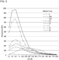
- the near-infrared fluorescence of microspheres prepared with various concentrations of ICG and albumin was measured.
- FIG. 5 is a graph showing the change in the near-infrared fluorescence signal intensity of microspheres according to the change in the concentration of ICG and albumin.
- the concentration of ICG with the strongest fluorescence intensity at the same albumin concentration was 6.5 to 25.8 ⁇ M. Therefore, in the present invention, the ICG concentration range is preferably 1 to 100 ⁇ M, and more preferably 3 to 30 ⁇ M.
- albumin used for microsphere production is ideal at 0.5 % w/v or higher, and can be suitable for microsphere production up to 20 % w/v, which is the highest albumin concentration experimentally possible.
- the suspension retention time of the ICG-Albumin-PMMA/hyaluronic acid composition according to the concentration of hyaluronic acid was measured.
- ICG-Albumin-PMMA was used at a concentration of 2 mg/ml, and the final injection of ICG-Albumin-PMMA microspheres showed the appearance of a very light green or white suspension when completely dissolved in the hyaluronic acid composition.
- the time for which the ICG-Albumin-PMMA injectable composition maintained suspension after it was prepared was measured, and the time for which the suspension was maintained according to the concentration of hyaluronic acid added was measured.
- the suspension retention time was defined as the time at which a distinction between the settled region and the floating region could be observed, based on a 20 % transmittance of 500 nm light as the criterion. The results are described in Table 3.
- Table 3 shows the time for which the suspension is maintained after the ICG-Albumin-PMMA microsphere injection is prepared, according to the concentration of the hyaluronic acid solvent.
- Concentration of hyaluronic acid solvent (%, w/v) Suspension retention time 0 (simple drinking water, distilled water) Within 10 minutes 0.1 Within 60 minutes 0.2 Within 3 hours 0.3 Within 4 hours 0.4 Within 12 hours 0.5 Within 24 hours 1.0 More than 24 hours 1.5 More than 24 hours 2.0 More than 24 hours 2.5 More than 24 hours
- the suspension retention time increased to within 60 minutes, but the suspension retention time was 1 to 3 hours when the hyaluronic acid concentration was 0.2 % (w/v), and it was confirmed that the suspension was over 3 hours when the hyaluronic acid concentration was 0.3 % (w/v).
- the time for preparing the injection and actually injecting it is less than 1 hour, it was determined that it was effective as an actual injection preparation starting from 0.2 % (w/v).
- FIG. 6 is a photograph showing the degree of dispersion of microsphere particles according to the concentration of hyaluronic acid in the biocompatible injection composition for labeling target lesion of the present invention. More specifically, the photographs were taken immediately after preparation and then at 30, 60, and 120 minutes for the ICG-HSA-PMMA/hyaluronic acid injection produced as described above: (a) on the left shows the injection with microsphere mixed in water for injection (distilled water), and (b) on the right shows a microsphere injection prepared with 0.3 % hyaluronic acid.
- the injectable composition prepared in the injection water quickly sank, whereas the injectable composition with 0.3% hyaluronic acid added did not completely sink for a long time (up to 120 minutes). That is, from the results of FIG. 3 , it can be concluded that the ICG-HSA-PMMA/hyaluronic acid injectable composition with 0.3 % hyaluronic acid added to the microsphere particles is very useful as an injectable composition because it will not quickly sink during injection preparation.
- the gliding force (N) on the finger during injection was measured under different conditions depending on the concentration of hyaluronic acid, and the usefulness of ICG-Albumin-PMMA/hyaluronic acid as an injectable composition was determined.
- the force applied to the syringe handle was measured for each needle.
- resistance was measured using an endoscopic syringe with a 1.8 m connecting tube (7 Fr, length 180 cm, MTW, Germany).
- the injection used in the experiment used a concentration of 2 mg/ml of ICG-Albumin-PMMA microspheres, and the concentration of hyaluronic acid in the final injection was set to 0.1 to 3.0 % (w/v).
- the experiment was performed immediately to prevent the microspheres from settling, and the strain-gauge dynamometer was used to measure the strain while the final injection was injected at a speed of 0.1 cc/sec (Digital Force Ga ⁇ g e DPU-500N, IMADA, Toyohashi, Japan). The results are described in Table 4 below.
- a force of 0.6 to 1.4 N is applied to the finger during the injection.
- the grip strength of an average adult is 400 N, and the grip strength that can be handled by the finger is weaker than this, and much less force is required to make delicate adjustments such as controlling the injection amount.
- a force gauge was installed between the thumb and index/middle finger in 10 men and women, and the maximum power (MaxPower) when applying the maximum force and the power when injecting comfortably (EasyPower) were measured.
- the maximum power (MaxPower) was 98.1 N (113.5 to 78.2, standard deviation 9.8) for adult males and 43.1 N (50.6 to 34.2, standard deviation 6.1) for adult females, and 25.4 N (29.7 to 20.1, standard deviation 3.3) for male adults and 14.5 N (18.3 to 11.5, standard deviation 2.2) for female adults when the force was applied comfortably.
- the ideal resistance of the injection was determined based on the maximum pressure of 43 N and the comfort level of 14.5 N for female adults. In other words, it was determined that the maximum force that can be injected was 40 N and the ideal resistance for comfortable injection was 10 N.
- the resistance was not larger than 10 N for injection up to 1% hyaluronic acid concentration, so there was no difficulty in use. Even for a puncture needle with a diameter of 20G and a much longer needle than other needles, the resistance did not exceed 10 N at 1 %.
- the pressure (A) at the time of immediate injection of the injectable composition to which hyaluronic acid is added, the pressure (B) at the beginning of the injection after the composition has settled down after 120 minutes of standing, and the pressure (C) at the middle of the injection are measured and are shown in Table 5 below.
- the injectable used in the experiment was ICG-Albumin-PMMA microspheres at a concentration of 2 mg/ml, and the concentration of hyaluronic acid in the final injection was 0.1 to 3.0 % (w/v).
- the syringe was 26G, and the unit of pressure applied to the syringe was measured in N.
- the gliding force was measured in units of N (Digital Force Ga ⁇ g e DPU-500N, IMADA, Toyohashi, Japan).
- the precipitation rate could be slow down, and the disadvantage of requiring excessive force to be applied to start the injection due to the injection needle being blocked by the precipitated composition could be resolved.
- PMMA microspheres conjugated with ICG and HSA which can generate near-infrared fluorescence signals, were expected to serve as markers that work more stably in vivo. Therefore, the microsphere particles were produced and their stability was confirmed under both in vitro and in vivo conditions.
- FIG. 7 is a graph showing a comparison of the change in intensity of near-infrared fluorescence signals over time for ICG, ICG-HSA, and ICG-HSA-PMMA particles under in vitro conditions, measured using IVIS (Perkin Elmer, Hopkinton, MA).
- a suitable mixing ratio for preparing microsphere particles exhibiting a high level of near-infrared fluorescence signal was to mix 50 ⁇ M) of ICG with 4 % of HSA and mix it with 300 mg of PMMA beads.
- a biopolymer such as albumin, it shows stronger fluorescence and longer duration than when ICG alone is embedded in microspheres.
- FIG. 8 is a photograph comparing the stability of the particles over time after placing the ICG-HSA-PMMA particles in HSA, distilled water, and agarose gel, respectively. More specifically, (a) of FIG. 8 is a photograph comparing the stability of the particles over time after placing the ICG-HSA-PMMA particles in HSA and distilled water, respectively. (b) of FIG. 8 is a photograph comparing the stability of the particles over time after placing the ICG-HSA-PMMA particles in agarose gel.
- the particles containing PMMA showed stability with a relatively small decrease in the intensity of the near-infrared fluorescence signal.
- the prepared particles were placed in (a) 20 % HSA, distilled water, and (b) agarose gel, respectively, and the near-infrared fluorescence signal emitted for 17 weeks was measured with a fluorescence camera, confirming stability even with the naked eye.
- the ICG-HSA-PMMA microspheres prepared in the above preparation example 4 were put into a hyaluronic acid solution or distilled water, and 100 ⁇ l was injected into the backs of ICR mice, respectively, and the intensity of the near-infrared fluorescence signal generated in each mouse for 4 months was measured using IVIS and fluorescence camera equipment.
- FIG. 9 is a comparison of the change in intensity of the near-infrared fluorescence signal over time when ICG-HSA-PMMA particles injected into ICR mice under in vivo conditions were injected together with hyaluronic acid or distilled water, as measured by IVIS. More specifically, (a) of FIG. 9 is a graph quantitatively showing the change in intensity of the near-infrared fluorescence signal over time when ICG-HSA-PMMA particles injected into ICR mice were measured by IVIS, and (b) of FIG. 9 is an image taken by IVIS over time when ICG-HSA-PMMA particles injected into ICR mice.
- FIG. 10 is a graph showing the change in intensity of the near-infrared fluorescence signal over time when ICG-HSA-PMMA particles were injected into ICR mice in vivo, together with hyaluronic acid or distilled water, and analyzed using the Image J program. It is also a result of taking pictures of the mouse with the fluorescence camera. More specifically, (a) of FIG. 10 is a graph showing quantitative changes in the intensity of the near-infrared fluorescence of ICG-HSA-PMMA particles injected into ICR mice over time, and (b) of FIG. 10 is an image taken with a fluorescence camera over time of ICG-HSA-PMMA particles injected into ICR mice.
- the fluorescence intensity was measured using the Image J program after shooting, and both mixtures showed high-intensity near-infrared fluorescence signals, but over time, the ICG-HSA-PMMA/hyaluronic acid mixture maintained stability, while the distilled water mixture showed a gradually decreasing fluorescence signal.
- the injection composition of the complex of the present invention may additionally include hyaluronic acid.
- the composition for labeling the target lesion of the present invention when injected into the target lesion, remains continuously for several months and can detect the location and size of the labeled lesion in real time during surgery, so it not only improves the success rate of surgical operation of the target lesion but also prevents excessive loss of normal tissue, so it can be widely used as an effective surgical marker.
- the results of the images taken at a maximum of 20 weeks were disclosed, but this was due to other circumstances such as the termination of the experiment according to the animal welfare-related experimental regulations, and it was confirmed that the fluorescence of the ICG-HAS-PMMA microspheres was well maintained up to 25 weeks.
- the fluorescence of the microspheres lasted for more than a year in vitro.
- composition for labeling a target lesion according to the present invention when injected into a target lesion, remains there for several months and can detect the location and size of the labeled lesion in real time during surgery, thereby not only improving the success rate of surgical operation for the target lesion, but also preventing excessive loss of normal tissue, and therefore can be widely utilized as an effective surgical marker.
Landscapes
- Health & Medical Sciences (AREA)
- Life Sciences & Earth Sciences (AREA)
- Biomedical Technology (AREA)
- Engineering & Computer Science (AREA)
- Animal Behavior & Ethology (AREA)
- General Health & Medical Sciences (AREA)
- Veterinary Medicine (AREA)
- Public Health (AREA)
- Epidemiology (AREA)
- Nuclear Medicine, Radiotherapy & Molecular Imaging (AREA)
- Proteomics, Peptides & Aminoacids (AREA)
- Surgery (AREA)
- Physics & Mathematics (AREA)
- Optics & Photonics (AREA)
- Oncology (AREA)
- Biodiversity & Conservation Biology (AREA)
- Oral & Maxillofacial Surgery (AREA)
- Pathology (AREA)
- Heart & Thoracic Surgery (AREA)
- Medical Informatics (AREA)
- Molecular Biology (AREA)
- Medicines Containing Antibodies Or Antigens For Use As Internal Diagnostic Agents (AREA)
Applications Claiming Priority (3)
| Application Number | Priority Date | Filing Date | Title |
|---|---|---|---|
| KR20220005362 | 2022-01-13 | ||
| KR1020220178024A KR20230109549A (ko) | 2022-01-13 | 2022-12-19 | 생체 친화적 목적 병변 표지용 조성물 및 이의 제조 방법 |
| PCT/KR2023/000601 WO2023136639A1 (ko) | 2022-01-13 | 2023-01-12 | 생체 친화적 목적 병변 표지용 조성물 및 이의 제조 방법 |
Publications (1)
| Publication Number | Publication Date |
|---|---|
| EP4464339A1 true EP4464339A1 (de) | 2024-11-20 |
Family
ID=87279414
Family Applications (1)
| Application Number | Title | Priority Date | Filing Date |
|---|---|---|---|
| EP23740482.7A Pending EP4464339A1 (de) | 2022-01-13 | 2023-01-12 | Biokompatible zusammensetzung zur markierung von zielläsionen und herstellungsverfahren dafür |
Country Status (4)
| Country | Link |
|---|---|
| US (1) | US20250090693A1 (de) |
| EP (1) | EP4464339A1 (de) |
| JP (1) | JP2025502233A (de) |
| WO (1) | WO2023136639A1 (de) |
Family Cites Families (5)
| Publication number | Priority date | Publication date | Assignee | Title |
|---|---|---|---|---|
| CA3149284A1 (en) * | 2009-12-15 | 2011-07-14 | Incept, Llc | Implants and biodegradable fiducial markers |
| US9072678B2 (en) * | 2013-03-14 | 2015-07-07 | Pathak Holdings Llc | Methods for local drug delivery by microinjection |
| US10986997B2 (en) * | 2013-12-31 | 2021-04-27 | Memorial Sloan Kettering Cancer Center | Systems, methods, and apparatus for multichannel imaging of fluorescent sources in real time |
| EP3179985A1 (de) * | 2014-08-14 | 2017-06-21 | Brown University | Zusammensetzungen zur stabilisierung und ausgabe von proteinen |
| US20210369875A1 (en) * | 2018-11-06 | 2021-12-02 | National Cancer Center | Alginic acid-based injectable hydrogel system |
-
2023
- 2023-01-12 US US18/728,190 patent/US20250090693A1/en active Pending
- 2023-01-12 JP JP2024541898A patent/JP2025502233A/ja active Pending
- 2023-01-12 WO PCT/KR2023/000601 patent/WO2023136639A1/ko not_active Ceased
- 2023-01-12 EP EP23740482.7A patent/EP4464339A1/de active Pending
Also Published As
| Publication number | Publication date |
|---|---|
| US20250090693A1 (en) | 2025-03-20 |
| JP2025502233A (ja) | 2025-01-24 |
| WO2023136639A1 (ko) | 2023-07-20 |
Similar Documents
| Publication | Publication Date | Title |
|---|---|---|
| CA2516116A1 (en) | Nanoparticle based stabilization of ir fluorescent dyes | |
| US20050158393A1 (en) | Colloidal metal labeled microparticles and methods for producing and using the same | |
| CN104919319B (zh) | 新型癌症病变标记用组合物 | |
| JP6618680B2 (ja) | 光力学的治療用自己組立型薬学の組成物 | |
| WO2012151593A1 (en) | Multifunctional infrared-emitting composites | |
| US10383957B2 (en) | Near-infrared fluorescent surgical dye markers | |
| JPH09508397A (ja) | 体組織を視覚的に標識化するための薬剤 | |
| Kim et al. | Multiporous PMMA microballs as a novel fluorescence tissue marker | |
| EP4464339A1 (de) | Biokompatible zusammensetzung zur markierung von zielläsionen und herstellungsverfahren dafür | |
| KR102179241B1 (ko) | 병변 표지용 주사제 조성물 | |
| KR20230109549A (ko) | 생체 친화적 목적 병변 표지용 조성물 및 이의 제조 방법 | |
| CN118541176A (zh) | 生物相容性目标病变标记用组合物及其制造方法 | |
| KR20250142557A (ko) | 온도 민감성 고분자 하이드로겔을 포함하는 형광 병변 표지용 조성물 | |
| US20250360298A1 (en) | Medical device for applying semi-permanent tattoo ink | |
| JP2020506929A (ja) | 腫瘍治療薬および腫瘍診断薬のための選択的放出システムならびに腫瘍組織のためのバイオセンサー | |
| Masotti | Multifunctional nanoparticles, nanocages and degradable polymers as a potential novel generation of non-invasive molecular and cellular imaging systems | |
| CN120939252A (zh) | 一种钆基磁共振/近红外荧光双模态探针及其制备和应用 | |
| CN109675063A (zh) | 靶向致痫灶区域p-糖蛋白的磁共振对比剂的制备方法 | |
| Rodriguez et al. | Stabilized micellar formulation of indocyanine green for near-infrared imaging |
Legal Events
| Date | Code | Title | Description |
|---|---|---|---|
| STAA | Information on the status of an ep patent application or granted ep patent |
Free format text: STATUS: THE INTERNATIONAL PUBLICATION HAS BEEN MADE |
|
| PUAI | Public reference made under article 153(3) epc to a published international application that has entered the european phase |
Free format text: ORIGINAL CODE: 0009012 |
|
| STAA | Information on the status of an ep patent application or granted ep patent |
Free format text: STATUS: REQUEST FOR EXAMINATION WAS MADE |
|
| 17P | Request for examination filed |
Effective date: 20240812 |
|
| AK | Designated contracting states |
Kind code of ref document: A1 Designated state(s): AL AT BE BG CH CY CZ DE DK EE ES FI FR GB GR HR HU IE IS IT LI LT LU LV MC ME MK MT NL NO PL PT RO RS SE SI SK SM TR |
|
| DAV | Request for validation of the european patent (deleted) | ||
| DAX | Request for extension of the european patent (deleted) |