EP4446744A2 - Vorbehandlungsverfahren für fluoreszenzbilddiagnose - Google Patents
Vorbehandlungsverfahren für fluoreszenzbilddiagnose Download PDFInfo
- Publication number
- EP4446744A2 EP4446744A2 EP24183773.1A EP24183773A EP4446744A2 EP 4446744 A2 EP4446744 A2 EP 4446744A2 EP 24183773 A EP24183773 A EP 24183773A EP 4446744 A2 EP4446744 A2 EP 4446744A2
- Authority
- EP
- European Patent Office
- Prior art keywords
- cell
- cancer
- pbs
- fluorescence
- cells
- Prior art date
- Legal status (The legal status is an assumption and is not a legal conclusion. Google has not performed a legal analysis and makes no representation as to the accuracy of the status listed.)
- Withdrawn
Links
Images




Classifications
-
- G—PHYSICS
- G01—MEASURING; TESTING
- G01N—INVESTIGATING OR ANALYSING MATERIALS BY DETERMINING THEIR CHEMICAL OR PHYSICAL PROPERTIES
- G01N1/00—Sampling; Preparing specimens for investigation
- G01N1/28—Preparing specimens for investigation including physical details of (bio-)chemical methods covered elsewhere, e.g. G01N33/50, C12Q
- G01N1/34—Purifying; Cleaning
-
- G—PHYSICS
- G01—MEASURING; TESTING
- G01N—INVESTIGATING OR ANALYSING MATERIALS BY DETERMINING THEIR CHEMICAL OR PHYSICAL PROPERTIES
- G01N21/00—Investigating or analysing materials by the use of optical means, i.e. using sub-millimetre waves, infrared, visible or ultraviolet light
- G01N21/62—Systems in which the material investigated is excited whereby it emits light or causes a change in wavelength of the incident light
- G01N21/63—Systems in which the material investigated is excited whereby it emits light or causes a change in wavelength of the incident light optically excited
- G01N21/64—Fluorescence; Phosphorescence
- G01N21/6486—Measuring fluorescence of biological material, e.g. DNA, RNA, cells
-
- A—HUMAN NECESSITIES
- A61—MEDICAL OR VETERINARY SCIENCE; HYGIENE
- A61K—PREPARATIONS FOR MEDICAL, DENTAL OR TOILETRY PURPOSES
- A61K49/00—Preparations for testing in vivo
- A61K49/001—Preparation for luminescence or biological staining
- A61K49/0013—Luminescence
- A61K49/0017—Fluorescence in vivo
-
- C—CHEMISTRY; METALLURGY
- C12—BIOCHEMISTRY; BEER; SPIRITS; WINE; VINEGAR; MICROBIOLOGY; ENZYMOLOGY; MUTATION OR GENETIC ENGINEERING
- C12M—APPARATUS FOR ENZYMOLOGY OR MICROBIOLOGY; APPARATUS FOR CULTURING MICROORGANISMS FOR PRODUCING BIOMASS, FOR GROWING CELLS OR FOR OBTAINING FERMENTATION OR METABOLIC PRODUCTS, i.e. BIOREACTORS OR FERMENTERS
- C12M1/00—Apparatus for enzymology or microbiology
- C12M1/34—Measuring or testing with condition measuring or sensing means, e.g. colony counters
-
- C—CHEMISTRY; METALLURGY
- C12—BIOCHEMISTRY; BEER; SPIRITS; WINE; VINEGAR; MICROBIOLOGY; ENZYMOLOGY; MUTATION OR GENETIC ENGINEERING
- C12Q—MEASURING OR TESTING PROCESSES INVOLVING ENZYMES, NUCLEIC ACIDS OR MICROORGANISMS; COMPOSITIONS OR TEST PAPERS THEREFOR; PROCESSES OF PREPARING SUCH COMPOSITIONS; CONDITION-RESPONSIVE CONTROL IN MICROBIOLOGICAL OR ENZYMOLOGICAL PROCESSES
- C12Q1/00—Measuring or testing processes involving enzymes, nucleic acids or microorganisms; Compositions therefor; Processes of preparing such compositions
- C12Q1/02—Measuring or testing processes involving enzymes, nucleic acids or microorganisms; Compositions therefor; Processes of preparing such compositions involving viable microorganisms
- C12Q1/04—Determining presence or kind of microorganism; Use of selective media for testing antibiotics or bacteriocides; Compositions containing a chemical indicator therefor
-
- G—PHYSICS
- G01—MEASURING; TESTING
- G01N—INVESTIGATING OR ANALYSING MATERIALS BY DETERMINING THEIR CHEMICAL OR PHYSICAL PROPERTIES
- G01N21/00—Investigating or analysing materials by the use of optical means, i.e. using sub-millimetre waves, infrared, visible or ultraviolet light
- G01N21/62—Systems in which the material investigated is excited whereby it emits light or causes a change in wavelength of the incident light
- G01N21/63—Systems in which the material investigated is excited whereby it emits light or causes a change in wavelength of the incident light optically excited
- G01N21/64—Fluorescence; Phosphorescence
- G01N21/6428—Measuring fluorescence of fluorescent products of reactions or of fluorochrome labelled reactive substances, e.g. measuring quenching effects, using measuring "optrodes"
-
- G—PHYSICS
- G01—MEASURING; TESTING
- G01N—INVESTIGATING OR ANALYSING MATERIALS BY DETERMINING THEIR CHEMICAL OR PHYSICAL PROPERTIES
- G01N21/00—Investigating or analysing materials by the use of optical means, i.e. using sub-millimetre waves, infrared, visible or ultraviolet light
- G01N21/75—Systems in which material is subjected to a chemical reaction, the progress or the result of the reaction being investigated
- G01N21/76—Chemiluminescence; Bioluminescence
- G01N21/763—Bioluminescence
-
- G—PHYSICS
- G01—MEASURING; TESTING
- G01N—INVESTIGATING OR ANALYSING MATERIALS BY DETERMINING THEIR CHEMICAL OR PHYSICAL PROPERTIES
- G01N33/00—Investigating or analysing materials by specific methods not covered by groups G01N1/00 - G01N31/00
- G01N33/48—Biological material, e.g. blood, urine; Haemocytometers
- G01N33/483—Physical analysis of biological material
- G01N33/4833—Physical analysis of biological material of solid biological material, e.g. tissue samples, cell cultures
-
- G—PHYSICS
- G01—MEASURING; TESTING
- G01N—INVESTIGATING OR ANALYSING MATERIALS BY DETERMINING THEIR CHEMICAL OR PHYSICAL PROPERTIES
- G01N33/00—Investigating or analysing materials by specific methods not covered by groups G01N1/00 - G01N31/00
- G01N33/48—Biological material, e.g. blood, urine; Haemocytometers
- G01N33/50—Chemical analysis of biological material, e.g. blood, urine; Testing involving biospecific ligand binding methods; Immunological testing
- G01N33/53—Immunoassay; Biospecific binding assay; Materials therefor
- G01N33/5306—Improving reaction conditions, e.g. reduction of non-specific binding, promotion of specific binding
-
- G—PHYSICS
- G01—MEASURING; TESTING
- G01N—INVESTIGATING OR ANALYSING MATERIALS BY DETERMINING THEIR CHEMICAL OR PHYSICAL PROPERTIES
- G01N33/00—Investigating or analysing materials by specific methods not covered by groups G01N1/00 - G01N31/00
- G01N33/48—Biological material, e.g. blood, urine; Haemocytometers
- G01N33/50—Chemical analysis of biological material, e.g. blood, urine; Testing involving biospecific ligand binding methods; Immunological testing
- G01N33/53—Immunoassay; Biospecific binding assay; Materials therefor
- G01N33/575—Immunoassay; Biospecific binding assay; Materials therefor for cancer
-
- G—PHYSICS
- G01—MEASURING; TESTING
- G01N—INVESTIGATING OR ANALYSING MATERIALS BY DETERMINING THEIR CHEMICAL OR PHYSICAL PROPERTIES
- G01N33/00—Investigating or analysing materials by specific methods not covered by groups G01N1/00 - G01N31/00
- G01N33/48—Biological material, e.g. blood, urine; Haemocytometers
- G01N33/50—Chemical analysis of biological material, e.g. blood, urine; Testing involving biospecific ligand binding methods; Immunological testing
- G01N33/58—Chemical analysis of biological material, e.g. blood, urine; Testing involving biospecific ligand binding methods; Immunological testing involving labelled substances
- G01N33/582—Chemical analysis of biological material, e.g. blood, urine; Testing involving biospecific ligand binding methods; Immunological testing involving labelled substances with fluorescent label
-
- G—PHYSICS
- G01—MEASURING; TESTING
- G01N—INVESTIGATING OR ANALYSING MATERIALS BY DETERMINING THEIR CHEMICAL OR PHYSICAL PROPERTIES
- G01N2800/00—Detection or diagnosis of diseases
Definitions
- the present invention relates to a method of pretreating tissue for fluorescent image diagnosis using a fluorescent diagnostic drug which emits fluorescence on reacting with a tumor marker or the like.
- Non Patent Literatures 1 to 8 There are problems in simple application of the fluorescent probe directly to a tumor region, such as insufficient fluorescence intensity in a short time period; difficulties in discriminating between the fluorescence and the intrinsic autoluminescence of cells; and rapid disappearance of the fluorescence.
- Non Patent Literature 9 a cancer region using a kinetic map
- Non Patent Literature 10 cutting, pressing and rinsing treatments
- Non Patent Literature 11 a multispectral open-air method
- Non Patent Literature 12 a fluorescent probe cross-linking to an intracellular protein
- an object of the present invention is to provide a method for enabling a target cell of a fluorescent probe present inside of a cell mass to rapidly emit fluorescence.
- the inventors have tried various treatment of a cell mass comprising target cells which is expected to emit fluorescence with a fluorescent probe, and investigated an improvement in light emission. As a result, the inventors found that removal of calcium ion from a cell mass containing target cells can allow the target cells locating inside of the cell mass to emit light rapidly, and thus achieved the present invention.
- the method of treatment of the present invention is a general-purpose method applicable to any adherent cell and any fluorescent probe, regardless of the type of a cell and the type of a fluorescent probe.
- the present invention relates to the following:
- the method for allowing a target cell to emit fluorescence according to the present invention can let the target cell present inside a cell mass or a tissue emit light, and thus can be used in the detection of a target cell in a sample, such as a clinical sample, in which the target cell may not be present on the surface.
- the present invention relates to a method for emitting fluorescence from a target cell, comprising:
- the present invention relates to a method for determining a region where the target cells reside, comprising:
- the present invention relates to a method for determining the presence or absence of target cells, comprising:
- the "target cell” means a cell that emits light by contacting with or taking up the employed fluorescent probe.
- a target cell and an employed fluorescent probe can be appropriately selected according to a purpose.
- the target cells are cancer cells, particularly, solid cancer cells.
- cancer can include, but are not limited to, lung cancer, non-small cell lung cancer, small-cell lung cancer, non-Hodgkin's lymphoma, adrenal cortex cancer, AIDS-related cancer, AIDS-related lymphoma, childhood cerebellar astrocytoma, childhood cerebral astrocytoma, basal cell cancer, skin cancer (non-melanoma), biliary tract cancer, extrahepatic bile duct cancer, intrahepatic bile duct cancer, bladder cancer, cancer of bone or joint, osteosarcoma and malignant fibrohistiocytic tumor, brain cancer, brain tumor, brain stem glioma, cerebellar astrocytoma, glioma, cerebral astrocytoma/malignant glioma, ependymoma, medulloblastoma, supratentorial primitive neuroectodermal tumor, visual pathway and hypothalamic glioma, head and neck cancer, meta
- the "cell mass” herein means a massive form of an accumulated plurality of homologous or heterologous cells, and includes a tissue.
- tissue herein means a biological structure of accumulated and three-dimensionally arranged homologous or heterologous cells.
- tissue includes an epithelial tissue, a connective tissue, a muscle tissue, and a nerve tissue.
- the "cell mass or tissue containing the target cell” may contain cells or tissues other than the target cell as long as the cell mass or the tissue contains the target cell.
- the "cell mass or tissue expected to contain the target cell” is not necessarily required to contain the target cell, but can be a cell mass or a tissue that has possibility to contain the target cell, and may contain cells or tissues other than the target cell.
- the "target cell mass” means a cell mass composed mainly of the target cell.
- the target cell emits the fluorescence by contacting with the fluorescent probe at a surface or taking up the fluorescent probe into the cell, and generating a fluorescent dye according to an environment of the surface or the inside the cell.
- the environment where the fluorescent probe generates the fluorescent dye may be, for example, a low-oxygen or acidic/alkaline environment, or may be an environment where a particular substance is present.
- the "fluorescent probe which emits fluorescence when contacted with or taken up into the target cell” is a fluorescent probe that emits fluorescence when reacted with a substance contained in the target cell, or emitting fluorescence due to low oxygen, acidic, or alkaline environment of the target cell or peripheral thereof.
- the acidic or the alkaline means low or high pH for a living body.
- the acidic environment can be pH 6 or lower, and the alkaline environment can be pH 8 or higher.
- Examples of the particular substance can include enzymes, ions, nitrogen monoxide, and reactive oxygen species.
- the enzyme is not particularly limited as long as it is a protein capable of catalyzing particular reaction. Examples thereof can include glycosidase, protease, and peptidase ( ⁇ -glutamyl transpeptidase, etc.).
- the enzyme may be present on cell membrane surface or intracellularly.
- Examples of the ion herein can include calcium ions and metal ions (e.g., iron ions, copper ions, and zinc ions).
- Examples of the active oxygen species can include hypochlorous acid, hydrogen peroxide, active oxygen, peroxy nitrite and hydroxy radicals.
- the fluorescent probe may be, for example, a substance that is hydrophilized not to be taken up into a cell.
- Specific examples of the fluorescent probe can include gGlu-HMRG, Leu-HMRG, EP-HMRG and FeRhoNox-1.
- the fluorescent probe may be in a DDS form which is, for example, embedded in a vehicle such as a liposome.
- the method to remove calcium ion is not particularly limited as long as the method can remove the calcium ion from within a cell mass or a tissue.
- calcium ion can be removed by impregnating the cell mass or the tissue containing the target cell in a buffer containing a metal ion chelating agent or a buffer solution containing no calcium.
- a metal ion chelating agent a divalent metal ion chelating agent such as ethylenediaminetetraacetic acid (EDTA), glycol ether diamine tetraacetic acid (EGTA), or citric acid can be used.
- the buffer solution examples include phosphate-buffered saline (PBS), phosphate buffer solutions, sodium citrate buffer solutions, citrate-phosphate buffer solutions, Tris-hydrochloride buffer solutions (Tris-HCl), MES, PIPES, HEPES, ADA, ACES, MOPSO, BES, MOPS, TES, DIPSO, TAPSO, POPSO, HEPPSO, EPPS, bicine, TAPS, and tricine.
- PBS phosphate-buffered saline
- Tris-HCl Tris-hydrochloride buffer solutions
- MES MES
- PIPES HEPES
- ADA Tris-hydrochloride buffer solutions
- MES MES
- PIPES HEPES
- ADA Tris-hydrochloride buffer solutions
- MES Tris-HCl
- PIPES PIPES
- HEPES HEPES
- ADA Tris-hydrochloride buffer solutions
- MES Tris-HCl
- PIPES
- the calcium ion does not need to be removed 100%, and is enough removed when cell-cell adhesion is loosen to allow the fluorescent probe to reach a site of interest in the cell mass or the tissue.
- the removal of the calcium ion is desirably performed under conditions that do not wash out the substance contained in the surface of the cell mass or the tissue and the fluorescent substance formed through the reaction.
- the contact of the fluorescent probe with the cell mass or the tissue may be performed by impregnating the cell mass or the tissue with a liquid containing the fluorescent probe, or may be performed by dropping or spraying a liquid containing the fluorescent probe to the cell mass or the tissue or a site comprising the cell mass or the tissue.
- the liquid containing the fluorescent probe is preferably a buffer solution, more preferably a buffer solution capable of removing calcium ions from the cell mass or the tissue.
- the fluorescent probe may be used as a form contained in a buffer containing a chelating agent or PBS containing no calcium.
- the fluorescence emission from the target cell can be measured and observed by appropriately detecting light emission at a wavelength according to the employed fluorescent probe using an apparatus and a method well known to those skilled in the art.
- the fluorescence emission from the target cell have intensity that can distinguish the target cell from non-target cells.
- the fluorescence emission from the target cell may have intensity that can visually distinguish the target cell from non-target cells.
- the method of the present invention is particularly useful in the case where a tumor cell or a cancer cell is present inside of a cell mass and thus can not be detected by only contacting with a fluorescent probe.
- the tumor cell or the cancer cell which is present inside of a cell mass can emit light, and thereby the presence of tumor or cancer present inside of a cell mass or a tissue can be detected more reliably.
- the target cell is a cancer cell or a tumor cell.
- the present invention relates to a method for discriminating a cancer cell or a tumor cell from non-cancer cells or non-tumor cells, comprising allowing the cancer cell or the tumor cell to emit fluorescence by the aforementioned method, and determining a cell emitting fluorescence as the cancer cell or the tumor cell and determining cells emitting no fluorescence as non-cancer cells or non-tumor cells, thereby discriminating the cancer cell or the tumor cell from non-cancer cells or non-tumor cells.
- Such discrimination of a cancer cell or a tumor cell from non-cancer cells or non-tumor cells enables reliable removal of cancer cells or tumor cells during surgery, for example, and can enhance the diagnostic accuracy of cancer or tumor diagnosis.
- the present invention relates to a method for diagnosing or monitoring cancer or tumor in a subject, or a method for providing information therefor, comprising removing calcium ions from within a cell mass sample or a tissue sample obtained from the subject; contacting the sample with a fluorescent probe that emits fluorescence when contacted with or taken up into a cancer cell or a tumor cell; and when a cell emitting fluorescence is present in the sample, diagnosing the subject as having the cancer or the tumor or judging the cancer or the tumor existing in the subject (or judging the subject being poor progress or poor prognosis), or providing information thereof.
- examples of the fluorescent probe that emits fluorescence by contacting with a cancer cell or a tumor cell or takin up into a cancer cell or a tumor cell include gGlu-HMRG.
- the "monitoring method" herein includes the monitoring of the presence or absence of cancer or tumor in a patient who may have cancer or tumor, and the monitoring whether a progress after operation or treatment of a cancer or tumor patient is good or poor, or whether a prognosis of a cancer or tumor patient is good or poor.
- the method of the present invention can be performed in vitro, ex vivo, and in vivo.
- the method of the present invention is performed for an in vivo target cell.
- the method of the present invention may further comprise removing the cells determined as the cancer cells or the tumor cells by the method.
- Lymph nodes including sentinel lymph node excised for the purpose of pathological diagnosis during breast cancer surgery were divided and transported from the operating room to an inspection room in a state wrapped in gauze wetted with PBS.
- a PBS impregnated sample was impregnated with a container where PBS was placed, and continuously impregnated for 1 minute, 5 minutes, 10 minutes, or an appropriate course of time from 1 minute to 30 minutes.
- a PBS non-impregnated sample was stored in state wrapped in gauze wetted with PBS to prevent drying out, instead of being impregnated with PBS, and used in fluorescence measurement.
- the lymph nodes were carefully taken out with tweezers and put on a Petri dish, and visible images of autofluorescence were taken in a fluorescence measuring machine.
- 50 ⁇ M gGlu-HMRG dissolved in PBS was added to the Petri dish where the lymph nodes were placed, until the lymph nodes completely soaked therein. The fluorescence measurement was continued.
- the pathological diagnosis of the specimens used in this test was also conducted and checked against final results about whether cancer metastasized to each lymph node.
- HeLa cells were inoculated at 10 5 cells/well and cultured for 4 to 6 days.
- the obtained spheroid was put on a glass bottom dish and cultured for about 3 hours. Then, the culture solution was removed, and the spheroid was impregnated with PBS(-) for 10 minutes. A control was washed with HBSS without being impregnated with PBS(-).
- HeLa cells human uterine cervical cancer cell line
- A549 cells human alveolar basal epithelial adenocarcinoma cell line
- Each obtained spheroid was allowed to adhere to a poly-L-lysine-coated glass bottom dish over approximately 24 hours.
- the medium was replaced with a buffer, and the spheroid was incubated at room temperature for 10 minutes (impregnation, rinse).
- the buffer in the dish was replaced with each buffer containing gGlu-HMRG dissolved to attain a final concentration of 0.1 ⁇ M (A549 cells) or 1 ⁇ M (HeLa cells), followed by observation under a fluorescence microscope (Leica DMI 6000 CS, lens: 10 ⁇ , filter Leica L5, ex. 460 to 500 nm, em. 512 to 542 nm).
- An area (ROI) having the distance from surface shown in Figure 4 was captured with ImageJ, and increase in the average luminance between the start of measurement and 5 minutes later was analyzed. Fluorescence was also observed by impregnating with saline and addition of gGlu-HMRG dissolved in PBS(-).
- the buffers used and their composition are as follows.
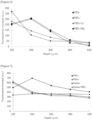
- PBS(-), PBS(+), PBS-Ca, and PBS-Mg obtained using A549 cells are shown in Figure 6 .
- PBS(-) impregnating treatment compared with impregnation with PBS(+) accelerated the increase rate of fluorescence intensity.
- Substantially the same results as those of PBS(-) were obtained in PBS-Mg of PBS(-) supplemented with only Mg 2+ .
- use of PBS-Ca of PBS supplemented with Ca 2+ less increased fluorescence, as in PBS(+).
- the results obtained using A549 cells are shown in Figure 9 .
- the A549 cells exhibited a tendency similar to that of the HeLa cells.
- PBS(+) which is PBS(-) supplemented with a divalent ion (Ca 2+ , Mg 2+ ) had an increase rate of fluorescence almost equivalent to that of no impregnation.
- the addition of 0.2 mM EDTA chelating a divalent ion caused larger increase of fluorescence intensity at a site deeper from surface even compared with PBS(-).
- Example 5 Influence of pretreatment when different fluorescent probe is used Fluorescence was measured by the same method as in Example 3 except that a 5 ⁇ M FeRhoNox-1 probe was used instead of gGlu-HMRG as the fluorescent probe. The cells used were only A549 cells.
- HeLa cells cultured on glass surface were reacted with gGlu-HMRG, and fluorescence intensity was compared.
- HeLa cells were inoculated at the same density to chambers of a 4-chamber glass bottom dish.
- the cells were cultured in DMEM+8% FBS, P.S. under conditions of 37°C and 5% CO 2 for approximately 8 hours.
- the cells were rinsed three times with PBS(-) adjusted to pH 6.5, 7.3, or 7.8, which was then replaced with each solution supplemented with 2 ⁇ M gGlu-HMRG.
- the cells were stained at 37°C under 5% CO 2 for 20 minutes and observed under a fluorescence microscope under the same conditions as above without the solution replaced. 100 ms exposure, gain 5 Int 5, ND1.
- Fluorescence intensity from 5 cells in fluorescence images, and background fluorescence intensity were measured with reference to superimposed images with DIC, and the mean and standard deviation were plotted.
- the fluorescence intensity of gGlu-HMRG and the fluorescence intensity of HMRG were measured at each pH.
- Fluorescence intensity had pH dependency, and lower pH decreased a signal and increased a background (not shown).
- the fluorescence intensity of gGlu-HMRG and the fluorescence intensity of HMRG were measured at each pH. As a result, the fluorescence intensity was decreased with the elevation of pH, indicating that a pH condition of 6 to 8 is preferred with pH 6 to 7.5 more preferred (not shown). These results indicated that pH influences the measurement of fluorescence intensity.
Landscapes
- Health & Medical Sciences (AREA)
- Life Sciences & Earth Sciences (AREA)
- Engineering & Computer Science (AREA)
- Chemical & Material Sciences (AREA)
- Immunology (AREA)
- Physics & Mathematics (AREA)
- Biomedical Technology (AREA)
- General Health & Medical Sciences (AREA)
- Molecular Biology (AREA)
- Biochemistry (AREA)
- Analytical Chemistry (AREA)
- Pathology (AREA)
- General Physics & Mathematics (AREA)
- Urology & Nephrology (AREA)
- Hematology (AREA)
- Biotechnology (AREA)
- Medicinal Chemistry (AREA)
- Microbiology (AREA)
- Food Science & Technology (AREA)
- Organic Chemistry (AREA)
- Cell Biology (AREA)
- Bioinformatics & Cheminformatics (AREA)
- Zoology (AREA)
- Wood Science & Technology (AREA)
- Chemical Kinetics & Catalysis (AREA)
- Optics & Photonics (AREA)
- Nuclear Medicine, Radiotherapy & Molecular Imaging (AREA)
- Biophysics (AREA)
- Proteomics, Peptides & Aminoacids (AREA)
- Genetics & Genomics (AREA)
- General Engineering & Computer Science (AREA)
- Sustainable Development (AREA)
- Toxicology (AREA)
- Plasma & Fusion (AREA)
- Animal Behavior & Ethology (AREA)
- Epidemiology (AREA)
- Veterinary Medicine (AREA)
- Public Health (AREA)
- Measuring Or Testing Involving Enzymes Or Micro-Organisms (AREA)
- Investigating Or Analysing Biological Materials (AREA)
Applications Claiming Priority (3)
| Application Number | Priority Date | Filing Date | Title |
|---|---|---|---|
| JP2018132928A JP6935883B2 (ja) | 2018-07-13 | 2018-07-13 | 蛍光画像診断の前処理方法 |
| EP19833189.4A EP3822359B1 (de) | 2018-07-13 | 2019-07-12 | Vorbehandlungsverfahren für fluoreszenzbilddiagnose |
| PCT/JP2019/027662 WO2020013308A1 (ja) | 2018-07-13 | 2019-07-12 | 蛍光画像診断の前処理方法 |
Related Parent Applications (2)
| Application Number | Title | Priority Date | Filing Date |
|---|---|---|---|
| EP19833189.4A Division EP3822359B1 (de) | 2018-07-13 | 2019-07-12 | Vorbehandlungsverfahren für fluoreszenzbilddiagnose |
| EP19833189.4A Division-Into EP3822359B1 (de) | 2018-07-13 | 2019-07-12 | Vorbehandlungsverfahren für fluoreszenzbilddiagnose |
Publications (2)
| Publication Number | Publication Date |
|---|---|
| EP4446744A2 true EP4446744A2 (de) | 2024-10-16 |
| EP4446744A3 EP4446744A3 (de) | 2024-12-25 |
Family
ID=69142468
Family Applications (2)
| Application Number | Title | Priority Date | Filing Date |
|---|---|---|---|
| EP24183773.1A Withdrawn EP4446744A3 (de) | 2018-07-13 | 2019-07-12 | Vorbehandlungsverfahren für fluoreszenzbilddiagnose |
| EP19833189.4A Active EP3822359B1 (de) | 2018-07-13 | 2019-07-12 | Vorbehandlungsverfahren für fluoreszenzbilddiagnose |
Family Applications After (1)
| Application Number | Title | Priority Date | Filing Date |
|---|---|---|---|
| EP19833189.4A Active EP3822359B1 (de) | 2018-07-13 | 2019-07-12 | Vorbehandlungsverfahren für fluoreszenzbilddiagnose |
Country Status (9)
| Country | Link |
|---|---|
| US (1) | US20210247277A1 (de) |
| EP (2) | EP4446744A3 (de) |
| JP (1) | JP6935883B2 (de) |
| KR (1) | KR102782107B1 (de) |
| CN (2) | CN119223929A (de) |
| AU (1) | AU2019301375A1 (de) |
| CA (1) | CA3105005A1 (de) |
| PH (1) | PH12020552199A1 (de) |
| WO (1) | WO2020013308A1 (de) |
Family Cites Families (10)
| Publication number | Priority date | Publication date | Assignee | Title |
|---|---|---|---|---|
| EP1360496A4 (de) * | 2001-02-16 | 2005-03-09 | Immunivest Corp | Verfahren und reagenzien zur schnellen und effizienten isolierung zirkulierender krebszellen |
| CA2497555C (en) * | 2002-09-20 | 2013-06-11 | Queen's University At Kingston | Detection of biological molecules by differential partitioning of enzyme substrates and products |
| KR20090026275A (ko) * | 2006-07-06 | 2009-03-12 | 로스캄프 리서치 엘엘씨 | 베타-아밀로이드 생성을 억제하기 위한 화합물 및 그의 조합물, 및 그의 사용 방법 |
| WO2009009114A2 (en) * | 2007-07-12 | 2009-01-15 | Oncomed Pharmaceuticals, Inc. | Compositions and methods for treating and diagnosing cancer |
| US9221759B2 (en) * | 2010-01-13 | 2015-12-29 | Rutgers, The State University Of New Jersey | Fluorophore chelated lanthanide luminescent probes with improved quantum efficiency |
| CN102670573A (zh) * | 2011-11-21 | 2012-09-19 | 西安交通大学 | 姜黄素衍生物在制备治疗前列腺癌疾病药物中的应用 |
| DE102012213838A1 (de) * | 2012-08-03 | 2014-02-06 | Katharina Pachmann | Verfahren zur Kultivierung einer Subpopulation zirkulierender epithelialer Tumorzellen aus einer Körperflüssigkeit |
| CN106461649B (zh) * | 2013-12-27 | 2020-03-17 | 香港大学 | 用于在细胞中标记蛋白质或其它生物分子的基于金属螯合的荧光探针 |
| US20160131560A1 (en) * | 2014-11-07 | 2016-05-12 | California Institute Of Technology | Methods for phenotyping of intact bones by tissue clearing and staining |
| KR101804063B1 (ko) * | 2016-08-03 | 2017-12-01 | 전남대학교산학협력단 | 종양 표적화 근적외선 형광 탐지자 |
-
2018
- 2018-07-13 JP JP2018132928A patent/JP6935883B2/ja active Active
-
2019
- 2019-07-12 EP EP24183773.1A patent/EP4446744A3/de not_active Withdrawn
- 2019-07-12 WO PCT/JP2019/027662 patent/WO2020013308A1/ja not_active Ceased
- 2019-07-12 CA CA3105005A patent/CA3105005A1/en active Pending
- 2019-07-12 CN CN202411233912.3A patent/CN119223929A/zh active Pending
- 2019-07-12 KR KR1020207034941A patent/KR102782107B1/ko active Active
- 2019-07-12 US US16/973,254 patent/US20210247277A1/en not_active Abandoned
- 2019-07-12 CN CN201980035559.9A patent/CN112204152A/zh active Pending
- 2019-07-12 AU AU2019301375A patent/AU2019301375A1/en not_active Abandoned
- 2019-07-12 EP EP19833189.4A patent/EP3822359B1/de active Active
-
2020
- 2020-12-17 PH PH12020552199A patent/PH12020552199A1/en unknown
Also Published As
| Publication number | Publication date |
|---|---|
| US20210247277A1 (en) | 2021-08-12 |
| JP6935883B2 (ja) | 2021-09-15 |
| EP3822359A1 (de) | 2021-05-19 |
| EP4446744A3 (de) | 2024-12-25 |
| AU2019301375A1 (en) | 2021-01-14 |
| WO2020013308A1 (ja) | 2020-01-16 |
| CN119223929A (zh) | 2024-12-31 |
| CN112204152A (zh) | 2021-01-08 |
| JP2020010604A (ja) | 2020-01-23 |
| CA3105005A1 (en) | 2020-01-16 |
| KR20210029146A (ko) | 2021-03-15 |
| PH12020552199A1 (en) | 2021-06-28 |
| EP3822359A4 (de) | 2022-04-27 |
| EP3822359B1 (de) | 2024-08-07 |
| KR102782107B1 (ko) | 2025-03-13 |
Similar Documents
| Publication | Publication Date | Title |
|---|---|---|
| Was et al. | Bafilomycin A1 triggers proliferative potential of senescent cancer cells in vitro and in NOD/SCID mice | |
| Council et al. | Differential expression of immunohistochemical markers in bladder smooth muscle and myofibroblasts, and the potential utility of desmin, smoothelin, and vimentin in staging of bladder carcinoma | |
| Lee et al. | Highly selective two-photon imaging of cysteine in cancerous cells and tissues | |
| EP3021119A1 (de) | Verfahren zur diagnose von krankheiten mittels morphologischer eigenschaften von luterial | |
| Kamarinos et al. | Immunofluorescence assay of ablated colorectal liver metastases: the frozen section of image-guided tumor ablation? | |
| EP3822359B1 (de) | Vorbehandlungsverfahren für fluoreszenzbilddiagnose | |
| Bianco Jr et al. | Proliferation of prostate cancer cells in the bone marrow predicts recurrence in patients with localized prostate cancer | |
| JP6052756B2 (ja) | 末梢循環癌細胞の検出方法および検出装置 | |
| WO2009110279A1 (en) | A method for evaluating or selecting agents having preventive or alleviative effects on spots | |
| Gonzalez et al. | Prognostic value of combined p53 and survivin in pT1G3 urothelial carcinoma of the bladder | |
| Muresan et al. | Monomeric C-Reactive Protein potential utilization in the histological assessment of inflammatory bowel disease (IBD) patients | |
| WO2014159907A1 (en) | Systems and methods employing human stem cell markers for detection, diagnosis and treatment of circulating tumor cells | |
| WO2014159920A1 (en) | A device and method for the detection and diagnosis of aggressive and metastatic cancer and cancer stem cells employing podocalyxin and tra biomarkers | |
| EP3701250A1 (de) | Fluoreszenzfarbstoff-akkumulationstest | |
| Khongkow et al. | In vitro methods for studying the mechanisms of resistance to DNA-damaging therapeutic drugs | |
| RU2790468C1 (ru) | Способ прогнозирования появления отдаленных метастазов меланомы хориоидеи | |
| EP4232821B1 (de) | Dna-schadensempfindlichkeit als neuer biomarker zum nachweis von krebs und krebsempfindlichkeit | |
| Hedley et al. | Relations between non–protein sulfydryl levels in the nucleus and cytoplasm, tumor oxygenation, and clinical outcome of patients with uterine cervical carcinoma | |
| US20140274773A1 (en) | Systems and methods for employing podocalyxin and tra human stem cell markers as prognostic markers for aggressive and metastatic cancer | |
| Christensen et al. | Highly sensitive luciferase-based assay with red fluorescent protein expression for accurate quantitative monitoring and real-time visualization of cell invasion | |
| TWI703215B (zh) | 用於預測藥物功效之活體外診斷方法 | |
| CN108449993A (zh) | 化学试剂或生化试剂的dna断裂基因毒性效应的个体预测方法 | |
| Ozolina | Exploring Antioxidants: OBE100 Extract and Nanodiamond-Based Quantum Sensing for Tissue Analysis | |
| JPWO2018101328A1 (ja) | 診療補助情報の取得方法 | |
| US11371995B2 (en) | Method for selecting individuals to be administered immune checkpoint inhibitor |
Legal Events
| Date | Code | Title | Description |
|---|---|---|---|
| PUAI | Public reference made under article 153(3) epc to a published international application that has entered the european phase |
Free format text: ORIGINAL CODE: 0009012 |
|
| STAA | Information on the status of an ep patent application or granted ep patent |
Free format text: STATUS: THE APPLICATION HAS BEEN PUBLISHED |
|
| AC | Divisional application: reference to earlier application |
Ref document number: 3822359 Country of ref document: EP Kind code of ref document: P |
|
| AK | Designated contracting states |
Kind code of ref document: A2 Designated state(s): AL AT BE BG CH CY CZ DE DK EE ES FI FR GB GR HR HU IE IS IT LI LT LU LV MC MK MT NL NO PL PT RO RS SE SI SK SM TR |
|
| REG | Reference to a national code |
Ref country code: DE Ref legal event code: R079 Free format text: PREVIOUS MAIN CLASS: G01N0033580000 Ipc: C12Q0001040000 |
|
| PUAL | Search report despatched |
Free format text: ORIGINAL CODE: 0009013 |
|
| AK | Designated contracting states |
Kind code of ref document: A3 Designated state(s): AL AT BE BG CH CY CZ DE DK EE ES FI FR GB GR HR HU IE IS IT LI LT LU LV MC MK MT NL NO PL PT RO RS SE SI SK SM TR |
|
| RIC1 | Information provided on ipc code assigned before grant |
Ipc: G01N 33/58 20060101ALI20241121BHEP Ipc: G01N 33/574 20060101ALI20241121BHEP Ipc: G01N 33/53 20060101ALI20241121BHEP Ipc: G01N 33/48 20060101ALI20241121BHEP Ipc: G01N 21/64 20060101ALI20241121BHEP Ipc: C12Q 1/04 20060101AFI20241121BHEP |
|
| STAA | Information on the status of an ep patent application or granted ep patent |
Free format text: STATUS: THE APPLICATION IS DEEMED TO BE WITHDRAWN |
|
| 18D | Application deemed to be withdrawn |
Effective date: 20250626 |