EP4442704A1 - Peptide having framework sequence for random region placement and peptide library composed of said peptide - Google Patents
Peptide having framework sequence for random region placement and peptide library composed of said peptide Download PDFInfo
- Publication number
- EP4442704A1 EP4442704A1 EP22901376.8A EP22901376A EP4442704A1 EP 4442704 A1 EP4442704 A1 EP 4442704A1 EP 22901376 A EP22901376 A EP 22901376A EP 4442704 A1 EP4442704 A1 EP 4442704A1
- Authority
- EP
- European Patent Office
- Prior art keywords
- amino acid
- seq
- peptide
- acid sequence
- framework
- Prior art date
- Legal status (The legal status is an assumption and is not a legal conclusion. Google has not performed a legal analysis and makes no representation as to the accuracy of the status listed.)
- Ceased
Links
Images



Classifications
-
- C—CHEMISTRY; METALLURGY
- C40—COMBINATORIAL TECHNOLOGY
- C40B—COMBINATORIAL CHEMISTRY; LIBRARIES, e.g. CHEMICAL LIBRARIES
- C40B40/00—Libraries per se, e.g. arrays, mixtures
- C40B40/04—Libraries containing only organic compounds
- C40B40/10—Libraries containing peptides or polypeptides, or derivatives thereof
-
- C—CHEMISTRY; METALLURGY
- C07—ORGANIC CHEMISTRY
- C07K—PEPTIDES
- C07K16/00—Immunoglobulins [IG], e.g. monoclonal or polyclonal antibodies
- C07K16/005—Immunoglobulins [IG], e.g. monoclonal or polyclonal antibodies constructed by phage libraries
-
- C—CHEMISTRY; METALLURGY
- C07—ORGANIC CHEMISTRY
- C07K—PEPTIDES
- C07K16/00—Immunoglobulins [IG], e.g. monoclonal or polyclonal antibodies
- C07K16/18—Immunoglobulins [IG], e.g. monoclonal or polyclonal antibodies against material from animals or humans
-
- C—CHEMISTRY; METALLURGY
- C07—ORGANIC CHEMISTRY
- C07K—PEPTIDES
- C07K16/00—Immunoglobulins [IG], e.g. monoclonal or polyclonal antibodies
- C07K16/18—Immunoglobulins [IG], e.g. monoclonal or polyclonal antibodies against material from animals or humans
- C07K16/28—Immunoglobulins [IG], e.g. monoclonal or polyclonal antibodies against material from animals or humans against receptors, cell surface antigens or cell surface determinants
- C07K16/30—Immunoglobulins [IG], e.g. monoclonal or polyclonal antibodies against material from animals or humans against receptors, cell surface antigens or cell surface determinants from tumour cells
- C07K16/303—Liver or Pancreas
-
- C—CHEMISTRY; METALLURGY
- C07—ORGANIC CHEMISTRY
- C07K—PEPTIDES
- C07K16/00—Immunoglobulins [IG], e.g. monoclonal or polyclonal antibodies
- C07K16/18—Immunoglobulins [IG], e.g. monoclonal or polyclonal antibodies against material from animals or humans
- C07K16/32—Immunoglobulins [IG], e.g. monoclonal or polyclonal antibodies against material from animals or humans against translation products of oncogenes
-
- G—PHYSICS
- G01—MEASURING; TESTING
- G01N—INVESTIGATING OR ANALYSING MATERIALS BY DETERMINING THEIR CHEMICAL OR PHYSICAL PROPERTIES
- G01N33/00—Investigating or analysing materials by specific methods not covered by groups G01N1/00 - G01N31/00
- G01N33/48—Biological material, e.g. blood, urine; Haemocytometers
- G01N33/50—Chemical analysis of biological material, e.g. blood, urine; Testing involving biospecific ligand binding methods; Immunological testing
- G01N33/68—Chemical analysis of biological material, e.g. blood, urine; Testing involving biospecific ligand binding methods; Immunological testing involving proteins, peptides or amino acids
- G01N33/6803—General methods of protein analysis not limited to specific proteins or families of proteins
- G01N33/6845—Methods of identifying protein-protein interactions in protein mixtures
-
- C—CHEMISTRY; METALLURGY
- C07—ORGANIC CHEMISTRY
- C07K—PEPTIDES
- C07K2317/00—Immunoglobulins specific features
- C07K2317/20—Immunoglobulins specific features characterized by taxonomic origin
- C07K2317/24—Immunoglobulins specific features characterized by taxonomic origin containing regions, domains or residues from different species, e.g. chimeric, humanized or veneered
-
- C—CHEMISTRY; METALLURGY
- C07—ORGANIC CHEMISTRY
- C07K—PEPTIDES
- C07K2317/00—Immunoglobulins specific features
- C07K2317/50—Immunoglobulins specific features characterized by immunoglobulin fragments
- C07K2317/56—Immunoglobulins specific features characterized by immunoglobulin fragments variable (Fv) region, i.e. VH and/or VL
- C07K2317/565—Complementarity determining region [CDR]
-
- C—CHEMISTRY; METALLURGY
- C07—ORGANIC CHEMISTRY
- C07K—PEPTIDES
- C07K2317/00—Immunoglobulins specific features
- C07K2317/50—Immunoglobulins specific features characterized by immunoglobulin fragments
- C07K2317/56—Immunoglobulins specific features characterized by immunoglobulin fragments variable (Fv) region, i.e. VH and/or VL
- C07K2317/567—Framework region [FR]
-
- C—CHEMISTRY; METALLURGY
- C07—ORGANIC CHEMISTRY
- C07K—PEPTIDES
- C07K2317/00—Immunoglobulins specific features
- C07K2317/50—Immunoglobulins specific features characterized by immunoglobulin fragments
- C07K2317/56—Immunoglobulins specific features characterized by immunoglobulin fragments variable (Fv) region, i.e. VH and/or VL
- C07K2317/569—Single domain, e.g. dAb, sdAb, VHH, VNAR or nanobody®
-
- C—CHEMISTRY; METALLURGY
- C07—ORGANIC CHEMISTRY
- C07K—PEPTIDES
- C07K2317/00—Immunoglobulins specific features
- C07K2317/90—Immunoglobulins specific features characterized by (pharmaco)kinetic aspects or by stability of the immunoglobulin
-
- C—CHEMISTRY; METALLURGY
- C07—ORGANIC CHEMISTRY
- C07K—PEPTIDES
- C07K2317/00—Immunoglobulins specific features
- C07K2317/90—Immunoglobulins specific features characterized by (pharmaco)kinetic aspects or by stability of the immunoglobulin
- C07K2317/92—Affinity (KD), association rate (Ka), dissociation rate (Kd) or EC50 value
-
- C—CHEMISTRY; METALLURGY
- C07—ORGANIC CHEMISTRY
- C07K—PEPTIDES
- C07K2317/00—Immunoglobulins specific features
- C07K2317/90—Immunoglobulins specific features characterized by (pharmaco)kinetic aspects or by stability of the immunoglobulin
- C07K2317/94—Stability, e.g. half-life, pH, temperature or enzyme-resistance
Definitions
- the biological defense mechanism involves cellular immunity and humoral immunity.
- the humoral immunity is such that an antibody produced by B cells recognizes a foreign substance entering the body, and binds specifically to an antigen (target) present on the surface of the foreign substance, followed by removal of the foreign substance from the living organism.
- the antibody is a protein called "immunoglobulin" (which may be referred to as “Ig” hereinafter), which is classified into IgA, IgD, IgE, IgG and IgM.
- IgG is called a common antibody because it is produced in large amounts, and consists of a heavy chain and a light chain in many mammals.
- IgG consists of only a heavy chain.
- Such an antibody consisting of a single domain having only a heavy chain is called a heavy chain antibody.
- the antibody can bind to an antigen only in a variable region, and thus is also called a single domain antibody, Nanobody (registered trademark), or VHH.
- the VHH whose amino acid sequences are precisely defined, can be used as a tool for biochemical analysis by an immunoprecipitation method or the like or as a tool for use in immunohistochemistry like conventional antibodies such as IgG.
- the VHH is known to differ from the conventional antibodies in binding site to an antigen. Specifically, it is known that the conventional antibody binds to an epitope on a convex part of the antigen, whereas the VHH binds to an epitope of an antigen which forms a gap, a cleft or the like of an antigen (see Prior Art Literatures 1 to 3).
- the above-described search typically comprises screening a peptide library using a display method such as a phage display method (see Prior Art Literature 1).
- the peptide library can be defined as a "large collection of peptides having a systematic combination of amino acids".
- the peptides include naive peptides obtained from various animals or the like, semisynthetic peptides obtained by modification of the naive peptides, and synthetic peptides obtained by chemical synthesis.
- epitope peptide mapping preparation of a peptide library for development of vaccine, and the like can be performed by screening the above-described synthetic peptide library.
- Patent Literature 1 International Publication No. WO 2003/025020
- mRNA display method is an excellent technique in terms of a library size.
- the mRNA display method has a problem in that an intended peptide cannot be obtained with stability because mRNA is likely to undergo degradation by RNase in an aqueous solution.
- Conventional art 2 phage display method
- a construct which expresses DNA encoding the displayed peptide be prepared, and finally introduced into Escherichia coli or other bacteria and expressed, and thus a translation system using cells is required.
- a first aspect of the present invention is a peptide consisting of three complementarity determining regions and four framework regions (which may be abbreviated as "FR” hereinafter), and comprising a sequence represented by SEQ ID NO: 1 as a framework 1 (hereinafter, referred to as "FR1").
- the FR preferably comprises an amino acid sequence represented by SEQ ID NO: 2 as a framework 2 (hereinafter, referred to as "FR2").
- X in the sequence of SEQ ID NO: 2 represents an arbitrary amino acid, which is preferably any amino acid selected from the group consisting of tyrosine, valine, phenylalanine, leucin, arginine, glycine and tryptophane from the viewpoint of tertiary structure stability.
- the FR2 is more preferably any sequence selected from the group consisting of amino acid sequences represented by SEQ ID NOS: 3 to 6 from the viewpoint of tertiary structure stability.
- the FR preferably comprises an amino acid sequence represented by SEQ ID NO: 7 as a framework 3 (hereinafter, referred to as "FR3").
- X in the sequence of SEQ ID NO: 7 represents an arbitrary amino acid, which is preferably any amino acid selected from the group consisting of leucine, valine, alanine (A), arginine (R) and lysine (K).
- the FR3 is preferably any sequence selected from the group consisting of amino acid sequences represented by SEQ ID NOS: 8 to 31 described below.
- the FR preferably comprises an amino acid sequence represented by SEQ ID NO: 32 in the following sequence listing as a framework 4 (hereinafter, referred to as "FR4").
- the FR1 to FR4 preferably consist, respectively, of 22 to 26 amino acids, 12 to 16 amino acids, 29 to 33 amino acids and 9 to 13 amino acids.
- the FR1 to FR4 more preferably consist, respectively, of 25 amino acids, 14 amino acids, 32 amino acids and 11 amino acids.
- the peptide preferably comprises a complementarity determining region 1 (hereinafter, referred to as "CDR1”) consisting of 8 to 12 amino acids, a complementarity determining region 2 (hereinafter, referred to as "CDR2”) consisting of 14 to 18 amino acids, and a complementarity determining region 3 (CDR3) consisting of any amino acid sequence selected from the group consisting of sequences of 6, 12 and 15 amino acids.
- CDR1 complementarity determining region 1
- CDR2 complementarity determining region 2
- CDR3 complementarity determining region 3
- a second aspect of the present invention is a peptide library composed of the peptides.
- a preferred peptide library is a peptide library in which a peptide constituting the peptide library consists of three complementarity determining regions and four framework regions,
- a particularly preferred peptide library is a peptide library in which a peptide constituting the peptide library consists of three complementarity determining regions and four framework regions,
- the library size of the peptide library is preferably about 10 12 to about 10 14 .
- the library is preferably a single domain antibody derived from the heavy chain antibody as described above, or a polyvalent form in which the heavy chain antibodies are bound to one another.
- the polyvalent form is preferably any one selected from the group consisting of di- to pentamers in which the antibodies are bound to one another via a linker.
- a framework sequence having a sequence on which CDR with a desired length can be placed and a peptide comprising the framework sequence. Furthermore, it is possible to obtain a framework sequence which can be used to efficiently screen single domain antibodies, and enables the acquisition of a peptide having structural characteristics as designed. Moreover, it is possible to construct a peptide library composed of the above-described peptides.
- the present invention is, as described above, a peptide (a) consisting of three CDRs and four FRs and (b) comprising specific sequences described below as FR1, FR2, FR3 and FR4.
- a peptide constituting the FR1 to FR4 can be designed and prepared as follows. First, the amino acid sequences of the human framework sequence IGHV3-23*01 DP-47 (FR1 to FR4 are shown as SEQ ID NOS: 34 to 37 in Table 1) are selected as basic sequences for use in design of framework sequence in the present invention for the reason of antigenicity and homology with VHH. FR to be changed in design is determined while the shape of a peptide that is ultimately formed is taken into account.
- the FR1 to FR4 consist, respectively, of 22 to 26 amino acids, 12 to 16 amino acids, 29 to 33 amino acids and 9 to 13 amino acids from the viewpoint of tertiary structure stability. It is more preferable that the FR1 to FR4 consist, respectively, of 24 amino acids, 14 amino acids, 32 amino acids and 11 amino acids from the viewpoint of tertiary structure stability.
- the sequences of FR2 and FR3 may be changed with no change made to FR1 and FR4.
- amino acids at desired sites in FR2 and FR3 can be replaced with another amino acid.
- the amino acid at the C-terminus of FR2 of the human framework sequence IGHV3-23*01 DP-47 can be changed from serine (S) to alanine (A) from the viewpoint of the stability of the three-dimensional structure of CDR2.
- Amino acids at other positions can be replaced with another amino acid.
- the 2nd, 10th or 12th amino acid from the N-terminus can be replaced with any amino acid selected from the group consisting of tyrosine (Y), valine (V), phenylalanine (F), leucine (L), arginine (R), glycine (G) and tryptophane (W).
- FR2 containing, at the above-described site, an amino acid to be replaced can be presented as the following amino acid sequence represented by SEQ ID NO: 2.
- SEQ ID NO: 2 WXRQAPGKGXEXVA
- X at the 2nd position from the N-terminus may be any amino acid selected from the group consisting of tyrosine, valine (V) and phenylalanine (F)
- X at the 10th position from the N-terminus may be any amino acid selected from the group consisting of arginine (R) or leucine (L)
- X at the 12th position from the N-terminus may be any amino acid selected from the group consisting of tryptophane (W), phenylalanine (F) and glycine (G).
- the FR2 preferably has a sequence represented by any amino acid sequence selected from the group consisting of sequences represented by SEQ ID NOS: 3 to 6. This is because by performing the replacement in this way, the steric structure of the peptide can be controlled to some extent.
- the amino acid at the C-terminus of FR3 of the human framework sequence IGHV3-23*01 DP-47 can be changed to alanine (A), valine (V), arginine (R) and leucine (L) because the region can be involved in determination of the structure of the CDR3 region in VHH, and also involved in binding to an antigen. Amino acids at other positions can also be replaced with another amino acid.
- amino acid at the 13th, 31st or 32nd position from the N-terminus can be replaced with any amino acid selected from the group consisting of leucine (L), valine (V), alanine (A), asparagine (N), lysine (K) and arginine (R).
- L leucine
- V valine
- A alanine
- N asparagine
- K lysine
- R arginine
- FR3 containing, at the above-described site, an amino acid to be replaced can be presented as an amino acid sequence represented by SEQ ID NO: 7.
- X13 may be any amino acid selected from the group consisting of valine (V) and leucine (L)
- X31 may be any amino acid selected from the group consisting of alanine (A), asparagine (N) or lysine (K)
- X32 may be an amino acid selected from the group consisting of alanine (A), valine (V), arginine (R) and leucine (L).
- the FR3 preferably has a sequence represented by an amino acid sequence selected from the group consisting of sequences of SEQ ID NOS: 8 to 31. This is because by performing the replacement in this way, the steric structure of the peptide can be controlled to some extent.
- FR1 to FR4 may be chemically synthesized by a solid phase synthesis method or other routine methods, or their synthesis may be outsourced to companies.
- the peptide has three complementarity determining regions (hereinafter, abbreviated as "CDR") as described above, which may be, respectively, a complementarity determining region 1 (hereinafter, referred to as "CDR1”) consisting of 8 to 12 amino acids, a complementarity determining region 2 (hereinafter, referred to as “CDR2”) consisting of 14 to 18 amino acids, and a complementarity determining region 3 (CDR3) consisting of 6, 12 or 15 amino acids. It is preferable that CDR1 consist of 10 amino acids, CDR2 consists of 16 amino acids or 17 amino acids, and CDR3 consists of 6, 12 or 15 amino acids because a peptide having characteristics described later can be obtained.
- CDR1 complementarity determining region 1
- CDR2 complementarity determining region 2
- CDR3 complementarity determining region 3
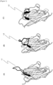
- CDR3 is classified roughly into three types according to distinctive loop structures L1 to L3 ( Figures 1(A) to 1(C) ).
- the loop structures L1 to L3 vary depending on the number of amino acids contained in CDR3, and influences the number of amino acid residues in CDR2.
- FR2 is in contact with the light chain, whereas in VHH, the site is exposed. This is because VHH consists of only a light chain as described above.
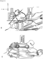
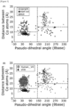
- the structures of the three types of CDR3 are classified as follows according to three elements: a distance between the C ⁇ atoms of an amino acid residue at a specific position which is contained in FR2 and an amino acid residue at a specific position which is contained in CDR3, an angle formed by two pseudo-planes made from the C ⁇ atom of an amino acid residue at the position of the terminus of CDR3, and a structure of a loop itself ( Figures 1(A) and 1(C) and Figures 2(A) and 2(B) ).
- the criterial for the classification will be described below.
- C ⁇ refers to the first carbon atom adjacent to a functional group.
- the distance between the two ⁇ carbon atoms is referred to as a C ⁇ distance.
- the C ⁇ distance is a distance between C ⁇ (n-5) in the (n-5)th amino acid residue and C ⁇ (46) in the 46th amino acid residue from the N-terminus of the peptide.
- Figure 1(E) is a view of the peptide seen from an X-Y plane.
- the first plane refers to a plane including a peptide bond contained in H102(n) and a peptide bond contained in an amino acid residue (H103(n+1)) adjacent to the amino acid residue (H102(n)) on the downstream side.
- the second plane refers to a plane including a peptide bond contained in an amino acid residue (H101(n-1)) containing H101 and a peptide bond contained in an amino acid residue (H100(n-2)) adjacent to the amino acid residue (H101(n-1)) on the downstream side.
- the angle formed by the first plane and the second plane refers to a "dihedral angle".
- VHH having a desired structure, among the upright type, the roll type or the half-roll type, can be prepared.
- a sub-library of the peptide can be constructed on the basis of the analyses of the antigen-binding site, the number of amino acid residues and the structure of CDR3.
- the sub-library refers to a library prepared by selecting a clone from an original DNA library under specific conditions according to the purpose of study or the like.
- a peptide constituting CDR1 to CDR3 can be prepared by selecting a target peptide.
- structure data for VHH and human VH can be obtained as information on VHH from, for example, SAbDab Database (http://opig.stats.ox.ac.uk/webapps/newsabdab/sabdab/) in Protein Data Bank (PDB).
- PDB Protein Data Bank
- Some selection criteria can be determined from the obtained various information on VHH, and VHH can be selected according to the criteria. Examples of the criteria for selection of VHH and human VH include those shown in Table 2 below. [Table 2] No.
- VHH selection criteria Human VH selection criteria 1 Have Chain ID of heavy chain and no Chain ID of light chain Have both Chain IDs of heavy chain and light chain 2 Tertiary structure determined by X-ray crystallographic analysis at a resolution of higher than 2.8 ⁇ Same as on the left 3 - Heavy chain and light chain are those of Homo sapiens 4 Not complex with hapten antigen Same as on the left
- CDR of VHH selected on the basis of the above criteria is determined by a Kabat method or another conventional method, for example.
- the CDR can be automatically determined using a commercially available tool, and examples of the tool include an ANARCI tool (http://opig.stats.ox.ac.uk/webapps/newsabdab/sabpred/anarci/).
- the obtained information of amino acid residues can be used for defining a variable region and CDR, and clustering for grouping data on the basis of similarity between variable region (domain) data is performed.
- the clustering can be performed using CD-HIT (http://weizhong-lab.ucsd.edu/cd-hit/) or another clustering program.
- CDR with a similarity (%id) of 100% indicating an exact match is removed during clustering, redundancy can be eliminated from the obtained cluster.
- curation the above process for eliminating redundancy is referred to as "curation".
- a sequence containing an amino acid sequence having a sufficient length as an antigen-binding site can be selected. If the length of the sequence obtained as described above has a length larger than that of the region determined by the Kabat method, for example, CDR1, a sequence obtained by combination with the sequence of a desired region prepared by a Chothia method, for example, CDR1 may be used as, for example, CDR1.
- Amino acid sequences of CDR1 and CDR2 can be designed by the following procedure.
- next-generation sequencer (which may be abbreviated as "NGS data” hereinafter) from a naive shuffling library for camelids, for example, alpacas, are used. It is preferable to use sequences concentrated to a certain level.
- concentration ratio is not limited, but is, for example, preferably about 0.001% or more for the reason of reliability of data.
- concentrated sequences amino acid sequences at BM positions in the concentrated sequences are used. This is intended to extract CDR1 and CDR2 of alpaca VHH with high structural stability.
- amino acid sequences with matching amino acid sequence at the BM position be selected from, for example, VHH sequences in PDB data, and taken as candidate amino acid sequences of CDR1 and CDR2.
- candidate amino acid sequences of CDR1 and CDR2 selected as described above are compared with the amino acid sequences of the germlines of alpacas. For example, with criteria established such that up to two mismatches for the sequence of CDR1 and up to three mismatches for the sequence of CDR2 are allowable, candidate sequences can be narrowed. In this way, sets for CDR1 and CDR2 can be provided.
- a library having a desired length can be designed on the basis of, for example, the distribution of the CDR3 loop length, a correlation between the CDR3 length and the structure classification, and the like.
- the CDR3 length may be made to vary between the upright and the roll, and appropriately selected so as to match the target peptide.
- CDR3 may have a length of 6 amino acids or 12 amino acids for the upright, and CDR3 may have a length of 12 amino acids or 15 amino acids for the roll.
- selection can be performed so that the ratios of 17 types of amino acids which do not include amino acids such as Cys, Met and Pro are equal. This is because the excluded amino acids may cause nonuniformity in the process of formulation of biological drugs.
- the peptide of the present invention is a humanized heavy chain antibody derived from a camelid or the like, and therefore is required to have the framework part humanized.
- a back mutation (BM) position in the peptide is determined on the basis of structure information available from a database such as that described above.
- a sequence which is used as a standard in humanization of the framework region, is selected. Examples of the sequence include DP-47, DP-51 and DP-29 of the human framework sequence IGHV3-23*01.
- the amino acid sequences of Caplacizumab, Ozoralizmab, Vobaralizmab, other VHH and the like which are commercially available or used in clinical trial can be used as a reference.
- examples of the method for designing the humanized framework sequence include the following methods.
- a plurality of peptides from different sources are provided for comparison of their amino acid sequences.
- assigning those amino acids without change in the framework sequence according to the present invention is preferable for the reason that the antigenicity is not increased.
- the above-described four VHH amino acid sequences are selected for comparison, and if the results show that amino acids assigned at the same position are the same, those amino acids are assigned at the same position without change in the peptide of the present invention. Repeating this operation to determine amino acids to be assigned is preferable in design of a humanized framework.
- amino acids to be assigned cannot be determined, it is preferable that a correlation between the above-described structure classification of the CDR3 loop and a sequence profile, structure-related knowledge, knowledge of human framework sequences and the like be used in combination to perform the determination.
- a correlation between the above-described structure classification of the CDR3 loop and a sequence profile, structure-related knowledge, knowledge of human framework sequences and the like be used in combination to perform the determination.
- some amino acids be replaced to reduce the occurrence frequency of, for example, cysteine and proline.
- DNA fragments containing, respectively, CDR1 to CDR3 designed as described above are synthesized by overlap extension PCR, and a library comprising an intended full-length peptide can be constructed using a restriction enzyme and a ligase.
- restriction enzyme examples include HphI, FokI, BsmBI, BtgZI and other Type IIS restriction enzymes (restriction enzymes that cleave a little distant site on one side by recognizing an asymmetric nucleotide sequence in which recognition and nuclease domains are separated), and examples of the ligase include T4 DNA ligase.
- Primers for amplification of FR1 to FR4 can be designed so that a desired sequence is contained, and performing the design so that the FR4 primer contains a linker hybridization sequence for cDNA display is preferable for use of the cDNA display method.
- the desired sequence include a His tag region, 3' UTR, a T7 promoter, a SD sequence, and 5' UTR.
- Primers for the sequences may be synthesized by a routine method, or their synthesis may be outsourced to an enterprise. Examples of the appropriate company include Eurofins Genomics K.K., and Sigma-Aldrich Co. LLC. For the reason of diversity of libraries, it is preferable that a large number of CDR1 primers different in the sequence of the CDR1 region and a large number of CDR2 primers different in the sequence of the CDR2 region be synthesized, mixed and used. It is preferable to use a trimer oligonucleotide for design of the CDR3 primer. This is because the occurrence frequency of amino acids can be controlled.
- overlap extension PCR is performed under desired conditions to synthesize a DNA fragment containing each CDR.
- a solution whose composition is shown in Table 3 below, can be used, the synthesized DNA fragments can be purified using, for example, AMPureXP (Beckman).
- the CDR1 fragment and the CDR2 fragment are treated with the restriction enzyme, purified, and then connected to each other with the ligase to obtain a ligation product (CDR1-2 fragment).
- the ligation product is purified by, for example, gel electrophoresis, and amplified using an outer primer.
- the amplified CDR1-2 fragment is purified, and the treated with a restriction enzyme again.
- CDR3 is treated with a restriction enzyme and purified in the same manner as described above, and is then connected to the CDR1-2 fragment with a ligase.
- the obtained ligation product (CDR1-2-3 fragment) is purified in the same manner as described above, thereby obtaining a library.
- the sequence of the CDR1-2-3 fragment obtained as described above is confirmed using, for example, a next-generation sequencer.
- the sequence of a biotin fragment that forms a main chain preferably has a sequence represented by SEQ ID NO: 33.
- BioTEG is bound to the 5'-terminus of the main chain, and N at the 3-position in the nucleotide sequence denotes guanosine.
- CCT is further bound via Amino C6-dt.
- N at the 12-position denotes 3-cyanovinylcarbazole.
- a puromycin segment that forms a side chain can have the sequence of 5'(5S)TcTCFCZZCC.
- P at the free terminus in the side chain sequence denotes puromycin as a protein-binding site.
- (5S) denotes 5' Thiol C6, c denotes cytidine, F denotes FITC-dT, and Z denotes Spacer 18.
- the chemical synthesis of the main chain and side chain may be outsourced to Eurofins Genomics K.K., Tsukuba Oligo Service Co., Ltd. or the like.
- the reduced puromycin segment solution after buffer exchange is mixed with the ethanol-precipitated product of the EMCS-modified biotin fragment, and the mixture is left to stand overnight at 2 to 6°C. Subsequently, DTT is added to the reaction liquid at a final concentration of 25 to 74 mM, the mixture is stirred at room temperature for 15 to 45 minutes, and ethanol precipitation is performed in the same manner as described above.
- the obtained ethanol-precipitated product is dissolved in 50 to 150 ⁇ L of Nuclease-free water (manufactured by Nacalai Tesque Inc.) to obtain a lysate.
- the lysate is separated by, for example, 10 to 15% polyacrylamide gel electrophoresis, and cnvK rG Linker fractions are cut.
- the isolated gel may be crushed using BioMasher II Set (Nippi Inc.) or the like, followed by addition of 400 to 600 ⁇ L of Nuclease-free water, stirring of the mixture overnight at 2 to 6°C, and extraction of cnvK rG Linker.
- the stirred solution is transferred into an appropriate centrifuging tube filter, for example, a Costar (registered trademark) Spin-X (registered trademark) centrifuging tube filter (0.22 ⁇ m cellulose acetate (Corning Inc.)) or the like, and then centrifuged at 10,000 to 20,000 xg for 10 to 20 minutes to separate the gel and the extract. Thereafter, ethanol precipitation is performed as described above again. In this way, intended cnvK Linker can be obtained.
- a Costar registered trademark
- Spin-X registered trademark
- the mRNA-linker-protein connected body is bound to a solid phase via the solid phase binding site, and the solid phase to which the mRNA-linker-protein connected body is bound is washed with a first buffer.
- a reverse transcription reaction is carried out to synthesize a cDNA chain, thereby obtaining a mRNA/cDNA-linker-protein connected body.
- the solid phase to which the mRNA/cDNA-linker-protein connected body is bound is washed with a second buffer, and the cleavage site of the main chain is cleaved with the predetermined endoribonuclease.
- the sample solution is heated at about 60°C to 100°C for about 2 minutes to about 60 minutes, and then left to stand at room temperature for about 2 minutes to about 60 minutes, and the liquid temperature is slowly lowered. Thereafter, the sample solution is further cooled to about -5°C to about 10°C. Specifically, for example, the sample solution is heated on an aluminum block at 90°C for 5 minutes, then transferred onto an aluminum block at 70°C, and placed thereon for 5 minutes, and a photo-crosslinking reaction of the mRNA and the linker is then carried out.
- the photo-crosslinking reaction is carried out by irradiation with 365 nm UV for 1 to 5 minutes in a 10 mM to 250 mM Tris-hydrochloride buffer containing 100 mM to 300 mM NaCl (pH 7.0 to 8.0).
- an Escherichia coli -derived cell-free protein synthesis system is preferably used, and furthermore, an Escherichia coli reconstructing cell-free protein synthesis system is more preferably used.
- a translation reaction can be carried out using an Escherichia coli reconstructing cell-free protein synthesis system and the connected body in combination added thereto.
- the amount of the Escherichia coli reconstructing cell-free protein synthesis system is set to about 8.5 ⁇ L to about 17 ⁇ L, the amount of the connected body is set to about 2.4 pmol to about 4 pmol, the size of the reaction system is set to about 12.5 ⁇ L to about 50 ⁇ L, and the reaction is carried out at about 20°C to about 40°C for about 10 minutes to about 30 minutes. Translation performed at about 37°C for about 30 minutes is associated with high formation efficiency and operation efficiency.
- a protein as a translated product and a mRNA-linker connected body are reacted at about 27°C to about 47°C for about 30 minutes to about 1.5 hours in the presence of, for example, about 0.3 M to about 1.6 M of potassium chloride and about 40 mM to about 170 mM of magnesium chloride (the concentration is a final concentration in all cases), the protein can be efficiently bound to the connected body.
- Examples of the solid phase on which the mRNA-linker-protein connected body is fixed include beads such as styrene beads, glass beads, agarose beads, sepharose beads and magnetic beads; substrates such as glass substrates, silicon (quarts) substrates, plastic substrates and metal substrates (for example, metal foil substrates); containers such as glass containers and plastic containers; and membranes made from a material such as nitrocellulose or polyvinylidene fluoride (PVDF).
- beads such as styrene beads, glass beads, agarose beads, sepharose beads and magnetic beads
- substrates such as glass substrates, silicon (quarts) substrates, plastic substrates and metal substrates (for example, metal foil substrates)
- containers such as glass containers and plastic containers
- membranes made from a material such as nitrocellulose or polyvinylidene fluoride (PVDF).
- the linker may be covalently bound directly to the solid phase using a known method as necessary (manufactured by Qiagen N.V, see LiquiChip Applications Handbook or the like).
- the connected body can be easily bound to the solid phase if avidin is bound to the solid phase in advance.
- a connected body that has not been bound to a solid phase is removed by washing with, for example, a 1 mM to 100 mM Tris-hydrochloride buffer (pH 7.0 to 9.0) or phosphate buffer (pH 7.0 to 9.0) containing about 0.1 M to about 10 M of sodium chloride, about 0.1 mM to about 10 mM of EDTA and about 0.01% to about 1% of a surfactant.
- a 20 mM Tris-hydrochloride buffer (pH 8.0) containing 2 M of sodium chloride, 2 mM of EDTA and 0.1% of Triton X-100 leads to good washing efficiency.
- a cDNA chain is synthesized by carrying out a reverse transcription reaction under predetermined conditions using the 3'-terminus of the main chain as a reaction initiation point and the mRNA as a template. As a result, a mRNA/cDNA-linker-protein connected body is obtained.
- the reverse reaction system can be arbitrarily selected, but it is preferable to prepare a reaction system by adding the mRNA-linker-protein connected body, dNTP Mix, DTT, a reverse transcriptase, a standard solution, and water freed of RNase (hereinafter, referred to as "RNase-free water”), followed by reverse transcription in the system under conditions of 30°C to 50°C for 5 minutes to 20 minutes.
- the free mRNA/cDNA-linker-protein connected body is then removed by performing washing with the same buffer as described above. Thereafter, the cleavage site of the main chain is cleaved with the endonuclease as described above.
- various proteins, as well as cDNAs corresponding to the proteins can be obtained using the linker according to the present invention.
- the obtained mRNA and linker DNA having puromycin are fixed on streptavidin-modified magnetic beads using an avidin-biotin bond, a protein is synthesized from mRNA using a cell-free translation system, and cDNA is synthesized from mRNA using a reverse transcription reaction.
- proteins as a phenotype and DNA sequence information as a genotype are mapped on a one-to-one basis on magnetic beads.
- a mRNA/cDNA-linker-protein connected body can be selected by utilizing, for example, the affinity of the obtained protein. Thereafter, a variation is introduced into a nucleotide sequence in the selected connected body and an amplification reaction is carried out by a PCR method or the like. By a predetermined method, the amplification product is connected to double-stranded DNA having a desired promoter sequence, thereby obtaining first-generation variant mRNA (hereinafter, abbreviated as "mRNA G1").
- peptide aptamers having various sequences can be obtained.
- CDR sequence of VHH obtained from the database was determined by a Kabat method which is a conventional method.
- CDR determination was automatically performed by the Kabat method to obtain the information of numbered amino acid sequences. The obtained information was used to define variable regions and CDR.
- the three types of loop structures of CDR3 were defined as follows. A loop structure in which the C ⁇ distance is 15 ⁇ or more and the dihedral angle is 140 degrees or more was defined as an upright type, a loop structure in which the C ⁇ distance is 10 ⁇ or more and the dihedral angle is 140 degrees or less was defined as a roll type, and a loop structure in which the C ⁇ distance is 15 ⁇ or more and the dihedral angle is less than 140 degrees or the C ⁇ distance is more than 10 ⁇ and the dihedral angle is less than 140 degrees is defined as a half-roll type.
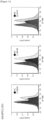
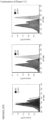
- Figure 5 shows a relationship between the length of CDR3 and the relevant classification type.
- CDR3 having a length of less than 12 amino acids tends to be classified as the upright type, and CDR3 having a large length of 12 amino acids or more tends to be classified as the roll type. It has also been found that the half-roll type has no relation with the CDR3 length.
- CDR3 classified as the upright type tends to contain a framework region at an antigen-binding site.
- CDR3 containing 14 or more amino acids is more likely to be classified as the roll type, and less likely to contain a framework region at an antigen-binding site. Further, it has become evident that the roll structure is stabilized as the framework region and the CDR3 region become closer to each other. Further, it has been found that in the case where 16 or more amino acids are contained in CDR3, the population of CDR3 containing cysteine increases, and the cysteine forms a disulfide bond with cysteine of CDR2 to form more stable CDR3.
- VHH library comprising four sub-libraries: Upright 6, Upright 12, Roll 12 and Roll 15 was designed on the basis of the analysis results for the above-described antigen-binding site, the number of amino acid residues and the structure of CDR3.
- CDR2 of upright type sub-library was formed from 16 amino acid residues
- CDR2 of roll type sub-library was formed from 17 amino acid residues. This is because a strong correlation between the number of amino acid residues constituting CDR2 (16 amino acid residues or 17 amino acid residues) and the formation of the upright type or roll type was confirmed.
- CDR1 consisted of 150 types of sequence sets
- CDR2 consisted of 69 or 71 types of sequence sets
- CDR3 consisted randomly of 17 types of amino acids which do not include three types: methionine, proline and cysteine.
- CDR3 having a length of 16 amino acids or more was not designed. This is because additional formation of a disulfide bond is not desirable from the pharmaceutical point of view.
- the CDR1 to CDR3 designed as described above were, as one set, incorporated into humanized FR.
- framework regions and CDR of VHH in the present library were defined as follows: FR1 (H1-H25), CDR1 (H26-H35), FR2 (H36-H49), CDR2 (H50-H65), FR3 (H66-H94), CDR3 (H95-H102) and FR4 (H103-H113).
- framework sequences were designed on the basis of the above definitions.
- BM positions in VHH were determined on the basis of the structure information.
- the framework region was humanized with reference to the amino acid sequences of three VHHs that are commercially available or used in clinical trial (Caplacizumab, Ozoralizmab and Vobaralizmab).
- DNA fragments comprising one each of CDR1 to CDR3, respectively, were synthesized by overlap extension PCR, and the fragments were ligated using Type IIS restriction enzyme and T4 DNA Ligase, thereby constructing a library comprising an intended full-length VHH sequence ( Figure 7 ).
- CDR1 primer 150 types of CDR1 primers different in the sequence of the CDR1 region were each synthesized, and as the CDR2 primer, 140 types of CDR2 primers different in the sequence of the CDR2 region (Upright type: 71 types, Roll type: 69 types) were each synthesized. These primers were mixed and used.
- a primer corresponding to CDR3 in each library was designed so as to control the occurrence frequency of the amino acid using a trimer oligonucleotide, and its synthesis was outsourced to Ella Biotech GmbH.
- DNA fragments containing each CDR were synthesized, where by overlap extension PCR, a CDR1 fragment was synthesized using the FR1 primer and the CDR1 primer, a CDR2 fragment was synthesized using the FR2 primer and the CDR2 primer, and a CDR3 fragment was synthesized using the CDR3 primer and the FR4 primer.
- the reaction composition was set to 125 ⁇ L of PrimeSTAR MAX PreMIX (2 ⁇ ) (Takara Bio Inc.), 5 ⁇ L of each of 10 ⁇ M forward/reverse primers and 115 ⁇ L of ultrapure water. However, these amounts were all doubled in the synthesis of the CDR3 fragment. Table 3 shows the reaction conditions.
- the CDR1 fragment and the CDR2 fragment were treated with a restriction enzyme at 55°C for 15 minutes using BsmB I (NEB).
- the DNA fragments treated with a restriction enzyme were purified by a routine method, and ligated at 16°C for 16 hours with T4 DNA Ligase (Takara Bio Inc.) to synthesize a connected body of the fragments, i.e., a CDR1-CDR2 fragment, as a ligation product.
- the ligation product was migrated at 200 V for 40 minutes with 8 M urea PAGE-gel to purify an intended product region, and PCR amplification was then performed 10 cycles at an annealing temperature of 62°C using an outer primer.
- the amplified CDR1-2 fragment was purified, and then treated with a restriction enzyme at 60°C for 15 minutes using BtgZ I (NEB).
- the CDR3 fragment was treated with a restriction enzyme at 55°C for 15 minutes using BsmBI, and purified with AMPure XP to obtain a purified CDR3 fragment.
- the purified CDR1-2 fragment and the purified CDR3 fragment were ligated at 16°C for 16 hours with T4 DNA Ligase to obtain a full-length VHH sequence.
- the ligation product was purified with the 8 M urea-denatured PAGE gel, and collected to construct a library.
- nucleotide sequence and the amino acid sequence in the library were analyzed using a next-generation sequencer (NGS Inc.).
- PCR amplification was performed on the above-described libraries.
- the composition of the reaction solution (total 25 ⁇ L) was set to 12.5 ⁇ L of PrimeSTAR MAX PreMIX (2 ⁇ ), 1 ⁇ L of a library sample, 0.5 ⁇ L of each of 20 ⁇ M forward/reverse primers (PL_prRd-N4_NL_FW, PL_prRd-N4_NL_RV), and 10.5 ⁇ L of ultrapure water.
- the reaction conditions were the same as in Table 3 above except that the annealing temperature was 62°C, the extension time was 5 seconds, and the cycle number for denaturation was 8.
- the obtained PCR product was purified using AMPureXP, and Index PCR was performed using the purified product as a template (reaction conditions are the same as the PCR conditions except that the annealing temperature was changed to 52°C).
- the composition of the reaction solution (total 25 ⁇ L) was set to 12.5 ⁇ L of PrimeSTAR MAX PreMIX (2 ⁇ ), 1 ⁇ L of the PCR product, 2 ⁇ L of each of 5 ⁇ M forward/reverse primers (Nextera XT Index 1 Primers (N7XX), Nextera XT Index 1 Primers (S5XX)), and 7.5 ⁇ L of ultrapure water.
- the Index PCR product was purified using AMPure as described above, and the DNA concentration was measured using NanoPad DS-11. Thereafter, the Index PCR purified product was diluted to a concentration of 10 nM, and the products were then collected together in one tube in an amount of 5 ⁇ L per product, and mixed.
- NGS In NGS here, analysis was performed using MiSeq Reagent Kit v3 (600-cycle) (Illumina, Inc.). A reagent cartridge was dissolved in deionized water. A 10 nM NGS sample library was diluted to a 4 nM library with a resuspension buffer. 5 ⁇ L of 0.2 M NaOH was added to 5 ⁇ L of the 4 nM library, the mixture was vortexed, and reacted at room temperature for 5 minutes, and 990 ⁇ L of ice-cooled HT1 was then added to prepare a 20 pM library. To 450 ⁇ L of the 20 pM library, 150 ⁇ L of ice-cooled HT1 was added to prepare a 15 pM sample library.
- a PhiX library to be used as a control DNA library was similarly prepared as a 15 pM library. Finally, 570 ⁇ L of the 15 pM sample library and 30 ⁇ L of the 15 pM PhiX library were mixed, the reagent cartridge was loaded with the ultimate NGS sample library, and the reagent cartridge was set in Miseq, followed by analysis. The obtained data was analyzed to confirm that each library had a nucleotide sequence consistent with the design.
- a cnvK linker for selection was prepared as follows.
- the sequence of a biotin fragment that forms a main chain was a sequence represented by SEQ ID NO: 33 below.
- BioTEG is bound to the 5'-terminus of the biotin fragment (main chain), and in the nucleotide sequence, N at the 3-position denotes guanosine, and N at the 12-position denotes 3-cyanovinylcarbazole.
- CCT was bound via Amino C6-dT.
- a puromycin segment that forms a side chain has the sequence of 5'(5S)TcTCFCZZCCP.
- P at the free terminus in the side chain sequence denotes puromycin as a protein-binding site.
- (5S) denotes 5' Thiol C6, c denotes cytidine, F denotes FITC-dT, and Z denotes Spacer 18.
- the chemical synthesis of the main chain and the side chain was outsourced to Tsukuba Oligo Service Co., Ltd.
- the reduced puromycin segment solution after buffer exchange was mixed with an ethanol-precipitated product of the EMCS-modified biotin fragment, and the mixture was left to stand overnight at 4°C. Subsequently, DTT was added to the reaction liquid at a final concentration of 50 mM, and the mixture was stirred at room temperature for 30 minutes. Thereafter, ethanol precipitation was performed using Quick-Precip Plus Solution (manufactured by Edge BioSystems Inc.). The ethanol-precipitated product was dissolved in 100 ⁇ L of Nuclease-free water (manufactured by nacalai tesque).
- the lysate was separated by 12% polyacrylamide gel electrophoresis, and cnvK rC Linker fractions were isolated.
- the isolated gel was crushed using BioMasher II Set (Nippi Inc.), 500 ⁇ L of Nuclease-free water was added, the mixture was stirred overnight at 4°C, and cnvK rC Linker was extracted.
- the stirred solution was transferred into a Costar (registered trademark) Spin-X (registered trademark) centrifuging tube filter, 0.22 ⁇ m cellulose acetate (Corning Inc.), and then centrifuged at 16,000 xg for 15 minutes to separate the gel and the extracted liquid.
- cnvK rC Linker was dissolved in Nuclease-free water, and stored at -20°C.
- Antigens used here as a target were HSA (human serum albumin), HER2 (human epidermal growth factor receptor 2), GPC3 (Glypican 3) and GPC1 (Glypican 1).
- HSA human serum albumin
- HER2 human epidermal growth factor receptor 2
- GPC3 Glypican 3
- GPC1 Glypican 1
- RNA concentration was measured using NanoPad DS-11, reagents were mixed at a final concentration of 200 mM for NaCl, 50 mM for Tris-HCl (pH 7.5), 1 ⁇ M for mRNA, and 1 ⁇ M for cnvK linker, and the mixture was heat-treated with the following program: (s1) at 90°C for 1 minute, then (s2) at 70°C for 1 minute and then (s3) at 4°C, to hybridize a linker for synthesis of cDNA display to mRNA.
- the mRNA-linker connected bodies of the sub-libraries were mixed to obtain a mixture.
- a cell-free translation reaction (which may be referred to as "IVV" hereinafter) was carried out at 37°C for 30 minutes using a reconstructing cell-free translation solution Purefrex 1.0 (Gene Frontier Corporation) and a DsbC set (Gene Frontier Corporation) as a SS binding auxiliary factor.
- the composition of the reaction liquid was set to 1,600 ⁇ L of solution I, 160 ⁇ L of each of solution II and solution III, 800 ⁇ L of a 1 pmol/ ⁇ L ligation product, 160 ⁇ L of a DsbC set diluted 4-fold, 160 ⁇ L of 20 mM GSSG and 160 ⁇ L of 80 mM GSH.
- 1,920 ⁇ L of an IVV formation buffer was added, and the mixture was then reacted at 37°C for 30 minutes to covalently bind the translated product to the linker via puromycin.
- 1,280 ⁇ L of 0.5 M EDTA was added, and the mixture was reacted at 4°C for 10 minutes to separate ribosome from the nucleic acid.
- a reverse transcription reaction solution comprising 1,280 ⁇ L of a 5 ⁇ RT buffer, 256 ⁇ L of 25 mM dNTPs, 128 ⁇ L of reverse transcriptase and 4,736 ⁇ L of ultrapure water was then added, and the mixture was reacted at 42°C for 1 hour to synthesize a cDNA display product (cDNA-mRNA-linker-peptide conjugate).
- the cDNA display product obtained as described above was eluted from the beads with 640 ⁇ L of an elution buffer containing RNase T1 (manufactured by Thermo Fisher Scientific). Thereafter, using 20 ⁇ L of His-Mag Sepharose (manufactured by Cytiva Company), His tag purification was performed on the cDNA display product to obtain 200 ⁇ L of a cDNA display molecule (cDNA-linker-peptide connected body).
- the cDNA display molecule and a biotinylated target protein were mixed in amounts shown in Table 4, and the mixture was incubated at 4°C for 30 minutes. Thereafter, the reaction liquid was mixed with 120 ⁇ L of washed Dynabeads Streptavidin MyOne C1, and the mixture was incubated at 4°C for 30 minute, and the binding cDNA display molecules were immobilized on beads via the biotinylated target protein.
- HSA biotinylated target protein
- the coated beads (cDNA display-biotinylated target protein streptavidin-beads) were washed with 200 ⁇ L of PBS-T three times (the tube was replaced before the third washing), then eluted twice with 100 ⁇ L of an elution buffer, and only nucleic acid regions of the cDNA display molecule, which have ability to bind to the target protein, were collected.
- the obtained eluted sample was purified using AMPure XP.
- in vitro selection was performed three or four rounds.
- the mixing was performed with the composition set to 20 ⁇ L of a cDNA display solution, 10 ⁇ L of a 1 mM biotinylated target protein, 2.5 ⁇ L of a 200 mg/mL heparin solution, 0.5 ⁇ L of 10% Tween and 67 ⁇ L of 1 ⁇ PBS so that the concentration of each target protein was 100 nM.
- BSA (FUJIFILM Wako Pure Chemical Corporation) was added at a final concentration of 0.5% for excluding non-specific binding molecules.
- selection was carried out without adding BSA.
- the beads were coated with MyOne T1 in the second round and with MyOne C1 in the third round. As described above, changing the surface characteristics of the beads used for each round was aimed at exclusion of cDNA display molecules binding to the beads in a non-specific manner (see Table 4 below).
- Table 4 Round mRNA/linker connected product (Ligation ; pmol) Target protein concentration (nM) Beads used and its amount ( ⁇ L) 1 st 400 500 MyOne C1 : 120 2 nd 12.5 100 MyOne T1 : 21 3 rd 12.5 100 MyOne C1 : 24
- PCR amplification was performed on DNA eluted in the selection. In the first round, quantitative PCR was not performed, and PCR was performed using the total amount of eluted sample as a template.
- the composition of the reaction solution (total 250 ⁇ L) was set to 125 ⁇ L of PrimeSTAR MAX PreMIX (2 ⁇ ), 100 ⁇ L of the eluted DNA sample, 5 ⁇ L of each of 20 ⁇ M forward/reverse primers (PL_T7pro, NewYtag_cnvk-linker), and 15 ⁇ L of ultrapure water.
- the PCR program was the same as in Table 3 except that the annealing temperature was 60°C, the extension time was 5 seconds, and the cycle number for denaturation or the like was 15.
- the PCR product was purified using AMPure XP.
- the composition of the solution used for quantitative PCR (total 25 ⁇ L) was set to 12.5 ⁇ L of THUNDERBIRD qPCRMix (TOYOBO CO., LTD.), 0.5 ⁇ L of each of 20 ⁇ M forward/reverse primers (PL_T7pro, NewYtag_cnvk-linker), 1 ⁇ L of eluted sample DNA and 10.5 ⁇ L of ultrapure water.
- the PCR program was such that after the initial denaturation at 98°C for 30 seconds, the cycle of denaturation at 98°C for 5 seconds, annealing at 60°C for 10 seconds and extension at 72°C for 35 seconds was repeated 35 times.
- the composition of the reaction solution (total 100 ⁇ L) was set to 50 ⁇ L of PrimeSTAR MAX PreMIX (2 ⁇ ), 25 ⁇ L of the eluted DNA sample, 2 ⁇ L of each of 20 ⁇ M forward/reverse primers (PL_T7pro, NewYtag_cnvk-linker), and 21 ⁇ L of ultrapure water.
- the program was the same as the program shown in Table 3 except that the annealing temperature was 65°C, the extension time was 5 seconds, and the cycle number for denaturation or the like was 15.
- PCR amplification was performed under the following conditions.
- the composition of the reaction liquid (total 25 ⁇ L) was set to 12.5 ⁇ L of PrimeSTAR MAX PreMIX (2 ⁇ ), 1 ⁇ L of the selection product, 0.5 ⁇ L of each of 20 ⁇ M forward/reverse primers (PL_prRd-N4_NL_FW, PL_prRd-N4_Ytag_RV), and 10.5 ⁇ L of ultrapure water.
- the PCR program was the same as in Table 3 except that the annealing temperature was 62°C, the extension time was 5 seconds, and the cycle number for denaturation or the like was 8.
- the obtained PCR product was purified using AMPure XP, and Index PCR was performed using the purified product as a template.
- the composition of the reaction liquid (total 25 ⁇ L) was 12.5 ⁇ L of PrimeSTAR MAX PreMIX (2 ⁇ ), 1 ⁇ L of the selection product, 0.5 ⁇ L of each of 5 ⁇ M forward/reverse primers (Nextera XT Index 1 Primers (N7XX), Nextera XT Index 2 Primers (S5XX)), and 10.5 ⁇ L of ultrapure water.
- the PCR program was the same as in Table 3 except that the annealing temperature was 52°C, the extension time was 5 seconds, and the cycle number for denaturation or the like was 8.
- the Index PCR product was similarly purified using AMPure XP, and the DNA concentration was measured using NanoPad DS-11.
- the Index PCR purification product was diluted to 10 nM, and the products were then collected together in one tube in an amount of 5 ⁇ L per product, and mixed.
- NGS In NGS here, analysis was performed using MiSeq Reagent Kit v3 (600-cycle) (Illumina, Inc.). A reagent cartridge was dissolved in deionized water. A 10 nM NGS sample library was diluted to a 4 nM library with a resuspension buffer. 5 ⁇ L of 0.2 M NaOH was added to 5 ⁇ L of the 4 nM library, the mixture was vortexed, and reacted at room temperature for 5 minutes, and 990 ⁇ L of ice-cooled HT1 was then added to prepare a 20 pM library. To 450 ⁇ L of the 20 pM library, 150 ⁇ L of ice-cooled HT1 was added to prepare a 15 pM sample library.
- a PhiX library to be used as a control DNA library was similarly prepared as a 15 pM library. Finally, 570 ⁇ L of the 15 pM sample library and 30 ⁇ L of the 15 pM PhiX library were mixed to obtain an ultimate NGS sample library. The reagent cartridge was loaded with the ultimate NGS sample library, and the reagent cartridge was set in Miseq, followed by analysis. The obtained NGS analysis data was processed to identify a binding sequence for each target molecule.
- the DNA library encoding VHH selected by the in vitro selection was cloned into a plasmid vector for VHH expression.
- a sequence to be treated with a restriction enzyme is added by PCR to obtain a PCR product.
- the PCR product and a C. glutamicum expression plasmid vector were treated with restriction enzyme BamHI at 37°C for 1 hour, and then with restriction enzyme SfiI at 50°C for 1 hour.
- the VHH DNA product corresponding to Insert was purified with AMPure XP, and the obtained Vector DNA product was subjected to agarose gel electrophoresis at 100 V for 30 minutes, thereby being isolated and purified.
- the Vector DNA purified as described above was subjected to a dephosphorylation reaction at 37°C for 1 hour using FastAP Thermosensitive Alkaline Phosphatase (manufactured by Thermo Fisher Scientific) which is a dephosphorylation enzyme. Thereafter, the Insert DNA and the Vector DNA were mixed at a molar ratio of 1: 10, and a ligation reaction was carried out overnight at 16°C using Ligation high (TOYOBO CO., LTD.), thereby obtaining a plasmid library containing a selected VHH library.
- FastAP Thermosensitive Alkaline Phosphatase manufactured by Thermo Fisher Scientific
- Each plasmid library was introduced into C. glutamicum by electroporation to obtain a transformant.
- the obtained transformant was inoculated into CM2G culture medium, and cultured overnight at 30°C. Thereafter, the culture solution was passed to PM1S culture medium as a culture medium for VHH expression, and cultured at 25°C for 72 hours to secret and express VHH (monomer) in the culture supernatant.
- the culture supernatant was collected by centrifugation at 3,000 xg, and bacterial cells were removed from the supernatant by treatment with a 0.22 ⁇ m filter.
- a His1K sensor chip was coated with the produced VHH clone to measure the activity to bind to each target molecule.
- a ligand was prepared such that the concentration of each target molecule was 400 nM, and 70 ⁇ L of a measurement liquid was added to a 384-well plate to measure the binding of each target molecule.
- the tip of Dip and Read (trademark) His1K Biosensors was immersed in 200 ⁇ L of PBS-T (0.05% Tween 20, pH 7.4) for 10 minutes for hydration of the sensor chip.
- the condition of measurement order in each run measurement was as follows.
- the VHH genes were identified by sequence analysis.
- a transformant taken as a Hit clone was cultured overnight at 37°C, and the culture solution was subjected to colony PCR. DNA purification was performed on the obtained PCR product using AMPure XP. Thereafter, analysis of each DNA was outsourced to Eurofins Genomics K.K.
- those identical in sequence were removed, and the remaining clones were taken as a Unique clone.
- a Unique clone identified by the sequence analysis was pre-cultured overnight at 30°C. Thereafter, the preculture solution was passed to PM1S culture medium as a culture medium for VHH expression, and cultured at 25°C for 72 hours to secret and express VHH (monomer) in the culture supernatant. The culture supernatant was collected by centrifugation, and bacterial cells were removed from the supernatant.
- the above-described sample was purified from the obtained culture supernatant in accordance with the written instructions with the product.
- 100 ⁇ L of an elution buffer 300 mM NaCl, 500 mM imidazole-containing 50 mM Tris-HCl (pH 7.5) was added, and centrifuged at 500 xg at 4°C for 2 minutes. The eluate was collected, and the eluate was used as a purified VHH clone sample.
- a His1K sensor chip was coated with the purified VHH clone in the sample, and the activity to bind to each target molecule was measured.
- seven concentrations of each target molecule in a 2-fold dilution series starting at 200 nM (200 nM, 100 nM, 50 nM, 25 nM, 12.5 nM, 6.25 nM and 3.125 nM) and a no target molecule condition were used, and 70 ⁇ L of each measurement solution was added to a 384-well plate.
- the tip of Dip and Read (trademark) His1K Biosensors was immersed in 200 ⁇ L of PBS-T (0.05% Tween 20, pH 7.4) for 10 minutes, so that the sensor chip had affinity for water.
- PBS-T 0.05% Tween 20, pH 7.4
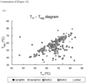
- Tm value thermal denaturation midpoint
- Tagg value aggregation initiation temperature
- the purified VHH clone was applied to 16-well-9 ⁇ L quartz cuvettes, and the cuvettes were set in UNcle, followed by measurement.
- a change in fluorescence of the protein solution and scattered light was measured under measurement conditions conforming to the template set as a default in the equipment, where the initiation temperature was 25°C, the end-point temperature was 95°C, and the temperature was raised at 0.3°C/min.
- the thermal denaturation midpoint (Tm value) was calculated on the basis of a decrease in fluorescence intensity (due to heat denaturation of the protein) and a change in fluorescence spectrum over about 330 nm to 350 nm.
- Example 5 the characteristic of VHH were analyzed in vitro. However, the binding ability in vitro is not necessarily consistent with the binding ability in vivo. Thus, for more accurately evaluating the binding ability of VHH, the ability of VHH to bind to target proteins (GPC3 and HER2) expressed in cells was examined by flow cytometry (FCM).
- FCM flow cytometry
- hepG2 as hepatocyte cancer was selected as cells expressing GPC3, and SK-BR-3 as breast cancers was selected as cells expressing HER2.
- These cells were cultured to 80% confluence (in an incubator at 37°C and 5% CO 2 , light shielded), and collected with PBS containing 5 mM EDTA. The collected cell sample was washed with 1% BSA/PBS, and a VHH solution diluted 100-fold with 1% BSA/PBS was added, followed by incubation at 4°C for 1 hour.
- the cells were washed three times with 1% BSA/PBS, and an AlexaFluor 488-bound anti-His tag antibody diluted 1000-fold (Medical & Biological Laboratory Co., Ltd.) was then added, followed by incubation at 4°C for 1 hour.
- the cells were washed with 1% BSA/PBS, and then resuspended with 1% BSA/PBS, and FCM analysis (SH800, Sony Corporation) was performed.
- the DNA library having a framework according to the present invention has been confirmed to exhibit binding activity both in vitro and in vivo.
- the invention of the present application is useful in the field of drug development.
Landscapes
- Health & Medical Sciences (AREA)
- Chemical & Material Sciences (AREA)
- Life Sciences & Earth Sciences (AREA)
- Organic Chemistry (AREA)
- Molecular Biology (AREA)
- Immunology (AREA)
- Biochemistry (AREA)
- Medicinal Chemistry (AREA)
- Proteomics, Peptides & Aminoacids (AREA)
- Biophysics (AREA)
- General Health & Medical Sciences (AREA)
- Genetics & Genomics (AREA)
- Engineering & Computer Science (AREA)
- Cell Biology (AREA)
- Physics & Mathematics (AREA)
- Biomedical Technology (AREA)
- Urology & Nephrology (AREA)
- Hematology (AREA)
- Oncology (AREA)
- Gastroenterology & Hepatology (AREA)
- Microbiology (AREA)
- Biotechnology (AREA)
- Bioinformatics & Computational Biology (AREA)
- Bioinformatics & Cheminformatics (AREA)
- Food Science & Technology (AREA)
- Analytical Chemistry (AREA)
- General Physics & Mathematics (AREA)
- Pathology (AREA)
- Virology (AREA)
- Chemical Kinetics & Catalysis (AREA)
- General Chemical & Material Sciences (AREA)
- Peptides Or Proteins (AREA)
Applications Claiming Priority (2)
| Application Number | Priority Date | Filing Date | Title |
|---|---|---|---|
| JP2021195792 | 2021-12-01 | ||
| PCT/JP2022/044235 WO2023100944A1 (ja) | 2021-12-01 | 2022-11-30 | ランダム領域搭載用フレームワーク配列を有するペプチド及びそのペプチドで構成されるペプチドライブラリ |
Publications (1)
| Publication Number | Publication Date |
|---|---|
| EP4442704A1 true EP4442704A1 (en) | 2024-10-09 |
Family
ID=86612245
Family Applications (1)
| Application Number | Title | Priority Date | Filing Date |
|---|---|---|---|
| EP22901376.8A Ceased EP4442704A1 (en) | 2021-12-01 | 2022-11-30 | Peptide having framework sequence for random region placement and peptide library composed of said peptide |
Country Status (5)
| Country | Link |
|---|---|
| US (1) | US20250019460A1 (https=) |
| EP (1) | EP4442704A1 (https=) |
| JP (1) | JPWO2023100944A1 (https=) |
| CN (1) | CN118339180A (https=) |
| WO (1) | WO2023100944A1 (https=) |
Family Cites Families (6)
| Publication number | Priority date | Publication date | Assignee | Title |
|---|---|---|---|---|
| JP4213586B2 (ja) | 2001-09-13 | 2009-01-21 | 株式会社抗体研究所 | ラクダ抗体ライブラリーの作製方法 |
| WO2006040153A2 (en) * | 2004-10-13 | 2006-04-20 | Ablynx N.V. | Single domain camelide anti -amyloid beta antibodies and polypeptides comprising the same for the treatment and diagnosis of degenarative neural diseases such as alzheimer's disease |
| EP2699603B1 (en) * | 2011-04-19 | 2016-03-02 | The United States of America As Represented by the Secretary Department of Health and Human Services | Human monoclonal antibodies specific for glypican-3 and use thereof |
| CN111699200B (zh) * | 2018-01-15 | 2023-05-26 | 南京传奇生物科技有限公司 | 针对pd-1的单域抗体和其变体 |
| CN113166261A (zh) * | 2018-10-11 | 2021-07-23 | 印希比股份有限公司 | B7h3单域抗体及其治疗性组合物 |
| CN115068625B (zh) * | 2021-03-16 | 2024-07-02 | 中国科学院上海药物研究所 | Pd-l1和tlr7双靶向纳米抗体偶联药物及其在抗肿瘤中的应用 |
-
2022
- 2022-11-30 JP JP2023565059A patent/JPWO2023100944A1/ja active Pending
- 2022-11-30 WO PCT/JP2022/044235 patent/WO2023100944A1/ja not_active Ceased
- 2022-11-30 CN CN202280079930.3A patent/CN118339180A/zh active Pending
- 2022-11-30 EP EP22901376.8A patent/EP4442704A1/en not_active Ceased
- 2022-11-30 US US18/714,591 patent/US20250019460A1/en active Pending
Also Published As
| Publication number | Publication date |
|---|---|
| WO2023100944A1 (ja) | 2023-06-08 |
| US20250019460A1 (en) | 2025-01-16 |
| JPWO2023100944A1 (https=) | 2023-06-08 |
| CN118339180A (zh) | 2024-07-12 |
Similar Documents
| Publication | Publication Date | Title |
|---|---|---|
| US12460018B2 (en) | System for generating immunoglobulin scaffolds for grafting non-human CDR amino acid sequences | |
| Feng et al. | Construction and next-generation sequencing analysis of a large phage-displayed VNAR single-domain antibody library from six naive nurse sharks | |
| JP2023524733A (ja) | 遺伝子発現を活性化及びサイレンシングするためのエフェクタードメインの作出、同定及び性質決定のための組成物、システム及び方法 | |
| KR102810907B1 (ko) | 항체 라이브러리 | |
| EP2647704B1 (en) | Polynucleotide construct capable of presenting fab in acellular translation system, and method for manufacturing and screening fab using same | |
| JP6073038B2 (ja) | 安定化された免疫グロブリン可変ドメイン選別方法及び選別されたドメインの応用 | |
| EP4130260A1 (en) | Construction method and application of antigen-specific binding polypeptide gene display vector | |
| JP2015531597A5 (https=) | ||
| CN111566262A (zh) | 动态人抗体轻链文库 | |
| JP7525123B2 (ja) | Dnaポリメラーゼの5’→3’エキソヌクレアーゼ活性ドメインに特異的に結合する抗体 | |
| EP4442704A1 (en) | Peptide having framework sequence for random region placement and peptide library composed of said peptide | |
| KR102194203B1 (ko) | 항체 나이브 라이브러리의 생성 방법, 상기 라이브러리 및 그 적용(들) | |
| JP2019068837A (ja) | 標的分子に結合するポリペプチドの決定方法及び決定システム | |
| Rothe et al. | Selection of human anti-CD28 scFvs from a T-NHL related scFv library using ribosome display | |
| EP4430398B1 (en) | METHODS FOR IDENTIFYING ANTIGEN-BINDING MOLECULES | |
| Fuji et al. | Discovery of IL-5-binding unnatural cyclic peptides from multiple libraries by directed evolution | |
| WO2009109572A2 (en) | Monovalent phage display of single variable domains | |
| JP2024075076A (ja) | 新規変異導入方法による進化ライブラリの作成およびそのライブラリを用いたペプチドスクリーニング方法 | |
| JP6293829B2 (ja) | 安定化された免疫グロブリン可変ドメイン選別方法及び選別されたドメインの応用 | |
| CN121717907A (zh) | 人bcma抗体及其应用 | |
| KR20260011157A (ko) | 항체 점도를 낮추는 방법 및 조성물 | |
| WO2023074749A1 (ja) | 抗体代替分子のスクリーニング方法 | |
| Douthwaite et al. | Accelerated protein evolution using ribosome display |
Legal Events
| Date | Code | Title | Description |
|---|---|---|---|
| STAA | Information on the status of an ep patent application or granted ep patent |
Free format text: STATUS: THE INTERNATIONAL PUBLICATION HAS BEEN MADE |
|
| PUAI | Public reference made under article 153(3) epc to a published international application that has entered the european phase |
Free format text: ORIGINAL CODE: 0009012 |
|
| STAA | Information on the status of an ep patent application or granted ep patent |
Free format text: STATUS: REQUEST FOR EXAMINATION WAS MADE |
|
| 17P | Request for examination filed |
Effective date: 20240627 |
|
| AK | Designated contracting states |
Kind code of ref document: A1 Designated state(s): AL AT BE BG CH CY CZ DE DK EE ES FI FR GB GR HR HU IE IS IT LI LT LU LV MC ME MK MT NL NO PL PT RO RS SE SI SK SM TR |
|
| RAP1 | Party data changed (applicant data changed or rights of an application transferred) |
Owner name: EPSILON MOLECULAR ENGINEERING INC. |
|
| REG | Reference to a national code |
Ref country code: DE Ref legal event code: R003 |
|
| STAA | Information on the status of an ep patent application or granted ep patent |
Free format text: STATUS: THE APPLICATION HAS BEEN REFUSED |
|
| DAV | Request for validation of the european patent (deleted) | ||
| DAX | Request for extension of the european patent (deleted) | ||
| 18R | Application refused |
Effective date: 20241128 |