EP4406505A2 - Computerprogramm und vorrichtung zur analyse einer zahnsituation - Google Patents
Computerprogramm und vorrichtung zur analyse einer zahnsituation Download PDFInfo
- Publication number
- EP4406505A2 EP4406505A2 EP24180883.1A EP24180883A EP4406505A2 EP 4406505 A2 EP4406505 A2 EP 4406505A2 EP 24180883 A EP24180883 A EP 24180883A EP 4406505 A2 EP4406505 A2 EP 4406505A2
- Authority
- EP
- European Patent Office
- Prior art keywords
- dental scene
- scene
- dental
- virtual
- updated
- Prior art date
- Legal status (The legal status is an assumption and is not a legal conclusion. Google has not performed a legal analysis and makes no representation as to the accuracy of the status listed.)
- Pending
Links
Images
Classifications
-
- G—PHYSICS
- G06—COMPUTING OR CALCULATING; COUNTING
- G06T—IMAGE DATA PROCESSING OR GENERATION, IN GENERAL
- G06T7/00—Image analysis
- G06T7/0002—Inspection of images, e.g. flaw detection
- G06T7/0012—Biomedical image inspection
-
- A—HUMAN NECESSITIES
- A61—MEDICAL OR VETERINARY SCIENCE; HYGIENE
- A61B—DIAGNOSIS; SURGERY; IDENTIFICATION
- A61B34/00—Computer-aided surgery; Manipulators or robots specially adapted for use in surgery
- A61B34/10—Computer-aided planning, simulation or modelling of surgical operations
-
- A—HUMAN NECESSITIES
- A61—MEDICAL OR VETERINARY SCIENCE; HYGIENE
- A61B—DIAGNOSIS; SURGERY; IDENTIFICATION
- A61B5/00—Measuring for diagnostic purposes; Identification of persons
- A61B5/0059—Measuring for diagnostic purposes; Identification of persons using light, e.g. diagnosis by transillumination, diascopy, fluorescence
- A61B5/0082—Measuring for diagnostic purposes; Identification of persons using light, e.g. diagnosis by transillumination, diascopy, fluorescence adapted for particular medical purposes
- A61B5/0088—Measuring for diagnostic purposes; Identification of persons using light, e.g. diagnosis by transillumination, diascopy, fluorescence adapted for particular medical purposes for oral or dental tissue
-
- A—HUMAN NECESSITIES
- A61—MEDICAL OR VETERINARY SCIENCE; HYGIENE
- A61B—DIAGNOSIS; SURGERY; IDENTIFICATION
- A61B5/00—Measuring for diagnostic purposes; Identification of persons
- A61B5/74—Details of notification to user or communication with user or patient; User input means
- A61B5/742—Details of notification to user or communication with user or patient; User input means using visual displays
-
- A—HUMAN NECESSITIES
- A61—MEDICAL OR VETERINARY SCIENCE; HYGIENE
- A61C—DENTISTRY; APPARATUS OR METHODS FOR ORAL OR DENTAL HYGIENE
- A61C1/00—Dental machines for boring or cutting ; General features of dental machines or apparatus, e.g. hand-piece design
- A61C1/08—Machine parts specially adapted for dentistry
- A61C1/082—Positioning or guiding, e.g. of drills
-
- A—HUMAN NECESSITIES
- A61—MEDICAL OR VETERINARY SCIENCE; HYGIENE
- A61C—DENTISTRY; APPARATUS OR METHODS FOR ORAL OR DENTAL HYGIENE
- A61C7/00—Orthodontics, i.e. obtaining or maintaining the desired position of teeth, e.g. by straightening, evening, regulating, separating, or by correcting malocclusions
- A61C7/002—Orthodontic computer assisted systems
-
- A—HUMAN NECESSITIES
- A61—MEDICAL OR VETERINARY SCIENCE; HYGIENE
- A61C—DENTISTRY; APPARATUS OR METHODS FOR ORAL OR DENTAL HYGIENE
- A61C9/00—Impression cups, i.e. impression trays; Impression methods
- A61C9/004—Means or methods for taking digitized impressions
-
- G—PHYSICS
- G06—COMPUTING OR CALCULATING; COUNTING
- G06T—IMAGE DATA PROCESSING OR GENERATION, IN GENERAL
- G06T17/00—Three dimensional [3D] modelling, e.g. data description of 3D objects
-
- G—PHYSICS
- G06—COMPUTING OR CALCULATING; COUNTING
- G06T—IMAGE DATA PROCESSING OR GENERATION, IN GENERAL
- G06T19/00—Manipulating 3D models or images for computer graphics
- G06T19/006—Mixed reality
-
- G—PHYSICS
- G06—COMPUTING OR CALCULATING; COUNTING
- G06T—IMAGE DATA PROCESSING OR GENERATION, IN GENERAL
- G06T7/00—Image analysis
- G06T7/0002—Inspection of images, e.g. flaw detection
- G06T7/0012—Biomedical image inspection
- G06T7/0014—Biomedical image inspection using an image reference approach
- G06T7/0016—Biomedical image inspection using an image reference approach involving temporal comparison
-
- G—PHYSICS
- G16—INFORMATION AND COMMUNICATION TECHNOLOGY [ICT] SPECIALLY ADAPTED FOR SPECIFIC APPLICATION FIELDS
- G16H—HEALTHCARE INFORMATICS, i.e. INFORMATION AND COMMUNICATION TECHNOLOGY [ICT] SPECIALLY ADAPTED FOR THE HANDLING OR PROCESSING OF MEDICAL OR HEALTHCARE DATA
- G16H30/00—ICT specially adapted for the handling or processing of medical images
- G16H30/40—ICT specially adapted for the handling or processing of medical images for processing medical images, e.g. editing
-
- G—PHYSICS
- G16—INFORMATION AND COMMUNICATION TECHNOLOGY [ICT] SPECIALLY ADAPTED FOR SPECIFIC APPLICATION FIELDS
- G16H—HEALTHCARE INFORMATICS, i.e. INFORMATION AND COMMUNICATION TECHNOLOGY [ICT] SPECIALLY ADAPTED FOR THE HANDLING OR PROCESSING OF MEDICAL OR HEALTHCARE DATA
- G16H50/00—ICT specially adapted for medical diagnosis, medical simulation or medical data mining; ICT specially adapted for detecting, monitoring or modelling epidemics or pandemics
- G16H50/20—ICT specially adapted for medical diagnosis, medical simulation or medical data mining; ICT specially adapted for detecting, monitoring or modelling epidemics or pandemics for computer-aided diagnosis, e.g. based on medical expert systems
-
- G—PHYSICS
- G16—INFORMATION AND COMMUNICATION TECHNOLOGY [ICT] SPECIALLY ADAPTED FOR SPECIFIC APPLICATION FIELDS
- G16H—HEALTHCARE INFORMATICS, i.e. INFORMATION AND COMMUNICATION TECHNOLOGY [ICT] SPECIALLY ADAPTED FOR THE HANDLING OR PROCESSING OF MEDICAL OR HEALTHCARE DATA
- G16H50/00—ICT specially adapted for medical diagnosis, medical simulation or medical data mining; ICT specially adapted for detecting, monitoring or modelling epidemics or pandemics
- G16H50/50—ICT specially adapted for medical diagnosis, medical simulation or medical data mining; ICT specially adapted for detecting, monitoring or modelling epidemics or pandemics for simulation or modelling of medical disorders
-
- A—HUMAN NECESSITIES
- A61—MEDICAL OR VETERINARY SCIENCE; HYGIENE
- A61B—DIAGNOSIS; SURGERY; IDENTIFICATION
- A61B34/00—Computer-aided surgery; Manipulators or robots specially adapted for use in surgery
- A61B34/10—Computer-aided planning, simulation or modelling of surgical operations
- A61B2034/101—Computer-aided simulation of surgical operations
- A61B2034/102—Modelling of surgical devices, implants or prosthesis
- A61B2034/104—Modelling the effect of the tool, e.g. the effect of an implanted prosthesis or for predicting the effect of ablation or burring
-
- A—HUMAN NECESSITIES
- A61—MEDICAL OR VETERINARY SCIENCE; HYGIENE
- A61B—DIAGNOSIS; SURGERY; IDENTIFICATION
- A61B34/00—Computer-aided surgery; Manipulators or robots specially adapted for use in surgery
- A61B34/10—Computer-aided planning, simulation or modelling of surgical operations
- A61B2034/101—Computer-aided simulation of surgical operations
- A61B2034/105—Modelling of the patient, e.g. for ligaments or bones
-
- A—HUMAN NECESSITIES
- A61—MEDICAL OR VETERINARY SCIENCE; HYGIENE
- A61B—DIAGNOSIS; SURGERY; IDENTIFICATION
- A61B90/00—Instruments, implements or accessories specially adapted for surgery or diagnosis and not covered by any of the groups A61B1/00 - A61B50/00, e.g. for luxation treatment or for protecting wound edges
- A61B90/36—Image-producing devices or illumination devices not otherwise provided for
- A61B2090/364—Correlation of different images or relation of image positions in respect to the body
-
- A—HUMAN NECESSITIES
- A61—MEDICAL OR VETERINARY SCIENCE; HYGIENE
- A61B—DIAGNOSIS; SURGERY; IDENTIFICATION
- A61B90/00—Instruments, implements or accessories specially adapted for surgery or diagnosis and not covered by any of the groups A61B1/00 - A61B50/00, e.g. for luxation treatment or for protecting wound edges
- A61B90/36—Image-producing devices or illumination devices not otherwise provided for
- A61B90/37—Surgical systems with images on a monitor during operation
- A61B2090/372—Details of monitor hardware
-
- A—HUMAN NECESSITIES
- A61—MEDICAL OR VETERINARY SCIENCE; HYGIENE
- A61B—DIAGNOSIS; SURGERY; IDENTIFICATION
- A61B90/00—Instruments, implements or accessories specially adapted for surgery or diagnosis and not covered by any of the groups A61B1/00 - A61B50/00, e.g. for luxation treatment or for protecting wound edges
- A61B90/50—Supports for surgical instruments, e.g. articulated arms
- A61B2090/502—Headgear, e.g. helmet, spectacles
-
- A—HUMAN NECESSITIES
- A61—MEDICAL OR VETERINARY SCIENCE; HYGIENE
- A61B—DIAGNOSIS; SURGERY; IDENTIFICATION
- A61B34/00—Computer-aided surgery; Manipulators or robots specially adapted for use in surgery
- A61B34/25—User interfaces for surgical systems
-
- A—HUMAN NECESSITIES
- A61—MEDICAL OR VETERINARY SCIENCE; HYGIENE
- A61B—DIAGNOSIS; SURGERY; IDENTIFICATION
- A61B90/00—Instruments, implements or accessories specially adapted for surgery or diagnosis and not covered by any of the groups A61B1/00 - A61B50/00, e.g. for luxation treatment or for protecting wound edges
- A61B90/36—Image-producing devices or illumination devices not otherwise provided for
- A61B90/361—Image-producing devices, e.g. surgical cameras
-
- A—HUMAN NECESSITIES
- A61—MEDICAL OR VETERINARY SCIENCE; HYGIENE
- A61C—DENTISTRY; APPARATUS OR METHODS FOR ORAL OR DENTAL HYGIENE
- A61C1/00—Dental machines for boring or cutting ; General features of dental machines or apparatus, e.g. hand-piece design
- A61C1/08—Machine parts specially adapted for dentistry
- A61C1/082—Positioning or guiding, e.g. of drills
- A61C1/084—Positioning or guiding, e.g. of drills of implanting tools
-
- G—PHYSICS
- G06—COMPUTING OR CALCULATING; COUNTING
- G06T—IMAGE DATA PROCESSING OR GENERATION, IN GENERAL
- G06T2207/00—Indexing scheme for image analysis or image enhancement
- G06T2207/30—Subject of image; Context of image processing
- G06T2207/30004—Biomedical image processing
- G06T2207/30036—Dental; Teeth
-
- G—PHYSICS
- G06—COMPUTING OR CALCULATING; COUNTING
- G06T—IMAGE DATA PROCESSING OR GENERATION, IN GENERAL
- G06T2210/00—Indexing scheme for image generation or computer graphics
- G06T2210/41—Medical
Definitions
- the present invention relates to the analysis of a dental situation of a patient.
- This assessment generally requires a comparison of the actual dental situation with a theoretical dental situation. For example, to diagnose the detachment of a bracket from an orthodontic appliance, it may be necessary to compare the actual dental situation in which the bracket is abnormally pulled away from the tooth to which it is supposed to be bonded, with a Theoretical dental situation in which said attachment is actually bonded to said tooth. This need exists in particular during tailor-made treatments, for which the attachments are shaped to adapt precisely to the morphology of the teeth and/or according to the specific prescriptions of the dental professional.
- the dental professional also makes a comparison between a theoretical dental situation and a real dental situation when seeking to assess whether a tooth is abnormally positioned or has an abnormal shape.
- the actual position or shape or dimension of the tooth must in particular be compared with a theoretical position or shape or dimension that the tooth should present at the time the dental professional makes the comparison.
- the analysis of the real dental situation must sometimes be carried out very quickly, even in real time.
- the dental professional intervenes using a tool to modify the shape or appearance of a tooth
- the action he exerts on the tool must be precise and constantly adapt to a real dental situation which is changing.
- the quality of the comparison between the real dental situation and the theoretical dental situation has an impact on the quality of the treatment, whether therapeutic or not.
- An aim of the invention is to meet, at least partially, these needs.
- step B) consists of a simulation, for a simulation instant, of the real dental scene represented on the updated image, then in a determination of a virtual dental scene according to said simulation .
- the process When the process is implemented by a dental professional, it allows him to improve the quality of his intervention on the patient. When it is implemented by the patient in particular, it makes it possible to realistically simulate the effect of a dental treatment or a modification of a dental treatment, which improves compliance.
- step C) the virtual dental scene is presented on a transparent screen, superimposed with the real dental scene that an operator can observe through the screen, in particular when he is facing the screen.
- the screen and the camera are immobilized relative to the operator.
- they are integrated into glasses.
- the method according to the invention is then particularly useful so that a dental professional wearing the glasses can evaluate, in real time, the dental situation of a patient he is examining.
- a method according to the invention can be used to evaluate in real time an abrasion or milling operation, or an operation of positioning an orthodontic appliance or a part of an orthodontic appliance.
- step C the virtual dental scene is presented, superimposed with the representation of the real dental scene on the updated image, on an opaque screen, that is to say through which the operator cannot see.
- the screen is that of a mobile phone, a tablet, a laptop, or a virtual reality headset.
- the screen can also be the glass of a mirror, preferably equipped with at least one camera.
- the operator manipulates a mobile phone, a laptop, a virtual reality headset or a mirror equipped with at least one camera, to acquire said updated image and visualize the real dental scene and the virtual dental scene.
- the dental professional can work on the patient's arch while looking at the screen. He then sees simultaneously on the screen the images acquired by the camera and the information provided by the virtual dental scene. In particular, he can work “blindly” (that is to say without seeing, directly or through a screen, the real dental scene), on regions of the arch that he cannot observe directly but can be observed by the camera, in particular when the camera is independent of the screen and, in particular when the camera can be introduced into the patient's mouth.
- the virtual dental scene is determined as a function of a value of at least one treatment parameter modifiable by interaction with said mobile phone, said laptop, said headset of virtual reality, the said camera or said mirror or said glasses, preferably with the cell phone.
- the treatment parameter relates to the wearing of an orthodontic appliance by the patient and/or compliance with a treatment instruction by the patient.
- a method according to the invention in particular according to this aspect of the invention, can facilitate the patient's decisions, in particular because it allows him, by choosing the simulation instant, to visualize the effect, on his appearance, of the different possible treatment options.
- the process can also be an effective educational tool to improve compliance.
- the virtual dental scene is determined by means of a three-dimensional digital model digitally modeling at least one arch of the patient, called a “reference model”.
- the reference model also serves, in step C), to position the virtual dental scene in relation to the real dental scene or its representation.
- composition of the virtual dental scene can thus be entirely automated.
- Step c) is a special case of step A).
- the optional characteristics applicable to step A) are therefore applicable to step c).
- Steps a), b) and d) together constitute a special case of step B).
- the optional characteristics applicable to step B) are therefore applicable to steps a), b) and d).
- Step e) is a special case of step C).
- the optional characteristics applicable to step C) are therefore applicable to step e).
- an arch of the patient or a physical model of said arch is scanned while said arch or said physical model of said arch wears or does not wear an orthodontic appliance, and in particular while said arch or said physical model of said arch does not wear orthodontic appliances.
- the updated instant may be different from the reference instant, and in particular be more than 3 days later than the reference instant.
- the method can then be used to visualize changes in the arch between these two times.
- the virtual dental scene is constituted by the reference image.
- the reference model can provide access to “hidden” information, that is to say which is not accessible or which is difficult to access for the operator.
- This hidden information may in particular depend on the reference image, and therefore on the updated image.
- at least part of the hidden information is selected in step d). Step d) thus makes it possible to create a virtual dental scene which contains such information and step e) makes it possible to present it on the real dental scene or on its representation.
- a method according to the invention can be used in particular for non-therapeutic purposes, in particular for research purposes, for example to evaluate the effectiveness of a treatment or an orthodontic appliance, or for aesthetic purposes, or for educational purposes.
- the virtual dental scene may be relative to the real dental scene as simulated at a simulation time or, for the past, as observed at a simulation time.
- the virtual dental scene can simulate the real dental scene at the simulation time.
- the simulation is preferably obtained by means of a computer, preferably by means of an artificial intelligence algorithm.
- the method according to the invention is then particularly useful so that an operator, and in particular a patient, can visualize a dental situation at a simulation moment prior or, preferably, subsequent to the updated moment, or visualize the evolution of such a dental situation by varying the simulation time.
- the method comprises a series of cycles of steps A) to C), or c) to e), the virtual dental scene being determined, at each cycle, to simulate a dental situation at the current simulation instant .
- the virtual dental scene is determined by means of a dynamic simulation tool configured to provide at least one element of the virtual dental scene as a function of a determined simulation instant, in particular as a function of a simulation time after the updated time.
- the method can be used, in particular for educational purposes, to visualize the effect of a modification of the frequency and/or the duration and/or the technique of brushing the teeth, or the effect of a delay in changing your orthodontic splint and/or a delay in making an appointment with the dental professional.
- it can be used to simulate, at the simulation time, the color and/or position of the patient's teeth.
- it can be used to visualize the effect of a teeth whitening treatment or the effect of cigarettes.
- the reference model can be modified to simulate the effect of the passage of time between the reference instant and the simulation instant, and in particular the effect of dental treatment, therapeutic or not, between these moments.
- a method according to the invention can be used in particular to visualize an abrasion suffered by a tooth or a deformation of the gum between the reference and updated instants or between the simulation instant and the updated instant, or guide the implementation placement of an orthodontic appliance or part of an orthodontic appliance or a dental implant or a decorative item.
- the virtual dental scene is relative to the real dental scene, preferably representing at least in part the real dental scene as it appears at the updated moment or as it should appear at the moment. instant updated according to a simulation.
- the virtual dental scene represents the patient's teeth, it represents them in their position at the updated moment or in their simulated position at the updated moment.
- the simulation instant is therefore identical to or close to the updated instant, for example separated from the updated instant by less than 1 month, less than 2 weeks or less than one week.
- the invention also relates to a device for implementing a method according to the invention.
- the camera is configured so as to acquire a succession of updated images of the real dental scene and transmit said updated images to the processing unit.
- the virtual dental scene is presented transparently on the real dental scene or on its representation in the updated image.
- the device comprises glasses in which the camera and the presentation means, preferably the camera, the presentation means and the processing unit are integrated.
- the presentation means are configured to present the virtual dental scene on a lens of the glasses, in realistic superposition with the real dental scene or to project the virtual dental scene onto the physical elements which constitute the real scene.
- the device comprises a mobile phone or a camera or a laptop or a virtual reality headset or a tablet or a mirror, in which the camera and the presentation means, preferably the camera, the means presentation and processing unit are integrated.
- the presentation means preferably include a screen making it possible to display the updated image and to present the virtual dental scene on the representation of the real dental scene, in “augmented reality”.
- the representation of the actual dental scene may be an image displayed on the screen of the mobile phone or camera or laptop or virtual reality headset or tablet.
- the representation of the real dental scene can be the reflection returned by the mirror.
- the observer can thus observe the real dental scene or its representation on the updated image and the virtual dental scene, preferably in transparency, on the real dental scene or on its representation on the updated image. He can thus immediately have access to the information contained in the virtual dental scene, presented in the environment of the real dental scene.
- the integration of the screen into glasses is particularly advantageous for this purpose.
- a “patient” is a person for whom a method according to the invention is carried out, regardless of whether this person is undergoing dental treatment or not.
- a “dental situation” defines a set of characteristics relating to a patient's arch at a time, for example the position of the teeth, their shape, the position of an orthodontic appliance, etc. at this moment.
- Dental care professional means any person qualified to provide dental care, which includes an orthodontist and a dentist.
- the operator who wears the glasses is preferably an orthodontist or dentist.
- doctor we mean the person who implements the process so that the virtual dental scene is presented to him in step C).
- the operator may in particular be the patient or the dental care professional.
- glasses we mean a device which can be worn in front of the eyes of an operator, preferably resting on the nose and/or ears of the operator.
- An “orthodontic appliance” is an appliance worn or intended to be worn by a patient.
- An orthodontic appliance can be intended for therapeutic or prophylactic treatment, but also to an aesthetic treatment.
- An orthodontic appliance may in particular be an arch and bracket appliance, or an orthodontic splint.
- Such a gutter extends so as to follow the successive teeth of the arch on which it is fixed. It defines a chute with a general “U” shape.
- the configuration of an orthodontic appliance can be determined in particular to ensure its fixation on the teeth, but also according to a desired target positioning for the teeth. More precisely, the shape is determined so that, in the service position, the orthodontic appliance exerts constraints tending to move the treated teeth towards their target positioning (active orthodontic appliance), or to maintain the teeth in this target positioning ( passive orthodontic appliance, or “retention”).
- model we mean a digital three-dimensional model.
- image we mean a two-dimensional image.
- An image is made up of pixels.
- Updated images are images taken at a so-called “updated” time. They are preferably extracted from a film taken by a camera.
- a “reference image” is a view of a “reference” model that has maximum agreement with an updated image.
- arch we mean all or part of a dental arch.
- image of an arcade or “model of an arcade”, we therefore mean a representation in 2 or 3 dimensions, respectively, of all or part of said arcade.
- a “scene” is made up of a set of elements that can be observed simultaneously.
- a “dental scene” is a scene with an arch.
- a “real scene” is made up of physical elements.
- a real dental scene therefore includes an arch, but also, optionally, an orthodontic appliance worn by the teeth of the arch or that the operator manipulates, and/or a tool that an operator manipulates.
- a physical element can be modeled by the reference model, for example a tooth contour, or not modeled by the reference model, for example a tool manipulated by the operator.
- a representation of a physical element is not necessarily realistic.
- a tool can be symbolized by a straight line.
- the presentation of a virtual dental scene is “transparent” on a real dental scene when it allows the operator to see, through said virtual dental scene, the real dental scene.
- the presentation of a virtual dental scene is "transparent" over an updated image representing a real dental scene when it allows the operator to see the updated image through said virtual dental scene.
- a virtual dental scene is displayed "overlay" or "in register” with a real dental scene or with a representation of a real dental scene on an updated image when it includes representations of physical elements of the real dental scene which are presented in such a way that the contours of said representations, possibly transparent, are superimposed on the contours of said physical elements or of said representations of said physical elements on the updated image, respectively.
- the superposition thus appears realistic. For example, when the observer observes a tooth in a dental arch, the presentation is determined so that the representation of the tooth on the virtual dental scene is superimposed on the view of this tooth perceived by the operator.
- the "location" of an element of the virtual dental scene refers to its position relative to the elements of the real dental scene or the representation of these elements on the updated image when the virtual dental scene is presented to the step C).
- a measure of the difference, or “distance”, between these two objects is called “ match” or “fit ” in English.
- two images or “views” that have maximum agreement represent substantially the same elements in the same way.
- the representations of the elements on these two images are substantially superimposable.
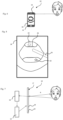
- a device 10 comprises a camera 16, a computer processing unit 12 and presentation means 18.
- the camera 16, the processing unit 12 and the presentation means are provided with means allowing them to communicate with each other .
- the camera 16 and/or the processing unit 12 and/or the presentation means can be integrated into glasses conventionally comprising a frame 14 configured to be worn by the nose and ears of an operator, in a mobile telephone , in a tablet, in a laptop, in a virtual reality headset, in a camera or in a mirror 17.
- the camera 16 is intended for the acquisition of updated images representing the real dental scene observed by the operator through the glasses ( figure 5 ) or on the screen of a mobile phone ( Figure 6 ) or a laptop, a virtual reality headset, a camera or a tablet, or on the glass 19 of the mirror ( figure 7 ). It can be a classic camera.
- the device comprises means for projecting patterns of structured light, for example a cloud of points, for example laser, in particular infrared.
- the real dental scene shows these patterns in a distorted manner, which makes it possible to deduce information relating to the depth.
- the glass 19 can be a one-way glass and the camera can be placed behind the glass, as on the figure 7 .
- the device comprises several cameras, which makes it possible, by simple trigonometric calculation, to evaluate the distance between the objects observed and the camera.
- the multiplicity of cameras thus makes it possible to speed up the search for reference images.
- the cameras also make it possible to simulate the observer's three-dimensional view of the real dental scene.
- presentation means we mean any means configured to receive, from the processing unit 12, a virtual dental scene and make said virtual dental scene appear on the real dental scene, on a screen through which an observer can see the real dental scene or on the updated image.
- the presentation means 18 comprise a screen and a projector for projecting the virtual dental scene onto the screen.
- the screen can be transparent. It is preferably fixed on the frame 14, and the virtual dental scene is presented on the screen so that the operator sees, by observing this screen, both the real dental scene through the screen, but also the scene virtual dental projected on the screen.
- the glasses conventionally comprise two lenses which extend in front of the two eyes of the operator, respectively, but the number of lenses is not limiting.
- the lenses may or may not be vision corrective.
- Several glasses can extend in front of the same eye.
- a first lens may correct the operator's vision and a second lens may be a lens serving as a screen.
- the HoloLens device developed by the Microsoft ® company, is an example of presentation means.
- the screen may be opaque and the updated image and the virtual dental scene are presented on the screen, in register, so that the operator sees, by observing this screen, both the updated image representing the real dental scene , but also the virtual dental scene.
- the camera can be immobilized relative to the screen, for example when the camera and the screen are integrated into a mobile phone ( Figure 6 ) or a laptop or a virtual reality headset or a camera or a tablet or a mirror
- the camera can be free (or “independent”) from the screen.
- this latter embodiment allows great flexibility.
- the camera can be inserted inside the mouth while the screen remains outside the mouth.
- the screen can be the reflective glass 19 of the mirror, and the virtual dental scene can be presented on the screen, in register with the reflection R returned by the glass 19, so that the operator sees, by observing this screen, both the reflection R representing the real dental scene, but also the virtual dental scene superimposed on the real dental scene.
- the reflection like the updated image displayed on the screen of glasses, mobile phone, laptop, virtual reality headset, camera or tablet, thus gives the operator a representation of real dental scene.
- the processing unit 12 may include the conventional electronic means of any computer, and in particular a central unit 22, a program and a memory 24.
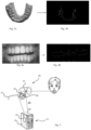
- the memory 24 preferably contains a reference model of at least part of the scene real dental, and in particular a model of the dental arch, for example as shown in the figure 2 .
- the program includes code instructions allowing, when executed, to implement step d), and in particular to analyze an updated image, to search for a view of the reference model which has maximum agreement with the updated image, that is to say a reference image, then to determine, accordingly, a virtual dental scene.
- the device also includes an interface allowing an operator to configure the virtual dental scene.
- the interface is configured so that an operator can modify a simulation instant determining the virtual dental scene.
- the content of the virtual dental scene may in particular consist of the representation of elements of the real dental scene in an appearance and/or at a position simulated for the past or future simulation instant or known at a past simulation instant. .
- the screen is a touch screen and the operator can modify the simulation time by entering a date in an input field or by moving one or more fingers on the screen. For example, moving to the right of the screen can lead to moving forward into the future and moving to the left of the screen can lead to moving back into the past.
- the presented virtual dental scene adapts accordingly, preferably in real time.
- the interface can also be a mechanical organ, for example a button or a wheel.
- the device is configured so that the time interval between the updated instant and the simulation instant evolves continuously from one cycle to another, that is to say that the succession of cycles of steps A) to C) or c) to e) simulate progress in time or return in time.
- the time difference between two successive simulation instants is constant, which makes it possible to visualize an evolution in which time passes at constant speed.
- the processing unit determines the conditions under which the virtual dental scene must be presented to appear, to the operator, in superposition with the real dental scene.
- the camera be arranged so as to capture an updated image that corresponds to the view of the scene real dental by the observer. It is also preferable for the screen to be at a constant distance, and oriented in a fixed manner relative to the observer.
- the processing unit can thus easily determine, from the updated image acquired by the camera, the view of the real dental scene observed by the operator, as well as the conditions in which the virtual dental scene must be presented. to appear, to the operator, superimposed on the real dental scene.
- a method according to the invention can be implemented by means of a device according to the invention.
- a reference model is generated, preferably by means of a scanner.
- the reference model may be a model of the patient's arch or be composed of a model of this arch and a model of an orthodontic appliance placed on said model of the arch.
- the creation of the reference model preferably results from taking measurements.
- an arch of the patient is scanned with a 3D scanner, optionally while the patient is wearing orthodontic appliances, or a cast of this arch, so as to create a reference model.
- the scan is carried out with a classic 3D scanner.
- the reference model obtained by means of a scanner represents an arch, but also, where applicable, any object scanned at the same time as the arch, for example an orthodontic appliance or a decorative article fixed to the arch.
- the reference model can alternatively be a generic arch model, chosen from a base of generic models, that is to say an arch model applicable, roughly, to several patients, or result from a deformation of such a generic model.
- the deformation may in particular be based on measurements or observations made on the patient, for example based on photos of their dental arches. Distorting a model so that it can match one or more photos can implement a metaheuristic method, as described in WO 2016/066651 notably.
- a generic arch model may in particular be a model obtained by statistical processing, for example an average, of more than 5, more than 10, more than 100 or more than 1000 historical models generated by means of a scanner.
- Historical models are preference of dental arch models of “historical” patients having dental characteristics identical or similar to those of the patient for whom the reference model is generated, for example having suffered from the same pathology and/or having the same age and/or having the same sex and/or having a similar body shape.
- the three-dimensional reference model can be observed from any angle.
- THE figures 2 And 3a are example views of a reference model.
- the views of the reference model are intended to be compared with the updated images in order to determine, for each updated image, a reference image that is as similar as possible to the updated image, then consequently constitute a virtual dental scene providing relative information. to the elements shown in the updated image.
- the reference time may be close to the updated time, for example distant (later or earlier) by less than 1 month, 2 weeks or 1 week or 3 days from the updated time. Updated images can then be used to deform a generic model until the reference model corresponding to the dental situation at the updated time is obtained. Furthermore, the virtual dental scene then provides information on the dental situation specific to the current moment. For example, if the reference model models tooth roots, the virtual dental scene can represent tooth roots.
- the reference time can be far from the updated time, for example more than 1 month away from the updated time, 2 weeks or 1 week or 3 days.
- the reference model can then differ significantly from reality at the updated moment.
- the information provided by the virtual dental scene can then relate to the evolution of the dental situation between the reference moment and the updated moment. For example, they can provide information about moving teeth or orthodontic appliances.
- the method can be used to monitor whether a passive retainer holds teeth in position after dental treatment.
- the reference model can be generated immediately after installation of the device and the acquisition of an updated image can take place one month after this installation.
- the reference image and the virtual dental scene will therefore concern the teeth in their position at the end of treatment and the presentation of the virtual dental scene on the real dental scene, at the updated moment, will thus allow the dental professional to detect the differences with their real positions.
- step b) in one embodiment, the reference model is modified.
- the modification can be made at any time between the reference time and the updated time. Preferably, it is carried out approximately at the reference time.
- the modification of the reference model can be a deformation of this model.
- the reference model is segmented, and in particular tooth models are created, as described in WO 2016/066651 . Then the tooth models are moved or distorted.
- the modification in step b) can simulate the effect of the passage of time, for example the effect of dental treatment, between the reference time and the updated time.
- the modification of the reference model thus leads to a model representing the teeth in a position or shape estimated for the moment updated.
- the displacement or deformation of tooth models can also be determined or refined from one or more updated images, as described in WO 2016/066651 .
- the initial reference model, determined in step a) then results from measurements by means of a scan of at least one arch or at least one molding of an arch.
- the method can thus be used to check whether the progress of a dental treatment conforms to predictions, the initial reference model being modified to correspond to the anticipated situation at the updated moment.
- significant changes can take a few days.
- the time interval between the updated instant and the reference instant is greater than 3 days, or even greater than 1 week, greater than 2 weeks, greater than 4 weeks, greater than 8 weeks, greater than 12 weeks, or even greater than 16 weeks.
- the reference image resulting from an observation of the modified reference model then shows the teeth in their anticipated positions or shapes at the updated instant. It constitutes or can be integrated into a virtual dental scene whose presentation, in step e), allows the dental care professional to detect the differences between the actual positions or shapes of the teeth and the positions or shapes anticipated for the moment updated.
- the modification of the initial reference model may consist of adding another model and/or modifying this other model.
- a model of an orthodontic appliance placed on the arch can be added to a reference model which would initially only represent the arch.
- the modification of the reference model can then include a modification of this orthodontic appliance so that it appears in its anticipated position or shape for the current moment. Its presentation, in step e), then allows the dental care professional to detect the differences between the actual position or shape of the orthodontic appliance and the anticipated position or shape for the current moment, and in particular to detect any separation of a fastener.
- step c) at the updated instant, an updated image representing a real dental scene is acquired with the camera.
- the updated image is represented in solid lines on screen 18.
- a device in which the camera is fixed on the spectacle frame or is integrated in a mirror makes it possible to acquire an updated image which represents with great precision the real dental scene observed by the operator through the glasses of these glasses or on this mirror.
- a device in which the camera is integrated into a mobile phone, a laptop, a tablet or a camera makes it possible to acquire an updated image which exactly represents the actual dental scene observed by the operator on the screen mobile phone, computer, tablet, or camera respectively.
- the camera can be rigidly attached or be independent of the screen.
- the independence of the camera and the screen advantageously allows the operator to operate without directly seeing the real dental scene, that is to say by only seeing it on the screen. He can thus work “blindly”.
- the updated image is in color, preferably in real color.
- step d we search for a reference image which has maximum agreement with the updated image.
- step d all known methods for achieving maximum matching are considered.
- Obtaining maximum agreement may in particular result from optimization so as to minimize said distance (“bestfit ”) .
- the reference image is sought by observing the reference model from different observation angles and different distances, until obtaining a view of the reference model which corresponds substantially to the updated image, that is to say -say which can be significantly superimposed on it. For each updated image, we thus obtain a reference image having maximum agreement with the updated image.
- This search is preferably carried out by means of a metaheuristic method, preferably evolutionary, preferably by simulated annealing.
- the exploration of the reference model to search for the reference image may include one or more of the characteristics of steps c), d) and e) of WO 2016 066651 , insofar as they concern such exploration.
- Discriminant information is characteristic information which can be extracted from an image ( “image feature ”), conventionally by computer processing of this image. It is preferably chosen from the group consisting of contour information, color information, density information, distance information, brilliance information, saturation information, reflection information and combinations of these. information.
- the figure 4b is an updated map relating to the contour of the teeth obtained from the updated image of the figure 4a .
- figure 3b is a reference map relating to the contour of the teeth obtained from the view of the figure 3a .
- the modification of the view is preferably guided by heuristic rules, for example by favoring the modifications which, according to an analysis of the previous distances, appear the most favorable for reducing it.
- Step iii) results in a reference image substantially superimposable on the updated image.
- Obtaining maximum agreement between the reference image and the updated image can also result from the superposition of landmarks identified on these two images, for example the superposition of fixed landmarks such as the end of a tooth or a point of contact between two teeth.
- the virtual dental scene preferably includes, or even consists of, the reference image.
- the reference image is a particular example of a virtual dental scene.
- a virtual dental scene includes a representation, preferably realistic, of one or more elements of the corresponding real dental scene.
- it comprises a representation of the arch, but also optionally, for example, a representation of an orthodontic appliance worn by the teeth of the arch or that the operator manipulates in front of the arch, or of a tool that the operator manipulates in front of the arch.
- the virtual dental scene can for example represent the contours 26 of the teeth and its projection on the teeth can facilitate the detection of movements or deformations of the teeth.
- the virtual dental scene may also include the representation of one or more physical elements of the real dental scene not modeled by the reference model, for example a tool 28 manipulated by the operator. To identify such physical elements, the updated image is analyzed, for example by means of artificial intelligence algorithms.
- the virtual dental scene may also include the representation of one or more physical elements which are not in the real dental scene, for example a tool or an orthodontic appliance or a decorative article 32, for example positioned on the virtual dental scene in such a manner. to appear, on the actual dental scene, at a location ("target location") where the orthodontic tool or appliance or decorative item, respectively, should be placed by the dental professional or the patient.
- a tool or an orthodontic appliance or a decorative article 32 for example positioned on the virtual dental scene in such a manner. to appear, on the actual dental scene, at a location ("target location") where the orthodontic tool or appliance or decorative item, respectively, should be placed by the dental professional or the patient.
- the representation of a physical element can be realistic, for example consisting of the contours of the physical element considered, or symbolic.
- the bit of a drill can be symbolized by an arrow.
- the representation of the physical elements of the virtual dental scene can be two-dimensional and/or three-dimensional.
- the presentation of the virtual dental scene may in particular be a hologram.
- the virtual dental scene may also include, in addition to or alternatively to the representation of physical elements, for example the reference image, one or more indicators 34.
- An indicator is an element of a virtual dental scene which does not represent, even symbolically, a physical element of the real dental scene.
- An indicator can in particular be a text or a symbol (a point, a line, an arrow, a solid color, etc.), for example a symbol illustrating differences between the image updated and the reference image, or providing information relating to an act carried out by the operator, for example indicating a direction for a tool that he is handling.
- the indicator points to a location on which the operator must take particular care, for example brushing.
- the indicator can be an image, preferably playful, for example a drawing of a monster or a microbe, on which the operator must apply a tool, for example a toothbrush. Such an indicator improves learning to brush your teeth, for example.
- indicator 34 points to a dental region that has been abraded.
- the nature of an indicator can be determined based on the reference image.
- the indicators may in particular be associated with the reference model or with a region of the reference model. They are for example stored in a database in the memory 24 of the processing unit.
- additional information can be associated with different regions of the reference model, for example to indicate caries, fragile areas, or bonding areas for an orthodontic appliance. Additional information can also be associated with the reference model as a whole, for example to specify the circumstances of its production or information on the patient or on the orthodontic appliance possibly represented. Indicators make it possible to present this additional information.
- the nature of an indicator can be determined based on the differences between the updated image and the reference image.
- indicators can be added to highlight a deformation or displacement of a tooth, or a detachment of an orthodontic appliance.
- an indicator can be determined depending on the context, and in particular depending on the distance to another element of the virtual dental scene and/or depending on a pursued objective. For example, it will appear with an appearance depending on the distance to a particular tooth or depending on the risk to the patient.
- the context is defined by the conditions under which the process is used.
- the operator may have the objective of checking the positioning of the orthodontic appliance worn by the patient.
- Indicators which are not linked to this verification, for example the indication of areas of caries, are then not necessary.
- the operator enters additional information into the database, depending on the intended use of the process. For example, it specifies a location to attach a decorative item or to mill a tooth.
- the corresponding indicators can advantageously be added to the virtual dental scene.
- an arrow may point to an area of a tooth to indicate to the operator where to attach an orthodontic appliance or decorative item, or where to act, for example by means of a tool like a milling cutter.
- An indicator can also represent, realistically or not, a non-visible part of a physical element of the real dental scene (and which, not being visible to the operator, therefore does not belong to the real dental scene ), for example a root of a tooth, a bone or a dental nerve.
- the elements which constitute the virtual dental scene are positioned in this scene according to the elements of the real dental scene, and therefore in this case, according to the reference image.
- the representations of real physical elements are preferably positioned in the virtual dental scene so that in step e), they are superimposed, precisely, with these real physical elements or with their representations on the updated image.
- an indicator providing information specific to a physical element is preferably positioned so that in step e) it appears in the immediate vicinity or superimposed on this physical element, for example at this tooth.
- Indicators can be arranged to be displayed, in the next step, where differences have been identified between the reference image and the updated image. For example, we can display a red mark at locations where an orthodontic appliance has become loose from the teeth.
- the context is evolving and the nature and/or location, in the virtual dental scene, of the indicators are adapted accordingly.
- the nature and/or location of the indicators can be determined as a function of the nature and/or location of indicators displayed in previous virtual dental scenes, that is to say determined during cycles c)-e ) (or A) to C)) previous, and/or as a function of the time interval with said previous virtual dental scenes, and/or as a function of differences between the updated images of cycles c)-e) (or A) to C)) successive.
- the method can be used to guide an action of the operator with a tool, for example drilling using a drill bit.
- An element of the virtual dental scene may be an arrow indicating the orientation of the drill and the position of its free end.
- the database may contain a protocol setting, for example, the point of penetration into the tooth, the trajectory and the optimal speed of advancement of the drill.
- the tool for example the drill, which appears on the successive updated images, is identified on these images and the location of the element in the virtual dental scene is adapted to take into account the real position of the 'tool.
- the representation of the indicator in the virtual dental scene can therefore evolve according to the real position of the tool. In particular, it can thus adapt to the real progress speed or to the real trajectory of the tool.
- the appearance of an element of the virtual dental scene changes depending on the context.
- the color of the element can be changed if the analysis of the context shows that the situation becomes dangerous, for example because the tool does not follow the desired trajectory or because the orthodontic appliance would lead to excessive correction positioning of the teeth.
- the virtual dental scene does not include the reference image.
- the reference image can be used to determine the nature and/or location of elements in the virtual dental scene. But it is not presented to the observer in step e) (or C)).
- the virtual dental scene is determined based on a past or future simulation time.
- it may include a representation, preferably realistic, of one or more elements of the corresponding real dental scene in a past or future configuration.
- it comprises a representation of the arch and/or a representation of an orthodontic appliance worn by the teeth of the arch as measured or simulated at a past simulation time.
- it includes a representation of the arch and/or a representation of an orthodontic appliance worn by the teeth of the arch as anticipated for a future simulation moment.
- the simulation at a past or future simulation time can be carried out by any simulation software or by an operator, for example by a dental professional.
- the virtual dental scene can, for example, represent the contours of the teeth, the positioning of an orthodontic appliance or the color of the teeth at the simulation moment.
- the presentation of the virtual dental scene on the real dental scene preferably transparently on the real dental scene, thus makes it possible to easily detect the evolution between the updated instant and the simulation instant.
- the information relating to this indicator is preferably included in the reference model.
- the reference model can be a model of the dental arch in which the crowns of the teeth, but also the roots of the teeth are modeled, for example by means of a cone beam scanner.
- the method according to the invention thus allows the operator to visualize parts of the patient's dental arch which are not visible on the real dental scene.
- these non-visible parts are represented in their real relative position in relation to the physical elements of the real dental scene. For example, the operator sees the roots and nerves of the teeth he observes in the patient's mouth.
- the operator can precisely introduce the needle until reaching a tooth root, without touching a dental nerve. The pain felt by the patient is limited.
- step e we present, depending on the reference image, the virtual dental scene superimposed on the real dental scene or on the representation of the real dental scene in the updated image.
- the virtual dental scene can be directly projected onto the real dental scene.
- red light spots can be projected onto teeth to identify bonding areas for orthodontic brackets.
- the virtual dental scene can be displayed on a screen.
- the operator can thus observe the virtual dental scene on the screen and the real dental scene through the screen, in transparency behind the virtual dental scene.
- the screen is opaque, particularly in the embodiment in which the screen is the screen of a mobile phone, a laptop, a virtual reality headset or a tablet, the The operator can thus simultaneously observe the updated image and the virtual dental scene on the screen.
- the virtual dental scene may be transparent enough to reveal the actual dental scene or updated image beneath it.
- the virtual dental scene may be opaque, so as not to reveal the actual dental scene or the updated image extending behind or below it.
- the opacity of the virtual dental scene is particularly advantageous for simulating a past or future dental situation, for example to visualize the whitening of teeth under the effect of a treatment for this purpose, or the movement of a tooth, for example under the effect of orthodontic treatment or in the absence of orthodontic treatment, for example to visualize a loosening of a tooth.
- the virtual dental scene When the virtual dental scene is presented on the updated image, it may be added on top of the updated image or, equivalently, the updated image may be replaced by a computer-processed image to reveal the virtual dental scene .
- the screen is the glass of a mirror
- the operator can simultaneously observe his reflection and the virtual dental scene, superimposed on his reflection, on the screen.
- the patient can visualize the effect of dental treatment, therapeutic or not.
- the virtual dental scene is presented based on the reference image and therefore, indirectly, based on the updated image.
- it is positioned, or "framed", in relation to the real dental scene according to the reference image.
- the virtual dental scene is presented in superposition, or “in register”, with the real dental scene or with its representation on the updated image, which facilitates comparison.
- the elements of the virtual dental scene which correspond to elements of the real dental scene are substantially exactly superimposed on the latter or on the representations of the latter.
- the differences between the real dental scene or its representation on the one hand and the virtual dental scene on the other hand thus appear clearly to the operator.
- the indicators also provide information to facilitate the analysis of the situation.
- characteristic marks are identified on the updated image whose position relative to the virtual dental scene is known. For example, tooth tips or characteristic tooth shapes represented in both the updated image and the virtual dental scene are identified.
- the reference image from which the virtual dental scene was determined can in particular be used to identify the position of the characteristic marks in the virtual scene.
- step e) (or C)) the representations of these characteristic marks are then superimposed on the updated image and in the virtual dental scene, which makes it possible to precisely position the virtual dental scene in relation to the characteristic marks represented on the updated image.
- step c) (or A)), preferably less than 30 seconds, less than 10 seconds, less than 5 seconds, preferably less than 3 seconds, preferably less than 1 s, less than 0.5 s, less than 0.2 s, preferably less than 0.1 s after step e) (or C)).
- the cycle of steps c) to e) (or A) to C)) is carried out in real time, without interruption.
- the process ends at the end of step e) (or C)), without resuming at step c) (or A)).
- the process is thus carried out “on demand”, and not in real time.
- the invention can be used during an intervention on the teeth, by the dental professional wearing the glasses.
- the method is used as part of a tooth abrasion, or “stripping”, or milling operation.
- the virtual dental scene represents the tooth in its final form, after the abrasion or milling to be carried out.
- the dental professional sees the contour of the tooth in its final form in precise superposition, that is to say in register, on the real tooth. This information allows him to machine the tooth with perfect precision.
- the method is used to attach a dental bracket, or bracket, to a tooth.
- the virtual dental scene represents the bracket in its final position, after its fixation on the tooth.
- the dental professional sees the virtual outline of the bracket in its final position relative to the real tooth. This information allows it to place the real attachment in exact superposition with the attachment of the virtual dental scene, and therefore to position it on the real tooth with perfect precision.
- the process can also be used to attach a decorative item to teeth.
- the invention allows the operator, in particular a dental professional, to access relevant information.
- This information is presented to him, preferably in real time, superimposed on the real dental scene that he observes directly or on the updated image.
- the invention thus makes it possible to considerably increase the quality of the analysis of the situation, and, if necessary, to adapt an intervention accordingly.
- the method is used to achieve better patient acceptance of the treatment.
- it can be used to realistically simulate dental treatment.
- the image acquisition device is then a mobile phone, a laptop, a tablet or a mirror.
- the patient films their dental arches, just like when taking a selfie.
- the patient is preferably filmed through the mirror surface, allowing the updated image to faithfully represent the R reflection observed by the patient.
- the patient sees corresponding virtual dental scenes, which represent a stage of the treatment dental at a simulation time.
- the simulation moment may be the moment marking the start of treatment and the virtual dental scene may represent an orthodontic appliance that the patient plans to wear.
- the patient can then visualize their face after the orthodontic appliance has been placed in position in their mouth. By turning the head or moving the mobile phone, laptop or tablet, or moving in front of the mirror, it can easily change the viewing direction, with the virtual dental scene adapting in real time.
- the patient can modify the simulation instant by interacting with the screen, for example by moving a virtual cursor.
- the virtual dental scene is adapted accordingly, in particular to take into account the effect of the treatment.
- the patient can therefore simulate the course of the treatment, that is to say visualize for example how the teeth will move and/or change color, particularly in the case of teeth whitening treatment or if teeth are subjected to a coloring agent, for example if the patient smokes or drinks large quantities of tea or coffee. Moving the cursor allows it to simulate an acceleration of time.
- This simulation facilitates decision-making by the patient and advantageously constitutes an incentive to follow the treatment.
- the patient can modify the value of other parameters of the simulation. For example, it can modify processing parameter values.
- the virtual dental scene displays warnings, preferably in the form of a message or highlighting an element of the virtual dental scene, for example by making an area appear in red. which needs to be brushed more carefully.
- the invention can be used to evaluate the dental situation of the patient, in particular to control the progress of a dental treatment, and/or, outside the context of a dental treatment, to control an evolution of the color and /or the shape and/or arrangement of the teeth or gums.
- the method is used to visualize the abrasion suffered by a tooth, for example due to bruxism.
- the virtual dental scene represents the tooth in its anterior form, before abrasion.
- the operator generally the patient or the dental professional, sees the contour of the tooth in its previous form superimposed on the real tooth or on its representation on the updated image. This information allows him to immediately and precisely visualize the extent of this wear, particularly when the virtual dental scene is presented transparently.
- the method is used to visualize a deformation of the gum, and in particular a recession of the gum.
- the virtual dental scene represents the gum in its initial shape, before its deformation.
- the operator generally the patient or the dental care professional, sees the contour of the gum in its initial form superimposed on the real gum or on its representation. This information allows it to immediately and precisely visualize this deformation, particularly when the virtual dental scene is presented in transparency.
- the patient is not limited to a human being.
- a method according to the invention can be used for another animal.
- the invention is not limited to a process comprising steps a) to e).
- the search for a reference image is not essential to the implementation of the invention. If a reference image is not available, the elements that constitute the virtual dental scene can be positioned on the real dental scene or its representation, depending on the updated image.
- a virtual dental scene can be determined based on several reference images obtained from several respective updated images acquired simultaneously or at different updated times, for example successive.
Landscapes
- Health & Medical Sciences (AREA)
- Engineering & Computer Science (AREA)
- Life Sciences & Earth Sciences (AREA)
- Public Health (AREA)
- General Health & Medical Sciences (AREA)
- Medical Informatics (AREA)
- Animal Behavior & Ethology (AREA)
- Veterinary Medicine (AREA)
- Epidemiology (AREA)
- Oral & Maxillofacial Surgery (AREA)
- Dentistry (AREA)
- Physics & Mathematics (AREA)
- Biomedical Technology (AREA)
- Surgery (AREA)
- Nuclear Medicine, Radiotherapy & Molecular Imaging (AREA)
- Pathology (AREA)
- Heart & Thoracic Surgery (AREA)
- Molecular Biology (AREA)
- General Engineering & Computer Science (AREA)
- General Physics & Mathematics (AREA)
- Theoretical Computer Science (AREA)
- Radiology & Medical Imaging (AREA)
- Primary Health Care (AREA)
- Biophysics (AREA)
- Software Systems (AREA)
- Computer Graphics (AREA)
- Quality & Reliability (AREA)
- Computer Vision & Pattern Recognition (AREA)
- Data Mining & Analysis (AREA)
- Databases & Information Systems (AREA)
- Computer Hardware Design (AREA)
- Robotics (AREA)
- Audiology, Speech & Language Pathology (AREA)
- Geometry (AREA)
- Dental Tools And Instruments Or Auxiliary Dental Instruments (AREA)
- Processing Or Creating Images (AREA)
Applications Claiming Priority (3)
| Application Number | Priority Date | Filing Date | Title |
|---|---|---|---|
| EP18305628.2A EP3572030B1 (de) | 2018-05-22 | 2018-05-22 | Verfahren zur analyse einer zahnsituation |
| PCT/EP2019/062382 WO2019224056A1 (fr) | 2018-05-22 | 2019-05-14 | Procede d'analyse d'une situation dentaire |
| EP19725306.5A EP3796866B1 (de) | 2018-05-22 | 2019-05-14 | Computerprogramm und vorrichtung zur analyse einer zahnsituation |
Related Parent Applications (2)
| Application Number | Title | Priority Date | Filing Date |
|---|---|---|---|
| EP19725306.5A Division EP3796866B1 (de) | 2018-05-22 | 2019-05-14 | Computerprogramm und vorrichtung zur analyse einer zahnsituation |
| EP19725306.5A Division-Into EP3796866B1 (de) | 2018-05-22 | 2019-05-14 | Computerprogramm und vorrichtung zur analyse einer zahnsituation |
Publications (2)
| Publication Number | Publication Date |
|---|---|
| EP4406505A2 true EP4406505A2 (de) | 2024-07-31 |
| EP4406505A3 EP4406505A3 (de) | 2024-09-18 |
Family
ID=62386327
Family Applications (3)
| Application Number | Title | Priority Date | Filing Date |
|---|---|---|---|
| EP18305628.2A Active EP3572030B1 (de) | 2018-05-22 | 2018-05-22 | Verfahren zur analyse einer zahnsituation |
| EP24180883.1A Pending EP4406505A3 (de) | 2018-05-22 | 2019-05-14 | Computerprogramm und vorrichtung zur analyse einer zahnsituation |
| EP19725306.5A Active EP3796866B1 (de) | 2018-05-22 | 2019-05-14 | Computerprogramm und vorrichtung zur analyse einer zahnsituation |
Family Applications Before (1)
| Application Number | Title | Priority Date | Filing Date |
|---|---|---|---|
| EP18305628.2A Active EP3572030B1 (de) | 2018-05-22 | 2018-05-22 | Verfahren zur analyse einer zahnsituation |
Family Applications After (1)
| Application Number | Title | Priority Date | Filing Date |
|---|---|---|---|
| EP19725306.5A Active EP3796866B1 (de) | 2018-05-22 | 2019-05-14 | Computerprogramm und vorrichtung zur analyse einer zahnsituation |
Country Status (5)
| Country | Link |
|---|---|
| US (3) | US11607292B2 (de) |
| EP (3) | EP3572030B1 (de) |
| CN (2) | CN115137507B (de) |
| ES (2) | ES2936080T3 (de) |
| WO (1) | WO2019224056A1 (de) |
Families Citing this family (8)
| Publication number | Priority date | Publication date | Assignee | Title |
|---|---|---|---|---|
| US11308618B2 (en) | 2019-04-14 | 2022-04-19 | Holovisions LLC | Healthy-Selfie(TM): a portable phone-moving device for telemedicine imaging using a mobile phone |
| US12014500B2 (en) | 2019-04-14 | 2024-06-18 | Holovisions LLC | Healthy-Selfie(TM): methods for remote medical imaging using a conventional smart phone or augmented reality eyewear |
| AU2021227914A1 (en) | 2020-02-26 | 2022-09-22 | Get-Grin Inc. | Systems and methods for remote dental monitoring |
| US20220218438A1 (en) * | 2021-01-14 | 2022-07-14 | Orthosnap Corp. | Creating three-dimensional (3d) animation |
| US12236819B1 (en) * | 2021-03-02 | 2025-02-25 | Apple Inc. | Augmenting a physical writing surface |
| CN113487611B (zh) * | 2021-09-07 | 2021-11-26 | 南通林德安全设备科技有限公司 | 基于人工智能的牙片图像处理方法及系统 |
| WO2023205449A1 (en) | 2022-04-22 | 2023-10-26 | Get-Grin Inc. | Systems and methods for intraoral imaging |
| US20250345130A1 (en) * | 2024-05-07 | 2025-11-13 | Mars Dental Ai Ltd. | Interactive visualization of dental implant position planning |
Citations (1)
| Publication number | Priority date | Publication date | Assignee | Title |
|---|---|---|---|---|
| WO2016066651A1 (fr) | 2014-10-27 | 2016-05-06 | H43 Development | Procede de controle de la dentition |
Family Cites Families (33)
| Publication number | Priority date | Publication date | Assignee | Title |
|---|---|---|---|---|
| US6632089B2 (en) * | 1999-11-30 | 2003-10-14 | Orametrix, Inc. | Orthodontic treatment planning with user-specified simulation of tooth movement |
| US7160110B2 (en) * | 1999-11-30 | 2007-01-09 | Orametrix, Inc. | Three-dimensional occlusal and interproximal contact detection and display using virtual tooth models |
| US7156655B2 (en) * | 2001-04-13 | 2007-01-02 | Orametrix, Inc. | Method and system for comprehensive evaluation of orthodontic treatment using unified workstation |
| ES2714375T3 (es) * | 2004-03-04 | 2019-05-28 | Align Technology Inc | Sistemas y métodos para la fabricación de una plantilla dental |
| CN1795826A (zh) | 2004-12-27 | 2006-07-05 | 上海雷硕医疗器械有限公司 | 一种牙齿影像自助记录器 |
| WO2009085752A2 (en) | 2007-12-21 | 2009-07-09 | 3M Innovative Properties Company | Orthodontic treatment monitoring based on reduced images |
| CN201244032Y (zh) | 2008-03-24 | 2009-05-27 | 孙立刚 | 便携式口腔扫描仪 |
| WO2010099036A1 (en) * | 2009-02-25 | 2010-09-02 | Dimensional Photonics International, Inc. | Intensity and color display for a three-dimensional metrology system |
| AU2013276501B2 (en) | 2012-06-15 | 2017-01-19 | Vita Zahnfabrik H. Rauter Gmbh & Co. Kg | Method for preparing a partial or full dental prosthesis |
| TWI504383B (zh) * | 2012-11-27 | 2015-10-21 | Nat Univ Chung Cheng | Computer - aided positioning guidance system for dental implants |
| US9256962B2 (en) | 2013-01-23 | 2016-02-09 | Orca Health Inc. | Personalizing medical conditions with augmented reality |
| US20140329194A1 (en) * | 2013-05-05 | 2014-11-06 | Rohit Sachdeva | Orthodontic treatment planning using biological constraints |
| IL227360A (en) * | 2013-07-07 | 2015-01-29 | Boris Fridzon | Method and system for presenting and planning or performing dental care |
| US10335098B2 (en) * | 2013-07-19 | 2019-07-02 | Axion Japan Co., Ltd. | Panoramic imaging apparatus and diagnostic imaging method in same apparatus |
| KR20160143654A (ko) * | 2014-02-21 | 2016-12-14 | 트리스페라 덴탈 아이엔씨. | 증강 현실 치과 디자인 방법 및 시스템 |
| CN105496573B (zh) * | 2014-09-23 | 2019-09-13 | 上海时代天使医疗器械有限公司 | 提供牙齿矫治用附件的方法以及该附件 |
| US9744001B2 (en) * | 2014-11-13 | 2017-08-29 | Align Technology, Inc. | Dental appliance with cavity for an unerupted or erupting tooth |
| US11147652B2 (en) * | 2014-11-13 | 2021-10-19 | Align Technology, Inc. | Method for tracking, predicting, and proactively correcting malocclusion and related issues |
| US11202690B2 (en) * | 2014-11-27 | 2021-12-21 | 3Shape A/S | Method of digitally designing a modified dental setup |
| US9770217B2 (en) * | 2015-01-30 | 2017-09-26 | Dental Imaging Technologies Corporation | Dental variation tracking and prediction |
| FR3032282B1 (fr) | 2015-02-03 | 2018-09-14 | Francois Duret | Dispositif de visualisation de l'interieur d'une bouche |
| KR101678910B1 (ko) * | 2015-06-09 | 2016-12-06 | (주) 시원 | 드릴링 가이드 장치 |
| US20170281313A1 (en) * | 2016-04-05 | 2017-10-05 | eClear International Co., Ltd. | Method and system for providing remote teeth alignment services and computer-readable medium on which the method is recorded |
| CN106137102A (zh) | 2016-06-24 | 2016-11-23 | 张麟 | 一种牙齿自检装置 |
| EP4254429A3 (de) * | 2016-08-24 | 2024-01-03 | Align Technology, Inc. | Verfahren zur visualisierung und herstellung eines ausrichters durch modifizierung der zahnstellung |
| US10660728B2 (en) * | 2016-10-20 | 2020-05-26 | Baliram Maraj | Systems and methods for dental treatment utilizing mixed reality and deep learning |
| EP3534832B1 (de) * | 2016-11-04 | 2023-09-27 | Align Technology, Inc. | Verfahren und vorrichtungen für zahnbilder |
| US10467815B2 (en) * | 2016-12-16 | 2019-11-05 | Align Technology, Inc. | Augmented reality planning and viewing of dental treatment outcomes |
| US10888399B2 (en) | 2016-12-16 | 2021-01-12 | Align Technology, Inc. | Augmented reality enhancements for dental practitioners |
| FR3066903B1 (fr) * | 2017-05-30 | 2022-02-18 | Dental Monitoring | Procede de determination d'un traitement orthodontique |
| US11083552B2 (en) | 2018-02-20 | 2021-08-10 | Ivoclar Vivadent Ag | Rendering of dental models |
| CN112087985B (zh) | 2018-05-10 | 2023-05-02 | 3M创新有限公司 | 经由实时增强可视化的模拟正畸处理 |
| US11151753B2 (en) * | 2018-09-28 | 2021-10-19 | Align Technology, Inc. | Generic framework for blurring of colors for teeth in generated images using height map |
-
2018
- 2018-05-22 EP EP18305628.2A patent/EP3572030B1/de active Active
- 2018-05-22 ES ES18305628T patent/ES2936080T3/es active Active
-
2019
- 2019-05-14 CN CN202210728554.8A patent/CN115137507B/zh active Active
- 2019-05-14 CN CN201980048904.2A patent/CN112469359B/zh active Active
- 2019-05-14 EP EP24180883.1A patent/EP4406505A3/de active Pending
- 2019-05-14 US US17/057,616 patent/US11607292B2/en active Active
- 2019-05-14 WO PCT/EP2019/062382 patent/WO2019224056A1/fr not_active Ceased
- 2019-05-14 EP EP19725306.5A patent/EP3796866B1/de active Active
- 2019-05-14 ES ES19725306T patent/ES2989086T3/es active Active
-
2022
- 2022-07-18 US US17/867,523 patent/US11666417B2/en active Active
-
2023
- 2023-04-27 US US18/140,222 patent/US12245912B2/en active Active
Patent Citations (1)
| Publication number | Priority date | Publication date | Assignee | Title |
|---|---|---|---|---|
| WO2016066651A1 (fr) | 2014-10-27 | 2016-05-06 | H43 Development | Procede de controle de la dentition |
Also Published As
| Publication number | Publication date |
|---|---|
| CN115137507A (zh) | 2022-10-04 |
| US20230404710A1 (en) | 2023-12-21 |
| BR112020023700A2 (pt) | 2021-02-09 |
| EP3796866B1 (de) | 2024-07-17 |
| US20210161621A1 (en) | 2021-06-03 |
| CN112469359A (zh) | 2021-03-09 |
| US11666417B2 (en) | 2023-06-06 |
| CN112469359B (zh) | 2022-07-12 |
| US12245912B2 (en) | 2025-03-11 |
| CN115137507B (zh) | 2025-10-28 |
| ES2936080T3 (es) | 2023-03-14 |
| EP3796866A1 (de) | 2021-03-31 |
| ES2989086T3 (es) | 2024-11-25 |
| EP3572030B1 (de) | 2022-11-02 |
| US11607292B2 (en) | 2023-03-21 |
| US20220346915A1 (en) | 2022-11-03 |
| EP4406505A3 (de) | 2024-09-18 |
| EP3572030A1 (de) | 2019-11-27 |
| WO2019224056A1 (fr) | 2019-11-28 |
Similar Documents
| Publication | Publication Date | Title |
|---|---|---|
| EP3796866B1 (de) | Computerprogramm und vorrichtung zur analyse einer zahnsituation | |
| EP3572029B1 (de) | Vorrichtung zur analyse einer zahnsituation | |
| EP3148402B1 (de) | Vorrichtung zur betrachtung der innenseite des mundes eines patienten | |
| EP4101418B1 (de) | System zur herstellung einer orthodontischen vorrichtung | |
| EP3659545B1 (de) | System zur überwachung der positionierung der zähne eines patienten | |
| EP3212115B1 (de) | Verfahren zur überwachung einer orthodontischen behandlung | |
| EP4049289B1 (de) | Computerprogramm zum erzeugen eines dentalen bildes | |
| EP4580546A1 (de) | Verfahren zum scannen eines zahnbogens eines benutzers | |
| BR112020023692B1 (pt) | Método de simulação de uma cena dentária virtual a partir de uma situação dentária real de um paciente, e uso do referido método |
Legal Events
| Date | Code | Title | Description |
|---|---|---|---|
| PUAI | Public reference made under article 153(3) epc to a published international application that has entered the european phase |
Free format text: ORIGINAL CODE: 0009012 |
|
| STAA | Information on the status of an ep patent application or granted ep patent |
Free format text: STATUS: THE APPLICATION HAS BEEN PUBLISHED |
|
| AC | Divisional application: reference to earlier application |
Ref document number: 3796866 Country of ref document: EP Kind code of ref document: P |
|
| AK | Designated contracting states |
Kind code of ref document: A2 Designated state(s): AL AT BE BG CH CY CZ DE DK EE ES FI FR GB GR HR HU IE IS IT LI LT LU LV MC MK MT NL NO PL PT RO RS SE SI SK SM TR |
|
| PUAL | Search report despatched |
Free format text: ORIGINAL CODE: 0009013 |
|
| AK | Designated contracting states |
Kind code of ref document: A3 Designated state(s): AL AT BE BG CH CY CZ DE DK EE ES FI FR GB GR HR HU IE IS IT LI LT LU LV MC MK MT NL NO PL PT RO RS SE SI SK SM TR |
|
| RIC1 | Information provided on ipc code assigned before grant |
Ipc: A61B 34/20 20160101AFI20240814BHEP |
|
| STAA | Information on the status of an ep patent application or granted ep patent |
Free format text: STATUS: REQUEST FOR EXAMINATION WAS MADE |
|
| 17P | Request for examination filed |
Effective date: 20250317 |
|
| STAA | Information on the status of an ep patent application or granted ep patent |
Free format text: STATUS: EXAMINATION IS IN PROGRESS |
|
| 17Q | First examination report despatched |
Effective date: 20251006 |