EP4396227B1 - Verbesserte fc-ausgeschaltete anti-oxmif-antikörper mit reduziertem aggregationspotenzial und reduzierter hydrophobizität - Google Patents
Verbesserte fc-ausgeschaltete anti-oxmif-antikörper mit reduziertem aggregationspotenzial und reduzierter hydrophobizität Download PDFInfo
- Publication number
- EP4396227B1 EP4396227B1 EP22776885.0A EP22776885A EP4396227B1 EP 4396227 B1 EP4396227 B1 EP 4396227B1 EP 22776885 A EP22776885 A EP 22776885A EP 4396227 B1 EP4396227 B1 EP 4396227B1
- Authority
- EP
- European Patent Office
- Prior art keywords
- antibody
- seq
- oxmif
- amino acid
- antibodies
- Prior art date
- Legal status (The legal status is an assumption and is not a legal conclusion. Google has not performed a legal analysis and makes no representation as to the accuracy of the status listed.)
- Active
Links
Images


Classifications
-
- A—HUMAN NECESSITIES
- A61—MEDICAL OR VETERINARY SCIENCE; HYGIENE
- A61K—PREPARATIONS FOR MEDICAL, DENTAL OR TOILETRY PURPOSES
- A61K39/00—Medicinal preparations containing antigens or antibodies
- A61K39/395—Antibodies; Immunoglobulins; Immune serum, e.g. antilymphocytic serum
- A61K39/39533—Antibodies; Immunoglobulins; Immune serum, e.g. antilymphocytic serum against materials from animals
- A61K39/39558—Antibodies; Immunoglobulins; Immune serum, e.g. antilymphocytic serum against materials from animals against tumor tissues, cells, antigens
-
- A—HUMAN NECESSITIES
- A61—MEDICAL OR VETERINARY SCIENCE; HYGIENE
- A61K—PREPARATIONS FOR MEDICAL, DENTAL OR TOILETRY PURPOSES
- A61K45/00—Medicinal preparations containing active ingredients not provided for in groups A61K31/00 - A61K41/00
- A61K45/06—Mixtures of active ingredients without chemical characterisation, e.g. antiphlogistics and cardiaca
-
- A—HUMAN NECESSITIES
- A61—MEDICAL OR VETERINARY SCIENCE; HYGIENE
- A61K—PREPARATIONS FOR MEDICAL, DENTAL OR TOILETRY PURPOSES
- A61K47/00—Medicinal preparations characterised by the non-active ingredients used, e.g. carriers or inert additives; Targeting or modifying agents chemically bound to the active ingredient
- A61K47/50—Medicinal preparations characterised by the non-active ingredients used, e.g. carriers or inert additives; Targeting or modifying agents chemically bound to the active ingredient the non-active ingredient being chemically bound to the active ingredient, e.g. polymer-drug conjugates
- A61K47/51—Medicinal preparations characterised by the non-active ingredients used, e.g. carriers or inert additives; Targeting or modifying agents chemically bound to the active ingredient the non-active ingredient being chemically bound to the active ingredient, e.g. polymer-drug conjugates the non-active ingredient being a modifying agent
- A61K47/68—Medicinal preparations characterised by the non-active ingredients used, e.g. carriers or inert additives; Targeting or modifying agents chemically bound to the active ingredient the non-active ingredient being chemically bound to the active ingredient, e.g. polymer-drug conjugates the non-active ingredient being a modifying agent the modifying agent being an antibody, an immunoglobulin or a fragment thereof, e.g. an Fc-fragment
- A61K47/6835—Medicinal preparations characterised by the non-active ingredients used, e.g. carriers or inert additives; Targeting or modifying agents chemically bound to the active ingredient the non-active ingredient being chemically bound to the active ingredient, e.g. polymer-drug conjugates the non-active ingredient being a modifying agent the modifying agent being an antibody, an immunoglobulin or a fragment thereof, e.g. an Fc-fragment the modifying agent being an antibody or an immunoglobulin bearing at least one antigen-binding site
- A61K47/6845—Medicinal preparations characterised by the non-active ingredients used, e.g. carriers or inert additives; Targeting or modifying agents chemically bound to the active ingredient the non-active ingredient being chemically bound to the active ingredient, e.g. polymer-drug conjugates the non-active ingredient being a modifying agent the modifying agent being an antibody, an immunoglobulin or a fragment thereof, e.g. an Fc-fragment the modifying agent being an antibody or an immunoglobulin bearing at least one antigen-binding site the antibody targeting a cytokine, e.g. growth factors, VEGF, TNF, a lymphokine or an interferon
-
- A—HUMAN NECESSITIES
- A61—MEDICAL OR VETERINARY SCIENCE; HYGIENE
- A61K—PREPARATIONS FOR MEDICAL, DENTAL OR TOILETRY PURPOSES
- A61K47/00—Medicinal preparations characterised by the non-active ingredients used, e.g. carriers or inert additives; Targeting or modifying agents chemically bound to the active ingredient
- A61K47/50—Medicinal preparations characterised by the non-active ingredients used, e.g. carriers or inert additives; Targeting or modifying agents chemically bound to the active ingredient the non-active ingredient being chemically bound to the active ingredient, e.g. polymer-drug conjugates
- A61K47/51—Medicinal preparations characterised by the non-active ingredients used, e.g. carriers or inert additives; Targeting or modifying agents chemically bound to the active ingredient the non-active ingredient being chemically bound to the active ingredient, e.g. polymer-drug conjugates the non-active ingredient being a modifying agent
- A61K47/68—Medicinal preparations characterised by the non-active ingredients used, e.g. carriers or inert additives; Targeting or modifying agents chemically bound to the active ingredient the non-active ingredient being chemically bound to the active ingredient, e.g. polymer-drug conjugates the non-active ingredient being a modifying agent the modifying agent being an antibody, an immunoglobulin or a fragment thereof, e.g. an Fc-fragment
- A61K47/6835—Medicinal preparations characterised by the non-active ingredients used, e.g. carriers or inert additives; Targeting or modifying agents chemically bound to the active ingredient the non-active ingredient being chemically bound to the active ingredient, e.g. polymer-drug conjugates the non-active ingredient being a modifying agent the modifying agent being an antibody, an immunoglobulin or a fragment thereof, e.g. an Fc-fragment the modifying agent being an antibody or an immunoglobulin bearing at least one antigen-binding site
- A61K47/6875—Medicinal preparations characterised by the non-active ingredients used, e.g. carriers or inert additives; Targeting or modifying agents chemically bound to the active ingredient the non-active ingredient being chemically bound to the active ingredient, e.g. polymer-drug conjugates the non-active ingredient being a modifying agent the modifying agent being an antibody, an immunoglobulin or a fragment thereof, e.g. an Fc-fragment the modifying agent being an antibody or an immunoglobulin bearing at least one antigen-binding site the antibody being a hybrid immunoglobulin
- A61K47/6879—Medicinal preparations characterised by the non-active ingredients used, e.g. carriers or inert additives; Targeting or modifying agents chemically bound to the active ingredient the non-active ingredient being chemically bound to the active ingredient, e.g. polymer-drug conjugates the non-active ingredient being a modifying agent the modifying agent being an antibody, an immunoglobulin or a fragment thereof, e.g. an Fc-fragment the modifying agent being an antibody or an immunoglobulin bearing at least one antigen-binding site the antibody being a hybrid immunoglobulin the immunoglobulin having two or more different antigen-binding sites, e.g. bispecific or multispecific immunoglobulin
-
- A—HUMAN NECESSITIES
- A61—MEDICAL OR VETERINARY SCIENCE; HYGIENE
- A61K—PREPARATIONS FOR MEDICAL, DENTAL OR TOILETRY PURPOSES
- A61K51/00—Preparations containing radioactive substances for use in therapy or testing in vivo
- A61K51/02—Preparations containing radioactive substances for use in therapy or testing in vivo characterised by the carrier, i.e. characterised by the agent or material covalently linked or complexing the radioactive nucleus
- A61K51/04—Organic compounds
- A61K51/08—Peptides, e.g. proteins, carriers being peptides, polyamino acids, proteins
- A61K51/088—Peptides, e.g. proteins, carriers being peptides, polyamino acids, proteins conjugates with carriers being peptides, polyamino acids or proteins
-
- A—HUMAN NECESSITIES
- A61—MEDICAL OR VETERINARY SCIENCE; HYGIENE
- A61P—SPECIFIC THERAPEUTIC ACTIVITY OF CHEMICAL COMPOUNDS OR MEDICINAL PREPARATIONS
- A61P13/00—Drugs for disorders of the urinary system
- A61P13/12—Drugs for disorders of the urinary system of the kidneys
-
- A—HUMAN NECESSITIES
- A61—MEDICAL OR VETERINARY SCIENCE; HYGIENE
- A61P—SPECIFIC THERAPEUTIC ACTIVITY OF CHEMICAL COMPOUNDS OR MEDICINAL PREPARATIONS
- A61P19/00—Drugs for skeletal disorders
- A61P19/02—Drugs for skeletal disorders for joint disorders, e.g. arthritis, arthrosis
-
- A—HUMAN NECESSITIES
- A61—MEDICAL OR VETERINARY SCIENCE; HYGIENE
- A61P—SPECIFIC THERAPEUTIC ACTIVITY OF CHEMICAL COMPOUNDS OR MEDICINAL PREPARATIONS
- A61P29/00—Non-central analgesic, antipyretic or antiinflammatory agents, e.g. antirheumatic agents; Non-steroidal antiinflammatory drugs [NSAID]
-
- A—HUMAN NECESSITIES
- A61—MEDICAL OR VETERINARY SCIENCE; HYGIENE
- A61P—SPECIFIC THERAPEUTIC ACTIVITY OF CHEMICAL COMPOUNDS OR MEDICINAL PREPARATIONS
- A61P35/00—Antineoplastic agents
-
- A—HUMAN NECESSITIES
- A61—MEDICAL OR VETERINARY SCIENCE; HYGIENE
- A61P—SPECIFIC THERAPEUTIC ACTIVITY OF CHEMICAL COMPOUNDS OR MEDICINAL PREPARATIONS
- A61P37/00—Drugs for immunological or allergic disorders
- A61P37/02—Immunomodulators
- A61P37/06—Immunosuppressants, e.g. drugs for graft rejection
-
- C—CHEMISTRY; METALLURGY
- C07—ORGANIC CHEMISTRY
- C07K—PEPTIDES
- C07K16/00—Immunoglobulins [IGs], e.g. monoclonal or polyclonal antibodies
- C07K16/18—Immunoglobulins [IGs], e.g. monoclonal or polyclonal antibodies against material from animals or humans
- C07K16/24—Immunoglobulins [IGs], e.g. monoclonal or polyclonal antibodies against material from animals or humans against cytokines, lymphokines or interferons
-
- C—CHEMISTRY; METALLURGY
- C07—ORGANIC CHEMISTRY
- C07K—PEPTIDES
- C07K16/00—Immunoglobulins [IGs], e.g. monoclonal or polyclonal antibodies
- C07K16/18—Immunoglobulins [IGs], e.g. monoclonal or polyclonal antibodies against material from animals or humans
- C07K16/28—Immunoglobulins [IGs], e.g. monoclonal or polyclonal antibodies against material from animals or humans against receptors, cell surface antigens or cell surface determinants
- C07K16/2803—Immunoglobulins [IGs], e.g. monoclonal or polyclonal antibodies against material from animals or humans against receptors, cell surface antigens or cell surface determinants against the immunoglobulin superfamily
- C07K16/2809—Immunoglobulins [IGs], e.g. monoclonal or polyclonal antibodies against material from animals or humans against receptors, cell surface antigens or cell surface determinants against the immunoglobulin superfamily against the T-cell receptor (TcR)-CD3 complex
-
- C—CHEMISTRY; METALLURGY
- C07—ORGANIC CHEMISTRY
- C07K—PEPTIDES
- C07K16/00—Immunoglobulins [IGs], e.g. monoclonal or polyclonal antibodies
- C07K16/44—Immunoglobulins [IGs], e.g. monoclonal or polyclonal antibodies against material not provided for elsewhere, e.g. haptens, metals, DNA, RNA, amino acids
-
- A—HUMAN NECESSITIES
- A61—MEDICAL OR VETERINARY SCIENCE; HYGIENE
- A61K—PREPARATIONS FOR MEDICAL, DENTAL OR TOILETRY PURPOSES
- A61K39/00—Medicinal preparations containing antigens or antibodies
- A61K2039/505—Medicinal preparations containing antigens or antibodies comprising antibodies
-
- A—HUMAN NECESSITIES
- A61—MEDICAL OR VETERINARY SCIENCE; HYGIENE
- A61K—PREPARATIONS FOR MEDICAL, DENTAL OR TOILETRY PURPOSES
- A61K39/00—Medicinal preparations containing antigens or antibodies
- A61K2039/545—Medicinal preparations containing antigens or antibodies characterised by the dose, timing or administration schedule
-
- A—HUMAN NECESSITIES
- A61—MEDICAL OR VETERINARY SCIENCE; HYGIENE
- A61K—PREPARATIONS FOR MEDICAL, DENTAL OR TOILETRY PURPOSES
- A61K2300/00—Mixtures or combinations of active ingredients, wherein at least one active ingredient is fully defined in groups A61K31/00 - A61K41/00
-
- C—CHEMISTRY; METALLURGY
- C07—ORGANIC CHEMISTRY
- C07K—PEPTIDES
- C07K2317/00—Immunoglobulins specific features
- C07K2317/30—Immunoglobulins specific features characterized by aspects of specificity or valency
- C07K2317/31—Immunoglobulins specific features characterized by aspects of specificity or valency multispecific
-
- C—CHEMISTRY; METALLURGY
- C07—ORGANIC CHEMISTRY
- C07K—PEPTIDES
- C07K2317/00—Immunoglobulins specific features
- C07K2317/30—Immunoglobulins specific features characterized by aspects of specificity or valency
- C07K2317/33—Crossreactivity, e.g. for species or epitope, or lack of said crossreactivity
-
- C—CHEMISTRY; METALLURGY
- C07—ORGANIC CHEMISTRY
- C07K—PEPTIDES
- C07K2317/00—Immunoglobulins specific features
- C07K2317/40—Immunoglobulins specific features characterized by post-translational modification
- C07K2317/41—Glycosylation, sialylation, or fucosylation
-
- C—CHEMISTRY; METALLURGY
- C07—ORGANIC CHEMISTRY
- C07K—PEPTIDES
- C07K2317/00—Immunoglobulins specific features
- C07K2317/50—Immunoglobulins specific features characterized by immunoglobulin fragments
- C07K2317/52—Constant or Fc region; Isotype
-
- C—CHEMISTRY; METALLURGY
- C07—ORGANIC CHEMISTRY
- C07K—PEPTIDES
- C07K2317/00—Immunoglobulins specific features
- C07K2317/50—Immunoglobulins specific features characterized by immunoglobulin fragments
- C07K2317/55—Fab or Fab'
-
- C—CHEMISTRY; METALLURGY
- C07—ORGANIC CHEMISTRY
- C07K—PEPTIDES
- C07K2317/00—Immunoglobulins specific features
- C07K2317/50—Immunoglobulins specific features characterized by immunoglobulin fragments
- C07K2317/56—Immunoglobulins specific features characterized by immunoglobulin fragments variable (Fv) region, i.e. VH and/or VL
- C07K2317/565—Complementarity determining region [CDR]
-
- C—CHEMISTRY; METALLURGY
- C07—ORGANIC CHEMISTRY
- C07K—PEPTIDES
- C07K2317/00—Immunoglobulins specific features
- C07K2317/50—Immunoglobulins specific features characterized by immunoglobulin fragments
- C07K2317/56—Immunoglobulins specific features characterized by immunoglobulin fragments variable (Fv) region, i.e. VH and/or VL
- C07K2317/567—Framework region [FR]
-
- C—CHEMISTRY; METALLURGY
- C07—ORGANIC CHEMISTRY
- C07K—PEPTIDES
- C07K2317/00—Immunoglobulins specific features
- C07K2317/60—Immunoglobulins specific features characterized by non-natural combinations of immunoglobulin fragments
- C07K2317/62—Immunoglobulins specific features characterized by non-natural combinations of immunoglobulin fragments comprising only variable region components
- C07K2317/622—Single chain antibody (scFv)
-
- C—CHEMISTRY; METALLURGY
- C07—ORGANIC CHEMISTRY
- C07K—PEPTIDES
- C07K2317/00—Immunoglobulins specific features
- C07K2317/60—Immunoglobulins specific features characterized by non-natural combinations of immunoglobulin fragments
- C07K2317/64—Immunoglobulins specific features characterized by non-natural combinations of immunoglobulin fragments comprising a combination of variable region and constant region components
-
- C—CHEMISTRY; METALLURGY
- C07—ORGANIC CHEMISTRY
- C07K—PEPTIDES
- C07K2317/00—Immunoglobulins specific features
- C07K2317/70—Immunoglobulins specific features characterized by effect upon binding to a cell or to an antigen
-
- C—CHEMISTRY; METALLURGY
- C07—ORGANIC CHEMISTRY
- C07K—PEPTIDES
- C07K2317/00—Immunoglobulins specific features
- C07K2317/70—Immunoglobulins specific features characterized by effect upon binding to a cell or to an antigen
- C07K2317/71—Decreased effector function due to an Fc-modification
-
- C—CHEMISTRY; METALLURGY
- C07—ORGANIC CHEMISTRY
- C07K—PEPTIDES
- C07K2317/00—Immunoglobulins specific features
- C07K2317/90—Immunoglobulins specific features characterized by (pharmaco)kinetic aspects or by stability of the immunoglobulin
-
- C—CHEMISTRY; METALLURGY
- C07—ORGANIC CHEMISTRY
- C07K—PEPTIDES
- C07K2317/00—Immunoglobulins specific features
- C07K2317/90—Immunoglobulins specific features characterized by (pharmaco)kinetic aspects or by stability of the immunoglobulin
- C07K2317/92—Affinity (KD), association rate (Ka), dissociation rate (Kd) or EC50 value
-
- C—CHEMISTRY; METALLURGY
- C07—ORGANIC CHEMISTRY
- C07K—PEPTIDES
- C07K2317/00—Immunoglobulins specific features
- C07K2317/90—Immunoglobulins specific features characterized by (pharmaco)kinetic aspects or by stability of the immunoglobulin
- C07K2317/94—Stability, e.g. half-life, pH, temperature or enzyme-resistance
-
- C—CHEMISTRY; METALLURGY
- C07—ORGANIC CHEMISTRY
- C07K—PEPTIDES
- C07K2319/00—Fusion polypeptide
Definitions
- the invention refers to Fc silenced anti-oxMIF antibodies with improved properties such as reduced aggregation potential and reduced hydrophobicity due to selected amino acid substitutions in the light and heavy chain variable domains, and their use in the treatment of oxMIF-related conditions.
- MIF promotes the production of other proinflammatory mediators, such as TNF (Calandra et al., 1994), nitric oxide (Bernhagen et al., 1994), and prostaglandin E2 (Mitchell et al., 1999; Sampey et al., 2001).
- TNF Calandra et al., 1994
- nitric oxide Zinc et al.
- prostaglandin E2 Mitsubishi glucocorticoids
- MIF counteracts the GC-induced inhibition of cytokine secretion in monocytes (TNF, IL-1, IL-6, and IL-8) (Calandra et al., 1995) and T-cells (Bacher et al., 1996) and overrides dexamethasone suppression of TNF-induced arachidonic acid release in fibroblasts (Mitchell et al., 1999).
- MIF increases the mortality of endotoxemic mice treated with dexamethasone (Calandra et al., 1995).
- MIF serum levels were described in patients with severe sepsis (Emonts et al., 2007; Bozza et al., 2004; Sprong et al., 2007).
- Plasma levels of MIF correlated with disease severity and a state of shock and were significantly higher in patients who died than in those who survived.
- MIF concentrations significantly correlated with elevated plasma concentrations of IL-1, IL-6, IL-10, IL-12, and cortisol. Elevated MIF levels in patients have furthermore been determined for numerous inflammatory diseases, e.g.
- MIF furthermore contributes to the maintenance of the inflammatory processes by inhibiting p53-dependent cell death and stimulating the survival of monocytes and macrophages (Mitchell et al., 2002).
- MIF has been shown to be up-regulated in a large variety of human neoplasms like pancreatic, breast, prostate, colon, brain, skin and lung-derived tumors (Bando et al., 2002; Chen et al., 2010; Kamimura et al., 2000; Meyer-Siegler and Hudson, 1996; Shimizu et al., 1999; Takahashi et al., 1998; Winner et al., 2007).
- MIF may contribute to a more aggressive tumor phenotype as compared to intracellular MIF (Verjans et al., 2009).
- MIF contributes to a microenvironment that favors tumor growth, angiogenesis, invasiveness and metastasis. Besides its pro-inflammatory functions, MIF exerts anti-apoptotic and pro-proliferative effects, including inhibition of p53 (Hudson et al., 1999; Mitchell and Bucala, 2000) and activation of the central kinases ERK1/2 (Mitchell et al., 1999) and AKT (Lue et al., 2007).
- MIF further has been described as a pro-angiogenic factor promoting neo-angiogenesis (Coleman et al., 2008) and tumor vascularization by stabilizing HIF-1 ⁇ (Winner et al., 2007) and up-regulation of pro-angiogenic factors like VEGF and IL-8 (Ren et al., 2004). MIF also acts as a chemokine and is expected to contribute to the inflammatory cell recruitment within the tumor environment via the chemokine receptors CXCR2 and CXCR4 (Bernhagen et al., 2007; Rendon et al., 2007).
- MIF is markedly different from other cytokines and chemokines because it is constitutively expressed and present in the circulation of healthy subjects.
- Pre-formed MIF is stored in cytoplasmic pools of macrophages, T-cells, and many other cells within the body, including the hypothalamic-pituitary-adrenal axis, allowing for rapid re-lease upon stimulation without de novo synthesis (Bernhagen et al., 1993; Bacher et al., 1997; Fingerle-Rowson et al., 2003).
- MIF Due to the ubiquitous nature of this protein, MIF can be considered as an inappropriate target for therapeutic intervention.
- MIF occurs in two immunologically distinct conformational isoforms, termed reduced MIF (redMIF) and oxidized MIF (oxMIF) (Thiele M. et al., 2015).
- RedMIF reduced MIF
- oxMIF oxidized MIF
- RedMIF was found to be the abundantly expressed isoform of MIF that can be found in the cytoplasm and in the circulation of any subject. RedMIF seems to represent a latent non-active storage form (Schinagl. A. et al., 2018).
- oxMIF seems to be the physiologic relevant and disease related isoform which can be detected in tumor tissue, specifically in tumor tissue from patients with colorectal, pancreatic, ovarian and lung cancer outlining a high tumor specificity of oxMIF (Schinagl. A. et al., 2016), but also in the circulation and in the inflamed tissue of patients with inflammatory diseases (Thiele et al., 2015).
- Glucocorticoids represent the most important and frequently used class of anti-inflammatory drugs in routine clinical practice (Schoothe et al., 2002).
- the utility of GC relates largely to their capacity to reduce the recruitment of inflammatory cells and suppress the pro-inflammatory cytokine and mediator synthesis, thereby effectively blocking the inflammatory cascade at many levels (Barnes et al., 2003).
- the prevalence of oral GC use has been estimated to be 0.5% of the general population and 1.4% of those older than 55 years (Ramsey-Goldman, 2002; Walsh et al., 1996).
- GCs are regarded clearly beneficial, substantial dose-dependent and often irreversible side-effects are recognized, which limit their use (Pisu et al., 2005). Side-effects of most concern include hypertension, obesity, osteoporosis, myopathy, oedema and immune compromise. Premature death related to atherosclerosis is also increasingly recognized in inflammatory diseases including RA and systemic lupus erythematosus (Wallberg-Jonsson et al., 2005; del Rincon et al., 2001; Solomon et al., 2003; EI-Magadmi et al., 2004; Manzi et al., 1999).
- MIFs cytokine macrophage migration inhibitory factors
- WO2009/086920A1 describes the anti-oxMIF antibody Bax69 (Imalumab).
- Protein aggregation specifically antibody aggregation is frequently observed at several stages of bioprocessing, including protein expression, purification and storage.
- Antibody aggregation can affect the overall yield of therapeutic protein manufacturing processes and may contribute to stability and immunogenicity of therapeutic antibodies.
- resistance to aggregation can be achieved by stabilizing the native state (i.e., resisting unfolding) or by reducing the propensity of the unfolded or partially folded states of the protein to aggregate.
- a disadvantage of stabilizing the native state is that proteins will likely be exposed to an environment in which they will unfold. Generally, when a protein is denatured or unfolds, amino acid residues that normally mediate intramolecular contacts in the interior of the protein are exposed. Such exposure often makes proteins prone to form intermolecular contacts and aggregate. In contrast to proteins that resist unfolding, a protein having a reduced propensity to aggregate when unfolded will simply refold into a bioactive non-aggregated state after exposure to such an environment.
- the aggregation-resistance or aggregation-propensity of antibodies and proteins comprising antigen binding domains thereof is usually limited by the most aggregation prone domain(s) contained therein and by the strength of its interaction with surrounding domains (if present).
- CDR grafting involves transplanting CDRs from one variable domain onto framework regions (FRs) of another variable domain. This strategy was shown to be useful in stabilizing an anti-EGP-2 scFv (Willuda J. et al., 1999). Disadvantages of this approach include the reduction in affinity that can occur following CDR grafting.
- variable domains can support the introduction of the requisite cysteine residues for disulfide bond formation without loss of affinity or without introducing an immunogenic epitope. Furthermore, formation of disulfide bonds under high protein concentrations can lead to protein aggregation, thus negating any potential positive effect of the bond.
- Fc-null or Fc-silenced antibodies may be a strategy to abrogate Fc effector function as strong immune effector functions through Fc ⁇ R and complement interactions may sometimes be detrimental to antibody mechanisms.
- modulating the neonatal Fc receptor (FcRn) binding of IgG antibodies in order to modulate pharmacokinetics has also gained increased notoriety.
- WO2017/178493A1 describes antibodies targeting TIM-3 (T-cell immunoglobulin and mucin domain containing 3) comprising modified CH1-CH3 domains for Fc silencing.
- TIM-3 T-cell immunoglobulin and mucin domain containing 3
- Fc silencing would render the antibody properties highly advantageous.
- the present invention discloses an Fc silenced anti-oxMIF antibody, comprising a variant Fc region of a wild-type human IgG comprising SEQ ID NO:1 (CH2-CH3) with one or more amino acid substitutions or glycosylation modifications, and the following variable domains:
- the recombinant anti-oxMIF antibody comprises the amino acid substitution W93F with reference to SEQ ID 2.
- the recombinant anti-oxMIF antibody comprises the amino acid substitution W97Y with reference to SEQ ID NO:3.
- the recombinant anti-oxMIF antibody comprises the amino acid substitutions W93F and W97Y.
- amino acid substitutions in the Fc region are L234A and L235A of SEQ ID NO:1 according to EU numbering.
- an Fc silenced anti-oxMIF antibody comprising an Fc region comprising an amino acid sequence selected from the group consisting of SEQ ID NOs:4, 5, 6, 7, 8, 9,43, and 85.
- variable light chain domain contains SEQ ID NO:5
- variable heavy chain domain contains any one or more of SEQ ID NOs:3, 4, 9, or 43.
- variable light chain domain contains SEQ ID NO:6, and the variable heavy chain domain is any one or more of SEQ ID NO:3, 4, 9, or 43
- variable light chain domain contains SEQ ID NO:7
- variable heavy chain domain is any one or more of SEQ ID NO:3, 4, 9, or 43.
- Fc gamma receptors are a well described family of proteins including membrane-bound surface receptors, atypical intracellular receptors and cytoplasmic glycoproteins. Fc ⁇ Rs control the humoral and innate immunity, which are essential for appropriate responses to infections and prevention of chronic inflammation or autoimmune diseases. Membrane-bound receptors are for example, Fc ⁇ RIIa, FcyRIIb-, FcyRllla, and FcyRla receptors. Antibodies can regulate immune responses through interacting with the Fc ⁇ Rs.
- Fc ⁇ RIIB On innate immune effector cells, activating and inhibitory Fc ⁇ Rs set a threshold for cell activation by immune complexes. Important examples for effector responses that are regulated by Fc ⁇ Rs are phagocytosis, ADCC and the release of inflammatory mediators. On dendritic cells (DCs), paired Fc ⁇ R expression regulates cell maturation and antigen presentation, thereby indirectly controlling the cellular immune response. On B cells, the inhibitory Fc ⁇ RIIB is essential for the maintenance of humoral tolerance. It acts as a late checkpoint at the level of class-switched memory B cells, plasmablasts or plasma cells. In addition, Fc ⁇ RIIB has an important role in regulating plasma-cell homeostasis and survival.
- DCs dendritic cells
- paired Fc ⁇ R expression regulates cell maturation and antigen presentation, thereby indirectly controlling the cellular immune response.
- B cells the inhibitory Fc ⁇ RIIB is essential for the maintenance of humoral tolerance. It acts as a
- the antibody-Fc ⁇ R interaction is influenced by several factors that have an impact on the expression level of activating and inhibitory Fc ⁇ Rs (such as cytokines) or change the affinity of the antibody-Fc ⁇ R interaction (such as differential antibody glycosylation). Depending on the specific glycosylation pattern, IgG molecules can have enhanced pro- or anti-inflammatory activities. Importantly, antibody glycosylation is regulated during immune responses.
- Fc ⁇ Rs are the neonatal Fc receptor (FcRn) and cytoplasmic glycoproteins such as complement factor C1q protein.
- FcRn is expressed by endothelial cells, which internalize serum components including soluble IgGs from the bloodstream by pinocytosis. IgG binding to FcRn is pH dependent; the acidic pH (pH 6.0) inside the endosomal compartment allows the IgGs to bind to FcRn. After recycling back to the cell surface, the IgG dissociates from FcRn at physiological pH ( ⁇ pH 7.2), is released back into the blood circulation and thereby protected from lysosomal degradation, leading to prolonged half-life of IgGs.
- FcRn therefore functions as the recycling of transcytosis receptor that is responsible for maintaining IgG and albumin in the circulation. Modifications of the Fc region resulting in decreased or silenced Fc regarding FcRn binding are known in the art and are described e.g. in Kenanova V. et al., 2005 and Pyzik M. et al. 2019.
- effector function as used herein is meant a biochemical event that results from the interaction of an antibody Fc region with an Fc receptor or ligand. Effector functions include but are not limited to ADCC, ADCP, and CDC.
- effector cell as used herein is meant a cell of the immune system that expresses one or more Fc receptors and mediates one or more effector functions. Effector cells include but are not limited to monocytes, macrophages, neutrophils, dendritic cells, eosinophils, mast cells, platelets, B cells, large granular lymphocytes, Langerhans' cells, natural killer (NK) cells, and T cells, and may be from any organism including but not limited to humans, mice, rats, rabbits, and monkeys. According to the invention, the anti-oxMIF antibodies described herein have silenced effector functions due to amino acid substitutions at selected positions in the heavy chain constant region, specifically in the Fc region.
- Decreased or silenced effector functions of these antibodies due to reduced complement- and FcyR-mediated activities can include reduced or abolished complement dependent cytotoxicity (CDC), antibody-dependent cell-mediated cytotoxicity (ADCC) and/or antibody dependent cellular phagocytosis (ADCP).
- CDC complement dependent cytotoxicity
- ADCC antibody-dependent cell-mediated cytotoxicity
- ADCP antibody dependent cellular phagocytosis
- Fc or "Fc region” (or fragment crystallizable region) as used herein refers to the polypeptide comprising the constant region of an antibody excluding the first constant region immunoglobulin domain (CH1 domain) and in some cases, part of the hinge.
- the Fc region refers to the C- terminal region of an antibody.
- the Fc region is composed of two identical protein fragments, derived from the second and third constant domains of the antibody's two heavy chains: Chain A and Chain B.
- the second and third constant domains are known as the CH2 domain and the CH3 domain, respectively.
- the CH2 domain comprises a CH2 domain sequence of Chain A and a CH2 domain sequence of Chain B.
- the CH3 domain comprises a CH3 domain sequence of Chain A and a CH3 domain sequence of Chain B.
- the Fc region includes the hinge region or a part thereof.
- the "CH2 domain" of a human IgG Fc region sequence usually extends from about amino acid 231 to about amino acid 340 according to EU numbering.
- the CH2 domain sequence is unique in that it is not closely paired with another domain sequence. Rather, two N-linked branched carbohydrate chains are interposed between the two CH2 domain sequences of an intact native IgG molecule.
- the "CH3 domain” comprises the stretch of residues C-terminal to a CH2 domain sequence in an Fc region sequence (i.e. from about amino acid residue 341 to about amino acid residue 447 of an IgG according to EU numbering).
- a “functional Fc region” possesses the “effector functions” and the FcRn binding of a native Fc region.
- exemplary “effector functions” include C1 q binding; complement dependent cytotoxicity; Fc receptor binding; antibody-dependent cell-mediated cytotoxicity (ADCC); etc.
- ADCC antibody-dependent cell-mediated cytotoxicity
- Such effector functions generally require the Fc region to be combined with a binding domain (e.g. an antibody variable domain) and can be assessed using various assays known in the art and as herein disclosed.
- a “native Fc region” comprises an amino acid sequence identical to the amino acid sequence of an Fc region found in nature.
- Native sequence human Fc regions include a native sequence human IgG1 Fc region (non-A and A allotypes); native sequence human IgG2 Fc region and native sequence human IgG3 Fc region as well as naturally occurring variants thereof.
- a “variant Fc region” comprises an amino acid sequence which differs from that of a native Fc region sequence by virtue of "one or more amino acid substitutions".
- the variant Fc region sequence has at least one amino acid substitution compared to a native Fc region sequence or to the Fc region sequence of a parent polypeptide, e.g. from about one to about twenty amino acid substitutions, and preferably from about one to about seventeen amino acid substitutions in a native Fc region sequence or in the Fc region sequence of the parent polypeptide.
- the variant Fc region sequence herein possesses at least about 80% identity with a native Fc region sequence and/or with an Fc region sequence of a parent polypeptide, and most preferably at least about 90% identity therewith, more preferably at least about 95% identity therewith.
- amino acid substitutions are at any one of positions E233, L234, L235, G236, I253, G237, P238, D265, S267, H268, N297, S298, T299, H310, E318, L328, P329, A330, P331 and H435 with reference to IgG1 according to EU numbering.
- amino acid substitutions are at any one or all of positions L234F, H268Q, K274Q, Y296F, A327G, A330S, P331S in the CH2 domain and R355Q, K409R, Q419E, P445L in the CH3 domain.
- FIG. 4 shows the strongly reduced effector functions of the newly designed Fc silenced antibodies C0115 & C0118 determined by reporter assays.
- A-B ADCC reporter bioassay with the newly designed Fc silenced antibodies C0115 and/or C0118 using engineered Jurkat effector cells stably expressing FcyRllla and HCT116-pMIF ( A ) or A2780-pMIF ( B ) target cells compared either to the anti-oxMIF control antibody C0008 ( B ) or their parent antibodies C0083 and C0090 with wt Fc ( A );
- C ADCP reporter bioassay with the newly designed Fc silenced antibodies C0115 and/or C0118 using engineered Jurkat effector cells stably expressing FcyRlla and HCT116-pMIF target cells compared to their parent antibodies C0083 and C0090 with wt Fc.
- Data were fitted to a sigmoidal, 4-parameter equation using GraphPad Prism (mean +
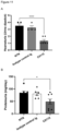
- Example 6 Strongly reduced CDC function of the newly designed anti-oxMIF antibody C0115 determined by a complement-dependent cytotoxicity (CDC) assay.
- Figure 5 clearly shows that the newly designed antibody C0115 harboring Fc silencing mutations (L234A/L235A) did not induce any CDC activity and the measured signals were even lower than the negative control (nivolumab, IgG4).
- the parent antibody C0083 with wt Fc showed complement dependent cell lysis of HCT116-pMIF cells, as expected for a human IgG1.
- the newly designed antibody C0115 harboring Fc silencing (L234A/L235A) mutations shows strongly reduced CDC activity.
- Figure 5 shows strongly reduced CDC activity of the newly designed Fc silenced antibody C0115 determined by a complement dependent cell toxicity bioassay.
- Example 7 Strongly reduced ADCC function of the newly designed anti-oxMIF antibodies C0115 and C0118 determined by a PBMC cell lysis bioassay.
- This assay was performed to determine cell lysis resulting from the antibody-dependent cytotoxicity (ADCC) induced the by newly designed anti-oxMIF antibodies. Cytotoxicity was measured by HiBiT Detection assay (Promega) which quantifies the release of a HiBiT tagged protein from the target cells using a non-lytic detection reagent containing LgBiT (LargeBiT) and furimazine (substrate). HiBiT and LgBiT spontaneously assemble to a functional NanoBiT ® enzyme and emits a quantifiable luminescence signal in the presence of the substrate.
- HiBiT Detection assay Promega
- HCT116-HiBiT-pMIF reporter target cells were generated as described in Example 6.
- FIG. 6 shows strongly reduced ADCC activity of the newly designed Fc silenced antibodies C0115 and C0118 determined by a PBMC mediated cell toxicity bioassay.
- Mean and SEM of two replicates using PBMCs from two healthy donors are shown. Data were fitted to a sigmoidal, 4-parameter equation using GraphPad Prism
- Example 8 Reduced unspecific binding of the newly designed anti-oxMIF antibodies C0115 and C0118 on A2780 MIF -/- cells.
- A2780 MIF -/- cell line was generated by CRISPR/Cas9 gene editing of human MIF gene in A2780 ovarian carcinoma cell line.
- target gene sequence was analysed and target sites were located according to the general rules of designing a targeting guidance RNA (gRNA) for GenCRISPR TM system.
- gRNA targeting guidance RNA
- a guide RNA (gRNA) was designed to specifically recognize the 5' region of the MIF gene (TTGGTGTTTACGATGAACATCGG, SEQ ID NO:40) and the gRNA sequence was cloned into the PX459 (addgene) vector containing S. pyogenes Cas9 (SpCas9) nuclease.
- Cells were resuspended in 50 ⁇ l of staining buffer, and 50 ⁇ l of serial dilutions of the newly designed anti-oxMIF antibodies C0115 and C0118 or their parent antibodies C0083 and C0090, respectively, or the control anti-oxMIF antibody C0008 or an isotype IgG (final concentrations 37 nM -9.4 nM) were added. After incubating for 40 min at 4°C, cells were washed with staining buffer, and resuspended in 100 ⁇ l of secondary antibody (goat anti-human IgG (H+L)-AlexaFluor 488, diluted 1:100). After incubating for 30 min at 4°C, cells were washed with staining buffer, resuspended in PBS+2% BSA and acquired on the CytoFlex-S flow cytometer (Beckman Coulter).
- secondary antibody goat anti-human IgG (H+L)-AlexaFluor
- Example 9 Strongly reduced unspecific cytokine release from human PBMCs by newly designed anti-oxMIF antibody C0115.
- Cytokine release syndrome is a form of systemic inflammatory response syndrome (SIRS) that can be triggered by a variety of factors such as infections. CRS is also known as an adverse effect of some monoclonal antibody medications. CRS occurs when large numbers of white blood cells, including B cells, T cells, natural killer cells, macrophages, dendritic cells, and monocytes are activated and release inflammatory cytokines, like IL-6, IFN-y, IL-8, MCP-1 amongst others, which activate more white blood cells in a positive feedback loop of pathogenic inflammation. This process, when dysregulated, can be life-threatening due to systemic hyper-inflammation, hypotensive shock, and multi-organ failure. Thus, the newly designed anti-oxMIF antibody C0115 was assessed for their potential to release inflammatory cytokines from PMBCs in an in-vitro assay.
- the newly designed anti-oxMIF antibody C0115 and the anti-oxMIF reference antibody C0008 were incubated at 70 nM, 7 nM, 0.7 nM, 0.07 nM with freshly thawed PBMCs (4-5x10 5 cells per well) from three healthy donors, or with medium only, in 150 ⁇ l of RPMI1640 medium supplemented with 5% ultra-low IgG serum in 96-well plates. After overnight incubation at 37°C/5% CO 2 in a humidified incubator, plates were centrifuged at 300 x g for 5 min to pellet the cells, and cleared supernatants were transferred to the 96-well V-bottom plates (BioLegend).
- Figure 8 illustrates that the newly designed anti-oxMIF antibody C0115 shows strongly reduced cytokine release from human PBMCs compared to reference antibody C0008.
- the anti-oxMIF antibody C0115 harboring Fc-silencing mutations and the reference anti-oxMIF antibody C0008 were incubated at the broad concentration range (70 nM, 7 nM, 0.7 nM, 0.07 nM) with human PBMCs overnight, and supernatants were analyzed in LegendPlex cytometric bead assays (BioLegend) for human MCP-1 ( A ), human IL-6 ( B ) and human TNF- ⁇ ( C ). Mean +/- SEM for cytokine concentrations (in pg/ml) are shown from 3 different PMBC donors.
- Example 10 Biodistribution of newly designed anti-oxMIF antibody C0115 and control antibody C0008 in Balb/c nude mice bearing xenografted human HCT116 colon carcinoma tumors.
- mice Biodistribution of newly designed anti-oxMIF antibody C0115 was investigated in female Balb/c nude mice carrying subcutaneous tumors of the human colon cancer cells HCT116 in comparison to the reference anti-oxMIF antibody C0008.
- Female Balb/c nude mice received unilateral, subcutaneous injections of 5x10 6 HCT116 cells in 50% PBS and 50% matrigel in a total injection volume of 100 ⁇ l. Upon reaching individual tumor volumes of 150-300 mm 3 , mice were assigned to treatment groups and received a single intravenous dose of 5 mg/kg IRDye 800CW-labeled C0115 or C0008.
- C0115 and C0008 were labelled with IRDye 800CW using the IRDye 800CW Protein labeling kit (high MW from LI-COR Biosciences) following the manufacturer's instructions.
- the protein concentration and labeling efficiency of the IRDye 800CW labeled antibodies was determined using the Nanodrop technology, and mice were dosed based on the protein concentration after labelling.
- Figure 9 shows a significant intra-tumoral distribution of intravenously administered IRDye 800CW-labeled C0115 and C0008, respectively, with tumor retention up to 7 days. It is evident from Figure 9(A) and quantitative image analysis ( Figure 9(B) ) that the tumor uptake of newly designed anti-oxMIF antibody C0115 was strongly enhanced and increased over 7 days compared to the reference anti-oxMIF antibody C0008, which showed a peak at ⁇ 24h. Figure 9 shows the tumor penetration and retention of the newly designed anti-oxMIF antibody C0115 and the reference antibody C0008 by infra-red in vivo imaging of mice carrying subcutaneous HCT116 tumors.
- mice were taken 1h, 6h, 24h, 48h, 72h, 96h and 168h post injection of the IRDye 800CW-labeled antibodies C0115 (upper panel) and C0008 (lower panel) dosed at 5 mg/kg;
- C0115 upper panel
- C0008 lower panel
- Mean of 3 mice is shown.
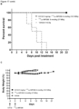
- Example 12 Efficacy of C0115 antibody in a collagen-II-induced arthritis (CIA) mouse model.
- mice were injected intradermally at the base of the tail with 100 ⁇ l (100 ⁇ g/mouse) of the obtained emulsion containing CII and CFA.
- a second booster of 100 ⁇ l of CII (100 ⁇ g/mouse) in IFA was administered.
- mice were treated twice a week (i.p.) for 20 days with vehicle, isotype control IgG1 (40 mg/kg), C0115 (at 20 mg/kg), or received daily injections of dexamethasone as standard-of-care drug (at 0.3 mg/kg).
- a paw thickness index was determined for each mouse by calculating the area under the curve (AUC) of the sum of the thickness for the 4 individual paws over the treatment.
- a cumulative disease score was calculated for each mouse by summing the scores for the individual mice over the treatment period. Calculations were done in GraphPad Prism and Ordinary One-way ANOVA followed by Fisher's LSD test was used for statistical analysis.
- Figure 10 reveals that treatment with C0115 at 20 mg/kg resulted in a significant amelioration of disease score ( A ) and paw oedema ( B ) compared to the vehicle-treated group. These effects (in particular, reduction of paw thickness) were comparable to the treatment with a high dose of standard-of-care corticosteroid drug dexamethasone (0.3 mg/kg). Isotype control (IgG)-treated group did not result in a reduced disease score. All treated groups had similar body weight variations during the disease course.
- Figure 10 shows that the newly designed Fc-silenced anti-oxMIF antibody C0115 ameliorates disease severity in a collagen II-induced DBA/1j mouse arthritis model.
- Cumulative disease score ( A ) and paw thickness ( B ) were assessed in a mouse arthritis model upon the treatment with C0115 (20 mg/ml), high dose dexamethasone (0.3 mg/kg) as a standard-of-care corticosteroid drug, or vehicle.
- Ordinary One-way ANOVA followed by Fisher's LSD test was used for statistical analysis in GraphPad Prism V 9.4 (*p ⁇ 0.05; **p ⁇ 0.01; ***p ⁇ 0.001).
- Example 13 Efficacy of C0115 antibody in a glomerular nephritis (GN) rat model.
- Glomerular nephritis was induced by injecting WKY male rats (weight range: 190-220 grams) with 100 ⁇ l of rabbit anti-rat NTS (nephrotoxic serum) intravenously. At day 4 and day 6 post-NTS injection, animals were treated (i.p.) with vehicle, isotype control IgG1, or C0115 (at 30 mg/kg). Urine was collected at day 0 (baseline), 4 (onset of disease) and 7 (after treatment). At the end of the study (day 8) animals were euthanized and blood as well as tissues (kidneys, liver, spleen, and lung) were collected.
- Histological (Crescentic counts; ED-1 macrophage staining, rat and rabbit IgG deposit) and biochemical analysis (Proteinuria and Haematuria) were performed to assess the severity of the disease.
- Ordinary One-way ANOVA followed by Dunnett's correction for multiple testing was used for statistical analysis in GraphPad Prism V 9.4 (*p ⁇ 0.05, **p ⁇ 0.01; ***p ⁇ 0.001, ****p ⁇ 0.0001).
- Figure 11 shows that the newly designed Fc-silenced anti-oxMIF antibody C0115 ameliorates disease severity in a nephrotoxic serum (NTS)-induced rat glomerulonephritis model.
- NTS nephrotoxic serum
- Haematuria in the urine (dipstick) A
- proteinuria B
- glomerular macrophage infiltration C
- Mean ⁇ SEM is shown, Ordinary One-way ANOVA followed by Dunnett's correction for multiple testing was used for statistical analysis in GraphPad Prism V 9.4 (*p ⁇ 0.05, ***p ⁇ 0.001, ****p ⁇ 0.0001).
- Example 14 Binding of bispecific antibodies (bsMabs) C0132 and C0133 (anti-oxMIF x anti-HSG) to oxMIF.
- FIG 12 Schematic drawing of the newly designed Fc silenced anti-oxMIF x anti-HSG bispecific antibodies (bsMabs) C0132 (Fab/scFv-Fc) and C0133 (Fab/FabscFv-Fc).
- Figure 13 shows the binding curves of the newly designed Fc silenced anti-oxMIF x anti-HSG bsMabs C0132 and C0133 to immobilized oxMIF. Bound antibodies were detected by an anti-human-IgG (Fc)-HRP conjugate, C0008 was used as reference antibody for bivalent binding to oxMIF. Data were fitted to a sigmoidal, 4-parameter equation using GraphPad Prism (mean +/- SEM of two replicates is shown)
- Example 15 Differential binding of C0132 and C0133 anti-oxMIF x anti-HSG bsMabs to oxMIF vs. redMIF.
- Anti-oxMIF x anti-HSG bsMabs C0132 and C0133, reference anti-oxMIF mAb C0008, and a human IgG1 isotype control were immobilized into microplates overnight at 4°C (15 nM for oxMIF bivalent and 30 nM for oxMIF monovalent antibodies). After blocking, wells were incubated with 50 ng/ml ( ⁇ 1,3 nM) of either redMIF or the oxMIF surrogate NTB-MIF (Schinagl et al., 2018). Captured oxMIF was detected with a polyclonal rabbit anti-MIF antibody and goat anti-rabbit IgG-HRP. Plates were stained with tetramethylbenzidine (TMB), and chromogenic reaction was stopped by addition of 30% H 2 SO 4 . OD was measured at 450 nm.
- TMB tetramethylbenzidine
- Figure 14 shows the differential binding of the newly designed Fc silenced anti-oxMIF x anti-HSG bsMabs C0132 and C0133 to oxMIF vs redMIF. C0008 was used as reference anti-oxMIF antibody. Mean and SEM of three replicates is shown.
- Example 16 Biodistribution of the newly designed Fc silenced anti-oxMIF x anti-HSG bsMabs C0132 and C0133 in Balb/c mice bearing syngenic CT26 colon tumors.
- C0132 and C0133 were labelled with IRDye 800CW using the IRDye 800CW Protein labeling kit (high MW from LI-COR Biosciences) following the manufacturer's instructions.
- the protein concentration and labeling efficiency of the IRDye 800CW-labeled antibodies was determined using the Nanodrop technology, and mice were dosed based on the protein concentration after labelling.
- In vivo imaging was performed in a LI-COR Pearl ® Trilogy imaging device performed at 785 nm excitation and 820 nm emission wavelength upon administration of labelled antibodies at the following time-points: 1h, 8h, 24h, 48h, 72h, 96h, 168h after dosing.
- Figure 15 shows a significant intra-tumoral distribution of intravenously administered IRDye 800CW-labeled C0132 and C0133, respectively, with tumor retention up to 7 days. It is evident from Figure 15 that one hour after injection and at all later imagining timepoints, the observed infrared signal in the tumours exceeded that of the background, indicating preferential accumulation of the antibodies in the tumour.
- Figure 15 shows the tumor penetration and retention of newly designed Fc silenced anti-oxMIF x anti-HSG bsMabs C0132 and C0133 assessed by infra-red in vivo imaging of mice carrying subcutaneous syngrafted CT26 tumors. Infra-red images of mice were taken 1h, 8h, 24h, 48h, 72h, 96h and 168h post injection of the IRDye 800CW labeled antibodies dosed at 5 mg/kg.
- IMP288 is a DOTA-conjugated D-Tyr-D-Lys-D-Glu-D-Lys-NH2 tetrapeptide in which both lysine residues are derivatized with HSG moiety via their ⁇ -aminogroup ( Figure 16A ).
- the DOTA chelating group of IMP288 is specifically designed for use with radiometals, including 177 Lu.
- IMP288 (Genepep, France) was labelled with 177 Lu at a specific activity of approximately 220 MBq/nmol while showing a radiochemical purity (RCP) ⁇ 95%. Briefly, IMP288 was diluted with sterile water to a final concentration of 1 mM (1.45 mg/ml). A 1/10 dilution of IMP288 stock solution was performed in 2-(N-morpholino) ethanesulfonic acid (MES) buffer (500 mM, pH 5.5).
- MES 2-(N-morpholino) ethanesulfonic acid
- DTPA-Na diethylenepenta-diaminetetraacetic acid sodium salt
- 177 Lu-IMP288 solution 50 ⁇ l of 177 Lu-IMP288 solution at 220 MBq/nmol and 1.68 nmol/ml corresponding to 0.084 nmol of IMP288 were mixed with 10-fold molar excess of each bsMab (0.84 nmol). Each reaction was incubated for 30 min at 37°C under gentle stirring. 177 Lu-IMP288 solution as well as 177 Lu-IMP288-bsMab solutions were applied to an iTLC strip and elution was done with 1:1 (v/v) solution of 0.15 M ammonium acetate (pH 5.5): MeOH. After elution, iTLC plates were exposed on a phosphorus screen (MS, BAS-IP, Fujifilm 2025) and revealed using Typhoon IP (Amersham) and associated software ImageQuant TL version 8.2.
- CT26 murine colorectal carcinoma (ATCC-CRL-2638) cells were expanded in RPMI1640 medium supplemented with 10% heat-inactivated FBS and 2 mM glutamine in a 37°C/ 5% CO2.
- a suspension of CT26 cells was prepared to 10 x 10 6 viable cells/mL in sterile PBS.
- Balb/c mice were subcutaneously injected in right flank with 1x10 6 CT26 cells suspended in 100 ⁇ L of PBS. Once tumours reached 50-100 mm 3 in volume (about 7-9 days post-inoculation) mice were randomized into treatment groups of 10 mice per group.
- Antibodies C0132 or C0133
- 177 Lu-IMP288 was administered 3 days after the injection of bispecific mAbs (day 0) by intravenous route at the molar ratio of bsMAb/HSGIMP288 hapten of 10:1 ( ⁇ 4 nmol/kg C0132 bsMAb or ⁇ 3 nmol/kg C0133 bsMab corresponding to 18.5 MBq respectively). Additionally, control groups received either 177 Lu-IMP288 at 37 MBq or vehicle on day 0 without any prior treatment. Tumor volumes and body weights were measured every 2-3 days until 21 days or until mice reached the humane endpoint of 1000 mm 3 tumor volume, and percentage of survival was determined.
- Statistical analysis was performed in GraphPad Prism V9.4 using Ordinary one-way ANOVA with Dunnett's correction for multiple testing.
- Figure 16 shows the structure of HSG hapten IMP288 ( A ) and ( B ) binding of bsMabs C0132 and C0133 to 177 Lu-IMP288 peptide assessed by iTLC based on change in migration profile of 177 Lu-IMP288 upon the incubation with the bsMabs when compared to the migration profile of 177 Lu-IMP288 peptide alone.
- Figure 17 shows PRAIT of syngrafted CT26 murine colorectal carcinomas in Balb/c mice with Fc silenced anti-oxMIF x anti-HSG bsMab C0132.
- C0132 was administered on day -3, followed by administration of 177 Lu-IMP288 3 days later (day 0).
- Statistical analysis was performed in GraphPad Prism V9.4 using Ordinary one-way ANOVA with Dunnett's correction for multiple testing; ** p ⁇ 0.01 vs. 177 Lu-IMP288;
- B Kaplan-Meier survival curves (in % survival),(C) body weights in % to body weights measured on day 0.
- Figure 18 shows PRAIT of syngrafted CT26 murine colorectal carcinomas in Balb/c mice with Fc silenced anti-oxMIF x anti-HSG bsMab C0133.
- C0133 was administered on day -3, followed by administration of 177 Lu-IMP288 3 days later (day 0).
- Statistical analysis was performed in GraphPad Prism V9.4 using Ordinary one-way ANOVA with Dunnett's correction for multiple testing; *** p ⁇ 0.001 vs. 177 Lu-IMP288.
- efficacy of the newly designed Fc-silenced bispecific anti-oxMIF x anti-HSG antibody C0132 was evaluated using a PRAIT approach together with the HSG hapten IMP288 (as described in US2005/0025709 ) and 177 Lu as radionuclide.
- In vivo efficacy of the RRAIT was evaluated on Balb/c mice bearing subcutaneous CT26 murine colorectal carcinoma tumors. Study was conducted as described in the Example 16 with some modifications. Briefly, Balb/c mice (Balb/c ByJ, Janvier Labs, France) were subcutaneously injected in right flank with 1x10 6 CT26 cells suspended in 100 ⁇ L of PBS. The tumor growth was controlled by visual observation and palpation until day 7 post-inoculation. In vivo therapeutic study was initiated when tumor size reached about 100-200 mm 3 for the 3 treatment groups (10 mice per group) and about 400-500 mm 3 for 1 control group (10 mice per group). BsMab C0132 was injected i.v.
- Statistical analysis (Ordinary one-way ANOVA with Dunnett's correction for multiple testing, *p ⁇ 0.05, **p ⁇ 0.01, ***p ⁇ 0.001) was performed in using GraphPad Prism for days 8 and 10 only, comparing each of the 2 C0132-treated group to the control group ( 177 Lu-IMP288 alone, no bsMab), because after day 11 animals in the control group ( 177 Lu-IMP288, no bsMab) had to be euthanised due to excessive tumour growth.
- Figure 19 shows efficacy of anti-oxMIF x anti-HSG bsMab C0132 in Balb/c using a PRAIT approach in mice syngrafted with CT26 murine colorectal carcinoma cells.
- C0132 was administered at 2.5 mg/ml, and 5 mg/ml on day -5, followed by administration of 177 Lu-IMP288 5 days later (day 0).
- Statistical analysis was performed in GraphPad Prism V9.4 using Ordinary one-way ANOVA with Dunnett's correction for multiple testing; *** p ⁇ 0.001 vs. 177 Lu-IMP288;
- B Kaplan-Meier survival curves (in % survival).
- efficacy of the newly designed Fc silenced bispecific anti-oxMIF x anti-HSG antibody C0132 was evaluated using a PRAIT approach together with the HSG hapten IMP288 (as described in US2005/0025709 ) and 177 Lu as radionuclide in a xenograft model of pancreatic cancer.
- Materials and Methods In vivo efficacy of the PRAIT was evaluated on Balb/c nude mice bearing subcutaneous CFPAC-1 ductal pancreatic adenocarcinoma tumors. Study was essentially conducted as described in the Example 16 and 17 with some modifications.
- the human pancreatic ductal adenocarcinoma cells, CFPAC-1 were provided by ATCC (Cat# CRL-1918). Cells were cultured in RPMI 1640 medium supplemented with 10% FBS and 2mM L-Glutamine. A suspension of CFPAC-1 cells was prepared to 50 x 10 6 cells/mL in sterile PBS. Balb/c nude mice (Balb/c ByAnNRj-Foxn1 nu/nu ) were subcutaneously injected in right flank with 5x10 6 CFPAC-1 cells suspended in 100 ⁇ L of PBS. The tumor growth was controlled by visual observation and palpation until day 4 post-inoculation.
- Statistical analysis (Ordinary one-way ANOVA with Dunnett's correction for multiple testing; **p ⁇ 0.01) was performed in GraphPad Prism V9.4 for day 14 only, comparing the C0132-treated group to the vehicle group.
- Figure 20 shows efficacy of anti-oxMIF x anti-HSG bsMab C0132 using a PRAIT approach in Balb/c nude mice xenografted with CFPAC-1 pancreatic adenocarcinoma cells.
- Statistical analysis was performed in GraphPad Prism V9.4 using Ordinary one-way ANOVA with Dunnett's correction for multiple testing; ** p ⁇ 0.01 vs. vehicle.
- Example 20 Efficacy of C0115 antibody in combination with glucocorticoids (GCs) in a mouse model of type II collagen induced arthritis (CIA).
- GCs glucocorticoids
- Glucocorticoids are potent immunosuppressive agents commonly used as a long-term therapy to control rheumatic diseases in human patients but associated with a variety of side effects.
- the efficacy of C0115 was evaluated as stand-alone treatment, or in combination with dexamethasone.
- mice were injected intradermally at the base of the tail with 100 ⁇ l (100 ⁇ g/mouse) of the obtained emulsion containing CII and CFA.
- a second booster of 100 ⁇ l of CII (100 ⁇ g/mouse) in IFA was administered.
- mice were treated twice a week (i.p.) for 20 days with: vehicle, C0115 (20 mg/kg) alone, or in combination with daily injections of low dose of dexamethasone (0.1 mg/kg).
- a control group of mice received daily injections of a high dose of dexamethasone (0.3 mg/kg) as standard-of-care drug.
- Figure 21 reveals that treatment with C0115 (20 mg/kg) resulted in a significant amelioration of clinical arthritis signs, indicated by cumulative disease score, compared to the vehicle-treated group. Moreover, combined treatment of C0115 (20mg/kg) and low dose of dexamethasone (0.1 mg/kg) resulted in further amelioration of clinical arthritis signs, compared to the treatment as single agents, and is comparable to the efficacy of a high dose of dexamethasone (0.3 mg/kg).
- Example 21 Efficacy of C0115 antibody and in combination with glucocorticoids (GCs) in a T cell transfer mouse model of colitis.
- GCs glucocorticoids
- BALB/c and C.B-17 SCID Engelbreviations: BALB/c and C.B-17 SCID (Envigo, San Pietro al Natisone, Udine, Italy) female mice aged between 8 to 9 weeks (weight range: 18-20 grams) were used in this study.
- Figure 22 indicates that treatment with C0115 (10 mg/kg) resulted in a significant amelioration of disease severity, evidenced by a significant reduction of cumulative stool score ( A ) compared to the vehicle-treated group and comparable to the high dose of dexamethasone (0.1 mg/kg). Combined treatment of C0115 at 10 mg/kg and low dose of dexamethasone (0.01 mg/kg) further ameliorated disease severity, evidenced by a further significant reduction of cumulative stool score.
Landscapes
- Health & Medical Sciences (AREA)
- Chemical & Material Sciences (AREA)
- Life Sciences & Earth Sciences (AREA)
- Medicinal Chemistry (AREA)
- General Health & Medical Sciences (AREA)
- Organic Chemistry (AREA)
- Immunology (AREA)
- Pharmacology & Pharmacy (AREA)
- Veterinary Medicine (AREA)
- Public Health (AREA)
- Animal Behavior & Ethology (AREA)
- Engineering & Computer Science (AREA)
- Bioinformatics & Cheminformatics (AREA)
- Proteomics, Peptides & Aminoacids (AREA)
- Epidemiology (AREA)
- General Chemical & Material Sciences (AREA)
- Chemical Kinetics & Catalysis (AREA)
- Nuclear Medicine, Radiotherapy & Molecular Imaging (AREA)
- Molecular Biology (AREA)
- Biochemistry (AREA)
- Genetics & Genomics (AREA)
- Biophysics (AREA)
- Urology & Nephrology (AREA)
- Transplantation (AREA)
- Rheumatology (AREA)
- Biomedical Technology (AREA)
- Oncology (AREA)
- Microbiology (AREA)
- Mycology (AREA)
- Physics & Mathematics (AREA)
- Optics & Photonics (AREA)
- Physical Education & Sports Medicine (AREA)
- Orthopedic Medicine & Surgery (AREA)
- Pain & Pain Management (AREA)
- Peptides Or Proteins (AREA)
- Medicines Containing Antibodies Or Antigens For Use As Internal Diagnostic Agents (AREA)
- Preparation Of Compounds By Using Micro-Organisms (AREA)
Claims (15)
- Fc-stummgeschalteter Anti-oxMIF-Antikörper, umfassend eine Variante der Fc-Region eines menschlichen IgG vom Wildtyp, umfassend SEQ ID NO1 mit einer oder mehreren Aminosäure-Substitutionen oder Glykosylierungsmodifikationen, und die folgenden variablen Domänen(a1) eine variable Domäne der leichten Kette umfassend SEQ ID NO:2 mit 1, 2, 3, 4 oder 5 Aminosäure-Substitutionen ausgewählt aus M30L, F49Y, A51G, P80S und W93F, oder(a2) eine variable Domäne der leichten Kette umfassend SEQ ID NO:2 mit 1, 2, 3, 4 oder 5 Aminosäure-Substitutionen ausgewählt aus M30L, F49Y, A51G, P80S und W93F, und mit 1, 2, 3, 4 oder 5 weiteren Aminosäure-Substitutionen, mit der Maßgabe, dass das- konservierte Tyrosin an Position 36 erhalten wird,und(b1) eine variable Domäne der schweren Kette umfassend SEQ ID NO:3; oder(b2) eine variablen Domäne der schweren Kette umfassend SEQ ID NO:3 mit 1 oder 2 Aminosäure-Substitutionen ausgewählt aus L5Q und W97Y, oder (b3) einer variablen Domäne der schweren Kette umfassend SEQ ID NO:3 mit 1 oder 2 Aminosäure-Substitutionen ausgewählt aus L5Q und W97Y, und mit 1, 2, 3, 4 oder 5 weiteren Aminosäure-Substitutionen;wobei die Aminosäure-Positionen nach Kabat nummeriert sind,wobei die Variante der Fc-Region eine verminderte FcyR-Bindung verglichen mit der Fc-Region der Wildtyp-IgG1 zeigt, wobei die eine oder die mehreren Aminosäure-Substitutionen in der Variante der Fc-Region an einer beliebigen der Positionen E233, L234, L235, G236, G237, P238, D265, S267, H268, N297, S298, T299, E318, L328, P329, A330, P331 von SEQ ID NO1 vorliegen, im Speziellen die Aminosäure-Substitutionen an den Positionen L234 und L235 vorliegen, nach dem EU-Nummerierungs-Index, und/oder wobei die Fc-Region nicht glykosyliert ist, und wobei der Antikörper ein reduziertes Aggregationspotential und reduzierte Hydrophobizität verglichen mit einem Antikörper umfassend SEQ ID NO2 und SEQ ID NO:3 aufweist, welchen die Aminosäure-Substitutionen fehlen.
- Fc-stummgeschalteter anti-oxMIF-Antikörper nach Anspruch 1, wobei die variable Domäne der leichten Kette die Aminosäure Substitution W93F umfasst und die variable Region der schweren Kette die Aminosäure Substitution W97Y umfasst.
- Fc-stummgeschalteter anti-oxMIF-Antikörper nach Anspruch 1 oder 2, umfassend variable Domänen umfassend eine Aminosäuresequenz ausgewählt aus der Gruppe bestehend aus SEQ ID NO:4, 5, 6, 7, 8, 9 und 43.
- Fc-stummgeschalteter anti-oxMIF-Antikörper nach den Ansprüchen 1 bis 3 umfassendi.) SEQ ID NO:3 und 6,ii.) SEQ ID NO:9 und 6,iii.) SEQ ID NO:4 und 6,iv.) SEQ ID NO:4 und 8,v.) SEQ ID NO:4 und 5vi.) SEQ ID NO:43 und eine beliebige der SEQ ID NO:5, 6 oder 8.
- Fc-stummgeschalteter anti-oxMIF-Antikörper nach Anspruch 4, ferner umfassend SEQ ID NO:14.
- Fc-stummgeschalteter anti-oxMIF-Antikörper nach einem der Ansprüche 1 oder 2, umfassend eine Aminosäuresequenz ausgewählt aus der Gruppe bestehend aus SEQ ID NO:15 und 16 oder aus SEQ ID NO:15 und 17.
- Fc-stummgeschalteter anti-oxMIF-Antikörper nach einem der Ansprüche 1 bis 6, ausgewählt aus der Gruppe bestehend aus bispezifischen Antikörpern, scFv-Fc, (scFv)z-Fc, scFvscFv-Fc, FabscFv-Fc, Fab(scFv)2-Fc, FabFab-scFv-Fc, FabFabcrossFab-Fc, IgG-scFv und IgG-(scFv)2.
- Fc-stummgeschalteter anti-oxMIF-Antikörper nach einem der Ansprüche 1 bis 7, wobei der Antikörper ein bispezifischer Antikörper ist, ferner umfassend zumindest eine Bindungsstelle, die spezifisch ein Epitop von CD3 oder Histamin-Succinyl-Glycin (HSG) bindet.
- Fc-stummgeschalteter Antikörper nach einem der Ansprüche 1 bis 8 zur Verwendung bei der Herstellung eines Medikaments.
- Pharmazeutische Zusammensetzung umfassend den Antikörper nach einem der Ansprüche 1 bis 8, gegebenenfalls zusammen mit einem pharmazeutischen Träger oder Adjuvans.
- Pharmazeutische Zusammensetzung nach Anspruch 10, wobei die pharmazeutische Zusammensetzung für die subkutane Verabreichung formuliert ist.
- Pharmazeutische Zusammensetzung nach Anspruch 10 oder 11, wobei die Zusammensetzung zur Verabreichung als einzige Substanz oder zur Verabreichung mit weiteren Wirkstoffen, vorzugsweise ausgewählt aus der Gruppe bestehend aus antiviralen, Antikrebs-, entzündungshemmenden und antibiotischen Substanzen, bestimmt ist.
- Pharmazeutische Zusammensetzung nach einem der Ansprüche 10 bis 12 zur Verwendung bei der Behandlung eines Patienten, der an einer entzündlichen Krankheit, einer Infektionskrankheit leidet, insbesondere bei der Behandlung von Asthma, Vaskulitis, Arthritis, Sepsis, septischem Schock, endotoxischem Schock, toxischem Schocksyndrom, akutem Lungenversagen (ARDS), Glomerulonephritis, entzündlicher Darmerkrankung, Morbus Crohn, Colitis ulcerosa, Peritonitis, Nephritis, NASH (nichtalkoholische Steatosehepatitis), multipler Sklerose, akuter und chronischer Pankreatitis, Typ-1-Diabetes, IgA-Nephropathie, interstitieller Zystitis, Post-COVID-Syndrom und Psoriasis, oder zur Verwendung in der Behandlung eines Patienten, der an einer hyperproliferativen Störung oder Krebs leidet, im Speziellen in der Behandlung von Dickdarmkrebs, Ovarialkrebs, Brustkrebs, Prostatakrebs, Bauchspeicheldrüsenkrebs, Magenkrebs und Lungenkrebs.
- Isolierte Nukleinsäure, die für den Antikörper nach einem der Ansprüche 1 bis 8 kodiert.
- Expressionsvektor, umfassend die Nukleinsäure nach Anspruch 14.
Applications Claiming Priority (4)
| Application Number | Priority Date | Filing Date | Title |
|---|---|---|---|
| EP21194701 | 2021-09-03 | ||
| EP21204108 | 2021-10-22 | ||
| EP22155394 | 2022-02-07 | ||
| PCT/EP2022/074449 WO2023031397A1 (en) | 2021-09-03 | 2022-09-02 | IMPROVED Fc SILENCED ANTI-oxMIF ANTIBODIES WITH REDUCED AGGREGATION POTENTIAL AND REDUCED HYDROPHOBICITY |
Publications (3)
| Publication Number | Publication Date |
|---|---|
| EP4396227A1 EP4396227A1 (de) | 2024-07-10 |
| EP4396227C0 EP4396227C0 (de) | 2025-07-09 |
| EP4396227B1 true EP4396227B1 (de) | 2025-07-09 |
Family
ID=83439101
Family Applications (1)
| Application Number | Title | Priority Date | Filing Date |
|---|---|---|---|
| EP22776885.0A Active EP4396227B1 (de) | 2021-09-03 | 2022-09-02 | Verbesserte fc-ausgeschaltete anti-oxmif-antikörper mit reduziertem aggregationspotenzial und reduzierter hydrophobizität |
Country Status (10)
| Country | Link |
|---|---|
| US (1) | US20250129148A1 (de) |
| EP (1) | EP4396227B1 (de) |
| JP (1) | JP2024532461A (de) |
| KR (1) | KR20240050375A (de) |
| AU (1) | AU2022336665A1 (de) |
| CA (1) | CA3230723A1 (de) |
| ES (1) | ES3038265T3 (de) |
| IL (1) | IL310958A (de) |
| PL (1) | PL4396227T3 (de) |
| WO (1) | WO2023031397A1 (de) |
Families Citing this family (1)
| Publication number | Priority date | Publication date | Assignee | Title |
|---|---|---|---|---|
| AU2024233139A1 (en) | 2023-03-03 | 2025-08-28 | Oncoone Research & Development Gmbh | Improved bispecific anti-tumor antigen/anti-hsg antibodies for pre-targeting of hyperproliferative disorders |
Family Cites Families (7)
| Publication number | Priority date | Publication date | Assignee | Title |
|---|---|---|---|---|
| US8165535B2 (en) | 2006-06-04 | 2012-04-24 | Samsung Electro-Mechanics | Systems, methods and apparatuses for complementary metal oxide semiconductor (CMOS) antenna switches using switched resonators |
| CA2529027C (en) | 2003-06-13 | 2013-09-10 | Immunomedics, Inc. | D-amino acid peptides |
| KR101654678B1 (ko) | 2008-01-04 | 2016-09-08 | 백스터 인터내셔널 인코포레이티드 | 항 mif 항체 |
| CA2968818A1 (en) * | 2015-01-09 | 2016-07-14 | Immunomedics, Inc. | Tumor therapy by bispecific antibody pretargeting |
| JP7123804B2 (ja) | 2016-04-12 | 2022-08-23 | シムフォゲン・アクティーゼルスカブ | 抗tim-3抗体および組成物 |
| AU2019281019A1 (en) | 2018-06-07 | 2020-11-26 | Oncoone Research & Development Gmbh | Anti-oxMIF/anti-CD3 antibody for cancer treatment |
| WO2022167474A1 (en) * | 2021-02-03 | 2022-08-11 | Oncoone Research & Development Gmbh | ANTI-oxMIF RADIOIMMUNOCONJUGATE |
-
2022
- 2022-09-02 KR KR1020247008656A patent/KR20240050375A/ko active Pending
- 2022-09-02 EP EP22776885.0A patent/EP4396227B1/de active Active
- 2022-09-02 IL IL310958A patent/IL310958A/en unknown
- 2022-09-02 CA CA3230723A patent/CA3230723A1/en active Pending
- 2022-09-02 ES ES22776885T patent/ES3038265T3/es active Active
- 2022-09-02 AU AU2022336665A patent/AU2022336665A1/en active Pending
- 2022-09-02 PL PL22776885.0T patent/PL4396227T3/pl unknown
- 2022-09-02 US US18/688,254 patent/US20250129148A1/en active Pending
- 2022-09-02 JP JP2024513731A patent/JP2024532461A/ja active Pending
- 2022-09-02 WO PCT/EP2022/074449 patent/WO2023031397A1/en not_active Ceased
Also Published As
| Publication number | Publication date |
|---|---|
| KR20240050375A (ko) | 2024-04-18 |
| PL4396227T3 (pl) | 2025-10-27 |
| WO2023031397A1 (en) | 2023-03-09 |
| JP2024532461A (ja) | 2024-09-05 |
| EP4396227C0 (de) | 2025-07-09 |
| IL310958A (en) | 2024-04-01 |
| US20250129148A1 (en) | 2025-04-24 |
| ES3038265T3 (en) | 2025-10-10 |
| AU2022336665A1 (en) | 2024-03-21 |
| CA3230723A1 (en) | 2023-03-09 |
| EP4396227A1 (de) | 2024-07-10 |
Similar Documents
| Publication | Publication Date | Title |
|---|---|---|
| CN110997712A (zh) | 特异性结合pd-1的抗体及其使用方法 | |
| US20220002398A1 (en) | ANTI-oxMIF/ANTI-CD3 ANTIBODY FOR CANCER TREATMENT | |
| JP2021510064A (ja) | 細胞傷害誘導治療剤 | |
| TW202233672A (zh) | 包括類δ配體3(DLL3)抗原結合區之蛋白質及其用途 | |
| CA3113539A1 (en) | Antibody constructs binding 4-1bb and tumor-associated antigens and uses thereof | |
| CN115461367A (zh) | 抗ox40抗体及其用途 | |
| CA3078141A1 (en) | Cd3/cd33 bispecific binding molecules | |
| EP4222170B1 (de) | Verbesserte anti-oxmif-antikörper mit reduziertem aggregationspotenzial und reduzierter hydrophobizität | |
| EP4396227B1 (de) | Verbesserte fc-ausgeschaltete anti-oxmif-antikörper mit reduziertem aggregationspotenzial und reduzierter hydrophobizität | |
| US20230265202A1 (en) | Antibody constructs binding 4-1bb and folate receptor alpha and uses thereof | |
| CN118302444A (zh) | 具有降低的聚集潜力和降低的疏水性的改善的Fc沉默的抗oxMIF抗体 | |
| HK40106756A (zh) | 具有降低的聚集潜力和降低的疏水性的改善的fc沉默的抗oxmif抗体 | |
| EA048864B1 (ru) | УЛУЧШЕННЫЕ АНТИТЕЛА ПРОТИВ oxMIF С ПОНИЖЕННЫМ ПОТЕНЦИАЛОМ АГРЕГАЦИИ И ПОНИЖЕННОЙ ГИДРОФОБНОСТЬЮ | |
| US20240301086A1 (en) | Tumor-associated antigens and cd3-binding proteins, related compositions, and methods | |
| CN121263443A (zh) | 抗ctla4抗体及其制备和使用方法 | |
| HK40091554A (zh) | 聚集潜在性降低和疏水性降低的改良抗oxmif抗体 | |
| HK40091554B (zh) | 聚集潜在性降低和疏水性降低的改良抗oxmif抗体 |
Legal Events
| Date | Code | Title | Description |
|---|---|---|---|
| STAA | Information on the status of an ep patent application or granted ep patent |
Free format text: STATUS: UNKNOWN |
|
| STAA | Information on the status of an ep patent application or granted ep patent |
Free format text: STATUS: THE INTERNATIONAL PUBLICATION HAS BEEN MADE |
|
| PUAI | Public reference made under article 153(3) epc to a published international application that has entered the european phase |
Free format text: ORIGINAL CODE: 0009012 |
|
| STAA | Information on the status of an ep patent application or granted ep patent |
Free format text: STATUS: REQUEST FOR EXAMINATION WAS MADE |
|
| 17P | Request for examination filed |
Effective date: 20240311 |
|
| AK | Designated contracting states |
Kind code of ref document: A1 Designated state(s): AL AT BE BG CH CY CZ DE DK EE ES FI FR GB GR HR HU IE IS IT LI LT LU LV MC MK MT NL NO PL PT RO RS SE SI SK SM TR |
|
| DAV | Request for validation of the european patent (deleted) | ||
| DAX | Request for extension of the european patent (deleted) | ||
| GRAP | Despatch of communication of intention to grant a patent |
Free format text: ORIGINAL CODE: EPIDOSNIGR1 |
|
| STAA | Information on the status of an ep patent application or granted ep patent |
Free format text: STATUS: GRANT OF PATENT IS INTENDED |
|
| RIC1 | Information provided on ipc code assigned before grant |
Ipc: A61K 47/68 20170101ALI20250110BHEP Ipc: A61K 51/04 20060101ALI20250110BHEP Ipc: A61K 49/00 20060101ALI20250110BHEP Ipc: A61P 13/12 20060101ALI20250110BHEP Ipc: A61P 35/00 20060101ALI20250110BHEP Ipc: A61P 37/06 20060101ALI20250110BHEP Ipc: A61K 31/00 20060101ALI20250110BHEP Ipc: A61K 39/395 20060101ALI20250110BHEP Ipc: C07K 16/46 20060101ALI20250110BHEP Ipc: C07K 16/44 20060101ALI20250110BHEP Ipc: C07K 16/24 20060101AFI20250110BHEP |
|
| INTG | Intention to grant announced |
Effective date: 20250210 |
|
| GRAS | Grant fee paid |
Free format text: ORIGINAL CODE: EPIDOSNIGR3 |
|
| GRAA | (expected) grant |
Free format text: ORIGINAL CODE: 0009210 |
|
| STAA | Information on the status of an ep patent application or granted ep patent |
Free format text: STATUS: THE PATENT HAS BEEN GRANTED |
|
| AK | Designated contracting states |
Kind code of ref document: B1 Designated state(s): AL AT BE BG CH CY CZ DE DK EE ES FI FR GB GR HR HU IE IS IT LI LT LU LV MC MK MT NL NO PL PT RO RS SE SI SK SM TR |
|
| REG | Reference to a national code |
Ref country code: GB Ref legal event code: FG4D |
|
| REG | Reference to a national code |
Ref country code: CH Ref legal event code: EP |
|
| REG | Reference to a national code |
Ref country code: IE Ref legal event code: FG4D |
|
| REG | Reference to a national code |
Ref country code: DE Ref legal event code: R096 Ref document number: 602022017416 Country of ref document: DE |
|
| U01 | Request for unitary effect filed |
Effective date: 20250806 |
|
| U07 | Unitary effect registered |
Designated state(s): AT BE BG DE DK EE FI FR IT LT LU LV MT NL PT RO SE SI Effective date: 20250819 |
|
| REG | Reference to a national code |
Ref country code: CH Ref legal event code: U11 Free format text: ST27 STATUS EVENT CODE: U-0-0-U10-U11 (AS PROVIDED BY THE NATIONAL OFFICE) Effective date: 20251001 |
|
| REG | Reference to a national code |
Ref country code: ES Ref legal event code: FG2A Ref document number: 3038265 Country of ref document: ES Kind code of ref document: T3 Effective date: 20251010 |
|
| PGFP | Annual fee paid to national office [announced via postgrant information from national office to epo] |
Ref country code: NO Payment date: 20250923 Year of fee payment: 4 |
|
| PGFP | Annual fee paid to national office [announced via postgrant information from national office to epo] |
Ref country code: IE Payment date: 20250918 Year of fee payment: 4 Ref country code: CZ Payment date: 20250828 Year of fee payment: 4 |
|
| PGFP | Annual fee paid to national office [announced via postgrant information from national office to epo] |
Ref country code: IS Payment date: 20250911 Year of fee payment: 4 |
|
| U20 | Renewal fee for the european patent with unitary effect paid |
Year of fee payment: 4 Effective date: 20251028 |
|
| PG25 | Lapsed in a contracting state [announced via postgrant information from national office to epo] |
Ref country code: HR Free format text: LAPSE BECAUSE OF FAILURE TO SUBMIT A TRANSLATION OF THE DESCRIPTION OR TO PAY THE FEE WITHIN THE PRESCRIBED TIME-LIMIT Effective date: 20250709 |
|
| PG25 | Lapsed in a contracting state [announced via postgrant information from national office to epo] |
Ref country code: GR Free format text: LAPSE BECAUSE OF FAILURE TO SUBMIT A TRANSLATION OF THE DESCRIPTION OR TO PAY THE FEE WITHIN THE PRESCRIBED TIME-LIMIT Effective date: 20251010 |
|
| PGFP | Annual fee paid to national office [announced via postgrant information from national office to epo] |
Ref country code: TR Payment date: 20251009 Year of fee payment: 4 |
|
| PGFP | Annual fee paid to national office [announced via postgrant information from national office to epo] |
Ref country code: CH Payment date: 20251001 Year of fee payment: 4 |
|
| PGFP | Annual fee paid to national office [announced via postgrant information from national office to epo] |
Ref country code: PL Payment date: 20250904 Year of fee payment: 4 |
|
| PG25 | Lapsed in a contracting state [announced via postgrant information from national office to epo] |
Ref country code: RS Free format text: LAPSE BECAUSE OF FAILURE TO SUBMIT A TRANSLATION OF THE DESCRIPTION OR TO PAY THE FEE WITHIN THE PRESCRIBED TIME-LIMIT Effective date: 20251009 |
|
| PGFP | Annual fee paid to national office [announced via postgrant information from national office to epo] |
Ref country code: ES Payment date: 20251030 Year of fee payment: 4 |