EP4295869A2 - Methods and devices to disrupt and contain pathogens - Google Patents
Methods and devices to disrupt and contain pathogens Download PDFInfo
- Publication number
- EP4295869A2 EP4295869A2 EP23205893.3A EP23205893A EP4295869A2 EP 4295869 A2 EP4295869 A2 EP 4295869A2 EP 23205893 A EP23205893 A EP 23205893A EP 4295869 A2 EP4295869 A2 EP 4295869A2
- Authority
- EP
- European Patent Office
- Prior art keywords
- debridement
- wound
- layer
- fibres
- gel
- Prior art date
- Legal status (The legal status is an assumption and is not a legal conclusion. Google has not performed a legal analysis and makes no representation as to the accuracy of the status listed.)
- Granted
Links
Images


Classifications
-
- A—HUMAN NECESSITIES
- A61—MEDICAL OR VETERINARY SCIENCE; HYGIENE
- A61L—METHODS OR APPARATUS FOR STERILISING MATERIALS OR OBJECTS IN GENERAL; DISINFECTION, STERILISATION OR DEODORISATION OF AIR; CHEMICAL ASPECTS OF BANDAGES, DRESSINGS, ABSORBENT PADS OR SURGICAL ARTICLES; MATERIALS FOR BANDAGES, DRESSINGS, ABSORBENT PADS OR SURGICAL ARTICLES
- A61L15/00—Chemical aspects of, or use of materials for, bandages, dressings or absorbent pads
- A61L15/16—Bandages, dressings or absorbent pads for physiological fluids such as urine or blood, e.g. sanitary towels, tampons
- A61L15/42—Use of materials characterised by their function or physical properties
- A61L15/425—Porous materials, e.g. foams or sponges
-
- A—HUMAN NECESSITIES
- A61—MEDICAL OR VETERINARY SCIENCE; HYGIENE
- A61F—FILTERS IMPLANTABLE INTO BLOOD VESSELS; PROSTHESES; DEVICES PROVIDING PATENCY TO, OR PREVENTING COLLAPSING OF, TUBULAR STRUCTURES OF THE BODY, e.g. STENTS; ORTHOPAEDIC, NURSING OR CONTRACEPTIVE DEVICES; FOMENTATION; TREATMENT OR PROTECTION OF EYES OR EARS; BANDAGES, DRESSINGS OR ABSORBENT PADS; FIRST-AID KITS
- A61F13/00—Bandages or dressings; Absorbent pads
- A61F13/02—Adhesive bandages or dressings
- A61F13/0203—Adhesive bandages or dressings with fluid retention members
- A61F13/0213—Adhesive bandages or dressings with fluid retention members the fluid retention member being a layer of hydrocolloid, gel forming material
-
- A—HUMAN NECESSITIES
- A61—MEDICAL OR VETERINARY SCIENCE; HYGIENE
- A61L—METHODS OR APPARATUS FOR STERILISING MATERIALS OR OBJECTS IN GENERAL; DISINFECTION, STERILISATION OR DEODORISATION OF AIR; CHEMICAL ASPECTS OF BANDAGES, DRESSINGS, ABSORBENT PADS OR SURGICAL ARTICLES; MATERIALS FOR BANDAGES, DRESSINGS, ABSORBENT PADS OR SURGICAL ARTICLES
- A61L15/00—Chemical aspects of, or use of materials for, bandages, dressings or absorbent pads
- A61L15/16—Bandages, dressings or absorbent pads for physiological fluids such as urine or blood, e.g. sanitary towels, tampons
- A61L15/42—Use of materials characterised by their function or physical properties
- A61L15/60—Liquid-swellable gel-forming materials, e.g. super-absorbents
-
- A—HUMAN NECESSITIES
- A61—MEDICAL OR VETERINARY SCIENCE; HYGIENE
- A61F—FILTERS IMPLANTABLE INTO BLOOD VESSELS; PROSTHESES; DEVICES PROVIDING PATENCY TO, OR PREVENTING COLLAPSING OF, TUBULAR STRUCTURES OF THE BODY, e.g. STENTS; ORTHOPAEDIC, NURSING OR CONTRACEPTIVE DEVICES; FOMENTATION; TREATMENT OR PROTECTION OF EYES OR EARS; BANDAGES, DRESSINGS OR ABSORBENT PADS; FIRST-AID KITS
- A61F13/00—Bandages or dressings; Absorbent pads
- A61F13/00051—Accessories for dressings
- A61F13/00063—Accessories for dressings comprising medicaments or additives, e.g. odor control, PH control, debriding, antimicrobic
-
- A—HUMAN NECESSITIES
- A61—MEDICAL OR VETERINARY SCIENCE; HYGIENE
- A61F—FILTERS IMPLANTABLE INTO BLOOD VESSELS; PROSTHESES; DEVICES PROVIDING PATENCY TO, OR PREVENTING COLLAPSING OF, TUBULAR STRUCTURES OF THE BODY, e.g. STENTS; ORTHOPAEDIC, NURSING OR CONTRACEPTIVE DEVICES; FOMENTATION; TREATMENT OR PROTECTION OF EYES OR EARS; BANDAGES, DRESSINGS OR ABSORBENT PADS; FIRST-AID KITS
- A61F13/00—Bandages or dressings; Absorbent pads
- A61F13/02—Adhesive bandages or dressings
- A61F13/0203—Adhesive bandages or dressings with fluid retention members
- A61F13/0206—Adhesive bandages or dressings with fluid retention members with absorbent fibrous layers, e.g. woven or non-woven absorbent pads or island dressings
-
- A—HUMAN NECESSITIES
- A61—MEDICAL OR VETERINARY SCIENCE; HYGIENE
- A61F—FILTERS IMPLANTABLE INTO BLOOD VESSELS; PROSTHESES; DEVICES PROVIDING PATENCY TO, OR PREVENTING COLLAPSING OF, TUBULAR STRUCTURES OF THE BODY, e.g. STENTS; ORTHOPAEDIC, NURSING OR CONTRACEPTIVE DEVICES; FOMENTATION; TREATMENT OR PROTECTION OF EYES OR EARS; BANDAGES, DRESSINGS OR ABSORBENT PADS; FIRST-AID KITS
- A61F13/00—Bandages or dressings; Absorbent pads
- A61F13/02—Adhesive bandages or dressings
- A61F13/0203—Adhesive bandages or dressings with fluid retention members
- A61F13/0223—Adhesive bandages or dressings with fluid retention members characterized by parametric properties of the fluid retention layer, e.g. absorbency, wicking capacity, liquid distribution
-
- A—HUMAN NECESSITIES
- A61—MEDICAL OR VETERINARY SCIENCE; HYGIENE
- A61F—FILTERS IMPLANTABLE INTO BLOOD VESSELS; PROSTHESES; DEVICES PROVIDING PATENCY TO, OR PREVENTING COLLAPSING OF, TUBULAR STRUCTURES OF THE BODY, e.g. STENTS; ORTHOPAEDIC, NURSING OR CONTRACEPTIVE DEVICES; FOMENTATION; TREATMENT OR PROTECTION OF EYES OR EARS; BANDAGES, DRESSINGS OR ABSORBENT PADS; FIRST-AID KITS
- A61F13/00—Bandages or dressings; Absorbent pads
- A61F13/02—Adhesive bandages or dressings
- A61F13/0203—Adhesive bandages or dressings with fluid retention members
- A61F13/0226—Adhesive bandages or dressings with fluid retention members characterised by the support layer
-
- A—HUMAN NECESSITIES
- A61—MEDICAL OR VETERINARY SCIENCE; HYGIENE
- A61L—METHODS OR APPARATUS FOR STERILISING MATERIALS OR OBJECTS IN GENERAL; DISINFECTION, STERILISATION OR DEODORISATION OF AIR; CHEMICAL ASPECTS OF BANDAGES, DRESSINGS, ABSORBENT PADS OR SURGICAL ARTICLES; MATERIALS FOR BANDAGES, DRESSINGS, ABSORBENT PADS OR SURGICAL ARTICLES
- A61L15/00—Chemical aspects of, or use of materials for, bandages, dressings or absorbent pads
- A61L15/16—Bandages, dressings or absorbent pads for physiological fluids such as urine or blood, e.g. sanitary towels, tampons
- A61L15/22—Bandages, dressings or absorbent pads for physiological fluids such as urine or blood, e.g. sanitary towels, tampons containing macromolecular materials
-
- A—HUMAN NECESSITIES
- A61—MEDICAL OR VETERINARY SCIENCE; HYGIENE
- A61L—METHODS OR APPARATUS FOR STERILISING MATERIALS OR OBJECTS IN GENERAL; DISINFECTION, STERILISATION OR DEODORISATION OF AIR; CHEMICAL ASPECTS OF BANDAGES, DRESSINGS, ABSORBENT PADS OR SURGICAL ARTICLES; MATERIALS FOR BANDAGES, DRESSINGS, ABSORBENT PADS OR SURGICAL ARTICLES
- A61L15/00—Chemical aspects of, or use of materials for, bandages, dressings or absorbent pads
- A61L15/16—Bandages, dressings or absorbent pads for physiological fluids such as urine or blood, e.g. sanitary towels, tampons
- A61L15/22—Bandages, dressings or absorbent pads for physiological fluids such as urine or blood, e.g. sanitary towels, tampons containing macromolecular materials
- A61L15/28—Polysaccharides or their derivatives
-
- A—HUMAN NECESSITIES
- A61—MEDICAL OR VETERINARY SCIENCE; HYGIENE
- A61L—METHODS OR APPARATUS FOR STERILISING MATERIALS OR OBJECTS IN GENERAL; DISINFECTION, STERILISATION OR DEODORISATION OF AIR; CHEMICAL ASPECTS OF BANDAGES, DRESSINGS, ABSORBENT PADS OR SURGICAL ARTICLES; MATERIALS FOR BANDAGES, DRESSINGS, ABSORBENT PADS OR SURGICAL ARTICLES
- A61L15/00—Chemical aspects of, or use of materials for, bandages, dressings or absorbent pads
- A61L15/16—Bandages, dressings or absorbent pads for physiological fluids such as urine or blood, e.g. sanitary towels, tampons
- A61L15/42—Use of materials characterised by their function or physical properties
-
- A—HUMAN NECESSITIES
- A61—MEDICAL OR VETERINARY SCIENCE; HYGIENE
- A61L—METHODS OR APPARATUS FOR STERILISING MATERIALS OR OBJECTS IN GENERAL; DISINFECTION, STERILISATION OR DEODORISATION OF AIR; CHEMICAL ASPECTS OF BANDAGES, DRESSINGS, ABSORBENT PADS OR SURGICAL ARTICLES; MATERIALS FOR BANDAGES, DRESSINGS, ABSORBENT PADS OR SURGICAL ARTICLES
- A61L15/00—Chemical aspects of, or use of materials for, bandages, dressings or absorbent pads
- A61L15/16—Bandages, dressings or absorbent pads for physiological fluids such as urine or blood, e.g. sanitary towels, tampons
- A61L15/42—Use of materials characterised by their function or physical properties
- A61L15/48—Surfactants
-
- A—HUMAN NECESSITIES
- A61—MEDICAL OR VETERINARY SCIENCE; HYGIENE
- A61L—METHODS OR APPARATUS FOR STERILISING MATERIALS OR OBJECTS IN GENERAL; DISINFECTION, STERILISATION OR DEODORISATION OF AIR; CHEMICAL ASPECTS OF BANDAGES, DRESSINGS, ABSORBENT PADS OR SURGICAL ARTICLES; MATERIALS FOR BANDAGES, DRESSINGS, ABSORBENT PADS OR SURGICAL ARTICLES
- A61L15/00—Chemical aspects of, or use of materials for, bandages, dressings or absorbent pads
- A61L15/16—Bandages, dressings or absorbent pads for physiological fluids such as urine or blood, e.g. sanitary towels, tampons
- A61L15/42—Use of materials characterised by their function or physical properties
- A61L15/56—Wetness-indicators or colourants
-
- C—CHEMISTRY; METALLURGY
- C12—BIOCHEMISTRY; BEER; SPIRITS; WINE; VINEGAR; MICROBIOLOGY; ENZYMOLOGY; MUTATION OR GENETIC ENGINEERING
- C12Q—MEASURING OR TESTING PROCESSES INVOLVING ENZYMES, NUCLEIC ACIDS OR MICROORGANISMS; COMPOSITIONS OR TEST PAPERS THEREFOR; PROCESSES OF PREPARING SUCH COMPOSITIONS; CONDITION-RESPONSIVE CONTROL IN MICROBIOLOGICAL OR ENZYMOLOGICAL PROCESSES
- C12Q1/00—Measuring or testing processes involving enzymes, nucleic acids or microorganisms; Compositions therefor; Processes of preparing such compositions
- C12Q1/02—Measuring or testing processes involving enzymes, nucleic acids or microorganisms; Compositions therefor; Processes of preparing such compositions involving viable microorganisms
- C12Q1/04—Determining presence or kind of microorganism; Use of selective media for testing antibiotics or bacteriocides; Compositions containing a chemical indicator therefor
-
- A—HUMAN NECESSITIES
- A61—MEDICAL OR VETERINARY SCIENCE; HYGIENE
- A61F—FILTERS IMPLANTABLE INTO BLOOD VESSELS; PROSTHESES; DEVICES PROVIDING PATENCY TO, OR PREVENTING COLLAPSING OF, TUBULAR STRUCTURES OF THE BODY, e.g. STENTS; ORTHOPAEDIC, NURSING OR CONTRACEPTIVE DEVICES; FOMENTATION; TREATMENT OR PROTECTION OF EYES OR EARS; BANDAGES, DRESSINGS OR ABSORBENT PADS; FIRST-AID KITS
- A61F13/00—Bandages or dressings; Absorbent pads
- A61F2013/00361—Plasters
- A61F2013/00902—Plasters containing means
- A61F2013/0094—Plasters containing means for sensing physical parameters
-
- A—HUMAN NECESSITIES
- A61—MEDICAL OR VETERINARY SCIENCE; HYGIENE
- A61L—METHODS OR APPARATUS FOR STERILISING MATERIALS OR OBJECTS IN GENERAL; DISINFECTION, STERILISATION OR DEODORISATION OF AIR; CHEMICAL ASPECTS OF BANDAGES, DRESSINGS, ABSORBENT PADS OR SURGICAL ARTICLES; MATERIALS FOR BANDAGES, DRESSINGS, ABSORBENT PADS OR SURGICAL ARTICLES
- A61L2300/00—Biologically active materials used in bandages, wound dressings, absorbent pads or medical devices
- A61L2300/40—Biologically active materials used in bandages, wound dressings, absorbent pads or medical devices characterised by a specific therapeutic activity or mode of action
- A61L2300/404—Biocides, antimicrobial agents, antiseptic agents
Definitions
- Wound healing is a complex dynamic process that results in the restoration of anatomic continuity and function: an ideally healed wound is one that has returned to normal anatomic structure, function, and appearance. Infection of the wound results in either a slower or an arrested healing process.
- Pathogens in a wound can produce toxins (e.g., Clostridium spp), noxious metabolites like ammonia that raise pH (e.g., Proteus spp), tissue-lytic enzymes like proteases, or further invade tissue by other means leading to an increase in wound area. In a worst case, pathogens can leave the wound and cause sepsis.
- Unwanted biological soft tissues such as devitalized tissue (e.g. slough, necrosis, eschar) or exogenous tissues (e.g. microbial biofilm) are a significant source of soft tissue infections. Removal of such unwanted tissues from surgical or chronic wounds, for example, is vital to help prevent infection and allow the site or wound to heal.
- tissue removal is achieved by various methods such as surgical debridement, sharp debridement, use of digesting enzymes, and biosurgery.
- Antimicrobial agents including antiseptics and antibiotics, are widely used to treat microbial infections.
- the emergence of antibiotic resistance and the ability of microorganisms to tolerate antimicrobial agents through their production of protective biofilm have significantly reduced their effectiveness. Therefore, new ways to disturb microbial habitats by disrupting biofilm, for example, increase susceptibility of microorganisms to antimicrobial agents, and develop alternative strategies to antimicrobial agents are required.
- conventional methods of debridement often cause pain and trauma, alternative methods that are effective but also less traumatic and painful are required.
- the plurality of bristles comprises a pile of monofilaments or fibres.
- the monofilaments comprise nylon, acetal, polypropylene, acrylic, polyvinyl chloride (PVC), polycarbonate, silicone, wool, or any combination thereof.
- the bristles comprise a pile of nylon monofilaments.
- the bristles comprise nonwoven or woven filaments arranged perpendicular to the wound contacting layer.
- the layer of gel-forming fibres are woven, non-woven, and/or knitted.
- the layer of gel-forming fibres is selected from the group consisting of sodium carboxymethylcellulose, alginate, cellulose, carboxyethylcellulose, cellulose ethyl sulfonate, modified cellulose, pectin, chitosan, modified chitosan, hyaluronic acid, polysaccharide, gum-derived polymer, and any combination thereof.
- the layer of gel-forming fibres comprises sodium carboxymethylcellulose.
- the layer of gel-forming fibres has an absorbency of at least 10 g/g and resists lateral spread of a fluid upon absorption.
- the layer of gel-forming fibres has an absorbency of 15 g/g to 50 g/g and resists lateral spread of a fluid upon absorption.
- the carboxymethylcellulose has a degree of substitution of at least 0.05 carboxymethyl groups per cellulose unit. In still other embodiments, the carboxymethylcellulose has a degree of substitution of at least 0.2 carboxymethyl groups per cellulose unit. In yet other embodiments, the carboxymethylcellulose has a degree of substitution of between 0.3 and 0.5 carboxymethyl groups per cellulose unit.
- the devices, methods and kits for debridement disclosed herein are capable of disrupting a biofilm on a wound.
- the devices and methods for debridement disclosed herein are capable of sequestering metal ions.
- the devices and methods for debridement disclosed herein further comprise anti-biofilm agents, metal chelators, surfactants, or any combination thereof.
- the devices and methods for debridement disclosed herein are capable of removing or loosening unwanted tissue or biofilm on a wound.
- the devices and methods for debridement disclosed herein are capable of sequestering bacteria and other components of a biofilm or unwanted tissue.
- the devices, methods and kits for debridement disclosed herein further comprise one or more support layers in contact with the layer of gel-forming fibres, wherein the support layer comprises foam, fabric, blended fabric, paper, polymer, plastic, or any combination thereof.
- the one or more support layers form a backing.
- an outer support layer comprises an anti-slip material.
- the anti-slip material comprises silicone.
- the devices, methods and kits disclosed herein further comprise a diagnostic unit in fluid communication with the layer of gel-forming fibres.
- the diagnostic unit comprises an enzyme-based assay or an electronic sensor.
- the diagnostic unit comprises one or more fibres or diagnostic disks, comprising: a) a reaction layer comprising one or more reagents that interact with a target enzyme indicative of a microbial infection, wherein the reagents are affixed to a solid phase; b) each reagent is sprayed, printed, or deposited in a reagent area separated by impermeable separators; c) each reagent area comprises a reporter area wherein a color, color change, or other detectable signal is observed; and d) a transparent cover comprising a window for visualizing the signal in the reporter area.
- the fluid communication comprises a wicking material, an opening, a channel, or a conduit.
- the wicking material comprises hydrophilic fibres, gelling fibres, filter paper, or any combination thereof.
- the diagnostic disks comprise one or more reagents selected from the group consisting of enzyme-reactive indicators, enzymes that produce colored products, pH indicators, protein responsive reagents, and moisture-detecting reagents.
- the enzyme-reactive indicators interact with one or more enzymes selected from the group consisting of elastase, lysozyme, cathepsin G, myeloperoxidase, and any combination thereof to produce a visible color or an electronic change in the reporter area of the diagnostic disk.
- the devices, methods and kits disclosed herein further comprise a handle attached to a non-wound-contacting surface of the debridement device.
- the handle comprises a loop, a stick, a tube, a tab, a bar, a pocket, a fold, a sleeve, or a protrusion on the non-wound-contacting surface.
- the handle is removably attached to the non-wound-contacting surface using a detachable securing member.
- the detachable securing member comprises a hook-and-loop element, an adhesive, or a sticker.
- the diagnostic assay comprises PCR, enzyme-linked immunosorbent assay (ELISA), or an immunoassay.
- the immunoassay comprises one or more reagents selected from the group consisting of enzyme-reactive indicators, enzymes that produce colored products, pH indicators, protein responsive reagents, and moisture-detecting reagents, and wherein the enzyme-reactive indicators interact with one or more enzymes selected from the group consisting of elastase, lysozyme, cathepsin G, myeloperoxidase, and any combination thereof to produce a detectable signal indicative of a microbial infection.
- enzyme-reactive indicators selected from the group consisting of enzyme-reactive indicators, enzymes that produce colored products, pH indicators, protein responsive reagents, and moisture-detecting reagents
- the enzyme-reactive indicators interact with one or more enzymes selected from the group consisting of elastase, lysozyme, cathepsin G, myeloperoxidase, and any combination thereof to produce a detectable signal indicative of a microbial infection.
- the diagnostic assay comprises one or more diagnostic disks in fluid communication with the debridement device, a standalone immunoassay, PCR, or ELISA.
- the diagnostic disks comprise: a) a reaction layer comprising one or more reagents that interact with a target enzyme indicative of a microbial infection, wherein the reagents are affixed to a solid phase; b) each reagent is sprayed, printed, or deposited in a reagent area separated by impermeable separators; c) each reagent area comprises a reporter area wherein a color, color change, or other detectable signal is observed; and d) a transparent cover comprising a window for visualizing the signal in the reporter area.
- the fluid communication comprises a wicking material, an opening, a channel, or a conduit.
- the wicking material comprises hydrophilic fibres, gelling fibres, or filter paper.
- the diagnostic disks comprise one or more reagents selected from the group consisting of enzyme-reactive indicators, enzymes that produce colored products, pH indicators, protein responsive reagents, and moisture-detecting reagents.
- the enzyme-reactive indicators interact with one or more enzymes selected from the group consisting of elastase, lysozyme, cathepsin G, myeloperoxidase, and any combination thereof to produce a visible color or an electronic change in the reporter area of the diagnostic disk.
- the layer of gel-forming fibres has an absorbency of at least 10 g/g and resists lateral spread of a fluid upon absorption.
- the devices and methods for debridement of a wound as disclosed herein are capable of disrupting a biofilm, or loosening or removing unwanted tissue on a wound.
- the devices and methods for debridement disclosed herein are capable of sequestering metal ions, bacteria, and/or unwanted wound tissue.
- the devices and methods for debridement disclosed herein comprise anti-biofilm agents, metal chelators, surfactants, or any combination thereof.
- the devices, methods and kits for debridement disclosed herein further comprise a handle attached to a non-wound-contacting surface of the debridement device.
- the handle comprises a loop, a stick, a tube, a tab, a bar, a pocket, a fold, a sleeve, a protrusion on the non-wound-contacting surface, or a detachable bar or stick.
- the devices and methods for debridement disclosed herein further comprise one or more diagnostic disks in fluid communication with the debridement device, comprising: a) a reaction layer comprising one or more reagents that interact with a target enzyme indicative of a microbial infection, wherein the reagents are affixed to a solid phase; b) each reagent is sprayed, printed, or deposited in a reagent area separated by impermeable separators; c) each reagent area comprises a reporter area wherein a color, color change, or other detectable signal is observed; and d) a transparent cover comprising a window for visualizing the signal in the reporter area.
- the fluid communication comprises wicking fibres, an opening, or a filter paper.
- one or more reagents in conjunction with the devices and methods for debridement disclosed herein are selected from the group consisting of enzyme-reactive indicators, enzymes that produce colored products, pH indicators, protein responsive reagents, and moisture-detecting reagents, and wherein the enzyme-reactive indicators interact with one or more enzymes selected from the group consisting of elastase, lysozyme, cathepsin G, myeloperoxidase, and any combination thereof to produce a detectable signal indicative of a microbial infection.
- kits for treating a wound comprising debriding a wound using the debridement device as described herein; analyzing a wound sample adsorbed to the debridement device using a diagnostic assay for presence of a microbial infection; optionally repeating the debriding step; and applying a wound dressing with or without an antimicrobial agent according to the diagnostic assay.
- the diagnostic assay comprises one or more diagnostic disks in fluid communication with the debridement device, a standalone immunoassay, PCR, or ELISA.
- the gel-forming fibres sequester disrupted biofilm, microorganisms, wound debris, metal ions, or combinations thereof.
- the gel-forming fibres are woven, non-woven, knitted, or combinations thereof.
- the gel-forming fibres is selected from the group consisting of sodium carboxymethylcellulose, alginate, cellulose, carboxyethylcellulose, cellulose ethyl sulfonate, modified cellulose, pectin, chitosan, modified chitosan, hyaluronic acid, polysaccharide, gum-derived polymer, and any combination thereof.
- the gel-forming fibres comprise sodium carboxymethylcellulose.
- the gel-forming fibres comprise an absorbency of at least 10 g/g. In some embodiments, the gel-forming fibres comprise an absorbency of 15 g/g to 50 g/g.
- the sodium carboxymethylcellulose has a degree of substitution of at least 0.05 carboxymethyl groups per cellulose unit. In still other embodiments, the sodium carboxymethylcellulose has a degree of substitution of at least 0.2 carboxymethyl groups per cellulose unit. In yet other embodiments, the sodium carboxymethylcellulose has a degree of substitution of between 0.3 and 0.5 carboxymethyl groups per cellulose unit.
- the anti-biofilm agent is selected from the group consisting of ethylenediaminetetraacetic acid (EDTA), tetrasodium EDTA, disodium EDTA, epigallocatechin gallate (EGCG), ellagic acid, esculetin, fisetin, reserpine, quercetin, linoleic acid, berberine, chitosan, eugenol, curcumin, synthetic halogenated furanone (F-56), Peptide 1018, CFT073 group-II capsular polysaccharide (Serotype K2), Pel polysaccharide, Psl polysaccharide, sophorolipid, colistin, nisin, subtilin, epidermin, gallidermin, sodium citrate, tannic acid, deoxyribonuclease I, glycoside hydrolase, bacteriophage-encoded endolysin (PlyC), silver, oc
- EDTA
- Disclosed herein are methods, devices and kits for debridement of a wound comprising: a) a device for debridement of a wound as described herein; and b) a solution or formulation for enhancing or assisting in debridement of the wound.
- the solution or formulation for enhancing or assisting in debridement of the wound comprises antimicrobial agents, anti-biofilm agents, metal chelators, surfactants, or any combination thereof.
- anti-biofilm agent is selected from the group consisting of ethylenediaminetetraacetic acid (EDTA), tetrasodium EDTA, disodium EDTA, epigallocatechin gallate (EGCG), ellagic acid, esculetin, fisetin, reserpine, quercetin, linoleic acid, berberine, chitosan, eugenol, curcumin, synthetic halogenated furanone (F-56), Peptide 1018, CFT073 group-II capsular polysaccharide (Serotype K2), Pel polysaccharide, Psl polysaccharide, sophorolipid, colistin, nisin, subtilin, epidermin, gallidermin, sodium citrate, tannic acid, deoxyribonuclease I, glycoside hydrolase, bacteriophage-encoded endolysin (PlyC), silver, octenidine
- the anti-biofilm agent is present between 0.01-5% by weight.
- the metal chelator comprises ethylenediaminetetraacetic acid (EDTA).
- the EDTA is present at a concentration of at least 0.01% by weight.
- the EDTA is present at a concentration between 0.2-0.8% by weight.
- the surfactant comprises a cationic surfactant, an anionic surfactant, a zwitterionic surfactant, a non-ionic surfactant, or a combination thereof.
- the surfactant is present at less than 2% by weight. In some embodiments, the surfactant is present at 0.01-2% by weight.
- the solution or formulation comprises sterile water or saline.
- the solution or formulation comprises an oxidizing agent.
- the oxidizing agent is hydrogen peroxide.
- the solution or formulation comprises iodine, polyhexanide (PHMB), chlorhexidine, hypochlorous acid, sodium hypochlorite, chlorine dioxide, acetic acid, or a combination thereof.
- Methods, devices, and kits in some embodiments, further comprise a treating agent.
- the treating agent comprises an antimicrobial or anti-biofilm composition. Methods, devices, and kits, in some embodiments, further comprise instructions for use.
- Disclosed herein are methods, devices and kits for detecting an infection in a sample comprising: a) a debridement device as described herein for collecting the sample; b) a diagnostic unit in fluid communication with the debridement device, wherein the diagnostic unit comprises an enzyme-based assay, an electronic sensor, or one or more reagents for indicating the infection; and c) a viewing window or reporter area wherein a color, color change, or other detectable signal based on the enzyme-based assay, the electronic sensor, or the one or more reagents for indicating the infection is observed.
- the one or more reagents is selected from the group consisting of enzyme-reactive indicators, enzymes that produce colored products, pH indicators, protein responsive reagents, and moisture-detecting reagents.
- the enzyme-reactive indicators interact with one or more enzymes selected from the group consisting of elastase, lysozyme, cathepsin G, and myeloperoxidase.
- Methods, devices, and kits in some embodiments, further comprise a wound dressing comprising gel-forming fibres with or without an antimicrobial agent. Methods, devices, and kits, in some embodiments, further comprise instructions for use.
- the one or more foams comprises open pore foams.
- a first foam of the one or more foams is on a first side and a second foam of the one or more foams is on a second side.
- the first foam comprises a rough texture.
- the second foam comprises a smooth texture.
- a first foam of the one or more foams and a second foam of the one or more foams are on a same side.
- the one or more foams comprises polyurethane.
- the one or more foams is in a butterfly shape.
- a pore of the one or more open foams comprises at diameter in a range of about 10 um to about 500 um.
- a height of the one or more open foams is in a range of about 1 centimeter to about 5 centimeters.
- a width of the one or more open foams is in a range of about 1 centimeter to about 10 centimeters.
- a length of the one or more open foams is in a range of about 1 centimeter to about 15 centimeters.
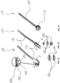
- FIGS. 1A-1C illustrate various forms of a debridement device 100 comprising a wound contacting layer 102 with a layer of monofilaments or bristles 104 in contact with a layer of gel-forming fibres 106. Some embodiments have a base layer for support 108.
- the debridement device 100 can be in the form of a stick or swab, with different shapes for the wound contacting surface, including a rectangular shape ( FIG. 1A ), a tubular shape ( FIG. 1B ), and a rounded or spear shape ( FIG. 1C ).
- a stick or handle 110 can comprise a hollow tube, a shaft, or a flat stick.
- FIGS. 2A-2C illustrate various forms of another embodiment of a debridement device 200, comprising a layer of monofilaments 202 in contact with a layer of gel-forming fibres 204.
- the debridement device 200 further comprises a support layer 206 that may be formed of foam, fabric, paper, polymer, plastic, or any other similar material or combination of material.
- the support layer 206 may be in contact with the fibres 202.
- the debridement device 200 comprises fibre blends within or between layers.
- the support layer 206 may comprise a fabric layer and a foam layer on top of a hydrofibre and monofilament layer.
- the debridement device 200 is a square or rectangular shape ( FIG. 2A ), a circular or elliptical shape ( FIG. 2B ), or a triangular shape ( FIG. 2C ).
- the debridement device 200 is a pad with a plurality of bristles 208 or a pile of monofilaments in contact with a layer of gel-forming fibres 208.
- the pad has a backing 210, comprising a fabric or fiber blends. Further still, the backing 210 may have an anti-slip surface.
- the debridement device 200 may also have a diagnostic unit 212 that is in fluid communication with a wound contacting layer 214.
- FIGS. 3A-3F illustrate types of handles 302, 304, 306, 308, 310 in contact with an outer support layer or a non-wound-contacting surface 312 of debridement pads 300.
- the debridement pads 300 may have a layer of gel-forming fibres with a pile of monofilaments or synthetic fibres on its surface as discussed herein.
- Different forms of handle include a soft fabric grip handle 302, soft fabric pocket or sleeve handle 308, fins 304 or tabs 310 that can be lifted when in use or folded flat when not in use, a Velcro ® or hook-an-loop securing unit that allows one to removably attach a plastic handle or knob 306, or an anti-slip layer 316 comprising silicone.
- FIG. 4 illustrates a debridement device 400 in fluid communication with a diagnostic unit 402 that detects a wound infection.
- the diagnostic unit 402 comprises a diagnostic disk comprising reagents printed or deposited on a solid phase, such as filter paper, and wicking fibres or threads 404, such as gelling fibres, that provide a fluid communication between the debridement device 400 and the diagnostic unit/disk 402.
- the debridement device 400 with infection detection comprises either a separate sticker 406 that is placed onto the debridement device 400, or is directly printed onto the debridement device.
- the sticker 406 or infection detection design can comprise different patterns.
- the diagnostic unit 402 may be an electronic sensor 408 capable of detecting infection.
- FIGS. 5A-5D illustrate wicking guide mechanisms 500 that provide a fluid communication between the debridement device and the diagnostic unit, including a hole 502 that allows fluid flow ( FIG. 5A ), wicking material 504 such as gelling fibres or similar wicking fibres that wick fluid from the debridement device to the diagnostic unit ( FIG. 5B ), a dimple or concave feature 506 comprising a non-woven material, fabric, or filter paper protruding outward to wick fluid ( FIG. 5C ), and a hydrophilic or gelling fiber disk 508 ( FIG. 5D
- FIGS. 6A-6B illustrate types of diagnostic units 600, 602 for detecting a microbial infection ( FIG. 6A ), including different shapes (including, but not limited to, square and rectangle and circle shapes), sizes and arrangement of the control and detection panels, as well as different arrangements of reagents 604 and reservoirs or reagent areas 606 ( FIG. 6B ).
- the arrangement of the different panels or separators 608 on the diagnostic unit allows for easy differentiation between the control and detection panels.
- the control(s) and reagents can be arranged in a radial manner with the signal reported in various forms to allow easy and fast detection of a microbial infection. ).
- one or more reagents of the diagnostic disk interacts with an enzyme marker for a microbial infection to produce a detectable signal in a reporter area 614 of the diagnostic unit 600, 602 which is visible through a window 610, such as a color change or an appearance of a signal or mark.
- FIG. 7 illustrates a schematic flow chart 700 for one embodiment of this disclosure. More specifically, a debridement device of this disclosure may be provide in box 702. The debridement device may be utilized to debride a wound in box 704. In one aspect of this disclosure, the debridement device may provide a sample of wound tissue in box 706. In box 708, the wound tissue may be analyzed for infection utilizing the methods and devices of this disclosure. In box 710, a wound dressing may be applied to the wound based on the results of the infection analysis of box 708 wherein the wound dressing is specifically tailored to address the type of infection identified in box 708.
- FIG. 8 illustrates one non-exclusive example of a kit 800 of this disclosure.
- the kit 800 may include a debridement device 802 having a diagnostic unit 804 of this disclosure.
- the kit 800 may also comprise a debridement solution 806 to assist in the debridement of a wound while using the debridement device 802.
- the kit 800 may also include a treating agent 808 that assists with treating the wound after debridement.
- multiple treating agents 808 may be provided to allow specific treatment of the wound based on the type of infection identified by the diagnostic unit 804.
- the devices provided herein comprise a wound contacting layer, the wound contacting layer comprising at least one layer, a plurality of layers, or a pile of fibres, filaments or bristles, the wound contact layer optionally in contact with or used in conjunction or in combination with the surface of a layer of hydrophilic fibres or gelling fibres.
- the layer of gelling fibres may comprise an absorbent material.
- the gelling fibres are arranged as one or more layers.
- the fibres, filaments or bristles protrude from a layer of gelling fibres or are perpendicular to the layer of gelling fibres.
- the bristles comprise a pile of nylon monofilaments that can loosen or remove unwanted tissue on a wound without causing significant trauma to the wound site.
- the bristles may also comprise other synthetic fibres with textured surfaces to allow efficient debridement.
- the devices and methods for debridement disclosed herein are capable of absorbing and sequestering wound exudate, metal ions, or bacteria.
- the devices and methods for debridement disclosed herein are capable of selectively and atraumatically removing unwanted tissues from infected sites.
- the devices and methods for debridement disclosed herein are capable of disrupting and sequestering microbial biofilm.
- the devices and methods for debridement disclosed herein are in fluid communication with a diagnostic unit for in situ detection of a microbial infection.
- a sample of wound tissue can be obtained from the devices for debridement disclosed herein, and analyzed for the presence of a microbial infection using, for example, any standard diagnostic assay, including PCR, enzyme-linked immunosorbent assay (ELISA), and other immunoassays.
- the term "filament” may refer to a "monofilament.” In some embodiments, the term “monofilament” refers to a single strand of thread or yarn.
- the thread or yarn can be a synthetic material.
- Monofilaments may form bristles.
- Non-limiting examples of synthetic materials that form monofilaments or bristles include nylon, polyester, polypropylene, polygalactic acid, acrylic, acetal, polyvinyl chloride (PVC), polycarbonate, silicone, chemically modified fibres, or any combination thereof.
- monofilaments comprise textured surfaces, such as micro-hooks or barbs that help to debride a wound surface, i.e ., loosen or remove unwanted tissue.
- monofilaments may also be referred to as bristles or debridement fibres.
- hydrophilic fibres or “hydrofibres” or “gel-forming fiber” refer to fibres that have high absorption capability and resist lateral spread or diffusion of a liquid upon absorption.
- hydrophilic fibres form a gel upon contact with a fluid to sequester or lock in the fluid, thus resisting lateral spread of the fluid.
- gel-forming fibres is AQUACEL ® .
- gelling fibres can sequester unwanted wound materials, such as loosened biofilm, wound debris, metal ions, and bacteria, thus preventing or mitigating further infection and helps to keep a wound clean and dry.
- gel-forming fibres have an absorbency of at least 10 g/g or 15 g/g to 50 g/g and resists lateral spread of a fluid upon absorption may be used.
- gelling fibres comprise carboxymethylcellulose with varying degree of substitution, e.g., at least 0.05, at least 0.2, or between 0.3 and 0.5 carboxymethyl groups per cellulose unit.
- gelling fibres comprise a surfactant, metal chelator, or antimicrobial agent.
- gel-forming fibres are selected from the group consisting of sodium carboxymethylcellulose, alginate or derivatized alginate, cellulose, carboxyethylcellulose, cellulose ethyl sulfonate, modified cellulose or derivatized cellulose, pectin, chitosan, modified chitosan or derivatized chitosan, hyaluronic acid or derivatives, polysaccharide, gum-derived polymer, polyacrylic acid or derivatives, and any combination thereof.
- degree of substitution refers to the average number of carboxymethylated hydroxyl groups per glucose or cellulose unit present in a cellulosic polymer such as carboxymethylcellulose. Degrees of substitution of carboxymethylcellulose can range from about 0.05 to about 0.5 carboxymethyl groups per cellulose unit, about 0.2 to about 0.5 carboxymethyl groups per cellulose unit, about 0.3 to about 0.5 carboxymethyl groups per cellulose unit. Degree of substitution can be about 0.05, about 0.06, about 0.07, about 0.08, about 0.09, about 0.1, about 0.2, about 0.3, about 0.4, about 0.5, about 0.6, about 0.7, about 0.8, about 0.9, or about 1.0 carboxymethyl groups per cellulose unit.
- swab tip or “swabbing end” refers to the wound-contacting end of a debridement device that can comprise one or more support layers, fibres or a fiber blend, or monofilaments.
- the terms swab tip and swabbing end are used interchangeably.
- sensor unit or “diagnostic unit” refers to a separate unit or chamber that comprises reagents for the detection of an infection.
- diagnostic unit or “diagnostic unit” refers to a separate unit or chamber that comprises reagents for the detection of an infection.
- sensor unit diagnostic unit, and sensor chamber are used interchangeably herein.
- the devices and methods for debridement disclosed herein may comprise other materials that absorb wound exudate, including fabric materials.
- fabric materials are broadly known, and may include foams, superabsorbent fibres, gelling foams, directional foams and the like.
- the herein described devices for wound debridement and disruption of microbial biofilm comprise a layer monofilaments or bristles that loosen or remove unwanted or necrotic tissue on a wound surface and a layer of high-absorbency gel-forming fibres, such as AQUACEL ® , or similar hydrophilic fibres, that can sequester wound fluid, bacteria, metal ions, and other unwanted tissue.
- the gelling fibres or hydrophilic fibres form an absorbent layer.
- the gelling fibres suitable for use in the absorbent layer of the present disclosure include hydrophilic fibres, which upon contact with a fluid or wound exudate, form a gel and sequester moisture and absorbed materials, such as disrupted biofilm, bacteria, or unwanted wound tissue, for examples.
- the fabric materials capable of disrupting biofilm and/or adsorbing disrupted biofilm.
- the fabric materials comprise anti-biofilm activity.
- the fabric materials sequester wound fluid, bacteria, metal ions, or other wound debris.
- the fabric materials comprise anti-biofilm activity and sequester wound fluid, bacteria, metal ions, or other wound debris.
- the fabric materials improve susceptibility of microorganisms to antimicrobial agents.
- the fabric materials as described herein reduce microbial burden at a colonized or infected site.
- the fabric materials reduce microbial burden at a colonized or infected site without antimicrobial agents.
- the fabric materials reduce microbial burden at a colonized or infected site with a low amount of antimicrobial agents.
- the fabric materials as described herein, in some embodiments, comprise various structures. Such structures include nonwoven fabrics, woven fabrics, composites, laminates, foams, knitted fabrics and combinations thereof. In some embodiments, the fabric materials comprise gelling fibres or hydrophilic fibres.
- the gelling fibres disrupt the biofilm by removing moisture from the biofilm. In some embodiments, the gelling fibres remove moisture by forming an absorbent layer. In some embodiments, the gelling fibres form a gel on contact with the moisture.
- the gelling fibres comprises gel-forming polymers or gelling fibres that have high absorbency and resist lateral spread upon absorption of a fluid. In some embodiments, the gelling fibres absorb and retain disrupted biofilm, microorganisms, and/or other wound debris. In some embodiments, the gelling fibres are configured to sequester metal ions. In some embodiments, the gelling fibres form a layer.
- Non-limiting examples of gel-forming polymers include cellulose, carboxymethylcellulose (CMC), carboxyethylcellulose, AQUACEL ® , oxidized cellulose (or a derivative thereof), cellulose ethyl sulfonate, other chemically modified cellulose, pectin, alginate, chitosan, modified chitosan, hyaluronic acid, polysaccharide, gum-derived polymer, or any combination thereof.
- the fibres comprise polyvinylpyrrolidone, polyvinyl alcohols, polyvinyl ethers, polyurethanes, polyacrlyates, polyacrylamides, collagen, gelatin or mixtures thereof.
- the gelling fibres comprise a blend of carboxymethylcellulose and alginate.
- the gelling fibres comprise a nonwoven layer of gel-forming fibres.
- the gel-forming fibres are selected from the group consisting of sodium carboxymethylcellulose, alginate, cellulose, carboxyethylcellulose, cellulose ethyl sulfonate, modified cellulose, pectin, chitosan, modified chitosan, hyaluronic acid, polysaccharide, gum-derived polymer, and any combination thereof.
- the cellulosic gelling fibres may have a degree of substitution of at least 0.05 carboxymethyl groups per cellulose unit. In some embodiments, the degree of substitution of the carboxymethylcellulose gel-forming fibres is at least 0.2 carboxymethyl groups per cellulose unit, more particularly between 0.3 and 0.5. In some embodiments, the gelling fibres comprise sodium carboxymethylcellulose with a comparatively lower degree of substitution.
- the ability to absorb moisture, gel, and disrupt materials such as biofilm is related to the degree of substitution of the cellulosic gelling fiber, including carboxymethylcellulose, as well as to the type of gelling fiber.
- the gelling fibres comprise a blend of carboxymethylcellulose with different degrees of substitution, thereby providing for different rates of disruption, absorption, and gelling.
- the gelling fibres comprise a blend of different fiber types to provide for disruption and absorption at the same time.
- the gel-forming action of the fibres helps to sequester or lock in unwanted wound materials, such as exudate, bacteria, or biofilm, which helps to keep a wound clean and dry, and mitigates or prevents the spread of an infection.
- the fabric material described herein comprises an agent comprising anti-biofilm activity.
- the agent comprising anti-biofilm activity comprises no antimicrobial activity.
- the agent comprising anti-biofilm activity comprises low levels of antimicrobial activity.
- Exemplary agents comprising antimicrobial activity include, but are not limited to, silver, antibiotics, antiseptics, antivirals, or antifungals.
- the fabric material comprises one or more agents comprising anti-biofilm activity.
- the agent comprising anti-biofilm activity is isolated from natural sources or is a synthetic compound, a chelating agent, a lantibiotic, a small molecule, an ion, a peptide, an enzyme, or a nanoparticle.
- agents comprising anti-biofilm activity include, but are not limited to, ethylenediaminetetraacetic acid (EDTA), tetrasodium EDTA, disodium EDTA, epigallocatechin gallate (EGCG), ellagic acid, esculetin, fisetin, reserpine, quercetin, linoleic acid, berberine, chitosan, eugenol, curcumin, synthetic halogenated furanone (F-56), Peptide 1018, CFT073 group-II capsular polysaccharide (Serotype K2), Pel polysaccharide, Psl polysaccharide, sophorolipid, colistin, nisin, subtilin, epidermin, gallidermin, sodium citrate, tannic acid, deoxyribonuclease I, glycoside hydrolase, bacteriophage-encoded endolysin (PlyC), silver, octenidine
- the fabric material comprising anti-biofilm activity in some embodiments, further comprises low levels of antimicrobial activity. In some embodiments, the fabric material comprising anti-biofilm activity further comprises no antimicrobial activity.
- the agents capable of sequestering biofilm components include, but are not limited to, ethylenediaminetetraacetic acid (EDTA), alkali metal salts, alkaline earth metal salts, ammonium salts thereof, and tetraalkylammonium salts.
- EDTA ethylenediaminetetraacetic acid
- alkali metal salts alkaline earth metal salts
- ammonium salts thereof tetraalkylammonium salts.
- Fabric materials as described herein may improve the susceptibility of microorganisms to antimicrobial agents.
- the fabric materials improve the susceptibility of microorganisms to antimicrobial agents by disrupting of biofilm and/or sequestering biofilm structural components, metal ions, wound exudate, microorganisms, and/or other wound debris.
- the fabric materials comprise an agent that provides anti-biofilm activity and low antimicrobial activity.
- Antimicrobial activity may be provided by an antimicrobial agent including, but are not limited to, silver, antibiotics, antiseptics, antivirals, or antifungals.
- the fabric materials following disruption of the biofilm and/or sequestration, the fabric materials further provides antimicrobial effects.
- Fabric materials as described herein comprising anti-biofilm activity and/or sequestration ability improve efficacy of biofilm disruption over, for example, standard debridement methods, including use of saline and gauze.
- the improved efficacy of biofilm disruption is at least 10%, 15%, 20%, 25%, 30%, 35%, 40%, 45%, 50%, 55%, 60%, 65%, 70%, 75%, 80%, 90%, 95%, 99%, or more than 99%.
- fabric materials improve susceptibility of microorganisms to antibacterial agents.
- the improved susceptibility of microorganisms to antibacterial agents is at least 10%, 15%, 20%, 25%, 30%, 35%, 40%, 45%, 50%, 55%, 60%, 65%, 70%, 75%, 80%, 90%, 95%, 99%, or more than 99%.
- the fabric materials as described herein reduce microbial burden at a colonized or infected site by at least 10%, 15%, 20%, 25%, 30%, 35%, 40%, 45%, 50%, 55%, 60%, 65%, 70%, 75%, 80%, 90%, 95%, 99%, or more than 99%. In some instances, the fabric materials reduce microbial burden at a colonized or infected site in a range of about 10% to about 99%, about 20% to about 80%, about 30% to about 70%, or about 40% to about 60%.
- the bristles, monofilaments, or debridement fibres are textured fibres or filaments that withstand the debriding action and help to loosen or remove unwanted tissue on a wound.
- bristles or monofilaments comprise nylon, acetal, polypropylene, acrylic, polyvinyl chloride (PVC), polycarbonate, silicone, wool, or any combination thereof.
- the debridement fibres are woven or nonwoven. In some embodiments, the nonwoven fibres form a layer. In some embodiments, the fibres are woven into a perpendicular or bristle-type arrangement. In some embodiments, the perpendicular or bristle-like arrangement comprises monofilaments.
- the debridement fibres are on the surface of or are in contact with the layer of gelling fibres to provide a disruptive or loosening effect followed by an absorptive effect by the gelling fibres.
- contacting a wound or biofilm with the debridement fibres may result in physically breaking up wound debris or biofilm, allowing for absorption of the material.
- the gelling action of certain fibres such as AQUACEL ® , further provides for sequestration of the absorbed materials. Such sequestration helps to clean a wound efficiently before applying a wound dressing, comprising gelling fibres.
- the debridement device includes anti-biofilm agents such as metal chelators or surfactants.
- the debridement fibres loosen unwanted tissues and materials through the action of surfactants and anti-biofilm components.
- the debridement device which has high absorbency and sequesters wound particulates, provides an easy and fast means for diagnosis or detection of a microbial infection in a wound.
- the debridement device is in fluid communication with a diagnostic unit, such as a diagnostic disk comprising one or more reagents printed on or deposited on a filter paper, for in situ diagnosis.
- Fluid communication in such embodiment comprises gelling fibres or hydrophilic threads, an opening, or filter paper that help to wick wound fluid or material from the layer of gelling fibres to the reagents in the diagnostic disk.
- the diagnostic disk is embedded in or inserted into a debridement device to allow facile detection of a bacterial infection in situ.
- such diagnostic disk is used in a standalone diagnostic device, wherein a sample of a wound can be extracted from the debridement device for further analysis using one or more conventional diagnostic assays, such as PCR, ELISA, or immunoassay.
- the debridement devices disclosed herein comprise debridement sticks or stick-swab devices. In some embodiments, the debridement devices disclosed herein comprise debridement swabs. In some embodiments, the debridement devices comprise debridement pads or pads with a backing, optionally with a handle. In some embodiments, the debridement devices disclosed herein comprise gel-forming fibres and a layer of bristles or monofilaments. In some embodiments, the gel-forming fibres comprise carboxymethylcellulose, such as AQUACEL ® .
- the debridement devices comprise a pile of monofilaments or bristles that protrude from the gel-forming fibres or are perpendicular to and in contact with the layer of gel-forming fibres.
- the monofilaments or bristles comprise nylon fibres.
- Other materials that can be used for monofilaments or bristles for debriding a wound include, but are not limited to, acetal, polypropylene, acrylic, polyvinyl chloride (PVC), polycarbonate, silicone, wool, or any combination thereof.
- the layer of gel-forming fibres is arranged in a layered structure that comprises support materials.
- Support materials can form a base layer that is in contact with the layer of gel-forming fibres.
- Support materials include foam, fabric, paper, plastic, wood, polymer, or any combination thereof, for example.
- the layered structure may further comprise an outer layer that covers the base layer or an outer transparent layer to allow visual detection of a diagnostic insert embedded or inserted in a debridement device for in situ detection of a microbial infection.
- fabric as an outer layer may cover a foam layer, with the foam layer located between the gel-forming fiber layer and the outer fabric layer.
- the outermost layer comprises an anti-slip material for holding the debridement device, such as silicone.
- Debridement devices as described herein, in some embodiments, comprise one or more foams.
- the one or more foams comprises open pore foams.
- the one or more foams are the same type of foam.
- the one or more foams are different types of foam.
- the one or more foams comprise variable abrasiveness. For example, a first foam on a first side comprises a rough texture and a second foam of a second side comprises a smooth texture.
- the first foam and the second foam are on a same side.
- the first foam comprising a rough texture is used for debridement.
- the second foam comprising a smooth texture is used for absorbance.
- the one or more foams are porous. In some embodiments, the one or more foams comprise different pore sizes. In some embodiments, a first foam on a first side comprises a first pore size. In some embodiments, a second foam on a second side comprises a second pore size. In some embodiments, the first foam on the first side and the second foam on the second side comprise the same pore size. In some embodiments, the first foam and the second foam are on the same side and comprise the same pore size.
- a pore of the one or more foams comprises a diameter of at least or about 10, 20, 30, 40, 50, 60, 70, 80, 90, 100, 120, 140, 160, 180, 200, 220, 240, 260, 280, 300, 320, 340, 360, 380, 400, 500, 600, 700, 800, 900, 1000, or more than 1000 micrometers (um).
- a pore of the one or more foams comprises a diameter in a range of about 10 um to about 1000 um, about 20 um to about 800 um, about 30 to about 700 um, about 40 to about 600 um, about 50 to about 500 um, about 60 um to about 400 um, about 70 um to about 300 um, about 80 um to about 200 um, or about 90 um to about 150 um.
- the one or more foams comprise various materials.
- the one or more foams comprises polyurethane, polyethylene, a silicone resin, a natural and synthetic rubber, polyglycolic acid, polylactic acid, or a copolymer thereof; a synthetic polymer, such as polyvinyl alcohol and polyvinylpyrolidone; a natural polymer, such as collagen, gelatin, karaya gum, guar gum, hyaluronic acid, sodium alginate, chitin, chitosan, fibrin, and cellulose, or a synthetic polymer derived therefrom; and a mixture thereof.
- the one or more foams polyurethane.
- a first foam on a first side comprises a first material.
- a second foam on a second side comprises a second material.
- a the first foam and the second foam are on a same side.
- the first material and the second material are the same.
- the first material and the second material are different.
- the first material comprises polyurethane, polyethylene, a silicone resin, a natural and synthetic rubber, polyglycolic acid, polylactic acid, or a copolymer thereof; a synthetic polymer, such as polyvinyl alcohol and polyvinylpyrolidone; a natural polymer, such as collagen, gelatin, karaya gum, guar gum, hyaluronic acid, sodium alginate, chitin, chitosan, fibrin, and cellulose, or a synthetic polymer derived therefrom; and a mixture thereof.
- the second material comprises polyurethane, polyethylene, a silicone resin, a natural and synthetic rubber, polyglycolic acid, polylactic acid, or a copolymer thereof; a synthetic polymer, such as polyvinyl alcohol and polyvinylpyrolidone; a natural polymer, such as collagen, gelatin, karaya gum, guar gum, hyaluronic acid, sodium alginate, chitin, chitosan, fibrin, and cellulose, or a synthetic polymer derived therefrom; and a mixture thereof.
- the debridement devices comprise one or more foams, wherein the one or more foams are of various shapes.
- the one or more foams are a butterfly shape.
- the one or more foams are a rectangular shape.
- the one or more foams are a circular shape.
- the one or more foams are a cylindrical shape.
- the debridement device comprises one or more foams of varying sizes.
- a length of the one or more foams is at least or about 1.0, 1.5, 2.5, 3.0, 3.5, 4.0, 4.5, 5.0, 5.5, 6.0, 6.5, 7.0, 7.5, 8.0, 8.5, 9.0, 9.5, 10.0, or more than 10 centimeters (cm).
- a width of the one or more foams is at least or about 1.0, 1.5, 2.5, 3.0, 3.5, 4.0, 4.5, 5.0, 5.5, 6.0, 6.5, 7.0, 7.5, 8.0, 8.5, 9.0, 9.5, 10.0, or more than 10 centimeters (cm).
- a height of the one or more foams is at least or about 1.0, 1.5, 2.5, 3.0, 3.5, 4.0, 4.5, 5.0, 5.5, 6.0, 6.5, 7.0, 7.5, 8.0, 8.5, 9.0, 9.5, 10.0, or more than 10 centimeters (cm).
- a length, width, or height is in a range of about 1 centimeter to about 15 centimeters, about 2 centimeters to about 12 centimeters, about 3 centimeters to about 10 centimeters, or about 4 to about 8 centimeters.
- the debridement devices disclosed herein further comprise a mechanism that allows for detection of infection of a wound.
- An exemplary mechanism that allows for detection of infection of a wound comprises a diagnostic unit.
- the diagnostic unit may be attached to the debridement device. Attachment may be in the form of an adhesive or any other suitable means that allows the diagnostic unit to be in fluid communication with the layer of gel-forming fibres in the debridement device.
- one or more diagnostic disks comprising reagents deposited or printed on filter paper or similar support phase
- a debridement device such as the handle of a stick-swab debridement device
- Additional mechanisms that allow for detection of infection with the combined use of a debridement device include embedding reagents or a diagnostic disk containing such reagents in a layer of gel-forming fibres with a transparent cover that allows visualization of the diagnostic disk, for example.
- a diagnostic disk can comprise a reaction layer comprising one or more reagents that interact with a target enzyme indicative of a microbial infection, wherein the reagents are affixed to a solid phase; each reagent is sprayed, printed, or deposited in a reagent area separated by impermeable separators; each reagent area comprises a reporter area wherein a color, color change, or other detectable signal is observed; and a transparent cover comprising a window for visualizing the signal in the reporter area.
- a wound sample can be extracted from the debridement device, which can be analyzed separately by any suitable diagnostic assay.
- the wound contacting layer further comprises a wicking guide mechanism, including, for example, one or more wicking stitching or wicking tufting or wicking threads, comprising gelling fibres or hydrophilic threads.
- the wicking guide mechanism provides fluid communication between reagents, such as reagents in a reagent layer of a diagnostic disk, for example, and the debridement device, such as the layer of gel-forming fibres.
- the wicking action allows fluid and unwanted materials, such as disrupted biofilm, bacteria, and wound debris, to be directed from the debridement device to a diagnostic disk or a reaction area where reagents are deposited for analyzing a wound sample for the presence of a microbial infection.
- the wicking guide mechanism provides a means for fluid communication between the debridement device and a diagnostic unit, either attached to the debridement device externally or embedded in a debridement device or a debridement pad in the form of a diagnostic disk.
- wicking fibres that are wettable and exhibit capillary action may be used for wicking stitching or wicking tufting or as fiber threads to form fluid communication between the wound material sequestered by the debridement device and the diagnostic reagents.
- the wicking fibres are solid or hollow. Examples of wicking fibres include, but are not limited to, cotton, rayon, viscose, wool, silk, polyester, polyamide, CMC, and polypropylene.
- Analytes associated with infection include structural components of an infectious organism such as a bacterium, fungus, protist, or a virus, for example.
- Analytes associated with infection also include biomarkers that are upregulated in response to infection. Such biomarkers may be associated with a host immune response to the infection. Exemplary biomarkers include lysozyme, MPO, cathepsin G, elastase, catalase, lipase, or esterase.
- Assays for the detection of infection include immunoassays, such as enzyme-linked immunosorbent assay (ELISA), Western Blotting, or any other immunoassay that allows for the detection of an analyte associated with an infection.
- Additional assays for the detection of infection include polymerase chain reaction (PCR). These assays may be used to directly test for the presence of an infectious organism, such as a bacterium, fungus, protist, or a virus. For example, PCR may be used to test for the presence of DNA and RNA such as transcripts or structural RNA molecules of an infectious organism.
- PCR may be used to test for the presence of DNA and RNA such as transcripts or structural RNA molecules of an infectious organism.
- These assays may also be used to determine upregulation of a biomarker in response to infection. Examples of biomarkers upregulated in response to infection include proteins, cytokines, and chemokines associated with a host immune response to infection.
- Assays further include reactions or processes that produce a visible color change or other detectable signal under conditions associated with the presence of infection.
- Suitable reagents include enzyme-reactive indicators, reagents that are sources of peroxide, enzymes that produce colored products, pH indicators, protein responsive reagents, and moisture-detecting reagents, for example.
- Moieties of enzyme-reactive indicators that are capable of producing a color change or detectable signal include, for example, a peroxidase substrate, arylamine, an amino phenol, a neutral dye, a charged dye, a nanoparticle, a colloidal gold particle, or an analog thereof.
- pH-sensitive reagents capable of producing a color change or detectable signal include bromothymol blue, phenol red, bromophenol red, chlorophenol red, thymol blue, bromocresol green, bromocresol purple, nitrazine yellow, or other sulfonephthalein dyes.
- kits that can be used in combination with the herein described devices for debridement and microbial/infection detection.
- the present disclosure further provides instructions that can direct a user (e.g ., a human user) to use the devices and methods disclosed herein, as well as the containers and kits that comprise such debridement and microbial/infection detection devices and methods disclosed herein.
- Containers for use with the methods and devices disclosed herein can be used to store, transport, and/or aliquot the devices and solutions useful for debridement and/or diagnosing and detecting a microbial infection.
- the containers provided herein may also include cleansing agents, therapeutic agent(s), such as antimicrobial and/or anti-biofilm compositions, and combinations thereof, for treatment of detected infections.
- Such containers can, for example, provide conditions suitable for transport and/or storage (e.g ., ambient or cooled storage for solution or therapeutic agent transport).
- the containers provided herein may contain compartments to divide or segregate parts or solutions of the methods and devices disclosed herein.
- Kits of the present disclosure provide various components for using the debridement and microbial/infection detection methods and devices described herein.
- Such components can include containers, storage equipment, debridement devices, swab or other collection devices, analyzer, control samples, and/or solutions or compositions for cleaning or detecting a microbial infection.
- kits allow for user-friendly, accurate and reliable use of the debridement and infection detection devices and methods described herein, including, but not limited to testing, administration, storage, transport, and other components needed to perform the methods and devices for debridement and microbial/infection detection disclosed herein.
- kits disclosed herein include solutions or formulations that assist in debridement of a wound by lubricating the debridement device prior to wound treatment.
- the solutions or formulations are physiologically-compatible and may be applied to the debridement device on an as-needed basis.
- the solution or formulations may be physiological saline or other solution compatible with skin and wound surfaces.
- kits disclosed herein may also include solutions or formulations that enhance debridement of a wound.
- the solutions or formulations may contain agents that enhance or assist in debridement of a wound.
- the debridement devices may include agents that enhance or assist in debridement of a wound.
- a liquid formulation such as sterile water or saline, is added to assist and/or activate the agents that enhance debridement of a wound.
- the solutions or formulations assist in breaking down biofilm and/or preventing its re-formation in or around the wound.
- the solutions or formulations disclosed herein include a metal chelating agent, including but not limited to ethylenediaminetetraacetic acid (EDTA), present as the di-, tri- or tetra-basic salt of EDTA.
- EDTA ethylenediaminetetraacetic acid
- the EDTA is present at concentration of at least 0.01%, at least 0.025%, at least 0.05%, at least 0.1%, at least 0.25%, at least 0.5%, at least 1%, at least 2%, at least 4% or at least 5% by weight.
- the EDTA is present at levels of between 0.01-4%, between 0.1-4%, between 0.1-3%, between 0.2-3%, between 0.2-2%, between 0.2-1%, or between 0.2-0.8% by weight.
- the solutions or formulation comprise a surface acting agent, including but not limited to cationic surfactants, anionic surfactants, zwitterionic surfactants and non-ionic surfactants compatible with and safe for use with a wound and skin surface.
- cationic surfactants including quaternary ammonium salts, alkyl pyridinium salt, alkyl imidazolium salt, alkyl morpholinium salt, a benzethonium salt or an ethoxylated quaternary ammonium salt or mixtures thereof.
- the quaternary ammonium salt may include monoalkyl trimethyl ammonium salt, dialkyl dimethyl ammonium salts, and monoalkyl monobenzyl dimethyl ammonium salts.
- the cationic surfactant may include a quaternary cationic surfactant, including a quaternary ammonium surfactant.
- the cationic surfactant is chosen from the group consisting of benzethonium, benzalkonium, dimethyldialkylonium, alkylpyridinium and alkyltrimethylammonium cations with any counter ion, for example, bromide, chloride, acetate or methyl sulfate.
- the surfactant is present at levels of at least 0.01%, at least 0.025%, at least 0.5%, at least 1%, at least 2% or at least 4% by weight. In some embodiments, the surfactant is present at levels of less than 2%, less than 1%, less than 0.5%, less than 0.25%, less than 0.1%, less than 0.05% and less than 0.025% by weight. In other embodiments, the surfactant is present at levels of between 0.01-2%, between 0.05-2%, between 0.05-1%, between 0.075-1%, between 0.01-1%, or between 0.2-1% by weight.
- the solutions or formulations comprise an antimicrobial and/or anti-biofilm agent.
- the antimicrobial and/or anti-biofilm agent is an oxidizing agent, such as peroxides, including hydrogen peroxide, and halogens, such as chlorine, fluorine or iodine, and combinations thereof, that are compatible with and safe for use with a wound and skin surface.
- the antimicrobial and/or anti-biofilm agent is iodine (for example, povidone iodine, cadexomer iodine), polyhexanide (PHMB), chlorhexidine, hypochlorous acid, sodium hypochlorite, chlorine dioxide, acetic acid, and the like and combinations thereof.
- the antimicrobial and/or anti-biofilm agent is present at levels of about at least 0.01%, at least 0.05%, at least 0.1%, at least 0.25%, at least 0.5%, at least 1%, at least 2% or at least 5% by weight. In other embodiments, the antimicrobial and/or anti-biofilm agent is present at levels of about between 0.01-5%, between 0.05-4%, between 0.05-3%, between 0.05-2%, between 0.075-1%, between 0.01-1%, or between 0.5-2% by weight.
- the antimicrobial and/or anti-biofilm agent is a metal ion, for example, silver, iron, nickel, copper, chromium, manganese, gold, gallium, magnesium, zinc, bismuth, tin and palladium, and combinations thereof, which are compatible with and safe for use with a wound and skin surface.
- the metal ion(s) is present at levels of about at least 0.00001%, at least 0.005%, at least 0.01%, at least 0.025%, at least 0.1%, , at least 0.5%, at least 1%, at least 2% or at least 5% by weight.
- the antimicrobial and/or anti-biofilm agent is present at levels of about between 0.00001-5%, between 0.0001-4%, between 0.001-4%, between 0.05-4%, between 0.1-4%, between 0.5-2%, or between 0.1-1% by weight.
- kits may include an after-care agent, including agents that assist in periwound skin recovery, including barrier creams and/or ointments, emollients, moisturizers, antiinflammatory agents, steroids, skin sealants or agents, and combinations thereof, and/or dressings and/or adhesive tape that maintain a moisture balance of the periwound periphery and avoid maceration and further damage to this area.
- agents that assist in periwound skin recovery including barrier creams and/or ointments, emollients, moisturizers, antiinflammatory agents, steroids, skin sealants or agents, and combinations thereof, and/or dressings and/or adhesive tape that maintain a moisture balance of the periwound periphery and avoid maceration and further damage to this area.
- buffering agents are included to maintain the pH of the composition about between 4 and 8, between 4 and 6 or between 4.5 and 5.5.
- the desired pH may be achieved by incorporating buffering agents in the composition.
- buffering agents which may be included are citric acid/di-sodium hydrogen phosphate, citric acid/sodium citrate, acetic acid/sodium acetate.
- the buffering agent may conveniently be present in an amount of about 0.5% to 2% by weight of the composition so as to provide an isotonic composition.
- the kit may contain two or more parts or containers for performing the methods disclosed herein.
- the kit may be a two-part system to be used sequentially, for example, a first part comprising a cleansing/loosening step comprising cleansing/loosening solutions, followed by a treatment step comprising, for example, an antimicrobial or anti-biofilm composition.
- the first part or second part may also comprise a physical debridement step.
- the first and second parts of the kits disclosed herein could also comprise debridement tools to assist in physical debridement using the methods and compositions disclosed herein.
- the debridement tools may comprise two types of cleansing pads that differ in roughness of the pads: a rougher side of the pad towards cleansing of the wound area, and a smoother pad to allow effective application of the antimicrobial and/or anti-biofilm agent to the cleansed wound tissue.
- the present disclosure provides instructions for using kits and/or containers in combination with the methods and devices described herein.
- the instructions may be on "printed matter," e.g., on paper or cardboard within or affixed to the kit, or on a label affixed to the kit or packaging material, or attached to a vial or tube containing a component of the kit. Instructions may additionally be included on a computer readable medium. Such instructions can direct a user to use the debridement and/or infection detection devices as well as the containers and kits that comprise such methods and devices.
- This example describes devices for debridement in the form of a stick with a debridement device at one end of the stick.
- FIGS. 1A-C Debridement devices 100 that take the form of a stick 110 with a debridement tip 112 are shown in FIGS. 1A-C .
- Stick type of debridement devices 100 can take a variety of forms.
- the debridement device on one end can be rectangular ( FIG. 1A ), cylindrical, oval, tubular ( FIG. 1B ), or round ( FIG. 1C ).
- a stick-type of debridement device 100 with a round end can take the form of a spear, as shown in FIG. 1C .
- the debridement device at one end of the stick 110 can be attached to a handle that is a hollow tube, a shaft, a flat bar, or a stick.
- the debridement device on one end of the stick 110 comprises a layered structure that comprises a base layer for support (i.e. support layer 108), an absorbent layer of gel-forming fibres 106 in contact with a layer or a pile of monofilaments in the form of a plurality of bristles 104.
- the absorbent or gel-forming fibres 106 can be hydrophilic fibres, CMC, or AQUACEL ® .
- the layered structure for a rectangular debridement end is shown in FIG. 1A comprises a base or support layer 108 surrounded by gelling fibres from which a pile of monofilaments protrude.
- Exemplary cross-sectional views of the layered structure of rectangular and cylindrical, oval, or tubular debridement devices in stick format are shown in FIG. 1A and FIG. 1B , respectively.
- This example describes debridement devices in the form of debridement pads 200.
- Debridement pads 200 are shown in FIGS. 2A-2C .
- the debridement pad 200 may be square or rectangular ( FIG. 2A ), circular or elliptical ( FIG. 2B ), or triangular ( FIG. 2C ) in shape.
- the shape of the debridement pad/device 200 is not important and can be adapted for various types of wounds of different sizes.
- the important features of a debridement device 200 comprise bristles or brush-like features 208 and gel-forming absorbent material, such as gel-forming fibres 204.
- the bristles or brush-like features 208 provide debriding function that helps to loosen or remove wound biofilm, unwanted material, or wound debris.
- the pad 200 may form a layered structure comprising a support fabric or backing 210, a support layer 206 that may be foam for further support, and an absorbent fiber layer comprising gel-forming fiber 204 with a layer or a pile of monofilaments or synthetic fibres on a wound contacting layer 214.
- the layered structure may comprise a blend of different fibres.
- the outer layer can comprise monofilaments that form the wound contacting layer 214.
- This example describes debridement pad handles and support layers.
- FIGS. 3A-3F Various embodiments of debridement pad handles 302, 304, 306, 308, 310 and support layer structures along the non-wound contacting surface 312 are shown in FIGS. 3A-3F .
- a handle may be directly attached to the support layer of the debridement pad (i.e. the surface of the debridement pad that is not in contact with the wound or the back of the debridement pad or the non-wound-contacting surface 312) as shown in FIGS. 3A-3D , or the handle 306 may be detachable as shown in FIG. 3E .
- the support layer of the debridement pad 300 can comprise a soft fabric grip 302 attached to the pad for holding the device 300.
- the debridement pad can also comprise a soft fabric pocket 308 for sliding fingers inside, thus allowing the user to hold the pad ( FIG. 3B ).
- Further types of handles attached to the support layer comprise fins 304 that are flat when not in use ( FIG. 3C ).
- the fins 304 can comprise a soft or thin back support.
- the debridement pad may also possess a handle or flap 310 in the form of a smaller central support grip that can be folded up for use ( FIG. 3D ).
- Detachable or external handles 306 may be attached to the outer surface of the support layer by a fastening member 314 ( FIG. 3E ).
- the fastening member 314 may be Velcro ® or similar hook-and-loop securing member present on both the outer surface of the debridement pad 300 and on the attachment edge of the handle 306, thus allowing the external handle 306 to be removably attached to the pad 300.
- the detachable or external handle 306 may be a plastic handle.
- the outer surface of the support layer 312 of the debridement pad 300 may comprise a thin, flexible layer of silicone, for example, that provides a textured anti-slip surface 316 ( FIG. 3F ).
- This example describes the combination of a debridement device with the ability to detect infection in a wound.
- one or more diagnostic units or diagnostic disks 402 capable of detecting infection may be combined with a debridement device 400, for example.
- One or more diagnostic disks 402 may be attached to the handle of the debridement device 400.
- Such diagnostic disks 402 can be manufactured in sheets 406, comprising reagents printed or deposited on a solid phase, such as filter paper, wherein individual diagnostic disks 402 can be applied, attached to, or embedded in a debridement device 400 using an adhesive or a sticker.
- one or more diagnostic disks 402 may be attached to the handle or another part of a debridement device 400 to allow detection of a microbial infection in situ.
- the debridement device can be used with or without an infection detection device, as needed.
- one or more diagnostic disks 402 are embedded in a debridement device 400, wherein an outermost layer of the debridement device 400 comprises a transparent cover or film to allow visualization of a signal from the diagnostic disks 402. This allows detection of an infection during debridement of a wound or in situ within a single device.
- the debridement device 400 may be in fluid communication with an electronic sensor 408 external to the debridement device 400 or partially in contact with the debridement device 400 through, for example, a sensor wire or a probe.
- a diagnostic unit 402 comprises a wicking guide mechanism 404 that allows fluid communication between the wound contacting layer and the reagents of the diagnostic unit or disk 402.
- the wicking guide mechanism 404 allows wound exudate or materials to be drawn into the diagnostic unit 402, thereby allowing detection of infection.
- the wicking guide mechanism 404 may comprise threads of fiber. As shown in FIG. 4 , the fibres may be gelling fiber. Additional materials that can be used for wicking include non-woven material, fabric, filter paper, or any combination of materials.
- This example describes wicking guide mechanisms 500 for use with a diagnostic unit 402 for infection detection.
- FIGS. 5A-5D illustrate various wicking guide mechanisms 500 that can be used with a sensor or diagnostic unit 402 for detection of infection in combination with a debridement device.
- Any suitable wicking material 504 can be used.
- Suitable wicking materials 504 include gelling fiber hydrophilic fibres, non-woven material, fabric, filter paper, or any combination of materials.
- the diagnostic or sensor unit can comprise a hole 502 in the bottom of the diagnostic unit, with the interior of the diagnostic unit comprising a wicking material 504 such as gelling fiber, a non-woven material, fabric, filter paper, or any combination of materials.
- a wicking material 504 such as gelling fiber, a non-woven material, fabric, filter paper, or any combination of materials.
- the hole 502 in the bottom allows fluid to flow from the wound contacting layer into the diagnostic unit or disk and to come in contact with the reagents 512 in a reaction layer 514, with the fluid being drawn up by the wicking material 504.
- FIG. 5B illustrates the use of threads or fibres as a wicking material 504.
- the threads or fibres protrude from a hole 502 in the bottom of the sensor unit and allow fluid to flow from the wound contacting layer into the diagnostic or senor unit, with the fluid being drawn up by the wicking material 504.
- the wicking material 504 may be gelling fiber or any other suitable wicking material 504.
- FIG. 5C shows a diagnostic or sensor unit with a dimple feature 506 that allows for wicking to take place.
- the diagnostic unit may be concave at the top surface, with the dimple feature 506 comprising a suitable wicking material 504 projecting out from the diagnostic unit.
- FIG. 5D illustrates use of a hydrophilic or gelling fiber disk 510 as a wicking guide.
- the hydrophilic or gelling fiber disk 510 is attached to the bottom of the diagnostic unit and allows fluid to be drawn through a hole 502 in the bottom of the sensor unit at the site of attachment or contact with the debridement device.
- the disk 510 may comprise gelling fibres or any other suitable wicking material504, including a non-woven material, fabric, filter paper, or any combination of materials.
- This example illustrates various arrangements of indicators, reagents, and diagnostic units 600, 602.
- a diagnostic unit or disk 600, 602 may be a lateral flow or dipstick device. As shown in FIG. 6A , a diagnostic unit comprising reagents 604 for detection of infection of a wound can be in the form of a square, rectangular 600, or round 602.
- the diagnostic disk 600, 602 may have clear areas or windows 610 for reading a detectable signal that can be of various shapes as shown in FIG. 6A .
- a detectable signal may include a color change, the appearance of a line or mark, or a digital display, for example.
- the diagnostic disk 600, 602 may further comprise a control window, indicating that a test has taken place or for comparing with the detectable signal.
- a diagnostic unit 600, 602 may comprise one or more diagnostic disks comprising reagents 604 or indicators for detection of infection.
- the diagnostic disk may comprise multiple lanes or sections arranged in a linear or radial configuration ( FIG. 6B ). Each lane or section comprises reagents or indicators for detection of infection that produce a detectable signal upon change of pH or as a result of an enzymatic reaction, for example.
- Each reagent 604 or indicator may be printed, sprayed, or deposited onto a solid phase, such as filter paper, with reagent lanes or sections separated by impermeable separators 608.
- the impermeable separators 608 also demarcate reservoirs 612 comprising wound exudate to be analyzed after wound exudate is drawn into the disk by fluid communication with a wound contacting layer.
- Reagents 604 can include reagents or indicators that interact with enzymes or analytes that are associated with the presence of infection.
- Reagents or indicators may detect the presence of host enzymes or biomarkers that are upregulated in response to infection. Examples of enzymes associated with the presence of infection include, but are not limited to, lysozyme, MPO, cathepsin G, elastase, catalase, lipase, or esterase.
- reagents or indicators may detect the presence of analytes or enzymes associated with an infectious organism.
- Reagents or indicators may also comprise pH-sensitive moieties or pH indicators that signal a change of pH over a variety of pH ranges.
- the pH-sensitive moiety presents a visible color change at alkaline pH.
- the pH-sensitive moiety is bromothymol blue, phenol red, bromophenol red, chlorophenol red, thymol blue, bromocresol green, bromocresol purple, nitrazine yellow, or other sulfonephthalein dyes.
- the diagnostic disk 600, 602 comprises a solid phase material selected from the group consisting of paper, viscose, regenerated cellulose, glass fiber, and similar material.
- This example describes fabric material for breaking down biofilm and absorbing disrupted biofilm, associated microorganisms, and other wound debris.
- the fabric material may comprise gel-forming fibres and an anti-biofilm agent.
- the gel-forming fibres may comprise carboxymethylcellulose.
- the gel-forming fibres comprise cellulose, carboxymethylcellulose (CMC), carboxyethylcellulose, AQUACEL ® , oxidized cellulose (or a derivative thereof), cellulose ethyl sulfonate, other chemically modified cellulose, pectin, alginate, chitosan, modified chitosan, hyaluronic acid, polysaccharide, or gum-derived polymer, or any combination thereof.
- the fabric material may further comprise an anti-biofilm agent.
- the anti-biofilm agent may be ethylenediaminetetraacetic acid (EDTA), tetrasodium EDTA, disodium EDTA, epigallocatechin gallate (EGCG), ellagic acid, esculetin, fisetin, reserpine, quercetin, linoleic acid, berberine, chitosan, eugenol, curcumin, synthetic halogenated furanone (F-56), Peptide 1018, CFT073 group-II capsular polysaccharide (Serotype K2), Pel polysaccharide, Psl polysaccharide, sophorolipid, colistin, nisin, subtilin, epidermin, gallidermin, sodium citrate, tannic acid, deoxyribonuclease I, glycoside hydrolase, bacteriophage-encoded endolysin (PlyC), silver
- the fabric material comprising gel-forming fibres and an anti-biofilm agent may be used on a wound.
- Application of the fabric material on the wound can result in disruption of the biofilm and/or adsorption of the disrupted biofilm.
- the fabric material may also sequester wound fluid, bacteria, metal ions, and/or other wound debris.
- the fabric material may improve susceptibility of microorganisms to antimicrobial agents and/or reduce microbial burden.
- This example describes devices for debridement comprising one or more foams.
- One or more foams may be attached to the handle of the debridement device.
- the one or more foams may be the debridement device.
- the one or more foams may comprise open pore foams.
- the one or more foams may comprise a first side comprising a rough texture and a second side comprising a smooth texture.
- the one or more foams may comprise a rough texture and a smooth texture on the same side.
- the one or more foams may comprise various materials such as polyurethane, polyethylene, a silicone resin, a natural and synthetic rubber, polyglycolic acid, polylactic acid, or a copolymer thereof; a synthetic polymer, such as polyvinyl alcohol and polyvinylpyrolidone; a natural polymer, such as collagen, gelatin, karaya gum, guar gum, hyaluronic acid, sodium alginate, chitin, chitosan, fibrin, and cellulose, or a synthetic polymer derived therefrom; and a mixture thereof.
- the one or more foams may comprise polyurethane.
- the one or more foams may comprise a height in a range of about 1 centimeter to about 5 centimeters.
- the one or more foams may comprise a width in a range of about 1 centimeter to about 10 centimeters.
- the one or more foams may comprise a length in a range of about 1 centimeter to about 15 centimeter.
- the foam comprising a first side comprising a rough texture may be used to gently scrub the wound area.
- the foam may be in a butterfly shape such that the first side is hidden.
- the foam comprising a second side comprising a smooth texture may be used to absorb the wound debris and wound liquid.
- the debridement devices comprising one or more foams may be combined with the ability to detect infection in a wound.
- One or more diagnostic units or diagnostic disks capable of detecting infection may be combined with the debridement devices comprising one or more foams.
Landscapes
- Health & Medical Sciences (AREA)
- Life Sciences & Earth Sciences (AREA)
- Chemical & Material Sciences (AREA)
- Engineering & Computer Science (AREA)
- General Health & Medical Sciences (AREA)
- Public Health (AREA)
- Veterinary Medicine (AREA)
- Animal Behavior & Ethology (AREA)
- Hematology (AREA)
- Materials Engineering (AREA)
- Epidemiology (AREA)
- Vascular Medicine (AREA)
- Heart & Thoracic Surgery (AREA)
- Biomedical Technology (AREA)
- Dispersion Chemistry (AREA)
- Organic Chemistry (AREA)
- Medicinal Chemistry (AREA)
- Proteomics, Peptides & Aminoacids (AREA)
- Zoology (AREA)
- Wood Science & Technology (AREA)
- Toxicology (AREA)
- Biotechnology (AREA)
- Immunology (AREA)
- Microbiology (AREA)
- Molecular Biology (AREA)
- Biophysics (AREA)
- Analytical Chemistry (AREA)
- Physics & Mathematics (AREA)
- Biochemistry (AREA)
- Bioinformatics & Cheminformatics (AREA)
- General Engineering & Computer Science (AREA)
- Genetics & Genomics (AREA)
- Materials For Medical Uses (AREA)
Abstract
Description
- This application claims the benefit of
U.S. Provisional Application No. 62/856,676 filed on June 3, 2019 - In mammals, injury triggers an organized complex cascade of cellular and biochemical events that result in a healed wound. Wound healing is a complex dynamic process that results in the restoration of anatomic continuity and function: an ideally healed wound is one that has returned to normal anatomic structure, function, and appearance. Infection of the wound results in either a slower or an arrested healing process. Pathogens in a wound can produce toxins (e.g., Clostridium spp), noxious metabolites like ammonia that raise pH (e.g., Proteus spp), tissue-lytic enzymes like proteases, or further invade tissue by other means leading to an increase in wound area. In a worst case, pathogens can leave the wound and cause sepsis.
- Unwanted biological soft tissues such as devitalized tissue (e.g. slough, necrosis, eschar) or exogenous tissues (e.g. microbial biofilm) are a significant source of soft tissue infections. Removal of such unwanted tissues from surgical or chronic wounds, for example, is vital to help prevent infection and allow the site or wound to heal. Such tissue removal (debridement) is achieved by various methods such as surgical debridement, sharp debridement, use of digesting enzymes, and biosurgery.
- Antimicrobial agents, including antiseptics and antibiotics, are widely used to treat microbial infections. However, the emergence of antibiotic resistance and the ability of microorganisms to tolerate antimicrobial agents through their production of protective biofilm have significantly reduced their effectiveness. Therefore, new ways to disturb microbial habitats by disrupting biofilm, for example, increase susceptibility of microorganisms to antimicrobial agents, and develop alternative strategies to antimicrobial agents are required. Moreover, because conventional methods of debridement often cause pain and trauma, alternative methods that are effective but also less traumatic and painful are required.
- Disclosed herein are devices, methods and kits for debridement of a wound, comprising a wound contacting layer with a plurality of bristles in contact with a layer of gel-forming fibres. In some embodiments, the plurality of bristles comprises a pile of monofilaments or fibres. In some embodiments, the monofilaments comprise nylon, acetal, polypropylene, acrylic, polyvinyl chloride (PVC), polycarbonate, silicone, wool, or any combination thereof. In yet other embodiments, the bristles comprise a pile of nylon monofilaments. In still other embodiments, the bristles comprise nonwoven or woven filaments arranged perpendicular to the wound contacting layer.
- In some embodiments, the layer of gel-forming fibres are woven, non-woven, and/or knitted. In yet other embodiments, the layer of gel-forming fibres is selected from the group consisting of sodium carboxymethylcellulose, alginate, cellulose, carboxyethylcellulose, cellulose ethyl sulfonate, modified cellulose, pectin, chitosan, modified chitosan, hyaluronic acid, polysaccharide, gum-derived polymer, and any combination thereof. In yet other embodiments, the layer of gel-forming fibres comprises sodium carboxymethylcellulose. In some embodiments, the layer of gel-forming fibres has an absorbency of at least 10 g/g and resists lateral spread of a fluid upon absorption. In other embodiments, the layer of gel-forming fibres has an absorbency of 15 g/g to 50 g/g and resists lateral spread of a fluid upon absorption. In yet other embodiments, the carboxymethylcellulose has a degree of substitution of at least 0.05 carboxymethyl groups per cellulose unit. In still other embodiments, the carboxymethylcellulose has a degree of substitution of at least 0.2 carboxymethyl groups per cellulose unit. In yet other embodiments, the carboxymethylcellulose has a degree of substitution of between 0.3 and 0.5 carboxymethyl groups per cellulose unit.
- In some embodiments, the devices, methods and kits for debridement disclosed herein are capable of disrupting a biofilm on a wound. In some embodiments, the devices and methods for debridement disclosed herein are capable of sequestering metal ions. In some embodiments, the devices and methods for debridement disclosed herein further comprise anti-biofilm agents, metal chelators, surfactants, or any combination thereof. In other embodiments, the devices and methods for debridement disclosed herein are capable of removing or loosening unwanted tissue or biofilm on a wound. In yet other embodiments, the devices and methods for debridement disclosed herein are capable of sequestering bacteria and other components of a biofilm or unwanted tissue.
- In some embodiments, the devices, methods and kits for debridement disclosed herein further comprise one or more support layers in contact with the layer of gel-forming fibres, wherein the support layer comprises foam, fabric, blended fabric, paper, polymer, plastic, or any combination thereof. In some embodiments, the one or more support layers form a backing. In yet other embodiments, an outer support layer comprises an anti-slip material. In still other embodiments, the anti-slip material comprises silicone.
- In other embodiments, the devices, methods and kits disclosed herein further comprise a diagnostic unit in fluid communication with the layer of gel-forming fibres. In some embodiments, the diagnostic unit comprises an enzyme-based assay or an electronic sensor. In other embodiments, the diagnostic unit comprises one or more fibres or diagnostic disks, comprising: a) a reaction layer comprising one or more reagents that interact with a target enzyme indicative of a microbial infection, wherein the reagents are affixed to a solid phase; b) each reagent is sprayed, printed, or deposited in a reagent area separated by impermeable separators; c) each reagent area comprises a reporter area wherein a color, color change, or other detectable signal is observed; and d) a transparent cover comprising a window for visualizing the signal in the reporter area. In some embodiments, the fluid communication comprises a wicking material, an opening, a channel, or a conduit. In yet other embodiments, the wicking material comprises hydrophilic fibres, gelling fibres, filter paper, or any combination thereof. In some embodiments, the diagnostic disks comprise one or more reagents selected from the group consisting of enzyme-reactive indicators, enzymes that produce colored products, pH indicators, protein responsive reagents, and moisture-detecting reagents. In other embodiments, the enzyme-reactive indicators interact with one or more enzymes selected from the group consisting of elastase, lysozyme, cathepsin G, myeloperoxidase, and any combination thereof to produce a visible color or an electronic change in the reporter area of the diagnostic disk.
- In some embodiments, the devices, methods and kits disclosed herein further comprise a handle attached to a non-wound-contacting surface of the debridement device. In some embodiments, the handle comprises a loop, a stick, a tube, a tab, a bar, a pocket, a fold, a sleeve, or a protrusion on the non-wound-contacting surface. In other embodiments, the handle is removably attached to the non-wound-contacting surface using a detachable securing member. In yet other embodiments, the detachable securing member comprises a hook-and-loop element, an adhesive, or a sticker.
- Disclosed herein are devices, methods and kits for detecting a wound infection, comprising debriding a wound using the debridement devices described herein, extracting a sample of wound tissue from the debridement device, and analyzing the sample for presence of a microbial infection using a diagnostic assay. In some embodiments, the diagnostic assay comprises PCR, enzyme-linked immunosorbent assay (ELISA), or an immunoassay. In other embodiments, the immunoassay comprises one or more reagents selected from the group consisting of enzyme-reactive indicators, enzymes that produce colored products, pH indicators, protein responsive reagents, and moisture-detecting reagents, and wherein the enzyme-reactive indicators interact with one or more enzymes selected from the group consisting of elastase, lysozyme, cathepsin G, myeloperoxidase, and any combination thereof to produce a detectable signal indicative of a microbial infection.
- Disclosed herein are methods, devices and kits for treating a wound, comprising cleaning a wound using a debridement device as described herein, analyzing a wound sample removed by the debridement device for presence of a microbial infection using a diagnostic assay; and applying a wound dressing comprising gel-forming fibres with or without an antimicrobial agent according to a result of the diagnostic assay. In some embodiments, the diagnostic assay comprises one or more diagnostic disks in fluid communication with the debridement device, a standalone immunoassay, PCR, or ELISA. In yet other embodiments, the diagnostic disks comprise: a) a reaction layer comprising one or more reagents that interact with a target enzyme indicative of a microbial infection, wherein the reagents are affixed to a solid phase; b) each reagent is sprayed, printed, or deposited in a reagent area separated by impermeable separators; c) each reagent area comprises a reporter area wherein a color, color change, or other detectable signal is observed; and d) a transparent cover comprising a window for visualizing the signal in the reporter area. In some embodiments, the fluid communication comprises a wicking material, an opening, a channel, or a conduit. In other embodiments, the wicking material comprises hydrophilic fibres, gelling fibres, or filter paper. In yet other embodiments, the diagnostic disks comprise one or more reagents selected from the group consisting of enzyme-reactive indicators, enzymes that produce colored products, pH indicators, protein responsive reagents, and moisture-detecting reagents. In still other embodiments, the enzyme-reactive indicators interact with one or more enzymes selected from the group consisting of elastase, lysozyme, cathepsin G, myeloperoxidase, and any combination thereof to produce a visible color or an electronic change in the reporter area of the diagnostic disk.
- Disclosed herein are devices, methods and kits for debridement of a wound, comprising a wound contacting layer with a plurality of nylon monofilaments or bristles in contact with a layer of gel-forming fibres comprising carboxymethylcellulose. In some embodiments, the layer of gel-forming fibres has an absorbency of at least 10 g/g and resists lateral spread of a fluid upon absorption. In yet other embodiments, the devices and methods for debridement of a wound as disclosed herein are capable of disrupting a biofilm, or loosening or removing unwanted tissue on a wound. In yet other embodiments, the devices and methods for debridement disclosed herein are capable of sequestering metal ions, bacteria, and/or unwanted wound tissue. In still other embodiments, the devices and methods for debridement disclosed herein comprise anti-biofilm agents, metal chelators, surfactants, or any combination thereof.
- In some embodiments, the devices, methods and kits for debridement disclosed herein further comprise a handle attached to a non-wound-contacting surface of the debridement device. In some embodiments, the handle comprises a loop, a stick, a tube, a tab, a bar, a pocket, a fold, a sleeve, a protrusion on the non-wound-contacting surface, or a detachable bar or stick. In some embodiments, the devices and methods for debridement disclosed herein further comprise one or more diagnostic disks in fluid communication with the debridement device, comprising: a) a reaction layer comprising one or more reagents that interact with a target enzyme indicative of a microbial infection, wherein the reagents are affixed to a solid phase; b) each reagent is sprayed, printed, or deposited in a reagent area separated by impermeable separators; c) each reagent area comprises a reporter area wherein a color, color change, or other detectable signal is observed; and d) a transparent cover comprising a window for visualizing the signal in the reporter area. In some embodiments, the fluid communication comprises wicking fibres, an opening, or a filter paper. In other embodiments, one or more reagents in conjunction with the devices and methods for debridement disclosed herein are selected from the group consisting of enzyme-reactive indicators, enzymes that produce colored products, pH indicators, protein responsive reagents, and moisture-detecting reagents, and wherein the enzyme-reactive indicators interact with one or more enzymes selected from the group consisting of elastase, lysozyme, cathepsin G, myeloperoxidase, and any combination thereof to produce a detectable signal indicative of a microbial infection.
- Disclosed herein are methods, devices and kits for disrupting a biofilm or removing unwanted tissue on a wound, comprising debriding a wound using the devices for debridement disclosed herein.
- Also disclosed herein are methods, devices and kits for treating a wound, comprising debriding a wound using the debridement device as described herein; analyzing a wound sample adsorbed to the debridement device using a diagnostic assay for presence of a microbial infection; optionally repeating the debriding step; and applying a wound dressing with or without an antimicrobial agent according to the diagnostic assay. In some embodiments, the diagnostic assay comprises one or more diagnostic disks in fluid communication with the debridement device, a standalone immunoassay, PCR, or ELISA.
- Disclosed herein are fabric materials and kits encompassing the same comprising gel-forming fibres and an anti-biofilm agent. In some embodiments, the gel-forming fibres sequester disrupted biofilm, microorganisms, wound debris, metal ions, or combinations thereof. In yet other embodiments, the gel-forming fibres are woven, non-woven, knitted, or combinations thereof. In still other embodiments, the gel-forming fibres is selected from the group consisting of sodium carboxymethylcellulose, alginate, cellulose, carboxyethylcellulose, cellulose ethyl sulfonate, modified cellulose, pectin, chitosan, modified chitosan, hyaluronic acid, polysaccharide, gum-derived polymer, and any combination thereof. In still other embodiments, the gel-forming fibres comprise sodium carboxymethylcellulose. In yet other embodiments, the gel-forming fibres comprise an absorbency of at least 10 g/g. In some embodiments, the gel-forming fibres comprise an absorbency of 15 g/g to 50 g/g. In still other embodiments, the sodium carboxymethylcellulose has a degree of substitution of at least 0.05 carboxymethyl groups per cellulose unit. In still other embodiments, the sodium carboxymethylcellulose has a degree of substitution of at least 0.2 carboxymethyl groups per cellulose unit. In yet other embodiments, the sodium carboxymethylcellulose has a degree of substitution of between 0.3 and 0.5 carboxymethyl groups per cellulose unit. In yet other embodiments, the anti-biofilm agent is selected from the group consisting of ethylenediaminetetraacetic acid (EDTA), tetrasodium EDTA, disodium EDTA, epigallocatechin gallate (EGCG), ellagic acid, esculetin, fisetin, reserpine, quercetin, linoleic acid, berberine, chitosan, eugenol, curcumin, synthetic halogenated furanone (F-56), Peptide 1018, CFT073 group-II capsular polysaccharide (Serotype K2), Pel polysaccharide, Psl polysaccharide, sophorolipid, colistin, nisin, subtilin, epidermin, gallidermin, sodium citrate, tannic acid, deoxyribonuclease I, glycoside hydrolase, bacteriophage-encoded endolysin (PlyC), silver, octenidine hydrochloride, chlorhexidine, cadexomer iodine, polyhexamethylene biguanide, usnic acid, benzethonium chloride (BC), aryl rhodanines, cis-2-decenoid acid (C2DA), and dispersin B.
- Disclosed herein are methods, devices and kits for debridement of a wound, comprising: a) a device for debridement of a wound as described herein; and b) a solution or formulation for enhancing or assisting in debridement of the wound. In some embodiments, the solution or formulation for enhancing or assisting in debridement of the wound comprises antimicrobial agents, anti-biofilm agents, metal chelators, surfactants, or any combination thereof. In some embodiments, anti-biofilm agent is selected from the group consisting of ethylenediaminetetraacetic acid (EDTA), tetrasodium EDTA, disodium EDTA, epigallocatechin gallate (EGCG), ellagic acid, esculetin, fisetin, reserpine, quercetin, linoleic acid, berberine, chitosan, eugenol, curcumin, synthetic halogenated furanone (F-56), Peptide 1018, CFT073 group-II capsular polysaccharide (Serotype K2), Pel polysaccharide, Psl polysaccharide, sophorolipid, colistin, nisin, subtilin, epidermin, gallidermin, sodium citrate, tannic acid, deoxyribonuclease I, glycoside hydrolase, bacteriophage-encoded endolysin (PlyC), silver, octenidine hydrochloride, chlorhexidine, cadexomer iodine, polyhexamethylene biguanide, usnic acid, benzethonium chloride (BC), aryl rhodanines, cis-2-decenoid acid (C2DA), and dispersin B. In some embodiments, the anti-biofilm agent is present between 0.01-5% by weight. In some embodiments, the metal chelator comprises ethylenediaminetetraacetic acid (EDTA). In some embodiments, the EDTA is present at a concentration of at least 0.01% by weight. In some embodiments, the EDTA is present at a concentration between 0.2-0.8% by weight. In some embodiments, the surfactant comprises a cationic surfactant, an anionic surfactant, a zwitterionic surfactant, a non-ionic surfactant, or a combination thereof. In some embodiments, the surfactant is present at less than 2% by weight. In some embodiments, the surfactant is present at 0.01-2% by weight. In yet other embodiments, the solution or formulation comprises sterile water or saline. In yet other embodiments, the solution or formulation comprises an oxidizing agent. In some embodiments, the oxidizing agent is hydrogen peroxide. In yet other embodiments, the solution or formulation comprises iodine, polyhexanide (PHMB), chlorhexidine, hypochlorous acid, sodium hypochlorite, chlorine dioxide, acetic acid, or a combination thereof. Methods, devices, and kits, in some embodiments, further comprise a treating agent. In some embodiments, the treating agent comprises an antimicrobial or anti-biofilm composition. Methods, devices, and kits, in some embodiments, further comprise instructions for use.
- Disclosed herein are methods, devices and kits for detecting an infection in a sample, comprising: a) a debridement device as described herein for collecting the sample; b) a diagnostic unit in fluid communication with the debridement device, wherein the diagnostic unit comprises an enzyme-based assay, an electronic sensor, or one or more reagents for indicating the infection; and c) a viewing window or reporter area wherein a color, color change, or other detectable signal based on the enzyme-based assay, the electronic sensor, or the one or more reagents for indicating the infection is observed. In some embodiments, the one or more reagents is selected from the group consisting of enzyme-reactive indicators, enzymes that produce colored products, pH indicators, protein responsive reagents, and moisture-detecting reagents. In some embodiments, the enzyme-reactive indicators interact with one or more enzymes selected from the group consisting of elastase, lysozyme, cathepsin G, and myeloperoxidase. Methods, devices, and kits, in some embodiments, further comprise a wound dressing comprising gel-forming fibres with or without an antimicrobial agent. Methods, devices, and kits, in some embodiments, further comprise instructions for use.
- Disclosed herein are methods, devices and kits for debridement of a wound, comprising a wound contacting layer comprising one or more foams. In some embodiments, the one or more foams comprises open pore foams. In some embodiments, a first foam of the one or more foams is on a first side and a second foam of the one or more foams is on a second side. In some embodiments, the first foam comprises a rough texture. In some embodiments, the second foam comprises a smooth texture. In some embodiments, a first foam of the one or more foams and a second foam of the one or more foams are on a same side. In some embodiments, the one or more foams comprises polyurethane. In some embodiments, the one or more foams is in a butterfly shape. In some embodiments, a pore of the one or more open foams comprises at diameter in a range of about 10 um to about 500 um. In some embodiments, a height of the one or more open foams is in a range of about 1 centimeter to about 5 centimeters. In some embodiments, a width of the one or more open foams is in a range of about 1 centimeter to about 10 centimeters. In some embodiments, a length of the one or more open foams is in a range of about 1 centimeter to about 15 centimeters.
- All publications, patents, and patent applications mentioned in this specification are herein incorporated by reference to the same extent as if each individual publication, patent, or patent application was specifically and individually indicated to be incorporated by reference.
- The novel features of the disclosure are set forth with particularity in the appended claims. A better understanding of the features and advantages of the present disclosure will be obtained by reference to the following detailed description that sets forth illustrative embodiments, in which the principles of the disclosure are utilized, and the accompanying drawings of which:
-
FIGS. 1A-1C illustrate various forms of adebridement device 100 comprising a wound contacting layer 102 with a layer of monofilaments or bristles 104 in contact with a layer of gel-formingfibres 106. Some embodiments have a base layer forsupport 108. Thedebridement device 100 can be in the form of a stick or swab, with different shapes for the wound contacting surface, including a rectangular shape (FIG. 1A ), a tubular shape (FIG. 1B ), and a rounded or spear shape (FIG. 1C ). A stick or handle 110 can comprise a hollow tube, a shaft, or a flat stick. -
FIGS. 2A-2C illustrate various forms of another embodiment of adebridement device 200, comprising a layer ofmonofilaments 202 in contact with a layer of gel-formingfibres 204. In some embodiments, thedebridement device 200 further comprises asupport layer 206 that may be formed of foam, fabric, paper, polymer, plastic, or any other similar material or combination of material. Thesupport layer 206 may be in contact with thefibres 202. In some embodiments, thedebridement device 200 comprises fibre blends within or between layers. InFIG. 2A (magnified image), for example, thesupport layer 206 may comprise a fabric layer and a foam layer on top of a hydrofibre and monofilament layer. In some embodiments, thedebridement device 200 is a square or rectangular shape (FIG. 2A ), a circular or elliptical shape (FIG. 2B ), or a triangular shape (FIG. 2C ). In some embodiments, thedebridement device 200 is a pad with a plurality ofbristles 208 or a pile of monofilaments in contact with a layer of gel-formingfibres 208. In some embodiments, the pad has abacking 210, comprising a fabric or fiber blends. Further still, thebacking 210 may have an anti-slip surface. Thedebridement device 200 may also have adiagnostic unit 212 that is in fluid communication with awound contacting layer 214. -
FIGS. 3A-3F illustrate types ofhandles surface 312 ofdebridement pads 300. Thedebridement pads 300 may have a layer of gel-forming fibres with a pile of monofilaments or synthetic fibres on its surface as discussed herein. Different forms of handle include a softfabric grip handle 302, soft fabric pocket orsleeve handle 308,fins 304 ortabs 310 that can be lifted when in use or folded flat when not in use, a Velcro® or hook-an-loop securing unit that allows one to removably attach a plastic handle orknob 306, or ananti-slip layer 316 comprising silicone. -
FIG. 4 illustrates adebridement device 400 in fluid communication with adiagnostic unit 402 that detects a wound infection. In one example, thediagnostic unit 402 comprises a diagnostic disk comprising reagents printed or deposited on a solid phase, such as filter paper, and wicking fibres orthreads 404, such as gelling fibres, that provide a fluid communication between thedebridement device 400 and the diagnostic unit/disk 402. In some embodiments, thedebridement device 400 with infection detection comprises either aseparate sticker 406 that is placed onto thedebridement device 400, or is directly printed onto the debridement device. In some embodiments, thesticker 406 or infection detection design can comprise different patterns. In yet another embodiment, thediagnostic unit 402 may be anelectronic sensor 408 capable of detecting infection. -
FIGS. 5A-5D illustrate wickingguide mechanisms 500 that provide a fluid communication between the debridement device and the diagnostic unit, including ahole 502 that allows fluid flow (FIG. 5A ), wickingmaterial 504 such as gelling fibres or similar wicking fibres that wick fluid from the debridement device to the diagnostic unit (FIG. 5B ), a dimple or concave feature 506 comprising a non-woven material, fabric, or filter paper protruding outward to wick fluid (FIG. 5C ), and a hydrophilic or gelling fiber disk 508 (FIG. 5D -
FIGS. 6A-6B illustrate types ofdiagnostic units FIG. 6A ), including different shapes (including, but not limited to, square and rectangle and circle shapes), sizes and arrangement of the control and detection panels, as well as different arrangements ofreagents 604 and reservoirs or reagent areas 606 (FIG. 6B ). The arrangement of the different panels orseparators 608 on the diagnostic unit allows for easy differentiation between the control and detection panels. The control(s) and reagents can be arranged in a radial manner with the signal reported in various forms to allow easy and fast detection of a microbial infection. ). In some embodiments, one or more reagents of the diagnostic disk interacts with an enzyme marker for a microbial infection to produce a detectable signal in areporter area 614 of thediagnostic unit window 610, such as a color change or an appearance of a signal or mark. -
FIG. 7 illustrates aschematic flow chart 700 for one embodiment of this disclosure. More specifically, a debridement device of this disclosure may be provide inbox 702. The debridement device may be utilized to debride a wound in box 704. In one aspect of this disclosure, the debridement device may provide a sample of wound tissue inbox 706. Inbox 708, the wound tissue may be analyzed for infection utilizing the methods and devices of this disclosure. Inbox 710, a wound dressing may be applied to the wound based on the results of the infection analysis ofbox 708 wherein the wound dressing is specifically tailored to address the type of infection identified inbox 708. -
FIG. 8 illustrates one non-exclusive example of akit 800 of this disclosure. Thekit 800 may include adebridement device 802 having adiagnostic unit 804 of this disclosure. Thekit 800 may also comprise adebridement solution 806 to assist in the debridement of a wound while using thedebridement device 802. Further, thekit 800 may also include a treatingagent 808 that assists with treating the wound after debridement. In one non-exclusive example, multiple treatingagents 808 may be provided to allow specific treatment of the wound based on the type of infection identified by thediagnostic unit 804. - Provided herein are devices and methods for debridement, optionally in combination with diagnosis or testing of wound exudate or surface. In some embodiments, the devices provided herein comprise a wound contacting layer, the wound contacting layer comprising at least one layer, a plurality of layers, or a pile of fibres, filaments or bristles, the wound contact layer optionally in contact with or used in conjunction or in combination with the surface of a layer of hydrophilic fibres or gelling fibres. The layer of gelling fibres may comprise an absorbent material. In some embodiments, the gelling fibres are arranged as one or more layers. In some embodiments, the fibres, filaments or bristles protrude from a layer of gelling fibres or are perpendicular to the layer of gelling fibres. In some embodiments, the bristles comprise a pile of nylon monofilaments that can loosen or remove unwanted tissue on a wound without causing significant trauma to the wound site. The bristles may also comprise other synthetic fibres with textured surfaces to allow efficient debridement. In some embodiments, the devices and methods for debridement disclosed herein are capable of absorbing and sequestering wound exudate, metal ions, or bacteria. In some embodiments, the devices and methods for debridement disclosed herein are capable of selectively and atraumatically removing unwanted tissues from infected sites. In some embodiments, the devices and methods for debridement disclosed herein are capable of disrupting and sequestering microbial biofilm. In some embodiments, the devices and methods for debridement disclosed herein are in fluid communication with a diagnostic unit for in situ detection of a microbial infection. In some embodiments, a sample of wound tissue can be obtained from the devices for debridement disclosed herein, and analyzed for the presence of a microbial infection using, for example, any standard diagnostic assay, including PCR, enzyme-linked immunosorbent assay (ELISA), and other immunoassays.
- As used in this specification and the appended claims, the singular forms "a", "an", and "the" include plural references unless the context clearly dictates otherwise. Thus, for example, references to "the method" includes one or more methods, and/or steps of the type described herein which will become apparent to those persons skilled in the art upon reading this disclosure and so forth.
- "About" as used herein when referring to a measurable value such as an amount, a temporal duration, and the like, is meant to encompass variations of ±20% or ±10%, or ±5%, or even ±1% from the specified value, as such variations are appropriate for the disclosed values or to perform the disclosed methods.
- Unless defined otherwise, all technical and scientific terms used herein have the same meaning as is commonly understood by one of skill in the art to which this disclosure belongs.
- As used herein, the term "filament" may refer to a "monofilament." In some embodiments, the term "monofilament" refers to a single strand of thread or yarn. The thread or yarn can be a synthetic material. Monofilaments may form bristles. Non-limiting examples of synthetic materials that form monofilaments or bristles include nylon, polyester, polypropylene, polygalactic acid, acrylic, acetal, polyvinyl chloride (PVC), polycarbonate, silicone, chemically modified fibres, or any combination thereof. In some embodiments, monofilaments comprise textured surfaces, such as micro-hooks or barbs that help to debride a wound surface, i.e., loosen or remove unwanted tissue. In some cases, monofilaments may also be referred to as bristles or debridement fibres.
- As used herein, the terms "hydrophilic fibres" or "hydrofibres" or "gel-forming fiber" refer to fibres that have high absorption capability and resist lateral spread or diffusion of a liquid upon absorption. In some embodiments, hydrophilic fibres form a gel upon contact with a fluid to sequester or lock in the fluid, thus resisting lateral spread of the fluid. One example of gel-forming fibres is AQUACEL®. In some embodiments, gelling fibres can sequester unwanted wound materials, such as loosened biofilm, wound debris, metal ions, and bacteria, thus preventing or mitigating further infection and helps to keep a wound clean and dry. For examples, gel-forming fibres have an absorbency of at least 10 g/g or 15 g/g to 50 g/g and resists lateral spread of a fluid upon absorption may be used.
- In some embodiments, gelling fibres comprise carboxymethylcellulose with varying degree of substitution, e.g., at least 0.05, at least 0.2, or between 0.3 and 0.5 carboxymethyl groups per cellulose unit. In some embodiments, gelling fibres comprise a surfactant, metal chelator, or antimicrobial agent. In other embodiments, gel-forming fibres are selected from the group consisting of sodium carboxymethylcellulose, alginate or derivatized alginate, cellulose, carboxyethylcellulose, cellulose ethyl sulfonate, modified cellulose or derivatized cellulose, pectin, chitosan, modified chitosan or derivatized chitosan, hyaluronic acid or derivatives, polysaccharide, gum-derived polymer, polyacrylic acid or derivatives, and any combination thereof.
- As used herein, "degree of substitution" refers to the average number of carboxymethylated hydroxyl groups per glucose or cellulose unit present in a cellulosic polymer such as carboxymethylcellulose. Degrees of substitution of carboxymethylcellulose can range from about 0.05 to about 0.5 carboxymethyl groups per cellulose unit, about 0.2 to about 0.5 carboxymethyl groups per cellulose unit, about 0.3 to about 0.5 carboxymethyl groups per cellulose unit. Degree of substitution can be about 0.05, about 0.06, about 0.07, about 0.08, about 0.09, about 0.1, about 0.2, about 0.3, about 0.4, about 0.5, about 0.6, about 0.7, about 0.8, about 0.9, or about 1.0 carboxymethyl groups per cellulose unit.
- As used herein, the term "swab tip" or "swabbing end" refers to the wound-contacting end of a debridement device that can comprise one or more support layers, fibres or a fiber blend, or monofilaments. The terms swab tip and swabbing end are used interchangeably.
- As used herein, the term "sensor unit" or "diagnostic unit" refers to a separate unit or chamber that comprises reagents for the detection of an infection. The terms sensor unit, diagnostic unit, and sensor chamber are used interchangeably herein.
- In some embodiments, the devices and methods for debridement disclosed herein may comprise other materials that absorb wound exudate, including fabric materials. Such fabric materials are broadly known, and may include foams, superabsorbent fibres, gelling foams, directional foams and the like.
- According to certain embodiments, the herein described devices for wound debridement and disruption of microbial biofilm comprise a layer monofilaments or bristles that loosen or remove unwanted or necrotic tissue on a wound surface and a layer of high-absorbency gel-forming fibres, such as AQUACEL®, or similar hydrophilic fibres, that can sequester wound fluid, bacteria, metal ions, and other unwanted tissue. In some embodiments, the gelling fibres or hydrophilic fibres form an absorbent layer. The gelling fibres suitable for use in the absorbent layer of the present disclosure include hydrophilic fibres, which upon contact with a fluid or wound exudate, form a gel and sequester moisture and absorbed materials, such as disrupted biofilm, bacteria, or unwanted wound tissue, for examples.
- Further described herein are fabric materials capable of disrupting biofilm and/or adsorbing disrupted biofilm. In some embodiments, the fabric materials comprise anti-biofilm activity. In some embodiments, the fabric materials sequester wound fluid, bacteria, metal ions, or other wound debris. In some embodiments, the fabric materials comprise anti-biofilm activity and sequester wound fluid, bacteria, metal ions, or other wound debris. In some embodiments, the fabric materials improve susceptibility of microorganisms to antimicrobial agents. In some instances, the fabric materials as described herein reduce microbial burden at a colonized or infected site. In some instances, the fabric materials reduce microbial burden at a colonized or infected site without antimicrobial agents. In some instances, the fabric materials reduce microbial burden at a colonized or infected site with a low amount of antimicrobial agents.
- The fabric materials as described herein, in some embodiments, comprise various structures. Such structures include nonwoven fabrics, woven fabrics, composites, laminates, foams, knitted fabrics and combinations thereof. In some embodiments, the fabric materials comprise gelling fibres or hydrophilic fibres.
- In some embodiments, the gelling fibres disrupt the biofilm by removing moisture from the biofilm. In some embodiments, the gelling fibres remove moisture by forming an absorbent layer. In some embodiments, the gelling fibres form a gel on contact with the moisture.
- In some embodiments, the gelling fibres comprises gel-forming polymers or gelling fibres that have high absorbency and resist lateral spread upon absorption of a fluid. In some embodiments, the gelling fibres absorb and retain disrupted biofilm, microorganisms, and/or other wound debris. In some embodiments, the gelling fibres are configured to sequester metal ions. In some embodiments, the gelling fibres form a layer. Non-limiting examples of gel-forming polymers include cellulose, carboxymethylcellulose (CMC), carboxyethylcellulose, AQUACEL®, oxidized cellulose (or a derivative thereof), cellulose ethyl sulfonate, other chemically modified cellulose, pectin, alginate, chitosan, modified chitosan, hyaluronic acid, polysaccharide, gum-derived polymer, or any combination thereof. In some embodiments, the fibres comprise polyvinylpyrrolidone, polyvinyl alcohols, polyvinyl ethers, polyurethanes, polyacrlyates, polyacrylamides, collagen, gelatin or mixtures thereof. In some embodiments, the gelling fibres comprise a blend of carboxymethylcellulose and alginate. In some embodiments, the gelling fibres comprise a nonwoven layer of gel-forming fibres.
- In other embodiments, the gel-forming fibres are selected from the group consisting of sodium carboxymethylcellulose, alginate, cellulose, carboxyethylcellulose, cellulose ethyl sulfonate, modified cellulose, pectin, chitosan, modified chitosan, hyaluronic acid, polysaccharide, gum-derived polymer, and any combination thereof.
- In some embodiments, the cellulosic gelling fibres may have a degree of substitution of at least 0.05 carboxymethyl groups per cellulose unit. In some embodiments, the degree of substitution of the carboxymethylcellulose gel-forming fibres is at least 0.2 carboxymethyl groups per cellulose unit, more particularly between 0.3 and 0.5. In some embodiments, the gelling fibres comprise sodium carboxymethylcellulose with a comparatively lower degree of substitution.
- The ability to absorb moisture, gel, and disrupt materials such as biofilm is related to the degree of substitution of the cellulosic gelling fiber, including carboxymethylcellulose, as well as to the type of gelling fiber. In some embodiments, the gelling fibres comprise a blend of carboxymethylcellulose with different degrees of substitution, thereby providing for different rates of disruption, absorption, and gelling. In some embodiments, the gelling fibres comprise a blend of different fiber types to provide for disruption and absorption at the same time. In some embodiments, the gel-forming action of the fibres helps to sequester or lock in unwanted wound materials, such as exudate, bacteria, or biofilm, which helps to keep a wound clean and dry, and mitigates or prevents the spread of an infection.
- In some embodiments, the fabric material described herein comprises an agent comprising anti-biofilm activity. In some embodiments, the agent comprising anti-biofilm activity comprises no antimicrobial activity. In some embodiments, the agent comprising anti-biofilm activity comprises low levels of antimicrobial activity. Exemplary agents comprising antimicrobial activity include, but are not limited to, silver, antibiotics, antiseptics, antivirals, or antifungals.
- In some embodiments, the fabric material comprises one or more agents comprising anti-biofilm activity. In some embodiments, the agent comprising anti-biofilm activity is isolated from natural sources or is a synthetic compound, a chelating agent, a lantibiotic, a small molecule, an ion, a peptide, an enzyme, or a nanoparticle. Exemplary agents comprising anti-biofilm activity include, but are not limited to, ethylenediaminetetraacetic acid (EDTA), tetrasodium EDTA, disodium EDTA, epigallocatechin gallate (EGCG), ellagic acid, esculetin, fisetin, reserpine, quercetin, linoleic acid, berberine, chitosan, eugenol, curcumin, synthetic halogenated furanone (F-56), Peptide 1018, CFT073 group-II capsular polysaccharide (Serotype K2), Pel polysaccharide, Psl polysaccharide, sophorolipid, colistin, nisin, subtilin, epidermin, gallidermin, sodium citrate, tannic acid, deoxyribonuclease I, glycoside hydrolase, bacteriophage-encoded endolysin (PlyC), silver, octenidine hydrochloride, chlorhexidine, cadexomer iodine, polyhexamethylene biguanide, usnic acid, benzethonium chloride (BC), aryl rhodanines, cis-2-decenoid acid (C2DA), and dispersin B.
- The fabric material comprising anti-biofilm activity, in some embodiments, further comprises low levels of antimicrobial activity. In some embodiments, the fabric material comprising anti-biofilm activity further comprises no antimicrobial activity.
- Described herein are fabric materials comprising agents capable of sequestering biofilm components. In some embodiments, the agents capable of sequestering biofilm components include, but are not limited to, ethylenediaminetetraacetic acid (EDTA), alkali metal salts, alkaline earth metal salts, ammonium salts thereof, and tetraalkylammonium salts.
- Fabric materials as described herein may improve the susceptibility of microorganisms to antimicrobial agents. In some instances, the fabric materials improve the susceptibility of microorganisms to antimicrobial agents by disrupting of biofilm and/or sequestering biofilm structural components, metal ions, wound exudate, microorganisms, and/or other wound debris. In some instances, the fabric materials comprise an agent that provides anti-biofilm activity and low antimicrobial activity. Antimicrobial activity may be provided by an antimicrobial agent including, but are not limited to, silver, antibiotics, antiseptics, antivirals, or antifungals. In some instances, following disruption of the biofilm and/or sequestration, the fabric materials further provides antimicrobial effects.
- Fabric materials as described herein comprising anti-biofilm activity and/or sequestration ability, in some embodiments, improve efficacy of biofilm disruption over, for example, standard debridement methods, including use of saline and gauze. In some instances, the improved efficacy of biofilm disruption is at least 10%, 15%, 20%, 25%, 30%, 35%, 40%, 45%, 50%, 55%, 60%, 65%, 70%, 75%, 80%, 90%, 95%, 99%, or more than 99%. In some embodiments, fabric materials improve susceptibility of microorganisms to antibacterial agents. In some instances, the improved susceptibility of microorganisms to antibacterial agents is at least 10%, 15%, 20%, 25%, 30%, 35%, 40%, 45%, 50%, 55%, 60%, 65%, 70%, 75%, 80%, 90%, 95%, 99%, or more than 99%.
- In some instances, the fabric materials as described herein reduce microbial burden at a colonized or infected site by at least 10%, 15%, 20%, 25%, 30%, 35%, 40%, 45%, 50%, 55%, 60%, 65%, 70%, 75%, 80%, 90%, 95%, 99%, or more than 99%. In some instances, the fabric materials reduce microbial burden at a colonized or infected site in a range of about 10% to about 99%, about 20% to about 80%, about 30% to about 70%, or about 40% to about 60%.
- In some embodiments, the bristles, monofilaments, or debridement fibres are textured fibres or filaments that withstand the debriding action and help to loosen or remove unwanted tissue on a wound. In some embodiments, bristles or monofilaments comprise nylon, acetal, polypropylene, acrylic, polyvinyl chloride (PVC), polycarbonate, silicone, wool, or any combination thereof.
- In some embodiments, the debridement fibres are woven or nonwoven. In some embodiments, the nonwoven fibres form a layer. In some embodiments, the fibres are woven into a perpendicular or bristle-type arrangement. In some embodiments, the perpendicular or bristle-like arrangement comprises monofilaments.
- In some embodiments, the debridement fibres are on the surface of or are in contact with the layer of gelling fibres to provide a disruptive or loosening effect followed by an absorptive effect by the gelling fibres. Thus, contacting a wound or biofilm with the debridement fibres may result in physically breaking up wound debris or biofilm, allowing for absorption of the material. In some embodiments, the gelling action of certain fibres, such as AQUACEL®, further provides for sequestration of the absorbed materials. Such sequestration helps to clean a wound efficiently before applying a wound dressing, comprising gelling fibres.
- In some embodiments, the debridement device includes anti-biofilm agents such as metal chelators or surfactants. In some embodiments, the debridement fibres loosen unwanted tissues and materials through the action of surfactants and anti-biofilm components. In some embodiments, the debridement device, which has high absorbency and sequesters wound particulates, provides an easy and fast means for diagnosis or detection of a microbial infection in a wound. In such embodiments, the debridement device is in fluid communication with a diagnostic unit, such as a diagnostic disk comprising one or more reagents printed on or deposited on a filter paper, for in situ diagnosis. Fluid communication in such embodiment comprises gelling fibres or hydrophilic threads, an opening, or filter paper that help to wick wound fluid or material from the layer of gelling fibres to the reagents in the diagnostic disk. In some embodiments, the diagnostic disk is embedded in or inserted into a debridement device to allow facile detection of a bacterial infection in situ. In some embodiments, such diagnostic disk is used in a standalone diagnostic device, wherein a sample of a wound can be extracted from the debridement device for further analysis using one or more conventional diagnostic assays, such as PCR, ELISA, or immunoassay.
- In some embodiments, the debridement devices disclosed herein comprise debridement sticks or stick-swab devices. In some embodiments, the debridement devices disclosed herein comprise debridement swabs. In some embodiments, the debridement devices comprise debridement pads or pads with a backing, optionally with a handle. In some embodiments, the debridement devices disclosed herein comprise gel-forming fibres and a layer of bristles or monofilaments. In some embodiments, the gel-forming fibres comprise carboxymethylcellulose, such as AQUACEL®.
- In some embodiments, the debridement devices comprise a pile of monofilaments or bristles that protrude from the gel-forming fibres or are perpendicular to and in contact with the layer of gel-forming fibres. In some embodiments, the monofilaments or bristles comprise nylon fibres. Other materials that can be used for monofilaments or bristles for debriding a wound include, but are not limited to, acetal, polypropylene, acrylic, polyvinyl chloride (PVC), polycarbonate, silicone, wool, or any combination thereof.
- In some embodiments, the layer of gel-forming fibres is arranged in a layered structure that comprises support materials. Support materials can form a base layer that is in contact with the layer of gel-forming fibres. Support materials include foam, fabric, paper, plastic, wood, polymer, or any combination thereof, for example. The layered structure may further comprise an outer layer that covers the base layer or an outer transparent layer to allow visual detection of a diagnostic insert embedded or inserted in a debridement device for in situ detection of a microbial infection. For example, fabric as an outer layer may cover a foam layer, with the foam layer located between the gel-forming fiber layer and the outer fabric layer. In some embodiments, the outermost layer comprises an anti-slip material for holding the debridement device, such as silicone.
- Debridement devices as described herein, in some embodiments, comprise one or more foams. In some embodiments, the one or more foams comprises open pore foams. In some embodiments, the one or more foams are the same type of foam. In some embodiments, the one or more foams are different types of foam. In some embodiments, the one or more foams comprise variable abrasiveness. For example, a first foam on a first side comprises a rough texture and a second foam of a second side comprises a smooth texture. In some embodiments, the first foam and the second foam are on a same side. In some embodiments, the first foam comprising a rough texture is used for debridement. In some embodiments, the second foam comprising a smooth texture is used for absorbance.
- In some embodiments, the one or more foams are porous. In some embodiments, the one or more foams comprise different pore sizes. In some embodiments, a first foam on a first side comprises a first pore size. In some embodiments, a second foam on a second side comprises a second pore size. In some embodiments, the first foam on the first side and the second foam on the second side comprise the same pore size. In some embodiments, the first foam and the second foam are on the same side and comprise the same pore size. In some embodiments, a pore of the one or more foams comprises a diameter of at least or about 10, 20, 30, 40, 50, 60, 70, 80, 90, 100, 120, 140, 160, 180, 200, 220, 240, 260, 280, 300, 320, 340, 360, 380, 400, 500, 600, 700, 800, 900, 1000, or more than 1000 micrometers (um). In some embodiments, a pore of the one or more foams comprises a diameter in a range of about 10 um to about 1000 um, about 20 um to about 800 um, about 30 to about 700 um, about 40 to about 600 um, about 50 to about 500 um, about 60 um to about 400 um, about 70 um to about 300 um, about 80 um to about 200 um, or about 90 um to about 150 um.
- In some embodiments, the one or more foams comprise various materials. In some embodiments, the one or more foams comprises polyurethane, polyethylene, a silicone resin, a natural and synthetic rubber, polyglycolic acid, polylactic acid, or a copolymer thereof; a synthetic polymer, such as polyvinyl alcohol and polyvinylpyrolidone; a natural polymer, such as collagen, gelatin, karaya gum, guar gum, hyaluronic acid, sodium alginate, chitin, chitosan, fibrin, and cellulose, or a synthetic polymer derived therefrom; and a mixture thereof. In some embodiments, the one or more foams polyurethane.
- In some embodiments, a first foam on a first side comprises a first material. In some embodiments, a second foam on a second side comprises a second material. In some embodiments, a the first foam and the second foam are on a same side. In some embodiments, the first material and the second material are the same. In some embodiments, the first material and the second material are different. In some embodiments, the first material comprises polyurethane, polyethylene, a silicone resin, a natural and synthetic rubber, polyglycolic acid, polylactic acid, or a copolymer thereof; a synthetic polymer, such as polyvinyl alcohol and polyvinylpyrolidone; a natural polymer, such as collagen, gelatin, karaya gum, guar gum, hyaluronic acid, sodium alginate, chitin, chitosan, fibrin, and cellulose, or a synthetic polymer derived therefrom; and a mixture thereof. In some embodiments, the second material comprises polyurethane, polyethylene, a silicone resin, a natural and synthetic rubber, polyglycolic acid, polylactic acid, or a copolymer thereof; a synthetic polymer, such as polyvinyl alcohol and polyvinylpyrolidone; a natural polymer, such as collagen, gelatin, karaya gum, guar gum, hyaluronic acid, sodium alginate, chitin, chitosan, fibrin, and cellulose, or a synthetic polymer derived therefrom; and a mixture thereof.
- In some embodiments, the debridement devices comprise one or more foams, wherein the one or more foams are of various shapes. In some embodiments, the one or more foams are a butterfly shape. In some embodiments, the one or more foams are a rectangular shape. In some embodiments, the one or more foams are a circular shape. In some embodiments, the one or more foams are a cylindrical shape.
- In some embodiments, the debridement device comprises one or more foams of varying sizes. In some embodiments, a length of the one or more foams is at least or about 1.0, 1.5, 2.5, 3.0, 3.5, 4.0, 4.5, 5.0, 5.5, 6.0, 6.5, 7.0, 7.5, 8.0, 8.5, 9.0, 9.5, 10.0, or more than 10 centimeters (cm). In some embodiments a width of the one or more foams is at least or about 1.0, 1.5, 2.5, 3.0, 3.5, 4.0, 4.5, 5.0, 5.5, 6.0, 6.5, 7.0, 7.5, 8.0, 8.5, 9.0, 9.5, 10.0, or more than 10 centimeters (cm). In some embodiments, a height of the one or more foams is at least or about 1.0, 1.5, 2.5, 3.0, 3.5, 4.0, 4.5, 5.0, 5.5, 6.0, 6.5, 7.0, 7.5, 8.0, 8.5, 9.0, 9.5, 10.0, or more than 10 centimeters (cm). In some embodiments, a length, width, or height is in a range of about 1 centimeter to about 15 centimeters, about 2 centimeters to about 12 centimeters, about 3 centimeters to about 10 centimeters, or about 4 to about 8 centimeters.
- In some embodiments, the debridement devices disclosed herein further comprise a mechanism that allows for detection of infection of a wound. An exemplary mechanism that allows for detection of infection of a wound comprises a diagnostic unit. The diagnostic unit may be attached to the debridement device. Attachment may be in the form of an adhesive or any other suitable means that allows the diagnostic unit to be in fluid communication with the layer of gel-forming fibres in the debridement device. As an example, one or more diagnostic disks, comprising reagents deposited or printed on filter paper or similar support phase, may be attached to a debridement device, such as the handle of a stick-swab debridement device, with fluid communication between the diagnostic disk and the layer of gel-forming fibres formed by a wicking mechanism, such as wicking threads, filter paper, or a conduit for fluid flow. Additional mechanisms that allow for detection of infection with the combined use of a debridement device include embedding reagents or a diagnostic disk containing such reagents in a layer of gel-forming fibres with a transparent cover that allows visualization of the diagnostic disk, for example.
- A diagnostic disk can comprise a reaction layer comprising one or more reagents that interact with a target enzyme indicative of a microbial infection, wherein the reagents are affixed to a solid phase; each reagent is sprayed, printed, or deposited in a reagent area separated by impermeable separators; each reagent area comprises a reporter area wherein a color, color change, or other detectable signal is observed; and a transparent cover comprising a window for visualizing the signal in the reporter area.
- In still further embodiments, a wound sample can be extracted from the debridement device, which can be analyzed separately by any suitable diagnostic assay.
- In some embodiments, the wound contacting layer further comprises a wicking guide mechanism, including, for example, one or more wicking stitching or wicking tufting or wicking threads, comprising gelling fibres or hydrophilic threads. The wicking guide mechanism provides fluid communication between reagents, such as reagents in a reagent layer of a diagnostic disk, for example, and the debridement device, such as the layer of gel-forming fibres. The wicking action allows fluid and unwanted materials, such as disrupted biofilm, bacteria, and wound debris, to be directed from the debridement device to a diagnostic disk or a reaction area where reagents are deposited for analyzing a wound sample for the presence of a microbial infection. Thus, the wicking guide mechanism provides a means for fluid communication between the debridement device and a diagnostic unit, either attached to the debridement device externally or embedded in a debridement device or a debridement pad in the form of a diagnostic disk. Wicking fibres that are wettable and exhibit capillary action may be used for wicking stitching or wicking tufting or as fiber threads to form fluid communication between the wound material sequestered by the debridement device and the diagnostic reagents. In some embodiments, the wicking fibres are solid or hollow. Examples of wicking fibres include, but are not limited to, cotton, rayon, viscose, wool, silk, polyester, polyamide, CMC, and polypropylene.
- Any assay for the detection of an analyte associated with an infection can be used. Analytes associated with infection include structural components of an infectious organism such as a bacterium, fungus, protist, or a virus, for example. Analytes associated with infection also include biomarkers that are upregulated in response to infection. Such biomarkers may be associated with a host immune response to the infection. Exemplary biomarkers include lysozyme, MPO, cathepsin G, elastase, catalase, lipase, or esterase.
- Assays for the detection of infection include immunoassays, such as enzyme-linked immunosorbent assay (ELISA), Western Blotting, or any other immunoassay that allows for the detection of an analyte associated with an infection. Additional assays for the detection of infection include polymerase chain reaction (PCR). These assays may be used to directly test for the presence of an infectious organism, such as a bacterium, fungus, protist, or a virus. For example, PCR may be used to test for the presence of DNA and RNA such as transcripts or structural RNA molecules of an infectious organism. These assays may also be used to determine upregulation of a biomarker in response to infection. Examples of biomarkers upregulated in response to infection include proteins, cytokines, and chemokines associated with a host immune response to infection.
- Assays further include reactions or processes that produce a visible color change or other detectable signal under conditions associated with the presence of infection. Suitable reagents include enzyme-reactive indicators, reagents that are sources of peroxide, enzymes that produce colored products, pH indicators, protein responsive reagents, and moisture-detecting reagents, for example. Moieties of enzyme-reactive indicators that are capable of producing a color change or detectable signal include, for example, a peroxidase substrate, arylamine, an amino phenol, a neutral dye, a charged dye, a nanoparticle, a colloidal gold particle, or an analog thereof. Examples of pH-sensitive reagents capable of producing a color change or detectable signal include bromothymol blue, phenol red, bromophenol red, chlorophenol red, thymol blue, bromocresol green, bromocresol purple, nitrazine yellow, or other sulfonephthalein dyes.
- Provided herein are containers and kits that can be used in combination with the herein described devices for debridement and microbial/infection detection. The present disclosure further provides instructions that can direct a user (e.g., a human user) to use the devices and methods disclosed herein, as well as the containers and kits that comprise such debridement and microbial/infection detection devices and methods disclosed herein.
- Containers for use with the methods and devices disclosed herein can be used to store, transport, and/or aliquot the devices and solutions useful for debridement and/or diagnosing and detecting a microbial infection. In some embodiments, the containers provided herein may also include cleansing agents, therapeutic agent(s), such as antimicrobial and/or anti-biofilm compositions, and combinations thereof, for treatment of detected infections. Such containers can, for example, provide conditions suitable for transport and/or storage (e.g., ambient or cooled storage for solution or therapeutic agent transport). In yet other embodiments, the containers provided herein may contain compartments to divide or segregate parts or solutions of the methods and devices disclosed herein.
- Kits of the present disclosure provide various components for using the debridement and microbial/infection detection methods and devices described herein. Such components can include containers, storage equipment, debridement devices, swab or other collection devices, analyzer, control samples, and/or solutions or compositions for cleaning or detecting a microbial infection. Generally, kits allow for user-friendly, accurate and reliable use of the debridement and infection detection devices and methods described herein, including, but not limited to testing, administration, storage, transport, and other components needed to perform the methods and devices for debridement and microbial/infection detection disclosed herein.
- In some embodiments, the kits disclosed herein include solutions or formulations that assist in debridement of a wound by lubricating the debridement device prior to wound treatment. The solutions or formulations are physiologically-compatible and may be applied to the debridement device on an as-needed basis. In some instances, the solution or formulations may be physiological saline or other solution compatible with skin and wound surfaces.
- The kits disclosed herein may also include solutions or formulations that enhance debridement of a wound. In some embodiments, the solutions or formulations may contain agents that enhance or assist in debridement of a wound. In other embodiments, the debridement devices may include agents that enhance or assist in debridement of a wound. In both instances, a liquid formulation, such as sterile water or saline, is added to assist and/or activate the agents that enhance debridement of a wound.
- In some embodiments, the solutions or formulations assist in breaking down biofilm and/or preventing its re-formation in or around the wound. In some embodiments, the solutions or formulations disclosed herein include a metal chelating agent, including but not limited to ethylenediaminetetraacetic acid (EDTA), present as the di-, tri- or tetra-basic salt of EDTA. In some embodiments, the EDTA is present at concentration of at least 0.01%, at least 0.025%, at least 0.05%, at least 0.1%, at least 0.25%, at least 0.5%, at least 1%, at least 2%, at least 4% or at least 5% by weight. In other embodiments, the EDTA is present at levels of between 0.01-4%, between 0.1-4%, between 0.1-3%, between 0.2-3%, between 0.2-2%, between 0.2-1%, or between 0.2-0.8% by weight.
- In some embodiments, the solutions or formulation comprise a surface acting agent, including but not limited to cationic surfactants, anionic surfactants, zwitterionic surfactants and non-ionic surfactants compatible with and safe for use with a wound and skin surface. Examples include, but are not limited to, cationic surfactants, including quaternary ammonium salts, alkyl pyridinium salt, alkyl imidazolium salt, alkyl morpholinium salt, a benzethonium salt or an ethoxylated quaternary ammonium salt or mixtures thereof. In some instances, the quaternary ammonium salt may include monoalkyl trimethyl ammonium salt, dialkyl dimethyl ammonium salts, and monoalkyl monobenzyl dimethyl ammonium salts. In some instances the cationic surfactant may include a quaternary cationic surfactant, including a quaternary ammonium surfactant. In some instances the cationic surfactant is chosen from the group consisting of benzethonium, benzalkonium, dimethyldialkylonium, alkylpyridinium and alkyltrimethylammonium cations with any counter ion, for example, bromide, chloride, acetate or methyl sulfate. In some embodiments, the surfactant is present at levels of at least 0.01%, at least 0.025%, at least 0.5%, at least 1%, at least 2% or at least 4% by weight. In some embodiments, the surfactant is present at levels of less than 2%, less than 1%, less than 0.5%, less than 0.25%, less than 0.1%, less than 0.05% and less than 0.025% by weight. In other embodiments, the surfactant is present at levels of between 0.01-2%, between 0.05-2%, between 0.05-1%, between 0.075-1%, between 0.01-1%, or between 0.2-1% by weight.
- In some embodiments, the solutions or formulations comprise an antimicrobial and/or anti-biofilm agent. In some embodiments, the antimicrobial and/or anti-biofilm agent is an oxidizing agent, such as peroxides, including hydrogen peroxide, and halogens, such as chlorine, fluorine or iodine, and combinations thereof, that are compatible with and safe for use with a wound and skin surface. In some instances, the antimicrobial and/or anti-biofilm agent is iodine (for example, povidone iodine, cadexomer iodine), polyhexanide (PHMB), chlorhexidine, hypochlorous acid, sodium hypochlorite, chlorine dioxide, acetic acid, and the like and combinations thereof. In some embodiments, the antimicrobial and/or anti-biofilm agent is present at levels of about at least 0.01%, at least 0.05%, at least 0.1%, at least 0.25%, at least 0.5%, at least 1%, at least 2% or at least 5% by weight. In other embodiments, the antimicrobial and/or anti-biofilm agent is present at levels of about between 0.01-5%, between 0.05-4%, between 0.05-3%, between 0.05-2%, between 0.075-1%, between 0.01-1%, or between 0.5-2% by weight.
- In yet other embodiments, the antimicrobial and/or anti-biofilm agent is a metal ion, for example, silver, iron, nickel, copper, chromium, manganese, gold, gallium, magnesium, zinc, bismuth, tin and palladium, and combinations thereof, which are compatible with and safe for use with a wound and skin surface. In some embodiments, the metal ion(s) is present at levels of about at least 0.00001%, at least 0.005%, at least 0.01%, at least 0.025%, at least 0.1%, , at least 0.5%, at least 1%, at least 2% or at least 5% by weight. In other embodiments, the antimicrobial and/or anti-biofilm agent is present at levels of about between 0.00001-5%, between 0.0001-4%, between 0.001-4%, between 0.05-4%, between 0.1-4%, between 0.5-2%, or between 0.1-1% by weight.
- In some embodiments, the kits may include an after-care agent, including agents that assist in periwound skin recovery, including barrier creams and/or ointments, emollients, moisturizers, antiinflammatory agents, steroids, skin sealants or agents, and combinations thereof, and/or dressings and/or adhesive tape that maintain a moisture balance of the periwound periphery and avoid maceration and further damage to this area.
- In some embodiments, buffering agents are included to maintain the pH of the composition about between 4 and 8, between 4 and 6 or between 4.5 and 5.5. The desired pH may be achieved by incorporating buffering agents in the composition. Examples of buffering agents which may be included are citric acid/di-sodium hydrogen phosphate, citric acid/sodium citrate, acetic acid/sodium acetate. The buffering agent may conveniently be present in an amount of about 0.5% to 2% by weight of the composition so as to provide an isotonic composition.
- In some embodiments, the kit may contain two or more parts or containers for performing the methods disclosed herein. In some embodiments, the kit may be a two-part system to be used sequentially, for example, a first part comprising a cleansing/loosening step comprising cleansing/loosening solutions, followed by a treatment step comprising, for example, an antimicrobial or anti-biofilm composition. In other embodiments, the first part or second part may also comprise a physical debridement step. In still other instances, the first and second parts of the kits disclosed herein could also comprise debridement tools to assist in physical debridement using the methods and compositions disclosed herein. In yet other instances, the debridement tools may comprise two types of cleansing pads that differ in roughness of the pads: a rougher side of the pad towards cleansing of the wound area, and a smoother pad to allow effective application of the antimicrobial and/or anti-biofilm agent to the cleansed wound tissue.
- The present disclosure provides instructions for using kits and/or containers in combination with the methods and devices described herein. The instructions may be on "printed matter," e.g., on paper or cardboard within or affixed to the kit, or on a label affixed to the kit or packaging material, or attached to a vial or tube containing a component of the kit. Instructions may additionally be included on a computer readable medium. Such instructions can direct a user to use the debridement and/or infection detection devices as well as the containers and kits that comprise such methods and devices.
- The following Examples are presented by way of illustration and not limitation.
- This example describes devices for debridement in the form of a stick with a debridement device at one end of the stick.
-
Debridement devices 100 that take the form of astick 110 with adebridement tip 112 are shown inFIGS. 1A-C . - Stick type of
debridement devices 100 can take a variety of forms. For example, the debridement device on one end can be rectangular (FIG. 1A ), cylindrical, oval, tubular (FIG. 1B ), or round (FIG. 1C ). A stick-type ofdebridement device 100 with a round end can take the form of a spear, as shown inFIG. 1C . The debridement device at one end of thestick 110 can be attached to a handle that is a hollow tube, a shaft, a flat bar, or a stick. - The debridement device on one end of the
stick 110 comprises a layered structure that comprises a base layer for support (i.e. support layer 108), an absorbent layer of gel-formingfibres 106 in contact with a layer or a pile of monofilaments in the form of a plurality ofbristles 104. The absorbent or gel-formingfibres 106 can be hydrophilic fibres, CMC, or AQUACEL®. The layered structure for a rectangular debridement end is shown inFIG. 1A comprises a base orsupport layer 108 surrounded by gelling fibres from which a pile of monofilaments protrude. Exemplary cross-sectional views of the layered structure of rectangular and cylindrical, oval, or tubular debridement devices in stick format are shown inFIG. 1A and FIG. 1B , respectively. - This example describes debridement devices in the form of
debridement pads 200. -
Debridement pads 200 are shown inFIGS. 2A-2C . In some embodiments, thedebridement pad 200 may be square or rectangular (FIG. 2A ), circular or elliptical (FIG. 2B ), or triangular (FIG. 2C ) in shape. The shape of the debridement pad/device 200 is not important and can be adapted for various types of wounds of different sizes. The important features of adebridement device 200 comprise bristles or brush-like features 208 and gel-forming absorbent material, such as gel-formingfibres 204. The bristles or brush-like features 208 provide debriding function that helps to loosen or remove wound biofilm, unwanted material, or wound debris. Once such wound materials are loosened or removed by the bristles or brush-like features 208, gel-formingabsorbent material 204 in contact with the bristles orbrush 208 can absorb or sequester such wound materials to clean, treat, or prepare a wound for wound dressing or further treatment. As shown in the inset ofFIG. 2A , thepad 200 may form a layered structure comprising a support fabric orbacking 210, asupport layer 206 that may be foam for further support, and an absorbent fiber layer comprising gel-formingfiber 204 with a layer or a pile of monofilaments or synthetic fibres on awound contacting layer 214. Alternatively, the layered structure may comprise a blend of different fibres. The outer layer can comprise monofilaments that form thewound contacting layer 214. - This example describes debridement pad handles and support layers.
- Various embodiments of debridement pad handles 302, 304, 306, 308, 310 and support layer structures along the
non-wound contacting surface 312 are shown inFIGS. 3A-3F . - A handle may be directly attached to the support layer of the debridement pad (i.e. the surface of the debridement pad that is not in contact with the wound or the back of the debridement pad or the non-wound-contacting surface 312) as shown in
FIGS. 3A-3D , or thehandle 306 may be detachable as shown inFIG. 3E . - As shown in
FIG. 3A , the support layer of thedebridement pad 300 can comprise asoft fabric grip 302 attached to the pad for holding thedevice 300. The debridement pad can also comprise asoft fabric pocket 308 for sliding fingers inside, thus allowing the user to hold the pad (FIG. 3B ). Further types of handles attached to the support layer comprisefins 304 that are flat when not in use (FIG. 3C ). Thefins 304 can comprise a soft or thin back support. The debridement pad may also possess a handle orflap 310 in the form of a smaller central support grip that can be folded up for use (FIG. 3D ). - Detachable or
external handles 306 may be attached to the outer surface of the support layer by a fastening member 314 (FIG. 3E ). Thefastening member 314 may be Velcro® or similar hook-and-loop securing member present on both the outer surface of thedebridement pad 300 and on the attachment edge of thehandle 306, thus allowing theexternal handle 306 to be removably attached to thepad 300. The detachable orexternal handle 306 may be a plastic handle. - Alternatively, the outer surface of the
support layer 312 of thedebridement pad 300 may comprise a thin, flexible layer of silicone, for example, that provides a textured anti-slip surface 316 (FIG. 3F ). - This example describes the combination of a debridement device with the ability to detect infection in a wound.
- As shown in
FIG. 4 , one or more diagnostic units ordiagnostic disks 402 capable of detecting infection may be combined with adebridement device 400, for example. One or morediagnostic disks 402 may be attached to the handle of thedebridement device 400. Suchdiagnostic disks 402 can be manufactured insheets 406, comprising reagents printed or deposited on a solid phase, such as filter paper, wherein individualdiagnostic disks 402 can be applied, attached to, or embedded in adebridement device 400 using an adhesive or a sticker. Thus, one or morediagnostic disks 402 may be attached to the handle or another part of adebridement device 400 to allow detection of a microbial infection in situ. - The debridement device can be used with or without an infection detection device, as needed. In some embodiments, one or more
diagnostic disks 402 are embedded in adebridement device 400, wherein an outermost layer of thedebridement device 400 comprises a transparent cover or film to allow visualization of a signal from thediagnostic disks 402. This allows detection of an infection during debridement of a wound or in situ within a single device. In other embodiments, thedebridement device 400 may be in fluid communication with anelectronic sensor 408 external to thedebridement device 400 or partially in contact with thedebridement device 400 through, for example, a sensor wire or a probe. - In various embodiments, a
diagnostic unit 402 comprises awicking guide mechanism 404 that allows fluid communication between the wound contacting layer and the reagents of the diagnostic unit ordisk 402. Thewicking guide mechanism 404 allows wound exudate or materials to be drawn into thediagnostic unit 402, thereby allowing detection of infection. Thewicking guide mechanism 404 may comprise threads of fiber. As shown inFIG. 4 , the fibres may be gelling fiber. Additional materials that can be used for wicking include non-woven material, fabric, filter paper, or any combination of materials. - This example describes wicking
guide mechanisms 500 for use with adiagnostic unit 402 for infection detection. -
FIGS. 5A-5D illustrate variouswicking guide mechanisms 500 that can be used with a sensor ordiagnostic unit 402 for detection of infection in combination with a debridement device. Anysuitable wicking material 504 can be used.Suitable wicking materials 504 include gelling fiber hydrophilic fibres, non-woven material, fabric, filter paper, or any combination of materials. - As shown in
FIG. 5A , the diagnostic or sensor unit can comprise ahole 502 in the bottom of the diagnostic unit, with the interior of the diagnostic unit comprising awicking material 504 such as gelling fiber, a non-woven material, fabric, filter paper, or any combination of materials. Thehole 502 in the bottom allows fluid to flow from the wound contacting layer into the diagnostic unit or disk and to come in contact with thereagents 512 in areaction layer 514, with the fluid being drawn up by the wickingmaterial 504. -
FIG. 5B illustrates the use of threads or fibres as awicking material 504. The threads or fibres protrude from ahole 502 in the bottom of the sensor unit and allow fluid to flow from the wound contacting layer into the diagnostic or senor unit, with the fluid being drawn up by the wickingmaterial 504. The wickingmaterial 504 may be gelling fiber or any othersuitable wicking material 504. -
FIG. 5C shows a diagnostic or sensor unit with a dimple feature 506 that allows for wicking to take place. The diagnostic unit may be concave at the top surface, with the dimple feature 506 comprising asuitable wicking material 504 projecting out from the diagnostic unit. -
FIG. 5D illustrates use of a hydrophilic or gellingfiber disk 510 as a wicking guide. The hydrophilic or gellingfiber disk 510 is attached to the bottom of the diagnostic unit and allows fluid to be drawn through ahole 502 in the bottom of the sensor unit at the site of attachment or contact with the debridement device. Thedisk 510 may comprise gelling fibres or any other suitable wicking material504, including a non-woven material, fabric, filter paper, or any combination of materials. - This example illustrates various arrangements of indicators, reagents, and
diagnostic units - A diagnostic unit or
disk FIG. 6A , a diagnosticunit comprising reagents 604 for detection of infection of a wound can be in the form of a square, rectangular 600, orround 602. Thediagnostic disk windows 610 for reading a detectable signal that can be of various shapes as shown inFIG. 6A . A detectable signal may include a color change, the appearance of a line or mark, or a digital display, for example. Thediagnostic disk - A
diagnostic unit disks comprising reagents 604 or indicators for detection of infection. The diagnostic disk may comprise multiple lanes or sections arranged in a linear or radial configuration (FIG. 6B ). Each lane or section comprises reagents or indicators for detection of infection that produce a detectable signal upon change of pH or as a result of an enzymatic reaction, for example. Eachreagent 604 or indicator may be printed, sprayed, or deposited onto a solid phase, such as filter paper, with reagent lanes or sections separated byimpermeable separators 608. Theimpermeable separators 608 also demarcatereservoirs 612 comprising wound exudate to be analyzed after wound exudate is drawn into the disk by fluid communication with a wound contacting layer. -
Reagents 604 can include reagents or indicators that interact with enzymes or analytes that are associated with the presence of infection. Reagents or indicators may detect the presence of host enzymes or biomarkers that are upregulated in response to infection. Examples of enzymes associated with the presence of infection include, but are not limited to, lysozyme, MPO, cathepsin G, elastase, catalase, lipase, or esterase. Alternatively, reagents or indicators may detect the presence of analytes or enzymes associated with an infectious organism. - Reagents or indicators may also comprise pH-sensitive moieties or pH indicators that signal a change of pH over a variety of pH ranges. In some embodiments, the pH-sensitive moiety presents a visible color change at alkaline pH. In some embodiments, the pH-sensitive moiety presents a visible color change at pH = 7.2-9.5. In some embodiments, the pH-sensitive moiety presents a visible color change at pH = 7.2-9.0. In some embodiments, the pH-sensitive moiety presents a visible color change at pH = 7.2-8.5. In some embodiments, the pH-sensitive moiety presents a visible color change at pH = 7.2-8.0. In some embodiments, the pH-sensitive moiety presents a visible color change at pH = 7.5-8.5. In some embodiments, the pH-sensitive moiety presents a visible color change at pH = 7.5-9.0. In some embodiments, the pH-sensitive moiety presents a visible color change at pH = 8.0-9.0. In some embodiments, the pH-sensitive moiety presents a visible color change at pH = 7.2, 7.3, 7.4, 7.5, 7.6, 7.7, 7.8, 7.9, 8.0, 8.1, 8.2, 8.3, 8.4, 8.5, 8.6, 8.7, 8.8, 8.9, 9.0, 9.1, 9.2, 9.3, 9.4, or 9.5, or increments thereof.
- In some embodiments, the pH-sensitive moiety presents a visible color change at neutral pH. In some embodiments, the pH-sensitive moiety presents a visible color change at pH = 6.9, 7.0, or 7.1, or increments thereof.
- In some embodiments, the pH-sensitive moiety presents a visible color change at acidic pH. In some embodiments, the pH-sensitive moiety presents a visible color change at pH = 4.5-6.8. In some embodiments, the pH-sensitive moiety presents a visible color change at pH = 4.5-6.5. In some embodiments, the pH-sensitive moiety presents a visible color change at pH = 5.0-6.8. In some embodiments, the pH-sensitive moiety presents a visible color change at pH = 5.4-6.8. In some embodiments, the pH-sensitive moiety presents a visible color change at pH = 5.4-6.5. In some embodiments, the pH-sensitive moiety presents a visible color change at pH = 4.5, 4.6, 4.7, 4.8, 4.9, 5.0, 5.1, 5.2, 5.3, 5.4, 5.5, 5.6, 5.7, 5.8, 5.9, 6.0, 6.1, 6.2, 6.3, 6.4, 6.5, 6.6, 6.7, 6.8, or 6.9, or increments thereof.
- In some embodiments, the pH-sensitive moiety is bromothymol blue, phenol red, bromophenol red, chlorophenol red, thymol blue, bromocresol green, bromocresol purple, nitrazine yellow, or other sulfonephthalein dyes.
- In some embodiments, the
diagnostic disk - This example describes fabric material for breaking down biofilm and absorbing disrupted biofilm, associated microorganisms, and other wound debris.
- The fabric material may comprise gel-forming fibres and an anti-biofilm agent. The gel-forming fibres may comprise carboxymethylcellulose. In some instances, the gel-forming fibres comprise cellulose, carboxymethylcellulose (CMC), carboxyethylcellulose, AQUACEL®, oxidized cellulose (or a derivative thereof), cellulose ethyl sulfonate, other chemically modified cellulose, pectin, alginate, chitosan, modified chitosan, hyaluronic acid, polysaccharide, or gum-derived polymer, or any combination thereof.
- The fabric material may further comprise an anti-biofilm agent. The anti-biofilm agent may be ethylenediaminetetraacetic acid (EDTA), tetrasodium EDTA, disodium EDTA, epigallocatechin gallate (EGCG), ellagic acid, esculetin, fisetin, reserpine, quercetin, linoleic acid, berberine, chitosan, eugenol, curcumin, synthetic halogenated furanone (F-56), Peptide 1018, CFT073 group-II capsular polysaccharide (Serotype K2), Pel polysaccharide, Psl polysaccharide, sophorolipid, colistin, nisin, subtilin, epidermin, gallidermin, sodium citrate, tannic acid, deoxyribonuclease I, glycoside hydrolase, bacteriophage-encoded endolysin (PlyC), silver, octenidine hydrochloride, chlorhexidine, cadexomer iodine, polyhexamethylene biguanide, usnic acid, benzethonium chloride (BC), aryl rhodanines, cis-2-decenoid acid (C2DA), or dispersin B.
- The fabric material comprising gel-forming fibres and an anti-biofilm agent may be used on a wound. Application of the fabric material on the wound can result in disruption of the biofilm and/or adsorption of the disrupted biofilm. The fabric material may also sequester wound fluid, bacteria, metal ions, and/or other wound debris. The fabric material may improve susceptibility of microorganisms to antimicrobial agents and/or reduce microbial burden.
- This example describes devices for debridement comprising one or more foams.
- One or more foams may be attached to the handle of the debridement device. The one or more foams may be the debridement device. The one or more foams may comprise open pore foams. The one or more foams may comprise a first side comprising a rough texture and a second side comprising a smooth texture. The one or more foams may comprise a rough texture and a smooth texture on the same side. The one or more foams may comprise various materials such as polyurethane, polyethylene, a silicone resin, a natural and synthetic rubber, polyglycolic acid, polylactic acid, or a copolymer thereof; a synthetic polymer, such as polyvinyl alcohol and polyvinylpyrolidone; a natural polymer, such as collagen, gelatin, karaya gum, guar gum, hyaluronic acid, sodium alginate, chitin, chitosan, fibrin, and cellulose, or a synthetic polymer derived therefrom; and a mixture thereof. The one or more foams may comprise polyurethane. The one or more foams may comprise a height in a range of about 1 centimeter to about 5 centimeters. The one or more foams may comprise a width in a range of about 1 centimeter to about 10 centimeters. The one or more foams may comprise a length in a range of about 1 centimeter to about 15 centimeter.
- In use, the foam comprising a first side comprising a rough texture may be used to gently scrub the wound area. The foam may be in a butterfly shape such that the first side is hidden. Following scrubbing of the wound area, the foam comprising a second side comprising a smooth texture may be used to absorb the wound debris and wound liquid.
- The debridement devices comprising one or more foams may be combined with the ability to detect infection in a wound. One or more diagnostic units or diagnostic disks capable of detecting infection may be combined with the debridement devices comprising one or more foams.
- While preferred embodiments of the present invention have been shown and described herein, it will be obvious to those skilled in the art that such embodiments are provided by way of example only. Numerous variations, changes, and substitutions will now occur to those skilled in the art without departing from the invention. It should be understood that various alternatives to the embodiments of the invention described herein may be employed in practicing the invention. It is intended that the following claims define the scope of the invention and that methods and structures within the scope of these claims and their equivalents be covered thereby.
- Certain definitions of the invention are set out in the following numbered clauses:
- Clause 1: A debridement device having a wound contacting layer for debridement of a wound, comprising:
- a layer of gel-forming fibres; and
- a plurality of bristles in contact with the gel-forming fibres.
- Clause 2: The device of clause 1, wherein the plurality of bristles comprise a pile of monofilaments or fibres.
- Clause 3: The device of clause 2, wherein the plurality of bristles comprise nylon, acetal, polypropylene, acrylic, polyvinyl chloride (PVC), polycarbonate, silicone, wool, or any combination thereof.
- Clause 4: The device of clause 1, wherein the plurality of bristles comprise filaments arranged perpendicular to the wound contacting layer.
- Clause 5: The device of clause 1, wherein the layer of gel-forming fibres are woven or knitted.
- Clause 6: The device of clause 1, wherein the layer of gel-forming fibres have an absorbency of at least 10 g/g and resists lateral spread of a fluid upon absorption.
- Clause 7: The device of clause 1, wherein the layer of gel-forming fibres comprise sodium carboxymethylcellulose that has a degree of substitution of at least 0.05 carboxymethyl groups per cellulose unit.
- Clause 8: The device of clause 1, further comprising anti-biofilm agents, metal chelators, surfactants, or any combination thereof.
- Clause 9: The device of clause 1, further comprising one or more support layers in contact with the layer of gel-forming fibres, wherein the support layer comprises foam, fabric, blended fabric, paper, polymer, plastic, or any combination thereof.
- Clause 10: The device of clause 9, further comprising a support layer forming a backing having an anti-slip material.
- Clause 11: The device of clause 1, further comprising a diagnostic unit in fluid communication with the layer of gel-forming fibres, wherein the diagnostic unit comprises at least one of an enzyme-based assay or an electronic sensor.
- Clause 12: The device of clause 11, wherein the diagnostic unit comprises one or more diagnostic disks, comprising:
- a reaction layer comprising one or more reagents configured to interact with a target enzyme indicative of a microbial infection, wherein the reagents are affixed to a solid phase;
- each reagent is sprayed, printed, or deposited in a reagent area separated by impermeable separators;
- each reagent area comprises a reporter area wherein a color, color change, or other detectable signal is observed; and
- a transparent cover comprising a window for visualizing the signal in the reporter area.
- Clause 13: The device of clause 11, wherein the fluid communication comprises one or more of a wicking material that comprises hydrophilic fibres, gelling fibres, or filter paper.
- Clause 14: The device of clause 1, further comprising a handle attached to a non-wound-contacting surface of the device.
- Clause 15: A method of detecting a wound infection, comprising:
providing a debridement device having a wound contacting layer for debridement of a wound comprising a layer of gel-forming fibres and a plurality of bristles in contact with the gel-forming fibres;- debriding a wound using the debridement device;
- extracting a sample of wound tissue from the debridement device; and
- analyzing the sample for presence of a microbial infection using a diagnostic assay.
- Clause 16: The method of clause 15, wherein the extracting and analyzing step comprises utilizing one or more diagnostic disk that utilizes a wicking material to be in fluid communication with the debridement device, the diagnostic disks comprising a reaction layer with one or more reagents that are configured to interact with a target enzyme indicative of a microbial infection, wherein each reagent is aligned with a reporter area wherein a color, color change, or other detectable signal is observable as part of the analyzing step.
- Clause 16: The method of clause 15, further comprising applying a wound dressing with an antimicrobial agent according to the diagnostic assay.
- Clause 17: A kit for debridement of a wound, comprising:
- a debridement device having a wound contacting layer for debridement of a wound;
- a solution for assisting in debridement of the wound comprising one or more of antimicrobial agents, anti-biofilm agents, metal chelators, or surfactants; and
- a treating agent comprising at least one of an antimicrobial and anti-biofilm composition.
- Clause 19: The kit of clause 18, further comprising: a diagnostic unit in fluid communication with the debridement device, wherein the diagnostic unit comprises at least one of an enzyme-based assay, an electronic sensor, or one or more reagents for indicating infection of the wound.
- Clause 20: The kit of clause 18, further wherein the wound contacting layer comprises one or more foams.
Claims (15)
- A debridement device having a wound contacting layer for debridement of a wound, comprising:a layer of gel-forming fibres; anda plurality of bristles in contact with the gel-forming fibres.
- The device of claim 1, wherein the plurality of bristles protrude perpendicularly from the layer of gelling fibres.
- The device of claim 1, wherein the layer of gel-forming fibres are woven or knitted.
- The device of claim 1, wherein the layer of gel-forming fibres have an absorbency of at least 10 g/g and resists lateral spread of a fluid upon absorption.
- The device of claim 1, wherein the layer of gel-forming fibres comprise sodium carboxymethylcellulose that has a degree of substitution of at least 0.05 carboxymethyl groups per cellulose unit.
- The device of claim 1, further comprising anti-biofilm agents, metal chelators, surfactants, or any combination thereof.
- The device of claim 1, further comprising a diagnostic unit in fluid communication with the layer of gel-forming fibres, wherein the diagnostic unit comprises at least one of an enzyme-based assay or an electronic sensor.
- The device of claim 7, wherein the diagnostic unit comprises one or more diagnostic disks, comprising:a) a reaction layer comprising one or more reagents configured to interact with a target enzyme indicative of a microbial infection, wherein the reagents are affixed to a solid phase;b) each reagent is sprayed, printed, or deposited in a reagent area separated by impermeable separators;c) each reagent area comprises a reporter area wherein a color, color change, or other detectable signal is observed; andd) a transparent cover comprising a window for visualizing the signal in the reporter area.
- The device of claim 1, further comprising a handle attached to a non-wound-contacting surface of the device.
- The device of claim 9 wherein the handle is a stick.
- The device of claim 10 wherein the layer of gel-forming fibres is arranged in a layered structure that comprises support materials; wherein the support materials form a base layer that is in contact with the layer of gel-forming fibres.
- The device of claim 11 wherein the debridement device is arranged on one end of the stick; the layer of gel-forming fibres is absorbent; and the plurality of bristles are a pile of monofilaments.
- The device of claim 12 wherein the base layer is surrounded by gelling fibres from which a pile of monofilaments protrude.
- A kit for debridement of a wound, comprising:the debridement device of claim 1 having a wound contacting layer for debridement of a wound;a solution for assisting in debridement of the wound comprising one or more of antimicrobial agents, anti-biofilm agents, metal chelators, or surfactants; anda treating agent comprising at least one of an antimicrobial and anti-biofilm composition.
- The kit of claim 14, further comprising: a diagnostic unit in fluid communication with the debridement device, wherein the diagnostic unit comprises at least one of an enzyme-based assay, an electronic sensor, or one or more reagents for indicating infection of the wound.
Applications Claiming Priority (3)
| Application Number | Priority Date | Filing Date | Title |
|---|---|---|---|
| US201962856676P | 2019-06-03 | 2019-06-03 | |
| EP20754343.0A EP3962544B1 (en) | 2019-06-03 | 2020-06-03 | Methods and devices to disrupt and contain pathogens |
| PCT/IB2020/000444 WO2020245656A1 (en) | 2019-06-03 | 2020-06-03 | Methods and devices to disrupt and contain pathogens |
Related Parent Applications (1)
| Application Number | Title | Priority Date | Filing Date |
|---|---|---|---|
| EP20754343.0A Division EP3962544B1 (en) | 2019-06-03 | 2020-06-03 | Methods and devices to disrupt and contain pathogens |
Publications (4)
| Publication Number | Publication Date |
|---|---|
| EP4295869A2 true EP4295869A2 (en) | 2023-12-27 |
| EP4295869A3 EP4295869A3 (en) | 2024-03-20 |
| EP4295869C0 EP4295869C0 (en) | 2026-01-28 |
| EP4295869B1 EP4295869B1 (en) | 2026-01-28 |
Family
ID=72046933
Family Applications (2)
| Application Number | Title | Priority Date | Filing Date |
|---|---|---|---|
| EP23205893.3A Active EP4295869B1 (en) | 2019-06-03 | 2020-06-03 | Devices to disrupt and contain pathogens |
| EP20754343.0A Active EP3962544B1 (en) | 2019-06-03 | 2020-06-03 | Methods and devices to disrupt and contain pathogens |
Family Applications After (1)
| Application Number | Title | Priority Date | Filing Date |
|---|---|---|---|
| EP20754343.0A Active EP3962544B1 (en) | 2019-06-03 | 2020-06-03 | Methods and devices to disrupt and contain pathogens |
Country Status (9)
| Country | Link |
|---|---|
| US (1) | US12076215B2 (en) |
| EP (2) | EP4295869B1 (en) |
| JP (1) | JP7511585B2 (en) |
| CN (1) | CN113939321A (en) |
| AU (1) | AU2020288401A1 (en) |
| BR (1) | BR112021024419A2 (en) |
| CO (1) | CO2021016536A2 (en) |
| SG (1) | SG11202112292QA (en) |
| WO (1) | WO2020245656A1 (en) |
Families Citing this family (4)
| Publication number | Priority date | Publication date | Assignee | Title |
|---|---|---|---|---|
| US12478928B2 (en) | 2019-12-27 | 2025-11-25 | Saraya Co., Ltd. | Ultrafine bubble generator and ultrafine bubble generation method |
| DE102020133583A1 (en) | 2020-12-15 | 2022-06-15 | Lohmann & Rauscher Gmbh | Method of making a wound care product and wound care product |
| IT202100006284A1 (en) * | 2021-03-16 | 2022-09-16 | U Earth Biotech Ltd | FILTERING MEANS OF A WEARABLE FACE MASK |
| WO2022260090A1 (en) * | 2021-06-08 | 2022-12-15 | サラヤ株式会社 | Biofilm remover and biofilm removal method |
Family Cites Families (694)
| Publication number | Priority date | Publication date | Assignee | Title |
|---|---|---|---|---|
| GB9421653D0 (en) * | 1994-10-27 | 1994-12-14 | Innovative Tech Ltd | Wound dressing |
| AU774512B2 (en) | 1999-04-02 | 2004-07-01 | Kci Licensing, Inc. | Vacuum assisted closure system with heating and cooling provision |
| GB0011202D0 (en) | 2000-05-09 | 2000-06-28 | Kci Licensing Inc | Abdominal wound dressing |
| US7700819B2 (en) | 2001-02-16 | 2010-04-20 | Kci Licensing, Inc. | Biocompatible wound dressing |
| GB2393120A (en) | 2002-09-18 | 2004-03-24 | Johnson & Johnson Medical Ltd | Compositions for wound treatment |
| GB0224986D0 (en) | 2002-10-28 | 2002-12-04 | Smith & Nephew | Apparatus |
| CA2512896C (en) | 2003-01-09 | 2012-10-16 | Polyganics B.V. | Biodegradable absorbent foam for packing antrums or other cavities of the human or animal body comprising a phase-separated polymer |
| JP2007502273A (en) | 2003-08-14 | 2007-02-08 | ミリケン・アンド・カンパニー | Silver-containing wound treatment device, configuration and manufacturing method thereof |
| GB0325129D0 (en) | 2003-10-28 | 2003-12-03 | Smith & Nephew | Apparatus in situ |
| US7909805B2 (en) | 2004-04-05 | 2011-03-22 | Bluesky Medical Group Incorporated | Flexible reduced pressure treatment appliance |
| GB0508531D0 (en) | 2005-04-27 | 2005-06-01 | Smith & Nephew | Sai with ultrasound |
| US10413644B2 (en) | 2004-04-27 | 2019-09-17 | Smith & Nephew Plc | Wound treatment apparatus and method |
| GB0409446D0 (en) | 2004-04-28 | 2004-06-02 | Smith & Nephew | Apparatus |
| US9162005B2 (en) | 2005-04-25 | 2015-10-20 | Arch Biosurgery, Inc. | Compositions for prevention of adhesions and other barrier applications |
| CA2619925A1 (en) | 2005-09-07 | 2007-03-15 | Tyco Healthcare Group Lp | Wound dressing with vacuum reservoir |
| BRPI0616591A2 (en) | 2005-09-07 | 2011-06-28 | Tyco Healthcare | independent wound dressing apparatus |
| JP2009506878A (en) | 2005-09-07 | 2009-02-19 | タイコ ヘルスケア グループ リミテッド パートナーシップ | Self-contained wound care with micropump |
| EP3616669A1 (en) | 2006-01-23 | 2020-03-04 | KCI Licensing, Inc. | System for treating a wound using ultrasonic debribement |
| CN102430158B (en) | 2006-02-06 | 2014-09-10 | 凯希特许有限公司 | Systems and methods for improved connection to wound dressings in conjunction with reduced pressure wound treatment systems |
| US7779625B2 (en) | 2006-05-11 | 2010-08-24 | Kalypto Medical, Inc. | Device and method for wound therapy |
| GB2439928A (en) | 2006-07-13 | 2008-01-16 | Ethicon Inc | Hydrogel wound dressings exhibiting reduced fiber losses |
| EP3473293A1 (en) | 2006-10-13 | 2019-04-24 | KCI Licensing, Inc. | Reduced pressure delivery system having a manually-activated pump for providing treatment to low-severity wounds |
| WO2008154158A2 (en) | 2007-05-24 | 2008-12-18 | Applied Tissue Technologies Llc | Woundtreatment device employing negative pressure |
| FR2916356B1 (en) | 2007-05-25 | 2009-08-28 | Urgo Soc Par Actions Simplifie | NOVEL AGENT FOR RELOCATING ACTIVE INGREDIENTS IN DRESSINGS CONTAINING AT LEAST ONE FATTY BODY |
| WO2009020612A1 (en) | 2007-08-06 | 2009-02-12 | Stb Lifesaving Technologies, Inc. | Methods and dressing for sealing internal injuries |
| GB2452720A (en) | 2007-09-11 | 2009-03-18 | Ethicon Inc | Wound dressing with an antimicrobial absorbent layer and an apertured cover sheet |
| EP3590480B1 (en) | 2007-10-11 | 2023-05-03 | 3M Innovative Properties Company | Closed incision negative pressure wound therapy device |
| WO2009067711A2 (en) | 2007-11-21 | 2009-05-28 | T.J. Smith & Nephew, Limited | Suction device and dressing |
| CN104382691B (en) | 2007-11-21 | 2018-08-31 | 史密夫及内修公开有限公司 | wound dressing |
| GB0722820D0 (en) | 2007-11-21 | 2008-01-02 | Smith & Nephew | Vacuum assisted wound dressing |
| CA2709289A1 (en) | 2007-12-12 | 2009-06-18 | 3M Innovative Properties Company | Hydrophilic gel materials and methods of making |
| US8152785B2 (en) | 2008-03-13 | 2012-04-10 | Tyco Healthcare Group Lp | Vacuum port for vacuum wound therapy |
| US20090234306A1 (en) | 2008-03-13 | 2009-09-17 | Tyco Healthcare Group Lp | Vacuum wound therapy wound dressing with variable performance zones |
| GB0805162D0 (en) | 2008-03-19 | 2008-04-23 | Bristol Myers Squibb Co | Antibacterial wound dressing |
| JP2011516167A (en) | 2008-04-04 | 2011-05-26 | スリーエム イノベイティブ プロパティズ カンパニー | Wound dressing with micropump |
| GB0808376D0 (en) | 2008-05-08 | 2008-06-18 | Bristol Myers Squibb Co | Wound dressing |
| KR101567619B1 (en) | 2008-05-21 | 2015-11-10 | 모리스 토파즈 | Wound healing device |
| ITAR20080022A1 (en) | 2008-05-26 | 2009-11-27 | Daniele Guidi | DRAINAGE DEVICE, IN PARTICULAR FOR ASPIRATION IN CASE OF SUCTION THERAPIES, FISTULAS, SURGICAL WOUND DEFICIENCIES, DECUBITUS INJURIES, TRAUMAS AND SIMILAR INJURIES. |
| US20200113741A1 (en) | 2008-05-30 | 2020-04-16 | Kci Licensing, Inc. | Dressing with tissue viewing capability |
| EP3556407B1 (en) | 2008-05-30 | 2023-03-15 | 3M Innovative Properties Company | Reduced-pressure, linear-wound treatment systems |
| JP5406283B2 (en) | 2008-05-30 | 2014-02-05 | ケーシーアイ ライセンシング インコーポレイテッド | Decompression / compression system and device for curved body parts |
| EP3115034B1 (en) | 2008-05-30 | 2020-05-06 | KCI Licensing, Inc. | Reduced-pressure, linear wound closing bolsters and systems |
| JP2011523712A (en) * | 2008-06-04 | 2011-08-18 | ケーシーアイ ライセンシング インコーポレイテッド | Detection of infections in decompression wound therapy |
| US8460698B2 (en) | 2008-08-01 | 2013-06-11 | Milliken & Company | Composite article suitable for use as a wound dressing |
| AU2009279525B2 (en) | 2008-08-08 | 2015-04-09 | Smith & Nephew Inc. | Wound dressing of continuous fibers |
| GB2463523B (en) | 2008-09-17 | 2013-05-01 | Medtrade Products Ltd | Wound care device |
| GB0904582D0 (en) | 2008-09-24 | 2009-04-29 | Lumina Adhesives | Switchable adhesives |
| GB0817796D0 (en) * | 2008-09-29 | 2008-11-05 | Convatec Inc | wound dressing |
| ES2829956T3 (en) | 2008-10-02 | 2021-06-02 | Lrr & D Ltd | Interface Layer Wound Dressing |
| US20100217357A1 (en) * | 2008-10-31 | 2010-08-26 | Da Silva Luiz B | Methods and Apparatus for Personal Care |
| EP3421020B1 (en) | 2008-11-07 | 2022-05-11 | 3M Innovative Properties Company | Reduced-pressure, wound-treatment dressings |
| RU2011114002A (en) | 2008-11-14 | 2012-12-20 | КейСиАй ЛАЙСЕНЗИНГ, ИНК. | FLUID POCKET, SYSTEM AND METHOD FOR STORING A FLUID FROM A FABRIC SECTION |
| CN102215798B (en) | 2008-11-18 | 2014-09-10 | 凯希特许有限公司 | Pressure Reducing Composite Manifold |
| US20100137775A1 (en) | 2008-11-25 | 2010-06-03 | Spiracur Inc. | Device for delivery of reduced pressure to body surfaces |
| US8361043B2 (en) | 2009-01-07 | 2013-01-29 | Spiracur Inc. | Reduced pressure therapy of the sacral region |
| CA2749555C (en) * | 2009-01-15 | 2017-02-28 | Convatec Technologies Inc. | Aspirated wound dressing |
| US8162907B2 (en) | 2009-01-20 | 2012-04-24 | Tyco Healthcare Group Lp | Method and apparatus for bridging from a dressing in negative pressure wound therapy |
| US20120046670A1 (en) * | 2009-01-28 | 2012-02-23 | Johannes Engl | Wound cleansing assembly |
| GB0902368D0 (en) | 2009-02-13 | 2009-04-01 | Smith & Nephew | Wound packing |
| US20190298578A1 (en) | 2009-03-26 | 2019-10-03 | Stephen Shulman | Vented emergency wound dressings with anti-thrombogenic layers |
| US10792404B2 (en) | 2009-04-10 | 2020-10-06 | Kci Licensing, Inc. | Methods and devices for applying closed incision negative pressure wound therapy |
| GB2470040A (en) | 2009-05-06 | 2010-11-10 | Systagenix Wound Man Ip Co Bv | Wound dressing material comprising N-acetyl cysteine |
| EP3050545B1 (en) | 2009-06-16 | 2019-07-24 | 3M Innovative Properties Company | Conformable medical dressing with self supporting substrate |
| DK2442835T3 (en) | 2009-06-16 | 2015-03-16 | Baxter Int | Styptic sponge |
| US20100324516A1 (en) | 2009-06-18 | 2010-12-23 | Tyco Healthcare Group Lp | Apparatus for Vacuum Bridging and/or Exudate Collection |
| US8469936B2 (en) | 2009-07-15 | 2013-06-25 | Kci Licensing, Inc. | Reduced-pressure dressings, systems, and methods employing desolidifying barrier layers |
| US20110066123A1 (en) | 2009-09-15 | 2011-03-17 | Aidan Marcus Tout | Medical dressings, systems, and methods employing sealants |
| EP2498988B1 (en) | 2009-11-09 | 2019-02-27 | 3M Innovative Properties Company | Medical articles and methods of making using miscible composition |
| WO2011057240A1 (en) | 2009-11-09 | 2011-05-12 | 3M Innovative Properties Company | Medical articles and methods of making using immiscible material |
| GB0919659D0 (en) | 2009-11-10 | 2009-12-23 | Convatec Technologies Inc | A component for a wound dressing |
| EP2515961B1 (en) | 2009-12-22 | 2019-04-17 | Smith & Nephew, Inc. | Apparatuses for negative pressure wound therapy |
| AU2011207572A1 (en) | 2010-01-20 | 2012-07-05 | Kci Licensing, Inc. | Foam wound inserts with regions of higher and lower densities, wound dressings, and methods |
| US8791315B2 (en) | 2010-02-26 | 2014-07-29 | Smith & Nephew, Inc. | Systems and methods for using negative pressure wound therapy to manage open abdominal wounds |
| US10709883B2 (en) | 2010-03-04 | 2020-07-14 | Donald Spector | Bandage with microneedles for antimicrobial delivery and fluid absorption from a wound |
| US8469935B2 (en) | 2010-03-11 | 2013-06-25 | Kci Licensing, Inc. | Abdominal treatment systems, delivery devices, and methods |
| US8721606B2 (en) | 2010-03-11 | 2014-05-13 | Kci Licensing, Inc. | Dressings, systems, and methods for treating a tissue site |
| US8430867B2 (en) | 2010-03-12 | 2013-04-30 | Kci Licensing, Inc. | Reduced-pressure dressing connection pads, systems, and methods |
| US9358158B2 (en) | 2010-03-16 | 2016-06-07 | Kci Licensing, Inc. | Patterned neo-epithelialization dressings, systems, and methods |
| US8814842B2 (en) | 2010-03-16 | 2014-08-26 | Kci Licensing, Inc. | Delivery-and-fluid-storage bridges for use with reduced-pressure systems |
| EP2552371B1 (en) | 2010-03-31 | 2020-06-24 | Pharmaplast SAE | A wound care dressing, a method and a production line for manufacturing the wound care dressing |
| US8632512B2 (en) | 2010-04-09 | 2014-01-21 | Kci Licensing, Inc. | Apparatuses, methods, and compositions for the treatment and prophylaxis of chronic wounds |
| JP6075732B2 (en) | 2010-04-13 | 2017-02-08 | ケーシーアイ ライセンシング インク | Compositions containing reactive components and wound dressings, devices and methods |
| US20190381222A9 (en) | 2010-04-16 | 2019-12-19 | Kci Licensing, Inc. | Reduced-Pressure Sources, Systems, And Methods Employing A Polymeric, Porous, Hydrophobic Material |
| US8702665B2 (en) | 2010-04-16 | 2014-04-22 | Kci Licensing, Inc. | Reduced-pressure sources, systems, and methods employing a polymeric, porous, hydrophobic material |
| GB201008347D0 (en) | 2010-05-19 | 2010-07-07 | Smith & Nephew | Wound protection |
| GB201006986D0 (en) | 2010-04-27 | 2010-06-09 | Smith & Nephew | Wound dressing |
| USRE48117E1 (en) | 2010-05-07 | 2020-07-28 | Smith & Nephew, Inc. | Apparatuses and methods for negative pressure wound therapy |
| US10639404B2 (en) | 2010-06-03 | 2020-05-05 | Wound Healing Technologies, Llc | Wound dressing |
| EP2582404B1 (en) | 2010-06-17 | 2020-08-19 | Covalon Technologies Inc. | Antimicrobial silicone-based wound dressings |
| US8795257B2 (en) | 2010-07-19 | 2014-08-05 | Kci Licensing, Inc. | Systems and methods for electrically detecting the presence of exudate in dressings |
| US8795246B2 (en) | 2010-08-10 | 2014-08-05 | Spiracur Inc. | Alarm system |
| GB201015656D0 (en) | 2010-09-20 | 2010-10-27 | Smith & Nephew | Pressure control apparatus |
| CN107033596A (en) | 2010-11-25 | 2017-08-11 | 史密夫及内修公开有限公司 | Composition I-II and products thereof and purposes |
| GB201020236D0 (en) * | 2010-11-30 | 2011-01-12 | Convatec Technologies Inc | A composition for detecting biofilms on viable tissues |
| US8613733B2 (en) | 2010-12-15 | 2013-12-24 | Kci Licensing, Inc. | Foam dressing with integral porous film |
| RU2016111981A (en) | 2010-12-22 | 2018-11-27 | Смит Энд Нефью, Инк. | DEVICE AND METHOD FOR TREATING RAS WITH NEGATIVE PRESSURE |
| GB2488749A (en) | 2011-01-31 | 2012-09-12 | Systagenix Wound Man Ip Co Bv | Laminated silicone coated wound dressing |
| RU2756986C2 (en) | 2011-02-04 | 2021-10-08 | Юниверсити Оф Массачусетс | Wound closure device with the creation of negative pressure |
| US9107990B2 (en) | 2011-02-14 | 2015-08-18 | Kci Licensing, Inc. | Reduced-pressure dressings, systems, and methods for use with linear wounds |
| WO2013022498A1 (en) | 2011-04-12 | 2013-02-14 | Kci Licensing, Inc. | Reduced-pressure dressings, systems, and methods with evaporative devices |
| GB201106491D0 (en) | 2011-04-15 | 2011-06-01 | Systagenix Wound Man Ip Co Bv | Patterened silicone coating |
| CA2828634A1 (en) | 2011-04-29 | 2012-11-01 | Kci Licensing, Inc. | Aptamer -modified polymeric materials for the binding of therapeutic factors in a wound environment |
| GB201108229D0 (en) | 2011-05-17 | 2011-06-29 | Smith & Nephew | Tissue healing |
| WO2012162287A1 (en) | 2011-05-26 | 2012-11-29 | Kci Licensing, Inc. | Systems and methods of stimulation and activation of fluids for use with instillation therapy |
| US9681993B2 (en) | 2011-06-07 | 2017-06-20 | Kci Licensing, Inc. | Solutions for bridging and pressure concentration reduction at wound sites |
| KR20140039024A (en) | 2011-06-07 | 2014-03-31 | 스미쓰 앤드 네퓨 피엘씨 | Wound contacting members and methods |
| EP2537538A1 (en) * | 2011-06-22 | 2012-12-26 | Biopharm Gesellschaft Zur Biotechnologischen Entwicklung Von Pharmaka mbH | Bioresorbable Wound Dressing |
| CN105749363B (en) | 2011-07-26 | 2019-04-23 | 史密夫及内修公开有限公司 | System and method for controlling the operation of Reduced pressure treatment system |
| US8758328B2 (en) | 2011-07-26 | 2014-06-24 | Kci Licensing, Inc. | Systems and methods for treating a tissue site with reduced pressure involving a reduced-pressure interface having a cutting element |
| GB201113515D0 (en) | 2011-08-04 | 2011-09-21 | Convatec Technologies Inc | A dressing |
| DE102011081818A1 (en) | 2011-08-30 | 2013-02-28 | Beiersdorf Ag | Active skin coatings |
| US11534523B2 (en) | 2011-08-30 | 2022-12-27 | Avery Dennison Corporation | Silicone absorbent adhesive layer |
| AU2012302238B2 (en) | 2011-08-31 | 2016-09-22 | Kci Licensing, Inc. | Inline storage pouches for use with body fluids |
| DE102011120492A1 (en) | 2011-09-02 | 2013-03-07 | BLüCHER GMBH | Wound dressing, useful e.g. for topical wound care, comprises an air-permeable layer having a porous and/or foam-based structure, preferably in the form of a solid foam, and a sorbent in the form of an activated carbon |
| PL2567682T5 (en) | 2011-09-09 | 2018-06-29 | Paul Hartmann Ag | Abdominal wound dressing with application aid |
| US9408913B2 (en) | 2011-09-12 | 2016-08-09 | Protege Biomedical, Llc | Composition and dressing for wound treatment |
| EP2572737B1 (en) | 2011-09-26 | 2016-03-23 | BSN Medical GmbH | Improved wound dressing |
| US9393354B2 (en) | 2011-11-01 | 2016-07-19 | J&M Shuler Medical, Inc. | Mechanical wound therapy for sub-atmospheric wound care system |
| WO2013064831A1 (en) | 2011-11-01 | 2013-05-10 | Brightwake Limited | Wound dressings, and yarn useful therein |
| US9233028B2 (en) | 2011-11-15 | 2016-01-12 | Kci Licensing, Inc. | Medical dressings, systems, and methods with thermally-enhanced vapor transmissions |
| US9132040B2 (en) | 2011-11-17 | 2015-09-15 | Ethicon, Inc. | Dressing device |
| EP2780045B1 (en) | 2011-11-18 | 2017-11-01 | KCI Licensing, Inc. | Tissue treatment systems and methods having a porous substrate with a compressed region and an expanded region |
| EP2782606B1 (en) | 2011-11-21 | 2021-01-20 | 3M Innovative Properties Company | Systems, devices, and methods for identifying portions of a wound filler left at a tissue site |
| US10940047B2 (en) | 2011-12-16 | 2021-03-09 | Kci Licensing, Inc. | Sealing systems and methods employing a hybrid switchable drape |
| WO2013106251A1 (en) | 2012-01-10 | 2013-07-18 | Kcl Licensing, Inc. | Systems and methods for delivering fluid to a wound therapy dressing |
| IN2014DN06200A (en) | 2012-01-25 | 2015-10-23 | Univ Akron | |
| GB201201751D0 (en) | 2012-02-01 | 2012-03-14 | Haemostatix Ltd | Haemostatic wound dressing |
| WO2013113906A1 (en) * | 2012-02-01 | 2013-08-08 | Birgit Riesinger | Wound care article comprising at least one surface having abrasive properties |
| CA2858110C (en) | 2012-02-02 | 2020-09-22 | Kci Licensing, Inc. | Foam structure wound inserts for directional granulation |
| CN106924830B (en) | 2012-02-21 | 2020-10-16 | 凯希特许有限公司 | Multi-orientation reservoir for use with reduced pressure treatment systems |
| US10470936B2 (en) | 2012-02-29 | 2019-11-12 | Hollister Incorporated | Buffered adhesive compositions for skin-adhering medical products |
| EP3622971A1 (en) | 2012-02-29 | 2020-03-18 | Hollister Incorporated | Buffered adhesive compositions for skin-adhering medical products |
| EP2636417B1 (en) | 2012-03-05 | 2017-04-26 | Lohmann & Rauscher GmbH | Wound treatment assembly and covering device for same |
| EP2825220B2 (en) | 2012-03-12 | 2021-03-03 | Smith & Nephew PLC | Reduced pressure apparatus and methods |
| US10576037B2 (en) | 2012-03-14 | 2020-03-03 | MAM Holdings of West Florida, L.L.C. | Compositions comprising placental collagen for use in wound healing |
| US9427505B2 (en) | 2012-05-15 | 2016-08-30 | Smith & Nephew Plc | Negative pressure wound therapy apparatus |
| EP2852419B1 (en) | 2012-05-22 | 2019-11-20 | Smith & Nephew plc | Wound closure device |
| BR112014029100A2 (en) | 2012-05-23 | 2017-06-27 | Smith & Nephew | negative pressure wound therapy apparatus and methods |
| GB201209745D0 (en) | 2012-05-31 | 2012-07-18 | Convatec Technologies Inc | Wound dressing |
| AU2013204083B2 (en) | 2012-06-03 | 2016-02-25 | Daniel Eduard Kleiner | Endoluminal vacuum therapy device |
| EP3597227A1 (en) | 2012-06-28 | 2020-01-22 | KCI Licensing, Inc. | Wound connection pad with rfid and integrated strain gauge pressure sensor |
| AU2013290445A1 (en) | 2012-07-16 | 2015-02-05 | University Of Massachusetts | Negative pressure wound closure device |
| US10434210B2 (en) | 2012-07-19 | 2019-10-08 | Innovotech, Inc. | Antimicrobial silver iodate |
| CN104487033B (en) | 2012-07-30 | 2018-06-15 | 凯希特许有限公司 | Decompression absorbability for treating tissue site applies part, system and manufactures the method for applying part |
| BR112015002154A2 (en) | 2012-08-01 | 2017-07-04 | Smith & Nephew | wound dressing |
| EP2882470B1 (en) | 2012-08-13 | 2020-03-18 | KCI Licensing, Inc. | Intelligent therapy system with evaporation management |
| AU2013309002B2 (en) | 2012-08-28 | 2016-05-26 | 3M Innovative Properties Company | Chlorhexidine gluconate compositions, resin systems and articles |
| EP3763397B1 (en) | 2012-08-31 | 2023-07-12 | Stryker European Operations Holdings LLC | Hemostatic foam |
| US20150202354A1 (en) | 2012-09-04 | 2015-07-23 | Integrated Healing Technolgies, LLC | Wound Dressing |
| EP2895213B8 (en) | 2012-09-12 | 2017-08-30 | KCI Licensing, Inc. | Systems for collecting exudates in reduced-pressure therapy |
| WO2014044400A1 (en) | 2012-09-20 | 2014-03-27 | Lohmann & Rauscher Gmbh | Vacuum treatment array and film for producing a vacuum treatment array |
| GB201216928D0 (en) | 2012-09-21 | 2012-11-07 | I2R Medical Ltd | Portable medical device system |
| US9877875B2 (en) | 2012-10-09 | 2018-01-30 | Parasol Medical LLC | Antimicrobial hydrogel formulation |
| US9572968B2 (en) | 2012-10-11 | 2017-02-21 | Hanuman Pelican, Inc. | Compressive oxygen diffusive wound dressings |
| JP6183831B2 (en) | 2012-10-23 | 2017-08-23 | 義之 小山 | Hydrogel forming material |
| US9616151B2 (en) | 2012-10-24 | 2017-04-11 | Kci Licensing, Inc. | Sulfhydryl-functionalized polymeric compositions for medical devices |
| EP2912087A1 (en) | 2012-10-24 | 2015-09-02 | KCI Licensing, Inc. | Amine-functionalized polymeric compositions for medical devices |
| EP2935688A2 (en) | 2012-12-20 | 2015-10-28 | ConvaTec Technologies Inc. | Processing of chemically modified cellulosic fibres |
| EP2934402B1 (en) | 2012-12-21 | 2020-10-07 | 3M Innovative Properties Company | Medical dressing comprising a flap |
| AU2013371545B2 (en) | 2013-01-03 | 2018-05-17 | 3M Innovative Properties Company | Moisture absorbing seal |
| GB201309369D0 (en) | 2013-05-24 | 2013-07-10 | Smith & Nephew | Moisture indicating system |
| US10973962B2 (en) | 2013-01-16 | 2021-04-13 | Kci Licensing, Inc. | Ion exchange enhanced absorbent systems |
| AP2015008698A0 (en) | 2013-01-28 | 2015-08-31 | Iwmt intellectual property holdings pty ltd | A wound dressing |
| EP3019547B1 (en) | 2013-03-05 | 2020-07-22 | The Penn State Research Foundation | Composite materials |
| EP2968702B1 (en) | 2013-03-13 | 2019-12-11 | KCI Licensing, Inc. | Expandable fluid collection canister |
| WO2014158526A1 (en) | 2013-03-14 | 2014-10-02 | Kci Licensing, Inc. | Negative pressure therapy with dynamic profile capability |
| US9669139B2 (en) | 2013-03-14 | 2017-06-06 | Kci Licensing, Inc. | Fluid collection canister with integrated moisture trap |
| CN105188792B (en) | 2013-03-14 | 2017-05-10 | 凯希特许有限公司 | Micro-porous conduit |
| US20160022861A1 (en) | 2013-03-15 | 2016-01-28 | Martin MacPhee | Compositions having absorbable materials, methods, and applicators for sealing injuries |
| US10493184B2 (en) | 2013-03-15 | 2019-12-03 | Smith & Nephew Plc | Wound dressing and method of treatment |
| WO2014151107A1 (en) | 2013-03-15 | 2014-09-25 | Kcl Licensing, Inc. | Wound healing compositions |
| US20140303261A1 (en) | 2013-03-15 | 2014-10-09 | Euromed Inc. | Adhesive Composition |
| RU2015137174A (en) | 2013-03-15 | 2017-04-24 | Смит энд Невью, Инк. | SOLUBLE FORMING FILM GEL AND WAY CLEANING METHOD |
| US10492956B2 (en) | 2013-03-15 | 2019-12-03 | Kci Licensing, Inc. | Topical vacuum-press surgical incisional dressings, surgical adjuncts, hybrids and composites |
| US20160120706A1 (en) | 2013-03-15 | 2016-05-05 | Smith & Nephew Plc | Wound dressing sealant and use thereof |
| GB2512841B (en) | 2013-04-08 | 2020-07-15 | Brightwake Ltd | Absorbent wound dressings |
| US9655990B2 (en) | 2013-04-08 | 2017-05-23 | Yeditepe Universitesi | Polymer based hydrogel |
| WO2014169250A1 (en) | 2013-04-11 | 2014-10-16 | President And Fellows Of Harvard College | Prefabricated alginate-drug bandages |
| WO2014170461A1 (en) | 2013-04-17 | 2014-10-23 | Molnlycke Health Care Ab | Wound pad |
| US10016380B2 (en) | 2013-05-01 | 2018-07-10 | Lanny Leo Johnson | Antimicrobials and methods of use thereof |
| US9884087B1 (en) | 2013-05-03 | 2018-02-06 | Chan Soon-Shiong Nanthealth Foundation | Compositions and methods of improved wound healing |
| CN105188795B (en) | 2013-05-10 | 2018-07-31 | 史密夫及内修公开有限公司 | For rinsing and the fluid connection of suction wound |
| BR112015028665B1 (en) | 2013-05-16 | 2022-09-13 | Bsn Medical Gmbh | DEVICE FOR THE TREATMENT OF INJURIES |
| AU2014268601B2 (en) | 2013-05-22 | 2017-06-15 | The Penn State Research Foundation | Wound dressings and applications thereof |
| US10772767B2 (en) | 2013-06-28 | 2020-09-15 | 3M Innovative Properties Company | Fibrin-coated wound dressing |
| US9993577B2 (en) | 2013-07-01 | 2018-06-12 | Trustees Of Boston University | Dissolvable hydrogel compositions for wound management and methods of use |
| US10765774B2 (en) | 2013-07-09 | 2020-09-08 | Ethicon, Inc. | Hemostatic pad assembly kit and method |
| EP3030207B1 (en) | 2013-08-05 | 2019-12-25 | 3M Innovative Properties Company | A support device with a contained cushioning element |
| AU2014307884B2 (en) | 2013-08-12 | 2019-03-28 | Bsn Medical Gmbh | Wound care article having a substantially polygonal or ellipsoid main surface and at least one recess arranged on one side |
| WO2015022907A1 (en) | 2013-08-13 | 2015-02-19 | 生化学工業株式会社 | Drug containing cationized chitosan |
| EP3038667B1 (en) | 2013-08-26 | 2019-10-09 | KCI Licensing, Inc. | Dressing interface with moisture controlling feature and sealing function |
| GB2518199A (en) | 2013-09-13 | 2015-03-18 | Xiros Ltd | Method of producing a swellable polymer fibre |
| US10342891B2 (en) | 2013-09-19 | 2019-07-09 | Medline Industries, Inc. | Wound dressing containing saccharide and collagen |
| EP3052158B1 (en) | 2013-10-02 | 2017-11-22 | KCI Licensing, Inc. | Disposable reduced-pressure therapy system with mechanical feedback |
| DK3054908T3 (en) | 2013-10-10 | 2018-06-14 | Hoffmann La Roche | CARRIER SYSTEM FOR A BODY-MOUNTED OBJECT AND PROCEDURE FOR PREPARING THEREOF |
| US20160271178A1 (en) | 2013-10-18 | 2016-09-22 | Agency For Science, Technology And Research | Nanoparticle-containing hydrogels |
| JP6723917B2 (en) | 2013-10-21 | 2020-07-15 | スミス アンド ネフュー インコーポレイテッド | Negative pressure wound closure device |
| GB201318842D0 (en) | 2013-10-24 | 2013-12-11 | First Water Ltd | Flexible hydrogel wound dressings |
| EP3470030B1 (en) | 2013-10-28 | 2024-06-26 | Solventum Intellectual Properties Company | Hybrid sealing tape |
| EP3656362A1 (en) | 2013-10-30 | 2020-05-27 | KCI Licensing, Inc. | Condensate absorbing and dissipating system related application |
| MX351120B (en) | 2013-11-07 | 2017-10-03 | Bsn Medical Gmbh | Medical dressing. |
| GB2522178B (en) | 2013-11-12 | 2018-07-18 | First Water Ltd | Multilayer composition |
| WO2015075406A1 (en) | 2013-11-19 | 2015-05-28 | Lipopeptide Ab | New treatment of chronic ulcers |
| SG11201604596RA (en) | 2013-12-12 | 2016-07-28 | Innovation Technologies Inc | Materials and methods for controlling infections |
| CN105873549B (en) | 2013-12-31 | 2017-10-27 | 3M创新有限公司 | Conformal drape covering dressing |
| MX2016009476A (en) | 2014-01-21 | 2016-10-13 | Smith & Nephew | Wound treatment apparatuses. |
| US10568771B2 (en) | 2014-01-24 | 2020-02-25 | Avent, Inc. | Traumatic wound dressing system with conformal cover |
| CN103784258B (en) * | 2014-01-27 | 2016-01-06 | 东华大学 | A kind of weaving base wound debridement material and manufacture method thereof |
| WO2015123340A1 (en) | 2014-02-11 | 2015-08-20 | Spiracur Inc. | Methods and devices for applying closed incision negative pressure wound therapy |
| DE102014202578A1 (en) | 2014-02-12 | 2015-08-13 | Aesculap Ag | Medical product and process for its preparation |
| EP3104817B1 (en) | 2014-02-14 | 2023-10-04 | Atomic Medical Innovations, Inc. | Systems and methods for tissue healing |
| WO2015123353A1 (en) | 2014-02-14 | 2015-08-20 | Kci Licensing, Inc. | Systems and methods for increasing absorbent capacity of a dressing |
| CA2940730C (en) | 2014-02-28 | 2022-07-26 | Kci Licensing, Inc. | Hybrid drape having a gel-coated perforated mesh |
| GB201404021D0 (en) | 2014-03-05 | 2014-04-23 | Lumina Adhesives Ab | Low cytotoxity switchable adhesive compositions, medical dressings and skin coverings, and methods of treatment using same |
| US20150367019A1 (en) | 2014-03-12 | 2015-12-24 | Stb, Ltd. | Hemostatic compositions and methods |
| AU2015231054B2 (en) | 2014-03-21 | 2019-11-07 | Medline Industries, Inc. | Wound management system and methods of using |
| EP3122300B1 (en) | 2014-03-24 | 2020-03-11 | Datt Life Sciences Private Limited | A ready to use biodegradable and biocompatible device and a method of preparation thereof |
| GB2524510B (en) | 2014-03-25 | 2020-02-19 | Brightwake Ltd | Wound dressing impregnated with honey |
| GB2535666B (en) | 2014-04-30 | 2017-03-29 | Matoke Holdings Ltd | Antimicrobial compositions |
| US10406266B2 (en) | 2014-05-02 | 2019-09-10 | Kci Licensing, Inc. | Fluid storage devices, systems, and methods |
| EP4424252B1 (en) | 2014-05-09 | 2026-03-11 | Solventum Intellectual Properties Company | System having tissue contact layer for use with negative pressure |
| WO2015172108A1 (en) | 2014-05-09 | 2015-11-12 | Kci Licensing, Inc. | Dressing with contracting layer for linear tissue sites |
| US10398610B2 (en) | 2014-05-13 | 2019-09-03 | The Procter & Gamble Company | Absorbent article with dual core |
| EP4035635B1 (en) | 2014-05-14 | 2025-06-25 | Brightwake Limited | Wound dressing |
| GB2526267B (en) | 2014-05-14 | 2020-10-28 | Brightwake Ltd | Dressing for surgical drain |
| US11026847B2 (en) | 2014-06-02 | 2021-06-08 | Zdzislaw Harry Piotrowski | Systems and methods for wound healing |
| AU2015269359B2 (en) | 2014-06-05 | 2019-08-15 | Kci Licensing, Inc. | Dressing with fluid acquisition and distribution characteristics |
| KR101743274B1 (en) | 2014-06-12 | 2017-06-02 | 주식회사 엘지화학 | Super absorbent polymer |
| WO2015194616A1 (en) | 2014-06-18 | 2015-12-23 | 東レ株式会社 | Laminate and production method therefor |
| CN106659590B (en) | 2014-06-18 | 2020-08-21 | 史密夫及内修公开有限公司 | Wound dressing |
| JP6659540B2 (en) | 2014-07-07 | 2020-03-04 | 株式会社村田製作所 | Negative pressure closure therapy device |
| EP3166650B1 (en) | 2014-07-09 | 2021-03-03 | Lubrizol Advanced Materials, Inc. | Hydrogel compositions |
| EP3167007A1 (en) | 2014-07-10 | 2017-05-17 | Smith & Nephew PLC | Improvements in and relating to polymer materials |
| CN107075165A (en) | 2014-07-24 | 2017-08-18 | 亚瑟罗凯尔公司 | Many sugarwhips of elasticity and application thereof |
| EP3659638B1 (en) | 2014-07-31 | 2026-01-07 | Smith & Nephew, Inc. | Systems and methods for applying reduced pressure therapy |
| US12133789B2 (en) | 2014-07-31 | 2024-11-05 | Smith & Nephew, Inc. | Reduced pressure therapy apparatus construction and control |
| PL3177329T3 (en) | 2014-08-04 | 2020-09-21 | Hospital Sant Joan De Deu | System for immediate release of active substances |
| US10583042B2 (en) | 2014-08-08 | 2020-03-10 | Neogenix, Llc | Wound care devices, apparatus, and treatment methods |
| US9770369B2 (en) | 2014-08-08 | 2017-09-26 | Neogenix, Llc | Wound care devices, apparatus, and treatment methods |
| JP6751076B2 (en) | 2014-08-11 | 2020-09-02 | ケーシーアイ ライセンシング インコーポレイテッド | Protease-regulated wound interface layer for use with negative pressure wound therapy |
| CN106999270B (en) | 2014-09-09 | 2021-01-01 | 华盛顿大学 | Functionalized zwitterionic and mixed-charge polymers, related hydrogels, and methods for their use |
| EP2995287A1 (en) | 2014-09-11 | 2016-03-16 | Mölnlycke Health Care AB | Medical dressing |
| EP2995324A1 (en) | 2014-09-11 | 2016-03-16 | Mölnlycke Health Care AB | Medical dressing |
| JP6793117B2 (en) | 2014-10-01 | 2020-12-02 | スリーエム イノベイティブ プロパティズ カンパニー | Porous devices, kits, and methods for debridement |
| AU2015328825B2 (en) | 2014-10-06 | 2019-02-21 | Gatt Technologies B.V. | Tissue-adhesive porous haemostatic product |
| EP3203954B1 (en) | 2014-10-06 | 2018-12-05 | KCI Licensing, Inc. | Multi-function dressing structure for negative-pressure therapy |
| US9592280B2 (en) * | 2014-10-10 | 2017-03-14 | Rochal Industries Llc | Compositions and kits for enzymatic debridement and methods of using the same |
| US10238719B2 (en) | 2014-10-10 | 2019-03-26 | Rochal Industries, Llc | Compositions and kits for enzymatic debridement and methods of using the same |
| US9855364B2 (en) | 2014-10-15 | 2018-01-02 | Allison Coomber | Wound dressing materials incorporating anthocyanins derived from fruit or vegetable sources |
| EP3011978B1 (en) | 2014-10-24 | 2017-05-10 | Sefar AG | Wound dressing material and method for producing the same |
| US10485893B2 (en) | 2014-11-13 | 2019-11-26 | Sarasota Medical Products, Inc. | Antimicrobial hydrocolloid dressing containing sequestered peroxide and preparation thereof |
| EP3023083A1 (en) | 2014-11-20 | 2016-05-25 | Mölnlycke Health Care AB | Wound dressings |
| US10653782B2 (en) | 2014-11-25 | 2020-05-19 | Nortwestern University | Retroviral particles expressing Sirt1 embedded within PPCN |
| US20180303873A1 (en) | 2014-12-04 | 2018-10-25 | 3M Innovative Properties Company | Antimicrobial compositions comprising bioglass |
| EP3229851A4 (en) | 2014-12-09 | 2018-08-01 | Tangible Science LLC | Medical device coating with a biocompatible layer |
| TW201622668A (en) | 2014-12-16 | 2016-07-01 | 準訊生醫股份有限公司 | Long-term effective patch structure |
| WO2016100098A1 (en) | 2014-12-17 | 2016-06-23 | Kci Licensing, Inc. | Dressing with offloading capability |
| WO2016099986A2 (en) | 2014-12-18 | 2016-06-23 | 3M Innovative Properties Company | Methods of handling adhesive laminate patches |
| WO2016100021A1 (en) | 2014-12-19 | 2016-06-23 | 3M Innovative Properties Company | Adhesive article comprising a poly(meth)acrylate-based primer layer and methods of making same |
| US10737002B2 (en) | 2014-12-22 | 2020-08-11 | Smith & Nephew Plc | Pressure sampling systems and methods for negative pressure wound therapy |
| JP2018510035A (en) | 2014-12-29 | 2018-04-12 | スミス アンド ネフュー ピーエルシーSmith & Nephew Public Limited Company | Negative pressure closure therapy device and method of operating the device |
| EP3240586B1 (en) | 2014-12-30 | 2024-08-28 | Smith & Nephew, Inc | System for applying reduced pressure therapy |
| JP6744315B2 (en) | 2014-12-30 | 2020-08-19 | スリーエム イノベイティブ プロパティズ カンパニー | Wound dressing having multiple adhesive layers |
| US10660851B2 (en) | 2015-01-02 | 2020-05-26 | Rxos Medical | Polyfunctional radical scavenger hydrogel formulation |
| KR101949455B1 (en) | 2015-01-07 | 2019-02-18 | 주식회사 엘지화학 | Superabsorbent Polymers with Improved Anticaking Property And Method Of Preparing The Same |
| GB201500430D0 (en) | 2015-01-12 | 2015-02-25 | Univ Birmingham | Dressing |
| US10806819B2 (en) | 2015-01-15 | 2020-10-20 | Marshall University Research Corporation | Wound coverings comprising vitamin d and related methods |
| GB201501334D0 (en) | 2015-01-27 | 2015-03-11 | Medtrade Products Ltd | Composition for a wound dressing |
| GB201501333D0 (en) | 2015-01-27 | 2015-03-11 | Medtrade Products Ltd | Composition for a wound dressing |
| GB201501330D0 (en) | 2015-01-27 | 2015-03-11 | Medtrade Products Ltd | Composition for a wound dressing |
| US20200093756A1 (en) | 2015-01-29 | 2020-03-26 | Sanmelix Laboratories, Inc. | Buckwheat honey and povidone-iodine wound-healing dressing |
| WO2016123539A1 (en) | 2015-01-29 | 2016-08-04 | Wardell Mark R | Wound healing compositions involving medicinal honey, mineral ions, and methylglyoxal, and methods of use |
| KR101841469B1 (en) | 2015-01-30 | 2018-03-23 | (주)메디팁 | Method for manufacturing wound covering material using biopolymer and wound covering material using biopolymer manufactured by the same |
| US10512707B2 (en) | 2015-02-02 | 2019-12-24 | University Of Southern California | System for sutureless closure of scleral perforations and other ocular tissue discontinuities |
| EP3253426B1 (en) | 2015-02-02 | 2019-11-20 | KCI Licensing, Inc. | Pressure-operated switch |
| WO2016126444A1 (en) | 2015-02-02 | 2016-08-11 | Kci Licensing, Inc. | Customizable closed tissue site dressing for improved postoperative removal |
| CA2975197A1 (en) | 2015-02-03 | 2016-08-11 | Matoke Holdings Limited | Antimicrobial fibers and compositions |
| GB201501965D0 (en) | 2015-02-05 | 2015-03-25 | Lumina Adhesives Ab | Polyurethane based switchable adhesives |
| US10485892B2 (en) | 2015-03-10 | 2019-11-26 | Covalon Technologies Inc. | Method for local reduction of microbial skin flora |
| EP3265137B1 (en) | 2015-03-10 | 2020-06-17 | Covalon Technologies Inc. | Method for local reduction of microbial skin flora |
| US10478394B2 (en) | 2015-03-11 | 2019-11-19 | Wayne State University | Compositions and methods to promote wound healing |
| GB201506236D0 (en) | 2015-04-13 | 2015-05-27 | Jellagen Pty Ltd | Modified collagen, methods of manufacture thereof |
| ES2773324T3 (en) | 2015-04-21 | 2020-07-10 | Moelnlycke Health Care Ab | A wound pad and a self-adhesive member comprising a wound pad |
| US11458042B2 (en) | 2015-04-23 | 2022-10-04 | Sharklet Technologies, Inc. | Bilayered devices for enhanced healing |
| CN107847632A (en) | 2015-04-25 | 2018-03-27 | 凯莫金德股份有限公司 | Wound packing material comprising chemical effect thing |
| EP3701920B1 (en) | 2015-04-27 | 2024-06-05 | Smith & Nephew plc | Reduced pressure apparatus and methods |
| WO2016179575A1 (en) | 2015-05-07 | 2016-11-10 | Kci Liensing, Inc. | A controlled release iodine structure for use with wound care |
| US10471190B2 (en) | 2015-05-08 | 2019-11-12 | Kci Licensing, Inc. | Wound debridement by irrigation with ultrasonically activated microbubbles |
| US10507259B2 (en) | 2015-05-08 | 2019-12-17 | First Quality Retail Services, Llc | Flexible absorbent pad |
| US20190298580A1 (en) | 2015-05-08 | 2019-10-03 | Kci Licensing, Inc. | Low-acuity dressing with integral pump |
| WO2016182977A1 (en) | 2015-05-08 | 2016-11-17 | Kci Licensing, Inc. | Low acuity dressing with integral pump |
| EP3092987A1 (en) | 2015-05-11 | 2016-11-16 | 3M Innovative Properties Company | System for treatment of wounds using serum |
| EP3093031A1 (en) | 2015-05-11 | 2016-11-16 | 3M Innovative Properties Company | Wound care system |
| US11071653B2 (en) | 2015-05-18 | 2021-07-27 | Smith & Nephew Plc | Heat-assisted pumping systems for use in negative pressure wound therapy |
| US10076594B2 (en) | 2015-05-18 | 2018-09-18 | Smith & Nephew Plc | Fluidic connector for negative pressure wound therapy |
| EP3303352A4 (en) | 2015-05-26 | 2018-12-26 | Monash University | Antibacterial bismuth complexes |
| US10780114B2 (en) | 2015-06-12 | 2020-09-22 | Jfe Mineral Company, Ltd. | Therapeutic agent for skin wound or rough skin |
| US11559421B2 (en) | 2015-06-25 | 2023-01-24 | Hill-Rom Services, Inc. | Protective dressing with reusable phase-change material cooling insert |
| TR201901383T4 (en) | 2015-06-29 | 2019-02-21 | Kci Licensing Inc | Apparatus for negative pressure therapy and irrigation. |
| EP3978042A1 (en) | 2015-07-14 | 2022-04-06 | 3M Innovative Properties Company | Medical dressing interface devices, systems, and methods |
| EP3117806B1 (en) | 2015-07-16 | 2020-06-10 | Lohmann & Rauscher GmbH | Wound treatment assembly |
| AU2016300968B2 (en) | 2015-07-24 | 2020-05-14 | Mölnlycke Health Care Ab | Absorbent antimicrobial wound dressings |
| US10583228B2 (en) | 2015-07-28 | 2020-03-10 | J&M Shuler Medical, Inc. | Sub-atmospheric wound therapy systems and methods |
| US10682257B2 (en) | 2015-07-29 | 2020-06-16 | Evophancie Biotech Ltd | Biological fiber composite dressing |
| US10471122B2 (en) | 2015-07-31 | 2019-11-12 | Blue Blood Biotech Corp. | Composition for use in promoting wound healing |
| US10639350B2 (en) | 2015-08-10 | 2020-05-05 | The Medical Research, Infrastructure and Health Services Fund of the Tel Aviv Medical Center | Methods and pharmaceutical compositions for improving wound healing using CD24 |
| KR101787192B1 (en) | 2015-08-12 | 2017-10-18 | 주식회사 제네웰 | Antimicrbacterial dressing material and method for preparing thereof |
| EP3135304A1 (en) | 2015-08-26 | 2017-03-01 | Mölnlycke Health Care AB | Foamed silicone in wound care |
| JP6875378B2 (en) | 2015-08-31 | 2021-05-26 | スリーエム イノベイティブ プロパティズ カンパニー | Negative pressure wound therapy dressing containing (meth) acrylate pressure-sensitive adhesive with enhanced adhesion to wet surfaces |
| WO2017052301A1 (en) | 2015-09-25 | 2017-03-30 | 롯데정밀화학 주식회사 | Composition for hydrogel sheet, hydrogel sheet manufactured therefrom, and method for manufacturing same |
| EP3356485B1 (en) | 2015-09-30 | 2020-08-05 | 3M Innovative Properties Company | Hydrogel compositions bonded to polymeric substrates |
| GB2543307B (en) | 2015-10-14 | 2020-12-09 | Selentus Science Ltd | Haemostatic device |
| RU2729930C2 (en) | 2015-10-30 | 2020-08-13 | Лорейн Каунти Коммьюнити Коллидж Инновейшн Фаундейшн | Device and method for treating wounds |
| CN108430524B (en) | 2015-11-13 | 2021-07-20 | 3M创新有限公司 | Antimicrobial products and methods of use |
| GB2544342B (en) | 2015-11-13 | 2020-06-03 | First Water Ltd | Compositions for application to wounds |
| EP3377014A1 (en) | 2015-11-18 | 2018-09-26 | KCI Licensing, Inc. | Medical drapes and methods for reducing trauma on removal |
| WO2017087163A1 (en) | 2015-11-20 | 2017-05-26 | Kci Licensing, Inc. | Medical system with flexible fluid storage bridge |
| GB201520990D0 (en) | 2015-11-27 | 2016-01-13 | Edixomed Ltd | Dressing system |
| FR3044893B1 (en) | 2015-12-09 | 2018-05-18 | Emile Droche | DRESSING FOR SKIN CARE IN A WET MEDIUM |
| US10575991B2 (en) | 2015-12-15 | 2020-03-03 | University Of Massachusetts | Negative pressure wound closure devices and methods |
| US11878106B2 (en) | 2015-12-29 | 2024-01-23 | 3M Innovative Properties Company | System and methods for the treatment of wounds with negative pressure and instillation of peroxide pyruvic acid |
| JP7065774B2 (en) | 2015-12-29 | 2022-05-12 | スリーエム イノベイティブ プロパティズ カンパニー | Systems and methods for the treatment of wounds with negative pressure and peroxypyruvic acid |
| AU2016383524A1 (en) | 2015-12-30 | 2018-05-24 | Smith & Nephew Plc | Negative pressure wound therapy apparatus |
| EP3187204B1 (en) | 2015-12-30 | 2020-07-15 | Paul Hartmann AG | Methods and devices for controlling negative pressure wound therapy |
| EP3397219B1 (en) | 2015-12-30 | 2020-10-21 | Smith & Nephew plc | Absorbent negative pressure wound therapy dressing |
| EP3400032B1 (en) | 2016-01-06 | 2020-03-18 | KCI Licensing, Inc. | System for the treatment of wounds with dressing having closed cells |
| WO2017122224A1 (en) | 2016-01-12 | 2017-07-20 | Council Of Scientific & Industrial Research | A nanobiocomposite formulation for wound healing and a process for the preparation thereof |
| DE102016000569B3 (en) | 2016-01-20 | 2017-06-22 | Lohmann & Rauscher Gmbh | Method for producing a film tube |
| US10918770B2 (en) | 2016-02-12 | 2021-02-16 | Corning Incorporated | Vacuum assisted wound closure assembly and methods of irradiating a wound using the same |
| FR3047901B1 (en) | 2016-02-22 | 2018-02-23 | Universite Pierre Et Marie Curie (Paris 6) | BIOMATERIAL COMPOSITIONS WITH CONTROLLED RELEASE OF ACTIVE INGREDIENTS |
| US20190060127A1 (en) | 2016-03-01 | 2019-02-28 | Kci Licensing, Inc. | Drape For Use With Medical Therapy Systems |
| KR101958014B1 (en) | 2016-03-14 | 2019-03-13 | 주식회사 엘지화학 | Preparation method of super absorbent polymer |
| FR3048885A1 (en) | 2016-03-17 | 2017-09-22 | Bluestar Silicones France | SILICONE ADHESIVE GEL WITH SKIN |
| WO2017161038A1 (en) | 2016-03-18 | 2017-09-21 | Kci Usa, Inc. | Antimicrobial wound dressing |
| KR101959547B1 (en) | 2016-03-25 | 2019-03-18 | 주식회사 엘지화학 | Preparation method for super absorbent polymer |
| CN109310528B (en) * | 2016-03-30 | 2021-07-20 | 康沃特克科技公司 | Detection of microbial infections in wounds |
| ES2968013T3 (en) * | 2016-03-30 | 2024-05-06 | Convatec Technologies Inc | Modified wound dressings |
| WO2017176849A1 (en) | 2016-04-05 | 2017-10-12 | Patrick Kenneth Powell | Wound therapy system |
| US10959884B2 (en) | 2016-04-08 | 2021-03-30 | Fiomet Ventures, Inc. | Pressure-sensing negative pressure therapy sponges and methods for the creation and use thereof |
| GB201608099D0 (en) * | 2016-05-09 | 2016-06-22 | Convatec Technologies Inc | Negative pressure wound dressing |
| EP3246050A1 (en) | 2016-05-17 | 2017-11-22 | BSN medical GmbH | Wound or skin patch |
| CN109789245B (en) | 2016-05-31 | 2022-10-18 | 瑞士奥克特珐玛公司 | Plasma-based membranes and methods of making and using the same |
| US20200282100A1 (en) | 2016-06-01 | 2020-09-10 | 3-D Matrix, Ltd. | Hemostatic Dressings with Self-Assembling Peptide Hydrogels |
| MX2019000234A (en) | 2016-07-08 | 2019-09-06 | Convatec Technologies Inc | Fluid collection apparatus. |
| TWI673056B (en) | 2016-07-22 | 2019-10-01 | 大江生醫股份有限公司 | Bacterium-containing hydrogel and method of making the same |
| US10076552B2 (en) | 2016-08-09 | 2018-09-18 | DATT MEDIPRODUCTS LIMITED and DATT LIFE SCIENCE PVT. LTD. | Multifunctional formulation comprised of natural ingredients and method of preparation/manufacturing thereof |
| WO2018031761A1 (en) | 2016-08-10 | 2018-02-15 | Argentum Medical, Llc | Antimicrobial hydrogel dressings |
| EP3281617B1 (en) | 2016-08-10 | 2020-09-23 | Advanced Medical Solutions Limited | Wound dressing |
| DE102016114819A1 (en) | 2016-08-10 | 2018-02-15 | Paul Hartmann Ag | Absorbent body for endoluminal negative pressure therapy |
| US11752235B2 (en) | 2016-08-16 | 2023-09-12 | Kci Licensing, Inc. | Collagen/ORC dressing encapsulated within a bioresorbable envelope |
| US11123215B2 (en) | 2016-08-16 | 2021-09-21 | Renuka Pradhan | Pressure relief apparatus for wound |
| US20180056087A1 (en) | 2016-08-26 | 2018-03-01 | Adolfo Ribeiro | Wearable Micro-LED Healing Bandage |
| ES2754400T3 (en) * | 2016-09-08 | 2020-04-17 | Sefar Ag | Indicator coating and procedure to indicate contamination in a wound |
| US11111362B2 (en) | 2016-09-26 | 2021-09-07 | Becton, Dickinson And Company | Breathable films with microbial barrier properties |
| TWI674903B (en) | 2016-09-26 | 2019-10-21 | 國立陽明大學 | Process for a preparation of the modified porcine plasma fibronectin for enhance wound healing |
| IT201600096247A1 (en) | 2016-09-26 | 2018-03-26 | Emodial S R L | Polyurethane-based bandage and hydrogel comprising chlorhexidine |
| WO2019053101A1 (en) | 2017-09-13 | 2019-03-21 | Smith & Nephew Plc | Negative pressure wound treatment apparatuses and methods with integrated electronics |
| US10940233B2 (en) | 2016-10-05 | 2021-03-09 | 3M Innovative Properties Company | Fibrinogen composition, method and wound articles |
| US10293080B2 (en) | 2016-10-05 | 2019-05-21 | The Arizona Board Of Regents On Behalf Of Northern Arizona University | Ionic liquids that sterilize and prevent biofilm formation in skin wound healing devices |
| US11246761B2 (en) | 2016-10-07 | 2022-02-15 | 3M Innovative Properties Company | Conformable wound dressing and delivery system |
| GB2555584B (en) | 2016-10-28 | 2020-05-27 | Smith & Nephew | Multi-layered wound dressing and method of manufacture |
| CN109906383A (en) | 2016-11-02 | 2019-06-18 | 荷兰联合利华有限公司 | The peculiar smell thing. sampling method |
| WO2018085457A1 (en) | 2016-11-02 | 2018-05-11 | Smith & Nephew Inc. | Wound closure devices |
| WO2018089563A1 (en) | 2016-11-11 | 2018-05-17 | 3M Innovative Properties Company | Trimmable conformable wound dressing |
| US11590255B2 (en) | 2016-11-11 | 2023-02-28 | Avery Dennison Corporation | Rubber-based soft gel skin adhesives |
| WO2018094061A1 (en) | 2016-11-18 | 2018-05-24 | Kci Licensing, Inc. | Medical system and dressing for use under compression |
| US10426874B2 (en) | 2016-12-02 | 2019-10-01 | Apex Medical Corp. | Wound management assembly and negative pressure wound therapy system |
| US11446185B2 (en) | 2016-12-02 | 2022-09-20 | 3M Innovative Properties Company | Muscle or joint support article |
| US20190314187A1 (en) | 2016-12-02 | 2019-10-17 | 3M Innovative Properties Company | Muscle or joint support article with bump |
| US10500104B2 (en) | 2016-12-06 | 2019-12-10 | Novomer, Inc. | Biodegradable sanitary articles with higher biobased content |
| EP3551195B1 (en) | 2016-12-06 | 2026-04-08 | Sami Shamoon College Of Engineering (R.A) | Topical antimicrobial formulations containing monovalent copper ions and systems for generating monovalent copper ions |
| WO2018107130A1 (en) | 2016-12-09 | 2018-06-14 | Sanvio,Inc. | Composition for treating wounds and other dermatological conditions |
| US11806217B2 (en) | 2016-12-12 | 2023-11-07 | Smith & Nephew Plc | Wound dressing |
| EP3335695B1 (en) | 2016-12-15 | 2020-02-05 | UPM-Kymmene Corporation | A method for freeze-drying hydrogel comprising nanofibrillar cellulose, a freeze-dried medical hydrogel comprising nanofibrillar cellulose, and a hydrogel comprising nanofibrillar cellulose |
| EP3335740B1 (en) | 2016-12-15 | 2024-07-24 | UPM-Kymmene Corporation | Medical hydrogel |
| DK3335696T3 (en) | 2016-12-15 | 2020-03-16 | Upm Kymmene Corp | Process for drying cell-free tissue extract in a hydrogel comprising nanofibrillar cellulose and a dried hydrogel comprising nanofibrillar cellulose and cell-free tissue extract |
| EP3338813B2 (en) | 2016-12-20 | 2026-04-15 | BSN Medical GmbH | Multi-layer wound care product with perforated release layer |
| CN108884236B (en) | 2016-12-22 | 2021-02-09 | 株式会社Lg化学 | Method for preparing superabsorbent polymer and superabsorbent polymer |
| AU2017382407B2 (en) | 2016-12-22 | 2023-08-17 | Applied Tissue Technologies Llc | Devices and methods for wound treatment |
| AU2017382363B2 (en) | 2016-12-23 | 2023-08-24 | Calgon Carbon Corporation | Activated carbon composite wound dressing |
| DE102016125579A1 (en) | 2016-12-23 | 2018-06-28 | Paul Hartmann Ag | Hydrous hydrogel composition comprising elemental silver particles |
| EP3562520B1 (en) | 2016-12-28 | 2022-08-10 | Systagenix Wound Management, Limited | Antimicrobial wound dressings |
| JP7255059B2 (en) | 2016-12-29 | 2023-04-11 | アルケア株式会社 | Foams and foam compositions |
| EP3565514A1 (en) | 2017-01-09 | 2019-11-13 | KCI Licensing, Inc. | Wound dressing layer for improved fluid removal |
| TWI621453B (en) | 2017-01-13 | 2018-04-21 | 廈門聖慈醫療器材有限公司 | Suction disc |
| KR102039701B1 (en) | 2017-01-19 | 2019-11-26 | 주식회사 리포바이오메드 | A pad for treating and alleviating the skin diseases associated with the exudation of plasma protein comprising atopic disease |
| EP3570798A1 (en) | 2017-01-23 | 2019-11-27 | Medela Holding AG | Porous wound insert for use in negative pressure therapy |
| WO2018140363A1 (en) | 2017-01-27 | 2018-08-02 | Aziyo Biologics, Inc. | Lyophilized placental composite sheet and uses thereof |
| ES2932273T3 (en) | 2017-02-06 | 2023-01-17 | Basf Se | fluid absorbent article |
| CN110709113A (en) | 2017-02-15 | 2020-01-17 | 新加坡施乐辉有限公司 | Negative pressure wound therapy device and method of use thereof |
| US11730853B2 (en) | 2017-02-16 | 2023-08-22 | Covestro Intellectual Property Gmbh & Co. Kg | Method for producing an adhesive-free wound contact composite material |
| CN110997028B (en) | 2017-02-22 | 2022-12-30 | 康奈尔大学 | Mechanical vacuum dressing for mechanical management, protection and aspiration of small incision wounds |
| AU2018228663B2 (en) | 2017-02-28 | 2023-05-18 | T.J.Smith And Nephew, Limited | Multiple dressing negative pressure wound therapy system |
| US20200009289A1 (en) | 2017-03-03 | 2020-01-09 | Loma Linda University Health | Compositions and methods for promoting hemostasis |
| WO2018162613A1 (en) | 2017-03-08 | 2018-09-13 | Smith & Nephew Plc | Negative pressure wound therapy device control in presence of fault condition |
| JP7134991B2 (en) | 2017-03-09 | 2022-09-12 | セクレタリー・デパートメント・オブ・バイオテクノロジー | Wound dressings for systems that combine negative pressure and fluid delivery |
| WO2018162736A1 (en) | 2017-03-09 | 2018-09-13 | Smith & Nephew Plc | Wound dressing, patch member and method of sensing one or more wound parameters |
| EP3378450A1 (en) | 2017-03-22 | 2018-09-26 | Mölnlycke Health Care AB | Method for manufacturing a wound dressing and a wound dressing |
| JP7150744B2 (en) | 2017-03-29 | 2022-10-11 | スリーエム イノベイティブ プロパティズ カンパニー | Hydrogel composition bound to a polymer matrix |
| EP3932442B1 (en) | 2017-04-04 | 2024-07-17 | Solventum Intellectual Properties Company | Apparatuses, systems, and methods for the treatment of a tissue site with negative pressure and oxygen |
| JP7675501B2 (en) | 2017-04-04 | 2025-05-13 | アンタイ-プラスミン テクノロジーズ, エルエルシー | How to improve non-surgical treatments |
| KR101852718B1 (en) | 2017-04-04 | 2018-05-18 | 주식회사 제네웰 | Kit for pain reduction of incision site after surgical operation |
| GB201800057D0 (en) | 2018-01-03 | 2018-02-14 | Smith & Nephew Inc | Component Positioning And stress Relief For Sensor Enabled Wound Dressings |
| CN108721677B (en) | 2017-04-17 | 2021-11-19 | 广西美丽肤医疗器械有限公司 | Composite material |
| WO2018195101A1 (en) | 2017-04-19 | 2018-10-25 | Smith & Nephew, Inc. | Negative pressure wound therapy canisters |
| DE102017003826A1 (en) | 2017-04-20 | 2018-10-25 | Lohmann & Rauscher Gmbh | Wound treatment arrangement for the negative pressure therapy |
| WO2018201257A1 (en) | 2017-05-04 | 2018-11-08 | Klox Technologies Inc. | Absorbent biophotonic devices and systems for wound healing |
| WO2018207586A1 (en) | 2017-05-10 | 2018-11-15 | 東レ株式会社 | Medical device |
| EP3621569B1 (en) | 2017-05-10 | 2021-07-28 | Mölnlycke Health Care AB | Composite foam in wound treatment |
| CN110870021B (en) | 2017-05-15 | 2024-12-20 | 史密夫及内修公开有限公司 | Negative pressure wound therapy system using Euler video zoom |
| EP3960138B8 (en) | 2017-05-16 | 2024-04-03 | Solventum Intellectual Properties Company | An absorbent negative-pressure dressing system for use with post-surgical breast wounds |
| US12138140B2 (en) | 2017-05-17 | 2024-11-12 | Uvic Industry Partnerships Inc | Wound covering for wound monitoring and therapeutic agent delivery |
| US20200170841A1 (en) | 2017-05-19 | 2020-06-04 | Kci Usa, Inc. | Dressings for filtering wound fluids |
| US20200155361A1 (en) | 2017-05-22 | 2020-05-21 | Kci Usa, Inc. | Extensible dressings |
| EP3634341A1 (en) * | 2017-05-22 | 2020-04-15 | KCI USA, Inc. | Post-operative surgical wound dressing |
| CN110785151A (en) | 2017-05-22 | 2020-02-11 | 凯希美国公司 | Elastically deformable wound dressing |
| EP3409248B1 (en) | 2017-06-01 | 2019-11-06 | Absorbest AB | Wound dressing |
| WO2018226630A1 (en) | 2017-06-07 | 2018-12-13 | Kci Licensing, Inc. | Assembly features and methods for a peel-and-place dressing for use with negative-pressure treatment |
| SG11201909388RA (en) | 2017-06-07 | 2019-11-28 | Kci Licensing Inc | Peel and place dressing for thick exudate and instillation |
| EP3634330A1 (en) | 2017-06-07 | 2020-04-15 | KCI Licensing, Inc. | Composite dressings for improved granulation reduced maceration with negative-pressure treatment |
| EP3634335B1 (en) | 2017-06-07 | 2023-05-24 | 3M Innovative Properties Company | Composite dressings for improved granulation and reduced maceration with negative-pressure treatment |
| WO2018226592A1 (en) | 2017-06-07 | 2018-12-13 | Kci Usa, Inc. | Wound dressing with odor absorption and increased moisture vapor transmission |
| US20200085629A1 (en) | 2017-06-07 | 2020-03-19 | Kci Licensing, Inc. | Composite dressings with even expansion profiles for treatment of wounds using negative-pressure treatment |
| EP3634336B1 (en) | 2017-06-07 | 2022-07-27 | 3M Innovative Properties Company | Composite dressings for improved granulation and reduced maceration with negative-pressure treatment |
| WO2018226687A1 (en) | 2017-06-07 | 2018-12-13 | Kci Licensing, Inc. | Methods for manufacturing and assembling dual material tissue interface for negative-pressure therapy |
| CA3065379A1 (en) | 2017-06-07 | 2018-12-13 | Kci Licensing, Inc. | Systems, apparatuses, and methods for negative-pressure treatment with reduced tissue in-growth |
| WO2018226691A1 (en) | 2017-06-07 | 2018-12-13 | Kci Licensing, Inc. | Methods for manufacturing and assembling dual material tissue interface for negative-pressure therapy |
| CN110831552B (en) | 2017-06-07 | 2022-06-10 | 3M创新知识产权公司 | Multilayer wound filler with extended wear time |
| JP2020523087A (en) | 2017-06-07 | 2020-08-06 | ケーシーアイ ライセンシング インコーポレイテッド | Customizable composite dressing for promoting granulation and reducing maceration by negative pressure treatment |
| EP4233813B1 (en) | 2017-06-07 | 2025-11-12 | Solventum Intellectual Properties Company | Composite dressings for improved granulation and reduced maceration with negative-pressure treatment |
| WO2018227144A1 (en) | 2017-06-08 | 2018-12-13 | Kci Licensing, Inc. | Negative-pressure therapy with oxygen |
| EP3412319A1 (en) | 2017-06-09 | 2018-12-12 | Mölnlycke Health Care AB | Foam in wound treatment |
| WO2018226429A1 (en) | 2017-06-09 | 2018-12-13 | Kci Licensing, Inc. | Granulating chronic wound dressing |
| JP2020523115A (en) | 2017-06-12 | 2020-08-06 | ケーシーアイ ライセンシング インコーポレイテッド | Foamed and textured, sintered polymeric wound filler |
| JP7179022B2 (en) | 2017-06-13 | 2022-11-28 | スミス アンド ネフュー ピーエルシー | Wound closure device and method of use |
| EP3638332A1 (en) | 2017-06-14 | 2020-04-22 | Smith & Nephew, Inc | Fluid removal management and control of wound closure in wound therapy |
| AU2018285235B2 (en) | 2017-06-14 | 2023-11-02 | T.J.Smith & Nephew, Limited | Negative pressure wound therapy apparatus |
| US11395873B2 (en) | 2017-06-14 | 2022-07-26 | Smith & Nephew, Inc. | Control of wound closure and fluid removal management in wound therapy |
| EP3641714A1 (en) | 2017-06-19 | 2020-04-29 | KCI USA, Inc. | Wound dressing with saturation indicator |
| US11633153B2 (en) | 2017-06-23 | 2023-04-25 | Smith & Nephew Plc | Positioning of sensors for sensor enabled wound monitoring or therapy |
| US10751212B2 (en) | 2017-06-26 | 2020-08-25 | Maryam Raza | Multilayer dressing device and method for preventing and treating pressure ulcers and chronic wounds |
| US20200214898A1 (en) | 2017-06-26 | 2020-07-09 | Kci Usa, Inc. | Absorbent wound dressing that incorporates a novel wound fluid indicating system |
| DE102017006025A1 (en) | 2017-06-27 | 2018-12-27 | Carl Freudenberg Kg | Hydrogel-forming multicomponent fiber |
| US11554051B2 (en) | 2017-06-30 | 2023-01-17 | T.J. Smith And Nephew, Limited | Negative pressure wound therapy apparatus |
| US20200121510A1 (en) | 2017-07-07 | 2020-04-23 | Smith & Nephew Plc | Wound therapy system and dressing for delivering oxygen to a wound |
| GB201711179D0 (en) | 2017-07-12 | 2017-08-23 | Smith & Nephew | Wound care materials, devices and uses |
| GB201711183D0 (en) | 2017-07-12 | 2017-08-23 | Smith & Nephew | Antimicrobial or wound care materials, devices and uses |
| FR3068975B1 (en) | 2017-07-12 | 2020-07-17 | Urgo Recherche Innovation Et Developpement | COMPOSITION FOR INTERFACE DRESSING |
| FR3068974B1 (en) | 2017-07-12 | 2019-08-02 | Urgo Recherche Innovation Et Developpement | DRESSING FOR THE CONTROLLED AND PROLONGED DELIVERY OF ASSETS |
| GB201711181D0 (en) | 2017-07-12 | 2017-08-23 | Smith & Nephew | Polymer foam material, device and use |
| IL271378B2 (en) | 2017-07-21 | 2023-03-01 | Speed Care Mineral Gmbh | Novel wound dressing for haemostasis |
| GB201809007D0 (en) | 2018-06-01 | 2018-07-18 | Smith & Nephew | Restriction of sensor-monitored region for sensor-enabled wound dressings |
| GB201804502D0 (en) | 2018-03-21 | 2018-05-02 | Smith & Nephew | Biocompatible encapsulation and component stress relief for sensor enabled negative pressure wound therapy dressings |
| EP3659631B1 (en) | 2017-07-26 | 2023-08-30 | Youreh Co., Ltd. | Wound dressing comprising hyaluronic acid-calcium and polylysine and manufacturing method therefor |
| GB201712165D0 (en) | 2017-07-28 | 2017-09-13 | Smith & Nephew | Wound dressing and method of manufacture |
| US10729826B2 (en) | 2017-07-29 | 2020-08-04 | Edward D. Lin | Wound cover apparatus and related methods of use |
| US10780201B2 (en) | 2017-07-29 | 2020-09-22 | Edward D. Lin | Control apparatus and related methods for wound therapy delivery |
| EP4445922A3 (en) | 2017-07-31 | 2024-12-04 | KCI USA, Inc. | Bioresorbable dressing with structural support |
| US11285048B2 (en) | 2017-08-02 | 2022-03-29 | Kci Licensing, Inc. | Multi-layer compartment dressing and negative-pressure treatment method |
| US20200246195A1 (en) | 2017-08-02 | 2020-08-06 | Kci Licensing, Inc. | Systems and methods for wound debridement |
| DE102017117828A1 (en) | 2017-08-07 | 2019-02-07 | Ivf Hartmann Ag | Bandage, in particular compression bandage |
| WO2019030136A1 (en) | 2017-08-07 | 2019-02-14 | Smith & Nephew Plc | Wound closure device with protective layer and method of use |
| SG11201912840UA (en) | 2017-08-09 | 2020-01-30 | Toray Industries | Medical device and method for manufacturing same |
| CA3072006A1 (en) | 2017-08-10 | 2019-02-14 | Smith & Nephew Plc | Positioning of sensors for sensor enabled wound monitoring or therapy |
| US20200197580A1 (en) | 2017-08-22 | 2020-06-25 | Kci Licensing, Inc. | In-line wound fluid sampling systems and methods for use with negative pressure wound therapy |
| GB201713511D0 (en) | 2017-08-23 | 2017-10-04 | Scapa Uk Ltd | Wound dressing |
| EP4527365A3 (en) | 2017-08-23 | 2025-06-18 | Osteal Therapeutics, Inc. | Post-operative surgical site wound treatment and method for device removal |
| US20200197559A1 (en) | 2017-08-24 | 2020-06-25 | Kci Usa, Inc. | Biomaterial and methods of making and using said biomaterial |
| GB2565823A (en) | 2017-08-24 | 2019-02-27 | Xiros Ltd | Psyllium based moisture absorbent material |
| US11246756B2 (en) | 2017-08-24 | 2022-02-15 | The United States Of America, As Represented By The Secretary Of Agriculture | Healthcare textiles |
| US11752041B2 (en) | 2017-09-05 | 2023-09-12 | Kci Licensing, Inc. | Systems and methods for mitigating premature light deactivation of light deactivated adhesive drapes using a filtering layer |
| EP3678620B1 (en) | 2017-09-05 | 2023-07-12 | 3M Innovative Properties Company | Systems and methods for mitigating premature light deactivation of light deactivated adhesive drapes |
| WO2019048624A1 (en) | 2017-09-10 | 2019-03-14 | Smith & Nephew Plc | Systems and methods for inspection of encapsulation and components in sensor equipped wound dressings |
| GB201718870D0 (en) | 2017-11-15 | 2017-12-27 | Smith & Nephew Inc | Sensor enabled wound therapy dressings and systems |
| GB201718070D0 (en) | 2017-11-01 | 2017-12-13 | Smith & Nephew | Negative pressure wound treatment apparatuses and methods with integrated electronics |
| EP3681549B1 (en) | 2017-09-14 | 2023-10-11 | 3M Innovative Properties Company | Oxygen therapy with fluid removal |
| EP3681545B1 (en) | 2017-09-15 | 2025-10-29 | Bard Access Systems, Inc. | Antimicrobial dressing with liner for a medical device |
| US12004926B2 (en) | 2017-09-18 | 2024-06-11 | Kci Licensing, Inc. | Wound dressings and systems with remote oxygen generation for topical wound therapy and related methods |
| US11547611B2 (en) | 2017-09-22 | 2023-01-10 | Kci Licensing, Inc. | Wound dressings and systems with high-flow therapeutic gas sources for topical wound therapy and related methods |
| IL254636A0 (en) | 2017-09-24 | 2017-11-30 | Reddress Ltd | Assembly and method for the preparation of a wound dressing |
| IL254644B (en) | 2017-09-24 | 2021-06-30 | Reddress Ltd | Wound dressing device, assembly and method |
| GB2566951A (en) | 2017-09-27 | 2019-04-03 | Brightwake Ltd | Compositions for wound treatment |
| US11266537B2 (en) | 2017-09-29 | 2022-03-08 | Kci Licensing, Inc. | Dressing exhibiting low tissue ingrowth and negative-pressure treatment method |
| EP3694457A1 (en) | 2017-10-09 | 2020-08-19 | 3M Innovative Properties Company | Securement dressing with conformal border |
| GB201716986D0 (en) | 2017-10-16 | 2017-11-29 | Matoke Holdings Ltd | Antimicrobial compositions |
| ES2911647T3 (en) | 2017-10-20 | 2022-05-20 | Safeguard Medical Tech Limited | Composition |
| JP7333316B2 (en) | 2017-10-23 | 2023-08-24 | スリーエム イノベイティブ プロパティズ カンパニー | Regional management of tissue sites on articular joints |
| US20200337906A1 (en) | 2017-10-23 | 2020-10-29 | Kci Licensing, Inc. | Wound Dressing For Use With Anti-Bacterial Material |
| WO2019084006A1 (en) | 2017-10-23 | 2019-05-02 | Kci Licensing, Inc. | Low profile distribution components for wound therapy |
| US12127916B2 (en) | 2017-10-23 | 2024-10-29 | Solventum Intellectual Properties Company | High-density evaporative bridge dressing |
| US11432967B2 (en) | 2017-10-23 | 2022-09-06 | Kci Licensing, Inc. | Fluid bridge for simultaneous application of negative pressure to multiple tissue sites |
| US11850124B2 (en) | 2017-10-24 | 2023-12-26 | 3M Innovative Properties Company | Debridement wound dressings and systems and methods using the same |
| EP3700479B8 (en) | 2017-10-26 | 2023-11-22 | 3M Innovative Properties Company | Wound dressing with welded elastic structure |
| WO2019083868A1 (en) | 2017-10-26 | 2019-05-02 | Kcl Licensing, Inc. | Manifolding apparatus or dressing exhibiting low tissue ingrowth and negative-pressure treatment method |
| US11466187B2 (en) | 2017-10-26 | 2022-10-11 | 3M Innovative Properties Company | Composition containing a silicone-based adhesive and cellulose nanocrystals, and methods and articles |
| KR102566942B1 (en) | 2017-10-27 | 2023-08-14 | 주식회사 엘지화학 | Preparation method of super absorbent polymer |
| AU2018355103B2 (en) | 2017-10-27 | 2021-01-07 | Solventum Intellectual Properties Company | Contoured foam dressing shaped for providing negative pressure to incisions in the breast |
| US11400202B2 (en) | 2017-10-30 | 2022-08-02 | Kci Licensing, Inc. | Systems, apparatuses, and methods for negative-pressure treatment with pressure delivery indication |
| WO2019089856A1 (en) | 2017-10-31 | 2019-05-09 | InMEDBio, LLC | Absorbent, breathable and pathogen blocking/killing wound care dressing and fabrication thereof |
| GB201718054D0 (en) | 2017-11-01 | 2017-12-13 | Smith & Nephew | Sterilization of integrated negative pressure wound treatment apparatuses and sterilization methods |
| GB201718014D0 (en) | 2017-11-01 | 2017-12-13 | Smith & Nephew | Dressing for negative pressure wound therapy with filter |
| WO2019086332A1 (en) | 2017-11-01 | 2019-05-09 | Smith & Nephew Plc | Negative pressure wound treatment apparatuses and methods with integrated electronics |
| WO2019089522A1 (en) | 2017-11-02 | 2019-05-09 | Systagenix Wound Management, Limited | Wound dressing with humidity colorimeter sensor |
| IL255404B (en) | 2017-11-02 | 2018-10-31 | Technion Res & Dev Foundation | Hipe-templated zwitterionic hydrogels, process of preparation and uses thereof |
| US11877910B2 (en) | 2017-11-03 | 2024-01-23 | Systagenix Wound Management, Limited | Nutrient-enriched dressing |
| CN111432761B (en) | 2017-11-03 | 2022-09-09 | 3M创新知识产权公司 | Dressings for extended wear |
| GB2568101B (en) | 2017-11-06 | 2022-09-07 | Brightwake Ltd | Antimicrobial dressing |
| EP3706689A4 (en) | 2017-11-08 | 2021-12-01 | University Of Massachusetts | POST-OPERATIVE HYBRID DRESSING TO OPTIMIZE SKIN TRANSPLANT PROCEDURES IN RECONSTRUCTIVE SURGERY |
| EP3706686B1 (en) | 2017-11-09 | 2023-06-07 | KCI Licensing, Inc. | Multi-module dressing and therapy methods |
| CN107899061A (en) | 2017-11-13 | 2018-04-13 | 广东泰宝医疗科技股份有限公司 | A kind of alginates wound repair dressing and preparation method thereof |
| WO2019094923A1 (en) | 2017-11-13 | 2019-05-16 | Kci Licensing, Inc. | Light-responsive pressure sensitive adhesives for wound dressings |
| EP3709943B1 (en) | 2017-11-15 | 2024-07-24 | Smith & Nephew PLC | Integrated sensor enabled wound monitoring and/or therapy dressings and systems |
| EP3720518B1 (en) | 2017-12-06 | 2024-09-04 | Cornell University | Manually-operated negative pressure wound therapy (npwt) bandage with improved pump efficiency, automatic pressure indicator and automatic pressure limiter |
| WO2019113091A1 (en) | 2017-12-06 | 2019-06-13 | Kci Licensing, Inc. | Wound dressing with negative pressure retaining valve |
| US11338055B2 (en) | 2017-12-11 | 2022-05-24 | Animal Ethics Pty Ltd | Wound dressing |
| EP3498242A1 (en) | 2017-12-15 | 2019-06-19 | Mölnlycke Health Care AB | Medical dressing |
| IL256405A (en) | 2017-12-19 | 2018-01-31 | Omrix Biopharmaceuticals Ltd | Wound dressing and a method for producing the same |
| US20200315853A1 (en) | 2017-12-20 | 2020-10-08 | Kci Licensing, Inc. | Wound dressing for the harvesting of superficial epidermal grafts |
| WO2019126368A1 (en) | 2017-12-20 | 2019-06-27 | Kci Usa, Inc. | Dressing including dehydrated placental tissue for wound healing |
| DE102017130893A1 (en) | 2017-12-21 | 2019-06-27 | Paul Hartmann Ag | pH regulating wound dressing |
| WO2019134169A1 (en) | 2018-01-08 | 2019-07-11 | 万绵水 | Connecting device for wound protection dressing, and wound protection dressing |
| WO2019139806A1 (en) | 2018-01-09 | 2019-07-18 | Kci Licensing, Inc. | Systems and methods for coupling a wearable therapy system to a dressing |
| WO2019152110A1 (en) | 2018-01-31 | 2019-08-08 | Kci Usa, Inc. | Antimicrobial composition, dressing, dressing components, and method |
| EP3539517B1 (en) * | 2018-03-13 | 2021-03-10 | BSN medical GmbH | Mechanical wound cleansing device |
| US20190282247A1 (en) * | 2018-03-15 | 2019-09-19 | Gyrus Acmi, Inc. D/B/A Olympus Surgical Technologies America | Small fragment retrieval device |
| KR102565968B1 (en) | 2018-03-26 | 2023-08-09 | 디로얄 인더스트리즈, 인코퍼레이티드 | Multi Lumen Bridge for Negative Pressure Wound Care System |
| US20190298882A1 (en) | 2018-03-27 | 2019-10-03 | Kevin M. Nelson | Hydrogel bandage |
| WO2019190993A1 (en) | 2018-03-29 | 2019-10-03 | Kci Licensing, Inc. | Wound therapy system with wound volume estimation |
| US20210015677A1 (en) | 2018-03-30 | 2021-01-21 | Kci Licensing, Inc. | An absorbent dressing incorporating ph wound condition indication |
| GB201805584D0 (en) | 2018-04-05 | 2018-05-23 | Smith & Nephew | Negative pressure wound treatment apparatuses and methods with integrated electronics |
| GB2572642B (en) | 2018-04-06 | 2021-03-31 | Pellis Care Ltd | Treatment of diabetic foot ulcers |
| US11040127B2 (en) | 2018-04-09 | 2021-06-22 | Kci Licensing, Inc. | Abdominal dressing with mechanism for fascial closure |
| WO2019199389A1 (en) | 2018-04-10 | 2019-10-17 | Kci Licensing, Inc. | Bridge dressing with fluid management |
| BR102018007306A2 (en) | 2018-04-11 | 2019-10-29 | Maria Cristina De Paula Mesquita | specific shape adhesive tape / plate making process / silicone dressing |
| US11998426B2 (en) | 2018-04-12 | 2024-06-04 | Kci Licensing, Inc. | Cutting template for a negative pressure wound therapy drape |
| WO2019200035A1 (en) | 2018-04-13 | 2019-10-17 | Kci Licensing, Inc. | Npwt system with selectively controllable airflow |
| CN112040996B (en) | 2018-04-13 | 2024-09-27 | 3M创新知识产权公司 | A system for dynamically measuring the application and limb movement of negative pressure incision closure dressings |
| WO2019199798A1 (en) | 2018-04-13 | 2019-10-17 | Kci Licensing, Inc. | Compression strain and negative pressure delivery indicator for a wound dressing |
| US12097093B2 (en) | 2018-04-13 | 2024-09-24 | Solventum Intellectual Properties Company | Dressing bolster with area pressure indicator |
| WO2019212825A2 (en) | 2018-05-03 | 2019-11-07 | Kci Licensing, Inc. | Negative pressure wound therapy system with detection of full absorbant dressing |
| CN112236110A (en) | 2018-05-04 | 2021-01-15 | 干燥可视有限责任公司 | Liquid detection product and preparation method thereof |
| AU2019265757B2 (en) | 2018-05-08 | 2024-05-09 | Fidia Farmaceutici S.P.A. | Wound dressing for treatment of damaged skin |
| EP3569261B1 (en) | 2018-05-14 | 2024-04-03 | Paul Hartmann AG | Functional wound dressing |
| EP3569210B1 (en) | 2018-05-15 | 2022-04-27 | The Procter & Gamble Company | Disposable absorbent articles |
| US10898606B2 (en) | 2018-05-15 | 2021-01-26 | Legacy Research and Development Group, LLC | Self-fusing low density silicone |
| WO2019221869A1 (en) | 2018-05-16 | 2019-11-21 | Midwest Training and Development Services, LLC | Negative pressure wound apposition dressing system |
| PL239827B1 (en) | 2018-05-18 | 2022-01-17 | Centrum Mat Polimerowych I Weglowych Polskiej Akademii Nauk | Dressing unwoven fabric and method for producing the dressing unwoven fabric |
| US12403214B2 (en) | 2018-05-21 | 2025-09-02 | Milliken & Company | Wound care device having fluid transfer and adhesive properties |
| US12478708B2 (en) | 2018-05-21 | 2025-11-25 | Milliken & Company | Wound care device having fluid transfer and adhesive properties |
| EP3801662B1 (en) | 2018-05-25 | 2022-02-16 | KCI Licensing, Inc. | System for providing active tissue site debridement |
| US12053205B2 (en) | 2018-05-25 | 2024-08-06 | Solventum Intellectual Properties Company | Method and system for providing active tissue site debridement |
| WO2019226900A1 (en) | 2018-05-25 | 2019-11-28 | Kci Licensing, Inc. | Method and system for providing active tissue site debridement |
| FR3082123B1 (en) | 2018-06-07 | 2020-10-16 | Urgo Rech Innovation Et Developpement | CELLULARIZED DRESSING AND ITS MANUFACTURING PROCESS |
| NL2021186B1 (en) | 2018-06-26 | 2020-01-06 | Icap Holding B V | Intelligent cap for skin tissue treatment |
| WO2020005546A1 (en) | 2018-06-27 | 2020-01-02 | Kci Licensing, Inc. | Wound therapy system with wound volume estimation using geometric approximation |
| US11628094B2 (en) | 2018-06-27 | 2023-04-18 | Kci Licensing, Inc. | Wound dressing for wound volume estimation |
| TWI693929B (en) | 2018-06-27 | 2020-05-21 | 南六企業股份有限公司 | Antibacterial wound dressing |
| WO2020005344A1 (en) | 2018-06-28 | 2020-01-02 | Kci Licensing, Inc. | Release liner with edge protection |
| WO2020005535A1 (en) | 2018-06-28 | 2020-01-02 | Kci Licensing, Inc. | A highly conformable wound dressing |
| EP3813746A1 (en) | 2018-06-28 | 2021-05-05 | KCI Licensing, Inc. | Long-duration, deep wound filler with means to prevent granulation in-growth |
| US11826234B2 (en) | 2018-06-28 | 2023-11-28 | Kci Licensing, Inc. | Distributed negative pressure wound therapy system incorporating an absorbent dressing and piezo-electric pump |
| WO2020003067A2 (en) | 2018-06-28 | 2020-01-02 | Systagenix Wound Management, Limited | Multilayer absorbent dressing construction |
| US20200000640A1 (en) | 2018-06-29 | 2020-01-02 | Milliken & Company | Multi-Layer Wound Care Device Having Absorption and Fluid Transfer Properties |
| CN112334162A (en) | 2018-07-04 | 2021-02-05 | 科洛普拉斯特公司 | Foam wound dressing comprising an antimicrobial agent |
| GB201811449D0 (en) | 2018-07-12 | 2018-08-29 | Smith & Nephew | Apparatuses and methods for negative pressure wound therapy |
| EP3820417B1 (en) | 2018-07-12 | 2025-02-19 | Solventum Intellectual Properties Company | Abdominal dressing with user selection of fascial closure force profile |
| WO2020014310A1 (en) | 2018-07-13 | 2020-01-16 | Kci Licensing, Inc. | Advanced wound dressing with compression and increased total fluid handling |
| US12220493B2 (en) | 2018-07-16 | 2025-02-11 | Systagenix Wound Management, Limited | Nitric oxide producing collagen/ORC dressing |
| US11806216B2 (en) | 2018-07-16 | 2023-11-07 | Kci Licensing, Inc. | Fluid instillation apparatus for use with negative-pressure system incorporating wireless therapy monitoring |
| US11383019B2 (en) | 2018-07-18 | 2022-07-12 | Kci Licensing, Inc. | Wound view dressing and customization kit |
| WO2020026061A1 (en) | 2018-07-30 | 2020-02-06 | 3M Innovative Properties Company | Antimicrobial foam articles and method of making the same |
| WO2020040863A2 (en) | 2018-07-31 | 2020-02-27 | Kci Licensing, Inc. | Devices and methods for preventing localized pressure points in distribution components for tissue therapy |
| US20200038252A1 (en) | 2018-07-31 | 2020-02-06 | Joseph Spiro | Tri-layered wound dressing and method therefor |
| US12178777B2 (en) | 2018-08-01 | 2024-12-31 | Solventum Intellectual Properties Company | Soft-tissue treatment with negative pressure |
| WO2020026144A1 (en) | 2018-08-01 | 2020-02-06 | Systagenix Wound Management, Limited | Dressing packaging with controlled hydration of fluid-activated dressing |
| EP3829504B1 (en) | 2018-08-03 | 2023-03-01 | KCI Licensing, Inc. | Flexible and conformable wound dressing with enhanced fluid absorption capability |
| CN118320197A (en) | 2018-08-03 | 2024-07-12 | 3M创新知识产权公司 | Wound treatment system with wound volume estimation |
| US20210161725A1 (en) | 2018-08-10 | 2021-06-03 | Kci Licensing, Inc. | Wound dressing system for management of fluids in a wound and methods for manufacturing same |
| WO2020036785A1 (en) | 2018-08-13 | 2020-02-20 | Kci Licensing, Inc. | Disruptive dressing for use with negative pressure and fluid instillation |
| US20200046876A1 (en) | 2018-08-13 | 2020-02-13 | Chuang Sheng Medicine Equipment Co. Ltd. | Hydrogel surgical dressing product having a multi-dimensional flexible hydrophilic structure-linkage composite |
| WO2020035811A1 (en) | 2018-08-17 | 2020-02-20 | 3M Innovative Properties Company | Wound dressing system |
| US11938236B2 (en) | 2018-08-17 | 2024-03-26 | Seoul Viosys Co., Ltd. | Medical dressing |
| WO2020040816A1 (en) | 2018-08-21 | 2020-02-27 | Kci Licensing, Inc. | System and method for utilizing pressure decay to determine available fluid capacity in a negative pressure dressing |
| US20200078223A1 (en) | 2018-08-21 | 2020-03-12 | Kci Licensing, Inc. | Dressing And System With Improved Total Fluid Handling |
| EP3840795B1 (en) | 2018-08-24 | 2024-07-17 | Solventum Intellectual Properties Company | Methods of managing moisture when using a low profile wound connection conduit |
| AU2019325411C1 (en) | 2018-08-24 | 2025-07-10 | Case Western Reserve University | Devices, systems, and methods for remotely monitoring and treating wounds or wound infections |
| US20210252182A1 (en) | 2018-08-27 | 2021-08-19 | Advamedica Inc. | Composite dressings, manufacturing methods and applications thereof |
| WO2020043665A1 (en) | 2018-08-27 | 2020-03-05 | Claudia Eder | Antiseptic gel |
| US11007083B2 (en) | 2018-08-28 | 2021-05-18 | Aatru Medical, LLC | Dressing |
| US11752039B2 (en) | 2018-08-28 | 2023-09-12 | Systagenix Wound Management, Limited | Dressings for reduced tissue ingrowth |
| US11752252B2 (en) | 2018-08-30 | 2023-09-12 | Kci Licensing, Inc. | Electro-mechanical pump for negative-pressure treatment |
| WO2020047255A1 (en) | 2018-08-31 | 2020-03-05 | Kci Licensing, Inc. | Cooling dressing for improved comfort |
| US11419972B2 (en) | 2018-09-04 | 2022-08-23 | Kci Licensing, Inc. | Wound therapy device, kit, and method for improved application to wounds on complex geometries |
| AU2019336327B2 (en) | 2018-09-04 | 2024-09-05 | Lohmann & Rauscher Gmbh | Wound cleansing device |
| US11471335B2 (en) | 2018-09-05 | 2022-10-18 | University Of South Carolina | Gel-within-gel wound dressing |
| WO2020051273A1 (en) | 2018-09-05 | 2020-03-12 | Kci Licensing, Inc. | Systems and methods for scheduling and controlling wound therapy |
| US11957858B2 (en) | 2018-09-12 | 2024-04-16 | 3M Innovative Properties Company | Wound therapy system with instillation therapy and dynamic pressure control |
| WO2020056182A1 (en) | 2018-09-12 | 2020-03-19 | Kci Licensing, Inc. | Systems, apparatuses, and methods for negative-pressure treatment with reduced tissue in-growth |
| WO2020055945A1 (en) | 2018-09-12 | 2020-03-19 | Kci Licensing, Inc. | Negative pressure wound therapy systems and methods to indicate total fluid handling |
| EP3622975A1 (en) | 2018-09-13 | 2020-03-18 | Absorbest AB | Absorbent tube and apparatus for negative pressure wound therapy |
| EP3849482A1 (en) | 2018-09-14 | 2021-07-21 | KCI Licensing, Inc. | Differential collapse wound dressings |
| WO2020060872A1 (en) | 2018-09-17 | 2020-03-26 | Kci Licensing, Inc. | Fluid ingress protection for npwt device |
| EP3852706A1 (en) | 2018-09-17 | 2021-07-28 | KCI Licensing, Inc. | Absorbent negative pressure dressing |
| CN112739294A (en) | 2018-09-19 | 2021-04-30 | 帝皇工业有限公司 | Pipe fitting connecting system for negative pressure wound treatment |
| WO2020060674A1 (en) | 2018-09-20 | 2020-03-26 | Kci Licensing, Inc. | Super-absorbent, low trauma, advanced wound dressing |
| US12622996B2 (en) | 2018-09-25 | 2026-05-12 | Systagenix Wound Management, Limited | Wound dressing compositions and uses thereof |
| WO2020064937A1 (en) | 2018-09-28 | 2020-04-02 | T.J.Smith And Nephew,Limited | Optical fibers for optically sensing through wound dressings |
| DE102018007692A1 (en) | 2018-09-30 | 2020-04-02 | Alexander Folwarzny | Wound dressing |
| US11510818B2 (en) | 2018-10-04 | 2022-11-29 | Milliken & Company | Wound dressing system |
| US11432965B2 (en) | 2018-10-05 | 2022-09-06 | Deborah Kantor | Medical bandage for the head, a limb or a stump |
| EP3632476A1 (en) | 2018-10-05 | 2020-04-08 | John J. Ryan (Sealing Products) Limited | Wound contact surface and method of manufacture |
| US11266538B2 (en) | 2018-10-07 | 2022-03-08 | Michael David Francis | Adhesive wound dressing |
| PL3636233T3 (en) | 2018-10-12 | 2023-10-16 | Ivf Hartmann Ag | Wet wound dressing having an adhesive edge |
| WO2020081391A1 (en) | 2018-10-15 | 2020-04-23 | Kci Licensing, Inc. | Micro balloon-on-tube wound filler |
| FR3087126A1 (en) | 2018-10-16 | 2020-04-17 | Jean Francois Van Cleef | COMPOSITE WOUND MOLDING PROTECTION DEVICE |
| GB201816838D0 (en) | 2018-10-16 | 2018-11-28 | Smith & Nephew | Systems and method for applying biocompatible encapsulation to sensor enabled wound monitoring and therapy dressings |
| WO2020081322A1 (en) | 2018-10-17 | 2020-04-23 | Kci Licensing, Inc. | Peel and place dressing having a closed-cell contact layer |
| US20210338490A1 (en) | 2018-10-17 | 2021-11-04 | Kci Licensing, Inc. | Systems, Apparatuses, And Methods For Negative-Pressure Treatment With Reduced Tissue In-Growth |
| GB2611267B (en) | 2018-10-18 | 2023-06-28 | Smith & Nephew | Tissue treatment device |
| GB201817052D0 (en) | 2018-10-19 | 2018-12-05 | Smith & Nephew | Tissue treatment device |
| US11839528B2 (en) | 2018-10-22 | 2023-12-12 | Medline Industries, Lp | Drypad with rapid absorption and liquid removal |
| US20200129339A1 (en) | 2018-10-24 | 2020-04-30 | Hydrofera, Llc | Sterilization of medical devices with enhanced antimicrobial properties |
| PL3643331T3 (en) | 2018-10-24 | 2026-04-27 | Paul Hartmann Ag | Ph-triggered therapeutic wound dressing |
| PL3643328T3 (en) | 2018-10-24 | 2026-04-13 | Paul Hartmann Ag | Ph-triggered diagnostic wound dressing |
| PL3643330T3 (en) | 2018-10-24 | 2026-03-09 | Paul Hartmann Ag | Ph-triggered buffered wound dressing |
| WO2020092598A1 (en) | 2018-10-30 | 2020-05-07 | Kci Licensing, Inc. | Ease of use dressing with integrated pouch and release liner |
| MX2021005161A (en) | 2018-11-02 | 2021-10-13 | Covalon Tech Inc | FOAM COMPOSITIONS, FOAM MATRIXES AND METHODS. |
| CN113507908A (en) | 2018-11-08 | 2021-10-15 | 3M创新知识产权公司 | Wound dressing with semi-rigid support using perforated dressing and negative pressure wound therapy to increase disruption |
| CN113543755B (en) | 2018-11-08 | 2023-12-15 | 3M创新知识产权公司 | Dressings with raised layers that allow clearance of macroscopic deformations of the wound bed |
| CN113507910A (en) | 2018-11-09 | 2021-10-15 | 3M创新知识产权公司 | Hybrid Adhesive Tissue Covers |
| EP4268861A3 (en) | 2018-11-13 | 2024-01-03 | 3M Innovative Properties Company | Low profile distribution components for wound therapy |
| US11238756B2 (en) | 2018-11-15 | 2022-02-01 | Kci Licensing, Inc. | Anatomical training and demonstration model for negative pressure and instillation therapy |
| GB2579211A (en) | 2018-11-23 | 2020-06-17 | Brightwake Ltd | Medical tube |
| GB2579368B (en) | 2018-11-29 | 2022-11-09 | Nexa Medical Ltd | Wound-dressing conditioning device |
| US20200179300A1 (en) | 2018-12-10 | 2020-06-11 | Joseph Urban | Topical Formulation Cures and Heals a Variety of Skin Conditions Including Ulcers, Decubitus Ulcers, Cancer, Abrasions and other Conditions and also accelerates the curing and healing of those Conditions |
| WO2020124038A1 (en) | 2018-12-13 | 2020-06-18 | University Of Massachusetts | Negative pressure wound closure devices and methods |
| GB2579790B (en) | 2018-12-13 | 2022-10-05 | Adv Med Solutions Ltd | Ribbon wound dressing |
| GB2579800B (en) | 2018-12-13 | 2021-11-03 | Adv Med Solutions Ltd | Resilient wound dressing |
| EP3669838A1 (en) | 2018-12-21 | 2020-06-24 | Paul Hartmann S.A. | Array of absorbent dressings for the treatment of wounds |
| PL3669843T3 (en) | 2018-12-21 | 2024-03-25 | Paul Hartmann Ag | Superabsorbent wound dressing with silicone wound contact layer |
| ES2988449T3 (en) | 2018-12-21 | 2024-11-20 | Hartmann Paul Ag | Superabsorbent wound dressing with silicone wound contact layer |
| EP3902882A1 (en) | 2018-12-27 | 2021-11-03 | 3M Innovative Properties Company | Hot melt processable (meth)acrylate-based medical adhesives |
| GB201900015D0 (en) | 2019-01-02 | 2019-02-13 | Smith & Nephew | Negative pressure wound therapy apparatus |
| EP3905998A1 (en) | 2019-01-03 | 2021-11-10 | KCI Licensing, Inc. | Superabsorbent laminate dressing |
| GB201900407D0 (en) | 2019-01-11 | 2019-02-27 | Smith & Nephew | Method of manufacturing a component for a wound dressing |
| WO2020150548A1 (en) | 2019-01-17 | 2020-07-23 | Scapa Tapes Na | Composition for use in wound dressings |
| CN109481731B (en) | 2019-01-23 | 2020-03-27 | 中南大学 | A kind of nano-oxide/kaolin composite hemostatic and antibacterial material, hemostatic and healing-promoting dressing and preparation method thereof |
| WO2020154175A1 (en) | 2019-01-24 | 2020-07-30 | Kci Licensing, Inc. | Variable density dressing |
| EP3917471B1 (en) | 2019-01-28 | 2024-06-19 | Solventum Intellectual Properties Company | Tearable dressing structure |
| CN113382755A (en) | 2019-01-28 | 2021-09-10 | 希普罗特克有限公司 | Antimicrobial compositions having procoagulant, immunomodulatory and tissue regeneration properties |
| CN113631127A (en) | 2019-01-29 | 2021-11-09 | 凯希特许有限公司 | Absorbent dressing with indicator and mechanical detachment of inflation force |
| EP3917591B1 (en) | 2019-01-29 | 2024-05-08 | Solventum Intellectual Properties Company | Removable and replaceable dressing interface for a negative-pressure therapy system |
| JP7528097B2 (en) | 2019-01-30 | 2024-08-05 | スミス アンド ネフュー ピーエルシー | Sensor-integrated covering material and system |
| TWM578166U (en) | 2019-02-01 | 2019-05-21 | 嬌朋生技股份有限公司 | Negative pressure wound dressing |
| EP3917476A1 (en) | 2019-02-01 | 2021-12-08 | KCI Licensing, Inc. | Abdominal negative pressure therapy dressing with remote wound sensing capability |
| US20200246190A1 (en) | 2019-02-01 | 2020-08-06 | Kci Licensing, Inc. | Drape strip having selectable adhesive |
| CN113365588B (en) | 2019-02-01 | 2022-08-30 | 凯希特许有限公司 | Partially transparent wound dressing |
| US20220226536A1 (en) | 2019-02-04 | 2022-07-21 | T.J.Smith And Nephew,Limited | Wound contact layer and dressing for iodine delivery |
| EP4480516A3 (en) | 2019-02-06 | 2025-01-22 | Solventum Intellectual Properties Company | Wound therapy system with internal alternating orifice |
| CN113382700A (en) | 2019-02-07 | 2021-09-10 | 凯希特许有限公司 | Contoured foam dressing shaped to provide negative pressure to an incision in a shoulder |
| IT201900002767A1 (en) | 2019-02-26 | 2020-08-26 | Univ Bologna Alma Mater Studiorum | MEDICAL DEVICE FOR THE TREATMENT OF SKIN DISEASES AND RELATIVE METHOD OF IMPLEMENTATION |
| US11529504B2 (en) | 2019-02-27 | 2022-12-20 | Jeffrey Wayne Hegg | Dynamic gas-flow wound dressing assembly and method for enhancing the effect of generated gas flow across a wound |
| WO2020174264A1 (en) | 2019-02-27 | 2020-09-03 | Systagenix Wound Management, Limited | Antimicrobial dressing, dressing components, and methods |
| EP3931231B1 (en) | 2019-02-28 | 2024-09-25 | Covestro Deutschland AG | Thermoplastic foam made from two specific polyurethane dispersions |
| EA202191884A1 (en) | 2019-02-28 | 2021-12-08 | Спид Кеа Минерал Гмбх | PASTE FOR MARKING TEXTILE FABRICS AND/OR OTHER PRODUCTS NOT CAPABLE OF X-RAY CONTRAST |
| WO2020185810A1 (en) | 2019-03-11 | 2020-09-17 | Kci Licensing, Inc. | Apparatus, system, and method for forming a compound film, and apparatus having a compound film |
| DE202019101382U1 (en) | 2019-03-12 | 2020-06-15 | Lohmann & Rauscher Gmbh | Vacuum treatment arrangement |
| US11540950B2 (en) | 2019-03-14 | 2023-01-03 | Coloplast A/S | Moisture sensing wound dressing |
| US12453513B2 (en) | 2019-03-14 | 2025-10-28 | Coloplast A/S | Data collection schemes for a wound dressing and related methods |
| US11998435B2 (en) | 2019-03-14 | 2024-06-04 | Coloplast A/S | Wound dressing with electrode multiplexing and related methods |
| WO2020197759A1 (en) | 2019-03-25 | 2020-10-01 | Kci Licensing, Inc. | Systems and methods for sensing ph of fluids on wound tissue interface |
| EP3946186B1 (en) | 2019-03-25 | 2024-07-10 | Solventum Intellectual Properties Company | Absorbent dressing with remote measurement of dressing moisture |
| EP3946498A1 (en) | 2019-03-27 | 2022-02-09 | KCI Licensing, Inc. | System and methods for the treatment of wounds with negative pressure and instillation of peroxide pyruvic acid |
| US20220167987A1 (en) | 2019-03-27 | 2022-06-02 | Kci Licensing, Inc. | Apparatus, system, and method for wound closure |
| US11471573B2 (en) | 2019-03-27 | 2022-10-18 | Kci Licensing, Inc. | Wound therapy system with wound volume estimation |
| EP3946500B1 (en) | 2019-03-29 | 2025-10-08 | Solventum Intellectual Properties Company | Negative-pressure treatment with area stabilization |
| JP7596293B2 (en) | 2019-03-29 | 2024-12-09 | スリーエム イノベイティブ プロパティズ カンパニー | Negative Pressure Therapy with Area Stabilization |
| US12409074B2 (en) | 2019-04-01 | 2025-09-09 | Solventum Intellectual Properties Company | Conformable dressing |
| US20200315894A1 (en) | 2019-04-03 | 2020-10-08 | Hill-Rom Services, Inc. | Dressing with targeted low-intensity vibration |
| US11305052B2 (en) | 2019-04-07 | 2022-04-19 | Steven Simpson | Suction-based medical dressing and method of dermal irrigation |
| US11504466B2 (en) | 2019-04-08 | 2022-11-22 | Jeffrey Hegg | Medical gauze and gas flow assembly and method of applying a medical gauze with gas flow on a wound |
| US20200323694A1 (en) | 2019-04-09 | 2020-10-15 | Bio Med Sciences, Inc. | Layered apertured wound dressing, process of manufacture and useful articles thereof |
| KR102047942B1 (en) | 2019-04-18 | 2019-11-22 | 충남대학교산학협력단 | wound dressing patch |
| AU2020267589B2 (en) | 2019-05-08 | 2024-01-04 | Musculoskeletal Transplant Foundation | Tissue derived porous matrices and methods for making and using same |
| EP3969072B1 (en) | 2019-05-14 | 2026-04-01 | Solventum Intellectual Properties Company | Wound therapy system with humidifier |
| WO2020251703A1 (en) | 2019-06-12 | 2020-12-17 | Kci Licensing, Inc. | Composite dressing for tissue closure with negative pressure |
| US20220395400A1 (en) | 2019-06-28 | 2022-12-15 | Kci Licensing, Inc. | Dressings With Polymer Delivery |
| EP3999006B1 (en) | 2019-07-17 | 2026-04-01 | Solventum Intellectual Properties Company | Wound dressing with multiple treatment zones |
| US20220249291A1 (en) | 2019-07-26 | 2022-08-11 | Kci Licensing, Inc. | Antimicrobial/antibacterial disruptive dressing for use with negative pressure and fluid instillation |
| EP4007552B1 (en) | 2019-08-01 | 2025-03-19 | Solventum Intellectual Properties Company | Hand dressing for use with negative pressure wound therapy |
| US20220265477A1 (en) | 2019-08-06 | 2022-08-25 | Kci Licensing, Inc. | Hand Dressing For Use With Negative Pressure Wound Therapy, Negative Pressure Wound Therapy System Comprising The Same, Method For Controlling A Pump Coupled To Said Dressing |
| US11504457B2 (en) | 2020-01-29 | 2022-11-22 | Travis L. Perry | Wound irrigation system |
| ES2958292T3 (en) | 2020-03-06 | 2024-02-06 | Hartmann Paul Ag | Virtual reality-based training program for wound care professionals |
| CA3175431A1 (en) | 2020-03-20 | 2021-09-23 | Convatec Limited | Debridement composition |
-
2020
- 2020-06-03 SG SG11202112292QA patent/SG11202112292QA/en unknown
- 2020-06-03 EP EP23205893.3A patent/EP4295869B1/en active Active
- 2020-06-03 WO PCT/IB2020/000444 patent/WO2020245656A1/en not_active Ceased
- 2020-06-03 CN CN202080040454.5A patent/CN113939321A/en active Pending
- 2020-06-03 AU AU2020288401A patent/AU2020288401A1/en not_active Abandoned
- 2020-06-03 US US16/891,632 patent/US12076215B2/en active Active
- 2020-06-03 BR BR112021024419A patent/BR112021024419A2/en not_active IP Right Cessation
- 2020-06-03 JP JP2021571696A patent/JP7511585B2/en active Active
- 2020-06-03 EP EP20754343.0A patent/EP3962544B1/en active Active
-
2021
- 2021-12-03 CO CONC2021/0016536A patent/CO2021016536A2/en unknown
Also Published As
| Publication number | Publication date |
|---|---|
| EP3962544A1 (en) | 2022-03-09 |
| EP3962544C0 (en) | 2023-11-08 |
| US20200375802A1 (en) | 2020-12-03 |
| CO2021016536A2 (en) | 2022-04-08 |
| JP7511585B2 (en) | 2024-07-05 |
| US12076215B2 (en) | 2024-09-03 |
| JP2022535822A (en) | 2022-08-10 |
| AU2020288401A1 (en) | 2021-12-02 |
| EP4295869C0 (en) | 2026-01-28 |
| EP4295869A3 (en) | 2024-03-20 |
| SG11202112292QA (en) | 2021-12-30 |
| WO2020245656A1 (en) | 2020-12-10 |
| CN113939321A (en) | 2022-01-14 |
| BR112021024419A2 (en) | 2022-01-18 |
| CA3142388A1 (en) | 2020-12-10 |
| EP4295869B1 (en) | 2026-01-28 |
| EP3962544B1 (en) | 2023-11-08 |
Similar Documents
| Publication | Publication Date | Title |
|---|---|---|
| EP3962544B1 (en) | Methods and devices to disrupt and contain pathogens | |
| US12343435B2 (en) | Wound care device having fluid transfer and adhesive properties | |
| US12478708B2 (en) | Wound care device having fluid transfer and adhesive properties | |
| US20220273495A1 (en) | Multi-Layer Wound Care Device Having Absorption and Fluid Transfer Properties | |
| ES2904480T3 (en) | Detection of microbial infections in wounds | |
| EP3004854B1 (en) | Moisture indicating system | |
| EP3061467A1 (en) | Wound dressing with a bacterial adsorbing composition | |
| JP2005513513A (en) | Method and apparatus for accumulating and testing biological samples | |
| JP7634543B2 (en) | Point-of-care devices and methods for detecting the infectious status of wounds - Patents.com | |
| Twardowski et al. | Exit-site healing post catheter implantation | |
| WO2012171922A1 (en) | Diagnostic device | |
| CA3142388C (en) | Methods and devices to disrupt and contain pathogens | |
| Lawrence | Wound infection | |
| WO2021130473A1 (en) | Point of care devices for detecting infection status of a wound | |
| Gilchrist | Sampling Bacterial Flora |
Legal Events
| Date | Code | Title | Description |
|---|---|---|---|
| PUAI | Public reference made under article 153(3) epc to a published international application that has entered the european phase |
Free format text: ORIGINAL CODE: 0009012 |
|
| STAA | Information on the status of an ep patent application or granted ep patent |
Free format text: STATUS: THE APPLICATION HAS BEEN PUBLISHED |
|
| AC | Divisional application: reference to earlier application |
Ref document number: 3962544 Country of ref document: EP Kind code of ref document: P |
|
| AK | Designated contracting states |
Kind code of ref document: A2 Designated state(s): AL AT BE BG CH CY CZ DE DK EE ES FI FR GB GR HR HU IE IS IT LI LT LU LV MC MK MT NL NO PL PT RO RS SE SI SK SM TR |
|
| PUAL | Search report despatched |
Free format text: ORIGINAL CODE: 0009013 |
|
| AK | Designated contracting states |
Kind code of ref document: A3 Designated state(s): AL AT BE BG CH CY CZ DE DK EE ES FI FR GB GR HR HU IE IS IT LI LT LU LV MC MK MT NL NO PL PT RO RS SE SI SK SM TR |
|
| RIC1 | Information provided on ipc code assigned before grant |
Ipc: A61L 15/60 20060101ALI20240213BHEP Ipc: A61L 15/48 20060101ALI20240213BHEP Ipc: A61L 15/42 20060101ALI20240213BHEP Ipc: A61L 15/22 20060101AFI20240213BHEP |
|
| STAA | Information on the status of an ep patent application or granted ep patent |
Free format text: STATUS: REQUEST FOR EXAMINATION WAS MADE |
|
| 17P | Request for examination filed |
Effective date: 20240920 |
|
| RBV | Designated contracting states (corrected) |
Designated state(s): AL AT BE BG CH CY CZ DE DK EE ES FI FR GB GR HR HU IE IS IT LI LT LU LV MC MK MT NL NO PL PT RO RS SE SI SK SM TR |
|
| GRAP | Despatch of communication of intention to grant a patent |
Free format text: ORIGINAL CODE: EPIDOSNIGR1 |
|
| STAA | Information on the status of an ep patent application or granted ep patent |
Free format text: STATUS: GRANT OF PATENT IS INTENDED |
|
| RIC1 | Information provided on ipc code assigned before grant |
Ipc: A61L 15/60 20060101ALI20250516BHEP Ipc: A61L 15/48 20060101ALI20250516BHEP Ipc: A61L 15/42 20060101ALI20250516BHEP Ipc: A61L 15/22 20060101AFI20250516BHEP |
|
| INTG | Intention to grant announced |
Effective date: 20250602 |
|
| GRAJ | Information related to disapproval of communication of intention to grant by the applicant or resumption of examination proceedings by the epo deleted |
Free format text: ORIGINAL CODE: EPIDOSDIGR1 |
|
| STAA | Information on the status of an ep patent application or granted ep patent |
Free format text: STATUS: REQUEST FOR EXAMINATION WAS MADE |
|
| GRAP | Despatch of communication of intention to grant a patent |
Free format text: ORIGINAL CODE: EPIDOSNIGR1 |
|
| STAA | Information on the status of an ep patent application or granted ep patent |
Free format text: STATUS: GRANT OF PATENT IS INTENDED |
|
| INTC | Intention to grant announced (deleted) | ||
| INTG | Intention to grant announced |
Effective date: 20251001 |
|
| GRAS | Grant fee paid |
Free format text: ORIGINAL CODE: EPIDOSNIGR3 |
|
| GRAA | (expected) grant |
Free format text: ORIGINAL CODE: 0009210 |
|
| STAA | Information on the status of an ep patent application or granted ep patent |
Free format text: STATUS: THE PATENT HAS BEEN GRANTED |
|
| AC | Divisional application: reference to earlier application |
Ref document number: 3962544 Country of ref document: EP Kind code of ref document: P |
|
| AK | Designated contracting states |
Kind code of ref document: B1 Designated state(s): AL AT BE BG CH CY CZ DE DK EE ES FI FR GB GR HR HU IE IS IT LI LT LU LV MC MK MT NL NO PL PT RO RS SE SI SK SM TR |
|
| REG | Reference to a national code |
Ref country code: CH Ref legal event code: F10 Free format text: ST27 STATUS EVENT CODE: U-0-0-F10-F00 (AS PROVIDED BY THE NATIONAL OFFICE) Effective date: 20260128 Ref country code: GB Ref legal event code: FG4D |
|
| REG | Reference to a national code |
Ref country code: DE Ref legal event code: R096 Ref document number: 602020066457 Country of ref document: DE |
|
| REG | Reference to a national code |
Ref country code: IE Ref legal event code: FG4D |
|
| U01 | Request for unitary effect filed |
Effective date: 20260210 |
|
| U07 | Unitary effect registered |
Designated state(s): AT BE BG DE DK EE FI FR IT LT LU LV MT NL PT RO SE SI Effective date: 20260213 |