JP2022506831A - Wound dressing with semi-rigid support for enhanced destruction using perforated dressings and negative pressure wound therapy - Google Patents
Wound dressing with semi-rigid support for enhanced destruction using perforated dressings and negative pressure wound therapy Download PDFInfo
- Publication number
- JP2022506831A JP2022506831A JP2021524440A JP2021524440A JP2022506831A JP 2022506831 A JP2022506831 A JP 2022506831A JP 2021524440 A JP2021524440 A JP 2021524440A JP 2021524440 A JP2021524440 A JP 2021524440A JP 2022506831 A JP2022506831 A JP 2022506831A
- Authority
- JP
- Japan
- Prior art keywords
- wound
- support layer
- layer
- contact layer
- wound dressing
- Prior art date
- Legal status (The legal status is an assumption and is not a legal conclusion. Google has not performed a legal analysis and makes no representation as to the accuracy of the status listed.)
- Pending
Links
- 206010052428 Wound Diseases 0.000 title claims description 170
- 208000027418 Wounds and injury Diseases 0.000 title claims description 170
- 230000006378 damage Effects 0.000 title claims description 29
- 238000009581 negative-pressure wound therapy Methods 0.000 title description 3
- 238000002560 therapeutic procedure Methods 0.000 claims description 213
- 239000006260 foam Substances 0.000 claims description 86
- 239000012530 fluid Substances 0.000 claims description 76
- 238000000034 method Methods 0.000 claims description 43
- 239000000463 material Substances 0.000 claims description 42
- 238000011282 treatment Methods 0.000 claims description 33
- 239000000126 substance Substances 0.000 claims description 9
- 230000029663 wound healing Effects 0.000 claims description 8
- 239000011149 active material Substances 0.000 claims description 5
- 229920002635 polyurethane Polymers 0.000 claims description 5
- 239000004814 polyurethane Substances 0.000 claims description 5
- 238000010146 3D printing Methods 0.000 claims description 4
- 230000000844 anti-bacterial effect Effects 0.000 claims description 4
- 239000002245 particle Substances 0.000 claims description 4
- 230000000149 penetrating effect Effects 0.000 claims description 4
- 229910052709 silver Inorganic materials 0.000 claims description 4
- 239000004332 silver Substances 0.000 claims description 4
- 229920001971 elastomer Polymers 0.000 claims description 2
- 229920003023 plastic Polymers 0.000 claims description 2
- 239000004033 plastic Substances 0.000 claims description 2
- 238000009966 trimming Methods 0.000 claims 1
- 238000001804 debridement Methods 0.000 abstract description 33
- 238000010586 diagram Methods 0.000 abstract description 6
- 210000001519 tissue Anatomy 0.000 description 286
- 230000014759 maintenance of location Effects 0.000 description 33
- 206010002329 Aneurysm Diseases 0.000 description 26
- 230000008569 process Effects 0.000 description 24
- 239000011148 porous material Substances 0.000 description 19
- 210000000416 exudates and transudate Anatomy 0.000 description 16
- 230000001225 therapeutic effect Effects 0.000 description 16
- 238000007906 compression Methods 0.000 description 15
- 239000011159 matrix material Substances 0.000 description 15
- 230000006835 compression Effects 0.000 description 14
- 206010033675 panniculitis Diseases 0.000 description 14
- 210000004304 subcutaneous tissue Anatomy 0.000 description 14
- 239000011800 void material Substances 0.000 description 14
- 230000035876 healing Effects 0.000 description 13
- 239000000853 adhesive Substances 0.000 description 12
- 230000001070 adhesive effect Effects 0.000 description 12
- 230000001338 necrotic effect Effects 0.000 description 12
- 210000002615 epidermis Anatomy 0.000 description 10
- 239000006261 foam material Substances 0.000 description 10
- 239000000243 solution Substances 0.000 description 10
- XLYOFNOQVPJJNP-UHFFFAOYSA-N water Chemical compound O XLYOFNOQVPJJNP-UHFFFAOYSA-N 0.000 description 10
- 230000008602 contraction Effects 0.000 description 9
- 208000015181 infectious disease Diseases 0.000 description 9
- 230000015572 biosynthetic process Effects 0.000 description 8
- 210000004027 cell Anatomy 0.000 description 8
- 102000004190 Enzymes Human genes 0.000 description 7
- 108090000790 Enzymes Proteins 0.000 description 7
- 239000004433 Thermoplastic polyurethane Substances 0.000 description 7
- 230000007423 decrease Effects 0.000 description 7
- 229920002725 thermoplastic elastomer Polymers 0.000 description 7
- 229920002803 thermoplastic polyurethane Polymers 0.000 description 7
- 208000035404 Autolysis Diseases 0.000 description 6
- 206010057248 Cell death Diseases 0.000 description 6
- 229920005830 Polyurethane Foam Polymers 0.000 description 6
- 206010039509 Scab Diseases 0.000 description 6
- 230000009286 beneficial effect Effects 0.000 description 6
- 230000008901 benefit Effects 0.000 description 6
- 238000005520 cutting process Methods 0.000 description 6
- 239000011496 polyurethane foam Substances 0.000 description 6
- 230000028043 self proteolysis Effects 0.000 description 6
- 206010063560 Excessive granulation tissue Diseases 0.000 description 5
- 208000025865 Ulcer Diseases 0.000 description 5
- 210000001126 granulation tissue Anatomy 0.000 description 5
- 239000007788 liquid Substances 0.000 description 5
- 238000010297 mechanical methods and process Methods 0.000 description 5
- 230000005226 mechanical processes and functions Effects 0.000 description 5
- 230000002093 peripheral effect Effects 0.000 description 5
- 231100000397 ulcer Toxicity 0.000 description 5
- 229920002413 Polyhexanide Polymers 0.000 description 4
- 239000004820 Pressure-sensitive adhesive Substances 0.000 description 4
- 230000004663 cell proliferation Effects 0.000 description 4
- 230000008859 change Effects 0.000 description 4
- 210000004207 dermis Anatomy 0.000 description 4
- 239000004744 fabric Substances 0.000 description 4
- 208000014674 injury Diseases 0.000 description 4
- 230000000737 periodic effect Effects 0.000 description 4
- 238000007789 sealing Methods 0.000 description 4
- 238000012360 testing method Methods 0.000 description 4
- 230000000699 topical effect Effects 0.000 description 4
- 230000008733 trauma Effects 0.000 description 4
- 206010051814 Eschar Diseases 0.000 description 3
- 102000009123 Fibrin Human genes 0.000 description 3
- 108010073385 Fibrin Proteins 0.000 description 3
- BWGVNKXGVNDBDI-UHFFFAOYSA-N Fibrin monomer Chemical compound CNC(=O)CNC(=O)CN BWGVNKXGVNDBDI-UHFFFAOYSA-N 0.000 description 3
- 229920000954 Polyglycolide Polymers 0.000 description 3
- PPBRXRYQALVLMV-UHFFFAOYSA-N Styrene Chemical compound C=CC1=CC=CC=C1 PPBRXRYQALVLMV-UHFFFAOYSA-N 0.000 description 3
- 208000033809 Suppuration Diseases 0.000 description 3
- 239000003242 anti bacterial agent Substances 0.000 description 3
- 239000001913 cellulose Substances 0.000 description 3
- 229920002678 cellulose Polymers 0.000 description 3
- 238000004140 cleaning Methods 0.000 description 3
- 238000011161 development Methods 0.000 description 3
- 230000002255 enzymatic effect Effects 0.000 description 3
- 231100000333 eschar Toxicity 0.000 description 3
- 229950003499 fibrin Drugs 0.000 description 3
- 238000013467 fragmentation Methods 0.000 description 3
- 238000006062 fragmentation reaction Methods 0.000 description 3
- 230000002209 hydrophobic effect Effects 0.000 description 3
- 230000001788 irregular Effects 0.000 description 3
- 230000033001 locomotion Effects 0.000 description 3
- 230000007246 mechanism Effects 0.000 description 3
- 239000004633 polyglycolic acid Substances 0.000 description 3
- 238000003860 storage Methods 0.000 description 3
- 238000012546 transfer Methods 0.000 description 3
- 238000003466 welding Methods 0.000 description 3
- VAZJLPXFVQHDFB-UHFFFAOYSA-N 1-(diaminomethylidene)-2-hexylguanidine Polymers CCCCCCN=C(N)N=C(N)N VAZJLPXFVQHDFB-UHFFFAOYSA-N 0.000 description 2
- RZVAJINKPMORJF-UHFFFAOYSA-N Acetaminophen Chemical compound CC(=O)NC1=CC=C(O)C=C1 RZVAJINKPMORJF-UHFFFAOYSA-N 0.000 description 2
- 102000008186 Collagen Human genes 0.000 description 2
- 108010035532 Collagen Proteins 0.000 description 2
- RTZKZFJDLAIYFH-UHFFFAOYSA-N Diethyl ether Chemical compound CCOCC RTZKZFJDLAIYFH-UHFFFAOYSA-N 0.000 description 2
- 102000018233 Fibroblast Growth Factor Human genes 0.000 description 2
- 108050007372 Fibroblast Growth Factor Proteins 0.000 description 2
- 206010017533 Fungal infection Diseases 0.000 description 2
- 206010061218 Inflammation Diseases 0.000 description 2
- 241001465754 Metazoa Species 0.000 description 2
- 208000031888 Mycoses Diseases 0.000 description 2
- 206010030113 Oedema Diseases 0.000 description 2
- 102000010780 Platelet-Derived Growth Factor Human genes 0.000 description 2
- 108010038512 Platelet-Derived Growth Factor Proteins 0.000 description 2
- 239000004372 Polyvinyl alcohol Substances 0.000 description 2
- 229920001247 Reticulated foam Polymers 0.000 description 2
- 206010000269 abscess Diseases 0.000 description 2
- 230000009471 action Effects 0.000 description 2
- 239000013543 active substance Substances 0.000 description 2
- 230000002358 autolytic effect Effects 0.000 description 2
- 230000004888 barrier function Effects 0.000 description 2
- 230000006399 behavior Effects 0.000 description 2
- 230000000740 bleeding effect Effects 0.000 description 2
- 239000008280 blood Substances 0.000 description 2
- 210000004369 blood Anatomy 0.000 description 2
- 238000005266 casting Methods 0.000 description 2
- 238000006243 chemical reaction Methods 0.000 description 2
- 230000001684 chronic effect Effects 0.000 description 2
- 239000011248 coating agent Substances 0.000 description 2
- 238000000576 coating method Methods 0.000 description 2
- 229920001436 collagen Polymers 0.000 description 2
- 239000012141 concentrate Substances 0.000 description 2
- 229920001577 copolymer Polymers 0.000 description 2
- 230000006837 decompression Effects 0.000 description 2
- 230000007547 defect Effects 0.000 description 2
- 239000003814 drug Substances 0.000 description 2
- 230000000694 effects Effects 0.000 description 2
- 239000007789 gas Substances 0.000 description 2
- 239000000499 gel Substances 0.000 description 2
- 239000008187 granular material Substances 0.000 description 2
- WQYVRQLZKVEZGA-UHFFFAOYSA-N hypochlorite Chemical compound Cl[O-] WQYVRQLZKVEZGA-UHFFFAOYSA-N 0.000 description 2
- 239000012678 infectious agent Substances 0.000 description 2
- 230000004054 inflammatory process Effects 0.000 description 2
- 238000003698 laser cutting Methods 0.000 description 2
- 238000004519 manufacturing process Methods 0.000 description 2
- 229910052751 metal Inorganic materials 0.000 description 2
- 239000002184 metal Substances 0.000 description 2
- 150000002739 metals Chemical class 0.000 description 2
- 239000000203 mixture Substances 0.000 description 2
- 230000036407 pain Effects 0.000 description 2
- 239000001814 pectin Substances 0.000 description 2
- 229920001277 pectin Polymers 0.000 description 2
- 235000010987 pectin Nutrition 0.000 description 2
- 239000004626 polylactic acid Substances 0.000 description 2
- 229920000642 polymer Polymers 0.000 description 2
- 229920002959 polymer blend Polymers 0.000 description 2
- 229920001296 polysiloxane Polymers 0.000 description 2
- 229920002451 polyvinyl alcohol Polymers 0.000 description 2
- 230000010349 pulsation Effects 0.000 description 2
- 230000004044 response Effects 0.000 description 2
- 230000000717 retained effect Effects 0.000 description 2
- SQGYOTSLMSWVJD-UHFFFAOYSA-N silver(1+) nitrate Chemical compound [Ag+].[O-]N(=O)=O SQGYOTSLMSWVJD-UHFFFAOYSA-N 0.000 description 2
- 210000003491 skin Anatomy 0.000 description 2
- 238000002791 soaking Methods 0.000 description 2
- UCSJYZPVAKXKNQ-HZYVHMACSA-N streptomycin Chemical compound CN[C@H]1[C@H](O)[C@@H](O)[C@H](CO)O[C@H]1O[C@@H]1[C@](C=O)(O)[C@H](C)O[C@H]1O[C@@H]1[C@@H](NC(N)=N)[C@H](O)[C@@H](NC(N)=N)[C@H](O)[C@H]1O UCSJYZPVAKXKNQ-HZYVHMACSA-N 0.000 description 2
- 229940124597 therapeutic agent Drugs 0.000 description 2
- 230000000472 traumatic effect Effects 0.000 description 2
- RLRINNKRRPQIGW-UHFFFAOYSA-N 1-ethenyl-2-[4-(2-ethenylphenyl)butyl]benzene Chemical compound C=CC1=CC=CC=C1CCCCC1=CC=CC=C1C=C RLRINNKRRPQIGW-UHFFFAOYSA-N 0.000 description 1
- 241000193738 Bacillus anthracis Species 0.000 description 1
- 241000894006 Bacteria Species 0.000 description 1
- 208000035143 Bacterial infection Diseases 0.000 description 1
- 235000014653 Carica parviflora Nutrition 0.000 description 1
- GHXZTYHSJHQHIJ-UHFFFAOYSA-N Chlorhexidine Chemical compound C=1C=C(Cl)C=CC=1NC(N)=NC(N)=NCCCCCCN=C(N)N=C(N)NC1=CC=C(Cl)C=C1 GHXZTYHSJHQHIJ-UHFFFAOYSA-N 0.000 description 1
- 241000243321 Cnidaria Species 0.000 description 1
- RYGMFSIKBFXOCR-UHFFFAOYSA-N Copper Chemical compound [Cu] RYGMFSIKBFXOCR-UHFFFAOYSA-N 0.000 description 1
- 102000004127 Cytokines Human genes 0.000 description 1
- 108090000695 Cytokines Proteins 0.000 description 1
- 206010056340 Diabetic ulcer Diseases 0.000 description 1
- 206010070245 Foreign body Diseases 0.000 description 1
- 206010017711 Gangrene Diseases 0.000 description 1
- 206010020649 Hyperkeratosis Diseases 0.000 description 1
- 102000011782 Keratins Human genes 0.000 description 1
- 108010076876 Keratins Proteins 0.000 description 1
- 206010028851 Necrosis Diseases 0.000 description 1
- 229930182555 Penicillin Natural products 0.000 description 1
- JGSARLDLIJGVTE-MBNYWOFBSA-N Penicillin G Chemical compound N([C@H]1[C@H]2SC([C@@H](N2C1=O)C(O)=O)(C)C)C(=O)CC1=CC=CC=C1 JGSARLDLIJGVTE-MBNYWOFBSA-N 0.000 description 1
- 208000005374 Poisoning Diseases 0.000 description 1
- 239000004721 Polyphenylene oxide Substances 0.000 description 1
- BQCADISMDOOEFD-UHFFFAOYSA-N Silver Chemical compound [Ag] BQCADISMDOOEFD-UHFFFAOYSA-N 0.000 description 1
- 208000003589 Spider Bites Diseases 0.000 description 1
- 241000282887 Suidae Species 0.000 description 1
- NINIDFKCEFEMDL-UHFFFAOYSA-N Sulfur Chemical compound [S] NINIDFKCEFEMDL-UHFFFAOYSA-N 0.000 description 1
- ATJFFYVFTNAWJD-UHFFFAOYSA-N Tin Chemical compound [Sn] ATJFFYVFTNAWJD-UHFFFAOYSA-N 0.000 description 1
- 238000002679 ablation Methods 0.000 description 1
- 150000001252 acrylic acid derivatives Chemical class 0.000 description 1
- 239000003522 acrylic cement Substances 0.000 description 1
- 230000001154 acute effect Effects 0.000 description 1
- 210000000577 adipose tissue Anatomy 0.000 description 1
- 230000002411 adverse Effects 0.000 description 1
- 230000000735 allogeneic effect Effects 0.000 description 1
- 210000003484 anatomy Anatomy 0.000 description 1
- 229940035674 anesthetics Drugs 0.000 description 1
- 230000003110 anti-inflammatory effect Effects 0.000 description 1
- 230000001580 bacterial effect Effects 0.000 description 1
- 208000022362 bacterial infectious disease Diseases 0.000 description 1
- 239000003899 bactericide agent Substances 0.000 description 1
- SNHRLVCMMWUAJD-SUYDQAKGSA-N betamethasone valerate Chemical compound C1CC2=CC(=O)C=C[C@]2(C)[C@]2(F)[C@@H]1[C@@H]1C[C@H](C)[C@@](C(=O)CO)(OC(=O)CCCC)[C@@]1(C)C[C@@H]2O SNHRLVCMMWUAJD-SUYDQAKGSA-N 0.000 description 1
- 230000004071 biological effect Effects 0.000 description 1
- 230000000903 blocking effect Effects 0.000 description 1
- 230000017531 blood circulation Effects 0.000 description 1
- 210000000988 bone and bone Anatomy 0.000 description 1
- 239000001506 calcium phosphate Substances 0.000 description 1
- 229910000389 calcium phosphate Inorganic materials 0.000 description 1
- 235000011010 calcium phosphates Nutrition 0.000 description 1
- 150000004649 carbonic acid derivatives Chemical class 0.000 description 1
- 210000000845 cartilage Anatomy 0.000 description 1
- 125000002091 cationic group Chemical group 0.000 description 1
- 239000003795 chemical substances by application Substances 0.000 description 1
- 229960003260 chlorhexidine Drugs 0.000 description 1
- 239000002131 composite material Substances 0.000 description 1
- 210000002808 connective tissue Anatomy 0.000 description 1
- 229910052802 copper Inorganic materials 0.000 description 1
- 239000010949 copper Substances 0.000 description 1
- 230000001934 delay Effects 0.000 description 1
- 238000003795 desorption Methods 0.000 description 1
- 230000006866 deterioration Effects 0.000 description 1
- 238000009826 distribution Methods 0.000 description 1
- 238000005553 drilling Methods 0.000 description 1
- 230000002500 effect on skin Effects 0.000 description 1
- 238000004134 energy conservation Methods 0.000 description 1
- 210000000981 epithelium Anatomy 0.000 description 1
- 238000011156 evaluation Methods 0.000 description 1
- 230000008020 evaporation Effects 0.000 description 1
- 238000001704 evaporation Methods 0.000 description 1
- 238000004299 exfoliation Methods 0.000 description 1
- 238000009950 felting Methods 0.000 description 1
- 229940126864 fibroblast growth factor Drugs 0.000 description 1
- 239000010408 film Substances 0.000 description 1
- 238000007667 floating Methods 0.000 description 1
- 230000009969 flowable effect Effects 0.000 description 1
- 238000009472 formulation Methods 0.000 description 1
- 230000006870 function Effects 0.000 description 1
- 239000003193 general anesthetic agent Substances 0.000 description 1
- 239000003292 glue Substances 0.000 description 1
- 238000005469 granulation Methods 0.000 description 1
- 230000003179 granulation Effects 0.000 description 1
- 230000037313 granulation tissue formation Effects 0.000 description 1
- 230000005484 gravity Effects 0.000 description 1
- 239000003102 growth factor Substances 0.000 description 1
- 230000036541 health Effects 0.000 description 1
- 230000036571 hydration Effects 0.000 description 1
- 238000006703 hydration reaction Methods 0.000 description 1
- 239000000416 hydrocolloid Substances 0.000 description 1
- 239000000017 hydrogel Substances 0.000 description 1
- 230000002706 hydrostatic effect Effects 0.000 description 1
- 229910052588 hydroxylapatite Inorganic materials 0.000 description 1
- 238000003384 imaging method Methods 0.000 description 1
- 238000007654 immersion Methods 0.000 description 1
- 239000007943 implant Substances 0.000 description 1
- 230000002458 infectious effect Effects 0.000 description 1
- 230000028709 inflammatory response Effects 0.000 description 1
- 230000000977 initiatory effect Effects 0.000 description 1
- 150000002500 ions Chemical class 0.000 description 1
- 239000000644 isotonic solution Substances 0.000 description 1
- 230000029774 keratinocyte migration Effects 0.000 description 1
- 210000003127 knee Anatomy 0.000 description 1
- 230000002045 lasting effect Effects 0.000 description 1
- 210000003041 ligament Anatomy 0.000 description 1
- 239000003589 local anesthetic agent Substances 0.000 description 1
- 230000008018 melting Effects 0.000 description 1
- 238000002844 melting Methods 0.000 description 1
- 239000012528 membrane Substances 0.000 description 1
- 230000015654 memory Effects 0.000 description 1
- 150000002734 metacrylic acid derivatives Chemical class 0.000 description 1
- 230000000813 microbial effect Effects 0.000 description 1
- 230000005012 migration Effects 0.000 description 1
- 238000013508 migration Methods 0.000 description 1
- 238000012986 modification Methods 0.000 description 1
- 230000004048 modification Effects 0.000 description 1
- 238000000465 moulding Methods 0.000 description 1
- 210000003205 muscle Anatomy 0.000 description 1
- 230000017074 necrotic cell death Effects 0.000 description 1
- 239000000041 non-steroidal anti-inflammatory agent Substances 0.000 description 1
- 229940021182 non-steroidal anti-inflammatory drug Drugs 0.000 description 1
- 230000037325 pain tolerance Effects 0.000 description 1
- 229960005489 paracetamol Drugs 0.000 description 1
- 210000004417 patella Anatomy 0.000 description 1
- 230000037361 pathway Effects 0.000 description 1
- 229940049954 penicillin Drugs 0.000 description 1
- XYJRXVWERLGGKC-UHFFFAOYSA-D pentacalcium;hydroxide;triphosphate Chemical compound [OH-].[Ca+2].[Ca+2].[Ca+2].[Ca+2].[Ca+2].[O-]P([O-])([O-])=O.[O-]P([O-])([O-])=O.[O-]P([O-])([O-])=O XYJRXVWERLGGKC-UHFFFAOYSA-D 0.000 description 1
- 230000002572 peristaltic effect Effects 0.000 description 1
- 229940021222 peritoneal dialysis isotonic solution Drugs 0.000 description 1
- 230000035699 permeability Effects 0.000 description 1
- 239000002504 physiological saline solution Substances 0.000 description 1
- 231100000572 poisoning Toxicity 0.000 description 1
- 230000000607 poisoning effect Effects 0.000 description 1
- 229920000747 poly(lactic acid) Polymers 0.000 description 1
- 229920000515 polycarbonate Polymers 0.000 description 1
- 239000004417 polycarbonate Substances 0.000 description 1
- 229920000728 polyester Polymers 0.000 description 1
- 229920000570 polyether Polymers 0.000 description 1
- 229920000098 polyolefin Polymers 0.000 description 1
- 229920006264 polyurethane film Polymers 0.000 description 1
- 238000002360 preparation method Methods 0.000 description 1
- 238000012545 processing Methods 0.000 description 1
- 239000004627 regenerated cellulose Substances 0.000 description 1
- 238000007634 remodeling Methods 0.000 description 1
- 238000002271 resection Methods 0.000 description 1
- 238000000926 separation method Methods 0.000 description 1
- 229910001961 silver nitrate Inorganic materials 0.000 description 1
- 230000007928 solubilization Effects 0.000 description 1
- 238000005063 solubilization Methods 0.000 description 1
- 125000006850 spacer group Chemical group 0.000 description 1
- 239000007921 spray Substances 0.000 description 1
- 150000003431 steroids Chemical class 0.000 description 1
- 230000004936 stimulating effect Effects 0.000 description 1
- 229960005322 streptomycin Drugs 0.000 description 1
- 239000011593 sulfur Substances 0.000 description 1
- 229910052717 sulfur Inorganic materials 0.000 description 1
- 239000003826 tablet Substances 0.000 description 1
- 210000002435 tendon Anatomy 0.000 description 1
- 238000003856 thermoforming Methods 0.000 description 1
- 239000010409 thin film Substances 0.000 description 1
- 229910052718 tin Inorganic materials 0.000 description 1
- 239000011135 tin Substances 0.000 description 1
- 230000000451 tissue damage Effects 0.000 description 1
- 231100000827 tissue damage Toxicity 0.000 description 1
- 230000009772 tissue formation Effects 0.000 description 1
- 229940100613 topical solution Drugs 0.000 description 1
- QORWJWZARLRLPR-UHFFFAOYSA-H tricalcium bis(phosphate) Chemical compound [Ca+2].[Ca+2].[Ca+2].[O-]P([O-])([O-])=O.[O-]P([O-])([O-])=O QORWJWZARLRLPR-UHFFFAOYSA-H 0.000 description 1
- 238000011144 upstream manufacturing Methods 0.000 description 1
- VBEQCZHXXJYVRD-GACYYNSASA-N uroanthelone Chemical compound C([C@@H](C(=O)N[C@H](C(=O)N[C@@H](CS)C(=O)N[C@@H](CC(N)=O)C(=O)N[C@@H](CS)C(=O)N[C@H](C(=O)N[C@@H]([C@@H](C)CC)C(=O)NCC(=O)N[C@@H](CC=1C=CC(O)=CC=1)C(=O)N[C@@H](CO)C(=O)NCC(=O)N[C@@H](CC(O)=O)C(=O)N[C@@H](CCCNC(N)=N)C(=O)N[C@@H](CS)C(=O)N[C@@H](CCC(N)=O)C(=O)N[C@@H]([C@@H](C)O)C(=O)N[C@@H](CCCNC(N)=N)C(=O)N[C@@H](CC(O)=O)C(=O)N[C@@H](CC(C)C)C(=O)N[C@@H](CCCNC(N)=N)C(=O)N[C@@H](CC=1C2=CC=CC=C2NC=1)C(=O)N[C@@H](CC=1C2=CC=CC=C2NC=1)C(=O)N[C@@H](CCC(O)=O)C(=O)N[C@@H](CC(C)C)C(=O)N[C@@H](CCCNC(N)=N)C(O)=O)C(C)C)[C@@H](C)O)NC(=O)[C@H](CO)NC(=O)[C@H](CC(O)=O)NC(=O)[C@H](CC(C)C)NC(=O)[C@H](CO)NC(=O)[C@H](CCC(O)=O)NC(=O)[C@@H](NC(=O)[C@H](CC=1NC=NC=1)NC(=O)[C@H](CCSC)NC(=O)[C@H](CS)NC(=O)[C@@H](NC(=O)CNC(=O)CNC(=O)[C@H](CC(N)=O)NC(=O)[C@H](CC(C)C)NC(=O)[C@H](CS)NC(=O)[C@H](CC=1C=CC(O)=CC=1)NC(=O)CNC(=O)[C@H](CC(O)=O)NC(=O)[C@H](CC=1C=CC(O)=CC=1)NC(=O)[C@H](CO)NC(=O)[C@H](CO)NC(=O)[C@H]1N(CCC1)C(=O)[C@H](CS)NC(=O)CNC(=O)[C@H]1N(CCC1)C(=O)[C@H](CC=1C=CC(O)=CC=1)NC(=O)[C@H](CO)NC(=O)[C@@H](N)CC(N)=O)C(C)C)[C@@H](C)CC)C1=CC=C(O)C=C1 VBEQCZHXXJYVRD-GACYYNSASA-N 0.000 description 1
- 230000008016 vaporization Effects 0.000 description 1
- 230000002792 vascular Effects 0.000 description 1
- 201000002282 venous insufficiency Diseases 0.000 description 1
- 229920002554 vinyl polymer Polymers 0.000 description 1
- 230000000007 visual effect Effects 0.000 description 1
- 239000002699 waste material Substances 0.000 description 1
Images


Classifications
-
- A—HUMAN NECESSITIES
- A61—MEDICAL OR VETERINARY SCIENCE; HYGIENE
- A61F—FILTERS IMPLANTABLE INTO BLOOD VESSELS; PROSTHESES; DEVICES PROVIDING PATENCY TO, OR PREVENTING COLLAPSING OF, TUBULAR STRUCTURES OF THE BODY, e.g. STENTS; ORTHOPAEDIC, NURSING OR CONTRACEPTIVE DEVICES; FOMENTATION; TREATMENT OR PROTECTION OF EYES OR EARS; BANDAGES, DRESSINGS OR ABSORBENT PADS; FIRST-AID KITS
- A61F13/00—Bandages or dressings; Absorbent pads
- A61F13/02—Adhesive plasters or dressings
- A61F13/0203—Adhesive plasters or dressings having a fluid handling member
- A61F13/0226—Adhesive plasters or dressings having a fluid handling member characterised by the support layer
-
- A—HUMAN NECESSITIES
- A61—MEDICAL OR VETERINARY SCIENCE; HYGIENE
- A61F—FILTERS IMPLANTABLE INTO BLOOD VESSELS; PROSTHESES; DEVICES PROVIDING PATENCY TO, OR PREVENTING COLLAPSING OF, TUBULAR STRUCTURES OF THE BODY, e.g. STENTS; ORTHOPAEDIC, NURSING OR CONTRACEPTIVE DEVICES; FOMENTATION; TREATMENT OR PROTECTION OF EYES OR EARS; BANDAGES, DRESSINGS OR ABSORBENT PADS; FIRST-AID KITS
- A61F13/00—Bandages or dressings; Absorbent pads
- A61F13/00051—Accessories for dressings
-
- A—HUMAN NECESSITIES
- A61—MEDICAL OR VETERINARY SCIENCE; HYGIENE
- A61F—FILTERS IMPLANTABLE INTO BLOOD VESSELS; PROSTHESES; DEVICES PROVIDING PATENCY TO, OR PREVENTING COLLAPSING OF, TUBULAR STRUCTURES OF THE BODY, e.g. STENTS; ORTHOPAEDIC, NURSING OR CONTRACEPTIVE DEVICES; FOMENTATION; TREATMENT OR PROTECTION OF EYES OR EARS; BANDAGES, DRESSINGS OR ABSORBENT PADS; FIRST-AID KITS
- A61F13/00—Bandages or dressings; Absorbent pads
- A61F13/02—Adhesive plasters or dressings
- A61F13/0203—Adhesive plasters or dressings having a fluid handling member
- A61F13/022—Adhesive plasters or dressings having a fluid handling member having more than one layer with different fluid handling characteristics
-
- A—HUMAN NECESSITIES
- A61—MEDICAL OR VETERINARY SCIENCE; HYGIENE
- A61F—FILTERS IMPLANTABLE INTO BLOOD VESSELS; PROSTHESES; DEVICES PROVIDING PATENCY TO, OR PREVENTING COLLAPSING OF, TUBULAR STRUCTURES OF THE BODY, e.g. STENTS; ORTHOPAEDIC, NURSING OR CONTRACEPTIVE DEVICES; FOMENTATION; TREATMENT OR PROTECTION OF EYES OR EARS; BANDAGES, DRESSINGS OR ABSORBENT PADS; FIRST-AID KITS
- A61F13/00—Bandages or dressings; Absorbent pads
- A61F13/02—Adhesive plasters or dressings
- A61F13/0203—Adhesive plasters or dressings having a fluid handling member
- A61F13/0223—Adhesive plasters or dressings having a fluid handling member characterized by parametric properties of the fluid handling layer, e.g. absorbency, wicking capacity, liquid distribution
-
- A61F13/05—
-
- A—HUMAN NECESSITIES
- A61—MEDICAL OR VETERINARY SCIENCE; HYGIENE
- A61F—FILTERS IMPLANTABLE INTO BLOOD VESSELS; PROSTHESES; DEVICES PROVIDING PATENCY TO, OR PREVENTING COLLAPSING OF, TUBULAR STRUCTURES OF THE BODY, e.g. STENTS; ORTHOPAEDIC, NURSING OR CONTRACEPTIVE DEVICES; FOMENTATION; TREATMENT OR PROTECTION OF EYES OR EARS; BANDAGES, DRESSINGS OR ABSORBENT PADS; FIRST-AID KITS
- A61F13/00—Bandages or dressings; Absorbent pads
- A61F13/02—Adhesive plasters or dressings
- A61F13/023—Adhesive plasters or dressings wound covering film layers without a fluid handling layer
- A61F13/0243—Adhesive plasters or dressings wound covering film layers without a fluid handling layer characterised by the properties of the skin contacting layer, e.g. air-vapor permeability
Abstract
創傷ドレッシングは、接触層及び支持層を備える。接触層は、創傷床に係合し、表面変形、スラフ除去、又は創傷床のデブリドマンを提供するように構成されており、第1の面と、創傷表面に面する第2の面とを有する。支持層も、第1の面と、接触層の第1の面に面する第2の面とを有し、支持層の一部分は、創傷床を取り囲む創傷周囲に被さるように構成される。【選択図】図9The wound dressing comprises a contact layer and a support layer. The contact layer is configured to engage the wound bed and provide surface deformation, slough removal, or debridement of the wound bed, with a first surface and a second surface facing the wound surface. .. The support layer also has a first surface and a second surface facing the first surface of the contact layer, and a part of the support layer is configured to cover the wound surrounding the wound bed. [Selection diagram] FIG. 9
Description
関連出願の相互参照
本出願は、「Wound Dressing with Semi-Rigid Support to Increase Disruption Using Perforated Dressing and Negative Pressure Wound Therapy」と題する、2018年11月8日に出願された米国仮特許出願第62/757,365号に対する優先権を主張し、あらゆる目的で同出願を本明細書に援用する。
Cross-reference of related applications This application is "Wound Dressing with Semi-Ridd Support to Increase Disruption Usage Used Dressing and Negative Pressure , 365, and the application is incorporated herein by reference for all purposes.
添付の特許請求の範囲に記載される本発明は、概して組織治療システムに関し、より詳細には、限定するものではないが、組織部位の生存不能な組織を破壊する(disrupt)ためのドレッシングに関する。 The invention described in the appended claims generally relates to a tissue treatment system and, in more detail, to a dressing for disrupting a non-viable tissue at a tissue site.
臨床研究及び臨床診療から、組織部位の近位における圧力を低下させることにより、その組織部位における新たな組織の成長を、増強及び加速させることができる点が示されている。この現象の用途は数多くあるが、創傷を治療するために特に有利であることが判明している。外傷であれ、手術であれ、又は別の原因であれ、創傷の病因とは関わりなく、転帰に関しては、創傷の適切なケアが重要である。減圧による創傷又は他の組織の治療は、一般に「陰圧療法」と称し得るが、例えば「陰圧創傷療法」、「減圧療法」、「真空療法」、「真空補助閉鎖法(vacuum-assisted closure)」、「局所陰圧法」を含む他の名称によっても知られている。陰圧療法は、上皮組織及び皮下組織の移行、血流の改善、及び創傷部位における組織の微小変形などを含めた、いくつもの利益をもたらすことができる。これらの利益は全体として、肉芽組織の発達を促進し、治癒時間を短縮することができる。 Clinical studies and clinical practice have shown that reducing pressure proximal to a tissue site can enhance and accelerate the growth of new tissue at that tissue site. Although there are many uses for this phenomenon, it has proven to be particularly advantageous for treating wounds. Proper care of the wound is important for outcomes, regardless of the cause of the wound, whether traumatic, surgical, or another cause. Treatment of wounds or other tissues with decompression may be commonly referred to as "negative pressure wound therapy", such as "negative pressure wound therapy", "decompression therapy", "vacuum therapy", "vacuum-assisted closure". ) ”, Also known by other names including“ local negative pressure method ”. Negative pressure therapy can bring a number of benefits, including migration of epithelial and subcutaneous tissue, improved blood flow, and microdeformation of tissue at the wound site. Overall, these benefits can promote the development of granulation tissue and reduce healing time.
陰圧療法の臨床的利益は広く知られているが、陰圧療法のコスト及び複雑さがその適用を制限する要因となる可能性があり、陰圧システム、陰圧構成要素、及び陰圧プロセスの開発及び操作が、製造業者、ヘルスケア提供者、及び患者に重大な課題を提示し続けている。 Although the clinical benefits of negative pressure therapy are widely known, the cost and complexity of negative pressure therapy can be a limiting factor in its application, including negative pressure systems, negative pressure components, and negative pressure processes. Development and operation continues to present significant challenges to manufacturers, healthcare providers, and patients.
組織部位内又は組織部位上にあるデブリ(debris)は、有益な療法の適用を妨げることが多く、治癒時間を引き延ばし、更なる組織損傷のリスクを高め得る。デブリとしては、壊死組織、異物、バイオフィルム、スラフ(slough)、痂皮(eschar)、及び組織の治癒に悪影響を及ぼし得る他のデブリを挙げることができる。組織デブリの除去は、デブリドマン(debridement)プロセスによって実施することができる。しかし、デブリドマンプロセスは、患者の疼痛を伴う可能性があり、組織部位の更なる損傷をもたらす場合がある。組織部位でのデブリドマンの実施は、陰圧療法又は滴下療法などの他の有益な療法の適用を著しく遅延させ得る時間のかかるプロセスとなる可能性もある。デブリの除去を支援して、治癒時間を短縮し、肯定的な患者の結果を高めるシステム、構成要素、及びプロセスの開発は、製造業者、ヘルスケア提供者、及び患者に重要な課題を提示し続けている。 Debris within or on tissue sites often interferes with the application of beneficial therapies, prolonging healing time and increasing the risk of further tissue damage. Debris can include necrotic tissue, foreign bodies, biofilms, sloughs, eschars, and other debris that can adversely affect tissue healing. Removal of tissue debris can be performed by a debridement process. However, the debridement process can be painful for the patient and can result in further damage to the tissue site. Debridement implementation at tissue sites can also be a time-consuming process that can significantly delay the application of other beneficial therapies such as negative pressure therapy or drop therapy. The development of systems, components, and processes that support debris removal, reduce healing times, and enhance positive patient outcomes presents significant challenges for manufacturers, healthcare providers, and patients. continuing.
陰圧療法及び滴下環境において組織部位の生存不能な組織を破壊するための、新規かつ有用なシステム、装置、及び方法について、添付の特許請求の範囲に記載する。当業者が主題を製造及び使用することを可能にする例示的な実施形態も提示される。例えば、創傷ドレッシングは、接触層及び支持層を含むことができる。いくつかの実施形態では、接触層は、創傷床に係合して変形させ、第1の面及び第2の面を有するように構成され得る。例示的な実施形態では、接触層の第2の面は、創傷表面に面し得る。いくつかの実施形態では、支持層は、第1の面及び第2の面を含むことができ、支持層の第2の面は、接触層の第1の面に面し得る。例示的な実施形態では、支持層の少なくとも一部分は、創傷床を取り囲む創傷周囲に被さるように構成され得る。 New and useful systems, devices, and methods for destroying non-viable tissue at tissue sites in negative pressure therapy and instillation environments are described in the appended claims. Illustrative embodiments that allow one of ordinary skill in the art to manufacture and use the subject are also presented. For example, the wound dressing can include a contact layer and a support layer. In some embodiments, the contact layer may be configured to engage and deform the wound bed to have a first surface and a second surface. In an exemplary embodiment, the second surface of the contact layer may face the wound surface. In some embodiments, the support layer can include a first surface and a second surface, the second surface of the support layer can face the first surface of the contact layer. In an exemplary embodiment, at least a portion of the support layer may be configured to wrap around the wound surrounding the wound bed.
より一般的に、創傷ドレッシングは、接触層、カバー層、及び半剛性支持層を含み得る。接触層は、接触層を貫通して形成された複数の開口部を有し、創傷床に配置されるように構成され得る。例えば、カバー層は、第1の面と、接触層に隣接して配置される第2の面とを有し得る。半剛性支持層は、第1の面と、カバー層の第1の面に隣接して配置される第2の面とを有し得る。支持層に孔又は開口部が形成され得る。例えば、創傷ドレッシングは、支持層の孔又は開口部を介して陰圧源に流体的に結合するように構成され得る。 More generally, the wound dressing may include a contact layer, a cover layer, and a semi-rigid support layer. The contact layer has multiple openings formed through the contact layer and can be configured to be placed on the wound bed. For example, the cover layer may have a first surface and a second surface disposed adjacent to the contact layer. The semi-rigid support layer may have a first surface and a second surface disposed adjacent to the first surface of the cover layer. Holes or openings may be formed in the support layer. For example, the wound dressing may be configured to fluidly bind to a negative pressure source through a hole or opening in the support layer.
あるいは、他の例示的な実施形態は、創傷ドレッシング及び療法ユニットを含む、創傷療法治療システムを備え得る。例えば、創傷ドレッシングは、接触層及び半剛性支持層を含み得る。接触層は、接触層を少なくとも部分的に貫通して形成された複数の開口部を含み得る。接触層は、創傷床に配置されるように構成され得る。半剛性支持層は、少なくとも接触層に被さり得る。療法ユニットは、陰圧ポンプ及び流体滴下ポンプのうちの少なくとも一方を含み得る。例えば、陰圧ポンプ及び流体滴下ポンプのうちの少なくとも一方は、支持層に流体的に結合され、陰圧又は流体滴下の少なくとも一方を創傷床に送達するように構成され得る。 Alternatively, other exemplary embodiments may comprise a wound therapy treatment system that includes a wound dressing and therapy unit. For example, the wound dressing may include a contact layer and a semi-rigid support layer. The contact layer may include a plurality of openings formed at least partially penetrating the contact layer. The contact layer may be configured to be placed on the wound bed. The semi-rigid support layer can cover at least the contact layer. The therapy unit may include at least one of a negative pressure pump and a fluid drip pump. For example, at least one of the negative pressure pump and the fluid dripping pump may be configured to be fluidly coupled to the support layer and deliver at least one of the negative pressure or fluid dripping to the wound bed.
いくつかの例では、創傷を治療する方法についても記載され得る。例えば、創傷を治療する方法は、可撓性創傷接触層を準備するステップであって、可撓性創傷接触層は、創傷接触層を少なくとも部分的に貫通して形成された複数の開口部を有し、創傷床に配置されるように構成されている、可撓性創傷接触層を準備するステップを含み得る。少なくとも接触層に被さるように半剛性支持層が適用され得る。支持層に療法ユニットが動作可能に結合され得る。療法ユニットは、陰圧又は流体滴下のうちの少なくとも一方を創傷床に送達し得る。 In some examples, methods of treating wounds may also be described. For example, a method of treating a wound is a step of preparing a flexible wound contact layer, wherein the flexible wound contact layer has a plurality of openings formed at least partially penetrating the wound contact layer. It may include the step of preparing a flexible wound contact layer that has and is configured to be placed on the wound bed. A semi-rigid support layer may be applied so as to cover at least the contact layer. Therapeutic units can be operably attached to the support layer. The therapy unit may deliver at least one of negative pressure or fluid instillation to the wound bed.
特許請求される主題を製造及び使用する目的、利点、及び好ましい態様が、例示的実施形態の以下の詳細な説明と併せて添付図面を参照することによって最もよく理解される。 The purpose, advantages, and preferred embodiments of manufacturing and using the claimed subject matter are best understood by reference to the accompanying drawings in conjunction with the following detailed description of exemplary embodiments.
例示的実施形態の以下の説明は、添付の特許請求の範囲に記載されている主題を当業者が作製及び使用することを可能にする情報を提供するものであるが、当該技術分野において既に周知の特定の詳細を省略している場合がある。それゆえ、以下の詳細な説明は、限定ではなく例示として解釈されるべきである。 The following description of an exemplary embodiment provides information that allows one of ordinary skill in the art to make and use the subject matter described in the appended claims, but is already well known in the art. Certain details of may be omitted. Therefore, the following detailed description should be construed as an example, not a limitation.
例示的な実施形態はまた、添付の図面に示される様々な要素間の空間的関係又は様々な要素間の空間的方位(orientation)を参照して本明細書に記載され得る。一般に、そのような関係又は方位は、治療を受ける位置にある患者と整合する又はその患者に対する基準系を想定している。しかしながら、当業者には認識されるはずであるように、この基準系は厳密な規定というよりは、むしろ単に説明上の便宜的なものである。 Exemplary embodiments may also be described herein with reference to the spatial relationships between the various elements shown in the accompanying drawings or the spatial orientation between the various elements. In general, such relationships or orientations are consistent with or envision a frame of reference for a patient in a position to receive treatment. However, as one of ordinary skill in the art should recognize, this frame of reference is merely an explanatory convenience rather than a strict rule.
図1は、陰圧療法、局所治療溶液の滴下、及び組織上のデブリの破壊をもたらすことができる、本明細書による療法システム100の例示的な実施形態の断面図であり、一部分が立面図で示されている。療法システム100は、ドレッシング及び陰圧源を含み得る。例えば、ドレッシング102は、図1に示すように、陰圧源104に流体的に結合され得る。図1Aは、図1の療法システム100の一部分の詳細図である。図1及び図1Aに示すように、ドレッシング102は、例えば、組織部位103などの組織部位に隣接又は近接して配置するための、ドレープ106などのカバー及び組織インタフェース107を含む。いくつかの実施形態では、組織インタフェース107は、保持層108などのカバー層であり得る。組織インタフェース107は、組織部位103に面するように適合された組織対向面111と、例えば保持層108に面するように適合された反対面113とを有する、接触層110とすることもできる。いくつかの実施形態では、組織インタフェース107は、保持層108と接触層110の両方とすることができ、保持層108と接触層110は、一体の構成要素であり得る。他の実施形態では、組織インタフェース107は、保持層108と接触層110を含むことができ、保持層と接触層110は、図1に示すような別個の構成要素であり得る。療法システム100はまた、ドレッシング102及び陰圧源104に結合された、容器112などの滲出液容器を含んでもよい。いくつかの実施形態では、容器112は、コネクタ114及びチューブ116によってドレッシング102に流体的に結合され得、容器112は、チューブ118によって陰圧源104に流体的に結合され得る。
FIG. 1 is a cross-sectional view of an exemplary embodiment of the
いくつかの実施形態では、療法システム100はまた、滴下溶液源を含んでもよい。例えば、流体源120もまた、図1の例示的な実施形態に示すように、チューブ122及びコネクタ124によってドレッシング102に流体的に結合され得る。
In some embodiments, the
一般に、療法システム100の構成要素は、直接又は間接的に結合させることができる。例えば、陰圧源104はまた、容器112に直接結合され得、容器112を介してドレッシング102に間接的に結合され得る。構成要素は、互いに流体的に結合して、構成要素間で流体(すなわち、液体及び/又は気体)を移送するための経路を提供することができる。
In general, the components of the
いくつかの実施形態では、構成要素は、チューブ116、チューブ118、及びチューブ122などのチューブによって流体的に結合され得る。本明細書で使用するとき、「チューブ」は、2つの端部の間で流体を運ぶように適合された1つ以上のルーメンを有する、チューブ、パイプ、ホース、導管、又は他の構造体を広範に指す。典型的には、チューブは、ある程度の可撓性を有する細長い円筒状の構造体であるが、幾何学的形状及び剛性は多様であり得る。構成要素はまた、例えば、互いに接触又は近接する表面を有することにより、チューブを使用せずに流体的に結合され得る。いくつかの実施形態では、構成要素は、加えて又は代わりに、物理的な近接性により、単一の構造に一体化されることにより、又は同じ材料片から形成されることにより、結合され得る。いくつかの実施形態では、構成要素は、互いに隣接して配置されることにより、又は互いに対して動作可能であることにより、結合され得る。いくつかの文脈では、結合はまた、機械的結合、熱的結合、電気的結合、又は化学的結合(例えば化学結合)を含み得る。
In some embodiments, the components can be fluidly coupled by tubes such as
動作中に、組織インタフェース107は、組織部位103の内部に、組織部位の上に、組織部位上に、又は組織部位に近接して配置され得る。ドレープ106は、組織インタフェース107の上に配置され、組織部位の近くの組織に封止され得る。例えば、ドレープ106は、組織周囲としても知られる、組織部位の周囲の無傷の表皮に封止され得る。それゆえ、ドレッシング102は、外部環境から実質的に隔離された、組織部位に近接する密閉療法環境128を提供することができ、陰圧源104は、密閉療法環境128の圧力を低下させることができる。密閉療法環境128において、組織インタフェース107によって組織部位103にわたって印加される陰圧により、組織部位103にマクロ歪み及びミクロ歪みが誘発され得るだけでなく、滲出液及び他の流体を組織部位103から除去することができ、これらは、容器112内に収集されて適切に廃棄され得る。
During operation, the
密閉療法環境内などの、別の構成要素又は場所における圧力を低下させるために陰圧源を使用する流体力学は、数学的に複雑となる恐れがある。しかし、陰圧療法及び滴下に応用可能な流体力学の基本原理は、当業者にとって一般に周知である。 Fluid dynamics using a negative pressure source to reduce pressure in another component or location, such as in a closed therapy environment, can be mathematically complex. However, the basic principles of fluid mechanics applicable to negative pressure therapy and dripping are generally well known to those of skill in the art.
一般に、流体は、流体経路に沿って圧力のより低い方に向かって流れる。それゆえ、用語「下流」とは典型的に、陰圧源により近いか又は陽圧源からより遠い、流体経路内の位置を指す。反対に、用語「上流」は、陰圧源からより遠いか又は陽圧源により近い、流体経路内の位置を指す。同様に、そのような基準系において流体の「入口」又は「出口」に関する特定の特徴を記載することが便利であり得、圧力を低下させるプロセスは、本明細書では、例えば、減圧を「送達する」、「分配する」、又は「発生させる」と記載される場合がある。この方位は一般に、本明細書におけるシステムの様々な特徴及び構成要素を記載するために想定される。 In general, the fluid flows along the fluid path towards the lower pressure. Therefore, the term "downstream" typically refers to a location in the fluid path that is closer to or farther from the negative pressure source. Conversely, the term "upstream" refers to a location in the fluid path that is farther from the negative pressure source or closer to the positive pressure source. Similarly, it may be convenient to describe specific features of the fluid "inlet" or "outlet" in such a frame of reference, and the process of reducing pressure is described herein, for example, to "deliver depressurization". May be described as "to", "to distribute", or "to generate". This orientation is generally envisioned to describe the various features and components of the system herein.
組織部位103などの用語「組織部位」は、この文脈では、限定するものではないが、骨組織、脂肪組織、筋組織、神経組織、真皮組織、血管組織、結合組織、軟骨、腱、若しくは靭帯を含めた、組織上又は組織内部に位置する、創傷又は欠損を広範に指す。創傷は、例えば、慢性の、急性の、外傷性の、亜急性の、及び裂開した創傷、中間層熱傷、潰瘍(糖尿病潰瘍、圧迫潰瘍、又は静脈不全潰瘍など)、弁状創、及び移植組織を含み得る。用語「組織部位」はまた、必ずしも創傷部又は欠損部ではなく、代わりに、追加的組織の成長を増す又は促すことが望ましいであろう、組織の領域を指す場合もある。例えば、特定の組織領域において陰圧を使用して、採取されて別の組織位置に移植され得る追加的組織を成長させ得る。図1に示すように、組織部位103は、表皮105、真皮109を通って皮下組織115内に及び得る。
The term "tissue site", such as
「陰圧」は、一般に、ドレッシング102によって提供される密閉療法環境128の外側の局所環境における周囲圧力などの局所的周囲圧力よりも低い圧力を指す。多くの場合、局所的周囲圧力はまた、組織部位が位置している大気圧でもあり得る。あるいは、この圧力は、組織部位における、組織に関連する静水圧よりも、低い場合もある。別途指示のない限り、本明細書で記述される圧力の値は、ゲージ圧である。同様に、陰圧の増加への言及は、典型的には絶対圧力の低下を指す一方で、陰圧の低減は、典型的には絶対圧力の上昇を指す。
"Negative pressure" generally refers to a pressure lower than the local ambient pressure, such as the ambient pressure in the local environment outside the
陰圧源104などの陰圧供給部は、陰圧にある空気のリザーバであり得、又は密閉容積内の圧力を低下させ得る手動若しくは電動のデバイス、例えば真空ポンプ、吸引ポンプ、多くの健康管理施設で利用可能な壁面吸引ポート、又はマイクロポンプなどであり得る。陰圧源はまた、陰圧を発生させる化学反応を開始できる錠剤、溶液、スプレー、又は他の送達機構を含むことができる。陰圧源はまた、陰圧を生成するポンプを駆動するために使用されるCO2シリンダなどの加圧ガスシリンダを含むこともできる。陰圧源は、センサ、処理ユニット、アラームインジケータ、メモリ、データベース、ソフトウェア、表示デバイス、又は陰圧療法を更に容易にするユーザインタフェースなどの、他の構成要素内に収容されてもよく、又はそれらと併用されてもよい。組織部位に印加される陰圧の大きさ及び性質は、要件療法に応じて変動し得るが、圧力は通常、粗い真空(rough vacuum)とも一般に称される低真空(low vacuum)、すなわち、-5mmHg(-667Pa)~-500mmHg(-66.7kPa)である。一般的な療法範囲は、-25mmHg(-3.3kPa)~約-350mmHg(-46.6kPa)、より一般的に-75mmHg(-9.9kPa)~-300mmHg(-39.9kPa)である。
A negative pressure supply such as a
コネクタ114及びコネクタ124などの「コネクタ」が、チューブを密閉療法環境128に流体的に結合するために使用され得る。陰圧源によって発生した陰圧は、チューブを通ってコネクタに送達され得る。例示的な一実施形態では、コネクタは、KCI(San Antonio,Texas)から入手可能なT.R.A.C.(登録商標)Pad又はSensa T.R.A.C.(登録商標)Padであり得る。例示的な一実施形態では、コネクタ114は、陰圧源104によって発生した陰圧を密閉療法環境128に送達することを可能にし得る。例示的な他の実施形態では、コネクタはまた、ドレープに挿通されたチューブであり得る。例示的な一実施形態では、コネクタ124は、流体源120によって提供される流体を密閉療法環境128に送達することを可能にし得る。例示的な一実施形態では、コネクタ114とコネクタ124は、KCI(San Antonio,Texas)から入手可能なVera T.R.A.C.(登録商標)Padなどの単一のデバイスに統合され得る。いくつかの実施形態では、コネクタ114及びコネクタ124は、密閉療法環境128に出入りする粒子を捕捉する1つ以上のフィルタを含み得る。
A "connector" such as the
組織インタフェース107は一般に、組織部位に接触するように適合されることができる。組織インタフェース107は、部分的に又は全体的に組織部位と接触し得る。組織部位が、例えば創傷である場合、組織インタフェース107は、部分的に若しくは完全に創傷を塞いでもよく、又は創傷の上に配置されてもよい。組織インタフェース107は、様々な要因、例えば、実施されている治療のタイプ、又は組織部位の性質及びサイズに依存して、多くの形態を取り得、多くのサイズ、形状、又は厚さを有し得る。例えば、組織インタフェース107のサイズ及び形状は、深く不規則な形状の組織部位の、輪郭に適合され得る。いくつかの実施形態では、組織インタフェース107は、螺旋状のカットシートで提供され得る。その上、組織インタフェース107の表面の一部又は全てが、組織部位にミクロ歪み及び応力を誘発し得る、平坦でない、粗い、又はギザギザしたプロファイルを有し得る。
いくつかの実施形態では、組織インタフェース107は、保持層108、接触層110、又はその両方を含んでもよく、またマニホールドであり得る。本文脈における「マニホールド」は一般に、陰圧下で組織部位にわたって流体を収集又は分配するように適合された複数の経路を提供する任意の物質又は構造を含む。例えば、マニホールドは、供給源から陰圧を受け取り、組織部位にわたって複数の開口を通して陰圧を分配するように適合し得、これは、組織部位にわたって流体を収集し、流体を供給源に向かって引き込む効果を有し得る。一部の実施形態では、組織部位にわたって流体を送達することを容易にするために、流体経路を逆転させることができ、又は二次流体経路を設けることもできる。
In some embodiments, the
いくつかの例示的な実施形態では、マニホールドの経路は、組織部位にわたって流体の分配又は収集を改善するために相互接続されたチャネルであり得る。例えば、セル発泡体、連続気泡発泡体、網状発泡体、多孔性組織集合体、及びガーゼ又はフェルト材料などの他の多孔質材は一般に、相互接続された流体チャネルを形成するように適合された細孔、縁、及び/又は壁を含む。液体、ゲル、及び他の発泡体はまた、開口及び流体チャネルを含んでもよく、又は含むように硬化されてもよい。いくつかの例示的な実施形態では、マニホールドは、陰圧を組織部位に一様に(又は準一様に)分配するように適合された相互接続されたセル又は細孔を有する多孔性発泡材料であり得る。発泡材料は、疎水性又は親水性のいずれかであり得る。発泡材料の細孔径は、処方される療法のニーズに応じて変動し得る。例えば、いくつかの実施形態では、保持層108は、約60~約2000マイクロメートルの範囲の細孔径を有する発泡体であり得る。他の実施形態では、保持層108は、約400~約600マイクロメートルの範囲の細孔径を有する発泡体であり得る。保持層108の引張強度も、処方される療法のニーズに応じて変動し得る。例えば、発泡体の引張強度は、局所治療溶液の滴下のために高められる場合がある。非限定的な一例では、保持層108は、Kinetic Concepts,Inc.(San Antonio,Texas)から入手可能なGranuFoam(登録商標)ドレッシングなどの連続気泡性網状ポリウレタン発泡体であり得、他の実施形態では、保持層108は、Kinetic Concepts,Inc.(San Antonio,Texas)から入手可能なV.A.C.VeraFlo(登録商標)発泡体などの連続気泡性網状ポリウレタン発泡体であり得る。他の実施形態では、保持層108は、非網状連続気泡性発泡体で形成され得る。
In some exemplary embodiments, the path of the manifold can be an interconnected channel to improve fluid distribution or collection across tissue sites. For example, cell foams, open cell foams, reticulated foams, porous tissue aggregates, and other porous materials such as gauze or felt materials were generally adapted to form interconnected fluid channels. Includes pores, edges, and / or walls. Liquids, gels, and other foams may also contain or be cured to contain openings and fluid channels. In some exemplary embodiments, the manifold is a porous foam material with interconnected cells or pores adapted to distribute negative pressure uniformly (or quasi-uniformly) to tissue sites. Can be. The foam material can be either hydrophobic or hydrophilic. The pore size of the foam material can vary depending on the needs of the prescribed therapy. For example, in some embodiments, the
組織インタフェース107が親水性材料で作製され得る例では、組織インタフェース107はまた、陰圧を組織部位に分配し続けながら、流体を吸い上げて組織部位から逃がし得る。組織インタフェース107の吸い上げ特性は、毛細管流動機序又は他の吸い上げ機序によって、組織部位から流体を引き寄せて逃がし得る。親水性発泡体の一例は、例えば、Kinetic Concepts,Inc.(San Antonio,Texas)から入手可能なV.A.C.WhiteFoam(登録商標)ドレッシングなどのポリビニルアルコールの連続気泡発泡体である。他の親水性発泡体としては、ポリエーテルから作製されたものを挙げることができる。親水性特性を呈し得る他の発泡体としては、親水性を付与するように処理又はコーティングされている、疎水性発泡体が挙げられる。
In an example where the
いくつかの実施形態では、組織インタフェース107は、生体再吸収性材料から構築することができる。好適な生体再吸収性材料としては、限定するものではないが、ポリ乳酸(PLA)とポリグリコール酸(PGA)とのポリマーブレンドを挙げることができる。ポリマーブレンドはまた、限定なしに、ポリカーボネート、ポリフマレート、及びカプララクトン(capralactone)を含んでもよい。組織インタフェース107は更に、新たな細胞増殖のためのスキャフォールドとして機能し得るものであり、又は、組織インタフェース107とスキャフォールド材料を併用して、細胞増殖を促進することもできる。スキャフォールドとは一般に、細胞増殖のためのテンプレートを提供する3次元多孔質構造体などの、細胞の増殖又は組織の形成を強化若しくは促進するために使用される、物質又は構造体である。スキャフォールド材料の実例としては、リン酸カルシウム、コラーゲン、PLA/PGA、コーラル(coral)ヒドロキシアパタイト、炭酸塩、又は、加工処理された同種移植片材料が挙げられる。
In some embodiments, the
いくつかの実施形態では、ドレープ106は、細菌に対する障壁、及び物理的外傷からの保護をもたらし得る。ドレープ106はまた、蒸発損失を低減できると共に、2つの構成要素間に、又は療法環境と局所的外部環境との間などの2つの環境間に、流体シールを提供できる材料から構築された封止部材であり得る。ドレープ106は、例えば、所与の陰圧源に対して組織部位における陰圧を維持するのに好適なシールを提供できるエラストマーフィルム又は膜であり得る。いくつかの例示的な実施形態では、ドレープ106は、水蒸気に対して透過性であるが液体に対しては不透過性である、ポリウレタンフィルムなどのポリマードレープであり得る。そのようなドレープは典型的に、約25~約50マイクロメートルの範囲の厚さを有する。透過性材料に関しては、透過性は一般に、所望の陰圧を維持することが可能であるように、十分に低くするべきである。例示的な実施形態では、ドレープ106は、ESTANE 5714Fであり得る。例示的な実施形態では、ドレープ106は、ポリアルコキシアルキルアクリレート及びメタクリレートなどのポリマーを含み得る。いくつかの実施形態では、ドレープ106は、主に独立気泡性である高密度ブロック化ポリウレタン発泡体の連続層を含み得る。そのようなドレープは、10マイクロメートル~約100マイクロメートルの範囲、好ましくは50マイクロメートル~70マイクロメートルの範囲の厚さを有し得る。いくつかの実施形態では、ドレープ106は、約60マイクロメートルの厚さを有する。
In some embodiments, the
取付デバイスを使用して、無傷の表皮、ガスケット、又は別のカバーなどの取付表面にドレープ106を取り付け得る。取付デバイスは、多くの形態を取り得る。例えば、取付デバイス、封止部材の周辺、その一部、又は全体に延びる、医学的に許容可能な感圧接着剤であってもよい。いくつかの実施形態では、例えば、ドレープ106の一部又は全てが、約25グラム/平方メートル(gsm)~約65gsmの塗布重量を有するアクリル接着剤によってコーティングされ得る。一部の実施形態では、封止を改善して漏れを低減するために、より厚い接着剤、又は接着剤の組み合わせを適用することができる。取付デバイスの他の例示的な実施形態としては、両面テープ、糊、ヒドロコロイド、ヒドロゲル、シリコーンゲル、又はオルガノゲルを挙げることができる。
The mounting device may be used to mount the
容器112は、組織部位から引き出された滲出液及び他の流体を管理するために使用できる、容器、キャニスタ、パウチ、又は他の貯留構成要素を表している。多くの環境では、流体の収集、貯留、及び廃棄のためには、剛性の容器が好ましいか若しくは必要とされる場合がある。他の環境では、流体は、剛性の容器に貯留されることなく、適切に廃棄される場合もあり、再使用可能な容器により、陰圧療法に関連する廃棄物及びコストを削減することも可能である。
The
流体源120は、滴下療法のための溶液を提供できる、容器、キャニスタ、パウチ、バッグ、又は他の貯留構成要素を表し得る。溶液の組成は、処方される療法に応じて変動し得るが、いくつかの処方に好適である溶液の例としては、次亜塩素酸塩系溶液、硝酸銀(0.5%)、イオウ系溶液、ビグアニド類、カチオン性溶液、及び等張溶液が挙げられる。いくつかの実施形態では、流体源120などの流体源が、大気圧以上にある流体のリザーバであってもよく、又は、例えば密閉療法環境128などの密閉容積部に流体を運べる、ポンプなどの手動若しくは電動のデバイスであってもよい。いくつかの実施形態では、流体源が、蠕動ポンプを含み得る。
The
組織部位の治療中、バイオフィルムが組織部位上又は組織部位内に発達する場合がある。バイオフィルムは、組織部位103などの組織部位を覆って、組織部位の治癒を損ない得る微生物感染を含む可能性がある。バイオフィルムはまた、局所治療が組織部位に到達することを防止することによって、局所的抗菌治療の有効性を低下させ得る。バイオフィルムの存在は、治癒時間を増加させ、様々な治療の有効性及び効率を低下させ、より深刻な感染のリスクを増加させ得る。
During treatment of the tissue site, biofilm may develop on or within the tissue site. The biofilm may contain a microbial infection that may cover a tissue site, such as the
バイオフィルムが存在しない場合でも、一部の組織部位は、通常の医療プロトコルに従って治癒しないことがあり、壊死組織の領域を広げることがある。壊死組織は、感染、毒、又は外傷の結果もたらされる死組織である場合があり、これは、死組織の除去を調節する通常の身体プロセスによって組織が除去され得るよりも速く組織を死滅させる。時には、壊死組織は、組織の粘稠液体の塊を含み得るスラフの形態であり得る。一般に、スラフは、組織における炎症反応を刺激する細菌感染及び真菌感染によって生じる。スラフは、クリーム色がかった黄色である場合があり、膿と称される場合もある。壊死組織はまた、痂皮も含み得る。痂皮は、乾燥して硬化した壊死組織の一部分であり得る。痂皮は、火傷、壊疽、潰瘍、真菌感染、クモの咬傷、又は炭疸の結果であり得る。外科用切除器具を使用することなく、痂皮を移動させることは困難であり得る。 Even in the absence of biofilm, some tissue sites may not heal according to conventional medical protocols and may expand the area of necrotic tissue. Necrotic tissue can be dead tissue resulting from infection, poisoning, or trauma, which kills tissue faster than it can be removed by normal physical processes that regulate the removal of dead tissue. Occasionally, necrotic tissue can be in the form of sloughs that may contain a mass of viscous liquid in the tissue. Generally, sloughs are caused by bacterial and fungal infections that stimulate the inflammatory response in tissues. Slavs may be creamy yellow and are sometimes referred to as pus. Necrotic tissue can also include crusts. The scab can be part of a dry and hardened necrotic tissue. Eschar can be the result of burns, gangrene, ulcers, fungal infections, spider bites, or anthrax. It can be difficult to move the scab without the use of surgical excision instruments.
組織部位103は、バイオフィルム、壊死組織、裂傷組織、失活組織、汚染組織、損傷組織、感染組織、滲出液、高粘稠滲出液、線維素スラフ(fibrinous slough)、及び/又は一般にデブリ130と称され得る他の物質を含み得る。デブリ130は、組織治療の有効性を阻害し、組織部位103の治癒を遅らせる場合がある。図1に示すように、デブリ130は、組織部位103の全て又は一部分を覆い得る。デブリが組織部位103にある場合、組織部位103の部位は、デブリ130を破壊する異なるプロセスによって治療され得る。破壊の例としては、デブリ130の軟化、皮下組織115などの所望の組織からのデブリ130の分離、組織部位103から除去するためのデブリ130の準備、及び組織部位103からのデブリ130の除去を挙げることができる。
デブリ130は、手術室で実施されるデブリドマンを必要とする可能性がある。場合によっては、デブリドマンを必要とする組織部位が生命を脅かさないことが、デブリドマンは優先度が低いと考慮され得る。優先度が低いケースは、より生命が脅かされる他のケースのために手術室の優先度が与えられるので、治療前の遅延を経験する可能性がある。結果として、優先度が低いケースは、一時しのぎの処置を必要とする場合がある。一時しのぎの処置には、デブリドマン、陰圧療法、又は滴下などの他の療法の前に組織部位が劣化するのを制限する、組織部位103などの組織部位の体液流停止が含まれ得る。
デブリドマンに際して、臨床医は、健康な生存組織と壊死組織とを区別することが困難であると気付く場合がある。結果として、通常のデブリドマン技術は、健康な組織を過度に除去したり、壊死組織を十分に除去しなかったりする場合がある。生存不能な組織の境界が真皮109などの深部真皮層よりも深くまで及んでいない場合、又は組織部位103がスラフ若しくはフィブリンなどのデブリ130によって覆われている場合、デブリ130を除去する穏やかな方法を考慮して、組織部位103への過剰な損傷を回避するべきである。
During debridement, clinicians may find it difficult to distinguish between healthy living and necrotic tissue. As a result, conventional debridement techniques may result in excessive removal of healthy tissue or inadequate removal of necrotic tissue. A gentle method of removing
デブリドマンは、デブリ130の除去を含み得る。いくつかのデブリドマンプロセスでは、機械的プロセスを使用してデブリ130を除去する。機械的プロセスは、組織部位からデブリ130を切除するための薄刃を有するメス又は他の切除ツールの使用を含み得る。他の機械的プロセスは、デブリ130に衝撃を与える粒子の流れを提供して摩耗プロセスにおいてデブリ130を除去できるデバイス、又は、デブリ130に衝撃を与える高圧流体のジェットを提供して水ジェット切除若しくは洗浄によってデブリ130を除去できるデバイスを使用し得る。典型的に、組織部位を創傷清拭する機械的プロセスは、疼痛を伴う場合があり、局所麻酔剤の適用を必要とし得る。機械的プロセスには、組織部位103に更なる損傷を引き起こし、治癒プロセスを遅延させる可能性がある、健康な組織の過度な除去のリスクもある。
Debridement may include removal of
デブリドマンはまた、自己分解プロセスによって実施され得る。例えば、自己分解プロセスは、壊死組織及びデブリを軟化して液化するために、組織部位によって生成される酵素及び水分の使用を伴い得る。典型的に、組織部位によって生成された流体が定位置に留まり、デブリに水分を与え得るように、デブリを有する組織部位の上にドレッシングを配置し得る。自己分解プロセスは、疼痛をなくすことができるが、緩慢であり、多くの日数を要する可能性がある。自己分解プロセスは、緩慢であるため、ドレッシングの何回もの交換を伴う場合もある。いくつかの自己分解プロセスは、デブリに水分が与えられるときに組織部位に供給される陰圧がデブリを引き離し得るように、陰圧療法と対にされ得る。場合によっては、組織部位に配置されて、組織部位にわたって陰圧を分配するマニホールドが、自己分解プロセスによって分解されたデブリによってブロックされ又は詰まってしまうことがある。マニホールドが詰まった場合、陰圧が、デブリを除去できなくなることがあり、自己分解プロセスが減速又は停止する可能性がある。 Debridement can also be performed by an autolysis process. For example, the autolysis process may involve the use of enzymes and water produced by the tissue site to soften and liquefy necrotic tissue and debris. Typically, the dressing may be placed on top of the tissue site with debris so that the fluid produced by the tissue site remains in place and can moisturize the debris. The autolytic process can eliminate pain, but it is slow and can take many days. The autolysis process is slow and may involve multiple dressing changes. Some autolytic processes can be paired with negative pressure therapy so that the negative pressure supplied to the tissue site when the debris is hydrated can pull the debris apart. In some cases, a manifold that is located at the tissue site and distributes negative pressure across the tissue site may be blocked or clogged by debris decomposed by the autolysis process. If the manifold is clogged, negative pressure may not be able to remove debris and the autolysis process may slow down or stop.
デブリドマンはまた、組織を消化する酵素又は他の薬剤を組織部位に添加することによって実施され得る。多くの場合、酵素の配置、及び酵素が組織部位と接触している時間の長さを厳密に制御し続けなければならない。酵素が必要以上に長く組織部位に残された場合、酵素は、健康な組織を過度に除去し、組織部位を汚染し、又は患者の他の領域に運ばれることがある。患者の他の領域に運ばれると、酵素は、無傷の組織を分解し、他の合併症を引き起こす場合がある。 Debridement can also be performed by adding an enzyme or other agent that digests tissue to the tissue site. In many cases, the placement of the enzyme and the length of time the enzyme is in contact with the tissue site must remain tightly controlled. If the enzyme is left in the tissue site longer than necessary, the enzyme may excessively remove healthy tissue, contaminate the tissue site, or be transported to other areas of the patient. When transported to other areas of the patient, the enzyme can break down intact tissue and cause other complications.
創傷床の変形によるデブリドマンもまた、創傷、特に熱傷及び潰瘍などの慢性創傷の治癒を促進し得る。創傷床の変形、並びに、スラフ、生存不能な組織、及び過剰な創傷滲出液の除去は、創傷の治癒に有益となり得る。いくつかの創傷ドレッシングは、創傷床に接触するように構成された接触層と、接触層の上部と係合するカバー層とを含む。場合によっては、カバー層を通して陰圧ポンプを流体的に結合して、創傷床を破壊してもよく、又は流体滴下ポンプを使用して、創傷床に流体を送達してもよい。創傷滲出液を分解して創傷床から除去して、創傷の治癒を促進することが有益となり得る。 Debridement due to deformation of the wound bed can also promote healing of wounds, especially chronic wounds such as burns and ulcers. Deformation of the wound bed and removal of sloughs, non-viable tissue, and excess wound exudate can be beneficial for wound healing. Some wound dressings include a contact layer configured to contact the wound bed and a cover layer that engages the top of the contact layer. In some cases, a negative pressure pump may be fluidly coupled through the cover layer to destroy the wound bed, or a fluid drip pump may be used to deliver the fluid to the wound bed. It may be beneficial to break down the wound exudate and remove it from the wound bed to promote wound healing.
加えて、いくつかの創傷は、広範囲な動きが起きる、かつ/又は特有の形状である、身体の領域で生じ得る。そのような創傷は、肘、膝頭、及び身体の他の部分で生じ得る。これらの創傷床からのフィブリン、スラフ、又は感染性物質などの、濃い創傷滲出液の除去を容易にするカスタマイズ可能な創傷ドレッシングを提供することが、有益となり得る。 In addition, some wounds can occur in areas of the body where widespread movement occurs and / or is a unique shape. Such wounds can occur on the elbows, kneecaps, and other parts of the body. It may be beneficial to provide a customizable wound dressing that facilitates the removal of thick wound exudates, such as fibrin, sloughs, or infectious agents from these wound beds.
これらの制限及びその他は、陰圧療法、滴下療法、及びデブリの破壊をもたらすことができる療法システム100によって対処され得る。いくつかの実施形態では、療法システム100は、組織部位の表面での機械的移動を、デブリの可溶化を助ける局所溶液の周期的な送達及び滞留と組み合わせてもたらすことができる。例えば、陰圧源は、組織部位に流体的に結合されて、陰圧療法のための陰圧を組織部位に提供し得る。いくつかの実施形態では、流体源が、組織部位に流体的に結合されて、滴下療法のための療法流体を組織部位に提供し得る。いくつかの実施形態では、療法システム100は、デブリを有する組織部位の領域を破壊するために陰圧療法によって使用され得る、組織部位に隣接して配置される接触層を含み得る。いくつかの実施形態では、療法システム100は、デブリを有する組織部位の領域を破壊するために滴下療法によって使用され得る、組織部位に隣接して配置される接触層を含み得る。いくつかの実施形態では、療法システム100は、デブリを有する組織部位の領域を破壊するために陰圧療法及び滴下療法の両方によって使用され得る、組織部位に隣接して配置される接触層を含み得る。デブリの破壊に続いて、陰圧療法、滴下療法、及び他のプロセスを使用して、組織部位からデブリを除去し得る。いくつかの実施形態では、療法システム100は、他の組織除去及びデブリドマン技術と併用され得る。例えば、療法システム100は、デブリを軟化させるために酵素的デブリドマンの前に使用され得る。別の例では、機械的デブリドマンを使用して組織部位におけるデブリの一部分を除去し得、療法システム100を使用して、組織部位に対する外傷のリスクを低減しながら残りのデブリを除去し得る。
These limitations and others can be addressed by Negative Pressure Therapy, Drop Therapy, and
療法システム100は、デブリ130を有する組織部位103に使用され得る。いくつかの実施形態では、接触層110は、接触層110がデブリ130と接触するように、組織部位103に隣接して配置され得る。いくつかの実施形態では、保持層108は、接触層110の上に配置され得る。他の実施形態では、組織部位103が、接触層110の厚さ134とほぼ同じ深さを有する場合、保持層108は使用されなくてもよい。また他の実施形態では、保持層108は、接触層110の上に配置され得、組織部位103の深さが、保持層108の厚さ及び接触層110の厚さ134を合わせたものよりも大きい場合、別の保持層108が、接触層110及び保持層108の上に配置され得る。
The
いくつかの実施形態では、接触層110は、実質的に一様な厚さを有し得る。接触層110は、厚さ134を有し得る。いくつかの実施形態では、厚さ134は、約7mm~約15mmであり得る。他の実施形態では、厚さ134は、組織部位103に必要とされる記述される範囲よりも薄くても、厚くてもよい。好ましい実施形態では、厚さ134は、約8mmであり得る。いくつかの実施形態では、接触層110の個々の部分が、厚さ134からの最小公差を有し得る。いくつかの実施形態では、厚さ134は、約2mmの公差を有し得る。いくつかの実施形態では、厚さ134は、約6mm~約10mmであり得る。接触層110は、接触層110が組織部位103の表面に沿うことができるように、可撓性であり得る。
In some embodiments, the
いくつかの実施形態では、接触層110は、スチレンエチレンブチレンスチレン(SEBS;styrene ethylene butylene styrene)コポリマーなどの熱可塑性エラストマー(TPE;thermoplastic elastomer)、又は熱可塑性ポリウレタン(TPU;thermoplastic polyurethane)から形成され得る。接触層110は、TPE又はTPUのシートを組み合わせることによって形成され得る。いくつかの実施形態では、TPE又はTPUのシートは、互いに接合、溶着、接着、又は別の方法で結合され得る。例えば、いくつかの実施形態では、TPE又はTPUのシートは、放射熱、高周波溶着、又はレーザ溶着を使用して溶着され得る。Supracor,Inc.、Hexacor,Ltd.、Hexcel Corp.、及びEconocorp,Inc.が、接触層110の形成に好適なTPE又はTPUシートを製造し得る。いくつかの実施形態では、厚さ約0.2mm~約2.0mmのTPE又はTPUのシートを使用して、厚さ134を有する構造を形成し得る。いくつかの実施形態では、接触層110は、スペーサファブリックとも呼ばれる3D織物から形成され得る。好適な3D織物は、Heathcoat Fabrics,Ltd.、Baltex、及びMueller Textil Groupによって製造され得る。接触層110は、ポリウレタン、シリコーン、ポリビニルアルコール、及び銅、スズ、銀、又は他の有益な金属などの金属から形成することもできる。
In some embodiments, the
いくつかの実施形態では、接触層110は、発泡体から形成され得る。例えば、セル発泡体、連続気泡発泡体、網状発泡体、又は多孔性組織集合体を使用して、接触層110を形成し得る。いくつかの実施形態では、接触層110は、GranuFoam(登録商標)、グレーフォーム(grey foam)、又はZotefoamから形成され得る。グレーフォームは、約60ppi(pores per inch)のポリエステルポリウレタン発泡体であり得る。Zotefoamは、独立気泡性架橋ポリオレフィン発泡体であり得る。非限定的な一例では、接触層110は、Kinetic Concepts,Inc.(San Antonio,Texas)から入手可能なGranuFoam(登録商標)ドレッシングなどの連続気泡性網状ポリウレタン発泡体であり得、他の実施形態では、接触層110は、Kinetic Concepts,Inc.(San Antonio,Texas)から入手可能なV.A.C.VeraFlo(登録商標)発泡体などの連続気泡性網状ポリウレタン発泡体であり得る。
In some embodiments, the
いくつかの実施形態では、接触層110は、機械的又は化学的に圧縮されて周囲圧力での発泡体の密度を増加させた発泡体から形成され得る。機械的又は化学的に圧縮された発泡体は、圧縮発泡体と称される場合がある。圧縮発泡体は、圧縮状態での発泡体の密度と、非圧縮状態での同じ発泡体の密度との比として定義される硬度係数(FF;firmness factor)によって特徴付けられ得る。例えば、5の硬度係数(FF)は、非圧縮状態での同じ発泡体の密度よりも5倍大きい密度を有する圧縮発泡体を指し得る。発泡体を機械的又は化学的に圧縮することにより、圧縮されていない同じ発泡体と比べて、周囲圧力での発泡体の厚さを減少させ得る。機械的又は化学的な圧縮によって発泡体の厚さを減少させることにより、発泡体の密度を高め得、これにより、発泡体の硬度係数(FF)を増加させ得る。発泡体の硬度係数(FF)を増加させることにより、発泡体の厚さと平行な方向での発泡体の剛性を高め得る。例えば、接触層110の硬度係数(FF)を増加させることにより、接触層110の厚さ134と平行な方向での接触層110の剛性を高め得る。いくつかの実施形態では、圧縮発泡体は、圧縮されたGranuFoam(登録商標)であり得る。GranuFoam(登録商標)は、非圧縮状態で約0.03グラム/立方センチメートル(g/cm3)の密度を有し得る。GranuFoam(登録商標)が圧縮されて5の硬度係数(FF)を有する場合、GranuFoam(登録商標)は、GranuFoam(登録商標)の密度が約0.15g/cm3になるまで圧縮され得る。V.A.C.VeraFlo(登録商標)発泡体はまた、圧縮されて、硬度係数(FF)が最大5である圧縮発泡体を形成し得る。いくつかの実施形態では、接触層110は、周囲圧力で約4mm~約15mm、より具体的に約8mmの厚さを有し得る。例示的な実施形態では、接触層の厚さ134が約8mmであり、接触層110が密閉療法環境128内に配置され、約-115mmHg~約-135mmHgの陰圧を受けた場合、接触層110の厚さ134は、約1mm~約5mm、一般に約3mm超となり得る。
In some embodiments, the
圧縮発泡体は、フェルト発泡体と称される場合もある。圧縮発泡体と同様に、フェルト発泡体は、発泡体を恒久的に圧縮して発泡体の密度を高める熱成形プロセスを受ける。フェルト発泡体はまた、フェルト発泡体の硬度係数を他の圧縮発泡体又は非圧縮発泡体の硬度係数と比較することにより、他のフェルト発泡体又は圧縮発泡体と比較され得る。一般に、圧縮発泡体又はフェルト発泡体は、1を超える硬度係数を有し得る。 The compressed foam is sometimes referred to as a felt foam. Like the compressed foam, the felt foam undergoes a thermoforming process that permanently compresses the foam to increase the density of the foam. The felt foam can also be compared to other felt or compressed foams by comparing the hardness factor of the felt foam with the hardness factor of other compressed or non-compressed foams. In general, compressed or felt foams can have a hardness factor greater than 1.
硬度係数(FF)はまた、圧縮発泡材料を非発泡材料と比較するために使用され得る。例えば、Supracor(登録商標)材料が、Supracor(登録商標)を圧縮発泡体と比較することを可能にする硬度係数(FF)を有し得る。いくつかの実施形態では、非発泡材料の硬度係数(FF)は、非発泡材料が、同じ硬度係数を有する圧縮発泡体の剛性と同等の剛性を有することを表し得る。例えば、以下の表1に示すように、接触層がSupracor(登録商標)から形成される場合、接触層は、3の硬度係数(FF)を有する圧縮されたGranuFoam(登録商標)材料の剛性とほぼ同じ剛性を有し得る。 The hardness factor (FF) can also be used to compare compressed foam materials with non-foam materials. For example, the Supracor® material may have a hardness factor (FF) that allows the Supracor® to be compared to the compressed foam. In some embodiments, the hardness factor (FF) of the non-foamed material may indicate that the non-foamed material has a stiffness comparable to that of a compressed foam having the same hardness factor. For example, as shown in Table 1 below, when the contact layer is formed from Supracor®, the contact layer is the stiffness of the compressed GranuFoam® material with a hardness factor of 3 (FF). It can have about the same rigidity.
一般に、圧縮発泡体が陰圧を受けた場合、圧縮発泡体は、同様の非圧縮発泡体よりも小さな変形を呈する。接触層110が圧縮発泡体で形成される場合、接触層110の厚さ134は、接触層110が同等の非圧縮発泡体で形成される場合よりも小さく変形し得る。変形の減少は、硬度係数(FF)によって反映される剛性の増加によって引き起こされ得る。陰圧の応力を受けた場合、圧縮発泡体で形成された接触層110は、非圧縮発泡体から形成された接触層110よりも少なく平坦化され得る。したがって、接触層110に陰圧が印加された場合、接触層110の厚さ134と平行な方向での接触層110の剛性により、接触層110を他の方向、例えば、厚さ134と垂直な方向で、より柔軟に又はより圧縮可能にすることが可能になる。圧縮発泡体を形成するために使用される発泡材料は、疎水性又は親水性のいずれかであり得る。圧縮発泡体を形成するために使用される発泡材料はまた、網状又は非網状のいずれかであり得る。発泡材料の細孔径は、接触層110のニーズ及び発泡体の圧縮量に応じて変動し得る。例えば、いくつかの実施形態では、非圧縮発泡体が、約400マイクロメートル~約600マイクロメートルの範囲の細孔径を有し得る。同じ発泡体が圧縮される場合、細孔径は、発泡体が非圧縮状態にあるときよりも小さくなり得る。
In general, when a compressed foam is subjected to negative pressure, the compressed foam exhibits less deformation than a similar uncompressed foam. When the
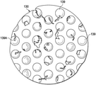
図2は、接触層110のいくつかの実施形態に関連し得る更なる詳細を示す平面図である。接触層110は、接触層110を通って延びる複数の貫通孔140又は他の穿孔を含んで、壁148を形成し得る。いくつかの実施形態では、壁148の外面が、接触層110の面と平行であり得る。他の実施形態では、壁148の内面が、接触層110の組織対向面111及び反対面113と略垂直であり得る。一般に、壁148の1つ以上の外面は、組織対向面111及び反対面113と一致し得る。壁148の1つ以上の内面は、各貫通孔140の外周152を形成し得、組織対向面111を反対面113に接続し得る。いくつかの実施形態では、貫通孔140は、図示するように円形であり得る。いくつかの実施形態では、貫通孔140は、約5mm~約20mmの直径を有し得、いくつかの実施形態では、貫通孔140の直径は、約10mmであり得る。貫通孔140は、接触層110の厚さ134にほぼ等しい深さを有し得る。例えば、貫通孔140は、周囲圧力で約6mm~約10mm、より具体的に約8mmの深さを有し得る。
FIG. 2 is a plan view showing further details that may be relevant to some embodiments of the
いくつかの実施形態では、接触層110は、第1の方位線136と、第1の方位線136と垂直な第2の方位線138とを有し得る。第1の方位線136及び第2の方位線138は、接触層110の対称線であり得る。対称線は、例えば、接触層110の組織対向面111又は反対面113を横切る仮想線であって、接触層110が対称線で折り畳まれた場合に貫通孔140と壁148とが同時に位置合わせされるような折り線を規定し得る。一般に、第1の方位線136及び第2の方位線138は、接触層110に関する記述を助ける。いくつかの実施形態では、第1の方位線136及び第2の方位線138は、接触層110の所望の収縮方向を指すために使用され得る。例えば、所望の収縮方向は、第2の方位線138と平行であり、第1の方位線136と垂直であり得る。他の実施形態では、所望の収縮方向は、第1の方位線136と平行であり、第2の方位線138と垂直であり得る。また他の実施形態では、所望の収縮方向は、第1の方位線136及び第2の方位線138の両方と垂直以外の角度であり得る。他の実施形態では、接触層110は、所望の収縮方向を有していなくてもよい。一般に、接触層110は、第2の方位線138が図1のデブリ130を横切って延びるように、組織部位103に配置され得る。接触層110は、長手方向縁部144及び横方向縁部146を含む略矩形のものとして示されているが、接触層110は他の形状であってもよい。例えば、接触層110は、ひし形、正方形、又は円形であり得る。いくつかの実施形態では、接触層110の形状は、治療されている組織部位のタイプに適応するように選択され得る。例えば、接触層110は、卵形又は円形の組織部位に適応するように卵形又は円形であり得る。いくつかの実施形態では、第1の方位線136は、長手方向縁部144と平行であり得る。
In some embodiments, the
より具体的に図3を参照すると、円形の単一の貫通孔140が示されている。貫通孔140は、中心150及び外周152を含み得る。貫通孔140は、穿孔形状係数(PSF;perforation shape factor)を有し得る。穿孔形状係数(PSF)は、第1の方位線136及び第2の方位線138に対する貫通孔140の向きを表し得る。一般に、穿孔形状係数(PSF)は、所望の収縮方向と平行な貫通孔140の最大長さの1/2と、所望の収縮方向と垂直な貫通孔140の最大長さの1/2との比である。説明のために、所望の収縮方向は、第2の方位線138と平行である。所望の収縮方向は、横力142によって示され得る。参照のために、貫通孔140は、六角形の対向する頂点間で中心150を通って第1の方位線136と平行に延びるX軸156と、六角形の対向する辺の間で中心150を通って第2の方位線138と平行に延びるY軸154とを有し得る。貫通孔140の穿孔形状係数(PSF)は、中心150から貫通孔140の外周152に延びるY軸154上の線分158と、中心150から貫通孔140の外周152に延びるX軸156上の線分160との比として定義され得る。線分158の長さが2.5mmであり、線分160の長さが2.5mmである場合、穿孔形状係数(PSF)は、1である。他の実施形態では、貫通孔140は、他の形状(例えば、卵形、六角形、正方形、三角形、又は無定形若しくは不規則な形状)及び向きを有し得、穿孔形状係数(PSF)が約0.5~約1.10の範囲となり得るように、第1の方位線136及び第2の方位線138に関して方向付けられ得る。
More specifically, referring to FIG. 3, a single circular through
図4を参照すると、図1の接触層110の一部分が示されている。接触層110は、平行な行に位置合わせされて、配列を形成する複数の貫通孔140を含み得る。貫通孔140の配列は、貫通孔140の第1の行162と、貫通孔140の第2の行164と、貫通孔140の第3の行166とを含み得る。いくつかの実施形態では、第1の行162などの行における貫通孔140に隣接する外周152の間の壁148の幅が、約5mmであり得る。隣接する行、例えば、第1の行162及び第2の行164における貫通孔140の中心150は、第2の方位線138から第1の方位線136に沿ってオフセットされることを特徴とし得る。いくつかの実施形態では、隣接する行の中心同士を結ぶ線が、第1の方位線136との筋交角度(SA;strut angle)を形成し得る。例えば、第1の行162における第1の貫通孔140Aが、中心150Aを有し得、第2の行164における第2の貫通孔140Bが、中心150Bを有し得る。筋交線168が、中心150Aと中心150Bを結び得る。筋交線168は、第1の方位線136と角度170を形成し得る。角度170は、接触層110の筋交角度(SA)であり得る。いくつかの実施形態では、筋交角度(SA)は約90°未満であり得る。他の実施形態では、筋交角度(SA)は、第1の方位線136に対して約30°~約70°であり得る。他の実施形態では、筋交角度(SA)は、第1の方位線136から約66°であり得る。一般に、筋交角度(SA)が減少するにつれて、第1の方位線136と平行な方向での接触層110の剛性が高まり得る。第1の方位線136と平行な接触層110の剛性を高めることにより、第1の方位線136と垂直な接触層110の圧縮性が増加し得る。その結果、接触層110に陰圧が印加された場合、接触層110は、第1の方位線136と垂直な方向でより柔軟に又はより圧縮可能になり得る。接触層110の圧縮性を第1の方位線136と垂直な方向で増加させることにより、接触層110は、圧潰して、より詳細に後述する横力142を組織部位103に加え得る。
Referring to FIG. 4, a portion of the
いくつかの実施形態では、互い違いの行における貫通孔140の中心150、例えば、第1の行162における第1の貫通孔140Aの中心150A、及び第3の行166における貫通孔140Cの中心150Cは、互いに第2の方位線138と平行に長さ172だけ離間され得る。いくつかの実施形態では、長さ172は、貫通孔140の有効直径よりも大きくなり得る。互い違いの行における貫通孔140の中心150が、長さ172だけ隔てられる場合、第1の方位線136と平行な壁148の外面は、連続していると考えられ得る。一般に、壁148の外面が貫通孔140の間に不連続部又は破断部を有していない場合、壁148の外面は連続し得る。いくつかの実施形態では、長さ172は、約7mm~約25mmであり得る。
In some embodiments, the
貫通孔140の形状にかかわらず、接触層110の貫通孔140は、接触層110の壁148の外面のみが、組織部位103に接触するために利用可能な表面を残すように、接触層110、接触層110の組織対向面111及び反対面113に空隙を残し得る。貫通孔140が圧潰し、接触層110を圧潰させ、第1の方位線136と垂直な方向で横力142を発生させ得るように、壁148の外面を最小限に抑えることが望ましい場合がある。しかし、接触層110が陰圧の印加を持続するには脆くなりすぎるほど、壁148の外面を最小限に抑えないことが望ましい場合もある。貫通孔140の空隙百分率(VS;void space percentage)は、接触層110の組織対向面111の総体積又は表面積に対する、貫通孔140によって形成された組織対向面111の空隙の体積又は表面積の百分率と等しくなり得る。いくつかの実施形態では、空隙百分率(VS)は、約40%~約75%であり得る。他の実施形態では、空隙百分率(VS)は、約55%であり得る。貫通孔140の構成は、空隙百分率(VS)に影響を与え、組織部位103に接触し得る接触層110の総表面積に影響を与えることもできる。いくつかの実施形態では、接触層110の長手方向縁部144及び横方向縁部146は、不連続であり得る。縁部が不連続であり得、ここでは、貫通孔140は縁部に被さり、縁部が非線形プロファイルを持つようになる。不連続な縁部が、角化細胞遊走の破壊を低減させ、陰圧がドレッシング102に印加されている間の上皮再形成を増進させ得る。
Regardless of the shape of the through
他の実施形態では、接触層110の貫通孔140は、接触層110の厚さ134よりも小さな深さを有し得る。例えば、孔140は、接触層110の組織対向面111に形成された止まり孔であり得る。孔140は、接触層110の壁148の組織対向面111上の外面のみが、周囲圧力で組織部位103に接触するために利用可能な表面を残すように、接触層110の組織対向面111に空隙を残し得る。組織対向面111から反対面113に向かって延びる孔140の深さが、厚さ134未満である場合、反対面113の空隙百分率(VS)はゼロとなり得、組織対向面111の空隙百分率(VS)はゼロよりも大きく、例えば55%となり得る。本明細書で使用するとき、孔140は、同様の構造、位置、及び動作特性を有する、貫通孔140に関して記載したものと同様であり、同様に動作し得る。
In another embodiment, the through
いくつかの実施形態では、貫通孔140は、接触層110の成形中に形成され得る。他の実施形態では、貫通孔140は、接触層110が形成された後に、接触層110を切除、溶融、ドリル加工、又は気化することによって形成され得る。例えば、貫通孔140は、接触層110の圧縮発泡体をレーザ切除することにより、接触層110に形成され得る。いくつかの実施形態では、貫通孔140は、貫通孔140の壁148の内面が厚さ134と平行になるように形成され得る。他の実施形態では、貫通孔140は、貫通孔140の壁148の内面が組織対向面111と垂直以外の角度を形成するように形成され得る。また他の実施形態では、貫通孔140の壁148の内面は、円錐形、角錐形、又は他の不規則な貫通孔形状を形成するように、貫通孔140の中心150に向かって先細になっていてもよい。貫通孔140の壁148の内面が先細になっている場合、貫通孔140は、接触層110の厚さ134よりも低い高さを有し得る。
In some embodiments, the through
いくつかの実施形態では、貫通孔140の形成により、接触層110の材料、例えば圧縮発泡体又はフェルト発泡体を熱成形し、組織対向面111と反対面113との間に延びる壁148の内面を平滑にし得る。本明細書で使用するとき、平滑性は、組織対向面111と反対面113との間に延びる壁148の内面が、接触層110の非切除部分と比べた場合に細孔を実質的に含まないように、貫通孔140を形成することを指す場合がある。例えば、接触層110に貫通孔140をレーザ切除することにより、接触層110の材料を塑性変形し、組織対向面111と反対面113との間に延びる壁148の内面にある細孔を閉鎖し得る。いくつかの実施形態では、壁148の平滑な内面により、貫通孔140を通した接触層110への組織の内部成長を、制限、又は別の方法で阻害し得る。他の実施形態では、壁148の平滑な内面は、平滑な材料又は平滑なコーティングによって形成され得る。
In some embodiments, the formation of through
いくつかの実施形態では、貫通孔140の有効直径は、貫通孔140を通る粒状物の流れを可能にするように選択され得る。いくつかの実施形態では、貫通孔140の直径は、組織部位103から持ち上げられる可溶化デブリのサイズに基づいて選択され得る。貫通孔140が大きいほど、より大きなデブリが接触層110を通過することが可能となり、貫通孔140が小さいほど、貫通孔よりも大きなデブリをブロックしながら、より小さなデブリが接触層110を通過することが可能となり得る。いくつかの実施形態では、ドレッシング102の連続的な適用には、組織部位103の可溶化デブリのサイズが減少するにつれて、連続的により小さな直径の貫通孔140を有する接触層110を使用することができる。貫通孔140の直径を連続的に小さくすることは、組織部位103の治療中に、デブリ130に対する組織破壊のレベルを微調整することにも役立ち得る。貫通孔140の直径は、接触層110内及びドレッシング102内の流体の移動に影響を与えることもできる。例えば、接触層110は、ドレッシング102内の流体を貫通孔140に向かって流して、組織部位103上のデブリ130の破壊を支援することができる。貫通孔140の直径を変動させることにより、流体の除去と陰圧の印加の両方に関して、ドレッシング102を通る流体の移動の仕方を変動させることができる。いくつかの実施形態では、貫通孔140の直径は、約5mm~約20mm、より具体的に約10mmである。
In some embodiments, the effective diameter of the through
非円形領域の有効直径は、非円形領域と同じ表面積を有する円形領域の直径として定義される。いくつかの実施形態では、各貫通孔140は、約3.5mmの有効直径を有し得る。他の実施形態では、各貫通孔140は、約5mm~約20mmの有効直径を有し得る。貫通孔140の有効直径は、接触層110の壁148を形成する材料の多孔性と区別されるべきである。一般に、貫通孔140の有効直径は、接触層110を形成する材料の細孔の有効直径よりも1桁大きい。例えば、貫通孔140の有効直径は、約1mmよりも大きくなり得、壁148は、約600マイクロメートル未満の細孔径を有するGranuFoam(登録商標)材料から形成され得る。いくつかの実施形態では、壁148の細孔は、材料を完全に貫通する開口部を形成しなくてもよい。一般に、貫通孔140は、発泡体形成プロセスによって形成された細孔を含んでおらず、貫通孔140は、材料の細孔の平均有効直径の10倍を超える平均有効直径を有し得る。
The effective diameter of a non-circular region is defined as the diameter of a circular region that has the same surface area as the non-circular region. In some embodiments, each through
ここで図2及び図4の両方を参照すると、貫通孔140は、貫通孔140の幾何学的形状、及び接触層110において第1の方位線136に関して隣接して互い違いの行の間の貫通孔140の位置合わせに応じたパターンを形成し得る。接触層110が陰圧を受けた場合、接触層110の貫通孔140は収縮し得る。本明細書で使用するとき、収縮は、接触層110などの本体の厚さと平行な本体の垂直方向の圧縮と、接触層110などの本体の厚さと垂直な本体の横方向の圧縮との両方を指すことができる。いくつかの実施形態では、空隙百分率(VS)、穿孔形状係数(PSF)、及び筋交角度(SA)により、図5により詳細に示すように、第1の方位線136と垂直な第2の方位線138に沿って接触層110を収縮させ得る。接触層110が組織部位103に配置された場合、接触層110は、図5により詳細に示すように、第2の方位線138に沿って横力142を発生させ、接触層110を収縮させ得る。横力142は、以下の表1に記載されるように、上述した係数を調節することによって最適化され得る。いくつかの実施形態では、貫通孔140は円形であり、約37°の筋交角度(SA)、約54%の空隙百分率(VS)、約5の硬度係数(FF)、約1の穿孔形状係数(PSF)、及び約5mmの直径を有し得る。接触層110が約-125mmHgの陰圧を受けた場合、接触層110は、約11.9Nの横力142が生じる。接触層110の貫通孔140の直径を約20mmに増加させ、空隙百分率(VS)を約52%に変更し、筋交角度(SA)を約52°に変更し、穿孔形状係数(PSF)及び硬度係数(FF)を同じままにした場合、横力142は約6.5Nに低下する。他の実施形態では、貫通孔140は、六角形であり、約66°の筋交角度(SA)、約55%の空隙百分率(VS)、約5の硬度係数(FF)、約1.07の穿孔形状係数(PSF)、及び約5mmの有効直径を有し得る。接触層110が約-125mmHgの陰圧を受けた場合、接触層110により生じる横力142は約13.3Nである。接触層110の貫通孔140の有効直径を10mmに増加させた場合、横力142は約7.5Nに低下する。
Referring here to both FIGS. 2 and 4, the through
図5を参照すると、接触層110は、横力142によって示すように、第2の位置、すなわち収縮位置にある。動作中、陰圧が、陰圧源104によって密閉療法環境128に供給される。陰圧の供給に応じて、接触層110は、図2に示した弛緩位置から図5に示す収縮位置に収縮する。一実施形態では、接触層110の厚さ134は、実質的に同じままである。例えば陰圧の排気により、陰圧が除去されると、接触層110は、膨張して弛緩位置に戻る。接触層110が、図5の収縮位置と図2の弛緩位置との間を循環した場合、接触層110の組織対向面111は、組織部位103からデブリ130を擦り落とすことにより、組織部位103のデブリ130を破壊し得る。組織対向面111及び壁148の内面又は横断面によって形成された貫通孔140の縁部が、組織部位103のデブリ130を破壊できる切刃を形成し、デブリ130が貫通孔140を通って出ることを可能にすることができる。いくつかの実施形態では、切刃は、各貫通孔140が組織対向面111と交差する外周152によって形成される。
Referring to FIG. 5, the
いくつかの実施形態では、材料、空隙百分率(VS)、硬度係数、筋交角度、孔形状、穿孔形状係数(PSF)、及び孔直径は、より弱い壁148の形成により、横力142によって示すように、横方向での接触層110の圧縮又は圧潰を増加させるように選択され得る。反対に、係数は、より強い壁148の形成により、横力142によって示すように、横方向での接触層110の圧縮又は圧潰を減少させるように選択され得る。同様に、本明細書に記載される係数は、横力142と垂直の接触層110の圧縮又は圧潰を減少又は増加させるように選択することができる。
In some embodiments, the material, void percentage (VS), hardness factor, brace angle, hole shape, perforation shape factor (PSF), and hole diameter are indicated by
いくつかの実施形態では、療法システム100は、周期的療法を提供し得る。周期的療法は、密閉療法環境128への陰圧の印加と、陰圧の排気とを交互に行い得る。いくつかの実施形態では、密閉療法環境128の圧力が所定療法圧力に達するまで、密閉療法環境128に陰圧が供給され得る。密閉療法環境128に陰圧を供給した場合、デブリ130及び皮下組織115は、貫通孔140に引き込まれ得る。いくつかの実施形態では、密閉療法環境128は、例えば約10分間などの所定療法期間にわたって療法圧力に維持され得る。他の実施形態では、療法期間は、適切な陰圧療法を組織部位103に供給する必要に応じて、より長くても、より短くてもよい。
In some embodiments, the
療法期間後、密閉療法環境128は排気され得る。例えば、陰圧源104は、密閉療法環境128を大気(図示せず)に流体的に結合し、密閉療法環境128を周囲圧力に戻すことを可能にし得る。いくつかの実施形態では、陰圧源104は、約1分間にわたって密閉療法環境128を排気し得る。他の実施形態では、陰圧源104は、より長い又はより短い期間にわたって密閉療法環境128を排気し得る。密閉療法環境128を排気した後、別の陰圧療法サイクルを開始するように陰圧源104を動作させ得る。
After the therapy period, the
いくつかの実施形態では、滴下療法を陰圧療法と組み合わせ得る。例えば、陰圧療法の療法期間に続いて、流体源120は、密閉療法環境128に流体を提供するように動作し得る。いくつかの実施形態では、流体源120は、陰圧源104が密閉療法環境128を排気している間に流体を提供し得る。例えば、流体源120は、流体源120から密閉療法環境128に滴下流体を移動させるように構成されたポンプを含み得る。いくつかの実施形態では、流体源120は、ポンプを有していなくてもよく、重力供給システムによって動作し得る。他の実施形態では、陰圧源104は、密閉療法環境128を排気しなくてもよい。代わりに、密閉療法環境128の陰圧は、流体源120から密閉療法環境128に滴下流体を引き込むために使用される。
In some embodiments, drop therapy may be combined with negative pressure therapy. For example, following the therapy period of negative pressure therapy, the
いくつかの実施形態では、流体源120は、密閉療法環境128に一定の体積の流体を提供し得る。いくつかの実施形態では、流体の体積は、密閉療法環境128の容積と同じであり得る。他の実施形態では、流体の体積は、滴下療法を適切に適用する必要に応じて、密閉療法環境128よりも少なくても、多くてもよい。組織部位103の滴下は、密閉療法環境128の圧力を、周囲圧力よりも高い圧力に、例えば、約0mmHg~約15mmHg、より具体的に約5mmHgに上昇させ得る。いくつかの実施形態では、流体源120によって提供される流体は、滞留時間にわたって密閉療法環境128に維持され得る。いくつかの実施形態では、滞留時間は約5分間である。他の実施形態では、滞留時間は、組織部位103に滴下療法を適切に施す必要に応じて、より長くても、より短くてもよい。例えば、滞留時間はゼロであり得る。
In some embodiments, the
滞留時間が終了すると、陰圧源104は、滴下流体を容器112に引き込むように動作され、療法のサイクルを完了させ得る。滴下流体が密閉療法環境128から陰圧によって除去されると、陰圧がまた、密閉療法環境128に供給され、療法の別のサイクルを開始させ得る。
At the end of the residence time, the
図6は、いくつかの実施形態と関連し得る更なる詳細を示す、接触層110の一部分の断面図である。接触層110及び保持層108は、皮下組織115を覆うデブリ130を有する組織部位103に配置され得る。ドレープ106は、保持層108の上に配置されて、陰圧療法又は滴下療法の適用のための密閉療法環境128を提供し得る。図6に示すように、保持層108は、密閉療法環境128の圧力がほぼ周囲圧力である場合に厚さ131を有し得る。いくつかの実施形態では、厚さ131は約8mmであり得る。他の実施形態では、厚さ131は約16mmであり得る。
FIG. 6 is a cross-sectional view of a portion of the
図7は、いくつかの実施形態と関連し得る更なる詳細を示す、陰圧療法中のドレッシング102の一部分の断面図である。例えば、図7は、密閉療法環境128の圧力が約125mmHgの陰圧となり得る時間を示し得る。いくつかの実施形態では、保持層108は予め圧縮されていない発泡体であり得、接触層110は予め圧縮された発泡体であり得る。陰圧の印加に応じて、接触層110は圧縮しなくてもよく、保持層108は、マニホールドが厚さ133を有するように圧縮してもよい。いくつかの実施形態では、陰圧療法中の保持層108の厚さ133は、密閉療法環境128の圧力がほぼ周囲圧力である場合の保持層108の厚さ131よりも小さくなり得る。
FIG. 7 is a cross-sectional view of a portion of the dressing 102 during negative pressure therapy, showing further details that may be associated with some embodiments. For example, FIG. 7 may show the time at which the pressure in the
いくつかの実施形態では、密閉療法環境128の陰圧は、接触層110の貫通孔140に隣接して保持層108に集中応力を発生させることができる。集中応力は、保持層108の一部分を接触層110の貫通孔140に引き込む、保持層108のマクロ変形を引き起こすことができる。同様に、密閉療法環境128の陰圧は、接触層110の貫通孔140に隣接してデブリ130に集中応力を発生させることができる。集中応力は、デブリ130及び皮下組織115の一部分を貫通孔140に引き込む、デブリ130及び皮下組織115のマクロ変形を引き起こすことができる。
In some embodiments, the negative pressure of the
図8は、陰圧療法中の接触層110の動作の更なる詳細を示す、接触層110の詳細図である。接触層110の反対面113と接触する保持層108の一部分が、貫通孔140に引き込まれて、隆起部137を形成し得る。隆起部137は、貫通孔140に対応する形状であり得る。保持層108からの隆起部137の高さは、密閉療法環境128の陰圧の圧力、貫通孔140の面積、及び保持層108の硬度係数に依存し得る。
FIG. 8 is a detailed view of the
同様に、接触層110の貫通孔140は、デブリ130及び皮下組織115の、接触層110の組織対向面111と接触する部分にマクロ圧力点を形成し、デブリ130及び皮下組織115に組織パッカリング及び瘤部139を引き起こし得る。
Similarly, the through
周囲組織の上の瘤部139の高さ802は、デブリ130の破壊を最大化し、皮下組織115又は他の所望の組織への損傷を最小限に抑えるように選択され得る。一般に、密閉療法環境128の圧力は、圧力が印加された面積に比例する力を及ぼすことができる。接触層110の貫通孔140では、圧力の印加に対する抵抗が接触層110の壁148よりも小さいので、力が集中し得る。貫通孔140での圧力によって発生した力に応じて、瘤部139を形成するデブリ130及び皮下組織115は、圧力によって印加される力がデブリ130及び皮下組織115の反力によって釣り合うまで、貫通孔140を通って貫通孔に引き込まれ得る。密閉療法環境128の陰圧が引裂きを引き起こし得るいくつかの実施形態では、接触層110の厚さ134は、周囲組織の上の瘤部139の高さ802を制限するように選択され得る。いくつかの実施形態では、保持層108は、接触層が圧縮可能である場合に、瘤部139の高さ802を陰圧下での接触層110の厚さ134に制限し得る。他の実施形態では、保持層108の隆起部137は、瘤部139の高さ802を接触層110の厚さ134よりも小さな高さ802に制限し得る。保持層108の硬度係数を制御することにより、保持層108の周囲材料の上の隆起部137の高さ802を制御することができる。瘤部139の高さ802は、接触層110の厚さ134と隆起部137の高さとの差に限定することができる。いくつかの実施形態では、隆起部137の高さは、保持層108の硬度係数が小さくなるにつれて、ゼロから数ミリメートルまで変動し得る。例示的な実施形態では、接触層110の厚さ134は、約7mmであり得る。陰圧の印加中に、隆起部137は、約4mm~約5mmの高さを有し、瘤部の高さ802を約2mm~約3mmに制限し得る。接触層110の厚さ134、保持層108の硬度係数、又はそれらの両方の制御によって瘤部139の高さ802を制御することにより、デブリ130に対する破壊の積極性及び引裂きを制御することができる。
The
いくつかの実施形態では、瘤部139の高さ802は、陰圧療法中の接触層110の予想される圧縮を制御することによって制御することもできる。例えば、接触層110は、約8mmの厚さ134を有し得る。接触層110が圧縮発泡体から形成される場合、接触層110の硬度係数はより大きくてもよく、しかし、接触層110は、密閉療法環境128の陰圧に応じて、依然として厚さを低減されてもよい。一実施形態では、密閉療法環境128での約-50mmHg~約-350mmHg、約-100mmHg~約-250mmHg、より具体的に約-125mmHgの陰圧の印加により、接触層110の厚さ134を約8mmから約3mmに低減し得る。保持層108が接触層110の上に配置された場合、瘤部139の高さ802は、陰圧療法中に接触層110の厚さ134以下に、例えば約3mmに制限され得る。瘤部139の高さ802を制御することにより、接触層110によってデブリ130に加えられる力を調節することができ、デブリ130が伸張する度合を変化させることができる。
In some embodiments, the
図9は、療法システム100のいくつかの実施形態と関連し得る更なる詳細を示す、ドレッシング102の組立図である。図9の例では、ドレッシング102は、ドレープ106、支持層902、保持層108、及び接触層110を備え得る。いくつかの実施形態では、ドレープ106は、第1の表面904と、ドレープ106の第1の表面904とは反対側にある第2の表面906とを有し得る。例えば、第1の表面904は、表面領域と、表面領域を取り囲む外周908とを有し得る。第2の表面906は、第1の表面904の表面領域と実質的に等しい表面領域を有し得る。第2の表面906の外周が、第1の表面904の外周908と実質的に同一の広がりを有し得る。例示的な実施形態では、開口910がドレープ106を貫通して形成され得る。
FIG. 9 is an assembly diagram of the dressing 102 showing further details that may be associated with some embodiments of the
例示的な実施形態では、支持層902は半剛性材料を含み得る。例えば、支持層902は、ゴム又はプラスチック製の材料などの多孔性材料を含み得る。例示的な実施形態では、支持層902は、非多孔性かつ非透過性であり得る、実質的に剛性の材料を含み得る。例示的な実施形態では、支持層902はポリウレタンを含み得る。いくつかの実施形態では、支持層902は、第1の表面911と、第1の表面911から支持層902を横切る第2の表面912とを有し得る。例えば、第1の表面911は、表面領域と、表面領域を取り囲む外周914とを有し得る。第2の表面912は、第1の表面911の表面領域と実質的に等しい表面領域を有し得る。第2の表面912の外周が、第1の表面911の外周914と実質的に同一の広がりを有し得る。例示的な実施形態では、外周914によって囲まれた支持層902の表面領域は、外周908によって囲まれたドレープ106の表面領域よりも小さくてもよい。例えば、外周914によって囲まれた支持層902の表面領域は、外周908によって囲まれたドレープ106の表面領域内に実質的に含まれ得る。例示的な実施形態では、開口916が、支持層902を貫通して形成され得る。いくつかの例示的な実施形態では、支持層902は一様な厚さを有し得る。例えば、支持層902は、約1mm~5mmの範囲の厚さを有し得る。
In an exemplary embodiment, the
いくつかの実施形態では、支持層902の第2の表面912は、創傷治癒を促進するための療法物質、表面破壊部、及び/又はトランスデューサを更に含み得る。例えば、支持層902は、有効な創傷治癒を促進するための抗菌剤又は他の活性剤を含んでもよく、あるいは抗菌剤又は他の活性剤が少なくとも部分的に注入されていてもよい。そのような活性材料の非限定的な例としては、抗菌性銀、銀酸化再生セルロース(ORC;oxidized regenerated cellulose)(例えば、約25重量パーセント(重量%)のイオン結合銀)、ポリヘキサメチレンビグアニド(PHMB;polyhexamethylene biguanide)、アセトアミノフェン、ステロイド、抗炎症性サイトカインなどの非ステロイド系抗炎症性薬、麻酔剤、ペニシリン又はストレプトマイシンなどの抗菌剤、クロルヘキシジンなどの殺菌剤、線維芽細胞増殖因子(FGF;fibroblast growth factor)、血小板由来増殖因子(PDGF;platelet derived growth factor)、又は上皮増殖因子(EGF;epidermal growth factor)などの増殖因子、及び他の治療剤の、個々又は任意の組み合わせが挙げられ得る。存在する場合、そのような活性材料は、療法有効性を示す有効レベルで含まれ得るが、好ましくは、ドレッシング102の任意の重要な又は所望の物理的、化学的、若しくは生物学的な特性を大きく抑制するような高レベルではない。治療目標に応じて、支持層902に注入される又は含まれる活性材料が、それが存在する層の約10重量百万分率(wppm;weight parts per million)~約10重量%、例えば、約50wppm~約5重量%、又は約100wppm~約1重量%のレベルで充填され得る。いくつかの実施形態では、活性材料は、支持層902の第2の表面912上の薄いフィルム内に含まれてもよく、又は支持層902内に分配されてもよい。
In some embodiments, the
例示的な実施形態では、保持層108は、第1の表面918と、保持層108の第1の表面918とは反対側にある第2の表面920とを有し得る。例えば、第1の表面918は、表面領域と、表面領域を取り囲む外周922とを有し得る。第2の表面920は、第1の表面918の表面領域と実質的に等しい表面領域を有し得る。第2の表面920の外周が、第1の表面918の外周922と実質的に同一の広がりを有し得る。例示的な実施形態では、外周922によって囲まれた保持層108の表面領域は、外周914によって囲まれた支持層902の表面領域よりも小さくてもよい。例えば、外周922によって囲まれた保持層108の表面領域は、外周914によって囲まれた支持層902の表面領域内に実質的に含まれ得る。例示的な実施形態では、接触層110は、第1の表面924と、第1の表面924から接触層110を横切る第2の表面926とを有し得る。例えば、第1の表面924は、接触層110の表面領域を取り囲む外周928によって囲まれ得る。第2の表面926の外周が、第1の表面924の外周928と実質的に同一の広がりを有し得る。いくつかの実施形態では、接触層110の第1の表面924の外周928は、組織部位に配置された場合、保持層108の第1の表面918の外周922と実質的に同一の広がりを有し得る。
In an exemplary embodiment, the
図10は、支持層902及び療法システム100のいくつかの実施形態に関連し得る更なる詳細を示す平面図である。図10Aは、支持層902及び療法システム100の一部分の詳細図である。図10に示すように、接触層110は、接触層110の第2の表面926がデブリ130の少なくとも一部分と接触し得るように、創傷内に配置され得る。例えば、デブリ130は、真皮109内及び皮下組織115内にあり得る。いくつかの実施形態では、接触層110の第1の表面924の少なくとも一部分が、保持層108の第2の表面920の少なくとも一部分と接触し得るように、保持層108は接触層110の上に配置され得る。例えば、接触層110の第1の表面924の少なくとも一部分が、保持層108の第2の表面920の少なくとも一部分に結合され得る。いくつかの実施形態では、保持層108の第1の表面918の少なくとも一部分が、支持層902の第2の表面912の少なくとも一部分と接触し得る。いくつかの実施形態では、保持層108の第1の表面918は、支持層902の第2の表面912に結合され得る。例示的な実施形態では、支持層902は、創傷周囲領域を越えて延び、表皮105の一部分と接触し得る。例えば、支持層902の第2の面の一部分が、表皮105の一部分と接触し得る。
FIG. 10 is a plan view showing further details that may be relevant to some embodiments of
例示的な実施形態では、ドレープ106の第2の表面906は、接着剤1002でコーティングされ得る。いくつかの実施形態では、接着剤1002は、連続的な水蒸気透過感圧接着剤、例えば、ポリウレタン系感圧接着剤であり得る。例えば、接着剤1002は、アクリレートエステルコポリマー系、ポリビニルエチルエーテル系、及びポリウレタン系の感圧接着剤であり得る。接着剤1002の坪量は、約20g/m2~約250g/m2、より好ましくは約50g/m2~約150g/m2であり得る。図10に示すように、ドレープ106は、接着剤1002が表皮105の少なくとも一部分及び支持層902の第1の表面911の少なくとも一部分と接触するように、支持層902の上に配置され得る。例えば、ドレープ106は、表皮105の一部分及び支持層902の第1の表面911の少なくとも一部分に結合又は接着され得る。いくつかの実施形態では、接着剤1002は、表皮105に結合されるドレープ106の周縁のみに配置され得る。いくつかの実施形態では、ドレープ106は、表皮105及び支持層902に対する流体シールを形成し、外部環境から実質的に隔離された流体密閉療法環境128を形成し得る。例示的な実施形態では、療法環境128は、開口910及び開口916を通してコネクタ114によってチューブ116に流体的に結合され得る。例えば、開口910及び開口916は、コネクタ114が療法環境128と流体連通することを可能にするように位置合わせされ得る。図10に示すように、ドレープ106の第2の表面906は、支持層902の第1の表面911に面し得る。例えば、支持層902の第1の表面911は、ドレープ106の第2の表面906の少なくとも一部分に接着剤1002によって結合又は接着され得る。支持層902、保持層108、及び接触層110は、療法環境128内に配置され得る。いくつかの実施形態では、ドレープ106は、支持層902から水分を吸い上げ、ドレープ106の第2の表面906にわたって水分を分配するように構成される。
In an exemplary embodiment, the
いくつかの実施形態では、支持層902は、創傷周囲領域を越えて延び、表皮105と接触し得る。例えば、表皮105は、支持層902の少なくとも一部分を支持し得る。例示的な実施形態では、支持層902及び表皮105は、ドレープ106を支持し、陰圧下でのドレープ106の変形を低減又は実質的に防止し得る。いくつかの実施形態では、支持層902は、陰圧下でのドレッシング102の圧縮を低減又は実質的に防止する。
In some embodiments, the
図11は、支持層902を更に備えるドレッシング102のいくつかの実施形態に関連し得る更なる詳細を示す組立図である。図11に示すように、支持層902のいくつかの例では、支持層902の第1の表面911の外周914は、支持層902、保持層018、及び接触層110が組織部位に配置された場合、保持層108の外周922及び接触層110の外周928と実質的に同一の広がりを有し得る。
FIG. 11 is an assembly diagram showing further details that may be relevant to some embodiments of the dressing 102 further comprising a
図12は、支持層902及び療法システム100のいくつかの実施形態に関連し得る更なる詳細を示す平面図である。図12に示すように、支持層902の第2の表面912は、保持層108の第1の表面918を越えて延びていなくてもよい。例えば、支持層902は、実質的に創傷周囲領域内に配置され得る。
FIG. 12 is a plan view showing further details that may be relevant to some embodiments of
いくつかの実施形態では、支持層902は、実質的に創傷周囲領域内に含まれ得る。いくつかの例示的な実施形態では、支持層902及びドレープ106が組織部位に配置された場合、ドレープ106とは反対側の支持層902の面が、保持層108の面と実質的に同一の広がりを有し得る。例示的な実施形態では、支持層902は、接着剤1006などによってドレープ106に結合され得る。例えば、ドレープ106に結合された支持層902は、陰圧下での変形及び/又は圧潰に対するドレープ106の抵抗を高め、陰圧下でのドレッシング102の変形及び/又は圧潰を低減又は実質的に防止する。
In some embodiments, the
いくつかの実施形態では、支持層902は、接着剤1002などによってドレープ106に結合され、接着、熱接合、又は溶着などによって保持層108に更に結合され得る。例示的な実施形態では、支持層902は、陰圧下での変形及び/又は圧潰に対するドレープ106の抵抗を更に高めると共に、陰圧下での変形及び/又は圧潰に対する保持層108の抵抗を高め得る。例示的な実施形態では、支持層902は、陰圧下でのドレッシング102の変形及び/又は圧潰を低減又は実質的に防止する。
In some embodiments, the
図13は、いくつかの実施形態に関連し得る更なる詳細を示す、ドレッシング102の一部分の断面図である。図13に示すように、支持層902は、ドレープ106と保持層108との間に設けられ得る。例えば、接着剤1002は、ドレープ106の第2の面に配置され、ドレープ106を支持層902の第1の表面911に接着し得る。例示的な実施形態では、支持層902の第2の表面912は、保持層108の第1の表面918と接触し得る。支持層902は、保持層108から切り離され、保持層108と支持層902が療法中に互い対して移動することを可能にすることができる。他の実施形態では、支持層902の第2の表面912は、保持層108の第1の表面918に結合、接合、又は接着され得る。いくつかの実施形態では、保持層108の第2の表面920は、接触層110の第1の表面924と接触し得る。保持層108は、接触層110から切り離され、保持層108と接触層110が療法中に互いに対して移動することを可能にすることができる。他の実施形態では、保持層108の第2の表面920は、接触層110の第1の表面924に結合、接合、又は接着され得る。ドレッシング102は、接触層110の第2の表面926がデブリ130と接触し得るように、組織部位に配置され得る。支持層902の追加により、陰圧下でのドレッシング102の圧縮に対する全体的な抵抗が高められ得る。いくつかの実施形態では、支持層902は厚さ1302を有し得る。例えば、厚さ1302は、約1mm~約5mmの範囲であり得る。
FIG. 13 is a cross-sectional view of a portion of the dressing 102 showing further details that may be relevant to some embodiments. As shown in FIG. 13, the
図14は、いくつかの実施形態に関連し得る更なる詳細を示す、陰圧療法中のドレッシング102の一部分の断面図である。図15は、陰圧療法中のドレッシング102の一部分の詳細図である。例えば、図14及び図15は、密閉療法環境128の圧力が約125mmHgの陰圧となり得る時間を示し得る。いくつかの実施形態では、保持層108は、予め圧縮されていない発泡体であり得、接触層110は、予め圧縮された発泡体又はフェルト発泡体であり得る。いくつかの実施形態では、支持層902は、保持層108の上に配置され得る。支持層902は、陰圧下でのドレッシング102の圧縮に対する全体的な抵抗を高めることができる。一般に、例示的な実施形態では、支持層902は、創傷療法治療のためにドレッシング102が使用されるときに、創傷滲出液又はフィブリン、スラフ、若しくは感染性物質などのデブリ130の変形、破壊、及び/又は断片化を高め得る。例えば、支持層902によって高められる創傷滲出液又は創傷デブリ130の変形、破壊、及び/又は断片化の増加により、創傷閉鎖のための創傷床の準備によって創傷のより速い治癒が促進され得る。
FIG. 14 is a cross-sectional view of a portion of the dressing 102 during negative pressure therapy, showing further details that may be relevant to some embodiments. FIG. 15 is a detailed view of a portion of the dressing 102 during negative pressure therapy. For example, FIGS. 14 and 15 may indicate the time at which the pressure in the
一般に、特定の理論に縛られたくはないが、一般的なエネルギー保存則によれば、ドレッシング102、療法環境128、及びデブリ130に陰圧源104によって導入されるエネルギーEtotalは、創傷環境内の陰圧を維持するのに必要なエネルギーEpressure、ドレッシング102を変形させるのに必要なエネルギーEdressing deformation、及び瘤部139を引き起こすために必要なエネルギーEnodulesに関して表現され得る。例えば、一般的な原理によれば、Etotalは、以下の式1によって推定又は概念的に示され得る。
![]()
![]()
例示的な実施形態では、支持層902の追加により、陰圧下での変形及び/又は圧潰に抵抗するドレッシング102の能力が高まる。したがって、例示的な実施形態では、支持層902の追加により、Edressing deformationを低下させることにより、瘤部139を形成するために必要な導入エネルギーEtotalが低下する。言い換えれば、陰圧下での変形及び/又は圧潰に対するドレッシング102の抵抗を支持層902が高める例示的な実施形態では、より容易に変形可能なドレッシングと比べて、瘤部139を形成するために必要なエネルギーは少なくなり得る。言い換えれば、陰圧下での変形及び/又は圧潰に対するドレッシング102の抵抗を支持層902が高める例示的な実施形態では、導入エネルギーEtotalのより多くの部分が、Enodulesとして瘤部139の形成に向けられ得る。例えば、ドレッシング102に同じエネルギーEtotalが導入される場合、瘤部139の高さ802は、(例えば、図15に示すように)支持層902を有するドレッシング102の例では、(例えば、図8に示すように)支持層902を有していないドレッシング102の例における瘤部139の高さ802を越えて増加し得る。式1は、例示的かつ概念的な記述として理解され得る。例えば、式1は、システムに存在する更なる非効率により、Etotalを正確に記述しない場合がある。
In an exemplary embodiment, the addition of the
例示的な実施形態では、ドレッシング102に含まれる層の固有の組み合わせは、他の利益をもたらしながら創傷治癒を促進するのに役立ち得る。例示的な実施形態では、支持層902は、特定の創傷形状に従ってカスタマイズするために、三次元印刷又は注型成形などのプロセスによって形成され得る。例えば、創傷は、肘や膝など、身体の非平面領域にしばしば生じる。例示的な実施形態では、支持層902の三次元印刷又は注型成形は、凹形状のドレッシング102の形成を可能にし、ドレッシング102が固有の創傷床又は創傷周囲に接着することを可能にし得る。特定の創傷床の形状と一致するように構成された所定形状のカスタマイズされた三次元印刷された支持層902を提供することにより、ドレッシング102は、創傷床に良好に適合し得る。いくつかの実施形態では、支持層902は、支持層902が特定の創傷の解剖学的構造と一致するように構成されるように、剛性が異なる領域を有することにより、更にカスタマイズすることができる。例えば、支持層902は、創傷床の領域とより良好に一致する変動する厚さ若しくは剛性の領域を有するように、印刷若しくは注型成形されてもよく、又は特定の創傷床状態のための特定のデブリドマンパターンを容易にするようにカスタマイズされてもよい。支持層902はまた、特定の創傷と共に使用することを意図したサイズ及び形状にトリミングされ得る。いくつかの実施形態では、支持層902は、はさみ、メス、又は他の好適な切除ツールを使用してトリミングされ得る。いくつかの実施形態によれば、支持層902は、創傷床及び周囲の創傷周囲領域に被さるように意図されたサイズ及び形状にトリミングされ得る。
In an exemplary embodiment, the unique combination of layers contained in the dressing 102 may help promote wound healing while providing other benefits. In an exemplary embodiment, the
図16は、いくつかの実施形態に関連し得る更なる詳細を示す、支持層902及び接触層110の一部分の断面図である。例えば、いくつかの実施形態では、保持層108を省略してもよい。例示的な実施形態では、支持層902は、接触層110と少なくとも部分的に接触し得る。いくつかの実施形態では、支持層902は、支持層902のデブリ130に面する面に複数の表面破壊部を含み得る。例えば、表面破壊部が、支持層902のデブリ130に面する面の一部分の周りに均等に分配されていても、又は一様でないパターンで分配されていてもよい。例示的な実施形態では、表面破壊部が、複数の貫通孔140のうちのいくつか又はそれぞれに延びる複数の突起1602を含み得る。いくつかの実施形態では、突起1602は、支持層902のデブリ130に面する面の、貫通孔140と同一の広がりを有する部分に配置され得る。例示的な実施形態では、突起1602は、突起1602が接触層110のドレープ106に面する面に接触すると共に貫通孔140内に延びるように、支持層902のデブリ130に面する面の全体にわたって均等に分配され得る。例えば、突起1602は、突起1602がマイクロスパイクとして作用するように、尖った端部を含み得る。例示的な実施形態では、突起1602は、支持層902のデブリ130に面する面が、粒状表面を有する低グリット研磨紙として作用し、研磨材として作用するように、丸みを帯びた表面を含み得る。例えば、突起1602は、瘤部139の破壊及び断片化を促進し得る。
FIG. 16 is a cross-sectional view of a portion of the
例示的な実施形態では、支持層902のデブリ130に面する面は、トランスデューサを含み得る。トランスデューサは、創傷床に超音波エネルギーを伝達して、デブリ130を破壊し、効果的な創傷治癒を促進することができる。いくつかの実施形態では、トランスデューサは、平坦な板状の圧電トランスデューサ又は磁歪トランスデューサであり得る。例えば、トランスデューサは、電気信号を、瘤部139を破壊するように作用する長手方向又は横方向の振動に変換することができる。トランスデューサは、自己発電型トランスデューサ、又は外部電源に依存して電気信号を提供する受動型トランスデューサであり得る。いくつかの実施形態では、トランスデューサは、直流(DC;direct current)電源又は可聴周波数発電器に結合され得る。
In an exemplary embodiment, the surface of the
いくつかの実施形態では、隆起部137及び瘤部139の形成により、陰圧療法中にデブリ130を組織インタフェース107と接触させたままにすることができる。例えば、瘤部139は、接触層110の貫通孔140及び保持層108の隆起部137の側壁に接触し得、周囲の組織は、接触層110の組織対向面111に接触し得る。いくつかの実施形態では、瘤部139の形成により、デブリ及び粒状物が周囲組織から持ち上げられ、ピストンのようにして動作して、デブリを密閉療法環境128から保持層108に向かって移動させ得る。
In some embodiments, the formation of the
密閉療法環境128の排気により密閉療法環境128の周囲圧力に戻ったことに応じて、デブリ130及び皮下組織115は、貫通孔140から離れ、図6及び図13に示した位置に戻り得る。
The
密閉療法環境128に対する陰圧の印加及び除去により、デブリ130を破壊することができる。例えば、図17は、いくつかの実施形態に関連し得る更なる詳細を示す、デブリ130の上面図である。図17に示すように、瘤部139は破れることがある。例えば、瘤部139Aのデブリ130は破れ、デブリ130の除去を支援し得る。いくつかの実施形態では、接触層110がデブリ130の上に配置されている間に陰圧療法及び滴下療法を繰り返し適用することにより、デブリ130が破壊され、ドレッシングの交換中にデブリ130を除去することが可能となり得る。他の実施形態では、接触層110は、デブリ130が陰圧によって除去され得るように、デブリ130を破壊し得る。また他の実施形態では、接触層110は、デブリ130を破壊し、デブリドマンプロセス中にデブリ130の除去を支援し得る。
療法の各サイクルにより、接触層110は、デブリ130に瘤部139を形成し得る。療法中の接触層110による瘤部139の形成及び瘤部139の解放により、デブリ130が破壊され得る。療法の各後続サイクルにより、デブリ130の破壊を高めることができる。
With each cycle of therapy, the
デブリ130の破壊は、接触層110の貫通孔140及び壁148によってデブリ130に加えられる集中した力により、少なくとも部分的に引き起こされ得る。デブリ130に加えられる力は、密閉療法環境128に供給される陰圧及び各貫通孔140の面積の関数とすることができる。例えば、密閉療法環境128に供給される陰圧が約125mmHgであり、各貫通孔140の直径が約5mmである場合、各貫通孔140において加えられる力は約0.07lbsである。各貫通孔140の直径が約8mmに増加した場合、各貫通孔140において加えられる力は、最大6倍に増加することができる。一般に、各貫通孔140の直径と各貫通孔140における印加力との関係は、線形ではなく、直径の増加に伴って指数関数的に増加することができる。
いくつかの実施形態では、陰圧源104によって印加される陰圧は、急速に循環され得る。例えば、陰圧は、数秒間にわたって供給された後に数秒間にわたって排気され、密閉療法環境128における陰圧の脈動を引き起こし得る。陰圧の脈動は、瘤部139を脈動させ、デブリ130の更なる破壊を引き起こすことができる。
In some embodiments, the negative pressure applied by the
いくつかの実施形態では、滴下療法及び陰圧療法の周期的適用は、マイクロ浮遊を引き起こし得る。例えば、陰圧は、陰圧療法サイクル中に密閉療法環境128に印加され得る。陰圧療法サイクルの終了に続いて、滴下療法サイクル中に滴下流体が供給され得る。滴下流体は、接触層110をデブリ130に対して浮遊させ得る。接触層110は、浮遊しているときに、陰圧療法サイクル中に接触層110が占めていた位置に対して位置を変化させ得る。位置の変化により、接触層110は、次の陰圧療法サイクル中に、デブリ130のわずかに異なる部分に係合し、デブリ130の破壊を支援し得る。
In some embodiments, periodic application of drop therapy and negative pressure therapy can cause microfloating. For example, negative pressure can be applied to the
いくつかの実施形態では、接触層110は、保持層108に接合され得る。他の実施形態では、保持層108は、接触層110を形成するために圧縮プロセス又はフェルト化プロセスを受ける部分を有し得る。次いで、複数の貫通孔140は、保持層108の圧縮発泡体部分に、瘤部139の所望の高さに対する深さで形成又は切除され得る。他の実施形態では、保持層108は、保持層108の一部分に形成された貫通孔140を有する圧縮発泡体又はフェルト発泡体であり得る。貫通孔140を有する保持層108の一部分は、接触層110を含み得る。
In some embodiments, the
いくつかの実施形態では、接触層110は、ドレッシングキットの構成要素として提供され得る。キットはパンチを含んでもよく、接触層110は貫通孔140なしで提供され得る。接触層110を使用するときに、使用者はパンチを使用して、接触層110の、デブリ130の上に配置され得る部分に貫通孔140を配置し得る。キットは、貫通孔140がデブリ130のみを破壊し、デブリ130の近く又は周囲にあり得る健康な組織を破壊しないように、特定の組織部位103に対して接触層110をカスタマイズする能力を臨床医などの使用者に提供する。
In some embodiments, the
接触層110は、貫通孔140がない他の発泡体と共に使用することもできる。接触層110は、組織部位103におけるデブリ130に適合するように切除されることができ、貫通孔140を有していないドレッシング材料は、組織部位103の残りの領域の上に配置され得る。同様に、他のドレッシング材料は、破壊が望まれない、接触層110と組織部位103との間に配置され得る。いくつかの実施形態では、キットは、約5mm~約15mm、より具体的に約8mmの厚さを有する第1の保持層108を含み得る。キットはまた、約10mm~約20mm、より具体的に約16mmの厚さを有する第2の保持層108を含むことができる。ドレッシング102の適用中、使用者は、組織部位103を塞ぐ必要に応じて、第1の保持層108及び第2の保持層108のうちの適切な保持層を選択し得る。
The
実験モデルでは、接触層110を、陰圧療法中に組織の収縮を可能にする、ヒト組織をシミュレートするDermosol創傷モデルを使用して、様々なマトリックスで評価した。マトリックスは、濃い滲出液又はバイオフィルムをシミュレートする可溶性Pectinマトリックス、スラフ及び痂皮をシミュレートする可溶性及び不溶性の複合マトリックス、並びにロバストな失活組織をシミュレートするPrisma(商標)などの不溶性マトリックスを含んでいた。実験結果は、接触層110が、Granufoam(登録商標)などの発泡体マニホールドを用いる陰圧療法と比べて、肉芽及び粒子損失をもたらし、剥離力及び出血の減少を伴うことを示した。加えて、実験結果は、滲出液及び感染性物質の緩み及び脱離を生じさせる、貫通孔140のパターンに関連する表面変化の一貫した視覚的証拠を提供した。
In the experimental model, the
実験モデルでは、各マトリックスは、直径5cm、深さ1cmであった。各マトリックスを、加熱された循環水を使用して制御された温度に維持した。Pectinマトリックスを、約1時間の実験時間にわたって続く、10分間の滴下浸漬期間及び30分間の陰圧療法期間をそれぞれ有する単一のサイクルに供した。シミュレートされる痂皮マトリックスを、約10時間の実験時間にわたって、20分間の滴下浸漬期間及び45分間の陰圧療法期間をそれぞれ有する10回のサイクルに供した。コラーゲンマトリクスを、約48時間の実験時間にわたって、20分間の滴下浸漬期間及び45分間の陰圧療法期間をそれぞれ有する48回のサイクルに供した。図18に示すように、各実験ケースでは、デブリの軟化及び他の破壊が起きた。 In the experimental model, each matrix was 5 cm in diameter and 1 cm in depth. Each matrix was maintained at a controlled temperature using heated circulating water. The Pectin matrix was subjected to a single cycle, each with a 10 minute instillation soaking period and a 30 minute negative pressure therapy period, each lasting an experimental time of about 1 hour. The simulated scab matrix was subjected to 10 cycles, each with a 20 minute drip immersion period and a 45 minute negative pressure therapy period, over an experimental time of approximately 10 hours. The collagen matrix was subjected to 48 cycles, each with a 20 minute drip soaking period and a 45 minute negative pressure therapy period, over an experimental time of approximately 48 hours. As shown in FIG. 18, debris softening and other destruction occurred in each experimental case.
別の実験モデルでは、ブタモデルにおいて接触層を用いる陰圧療法及び滴下を使用する肉芽組織形成及び組織治癒の経過に関する効果を研究した。モデルは、矩形の全層背側切除創傷を使用した。創傷のサイズは、雌の家畜ブタで3cm×7cmであった。実験モデルで使用されたドレッシングを、7日間の調査期間中、2~3日毎に交換した。各動物は、Granufoam(登録商標)などの発泡体マニホールド、及び生理食塩水滴下により手当てされた対照部位を含んでいた。各動物は、12箇所の組織部位を有していた。ドレッシングの交換毎に180度の剥離試験を実施して、ドレッシングを除去するために必要な剥離力、及びその部位における発泡体の保持量を決定した。7日間の終了後に組織学的検査所見(histology)を実施して、肉芽組織の厚さを決定すると共に、炎症、膿瘍、及び水腫のスコア評価を行った。スコア評価には、以下のスケールを使用した:0存在しない状態、1最小限の状態、2軽度の状態、3中等度の状態、4顕著な状態、及び5重度な状態。加えて、ドレッシングの交換毎に各組織部位で撮影を行って、治癒の経過及び発泡体の保持量を決定した。療法は、10分間の滴下浸漬期間、-125mmHgでの3.5時間の陰圧療法期間を含み、1日当たり6.5回の療法サイクルを実施した。6種の実験ドレッシングを試験した。2種のドレッシングにおいて接触層の貫通孔が円形であった。1つ目では、貫通孔は、直径が4mm、隣接する貫通孔の外周の間の間隔が3mmであり、2つ目では、貫通孔は、直径が10mm、隣接する貫通孔の外周の間の間隔が5mmであった。2種のドレッシングにおいて接触層の貫通孔が六角形であった。1つ目では、貫通孔は、辺が4mm、隣接する貫通孔の外周の間の間隔が3mmであった。2つ目では、貫通孔は、辺が10mm、隣接する貫通孔の外周の間の間隔が5mmであった。2種のドレッシングにおいて接触層の貫通孔が楕円形であった。1つ目では、貫通孔は、10mm×5mmの楕円形であり、間隔が狭く、2つ目では、貫通孔は、5mm×10mmの楕円形であり、間隔が広かった。 In another experimental model, the effect on the course of granulation tissue formation and tissue healing using negative pressure therapy with a contact layer and instillation in a porcine model was studied. The model used a rectangular full-thickness dorsal resection wound. The size of the wound was 3 cm x 7 cm in female domestic pigs. The dressings used in the experimental model were changed every 2-3 days during the 7-day study period. Each animal contained a foam manifold such as Granufoam® and a control site treated by dripping physiological saline. Each animal had 12 tissue sites. A 180 degree peeling test was performed for each dressing change to determine the peeling force required to remove the dressing and the amount of foam retained at that site. After 7 days, histology was performed to determine the thickness of the granulation tissue and to score inflammation, abscess, and edema. The following scales were used for score evaluation: 0 non-existent, 1 minimal, 2 mild, 3 moderate, 4 prominent, and 5 severe. In addition, imaging was performed at each tissue site after each dressing change to determine the course of healing and the amount of foam retained. The therapy included a 10 minute drip soak period and a 3.5 hour negative pressure therapy period at -125 mmHg, with 6.5 therapy cycles per day. Six experimental dressings were tested. In the two dressings, the through holes in the contact layer were circular. In the first, the through hole has a diameter of 4 mm and the spacing between the outer circumferences of the adjacent through holes is 3 mm, and in the second, the through hole has a diameter of 10 mm and is between the outer circumferences of the adjacent through holes. The spacing was 5 mm. In the two dressings, the through holes in the contact layer were hexagonal. In the first, the through holes had sides of 4 mm and the spacing between the outer circumferences of the adjacent through holes was 3 mm. In the second, the through hole had a side of 10 mm and the distance between the outer circumferences of the adjacent through holes was 5 mm. In the two dressings, the through holes in the contact layer were oval. In the first, the through holes were 10 mm × 5 mm oval and the spacing was narrow, and in the second, the through holes were 5 mm × 10 mm oval and the spacing was wide.
対照群は、7日目に、平均5.1ニュートン(N)の剥離力、及び最大出血を経験した。肉芽組織の厚さは名目上のものであり、水腫で1.8、膿瘍で0.5、炎症で1.2と測定された。本明細書に記載される様々な接触層の場合、平均剥離力は、3日目、5日目、及び7日目で5.1N未満であった。加えて、実験ドレッシングの総失血量は、7日目では、対照ドレッシングの失血の50%であった。実験接触層の組織学的スコアは、3未満であった。加えて、実験ドレッシングの肉芽組織の厚さは、3mm~5mmと測定され、対照ドレッシングの肉芽組織の厚さは、約4mmであった。 The control group experienced an average of 5.1 Newton (N) exfoliation force and maximum bleeding on day 7. Granulation tissue thickness was nominal, measured 1.8 for edema, 0.5 for abscess, and 1.2 for inflammation. For the various contact layers described herein, the average peeling force was less than 5.1 N on days 3, 5, and 7. In addition, the total blood loss in the experimental dressing was 50% of the blood loss in the control dressing on day 7. The histological score of the experimental contact layer was less than 3. In addition, the thickness of the granulation tissue of the experimental dressing was measured to be 3 mm to 5 mm, and the thickness of the granulation tissue of the control dressing was about 4 mm.
他の実験結果に関連するデブリの破壊を図19及び図20に示す。図19に示すように、セルロース試験マトリックスを、円形の孔を有する接触層及び発泡材料を用いて治療した。円形の孔を有する接触層を用いて治療した部分は、治療後の組織でのデブリの破壊を実証した。対照的に、発泡材料を用いて治療したセルロース試験マトリックスの部分では、破壊が皆無かそれに近かった。図20は、濃密ゲル様のパルプ(pulp)を有する線維性マトリックスを示す、図19のセルロース試験マトリックスの拡大図を示す。接触層を用いた治療に続いて、線維性マトリックスは、マトリックスの破裂を含む破壊の証拠を示した。 Debris destruction related to other experimental results is shown in FIGS. 19 and 20. As shown in FIG. 19, the cellulose test matrix was treated with a contact layer with circular pores and a foam material. Areas treated with a contact layer with circular holes demonstrated debris destruction in tissue after treatment. In contrast, parts of the cellulose test matrix treated with foam material had no or near disruption. FIG. 20 shows an enlarged view of the cellulose test matrix of FIG. 19, showing a fibrous matrix with dense gel-like pulp. Following treatment with the contact layer, the fibrous matrix showed evidence of destruction, including rupture of the matrix.
図21は、接触層210のいくつかの実施形態に関連し得る更なる詳細を示す平面図である。接触層210は、接触層110と同様であり、図1~図16に関して上述したように動作し得る。同様の要素は、200番台で示す同様の番号を有し得る。例えば、接触層210は、長手方向縁部244及び横方向縁部246を含む略矩形のものとして示されている。接触層210は、第1の方位線236と、第1の方位線236と垂直な第2の方位線238とを有し得る。接触層210は、接触層210を通って延びる複数の貫通孔240又は穿孔を含んで、接触層210を通って延びる壁248を形成し得る。壁248は、組織対向面211と交差して切刃を形成する、内面又は横断面を有し得る。いくつかの実施形態では、貫通孔240は、図示のとおり六角形であり得る。
FIG. 21 is a plan view showing further details that may be relevant to some embodiments of the
図22は、接触層310のいくつかの実施形態に関連し得る更なる詳細を示す平面図である。接触層310は、接触層110と同様であり、図1~図16に関して上述したように動作し得る。同様の要素は、300番台で示す同様の参照番号を有し得る。いくつかの実施形態では、接触層310は、第1の方位線336と、第1の方位線336と垂直な第2の方位線338とを有し得る。接触層310は、接触層310を通って延びる複数の貫通孔340又は穿孔を含んで、接触層310を通って延びる壁348を形成し得る。壁348は、組織対向面311と交差して切刃を形成する、内面又は横断面を有し得る。いくつかの実施形態では、貫通孔340は、図示のとおり卵形であり得る。
FIG. 22 is a plan view showing further details that may be relevant to some embodiments of the
図23は、接触層410のいくつかの実施形態に関連し得る更なる詳細を示す平面図である。接触層410は、接触層110と同様であり、図1~図16に関して説明したように動作し得る。同様の要素は、400番台で示す同様の参照番号を有し得る。例えば、接触層410は、長手方向縁部444及び横方向縁部446を含む略矩形のものとして示されている。いくつかの実施形態では、接触層410は、第1の方位線436と、第1の方位線436と垂直な第2の方位線438とを有し得る。接触層410は、接触層410を通って延びる複数の貫通孔440又は穿孔を含んで、接触層410を通って延びる壁448を形成し得る。壁448は、組織対向面411と交差して切刃を形成する、内面又は横断面を有し得る。いくつかの実施形態では、貫通孔440は、図示のとおり三角形であり得る。
FIG. 23 is a plan view showing further details that may be relevant to some embodiments of the contact layer 410. The contact layer 410 is similar to the
接触層210、接触層310、及び接触層410のそれぞれの貫通孔240、貫通孔340、及び貫通孔440は、異なる方法でデブリ130の破壊に影響を与える、集中応力を発生させ得る。例えば、接触層410の三角形の貫通孔440は、デブリ130の破壊が貫通孔440の頂点に集中し得るように、接触層410の応力を貫通孔440の頂点に集中させ得る。同様に、異なる形状の貫通孔240及び貫通孔340はまた、接触層210及び接触層310によって発生した応力を他の好都合な領域に集中させ得る。
The through
接触層110などの接触層によって発生した横力142などの横力は、密閉療法環境に療法圧力の陰圧を印加することによって発生した圧縮力に関連し得る。例えば、横力142は、密閉療法環境128の療法圧力(TP;therapy pressure)と、接触層110の圧縮性係数(CF;compressibility factor)と、接触層110の組織対向面111の表面積(A)との積に比例し得る。この関係は、以下の式2で表される。
![]()
![]()
いくつかの実施形態では、療法圧力TPはN/m2で測定され、圧縮性係数(CF)は無次元であり、面積(A)はm2で測定され、横力はニュートン(N)で測定される。接触層への陰圧の印加に起因する圧縮性係数(CF)は、例えば、接触層の空隙百分率(VS)と、接触層の硬度係数(FF)と、接触層の貫通孔の筋交角度(SA)と、接触層の貫通孔の穿孔形状係数(PSF)との積に比例する無次元数であり得る。この関係は、以下のとおり表される。
![]()
![]()
上記の式2及び式3に基づいて、異なる形状の貫通孔を有する異なる材料から形成された接触層を製造及び試験して、接触層の横力を決定した。各接触層について、療法圧力TPは、約-125mmHgであり、接触層の寸法は、接触層の組織対向面の表面積(A)が約106cm2、すなわち0.0106m2となるように、約200mm×約53mmであった。上述した式2及び式3に基づいて、硬度係数(FF)が3のSupracor(登録商標)接触層210の横力は、約13.3であり、Supracor(登録商標)接触層210は、対向する頂点間の距離が5mmの六角形の貫通孔240、1.07の穿孔形状係数(PSF)、約66°の筋交角度(SA)、及び約55%の空隙百分率(VS)を有していた。同様の寸法のGranuFoam(登録商標)接触層110は、約9.1ニュートン(N)の横力142を発生させた。
Based on Formulas 2 and 3 above, contact layers made of different materials with differently shaped through holes were manufactured and tested to determine the lateral force of the contact layer. For each contact layer, the therapeutic pressure TP is about -125 mmHg and the dimensions of the contact layer are about 200 mm so that the surface area (A) of the tissue facing surface of the contact layer is about 106 cm 2 , or 0.0106 m 2 . × It was about 53 mm. Based on Equations 2 and 3 described above, the lateral force of the Supracor

いくつかの実施形態では、上述した式は、接触層から創傷への力の伝達による力の損失により、横力を正確に記述しない場合がある。例えば、ドレープ106の弾性率及び伸張、組織部位103の弾性率、組織部位103の上でのドレープ106の滑り、及び接触層110と組織部位103との間の摩擦により、横力142の実際値が、横力142の計算値を下回る場合がある。
In some embodiments, the above equation may not accurately describe lateral force due to the loss of force due to the transfer of force from the contact layer to the wound. For example, due to the elastic modulus and elongation of the
いくつかの実施形態では、式2及び式3の上述した式は、接触層から創傷への力の伝達による力の損失により、横力を正確に記述しない場合がある。例えば、ドレープ106の弾性率及び伸張、組織部位103の弾性率、組織部位103の上でのドレープ106の滑り、及び接触層110と組織部位103との間の摩擦により、横力142の実際値が、横力142の計算値を下回る場合がある。
In some embodiments, the above-mentioned equations of Equations 2 and 3 may not accurately describe the lateral force due to the loss of force due to the transfer of force from the contact layer to the wound. For example, due to the elastic modulus and elongation of the
図24は、図10及び図12の療法システム100のいくつかの実施形態に関連し得る例示的な動作を示すフローチャート2400である。ブロック2301において、組織部位103上で使用するために、接触層110を選択することができる。
FIG. 24 is a flowchart 2400 showing exemplary operations that may be relevant to some embodiments of the
ブロック2403において、選択された接触層を組織部位に配置することができる。例えば、接触層110を、組織部位103のデブリ130の上に配置することができる。ブロック2405において、組織部位上で使用するために、保持層を選択され得る。例えば、組織部位103上で使用するために、保持層108を選択することができる。ブロック2407において、選択された保持層を接触層の上に配置することができる。例えば、接触層110の上に保持層108を配置することができる。ブロック2409において、使用するために、支持層を選択することができる。例えば、使用するために、支持層902を選択することができる。ブロック2411において、選択された支持層を保持層の上に配置することができる。例えば、保持層108の上に支持層902を配置することができる。ブロック2413において、保持層、支持層、保持層、接触層、及び組織部位の上に、封止部材を配置することができ、ブロック2415において、組織部位を取り囲む組織に封止部材を封止することができる。例えば、組織部位103の上にドレープ106を配置し、組織部位103を取り囲む組織に封止することができる。
At
ブロック2417において、封止部材によって形成された密閉空間に陰圧源を流体的に結合することができる。例えば、ドレープ106によって形成された密閉療法環境128に陰圧源104を流体的に結合することができる。ブロック2419において、陰圧源が、密閉空間に陰圧を供給することができる。例えば、陰圧源104のコントローラが、陰圧源104を作動させて、密閉療法環境128から流体を引き出し、それにより、陰圧療法期間にわたって密閉療法環境128に陰圧を供給することができる。
At
ブロック2421において、コントローラは、陰圧療法期間が終了したかどうか判定することができる。例えば、陰圧源104のコントローラは、陰圧を供給するために陰圧源が作動されたときに始動したタイマーが、所定時間に達したかどうか判定することができる。所定時間は、所定時間の陰圧療法のための予想されるタイマーインターバルに基づいてもよく、又は使用者によって選択された時間であってもよい。ブロック2421において、タイマーがタイムアウトしていない場合、方法は、「いいえ」経路上でブロック2419へと継続することができ、陰圧源のコントローラは、密閉空間に陰圧を供給し続けることができる。
At
ブロック2421において、タイマーがタイムアウトした場合、方法は、「はい」経路上でブロック2423へと継続し、陰圧源のコントローラは、密閉空間の陰圧を周囲環境に排気することができる。例えば、陰圧源104のコントローラは、密閉療法環境128を周囲環境に排気することができる。
At
ブロック2425において、方法は、療法が終了したかどうか判定する。例えば、陰圧源104のコントローラは、所定数の供給及び排気サイクルが完了したかどうか判定することができる。所定数の供給及び排気サイクルは、療法の標準的なサイクル数とすることができ、又は使用者によって入力されるサイクル数とすることができる。療法が終了していない場合、方法は、「いいえ」経路上でブロック2419へと継続し、陰圧源は、密閉空間に陰圧を供給するように動作することができる。療法が終了した場合、方法は、「はい」経路上で継続し、方法は終了することができる。
At
図25は、図10及び図12の療法システム100のいくつかの実施形態に関連し得る例示的な動作を示すフローチャート2500である。例えば、動作は、動作を実行するように構成された、陰圧源104などの陰圧源のコントローラによって実施され得る。動作はまた、臨床医などの使用者によって実施され得る。ブロック2501において、使用者は、組織部位を診察することができる。例えば、臨床医が組織部位103を診察することができる。
FIG. 25 is a flowchart 2500 showing exemplary behavior that may be associated with some embodiments of the
ブロック2503において、使用者は、使用者の診察に基づいて、組織部位のデブリの状態を判定することができる。例えば、臨床医は、デブリ130が組織部位103を覆っていると判定することができる。状態の判定は、デブリ130の厚さ、デブリ130のコンシステンシ、デブリ130の色、及びデブリ130の水分レベルを含むこともできる。例えば、臨床医は、組織部位103のデブリ130が、薄い(thin)、濃い(thick)、流れやすい、固い、粗い、しっかりした、滑らか、重い(heavy)、又は軽い(light)コンシステンシであり得ると確認し得る。臨床医は、組織部位103のデブリ130が、黒色、赤色、茶色、緑色、黄色、灰色、又は他の色であると判定することができ、これらの色は、デブリ130の感染の状態を示す。臨床医は、組織部位103のデブリ130の水分が、液体の不在から飽和までのスケール上で、どの程度であるかを判定することもできる。いくつかの実施形態では、水分レベルの確認は、どの程度の滲出液が組織部位103に存在するかを臨床医が理解するのを助ける。
At
ブロック2505において、使用者は、治療に影響を与える他のパラメータを判定することができる。例えば、臨床医は、患者の疼痛耐性、環境、好み、年齢、同時罹患率、生活の質、介護者のリソース、及び介護者の技能を判定することができる。
At
ブロック2507において、ブロック2503及びブロック2505で判定された情報に基づいて、使用者は、組織部位のデブリの治療の望ましい目標を決定することができる。例えば、臨床医は、デブリ130が、壊死組織、痂皮、障害のある組織、他の感染源、滲出液、スラフ(角質増殖、膿、異物、バイオフィルム、又は他のタイプのバイオバーデンを含む)を含むかどうか判定することができる。臨床医は、治療が、不快な悪臭、過剰な水分、並びにデブリ130及び組織部位103の感染のリスクを低下させるかどうか判定することができる。
In
ブロック2509において、組織部位のデブリの治療の望ましい目標の決定に応じて、使用者は、治療の望ましい目標を達成するための、硬度係数、厚さ、貫通孔形状、貫通孔サイズ、及び配列パターンを有する接触層を選択することができる。例えば、臨床医は、ブロック2503で確認された厚さを上回る厚さ134の接触層110を選択することができる。臨床医はまた、黄味を帯びた色、濃いコンシステンシ、及び高水分レベルを有するデブリ130の流動を可能にするために、円形の貫通孔140を有する接触層110を選択することができる。臨床医は、組織部位103における最大の可溶化デブリ130のサイズよりも大きな直径の貫通孔140を有する接触層110を更に選択することができる。他の実施形態では、臨床医は、例えば、デブリ130が粗く、黒く、低含水量である場合、三角形の貫通孔140を有する接触層110を選択することができる。
In
ブロック2511において、組織部位のデブリの治療の望ましい目標の決定に応じて、使用者は、保持層を選択することができる。例えば、臨床医は、一定の硬度係数及び一定の厚さを有する保持層108を選択することができる。一般に、保持層108の厚さは、組織部位103を塞ぐように選択され得る。臨床医は、瘤部139の高さを制限するように保持層108の硬度係数を選択することができる。例えば、デブリ130が接触層110よりも薄く、流れやすいコンシステンシであり、滑らかな表面を有する場合、臨床医は、小さな硬度係数の保持層108を選択することができる。保持層108の硬度係数が小さいことにより、隆起部137は、大きな硬度係数の保持層108を上回る高さを有し、それにより、瘤部139の高さを低くすることが可能である。
At
ブロック2513において、使用者は、支持層を選択することができる。例えば、臨床医が、支持層902の材料、サイズ、形状、及び厚さを選択することができる。臨床医は、治療されている特定の組織部位のために陰圧下での変形及び/又は圧潰に対する抵抗を高めるニーズに応じて、支持層902を選択することができる。前述したように、変形及び/又は圧潰に抵抗するドレッシング102の能力を高めることにより、陰圧として伝達されるエネルギーのより多くの部分が瘤部139を形成することを可能にし得る。ブロック2515において、使用者は、接触層を用いて治療を行うことができる。例えば、臨床医は、接触層110及び療法システム100を用いて治療を行うことができ、その後に方法は終了する。
At
本明細書で説明されるシステム、装置、及び方法は、著しい利点をもたらし得る。例えば、接触層の機械的な擦り作用と、滴下及び陰圧療法の水分付与及び洗浄作用との組み合わせにより、疼痛を殆ど又は全く伴わない、組織部位のデブリドマンが可能となり得る。本明細書に記載されるような接触層はまた、他の機械的デブリドマンプロセス及び酵素的デブリドマンプロセスよりも、臨床医又は他の付き添い者による監視を必要としなくてもよい。加えて、本明細書に記載されるような接触層は、組織部位の自己分解デブリドマン中に発生し得るように、除去された壊死組織によってブロックされなくなることがある。更に、本明細書に記載される接触層は、壊死、痂皮、障害のある組織、感染源、滲出液、スラフ(角質増殖、膿、異物、デブリ、及び他のタイプのバイオバーデン又は治癒に対するバリアを含む)の除去を支援することができる。接触層は、組織部位の縁部及び上皮形成を刺激している間の、悪臭、過剰な創傷の水分、及び感染のリスクを低下させることができる。本明細書に記載される接触層はまた、濃い滲出液の除去を改善し、滴下及び陰圧療法デバイスの早期の配置を可能にすることができ、他のデブリドマンプロセスの使用を制限又は防止することができ、デブリドマンが困難となる組織部位に使用することができる。 The systems, devices, and methods described herein can provide significant advantages. For example, the combination of the mechanical rubbing action of the contact layer with the hydration and cleaning action of drip and negative pressure therapy may enable debridement of tissue sites with little or no pain. Contact layers as described herein may also require less supervision by a clinician or other attendant than other mechanical and enzymatic debridement processes. In addition, the contact layer as described herein may not be blocked by the removed necrotic tissue as it may occur during autolysis debridement of tissue sites. In addition, the contact layers described herein are against necrosis, crusts, damaged tissues, sources of infection, exudates, slaves (hyperkeratosis, pus, foreign bodies, debris, and other types of bioburden or healing). Can assist in the removal of barriers). The contact layer can reduce stink, excess wound moisture, and the risk of infection while stimulating the edges and epithelialization of tissue sites. The contact layers described herein can also improve the removal of thick exudates and allow early placement of instillation and negative pressure therapy devices, limiting or preventing the use of other debridement processes. It can be used for tissue sites where debridement is difficult.
いくつかの実施形態では、療法システムは、他の組織除去及びデブリドマン技術と併用され得る。例えば、療法システムは、酵素的デブリドマンの前に、デブリを軟化させるために使用され得る。別の例では、機械的デブリドマンが、組織部位におけるデブリの一部分を除去するために使用され得、療法システムは、組織部位に対する外傷のリスクを低下させながら残りのデブリを除去するために使用され得る。 In some embodiments, the therapy system can be combined with other tissue removal and debridement techniques. For example, a therapeutic system can be used to soften debridement prior to enzymatic debridement. In another example, mechanical debridement can be used to remove a portion of debridement at a tissue site, and a therapeutic system can be used to remove the remaining debridement while reducing the risk of trauma to the tissue site. ..
いくつかの例示的な実施形態で示してきたが、当業者であれば、本明細書に記載されるシステム、装置、及び方法が様々な変更及び修正を許容することを認識するであろう。その上、「又は(or)」などの用語を使用する様々な代替形態の説明は、文脈によって明らかに必要とされない限り、相互排他性を必要とせず、また、定冠詞「1つの(a)」又は「1つの(an)」は、文脈によって明らかに必要とされない限り、対象を単一の例に限定しない。 Although shown in some exemplary embodiments, those skilled in the art will recognize that the systems, devices, and methods described herein allow for various changes and modifications. Moreover, explanations of various alternative forms using terms such as "or" do not require mutual exclusivity and the definite article "one (a)" or "One (an)" does not limit the subject to a single example unless explicitly required by the context.
添付の特許請求の範囲は、上述の主題の新規かつ発明的な態様を記載するものであるが、特許請求の範囲はまた、具体的には詳細に列挙されていない追加的主題も包含し得る。例えば、新規かつ発明的な特徴を、当業者には既知であるものから区別するために、必要とされない場合には、特定の特徴、要素、又は態様を、特許請求の範囲から省略することができる。本明細書に記載される特徴、要素、及び態様はまた、添付の特許請求の範囲によって規定される本発明の範囲から逸脱することなく、組み合わされるか、又は同じ、等価な、若しくは類似の目的を果たす代替的な特徴によって置き換えられてもよい。 While the appended claims describe novel and inventive aspects of the subject matter described above, the claims may also include additional subject matter not specifically listed in detail. .. For example, in order to distinguish new and invention features from those known to those skilled in the art, certain features, elements, or embodiments may be omitted from the claims if not required. can. The features, elements, and embodiments described herein are also combined or combined, equivalent, or similar purposes without departing from the scope of the invention as defined by the appended claims. May be replaced by alternative features that fulfill.
Claims (51)
創傷床に係合して創傷清拭するように構成された接触層であって、第1の面と、創傷表面に面する第2の面とを有する、接触層と、
第1の面と、前記接触層の前記第1の面に面する第2の面とを有する支持層であって、前記支持層の少なくとも一部分は、前記創傷床を取り囲む創傷周囲に被さるように構成されている、支持層と
を備える、創傷ドレッシング。 Wound dressing
A contact layer configured to engage and clean the wound bed, having a first surface and a second surface facing the wound surface.
A support layer having a first surface and a second surface of the contact layer facing the first surface, such that at least a portion of the support layer covers the wound surrounding the wound bed. A wound dressing that is constructed, with a support layer.
接触層であって、前記接触層を通って形成された複数の開口部を有し、創床に配置されるように構成された接触層と、
第1の面と、前記接触層に隣接して配置される第2の面とを有するカバー層と、
第1の面と、前記カバー層の前記第1の面に隣接して配置される第2の面とを有する、半剛性の支持層と、
前記支持層に形成された孔であって、陰圧源に流体的に結合するように構成された孔と
を備える創傷ドレッシング。 Wound dressing
A contact layer that has a plurality of openings formed through the contact layer and is configured to be disposed on the wound bed.
A cover layer having a first surface and a second surface arranged adjacent to the contact layer,
A semi-rigid support layer having a first surface and a second surface disposed adjacent to the first surface of the cover layer.
A wound dressing comprising a hole formed in the support layer and configured to fluidly bind to a negative pressure source.
接触層であって、前記接触層を少なくとも部分的に貫通して形成された複数の開口部を有し、創傷床に配置されるように構成された接触層、及び
少なくとも前記接触層に被さる半剛性支持層を含む、創傷ドレッシングと、
陰圧ポンプ及び流体滴下ポンプの少なくとも一方を含む療法ユニットであって、前記陰圧ポンプ及び前記流体滴下ポンプのうちの少なくとも一方が、前記支持層に流体的に結合され、陰圧又は流体滴下のうちの少なくとも一方を前記創傷床に送達するように構成されている、療法ユニットと
を備える、創傷療法治療システム。 Wound therapy treatment system
A contact layer having a plurality of openings formed at least partially penetrating the contact layer and configured to be placed on the wound bed, and at least a half covering the contact layer. Wound dressing, including a rigid support layer,
A therapy unit comprising at least one of a negative pressure pump and a fluid dripping pump, wherein at least one of the negative pressure pump and the fluid dripping pump is fluidly coupled to the support layer for negative pressure or fluid dripping. A wound therapy treatment system comprising a therapy unit configured to deliver at least one of them to the wound bed.
可撓性創傷接触層を準備するステップであって、前記可撓性創傷接触層は、前記可撓性創傷接触層を少なくとも部分的に貫通して形成された複数の開口部を有し、創傷床に配置されるように構成される、可撓性創傷接触層を準備するステップと、
少なくとも前記可撓性創傷接触層に被さる半剛性支持層を適用するステップと、
前記支持層に療法ユニットを動作可能に接続するステップであって、前記療法ユニットは、陰圧又は流体滴下のうちの少なくとも一方を前記創傷床に送達するように構成されている、療法ユニットを動作可能に接続するステップと
を含む、方法。 It ’s a way to heal wounds.
A step of preparing a flexible wound contact layer, wherein the flexible wound contact layer has a plurality of openings formed at least partially penetrating the flexible wound contact layer, and the wound has a wound. With the steps to prepare a flexible wound contact layer configured to be placed on the floor,
At least the step of applying the semi-rigid support layer that covers the flexible wound contact layer,
A step of operably connecting a therapy unit to the support layer, wherein the therapy unit operates a therapy unit configured to deliver at least one of negative pressure or fluid dripping to the wound bed. How to connect, including steps and possible.
Applications Claiming Priority (3)
| Application Number | Priority Date | Filing Date | Title |
|---|---|---|---|
| US201862757365P | 2018-11-08 | 2018-11-08 | |
| US62/757,365 | 2018-11-08 | ||
| PCT/US2019/060567 WO2020097529A1 (en) | 2018-11-08 | 2019-11-08 | Wound dressing with semi-rigid support to increase disruption using perforated dressing and negative pressure wound therapy |
Publications (2)
| Publication Number | Publication Date |
|---|---|
| JP2022506831A true JP2022506831A (en) | 2022-01-17 |
| JPWO2020097529A5 JPWO2020097529A5 (en) | 2022-11-15 |
Family
ID=68808520
Family Applications (1)
| Application Number | Title | Priority Date | Filing Date |
|---|---|---|---|
| JP2021524440A Pending JP2022506831A (en) | 2018-11-08 | 2019-11-08 | Wound dressing with semi-rigid support for enhanced destruction using perforated dressings and negative pressure wound therapy |
Country Status (5)
| Country | Link |
|---|---|
| US (1) | US20200155359A1 (en) |
| EP (1) | EP3876885B1 (en) |
| JP (1) | JP2022506831A (en) |
| CN (1) | CN113507908A (en) |
| WO (1) | WO2020097529A1 (en) |
Families Citing this family (13)
| Publication number | Priority date | Publication date | Assignee | Title |
|---|---|---|---|---|
| GB0808376D0 (en) | 2008-05-08 | 2008-06-18 | Bristol Myers Squibb Co | Wound dressing |
| GB201020236D0 (en) | 2010-11-30 | 2011-01-12 | Convatec Technologies Inc | A composition for detecting biofilms on viable tissues |
| JP5965409B2 (en) | 2010-12-08 | 2016-08-03 | コンバテック・テクノロジーズ・インコーポレイテッドConvatec Technologies Inc | Integrated system for assessing wound exudate |
| CA2819549C (en) | 2010-12-08 | 2019-09-10 | Convatec Technologies Inc. | Wound exudate system accessory |
| GB2497406A (en) | 2011-11-29 | 2013-06-12 | Webtec Converting Llc | Dressing with a perforated binder layer |
| WO2014096843A2 (en) | 2012-12-20 | 2014-06-26 | Convatec Technologies Inc. | Processing of chemically modified cellulosic fibres |
| CN109689005B (en) | 2016-07-08 | 2022-03-04 | 康沃特克科技公司 | Fluid flow sensing |
| BR112019000284A2 (en) | 2016-07-08 | 2019-04-16 | Convatec Technologies Inc. | fluid collection apparatus |
| EP3481349B1 (en) | 2016-07-08 | 2021-05-05 | ConvaTec Technologies Inc. | Flexible negative pressure system |
| US11331221B2 (en) | 2019-12-27 | 2022-05-17 | Convatec Limited | Negative pressure wound dressing |
| US11771819B2 (en) | 2019-12-27 | 2023-10-03 | Convatec Limited | Low profile filter devices suitable for use in negative pressure wound therapy systems |
| WO2022101717A1 (en) * | 2020-11-11 | 2022-05-19 | Kci Manufacturing Unlimited Company | Supported drape for negative-pressure therapy dressing |
| CN114904039A (en) * | 2022-05-07 | 2022-08-16 | 山西合瑞科技有限公司 | Dressing for promoting chronic wound healing based on piezoelectric effect and preparation method thereof |
Family Cites Families (13)
| Publication number | Priority date | Publication date | Assignee | Title |
|---|---|---|---|---|
| US10058642B2 (en) * | 2004-04-05 | 2018-08-28 | Bluesky Medical Group Incorporated | Reduced pressure treatment system |
| EP2129409B2 (en) * | 2007-03-14 | 2021-11-24 | The Board of Trustees of the Leland Stanford Junior University | Devices for application of reduced pressure therapy |
| CN104127924B (en) * | 2008-03-05 | 2017-07-21 | 凯希特许有限公司 | For decompression to be applied into tissue site and the dressing and method of the fluid at collection and storage organization position |
| US8469936B2 (en) * | 2009-07-15 | 2013-06-25 | Kci Licensing, Inc. | Reduced-pressure dressings, systems, and methods employing desolidifying barrier layers |
| US8690844B2 (en) * | 2009-08-27 | 2014-04-08 | Kci Licensing, Inc. | Re-epithelialization wound dressings and systems |
| US9358158B2 (en) * | 2010-03-16 | 2016-06-07 | Kci Licensing, Inc. | Patterned neo-epithelialization dressings, systems, and methods |
| AU2014248519B2 (en) * | 2013-03-13 | 2018-12-20 | Smith & Nephew Inc. | Negative pressure wound closure device and systems and methods of use in treating wounds with negative pressure |
| EP3096728B1 (en) * | 2014-01-21 | 2021-12-15 | Smith & Nephew plc | Collapsible dressing for negative pressure wound treatment |
| EP3354241B1 (en) * | 2014-05-09 | 2020-12-30 | 3M Innovative Properties Company | Disruptive dressing for use with negative pressure and fluid instillation |
| WO2015173547A1 (en) * | 2014-05-14 | 2015-11-19 | Brightwake Limited | Wound dressing |
| GB201414146D0 (en) * | 2014-08-08 | 2014-09-24 | Medtrade Products Ltd | Wound dressing |
| CA2963263A1 (en) * | 2014-10-06 | 2016-04-14 | Kci Licensing, Inc. | Multi-function dressing structure for negative-pressure therapy |
| GB2555584B (en) * | 2016-10-28 | 2020-05-27 | Smith & Nephew | Multi-layered wound dressing and method of manufacture |
-
2019
- 2019-11-08 EP EP19816985.6A patent/EP3876885B1/en active Active
- 2019-11-08 CN CN201980081697.0A patent/CN113507908A/en active Pending
- 2019-11-08 WO PCT/US2019/060567 patent/WO2020097529A1/en unknown
- 2019-11-08 JP JP2021524440A patent/JP2022506831A/en active Pending
- 2019-11-08 US US16/678,450 patent/US20200155359A1/en active Pending
Also Published As
| Publication number | Publication date |
|---|---|
| CN113507908A (en) | 2021-10-15 |
| WO2020097529A1 (en) | 2020-05-14 |
| US20200155359A1 (en) | 2020-05-21 |
| EP3876885B1 (en) | 2023-11-29 |
| EP3876885A1 (en) | 2021-09-15 |
Similar Documents
| Publication | Publication Date | Title |
|---|---|---|
| JP2022506831A (en) | Wound dressing with semi-rigid support for enhanced destruction using perforated dressings and negative pressure wound therapy | |
| AU2020203649B2 (en) | Disruptive dressing for use with negative pressure and fluid instillation | |
| US11850124B2 (en) | Debridement wound dressings and systems and methods using the same | |
| JP7090667B2 (en) | Wound cleaning dressing for use with negative pressure and fluid dripping | |
| CN113543755B (en) | Dressing with a protruding layer allowing removal of macroscopic deformations of the wound bed | |
| JP5911066B2 (en) | Re-epithelial wound medical material and system | |
| JP2022518043A (en) | Variable density dressing | |
| JP2023536800A (en) | Tissue interface for tissue debridement |
Legal Events
| Date | Code | Title | Description |
|---|---|---|---|
| A521 | Request for written amendment filed |
Free format text: JAPANESE INTERMEDIATE CODE: A523 Effective date: 20221107 |
|
| A621 | Written request for application examination |
Free format text: JAPANESE INTERMEDIATE CODE: A621 Effective date: 20221107 |
|
| A977 | Report on retrieval |
Free format text: JAPANESE INTERMEDIATE CODE: A971007 Effective date: 20230929 |
|
| A131 | Notification of reasons for refusal |
Free format text: JAPANESE INTERMEDIATE CODE: A131 Effective date: 20231003 |
|
| A521 | Request for written amendment filed |
Free format text: JAPANESE INTERMEDIATE CODE: A523 Effective date: 20240104 |
|
| A711 | Notification of change in applicant |
Free format text: JAPANESE INTERMEDIATE CODE: A711 Effective date: 20240202 |