EP4254418A1 - Verfahren und system zur erkennung von chromosomalen anomalien bei fötus - Google Patents
Verfahren und system zur erkennung von chromosomalen anomalien bei fötus Download PDFInfo
- Publication number
- EP4254418A1 EP4254418A1 EP20962929.4A EP20962929A EP4254418A1 EP 4254418 A1 EP4254418 A1 EP 4254418A1 EP 20962929 A EP20962929 A EP 20962929A EP 4254418 A1 EP4254418 A1 EP 4254418A1
- Authority
- EP
- European Patent Office
- Prior art keywords
- sequence
- feature vector
- module
- chromosome
- layer
- Prior art date
- Legal status (The legal status is an assumption and is not a legal conclusion. Google has not performed a legal analysis and makes no representation as to the accuracy of the status listed.)
- Pending
Links
Images


Classifications
-
- G—PHYSICS
- G16—INFORMATION AND COMMUNICATION TECHNOLOGY [ICT] SPECIALLY ADAPTED FOR SPECIFIC APPLICATION FIELDS
- G16B—BIOINFORMATICS, i.e. INFORMATION AND COMMUNICATION TECHNOLOGY [ICT] SPECIALLY ADAPTED FOR GENETIC OR PROTEIN-RELATED DATA PROCESSING IN COMPUTATIONAL MOLECULAR BIOLOGY
- G16B40/00—ICT specially adapted for biostatistics; ICT specially adapted for bioinformatics-related machine learning or data mining, e.g. knowledge discovery or pattern finding
- G16B40/20—Supervised data analysis
-
- C—CHEMISTRY; METALLURGY
- C12—BIOCHEMISTRY; BEER; SPIRITS; WINE; VINEGAR; MICROBIOLOGY; ENZYMOLOGY; MUTATION OR GENETIC ENGINEERING
- C12Q—MEASURING OR TESTING PROCESSES INVOLVING ENZYMES, NUCLEIC ACIDS OR MICROORGANISMS; COMPOSITIONS OR TEST PAPERS THEREFOR; PROCESSES OF PREPARING SUCH COMPOSITIONS; CONDITION-RESPONSIVE CONTROL IN MICROBIOLOGICAL OR ENZYMOLOGICAL PROCESSES
- C12Q1/00—Measuring or testing processes involving enzymes, nucleic acids or microorganisms; Compositions therefor; Processes of preparing such compositions
- C12Q1/68—Measuring or testing processes involving enzymes, nucleic acids or microorganisms; Compositions therefor; Processes of preparing such compositions involving nucleic acids
- C12Q1/6869—Methods for sequencing
-
- C—CHEMISTRY; METALLURGY
- C12—BIOCHEMISTRY; BEER; SPIRITS; WINE; VINEGAR; MICROBIOLOGY; ENZYMOLOGY; MUTATION OR GENETIC ENGINEERING
- C12Q—MEASURING OR TESTING PROCESSES INVOLVING ENZYMES, NUCLEIC ACIDS OR MICROORGANISMS; COMPOSITIONS OR TEST PAPERS THEREFOR; PROCESSES OF PREPARING SUCH COMPOSITIONS; CONDITION-RESPONSIVE CONTROL IN MICROBIOLOGICAL OR ENZYMOLOGICAL PROCESSES
- C12Q1/00—Measuring or testing processes involving enzymes, nucleic acids or microorganisms; Compositions therefor; Processes of preparing such compositions
- C12Q1/68—Measuring or testing processes involving enzymes, nucleic acids or microorganisms; Compositions therefor; Processes of preparing such compositions involving nucleic acids
- C12Q1/6876—Nucleic acid products used in the analysis of nucleic acids, e.g. primers or probes
- C12Q1/6883—Nucleic acid products used in the analysis of nucleic acids, e.g. primers or probes for diseases caused by alterations of genetic material
-
- G—PHYSICS
- G06—COMPUTING OR CALCULATING; COUNTING
- G06N—COMPUTING ARRANGEMENTS BASED ON SPECIFIC COMPUTATIONAL MODELS
- G06N20/00—Machine learning
- G06N20/20—Ensemble learning
-
- G—PHYSICS
- G06—COMPUTING OR CALCULATING; COUNTING
- G06N—COMPUTING ARRANGEMENTS BASED ON SPECIFIC COMPUTATIONAL MODELS
- G06N3/00—Computing arrangements based on biological models
- G06N3/02—Neural networks
- G06N3/04—Architecture, e.g. interconnection topology
- G06N3/045—Combinations of networks
- G06N3/0455—Auto-encoder networks; Encoder-decoder networks
-
- G—PHYSICS
- G06—COMPUTING OR CALCULATING; COUNTING
- G06N—COMPUTING ARRANGEMENTS BASED ON SPECIFIC COMPUTATIONAL MODELS
- G06N3/00—Computing arrangements based on biological models
- G06N3/02—Neural networks
- G06N3/04—Architecture, e.g. interconnection topology
- G06N3/0464—Convolutional networks [CNN, ConvNet]
-
- G—PHYSICS
- G06—COMPUTING OR CALCULATING; COUNTING
- G06N—COMPUTING ARRANGEMENTS BASED ON SPECIFIC COMPUTATIONAL MODELS
- G06N3/00—Computing arrangements based on biological models
- G06N3/02—Neural networks
- G06N3/08—Learning methods
- G06N3/082—Learning methods modifying the architecture, e.g. adding, deleting or silencing nodes or connections
-
- G—PHYSICS
- G06—COMPUTING OR CALCULATING; COUNTING
- G06N—COMPUTING ARRANGEMENTS BASED ON SPECIFIC COMPUTATIONAL MODELS
- G06N3/00—Computing arrangements based on biological models
- G06N3/02—Neural networks
- G06N3/08—Learning methods
- G06N3/09—Supervised learning
-
- G—PHYSICS
- G06—COMPUTING OR CALCULATING; COUNTING
- G06N—COMPUTING ARRANGEMENTS BASED ON SPECIFIC COMPUTATIONAL MODELS
- G06N5/00—Computing arrangements using knowledge-based models
- G06N5/01—Dynamic search techniques; Heuristics; Dynamic trees; Branch-and-bound
-
- G—PHYSICS
- G06—COMPUTING OR CALCULATING; COUNTING
- G06N—COMPUTING ARRANGEMENTS BASED ON SPECIFIC COMPUTATIONAL MODELS
- G06N7/00—Computing arrangements based on specific mathematical models
- G06N7/01—Probabilistic graphical models, e.g. probabilistic networks
-
- G—PHYSICS
- G16—INFORMATION AND COMMUNICATION TECHNOLOGY [ICT] SPECIALLY ADAPTED FOR SPECIFIC APPLICATION FIELDS
- G16B—BIOINFORMATICS, i.e. INFORMATION AND COMMUNICATION TECHNOLOGY [ICT] SPECIALLY ADAPTED FOR GENETIC OR PROTEIN-RELATED DATA PROCESSING IN COMPUTATIONAL MOLECULAR BIOLOGY
- G16B20/00—ICT specially adapted for functional genomics or proteomics, e.g. genotype-phenotype associations
- G16B20/10—Ploidy or copy number detection
-
- G—PHYSICS
- G16—INFORMATION AND COMMUNICATION TECHNOLOGY [ICT] SPECIALLY ADAPTED FOR SPECIFIC APPLICATION FIELDS
- G16B—BIOINFORMATICS, i.e. INFORMATION AND COMMUNICATION TECHNOLOGY [ICT] SPECIALLY ADAPTED FOR GENETIC OR PROTEIN-RELATED DATA PROCESSING IN COMPUTATIONAL MOLECULAR BIOLOGY
- G16B20/00—ICT specially adapted for functional genomics or proteomics, e.g. genotype-phenotype associations
- G16B20/20—Allele or variant detection, e.g. single nucleotide polymorphism [SNP] detection
-
- G—PHYSICS
- G16—INFORMATION AND COMMUNICATION TECHNOLOGY [ICT] SPECIALLY ADAPTED FOR SPECIFIC APPLICATION FIELDS
- G16B—BIOINFORMATICS, i.e. INFORMATION AND COMMUNICATION TECHNOLOGY [ICT] SPECIALLY ADAPTED FOR GENETIC OR PROTEIN-RELATED DATA PROCESSING IN COMPUTATIONAL MOLECULAR BIOLOGY
- G16B25/00—ICT specially adapted for hybridisation; ICT specially adapted for gene or protein expression
-
- G—PHYSICS
- G16—INFORMATION AND COMMUNICATION TECHNOLOGY [ICT] SPECIALLY ADAPTED FOR SPECIFIC APPLICATION FIELDS
- G16B—BIOINFORMATICS, i.e. INFORMATION AND COMMUNICATION TECHNOLOGY [ICT] SPECIALLY ADAPTED FOR GENETIC OR PROTEIN-RELATED DATA PROCESSING IN COMPUTATIONAL MOLECULAR BIOLOGY
- G16B30/00—ICT specially adapted for sequence analysis involving nucleotides or amino acids
- G16B30/10—Sequence alignment; Homology search
-
- G—PHYSICS
- G16—INFORMATION AND COMMUNICATION TECHNOLOGY [ICT] SPECIALLY ADAPTED FOR SPECIFIC APPLICATION FIELDS
- G16H—HEALTHCARE INFORMATICS, i.e. INFORMATION AND COMMUNICATION TECHNOLOGY [ICT] SPECIALLY ADAPTED FOR THE HANDLING OR PROCESSING OF MEDICAL OR HEALTHCARE DATA
- G16H50/00—ICT specially adapted for medical diagnosis, medical simulation or medical data mining; ICT specially adapted for detecting, monitoring or modelling epidemics or pandemics
- G16H50/20—ICT specially adapted for medical diagnosis, medical simulation or medical data mining; ICT specially adapted for detecting, monitoring or modelling epidemics or pandemics for computer-aided diagnosis, e.g. based on medical expert systems
-
- C—CHEMISTRY; METALLURGY
- C12—BIOCHEMISTRY; BEER; SPIRITS; WINE; VINEGAR; MICROBIOLOGY; ENZYMOLOGY; MUTATION OR GENETIC ENGINEERING
- C12Q—MEASURING OR TESTING PROCESSES INVOLVING ENZYMES, NUCLEIC ACIDS OR MICROORGANISMS; COMPOSITIONS OR TEST PAPERS THEREFOR; PROCESSES OF PREPARING SUCH COMPOSITIONS; CONDITION-RESPONSIVE CONTROL IN MICROBIOLOGICAL OR ENZYMOLOGICAL PROCESSES
- C12Q2600/00—Oligonucleotides characterized by their use
- C12Q2600/156—Polymorphic or mutational markers
-
- G—PHYSICS
- G06—COMPUTING OR CALCULATING; COUNTING
- G06N—COMPUTING ARRANGEMENTS BASED ON SPECIFIC COMPUTATIONAL MODELS
- G06N20/00—Machine learning
- G06N20/10—Machine learning using kernel methods, e.g. support vector machines [SVM]
-
- G—PHYSICS
- G06—COMPUTING OR CALCULATING; COUNTING
- G06N—COMPUTING ARRANGEMENTS BASED ON SPECIFIC COMPUTATIONAL MODELS
- G06N3/00—Computing arrangements based on biological models
- G06N3/02—Neural networks
- G06N3/04—Architecture, e.g. interconnection topology
- G06N3/048—Activation functions
-
- G—PHYSICS
- G06—COMPUTING OR CALCULATING; COUNTING
- G06N—COMPUTING ARRANGEMENTS BASED ON SPECIFIC COMPUTATIONAL MODELS
- G06N3/00—Computing arrangements based on biological models
- G06N3/02—Neural networks
- G06N3/08—Learning methods
- G06N3/084—Backpropagation, e.g. using gradient descent
Definitions
- the present invention relates to the field of biotechnology, and more specifically, relates to method and system of detecting fetal chromosomal abnormalities.
- Chromosomal aneuploidy disease refers to a kind of serious genetic diseases in which the number of individual chromosomes in the fetus increases or decreases, thereby affecting the normal gene expression. It mainly includes trisomy 21 syndrome, trisomy 18 syndrome, trisomy 13 syndrome, 5p-syndrome, etc. Chromosomal aneuploidy disease has a higher risk of death and disability, and there is no effective treatment. At present, prenatal screening and prenatal diagnosis are mainly used to reduce the birth rate of children with chromosomal aneuploidy.
- the traditional chromosomal aneuploidy detection mainly includes noninvasive prenatal screening based on ultrasonic diagnostic examination or serological screening and prenatal diagnosis based on invasive sampling.
- the prenatal screening method based on ultrasonic diagnostic examination may be used to judge whether the fetal chromosome is abnormal by checking the thickness of the nuchal translucency (NT) of the fetus at 10-14 gestational weeks. It is generally believed that the risk of fetal chromosomal aneuploidy is higher when NT is greater than 3 mm.
- the prenatal screening based on serology is performed at 13-16 gestational weeks by detecting the concentrations of alpha fetoprotein (AFP) and human chorionic gonadotrophin (HCG) in the maternal serum to calculate the risk factor of fetal chromosomal abnormalities in combination with the due date and age of the pregnant woman, and the gestational week of blood sampling.
- the prenatal diagnostic method based on invasive sampling generally obtains fetal samples by amniocentesis, cordocentesis or direct chorionic sampling at 16-24 gestational weeks to detect whether the fetus has a chromosomal abnormality.
- the combined screening method based on ultrasonic diagnostic examination and serology is not to directly detect the fetal chromosomes, but to estimate the risk of fetal disease, with a detection accuracy of 50%-95% and a false positive rate of 3%-7% [1,2] .
- the method based on invasive sampling can directly diagnose fetal aneuploidy accurately, which is the "gold standard" for the detection and diagnosis of fetal chromosomal abnormalities.
- this method will lead to a certain of abortion rate (0.5%-2%), and the pregnant woman suffering from infectious diseases such as hepatitis B is not suitable for invasive sampling (such as amniocentesis) due to a risk of infecting the fetus.
- amniocentesis needs to be carried out under the guidance of B-scan ultrasonography, which takes a long time and requires high technical requirements for operators.
- NGS Next Generation Sequencing
- NIPT In NIPT technology, taking use of maternal peripheral blood, sequencing cell-free DNA in maternal peripheral blood (including cell-free fetal DNA) by NGS technology, combining with bioinformatics analysis to obtain fetal genetic information, it can detect whether a fetus suffers from chromosomal abnormal diseases such as trisomy 21 syndrome (Down syndrome), trisomy 18 syndrome (Edwards syndrome) and trisomy 13 syndrome (Patau syndrome).
- chromosomal abnormal diseases such as trisomy 21 syndrome (Down syndrome), trisomy 18 syndrome (Edwards syndrome) and trisomy 13 syndrome (Patau syndrome).
- NIPT technology has a high sensitivity and specificity (the sensitivity of each of T21, T18 and T13 is above 99%) and a low false positive rate ( ⁇ 0.1%), and it has been widely used in clinical practice [3-5] .
- NIPT technology can reduce the false positive rate of serological screening and avoid the risk of fetal intrauterine infection and abortion caused by invasive prenatal diagnostic operations (such as amniocentesis and chorionic villi sampling, etc.). It is a noninvasive prenatal screening technology with high safety in early and middle pregnancy.
- the conventional NIPT based on NGS technology detects fetal chromosomal abnormalities by calculating the read count of sequencing and using Baseline Z-Test [6] .
- the principle is as follows: firstly, the maternal peripheral blood samples at 12-22 gestational weeks are taken, and the cell-free DNA in peripheral blood samples is sequenced using NGS technology, and the obtained sequencing read segments are aligned with the human reference genome sequence (and the GC content is corrected for the read count simultaneously); then the number of unique mapping reads of each chromosome is counted and its proportion to the total unique mapping read counts of chromosomes in the sample is calculated; further, the Z-score of a chromosome in the sample to be detected is obtained by subtracting the mean value of the corresponding proportion of unique mapping read counts of the chromosome in the control sample (i.e.
- the Z-score is compared with a given threshold, and it is judged as a high risk of trisomy syndrome if the Z-score is bigger than the threshold; otherwise, it is judged as a low risk.
- the mean value of the unique mapping read counts of each chromosome in the normal samples of the control group is the Baseline Value.
- the given threshold of Z-score is generally 3, which is statistically defined, i.e ., 99.9% deviation from normal expectation.
- Different statistical hypothesis tests can be selected according to different Baseline Values. For example, correlation analysis and T-Test are adopted in reference [7] , and the median of the read count of each chromosome in a fixed-size window in the sample is used as the Baseline Value, which represents the read count of this chromosome, and the median of the total read count of chromosomes in the sample is used to represent the read count of the sample; then the read count of each chromosome is divided by the read count of the sample to obtain the normalized read count of corresponding chromosome; finally, the normalized read count of each chromosome of all samples in the control group is used to calculate the confidence interval and the sample is considered to be abnormal when the score of the sample to be detected does not fall within the confidence interval.
- a reference chromosome with a GC content similar to that of the chromosome of interest (such as chromosome 21) is selected in the sample of known karyotypes, and the read count of the reference chromosome is used as the baseline value for Z-test, which allow the detection accuracy of interested chromosomal abnormalities in the sample of known karyotypes reach the maximum.
- the reference chromosome served as the Baseline Value is the so-called Internal Chromosome.
- NIFTY Noninvasive Fetal Trisomy
- this method In addition to comparing the read count of the chromosome with that of normal control samples, this method also considers the proportion of cell-free fetal DNA.
- binary hypothesis test, logarithmic likelihood ratio and FCAPS binary segmentation algorithm are used to judge detection results.
- NIFTY is a approach based on genome-wide. This method has been verified by a large population with high accuracy, but the process is relatively complicated.
- the aforementioned statistical hypothesis test (Z-Test or T-Test) method based on the read count is the key of the current NIPT analysis.
- the current NIPT analysis method may lead to the deviation of sequencing read segments distribution of individual sample, which will lead to the fluctuation of Z-score calculation in different situations, thus affecting the final result judgment and related performance indicators;
- the current NIPT analysis method highly depends on the proportion of cell-free fetal DNA in maternal peripheral blood, and the excessively low proportion of cell-free fetal DNA ( ⁇ 4%) may increase the risk of false negative detection due to the large individual difference among pregnant woman;
- the current NIPT analysis method performs well in the detection of trisomy 21 syndrome, but its accuracy in the detection of trisomy 18 syndrome and trisomy 13 syndrome is poor due to the individual difference of pregnant woman and the deviation of GC content in different chromosomes;
- the current NIPT analysis method mainly detects the common trisomy syndrome represented by Down syndrome, and has limited clinical effect on the detection of chromosome micro
- a new technology based on the machine learning model using NIPT sequencing results to detect chromosomal abnormalities has been proposed.
- a method of assisting NIPT decisions using a Support Vector Machine (SVM) has been proposes in reference [10] .
- SVM Support Vector Machine
- 6 different Z-score results are obtained by calculating different Baseline Values and clinical indications of the samples are also added for training the SVM model to judge chromosomal abnormalities.
- a Bayes method for judging chromosomal abnormalities have been designed in reference [11] .
- This method utilizes the prior information of cell-free fetal DNA proportion, uses Hidden Markov Model (HMM) to eliminate the interference of population level and maternal CNV, and performs GC content correction, then calculates the Bayes factor by combining with a likelihood value of Z-Test and an inferred prior value of cell-free fetal DNA proportion from the sex chromosome content.
- HMM Hidden Markov Model
- multiple risk factors such as the age of the pregnant woman are incorporated into the prior probability to correct the Bayes factor, and the Z-score and Bayes factor are integrated to evaluate whether the chromosome is abnormal.
- cell-free fetal DNA and cell-free maternal DNA may be first isolated from peripheral blood samples, and various single nucleotide variation (SNV) loci are amplified from the isolated free DNA, and the amplified products are sequenced to determine the genetic sequencing data or genetic array data of multiple SNV loci. Then, based on these genetic sequencing data or genetic array data, the artificial neural network model is trained to detect the ploidy state, tissue cancer state, or organ transplantation rejection state of the individual chromosomes.
- SNV single nucleotide variation
- the aforementioned methods based on machine learning model using NIPT sequencing results to detect chromosomal abnormalities also have the following limitations: most of these methods calculate the desirable features for model training based on the read count of sequencing data; most of these methods rely on the calculation of Z-score; either the calculation is too complex (e.g., reference [11] ), or the model design is too simple (e.g., patent publication [12] ), or genetic sequencing data or genetic array data based on SNV loci are required (e.g., patent publication [13] ), which limit clinical application prospects, model scalability and detection accuracy; and the detection accuracy needs to be improved.
- the invention aims at least to further improve the detection accuracy of chromosomal abnormalities based on the deep hybrid model.
- the invention provides a method of detecting a fetal chromosomal abnormality, comprising:
- the cell-free nucleic acid fragments are derived from peripheral blood, liver, and/or placenta of the pregnant woman.
- the cell-free nucleic acid fragments are cell-free DNA.
- the sequencing data are derived from ultra-low depth sequencing; preferably, the sequencing depth of the ultra-low depth sequencing is 1 ⁇ , 0.1 ⁇ , or 0.01 ⁇ .
- the read segments are aligned to the reference genome to obtain the unique mapping reads (preferably, GC content correction is performed); preferably, the subsequent steps are carried out with the unique mapping reads (preferably, the read segments are corrected by GC content).
- the GC content correction is performed as follows:
- the phenotypic feature data of the pregnant woman are selected from one or combination of more of: age, gestational week, height, weight, BMI, biochemical test results of prenatal examination, ultrasonic diagnosis results, and cell-free fetal DNA concentration in plasma.
- the phenotypic feature data of the pregnant woman are subjected to outlier processing, missing value processing and/or null value processing.
- the phenotypic data of the pregnant woman sample will be judged as outliers if the following records appear:
- the missing values and null values are padded by missForest algorithm.
- the chromosome is chromosome 21, chromosome 18, chromosome 13 and/or a sex chromosome.
- the sequence feature matrix includes the number, the base quality and the mapping quality of read segments within the sliding windows.
- the base quality includes the mean, the standard deviation, the skewness, and/or the kurtosis of the base quality.
- the mapping quality includes the mean, the standard deviation, the skewness and/or the kurtosis of the mapping quality.
- the sequence feature matrix is normalized.
- the trained machine learning model is a neural network model or an AutoEncoder model; preferably, the neural network model is a deep neural network model, and more preferably, the neural network model is a deep neural network model based on 1D convolution.
- the structure of the deep neural network model includes:
- the pre-module includes:
- the core module consists of one or more residual submodules with the same structure, wherein the output of each residual module is the input of the next residual module.
- the residual submodule includes:
- the SE module includes:
- the sSE module includes:
- the combined feature vector is obtained by combining the sequence feature vector and the phenotypic feature vector of the pregnant woman.
- the classification detection model is an ensemble learning model.
- the ensemble learning model is an ensemble learning model based on Stacking or Majority Voting; preferably, the ensemble learning model is one or more of: support vector machine model, naive Bayes classifier, random forest classifier, XGBoost and logistic regression.
- the chromosomal abnormality includes at least one or more of: trisomy 21 syndrome, trisomy 18 syndrome, trisomy 13 syndrome, 5p-syndrome, chromosomal microdeletion and chromosomal microduplication.
- the invention provides a method of constructing a classification detection model for detecting a fetal chromosomal abnormality, comprising:
- the fetal chromosomal sate of each of the pregnant women is one or more of: normal diploid, chromosomal aneuploidy, partial monosomy syndrome, chromosomal microdeletion and chromosomal microduplication.
- the chromosomal aneuploidy includes at least one or more of: trisomy 21 syndrome, trisomy 18 syndrome and trisomy 13 syndrome.
- the partial monosomy syndrome includes 5p-syndrome.
- the number of the pregnant women is greater than 10, and the ratio of the number of fetuses with normal diploid to that of fetuses with chromosomal aneuploidy is 1 ⁇ 2 to 2.
- the trained machine learning model includes an output layer.
- the structure of the deep neural network model includes an output layer after the first global average pooling layer, and the output layer is connected with the first global average pooling layer and is a fully connected layer with a number of output neuron of 1, which is used to output the chromosomal abnormality state.
- the invention provides a system of detecting a fetal chromosomal abnormality, comprising:
- system further includes an alignment module, for aligning the reads of the sequencing data to a reference genome to obtain the unique mapping reads.
- the cell-free nucleic acid fragments are derived from the peripheral blood, liver, and/or placenta of the pregnant woman.
- the cell-free nucleic acid fragments are cell-free DNA.
- the sequencing data are derived from ultra-low depth sequencing; preferably, the sequencing depth of the ultra-low depth sequencing is 1 ⁇ , 0.1 ⁇ , or 0.01 ⁇ .
- the read segments are aligned to the reference genome to obtain the unique mapping reads (preferably, GC content correction is preformed); preferably, the subsequent steps are carried out with the unique mapping reads (preferably, the read segments are corrected by GC content).
- the GC content correction is performed as follows:
- the phenotypic feature data of the pregnant woman are selected from one or combination of more of: age, gestational week, height, weight, BMI, biochemical test results of prenatal examination, ultrasonic diagnosis results, and cell-free fetal DNA concentration in plasma.
- the phenotypic feature data of the pregnant woman are subjected to outlier processing, missing value processing and/or null value processing.
- the phenotypic data of the pregnant woman sample will be judged as outliers if the following records appear:
- the missing values and null values are padded by missForest algorithm.
- the chromosome is chromosome 21, chromosome 18, chromosome 13 and/or a sex chromosome.
- the sequence feature matrix includes the number, the base quality and the mapping quality of read segments within the sliding windows.
- the base quality includes the mean, the standard deviation, the skewness, and/or the kurtosis of the base quality.
- the mapping quality includes the mean, the standard deviation, the skewness and/or the kurtosis of the mapping quality.
- the sequence feature matrix is normalized.
- the trained machine learning model is a neural network model or an AutoEncoder model; preferably, the neural network model is a deep neural network model, and more preferably, the neural network model is a deep neural network model based on 1D convolution.
- the definition in the embodiments of the first aspect of the invention also applies.
- the combined feature vector is obtained by combining the sequence feature vector and the phenotypic feature vector of the pregnant woman.
- the classification detection model is an ensemble learning model.
- the ensemble learning model is an ensemble learning model based on Stacking or Majority Voting; preferably, the ensemble learning model is one or more of: support vector machine model, naive Bayes classifier, random forest classifier, XGBoost and logistic regression.
- the invention provides a system of constructing a classification detection model for detecting a fetal chromosomal abnormality, comprising:
- system further includes an alignment module, for aligning the read segments of the sequencing data to a reference genome to obtain the unique mapping reads.
- the trained machine learning model includes an output layer.
- the structure of the deep neural network model includes an output layer after the first global average pooling layer, and the output layer is connected with the first global average pooling layer and is a fully connected layer with a number of output neuron of 1, which is used to output the chromosomal abnormality state.
- the method and model of the invention are based on the innovative algorithm of sequencing data instead of Z-Test, and avoid the clinical problem that it is difficult to judge depending on the threshold when the result score falls in the "grey area".
- the hybrid model proposed by the invention can be automatically upgraded and optimized to improve the detection accuracy.
- the method of detecting fetal chromosomal abnormalities can be implemented by the system of detecting fetal chromosomal abnormalities; the method of constructing a classification detection model for detecting fetal chromosomal abnormalities can be implemented by the system of detection model for detecting fetal chromosomal abnormalities.
- the data acquisition module is used for obtaining the sequencing data of cell-free nucleic acid fragments and the clinical phenotypic feature data of a pregnant woman, wherein the sequencing data comprise a plurality of read segments, the fetal chromosomal state of the pregnant woman is known (training samples) or unknown (samples to be detected), and the clinical phenotypic feature data of the pregnant woman form a phenotypic feature vector of the pregnant woman.
- the data acquisition module can include a data receiving module for receiving the above data.
- the data acquisition module can further include a sequencer, which can obtain sequencing data by inputting the cell-free nucleic acid of a pregnant woman for sequencing.
- Sequencing can be high throughput sequencing, and can be ultra-low depth sequencing, and the sequencing depth of the ultra-low depth sequencing is 1 ⁇ , 0.1 ⁇ , or 0.01 ⁇ .
- the cell-free nucleic acid can be derived from the peripheral blood, liver, and/or placenta of pregnant woman.
- the clinical phenotypic feature of the pregnant woman and the fetal chromosomal state of the pregnant woman (training samples) can be available in the database, wherein the fetal chromosomal state of the pregnant woman can be chromosomal aneuploidy, microdeletion and/or microduplication.
- the alignment module is used for aligning the read segments to a reference genome to obtain the unique mapping reads.
- Application software that aligns the sequences to a reference genome can be available from an open-source developer, for example, from some online websites, or can be self-developed.
- the sequence feature matrix generation module is used for performing window division on at least part of a chromosome sequence of a reference genome to obtain sliding windows, counting the read segments falling within the sliding windows, and generating a sequence feature matrix of the chromosome sequence.
- This can be implemented by using windows with a fixed length to slide on the chromosome sequence and windows with a fixed length may be 10k, 100k, 1M, or 10M, etc.
- Step size can be any length and is generally set as half of the length of sliding windows for convenient calculation.
- the length of chromosome sequence is only required to be greater than that of the sliding window, which can be 10k, 100k, 1M, 10M, or 100M ...... till the length of an entire chromosome.
- Chromosome can be the target chromosome, for example, Chromosome 21 corresponding to the detection of trisomy 21 syndrome, Chromosome 18 corresponding to the detection of trisomy 18 syndrome, Chromosome 13 corresponding to the detection of trisomy 13 syndrome, Chromosomes XY corresponding to the detection of sex chromosome abnormality, and all autosomes corresponding to the detection of chromosomal microdeletion/microduplication.
- the parameters including the number of reads, base quality (a measure of sequencing accuracy) and mapping quality (a measure of the accuracy of aligning the read segments to the reference genome, and the higher the mapping quality, the more unique the alignment position of the read segments to the reference genome), etc. are counted, which can be done using computer software.
- the sequence feature extraction module is used for extracting sequence features of a chromosome sequence.
- the sequence feature vector generation module uses the sequence feature matrix and the fetal chromosomal state of the pregnant woman to construct the training data set and train a machine learning model to extract the sequence feature vector of the chromosome sequence.
- the sequence feature vector generation module uses the sequence feature matrix to construct the testing data set and input into the trained machine learning model, such as the deep neural network model, to extract the sequence feature vector of the chromosome sequence.
- the classification detection module such as the training module of the ensemble learning model, is used to train a classification detection model by the combined feature vector formed by the sequence feature vector and the phenotypic feature vector of the pregnant woman as well as the fetal chromosome state to obtain the trained classification detection model.
- the classification detection module is used to combine the sequence feature vector with the phenotypic feature vector of the pregnant woman to form a combined feature vector as an input, and utilize the trained classification detection model to detect chromosomal abnormality state.
- the present invention proposes a completely innovative method of detecting chromosomal abnormalities, such as aneuploidy, microdeletion or microduplication.
- the present invention does not detect aneuploidy directly based on the number of read segments and Z-score and does not require tedious work of data preprocessing and feature extraction selection.
- the invention designs a machine learning model to automatically extract the sequence feature vector from the sequence feature matrix generated from the sequencing data and combine the sequence feature vector with the clinical phenotype feature of pregnant woman, and use the classification detection model for detection, so that finally obtain the prediction result of whether there is a genetic abnormality in the fetal chromosome.
- the machine learning model is used to automatically extract the sequence feature vector from the sequencing data, which avoids the disadvantages of traditional manual extraction of NIPT whole-genome sequence features.
- the method of the present invention not only fully mines the sequencing data information, but also makes full use of the clinical phenotype information of pregnant woman (the phenotype data information that can be added into the model includes maternity age, gestational week, height, weight, BMI (body mass index), biochemical test results of prenatal examination, and ultrasonic diagnosis results such as NT value, etc.), and combine the extracted sequence feature vector with the phenotypic feature vector of the pregnant woman, so as to fully mine the abundant feature data information contained in the NIPT sequencing data and the clinical phenotypic result of the pregnant woman, and ensure the high reliability and validity of the detection results.
- the method of the present invention not only can be used to detect the common trisomy syndrome, but also can be used to detect other chromosome defects, such as chromosomal copy number variation, chromosomal microdeletion, chromosomal microduplication, etc.
- extracting the sequence feature vector can also be carried out by using the deep neural network model based on Autoencoder network or Variational Autoencoder network, etc.
- an ensemble learning model based on Stacking or Majority Voting is trained to detect chromosomal abnormalities, and the findings of aneuploidy by different classifiers are fully utilized, greatly improving the accuracy of finding aneuploidy.
- the reference genome refers to the map of human genome with normal diploid chromosomes produced by, for example, the Human Genome Project, such as hg38, hg19, etc.
- the reference genome can be one chromosome or more chromosomes, or it can be part of a chromosome.
- Example 1 Example of constructing a detection model.
- Base quality is for quantitative description of the accuracy of sequencing results;
- the mean, standard deviation, skewness and kurtosis of base quality refer to the mean, standard deviation, skewness and kurtosis of all base quality in sequencing reads, respectively.
- Map quality refers to the reliability of the alignment of a given sequencing read segment to a reference genome sequence;
- the mean, standard deviation, skewness and kurtosis of map quality refer to the mean, standard deviation, skewness and kurtosis of map quality of given sequencing read segments, respectively.
- Z ( k ) is the normalized sequence feature matrix of sample k (hereinafter referred to the normalized sequence feature matrix), and k ⁇ [1,N], being defined as:
- Z i , j k X i , j k ⁇ ⁇ i , j ⁇ i , j , wherein X i , j k represents the j th sequence feature vector in the i th sliding window of sample k in the training set, ⁇ i,j is the mean of the j th sequence feature vectors in the i th sliding window in the training set, ⁇ i,j is the standard deviation of the j th sequence feature vectors in the i th sliding window in the training set, i is an integer ⁇ 1 and j is an integer
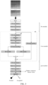
- a deep neural network model is constructed, and its structure is shown in FIG. 3 . All convolution layers involved in the deep neural network model are subjected to 1D convolution operations. Unless otherwise specified, the parameters of the 1D convolution kernel (i . e ., 1D filter) are the same, that is, the number of 1D convolution kernels is f ; the size of 1D convolution kernel is k ; the step size of 1D convolution operation is s ; the 1D convolution kernel uses L2 regularization and the regularization factor is r L 2 ; the initialization function of the 1D convolution kernel is g; the size of the output feature map of the 1D convolution operation is set to remain the same as that of the input feature map; the size of the pooling kernel is p , and the pooling step size is p s .
- the used Dropout Ratio of the Dropout layer involved in the deep neural network model is the same and is set as d.
- the structure of the deep neural network model includes:
- the input layer is used to receive the normalized sequence feature matrix Z ( k ) with a size of h ⁇ w .
- the pre-module is connected with the input layer, and is used for performing the first convolution and activation operation of the input sequence feature matrix to obtain the abstract representation feature map.
- the module includes: a 1D convolution layer, a batch normalization layer connected with the 1D convolution layer, and a ReLU activation layer connected with the batch normalization layer.
- the core module is connected with the pre-module, and is used for further abstraction and feature extraction of the feature map, and strengthening the expression ability of the neural network by effectively increasing the depth of the neural network model.
- the core module consists of three repeated operations of residual modules with the same structure, wherein the output of each residual module is the input of the next residual module.
- Each residual module includes:
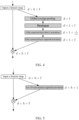
- the Reduction Ratio of SE module is set as r SE , and as shown in FIG. 4 , the structure of SE module includes (see reference [16] for details):
- the post-module has the same structure as the pre-module, and the only difference is that the number of 1D convolution kernels in the post-module is set as n out , which is used for feature abstraction representation of the feature map from the core module before output.
- the first global average pooling layer is connected with the post-module, and is used for vectorizing the feature map of the feature abstraction representation.
- the output layer is connected with the first global average pooling layer, and is a fully connected layer with a number of output neuron of 1, and the activation function is sigmoid function, which is used to output the chromosomal abnormality.
- the training set is used to train the deep neural network model in step 4, and the sequence feature vector of the sample is calculated using the trained deep neural network model.
- the process is as follows:
- Phenotypic data set of the pregnant woman is preprocessed, which includes outliers processing and missing values or null values processing.
- the phenotypic data of the pregnant woman sample are judged as outliers if the following records appear:
- MissForest algorithm is used for missing value padding, which is a non-parametric missing value padding algorithm based on random forest (see reference [18] for details). Its algorithm is as follows:
- BMI x weight x height / 100 2
- the ensemble learning algorithm based on Stacking is used to predict aneuploidy.
- the algorithm is as follows (see reference [19] for details):
- Example 2 Example of detecting chromosomal abnormalities.
- the invention proposes a method of detecting a fetal chromosomal abnormality, which uses nucleic acid sequencing results of noninvasive prenatal testing (NIPT) and phenotypic data of a pregnant woman together to predict whether the genetic abnormality presents in fetal chromosomes.
- NIPT noninvasive prenatal testing
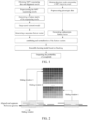
- FIG. 1 the process and steps of the method of detecting a fetal chromosomal abnormality are shown in FIG. 1 , and the specific process is described below.
- High throughput sequencing platform BGIseq500 is used to sequence samples to be detected (SE35 is adopted, with a sequencing depth of 0.1 ⁇ ).
- SE35 is adopted, with a sequencing depth of 0.1 ⁇ .
- the sequencing data are aligned to a reference genome and the repeated alignment sequences are filtered to obtain the unique mapping reads.
- Base quality is for quantitative description of the accuracy of sequencing results; the mean, standard deviation, skewness and kurtosis of base quality refer to the mean, standard deviation, skewness and kurtosis of all base quality in sequencing read segments, respectively.
- Map quality refers to the reliability of the alignment of a given sequence segment to a reference genome sequence; the mean, standard deviation, skewness and kurtosis of map quality refer to the mean, standard deviation, skewness and kurtosis of map quality of given sequencing read segments, respectively.
- Example 4 The trained deep neural network model in Example 1 is used to calculate the sequence feature vector of the sample, and the process is as follows:
- the phenotypic data of the pregnant woman sample to be detected are judged as outliers if the following records appear:
- Example 3 Example for validation.
- This example uses 1205 samples with "trisomy 21 (T21)" as positive samples and 1600 samples with normal chromosome (diploid) as negative samples.
- Table 1 describes the number of training samples and testing samples. Total number of samples Number of samples in Number of samples in (N) training set (90% ⁇ N) testing set (10% ⁇ N) Positive samples (T21) 1205 1084 121 Negative samples (Normal) 1600 1440 160
- the feature matrix of corresponded sequencing data in the training set is used to train the deep neural network model.
- the learning rate will be reduced by 2 times ( i.e ., multiplied by 0.5); if the accuracy rate does not improve after 10 successive iterations, the training stops.
- step 7 in above Example 1 the phenotypic features of all samples in the whole data set (including the training set and testing set) are obtained and the outliers of the phenotypic features are processed.
- step 7 in above Example 1 the phenotypic features in the training set are subjected to the missing value padding, and the padding model of the missing values is saved.
- step 7 in above Example 1 BMI is calculated for the phenotypic features in the training set after the missing value processing, as shown in FIG. 6 .
- step 8 in above Example 1 the sequence feature vector in the training set is combined with the phenotypic feature vector of the corresponding sample to obtain a combined feature vector.
- step 9 in above Example 1 the combined feature vector of each sample in the training set is normalized to obtain the normalized feature vector and the normalization model of the combined feature vector is saved.
- the saved padding model of the missing values is used for the missing value padding of the phenotypic features of each sample in the testing set, and the sequence feature vector of the testing set is then combined with the phenotypic feature vector of the corresponding sample to obtain the combined feature vector of the testing set, and then the saved normalization model of the combined feature vector is used to normalize the combined feature vector in the testing set.
- the normalized feature vector of the training set obtained in above step 10 is used to train the ensemble learning model based on Stacking, as shown in FIG. 7 .
- step 13 The trained ensemble learning model based on Stacking described in step 12 is verified using the testing set.
- the invention proposes using a machine learning model (such as a deep neural network) to extract the sequence feature vector of NIPT sequencing data, and then combining the sequence feature vector (the features including but not limited to the read count, base quality and mapping quality) with the phenotypic feature vector of the pregnant woman (the phenotypic features of pregnant woman including but not limited to maternity age, gestational week, height, weight, BMI, biochemical test results of the prenatal examination, and ultrasonic diagnosis results such as NT value, etc.) to form a vector combination, and then using a classification model, such as an ensemble learning model based on Stacking, to obtain the final predictive aneuploidy.
- a machine learning model such as a deep neural network
- extracting the sequence feature vector is not limited to the method used herein but also can be used including but not limited to an Autoencoder network or a Variational Autoencoder network.
- the model structure proposed by the invention is a hybrid model, that is, the model comprises 2 stages. In the first stage, a machine learning model (such as a deep neural network) is used to calculate the sequence feature vector. In the second phase, a classification model (such as an ensemble learning model based on Stacking) is used to predict aneuploidy by using the combination of sequence feature vector and phenotypic feature vector. Other ensemble learning models, such as a model based on Majority Voting, can also be used.
- the verified advanced deep neural network model used in the examples of the invention has the following features on network design and architecture: the deep neural network model used in the examples of the invention is a deep neural network model based on 1D convolutional model; the deep neural network model used in the examples of the invention is a network model based on residual network; the SE module of Squeeze-Excite network is introduced into the deep neural network model used in the examples of the invention.
- the neural network model used in the examples of the invention has more layers (see Example 3), and effectively reduces the risk of gradient disappearance and overfitting in the process of training model, and improves the stability, and thus effectively improves the accuracy of model prediction result.
- the invention can be implemented as a computer-readable storage medium, on which a computer program is stored, and the steps to implement the method of the invention are executed when the computer program is executed by a processor.
- the computer program is distributed over several computer devices or processors coupled by network, so that the computer program is stored, accessed, and executed in a distributed manner by one or more computer devices or processors.
- a single step/operation, or two or more steps/operations can be executed by a single computer device or processor or by two or more computer devices or processors.
- One or more steps/operations can be executed by one or more computer devices or processors, and one or more other steps/operations can be executed by one or more other computer devices or processors.
- One or more computer devices or processors can execute a single step/operation, or two or more steps/operations.
Landscapes
- Engineering & Computer Science (AREA)
- Life Sciences & Earth Sciences (AREA)
- Health & Medical Sciences (AREA)
- Physics & Mathematics (AREA)
- Chemical & Material Sciences (AREA)
- Theoretical Computer Science (AREA)
- Proteomics, Peptides & Aminoacids (AREA)
- General Health & Medical Sciences (AREA)
- Biophysics (AREA)
- Bioinformatics & Cheminformatics (AREA)
- Medical Informatics (AREA)
- Organic Chemistry (AREA)
- Molecular Biology (AREA)
- Data Mining & Analysis (AREA)
- Biotechnology (AREA)
- Analytical Chemistry (AREA)
- General Engineering & Computer Science (AREA)
- Genetics & Genomics (AREA)
- Software Systems (AREA)
- Evolutionary Computation (AREA)
- Artificial Intelligence (AREA)
- Wood Science & Technology (AREA)
- Zoology (AREA)
- General Physics & Mathematics (AREA)
- Evolutionary Biology (AREA)
- Spectroscopy & Molecular Physics (AREA)
- Bioinformatics & Computational Biology (AREA)
- Biomedical Technology (AREA)
- Mathematical Physics (AREA)
- Computing Systems (AREA)
- Public Health (AREA)
- Computational Linguistics (AREA)
- Biochemistry (AREA)
- Microbiology (AREA)
- Immunology (AREA)
- Pathology (AREA)
- Computer Vision & Pattern Recognition (AREA)
- Databases & Information Systems (AREA)
- Epidemiology (AREA)
- Bioethics (AREA)
Applications Claiming Priority (1)
| Application Number | Priority Date | Filing Date | Title |
|---|---|---|---|
| PCT/CN2020/132331 WO2022110039A1 (zh) | 2020-11-27 | 2020-11-27 | 一种胎儿染色体异常的检测方法与系统 |
Publications (2)
| Publication Number | Publication Date |
|---|---|
| EP4254418A1 true EP4254418A1 (de) | 2023-10-04 |
| EP4254418A4 EP4254418A4 (de) | 2024-03-27 |
Family
ID=81753821
Family Applications (1)
| Application Number | Title | Priority Date | Filing Date |
|---|---|---|---|
| EP20962929.4A Pending EP4254418A4 (de) | 2020-11-27 | 2020-11-27 | Verfahren und system zur erkennung von chromosomalen anomalien bei fötus |
Country Status (8)
| Country | Link |
|---|---|
| US (1) | US20240013859A1 (de) |
| EP (1) | EP4254418A4 (de) |
| JP (1) | JP7610710B2 (de) |
| KR (1) | KR20230110615A (de) |
| CN (1) | CN116648752B (de) |
| AU (1) | AU2020479407A1 (de) |
| CA (1) | CA3200221A1 (de) |
| WO (1) | WO2022110039A1 (de) |
Cited By (1)
| Publication number | Priority date | Publication date | Assignee | Title |
|---|---|---|---|---|
| WO2025169386A1 (ja) * | 2024-02-08 | 2025-08-14 | 学校法人藤田学園 | 胚の染色体異数性の検査方法 |
Families Citing this family (10)
| Publication number | Priority date | Publication date | Assignee | Title |
|---|---|---|---|---|
| US20220283922A1 (en) * | 2021-03-02 | 2022-09-08 | Nice Ltd. | Systems and methods for analyzing and segmenting automation sequences |
| EP4399724A4 (de) * | 2021-09-08 | 2025-06-25 | Janssen Research & Development, LLC | Multimodales system und verfahren zur vorhersage von krebs |
| CN114792548B (zh) * | 2022-06-14 | 2022-09-09 | 北京贝瑞和康生物技术有限公司 | 校正测序数据、检测拷贝数变异的方法、设备和介质 |
| CN114841294B (zh) * | 2022-07-04 | 2022-10-28 | 杭州德适生物科技有限公司 | 一种检测染色体结构异常的分类器模型训练方法及装置 |
| CN117095747B (zh) * | 2023-08-29 | 2024-04-30 | 广东省农业科学院水稻研究所 | 一种基于线性泛基因组和人工智能模型检测群体倒位或转座子端点基因型的方法 |
| WO2025208288A1 (zh) * | 2024-04-01 | 2025-10-09 | 深圳华大生命科学研究院 | 基因预测方法、装置、计算机设备及计算机可读存储介质 |
| CN118098345B (zh) * | 2024-04-28 | 2024-08-09 | 深圳市真迈生物科技有限公司 | 一种染色体非整倍体的检测方法、装置、设备及存储介质 |
| CN120727103B (zh) * | 2025-07-04 | 2026-02-24 | 内蒙古大学 | 一种基于深度测序8bp寡核苷酸双片段特征的先兆子痫无创筛查方法 |
| CN121171329A (zh) * | 2025-09-04 | 2025-12-19 | 国家卫生健康委科学技术研究所 | 一种测序数据处理方法 |
| CN121191773A (zh) * | 2025-11-25 | 2025-12-23 | 山东管理学院 | 一种男胎nipt最佳时点确定与女胎异常判定方法及系统 |
Family Cites Families (14)
| Publication number | Priority date | Publication date | Assignee | Title |
|---|---|---|---|---|
| CN1600265A (zh) * | 2004-09-27 | 2005-03-30 | 郑末晶 | 唐氏综合征和神经管缺陷产前筛查方法 |
| CN104232777B (zh) * | 2014-09-19 | 2016-08-24 | 天津华大基因科技有限公司 | 同时确定胎儿核酸含量和染色体非整倍性的方法及装置 |
| WO2018064486A1 (en) * | 2016-09-29 | 2018-04-05 | Counsyl, Inc. | Noninvasive prenatal screening using dynamic iterative depth optimization |
| AU2018244815A1 (en) | 2017-03-31 | 2019-10-31 | Premaitha Limited | Method of detecting a fetal chromosomal abnormality |
| KR102145417B1 (ko) | 2017-05-24 | 2020-08-19 | 지니너스 주식회사 | 무세포 핵산으로부터 수득된 서열 분석 데이터에 대한 배경 대립인자의 빈도 분포를 생성하는 방법 및 이를 이용하여 무세포 핵산으로부터 변이를 검출하는 방법 |
| EP3682035B1 (de) * | 2017-09-15 | 2025-02-05 | The Regents of the University of California | Nachweis von somatischen einzelnukleotidvarianten aus zellfreier nukleinsäure mit anwendung in der überwachung von minimaler resterkrankung |
| US11168356B2 (en) | 2017-11-02 | 2021-11-09 | The Chinese University Of Hong Kong | Using nucleic acid size range for noninvasive cancer detection |
| CN108611408A (zh) * | 2018-02-23 | 2018-10-02 | 深圳市瀚海基因生物科技有限公司 | 检测胎儿染色体非整倍性的方法和装置 |
| US12462935B2 (en) | 2018-03-30 | 2025-11-04 | Nucleix Ltd. | Deep learning-based methods, devices, and systems for prenatal testing |
| JP2021530231A (ja) | 2018-07-17 | 2021-11-11 | ナテラ, インコーポレイテッド | ニューラルネットワークを使用して倍数性状態を呼び出すための方法およびシステム |
| US12387817B2 (en) * | 2019-05-13 | 2025-08-12 | Nvidia Corporation | Sequence variation detection using deep learning |
| CN111286529A (zh) * | 2019-07-22 | 2020-06-16 | 常州市妇幼保健院 | 一种利用外周血胎儿游离dna产前筛查假阳性的试剂盒 |
| KR20220122596A (ko) * | 2019-12-31 | 2022-09-02 | 비지아이 클리니컬 래보러토리즈 (셴젠) 컴퍼니 리미티드 | 염색체 이수성 판별 및 분류 모델 구성 방법 및 장치 |
| CN111292802B (zh) * | 2020-02-03 | 2021-03-16 | 至本医疗科技(上海)有限公司 | 用于检测突变的方法、电子设备和计算机存储介质 |
-
2020
- 2020-11-27 CA CA3200221A patent/CA3200221A1/en active Pending
- 2020-11-27 AU AU2020479407A patent/AU2020479407A1/en active Pending
- 2020-11-27 CN CN202080107528.2A patent/CN116648752B/zh active Active
- 2020-11-27 EP EP20962929.4A patent/EP4254418A4/de active Pending
- 2020-11-27 JP JP2023532353A patent/JP7610710B2/ja active Active
- 2020-11-27 WO PCT/CN2020/132331 patent/WO2022110039A1/zh not_active Ceased
- 2020-11-27 US US18/254,842 patent/US20240013859A1/en active Pending
- 2020-11-27 KR KR1020237021684A patent/KR20230110615A/ko active Pending
Cited By (1)
| Publication number | Priority date | Publication date | Assignee | Title |
|---|---|---|---|---|
| WO2025169386A1 (ja) * | 2024-02-08 | 2025-08-14 | 学校法人藤田学園 | 胚の染色体異数性の検査方法 |
Also Published As
| Publication number | Publication date |
|---|---|
| CA3200221A1 (en) | 2022-06-02 |
| CN116648752B (zh) | 2025-07-29 |
| KR20230110615A (ko) | 2023-07-24 |
| WO2022110039A1 (zh) | 2022-06-02 |
| AU2020479407A1 (en) | 2023-06-29 |
| US20240013859A1 (en) | 2024-01-11 |
| JP7610710B2 (ja) | 2025-01-08 |
| CN116648752A (zh) | 2023-08-25 |
| JP2024505780A (ja) | 2024-02-08 |
| EP4254418A4 (de) | 2024-03-27 |
Similar Documents
| Publication | Publication Date | Title |
|---|---|---|
| EP4254418A1 (de) | Verfahren und system zur erkennung von chromosomalen anomalien bei fötus | |
| US20230187021A1 (en) | Methods for Non-Invasive Assessment of Genomic Instability | |
| Venkat et al. | Investigating genes associated with heart failure, atrial fibrillation, and other cardiovascular diseases, and predicting disease using machine learning techniques for translational research and precision medicine | |
| CN104732118B (zh) | 等位基因调用和倍性调用的方法 | |
| US7133856B2 (en) | Binary tree for complex supervised learning | |
| CA3129831A1 (en) | An integrated machine-learning framework to estimate homologous recombination deficiency | |
| CN114822682B (zh) | 与早发型重度子痫前期发生相关的基因组合及其应用 | |
| CN113362888A (zh) | 一种基于随机森林的深度特征选择算法提高胃癌预后预测精度系统、方法、设备及介质 | |
| US20230005569A1 (en) | Chromosomal and Sub-Chromosomal Copy Number Variation Detection | |
| EP4086356A1 (de) | Verfahren zur bestimmung von chromosomenaneuploidie und konstruktionsklassifizierungsmodell und vorrichtung | |
| Garrido-Martín et al. | A fast non-parametric test of association for multiple traits | |
| CN107463797B (zh) | 高通量测序的生物信息分析方法及装置、设备及存储介质 | |
| US11869630B2 (en) | Screening system and method for determining a presence and an assessment score of cell-free DNA fragments | |
| KR102534968B1 (ko) | 암 발생여부를 진단 또는 예측하는 방법 | |
| CN113593629B (zh) | 基于半导体测序的降低无创产前检测假阳性假阴性的方法 | |
| CN119475257A (zh) | 多模态数据融合的idh野生型胶质母细胞瘤分型方法及系统 | |
| Bhattacharya et al. | Effects of gene–environment and gene–gene interactions in case-control studies: A novel Bayesian semiparametric approach | |
| CN117778566A (zh) | 用于预测甲状腺癌转移的标志物及其应用 | |
| CN113345515A (zh) | 新发平衡易位家系中胚胎遗传性检测方法及装置 | |
| CN119230114B (zh) | 早期识别高危孕妇胎盘植入性疾病发生风险的分类器及其训练系统 | |
| US20240203521A1 (en) | Evaluation and improvement of genetic screening tests using receiver operating characteristic curves | |
| WO2025165914A1 (en) | Predicting dna methylation and tumor types from histopathology | |
| Sayyareh et al. | Early Detection of Down Syndrome Using Machine Learning Algorithms | |
| Emons et al. | Differential co-localisation analysis of multi-sample and multi-condition experiments with spatialFDA | |
| CN120877858A (zh) | 基于基因相对表达顺序的braf突变型结直肠癌分类系统 |
Legal Events
| Date | Code | Title | Description |
|---|---|---|---|
| STAA | Information on the status of an ep patent application or granted ep patent |
Free format text: STATUS: THE INTERNATIONAL PUBLICATION HAS BEEN MADE |
|
| PUAI | Public reference made under article 153(3) epc to a published international application that has entered the european phase |
Free format text: ORIGINAL CODE: 0009012 |
|
| STAA | Information on the status of an ep patent application or granted ep patent |
Free format text: STATUS: REQUEST FOR EXAMINATION WAS MADE |
|
| 17P | Request for examination filed |
Effective date: 20230613 |
|
| AK | Designated contracting states |
Kind code of ref document: A1 Designated state(s): AL AT BE BG CH CY CZ DE DK EE ES FI FR GB GR HR HU IE IS IT LI LT LU LV MC MK MT NL NO PL PT RO RS SE SI SK SM TR |
|
| DAV | Request for validation of the european patent (deleted) | ||
| DAX | Request for extension of the european patent (deleted) | ||
| A4 | Supplementary search report drawn up and despatched |
Effective date: 20240226 |
|
| RIC1 | Information provided on ipc code assigned before grant |
Ipc: C12Q 1/6883 20180101ALN20240220BHEP Ipc: G16B 40/20 20190101ALI20240220BHEP Ipc: G16B 30/10 20190101ALI20240220BHEP Ipc: G16B 20/10 20190101AFI20240220BHEP |