EP4218801A2 - Antibody modification method for purifying bispecific antibody - Google Patents
Antibody modification method for purifying bispecific antibody Download PDFInfo
- Publication number
- EP4218801A2 EP4218801A2 EP23150077.8A EP23150077A EP4218801A2 EP 4218801 A2 EP4218801 A2 EP 4218801A2 EP 23150077 A EP23150077 A EP 23150077A EP 4218801 A2 EP4218801 A2 EP 4218801A2
- Authority
- EP
- European Patent Office
- Prior art keywords
- antibody
- polypeptide
- amino acid
- humanized
- antibodies
- Prior art date
- Legal status (The legal status is an assumption and is not a legal conclusion. Google has not performed a legal analysis and makes no representation as to the accuracy of the status listed.)
- Pending
Links
Images











Classifications
-
- C—CHEMISTRY; METALLURGY
- C07—ORGANIC CHEMISTRY
- C07K—PEPTIDES
- C07K16/00—Immunoglobulins [IG], e.g. monoclonal or polyclonal antibodies
-
- A—HUMAN NECESSITIES
- A61—MEDICAL OR VETERINARY SCIENCE; HYGIENE
- A61P—SPECIFIC THERAPEUTIC ACTIVITY OF CHEMICAL COMPOUNDS OR MEDICINAL PREPARATIONS
- A61P35/00—Antineoplastic agents
-
- A—HUMAN NECESSITIES
- A61—MEDICAL OR VETERINARY SCIENCE; HYGIENE
- A61P—SPECIFIC THERAPEUTIC ACTIVITY OF CHEMICAL COMPOUNDS OR MEDICINAL PREPARATIONS
- A61P37/00—Drugs for immunological or allergic disorders
-
- A—HUMAN NECESSITIES
- A61—MEDICAL OR VETERINARY SCIENCE; HYGIENE
- A61P—SPECIFIC THERAPEUTIC ACTIVITY OF CHEMICAL COMPOUNDS OR MEDICINAL PREPARATIONS
- A61P43/00—Drugs for specific purposes, not provided for in groups A61P1/00-A61P41/00
-
- C—CHEMISTRY; METALLURGY
- C07—ORGANIC CHEMISTRY
- C07K—PEPTIDES
- C07K16/00—Immunoglobulins [IG], e.g. monoclonal or polyclonal antibodies
- C07K16/18—Immunoglobulins [IG], e.g. monoclonal or polyclonal antibodies against material from animals or humans
- C07K16/24—Immunoglobulins [IG], e.g. monoclonal or polyclonal antibodies against material from animals or humans against cytokines, lymphokines or interferons
- C07K16/244—Interleukins [IL]
- C07K16/248—IL-6
-
- C—CHEMISTRY; METALLURGY
- C07—ORGANIC CHEMISTRY
- C07K—PEPTIDES
- C07K2317/00—Immunoglobulins specific features
- C07K2317/20—Immunoglobulins specific features characterized by taxonomic origin
- C07K2317/24—Immunoglobulins specific features characterized by taxonomic origin containing regions, domains or residues from different species, e.g. chimeric, humanized or veneered
-
- C—CHEMISTRY; METALLURGY
- C07—ORGANIC CHEMISTRY
- C07K—PEPTIDES
- C07K2317/00—Immunoglobulins specific features
- C07K2317/30—Immunoglobulins specific features characterized by aspects of specificity or valency
- C07K2317/31—Immunoglobulins specific features characterized by aspects of specificity or valency multispecific
-
- C—CHEMISTRY; METALLURGY
- C07—ORGANIC CHEMISTRY
- C07K—PEPTIDES
- C07K2317/00—Immunoglobulins specific features
- C07K2317/50—Immunoglobulins specific features characterized by immunoglobulin fragments
- C07K2317/52—Constant or Fc region; Isotype
- C07K2317/522—CH1 domain
-
- C—CHEMISTRY; METALLURGY
- C07—ORGANIC CHEMISTRY
- C07K—PEPTIDES
- C07K2317/00—Immunoglobulins specific features
- C07K2317/50—Immunoglobulins specific features characterized by immunoglobulin fragments
- C07K2317/52—Constant or Fc region; Isotype
- C07K2317/524—CH2 domain
-
- C—CHEMISTRY; METALLURGY
- C07—ORGANIC CHEMISTRY
- C07K—PEPTIDES
- C07K2317/00—Immunoglobulins specific features
- C07K2317/50—Immunoglobulins specific features characterized by immunoglobulin fragments
- C07K2317/52—Constant or Fc region; Isotype
- C07K2317/526—CH3 domain
-
- C—CHEMISTRY; METALLURGY
- C07—ORGANIC CHEMISTRY
- C07K—PEPTIDES
- C07K2317/00—Immunoglobulins specific features
- C07K2317/50—Immunoglobulins specific features characterized by immunoglobulin fragments
- C07K2317/56—Immunoglobulins specific features characterized by immunoglobulin fragments variable (Fv) region, i.e. VH and/or VL
-
- C—CHEMISTRY; METALLURGY
- C07—ORGANIC CHEMISTRY
- C07K—PEPTIDES
- C07K2317/00—Immunoglobulins specific features
- C07K2317/50—Immunoglobulins specific features characterized by immunoglobulin fragments
- C07K2317/56—Immunoglobulins specific features characterized by immunoglobulin fragments variable (Fv) region, i.e. VH and/or VL
- C07K2317/567—Framework region [FR]
-
- C—CHEMISTRY; METALLURGY
- C07—ORGANIC CHEMISTRY
- C07K—PEPTIDES
- C07K2317/00—Immunoglobulins specific features
- C07K2317/70—Immunoglobulins specific features characterized by effect upon binding to a cell or to an antigen
- C07K2317/76—Antagonist effect on antigen, e.g. neutralization or inhibition of binding
Definitions
- the present invention relates to methods of modifying antibodies for purification of bispecific antibodies, methods for separating the bispecific antibodies, and pharmaceutical compositions and such comprising the bispecific antibodies as active ingredients.
- bispecific antibodies that can simultaneously recognize two types of antigens, e.g., antibody A and antibody B (see Non-Patent Document 1).
- MDX-210 which is currently under clinical trial investigation, is an IgG-type bispecific antibody that retargets FcyRI-expressing monocytes and such to HER-2/neu-expressing cancer cells (see Non-Patent Document 2).
- antibodies are produced using genetic recombination techniques.
- One specific technique involves the cloning of a DNA encoding an antibody protein from antibody-producing cells, such as hybridomas or sensitized lymphocytes that produce antibodies or a phage library presenting antibody genes, and the insertion of such into a suitable vector, which is then transfected into host cells for antibody production.
- Production of IgG type bispecific antibodies using genetic recombination techniques involves the introduction of a total of four types of genes into cells, in which these genes of H chains and L chains constitute two types of IgGs of interest, and the secretion of the antibodies by coexpression.
- Non-Patent Document 3 and Non-Patent Document 4 the content of the chain A-chain B heterodimer of interest can be increased up to a maximum of about 95% as shown in Non-Patent Document 3 and Non-Patent Document 4, the remaining 5% is impurities and consists of chain A and chain B homodimers.
- the chain A-chain B heterodimers must be purified to the highest possible purity from the three types of molecular species (chain A homodimer, chain B homodimer, and chain A-chain B heterodimer) that are produced when a common L chain is used (Non-Patent Document 3 and Non-Patent Document 4).
- Non-Patent Document 5 reports a method for selectively purifying the chain A-chain B heterodimer using mouse IgG2a as chain A and rat IgG2b as chain B. This method uses difference between the respective mouse IgG2a and rat IgG2b H chains in their affinity for protein A, and purifies the chain A-chain B heterodimer by controlling the pH for elution from protein A.
- this method since constant regions from mouse and rat are used, this method is difficult to apply to pharmaceuticals for human from the perspective of antigenicity. Furthermore, since this method cannot separate the chain A-chain B heterodimer, which is composed from H chains belonging to the same subclass, its use is limited.
- Non-Patent Document 6 A method for purifying the chain A-chain B heterodimer using hydrophobic interaction chromatography is reported in Non-Patent Document 6.
- the peak of the chain A-chain B heterodimer of interest containing anti-CD3 mouse IgG2a and anti-CD19 mouse IgG 1 is not sufficiently separated.
- H chains belonging to different subclasses are used, and the difference in their hydrophobicity seems to be used for the separation.
- this method may not necessarily separate the chain A-chain B heterodimer composed from H chains belonging to the same subclass.
- Non-Patent Document 7 A method for purifying the chain A-chain B heterodimer using thiophilic affinity chromatography is reported in Non-Patent Document 7.
- this method cannot be adopted to separate the chain A-chain B heterodimer composed from H chains belonging to the same subclass, because it uses mouse IgG1 and rat IgG2a, and the free cysteines (thiol groups) in the hinge regions.
- this method is not suitable for development of stable pharmaceutical formulations.
- Affinity chromatography using antigens is reported in Non-Patent Document 8.
- affinity chromatography using proteins or peptide antigens is problematic in terms of cost and column stability, production of pharmaceuticals using affinity chromatography is unconventional.
- affinity chromatography must be performed twice, and this is expected to become costly. It has been reported that there are antibodies that recognize only the three-dimensional structures of antigens as well as antibodies that have desired functions but low affinity. For antibodies with such characteristics, it is difficult to adopt affinity chromatography that uses antigens. Therefore, purification of bispecific antibodies using affinity chromatography cannot be used widely.
- Non-Patent Document 9 since the amino acid sequence homology between antibody variable regions is very high (Non-Patent Document 9), it has been difficult to purify the chain A-chain B heterodimer to a high purity that is acceptable for pharmaceuticals solely based on the differences in their variable region sequences.
- Carter P "Bispecific human IgG by design.”, J. Immunol. Methods., Feb 2001, Vol. 248(1-2), p.7-15 .
- the present invention was achieved in view of the above circumstances.
- An objective of the present invention is to provide methods for modifying the amino acids of antibody variable regions to efficiently purify bispecific antibodies, pharmaceutical compositions comprising the modified bispecific antibodies, and methods for producing the bispecific antibody pharmaceutical compositions.
- Another objective of the present invention is to provide bispecific antibodies in which the heavy chain constant regions have been modified, pharmaceutical compositions comprising the modified bispecific antibodies, and methods for producing the bispecific antibody pharmaceutical compositions.
- the present inventors conducted dedicated research on methods that substitute amino acids in antibody variable regions. These methods use a standard chromatography column to efficiently purify bispecific antibodies of interest which was conventionally challenging.
- the present inventors devised methods for efficiently purifying bispecific antibodies using a chromatography column based on the difference in isoelectric points of the H chains of two types of antibodies, and the difference is introduced by modifying the amino acids present on the surface of the variable regions of the two types of antibodies that constitute a bispecific antibody. Specifically, the present inventors discovered sites of modification in the antibody H chain that enable regulation of the isoelectric point alone without reducing the antibody function (activity). Furthermore, the present inventors confirmed that bispecific antibodies obtained by the methods of the present invention actually maintain their functions.
- the present inventors successfully developed methods for substituting amino acids in the antibody variable regions as efficient methods for purifying any bispecific antibody by using a standard chromatography column, and thereby completed the present invention.
- the present inventors further devised methods to efficiently purify bispecific antibodies using a chromatography column based on the difference in isoelectric point. Constant regions of different subclasses originally having different isoelectric points are used as the constant regions of the two types of H chains that constitute a bispecific antibody. Furthermore, the present inventors confirmed that the bispecific antibodies obtained by the methods of the present invention actually maintain their functions.
- the present invention relates to methods of substituting amino acids in the antibody variable regions for efficient purification by using a chromatography column, pharmaceutical compositions comprising the modified bispecific antibodies, and methods for producing the bispecific antibody pharmaceutical compositions.
- the present invention also relates to bispecific antibodies in which the heavy chain constant regions have been modified, pharmaceutical compositions comprising the modified bispecific antibodies, and methods for producing the bispecific antibody pharmaceutical compositions. More specifically, the present invention relates to the following:
- the present invention provides methods of modifying antibodies for production of multispecific antibodies.
- a preferred embodiment of the production methods of the present invention is a method comprising modifying both or either one of a nucleic acid encoding the amino acid residues of a first polypeptide and a nucleic acid encoding the amino acid residues of a second polypeptide, so that the isoelectric points of the first polypeptide and second polypeptide will be different. That is, multispecific antibodies can be produced based on differences in isoelectric point (pI), and the difference can be introduced into polypeptides by altering the charges of the amino acid residues in the first polypeptide and second polypeptide. More specifically, a preferred production method comprises the following steps of
- polypeptides generally refers to peptides and proteins whose length is approximately ten amino acids or longer. Polypeptides are generally derived from organisms, but are not particularly limited thereto, and for example, they may be composed of an artificially designed sequence. They may also be naturally derived polypeptides, synthetic polypeptides, recombinant polypeptides, or such. Additionally, fragments of the above-mentioned polypeptides are also included in the polypeptides of the present invention.
- the phrase "the difference between the isoelectric points of the polypeptides" means that the isoelectric points of two or more polypeptides are made unequal by modifying the charges of the amino acids on the surface of each polypeptide.
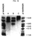
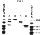
- the difference in the isoelectric points can be observed, for example, by using a technique such as isoelectric focusing.
- the isoelectric points are preferably modified without altering the structure and/or function (activity) of the polypeptides.
- the present invention provides a method for producing a multispecific antibody comprising a first polypeptide and a second polypeptide, wherein the method comprises the steps of
- a preferred embodiment of the purification methods of the present invention is a method comprising the step of modifying both or either one of a nucleic acid encoding the amino acid residues of a first polypeptide and a nucleic acid encoding the amino acid residues of a second polypeptide, so that the isoelectric points of the first polypeptide and second polypeptide will be different. That is, the difference in isoelectric point (pI) is introduced into the polypeptides by altering the charges of the amino acid residues of the first polypeptide and those of the second polypeptide.
- a purification method comprises the following steps of:
- the nucleic acids of the present invention are generally cloned (inserted) into suitable vectors and then introduced into host cells. These vectors are not particularly limited so long as the inserted nucleic acids are stably maintained.
- the cloning vectors are preferably pBluescript vectors (Stratagene) and such, while various commercially available vectors may be used.
- expression vectors are particularly useful. There is no particular limitation on expression vectors, so long as they can express polypeptides in test tubes, E. coli, cultured cells, or individual organisms.
- preferred vectors include pBEST vectors (Promega) for expression in test tubes, pET vectors (Invitrogen) in E . coli, the pME18S-FL3 vector (GenBank Accession No. AB009864) in cultured cells, and the pME18S vector ( Mol. Cell Biol. 8:466-472(1998 )) in individual rganisms. Insertion of the DNAs of the present invention into vectors can be performed, for example, by standard methods such as ligase reactions using restriction enzyme sites ( Current protocols in Molecular Biology edit. Ausubel et al. (1987) Publish. John Wiley & Sons. Section 11.4-11.11 ).
- Cells used for expressing polypeptides include bacterial cells (for example, Streptococcus, Staphylococcus, E. coli, Streptomyces, and Bacillus subtilis ) , fungal cells (for example, yeast and Aspergillus ) , insect cells (for example, Drosophila S2 and Spodoptera SF9), animal cells (for example, CHO, COS, HeLa, C127, 3T3, BHK, HEK293, Bowes melanoma cell), and plant cells.
- bacterial cells for example, Streptococcus, Staphylococcus, E. coli, Streptomyces, and Bacillus subtilis
- fungal cells for example, yeast and Aspergillus
- insect cells for example, Drosophila S2 and Spodoptera SF9
- animal cells for example, CHO, COS, HeLa, C127, 3T3, BHK, HEK293, Bowes melanoma cell
- Vectors can be introduced into host cells using known methods, such as the calcium phosphate precipitation method, electroporation method ( Current protocols in Molecular Biology edit. Ausubel et al. (1987) Publish. John Wiley & Sons. Section 9.1-9.9 ), lipofection method, and microinjection method.
- known methods such as the calcium phosphate precipitation method, electroporation method ( Current protocols in Molecular Biology edit. Ausubel et al. (1987) Publish. John Wiley & Sons. Section 9.1-9.9 ), lipofection method, and microinjection method.
- suitable secretion signals can be incorporated into the polypeptides of interest. These signals may be intrinsic or foreign to the polypeptides of interest.
- the multispecific antibodies (polypeptides) produced by the above-mentioned methods can be harvested by collecting the media.
- the polypeptides of the present invention are produced inside cells, the cells first are lysed, and then these polypeptides are collected.
- polypeptides of the present invention can be collected and purified from recombinant cell cultures using known methods, including ammonium sulfate or ethanol precipitation, acidic extraction, anion or cation exchange chromatography, phosphocellulose chromatography, hydrophobic interaction chromatography, affinity chromatography, hydroxyapatite chromatography, and lectin chromatography.
- compositions comprising a multispecific antibody of the present invention and a pharmaceutically acceptable carrier.
- compositions generally refers to agents for treating or preventing, or testing and diagnosing diseases.
- compositions of the present invention can be formulated by methods known to those skilled in the art.
- such pharmaceutical compositions can be used parenterally in the form of injections, which are sterile solutions or suspensions prepared with water or another pharmaceutically acceptable liquid.
- such compositions may be formulated by appropriately combining with a pharmaceutically acceptable carrier or medium, specifically, sterile water, physiological saline, vegetable oil, emulsifier, suspension, surfactant, stabilizer, flavoring agent, excipient, vehicle, preservative, binder, or such, and mixed in a unit dose form that meets the generally accepted requirements for preparation of pharmaceuticals.
- the amount of active ingredient is adjusted such that a suitable amount within a specified range is obtained.
- Sterile compositions for injection can be formulated using vehicles such as distilled water for injection, according to standard protocols for formulation.
- Aqueous solutions for injection include, for example, physiological saline and isotonic solutions containing glucose or other adjuvants (for example, D-sorbitol, D-mannose, D-mannitol, and sodium chloride).
- glucose or other adjuvants for example, D-sorbitol, D-mannose, D-mannitol, and sodium chloride.
- Appropriate solubilizers for example, alcohols (ethanol and such), polyalcohols (propylene glycol, polyethylene glycol, and such), non-ionic surfactants (polysorbate 80 TM , HCO-50, and such) may be used in combination.
- Oils include sesame and soybean oils.
- Benzyl benzoate and/or benzyl alcohol can be used as solubilizers in combination.
- Buffers for example, phosphate buffer and sodium acetate buffer
- soothing agents for example, procaine hydrochloride
- stabilizers for example, benzyl alcohol and phenol
- antioxidants can also be combined. Prepared injections are generally filled into appropriate ampules.
- compositions of the present invention are preferably administered parenterally.
- the compositions may be in the form of injections, transnasal agents, transpulmonary agents, or transdermal agents.
- such compositions can be administered systemically or locally by intravenous injection, intramuscular injection, intraperitoneal injection, subcutaneous injection, or such.
- the administration methods can be appropriately selected in consideration of a patient's age and symptoms.
- the dosage of a pharmaceutical composition comprising an antibody or a polynucleotide encoding an antibody may be set, for example, within the range of 0.0001 to 1000 mg/kg weight for each administration. Alternatively, the dosage may be, for example, from 0.001 to 100,000 mg per patient. However, in the present invention, the dosage is not necessarily limited to the ranges described above. Although the dosage and administration method vary depending on a patient's weight, age, symptoms, and such, those skilled in the art can select appropriate dosage and administration methods in consideration of the factors described above.
- the multispecific antibodies of the present invention can be formulated by combining with other pharmaceutical components as necessary.
- the present invention also provides nucleic acids encoding polypeptides that constitute the multispecific antibodies of the present invention. Furthermore, vectors that carry these nucleic acids are also included in the present invention.
- the present invention also provides host cells carrying the above described nucleic acids.
- the host cells are not particularly limited and include, for example, E coli and various animal cells.
- the host cells may be used, for example, as a production system to produce and express the antibodies or polypeptides of the present invention.
- Eukaryotic cells that can be used as host cells include, for example, animal cells, plant cells, and fungal cells.
- Animal cells include: mammalian cells, for example, CHO (J. Exp. Med. (1995) 108, 945 ), COS, HEK293, 3T3, myeloma, BHK (baby hamster kidney), HeLa, and Vero; amphibian cells such as Xenopus laevis oocytes ( Valle, et al., Nature (1981) 291: 338-340 ); and insect cells such as Sf9, Sf21, and Tn5.
- mammalian cells for example, CHO (J. Exp. Med. (1995) 108, 945 ), COS, HEK293, 3T3, myeloma, BHK (baby hamster kidney), HeLa, and Vero
- amphibian cells such as Xenopus laevis oocytes ( Valle, et al., Nature (1981)
- CHO-DG44 For expressing the antibodies of the present invention, CHO-DG44, CHO-DX11B, COS7 cells, HEK293 cells, and BHK cells can be suitably used. Of the animal cells, CHO cells are particularly preferable for large-scale expression.
- Vectors can be introduced into a host cell by, for example, calcium phosphate methods, DEAE-dextran methods, methods using cationic liposome DOTAP (Boehringer-Mannheim), electroporation methods, or lipofection methods.
- plant cells such as Nicotiana tabacum-derived cells and Lemna minor cells are protein production systems, and these cells can be used to produce antibodies of the present invention by methods that culture calluses from these cells.
- Protein expression systems that use fungal cells including yeast cells, for example, cells of the genus Saccharomyces ( Saccharomyces cerevisiae, Saccharomyces pombe, etc.), and cells of filamentous fungi, for example, the genus Aspergillus ( Aspergillus niger, etc.) are known, and these cells can be used as a host to produce antibodies of the present invention.
- prokaryotic cells When prokaryotic cells are used, production systems that use bacterial cells are available. Production systems that use bacterial cells including Bacillus subtilis as well as E . coli described above are known, and they can be used to produce antibodies of the present invention.
- a polynucleotide encoding an antibody of the present invention may be expressed by culturing the host cell transformed with an expression vector comprising the polynucleotide. Culturing can be performed according to known methods. For example, when animal cells are used as a host, DMEM, MEM, RPMI 1640, or IMDM may be used as the culture medium. The culture medium may be used with serum supplement solutions such as FBS or fetal calf serum (FCS). Alternatively, cells can be cultured in serum-free cultures. The preferred pH is about 6 to 8 during the course of culturing. Incubation is carried out typically at about 30 to 40°C for about 15 to 200 hours. Medium is exchanged, aerated, or agitated, as necessary.
- systems for producing polypeptides in vivo include, for example, those using animals and those using plants.
- a polynucleotide of interest is introduced into an animal or plant to produce the polypeptide in the body of the animal or the plant, and then the polypeptide is collected.
- the "host" of the present invention includes such animals and plants.
- mammals When animals are used, production systems that use mammals or insects are available. Mammals such as goat, pig, sheep, mouse, and cattle may be used ( Vicki Glaser, SPECTRUM Biotechnology Applications (1993 )). When mammals are used, transgenic animals may be used.
- a polynucleotide encoding a antibody of the present invention may be prepared as a fusion gene with a gene encoding a polypeptide specifically produced in milk, such as goat ⁇ -casein.
- polynucleotide fragments containing this fusion gene are injected into goat embryos, which are then introduced back into female goats.
- the antibody of interest can be obtained from milk produced by the transgenic goats, which are born from the goats that received the embryos, or by their offspring.
- Appropriate hormones may be administered to the transgenic goats to increase the volume of milk containing the antibody produced by the transgenic goats ( Ebert et al., Bio/Technology (1994) 12: 699-702 ).
- Insects such as silkworms may be used for producing antibodies of the present invention.
- silkworms When silkworms are used, baculoviruses carrying a polynucleotide encoding an antibody of interest can be used to infect silkworms, so that the antibody of interest can be obtained from the body fluids of these silkworms ( Susumu et al., Nature (1985) 315:592-594 ).
- Plants used for producing antibodies of the present invention include, for example, tobacco.
- a polynucleotide encoding an antibody of interest is inserted into a plant expression vector, for example, pMON 530, and then the vector is introduced into a bacterium such as Agrobacterium tumefaciens.
- the bacteria are then used to infect tobacco such as Nicotiana tabacum, and the desired antibody can be obtained from the leaves of the tobacco ( Ma et al., Eur. J. Immunol. (1994) 24: 131-138 ).
- the same bacteria can be used to infect Lemna minor, and after cloning, the desired antibody can be obtained from the cells of Lemna minor ( Cox K.M. et al., Nat. Biotechnol. 2006 Dec; 24(12):1591-1597 ).
- the antibody thus obtained may be isolated from the inside or outside (such as the medium and milk) of host cells, and purified as a substantially pure and homogenous antibody. Methods used for separating and purifying an antibody are not limited, and methods used in standard polypeptide purification may be applied. Antibodies may be isolated and purified by selecting an appropriate combination of, for example, chromatographic columns, filtration, ultrafiltration, salting-out, solvent precipitation, solvent extraction, distillation, immunoprecipitation, SDS-polyacrylamide gel electrophoresis, isoelectric focusing, dialysis, recrystallization, and such.
- Chromatographies include, for example, affinity chromatography, ion exchange chromatography, hydrophobic chromatography, gel filtration, reverse-phase chromatography, and adsorption chromatography ( strategies for Protein Purification and Characterization: A Laboratory Course Manual. Ed Daniel R. Marshak et al., (1996) Cold Spring Harbor Laboratory Press ). These chromatographies can be carried out using liquid phase chromatography such as HPLC and FPLC. Examples of columns for affinity chromatography include protein A columns and protein G columns. Examples of the columns that use protein A include Hyper D, POROS, and Sepharose F. F. (Pharmacia).
- An antibody can be modified arbitrarily, and peptides can be deleted partially from the antibody by treatment with an appropriate protein modifying enzyme before or after antibody purification, as necessary.
- protein modifying enzymes include, for example, trypsins, chymotrypsins, lysyl endopeptidases, protein kinases, and glucosidases.
- Another preferred embodiment of the present invention includes a method for producing a multispecific antibody of the present invention, wherein the method comprises the steps of culturing the host cells of the present invention as described above and collecting the polypeptide from the cell culture.
- multispecific antibody refers to an antibody that can specifically bind to at least two different types of antigens.
- preferred multispecific antibodies obtained by the production methods or purification methods of the present invention include bispecific antibodies (BsAbs) (also called dual-specificity antibodies) that can specifically bind to two antigens.
- BsAbs bispecific antibodies
- dual-specificity antibodies also called dual-specificity antibodies
- the term "different antigens” does not necessarily mean that the antigens themselves are different, and may mean that the epitopes are different. Therefore, for example, different epitopes within a single molecule are also included in the "different antigens" of the present invention.
- two antibodies that recognize different epitopes within a single molecule are regarded as antibodies that recognize different antigens.
- the multispecific antibodies of the present invention are antibodies having specificity to two or more different antigens, or molecules comprising fragments of such antibodies.
- the phrase "modification of nucleic acids” comprises modification of nucleic acids that results in separated peaks of a first polypeptide and a second polypeptide by standard chromatography analysis.
- the phrase "modification of nucleic acids” refers to modifying nucleic acids that correspond to the amino acid residues that are introduced by “modification” of the present invention. More specifically, the phrase refers to altering nucleic acids encoding the original amino acid residues (before modification) to nucleic acids encoding amino acid residues that are introduced by the modification.
- the phrase means gene manipulation or mutagenesis that modifies the original nucleic acids by inserting, deleting, or substituting at least one nucleotide, to produce a codon that encodes an amino acid residue of interest More specifically, a codon encoding the original amino acid residue is replaced by a codon encoding the amino acid residue to be introduced by the modification.
- nucleic acid modifications can be carried out appropriately by those skilled in the art using known techniques, for example, site-directed mutagenesis or PCR mutagenesis.
- the modification positions in the present invention include, for example, (1) amino acid residues on the surface of a polypeptide, (2) amino acid residues in the variable region, preferably in the FR region, and (3) amino acid residues in the constant region.
- amino acids on the surface of a polypeptide are amino acids whose side chains can contact solvent molecules (usually water molecules). It is not necessary for the entire side chain to be in contact with solvent molecules, and even if only part of the side chain is in contact with solvent molecules, the amino acid is considered to be an amino acid on surface.
- solvent molecules usually water molecules.
- the amino acid is considered to be an amino acid on surface.
- Those skilled in the art can produce homology models of polypeptides or antibodies by homology modeling and such using commercially available software, and thereby selecting appropriate residues as amino acids on the surface.
- Those skilled in the art can suitably select surface amino acids in the antibody variable region using homology models produced by homology modeling and such.
- H1, H3, H5, H8, H10, H12, H13, H15, H16, H19, H23, H25, H26, H39, H42, H43, H44, H46, H68, H71, H72, H73, H75, H76, H81, H82b, H83, H85, H86, H105, H108, H110, and H112 are examples of surface amino acids.
- the surface amino acids of the present invention are not limited thereto.
- surface amino acids can be similarly selected using homology models.
- L1, L3, L7, L8, L9, L11, L12, L16, L17, L18, L20, L22, L38, L39, L41, L42, L43, L45, L46, L49, L57, L60, L63, L65, L66, L68, L69, L70, L74, L76, L77, L79, L80, L81, L85, L100, L103, L105, L106, L107, and L108 are examples of surface amino acids.
- the surface amino acids of the present invention are not limited thereto.
- surface amino acids can be similarly selected using homology models.
- amino acid residues in the variable region include amino acid residues in the heavy chain variable region (VH) or light chain variable region (VL), and are preferably amino acid residues in the framework region (FR).
- surface-exposed amino acids other than those in CDR are, for example, H10, H12, H23, H39, H43, and H105 in the FR region, but are not limited thereto.
- polypeptides with nucleic acid modification are preferably a homomultimer of a first polypeptide, a homomultimer of a second polypeptide, and a heteromultimer of the first polypeptide and second polypeptide.
- the homomultimer of a first polypeptide is, for example, a homodimer of the humanized A69-H chain and humanized BBA-L chain
- a homomultimer of a second polypeptide is, for example, a homodimer of the humanized B26-H chain and humanized BBA-L chain
- a heteromultimer of a first polypeptide and a second polypeptide is, for example, a heterodimer of the humanized A69-H chain, humanized B26-H chain, and humanized BBA-L chain.
- the polypeptides are not limited thereto.
- Examples of standard chromatography in the present invention include cation exchange chromatography, anion exchange chromatography, hydrophobic chromatography, hydroxyapatite chromatography, hydrophobic charge interaction chromatography, and chromatofocusing.
- a first polypeptide and a second polypeptide preferably comprise a heavy chain variable region (VH).
- the variable region may comprise, for example, a complementary determining region (CDR) and a framework region (FR).
- the number of amino acid residues that undergo modification in the methods of the present invention is not particularly limited. However, for example, when the variable region(s) of an antibody is modified, it is preferable that for the separation of polypeptides of interest, the number of modified amino acid residues be kept to the minimum as necessary, so as not to decrease the antigen binding activity or increase the antigenicity of the antibody.
- the amino acid sequences after modification in the present invention are preferably human sequences, but are not limited thereto. Furthermore, in order to turn each of the modified FRs (FR1, FR2, FR3, and FR4) into a human sequence, mutations may be introduced into positions other than those that have been modified for alteration of isoelectric point. The method of replacing each FR with a human sequence in this manner has been reported in a Non-Patent Document ( Ono K. et al., Mol. Immunol. 1999 Apr; 36(6):387-395 ).
- the FR can be modified into another human FR having a different isoelectric point (for example, FR3 can be replaced with another human FR having a lower isoelectric point).
- FR3 can be replaced with another human FR having a lower isoelectric point.
- the desired multispecific antibody can be obtained by repeating modification of surface charge and evaluation of polypeptide separation.
- a multispecific antibody preferably comprises a third polypeptide comprising a light chain variable region, and preferably, the first polypeptide and the second polypeptide each forms a multimer with the third polypeptide.
- a first polypeptide and a second polypeptide preferably comprise a heavy chain constant region that preferably generates different pIs for the first polypeptide and second polypeptide.
- heavy chain constant regions include heavy chain constant regions of antibodies having different pIs.
- the pI difference can be introduced into the first polypeptide and the second polypeptide using the heavy chain constant regions of IgG1, IgG2, IgG3, or IgG4 which have pIs that are originally different from each other.
- amino acids in the heavy chain constant regions of the first polypeptide and the second polypeptide that cause differences in isoelectric point among these subclasses can be modified alone, or in combination with adjacent amino acids that do not have any effect on the isoelectric points to generate non-wild-type human constant regions, and pI difference can be introduced into the two constant regions.
- positions to be modified for introducing pI difference into the constant regions include, for example, positions 137, 196, 203, 214, 217, 233, 268, 274, 276, 297, 355, 392, 419, and 435, EU numbering, in the H chain constant region.
- position 297 which is a glycosylated site
- position 297 is another example of a position to be modified for introducing pI difference.
- methods that comprise the above-mentioned first polypeptide and second polypeptide comprising a heavy chain constant region methods that combine with the method in which the above-mentioned first polypeptide and second polypeptide comprise a heavy chain variable region, and/or the method in which the multispecific antibody comprises a third polypeptide comprising a light chain variable region, and a first polypeptide and a second polypeptide that each forms a multimer with the third polypeptide, are included in the present invention.
- Multispecific antibodies produced by the above-mentioned methods are also included in the present invention.
- the first polypeptide in the multispecific antibody provided by the present invention comprises a heavy chain variable region and/or a heavy chain constant region
- at least one amino acid residue in the region is made to carry a charge so that "the isoelectric points will be different".
- the amino acid residue(s) is selected from amino acid residues at positions 10, 12, 23, 39, 43, and 105, Kabat numbering, in the heavy chain variable region, or amino acid residues at positions 137, 196, 203, 214, 217, 233, 268, 274, 276, 297, 355, 392, 419, and 435, EU numbering, in the heavy chain constant region.
- amino acid residues other than the charged amino acid residue may have the same type of charge as that of the charged amino acid residue, or may be uncharged, or may have the opposite charge of that of the charged amino acid residue, as long as the isoelectric point of the first polypeptide and that of the second polypeptide are different.
- the above-mentioned multispecific antibodies of the present invention comprise a second polypeptide that preferably has the opposite charge of that of the charged amino acid residue in the first polypeptide, or is uncharged. More specifically, the second polypeptide in the multispecific antibodies comprises a heavy chain variable region and/or a heavy chain constant region, and at least one amino acid residue in the region is uncharged or has the opposite charge of that of the amino acid residue selected to carry a charge in the heavy chain variable region and/or the heavy chain constant region in the first polypeptide.
- amino acid residue(s) is selected from amino acid residues at positions 10, 12, 23, 39, 43, and 105, Kabat numbering, in the heavy chain variable region, or amino acid residues at positions 137, 196, 203, 214, 217, 233, 268, 274, 276, 297, 355, 392, 419, and 435, EU numbering, in the heavy chain constant region.
- amino acid residues other than the charged amino acid residue may have the same type of charge as that of the charged amino acid residue, or may be uncharged, or may have the opposite charge of that of the charged amino acid residue, as long as the isoelectric point of the first polypeptide and that of the second polypeptide are different.
- an IgG2 or IgG4 sequence to position 137 an IgG1, IgG2, or IgG4 sequence to position 196, an IgG2 or IgG4 sequence to position 203, an IgG2 sequence to position 214, an IgG1, IgG3, or IgG4 sequence to position 217, an IgG1, IgG3, or IgG4 sequence to position 233, an IgG4 sequence to position 268, an IgG2, IgG3, or IgG4 sequence to position 274, an IgG1, IgG2, or IgG4 sequence to position 276, an IgG4 sequence to position 355, an IgG3 sequence to position 392, an IgG4 sequence to position 419, and an IgG1, IgG2, or IgG4 sequence to position 435.
- an IgG1 or IgG3 sequence to position 137 an IgG3 sequence to position 196, the IgG1 or IgG3 sequence to position 203, an IgG1, IgG3, or IgG4 sequence to position 214, an IgG2 sequence to position 217, an IgG2 sequence to position 233, an IgG1, IgG2, or IgG3 sequence to position 268, an IgG1 sequence to position 274, an IgG3 sequence to position 276, an IgG1, IgG2, or IgG3 sequence to position 355, an IgG1, IgG2, or IgG4 sequence to position 392, an IgG1, IgG2, or IgG3 sequence to position 419, and an IgG3 sequence to position 435.
- amino acids are known to be charged amino acids.
- lysine (K), arginine (R), and histidine (H) are known as positively charged amino acids (cationic amino acids).
- Aspartic acid (D), glutamic acid (E), and such are known as negatively charged amino acids (anionic amino acids).
- charged amino acid residues are suitably selected from amino acid residues included in either one of groups (a) and (b) below, but are not particularly limited thereto:
- the phrase "having the same type of charge” means, for example, that the above-mentioned amino acid residue in the heavy chain variable region according to Kabat numbering and the above-mentioned amino acid residue in the heavy chain constant region according to EU numbering both carry an amino acid residue included in either one of the above-mentioned groups (a) and (b).
- phrases, "having the opposite charge” means that, for example, at least one of the above-mentioned amino acid residues, by Kabat numbering or EU numbering, in the second polypeptide comprising a heavy chain variable region and/or a heavy chain constant region is included in either one of the above-mentioned groups (a) or (b), and its corresponding amino acid residue at a position in the heavy chain variable region and/or heavy chain constant region comprised in the first polypeptide is included in the other group.
- the present invention provides multispecific antibodies, in which the above-mentioned amino acid residues having the same type of charge are selected from the amino acid residues included in either one of the above-mentioned group (a) or (b).
- the original amino acid residue (before modification) if it is already charged, it may be modified to be an uncharged amino acid residue.
- an amino acid residue is preferably modified such that the isoelectric point (pI) of the first polypeptide and that of the second polypeptide will be different. Furthermore, when multiple amino acid residues are introduced by modification, a few uncharged amino acid residues may be included in these amino acid residues.
- variable region of the first polypeptide comprises the amino acid sequence of any one of (a1) to (a7) below
- variable region of the second polypeptide comprises the amino acid sequence of any one of (b)1 to (b3) below
- variable region of the third polypeptide comprises the amino acid sequence of (c1) or (c2) below:
- amino acids are specific examples of the amino acids subjected to modification in the present invention.
- variable regions are not limited to those comprising these amino acids.
- variable region of the first polypeptide comprises the amino acid sequence of SEQ ID NO: 11
- variable region of the second polypeptide comprises the amino acid sequence of SEQ ID NO: 16
- variable region of the third polypeptide comprises the amino acid sequence of SEQ ID NO: 17.
- variable region of the first polypeptide comprises the amino acid sequence of SEQ ID NO: 12
- variable region of the second polypeptide comprises the amino acid sequence of SEQ ID NO: 16
- variable region of the third polypeptide comprises the amino acid sequence of SEQ ID NO: 18.
- a further preferred embodiment of the above-mentioned multispecific antibody is a multispecific antibody, wherein the first polypeptide and the second polypeptide comprise the human IgG4 constant region, and wherein the third polypeptide comprises the human ⁇ constant region.
- antibody is used in the broadest sense, and includes monoclonal antibodies, polyclonal antibodies, and mutant antibodies, such as chimeric antibodies, humanized antibodies, minibodies (including antibody fragments), and multispecific antibodies, as long as they display a desired biological activity.
- the methods of antibody modification of the present invention can be used favorably on these antibodies when they are obtained (produced).
- the "antibodies" of the present invention include antibodies in which the charge of amino acid residues has been modified as described above, and whose amino acid sequences have been further modified by amino acid substitutions, deletions, additions, and/or insertions.
- the antibodies also include antibodies whose amino acid sequences have been modified by amino acid substitution, deletion, addition, and/or insertion, or chimerization, humanization, or such, and in which the charge of amino acid residues has been further modified. In short, modifications may be performed at the same time when mouse antibodies are humanized, or further modifications may be performed on humanized antibodies.
- amino acid sequence modifications such as amino acid substitutions, deletions, additions, and/or insertions, and humanization and chimerization, can be achieved by methods known to those skilled in the art.
- the amino acid sequences of the antibody variable and constant regions may also be modified by amino acid substitutions, deletions, additions, and/or insertions, or chimerization, humanization and the like.
- the antibodies of the present invention may be derived from any animal, such as a mouse, human, rat, rabbit, goat, or camel. Furthermore, the antibodies may be modified, for example, chimeric antibodies, and in particular, modified antibodies that include amino acid substitutions in their sequence, such as humanized antibodies.
- the antibodies may be any type of antibody, such as antibody modification products linked with various molecules, antibody fragments, and minibodies.
- Chimeric antibodies are antibodies prepared by combining sequences derived from different animals.
- An example is an antibody having heavy and light chain variable (V) regions from a mouse antibody and heavy and light chain constant (C) regions from a human antibody.
- Chimeric antibodies can be prepared by known methods. To obtain such chimeric antibodies, for example, a DNA encoding an antibody V region may be ligated with a DNA encoding a human antibody C region; the resulting ligation product can be inserted into an expression vector; and the construct can be introduced into a host to produce the chimeric antibody.
- Humanized antibodies are also referred to as reshaped human antibodies, and can be obtained by substituting the complementary determining region (CDR) of a human antibody for the CDR of an antibody derived from a nonhuman mammal, for example, a mouse.
- CDR complementary determining region
- Methods for identifying CDRs are known in the art ( Kabat et al., Sequence of Proteins of Immunological Interest (1987), National Institute of Health, Bethesda, Md .; Chothia et al., Nature (1989) 342:877 ).
- General genetic recombination techniques suitable for this purpose are also known (see European Patent Application EP 125023 ; and WO 96/02576 ).
- the CDR of a mouse antibody can be determined by known methods, and a DNA can be prepared such that it encodes an antibody in which the CDR is ligated with the framework region (FR) of a human antibody.
- a humanized antibody can then be produced using a system that uses conventional expression vectors.
- DNAs can be synthesized by PCR, using as primers several oligonucleotides designed to include portions that overlap the ends of both the CDR and FR regions (see the method described in WO 98/13388 ).
- Human antibody FRs linked via CDRs are selected such that the CDRs form a suitable antigen binding site.
- amino acids in the FRs of an antibody variable region may be substituted so that the CDRs of the reshaped human antibody can form a suitable antigen binding site ( Sato, K. et al., Cancer Res. (1993) 53:851-856 ).
- Modifiable amino acid residues in the FRs include portions that directly bind to an antigen via non-covalent bonds ( Amit et al., Science (1986) 233: 747-53 ), portions that have some impact or effect on the CDR structure ( Chothia et al., J. Mol. Biol. (1987) 196: 901-17 ), and portions involved in the interaction between VH and VL ( EP 239400 ).
- the C regions of these antibodies are preferably derived from human antibodies.
- C ⁇ 1, Cy2, Cy3, and C ⁇ 4 can be used for the H chain, while C ⁇ and C ⁇ can be used for the L chain.
- the human antibody C region may be modified as required to improve antibody or production stability.
- a chimeric antibody of the present invention preferably includes a variable region of an antibody derived from a nonhuman mammal and a constant region of a human antibody.
- a humanized antibody preferably includes CDRs of an antibody derived from a nonhuman mammal and FRs and C regions of a human antibody.
- the constant regions of the human antibodies include specific amino acid sequences, which vary depending on the isotype of the antibody, for example, IgG (IgG1, IgG2, IgG3, and IgG4), IgM, IgA, IgD, and IgE.
- the constant regions used to prepare the humanized antibodies of the present invention may be the constant regions of antibodies of any isotype.
- a constant region of human IgG is preferably used, although the invention is not limited thereto.
- the FRs derived from a human antibody, which are used to prepare the humanized antibodies, are not particularly limited, and thus may be derived from an antibody of any isotype.
- variable and constant regions of chimeric or humanized antibodies of the present invention may be modified by deletion, substitution, insertion, and/or addition, so long as the antibodies exhibit the same binding specificity as that of the original antibodies.
- minibodies are useful as the antibodies because of their in vivo kinetic characteristics and low-cost production using E. coli, plant cells, or such.
- Antibody fragments are one type of minibody.
- minibodies includes antibodies that include an antibody fragment as a partial structural unit.
- the minibodies of the present invention are not particularly limited by their structure nor their method of production, so long as they have antigen binding activity. Some minibodies have an activity greater than that of a whole antibody ( Orita et al., Blood (2005) 105:562-566 ).
- the "antibody fragments” are not particularly limited, so long as they are a portion of a whole antibody (for example, whole IgG).
- the antibody fragments preferably include a heavy chain variable region (VH) or a light chain variable region (VL).
- Examples of preferred antibody fragments are: Fab, F(ab') 2 , Fab', and Fv.
- the amino acid sequence of a VH or VL in an antibody fragment may be modified by substitution, deletion, addition, and/or insertion. Furthermore, some portions of a VH and VL may be deleted, so long as the resulting fragments retain their antigen binding ability.
- Fv is a minimal antibody fragment composed of the complete antigen recognition and binding sites.
- “Fv” is a dimer (VH-VL dimer) composed of one unit of VH and one unit of VL bound very strongly by non-covalent bonding.
- variable region An antigen binding site is formed on the surface of the VH-VL dimer by the three complementary determining regions (CDRs) of each variable region.
- CDRs complementary determining regions
- Six CDRs confer an antigen binding site to the antibody.
- one variable region or half of an Fv composed of only three antigen-specific CDRs
- molecules smaller than Fv are also included in the context of antibody fragments of the present invention.
- the variable regions of an antibody fragment may also be chimerized or humanized.
- the minibodies preferably include both VH and VL.
- suitable minibodies include antibody fragments such as Fab, Fab', F(ab')2, and Fv, and scFv (single-chain Fv), which can be prepared using antibody fragments, ( Huston et al., Proc. Natl. Acad. Sci. USA (1988) 85: 5879-83 ; Pluckthun "The Pharmacology of Monoclonal Antibodies” Vol. 113, Resenburg and Moore (eds.), Springer Verlag, New York, pp. 269-315, (1994 )); diabodies ( Holliger et al., Proc. Natl. Acad. Sci.
- An antibody fragment can be prepared by treating an antibody with an enzyme, for example, a protease such as papain or pepsin (see Morimoto et al., J. Biochem. Biophys. Methods (1992) 24: 107-17 ; Brennan et al., Science (1985) 229:81 ).
- an enzyme for example, a protease such as papain or pepsin (see Morimoto et al., J. Biochem. Biophys. Methods (1992) 24: 107-17 ; Brennan et al., Science (1985) 229:81 ).
- antibody fragments can also be produced by genetic recombination based on its amino acid sequence.
- a minibody having a structure that results from modification of an antibody fragment can be prepared using antibody fragments obtained by enzyme treatment or genetic recombination.
- the minibody may be expressed in appropriate host cells (see, for example, Co et al., J. Immunol. (1994) 152: 2968-76 ; Better and Horwitz, Methods Enzymol. (1989) 178: 476-96 ; Pluckthun and Skerra, Methods Enzymol. (1989) 178: 497-515 ; Lamoyi, Methods Enzymol. (1986) 121: 652-63 ; Rousseaux et al., Methods Enzymol. (1986) 121: 663-9 ; Bird and Walker, Trends Biotechnol. (1991) 9: 132-7 ).
- scFVs are single-chain polypeptides that include two variable regions linked together via a linker or such, as required.
- the two variable regions in an scFv are typically one VH and one VL, but an scFv may include two VH or two VL.
- scFv polypeptides include a linker between the VH and VL domains, thereby forming a paired portion of VH and VL required for antigen binding.
- a peptide linker composed of ten or more amino acids is typically used as the linker between VH and VL when forming an intramolecular paired portion between VH and VL.
- linkers of the scFv of the present invention are not limited to such peptide linkers, so long as they do not inhibit the formation of an scFv.
- diabodies refers to bivalent antibody fragments constructed by gene fusion ( P. Holliger et al., Proc. Natl. Acad. Sci. USA 90: 6444-6448 (1993 ); EP 404,097 ; WO93/11161 and such).
- Diabodies are dimers composed of two polypeptide chains, wherein each polypeptide chain includes within the same chain a light chain variable region (VL) and a heavy chain variable region (VH) connected with a linker short enough to disable interaction of these two regions, for example a linker of about five amino acid residues.
- VL and VH encoded on the same polypeptide chain will form a dimer because the linker between VL and VH is too short to form a single chain V region fragment. Therefore, the resulting diabody has two antigen-binding sites.
- VL and VH directed against two different epitopes (a and b) are expressed simultaneously as combinations of VLa-VHb and VLb-VHa connected with a linker of about five residues, they are secreted as bispecific Db.
- diabodies include two molecules of scFvs, they thus composed of four variable regions, and as a result have two antigen binding sites.
- linkers forming a connection between VH and VL in each scFv molecules are linkers of about five amino acids when used as peptide linkers.
- scFv linkers for diabody formation are not limited to such peptide linkers so long as they do not interfere with scFv expression and diabody formation.
- an example of a multispecific antibody is a bispecific antibody.
- bispecific antibody may be, for example, an antibody having a structure in which a heavy chain variable region and a light chain variable region are linked in a single chain (for example, sc(Fv)2).
- the bispecific antibody may also be an antibody-like molecule (for example, scFv-Fc) produced by fusing an scFv (or sc(Fv)2), in which a heavy chain variable region and a light chain variable region are linked, to an Fc region (a constant region lacking the CH1 domain).
- a multispecific antibody consisting of scFv-Fc has an (scFv)2-Fc type structure with VH1-linker-VL1-Fc as the first polypeptide and VH2-linker-VL2-Fc as the second polypeptide.
- the bispecific antibody may be an antibody-like molecule in which a single domain antibody is linked with an Fc region ( Curr. Opin. Drug Discov. Devel. 2006, 9(2), 184-93 ).
- an antibody of the present invention known sequences can be used, or they can be obtained by methods known to those skilled in the art. For example, they may be obtained from an antibody library, or they may be obtained by cloning genes encoding the antibody from hybridomas producing monoclonal antibodies.
- antibody libraries many antibody libraries are already well known, and since methods for producing antibody libraries are known, those skilled in the art can appropriately obtain antibody libraries.
- antibody phage libraries one can refer to the literature such as Clackson et al., Nature 1991, 352: 624-8 ; Marks et al., J. Mol. Biol. 1991, 222: 581-97 ; Waterhouses et al., Nucleic Acids Res. 1993, 21: 2265-6 ; Griffiths et al., EMBO J. 1994, 13: 3245-60 ; Vaughan et al., Nature Biotechnology 1996, 14: 309-14 ; and Japanese Patent Kohyo Publication No.
- variable regions of human antibodies can be expressed on the surface of phages as single chain antibodies (scFvs) using phage display methods, and phages that bind to antigens can be selected. Genetic analysis of the selected phages can determine the DNA sequences encoding the variable regions of human antibodies that bind to the antigens.
- suitable expression vectors can be produced based on these sequences to obtain human antibodies. These methods are already well known, and one can refer to WO92/01047 , WO92/20791 , WO93/06213 , WO93/11236 , WO93/19172 , WO95/01438 , and WO95/15388 .
- known techniques may be used, involving the use of desired antigens or cells expressing the desired antigens as sensitizing antigens, using these to perform immunizations according to conventional immunization methods, fusing the immune cells thus obtained with known parent cells by ordinary cell fusion methods, screening monoclonal antibody producing cells (hybridomas) by ordinary screening methods, synthesizing cDNAs of antibody variable regions (V regions) from mRNAs of the obtained hybridomas using reverse transcriptase, and linking them with DNAs encoding the desired antibody constant regions (C regions).
- V regions antibody variable regions
- C regions desired antibody constant regions
- sensitizing antigens for obtaining the above-mentioned antibody genes encoding the H chains and L chains include both complete antigens with immunogenicity and incomplete antigens composed of haptens and such that do not show antigenicity.
- full length proteins and partial peptides of proteins of interest can be used.
- substances composed of polysaccharides, nucleic acids, lipids, and such may become antigens.
- Antigens can be prepared by methods known to those skilled in the art, and they can be prepared, for example, by the following methods using baculoviruses (for example, WO98/46777 ).
- Hybridomas can be produced, for example, the following methods of Milstein et al. (a Kohler and C. Milstein, Methods Enzymol. 1981, 73: 3-46 ), and such.
- an antigen When the immunogenicity of an antigen is low, it can be linked to a macromolecule that has immunogenicity, such as albumin, and then used for immunization.
- antigens with other molecules if necessary, they can be converted into soluble antigens.
- transmembrane molecules such as receptors are used as antigens, portions of the extracellular regions of the receptors can be used as a fragment, or cells expressing transmembrane molecules on their cell surface may be used as immunogens.
- Antibody-producing cells can be obtained by immunizing animals using suitable sensitizing antigens described above. Alternatively, antibody-producing cells can be prepared by in vitro immunization of lymphocytes that can produce antibodies. Various mammals can be used as the animals for immunization, where rodents, lagomorphas and primates are generally used. Examples of such animals include mice, rats, and hamsters for rodents, rabbits for lagomorphas, and monkeys including the cynomolgus monkey, rhesus monkey, hamadryas, and chimpanzees for primates.
- transgenic animals carrying human antibody gene repertoires are also known, and human antibodies can be obtained by using these animals (see WO96/34096 ; Mendez et al., Nat. Genet. 1997, 15: 146-56 ).
- desired human antibodies having binding activity against antigens can be obtained by in vitro sensitization of human lymphocytes with desired antigens or cells expressing the desired antigens, and then fusing the sensitized lymphocytes with human myeloma cells such as U266 (see Japanese Patent Application Kokoku Publication No. ( JP-B) H1-59878 (examined, approved Japanese patent application published for opposition)).
- desired human antibodies can be obtained by immunizing transgenic animals carrying a complete repertoire of human antibody genes, with desired antigens (see WO93/12227 , WO92/03918 , WO94/02602 , WO96/34096 , and WO96/33735 ).
- Animal immunization can be carried out by appropriately diluting and suspending a sensitizing antigen in Phosphate-Buffered Saline (PBS), physiological saline, or such, and forming an emulsion by mixing an adjuvant if necessary, followed by an intraperitoneal or subcutaneous injection into animals. After that, the sensitizing antigen mixed with Freund's incomplete adjuvant is preferably administered several times every four to 21 days. Antibody production can be confirmed by measuring the target antibody titer in animal sera using conventional methods.
- PBS Phosphate-Buffered Saline
- physiological saline physiological saline
- Antibody production can be confirmed by measuring the target antibody titer in animal sera using conventional methods.
- Antibody-producing cells obtained from lymphocytes or animals immunized with a desired antigen can be fused with myeloma cells to generate hybridomas using conventional fusing agents (for example, polyethylene glycol) (Goding, Monoclonal Antibodies: Principles and Practice, Academic Press, 1986, 59-103).
- fusing agents for example, polyethylene glycol
- hybridoma cells can be cultured and grown, and the binding specificity of the antibody produced from these hybridomas can be measured using known analysis methods, such as immunoprecipitation, radioimmunoassay
- hybridomas that produce antibodies of interest whose specificity, affinity, or activity has been determined can be subcloned by methods such as limiting dilution.
- genes encoding the selected antibodies can be cloned from hybridomas or antibody-producing cells (sensitized lymphocytes, and such) using probes that may specifically bind to the antibodies (for example, oligonucleotides complementary to sequences encoding the antibody constant regions). Cloning from mRNA using RT-PCR is also possible.
- Immunoglobulins are classified into five different classes, IgA, IgD, IgE, IgG and IgM. These classes are further divided into several subclasses (isotypes) (for example, IgG-1, IgG-2, IgG-3, and IgG-4; IgA-1 and IgA-2; and such).
- H chains and L chains used in the present invention to produce antibodies are not particularly limited and may derive from antibodies belonging to any of these classes or subclasses; however, IgG is particularly preferred.
- H-chain-encoding genes and L-chain-encoding genes using genetic engineering techniques.
- Genetically modified antibodies such as chimeric antibodies, humanized antibodies that have been artificially modified for the purpose of decreasing heterologous antigenicity and such against humans, can be appropriately produced if necessary for antibodies such as mouse antibodies, rat antibodies, rabbit antibodies, hamster antibodies, sheep antibodies, and camel antibodies.
- Chimeric antibodies are antibodies composed of a nonhuman mammal antibody H chain and L chain variable regions, such as mouse antibody, and the H chain and L chain constant regions of human antibody.
- a humanized antibody which is also called a reshaped human antibody, can be synthesized by PCR from a number of oligonucleotides produced so that they have overlapping portions at the ends of DNA sequences designed to link the complementary determining regions (CDRs) of an antibody of a nonhuman mammal such as a mouse.
- the obtained DNA can be ligated to a DNA encoding a human antibody constant region.
- the ligated DNA can be incorporated into an expression vector, and the vector can be introduced into a host to produce the antibody (see EP239400 and WO96/02576 ).
- Human antibody FRs that are ligated via the CDR are selected when the CDR forms a favorable antigen-binding site. If necessary, amino acids in the framework region of an antibody variable region may be substituted such that the CDR of the reshaped human antibody forms an appropriate antigen-binding site ( K. Sato et al., Cancer Res. 1993, 53: 851-856 ).
- antibodies may be modified to improve their biological properties, for example, antigenic affinity.
- modifications can be carried out using methods such as site-directed mutagenesis (see for example, Kunkel (1985) Proc. Natl. Acad. Sci. USA 82: 488 ), PCR mutagenesis, and cassette mutagenesis.
- mutant antibodies whose biological properties have been improved show amino acid sequence homology and/or similarity of 70% or higher, more preferably 80% or higher, and even more preferably 90% or higher (for example, 95% or higher, 97%, 98%, 99%, etc.), when compared to the amino acid sequence of the original antibody variable region.
- sequence homology and/or similarity is defined as the ratio of amino acid residues that are homologous (same residue) or similar (amino acid residues classified into the same group based on the general properties of amino acid side chains) to the original antibody residues, after the sequence homology value has been maximized by sequence alignment and gap introduction, if necessary.
- amino acid residues are classified into groups based on the characteristics of their side chains: (1) hydrophobic: alanine, isoleucine, valine, methionine, and leucine; (2) neutral hydrophilic: asparagine, glutamine, cysteine, threonine, and serine; (3) acidic: aspartic acid, and glutamic acid; (4) basic: arginine, histidine, and lysine; (5) residues that affect the orientation of the chain: glycine, and proline; and (5) aromatic: tyrosine, tryptophan, and phenylalanine.
- CDRs complementary determining regions
- hypervariable regions present in the H chain and L chain variable regions interact to form the antigen binding site(s) of an antibody.
- CDRs complementary determining regions
- variable regions are known to have the ability to recognize and bind to the antigen, although the affinity will be lower than when all binding sites are included. Therefore, antibody genes of the present invention encoding the H chain and L chain only have to encode fragment portions having each of the antigen binding sites of H chain and L chain, and polypeptides encoded by these genes only have to maintain affinity with the desired antigens.
- desired bispecific antibodies that actually have activities can be obtained efficiently by the methods of the present invention.
- Heavy chain variable regions are generally composed of three CDR regions and four FR regions, as described above.
- amino acid residues subjected to "modification” can be appropriately selected, for example, from amino acid residues in the CDR regions or FR regions.
- modification of amino acid residues in the CDR regions can decrease affinity towards antigens. Therefore, in the present invention, amino acid residues subjected to "modification” are not particularly limited, but are preferably appropriately selected from amino acid residues in the FR regions.
- variable region FRs for antibodies in organisms such as human or mouse
- sequences that can be used as variable region FRs for antibodies in organisms such as human or mouse can be appropriately obtained by those skilled in the art using public databases. More specifically, amino acid sequence information of the FR regions can be obtained by means described later in the Examples.
- a database was constructed by obtaining amino acid sequence data of human antibodies from the publicly disclosed in Kabat Database (ftp://ftp.ebi.ac.uk/pub/databases/kabat/) and IMGT Database (http://imgt.cines.fr/), and homology search was performed on the database for the mouse A69 H chain variable region (amino acid sequence: SEQ ID NO: 19), mouse B26 H chain variable region (amino acid sequence: SEQ ID NO: 20), and mouse BBA L chain variable region (amino acid sequence: SEQ ID NO: 21).
- the results confirmed that they have high homologies to the human antibody sequences below, and it was thus decided that they could be used as framework region (hereinafter referred to as FR) for humanized antibodies.
- the complementary determining region (hereinafter referred to as CDR) of each of the mouse antibodies was grafted into the FR of human antibodies of (1)-(3), and humanized antibodies were thus prepared.
- homology search web site publicly disclosed by NCBI (http://www.ncbi.nlm.nih.gov/BLAST/) was used to search for secretory signal sequences of human antibodies that are highly homologous to human antibodies of (4)-(6).
- the following secretory signal sequences obtained by the search were used.
- nucleotide sequence encoding the amino acid sequence covering from the secretory signal sequence to the antibody variable region twelve synthetic oligo-DNAs of about 50 bases were prepared, such that about 20 bases at the 3' end hybridize with each other.
- the synthetic oligo-DNAs were designed such that they encode a human sequence at the 5' side and a mouse sequence at the 3' side, or such that all the nucleotides encode a human sequence.
- a primer that anneals to the 5'-end of an antibody variable region gene and has the Xhol cleavage sequence and a primer that encodes the 5'-end sequence of the intron sequence, anneals to the 3'-end of an antibody variable region gene, and has the SfiI cleavage sequence were prepared.
- the fragments were cloned using the pGEM-T Easy Vector System (Promega) according to the method described in the attached instruction manual.
- the nucleotide sequence of each DNA fragment was determined using the BigDye Terminator Cycle Sequencing Kit (Applied Biosystems) and ABI PRISM 3730xL DNA Sequencer or ABI PRISM 3700 DNA Sequencer (Applied Biosystems) according to the method described in the attached instruction manual.
- the H-chain variable region fragment-inserted plasmid was digested with XhoI and SfiI, and the L-chain variable region fragment-inserted plasmid was digested with EcoRI, after they were confirmed to have the correct humanized antibody variable region gene sequence. Then, the reaction solutions were subjected to 1% agarose gel electrophoresis. DNA fragments having the desired size (about 400 bp) were purified using the QIAquick Gel Extraction Kit (QIAGEN) according to the method described in the attached instruction manual, and eluted with 30 ⁇ L of sterilized water. Thereafter, vectors for expression in animal cells were prepared as follows.
- an IgG4 that has amino acid substitution in its CH3 portion was used by referring to the knobs-into-holes technique for IgG1 ( Merchant AM et al., Nature Biotechnology, 1998, Vol. 16, p. 677-681 ). Furthermore, to promote H-chain dimer formation, amino acid substitution (-ppcpScp- ⁇ -ppcpPcp-) was also introduced into the hinge.
- Humanized A69 H chain expression vector was prepared by inserting the humanized A69 H chain variable region antibody gene fragment into an expression vector prepared by inserting an Y349C and T366W-substituted constant region gene into pCAGGS comprising a chicken ⁇ -actin promoter ( Niwa et al., Gene, 1991, 108: 193-199 ).
- Humanized B26 H chain expression vector was prepared by inserting the humanized B26 H chain variable region antibody gene fragment into an expression vector prepared by inserting an E356C, T366S, L368A, and Y407V-substituted constant region gene to pCAGGS.
- the plasmid (pCAG-gxDNA) was prepared by inserting a wild-type antibody L chain constant region into pCAGGS, and was digested with EcoRI to prepare an expression vector into which the humanized BBA L chain variable region antibody gene fragment was inserted. Ligation reaction was performed using the Rapid DNA Ligation Kit (Roche Diagnostics), and the E. coli strain DH5 ⁇ (TOYOBO) was transformed.
- Humanized bispecific antibodies were expressed using the following method. Human fetal renal carcinoma cell-derived humanized bispecific antibodies were expressed using the method described in Example 1-2, or using the following method. Human fetal renal carcinoma cell-derived HEK293H strain (Invitrogen) was suspended in a DMEM medium (Invitrogen) containing 10% Fetal Bovine Serum (Invitrogen), seeded at a cell density of 5-6 x 10 5 cells/mL (10 mL per dish) in dishes used for adhesive cells (10-cm diameter, CORNING), and cultured for one day and night in a CO 2 incubator (37°C, 5% CO 2 ).
- DMEM medium Invitrogen
- Fetal Bovine Serum Fetal Bovine Serum
- CHO-S-SFM-II Invitrogen
- Fetal Bovine Serum Fetal Bovine Serum
- the plasmid DNA mixture solution prepared in 1-2 was mixed with 20.7 ⁇ L of 1 ⁇ g/mL Polyethylenimine (Polysciences Inc.) and 690 ⁇ L of CHO-S-SFMII medium, left to stand at room temperature for ten minutes, and then added to the cells in each dish.
- the cells were then incubated in a CO 2 incubator (37°C, 5% CO 2 ) for four to five hours.
- CHO-S-SFM-II Invitrogen
- Fetal Bovine Serum Invitrogen
- the culture supernatant was collected, then cells were removed by centrifugation (at approximately 2000 g for five minutes at room temperature), and the solution was sterilized by passing it through a 0.22 ⁇ m filter MILLEX ® -GV (Millipore). The sample was stored at 4°C until use.
- rProtein A Sepharose TM Fast Flow 100 ⁇ L of rProtein A Sepharose TM Fast Flow (Amersham Biosciences) was added to the culture supernatant obtained by the method described in Example 1-2, and the solution was mixed by rotation at 4°C for four hours. The solution was transferred to an Ultrafree ® -MC 0.22- ⁇ m filter cup (Millipore). After three washes with 500 ⁇ L of TBS containing 0.01% Tween ® 20, the rProtein A Sepharose TM resin was suspended in 100 ⁇ L of 50 mM aqueous sodium acetate solution containing 0.01% Tween ® 20 at pH 3.3, and left to stand for two minutes, and then, the antibody was eluted. The eluate was immediately neutralized by adding 6.7 ⁇ L of 1.5 M Tris-HCl, pH 7.8.
- Goat anti-human IgG (Biosource International) was adjusted to 1 ⁇ g/mL with a coating buffer, and immobilized to a Nunc-Immuno plate (Nunc). After blocking with a diluent buffer (D.B.), a sample of the culture supernatant suitably diluted with D.B. was added. Furthermore, eleven three-fold serial dilutions of human IgG4 (humanized anti-TF antibody, see WO 99/51743 ) starting from 2000 ng/mL were made with D.B., and added as a standard for antibody concentration calculation. After three washes, goat anti-human IgG, alkaline phosphatase (Biosource International) was added for reaction.
- D.B. diluent buffer
- human IgG4 humanized anti-TF antibody, see WO 99/51743
- measurements were performed with Biacore 1000 (BIACORE) using Protein A-immobilized Sensor Chip CM5 (BIACORE). More specifically, according to the manufacturer's protocol, an activated sensor chip was reacted with Protein A (SIGMA) solution diluted to 50 ⁇ g/mL with 10 mM aqueous sodium acetate solution (pH 4.0, BIACORE) at 5 ⁇ L/minute for 30 minutes, and then a blocking procedure was carried out to produce a Protein A-immobilized sensor chip. This sensor chip was used to measure the concentrations of the culture supernatant and the purified products on Biacore 1000 (BIACORE). HBS-EP Buffer (BIACORE) was used for the immobilization of the sensor chip and for the concentration measurements.
- SIGMA Protein A
- HBS-EP Buffer BIACORE
- APTT activated partial thromboplastin time
- the Factor VIII-like activity (%) of a bispecific antibody was calculated from the coagulation time measured when the bispecific antibody was added.
- the human antibody FR of humanized bispecific antibodies that displayed decreased blood coagulation ability in the above-mentioned blood coagulation activity assay were subjected to amino acid modification in order to increase the activity. More specifically, the QuikChange Site-Directed Mutagenesis Kit (Stratagene) was used to introduce mutations into the humanized antibody variable region according to the method described in the attached instruction manual. The H-chain variable region fragment-inserted plasmid digested with XhoI and SfiI, and the L-chain variable region fragment-inserted plasmid was digested with EcoRI, , after they were confirmed to have the desired humanized antibody variable region gene sequence. Then, the reaction solutions were subjected to 1% agarose gel electrophoresis.
- DNA fragments having the desired size (about 400 bp) were purified using the QIAquick Gel Extraction Kit (QIAGEN) according to the method described in the attached instruction manual, and eluted with 30 ⁇ L of sterilized water. Then, plasmids for expression in animal cells were prepared according to the method described in Example 1-2. Humanized bispecific antibodies were prepared according to the method described in Examples 1-3, 1-4, and 1-5, and blood coagulation activity was evaluated according to the method described in Example 1-6.
- a humanized bispecific antibody (humanized A69 (hA69a)/humanized B26 (hB26-F123e4)/humanized BBA (hAL-F123j4)) having the same level of activity as the chimeric bispecific antibody (A69/B26/BBA) was obtained ( Fig. 1 ).
- the antibody variable region sequences are shown in the following SEQ ID NOs.
- a bispecific antibody In preparation of a bispecific antibody, when two types of H chains and one type of L chain are used for expression, the following three types of antibodies are expressed: a homodimer of the humanized A69 H chain and humanized BBA L chain, a homodimer of the humanized B26 H chain and humanized BBA L chain, and a heterodimer of the humanized A69 H chain, humanized B26 H chain, and humanized BBA L chain.
- the objective is to purify only the bispecific antibody by separating these three types of antibodies, and thus amino acid modifications were carried out to decrease the isoelectric point of the humanized A69 H chain variable region and increase the isoelectric point of the humanized B26 H chain variable region.
- antibody Fv region models were prepared for the humanized A69 antibody and humanized B26 antibody by homology modeling using the MOE software (Chemical Computing Group Inc.), and the amino acid resides exposed on the surface of the variable regions of the humanized A69 antibody and humanized B26 antibody were confirmed.
- the models are shown in Fig. 2 .
- the surface-exposed amino acids in the FR sequence outside CDR, H10, H12, H23, H39, H43, and H105 are thought to be candidates that can alter the isoelectric point without decreasing the activity.
- DNA fragments having the desired size (about 400 bp) were purified using the QIAquick Gel Extraction Kit (QIAGEN) according to the method described in the attached instruction manual, and eluted with 30 ⁇ L of sterilized water.
- H-chain expression vectors were prepared by inserting the prepared DNA fragments into an expression plasmid carrying a wild-type constant region and an expression plasmid in which the constant region amino acids have been replaced using the knobs-into-holes technique.
- humanized bispecific antibodies were prepared by the method described in Examples 1-3, 1-4, and 1-5. Sequences of the modified humanized antibody variable regions are shown in the SEQ ID NOs in Table 1 shown below.
- modified antibodies were prepared and analyzed by isoelectric focusing.
- the humanized BBA L-chain (hAL-F123j4) expression vector was simultaneously expressed together with the H-chain expression vector of unmodified hA69a, or hA69-p18, hA69-p8, hA69-p17, or hA69-p16 modified from the humanized A69 H chain.
- Five types of antibodies composed of hA69a, hA69-p18, hA69-p8, hA69-p17, or hA69-p16 homodimers were prepared.
- the humanized BBA L-chain expression vector was simultaneously expressed together with the H-chain expression vector of unmodified hB26-F123e4, or hB26-p19 or hB26-p15 modified from the humanized B26 H chain.
- Three types of antibodies composed of hB26-F123e4, hB26-p19, or hB26-p15 homodimers were prepared. Isoelectric focusing was performed as follows. PhastGel Dry IEF gel (Amersham Biosciences) was swollen for about 30 minutes in the swelling solution described below using the Phastsystem Cassette (Amersham Biosciences).
- Electrophoresis was performed using the swollen gel by PhastSystem (Amersham Biosciences) according to the following program. The samples were applied to the gel in Step 2. ApI calibration kit (Amersham Biosciences) was used as the pI marker. Step 1: 2000 V 2.5 mA 3.5 W 15°C 75 Vh Step 2: 200 V 2.5 mA 3.5 W 15°C 15 Vh Step 3: 2000 V 2.5 mA 3.5 W 15°C 410 Vh
- the gel was fixed with 20% TCA, and then silver stained using a silver staining kit, protein (Amersham Biosciences) according to the protocol attached to the kit. After staining, the isoelectric points of the samples were calculated from the known isoelectric points of the pI marker.
- the results of analysis of the unmodified and modified humanized A69 antibody homodimers and the humanized B26 antibody homodimer are shown in Fig. 3 .
- Band shifts were observed in the isoelectric focusing due to modification of the surface charge.
- the isoelectric points of the respective antibodies estimated in reference to the pI marker were approximately 8.4 for modified hA69-p18, approximately 8.2 for modified hA69-p17, approximately 8.2 for modified hA69-p8, and approximately 8.1 for modified hA69-p16, in contrast to approximately 8.8 for the unmodified hA69a homodimer. That is, the modification was able to provide a maximum isoelectric point difference of approximately 0.7.
- the isoelectric points were approximately 9.3 for modified hB26-p19 and approximately 9.4 for modified hB26-p15, in contrast to approximately 9.1 for unmodified hB26-F123e4. That is, the modification was able to provide a maximum isoelectric point difference of approximately 0.3. It was shown that the isoelectric point can be altered by modifying the charges on the surface amino acids in the variable region selected for this examination: H12, H23, H39, H43, and H105.
- Cation exchange chromagographic analysis was performed by the following method using the modified antibodies produced in Example 4 to evaluate the effect of the modification on the separation of the two antibodies.
- the conditions for the cation exchange chromatographic analysis were as follows. The retention time was calculated for the humanized A69 antibody homodimer and the humanized B26 antibody homodimer.
- Peak shifts were observed when comparing the unmodified humanized B26 antibody with those that have been modified to have higher isoelectric points for the variable region, and the retention time was increased to approximately 28.4 minutes and approximately 29.4 minutes, as the number of modifications increased.
- the retention time can be altered by modifying the charges on the surface amino acids in the variable region selected for this examination, H12, H23, H39, H43, and H105, and thereby modifying the surface charges of the two types of antibodies.
- Example 4 According to the isoelectric point measurements in Example 4, despite a pI difference of 0.3 between the unmodified hA69a homodimer and the unmodified hB26-F123e4 homodimer, the retention time for both of the homodimers was around 25 minutes, and thus they could not be separated ( Fig. 9 ). However, the pI difference between the unmodified hA69a homodimer and hB26-p19 was 0.5, and thus they were separated by a retention time difference of approximately 2.6 minutes. Also, the pI difference between hA69-p18 and the hB26 homodimer was 0.7, and thus they were separated by a retention time difference of approximately 3.4 minutes.
- APTT activated partial thromboplastin time
- Coagulation reaction was initiated by adding 50 ⁇ L of 20 mM CaCl 2 (Dade Behring) to the mixture. The time period until coagulation was measured with KC10A (Amelung) linked to CR-A (Amelung).
- the Factor VIII-like activity (%) of a bispecific antibody was calculated from the coagulation time measured when the bispecific antibody was added.
- H97 was confirmed to be a surface-exposed amino acid.
- the humanized A69 H chain hA69-N97R has a sequence in which asparagine at position 97 in CDR3 has been replaced with arginine.
- Modified antibodies were prepared by producing an expression vector carrying hA69-N97R according to the method of Example 1-2, and expressing it together with the humanized BBA L chain hAL-F123j4. To evaluate the alteration of surface charge of this antibody, isoelectric focusing was performed according to the method of Example 4. As indicated in Fig.
- Factor IXa the activity of binding to the antigen, Factor IXa was assayed by the following method.
- Factor IXa ⁇ Enzyme Research Laboratories
- coating buffer 100 mM sodium bicarbonate, pH 9.6, 0.02% sodium azide
- the plate was blocked with diluent buffer (50 mM Tris-HCl, pH 8.1,1% bovine serum albumin, 1 mM MgCl 2 , 0.15 M NaCl, 0.05% Tween ® 20, 0.02% sodium azide) at room temperature for two hours. After removal of the buffer, a purified antibody diluted in the diluent buffer was added to the plate at 100 ⁇ L/well and incubated at room temperature for one hour.
- diluent buffer 50 mM Tris-HCl, pH 8.1,1% bovine serum albumin, 1 mM MgCl 2 , 0.15 M NaCl, 0.05% Tween ® 20, 0.02% sodium azide
- the plate was washed three times, then alkaline phosphatase-labeled goat anti-mouse IgG (BIOSOURCE) diluted at 1/4000 with the diluent buffer was added at 100 ⁇ L/well. This was then incubated at room temperature for one hour. The plate was washed five times, then a chromogenic substrate (Sigma) was added at 100 ⁇ L/well. This was then incubated at room temperature for 30 minutes. The absorbance at 405 nm (control: 655 nm) was measured using the Model 3550 Microplate Reader (Bio-Rad Laboratories). As a result, as shown in Fig.
- the antibody in which the CDR had been modified to alter the surface charge showed a binding activity equivalent to that of the antibody before modification.
- the sites of modification may not only be in the FR indicated in Example 5, but also in the CDR.
- An unmodified humanized bispecific antibody was prepared using the unmodified antibodies (humanized A69 H chain hA69a and humanized B26 H chain hB26-F123e4) shown in Table 1, as well as the humanized BBAL chain hAL-F123j4 (SEQ ID NO: 5).
- a humanized bispecific PF antibody was prepared using the modified antibodies (the modified form of the humanized A69 H chain hA69-PFL and the modified form of the humanized B26 H chain hB26-PF) shown in Table 1, as well as the humanized BBA L chain hAL-s8 (SEQ ID NO: 17).
- H-chain expression vectors were prepared using an expression vector carrying the wild-type constant region, and the antibodies were prepared by the method described in Examples 1-3, 1-4, and 1-5.
- cation exchange chromatographic analysis was carried out according to the method described in Example 5.
- Figs. 9 and 10 The results of analysis of the unmodified humanized bispecific antibody and humanized bispecific PF antibody are shown in Figs. 9 and 10 .
- the results showed that the two types of homodimers and the bispecific antibody failed to separate, and were eluted as a single peak.
- the humanized bispecific PF antibody each of the two types of homodimers and the desired bispecific antibody were separated, and were eluted as three peaks in the following order: hA69-PF homodimer, humanized bispecific PF antibody, and hB26-PF homodimer.
- the two types of homodimers and the humanized bispecific PF antibody were purified. These fractions were concentrated using Amicon Ultra, MWCO 10000 (Millipore), then dialyzed overnight against 20 mM sodium acetate, 150 mM NaCl, pH 6.0 while cooling. Then, the concentrations were measured.
- the isoelectric point difference between the humanized A69-PF antibody homodimer and the humanized bispecific PF antibody was approximately 0.7, while the isoelectric point difference between the humanized B26-PF antibody homodimer and the humanized bispecific PF antibody was approximately 0.6.
- the coagulation activity of the bispecific PF antibody purified according to the method described in Example 6 was evaluated.
- the coagulation activity was compared with those of the following three types of antibodies expressed using an IgG4 constant region with the knobs-into-holes technique: the aforementioned chimeric bispecific antibody; the bispecific antibody composed of hA69a (SEQ ID NO: 2), hB26-F123e4 (SEQ ID NO: 4), and hAL-F123j4 (SEQ ID NO: 6) whose variable regions were not modified; and the bispecific antibody having the same variable regions as those of the purified bispecific PF antibody.
- the results of the evaluation are shown in Fig. 12 .
- the coagulation activity of the bispecific PF antibody carrying an IgG4 constant region prepared based on the knobs-into-holes technique was equivalent to that of the bispecific PF antibody carrying a wild-type constant region purified by cation exchange chromatography.
- modifications at H10, H12, H23, H39, H43, and H105 in the variable region enable purification of bispecific antibodies to a high purity without affecting their activities.
- antibody-expressing cell lines were established as follows.
- the H-chain constant region was amplified by PCR using the wild-type H-chain constant region gene of human IgG4 as a template, as well as a 5'-end primer designed such that the nucleotide sequence encoding the two amino acids (Ala-Ser) at the N terminus of the H-chain constant region will be the NheI recognition sequence (GCTAGC), and a primer designed to anneal to the 3'-end and carry the NotI recognizing site, and was linked to a vector prepared by digesting the pBluescriptKS+ vector (TOYOBO) with NheI and NotI (both from Takara), to produce pBCH4 comprising the IgG4 constant region gene.
- a 5'-end primer designed such that the nucleotide sequence encoding the two amino acids (Ala-Ser) at the N terminus of the H-chain constant region will be the NheI recognition sequence (GCTAGC), and a primer designed to anneal to the 3'-end and carry the NotI recognizing
- PCR was performed using a primer that is complementary to the 5'-end nucleotide sequence of the H-chain variable region of the humanized A69 H chain antibody (hA69-KQ) and humanized B26 H chain antibody (hB26-PF) shown in Table 1, and which has a Kozak sequence (CCACC) and the EcoRI recognition sequence, and a primer to the 3'-end nucleotide sequence and has the NheI recognition sequence.
- the obtained PCR products were digested with EcoRI and NheI (both from Takara), and were inserted into pBCH4 similarly digested with EcoRI and NheI, to link the variable region and the constant region.
- the prepared humanized A69 H chain antibody vector was digested with EcoRI and NheI (both from Takara), and was cloned into the pCXND3 expression vector for animal cells similarly digested with EcoRI and NotI.
- the course of construction of the present vector pCXND3 will be described below.
- To separate the antibody H chain gene and the vector in DHFR- ⁇ E-rVH-PM1-f (see WO92/19759 ), it was digested at the EcoRI and SmaI restriction sites to recover only the vector part. Subsequently, the EcoRI-NotI-BamHI adaptor (Takara) was cloned into this vector. The resulting vector was named pCHOI.
- the region of pCHOI for expressing the DHFR gene was cloned into the HindIII restriction site of pCXN ( Niwa et al., Gene 1991; 108: 193-200 ).
- the resulting vector was named pCXND3.
- the prepared humanized B26 H chain antibody vector was digested with EcoRI and NotI (both from Takara), and was cloned in the pCXZD1 expression vector for animal cells similarly digested with EcoRI and NotI.
- the pCXZD1 vector is an expression vector in which the neomycin resistance gene of the pCXND3 vector has been replaced with a Zeocin resistance gene.
- PCR was performed using a synthetic oligonucleotide that is complementary to the 5'-end nucleotide sequence of the L chain variable region of the humanized BBA L chain antibody (hAL-AQ, SEQ ID NO: 18) and which has a Kozak sequence, and a synthetic oligonucleotide that is complementary to the 3'-end nucleotide sequence and has the BsiWI site.
- the obtained PCR product was cloned in the pBCL vector, in which the human kappa chain constant region was inserted into the pBluescript KS+ vector.
- the human L chain variable region and constant region were linked together via the BsiWI site.
- the produced L chain gene fragment was cloned into the expression vector pUCAG.
- the pUCAG vector was prepared by digesting pCXN ( Niwa et al., Gene 1991; 108: 193-200 ) with the restriction enzyme BamHI to obtain a 2.6-kbp fragment, and cloning the fragment into the BamHI restriction site of the pUC19 vector (TOYOBO).
- the vector produced by cloning the L chain into pUCAG was digested with the restriction enzyme BamHI, and this was cloned into the expression vector pHygDHFR-4b containing a hygromycin resistance gene.
- the three types of expression vectors thus produced were linearized with restriction enzymes, and then they were transfected into CHO-DG44 cells to establish antibody-expressing cell lines.
- a ten-fold diluted solution was prepared using the same culture medium, and added at 100 ⁇ L/well to a 96-well culture plate. After culturing in a CO 2 incubator (5% CO 2 ) for one day and night, Geneticin (Invitrogen), Zeocin (Invitrogen), and Hygromycin B (Invitrogen) were added at 0.5 mg/mL, 0.6 mg/mL, and 0.4 mg/mL, respectively, and culture was continued for two weeks. Colonies of transfected cells showing drug resistance were sequentially cultured and expanded. Large scale culturing was carried out using the established high expression cell lines, and the culture supernatant was obtained.
- Bispecific antibodies were purified from the culture supernatant obtained in Example 8 by the following method.
- Solvent A 20 mmol/L sodium acetate buffer, pH 6.0
- Solvent B 20 mmol/L sodium acetate buffer, 1 mol/L NaCl, pH 6.0
- Flow Rate 10 mL/min (113 cm/h), or 5.3 mL/min (60 cm/h) only at the time of elution
- Gradient 0 ⁇ 15%
- B Step wise 3 Column Volume (CV) flushed 15 ⁇ 22% B gradient 2.5 CV 22 ⁇ 30% B gradient 6 CV 30 ⁇ 100% B Step wise 3 CV flushed
- bispecific antibodies can be also separated and purified using a standard preparative column.
- the coagulation activity of the humanized bispecific antibody prepared in Example 9 was evaluated according to the method described in Example 6. The results of the evaluation are shown in Fig. 14 .
- the humanized bispecific antibody purified in Example 9 had a coagulation activity equivalent to that of the humanized bispecific PF antibody prepared in Example 8. Thus, it was shown that even if amino acid sequences of the variable regions are slightly different in hA69-PFL and hA69-KQ, or even if the antibodies are purified using a standard preparative column, the antibody activities were not affected.
- the humanized bispecific antibody of interest and the two types of homodimeric antibodies can be separated and purified without modifying the structure or function (activity) of the antibody, by altering the surface charge through modification of the H-chain variable region when preparing the bispecific antibody. Since it was shown that bispecific antibodies can be separated and purified on a standard preparative column using the present method, this will be useful as a method for producing pharmaceuticals comprising a bispecific antibody.
- reaction solution (1 ⁇ L each of 20 ⁇ M K62 primer (5' cac cgt etc etc age etc cac caa 3', SEQ ID NO: 22) and K63 primer (5' gtg gca etc att tac ccg gag aca 3', SEQ ID NO: 23), 5 ⁇ L of MTC Multiple Tissue cDNA Panels (peripheral leukocytes) (Clontech), 4 ⁇ L of 5 ⁇ Prime STAR Buffer, 4 ⁇ L of 2.5 mM dNTPs, and 1 ⁇ L of PrimeSTAR HS DNA Polymerase (the above from Takara)) was prepared, and this was subjected to PCR.
- reaction solution 1 ⁇ L each of 20 ⁇ M K62 primer (5' cac cgt etc etc age etc cac caa 3', SEQ ID NO: 22) and K63 primer (5' gtg gca etc att tac ccg gag aca 3', SEQ ID NO: 23
- PCR was performed using a thermal cycler. GeneAmp PCR system 9700 (Perkin Elmer), by heating at 98°C for two minutes, followed by 30 cycles of reacting at 98°C for ten seconds, 60°C for five seconds, and 72°C for two minutes per cycle, and finally heating at 72°C for ten minutes. After PCR, the reaction solution was subjected to 1% agarose gel electrophoresis. Amplified fragments having the size of interest (approximately 1000 bp) were purified using the QIAquick Gel Extraction Kit (QIAGEN) according to the method described in the attached instruction manual, and eluted with 50 ⁇ L of sterile water.
- QIAquick Gel Extraction Kit QIAquick Gel Extraction Kit
- r-Taq treatment was performed to add A (Adenosine) to the ends of the amplified fragments.
- A Addenosine
- the obtained amplified fragments were incubated in 10 ⁇ L of rTaq reaction solution (1 ⁇ L of 10 ⁇ rTaq reaction solution, 1 ⁇ L of 2.5 mM dNTPs, 1 ⁇ L of rTaq, and 7 ⁇ L of the above-mentioned amplified fragments) at 72°C for 30 minutes.
- the r-Taq-treated fragments were cloned into the pCR2.1-TOPO vector (Invitrogen), and the nucleotide sequences were determined.
- the nucleotide sequence of each DNA fragment was determined by a DNA sequencer ABI PRISM 3730xL Genetic Analyzer (Applied Biosystems) using the BigDye Terminator 3.1 Cycle Sequencing Kit (Applied Biosystems) according to the method described in the attached instruction manual.
- the determined nucleotide sequences were compared to the sequence of Accession No. BX640623. If nucleotides in a determined sequence encode an amino acid sequence different from the corresponding sequence of BX640623, such nucleotides were considered to be mutations inserted during PCR amplification. In such cases, amino acid substitutions were performed using the Quick Change Site-Directed Mutagenesis Kit (Stratagene) so that the amino acid sequence will be the same as that of BX640623. The Quick Change Site-Directed Mutagenesis Kit (Stratagene) was used according to the method described in the attached instruction manual.
- a mutation was introduced such that the first two amino acids (Ala-Ser) of the human IgG2 H chain constant region were encoded by the restriction enzyme NheI recognition sequence (GCTAGC).
- the nucleotide sequence and amino acid sequence of the human IgG2 H-chain constant region used in this examination are shown in SEQ ID NOs: 24 and 25, respectively.
- Antibody expression vectors in which the H-chain variable region of the humanized PM-1 antibody is linked to one of the H chain constant regions of human IgG 1, human IgG2, and human IgG4, were prepared as follows.
- PCR was performed using a synthetic oligonucleotide that has a Kozak sequence and is complementary to the 5'-end nucleotide sequence of the H-chain variable region of the humanized anti-human interleukin 6 receptor antibody (humanized PM-1 antibody) described in a Non-Patent Document ( Sato K. et al., Cancer Research 1993, 53:851-856 ), and a synthetic oligonucleotide that has the NheI recognition sequence and is complementary to the 3'-end nucleotide sequence.
- the obtained PCR product was cloned into the pB-CH vector, in which the human IgG1 H chain constant region (see Sato, K.
- H-chain variable region gene of the humanized PM-1 antibody was linked to the human IgG4 constant region gene (see WO 99/51743 ) or the human IgG2 H chain gene prepared in Example 11-1 via the 5'-end NheI site, and then inserted into the pCAGGS vector.
- Each H-chain expression vector expresses the H chain by linking the H-chain variable region of the humanized PM-1 antibody to the humanized H chain constant region via the Nhel sequence.
- PCR was performed using a synthetic oligonucleotide that has a Kozak sequence and is complementary to the 5'-end nucleotide sequence of the L-chain variable region of the humanized PM-1 antibody, and a synthetic oligonucleotide that has the restriction enzyme BsiWI recognition sequence and is complementary to the 3'-end nucleotide sequence.
- the obtained PCR product was cloned into the pB-CL vector, in which the human kappa chain constant region was inserted into the pBluescript KS+ vector (TOYOBO).
- the L chain gene fragment in which the L-chain variable region and constant region were linked, was inserted into the pCAGGS vector whose expression is regulated by the chicken ⁇ -actin promoter.
- the L chain is expressed by linking the L-chain variable region of the humanized PM-1 antibody to the human kappa chain constant region via the BsiWI sequence.
- Subclass hybrid antibodies can be produced by combining any two of the humanized PM-1 antibody H chain expression vectors carrying the constant region of human IgG1, human IgG2, or human IgG4, and coexpressing them with the humanized PM-1 antibody L-chain expression vector in cells for expression. Each antibody was expressed by the method described in Example 4-2 or the following method.
- Human fetal renal carcinoma cell-derived HEK293H strain (Invitrogen) was suspended in a DMEM medium (Invitrogen) containing 10% Fetal Bovine Serum (Invitrogen), and this was seeded at a cell density of 5-6 x 10 5 cells/mL (10 mL per dish) in dishes used for adhesive cells (10-cm diameter, CORNING) and cultured for one day and night in a CO 2 incubator (37°C, 5% CO 2 ). Then, the medium was removed by suction, and 6.9 mL of CHO-S-SFM-II (Invitrogen) medium was added. As described below, the mixture solutions for expressing the different subclass antibodies and the mixture solutions for expressing the hybrid antibodies (a total of 13.8 ⁇ g) were prepared using the plasmid DNAs prepared in 11-2.
- Each mixture solution was mixed with 20.7 ⁇ L of 1 ⁇ g/mL Polyethylenimine (Polysciences Inc.) and 690 ⁇ L of CHO-S-SFMII medium, left to stand at room temperature for ten minutes, then added to the cells in each dish, and then the cells were then incubated in a CO 2 incubator (37°C, 5% CO 2 ) for four to five hours. Thereafter, 6.9 mL of CHO-S-SFM-II (Invitrogen) medium was added and then the cells were incubated in a CO 2 incubator for three days.
- CHO-S-SFM-II Invitrogen
- the culture supernatant was collected, then cells were removed by centrifugation (at approximately 2000 g for five minutes at room temperature), and the solution was sterilized by passing it through a 0.22 ⁇ m filter MILLEX ® -GV (Millipore). The sample was stored at 4°C until use.
- rProtein A Sepharose TM Fast Flow 100 ⁇ L of rProtein A Sepharose TM Fast Flow (Amersham Biosciences) was added to the culture supernatant obtained by the method described in Example 11-3, and the solution was mixed by rotation at 4°C for four hours. The solution was transferred to an Ultrafree ® -MC 0.22- ⁇ m filter cup (Millipore). After three washes with 500 ⁇ L of TBS, the rProtein A Sepharose TM resin was suspended in 100 ⁇ L of 50 mM aqueous sodium acetate solution at pH 3.0, and left to stand for two minutes, and then, the antibody was eluted.
- the eluate was immediately neutralized by adding 6.7 ⁇ L of 1.5 M Tris-HCl, 150 mM NaCl, pH 8.0.
- the buffer of the obtained antibody solution was exchanged by dialyzing it against PBS for activity measurement, or against 20 mM acetic acid buffer containing 150 mM NaCl, pH 6.0 for DSC measurement.
- the purified antibody carrying the H-chain constant region of human IgG1 will be referred to as "unmodified humanized anti-PM-1 antibody”
- the antibody carrying the H-chain constant region of human IgG2 will be referred to as “IgG2-substituted humanized anti-PM-1 antibody”
- the antibody carrying the H-chain constant region of human IgG4 will be referred to as "IgG4-substituted humanized anti-PM-1 antibody”.
- the absorbance of the antibody-containing solution obtained in 11-4 at 280 nm was measured on ND-1000 Spectrophotometer (NanoDrop) using 2 ⁇ L of the solution, or on the DU600 spectrophotometer (BECKMAN) using 50 ⁇ L of the solution.
- the antibody concentration was calculated from the obtained values using the following equation. PBS, or 20 mM acetic acid buffer containing 150 mM NaCl at pH6.0 was used as the blank.
- Antibody concentration (mg/mL) absorbance x dilution factor / 14.6 x 10
- Isoelectric focusing analysis was carried out to evaluate the alteration of surface charge as a result of substitution in the constant region.
- PhastGel Dry IEF (Amersham Biosciences) gel was swollen for about 30 minutes in the swelling solution described below using the Phastsystem Cassette (Amersham Biosciences). 20% Glycerol 1.5 mL Pharmalyte 8-10.5 for IEF (Amersham Biosciences) 100 mL
- Electrophoresis was performed using the swollen gel by PhastSystem (Amersham Biosciences) according to the following program. The samples were applied to the gel in Step 2. A pI calibration kit (Amersham Biosciences) was used as the pI marker. Step 1: 2000 V 2.5 mA 3.5 W 15°C 75 Vh Step 2: 200 V 2.5 mA 3.5 W 15°C 15 Vh Step 3: 2000 V 2.5 mA 3.5 W 15°C 410 Vh
- the gel was fixed with 20% TCA, and then silver stained using a silver staining kit, protein (Amersham Biosciences) according to the protocol attached to the kit. After staining, the isoelectric points of the samples were calculated from the known isoelectric points of the pI marker.
- the results of analysis of the unmodified, IgG2-substituted and IgG4-substituted humanized PM-1 antibodies are shown in Fig. 15 .
- Band shifts were observed in isoelectric focusing due to subclass substitution.
- the isoelectric points of the respective antibodies estimated in reference to the pI marker were approximately 8.9 for the IgG2-substituted humanized PM-1 antibody, and approximately 8.7 for the IgG4-substituted humanized PM-1 antibody, in contrast to approximately 9.3 for the unmodified humanized PM-1 antibody. That is, the substitution was able to provide a maximum isoelectric point difference of approximately 0.6. It was shown in this examination that the isoelectric points can be altered by substituting the constant region of an antibody subclass.
- Fig. 16 the results of analysis of coexpressed unmodified and IgG2-substituted humanized PM-1 antibodies, and coexpressed unmodified and IgG4-substituted humanized PM-1 antibodies are shown in Fig. 16 .
- the isoelectric points of the respective subclass hybrid antibodies estimated in reference to the pI marker were 9.2 for the unmodified humanized PM-1/IgG2-substituted human PM-1 hybrid antibody and 9.0 for the unmodified humanized PM-1/IgG4-substituted human PM-1 hybrid antibody. It was shown in this study that subclass hybrid antibodies can be produced by coexpressing a combination of expression vectors of subclass antibodies, and that the hybrid antibodies can be separated by their difference in isoelectric points.
- Cation exchange chromatographic analysis was performed by the following method using the subclass hybrid antibodies prepared in Example 11, and the effect of the subclass substitution on separation was evaluated.
- the conditions for cation exchange chromatographic analysis were as follows. The retention time was calculated for the unmodified humanized PM-1 antibody, the IgG2-substituted humanized PM-1 antibody, the IgG4-substituted humanized PM-1 antibody, the hybrid antibody of the unmodified humanized PM-1 antibody and IgG2-substituted humanized PM-1 antibody, and the hybrid antibody of the unmodified humanized PM-1 antibody and IgG4-substituted humanized PM-1 antibody.
- the results of analysis of singly expressed unmodified, IgG2-substituted, and IgG4-substituted humanized PM-1 antibodies are shown in Fig. 17 .
- the retention time of the unmodified humanized PM-1 antibody, IgG2-substituted humanized PM-1 antibody, and IgG4-substituted humanized PM-1 antibody was 60.2 minutes, 30.5 minutes, and 30.3 minutes, respectively. That is, the retention time was altered by slightly less than 30 minutes due to subclass substitution. On the other hand, the retention time was nearly the same for the IgG2-substituted humanized PM-1 antibody and the IgG4-substituted humanized PM-1 antibody, which showed a pI difference according to isoelectric focusing.
- Fig. 18 the results of analysis of coexpressing unmodified and IgG2-substituted humanized PM-1 antibodies, and coexpressing unmodified and IgG4-substituted humanized PM-1 antibodies are shown in Fig. 18 .
- the homodimers of each subclass and heterodimer were observed as three main peaks in the combination of the unmodified humanized PM-1 antibody and IgG2-substituted humanized PM-1 antibody, and the combination of the unmodified humanized PM-1 antibody and IgG4-substituted humanized PM-1 antibody.
- the retention time was approximately 43.8 minutes for the unmodified humanized PM-1/IgG2-substituted humanized PM-1 hybrid antibody, and approximately 45.1 minutes for the unmodified humanized PM-1/IgG4-substituted humanized PM-1 hybrid antibody. That is, these antibodies were separated from the respective homodimers with a retention time difference of 10 minutes or more. It was shown in this examination that subclass hybrid antibodies can be produced by coexpressing a combination of expression vectors of subclass antibodies, and that the hybrid antibodies can be separated by ion exchange chromatography.
- Example 11 The antibody solutions obtained in Example 11 were concentrated using Amicon-Ultra4 (Amicon), then enveloped in EasySep (TOMY SEIKO). After buffer exchange was performed by dialysis against 5 mM citric acid buffer (pH 6.5), subclass hybrid antibodies were purified under the following conditions.
- Fig. 19 Peak fractions from multiple experiments were combined, concentrated using Amicon-Ultra4 (Amicon), and then enveloped in EasySep (TOMY SEIKO). The buffer was exchanged by dialysis against PBS for activity measurement, or against 20 mM acetic acid buffer containing 150 mM NaCl, pH 6.0 for DSC measurement. The fractionated peaks were reanalyzed under conditions similar to those described above and the results are shown in Fig. 20 . This shows that subclass hybrid antibodies can be fractionated and purified by ion exchange chromatography methods.
- This technique enables separation of antibodies carrying a common H-chain variable region by using constant regions of different subclasses with different pI values.
- bispecific antibodies can be separated by ion exchange chromatography by linking the H-chain variable regions to subclass H-chain constant regions with different pI values.
- the pI difference between the molecules can be increased to further facilitate separation and purification by combining the above technique with the technique for introducing mutations into the variable region as shown in Example 9.
- bispecific antibodies can be separated and purified by ion exchange chromatography by substituting the H-chain variable region with a naturally-occurring IgG subclass sequence, without worrying about antigenicity.
- PhastGel Dry IEF (Amersham Biosciences) gel was swollen for about 30 minutes in the swelling solution described below using the Phastsystem Cassette (Amersham Biosciences). MilliQ water 1.5 mL Pharmalyte 5-8 for IEF (Amersham Biosciences) 50 ⁇ L Pharmalyte 8-10.5 for IEF (Amersham Biosciences) 50 ⁇ L
- Electrophoresis was performed using the swollen gel by PhastSystem (Amersham Biosciences) according to the following program. The samples were applied to the gel in Step 2. A pI calibration kit (Amersham Biosciences) was used as the pI marker. Step 1: 2000 V 2.5 mA 3.5 W 15°C 75 Vh Step 2: 200 V 2.5 mA 3.5 W 15°C 15 Vh Step 3: 2000 V 2.5 mA 3.5 W 15°C 410 Vh
- the gel was fixed with 20% TCA, and then silver stained using a silver staining kit, protein (Amersham Biosciences) according to the protocol attached to the kit.
- Fig. 21 The results of analysis of the fractionated and purified subclass hybrid antibody products are shown in Fig. 21 . It was shown that the products can be purified without containing any subclass homodimer by using ion exchange chromatography.
- Example 15 Activity assessment of fractionated and purified subclass hybrid antibody products 15-1. Establishment of human gp130-expressing BaF3 cell line and human gp130/human IL-6 receptor-coexpressing BaF3 cell line
- a BaF3 cell line expressing human gp130 was established as described below.
- the full-length human gp130 cDNA ( Hibi et al., Cell 1990; 63:1149-1157 (GenBank Accession No. NM_002184)) was amplified by PCR, and cloned into the pCOS2Zeo expression vector, from which the DHFR gene expression site of pCHOI ( Hirata et al., FEBS Letter 1994; 356:244-248 ) was removed, and to which a Zeocin resistance gene expression site was inserted, to construct pCOS2Zeo/gp130.
- pCOS2Zeo/gp130 10 ⁇ g was mixed with BaF3 cells (0.8 x 10 7 cells) suspended in PBS, then a pulse was applied at 0.33 kV and a capacity of 950 ⁇ FD using Gene Pulser (Bio-Rad).
- BaF3 cells subjected to the gene transfer by electroporation treatment were cultured for one day and night in RPMI1640 medium (Invitrogen) containing 0.2 ng/mL of mouse interleukin-3 (Peprotech), 10% Fetal Bovine Serum (hereinafter referred to as FBS, HyClone).
- the cells were selected by adding RPMI1640 medium containing 100 ng/mL of human interleukin-6 (R&D), 100 ng/mL of soluble human interleukin-6 receptor (R&D systems), and 10% FBS, to establish a human gp130-expressing BaF3 cell line (hereinafter referred to as BaF3/gp130). 15-2. Assessment of the activity of fractionated and purified subclass hybrid antibody products to neutralize human IL-6
- IL-6 neutralizing activity was evaluated using BaF3/gp130 showing IL-6-dependent growth, as described below.
- Purified unmodified humanized PM-1 antibody, unmodified/IgG4-substituted humanized PM-1 subclass hybrid antibody, and IgG4-substituted humanized PM-1 antibody were diluted in RPMI1640 containing 10% FBS to 10 ⁇ g/mL.
- a gene that encodes the 1st to the 344th amino acids of the soluble human IL-6 receptor ( Yamasaki et al., Science 1988; 241:825-828 (GenBank Accession No. X12830)) was introduced into CHO cells. Then, the receptor was purified from the culture supernatant. Cells were cultured for 72 hours under conditions of 37°C and 5% COz. Then, WST-8 reagent (Cell Counting Kit-8, Dojindo Laboratories) diluted two-fold with PBS was added at 20 ⁇ L/well. Immediately after, the absorbance at 450 nm (reference wavelength: 620 nm) was measured using SUNRISE CLASSIC (TECAN). After a two-hour culture, the absorbance at 450 nm (reference wavelength: 620 nm) was measured again. The IL-6 neutralizing activity was evaluated using the change in absorbance during the two-hour culture as an index.
- the fractioned and purified unmodified humanized PM-1 antibody, unmodified/IgG4-substituted humanized PM-1 subclass hybrid antibody, and IgG4-substituted humanized PM-1 antibody had neutralizing activities equivalent to that of a purified product of humanized PM-1 antibody (bulk).
- the subclass hybrid antibodies do not lose their original antigen-binding activities, and they function as neutralizing antibodies.
- the isoelectric point of an antibody can be altered by only a small number of amino acid substitutions without changing its structure/function (activity). This enables efficient purification of bispecific antibodies to high purity using a standard chromatography column. The high purity allows development of the bispecific antibodies into pharmaceuticals. Thus, the methods of the present invention are highly useful for developing bispecific antibodies as pharmaceuticals.
- Bispecific antibodies that actually have activities can be obtained efficiently by the methods of the present invention.
- the invention further comprises the following items:
Landscapes
- Chemical & Material Sciences (AREA)
- Health & Medical Sciences (AREA)
- Organic Chemistry (AREA)
- Life Sciences & Earth Sciences (AREA)
- Medicinal Chemistry (AREA)
- General Health & Medical Sciences (AREA)
- Immunology (AREA)
- Genetics & Genomics (AREA)
- Proteomics, Peptides & Aminoacids (AREA)
- Molecular Biology (AREA)
- Biophysics (AREA)
- Biochemistry (AREA)
- Chemical Kinetics & Catalysis (AREA)
- General Chemical & Material Sciences (AREA)
- Public Health (AREA)
- Nuclear Medicine, Radiotherapy & Molecular Imaging (AREA)
- Veterinary Medicine (AREA)
- Animal Behavior & Ethology (AREA)
- Pharmacology & Pharmacy (AREA)
- Engineering & Computer Science (AREA)
- Bioinformatics & Cheminformatics (AREA)
- Peptides Or Proteins (AREA)
- Preparation Of Compounds By Using Micro-Organisms (AREA)
- Micro-Organisms Or Cultivation Processes Thereof (AREA)
- Medicines Containing Antibodies Or Antigens For Use As Internal Diagnostic Agents (AREA)
Abstract
Description
- The present invention relates to methods of modifying antibodies for purification of bispecific antibodies, methods for separating the bispecific antibodies, and pharmaceutical compositions and such comprising the bispecific antibodies as active ingredients.
- Due to their highly stable nature in blood and relatively few side effects, antibodies have been receiving much attention as pharmaceuticals. Of particular note are bispecific antibodies that can simultaneously recognize two types of antigens, e.g., antibody A and antibody B (see Non-Patent Document 1). MDX-210, which is currently under clinical trial investigation, is an IgG-type bispecific antibody that retargets FcyRI-expressing monocytes and such to HER-2/neu-expressing cancer cells (see Non-Patent Document 2). In general, antibodies are produced using genetic recombination techniques. One specific technique involves the cloning of a DNA encoding an antibody protein from antibody-producing cells, such as hybridomas or sensitized lymphocytes that produce antibodies or a phage library presenting antibody genes, and the insertion of such into a suitable vector, which is then transfected into host cells for antibody production. Production of IgG type bispecific antibodies using genetic recombination techniques involves the introduction of a total of four types of genes into cells, in which these genes of H chains and L chains constitute two types of IgGs of interest, and the secretion of the antibodies by coexpression. In this type of system, expression of the constituent genes of the wild type H chain and L chains leads to random association between two types of H chains and association between H and L chains, and thus, the proportion of the bispecific antibody of interest becomes very small. More particularly, only one out of ten types produced is the bispecific antibody of interest, rendering the production efficiency quite low. Decreased efficiency in the production of the antibody of interest is not only an obstacle for purifying the antibody of interest, but also increases the nonuniformity, such as the lot-to-lot differences, which, in turn, leads to swelling production costs.
- Techniques for obtaining L chains commonly shared by both H chains, and the knobs-into-holes technique for heterologous association of H chains have been reported as efficient bispecific antibody production methods for developing bispecific antibodies. More specifically, a common L chain, which can maintain both antigen binding activities of the respective H chains that recognize antigen A and antigen B, is identified from a phage library or such. Then, formation of H chain heterodimers is promoted by substituting an amino acid side chain present in the CH3 region of one of the H chains with a larger side chain (knob), and substituting an amino acid side chain present in the CH3 region of the other H chain with a smaller side chain (hole), to place the knob within the hole. Thus, bispecific antibodies of interest can be efficiently obtained (see
Patent Document 1, Non-Patent Document 3, and Non-Patent Document 4). - However, even when the knobs-into-holes technique is used for H chain heterodimers, although the content of the chain A-chain B heterodimer of interest can be increased up to a maximum of about 95% as shown in Non-Patent Document 3 and
Non-Patent Document 4, the remaining 5% is impurities and consists of chain A and chain B homodimers. To develop bispecific antibodies as pharmaceuticals, the chain A-chain B heterodimers must be purified to the highest possible purity from the three types of molecular species (chain A homodimer, chain B homodimer, and chain A-chain B heterodimer) that are produced when a common L chain is used (Non-Patent Document 3 and Non-Patent Document 4). Therefore, it is necessary to remove the 5% impurities, which are the chain A and chain B homodimers, thereby purifying the chain A-chain B heterodimer to a high purity that allows the heterodimer to be developed into a pharmaceutical. When a common L chain is used without the knobs-into-holes technique, the production ratio of the chain A homodimer, chain A-chain B heterodimer, and chain B homodimer is theoretically 1:2:1, and the 50% impurities which are the chain A and chain B homodimers must be removed. - Several chromatographic methods for separating the chain A-chain B heterodimer from the chain A and chain B homodimers at the level of pharmaceutical manufacturing have been reported. Non-Patent Document 5 reports a method for selectively purifying the chain A-chain B heterodimer using mouse IgG2a as chain A and rat IgG2b as chain B. This method uses difference between the respective mouse IgG2a and rat IgG2b H chains in their affinity for protein A, and purifies the chain A-chain B heterodimer by controlling the pH for elution from protein A. However, since constant regions from mouse and rat are used, this method is difficult to apply to pharmaceuticals for human from the perspective of antigenicity. Furthermore, since this method cannot separate the chain A-chain B heterodimer, which is composed from H chains belonging to the same subclass, its use is limited.
- A method for purifying the chain A-chain B heterodimer using hydrophobic interaction chromatography is reported in Non-Patent Document 6. However, the peak of the chain A-chain B heterodimer of interest containing anti-CD3 mouse IgG2a and
anti-CD19 mouse IgG 1 is not sufficiently separated. In addition, H chains belonging to different subclasses are used, and the difference in their hydrophobicity seems to be used for the separation. Thus, this method may not necessarily separate the chain A-chain B heterodimer composed from H chains belonging to the same subclass. - A method for purifying the chain A-chain B heterodimer using thiophilic affinity chromatography is reported in Non-Patent Document 7. However, this method cannot be adopted to separate the chain A-chain B heterodimer composed from H chains belonging to the same subclass, because it uses mouse IgG1 and rat IgG2a, and the free cysteines (thiol groups) in the hinge regions. In addition, since the free cysteines are involved in aggregation during storage, this method is not suitable for development of stable pharmaceutical formulations.
- Affinity chromatography using antigens is reported in
Non-Patent Document 8. However, since affinity chromatography using proteins or peptide antigens is problematic in terms of cost and column stability, production of pharmaceuticals using affinity chromatography is unconventional. Furthermore, to purify the chain A-chain B heterodimer that binds to both antigens, affinity chromatography must be performed twice, and this is expected to become costly. It has been reported that there are antibodies that recognize only the three-dimensional structures of antigens as well as antibodies that have desired functions but low affinity. For antibodies with such characteristics, it is difficult to adopt affinity chromatography that uses antigens. Therefore, purification of bispecific antibodies using affinity chromatography cannot be used widely. - As described above, purification of the chain A-chain B heterodimer of a bispecific antibody has been performed only within limited scope. There has been no report on methods for purifying the chain A-chain B heterodimer of a bispecific antibody composed from the same H chain subclass and constant region sequence to a high purity that is acceptable for pharmaceuticals. When two types of antibodies constituting a bispecific antibody have the same constant region sequence, the chain A-chain B heterodimer needs to be separated based solely on the differences in their variable region sequences. However, since the amino acid sequence homology between antibody variable regions is very high (Non-Patent Document 9), it has been difficult to purify the chain A-chain B heterodimer to a high purity that is acceptable for pharmaceuticals solely based on the differences in their variable region sequences.
-
WO 96/27011 - Marvin JS, and Zhu Z, "Recombinant approaches to IgG-like bispecific antibodies.", Acta. Pharmacol. Sin., June 2005, Vol. 26(6), p.649-58.
- Segal D. M. et al., Current Opinion in Immunology, 1999, Vol. 11, p.558-562.
- Merchant AM et al., "An efficient route to human bispecific IgG", Nat. Biotechnol., Jul 1998, Vol. 16(7), p.677-81.
- Carter P, "Bispecific human IgG by design.", J. Immunol. Methods., Feb 2001, Vol. 248(1-2), p.7-15.
- Lindhofer H et al., "Preferential species-restricted heavy/light chain pairing in rat/mouse quadromas. Implications for a single-step purification of bispecific antibodies.", J. Immunol., Jul 1, 1995, Vol. 155(1), p.219-25.
- Manzke O et al., "Single-step purification of bispecific monoclonal antibodies for immunotherapeutic use by hydrophobic interaction chromatography.", J. Immunol. Methods., Oct 13, 1997, Vol. 208(1), p.65-73.
- Kreutz FT et al., "Efficient bispecific monoclonal antibody purification using gradient thiophilic affinity chromatography.", J. Chromatogr. B. Biomed. Sci. Appl., Sep 4, 1998, Vol. 714(2), p.161-70.
- Gupta S and Suresh M, "Affinity chromatography and co-chromatography of bispecific monoclonal antibody immunoconjugates.", J. Biochem. Biophys. Methods., May 31, 2002, Vol. 51(3), p.203-16. Review.
- Carl Branden, Introduction to Protein Structure 2nd edition, Newton Press.
- The present invention was achieved in view of the above circumstances. An objective of the present invention is to provide methods for modifying the amino acids of antibody variable regions to efficiently purify bispecific antibodies, pharmaceutical compositions comprising the modified bispecific antibodies, and methods for producing the bispecific antibody pharmaceutical compositions. Another objective of the present invention is to provide bispecific antibodies in which the heavy chain constant regions have been modified, pharmaceutical compositions comprising the modified bispecific antibodies, and methods for producing the bispecific antibody pharmaceutical compositions.
- The present inventors conducted dedicated research on methods that substitute amino acids in antibody variable regions. These methods use a standard chromatography column to efficiently purify bispecific antibodies of interest which was conventionally challenging.
- The present inventors devised methods for efficiently purifying bispecific antibodies using a chromatography column based on the difference in isoelectric points of the H chains of two types of antibodies, and the difference is introduced by modifying the amino acids present on the surface of the variable regions of the two types of antibodies that constitute a bispecific antibody. Specifically, the present inventors discovered sites of modification in the antibody H chain that enable regulation of the isoelectric point alone without reducing the antibody function (activity). Furthermore, the present inventors confirmed that bispecific antibodies obtained by the methods of the present invention actually maintain their functions.
- As described above, the present inventors successfully developed methods for substituting amino acids in the antibody variable regions as efficient methods for purifying any bispecific antibody by using a standard chromatography column, and thereby completed the present invention.
- The present inventors further devised methods to efficiently purify bispecific antibodies using a chromatography column based on the difference in isoelectric point. Constant regions of different subclasses originally having different isoelectric points are used as the constant regions of the two types of H chains that constitute a bispecific antibody. Furthermore, the present inventors confirmed that the bispecific antibodies obtained by the methods of the present invention actually maintain their functions.
- The present invention relates to methods of substituting amino acids in the antibody variable regions for efficient purification by using a chromatography column, pharmaceutical compositions comprising the modified bispecific antibodies, and methods for producing the bispecific antibody pharmaceutical compositions. The present invention also relates to bispecific antibodies in which the heavy chain constant regions have been modified, pharmaceutical compositions comprising the modified bispecific antibodies, and methods for producing the bispecific antibody pharmaceutical compositions. More specifically, the present invention relates to the following:
- [1] A method for producing a multispecific antibody comprising a first polypeptide and a second polypeptide, wherein the method comprises the steps of:
- (a) modifying both or either one of a nucleic acid encoding the amino acid residues of the first polypeptide and a nucleic acid encoding the amino acid residues of the second polypeptide, such that the difference between the isoelectric point of the first polypeptide and that of the second polypeptide will be increased;
- (b) culturing host cells to express the nucleic acids; and
- (c) collecting the multispecific antibody from the host cell culture.
- [2] The method of [1], wherein the modification of step (a) is modifying the nucleic acids so that the peaks of the homomultimer of the first polypeptide, the homomultimer of the second polypeptide, and the heteromultimer of the first polypeptide and the second polypeptide will be separated in standard chromatography analysis.
- [3] The method of [1], wherein the first polypeptide and the second polypeptide comprise a heavy chain variable region.
- [4] The method of [3], wherein the multispecific antibody comprises a third polypeptide comprising a light chain variable region, and wherein the first polypeptide and the second polypeptide each forms a multimer with said third polypeptide.
- [5] The method of any one of [1] to [4], wherein the first polypeptide and the second polypeptide comprise a heavy chain constant region.
- [6] The method of [5], wherein the heavy chain constant regions comprised in the first polypeptide and the second polypeptide are heavy chain constant regions whose isoelectric points are different from each other.
- [7] The method of [6], wherein the heavy chain constant regions with different isoelectric points are IgG1 and IgG4, or IgG1 and IgG2.
- [8] The method of [1], wherein the multispecific antibody is a bispecific antibody.
- [9] A multispecific antibody produced by the method of [1].
- [10] A method for purifying a multispecific antibody comprising a first polypeptide and a second polypeptide, wherein the method comprises the steps of
- (a) modifying both or either one of a nucleic acid encoding the amino acid residues of the first polypeptide and a nucleic acid encoding the amino acid residues of the second polypeptide, such that the difference between the isoelectric point of the first polypeptide and that of the second polypeptide will be increased;
- (b) culturing host cells to express the nucleic acids; and
- (c) purifying said multispecific antibody from the host cell culture by standard chromatography.
- [11] The method of [10], wherein the modification of step (a) is modifying the nucleic acids so that the peaks of the homomultimer of the first polypeptide, the homomultimer of the second polypeptide, and the heteromultimer of the first polypeptide and the second polypeptide will be separated in standard chromatography analysis.
- [12] The method of [10], wherein the first polypeptide and the second polypeptide comprise a heavy chain variable region.
- [13] The method of [12], wherein the multispecific antibody comprises a third polypeptide comprising a light chain variable region, and wherein the first polypeptide and the second polypeptide each forms a multimer with said third polypeptide.
- [14] The method of any one of [10] to [13], wherein the first polypeptide and the second polypeptide comprise a heavy chain constant region.
- [15] The method of [14], wherein the heavy chain constant regions comprised in the first polypeptide and the second polypeptide are heavy chain constant regions whose isoelectric points are different from each other.
- [16] The method of [15], wherein the heavy chain constant regions with different isoelectric points are IgG1 and IgG4, or IgG1 and IgG2.
- [17] The method of [10], wherein the multispecific antibody is a bispecific antibody.
- [18] A method for producing a multispecific antibody, wherein the method comprises the purification steps according to the method of [10].
- [19] A multispecific antibody produced by the method of [18].
- [20] A multispecific antibody comprising a first polypeptide and a second polypeptide, wherein the first polypeptide comprises a heavy chain variable region and/or a heavy chain constant region, wherein at least one amino acid residue selected from the amino acid residues at
positions 10, 12, 23, 39, 43, and 105, Kabat numbering, in said heavy chain variable region, or the amino acid residues at positions 137, 196, 203, 214, 217, 233, 268, 274, 276, 297, 355, 392, 419, and 435, EU numbering, in said heavy chain constant region, carries a charge, and wherein the isoelectric point of the first polypeptide and that of the second polypeptide are different from each other. - [21] The multispecific antibody of [20], wherein the second polypeptide comprises a heavy chain variable region and/or a heavy chain constant region, wherein at least one amino acid residue selected from the amino acid residues at
positions 10, 12, 23, 39, 43, and 105, Kabat numbering, in said heavy chain variable region, or the amino acid residues at positions 137, 196, 203, 214, 217, 233, 268, 274, 276, 297, 355, 392, 419, and 435, EU numbering, in said heavy chain constant region, is uncharged or carries the opposite charge of that of the charged amino acid residue selected in the heavy chain variable region and/or heavy chain constant region comprised in the first polypeptide. - [22] The multispecific antibody of [20], wherein the charged amino acid residue and the amino acid residue having the opposite charge of that of said charged amino acid residue are selected from the amino acid residues included in either of the following groups, respectively:
- (a) glutamic acid (E), and aspartic acid (D); and
- (b) lysine (K), arginine (R), and histidine (H).
- [23] A multispecific antibody in which the isoelectric point of a first polypeptide and that of a second polypeptide are different, and wherein the peaks of the homomultimer of the first polypeptide, the homomultimer of the second polypeptide, and the heteromultimer of the first polypeptide and the second polypeptide are separated in standard chromatography analysis.
- [24] The multispecific antibody of [23], wherein the first polypeptide and the second polypeptide comprise a heavy chain variable region.
- [25] The multispecific antibody of [24], wherein the multispecific antibody comprises a third polypeptide comprising a light chain variable region, and wherein the first polypeptide and the second polypeptide each forms a multimer with said third polypeptide.
- [26] The multispecific antibody of any one of [23] to [25], wherein the first polypeptide and the second polypeptide comprise a heavy chain constant region.
- [27] The multispecific antibody of [26], wherein the heavy chain constant regions comprised in the first polypeptide and the second polypeptide are heavy chain constant regions whose isoelectric points are different from each other.
- [28] The multispecific antibody of [27], wherein the heavy chain constant regions with different isoelectric points are IgG1 and IgG4, or IgG1 and IgG2.
- [29] The multispecific antibody of [23], wherein the multispecific antibody is a bispecific antibody.
- [30] A composition comprising the multispecific antibody of any one of [23] to [29], and a pharmaceutically acceptable carrier.
- [31] A nucleic acid encoding a polypeptide constituting the multispecific antibody of any one of [23] to [29].
- [32] A host cell comprising the nucleic acid of [31].
- [33] A method for producing the multispecific antibody of any one of [23] to [29], wherein the method comprises the step of culturing the host cell of [32], and collecting the polypeptides from the cell culture.
- [34] The multispecific antibody of [25], wherein the variable region of the first polypeptide comprises an amino acid sequence of any one of the following (a1) to (a7), wherein the variable region of the second polypeptide comprises an amino acid sequence of any one of the following (b1) to (b3), and wherein the variable region of the third polypeptide comprises an amino acid sequence of the following (c1) or (c2):
- (a1) SEQ ID NO: 7
- (a2) SEQ ID NO: 8
- (a3) SEQ ID NO: 9
- (a4) SEQ ID NO: 10
- (a5) SEQ ID NO: 11
- (a6) SEQ ID NO: 12
- (a7) SEQ ID NO: 13
- (b1) SEQ ID NO: 14
- (b2) SEQ ID NO: 15
- (b3) SEQ ID NO: 16
- (c1) SEQ ID NO: 17
- (c2) SEQ ID NO: 18
- [35] The multispecific antibody of [34], wherein the variable region of the first polypeptide comprises the amino acid sequence of SEQ ID NO: 11, wherein the variable region of the second polypeptide comprises the amino acid sequence of SEQ ID NO: 16, and wherein the variable region of the third polypeptide comprises the amino acid sequence of SEQ ID NO: 17.
- [36] The multispecific antibody of [34], wherein the variable region of the first polypeptide comprises the amino acid sequence of SEQ ID NO: 12, wherein the variable region of the second polypeptide comprises the amino acid sequence of SEQ ID NO: 16, and wherein the variable region of the third polypeptide comprises the amino acid sequence of SEQ ID NO: 18.
- [37] The multispecific antibody of any one of [34] to [36], wherein the first polypeptide and the second polypeptide comprise the human IgG4 constant region, and wherein the third polypeptide comprises the human κ constant region.
-
-
Fig. 1 depicts the result of evaluating the coagulation activity of a humanized bispecific antibody (humanized A69 (hA69a)/humanized B26 (hB26-F123e4)/humanized BBA (hAL-F123j4)). The result shows that the humanized bispecific antibody has a coagulation activity equal to or greater than that of a chimeric bispecific antibody. -
Fig. 2 depicts the result of antibody modeling for the humanized A69 H chain variable region (hA69a) with humanized BBA (hAL-F123j4), and the humanized B26 H chain variable region (hB26-F123e4) with humanized BBA (hAL-F123j4). The side chains of the amino acids whose surface charges can be modified are emphasized. The numbering was adopted from sequence numbers in the Kabat database (Kabat EA et al. 1991. Sequences of Proteins of Immunological Interest. NIH). -
Fig. 3 is a photograph depicting the result of isoelectric focusing analysis of the unmodified humanized A69 antibody homodimer, humanized A69 antibody homodimers having a modified variable region, the unmodified humanized B26 antibody homodimer, and humanized B26 antibody homodimers having a modified variable region. The result confirms that the isoelectric points are altered by the modifications. -
Fig. 4 depicts the result of cation exchange chromatographic analysis of humanized A69 antibody homodimers having a modified variable region. The result confirms that the peaks of the modified antibodies have been shifted compared to that of the unmodified antibody. -
Fig. 5 depicts the result of cation exchange chromatographic analysis of humanized B26 antibody homodimers having a modified variable region. The result confirms that the peaks of the modified antibodies have been shifted compared to that of the unmodified antibody. -
Fig. 6 depicts the result of evaluating the coagulation activity of humanized bispecific antibodies having a modified variable region (knobs-into-holes technique has been applied to the H chain constant regions). The result shows that the modified antibodies have a coagulation activity equivalent to that of the unmodified antibody. -
Fig. 7 is a photograph depicting the result of isoelectric focusing analysis of a humanized A69 antibody homodimer having a modified variable region (CDR). The results confirm that the band of the modified antibody has been shifted compared to that of the unmodified antibody. -
Fig. 8 depicts the result of evaluating the binding activity of the humanized A69 antibody homodimer having a modified variable region (CDR) towards the antigen Factor IXa. The result shows that the modified antibody has a binding activity equivalent to that of the unmodified antibody. -
Fig. 9 depicts the result of cation exchange chromatographic analysis of an unmodified humanized bispecific antibody prepared using unmodified humanized A69-H chain hA69a, unmodified humanized B26-H chain hB26-F123e4, and humanized BBA-L chain hAL-F123j4. As a result, the two types of homodimers and the bispecific antibody were eluted as a single peak without separation. -
Fig. 10 depicts the result of cation exchange chromatographic analysis of a humanized bispecific PF antibody prepared using hA69-PF, a modified form of the humanized A69-H chain; hA26-PF, a modified form of the humanized B26-H chain; and hAL-s8, the humanized BBA-L chain. As a result, the two types of homodimers and the bispecific antibody were individually separated and eluted as three peaks in the following order: thA69-PF homodimer, humanized bispecific PF antibody, and hB26-PF homodimer. -
Fig. 11 is a photograph depicting the result of isoelectric focusing analysis of purified humanized A69 antibody-PF homodimers, humanized B26-PF antibody homodimers, and humanized bispecific PF antibodies. The result confirms that the bispecific antibody of interest has been purified. -
Fig. 12 depicts the result of evaluating the coagulation activity of purified humanized bispecific PF antibodies (the H chain constant region is wild-type). The result shows that the purified antibodies have a coagulation activity equivalent to that of the bispecific antibody KiH, the H chain constant region of which was produced by the knobs-into-holes technique. -
Fig. 13 depicts a chromatogram obtained when the bispecific antibody was purified from a culture supernatant containing three types of antibodies, the humanized A69 antibody homodimer, humanized B26 antibody homodimer, and humanized bispecific antibody, using a standard preparative column. -
Fig. 14 depicts the result of evaluating the coagulation activity of a humanized bispecific antibody (the H chain constant region is wild-type) purified using a standard preparative column. The results show that the humanized bispecific antibody has a coagulation activity equivalent to that of the humanized bispecific PF antibody. -
Fig. 15 is a photograph depicting the result of isoelectric focusing analysis of unmodified, IgG2-substituted, and IgG4-substituted humanized PM-1 antibodies. The result confirms that the modifications alter the isoelectric point of the antibody. A: unmodified humanized PM-1 antibody; B: IgG2-substituted humanized PM-1 antibody; C: IgG4-substituted humanized PM-1 antibody. -
Fig. 16 is a photograph depicting the result of isoelectric focusing analysis of the unmodified humanized PM-1 antibody coexpressed with the IgG2-substituted or IgG4-substituted humanized PM-1 antibody. The result shows that the different subclass antibodies and subclass hybrid antibodies are separated according to their pI differences. A: coexpression of the unmodified humanized PM-1 antibody and the IgG2-substituted humanized PM-1 antibody; B: coexpression of the unmodified humanized PM-1 antibody and the IgG4-substituted humanized PM-1 antibody; C: humanized PM-1 antibody purified product (bulk). -
Fig. 17 depicts the result of cation exchange chromatographic analysis of singly expressed unmodified, IgG2-substituted, and IgG4-substituted humanized PM-1 antibodies. The result confirms that the peaks of the modified antibodies have been shifted compared to that of the unmodified antibody. -
Fig. 18 depicts the result of cation exchange chromatographic analysis of the unmodified humanized PM-1 antibody coexpressed with the IgG2-substituted or IgG4-substituted humanized PM-1 antibody. As a result, the homodimers of each subclass and the heterodimer were observed as three major peaks in the combination of the unmodified humanized PM-1 antibody and the IgG2-substituted humanized PM-1 antibody, and in the combination of the unmodified humanized PM-1 antibody and the IgG4-substituted humanized PM-1 antibody. A: coexpression of the unmodified humanized PM-1 antibody and the IgG2-substituted humanized PM-1 antibody; B: coexpression of the unmodified humanized PM-1 antibody and the IgG4-substituted humanized PM-1 antibody. -
Fig. 19 depicts the result of purification of the homodimers and heterodimer by cation exchange chromatography from coexpression of the unmodified humanized PM-1 antibody and the IgG4-substituted humanized PM-1 antibody. As a result, three peaks were eluted in the following order: IgG4-substituted humanized PM-1 antibody homodimer, unmodified humanized PM-1/IgG4-substituted humanized PM-1 hybrid antibody, and unmodified humanized PM-1 antibody homodimer. These peaks were fractionated. Arrows indicate the approximate fractionation ranges. -
Fig. 20 depicts the result of rechromatography of the unmodified humanized PM-1 antibody homodimer, unmodified humanized PM-1/IgG4-substituted humanized PM-1 hybrid antibody, and IgG4-substituted humanized PM-1 antibody homodimer, which were purified by cation exchange chromatography. As a result, the subclass hybrid antibody was confirmed to be purified. -
Fig. 21 is a photograph depicting the result of isoelectric focusing analysis of the unmodified humanized PM-1 antibody homodimer, unmodified/IgG4-substituted humanized PM-1 hybrid antibody, and IgG4-substituted humanized PM-1 antibody homodimer, which were purified by cation exchange chromatography. As a result, the subclass hybrid antibody of interest was confirmed to be purified. A: coexpression of the unmodified humanized PM-1 antibody and the IgG4-substituted humanized PM-1 antibody; B: fraction of the unmodified humanized PM-1 antibody; C: fraction of the unmodified humanized PM-1/IgG4-substituted humanized PM-1 hybrid antibody; D: fraction of the IgG4-substituted humanized PM-1 antibody. -
Fig. 22 depicts the result of evaluating the human IL-6 neutralizing activity of the unmodified humanized PM-1 antibody homodimer, unmodified humanized PM-1/IgG4-substituted humanized PM-1 hybrid antibody, and IgG4-substituted humanized PM-1 antibody homodimer, which were purified by cation exchange chromatography. As a result, all the antibodies showed a neutralizing activity equivalent to that of the purified humanized PM-1 antibody. A and B: BaF3 cell line expressing human gp130; C and D: BaF3 cell line coexpressing human gp130 and human IL-6 receptor. Filled circle (●): humanized PM-1 antibody purified product (bulk); open square (□): unmodified humanized PM-1 antibody; open triangle (Δ): IgG4-substituted humanized PM-1 antibody; x: unmodified humanized PM-1/IgG4-substituted humanized PM-1 hybrid antibody. - The present invention provides methods of modifying antibodies for production of multispecific antibodies. A preferred embodiment of the production methods of the present invention is a method comprising modifying both or either one of a nucleic acid encoding the amino acid residues of a first polypeptide and a nucleic acid encoding the amino acid residues of a second polypeptide, so that the isoelectric points of the first polypeptide and second polypeptide will be different. That is, multispecific antibodies can be produced based on differences in isoelectric point (pI), and the difference can be introduced into polypeptides by altering the charges of the amino acid residues in the first polypeptide and second polypeptide. More specifically, a preferred production method comprises the following steps of
- (a) modifying both or either one of a nucleic acid encoding the amino acid residues of the first polypeptide and a nucleic acid encoding the amino acid residues of the second polypeptide, so that the difference between the isoelectric points of the first polypeptide and second polypeptide will be increased;
- (b) culturing host cells to express the nucleic acids; and
- (c) collecting a multispecific antibody from the host cell culture.
- In the present invention, "polypeptides" generally refers to peptides and proteins whose length is approximately ten amino acids or longer. Polypeptides are generally derived from organisms, but are not particularly limited thereto, and for example, they may be composed of an artificially designed sequence. They may also be naturally derived polypeptides, synthetic polypeptides, recombinant polypeptides, or such. Additionally, fragments of the above-mentioned polypeptides are also included in the polypeptides of the present invention.
- In the present invention, the phrase "the difference between the isoelectric points of the polypeptides" means that the isoelectric points of two or more polypeptides are made unequal by modifying the charges of the amino acids on the surface of each polypeptide. The difference in the isoelectric points can be observed, for example, by using a technique such as isoelectric focusing. In the present invention, the isoelectric points are preferably modified without altering the structure and/or function (activity) of the polypeptides.
- That is, the present invention provides a method for producing a multispecific antibody comprising a first polypeptide and a second polypeptide, wherein the method comprises the steps of
- (a) modifying both or either one of a nucleic acid encoding the amino acid residues of . the first polypeptide and a nucleic acid encoding the amino acid residues of the second polypeptide, so that the difference between the isoelectric point of the first polypeptide and that of the second polypeptide will be 0.5 or more, preferably 0.7 or more, and more preferably 0.9 or more;
- (b) culturing host cells to express the nucleic acids; and
- (c) collecting the multispecific antibody from the host cell culture.
- Furthermore, the present invention provides methods of modifying antibodies for purification of multispecific antibodies. A preferred embodiment of the purification methods of the present invention is a method comprising the step of modifying both or either one of a nucleic acid encoding the amino acid residues of a first polypeptide and a nucleic acid encoding the amino acid residues of a second polypeptide, so that the isoelectric points of the first polypeptide and second polypeptide will be different. That is, the difference in isoelectric point (pI) is introduced into the polypeptides by altering the charges of the amino acid residues of the first polypeptide and those of the second polypeptide. Thus, multispecific antibodies can be purified using this difference in isoelectric points. More specifically, a purification method comprises the following steps of:
- (a) modifying both or either one of a nucleic acid encoding the amino acid residues of the first polypeptide and a nucleic acid encoding the amino acid residues of the second polypeptide, so that the difference between the isoelectric point of the first polypeptide and second polypeptide will be increased;
- (b) culturing host cells to express the nucleic acids; and
- (c) purifying said multispecific antibody from the host cell culture by standard chromatography.
- Methods for producing multispecific antibodies which comprise the purification steps of the above-mentioned purification methods are also included in the present invention.
- The nucleic acids of the present invention are generally cloned (inserted) into suitable vectors and then introduced into host cells. These vectors are not particularly limited so long as the inserted nucleic acids are stably maintained. For example, when Escherichia coli is used as a host, the cloning vectors are preferably pBluescript vectors (Stratagene) and such, while various commercially available vectors may be used. When vectors are used for the purpose of producing the multispecific antibodies (polypeptides) of the present invention, expression vectors are particularly useful. There is no particular limitation on expression vectors, so long as they can express polypeptides in test tubes, E. coli, cultured cells, or individual organisms. For example, preferred vectors include pBEST vectors (Promega) for expression in test tubes, pET vectors (Invitrogen) in E. coli, the pME18S-FL3 vector (GenBank Accession No. AB009864) in cultured cells, and the pME18S vector (Mol. Cell Biol. 8:466-472(1998)) in individual rganisms. Insertion of the DNAs of the present invention into vectors can be performed, for example, by standard methods such as ligase reactions using restriction enzyme sites (Current protocols in Molecular Biology edit. Ausubel et al. (1987) Publish. John Wiley & Sons. Section 11.4-11.11).
- There is no particular limitation on the above-mentioned host cells, and various host cells are used depending on the purpose. Cells used for expressing polypeptides include bacterial cells (for example, Streptococcus, Staphylococcus, E. coli, Streptomyces, and Bacillus subtilis), fungal cells (for example, yeast and Aspergillus), insect cells (for example, Drosophila S2 and Spodoptera SF9), animal cells (for example, CHO, COS, HeLa, C127, 3T3, BHK, HEK293, Bowes melanoma cell), and plant cells. Vectors can be introduced into host cells using known methods, such as the calcium phosphate precipitation method, electroporation method (Current protocols in Molecular Biology edit. Ausubel et al. (1987) Publish. John Wiley & Sons. Section 9.1-9.9), lipofection method, and microinjection method.
- For secreting host cell-expressed polypeptides into the lumen of the endoplasmic reticulum, periplasmic space, or extracellular environment, suitable secretion signals can be incorporated into the polypeptides of interest. These signals may be intrinsic or foreign to the polypeptides of interest.
- When the polypeptides of the present invention are secreted into culture media, the multispecific antibodies (polypeptides) produced by the above-mentioned methods can be harvested by collecting the media. When the polypeptides of the present invention are produced inside cells, the cells first are lysed, and then these polypeptides are collected.
- The polypeptides of the present invention can be collected and purified from recombinant cell cultures using known methods, including ammonium sulfate or ethanol precipitation, acidic extraction, anion or cation exchange chromatography, phosphocellulose chromatography, hydrophobic interaction chromatography, affinity chromatography, hydroxyapatite chromatography, and lectin chromatography.
- Furthermore, the present invention relates to compositions (agents) comprising a multispecific antibody of the present invention and a pharmaceutically acceptable carrier.
- In the present invention, "pharmaceutical compositions" generally refers to agents for treating or preventing, or testing and diagnosing diseases.
- The pharmaceutical compositions of the present invention can be formulated by methods known to those skilled in the art. For example, such pharmaceutical compositions can be used parenterally in the form of injections, which are sterile solutions or suspensions prepared with water or another pharmaceutically acceptable liquid. For example, such compositions may be formulated by appropriately combining with a pharmaceutically acceptable carrier or medium, specifically, sterile water, physiological saline, vegetable oil, emulsifier, suspension, surfactant, stabilizer, flavoring agent, excipient, vehicle, preservative, binder, or such, and mixed in a unit dose form that meets the generally accepted requirements for preparation of pharmaceuticals.
In such preparations, the amount of active ingredient is adjusted such that a suitable amount within a specified range is obtained. - Sterile compositions for injection can be formulated using vehicles such as distilled water for injection, according to standard protocols for formulation.
- Aqueous solutions for injection include, for example, physiological saline and isotonic solutions containing glucose or other adjuvants (for example, D-sorbitol, D-mannose, D-mannitol, and sodium chloride). Appropriate solubilizers, for example, alcohols (ethanol and such), polyalcohols (propylene glycol, polyethylene glycol, and such), non-ionic surfactants (
polysorbate 80™, HCO-50, and such) may be used in combination. - Oils include sesame and soybean oils. Benzyl benzoate and/or benzyl alcohol can be used as solubilizers in combination. Buffers (for example, phosphate buffer and sodium acetate buffer), soothing agents (for example, procaine hydrochloride), stabilizers (for example, benzyl alcohol and phenol), and/or antioxidants can also be combined. Prepared injections are generally filled into appropriate ampules.
- The pharmaceutical compositions of the present invention are preferably administered parenterally. For example, the compositions may be in the form of injections, transnasal agents, transpulmonary agents, or transdermal agents. For example, such compositions can be administered systemically or locally by intravenous injection, intramuscular injection, intraperitoneal injection, subcutaneous injection, or such.
- The administration methods can be appropriately selected in consideration of a patient's age and symptoms. The dosage of a pharmaceutical composition comprising an antibody or a polynucleotide encoding an antibody may be set, for example, within the range of 0.0001 to 1000 mg/kg weight for each administration. Alternatively, the dosage may be, for example, from 0.001 to 100,000 mg per patient. However, in the present invention, the dosage is not necessarily limited to the ranges described above. Although the dosage and administration method vary depending on a patient's weight, age, symptoms, and such, those skilled in the art can select appropriate dosage and administration methods in consideration of the factors described above.
- The multispecific antibodies of the present invention can be formulated by combining with other pharmaceutical components as necessary.
- The present invention also provides nucleic acids encoding polypeptides that constitute the multispecific antibodies of the present invention. Furthermore, vectors that carry these nucleic acids are also included in the present invention.
- The present invention also provides host cells carrying the above described nucleic acids. The host cells are not particularly limited and include, for example, E coli and various animal cells. The host cells may be used, for example, as a production system to produce and express the antibodies or polypeptides of the present invention. There are in vitro and in vivo systems for production of polypeptides. Production systems that use eukaryotic cells or prokaryotic cells are examples of an in vitro production system.
- Eukaryotic cells that can be used as host cells include, for example, animal cells, plant cells, and fungal cells. Animal cells include: mammalian cells, for example, CHO (J. Exp. Med. (1995) 108, 945), COS, HEK293, 3T3, myeloma, BHK (baby hamster kidney), HeLa, and Vero; amphibian cells such as Xenopus laevis oocytes (Valle, et al., Nature (1981) 291: 338-340); and insect cells such as Sf9, Sf21, and Tn5. For expressing the antibodies of the present invention, CHO-DG44, CHO-DX11B, COS7 cells, HEK293 cells, and BHK cells can be suitably used. Of the animal cells, CHO cells are particularly preferable for large-scale expression. Vectors can be introduced into a host cell by, for example, calcium phosphate methods, DEAE-dextran methods, methods using cationic liposome DOTAP (Boehringer-Mannheim), electroporation methods, or lipofection methods.
- It is known that plant cells such as Nicotiana tabacum-derived cells and Lemna minor cells are protein production systems, and these cells can be used to produce antibodies of the present invention by methods that culture calluses from these cells. Protein expression systems that use fungal cells including yeast cells, for example, cells of the genus Saccharomyces (Saccharomyces cerevisiae, Saccharomyces pombe, etc.), and cells of filamentous fungi, for example, the genus Aspergillus (Aspergillus niger, etc.) are known, and these cells can be used as a host to produce antibodies of the present invention.
- When prokaryotic cells are used, production systems that use bacterial cells are available. Production systems that use bacterial cells including Bacillus subtilis as well as E. coli described above are known, and they can be used to produce antibodies of the present invention.
- When an antibody is produced using a host cell of the present invention, a polynucleotide encoding an antibody of the present invention may be expressed by culturing the host cell transformed with an expression vector comprising the polynucleotide. Culturing can be performed according to known methods. For example, when animal cells are used as a host, DMEM, MEM, RPMI 1640, or IMDM may be used as the culture medium. The culture medium may be used with serum supplement solutions such as FBS or fetal calf serum (FCS). Alternatively, cells can be cultured in serum-free cultures. The preferred pH is about 6 to 8 during the course of culturing. Incubation is carried out typically at about 30 to 40°C for about 15 to 200 hours. Medium is exchanged, aerated, or agitated, as necessary.
- On the other hand, systems for producing polypeptides in vivo include, for example, those using animals and those using plants. A polynucleotide of interest is introduced into an animal or plant to produce the polypeptide in the body of the animal or the plant, and then the polypeptide is collected. The "host" of the present invention includes such animals and plants.
- When animals are used, production systems that use mammals or insects are available. Mammals such as goat, pig, sheep, mouse, and cattle may be used (Vicki Glaser, SPECTRUM Biotechnology Applications (1993)). When mammals are used, transgenic animals may be used.
- For example, a polynucleotide encoding a antibody of the present invention may be prepared as a fusion gene with a gene encoding a polypeptide specifically produced in milk, such as goat β-casein. Next, polynucleotide fragments containing this fusion gene are injected into goat embryos, which are then introduced back into female goats. The antibody of interest can be obtained from milk produced by the transgenic goats, which are born from the goats that received the embryos, or by their offspring. Appropriate hormones may be administered to the transgenic goats to increase the volume of milk containing the antibody produced by the transgenic goats (Ebert et al., Bio/Technology (1994) 12: 699-702).
- Insects such as silkworms may be used for producing antibodies of the present invention. When silkworms are used, baculoviruses carrying a polynucleotide encoding an antibody of interest can be used to infect silkworms, so that the antibody of interest can be obtained from the body fluids of these silkworms (Susumu et al., Nature (1985) 315:592-594).
- Plants used for producing antibodies of the present invention include, for example, tobacco. When tobacco is used, a polynucleotide encoding an antibody of interest is inserted into a plant expression vector, for example, pMON 530, and then the vector is introduced into a bacterium such as Agrobacterium tumefaciens. The bacteria are then used to infect tobacco such as Nicotiana tabacum, and the desired antibody can be obtained from the leaves of the tobacco (Ma et al., Eur. J. Immunol. (1994) 24: 131-138). Alternatively, the same bacteria can be used to infect Lemna minor, and after cloning, the desired antibody can be obtained from the cells of Lemna minor (Cox K.M. et al., Nat. Biotechnol. 2006 Dec; 24(12):1591-1597).
- The antibody thus obtained may be isolated from the inside or outside (such as the medium and milk) of host cells, and purified as a substantially pure and homogenous antibody. Methods used for separating and purifying an antibody are not limited, and methods used in standard polypeptide purification may be applied. Antibodies may be isolated and purified by selecting an appropriate combination of, for example, chromatographic columns, filtration, ultrafiltration, salting-out, solvent precipitation, solvent extraction, distillation, immunoprecipitation, SDS-polyacrylamide gel electrophoresis, isoelectric focusing, dialysis, recrystallization, and such.
- Chromatographies include, for example, affinity chromatography, ion exchange chromatography, hydrophobic chromatography, gel filtration, reverse-phase chromatography, and adsorption chromatography (Strategies for Protein Purification and Characterization: A Laboratory Course Manual. Ed Daniel R. Marshak et al., (1996) Cold Spring Harbor Laboratory Press). These chromatographies can be carried out using liquid phase chromatography such as HPLC and FPLC. Examples of columns for affinity chromatography include protein A columns and protein G columns. Examples of the columns that use protein A include Hyper D, POROS, and Sepharose F. F. (Pharmacia).
- An antibody can be modified arbitrarily, and peptides can be deleted partially from the antibody by treatment with an appropriate protein modifying enzyme before or after antibody purification, as necessary. Such protein modifying enzymes include, for example, trypsins, chymotrypsins, lysyl endopeptidases, protein kinases, and glucosidases.
- Another preferred embodiment of the present invention includes a method for producing a multispecific antibody of the present invention, wherein the method comprises the steps of culturing the host cells of the present invention as described above and collecting the polypeptide from the cell culture.
- In the present invention, the term "multispecific antibody" refers to an antibody that can specifically bind to at least two different types of antigens. Examples of preferred multispecific antibodies obtained by the production methods or purification methods of the present invention include bispecific antibodies (BsAbs) (also called dual-specificity antibodies) that can specifically bind to two antigens.
- In the present invention, the term "different antigens" does not necessarily mean that the antigens themselves are different, and may mean that the epitopes are different. Therefore, for example, different epitopes within a single molecule are also included in the "different antigens" of the present invention. In the present invention, two antibodies that recognize different epitopes within a single molecule are regarded as antibodies that recognize different antigens.
- The multispecific antibodies of the present invention are antibodies having specificity to two or more different antigens, or molecules comprising fragments of such antibodies.
- In the above-mentioned methods of the present invention, the phrase "modification of nucleic acids" comprises modification of nucleic acids that results in separated peaks of a first polypeptide and a second polypeptide by standard chromatography analysis.
- In the methods of the present invention, the phrase "modification of nucleic acids" refers to modifying nucleic acids that correspond to the amino acid residues that are introduced by "modification" of the present invention. More specifically, the phrase refers to altering nucleic acids encoding the original amino acid residues (before modification) to nucleic acids encoding amino acid residues that are introduced by the modification.
- Usually, the phrase means gene manipulation or mutagenesis that modifies the original nucleic acids by inserting, deleting, or substituting at least one nucleotide, to produce a codon that encodes an amino acid residue of interest More specifically, a codon encoding the original amino acid residue is replaced by a codon encoding the amino acid residue to be introduced by the modification. Such nucleic acid modifications can be carried out appropriately by those skilled in the art using known techniques, for example, site-directed mutagenesis or PCR mutagenesis.
- The modification positions in the present invention include, for example, (1) amino acid residues on the surface of a polypeptide, (2) amino acid residues in the variable region, preferably in the FR region, and (3) amino acid residues in the constant region.
- "Amino acids on the surface of a polypeptide" are amino acids whose side chains can contact solvent molecules (usually water molecules). It is not necessary for the entire side chain to be in contact with solvent molecules, and even if only part of the side chain is in contact with solvent molecules, the amino acid is considered to be an amino acid on surface. Those skilled in the art can produce homology models of polypeptides or antibodies by homology modeling and such using commercially available software, and thereby selecting appropriate residues as amino acids on the surface.
- Those skilled in the art can suitably select surface amino acids in the antibody variable region using homology models produced by homology modeling and such. For example, in the H chain FR region, H1, H3, H5, H8, H10, H12, H13, H15, H16, H19, H23, H25, H26, H39, H42, H43, H44, H46, H68, H71, H72, H73, H75, H76, H81, H82b, H83, H85, H86, H105, H108, H110, and H112 are examples of surface amino acids. However, the surface amino acids of the present invention are not limited thereto. For the H chain CDR region, surface amino acids can be similarly selected using homology models. For example, H97 is exposed to the surface in most antibodies. In the L chain FR region, L1, L3, L7, L8, L9, L11, L12, L16, L17, L18, L20, L22, L38, L39, L41, L42, L43, L45, L46, L49, L57, L60, L63, L65, L66, L68, L69, L70, L74, L76, L77, L79, L80, L81, L85, L100, L103, L105, L106, L107, and L108 are examples of surface amino acids. However, the surface amino acids of the present invention are not limited thereto. Furthermore, for the L chain CDR region, surface amino acids can be similarly selected using homology models.
- In the present invention, amino acid residues in the variable region include amino acid residues in the heavy chain variable region (VH) or light chain variable region (VL), and are preferably amino acid residues in the framework region (FR).
- In the present invention, surface-exposed amino acids other than those in CDR are, for example, H10, H12, H23, H39, H43, and H105 in the FR region, but are not limited thereto.
- In the present invention, polypeptides with nucleic acid modification are preferably a homomultimer of a first polypeptide, a homomultimer of a second polypeptide, and a heteromultimer of the first polypeptide and second polypeptide. As described in the Examples below, the homomultimer of a first polypeptide is, for example, a homodimer of the humanized A69-H chain and humanized BBA-L chain; a homomultimer of a second polypeptide is, for example, a homodimer of the humanized B26-H chain and humanized BBA-L chain; and a heteromultimer of a first polypeptide and a second polypeptide is, for example, a heterodimer of the humanized A69-H chain, humanized B26-H chain, and humanized BBA-L chain. However, the polypeptides are not limited thereto.
- Examples of standard chromatography in the present invention include cation exchange chromatography, anion exchange chromatography, hydrophobic chromatography, hydroxyapatite chromatography, hydrophobic charge interaction chromatography, and chromatofocusing.
- In the above-mentioned methods of the present invention, a first polypeptide and a second polypeptide preferably comprise a heavy chain variable region (VH). The variable region may comprise, for example, a complementary determining region (CDR) and a framework region (FR).
- The number of amino acid residues that undergo modification in the methods of the present invention is not particularly limited. However, for example, when the variable region(s) of an antibody is modified, it is preferable that for the separation of polypeptides of interest, the number of modified amino acid residues be kept to the minimum as necessary, so as not to decrease the antigen binding activity or increase the antigenicity of the antibody.
- In order to not increase the antigenicity, the amino acid sequences after modification in the present invention are preferably human sequences, but are not limited thereto. Furthermore, in order to turn each of the modified FRs (FR1, FR2, FR3, and FR4) into a human sequence, mutations may be introduced into positions other than those that have been modified for alteration of isoelectric point. The method of replacing each FR with a human sequence in this manner has been reported in a Non-Patent Document (Ono K. et al., Mol. Immunol. 1999 Apr; 36(6):387-395). Furthermore, to alter the isoelectric point of each FR, the FR can be modified into another human FR having a different isoelectric point (for example, FR3 can be replaced with another human FR having a lower isoelectric point). Such a humanization method has been reported in a Non-Patent Document (Dall'Acqua WF., Methods. 2005 May; 36(1):43-60).
- Furthermore, when separation of the polypeptides of interest cannot be achieved by slight modifications to the surface charge, the desired multispecific antibody can be obtained by repeating modification of surface charge and evaluation of polypeptide separation.
- Furthermore, in the above-mentioned methods of the present invention, a multispecific antibody preferably comprises a third polypeptide comprising a light chain variable region, and preferably, the first polypeptide and the second polypeptide each forms a multimer with the third polypeptide.
- Additionally, in the above-mentioned methods of the present invention, a first polypeptide and a second polypeptide preferably comprise a heavy chain constant region that preferably generates different pIs for the first polypeptide and second polypeptide. Examples of such heavy chain constant regions include heavy chain constant regions of antibodies having different pIs. The pI difference can be introduced into the first polypeptide and the second polypeptide using the heavy chain constant regions of IgG1, IgG2, IgG3, or IgG4 which have pIs that are originally different from each other. Alternatively, the amino acids in the heavy chain constant regions of the first polypeptide and the second polypeptide that cause differences in isoelectric point among these subclasses can be modified alone, or in combination with adjacent amino acids that do not have any effect on the isoelectric points to generate non-wild-type human constant regions, and pI difference can be introduced into the two constant regions. Examples of positions to be modified for introducing pI difference into the constant regions include, for example, positions 137, 196, 203, 214, 217, 233, 268, 274, 276, 297, 355, 392, 419, and 435, EU numbering, in the H chain constant region.
- Furthermore, since removal of sugar chains from a heavy chain constant region generates pI difference, position 297, which is a glycosylated site, is another example of a position to be modified for introducing pI difference.
- For methods that comprise the above-mentioned first polypeptide and second polypeptide comprising a heavy chain constant region, methods that combine with the method in which the above-mentioned first polypeptide and second polypeptide comprise a heavy chain variable region, and/or the method in which the multispecific antibody comprises a third polypeptide comprising a light chain variable region, and a first polypeptide and a second polypeptide that each forms a multimer with the third polypeptide, are included in the present invention.
- Multispecific antibodies produced by the above-mentioned methods are also included in the present invention.
- Furthermore, in an embodiment, when the first polypeptide in the multispecific antibody provided by the present invention comprises a heavy chain variable region and/or a heavy chain constant region, at least one amino acid residue in the region is made to carry a charge so that "the isoelectric points will be different". For example, the amino acid residue(s) is selected from amino acid residues at
positions 10, 12, 23, 39, 43, and 105, Kabat numbering, in the heavy chain variable region, or amino acid residues at positions 137, 196, 203, 214, 217, 233, 268, 274, 276, 297, 355, 392, 419, and 435, EU numbering, in the heavy chain constant region. Of the amino acid residues of the first polypeptide indicated by the above-mentioned numbering, amino acid residues other than the charged amino acid residue may have the same type of charge as that of the charged amino acid residue, or may be uncharged, or may have the opposite charge of that of the charged amino acid residue, as long as the isoelectric point of the first polypeptide and that of the second polypeptide are different. - The above-mentioned multispecific antibodies of the present invention comprise a second polypeptide that preferably has the opposite charge of that of the charged amino acid residue in the first polypeptide, or is uncharged. More specifically, the second polypeptide in the multispecific antibodies comprises a heavy chain variable region and/or a heavy chain constant region, and at least one amino acid residue in the region is uncharged or has the opposite charge of that of the amino acid residue selected to carry a charge in the heavy chain variable region and/or the heavy chain constant region in the first polypeptide. The amino acid residue(s) is selected from amino acid residues at
positions 10, 12, 23, 39, 43, and 105, Kabat numbering, in the heavy chain variable region, or amino acid residues at positions 137, 196, 203, 214, 217, 233, 268, 274, 276, 297, 355, 392, 419, and 435, EU numbering, in the heavy chain constant region. Of the amino acid residues of the second polypeptide indicated by the above-mentioned numbering, amino acid residues other than the charged amino acid residue may have the same type of charge as that of the charged amino acid residue, or may be uncharged, or may have the opposite charge of that of the charged amino acid residue, as long as the isoelectric point of the first polypeptide and that of the second polypeptide are different. To lower isoelectric points, it is desirable to apply, for example, an IgG2 or IgG4 sequence to position 137, an IgG1, IgG2, or IgG4 sequence to position 196, an IgG2 or IgG4 sequence to position 203, an IgG2 sequence to position 214, an IgG1, IgG3, or IgG4 sequence to position 217, an IgG1, IgG3, or IgG4 sequence to position 233, an IgG4 sequence to position 268, an IgG2, IgG3, or IgG4 sequence to position 274, an IgG1, IgG2, or IgG4 sequence to position 276, an IgG4 sequence to position 355, an IgG3 sequence to position 392, an IgG4 sequence to position 419, and an IgG1, IgG2, or IgG4 sequence to position 435. To increase isoelectric points, it is desirable to apply, for example, an IgG1 or IgG3 sequence to position 137, an IgG3 sequence to position 196, the IgG1 or IgG3 sequence to position 203, an IgG1, IgG3, or IgG4 sequence to position 214, an IgG2 sequence to position 217, an IgG2 sequence to position 233, an IgG1, IgG2, or IgG3 sequence to position 268, an IgG1 sequence to position 274, an IgG3 sequence to position 276, an IgG1, IgG2, or IgG3 sequence to position 355, an IgG1, IgG2, or IgG4 sequence to position 392, an IgG1, IgG2, or IgG3 sequence to position 419, and an IgG3 sequence to position 435. - It is not necessary to apply all of these sequences, as long as there is sufficient difference between the isoelectric points of the two H chains.
- Some amino acids are known to be charged amino acids. Generally, lysine (K), arginine (R), and histidine (H) are known as positively charged amino acids (cationic amino acids). Aspartic acid (D), glutamic acid (E), and such are known as negatively charged amino acids (anionic amino acids).
- Preferably, the above-mentioned "charged amino acid residues" are suitably selected from amino acid residues included in either one of groups (a) and (b) below, but are not particularly limited thereto:
- (a) glutamic acid (E), and aspartic acid (D); and
- (b) lysine (K), arginine (R), and histidine (H).
- Regarding the above-mentioned antibodies, the phrase "having the same type of charge" means, for example, that the above-mentioned amino acid residue in the heavy chain variable region according to Kabat numbering and the above-mentioned amino acid residue in the heavy chain constant region according to EU numbering both carry an amino acid residue included in either one of the above-mentioned groups (a) and (b).
- The phrase, "having the opposite charge" means that, for example, at least one of the above-mentioned amino acid residues, by Kabat numbering or EU numbering, in the second polypeptide comprising a heavy chain variable region and/or a heavy chain constant region is included in either one of the above-mentioned groups (a) or (b), and its corresponding amino acid residue at a position in the heavy chain variable region and/or heavy chain constant region comprised in the first polypeptide is included in the other group.
- More specifically, the present invention provides multispecific antibodies, in which the above-mentioned amino acid residues having the same type of charge are selected from the amino acid residues included in either one of the above-mentioned group (a) or (b).
- In a preferred embodiment of the present invention, if the original amino acid residue (before modification) is already charged, it may be modified to be an uncharged amino acid residue.
- In the present invention, an amino acid residue is preferably modified such that the isoelectric point (pI) of the first polypeptide and that of the second polypeptide will be different. Furthermore, when multiple amino acid residues are introduced by modification, a few uncharged amino acid residues may be included in these amino acid residues.
- Furthermore, the present invention provides multispecific antibodies, wherein the variable region of the first polypeptide comprises the amino acid sequence of any one of (a1) to (a7) below, wherein the variable region of the second polypeptide comprises the amino acid sequence of any one of (b)1 to (b3) below, and wherein the variable region of the third polypeptide comprises the amino acid sequence of (c1) or (c2) below:
- (a1) SEQ ID NO: 7
- (a2) SEQ ID NO: 8
- (a3) SEQ ID NO: 9
- (a4) SEQ ID NO: 10
- (a5) SEQ ID NO: 11
- (a6) SEQ ID NO: 12
- (a7) SEQ ID NO: 13
- (b1) SEQ ID NO: 14
- (b2) SEQ ID NO: 15
- (b3) SEQ ID NO: 16
- (c1) SEQ ID NO: 17
- (c2) SEQ ID NO: 18
- The above amino acid sequences are specific examples of the amino acids subjected to modification in the present invention. However, the variable regions are not limited to those comprising these amino acids.
- A preferred embodiment of the above-mentioned multispecific antibodies is, for example, a multispecific antibody, wherein the variable region of the first polypeptide comprises the amino acid sequence of SEQ ID NO: 11, wherein the variable region of the second polypeptide comprises the amino acid sequence of SEQ ID NO: 16, and wherein the variable region of the third polypeptide comprises the amino acid sequence of SEQ ID NO: 17.
- Another preferred embodiment is, for example, a multispecific antibody, wherein the variable region of the first polypeptide comprises the amino acid sequence of SEQ ID NO: 12, wherein the variable region of the second polypeptide comprises the amino acid sequence of SEQ ID NO: 16, and wherein the variable region of the third polypeptide comprises the amino acid sequence of SEQ ID NO: 18.
- A further preferred embodiment of the above-mentioned multispecific antibody is a multispecific antibody, wherein the first polypeptide and the second polypeptide comprise the human IgG4 constant region, and wherein the third polypeptide comprises the human κ constant region.
- Herein, the term "antibody" is used in the broadest sense, and includes monoclonal antibodies, polyclonal antibodies, and mutant antibodies, such as chimeric antibodies, humanized antibodies, minibodies (including antibody fragments), and multispecific antibodies, as long as they display a desired biological activity. In the present invention, the methods of antibody modification of the present invention can be used favorably on these antibodies when they are obtained (produced).
- The "antibodies" of the present invention include antibodies in which the charge of amino acid residues has been modified as described above, and whose amino acid sequences have been further modified by amino acid substitutions, deletions, additions, and/or insertions. The antibodies also include antibodies whose amino acid sequences have been modified by amino acid substitution, deletion, addition, and/or insertion, or chimerization, humanization, or such, and in which the charge of amino acid residues has been further modified. In short, modifications may be performed at the same time when mouse antibodies are humanized, or further modifications may be performed on humanized antibodies.
- Amino acid sequence modifications, such as amino acid substitutions, deletions, additions, and/or insertions, and humanization and chimerization, can be achieved by methods known to those skilled in the art. When the antibodies of the present invention are prepared as recombinant antibodies, likewise, the amino acid sequences of the antibody variable and constant regions may also be modified by amino acid substitutions, deletions, additions, and/or insertions, or chimerization, humanization and the like.
- The antibodies of the present invention may be derived from any animal, such as a mouse, human, rat, rabbit, goat, or camel. Furthermore, the antibodies may be modified, for example, chimeric antibodies, and in particular, modified antibodies that include amino acid substitutions in their sequence, such as humanized antibodies. The antibodies may be any type of antibody, such as antibody modification products linked with various molecules, antibody fragments, and minibodies.
- "Chimeric antibodies" are antibodies prepared by combining sequences derived from different animals. An example is an antibody having heavy and light chain variable (V) regions from a mouse antibody and heavy and light chain constant (C) regions from a human antibody. Chimeric antibodies can be prepared by known methods. To obtain such chimeric antibodies, for example, a DNA encoding an antibody V region may be ligated with a DNA encoding a human antibody C region; the resulting ligation product can be inserted into an expression vector; and the construct can be introduced into a host to produce the chimeric antibody.
- "Humanized antibodies" are also referred to as reshaped human antibodies, and can be obtained by substituting the complementary determining region (CDR) of a human antibody for the CDR of an antibody derived from a nonhuman mammal, for example, a mouse. Methods for identifying CDRs are known in the art (Kabat et al., Sequence of Proteins of Immunological Interest (1987), National Institute of Health, Bethesda, Md.; Chothia et al., Nature (1989) 342:877). General genetic recombination techniques suitable for this purpose are also known (see European Patent Application
EP 125023 WO 96/02576 WO 98/13388 EP 239400 - When the antibodies of the present invention are chimeric antibodies or humanized antibodies, the C regions of these antibodies are preferably derived from human antibodies. For example, Cγ1, Cy2, Cy3, and Cγ4 can be used for the H chain, while Cκ and Cλ can be used for the L chain. Meanwhile, the human antibody C region may be modified as required to improve antibody or production stability. A chimeric antibody of the present invention preferably includes a variable region of an antibody derived from a nonhuman mammal and a constant region of a human antibody. A humanized antibody preferably includes CDRs of an antibody derived from a nonhuman mammal and FRs and C regions of a human antibody. The constant regions of the human antibodies include specific amino acid sequences, which vary depending on the isotype of the antibody, for example, IgG (IgG1, IgG2, IgG3, and IgG4), IgM, IgA, IgD, and IgE. The constant regions used to prepare the humanized antibodies of the present invention may be the constant regions of antibodies of any isotype. A constant region of human IgG is preferably used, although the invention is not limited thereto. The FRs derived from a human antibody, which are used to prepare the humanized antibodies, are not particularly limited, and thus may be derived from an antibody of any isotype.
- The variable and constant regions of chimeric or humanized antibodies of the present invention may be modified by deletion, substitution, insertion, and/or addition, so long as the antibodies exhibit the same binding specificity as that of the original antibodies.
- Since their antigenicity in the human body has been attenuated, chimeric and humanized antibodies using human-derived sequences are expected to find utility when administered to humans for therapeutic purposes or such.
- In addition, minibodies are useful as the antibodies because of their in vivo kinetic characteristics and low-cost production using E. coli, plant cells, or such.
- Antibody fragments are one type of minibody. The term "minibodies" includes antibodies that include an antibody fragment as a partial structural unit. The minibodies of the present invention are not particularly limited by their structure nor their method of production, so long as they have antigen binding activity. Some minibodies have an activity greater than that of a whole antibody (Orita et al., Blood (2005) 105:562-566). Herein, the "antibody fragments" are not particularly limited, so long as they are a portion of a whole antibody (for example, whole IgG). However, the antibody fragments preferably include a heavy chain variable region (VH) or a light chain variable region (VL). Examples of preferred antibody fragments are: Fab, F(ab')2, Fab', and Fv. The amino acid sequence of a VH or VL in an antibody fragment may be modified by substitution, deletion, addition, and/or insertion. Furthermore, some portions of a VH and VL may be deleted, so long as the resulting fragments retain their antigen binding ability. For example, of the antibody fragments described above, "Fv" is a minimal antibody fragment composed of the complete antigen recognition and binding sites. "Fv" is a dimer (VH-VL dimer) composed of one unit of VH and one unit of VL bound very strongly by non-covalent bonding. An antigen binding site is formed on the surface of the VH-VL dimer by the three complementary determining regions (CDRs) of each variable region. Six CDRs confer an antigen binding site to the antibody. However, even one variable region (or half of an Fv composed of only three antigen-specific CDRs) has the ability to recognize and bind to an antigen, although its affinity is lower than that of the complete binding site. Thus, molecules smaller than Fv are also included in the context of antibody fragments of the present invention. The variable regions of an antibody fragment may also be chimerized or humanized.
- The minibodies preferably include both VH and VL. Examples of suitable minibodies include antibody fragments such as Fab, Fab', F(ab')2, and Fv, and scFv (single-chain Fv), which can be prepared using antibody fragments, (Huston et al., Proc. Natl. Acad. Sci. USA (1988) 85: 5879-83; Pluckthun "The Pharmacology of Monoclonal Antibodies" Vol. 113, Resenburg and Moore (eds.), Springer Verlag, New York, pp. 269-315, (1994)); diabodies (Holliger et al., Proc. Natl. Acad. Sci. USA (1993) 90:6444-8;
EP 404097 WO93/11161 - An antibody fragment can be prepared by treating an antibody with an enzyme, for example, a protease such as papain or pepsin (see Morimoto et al., J. Biochem. Biophys. Methods (1992) 24: 107-17; Brennan et al., Science (1985) 229:81). Alternatively, antibody fragments can also be produced by genetic recombination based on its amino acid sequence.
- A minibody having a structure that results from modification of an antibody fragment can be prepared using antibody fragments obtained by enzyme treatment or genetic recombination. Alternatively, after constructing a gene which encodes a whole minibody, and introducing the construct into an expression vector, the minibody may be expressed in appropriate host cells (see, for example, Co et al., J. Immunol. (1994) 152: 2968-76; Better and Horwitz, Methods Enzymol. (1989) 178: 476-96; Pluckthun and Skerra, Methods Enzymol. (1989) 178: 497-515; Lamoyi, Methods Enzymol. (1986) 121: 652-63; Rousseaux et al., Methods Enzymol. (1986) 121: 663-9; Bird and Walker, Trends Biotechnol. (1991) 9: 132-7).
- The above described scFVs are single-chain polypeptides that include two variable regions linked together via a linker or such, as required. The two variable regions in an scFv are typically one VH and one VL, but an scFv may include two VH or two VL. In general, scFv polypeptides include a linker between the VH and VL domains, thereby forming a paired portion of VH and VL required for antigen binding. A peptide linker composed of ten or more amino acids is typically used as the linker between VH and VL when forming an intramolecular paired portion between VH and VL. However, the linkers of the scFv of the present invention are not limited to such peptide linkers, so long as they do not inhibit the formation of an scFv. To review scFv, see Pluckthun "The Pharmacology of Monoclonal Antibody", Vol. 113 (Rosenburg and Moore ed., Springer Verlag, NY, pp.269-315 (1994)).
- The term, "diabodies (Db)" refers to bivalent antibody fragments constructed by gene fusion (P. Holliger et al., Proc. Natl. Acad. Sci. USA 90: 6444-6448 (1993);
EP 404,097 WO93/11161 - Since diabodies include two molecules of scFvs, they thus composed of four variable regions, and as a result have two antigen binding sites. When the objective is to form a diabody, unlike as in the case with scFvs that do not form dimers, ordinarily, linkers forming a connection between VH and VL in each scFv molecules are linkers of about five amino acids when used as peptide linkers. However, scFv linkers for diabody formation are not limited to such peptide linkers so long as they do not interfere with scFv expression and diabody formation.
- More preferably, in the present invention, an example of a multispecific antibody is a bispecific antibody.
- The above-mentioned "bispecific antibody" may be, for example, an antibody having a structure in which a heavy chain variable region and a light chain variable region are linked in a single chain (for example, sc(Fv)2). The bispecific antibody may also be an antibody-like molecule (for example, scFv-Fc) produced by fusing an scFv (or sc(Fv)2), in which a heavy chain variable region and a light chain variable region are linked, to an Fc region (a constant region lacking the CH1 domain). A multispecific antibody consisting of scFv-Fc has an (scFv)2-Fc type structure with VH1-linker-VL1-Fc as the first polypeptide and VH2-linker-VL2-Fc as the second polypeptide. Alternatively, the bispecific antibody may be an antibody-like molecule in which a single domain antibody is linked with an Fc region (Curr. Opin. Drug Discov. Devel. 2006, 9(2), 184-93).
- As for the genes encoding the H chain or L chain of antibodies before introduction of mutations by methods of the present invention (herein, it may be simply referred to as "an antibody of the present invention"), known sequences can be used, or they can be obtained by methods known to those skilled in the art. For example, they may be obtained from an antibody library, or they may be obtained by cloning genes encoding the antibody from hybridomas producing monoclonal antibodies.
- Regarding antibody libraries, many antibody libraries are already well known, and since methods for producing antibody libraries are known, those skilled in the art can appropriately obtain antibody libraries. For example, regarding antibody phage libraries, one can refer to the literature such as Clackson et al., Nature 1991, 352: 624-8; Marks et al., J. Mol. Biol. 1991, 222: 581-97; Waterhouses et al., Nucleic Acids Res. 1993, 21: 2265-6; Griffiths et al., EMBO J. 1994, 13: 3245-60; Vaughan et al., Nature Biotechnology 1996, 14: 309-14; and Japanese Patent Kohyo Publication No. (
JP-A) H20-504970 WO95/15393 WO92/01047 WO92/20791 WO93/06213 WO93/11236 WO93/19172 WO95/01438 WO95/15388 - As for methods for obtaining genes encoding antibodies from hybridomas, known techniques may be used, involving the use of desired antigens or cells expressing the desired antigens as sensitizing antigens, using these to perform immunizations according to conventional immunization methods, fusing the immune cells thus obtained with known parent cells by ordinary cell fusion methods, screening monoclonal antibody producing cells (hybridomas) by ordinary screening methods, synthesizing cDNAs of antibody variable regions (V regions) from mRNAs of the obtained hybridomas using reverse transcriptase, and linking them with DNAs encoding the desired antibody constant regions (C regions).
- More specifically, without being particular limited to the following examples, sensitizing antigens for obtaining the above-mentioned antibody genes encoding the H chains and L chains include both complete antigens with immunogenicity and incomplete antigens composed of haptens and such that do not show antigenicity. For example, full length proteins and partial peptides of proteins of interest can be used. In addition, it is known that substances composed of polysaccharides, nucleic acids, lipids, and such may become antigens. Thus, there are no particular limitations on antigens of the antibodies of the present invention. Antigens can be prepared by methods known to those skilled in the art, and they can be prepared, for example, by the following methods using baculoviruses (for example,
WO98/46777 - Antibody-producing cells can be obtained by immunizing animals using suitable sensitizing antigens described above. Alternatively, antibody-producing cells can be prepared by in vitro immunization of lymphocytes that can produce antibodies. Various mammals can be used as the animals for immunization, where rodents, lagomorphas and primates are generally used. Examples of such animals include mice, rats, and hamsters for rodents, rabbits for lagomorphas, and monkeys including the cynomolgus monkey, rhesus monkey, hamadryas, and chimpanzees for primates. In addition, transgenic animals carrying human antibody gene repertoires are also known, and human antibodies can be obtained by using these animals (see
WO96/34096 JP-B) H1-59878 WO93/12227 WO92/03918 WO94/02602 WO96/34096 WO96/33735 - Animal immunization can be carried out by appropriately diluting and suspending a sensitizing antigen in Phosphate-Buffered Saline (PBS), physiological saline, or such, and forming an emulsion by mixing an adjuvant if necessary, followed by an intraperitoneal or subcutaneous injection into animals. After that, the sensitizing antigen mixed with Freund's incomplete adjuvant is preferably administered several times every four to 21 days. Antibody production can be confirmed by measuring the target antibody titer in animal sera using conventional methods.
- Antibody-producing cells obtained from lymphocytes or animals immunized with a desired antigen can be fused with myeloma cells to generate hybridomas using conventional fusing agents (for example, polyethylene glycol) (Goding, Monoclonal Antibodies: Principles and Practice, Academic Press, 1986, 59-103). When required, hybridoma cells can be cultured and grown, and the binding specificity of the antibody produced from these hybridomas can be measured using known analysis methods, such as immunoprecipitation, radioimmunoassay
- (RIA), and enzyme-linked immunosorbent assay (ELISA). Thereafter, hybridomas that produce antibodies of interest whose specificity, affinity, or activity has been determined can be subcloned by methods such as limiting dilution.
- Next, genes encoding the selected antibodies can be cloned from hybridomas or antibody-producing cells (sensitized lymphocytes, and such) using probes that may specifically bind to the antibodies (for example, oligonucleotides complementary to sequences encoding the antibody constant regions). Cloning from mRNA using RT-PCR is also possible. Immunoglobulins are classified into five different classes, IgA, IgD, IgE, IgG and IgM. These classes are further divided into several subclasses (isotypes) (for example, IgG-1, IgG-2, IgG-3, and IgG-4; IgA-1 and IgA-2; and such). H chains and L chains used in the present invention to produce antibodies are not particularly limited and may derive from antibodies belonging to any of these classes or subclasses; however, IgG is particularly preferred.
- Herein, it is possible to modify H-chain-encoding genes and L-chain-encoding genes using genetic engineering techniques. Genetically modified antibodies, such as chimeric antibodies, humanized antibodies that have been artificially modified for the purpose of decreasing heterologous antigenicity and such against humans, can be appropriately produced if necessary for antibodies such as mouse antibodies, rat antibodies, rabbit antibodies, hamster antibodies, sheep antibodies, and camel antibodies. Chimeric antibodies are antibodies composed of a nonhuman mammal antibody H chain and L chain variable regions, such as mouse antibody, and the H chain and L chain constant regions of human antibody. They can be obtained by ligating the DNA encoding a variable region of a mouse antibody to the DNA encoding a constant region of a human antibody, incorporating them into an expression vector, and introducing the vector into a host for production of the antibody. A humanized antibody, which is also called a reshaped human antibody, can be synthesized by PCR from a number of oligonucleotides produced so that they have overlapping portions at the ends of DNA sequences designed to link the complementary determining regions (CDRs) of an antibody of a nonhuman mammal such as a mouse. The obtained DNA can be ligated to a DNA encoding a human antibody constant region. The ligated DNA can be incorporated into an expression vector, and the vector can be introduced into a host to produce the antibody (see
EP239400 WO96/02576 - In addition to the humanization techniques described above, antibodies may be modified to improve their biological properties, for example, antigenic affinity. In the present invention, such modifications can be carried out using methods such as site-directed mutagenesis (see for example, Kunkel (1985) Proc. Natl. Acad. Sci. USA 82: 488), PCR mutagenesis, and cassette mutagenesis. In general, mutant antibodies whose biological properties have been improved show amino acid sequence homology and/or similarity of 70% or higher, more preferably 80% or higher, and even more preferably 90% or higher (for example, 95% or higher, 97%, 98%, 99%, etc.), when compared to the amino acid sequence of the original antibody variable region. Herein, sequence homology and/or similarity is defined as the ratio of amino acid residues that are homologous (same residue) or similar (amino acid residues classified into the same group based on the general properties of amino acid side chains) to the original antibody residues, after the sequence homology value has been maximized by sequence alignment and gap introduction, if necessary. Generally, naturally-occurring amino acid residues are classified into groups based on the characteristics of their side chains: (1) hydrophobic: alanine, isoleucine, valine, methionine, and leucine; (2) neutral hydrophilic: asparagine, glutamine, cysteine, threonine, and serine; (3) acidic: aspartic acid, and glutamic acid; (4) basic: arginine, histidine, and lysine; (5) residues that affect the orientation of the chain: glycine, and proline; and (5) aromatic: tyrosine, tryptophan, and phenylalanine.
- Ordinarily, a total of six complementary determining regions (CDRs; hypervariable regions) present in the H chain and L chain variable regions interact to form the antigen binding site(s) of an antibody. Even one of these variable regions is known to have the ability to recognize and bind to the antigen, although the affinity will be lower than when all binding sites are included. Therefore, antibody genes of the present invention encoding the H chain and L chain only have to encode fragment portions having each of the antigen binding sites of H chain and L chain, and polypeptides encoded by these genes only have to maintain affinity with the desired antigens.
- For example, as described above, desired bispecific antibodies that actually have activities can be obtained efficiently by the methods of the present invention.
- Heavy chain variable regions are generally composed of three CDR regions and four FR regions, as described above. In a preferred embodiment of the present invention, amino acid residues subjected to "modification" can be appropriately selected, for example, from amino acid residues in the CDR regions or FR regions. Generally, modification of amino acid residues in the CDR regions can decrease affinity towards antigens. Therefore, in the present invention, amino acid residues subjected to "modification" are not particularly limited, but are preferably appropriately selected from amino acid residues in the FR regions.
- Furthermore, sequences that can be used as variable region FRs for antibodies in organisms such as human or mouse can be appropriately obtained by those skilled in the art using public databases. More specifically, amino acid sequence information of the FR regions can be obtained by means described later in the Examples.
- All prior art references cited herein are incorporated by reference into this specification.
- Herein below, the present invention will be specifically described with reference to the Examples, but it is not to be construed as being limited thereto.
- A bispecific antibody composed of a combination of the anti-Factor IXa antibody A69-VH, anti-Factor X antibody B26-VH, and hybrid L chain (BBA), which was the most effective in shortening the blood coagulation time in
Japanese Patent Application No. 2005-112514 - A database was constructed by obtaining amino acid sequence data of human antibodies from the publicly disclosed in Kabat Database (ftp://ftp.ebi.ac.uk/pub/databases/kabat/) and IMGT Database (http://imgt.cines.fr/), and homology search was performed on the database for the mouse A69 H chain variable region (amino acid sequence: SEQ ID NO: 19), mouse B26 H chain variable region (amino acid sequence: SEQ ID NO: 20), and mouse BBA L chain variable region (amino acid sequence: SEQ ID NO: 21). The results confirmed that they have high homologies to the human antibody sequences below, and it was thus decided that they could be used as framework region (hereinafter referred to as FR) for humanized antibodies.
- (1) A69 H chain variable region: KABATID-000064 (Kabat Database) (Kipps et al., J. Clin. Invest. 1991; 87:2087-2096)
- (2) B26 H chain variable region: EMBL Accession No. AB063872 (IMGT Database) (Unpublished data)
- (3) BBA L chain variable region: KABATID-024300 (Kabat Database) (Welschof et al., J. Immunol. Method. 1995; 179:203-214)
- The complementary determining region (hereinafter referred to as CDR) of each of the mouse antibodies was grafted into the FR of human antibodies of (1)-(3), and humanized antibodies were thus prepared.
- Moreover, the homology search web site publicly disclosed by NCBI (http://www.ncbi.nlm.nih.gov/BLAST/) was used to search for secretory signal sequences of human antibodies that are highly homologous to human antibodies of (4)-(6). The following secretory signal sequences obtained by the search were used.
- (4) A69 H chain variable region: GenBank Accession No. AF062257
- (5) B26 H chain variable region: GenBank Accession No. ACC18248
- (6) BBA L chain variable region: GenBank Accession No. AAA59100
- For a nucleotide sequence encoding the amino acid sequence covering from the secretory signal sequence to the antibody variable region, twelve synthetic oligo-DNAs of about 50 bases were prepared, such that about 20 bases at the 3' end hybridize with each other. The synthetic oligo-DNAs were designed such that they encode a human sequence at the 5' side and a mouse sequence at the 3' side, or such that all the nucleotides encode a human sequence. Furthermore, a primer that anneals to the 5'-end of an antibody variable region gene and has the Xhol cleavage sequence, and a primer that encodes the 5'-end sequence of the intron sequence, anneals to the 3'-end of an antibody variable region gene, and has the SfiI cleavage sequence were prepared.
- 1 µL each of synthetic oligo-DNAs prepared at 2.5 µM were mixed, and 1x TaKaRa Ex Taq Buffer, 0.4 mM dNTPs, and 0.5 unit of TaKaRa Ex Taq (all from Takara) were added to prepare a 48 µL reaction solution. After heating at 94°C for five minutes, two cycles of reaction at 94°C for two minutes, 55°C for two minutes, and 72°C for two minutes were carried out to assemble and elongate each of the synthetic oligo-DNAs. Next, 1 µL (10 µM each) each of the primers that anneal to the 5' end and 3' end of the antibody gene were added, and the antibody variable region gene was amplified by 35 cycles of reaction at 94°C for 30 seconds, 55°C for 30 seconds, and 72°C for one minute, and then reaction at 72°C for five minutes.. After PCR was carried out, the whole amount of the reaction solution was subjected to 1% agarose gel electrophoresis. Amplified fragments of the desired size (about 400 bp) were purified using the QIAquick Gel Extraction Kit (QIAGEN) according to the method described in the attached instruction manual, and eluted with 30 µL of sterilized water. The fragments were cloned using the pGEM-T Easy Vector System (Promega) according to the method described in the attached instruction manual. The nucleotide sequence of each DNA fragment was determined using the BigDye Terminator Cycle Sequencing Kit (Applied Biosystems) and ABI PRISM 3730xL DNA Sequencer or ABI PRISM 3700 DNA Sequencer (Applied Biosystems) according to the method described in the attached instruction manual.
- The H-chain variable region fragment-inserted plasmid was digested with XhoI and SfiI, and the L-chain variable region fragment-inserted plasmid was digested with EcoRI, after they were confirmed to have the correct humanized antibody variable region gene sequence. Then, the reaction solutions were subjected to 1% agarose gel electrophoresis. DNA fragments having the desired size (about 400 bp) were purified using the QIAquick Gel Extraction Kit (QIAGEN) according to the method described in the attached instruction manual, and eluted with 30 µL of sterilized water. Thereafter, vectors for expression in animal cells were prepared as follows. To preferentially express IgG4 with H chains of a heterologous combination, an IgG4 that has amino acid substitution in its CH3 portion was used by referring to the knobs-into-holes technique for IgG1 (Merchant AM et al., Nature Biotechnology, 1998, Vol. 16, p. 677-681). Furthermore, to promote H-chain dimer formation, amino acid substitution (-ppcpScp- → -ppcpPcp-) was also introduced into the hinge. Humanized A69 H chain expression vector was prepared by inserting the humanized A69 H chain variable region antibody gene fragment into an expression vector prepared by inserting an Y349C and T366W-substituted constant region gene into pCAGGS comprising a chicken β-actin promoter (Niwa et al., Gene, 1991, 108: 193-199). Humanized B26 H chain expression vector was prepared by inserting the humanized B26 H chain variable region antibody gene fragment into an expression vector prepared by inserting an E356C, T366S, L368A, and Y407V-substituted constant region gene to pCAGGS. The plasmid (pCAG-gxDNA) was prepared by inserting a wild-type antibody L chain constant region into pCAGGS, and was digested with EcoRI to prepare an expression vector into which the humanized BBA L chain variable region antibody gene fragment was inserted. Ligation reaction was performed using the Rapid DNA Ligation Kit (Roche Diagnostics), and the E. coli strain DH5α (TOYOBO) was transformed.
- Humanized bispecific antibodies were expressed using the following method. Human fetal renal carcinoma cell-derived humanized bispecific antibodies were expressed using the method described in Example 1-2, or using the following method. Human fetal renal carcinoma cell-derived HEK293H strain (Invitrogen) was suspended in a DMEM medium (Invitrogen) containing 10% Fetal Bovine Serum (Invitrogen), seeded at a cell density of 5-6
x 105 cells/mL (10 mL per dish) in dishes used for adhesive cells (10-cm diameter, CORNING), and cultured for one day and night in a CO2 incubator (37°C, 5% CO2). Then, the medium was removed by suction, and 6.9 mL of CHO-S-SFM-II (Invitrogen) medium containing 1% Fetal Bovine Serum (Invitrogen) was added. The plasmid DNA mixture solution prepared in 1-2 (total of 13.8 µg) was mixed with 20.7 µL of 1 µg/mL Polyethylenimine (Polysciences Inc.) and 690 µL of CHO-S-SFMII medium, left to stand at room temperature for ten minutes, and then added to the cells in each dish. The cells were then incubated in a CO2 incubator (37°C, 5% CO2) for four to five hours. Then, 6.9 mL of CHO-S-SFM-II (Invitrogen) medium containing 1% Fetal Bovine Serum (Invitrogen) was added and then the cells were incubated in a CO2 incubator for three days. The culture supernatant was collected, then cells were removed by centrifugation (at approximately 2000 g for five minutes at room temperature), and the solution was sterilized by passing it through a 0.22 µm filter MILLEX®-GV (Millipore). The sample was stored at 4°C until use. - 100 µL of rProtein A Sepharose™ Fast Flow (Amersham Biosciences) was added to the culture supernatant obtained by the method described in Example 1-2, and the solution was mixed by rotation at 4°C for four hours. The solution was transferred to an Ultrafree®-MC 0.22-µm filter cup (Millipore). After three washes with 500 µL of TBS containing 0.01
% Tween ® 20, the rProtein A Sepharose™ resin was suspended in 100 µL of 50 mM aqueous sodium acetate solution containing 0.01% Tween ® 20 at pH 3.3, and left to stand for two minutes, and then, the antibody was eluted. The eluate was immediately neutralized by adding 6.7 µL of 1.5 M Tris-HCl, pH 7.8. - Measurements were performed by two methods described below.
- Goat anti-human IgG (Biosource International) was adjusted to 1 µg/mL with a coating buffer, and immobilized to a Nunc-Immuno plate (Nunc). After blocking with a diluent buffer (D.B.), a sample of the culture supernatant suitably diluted with D.B. was added. Furthermore, eleven three-fold serial dilutions of human IgG4 (humanized anti-TF antibody, see
WO 99/51743 - Alternatively, measurements were performed with Biacore 1000 (BIACORE) using Protein A-immobilized Sensor Chip CM5 (BIACORE). More specifically, according to the manufacturer's protocol, an activated sensor chip was reacted with Protein A (SIGMA) solution diluted to 50 µg/mL with 10 mM aqueous sodium acetate solution (pH 4.0, BIACORE) at 5 µL/minute for 30 minutes, and then a blocking procedure was carried out to produce a Protein A-immobilized sensor chip. This sensor chip was used to measure the concentrations of the culture supernatant and the purified products on Biacore 1000 (BIACORE). HBS-EP Buffer (BIACORE) was used for the immobilization of the sensor chip and for the concentration measurements. Six two-fold serial dilutions of a humanized IgG4 antibody (humanized anti-TF antibody, see
WO99/51743 - To determine whether a bispecific antibody is capable of correcting the coagulation ability of hemophilia A blood, effects of the antibody on the activated partial thromboplastin time (APTT) were determined using Factor VIII-deficient plasma. 50 µL of an antibody solution at a variety of concentrations, 50 µL of Factor VIII-deficient plasma (Biomerieux), and 50 µL of the APTT reagent (Dade Behring) were mixed and heated at 37°C for three minutes. Coagulation reaction was initiated by adding 50 µL of 20 mM CaCl2 (Dade Behring) to the mixture. The time period until coagulation was measured with KC10A (Amelung) linked to CR-A (Amelung).
- Using a calibration curve produced by defining the coagulation time of Factor VIII-deficient plasma as 0% and the coagulation time of normal plasma as 100%, the Factor VIII-like activity (%) of a bispecific antibody was calculated from the coagulation time measured when the bispecific antibody was added.
- The human antibody FR of humanized bispecific antibodies that displayed decreased blood coagulation ability in the above-mentioned blood coagulation activity assay were subjected to amino acid modification in order to increase the activity. More specifically, the QuikChange Site-Directed Mutagenesis Kit (Stratagene) was used to introduce mutations into the humanized antibody variable region according to the method described in the attached instruction manual. The H-chain variable region fragment-inserted plasmid digested with XhoI and SfiI, and the L-chain variable region fragment-inserted plasmid was digested with EcoRI, , after they were confirmed to have the desired humanized antibody variable region gene sequence. Then, the reaction solutions were subjected to 1% agarose gel electrophoresis. DNA fragments having the desired size (about 400 bp) were purified using the QIAquick Gel Extraction Kit (QIAGEN) according to the method described in the attached instruction manual, and eluted with 30 µL of sterilized water. Then, plasmids for expression in animal cells were prepared according to the method described in Example 1-2. Humanized bispecific antibodies were prepared according to the method described in Examples 1-3, 1-4, and 1-5, and blood coagulation activity was evaluated according to the method described in Example 1-6.
- By repeating amino acid modifications of the FR sequence and assessment of blood coagulation ability, a humanized bispecific antibody (humanized A69 (hA69a)/humanized B26 (hB26-F123e4)/humanized BBA (hAL-F123j4)) having the same level of activity as the chimeric bispecific antibody (A69/B26/BBA) was obtained (
Fig. 1 ). The antibody variable region sequences are shown in the following SEQ ID NOs. - (1) humanized A69 antibody VH (hA69a): SEQ ID NO: 1 (nucleotide sequence), SEQ ID NO: 2 (amino acid sequence)
- (2) humanized B26 antibody VH (hB26-F123e4): SEQ ID NO: 3 (nucleotide sequence), SEQ ID NO: 4 (amino acid sequence)
- (3) humanized BBA antibody VL (hAL-F123j4): SEQ ID NO: 5 (nucleotide sequence), SEQ ID NO: 6 (amino acid sequence)
- In preparation of a bispecific antibody, when two types of H chains and one type of L chain are used for expression, the following three types of antibodies are expressed: a homodimer of the humanized A69 H chain and humanized BBA L chain, a homodimer of the humanized B26 H chain and humanized BBA L chain, and a heterodimer of the humanized A69 H chain, humanized B26 H chain, and humanized BBA L chain. The objective is to purify only the bispecific antibody by separating these three types of antibodies, and thus amino acid modifications were carried out to decrease the isoelectric point of the humanized A69 H chain variable region and increase the isoelectric point of the humanized B26 H chain variable region.
- First, antibody Fv region models were prepared for the humanized A69 antibody and humanized B26 antibody by homology modeling using the MOE software (Chemical Computing Group Inc.), and the amino acid resides exposed on the surface of the variable regions of the humanized A69 antibody and humanized B26 antibody were confirmed. The models are shown in
Fig. 2 . According to a detailed analysis of these models, of the surface-exposed amino acids in the FR sequence outside CDR, H10, H12, H23, H39, H43, and H105 (based on Kabat numbering; Kabat EA et al. 1991. Sequences of Proteins of Immunological Interest NIH) are thought to be candidates that can alter the isoelectric point without decreasing the activity. - Amino acid modifications were carried out at the sites selected in Example 2 to prepare modified antibodies. Specifically, the QuikChange Site-Directed Mutagenesis Kit (Stratagene) was used to introduce mutations into the produced humanized A69 antibody H-chain variable region (hA69a, nucleotide SEQ ID NO: 1) and humanized B26 antibody H-chain variable region (hB26-F123e4, nucleotide SEQ ID NO: 3) according the method described in the attached instruction manual. The H-chain variable region fragment-inserted plasmid was confirmed to have the desired humanized antibody variable region gene sequence, and digested with XhoI and SfiI. Then, the reaction solution was subjected to 1% agarose gel electrophoresis. DNA fragments having the desired size (about 400 bp) were purified using the QIAquick Gel Extraction Kit (QIAGEN) according to the method described in the attached instruction manual, and eluted with 30 µL of sterilized water. According to the method described in Example 1-2, H-chain expression vectors were prepared by inserting the prepared DNA fragments into an expression plasmid carrying a wild-type constant region and an expression plasmid in which the constant region amino acids have been replaced using the knobs-into-holes technique. Thereafter, humanized bispecific antibodies were prepared by the method described in Examples 1-3, 1-4, and 1-5. Sequences of the modified humanized antibody variable regions are shown in the SEQ ID NOs in Table 1 shown below.
Table 1 Name Humanized A69 H chain variable region Modified position(s) Amino acid SEQ ID NO: hA69a - 2 hA69-p18 Q43E, Q105E 7 hA69-p8 K12V, Q43E, Q105E 8 hA69-p17 K23T, O43E, Q105E 9 hA69-p16 K12V, K23T, Q43E, Q105E 10 hA69-PFL Q1E, K12V, K23T, G27Y, Q43E, N97L, Q105E 11 hA69-KQ Q1E, K12Q, G16A, G27Y, S30T, Q43E, N97Q, Q105E 12 hA69-N97R N97R 13 Name Humanized B26 H chain variable region Modified position(s) Amino acid SEQ ID NO: hB26-F123e4 - 4 hB26-p19 Q43K, Q105R 14 hB26-p15 Q39K, Q43K, Q105R 15 hB26-PF Q1E, P9A, D100, M28T, A37V, Q43K, Q105R 16 - To evaluate the alteration of surface charge due to amino acid modifications in the variable region, modified antibodies were prepared and analyzed by isoelectric focusing.
- The humanized BBA L-chain (hAL-F123j4) expression vector was simultaneously expressed together with the H-chain expression vector of unmodified hA69a, or hA69-p18, hA69-p8, hA69-p17, or hA69-p16 modified from the humanized A69 H chain. Five types of antibodies composed of hA69a, hA69-p18, hA69-p8, hA69-p17, or hA69-p16 homodimers were prepared. Similarly, the humanized BBA L-chain expression vector was simultaneously expressed together with the H-chain expression vector of unmodified hB26-F123e4, or hB26-p19 or hB26-p15 modified from the humanized B26 H chain. Three types of antibodies composed of hB26-F123e4, hB26-p19, or hB26-p15 homodimers were prepared. Isoelectric focusing was performed as follows. PhastGel Dry IEF gel (Amersham Biosciences) was swollen for about 30 minutes in the swelling solution described below using the Phastsystem Cassette (Amersham Biosciences).
20% Glycerol 0.95 mL MilliQ water 0.95 mL Bio-Lyte 7/9 (Bio-Rad) 10 µL Bio-Lyte 3/10 (Bio-Rad) 10 µL Pharmalyte 8-10.5 for IEF (Amersham Biosciences) 80 µL - Electrophoresis was performed using the swollen gel by PhastSystem (Amersham Biosciences) according to the following program. The samples were applied to the gel in
Step 2. ApI calibration kit (Amersham Biosciences) was used as the pI marker.Step 1: 2000 V 2.5 mA 3.5 W 15°C 75 Vh Step 2: 200 V 2.5 mA 3.5 W 15°C 15 Vh Step 3: 2000 V 2.5 mA 3.5 W 15°C 410 Vh - After electrophoresis, the gel was fixed with 20% TCA, and then silver stained using a silver staining kit, protein (Amersham Biosciences) according to the protocol attached to the kit. After staining, the isoelectric points of the samples were calculated from the known isoelectric points of the pI marker.
- The results of analysis of the unmodified and modified humanized A69 antibody homodimers and the humanized B26 antibody homodimer are shown in
Fig. 3 . Band shifts were observed in the isoelectric focusing due to modification of the surface charge. The isoelectric points of the respective antibodies estimated in reference to the pI marker were approximately 8.4 for modified hA69-p18, approximately 8.2 for modified hA69-p17, approximately 8.2 for modified hA69-p8, and approximately 8.1 for modified hA69-p16, in contrast to approximately 8.8 for the unmodified hA69a homodimer. That is, the modification was able to provide a maximum isoelectric point difference of approximately 0.7. Similarly, regarding the humanized B26 homodimers, the isoelectric points were approximately 9.3 for modified hB26-p19 and approximately 9.4 for modified hB26-p15, in contrast to approximately 9.1 for unmodified hB26-F123e4. That is, the modification was able to provide a maximum isoelectric point difference of approximately 0.3. It was shown that the isoelectric point can be altered by modifying the charges on the surface amino acids in the variable region selected for this examination: H12, H23, H39, H43, and H105. - Cation exchange chromagographic analysis was performed by the following method using the modified antibodies produced in Example 4 to evaluate the effect of the modification on the separation of the two antibodies. The conditions for the cation exchange chromatographic analysis were as follows. The retention time was calculated for the humanized A69 antibody homodimer and the humanized B26 antibody homodimer.
- Column: ProPac WCX-10, 4 x 250 mm, (Dionex)
- Mobile phase: A: 10 mmol/L NaH2PO4/Na2HPO4, pH 6.25
B: 10 mmol/L NaH2PO4/Na2HPO4, 500 mmol/L NaCl, pH 6.25 - Flow rate: 1.0 mL/min
- Gradient: 10% B (5 min) → (40 min) → 60% B → (5 min) → 100% B (5 min)
- Detection: 220 nm
- The results of analysis of the five types of unmodified and modified humanized A69 antibody homodimers, and the three types of unmodified and modified humanized B26 antibody homodimers are shown in
Fig. 4 andFig. 5 , respectively. The retention time for both the unmodified humanized A69 antibody homodimer and the humanized B26 antibody homodimer was around 25 minutes, and thus separation of these homodimers was impossible, much less isolation of the desired bispecific antibody. Peak shifts were observed when comparing the unmodified humanized A69 antibody with those that have been modified to have lower isoelectric points, and the retention time was decreased to approximately 22.4 minutes, approximately 21.2 minutes, and approximately 20.2 minutes, as the number of modifications increased. Peak shifts were observed when comparing the unmodified humanized B26 antibody with those that have been modified to have higher isoelectric points for the variable region, and the retention time was increased to approximately 28.4 minutes and approximately 29.4 minutes, as the number of modifications increased. Thus, it was shown that the retention time can be altered by modifying the charges on the surface amino acids in the variable region selected for this examination, H12, H23, H39, H43, and H105, and thereby modifying the surface charges of the two types of antibodies. - According to the isoelectric point measurements in Example 4, despite a pI difference of 0.3 between the unmodified hA69a homodimer and the unmodified hB26-F123e4 homodimer, the retention time for both of the homodimers was around 25 minutes, and thus they could not be separated (
Fig. 9 ). However, the pI difference between the unmodified hA69a homodimer and hB26-p19 was 0.5, and thus they were separated by a retention time difference of approximately 2.6 minutes. Also, the pI difference between hA69-p18 and the hB26 homodimer was 0.7, and thus they were separated by a retention time difference of approximately 3.4 minutes. Moreover, a maximum pI difference of 1.3 was generated between hA69-p16 and hB26-p15, and thus they were separated by a retention time difference of approximately 9.2 min. As described, the modifications made it possible to separate the two homodimers for the first time. - Based on the observation of surface charge alteration made through the analyses in Examples 4 and 5, two types of modified humanized antibody H chains (hA69-p8 and hB26-p15) and a humanized L chain (hAL-F123j4) were expressed to prepare humanized bispecific antibodies. An expression vector, into which The IgG4 constant region based on the knobs-into-holes technique was inserted into an expression vector, and it was used as the H-chain expression vector to efficiently promote heterodimerization. Coagulation activities were assessed according to the method described below using prepared humanized bispecific antibodies.
- To determine whether a bispecific antibody is capable of correcting the coagulation ability of hemophilia A blood, effects of the antibody on the activated partial thromboplastin time (APTT) were determined using Factor VIII-deficient plasma. 50 µL of an antibody solution at a variety of concentrations, 50 µL of Factor VIII-deficient plasma (Biomerieux), and 50 µL of the APTT reagent (Dade Behring) were mixed and heated at 37°C for three minutes.
- Coagulation reaction was initiated by adding 50 µL of 20 mM CaCl2 (Dade Behring) to the mixture. The time period until coagulation was measured with KC10A (Amelung) linked to CR-A (Amelung).
- Using a calibration curve produced by defining the coagulation time of Factor VIII-deficient plasma as 0% and the coagulation time of normal plasma as 100%, the Factor VIII-like activity (%) of a bispecific antibody was calculated from the coagulation time measured when the bispecific antibody was added.
- The results of the activity assessment are shown in
Fig. 6 . Since the humanized bispecific antibody with a modified variable region showed a coagulation activity equivalent to that of the unmodified humanized bispecific antibody, it was shown that modification of the variable region in this Example does not affect antibody activities. - As a result of the analysis of the humanized A69 antibody model produced in Example 2, H97 was confirmed to be a surface-exposed amino acid. Of the antibodies shown in Table 1, the humanized A69 H chain hA69-N97R has a sequence in which asparagine at position 97 in CDR3 has been replaced with arginine. Modified antibodies were prepared by producing an expression vector carrying hA69-N97R according to the method of Example 1-2, and expressing it together with the humanized BBA L chain hAL-F123j4. To evaluate the alteration of surface charge of this antibody, isoelectric focusing was performed according to the method of Example 4. As indicated in
Fig. 7 , while the isoelectric point of the unmodified antibody (hA69a/hAL-F123j4) was 8.9, that of the modified antibody (hA69-N97R/hAL-F123j4) was 9.1, and thus alteration of the surface charge was also observed in the amino acid substitution of CDR. - To evaluate the function of the modified antibodies, the activity of binding to the antigen, Factor IXa, was assayed by the following method. Factor IXaβ (Enzyme Research Laboratories) diluted to 1 µg/mL with coating buffer (100 mM sodium bicarbonate, pH 9.6, 0.02% sodium azide) was dispensed at 100 µL/well into a Nunc-Immuno plate (Nunc-Immuno™ 96 MicroWell™ plates MaxiSoip™ (Nalge Nunc International)), and then incubated overnight at 4°C. After three washes with PBS(-) containing
Tween ® 20, the plate was blocked with diluent buffer (50 mM Tris-HCl, pH 8.1,1% bovine serum albumin, 1 mM MgCl2, 0.15 M NaCl, 0.05% Tween ® 20, 0.02% sodium azide) at room temperature for two hours. After removal of the buffer, a purified antibody diluted in the diluent buffer was added to the plate at 100 µL/well and incubated at room temperature for one hour. The plate was washed three times, then alkaline phosphatase-labeled goat anti-mouse IgG (BIOSOURCE) diluted at 1/4000 with the diluent buffer was added at 100 µL/well. This was then incubated at room temperature for one hour. The plate was washed five times, then a chromogenic substrate (Sigma) was added at 100 µL/well. This was then incubated at room temperature for 30 minutes. The absorbance at 405 nm (control: 655 nm) was measured using the Model 3550 Microplate Reader (Bio-Rad Laboratories). As a result, as shown inFig. 8 , the antibody in which the CDR had been modified to alter the surface charge showed a binding activity equivalent to that of the antibody before modification. Thus, it was shown that when the surface charge is modified, the sites of modification may not only be in the FR indicated in Example 5, but also in the CDR. - An unmodified humanized bispecific antibody was prepared using the unmodified antibodies (humanized A69 H chain hA69a and humanized B26 H chain hB26-F123e4) shown in Table 1, as well as the humanized BBAL chain hAL-F123j4 (SEQ ID NO: 5). A humanized bispecific PF antibody was prepared using the modified antibodies (the modified form of the humanized A69 H chain hA69-PFL and the modified form of the humanized B26 H chain hB26-PF) shown in Table 1, as well as the humanized BBA L chain hAL-s8 (SEQ ID NO: 17). According to the method described in Example 1-2, H-chain expression vectors were prepared using an expression vector carrying the wild-type constant region, and the antibodies were prepared by the method described in Examples 1-3, 1-4, and 1-5. Using a mixture solution containing the two types of homodimers and the bispecific antibody, cation exchange chromatographic analysis was carried out according to the method described in Example 5.
- The results of analysis of the unmodified humanized bispecific antibody and humanized bispecific PF antibody are shown in
Figs. 9 and10 . Regarding the unmodified humanized bispecific antibody, the results showed that the two types of homodimers and the bispecific antibody failed to separate, and were eluted as a single peak. In contrast, regarding the humanized bispecific PF antibody, each of the two types of homodimers and the desired bispecific antibody were separated, and were eluted as three peaks in the following order: hA69-PF homodimer, humanized bispecific PF antibody, and hB26-PF homodimer. By fractionating the three types of peaks in cation exchange chromatographic analysis, the two types of homodimers and the humanized bispecific PF antibody were purified. These fractions were concentrated using Amicon Ultra, MWCO 10000 (Millipore), then dialyzed overnight against 20 mM sodium acetate, 150 mM NaCl, pH 6.0 while cooling. Then, the concentrations were measured. - After purifying each antibody, isoelectric focusing was performed according to the method described in Example 4. As shown in
Fig. 11 , the three bands of antibodies were present before the cation exchange chromatographic analysis, and thus it was confirmed that each of the antibodies can be purified by cation exchange chromatography. The following was also confirmed. The isoelectric points of the humanized A69-PF antibody homodimer, humanized bispecific PF antibody, humanized B26-PF antibody homodimer were approximately 7.9, approximately 8.6, and approximately 9.2, respectively. The isoelectric point difference between the humanized A69-PF antibody homodimer and the humanized bispecific PF antibody was approximately 0.7, while the isoelectric point difference between the humanized B26-PF antibody homodimer and the humanized bispecific PF antibody was approximately 0.6. - Next, the coagulation activity of the bispecific PF antibody purified according to the method described in Example 6 was evaluated. The coagulation activity was compared with those of the following three types of antibodies expressed using an IgG4 constant region with the knobs-into-holes technique: the aforementioned chimeric bispecific antibody; the bispecific antibody composed of hA69a (SEQ ID NO: 2), hB26-F123e4 (SEQ ID NO: 4), and hAL-F123j4 (SEQ ID NO: 6) whose variable regions were not modified; and the bispecific antibody having the same variable regions as those of the purified bispecific PF antibody. The results of the evaluation are shown in
Fig. 12 . The coagulation activity of the bispecific PF antibody carrying an IgG4 constant region prepared based on the knobs-into-holes technique was equivalent to that of the bispecific PF antibody carrying a wild-type constant region purified by cation exchange chromatography. Thus, it was shown that in the present example, modifications at H10, H12, H23, H39, H43, and H105 in the variable region enable purification of bispecific antibodies to a high purity without affecting their activities. - To prepare modified humanized bispecific antibodies, antibody-expressing cell lines were established as follows.
- The H-chain constant region was amplified by PCR using the wild-type H-chain constant region gene of human IgG4 as a template, as well as a 5'-end primer designed such that the nucleotide sequence encoding the two amino acids (Ala-Ser) at the N terminus of the H-chain constant region will be the NheI recognition sequence (GCTAGC), and a primer designed to anneal to the 3'-end and carry the NotI recognizing site, and was linked to a vector prepared by digesting the pBluescriptKS+ vector (TOYOBO) with NheI and NotI (both from Takara), to produce pBCH4 comprising the IgG4 constant region gene. PCR was performed using a primer that is complementary to the 5'-end nucleotide sequence of the H-chain variable region of the humanized A69 H chain antibody (hA69-KQ) and humanized B26 H chain antibody (hB26-PF) shown in Table 1, and which has a Kozak sequence (CCACC) and the EcoRI recognition sequence, and a primer to the 3'-end nucleotide sequence and has the NheI recognition sequence. The obtained PCR products were digested with EcoRI and NheI (both from Takara), and were inserted into pBCH4 similarly digested with EcoRI and NheI, to link the variable region and the constant region. The prepared humanized A69 H chain antibody vector was digested with EcoRI and NheI (both from Takara), and was cloned into the pCXND3 expression vector for animal cells similarly digested with EcoRI and NotI.
- The course of construction of the present vector pCXND3 will be described below. To separate the antibody H chain gene and the vector in DHFR-ΔE-rVH-PM1-f (see
WO92/19759 - Preparation of stable expression cell lines was carried out as follows. Genes were introduced by the electroporation method using Gene Pulser II (Bio-Rad). Each antibody expression vector and 0.75 mL of CHO cells (1 × 10⁷ cells/ml) suspended in PBS were mixed together, and cooled on ice for ten minutes. Then, the mixture was transferred into a cuvette, and then a pulse was applied at 1.5 kV and a capacitance of 25 µFD. After a recovery time of ten minutes at room temperature, the cells subjected to the electroporation treatment were suspended in 40 mL of CHO-S-SFMII medium (Invitrogen) containing 1 × HT supplement (Invitrogen). A ten-fold diluted solution was prepared using the same culture medium, and added at 100 µL/well to a 96-well culture plate. After culturing in a CO2 incubator (5% CO2) for one day and night, Geneticin (Invitrogen), Zeocin (Invitrogen), and Hygromycin B (Invitrogen) were added at 0.5 mg/mL, 0.6 mg/mL, and 0.4 mg/mL, respectively, and culture was continued for two weeks. Colonies of transfected cells showing drug resistance were sequentially cultured and expanded. Large scale culturing was carried out using the established high expression cell lines, and the culture supernatant was obtained.
- Bispecific antibodies were purified from the culture supernatant obtained in Example 8 by the following method. The culture supernatant was applied to a rProtein A Sepharose Fast Flow column (Amersham Biosciences, 50 mm I.D. x 9.9 cm H. = 194.3 mL with resin) equilibrated using an equilibration buffer (20 mmol/L sodium phosphate buffer, 1 mol/L NaCl). After washing with washing buffer 1 (20 mmol/L sodium phosphate buffer, 1 mol/L NaCl, pH 7.0), and then with washing buffer 2 (50 mmol/L sodium acetate buffer, pH 6.0), elution was carried out using 100 mmol/L acetic acid. Immediately after elution, the eluate was diluted three-fold with 20 mmol/L sodium acetate buffer, pH 6.0.
- The obtained purified solution was applied to a standard preparative column, SP TOYOPEARL 650M column (TOSO, 26 mm I.D. × 22.3 cm H. = 118.3 mL with resin) equilibrated with Solvent A (20 mmol/L sodium acetate buffer, pH 6.0). Separation was performed based on the difference in surface charge between antibodies, using the following solutions and gradient conditions.
Solvent A: 20 mmol/L sodium acetate buffer, pH 6.0
Solvent B: 20 mmol/L sodium acetate buffer, 1 mol/L NaCl, pH 6.0
Flow Rate: 10 mL/min (113 cm/h), or 5.3 mL/min (60 cm/h) only at the time of elution
Gradient:0 → 15% B Step wise 3 Column Volume (CV) flushed 15 → 22% B gradient 2.5 CV 22 → 30% B gradient 6 CV 30 → 100% B Step wise 3 CV flushed - As a result of the elution, three peaks were detected as shown in
Fig. 13 . Thus, it was shown that bispecific antibodies can be also separated and purified using a standard preparative column. - The coagulation activity of the humanized bispecific antibody prepared in Example 9 was evaluated according to the method described in Example 6. The results of the evaluation are shown in
Fig. 14 . The humanized bispecific antibody purified in Example 9 had a coagulation activity equivalent to that of the humanized bispecific PF antibody prepared in Example 8. Thus, it was shown that even if amino acid sequences of the variable regions are slightly different in hA69-PFL and hA69-KQ, or even if the antibodies are purified using a standard preparative column, the antibody activities were not affected. - In view of the above, it was shown that the humanized bispecific antibody of interest and the two types of homodimeric antibodies can be separated and purified without modifying the structure or function (activity) of the antibody, by altering the surface charge through modification of the H-chain variable region when preparing the bispecific antibody. Since it was shown that bispecific antibodies can be separated and purified on a standard preparative column using the present method, this will be useful as a method for producing pharmaceuticals comprising a bispecific antibody.
- The following procedures were carried out to clone the human IgG2 antibody H-chain constant region gene.
- For amplification of the cDNA fragment, 50 µL of reaction solution (1 µL each of 20 µM K62 primer (5' cac cgt etc etc age etc cac caa 3', SEQ ID NO: 22) and K63 primer (5' gtg gca etc att tac ccg gag aca 3', SEQ ID NO: 23), 5 µL of MTC Multiple Tissue cDNA Panels (peripheral leukocytes) (Clontech), 4 µL of 5 × Prime STAR Buffer, 4 µL of 2.5 mM dNTPs, and 1 µL of PrimeSTAR HS DNA Polymerase (the above from Takara)) was prepared, and this was subjected to PCR. PCR was performed using a thermal cycler. GeneAmp PCR system 9700 (Perkin Elmer), by heating at 98°C for two minutes, followed by 30 cycles of reacting at 98°C for ten seconds, 60°C for five seconds, and 72°C for two minutes per cycle, and finally heating at 72°C for ten minutes. After PCR, the reaction solution was subjected to 1% agarose gel electrophoresis. Amplified fragments having the size of interest (approximately 1000 bp) were purified using the QIAquick Gel Extraction Kit (QIAGEN) according to the method described in the attached instruction manual, and eluted with 50 µL of sterile water. Then, r-Taq treatment was performed to add A (Adenosine) to the ends of the amplified fragments. For the r-Taq treatment, the obtained amplified fragments were incubated in 10 µL of rTaq reaction solution (1 µL of 10 × rTaq reaction solution, 1 µL of 2.5 mM dNTPs, 1 µL of rTaq, and 7 µL of the above-mentioned amplified fragments) at 72°C for 30 minutes. The r-Taq-treated fragments were cloned into the pCR2.1-TOPO vector (Invitrogen), and the nucleotide sequences were determined. The nucleotide sequence of each DNA fragment was determined by a DNA sequencer ABI PRISM 3730xL Genetic Analyzer (Applied Biosystems) using the BigDye Terminator 3.1 Cycle Sequencing Kit (Applied Biosystems) according to the method described in the attached instruction manual.
- The determined nucleotide sequences were compared to the sequence of Accession No. BX640623. If nucleotides in a determined sequence encode an amino acid sequence different from the corresponding sequence of BX640623, such nucleotides were considered to be mutations inserted during PCR amplification. In such cases, amino acid substitutions were performed using the Quick Change Site-Directed Mutagenesis Kit (Stratagene) so that the amino acid sequence will be the same as that of BX640623. The Quick Change Site-Directed Mutagenesis Kit (Stratagene) was used according to the method described in the attached instruction manual. Furthermore, to link the human IgG2 H chain constant region gene to a desired variable region gene, a mutation was introduced such that the first two amino acids (Ala-Ser) of the human IgG2 H chain constant region were encoded by the restriction enzyme NheI recognition sequence (GCTAGC). The nucleotide sequence and amino acid sequence of the human IgG2 H-chain constant region used in this examination are shown in SEQ ID NOs: 24 and 25, respectively.
- Antibody expression vectors, in which the H-chain variable region of the humanized PM-1 antibody is linked to one of the H chain constant regions of
human IgG 1, human IgG2, and human IgG4, were prepared as follows. - PCR was performed using a synthetic oligonucleotide that has a Kozak sequence and is complementary to the 5'-end nucleotide sequence of the H-chain variable region of the humanized anti-human interleukin 6 receptor antibody (humanized PM-1 antibody) described in a Non-Patent Document (Sato K. et al., Cancer Research 1993, 53:851-856), and a synthetic oligonucleotide that has the NheI recognition sequence and is complementary to the 3'-end nucleotide sequence. The obtained PCR product was cloned into the pB-CH vector, in which the human IgG1 H chain constant region (see Sato, K. et al., Cancer Research 1993, 53:851-856) was inserted into the pBluescript KS+ vector (TOYOBO). The H chain gene fragment, in which the H-chain variable region and constant region were linked, was inserted into the pCAGGS vector whose expression is regulated by the chicken β-actin promoter (Niwa et al. 1991 Gene, 108: 193-199). The PCR-amplified H-chain variable region gene of the humanized PM-1 antibody was linked to the human IgG4 constant region gene (see
WO 99/51743 - Similarly, PCR was performed using a synthetic oligonucleotide that has a Kozak sequence and is complementary to the 5'-end nucleotide sequence of the L-chain variable region of the humanized PM-1 antibody, and a synthetic oligonucleotide that has the restriction enzyme BsiWI recognition sequence and is complementary to the 3'-end nucleotide sequence. The obtained PCR product was cloned into the pB-CL vector, in which the human kappa chain constant region was inserted into the pBluescript KS+ vector (TOYOBO). The L chain gene fragment, in which the L-chain variable region and constant region were linked, was inserted into the pCAGGS vector whose expression is regulated by the chicken β-actin promoter. The L chain is expressed by linking the L-chain variable region of the humanized PM-1 antibody to the human kappa chain constant region via the BsiWI sequence.
- Subclass hybrid antibodies can be produced by combining any two of the humanized PM-1 antibody H chain expression vectors carrying the constant region of human IgG1, human IgG2, or human IgG4, and coexpressing them with the humanized PM-1 antibody L-chain expression vector in cells for expression. Each antibody was expressed by the method described in Example 4-2 or the following method. Human fetal renal carcinoma cell-derived HEK293H strain (Invitrogen) was suspended in a DMEM medium (Invitrogen) containing 10% Fetal Bovine Serum (Invitrogen), and this was seeded at a cell density of 5-6
x 105 cells/mL (10 mL per dish) in dishes used for adhesive cells (10-cm diameter, CORNING) and cultured for one day and night in a CO2 incubator (37°C, 5% CO2). Then, the medium was removed by suction, and 6.9 mL of CHO-S-SFM-II (Invitrogen) medium was added. As described below, the mixture solutions for expressing the different subclass antibodies and the mixture solutions for expressing the hybrid antibodies (a total of 13.8 µg) were prepared using the plasmid DNAs prepared in 11-2. - (1) L-chain expression vector 6.9 µg, IgG1 H-chain expression vector 6.9 µg
- (2) L-chain expression vector 6.9 µg, IgG2 H-chain expression vector 6.9 µg
- (3) L-chain expression vector 6.9 µg, IgG4 H-chain expression vector 6.9 µg
- (4) L-chain expression vector 6.9 µg, IgG1 H-chain expression vector 3.45 µg, IgG2 H-chain expression vector 3.45 µg
- (5) L-chain expression vector 6.9 µg, IgG2 H-chain expression vector 3.45 µg, IgG4 H-chain expression vector 3.45 µg
- (6) L-chain expression vector 6.9 µg, IgG1 H-chain expression vector 3.45 µg, IgG4 H-chain expression vector 3.45 µg
- Each mixture solution was mixed with 20.7 µL of 1 µg/mL Polyethylenimine (Polysciences Inc.) and 690 µL of CHO-S-SFMII medium, left to stand at room temperature for ten minutes, then added to the cells in each dish, and then the cells were then incubated in a CO2 incubator (37°C, 5% CO2) for four to five hours. Thereafter, 6.9 mL of CHO-S-SFM-II (Invitrogen) medium was added and then the cells were incubated in a CO2 incubator for three days. The culture supernatant was collected, then cells were removed by centrifugation (at approximately 2000 g for five minutes at room temperature), and the solution was sterilized by passing it through a 0.22 µm filter MILLEX®-GV (Millipore). The sample was stored at 4°C until use.
- 100 µL of rProtein A Sepharose™ Fast Flow (Amersham Biosciences) was added to the culture supernatant obtained by the method described in Example 11-3, and the solution was mixed by rotation at 4°C for four hours. The solution was transferred to an Ultrafree®-MC 0.22-µm filter cup (Millipore). After three washes with 500 µL of TBS, the rProtein A Sepharose™ resin was suspended in 100 µL of 50 mM aqueous sodium acetate solution at pH 3.0, and left to stand for two minutes, and then, the antibody was eluted. The eluate was immediately neutralized by adding 6.7 µL of 1.5 M Tris-HCl, 150 mM NaCl, pH 8.0. The buffer of the obtained antibody solution was exchanged by dialyzing it against PBS for activity measurement, or against 20 mM acetic acid buffer containing 150 mM NaCl, pH 6.0 for DSC measurement. Hereinbelow, the purified antibody carrying the H-chain constant region of human IgG1 will be referred to as "unmodified humanized anti-PM-1 antibody", the antibody carrying the H-chain constant region of human IgG2 will be referred to as "IgG2-substituted humanized anti-PM-1 antibody", and the antibody carrying the H-chain constant region of human IgG4 will be referred to as "IgG4-substituted humanized anti-PM-1 antibody".
- The absorbance of the antibody-containing solution obtained in 11-4 at 280 nm was measured on ND-1000 Spectrophotometer (NanoDrop) using 2 µL of the solution, or on the DU600 spectrophotometer (BECKMAN) using 50 µL of the solution. The antibody concentration was calculated from the obtained values using the following equation. PBS, or 20 mM acetic acid buffer containing 150 mM NaCl at pH6.0 was used as the blank. Antibody concentration (mg/mL) = absorbance x dilution factor / 14.6 x 10
- Isoelectric focusing analysis was carried out to evaluate the alteration of surface charge as a result of substitution in the constant region.
- Isoelectric focusing was performed as follows. PhastGel Dry IEF (Amersham Biosciences) gel was swollen for about 30 minutes in the swelling solution described below using the Phastsystem Cassette (Amersham Biosciences).
20% Glycerol 1.5 mL Pharmalyte 8-10.5 for IEF (Amersham Biosciences) 100 mL - Electrophoresis was performed using the swollen gel by PhastSystem (Amersham Biosciences) according to the following program. The samples were applied to the gel in
Step 2. A pI calibration kit (Amersham Biosciences) was used as the pI marker.Step 1: 2000 V 2.5 mA 3.5 W 15°C 75 Vh Step 2: 200 V 2.5 mA 3.5 W 15°C 15 Vh Step 3: 2000 V 2.5 mA 3.5 W 15°C 410 Vh - After electrophoresis, the gel was fixed with 20% TCA, and then silver stained using a silver staining kit, protein (Amersham Biosciences) according to the protocol attached to the kit. After staining, the isoelectric points of the samples were calculated from the known isoelectric points of the pI marker.
- The results of analysis of the unmodified, IgG2-substituted and IgG4-substituted humanized PM-1 antibodies are shown in
Fig. 15 . Band shifts were observed in isoelectric focusing due to subclass substitution. The isoelectric points of the respective antibodies estimated in reference to the pI marker were approximately 8.9 for the IgG2-substituted humanized PM-1 antibody, and approximately 8.7 for the IgG4-substituted humanized PM-1 antibody, in contrast to approximately 9.3 for the unmodified humanized PM-1 antibody. That is, the substitution was able to provide a maximum isoelectric point difference of approximately 0.6. It was shown in this examination that the isoelectric points can be altered by substituting the constant region of an antibody subclass. - Next, the results of analysis of coexpressed unmodified and IgG2-substituted humanized PM-1 antibodies, and coexpressed unmodified and IgG4-substituted humanized PM-1 antibodies are shown in
Fig. 16 . Herein, three major bands corresponding to the homodimers of the respective subclasses and the heterodimer were observed in each combination. The isoelectric points of the respective subclass hybrid antibodies estimated in reference to the pI marker were 9.2 for the unmodified humanized PM-1/IgG2-substituted human PM-1 hybrid antibody and 9.0 for the unmodified humanized PM-1/IgG4-substituted human PM-1 hybrid antibody. It was shown in this study that subclass hybrid antibodies can be produced by coexpressing a combination of expression vectors of subclass antibodies, and that the hybrid antibodies can be separated by their difference in isoelectric points. - Cation exchange chromatographic analysis was performed by the following method using the subclass hybrid antibodies prepared in Example 11, and the effect of the subclass substitution on separation was evaluated. The conditions for cation exchange chromatographic analysis were as follows. The retention time was calculated for the unmodified humanized PM-1 antibody, the IgG2-substituted humanized PM-1 antibody, the IgG4-substituted humanized PM-1 antibody, the hybrid antibody of the unmodified humanized PM-1 antibody and IgG2-substituted humanized PM-1 antibody, and the hybrid antibody of the unmodified humanized PM-1 antibody and IgG4-substituted humanized PM-1 antibody.
- Column: ProPac WCX-10, 4 x 250 mm, (Dionex)
- Mobile phase: A: 25 mmol/L MES/NaOH, pH 6.1
B: 25 mmol/L MES/NaOH, 250 mmol/L NaCl, pH 6.1 - Flow rate: 0.5 mL/min
- Gradient: 25% B (5 min) → (105 min) --+ 67% B → (1 min) → 100% B (5 min)
- Detection: 280 nm
- The results of analysis of singly expressed unmodified, IgG2-substituted, and IgG4-substituted humanized PM-1 antibodies are shown in
Fig. 17 . The retention time of the unmodified humanized PM-1 antibody, IgG2-substituted humanized PM-1 antibody, and IgG4-substituted humanized PM-1 antibody was 60.2 minutes, 30.5 minutes, and 30.3 minutes, respectively. That is, the retention time was altered by slightly less than 30 minutes due to subclass substitution. On the other hand, the retention time was nearly the same for the IgG2-substituted humanized PM-1 antibody and the IgG4-substituted humanized PM-1 antibody, which showed a pI difference according to isoelectric focusing. Next, the results of analysis of coexpressing unmodified and IgG2-substituted humanized PM-1 antibodies, and coexpressing unmodified and IgG4-substituted humanized PM-1 antibodies are shown inFig. 18 . The homodimers of each subclass and heterodimer were observed as three main peaks in the combination of the unmodified humanized PM-1 antibody and IgG2-substituted humanized PM-1 antibody, and the combination of the unmodified humanized PM-1 antibody and IgG4-substituted humanized PM-1 antibody. The retention time was approximately 43.8 minutes for the unmodified humanized PM-1/IgG2-substituted humanized PM-1 hybrid antibody, and approximately 45.1 minutes for the unmodified humanized PM-1/IgG4-substituted humanized PM-1 hybrid antibody. That is, these antibodies were separated from the respective homodimers with a retention time difference of 10 minutes or more. It was shown in this examination that subclass hybrid antibodies can be produced by coexpressing a combination of expression vectors of subclass antibodies, and that the hybrid antibodies can be separated by ion exchange chromatography. - The antibody solutions obtained in Example 11 were concentrated using Amicon-Ultra4 (Amicon), then enveloped in EasySep (TOMY SEIKO). After buffer exchange was performed by dialysis against 5 mM citric acid buffer (pH 6.5), subclass hybrid antibodies were purified under the following conditions.
- Column: Poly CAT A, 4.6 x 100 mm, particle diameter: 3 µm, pore diameter: 150 nm (Poly LC)
- Mobile Phase: A: 25 mmol/L MES/NaOH, pH 6.1
B: 25 mmol/L MES/NaOH, 250 mmol/L sodium acetate, pH 6.1 - Flow rate: 1.0 mL/min
- Gradient: 35% B (5 min) → (54 min) → 65% B → (1 min) → 100% B (5 min)
- Detection: 280 nm
- Approximately 100 to 200 µg was loaded each time, and peaks of the unmodified humanized PM-1 antibody, unmodified humanized PM-1/IgG4-substituted humanized PM-1 subclass hybrid antibody, and IgG4-substituted humanized PM-1 antibody were fractionated. A chromatogram of the fractionations is shown in
Fig. 19 . Peak fractions from multiple experiments were combined, concentrated using Amicon-Ultra4 (Amicon), and then enveloped in EasySep (TOMY SEIKO). The buffer was exchanged by dialysis against PBS for activity measurement, or against 20 mM acetic acid buffer containing 150 mM NaCl, pH 6.0 for DSC measurement. The fractionated peaks were reanalyzed under conditions similar to those described above and the results are shown inFig. 20 . This shows that subclass hybrid antibodies can be fractionated and purified by ion exchange chromatography methods. - This technique enables separation of antibodies carrying a common H-chain variable region by using constant regions of different subclasses with different pI values. Thus, even if different H-chain variable regions do not have a pI difference, bispecific antibodies can be separated by ion exchange chromatography by linking the H-chain variable regions to subclass H-chain constant regions with different pI values. Furthermore, if the H-chain variable regions are different, the pI difference between the molecules can be increased to further facilitate separation and purification by combining the above technique with the technique for introducing mutations into the variable region as shown in Example 9. Even if introduction of mutations into the H-chain variable region is difficult, bispecific antibodies can be separated and purified by ion exchange chromatography by substituting the H-chain variable region with a naturally-occurring IgG subclass sequence, without worrying about antigenicity.
- To evaluate the purity of the fractionated products, isoelectric focusing analysis was carried out.
- Isoelectric focusing was carried out as follows. PhastGel Dry IEF (Amersham Biosciences) gel was swollen for about 30 minutes in the swelling solution described below using the Phastsystem Cassette (Amersham Biosciences).
MilliQ water 1.5 mL Pharmalyte 5-8 for IEF (Amersham Biosciences) 50 µL Pharmalyte 8-10.5 for IEF (Amersham Biosciences) 50 µL - Electrophoresis was performed using the swollen gel by PhastSystem (Amersham Biosciences) according to the following program. The samples were applied to the gel in
Step 2. A pI calibration kit (Amersham Biosciences) was used as the pI marker.Step 1: 2000 V 2.5 mA 3.5 W 15°C 75 Vh Step 2: 200 V 2.5 mA 3.5 W 15°C 15 Vh Step 3: 2000 V 2.5 mA 3.5 W 15°C 410 Vh - After electrophoresis, the gel was fixed with 20% TCA, and then silver stained using a silver staining kit, protein (Amersham Biosciences) according to the protocol attached to the kit.
- The results of analysis of the fractionated and purified subclass hybrid antibody products are shown in
Fig. 21 . It was shown that the products can be purified without containing any subclass homodimer by using ion exchange chromatography. - To obtain a cell line showing IL-6-dependent growth, a BaF3 cell line expressing human gp130 was established as described below.
- The full-length human gp130 cDNA (Hibi et al., Cell 1990; 63:1149-1157 (GenBank Accession No. NM_002184)) was amplified by PCR, and cloned into the pCOS2Zeo expression vector, from which the DHFR gene expression site of pCHOI (Hirata et al., FEBS Letter 1994; 356:244-248) was removed, and to which a Zeocin resistance gene expression site was inserted, to construct pCOS2Zeo/gp130.
- 10 µg of pCOS2Zeo/gp130 was mixed with BaF3 cells (0.8 x 10⁷ cells) suspended in PBS, then a pulse was applied at 0.33 kV and a capacity of 950 µFD using Gene Pulser (Bio-Rad). BaF3 cells subjected to the gene transfer by electroporation treatment were cultured for one day and night in RPMI1640 medium (Invitrogen) containing 0.2 ng/mL of mouse interleukin-3 (Peprotech), 10% Fetal Bovine Serum (hereinafter referred to as FBS, HyClone). Then, the cells were selected by adding RPMI1640 medium containing 100 ng/mL of human interleukin-6 (R&D), 100 ng/mL of soluble human interleukin-6 receptor (R&D systems), and 10% FBS, to establish a human gp130-expressing BaF3 cell line (hereinafter referred to as BaF3/gp130).
15-2. Assessment of the activity of fractionated and purified subclass hybrid antibody products to neutralize human IL-6 - IL-6 neutralizing activity was evaluated using BaF3/gp130 showing IL-6-dependent growth, as described below. Purified unmodified humanized PM-1 antibody, unmodified/IgG4-substituted humanized PM-1 subclass hybrid antibody, and IgG4-substituted humanized PM-1 antibody were diluted in RPMI1640 containing 10% FBS to 10 µg/mL.
- Seven three-fold serial dilutions were prepared in this solution, and 50 µL of each solution was dispensed into each well of a 96-well plate (CORNING). Next, after washing three times with RPMI1640 medium containing 10% FBS (HyClone), BaF3/gp130 was suspended at 5 x 10⁴ cells/mL in RPMI1640 medium containing 60 ng/mL of human interleukin-6 (TORAY), 60 ng/mL of soluble human IL-6 receptor (prepared by the present inventors' company), and 10% FBS. 50 µL of this suspension was added to each well and mixed. Then, antibody samples were dispensed. Soluble human IL-6 receptor was prepared by the method indicated below. A gene that encodes the 1st to the 344th amino acids of the soluble human IL-6 receptor (Yamasaki et al., Science 1988; 241:825-828 (GenBank Accession No. X12830)) was introduced into CHO cells. Then, the receptor was purified from the culture supernatant. Cells were cultured for 72 hours under conditions of 37°C and 5% COz. Then, WST-8 reagent (Cell Counting Kit-8, Dojindo Laboratories) diluted two-fold with PBS was added at 20 µL/well. Immediately after, the absorbance at 450 nm (reference wavelength: 620 nm) was measured using SUNRISE CLASSIC (TECAN). After a two-hour culture, the absorbance at 450 nm (reference wavelength: 620 nm) was measured again. The IL-6 neutralizing activity was evaluated using the change in absorbance during the two-hour culture as an index.
- As a result, as shown in
Fig. 22 , the fractioned and purified unmodified humanized PM-1 antibody, unmodified/IgG4-substituted humanized PM-1 subclass hybrid antibody, and IgG4-substituted humanized PM-1 antibody had neutralizing activities equivalent to that of a purified product of humanized PM-1 antibody (bulk). In view of the above, it was shown that the subclass hybrid antibodies do not lose their original antigen-binding activities, and they function as neutralizing antibodies. - In the methods of the present invention, the isoelectric point of an antibody can be altered by only a small number of amino acid substitutions without changing its structure/function (activity). This enables efficient purification of bispecific antibodies to high purity using a standard chromatography column. The high purity allows development of the bispecific antibodies into pharmaceuticals. Thus, the methods of the present invention are highly useful for developing bispecific antibodies as pharmaceuticals.
- Bispecific antibodies that actually have activities can be obtained efficiently by the methods of the present invention.
- The invention further comprises the following items:
- 1. A method for producing a multispecific antibody comprising a first polypeptide and a second polypeptide, wherein the method comprises the steps of
- (a) modifying both or either one of a nucleic acid encoding the amino acid residues of the first polypeptide and a nucleic acid encoding the amino acid residues of the second polypeptide, such that the difference between the isoelectric point of the first polypeptide and that of the second polypeptide will be increased;
- (b) culturing host cells to express the nucleic acids; and
- (c) collecting the multispecific antibody from the host cell culture.
- 2. The method of
item 1, wherein the modification of step (a) is modifying the nucleic acids so that the peaks of the homomultimer of the first polypeptide, the homomultimer of the second polypeptide, and the heteromultimer of the first polypeptide and the second polypeptide will be separated in standard chromatography analysis. - 3. The method of
item 1, wherein the first polypeptide and the second polypeptide comprise a heavy chain variable region. - 4. The method of item 3, wherein the multispecific antibody comprises a third polypeptide comprising a light chain variable region, and wherein the first polypeptide and the second polypeptide each forms a multimer with said third polypeptide.
- 5. The method of any one of
items 1 to 4, wherein the first polypeptide and the second polypeptide comprise a heavy chain constant region. - 6. The method of item 5, wherein the heavy chain constant regions comprised in the first polypeptide and the second polypeptide are heavy chain constant regions whose isoelectric points are different from each other.
- 7. The method of item 6, wherein the heavy chain constant regions with different isoelectric points are IgG1 and IgG4, or IgG1 and IgG2.
- 8. The method of
item 1, wherein the multispecific antibody is a bispecific antibody. - 9. A multispecific antibody produced by the method of
item 1. - 10. A method for purifying a multispecific antibody comprising a first polypeptide and a second polypeptide, wherein the method comprises the steps of
- (a) modifying both or either one of a nucleic acid encoding the amino acid residues of the first polypeptide and a nucleic acid encoding the amino acid residues of the second polypeptide, such that the difference between the isoelectric point of the first polypeptide and that of the second polypeptide will be increased;
- (b) culturing host cells to express the nucleic acids; and
- (c) purifying said multispecific antibody from the host cell culture by standard chromatography.
- 11. The method of
item 10, wherein the modification of step (a) is modifying the nucleic acids so that the peaks of the homomultimer of the first polypeptide, the homomultimer of the second polypeptide, and the heteromultimer of the first polypeptide and the second polypeptide will be separated in standard chromatography analysis. - 12. The method of
item 10, wherein the first polypeptide and the second polypeptide comprise a heavy chain variable region. - 13. The method of item 12, wherein the multispecific antibody comprises a third polypeptide comprising a light chain variable region, and wherein the first polypeptide and the second polypeptide each forms a multimer with said third polypeptide.
- 14. The method of any one of
items 10 to 13, wherein the first polypeptide and the second polypeptide comprise a heavy chain constant region. - 15. The method of item 14, wherein the heavy chain constant regions comprised in the first polypeptide and the second polypeptide are heavy chain constant regions whose isoelectric points are different from each other.
- 16. The method of item 15, wherein the heavy chain constant regions with different isoelectric points are IgG1 and IgG4, or IgG1 and IgG2.
- 17. The method of
item 10, wherein the multispecific antibody is a bispecific antibody. - 18. A method for producing a multispecific antibody, wherein the method comprises the purification steps according to the method of
item 10. - 19. A multispecific antibody produced by the method of item 18.
- 20. A multispecific antibody comprising a first polypeptide and a second polypeptide, wherein the first polypeptide comprises a heavy chain variable region and/or a heavy chain constant region, wherein at least one amino acid residue selected from the amino acid residues at
positions 10, 12, 23, 39, 43, and 105, Kabat numbering, in said heavy chain variable region, or the amino acid residues at positions 137, 196, 203, 214, 217, 233, 268, 274, 276, 297, 355, 392, 419, and 435, EU numbering, in said heavy chain constant region, carries a charge, and wherein the isoelectric point of the first polypeptide and that of the second polypeptide are different from each other. - 21. The multispecific antibody of
item 20, wherein the second polypeptide comprises a heavy chain variable region and/or a heavy chain constant region, wherein at least one amino acid residue selected from the amino acid residues atpositions 10, 12, 23, 39, 43, and 105, Kabat numbering, in said heavy chain variable region, or the amino acid residues at positions 137, 196, 203, 214, 217, 233, 268, 274, 276, 297, 355, 392, 419, and 435, EU numbering, in said heavy chain constant region, is uncharged or carries the opposite charge of that of the charged amino acid residue selected in the heavy chain variable region and/or heavy chain constant region comprised in the first polypeptide. - 22. The multispecific antibody of
item 20, wherein the charged amino acid residue and the amino acid residue having the opposite charge of that of said charged amino acid residue are selected from the amino acid residues included in either of the following groups, respectively:- (a) glutamic acid (E), and aspartic acid (D); and
- (b) lysine (K), arginine (R), and histidine (H).
- 23. A multispecific antibody in which the isoelectric point of a first polypeptide and that of a second polypeptide are different, and wherein the peaks of the homomultimer of the first polypeptide, the homomultimer of the second polypeptide, and the heteromultimer of the first polypeptide and the second polypeptide are separated in standard chromatography analysis.
- 24. The multispecific antibody of item 23, wherein the first polypeptide and the second polypeptide comprise a heavy chain variable region.
- 25. The multispecific antibody of item 24, wherein the multispecific antibody comprises a third polypeptide comprising a light chain variable region, and wherein the first polypeptide and the second polypeptide each forms a multimer with said third polypeptide.
- 26. The multispecific antibody of any one of items 23 to 25, wherein the first polypeptide and the second polypeptide comprise a heavy chain constant region.
- 27. The multispecific antibody of item 26, wherein the heavy chain constant regions comprised in the first polypeptide and the second polypeptide are heavy chain constant regions whose isoelectric points are different from each other.
- 28. The multispecific antibody of item 27, wherein the heavy chain constant regions with different isoelectric points are IgG1 and IgG4, or IgG1 and IgG2.
- 29. The multispecific antibody of item 23, wherein the multispecific antibody is a bispecific antibody.
- 30. A composition comprising the multispecific antibody of any one of items 23 to 29, and a pharmaceutically acceptable carrier.
- 31. A nucleic acid encoding a polypeptide constituting the multispecific antibody of any one of items 23 to 29.
- 32. A host cell comprising the nucleic acid of item 31.
- 33. A method for producing the multispecific antibody of any one of items 23 to 29, wherein the method comprises the step of culturing the host cell of item 32, and collecting the polypeptides from the cell culture.
- 34. The multispecific antibody of
item 25, wherein the variable region of the first polypeptide comprises an amino acid sequence of any one of the following (a1) to (a7), wherein the variable region of the second polypeptide comprises an amino acid sequence of any one of the following (b1) to (b3), and wherein the variable region of the third polypeptide comprises an amino acid sequence of the following (c1) or (c2):- (a1) SEQ ID NO: 7
- (a2) SEQ ID NO: 8
- (a3) SEQ ID NO: 9
- (a4) SEQ ID NO: 10
- (a5) SEQ ID NO: 11
- (a6) SEQ ID NO: 12
- (a7) SEQ ID NO: 13
- (b1) SEQ ID NO: 14
- (b2) SEQ ID NO: 15
- (b3) SEQ ID NO: 16
- (c1) SEQ ID NO: 17
- (c2) SEQ ID NO: 18
- 35. The multispecific antibody of item 34, wherein the variable region of the first polypeptide comprises the amino acid sequence of SEQ ID NO: 11, wherein the variable region of the second polypeptide comprises the amino acid sequence of SEQ ID NO: 16, and wherein the variable region of the third polypeptide comprises the amino acid sequence of SEQ ID NO: 17.
- 36. The multispecific antibody of item 34, wherein the variable region of the first polypeptide comprises the amino acid sequence of SEQ ID NO: 12, wherein the variable region of the second polypeptide comprises the amino acid sequence of SEQ ID NO: 16, and wherein the variable region of the third polypeptide comprises the amino acid sequence of SEQ ID NO: 18.
- 37. The multispecific antibody of any one of items 34 to 36, wherein the first polypeptide and the second polypeptide comprise the human IgG4 constant region, and wherein the third polypeptide comprises the human κ constant region.
Claims (10)
- A method for purifying a multispecific antibody comprising a first polypeptide and a second polypeptide whose isoelectric points are different, wherein the method comprises(a) preparing both or either one of a nucleic acid encoding the amino acid residues of the first polypeptide and a nucleic acid encoding the amino acid residues of the second polypeptide, andthe first polypeptide comprises a heavy chain variable region in which at least one of the amino acid residues at positions 1, 3, 5, 8, 10, 12, 13, 15, 16, 19, 23, 25, 26, 39, 42, 43, 44, 46, 68, 71, 72, 73, 75, 76, 81, 82b, 83, 85, 86, 97, 105, 108, 110, and 112, Kabat numbering, carries a charge, andthe second polypeptide comprises a heavy chain variable region in which at least one of the amino acid residues at positions 1, 3, 5, 8, 10, 12, 13, 15, 16, 19, 23, 25, 26, 39, 42, 43, 44, 46, 68, 71, 72, 73, 75, 76, 81, 82b, 83, 85, 86, 97, 105, 108, 110, and 112, Kabat numbering, carries the opposite charge of that of the charged amino acid residue selected in the heavy chain variable region comprised in the first polypeptidewherein the charged amino acid residue and the amino acid residue having the opposite charge of that of said charged amino acid residue are selected from the amino acid residues included in either of the following groups, respectively:(i) glutamic acid (E), and aspartic acid (D); and(ii) lysine (K), arginine (R), and histidine (H);(b) culturing host cells to express the nucleic acids; and(c) purifying the multispecific antibody using the difference in isoelectric points.
- The method of claim 1, wherein the first polypeptide comprises a heavy chain variable region in which at least one of the amino acid residues at positions 10, 12, 23, 39, 43, and 105, Kabat numbering, carries a charge, and the second polypeptide comprises a heavy chain variable region in which at least one of the amino acid residues at positions 10, 12, 23, 39, 43, and 105, Kabat numbering, carries the opposite charge of that of the charged amino acid residue selected in the heavy chain variable region comprised in the first polypeptide.
- The method of claim 1, wherein the first polypeptide comprises a heavy chain variable region in which at least one of the amino acid residues at positions 10, 12, 13, 16, 23, 71, 73, 75 and 85, Kabat numbering, carries a charge, and the second polypeptide comprises a heavy chain variable region in which at least one of the amino acid residues at positions 10, 12, 13, 16, 23, 71, 73, 75 and 85, Kabat numbering, carries the opposite charge of that of the charged amino acid residue selected in the heavy chain variable region comprised in the first polypeptide.
- The method of any one of claims 1 to 3, wherein the modification of step (a) is modifying the nucleic acids so that the peaks of the homomultimer of the first polypeptide, the homomultimer of the second polypeptide, and the heteromultimer of the first polypeptide and the second polypeptide will be separated in standard chromatography analysis.
- The method of any one of claims 1 to 4, wherein the multispecific antibody comprises a third polypeptide comprising a light chain variable region, and wherein the first polypeptide and the second polypeptide each forms a multimer with said third polypeptide.
- The method of any one of claims 1 to 5, wherein the first polypeptide and the second polypeptide comprise a heavy chain constant region.
- The method of claim 6, wherein the heavy chain constant regions comprised in the first polypeptide and the second polypeptide are heavy chain constant regions whose isoelectric points are different from each other.
- The method of any one of claims 1 to 7, wherein the heavy chain constant regions with different isoelectric points are IgG1 and IgG4, or IgG1 and IgG2.
- The method of any one of claims 1 to 8, wherein the multispecific antibody is a bispecific antibody.
- The method of any one of claims 1 to 9, wherein the difference between the isoelectric point of the first polypeptide and that of the second polypeptide will be 0.5 or more.
Applications Claiming Priority (5)
| Application Number | Priority Date | Filing Date | Title |
|---|---|---|---|
| JP2006097795 | 2006-03-31 | ||
| JP2006275804 | 2006-10-06 | ||
| PCT/JP2007/057058 WO2007114325A1 (en) | 2006-03-31 | 2007-03-30 | Antibody modification method for purifying bispecific antibody |
| EP17196942.1A EP3345616A1 (en) | 2006-03-31 | 2007-03-30 | Antibody modification method for purifying bispecific antibody |
| EP07740494.5A EP2009101B1 (en) | 2006-03-31 | 2007-03-30 | Antibody modification method for purifying bispecific antibody |
Related Parent Applications (2)
| Application Number | Title | Priority Date | Filing Date |
|---|---|---|---|
| EP07740494.5A Division EP2009101B1 (en) | 2006-03-31 | 2007-03-30 | Antibody modification method for purifying bispecific antibody |
| EP17196942.1A Division EP3345616A1 (en) | 2006-03-31 | 2007-03-30 | Antibody modification method for purifying bispecific antibody |
Publications (2)
| Publication Number | Publication Date |
|---|---|
| EP4218801A2 true EP4218801A2 (en) | 2023-08-02 |
| EP4218801A3 EP4218801A3 (en) | 2023-08-23 |
Family
ID=38563592
Family Applications (3)
| Application Number | Title | Priority Date | Filing Date |
|---|---|---|---|
| EP23150077.8A Pending EP4218801A3 (en) | 2006-03-31 | 2007-03-30 | Antibody modification method for purifying bispecific antibody |
| EP17196942.1A Ceased EP3345616A1 (en) | 2006-03-31 | 2007-03-30 | Antibody modification method for purifying bispecific antibody |
| EP07740494.5A Active EP2009101B1 (en) | 2006-03-31 | 2007-03-30 | Antibody modification method for purifying bispecific antibody |
Family Applications After (2)
| Application Number | Title | Priority Date | Filing Date |
|---|---|---|---|
| EP17196942.1A Ceased EP3345616A1 (en) | 2006-03-31 | 2007-03-30 | Antibody modification method for purifying bispecific antibody |
| EP07740494.5A Active EP2009101B1 (en) | 2006-03-31 | 2007-03-30 | Antibody modification method for purifying bispecific antibody |
Country Status (8)
| Country | Link |
|---|---|
| US (3) | US9670269B2 (en) |
| EP (3) | EP4218801A3 (en) |
| JP (3) | JP5144499B2 (en) |
| CN (1) | CN105177091A (en) |
| DK (1) | DK2009101T3 (en) |
| ES (1) | ES2654040T3 (en) |
| HK (1) | HK1217217A1 (en) |
| WO (1) | WO2007114325A1 (en) |
Families Citing this family (267)
| Publication number | Priority date | Publication date | Assignee | Title |
|---|---|---|---|---|
| JP4794301B2 (en) * | 2003-06-11 | 2011-10-19 | 中外製薬株式会社 | Antibody production method |
| AU2003271174A1 (en) | 2003-10-10 | 2005-04-27 | Chugai Seiyaku Kabushiki Kaisha | Double specific antibodies substituting for functional protein |
| AU2003271186A1 (en) * | 2003-10-14 | 2005-04-27 | Chugai Seiyaku Kabushiki Kaisha | Double specific antibodies substituting for functional protein |
| KR101374454B1 (en) | 2005-03-31 | 2014-03-17 | 추가이 세이야쿠 가부시키가이샤 | Methods for producing polypeptides by regulating polypeptide association |
| EP2824183B1 (en) * | 2005-04-08 | 2020-07-29 | Chugai Seiyaku Kabushiki Kaisha | Methods for producing bispecific antibodies |
| EP4793360A2 (en) | 2006-03-31 | 2026-08-19 | Chugai Seiyaku Kabushiki Kaisha | Methods for controlling blood pharmacokinetics of antibodies |
| JP5144499B2 (en) | 2006-03-31 | 2013-02-13 | 中外製薬株式会社 | Antibody modification method for purifying bispecific antibodies |
| NZ581468A (en) | 2007-06-25 | 2012-09-28 | Esbatech Alcon Biomed Res Unit | Methods of modifying antibodies, and modified antibodies with improved functional properties |
| EP3127921A1 (en) | 2007-09-26 | 2017-02-08 | Chugai Seiyaku Kabushiki Kaisha | Method of modifying isoelectric point of antibody via amino acid substition in cdr |
| ES2687808T3 (en) | 2007-09-26 | 2018-10-29 | Chugai Seiyaku Kabushiki Kaisha | Constant region of modified antibody |
| KR20150126724A (en) * | 2007-09-28 | 2015-11-12 | 추가이 세이야쿠 가부시키가이샤 | Anti-glypican-3 antibody having improved kinetics in plasma |
| TWI548418B (en) | 2007-12-05 | 2016-09-11 | 中外製藥股份有限公司 | Anti-NR10 / IL-31RA antibody and its use |
| EP3663318A1 (en) * | 2008-01-07 | 2020-06-10 | Amgen Inc. | Method for making antibody fc-heterodimeric molecules using electrostatic steering effects |
| CL2009000647A1 (en) * | 2008-04-04 | 2010-06-04 | Chugai Pharmaceutical Co Ltd | Pharmaceutical composition for treating or preventing liver cancer comprising a combination of a chemotherapeutic agent and an anti-glypican 3 antibody; agent for attenuating a side effect comprising said antibody; method of treating or preventing liver cancer of a subject. |
| TW201920257A (en) | 2008-04-11 | 2019-06-01 | 日商中外製藥股份有限公司 | Antigen-binding molecule capable of binding two or more antigen molecules repeatedly |
| TWI440469B (en) * | 2008-09-26 | 2014-06-11 | Chugai Pharmaceutical Co Ltd | Improved antibody molecules |
| TWI544077B (en) | 2009-03-19 | 2016-08-01 | 中外製藥股份有限公司 | Antibody constant region change body |
| JP5717624B2 (en) | 2009-03-19 | 2015-05-13 | 中外製薬株式会社 | Antibody constant region variants |
| CN102459344A (en) | 2009-05-15 | 2012-05-16 | 中外制药株式会社 | anti-AXL antibody |
| TWI507525B (en) * | 2009-06-26 | 2015-11-11 | Regeneron Pharma | Easy-to-separate bispecific antibody with native immunoglobulin form |
| US9493578B2 (en) | 2009-09-02 | 2016-11-15 | Xencor, Inc. | Compositions and methods for simultaneous bivalent and monovalent co-engagement of antigens |
| US10150808B2 (en) | 2009-09-24 | 2018-12-11 | Chugai Seiyaku Kabushiki Kaisha | Modified antibody constant regions |
| AU2010336277B2 (en) * | 2009-12-25 | 2015-02-19 | Chugai Seiyaku Kabushiki Kaisha | Polypeptide modification method for purifying polypeptide multimers |
| AR080428A1 (en) | 2010-01-20 | 2012-04-11 | Chugai Pharmaceutical Co Ltd | FORMULATIONS STABILIZED LIQUID CONTAINERS OF ANTIBODIES |
| US8685524B2 (en) | 2010-01-29 | 2014-04-01 | Toray Industries, Inc. | Polylactic acid-based resin sheet |
| EP2543730B1 (en) | 2010-03-04 | 2018-10-31 | Chugai Seiyaku Kabushiki Kaisha | Antibody constant region variant |
| EP2598530A2 (en) | 2010-07-29 | 2013-06-05 | Xencor, Inc. | Antibodies with modified isoelectric points |
| CN103429620B (en) | 2010-11-05 | 2018-03-06 | 酵活有限公司 | Antibody design for stable heterodimerization with mutations in the Fc domain |
| CN105859889B (en) | 2010-11-17 | 2020-01-07 | 中外制药株式会社 | Multispecific antigen binding molecules with functions that replace the functions of coagulation factor VIII |
| TWI736437B (en) | 2010-11-30 | 2021-08-11 | 日商中外製藥股份有限公司 | Cell injury-inducing therapeutic agent |
| TWI654203B (en) | 2010-11-30 | 2019-03-21 | Chugai Seiyaku Kabushiki Kaisha | Antibodies with calcium-dependent antigen binding ability |
| SG10201609665PA (en) | 2011-02-25 | 2017-01-27 | Chugai Pharmaceutical Co Ltd | FcɣRIIb-SPECIFIC Fc ANTIBODY |
| EP2500073A1 (en) | 2011-03-17 | 2012-09-19 | ChromaCon AG | Method for identification and purification of multi-specific polypeptides |
| WO2013002362A1 (en) | 2011-06-30 | 2013-01-03 | 中外製薬株式会社 | Heterodimerized polypeptide |
| WO2013022855A1 (en) * | 2011-08-05 | 2013-02-14 | Xencor, Inc. | Antibodies with modified isoelectric points and immunofiltering |
| TW201817745A (en) | 2011-09-30 | 2018-05-16 | 日商中外製藥股份有限公司 | Therapeutic antigen-binding molecule having an FcRn binding domain that promotes antigen clearance |
| JP6322411B2 (en) | 2011-09-30 | 2018-05-09 | 中外製薬株式会社 | Antigen-binding molecules that promote the disappearance of antigens with multiple physiological activities |
| US12466897B2 (en) | 2011-10-10 | 2025-11-11 | Xencor, Inc. | Heterodimeric human IgG1 polypeptides with isoelectric point modifications |
| US10851178B2 (en) | 2011-10-10 | 2020-12-01 | Xencor, Inc. | Heterodimeric human IgG1 polypeptides with isoelectric point modifications |
| DK2766392T3 (en) * | 2011-10-10 | 2019-10-07 | Xencor Inc | PROCEDURE FOR CLEANING ANTIBODIES |
| BR112014010257A2 (en) * | 2011-10-31 | 2017-04-18 | Chugai Pharmaceutical Co Ltd | antigen binding molecule having regulated conjugation between heavy and light chains |
| US9574010B2 (en) | 2011-11-04 | 2017-02-21 | Zymeworks Inc. | Stable heterodimeric antibody design with mutations in the Fc domain |
| KR20210074395A (en) * | 2011-11-30 | 2021-06-21 | 추가이 세이야쿠 가부시키가이샤 | Drug containing carrier into cell for forming immune complex |
| EP4624490A3 (en) | 2012-02-09 | 2025-11-05 | Chugai Seiyaku Kabushiki Kaisha | Modified fc region of antibody |
| CA2865158C (en) | 2012-02-24 | 2022-11-01 | Chugai Seiyaku Kabushiki Kaisha | Antigen-binding molecule for promoting disappearance of antigen via fc.gamma.riib |
| EP4310191A3 (en) | 2012-06-14 | 2024-05-15 | Chugai Seiyaku Kabushiki Kaisha | Antigen-binding molecule containing modified fc region |
| WO2014004586A1 (en) | 2012-06-25 | 2014-01-03 | Zymeworks Inc. | Process and methods for efficient manufacturing of highly pure asymmetric antibodies in mammalian cells |
| TWI855488B (en) | 2012-08-24 | 2024-09-11 | 日商中外製藥股份有限公司 | Fcγriib-specific fc region variant |
| CA2889951C (en) | 2012-11-02 | 2023-04-18 | Zymeworks Inc. | Crystal structures of heterodimeric fc domains |
| MX385344B (en) | 2012-11-28 | 2025-03-18 | Zymeworks Bc Inc | GENETICALLY MODIFIED IMMUNOGLOBULIN HEAVY CHAIN-LIGHT CHAIN PAIRS AND THEIR USES. |
| US9914785B2 (en) | 2012-11-28 | 2018-03-13 | Zymeworks Inc. | Engineered immunoglobulin heavy chain-light chain pairs and uses thereof |
| CN105102618B (en) * | 2012-12-27 | 2018-04-17 | 中外制药株式会社 | heterodimeric polypeptide |
| US10487155B2 (en) | 2013-01-14 | 2019-11-26 | Xencor, Inc. | Heterodimeric proteins |
| US10131710B2 (en) | 2013-01-14 | 2018-11-20 | Xencor, Inc. | Optimized antibody variable regions |
| KR102391731B1 (en) | 2013-01-14 | 2022-04-27 | 젠코어 인코포레이티드 | Novel heterodimeric proteins |
| US10968276B2 (en) | 2013-03-12 | 2021-04-06 | Xencor, Inc. | Optimized anti-CD3 variable regions |
| US9701759B2 (en) | 2013-01-14 | 2017-07-11 | Xencor, Inc. | Heterodimeric proteins |
| US11053316B2 (en) | 2013-01-14 | 2021-07-06 | Xencor, Inc. | Optimized antibody variable regions |
| US9605084B2 (en) | 2013-03-15 | 2017-03-28 | Xencor, Inc. | Heterodimeric proteins |
| WO2014113510A1 (en) | 2013-01-15 | 2014-07-24 | Xencor, Inc. | Rapid clearance of antigen complexes using novel antibodies |
| EP3587448B1 (en) * | 2013-03-15 | 2021-05-19 | Xencor, Inc. | Heterodimeric proteins |
| US10519242B2 (en) | 2013-03-15 | 2019-12-31 | Xencor, Inc. | Targeting regulatory T cells with heterodimeric proteins |
| US10106624B2 (en) | 2013-03-15 | 2018-10-23 | Xencor, Inc. | Heterodimeric proteins |
| EP3421495A3 (en) | 2013-03-15 | 2019-05-15 | Xencor, Inc. | Modulation of t cells with bispecific antibodies and fc fusions |
| US10858417B2 (en) | 2013-03-15 | 2020-12-08 | Xencor, Inc. | Heterodimeric proteins |
| SG11201508170TA (en) | 2013-04-02 | 2015-11-27 | Chugai Pharmaceutical Co Ltd | Fc REGION VARIANT |
| CA2925256C (en) | 2013-09-27 | 2023-08-15 | Chugai Seiyaku Kabushiki Kaisha | Method for producing polypeptide heteromultimer |
| MX2016003617A (en) | 2013-09-30 | 2016-07-21 | Chugai Pharmaceutical Co Ltd | Method for producing antigen-binding molecule using modified helper phage. |
| CA2929044A1 (en) | 2013-11-11 | 2015-05-14 | Chugai Seiyaku Kabushiki Kaisha | Antigen-binding molecule containing modified antibody variable region |
| LT3074424T (en) | 2013-11-27 | 2025-05-26 | Zymeworks Bc Inc. | BISPECIFIC ANTIGEN-BINDING CONSTRUCTS TARGETED TO HER2 |
| UA122479C2 (en) | 2014-01-24 | 2020-11-25 | Дана-Фарбер Кенсер Інстітьют, Інк. | Isolated ANTIBODY MOLECULE BINDS TO HUMAN PROGRAMMED DEATH-1 (PD-1) PROTEIN |
| PL3099717T3 (en) | 2014-01-31 | 2019-10-31 | Novartis Ag | Antibody molecules to tim-3 and uses thereof |
| EA201691765A1 (en) | 2014-03-14 | 2016-12-30 | Новартис Аг | ANTIBODY MOLECULES AGAINST LAG-3 AND THEIR APPLICATIONS |
| CN122399016A (en) | 2014-03-15 | 2026-07-17 | 诺华股份有限公司 | Treatment of cancer using chimeric antigen receptor |
| PL3122781T3 (en) | 2014-03-28 | 2020-06-15 | Xencor, Inc. | Bispecific antibodies that bind to cd38 and cd3 |
| DK3107938T3 (en) | 2014-05-28 | 2022-07-11 | Zymeworks Inc | MODIFIED, ANTIGEN-BINDING POLYPEPTIDE CONSTRUCTS AND USES THEREOF |
| JP2017528433A (en) | 2014-07-21 | 2017-09-28 | ノバルティス アーゲー | Low immunoenhancing dose of mTOR inhibitor and CAR combination |
| AU2015292744C1 (en) | 2014-07-21 | 2021-01-21 | Novartis Ag | Treatment of cancer using humanized anti-BCMA chimeric antigen receptor |
| WO2016014553A1 (en) | 2014-07-21 | 2016-01-28 | Novartis Ag | Sortase synthesized chimeric antigen receptors |
| JP6831777B2 (en) | 2014-07-21 | 2021-02-17 | ノバルティス アーゲー | Treatment of cancer using the CD33 chimeric antigen receptor |
| WO2016019300A1 (en) | 2014-07-31 | 2016-02-04 | Novartis Ag | Subset-optimized chimeric antigen receptor-containing t-cells |
| EP3180359A1 (en) | 2014-08-14 | 2017-06-21 | Novartis AG | Treatment of cancer using gfr alpha-4 chimeric antigen receptor |
| EP3712171A1 (en) | 2014-08-19 | 2020-09-23 | Novartis AG | Treatment of cancer using a cd123 chimeric antigen receptor |
| US10577417B2 (en) | 2014-09-17 | 2020-03-03 | Novartis Ag | Targeting cytotoxic cells with chimeric receptors for adoptive immunotherapy |
| MA40764A (en) | 2014-09-26 | 2017-08-01 | Chugai Pharmaceutical Co Ltd | THERAPEUTIC AGENT INDUCING CYTOTOXICITY |
| TWI700300B (en) | 2014-09-26 | 2020-08-01 | 日商中外製藥股份有限公司 | Antibodies that neutralize substances with the function of FVIII coagulation factor (FVIII) |
| TWI701435B (en) | 2014-09-26 | 2020-08-11 | 日商中外製藥股份有限公司 | Method to determine the reactivity of FVIII |
| SG11201702401RA (en) | 2014-10-14 | 2017-04-27 | Novartis Ag | Antibody molecules to pd-l1 and uses thereof |
| US11154615B2 (en) | 2014-11-11 | 2021-10-26 | Chugai Seiyaku Kabushiki Kaisha | Library of antigen-binding molecules including modified antibody variable region |
| JP6696982B2 (en) | 2014-11-26 | 2020-05-20 | ゼンコー・インコーポレイテッドXencor、 Inc. | Heterodimeric antibodies that bind to CD3 and tumor antigens |
| US10259887B2 (en) | 2014-11-26 | 2019-04-16 | Xencor, Inc. | Heterodimeric antibodies that bind CD3 and tumor antigens |
| HK1245171A1 (en) | 2014-11-26 | 2018-08-24 | Xencor, Inc. | Heterodimeric antibodies that bind cd3 and cd38 |
| US20180334490A1 (en) | 2014-12-03 | 2018-11-22 | Qilong H. Wu | Methods for b cell preconditioning in car therapy |
| MX2017005774A (en) | 2014-12-19 | 2017-07-28 | Chugai Pharmaceutical Co Ltd | Anti-myostatin antibodies, polypeptides containing variant fc regions, and methods of use. |
| EA201791366A1 (en) | 2014-12-19 | 2018-02-28 | Чугаи Сейяку Кабусики Кайся | ANTIBODIES TO C5 AND METHODS OF THEIR APPLICATION |
| US10428155B2 (en) | 2014-12-22 | 2019-10-01 | Xencor, Inc. | Trispecific antibodies |
| CN114773469B (en) | 2015-02-05 | 2025-12-19 | 中外制药株式会社 | Antibodies comprising ion concentration-dependent antigen binding domains, variants of the FC region, IL-8-binding antibodies and uses thereof |
| TWI759261B (en) | 2015-02-27 | 2022-04-01 | 日商中外製藥股份有限公司 | Use of IL-6 receptor antibody for preparing pharmaceutical composition |
| US10227411B2 (en) | 2015-03-05 | 2019-03-12 | Xencor, Inc. | Modulation of T cells with bispecific antibodies and FC fusions |
| JP7082484B2 (en) | 2015-04-01 | 2022-06-08 | 中外製薬株式会社 | Method for Producing Polypeptide Heterogeneous Multimer |
| PL3280729T3 (en) | 2015-04-08 | 2022-08-22 | Novartis Ag | Cd20 therapies, cd22 therapies, and combination therapies with a cd19 chimeric antigen receptor (car) - expressing cell |
| EP3280444B1 (en) * | 2015-04-10 | 2024-05-08 | Adimab, LLC | Methods for purifying heterodimeric multispecific antibodies from parental homodimeric antibody species |
| WO2016172583A1 (en) | 2015-04-23 | 2016-10-27 | Novartis Ag | Treatment of cancer using chimeric antigen receptor and protein kinase a blocker |
| CN107531784B (en) | 2015-04-29 | 2021-08-27 | 生物医学研究所 | Ultra-potent neutralization of cytokines by multispecific antibodies and uses thereof |
| US10676723B2 (en) | 2015-05-11 | 2020-06-09 | David Gordon Bermudes | Chimeric protein toxins for expression by therapeutic bacteria |
| EP3294775B1 (en) | 2015-05-12 | 2021-07-14 | Regeneron Pharmaceuticals, Inc. | Multimeric protein purity determination |
| US20180207273A1 (en) | 2015-07-29 | 2018-07-26 | Novartis Ag | Combination therapies comprising antibody molecules to tim-3 |
| EP3328418A1 (en) | 2015-07-29 | 2018-06-06 | Novartis AG | Combination therapies comprising antibody molecules to pd-1 |
| US20180340025A1 (en) | 2015-07-29 | 2018-11-29 | Novartis Ag | Combination therapies comprising antibody molecules to lag-3 |
| CN108283001B (en) | 2015-10-08 | 2022-08-19 | 酵活有限公司 | Antigen binding polypeptide constructs comprising kappa and lambda light chains and uses thereof |
| JP6931329B2 (en) | 2015-11-18 | 2021-09-01 | 中外製薬株式会社 | Combination therapy using T cell redirection antigen-binding molecule for cells with immunosuppressive function |
| EP3378488A4 (en) | 2015-11-18 | 2019-10-30 | Chugai Seiyaku Kabushiki Kaisha | METHOD FOR IMPROVING THE HUMORAL IMMUNE REACTION |
| EP3387013B1 (en) | 2015-12-07 | 2022-06-08 | Xencor, Inc. | Heterodimeric antibodies that bind cd3 and psma |
| CN108697794A (en) | 2015-12-17 | 2018-10-23 | 诺华股份有限公司 | Combinations of C-MET inhibitors and anti-PD-1 antibody molecules and uses thereof |
| CN108495651A (en) | 2015-12-17 | 2018-09-04 | 诺华股份有限公司 | The antibody molecule and application thereof of anti-PD-1 |
| EA201891420A1 (en) | 2015-12-18 | 2019-02-28 | Чугаи Сейяку Кабусики Кайся | ANTIBODIES TO MYOSTATIN, POLYPEPTIDES, CONTAINING OPTIONS OF FC-REGIONS, AND METHODS OF THEIR APPLICATION |
| PT3393504T (en) | 2015-12-22 | 2025-12-30 | Novartis Ag | Mesothelin chimeric antigen receptor (CAR) and anti-PD-L1 inhibitor antibody for combined use in anticancer therapy. |
| EP3394098A4 (en) | 2015-12-25 | 2019-11-13 | Chugai Seiyaku Kabushiki Kaisha | ANTI-MYOSTATIN ANTIBODIES AND METHODS OF USE |
| US20200270363A1 (en) | 2015-12-25 | 2020-08-27 | Chugai Seiyaku Kabushiki Kaisha | Antibody having enhanced activity, and method for modifying same |
| SG11201803989WA (en) | 2015-12-28 | 2018-06-28 | Chugai Pharmaceutical Co Ltd | Method for promoting efficiency of purification of fc region-containing polypeptide |
| EP3851457A1 (en) | 2016-01-21 | 2021-07-21 | Novartis AG | Multispecific molecules targeting cll-1 |
| SG11201807489PA (en) | 2016-03-04 | 2018-09-27 | Novartis Ag | Cells expressing multiple chimeric antigen receptor (car) molecules and uses therefore |
| EP3431102A4 (en) | 2016-03-14 | 2019-09-25 | Chugai Seiyaku Kabushiki Kaisha | THERAPEUTIC MEDICINE INDUCING CELLULAR INJURY FOR USE IN THE TREATMENT OF CANCER |
| EP3432924A1 (en) | 2016-03-23 | 2019-01-30 | Novartis AG | Cell secreted minibodies and uses thereof |
| US20230071283A1 (en) | 2016-04-15 | 2023-03-09 | Aimee S. Payne | Compositions and methods for selective protein expression |
| AU2017255077B2 (en) | 2016-04-28 | 2024-05-16 | Chugai Seiyaku Kabushiki Kaisha | Antibody-containing preparation |
| WO2017210617A2 (en) | 2016-06-02 | 2017-12-07 | Porter, David, L. | Therapeutic regimens for chimeric antigen receptor (car)- expressing cells |
| CA3026151A1 (en) | 2016-06-14 | 2017-12-21 | Xencor, Inc. | Bispecific checkpoint inhibitor antibodies |
| TWI760396B (en) | 2016-06-17 | 2022-04-11 | 日商中外製藥股份有限公司 | Anti-myostatin antibodies and methods of use |
| RU2019102008A (en) | 2016-06-28 | 2020-07-28 | Ксенкор, Инк. | HETERODIMERIC ANTIBODIES THAT BIND TYPE 2 SOMATOSTATIN RECEPTOR |
| EP3484455A2 (en) | 2016-07-15 | 2019-05-22 | Novartis AG | Treatment and prevention of cytokine release syndrome using a chimeric antigen receptor in combination with a kinase inhibitor |
| KR20230100748A (en) | 2016-07-28 | 2023-07-05 | 노파르티스 아게 | Combination therapies of chimeric antigen receptors adn pd-1 inhibitors |
| CA3027018A1 (en) | 2016-07-29 | 2018-02-01 | Chugai Seiyaku Kabushiki Kaisha | Bispecific antibody exhibiting increased alternative fviii-cofactor-function activity |
| SG11201900885VA (en) | 2016-08-01 | 2019-02-27 | Novartis Ag | Treatment of cancer using a chimeric antigen receptor in combination with an inhibitor of a pro-m2 macrophage molecule |
| CN116271014A (en) | 2016-08-05 | 2023-06-23 | 中外制药株式会社 | Composition for preventing or treating IL-8 related diseases |
| KR101933656B1 (en) * | 2016-08-20 | 2018-12-28 | (주) 아이벤트러스 | Methods of identifying of selective production of bispecific antibody of interest |
| WO2018038469A1 (en) * | 2016-08-20 | 2018-03-01 | (주)아이벤트러스 | Method for confirming selective production of desired bispecific antibody |
| US10793632B2 (en) | 2016-08-30 | 2020-10-06 | Xencor, Inc. | Bispecific immunomodulatory antibodies that bind costimulatory and checkpoint receptors |
| US11352438B2 (en) | 2016-09-06 | 2022-06-07 | Chugai Seiyaku Kabushiki Kaisha | Methods of using a bispecific antibody that recognizes coagulation factor IX and/or activated coagulation factor IX and coagulation factor X and/or activated coagulation factor X |
| SG10201607778XA (en) | 2016-09-16 | 2018-04-27 | Chugai Pharmaceutical Co Ltd | Anti-Dengue Virus Antibodies, Polypeptides Containing Variant Fc Regions, And Methods Of Use |
| JP7467117B2 (en) | 2016-10-07 | 2024-04-15 | ノバルティス アーゲー | Chimeric antigen receptors for the treatment of cancer - Patents.com |
| MX2019004327A (en) | 2016-10-14 | 2019-10-14 | Xencor Inc | Bispecific heterodimeric fusion proteins containing il-15/il-15ralpha fc-fusion proteins and pd-1 antibody fragments. |
| US11129906B1 (en) | 2016-12-07 | 2021-09-28 | David Gordon Bermudes | Chimeric protein toxins for expression by therapeutic bacteria |
| US11535662B2 (en) | 2017-01-26 | 2022-12-27 | Novartis Ag | CD28 compositions and methods for chimeric antigen receptor therapy |
| WO2018160731A1 (en) | 2017-02-28 | 2018-09-07 | Novartis Ag | Shp inhibitor compositions and uses for chimeric antigen receptor therapy |
| JP7209298B2 (en) | 2017-03-31 | 2023-01-20 | 公立大学法人奈良県立医科大学 | A pharmaceutical composition used for the prevention and/or treatment of blood coagulation factor IX disorders, containing a multispecific antigen-binding molecule that substitutes for the function of blood coagulation factor VIII |
| EP3615055A1 (en) | 2017-04-28 | 2020-03-04 | Novartis AG | Cells expressing a bcma-targeting chimeric antigen receptor, and combination therapy with a gamma secretase inhibitor |
| EP3615068A1 (en) | 2017-04-28 | 2020-03-04 | Novartis AG | Bcma-targeting agent, and combination therapy with a gamma secretase inhibitor |
| EP3620531A4 (en) | 2017-05-02 | 2021-03-17 | National Center of Neurology and Psychiatry | METHOD FOR PREDICTING AND EVALUATING A THERAPEUTIC EFFECT IN DISEASES ASSOCIATED WITH IL-6 AND NEUTROPHILS |
| WO2018224950A1 (en) | 2017-06-05 | 2018-12-13 | Janssen Biotech, Inc. | Methods of engineering surface charge for bispecific antibody production |
| MX2019015885A (en) | 2017-06-22 | 2020-09-10 | Novartis Ag | Antibody molecules to cd73 and uses thereof. |
| CN111050791A (en) | 2017-06-27 | 2020-04-21 | 诺华股份有限公司 | Dosing regimens for anti-TIM-3 antibodies and uses thereof |
| AU2018291497A1 (en) | 2017-06-30 | 2020-01-16 | Xencor, Inc. | Targeted heterodimeric Fc fusion proteins containing IL-15/IL-15Ra and antigen binding domains |
| SG10201913147WA (en) | 2017-07-11 | 2020-02-27 | Compass Therapeutics Llc | Agonist antibodies that bind human cd137 and uses thereof |
| WO2019018730A1 (en) | 2017-07-20 | 2019-01-24 | Novartis Ag | Dosage regimens of anti-lag-3 antibodies and uses thereof |
| JP7801095B2 (en) * | 2017-08-01 | 2026-01-16 | エイビー セラピューティクス インコーポレイテッド | Bispecific antibodies and uses thereof |
| CA3071236A1 (en) | 2017-09-29 | 2019-04-04 | Chugai Seiyaku Kabushiki Kaisha | Multispecific antigen-binding molecule having blood coagulation factor viii (fviii) cofactor function-substituting activity, and pharmaceutical formulation containing said moleculeas active ingredient |
| WO2019077092A1 (en) | 2017-10-20 | 2019-04-25 | F. Hoffmann-La Roche Ag | Method for generating multispecific antibodies from monospecific antibodies |
| IL274071B2 (en) | 2017-10-30 | 2025-10-01 | Hoffmann La Roche | Method for in vivo generation of multispecific antibodies from monospecific antibodies |
| US20210179709A1 (en) | 2017-10-31 | 2021-06-17 | Novartis Ag | Anti-car compositions and methods |
| US11718679B2 (en) | 2017-10-31 | 2023-08-08 | Compass Therapeutics Llc | CD137 antibodies and PD-1 antagonists and uses thereof |
| KR20260090008A (en) * | 2017-11-01 | 2026-06-16 | 추가이 세이야쿠 가부시키가이샤 | Antibody variant and isoform with lowered biological activity |
| AU2018366199A1 (en) | 2017-11-08 | 2020-05-28 | Xencor, Inc. | Bispecific and monospecific antibodies using novel anti-PD-1 sequences |
| US10981992B2 (en) | 2017-11-08 | 2021-04-20 | Xencor, Inc. | Bispecific immunomodulatory antibodies that bind costimulatory and checkpoint receptors |
| MX2020004756A (en) | 2017-11-16 | 2020-08-20 | Novartis Ag | Combination therapies. |
| EP3713961A2 (en) | 2017-11-20 | 2020-09-30 | Compass Therapeutics LLC | Cd137 antibodies and tumor antigen-targeting antibodies and uses thereof |
| JP7765181B2 (en) | 2017-12-19 | 2025-11-06 | ゼンコア インコーポレイテッド | Modified IL-2 FC fusion proteins |
| JP7314146B2 (en) | 2017-12-28 | 2023-07-25 | 中外製薬株式会社 | Cytotoxicity-inducing therapeutic agent |
| US12247060B2 (en) | 2018-01-09 | 2025-03-11 | Marengo Therapeutics, Inc. | Calreticulin binding constructs and engineered T cells for the treatment of diseases |
| AU2019215031C1 (en) | 2018-01-31 | 2026-02-26 | Novartis Ag | Combination therapy using a chimeric antigen receptor |
| EP3765517A1 (en) | 2018-03-14 | 2021-01-20 | Elstar Therapeutics, Inc. | Multifunctional molecules that bind to calreticulin and uses thereof |
| TWI827585B (en) | 2018-03-15 | 2024-01-01 | 日商中外製藥股份有限公司 | Anti-dengue virus antibodies having cross-reactivity to zika virus and methods of use |
| CN112469477A (en) | 2018-04-04 | 2021-03-09 | Xencor股份有限公司 | Heterodimeric antibodies binding to fibroblast activation proteins |
| US20210147547A1 (en) | 2018-04-13 | 2021-05-20 | Novartis Ag | Dosage Regimens For Anti-Pd-L1 Antibodies And Uses Thereof |
| CA3097593A1 (en) | 2018-04-18 | 2019-10-24 | Xencor, Inc. | Pd-1 targeted heterodimeric fusion proteins containing il-15/il-15ra fc-fusion proteins and pd-1 antigen binding domains and uses thereof |
| KR20210003814A (en) | 2018-04-18 | 2021-01-12 | 젠코어 인코포레이티드 | TIM-3 targeting heterodimer fusion protein containing IL-15/IL-15Rα Fc-fusion protein and TIM-3 antigen binding domain |
| US20210047405A1 (en) | 2018-04-27 | 2021-02-18 | Novartis Ag | Car t cell therapies with enhanced efficacy |
| WO2019226658A1 (en) | 2018-05-21 | 2019-11-28 | Compass Therapeutics Llc | Multispecific antigen-binding compositions and methods of use |
| CN112384534A (en) | 2018-05-21 | 2021-02-19 | 指南针制药有限责任公司 | Compositions and methods for enhancing killing of target cells by NK cells |
| US20210213063A1 (en) | 2018-05-25 | 2021-07-15 | Novartis Ag | Combination therapy with chimeric antigen receptor (car) therapies |
| WO2019232244A2 (en) | 2018-05-31 | 2019-12-05 | Novartis Ag | Antibody molecules to cd73 and uses thereof |
| SG11202011461TA (en) | 2018-06-01 | 2020-12-30 | Compugen Ltd | Anti-pvrig/anti-tigit bispecific antibodies and methods of use |
| IL279367B2 (en) | 2018-06-13 | 2026-05-01 | Novartis Ag | Bcma chimeric antigen receptors and uses thereof |
| CA3104295A1 (en) | 2018-06-19 | 2019-12-26 | Atarga, Llc | Antibody molecules to complement component 5 and uses thereof |
| WO2019244973A1 (en) | 2018-06-20 | 2019-12-26 | 中外製薬株式会社 | Method for activating immune response of target cell and composition therefor |
| US12247065B2 (en) | 2018-06-22 | 2025-03-11 | Genmab A/S | Method for producing a controlled mixture of two or more different antibodies |
| WO2020010250A2 (en) | 2018-07-03 | 2020-01-09 | Elstar Therapeutics, Inc. | Anti-tcr antibody molecules and uses thereof |
| AR116109A1 (en) | 2018-07-10 | 2021-03-31 | Novartis Ag | DERIVATIVES OF 3- (5-AMINO-1-OXOISOINDOLIN-2-IL) PIPERIDINE-2,6-DIONA AND USES OF THE SAME |
| JP2021531306A (en) | 2018-07-25 | 2021-11-18 | アドバンスド アクセラレーター アプリケーションズ エスエー | How to treat neuroendocrine tumors |
| KR20210068079A (en) | 2018-09-28 | 2021-06-08 | 추가이 세이야쿠 가부시키가이샤 | antigen binding molecule capable of binding to CD3 and CD137 but not simultaneously |
| KR102884523B1 (en) | 2018-10-03 | 2025-11-10 | 젠코어 인코포레이티드 | IL-12 heterodimeric Fc-fusion protein |
| US12331320B2 (en) | 2018-10-10 | 2025-06-17 | The Research Foundation For The State University Of New York | Genome edited cancer cell vaccines |
| JP7497351B2 (en) | 2018-11-13 | 2024-06-10 | コンパス セラピューティクス リミテッド ライアビリティ カンパニー | Multispecific binding constructs for checkpoint molecules and uses thereof |
| AU2019402189B2 (en) | 2018-12-20 | 2023-04-13 | Novartis Ag | Dosing regimen and pharmaceutical combination comprising 3-(1-oxoisoindolin-2-yl)piperidine-2,6-dione derivatives |
| US20210386763A1 (en) | 2018-12-20 | 2021-12-16 | Novartis Ag | Pharmaceutical combinations |
| JP7734075B2 (en) | 2018-12-24 | 2025-09-04 | サノフイ | Pseudo-Fab-based multispecific binding proteins |
| CN113660956B (en) | 2019-01-28 | 2024-10-29 | Ab诊疗公司 | Bispecific antibodies and their uses |
| MX2021009769A (en) * | 2019-02-14 | 2021-09-08 | Merus Nv | Producing compositions comprising two or more antibodies. |
| CA3123519A1 (en) | 2019-02-15 | 2020-08-20 | Novartis Ag | Substituted 3-(1-oxoisoindolin-2-yl)piperidine-2,6-dione derivatives and uses thereof |
| US10871640B2 (en) | 2019-02-15 | 2020-12-22 | Perkinelmer Cellular Technologies Germany Gmbh | Methods and systems for automated imaging of three-dimensional objects |
| EP3924054B1 (en) | 2019-02-15 | 2025-04-02 | Novartis AG | 3-(1-oxo-5-(piperidin-4-yl)isoindolin-2-yl)piperidine-2,6-dione derivatives and uses thereof |
| JP7579795B2 (en) | 2019-02-21 | 2024-11-08 | マレンゴ・セラピューティクス,インコーポレーテッド | Antibody molecules that bind to NKp30 and uses thereof |
| AU2020226893B2 (en) | 2019-02-21 | 2025-02-27 | Marengo Therapeutics, Inc. | Multifunctional molecules that bind to T cell related cancer cells and uses thereof |
| EP3927371A1 (en) | 2019-02-22 | 2021-12-29 | Novartis AG | Combination therapies of egfrviii chimeric antigen receptors and pd-1 inhibitors |
| EP3930850A1 (en) | 2019-03-01 | 2022-01-05 | Xencor, Inc. | Heterodimeric antibodies that bind enpp3 and cd3 |
| PH12021552414A1 (en) | 2019-03-29 | 2022-07-25 | Atarga Llc | Anti fgf23 antibody |
| EP3966238A2 (en) * | 2019-05-09 | 2022-03-16 | Merus N.V. | Variant domains for multimerizing proteins and separation thereof |
| KR20220005568A (en) * | 2019-05-09 | 2022-01-13 | 제넨테크, 인크. | Methods for making antibodies |
| CN113905757B (en) | 2019-06-05 | 2026-01-13 | 中外制药株式会社 | Antibody cleavage site binding molecules |
| JP7359864B2 (en) | 2019-07-10 | 2023-10-11 | 中外製薬株式会社 | Claudin-6 binding molecules and their uses |
| CN114786679A (en) | 2019-10-21 | 2022-07-22 | 诺华股份有限公司 | Combination therapy with Vernetork and TIM-3 inhibitors |
| WO2021079195A1 (en) | 2019-10-21 | 2021-04-29 | Novartis Ag | Tim-3 inhibitors and uses thereof |
| US20220372070A1 (en) * | 2019-11-07 | 2022-11-24 | Amgen Inc. | High salt load conditioning during cation exchange chromatography to remove product-related impurities |
| WO2021108661A2 (en) | 2019-11-26 | 2021-06-03 | Novartis Ag | Chimeric antigen receptors and uses thereof |
| US20230057071A1 (en) | 2019-12-20 | 2023-02-23 | Novartis Ag | Combination of anti tim-3 antibody mbg453 and anti tgf-beta antibody nis793, with or without decitabine or the anti pd-1 antibody spartalizumab, for treating myelofibrosis and myelodysplastic syndrome |
| MY202559A (en) | 2019-12-27 | 2024-05-08 | Chugai Pharmaceutical Co Ltd | Anti-ctla-4 antibody and use thereof |
| WO2021138474A2 (en) | 2020-01-03 | 2021-07-08 | Marengo Therapeutics, Inc. | Anti-tcr antibody molecules and uses thereof |
| CN115298322A (en) | 2020-01-17 | 2022-11-04 | 贝克顿迪金森公司 | Methods and compositions for single-cell secretomics |
| EP4090335A1 (en) | 2020-01-17 | 2022-11-23 | Novartis AG | Combination comprising a tim-3 inhibitor and a hypomethylating agent for use in treating myelodysplastic syndrome or chronic myelomonocytic leukemia |
| IL295878A (en) | 2020-02-27 | 2022-10-01 | Novartis Ag | Methods of making chimeric antigen receptor-expressing cells |
| JP7736688B2 (en) | 2020-03-31 | 2025-09-09 | 中外製薬株式会社 | Methods for producing multispecific antigen-binding molecules |
| US20240183826A9 (en) * | 2020-04-02 | 2024-06-06 | Chugai Seiyaku Kabushiki Kaisha | Analysis method for impurity molecules in composition containing multi-specific antigen-binding molecules |
| WO2021231976A1 (en) | 2020-05-14 | 2021-11-18 | Xencor, Inc. | Heterodimeric antibodies that bind prostate specific membrane antigen (psma) and cd3 |
| EP4149632A1 (en) | 2020-05-14 | 2023-03-22 | Xencor, Inc. | Heterodimeric antibodies that bind msln and cd3 |
| US12558423B2 (en) | 2020-05-29 | 2026-02-24 | Chugai Seiyaku Kabushiki Kaisha | Antibody-containing formulation |
| EP4168007A1 (en) | 2020-06-23 | 2023-04-26 | Novartis AG | Dosing regimen comprising 3-(1-oxoisoindolin-2-yl)piperidine-2,6-dione derivatives |
| JP2020162622A (en) * | 2020-06-25 | 2020-10-08 | インスティテュート フォー リサーチ イン バイオメディシン | Very strong neutralization of cytokines by multispecific antibodies and their utilization |
| CA3176556A1 (en) | 2020-07-16 | 2022-01-20 | Novartis Ag | Anti-betacellulin antibodies, fragments thereof, and multi-specific binding molecules |
| WO2022026592A2 (en) | 2020-07-28 | 2022-02-03 | Celltas Bio, Inc. | Antibody molecules to coronavirus and uses thereof |
| TW202220677A (en) | 2020-07-31 | 2022-06-01 | 日商中外製藥股份有限公司 | Pharmaceutical composition comprising cell expressing chimeric receptor |
| EP4188549A1 (en) | 2020-08-03 | 2023-06-07 | Novartis AG | Heteroaryl substituted 3-(1-oxoisoindolin-2-yl)piperidine-2,6-dione derivatives and uses thereof |
| US11919958B2 (en) | 2020-08-19 | 2024-03-05 | Xencor, Inc. | Anti-CD28 compositions |
| US20230416371A1 (en) | 2020-08-28 | 2023-12-28 | Chugai Seiyaku Kabushiki Kaisha | Heterodimer fc polypeptide |
| WO2022043558A1 (en) | 2020-08-31 | 2022-03-03 | Advanced Accelerator Applications International Sa | Method of treating psma-expressing cancers |
| US20230321285A1 (en) | 2020-08-31 | 2023-10-12 | Advanced Accelerator Applications International Sa | Method of treating psma-expressing cancers |
| MX2023002191A (en) | 2020-09-18 | 2023-03-03 | Chugai Pharmaceutical Co Ltd | Anti-hla-dq2.5 antibody and its use for the treatment of celiac disease. |
| WO2022097065A2 (en) | 2020-11-06 | 2022-05-12 | Novartis Ag | ANTIBODY Fc VARIANTS |
| KR20230107617A (en) | 2020-11-13 | 2023-07-17 | 노파르티스 아게 | Combination therapy using chimeric antigen receptor (CAR)-expressing cells |
| EP4284510A1 (en) | 2021-01-29 | 2023-12-06 | Novartis AG | Dosage regimes for anti-cd73 and anti-entpd2 antibodies and uses thereof |
| EP4305067A1 (en) | 2021-03-09 | 2024-01-17 | Xencor, Inc. | Heterodimeric antibodies that bind cd3 and cldn6 |
| US11859012B2 (en) | 2021-03-10 | 2024-01-02 | Xencor, Inc. | Heterodimeric antibodies that bind CD3 and GPC3 |
| TW202304979A (en) | 2021-04-07 | 2023-02-01 | 瑞士商諾華公司 | USES OF ANTI-TGFβ ANTIBODIES AND OTHER THERAPEUTIC AGENTS FOR THE TREATMENT OF PROLIFERATIVE DISEASES |
| AR125874A1 (en) | 2021-05-18 | 2023-08-23 | Novartis Ag | COMBINATION THERAPIES |
| US12448451B2 (en) | 2021-06-25 | 2025-10-21 | Chugai Seiyaku Kabushiki Kaisha | Anti-CTLA-4 antibody and use thereof |
| CN117616123B (en) | 2021-06-25 | 2024-11-08 | 中外制药株式会社 | Anti-CTLA-4 antibodies |
| IL308633B2 (en) | 2021-06-25 | 2025-12-01 | Chugai Pharmaceutical Co Ltd | Use of anti-ctla-4 antibodies |
| IL311003A (en) | 2021-08-25 | 2024-04-01 | Denali Therapeutics Inc | Anti-her2 antibodies and methods of use thereof |
| WO2023044483A2 (en) | 2021-09-20 | 2023-03-23 | Voyager Therapeutics, Inc. | Compositions and methods for the treatment of her2 positive cancer |
| JPWO2023053282A1 (en) | 2021-09-29 | 2023-04-06 | ||
| EP4413998A4 (en) | 2021-10-08 | 2026-02-25 | Chugai Pharmaceutical Co Ltd | METHOD FOR PRODUCING A PRE-FILLED SYRINGE FORMULATION |
| WO2023092004A1 (en) | 2021-11-17 | 2023-05-25 | Voyager Therapeutics, Inc. | Compositions and methods for the treatment of tau-related disorders |
| TW202342548A (en) | 2022-02-07 | 2023-11-01 | 美商威特拉公司 | Anti-idiotype antibody molecules and uses thereof |
| IL316174A (en) | 2022-04-26 | 2024-12-01 | Novartis Ag | Multispecific antibodies targeting il-13 and il-18 |
| WO2023220695A2 (en) | 2022-05-13 | 2023-11-16 | Voyager Therapeutics, Inc. | Compositions and methods for the treatment of her2 positive cancer |
| CA3263767A1 (en) | 2022-08-03 | 2024-02-08 | Voyager Therapeutics, Inc. | Compositions and methods for crossing the blood brain barrier |
| WO2024111657A1 (en) | 2022-11-25 | 2024-05-30 | 中外製薬株式会社 | Method for producing protein |
| WO2024168061A2 (en) | 2023-02-07 | 2024-08-15 | Ayan Therapeutics Inc. | Antibody molecules binding to sars-cov-2 |
| AU2024368847A1 (en) * | 2023-10-26 | 2026-06-11 | Amgen Inc. | Leveraging native antibody properties to guide bispecific selection |
| KR20260115642A (en) | 2023-11-20 | 2026-07-24 | 지앙수 헨그루이 파마슈티컬스 컴퍼니 리미티드 | Method for producing heterogeneous multimers through recombination reaction |
| WO2025122634A1 (en) | 2023-12-05 | 2025-06-12 | Voyager Therapeutics, Inc. | Compositions and methods for the treatment of tau-related disorders |
| WO2025157168A1 (en) * | 2024-01-23 | 2025-07-31 | Wuxi Biologics (Shanghai) Co., Ltd. | Imaged capillary isoelectric focusing for detecting mispairing byproduct in asymmetric bispecific antibodies |
| WO2025249551A1 (en) * | 2024-05-31 | 2025-12-04 | 中外製薬株式会社 | Method for quantifying mispaired light chain exchanger of bispecific antibody |
| WO2026036047A1 (en) | 2024-08-08 | 2026-02-12 | Altus Enterprises, Inc. | Antibody molecules to fixa and fx and uses thereof |
| US20260167709A1 (en) | 2024-09-11 | 2026-06-18 | Novartis Ag | Antibodies targeting il-31 |
| US20260109746A1 (en) | 2024-10-22 | 2026-04-23 | Gilead Sciences, Inc. | Cd4-targeted il-15 molecules and methods of use |
| WO2026102093A1 (en) | 2024-11-07 | 2026-05-15 | Voyager Therapeutics, Inc. | Compositions and methods for the treatment of tau-related disorders |
| WO2026151679A1 (en) | 2025-01-07 | 2026-07-16 | Compass Therapeutics, Llc. | Multispecific binding constructs targeting pd-1 and vegf and uses thereof |
Citations (24)
| Publication number | Priority date | Publication date | Assignee | Title |
|---|---|---|---|---|
| EP0125023A1 (en) | 1983-04-08 | 1984-11-14 | Genentech, Inc. | Recombinant immunoglobulin preparations, methods for their preparation, DNA sequences, expression vectors and recombinant host cells therefor |
| EP0239400A2 (en) | 1986-03-27 | 1987-09-30 | Medical Research Council | Recombinant antibodies and methods for their production |
| JPH0159878B2 (en) | 1982-05-21 | 1989-12-20 | Yunibaashitei Obu Karifuorunia | |
| EP0404097A2 (en) | 1989-06-22 | 1990-12-27 | BEHRINGWERKE Aktiengesellschaft | Bispecific and oligospecific, mono- and oligovalent receptors, production and applications thereof |
| WO1992001047A1 (en) | 1990-07-10 | 1992-01-23 | Cambridge Antibody Technology Limited | Methods for producing members of specific binding pairs |
| WO1992003918A1 (en) | 1990-08-29 | 1992-03-19 | Genpharm International, Inc. | Transgenic non-human animals capable of producing heterologous antibodies |
| WO1992019759A1 (en) | 1991-04-25 | 1992-11-12 | Chugai Seiyaku Kabushiki Kaisha | Reconstituted human antibody against human interleukin 6 receptor |
| WO1992020791A1 (en) | 1990-07-10 | 1992-11-26 | Cambridge Antibody Technology Limited | Methods for producing members of specific binding pairs |
| WO1993006213A1 (en) | 1991-09-23 | 1993-04-01 | Medical Research Council | Production of chimeric antibodies - a combinatorial approach |
| WO1993011161A1 (en) | 1991-11-25 | 1993-06-10 | Enzon, Inc. | Multivalent antigen-binding proteins |
| WO1993011236A1 (en) | 1991-12-02 | 1993-06-10 | Medical Research Council | Production of anti-self antibodies from antibody segment repertoires and displayed on phage |
| WO1993012227A1 (en) | 1991-12-17 | 1993-06-24 | Genpharm International, Inc. | Transgenic non-human animals capable of producing heterologous antibodies |
| WO1993019172A1 (en) | 1992-03-24 | 1993-09-30 | Cambridge Antibody Technology Limited | Methods for producing members of specific binding pairs |
| WO1994002602A1 (en) | 1992-07-24 | 1994-02-03 | Cell Genesys, Inc. | Generation of xenogeneic antibodies |
| WO1995001438A1 (en) | 1993-06-30 | 1995-01-12 | Medical Research Council | Sbp members with a chemical moiety covalently bound within the binding site; production and selection thereof |
| WO1995015388A1 (en) | 1993-12-03 | 1995-06-08 | Medical Research Council | Recombinant binding proteins and peptides |
| WO1995015393A1 (en) | 1993-12-03 | 1995-06-08 | Asahi Kasei Kogyo Kabushiki Kaisha | Novel expression screening vector |
| WO1996002576A1 (en) | 1994-07-13 | 1996-02-01 | Chugai Seiyaku Kabushiki Kaisha | Reconstituted human antibody against human interleukin-8 |
| WO1996034096A1 (en) | 1995-04-28 | 1996-10-31 | Abgenix, Inc. | Human antibodies derived from immunized xenomice |
| WO1996033735A1 (en) | 1995-04-27 | 1996-10-31 | Abgenix, Inc. | Human antibodies derived from immunized xenomice |
| WO1998013388A1 (en) | 1996-09-26 | 1998-04-02 | Chugai Seiyaku Kabushiki Kaisha | Antibody against human parathormone related peptides |
| WO1998046777A1 (en) | 1997-04-11 | 1998-10-22 | Centre National De La Recherche Scientifique (Cnrs) | Preparing membrane receptors from extracellular baciloviruses |
| WO1999051743A1 (en) | 1998-04-03 | 1999-10-14 | Chugai Seiyaku Kabushiki Kaisha | Humanized antibody against human tissue factor (tf) and process for constructing humanized antibody |
| JP2005112514A (en) | 2003-10-06 | 2005-04-28 | Tadano Ltd | Expansion boom |
Family Cites Families (344)
| Publication number | Priority date | Publication date | Assignee | Title |
|---|---|---|---|---|
| JPS5144499B1 (en) | 1970-08-29 | 1976-11-29 | ||
| JPS5334319B2 (en) | 1971-12-28 | 1978-09-20 | ||
| JPS5717624B2 (en) | 1974-04-17 | 1982-04-12 | ||
| JPS59878B2 (en) | 1975-09-04 | 1984-01-09 | 松下電工株式会社 | sensor |
| US4208479A (en) | 1977-07-14 | 1980-06-17 | Syva Company | Label modified immunoassays |
| US4474893A (en) | 1981-07-01 | 1984-10-02 | The University of Texas System Cancer Center | Recombinant monoclonal antibodies |
| US4444878A (en) | 1981-12-21 | 1984-04-24 | Boston Biomedical Research Institute, Inc. | Bispecific antibody determinants |
| JPH0234615Y2 (en) | 1986-08-08 | 1990-09-18 | ||
| JPH06104071B2 (en) | 1986-08-24 | 1994-12-21 | 財団法人化学及血清療法研究所 | Factor IX Monoclonal antibody specific for conformation |
| JP3101690B2 (en) | 1987-03-18 | 2000-10-23 | エス・ビィ・2・インコーポレイテッド | Modifications of or for denatured antibodies |
| US5004697A (en) | 1987-08-17 | 1991-04-02 | Univ. Of Ca | Cationized antibodies for delivery through the blood-brain barrier |
| US5670373A (en) | 1988-01-22 | 1997-09-23 | Kishimoto; Tadamitsu | Antibody to human interleukin-6 receptor |
| US5322678A (en) | 1988-02-17 | 1994-06-21 | Neorx Corporation | Alteration of pharmacokinetics of proteins by charge modification |
| US6010902A (en) | 1988-04-04 | 2000-01-04 | Bristol-Meyers Squibb Company | Antibody heteroconjugates and bispecific antibodies for use in regulation of lymphocyte activity |
| US5126250A (en) | 1988-09-28 | 1992-06-30 | Eli Lilly And Company | Method for the reduction of heterogeneity of monoclonal antibodies |
| IL89491A0 (en) | 1988-11-17 | 1989-09-10 | Hybritech Inc | Bifunctional chimeric antibodies |
| JPH0341033A (en) | 1989-07-07 | 1991-02-21 | Kyowa Hakko Kogyo Co Ltd | Stable preparation containing motilins |
| GB8916400D0 (en) | 1989-07-18 | 1989-09-06 | Dynal As | Modified igg3 |
| EP0505357B1 (en) | 1989-12-11 | 1999-03-10 | Immunomedics, Inc. | Method for antibody targeting of diagnostic or therapeutic agents |
| US5859205A (en) * | 1989-12-21 | 1999-01-12 | Celltech Limited | Humanised antibodies |
| TW212184B (en) | 1990-04-02 | 1993-09-01 | Takeda Pharm Industry Co Ltd | |
| JPH05184383A (en) | 1990-06-19 | 1993-07-27 | Dainabotsuto Kk | Bispecific antibody |
| JPH05199894A (en) | 1990-08-20 | 1993-08-10 | Takeda Chem Ind Ltd | Bi-specific antibody and antibody-containing medicine |
| JPH05304992A (en) | 1991-06-20 | 1993-11-19 | Takeda Chem Ind Ltd | Hybridoma-monoclonal antibody and medicine containing antibody |
| US5637481A (en) | 1993-02-01 | 1997-06-10 | Bristol-Myers Squibb Company | Expression vectors encoding bispecific fusion proteins and methods of producing biologically active bispecific fusion proteins in a mammalian cell |
| US6136310A (en) | 1991-07-25 | 2000-10-24 | Idec Pharmaceuticals Corporation | Recombinant anti-CD4 antibodies for human therapy |
| US6027725A (en) | 1991-11-25 | 2000-02-22 | Enzon, Inc. | Multivalent antigen-binding proteins |
| US5667988A (en) | 1992-01-27 | 1997-09-16 | The Scripps Research Institute | Methods for producing antibody libraries using universal or randomized immunoglobulin light chains |
| JPH05203652A (en) | 1992-01-28 | 1993-08-10 | Fuji Photo Film Co Ltd | Antibody enzyme immunoassay |
| JPH05213775A (en) | 1992-02-05 | 1993-08-24 | Otsuka Pharmaceut Co Ltd | Bfa antibody |
| US6749853B1 (en) | 1992-03-05 | 2004-06-15 | Board Of Regents, The University Of Texas System | Combined methods and compositions for coagulation and tumor treatment |
| US6129914A (en) * | 1992-03-27 | 2000-10-10 | Protein Design Labs, Inc. | Bispecific antibody effective to treat B-cell lymphoma and cell line |
| US5744446A (en) | 1992-04-07 | 1998-04-28 | Emory University | Hybrid human/animal factor VIII |
| ZA936260B (en) | 1992-09-09 | 1994-03-18 | Smithkline Beecham Corp | Novel antibodies for conferring passive immunity against infection by a pathogen in man |
| US5639641A (en) | 1992-09-09 | 1997-06-17 | Immunogen Inc. | Resurfacing of rodent antibodies |
| CA2126967A1 (en) | 1992-11-04 | 1994-05-11 | Anna M. Wu | Novel antibody construct |
| ATE199392T1 (en) | 1992-12-04 | 2001-03-15 | Medical Res Council | MULTIVALENT AND MULTI-SPECIFIC BINDING PROTEINS, THEIR PRODUCTION AND USE |
| CA2117477C (en) | 1992-12-11 | 2001-06-12 | Peter S. Mezes | Multivalent single chain antibodies |
| JPH08510555A (en) | 1993-07-01 | 1996-11-05 | デイド、インターナショナル、インコーポレイテッド | Method for the preparation of factor X depleted plasma |
| UA40577C2 (en) | 1993-08-02 | 2001-08-15 | Мерк Патент Гмбх | Bispecific antigen molecule for lysis of tumor cells, method for preparing of bispecific antigen molecule, monoclonal antibody (variants), pharmaceutical preparation, pharmaceutical kit for lysis of tumor cells (variants), method of lysis of tumor cells |
| IL107742A0 (en) | 1993-11-24 | 1994-02-27 | Yeda Res & Dev | Chemically-modified binding proteins |
| US5945311A (en) | 1994-06-03 | 1999-08-31 | GSF--Forschungszentrumfur Umweltund Gesundheit | Method for producing heterologous bi-specific antibodies |
| DE4419399C1 (en) | 1994-06-03 | 1995-03-09 | Gsf Forschungszentrum Umwelt | Process for the preparation of heterologous bispecific antibodies |
| US8017121B2 (en) | 1994-06-30 | 2011-09-13 | Chugai Seiyaku Kabushika Kaisha | Chronic rheumatoid arthritis therapy containing IL-6 antagonist as effective component |
| EP0771216B9 (en) | 1994-07-11 | 2003-01-02 | The Board Of Regents, The University Of Texas System | Methods and compositions for the specific coagulation of tumoral vasculature |
| AU3272695A (en) | 1994-08-12 | 1996-03-07 | Immunomedics Inc. | Immunoconjugates and humanized antibodies specific for b-cell lymphoma and leukemia cells |
| US6451523B1 (en) | 1994-09-14 | 2002-09-17 | Interneuron Pharmaceuticals, Inc. | Detection of a leptin receptor variant and methods for regulating obesity |
| US6309636B1 (en) | 1995-09-14 | 2001-10-30 | Cancer Research Institute Of Contra Costa | Recombinant peptides derived from the Mc3 anti-BA46 antibody, methods of use thereof, and methods of humanizing antibody peptides |
| AU700819B2 (en) | 1994-10-07 | 1999-01-14 | Chugai Seiyaku Kabushiki Kaisha | Rheumatoid arthritis remedy containing IL-6 antagonist as effective component |
| PL182089B1 (en) | 1994-10-21 | 2001-11-30 | Chugai Pharmaceutical Co Ltd | Pharmaceutic compositions for treating diseases caused by production of il-6 |
| AU4289496A (en) | 1994-12-02 | 1996-06-19 | Chiron Corporation | Method of promoting an immune response with a bispecific antibody |
| US6485943B2 (en) | 1995-01-17 | 2002-11-26 | The University Of Chicago | Method for altering antibody light chain interactions |
| WO1996026648A1 (en) | 1995-02-28 | 1996-09-06 | The Procter & Gamble Company | Preparation of noncarbonated beverage products having superior microbial stability |
| US5731168A (en) | 1995-03-01 | 1998-03-24 | Genentech, Inc. | Method for making heteromultimeric polypeptides |
| EP0871673B1 (en) | 1995-05-03 | 2006-04-05 | Bioenhancementsments Ltd. | Bispecific antibodies in which the binding capability is reversibly inhibited by a photocleavable moiety |
| FR2734588B1 (en) | 1995-05-24 | 1997-07-11 | Rector Sa | PRE-STRESSED CONCRETE BEAM AND MANUFACTURING METHOD THEREOF |
| ES2233974T3 (en) | 1995-09-11 | 2005-06-16 | Kyowa Hakko Kogyo Co., Ltd. | ANTIBODY AGAINST THE ALFA CHAIN OF THE 5 HUMAN INTERLEUCINE RECEIVER. |
| AU721129B2 (en) | 1996-01-08 | 2000-06-22 | Genentech Inc. | WSX receptor and ligands |
| MA24512A1 (en) | 1996-01-17 | 1998-12-31 | Univ Vermont And State Agrienl | PROCESS FOR THE PREPARATION OF ANTICOAGULATING AGENTS USEFUL IN THE TREATMENT OF THROMBOSIS |
| FR2745008A1 (en) | 1996-02-20 | 1997-08-22 | Ass Pour Le Dev De La Rech En | MODIFIED NUCLEAR GLUCOCORTICOID RECEPTOR, DNA FRAGMENTS ENCODING SAID RECEPTOR, AND METHODS IN WHICH THEY ARE USED |
| US5945231A (en) * | 1996-03-26 | 1999-08-31 | California Institute Of Technology | Direct liquid-feed fuel cell with membrane electrolyte and manufacturing thereof |
| JP3032287U (en) | 1996-06-10 | 1996-12-17 | 幸喜 高橋 | Human form |
| US20020147326A1 (en) | 1996-06-14 | 2002-10-10 | Smithkline Beecham Corporation | Hexameric fusion proteins and uses therefor |
| NZ333650A (en) | 1996-07-19 | 2000-02-28 | Amgen Inc | Analogs of cationic proteins like NT-3 (neurotrophin-3) and BDNF (brain derived neurotrophic factor) that are circulated more readily due to a lower isoelectric point and a lower protein charge relative to the native protein |
| JPH10165184A (en) | 1996-12-16 | 1998-06-23 | Tosoh Corp | Methods for producing antibodies, genes and chimeric antibodies |
| US5990286A (en) | 1996-12-18 | 1999-11-23 | Techniclone, Inc. | Antibodies with reduced net positive charge |
| US6323000B2 (en) | 1996-12-20 | 2001-11-27 | Clark A. Briggs | Variant human α7 acetylcholine receptor subunit, and methods of production and uses thereof |
| WO1998041641A1 (en) | 1997-03-20 | 1998-09-24 | The Government Of The United States As Represented By The Secretary Of The Department Of Health And Human Services | Recombinant antibodies and immunoconjugates targeted to cd-22 bearing cells and tumors |
| US6306393B1 (en) | 1997-03-24 | 2001-10-23 | Immunomedics, Inc. | Immunotherapy of B-cell malignancies using anti-CD22 antibodies |
| US6183744B1 (en) | 1997-03-24 | 2001-02-06 | Immunomedics, Inc. | Immunotherapy of B-cell malignancies using anti-CD22 antibodies |
| US20070059302A1 (en) | 1997-04-07 | 2007-03-15 | Genentech, Inc. | Anti-vegf antibodies |
| US6884879B1 (en) | 1997-04-07 | 2005-04-26 | Genentech, Inc. | Anti-VEGF antibodies |
| US20020062010A1 (en) | 1997-05-02 | 2002-05-23 | Genentech, Inc. | Method for making multispecific antibodies having heteromultimeric and common components |
| US20030207346A1 (en) | 1997-05-02 | 2003-11-06 | William R. Arathoon | Method for making multispecific antibodies having heteromultimeric and common components |
| EP0979281B1 (en) | 1997-05-02 | 2005-07-20 | Genentech, Inc. | A method for making multispecific antibodies having heteromultimeric and common components |
| DE19725586C2 (en) | 1997-06-17 | 1999-06-24 | Gsf Forschungszentrum Umwelt | Process for the preparation of cell preparations for immunization by means of heterologous intact bispecific and / or trispecific antibodies |
| WO1999002567A2 (en) | 1997-07-08 | 1999-01-21 | Board Of Regents, The University Of Texas System | Compositions and methods for producing homoconjugates of antibodies which induce growth arrest or apoptosis of tumor cells |
| US5980893A (en) | 1997-07-17 | 1999-11-09 | Beth Israel Deaconess Medical Center, Inc. | Agonist murine monoclonal antibody as a stimulant for megakaryocytopoiesis |
| US6207805B1 (en) | 1997-07-18 | 2001-03-27 | University Of Iowa Research Foundation | Prostate cell surface antigen-specific antibodies |
| US20020187150A1 (en) | 1997-08-15 | 2002-12-12 | Chugai Seiyaku Kabushiki Kaisha | Preventive and/or therapeutic agent for systemic lupus erythematosus comprising anti-IL-6 receptor antibody as an active ingredient |
| US6342220B1 (en) | 1997-08-25 | 2002-01-29 | Genentech, Inc. | Agonist antibodies |
| AU750453B2 (en) | 1997-10-03 | 2002-07-18 | Chugai Seiyaku Kabushiki Kaisha | Natural human antibody |
| ATE383875T1 (en) | 1998-03-17 | 2008-02-15 | Chugai Pharmaceutical Co Ltd | PROPHYLACTIC OR THERAPEUTIC AGENTS AGAINST INFLAMMATORY DISEASES OF THE DIGESTIVE TRACT CONTAINING ANTAGONISTIC IL-6 RECEPTOR ANTIBODIES |
| DE19819846B4 (en) | 1998-05-05 | 2016-11-24 | Deutsches Krebsforschungszentrum Stiftung des öffentlichen Rechts | Multivalent antibody constructs |
| GB9809951D0 (en) | 1998-05-08 | 1998-07-08 | Univ Cambridge Tech | Binding molecules |
| US7081360B2 (en) | 1998-07-28 | 2006-07-25 | Cadus Technologies, Inc. | Expression of G protein-coupled receptors with altered ligand binding and/or coupling properties |
| JP2002522063A (en) | 1998-08-17 | 2002-07-23 | アブジェニックス インコーポレイテッド | Generation of modified molecules with increased serum half-life |
| WO2000018806A1 (en) | 1998-09-25 | 2000-04-06 | Horst Lindhofer | Bispecific and trispecific antibodies which specifically react with inducible surface antigens as operational target structures |
| WO2000032634A1 (en) | 1998-12-01 | 2000-06-08 | Protein Design Labs, Inc. | Humanized antibodies to gamma-interferon |
| US7286585B2 (en) * | 1998-12-21 | 2007-10-23 | Finisar Corporation | Low temperature grown layers with migration enhanced epitaxy adjacent to an InGaAsN(Sb) based active region |
| US6737056B1 (en) | 1999-01-15 | 2004-05-18 | Genentech, Inc. | Polypeptide variants with altered effector function |
| US7183387B1 (en) | 1999-01-15 | 2007-02-27 | Genentech, Inc. | Polypeptide variants with altered effector function |
| HUP0104865A3 (en) | 1999-01-15 | 2004-07-28 | Genentech Inc | Polypeptide variants with altered effector function |
| US6897044B1 (en) | 1999-01-28 | 2005-05-24 | Biogen Idec, Inc. | Production of tetravalent antibodies |
| US6972125B2 (en) | 1999-02-12 | 2005-12-06 | Genetics Institute, Llc | Humanized immunoglobulin reactive with B7-2 and methods of treatment therewith |
| CO5280147A1 (en) | 1999-05-18 | 2003-05-30 | Smithkline Beecham Corp | MONOCLONAL HUMAN ANTIBODY |
| SK782002A3 (en) | 1999-07-21 | 2003-08-05 | Lexigen Pharm Corp | FC fusion proteins for enhancing the immunogenicity of protein and peptide antigens |
| AT411997B (en) | 1999-09-14 | 2004-08-26 | Baxter Ag | FACTOR IX / FACTOR IXA ACTIVATING ANTIBODIES AND ANTIBODY DERIVATIVES |
| SE9903895D0 (en) | 1999-10-28 | 1999-10-28 | Active Biotech Ab | Novel compounds |
| US20020028178A1 (en) | 2000-07-12 | 2002-03-07 | Nabil Hanna | Treatment of B cell malignancies using combination of B cell depleting antibody and immune modulating antibody related applications |
| JP2003515323A (en) | 1999-11-18 | 2003-05-07 | オックスフォード バイオメディカ(ユーケイ)リミテッド | Body |
| AU2578401A (en) | 1999-12-14 | 2001-06-25 | Burnham Institute, The | Bcl-g polypeptides, encoding nucleic acids and methods of use |
| WO2001064713A2 (en) | 2000-03-01 | 2001-09-07 | Christoph Gasche | Mammalian interleukin-10 (il-10) receptor variants |
| TWI242043B (en) | 2000-03-10 | 2005-10-21 | Chugai Pharmaceutical Co Ltd | Polypeptide inducing apoptosis |
| JP2003527849A (en) | 2000-03-22 | 2003-09-24 | キュラゲン コーポレイション | Novel peptide and nucleic acid encoding the same |
| EP1283722A1 (en) | 2000-03-31 | 2003-02-19 | Idec Pharmaceuticals Corporation | Combined use of anti-cytokine antibodies or antagonists and anti-cd20 for the treatment of b cell lymphoma |
| US20020164668A1 (en) * | 2000-04-03 | 2002-11-07 | Durham L. Kathryn | Nucleic acid molecules, polypeptides and uses therefor, including diagnosis and treatment of alzheimer's disease |
| AU2001246934A1 (en) | 2000-04-17 | 2001-10-30 | Chugai Seiyaku Kabushiki Kaisha | Agonist antibodies |
| JP2004511426A (en) | 2000-05-03 | 2004-04-15 | ミュンヘン バイオテク アーゲー | Cationic diagnostic, imaging and therapeutic agents associated with activated vascular sites |
| AU2001264946A1 (en) | 2000-05-24 | 2001-12-03 | Imclone Systems Incorporated | Bispecific immunoglobulin-like antigen binding proteins and method of production |
| AU6836301A (en) | 2000-06-20 | 2002-01-02 | Idec Pharma Corp | Treatment of b-cell associated diseases such as malignancies and autoimmune diseases using a cold anti-cd20 antibody/radiolabeled anti-cd22 antibody combination |
| JP2004502742A (en) | 2000-07-12 | 2004-01-29 | アイデック ファーマスーティカルズ コーポレイション | B-cell malignant disease treatment-related application using antibodies that kill B cells and immunomodulatory antibodies in combination |
| US7732133B2 (en) | 2000-07-17 | 2010-06-08 | Chugai Seiyaku Kabushiki Kaisha | Screening methods for biologically active ligands |
| WO2002022212A2 (en) | 2000-09-18 | 2002-03-21 | Idec Pharmaceuticals Corporation | Combination therapy for treatment of autoimmune diseases using b cell depleting/immunoregulatory antibody combination |
| DE60139944D1 (en) | 2000-10-12 | 2009-10-29 | Genentech Inc | LOW VISCOSIS CONCENTRATED PROTEIN FORMULATIONS |
| AU1091702A (en) | 2000-10-20 | 2002-04-29 | Chugai Pharmaceutical Co Ltd | Degraded tpo agonist antibody |
| EP1327681A4 (en) | 2000-10-20 | 2004-09-01 | Chugai Pharmaceutical Co Ltd | DEGREE AGONIST ANTIBODIES |
| RU2287534C2 (en) | 2000-10-20 | 2006-11-20 | Тугаи Сейяку Кабусики Кайся | Degraded antibody as tpo agonist |
| CN1308447C (en) | 2000-10-20 | 2007-04-04 | 中外制药株式会社 | Low molecular weight agonist antibody |
| JP4889187B2 (en) | 2000-10-27 | 2012-03-07 | 中外製薬株式会社 | A blood MMP-3 concentration reducing agent comprising an IL-6 antagonist as an active ingredient |
| EP1355919B1 (en) | 2000-12-12 | 2010-11-24 | MedImmune, LLC | Molecules with extended half-lives, compositions and uses thereof |
| DK1366067T3 (en) | 2001-03-07 | 2012-10-22 | Merck Patent Gmbh | EXPRESSION TECHNOLOGY FOR PROTEINS CONTAINING A HYBRID ISOTYPE ANTIBODY UNIT |
| AU2002307062A1 (en) | 2001-04-02 | 2002-10-15 | Purdue Pharma L.P. | Thrombopoietin (tpo) synthebody for stimulation of platelet production |
| UA80091C2 (en) | 2001-04-02 | 2007-08-27 | Chugai Pharmaceutical Co Ltd | Remedies for infant chronic arthritis-relating diseases and still's disease which contain an interleukin-6 (il-6) antagonist |
| CN1294148C (en) | 2001-04-11 | 2007-01-10 | 中国科学院遗传与发育生物学研究所 | Single-stranded cyctic trispecific antibody |
| EP1423431B1 (en) | 2001-04-13 | 2010-06-16 | Biogen Idec MA Inc. | Antibodies to vla-1 |
| EP1411118B1 (en) | 2001-06-22 | 2008-09-03 | Chugai Seiyaku Kabushiki Kaisha | Cell proliferation inhibitors containing anti-glypican-3 antibody |
| JP4303105B2 (en) | 2001-06-28 | 2009-07-29 | ドマンティス リミテッド | Dual specific ligands and their use |
| US6833441B2 (en) | 2001-08-01 | 2004-12-21 | Abmaxis, Inc. | Compositions and methods for generating chimeric heteromultimers |
| US20030049203A1 (en) | 2001-08-31 | 2003-03-13 | Elmaleh David R. | Targeted nucleic acid constructs and uses related thereto |
| CN1604966A (en) | 2001-10-15 | 2005-04-06 | 免疫医疗公司 | Direct targeting binding proteins |
| PL213948B1 (en) | 2001-10-25 | 2013-05-31 | Genentech Inc | Glycoprotein compositions |
| US20030190705A1 (en) | 2001-10-29 | 2003-10-09 | Sunol Molecular Corporation | Method of humanizing immune system molecules |
| DE10156482A1 (en) | 2001-11-12 | 2003-05-28 | Gundram Jung | Bispecific antibody molecule |
| MXPA04007583A (en) | 2002-02-11 | 2005-04-25 | Genentech Inc | Antibody variants with faster antigen association rates. |
| US20040002587A1 (en) | 2002-02-20 | 2004-01-01 | Watkins Jeffry D. | Fc region variants |
| US20040132101A1 (en) | 2002-09-27 | 2004-07-08 | Xencor | Optimized Fc variants and methods for their generation |
| US8188231B2 (en) | 2002-09-27 | 2012-05-29 | Xencor, Inc. | Optimized FC variants |
| AU2003217912A1 (en) | 2002-03-01 | 2003-09-16 | Xencor | Antibody optimization |
| US7736652B2 (en) | 2002-03-21 | 2010-06-15 | The Regents Of The University Of California | Antibody fusion proteins: effective adjuvants of protein vaccination |
| EP1500665B1 (en) | 2002-04-15 | 2011-06-15 | Chugai Seiyaku Kabushiki Kaisha | METHODS FOR CONSTRUCTING scDb LIBRARIES |
| DE60327199D1 (en) | 2002-04-26 | 2009-05-28 | Chugai Pharmaceutical Co Ltd | PROCESS FOR SCREENING AGONISTIC ANTIBODIES |
| WO2003107218A1 (en) * | 2002-05-31 | 2003-12-24 | セレスター・レキシコ・サイエンシズ株式会社 | Interaction predicting device |
| JP2004086862A (en) | 2002-05-31 | 2004-03-18 | Celestar Lexico-Sciences Inc | Apparatus, method and program for processing protein interaction information, and recording medium |
| WO2003104425A2 (en) | 2002-06-07 | 2003-12-18 | The Government Of The United States Of America, As Represented By The Secretary Of The Department Of Health And Human Services | Novel stable anti-cd22 antibodies |
| AU2003256266A1 (en) | 2002-06-12 | 2003-12-31 | Genencor International, Inc. | Methods and compositions for milieu-dependent binding of a targeted agent to a target |
| CN1678634A (en) | 2002-06-28 | 2005-10-05 | 多曼蒂斯有限公司 | Immunoglobulin single variant antigen-binding region and specific constructs thereof |
| NZ537277A (en) | 2002-07-18 | 2008-04-30 | Crucell Holland Bv | Recombinant production of mixtures of antibodies |
| AU2003264009A1 (en) | 2002-08-15 | 2004-03-03 | Epitomics, Inc. | Humanized rabbit antibodies |
| WO2004019966A1 (en) | 2002-08-27 | 2004-03-11 | Chugai Seiyaku Kabushiki Kaisha | Method of stabilizing protein solution preparation |
| US20060235208A1 (en) | 2002-09-27 | 2006-10-19 | Xencor, Inc. | Fc variants with optimized properties |
| US8158385B2 (en) | 2002-10-11 | 2012-04-17 | Chugai Seiyaku Kabushiki Kaisha | Cell death-inducing agent |
| US7217797B2 (en) | 2002-10-15 | 2007-05-15 | Pdl Biopharma, Inc. | Alteration of FcRn binding affinities or serum half-lives of antibodies by mutagenesis |
| GB0224082D0 (en) * | 2002-10-16 | 2002-11-27 | Celltech R&D Ltd | Biological products |
| KR100944575B1 (en) | 2002-10-17 | 2010-02-25 | 젠맵 에이/에스 | Human monoclonal antibodies against CD20 |
| DK1575517T3 (en) | 2002-12-24 | 2012-05-14 | Rinat Neuroscience Corp | ANTI-NGF ANTIBODIES AND PROCEDURES FOR USING THE SAME |
| AU2003303543A1 (en) | 2002-12-26 | 2004-07-29 | Chugai Seiyaku Kabushiki Kaisha | Agonist antibody against heteroreceptor |
| US8337841B2 (en) | 2003-01-21 | 2012-12-25 | Chugai Seiyaku Kabushiki Kaisha | Methods of screening for antibody light chains |
| US7223393B2 (en) | 2003-02-07 | 2007-05-29 | Pdl Biopharma, Inc | Amphiregulin antibodies and their use to treat cancer and psoriasis |
| US20090010920A1 (en) | 2003-03-03 | 2009-01-08 | Xencor, Inc. | Fc Variants Having Decreased Affinity for FcyRIIb |
| US8388955B2 (en) | 2003-03-03 | 2013-03-05 | Xencor, Inc. | Fc variants |
| EP1616880A4 (en) | 2003-03-13 | 2006-07-26 | Chugai Pharmaceutical Co Ltd | LIGAND HAVING AGONIST ACTIVITY WITH RESPECT TO A RECEPTOR HAVING MUTATION |
| JP2004279086A (en) | 2003-03-13 | 2004-10-07 | Konica Minolta Holdings Inc | Radiation image conversion panel and method for manufacturing it |
| EP1609803A4 (en) | 2003-03-31 | 2006-05-24 | Chugai Pharmaceutical Co Ltd | MODIFIED ANTIBODY AGAINST CD22 AND ITS USE |
| GB2400851B (en) | 2003-04-25 | 2004-12-15 | Bioinvent Int Ab | Identifying binding of a polypeptide to a polypeptide target |
| GB2401040A (en) | 2003-04-28 | 2004-11-03 | Chugai Pharmaceutical Co Ltd | Method for treating interleukin-6 related diseases |
| WO2004106375A1 (en) * | 2003-05-30 | 2004-12-09 | Merus Biopharmaceuticals B.V. I.O. | Fab library for the preparation of anti vegf and anti rabies virus fabs |
| ES2537738T3 (en) | 2003-06-05 | 2015-06-11 | Genentech, Inc. | Combination therapy for B cell disorders |
| JP4794301B2 (en) | 2003-06-11 | 2011-10-19 | 中外製薬株式会社 | Antibody production method |
| EP1636264A2 (en) | 2003-06-24 | 2006-03-22 | MERCK PATENT GmbH | Tumour necrosis factor receptor molecules with reduced immunogenicity |
| EP1644416A4 (en) | 2003-06-30 | 2007-08-29 | Centocor Inc | Engineered anti-target immunoglobulin derived proteins, compositions, methods and uses |
| US7297336B2 (en) | 2003-09-12 | 2007-11-20 | Baxter International Inc. | Factor IXa specific antibodies displaying factor VIIIa like activity |
| JP2005101105A (en) | 2003-09-22 | 2005-04-14 | Canon Inc | Positioning apparatus, exposure apparatus, and device manufacturing method |
| US20060134105A1 (en) | 2004-10-21 | 2006-06-22 | Xencor, Inc. | IgG immunoglobulin variants with optimized effector function |
| JP2005107451A (en) | 2003-10-02 | 2005-04-21 | Canon Inc | Manufacturing method of three-dimensional structure |
| AU2003271174A1 (en) | 2003-10-10 | 2005-04-27 | Chugai Seiyaku Kabushiki Kaisha | Double specific antibodies substituting for functional protein |
| AU2003271186A1 (en) | 2003-10-14 | 2005-04-27 | Chugai Seiyaku Kabushiki Kaisha | Double specific antibodies substituting for functional protein |
| WO2005047327A2 (en) | 2003-11-12 | 2005-05-26 | Biogen Idec Ma Inc. | NEONATAL Fc RECEPTOR (FcRn)-BINDING POLYPEPTIDE VARIANTS, DIMERIC Fc BINDING PROTEINS AND METHODS RELATED THERETO |
| WO2005063815A2 (en) | 2003-11-12 | 2005-07-14 | Biogen Idec Ma Inc. | Fcϝ receptor-binding polypeptide variants and methods related thereto |
| US20050142133A1 (en) | 2003-12-03 | 2005-06-30 | Xencor, Inc. | Optimized proteins that target the epidermal growth factor receptor |
| PT2418220T (en) | 2003-12-10 | 2017-10-04 | Squibb & Sons Llc | Interferon alpha antibodies and their uses |
| DK2383295T3 (en) | 2003-12-10 | 2015-06-15 | Squibb & Sons Llc | Ip-10 antibodies and uses thereof |
| TW200530269A (en) | 2003-12-12 | 2005-09-16 | Chugai Pharmaceutical Co Ltd | Anti-Mpl antibodies |
| US20070281327A1 (en) | 2003-12-12 | 2007-12-06 | Kiyotaka Nakano | Methods of Screening for Modified Antibodies With Agonistic Activities |
| TW200530266A (en) | 2003-12-12 | 2005-09-16 | Chugai Pharmaceutical Co Ltd | Method of reinforcing antibody activity |
| EP1710255A4 (en) | 2003-12-12 | 2008-09-24 | Chugai Pharmaceutical Co Ltd | MODIFIED ANTIBODIES RECOGNIZING A TRIMER OR LARGER RECEPTOR |
| AR048210A1 (en) | 2003-12-19 | 2006-04-12 | Chugai Pharmaceutical Co Ltd | A PREVENTIVE AGENT FOR VASCULITIS. |
| WO2005062916A2 (en) | 2003-12-22 | 2005-07-14 | Centocor, Inc. | Methods for generating multimeric molecules |
| EP1707627B1 (en) | 2003-12-25 | 2012-11-14 | Kyowa Hakko Kirin Co., Ltd. | Antagonistic anti-CD40 antibody mutant |
| KR101163424B1 (en) | 2003-12-30 | 2012-07-13 | 메르크 파텐트 게엠베하 | Il-7 fusion proteins |
| US20050266425A1 (en) | 2003-12-31 | 2005-12-01 | Vaccinex, Inc. | Methods for producing and identifying multispecific antibodies |
| DOP2005000002A (en) | 2004-01-09 | 2005-08-15 | Pfizer | "ANTIBODIES AGAINST MADCAM" |
| CN1330448C (en) * | 2004-02-25 | 2007-08-08 | 苏州宝时得电动工具有限公司 | Reciprocating rod balancing mechanism of reciprocating type electric power tool |
| US7276585B2 (en) | 2004-03-24 | 2007-10-02 | Xencor, Inc. | Immunoglobulin variants outside the Fc region |
| AR048335A1 (en) | 2004-03-24 | 2006-04-19 | Chugai Pharmaceutical Co Ltd | THERAPEUTIC AGENTS FOR INTERNAL EAR DISORDERS CONTAINING AN IL-6 ANTAGONIST AS AN ACTIVE INGREDIENT |
| WO2005112564A2 (en) | 2004-04-15 | 2005-12-01 | The Government Of The United States Of America As Represented By The Secretary Of The Department Of Health And Human Services | Germline and sequence variants of humanized antibodies and methods of making and using them |
| TW200605906A (en) | 2004-05-11 | 2006-02-16 | Chugai Pharmaceutical Co Ltd | Remedy for thrombopenia |
| SG188175A1 (en) | 2004-06-03 | 2013-03-28 | Novimmune Sa | Anti-cd3 antibodies and methods of use thereof |
| KR100620554B1 (en) | 2004-06-05 | 2006-09-06 | 한국생명공학연구원 | Humanized antibody against TA-72 |
| AR049390A1 (en) | 2004-06-09 | 2006-07-26 | Wyeth Corp | ANTIBODIES AGAINST HUMAN INTERLEUQUINE-13 AND USES OF THE SAME |
| EP1773391A4 (en) | 2004-06-25 | 2009-01-21 | Medimmune Inc | Increasing the production of recombinant antibodies in mammalian cells by site-directed mutagenesis |
| DE102004032634A1 (en) | 2004-07-06 | 2006-02-16 | Sms Demag Ag | Method and device for measuring and controlling the flatness and / or the strip tensions of a stainless steel strip or a stainless steel foil during cold rolling in a multi-roll stand, in particular in a 20-roll Sendizimir rolling mill |
| AU2005256113B2 (en) | 2004-07-09 | 2011-02-10 | Chugai Seiyaku Kabushiki Kaisha | Anti-glypican 3 antibody |
| EP3342782B1 (en) | 2004-07-15 | 2022-08-17 | Xencor, Inc. | Optimized fc variants |
| BRPI0514068B8 (en) | 2004-08-04 | 2021-05-25 | Applied Molecular Evolution Inc | anti-cd20 antibody, and, pharmaceutical composition |
| WO2006028936A2 (en) * | 2004-09-02 | 2006-03-16 | Genentech, Inc. | Heteromultimeric molecules |
| WO2006031825A2 (en) | 2004-09-13 | 2006-03-23 | Macrogenics, Inc. | Humanized antibodies against west nile virus and therapeutic and prophylactic uses thereof |
| US20060074225A1 (en) | 2004-09-14 | 2006-04-06 | Xencor, Inc. | Monomeric immunoglobulin Fc domains |
| EP1799718A1 (en) | 2004-09-14 | 2007-06-27 | National Institute for Biological Standards and Control (NIBSC) | Vaccine |
| TWI309240B (en) | 2004-09-17 | 2009-05-01 | Hoffmann La Roche | Anti-ox40l antibodies |
| US7563443B2 (en) | 2004-09-17 | 2009-07-21 | Domantis Limited | Monovalent anti-CD40L antibody polypeptides and compositions thereof |
| JP4958555B2 (en) | 2004-09-22 | 2012-06-20 | 協和発酵キリン株式会社 | Stabilized human IgG4 antibody |
| WO2006047350A2 (en) | 2004-10-21 | 2006-05-04 | Xencor, Inc. | IgG IMMUNOGLOBULIN VARIANTS WITH OPTIMIZED EFFECTOR FUNCTION |
| EP1805320B1 (en) | 2004-10-22 | 2014-09-03 | Amgen Inc. | Methods for refolding of recombinant antibodies |
| US7462697B2 (en) | 2004-11-08 | 2008-12-09 | Epitomics, Inc. | Methods for antibody engineering |
| AU2005335714B2 (en) | 2004-11-10 | 2012-07-26 | Macrogenics, Inc. | Engineering Fc antibody regions to confer effector function |
| WO2006065208A1 (en) | 2004-12-14 | 2006-06-22 | Ge Healthcare Bio-Sciences Ab | Purification of immunoglobulins |
| US8728828B2 (en) | 2004-12-22 | 2014-05-20 | Ge Healthcare Bio-Sciences Ab | Purification of immunoglobulins |
| JPWO2006067847A1 (en) | 2004-12-22 | 2008-06-12 | 中外製薬株式会社 | Antibody production method using cells in which fucose transporter function is inhibited |
| CA2592015A1 (en) | 2004-12-27 | 2006-07-06 | Progenics Pharmaceuticals (Nevada), Inc. | Orally deliverable and anti-toxin antibodies and methods for making and using them |
| RU2481356C2 (en) | 2004-12-28 | 2013-05-10 | Иннейт Фарма | Nkg2a monoclonal antibodies |
| EP1847602B1 (en) | 2005-01-12 | 2014-04-23 | Kyowa Hakko Kirin Co., Ltd. | STABILIZED HUMAN IgG2 ANTIBODIES |
| CA2595169A1 (en) | 2005-01-12 | 2006-07-20 | Xencor, Inc. | Antibodies and fc fusion proteins with altered immunogenicity |
| US20060263357A1 (en) | 2005-05-05 | 2006-11-23 | Tedder Thomas F | Anti-CD19 antibody therapy for autoimmune disease |
| KR101374454B1 (en) | 2005-03-31 | 2014-03-17 | 추가이 세이야쿠 가부시키가이샤 | Methods for producing polypeptides by regulating polypeptide association |
| JP5057967B2 (en) | 2005-03-31 | 2012-10-24 | 中外製薬株式会社 | sc (Fv) 2 structural isomer |
| AU2006230413B8 (en) | 2005-03-31 | 2011-01-20 | Xencor, Inc | Fc variants with optimized properties |
| EP2824183B1 (en) | 2005-04-08 | 2020-07-29 | Chugai Seiyaku Kabushiki Kaisha | Methods for producing bispecific antibodies |
| RU2403261C2 (en) | 2005-04-15 | 2010-11-10 | Дженентек, Инк. | Hgf beta-chain versions |
| CA2606102C (en) | 2005-04-26 | 2014-09-30 | Medimmune, Inc. | Modulation of antibody effector function by hinge domain engineering |
| CN101237890A (en) | 2005-06-10 | 2008-08-06 | 中外制药株式会社 | Stabilizer and Utilization of Protein Preparations Containing Meglumine |
| US20090028854A1 (en) | 2005-06-10 | 2009-01-29 | Chugai Seiyaku Kabushiki Kaisha | sc(Fv)2 SITE-DIRECTED MUTANT |
| JP5085322B2 (en) | 2005-06-10 | 2012-11-28 | 中外製薬株式会社 | Pharmaceutical composition containing sc (Fv) 2 |
| KR20080025174A (en) * | 2005-06-23 | 2008-03-19 | 메디뮨 인코포레이티드 | Antibody Preparations with Optimized Aggregation and Fragmentation Profiles |
| CA2614766A1 (en) | 2005-07-11 | 2007-01-18 | Macrogenics, Inc. | Methods of treating autoimmune disease using humanized anti-cd16a antibodies |
| DK2573114T3 (en) | 2005-08-10 | 2016-07-04 | Macrogenics Inc | The identification and production of antibodies with variant Fc regions, and methods of using same |
| US7888486B2 (en) | 2005-08-19 | 2011-02-15 | Wyeth Llc | Antagonist antibodies against GDF-8 |
| CA2622772A1 (en) | 2005-09-29 | 2007-04-12 | Viral Logic Systems Technology Corp. | Immunomodulatory compositions and uses therefor |
| US8470316B2 (en) | 2005-10-14 | 2013-06-25 | Chugai Seiyaku Kabushiki Kaisha | Agents for suppressing damage to transplanted islets after islet transplantation |
| RU2450830C2 (en) | 2005-10-21 | 2012-05-20 | Чугаи Сейяку Кабусики Кайся | Agent for cardiopathy |
| WO2007060411A1 (en) | 2005-11-24 | 2007-05-31 | Ucb Pharma S.A. | Anti-tnf alpha antibodies which selectively inhibit tnf alpha signalling through the p55r |
| EP1820513A1 (en) | 2006-02-15 | 2007-08-22 | Trion Pharma Gmbh | Destruction of tumor cells expressing low to medium levels of tumor associated target antigens by trifunctional bispecific antibodies |
| TW200745163A (en) | 2006-02-17 | 2007-12-16 | Syntonix Pharmaceuticals Inc | Peptides that block the binding of IgG to FcRn |
| EP2006380A4 (en) | 2006-03-23 | 2010-08-11 | Kyowa Hakko Kirin Co Ltd | Agonistic antibody directed against human thrombopoietin receptor |
| DK1999154T3 (en) | 2006-03-24 | 2012-12-03 | Merck Patent Gmbh | MANUFACTURED HETERODIMERED PROTEIN DOMAINS |
| JP5144499B2 (en) | 2006-03-31 | 2013-02-13 | 中外製薬株式会社 | Antibody modification method for purifying bispecific antibodies |
| EP4793360A2 (en) | 2006-03-31 | 2026-08-19 | Chugai Seiyaku Kabushiki Kaisha | Methods for controlling blood pharmacokinetics of antibodies |
| JP5754875B2 (en) | 2006-04-07 | 2015-07-29 | 国立大学法人大阪大学 | Muscle regeneration promoter |
| US20090258001A1 (en) | 2006-06-06 | 2009-10-15 | Paul Ponath | Administration of anti-CD3 antibodies in the treatment of autoimmune diseases |
| ES2544888T3 (en) | 2006-06-08 | 2015-09-04 | Chugai Seiyaku Kabushiki Kaisha | Prevention or remedy for inflammatory diseases |
| WO2007147901A1 (en) | 2006-06-22 | 2007-12-27 | Novo Nordisk A/S | Production of bispecific antibodies |
| US20100034194A1 (en) | 2006-10-11 | 2010-02-11 | Siemens Communications Inc. | Eliminating unreachable subscribers in voice-over-ip networks |
| EP2107115A1 (en) | 2007-01-24 | 2009-10-07 | Kyowa Hakko Kirin Co., Ltd. | Genetically recombinant antibody composition capable of binding specifically to ganglioside gm2 |
| WO2008092117A2 (en) | 2007-01-25 | 2008-07-31 | Xencor, Inc. | Immunoglobulins with modifications in the fcr binding region |
| EP2626372B1 (en) | 2007-03-29 | 2018-03-21 | Genmab A/S | Bispecific antibodies and methods for production thereof |
| AU2008255352B2 (en) | 2007-05-31 | 2014-05-22 | Genmab A/S | Stable IgG4 antibodies |
| WO2008145141A1 (en) | 2007-05-31 | 2008-12-04 | Genmab A/S | Method for extending the half-life of exogenous or endogenous soluble molecules |
| ES2540854T3 (en) | 2007-06-29 | 2015-07-14 | Merck Sharp & Dohme Corp. | Uses of MDL-1 antagonists |
| EP2031064A1 (en) | 2007-08-29 | 2009-03-04 | Boehringer Ingelheim Pharma GmbH & Co. KG | Method for increasing protein titres |
| JP5963341B2 (en) | 2007-09-14 | 2016-08-10 | アムジエン・インコーポレーテツド | Homogeneous antibody population |
| RU2505603C2 (en) | 2007-09-26 | 2014-01-27 | Чугаи Сейяку Кабусики Кайся | Antibody against il-6 receptor |
| WO2009041734A1 (en) | 2007-09-26 | 2009-04-02 | Kyowa Hakko Kirin Co., Ltd. | Agonistic antibody against human thrombopoietin receptor |
| ES2687808T3 (en) | 2007-09-26 | 2018-10-29 | Chugai Seiyaku Kabushiki Kaisha | Constant region of modified antibody |
| EP3127921A1 (en) | 2007-09-26 | 2017-02-08 | Chugai Seiyaku Kabushiki Kaisha | Method of modifying isoelectric point of antibody via amino acid substition in cdr |
| KR20150126724A (en) | 2007-09-28 | 2015-11-12 | 추가이 세이야쿠 가부시키가이샤 | Anti-glypican-3 antibody having improved kinetics in plasma |
| JO3076B1 (en) | 2007-10-17 | 2017-03-15 | Janssen Alzheimer Immunotherap | Immunotherapy regimes dependent on apoe status |
| WO2009053368A1 (en) | 2007-10-22 | 2009-04-30 | Merck Serono S.A. | Single ifn-beta fused to a mutated igg fc fragment |
| TWI548418B (en) | 2007-12-05 | 2016-09-11 | 中外製藥股份有限公司 | Anti-NR10 / IL-31RA antibody and its use |
| CN105732813A (en) | 2007-12-18 | 2016-07-06 | 生物联合公司 | Antibodies recognizing a carbohydrate containing epitope on cd-43 and cea expressed on cancer cells and methods using same |
| US8242247B2 (en) | 2007-12-21 | 2012-08-14 | Hoffmann-La Roche Inc. | Bivalent, bispecific antibodies |
| US20090162359A1 (en) | 2007-12-21 | 2009-06-25 | Christian Klein | Bivalent, bispecific antibodies |
| PE20091174A1 (en) | 2007-12-27 | 2009-08-03 | Chugai Pharmaceutical Co Ltd | LIQUID FORMULATION WITH HIGH CONCENTRATION OF ANTIBODY CONTENT |
| EP3663318A1 (en) | 2008-01-07 | 2020-06-10 | Amgen Inc. | Method for making antibody fc-heterodimeric molecules using electrostatic steering effects |
| MX2010008578A (en) | 2008-02-08 | 2010-11-10 | Medimmune Llc | Anti-ifnar1 antibodies with reduced fc ligand affinity. |
| JP5425180B2 (en) | 2008-03-27 | 2014-02-26 | ザイモジェネティクス, インコーポレイテッド | Compositions and methods for inhibiting PDGFRβ and VEGF-A |
| TW201920257A (en) | 2008-04-11 | 2019-06-01 | 日商中外製藥股份有限公司 | Antigen-binding molecule capable of binding two or more antigen molecules repeatedly |
| EP2282769A4 (en) | 2008-04-29 | 2012-04-25 | Abbott Lab | Dual variable domain immunoglobulins and uses thereof |
| WO2009139822A1 (en) | 2008-05-01 | 2009-11-19 | Amgen Inc. | Anti-hepcidin antibodies and methods of use |
| TWI440469B (en) | 2008-09-26 | 2014-06-11 | Chugai Pharmaceutical Co Ltd | Improved antibody molecules |
| KR20110047255A (en) | 2008-09-26 | 2011-05-06 | 로슈 글리카트 아게 | Bispecific Anti-EGFF / Anti-IFF-1R Antibody |
| AR074438A1 (en) | 2008-12-02 | 2011-01-19 | Pf Medicament | PROCESS FOR THE MODULATION OF ANTAGONIST ACTIVITY OF A MONOCLONAL ANTIBODY |
| WO2010064090A1 (en) | 2008-12-02 | 2010-06-10 | Pierre Fabre Medicament | Process for the modulation of the antagonistic activity of a monoclonal antibody |
| DE202008016028U1 (en) | 2008-12-04 | 2010-04-15 | Melitta Haushaltsprodukte Gmbh & Co. Kg | Containers for storing objects |
| JP2012515556A (en) | 2009-01-23 | 2012-07-12 | バイオジェン・アイデック・エムエイ・インコーポレイテッド | Stabilized Fc polypeptides with reduced effector function and methods of use |
| NZ594514A (en) | 2009-03-05 | 2013-06-28 | Abbott Lab | Interleukin-17 BINDING PROTEINS |
| JP2010210772A (en) | 2009-03-13 | 2010-09-24 | Dainippon Screen Mfg Co Ltd | Method of manufacturing liquid crystal display device |
| TWI544077B (en) | 2009-03-19 | 2016-08-01 | 中外製藥股份有限公司 | Antibody constant region change body |
| JP5717624B2 (en) | 2009-03-19 | 2015-05-13 | 中外製薬株式会社 | Antibody constant region variants |
| EP2233500A1 (en) | 2009-03-20 | 2010-09-29 | LFB Biotechnologies | Optimized Fc variants |
| MA33256B1 (en) | 2009-04-01 | 2012-05-02 | Genentech Inc | Antibody ,,, fcrh5 and methods of use |
| PT2417156E (en) | 2009-04-07 | 2015-04-29 | Roche Glycart Ag | Trivalent, bispecific antibodies |
| US9067986B2 (en) | 2009-04-27 | 2015-06-30 | Oncomed Pharmaceuticals, Inc. | Method for making heteromultimeric molecules |
| TWI507525B (en) | 2009-06-26 | 2015-11-11 | Regeneron Pharma | Easy-to-separate bispecific antibody with native immunoglobulin form |
| US10150808B2 (en) | 2009-09-24 | 2018-12-11 | Chugai Seiyaku Kabushiki Kaisha | Modified antibody constant regions |
| US10053513B2 (en) | 2009-11-30 | 2018-08-21 | Janssen Biotech, Inc. | Antibody Fc mutants with ablated effector functions |
| AU2010336277B2 (en) | 2009-12-25 | 2015-02-19 | Chugai Seiyaku Kabushiki Kaisha | Polypeptide modification method for purifying polypeptide multimers |
| US20130089554A1 (en) | 2009-12-29 | 2013-04-11 | Emergent Product Development Seattle, Llc | RON Binding Constructs and Methods of Use Thereof |
| AR080428A1 (en) | 2010-01-20 | 2012-04-11 | Chugai Pharmaceutical Co Ltd | FORMULATIONS STABILIZED LIQUID CONTAINERS OF ANTIBODIES |
| JP5947727B2 (en) | 2010-01-20 | 2016-07-06 | メルク・シャープ・アンド・ドーム・コーポレーションMerck Sharp & Dohme Corp. | Immunomodulation with anti-ILT5 antibodies and ILT5-binding antibody fragments |
| AU2011207253B2 (en) | 2010-01-20 | 2015-02-12 | Merck Sharp & Dohme Corp. | Anti-ILT5 antibodies and ILT5-binding antibody fragments |
| CA2791652C (en) | 2010-03-02 | 2018-06-12 | Kyowa Kirin Co., Ltd. | Modified antibody composition |
| EP2543730B1 (en) | 2010-03-04 | 2018-10-31 | Chugai Seiyaku Kabushiki Kaisha | Antibody constant region variant |
| NZ602220A (en) | 2010-03-11 | 2014-10-31 | Rinat Neuroscience Corp | Antibodies with ph dependent antigen binding |
| US9162161B2 (en) | 2010-03-31 | 2015-10-20 | Jsr Corporation | Filler for affinity chromatography |
| PT2560993T (en) | 2010-04-20 | 2024-09-16 | Genmab As | FC HETERODIMERIC ANTIBODY CONTAINING PROTEINS AND METHODS FOR THEIR PRODUCTION |
| WO2011133886A2 (en) | 2010-04-23 | 2011-10-27 | Genentech, Inc. | Production of heteromultimeric proteins |
| WO2011143545A1 (en) | 2010-05-14 | 2011-11-17 | Rinat Neuroscience Corporation | Heterodimeric proteins and methods for producing and purifying them |
| IL299365A (en) | 2010-05-27 | 2023-02-01 | Genmab As | Monoclonal antibodies aganist her2 |
| EP2598530A2 (en) | 2010-07-29 | 2013-06-05 | Xencor, Inc. | Antibodies with modified isoelectric points |
| JP2013537416A (en) | 2010-08-13 | 2013-10-03 | メディミューン リミテッド | Monomer polypeptide containing mutant Fc region and method of use |
| CN103429620B (en) | 2010-11-05 | 2018-03-06 | 酵活有限公司 | Antibody design for stable heterodimerization with mutations in the Fc domain |
| CN105859889B (en) | 2010-11-17 | 2020-01-07 | 中外制药株式会社 | Multispecific antigen binding molecules with functions that replace the functions of coagulation factor VIII |
| TWI736437B (en) | 2010-11-30 | 2021-08-11 | 日商中外製藥股份有限公司 | Cell injury-inducing therapeutic agent |
| US20140112883A1 (en) | 2011-04-20 | 2014-04-24 | Liquidating Trust | Methods for reducing an adverse immune response to a foreign antigen in a human subject with anti-cd4 antibodies or cd4-binding fragments thereof or cd4-binding molecules |
| IN2014CN03887A (en) | 2011-10-27 | 2015-10-16 | Genmab As | |
| BR112014010257A2 (en) | 2011-10-31 | 2017-04-18 | Chugai Pharmaceutical Co Ltd | antigen binding molecule having regulated conjugation between heavy and light chains |
| US9574010B2 (en) | 2011-11-04 | 2017-02-21 | Zymeworks Inc. | Stable heterodimeric antibody design with mutations in the Fc domain |
| GB201203051D0 (en) | 2012-02-22 | 2012-04-04 | Ucb Pharma Sa | Biological products |
| JP5859148B2 (en) | 2012-03-08 | 2016-02-10 | エフ.ホフマン−ラ ロシュ アーゲーF. Hoffmann−La Roche Aktiengesellschaft | Aβ antibody preparation |
| EP3517548A1 (en) | 2012-03-13 | 2019-07-31 | NovImmune S.A. | Readily isolated bispecific antibodies with native immunoglobulin format |
| US9751942B2 (en) | 2012-03-29 | 2017-09-05 | Chugai Seiyaku Kabushiki Kaisha | Anti-LAMP5 antibody and utilization thereof |
| WO2013157953A1 (en) | 2012-04-20 | 2013-10-24 | Merus B.V. | Methods and means for the production of ig-like molecules |
| NZ704771A (en) | 2012-08-13 | 2018-02-23 | Regeneron Pharma | Anti-pcsk9 antibodies with ph-dependent binding characteristics |
| WO2014054804A1 (en) | 2012-10-05 | 2014-04-10 | 協和発酵キリン株式会社 | Heterodimeric protein composition |
| CA2889951C (en) | 2012-11-02 | 2023-04-18 | Zymeworks Inc. | Crystal structures of heterodimeric fc domains |
| ES2699599T3 (en) | 2013-03-15 | 2019-02-11 | Abbvie Biotherapeutics Inc | Fc variants |
| CA2925256C (en) | 2013-09-27 | 2023-08-15 | Chugai Seiyaku Kabushiki Kaisha | Method for producing polypeptide heteromultimer |
| AP2016009222A0 (en) | 2013-11-04 | 2016-05-31 | Glenmark Pharmaceuticals Sa | Production of t cell retargeting hetero-dimeric immunoglobulins |
| TW201625299A (en) | 2014-06-20 | 2016-07-16 | Chugai Pharmaceutical Co Ltd | Medicinal composition for the prevention and/or treatment of a disease which is afflicted and/or progressed due to a decrease or deficiency of the activity of the VIII clotting factor and/or the activated VIII clotting factor |
| JP6630036B2 (en) | 2014-09-30 | 2020-01-15 | Jsr株式会社 | Method for purifying target substance and carrier for mixed mode |
| CN114773469B (en) | 2015-02-05 | 2025-12-19 | 中外制药株式会社 | Antibodies comprising ion concentration-dependent antigen binding domains, variants of the FC region, IL-8-binding antibodies and uses thereof |
| JP7082484B2 (en) | 2015-04-01 | 2022-06-08 | 中外製薬株式会社 | Method for Producing Polypeptide Heterogeneous Multimer |
| CA2978038A1 (en) | 2015-04-17 | 2016-10-20 | F. Hoffmann-La Roche Ag | Combination therapy with coagulation factors and multispecific antibodies |
| JP2018123055A (en) | 2015-04-24 | 2018-08-09 | 公立大学法人奈良県立医科大学 | Pharmaceutical composition used for prevention and / or treatment of blood coagulation factor XI (FXI) abnormality, comprising a multispecific antigen-binding molecule that substitutes for the function of blood coagulation factor VIII (FVIII) |
| SG11201803989WA (en) | 2015-12-28 | 2018-06-28 | Chugai Pharmaceutical Co Ltd | Method for promoting efficiency of purification of fc region-containing polypeptide |
| JOP20170017B1 (en) | 2016-01-25 | 2021-08-17 | Amgen Res Munich Gmbh | Pharmaceutical composition comprising bispecific antibody constructs |
| AU2017255077B2 (en) | 2016-04-28 | 2024-05-16 | Chugai Seiyaku Kabushiki Kaisha | Antibody-containing preparation |
| JP7209298B2 (en) | 2017-03-31 | 2023-01-20 | 公立大学法人奈良県立医科大学 | A pharmaceutical composition used for the prevention and/or treatment of blood coagulation factor IX disorders, containing a multispecific antigen-binding molecule that substitutes for the function of blood coagulation factor VIII |
| KR20260090008A (en) | 2017-11-01 | 2026-06-16 | 추가이 세이야쿠 가부시키가이샤 | Antibody variant and isoform with lowered biological activity |
| MY202559A (en) | 2019-12-27 | 2024-05-08 | Chugai Pharmaceutical Co Ltd | Anti-ctla-4 antibody and use thereof |
| US20240183826A9 (en) | 2020-04-02 | 2024-06-06 | Chugai Seiyaku Kabushiki Kaisha | Analysis method for impurity molecules in composition containing multi-specific antigen-binding molecules |
| US12448451B2 (en) | 2021-06-25 | 2025-10-21 | Chugai Seiyaku Kabushiki Kaisha | Anti-CTLA-4 antibody and use thereof |
| IL308633B2 (en) | 2021-06-25 | 2025-12-01 | Chugai Pharmaceutical Co Ltd | Use of anti-ctla-4 antibodies |
| CN117616123B (en) | 2021-06-25 | 2024-11-08 | 中外制药株式会社 | Anti-CTLA-4 antibodies |
-
2007
- 2007-03-30 JP JP2008508649A patent/JP5144499B2/en active Active
- 2007-03-30 ES ES07740494.5T patent/ES2654040T3/en active Active
- 2007-03-30 EP EP23150077.8A patent/EP4218801A3/en active Pending
- 2007-03-30 WO PCT/JP2007/057058 patent/WO2007114325A1/en not_active Ceased
- 2007-03-30 US US12/295,075 patent/US9670269B2/en active Active
- 2007-03-30 EP EP17196942.1A patent/EP3345616A1/en not_active Ceased
- 2007-03-30 CN CN201510498322.8A patent/CN105177091A/en active Pending
- 2007-03-30 EP EP07740494.5A patent/EP2009101B1/en active Active
- 2007-03-30 DK DK07740494.5T patent/DK2009101T3/en active
-
2012
- 2012-10-29 JP JP2012237732A patent/JP6055271B2/en active Active
-
2015
- 2015-05-12 JP JP2015097234A patent/JP6219877B2/en active Active
-
2016
- 2016-05-06 HK HK16105186.5A patent/HK1217217A1/en unknown
-
2017
- 2017-04-19 US US15/490,936 patent/US10934344B2/en not_active Ceased
-
2023
- 2023-02-28 US US18/176,201 patent/USRE50968E1/en active Active
Patent Citations (24)
| Publication number | Priority date | Publication date | Assignee | Title |
|---|---|---|---|---|
| JPH0159878B2 (en) | 1982-05-21 | 1989-12-20 | Yunibaashitei Obu Karifuorunia | |
| EP0125023A1 (en) | 1983-04-08 | 1984-11-14 | Genentech, Inc. | Recombinant immunoglobulin preparations, methods for their preparation, DNA sequences, expression vectors and recombinant host cells therefor |
| EP0239400A2 (en) | 1986-03-27 | 1987-09-30 | Medical Research Council | Recombinant antibodies and methods for their production |
| EP0404097A2 (en) | 1989-06-22 | 1990-12-27 | BEHRINGWERKE Aktiengesellschaft | Bispecific and oligospecific, mono- and oligovalent receptors, production and applications thereof |
| WO1992001047A1 (en) | 1990-07-10 | 1992-01-23 | Cambridge Antibody Technology Limited | Methods for producing members of specific binding pairs |
| WO1992020791A1 (en) | 1990-07-10 | 1992-11-26 | Cambridge Antibody Technology Limited | Methods for producing members of specific binding pairs |
| WO1992003918A1 (en) | 1990-08-29 | 1992-03-19 | Genpharm International, Inc. | Transgenic non-human animals capable of producing heterologous antibodies |
| WO1992019759A1 (en) | 1991-04-25 | 1992-11-12 | Chugai Seiyaku Kabushiki Kaisha | Reconstituted human antibody against human interleukin 6 receptor |
| WO1993006213A1 (en) | 1991-09-23 | 1993-04-01 | Medical Research Council | Production of chimeric antibodies - a combinatorial approach |
| WO1993011161A1 (en) | 1991-11-25 | 1993-06-10 | Enzon, Inc. | Multivalent antigen-binding proteins |
| WO1993011236A1 (en) | 1991-12-02 | 1993-06-10 | Medical Research Council | Production of anti-self antibodies from antibody segment repertoires and displayed on phage |
| WO1993012227A1 (en) | 1991-12-17 | 1993-06-24 | Genpharm International, Inc. | Transgenic non-human animals capable of producing heterologous antibodies |
| WO1993019172A1 (en) | 1992-03-24 | 1993-09-30 | Cambridge Antibody Technology Limited | Methods for producing members of specific binding pairs |
| WO1994002602A1 (en) | 1992-07-24 | 1994-02-03 | Cell Genesys, Inc. | Generation of xenogeneic antibodies |
| WO1995001438A1 (en) | 1993-06-30 | 1995-01-12 | Medical Research Council | Sbp members with a chemical moiety covalently bound within the binding site; production and selection thereof |
| WO1995015388A1 (en) | 1993-12-03 | 1995-06-08 | Medical Research Council | Recombinant binding proteins and peptides |
| WO1995015393A1 (en) | 1993-12-03 | 1995-06-08 | Asahi Kasei Kogyo Kabushiki Kaisha | Novel expression screening vector |
| WO1996002576A1 (en) | 1994-07-13 | 1996-02-01 | Chugai Seiyaku Kabushiki Kaisha | Reconstituted human antibody against human interleukin-8 |
| WO1996033735A1 (en) | 1995-04-27 | 1996-10-31 | Abgenix, Inc. | Human antibodies derived from immunized xenomice |
| WO1996034096A1 (en) | 1995-04-28 | 1996-10-31 | Abgenix, Inc. | Human antibodies derived from immunized xenomice |
| WO1998013388A1 (en) | 1996-09-26 | 1998-04-02 | Chugai Seiyaku Kabushiki Kaisha | Antibody against human parathormone related peptides |
| WO1998046777A1 (en) | 1997-04-11 | 1998-10-22 | Centre National De La Recherche Scientifique (Cnrs) | Preparing membrane receptors from extracellular baciloviruses |
| WO1999051743A1 (en) | 1998-04-03 | 1999-10-14 | Chugai Seiyaku Kabushiki Kaisha | Humanized antibody against human tissue factor (tf) and process for constructing humanized antibody |
| JP2005112514A (en) | 2003-10-06 | 2005-04-28 | Tadano Ltd | Expansion boom |
Non-Patent Citations (60)
| Title |
|---|
| "A Laboratory Course Manual", 1996, COLD SPRING HARBOR LABORATORY PRESS, article "Strategies for Protein Purification and Characterization" |
| "GenBank", Database accession no. NM_002184 |
| A KOHLERC. MILSTEIN, METHODS ENZYMOL., vol. 73, 1981, pages 3 - 46 |
| AMIT ET AL., SCIENCE, vol. 233, 1986, pages 747 - 53 |
| ATWELL ET AL., MOL.IMMUNOL., vol. 33, 1996, pages 1301 - 12 |
| BIRDWALKER, TRENDS BIOTECHNOL., vol. 9, 1991, pages 132 - 7 |
| BRENNAN ET AL., SCIENCE, vol. 229, 1985, pages 81 |
| CANCER RESEARCH, vol. 60, 2000, pages 4336 - 41 |
| CARTER P: "Bispecific human IgG by design", J. IMMUNOL. METHODS., vol. 248, no. 1-2, February 2001 (2001-02-01), pages 7 - 15, XP002974199, DOI: 10.1016/S0022-1759(00)00339-2 |
| CHO, J. EXP. MED., vol. 108, 1995, pages 945 |
| CHOTHIA ET AL., J. MOL. BIOL., vol. 196, 1987, pages 901 - 17 |
| CHOTHIA ET AL., NATURE, vol. 342, 1989, pages 877 |
| CLACKSON ET AL., NATURE, vol. 352, 1991, pages 624 - 8 |
| CO ET AL., J. IMMUNOL., vol. 152, 1994, pages 2968 - 76 |
| COX K.M. ET AL., NAT. BIOTECHNOL., vol. 24, no. 12, December 2006 (2006-12-01), pages 1591 - 1597 |
| CURR. OPIN. DRUG DISCOV. DEVEL., vol. 9, no. 2, 2006, pages 184 - 93 |
| DALL'ACQUA WF, METHODS, vol. 36, no. 1, May 2005 (2005-05-01), pages 43 - 60 |
| EBERT ET AL., BIO/TECHNOLOGY, vol. 12, 1994, pages 699 - 702 |
| GODING: "Monoclonal Antibodies: Principles and Practice", 1986, ACADEMIC PRESS, pages: 59 - 103 |
| GRIFFITHS ET AL., EMBO J., vol. 113, 1994, pages 3245 - 315 |
| GUPTA SSURESH M: "Affinity chromatography and co-chromatography of bispecific monoclonal antibody immunoconjugates", J. BIOCHEM. BIOPHYS. METHODS., vol. 51, no. 3, 31 May 2002 (2002-05-31), pages 203 - 16, XP002705274, DOI: 10.1016/S0165-022X(01)00231-7 |
| HIBI ET AL., CELL, vol. 63, 1990, pages 1149 - 1157 |
| HIRATA ET AL., FEBS LETTER, vol. 356, 1994, pages 244 - 248 |
| HOLLIGER ET AL., PROTEIN ENGINEERING, vol. 9, 1996, pages 299 - 305 |
| HUDSON ET AL., J IMMUNOL. METHODS, vol. 231, 1999, pages 177 - 89 |
| HUSTON ET AL., PROC. NATL. ACAD. SCI. USA, vol. 85, 1988, pages 5879 - 83 |
| IMMUNOL. METHOD., vol. 179, 1995, pages 203 - 214 |
| JOHN ET AL., PROTEIN ENGINEERING, vol. 12, no. 7, 1999, pages 597 - 604 |
| JOHNSON ET AL., METHOD IN ENZYMOLOGY, vol. 203, 1991, pages 88 - 98 |
| JOURNAL OF IMMUNOLOGICAL METHODS, vol. 231, 1999, pages 177 - 89 |
| KABAT EA ET AL.: "Sequences of Proteins of Immunological Interest", 1991, NIH |
| KIPPS ET AL., J. CLIN. INVEST., vol. 87, 1991, pages 2087 - 2096 |
| KREUTZ FT ET AL.: "Efficient bispecific monoclonal antibody purification using gradient thiophilic affinity chromatography.", J. CHROMATOGR. B. BIOMED. SCI. APPL., vol. 714, no. 2, 4 September 1998 (1998-09-04), pages 161 - 70, XP004146952, DOI: 10.1016/S0378-4347(98)00180-7 |
| KUNKEL, PROC. NATL. ACAD. SCI. USA, vol. 82, 1985, pages 488 |
| LINDHOFER H ET AL.: "Preferential species-restricted heavy/light chain pairing in rat/mouse quadromas. Implications for a single-step purification of bispecific antibodies.", J. IMMUNOL., vol. 155, no. 1, 1 July 1995 (1995-07-01), pages 219 - 25, XP002190775 |
| MA ET AL., EUR. J. IMMUNOL., vol. 24, 1994, pages 131 - 138 |
| MANZKE O ET AL.: "Single-step purification of bispecific monoclonal antibodies for immunotherapeutic use by hydrophobic interaction chromatography", J. IMMUNOL. METHODS., vol. 208, no. 1, 13 October 1997 (1997-10-13), pages 65 - 73, XP002306760 |
| MARKS ET AL., J. MOL. BIOL., vol. 222, 1991, pages 581 - 97 |
| MARVIN JSZHU Z: "Recombinant approaches to IgG-like bispecific antibodies.", ACTA. PHARMACOL. SIN., vol. 26, no. 6, June 2005 (2005-06-01), pages 649 - 58 |
| MENDEZ ET AL., NAT. GENET., vol. 15, 1997, pages 146 - 56 |
| MERCHANT AM ET AL., NATURE BIOTECHNOLOGY, vol. 16, 1998, pages 677 - 681 |
| MERCHANT AM ET AL.: "An efficient route to human bispecific IgG", NAT. BIOTECHNOL., vol. 16, no. 7, July 1998 (1998-07-01), pages 677 - 81, XP002141015, DOI: 10.1038/nbt0798-677 |
| MOL. CELL BIOL., vol. 8, 1998, pages 466 - 472 |
| MORIMOTO ET AL., J. BIOCHEM. BIOPHYS. METHODS, vol. 24, 1992, pages 107 - 17 |
| NIWA ET AL., GENE, vol. 108, 1991, pages 193 - 200 |
| ONO K. ET AL., MOL. IMMUNOL., vol. 36, no. 6, April 1999 (1999-04-01), pages 387 - 395 |
| ORITA ET AL., BLOOD, vol. 105, 2005, pages 562 - 566 |
| P. HOLLIGER ET AL., PROC. NATL. ACAD. SCI. USA, vol. 90, 1993, pages 6444 - 6448 |
| PERISIC ET AL., STRUCTURE, vol. 113, 1994, pages 1217 - 315 |
| PLUCKTHUNSKERRA, METHODS ENZYMOL., vol. 178, 1989, pages 497 - 515 |
| ROUSSEAUX ET AL., METHODS ENZYMOL., vol. 121, 1986, pages 663 - 63 |
| SATO, K. ET AL., CANCER RES., vol. 53, 1993, pages 851 - 856 |
| SATO, K. ET AL., CANCER RESEARCH, vol. 53, 1993, pages 851 - 856 |
| SEGAL D. M. ET AL., CURRENT OPINION IN IMMUNOLOGY, vol. 11, 1999, pages 558 - 562 |
| SUSUMU ET AL., NATURE, vol. 315, 1985, pages 592 - 594 |
| VALLE ET AL., NATURE, vol. 291, 1981, pages 338 - 340 |
| VAUGHAN ET AL., NATURE BIOTECHNOLOGY, vol. 14, 1996, pages 309 - 14 |
| VICKI GLASER, SPECTRUM BIOTECHNOLOGY APPLICATIONS, 1993 |
| WATERHOUSES ET AL., NUCLEIC ACIDS RES., vol. 21, 1993, pages 2265 - 6 |
| YAMASAKI ET AL., SCIENCE, vol. 241, 1988, pages 825 - 828 |
Also Published As
| Publication number | Publication date |
|---|---|
| US10934344B2 (en) | 2021-03-02 |
| EP2009101B1 (en) | 2017-10-25 |
| EP3345616A1 (en) | 2018-07-11 |
| US20170283483A1 (en) | 2017-10-05 |
| US20090263392A1 (en) | 2009-10-22 |
| EP2009101A1 (en) | 2008-12-31 |
| CN105177091A (en) | 2015-12-23 |
| US9670269B2 (en) | 2017-06-06 |
| JP6219877B2 (en) | 2017-10-25 |
| EP2009101A4 (en) | 2009-10-21 |
| DK2009101T3 (en) | 2018-01-15 |
| USRE50968E1 (en) | 2026-07-28 |
| WO2007114325A1 (en) | 2007-10-11 |
| EP4218801A3 (en) | 2023-08-23 |
| JP6055271B2 (en) | 2016-12-27 |
| JP5144499B2 (en) | 2013-02-13 |
| JPWO2007114325A1 (en) | 2009-08-20 |
| ES2654040T3 (en) | 2018-02-12 |
| HK1217217A1 (en) | 2016-12-30 |
| JP2015146822A (en) | 2015-08-20 |
| JP2013078313A (en) | 2013-05-02 |
Similar Documents
| Publication | Publication Date | Title |
|---|---|---|
| US10934344B2 (en) | Methods of modifying antibodies for purification of bispecific antibodies | |
| US20260035728A1 (en) | Methods for producing polypeptides by regulating polypeptide association | |
| US12473375B2 (en) | Methods for controlling blood pharmacokinetics of antibodies | |
| CN101460622A (en) | Antibody modification process for purification of bispecific antibodies | |
| HK1129420A (en) | Antibody modification method for purifying bispecific antibody | |
| HK1208229B (en) | Method for control of blood kinetics of antibody |
Legal Events
| Date | Code | Title | Description |
|---|---|---|---|
| PUAI | Public reference made under article 153(3) epc to a published international application that has entered the european phase |
Free format text: ORIGINAL CODE: 0009012 |
|
| STAA | Information on the status of an ep patent application or granted ep patent |
Free format text: STATUS: THE APPLICATION HAS BEEN PUBLISHED |
|
| PUAL | Search report despatched |
Free format text: ORIGINAL CODE: 0009013 |
|
| AC | Divisional application: reference to earlier application |
Ref document number: 2009101 Country of ref document: EP Kind code of ref document: P Ref document number: 3345616 Country of ref document: EP Kind code of ref document: P |
|
| AK | Designated contracting states |
Kind code of ref document: A2 Designated state(s): AT BE BG CH CY CZ DE DK EE ES FI FR GB GR HU IE IS IT LI LT LU LV MC MT NL PL PT RO SE SI SK TR |
|
| AK | Designated contracting states |
Kind code of ref document: A3 Designated state(s): AT BE BG CH CY CZ DE DK EE ES FI FR GB GR HU IE IS IT LI LT LU LV MC MT NL PL PT RO SE SI SK TR |
|
| RIC1 | Information provided on ipc code assigned before grant |
Ipc: C12P 21/02 20060101ALI20230720BHEP Ipc: C12N 5/10 20060101ALI20230720BHEP Ipc: C12N 15/09 20060101ALI20230720BHEP Ipc: C12N 1/21 20060101ALI20230720BHEP Ipc: C12N 1/19 20060101ALI20230720BHEP Ipc: C12N 1/15 20060101ALI20230720BHEP Ipc: C07K 19/00 20060101ALI20230720BHEP Ipc: C07K 16/00 20060101ALI20230720BHEP Ipc: A61P 43/00 20060101ALI20230720BHEP Ipc: A61K 39/00 20060101AFI20230720BHEP |
|
| STAA | Information on the status of an ep patent application or granted ep patent |
Free format text: STATUS: REQUEST FOR EXAMINATION WAS MADE |
|
| 17P | Request for examination filed |
Effective date: 20240216 |
|
| RBV | Designated contracting states (corrected) |
Designated state(s): AT BE BG CH CY CZ DE DK EE ES FI FR GB GR HU IE IS IT LI LT LU LV MC MT NL PL PT RO SE SI SK TR |
|
| STAA | Information on the status of an ep patent application or granted ep patent |
Free format text: STATUS: EXAMINATION IS IN PROGRESS |
|
| 17Q | First examination report despatched |
Effective date: 20240423 |